EP3833386B1 - Multi-specific binding proteins that bind her2, nkg2d, and cd16, and methods of use - Google Patents
Multi-specific binding proteins that bind her2, nkg2d, and cd16, and methods of use Download PDFInfo
- Publication number
- EP3833386B1 EP3833386B1 EP19847982.6A EP19847982A EP3833386B1 EP 3833386 B1 EP3833386 B1 EP 3833386B1 EP 19847982 A EP19847982 A EP 19847982A EP 3833386 B1 EP3833386 B1 EP 3833386B1
- Authority
- EP
- European Patent Office
- Prior art keywords
- seq
- cancer
- trinket
- domain
- cells
- Prior art date
- Legal status (The legal status is an assumption and is not a legal conclusion. Google has not performed a legal analysis and makes no representation as to the accuracy of the status listed.)
- Active
Links
Images







Classifications
-
- C—CHEMISTRY; METALLURGY
- C07—ORGANIC CHEMISTRY
- C07K—PEPTIDES
- C07K16/00—Immunoglobulins [IG], e.g. monoclonal or polyclonal antibodies
- C07K16/18—Immunoglobulins [IG], e.g. monoclonal or polyclonal antibodies against material from animals or humans
- C07K16/28—Immunoglobulins [IG], e.g. monoclonal or polyclonal antibodies against material from animals or humans against receptors, cell surface antigens or cell surface determinants
- C07K16/2803—Immunoglobulins [IG], e.g. monoclonal or polyclonal antibodies against material from animals or humans against receptors, cell surface antigens or cell surface determinants against the immunoglobulin superfamily
- C07K16/283—Immunoglobulins [IG], e.g. monoclonal or polyclonal antibodies against material from animals or humans against receptors, cell surface antigens or cell surface determinants against the immunoglobulin superfamily against Fc-receptors, e.g. CD16, CD32, CD64
-
- A—HUMAN NECESSITIES
- A61—MEDICAL OR VETERINARY SCIENCE; HYGIENE
- A61P—SPECIFIC THERAPEUTIC ACTIVITY OF CHEMICAL COMPOUNDS OR MEDICINAL PREPARATIONS
- A61P35/00—Antineoplastic agents
-
- C—CHEMISTRY; METALLURGY
- C07—ORGANIC CHEMISTRY
- C07K—PEPTIDES
- C07K16/00—Immunoglobulins [IG], e.g. monoclonal or polyclonal antibodies
- C07K16/18—Immunoglobulins [IG], e.g. monoclonal or polyclonal antibodies against material from animals or humans
- C07K16/28—Immunoglobulins [IG], e.g. monoclonal or polyclonal antibodies against material from animals or humans against receptors, cell surface antigens or cell surface determinants
- C07K16/2851—Immunoglobulins [IG], e.g. monoclonal or polyclonal antibodies against material from animals or humans against receptors, cell surface antigens or cell surface determinants against the lectin superfamily, e.g. CD23, CD72
-
- C—CHEMISTRY; METALLURGY
- C07—ORGANIC CHEMISTRY
- C07K—PEPTIDES
- C07K16/00—Immunoglobulins [IG], e.g. monoclonal or polyclonal antibodies
- C07K16/18—Immunoglobulins [IG], e.g. monoclonal or polyclonal antibodies against material from animals or humans
- C07K16/32—Immunoglobulins [IG], e.g. monoclonal or polyclonal antibodies against material from animals or humans against translation products of oncogenes
-
- A—HUMAN NECESSITIES
- A61—MEDICAL OR VETERINARY SCIENCE; HYGIENE
- A61K—PREPARATIONS FOR MEDICAL, DENTAL OR TOILETRY PURPOSES
- A61K39/00—Medicinal preparations containing antigens or antibodies
- A61K2039/505—Medicinal preparations containing antigens or antibodies comprising antibodies
-
- C—CHEMISTRY; METALLURGY
- C07—ORGANIC CHEMISTRY
- C07K—PEPTIDES
- C07K2317/00—Immunoglobulins specific features
- C07K2317/20—Immunoglobulins specific features characterized by taxonomic origin
- C07K2317/21—Immunoglobulins specific features characterized by taxonomic origin from primates, e.g. man
-
- C—CHEMISTRY; METALLURGY
- C07—ORGANIC CHEMISTRY
- C07K—PEPTIDES
- C07K2317/00—Immunoglobulins specific features
- C07K2317/30—Immunoglobulins specific features characterized by aspects of specificity or valency
- C07K2317/31—Immunoglobulins specific features characterized by aspects of specificity or valency multispecific
-
- C—CHEMISTRY; METALLURGY
- C07—ORGANIC CHEMISTRY
- C07K—PEPTIDES
- C07K2317/00—Immunoglobulins specific features
- C07K2317/50—Immunoglobulins specific features characterized by immunoglobulin fragments
- C07K2317/52—Constant or Fc region; Isotype
-
- C—CHEMISTRY; METALLURGY
- C07—ORGANIC CHEMISTRY
- C07K—PEPTIDES
- C07K2317/00—Immunoglobulins specific features
- C07K2317/50—Immunoglobulins specific features characterized by immunoglobulin fragments
- C07K2317/55—Fab or Fab'
-
- C—CHEMISTRY; METALLURGY
- C07—ORGANIC CHEMISTRY
- C07K—PEPTIDES
- C07K2317/00—Immunoglobulins specific features
- C07K2317/50—Immunoglobulins specific features characterized by immunoglobulin fragments
- C07K2317/56—Immunoglobulins specific features characterized by immunoglobulin fragments variable (Fv) region, i.e. VH and/or VL
- C07K2317/565—Complementarity determining region [CDR]
-
- C—CHEMISTRY; METALLURGY
- C07—ORGANIC CHEMISTRY
- C07K—PEPTIDES
- C07K2317/00—Immunoglobulins specific features
- C07K2317/60—Immunoglobulins specific features characterized by non-natural combinations of immunoglobulin fragments
- C07K2317/62—Immunoglobulins specific features characterized by non-natural combinations of immunoglobulin fragments comprising only variable region components
- C07K2317/622—Single chain antibody (scFv)
-
- C—CHEMISTRY; METALLURGY
- C07—ORGANIC CHEMISTRY
- C07K—PEPTIDES
- C07K2317/00—Immunoglobulins specific features
- C07K2317/70—Immunoglobulins specific features characterized by effect upon binding to a cell or to an antigen
- C07K2317/73—Inducing cell death, e.g. apoptosis, necrosis or inhibition of cell proliferation
-
- C—CHEMISTRY; METALLURGY
- C07—ORGANIC CHEMISTRY
- C07K—PEPTIDES
- C07K2317/00—Immunoglobulins specific features
- C07K2317/90—Immunoglobulins specific features characterized by (pharmaco)kinetic aspects or by stability of the immunoglobulin
- C07K2317/92—Affinity (KD), association rate (Ka), dissociation rate (Kd) or EC50 value
Definitions
- the invention relates to multi-specific binding proteins that bind to NKG2D, CD16, and epidermal growth factor receptor 2 (HER2 or ErbB2). These multi-specific binding proteins are useful for killing human cancer cells expressing Her2 without significant cytotoxicity to non-cancerous healthy human cells expressing HER2.
- Cancer continues to be a significant health problem despite the substantial research efforts and scientific advances reported in the literature for treating this disease.
- Some of the most frequently diagnosed cancers include prostate cancer, breast cancer, and lung cancer.
- Prostate cancer is the most common form of cancer in men.
- Breast cancer remains a leading cause of death in women.
- Current treatment options for these cancers are not effective for all patients and/or can have substantial adverse side effects.
- Other types of cancer also remain challenging to treat using existing therapeutic options.
- Cancer immunotherapies are desirable because they are highly specific and can facilitate destruction of cancer cells using the patient's own immune system. Fusion proteins such as bi-specific T-cell engagers are cancer immunotherapies described in the literature that bind to tumor cells and T-cells to facilitate destruction of tumor cells. Antibodies that bind to certain tumor-associated antigens and to certain immune cells have been described in the literature. See, e.g. , WO 2016/134371 and WO 2015/095412 .
- H.-M. CHO et al: CANCER RESEARCH, vol. 70, no. 24, 14 December 2010, pages 10121-10130 and WO 2007/002905 relate to delivery of NKG2D ligand using an anti-HER2 antibody-NKG2D ligand fusion protein.
- WO 2016/090278 relates to polypeptides having specific antigen-binding properties of Fv and modified ⁇ 1- ⁇ 2 domains of NKG2D ligands.
- WO 2017/081190 relates to anti-NKG2D single domain antibodies.
- WO2018/152518 , WO 2019/157332 , WO 2019/157366 and WO 2019/231920 describe among other subject matters multi-specific binding proteins that bind HER2, the NKG2D receptor and CD 16.
- NK cells Natural killer cells are a component of the innate immune system and make up approximately 15% of circulating lymphocytes. NK cells infiltrate virtually all tissues and were originally characterized by their ability to kill tumor cells effectively without the need for prior sensitization. Activated NK cells kill target cells by means similar to cytotoxic T cells - i.e., via cytolytic granules that contain perforin and granzymes as well as via death receptor pathways. Activated NK cells also secrete inflammatory cytokines such as IFN- ⁇ and chemokines that promote the recruitment of other leukocytes to the target tissue.
- cytotoxic T cells i.e., via cytolytic granules that contain perforin and granzymes as well as via death receptor pathways.
- Activated NK cells also secrete inflammatory cytokines such as IFN- ⁇ and chemokines that promote the recruitment of other leukocytes to the target tissue.
- NK cells respond to signals through a variety of activating and inhibitory receptors on their surface. For example, when NK cells encounter healthy self-cells, their activity is inhibited through activation of the killer-cell immunoglobulin-like receptors (KIRs). Alternatively, when NK cells encounter foreign cells or cancer cells, they are activated via their activating receptors (e.g., NKG2D, NCRs, DNAM1). NK cells are also activated by the constant region of some immunoglobulins through CD16 receptors on their surface. The overall sensitivity of NK cells to activation depends on the sum of stimulatory and inhibitory signals.
- KIRs killer-cell immunoglobulin-like receptors
- HER2 (ErbB2) is a transmembrane glycoprotein, which belongs to the epidermal growth factor receptor family. It is a receptor tyrosine kinase and regulates cell survival, proliferation, and growth. HER2 plays an important role in human malignancies.
- the ERBB2 gene is amplified or overexpressed in approximately 30% of human breast cancers. Patients with HER2-overexpressing breast cancer have substantially lower overall survival rates and shorter disease-free intervals than patients whose cancer does not overexpress HER2. Moreover, overexpression of HER2 leads to increased breast cancer metastasis. Over-expression of HER2 is also known to occur in many other cancer types, including breast, ovarian, esophageal, bladder and gastric cancer, salivary duct carcinoma, adenocarcinoma of the lung and aggressive forms of uterine cancer, such as uterine serous endometrial carcinoma.
- the invention relates to multi-specific binding proteins that bind to and kill human cancer cells expressing epidermal growth factor receptor 2 (HER2 or ErbB2).
- the invention provides multi-specific binding proteins that bind to HER2 on a cancer cell and to the NKG2D receptor and CD16 receptor on natural killer cells.
- Such proteins can engage more than one kind of NK activating receptor, and may block the binding of natural ligands to NKG2D.
- the proteins can agonize NK cells in humans, and in other species such as rodents and cynomolgus monkeys.
- the proteins can stimulate T cells in humans, and in other species such as human, rodents and cynomolgus monkeys.
- the present invention provides a multispecific binding protein comprising: a Fab fragment that binds NKG2D, a single-chain variable fragment (scFv) that binds HER2 and an antibody Fc domain or a portion thereof sufficient to bind CD16, comprising: protein comprising:
- the scFv is linked to the antibody Fc domain that binds CD16via a hinge comprising Ala-Ser.
- the scFv comprises a heavy chain variable domain and a light chain variable domain.
- the heavy chain variable domain of the scFv may form a disulfide bridge with the light chain variable domain of the scFv.
- the disulfide bridge may be formed between C44 of the heavy chain variable domain and C100 of the light chain variable domain.
- the light chain variable domain of the scFv is linked to the heavy chain variable domain of the scFv via a flexible linker.
- the flexible linker consists of the amino acid sequence of SEQ ID NO: 143.
- the light chain variable domain is positioned to the N-terminus of the heavy chain variable domain.
- the Fab fragment is linked to the antibody Fc domain that binds CD16.
- the first antigen-binding site that binds NKG2D comprises a heavy chain variable domain comprising complementarity-determining region 1 (CDR1), complementarity-determining region 2 (CDR2), and complementarity-determining region 3 (CDR3) sequences represented by the amino acid sequences of SEQ ID NOs: 168, 96, and 169, respectively; and a light chain variable domain comprising CDR1, CDR2, and CDR3 sequences represented by the amino acid sequences of SEQ ID NOs: 99, 100, and 101, respectively.
- CDR1 complementarity-determining region 1
- CDR2 complementarity-determining region 2
- CDR3 complementarity-determining region 3
- the first antigen-binding site that binds NKG2D comprises a heavy chain variable domain comprising CDR1, CDR2, and CDR3 sequences represented by the amino acid sequences of SEQ ID NOs: 95, 96, and 97, respectively; and a light chain variable domain comprising CDR1, CDR2, and CDR3 sequences represented by the amino acid sequences of SEQ ID NOs: 99, 100, and 101, respectively.
- a protein of the present invention comprises a first antigen-binding that binds NKG2D, which comprises:
- the first antigen-binding site comprises a heavy chain variable domain related to SEQ ID NO:94 and a light chain variable domain related to SEQ ID NO:98.
- the first antigen-binding site may bind to NKG2D with a K D of 2 nM to 120 nM, as measured by surface plasmon resonance.
- the protein may bind to NKG2D with a K D of 2 nM to 120 nM, as measured by surface plasmon resonance.
- the second antigen-binding site binding to HER2 comprises a heavy chain variable domain related to SEQ ID NO: 195 and a light chain variable domain related to SEQ ID NO: 196.
- the second antigen-binding site binding to HER2 comprises an scFv comprising the amino acid sequence of SEQ ID NO: 139.
- the antibody Fc domain comprises hinge and CH2 domains of a human IgG1 antibody.
- the protein comprises a sequence of SEQ ID NO: 141. This sequence represents the heavy chain portion of the Fab fragment linked to the antibody Fc domain.
- the protein comprises a sequence of SEQ ID NO: 140. This sequence represents the scFv linked to the antibody Fc domain.
- the present invention provides a protein comprising (a) a first polypeptide comprising the amino acid sequence of SEQ ID NO: 141; (b) a second polypeptide comprising the amino acid sequence of SEQ ID NO: 140; and (c) a third polypeptide comprising the amino acid sequence of SEQ ID NO: 142.
- the instant disclosure provides a formulation comprising a protein disclosed herein and a pharmaceutically acceptable carrier.
- the instant disclosure provides a cell comprising one or more nucleic acids expressing a protein disclosed herein.
- the instant disclosure provides the protein disclosed herein for use in a method of treating cancer, wherein the method comprises administering a protein or a formulation disclosed herein to a patient in need thereof.
- the cancer is selected from the group consisting of breast cancer, thyroid cancer, gastric cancer, renal cell carcinoma, adenocarcinoma of the lung, prostate cancer, cholangiocarcinoma, uterine cancer, pancreatic cancer, colorectal cancer, ovarian cancer, cervical cancer, head and neck cancer, lung squamous, mesothelioma, liver cancer, mesothelioma, sarcoma, and gall bladder cancer.
- the invention provides multi-specific binding proteins that bind HER2 on a cancer cell and the NKG2D receptor and CD16 receptor on natural killer cells, pharmaceutical compositions comprising such multi-specific binding proteins, and such multi-specific proteins and pharmaceutical compositions for use in therapeutic methods, including for use in the treatment of cancer.
- the present invention is as defined in the appended claims. Various aspects of the invention are set forth below in sections; however, aspects of the invention described in one particular section are not to be limited to any particular section.
- the term "antigen-binding site” refers to the part of the immunoglobulin molecule that participates in antigen binding.
- the antigen-binding site is formed by amino acid residues of the N-terminal variable ("V") regions of the heavy ("H") and light (“L”) chains.
- V N-terminal variable
- H heavy
- L light
- Three highly divergent stretches within the V regions of the heavy and light chains are referred to as “hypervariable regions" which are interposed between more conserved flanking stretches known as “framework regions,” or "FR.”
- FR refers to amino acid sequences which are naturally found between and adjacent to hypervariable regions in immunoglobulins.
- the three hypervariable regions of a light chain and the three hypervariable regions of a heavy chain are disposed relative to each other in three dimensional space to form an antigen-binding surface.
- the antigen-binding surface is complementary to the three-dimensional surface of a bound antigen, and the three hypervariable regions of each of the heavy and light chains are referred to as "complementarity-determining regions," or "CDRs.”
- CDRs complementarity-determining regions
- the antigen-binding site is formed by a single antibody chain providing a "single domain antibody.”
- Antigen-binding sites can exist in an intact antibody, in an antigen-binding fragment of an antibody that retains the antigen-binding surface, or in a recombinant polypeptide such as an scFv, using a peptide linker to connect the heavy chain variable domain to the light chain variable domain in a single polypeptide. All the amino acid positions in heavy or light chain variable regions disclosed herein are numbered according
- tumor-associated antigen means any antigen including but not limited to a protein, glycoprotein, ganglioside, carbohydrate, or lipid that is associated with cancer. Such an antigen can be expressed on malignant cells or in the tumor microenvironment, such as on tumor-associated blood vessels, extracellular matrix, mesenchymal stroma, or immune infiltrates.
- the CDRs of an antigen-binding site can be determined by the methods described in Kabat et al., J. Biol. Chem. 252, 6609-6616 (1977 ) and Kabat et al., Sequences of protein of immunological interest. (1991 ), Chothia et al., J. Mol. Biol. 196:901-917 (1987 ), and MacCallum et al., J. Mol. Biol. 262:732-745 (1996 ).
- the CDRs determined under these definitions typically include overlapping or subsets of amino acid residues when compared against each other.
- the term "CDR" is a CDR as defined by MacCallum et al., J. Mol. Biol.
- CDR is a CDR as defined by Kabat et al., J. Biol. Chem. 252, 6609-6616 (1977 ) and Kabat et al., Sequences of protein of immunological interest. (1991 ).
- heavy chain CDRs and light chain CDRs of an antibody are defined using different conventions.
- the heavy chain CDRs are defined according to MacCallum ( supra ), and the light CDRs are defined according to Kabat ( supra ).
- CDRH1, CDRH2 and CDRH3 denote the heavy chain CDRs
- CDRL1, CDRL2 and CDRL3 denote the light chain CDRs.
- the terms "subject” and “patient” refer to an organism to be treated by the methods and compositions described herein. Such organisms preferably include, but are not limited to, mammals (e.g. , murines, simians, equines, bovines, porcines, canines, felines, and the like), and more preferably include humans.
- the term "effective amount” refers to the amount of a compound (e.g. , a compound of the present invention) sufficient to effect beneficial or desired results.
- An effective amount can be administered in one or more administrations, applications or dosages and is not intended to be limited to a particular formulation or administration route.
- the term “treating” includes any effect, e.g. , lessening, reducing, modulating, ameliorating or eliminating, that results in the improvement of the condition, disease, disorder, and the like, or ameliorating a symptom thereof.
- composition refers to the combination of an active agent with a carrier, inert or active, making the composition especially suitable for diagnostic or therapeutic use in vivo or ex vivo.
- the term "pharmaceutically acceptable carrier” refers to any of the standard pharmaceutical carriers, such as a phosphate buffered saline solution, water, emulsions ( e.g. , such as an oil/water or water/oil emulsions), and various types of wetting agents.
- the compositions also can include stabilizers and preservatives.
- stabilizers and adjuvants see e.g. , Martin, Remington's Pharmaceutical Sciences, 15th Ed., Mack Publ. Co., Easton, PA [1975 ].
- the term "pharmaceutically acceptable salt” refers to any pharmaceutically acceptable salt (e.g. , acid or base) of a compound of the present invention which, upon administration to a subject, is capable of providing a compound of this invention or an active metabolite or residue thereof.
- salts of the compounds of the present invention may be derived from inorganic or organic acids and bases.
- Exemplary acids include, but are not limited to, hydrochloric, hydrobromic, sulfuric, nitric, perchloric, fumaric, maleic, phosphoric, glycolic, lactic, salicylic, succinic, toluene-p-sulfonic, tartaric, acetic, citric, methanesulfonic, ethanesulfonic, formic, benzoic, malonic, naphthalene-2-sulfonic, benzenesulfonic acid, and the like.
- Other acids such as oxalic, while not in themselves pharmaceutically acceptable, may be employed in the preparation of salts useful as intermediates in obtaining the compounds of the invention and their pharmaceutically acceptable acid addition salts.
- Exemplary bases include, but are not limited to, alkali metal (e.g., sodium) hydroxides, alkaline earth metal (e.g., magnesium) hydroxides, ammonia, and compounds of formula NW 4 + , wherein W is C 1-4 alkyl, and the like.
- alkali metal e.g., sodium
- alkaline earth metal e.g., magnesium
- W is C 1-4 alkyl
- Exemplary salts include, but are not limited to: acetate, adipate, alginate, aspartate, benzoate, benzenesulfonate, bisulfate, butyrate, citrate, camphorate, camphorsulfonate, cyclopentanepropionate, digluconate, dodecylsulfate, ethanesulfonate, fumarate, flucoheptanoate, glycerophosphate, hemisulfate, heptanoate, hexanoate, hydrochloride, hydrobromide, hydroiodide, 2-hydroxyethanesulfonate, lactate, maleate, methanesulfonate, 2-naphthalenesulfonate, nicotinate, oxalate, palmoate, pectinate, persulfate, phenylpropionate, picrate, pivalate, propionate, succinate, tartrate, thiocyanate
- salts of the compounds of the present invention are contemplated as being pharmaceutically acceptable.
- salts of acids and bases that are non-pharmaceutically acceptable may also find use, for example, in the preparation or purification of a pharmaceutically acceptable compound.
- compositions are described as having, including, or comprising specific components, or where processes and methods are described as having, including, or comprising specific steps, it is contemplated that, additionally, there are compositions of the present invention that consist essentially of, or consist of, the recited components, and that there are processes and methods according to the present invention that consist essentially of, or consist of, the recited processing steps.
- compositions specifying a percentage are by weight unless otherwise specified. Further, if a variable is not accompanied by a definition, then the previous definition of the variable controls.
- the invention provides multi-specific binding proteins that bind HER2 on a cancer cell and the NKG2D receptor and CD16 receptor on natural killer cells to activate the natural killer cell.
- the multi-specific binding proteins are useful in the pharmaceutical compositions and therapeutic methods described herein. Binding of the multi-specific binding protein to the NKG2D receptor and CD16 receptor on natural killer cell enhances the activity of the natural killer cell toward destruction of a cancer cell. Binding of the multi-specific binding protein to HER2 on a cancer cell brings the cancer cell into proximity with the natural killer cell, which facilitates direct and indirect destruction of the cancer cell by the natural killer cell. Further description of exemplary multi-specific binding proteins is provided below.
- the first component of the multi-specific binding proteins binds to NKG2D receptor-expressing cells, which can include but are not limited to NK cells, NKT cells, ⁇ T cells and CD8 + ⁇ T cells.
- NKG2D receptor-expressing cells can include but are not limited to NK cells, NKT cells, ⁇ T cells and CD8 + ⁇ T cells.
- the multi-specific binding proteins may block natural ligands, such as LTLBP6 and MICA, from binding to NKG2D and activating NK cells.
- the second component of the multi-specific binding proteins binds to HER2-expressing cells, which can include but are limited to breast, ovarian, esophageal, bladder and gastric cancer, salivary duct carcinoma, adenocarcinoma of the lung and aggressive forms of uterine cancer, such as uterine serous endometrial carcinoma.
- the third component for the multi-specific binding proteins binds to cells expressing CD16, an Fc receptor on the surface of leukocytes including natural killer cells, macrophages, neutrophils, eosinophils, mast cells, and follicular dendritic cells.
- the multi-specific binding proteins described herein can take various formats.
- one format involves a heterodimeric, multi-specific antibody including a first immunoglobulin heavy chain, a second immunoglobulin heavy chain and an immunoglobulin light chain ( FIG. 1 ).
- the first immunoglobulin heavy chain includes a first Fc (hinge-CH2-CH3) domain fused via either a linker or an antibody hinge to an Fab fragment composed of a heavy chain portion comprising a heavy chain variable domain and a heavy chain CH1 domain, and a light chain portion comprising a light chain variable domain and a light chain constant domain (CL), wherein the heavy chain and light chain portions of the Fab fragment pair and bind NKG2D.
- the second immunoglobulin heavy chain includes a second Fc (hinge-CH2-CH3) domain fused via either a linker or an antibody hinge to a single-chain variable fragment (scFv) composed of a heavy chain variable domain and light chain variable domain which pair and bind the HER2 antigen.
- scFv single-chain variable fragment
- the single-chain variable fragment (scFv) described above is linked to the antibody constant domain via a hinge sequence.
- the hinge comprises amino acids Ala-Ser.
- the hinge sequence can provide flexibility of binding to the target antigen, and balance between flexibility and optimal geometry.
- the single-chain variable fragment (scFv) described above includes a heavy chain variable domain and a light chain variable domain.
- the heavy chain variable domain forms a disulfide bridge with the light chain variable domain to enhance stability of the scFv.
- a disulfide bridge can be formed between the C44 residue of the heavy chain variable domain and the C 100 residue of the light chain variable domain, the amino acid positions numbered under Kabat.
- the heavy chain variable domain is linked to the light chain variable domain via a flexible linker. Any suitable linker can be used, for example, the (G4S) 4 linker (SEQ ID NO:203). I.
- the heavy chain variable domain is positioned at the C terminus of the light chain variable domain.
- the multi-specific binding proteins described herein can further include one or more additional antigen-binding sites.
- the additional antigen-binding site(s) may be fused to the C-terminus of the constant region CH2 domain or to the C-terminus of the constant region CH3 domain, optionally via a linker sequence.
- the additional antigen-binding site(s) may take the form of a single-chain variable region (scFv) that is optionally disulfide-stabilized, resulting in a tetravalent or trivalent multi-specific binding protein.
- a multi-specific binding protein includes an NKG2D-binding site, a HER2-binding site, a third antigen-binding site that binds a tumor-associated antigen, and an antibody constant region or a portion thereof sufficient to bind CD16, or a fourth antigen-binding site that binds CD16.
- Any one of these antigen-binding sites can either take the form of an Fab or an scFv, such as the scFv described above.
- the third antigen-binding site may bind a different tumor-associated antigen from HER2.
- the third antigen-binding site may bind to the same tumor-associated antigen HER2, and the exemplary formats are shown in FIGs. 2C and 2D.
- the multi-specific binding proteins can provide bivalent engagement of HER2.
- Bivalent engagement of HER2 by the multi-specific proteins can stabilize the HER2 on cancer cell surface, and enhance cytotoxicity of NK cells towards the cancer cells.
- Bivalent engagement of HER2 by the multi-specific proteins can confer stronger binding of the multi-specific proteins to the cancer cells, thereby facilitating stronger cytotoxic response of NK cells towards the cancer cells, especially towards cancer cells expressing a low level of HER2.
- CD16 binding is mediated by the hinge region and the CH2 domain.
- the interaction with CD16 is primarily focused on amino acid residues Asp 265 - Glu 269, Asn 297 - Thr 299, Ala 327 - Ile 332, Leu 234 - Ser 239, and carbohydrate residue N-acetyl-D-glucosamine in the CH2 domain (see, Sondermann et al., Nature, 406 (6793):267-273 ).
- mutations can be selected to enhance or reduce the binding affinity to CD16, such as by using phage-displayed libraries or yeast surface-displayed cDNA libraries, or can be designed based on the known three-dimensional structure of the interaction.
- the antibody constant domain comprises a CH2 domain and a CH3 domain of an IgG antibody, for example, a human IgG1 antibody.
- mutations are introduced in the antibody constant domain to enable heterodimerization with another antibody constant domain.
- the antibody constant domain is derived from the constant domain of a human IgG1
- the antibody constant domain can comprise an amino acid sequence at least 90% identical to amino acids 234-332 of a human IgG1 antibody, and differs at one or more positions selected from the group consisting of Q347, Y349S354, K360, , D399, F405, K409,. All the amino acid positions in an Fc domain or hinge region disclosed herein are numbered according to EU numbering.
- the antibody constant domain can comprise an amino acid sequence at least 90% identical to amino acids 234-332 of a human IgG1 antibody, and differs by one or more substitutions selected from the group consisting of Q347R, Y349CS354C, K360E, , D399V, F405T, K409W,
- the multi-specific binding proteins can engage more than one kind of NK-activating receptor, and may block the binding of natural ligands to NKG2D.
- the proteins can agonize NK cells in humans.
- the proteins can agonize NK cells in humans and in other species such as rodents and cynomolgus monkeys.
- Table 1 lists peptide sequences of heavy chain variable domains and light chain variable domains that, in combination, can bind to NKG2D.
- the heavy chain variable domain and the light chain variable domain are arranged in Fab format.
- NKG2D binding domains listed in Table 1 can vary in their binding affinity to NKG2D, nevertheless, they all activate human NK cells.
- a heavy chain variable domain represented by SEQ ID NO: 110 can be paired with a light chain variable domain represented by SEQ ID NO:111 to form an antigen-binding site that can bind to NKG2D, as illustrated in US 9,273,136 .
- a heavy chain variable domain represented by SEQ ID NO:112 can be paired with a light chain variable domain represented by SEQ ID NO:113 to form an antigen-binding site that can bind to NKG2D, as illustrated in US 7,879,985 .
- the multi-specific binding proteins can bind to NKG2D-expressing cells, which include but are not limited to NK cells, ⁇ T cells and CD8 + ⁇ T cells.
- NKG2D-expressing cells include but are not limited to NK cells, ⁇ T cells and CD8 + ⁇ T cells.
- the multi-specific binding proteins may block natural ligands, such as LTLBP6 and MICA, from binding to NKG2D and activating NK cells.
- the multi-specific binding proteins binds to cells expressing CD16, an Fc receptor on the surface of leukocytes including natural killer cells, macrophages, neutrophils, eosinophils, mast cells, and follicular dendritic cells.
- a protein of the present disclosure binds to NKG2D with an affinity of K D of 2 nM to 120 nM, e.g.
- 2 nM to 110 nM 2 nM to 100 nM, 2 nM to 90 nM, 2 nM to 80 nM, 2 nM to 70 nM, 2 nM to 60 nM, 2 nM to 50 nM, 2 nM to 40 nM, 2 nM to 30 nM, 2 nM to 20 nM, 2 nM to 10 nM, about 15 nM, about 14 nM, about 13 nM, about 12 nM, about 11 nM, about 10 nM, about 9 nM, about 8 nM, about 7 nM, about 6 nM, about 5 nM, about 4.5 nM, about 4 nM, about 3.5 nM, about 3 nM, about 2.5 nM, about 2 nM, about 1.5 nM, about 1 nM, between about 0.5 nM to about 1 nM, about 1 nM to about 2 nM, about
- the HER2-binding site of the multi-specific binding protein disclosed herein comprises a heavy chain variable domain and a light chain variable domain fused together to from an scFv.
- Table 2 lists peptide sequences of heavy chain variable domains and light chain variable domains that, in combination, can bind to HER2.
- Table 2 Clones Heavy chain variable domain amino acid sequence
- Light chain variable domain amino acid sequence Trastuzumab CDR1(SEQ ID NO:119) - QDVNTAVA CDR1(SEQ ID NO:115) - GFNIKDT CDR2 (SEQ ID NO:116) - YPTNGY CDR2 (SEQ ID NO:120) - SASFLYS CDR3 (SEQ ID NO:117) - WGGDGFYAMDY CDR3 (SEQ ID NO:121) - QQHYTTPPT Trastuzumab (VH and VL in scFv construct) CDR1(SEQ ID NO:115) - GFNIKDT CDR1(SEQ ID NO:119) - QDVNTAVA CDR2 (SEQ ID NO:116) - YPTNGY CDR3 (SEQ ID NO
- novel antigen-binding sites that can bind to HER2 can be identified by screening for binding to the amino acid sequence defined by SEQ ID NO: 138 or a mature extracellular fragment thereof.
- the VH and VL of the scFv can be positioned so the VL is positioned N-terminal to the VH.
- the VH and VL of the scFv can be connected via a linker, e.g. , a peptide linker.
- the peptide linker is a flexible linker.
- peptides are selected with properties that confer flexibility, do not interfere with the structure and function of the other domains of the proteins, and resist cleavage from proteases. For example, glycine and serine residues generally provide protease resistance.
- the VL is positioned N-terminal to the VH and is connected to the VH via a linker.
- the length of the linker (e.g. , flexible linker) can be "long," e.g. , at least 13 amino acid residues.
- the linker is 10-50, 10-40, 10-30, 10-25, 10-20, 15-50, 15-40, 15-30, 15-25, 15-20, 20-50, 20-40, 20-30, or 20-25 amino acid residues in length.
- Linkers disclosed herein comprise or consist of a (GS) n (SEQ ID NO:204), (GGS) n (SEQ ID NO:205), (GGGS) n (SEQ ID NO:206), (GGSG) n (SEQ ID NO:207), (GGSGG) n (SEQ ID NO:208), and (GGGGS) n (SEQ ID NO:209) sequence, wherein n is 1, 2, 3, 4, 5, 6, 7, 8, 9, 10, 11, 12, 13, 14, 15, 16, 17, 18, 19, or 20.
- Linkers disclosed herein comprise or consist of an amino acid sequence selected from SEQ ID NO:143, SEQ ID NO:201, SEQ ID NO:202, SEQ ID NO: 103, SEQ ID NO: 104, SEQ ID NO:83, SEQ ID NO:84, SEQ ID NO:150, SEQ ID NO: 152, and SEQ ID NO: 154, as listed in Table 3.
- the linker comprises or consists of an amino acid sequence SEQ ID NO: 143 as listed in Table 3.
- the linker is a (G 4 S) 4 (SEQ ID NO:203) linker consisting of the sequence of SEQ ID NO:143.
- CD16 binding is mediated by the hinge region and the CH2 domain.
- the interaction with CD16 is primarily focused on amino acid residues Asp 265 - Glu 269, Asn 297 - Thr 299, Ala 327 - Ile 332, Leu 234 - Ser 239, and carbohydrate residue N-acetyl-D-glucosamine in the CH2 domain ( see , Sondermann et al, Nature, 406 (6793):267-273 ).
- mutations can be selected to enhance or reduce the binding affinity to CD16, such as by using phage-displayed libraries or yeast surface-displayed cDNA libraries, or can be designed based on the known three-dimensional structure of the interaction.
- the assembly of heterodimeric antibody heavy chains can be accomplished by expressing two different antibody heavy chain sequences in the same cell, which may lead to the assembly of homodimers of each antibody heavy chain as well as assembly of heterodimers. Promoting the preferential assembly of heterodimers can be accomplished by incorporating different mutations in the CH3 domain of each antibody heavy chain constant region as shown in US13/494870 , US16/028850 , US11/533709 , US12/875015 , US13/289934 , US14/773418 , US12/811207 , US13/866756 , US14/647480 , and US14/830336 .
- mutations can be made in the CH3 domain based on human IgG1 and incorporating distinct pairs of amino acid substitutions within a first polypeptide and a second polypeptide that allow these two chains to selectively heterodimerize with each other.
- the positions of amino acid substitutions illustrated below are all numbered according to the EU index as in Kabat.
- an amino acid substitution in the first polypeptide replaces the original amino acid with a larger amino acid, selected from arginine (R), phenylalanine (F), tyrosine (Y) or tryptophan (W), and at least one amino acid substitution in the second polypeptide replaces the original amino acid(s) with a smaller amino acid(s), chosen from alanine (A), serine (S), threonine (T), or valine (V), such that the larger amino acid substitution (a protuberance) fits into the surface of the smaller amino acid substitutions (a cavity).
- one polypeptide can incorporate a T366W substitution, and the other can incorporate three substitutions including T366S, L368A, and Y407V.
- An antibody heavy chain variable domain can be coupled to an amino acid sequence at least 90% identical to an antibody constant region, such as an IgG constant region including hinge, CH2 and CH3 domains with or without CH1 domain.
- the amino acid sequence of the constant region is at least 90% identical to a human antibody constant region, such as a human IgG1 constant region.
- One or more mutations can be incorporated into the constant region as compared to human IgG1 constant region, for example at Q347, Y349, S354, K360, D399, F405, and/or K409. Exemplary substitutions include, for example, Q347R, Y349S, Y349C, K360E, D399V, F405T, and K409W.
- mutations that can be incorporated into the CH1 of a human IgG1 constant region may be at amino acid V125, F126, P127, T135, T139, A140, F170, P171, and/or V173.
- Mutations that can be incorporated into the C ⁇ of a human IgG1 constant region may be at amino acid E123, F116, S176, V163, S 174, and/or T164.
- Amino acid substitutions could be selected from the following sets of substitutions shown in Table 4.
- Table 4 First Polypeptide Second Polypeptide Set 1 S364E/F405A Y349K/T394F Set 2 S364H/D401K Y349T/T411E Set 3 S364H/T394F Y349T/F405A Set 4 S364E/T394F Y349K/F405A Set 5 S364E/T411E Y349K/D401K Set 6 S364D/T394F Y349K/F405A Set 7 S364H/F405A Y349T/T394F Set 8 S364K/E357Q L368D/K370S Set 9 L368D/K370S S364K Set 10 L368E/K370S S364K Set 11 K360E/Q362E D401K Set 12 L368D/K370S S364K/E3
- amino acid substitutions could be selected from the following sets of substitutions shown in Table 5.
- Table 5 First Polypeptide Second Polypeptide Set 1 K409W D399V/F405T Set 2 Y349S E357W Set 3 K360E Q347R Set 4 K360E/K409W Q347R/D399V/F405T Set 5 Q347E/K360E/K409W Q347R/D399V/F405T Set 6 Y349S/K409W E357W/D399V/F405T
- amino acid substitutions could be selected from the following set of substitutions shown in Table 6.
- Table 6 First Polypeptide Second Polypeptide Set 1 T366K/L351K L351D/L368E Set 2 T366K/L351K L351D/Y349E Set 3 T366K/L351K L351D/Y349D Set 4 T366K/L351K L351D/Y349E/L368E Set 5 T366K/L351K L351D/Y349D/L368E Set 6 E356K/D399K K392D/K409D
- At least one amino acid substitution in each polypeptide chain could be selected from Table 7.
- Table 7 First Polypeptide Second Polypeptide L351Y, D399R, D399K, S400K, S400R, Y407A, Y407I, Y407V T366V, T366I, T366L, T366M, N390D, N390E, K392L, K392M, K392V, K392F K392D, K392E, K409F, K409W, T411D and T411E
- At least one amino acid substitutions could be selected from the following set of substitutions in Table 8, where the position(s) indicated in the First Polypeptide column is replaced by any known negatively-charged amino acid, and the position(s) indicated in the Second Polypeptide Column is replaced by any known positively-charged amino acid.
- Table 8 First Polypeptide Second Polypeptide K392, K370, K409, or K439 D399, E356, or E357
- At least one amino acid substitutions could be selected from the following set of in Table 9, where the position(s) indicated in the First Polypeptide column is replaced by any known positively-charged amino acid, and the position(s) indicated in the Second Polypeptide Column is replaced by any known negatively-charged amino acid.
- amino acid substitutions could be selected from the following set in Table 10.
- Table 10 First Polypeptide Second Polypeptide T350V, L351Y, F405A, and Y407V T350V, T366L, K392L, and T394W
- the structural stability of a hetero-multimeric protein may be increased by introducing S354C on either of the first or second polypeptide chain, and Y349C on the opposing polypeptide chain, which forms an artificial disulfide bridge within the interface of the two polypeptides.
- amino acid sequence of one polypeptide chain of the antibody constant region may differ from the amino acid sequence of an IgG1 constant region by an S354C substitution and wherein the amino acid sequence of the other polypeptide chain of the antibody constant region differs from the amino acid sequence of an IgG1 constant region by a Y349C substitution.
- amino acid sequence of one polypeptide chain of the antibody constant region may differ from the amino acid sequence of an IgG1 constant region by a Y349C substitution and wherein the amino acid sequence of the other polypeptide chain of the antibody constant region differs from the amino acid sequence of an IgG1 constant region by an S354C substitution.
- the amino acid sequence of one polypeptide chain of the antibody constant region may differ from the amino acid sequence of an IgG1 constant region by K360E and K409W substitutions and wherein the amino acid sequence of the other polypeptide chain of the antibody constant region differs from the amino acid sequence of an IgG1 constant region by Q347R, D399V and F405T substitutions.
- the amino acid sequence of one polypeptide chain of the antibody constant region may differ from the amino acid sequence of an IgG1 constant region by Q347R, D399V and F405T substitutions and wherein the amino acid sequence of the other polypeptide chain of the antibody constant region differs from the amino acid sequence of an IgG1 constant region by K360E and K409W substitutions.
- Certain proteins described in the present disclosure have an Fc domain, which comprises one or more mutations that reduce the ability of the Fc sequence to induce antibody-dependent cellular cytotoxicity (ADCC) and/or antibody-dependent cellular phagocytosis (ADCP). At least one mutation is located in the regions including amino acid positions 232-239, 265-270, 296-299, and 325-332 ( see Want et al., Protein Cell (2018) 9(1):63-73 ).
- the mutations may include an amino acid substitution (relative to wild-type human IgG1) at one or more positions 233, 234, 235, 297, and 329.
- the one or more mutations may include E233P; L234A; L235A; N297A, N297Q, N297G, or N297D; and/or P329A, P329G, or P329R relative to wild-type human IgG1.
- the one or more mutations may include L234A and L235A relative to wild-type human IgG1.
- the one or more mutations may include L234A, L235A, and P329A relative to wild-type human IgG1.
- the mutation may be present on each of the two polypeptide chains of the Fc domain.
- TriNKETs comprising a HER2-binding scFv and an NKG2D-binding Fab each linked to an antibody constant region, wherein the antibody constant regions include mutations that enable heterodimerization of two Fc chains.
- the scFv comprises a heavy chain variable domain (VH) and a light chain variable domain (VL) derived from an anti-HER2 antibody (e.g., trastuzumab), and further comprises substitution of Cys for the amino acid residues at position 100 of VL and position 44 of VH, thereby facilitating formation of a disulfide bridge between the VH and VL of the scFv.
- the VL is linked N-terminal to the VH via a (G 4 S) 4 linker (SEQ ID NO:203), and the VH is linked N-terminal to an Fc via an Ala-Ser linker.
- the Ala-Ser linker is included at the elbow hinge region sequence to balance between flexibility and optimal geometry.
- Fc includes an antibody hinge, CH2, and CH3.
- each of the TriNKETs described below comprises the following three polypeptide chains:
- the multi-specific binding protein of the present disclosure comprises a first polypeptide chain, a second polypeptide chain, and a third polypeptide chain, wherein the first, second, and third polypeptide chains comprise the amino acid sequences of Chain A, Chain B, and Chain C, respectively, of a TriNKET disclosed in Table 11.
- the first, second, and third polypeptide chains may consist of the amino acid sequences of Chain A, Chain B, and Chain C, respectively, of a TriNKET disclosed in Table 11.
- the Fc domain linked to the NKG2D-binding Fab fragment may comprise the mutations of Q347R, D399V, and F405T, and the Fc domain linked to the HER2 scFv may comprise matching mutations K360E and K409W for forming a heterodimer.
- the Fc domain linked to the NKG2D-binding Fab fragment may comprise knob mutations T366S, L368A, and Y407V
- the Fc domain linked to the HER2-binding scFv may comprise a "hole" mutation T366W.
- the Fc domain linked to the NKG2D-binding Fab fragment may include an S354C substitution in the CH3 domain, which forms a disulfide bond with a Y349C substitution on the Fc linked to the HER2-binding scFv.
- TriNKETs and their polypeptide chains are described in more detail below.
- amino acid sequences (G 4 S) 4 (SEQ ID NO:203) and Ala-Ser linkers are bold-underlined; Cys residues in scFv that form disulfide bridges are bold-italic-underlined; Fc heterodimerization mutations are bold-underlined; and CDR sequences under Kabat are underlined.
- a TriNKET of the present disclosure is A49-F3'-TriNKET-Trastuzumab.
- A49-F3'-TriNKET-Trastuzumab includes a single-chain variable fragment (scFv) (SEQ ID NO: 139) derived from trastuzumab that binds HER2, linked via a hinge comprising Ala-Ser to an Fc domain; and an NKG2D-binding Fab fragment derived from A49 including a heavy chain portion comprising a heavy chain variable domain (SEQ ID NO:94) and a CH1 domain, and a light chain portion comprising a light chain variable domain (SEQ ID NO:98) and a light chain constant domain, wherein the heavy chain variable domain is connected to the CH1 domain, and the CH1 domain is connected to the Fc domain.
- A49-F3'-TriNKET-Trastuzumab includes three polypeptides, having the sequences of SEQ ID NO: 140, SEQ ID NO:
- SEQ ID NO: 140 represents the full sequence of the HER2-binding scFv linked to an Fc domain via a hinge comprising Ala-Ser (scFv-Fc).
- the Fc domain linked to the scFv includes Q347R, D399V, and F405T substitutions for heterodimerization and an S354C substitution for forming a disulfide bond with a Y349C substitution in SEQ ID NO: 141 as described below.
- the scFv (SEQ ID NO: 139) includes a heavy chain variable domain of trastuzumab connected to the N-terminus of a light chain variable domain of trastuzumab via a (G 4 S) 4 linker (SEQ ID NO:203), the scFv represented as VL-(G 4 S) 4 -VH ("(G 4 S) 4 " is represented by SEQ ID NO:203 or SEQ ID NO:143).
- the heavy and the light variable domains of the scFv are also connected through a disulfide bridge between C100 of VL and C44 of VH, as a result of Q100C and G44C substitutions in the VL and VH, respectively.
- SEQ ID NO: 141 represents the heavy chain portion of the Fab fragment, which comprises a heavy chain variable domain (SEQ ID NO:94) of an NKG2D-binding site and a CH1 domain, connected to an Fc domain.
- the Fc domain in SEQ ID NO: 141 includes a Y349C substitution in the CH3 domain, which forms a disulfide bond with an S354C substitution on the Fc linked to the HER2-binding scFv (SEQ ID NO: 140).
- the Fc domain also includes K360E and K409W substitutions for heterodimerization with the Fc in SEQ ID NO: 140.
- SEQ ID NO: 142 represents the light chain portion of the Fab fragment comprising a light chain variable domain (SEQ ID NO:98) of an NKG2D-binding site and a light chain constant domain.
- A49MI-F3'-TriNKET-Trastuzumab includes the same Her2-binding scFv (SEQ ID NO: 139) as in A49-F3'-TriNKET-Trastuzumab linked via a hinge comprising Ala-Ser to an Fc domain; and an NKG2D-binding Fab fragment derived from A49MI including a heavy chain portion comprising a heavy chain variable domain (SEQ ID NO: 144) and a CH1 domain, and a light chain portion comprising a light chain variable domain (SEQ ID NO:98) and a light chain constant domain, wherein the heavy chain variable domain is connected to the CH1 domain, and the CH1 domain is connected to the Fc domain.
- A49MI-F3'-TriNKET-Trastuzumab includes three polypeptides, having the sequences of SEQ ID NO: 140 (as in A49-F3'-TriNKET-Trastuzumab), SEQ ID NO: 145, and SEQ ID NO: 142 (as in A49-F3'-TriNKET-Trastuzumab).
- SEQ ID NO: 145 represents a heavy chain portion of the Fab fragment, which comprises a heavy chain variable domain (SEQ ID NO: 144) of an NKG2D-binding site and a CH1 domain, connected to an Fc domain.
- SEQ ID NO: 144 wherein a methionine in the CDR3 of SEQ ID NO:94 has been substituted by isoleucine (M ⁇ I substitution; shown within a third bracket [] in SEQ ID NO: 144 and SEQ ID NO: 145).
- the Fc domain in SEQ ID NO: 145 includes a Y349C substitution in the CH3 domain, which forms a disulfide bond with an S354C substitution in the Fc linked to the HER2-binding scFv (SEQ ID NO: 140).
- the Fc domain also includes K360E and K409W substitutions.
- KiH refers to the knobs-into-holes (KiH) Fc technology, which involves engineering of the CH3 domains to create either a "knob” or a "hole” in each heavy chain to promote heterodimerization.
- KiH Fc technology was to introduce a "knob” in one CH3 domain (CH3A) by substitution of a small residue with a bulky one (e.g., T366W CH3A in EU numbering).
- a complementary "hole” surface was created on the other CH3 domain (CH3B) by replacing the closest neighboring residues to the knob with smaller ones (e.g., T366S/L368A/Y407V CH3B ).
- the "hole” mutation was optimized by structured-guided phage library screening ( Atwell S, Ridgway JB, Wells JA, Carter P., Stable heterodimers from remodeling the domain interface of a homodimer using a phage display library, J. Mol. Biol. (1997) 270(1):26-35 ).
- A49-F3'-KiH-TriNKET-Trastuzumab includes the same Her2-binding scFv (SEQ ID NO: 139) as in A49-F3'-TriNKET-Trastuzumab linked via a hinge comprising Ala-Ser to an Fc domain comprising the "hole" substitutions of T366S, L368A, and Y407V; and the same NKG2D-binding Fab fragment as in A49-F3'-TriNKET-Trastuzumab, the CH1 domain of which is connected to an Fc domain comprising the "knob" substitution of T366W.
- A49-F3'-KiH-TriNKET-Trastuzumab includes three polypeptides, having the sequences of SEQ ID NO: 146, SEQ ID NO: 147, and SEQ ID NO: 142 (as in A49-F3'-TriNKET-Trastuzumab).
- SEQ ID NO: 146 represents the full sequence of the HER2-binding scFv (SEQ ID NO: 139) linked to an Fc domain via a hinge comprising Ala-Ser (scFv-Fc).
- the Fc domain linked to the scFv includes T366S, L368A, and Y407V substitutions for heterodimerization and an S354C substitution for forming a disulfide bond with a Y349C substitution in SEQ ID NO: 147 as described below.
- SEQ ID NO: 147 represents the heavy chain portion of a Fab fragment, which comprises a heavy chain variable domain (SEQ ID NO:94) of an NKG2D-binding site derived from A49 and a CH1 domain, connected to an Fc domain.
- the Fc domain in SEQ ID NO: 147 includes an S354C substitution, which forms a disulfide bond with a Y349C substitution in the CH3 domain of the Fc linked to the HER2-binding scFv (SEQ ID NO: 146).
- the Fc domain also includes a T366W substitution.
- A49MI-F3'-KiH-TriNKET-Trastuzumab includes the same Her2-binding scFv (SEQ ID NO: 139) as in A49-F3'-TriNKET-Trastuzumab linked via a hinge comprising Ala-Ser to an Fc domain comprising the "hole" substitutions of T366S, L368A, and Y407V; and the same NKG2D-binding Fab fragment as in A49MI-F3'-TriNKET-Trastuzumab, the CH1 domain of which is connected to an Fc domain comprising the "knob" substitution of T366W.
- A49MI-F3'-KiH-TriNKET-Trastuzumab includes three polypeptides, having the sequences of SEQ ID NO: 146 (as in A49-F3'-KiH-TriNKET-Trastuzumab), SEQ ID NO: 194, and SEQ ID NO: 142 (as in A49-F3'-TriNKET-Trastuzumab).
- SEQ ID NO: 194 represents the heavy chain portion of a Fab fragment, which comprises a heavy chain variable domain (SEQ ID NO: 144) of an NKG2D-binding site derived from A49MI and a CH1 domain, connected to an Fc domain.
- the Fc domain in SEQ ID NO: 194 includes an S354C substitution, which forms a disulfide bond with a Y349C substitution in the CH3 domain of the Fc linked to the HER2-binding scFv (SEQ ID NO: 146).
- the Fc domain also includes a T366W substitution.
- A44-F3'-TriNKET-Trastuzumab includes the same Her2-binding scFv (SEQ ID NO: 139) as in A49-F3'-TriNKET-Trastuzumab linked via a hinge comprising Ala-Ser to an Fc domain; and an NKG2D-binding Fab fragment derived from A44 including a heavy chain portion comprising a heavy chain variable domain (SEQ ID NO:86) and a CH1 domain, and a light chain portion comprising a light chain variable domain (SEQ ID NO:90) and a light chain constant domain, wherein the heavy chain variable domain is connected to the CH1 domain, and the CH1 domain is connected to the Fc domain.
- A44-F3'-TriNKET-Trastuzumab includes three polypeptides, having the sequences of SEQ ID NO:140 (as in A49-F3'-TriNKET-Trastuzumab), SEQ ID NO:155, and SEQ ID NO:149.
- SEQ ID NO: 155 represents a heavy chain variable domain (SEQ ID NO:86) of an NKG2D-binding site derived from A44, connected to an Fc domain.
- the Fc domain in SEQ ID NO: 155 includes a Y349C substitution in the CH3 domain, which forms a disulfide bond with an S354C substitution on the Fc linked to the HER2-binding scFv (SEQ ID NO:140).
- the Fc domain also includes K360E and K409W substitutions.
- SEQ ID NO: 149 represents the light chain portion of the Fab fragment comprising a light chain variable domain (SEQ ID NO:90) of an NKG2D-binding site and a light chain constant domain.
- A44-F3'-KiH-TriNKET-Trastuzumab includes the same Her2-binding scFv (SEQ ID NO: 139) as in A49-F3'-TriNKET-Trastuzumab linked via a hinge comprising Ala-Ser to an Fc domain comprising the "hole" substitutions of T366S, L368A, and Y407V; and the same NKG2D-binding Fab fragment as in A44-F3'-TriNKET-Trastuzumab, the CH1 domain of which is connected to an Fc domain comprising the "knob" substitution of T366W.
- Her2-binding scFv SEQ ID NO: 139
- A44-F3'-KiH-TriNKET-Trastuzumab includes three polypeptides, having the sequences of SEQ ID NO: 146 (as in A49-F3'-KiH-TriNKET-Trastuzumab), SEQ ID NO: 148, and SEQ ID NO: 149 (as in A44-F3'-TriNKET-Trastuzumab).
- SEQ ID NO: 148 represents a heavy chain variable domain (SEQ ID NO:86) of an NKG2D-binding site derived from A44, connected to an Fc domain.
- the Fc domain in SEQ ID NO: 148 includes a Y349C substitution in the CH3 domain, which forms a disulfide bond with an S354C substitution on the Fc linked to the HER2-binding scFv (SEQ ID NO: 146).
- the Fc domain also includes a T366W substitution.
- A49-F3'-TriNKET-Pertuzumab includes an scFv (SEQ ID NO: 189) derived from pertuzumab that binds HER2, linked via a hinge comprising Ala-Ser to an Fc domain; and the same NKG2D-binding Fab fragment as in A49-F3'-TriNKET-Trastuzumab, the CH1 domain of which is connected to an Fc domain.
- A49-F3'-TriNKET-Pertuzumab includes three polypeptides, having the sequences of SEQ ID NO: 190, SEQ ID NO: 141 (as in A49-F3'-TriNKET-Trastuzumab), and SEQ ID NO: 142 (as in A49-F3'-TriNKET-Trastuzumab).
- SEQ ID NO: 190 represents the full sequence of the HER2-binding scFv linked to an Fc domain via a hinge comprising Ala-Ser (scFv-Fc).
- the Fc domain linked to the scFv includes Q347R, D399V, and F405T substitutions for heterodimerization and an S354C substitution for forming a disulfide bond with a Y349C substitution in SEQ ID NO: 141 as described above.
- the scFv (SEQ ID NO: 189) includes a heavy chain variable domain of pertuzumab connected to the N-terminus of a light chain variable domain of pertuzumab via a (G 4 S) 4 linker (SEQ ID NO:203), the scFv represented as VL-(G 4 S) 4 -VH ("(G 4 S) 4 " is represented by SEQ ID NO:203 or SEQ ID NO: 143).
- the heavy and the light variable domains of the scFv are also connected through a disulfide bridge between C100 of VL and C44 of VH, as a result of Q100C and G44C substitutions in the VL and VH, respectively.
- A49MI-F3'-TriNKET-Pertuzumab includes the same Her2-binding scFv (SEQ ID NO: 189) as in A49-F3'-TriNKET-Pertuzumab linked via a hinge comprising Ala-Ser to an Fc domain; and the same NKG2D-binding Fab fragment as in A49MI-F3'-TriNKET-Trastuzumab, the CH1 domain of which is connected to an Fc domain.
- A49MI-F3'-TriNKET-Pertuzumab includes three polypeptides, having the sequences of SEQ ID NO: 190 (as in A49-F3'-KiH-TriNKET-Pertuzumab), SEQ ID NO: 145 (as in A49MI-F3'-TriNKET-Trastuzumab), and SEQ ID NO: 142 (as in A49-F3'-TriNKET-Trastuzumab).
- A49-F3'-KiH-TriNKET-Pertuzumab includes the same Her2-binding scFv (SEQ ID NO: 189) as in A49-F3'-TriNKET-Pertuzumab linked via a hinge comprising Ala-Ser to an Fc domain; and the same NKG2D-binding Fab fragment as in A49-F3'-TriNKET-Trastuzumab, the CH1 domain of which is connected to an Fc domain.
- the Fc domain linked to the scFv includes the "hole” substitutions of T366S, L368A, and Y407V
- the Fc domain linked to the Fab fragment includes the "knob” substitution of T366W.
- A49-F3'-KiH-TriNKET-Pertuzumab includes three polypeptides, having the sequences of SEQ ID NO: 191, SEQ ID NO: 147 (as in A49-F3'-KiH-TriNKET-Trastuzumab), and SEQ ID NO: 142 (as in A49-F3'-TriNKET-Trastuzumab).
- SEQ ID NO: 191 represents the full sequence of the HER2-binding scFv (SEQ ID NO: 189) linked to an Fc domain via a hinge comprising Ala-Ser (scFv-Fc).
- the Fc domain linked to the scFv includes T366S, L368A, and Y407V substitutions for heterodimerization and an S354C substitution for forming a disulfide bond with a Y349C substitution in SEQ ID NO: 191 as described above.
- A49MI-F3'-KiH-TriNKET-Pertuzumab includes the same Her2-binding scFv (SEQ ID NO: 189) as in A49-F3'-TriNKET-Pertuzumab linked via a hinge comprising Ala-Ser to an Fc domain; and the same NKG2D-binding Fab fragment as in A49MI-F3'-TriNKET-Trastuzumab, the CH1 domain of which is connected to an Fc domain.
- A49MI-F3'-KiH-TriNKET-Pertuzumab includes three polypeptides, having the sequences of SEQ ID NO: 191 (as in A49-F3'-KiH-TriNKET-Pertuzumab), SEQ ID NO: 194 (as in A49MI-F3'-KiH-TriNKET-Trastuzumab), and SEQ ID NO:142 (as in A49-F3'-TriNKET-Trastuzumab).
- A44-F3'-TriNKET-Pertuzumab includes the same Her2-binding scFv (SEQ ID NO: 189) as in A49-F3'-TriNKET-Pertuzumab linked via a hinge comprising Ala-Ser to an Fc domain; and the same NKG2D-binding Fab fragment as in A44-F3'-TriNKET-Trastuzumab, the CH1 domain of which is connected to an Fc domain.
- A44-F3'-TriNKET-Pertuzumab includes three polypeptides, having the sequences of SEQ ID NO: 190 (as in A49-F3'-KiH-TriNKET-Pertuzumab), SEQ ID NO:155 (as in A44-F3'-TriNKET-Trastuzumab), and SEQ ID NO: 149 (as in A44-F3'-TriNKET-Trastuzumab).
- A44-F3'-KiH-TriNKET-Pertuzumab includes the same Her2-binding scFv (SEQ ID NO: 189) as in A49-F3'-TriNKET-Pertuzumab linked via a hinge comprising Ala-Ser to an Fc domain; and the same NKG2D-binding Fab fragment as in A44-F3'-TriNKET-Trastuzumab, the CH1 domain of which is connected to an Fc domain.
- the Fc domain linked to the scFv includes the "hole” substitutions of T366S, L368A, and Y407V
- the Fc domain linked to the Fab fragment includes the "knob” substitution of T366W.
- A44-F3'-KiH-TriNKET-Pertuzumab includes three polypeptides, having the sequences of SEQ ID NO: 191 (as in A49-F3'-KiH-TriNKET-Pertuzumab), SEQ ID NO: 148 (as in A44-F3'-KiH-TriNKET-Trastuzumab), and SEQ ID NO: 149 (as in A44-F3'-TriNKET-Trastuzumab).
- A49-F3'-TriNKET-MGAH22 includes an scFv (SEQ ID NO: 171) derived from MGAH22 that binds HER2, linked via a hinge comprising Ala-Ser to an Fc domain; and the same NKG2D-binding Fab fragment as in A49-F3'-TriNKET-Trastuzumab, the CH1 domain of which is connected to an Fc domain.
- the Fc domain linked to the scFv includes Q347R, D399V, and F405T substitutions, and the Fc domain linked to the Fab fragment includes K360E and K409W substitutions.
- A49-F3'-TriNKET-MGAH22 includes three polypeptides, having the sequences of SEQ ID NO: 192, SEQ ID NO: 141 (as in A49-F3'-TriNKET-Trastuzumab), and SEQ ID NO: 142 (as in A49-F3'-TriNKET-Trastuzumab).
- SEQ ID NO: 192 represents the full sequence of the HER2-binding scFv linked to an Fc domain via a hinge comprising Ala-Ser (scFv-Fc).
- the Fc domain linked to the scFv includes Q347R, D399V, and F405T substitutions for heterodimerization and an S354C substitution for forming a disulfide bond with a Y349C substitution in SEQ ID NO: 141 as described above.
- the scFv (SEQ ID NO: 171) includes a heavy chain variable domain of pertuzumab connected to the N-terminus of a light chain variable domain of pertuzumab via a (G 4 S) 4 linker (SEQ ID NO:203), the scFv represented as VL-(G 4 S) 4 -VH ("(G 4 S) 4 " is represented by SEQ ID NO:203 or SEQ ID NO: 143).
- the heavy and the light variable domains of the scFv are also connected through a disulfide bridge between C100 of VL and C44 of VH, as a result of G100C and G44C substitutions in the VL and VH, respectively.
- A49MI-F3'-TriNKET-MGAH22 includes the same Her2-binding scFv (SEQ ID NO: 171) as in A49-F3'-TriNKET-MGAH22 linked via a hinge comprising Ala-Ser to an Fc domain; and the same NKG2D-binding Fab fragment as in A49MI-F3'-TriNKET-Trastuzumab, the CH1 domain of which is connected to an Fc domain.
- the Fc domain linked to the scFv includes Q347R, D399V, and F405T substitutions, and the Fc domain linked to the Fab fragment includes K360E and K409W substitutions.
- A49MI-F3'-KiH-TriNKET-MGAH22 includes three polypeptides, having the sequences of SEQ ID NO: 192 (as in A49-F3'-TriNKET-MGAH22), SEQ ID NO: 145 (as in A49MI-F3'-TriNKET-Trastuzumab), and SEQ ID NO: 142 (as in A49-F3'-TriNKET-Trastuzumab).
- A49-F3'-KiH-TriNKET-MGAH22 is A49-F3'-KiH-TriNKET-MGAH22.
- A49-F3'-KiH-TriNKET-MGAH22 includes the same Her2-binding scFv (SEQ ID NO: 171) as in A49-F3'-TriNKET-MGAH22 linked via a hinge comprising Ala-Ser to an Fc domain; and the same NKG2D-binding Fab fragment as in A49-F3'-TriNKET-Trastuzumab, the CH1 domain of which is connected to an Fc domain.
- the Fc domain linked to the scFv includes the "hole” substitutions of T366S, L368A, and Y407V
- the Fc domain linked to the Fab fragment includes the "knob” substitution of T366W.
- A49-F3'-KiH-TriNKET-MGAH22 includes three polypeptides, having the sequences of SEQ ID NO:193, SEQ ID NO:147 (as in A49-F3'-KiH-TriNKET-Trastuzumab), and SEQ ID NO:142 (as in A49-F3'-TriNKET-Trastuzumab).
- SEQ ID NO: 193 represents the full sequence of the HER2-binding scFv (SEQ ID NO: 171) linked to an Fc domain via a hinge comprising Ala-Ser (scFv-Fc).
- the Fc domain linked to the scFv includes T366S, L368A, and Y407V substitutions for heterodimerization and an S354C substitution for forming a disulfide bond with a Y349C substitution in SEQ ID NO: 147 as described above.
- A49MI-F3'-KiH-TriNKET-MGAH22 Another exemplary TriNKET of the present disclosure is A49MI-F3'-KiH-TriNKET-MGAH22.
- A49MI-F3'-KiH-TriNKET-MGAH22 includes the same Her2-binding scFv (SEQ ID NO: 171) as in A49-F3'-TriNKET-MGAH22 linked via a hinge comprising Ala-Ser to an Fc domain; and the same NKG2D-binding Fab fragment as in A49MI-F3'-TriNKET-Trastuzumab, the CH1 domain of which is connected to an Fc domain.
- the Fc domain linked to the scFv includes the "hole” substitutions of T366S, L368A, and Y407V
- the Fc domain linked to the Fab fragment includes the "knob” substitution of T366W.
- A49MI-F3'-KiH-TriNKET-MGAH22 includes three polypeptides, having the sequences of SEQ ID NO: 193 (as in A49-F3'-KiH-TriNKET-MGAH22), SEQ ID NO:194 (as in A49MI-F3'-KiH-TriNKET-Trastuzumab), and SEQ ID NO: 142 (as in A49-F3'-TriNKET-Trastuzumab).
- A44-F3'-TriNKET-MGAH22 includes the same Her2-binding scFv (SEQ ID NO: 171) as in A49-F3'-TriNKET-MGAH22 linked via a hinge comprising Ala-Ser to an Fc domain; and the same NKG2D-binding Fab fragment as in A44-F3'-TriNKET-Trastuzumab, the CH1 domain of which is connected to an Fc domain.
- the Fc domain linked to the scFv includes Q347R, D399V, and F405T substitutions, and the Fc domain linked to the Fab fragment includes K360E and K409W substitutions.
- A44-F3'-TriNKET-MGAH22 includes three polypeptides, having the sequences of SEQ ID NO: 192 (as in A49-F3'-TriNKET-MGAH22), SEQ ID NO: 155 (as in A44-F3'-TriNKET-Trastuzumab), and SEQ ID NO: 149 (as in A44-F3'-TriNKET-Trastuzumab).
- A44-F3'-KiH-TriNKET-MGAH22 includes the same Her2-binding scFv (SEQ ID NO: 171) as in A49-F3'-TriNKET-MGAH22 linked via a hinge comprising Ala-Ser to an Fc domain; and the same NKG2D-binding Fab fragment as in A44-F3'-TriNKET-Trastuzumab, the CH1 domain of which is connected to an Fc domain.
- the Fc domain linked to the scFv includes the "hole” substitutions of T366S, L368A, and Y407V
- the Fc domain linked to the Fab fragment includes the "knob” substitution of T366W.
- A44-F3'-KiH-TriNKET-MGAH22 includes three polypeptides, having the sequences of SEQ ID NO: 193 (as in A49-F3'-KiH-TriNKET-MGAH22), SEQ ID NO: 148 (as in A44-F3'-KiH-TriNKET-Trastuzumab), and SEQ ID NO: 149 (as in A44-F3'-TriNKET-Trastuzumab).
- a TriNKET of the present disclosure may be identical to one of the exemplary TriNKETs described above that includes the EW-RVT Fc mutations, except that the Fc domain linked to the NKG2D-binding Fab fragment comprises the substitutions of Q347R, D399V, and F405T, and the Fc domain linked to the HER2-binding scFv comprises matching substitutions K360E and K409W for forming a heterodimer.
- a TriNKET of the present disclosure may be identical to one of the exemplary TriNKETs described above that includes the KiH Fc mutations, except that the Fc domain linked to the NKG2D-binding Fab fragment comprises the "hole” substitutions of T366S, L368A, and Y407V, and the Fc domain linked to the HER2-binding scFv comprises the "knob" substitution of T366W for forming a heterodimer.
- a TriNKET of the present disclosure may be identical to one of the exemplary TriNKETs described above, except that the Fc domain linked to the NKG2D-binding Fab fragment includes an S354C substitution in the CH3 domain, and the Fc domain linked to the HER2-binding scFv includes a matching Y349C substitution in the CH3 domain for forming a disulfide bond.
- N-terminal glutamate (E) or glutamine (Q) can be cyclized to form a lactam (e.g., spontaneously or catalyzed by an enzyme present during production and/or storage). Accordingly, where the N-terminal residue of an amino acid sequence of a polypeptide is E or Q, a corresponding amino acid sequence with the E or Q replaced with pyroglutamate is also contemplated herein.
- the C-terminal lysine (K) of a protein can be removed (e.g., spontaneously or catalyzed by an enzyme present during production and/or storage). Such removal of K is often observed with proteins that comprise an Fc domain at its C-terminus. Accordingly, where the C-terminal residue of an amino acid sequence of a polypeptide ( e.g. , an Fc domain sequence) is K, a corresponding amino acid sequence with the K removed is also contemplated herein.
- the multi-specific proteins described above can be made using recombinant DNA technology well known to a skilled person in the art.
- a first nucleic acid sequence encoding the first immunoglobulin heavy chain can be cloned into a first expression vector
- a second nucleic acid sequence encoding the second immunoglobulin heavy chain can be cloned into a second expression vector
- a third nucleic acid sequence encoding the immunoglobulin light chain can be cloned into a third expression vector
- the first, second, and third expression vectors can be stably transfected together into host cells to produce the multimeric proteins.
- Clones can be cultured under conditions suitable for bio-reactor scale-up and maintained expression of the multi-specific protein.
- the multi-specific proteins can be isolated and purified using methods known in the art including centrifugation, depth filtration, cell lysis, homogenization, freeze-thawing, affinity purification, gel filtration, ion exchange chromatography, hydrophobic interaction exchange chromatography, and mixed-mode chromatography.
- a multi-specific binding protein of the present disclosure e.g., A49-F3'-TriNKET-Trastuzumab, which include an NKG2D-binding Fab fragment and a HER2-binding scFv domain, bind to cells expressing low levels of HER2 at a level higher than a monoclonal antibody having the same HER2-binding domain.
- the multi-specific binding proteins that include an NKG2D-binding Fab domain and a HER2-binding scFv domain derived from trastuzumab, e.g., A49-F3'-TriNKET-Trastuzumab, can bind to low-HER2 expressing cells at a level higher than trastuzumab.
- the multi-specific binding proteins described herein are more effective in reducing tumor growth and killing cancer cells.
- a multi-specific binding protein of the present disclosure that targets HER2-expressing tumor/cancer cells is more effective than trastuzumab.
- a TriNKET of the present disclosure A49-F3'-TriNKET-Trastuzumab (comprising an HER2-binding scFv (SEQ ID NO: 139) linked to an Fc domain via a hinge comprising Ala-Ser (scFv-Fc represented by SEQ ID NO: 140); and an NKG2D-binding Fab fragment including a heavy chain portion comprising a heavy chain variable domain of ADI-27749 (A49) (SEQ ID NO:94) and a CH1 domain, and a light chain portion comprising a light chain variable domain (SEQ ID NO:98) and a light chain constant domain, where the heavy chain variable domain is connected to the CH1, and the CH1 domain is connected to the Fc domain (heavy chain
- A49-F3'-TriNKET-Trastuzumab has superior NK-mediated cell lysis of a human cancer cell line with higher expression than the HER2+ cell line (HER2++) compared to trastuzumab. And even against a human cancer cell line with the highest level of HER2 expression (compared to HER+ and HER2++ cell lines) (HER2+++), A49-F3'-TriNKET-Trastuzumab has superior NK-mediated cell lysis compared to trastuzumab.
- the multi-specific binding proteins described herein including an NKG2D-binding domain can delay progression of the tumor more effectively than monoclonal antibodies that include the same tumor antigen-binding domain.
- the multi-specific binding proteins including an NKG2D-binding domain e.g ., A49-F3'-TriNKET-Trastuzumab, A49MI-F3'-TriNKET-Trastuzumab, A49-F3'-KiH-TriNKET-Trastuzumab, A44-F3'-TriNKET-Trastuzumab
- the multi-specific binding proteins including an NKG2D-binding domain e.g ., A49-F3'-TriNKET-Trastuzumab, A49MI-F3'-TriNKET-Trastuzumab, A49-F3'-KiH-TriNKET-Trastuzumab, A44-F3'-TriNKET-Trastuzuma
- the multi-specific binding proteins described herein including an NKG2D-binding domain e.g. , A49-F3'-TriNKET-Trastuzumab, A49MI-F3'-TriNKET-Trastuzumab, A49-F3'-KiH-TriNKET-Trastuzumab, A44-F3'-TriNKET-Trastuzumab
- bind to non-cancerous human cells e.g. , human cardiomyocytes
- HER2++ cancer cells medium level expression
- the multi-specific binding proteins do not induce NK-mediated killing of healthy non-cancerous human cells (e.g. , human cardiomyocytes).
- the multi-specific binding proteins described herein including an NKG2D-binding domain e.g. , A49-F3'-TriNKET-Trastuzumab, A49MI-F3'-TriNKET-Trastuzumab, A49-F3'-KiH-TriNKET-Trastuzumab, A44-F3'-TriNKET-Trastuzumab
- TAA+ Tumor-Associated Antigen positive
- A49-F3'-TriNKET-Trastuzumab enhances the cytotoxic activity of human primary CD8+ T cells after culture with IL-15 in a dose-dependent manner ( FIG. 13A ).
- A49-F3'-TriNKET-Trastuzumab also enhances the cytotoxic activity of human primary CD8+ T cells after culture with IL-2 ( FIG. 13B ).
- anti-HER2 monoclonal antibodies margetuximab or trastuzumab does not have similar effects.
- Margetuximab (also called MGAH22) is an Fc-optimized monoclonal antibody that binds HER2.
- the heavy chain variable domain of margetuximab is represented by SEQ ID NO: 130
- the light chain variable domain of margetuximab is represented by SEQ ID NO: 134.
- Margetuximab includes F243L, R292P, Y300L, and P396L substitutions in the Fc domain, which are designed to be ADCC enhancing mutations.
- the heavy chain and light chain sequences are provided in SEQ ID NO:151 and SEQ ID NO:153, respectively.
- the F243L, R292P, Y300L, and P396L substitutions are bold-underlined.
- A49-F3'-TriNKET-Trastuzumab shows weak binding to cells expressing NKG2D.
- the multi-specific binding proteins described herein including an NKG2D-binding domain e.g.
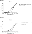
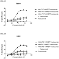
- A49-F3'-TriNKET-Trastuzumab exhibit a significant advantage in potency and maximum lysis of target cells compared to the combination of Fc-silent TriNKET ("A49si-F3'-TriNKET-Trastuzumab"; the amino acid sequence of the constant region has L234A, L235A, and P329G (LALAPG) mutations, which reduce effector functions of the Fc), and trastuzumab, when the target cells were HER2+ (786-O cells) ( FIG. 19 ) or HER2++ (H661 cells) ( FIG. 20 ), suggesting A49-F3'-TriNKET-Trastuzumab can mediate robust effector cell dependent killing of cancer cells expressing HER2.
- the multi-specific binding proteins described herein e.g. , A49-F3'-TriNKET-Trastuzumab
- the multi-specific binding proteins described herein are advantageous in treating HER2-expressing cancers.
- the invention provides a multi-specific binding protein as defined in the claims and/or a pharmaceutical composition as defined in the claims for use in methods of treating cancer.
- the methods may be used to treat a variety of cancers which express HER2 by administering to a patient in need thereof a therapeutically effective amount of a multi-specific binding protein described herein.
- the therapeutic method can be characterized according to the cancer to be treated.
- the cancer is breast, ovarian, esophageal, bladder or gastric cancer, salivary duct carcinoma, salivary duct carcinomas, adenocarcinoma of the lung or aggressive forms of uterine cancer, such as uterine serous endometrial carcinoma.
- the cancer is brain cancer, breast cancer, cervical cancer, colon cancer, colorectal cancer, endometrial cancer, esophageal cancer, leukemia, lung cancer, liver cancer, melanoma, ovarian cancer, pancreatic cancer, rectal cancer, renal cancer, stomach cancer, testicular cancer, or uterine cancer.
- the cancer is a squamous cell carcinoma, adenocarcinoma, small cell carcinoma, melanoma, neuroblastoma, sarcoma (e.g.
- an angiosarcoma or chondrosarcoma larynx cancer, parotid cancer, bilary tract cancer, thyroid cancer, acral lentiginous melanoma, actinic keratoses, acute lymphocytic leukemia, acute myeloid leukemia, adenoid cystic carcinoma, adenomas, adenosarcoma, adenosquamous carcinoma, anal canal cancer, anal cancer, anorectum cancer, astrocytic tumor, bartholin gland carcinoma, basal cell carcinoma, biliary cancer, bone cancer, bone marrow cancer, bronchial cancer, bronchial gland carcinoma, carcinoid, cholangiocarcinoma, chondosarcoma, choriod plexus papilloma/carcinoma, chronic lymphocytic leukemia, chronic myeloid leukemia, clear cell carcinoma, connective tissue cancer, cystadenoma, digestive system cancer,
- the cancer is non-Hodgkin's lymphoma, such as a B-cell lymphoma or a T-cell lymphoma.
- the non-Hodgkin's lymphoma is a B-cell lymphoma, such as a diffuse large B-cell lymphoma, primary mediastinal B-cell lymphoma, follicular lymphoma, small lymphocytic lymphoma, mantle cell lymphoma, marginal zone B-cell lymphoma, extranodal marginal zone B-cell lymphoma, nodal marginal zone B-cell lymphoma, splenic marginal zone B-cell lymphoma, Burkitt lymphoma, lymphoplasmacytic lymphoma, hairy cell leukemia, or primary central nervous system (CNS) lymphoma.
- B-cell lymphoma such as a diffuse large B-cell lymphoma, primary mediastinal B-cell lymphoma, follicular lymphom
- the non-Hodgkin's lymphoma is a T-cell lymphoma, such as a precursor T-lymphoblastic lymphoma, peripheral T-cell lymphoma, cutaneous T-cell lymphoma, angioimmunoblastic T-cell lymphoma, extranodal natural killer/T-cell lymphoma, enteropathy type T-cell lymphoma, subcutaneous panniculitis-like T-cell lymphoma, anaplastic large cell lymphoma, or peripheral T-cell lymphoma.
- T-cell lymphoma such as a precursor T-lymphoblastic lymphoma, peripheral T-cell lymphoma, cutaneous T-cell lymphoma, angioimmunoblastic T-cell lymphoma, extranodal natural killer/T-cell lymphoma, enteropathy type T-cell lymphoma, subcutaneous panniculitis-like T-cell lymphoma, anaplastic large cell lymphoma, or
- the cancer is breast cancer, thyroid cancer, gastric cancer, renal cell carcinoma, adenocarcinoma of the lung, prostate cancer, cholangiocarcinoma, uterine cancer, pancreatic cancer, colorectal cancer, ovarian cancer, cervical cancer, head and neck cancer, lung squamous, mesothelioma, liver cancer, sarcoma, and gall bladder cancer.
- the cancer to be treated can be characterized according to the presence of a particular antigen expressed on the surface of the cancer cell.
- the cancer cell can express one or more of the following in addition to HER2: CD2, CD 19, CD20, CD30, CD38, CD40, CD52, CD70, EGFR/ERBB1, IGF1R, HER3/ERBB3, HER4/ERBB4, MUC1, cMET, SLAMF7, PSCA, MICA, MICB, TRAILR1, TRAILR2, MAGE-A3, B7.1, B7.2, CTLA4, and PD1.
- a multi-specific binding protein described herein can be for use in a method to treat cancer in combination with additional therapeutic agents to treat cancer.
- Exemplary therapeutic agents that may be used as part of a combination therapy in treating cancer, include, for example, radiation, mitomycin, tretinoin, ribomustin, gemcitabine, vincristine, etoposide, cladribine, mitobronitol, methotrexate, doxorubicin, carboquone, pentostatin, nitracrine, zinostatin, cetrorelix, letrozole, raltitrexed, daunorubicin, fadrozole, fotemustine, thymalfasin, sobuzoxane, nedaplatin, cytarabine, bicalutamide, vinorelbine, vesnarinone, aminoglutethimide, amsacrine, proglumide, elliptinium acetate, ketanserin, doxifluridine, etretinate, isotretinoin, streptozoc
- immune checkpoint inhibitors include agents that inhibit one or more of (i) cytotoxic T lymphocyte-associated antigen 4 (CTLA4), (ii) programmed cell death protein 1 (PD1), (iii) PDL1, (iv) LAG3, (v) B7-H3, (vi) B7-H4, and (vii) TIM3.
- CTLA4 inhibitor ipilimumab has been approved by the United States Food and Drug Administration for treating melanoma.
- agents that may be used as part of a combination therapy in treating cancer are monoclonal antibody agents that target non-checkpoint targets (e.g. , herceptin) and non-cytotoxic agents (e.g. , tyrosine-kinase inhibitors).
- non-checkpoint targets e.g. , herceptin
- non-cytotoxic agents e.g. , tyrosine-kinase inhibitors
- anti-cancer agents include, for example: (i) an inhibitor selected from an ALK Inhibitor, an ATR Inhibitor, an A2A Antagonist, a Base Excision Repair Inhibitor, a Bcr-Abl Tyrosine Kinase Inhibitor, a Bruton's Tyrosine Kinase Inhibitor, a CDC7 Inhibitor, a CHK1 Inhibitor, a Cyclin-Dependent Kinase Inhibitor, a DNA-PK Inhibitor, an Inhibitor of both DNA-PK and mTOR, a DNMT1 Inhibitor, a DNMT1 Inhibitor plus 2-chloro-deoxyadenosine, an HDAC Inhibitor, a Hedgehog Signaling Pathway Inhibitor, an IDO Inhibitor, a JAK Inhibitor, a mTOR Inhibitor, a MEK Inhibitor, a MELK
- Proteins of the invention can also be used as an adjunct to surgical removal of the primary lesion.
- the amount of multi-specific binding protein and additional therapeutic agent and the relative timing of administration may be selected in order to achieve a desired combined therapeutic effect.
- the therapeutic agents in the combination, or a pharmaceutical composition or compositions comprising the therapeutic agents may be administered in any order such as, for example, sequentially, concurrently, together, simultaneously and the like.
- a multi-specific binding protein may be administered during a time when the additional therapeutic agent(s) exerts its prophylactic or therapeutic effect, or vice versa.
- compositions that contain a therapeutically effective amount of a protein described herein.
- the composition can be formulated for use in a variety of drug delivery systems.
- One or more physiologically acceptable excipients or carriers can also be included in the composition for proper formulation.
- Suitable formulations for use in the present disclosure are found in Remington's Pharmaceutical Sciences, Mack Publishing Company, Philadelphia, Pa., 17th ed., 1985 .
- Langer Science 249:1527-1533, 1990 ).
- the intravenous drug delivery formulation of the present disclosure may be contained in a bag, a pen, or a syringe.
- the bag may be connected to a channel comprising a tube and/or a needle.
- the formulation may be a lyophilized formulation or a liquid formulation.
- the formulation may freeze-dried (lyophilized) and contained in 12 to 60 vials.
- the formulation may be freeze-dried and 45 mg of the freeze-dried formulation may be contained in one vial.
- the about 40 mg - about 100 mg of freeze-dried formulation may be contained in one vial.
- freeze dried formulation from 12, 27, or 45 vials are combined to obtained a therapeutic dose of the protein in the intravenous drug formulation.
- the formulation may be a liquid formulation and stored as about 250 mg/vial to about 1000 mg/vial.
- the formulation may be a liquid formulation and stored as about 600 mg/vial.
- the formulation may be a liquid formulation and stored as about 250 mg/vial.
- the protein could exist in a liquid aqueous pharmaceutical formulation including a therapeutically effective amount of the protein in a buffered solution forming a formulation.
- compositions may be sterilized by conventional sterilization techniques, or may be sterile filtered.
- the resulting aqueous solutions may be packaged for use as-is, or lyophilized, the lyophilized preparation being combined with a sterile aqueous carrier prior to administration.
- the pH of the preparations typically will be between 3 and 11, more preferably between 5 and 9 or between 6 and 8, and most preferably between 7 and 8, such as 7 to 7.5.
- the resulting compositions in solid form may be packaged in multiple single dose units, each containing a fixed amount of the above-mentioned agent or agents.
- the composition in solid form can also be packaged in a container for a flexible quantity.
- the present disclosure provides a formulation with an extended shelf life including the protein of the present disclosure, in combination with mannitol, citric acid monohydrate, sodium citrate, disodium phosphate dihydrate, sodium dihydrogen phosphate dihydrate, sodium chloride, polysorbate 80, water, and sodium hydroxide.
- An aqueous formulation may be prepared including the protein of the present disclosure in a pH-buffered solution.
- the buffer may have a pH ranging from about 4 to about 8, e.g., from about 4.5 to about 6.0, or from about 4.8 to about 5.5, or may have a pH of about 5.0 to about 5.2. Ranges intermediate to the above recited pH's are also intended to be part of this disclosure. For example, ranges of values using a combination of any of the above recited values as upper and/or lower limits are intended to be included. Examples of buffers that will control the pH within this range include acetate (e.g., sodium acetate), succinate (such as sodium succinate), gluconate, histidine, citrate and other organic acid buffers.
- the formulation may include a buffer system which contains citrate and phosphate to maintain the pH in a range of about 4 to about 8.
- the pH range may be from about 4.5 to about 6.0, or from about pH 4.8 to about 5.5, or in a pH range of about 5.0 to about 5.2.
- the buffer system may include citric acid monohydrate, sodium citrate, disodium phosphate dihydrate, and/or sodium dihydrogen phosphate dihydrate.
- the buffer system may include about 1.3 mg/mL of citric acid ( e.g., 1.305 mg/mL), about 0.3 mg/mL of sodium citrate ( e.g., 0.305 mg/mL), about 1.5 mg/mL of disodium phosphate dihydrate ( e.g.
- the buffer system may include about 1 to 1.5 mg/mL of citric acid, about 0.25 to 0.5 mg/mL of sodium citrate, about 1.25 to 1.75 mg/mL of disodium phosphate dihydrate, about 0.7 to 1.1 mg/mL of sodium dihydrogen phosphate dihydrate, and about 6.0 to 6.4 mg/mL of sodium chloride.
- the pH of the formulation may be adjusted with sodium hydroxide.
- a polyol which acts as a tonicifier and may stabilize the antibody, may also be included in the formulation.
- the polyol is added to the formulation in an amount which may vary with respect to the desired isotonicity of the formulation.
- the aqueous formulation may be isotonic.
- the amount of polyol added may also be altered with respect to the molecular weight of the polyol. For example, a lower amount of a monosaccharide (e.g. , mannitol) may be added, compared to a disaccharide (such as trehalose).
- the polyol which may be used in the formulation as a tonicity agent is mannitol
- the mannitol concentration may be about 5 to about 20 mg/mL.
- the concentration of mannitol may be about 7.5 to 15 mg/mL.
- the concentration of mannitol may be about 10 to 14 mg/mL.
- the concentration of mannitol may be about 12 mg/mL.
- the polyol sorbitol may be included in the formulation.
- a detergent or surfactant may also be added to the formulation.
- exemplary detergents include nonionic detergents such as polysorbates (e.g. , polysorbates 20, 80 etc.) or poloxamers ( e.g. , poloxamer 188).
- the amount of detergent added is such that it reduces aggregation of the formulated antibody and/or minimizes the formation of particulates in the formulation and/or reduces adsorption.
- the formulation may include a surfactant which is a polysorbate.
- the formulation may contain the detergent polysorbate 80 or Tween 80.
- Tween 80 is a term used to describe polyoxyethylene (20) sorbitanmonooleate ( see Fiedler, Lexikon der Hifsstoffe, Editio Cantor Verlag Aulendorf, 4th ed., 1996 ).
- the formulation may contain between about 0.1 mg/mL and about 10 mg/mL of polysorbate 80, or between about 0.5 mg/mL and about 5 mg/mL. About 0.1% polysorbate 80 may be added in the formulation.
- the protein product of the present disclosure may be formulated as a liquid formulation.
- the liquid formulation may be presented at about a 10 mg/mL concentration in either a USP / Ph Eur type I 50R vial closed with a rubber stopper and sealed with an aluminum crimp seal closure.
- the stopper may be made of elastomer complying with USP and Ph Eur.
- Vials may be filled with about 61.2 mL of the protein product solution in order to allow an extractable volume of about 60 mL.
- the liquid formulation may be diluted with about 0.9% saline solution.
- the liquid formulation of the disclosure may be prepared as a 10 mg/mL concentration solution in combination with a sugar at stabilizing levels.
- the liquid formulation may be prepared in an aqueous carrier.
- a stabilizer may be added in an amount no greater than that which may result in a viscosity undesirable or unsuitable for intravenous administration.
- the sugar may be disaccharides, e.g. , sucrose.
- the liquid formulation may also include one or more of a buffering agent, a surfactant, and a preservative.
- the pH of the liquid formulation may be set by addition of a pharmaceutically acceptable acid and/or base.
- the pharmaceutically acceptable acid may be hydrochloric acid.
- the base may be sodium hydroxide.
- deamidation is a common product variant of peptides and proteins that may occur during fermentation, harvest/cell clarification, purification, drug substance/drug product storage and during sample analysis.
- Deamidation is the loss of NH 3 from a protein forming a succinimide intermediate that can undergo hydrolysis.
- the succinimide intermediate results in a 17 dalton mass decrease of the parent peptide.
- the subsequent hydrolysis results in an 18 dalton mass increase.
- Isolation of the succinimide intermediate is difficult due to instability under aqueous conditions. As such, deamidation is typically detectable as 1 dalton mass increase. Deamidation of an asparagine results in either aspartic or isoaspartic acid.
- the parameters affecting the rate of deamidation include pH, temperature, solvent dielectric constant, ionic strength, primary sequence, local polypeptide conformation and tertiary structure.
- the amino acid residues adjacent to Asn in the peptide chain affect deamidation rates. Gly and Ser following an Asn in protein sequences results in a higher susceptibility to deamidation.
- the liquid formulation of the present disclosure may be preserved under conditions of pH and humidity to prevent deamination of the protein product.
- the aqueous carrier of interest herein is one which is pharmaceutically acceptable (safe and non-toxic for administration to a human) and is useful for the preparation of a liquid formulation.
- Illustrative carriers include sterile water for injection (SWFI), bacteriostatic water for injection (BWFI), a pH buffered solution (e.g. , phosphate-buffered saline), sterile saline solution, Ringer's solution or dextrose solution.
- a preservative may be optionally added to the formulations herein to reduce bacterial action.
- the addition of a preservative may, for example, facilitate the production of a multi-use (multiple-dose) formulation.
- Intravenous (IV) formulations may be the preferred administration route in particular instances, such as when a patient is in the hospital after transplantation receiving all drugs via the IV route.
- the liquid formulation may be diluted with 0.9% Sodium Chloride solution before administration.
- the diluted drug product for injection may be isotonic and suitable for administration by intravenous infusion.
- a salt or buffer components may be added in an amount of about 10 mM to 200 mM.
- the salts and/or buffers are pharmaceutically acceptable and are derived from various known acids (inorganic and organic) with "base forming" metals or amines.
- the buffer may be phosphate buffer.
- the buffer may be glycinate, carbonate, citrate buffers, in which case, sodium, potassium or ammonium ions can serve as counterion.
- a preservative may be optionally added to the formulations herein to reduce bacterial action.
- the addition of a preservative may, for example, facilitate the production of a multi-use (multiple-dose) formulation.
- the aqueous carrier of interest herein is one which is pharmaceutically acceptable (safe and non-toxic for administration to a human) and is useful for the preparation of a liquid formulation.
- Illustrative carriers include sterile water for injection (SWFI), bacteriostatic water for injection (BWFI), a pH buffered solution (e.g. , phosphate-buffered saline), sterile saline solution, Ringer's solution or dextrose solution.
- the protein of the present disclosure could exist in a lyophilized formulation including the proteins and a lyoprotectant.
- the lyoprotectant may be sugar, e.g., disaccharides.
- the lyoprotectant may be sucrose or maltose.
- the lyophilized formulation may also include one or more of a buffering agent, a surfactant, a bulking agent, and/or a preservative.
- the amount of sucrose or maltose useful for stabilization of the lyophilized drug product may be in a weight ratio of at least 1:2 protein to sucrose or maltose.
- the protein to sucrose or maltose weight ratio may be of from 1:2 to 1:5.
- the pH of the formulation, prior to lyophilization, may be set by addition of a pharmaceutically acceptable acid and/or base.
- the pharmaceutically acceptable acid may be hydrochloric acid.
- the pharmaceutically acceptable base may be sodium hydroxide.
- the pH of the solution containing the protein of the present disclosure may be adjusted between 6 to 8.
- the pH range for the lyophilized drug product may be from 7 to 8.
- a salt or buffer components may be added in an amount of 10 mM - 200 mM.
- the salts and/or buffers are pharmaceutically acceptable and are derived from various known acids (inorganic and organic) with "base forming" metals or amines.
- the buffer may be phosphate buffer.
- the buffer may be glycinate, carbonate, citrate buffers, in which case, sodium, potassium or ammonium ions can serve as counterion.
- a “bulking agent” may be added.
- a “bulking agent” is a compound which adds mass to a lyophilized mixture and contributes to the physical structure of the lyophilized cake (e.g., facilitates the production of an essentially uniform lyophilized cake which maintains an open pore structure).
- Illustrative bulking agents include mannitol, glycine, polyethylene glycol and sorbitol. The lyophilized formulations may contain such bulking agents.
- a preservative may be optionally added to the formulations herein to reduce bacterial action.
- the addition of a preservative may, for example, facilitate the production of a multi-use (multiple-dose) formulation.
- the lyophilized drug product may be constituted with an aqueous carrier.
- the aqueous carrier of interest herein is one which is pharmaceutically acceptable (e.g. , safe and non-toxic for administration to a human) and is useful for the preparation of a liquid formulation, after lyophilization.
- Illustrative diluents include sterile water for injection (SWFI), bacteriostatic water for injection (BWFI), a pH buffered solution (e.g. , phosphate-buffered saline), sterile saline solution, Ringer's solution or dextrose solution.
- the lyophilized drug product of the current disclosure may be reconstituted with either Sterile Water for Injection, USP (SWFI) or about 0.9% Sodium Chloride Injection, USP. During reconstitution, the lyophilized powder dissolves into a solution.
- SWFI Sterile Water for Injection
- USP Sterile Water for Injection
- USP 0.9% Sodium Chloride Injection
- the lyophilized protein product of the instant disclosure may be constituted to about 4.5 mL water for injection and diluted with 0.9% saline solution (sodium chloride solution).
- Actual dosage levels of the active ingredients in the pharmaceutical compositions of this invention may be varied so as to obtain an amount of the active ingredient which is effective to achieve the desired therapeutic response for a particular patient, composition, and mode of administration, without being toxic to the patient.
- the specific dose can be a uniform dose for each patient, for example, 50 to 5000 mg of protein.
- a patient's dose can be tailored to the approximate body weight or surface area of the patient.
- Other factors in determining the appropriate dosage can include the disease or condition to be treated or prevented, the severity of the disease, the route of administration, and the age, sex and medical condition of the patient. Further refinement of the calculations necessary to determine the appropriate dosage for treatment is routinely made by those skilled in the art, especially in light of the dosage information and assays disclosed herein.
- the dosage can also be determined through the use of known assays for determining dosages used in conjunction with appropriate dose-response data. An individual patient's dosage can be adjusted as the progress of the disease is monitored.
- Blood levels of the targetable construct or complex in a patient can be measured to see if the dosage needs to be adjusted to reach or maintain an effective concentration.
- Pharmacogenomics may be used to determine which targetable constructs and/or complexes, and dosages thereof, are most likely to be effective for a given individual ( Schmitz et al., Clinica Chimica Acta 308: 43-53, 2001 ; Steimer et al., Clinica Chimica Acta 308: 33-41, 2001 ).
- dosages based on body weight are from about 0.01 ⁇ g to about 100 mg per kg of body weight, such as about 0.01 ⁇ g to about 100 mg/kg of body weight, about 0.01 ⁇ g to about 50 mg/kg of body weight, about 0.01 ⁇ g to about 10 mg/kg of body weight, about 0.01 ⁇ g to about 1 mg/kg of body weight, about 0.01 ⁇ g to about 100 ⁇ g/kg of body weight, about 0.01 ⁇ g to about 50 ⁇ g/kg of body weight, about 0.01 ⁇ g to about 10 ⁇ g/kg of body weight, about 0.01 ⁇ g to about 1 ⁇ g/kg of body weight, about 0.01 ⁇ g to about 0.1 ⁇ g/kg of body weight, about 0.1 ⁇ g to about 100 mg/kg of body weight, about 0.1 ⁇ g to about 50 mg/kg of body weight, about 0.1 ⁇ g to about 10 mg/kg of body weight, about 0.1 ⁇ g to about 1 mg/kg of body weight, about 0.1 ⁇ g to about
- Doses may be given once or more times daily, weekly, monthly or yearly, or even once every 2 to 20 years. Persons of ordinary skill in the art can easily estimate repetition rates for dosing based on measured residence times and concentrations of the targetable construct or complex in bodily fluids or tissues. Administration could be intravenous, intraarterial, intraperitoneal, intramuscular, subcutaneous, intrapleural, intrathecal, intracavitary, by perfusion through a catheter or by direct intralesional injection. This may be administered once or more times daily, once or more times weekly, once or more times monthly, and once or more times annually.
- Example 1 Primary human NK cell cytotoxicity assay:
- PBMCs Peripheral blood mononuclear cells
- Isolated PBMCs were washed and prepared for NK cell isolation.
- NK cells were isolated using a negative selection technique with magnetic beads, purity of isolated NK cells was typically >90% CD3 - CD56 + .
- Isolated NK cells were rested overnight, rested NK cells were used the following day in cytotoxicity assays.
- Human cancer cell lines expressing HER2 were harvested from culture, cells were washed with HBS, and were resuspended in growth media at 10 6 /mL for labeling with BATDA reagent (Perkin Elmer AD0116). Manufacturer instructions were followed for labeling of the target cells. After labeling cells were washed 3x with HBS, and were resuspended at 0.5-1.0 ⁇ 10 5 /mL in culture media. To prepare the background wells an aliquot of the labeled cells was put aside, and the cells were spun out of the media. 100 ⁇ l of the media were carefully added to wells in triplicate to avoid disturbing the pelleted cells. 100 ⁇ l of BATDA labeled cells were added to each well of the 96-well plate.
- TriNKET against HER2 A49-F3'-TriNKET-Trastuzumab (comprising an HER2-binding scFv (SEQ ID NO:139) linked to an Fc domain via a hinge comprising Ala-Ser (scFv-Fc represented by SEQ ID NO:140); and an NKG2D-binding Fab fragment including a heavy chain portion comprising a heavy chain variable domain of ADI-27749 (A49) (SEQ ID NO:94) and a CH1 domain, and a light chain portion comprising a light chain variable domain (SEQ ID NO:98) and a light chain constant domain, where the heavy chain variable domain is connected to the CH1, and the CH1 domain is connected to the Fc domain (heavy chain portion represented as VH-CH1-Fc, amino acid sequence set forth in SEQ
- NK cells were harvested from culture, cells were washed, and were resuspended at 10 5 -2.0 ⁇ 10 6 /mL in culture media depending on the desired E:T ratio. 50 ⁇ l of NK cells were added to each well of the plate to make a total of 200 ⁇ l culture volume. The plate was incubated at 37 °C with 5% CO2 for 2-3 hours before developing the assay.
- SkBr-3 cells stably expressing NucLight Green were generated using IncuCyte NucLight Green Reagent (catalog # 4475). NucLight Green expressing cells were selected in puromycin to obtain a homogenous population. SkBr-3-NucLight Green cells were maintained in growth media containing puromycin before use in assays. SkBr-3-NucLight Green target cells were prepared as follows for cytotoxicity assays.
- NucLight Green expressing cells were harvested from culture, and were washed to remove residual selection antibiotic, cells were resuspended in fresh culture media and seeded into a 96 well flat bottom plate. The plate was placed in the IncuCyte S3 overnight to monitor cell attachment and growth. The next day human PBMCs were isolated using density gradient centrifugation, and A49-F3'-TriNKET-Trastuzumab or mAb dilutions were prepared in primary cell culture media. Diluted A49-F3'-TriNKET-Trastuzumab and mAbs were added to SkBr-3-NucLight Green cells, followed by freshly isolated PBMCs. The plate was then returned to the IncuCyte S3.
- % Growth ((Green object count time X) / (green object count time zero)) * 100%.
- FIG. 3, FIG. 4 , and FIG. 5 show TriNKET-mediated killing of three cell lines with different levels of HER2 expression. TriNKETs provided more potent, and higher maximal killing against all HER2-positive cells lines compared to the anti-HER2 monoclonal antibody trastuzumab.
- FIG. 3 shows NK-mediated cell lysis of the HER2 1+ human cancer cell line 786-O.
- trastuzumab shows little activity against HER2 1+ cell lines, increasing specific lysis slightly higher than background killing.
- A49-F3'-TriNKET-Trastuzumab (comprising an HER2-binding scFv (SEQ ID NO: 139) linked to an Fc domain via a hinge comprising Ala-Ser (scFv-Fc represented by SEQ ID NO: 140); and an NKG2D-binding Fab fragment including a heavy chain portion comprising a heavy chain variable domain of ADI-27749 (A49) (SEQ ID NO:94) and a CH1 domain, and a light chain portion comprising a light chain variable domain (SEQ ID NO:98) and a light chain constant domain, where the heavy chain variable domain is connected to the CH1 domain, and the CH1 domain is connected to the Fc domain (heavy chain portion represented as VH-CH
- FIG. 4 demonstrates NK-mediated cell lysis of the human cancer cell line H661 with higher level of HER2 expression that the 786-O cell line (denoted as HER2++).
- trastuzumab showed improved killing against higher levels of HER2 expressed on H661 target cells.
- A49-F3'-TriNKET-Trastuzumab still showed superior lysis of H661 target cells for both potency and maximal killing.
- FIG. 5 shows NK-mediated cell lysis of the cancer cell line SkBr-3 with the highest level of HER2 expression among the three cell lines tested (denoted as HER2+++).
- Trastuzumab showed increased potency against SkBr-3 cells compared to HER2+ and HER2++ cell lines, but A49-F3'-TriNKET-Trastuzumab still showed superior potency and maximal killing.
- FIG. 6 and FIG. 7 show the effect of TriNKET or mAb on 72-hour co-cultures of human PBMCs and SkBr-3 HER2+ target cells.
- SkBr-3 cells proliferated about 3-fold over a 72-hour period when cultured without effector PBMCs. Donor variability was evident in effector PBMCs.
- PBMCs were added to SkBr-3 cultures, growth of the target SkBr-3 cells was nominally reduced ( FIG. 6 ).
- the addition of PBMC effector cells had negligible effect on SkBr-3 cell growth ( FIG. 7 ).
- the effector PBMCs When trastuzumab was added to the co-culture, the effector PBMCs had an increased ability to lyse target SkBr-3 cells, indicated as a decrease in % growth in FIG. 6 and FIG. 7 .
- A49-F3'-TriNKET-Trastuzumab ( FIG. 6 and FIG. 7 ) and A44-F3'-KiH-TriNKET-Trastuzumab ( FIG. 7 ) were added to the co-culture, the effector PBMCs were even more effective in lysing SkBr-3 target cells compared to trastuzumab, resulting in a faster and more complete reduction in SkBr-3 cells.
- Trispecific-binding proteins TriNKETs
- mAb monoclonal Ab
- blood was washed 3x following incubation with TriNKET.
- Directly labeled immunophenotyping mAbs and secondary antibody specific to trastuzumab were added to samples. After a 20 minute incubation, 2 mL of 1x RBC lysis/fixation solution was added to each sample for 15 minutes at room temperature (RT), samples were then washed to remove red blood cells (RBCs). After washing, the samples were resuspended for FACS analysis.
- FIGs. 8A-8F show binding of A49-F3'-TriNKET-Trastuzumab (an NKG2D-binding domain from clone ADI-27749; and an HER2-binding scFv comprising SEQ ID NO: 139, derived from trastuzumab monoclonal antibody) in whole human blood.
- A49-F3'-TriNKET-Trastuzumab binding in human whole blood was the same as trastuzumab.
- A49-F3'-TriNKET-Trastuzumab and trastuzumab demonstrated minimal binding to all populations of immune cells in blood. Small shifts were observed for trastuzumab and A49-F3'-TriNKET-Trastuzumab in both B cell and monocyte populations compared to secondary control samples. Binding observed on B cells and monocytes can likely be attributed to FcR interactions, rather than being Fab specific.
- Human cardiomyocytes differentiated from induced pluripotent stem cells (Cellular Dynamics/Fuji Film), 786-O, H661 and SKBR3 cancer cells were used to evaluate binding of A49-F3'-TriNKET-Trastuzumab to these cells.
- the human renal cell carcinoma cell line 786-O expresses low levels of HER2
- the human lung cancer cell line H661 expresses moderate levels of HER2
- the human breast cancer cell line SKBR3 expresses high levels of HER2.
- TriNKETs were diluted to 3.8e-4 to 100 ⁇ g/mL, and the dilutions were used as primary antibody stain.
- Binding of the TriNKET was detected using a fluorophore-conjugated anti-human IgG secondary antibody. Cells were analyzed by flow cytometry. Binding fluorescence intensity (MFI) to cells expressing HER2 was normalized to cells stained with a control (non-specific) TriNKET to obtain fold over background (FOB) values.
- MFI fluorescence intensity
- PBMCs peripheral blood buffy coats using density gradient centrifugation.
- SKBR3 target cells were labeled with BacMam 3.0 NucLight Green (#4622) to allow for tracking of the target cells. The manufacturer's protocol was followed for labeling of SKBR3 target cells.
- Human cardiomyocytes were unlabeled.
- Monoclonal antibodies or TriNKETs were diluted into culture media. 50 ⁇ l of TriNKETs and human PBMCs were added to wells of a 96-well plate already containing target cells, 50 ⁇ l of complete culture media was added for a total of 200 ⁇ l culture volume.
- Image collection was setup on the IncuCyte S3. Image analysis was done using the IncuCyte S3 software. Masks for the green channel was created to count the number of tumor cells. Confluency of cardiomyocytes in the phase channel was used to assess cell viability and calculate % killing.
- A49-F3'-TriNKET-Trastuzumab binds to human cardiomyocytes to similar extent as binding to H661 cells.
- FIG. 9 shows binding of A49-F3'-TriNKET-Trastuzumab to human cardiomyocytes, SKBR3, H661 and 786-O cancer cells, where the binding to cardiomyocytes is similar to H661 cells (medium HER2 surface expression levels).
- FIGs. 10A-10B and FIGs. 11A-11B show killing of SKBR3 cells by human PBMCs in the presence of A49-F3'-TriNKET-Trastuzumab, whereas viability of cardiomyocytes are minimally affected.
- FIG. 10A shows A49-F3'-TriNKET-Trastuzumab-mediated human PBMC killing of SKBR3 cancer cells
- FIG. 10B shows that A49-F3'-TriNKET-Trastuzumab did not kill non-malignant healthy cardiomyocytes after 3 days in co-culture at PBMC to target cell ratio (E:T) of 1:1.
- FIG. 11A shows A49-F3'-TriNKET-Trastuzumab-mediated human PBMC killing of SKBR3 cancer cells
- FIG. 11B shows that A49-F3'-TriNKET-Trastuzumab did not kill non-malignant healthy cardiomyocytes after 3 days in co-culture at E:T of 20: 1.
- Human PBMCs were isolated from human peripheral blood buffy coats using density gradient centrifugation. Isolated PBMCs were stimulated with 1 ⁇ g/mL Concanavalin A (ConA) at 37 °C for 18 hours. Then ConA was removed and cultured with 25 unit/mL IL-2 at 37 °C for 4 days. CD8+ T cells were purified using a negative selection technique with magnetic beads, then cultured in media containing 25 unit/mL IL-2 or 10 ng/mL IL-15 at 37 °C for 8-10 days.
- ConA Concanavalin A
- CD8+ T cells generated above were analyzed by flow cytometry for CD8+ T cell purity as well as NKG2D and CD16 expression. Cells were stained with fluorophore conjugated antibodies against CD3, CD8, NKG2D, CD16, and analyzed by flow cytometry.
- Human cancer cell line SkBr-3 expressing a target of interest, HER2 was harvested from culture. Cells were washed and resuspended in growth media at 10 6 /mL for labeling with BATDA reagent (Perkin Elmer AD0116). Manufacturer instructions were followed for labeling of the target cells. After labeling cells were washed three times with HBS, and were resuspended at 0.5 ⁇ 10 5 /mL in culture media. 100 ⁇ l of BATDA labeled cells were added to each well of the 96-well plate. Wells were saved for spontaneous release from target cells, and wells were prepared for max lysis of target cells by addition of 1% Triton-X.
- Monoclonal antibodies, TriNKETs and controls were diluted in culture media; 50 ⁇ l of diluted mAb/TriNKET were added to each well.
- % Specific lysis ((Experimental release - Spontaneous release) / (Maximum release - Spontaneous release)) * 100%.
- CD8+ T cells were added to each well of the plate to make a total of 200 ⁇ l culture volume.
- the plate was incubated at 37 °C with 5% CO 2 for up to 7 days.
- Image collection was setup on the IncuCyte S3. Images for the phase and green channels were collected every hour, with 2 images per well. Image analysis was done using the IncuCyte S3 software. Green object count/well was used to measure the number of live tumor cells.
- CD8+ T cells generated with conA stimulation and cultured with IL-15 were of high purity (99% of CD3+CD8+ cells), and all expressed NKG2D but not CD16 ( FIGs. 12A- 12C ). Similar results are observed with CD8+ T cells generated with IL-2 culture.
- A49-F3'-TriNKET-Trastuzumab was assayed.
- A49-F3'-TriNKET-Trastuzumab enhanced the cytotoxic activity of human primary CD8+ T cells after culture with IL-15 in a dose-dependent manner ( FIG. 13A ).
- A49-F3'-TriNKET-Trastuzumab also enhanced the cytotoxic activity of human primary CD8+ T cells after culture with IL-2 ( FIG. 13B ).
- FIG. 14A When the cells were treated with A49-F3'-TriNKET-Trastuzumab, the growth of SkBr-3 showed some inhibition due to HER2 signal blockade ( FIG. 14A ). In the presence of anti-CD3 antibody (mIgG1 antibody cross-linked with F(ab')2 to anti-mIgG1) ( FIG.
- TriNKET Human cancer cell lines expressing HER2 were used to assess tumor antigen binding of a TriNKET (A49-F3'-TriNKET-Trastuzumab) and monoclonal antibodies.
- the human renal cell carcinoma cell line 786-O expressed low levels of HER2, the human lung cancer cell line NCI-H661 expressed moderate levels of HER2, and the human breast cancer cell line SkBr-3 expressed high levels of HER2. All three cell lines were used to assess their binding affinity to TriNKET. TriNKETs were diluted in a series of concentrations, and were incubated with the respective cells.
- Binding of the TriNKET (A49-F3'-TriNKET-Trastuzumab) to the cells was detected using a fluorophore conjugated to an anti-human IgG secondary antibody, and the cells were analyzed by flow cytometry.
- the level of binding at each concentration of the TriNKET (A49-F3'-TriNKET-Trastuzumab) was calculated as a percentage value of median fluorescence intensity (MFI) of the cells relative to the maximum MFI observed with the cells incubated with 670 nM of TriNKET.
- MFI median fluorescence intensity
- the level of binding at each concentration of TriNKET was calculated as a fold over background (FOB) value of MFI of the cells relative to the background MFI observed with the cells incubated with the secondary antibody only.
- FOB fold over background
- the TriNKET A49-F3'-TriNKET-Trastuzumab
- trastuzumab exhibited the most potent binding to the SkBr-3 cells ( FIG. 15A ), which had a high expression level of HER2 (HER+++).
- the binding affinity of the HER2-targeted TriNKET and trastuzumab to the NCI-H661, which had a moderate expression level of HER2 (HER2++) was similar ( FIG. 16A ) to but slightly higher than the binding affinity to the 786-O cells ( FIG. 17A ), which had a low expression level of HER2 (HER2+).
- This result suggested that the affinity of the HER2-targeted TriNKET and trastuzumab to HER2-expressing cells generally correlated with the expression level of HER2 on the cells.
- Trastuzumab showed a higher binding affinity than the TriNKET (A49-F3'-TriNKET-Trastuzumab ) with each of the three cell lines (SkBr-3 ( FIG. 15A ), NCI-H661 ( FIG. 16A ), and 786-O ( FIG. 17A )).
- FOB fold over background
- the maximum binding of the TriNKET to each of the three cell lines was greater than the maximum binding of trastuzumab (SkBr-3 ( FIGs. 15B ), NCI-H661 ( FIG. 16B ), and 786-O ( FIG. 17B )).
- the difference was especially significant with the SkBr-3 cells, which had a high HER2 expression level ( FIG. 15B ).
- TriNKETs The affinity of TriNKETs to EL4 cells that express NKG2D was measured by a similar method. As shown in Figures 18A-18B , different NKG2D targeting domains used in the TriNKET resulted in different levels of binding to NKG2D expressed on EL4 cells. TriNKETs of clones A44, F63, and E79 bound to NKG2D strongly ( FIG. 18A and FIG. 18B ), whereas a TriNKET of clone A49 bound to NKG2D weakly ( FIG. 18A and FIG. 18B ).
- PBMCs peripheral blood buffy coats using density gradient centrifugation and were washed.
- NK cells were isolated from the PBMCs using a negative selection technique with magnetic beads. Typically, with this technique, more than 90% of the harvested cells were CD3 - CD56 + .
- the isolated NK cells were rested overnight. Rested NK cells were used the following day in cytotoxicity assays.
- Human cancer cell lines expressing a target of interest were harvested from culture. The cells were washed with HBS, and were resuspended in growth media at 10 6 cells/mL for labeling with BATDA reagent (Perkin Elmer AD0116). Manufacturer instructions were followed for labeling of the target cells. After labeling, the cells were washed three times with HBS, and were resuspended at 0.5-1.0 ⁇ 10 5 /mL in culture media. 100 ⁇ l of BATDA labeled cells were added to each well of the 96-well plate.
- trastuzumab and A49-F3'-TriNKET-Trastuzumab was diluted in culture media, and 50 ⁇ l of diluted trastuzumab and A49-F3'-TriNKET-Trastuzumab were added, respectively, to each of their corresponding wells for the experiment.
- Rested and/or activated NK cells were harvested from culture. The cells were washed and resuspended at 10 5 -2.0 ⁇ 10 6 /mL in culture media depending on the desired E:T ratio. 50 ⁇ l of NK cells were added to each well of the plate to make a total of 200 ⁇ l culture volume.
- the plate was removed from the incubator and the cells were pelleted by centrifugation at 200g for 5 minutes. 20 ⁇ l of culture supernatant were transferred to a clean microplate provided from the manufacturer, and 200 ⁇ l of room temperature europium solution were added to each well. The plate was protected from light and incubated on a plate shaker at 250rpm for 15 minutes. Fluorescence levels were read using either Victor 3 or SpectraMax i3X instruments.
- % Specific lysis (Experimental release - Spontaneous release) / (Maximum release - Spontaneous release) * 100%
- bispecific antibody engaging NKG2D and HER2 was combined with trastuzumab, and compared to the TriNKET containing all three binding domains on one molecule. Accordingly, 786-O and H661 target cells were incubated with isolated NK cells in the presence of HER2-targeted TriNKET, trastuzumab, Fc-silent TriNKET (comprising SEQ ID NO: 156 and SEQ ID NO: 157, both of which include L234A, L235A, and P329G (LALAPG) substitutions in the Fc domain (shown within a third-bracket [])), or the combination of Fc-silent TriNKET and trastuzumab.
- TriNKET exhibited a significant advantage in potency and maximum lysis of target cells compared to the combination of Fc-silent TriNKET and trastuzumab, when the target cells were 786-O cells ( FIG. 19 ) or H661 cells ( FIG. 20 ) .
- ADI-27749 contains, inter alia, a heavy chain CDR3 having the amino acid sequence of GAPMGAAAGWFDP (SEQ ID NO: 169).
- the Met at position 102 of SEQ ID NO:94 i.e., at position 4 of this CDR3 sequence
- TriNKET A Binding of A49-F3'-TriNKET-Trastuzumab (“TriNKET A”) and a mutant form of TriNKET A having a substitution of Ile for the Met (“TriNKET A*") to a fusion protein of human NKG2D and murine Fc ("mFc-hNKG2D”) was characterized by surface plasmon resonance (SPR) at 37 °C. Steady state affinity fit was utilized to obtain the equilibrium affinity data. The equilibrium affinity constants were calculated, and data from two independent experiments for TriNKET A* and the independent experiments for TriNKET A were averaged.
- SPR surface plasmon resonance
- the equilibrium affinity constant (K D ) obtained from the affinity fit was very similar between the replicates, which suggested a high confidence in the measured parameters.
- the K D values indicated that the M102 variant has less than 2-fold reduced affinity for human NKG2D compared to TriNKET A.
- the K D for TriNKET A* was (4.81 ⁇ 0.39) ⁇ 10 -7 M, while the K D for TriNKET A was (3.37 ⁇ 0.30) ⁇ 10 -7 M (calculated from the affinity fit).
- K D values suggested that the M102 mutation had only a minor effect on the binding of an A49-containing TriNKET to human NKG2D.
- KHYG-1 cells expressing the high-affinity variant of CD16a were generated through retroviral transduction. Following transduction, cells were selected in puromycin-containing growth media to generate a selected population of KHYG-1-CD16V cells. The selected population was maintained in media containing 10 ng/mL human IL-2.
- the KHYG-1-CD16V cells were harvested from culture, pelleted, washed three times in culture media without IL-2, and resuspended in culture media without IL-2 and rested for 24 hours.
- SKBR-3 expressing the tumor antigen HER2 were selected as target cells.
- SKBR-3 expressing HER2 were harvested from culture. The cells were washed with Hepes Buffered Saline (HBS), and were resuspended in growth media at 10 6 cells/mL for labeling with BATDA (hydrophobic esterified form of TDA (bis(acetoxymethyl) 2,2':6',2"-terpyridine-6,6"-dicarboxylate) reagent (Perkin Elmer C136-100) (BATDA diffuses through the cell membrane of viable cells, and is hydrolyzed by intracellular esterases resulting in accumulation of membrane permeable TDA inside target cells.
- BATDA hydrophobic esterified form of TDA (bis(acetoxymethyl) 2,2':6',2"-terpyridine-6,6"-dicarboxylate) reagent
- BATDA diffuses through the cell membrane of viable cells, and is hydrolyzed by intracellular esterases
- the TDA released from lysed cells into the supernatant is chelated with Eu3+, and the NK cell activity is quantified by measuring the intense fluorescence of the EuTDA chelate formed. ( See Blomberg et al. J. Immunol. Methods (1996) 193(2): 199-206 )).
- Manufacturer instructions were followed for labeling of the target cells. After labeling, the cells were washed three times with HBS and were resuspended at 0.5 ⁇ 10 5 cells /mL in culture media. 100 ⁇ l of BATDA labeled cells were added to each well of a 96-well plate.
- TriNKETs were serially diluted in culture media, and 50 ⁇ l of a diluted TriNKET were added to each well.
- Rested NK cells were harvested from culture, washed, and resuspended at 1.0 ⁇ 10 6 cells/mL in culture media.
- 50 ⁇ l of NK cells were added to each well of the plate to attain a desired E:T ratio of 10:1 and to make a total of 200 ⁇ l culture volume in each well. The plate was incubated at 37 °C with 5% CO 2 for 2-3 hours.
- % specific lysis ((Experimental release - Spontaneous release) / (Maximum release - Spontaneous release)) ⁇ 100%.
- the EC 50 was less than 2-fold increased and maximum % specific lysis values of TriNKET A* was identical to that of TriNKET A, suggesting that the M102 mutation did not have a substantial affect on the biological activity of TriNKET A.
Landscapes
- Health & Medical Sciences (AREA)
- Chemical & Material Sciences (AREA)
- Organic Chemistry (AREA)
- Immunology (AREA)
- General Health & Medical Sciences (AREA)
- Medicinal Chemistry (AREA)
- Life Sciences & Earth Sciences (AREA)
- Biophysics (AREA)
- Genetics & Genomics (AREA)
- Molecular Biology (AREA)
- Proteomics, Peptides & Aminoacids (AREA)
- Biochemistry (AREA)
- Oncology (AREA)
- Chemical Kinetics & Catalysis (AREA)
- Nuclear Medicine, Radiotherapy & Molecular Imaging (AREA)
- Pharmacology & Pharmacy (AREA)
- Animal Behavior & Ethology (AREA)
- Public Health (AREA)
- Veterinary Medicine (AREA)
- General Chemical & Material Sciences (AREA)
- Peptides Or Proteins (AREA)
- Medicines That Contain Protein Lipid Enzymes And Other Medicines (AREA)
- Medicines Containing Antibodies Or Antigens For Use As Internal Diagnostic Agents (AREA)
- Micro-Organisms Or Cultivation Processes Thereof (AREA)
- Medicines Containing Material From Animals Or Micro-Organisms (AREA)
Description
- This application claims the benefit of and priority to
U.S. Provisional Patent Application No. 62/716,259, filed August 8, 2018 - The instant application contains a Sequence Listing which has been submitted electronically in ASCII format. Said ASCII copy, created on August 6, 2019, is named DFY-057WO_SL.txt and is 208,511 bytes in size.
- The invention relates to multi-specific binding proteins that bind to NKG2D, CD16, and epidermal growth factor receptor 2 (HER2 or ErbB2). These multi-specific binding proteins are useful for killing human cancer cells expressing Her2 without significant cytotoxicity to non-cancerous healthy human cells expressing HER2.
- Cancer continues to be a significant health problem despite the substantial research efforts and scientific advances reported in the literature for treating this disease. Some of the most frequently diagnosed cancers include prostate cancer, breast cancer, and lung cancer. Prostate cancer is the most common form of cancer in men. Breast cancer remains a leading cause of death in women. Current treatment options for these cancers are not effective for all patients and/or can have substantial adverse side effects. Other types of cancer also remain challenging to treat using existing therapeutic options.
- Cancer immunotherapies are desirable because they are highly specific and can facilitate destruction of cancer cells using the patient's own immune system. Fusion proteins such as bi-specific T-cell engagers are cancer immunotherapies described in the literature that bind to tumor cells and T-cells to facilitate destruction of tumor cells. Antibodies that bind to certain tumor-associated antigens and to certain immune cells have been described in the literature. See, e.g.,
WO 2016/134371 andWO 2015/095412 . - C. GERMAIN ET AL: PROTEIN ENGINEERING DESIGN AND SELECTION, vol. 21, no.11, 1 January 2008, pages 665-672 describes redirecting NK cells mediated tumor cell lysis by a recombinant bifunctional protein and relates to using the "knob into hole" heterodimerization strategy, in which "knob" and "hole" variants were generated by directed mutagenesis within the CH3 domain of human IgG1 Fc fragments fused to an anti-CEA or anti-HER2 scFv or to the H60 murine ligand of NKG2D, respectively.
- H.-M. CHO et al: CANCER RESEARCH, vol. 70, no. 24, 14 December 2010, pages 10121-10130 and
WO 2007/002905 relate to delivery of NKG2D ligand using an anti-HER2 antibody-NKG2D ligand fusion protein. -
WO 2016/090278 relates to polypeptides having specific antigen-binding properties of Fv and modified α1-α2 domains of NKG2D ligands. -
WO 2017/081190 relates to anti-NKG2D single domain antibodies. - U. BRINKMAN et al; MABS, vol. 9, no. 2, 10 January 2017 pages 182-212 relates to making of bispecific antibodies.
-
WO2018/152518 ,WO 2019/157332 ,WO 2019/157366 andWO 2019/231920 describe among other subject matters multi-specific binding proteins that bind HER2, the NKG2D receptor and CD 16. - Natural killer (NK) cells are a component of the innate immune system and make up approximately 15% of circulating lymphocytes. NK cells infiltrate virtually all tissues and were originally characterized by their ability to kill tumor cells effectively without the need for prior sensitization. Activated NK cells kill target cells by means similar to cytotoxic T cells - i.e., via cytolytic granules that contain perforin and granzymes as well as via death receptor pathways. Activated NK cells also secrete inflammatory cytokines such as IFN-γ and chemokines that promote the recruitment of other leukocytes to the target tissue.
- NK cells respond to signals through a variety of activating and inhibitory receptors on their surface. For example, when NK cells encounter healthy self-cells, their activity is inhibited through activation of the killer-cell immunoglobulin-like receptors (KIRs). Alternatively, when NK cells encounter foreign cells or cancer cells, they are activated via their activating receptors (e.g., NKG2D, NCRs, DNAM1). NK cells are also activated by the constant region of some immunoglobulins through CD16 receptors on their surface. The overall sensitivity of NK cells to activation depends on the sum of stimulatory and inhibitory signals.
- HER2 (ErbB2) is a transmembrane glycoprotein, which belongs to the epidermal growth factor receptor family. It is a receptor tyrosine kinase and regulates cell survival, proliferation, and growth. HER2 plays an important role in human malignancies.
- The ERBB2 gene is amplified or overexpressed in approximately 30% of human breast cancers. Patients with HER2-overexpressing breast cancer have substantially lower overall survival rates and shorter disease-free intervals than patients whose cancer does not overexpress HER2. Moreover, overexpression of HER2 leads to increased breast cancer metastasis. Over-expression of HER2 is also known to occur in many other cancer types, including breast, ovarian, esophageal, bladder and gastric cancer, salivary duct carcinoma, adenocarcinoma of the lung and aggressive forms of uterine cancer, such as uterine serous endometrial carcinoma.
- The invention relates to multi-specific binding proteins that bind to and kill human cancer cells expressing epidermal growth factor receptor 2 (HER2 or ErbB2). The invention provides multi-specific binding proteins that bind to HER2 on a cancer cell and to the NKG2D receptor and CD16 receptor on natural killer cells. Such proteins can engage more than one kind of NK activating receptor, and may block the binding of natural ligands to NKG2D. In certain embodiments, the proteins can agonize NK cells in humans, and in other species such as rodents and cynomolgus monkeys. In certain embodiments, the proteins can stimulate T cells in humans, and in other species such as human, rodents and cynomolgus monkeys. Various aspects and embodiments of the invention are described in further detail below.
- The present invention provides a multispecific binding protein comprising:
a Fab fragment that binds NKG2D, a single-chain variable fragment (scFv) that binds HER2 and an antibody Fc domain or a portion thereof sufficient to bind CD16, comprising: protein comprising: - (a) a first polypeptide comprising the amino acid sequence of SEQ ID NO: 141;
- (b) a second polypeptide comprising the amino acid sequence of SEQ ID NO: 140; and
- (c) a third polypeptide comprising the amino acid sequence of SEQ ID NO: 142.
- The scFv is linked to the antibody Fc domain that binds CD16via a hinge comprising Ala-Ser.
- The scFv comprises a heavy chain variable domain and a light chain variable domain. The heavy chain variable domain of the scFv may form a disulfide bridge with the light chain variable domain of the scFv. The disulfide bridge may be formed between C44 of the heavy chain variable domain and C100 of the light chain variable domain.
- The light chain variable domain of the scFv is linked to the heavy chain variable domain of the scFv via a flexible linker. The flexible linker consists of the amino acid sequence of SEQ ID NO: 143. , The light chain variable domain is positioned to the N-terminus of the heavy chain variable domain.
- The Fab fragment is linked to the antibody Fc domain that binds CD16.
- The first antigen-binding site that binds NKG2D comprises a heavy chain variable domain comprising complementarity-determining region 1 (CDR1), complementarity-determining region 2 (CDR2), and complementarity-determining region 3 (CDR3) sequences represented by the amino acid sequences of SEQ ID NOs: 168, 96, and 169, respectively; and a light chain variable domain comprising CDR1, CDR2, and CDR3 sequences represented by the amino acid sequences of SEQ ID NOs: 99, 100, and 101, respectively. The first antigen-binding site that binds NKG2D comprises a heavy chain variable domain comprising CDR1, CDR2, and CDR3 sequences represented by the amino acid sequences of SEQ ID NOs: 95, 96, and 97, respectively; and a light chain variable domain comprising CDR1, CDR2, and CDR3 sequences represented by the amino acid sequences of SEQ ID NOs: 99, 100, and 101, respectively.
- A protein of the present invention comprises a first antigen-binding that binds NKG2D, which comprises:
- a heavy chain variable domain comprising complementarity-determining region 1 (CDR1), complementarity-determining region 2 (CDR2), and complementarity-determining region 3 (CDR3) sequences represented by the amino acid sequences of SEQ ID NOs: 168, 96, and 169, respectively; and a light chain variable domain comprising CDR1, CDR2, and CDR3 sequences represented by the amino acid sequences of SEQ ID NOs: 99, 100, and 101, respectively; and
- a second antigen-binding site comprising an scFv that binds HER2, which comprises:
a heavy chain variable domain comprising CDR1, CDR2, and CDR3 sequences represented by the amino acid sequences of SEQ ID NOs: 115, 116, and 117, respectively, and a light chain variable domain comprising CDR1, CDR2, and CDR3 sequences represented by the amino acid sequences of SEQ ID NOs: 119, 120, and 121, respectively. - The first antigen-binding site comprises a heavy chain variable domain related to SEQ ID NO:94 and a light chain variable domain related to SEQ ID NO:98.
- The first antigen-binding site may bind to NKG2D with a KD of 2 nM to 120 nM, as measured by surface plasmon resonance. The protein may bind to NKG2D with a KD of 2 nM to 120 nM, as measured by surface plasmon resonance.
- The second antigen-binding site binding to HER2 comprises a heavy chain variable domain related to SEQ ID NO: 195 and a light chain variable domain related to SEQ ID NO: 196. The second antigen-binding site binding to HER2 comprises an scFv comprising the amino acid sequence of SEQ ID NO: 139.
- The antibody Fc domain comprises hinge and CH2 domains of a human IgG1 antibody.
- The protein comprises a sequence of SEQ ID NO: 141. This sequence represents the heavy chain portion of the Fab fragment linked to the antibody Fc domain.
- The protein comprises a sequence of SEQ ID NO: 140. This sequence represents the scFv linked to the antibody Fc domain.
- The present invention provides a protein comprising (a) a first polypeptide comprising the amino acid sequence of SEQ ID NO: 141; (b) a second polypeptide comprising the amino acid sequence of SEQ ID NO: 140; and (c) a third polypeptide comprising the amino acid sequence of SEQ ID NO: 142.
- In another aspect, the instant disclosure provides a formulation comprising a protein disclosed herein and a pharmaceutically acceptable carrier.
- In another aspect, the instant disclosure provides a cell comprising one or more nucleic acids expressing a protein disclosed herein.
- In another aspect, the instant disclosure provides the protein disclosed herein for use in a method of treating cancer, wherein the method comprises administering a protein or a formulation disclosed herein to a patient in need thereof. In certain embodiments, the cancer is selected from the group consisting of breast cancer, thyroid cancer, gastric cancer, renal cell carcinoma, adenocarcinoma of the lung, prostate cancer, cholangiocarcinoma, uterine cancer, pancreatic cancer, colorectal cancer, ovarian cancer, cervical cancer, head and neck cancer, lung squamous, mesothelioma, liver cancer, mesothelioma, sarcoma, and gall bladder cancer.
-
-
FIG. 1 illustrates a trispecific antibody (TriNKET) that contains an HER2-binding scFv, a NKG2D-targeting Fab, and a heterodimerized antibody constant region/domain ("CD domain") that binds CD16 (scFv-Fab format). In an exemplary embodiment, the Fc domain linked to the Fab fragment comprises mutations of K360E and K409W and the Fc domain linked to the scFv comprises matching mutations Q347R, D399V, and F405T for forming an Fc heterodimer (shown as a triangular lock-and-key format in the Fc domains inFIG. 1 ). The antibody format is referred herein as F3'-TriNKET. In another exemplary embodiment, the Fc domain linked to the Fab fragment comprises the mutations of Q347R, D399V, and F405T, and the Fc domain linked to the scFv comprises matching mutations K360E and K409W for forming a heterodimer. -
FIG. 2 is a representation of a "knob-in-hole" (KiH) TriNKET, which includes an scFv that binds HER2 linked to an Fc domain via a hinge comprising Ala-Ser, and an NKG2D-binding Fab fragment including (a) a heavy chain portion comprising a heavy chain variable domain and a CH1 domain connected to an Fc domain, and (b) a light chain portion comprising a light chain variable domain and a CL domain. In an exemplary embodiment, the Fc domain linked to the Fab fragment comprises a knob mutation T366W, and the Fc domain linked to the scFv comprises matching "hole" mutations T366S, L368A, Y407V (shown as a triangular lock-and-key format in the Fc domains inFIG. 2 ). In an exemplary embodiment, the Fc domain linked to the Fab fragment comprises knob mutations T366S, L368A, Y407V, and the Fc domain linked to the scFv comprises a "hole" mutation T366W. -
FIG. 3 are line graphs demonstrating HER2-targeted TriNKETs are more potent than trastuzumab on a HER2+ (low) cell line. -
FIG. 4 are line graphs demonstrating HER2-targeted TriNKETs are more potent than trastuzumab on a HER2++ cell line. -
FIG. 5 are line graphs demonstrating HER2-targeted TriNKETs are more potent than trastuzumab on a HER2+++ cell line. -
FIG. 6 shows HER2-targeted TriNKETs outperform trastuzumab in a long-term killing assay. -
FIG. 7 shows HER2-targeted TriNKETs outperform trastuzumab in a long-term killing assay. -
FIGs. 8A to 8F are FACS showing that HER2-Targeted TriNKETs show minimal binding to immune cells in human blood.FIG 8A shows that HER2-Targeted TriNKETs show minimal binding to NK cells in human blood;FIG. 8B shows that HER2-Targeted TriNKETs show minimal binding to CD8+ T cells;FIG. 8C shows that HER2-Targeted TriNKETs show minimal binding to CD4+ T cells;FIG. 8D shows that HER2-Targeted TriNKETs show minimal binding to B cells;FIG. 8E shows that HER2-Targeted TriNKETs show minimal binding to monocytes; andFIG. 8F shows that HER2-Targeted TriNKETs show minimal binding to granulocytes (Dotted line - secondary control; dashed line - Trastuzumab; Solid line - HER2-F3'-TriNKET-A49). -
FIG. 9 are line graphs demonstrating binding of A49-F3'-TriNKET-Trastuzumab to human cardiomyocytes, SKBR3, H661 and 786-O cancer cells. -
FIG. 10A shows that A49-F3'-TriNKET-Trastuzumab-mediated human PBMC killing of SKBR3 cancer after 3 days in co-culture at PBMC to target cell ratio (E:T) of 1:1. -
FIG. 10B shows that A49-F3'-TriNKET-Trastuzumab does not kill non-malignant healthy cardiomyocytes even after 3 days in co-culture at PBMC to target cell ratio (E:T) of 1:1. -
FIG. 11A shows A49-F3'-TriNKET-Trastuzumab-mediated human PBMC killing of SKBR3 cancer cells after 3 days in co-culture at PBMC to target cell ratio (E:T) of 20:1. -
FIG. 11B shows that A49-F3'-TriNKET-Trastuzumab does not kill non-malignant healthy cardiomyocytes even after 3 days in co-culture at PBMC to target cell ratio (E:T) of 20:1. -
FIGs. 12A to 12B show that CD8+ T cells generated with conA stimulation and cultured with IL-15 were of high purity (99% of CD3+CD8+ cells) (FIG. 12A ), and all expressed NKG2D (FIG. 12B ), but not CD16 (FIG. 12C ). -
FIGs. 13A to 13B are graphs showing cytotoxic activity of CD8+ T cells in the presence of A49-F3'-TriNKET-Trastuzumab, after culturing with IL-15.FIG. 13A shows enhanced killing of SkBr-3 tumor cells in short-term co-cultures by A49-F3'-TriNKET-Trastuzumab. HER2-targeting A49-F3'-TriNKET-Trastuzumab triggered dose-dependent lysis of SkBr-3 target cells by IL-15 stimulated CD8+ T cell.FIG. 13B shows that 67 nM of HER2-targeting TriNKET triggered lysis of SkBr-3 target cells by IL-2 stimulated CD8+ T cell. Dotted line indicates the effect with only CD8+ T cells co-cultured with SkBr-3 tumor cells (untreated). -
FIG. 14A shows percent growth levels of SkBr-3 cells cultured alone, co-cultured with CD8+ T cells, co-cultured with CD8+ T cells and A49-F3'-TriNKET-Trastuzumab, each in the presence of anti-CD3. -
FIG. 14B shows percent growth level of SkBr-3 cells cultured alone, co-cultured with CD8+ T cells, co-cultured with CD8+ T cells and A49-F3'-TriNKET-Trastuzumab, each in the absence of anti-CD3. -
FIGs. 15A to 15B show TriNKET binding to SkBr-3 cell line that has a high level of HER2 expression, as measured by flow cytometry of SkBr-3 cells incubated with a series of concentrations of TriNKET or trastuzumab and a secondary antibody conjugated with a fluorophore.FIG. 15A shows the level of binding as percentage values of median fluorescence intensity (MFI) relative to the maximum MFI observed with the cells incubated with 670 nM of TriNKET.FIG. 15B shows the level of binding as fold over background (FOB) values of MFI relative to the background MFI observed with the cells incubated with the secondary antibody only. -
FIGs. 16A to 16B show TriNKET binding to NCI-H661 cell line that has a moderate level of HER2 expression, as measured by flow cytometry of NCI-H661 cells incubated with a series of concentrations of TriNKET or trastuzumab and a secondary antibody conjugated with a fluorophore.FIG. 16A shows the level of binding as percentage values of median fluorescence intensity (MFI) relative to the maximum MFI observed with the cells incubated with 670 nM of TriNKET.FIG. 16B shows the level of binding as fold over background (FOB) values of MFI relative to the background MFI observed with the cells incubated with the secondary antibody only. -
FIGs. 17A to 17B show TriNKET binding to 786-O cell line that has a low level of HER2 expression, as measured by flow cytometry of 786-O cells incubated with a series of concentrations of TriNKET or trastuzumab and a secondary antibody conjugated with a fluorophore.FIG. 16A shows the level of binding as percentage values of median fluorescence intensity (MFI) relative to the maximum MFI observed with the cells incubated with 670 nM of TriNKET.FIG. 16B shows the level of binding as fold over background (FOB) values of MFI relative to the background MFI observed with the cells incubated with the secondary antibody only. -
FIGs. 18A to 18B show HER2-targeted TriNKETs binding to hNKG2D-expressing EL4 cells, as measured by flow cytometry of EL4 cells incubated with a series of concentrations of TriNKETs or trastuzumab and a secondary antibody conjugated with a fluorophore. The levels of binding are shown as fold over background (FOB) values of MFI relative to the background MFI observed with the cells incubated with the secondary antibody only. -
FIG. 19 shows TriNKETs are more potent and effective in mediating NK-cell killing of 786-O target cells than the combination of Fc-silent TriNKET and trastuzumab. -
FIG. 20 shows TriNKETs are more potent and effective in mediating NK-cell killing of H661 target cells than the combination of Fc-silent TriNKET and trastuzumab. -
FIG. 21 is a line graph showing the potency of TriNKET A and TriNKET A* (in which M102 of A49-F3'-TriNKET-Trastuzumab is substituted with I) in mediating cytotoxicity of NK cells against SKBR-3 target cells. - The invention provides multi-specific binding proteins that bind HER2 on a cancer cell and the NKG2D receptor and CD16 receptor on natural killer cells, pharmaceutical compositions comprising such multi-specific binding proteins, and such multi-specific proteins and pharmaceutical compositions for use in therapeutic methods, including for use in the treatment of cancer. The present invention is as defined in the appended claims. Various aspects of the invention are set forth below in sections; however, aspects of the invention described in one particular section are not to be limited to any particular section.
- To facilitate an understanding of the present invention, a number of terms and phrases are defined below.
- The terms "a" and "an" as used herein mean "one or more" and include the plural unless the context is inappropriate.
- As used herein, the term "antigen-binding site" refers to the part of the immunoglobulin molecule that participates in antigen binding. In human antibodies, the antigen-binding site is formed by amino acid residues of the N-terminal variable ("V") regions of the heavy ("H") and light ("L") chains. Three highly divergent stretches within the V regions of the heavy and light chains are referred to as "hypervariable regions" which are interposed between more conserved flanking stretches known as "framework regions," or "FR." Thus the term "FR" refers to amino acid sequences which are naturally found between and adjacent to hypervariable regions in immunoglobulins. In a human antibody molecule, the three hypervariable regions of a light chain and the three hypervariable regions of a heavy chain are disposed relative to each other in three dimensional space to form an antigen-binding surface. The antigen-binding surface is complementary to the three-dimensional surface of a bound antigen, and the three hypervariable regions of each of the heavy and light chains are referred to as "complementarity-determining regions," or "CDRs." In certain animals, such as camels and cartilaginous fish, the antigen-binding site is formed by a single antibody chain providing a "single domain antibody." Antigen-binding sites can exist in an intact antibody, in an antigen-binding fragment of an antibody that retains the antigen-binding surface, or in a recombinant polypeptide such as an scFv, using a peptide linker to connect the heavy chain variable domain to the light chain variable domain in a single polypeptide. All the amino acid positions in heavy or light chain variable regions disclosed herein are numbered according to Kabat numbering.
- The term "tumor-associated antigen" as used herein means any antigen including but not limited to a protein, glycoprotein, ganglioside, carbohydrate, or lipid that is associated with cancer. Such an antigen can be expressed on malignant cells or in the tumor microenvironment, such as on tumor-associated blood vessels, extracellular matrix, mesenchymal stroma, or immune infiltrates.
- The CDRs of an antigen-binding site can be determined by the methods described in Kabat et al., J. Biol. Chem. 252, 6609-6616 (1977) and Kabat et al., Sequences of protein of immunological interest. (1991), Chothia et al., J. Mol. Biol. 196:901-917 (1987), and MacCallum et al., J. Mol. Biol. 262:732-745 (1996). The CDRs determined under these definitions typically include overlapping or subsets of amino acid residues when compared against each other. In certain instances, the term "CDR" is a CDR as defined by MacCallum et al., J. Mol. Biol. 262:732-745 (1996) and Martin A., Protein Sequence and Structure Analysis of Antibody Variable Domains, in Antibody Engineering, Kontermann and Dubel, eds., Chapter 31, pp. 422-439, Springer-Verlag, Berlin (2001). In certain instances, the term "CDR" is a CDR as defined by Kabat et al., J. Biol. Chem. 252, 6609-6616 (1977) and Kabat et al., Sequences of protein of immunological interest. (1991). In certain instances, heavy chain CDRs and light chain CDRs of an antibody are defined using different conventions. For example, in certain instances, the heavy chain CDRs are defined according to MacCallum (supra), and the light CDRs are defined according to Kabat (supra). CDRH1, CDRH2 and CDRH3 denote the heavy chain CDRs, and CDRL1, CDRL2 and CDRL3 denote the light chain CDRs.
- As used herein, the terms "subject" and "patient" refer to an organism to be treated by the methods and compositions described herein. Such organisms preferably include, but are not limited to, mammals (e.g., murines, simians, equines, bovines, porcines, canines, felines, and the like), and more preferably include humans.
- As used herein, the term "effective amount" refers to the amount of a compound (e.g., a compound of the present invention) sufficient to effect beneficial or desired results. An effective amount can be administered in one or more administrations, applications or dosages and is not intended to be limited to a particular formulation or administration route. As used herein, the term "treating" includes any effect, e.g., lessening, reducing, modulating, ameliorating or eliminating, that results in the improvement of the condition, disease, disorder, and the like, or ameliorating a symptom thereof.
- As used herein, the term "pharmaceutical composition" refers to the combination of an active agent with a carrier, inert or active, making the composition especially suitable for diagnostic or therapeutic use in vivo or ex vivo.
- As used herein, the term "pharmaceutically acceptable carrier" refers to any of the standard pharmaceutical carriers, such as a phosphate buffered saline solution, water, emulsions (e.g., such as an oil/water or water/oil emulsions), and various types of wetting agents. The compositions also can include stabilizers and preservatives. For examples of carriers, stabilizers and adjuvants, see e.g., Martin, Remington's Pharmaceutical Sciences, 15th Ed., Mack Publ. Co., Easton, PA [1975].
- As used herein, the term "pharmaceutically acceptable salt" refers to any pharmaceutically acceptable salt (e.g., acid or base) of a compound of the present invention which, upon administration to a subject, is capable of providing a compound of this invention or an active metabolite or residue thereof. As is known to those of skill in the art, "salts" of the compounds of the present invention may be derived from inorganic or organic acids and bases. Exemplary acids include, but are not limited to, hydrochloric, hydrobromic, sulfuric, nitric, perchloric, fumaric, maleic, phosphoric, glycolic, lactic, salicylic, succinic, toluene-p-sulfonic, tartaric, acetic, citric, methanesulfonic, ethanesulfonic, formic, benzoic, malonic, naphthalene-2-sulfonic, benzenesulfonic acid, and the like. Other acids, such as oxalic, while not in themselves pharmaceutically acceptable, may be employed in the preparation of salts useful as intermediates in obtaining the compounds of the invention and their pharmaceutically acceptable acid addition salts.
- Exemplary bases include, but are not limited to, alkali metal (e.g., sodium) hydroxides, alkaline earth metal (e.g., magnesium) hydroxides, ammonia, and compounds of formula NW4 +, wherein W is C1-4 alkyl, and the like.
- Exemplary salts include, but are not limited to: acetate, adipate, alginate, aspartate, benzoate, benzenesulfonate, bisulfate, butyrate, citrate, camphorate, camphorsulfonate, cyclopentanepropionate, digluconate, dodecylsulfate, ethanesulfonate, fumarate, flucoheptanoate, glycerophosphate, hemisulfate, heptanoate, hexanoate, hydrochloride, hydrobromide, hydroiodide, 2-hydroxyethanesulfonate, lactate, maleate, methanesulfonate, 2-naphthalenesulfonate, nicotinate, oxalate, palmoate, pectinate, persulfate, phenylpropionate, picrate, pivalate, propionate, succinate, tartrate, thiocyanate, tosylate, undecanoate, and the like. Other examples of salts include anions of the compounds of the present invention compounded with a suitable cation such as Na+, NH4 +, and NW4 + (wherein W is a C1-4 alkyl group), and the like.
- For therapeutic use, salts of the compounds of the present invention are contemplated as being pharmaceutically acceptable. However, salts of acids and bases that are non-pharmaceutically acceptable may also find use, for example, in the preparation or purification of a pharmaceutically acceptable compound.
- Throughout the description, where compositions are described as having, including, or comprising specific components, or where processes and methods are described as having, including, or comprising specific steps, it is contemplated that, additionally, there are compositions of the present invention that consist essentially of, or consist of, the recited components, and that there are processes and methods according to the present invention that consist essentially of, or consist of, the recited processing steps.
- As a general matter, compositions specifying a percentage are by weight unless otherwise specified. Further, if a variable is not accompanied by a definition, then the previous definition of the variable controls.
- The invention provides multi-specific binding proteins that bind HER2 on a cancer cell and the NKG2D receptor and CD16 receptor on natural killer cells to activate the natural killer cell. The multi-specific binding proteins are useful in the pharmaceutical compositions and therapeutic methods described herein. Binding of the multi-specific binding protein to the NKG2D receptor and CD16 receptor on natural killer cell enhances the activity of the natural killer cell toward destruction of a cancer cell. Binding of the multi-specific binding protein to HER2 on a cancer cell brings the cancer cell into proximity with the natural killer cell, which facilitates direct and indirect destruction of the cancer cell by the natural killer cell. Further description of exemplary multi-specific binding proteins is provided below.
- The first component of the multi-specific binding proteins binds to NKG2D receptor-expressing cells, which can include but are not limited to NK cells, NKT cells, γδ T cells and CD8+ αβ T cells. Upon NKG2D binding, the multi-specific binding proteins may block natural ligands, such as LTLBP6 and MICA, from binding to NKG2D and activating NK cells.
- The second component of the multi-specific binding proteins binds to HER2-expressing cells, which can include but are limited to breast, ovarian, esophageal, bladder and gastric cancer, salivary duct carcinoma, adenocarcinoma of the lung and aggressive forms of uterine cancer, such as uterine serous endometrial carcinoma.
- The third component for the multi-specific binding proteins binds to cells expressing CD16, an Fc receptor on the surface of leukocytes including natural killer cells, macrophages, neutrophils, eosinophils, mast cells, and follicular dendritic cells.
- The multi-specific binding proteins described herein can take various formats. For example, one format involves a heterodimeric, multi-specific antibody including a first immunoglobulin heavy chain, a second immunoglobulin heavy chain and an immunoglobulin light chain (
FIG. 1 ). The first immunoglobulin heavy chain includes a first Fc (hinge-CH2-CH3) domain fused via either a linker or an antibody hinge to an Fab fragment composed of a heavy chain portion comprising a heavy chain variable domain and a heavy chain CH1 domain, and a light chain portion comprising a light chain variable domain and a light chain constant domain (CL), wherein the heavy chain and light chain portions of the Fab fragment pair and bind NKG2D. The second immunoglobulin heavy chain includes a second Fc (hinge-CH2-CH3) domain fused via either a linker or an antibody hinge to a single-chain variable fragment (scFv) composed of a heavy chain variable domain and light chain variable domain which pair and bind the HER2 antigen. - The single-chain variable fragment (scFv) described above is linked to the antibody constant domain via a hinge sequence. The hinge comprises amino acids Ala-Ser. The hinge sequence can provide flexibility of binding to the target antigen, and balance between flexibility and optimal geometry.
- The single-chain variable fragment (scFv) described above includes a heavy chain variable domain and a light chain variable domain. The heavy chain variable domain forms a disulfide bridge with the light chain variable domain to enhance stability of the scFv. For example, a disulfide bridge can be formed between the C44 residue of the heavy chain variable domain and the
C 100 residue of the light chain variable domain, the amino acid positions numbered under Kabat. The heavy chain variable domain is linked to the light chain variable domain via a flexible linker. Any suitable linker can be used, for example, the (G4S)4 linker (SEQ ID NO:203). I. In the scFv, the heavy chain variable domain is positioned at the C terminus of the light chain variable domain. - The multi-specific binding proteins described herein can further include one or more additional antigen-binding sites. The additional antigen-binding site(s) may be fused to the C-terminus of the constant region CH2 domain or to the C-terminus of the constant region CH3 domain, optionally via a linker sequence. The additional antigen-binding site(s) may take the form of a single-chain variable region (scFv) that is optionally disulfide-stabilized, resulting in a tetravalent or trivalent multi-specific binding protein. For example, a multi-specific binding protein includes an NKG2D-binding site, a HER2-binding site, a third antigen-binding site that binds a tumor-associated antigen, and an antibody constant region or a portion thereof sufficient to bind CD16, or a fourth antigen-binding site that binds CD16. Any one of these antigen-binding sites can either take the form of an Fab or an scFv, such as the scFv described above. The third antigen-binding site may bind a different tumor-associated antigen from HER2. The third antigen-binding site may bind to the same tumor-associated antigen HER2, and the exemplary formats are shown in FIGs. 2C and 2D. Accordingly, the multi-specific binding proteins can provide bivalent engagement of HER2. Bivalent engagement of HER2 by the multi-specific proteins can stabilize the HER2 on cancer cell surface, and enhance cytotoxicity of NK cells towards the cancer cells. Bivalent engagement of HER2 by the multi-specific proteins can confer stronger binding of the multi-specific proteins to the cancer cells, thereby facilitating stronger cytotoxic response of NK cells towards the cancer cells, especially towards cancer cells expressing a low level of HER2.
- Within the Fc domain, CD16 binding is mediated by the hinge region and the CH2 domain. For example, within human IgG1, the interaction with CD16 is primarily focused on amino acid residues Asp 265 - Glu 269, Asn 297 - Thr 299, Ala 327 - Ile 332, Leu 234 - Ser 239, and carbohydrate residue N-acetyl-D-glucosamine in the CH2 domain (see, Sondermann et al., Nature, 406 (6793):267-273). Based on the known domains, mutations can be selected to enhance or reduce the binding affinity to CD16, such as by using phage-displayed libraries or yeast surface-displayed cDNA libraries, or can be designed based on the known three-dimensional structure of the interaction.
- The antibody constant domain comprises a CH2 domain and a CH3 domain of an IgG antibody, for example, a human IgG1 antibody. In some instances, mutations are introduced in the antibody constant domain to enable heterodimerization with another antibody constant domain. For example, if the antibody constant domain is derived from the constant domain of a human IgG1, the antibody constant domain can comprise an amino acid sequence at least 90% identical to amino acids 234-332 of a human IgG1 antibody, and differs at one or more positions selected from the group consisting of Q347, Y349S354, K360, , D399, F405, K409,. All the amino acid positions in an Fc domain or hinge region disclosed herein are numbered according to EU numbering.
- The antibody constant domain can comprise an amino acid sequence at least 90% identical to amino acids 234-332 of a human IgG1 antibody, and differs by one or more substitutions selected from the group consisting of Q347R, Y349CS354C, K360E, , D399V, F405T, K409W,
- Individual components of the multi-specific binding proteins are described in more detail below.
- Upon binding to the NKG2D receptor and CD16 receptor on natural killer cells, and a tumor-associated antigen on cancer cells, the multi-specific binding proteins can engage more than one kind of NK-activating receptor, and may block the binding of natural ligands to NKG2D. The proteins can agonize NK cells in humans. The proteins can agonize NK cells in humans and in other species such as rodents and cynomolgus monkeys.
- Table 1 lists peptide sequences of heavy chain variable domains and light chain variable domains that, in combination, can bind to NKG2D. The heavy chain variable domain and the light chain variable domain are arranged in Fab format.
- The NKG2D binding domains listed in Table 1 can vary in their binding affinity to NKG2D, nevertheless, they all activate human NK cells.
- Unless indicated otherwise, the CDR sequences provided in Table 1 are determined under Kabat.
Table 1 Clones Heavy chain variable region amino acid sequence Light chain variable region amino acid sequence ADI-27705 

CDR1 (SEQ ID NO:3) - GSFSGYYWS CDR2 (SEQ ID NO:4) - EIDHS GSTNYNP SLKS CDR3 (SEQ ID NO:5) - ARARGPWSFDP ADI-27724 
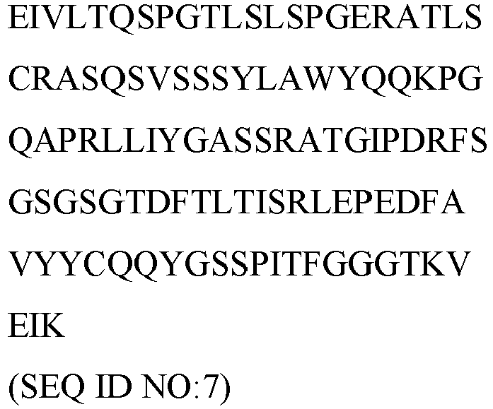
ADI-27740 (A40) 

ADI-27741 

ADI-27743 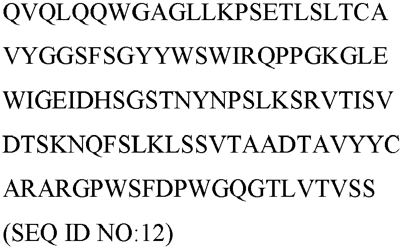
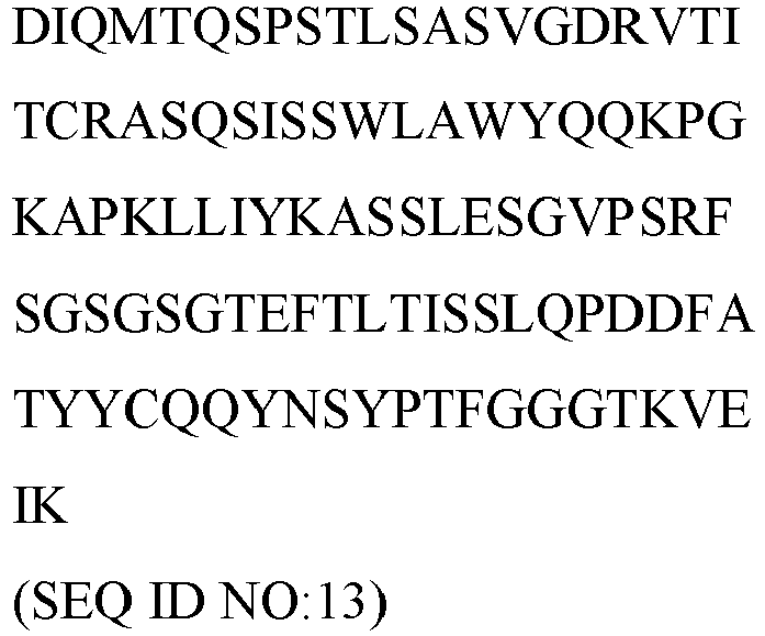
ADI-28153 


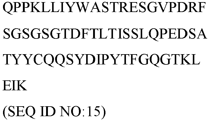
ADI-28226 (C26) 

ADI-28154 

ADI-29399 

ADI-29401 

ADI-29403 

ADI-29405 

ADI-29407 
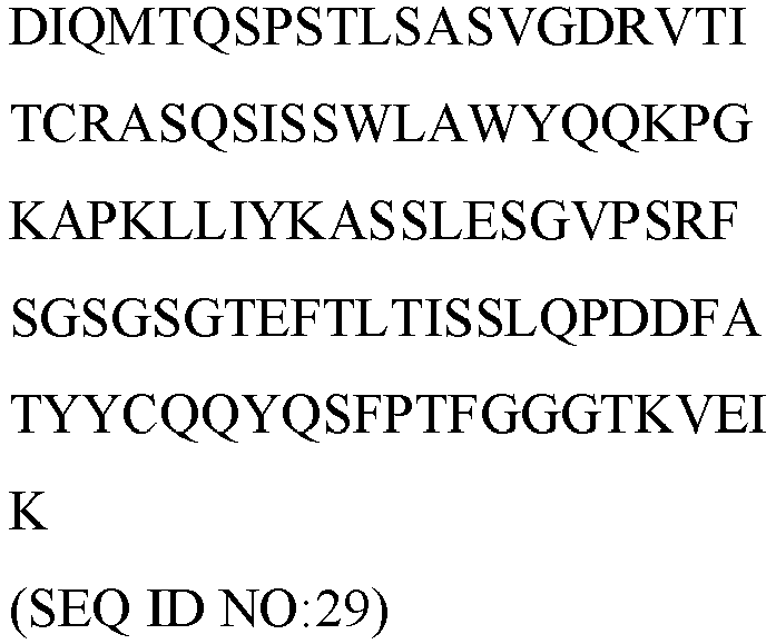
ADI-29419 

ADI-29421 


ADI-29424 

ADI-29425 

ADI-29426 

ADI-29429 

ADI-29447 (F47) 


ADI-27727 CDR1 (SEQ ID NO:45) - GTFSSYAIS (non-Kabat) or SYAIS (SEQ ID NO: 158) CDR1 (SEQ ID NO:49) - KSSQSVLYSSNNKNYLA
CDR2 (SEQ ID NO:50) - WASTRES CDR2 (SEQ ID NO:46) - GIIPIFGTANYAQKFQG CDR3 (SEQ ID NO:51) - QQYYSTPIT CDR3 (SEQ ID NO:47) - ARGDSSIRHAYYYYGMDV (non-Kabat) or GDSSIRHAYYYYGMDV (SEQ ID NO:159) ADI-29443 (F43) CDR1 (SEQ ID NO:53) - GSISSSSYYWG (non-Kabat) or SSSYYWG (SEQ ID NO:160) CDR2 CDR1 (SEQ ID NO:57) - RASQSVSRYLA
(SEQ ID NO:54) - SIYYSGSTYYNPSLKS CDR2 (SEQ ID NO:58) - DASNRAT CDR3 (SEQ ID NO:55) -ARGSDRFHPYFDY (non-Kabat) or GSDRFHPYFDY (SEQ ID NO:161) CDR3 (SEQ ID NO:59) - QQFDTWPPT ADI-29404 (F04) 
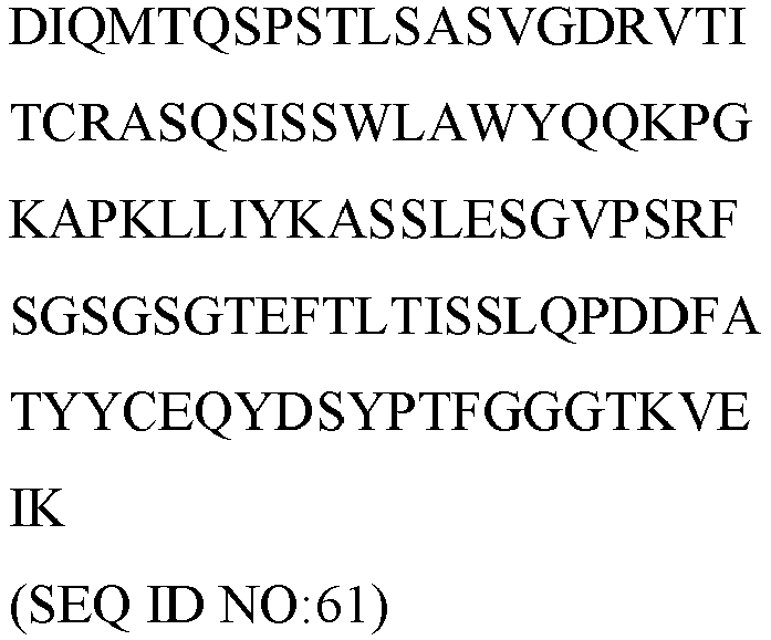
ADI-28200 CDR1 (SEQ ID NO:63) - GTFSSYAIS (non-Kabat) or SYAIS (SEQ ID NO:158) CDR2 (SEQ ID NO:64) - GIIPIFGTANYAQKFQG CDR1 (SEQ ID NO:67) - ES SQSLLNSGNQKNYLT
CDR2 (SEQ ID NO:68) -WASTRES CDR3 (SEQ ID NO:65) - ARRGRKASGSFYYYYGMDV CDR3 (SEQ ID NO:69) - QNDYSYPYT ADI-29379 (E79) CDR1 (SEQ ID NO:71) - YTFTSYYMH (non-Kabat) or SYYMIH (SEQ ID NO:162) CDR1 (SEQ ID NO:75) - RASQSVSSNLA
CDR2 (SEQ ID NO:76) - GASTRAT CDR2 (SEQ ID NO:72) - IINPSGGSTSYAQKFQG CDR3 (SEQ ID NO:77) - QQYDDWPFT CDR3 (SEQ ID NO:73) - ARGAPNYGDTTHDYYYMDV (non-Kabat) or GAPNYGDTTHDYYYMDV (SEQ ID NO: 163) ADI-29463 (F63) CDR1 (SEQ ID NO:79) - YTFTGYYMH (non-Kabat) or GYYMH (SEQ ID NO:164) CDR1 (SEQ ID NO:75) - RASQSVSSNLA
CDR2 (SEQ ID NO:76) - GASTRAT CDR2 (SEQ ID NO:80) - WINPNSGGTNYAQKFQG CDR3 (SEQ ID NO:85) - QQDDYWPPT CDR3 (SEQ ID NO:81) - ARDTGEYYDTDDHGMDV (non-Kabat) or DTGEYYDTDDHGMDV (SEQ ID NO:165) ADI-27744 (A44) CDR1 (SEQ ID NO:87) - FTFSSYAMS (non-Kabat) or SYAMS (SEQ ID NO:166) CDR1 (SEQ ID NO:91) - RASQGIDSWLA
CDR2 (SEQ ID NO:88) - AISGSGGSTYYADSVKG CDR2 (SEQ ID NO:92) - AASSLQS CDR3 (SEQ ID NO:89) - AKDGGYYDSGAGDY (non-Kabat) CDR3 (SEQ ID NO:93) - QQGVSYPRT or DGGYYDSGAGDY (SEQ ID NO: 167) ADI-27749 (A49) CDR1 (SEQ ID NO:95) - FTFSSYSMN (non-Kabat) or SYSMN CDR1 (SEQ ID NO:99) - RASQGISSWLA CDR2 (SEQ ID NO:100) -
(SEQ ID NO:168) CDR2 (SEQ ID NO:96) - AASSLQS CDR3 (SEQ ID NO:101) - SISSSSSYIYYADSVKG CDR3 (SEQ ID NO:97) - ARGAPMGAAAGWFDP (non-Kabat) or GAPMGAAAGWFDP (SEQ ID NO: 169) QQGVSFPRT ADI-29378 (E78) CDR1 (SEQ ID NO:71) - YTFTSYYMH (non-Kabat) or SYYMIH (SEQ ID NO:162) CDR1 (SEQ ID NO:107) - RASQSVSSYLA
CDR2 (SEQ ID NO:108) - DASNRAT CDR2 (SEQ ID NO:72) - IINPSGGSTSYAQKFQG CDR3 (SEQ ID NO:109) - QQSDNWPFT CDR3 (SEQ ID NO:105) - AREGAGFAYGMDYYYMDV (non-Kabat) or EGAGFAYGMDYYYMDV (SEQ ID NO:170) A49MI CDR1 (SEQ ID NO:95) - FTFSSYSMN (non-Kabat) or SYSMN (SEQ ID NO:168) CDR1 (SEQ ID NO:99) - RASQGISSWLA
CDR2 (SEQ ID NO:100) - AASSLQS CDR2 (SEQ ID NO:96) - SISSSSSYIYYADSVKG CDR3 (SEQ ID NO:101) - CDR3: (non-Kabat) ARGAPIGAAAGWFDP (SEQ ID NO:172) or GAPIGAAAGWFDP (SEQ ID NO:173) QQGVSFPRT A49MQ CDR1 (SEQ ID NO:95) - FTFSSYSMN (non-Kabat) or SYSMN (SEQ ID NO:168) CDR1 (SEQ ID NO:99) - RASQGISSWLA
CDR2 (SEQ ID NO:96) - SISSSSSYIYYADSVKG CDR2 (SEQ ID NO:100) - AASSLQS CDR3 (non-Kabat) (SEQ ID NO:175) - ARGAPQGAAAGWFDP or CDR3 (SEQ ID NO:176) - GAPQGAAAGWFDP CDR3 (SEQ ID NO:101) - QQGVSFPRT A49MI, CDR1 (SEQ ID NO:95) - FTFSSYSMN (non-Kabat) or SYSMN ID (SEQ NO:168) CDR1 (SEQ ID NO:99) - RASQGISSWLA
CDR2 (SEQ ID NO:96) - SISSSSSYIYYADSVKG CDR2 (SEQ ID NO:100) - AASSLQS CDR3 (non-Kabat) (SEQ ID NO: 178) - ARGAPLGAAAGWFDP or CDR3 (SEQ ID NO:179) - GAPLGAAAGWFDP CDR3 (SEQ ID NO:101) - QQGVSFPRT A49MF CDR1 (SEQ ID NO:95) - FTFSSYSMN (non-Kabat) or SYSMN (SEQ ID NO:168) CDR1 (SEQ ID NO:99) - RASQGISSWLA
CDR2 (SEQ ID NO:96) - SISSSSSYIYYADSVKG CDR2 (SEQ ID NO:100) - AASSLQS CDR3 (non-Kabat) (SEQ ID NO: 181) - ARGAPFGAAAGWFDP or CDR3 (SEQ ID NO:182) - GAPFGAAAGWFDP CDR3 (SEQ ID NO:101) - QQGVSFPRT A49MV CDR1 (SEQ ID NO:95) - FTFSSYSMN (non-Kabat) or SYSMN (SEQ ID NO:168) CDR1 (SEQ ID NO:99) - RASQGISSWLA
CDR2 (SEQ ID NO:100) - AASSLQS CDR2 (SEQ ID NO:96) - SISSSSSYIYYADSVKG CDR3 (SEQ ID NO:101) - QQGVSFPRT CDR3 (non-Kabat) (SEQ ID NO: 184) - ARGAPVGAAAGWFDP or CDR3 (SEQ ID NO:185) - GAPVGAAAGWFDP A49-consensus  CDR1 (SEQ ID NO:99) - RASQGISSWLA
CDR1 (SEQ ID NO:95) - FTFSSYSMN (non-Kabat) or SYSMN (SEQ ID NO:168) CDR2 (SEQ ID NO:100) - AASSLQS CDR3 (SEQ ID NO:101) - QQGVSFPRT CDR2 (SEQ ID NO:96) - SISSSSSYIYYADSVKG CDR3 (non-Kabat) (SEQ ID NO: 187) - ARGAPXGAAAGWFDP or CDR3 (SEQ ID NO:188) - GAPXGAAAGWFDP, wherein X is M, L, I, V, Q, or F - Alternatively, a heavy chain variable domain represented by SEQ ID NO: 110 can be paired with a light chain variable domain represented by SEQ ID NO:111 to form an antigen-binding site that can bind to NKG2D, as illustrated in
US 9,273,136 - SEQ ID NO: 110

- SEQ ID NO:111

- Alternatively, a heavy chain variable domain represented by SEQ ID NO:112 can be paired with a light chain variable domain represented by SEQ ID NO:113 to form an antigen-binding site that can bind to NKG2D, as illustrated in
US 7,879,985 . - SEQ ID NO:112

- SEQ ID NO:113

- The multi-specific binding proteins can bind to NKG2D-expressing cells, which include but are not limited to NK cells, γδ T cells and CD8+ αβ T cells. Upon NKG2D binding, the multi-specific binding proteins may block natural ligands, such as LTLBP6 and MICA, from binding to NKG2D and activating NK cells.
- The multi-specific binding proteins binds to cells expressing CD16, an Fc receptor on the surface of leukocytes including natural killer cells, macrophages, neutrophils, eosinophils, mast cells, and follicular dendritic cells. A protein of the present disclosure binds to NKG2D with an affinity of KD of 2 nM to 120 nM, e.g., 2 nM to 110 nM, 2 nM to 100 nM, 2 nM to 90 nM, 2 nM to 80 nM, 2 nM to 70 nM, 2 nM to 60 nM, 2 nM to 50 nM, 2 nM to 40 nM, 2 nM to 30 nM, 2 nM to 20 nM, 2 nM to 10 nM, about 15 nM, about 14 nM, about 13 nM, about 12 nM, about 11 nM, about 10 nM, about 9 nM, about 8 nM, about 7 nM, about 6 nM, about 5 nM, about 4.5 nM, about 4 nM, about 3.5 nM, about 3 nM, about 2.5 nM, about 2 nM, about 1.5 nM, about 1 nM, between about 0.5 nM to about 1 nM, about 1 nM to about 2 nM, about 2 nM to 3 nM, about 3 nM to 4 nM, about 4 nM to about 5 nM, about 5 nM to about 6 nM, about 6 nM to about 7 nM, about 7 nM to about 8 nM, about 8 nM to about 9 nM, about 9 nM to about 10 nM, about 1 nM to about 10 nM, about 2 nM to about 10 nM, about 3 nM to about 10 nM, about 4 nM to about 10 nM, about 5 nM to about 10 nM, about 6 nM to about 10 nM, about 7 nM to about 10 nM, or about 8 nM to about 10 nM. In some instances, NKG2D-binding sites bind to NKG2D with a KD of 10 to 62 nM.
- The HER2-binding site of the multi-specific binding protein disclosed herein comprises a heavy chain variable domain and a light chain variable domain fused together to from an scFv.
- Table 2 lists peptide sequences of heavy chain variable domains and light chain variable domains that, in combination, can bind to HER2.
Table 2 Clones Heavy chain variable domain amino acid sequence Light chain variable domain amino acid sequence Trastuzumab  CDR1(SEQ ID NO:119) - QDVNTAVA
CDR1(SEQ ID NO:115) - GFNIKDT CDR2 (SEQ ID NO:116) - YPTNGY CDR2 (SEQ ID NO:120) - SASFLYS CDR3 (SEQ ID NO:117) - WGGDGFYAMDY CDR3 (SEQ ID NO:121) - QQHYTTPPT Trastuzumab (VH and VL in scFv construct) 

CDR1(SEQ ID NO:115) - GFNIKDT CDR1(SEQ ID NO:119) - QDVNTAVA CDR2 (SEQ ID NO:116) - YPTNGY CDR3 (SEQ ID NO:117) - WGGDGFYAMDY CDR2 (SEQ ID NO:120) - SASFLYS CDR3 (SEQ ID NO:121) - QQHYTTPPT Trastuzumab -scFv 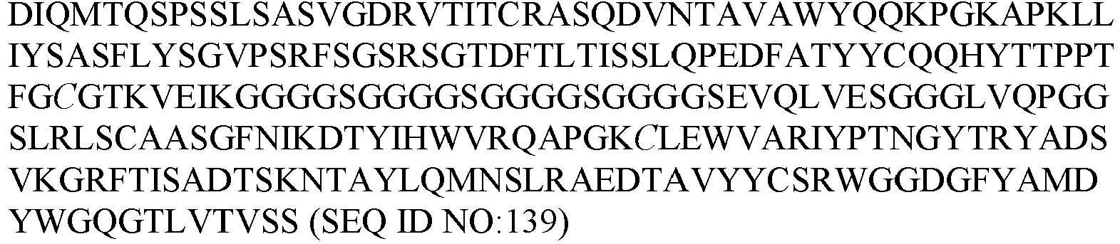
Pertuzumab CDR1 (SEQ ID NO:123) - GFTFTDY CDR1 (SEQ ID NO:127) - QDVSIGVA
CDR2 (SEQ ID NO:128) - SASYRYT CDR2 (SEQ ID NO:124) - NPNSGG CDR3 (SEQ ID NO:125) - NLGPSFYFDY CDR3 (SEQ ID NO: 129) - QQYYIYPYT Pertuzumab (VH and VL in scFv construct) CDR1 (SEQ ID NO:123) - GFTFTDY CDR1 (SEQ ID NO: 127) - QDVSIGVA
CDR2 (SEQ ID NO:124) - NPNSGG CDR2 (SEQ ID NO:128) - SASYRYT CDR3 (SEQ ID NO:125) - NLGPSFYFDY CDR3 (SEQ ID NO:129) - QQYYIYPYT Pertuzumab scFv 
MGAH22 ( US 8,802,093 )CDR1 (SEQ ID NO:131) - GFNIKDT CDR1 (SEQ ID NO:135) - QDVNTAVA
CDR2 (SEQ ID NO:136) - SASFRYT CDR2 (SEQ ID NO:132) - YPTNGY CDR3 (SEQ ID NO:133) - WGGDGFYAMDY CDR3 (SEQ ID NO:137) - QQHYTTPPT MGAH22 (VH and VL in scFv construct) CDR1 (SEQ ID NO:131) - GFNIKDT CDR1 (SEQ ID NO:135) - QDVNTAVA
CDR2 (SEQ ID NO:132) - YPTNGY CDR2 (SEQ ID NO:136) - SASFRYT CDR3 (SEQ ID NO:133) - WGGDGFYAMDY CDR3 (SEQ ID NO:137) - QQHYTTPPT MGAH22 scFv 
- Alternatively, novel antigen-binding sites that can bind to HER2 can be identified by screening for binding to the amino acid sequence defined by SEQ ID NO: 138 or a mature extracellular fragment thereof.

- The VH and VL of the scFv can be positioned so the VL is positioned N-terminal to the VH.
- The VH and VL of the scFv can be connected via a linker, e.g., a peptide linker. The peptide linker is a flexible linker. Regarding the amino acid composition of the linker, peptides are selected with properties that confer flexibility, do not interfere with the structure and function of the other domains of the proteins, and resist cleavage from proteases. For example, glycine and serine residues generally provide protease resistance. The VL is positioned N-terminal to the VH and is connected to the VH via a linker.
- The length of the linker (e.g., flexible linker) can be "long," e.g., at least 13 amino acid residues. The linker is 10-50, 10-40, 10-30, 10-25, 10-20, 15-50, 15-40, 15-30, 15-25, 15-20, 20-50, 20-40, 20-30, or 20-25 amino acid residues in length.
- Linkers disclosed herein comprise or consist of a (GS)n (SEQ ID NO:204), (GGS)n (SEQ ID NO:205), (GGGS)n (SEQ ID NO:206), (GGSG)n (SEQ ID NO:207), (GGSGG)n (SEQ ID NO:208), and (GGGGS)n (SEQ ID NO:209) sequence, wherein n is 1, 2, 3, 4, 5, 6, 7, 8, 9, 10, 11, 12, 13, 14, 15, 16, 17, 18, 19, or 20. Linkers disclosed herein comprise or consist of an amino acid sequence selected from SEQ ID NO:143, SEQ ID NO:201, SEQ ID NO:202, SEQ ID NO: 103, SEQ ID NO: 104, SEQ ID NO:83, SEQ ID NO:84, SEQ ID NO:150, SEQ ID NO: 152, and SEQ ID NO: 154, as listed in Table 3. The linker comprises or consists of an amino acid sequence SEQ ID NO: 143 as listed in Table 3. The linker is a (G4S)4 (SEQ ID NO:203) linker consisting of the sequence of SEQ ID NO:143.
Table 3 SEQ ID Amino Acid Sequence SEQ ID NO:201 GSGSGSGSGSGSGSGSGSGS SEQ ID NO:202 GGSGGSGGSGGSGGSGGSGGSGGSGGSGGS SEQ ID NO:103 
SEQ ID NO:104 
SEQ ID NO:83 
SEQ ID NO:84 
SEQ ID NO:143 GGGGSGGGGSGGGGSGGGGS SEQ ID NO:150 GGGGSGGGGSGGGGS SEQ ID NO:152 
SEQ ID NO:154 
- Within the Fc domain, CD16 binding is mediated by the hinge region and the CH2 domain. For example, within human IgG1, the interaction with CD16 is primarily focused on amino acid residues Asp 265 - Glu 269, Asn 297 - Thr 299, Ala 327 - Ile 332, Leu 234 - Ser 239, and carbohydrate residue N-acetyl-D-glucosamine in the CH2 domain (see, Sondermann et al, Nature, 406 (6793):267-273). Based on the known domains, mutations can be selected to enhance or reduce the binding affinity to CD16, such as by using phage-displayed libraries or yeast surface-displayed cDNA libraries, or can be designed based on the known three-dimensional structure of the interaction.
- The assembly of heterodimeric antibody heavy chains can be accomplished by expressing two different antibody heavy chain sequences in the same cell, which may lead to the assembly of homodimers of each antibody heavy chain as well as assembly of heterodimers. Promoting the preferential assembly of heterodimers can be accomplished by incorporating different mutations in the CH3 domain of each antibody heavy chain constant region as shown in
US13/494870 US16/028850 US11/533709 US12/875015 US13/289934 US14/773418 US12/811207 US13/866756 US14/647480 US14/830336 - In one scenario, an amino acid substitution in the first polypeptide replaces the original amino acid with a larger amino acid, selected from arginine (R), phenylalanine (F), tyrosine (Y) or tryptophan (W), and at least one amino acid substitution in the second polypeptide replaces the original amino acid(s) with a smaller amino acid(s), chosen from alanine (A), serine (S), threonine (T), or valine (V), such that the larger amino acid substitution (a protuberance) fits into the surface of the smaller amino acid substitutions (a cavity). For example, one polypeptide can incorporate a T366W substitution, and the other can incorporate three substitutions including T366S, L368A, and Y407V.
- An antibody heavy chain variable domain can be coupled to an amino acid sequence at least 90% identical to an antibody constant region, such as an IgG constant region including hinge, CH2 and CH3 domains with or without CH1 domain. The amino acid sequence of the constant region is at least 90% identical to a human antibody constant region, such as a human IgG1 constant region One or more mutations can be incorporated into the constant region as compared to human IgG1 constant region, for example at Q347, Y349, S354, K360, D399, F405, and/or K409. Exemplary substitutions include, for example, Q347R, Y349S, Y349C, K360E, D399V, F405T, and K409W.
- Disclosed herein are mutations that can be incorporated into the CH1 of a human IgG1 constant region may be at amino acid V125, F126, P127, T135, T139, A140, F170, P171, and/or V173. Mutations that can be incorporated into the Cκ of a human IgG1 constant region may be at amino acid E123, F116, S176, V163, S 174, and/or T164.
- Amino acid substitutions could be selected from the following sets of substitutions shown in Table 4.
Table 4 First Polypeptide Second Polypeptide Set 1 S364E/F405A Y349K/ T394F Set 2 S364H/D401K Y349T/ T411E Set 3 S364H/T394F Y349T/F405A Set 4 S364E/T394F Y349K/ F405A Set 5 S364E/T411E Y349K/D401K Set 6 S364D/T394F Y349K/F405A Set 7 S364H/F405A Y349T/T394F Set 8 S364K/E357Q L368D/K370S Set 9 L368D/ K370S S364K Set 10 L368E/K370S S364K Set 11 K360E/Q362E D401K Set 12 L368D/K370S S364K/E357L Set 13 K370S S364K/E357Q Set 14 F405L K409R Set 15 K409R F405L - Alternatively, amino acid substitutions could be selected from the following sets of substitutions shown in Table 5.
Table 5 First Polypeptide Second Polypeptide Set 1 K409W D399V/ F405T Set 2 Y349S E357W Set 3 K360E Q347R Set 4 K360E/K409W Q347R/D399V/ F405T Set 5 Q347E/K360E/K409W Q347R/D399V/F405T Set 6 Y349S/K409W E357W/D399V/F405T - Alternatively, amino acid substitutions could be selected from the following set of substitutions shown in Table 6.
Table 6 First Polypeptide Second Polypeptide Set 1 T366K/L351K L351D/ L368E Set 2 T366K/L351K L351D/ Y349E Set 3 T366K/L351K L351D/Y349D Set 4 T366K/L351K L351D/Y349E/ L368E Set 5 T366K/L351K L351D/Y349D/L368E Set 6 E356K/D399K K392D/K409D - Alternatively, at least one amino acid substitution in each polypeptide chain could be selected from Table 7.
Table 7 First Polypeptide Second Polypeptide L351Y, D399R, D399K, S400K, S400R, Y407A, Y407I, Y407V T366V, T366I, T366L, T366M, N390D, N390E, K392L, K392M, K392V, K392F K392D, K392E, K409F, K409W, T411D and T411E - Alternatively, at least one amino acid substitutions could be selected from the following set of substitutions in Table 8, where the position(s) indicated in the First Polypeptide column is replaced by any known negatively-charged amino acid, and the position(s) indicated in the Second Polypeptide Column is replaced by any known positively-charged amino acid.
Table 8 First Polypeptide Second Polypeptide K392, K370, K409, or K439 D399, E356, or E357 - Alternatively, at least one amino acid substitutions could be selected from the following set of in Table 9, where the position(s) indicated in the First Polypeptide column is replaced by any known positively-charged amino acid, and the position(s) indicated in the Second Polypeptide Column is replaced by any known negatively-charged amino acid.
Table 9 First Polypeptide Second Polypeptide D399, E356, or E357 K409, K439, K370, or K392 - Alternatively, amino acid substitutions could be selected from the following set in Table 10.
Table 10 First Polypeptide Second Polypeptide T350V, L351Y, F405A, and Y407V T350V, T366L, K392L, and T394W - Alternatively, or in addition, the structural stability of a hetero-multimeric protein may be increased by introducing S354C on either of the first or second polypeptide chain, and Y349C on the opposing polypeptide chain, which forms an artificial disulfide bridge within the interface of the two polypeptides.
- The amino acid sequence of one polypeptide chain of the antibody constant region may differ from the amino acid sequence of an IgG1 constant region by an S354C substitution and wherein the amino acid sequence of the other polypeptide chain of the antibody constant region differs from the amino acid sequence of an IgG1 constant region by a Y349C substitution.
- The amino acid sequence of one polypeptide chain of the antibody constant region may differ from the amino acid sequence of an IgG1 constant region by a Y349C substitution and wherein the amino acid sequence of the other polypeptide chain of the antibody constant region differs from the amino acid sequence of an IgG1 constant region by an S354C substitution.
- The amino acid sequence of one polypeptide chain of the antibody constant region may differ from the amino acid sequence of an IgG1 constant region by K360E and K409W substitutions and wherein the amino acid sequence of the other polypeptide chain of the antibody constant region differs from the amino acid sequence of an IgG1 constant region by Q347R, D399V and F405T substitutions.
- The amino acid sequence of one polypeptide chain of the antibody constant region may differ from the amino acid sequence of an IgG1 constant region by Q347R, D399V and F405T substitutions and wherein the amino acid sequence of the other polypeptide chain of the antibody constant region differs from the amino acid sequence of an IgG1 constant region by K360E and K409W substitutions.
- Certain proteins described in the present disclosure have an Fc domain, which comprises one or more mutations that reduce the ability of the Fc sequence to induce antibody-dependent cellular cytotoxicity (ADCC) and/or antibody-dependent cellular phagocytosis (ADCP). At least one mutation is located in the regions including amino acid positions 232-239, 265-270, 296-299, and 325-332 (see Want et al., Protein Cell (2018) 9(1):63-73). The mutations may include an amino acid substitution (relative to wild-type human IgG1) at one or more positions 233, 234, 235, 297, and 329. The one or more mutations may include E233P; L234A; L235A; N297A, N297Q, N297G, or N297D; and/or P329A, P329G, or P329R relative to wild-type human IgG1. The one or more mutations may include L234A and L235A relative to wild-type human IgG1. Alternatively, the one or more mutations may include L234A, L235A, and P329A relative to wild-type human IgG1. The mutation may be present on each of the two polypeptide chains of the Fc domain.
- Listed below are examples of TriNKETs comprising a HER2-binding scFv and an NKG2D-binding Fab each linked to an antibody constant region, wherein the antibody constant regions include mutations that enable heterodimerization of two Fc chains. The scFv comprises a heavy chain variable domain (VH) and a light chain variable domain (VL) derived from an anti-HER2 antibody (e.g., trastuzumab), and further comprises substitution of Cys for the amino acid residues at
position 100 of VL and position 44 of VH, thereby facilitating formation of a disulfide bridge between the VH and VL of the scFv. The VL is linked N-terminal to the VH via a (G4S)4 linker (SEQ ID NO:203), and the VH is linked N-terminal to an Fc via an Ala-Ser linker. The Ala-Ser linker is included at the elbow hinge region sequence to balance between flexibility and optimal geometry. As used herein to describe these exemplary TriNKETs, Fc includes an antibody hinge, CH2, and CH3. - Accordingly, each of the TriNKETs described below comprises the following three polypeptide chains:
- Chain A, comprising from N-terminus to C-terminus: VH of an NKG2D-binding Fab, CH1, and Fc;
- Chain B, comprising from N-terminus to C-terminus: VL of a HER2-binding scFv, (G4S)4 linker (SEQ ID NO:203), VH of the HER2-binding scFv, Ala-Ser linker, and Fc; and
- Chain C, comprising from N-terminus to C-terminus: VL of the NKG2D-binding Fab, and CL.
- The amino acid sequences of the exemplary TriNKETs are summarized in Table 11.
Table 11 TriNKET Construct NKG2D Binding Fab HER2 Binding scFv Human IgG1 Fc Chain A Chain B Chain C A49-F3'-TriNKET-Trastuzumab A49 Trastuzumab EW-RVT SEQ ID NO:141 SEQ ID NO:140 SEQ ID NO:142 A49-F3'-KiH-TriNKET-Trastuzumab A49 Trastuzumab KiH SEQ ID NO:147 SEQ ID NO:146 SEQ ID NO:142 A49-F3'-TriNKET-Pertuzumab A49 Pertuzumab EW-RVT SEQ ID NO:141 SEQ ID NO:190 SEQ ID NO:142 A49-F3'-KiH-TriNKET-Pertuzumab A49 Pertuzumab KiH SEQ ID NO:147 SEQ ID NO:191 SEQ ID NO:142 A49-F3'-TriNKET-MGAH22 A49 MGAH22 EW-RVT SEQ ID NO:141 SEQ ID NO:192 SEQ ID NO:142 A49-F3'-KiH-TriNKET-MGAH22 A49 MGAH22 KiH SEQ ID NO:147 SEQ ID NO:193 SEQ ID NO:142 A49MI-F3'-TriNKET-Trastuzumab A49MI Trastuzumab EW-RVT SEQ ID NO:145 SEQ ID NO:140 SEQ ID NO:142 A49MI-F3'-KiH-TriNKET-Trastuzumab A49MI Trastuzumab KiH SEQ ID NO:194 SEQ ID NO:146 SEQ ID NO:142 A49MI-F3'-TriNKET-Pertuzumab A49MI Pertuzumab EW-RVT SEQ ID NO:145 SEQ ID NO:190 SEQ ID NO:142 A49MI-F3'-KiH-TriNKET-Pertuzumab A49MI Pertuzumab KiH SEQ ID NO:194 SEQ ID NO:191 SEQ ID NO:142 A49MI-F3'-TriNKET-MGAH22 A49MI MGAH22 EW-RVT SEQ ID NO:145 SEQ ID NO:192 SEQ ID NO:142 A49MI-F3'-KiH-TriNKET-MGAH22 A49MI MGAH22 KiH SEQ ID NO:194 SEQ ID NO:193 SEQ ID NO:142 A44-F3'-TriNKET-Trastuzumab A44 Trastuzumab EW-RVT SEQ ID NO:155 SEQ ID NO:140 SEQ ID NO:149 A44-F3'-KiH-TriNKET-Trastuzumab A44 Trastuzumab KiH SEQ ID NO:148 SEQ ID NO:146 SEQ ID NO:149 A44-F3'-TriNKET-Pertuzumab A44 Pertuzumab EW-RVT SEQ ID NO:155 SEQ ID NO:190 SEQ ID NO:149 A44-F3'-KiH-TriNKET-Pertuzumab A44 Pertuzumab KiH SEQ ID NO:148 SEQ ID NO:191 SEQ ID NO:149 A44-F3'-TriNKET-MGAH22 A44 MGAH22 EW-RVT SEQ ID NO:155 SEQ ID NO:192 SEQ ID NO:149 A44-F3'-KiH-TriNKET-MGAH22 A44 MGAH22 KiH SEQ ID NO:148 SEQ ID NO:193 SEQ ID NO:149 - , The multi-specific binding protein of the present disclosure comprises a first polypeptide chain, a second polypeptide chain, and a third polypeptide chain, wherein the first, second, and third polypeptide chains comprise the amino acid sequences of Chain A, Chain B, and Chain C, respectively, of a TriNKET disclosed in Table 11. The first, second, and third polypeptide chains may consist of the amino acid sequences of Chain A, Chain B, and Chain C, respectively, of a TriNKET disclosed in Table 11.
- In this disclosure the Fc domain linked to the NKG2D-binding Fab fragment may comprise the mutations of Q347R, D399V, and F405T, and the Fc domain linked to the HER2 scFv may comprise matching mutations K360E and K409W for forming a heterodimer. Described but not claimed in this disclosure, the Fc domain linked to the NKG2D-binding Fab fragment may comprise knob mutations T366S, L368A, and Y407V, and the Fc domain linked to the HER2-binding scFv may comprise a "hole" mutation T366W. In this disclosure the Fc domain linked to the NKG2D-binding Fab fragment may include an S354C substitution in the CH3 domain, which forms a disulfide bond with a Y349C substitution on the Fc linked to the HER2-binding scFv.
- Specific TriNKETs and their polypeptide chains are described in more detail below. In the amino acid sequences, (G4S)4 (SEQ ID NO:203) and Ala-Ser linkers are bold-underlined; Cys residues in scFv that form disulfide bridges are bold-italic-underlined; Fc heterodimerization mutations are bold-underlined; and CDR sequences under Kabat are underlined.
- For example, a TriNKET of the present disclosure is A49-F3'-TriNKET-Trastuzumab. A49-F3'-TriNKET-Trastuzumab includes a single-chain variable fragment (scFv) (SEQ ID NO: 139) derived from trastuzumab that binds HER2, linked via a hinge comprising Ala-Ser to an Fc domain; and an NKG2D-binding Fab fragment derived from A49 including a heavy chain portion comprising a heavy chain variable domain (SEQ ID NO:94) and a CH1 domain, and a light chain portion comprising a light chain variable domain (SEQ ID NO:98) and a light chain constant domain, wherein the heavy chain variable domain is connected to the CH1 domain, and the CH1 domain is connected to the Fc domain. A49-F3'-TriNKET-Trastuzumab includes three polypeptides, having the sequences of SEQ ID NO: 140, SEQ ID NO:141, and SEQ ID NO:142.
- SEQ ID NO: 140 represents the full sequence of the HER2-binding scFv linked to an Fc domain via a hinge comprising Ala-Ser (scFv-Fc). The Fc domain linked to the scFv includes Q347R, D399V, and F405T substitutions for heterodimerization and an S354C substitution for forming a disulfide bond with a Y349C substitution in SEQ ID NO: 141 as described below. The scFv (SEQ ID NO: 139) includes a heavy chain variable domain of trastuzumab connected to the N-terminus of a light chain variable domain of trastuzumab via a (G4S)4 linker (SEQ ID NO:203), the scFv represented as VL-(G4S)4-VH ("(G4S)4" is represented by SEQ ID NO:203 or SEQ ID NO:143). The heavy and the light variable domains of the scFv are also connected through a disulfide bridge between C100 of VL and C44 of VH, as a result of Q100C and G44C substitutions in the VL and VH, respectively.
- Trastuzumab scFv

- Trastuzumab scFv-Fc (RV1)

- AS

- SEQ ID NO: 141 represents the heavy chain portion of the Fab fragment, which comprises a heavy chain variable domain (SEQ ID NO:94) of an NKG2D-binding site and a CH1 domain, connected to an Fc domain. The Fc domain in SEQ ID NO: 141 includes a Y349C substitution in the CH3 domain, which forms a disulfide bond with an S354C substitution on the Fc linked to the HER2-binding scFv (SEQ ID NO: 140). In SEQ ID NO: 141, the Fc domain also includes K360E and K409W substitutions for heterodimerization with the Fc in SEQ ID NO: 140.
- A49 VH

- A49 VH-CHI-Fc (EW)

- SEQ ID NO: 142 represents the light chain portion of the Fab fragment comprising a light chain variable domain (SEQ ID NO:98) of an NKG2D-binding site and a light chain constant domain.
- A49 VL

- A49 VL-LC

- Another TriNKET of the present disclosure is A49MI-F3'-TriNKET-Trastuzumab. A49MI-F3'-TriNKET-Trastuzumab includes the same Her2-binding scFv (SEQ ID NO: 139) as in A49-F3'-TriNKET-Trastuzumab linked via a hinge comprising Ala-Ser to an Fc domain; and an NKG2D-binding Fab fragment derived from A49MI including a heavy chain portion comprising a heavy chain variable domain (SEQ ID NO: 144) and a CH1 domain, and a light chain portion comprising a light chain variable domain (SEQ ID NO:98) and a light chain constant domain, wherein the heavy chain variable domain is connected to the CH1 domain, and the CH1 domain is connected to the Fc domain. A49MI-F3'-TriNKET-Trastuzumab includes three polypeptides, having the sequences of SEQ ID NO: 140 (as in A49-F3'-TriNKET-Trastuzumab), SEQ ID NO: 145, and SEQ ID NO: 142 (as in A49-F3'-TriNKET-Trastuzumab).
- SEQ ID NO: 145 represents a heavy chain portion of the Fab fragment, which comprises a heavy chain variable domain (SEQ ID NO: 144) of an NKG2D-binding site and a CH1 domain, connected to an Fc domain. In SEQ ID NO: 144, wherein a methionine in the CDR3 of SEQ ID NO:94 has been substituted by isoleucine (M → I substitution; shown within a third bracket [] in SEQ ID NO: 144 and SEQ ID NO: 145). The Fc domain in SEQ ID NO: 145 includes a Y349C substitution in the CH3 domain, which forms a disulfide bond with an S354C substitution in the Fc linked to the HER2-binding scFv (SEQ ID NO: 140). In SEQ ID NO: 145, the Fc domain also includes K360E and K409W substitutions.
- A49MI VH

- A49MI VH-CHI-Fc (EW)

- Another TriNKET of the present disclosure is A49-F3'-KiH-TriNKET-Trastuzumab. KiH refers to the knobs-into-holes (KiH) Fc technology, which involves engineering of the CH3 domains to create either a "knob" or a "hole" in each heavy chain to promote heterodimerization. The concept behind the KiH Fc technology was to introduce a "knob" in one CH3 domain (CH3A) by substitution of a small residue with a bulky one (e.g., T366WCH3A in EU numbering). To accommodate the "knob," a complementary "hole" surface was created on the other CH3 domain (CH3B) by replacing the closest neighboring residues to the knob with smaller ones (e.g., T366S/L368A/Y407VCH3B). The "hole" mutation was optimized by structured-guided phage library screening (Atwell S, Ridgway JB, Wells JA, Carter P., Stable heterodimers from remodeling the domain interface of a homodimer using a phage display library, J. Mol. Biol. (1997) 270(1):26-35). X-ray crystal structures of KiH Fc variants (Elliott JM, Ultsch M, Lee J, Tong R, Takeda K, Spiess C, et al., Antiparallel conformation of knob and hole a glycosylated half-antibody homodimers is mediated by a CH2-CH3 hydrophobic interaction. J. Mol. Biol. (2014) 426(9): 1947-57; Mimoto F, Kadono S, Katada H, Igawa T, Kamikawa T, Hattori K. Crystal structure of a novel asymmetrically engineered Fc variant with improved affinity for FcyRs. Mol. Immunol. (2014) 58(1):132-8) demonstrated that heterodimerization is thermodynamically favored by hydrophobic interactions driven by steric complementarity at the inter-CH3 domain core interface, whereas the knob-knob and the hole-hole interfaces do not favor homodimerization owing to steric hindrance and disruption of the favorable interactions, respectively.
- A49-F3'-KiH-TriNKET-Trastuzumab includes the same Her2-binding scFv (SEQ ID NO: 139) as in A49-F3'-TriNKET-Trastuzumab linked via a hinge comprising Ala-Ser to an Fc domain comprising the "hole" substitutions of T366S, L368A, and Y407V; and the same NKG2D-binding Fab fragment as in A49-F3'-TriNKET-Trastuzumab, the CH1 domain of which is connected to an Fc domain comprising the "knob" substitution of T366W. A49-F3'-KiH-TriNKET-Trastuzumab includes three polypeptides, having the sequences of SEQ ID NO: 146, SEQ ID NO: 147, and SEQ ID NO: 142 (as in A49-F3'-TriNKET-Trastuzumab).
- SEQ ID NO: 146 represents the full sequence of the HER2-binding scFv (SEQ ID NO: 139) linked to an Fc domain via a hinge comprising Ala-Ser (scFv-Fc). The Fc domain linked to the scFv includes T366S, L368A, and Y407V substitutions for heterodimerization and an S354C substitution for forming a disulfide bond with a Y349C substitution in SEQ ID NO: 147 as described below.
Trastuzumab scFv-Fc (KiH)
- SEQ ID NO: 147 represents the heavy chain portion of a Fab fragment, which comprises a heavy chain variable domain (SEQ ID NO:94) of an NKG2D-binding site derived from A49 and a CH1 domain, connected to an Fc domain. The Fc domain in SEQ ID NO: 147 includes an S354C substitution, which forms a disulfide bond with a Y349C substitution in the CH3 domain of the Fc linked to the HER2-binding scFv (SEQ ID NO: 146). In SEQ ID NO: 147, the Fc domain also includes a T366W substitution.
-


- Another TriNKET of the present disclosure is A49MI-F3'-KiH-TriNKET-Trastuzumab. A49MI-F3'-KiH-TriNKET-Trastuzumab includes the same Her2-binding scFv (SEQ ID NO: 139) as in A49-F3'-TriNKET-Trastuzumab linked via a hinge comprising Ala-Ser to an Fc domain comprising the "hole" substitutions of T366S, L368A, and Y407V; and the same NKG2D-binding Fab fragment as in A49MI-F3'-TriNKET-Trastuzumab, the CH1 domain of which is connected to an Fc domain comprising the "knob" substitution of T366W. A49MI-F3'-KiH-TriNKET-Trastuzumab includes three polypeptides, having the sequences of SEQ ID NO: 146 (as in A49-F3'-KiH-TriNKET-Trastuzumab), SEQ ID NO: 194, and SEQ ID NO: 142 (as in A49-F3'-TriNKET-Trastuzumab).
- SEQ ID NO: 194 represents the heavy chain portion of a Fab fragment, which comprises a heavy chain variable domain (SEQ ID NO: 144) of an NKG2D-binding site derived from A49MI and a CH1 domain, connected to an Fc domain. The Fc domain in SEQ ID NO: 194 includes an S354C substitution, which forms a disulfide bond with a Y349C substitution in the CH3 domain of the Fc linked to the HER2-binding scFv (SEQ ID NO: 146). In SEQ ID NO: 194, the Fc domain also includes a T366W substitution.
-

- Another exemplary TriNKET of the present disclosure is A44-F3'-TriNKET-Trastuzumab. A44-F3'-TriNKET-Trastuzumab includes the same Her2-binding scFv (SEQ ID NO: 139) as in A49-F3'-TriNKET-Trastuzumab linked via a hinge comprising Ala-Ser to an Fc domain; and an NKG2D-binding Fab fragment derived from A44 including a heavy chain portion comprising a heavy chain variable domain (SEQ ID NO:86) and a CH1 domain, and a light chain portion comprising a light chain variable domain (SEQ ID NO:90) and a light chain constant domain, wherein the heavy chain variable domain is connected to the CH1 domain, and the CH1 domain is connected to the Fc domain. A44-F3'-TriNKET-Trastuzumab includes three polypeptides, having the sequences of SEQ ID NO:140 (as in A49-F3'-TriNKET-Trastuzumab), SEQ ID NO:155, and SEQ ID NO:149.
- SEQ ID NO: 155 represents a heavy chain variable domain (SEQ ID NO:86) of an NKG2D-binding site derived from A44, connected to an Fc domain. The Fc domain in SEQ ID NO: 155 includes a Y349C substitution in the CH3 domain, which forms a disulfide bond with an S354C substitution on the Fc linked to the HER2-binding scFv (SEQ ID NO:140). In SEQ ID NO: 155, the Fc domain also includes K360E and K409W substitutions.
- A44 YH

- A44 VH-CHI-Fc (EW)

- SEQ ID NO: 149 represents the light chain portion of the Fab fragment comprising a light chain variable domain (SEQ ID NO:90) of an NKG2D-binding site and a light chain constant domain.
- A44 VL

- A44 VL-CL

- Another exemplary TriNKET of the present disclosure is A44-F3'-KiH-TriNKET-Trastuzumab. A44-F3'-KiH-TriNKET-Trastuzumab includes the same Her2-binding scFv (SEQ ID NO: 139) as in A49-F3'-TriNKET-Trastuzumab linked via a hinge comprising Ala-Ser to an Fc domain comprising the "hole" substitutions of T366S, L368A, and Y407V; and the same NKG2D-binding Fab fragment as in A44-F3'-TriNKET-Trastuzumab, the CH1 domain of which is connected to an Fc domain comprising the "knob" substitution of T366W. A44-F3'-KiH-TriNKET-Trastuzumab includes three polypeptides, having the sequences of SEQ ID NO: 146 (as in A49-F3'-KiH-TriNKET-Trastuzumab), SEQ ID NO: 148, and SEQ ID NO: 149 (as in A44-F3'-TriNKET-Trastuzumab).
- SEQ ID NO: 148 represents a heavy chain variable domain (SEQ ID NO:86) of an NKG2D-binding site derived from A44, connected to an Fc domain. The Fc domain in SEQ ID NO: 148 includes a Y349C substitution in the CH3 domain, which forms a disulfide bond with an S354C substitution on the Fc linked to the HER2-binding scFv (SEQ ID NO: 146). In SEQ ID NO: 148, the Fc domain also includes a T366W substitution.
-

- Another TriNKET of the present disclosure is A49-F3'-TriNKET-Pertuzumab. A49-F3'-TriNKET-Pertuzumab includes an scFv (SEQ ID NO: 189) derived from pertuzumab that binds HER2, linked via a hinge comprising Ala-Ser to an Fc domain; and the same NKG2D-binding Fab fragment as in A49-F3'-TriNKET-Trastuzumab, the CH1 domain of which is connected to an Fc domain. The Fc domain linked to the scFv includes Q347R, D399V, and F405T substitutions, and the Fc domain linked to the Fab fragment includes K360E and K409W substitutions. A49-F3'-TriNKET-Pertuzumab includes three polypeptides, having the sequences of SEQ ID NO: 190, SEQ ID NO: 141 (as in A49-F3'-TriNKET-Trastuzumab), and SEQ ID NO: 142 (as in A49-F3'-TriNKET-Trastuzumab).
- SEQ ID NO: 190 represents the full sequence of the HER2-binding scFv linked to an Fc domain via a hinge comprising Ala-Ser (scFv-Fc). The Fc domain linked to the scFv includes Q347R, D399V, and F405T substitutions for heterodimerization and an S354C substitution for forming a disulfide bond with a Y349C substitution in SEQ ID NO: 141 as described above. The scFv (SEQ ID NO: 189) includes a heavy chain variable domain of pertuzumab connected to the N-terminus of a light chain variable domain of pertuzumab via a (G4S)4 linker (SEQ ID NO:203), the scFv represented as VL-(G4S)4-VH ("(G4S)4" is represented by SEQ ID NO:203 or SEQ ID NO: 143). The heavy and the light variable domains of the scFv are also connected through a disulfide bridge between C100 of VL and C44 of VH, as a result of Q100C and G44C substitutions in the VL and VH, respectively.
- Pertuzumab scFv

- Pertuzumab scFv-Fc

- AS

- Another exemplary TriNKET of the present disclosure is A49MI-F3'-TriNKET-Pertuzumab. A49MI-F3'-TriNKET-Pertuzumab includes the same Her2-binding scFv (SEQ ID NO: 189) as in A49-F3'-TriNKET-Pertuzumab linked via a hinge comprising Ala-Ser to an Fc domain; and the same NKG2D-binding Fab fragment as in A49MI-F3'-TriNKET-Trastuzumab, the CH1 domain of which is connected to an Fc domain. The Fc domain linked to the scFv includes Q347R, D399V, and F405T substitutions, and the Fc domain linked to the Fab fragment includes K360E and K409W substitutions. A49MI-F3'-TriNKET-Pertuzumab includes three polypeptides, having the sequences of SEQ ID NO: 190 (as in A49-F3'-KiH-TriNKET-Pertuzumab), SEQ ID NO: 145 (as in A49MI-F3'-TriNKET-Trastuzumab), and SEQ ID NO: 142 (as in A49-F3'-TriNKET-Trastuzumab).
- Another exemplary TriNKET of the present disclosure is A49-F3'-KiH-TriNKET-Pertuzumab. A49-F3'-KiH-TriNKET-Pertuzumab includes the same Her2-binding scFv (SEQ ID NO: 189) as in A49-F3'-TriNKET-Pertuzumab linked via a hinge comprising Ala-Ser to an Fc domain; and the same NKG2D-binding Fab fragment as in A49-F3'-TriNKET-Trastuzumab, the CH1 domain of which is connected to an Fc domain. The Fc domain linked to the scFv includes the "hole" substitutions of T366S, L368A, and Y407V, and the Fc domain linked to the Fab fragment includes the "knob" substitution of T366W. A49-F3'-KiH-TriNKET-Pertuzumab includes three polypeptides, having the sequences of SEQ ID NO: 191, SEQ ID NO: 147 (as in A49-F3'-KiH-TriNKET-Trastuzumab), and SEQ ID NO: 142 (as in A49-F3'-TriNKET-Trastuzumab).
- SEQ ID NO: 191 represents the full sequence of the HER2-binding scFv (SEQ ID NO: 189) linked to an Fc domain via a hinge comprising Ala-Ser (scFv-Fc). The Fc domain linked to the scFv includes T366S, L368A, and Y407V substitutions for heterodimerization and an S354C substitution for forming a disulfide bond with a Y349C substitution in SEQ ID NO: 191 as described above.
- Pertuzumab scFv-Fc (KiH)

- AS

- Another exemplary TriNKET of the present disclosure is A49MI-F3'-KiH-TriNKET-Pertuzumab. A49MI-F3'-KiH-TriNKET-Pertuzumab includes the same Her2-binding scFv (SEQ ID NO: 189) as in A49-F3'-TriNKET-Pertuzumab linked via a hinge comprising Ala-Ser to an Fc domain; and the same NKG2D-binding Fab fragment as in A49MI-F3'-TriNKET-Trastuzumab, the CH1 domain of which is connected to an Fc domain. The Fc domain linked to the scFv includes the "hole" substitutions of T366S, L368A, and Y407V, and the Fc domain linked to the Fab fragment includes the "knob" substitution of T366W. A49MI-F3'-KiH-TriNKET-Pertuzumab includes three polypeptides, having the sequences of SEQ ID NO: 191 (as in A49-F3'-KiH-TriNKET-Pertuzumab), SEQ ID NO: 194 (as in A49MI-F3'-KiH-TriNKET-Trastuzumab), and SEQ ID NO:142 (as in A49-F3'-TriNKET-Trastuzumab).
- Another exemplary TriNKET of the present disclosure is A44-F3'-TriNKET-Pertuzumab. A44-F3'-TriNKET-Pertuzumab includes the same Her2-binding scFv (SEQ ID NO: 189) as in A49-F3'-TriNKET-Pertuzumab linked via a hinge comprising Ala-Ser to an Fc domain; and the same NKG2D-binding Fab fragment as in A44-F3'-TriNKET-Trastuzumab, the CH1 domain of which is connected to an Fc domain. The Fc domain linked to the scFv includes Q347R, D399V, and F405T substitutions, and the Fc domain linked to the Fab fragment includes K360E and K409W substitutions. A44-F3'-TriNKET-Pertuzumab includes three polypeptides, having the sequences of SEQ ID NO: 190 (as in A49-F3'-KiH-TriNKET-Pertuzumab), SEQ ID NO:155 (as in A44-F3'-TriNKET-Trastuzumab), and SEQ ID NO: 149 (as in A44-F3'-TriNKET-Trastuzumab).
- Another exemplary TriNKET of the present disclosure is A44-F3'-KiH-TriNKET-Pertuzumab. A44-F3'-KiH-TriNKET-Pertuzumab includes the same Her2-binding scFv (SEQ ID NO: 189) as in A49-F3'-TriNKET-Pertuzumab linked via a hinge comprising Ala-Ser to an Fc domain; and the same NKG2D-binding Fab fragment as in A44-F3'-TriNKET-Trastuzumab, the CH1 domain of which is connected to an Fc domain. The Fc domain linked to the scFv includes the "hole" substitutions of T366S, L368A, and Y407V, and the Fc domain linked to the Fab fragment includes the "knob" substitution of T366W. A44-F3'-KiH-TriNKET-Pertuzumab includes three polypeptides, having the sequences of SEQ ID NO: 191 (as in A49-F3'-KiH-TriNKET-Pertuzumab), SEQ ID NO: 148 (as in A44-F3'-KiH-TriNKET-Trastuzumab), and SEQ ID NO: 149 (as in A44-F3'-TriNKET-Trastuzumab).
- Another TriNKET of the present disclosure is A49-F3'-TriNKET-MGAH22. A49-F3'-TriNKET-MGAH22 includes an scFv (SEQ ID NO: 171) derived from MGAH22 that binds HER2, linked via a hinge comprising Ala-Ser to an Fc domain; and the same NKG2D-binding Fab fragment as in A49-F3'-TriNKET-Trastuzumab, the CH1 domain of which is connected to an Fc domain. The Fc domain linked to the scFv includes Q347R, D399V, and F405T substitutions, and the Fc domain linked to the Fab fragment includes K360E and K409W substitutions. A49-F3'-TriNKET-MGAH22 includes three polypeptides, having the sequences of SEQ ID NO: 192, SEQ ID NO: 141 (as in A49-F3'-TriNKET-Trastuzumab), and SEQ ID NO: 142 (as in A49-F3'-TriNKET-Trastuzumab).
- SEQ ID NO: 192 represents the full sequence of the HER2-binding scFv linked to an Fc domain via a hinge comprising Ala-Ser (scFv-Fc). The Fc domain linked to the scFv includes Q347R, D399V, and F405T substitutions for heterodimerization and an S354C substitution for forming a disulfide bond with a Y349C substitution in SEQ ID NO: 141 as described above. The scFv (SEQ ID NO: 171) includes a heavy chain variable domain of pertuzumab connected to the N-terminus of a light chain variable domain of pertuzumab via a (G4S)4 linker (SEQ ID NO:203), the scFv represented as VL-(G4S)4-VH ("(G4S)4" is represented by SEQ ID NO:203 or SEQ ID NO: 143). The heavy and the light variable domains of the scFv are also connected through a disulfide bridge between C100 of VL and C44 of VH, as a result of G100C and G44C substitutions in the VL and VH, respectively.
- MGAH22 scFv

- MGAH22 scFv-Fc

- AS

- Another TriNKET of the present disclosure is A49MI-F3'-TriNKET-MGAH22. A49MI-F3'-TriNKET-MGAH22 includes the same Her2-binding scFv (SEQ ID NO: 171) as in A49-F3'-TriNKET-MGAH22 linked via a hinge comprising Ala-Ser to an Fc domain; and the same NKG2D-binding Fab fragment as in A49MI-F3'-TriNKET-Trastuzumab, the CH1 domain of which is connected to an Fc domain. The Fc domain linked to the scFv includes Q347R, D399V, and F405T substitutions, and the Fc domain linked to the Fab fragment includes K360E and K409W substitutions. A49MI-F3'-KiH-TriNKET-MGAH22 includes three polypeptides, having the sequences of SEQ ID NO: 192 (as in A49-F3'-TriNKET-MGAH22), SEQ ID NO: 145 (as in A49MI-F3'-TriNKET-Trastuzumab), and SEQ ID NO: 142 (as in A49-F3'-TriNKET-Trastuzumab).
- Another TriNKET of the present disclosure is A49-F3'-KiH-TriNKET-MGAH22. A49-F3'-KiH-TriNKET-MGAH22 includes the same Her2-binding scFv (SEQ ID NO: 171) as in A49-F3'-TriNKET-MGAH22 linked via a hinge comprising Ala-Ser to an Fc domain; and the same NKG2D-binding Fab fragment as in A49-F3'-TriNKET-Trastuzumab, the CH1 domain of which is connected to an Fc domain. The Fc domain linked to the scFv includes the "hole" substitutions of T366S, L368A, and Y407V, and the Fc domain linked to the Fab fragment includes the "knob" substitution of T366W. A49-F3'-KiH-TriNKET-MGAH22 includes three polypeptides, having the sequences of SEQ ID NO:193, SEQ ID NO:147 (as in A49-F3'-KiH-TriNKET-Trastuzumab), and SEQ ID NO:142 (as in A49-F3'-TriNKET-Trastuzumab).
- SEQ ID NO: 193 represents the full sequence of the HER2-binding scFv (SEQ ID NO: 171) linked to an Fc domain via a hinge comprising Ala-Ser (scFv-Fc). The Fc domain linked to the scFv includes T366S, L368A, and Y407V substitutions for heterodimerization and an S354C substitution for forming a disulfide bond with a Y349C substitution in SEQ ID NO: 147 as described above.
- MGAH22 scFv-Fc (KiH)

- AS

- Another exemplary TriNKET of the present disclosure is A49MI-F3'-KiH-TriNKET-MGAH22. A49MI-F3'-KiH-TriNKET-MGAH22 includes the same Her2-binding scFv (SEQ ID NO: 171) as in A49-F3'-TriNKET-MGAH22 linked via a hinge comprising Ala-Ser to an Fc domain; and the same NKG2D-binding Fab fragment as in A49MI-F3'-TriNKET-Trastuzumab, the CH1 domain of which is connected to an Fc domain. The Fc domain linked to the scFv includes the "hole" substitutions of T366S, L368A, and Y407V, and the Fc domain linked to the Fab fragment includes the "knob" substitution of T366W. A49MI-F3'-KiH-TriNKET-MGAH22 includes three polypeptides, having the sequences of SEQ ID NO: 193 (as in A49-F3'-KiH-TriNKET-MGAH22), SEQ ID NO:194 (as in A49MI-F3'-KiH-TriNKET-Trastuzumab), and SEQ ID NO: 142 (as in A49-F3'-TriNKET-Trastuzumab).
- Another exemplary TriNKET of the present disclosure is A44-F3'-TriNKET-MGAH22. A44-F3'-TriNKET-MGAH22 includes the same Her2-binding scFv (SEQ ID NO: 171) as in A49-F3'-TriNKET-MGAH22 linked via a hinge comprising Ala-Ser to an Fc domain; and the same NKG2D-binding Fab fragment as in A44-F3'-TriNKET-Trastuzumab, the CH1 domain of which is connected to an Fc domain. The Fc domain linked to the scFv includes Q347R, D399V, and F405T substitutions, and the Fc domain linked to the Fab fragment includes K360E and K409W substitutions. A44-F3'-TriNKET-MGAH22 includes three polypeptides, having the sequences of SEQ ID NO: 192 (as in A49-F3'-TriNKET-MGAH22), SEQ ID NO: 155 (as in A44-F3'-TriNKET-Trastuzumab), and SEQ ID NO: 149 (as in A44-F3'-TriNKET-Trastuzumab).
- Another exemplary TriNKET of the present disclosure is A44-F3'-KiH-TriNKET-MGAH22. A44-F3'-KiH-TriNKET-MGAH22 includes the same Her2-binding scFv (SEQ ID NO: 171) as in A49-F3'-TriNKET-MGAH22 linked via a hinge comprising Ala-Ser to an Fc domain; and the same NKG2D-binding Fab fragment as in A44-F3'-TriNKET-Trastuzumab, the CH1 domain of which is connected to an Fc domain. The Fc domain linked to the scFv includes the "hole" substitutions of T366S, L368A, and Y407V, and the Fc domain linked to the Fab fragment includes the "knob" substitution of T366W. A44-F3'-KiH-TriNKET-MGAH22 includes three polypeptides, having the sequences of SEQ ID NO: 193 (as in A49-F3'-KiH-TriNKET-MGAH22), SEQ ID NO: 148 (as in A44-F3'-KiH-TriNKET-Trastuzumab), and SEQ ID NO: 149 (as in A44-F3'-TriNKET-Trastuzumab).
- A TriNKET of the present disclosure may be identical to one of the exemplary TriNKETs described above that includes the EW-RVT Fc mutations, except that the Fc domain linked to the NKG2D-binding Fab fragment comprises the substitutions of Q347R, D399V, and F405T, and the Fc domain linked to the HER2-binding scFv comprises matching substitutions K360E and K409W for forming a heterodimer. A TriNKET of the present disclosure may be identical to one of the exemplary TriNKETs described above that includes the KiH Fc mutations, except that the Fc domain linked to the NKG2D-binding Fab fragment comprises the "hole" substitutions of T366S, L368A, and Y407V, and the Fc domain linked to the HER2-binding scFv comprises the "knob" substitution of T366W for forming a heterodimer.
- A TriNKET of the present disclosure may be identical to one of the exemplary TriNKETs described above, except that the Fc domain linked to the NKG2D-binding Fab fragment includes an S354C substitution in the CH3 domain, and the Fc domain linked to the HER2-binding scFv includes a matching Y349C substitution in the CH3 domain for forming a disulfide bond.
- A skilled person in the art would appreciate that during production and/or storage of proteins, N-terminal glutamate (E) or glutamine (Q) can be cyclized to form a lactam (e.g., spontaneously or catalyzed by an enzyme present during production and/or storage). Accordingly, where the N-terminal residue of an amino acid sequence of a polypeptide is E or Q, a corresponding amino acid sequence with the E or Q replaced with pyroglutamate is also contemplated herein.
- A skilled person in the art would also appreciate that during protein production and/or storage, the C-terminal lysine (K) of a protein can be removed (e.g., spontaneously or catalyzed by an enzyme present during production and/or storage). Such removal of K is often observed with proteins that comprise an Fc domain at its C-terminus. Accordingly, where the C-terminal residue of an amino acid sequence of a polypeptide (e.g., an Fc domain sequence) is K, a corresponding amino acid sequence with the K removed is also contemplated herein.
- The multi-specific proteins described above can be made using recombinant DNA technology well known to a skilled person in the art. For example, a first nucleic acid sequence encoding the first immunoglobulin heavy chain can be cloned into a first expression vector; a second nucleic acid sequence encoding the second immunoglobulin heavy chain can be cloned into a second expression vector; a third nucleic acid sequence encoding the immunoglobulin light chain can be cloned into a third expression vector; and the first, second, and third expression vectors can be stably transfected together into host cells to produce the multimeric proteins.
- To achieve the highest yield of the multi-specific protein, different ratios of the first, second, and third expression vector can be explored to determine the optimal ratio for transfection into the host cells. After transfection, single clones can be isolated for cell bank generation using methods known in the art, such as limited dilution, ELISA, FACS, microscopy, or Clonepix.
- Clones can be cultured under conditions suitable for bio-reactor scale-up and maintained expression of the multi-specific protein. The multi-specific proteins can be isolated and purified using methods known in the art including centrifugation, depth filtration, cell lysis, homogenization, freeze-thawing, affinity purification, gel filtration, ion exchange chromatography, hydrophobic interaction exchange chromatography, and mixed-mode chromatography.
- A multi-specific binding protein of the present disclosure, e.g., A49-F3'-TriNKET-Trastuzumab, which include an NKG2D-binding Fab fragment and a HER2-binding scFv domain, bind to cells expressing low levels of HER2 at a level higher than a monoclonal antibody having the same HER2-binding domain. For example, the multi-specific binding proteins that include an NKG2D-binding Fab domain and a HER2-binding scFv domain derived from trastuzumab, e.g., A49-F3'-TriNKET-Trastuzumab, can bind to low-HER2 expressing cells at a level higher than trastuzumab.
- Moreover, the multi-specific binding proteins described herein are more effective in reducing tumor growth and killing cancer cells. For example, a multi-specific binding protein of the present disclosure that targets HER2-expressing tumor/cancer cells is more effective than trastuzumab. A TriNKET of the present disclosure A49-F3'-TriNKET-Trastuzumab (comprising an HER2-binding scFv (SEQ ID NO: 139) linked to an Fc domain via a hinge comprising Ala-Ser (scFv-Fc represented by SEQ ID NO: 140); and an NKG2D-binding Fab fragment including a heavy chain portion comprising a heavy chain variable domain of ADI-27749 (A49) (SEQ ID NO:94) and a CH1 domain, and a light chain portion comprising a light chain variable domain (SEQ ID NO:98) and a light chain constant domain, where the heavy chain variable domain is connected to the CH1, and the CH1 domain is connected to the Fc domain (heavy chain portion represented as VH-CH1-Fc, amino acid sequence set forth in SEQ ID NO:141)) is effective in promoting NK-mediated cell lysis of a human cancer cell line with low level of HER2 expression (HER2+), while trastuzumab shows little activity against this cell line. Moreover, A49-F3'-TriNKET-Trastuzumab has superior NK-mediated cell lysis of a human cancer cell line with higher expression than the HER2+ cell line (HER2++) compared to trastuzumab. And even against a human cancer cell line with the highest level of HER2 expression (compared to HER+ and HER2++ cell lines) (HER2+++), A49-F3'-TriNKET-Trastuzumab has superior NK-mediated cell lysis compared to trastuzumab.
- The multi-specific binding proteins described herein including an NKG2D-binding domain (e.g., A49-F3'-TriNKET-Trastuzumab, A49MI-F3'-TriNKET-Trastuzumab, A49-F3'-KiH-TriNKET-Trastuzumab, A44-F3'-TriNKET-Trastuzumab) can delay progression of the tumor more effectively than monoclonal antibodies that include the same tumor antigen-binding domain., The multi-specific binding proteins including an NKG2D-binding domain (e.g., A49-F3'-TriNKET-Trastuzumab, A49MI-F3'-TriNKET-Trastuzumab, A49-F3'-KiH-TriNKET-Trastuzumab, A44-F3'-TriNKET-Trastuzumab) can be more effective against cancer metastases than monoclonal antibodies that include the same tumor antigen-binding domain.
- The multi-specific binding proteins described herein including an NKG2D-binding domain (e.g., A49-F3'-TriNKET-Trastuzumab, A49MI-F3'-TriNKET-Trastuzumab, A49-F3'-KiH-TriNKET-Trastuzumab, A44-F3'-TriNKET-Trastuzumab) bind to non-cancerous human cells (e.g., human cardiomyocytes) to a similar extent as binding to HER2++ cancer cells (medium level expression). However, despite the comparable binding, the multi-specific binding proteins do not induce NK-mediated killing of healthy non-cancerous human cells (e.g., human cardiomyocytes).
- The multi-specific binding proteins described herein including an NKG2D-binding domain (e.g., A49-F3'-TriNKET-Trastuzumab, A49MI-F3'-TriNKET-Trastuzumab, A49-F3'-KiH-TriNKET-Trastuzumab, A44-F3'-TriNKET-Trastuzumab) trigger CD8+ T cell lysis of Tumor-Associated Antigen positive (TAA+) tumor cells. For example, A49-F3'-TriNKET-Trastuzumab enhances the cytotoxic activity of human primary CD8+ T cells after culture with IL-15 in a dose-dependent manner (
FIG. 13A ). A49-F3'-TriNKET-Trastuzumab also enhances the cytotoxic activity of human primary CD8+ T cells after culture with IL-2 (FIG. 13B ). In contrast, anti-HER2 monoclonal antibodies margetuximab or trastuzumab does not have similar effects. - Margetuximab (also called MGAH22) is an Fc-optimized monoclonal antibody that binds HER2. The heavy chain variable domain of margetuximab is represented by SEQ ID NO: 130, and the light chain variable domain of margetuximab is represented by SEQ ID NO: 134. Margetuximab includes F243L, R292P, Y300L, and P396L substitutions in the Fc domain, which are designed to be ADCC enhancing mutations. The heavy chain and light chain sequences are provided in SEQ ID NO:151 and SEQ ID NO:153, respectively. The F243L, R292P, Y300L, and P396L substitutions are bold-underlined.
- MGAH22 heavy chain


- MGAH22 light chain

- Compared to several HER2-targeted TriNKETs, A49-F3'-TriNKET-Trastuzumab shows weak binding to cells expressing NKG2D. The multi-specific binding proteins described herein including an NKG2D-binding domain (e.g., A49-F3'-TriNKET-Trastuzumab) exhibit a significant advantage in potency and maximum lysis of target cells compared to the combination of Fc-silent TriNKET ("A49si-F3'-TriNKET-Trastuzumab"; the amino acid sequence of the constant region has L234A, L235A, and P329G (LALAPG) mutations, which reduce effector functions of the Fc), and trastuzumab, when the target cells were HER2+ (786-O cells) (
FIG. 19 ) or HER2++ (H661 cells) (FIG. 20 ), suggesting A49-F3'-TriNKET-Trastuzumab can mediate robust effector cell dependent killing of cancer cells expressing HER2. - Accordingly, compared to monoclonal antibodies, the multi-specific binding proteins described herein (e.g., A49-F3'-TriNKET-Trastuzumab) are advantageous in treating HER2-expressing cancers.
- The invention provides a multi-specific binding protein as defined in the claims and/or a pharmaceutical composition as defined in the claims for use in methods of treating cancer. The methods may be used to treat a variety of cancers which express HER2 by administering to a patient in need thereof a therapeutically effective amount of a multi-specific binding protein described herein.
- The therapeutic method can be characterized according to the cancer to be treated. For example, in certain embodiments, the cancer is breast, ovarian, esophageal, bladder or gastric cancer, salivary duct carcinoma, salivary duct carcinomas, adenocarcinoma of the lung or aggressive forms of uterine cancer, such as uterine serous endometrial carcinoma.
- In certain other embodiments, the cancer is brain cancer, breast cancer, cervical cancer, colon cancer, colorectal cancer, endometrial cancer, esophageal cancer, leukemia, lung cancer, liver cancer, melanoma, ovarian cancer, pancreatic cancer, rectal cancer, renal cancer, stomach cancer, testicular cancer, or uterine cancer. In yet other embodiments, the cancer is a squamous cell carcinoma, adenocarcinoma, small cell carcinoma, melanoma, neuroblastoma, sarcoma (e.g., an angiosarcoma or chondrosarcoma), larynx cancer, parotid cancer, bilary tract cancer, thyroid cancer, acral lentiginous melanoma, actinic keratoses, acute lymphocytic leukemia, acute myeloid leukemia, adenoid cystic carcinoma, adenomas, adenosarcoma, adenosquamous carcinoma, anal canal cancer, anal cancer, anorectum cancer, astrocytic tumor, bartholin gland carcinoma, basal cell carcinoma, biliary cancer, bone cancer, bone marrow cancer, bronchial cancer, bronchial gland carcinoma, carcinoid, cholangiocarcinoma, chondosarcoma, choriod plexus papilloma/carcinoma, chronic lymphocytic leukemia, chronic myeloid leukemia, clear cell carcinoma, connective tissue cancer, cystadenoma, digestive system cancer, duodenum cancer, endocrine system cancer, endodermal sinus tumor, endometrial hyperplasia, endometrial stromal sarcoma, endometrioid adenocarcinoma, endothelial cell cancer, ependymal cancer, epithelial cell cancer, Ewing's sarcoma, eye and orbit cancer, female genital cancer, focal nodular hyperplasia, gallbladder cancer, gastric antrum cancer, gastric fundus cancer, gastrinoma, glioblastoma, glucagonoma, heart cancer, hemangiblastomas, hemangioendothelioma, hemangiomas, hepatic adenoma, hepatic adenomatosis, hepatobiliary cancer, hepatocellular carcinoma, Hodgkin's disease, ileum cancer, insulinoma, intaepithelial neoplasia, interepithelial squamous cell neoplasia, intrahepatic bile duct cancer, invasive squamous cell carcinoma, jejunum cancer, joint cancer, Kaposi's sarcoma, pelvic cancer, large cell carcinoma, large intestine cancer, leiomyosarcoma, lentigo maligna melanomas, lymphoma, male genital cancer, malignant melanoma, malignant mesothelial tumors, medulloblastoma, medulloepithelioma, meningeal cancer, mesothelial cancer, metastatic carcinoma, mouth cancer, mucoepidermoid carcinoma, multiple myeloma, muscle cancer, nasal tract cancer, nervous system cancer, neuroepithelial adenocarcinoma nodular melanoma, non-epithelial skin cancer, non-Hodgkin's lymphoma, oat cell carcinoma, oligodendroglial cancer, oral cavity cancer, osteosarcoma, papillary serous adenocarcinoma, penile cancer, pharynx cancer, pituitary tumors, plasmacytoma, pseudosarcoma, pulmonary blastoma, rectal cancer, renal cell carcinoma, respiratory system cancer, retinoblastoma, rhabdomyosarcoma, sarcoma, serous carcinoma, sinus cancer, skin cancer, small cell carcinoma, small intestine cancer, smooth muscle cancer, soft tissue cancer, somatostatin-secreting tumor, spine cancer, squamous cell carcinoma, striated muscle cancer, submesothelial cancer, superficial spreading melanoma, T cell leukemia, tongue cancer, undifferentiated carcinoma, ureter cancer, urethra cancer, urinary bladder cancer, urinary system cancer, uterine cervix cancer, uterine corpus cancer, uveal melanoma, vaginal cancer, verrucous carcinoma, VIPoma, vulva cancer, well-differentiated carcinoma, or Wilms tumor.
- In certain other embodiments, the cancer is non-Hodgkin's lymphoma, such as a B-cell lymphoma or a T-cell lymphoma. In certain embodiments, the non-Hodgkin's lymphoma is a B-cell lymphoma, such as a diffuse large B-cell lymphoma, primary mediastinal B-cell lymphoma, follicular lymphoma, small lymphocytic lymphoma, mantle cell lymphoma, marginal zone B-cell lymphoma, extranodal marginal zone B-cell lymphoma, nodal marginal zone B-cell lymphoma, splenic marginal zone B-cell lymphoma, Burkitt lymphoma, lymphoplasmacytic lymphoma, hairy cell leukemia, or primary central nervous system (CNS) lymphoma. In certain other embodiments, the non-Hodgkin's lymphoma is a T-cell lymphoma, such as a precursor T-lymphoblastic lymphoma, peripheral T-cell lymphoma, cutaneous T-cell lymphoma, angioimmunoblastic T-cell lymphoma, extranodal natural killer/T-cell lymphoma, enteropathy type T-cell lymphoma, subcutaneous panniculitis-like T-cell lymphoma, anaplastic large cell lymphoma, or peripheral T-cell lymphoma.
- In certain other embodiments, the cancer is breast cancer, thyroid cancer, gastric cancer, renal cell carcinoma, adenocarcinoma of the lung, prostate cancer, cholangiocarcinoma, uterine cancer, pancreatic cancer, colorectal cancer, ovarian cancer, cervical cancer, head and neck cancer, lung squamous, mesothelioma, liver cancer, sarcoma, and gall bladder cancer.
- The cancer to be treated can be characterized according to the presence of a particular antigen expressed on the surface of the cancer cell. In certain embodiments, the cancer cell can express one or more of the following in addition to HER2: CD2, CD 19, CD20, CD30, CD38, CD40, CD52, CD70, EGFR/ERBB1, IGF1R, HER3/ERBB3, HER4/ERBB4, MUC1, cMET, SLAMF7, PSCA, MICA, MICB, TRAILR1, TRAILR2, MAGE-A3, B7.1, B7.2, CTLA4, and PD1.
- Another aspect of the invention provides for combination therapy. A multi-specific binding protein described herein can be for use in a method to treat cancer in combination with additional therapeutic agents to treat cancer.
- Exemplary therapeutic agents that may be used as part of a combination therapy in treating cancer, include, for example, radiation, mitomycin, tretinoin, ribomustin, gemcitabine, vincristine, etoposide, cladribine, mitobronitol, methotrexate, doxorubicin, carboquone, pentostatin, nitracrine, zinostatin, cetrorelix, letrozole, raltitrexed, daunorubicin, fadrozole, fotemustine, thymalfasin, sobuzoxane, nedaplatin, cytarabine, bicalutamide, vinorelbine, vesnarinone, aminoglutethimide, amsacrine, proglumide, elliptinium acetate, ketanserin, doxifluridine, etretinate, isotretinoin, streptozocin, nimustine, vindesine, flutamide, drogenil, butocin, carmofur, razoxane, sizofilan, carboplatin, mitolactol, tegafur, ifosfamide, prednimustine, picibanil, levamisole, teniposide, improsulfan, enocitabine, lisuride, oxymetholone, tamoxifen, progesterone, mepitiostane, epitiostanol, formestane, interferon-alpha, interferon-2 alpha, interferon-beta, interferon-gamma (IFN-γ), colony stimulating factor-1, colony stimulating factor-2, denileukin diftitox, interleukin-2, luteinizing hormone releasing factor and variations of the aforementioned agents that may exhibit differential binding to its cognate receptor, or increased or decreased serum half-life.
- An additional class of agents that may be used as part of a combination therapy in treating cancer is immune checkpoint inhibitors. Exemplary immune checkpoint inhibitors include agents that inhibit one or more of (i) cytotoxic T lymphocyte-associated antigen 4 (CTLA4), (ii) programmed cell death protein 1 (PD1), (iii) PDL1, (iv) LAG3, (v) B7-H3, (vi) B7-H4, and (vii) TIM3. The CTLA4 inhibitor ipilimumab has been approved by the United States Food and Drug Administration for treating melanoma.
- Yet other agents that may be used as part of a combination therapy in treating cancer are monoclonal antibody agents that target non-checkpoint targets (e.g., herceptin) and non-cytotoxic agents (e.g., tyrosine-kinase inhibitors).
- Yet other categories of anti-cancer agents include, for example: (i) an inhibitor selected from an ALK Inhibitor, an ATR Inhibitor, an A2A Antagonist, a Base Excision Repair Inhibitor, a Bcr-Abl Tyrosine Kinase Inhibitor, a Bruton's Tyrosine Kinase Inhibitor, a CDC7 Inhibitor, a CHK1 Inhibitor, a Cyclin-Dependent Kinase Inhibitor, a DNA-PK Inhibitor, an Inhibitor of both DNA-PK and mTOR, a DNMT1 Inhibitor, a DNMT1 Inhibitor plus 2-chloro-deoxyadenosine, an HDAC Inhibitor, a Hedgehog Signaling Pathway Inhibitor, an IDO Inhibitor, a JAK Inhibitor, a mTOR Inhibitor, a MEK Inhibitor, a MELK Inhibitor, a MTH1 Inhibitor, a PARP Inhibitor, a Phosphoinositide 3-Kinase Inhibitor, an Inhibitor of both PARP1 and DHODH, a Proteasome Inhibitor, a Topoisomerase-II Inhibitor, a Tyrosine Kinase Inhibitor, a VEGFR Inhibitor, and a WEE1 Inhibitor; (ii) an agonist of OX40, CD137, CD40, GITR, CD27, HVEM, TNFRSF25, or ICOS; and (iii) a cytokine selected from IL-12, IL-15, GM-CSF, and G-CSF.
- Proteins of the invention can also be used as an adjunct to surgical removal of the primary lesion.
- The amount of multi-specific binding protein and additional therapeutic agent and the relative timing of administration may be selected in order to achieve a desired combined therapeutic effect. For example, when administering a combination therapy to a patient in need of such administration, the therapeutic agents in the combination, or a pharmaceutical composition or compositions comprising the therapeutic agents, may be administered in any order such as, for example, sequentially, concurrently, together, simultaneously and the like. Further, for example, a multi-specific binding protein may be administered during a time when the additional therapeutic agent(s) exerts its prophylactic or therapeutic effect, or vice versa.
- The present disclosure also features pharmaceutical compositions that contain a therapeutically effective amount of a protein described herein. The composition can be formulated for use in a variety of drug delivery systems. One or more physiologically acceptable excipients or carriers can also be included in the composition for proper formulation. Suitable formulations for use in the present disclosure are found in Remington's Pharmaceutical Sciences, Mack Publishing Company, Philadelphia, Pa., 17th ed., 1985. For a brief review of methods for drug delivery, see, e.g., Langer (Science 249:1527-1533, 1990).
- The intravenous drug delivery formulation of the present disclosure may be contained in a bag, a pen, or a syringe. The bag may be connected to a channel comprising a tube and/or a needle. The formulation may be a lyophilized formulation or a liquid formulation. The formulation may freeze-dried (lyophilized) and contained in 12 to 60 vials. The formulation may be freeze-dried and 45 mg of the freeze-dried formulation may be contained in one vial. The about 40 mg - about 100 mg of freeze-dried formulation may be contained in one vial. In certain instances, freeze dried formulation from 12, 27, or 45 vials are combined to obtained a therapeutic dose of the protein in the intravenous drug formulation. The formulation may be a liquid formulation and stored as about 250 mg/vial to about 1000 mg/vial. The formulation may be a liquid formulation and stored as about 600 mg/vial. The formulation may be a liquid formulation and stored as about 250 mg/vial.
- The protein could exist in a liquid aqueous pharmaceutical formulation including a therapeutically effective amount of the protein in a buffered solution forming a formulation.
- These compositions may be sterilized by conventional sterilization techniques, or may be sterile filtered. The resulting aqueous solutions may be packaged for use as-is, or lyophilized, the lyophilized preparation being combined with a sterile aqueous carrier prior to administration. The pH of the preparations typically will be between 3 and 11, more preferably between 5 and 9 or between 6 and 8, and most preferably between 7 and 8, such as 7 to 7.5. The resulting compositions in solid form may be packaged in multiple single dose units, each containing a fixed amount of the above-mentioned agent or agents. The composition in solid form can also be packaged in a container for a flexible quantity.
- The present disclosure provides a formulation with an extended shelf life including the protein of the present disclosure, in combination with mannitol, citric acid monohydrate, sodium citrate, disodium phosphate dihydrate, sodium dihydrogen phosphate dihydrate, sodium chloride,
polysorbate 80, water, and sodium hydroxide. - An aqueous formulation may be prepared including the protein of the present disclosure in a pH-buffered solution. The buffer may have a pH ranging from about 4 to about 8, e.g., from about 4.5 to about 6.0, or from about 4.8 to about 5.5, or may have a pH of about 5.0 to about 5.2. Ranges intermediate to the above recited pH's are also intended to be part of this disclosure. For example, ranges of values using a combination of any of the above recited values as upper and/or lower limits are intended to be included. Examples of buffers that will control the pH within this range include acetate (e.g., sodium acetate), succinate (such as sodium succinate), gluconate, histidine, citrate and other organic acid buffers.
- The formulation may include a buffer system which contains citrate and phosphate to maintain the pH in a range of about 4 to about 8. The pH range may be from about 4.5 to about 6.0, or from about pH 4.8 to about 5.5, or in a pH range of about 5.0 to about 5.2. The buffer system may include citric acid monohydrate, sodium citrate, disodium phosphate dihydrate, and/or sodium dihydrogen phosphate dihydrate. The buffer system may include about 1.3 mg/mL of citric acid (e.g., 1.305 mg/mL), about 0.3 mg/mL of sodium citrate (e.g., 0.305 mg/mL), about 1.5 mg/mL of disodium phosphate dihydrate (e.g., 1.53 mg/mL), about 0.9 mg/mL of sodium dihydrogen phosphate dihydrate (e.g., 0.86), and about 6.2 mg/mL of sodium chloride (e.g., 6.165 mg/mL). The buffer system may include about 1 to 1.5 mg/mL of citric acid, about 0.25 to 0.5 mg/mL of sodium citrate, about 1.25 to 1.75 mg/mL of disodium phosphate dihydrate, about 0.7 to 1.1 mg/mL of sodium dihydrogen phosphate dihydrate, and about 6.0 to 6.4 mg/mL of sodium chloride. The pH of the formulation may be adjusted with sodium hydroxide.
- A polyol, which acts as a tonicifier and may stabilize the antibody, may also be included in the formulation. The polyol is added to the formulation in an amount which may vary with respect to the desired isotonicity of the formulation. The aqueous formulation may be isotonic. The amount of polyol added may also be altered with respect to the molecular weight of the polyol. For example, a lower amount of a monosaccharide (e.g., mannitol) may be added, compared to a disaccharide (such as trehalose). The polyol which may be used in the formulation as a tonicity agent is mannitol The mannitol concentration may be about 5 to about 20 mg/mL. The concentration of mannitol may be about 7.5 to 15 mg/mL. The concentration of mannitol may be about 10 to 14 mg/mL. The concentration of mannitol may be about 12 mg/mL. The polyol sorbitol may be included in the formulation.
- A detergent or surfactant may also be added to the formulation. Exemplary detergents include nonionic detergents such as polysorbates (e.g., polysorbates 20, 80 etc.) or poloxamers (e.g., poloxamer 188). The amount of detergent added is such that it reduces aggregation of the formulated antibody and/or minimizes the formation of particulates in the formulation and/or reduces adsorption. The formulation may include a surfactant which is a polysorbate. The formulation may contain the
detergent polysorbate 80 orTween 80.Tween 80 is a term used to describe polyoxyethylene (20) sorbitanmonooleate (see Fiedler, Lexikon der Hifsstoffe, Editio Cantor Verlag Aulendorf, 4th ed., 1996). The formulation may contain between about 0.1 mg/mL and about 10 mg/mL ofpolysorbate 80, or between about 0.5 mg/mL and about 5 mg/mL. About 0.1% polysorbate 80 may be added in the formulation. - The protein product of the present disclosure may be formulated as a liquid formulation. The liquid formulation may be presented at about a 10 mg/mL concentration in either a USP / Ph Eur type I 50R vial closed with a rubber stopper and sealed with an aluminum crimp seal closure. The stopper may be made of elastomer complying with USP and Ph Eur. Vials may be filled with about 61.2 mL of the protein product solution in order to allow an extractable volume of about 60 mL. In certain embodiments, the liquid formulation may be diluted with about 0.9% saline solution.
- The liquid formulation of the disclosure may be prepared as a 10 mg/mL concentration solution in combination with a sugar at stabilizing levels. The liquid formulation may be prepared in an aqueous carrier. A stabilizer may be added in an amount no greater than that which may result in a viscosity undesirable or unsuitable for intravenous administration. The sugar may be disaccharides, e.g., sucrose. The liquid formulation may also include one or more of a buffering agent, a surfactant, and a preservative.
- The pH of the liquid formulation may be set by addition of a pharmaceutically acceptable acid and/or base. The pharmaceutically acceptable acid may be hydrochloric acid. The base may be sodium hydroxide.
- In addition to aggregation, deamidation is a common product variant of peptides and proteins that may occur during fermentation, harvest/cell clarification, purification, drug substance/drug product storage and during sample analysis. Deamidation is the loss of NH3 from a protein forming a succinimide intermediate that can undergo hydrolysis. The succinimide intermediate results in a 17 dalton mass decrease of the parent peptide. The subsequent hydrolysis results in an 18 dalton mass increase. Isolation of the succinimide intermediate is difficult due to instability under aqueous conditions. As such, deamidation is typically detectable as 1 dalton mass increase. Deamidation of an asparagine results in either aspartic or isoaspartic acid. The parameters affecting the rate of deamidation include pH, temperature, solvent dielectric constant, ionic strength, primary sequence, local polypeptide conformation and tertiary structure. The amino acid residues adjacent to Asn in the peptide chain affect deamidation rates. Gly and Ser following an Asn in protein sequences results in a higher susceptibility to deamidation.
- The liquid formulation of the present disclosure may be preserved under conditions of pH and humidity to prevent deamination of the protein product.
- The aqueous carrier of interest herein is one which is pharmaceutically acceptable (safe and non-toxic for administration to a human) and is useful for the preparation of a liquid formulation. Illustrative carriers include sterile water for injection (SWFI), bacteriostatic water for injection (BWFI), a pH buffered solution (e.g., phosphate-buffered saline), sterile saline solution, Ringer's solution or dextrose solution.
- A preservative may be optionally added to the formulations herein to reduce bacterial action. The addition of a preservative may, for example, facilitate the production of a multi-use (multiple-dose) formulation.
- Intravenous (IV) formulations may be the preferred administration route in particular instances, such as when a patient is in the hospital after transplantation receiving all drugs via the IV route. The liquid formulation may be diluted with 0.9% Sodium Chloride solution before administration. The diluted drug product for injection may be isotonic and suitable for administration by intravenous infusion.
- A salt or buffer components may be added in an amount of about 10 mM to 200 mM. The salts and/or buffers are pharmaceutically acceptable and are derived from various known acids (inorganic and organic) with "base forming" metals or amines. The buffer may be phosphate buffer. The buffer may be glycinate, carbonate, citrate buffers, in which case, sodium, potassium or ammonium ions can serve as counterion.
- A preservative may be optionally added to the formulations herein to reduce bacterial action. The addition of a preservative may, for example, facilitate the production of a multi-use (multiple-dose) formulation.
- The aqueous carrier of interest herein is one which is pharmaceutically acceptable (safe and non-toxic for administration to a human) and is useful for the preparation of a liquid formulation. Illustrative carriers include sterile water for injection (SWFI), bacteriostatic water for injection (BWFI), a pH buffered solution (e.g., phosphate-buffered saline), sterile saline solution, Ringer's solution or dextrose solution.
- The protein of the present disclosure could exist in a lyophilized formulation including the proteins and a lyoprotectant. The lyoprotectant may be sugar, e.g., disaccharides. The lyoprotectant may be sucrose or maltose. The lyophilized formulation may also include one or more of a buffering agent, a surfactant, a bulking agent, and/or a preservative.
- The amount of sucrose or maltose useful for stabilization of the lyophilized drug product may be in a weight ratio of at least 1:2 protein to sucrose or maltose. The protein to sucrose or maltose weight ratio may be of from 1:2 to 1:5.
- The pH of the formulation, prior to lyophilization, may be set by addition of a pharmaceutically acceptable acid and/or base. In certain embodiments the pharmaceutically acceptable acid may be hydrochloric acid. The pharmaceutically acceptable base may be sodium hydroxide.
- Before lyophilization, the pH of the solution containing the protein of the present disclosure may be adjusted between 6 to 8. The pH range for the lyophilized drug product may be from 7 to 8.
- A salt or buffer components may be added in an amount of 10 mM - 200 mM. The salts and/or buffers are pharmaceutically acceptable and are derived from various known acids (inorganic and organic) with "base forming" metals or amines. The buffer may be phosphate buffer. The buffer may be glycinate, carbonate, citrate buffers, in which case, sodium, potassium or ammonium ions can serve as counterion.
- A "bulking agent" may be added. A "bulking agent" is a compound which adds mass to a lyophilized mixture and contributes to the physical structure of the lyophilized cake (e.g., facilitates the production of an essentially uniform lyophilized cake which maintains an open pore structure). Illustrative bulking agents include mannitol, glycine, polyethylene glycol and sorbitol. The lyophilized formulations may contain such bulking agents.
- A preservative may be optionally added to the formulations herein to reduce bacterial action. The addition of a preservative may, for example, facilitate the production of a multi-use (multiple-dose) formulation.
- The lyophilized drug product may be constituted with an aqueous carrier. The aqueous carrier of interest herein is one which is pharmaceutically acceptable (e.g., safe and non-toxic for administration to a human) and is useful for the preparation of a liquid formulation, after lyophilization. Illustrative diluents include sterile water for injection (SWFI), bacteriostatic water for injection (BWFI), a pH buffered solution (e.g., phosphate-buffered saline), sterile saline solution, Ringer's solution or dextrose solution.
- The lyophilized drug product of the current disclosure may be reconstituted with either Sterile Water for Injection, USP (SWFI) or about 0.9% Sodium Chloride Injection, USP. During reconstitution, the lyophilized powder dissolves into a solution.
- The lyophilized protein product of the instant disclosure may be constituted to about 4.5 mL water for injection and diluted with 0.9% saline solution (sodium chloride solution).
- Actual dosage levels of the active ingredients in the pharmaceutical compositions of this invention may be varied so as to obtain an amount of the active ingredient which is effective to achieve the desired therapeutic response for a particular patient, composition, and mode of administration, without being toxic to the patient.
- The specific dose can be a uniform dose for each patient, for example, 50 to 5000 mg of protein. Alternatively, a patient's dose can be tailored to the approximate body weight or surface area of the patient. Other factors in determining the appropriate dosage can include the disease or condition to be treated or prevented, the severity of the disease, the route of administration, and the age, sex and medical condition of the patient. Further refinement of the calculations necessary to determine the appropriate dosage for treatment is routinely made by those skilled in the art, especially in light of the dosage information and assays disclosed herein. The dosage can also be determined through the use of known assays for determining dosages used in conjunction with appropriate dose-response data. An individual patient's dosage can be adjusted as the progress of the disease is monitored. Blood levels of the targetable construct or complex in a patient can be measured to see if the dosage needs to be adjusted to reach or maintain an effective concentration. Pharmacogenomics may be used to determine which targetable constructs and/or complexes, and dosages thereof, are most likely to be effective for a given individual (Schmitz et al., Clinica Chimica Acta 308: 43-53, 2001; Steimer et al., Clinica Chimica Acta 308: 33-41, 2001).
- In general, dosages based on body weight are from about 0.01 µg to about 100 mg per kg of body weight, such as about 0.01 µg to about 100 mg/kg of body weight, about 0.01 µg to about 50 mg/kg of body weight, about 0.01 µg to about 10 mg/kg of body weight, about 0.01 µg to about 1 mg/kg of body weight, about 0.01 µg to about 100 µg/kg of body weight, about 0.01 µg to about 50 µg/kg of body weight, about 0.01 µg to about 10 µg/kg of body weight, about 0.01 µg to about 1 µg/kg of body weight, about 0.01 µg to about 0.1 µg/kg of body weight, about 0.1 µg to about 100 mg/kg of body weight, about 0.1 µg to about 50 mg/kg of body weight, about 0.1 µg to about 10 mg/kg of body weight, about 0.1 µg to about 1 mg/kg of body weight, about 0.1 µg to about 100 µg/kg of body weight, about 0.1 µg to about 10 µg/kg of body weight, about 0.1 µg to about 1 µg/kg of body weight, about 1 µg to about 100 mg/kg of body weight, about 1 µg to about 50 mg/kg of body weight, about 1 µg to about 10 mg/kg of body weight, about 1 µg to about 1 mg/kg of body weight, about 1 µg to about 100 µg/kg of body weight, about 1 µg to about 50 µg/kg of body weight, about 1 µg to about 10 µg/kg of body weight, about 10 µg to about 100 mg/kg of body weight, about 10 µg to about 50 mg/kg of body weight, about 10 µg to about 10 mg/kg of body weight, about 10 µg to about 1 mg/kg of body weight, about 10 µg to about 100 µg/kg of body weight, about 10 µg to about 50 µg/kg of body weight, about 50 µg to about 100 mg/kg of body weight, about 50µg to about 50 mg/kg of body weight, about 50 µg to about 10 mg/kg of body weight, about 50 µg to about 1 mg/kg of body weight, about 50 µg to about 100 µg/kg of body weight, about 100 µg to about 100 mg/kg of body weight, about 100 µg to about 50 mg/kg of body weight, about 100 µg to about 10 mg/kg of body weight, about 100 µg to about 1 mg/kg of body weight, about 1 mg to about 100 mg/kg of body weight, about 1 mg to about 50 mg/kg of body weight, about 1 mg to about 10 mg/kg of body weight, about 10 mg to about 100 mg/kg of body weight, about 10 mg to about 50 mg/kg of body weight, about 50 mg to about 100 mg/kg of body weight.
- Doses may be given once or more times daily, weekly, monthly or yearly, or even once every 2 to 20 years. Persons of ordinary skill in the art can easily estimate repetition rates for dosing based on measured residence times and concentrations of the targetable construct or complex in bodily fluids or tissues. Administration could be intravenous, intraarterial, intraperitoneal, intramuscular, subcutaneous, intrapleural, intrathecal, intracavitary, by perfusion through a catheter or by direct intralesional injection. This may be administered once or more times daily, once or more times weekly, once or more times monthly, and once or more times annually.
- The description above describes multiple aspects . The patent application specifically contemplates all combinations and permutations of the aspects.
- The invention now being generally described, will be more readily understood by reference to the following examples, which are included merely for purposes of illustration of certain aspects and embodiments of the present invention, and is not intended to limit the invention The invention in the broadest sense is as defined in the claims.
- Peripheral blood mononuclear cells (PBMCs) were isolated from human peripheral blood buffy coats using density gradient centrifugation. Isolated PBMCs were washed and prepared for NK cell isolation. NK cells were isolated using a negative selection technique with magnetic beads, purity of isolated NK cells was typically >90% CD3-CD56+. Isolated NK cells were rested overnight, rested NK cells were used the following day in cytotoxicity assays.
- Human cancer cell lines expressing HER2 were harvested from culture, cells were washed with HBS, and were resuspended in growth media at 106/mL for labeling with BATDA reagent (Perkin Elmer AD0116). Manufacturer instructions were followed for labeling of the target cells. After labeling cells were washed 3x with HBS, and were resuspended at 0.5-1.0×10⁵/mL in culture media. To prepare the background wells an aliquot of the labeled cells was put aside, and the cells were spun out of the media. 100 µl of the media were carefully added to wells in triplicate to avoid disturbing the pelleted cells. 100 µl of BATDA labeled cells were added to each well of the 96-well plate. Wells were saved for spontaneous release from target cells, and wells were prepared for max lysis of target cells by addition of 1% Triton-X. Monoclonal antibodies or a TriNKET against HER2 (A49-F3'-TriNKET-Trastuzumab (comprising an HER2-binding scFv (SEQ ID NO:139) linked to an Fc domain via a hinge comprising Ala-Ser (scFv-Fc represented by SEQ ID NO:140); and an NKG2D-binding Fab fragment including a heavy chain portion comprising a heavy chain variable domain of ADI-27749 (A49) (SEQ ID NO:94) and a CH1 domain, and a light chain portion comprising a light chain variable domain (SEQ ID NO:98) and a light chain constant domain, where the heavy chain variable domain is connected to the CH1, and the CH1 domain is connected to the Fc domain (heavy chain portion represented as VH-CH1-Fc, amino acid sequence set forth in SEQ ID NO:141))) were diluted in culture media, and 50 µl of diluted mAb or the TriNKET were added to each well. Rested NK cells were harvested from culture, cells were washed, and were resuspended at 105-2.0×10⁶/mL in culture media depending on the desired E:T ratio. 50 µl of NK cells were added to each well of the plate to make a total of 200 µl culture volume. The plate was incubated at 37 °C with 5% CO2 for 2-3 hours before developing the assay.
- After culturing for 2-3 hours, the plate was removed from the incubator and the cells were pelleted by centrifugation at 200g for 5 minutes. 20 µl of culture supernatant was transferred to a clean microplate provided from the manufacturer, 200 µl of room temperature europium solution was added to each well. The plate was protected from the light and incubated on a plate shaker at 250rpm for 15 minutes. Plate was read using either
Victor 3 or SpectraMax i3X instruments. % Specific lysis was calculated as follows: % Specific lysis = ((Experimental release - Spontaneous release) / (Maximum release - Spontaneous release)) * 100%. - SkBr-3 cells stably expressing NucLight Green were generated using IncuCyte NucLight Green Reagent (catalog # 4475). NucLight Green expressing cells were selected in puromycin to obtain a homogenous population. SkBr-3-NucLight Green cells were maintained in growth media containing puromycin before use in assays. SkBr-3-NucLight Green target cells were prepared as follows for cytotoxicity assays.
- NucLight Green expressing cells were harvested from culture, and were washed to remove residual selection antibiotic, cells were resuspended in fresh culture media and seeded into a 96 well flat bottom plate. The plate was placed in the IncuCyte S3 overnight to monitor cell attachment and growth. The next day human PBMCs were isolated using density gradient centrifugation, and A49-F3'-TriNKET-Trastuzumab or mAb dilutions were prepared in primary cell culture media. Diluted A49-F3'-TriNKET-Trastuzumab and mAbs were added to SkBr-3-NucLight Green cells, followed by freshly isolated PBMCs. The plate was then returned to the IncuCyte S3.
- Image collection was setup on the IncuCyte S3. Images for the phase and green channels were collected every hour, with 2 images per well. Image analysis was done using the IncuCyte S3 software. Masks for the green channel were created to count the number of SkBr-3 tumor cells. Percent growth was calculated as follows: % Growth = ((Green object count time X) / (green object count time zero)) * 100%.
-
FIG. 3, FIG. 4 , andFIG. 5 show TriNKET-mediated killing of three cell lines with different levels of HER2 expression. TriNKETs provided more potent, and higher maximal killing against all HER2-positive cells lines compared to the anti-HER2 monoclonal antibody trastuzumab. -
FIG. 3 shows NK-mediated cell lysis of theHER2 1+ human cancer cell line 786-O. trastuzumab shows little activity againstHER2 1+ cell lines, increasing specific lysis slightly higher than background killing. However, A49-F3'-TriNKET-Trastuzumab (comprising an HER2-binding scFv (SEQ ID NO: 139) linked to an Fc domain via a hinge comprising Ala-Ser (scFv-Fc represented by SEQ ID NO: 140); and an NKG2D-binding Fab fragment including a heavy chain portion comprising a heavy chain variable domain of ADI-27749 (A49) (SEQ ID NO:94) and a CH1 domain, and a light chain portion comprising a light chain variable domain (SEQ ID NO:98) and a light chain constant domain, where the heavy chain variable domain is connected to the CH1 domain, and the CH1 domain is connected to the Fc domain (heavy chain portion represented as VH-CH1-Fc, amino acid sequence set forth in SEQ ID NO:141)), and A44-F3'-KiH-TriNKET-Trastuzumab (comprising an HER2-binding scFv comprising SEQ ID NO: 139, linked to an Fc domain via a hinge comprising Ala-Ser (scFv-Fc represented by SEQ ID NO: 146), and an NKG2D-binding Fab fragment including a heavy chain portion comprising a heavy chain variable domain of ADI-27744 (A44) (SEQ ID NO:86) and a CH1 domain, and a light chain portion comprising a light chain variable domain (SEQ ID NO:90) and a light chain constant domain, where the heavy chain variable domain is connected to the CH1 domain, and the CH1 domain is connected to the Fc domain (heavy chain portion represented as VH-CH1-Fc, amino acid sequence set forth in SEQ ID NO:148)) were more effective at targeting 786-O cells, showing greater specific lysis than monoclonal antibody. -
FIG. 4 demonstrates NK-mediated cell lysis of the human cancer cell line H661 with higher level of HER2 expression that the 786-O cell line (denoted as HER2++). Compared to the 786-O cell line, trastuzumab showed improved killing against higher levels of HER2 expressed on H661 target cells. Despite improved lysis mediated by trastuzumab, A49-F3'-TriNKET-Trastuzumab still showed superior lysis of H661 target cells for both potency and maximal killing. -
FIG. 5 shows NK-mediated cell lysis of the cancer cell line SkBr-3 with the highest level of HER2 expression among the three cell lines tested (denoted as HER2+++). Trastuzumab showed increased potency against SkBr-3 cells compared to HER2+ and HER2++ cell lines, but A49-F3'-TriNKET-Trastuzumab still showed superior potency and maximal killing. -
FIG. 6 andFIG. 7 show the effect of TriNKET or mAb on 72-hour co-cultures of human PBMCs and SkBr-3 HER2+ target cells. SkBr-3 cells proliferated about 3-fold over a 72-hour period when cultured without effector PBMCs. Donor variability was evident in effector PBMCs. When PBMCs were added to SkBr-3 cultures, growth of the target SkBr-3 cells was nominally reduced (FIG. 6 ). In another experiment, the addition of PBMC effector cells had negligible effect on SkBr-3 cell growth (FIG. 7 ). When trastuzumab was added to the co-culture, the effector PBMCs had an increased ability to lyse target SkBr-3 cells, indicated as a decrease in % growth inFIG. 6 andFIG. 7 . When A49-F3'-TriNKET-Trastuzumab (FIG. 6 andFIG. 7 ) and A44-F3'-KiH-TriNKET-Trastuzumab (FIG. 7 ) were added to the co-culture, the effector PBMCs were even more effective in lysing SkBr-3 target cells compared to trastuzumab, resulting in a faster and more complete reduction in SkBr-3 cells. - 100 µl of heparinized human whole blood was added to each tube/well. Trispecific-binding proteins (TriNKETs) or monoclonal Ab (mAb) was added directly into whole blood, and samples were incubated at room temperature for 20 minutes. For detection of unlabeled TriNKETs/mAbs, blood was washed 3x following incubation with TriNKET. Directly labeled immunophenotyping mAbs and secondary antibody specific to trastuzumab were added to samples. After a 20 minute incubation, 2 mL of 1x RBC lysis/fixation solution was added to each sample for 15 minutes at room temperature (RT), samples were then washed to remove red blood cells (RBCs). After washing, the samples were resuspended for FACS analysis.
- Binding of the A49-F3'-TriNKET-Trastuzumab was compared to trastuzumab and secondary antibody control samples.
FIGs. 8A-8F show binding of A49-F3'-TriNKET-Trastuzumab (an NKG2D-binding domain from clone ADI-27749; and an HER2-binding scFv comprising SEQ ID NO: 139, derived from trastuzumab monoclonal antibody) in whole human blood. A49-F3'-TriNKET-Trastuzumab binding in human whole blood was the same as trastuzumab. A49-F3'-TriNKET-Trastuzumab and trastuzumab demonstrated minimal binding to all populations of immune cells in blood. Small shifts were observed for trastuzumab and A49-F3'-TriNKET-Trastuzumab in both B cell and monocyte populations compared to secondary control samples. Binding observed on B cells and monocytes can likely be attributed to FcR interactions, rather than being Fab specific. - Human cardiomyocytes differentiated from induced pluripotent stem cells (Cellular Dynamics/Fuji Film), 786-O, H661 and SKBR3 cancer cells were used to evaluate binding of A49-F3'-TriNKET-Trastuzumab to these cells. The human renal cell carcinoma cell line 786-O expresses low levels of HER2, the human lung cancer cell line H661 expresses moderate levels of HER2, while the human breast cancer cell line SKBR3 expresses high levels of HER2. TriNKETs were diluted to 3.8e-4 to 100 µg/mL, and the dilutions were used as primary antibody stain. Binding of the TriNKET was detected using a fluorophore-conjugated anti-human IgG secondary antibody. Cells were analyzed by flow cytometry. Binding fluorescence intensity (MFI) to cells expressing HER2 was normalized to cells stained with a control (non-specific) TriNKET to obtain fold over background (FOB) values.
- PBMCs were isolated from human peripheral blood buffy coats using density gradient centrifugation. SKBR3 target cells were labeled with BacMam 3.0 NucLight Green (#4622) to allow for tracking of the target cells. The manufacturer's protocol was followed for labeling of SKBR3 target cells. Human cardiomyocytes were unlabeled. Monoclonal antibodies or TriNKETs were diluted into culture media. 50 µl of TriNKETs and human PBMCs were added to wells of a 96-well plate already containing target cells, 50 µl of complete culture media was added for a total of 200 µl culture volume.
- Image collection was setup on the IncuCyte S3. Image analysis was done using the IncuCyte S3 software. Masks for the green channel was created to count the number of tumor cells. Confluency of cardiomyocytes in the phase channel was used to assess cell viability and calculate % killing.
- A49-F3'-TriNKET-Trastuzumab binds to human cardiomyocytes to similar extent as binding to H661 cells.
FIG. 9 shows binding of A49-F3'-TriNKET-Trastuzumab to human cardiomyocytes, SKBR3, H661 and 786-O cancer cells, where the binding to cardiomyocytes is similar to H661 cells (medium HER2 surface expression levels). -
FIGs. 10A-10B andFIGs. 11A-11B show killing of SKBR3 cells by human PBMCs in the presence of A49-F3'-TriNKET-Trastuzumab, whereas viability of cardiomyocytes are minimally affected. -
FIG. 10A shows A49-F3'-TriNKET-Trastuzumab-mediated human PBMC killing of SKBR3 cancer cells;FIG. 10B shows that A49-F3'-TriNKET-Trastuzumab did not kill non-malignant healthy cardiomyocytes after 3 days in co-culture at PBMC to target cell ratio (E:T) of 1:1. -
FIG. 11A shows A49-F3'-TriNKET-Trastuzumab-mediated human PBMC killing of SKBR3 cancer cells;FIG. 11B shows that A49-F3'-TriNKET-Trastuzumab did not kill non-malignant healthy cardiomyocytes after 3 days in co-culture at E:T of 20: 1. - Human PBMCs were isolated from human peripheral blood buffy coats using density gradient centrifugation. Isolated PBMCs were stimulated with 1 µg/mL Concanavalin A (ConA) at 37 °C for 18 hours. Then ConA was removed and cultured with 25 unit/mL IL-2 at 37 °C for 4 days. CD8+ T cells were purified using a negative selection technique with magnetic beads, then cultured in media containing 25 unit/mL IL-2 or 10 ng/mL IL-15 at 37 °C for 8-10 days.
- Human effector CD8+ T cells generated above were analyzed by flow cytometry for CD8+ T cell purity as well as NKG2D and CD16 expression. Cells were stained with fluorophore conjugated antibodies against CD3, CD8, NKG2D, CD16, and analyzed by flow cytometry.
- Human cancer cell line SkBr-3 expressing a target of interest, HER2, was harvested from culture. Cells were washed and resuspended in growth media at 106/mL for labeling with BATDA reagent (Perkin Elmer AD0116). Manufacturer instructions were followed for labeling of the target cells. After labeling cells were washed three times with HBS, and were resuspended at 0.5×10⁵/mL in culture media. 100 µl of BATDA labeled cells were added to each well of the 96-well plate. Wells were saved for spontaneous release from target cells, and wells were prepared for max lysis of target cells by addition of 1% Triton-X.
- Monoclonal antibodies, TriNKETs and controls were diluted in culture media; 50 µl of diluted mAb/TriNKET were added to each well. CD8 effector T cells were harvested from culture, washed, and resuspended at 5×10⁶/mL in culture media (E:T ratio = 50:1). Then 50 µl of CD8 T cells were added to each well of the plate to make a total of 200 µl culture volume. The plate was incubated at 37 °C with 5% CO2 for 3.5 hours before developing the assay. After incubation, the plate was removed from the incubator and the cells were pelleted by centrifugation at 500g for 5 minutes. Then 20 µl of culture supernatant were transferred to a clean microplate provided from the manufacturer, 200 µl of room temperature europium solution were added to each well. The plate was protected from the light and incubated on a plate shaker at 250 rpm for 15 minutes. The plate was read using either
Victor 3 or SpectraMax i3X instruments. % Specific lysis was calculated as follows: % Specific lysis = ((Experimental release - Spontaneous release) / (Maximum release - Spontaneous release)) * 100%. - Human cancer cell line SkBr-3 expressing a target of interest, HER2, was labeled with BacMam 3.0 NucLight Green (#4622) to allow for tracking of the target cells. SkBr-3 target cells were harvested from culture, washed, resuspended in growth media, and plated at 5,000/well in a 96-well plate. The plate was incubated at 37 °C with 5% CO2 overnight. Monoclonal antibodies, TriNKETs and controls were diluted in culture media; 50 µl of diluted mAb or TriNKET were added to each well. CD8 effector T cells were harvested from culture, washed, and resuspended at 1×10⁶/mL in culture media (E:T ratio=10:1). Then 50 µl of CD8+ T cells were added to each well of the plate to make a total of 200 µl culture volume. The plate was incubated at 37 °C with 5% CO2 for up to 7 days. Image collection was setup on the IncuCyte S3. Images for the phase and green channels were collected every hour, with 2 images per well. Image analysis was done using the IncuCyte S3 software. Green object count/well was used to measure the number of live tumor cells.
- CD8+ T cells generated with conA stimulation and cultured with IL-15 were of high purity (99% of CD3+CD8+ cells), and all expressed NKG2D but not CD16 (
FIGs. 12A- 12C). Similar results are observed with CD8+ T cells generated with IL-2 culture. - The effect of A49-F3'-TriNKET-Trastuzumab on the cytotoxic activity of human primary CD8+ T cells after culture with IL-15 was assayed. A49-F3'-TriNKET-Trastuzumab enhanced the cytotoxic activity of human primary CD8+ T cells after culture with IL-15 in a dose-dependent manner (
FIG. 13A ). A49-F3'-TriNKET-Trastuzumab also enhanced the cytotoxic activity of human primary CD8+ T cells after culture with IL-2 (FIG. 13B ). - Margetuximab or Herceptin did not show the effects observed with A49-F3'-TriNKET-Trastuzumab.
- SkBr-3 cells cultured alone or co-cultured with CD8+ T cells proliferated in the culture (
FIG. 14A ). When the cells were treated with A49-F3'-TriNKET-Trastuzumab, the growth of SkBr-3 showed some inhibition due to HER2 signal blockade (FIG. 14A ). In the presence of anti-CD3 antibody (mIgG1 antibody cross-linked with F(ab')2 to anti-mIgG1) (FIG. 14A ), adding A49-F3'-TriNKET-Trastuzumab to the co-culture of SkBr-3 cells and CD8 T cells showed much more inhibition of tumor cell growth than A49-F3'-TriNKET-Trastuzumab alone, indicating that A49-F3'-TriNKET-Trastuzumab enhances CD8+ T cell cytotoxicity activity. When T cells were not activated by anti-CD3, the addition of CD8+ T cells did not further inhibit cell growth, indicating that the ability of A49-F3'-TriNKET-Trastuzumab to enhance CD8+ T cell cytotoxicity is dependent on T cell activation by anti-CD3 (FIG. 14B ). - Human cancer cell lines expressing HER2 were used to assess tumor antigen binding of a TriNKET (A49-F3'-TriNKET-Trastuzumab) and monoclonal antibodies. The human renal cell carcinoma cell line 786-O expressed low levels of HER2, the human lung cancer cell line NCI-H661 expressed moderate levels of HER2, and the human breast cancer cell line SkBr-3 expressed high levels of HER2. All three cell lines were used to assess their binding affinity to TriNKET. TriNKETs were diluted in a series of concentrations, and were incubated with the respective cells.
- Binding of the TriNKET (A49-F3'-TriNKET-Trastuzumab) to the cells was detected using a fluorophore conjugated to an anti-human IgG secondary antibody, and the cells were analyzed by flow cytometry. The level of binding at each concentration of the TriNKET (A49-F3'-TriNKET-Trastuzumab) was calculated as a percentage value of median fluorescence intensity (MFI) of the cells relative to the maximum MFI observed with the cells incubated with 670 nM of TriNKET. Alternatively, the level of binding at each concentration of TriNKET was calculated as a fold over background (FOB) value of MFI of the cells relative to the background MFI observed with the cells incubated with the secondary antibody only. Trastuzumab was used as a control in place of TriNKET in each experiment.
- As shown in
FIGs. 15A-15B ,16A-16B , and17A-17B , the TriNKET (A49-F3'-TriNKET-Trastuzumab) and trastuzumab exhibited the most potent binding to the SkBr-3 cells (FIG. 15A ), which had a high expression level of HER2 (HER+++). The binding affinity of the HER2-targeted TriNKET and trastuzumab to the NCI-H661, which had a moderate expression level of HER2 (HER2++), was similar (FIG. 16A ) to but slightly higher than the binding affinity to the 786-O cells (FIG. 17A ), which had a low expression level of HER2 (HER2+). This result suggested that the affinity of the HER2-targeted TriNKET and trastuzumab to HER2-expressing cells generally correlated with the expression level of HER2 on the cells. - Trastuzumab showed a higher binding affinity than the TriNKET (A49-F3'-TriNKET-Trastuzumab ) with each of the three cell lines (SkBr-3 (
FIG. 15A ), NCI-H661 (FIG. 16A ), and 786-O (FIG. 17A )). However, when expressed as fold over background (FOB) values, the maximum binding of the TriNKET to each of the three cell lines was greater than the maximum binding of trastuzumab (SkBr-3 (FIGs. 15B ), NCI-H661 (FIG. 16B ), and 786-O (FIG. 17B )). The difference was especially significant with the SkBr-3 cells, which had a high HER2 expression level (FIG. 15B ). - The affinity of TriNKETs to EL4 cells that express NKG2D was measured by a similar method. As shown in
Figures 18A-18B , different NKG2D targeting domains used in the TriNKET resulted in different levels of binding to NKG2D expressed on EL4 cells. TriNKETs of clones A44, F63, and E79 bound to NKG2D strongly (FIG. 18A and FIG. 18B ), whereas a TriNKET of clone A49 bound to NKG2D weakly (FIG. 18A and FIG. 18B ). - PBMCs were isolated from human peripheral blood buffy coats using density gradient centrifugation and were washed. NK cells were isolated from the PBMCs using a negative selection technique with magnetic beads. Typically, with this technique, more than 90% of the harvested cells were CD3-CD56+. The isolated NK cells were rested overnight. Rested NK cells were used the following day in cytotoxicity assays.
- Human cancer cell lines expressing a target of interest were harvested from culture. The cells were washed with HBS, and were resuspended in growth media at 106 cells/mL for labeling with BATDA reagent (Perkin Elmer AD0116). Manufacturer instructions were followed for labeling of the target cells. After labeling, the cells were washed three times with HBS, and were resuspended at 0.5-1.0×10⁵/mL in culture media. 100 µl of BATDA labeled cells were added to each well of the 96-well plate. Trastuzumab and A49-F3'-TriNKET-Trastuzumab was diluted in culture media, and 50 µl of diluted trastuzumab and A49-F3'-TriNKET-Trastuzumab were added, respectively, to each of their corresponding wells for the experiment. Rested and/or activated NK cells were harvested from culture. The cells were washed and resuspended at 105-2.0×10⁶/mL in culture media depending on the desired E:T ratio. 50 µl of NK cells were added to each well of the plate to make a total of 200 µl culture volume. To measure the spontaneous release of the BATDA hydrolysis product (e.g., due to spontaneous cell death), no NK cell, mAb, or TriNKET was added to the target cells. To measure the maximum release of the BATDA hydrolysis product, the target cells were lysed by addition of 1% Triton-X. The plate was incubated at 37 °C with 5% CO2 for 2-3 hours.
- After culturing for 2-3 hours, the plate was removed from the incubator and the cells were pelleted by centrifugation at 200g for 5 minutes. 20 µl of culture supernatant were transferred to a clean microplate provided from the manufacturer, and 200 µl of room temperature europium solution were added to each well. The plate was protected from light and incubated on a plate shaker at 250rpm for 15 minutes. Fluorescence levels were read using either
Victor 3 or SpectraMax i3X instruments. - The percentage of specific lysis was calculated as: % Specific lysis = (Experimental release - Spontaneous release) / (Maximum release - Spontaneous release) * 100%
- To mimic the binding properties of TriNKETs on separate molecules, bispecific antibody engaging NKG2D and HER2 was combined with trastuzumab, and compared to the TriNKET containing all three binding domains on one molecule. Accordingly, 786-O and H661 target cells were incubated with isolated NK cells in the presence of HER2-targeted TriNKET, trastuzumab, Fc-silent TriNKET (comprising SEQ ID NO: 156 and SEQ ID NO: 157, both of which include L234A, L235A, and P329G (LALAPG) substitutions in the Fc domain (shown within a third-bracket [])), or the combination of Fc-silent TriNKET and trastuzumab. Remarkably, TriNKET exhibited a significant advantage in potency and maximum lysis of target cells compared to the combination of Fc-silent TriNKET and trastuzumab, when the target cells were 786-O cells (
FIG. 19 ) or H661 cells (FIG. 20 ). - (Includes L234A, L235A, and P329G (LALAPG) substitutions (shown within a third-bracket []))

-


- (Includes L234A, L235A, and P329G (LALAPG) substitutions (shown within a third-bracket []))

- As described above, ADI-27749 (A49) contains, inter alia, a heavy chain CDR3 having the amino acid sequence of GAPMGAAAGWFDP (SEQ ID NO: 169). The Met at position 102 of SEQ ID NO:94 (i.e., at position 4 of this CDR3 sequence) may be replaced by Gln, Leu, Ile, Phe, or Val, thereby generating NKG2D-binding sites A49MQ, A49ML, A49MI, A49MF, and A49MV, respectively, having the corresponding heavy chain variable region, light chain variable region, and CDR sequences provided in Table 1.
- Binding of A49-F3'-TriNKET-Trastuzumab ("TriNKET A") and a mutant form of TriNKET A having a substitution of Ile for the Met ("TriNKET A*") to a fusion protein of human NKG2D and murine Fc ("mFc-hNKG2D") was characterized by surface plasmon resonance (SPR) at 37 °C. Steady state affinity fit was utilized to obtain the equilibrium affinity data. The equilibrium affinity constants were calculated, and data from two independent experiments for TriNKET A* and the independent experiments for TriNKET A were averaged.
Table 12 Capture Analyte Steady State Affinity K D (M) mFc-hNKG2D TriNKET A* 5.09 x 10⁻⁷ mFc-hNKG2D TriNKET A* 4.54 x 10⁻⁷ Average 4.81 x 10 -7 mFc-hNKG2D TriNKET A 3.70 x 10⁻⁷ mFc-hNKG2D TriNKET A 3.28 x 10⁻⁷ mFc-hNKG2D TriNKET A 3.13 x 10⁻⁷ Average±stdev (3.37±0.30) x 10-7 - As shown in Table 12, the equilibrium affinity constant (KD) obtained from the affinity fit was very similar between the replicates, which suggested a high confidence in the measured parameters. The KD values indicated that the M102 variant has less than 2-fold reduced affinity for human NKG2D compared to TriNKET A. The KD for TriNKET A* was (4.81±0.39) × 10-7 M, while the KD for TriNKET A was (3.37±0.30) × 10-7 M (calculated from the affinity fit). These KD values suggested that the M102 mutation had only a minor effect on the binding of an A49-containing TriNKET to human NKG2D.
- Additionally, the effect of the M102 mutation on the potency of TriNKETs was assessed in a cytotoxicity assay. Briefly, KHYG-1 cells expressing the high-affinity variant of CD16a (158V) were generated through retroviral transduction. Following transduction, cells were selected in puromycin-containing growth media to generate a selected population of KHYG-1-CD16V cells. The selected population was maintained in media containing 10 ng/mL human IL-2. To prepare the KHYG-1-CD16V cells for use as effectors in cytotoxicity assays, the cells were harvested from culture, pelleted, washed three times in culture media without IL-2, and resuspended in culture media without IL-2 and rested for 24 hours.
- To measure the activity of TriNKET A and TriNKET A*, human cancer cell line SKBR-3 expressing the tumor antigen HER2 were selected as target cells. SKBR-3 expressing HER2 were harvested from culture. The cells were washed with Hepes Buffered Saline (HBS), and were resuspended in growth media at 106 cells/mL for labeling with BATDA (hydrophobic esterified form of TDA (bis(acetoxymethyl) 2,2':6',2"-terpyridine-6,6"-dicarboxylate) reagent (Perkin Elmer C136-100) (BATDA diffuses through the cell membrane of viable cells, and is hydrolyzed by intracellular esterases resulting in accumulation of membrane permeable TDA inside target cells. After incubation of the target cells with the effector cells, the TDA released from lysed cells into the supernatant is chelated with Eu3+, and the NK cell activity is quantified by measuring the intense fluorescence of the EuTDA chelate formed. (See Blomberg et al. J. Immunol. Methods (1996) 193(2): 199-206)). Manufacturer instructions were followed for labeling of the target cells. After labeling, the cells were washed three times with HBS and were resuspended at 0.5×10⁵ cells /mL in culture media. 100 µl of BATDA labeled cells were added to each well of a 96-well plate.
- TriNKETs were serially diluted in culture media, and 50 µl of a diluted TriNKET were added to each well. Rested NK cells were harvested from culture, washed, and resuspended at 1.0×10⁶ cells/mL in culture media. 50 µl of NK cells were added to each well of the plate to attain a desired E:T ratio of 10:1 and to make a total of 200 µl culture volume in each well. The plate was incubated at 37 °C with 5% CO2 for 2-3 hours.
- After culturing, the plate was removed from the incubator, and the cells were pelleted by centrifugation at 200×g for 5 minutes. 20 µl of culture supernatant were transferred to a clean microplate provided from the manufacturer. Supernatant from the labeled cells incubated alone without NK cells was used to measure spontaneous release of
fluorescence enhancing ligand 2,2':6',2"-terpyridine-6,6"-dicarboxylic acid (TDA) (see Blomberg et al. J. Immunol. Methods (1996) 193(2): 199-206) . Supernatant from labeled cells incubated with 1% Triton-X was used to measure maximum lysis of the target cells. Supernatant from the labeled cells prior to the 2-3 hours of incubation was used to measure the background and for quality control purposes. - 200 µl of room temperature europium solution (Perkin Elmer C135-100) was added to each well containing culture supernatant. The plate was protected from light and incubated on a plate shaker at 250 rpm for 15 minutes. Fluorescence was measured using a SpectraMax i3X instrument. The fluorescent levels represented lysis of the target cells. The values of % specific lysis were calculated as: % specific lysis = ((Experimental release - Spontaneous release) / (Maximum release - Spontaneous release)) × 100%.
- The % specific lysis values were plotted in
FIG. 21 , and the EC50 and maximum % specific lysis values are summarized in Table 13.Table 13 Protein EC50 (nM) Max lysis (%) TriNKET A 0.39 35 TriNKET A* 0.65 36 - The EC50 was less than 2-fold increased and maximum % specific lysis values of TriNKET A* was identical to that of TriNKET A, suggesting that the M102 mutation did not have a substantial affect on the biological activity of TriNKET A.
Claims (15)
- A multispecific binding protein comprising:
a Fab fragment that binds NKG2D, a single-chain variable fragment (scFv) that binds HER2 and an antibody Fc domain or a portion thereof sufficient to bind CD16, comprising:(a) a first polypeptide comprising the amino acid sequence of SEQ ID NO:141;(b) a second polypeptide comprising the amino acid sequence of SEQ ID NO:140; and(c) a third polypeptide comprising the amino acid sequence of SEQ ID NO:142. - A formulation comprising a protein according to claim 1 and a pharmaceutically acceptable carrier.
- A cell comprising one or more nucleic acids expressing a protein according to claim 1.
- A protein according to claim 1 for use in a method of treating cancer, wherein the method comprises administering the protein to a patient.
- The protein for the use of claim 4, wherein the cancer is selected from the group consisting of breast cancer, thyroid cancer, gastric cancer, renal cell carcinoma, adenocarcinoma of the lung, prostate cancer, cholangiocarcinoma, uterine cancer, pancreatic cancer, colorectal cancer, ovarian cancer, cervical cancer, head and neck cancer, lung squamous, mesothelioma, liver cancer, mesothelioma, sarcoma, and gall bladder cancer.
- The protein for the use of claim 4, wherein the cancer is gastric cancer.
- The protein for the use of claim 4, wherein the cancer is esophageal cancer.
- The protein for the use of claim 4, wherein the cancer is breast cancer.
- The protein for the use of claim 4, wherein the cancer is bladder cancer.
- The protein for the use of claim 4, wherein the cancer is lung cancer.
- The protein for the use of claim 4, wherein the cancer is adenocarcinoma of the lung.
- The protein for the use of claim 4, wherein the cancer is lung squamous cell carcinoma.
- The protein for the use of claim 4, wherein the method further comprises administering an immune checkpoint inhibitor that inhibits programmed cell death protein 1 (PD1).
- The protein for the use of claim 4, wherein the method further comprises administering an immune checkpoint inhibitor that inhibits PDL1.
- The protein for the use of claim 4, wherein the method further comprises administering an immune checkpoint inhibitor that inhibits LAG3.
Priority Applications (1)
| Application Number | Priority Date | Filing Date | Title |
|---|---|---|---|
| EP23170653.2A EP4249067A3 (en) | 2018-08-08 | 2019-08-07 | Multi-specific binding proteins that bind her2, nkg2d, and cd16, and methods of use |
Applications Claiming Priority (2)
| Application Number | Priority Date | Filing Date | Title |
|---|---|---|---|
| US201862716259P | 2018-08-08 | 2018-08-08 | |
| PCT/US2019/045561 WO2020033587A1 (en) | 2018-08-08 | 2019-08-07 | Multi-specific binding proteins that bind her2, nkg2d, and cd16, and methods of use |
Related Child Applications (2)
| Application Number | Title | Priority Date | Filing Date |
|---|---|---|---|
| EP23170653.2A Division-Into EP4249067A3 (en) | 2018-08-08 | 2019-08-07 | Multi-specific binding proteins that bind her2, nkg2d, and cd16, and methods of use |
| EP23170653.2A Division EP4249067A3 (en) | 2018-08-08 | 2019-08-07 | Multi-specific binding proteins that bind her2, nkg2d, and cd16, and methods of use |
Publications (3)
| Publication Number | Publication Date |
|---|---|
| EP3833386A1 EP3833386A1 (en) | 2021-06-16 |
| EP3833386A4 EP3833386A4 (en) | 2022-07-06 |
| EP3833386B1 true EP3833386B1 (en) | 2023-06-14 |
Family
ID=69413631
Family Applications (2)
| Application Number | Title | Priority Date | Filing Date |
|---|---|---|---|
| EP23170653.2A Withdrawn EP4249067A3 (en) | 2018-08-08 | 2019-08-07 | Multi-specific binding proteins that bind her2, nkg2d, and cd16, and methods of use |
| EP19847982.6A Active EP3833386B1 (en) | 2018-08-08 | 2019-08-07 | Multi-specific binding proteins that bind her2, nkg2d, and cd16, and methods of use |
Family Applications Before (1)
| Application Number | Title | Priority Date | Filing Date |
|---|---|---|---|
| EP23170653.2A Withdrawn EP4249067A3 (en) | 2018-08-08 | 2019-08-07 | Multi-specific binding proteins that bind her2, nkg2d, and cd16, and methods of use |
Country Status (19)
| Country | Link |
|---|---|
| US (2) | US12275791B2 (en) |
| EP (2) | EP4249067A3 (en) |
| JP (1) | JP7431394B2 (en) |
| KR (1) | KR102645411B1 (en) |
| CN (1) | CN112770769B (en) |
| AU (1) | AU2019318425B2 (en) |
| BR (1) | BR112021002213A2 (en) |
| CA (1) | CA3108794A1 (en) |
| DK (1) | DK3833386T5 (en) |
| EA (2) | EA202091888A1 (en) |
| ES (1) | ES2954851T3 (en) |
| FI (1) | FI3833386T3 (en) |
| IL (1) | IL280656B2 (en) |
| MX (1) | MX2021001508A (en) |
| MY (1) | MY201987A (en) |
| SA (1) | SA521421188B1 (en) |
| SG (1) | SG11202101276QA (en) |
| WO (1) | WO2020033587A1 (en) |
| ZA (1) | ZA202100916B (en) |
Families Citing this family (23)
| Publication number | Priority date | Publication date | Assignee | Title |
|---|---|---|---|---|
| CN110944651A (en) | 2017-02-08 | 2020-03-31 | 蜻蜓疗法股份有限公司 | Multispecific binding proteins for natural killer cell activation and therapeutic uses thereof for treating cancer |
| EP4273258A3 (en) | 2017-02-20 | 2024-01-17 | Dragonfly Therapeutics, Inc. | Proteins binding her2, nkg2d and cd16 |
| US12247060B2 (en) | 2018-01-09 | 2025-03-11 | Marengo Therapeutics, Inc. | Calreticulin binding constructs and engineered T cells for the treatment of diseases |
| CA3090244A1 (en) | 2018-02-08 | 2019-08-15 | Dragonfly Therapeutics, Inc. | Antibody variable domains targeting the nkg2d receptor |
| MX2020008333A (en) | 2018-02-08 | 2020-09-21 | Dragonfly Therapeutics Inc | COMBINATION THERAPY OF CANCER INCLUDING MULTISPECIFIC BINDING PROTEINS THAT ACTIVATE NATURAL KILLER CELLS. |
| CN112119093A (en) | 2018-02-20 | 2020-12-22 | 蜻蜓治疗公司 | Multispecific binding proteins that bind CD33, NKG2D, and CD16 and methods of use |
| MA52206A (en) | 2018-03-27 | 2021-02-17 | Xyphos Biosciences Inc | A1-A2 MODIFIED DOMAINS OF NON-NATURAL NKG2D LIGANDS BINDING TO NON-NATURAL NKG2D RECEPTORS |
| WO2020010250A2 (en) | 2018-07-03 | 2020-01-09 | Elstar Therapeutics, Inc. | Anti-tcr antibody molecules and uses thereof |
| CN113164570B (en) | 2018-08-08 | 2024-05-14 | 蜻蜓疗法股份有限公司 | Proteins binding NKG2D, CD and tumor-associated antigens |
| EA202091888A1 (en) | 2018-08-08 | 2020-10-23 | Драгонфлай Терапьютикс, Инк. | VARIABLE ANTIBODY DOMAINS TARGETED ON THE NKG2D RECEPTOR |
| CA3108427A1 (en) | 2018-08-08 | 2020-02-13 | Dragonfly Therapeutics, Inc. | Multi-specific binding proteins that bind bcma, nkg2d and cd16, and methods of use |
| AU2020226893B2 (en) | 2019-02-21 | 2025-02-27 | Marengo Therapeutics, Inc. | Multifunctional molecules that bind to T cell related cancer cells and uses thereof |
| JP7579795B2 (en) | 2019-02-21 | 2024-11-08 | マレンゴ・セラピューティクス,インコーポレーテッド | Antibody molecules that bind to NKp30 and uses thereof |
| JP2022546454A (en) * | 2019-08-30 | 2022-11-04 | ドラゴンフライ セラピューティクス, インコーポレイテッド | Pharmaceutical formulations and dosage regimens of multispecific binding proteins that bind HER2, NKG2D and CD16 for cancer treatment |
| WO2021138474A2 (en) | 2020-01-03 | 2021-07-08 | Marengo Therapeutics, Inc. | Anti-tcr antibody molecules and uses thereof |
| CN116096754A (en) * | 2020-05-04 | 2023-05-09 | 免疫里森公司 | Precursor trispecific antibody constructs and methods of use thereof |
| AU2021268340A1 (en) | 2020-05-06 | 2022-12-08 | Dragonfly Therapeutics, Inc. | Proteins binding NKG2D, CD16 and CLEC12A |
| CN116462766A (en) * | 2020-05-21 | 2023-07-21 | 美勒斯公司 | Methods and means for producing IG-like molecules |
| EP4301774A4 (en) * | 2021-03-03 | 2025-08-13 | Dragonfly Therapeutics Inc | Methods for treating cancer with multispecific binding proteins for binding NKG2D, CD16, and a tumor-associated antigen |
| CN116023501A (en) * | 2021-10-25 | 2023-04-28 | 三生国健药业(上海)股份有限公司 | An anti-HER2 immunotoxin molecule, its preparation method and application |
| EP4476263A4 (en) * | 2022-02-09 | 2026-01-28 | Dragonfly Therapeutics Inc | Pharmaceutical formulations and therapeutic uses of multispecific binding proteins for binding EGFR, NKG2D and CD16 |
| EP4601619A4 (en) * | 2022-10-12 | 2026-08-05 | Marengo Therapeutics Inc | TCR-binding multifunctional molecules and uses thereof |
| GB202412004D0 (en) * | 2024-08-14 | 2024-09-25 | Cytospire Therapeutics Ltd | Novel conditional immunoconjugates |
Family Cites Families (314)
| Publication number | Priority date | Publication date | Assignee | Title |
|---|---|---|---|---|
| EP0366707A1 (en) | 1987-05-06 | 1990-05-09 | OOSTERWIJK, Egbert | Monoclonal antibodies to renal cell carcinoma |
| WO1989006692A1 (en) | 1988-01-12 | 1989-07-27 | Genentech, Inc. | Method of treating tumor cells by inhibiting growth factor receptor function |
| WO1989006544A1 (en) | 1988-01-15 | 1989-07-27 | Centocor, Inc. | Heteroligating antibodies and therapeutic uses thereof |
| AU8727291A (en) | 1990-10-29 | 1992-06-11 | Cetus Oncology Corporation | Bispecific antibodies, method of production, and uses thereof |
| WO1994004679A1 (en) | 1991-06-14 | 1994-03-03 | Genentech, Inc. | Method for making humanized antibodies |
| EP0590058B1 (en) | 1991-06-14 | 2003-11-26 | Genentech, Inc. | HUMANIZED Heregulin ANTIBODy |
| US5776427A (en) | 1992-03-05 | 1998-07-07 | Board Of Regents, The University Of Texas System | Methods for targeting the vasculature of solid tumors |
| EP1306095A3 (en) | 1992-03-05 | 2003-06-25 | Board Of Regents, The University Of Texas System | Methods and compositions for targeting the vasculature of solid tumors |
| US6036955A (en) | 1992-03-05 | 2000-03-14 | The Scripps Research Institute | Kits and methods for the specific coagulation of vasculature |
| US5965132A (en) | 1992-03-05 | 1999-10-12 | Board Of Regents, The University Of Texas System | Methods and compositions for targeting the vasculature of solid tumors |
| US7381803B1 (en) | 1992-03-27 | 2008-06-03 | Pdl Biopharma, Inc. | Humanized antibodies against CD3 |
| US6129914A (en) | 1992-03-27 | 2000-10-10 | Protein Design Labs, Inc. | Bispecific antibody effective to treat B-cell lymphoma and cell line |
| US5622701A (en) | 1994-06-14 | 1997-04-22 | Protein Design Labs, Inc. | Cross-reacting monoclonal antibodies specific for E- and P-selectin |
| US5731168A (en) | 1995-03-01 | 1998-03-24 | Genentech, Inc. | Method for making heteromultimeric polypeptides |
| EP0871673B1 (en) | 1995-05-03 | 2006-04-05 | Bioenhancementsments Ltd. | Bispecific antibodies in which the binding capability is reversibly inhibited by a photocleavable moiety |
| DE19531346A1 (en) | 1995-08-25 | 1997-02-27 | Gsf Forschungszentrum Umwelt | Medicines for immunotherapy, containing antibodies that specifically recognize the MHCII antigen of a patient to be treated |
| US7951917B1 (en) | 1997-05-02 | 2011-05-31 | Genentech, Inc. | Method for making multispecific antibodies having heteromultimeric and common components |
| CA2326389C (en) | 1998-04-21 | 2007-01-23 | Micromet Gesellschaft Fur Biomedizinische Forschung Mbh | Novel cd19xcd3 specific polypeptides and uses thereof |
| US6572856B1 (en) | 1998-09-10 | 2003-06-03 | The University Of Virginia Patent Foundation | Methods for the prevention and treatment of cancer using anti-C3b(i) antibodies |
| EP1124568B1 (en) | 1998-10-21 | 2007-08-22 | Altor BioScience Corporation | Polyspecific binding molecules and uses thereof |
| US6737056B1 (en) | 1999-01-15 | 2004-05-18 | Genentech, Inc. | Polypeptide variants with altered effector function |
| EP1266014A2 (en) | 2000-03-24 | 2002-12-18 | Peter Kufer | Multifunctional polypeptides comprising a binding site to an epitope of the nkg2d receptor complex |
| AU2001264946A1 (en) | 2000-05-24 | 2001-12-03 | Imclone Systems Incorporated | Bispecific immunoglobulin-like antigen binding proteins and method of production |
| US20040115198A1 (en) | 2001-02-28 | 2004-06-17 | Fred Hutchinson Cancer Research Center | Activation of lymphocyte populations expressing NKG2D using anti-NKG2D antibodies and ligand derivatives |
| US20030095965A1 (en) | 2001-05-02 | 2003-05-22 | Katrien Van Beneden | Antibodies to Ly49E and CD94/NKG2 receptors |
| US20020193569A1 (en) | 2001-06-04 | 2002-12-19 | Idec Pharmaceuticals Corporation | Bispecific fusion protein and method of use for enhancing effector cell killing of target cells |
| DE10156482A1 (en) | 2001-11-12 | 2003-05-28 | Gundram Jung | Bispecific antibody molecule |
| EP1542724A4 (en) | 2002-08-19 | 2005-10-19 | Abgenix Inc | Antibodies directed to monocyte chemo-attractant protein-1 (mcp-1) and uses thereof |
| US7820166B2 (en) | 2002-10-11 | 2010-10-26 | Micromet Ag | Potent T cell modulating molecules |
| DE10261223A1 (en) | 2002-12-20 | 2004-07-08 | MedInnova Gesellschaft für medizinische Innovationen aus akademischer Forschung mbH | Increasing the immune response through substances that influence the function of natural killer cells |
| CA2515100A1 (en) | 2003-02-06 | 2004-08-19 | Micromet Ag | Trimeric polypeptide construct to induce an enduring t cell response |
| US7666417B2 (en) | 2003-04-22 | 2010-02-23 | Fred Hutchinson Cancer Research Center | Methods and compositions for treating autoimmune diseases or conditions |
| AU2004242845B2 (en) | 2003-05-31 | 2011-06-02 | Amgen Research (Munich) Gmbh | Human-anti-human CD3 binding molecules |
| AU2004242846A1 (en) | 2003-05-31 | 2004-12-09 | Micromet Ag | Pharmaceutical compositions comprising bispecific anti-CD3, anti-CD19 antibody constructs for the treatment of B-cell related disorders |
| HRP20120845T1 (en) | 2003-07-02 | 2012-11-30 | Innate Pharma | Pan-kir2dl nk-receptor antibodies and their use in diagnostic and therapy |
| HUE033129T2 (en) | 2003-07-24 | 2017-11-28 | Innate Pharma Sa | Methods and compositions for increasing the efficiency of therapeutic antibodies using nk cell potentiating compounds |
| HN2004000285A (en) | 2003-08-04 | 2006-04-27 | Pfizer Prod Inc | ANTIBODIES DIRECTED TO c-MET |
| US8076459B2 (en) | 2003-10-16 | 2011-12-13 | Micromet Ag | Multispecfic deimmunized CD3-binders |
| US7235641B2 (en) | 2003-12-22 | 2007-06-26 | Micromet Ag | Bispecific antibodies |
| US20110020273A1 (en) | 2005-04-06 | 2011-01-27 | Ibc Pharmaceuticals, Inc. | Bispecific Immunocytokine Dock-and-Lock (DNL) Complexes and Therapeutic Use Thereof |
| BRPI0507649A (en) | 2004-02-16 | 2007-07-10 | Micromet Ag | bispecifically binding molecule, nucleic acid sequence, vector, process for the production of bispecifically binding molecule, composition, use of bispecifically binding molecule in the preparation of the pharmaceutical composition, and kit |
| WO2005105849A1 (en) | 2004-04-30 | 2005-11-10 | Innate Pharma | Compositions and methods for treating proliferative disorders such as nk-type ldgl |
| JP5014988B2 (en) | 2004-07-16 | 2012-08-29 | マイクロメット アクツィエン ゲゼルシャフト | Expression enhancing polypeptide |
| UA90480C2 (en) | 2004-07-22 | 2010-05-11 | Джэнэнтэк, Инк. | HER2 antibody composition exhibiting anticancer activity |
| EP1809669A2 (en) | 2004-10-01 | 2007-07-25 | Avidex Ltd | T-cell receptors containing a non-native disulfide interchain bond linked to therapeutic agents |
| DOP2006000029A (en) | 2005-02-07 | 2006-08-15 | Genentech Inc | ANTIBODY VARIANTS AND USES THEREOF. (VARIATIONS OF AN ANTIBODY AND USES OF THE SAME) |
| JP5838021B2 (en) | 2005-04-15 | 2015-12-24 | マクロジェニクス,インコーポレーテッド | Covalently bonded diabody and its use |
| US9284375B2 (en) | 2005-04-15 | 2016-03-15 | Macrogenics, Inc. | Covalent diabodies and uses thereof |
| JP2006316040A (en) | 2005-05-13 | 2006-11-24 | Genentech Inc | HERCEPTIN® adjuvant therapy |
| WO2007002905A1 (en) | 2005-06-29 | 2007-01-04 | University Of Miami | Antibody-immune cell ligand fusion protein for cancer therapy |
| EP1928495A2 (en) | 2005-08-19 | 2008-06-11 | Cerus Corporation | Antibody-mediated enhancement of immune response |
| PE20071101A1 (en) | 2005-08-31 | 2007-12-21 | Amgen Inc | POLYPEPTIDES AND ANTIBODIES |
| US8236308B2 (en) | 2005-10-11 | 2012-08-07 | Micromet Ag | Composition comprising cross-species-specific antibodies and uses thereof |
| EP1934260B1 (en) | 2005-10-14 | 2017-05-17 | Innate Pharma | Compositions and methods for treating proliferative disorders |
| EP1945771A1 (en) | 2005-10-28 | 2008-07-23 | Novo Nordisk A/S | Fusion proteins that bind effector lymphocytes and target cells |
| CA2628253A1 (en) | 2005-11-03 | 2007-08-30 | Genentech, Inc. | Therapeutic anti-her2 antibody fusion polypeptides |
| EP1949107A1 (en) | 2005-11-03 | 2008-07-30 | Fred Hutchinson Cancer Research Center | Negative immunomodulation of immune responses by nkg2d-positive cd4-positive cells |
| US8007796B2 (en) | 2005-12-16 | 2011-08-30 | Micromet Ag | Means and methods for the treatment of tumorous diseases |
| EP1820513A1 (en) | 2006-02-15 | 2007-08-22 | Trion Pharma Gmbh | Destruction of tumor cells expressing low to medium levels of tumor associated target antigens by trifunctional bispecific antibodies |
| CN105837690A (en) | 2006-06-12 | 2016-08-10 | 新兴产品开发西雅图有限公司 | Single-chain multivalent binding proteins with effector function |
| GB0614780D0 (en) | 2006-07-25 | 2006-09-06 | Ucb Sa | Biological products |
| CA2663388C (en) | 2006-09-07 | 2017-01-17 | Crucell Holland B.V. | Human binding molecules capable of neutralizing influenza virus h5n1 and uses thereof |
| EP1900752A1 (en) | 2006-09-15 | 2008-03-19 | DOMPE' pha.r.ma s.p.a. | Human anti-folate receptor alpha antibodies and antibody fragments for the radioimmunotherapy of ovarian carcinoma |
| WO2009077483A1 (en) | 2007-12-14 | 2009-06-25 | Novo Nordisk A/S | Antibodies against human nkg2d and uses thereof |
| CN101269760B (en) | 2007-03-23 | 2011-11-16 | 海德堡印刷机械股份公司 | Paper delivery drum and perfecting press |
| MX2009012343A (en) | 2007-05-14 | 2010-02-10 | Biogen Idec Inc | REGIONS FC (SC FC) OF SIMPLE CHAIN, LINK POLYPEPTIDES THAT INCLUDE THE SAME, AND METHODS RELATED TO IT. |
| EP2014680A1 (en) | 2007-07-10 | 2009-01-14 | Friedrich-Alexander-Universität Erlangen-Nürnberg | Recombinant, single-chain, trivalent tri-specific or bi-specific antibody derivatives |
| US8119130B2 (en) | 2007-07-25 | 2012-02-21 | Medimmune Limited | Targeted binding agents directed to KDR and uses thereof—035 |
| CA2695374A1 (en) | 2007-08-15 | 2009-02-19 | Amunix, Inc. | Compositions and methods for modifying properties of biologically active polypeptides |
| JOP20080381B1 (en) | 2007-08-23 | 2023-03-28 | Amgen Inc | Antigen Binding Proteins to Proprotein Convertase subtillisin Kexin type 9 (pcsk9) |
| WO2009036379A2 (en) | 2007-09-14 | 2009-03-19 | Adimab, Inc. | Rationally designed, synthetic antibody libraries and uses therefor |
| EP3663318A1 (en) | 2008-01-07 | 2020-06-10 | Amgen Inc. | Method for making antibody fc-heterodimeric molecules using electrostatic steering effects |
| US20090226442A1 (en) | 2008-01-22 | 2009-09-10 | Biogen Idec Ma Inc. | RON antibodies and uses thereof |
| KR20110016959A (en) | 2008-06-03 | 2011-02-18 | 아보트 러보러터리즈 | Dual Variable Domain Immunoglobulins and Uses thereof |
| US8067339B2 (en) | 2008-07-09 | 2011-11-29 | Merck Sharp & Dohme Corp. | Surface display of whole antibodies in eukaryotes |
| US9273136B2 (en) | 2008-08-04 | 2016-03-01 | The United States Of America, As Represented By The Secretary, Department Of Health And Human Services | Fully human anti-human NKG2D monoclonal antibodies |
| JP2012503490A (en) | 2008-09-26 | 2012-02-09 | ワイス・エルエルシー | Single chain antibody library design |
| US9221902B2 (en) | 2008-11-07 | 2015-12-29 | Fabrus, Inc. | Combinatorial antibody libraries and uses thereof |
| ES2550384T3 (en) | 2008-12-18 | 2015-11-06 | Dana-Farber Cancer Institute, Inc. | NKG2D-Fc for immunotherapy |
| JP5734201B2 (en) | 2008-12-19 | 2015-06-17 | マクロジェニクス,インコーポレーテッド | Covalently bonded diabody and use thereof |
| CN102378768A (en) | 2009-04-07 | 2012-03-14 | 罗氏格黎卡特股份公司 | Bispecific anti-erbb-3/anti-c-met antibodies |
| WO2010132697A2 (en) | 2009-05-13 | 2010-11-18 | Genzyme Corporation | Methods and compositions for treatment |
| GB0909904D0 (en) | 2009-06-09 | 2009-07-22 | Affitech As | Product |
| US20130209514A1 (en) | 2009-06-09 | 2013-08-15 | Eli Gilboa | Aptamer-targeted costimulatory ligand aptamer |
| US9050375B2 (en) | 2009-07-06 | 2015-06-09 | Hoffmann-La Roche, Inc. | Bi-specific digoxigenin binding antibodies |
| TW201109438A (en) | 2009-07-29 | 2011-03-16 | Abbott Lab | Dual variable domain immunoglobulins and uses thereof |
| US9493578B2 (en) | 2009-09-02 | 2016-11-15 | Xencor, Inc. | Compositions and methods for simultaneous bivalent and monovalent co-engagement of antigens |
| WO2011030851A1 (en) | 2009-09-11 | 2011-03-17 | タカラバイオ株式会社 | Process for production of natural killer cells |
| DE102009045006A1 (en) | 2009-09-25 | 2011-04-14 | Technische Universität Dresden | Anti-CD33 antibodies and their use for immuno-targeting in the treatment of CD33-associated diseases |
| NZ598732A (en) | 2009-10-27 | 2014-09-26 | Amgen Res Munich Gmbh | Dosage regimen for administering a cd19xcd3 bispecific antibody |
| US8999398B2 (en) | 2009-11-06 | 2015-04-07 | Transtarget Inc. | Polyclonal bispecific antibody compositions and method of use |
| EP2332994A1 (en) | 2009-12-09 | 2011-06-15 | Friedrich-Alexander-Universität Erlangen-Nürnberg | Trispecific therapeutics against acute myeloid leukaemia |
| US20120288507A1 (en) | 2009-12-18 | 2012-11-15 | Amgen Inc. | Wise binding agents and epitopes |
| US8658765B2 (en) | 2009-12-31 | 2014-02-25 | Avidbiotics Corp. | Non-natural MIC proteins |
| US8796420B2 (en) | 2009-12-31 | 2014-08-05 | Avidbiotics Corp. | Non-natural MIC proteins |
| US20120294857A1 (en) | 2010-01-11 | 2012-11-22 | Trustees Of Dartmouth College | Monomeric Bi-Specific Fusion Protein |
| PH12012501751A1 (en) | 2010-03-04 | 2012-11-12 | Macrogenics Inc | Antibodies reactive with b7-h3, immunologically active fragments thereof and uses thereof |
| DK2542256T3 (en) | 2010-03-04 | 2019-08-26 | Macrogenics Inc | ANTIBODIES REACTIVE WITH B7-H3, IMMUNOLOGICALLY ACTIVE FRAGMENTS THEREOF AND APPLICATIONS THEREOF |
| TWI653333B (en) | 2010-04-01 | 2019-03-11 | 安進研究(慕尼黑)有限責任公司 | Cross-species specific PSMAxCD3 bispecific single chain antibody |
| PT2560993T (en) | 2010-04-20 | 2024-09-16 | Genmab As | FC HETERODIMERIC ANTIBODY CONTAINING PROTEINS AND METHODS FOR THEIR PRODUCTION |
| WO2011133886A2 (en) | 2010-04-23 | 2011-10-27 | Genentech, Inc. | Production of heteromultimeric proteins |
| WO2011143545A1 (en) | 2010-05-14 | 2011-11-17 | Rinat Neuroscience Corporation | Heterodimeric proteins and methods for producing and purifying them |
| NZ603581A (en) | 2010-06-19 | 2015-05-29 | Sloan Kettering Inst Cancer | Anti-gd2 antibodies |
| UY33492A (en) | 2010-07-09 | 2012-01-31 | Abbott Lab | IMMUNOGLOBULINS WITH DUAL VARIABLE DOMAIN AND USES OF THE SAME |
| DE102010039018B4 (en) | 2010-08-06 | 2013-02-28 | Technische Universität Dresden | Anti-La antibodies and their use for immuno-targeting |
| JP2013537416A (en) | 2010-08-13 | 2013-10-03 | メディミューン リミテッド | Monomer polypeptide containing mutant Fc region and method of use |
| CN103068846B9 (en) | 2010-08-24 | 2016-09-28 | 弗·哈夫曼-拉罗切有限公司 | Bispecific antibodies comprising disulfide-stabilized Fv fragments |
| TWI560199B (en) | 2010-08-31 | 2016-12-01 | Sanofi Sa | Peptide or peptide complex binding to α2 integrin and methods and uses involving the same |
| WO2012032080A1 (en) | 2010-09-07 | 2012-03-15 | F-Star Biotechnologische Forschungs- Und Entwicklungsges.M.B.H | Stabilised human fc |
| WO2012034039A2 (en) | 2010-09-10 | 2012-03-15 | Apexigen, Inc. | Anti-il-1 beta antibodies and methods of use |
| UA112062C2 (en) | 2010-10-04 | 2016-07-25 | Бьорінгер Інгельхайм Інтернаціональ Гмбх | CD33-Binding Agent |
| UY33707A (en) | 2010-11-04 | 2012-05-31 | Abbott Lab | IMMUNOGLOBULINS WITH DUAL VARIABLE DOMAIN AND USES OF THE SAME |
| CN103429620B (en) | 2010-11-05 | 2018-03-06 | 酵活有限公司 | Antibody design for stable heterodimerization with mutations in the Fc domain |
| CA2817015A1 (en) | 2010-11-09 | 2012-05-18 | Altimab Therapeutics, Inc. | Protein complexes for antigen binding and methods of use |
| CN105859889B (en) | 2010-11-17 | 2020-01-07 | 中外制药株式会社 | Multispecific antigen binding molecules with functions that replace the functions of coagulation factor VIII |
| US20120195900A1 (en) | 2010-12-22 | 2012-08-02 | Abbott Laboratories | Tri-variable domain binding proteins and uses thereof |
| SG10201609665PA (en) | 2011-02-25 | 2017-01-27 | Chugai Pharmaceutical Co Ltd | FcɣRIIb-SPECIFIC Fc ANTIBODY |
| WO2012125850A1 (en) | 2011-03-16 | 2012-09-20 | Amgen Inc. | Fc variants |
| JP2014511836A (en) | 2011-03-25 | 2014-05-19 | グレンマーク ファーマシューティカルズ, エセ.アー. | Heterodimeric immunoglobulin |
| WO2015070061A1 (en) | 2013-11-07 | 2015-05-14 | Memorial Sloan-Kettering Cancer Center | Anti-wt1/hla bi-specific antibody |
| JP6400470B2 (en) | 2011-05-16 | 2018-10-03 | ジェネロン(シャンハイ)コーポレイション リミテッド | Multispecific Fab fusion proteins and methods of use |
| US9376495B2 (en) | 2011-05-21 | 2016-06-28 | Macrogenics, Inc. | Deimmunized serum-binding domains and their use in extending serum half-life |
| DK2714738T3 (en) | 2011-05-24 | 2019-01-28 | Zyngenia Inc | MULTIVALENT AND MONOVALENT MULTISPECIFIC COMPLEXES AND THEIR APPLICATIONS |
| US8852599B2 (en) | 2011-05-26 | 2014-10-07 | Bristol-Myers Squibb Company | Immunoconjugates, compositions for making them, and methods of making and use |
| WO2012175613A1 (en) | 2011-06-21 | 2012-12-27 | Innate Pharma | NKp46-MEDIATED NK CELL TUNING |
| WO2013002362A1 (en) | 2011-06-30 | 2013-01-03 | 中外製薬株式会社 | Heterodimerized polypeptide |
| DE102011118022B4 (en) | 2011-06-30 | 2018-01-18 | Gemoab Monoclonals Gmbh | Antibodies against the prostate-specific stem cell antigen and its use |
| US8790651B2 (en) | 2011-07-21 | 2014-07-29 | Zoetis Llc | Interleukin-31 monoclonal antibody |
| AU2011373925B2 (en) | 2011-07-22 | 2016-04-28 | Affimed Therapeutics Ag | Multivalent antigen-binding Fv molecule |
| WO2013036799A2 (en) | 2011-09-09 | 2013-03-14 | Fred Hutchinson Cancer Research Center | Methods and compositions involving nkg2d inhibitors and cancer |
| CN107287660A (en) | 2011-09-30 | 2017-10-24 | 中外制药株式会社 | Ion concentration dependence binding molecule library |
| JP6322411B2 (en) | 2011-09-30 | 2018-05-09 | 中外製薬株式会社 | Antigen-binding molecules that promote the disappearance of antigens with multiple physiological activities |
| CN104203981A (en) | 2011-12-19 | 2014-12-10 | 合成免疫股份有限公司 | Bispecific antibody molecule |
| WO2013120012A2 (en) | 2012-02-08 | 2013-08-15 | Igm Biosciences Inc. | Cdim binding proteins and uses thereof |
| GB201203442D0 (en) | 2012-02-28 | 2012-04-11 | Univ Birmingham | Immunotherapeutic molecules and uses |
| US9029510B2 (en) | 2012-03-30 | 2015-05-12 | Sorrento Therapeutics, Inc. | Fully human antibodies that bind to VEGFR2 and methods of use thereof |
| WO2013157953A1 (en) | 2012-04-20 | 2013-10-24 | Merus B.V. | Methods and means for the production of ig-like molecules |
| US9163090B2 (en) | 2012-05-07 | 2015-10-20 | Cellerant Therapeutics, Inc. | Antibodies specific for CLL-1 |
| WO2013180200A1 (en) | 2012-05-30 | 2013-12-05 | 中外製薬株式会社 | Target-tissue-specific antigen-binding molecule |
| EP4310191A3 (en) | 2012-06-14 | 2024-05-15 | Chugai Seiyaku Kabushiki Kaisha | Antigen-binding molecule containing modified fc region |
| HK1208181A1 (en) | 2012-06-21 | 2016-02-26 | Sorrento Therapeutics, Inc. | Antigen binding proteins that bind c-met |
| ES2597228T3 (en) | 2012-06-27 | 2017-01-17 | F. Hoffmann-La Roche Ag | Procedure for the selection and production of objectification entities, such as targets, highly selective and multi-specific, customized, which contain at least two different binding entities, and use of these |
| BR112015000798A2 (en) | 2012-07-13 | 2017-06-27 | Zymeworks Inc | Bispecific asymmetric heterodimers comprising anti-cd3 constructs |
| US9610332B2 (en) * | 2012-07-18 | 2017-04-04 | Massachusetts Institute Of Technology | Compositions and methods for modulating BRD4 bioactivity |
| CN104640561A (en) | 2012-07-23 | 2015-05-20 | 酵活有限公司 | Immunoglobulin constructs comprising selective pairing of light and heavy chains |
| US20150203591A1 (en) | 2012-08-02 | 2015-07-23 | Regeneron Pharmaceuticals, Inc. | Mutivalent antigen-binding proteins |
| AU2013302696B9 (en) | 2012-08-14 | 2018-08-09 | Ibc Pharmaceuticals, Inc. | T-cell redirecting bispecific antibodies for treatment of disease |
| BR112015006824A2 (en) | 2012-09-27 | 2017-07-04 | Merus B V | bispecific igg antibody, method for producing a bispecific igg antibody, antibody, pharmaceutical composition and use of a bispecific igg antibody |
| US9562110B2 (en) * | 2012-11-21 | 2017-02-07 | Wuhan Yzy Biopharma Co., Ltd. | Bispecific antibody |
| EA032681B1 (en) * | 2012-11-27 | 2019-07-31 | Аджоу Юниверсити Индастри-Академик Кооперейшн Фаундейшн | HETERODIMER Fc OF IMMUNOGLOBULIN COMPRISING A CH3 DOMAIN VARIANT FOR FORMING AN ANTIBODY OR A FUSION PROTEIN WITH A HETERODIMERIC Fc AT HIGH EFFICIENCY, METHOD FOR PREPARING AND USING SAME |
| EP2934577A1 (en) | 2012-12-19 | 2015-10-28 | Adimab, LLC | Multivalent antibody analogs, and methods of their preparation and use |
| MX387324B (en) | 2013-01-10 | 2025-03-18 | Genmab Bv | HUMAN IGG1 FC REGION VARIANTS AND THEIR USES. |
| KR102391731B1 (en) | 2013-01-14 | 2022-04-27 | 젠코어 인코포레이티드 | Novel heterodimeric proteins |
| US10968276B2 (en) | 2013-03-12 | 2021-04-06 | Xencor, Inc. | Optimized anti-CD3 variable regions |
| EP2948475A2 (en) | 2013-01-23 | 2015-12-02 | AbbVie Inc. | Methods and compositions for modulating an immune response |
| CN104995341B (en) * | 2013-01-24 | 2018-09-07 | 埃布特拉斯有限公司 | Based on protein bound libraries FV and preparation method thereof |
| JP6636803B2 (en) | 2013-02-05 | 2020-01-29 | エンクマフ エスアーエールエル | Method for selection of antibodies to BCMA |
| HK1218930A1 (en) | 2013-02-08 | 2017-03-17 | Abbvie Stemcentrx Llc | Novel multispecific constructs |
| CN110437337B (en) | 2013-02-26 | 2024-07-30 | 罗切格利卡特公司 | Bispecific T cell activating antigen binding molecules |
| CA2906566C (en) | 2013-03-14 | 2024-09-10 | Macrogenics, Inc. | Bispecific molecules that are immunoreactive with immune effector cells that express an activating receptor |
| US10047167B2 (en) | 2013-03-15 | 2018-08-14 | Eli Lilly And Company | Methods for producing fabs and bi-specific antibodies |
| US10858417B2 (en) | 2013-03-15 | 2020-12-08 | Xencor, Inc. | Heterodimeric proteins |
| AU2014228772B2 (en) | 2013-03-15 | 2019-02-28 | Memorial Sloan Kettering Cancer Center | High affinity anti-GD2 antibodies |
| EP3587448B1 (en) * | 2013-03-15 | 2021-05-19 | Xencor, Inc. | Heterodimeric proteins |
| WO2014165818A2 (en) | 2013-04-05 | 2014-10-09 | T Cell Therapeutics, Inc. | Compositions and methods for preventing and treating prostate cancer |
| HK1221908A1 (en) | 2013-04-29 | 2017-06-16 | Adimab, Llc | Polyspecificity reagents, methods for their preparation and use |
| CA2911946A1 (en) | 2013-05-10 | 2014-11-13 | Numab Ag | Bispecific constructs and their use in the treatment of various diseases |
| WO2014198748A1 (en) | 2013-06-11 | 2014-12-18 | INSERM (Institut National de la Santé et de la Recherche Médicale) | Anti-her2 single domain antibodies, polypeptides comprising thereof and their use for treating cancer |
| TW201536810A (en) | 2013-07-15 | 2015-10-01 | Novo Nordisk As | Antibodies that bind urokinase plasminogen activator |
| TWI888366B (en) | 2013-07-16 | 2025-07-01 | 美商建南德克公司 | Methods of treating cancer using pd-1 axis binding antagonists and tigit inhibitors |
| JP6693872B2 (en) | 2013-07-25 | 2020-05-13 | シトムクス セラピューティクス,インコーポレイティド | Multispecific antibodies, multispecific activatable antibodies, and methods of use thereof |
| ES2773306T3 (en) | 2013-09-16 | 2020-07-10 | Helmholtz Zentrum Muenchen Deutsches Forschungszentrum Gesundheit & Umwelt Gmbh | Bi or multispecific polypeptides binding to immune effector cell surface antigens and HBV antigens to treat HBV infections and associated states |
| GB2518221A (en) | 2013-09-16 | 2015-03-18 | Sergej Michailovic Kiprijanov | Tetravalent antigen-binding protein molecule |
| GB2519786A (en) | 2013-10-30 | 2015-05-06 | Sergej Michailovic Kiprijanov | Multivalent antigen-binding protein molecules |
| DE102013019352A1 (en) | 2013-11-13 | 2015-09-17 | Elke Pogge Von Strandmann | Tri-specific recombinant antibody derivatives for the treatment of malignant diseases by activating an NK cell-based immune response |
| LT3074424T (en) | 2013-11-27 | 2025-05-26 | Zymeworks Bc Inc. | BISPECIFIC ANTIGEN-BINDING CONSTRUCTS TARGETED TO HER2 |
| AR098743A1 (en) | 2013-12-13 | 2016-06-08 | Genentech Inc | ANTI-CD33 ANTIBODIES AND IMMUNOCATION |
| WO2015095412A1 (en) | 2013-12-19 | 2015-06-25 | Zhong Wang | Bispecific antibody with two single-domain antigen-binding fragments |
| JP6510532B2 (en) | 2013-12-20 | 2019-05-08 | エフ・ホフマン−ラ・ロシュ・アクチェンゲゼルシャフト | Bispecific HER2 antibodies and methods of use |
| BR112016011027A2 (en) | 2013-12-20 | 2017-12-05 | Genentech Inc | method of producing an antibody, antibodies, pharmaceutical composition, polynucleotide, vector, host cell, method of treating asthma and method of treating a disorder |
| KR20160122127A (en) | 2013-12-23 | 2016-10-21 | 자임워크스 인코포레이티드 | Antibodies comprising c-terminal light chain polypeptide extensions and conjugates and methods of use thereof |
| AU2014374055B2 (en) | 2013-12-30 | 2018-11-08 | Epimab Biotherapeutics Inc. | Fabs-in-tandem immunoglobulin and uses thereof |
| US20160326249A1 (en) | 2014-01-15 | 2016-11-10 | Zymeworks Inc. | Bi-specific cd3 and cd19 antigen-binding constructs |
| RU2744046C2 (en) | 2014-03-05 | 2021-03-02 | Отолус Лимитед | CHIMERIC ANTIGEN RECEPTOR (CAR) WITH ANTIGEN-BINDING DOMAINS TO THE T-CELL RECEPTOR β- CONSTANT REGION |
| KR20160146747A (en) | 2014-03-31 | 2016-12-21 | 제넨테크, 인크. | Combination therapy comprising anti-angiogenesis agents and ox40 binding agonists |
| EP4050026A1 (en) | 2014-04-01 | 2022-08-31 | Adimab, LLC | Method of obtaining or identifying one or more common light chains for use in preparing a multispecific antibody |
| UA117289C2 (en) | 2014-04-02 | 2018-07-10 | Ф. Хоффманн-Ля Рош Аг | MULTISPECIFIC ANTIBODY |
| EP4011910A1 (en) | 2014-04-03 | 2022-06-15 | IGM Biosciences, Inc. | Modified j-chain |
| CA2943299A1 (en) * | 2014-04-11 | 2015-10-15 | Medimmune, Llc | Bispecific her2 antibodies |
| EP2930188A1 (en) | 2014-04-13 | 2015-10-14 | Affimed Therapeutics AG | Trifunctional antigen-binding molecule |
| EP2942629A1 (en) | 2014-05-08 | 2015-11-11 | Universität Würzburg | Predictive markers for successful cancer immunotherapy |
| US10647768B2 (en) | 2014-05-29 | 2020-05-12 | Macrogenics, Inc. | Multi-chain polypeptide-containing tri-specific binding molecules |
| GB201409558D0 (en) | 2014-05-29 | 2014-07-16 | Ucb Biopharma Sprl | Method |
| AU2015279321B2 (en) | 2014-06-27 | 2021-03-04 | Innate Pharma, S.A. | Multispecific antigen binding proteins |
| JP6822849B2 (en) | 2014-06-27 | 2021-01-27 | イナート・ファルマ・ソシエテ・アノニムInnate Pharma Pharma S.A. | Multispecific NKp46 binding protein |
| WO2015197582A1 (en) | 2014-06-27 | 2015-12-30 | Innate Pharma | Monomeric multispecific antigen binding proteins |
| MX2016017393A (en) | 2014-07-01 | 2017-09-05 | Pfizer | Bispecific heterodimeric diabodies and uses thereof. |
| AU2015292744C1 (en) | 2014-07-21 | 2021-01-21 | Novartis Ag | Treatment of cancer using humanized anti-BCMA chimeric antigen receptor |
| WO2016011571A1 (en) | 2014-07-21 | 2016-01-28 | Wuhan Yzy Biopharma Co., Ltd. | Bispecific antibody-mediated cancer therapy with cytokine-induced killer cell |
| JP6831777B2 (en) | 2014-07-21 | 2021-02-17 | ノバルティス アーゲー | Treatment of cancer using the CD33 chimeric antigen receptor |
| US9777073B2 (en) | 2014-07-21 | 2017-10-03 | Wuhan Yzy Biopharma Co., Ltd. | Construction and application of bispecific antibody EpCAM×CD3 |
| JP6390239B2 (en) | 2014-07-25 | 2018-09-19 | 富士ゼロックス株式会社 | Information processing apparatus and program |
| EP2985294A1 (en) | 2014-08-14 | 2016-02-17 | Deutsches Krebsforschungszentrum | Recombinant antibody molecule and its use for target cell restricted T cell activation |
| EP3180359A1 (en) | 2014-08-14 | 2017-06-21 | Novartis AG | Treatment of cancer using gfr alpha-4 chimeric antigen receptor |
| JO3663B1 (en) | 2014-08-19 | 2020-08-27 | Merck Sharp & Dohme | Anti-lag3 antibodies and antigen-binding fragments |
| RU2017105425A (en) | 2014-08-28 | 2018-09-28 | Академиш Зикенхёйс Лейден Х.О.Д.Н. Люмк | ANTIBODY COMBINATIONS AGAINST CD94 / NKG2A AND / OR CD94 / NKG2B AND VACCINES |
| ES2688035T3 (en) | 2014-08-29 | 2018-10-30 | Gemoab Monoclonals Gmbh | Universal antigen receptor that expresses immune cells for addressing multiple multiple antigens, procedure for manufacturing it and using it for the treatment of cancer, infections and autoimmune diseases |
| CN107074961A (en) | 2014-11-03 | 2017-08-18 | 默克专利股份公司 | Method for generating the variable antibody domain of bispecific shark and application thereof |
| WO2016087416A1 (en) | 2014-12-03 | 2016-06-09 | F. Hoffmann-La Roche Ag | Multispecific antibodies |
| LT3227311T (en) * | 2014-12-05 | 2022-02-25 | Xyphos Biosciences Inc. | VARIABLE ANTIBODY FRAGMENTS AND MODIFIED A1G-A2 DOMAINS OF NKG2D INSERT |
| SG11201704726VA (en) | 2014-12-17 | 2017-07-28 | Intrexon Corp | Intercalated single-chain variable fragments |
| MX384198B (en) | 2014-12-19 | 2025-03-14 | Chiome Bioscience Inc | FUSION PROTEIN COMPRISING THREE 5T4 AND CD3 BINDING DOMAINS. |
| CA2972800A1 (en) | 2015-01-02 | 2016-07-07 | Dyax Corp. | Bispecific antibodies against plasma kallikrein and factor xii |
| WO2016115274A1 (en) * | 2015-01-14 | 2016-07-21 | Compass Therapeutics Llc | Multispecific immunomodulatory antigen-binding constructs |
| WO2016122701A1 (en) | 2015-01-26 | 2016-08-04 | Macrogenics, Inc. | Anti-dr5 antibodies and molecules comprising dr5-binding domains thereof |
| CN107530424A (en) | 2015-02-20 | 2018-01-02 | 俄亥俄州国家创新基金会 | For NKG2D and the bivalent antibody of tumor associated antigen |
| MA41613A (en) | 2015-02-23 | 2018-01-02 | Abbvie Stemcentrx Llc | ANTI-DLL3 CHEMERICAL ANTIGENIC RECEPTORS AND METHODS FOR USING SUCH RECEIVERS |
| WO2016135041A1 (en) | 2015-02-26 | 2016-09-01 | INSERM (Institut National de la Santé et de la Recherche Médicale) | Fusion proteins and antibodies comprising thereof for promoting apoptosis |
| WO2016142768A1 (en) | 2015-03-10 | 2016-09-15 | Eureka Therapeutics, Inc. | Ror2 antibody |
| RU2733496C2 (en) | 2015-03-16 | 2020-10-02 | Гельмгольц Центрум Мюнхен - Дойчес Форшунгсцентрум Фюр Гезундхайт Унд Умвельт (Гмбх) | Trispecific binding molecules for treating viral hepatitis b infection and bound states |
| CN114456272A (en) | 2015-04-03 | 2022-05-10 | 优瑞科生物技术公司 | Constructs targeting AFP peptide/MHC complexes and their uses |
| SG11201708257UA (en) | 2015-04-06 | 2017-11-29 | Subdomain Llc | De novo binding domain containing polypeptides and uses thereof |
| EP3991748A3 (en) | 2015-04-07 | 2022-08-24 | Alector LLC | Anti-sortilin antibodies and methods of use thereof |
| WO2016166139A1 (en) | 2015-04-14 | 2016-10-20 | Eberhard Karls Universität Tübingen | Bispecific fusion proteins for enhancing immune responses of lymphocytes against tumor cells |
| HK1253507A1 (en) | 2015-05-18 | 2019-06-21 | Eureka Therapeutics, Inc. | Anti-ror1 antibodies |
| EP3095792A1 (en) | 2015-05-19 | 2016-11-23 | Klinikum rechts der Isar der Technischen Universität München | T cell receptor with specificity for myeloperoxidase peptide and uses thereof |
| US20180147257A1 (en) | 2015-05-22 | 2018-05-31 | The Board Of Trustees Of The Leland Stanford Junior University | Btn3a ectodomain proteins and methods of use |
| AU2016271111B2 (en) | 2015-05-29 | 2022-04-28 | Bristol-Myers Squibb Company | Antibodies against OX40 and uses thereof |
| DK3303394T3 (en) | 2015-05-29 | 2020-07-06 | Agenus Inc | ANTI-CTLA-4 ANTIBODIES AND PROCEDURES FOR USE THEREOF |
| EP3307779A2 (en) | 2015-06-12 | 2018-04-18 | Alector LLC | Anti-cd33 antibodies and methods of use thereof |
| AU2016274989A1 (en) | 2015-06-12 | 2017-11-02 | Immunomedics, Inc. | Disease therapy with chimeric antigen receptor (car) constructs and t cells (car-t) or nk cells (car-nk) expressing car constructs |
| JP6840682B2 (en) | 2015-06-23 | 2021-03-10 | イナート・ファルマ・ソシエテ・アノニムInnate Pharma Pharma S.A. | Multispecific antigen binding protein |
| AU2016284871B2 (en) | 2015-06-23 | 2022-09-29 | Innate Pharma | Multispecific NK engager proteins |
| CA2991264C (en) | 2015-07-06 | 2023-10-10 | Ucb Biopharma Sprl | Tau-binding antibodies |
| WO2017011342A1 (en) | 2015-07-10 | 2017-01-19 | Abbvie Inc. | Igm-or-ige-modified binding proteins and uses thereof |
| JP2018524373A (en) | 2015-07-15 | 2018-08-30 | ザイムワークス,インコーポレイテッド | Drug-conjugated bispecific antigen-binding construct |
| WO2017011803A1 (en) | 2015-07-16 | 2017-01-19 | Cellerant Therapeutics, Inc. | Cysteine-substituted immunoglobulins |
| TWI793062B (en) | 2015-07-31 | 2023-02-21 | 德商安美基研究(慕尼黑)公司 | Antibody constructs for dll3 and cd3 |
| CA2998589A1 (en) | 2015-09-14 | 2017-03-23 | Compass Therapeutics Llc | Compositions and methods for treating cancer via antagonism of the cd155/tigit pathway and tgf-.beta. |
| MY206406A (en) | 2015-10-02 | 2024-12-16 | Hoffmann La Roche | Bispecific antibodies specific for a costimulatory tnf receptor |
| KR101851380B1 (en) | 2015-10-12 | 2018-04-23 | 아주대학교산학협력단 | Method to generate CH3 domain mutant pairs of heterodimeric Fc using yeast mating and CH3 domain mutant pairs thereby |
| SG11201803567XA (en) | 2015-10-29 | 2018-05-30 | Alector Llc | Anti-siglec-9 antibodies and methods of use thereof |
| ES2909973T3 (en) * | 2015-11-04 | 2022-05-10 | Hope City | Chimeric receptors for the antigen that target HER2 |
| EP3373952A4 (en) | 2015-11-10 | 2019-05-22 | Fred Hutchinson Cancer Research Center | LURES NKG2D |
| US20180327499A1 (en) * | 2015-11-13 | 2018-11-15 | INSERM (Institut National de la Santé et de la Recherche Médicale) | Anti- nkg2d single domain antibodies and uses thereof |
| CA3007898A1 (en) | 2015-12-28 | 2017-07-06 | Innate Pharma | Variable regions for nkp46 binding proteins |
| US20190031785A1 (en) | 2016-01-13 | 2019-01-31 | Compass Therapeutics Llc | Multispecific immunomodulatory antigen-binding constructs |
| EP3851457A1 (en) | 2016-01-21 | 2021-07-21 | Novartis AG | Multispecific molecules targeting cll-1 |
| AR108034A1 (en) | 2016-02-17 | 2018-07-11 | Macrogenics Inc | MOLECULES OF UNION TO ROR1, AND METHODS OF USE OF THE SAME |
| AU2017222700B2 (en) | 2016-02-26 | 2018-09-27 | Imunexus Therapeutics Limited | Multi-specific molecules |
| US11291721B2 (en) * | 2016-03-21 | 2022-04-05 | Marengo Therapeutics, Inc. | Multispecific and multifunctional molecules and uses thereof |
| EP3432924A1 (en) | 2016-03-23 | 2019-01-30 | Novartis AG | Cell secreted minibodies and uses thereof |
| WO2017177337A1 (en) | 2016-04-15 | 2017-10-19 | Zymeworks Inc. | Multi-specific antigen-binding constructs targeting immunotherapeutics |
| CN109069633A (en) | 2016-04-15 | 2018-12-21 | 宏观基因有限公司 | Novel B7-H3- binding molecule, its antibody drug conjugate and its application method |
| GB201610044D0 (en) | 2016-06-08 | 2016-07-20 | Ucb Biopharma Sprl | Antibodies |
| CA3026151A1 (en) | 2016-06-14 | 2017-12-21 | Xencor, Inc. | Bispecific checkpoint inhibitor antibodies |
| US9567399B1 (en) | 2016-06-20 | 2017-02-14 | Kymab Limited | Antibodies and immunocytokines |
| CN105906722B (en) | 2016-06-24 | 2020-02-07 | 安徽未名细胞治疗有限公司 | Her2 specific chimeric antigen receptor and application thereof |
| US20200048347A1 (en) | 2016-09-01 | 2020-02-13 | Immunomab, Inc. | Bispecific antibodies |
| WO2018071777A1 (en) | 2016-10-14 | 2018-04-19 | Harpoon Therapeutics, Inc. | Innate immune cell trispecific binding proteins and methods of use |
| EP3532091A2 (en) | 2016-10-29 | 2019-09-04 | H. Hoffnabb-La Roche Ag | Anti-mic antibidies and methods of use |
| CN110177803A (en) | 2016-11-22 | 2019-08-27 | T细胞受体治疗公司 | Compositions and methods for TCR reprogramming using fusion proteins |
| JOP20190134A1 (en) * | 2016-12-23 | 2019-06-02 | Potenza Therapeutics Inc | Anti-neuropilin antigen-binding proteins and methods of use thereof |
| US20200095327A1 (en) | 2017-02-08 | 2020-03-26 | Dragonfly Therapeutics, Inc. | Antibody heavy chain variable domains targeting the nkg2d receptor |
| CN110944651A (en) * | 2017-02-08 | 2020-03-31 | 蜻蜓疗法股份有限公司 | Multispecific binding proteins for natural killer cell activation and therapeutic uses thereof for treating cancer |
| CA3053275A1 (en) | 2017-02-10 | 2018-08-16 | Dragonfly Therapeutics, Inc. | Proteins binding psma, nkg2d and cd16 |
| AU2018219348A1 (en) | 2017-02-10 | 2019-08-29 | Dragonfly Therapeutics, Inc. | Proteins binding BCMA, NKG2D and CD16 |
| EP4273258A3 (en) * | 2017-02-20 | 2024-01-17 | Dragonfly Therapeutics, Inc. | Proteins binding her2, nkg2d and cd16 |
| AU2018220734B2 (en) | 2017-02-20 | 2025-03-06 | Dragonfly Therapeutics, Inc. | Proteins binding CD33, NKG2D and CD16 |
| WO2018152530A1 (en) | 2017-02-20 | 2018-08-23 | Adimab, Llc | Proteins binding gd2, nkg2d and cd16 |
| EP3583132A4 (en) | 2017-02-20 | 2020-11-04 | Dragonfly Therapeutics, Inc. | PROTEINS THAT BIND CD123, NKG2D AND CD16 |
| MX2019010156A (en) | 2017-02-27 | 2020-01-09 | Dragonfly Therapeutics Inc | Multispecific binding proteins targeting caix, ano1, mesothelin,trop2, cea, or claudin-18.2. |
| EP3615068A1 (en) | 2017-04-28 | 2020-03-04 | Novartis AG | Bcma-targeting agent, and combination therapy with a gamma secretase inhibitor |
| AU2018271872A1 (en) | 2017-05-23 | 2019-12-12 | Dragonfly Therapeutics, Inc. | A protein binding NKG2D, CD16 and ROR1 or ROR2 |
| CA3064714A1 (en) | 2017-05-23 | 2018-11-29 | Dragonfly Therapeutics, Inc. | A protein binding nkg2d, cd16 and a tumor-associated antigen |
| CA3064567A1 (en) | 2017-05-23 | 2018-11-29 | Dragonfly Therapeutics, Inc. | A protein binding nkg2d, cd16 and a tumor-associated antigen |
| JP2019014449A (en) | 2017-07-10 | 2019-01-31 | トヨタ自動車株式会社 | Vehicular power transmission device |
| BR112020001972A2 (en) | 2017-07-31 | 2020-08-04 | Dragonfly Therapeutics, Inc. | nkg2d, cd16 and flt3 binding proteins |
| KR20210029298A (en) | 2017-08-16 | 2021-03-15 | 드래곤플라이 쎄라퓨틱스, 인크. | Proteins binding nkg2d, cd16, and egfr, ccr4, or pd-l1 |
| IL311488A (en) | 2017-08-23 | 2024-05-01 | Dragonfly Therapeutics Inc | Proteins binding nkg2d, cd16 and a tumor-associated antigen |
| RU2021110369A (en) | 2017-09-07 | 2021-06-01 | Драгонфлай Терапьютикс, Инк. | PROTEINS BINDING NKG2D, CD16 AND TUMOR ASSOCIATED ANTIGEN |
| MX2020002880A (en) | 2017-09-14 | 2020-10-01 | Dragonfly Therapeutics Inc | Proteins binding nkg2d, cd16, and c-type lectin-like molecule-1 (cll-1). |
| CA3090244A1 (en) * | 2018-02-08 | 2019-08-15 | Dragonfly Therapeutics, Inc. | Antibody variable domains targeting the nkg2d receptor |
| MX2020008333A (en) | 2018-02-08 | 2020-09-21 | Dragonfly Therapeutics Inc | COMBINATION THERAPY OF CANCER INCLUDING MULTISPECIFIC BINDING PROTEINS THAT ACTIVATE NATURAL KILLER CELLS. |
| EA202091976A1 (en) | 2018-02-20 | 2021-07-06 | Драгонфлай Терапьютикс, Инк. | VARIABLE ANTIBODY DOMAINS TARGETING CD33 AND THEIR APPLICATION |
| CN112119093A (en) | 2018-02-20 | 2020-12-22 | 蜻蜓治疗公司 | Multispecific binding proteins that bind CD33, NKG2D, and CD16 and methods of use |
| US20220025037A1 (en) | 2018-04-03 | 2022-01-27 | Dragonfly Therapeutics, Inc. | Antibody variable domains targeting dll3, and use thereof |
| EP3773676A4 (en) | 2018-04-03 | 2022-05-18 | Dragonfly Therapeutics, Inc. | BINDING PROTEINS TO NKG2D, CD16 AND TUMOR-ASSOCIATED ANTIGEN, MDSC AND/OR TAM |
| JP2021523140A (en) | 2018-05-07 | 2021-09-02 | ドラゴンフライ セラピューティクス, インコーポレイテッド | Proteins that bind to NKG2D, CD16, and tumor-related antigens |
| SG11202011139YA (en) | 2018-05-16 | 2020-12-30 | Dragonfly Therapeutics Inc | Protein binding nkg2d, cd16 and a fibroblast activation protein |
| EA202091977A1 (en) | 2018-05-28 | 2021-02-09 | Драгонфлай Терапьютикс, Инк. | MULTI-SPECIFIC BINDING PROTEINS THAT BIND CD33, NKG2D AND CD16 AND METHODS OF APPLICATION |
| EA202091888A1 (en) | 2018-08-08 | 2020-10-23 | Драгонфлай Терапьютикс, Инк. | VARIABLE ANTIBODY DOMAINS TARGETED ON THE NKG2D RECEPTOR |
| CA3108427A1 (en) | 2018-08-08 | 2020-02-13 | Dragonfly Therapeutics, Inc. | Multi-specific binding proteins that bind bcma, nkg2d and cd16, and methods of use |
| WO2020033702A1 (en) | 2018-08-08 | 2020-02-13 | Dragonfly Therapeutics, Inc. | Proteins binding nkg2d, cd16 and a tumor-associated antigen |
| CN113164570B (en) | 2018-08-08 | 2024-05-14 | 蜻蜓疗法股份有限公司 | Proteins binding NKG2D, CD and tumor-associated antigens |
| KR20210080460A (en) | 2018-10-23 | 2021-06-30 | 드래곤플라이 쎄라퓨틱스, 인크. | Heterodimeric Fc-fused protein |
| US20230002450A1 (en) | 2019-02-18 | 2023-01-05 | Courier Therapeutics, Inc. | Bispecific Fusion Protein Using Orthopoxvirus Major Histocompatibility Complex (MHC) Class I-Like Protein (OMCP) and Tumor-Specific Binding Partner |
| CN114245807B (en) | 2019-06-25 | 2025-05-02 | 吉利德科学公司 | FLT3L-FC fusion proteins and methods of use |
| AR119393A1 (en) | 2019-07-15 | 2021-12-15 | Hoffmann La Roche | ANTIBODIES THAT BIND NKG2D |
| JP2022546454A (en) | 2019-08-30 | 2022-11-04 | ドラゴンフライ セラピューティクス, インコーポレイテッド | Pharmaceutical formulations and dosage regimens of multispecific binding proteins that bind HER2, NKG2D and CD16 for cancer treatment |
| BR112022007128A2 (en) | 2019-10-15 | 2022-07-05 | Dragonfly Therapeutics Inc | NKG2D, CD16 AND FLT3 PROTEIN BINDING |
| CN115348972A (en) | 2019-10-15 | 2022-11-15 | 蜻蜓疗法股份有限公司 | Antibodies targeting FLT3 and uses thereof |
| AU2021260960A1 (en) | 2020-04-22 | 2022-11-24 | Dragonfly Therapeutics, Inc. | Formulation, dosage regimen, and manufacturing process for heterodimeric Fc-fused proteins |
| WO2021226163A2 (en) | 2020-05-06 | 2021-11-11 | Dragonfly Therapeutics, Inc. | Antibodies targeting clec12a and use thereof |
| AU2021268340A1 (en) | 2020-05-06 | 2022-12-08 | Dragonfly Therapeutics, Inc. | Proteins binding NKG2D, CD16 and CLEC12A |
| BR112023002116A2 (en) | 2020-08-05 | 2023-04-25 | Dragonfly Therapeutics Inc | PROTEINS THAT BINDING NKG2D, CD16, AND EGFR |
| EP4192874A1 (en) | 2020-08-05 | 2023-06-14 | Dragonfly Therapeutics, Inc. | Antibodies targeting egfr and use thereof |
| EP4301774A4 (en) | 2021-03-03 | 2025-08-13 | Dragonfly Therapeutics Inc | Methods for treating cancer with multispecific binding proteins for binding NKG2D, CD16, and a tumor-associated antigen |
| JP2024537772A (en) | 2021-09-29 | 2024-10-16 | ドラゴンフライ セラピューティクス, インコーポレイテッド | Antibodies targeting BAFF-R and uses thereof |
| JP2024537779A (en) | 2021-09-29 | 2024-10-16 | ドラゴンフライ セラピューティクス, インコーポレイテッド | Proteins that bind to NKG2D, CD16, and BAFF-R |
| WO2023107954A1 (en) | 2021-12-08 | 2023-06-15 | Dragonfly Therapeutics, Inc. | Antibodies targeting 5t4 and uses thereof |
| US20230203202A1 (en) | 2021-12-08 | 2023-06-29 | Dragonfly Therapeutics, Inc. | Proteins binding nkg2d, cd16 and 5t4 |
| EP4476263A4 (en) | 2022-02-09 | 2026-01-28 | Dragonfly Therapeutics Inc | Pharmaceutical formulations and therapeutic uses of multispecific binding proteins for binding EGFR, NKG2D and CD16 |
| US20230416402A1 (en) | 2022-03-03 | 2023-12-28 | Dragonfly Therapeutics, Inc. | Methods of treating cancer using multi-specific binding proteins that bind nkg2d, cd16, and her2 |
-
2019
- 2019-02-08 EA EA202091888A patent/EA202091888A1/en unknown
- 2019-08-07 US US17/265,876 patent/US12275791B2/en active Active
- 2019-08-07 DK DK19847982.6T patent/DK3833386T5/en active
- 2019-08-07 EP EP23170653.2A patent/EP4249067A3/en not_active Withdrawn
- 2019-08-07 KR KR1020217006243A patent/KR102645411B1/en active Active
- 2019-08-07 MX MX2021001508A patent/MX2021001508A/en unknown
- 2019-08-07 IL IL280656A patent/IL280656B2/en unknown
- 2019-08-07 AU AU2019318425A patent/AU2019318425B2/en active Active
- 2019-08-07 SG SG11202101276QA patent/SG11202101276QA/en unknown
- 2019-08-07 CN CN201980061436.2A patent/CN112770769B/en active Active
- 2019-08-07 CA CA3108794A patent/CA3108794A1/en active Pending
- 2019-08-07 FI FIEP19847982.6T patent/FI3833386T3/en active
- 2019-08-07 WO PCT/US2019/045561 patent/WO2020033587A1/en not_active Ceased
- 2019-08-07 EA EA202190453A patent/EA202190453A1/en unknown
- 2019-08-07 MY MYPI2021000690A patent/MY201987A/en unknown
- 2019-08-07 JP JP2021506447A patent/JP7431394B2/en active Active
- 2019-08-07 ES ES19847982T patent/ES2954851T3/en active Active
- 2019-08-07 EP EP19847982.6A patent/EP3833386B1/en active Active
- 2019-08-07 BR BR112021002213-9A patent/BR112021002213A2/en not_active Application Discontinuation
-
2021
- 2021-02-07 SA SA521421188A patent/SA521421188B1/en unknown
- 2021-02-10 ZA ZA2021/00916A patent/ZA202100916B/en unknown
-
2025
- 2025-03-11 US US19/076,034 patent/US20250206827A1/en not_active Abandoned
Also Published As
| Publication number | Publication date |
|---|---|
| US20210198369A1 (en) | 2021-07-01 |
| EP3833386A4 (en) | 2022-07-06 |
| DK3833386T5 (en) | 2024-08-19 |
| WO2020033587A1 (en) | 2020-02-13 |
| EA202190453A1 (en) | 2021-06-10 |
| AU2019318425A1 (en) | 2021-03-04 |
| JP7431394B2 (en) | 2024-02-15 |
| JP2021533161A (en) | 2021-12-02 |
| SA521421188B1 (en) | 2024-07-25 |
| FI3833386T3 (en) | 2023-08-28 |
| US12275791B2 (en) | 2025-04-15 |
| US20250206827A1 (en) | 2025-06-26 |
| MY201987A (en) | 2024-03-27 |
| EP4249067A2 (en) | 2023-09-27 |
| SG11202101276QA (en) | 2021-03-30 |
| IL280656B2 (en) | 2024-03-01 |
| ES2954851T3 (en) | 2023-11-27 |
| BR112021002213A2 (en) | 2021-07-20 |
| CN112770769B (en) | 2024-07-12 |
| DK3833386T3 (en) | 2023-09-04 |
| EA202091888A1 (en) | 2020-10-23 |
| CN112770769A (en) | 2021-05-07 |
| EP4249067A3 (en) | 2023-11-22 |
| CA3108794A1 (en) | 2020-02-13 |
| KR102645411B1 (en) | 2024-03-11 |
| IL280656B1 (en) | 2023-11-01 |
| IL280656A (en) | 2021-03-25 |
| MX2021001508A (en) | 2021-07-02 |
| AU2019318425B2 (en) | 2024-02-29 |
| KR20210042121A (en) | 2021-04-16 |
| EP3833386A1 (en) | 2021-06-16 |
| ZA202100916B (en) | 2024-08-28 |
Similar Documents
| Publication | Publication Date | Title |
|---|---|---|
| US20250206827A1 (en) | Multi-specific binding proteins that bind her2, nkg2d, and cd16, and methods of use | |
| CA3221995C (en) | Multi-specific binding proteins for activation of natural killer cells and therapeutic uses thereof to treat cancer | |
| AU2018220736B2 (en) | Proteins binding HER2, NKG2D and CD16 | |
| AU2018329937B2 (en) | Proteins binding nkg2d, cd16 and nectin4 | |
| AU2018220734B2 (en) | Proteins binding CD33, NKG2D and CD16 | |
| US20210206859A1 (en) | Proteins binding nkg2d, cd16 and a tumor-associated antigen | |
| US12378318B2 (en) | Proteins binding NKG2D, CD16 and a tumor-associated antigen | |
| US20240018266A1 (en) | Proteins binding cd123, nkg2d and cd16 | |
| EP4192872B1 (en) | Proteins binding nkg2d, cd16 and egfr | |
| CA3176049A1 (en) | Proteins binding nkg2d, cd16, and egfr, ccr4, or pd-l1 | |
| AU2018331412A1 (en) | Proteins binding NKG2D, CD16, and C-type lectin-like molecule-1 (CLL-1) | |
| CA3054738A1 (en) | Multispecific binding proteins targeting cea | |
| US20200231700A1 (en) | Proteins binding gd2, nkg2d and cd16 | |
| CA3053275A1 (en) | Proteins binding psma, nkg2d and cd16 | |
| HK40053282A (en) | Multi-specific binding proteins that bind her2, nkg2d, and cd16, and methods of use | |
| HK40053282B (en) | Multi-specific binding proteins that bind her2, nkg2d, and cd16, and methods of use | |
| EA046929B1 (en) | POLYSPECIFIC BINDING PROTEINS THAT BIND TO HER2, NKG2D AND CD16 AND METHODS OF THEIR APPLICATION |
Legal Events
| Date | Code | Title | Description |
|---|---|---|---|
| STAA | Information on the status of an ep patent application or granted ep patent |
Free format text: STATUS: THE INTERNATIONAL PUBLICATION HAS BEEN MADE |
|
| PUAI | Public reference made under article 153(3) epc to a published international application that has entered the european phase |
Free format text: ORIGINAL CODE: 0009012 |
|
| STAA | Information on the status of an ep patent application or granted ep patent |
Free format text: STATUS: REQUEST FOR EXAMINATION WAS MADE |
|
| 17P | Request for examination filed |
Effective date: 20210218 |
|
| AK | Designated contracting states |
Kind code of ref document: A1 Designated state(s): AL AT BE BG CH CY CZ DE DK EE ES FI FR GB GR HR HU IE IS IT LI LT LU LV MC MK MT NL NO PL PT RO RS SE SI SK SM TR |
|
| DAV | Request for validation of the european patent (deleted) | ||
| DAX | Request for extension of the european patent (deleted) | ||
| REG | Reference to a national code |
Ref country code: HK Ref legal event code: DE Ref document number: 40053282 Country of ref document: HK |
|
| A4 | Supplementary search report drawn up and despatched |
Effective date: 20220607 |
|
| RIC1 | Information provided on ipc code assigned before grant |
Ipc: C07K 14/705 20060101ALI20220601BHEP Ipc: A61K 38/17 20060101ALI20220601BHEP Ipc: A61P 35/00 20060101ALI20220601BHEP Ipc: A61K 39/395 20060101ALI20220601BHEP Ipc: C07K 16/32 20060101ALI20220601BHEP Ipc: C07K 16/28 20060101AFI20220601BHEP |
|
| STAA | Information on the status of an ep patent application or granted ep patent |
Free format text: STATUS: EXAMINATION IS IN PROGRESS |
|
| 17Q | First examination report despatched |
Effective date: 20221111 |
|
| RIN1 | Information on inventor provided before grant (corrected) |
Inventor name: WEI, RONNIE Inventor name: O'NEIL,STEVEN Inventor name: FALLON, DANIEL Inventor name: PRINZ, BIANKA Inventor name: LUNDE, BRADLEY, M. Inventor name: WAGTMANN, NICOLAI Inventor name: HANEY, WILLIAM Inventor name: GRINBERG, ASYA Inventor name: CHEUNG, ANN, F. Inventor name: CHANG, GREGORY, P. |
|
| REG | Reference to a national code |
Ref country code: DE Ref legal event code: R079 Free format text: PREVIOUS MAIN CLASS: A61K0039000000 Ipc: C07K0016280000 Ref document number: 602019031168 Country of ref document: DE |
|
| GRAP | Despatch of communication of intention to grant a patent |
Free format text: ORIGINAL CODE: EPIDOSNIGR1 |
|
| STAA | Information on the status of an ep patent application or granted ep patent |
Free format text: STATUS: GRANT OF PATENT IS INTENDED |
|
| RIC1 | Information provided on ipc code assigned before grant |
Ipc: A61P 35/00 20060101ALI20230201BHEP Ipc: A61K 39/395 20060101ALI20230201BHEP Ipc: C07K 16/32 20060101ALI20230201BHEP Ipc: C07K 16/28 20060101AFI20230201BHEP |
|
| INTG | Intention to grant announced |
Effective date: 20230214 |
|
| GRAS | Grant fee paid |
Free format text: ORIGINAL CODE: EPIDOSNIGR3 |
|
| GRAA | (expected) grant |
Free format text: ORIGINAL CODE: 0009210 |
|
| STAA | Information on the status of an ep patent application or granted ep patent |
Free format text: STATUS: THE PATENT HAS BEEN GRANTED |
|
| AK | Designated contracting states |
Kind code of ref document: B1 Designated state(s): AL AT BE BG CH CY CZ DE DK EE ES FI FR GB GR HR HU IE IS IT LI LT LU LV MC MK MT NL NO PL PT RO RS SE SI SK SM TR |
|
| P01 | Opt-out of the competence of the unified patent court (upc) registered |
Effective date: 20230419 |
|
| REG | Reference to a national code |
Ref country code: CH Ref legal event code: EP |
|
| REG | Reference to a national code |
Ref country code: DE Ref legal event code: R096 Ref document number: 602019031168 Country of ref document: DE |
|
| REG | Reference to a national code |
Ref country code: AT Ref legal event code: REF Ref document number: 1579185 Country of ref document: AT Kind code of ref document: T Effective date: 20230715 |
|
| REG | Reference to a national code |
Ref country code: FI Ref legal event code: FGE |
|
| REG | Reference to a national code |
Ref country code: DK Ref legal event code: T3 Effective date: 20230829 |
|
| REG | Reference to a national code |
Ref country code: NL Ref legal event code: FP |
|
| REG | Reference to a national code |
Ref country code: SE Ref legal event code: TRGR |
|
| REG | Reference to a national code |
Ref country code: NO Ref legal event code: T2 Effective date: 20230614 |
|
| REG | Reference to a national code |
Ref country code: LT Ref legal event code: MG9D |
|
| REG | Reference to a national code |
Ref country code: AT Ref legal event code: MK05 Ref document number: 1579185 Country of ref document: AT Kind code of ref document: T Effective date: 20230614 |
|
| REG | Reference to a national code |
Ref country code: ES Ref legal event code: FG2A Ref document number: 2954851 Country of ref document: ES Kind code of ref document: T3 Effective date: 20231127 |
|
| PG25 | Lapsed in a contracting state [announced via postgrant information from national office to epo] |
Ref country code: RS Free format text: LAPSE BECAUSE OF FAILURE TO SUBMIT A TRANSLATION OF THE DESCRIPTION OR TO PAY THE FEE WITHIN THE PRESCRIBED TIME-LIMIT Effective date: 20230614 Ref country code: LV Free format text: LAPSE BECAUSE OF FAILURE TO SUBMIT A TRANSLATION OF THE DESCRIPTION OR TO PAY THE FEE WITHIN THE PRESCRIBED TIME-LIMIT Effective date: 20230614 Ref country code: LT Free format text: LAPSE BECAUSE OF FAILURE TO SUBMIT A TRANSLATION OF THE DESCRIPTION OR TO PAY THE FEE WITHIN THE PRESCRIBED TIME-LIMIT Effective date: 20230614 Ref country code: HR Free format text: LAPSE BECAUSE OF FAILURE TO SUBMIT A TRANSLATION OF THE DESCRIPTION OR TO PAY THE FEE WITHIN THE PRESCRIBED TIME-LIMIT Effective date: 20230614 Ref country code: GR Free format text: LAPSE BECAUSE OF FAILURE TO SUBMIT A TRANSLATION OF THE DESCRIPTION OR TO PAY THE FEE WITHIN THE PRESCRIBED TIME-LIMIT Effective date: 20230915 |
|
| PG25 | Lapsed in a contracting state [announced via postgrant information from national office to epo] |
Ref country code: SK Free format text: LAPSE BECAUSE OF FAILURE TO SUBMIT A TRANSLATION OF THE DESCRIPTION OR TO PAY THE FEE WITHIN THE PRESCRIBED TIME-LIMIT Effective date: 20230614 |
|
| PG25 | Lapsed in a contracting state [announced via postgrant information from national office to epo] |
Ref country code: IS Free format text: LAPSE BECAUSE OF FAILURE TO SUBMIT A TRANSLATION OF THE DESCRIPTION OR TO PAY THE FEE WITHIN THE PRESCRIBED TIME-LIMIT Effective date: 20231014 |
|
| PG25 | Lapsed in a contracting state [announced via postgrant information from national office to epo] |
Ref country code: SM Free format text: LAPSE BECAUSE OF FAILURE TO SUBMIT A TRANSLATION OF THE DESCRIPTION OR TO PAY THE FEE WITHIN THE PRESCRIBED TIME-LIMIT Effective date: 20230614 Ref country code: SK Free format text: LAPSE BECAUSE OF FAILURE TO SUBMIT A TRANSLATION OF THE DESCRIPTION OR TO PAY THE FEE WITHIN THE PRESCRIBED TIME-LIMIT Effective date: 20230614 Ref country code: RO Free format text: LAPSE BECAUSE OF FAILURE TO SUBMIT A TRANSLATION OF THE DESCRIPTION OR TO PAY THE FEE WITHIN THE PRESCRIBED TIME-LIMIT Effective date: 20230614 Ref country code: PT Free format text: LAPSE BECAUSE OF FAILURE TO SUBMIT A TRANSLATION OF THE DESCRIPTION OR TO PAY THE FEE WITHIN THE PRESCRIBED TIME-LIMIT Effective date: 20231016 Ref country code: IS Free format text: LAPSE BECAUSE OF FAILURE TO SUBMIT A TRANSLATION OF THE DESCRIPTION OR TO PAY THE FEE WITHIN THE PRESCRIBED TIME-LIMIT Effective date: 20231014 Ref country code: EE Free format text: LAPSE BECAUSE OF FAILURE TO SUBMIT A TRANSLATION OF THE DESCRIPTION OR TO PAY THE FEE WITHIN THE PRESCRIBED TIME-LIMIT Effective date: 20230614 Ref country code: CZ Free format text: LAPSE BECAUSE OF FAILURE TO SUBMIT A TRANSLATION OF THE DESCRIPTION OR TO PAY THE FEE WITHIN THE PRESCRIBED TIME-LIMIT Effective date: 20230614 Ref country code: AT Free format text: LAPSE BECAUSE OF FAILURE TO SUBMIT A TRANSLATION OF THE DESCRIPTION OR TO PAY THE FEE WITHIN THE PRESCRIBED TIME-LIMIT Effective date: 20230614 |
|
| PG25 | Lapsed in a contracting state [announced via postgrant information from national office to epo] |
Ref country code: PL Free format text: LAPSE BECAUSE OF FAILURE TO SUBMIT A TRANSLATION OF THE DESCRIPTION OR TO PAY THE FEE WITHIN THE PRESCRIBED TIME-LIMIT Effective date: 20230614 |
|
| PG25 | Lapsed in a contracting state [announced via postgrant information from national office to epo] |
Ref country code: MC Free format text: LAPSE BECAUSE OF FAILURE TO SUBMIT A TRANSLATION OF THE DESCRIPTION OR TO PAY THE FEE WITHIN THE PRESCRIBED TIME-LIMIT Effective date: 20230614 |
|
| REG | Reference to a national code |
Ref country code: DE Ref legal event code: R097 Ref document number: 602019031168 Country of ref document: DE |
|
| PG25 | Lapsed in a contracting state [announced via postgrant information from national office to epo] |
Ref country code: MC Free format text: LAPSE BECAUSE OF FAILURE TO SUBMIT A TRANSLATION OF THE DESCRIPTION OR TO PAY THE FEE WITHIN THE PRESCRIBED TIME-LIMIT Effective date: 20230614 |
|
| PG25 | Lapsed in a contracting state [announced via postgrant information from national office to epo] |
Ref country code: LU Free format text: LAPSE BECAUSE OF NON-PAYMENT OF DUE FEES Effective date: 20230807 |
|
| PLBE | No opposition filed within time limit |
Free format text: ORIGINAL CODE: 0009261 |
|
| STAA | Information on the status of an ep patent application or granted ep patent |
Free format text: STATUS: NO OPPOSITION FILED WITHIN TIME LIMIT |
|
| PG25 | Lapsed in a contracting state [announced via postgrant information from national office to epo] |
Ref country code: LU Free format text: LAPSE BECAUSE OF NON-PAYMENT OF DUE FEES Effective date: 20230807 |
|
| PG25 | Lapsed in a contracting state [announced via postgrant information from national office to epo] |
Ref country code: SI Free format text: LAPSE BECAUSE OF FAILURE TO SUBMIT A TRANSLATION OF THE DESCRIPTION OR TO PAY THE FEE WITHIN THE PRESCRIBED TIME-LIMIT Effective date: 20230614 |
|
| 26N | No opposition filed |
Effective date: 20240315 |
|
| PG25 | Lapsed in a contracting state [announced via postgrant information from national office to epo] |
Ref country code: SI Free format text: LAPSE BECAUSE OF FAILURE TO SUBMIT A TRANSLATION OF THE DESCRIPTION OR TO PAY THE FEE WITHIN THE PRESCRIBED TIME-LIMIT Effective date: 20230614 |
|
| REG | Reference to a national code |
Ref country code: DK Ref legal event code: T5 Effective date: 20240813 |
|
| PG25 | Lapsed in a contracting state [announced via postgrant information from national office to epo] |
Ref country code: BG Free format text: LAPSE BECAUSE OF FAILURE TO SUBMIT A TRANSLATION OF THE DESCRIPTION OR TO PAY THE FEE WITHIN THE PRESCRIBED TIME-LIMIT Effective date: 20230614 |
|
| PG25 | Lapsed in a contracting state [announced via postgrant information from national office to epo] |
Ref country code: BG Free format text: LAPSE BECAUSE OF FAILURE TO SUBMIT A TRANSLATION OF THE DESCRIPTION OR TO PAY THE FEE WITHIN THE PRESCRIBED TIME-LIMIT Effective date: 20230614 |
|
| PG25 | Lapsed in a contracting state [announced via postgrant information from national office to epo] |
Ref country code: CY Free format text: LAPSE BECAUSE OF FAILURE TO SUBMIT A TRANSLATION OF THE DESCRIPTION OR TO PAY THE FEE WITHIN THE PRESCRIBED TIME-LIMIT; INVALID AB INITIO Effective date: 20190807 |
|
| PG25 | Lapsed in a contracting state [announced via postgrant information from national office to epo] |
Ref country code: HU Free format text: LAPSE BECAUSE OF FAILURE TO SUBMIT A TRANSLATION OF THE DESCRIPTION OR TO PAY THE FEE WITHIN THE PRESCRIBED TIME-LIMIT; INVALID AB INITIO Effective date: 20190807 |
|
| PGFP | Annual fee paid to national office [announced via postgrant information from national office to epo] |
Ref country code: NL Payment date: 20250826 Year of fee payment: 7 |
|
| PGFP | Annual fee paid to national office [announced via postgrant information from national office to epo] |
Ref country code: FI Payment date: 20250825 Year of fee payment: 7 Ref country code: ES Payment date: 20250901 Year of fee payment: 7 |
|
| PGFP | Annual fee paid to national office [announced via postgrant information from national office to epo] |
Ref country code: DK Payment date: 20250825 Year of fee payment: 7 Ref country code: DE Payment date: 20250827 Year of fee payment: 7 |
|
| PGFP | Annual fee paid to national office [announced via postgrant information from national office to epo] |
Ref country code: NO Payment date: 20250827 Year of fee payment: 7 |
|
| PGFP | Annual fee paid to national office [announced via postgrant information from national office to epo] |
Ref country code: IT Payment date: 20250820 Year of fee payment: 7 |
|
| PGFP | Annual fee paid to national office [announced via postgrant information from national office to epo] |
Ref country code: BE Payment date: 20250827 Year of fee payment: 7 Ref country code: GB Payment date: 20250827 Year of fee payment: 7 |
|
| PGFP | Annual fee paid to national office [announced via postgrant information from national office to epo] |
Ref country code: FR Payment date: 20250825 Year of fee payment: 7 |
|
| PGFP | Annual fee paid to national office [announced via postgrant information from national office to epo] |
Ref country code: CH Payment date: 20250901 Year of fee payment: 7 Ref country code: SE Payment date: 20250827 Year of fee payment: 7 |
|
| PGFP | Annual fee paid to national office [announced via postgrant information from national office to epo] |
Ref country code: IE Payment date: 20250827 Year of fee payment: 7 |
|
| PG25 | Lapsed in a contracting state [announced via postgrant information from national office to epo] |
Ref country code: TR Free format text: LAPSE BECAUSE OF FAILURE TO SUBMIT A TRANSLATION OF THE DESCRIPTION OR TO PAY THE FEE WITHIN THE PRESCRIBED TIME-LIMIT Effective date: 20230614 |