EP3828202A1 - Pre-filled plastic syringe containing a vegf antagonist - Google Patents
Pre-filled plastic syringe containing a vegf antagonist Download PDFInfo
- Publication number
- EP3828202A1 EP3828202A1 EP20205103.3A EP20205103A EP3828202A1 EP 3828202 A1 EP3828202 A1 EP 3828202A1 EP 20205103 A EP20205103 A EP 20205103A EP 3828202 A1 EP3828202 A1 EP 3828202A1
- Authority
- EP
- European Patent Office
- Prior art keywords
- syringe
- filled syringe
- filled
- vegf
- syringes
- Prior art date
- Legal status (The legal status is an assumption and is not a legal conclusion. Google has not performed a legal analysis and makes no representation as to the accuracy of the status listed.)
- Pending
Links
- 239000005557 antagonist Substances 0.000 title claims abstract description 41
- 239000004033 plastic Substances 0.000 title claims abstract description 29
- 101100372758 Danio rerio vegfaa gene Proteins 0.000 title 1
- 101150030763 Vegfa gene Proteins 0.000 title 1
- 229940071643 prefilled syringe Drugs 0.000 claims abstract description 66
- 102000005789 Vascular Endothelial Growth Factors Human genes 0.000 claims abstract description 50
- 108010019530 Vascular Endothelial Growth Factors Proteins 0.000 claims abstract description 50
- 208000022873 Ocular disease Diseases 0.000 claims abstract description 12
- 229960003876 ranibizumab Drugs 0.000 claims description 37
- 108010081667 aflibercept Proteins 0.000 claims description 24
- 229920000089 Cyclic olefin copolymer Polymers 0.000 claims description 19
- 229960002833 aflibercept Drugs 0.000 claims description 19
- 206010012688 Diabetic retinal oedema Diseases 0.000 claims description 18
- 239000012669 liquid formulation Substances 0.000 claims description 17
- 239000002245 particle Substances 0.000 claims description 17
- 206010064930 age-related macular degeneration Diseases 0.000 claims description 16
- 239000011248 coating agent Substances 0.000 claims description 15
- 238000000576 coating method Methods 0.000 claims description 15
- 208000002780 macular degeneration Diseases 0.000 claims description 14
- 230000004393 visual impairment Effects 0.000 claims description 14
- 206010047571 Visual impairment Diseases 0.000 claims description 13
- 208000029257 vision disease Diseases 0.000 claims description 13
- 108091008605 VEGF receptors Proteins 0.000 claims description 11
- 102000009484 Vascular Endothelial Growth Factor Receptors Human genes 0.000 claims description 11
- 108020001507 fusion proteins Proteins 0.000 claims description 11
- 102000037865 fusion proteins Human genes 0.000 claims description 11
- 206010060823 Choroidal neovascularisation Diseases 0.000 claims description 10
- 229920001577 copolymer Polymers 0.000 claims description 9
- 201000011190 diabetic macular edema Diseases 0.000 claims description 7
- 239000012634 fragment Substances 0.000 claims description 7
- 208000004644 retinal vein occlusion Diseases 0.000 claims description 7
- 239000000427 antigen Substances 0.000 claims description 6
- 102000036639 antigens Human genes 0.000 claims description 6
- 108091007433 antigens Proteins 0.000 claims description 6
- 206010012689 Diabetic retinopathy Diseases 0.000 claims description 5
- 206010025415 Macular oedema Diseases 0.000 claims description 4
- 206010073286 Pathologic myopia Diseases 0.000 claims description 4
- 201000010230 macular retinal edema Diseases 0.000 claims description 4
- 239000004447 silicone coating Substances 0.000 claims description 4
- 229940079593 drug Drugs 0.000 description 23
- 239000003814 drug Substances 0.000 description 23
- 239000011521 glass Substances 0.000 description 22
- 239000000203 mixture Substances 0.000 description 17
- 229920001296 polysiloxane Polymers 0.000 description 17
- 102000004169 proteins and genes Human genes 0.000 description 17
- 108090000623 proteins and genes Proteins 0.000 description 17
- 229960000397 bevacizumab Drugs 0.000 description 16
- 241000894007 species Species 0.000 description 16
- 238000004458 analytical method Methods 0.000 description 14
- 239000005388 borosilicate glass Substances 0.000 description 13
- 238000002347 injection Methods 0.000 description 11
- 239000007924 injection Substances 0.000 description 11
- 239000010410 layer Substances 0.000 description 11
- 229920002545 silicone oil Polymers 0.000 description 11
- 239000000243 solution Substances 0.000 description 10
- 239000007788 liquid Substances 0.000 description 9
- 229920001213 Polysorbate 20 Polymers 0.000 description 8
- FAPWRFPIFSIZLT-UHFFFAOYSA-M Sodium chloride Chemical compound [Na+].[Cl-] FAPWRFPIFSIZLT-UHFFFAOYSA-M 0.000 description 8
- QVGXLLKOCUKJST-UHFFFAOYSA-N atomic oxygen Chemical compound [O] QVGXLLKOCUKJST-UHFFFAOYSA-N 0.000 description 8
- 239000000463 material Substances 0.000 description 8
- 239000001301 oxygen Substances 0.000 description 8
- 229910052760 oxygen Inorganic materials 0.000 description 8
- 239000000256 polyoxyethylene sorbitan monolaurate Substances 0.000 description 8
- 235000010486 polyoxyethylene sorbitan monolaurate Nutrition 0.000 description 8
- 229940068977 polysorbate 20 Drugs 0.000 description 8
- WFKWXMTUELFFGS-UHFFFAOYSA-N tungsten Chemical compound [W] WFKWXMTUELFFGS-UHFFFAOYSA-N 0.000 description 8
- 239000010937 tungsten Substances 0.000 description 8
- 229910052721 tungsten Inorganic materials 0.000 description 8
- 230000004071 biological effect Effects 0.000 description 7
- 210000001508 eye Anatomy 0.000 description 7
- 238000012360 testing method Methods 0.000 description 7
- 238000000034 method Methods 0.000 description 6
- 238000003860 storage Methods 0.000 description 6
- DPVHGFAJLZWDOC-PVXXTIHASA-N (2r,3s,4s,5r,6r)-2-(hydroxymethyl)-6-[(2r,3r,4s,5s,6r)-3,4,5-trihydroxy-6-(hydroxymethyl)oxan-2-yl]oxyoxane-3,4,5-triol;dihydrate Chemical compound O.O.O[C@@H]1[C@@H](O)[C@H](O)[C@@H](CO)O[C@@H]1O[C@@H]1[C@H](O)[C@@H](O)[C@H](O)[C@@H](CO)O1 DPVHGFAJLZWDOC-PVXXTIHASA-N 0.000 description 5
- VYPSYNLAJGMNEJ-UHFFFAOYSA-N Silicium dioxide Chemical compound O=[Si]=O VYPSYNLAJGMNEJ-UHFFFAOYSA-N 0.000 description 5
- 230000002378 acidificating effect Effects 0.000 description 5
- 239000000853 adhesive Substances 0.000 description 5
- 230000001070 adhesive effect Effects 0.000 description 5
- 230000004888 barrier function Effects 0.000 description 5
- 201000010099 disease Diseases 0.000 description 5
- 208000037265 diseases, disorders, signs and symptoms Diseases 0.000 description 5
- 230000002209 hydrophobic effect Effects 0.000 description 5
- 102000005962 receptors Human genes 0.000 description 5
- 108020003175 receptors Proteins 0.000 description 5
- 239000002904 solvent Substances 0.000 description 5
- 239000000126 substance Substances 0.000 description 5
- XLYOFNOQVPJJNP-UHFFFAOYSA-N water Chemical compound O XLYOFNOQVPJJNP-UHFFFAOYSA-N 0.000 description 5
- DTQVDTLACAAQTR-UHFFFAOYSA-N Trifluoroacetic acid Chemical compound OC(=O)C(F)(F)F DTQVDTLACAAQTR-UHFFFAOYSA-N 0.000 description 4
- 210000004027 cell Anatomy 0.000 description 4
- 239000003480 eluent Substances 0.000 description 4
- HNDVDQJCIGZPNO-UHFFFAOYSA-N histidine Natural products OC(=O)C(N)CC1=CN=CN1 HNDVDQJCIGZPNO-UHFFFAOYSA-N 0.000 description 4
- 239000012535 impurity Substances 0.000 description 4
- 238000004007 reversed phase HPLC Methods 0.000 description 4
- 238000001542 size-exclusion chromatography Methods 0.000 description 4
- 239000011780 sodium chloride Substances 0.000 description 4
- WLCZTRVUXYALDD-IBGZPJMESA-N 7-[[(2s)-2,6-bis(2-methoxyethoxycarbonylamino)hexanoyl]amino]heptoxy-methylphosphinic acid Chemical compound COCCOC(=O)NCCCC[C@H](NC(=O)OCCOC)C(=O)NCCCCCCCOP(C)(O)=O WLCZTRVUXYALDD-IBGZPJMESA-N 0.000 description 3
- WEVYAHXRMPXWCK-UHFFFAOYSA-N Acetonitrile Chemical compound CC#N WEVYAHXRMPXWCK-UHFFFAOYSA-N 0.000 description 3
- 229930006000 Sucrose Natural products 0.000 description 3
- CZMRCDWAGMRECN-UGDNZRGBSA-N Sucrose Chemical compound O[C@H]1[C@H](O)[C@@H](CO)O[C@@]1(CO)O[C@@H]1[C@H](O)[C@@H](O)[C@H](O)[C@@H](CO)O1 CZMRCDWAGMRECN-UGDNZRGBSA-N 0.000 description 3
- 208000000208 Wet Macular Degeneration Diseases 0.000 description 3
- 239000006096 absorbing agent Substances 0.000 description 3
- 230000033115 angiogenesis Effects 0.000 description 3
- 239000000872 buffer Substances 0.000 description 3
- 238000005277 cation exchange chromatography Methods 0.000 description 3
- 230000006378 damage Effects 0.000 description 3
- 238000010828 elution Methods 0.000 description 3
- 238000009472 formulation Methods 0.000 description 3
- 150000002500 ions Chemical class 0.000 description 3
- 230000004048 modification Effects 0.000 description 3
- 238000012986 modification Methods 0.000 description 3
- 239000008194 pharmaceutical composition Substances 0.000 description 3
- 230000008569 process Effects 0.000 description 3
- 230000035755 proliferation Effects 0.000 description 3
- BDERNNFJNOPAEC-UHFFFAOYSA-N propan-1-ol Chemical compound CCCO BDERNNFJNOPAEC-UHFFFAOYSA-N 0.000 description 3
- 230000004845 protein aggregation Effects 0.000 description 3
- 230000006318 protein oxidation Effects 0.000 description 3
- 239000011347 resin Substances 0.000 description 3
- 229920005989 resin Polymers 0.000 description 3
- 210000001525 retina Anatomy 0.000 description 3
- 238000007789 sealing Methods 0.000 description 3
- 239000005720 sucrose Substances 0.000 description 3
- 238000009864 tensile test Methods 0.000 description 3
- 229940074409 trehalose dihydrate Drugs 0.000 description 3
- 239000008215 water for injection Substances 0.000 description 3
- CIWBSHSKHKDKBQ-JLAZNSOCSA-N Ascorbic acid Chemical compound OC[C@H](O)[C@H]1OC(=O)C(O)=C1O CIWBSHSKHKDKBQ-JLAZNSOCSA-N 0.000 description 2
- 101000808011 Homo sapiens Vascular endothelial growth factor A Proteins 0.000 description 2
- 102000001706 Immunoglobulin Fab Fragments Human genes 0.000 description 2
- 108010054477 Immunoglobulin Fab Fragments Proteins 0.000 description 2
- 241001529936 Murinae Species 0.000 description 2
- 208000012266 Needlestick injury Diseases 0.000 description 2
- 206010029113 Neovascularisation Diseases 0.000 description 2
- 102000009524 Vascular Endothelial Growth Factor A Human genes 0.000 description 2
- 108010073929 Vascular Endothelial Growth Factor A Proteins 0.000 description 2
- 208000027418 Wounds and injury Diseases 0.000 description 2
- 230000002491 angiogenic effect Effects 0.000 description 2
- 230000001413 cellular effect Effects 0.000 description 2
- 201000005667 central retinal vein occlusion Diseases 0.000 description 2
- 239000011247 coating layer Substances 0.000 description 2
- 125000004122 cyclic group Chemical group 0.000 description 2
- 230000000694 effects Effects 0.000 description 2
- 210000002889 endothelial cell Anatomy 0.000 description 2
- 229940051306 eylea Drugs 0.000 description 2
- 239000012530 fluid Substances 0.000 description 2
- 239000007789 gas Substances 0.000 description 2
- 102000058223 human VEGFA Human genes 0.000 description 2
- 238000003384 imaging method Methods 0.000 description 2
- 208000014674 injury Diseases 0.000 description 2
- 230000002452 interceptive effect Effects 0.000 description 2
- 238000004519 manufacturing process Methods 0.000 description 2
- 230000002297 mitogenic effect Effects 0.000 description 2
- 239000000178 monomer Substances 0.000 description 2
- 229960003407 pegaptanib Drugs 0.000 description 2
- 239000004417 polycarbonate Substances 0.000 description 2
- 229920000515 polycarbonate Polymers 0.000 description 2
- 239000008057 potassium phosphate buffer Substances 0.000 description 2
- 230000002829 reductive effect Effects 0.000 description 2
- 108091008601 sVEGFR Proteins 0.000 description 2
- 229910052814 silicon oxide Inorganic materials 0.000 description 2
- 239000001488 sodium phosphate Substances 0.000 description 2
- 229910000162 sodium phosphate Inorganic materials 0.000 description 2
- 239000012064 sodium phosphate buffer Substances 0.000 description 2
- LWIHDJKSTIGBAC-UHFFFAOYSA-K tripotassium phosphate Chemical compound [K+].[K+].[K+].[O-]P([O-])([O-])=O LWIHDJKSTIGBAC-UHFFFAOYSA-K 0.000 description 2
- RYFMWSXOAZQYPI-UHFFFAOYSA-K trisodium phosphate Chemical compound [Na+].[Na+].[Na+].[O-]P([O-])([O-])=O RYFMWSXOAZQYPI-UHFFFAOYSA-K 0.000 description 2
- 230000008728 vascular permeability Effects 0.000 description 2
- CMXXUDSWGMGYLZ-XRIGFGBMSA-N (2s)-2-amino-3-(1h-imidazol-5-yl)propanoic acid;hydron;chloride;hydrate Chemical compound O.Cl.OC(=O)[C@@H](N)CC1=CN=CN1 CMXXUDSWGMGYLZ-XRIGFGBMSA-N 0.000 description 1
- HNSDLXPSAYFUHK-UHFFFAOYSA-N 1,4-bis(2-ethylhexyl) sulfosuccinate Chemical compound CCCCC(CC)COC(=O)CC(S(O)(=O)=O)C(=O)OCC(CC)CCCC HNSDLXPSAYFUHK-UHFFFAOYSA-N 0.000 description 1
- 102000008102 Ankyrins Human genes 0.000 description 1
- 108010049777 Ankyrins Proteins 0.000 description 1
- 108091023037 Aptamer Proteins 0.000 description 1
- 201000004569 Blindness Diseases 0.000 description 1
- 102000014914 Carrier Proteins Human genes 0.000 description 1
- 206010055665 Corneal neovascularisation Diseases 0.000 description 1
- 108700022150 Designed Ankyrin Repeat Proteins Proteins 0.000 description 1
- 108010041308 Endothelial Growth Factors Proteins 0.000 description 1
- 241000588724 Escherichia coli Species 0.000 description 1
- OTMSDBZUPAUEDD-UHFFFAOYSA-N Ethane Chemical compound CC OTMSDBZUPAUEDD-UHFFFAOYSA-N 0.000 description 1
- 239000004277 Ferrous carbonate Substances 0.000 description 1
- 201000002563 Histoplasmosis Diseases 0.000 description 1
- 241000282412 Homo Species 0.000 description 1
- 238000004566 IR spectroscopy Methods 0.000 description 1
- 108060003951 Immunoglobulin Proteins 0.000 description 1
- 102000008394 Immunoglobulin Fragments Human genes 0.000 description 1
- 108010021625 Immunoglobulin Fragments Proteins 0.000 description 1
- 201000006165 Kuhnt-Junius degeneration Diseases 0.000 description 1
- HNDVDQJCIGZPNO-YFKPBYRVSA-N L-histidine Chemical compound OC(=O)[C@@H](N)CC1=CN=CN1 HNDVDQJCIGZPNO-YFKPBYRVSA-N 0.000 description 1
- 206010025421 Macule Diseases 0.000 description 1
- 206010028980 Neoplasm Diseases 0.000 description 1
- 102000001708 Protein Isoforms Human genes 0.000 description 1
- 108010029485 Protein Isoforms Proteins 0.000 description 1
- 108010008281 Recombinant Fusion Proteins Proteins 0.000 description 1
- 102000007056 Recombinant Fusion Proteins Human genes 0.000 description 1
- 206010055666 Retinal neovascularisation Diseases 0.000 description 1
- 206010038923 Retinopathy Diseases 0.000 description 1
- 206010038934 Retinopathy proliferative Diseases 0.000 description 1
- XUIMIQQOPSSXEZ-UHFFFAOYSA-N Silicon Chemical compound [Si] XUIMIQQOPSSXEZ-UHFFFAOYSA-N 0.000 description 1
- 241000251539 Vertebrata <Metazoa> Species 0.000 description 1
- 230000002411 adverse Effects 0.000 description 1
- 239000005030 aluminium foil Substances 0.000 description 1
- 230000002137 anti-vascular effect Effects 0.000 description 1
- 235000010323 ascorbic acid Nutrition 0.000 description 1
- 239000011668 ascorbic acid Substances 0.000 description 1
- 238000013475 authorization Methods 0.000 description 1
- 229940120638 avastin Drugs 0.000 description 1
- 230000008901 benefit Effects 0.000 description 1
- 108091008324 binding proteins Proteins 0.000 description 1
- 210000004204 blood vessel Anatomy 0.000 description 1
- 210000005252 bulbus oculi Anatomy 0.000 description 1
- 201000011510 cancer Diseases 0.000 description 1
- 238000005341 cation exchange Methods 0.000 description 1
- 230000020411 cell activation Effects 0.000 description 1
- 230000004663 cell proliferation Effects 0.000 description 1
- 238000003570 cell viability assay Methods 0.000 description 1
- 230000008859 change Effects 0.000 description 1
- 229910052681 coesite Inorganic materials 0.000 description 1
- 239000012141 concentrate Substances 0.000 description 1
- 239000000356 contaminant Substances 0.000 description 1
- 238000007334 copolymerization reaction Methods 0.000 description 1
- 229910052906 cristobalite Inorganic materials 0.000 description 1
- 208000001309 degenerative myopia Diseases 0.000 description 1
- 230000004340 degenerative myopia Effects 0.000 description 1
- 230000001419 dependent effect Effects 0.000 description 1
- 206010012601 diabetes mellitus Diseases 0.000 description 1
- 239000012895 dilution Substances 0.000 description 1
- 238000010790 dilution Methods 0.000 description 1
- 230000002222 downregulating effect Effects 0.000 description 1
- 238000012377 drug delivery Methods 0.000 description 1
- 229940126534 drug product Drugs 0.000 description 1
- 229920001971 elastomer Polymers 0.000 description 1
- 239000000806 elastomer Substances 0.000 description 1
- 239000013536 elastomeric material Substances 0.000 description 1
- 238000005516 engineering process Methods 0.000 description 1
- 229920000840 ethylene tetrafluoroethylene copolymer Polymers 0.000 description 1
- 239000012632 extractable Substances 0.000 description 1
- 229960001459 ferrous ascorbate Drugs 0.000 description 1
- 229960004652 ferrous carbonate Drugs 0.000 description 1
- RAQDACVRFCEPDA-UHFFFAOYSA-L ferrous carbonate Chemical compound [Fe+2].[O-]C([O-])=O RAQDACVRFCEPDA-UHFFFAOYSA-L 0.000 description 1
- 235000019268 ferrous carbonate Nutrition 0.000 description 1
- NBVXSUQYWXRMNV-UHFFFAOYSA-N fluoromethane Chemical compound FC NBVXSUQYWXRMNV-UHFFFAOYSA-N 0.000 description 1
- 229920002313 fluoropolymer Polymers 0.000 description 1
- 239000004811 fluoropolymer Substances 0.000 description 1
- 239000003292 glue Substances 0.000 description 1
- 238000005984 hydrogenation reaction Methods 0.000 description 1
- 102000018358 immunoglobulin Human genes 0.000 description 1
- 238000000338 in vitro Methods 0.000 description 1
- 230000036512 infertility Effects 0.000 description 1
- 230000004410 intraocular pressure Effects 0.000 description 1
- 238000004255 ion exchange chromatography Methods 0.000 description 1
- 229910000015 iron(II) carbonate Inorganic materials 0.000 description 1
- 208000028867 ischemia Diseases 0.000 description 1
- 238000010829 isocratic elution Methods 0.000 description 1
- 230000002147 killing effect Effects 0.000 description 1
- 239000012633 leachable Substances 0.000 description 1
- 238000012538 light obscuration Methods 0.000 description 1
- 230000000670 limiting effect Effects 0.000 description 1
- 208000018769 loss of vision Diseases 0.000 description 1
- 231100000864 loss of vision Toxicity 0.000 description 1
- 239000000314 lubricant Substances 0.000 description 1
- 229940076783 lucentis Drugs 0.000 description 1
- 229940092110 macugen Drugs 0.000 description 1
- MYWUZJCMWCOHBA-VIFPVBQESA-N methamphetamine Chemical compound CN[C@@H](C)CC1=CC=CC=C1 MYWUZJCMWCOHBA-VIFPVBQESA-N 0.000 description 1
- XBFJAVXCNXDMBH-GEDKWGBFSA-N molport-035-785-283 Chemical compound C1[C@@H](C23)C=C[C@H]1C3[C@@H]1C[C@H]2CC1 XBFJAVXCNXDMBH-GEDKWGBFSA-N 0.000 description 1
- 229920003052 natural elastomer Polymers 0.000 description 1
- 229920001194 natural rubber Polymers 0.000 description 1
- 239000007764 o/w emulsion Substances 0.000 description 1
- 239000003921 oil Substances 0.000 description 1
- 239000002997 ophthalmic solution Substances 0.000 description 1
- 238000004806 packaging method and process Methods 0.000 description 1
- 239000011087 paperboard Substances 0.000 description 1
- 239000010702 perfluoropolyether Substances 0.000 description 1
- 239000000825 pharmaceutical preparation Substances 0.000 description 1
- 239000002861 polymer material Substances 0.000 description 1
- 230000026341 positive regulation of angiogenesis Effects 0.000 description 1
- 229910000160 potassium phosphate Inorganic materials 0.000 description 1
- 235000011009 potassium phosphates Nutrition 0.000 description 1
- 238000002360 preparation method Methods 0.000 description 1
- 230000037452 priming Effects 0.000 description 1
- 230000009145 protein modification Effects 0.000 description 1
- 230000009467 reduction Effects 0.000 description 1
- 238000011160 research Methods 0.000 description 1
- 230000002441 reversible effect Effects 0.000 description 1
- 238000007152 ring opening metathesis polymerisation reaction Methods 0.000 description 1
- 230000019491 signal transduction Effects 0.000 description 1
- 229910052710 silicon Inorganic materials 0.000 description 1
- 239000010703 silicon Substances 0.000 description 1
- 239000000377 silicon dioxide Substances 0.000 description 1
- 229960003339 sodium phosphate Drugs 0.000 description 1
- 239000007921 spray Substances 0.000 description 1
- 230000001954 sterilising effect Effects 0.000 description 1
- 238000004659 sterilization and disinfection Methods 0.000 description 1
- 229910052682 stishovite Inorganic materials 0.000 description 1
- 229920003051 synthetic elastomer Polymers 0.000 description 1
- 239000005061 synthetic rubber Substances 0.000 description 1
- 229910052905 tridymite Inorganic materials 0.000 description 1
- 210000003606 umbilical vein Anatomy 0.000 description 1
- 230000004222 uncontrolled growth Effects 0.000 description 1
- 210000003556 vascular endothelial cell Anatomy 0.000 description 1
- 239000012905 visible particle Substances 0.000 description 1
- 230000000007 visual effect Effects 0.000 description 1
- 210000004127 vitreous body Anatomy 0.000 description 1
- 208000006542 von Hippel-Lindau disease Diseases 0.000 description 1
- 239000002699 waste material Substances 0.000 description 1
- 238000005303 weighing Methods 0.000 description 1
Images


Classifications
-
- A—HUMAN NECESSITIES
- A61—MEDICAL OR VETERINARY SCIENCE; HYGIENE
- A61M—DEVICES FOR INTRODUCING MEDIA INTO, OR ONTO, THE BODY; DEVICES FOR TRANSDUCING BODY MEDIA OR FOR TAKING MEDIA FROM THE BODY; DEVICES FOR PRODUCING OR ENDING SLEEP OR STUPOR
- A61M5/00—Devices for bringing media into the body in a subcutaneous, intra-vascular or intramuscular way; Accessories therefor, e.g. filling or cleaning devices, arm-rests
- A61M5/178—Syringes
- A61M5/31—Details
- A61M5/3129—Syringe barrels
-
- A—HUMAN NECESSITIES
- A61—MEDICAL OR VETERINARY SCIENCE; HYGIENE
- A61K—PREPARATIONS FOR MEDICAL, DENTAL OR TOILETRY PURPOSES
- A61K38/00—Medicinal preparations containing peptides
- A61K38/16—Peptides having more than 20 amino acids; Gastrins; Somatostatins; Melanotropins; Derivatives thereof
- A61K38/17—Peptides having more than 20 amino acids; Gastrins; Somatostatins; Melanotropins; Derivatives thereof from animals; from humans
- A61K38/177—Receptors; Cell surface antigens; Cell surface determinants
- A61K38/179—Receptors; Cell surface antigens; Cell surface determinants for growth factors; for growth regulators
-
- A—HUMAN NECESSITIES
- A61—MEDICAL OR VETERINARY SCIENCE; HYGIENE
- A61K—PREPARATIONS FOR MEDICAL, DENTAL OR TOILETRY PURPOSES
- A61K39/00—Medicinal preparations containing antigens or antibodies
- A61K39/395—Antibodies; Immunoglobulins; Immune serum, e.g. antilymphocytic serum
-
- A—HUMAN NECESSITIES
- A61—MEDICAL OR VETERINARY SCIENCE; HYGIENE
- A61K—PREPARATIONS FOR MEDICAL, DENTAL OR TOILETRY PURPOSES
- A61K39/00—Medicinal preparations containing antigens or antibodies
- A61K39/395—Antibodies; Immunoglobulins; Immune serum, e.g. antilymphocytic serum
- A61K39/39533—Antibodies; Immunoglobulins; Immune serum, e.g. antilymphocytic serum against materials from animals
- A61K39/3955—Antibodies; Immunoglobulins; Immune serum, e.g. antilymphocytic serum against materials from animals against proteinaceous materials, e.g. enzymes, hormones, lymphokines
-
- A—HUMAN NECESSITIES
- A61—MEDICAL OR VETERINARY SCIENCE; HYGIENE
- A61K—PREPARATIONS FOR MEDICAL, DENTAL OR TOILETRY PURPOSES
- A61K39/00—Medicinal preparations containing antigens or antibodies
- A61K39/395—Antibodies; Immunoglobulins; Immune serum, e.g. antilymphocytic serum
- A61K39/39591—Stabilisation, fragmentation
-
- A—HUMAN NECESSITIES
- A61—MEDICAL OR VETERINARY SCIENCE; HYGIENE
- A61K—PREPARATIONS FOR MEDICAL, DENTAL OR TOILETRY PURPOSES
- A61K9/00—Medicinal preparations characterised by special physical form
- A61K9/08—Solutions
-
- A—HUMAN NECESSITIES
- A61—MEDICAL OR VETERINARY SCIENCE; HYGIENE
- A61M—DEVICES FOR INTRODUCING MEDIA INTO, OR ONTO, THE BODY; DEVICES FOR TRANSDUCING BODY MEDIA OR FOR TAKING MEDIA FROM THE BODY; DEVICES FOR PRODUCING OR ENDING SLEEP OR STUPOR
- A61M5/00—Devices for bringing media into the body in a subcutaneous, intra-vascular or intramuscular way; Accessories therefor, e.g. filling or cleaning devices, arm-rests
- A61M5/178—Syringes
- A61M5/20—Automatic syringes, e.g. with automatically actuated piston rod, with automatic needle injection, filling automatically
- A61M5/2033—Spring-loaded one-shot injectors with or without automatic needle insertion
-
- A—HUMAN NECESSITIES
- A61—MEDICAL OR VETERINARY SCIENCE; HYGIENE
- A61M—DEVICES FOR INTRODUCING MEDIA INTO, OR ONTO, THE BODY; DEVICES FOR TRANSDUCING BODY MEDIA OR FOR TAKING MEDIA FROM THE BODY; DEVICES FOR PRODUCING OR ENDING SLEEP OR STUPOR
- A61M5/00—Devices for bringing media into the body in a subcutaneous, intra-vascular or intramuscular way; Accessories therefor, e.g. filling or cleaning devices, arm-rests
- A61M5/178—Syringes
- A61M5/31—Details
- A61M5/315—Pistons; Piston-rods; Guiding, blocking or restricting the movement of the rod or piston; Appliances on the rod for facilitating dosing ; Dosing mechanisms
- A61M5/31511—Piston or piston-rod constructions, e.g. connection of piston with piston-rod
-
- A—HUMAN NECESSITIES
- A61—MEDICAL OR VETERINARY SCIENCE; HYGIENE
- A61M—DEVICES FOR INTRODUCING MEDIA INTO, OR ONTO, THE BODY; DEVICES FOR TRANSDUCING BODY MEDIA OR FOR TAKING MEDIA FROM THE BODY; DEVICES FOR PRODUCING OR ENDING SLEEP OR STUPOR
- A61M5/00—Devices for bringing media into the body in a subcutaneous, intra-vascular or intramuscular way; Accessories therefor, e.g. filling or cleaning devices, arm-rests
- A61M5/178—Syringes
- A61M5/31—Details
- A61M5/32—Needles; Details of needles pertaining to their connection with syringe or hub; Accessories for bringing the needle into, or holding the needle on, the body; Devices for protection of needles
- A61M5/329—Needles; Details of needles pertaining to their connection with syringe or hub; Accessories for bringing the needle into, or holding the needle on, the body; Devices for protection of needles characterised by features of the needle shaft
-
- A—HUMAN NECESSITIES
- A61—MEDICAL OR VETERINARY SCIENCE; HYGIENE
- A61P—SPECIFIC THERAPEUTIC ACTIVITY OF CHEMICAL COMPOUNDS OR MEDICINAL PREPARATIONS
- A61P27/00—Drugs for disorders of the senses
- A61P27/02—Ophthalmic agents
-
- C—CHEMISTRY; METALLURGY
- C07—ORGANIC CHEMISTRY
- C07K—PEPTIDES
- C07K14/00—Peptides having more than 20 amino acids; Gastrins; Somatostatins; Melanotropins; Derivatives thereof
- C07K14/435—Peptides having more than 20 amino acids; Gastrins; Somatostatins; Melanotropins; Derivatives thereof from animals; from humans
- C07K14/705—Receptors; Cell surface antigens; Cell surface determinants
- C07K14/71—Receptors; Cell surface antigens; Cell surface determinants for growth factors; for growth regulators
-
- C—CHEMISTRY; METALLURGY
- C07—ORGANIC CHEMISTRY
- C07K—PEPTIDES
- C07K16/00—Immunoglobulins [IGs], e.g. monoclonal or polyclonal antibodies
- C07K16/18—Immunoglobulins [IGs], e.g. monoclonal or polyclonal antibodies against material from animals or humans
- C07K16/22—Immunoglobulins [IGs], e.g. monoclonal or polyclonal antibodies against material from animals or humans against growth factors ; against growth regulators
-
- A—HUMAN NECESSITIES
- A61—MEDICAL OR VETERINARY SCIENCE; HYGIENE
- A61F—FILTERS IMPLANTABLE INTO BLOOD VESSELS; PROSTHESES; DEVICES PROVIDING PATENCY TO, OR PREVENTING COLLAPSING OF, TUBULAR STRUCTURES OF THE BODY, e.g. STENTS; ORTHOPAEDIC, NURSING OR CONTRACEPTIVE DEVICES; FOMENTATION; TREATMENT OR PROTECTION OF EYES OR EARS; BANDAGES, DRESSINGS OR ABSORBENT PADS; FIRST-AID KITS
- A61F9/00—Methods or devices for treatment of the eyes; Devices for putting-in contact lenses; Devices to correct squinting; Apparatus to guide the blind; Protective devices for the eyes, carried on the body or in the hand
- A61F9/0008—Introducing ophthalmic products into the ocular cavity or retaining products therein
- A61F9/0017—Introducing ophthalmic products into the ocular cavity or retaining products therein implantable in, or in contact with, the eye, e.g. ocular inserts
-
- A—HUMAN NECESSITIES
- A61—MEDICAL OR VETERINARY SCIENCE; HYGIENE
- A61K—PREPARATIONS FOR MEDICAL, DENTAL OR TOILETRY PURPOSES
- A61K39/00—Medicinal preparations containing antigens or antibodies
- A61K2039/54—Medicinal preparations containing antigens or antibodies characterised by the route of administration
-
- A—HUMAN NECESSITIES
- A61—MEDICAL OR VETERINARY SCIENCE; HYGIENE
- A61K—PREPARATIONS FOR MEDICAL, DENTAL OR TOILETRY PURPOSES
- A61K39/00—Medicinal preparations containing antigens or antibodies
- A61K2039/545—Medicinal preparations containing antigens or antibodies characterised by the dose, timing or administration schedule
-
- A—HUMAN NECESSITIES
- A61—MEDICAL OR VETERINARY SCIENCE; HYGIENE
- A61M—DEVICES FOR INTRODUCING MEDIA INTO, OR ONTO, THE BODY; DEVICES FOR TRANSDUCING BODY MEDIA OR FOR TAKING MEDIA FROM THE BODY; DEVICES FOR PRODUCING OR ENDING SLEEP OR STUPOR
- A61M2205/00—General characteristics of the apparatus
- A61M2205/02—General characteristics of the apparatus characterised by a particular materials
- A61M2205/0238—General characteristics of the apparatus characterised by a particular materials the material being a coating or protective layer
-
- C—CHEMISTRY; METALLURGY
- C07—ORGANIC CHEMISTRY
- C07K—PEPTIDES
- C07K2319/00—Fusion polypeptide
- C07K2319/30—Non-immunoglobulin-derived peptide or protein having an immunoglobulin constant or Fc region, or a fragment thereof, attached thereto
Definitions
- the present invention relates to a pre-filled syringe containing a VEGF antagonist and comprising a plastic barrel which is silicone-free, kits comprising this syringe and the use of the syringe for the administration of a VEGF antagonist in the treatment of ocular diseases.
- Ocular diseases such as age-related macular degeneration and diabetic macular edema are caused by the uncontrolled growth of blood vessels in the eye.
- one option to treat these and similar diseases is to inhibit angiogenesis in the eye.
- VEGF is a key factor in the stimulation of angiogenesis, it is an attractive target for down-regulating angiogenesis.
- Aflibercept marketed under the name Eylea®, is a recombinant fusion protein consisting of the VEGF binding portion from the extracellular domains of human VEGF receptors 1 and 2 that are fused to the Fc portion of the human IgGI immunoglobulin. It is approved for the treatment of wet macular degeneration.
- Ranibizumab marketed under the name Lucentis®, is a Fab fragment of a humanized murine monoclonal antibody directed against VEGF and has been approved for the treatment of ocular diseases such as age-related macular degeneration and diabetic macular edema.
- Both bevacizumab and ranibizumab are presented in glass vials from which they are usually drawn with a syringe shortly before injection into the eye.
- some companies repackage it in ready to use plastic syringes under sterile conditions, thereby allowing more than one syringe to be drawn from one glass vial.
- silicone oil microdroplets and protein aggregates have been observed ( Liu et al. (2011) Invest. Ophthalmol. Vis. Sci. 52(2): 1023-1034 ).
- Such silicone oil contaminants and protein aggregates may be responsible for the increase in intraocular pressure observed in patients treated with bevacizumab or ranibizumab ( Kahook et al. (2009) Ophthalmic Surg. Lasers Imaging 40: 293-295 ; Good et al. (2011) Br. J. Ophthalmol. 95(8): 1111-1114 ).
- AU 2012101677 A4 discloses pre-filled syringes containing a VEGF antagonist which syringes have a low silicone content.
- the whole disclosure of this document is focussed on the use of glass syringes and therefore teaches that a low amount of silicone has to be present within the syringe.
- ranibizumab syringe has been approved by the European Medicines Agency (EMA).
- EMA European Medicines Agency
- the syringe barrel consists of borosilicate glass which was spray-coated with silicon oil-in-water emulsion and subsequently heat-fixed (so-called "baked silicone") (poster presentation by Clunas et al. at the 5th World Congress on Controversies in Ophthalmology, March 20-23, 2014 ; poster presentation of Michaud et al. at the ARVO Annual Meeting 2014 ).
- Pre-filled syringes have many benefits compared to a vial and a separately provided syringe, such as improved convenience, affordability, accuracy, sterility, and safety.
- the use of pre-filled syringes results in greater dose precision, in a reduction of the potential for needle sticks injuries that can occur while drawing medication from vials, in pre-measured dosage reducing dosing errors due to the need to reconstite and/or draw medication into a syringe, and in less overfilling of the syringe helping to reduce costs by minimising drug waste.
- glass syringes such as the approved ranibizumab pre-filled syringe are prone to breakage and have a relatively large weight compared to plastic syringes.
- silicone oil droplets occur in the vitreous cavity after intravitreal administration of VEGF antagonists and it was hypothesized that the silicone oil is derived from the needles and syringes used for the injections ( Bakri and Ekdawi (2008) Retina 28: 996-1001 ).
- WO 2011/117878 A1 discloses a polycarbonate syringe, but it is not apparent whether the syringe barrel has been coated with silicone and whether the syringe is suitable for intraocular administration.
- WO 2009/099641 A2 discloses that in cyclic olefin polymer syringes without lubricant less visible particles form than in a glass syringe coated with silicone. However, it is not apparent whether this syringe can be used in ophthalmological applications.
- an anti-VEGF antibody solution is stable, i.e. the antibody is not significantly modified and does not aggregate significantly during storage when filled into a pre-filled syringe which comprises a silicone-free plastic syringe barrel, although it had been postulated that a plastic syringe is more permeable than a glass syringe for gases such as oxygen which may lead to protein modifications (see, e.g., Dierick and Yoshino (2015) OnDrugDelivery No. 55: 10-16 ).
- the syringe does not have to be packaged with an oxygen absorber.
- the pre-filled syringe of the present invention does not contain a significant amount of particles.
- the forces required for injection of a solution from the pre-filled syringe of the present invention are comparable to the forces required for injection from a glass syringe.
- the pre-filled syringe of the present invention therefore overcomes the disadvantages of glass syringes discussed above and may be used for administration of VEGF antagonists to the eye.
- the present invention provides a pre-filled syringe containing a liquid formulation of a VEGF antagonist and comprising a syringe barrel, wherein the syringe barrel is made of plastic and is silicone-free.
- the VEGF antagonist is an anti-VEGF antibody or an antigen-binding fragment of such antibody or a soluble VEGF receptor fusion protein and more preferably the anti-VEGF antagonist is ranibizumab or aflibercept.
- the antagonist concentration is 1 to 100 mg/ml.
- the pre-filled syringe contains less than 50 particles per ml of the liquid formulation having a diameter of 10 ⁇ m or greater.
- the pre-filled syringe contains less than 5 particles per ml of the liquid formulation having a diameter of 25 ⁇ m or greater.
- the pre-filled syringe has a glide force of less than or equal to ION.
- the pre-filled syringe further comprises a silicone-free stopper.
- the syringe barrel is made of cycloolefin polymer or cycloolefin copolymer.
- the syringe barrel comprises an internal coating other than a silicone coating.
- the pre-filled syringe comprises a staked needle.
- the present invention also provides a kit comprising one or more pre-filled syringes according to the present invention.
- the kit is a blister pack.
- the pre-filled syringe may be used in administering a VEGF antagonist to a patient having an ocular disease, preferably having an ocular disease selected from the group consisting of age-related macular degeneration (AMD), visual impairment due to diabetic macular oedema (DME), visual impairment due to macular oedema secondary to retinal vein occlusion (branch RVO or central RVO), diabetic retinopathy in patients with diabetic macular edema or visual impairment due to choroidal neovascularisation (CNV) secondary to pathologic myopia.
- AMD age-related macular degeneration
- DME diabetic macular oedema
- CNV choroidal neovascularisation
- a volume of 30 to 100 ⁇ l of the liquid formulation is administered to the patient.
- the term "obtained” is considered to be a preferred embodiment of the term "obtainable”.
- a "pre-filled syringe” is a syringe which is supplied by the manufacturer in a filled state, i.e. a measured dose of the drug to be administered is already present in the syringe when it is purchased and ready for administration.
- the pharmaceutical composition containing the drug does not have to be drawn from a vial containing the composition by using an empty syringe.
- pre-filled syringe within the meaning of the present invention does not refer to syringes the content of which has been drawn from a vial in a repackaging process.
- the drug contained in the pre-filled syringe of the present invention i.e. the VEGF antagonist, preferably an anti-VEGF antibody
- the VEGF antagonist preferably an anti-VEGF antibody
- the drug contained in the pre-filled syringe of the present invention i.e. the VEGF antagonist, preferably an anti-VEGF antibody or a VEGF receptor fusion protein and more preferably ranibizumab or aflibercept, is stable at room temperature, i.e.
- the drug contained in the pre-filled syringe of the present invention i.e. the VEGF antagonist, preferably an anti-VEGF antibody or a VEGF receptor fusion protein and more preferably ranibizumab or aflibercept, is stable at a temperature of about 40°C, for at least 1 hour or 2 hours, preferably for at least four or six hours, more preferably for at least 10 or 12 hours, and most preferably for at least 18 or 24 hours.
- the stability of the drug within the syringe can for example be determined by ion exchange chromatography by which modifications of the drug such as oxidized and deamidated species can be detected or by size exclusion chromatography by which aggregates of the drugs can be detected. A description of such an analysis is provided in the examples section.
- the drug i.e. the VEGF antagonist, preferably the anti-VEGF antibody, is considered stable, if the sum of all impurities comprising aggregates and chemically modified species is less than 2%, preferably less than 1.5%, more preferably less than 1.2% and most preferably less than 1% compared to the amount of non-modified, non-aggregated drug.
- the drug contained in the pre-filled syringe of the present invention i.e. the VEGF antagonist, preferably an anti-VEGF antibody or a VEGF receptor fusion protein and more preferably ranibizumab or aflibercept, retains its biological activity when stored at a temperature of 2 to 8°C for at least six months, preferably for at least 9 months, more preferably for at least one year, particularly preferably for at least 18 months and most preferably for about two years.
- the drug contained in the pre-filled syringe of the present invention i.e.
- the VEGF antagonist preferably an anti-VEGF antibody or a VEGF receptor fusion protein and more preferably ranibizumab or aflibercept, retains its biological activity when stored at room temperature, i.e. a temperature between 20°C and 25°C and 60% relative humidity for at least one day, preferably three days or one week, more preferably two weeks or three weeks and most preferably one month.
- room temperature i.e. a temperature between 20°C and 25°C and 60% relative humidity for at least one day, preferably three days or one week, more preferably two weeks or three weeks and most preferably one month.
- the drug contained in the pre-filled syringe of the present invention i.e.
- the VEGF antagonist preferably an anti-VEGF antibody or a VEGF receptor fusion protein and more preferably ranibizumab or aflibercept, retains its biological activity when stored at a temperature of about 40°C and 75% relative humidity for at least 1 hour or 2 hours, preferably for at least four or six hours, more preferably for at least 10 or 12 hours, and most preferably for at least 18 or 24 hours.
- the biological activity of the VEGF antagonist preferably an anti-VEGF antibody or a VEGF receptor fusion protein and more preferably ranibizumab or aflibercept can be determined by incubating different dilutions of the antagonist which was stored under the conditions described above with human umbilical vein endothelial cells (HUVEC) and VEGF and measuring the VEGF-induced proliferation of the cells in the presence of the antagonist, i.e. by the CellTiter-Blue® Cell Viability Assay available from Promega, in comparison to cells not incubated with the antagonist. Since the VEGF antagonist inhibits VEGF-induced signal transduction, the VEGF-induced proliferation will be reduced, if biologically active VEGF antagonist is present in the sample.
- HUVEC human umbilical vein endothelial cells
- the VEGF antagonist preferably the anti-VEGF antibody or VEGF receptor fusion protein and more preferably ranibizumab or aflibercept retains its biological activity after storage in the pre-filled syringe, such that the VEGF-induced proliferation is inhibited in HUVEC.
- the components of a pre-filled syringe are known to a skilled person and basically comprise a syringe barrel and a plunger.
- the syringe barrel contains a defined volume of the liquid composition which can be expelled from the barrel through an outlet positioned on one end of the barrel when the plunger is pushed into and moves along the barrel.
- the syringe barrel typically has a substantially cylindrical shape.
- the outlet may comprise a projection from the outlet end through which extends a channel having a smaller diameter than the rest of the syringe barrel.
- the outlet may be adapted, for example by a luer lock type connection, for connection with a needle or other accessory such as a sealing device which is able to seal the barrel and can be removed to allow a needle to be attached to the syringe. This sealing can be achieved by the use of known sealing devices such as the OVSTM system of Vetter Pharma International GmbH.
- the syringe outlet is firmly connected with a needle so that the pre-filled syringe is supplied with a staked needle and does not need to be assembled prior to use. In this case, the risk of injuries with the needle during assembly of the syringe before injection is reduced.
- the staked needle can be attached to the pre-filled plastic syringe of the present invention without using an adhesive, since it can be moulded into the syringe.
- an adhesive is required to attach the needle to a glass syringe and can lead to impurities or increased protein oxidation (presentation of Adler at the 2011 PDA Europe The Universe of Pre-Filled Syringes and Injection Devices, Basel, 7-11 November 2011; presentation of Markovic at the PDA Single Use Systems Workshop, Bethesda, 22-23 June 2011).
- the needle size is typically 30 gauge, although 31-, 32, 33- and 34-gauge needles may also be used.
- the pre-filled syringe may be equipped with a passive needle safety guard to further avoid the danger of needle sticks after injection.
- the pre-filled syringe of the present invention comprises a syringe barrel which is made from plastic material.
- the plastic material is selected from cycloolefin polymer and cycloolefin copolymer.
- Cycloolefin copolymers may be produced by chain copolymerization of cyclic monomers such as 8,9,10-trinornorn-2-ene or 1,2,3,4,4a,5,8,8a-octahydro-1,4:5,8-dimethanonaphthalene with ethane.
- Suitable copolymers are those of the TopasTM type which are available in a variety of grades.
- Cycloolefin polymers may for example be produced by ring-opening metathesis polymerization of various cyclic monomers followed by hydrogenation.
- Suitable commercially available containers made of cycloolefin polymer material include containers manufactured from CZTM resin, ZeonorTM and ZeonexTM.
- the syringe barrel is silicone-free which means that the inner surface of the syringe barrel has not been treated with silicone oil. Hence, no silicone oil can be detected within the pre-filled syringe of the present invention.
- the presence and thickness of silicone layers can be determined by known methods such as the rap.ID Layer Explorer® application which can also be used to measure the amount of silicone oil within the syringe barrel.
- the amount of silicone oil within the syringe barrel can also be measured by differential weighing methods and quantitation by infrared spectroscopy of the oil diluted in a suitable solvent.
- the pre-filled syringe is uncoated, i.e. the cycloolefin polymer or copolymer material is in direct contact with the liquid composition contained therein and the syringe barrel does not contain any material other than the plastic material of which the syringe is made.
- the pre-filled syringe may comprise an internal coating other than a silicone coating.
- the term "internal coating” is intended to mean a coating on the inner side of the syringe barrel which is in contact with the drug solution.
- Examples of such an internal coating include a fluorocarbon film made from a modified ethylene-tetrafluoroethylene copolymer (also called Flurotec® film, available from West Pharmaceutical Services) and a perfluoropolyether film crosslinked by an Atmospheric Plasma ImmobilizationTM process (also called TriboGlide®, available from TriboFilm Research and described in WO 2005/094214 A2 ).
- Another example of an internal coating is a silicon-oxide barrier coating applied to the plastic surface.
- Such plastic syringes coated with a silicon oxide barrier coating and optionally further coating layers are available from SiO 2 and described in ONdrug Delivery Magazine issue 45, October 2013 and issue 47, February 2014 as well as in WO 2014/059012 A1 .
- the pre-filled syringe may also comprise a layer within the plastic body of the syringe so that the syringe comprises from the outside to the interior three layers: an external plastic layer which is in contact with the environment, an intermediate layer made of a material other than the external and internal plastic layers and an internal plastic layer which is made of the same or another plastic material than the external layer and which is in contact with the drug solution.
- the intermediate layer may comprise an oxygen-absorbing resin so that the syringe may comprise from the outside to the interior the following three layers: cycloolefin copolymer - oxygen-absorbing resin - cycloolefin copolymer.
- Such syringes are marketed under the name Oxy-CaptTM by Mitsubishi Gas Chemical Company and are for example described in WO 2014/136914 A1 .
- the syringe may also comprise a coating on the outer surface of the syringe which is in contact with the environment such as an oxygen barrier coating.
- the syringe barrel is also tungsten-free, i.e. it does not contain any traces of tungsten, since it is not necessary to use tungsten in the syringe manufacturing process. Hence, there is no risk of tungsten-induced protein aggregation.
- the syringe barrel comprises a mark such as a line printed on the syringe barrel which line allows the person injecting the liquid composition to align a pre-determined part of the stopper (such as the tip of the front surface) or plunger with the mark. Thereby, any excess liquid composition and potential air bubbles are removed from the syringe barrel, allowing the safe administration of an exact predetermined dosage to the patient.
- the plunger is pulled and pushed along inside the syringe barrel, allowing the syringe to expel the liquid formulation through the outlet.
- the stopper portion is typically made of an elastomeric material such as natural or synthetic rubber, which engages an inner surface of the syringe barrel to create a seal that facilitates ejecting the liquid formulation from the syringe when pressure is applied to the plunger.
- the stopper may be coated with a fluoropolymer film such as FluroTec® barrier film or a polytetrafluoroethylene-like film such as an Omniflex stopper.
- a fluoropolymer film such as FluroTec® barrier film or a polytetrafluoroethylene-like film such as an Omniflex stopper.
- This type of coating serves as an effective barrier between the drug and the elastomer, reducing the potential for extractables or leachables which are inherent to all materials.
- the coating reduces the occurrence of the reverse process, where the drug product can adsorb or absorb into the plunger.
- the stopper is preferably silicone-free, i.e. at least the surface of the stopper which comes into contact with the drug solution and more preferably the complete stopper has not been coated with silicone oil.
- the syringe has a nominal maximum fill volume, i.e. a volume which can be maximally taken up by the syringe, of 0.3 ml to 1.5ml, preferably of 0.5 ml to 1.0 ml, most preferably 0.5 ml or 1.0 ml.
- the volume of the liquid composition filled into the syringe is about 0.05 ml to about 1ml, preferably about 0.1 ml to about 0.5 ml, more preferably 0.14 ml to 0.3 ml and most preferably 0.15 ml to 0.2 ml.
- the syringe is usually filled with a volume which is larger than the volume actually administered to the patient to take into account any dead space within the syringe and the needle and the loss due to the preparation of the syringe for injection.
- the volume which is actually administered to the patient is between 0.01 ml and 1 ml, preferably between 0.02 and 0.5 ml, more preferably between 0.025 and 0.5 ml, even more preferably between 0.03 ml and 0.05 ml and most preferably the volume which is actually administered to the patient is 0.05 ml.
- Ranibizumab is typically administered in a volume of 0.05 ml with a ranibizumab concentration of 6 or 10 mg/ml or in a volume of 0.03 ml or 0.05 ml with a ranibizumab concentration of 10 mg/ml, yielding a delivered amount of 0.3 or 0.5 mg.
- the administered volume is typically 0.05 ml with an aflibercept concentration of 40 mg/ml, yielding a delivered amount of 2 mg.
- bevacizumab is used off-label for the treatment of ocular diseases.
- the administered volume of bevacizumab is 0.05 ml with a bevacizumab concentration of 25 mg/ml, yielding a delivered amount of 1.25 mg.
- the syringe is filled with a volume of the liquid composition of 0.15 ml to 0.2 ml and 0.03 ml to 0.05 ml of the liquid composition are administered to the patient.
- VEGF antagonist refers to a molecule which specifically interacts with VEGF and inhibits one or more of its biological activities, e.g. its mitogenic, angiogenic and/or vascular permeability activity. It is intended to include both anti-VEGF antibodies and antigen-binding fragments thereof and non-antibody VEGF antagonists.
- Non-antibody VEGF antagonists include aflibercept, pegaptanib and antibody mimetics.
- the non-antibody VEGF antagonist is aflibercept.
- Aflibercept which is presently marketed under the name Eylea® and which is also known as VEGF-trap is a recombinant human soluble VEGF receptor fusion protein in which portions of human VEGF receptors 1 and 2 extracellular domains are fused to the Fc portion of human IgG1 ( Holash et al. (2002) Proc. Natl. Acad. Sci. USA 99(17): 11393-11398 ; WO 00/75319 A1 ).
- the present commercial aflibercept formulation contains sodium phosphate, sodium chloride, polysorbate 20, sucrose and water for injection and is supplied in a concentration of 40 mg/ml.
- Pegaptanib which is presently marketed under the name Macugen® is a pegylated anti-vascular endothelial growth factor (VEGF) aptamer ( Bell et al. (1999) In Vitro Cell Dev Biol Anim. 35(9): 533-42 ).
- Antibody mimetics which are VEGF antagonists include binding proteins comprising an ankyrin repeat domain that binds VEGF and inhibits its binding to the receptor, such as DARPin® MP0112 (see also WO 2010/060748 and WO 2011/135067 ).
- anti-VEGF antibody refers to an antibody or antibody fragment such as a Fab or a scFV fragment that specifically binds to VEGF and inhibits one or more of its biological activities, e.g. its mitogenic, angiogenic and/or vascular permeability activity.
- Anti-VEGF antibodies act, e.g., by interfering with the binding of VEGF to a cellular receptor, by interfering with vascular endothelial cell activation after VEGF binding to a cellular receptor, or by killing cells activated by VEGF.
- Anti-VEGF antibodies include, e.g., antibodies A4.6.1, bevacizumab, ranibizumab, G6, B20, 2C3, and others as described in, for example, WO 98/45331 , US 2003/0190317 , US 6,582,959 , US 6,703,020 , WO 98/45332 , WO 96/30046 , WO 94/10202 , WO 2005/044853 , EP 0 666 868 B1 , WO 2009/155724 and Popkov et al. (2004) J. Immunol. Meth. 288: 149-64 .
- the anti-VEGF antibody or antigen-binding fragment thereof present in the pharmaceutical composition of the present invention is ranibizumab or bevacizumab. Most preferably, it is ranibizumab or an antigen-binding fragment thereof.
- ranibizumab is a humanised monoclonal Fab fragment directed against VEGF-A having the light and heavy chain variable domain sequences of Y0317 as described in SEQ ID Nos. 115 and 116 of WO 98/45331 and Chen et al. (1999) J. Mol. Biol. 293: 865-81 .
- the CAS number of ranibizumab is 347396-82-1.
- Ranibizumab is produced recombinantly in Escherichia coli, e.g. as described in WO 98/45331 A2 .
- the present commercial ranibizumab formulation contains ⁇ , ⁇ -trehalose dihydrate, histidine hydrochloride monohydrate, histidine, polysorbate 20 and water for injection and is supplied in a concentration of 10 mg/ml.
- Bevacizumab is a full-length, humanized murine monoclonal antibody that recognizes all isoforms of VEGF and which is the parent antibody of ranibizumab.
- the CAS number of bevacizumab is 216974-75-3. Bevacizumab inhibits angiogenesis and is presently approved for the treatment of different cancer types. However, it is also used off-label in ophthalmological diseases such as age-related macular degeneration.
- the present commercial bevacizumab formulation contains ⁇ , ⁇ -trehalose dihydrate, sodium phosphate, polysorbate 20 and water for injection and is supplied as a concentrate with a concentration of 25 mg/ml.
- the liquid composition within the pre-filled syringe of the present invention has a low particle content.
- it comprises less than 50 particles having a size of more than 10 ⁇ m after the syringe has been rotated at 40°C for five minutes, two weeks or four weeks or after three freeze-thaw cycles from +5°C to -20°C with 1°C per minute or after storage of the syringe at 5°C or 25°C and 60% relative humidity for three months.
- the pre-filled syringe meets the requirements of United States Pharmacopoiea ⁇ 789> for ophthalmic solutions with respect to these particle sizes.
- the pre-filled syringe of the present invention further has excellent gliding behaviour.
- the break loose force i.e. the force required to initiate the movement of the plunger
- the break loose force does not change significantly, i.e. by more than 10%, when the syringe is stored for an extended period such as eight weeks.
- the break loose force increases upon storage by at least twofold.
- the gliding force i.e. the force required to sustain the movement of the plunger along the syringe barrel to expel the liquid composition, is less than ION, preferably less than 9N, more preferably less than 8N and most preferably less than 7N. In a particularly preferred embodiment there is no significant difference between the break loose force and the gliding force.
- the present invention also provides a kit comprising one or more of the pre-filled syringes of the present invention.
- the kit comprises a blister pack.
- a "blister pack” has a cavity or pocket which is usually made from thermoformed plastic and a backing of paperboard or a lidding seal of aluminium foil or plastic.
- the blister pack may be aseptically sterile before the sterile syringe is packaged into it under aseptic conditions. Hence, no sterilization after packaging is required.
- the kit may further comprise a needle, if the pre-filled syringe does not comprise a staked-in needle.
- the kit may further comprise instructions for use.
- the kit does not comprise an oxygen absorber which is typically used to reduce the level of oxygen within a package such as a blister pack.
- Oxygen absorbers usually contain a substance such as ferrous carbonate or ascorbate which substance reacts with any oxygen within a package with a high affinity, thereby reducing the oxygen content of the package.
- intraocular neovascular disease is a disease characterized by ocular neovascularisation.
- intraocular neovascular diseases include, e.g., proliferative retinopathies, choroidal neovascularisation (CNV), age-related macular degeneration (AMD), diabetic and other ischemia-related retinopathies, diabetic macular oedema, diabetic retinopathy in patients with diabetic macular edema, pathological myopia, von Hippel-Lindau disease, histoplasmosis of the eye, Central Retinal Vein Occlusion (CRVO), Branch Retinal Vein Occlusion (BRVO), corneal neovascularisation, and retinal neovascularisation.
- the term "age-related macular degeneration” refers to a medical condition which usually affects older adults and results in a loss of vision in the centre of the visual field (the macula) because of damage to the retina.
- the pre-filled syringe is for use in the intravitreal injection of a VEGF antagonist as defined herein.
- vitreous humour also called vitreous body or simply vitreous
- clear gel fills the space between the lens and the retina of the eyeball of humans and other vertebrates.
- Syringe size Syringe barrel Syringe type Silicone level [mg] Adhesive and tungsten 1 1.0 ml Cyclo olefin polymer Staked needle no silicone no 2 1.0 ml Borosilicate glass Luer cones Baked on silicone no 3 1.0 ml Borosilicate glass Luer cones 0.6 no 4 0.5 ml Cyclo olefin polymer Luer cones 0.6 no 5 0.5 ml Borosilicate glass Staked needle 0.6 yes 6 0.5 ml Borosilicate glass Staked needle 0.6 yes 7 1.0 ml Polycarbonate Luer cones 1.0 no 8 1.0mL Cyclo olefin polymer Staked needle no silicone no 9 1.0 ml Borosilicate glass Staked needle ⁇ 0.2 yes 10 1.0 ml Borosilicate glass Staked needle 0.4-0.6 yes
- the syringes from Table 1 were rotated from needle to stopper with a speed of 1 cycle/10 seconds at 40°C for five minutes, two weeks and four weeks or were subjected to five freeze/thaw cycles (+5 to -20°C with 1°C/min).
- Table 2 No. Syringe size Syringe barrel Syringe type Silicone level [mg] Adhesive and tungsten 2 1.0 ml Borosilicate glass Luer cone Baked-on no 11 1.0 ml Cyclo olefin polymer Luer cone no silicone no 12 1.0 ml Cyclo olefin polymer Luer cone 1.5 no 13 1.0 mL Borosilicate glass Staked needle 0.7 yes 14 1.0 mL Borosilicate glass Staked needle 0.25 ⁇ 0.2 yes
- the syringe No. 11 has the same barrel as the syringe 1 of Table 1, but is equipped with a Luer cone instead of a staked needle.
- the syringes from Table 2 were incubated at 5°C for three, six and twelve months, at 25°C/60% relative humidity for two weeks, one month and three months and 40°C/75% relative humidity without rotation and then analyzed as described above for the syringes from Table 1.
- the pre-filled silicone-free cycloolefin polymer syringe 11 has low particle levels under all conditions tested.
- the syringes as listed above in Table 1 were subjected to the stress conditions described in Example 1 for the syringes of Table 1. Further, the syringes as listed above in Table 2 were subjected to the conditions described in Example 1 for the syringes of Table 2.
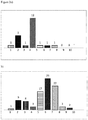
- the samples were analyzed by RP-HPLC for the presence of hydrophilic and hydrophobic species, by cation exchange chromatography for the presence of acidic and basic variants of the antibody and by size exclusion chromatography for the presence of aggregates.
- the protein samples from the syringes were loaded onto a ZORBAX 300SB-C18, 4.6 x 100 mm, 3.5 ⁇ m column to detect hydrophilic and hydrophobic impurities.
- the protein was eluted with a gradient of eluent A (0.1% trifluoroacetic acid in water) and eluent B (0.1% trifluoroacetic acid in 70% acetonitrile, 20% 1-propanol and 10% water) according to the following Table 3: Time [min] Flow [mL/min] Solvent composition Eluent A [%] Solvent composition Eluent B [%] 0 1.0 100 0 7 1.0 62.5 37.5 10 1.0 62.5 37.5 26 1.0 58.5 41.5 31 1.0 58.5 41.5 33 1.0 0 100 35 1.0 0 100 37 1.0 100 0 45 1.0 100 0
- Eluted species were detected and displayed on a graph showing the concentration of the eluted species vs. time.
- the elution profile showed a main peak with the unmodified protein and some further peaks eluting before and after the main peak, representing hydrophilic and hydrophobic variants of the protein, respectively.
- the total area of all peaks as well as the area of the single peaks was determined.
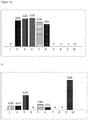
- Figures 3 and 4 show the percentage of the peak area for hydrophilic species and hydrophobic species, respectively, in relation to the total peak area of the eluted species for the syringes of Table 1.
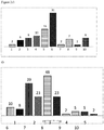
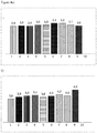
- the protein samples from the syringes were loaded onto a Dionex, BioLCProPac® WCX-10, 4.0 x 250 mm, 10 ⁇ m column to detect acidic and basic variants of the protein.
- the protein was eluted with a gradient of mobile phase A (20 mM potassium phosphate buffer, ph 6.0) and mobile phase B (250 mM KCl, 20 mM potassium phosphate buffer, ph 6.0) according to the following Table 4: Time [min] Solvent composition [%-B] Solvent composition [mM KCl] 0 0 0 3 0 0 33 50 125 35 50 125 36 0 0 40 0 0 0
- Eluted species were detected and displayed on a graph showing the concentration of the eluted species vs. time.
- the elution profile showed a main peak with the unmodified protein and some further peaks eluting before and after the main peak, representing acidic and basic variants of the protein, respectively.
- the total area of all peaks as well as the area of the single peaks was determined.
- Figures 5 and 6 show the percentage of the peak area for acidic variants and basic variants, respectively, in relation to the total peak area of the eluted species for the syringes of Table 1.
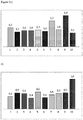
- the protein samples from the syringes were loaded onto a YMC-Pack Diol-200, 5 ⁇ m, 20 nm (8.0 x 300 mm) column to detect aggregates of the protein.
- the protein was eluted by isocratic elution using 0.1 M potassium phosphate and 0.2 M sodium chloride. Eluted species were detected and displayed on a graph showing the concentration of the eluted species vs. time.
- the elution profile showed a main peak with the non-aggregated protein and some further peaks of the protein representing aggregated forms of the protein. The area of all peaks was determined.
- Figure 7 shows the percentage of peak area for the aggregates in relation to the total peak area of the eluted species for the syringes of Table 1.
- ranibizumab is comparable in the pre-filled plastic syringes of the present invention (syringes 1 and 8) and the glass syringes under the conditions tested.
- the syringes 1, 2, 7, 8 and 9 as listed above in Table 1 were tested for their stopper movement forces, i.e. the break loose force and the gliding force.
- 0.165 ml of a solution containing the anti-VEGF antibody ranibizumab in a concentration of 10 mg/ml as well as 10 mM histidine buffer, 10% (w/v) trehalose dihydrate, 0.01% (w/v) polysorbate 20, pH 5.5 were filled into the above syringes.
- 30G x 0.5" needles were attached to the luer cone syringes. The testing was performed at a stopper speed of 190 mm/min over a travel length of 10.9 mm in a Tensile testing machine (TH2730, Thümler).
- Table 5 The results of the test are shown in Table 5 below.
- Table 5 No. 1 No. 2 No. 7 No. 8 No. 10 Break loose force Average of 5 syringes 3.4N 3.4N 0.4N 3.5N 3.5N Maximum individual value 3.8N 3.6N 0.8N 4.1N 3.8N Gliding force Average of 5 syringes 3.0N 10.4N 1.9N 6.1N 4.7N Maximum individual value 4.0N 11.2N 2.3N 6.5N 5.
- the pre-filled silicone-free cycloolefin polymer syringes 1 and 8, i.e. the syringes of the present invention, have a gliding behavior which is comparable or even superior to that of syringes 2 and 10 which are coated with silicone oil.
- the syringes as listed above in Table 2 were tested for their stopper movement forces, i.e. the break loose force and the gliding force.
- 0.165 ml of a solution containing 10 mM histidine buffer, pH 5.5, 10% (w/v) trehalose dihydrate, 0.01% polysorbate 20 were filled into the above syringes.
- 30G x 0.5" needles were attached to the luer cone syringes. The testing was performed at a stopper speed of 190 mm/min over a travel length of 10.9 mm in a Tensile testing machine (TH2730, Thümler).
- the pre-filled silicone-free cycloolefin polymer syringe 11, i.e. a syringe of the present invention, has a gliding behavior which is comparable or even superior to that of syringes which are coated with silicone oil.
- Syringe size Syringe barrel Syringe type Silicone level [mg] Adhesive and tungsten 2 1.0 ml Borosilicate glass Luer cone Baked-on no 11 1.0 ml Cyclo olefin polymer Luer cone no silicone no 12 1.0 ml Cyclo olefin polymer Luer cone 1.5 no 13 1.0 mL Borosilicate glass Staked needle 0.7 yes 14 1.0 mL Borosilicate glass Staked needle 0.25 ⁇ 0.2 yes
- the syringes from Table 6 were rotated from needle to stopper with a speed of 1 cycle/10 seconds at 40°C for five minutes, two weeks and four weeks or were subjected to five freeze/thaw cycles (+5 to -20°C with 1°C/min).
- the syringes were also incubated at 5°C for three, six and twelve months, at 25°C/60% relative humidity for two weeks, one month and three months and 40°C/75% relative humidity without rotation and then analyzed as described above for the syringes from Table 1.
- the syringes as listed above in Table 6 were tested for their stopper movement forces, i.e. the break loose force and the gliding force.
- 0.165 ml of a solution containing the VEGF receptor fusion protein aflibercept in a concentration of 40 mg/ml and 10 mM sodium phosphate buffer, 40 mM sodium chloride, 5% (w/v) sucrose, 0.03% (w/v) polysorbate 20, pH 6.2 was filled into the above syringes.
- 30G x 0.5" needles were attached to the luer cone syringes. The testing was performed at a stopper speed of 190 mm/min over a travel length of 10.9 mm in a Tensile testing machine (TH2730, Thümler).
Abstract
Description
- The present invention relates to a pre-filled syringe containing a VEGF antagonist and comprising a plastic barrel which is silicone-free, kits comprising this syringe and the use of the syringe for the administration of a VEGF antagonist in the treatment of ocular diseases.
- Ocular diseases such as age-related macular degeneration and diabetic macular edema are caused by the uncontrolled growth of blood vessels in the eye. Hence, one option to treat these and similar diseases is to inhibit angiogenesis in the eye. Since VEGF is a key factor in the stimulation of angiogenesis, it is an attractive target for down-regulating angiogenesis.
- Aflibercept, marketed under the name Eylea®, is a recombinant fusion protein consisting of the VEGF binding portion from the extracellular domains of
human VEGF receptors - Both bevacizumab and ranibizumab are presented in glass vials from which they are usually drawn with a syringe shortly before injection into the eye. To use the whole content of the commercial vials of these antibodies, some companies repackage it in ready to use plastic syringes under sterile conditions, thereby allowing more than one syringe to be drawn from one glass vial. However, in the repackaged syringes silicone oil microdroplets and protein aggregates have been observed (Liu et al. (2011) Invest. Ophthalmol. Vis. Sci. 52(2): 1023-1034). Such silicone oil contaminants and protein aggregates may be responsible for the increase in intraocular pressure observed in patients treated with bevacizumab or ranibizumab (Kahook et al. (2009) Ophthalmic Surg. Lasers Imaging 40: 293-295; Good et al. (2011) Br. J. Ophthalmol. 95(8): 1111-1114).
-
AU 2012101677 A4 - Further, recently a pre-filled ranibizumab syringe has been approved by the European Medicines Agency (EMA). The syringe barrel consists of borosilicate glass which was spray-coated with silicon oil-in-water emulsion and subsequently heat-fixed (so-called "baked silicone") (poster presentation by Clunas et al. at the 5th World Congress on Controversies in Ophthalmology, March 20-23, 2014; poster presentation of Michaud et al. at the ARVO Annual Meeting 2014).
- Pre-filled syringes have many benefits compared to a vial and a separately provided syringe, such as improved convenience, affordability, accuracy, sterility, and safety. The use of pre-filled syringes results in greater dose precision, in a reduction of the potential for needle sticks injuries that can occur while drawing medication from vials, in pre-measured dosage reducing dosing errors due to the need to reconstite and/or draw medication into a syringe, and in less overfilling of the syringe helping to reduce costs by minimising drug waste.
- However, glass syringes such as the approved ranibizumab pre-filled syringe are prone to breakage and have a relatively large weight compared to plastic syringes.
- Further, they have to be treated with silicone to enable the correct movement of the stopper within the glass barrel and thereby effective and accurate drug delivery. It has been shown that silicone oil droplets occur in the vitreous cavity after intravitreal administration of VEGF antagonists and it was hypothesized that the silicone oil is derived from the needles and syringes used for the injections (Bakri and Ekdawi (2008) Retina 28: 996-1001).
- Additionally, the glue which is necessary to attach a staked-in needle to a glass syringe can lead to impurities or increased protein oxidation (presentation of Adler at the 2011 PDA Europe The Universe of Pre-Filled Syringes and Injection Devices, Basel, 7-11 November 2011; presentation of Markovic at the PDA Single Use Systems Workshop, Bethesda, 22-23 June 2011).
- Finally, during the manufacturing of glass pre-fillable syringes usually tungsten pins are used. It has been shown that soluble tungsten found in pre-filled syringes leads to protein aggregation and protein oxidation (Liu et al. (2010) PDA J. Pharm. Sci. Technol. 64(1): 11-19; Seidl et al. (2012) Pharm. Res. 29: 1454-1467).
- Problems with glass pre-filled syringes have led to several product recalls in the past.
- Several non-glass pre-filled syringes have been described.
WO 2011/117878 A1 discloses a polycarbonate syringe, but it is not apparent whether the syringe barrel has been coated with silicone and whether the syringe is suitable for intraocular administration.WO 2009/099641 A2 discloses that in cyclic olefin polymer syringes without lubricant less visible particles form than in a glass syringe coated with silicone. However, it is not apparent whether this syringe can be used in ophthalmological applications. - Hence, there is still a need for non-glass syringes which can safely deliver the drug to the eye and which avoid the above disadvantages of using glass syringes, but in which the drug is stable for the storage period.
- The present inventors have surprisingly found that an anti-VEGF antibody solution is stable, i.e. the antibody is not significantly modified and does not aggregate significantly during storage when filled into a pre-filled syringe which comprises a silicone-free plastic syringe barrel, although it had been postulated that a plastic syringe is more permeable than a glass syringe for gases such as oxygen which may lead to protein modifications (see, e.g., Dierick and Yoshino (2015) OnDrugDelivery No. 55: 10-16). Hence, the syringe does not have to be packaged with an oxygen absorber. Further, the pre-filled syringe of the present invention does not contain a significant amount of particles. Finally, the forces required for injection of a solution from the pre-filled syringe of the present invention are comparable to the forces required for injection from a glass syringe.
- The pre-filled syringe of the present invention therefore overcomes the disadvantages of glass syringes discussed above and may be used for administration of VEGF antagonists to the eye.
- Accordingly, the present invention provides a pre-filled syringe containing a liquid formulation of a VEGF antagonist and comprising a syringe barrel, wherein the syringe barrel is made of plastic and is silicone-free.
- In a preferred embodiment the VEGF antagonist is an anti-VEGF antibody or an antigen-binding fragment of such antibody or a soluble VEGF receptor fusion protein and more preferably the anti-VEGF antagonist is ranibizumab or aflibercept.
- Preferably, the antagonist concentration is 1 to 100 mg/ml.
- In one aspect of the invention the pre-filled syringe contains less than 50 particles per ml of the liquid formulation having a diameter of 10 µm or greater.
- In another aspect of the invention the pre-filled syringe contains less than 5 particles per ml of the liquid formulation having a diameter of 25 µm or greater.
- In still another aspect of the invention the pre-filled syringe has a glide force of less than or equal to ION.
- In a preferred embodiment the pre-filled syringe further comprises a silicone-free stopper.
- Preferably, the syringe barrel is made of cycloolefin polymer or cycloolefin copolymer.
- In a preferred embodiment the syringe barrel comprises an internal coating other than a silicone coating.
- Also preferably, the pre-filled syringe comprises a staked needle.
- The present invention also provides a kit comprising one or more pre-filled syringes according to the present invention. Preferably, the kit is a blister pack.
- The pre-filled syringe may be used in administering a VEGF antagonist to a patient having an ocular disease, preferably having an ocular disease selected from the group consisting of age-related macular degeneration (AMD), visual impairment due to diabetic macular oedema (DME), visual impairment due to macular oedema secondary to retinal vein occlusion (branch RVO or central RVO), diabetic retinopathy in patients with diabetic macular edema or visual impairment due to choroidal neovascularisation (CNV) secondary to pathologic myopia.
- Preferably, a volume of 30 to 100 µl of the liquid formulation is administered to the patient.
-
-
Figure 1 : Number of particles with a diameter of 10 µm or greater after rotation from needle to stopper for 5 minutes (a), 2 weeks (b), 4 weeks (c) as well as after five freeze/thaw cycles (d) in the syringes listed in Table 1;syringe 10 was not measured in the analysis after 5 minutes and 2 weeks -
Figure 2 : Number of particles with a diameter of 25 µm or greater after rotation from needle to stopper for 5 minutes (a), 2 weeks (b), 4 weeks (c) as well as after five freeze/thaw cycles (d) in the syringes listed in Table 1;syringe 10 was not measured in the analysis after 5 minutes and 2 weeks -
Figure 3 : Percentage of hydrophilic species measured by RP-HPLC analysis of the content of the syringes listed in Table 1 after rotation from needle to stopper for 5 minutes (a), 2 weeks (b) and 4 weeks (c) as well as after five freeze/thaw cycles (d);syringe 10 was not measured in the analysis after 5 minutes and 2 weeks -
Figure 4 : Percentage of hydrophobic species measured by RP-HPLC analysis of the content of the syringes listed in Table 1 after rotation from needle to stopper for 5 minutes (a), 2 weeks (b) and 4 weeks (c) as well as after five freeze/thaw cycles (d);syringe 10 was not measured in the analysis after 5 minutes and 2 weeks -
Figure 5 : Percentage of acidic variants measured by cation exchange chromatography of the content of the syringes listed in Table 1 after rotation from needle to stopper for 5 minutes (a), 2 weeks (b) and 4 weeks (c) as well as after five freeze/thaw cycles (d);syringe 10 was not measured in the analysis after 5 minutes and 2 weeks -
Figure 6 : Percentage of basic variants measured by cation exchange chromatography of the content of the syringes listed in Table 1 after rotation from needle to stopper for 5 minutes (a), 2 weeks (b) and 4 weeks (c) as well as after five freeze/thaw cycles (d);syringe 10 was not measured in the analysis after 5 minutes and 2 weeks -
Figure 7 : Analysis of protein aggregation by size exclusion chromatography after rotation from needle to stopper for 5 minutes (a), 2 weeks (b) and 4 weeks (c) as well as after five freeze/thaw cycles (d) of the content of the syringes listed in Table 1;syringe 10 was not measured in the analysis after 5 minutes and 2 weeks - The present invention as illustratively described in the following may suitably be practiced in the absence of any element or elements, limitation or limitations, not specifically disclosed herein.
- The present invention will be described with respect to particular embodiments, but the invention is not limited thereto, but only by the claims.
- Where the term "comprising" is used in the present description and claims, it does not exclude other elements. For the purposes of the present invention, the term "consisting of' is considered to be a preferred embodiment of the term "comprising". If hereinafter a group is defined to comprise at least a certain number of embodiments, this is also to be understood to disclose a group which preferably consists only of these embodiments.
- For the purposes of the present invention, the term "obtained" is considered to be a preferred embodiment of the term "obtainable".
- Where an indefinite or definite article is used when referring to a singular noun, e.g. "a", "an" or "the", this includes a plural of that noun unless something else is specifically stated.
- A "pre-filled syringe" is a syringe which is supplied by the manufacturer in a filled state, i.e. a measured dose of the drug to be administered is already present in the syringe when it is purchased and ready for administration. In particular, the pharmaceutical composition containing the drug does not have to be drawn from a vial containing the composition by using an empty syringe. The term pre-filled syringe within the meaning of the present invention does not refer to syringes the content of which has been drawn from a vial in a repackaging process.
- The drug contained in the pre-filled syringe of the present invention, i.e. the VEGF antagonist, preferably an anti-VEGF antibody, is stable at a temperature of 2 to 8°C for at least six months, preferably for at least 9 months, more preferably for at least one year, particularly preferably for at least 18 months and most preferably for about two years. The drug contained in the pre-filled syringe of the present invention, i.e. the VEGF antagonist, preferably an anti-VEGF antibody or a VEGF receptor fusion protein and more preferably ranibizumab or aflibercept, is stable at room temperature, i.e. a temperature between 20°C and 25°C, for at least 24 hours, preferably for at least three days or one week, more preferably for at least two or three weeks, and most preferably for about 4 weeks. The drug contained in the pre-filled syringe of the present invention, i.e. the VEGF antagonist, preferably an anti-VEGF antibody or a VEGF receptor fusion protein and more preferably ranibizumab or aflibercept, is stable at a temperature of about 40°C, for at least 1 hour or 2 hours, preferably for at least four or six hours, more preferably for at least 10 or 12 hours, and most preferably for at least 18 or 24 hours.
- The stability of the drug within the syringe can for example be determined by ion exchange chromatography by which modifications of the drug such as oxidized and deamidated species can be detected or by size exclusion chromatography by which aggregates of the drugs can be detected. A description of such an analysis is provided in the examples section.
- The drug, i.e. the VEGF antagonist, preferably the anti-VEGF antibody, is considered stable, if the sum of all impurities comprising aggregates and chemically modified species is less than 2%, preferably less than 1.5%, more preferably less than 1.2% and most preferably less than 1% compared to the amount of non-modified, non-aggregated drug.
- The drug contained in the pre-filled syringe of the present invention, i.e. the VEGF antagonist, preferably an anti-VEGF antibody or a VEGF receptor fusion protein and more preferably ranibizumab or aflibercept, retains its biological activity when stored at a temperature of 2 to 8°C for at least six months, preferably for at least 9 months, more preferably for at least one year, particularly preferably for at least 18 months and most preferably for about two years. The drug contained in the pre-filled syringe of the present invention, i.e. the VEGF antagonist, preferably an anti-VEGF antibody or a VEGF receptor fusion protein and more preferably ranibizumab or aflibercept, retains its biological activity when stored at room temperature, i.e. a temperature between 20°C and 25°C and 60% relative humidity for at least one day, preferably three days or one week, more preferably two weeks or three weeks and most preferably one month.. The drug contained in the pre-filled syringe of the present invention, i.e. the VEGF antagonist, preferably an anti-VEGF antibody or a VEGF receptor fusion protein and more preferably ranibizumab or aflibercept, retains its biological activity when stored at a temperature of about 40°C and 75% relative humidity for at least 1 hour or 2 hours, preferably for at least four or six hours, more preferably for at least 10 or 12 hours, and most preferably for at least 18 or 24 hours.
- The biological activity of the VEGF antagonist, preferably an anti-VEGF antibody or a VEGF receptor fusion protein and more preferably ranibizumab or aflibercept can be determined by incubating different dilutions of the antagonist which was stored under the conditions described above with human umbilical vein endothelial cells (HUVEC) and VEGF and measuring the VEGF-induced proliferation of the cells in the presence of the antagonist, i.e. by the CellTiter-Blue® Cell Viability Assay available from Promega, in comparison to cells not incubated with the antagonist. Since the VEGF antagonist inhibits VEGF-induced signal transduction, the VEGF-induced proliferation will be reduced, if biologically active VEGF antagonist is present in the sample.
- The VEGF antagonist, preferably the anti-VEGF antibody or VEGF receptor fusion protein and more preferably ranibizumab or aflibercept retains its biological activity after storage in the pre-filled syringe, such that the VEGF-induced proliferation is inhibited in HUVEC.
- The components of a pre-filled syringe are known to a skilled person and basically comprise a syringe barrel and a plunger.
- The syringe barrel contains a defined volume of the liquid composition which can be expelled from the barrel through an outlet positioned on one end of the barrel when the plunger is pushed into and moves along the barrel. The syringe barrel typically has a substantially cylindrical shape. The outlet may comprise a projection from the outlet end through which extends a channel having a smaller diameter than the rest of the syringe barrel. The outlet may be adapted, for example by a luer lock type connection, for connection with a needle or other accessory such as a sealing device which is able to seal the barrel and can be removed to allow a needle to be attached to the syringe. This sealing can be achieved by the use of known sealing devices such as the OVS™ system of Vetter Pharma International GmbH.
- In the pre-filled syringe of the present invention the syringe outlet is firmly connected with a needle so that the pre-filled syringe is supplied with a staked needle and does not need to be assembled prior to use. In this case, the risk of injuries with the needle during assembly of the syringe before injection is reduced. The staked needle can be attached to the pre-filled plastic syringe of the present invention without using an adhesive, since it can be moulded into the syringe. In contrast, an adhesive is required to attach the needle to a glass syringe and can lead to impurities or increased protein oxidation (presentation of Adler at the 2011 PDA Europe The Universe of Pre-Filled Syringes and Injection Devices, Basel, 7-11 November 2011; presentation of Markovic at the PDA Single Use Systems Workshop, Bethesda, 22-23 June 2011).
- For intravitreal administration the needle size is typically 30 gauge, although 31-, 32, 33- and 34-gauge needles may also be used. The pre-filled syringe may be equipped with a passive needle safety guard to further avoid the danger of needle sticks after injection.
- The pre-filled syringe of the present invention comprises a syringe barrel which is made from plastic material. Preferably, the plastic material is selected from cycloolefin polymer and cycloolefin copolymer.
- Cycloolefin copolymers may be produced by chain copolymerization of cyclic monomers such as 8,9,10-trinornorn-2-ene or 1,2,3,4,4a,5,8,8a-octahydro-1,4:5,8-dimethanonaphthalene with ethane. Suitable copolymers are those of the Topas™ type which are available in a variety of grades.
- Cycloolefin polymers may for example be produced by ring-opening metathesis polymerization of various cyclic monomers followed by hydrogenation. Suitable commercially available containers made of cycloolefin polymer material include containers manufactured from CZ™ resin, Zeonor™ and Zeonex™.
- According to the present invention the syringe barrel is silicone-free which means that the inner surface of the syringe barrel has not been treated with silicone oil. Hence, no silicone oil can be detected within the pre-filled syringe of the present invention.
- The presence and thickness of silicone layers can be determined by known methods such as the rap.ID Layer Explorer® application which can also be used to measure the amount of silicone oil within the syringe barrel. The amount of silicone oil within the syringe barrel can also be measured by differential weighing methods and quantitation by infrared spectroscopy of the oil diluted in a suitable solvent.
- Preferably, the pre-filled syringe is uncoated, i.e. the cycloolefin polymer or copolymer material is in direct contact with the liquid composition contained therein and the syringe barrel does not contain any material other than the plastic material of which the syringe is made.
- Alternatively, the pre-filled syringe may comprise an internal coating other than a silicone coating. The term "internal coating" is intended to mean a coating on the inner side of the syringe barrel which is in contact with the drug solution. Examples of such an internal coating include a fluorocarbon film made from a modified ethylene-tetrafluoroethylene copolymer (also called Flurotec® film, available from West Pharmaceutical Services) and a perfluoropolyether film crosslinked by an Atmospheric Plasma Immobilization™ process (also called TriboGlide®, available from TriboFilm Research and described in
WO 2005/094214 A2 ). Another example of an internal coating is a silicon-oxide barrier coating applied to the plastic surface. To the silicone oxide layer further coating layers may be applied, resulting in a multilayer coating. Such plastic syringes coated with a silicon oxide barrier coating and optionally further coating layers are available from SiO2 and described in ONdrug Delivery Magazine issue 45, October 2013 andissue 47, February 2014 as well as inWO 2014/059012 A1 . - The pre-filled syringe may also comprise a layer within the plastic body of the syringe so that the syringe comprises from the outside to the interior three layers: an external plastic layer which is in contact with the environment, an intermediate layer made of a material other than the external and internal plastic layers and an internal plastic layer which is made of the same or another plastic material than the external layer and which is in contact with the drug solution. The intermediate layer may comprise an oxygen-absorbing resin so that the syringe may comprise from the outside to the interior the following three layers: cycloolefin copolymer - oxygen-absorbing resin - cycloolefin copolymer. Such syringes are marketed under the name Oxy-Capt™ by Mitsubishi Gas Chemical Company and are for example described in
WO 2014/136914 A1 . - Alternatively, the syringe may also comprise a coating on the outer surface of the syringe which is in contact with the environment such as an oxygen barrier coating. The syringe barrel is also tungsten-free, i.e. it does not contain any traces of tungsten, since it is not necessary to use tungsten in the syringe manufacturing process. Hence, there is no risk of tungsten-induced protein aggregation.
- In one embodiment the syringe barrel comprises a mark such as a line printed on the syringe barrel which line allows the person injecting the liquid composition to align a pre-determined part of the stopper (such as the tip of the front surface) or plunger with the mark. Thereby, any excess liquid composition and potential air bubbles are removed from the syringe barrel, allowing the safe administration of an exact predetermined dosage to the patient.
- The plunger is pulled and pushed along inside the syringe barrel, allowing the syringe to expel the liquid formulation through the outlet.
- At the end of the plunger which is in contact with the liquid formulation a stopper is located. The stopper portion is typically made of an elastomeric material such as natural or synthetic rubber, which engages an inner surface of the syringe barrel to create a seal that facilitates ejecting the liquid formulation from the syringe when pressure is applied to the plunger.
- The stopper may be coated with a fluoropolymer film such as FluroTec® barrier film or a polytetrafluoroethylene-like film such as an Omniflex stopper. This type of coating serves as an effective barrier between the drug and the elastomer, reducing the potential for extractables or leachables which are inherent to all materials. In addition, the coating reduces the occurrence of the reverse process, where the drug product can adsorb or absorb into the plunger. The stopper is preferably silicone-free, i.e. at least the surface of the stopper which comes into contact with the drug solution and more preferably the complete stopper has not been coated with silicone oil.
- The syringe has a nominal maximum fill volume, i.e. a volume which can be maximally taken up by the syringe, of 0.3 ml to 1.5ml, preferably of 0.5 ml to 1.0 ml, most preferably 0.5 ml or 1.0 ml. The volume of the liquid composition filled into the syringe is about 0.05 ml to about 1ml, preferably about 0.1 ml to about 0.5 ml, more preferably 0.14 ml to 0.3 ml and most preferably 0.15 ml to 0.2 ml.
- The skilled person knows that the syringe is usually filled with a volume which is larger than the volume actually administered to the patient to take into account any dead space within the syringe and the needle and the loss due to the preparation of the syringe for injection. Hence, the volume which is actually administered to the patient is between 0.01 ml and 1 ml, preferably between 0.02 and 0.5 ml, more preferably between 0.025 and 0.5 ml, even more preferably between 0.03 ml and 0.05 ml and most preferably the volume which is actually administered to the patient is 0.05 ml.
- Ranibizumab is typically administered in a volume of 0.05 ml with a ranibizumab concentration of 6 or 10 mg/ml or in a volume of 0.03 ml or 0.05 ml with a ranibizumab concentration of 10 mg/ml, yielding a delivered amount of 0.3 or 0.5 mg. For aflibercept the administered volume is typically 0.05 ml with an aflibercept concentration of 40 mg/ml, yielding a delivered amount of 2 mg. As discussed above, bevacizumab is used off-label for the treatment of ocular diseases. In this case, the administered volume of bevacizumab is 0.05 ml with a bevacizumab concentration of 25 mg/ml, yielding a delivered amount of 1.25 mg.
- Hence, in one embodiment the syringe is filled with a volume of the liquid composition of 0.15 ml to 0.2 ml and 0.03 ml to 0.05 ml of the liquid composition are administered to the patient.
- The term "VEGF antagonist" refers to a molecule which specifically interacts with VEGF and inhibits one or more of its biological activities, e.g. its mitogenic, angiogenic and/or vascular permeability activity. It is intended to include both anti-VEGF antibodies and antigen-binding fragments thereof and non-antibody VEGF antagonists.
- Non-antibody VEGF antagonists include aflibercept, pegaptanib and antibody mimetics. Preferably, the non-antibody VEGF antagonist is aflibercept. Aflibercept which is presently marketed under the name Eylea® and which is also known as VEGF-trap is a recombinant human soluble VEGF receptor fusion protein in which portions of
human VEGF receptors WO 00/75319 A1 polysorbate 20, sucrose and water for injection and is supplied in a concentration of 40 mg/ml. - Pegaptanib which is presently marketed under the name Macugen® is a pegylated anti-vascular endothelial growth factor (VEGF) aptamer (Bell et al. (1999) In Vitro Cell Dev Biol Anim. 35(9): 533-42). Antibody mimetics which are VEGF antagonists include binding proteins comprising an ankyrin repeat domain that binds VEGF and inhibits its binding to the receptor, such as DARPin® MP0112 (see also
WO 2010/060748 andWO 2011/135067 ). - The term "anti-VEGF antibody" refers to an antibody or antibody fragment such as a Fab or a scFV fragment that specifically binds to VEGF and inhibits one or more of its biological activities, e.g. its mitogenic, angiogenic and/or vascular permeability activity. Anti-VEGF antibodies act, e.g., by interfering with the binding of VEGF to a cellular receptor, by interfering with vascular endothelial cell activation after VEGF binding to a cellular receptor, or by killing cells activated by VEGF. Anti-VEGF antibodies include, e.g., antibodies A4.6.1, bevacizumab, ranibizumab, G6, B20, 2C3, and others as described in, for example,
WO 98/45331 US 2003/0190317 ,US 6,582,959 ,US 6,703,020 ,WO 98/45332 WO 96/30046 WO 94/10202 WO 2005/044853 ,EP 0 666 868 B1WO 2009/155724 and Popkov et al. (2004) J. Immunol. Meth. 288: 149-64. Preferably, the anti-VEGF antibody or antigen-binding fragment thereof present in the pharmaceutical composition of the present invention is ranibizumab or bevacizumab. Most preferably, it is ranibizumab or an antigen-binding fragment thereof. - "Ranibizumab" is a humanised monoclonal Fab fragment directed against VEGF-A having the light and heavy chain variable domain sequences of Y0317 as described in SEQ ID Nos. 115 and 116 of
WO 98/45331 WO 98/45331 A2 polysorbate 20 and water for injection and is supplied in a concentration of 10 mg/ml. - "Bevacizumab" is a full-length, humanized murine monoclonal antibody that recognizes all isoforms of VEGF and which is the parent antibody of ranibizumab. The CAS number of bevacizumab is 216974-75-3. Bevacizumab inhibits angiogenesis and is presently approved for the treatment of different cancer types. However, it is also used off-label in ophthalmological diseases such as age-related macular degeneration. The present commercial bevacizumab formulation contains α,α-trehalose dihydrate, sodium phosphate,
polysorbate 20 and water for injection and is supplied as a concentrate with a concentration of 25 mg/ml. - The antibody concentration within the pre-filled syringes of the present invention is typically 1-100 mg/ml, preferably 2-75 mg/ml, more preferably 3-50 mg/ml, even more preferably 5 to 30 mg/ml and most preferably 6 or 10 mg/ml. If ranibizumab is contained within the pre-filled syringe of the present invention the ranibizumab concentration is 10 mg/ml.
- The liquid composition within the pre-filled syringe of the present invention has a low particle content. In particular, it comprises less than 50 particles having a size of more than 10 µm after the syringe has been rotated at 40°C for five minutes, two weeks or four weeks or after three freeze-thaw cycles from +5°C to -20°C with 1°C per minute or after storage of the syringe at 5°C or 25°C and 60% relative humidity for three months. Alternatively or additionally, it comprises less than 5 particles having a size of more than 25 µm after the syringe has been rotated at 40°C for five minutes, two weeks or four weeks or after three freeze-thaw cycles from +5°C to - 20°C with 1°C per minute or after storage of the syringe at 5°C or 25°C/60% relative humidity for three months. Hence, the pre-filled syringe meets the requirements of United States Pharmacopoiea <789> for ophthalmic solutions with respect to these particle sizes.
- The pre-filled syringe of the present invention further has excellent gliding behaviour. In particular, the break loose force, i.e. the force required to initiate the movement of the plunger, is less than 10N or 9N, preferably less than 8N or 7N, more preferably less than 6N and most preferably less than 5N. The break loose force does not change significantly, i.e. by more than 10%, when the syringe is stored for an extended period such as eight weeks. In contrast, in a syringe containing silicone the break loose force increases upon storage by at least twofold.
- Further, the gliding force, i.e. the force required to sustain the movement of the plunger along the syringe barrel to expel the liquid composition, is less than ION, preferably less than 9N, more preferably less than 8N and most preferably less than 7N. In a particularly preferred embodiment there is no significant difference between the break loose force and the gliding force.
- The present invention also provides a kit comprising one or more of the pre-filled syringes of the present invention. Preferably, the kit comprises a blister pack. A "blister pack" has a cavity or pocket which is usually made from thermoformed plastic and a backing of paperboard or a lidding seal of aluminium foil or plastic. The blister pack may be aseptically sterile before the sterile syringe is packaged into it under aseptic conditions. Hence, no sterilization after packaging is required. The kit may further comprise a needle, if the pre-filled syringe does not comprise a staked-in needle. The kit may further comprise instructions for use.
- Preferably, the kit does not comprise an oxygen absorber which is typically used to reduce the level of oxygen within a package such as a blister pack. Oxygen absorbers usually contain a substance such as ferrous carbonate or ascorbate which substance reacts with any oxygen within a package with a high affinity, thereby reducing the oxygen content of the package.
- An "intraocular neovascular disease" is a disease characterized by ocular neovascularisation. Examples of intraocular neovascular diseases include, e.g., proliferative retinopathies, choroidal neovascularisation (CNV), age-related macular degeneration (AMD), diabetic and other ischemia-related retinopathies, diabetic macular oedema, diabetic retinopathy in patients with diabetic macular edema, pathological myopia, von Hippel-Lindau disease, histoplasmosis of the eye, Central Retinal Vein Occlusion (CRVO), Branch Retinal Vein Occlusion (BRVO), corneal neovascularisation, and retinal neovascularisation. The term "age-related macular degeneration" refers to a medical condition which usually affects older adults and results in a loss of vision in the centre of the visual field (the macula) because of damage to the retina.
- Preferably, the pre-filled syringe is for use in the intravitreal injection of a VEGF antagonist as defined herein.
- The term "intravitreal injection" refers to the administration of a pharmaceutical composition in which the substance is injected directly into the eye. More specifically, the substance is injected into the vitreous humour (also called vitreous body or simply vitreous) which is the clear gel that fills the space between the lens and the retina of the eyeball of humans and other vertebrates.
- While the invention has been illustrated and described in detail in the drawings and foregoing description, such illustration and description are to be considered illustrative or exemplary and not restrictive. The invention is not limited to the disclosed embodiments. Other variations to the disclosed embodiments can be understood and effected by those skilled in the art in practicing a claimed invention, from a study of the drawings, the disclosure, and the dependent claims.
- The detailed description is merely exemplary in nature and is not intended to limit application and uses. The following examples further illustrate the present invention without, however, limiting the scope of the invention thereto. Various changes and modifications can be made by those skilled in the art on the basis of the description of the invention, and such changes and modifications are also included in the present invention.
- 400 µl of a solution of the anti-VEGF antibody ranibizumab containing 1 mg/ml of the antibody and histidine buffer, trehalose dihydrate,
polysorbate 20, pH 5.5 was filled into the following syringes:Table 1: No. Syringe size Syringe barrel Syringe type Silicone level [mg] Adhesive and tungsten 1 1.0 ml Cyclo olefin polymer Staked needle no silicone no 2 1.0 ml Borosilicate glass Luer cones Baked on silicone no 3 1.0 ml Borosilicate glass Luer cones 0.6 no 4 0.5 ml Cyclo olefin polymer Luer cones 0.6 no 5 0.5 ml Borosilicate glass Staked needle 0.6 yes 6 0.5 ml Borosilicate glass Staked needle 0.6 yes 7 1.0 ml Polycarbonate Luer cones 1.0 no 8 1.0mL Cyclo olefin polymer Staked needle no silicone no 9 1.0 ml Borosilicate glass Staked needle <0.2 yes 10 1.0 ml Borosilicate glass Staked needle 0.4-0.6 yes - The syringes from Table 1 were rotated from needle to stopper with a speed of 1 cycle/10 seconds at 40°C for five minutes, two weeks and four weeks or were subjected to five freeze/thaw cycles (+5 to -20°C with 1°C/min). Afterwards, the light obscuration was determined with the FlowCam PV bench top system (Fluid Imaging Technologies Inc., Maine, USA) using the System software (VisualSpreadsheet software, version 3.4.8) and the following parameters:
Mode: AutoImage Priming Method: machine prime Flow Rate: 0.040 ml/min Recalibrations: 0 Stop Reason: Sample Volume Processed Sample Volume Aspirated: 0.2900 ml Sample Volume Processed: 0.2893 ml Fluid Volume Imaged: 0.1822 ml Efficiency: 63.0% Frame Rate: 22.00 fps Magnification: 10X Calibration Factor: 0.6979 Syringe Size: 1.00 ml - The results of the analysis are shown in
Figures 1 and2 . The pre-filled silicone-freecycloolefin polymer syringes - Additionally, the syringes listed in Table 2 were tested:
Table 2: No. Syringe size Syringe barrel Syringe type Silicone level [mg] Adhesive and tungsten 2 1.0 ml Borosilicate glass Luer cone Baked-on no 11 1.0 ml Cyclo olefin polymer Luer cone no silicone no 12 1.0 ml Cyclo olefin polymer Luer cone 1.5 no 13 1.0 mL Borosilicate glass Staked needle 0.7 yes 14 1.0 mL Borosilicate glass Staked needle 0.25 ± 0.2 yes - The syringe No. 11 has the same barrel as the
syringe 1 of Table 1, but is equipped with a Luer cone instead of a staked needle. - The syringes from Table 2 were incubated at 5°C for three, six and twelve months, at 25°C/60% relative humidity for two weeks, one month and three months and 40°C/75% relative humidity without rotation and then analyzed as described above for the syringes from Table 1.
- The pre-filled silicone-free cycloolefin polymer syringe 11 has low particle levels under all conditions tested.
- The syringes as listed above in Table 1 were subjected to the stress conditions described in Example 1 for the syringes of Table 1. Further, the syringes as listed above in Table 2 were subjected to the conditions described in Example 1 for the syringes of Table 2.
- Afterwards, the samples were analyzed by RP-HPLC for the presence of hydrophilic and hydrophobic species, by cation exchange chromatography for the presence of acidic and basic variants of the antibody and by size exclusion chromatography for the presence of aggregates.
- The protein samples from the syringes were loaded onto a ZORBAX 300SB-C18, 4.6 x 100 mm, 3.5 µm column to detect hydrophilic and hydrophobic impurities.
- The protein was eluted with a gradient of eluent A (0.1% trifluoroacetic acid in water) and eluent B (0.1% trifluoroacetic acid in 70% acetonitrile, 20% 1-propanol and 10% water) according to the following Table 3:
Time [min] Flow [mL/min] Solvent composition Eluent A [%] Solvent composition Eluent B [%] 0 1.0 100 0 7 1.0 62.5 37.5 10 1.0 62.5 37.5 26 1.0 58.5 41.5 31 1.0 58.5 41.5 33 1.0 0 100 35 1.0 0 100 37 1.0 100 0 45 1.0 100 0 - Eluted species were detected and displayed on a graph showing the concentration of the eluted species vs. time. The elution profile showed a main peak with the unmodified protein and some further peaks eluting before and after the main peak, representing hydrophilic and hydrophobic variants of the protein, respectively. The total area of all peaks as well as the area of the single peaks was determined.
Figures 3 and4 show the percentage of the peak area for hydrophilic species and hydrophobic species, respectively, in relation to the total peak area of the eluted species for the syringes of Table 1. - The protein samples from the syringes were loaded onto a Dionex, BioLCProPac® WCX-10, 4.0 x 250 mm, 10 µm column to detect acidic and basic variants of the protein.
- The protein was eluted with a gradient of mobile phase A (20 mM potassium phosphate buffer, ph 6.0) and mobile phase B (250 mM KCl, 20 mM potassium phosphate buffer, ph 6.0) according to the following Table 4:
Time [min] Solvent composition [%-B] Solvent composition [mM KCl] 0 0 0 3 0 0 33 50 125 35 50 125 36 0 0 40 0 0 - Eluted species were detected and displayed on a graph showing the concentration of the eluted species vs. time. The elution profile showed a main peak with the unmodified protein and some further peaks eluting before and after the main peak, representing acidic and basic variants of the protein, respectively. The total area of all peaks as well as the area of the single peaks was determined.
Figures 5 and6 show the percentage of the peak area for acidic variants and basic variants, respectively, in relation to the total peak area of the eluted species for the syringes of Table 1. - The protein samples from the syringes were loaded onto a YMC-Pack Diol-200, 5 µm, 20 nm (8.0 x 300 mm) column to detect aggregates of the protein.
- The protein was eluted by isocratic elution using 0.1 M potassium phosphate and 0.2 M sodium chloride. Eluted species were detected and displayed on a graph showing the concentration of the eluted species vs. time. The elution profile showed a main peak with the non-aggregated protein and some further peaks of the protein representing aggregated forms of the protein. The area of all peaks was determined.
Figure 7 shows the percentage of peak area for the aggregates in relation to the total peak area of the eluted species for the syringes of Table 1. - From the results shown in
Figures 3 to 7 it is apparent that the stability of ranibizumab is comparable in the pre-filled plastic syringes of the present invention (syringes 1 and 8) and the glass syringes under the conditions tested. - The
syringes polysorbate 20, pH 5.5 were filled into the above syringes. Prior to testing, 30G x 0.5" needles were attached to the luer cone syringes. The testing was performed at a stopper speed of 190 mm/min over a travel length of 10.9 mm in a Tensile testing machine (TH2730, Thümler). - The results of the test are shown in Table 5 below.
Table 5: No. 1 No. 2 No. 7 No. 8 No. 10 Break loose force Average of 5 syringes 3.4N 3.4N 0.4N 3.5N 3.5N Maximum individual value 3.8N 3.6N 0.8N 4.1N 3.8N Gliding force Average of 5 syringes 3.0N 10.4N 1.9N 6.1N 4.7N Maximum individual value 4.0N 11.2N 2.3N 6.5N 5.IN - The pre-filled silicone-free
cycloolefin polymer syringes syringes - Further, the syringes as listed above in Table 2 were tested for their stopper movement forces, i.e. the break loose force and the gliding force. To this end, 0.165 ml of a solution containing 10 mM histidine buffer, pH 5.5, 10% (w/v) trehalose dihydrate, 0.01
% polysorbate 20 were filled into the above syringes. Prior to testing, 30G x 0.5" needles were attached to the luer cone syringes. The testing was performed at a stopper speed of 190 mm/min over a travel length of 10.9 mm in a Tensile testing machine (TH2730, Thümler). - The pre-filled silicone-free cycloolefin polymer syringe 11, i.e. a syringe of the present invention, has a gliding behavior which is comparable or even superior to that of syringes which are coated with silicone oil.
- 400 µl of a solution of the VEGF receptor fusion protein aflibercept containing 1 mg/ml of the antibody and 10 mM sodium phosphate buffer, 40 mM sodium chloride, 5% (w/v) sucrose, 0.03% (w/v)
polysorbate 20, pH 6.2 was filled into the following syringes:Table 6: No. Syringe size Syringe barrel Syringe type Silicone level [mg] Adhesive and tungsten 2 1.0 ml Borosilicate glass Luer cone Baked-on no 11 1.0 ml Cyclo olefin polymer Luer cone no silicone no 12 1.0 ml Cyclo olefin polymer Luer cone 1.5 no 13 1.0 mL Borosilicate glass Staked needle 0.7 yes 14 1.0 mL Borosilicate glass Staked needle 0.25 ± 0.2 yes - The syringes from Table 6 were rotated from needle to stopper with a speed of 1 cycle/10 seconds at 40°C for five minutes, two weeks and four weeks or were subjected to five freeze/thaw cycles (+5 to -20°C with 1°C/min). The syringes were also incubated at 5°C for three, six and twelve months, at 25°C/60% relative humidity for two weeks, one month and three months and 40°C/75% relative humidity without rotation and then analyzed as described above for the syringes from Table 1.
- The syringes as listed above in Table 6 were tested for their stopper movement forces, i.e. the break loose force and the gliding force. To this end, 0.165 ml of a solution containing the VEGF receptor fusion protein aflibercept in a concentration of 40 mg/ml and 10 mM sodium phosphate buffer, 40 mM sodium chloride, 5% (w/v) sucrose, 0.03% (w/v)
polysorbate 20, pH 6.2 was filled into the above syringes. Prior to testing, 30G x 0.5" needles were attached to the luer cone syringes. The testing was performed at a stopper speed of 190 mm/min over a travel length of 10.9 mm in a Tensile testing machine (TH2730, Thümler). - Further embodiments of the invention are described herein:
- 1. Pre-filled syringe containing a liquid formulation of a VEGF antagonist and comprising a syringe barrel, wherein the syringe barrel is made of plastic and is silicone-free.
- 2. Pre-filled syringe according to
item 1, wherein the VEGF antagonist is an anti-VEGF antibody or an antigen-binding fragment of such antibody or a VEGF receptor fusion protein. - 3. Pre-filled syringe according to
item 2, wherein the anti-VEGF antibody is ranibizumab or aflibercept. - 4. Pre-filled syringe according to any of the preceding items, wherein the antagonist concentration is 1 to 100 mg/ml.
- 5. Pre-filled syringe according to any of the preceding items, containing less than 50 particles per ml of the liquid formulation having a diameter of 10 µm or greater.
- 6. Pre-filled syringe according to any of the preceding items, containing less than 5 particles per ml of the liquid formulation having a diameter of 25 µm or greater.
- 7. Pre-filled syringe according to any of the preceding items, having a slide force of less than or equal to ION.
- 8. Pre-filled syringe according to any of the preceding items, further comprising a silicone-free stopper.
- 9. Pre-filled syringe according to any of the preceding items, wherein the syringe barrel is made of cycloolefin polymer or cycloolefin copolymer.
- 10. Pre-filled syringe according to any of the preceding items, wherein the syringe barrel comprises an internal coating other than a silicone coating.
- 11. Pre-filled syringe according to any of the preceding items, comprising a staked needle.
- 12. Kit comprising one or more pre-filled syringes of any of the preceding items.
- 13. Pre-filled syringe of any of
items 1 to 11 for use in administering a liquid formulation of a VEGF antagonist to a patient having an ocular disease. - 14. Pre-filled syringe for the use according to item 13, wherein the ocular disease is selected from the group consisting of age-related macular degeneration (AMD), visual impairment due to diabetic macular oedema (DME), visual impairment due to macular oedema secondary to retinal vein occlusion (branch RVO or central RVO), diabetic retinopathy in patients with diabetic macular edema or visual impairment due to choroidal neovascularisation (CNV) secondary to pathologic myopia.
- 15. Pre-filled syringe for the use according to item 13 or 14,
Claims (15)
- Pre-filled syringe containing a liquid formulation of a VEGF antagonist and comprising a syringe barrel, wherein the syringe barrel is made of plastic and is silicone-free.
- Pre-filled syringe according to item 1, wherein the VEGF antagonist is an anti-VEGF antibody or an antigen-binding fragment of such antibody or a VEGF receptor fusion protein.
- Pre-filled syringe according to item 2, wherein the anti-VEGF antibody is ranibizumab or aflibercept.
- Pre-filled syringe according to any of the preceding items, wherein the antagonist concentration is 1 to 100 mg/ml.
- Pre-filled syringe according to any of the preceding items, containing less than 50 particles per ml of the liquid formulation having a diameter of 10 µm or greater.
- Pre-filled syringe according to any of the preceding items, containing less than 5 particles per ml of the liquid formulation having a diameter of 25 µm or greater.
- Pre-filled syringe according to any of the preceding items, having a slide force of less than or equal to 10N.
- Pre-filled syringe according to any of the preceding items, further comprising a silicone-free stopper.
- Pre-filled syringe according to any of the preceding items, wherein the syringe barrel is made of cycloolefin polymer or cycloolefin copolymer.
- Pre-filled syringe according to any of the preceding items, wherein the syringe barrel comprises an internal coating other than a silicone coating.
- Pre-filled syringe according to any of the preceding items, comprising a staked needle.
- Kit comprising one or more pre-filled syringes of any of the preceding items.
- Pre-filled syringe of any of items 1 to 11 for use in administering a liquid formulation of a VEGF antagonist to a patient having an ocular disease.
- Pre-filled syringe for the use according to item 13, wherein the ocular disease is selected from the group consisting of age-related macular degeneration (AMD), visual impairment due to diabetic macular oedema (DME), visual impairment due to macular oedema secondary to retinal vein occlusion (branch RVO or central RVO), diabetic retinopathy in patients with diabetic macular edema or visual impairment due to choroidal neovascularisation (CNV) secondary to pathologic myopia.
- Pre-filled syringe for the use according to item 13 or 14, wherein a volume of 30 to 100 µl of the liquid formulation is administered to the patient.
Applications Claiming Priority (4)
| Application Number | Priority Date | Filing Date | Title |
|---|---|---|---|
| EP14167889 | 2014-05-12 | ||
| PCT/EP2015/060514 WO2015173260A1 (en) | 2014-05-12 | 2015-05-12 | Pre-filled plastic syringe containing a vegf antagonist |
| EP18196113.7A EP3492495A1 (en) | 2014-05-12 | 2015-05-12 | Pre-filled plastic syringe containing a vegf antagonist |
| EP15721021.2A EP3143044A1 (en) | 2014-05-12 | 2015-05-12 | Pre-filled plastic syringe containing a vegf antagonist |
Related Parent Applications (2)
| Application Number | Title | Priority Date | Filing Date |
|---|---|---|---|
| EP15721021.2A Division EP3143044A1 (en) | 2014-05-12 | 2015-05-12 | Pre-filled plastic syringe containing a vegf antagonist |
| EP18196113.7A Division EP3492495A1 (en) | 2014-05-12 | 2015-05-12 | Pre-filled plastic syringe containing a vegf antagonist |
Publications (1)
| Publication Number | Publication Date |
|---|---|
| EP3828202A1 true EP3828202A1 (en) | 2021-06-02 |
Family
ID=50687323
Family Applications (3)
| Application Number | Title | Priority Date | Filing Date |
|---|---|---|---|
| EP18196113.7A Withdrawn EP3492495A1 (en) | 2014-05-12 | 2015-05-12 | Pre-filled plastic syringe containing a vegf antagonist |
| EP15721021.2A Withdrawn EP3143044A1 (en) | 2014-05-12 | 2015-05-12 | Pre-filled plastic syringe containing a vegf antagonist |
| EP20205103.3A Pending EP3828202A1 (en) | 2014-05-12 | 2015-05-12 | Pre-filled plastic syringe containing a vegf antagonist |
Family Applications Before (2)
| Application Number | Title | Priority Date | Filing Date |
|---|---|---|---|
| EP18196113.7A Withdrawn EP3492495A1 (en) | 2014-05-12 | 2015-05-12 | Pre-filled plastic syringe containing a vegf antagonist |
| EP15721021.2A Withdrawn EP3143044A1 (en) | 2014-05-12 | 2015-05-12 | Pre-filled plastic syringe containing a vegf antagonist |
Country Status (9)
| Country | Link |
|---|---|
| US (2) | US20170232199A1 (en) |
| EP (3) | EP3492495A1 (en) |
| JP (3) | JP6802069B2 (en) |
| KR (1) | KR20170003973A (en) |
| CN (1) | CN106456757A (en) |
| AU (1) | AU2015260955B2 (en) |
| CA (1) | CA2947456C (en) |
| RU (1) | RU2702748C2 (en) |
| WO (1) | WO2015173260A1 (en) |
Families Citing this family (18)
| Publication number | Priority date | Publication date | Assignee | Title |
|---|---|---|---|---|
| US20170232199A1 (en) | 2014-05-12 | 2017-08-17 | Formycon Ag | Pre-Filled Plastic Syringe Containing a VEGF Antagonist |
| US9840553B2 (en) | 2014-06-28 | 2017-12-12 | Kodiak Sciences Inc. | Dual PDGF/VEGF antagonists |
| US10925927B2 (en) | 2015-11-18 | 2021-02-23 | Formycon Ag | Pre-filled pharmaceutical package comprising a liquid formulation of a VEGF-antagonist |
| KR20180105123A (en) | 2015-11-18 | 2018-09-27 | 에스아이오2 메디컬 프로덕츠, 인크. | Pharmaceutical package for ophthalmic preparations |
| AU2016381964B2 (en) | 2015-12-30 | 2024-02-15 | Kodiak Sciences Inc. | Antibodies and conjugates thereof |
| AU2017213103B2 (en) | 2016-01-26 | 2022-08-11 | Formycon Ag | Liquid formulation of a VEGF antagonist |
| CN110612101A (en) * | 2017-04-14 | 2019-12-24 | 安能泰制药有限公司 | Pre-filled syringe containing moxifloxacin |
| WO2018217995A1 (en) * | 2017-05-24 | 2018-11-29 | Formycon Ag | Sterilizable pre-filled pharmaceutical packages comprising a liquid formulation of a vegf-antagonist |
| AU2018295781B2 (en) | 2017-07-04 | 2020-12-24 | Terumo Kabushiki Kaisha | Resin-made syringe, prefilled syringe, and method for producing resin-made syringe |
| US20200390596A1 (en) | 2018-01-03 | 2020-12-17 | Irenix Medical, Inc. | Therapeutic Agent Delivery Devices Having Integrated Pain Mitigation, and Methods for Using the Same |
| BR112020015074A2 (en) | 2018-01-26 | 2020-12-08 | The Regents Of The University Of California | METHODS AND COMPOSITIONS FOR TREATING ANGIOGENIC DISORDERS WITH THE USE OF ANTI-VEGF AGENTS |
| KR101941497B1 (en) | 2018-05-02 | 2019-01-23 | 배대성 | Spray induction tool for a prostate liquid |
| MX2020011848A (en) | 2018-05-10 | 2021-03-29 | Regeneron Pharma | High concentration vegf receptor fusion protein containing formulations. |
| CN109607751B (en) * | 2018-12-14 | 2021-07-13 | 广西南宁绿泽环保科技有限公司 | Air-water balancer of center source composite membrane bioreactor |
| WO2021072265A1 (en) | 2019-10-10 | 2021-04-15 | Kodiak Sciences Inc. | Methods of treating an eye disorder |
| CA3168512A1 (en) | 2019-11-25 | 2021-06-03 | Napoleone Ferrara | Long-acting vegf inhibitors for intraocular neovascularization |
| KR102419910B1 (en) * | 2021-04-15 | 2022-07-14 | 주식회사 알테오젠 | Pre-filled syringe filled with low-volume liquid pharmaceuticals and Manufacturing method thereof |
| WO2023206134A1 (en) * | 2022-04-27 | 2023-11-02 | Beijing Sightnovo Medical Technology Co., Ltd | Compositions and methods for eye diseases |
Citations (20)
| Publication number | Priority date | Publication date | Assignee | Title |
|---|---|---|---|---|
| WO1994010202A1 (en) | 1992-10-28 | 1994-05-11 | Genentech, Inc. | Vascular endothelial cell growth factor antagonists |
| WO1996030046A1 (en) | 1995-03-30 | 1996-10-03 | Genentech, Inc. | Vascular endothelial cell growth factor antagonists |
| WO1998045331A2 (en) | 1997-04-07 | 1998-10-15 | Genentech, Inc. | Anti-vegf antibodies |
| WO1998045332A2 (en) | 1997-04-07 | 1998-10-15 | Genentech, Inc. | Humanized antibodies and methods for forming humanized antibodies |
| WO2000075319A1 (en) | 1999-06-08 | 2000-12-14 | Regeneron Pharmaceuticals, Inc. | Modified chimeric polypeptides with improved pharmacokinetic properties |
| US6582959B2 (en) | 1991-03-29 | 2003-06-24 | Genentech, Inc. | Antibodies to vascular endothelial cell growth factor |
| US20030190317A1 (en) | 1997-04-07 | 2003-10-09 | Genentech, Inc. | Anti-VEGF antibodies |
| US6703020B1 (en) | 1999-04-28 | 2004-03-09 | Board Of Regents, The University Of Texas System | Antibody conjugate methods for selectively inhibiting VEGF |
| WO2005044853A2 (en) | 2003-11-01 | 2005-05-19 | Genentech, Inc. | Anti-vegf antibodies |
| WO2005094214A2 (en) | 2004-03-02 | 2005-10-13 | Sakhrani Vinay G | Improved article with lubricated surface and method |
| WO2009099641A2 (en) | 2008-02-07 | 2009-08-13 | Amgen Inc. | Stabilized protein compositions |
| WO2009155724A2 (en) | 2008-06-25 | 2009-12-30 | Esbatech, An Alcon Biomedical Research Unit Llc | Stable and soluble antibodies inhibiting vegf |
| WO2010060748A1 (en) | 2008-11-03 | 2010-06-03 | Molecular Partners Ag | Binding proteins inhibiting the vegf-a receptor interaction |
| WO2011117878A1 (en) | 2010-03-20 | 2011-09-29 | Pawan Trilokchand Agrawal | Plastic pre-filled syringe |
| WO2011135067A1 (en) | 2010-04-30 | 2011-11-03 | Molecular Partners Ag | Modified binding proteins inhibiting the vegf-a receptor interaction |
| AU2012101677A4 (en) | 2012-07-03 | 2012-12-13 | Novartis Ag | Device |
| WO2014005728A1 (en) * | 2012-07-03 | 2014-01-09 | Novartis Ag | Syringe |
| WO2014059012A1 (en) | 2012-10-12 | 2014-04-17 | Sio2 Medical Products, Inc. | Process for the internal coating of hollow bodies |
| WO2014136914A1 (en) | 2013-03-06 | 2014-09-12 | 三菱瓦斯化学株式会社 | Oxygen-absorbing multilayer body, oxygen-absorbing container, oxygen-absorbing airtight container, oxygen-absorbing push-through pack, and storage method using same |
| EP3143044A1 (en) | 2014-05-12 | 2017-03-22 | Formycon AG | Pre-filled plastic syringe containing a vegf antagonist |
Family Cites Families (16)
| Publication number | Priority date | Publication date | Assignee | Title |
|---|---|---|---|---|
| JP2002241264A (en) * | 2001-02-19 | 2002-08-28 | Asahi Kasei Corp | Prefilled syringe pharmaceutical preparation of elcatonin |
| CN2531794Y (en) * | 2001-11-17 | 2003-01-22 | 陶日新 | Diaphragmatic type disposable syringe |
| JP4481587B2 (en) * | 2003-04-25 | 2010-06-16 | テルモ株式会社 | Syringe |
| DE102004026805A1 (en) * | 2004-06-02 | 2006-01-05 | Disetronic Licensing Ag | Container for storing a product to be injected and injection device |
| EP3106189A1 (en) * | 2007-04-26 | 2016-12-21 | Daikyo Seiko, LTD. | Mold and method for molding same for needle-equipped syringe barrel and needle-equipped syringe |
| US9597458B2 (en) * | 2009-10-29 | 2017-03-21 | W. L. Gore & Associates, Inc. | Fluoropolymer barrier materials for containers |
| CN101785891A (en) * | 2010-01-21 | 2010-07-28 | 王建友 | injector |
| AU2011235002B2 (en) * | 2010-03-31 | 2016-08-11 | Ocuject, Llc | Device and method for intraocular drug delivery |
| WO2012003221A1 (en) * | 2010-06-29 | 2012-01-05 | Cv Holdings, Llc | Syringe with integrated needle |
| US9289557B2 (en) * | 2011-03-28 | 2016-03-22 | Becton, Dickinson And Company | Plastic stopper |
| WO2013115331A1 (en) * | 2012-01-31 | 2013-08-08 | テルモ株式会社 | Syringe for use as pre-filled syringe |
| US9504603B2 (en) * | 2012-04-02 | 2016-11-29 | Ocuject, Llc | Intraocular delivery devices and methods therefor |
| PL3777834T3 (en) * | 2012-06-01 | 2022-05-30 | Novartis Ag | Syringe |
| AU2013100070B4 (en) * | 2012-07-03 | 2013-04-04 | Novartis Ag | Use of device |
| WO2014203183A1 (en) * | 2013-06-20 | 2014-12-24 | Novartis Ag | Use of a vegf antagonist in treating macular edema |
| KR20160029794A (en) * | 2013-07-11 | 2016-03-15 | 노파르티스 아게 | Use of a vegf antagonist in treating chorioretinal neovascular and permeability disorders in paediatric patients |
-
2015
- 2015-05-12 US US15/310,012 patent/US20170232199A1/en not_active Abandoned
- 2015-05-12 CN CN201580024757.7A patent/CN106456757A/en active Pending
- 2015-05-12 AU AU2015260955A patent/AU2015260955B2/en active Active
- 2015-05-12 WO PCT/EP2015/060514 patent/WO2015173260A1/en active Application Filing
- 2015-05-12 EP EP18196113.7A patent/EP3492495A1/en not_active Withdrawn
- 2015-05-12 JP JP2016567833A patent/JP6802069B2/en not_active Ceased
- 2015-05-12 EP EP15721021.2A patent/EP3143044A1/en not_active Withdrawn
- 2015-05-12 RU RU2016148324A patent/RU2702748C2/en active
- 2015-05-12 KR KR1020167034325A patent/KR20170003973A/en not_active Application Discontinuation
- 2015-05-12 EP EP20205103.3A patent/EP3828202A1/en active Pending
- 2015-05-12 CA CA2947456A patent/CA2947456C/en active Active
-
2020
- 2020-08-13 US US16/992,867 patent/US20210008284A1/en active Pending
- 2020-11-26 JP JP2020195895A patent/JP2021049351A/en active Pending
-
2022
- 2022-10-13 JP JP2022164411A patent/JP2022186793A/en active Pending
Patent Citations (25)
| Publication number | Priority date | Publication date | Assignee | Title |
|---|---|---|---|---|
| US6582959B2 (en) | 1991-03-29 | 2003-06-24 | Genentech, Inc. | Antibodies to vascular endothelial cell growth factor |
| WO1994010202A1 (en) | 1992-10-28 | 1994-05-11 | Genentech, Inc. | Vascular endothelial cell growth factor antagonists |
| EP0666868B1 (en) | 1992-10-28 | 2002-04-03 | Genentech, Inc. | Use of anti-VEGF antibodies for the treatment of cancer |
| WO1996030046A1 (en) | 1995-03-30 | 1996-10-03 | Genentech, Inc. | Vascular endothelial cell growth factor antagonists |
| WO1998045331A2 (en) | 1997-04-07 | 1998-10-15 | Genentech, Inc. | Anti-vegf antibodies |
| WO1998045332A2 (en) | 1997-04-07 | 1998-10-15 | Genentech, Inc. | Humanized antibodies and methods for forming humanized antibodies |
| US20030190317A1 (en) | 1997-04-07 | 2003-10-09 | Genentech, Inc. | Anti-VEGF antibodies |
| US6703020B1 (en) | 1999-04-28 | 2004-03-09 | Board Of Regents, The University Of Texas System | Antibody conjugate methods for selectively inhibiting VEGF |
| WO2000075319A1 (en) | 1999-06-08 | 2000-12-14 | Regeneron Pharmaceuticals, Inc. | Modified chimeric polypeptides with improved pharmacokinetic properties |
| WO2005044853A2 (en) | 2003-11-01 | 2005-05-19 | Genentech, Inc. | Anti-vegf antibodies |
| WO2005094214A2 (en) | 2004-03-02 | 2005-10-13 | Sakhrani Vinay G | Improved article with lubricated surface and method |
| WO2009099641A2 (en) | 2008-02-07 | 2009-08-13 | Amgen Inc. | Stabilized protein compositions |
| WO2009155724A2 (en) | 2008-06-25 | 2009-12-30 | Esbatech, An Alcon Biomedical Research Unit Llc | Stable and soluble antibodies inhibiting vegf |
| WO2010060748A1 (en) | 2008-11-03 | 2010-06-03 | Molecular Partners Ag | Binding proteins inhibiting the vegf-a receptor interaction |
| WO2011117878A1 (en) | 2010-03-20 | 2011-09-29 | Pawan Trilokchand Agrawal | Plastic pre-filled syringe |
| WO2011135067A1 (en) | 2010-04-30 | 2011-11-03 | Molecular Partners Ag | Modified binding proteins inhibiting the vegf-a receptor interaction |
| AU2012101677A4 (en) | 2012-07-03 | 2012-12-13 | Novartis Ag | Device |
| AU2012101677B4 (en) | 2012-07-03 | 2012-12-20 | Novartis Ag | Device |
| WO2014005728A1 (en) * | 2012-07-03 | 2014-01-09 | Novartis Ag | Syringe |
| WO2014059012A1 (en) | 2012-10-12 | 2014-04-17 | Sio2 Medical Products, Inc. | Process for the internal coating of hollow bodies |
| WO2014136914A1 (en) | 2013-03-06 | 2014-09-12 | 三菱瓦斯化学株式会社 | Oxygen-absorbing multilayer body, oxygen-absorbing container, oxygen-absorbing airtight container, oxygen-absorbing push-through pack, and storage method using same |
| EP3143044A1 (en) | 2014-05-12 | 2017-03-22 | Formycon AG | Pre-filled plastic syringe containing a vegf antagonist |
| US20170232199A1 (en) | 2014-05-12 | 2017-08-17 | Formycon Ag | Pre-Filled Plastic Syringe Containing a VEGF Antagonist |
| EP3492495A1 (en) | 2014-05-12 | 2019-06-05 | Formycon AG | Pre-filled plastic syringe containing a vegf antagonist |
| JP6802069B2 (en) | 2014-05-12 | 2020-12-16 | フォーマイコン アーゲーFormycon Ag | Prefilled plastic syringe containing VEGF antagonist |
Non-Patent Citations (21)
| Title |
|---|
| ADLER, 2011 PDA EUROPE THE UNIVERSE OF PRE-FILLED SYRINGES AND INJECTION DEVICES, BASEL, 7 November 2011 (2011-11-07) |
| BAKRIEKDAWI, RETINA, vol. 28, 2008, pages 996 - 1001 |
| BELL ET AL., IN VITRO CELL DEV BIOL ANIM, vol. 35, no. 9, 1999, pages 533 - 42 |
| CAS, no. 216974-75-3 |
| CHEN ET AL., J. MOL. BIOL., vol. 293, 1999, pages 865 - 81 |
| CLUNAS ET AL., 5TH WORLD CONGRESS ON CONTROVERSIES IN OPHTHALMOLOGY, 20 March 2014 (2014-03-20) |
| DIERICKYOSHINO, ONDRUGDELIVERY, vol. 55, 2015, pages 10 - 16 |
| GOOD ET AL., BR. J. OPHTHALMOL., vol. 95, no. 8, 2011, pages 1111 - 1114 |
| HOLASH ET AL., PROC. NATL. ACAD. SCI. USA, vol. 99, no. 17, 2002, pages 11393 - 11398 |
| JOHNSONSHARMA, CURR. OPIN. OPHTHALMOL., vol. 24, no. 3, 2013, pages 205 - 12 |
| KAHOOK ET AL., OPHTHALMIC SURG. LASERS IMAGING, vol. 40, 2009, pages 293 - 295 |
| LAHENDRO B: "PREFILLED SYRINGES: THE CONTAINER OF CHOICE FOR TODAY'S INJECTABLES", 1 January 2008 (2008-01-01), pages 24 - 26, XP055371894, Retrieved from the Internet <URL:http://www.ondrugdelivery.com/publications/prefilled_syringes_2008.pdf> [retrieved on 20170511] * |
| LIU ET AL., INVEST. OPHTHALMOL. VIS. SCI., vol. 52, no. 2, 2011, pages 1023 - 1034 |
| LIU ET AL., PDA J. PHARM. SCI. TECHNOL., vol. 64, no. 1, 2010, pages 11 - 19 |
| MARKOVIC, PDA SINGLE USE SYSTEMS WORKSHOP, BETHESDA, 22 June 2011 (2011-06-22) |
| MICHAUD ET AL., ARVO ANNUAL MEETING, 2014 |
| MINTZ C.: "Prefilled syringes: The Next "Big Thing"", 30 August 2012 (2012-08-30), XP002788671, Retrieved from the Internet <URL:https://www.bioprocessonline.com/doc/prefilled-syringes-the-next-big-thing-0001> [retrieved on 20190206] * |
| ONDRUG DELIVERY MAGAZINE, October 2013 (2013-10-01) |
| POPKOV ET AL., J. IMMUNOL. METH., vol. 288, 2004, pages 149 - 64 |
| SEIDL ET AL., PHARM. RES., vol. 29, 2012, pages 1454 - 1467 |
| VILIVALM V.: "Prefilled syringes: a study of protein sensitivity to tungsten", 1 March 2012 (2012-03-01), pages 60 - 63, XP055372643, Retrieved from the Internet <URL:http://www.iptonline.com/pdf_viewarticle.asp?cat=6&article=838> [retrieved on 20170515] * |
Also Published As
| Publication number | Publication date |
|---|---|
| US20210008284A1 (en) | 2021-01-14 |
| CN106456757A (en) | 2017-02-22 |
| US20170232199A1 (en) | 2017-08-17 |
| EP3143044A1 (en) | 2017-03-22 |
| RU2016148324A3 (en) | 2018-12-17 |
| RU2016148324A (en) | 2018-06-13 |
| JP2021049351A (en) | 2021-04-01 |
| AU2015260955A1 (en) | 2016-11-17 |
| KR20170003973A (en) | 2017-01-10 |
| JP2022186793A (en) | 2022-12-15 |
| CA2947456A1 (en) | 2015-11-19 |
| WO2015173260A1 (en) | 2015-11-19 |
| JP2017515596A (en) | 2017-06-15 |
| AU2015260955B2 (en) | 2020-06-18 |
| RU2702748C2 (en) | 2019-10-11 |
| JP6802069B2 (en) | 2020-12-16 |
| CA2947456C (en) | 2023-03-14 |
| EP3492495A1 (en) | 2019-06-05 |
Similar Documents
| Publication | Publication Date | Title |
|---|---|---|
| US20210008284A1 (en) | Pre-filled plastic syringe containing a VEGF antagonist | |
| US20220362441A1 (en) | Pre-filled plastic syringe containing a vegf antagonist | |
| CN110709062B (en) | Stable antibody formulations | |
| US20230248806A1 (en) | Liquid Formulation of a VEGF Antagonist | |
| ES2906812T3 (en) | Syringe | |
| CA2904458C (en) | Protein formulations and methods of making same | |
| KR20180082971A (en) | Stable Liquid Formulation | |
| CA3129901A1 (en) | Therapeutic antibody formulation | |
| WO2018215580A1 (en) | Method for sterilizing prefilled plastic syringes containing a vegf antagonist | |
| AU2013204044B2 (en) | Protein formulations and methods of making same | |
| KR20230120696A (en) | Syringe comprising ophthalmic formulation | |
| TWI760345B (en) | Antibody formulations | |
| EP4190310A1 (en) | Stable pharmaceutical preparation | |
| KR20240021856A (en) | Pharmaceutical formulations containing anti-IgE antibodies |
Legal Events
| Date | Code | Title | Description |
|---|---|---|---|
| PUAI | Public reference made under article 153(3) epc to a published international application that has entered the european phase |
Free format text: ORIGINAL CODE: 0009012 |
|
| STAA | Information on the status of an ep patent application or granted ep patent |
Free format text: STATUS: THE APPLICATION HAS BEEN PUBLISHED |
|
| AC | Divisional application: reference to earlier application |
Ref document number: 3492495 Country of ref document: EP Kind code of ref document: P Ref document number: 3143044 Country of ref document: EP Kind code of ref document: P |
|
| AK | Designated contracting states |
Kind code of ref document: A1 Designated state(s): AL AT BE BG CH CY CZ DE DK EE ES FI FR GB GR HR HU IE IS IT LI LT LU LV MC MK MT NL NO PL PT RO RS SE SI SK SM TR |
|
| STAA | Information on the status of an ep patent application or granted ep patent |
Free format text: STATUS: REQUEST FOR EXAMINATION WAS MADE |
|
| 17P | Request for examination filed |
Effective date: 20211202 |
|
| RBV | Designated contracting states (corrected) |
Designated state(s): AL AT BE BG CH CY CZ DE DK EE ES FI FR GB GR HR HU IE IS IT LI LT LU LV MC MK MT NL NO PL PT RO RS SE SI SK SM TR |
|
| STAA | Information on the status of an ep patent application or granted ep patent |
Free format text: STATUS: EXAMINATION IS IN PROGRESS |
|
| 17Q | First examination report despatched |
Effective date: 20220721 |
|
| TPAC | Observations filed by third parties |
Free format text: ORIGINAL CODE: EPIDOSNTIPA |