EP3819005B1 - Electric field cancer treatment device using optimization algorithm - Google Patents
Electric field cancer treatment device using optimization algorithm Download PDFInfo
- Publication number
- EP3819005B1 EP3819005B1 EP19831280.3A EP19831280A EP3819005B1 EP 3819005 B1 EP3819005 B1 EP 3819005B1 EP 19831280 A EP19831280 A EP 19831280A EP 3819005 B1 EP3819005 B1 EP 3819005B1
- Authority
- EP
- European Patent Office
- Prior art keywords
- electric field
- tumors
- electrodes
- optimization algorithm
- optimizer
- Prior art date
- Legal status (The legal status is an assumption and is not a legal conclusion. Google has not performed a legal analysis and makes no representation as to the accuracy of the status listed.)
- Active
Links
Images


Classifications
-
- A—HUMAN NECESSITIES
- A61—MEDICAL OR VETERINARY SCIENCE; HYGIENE
- A61B—DIAGNOSIS; SURGERY; IDENTIFICATION
- A61B5/00—Measuring for diagnostic purposes; Identification of persons
- A61B5/0033—Features or image-related aspects of imaging apparatus, e.g. for MRI, optical tomography or impedance tomography apparatus; Arrangements of imaging apparatus in a room
- A61B5/004—Features or image-related aspects of imaging apparatus, e.g. for MRI, optical tomography or impedance tomography apparatus; Arrangements of imaging apparatus in a room adapted for image acquisition of a particular organ or body part
- A61B5/0042—Features or image-related aspects of imaging apparatus, e.g. for MRI, optical tomography or impedance tomography apparatus; Arrangements of imaging apparatus in a room adapted for image acquisition of a particular organ or body part for the brain
-
- A—HUMAN NECESSITIES
- A61—MEDICAL OR VETERINARY SCIENCE; HYGIENE
- A61B—DIAGNOSIS; SURGERY; IDENTIFICATION
- A61B5/00—Measuring for diagnostic purposes; Identification of persons
- A61B5/48—Other medical applications
- A61B5/4836—Diagnosis combined with treatment in closed-loop systems or methods
-
- A—HUMAN NECESSITIES
- A61—MEDICAL OR VETERINARY SCIENCE; HYGIENE
- A61N—ELECTROTHERAPY; MAGNETOTHERAPY; RADIATION THERAPY; ULTRASOUND THERAPY
- A61N1/00—Electrotherapy; Circuits therefor
- A61N1/02—Details
- A61N1/04—Electrodes
- A61N1/0404—Electrodes for external use
- A61N1/0472—Structure-related aspects
- A61N1/0492—Patch electrodes
-
- A—HUMAN NECESSITIES
- A61—MEDICAL OR VETERINARY SCIENCE; HYGIENE
- A61N—ELECTROTHERAPY; MAGNETOTHERAPY; RADIATION THERAPY; ULTRASOUND THERAPY
- A61N1/00—Electrotherapy; Circuits therefor
- A61N1/02—Details
- A61N1/08—Arrangements or circuits for monitoring, protecting, controlling or indicating
-
- A—HUMAN NECESSITIES
- A61—MEDICAL OR VETERINARY SCIENCE; HYGIENE
- A61N—ELECTROTHERAPY; MAGNETOTHERAPY; RADIATION THERAPY; ULTRASOUND THERAPY
- A61N1/00—Electrotherapy; Circuits therefor
- A61N1/18—Applying electric currents by contact electrodes
- A61N1/32—Applying electric currents by contact electrodes alternating or intermittent currents
- A61N1/36—Applying electric currents by contact electrodes alternating or intermittent currents for stimulation
- A61N1/36002—Cancer treatment, e.g. tumour
-
- A—HUMAN NECESSITIES
- A61—MEDICAL OR VETERINARY SCIENCE; HYGIENE
- A61N—ELECTROTHERAPY; MAGNETOTHERAPY; RADIATION THERAPY; ULTRASOUND THERAPY
- A61N1/00—Electrotherapy; Circuits therefor
- A61N1/18—Applying electric currents by contact electrodes
- A61N1/32—Applying electric currents by contact electrodes alternating or intermittent currents
- A61N1/36—Applying electric currents by contact electrodes alternating or intermittent currents for stimulation
- A61N1/36014—External stimulators, e.g. with patch electrodes
- A61N1/3603—Control systems
-
- A—HUMAN NECESSITIES
- A61—MEDICAL OR VETERINARY SCIENCE; HYGIENE
- A61N—ELECTROTHERAPY; MAGNETOTHERAPY; RADIATION THERAPY; ULTRASOUND THERAPY
- A61N1/00—Electrotherapy; Circuits therefor
- A61N1/40—Applying electric fields by inductive or capacitive coupling ; Applying radio-frequency signals
-
- G—PHYSICS
- G06—COMPUTING OR CALCULATING; COUNTING
- G06F—ELECTRIC DIGITAL DATA PROCESSING
- G06F18/00—Pattern recognition
- G06F18/20—Analysing
- G06F18/24—Classification techniques
-
- G—PHYSICS
- G16—INFORMATION AND COMMUNICATION TECHNOLOGY [ICT] SPECIALLY ADAPTED FOR SPECIFIC APPLICATION FIELDS
- G16H—HEALTHCARE INFORMATICS, i.e. INFORMATION AND COMMUNICATION TECHNOLOGY [ICT] SPECIALLY ADAPTED FOR THE HANDLING OR PROCESSING OF MEDICAL OR HEALTHCARE DATA
- G16H20/00—ICT specially adapted for therapies or health-improving plans, e.g. for handling prescriptions, for steering therapy or for monitoring patient compliance
- G16H20/30—ICT specially adapted for therapies or health-improving plans, e.g. for handling prescriptions, for steering therapy or for monitoring patient compliance relating to physical therapies or activities, e.g. physiotherapy, acupressure or exercising
-
- G—PHYSICS
- G16—INFORMATION AND COMMUNICATION TECHNOLOGY [ICT] SPECIALLY ADAPTED FOR SPECIFIC APPLICATION FIELDS
- G16H—HEALTHCARE INFORMATICS, i.e. INFORMATION AND COMMUNICATION TECHNOLOGY [ICT] SPECIALLY ADAPTED FOR THE HANDLING OR PROCESSING OF MEDICAL OR HEALTHCARE DATA
- G16H30/00—ICT specially adapted for the handling or processing of medical images
- G16H30/40—ICT specially adapted for the handling or processing of medical images for processing medical images, e.g. editing
-
- G—PHYSICS
- G16—INFORMATION AND COMMUNICATION TECHNOLOGY [ICT] SPECIALLY ADAPTED FOR SPECIFIC APPLICATION FIELDS
- G16H—HEALTHCARE INFORMATICS, i.e. INFORMATION AND COMMUNICATION TECHNOLOGY [ICT] SPECIALLY ADAPTED FOR THE HANDLING OR PROCESSING OF MEDICAL OR HEALTHCARE DATA
- G16H50/00—ICT specially adapted for medical diagnosis, medical simulation or medical data mining; ICT specially adapted for detecting, monitoring or modelling epidemics or pandemics
- G16H50/50—ICT specially adapted for medical diagnosis, medical simulation or medical data mining; ICT specially adapted for detecting, monitoring or modelling epidemics or pandemics for simulation or modelling of medical disorders
-
- A—HUMAN NECESSITIES
- A61—MEDICAL OR VETERINARY SCIENCE; HYGIENE
- A61B—DIAGNOSIS; SURGERY; IDENTIFICATION
- A61B5/00—Measuring for diagnostic purposes; Identification of persons
- A61B5/05—Detecting, measuring or recording for diagnosis by means of electric currents or magnetic fields; Measuring using microwaves or radio waves
- A61B5/055—Detecting, measuring or recording for diagnosis by means of electric currents or magnetic fields; Measuring using microwaves or radio waves involving electronic [EMR] or nuclear [NMR] magnetic resonance, e.g. magnetic resonance imaging
Definitions
- the present disclosure describes an apparatus for alternating electric fields therapy using an optimization algorithm.
- this algorithm is designed to optimize the magnitude of electric fields, such that the prescribed or higher electric field intensity is applied to tumors in a patient's body while minimizing the electric field intensity transmitted to normal tissues.
- Electromagnetic waves are used to treat tumors, with the effects depending on the frequency of the waves.
- X-rays which have a frequency of about 10 10 MHz, treat cancers by breaking the DNA double helix of cancer cells, causing cell death.
- application of electromagnetic waves with frequencies in the range of about 10 MHz to tumors in the human body produces heat inside the tumors.
- Tumor therapy using electromagnetic waves in the range of 100 to 300 kHz is known as Tumor Treating Fields (TTFields). This technology treats tumors by delaying cell division to induce cell death. Although introduced less than 10 years ago, TTFields is currently used to treat patients in about 1000 treatment centers worldwide.
- TTFields greatly affects cells that are dividing, it has little effect on normal cells that divide more slowly than tumor cells or on cells that virtually do not divide. Only a few studies to date have described the effects of TTFields on normal cells or the effects of electric fields on each major organ in the human body. Additionally, because this technology has been used for only 10 years, the potential risks of long-term side effects remain unclear.
- the present disclosure describes an apparatus for applying alternating electric fields therapy to treat tumors and a method for planning therapy.
- this method not only the position of the electrodes but the voltage applied to each is calculated through an optimization algorithm to further reduce electric fields unnecessarily transmitted to normal tissues.
- US 2004/176804 A1 discloses an apparatus and a method to compute the optimal spatial and temporal characteristics for combating tumor growth within a body on the basis of cytological (as provided by biopsies, etc.) and anatomical data (as provided by CT, MRI, PET, etc.), as well as the electric properties of the different elements.
- the apparatus applies the fields that have maximal effect on the tumor and minimal effect on all other tissues by adjusting both the field generator output characteristics and by optimal positioning of the insulated electrodes or isolects on the patient's body.
- US 2018/160933 A1 discloses a method comprising receiving images of a body area of a patient, identifying abnormal tissue in the image, generating a data set with the abnormal tissue masked out, deforming a model template in space so that features in the deformed model template line up with corresponding features in the data set, placing data representing the abnormal tissue back into the deformed model template, generating a model of electrical properties of tissues in the body area based on the deformed and modified model template, and determining an electrode placement layout that maximizes field strength in the abnormal tissue by using the model of electrical properties to simulate electromagnetic field distributions in the body area caused by simulated electrodes placed respective to the body area.
- the present disclosure describes an apparatus and method for alternating electric fields therapy using an algorithm that optimizes the magnitude of the electric fields applied to tumors while minimizing the electric field intensity applied to normal tissues.
- an apparatus for alternating electric fields therapy to treat tumors in a patient applies electric fields to tumors and normal tissues using one or more pairs of electrode pads, with most of these electrodes being arranged according to an embodiment of the present disclosure.
- This includes an image classifier to classify at least one organ in patient imaging of each organ, as well as an electric field optimizer to set the number and position of the applied individual electrodes, based on the classification of tissues as tumors or normal tissues.
- These electrodes are arranged on electrode pads of preset sizes, and the magnitudes of voltage for the set electrodes determined.
- the electric field optimizer can set the magnitude of the voltage applied to most electrodes based on least one of the tumor types to be treated, the position of these tumors, and the boundary conditions with normal tissues.
- This electric field optimizer can calculate the magnitude of the voltage to be applied to the tumors, with this voltage being at least the prescribed electric field intensity, while applying a minimal electric field intensity to normal organs.
- This electric field optimizer can set the electric field limit value according to the importance of the organs, thereby allowing the electrodes to transmit the preset or lower electric field limit value.
- This electric field optimizer can have an object function, which is set using the intensity of at least one of the electric fields transmitted to tumors or normal organs, with a weighting for each normal organ or an electric field limit value.
- One pair of electrode pads can form a ground.
- This method for planning therapy to treat tumors in patients by applying electric fields to the tumors and normal tissues involves one or more pairs of electrode pads, with most electrodes being arranged according to an embodiment of the present disclosure.
- This method includes patient imaging and classifying the image for each organ, creating at least one treatment plan by arranging the number and position of electrodes on the electrode pads of a preset size based on the tumors in the patient and the boundary conditions between the tumors and normal tissues. The magnitude of the voltage applied to most of the electrodes is set using an electric field optimizer. Each formulated treatment plan is subsequently analyzed and evaluated.
- the magnitude of voltage applied to the electrodes can be calculated using an electric field optimizer to apply prescribed or higher electric fields to the tumors and minimal electric fields to normal tissues.
- the voltage applied to the electrodes can be between 0 V and 150 V and the frequency between 100 kHz and 300 kHz.
- the magnitude of the voltage to be applied to the electrodes can be determined based on the magnitude of the electric fields prescribed for the tumors, the weighting assigned to each organ and/or the preset electric field limit.
- the apparatus and method for alternating electric fields therapy using an optimization algorithm apply different electric fields to tumors and normal tissues.
- the voltage applied to each electrode using the optimization algorithm was designed to reduce the magnitude of electric fields transmitted to the normal tissues, so that it was less than the magnitude applied to the tumors, thereby reducing the risk of side effects of treatment and increasing the probability of treatment success.
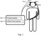
- FIG. 1 shows a conceptual diagram of an apparatus for alternating electric fields therapy using an optimization algorithm, as embodied in the present disclosure
- FIG. 2 shows a conceptual diagram showing the arrangement of electrodes of an apparatus for alternating electric fields as describe in the present disclosure.
- the apparatus for alternating electric fields using an optimization algorithm is configured to treat tumors in a patient by applying voltage to form separate electric fields in tumors and normal tissues.
- This apparatus includes a pair of electrode pads 110, an image classifier 120 and an electric field optimizer 130.
- the pair of electrode pads 110 includes most of the electrodes 111, which are variously arranged according to the shape of the electrode pads 110.
- FIG. 2 shows the electrodes 111 arranged in a 3 ⁇ 3 matrix, as illustrated, the numbers of electrodes 111 and the interval between them may be modified through the electric field optimizer 130, as described below. The shape and arrangement are not limited to those shown in FIG. 2 . Although two pairs of electrode pads 110 are shown, the number of electrode pads 110 may increase or decrease depending on the patient's treatment conditions.
- the magnitudes of the electric fields transmitted to the tumors and normal tissues can be altered by arranging the configuration of the electrodes 111 to adjust the applied frequency and voltage, thereby providing effective treatment based on the boundary conditions between a patient's tumors and normal tissues.
- the pair of electrode pads 110 may form the ground G on one of the pair.
- the image classifier 120 serves to classify at least one organ in a patient's image.
- This image may be a magnetic resonance (MR) or computed tomography (CT) image.
- MR or CT image includes the tumors together with a variety of organs.
- the image classifier 120 classifies most of the images for each organ, and determines the distance between normal tissues and tumors along with their positional relationships.
- the image classifier 120 may use the data to adjust the size of the pair of electrode pads 110 or the arrangement of the electrodes by automatically classifying them according to the organ or by setting the boundaries for each organ according to the user definition.
- the image classifier 120 can reconstruct each image into a 3-dimensional image of each organ and clearly determine their positional relationships.
- the electric field optimizer 130 sets the number and position of applied electrodes based on the classified tumors and normal tissues, arranges the electrodes on electrode pads 110 of preset size, and determines the magnitudes of voltage for the set electrodes. That is, the electric field optimizer 130 sets the magnitude of voltage applied to the electrodes by considering the classified tumors and normal tissues and the conditions of the tumors.
- the electric field optimizer 130 can set the magnitude of voltage applied to the electrodes by considering at least one of the types of tumors to be treated, the position of the tumors, and the boundary conditions between the tumors and normal tissues. In this instance, the electric field optimizer 130 can set the electrode weighting as a variable, divide most of the electrodes into those applying voltage and a ground electrode, and set the frequency of most of the electrodes depending on the type of tumors. For example, the weighting and voltage of each electrode can be calculated by setting the frequency to 200 kHz for glioblastomas and 150 kHz for lung cancers.
- the electric field optimizer 130 can have an object function that is set based on the intensity of at least one of the electric fields transmitted to the tumors or the normal organs, the weighting assigned to each normal organ or the electric field limit. Additionally, the electric field optimizer 130 can set the electric field limit according to the importance of organs, allowing the electrodes to transmit the preset or lower electric field.
- the electric field limit refers to the range of electric fields set to prevent the preset or higher electric field intensity from being transmitted to normal organs.
- the object function can adjust the electric field intensity to apply minimal electric fields to the major organs of normal tissues.
- the prescribed electric field is the magnitude of electric fields for transmitting a predetermined or higher electric field intensity to the total volume of the tumors
- the electric field limit is the magnitude of electric fields for transmitting the predetermined or lower electric field intensity to the total volume of the normal organs.
- a treatment plan can be created through optimization to apply a prescribed electric field intensity of 1.5 V/cm to tumors and transmit an electric field intensity limit of ⁇ 0.5 V/cm to the total volume of major organs.
- the average electric field intensity is the average transmitted to one organ, with the same electric field not transmitted to all parts of that organ. Because the transmitted electric field intensity is different for each microvolume, the average electric field intensity is determined for individual organs.
- Equation 1 After setting an electric field at position E i , spaced a predetermined distance from the tumor, as in Equation 1, the electric field intensity can be calculated from the conditions shown in Equations 2 to 4.
- E i is the electric field intensity value at the i th position
- w j is the weighting of the j th electrode
- e ij is the electric field intensity transmitted to the i th position by the j th electrode.
- Equation 2 calculates electric fields based on the prescribed electric field intensity.
- E 0 is the prescribed electric field intensity.
- Equation 3 calculates electric fields based on the object function f, considering that the weighting was dependent on the importance of organs and the electric field intensity limit of normal organs.
- the electric field intensity limit is a constant that differs depending on the importance of organs.
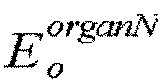
- W N is the weighting for each organ and E o organN is the electric field intensity limit for the N th normal organ.
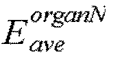
- W N is the weighting for each organ and E ave organN is the average electric field intensity of the N th normal organ.
- the object function for calculating the electric fields acting on normal organs is not limited to the above Equation and differs depending on the type and position of tumors.
- the present disclosure sets the electric field based on a voltage of 0 V to 150 V applied to the electrodes and a frequency of 100 kHz to 300 kHz, as well as on the position of the tumors and the importance of each organ. This electric field intensity is converted to the voltage applied to the electrodes.
- the voltage range of 0 V to 150 V may be similar to the range used for conventional treatment apparatuses, but the present disclosure sets the arrangement and position of the electrodes by considering the position of the tumors and normal organs and their relationship, and applies different magnitudes of therapeutic and general electric fields to most of the electrodes, preventing damage to normal tissues.
- FIG. 3 shows a flowchart of a method for alternating electric fields therapy using an optimization algorithm according to an embodiment of the present disclosure. This method treats tumors in a patient by transmitting electric fields to the tumors and normal tissues using one or more pairs of electrode pads that include most of the electrodes.
- an image is retrieved based on imaging of the patient, and the retrieved image is classified for each organ (S100).
- Organs may be classified more effectively according to their importance by setting the resolution range for each organ individually.
- At least one treatment plan is created by arranging the number and position of electrodes on the electrode pads of a preset size, based on the patient's tumors and the boundary conditions between the tumors and normal tissues, and by setting the magnitude of voltage applied to most electrodes through the electric field optimizer (S200).
- S200 electric field optimizer
- three or more treatment plans can be created, with these plans adapted for different sizes and numbers of electrodes.
- the electrodes can be arranged in a matrix ( FIG. 2 ), they can also be arranged in other configurations; for example, asymmetrically or at different intervals. In this example, the pair of electrodes facing each other may be placed at the same position. Additionally, the applied voltage and frequency may differ between tumors and normal tissues, with voltages ranging from 0 V to 150 V and frequencies from 100 kHz to 300 kHz.
- the created treatment plans are analyzed and evaluated (S300).
- the conditions for optimal results may be determined by individually analyzing and evaluating each of the treatment plans.
- the patient is treated by applying the set voltage to most of the electrodes under the optimal conditions determined by the analyzed treatment plans (S400).
- FIG. 4 and 5 show diagrams comparing simulations by an apparatus for alternating electric fields therapy using an optimization algorithm, as embodied in the present disclosure, and the conventional method.
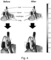
- FIG. 4 shows the electric field distributions when the same voltage is applied to all electrodes in the conventional manner (before) and when different voltages are applied to the electrodes using the electric field optimizer (after).
- Table 1 lists the findings of a quantitative analysis of the results shown in FIG. 4 .
- FIG. 5 shows the electric field distributions when the same voltage is applied to all electrodes in the conventional manner (before) and when different voltages are applied to the electrodes using the optimization algorithm (after).
- Table 2 lists the findings of a quantitative analysis of the results in FIG. 5 , showing that the indicator values are all reduced after application of the electric field optimizer. That is, the electric field intensity transmitted to normal organs is greatly reduced after application of the electric field optimizer.
- Table 2 Liver Stomach Right kidney Left Kidney Before After Difference Before After Difference Before After Difference V 30 100 100 0 100 100 0 100 100 0 0 60 100 84 16 100 100 0 100 100 0 V 90 25 11 14 94 50 44 92 71 21 Eave 1.256 1.082 0.174 1.784 1.363 0.421 1.730 1.481 0.249
Landscapes
- Health & Medical Sciences (AREA)
- Engineering & Computer Science (AREA)
- Life Sciences & Earth Sciences (AREA)
- Public Health (AREA)
- General Health & Medical Sciences (AREA)
- Radiology & Medical Imaging (AREA)
- Nuclear Medicine, Radiotherapy & Molecular Imaging (AREA)
- Biomedical Technology (AREA)
- Veterinary Medicine (AREA)
- Animal Behavior & Ethology (AREA)
- Medical Informatics (AREA)
- Heart & Thoracic Surgery (AREA)
- Biophysics (AREA)
- Oncology (AREA)
- Hospice & Palliative Care (AREA)
- Data Mining & Analysis (AREA)
- Physics & Mathematics (AREA)
- Primary Health Care (AREA)
- Epidemiology (AREA)
- Pathology (AREA)
- Neurology (AREA)
- Theoretical Computer Science (AREA)
- Surgery (AREA)
- Molecular Biology (AREA)
- General Physics & Mathematics (AREA)
- Bioinformatics & Computational Biology (AREA)
- Computer Vision & Pattern Recognition (AREA)
- Evolutionary Biology (AREA)
- Bioinformatics & Cheminformatics (AREA)
- Artificial Intelligence (AREA)
- Evolutionary Computation (AREA)
- General Engineering & Computer Science (AREA)
- Physical Education & Sports Medicine (AREA)
- Databases & Information Systems (AREA)
- Electrotherapy Devices (AREA)
- Measurement And Recording Of Electrical Phenomena And Electrical Characteristics Of The Living Body (AREA)
Description
- The present disclosure describes an apparatus for alternating electric fields therapy using an optimization algorithm. In particular, this algorithm is designed to optimize the magnitude of electric fields, such that the prescribed or higher electric field intensity is applied to tumors in a patient's body while minimizing the electric field intensity transmitted to normal tissues.
- Electromagnetic waves are used to treat tumors, with the effects depending on the frequency of the waves. X-rays, which have a frequency of about 1010 MHz, treat cancers by breaking the DNA double helix of cancer cells, causing cell death. By contrast, application of electromagnetic waves with frequencies in the range of about 10 MHz to tumors in the human body produces heat inside the tumors.
- Tumor therapy using electromagnetic waves in the range of 100 to 300 kHz is known as Tumor Treating Fields (TTFields). This technology treats tumors by delaying cell division to induce cell death. Although introduced less than 10 years ago, TTFields is currently used to treat patients in about 1000 treatment centers worldwide.
- Although TTFields greatly affects cells that are dividing, it has little effect on normal cells that divide more slowly than tumor cells or on cells that virtually do not divide. Only a few studies to date have described the effects of TTFields on normal cells or the effects of electric fields on each major organ in the human body. Additionally, because this technology has been used for only 10 years, the potential risks of long-term side effects remain unclear.
- Current treatment involves placing the electrodes at positions to apply maximal electric fields to tumors, while, at the same time, applying minimal electric fields to normal tissues. However, the same voltage is applied to all electrodes, which may result in the unnecessary application of electric fields to normal tissues.
- Accordingly, the present disclosure describes an apparatus for applying alternating electric fields therapy to treat tumors and a method for planning therapy. In this method, not only the position of the electrodes but the voltage applied to each is calculated through an optimization algorithm to further reduce electric fields unnecessarily transmitted to normal tissues.
-
US 2004/176804 A1 discloses an apparatus and a method to compute the optimal spatial and temporal characteristics for combating tumor growth within a body on the basis of cytological (as provided by biopsies, etc.) and anatomical data (as provided by CT, MRI, PET, etc.), as well as the electric properties of the different elements. On the basis of this computation, the apparatus applies the fields that have maximal effect on the tumor and minimal effect on all other tissues by adjusting both the field generator output characteristics and by optimal positioning of the insulated electrodes or isolects on the patient's body. -
US 2018/160933 A1 discloses a method comprising receiving images of a body area of a patient, identifying abnormal tissue in the image, generating a data set with the abnormal tissue masked out, deforming a model template in space so that features in the deformed model template line up with corresponding features in the data set, placing data representing the abnormal tissue back into the deformed model template, generating a model of electrical properties of tissues in the body area based on the deformed and modified model template, and determining an electrode placement layout that maximizes field strength in the abnormal tissue by using the model of electrical properties to simulate electromagnetic field distributions in the body area caused by simulated electrodes placed respective to the body area. - The invention is defined by the
independent claim 1. In the following any method of treatment is not claimed and disclosed for illustrative purposes only. - The present disclosure describes an apparatus and method for alternating electric fields therapy using an algorithm that optimizes the magnitude of the electric fields applied to tumors while minimizing the electric field intensity applied to normal tissues.
- The objective of the present disclosure is not limited to the above-mentioned objective, and other objectives not mentioned herein will be clearly understood by those skilled in the art from the following description.
- To achieve the above-described objective, an apparatus for alternating electric fields therapy to treat tumors in a patient is required. This apparatus applies electric fields to tumors and normal tissues using one or more pairs of electrode pads, with most of these electrodes being arranged according to an embodiment of the present disclosure. This includes an image classifier to classify at least one organ in patient imaging of each organ, as well as an electric field optimizer to set the number and position of the applied individual electrodes, based on the classification of tissues as tumors or normal tissues. These electrodes are arranged on electrode pads of preset sizes, and the magnitudes of voltage for the set electrodes determined. The electric field optimizer determines an individual electric field intensity based on the following equation below


- The electric field optimizer can set the magnitude of the voltage applied to most electrodes based on least one of the tumor types to be treated, the position of these tumors, and the boundary conditions with normal tissues.
- This electric field optimizer can calculate the magnitude of the voltage to be applied to the tumors, with this voltage being at least the prescribed electric field intensity, while applying a minimal electric field intensity to normal organs.
- This electric field optimizer can set the electric field limit value according to the importance of the organs, thereby allowing the electrodes to transmit the preset or lower electric field limit value.
- This electric field optimizer can have an object function, which is set using the intensity of at least one of the electric fields transmitted to tumors or normal organs, with a weighting for each normal organ or an electric field limit value.
- One pair of electrode pads can form a ground.
- This method for planning therapy to treat tumors in patients by applying electric fields to the tumors and normal tissues involves one or more pairs of electrode pads, with most electrodes being arranged according to an embodiment of the present disclosure. This method includes patient imaging and classifying the image for each organ, creating at least one treatment plan by arranging the number and position of electrodes on the electrode pads of a preset size based on the tumors in the patient and the boundary conditions between the tumors and normal tissues. The magnitude of the voltage applied to most of the electrodes is set using an electric field optimizer. Each formulated treatment plan is subsequently analyzed and evaluated. The electric field optimizer determines an individual electric field intensity based on the following equation below


- The magnitude of voltage applied to the electrodes can be calculated using an electric field optimizer to apply prescribed or higher electric fields to the tumors and minimal electric fields to normal tissues.
- The voltage applied to the electrodes can be between 0 V and 150 V and the frequency between 100 kHz and 300 kHz.
- The magnitude of the voltage to be applied to the electrodes can be determined based on the magnitude of the electric fields prescribed for the tumors, the weighting assigned to each organ and/or the preset electric field limit.
- The apparatus and method for alternating electric fields therapy using an optimization algorithm, as described in the present disclosure, apply different electric fields to tumors and normal tissues. The voltage applied to each electrode using the optimization algorithm was designed to reduce the magnitude of electric fields transmitted to the normal tissues, so that it was less than the magnitude applied to the tumors, thereby reducing the risk of side effects of treatment and increasing the probability of treatment success.
- The effects of the present disclosure are not limited to the above-mentioned effects, and other effects not mentioned herein will be clearly understood by those skilled in the art from the following description.
-
-
FIG. 1 shows a conceptual diagram of an apparatus for alternating electric fields therapy using an optimization algorithm, as embodied in the present disclosure. -
FIG. 2 shows a conceptual diagram showing the arrangement of electrodes of an apparatus for alternating electric fields, as embodied in the present disclosure. -
FIG. 3 shows a flowchart of a method for alternating electric fields therapy using an optimization algorithm, as embodied in the present disclosure. -
FIG. 4 and5 show diagrams comparing simulations by an apparatus for alternating electric fields therapy using an optimization algorithm, as embodied in the present disclosure, and the conventional method. - Hereinafter, exemplary embodiments of the present disclosure will be described in detail with reference to the accompanying drawings. Identical symbols represent identical elements in the accompanying drawings. Detailed descriptions of known functions and elements that make the subject matter of the present disclosure ambiguous are omitted. Similarly, some elements are exaggerated, omitted or shown schematically in the accompanying drawings.
- Unless the context clearly indicates otherwise, "comprises" specifies the presence of stated elements but does not preclude the presence or addition of one or more other elements. In addition, throughout the specification, when an element is referred to as being "on" another element, it indicates that the element is disposed on or below the other element, but it does not necessarily indicate the gravitational direction.
-
FIG. 1 shows a conceptual diagram of an apparatus for alternating electric fields therapy using an optimization algorithm, as embodied in the present disclosure, andFIG. 2 shows a conceptual diagram showing the arrangement of electrodes of an apparatus for alternating electric fields as describe in the present disclosure. - With reference to
FIG. 1 and2 , the apparatus for alternating electric fields using an optimization algorithm, as embodied in the present disclosure, is configured to treat tumors in a patient by applying voltage to form separate electric fields in tumors and normal tissues. This apparatus includes a pair ofelectrode pads 110, animage classifier 120 and anelectric field optimizer 130. - The pair of
electrode pads 110 includes most of theelectrodes 111, which are variously arranged according to the shape of theelectrode pads 110. AlthoughFIG. 2 shows theelectrodes 111 arranged in a 3 × 3 matrix, as illustrated, the numbers ofelectrodes 111 and the interval between them may be modified through theelectric field optimizer 130, as described below. The shape and arrangement are not limited to those shown inFIG. 2 . Although two pairs ofelectrode pads 110 are shown, the number ofelectrode pads 110 may increase or decrease depending on the patient's treatment conditions. - The magnitudes of the electric fields transmitted to the tumors and normal tissues can be altered by arranging the configuration of the
electrodes 111 to adjust the applied frequency and voltage, thereby providing effective treatment based on the boundary conditions between a patient's tumors and normal tissues. For example, the pair ofelectrode pads 110 may form the ground G on one of the pair. - The
image classifier 120 serves to classify at least one organ in a patient's image. This image may be a magnetic resonance (MR) or computed tomography (CT) image. Each MR or CT image includes the tumors together with a variety of organs. In this instance, theimage classifier 120 classifies most of the images for each organ, and determines the distance between normal tissues and tumors along with their positional relationships. Theimage classifier 120 may use the data to adjust the size of the pair ofelectrode pads 110 or the arrangement of the electrodes by automatically classifying them according to the organ or by setting the boundaries for each organ according to the user definition. Theimage classifier 120 can reconstruct each image into a 3-dimensional image of each organ and clearly determine their positional relationships. - The
electric field optimizer 130 sets the number and position of applied electrodes based on the classified tumors and normal tissues, arranges the electrodes onelectrode pads 110 of preset size, and determines the magnitudes of voltage for the set electrodes. That is, theelectric field optimizer 130 sets the magnitude of voltage applied to the electrodes by considering the classified tumors and normal tissues and the conditions of the tumors. - The
electric field optimizer 130 can set the magnitude of voltage applied to the electrodes by considering at least one of the types of tumors to be treated, the position of the tumors, and the boundary conditions between the tumors and normal tissues. In this instance, theelectric field optimizer 130 can set the electrode weighting as a variable, divide most of the electrodes into those applying voltage and a ground electrode, and set the frequency of most of the electrodes depending on the type of tumors. For example, the weighting and voltage of each electrode can be calculated by setting the frequency to 200 kHz for glioblastomas and 150 kHz for lung cancers. - Thus, when transmitting the adjusted electric fields into the body, different electric fields are applied to tumors and normal tissues, thereby reducing the magnitude of electric fields transmitted to normal tissues so that they are lower than the magnitude transmitted to tumors, reducing the risk of side effects of treatment and increasing the probability of treatment success.
- The
electric field optimizer 130 can have an object function that is set based on the intensity of at least one of the electric fields transmitted to the tumors or the normal organs, the weighting assigned to each normal organ or the electric field limit. Additionally, theelectric field optimizer 130 can set the electric field limit according to the importance of organs, allowing the electrodes to transmit the preset or lower electric field. Here, the electric field limit refers to the range of electric fields set to prevent the preset or higher electric field intensity from being transmitted to normal organs. - When electric fields intensities equal to or higher than those prescribed are applied to the tumors, the object function can adjust the electric field intensity to apply minimal electric fields to the major organs of normal tissues. Here, the prescribed electric field is the magnitude of electric fields for transmitting a predetermined or higher electric field intensity to the total volume of the tumors, and the electric field limit is the magnitude of electric fields for transmitting the predetermined or lower electric field intensity to the total volume of the normal organs.
- For example, in alternating electric fields therapy, a treatment plan can be created through optimization to apply a prescribed electric field intensity of 1.5 V/cm to tumors and transmit an electric field intensity limit of ≤0.5 V/cm to the total volume of major organs. In this instance, the average electric field intensity is the average transmitted to one organ, with the same electric field not transmitted to all parts of that organ. Because the transmitted electric field intensity is different for each microvolume, the average electric field intensity is determined for individual organs.
- After setting an electric field at position Ei, spaced a predetermined distance from the tumor, as in
Equation 1, the electric field intensity can be calculated from the conditions shown inEquations 2 to 4.
- Here, Ei is the electric field intensity value at the ith position, wj is the weighting of the jth electrode, and eij is the electric field intensity transmitted to the ith position by the jth electrode.
-
Equation 2 calculates electric fields based on the prescribed electric field intensity.
- Here, E 0 is the prescribed electric field intensity.
-
Equation 3 calculates electric fields based on the object function f, considering that the weighting was dependent on the importance of organs and the electric field intensity limit of normal organs. The electric field intensity limit is a constant that differs depending on the importance of organs.
- Here, WN is the weighting for each organ and
-
Equation 4 calculates electric fields by considering the weighting for each organ and the average electric field intensity of the Nth normal organ.
- Where WN is the weighting for each organ and
- As such, the object function for calculating the electric fields acting on normal organs is not limited to the above Equation and differs depending on the type and position of tumors.
- The present disclosure sets the electric field based on a voltage of 0 V to 150 V applied to the electrodes and a frequency of 100 kHz to 300 kHz, as well as on the position of the tumors and the importance of each organ. This electric field intensity is converted to the voltage applied to the electrodes.
- The voltage range of 0 V to 150 V may be similar to the range used for conventional treatment apparatuses, but the present disclosure sets the arrangement and position of the electrodes by considering the position of the tumors and normal organs and their relationship, and applies different magnitudes of therapeutic and general electric fields to most of the electrodes, preventing damage to normal tissues.
-
FIG. 3 shows a flowchart of a method for alternating electric fields therapy using an optimization algorithm according to an embodiment of the present disclosure. This method treats tumors in a patient by transmitting electric fields to the tumors and normal tissues using one or more pairs of electrode pads that include most of the electrodes. - First, an image is retrieved based on imaging of the patient, and the retrieved image is classified for each organ (S100). Organs may be classified more effectively according to their importance by setting the resolution range for each organ individually.
- Subsequently, at least one treatment plan is created by arranging the number and position of electrodes on the electrode pads of a preset size, based on the patient's tumors and the boundary conditions between the tumors and normal tissues, and by setting the magnitude of voltage applied to most electrodes through the electric field optimizer (S200). In this example, three or more treatment plans can be created, with these plans adapted for different sizes and numbers of electrodes.
- Although the electrodes can be arranged in a matrix (
FIG. 2 ), they can also be arranged in other configurations; for example, asymmetrically or at different intervals. In this example, the pair of electrodes facing each other may be placed at the same position. Additionally, the applied voltage and frequency may differ between tumors and normal tissues, with voltages ranging from 0 V to 150 V and frequencies from 100 kHz to 300 kHz. - The created treatment plans are analyzed and evaluated (S300). The conditions for optimal results may be determined by individually analyzing and evaluating each of the treatment plans.
- Finally, the patient is treated by applying the set voltage to most of the electrodes under the optimal conditions determined by the analyzed treatment plans (S400).
-
FIG. 4 and5 show diagrams comparing simulations by an apparatus for alternating electric fields therapy using an optimization algorithm, as embodied in the present disclosure, and the conventional method. -
FIG. 4 shows the electric field distributions when the same voltage is applied to all electrodes in the conventional manner (before) and when different voltages are applied to the electrodes using the electric field optimizer (after). - Two pictures below the arrow in
FIG. 4 show an enlarged simulation of the throat. The red color, indicating high electric field intensity, shows that this intensity is reduced when different magnitudes of voltages are applied using the electric field optimizer. - Table 1 lists the findings of a quantitative analysis of the results shown in
FIG. 4 . V30, V60 and V90 denote the relative volumes to which 30% (1.5 V/cm × 0.3 = 0.45 V/cm), 60% and 90% of the prescribed electric field intensity, respectively, are transmitted, and Eave denotes the average electric field intensity transmitted to organs. Comparisons before and after the application of the electric field optimizer show that the optimizer reduces the electric field intensity transmitted to major organs.[Table 1] Thyroid Cord Esophagus Before After Difference Before After Difference V30 82 80 2 100 100 0 V60 72 68 4 94 88 6 V90 61 58 3 38 8 30 Eave 1.486 1.380 0.106 1.301 1.150 0.151 Thyroid Right lung Left lung Before After Difference Before After Difference V 30 100 100 0 100 100 0 V60 79 80 -1 81 76 5 V90 38 38 0 36 30 6 Eave 1.246 1.258 -0.012 1.220 1.148 0.072 -
FIG. 5 shows the electric field distributions when the same voltage is applied to all electrodes in the conventional manner (before) and when different voltages are applied to the electrodes using the optimization algorithm (after). - Assessments of the kidneys show that the area of red color, indicating high electric field intensity, is smaller after than before applying the optimization algorithm.
- Table 2 lists the findings of a quantitative analysis of the results in
FIG. 5 , showing that the indicator values are all reduced after application of the electric field optimizer. That is, the electric field intensity transmitted to normal organs is greatly reduced after application of the electric field optimizer.[Table 2] Liver Stomach Right kidney Left Kidney Before After Difference Before After Difference Before After Difference V 30 100 100 0 100 100 0 100 100 0 060 100 84 16 100 100 0 100 100 0 V90 25 11 14 94 50 44 92 71 21 Eave 1.256 1.082 0.174 1.784 1.363 0.421 1.730 1.481 0.249 - The embodiments of the present disclosure shown in the specification and drawings present a specific example to more easily describe the technical subject matter and help in understanding the present disclosure, but they are not intended to limit the scope of this disclosure. The embodiments as well as the variants of the present disclosure may be easily carried out.
Claims (6)
- An apparatus for alternating electric fields therapy using an optimization algorithm to treat tumors in a patient by applying electric fields to the tumors and normal tissues using one or more pairs of electrode pads containing most of the electrodes, the apparatus consisting of:an image classifier configured to classify at least one organ on patient imaging; andan electric field optimizer configured to create a treatment plan by setting the number and position of the applied individual electrodes based on the classifications of the tumors and normal tissues, arranging the electrodes on the electrode pads of preset size, and determining the magnitudes of voltage to apply to the electrodes, wherein the electric field optimizer is configured to determine an individual electric field intensity based on the following equation below

 wherein Ei is the electric field intensity value at the ith position, wj is the weighting of the jth electrode, eij is the electric field intensity transmitted to the ith position by the jth electrode, and E 0 is the prescribed electric field intensity.
 - The apparatus for alternating electric fields therapy using an optimization algorithm according to claim 1, wherein the electric field optimizer is configured to set the magnitude of voltage to be applied to most of the electrodes by considering at least one of the type of the tumors to be treated, the position of the tumors and/or boundary conditions between the tumors and normal tissues.
- The apparatus for alternating electric fields therapy using an optimization algorithm according to claim 2, wherein the electric field optimizer is configured to calculate the magnitude of voltage of the electrodes to apply a prescribed or higher electric field intensity to tumors and transmit a minimal electric field intensity to normal organs.
- The apparatus for alternating electric fields therapy using an optimization algorithm according to claim 2, wherein the electric field optimizer is configured to set an electric field limit according to the importance of the organs, allowing the electrodes to transmit the preset or lower electric field limit.
- The apparatus for alternating electric fields therapy using an optimization algorithm according to claim 3, wherein the electric field optimizer has an object function set using at least one of the electric field intensities transmitted to the tumors or normal organs, a weighting for each normal organ and/or an electric field limit.
- The apparatus for alternating electric fields therapy using an optimization algorithm according to claim 1, wherein the pair of electrode pads forms a ground (G) on one pad of the pair.
Applications Claiming Priority (3)
| Application Number | Priority Date | Filing Date | Title |
|---|---|---|---|
| KR20180077093 | 2018-07-03 | ||
| KR1020180097291A KR102104961B1 (en) | 2018-07-03 | 2018-08-21 | Apparatus for alternating electric fields therapy using optimized algorithm |
| PCT/KR2019/002009 WO2020009306A1 (en) | 2018-07-03 | 2019-02-20 | Electric field cancer treatment device and method using optimization algorithm |
Publications (4)
| Publication Number | Publication Date |
|---|---|
| EP3819005A1 EP3819005A1 (en) | 2021-05-12 |
| EP3819005A4 EP3819005A4 (en) | 2022-03-30 |
| EP3819005C0 EP3819005C0 (en) | 2025-06-11 |
| EP3819005B1 true EP3819005B1 (en) | 2025-06-11 |
Family
ID=69153375
Family Applications (1)
| Application Number | Title | Priority Date | Filing Date |
|---|---|---|---|
| EP19831280.3A Active EP3819005B1 (en) | 2018-07-03 | 2019-02-20 | Electric field cancer treatment device using optimization algorithm |
Country Status (3)
| Country | Link |
|---|---|
| US (1) | US11833351B2 (en) |
| EP (1) | EP3819005B1 (en) |
| KR (1) | KR102104961B1 (en) |
Families Citing this family (15)
| Publication number | Priority date | Publication date | Assignee | Title |
|---|---|---|---|---|
| KR102464313B1 (en) | 2020-05-18 | 2022-11-08 | 주식회사 필드큐어 | Apparatus and Method for Quality Assurance in Electric Field Therapy |
| KR102490645B1 (en) * | 2020-07-16 | 2023-01-25 | 고려대학교 산학협력단 | System and Method for Planning Electric Fields Therapy based on Absorbed Energy |
| KR102458768B1 (en) * | 2020-09-29 | 2022-10-26 | 고려대학교 산학협력단 | Method and System for Optimizing Tumor Treating Fields based on Temperature Control of Tissues and Absorbed Energy, and Operation Method and System for Tumor Treating Using Electric Field |
| KR102556846B1 (en) * | 2020-10-26 | 2023-07-18 | 주식회사 뉴아인 | Device that induces destroying cells on abnormally dividing cells and the driving method thereof |
| EP4320631A4 (en) | 2021-04-04 | 2025-03-12 | Dignity Health | SYSTEMS AND METHODS FOR PLANNING AND MONITORING MACHINE LEARNING-GUIDED TREATMENT OF ELECTRIC FIELD THERAPEUTIC IMPLANTS |
| KR102562095B1 (en) * | 2021-06-14 | 2023-08-02 | 주식회사 필드큐어 | Method and System for Optimizing Electrode Array Structure in Electrotherapy |
| CN113663215B (en) * | 2021-10-22 | 2022-01-21 | 杭州维纳安可医疗科技有限责任公司 | Electric field generating apparatus, control method thereof, and computer-readable storage medium |
| KR102833919B1 (en) * | 2022-04-27 | 2025-07-15 | 한국전자통신연구원 | Method and apparatus for personalized electric field cancer treatment |
| EP4338789B1 (en) * | 2022-09-16 | 2025-03-12 | Fieldcure Co., Ltd. | System for determining and evaluating tumor treatment plan using dose based on impulse |
| KR102953371B1 (en) | 2023-04-05 | 2026-04-17 | 주식회사 뉴아인 | Electric field focusing device for apoptosis |
| KR102953368B1 (en) | 2023-04-05 | 2026-04-17 | 주식회사 뉴아인 | Apparartus of apoptosis using electric field |
| KR20240149472A (en) | 2023-04-05 | 2024-10-15 | 주식회사 뉴아인 | Electric field focusing device for apoptosis |
| WO2025049735A1 (en) * | 2023-08-30 | 2025-03-06 | Boston Scientific Scimed, Inc. | Implantable medical systems for cancer treatment with activation energy based therapy modulation |
| CN117180617B (en) * | 2023-10-09 | 2025-01-21 | 深圳奥派森生物电磁科技有限公司 | Electrodes and systems for electric field therapy |
| KR102869039B1 (en) * | 2024-11-19 | 2025-10-14 | 고려대학교 산학협력단 | Method and system for time sequential temperature-volume histogram calculation, and high-frequency stimulation system including the same |
Family Cites Families (13)
| Publication number | Priority date | Publication date | Assignee | Title |
|---|---|---|---|---|
| US6853864B2 (en) * | 2000-02-02 | 2005-02-08 | Catholic University Of America, The | Use of electromagnetic fields in cancer and other therapies |
| US7146210B2 (en) * | 2000-02-17 | 2006-12-05 | Standen Ltd. | Apparatus and method for optimizing tumor treatment efficiency by electric fields |
| US7507829B2 (en) | 2002-12-19 | 2009-03-24 | Teva Pharmaceuticals Industries, Ltd | Solid states of pantoprazole sodium, processes for preparing them and processes for preparing known pantoprazole sodium hydrates |
| US7343030B2 (en) * | 2003-08-05 | 2008-03-11 | Imquant, Inc. | Dynamic tumor treatment system |
| ES2344215T3 (en) | 2004-12-07 | 2010-08-20 | Standen Ltd. | ELECTRODES TO APPLY A LIVE ELECTRIC FIELD DURING A DILATED TIME PERIOD. |
| US7590219B2 (en) * | 2006-04-07 | 2009-09-15 | Accuray Incorporated | Automatically determining a beam parameter for radiation treatment planning |
| JP2012521863A (en) * | 2009-03-31 | 2012-09-20 | アンジオダイナミツクス・インコーポレイテツド | System and method for treatment area estimation and interactive patient treatment planning of treatment devices |
| CN105209112B (en) * | 2014-01-21 | 2017-06-06 | 阳晓红 | Potential variable medical equipment |
| WO2016004152A2 (en) | 2014-07-03 | 2016-01-07 | Duke University | Systems and methods for model-based optimization of spinal cord stimulation electrodes and devices |
| KR101688520B1 (en) | 2015-05-14 | 2016-12-22 | 고려대학교 산학협력단 | Apparatus and Method for Treating Cancer Using Discontinuous Fractional Alternative Electric Field |
| US10188851B2 (en) | 2015-10-28 | 2019-01-29 | Novocure Limited | TTField treatment with optimization of electrode positions on the head based on MRI-based conductivity measurements |
| WO2018106843A1 (en) | 2016-12-06 | 2018-06-14 | The Regents Of The University Of California | Optimal multi-electrode transcutaneous stimulation with high focality and intensity |
| DK3554631T3 (en) | 2016-12-13 | 2022-05-30 | Novocure Gmbh | TREATMENT OF PATIENTS WITH TTFIELDS WITH ELECTRODEPOSITIONS OPTIMIZED USING DEFORMABLE TEMPLATES |
-
2018
- 2018-08-21 KR KR1020180097291A patent/KR102104961B1/en active Active
-
2019
- 2019-02-20 US US17/257,252 patent/US11833351B2/en active Active
- 2019-02-20 EP EP19831280.3A patent/EP3819005B1/en active Active
Also Published As
| Publication number | Publication date |
|---|---|
| EP3819005A4 (en) | 2022-03-30 |
| US11833351B2 (en) | 2023-12-05 |
| EP3819005C0 (en) | 2025-06-11 |
| EP3819005A1 (en) | 2021-05-12 |
| KR102104961B1 (en) | 2020-04-27 |
| US20210228896A1 (en) | 2021-07-29 |
| KR20200004228A (en) | 2020-01-13 |
Similar Documents
| Publication | Publication Date | Title |
|---|---|---|
| US11833351B2 (en) | Apparatus and method for alternating electric fields therapy using an optimization algorithm | |
| US12515044B2 (en) | Using power loss density and related measures to quantify the dose of tumor treating fields (TTFields) | |
| US11890467B2 (en) | Delivering tumor treating fields (TTFields) to the neck | |
| KR102312079B1 (en) | Ttfield treatment with optimization of electrode positions on the head based on mri-based conductivity measurements | |
| Mock et al. | Treatment planning comparison of conventional, 3D conformal, and intensity-modulated photon (IMRT) and proton therapy for paranasal sinus carcinoma | |
| Patel et al. | Comparison of whole versus partial vertebral body stereotactic body radiation therapy for spinal metastases | |
| Murzin et al. | 4π plan optimization for cortical-sparing brain radiotherapy | |
| Mihai et al. | Inverse vs. forward breast IMRT planning | |
| EP3967365A1 (en) | Absorption-energy-based electric field cancer treatment planning system and method | |
| KR20220140459A (en) | System and Method for Planning Electric Fields Therapy based on Absorbed Energy | |
| Moszyńska-Zielińska et al. | Does obesity hinder radiotherapy in endometrial cancer patients? The implementation of new techniques in adjuvant radiotherapy–focus on obese patients | |
| Vellini et al. | A deep learning algorithm to generate synthetic computed tomography images for brain treatments from 0.35 T magnetic resonance imaging | |
| Bomzon et al. | Using computational phantoms to improve delivery of Tumor Treating Fields (TTFields) to patients | |
| Yeo | Palliative radiotherapy for thoracic spine metastases: Dosimetric advantage of three‑dimensional conformal plans | |
| Suzuki et al. | Impact of lifetime attributable risk of radiation-induced secondary cancer in proton craniospinal irradiation with vertebral-body-sparing for young pediatric patients with medulloblastoma | |
| Boughalia et al. | Assessment and quantification of patient set-up errors in nasopharyngeal cancer patients and their biological and dosimetric impact in terms of generalized equivalent uniform dose (gEUD), tumour control probability (TCP) and normal tissue complication probability (NTCP) | |
| Czech et al. | Proliferative activity as measured by MIB-1 labeling index and long-term outcome of visual pathway astrocytomas in children | |
| Nangia et al. | Compensator-based intensity-modulated radiotherapy in head and neck cancer: our experience in achieving dosimetric parameters and their clinical correlation | |
| Chang et al. | A retrospective study on the investigation of potential clinical benefits of online adaptive proton therapy for head and neck cancer | |
| Ko et al. | Effect of Electrode Array Position on Electric Field Intensity in Glioblastoma Patients Undergoing Electric Field Therapy | |
| Shimizu et al. | Dosimetric effects of dose calculation grid size on the epidural space dose | |
| US20240416125A1 (en) | Method for Non-Invasive or Minimally-Invasive Stimulation of Deep Brain Targets | |
| Dahbi et al. | Radiation-Induced Lumbosacral Radiculopathy: A Comprehensive Clinical and Dosimetric Study | |
| Vos et al. | Determination of an optimal treatment margin for intracranial tumours treated with radiotherapy at Groote Schuur Hospital | |
| Van Hoornweder et al. | Using head models to minimize electric field strength variability induced by tES: introducing GetTissueThickness (GTT) and 2-Sample Prospective E-field Dosing (2-SPED) |
Legal Events
| Date | Code | Title | Description |
|---|---|---|---|
| STAA | Information on the status of an ep patent application or granted ep patent |
Free format text: STATUS: THE INTERNATIONAL PUBLICATION HAS BEEN MADE |
|
| PUAI | Public reference made under article 153(3) epc to a published international application that has entered the european phase |
Free format text: ORIGINAL CODE: 0009012 |
|
| STAA | Information on the status of an ep patent application or granted ep patent |
Free format text: STATUS: REQUEST FOR EXAMINATION WAS MADE |
|
| 17P | Request for examination filed |
Effective date: 20210108 |
|
| AK | Designated contracting states |
Kind code of ref document: A1 Designated state(s): AL AT BE BG CH CY CZ DE DK EE ES FI FR GB GR HR HU IE IS IT LI LT LU LV MC MK MT NL NO PL PT RO RS SE SI SK SM TR |
|
| DAV | Request for validation of the european patent (deleted) | ||
| DAX | Request for extension of the european patent (deleted) | ||
| A4 | Supplementary search report drawn up and despatched |
Effective date: 20220228 |
|
| RIC1 | Information provided on ipc code assigned before grant |
Ipc: G16H 50/50 20180101ALI20220222BHEP Ipc: A61B 5/055 20060101ALI20220222BHEP Ipc: A61B 5/00 20060101ALI20220222BHEP Ipc: A61N 1/04 20060101ALI20220222BHEP Ipc: A61N 1/08 20060101ALI20220222BHEP Ipc: A61N 1/36 20060101ALI20220222BHEP Ipc: A61N 1/40 20060101AFI20220222BHEP |
|
| GRAP | Despatch of communication of intention to grant a patent |
Free format text: ORIGINAL CODE: EPIDOSNIGR1 |
|
| STAA | Information on the status of an ep patent application or granted ep patent |
Free format text: STATUS: GRANT OF PATENT IS INTENDED |
|
| INTG | Intention to grant announced |
Effective date: 20250130 |
|
| GRAS | Grant fee paid |
Free format text: ORIGINAL CODE: EPIDOSNIGR3 |
|
| GRAA | (expected) grant |
Free format text: ORIGINAL CODE: 0009210 |
|
| STAA | Information on the status of an ep patent application or granted ep patent |
Free format text: STATUS: THE PATENT HAS BEEN GRANTED |
|
| AK | Designated contracting states |
Kind code of ref document: B1 Designated state(s): AL AT BE BG CH CY CZ DE DK EE ES FI FR GB GR HR HU IE IS IT LI LT LU LV MC MK MT NL NO PL PT RO RS SE SI SK SM TR |
|
| REG | Reference to a national code |
Ref country code: GB Ref legal event code: FG4D |
|
| REG | Reference to a national code |
Ref country code: CH Ref legal event code: EP |
|
| REG | Reference to a national code |
Ref country code: IE Ref legal event code: FG4D |
|
| REG | Reference to a national code |
Ref country code: DE Ref legal event code: R096 Ref document number: 602019071087 Country of ref document: DE |
|
| U01 | Request for unitary effect filed |
Effective date: 20250612 |
|
| U07 | Unitary effect registered |
Designated state(s): AT BE BG DE DK EE FI FR IT LT LU LV MT NL PT RO SE SI Effective date: 20250623 |
|
| PG25 | Lapsed in a contracting state [announced via postgrant information from national office to epo] |
Ref country code: ES Free format text: LAPSE BECAUSE OF FAILURE TO SUBMIT A TRANSLATION OF THE DESCRIPTION OR TO PAY THE FEE WITHIN THE PRESCRIBED TIME-LIMIT Effective date: 20250611 |
|
| PG25 | Lapsed in a contracting state [announced via postgrant information from national office to epo] |
Ref country code: GR Free format text: LAPSE BECAUSE OF FAILURE TO SUBMIT A TRANSLATION OF THE DESCRIPTION OR TO PAY THE FEE WITHIN THE PRESCRIBED TIME-LIMIT Effective date: 20250912 Ref country code: NO Free format text: LAPSE BECAUSE OF FAILURE TO SUBMIT A TRANSLATION OF THE DESCRIPTION OR TO PAY THE FEE WITHIN THE PRESCRIBED TIME-LIMIT Effective date: 20250911 |
|
| PG25 | Lapsed in a contracting state [announced via postgrant information from national office to epo] |
Ref country code: HR Free format text: LAPSE BECAUSE OF FAILURE TO SUBMIT A TRANSLATION OF THE DESCRIPTION OR TO PAY THE FEE WITHIN THE PRESCRIBED TIME-LIMIT Effective date: 20250611 |
|
| PG25 | Lapsed in a contracting state [announced via postgrant information from national office to epo] |
Ref country code: RS Free format text: LAPSE BECAUSE OF FAILURE TO SUBMIT A TRANSLATION OF THE DESCRIPTION OR TO PAY THE FEE WITHIN THE PRESCRIBED TIME-LIMIT Effective date: 20250911 |
|
| PG25 | Lapsed in a contracting state [announced via postgrant information from national office to epo] |
Ref country code: IS Free format text: LAPSE BECAUSE OF FAILURE TO SUBMIT A TRANSLATION OF THE DESCRIPTION OR TO PAY THE FEE WITHIN THE PRESCRIBED TIME-LIMIT Effective date: 20251011 |
|
| PG25 | Lapsed in a contracting state [announced via postgrant information from national office to epo] |
Ref country code: SM Free format text: LAPSE BECAUSE OF FAILURE TO SUBMIT A TRANSLATION OF THE DESCRIPTION OR TO PAY THE FEE WITHIN THE PRESCRIBED TIME-LIMIT Effective date: 20250611 |
|
| U20 | Renewal fee for the european patent with unitary effect paid |
Year of fee payment: 8 Effective date: 20251211 |
|
| PG25 | Lapsed in a contracting state [announced via postgrant information from national office to epo] |
Ref country code: CZ Free format text: LAPSE BECAUSE OF FAILURE TO SUBMIT A TRANSLATION OF THE DESCRIPTION OR TO PAY THE FEE WITHIN THE PRESCRIBED TIME-LIMIT Effective date: 20250611 |
|
| PG25 | Lapsed in a contracting state [announced via postgrant information from national office to epo] |
Ref country code: PL Free format text: LAPSE BECAUSE OF FAILURE TO SUBMIT A TRANSLATION OF THE DESCRIPTION OR TO PAY THE FEE WITHIN THE PRESCRIBED TIME-LIMIT Effective date: 20250611 |
|
| PG25 | Lapsed in a contracting state [announced via postgrant information from national office to epo] |
Ref country code: SK Free format text: LAPSE BECAUSE OF FAILURE TO SUBMIT A TRANSLATION OF THE DESCRIPTION OR TO PAY THE FEE WITHIN THE PRESCRIBED TIME-LIMIT Effective date: 20250611 |
|
| PLBE | No opposition filed within time limit |
Free format text: ORIGINAL CODE: 0009261 |
|
| STAA | Information on the status of an ep patent application or granted ep patent |
Free format text: STATUS: NO OPPOSITION FILED WITHIN TIME LIMIT |
|
| REG | Reference to a national code |
Ref country code: CH Ref legal event code: L10 Free format text: ST27 STATUS EVENT CODE: U-0-0-L10-L00 (AS PROVIDED BY THE NATIONAL OFFICE) Effective date: 20260423 |