EP3813670B1 - System und verfahren für high-fidelity-computertomographie - Google Patents
System und verfahren für high-fidelity-computertomographie Download PDFInfo
- Publication number
- EP3813670B1 EP3813670B1 EP19740276.1A EP19740276A EP3813670B1 EP 3813670 B1 EP3813670 B1 EP 3813670B1 EP 19740276 A EP19740276 A EP 19740276A EP 3813670 B1 EP3813670 B1 EP 3813670B1
- Authority
- EP
- European Patent Office
- Prior art keywords
- image
- energy
- images
- data
- noise
- Prior art date
- Legal status (The legal status is an assumption and is not a legal conclusion. Google has not performed a legal analysis and makes no representation as to the accuracy of the status listed.)
- Active
Links
Images


Classifications
-
- G—PHYSICS
- G06—COMPUTING OR CALCULATING; COUNTING
- G06T—IMAGE DATA PROCESSING OR GENERATION, IN GENERAL
- G06T12/00—Tomographic reconstruction from projections
- G06T12/30—Image post-processing, e.g. metal artefact correction
-
- A—HUMAN NECESSITIES
- A61—MEDICAL OR VETERINARY SCIENCE; HYGIENE
- A61B—DIAGNOSIS; SURGERY; IDENTIFICATION
- A61B6/00—Apparatus or devices for radiation diagnosis; Apparatus or devices for radiation diagnosis combined with radiation therapy equipment
- A61B6/02—Arrangements for diagnosis sequentially in different planes; Stereoscopic radiation diagnosis
- A61B6/03—Computed tomography [CT]
- A61B6/032—Transmission computed tomography [CT]
-
- A—HUMAN NECESSITIES
- A61—MEDICAL OR VETERINARY SCIENCE; HYGIENE
- A61B—DIAGNOSIS; SURGERY; IDENTIFICATION
- A61B6/00—Apparatus or devices for radiation diagnosis; Apparatus or devices for radiation diagnosis combined with radiation therapy equipment
- A61B6/42—Arrangements for detecting radiation specially adapted for radiation diagnosis
- A61B6/4208—Arrangements for detecting radiation specially adapted for radiation diagnosis characterised by using a particular type of detector
- A61B6/4241—Arrangements for detecting radiation specially adapted for radiation diagnosis characterised by using a particular type of detector using energy resolving detectors, e.g. photon counting
-
- A—HUMAN NECESSITIES
- A61—MEDICAL OR VETERINARY SCIENCE; HYGIENE
- A61B—DIAGNOSIS; SURGERY; IDENTIFICATION
- A61B6/00—Apparatus or devices for radiation diagnosis; Apparatus or devices for radiation diagnosis combined with radiation therapy equipment
- A61B6/52—Devices using data or image processing specially adapted for radiation diagnosis
- A61B6/5211—Devices using data or image processing specially adapted for radiation diagnosis involving processing of medical diagnostic data
-
- A—HUMAN NECESSITIES
- A61—MEDICAL OR VETERINARY SCIENCE; HYGIENE
- A61B—DIAGNOSIS; SURGERY; IDENTIFICATION
- A61B6/00—Apparatus or devices for radiation diagnosis; Apparatus or devices for radiation diagnosis combined with radiation therapy equipment
- A61B6/52—Devices using data or image processing specially adapted for radiation diagnosis
- A61B6/5258—Devices using data or image processing specially adapted for radiation diagnosis involving detection or reduction of artifacts or noise
-
- G—PHYSICS
- G06—COMPUTING OR CALCULATING; COUNTING
- G06T—IMAGE DATA PROCESSING OR GENERATION, IN GENERAL
- G06T12/00—Tomographic reconstruction from projections
- G06T12/10—Image preprocessing, e.g. calibration, positioning of sources or scatter correction
-
- G—PHYSICS
- G06—COMPUTING OR CALCULATING; COUNTING
- G06T—IMAGE DATA PROCESSING OR GENERATION, IN GENERAL
- G06T5/00—Image enhancement or restoration
- G06T5/50—Image enhancement or restoration using two or more images, e.g. averaging or subtraction
-
- G—PHYSICS
- G06—COMPUTING OR CALCULATING; COUNTING
- G06T—IMAGE DATA PROCESSING OR GENERATION, IN GENERAL
- G06T5/00—Image enhancement or restoration
- G06T5/70—Denoising; Smoothing
-
- G—PHYSICS
- G16—INFORMATION AND COMMUNICATION TECHNOLOGY [ICT] SPECIALLY ADAPTED FOR SPECIFIC APPLICATION FIELDS
- G16H—HEALTHCARE INFORMATICS, i.e. INFORMATION AND COMMUNICATION TECHNOLOGY [ICT] SPECIALLY ADAPTED FOR THE HANDLING OR PROCESSING OF MEDICAL OR HEALTHCARE DATA
- G16H50/00—ICT specially adapted for medical diagnosis, medical simulation or medical data mining; ICT specially adapted for detecting, monitoring or modelling epidemics or pandemics
- G16H50/20—ICT specially adapted for medical diagnosis, medical simulation or medical data mining; ICT specially adapted for detecting, monitoring or modelling epidemics or pandemics for computer-aided diagnosis, e.g. based on medical expert systems
-
- G—PHYSICS
- G06—COMPUTING OR CALCULATING; COUNTING
- G06T—IMAGE DATA PROCESSING OR GENERATION, IN GENERAL
- G06T2207/00—Indexing scheme for image analysis or image enhancement
- G06T2207/10—Image acquisition modality
- G06T2207/10072—Tomographic images
- G06T2207/10081—Computed x-ray tomography [CT]
-
- G—PHYSICS
- G06—COMPUTING OR CALCULATING; COUNTING
- G06T—IMAGE DATA PROCESSING OR GENERATION, IN GENERAL
- G06T2211/00—Image generation
- G06T2211/40—Computed tomography
- G06T2211/408—Dual energy
-
- G—PHYSICS
- G06—COMPUTING OR CALCULATING; COUNTING
- G06T—IMAGE DATA PROCESSING OR GENERATION, IN GENERAL
- G06T2211/00—Image generation
- G06T2211/40—Computed tomography
- G06T2211/424—Iterative
Definitions
- Dual-energy or multi-energy CT also known as spectral CT, complements conventional CT by obtaining x-ray attenuation measurements at two or more energy spectra.
- Multi-energy CT allows for identification and quantification of different materials by exploiting the dependence of the x-ray attenuation of each material on x-ray energy.
- Dual-energy CT (a subtype of multi-energy CT with two energies), has enabled a wide range of clinical applications to benefit patient diagnosis and care.
- Multi energy CT e.g. three or more energies
- One example to achieve multi-energy CT is to use the photon counting detectors, which are capable at the present time of producing 4-8 different energy bins.
- Multi-energy CT-based quantitative imaging exploits differences in x-ray attenuation among different materials.
- CT images because different materials cause different levels of x-ray scattering and absorption, proper calibration of image pixel values versus x-ray beam energy can be used to qualitatively and quantitatively evaluate an imaged object's material composition.
- Multi-energy processing can be performed using dual-energy or multi-energy CT.
- multi energy processing can include both the process of material decomposition, and the process of synthesizing other types of images to enhance certain image features using the results of material decomposition.
- multi energy CT When performing multi-energy processing, a major limitation of multi energy CT is the high image noise and compromised CNR.
- the applied radiation dose must be divided into multiple energy bins in multi-energy CT, which increases image noise within each energy-specific image.
- the mathematical process used to identify and quantify different materials, i.e., material decomposition is known to increase image noise.
- the multi-energy CT process of material decomposition is intrinsically susceptible to noise amplification and this process usually amplifies noise in the output images such as the material-specific images.
- material-specific images can be contaminated by the presence of strong noise, which compromises the conspicuity of small objects, and hinders the delineation of anatomical regions of interest and associated pathology.
- the noise amplification effect can also affect the images derived from the material specific images, such as the virtual monoenergetic/monochromatic images, especially at lower (e.g., 50 keV) or higher keV (e.g., 120 keV).
- a thick slice image may be reconstructed using more photons and therefore usually has lower image noise.
- This principle applies at least to both conventional CT and multi-energy CT.
- a large image slice thickness is typically utilized.
- a large image thickness can aggregate the partial volume effect, which causes the key anatomical structure and pathology of interest to be mixed together with background tissue, and therefore obscures the visibility of these key features, compromising their CNR.
- a thinner slice thickness can reduce the partial volume effect, but suffers from increased image noise, which also compromises the CNR of these key image features.
- US 2018/101948 A1 relates to four-dimensional (4D) image data acquired in many clinical imaging procedures, where the common structure oft he three-dimensional (3D) images in the 4D set is relied upon as the redundant data for denoising the 3D images.
- US 2017/221234 Al relates to images of the same type that are either collected over time or over different energies.
- the present disclosure addresses the aforementioned drawbacks by providing a system and method for high fidelity CT data processing.
- the system and method exploit redundant CT image information in order to reduce image noise while reducing partial volume effects, such as by generating a thin slice CT image with reduced partial volume effects without increased noise.
- the system and method exploits spatiospectral data redundancy within a multi-energy CT data set to retain structural details in the output of multi-energy CT processing while reducing image noise in them.
- a method for reducing noise in an image obtained using a computed tomography (CT) system.
- the method includes accessing, with a computer system, CT data acquired from a subject using the CT system.
- the CT data include a first image of the subject and a second image of the subject, and the first image and the second image contain redundant data.
- the method also includes generating with the computer system a denoised image having reduced noise relative to the first image by: inputting the first image and the second image to an objective function that includes a regularization term having a first component and a second component; the first component of the regularization term is a total variation of the first image and the second component of the regularization term exploits the redundant data in the first image and the second image by promoting sparsity in a difference between the first image and the second image.
- the method also includes minimizing the objective function in order to iteratively denoise the first image, generating output as the denoised image.
- a system for reducing noise in an image obtained using a computed tomography (CT) system.
- the system includes a computer system configured to access CT data acquired from a subject using the CT system.
- the CT data include a first image of the subject and a second image of the subject; where the first image and the second image contain redundant data.
- the computer system is also configured to generate a denoised image having reduced noise relative to the first image by: inputting the first image and the second image to an objective function that includes a regularization term having a first component and a second component; the first component of the regularization term comprises a total variation of the first image and the second component of the regularization term exploits the redundant data in the first image and the second image by promoting sparsity in a difference between the first image and the second image; and minimizing the objective function in order to iteratively denoise the first image, generating output as the denoised image.
- a method for determining a material decomposition using a computed tomography (CT) system.
- the method includes imaging a subject with the CT system using at least two different energy levels to acquire CT data associated with each of the energy levels.
- Multi-energy CT images are then reconstructed from the acquired CT data associated with each of the energy levels.
- Multi-energy processing is then performed based upon the image data, by minimizing an objective function that includes spatiospectral similarity between an image output of multi-energy processing and the multi-energy CT image.
- the generation of these derived images can be performed by minimizing an objective function that includes spatiospectral similarity between these derived images and the multi-energy CT images.
- a method for processing the direct and indirect outputs from any arbitrary material decomposition method to reduce noise or increase contrast-to-noise ratio in them.
- the multi-energy CT data can first be processed using any arbitrary multi-energy CT processing techniques.
- Their direct or indirect outputs such as material specific images or virtual monoenergetic images, can then be used as initial estimates, or serve as the inputs to the present method, which exploits the spatiospectral data redundancy between these images and the multi-energy CT images to reduce image noise and increase CNR, while retaining image details.
- a system for performing multi-energy processing using a computed tomography (CT) system.
- the system includes a photon counting CT system configured to image a subject using at least two different energy levels to acquire CT data associated with each of the energy levels.
- a computer system is also provided and configured to reconstruct the acquired CT data associated with each of the energy levels to produce multi-energy CT image data associated with each of the energy levels.
- the computer system also performs multi-energy processing of the image data where an objective function is minimized that includes spatiospectral similarity between an image output of multi-energy processing and the multi-energy CT image.
- the computer also performs denoising of images directly or indirectly derived from multi-energy data sets by minimizing an objective function that includes spatiospectral similarity between the multi-energy CT images and the images derived from multi-energy data sets.
- Described here are systems and methods for improving the quality of images obtained using a computed tomography (CT) system, which may be a single energy CT system or a multi-energy CT system.
- CT computed tomography
- the quality of an image is improved by denoising the image, increasing the contrast-to-noise ratio (CNR) of the image, or both.
- CNR contrast-to-noise ratio
- the systems and methods described in the present disclosure exploit redundant data between a first image and a second image in order to denoised or otherwise improve the quality of the first image.
- the first image is an image with a thinner slice thickness than the second image.
- the redundant data include spatial redundancy between the first and second image, such as structural similarity between the images.
- the first image can be a material-specific image (e.g., a material basis image) and the second image can be a multi-energy image.
- the redundant data can include spatial redundancy, spectral redundancy, or spatiospectral redundancy between the first image and the second image.
- high fidelity CT imaging is achieved using prior knowledge.
- Prior knowledge may include exploiting redundant information existing in the CT images.
- redundant information includes information along the slice increment direction.
- a first, thin slice thickness CT image may be generated with reduced partial volume effects and without increase image noise, therefore with improved CNR. Dose reduction may be achieved while maintaining the same image quality.
- a second, high resolution image may be generated with reduced image noise, which can be relevant for applications requiring high spatial fidelity, or the system and method may be used to reduce patient dose without sacrificing image quality and diagnostic outcome. Spatial image resolution may also be preserved in all directions, along with the natural appearance of CT images.
- an image-domain material decomposition algorithm that exploits spatiospectral data redundancy between the decomposition basis images (e.g., material specific images, electron density images, photoelectric effect images, virtual monoenergetic images) and the spectral CT images is provided.
- This algorithm includes reducing image noise and increasing the contrast-to-noise ratio in multi-energy CT, while simultaneously preserving image details, maintaining the natural appearance of the image to enhance detectability, and facilitating reader acceptance.
- the appearance of the image, or the noise texture of the image can be evaluated using its noise power spectra, which represent the power or energy distribution of image noise as a function of spatial frequency.
- a natural appearance refers to noise power spectra spanning across a desirable spatial frequency range, similar as those of standard CT images.
- a modified or unnatural noise texture usually features more loss of noise energy in the high frequency component, causing a shift of noise power spectra toward lower frequency range.
- the image-domain material decomposition approach produces decomposition basis images from CT images measured with different energy spectra.
- this algorithm exploits redundant information and structural similarities between the individual material-specific images and the measured spectral CT images to reduce the noise present in the material-specific images while preserving structural details and spatial resolution.
- Multiple normalization schemes may be used to enable comparison between material-specific images and multi-energy images and consequently reduce image noise in the material-specific images.
- a blending technique may be used to achieve natural image appearance and noise texture, consequently improving detectability and reader acceptance. In contrast to this approach, many conventional noise reduction techniques change the noise texture and consequently sacrifice detectability even at lower image noise.
- the output of multi-energy CT processing may include the direct and indirect output of multi-energy CT data processing or related processes, such as material-specific images, virtual monoenergetic images, electron density images, effective atomic number images, Compton effect images, and photoelectric effect images.
- This data redundancy can be found between the direct output of material decomposition (e.g., individual material-specific images) and the source multi-energy CT images.
- This data redundancy can also be found between the images derived from multi-energy CT processing such as virtual monoenergetic images and the source multi-energy CT images.
- the system and method of the present disclosure can be implemented to directly treat multi-energy CT data processing such as material decomposition, generation and processing of virtual monoenergetic images as an integrated process by solving a regularized optimization problem with spectral CT images measured with different energy spectra as inputs.
- the method reduces image noise and increases the contrast-to-noise ratio in multi-energy CT material decomposition or related analysis, while simultaneously maintaining the natural appearance of the output image to enhance detectability and facilitate reader acceptance.
- the system and method can also be used to denoise different types of multi-energy processing output images from any arbitrary multi-energy processing method. These output images can include, but are not limited to virtual monoenergetic (monochromatic) images, electron density images and effective atomic number images.
- a denoising and image processing framework that exploits spatiospectral data redundancy and structural similarity between the multi-energy CT images and the images derived from multi-energy CT data processing is provided that includes reducing image noise and increasing the contrast-to-noise ratio in the images derived from multi-energy CT data processing.
- This method can be applied to direct or indirect output of relevant multi-energy CT processing such as the material specific images, virtual monoenergetic images, electron density images and the effective atomic number images, while preserving image details, spatial resolution and noise texture of the original images.
- the multi-energy CT processing algorithm can be performed in projection space, image space, or both.
- the algorithm is applicable to all types of multi-energy CT, including but not limited to techniques using either energy-integrating detectors or photon-counting detectors.
- Input data to the proposed algorithm can in some instances include threshold data or bin data. Tunable parameters to balance noise reduction and noise texture may also be included.
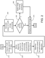
- FIG. 1 a flowchart is illustrated as setting forth the steps of an example method for denoising an image, or otherwise improving the image quality of an image, obtained with a CT system based on exploiting redundant information between the image and another image obtained with the CT system.
- the method includes accessing CT data with a computer system, as indicated at step 102.
- the CT data may be accessed by retrieving previously acquired CT data from a memory or other data storage device or medium.
- the CT data may be accessed by acquiring such data with a CT system and communicating or otherwise transmitting the acquired CT data to the computer system, which may be a part of the CT system.
- the CT data may include raw data acquired with the CT system, images reconstructed from raw data acquired with the CT system, qualitative or quantitative images generated from reconstructed images (e.g., output from a multi-energy processing), or combinations thereof.
- the CT data may include single energy CT data, multi-energy CT data, or both.
- the CT data includes first data and second data.
- the first data may include a first image of a subject and the second data may include a second image of the subject.
- the first image and the second image share redundant information, which may include spatial redundancy, spectral redundancy, or spatiospectral redundancy.
- the first image may be a thin slice image and the second image may be a thick slice image that has a thicker slice thickness than the first image, but otherwise shares common spatial information with the first image.
- the first image may correspond to a thin slice that is spatially contained within the thicker slice associated with the second image.
- the first image may be an image output from a multi-energy processing and the second image may be a multi-energy image.
- the first image may be a material-specific image, a virtual monoenergetic image, an electron density image, an effective atomic number image, a Compton effect image, a photoelectric effect image, or other such image generated from a multi-energy processing.
- the redundant data between the first image and the second image can be exploited to reduce noise, or otherwise improve the image quality, of the first image.
- the first image and the second image are input to an objective function.
- the objective function generally includes a data consistency term and a regularization term.
- the regularization term preferably includes a first component and a second component.
- the first component includes a total variation of the first image, and in some instances can include other norms or transformations of the first image.
- the second component promotes sparsity in a difference between the first image and the second image in order to exploit the redundant data in the first and second images.
- the second component can implement a total variation of the difference between the first and second images; however, other norms or transformations of the difference between the first and second images may also be used in some instances.
- the first and second components can be weighted to control a balance between the two components on the regularization term.
- the objective function is then minimized using a computer system in order to iteratively reduce noise in the first image, or to otherwise improve the image quality of the first image, as indicated at step 106. That is, minimizing the objective function generates output as the denoised image, or otherwise improved image. This output image can then be stored for later use, or displayed to a user, as indicated at step 108.
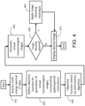
- Multi-energy CT image data are acquired at step 210.
- Multi-energy CT data processing begins at step 220 by designing an objective function for multi-energy processing which incorporates the spatiospectral redundancy between the decomposition basis images (or images related to decomposition basis images) and the measured multi-energy CT images, and proceeds by the solution to this objective function being performed by an optimization algorithm at 230.
- the output of multi-energy processing is updated at 240 based upon the results of the iterative optimization from step 230.
- a user may select to perform image blending at step 250.
- a linear blending technique to combine the output of the present method with a standard least-squares based decomposition may be used at 260 to maintain the original image texture while reducing noise.
- a final image or data output reflecting the material composition of the subject being imaged is displayed at 270.
- the multi-energy processing refers to basis material decomposition
- the output refers to material-specific images such as iodine, soft tissue images.
- the multi-energy processing refers to decomposition based on different physical effects or based on basis material, and the like.
- the output refers to virtual monoenergetic images at a single energy level.
- the multi-energy processing refers to decomposition based on different physical effects
- the output refers to images denoting the intensity of different physical effects such as Compton and photoelectric effects.
- the multi-energy processing refers to decomposition into electron density and effective atomic number images
- the output refers to electron density and effective atomic number images.
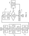
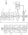
- Multi-energy CT image data are acquired at step 710.
- Standard material decomposition or another relevant multi-energy CT processing algorithm is performed at step 720 to generate output images such as material specific images and virtual monoenergetic images.
- the denoising of the output from multi-energy processing is then performed at step 730 by designing an objective function which incorporates the spatiospectral redundancy between the decomposition basis images (or images related to decomposition basis images) and the measured multi-energy CT data, and proceeds by the solution to this objective function being performed by an optimization algorithm at 740.
- the output of multi-energy CT data processing includes, but is not limited to, material specific images, electron density images, photoelectric images and virtual monoenergetic images.
- the denoised images were then updated based upon the results of the iterative optimization from step 750.
- the user may select at step 760 to apply a similar blending technique at step 770 using images before and after denoising to maintain the original image texture.
- a final image or data output reflecting the denoised version of multi-energy CT processing output is displayed at 780.
- kidney stone characterization determinee elemental composition of stones
- plaque characterization determine elemental composition of plaques
- bone, plaque, or calcium removal identify calcified object and virtually remove the calcium signal
- virtual non-contrast identify iodine signal and remove it
- differentiation between gout and pseudo-gout determinee elemental composition of crystal within join spaces
- virtual mono-energetic images determinee elemental composition of materials in an image and use known attenuation coefficients for those materials to synthesize images at any selected photon energy
- rho/Z decomposition or photoelectric and Compton effects.
- an image formation model is provided.
- the multi-energy CT images reconstructed from data acquired using different energy spectra are represented using decomposition bases, such as basis material specific images, Compton and photoelectric effect images.
- decomposition bases such as basis material specific images, Compton and photoelectric effect images.
- the multi-energy CT images are decomposed into a series of material-specific images.
- w can represent material-specific images, or other decomposition bases such as images de
- f vectorized format of ⁇ eff ( n, e )
- E is the number of energy spectra settings.
- N is the number of pixels.
- w vectorized format of w i ( n ) concatenated for different decomposition bases
- M is the number of decomposition bases.
- w represents the material specific images (i.e., water/iodine/calcium, etc.) and describes the concentration (mass density or volume fraction in each voxel) of each basis material.
- Each element m e,m in ⁇ 0 represents the x-ray attenuation or CT number per volume/mass density for each material at different energy levels, and ⁇ is the Kronecker product.
- the ⁇ represents system matrix can be obtained from either image domain measurements or projection domain measurements using a calibration phantom with material of known material density. It can also assume theoretical values based on physical principles.
- the matrix representation of ⁇ can also include an additional row to account for the volume conservation or mass conservation constraint which allows for solving for M+1 materials.
- CT images may be reconstructed from x-ray photons within different energy windows/bins acquired using various techniques mentioned previously. These energy windows/bins may be overlapped.
- the calibration matrix takes on the form of an E by M matrix.
- the number of materials to be decomposed needs to be smaller than the number of available energy settings, i.e., M ⁇ E, as the system matrix ⁇ is otherwise rank deficient.
- a total of M+1 material-specific images can be calculated.
- ⁇ i represents the mass density of the i-th basis material.
- the (M+1)-th material e.g., water
- the (M+1)-th material can be later solved by assuming the volume fractions within a voxel of different material add up to unity.
- g represents projection data after forward projection operator A.
- Forward projection operator A is a linear operator when g is already processed with a logarithm operation. A becomes a nonlinear operation for the more general case with g denoting the photon count data or raw energy integrating signal strength.
- P i denotes a binary diagonal matrix selecting the i-th decomposition basis image w i from w
- f MD i denotes the regularization term applied to the i-th decomposition basis image w i
- c is a parameter that controls the relative weight between total variation (TV) of each decomposition basis image itself, ⁇ w i ⁇ TV , and the difference between the decomposition basis image and its prior image, ⁇ w i - ⁇ i ( f prior ) ⁇ TV .
- ⁇ w i ⁇ TV is a term that directly minimizes the total variation (TV) of each decomposition basis image.
- the ⁇ w i - ⁇ i ( f prior ) ⁇ TV term applies a sparsifying transform to the difference between the decomposition image and its corresponding material prior image.
- ⁇ w i - ⁇ i ( f prior ) ⁇ TV it is assumed that the individual decomposition image (w i ) and the basis material prior image ⁇ i ( f prior ) share structural redundancy, so that after removing the contribution of each decomposition image (w i ), the residual image is still sparse after TV transform.
- ⁇ prior,i represents an empirical calibration coefficient and is a scaling factor that converts the prior spectral CT image to a prior decomposition basis image and can be obtained from the same calibration scan using data from the corresponding energy spectrum.
- the algorithm reduces to the TV-penalized material decomposition.
- c 1
- the algorithm uses structural similarity between the image and the prior image to perform material decomposition.
- Standard, direct material decomposition is equivalent to performing decomposition without either regularization terms by only minimizing the data fidelity term from a least-squares sense.
- ⁇ i ( f prior ) can also be other types of transformation using f prior .
- the measured prior spectral CT image f prior may be a high SNR image.
- a PCD system such as a research whole-body CT system
- the CT image reconstructed using all the available photons of different energy spectra typically has the highest SNR, which can be used as f prior .
- a conventional CT system uses energy integrated detectors (EID). On these systems, an image with higher SNR and reasonable contrast can be used as the prior image.
- one low-noise CT image e.g., "tube A” image
- a weighted mixed image between tube A and B sub systems can be used as the prior image.
- Data processing may include using different c values ranging from 0 to 1 (with 0.1 increment) depending upon the effect of parameter c on decomposition performance.
- the denoising performance and decomposition accuracy may be quantified using root-mean-square error (RMSE), which accounts for both noise and bias in the material specific images.
- RMSE root-mean-square error
- the parameter c can be determined by evaluating the bias, noise, and RMSE of the decomposed material specific images based on phantom measurements of known material compositions.
- the algorithm parameter ⁇ controls the preference of data fidelity term over regularization term, and is usually determined empirically. A larger ⁇ can be used for high SNR images or high dose scan, while for low SNR scans a smaller ⁇ can be used. For example, a user may select a ⁇ from 1 ⁇ 10 2 to 5 ⁇ 10 3 in order to yield desirable denoising performance.
- the objective function can incorporate the synthetization of other multi-energy processing output, such as other decomposition basis images or virtual monoenergetic images.
- the optimization problem is solved by an adaptive gradient descent algorithm.
- the optimization problem is solved by an alternating direction method of multiplier (ADMM) algorithm. Any other appropriate numerical algorithm may be used to solve the optimization problem, such as primal-dual algorithm, conjugate gradient algorithm, Newton's method, or Quasi Newton's method like Broyden-Fletcher-Goldfarb-Shanno algorithm and the like.
- the optimization problem is solved iteratively by an alternating direction method of multiplier (ADMM) algorithm.
- the ADMM algorithm is a flexible numerical optimization strategy that can be used for a variety of convex optimization problems. It decomposes a compound target optimization problem into a series of relatively easier tasks.
- ⁇ i denotes the Lagrangian multiplier vector
- ⁇ is an optimization constant that controls the rate of convergence of the ADMM algorithm and is typically determined empirically.
- ADMM the minimization of L ( w, w i , ⁇ i ) is done sequentially with respect to w and w i , followed by an update for ⁇ i .
- phantom data was acquired on a research, whole-body photon-counting-detector-based CT system and on a dual-source, dual-energy CT system. Phantom results show that the proposed multi-energy processing can reduce the root-mean-square-error of basis material quantification by 75% compared to the standard material decomposition based on matrix inversion, while preserving structural details and image resolution in the material-specific images.
- Noise reduction algorithms can change image texture and create patchy looking images.
- a linear blending technique to combine the output of multi-energy processing from the present method with the standard least-squares based method may be used to maintain the original image texture while reducing noise.
- the spectral CT images reconstructed from data acquired using different energy spectra are decomposed into material-specific images.
- spectral CT images are decomposed to other decomposition bases. These include, but not limited to, effective atomic number and electron density, photoelectric effect and Compton effect images.
- the objective function can incorporate the synthetization of other multi-energy processing output, such as other decomposition basis images or virtual monoenergetic images. A constrained optimization scheme may be used. The synthetization can be performed during or after solving the optimization problem.
- the output images of other multi-energy CT processing methods can be processed and denoised using the present disclosure to enhance certain image features, such as reduced noise and enhanced CNR, by minimizing an objective function that exploits the spatiospectral data redundancy between the source multi-energy processing output and the multi-energy CT images.
- f prior is the low-noise, spectral prior image associated with the same imaging object.
- ⁇ ( f prior ) is a transformation applied to f prior to convert it to the similar scale and meaning of x.
- ⁇ ( f prior ) can be similar to the one used in previous sections.
- x represent the virtual monoenergetic image of interest
- ⁇ ( f prior ) can be reduced to f prior itself.
- the objective function contains a data fidelity term x ⁇ x 0 2 2 and a regularization term c ⁇ x ⁇ TV + (1 - c ) ⁇ x - ⁇ ( f prior ) ⁇ TV , with ⁇ controlling the relative contribution of the two terms.
- the value of ⁇ is usually determined empirically based on the source image noise level and the desired denoising level.
- the regularization term can be further separated into two components.
- the first component ⁇ x ⁇ TV is the conventional TV regularization term.
- the second component ⁇ x - ⁇ ( f prior ) ⁇ TV promotes spatiospectral sparsity by applying TV transform to the difference image between the virtual monoenergetic image, x, and its corresponding spectral prior image, ⁇ ( f prior ). Therefore, including this term exerts spatiospectral sparsity regularization on x.
- c is a parameter (0 ⁇ c ⁇ 1) that controls the relative preference placed on one term versus the other.
- the parameter c can be determined based on image resolution evaluation using modulation transfer function and noise texture evaluation using noise power spectra.
- the optimization problems shown in Eq. 1 can be solved using a standard iterative solver such as gradient descent algorithm.
- the optimization problem can also be solved using other relevant optimization algorithms such as conjugate gradient, ADMM, primal dual algorithm, Newton's method and quasi Newton's method.
- the objective function can also incorporate the synthetization of other multi-energy CT output such as other decomposition bases or virtual monoenergetic images. Synthetization can also be performed after solving the optimization problem.
- a prior knowledge aware iterative denoising technique may utilize structural similarity between a thicker slice and a thinner slice to perform denoising.
- the spatial redundancy in the slice increment direction is used to reduce image noise while preserving the same resolution, and/or noise texture and/or slice sensitivity profile of the original images.
- a thin slice image with comparable noise levels as that of a thicker slice image may be produced while taking advantage of reduced partial volume effect of a thin slice image.
- Low-noise, thin-slice, high-resolution images may be produced to provide better visualization of fine anatomical structures.
- the method may allow for the diagnostic value of CT images to be improved by satisfying the clinical demands for low image noise and reduced partial volume effect of a thin slice image.
- image sharpness, noise texture, and slice profile of the original image is retained before denoising.
- a non-limiting example of a slice profile includes a cross-section graph of contrast levels across a slice.
- a thin-slice image may be denoised with a lower noise level, reduced partial volume effect, and the image may retain key pathology visibility. Parameters may be adjusted to achieve a desired level of noise reduction, noise texture, and/or image artifact.
- Non-limiting example cases where key pathology visibility may be important include non-contrast head CT cases, and the like.
- Non-limiting example high-resolution CT images that may benefit from reduced noise include thin-slice, high-resolution wrist images, and the like.
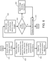
- CT image data that includes thick slice data are acquired, or otherwise accessed, at step 810.
- the denoising of the data is then performed at step 820 by designing an objective function which incorporates the spatial data redundancy between the prior, thick slice, image and the image to be denoised, and proceeds by the solution to this objective function being performed by an optimization algorithm at 830.
- the denoised images are then updated based upon the results of the iterative optimization at step 840.
- the user may select at step 850 to apply a similar blending technique at step 860 using images before and after denoising to maintain the original image texture.
- a final image or data output reflecting the denoised version of input image is displayed at 870.
- the final image may include higher resolution and lower noise, and may be displayed for a user or stored for future use in an appropriate image storage system.
- Equation (1) above may be used where the operator ⁇ TV calculates the total-variation (TV) of a vector; x represents the denoised images; x 0 represents the input thin-slice image to be denoised.
- f prior is the low-noise, prior image at the same spatial location, or a nearby spatial location, but with a larger slice thickness.
- a nearby spatial location may be defined by a user as being within a certain selected distance, or a certain number of slices.
- the objective function contains a data fidelity term x ⁇ x 0 2 2 and a regularization term c ⁇ x ⁇ TV + (1 - c ) ⁇ x - f priorTV , with ⁇ controlling the relative contribution of the two terms.
- the regularization term can be further separated into two components.
- the first component ⁇ x ⁇ TV penalizes the total variation of the image, while the second component ⁇ x - f prior ⁇ TV exploits the structural similarity between the prior image and the image to be denoised by promoting the sparsity in their difference image.
- the method may be implemented in the time domain.
- the method for denoising using a larger slice thickness may be applied to images acquired on various CT systems from different vendors and can be utilized for all types of CT applications including for any body part or pathology. Iterative reconstruction techniques may also be employed with the method such as those provided by system vendors or other research techniques to further lower image noise.
- the system and method may be used to grade cartilage health.
- Conventional techniques are invasive and require multiple instruments.
- the system and method of the present disclosure may provide for quantitative Glycosaminoglycans (GAG) information, such as by using a targeted contrast agent at a high spatial resolution. This may also allow for assessing osteoarthritis (OA).
- GAG Glycosaminoglycans
- OA osteoarthritis
- PCD-CT acquires spectral data, which may be used to quantify the mass density of specific K-edge contrast agents, such as gadolinium, which in turn may be used to mark the GAG levels within a voxel of interest.
- Dotarem Gd-based anionic contrast, FDA approved for MRI intravenous administration
- density may be quantified using a material decomposition process, as described above.
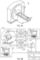
- the CT system includes a gantry 902, to which at least one x-ray source 904 is coupled.
- the x-ray source 904 projects an x-ray beam 906, which may be a fan-beam or cone-beam of x-rays, towards a detector array 908 on the opposite side of the gantry 902.
- the detector array 908 includes a number of x-ray detector elements 910. Together, the x-ray detector elements 910 sense the projected x-rays 906 that pass through a subject 912, such as a medical patient or an object undergoing examination, that is positioned in the CT system 900.
- Each x-ray detector element 910 produces an electrical signal that may represent the intensity of an impinging x-ray beam and, hence, the attenuation of the beam as it passes through the subject 912.
- each x-ray detector 910 is capable of counting the number of x-ray photons that impinge upon the detector 910.
- the system can include a second x-ray source and a second x-ray detector (not shown) operable at a different energy level than x-ray source 904 and detector 910. Any number of x-ray sources and corresponding x-ray detectors operable at different energies may be used, or a single x-ray source 904 may be operable to emit different energies that impinge upon detector 910.
- the gantry 902 and the components mounted thereon rotate about a center of rotation 914 located within the CT system 900.
- the CT system 900 also includes an operator workstation 916, which typically includes a display 918; one or more input devices 920, such as a keyboard and mouse; and a computer processor 922.
- the computer processor 922 may include a commercially available programmable machine running a commercially available operating system.
- the operator workstation 916 provides the operator interface that enables scanning control parameters to be entered into the CT system 900.
- the operator workstation 916 is in communication with a data store server 924 and an image reconstruction system 926.
- the operator workstation 916, data store sever 924, and image reconstruction system 926 may be connected via a communication system 928, which may include any suitable network connection, whether wired, wireless, or a combination of both.
- the communication system 928 may include both proprietary or dedicated networks, as well as open networks, such as the internet.
- the operator workstation 916 is also in communication with a control system 930 that controls operation of the CT system 900.
- the control system 930 generally includes an x-ray controller 932, a table controller 934, a gantry controller 936, and a data acquisition system 938.
- the x-ray controller 932 provides power and timing signals to the x-ray source 904 and the gantry controller 936 controls the rotational speed and position of the gantry 902.
- the table controller 934 controls a table 940 to position the subject 912 in the gantry 902 of the CT system 900.
- the DAS 938 samples data from the detector elements 910 and converts the data to digital signals for subsequent processing. For instance, digitized x-ray data are communicated from the DAS 938 to the data store server 924.
- the image reconstruction system 926 then retrieves the x-ray data from the data store server 924 and reconstructs an image therefrom.
- the image reconstruction system 926 may include a commercially available computer processor, or may be a highly parallel computer architecture, such as a system that includes multiple-core processors and massively parallel, high-density computing devices.
- image reconstruction can also be performed on the processor 922 in the operator workstation 916. Reconstructed images can then be communicated back to the data store server 924 for storage or to the operator workstation 916 to be displayed to the operator or clinician.
- the CT system 900 may also include one or more networked workstations 942.
- a networked workstation 942 may include a display 944; one or more input devices 946, such as a keyboard and mouse; and a processor 948.
- the networked workstation 942 may be located within the same facility as the operator workstation 916, or in a different facility, such as a different healthcare institution or clinic.
- the networked workstation 942 may gain remote access to the data store server 924 and/or the image reconstruction system 926 via the communication system 928. Accordingly, multiple networked workstations 942 may have access to the data store server 924 and/or image reconstruction system 926. In this manner, x-ray data, reconstructed images, or other data may be exchanged between the data store server 924, the image reconstruction system 926, and the networked workstations 942, such that the data or images may be remotely processed by a networked workstation 942.
- This data may be exchanged in any suitable format, such as in accordance with the transmission control protocol ("TCP"), the internet protocol (“IP”), or other known or suitable protocols.
- TCP transmission control protocol
- IP internet protocol
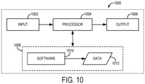
- the computer system 1000 generally includes an input 1002, at least one hardware processor 1004, a memory 1006, and an output 1008.
- the computer system 1000 is generally implemented with a hardware processor 1004 and a memory 1006.
- the computer system 1000 can be a workstation, a notebook computer, a tablet device, a mobile device, a multimedia device, a network server, a mainframe, one or more controllers, one or more microcontrollers, or any other general-purpose or application-specific computing device.
- the computer system 1000 may operate autonomously or semiautonomously, or may read executable software instructions from the memory 1006 or a computer-readable medium (e.g., a hard drive, a CD-ROM, flash memory), or may receive instructions via the input 1002 from a user, or any another source logically connected to a computer or device, such as another networked computer or server.
- a computer-readable medium e.g., a hard drive, a CD-ROM, flash memory
- the computer system 1000 can also include any suitable device for reading computer-readable storage media.
- the computer system 1000 is programmed or otherwise configured to implement the methods and algorithms described in the present disclosure.
- the computer system 1000 can be programmed to denoised images or otherwise improve the image quality of images acquired with a CT system in accordance with embodiments described in the present disclosure.
- the input 1002 may take any suitable shape or form, as desired, for operation of the computer system 1000, including the ability for selecting, entering, or otherwise specifying parameters consistent with performing tasks, processing data, or operating the computer system 1000.
- the input 1002 may be configured to receive data, such as data acquired with a CT system. Such data may be processed as described above to improve the image quality of an input image based by exploiting redundant data in an iterative optimization algorithm, such as those described above.
- the input 1002 may also be configured to receive any other data or information considered useful for generating images with improved image quality, such as reduced noise, using the methods described above.
- the one or more hardware processors 1004 may also be configured to carry out any number of post-processing steps on data received by way of the input 1002.
- the memory 1006 may contain software 1010 and data 1012, such as data acquired with a CT system, and may be configured for storage and retrieval of processed information, instructions, and data to be processed by the one or more hardware processors 1004.
- the software 1010 may contain instructions directed to generating a denoised image, or an image with otherwise improved image quality, by exploiting redundant data between two input images having spatial redundancy, spectral redundancy, or spatiospectral redundancy.
- the output 1008 may take any shape or form, as desired, and may be configured for displaying images, such as denoised image, in addition to other desired information.
- any suitable computer readable media can be used for storing instructions for performing the functions and/or processes described herein.
- computer readable media can be transitory or non-transitory.
- non-transitory computer readable media can include media such as magnetic media (e.g., hard disks, floppy disks), optical media (e.g., compact discs, digital video discs, Blu-ray discs), semiconductor media (e.g., random access memory (“RAM”), flash memory, electrically programmable read only memory (“EPROM”), electrically erasable programmable read only memory (“EEPROM”)), any suitable media that is not fleeting or devoid of any semblance of permanence during transmission, and/or any suitable tangible media.
- RAM random access memory
- EPROM electrically programmable read only memory
- EEPROM electrically erasable programmable read only memory
- transitory computer readable media can include signals on networks, in wires, conductors, optical fibers, circuits, or any suitable media that is fleeting and devoid of any semblance of permanence during transmission, and/or any suitable intangible media.
Landscapes
- Engineering & Computer Science (AREA)
- Health & Medical Sciences (AREA)
- Life Sciences & Earth Sciences (AREA)
- Medical Informatics (AREA)
- Physics & Mathematics (AREA)
- Public Health (AREA)
- Biomedical Technology (AREA)
- Pathology (AREA)
- General Health & Medical Sciences (AREA)
- Theoretical Computer Science (AREA)
- Biophysics (AREA)
- Veterinary Medicine (AREA)
- Nuclear Medicine, Radiotherapy & Molecular Imaging (AREA)
- Radiology & Medical Imaging (AREA)
- High Energy & Nuclear Physics (AREA)
- Heart & Thoracic Surgery (AREA)
- Molecular Biology (AREA)
- Surgery (AREA)
- Animal Behavior & Ethology (AREA)
- Optics & Photonics (AREA)
- General Physics & Mathematics (AREA)
- Computer Vision & Pattern Recognition (AREA)
- Pulmonology (AREA)
- Data Mining & Analysis (AREA)
- Databases & Information Systems (AREA)
- Epidemiology (AREA)
- Primary Health Care (AREA)
- Apparatus For Radiation Diagnosis (AREA)
- Image Processing (AREA)
Claims (12)
- Verfahren zur Verringerung des Rauschens in einem Bild, das unter Verwendung eines Computertomographiesystems (CT) erhalten wurde, wobei das Verfahren umfasst:(a) Zugreifen mit einem Computersystem auf CT-Daten, die von einem Subjekt unter Verwendung des CT-Systems erfasst wurden, wobei die CT-Daten ein erstes Bild des Subjekts und ein zweites Bild des Subjekts umfassen, wobei das erste Bild und das zweite Bild redundante Daten enthalten;(b) Erzeugen eines entrauschten Bildes, das im Vergleich zum ersten Bild ein reduziertes Rauschen aufweist, mit dem Computersystem durch:dadurch gekennzeichnet, dass das erste Bild eine geringere Schnittdicke als das zweite Bild aufweist und die redundanten Daten strukturelle Ähnlichkeiten zwischen dem ersten Bild und dem zweiten Bild umfassen.Eingeben des ersten Bildes und des zweiten Bildes in eine Zielfunktion, die einen Regularisierungsterm enthält, der die redundanten Daten in dem ersten Bild und dem zweiten Bild ausnutzt;Minimieren der Zielfunktion, um das erste Bild iterativ zu entrauschen, wobei das entrauschte Bild als Ausgabe erzeugt wird,
- Verfahren nach Anspruch 1, wobei der Regularisierungsterm eine erste Komponente und eine zweite Komponente enthält, wobei die erste Komponente des Regularisierungsterms eine Gesamtvariation des ersten Bildes umfasst und die zweite Komponente des Regularisierungsterms die redundanten Daten in dem ersten Bild und dem zweiten Bild ausnutzt, indem sie die Streuung in einer Differenz zwischen dem ersten Bild und dem zweiten Bild fördert.
- Verfahren nach Anspruch 1, wobei die strukturellen Ähnlichkeiten eine räumliche Redundanz in der Richtung eines Schnittinkrements umfassen.
- Verfahren nach Anspruch 1, wobei das entrauschte Bild mindestens eines von einer Bildauflösung, einer Rauschtextur oder einem Schnittempfindlichkeitsprofil aufweist, die mit dem ersten Bild übereinstimmen.
- Verfahren nach Anspruch 1, wobei sich das zweite Bild an der gleichen räumlichen Position wie das erste Bild befindet.
- Verfahren nach Anspruch 1, wobei die CT-Daten Multi-Energie-Spektral-CT-Daten umfassen, das zweite Bild ein Multi-Energie-Bild ist und die redundanten Daten entweder spektrale Redundanz oder räumliche Redundanz zwischen dem ersten Bild und dem zweiten Bild umfassen.
- Verfahren nach Anspruch 6, wobei das erste Bild eines der folgenden Bilder umfasst: ein materialspezifisches Bild, ein virtuelles monoenergetisches Bild, ein Elektronendichtebild, ein Bild der effektiven Ordnungszahl, ein Compton-Effekt-Bild oder ein photoelektrisches Effektbild.
- Verfahren nach Anspruch 1, bei dem die Zielfunktion iterativ minimiert wird, wobei mindestens einer der folgenden Algorithmen verwendet wird: ein ADMM-Algorithmus (Alternating Direction Method of Multiplier), ein Gradientenabstiegsalgorithmus, ein primär-dualer Algorithmus, ein konjugierter Gradientenalgorithmus, das Newton-Verfahren und ein Quasi-Newton-Verfahren wie der Broyden-Fletcher-Goldfarb-Shanno-Algorithmus.
- Verfahren nach Anspruch 1, wobei das Erzeugen des entrauschten Bildes mit dem Computersystem ferner das Überblenden des entrauschten Bildes mit einem anderen Bild des Subjekts umfasst, das aus den CT-Daten erzeugt wurde, um ein natürliches Bildaussehen und eine Rauschtextur in dem entrauschten Bild zu erzielen.
- Verfahren nach Anspruch 9, wobei das entrauschte Bild und das andere Bild auf der Grundlage eines Überblendungsparameters, der die Stärke der Überblendung angibt, gemischt werden.
- Verfahren nach Anspruch 9, wobei das entrauschte Bild und das andere Bild unter Verwendung einer linearen Überblendung gemischt werden.
- Verfahren nach Anspruch 1, wobei:die CT-Daten Multi-Energie-CT-Daten umfassen;die Zielfunktion so konfiguriert ist, dass sie M Materialien umfasst, so dass das erste Bild für jedes der M Materialien eingegeben wird; undmindestens eine der physikalischen, chemischen, volumen- oder masseerhaltenden Randbedingungen in die Zielfunktion aufgenommen wird, um die Durchführung der Materialzersetzung bei M+1 Materialien auszulegen.
Priority Applications (1)
| Application Number | Priority Date | Filing Date | Title |
|---|---|---|---|
| EP25182514.7A EP4592959A3 (de) | 2018-06-29 | 2019-06-28 | System und verfahren für computertomografie mit hoher genauigkeit |
Applications Claiming Priority (2)
| Application Number | Priority Date | Filing Date | Title |
|---|---|---|---|
| US201862692350P | 2018-06-29 | 2018-06-29 | |
| PCT/US2019/039725 WO2020006352A1 (en) | 2018-06-29 | 2019-06-28 | System and method for high fidelity computed tomography |
Related Child Applications (1)
| Application Number | Title | Priority Date | Filing Date |
|---|---|---|---|
| EP25182514.7A Division EP4592959A3 (de) | 2018-06-29 | 2019-06-28 | System und verfahren für computertomografie mit hoher genauigkeit |
Publications (2)
| Publication Number | Publication Date |
|---|---|
| EP3813670A1 EP3813670A1 (de) | 2021-05-05 |
| EP3813670B1 true EP3813670B1 (de) | 2025-06-18 |
Family
ID=67297423
Family Applications (2)
| Application Number | Title | Priority Date | Filing Date |
|---|---|---|---|
| EP19740276.1A Active EP3813670B1 (de) | 2018-06-29 | 2019-06-28 | System und verfahren für high-fidelity-computertomographie |
| EP25182514.7A Pending EP4592959A3 (de) | 2018-06-29 | 2019-06-28 | System und verfahren für computertomografie mit hoher genauigkeit |
Family Applications After (1)
| Application Number | Title | Priority Date | Filing Date |
|---|---|---|---|
| EP25182514.7A Pending EP4592959A3 (de) | 2018-06-29 | 2019-06-28 | System und verfahren für computertomografie mit hoher genauigkeit |
Country Status (3)
| Country | Link |
|---|---|
| US (1) | US12056796B2 (de) |
| EP (2) | EP3813670B1 (de) |
| WO (1) | WO2020006352A1 (de) |
Families Citing this family (7)
| Publication number | Priority date | Publication date | Assignee | Title |
|---|---|---|---|---|
| DE102018221691A1 (de) | 2018-12-13 | 2020-06-18 | Siemens Healthcare Gmbh | Individuell angepasstes Erzeugen von virtuellen Bilddaten auf Basis einer Multi-Energie-Röntgenbildgebung |
| CN112424835B (zh) * | 2020-05-18 | 2023-11-24 | 上海联影医疗科技股份有限公司 | 用于图像重建的系统和方法 |
| CA3124052C (en) | 2020-06-08 | 2022-05-10 | Guangzhou Computational Super-Resolution Biotech Co., Ltd. | Systems and methods for image processing |
| US20240237956A1 (en) * | 2021-07-13 | 2024-07-18 | Mayo Foundation For Medical Education And Research | Systems, Methods, and Media for Generating Low-Energy Virtual Monoenergetic Images from Multi-Energy Computed Tomography Data |
| US20230157657A1 (en) * | 2021-11-24 | 2023-05-25 | Canon Medical Systems Corporation | X-ray diagnostic apparatus, medical image processing apparatus, and medical image processing method |
| US20240338866A1 (en) * | 2024-06-18 | 2024-10-10 | Mayo Foundation For Medical Education And Research | System and Method for High Fidelity Computed Tomography |
| CN119646552B (zh) * | 2024-12-03 | 2025-07-11 | 香港中文大学(深圳) | 基于聚类的异构联邦基础模型自适应微调方法及计算机装置 |
Family Cites Families (5)
| Publication number | Priority date | Publication date | Assignee | Title |
|---|---|---|---|---|
| US9558570B2 (en) * | 2013-04-16 | 2017-01-31 | The Research Foundation For The State University Of New York | Iterative reconstruction for X-ray computed tomography using prior-image induced nonlocal regularization |
| NZ737271A (en) | 2015-04-20 | 2019-08-30 | Mars Bioimaging Ltd | Improving material identification using multi-energy ct image data |
| US10332281B2 (en) * | 2016-01-29 | 2019-06-25 | Wisconsin Alumni Research Foundation | System and method for denoising medical images by enforcing low rank spatial-temporal or spatial-spectral image matrices |
| US11328391B2 (en) * | 2016-05-06 | 2022-05-10 | Mayo Foundation For Medical Education And Research | System and method for controlling noise in multi-energy computed tomography images based on spatio-spectral information |
| US10360677B2 (en) * | 2016-10-07 | 2019-07-23 | Toshiba Medical Systems Corporation | Apparatus and method for joint-edge-preserving regularization to reduce noise in four-dimensional computed tomography images |
-
2019
- 2019-06-28 US US17/256,404 patent/US12056796B2/en active Active
- 2019-06-28 WO PCT/US2019/039725 patent/WO2020006352A1/en not_active Ceased
- 2019-06-28 EP EP19740276.1A patent/EP3813670B1/de active Active
- 2019-06-28 EP EP25182514.7A patent/EP4592959A3/de active Pending
Also Published As
| Publication number | Publication date |
|---|---|
| US20210319600A1 (en) | 2021-10-14 |
| EP4592959A3 (de) | 2025-10-15 |
| WO2020006352A1 (en) | 2020-01-02 |
| EP4592959A2 (de) | 2025-07-30 |
| US12056796B2 (en) | 2024-08-06 |
| EP3813670A1 (de) | 2021-05-05 |
Similar Documents
| Publication | Publication Date | Title |
|---|---|---|
| EP3813670B1 (de) | System und verfahren für high-fidelity-computertomographie | |
| US11592586B2 (en) | Methods for optimizing imaging technique parameters for photon-counting computed tomography | |
| Garnett | A comprehensive review of dual-energy and multi-spectral computed tomography | |
| US11176642B2 (en) | System and method for processing data acquired utilizing multi-energy computed tomography imaging | |
| US10147168B2 (en) | Spectral CT | |
| US8160206B2 (en) | Dual-energy imaging at reduced sample rates | |
| US9036886B2 (en) | System and method for correcting for metal artifacts using multi-energy computed tomography | |
| EP2958494B1 (de) | Strukturausbreitungswiederherstellung für spektral-ct | |
| US10332281B2 (en) | System and method for denoising medical images by enforcing low rank spatial-temporal or spatial-spectral image matrices | |
| US9600866B2 (en) | Projection data de-noising | |
| JP2018140165A (ja) | 医用画像生成装置 | |
| US10111638B2 (en) | Apparatus and method for registration and reprojection-based material decomposition for spectrally resolved computed tomography | |
| CN105793894B (zh) | 根据图像数据来进行骨骼分割 | |
| US11350895B2 (en) | System and method for spectral computed tomography using single polychromatic x-ray spectrum acquisition | |
| Hayes et al. | High pitch helical CT reconstruction | |
| EP4315250B1 (de) | Systeme und verfahren zur multi-kernel-synthese und kernel-umwandlung in der medizinischen bildgebung | |
| US9984476B2 (en) | Methods and systems for automatic segmentation | |
| Natterer et al. | Past and future directions in x‐ray computed tomography (CT) | |
| Peng et al. | Dual-energy cone-beam CT using two complementary limited-angle scans with a projection-consistent diffusion model | |
| CN116868236A (zh) | 用于能谱成像的投影域材料分解 | |
| US20240338866A1 (en) | System and Method for High Fidelity Computed Tomography | |
| Heverhagen | Physics of computed tomography scanning | |
| US10307114B1 (en) | Iterative volume image reconstruction using synthetic projection images | |
| Peng et al. | Dual-Energy Cone-Beam CT Using Two Orthogonal Projection Views: A Phantom Study | |
| Hsieh | An Overview of CT Reconstruction with Applications to Photon Counting Detectors |
Legal Events
| Date | Code | Title | Description |
|---|---|---|---|
| STAA | Information on the status of an ep patent application or granted ep patent |
Free format text: STATUS: UNKNOWN |
|
| STAA | Information on the status of an ep patent application or granted ep patent |
Free format text: STATUS: THE INTERNATIONAL PUBLICATION HAS BEEN MADE |
|
| PUAI | Public reference made under article 153(3) epc to a published international application that has entered the european phase |
Free format text: ORIGINAL CODE: 0009012 |
|
| STAA | Information on the status of an ep patent application or granted ep patent |
Free format text: STATUS: REQUEST FOR EXAMINATION WAS MADE |
|
| 17P | Request for examination filed |
Effective date: 20210122 |
|
| AK | Designated contracting states |
Kind code of ref document: A1 Designated state(s): AL AT BE BG CH CY CZ DE DK EE ES FI FR GB GR HR HU IE IS IT LI LT LU LV MC MK MT NL NO PL PT RO RS SE SI SK SM TR |
|
| DAV | Request for validation of the european patent (deleted) | ||
| DAX | Request for extension of the european patent (deleted) | ||
| STAA | Information on the status of an ep patent application or granted ep patent |
Free format text: STATUS: EXAMINATION IS IN PROGRESS |
|
| 17Q | First examination report despatched |
Effective date: 20230215 |
|
| P01 | Opt-out of the competence of the unified patent court (upc) registered |
Effective date: 20230602 |
|
| REG | Reference to a national code |
Ref country code: DE Ref legal event code: R079 Free format text: PREVIOUS MAIN CLASS: A61B0006030000 Ref country code: DE Ref legal event code: R079 Ref document number: 602019071249 Country of ref document: DE Free format text: PREVIOUS MAIN CLASS: A61B0006030000 Ipc: A61B0006420000 |
|
| GRAP | Despatch of communication of intention to grant a patent |
Free format text: ORIGINAL CODE: EPIDOSNIGR1 |
|
| STAA | Information on the status of an ep patent application or granted ep patent |
Free format text: STATUS: GRANT OF PATENT IS INTENDED |
|
| RIC1 | Information provided on ipc code assigned before grant |
Ipc: G06T 11/00 20060101ALI20240802BHEP Ipc: G06T 5/70 20240101ALI20240802BHEP Ipc: G16H 50/20 20180101ALI20240802BHEP Ipc: A61B 6/00 20060101ALI20240802BHEP Ipc: A61B 6/03 20060101ALI20240802BHEP Ipc: A61B 6/42 20240101AFI20240802BHEP |
|
| INTG | Intention to grant announced |
Effective date: 20240903 |
|
| GRAJ | Information related to disapproval of communication of intention to grant by the applicant or resumption of examination proceedings by the epo deleted |
Free format text: ORIGINAL CODE: EPIDOSDIGR1 |
|
| STAA | Information on the status of an ep patent application or granted ep patent |
Free format text: STATUS: EXAMINATION IS IN PROGRESS |
|
| GRAP | Despatch of communication of intention to grant a patent |
Free format text: ORIGINAL CODE: EPIDOSNIGR1 |
|
| STAA | Information on the status of an ep patent application or granted ep patent |
Free format text: STATUS: GRANT OF PATENT IS INTENDED |
|
| INTC | Intention to grant announced (deleted) | ||
| INTG | Intention to grant announced |
Effective date: 20250113 |
|
| GRAS | Grant fee paid |
Free format text: ORIGINAL CODE: EPIDOSNIGR3 |
|
| GRAA | (expected) grant |
Free format text: ORIGINAL CODE: 0009210 |
|
| STAA | Information on the status of an ep patent application or granted ep patent |
Free format text: STATUS: THE PATENT HAS BEEN GRANTED |
|
| AK | Designated contracting states |
Kind code of ref document: B1 Designated state(s): AL AT BE BG CH CY CZ DE DK EE ES FI FR GB GR HR HU IE IS IT LI LT LU LV MC MK MT NL NO PL PT RO RS SE SI SK SM TR |
|
| REG | Reference to a national code |
Ref country code: GB Ref legal event code: FG4D |
|
| REG | Reference to a national code |
Ref country code: CH Ref legal event code: EP |
|
| REG | Reference to a national code |
Ref country code: DE Ref legal event code: R096 Ref document number: 602019071249 Country of ref document: DE |
|
| REG | Reference to a national code |
Ref country code: CH Ref legal event code: EP |
|
| REG | Reference to a national code |
Ref country code: IE Ref legal event code: FG4D |
|
| PGFP | Annual fee paid to national office [announced via postgrant information from national office to epo] |
Ref country code: NL Payment date: 20250726 Year of fee payment: 7 |
|
| REG | Reference to a national code |
Ref country code: NL Ref legal event code: FP |
|
| PG25 | Lapsed in a contracting state [announced via postgrant information from national office to epo] |
Ref country code: FI Free format text: LAPSE BECAUSE OF FAILURE TO SUBMIT A TRANSLATION OF THE DESCRIPTION OR TO PAY THE FEE WITHIN THE PRESCRIBED TIME-LIMIT Effective date: 20250618 |
|
| PGFP | Annual fee paid to national office [announced via postgrant information from national office to epo] |
Ref country code: DE Payment date: 20250729 Year of fee payment: 7 |
|
| REG | Reference to a national code |
Ref country code: LT Ref legal event code: MG9D |
|
| PG25 | Lapsed in a contracting state [announced via postgrant information from national office to epo] |
Ref country code: GR Free format text: LAPSE BECAUSE OF FAILURE TO SUBMIT A TRANSLATION OF THE DESCRIPTION OR TO PAY THE FEE WITHIN THE PRESCRIBED TIME-LIMIT Effective date: 20250919 Ref country code: NO Free format text: LAPSE BECAUSE OF FAILURE TO SUBMIT A TRANSLATION OF THE DESCRIPTION OR TO PAY THE FEE WITHIN THE PRESCRIBED TIME-LIMIT Effective date: 20250918 |
|
| PG25 | Lapsed in a contracting state [announced via postgrant information from national office to epo] |
Ref country code: BG Free format text: LAPSE BECAUSE OF FAILURE TO SUBMIT A TRANSLATION OF THE DESCRIPTION OR TO PAY THE FEE WITHIN THE PRESCRIBED TIME-LIMIT Effective date: 20250618 |
|
| PG25 | Lapsed in a contracting state [announced via postgrant information from national office to epo] |
Ref country code: HR Free format text: LAPSE BECAUSE OF FAILURE TO SUBMIT A TRANSLATION OF THE DESCRIPTION OR TO PAY THE FEE WITHIN THE PRESCRIBED TIME-LIMIT Effective date: 20250618 |
|
| PG25 | Lapsed in a contracting state [announced via postgrant information from national office to epo] |
Ref country code: RS Free format text: LAPSE BECAUSE OF FAILURE TO SUBMIT A TRANSLATION OF THE DESCRIPTION OR TO PAY THE FEE WITHIN THE PRESCRIBED TIME-LIMIT Effective date: 20250918 |
|
| PG25 | Lapsed in a contracting state [announced via postgrant information from national office to epo] |
Ref country code: LV Free format text: LAPSE BECAUSE OF FAILURE TO SUBMIT A TRANSLATION OF THE DESCRIPTION OR TO PAY THE FEE WITHIN THE PRESCRIBED TIME-LIMIT Effective date: 20250618 |
|
| PG25 | Lapsed in a contracting state [announced via postgrant information from national office to epo] |
Ref country code: PT Free format text: LAPSE BECAUSE OF FAILURE TO SUBMIT A TRANSLATION OF THE DESCRIPTION OR TO PAY THE FEE WITHIN THE PRESCRIBED TIME-LIMIT Effective date: 20251020 |
|
| REG | Reference to a national code |
Ref country code: AT Ref legal event code: MK05 Ref document number: 1803462 Country of ref document: AT Kind code of ref document: T Effective date: 20250618 |
|
| PG25 | Lapsed in a contracting state [announced via postgrant information from national office to epo] |
Ref country code: IS Free format text: LAPSE BECAUSE OF FAILURE TO SUBMIT A TRANSLATION OF THE DESCRIPTION OR TO PAY THE FEE WITHIN THE PRESCRIBED TIME-LIMIT Effective date: 20251018 |
|
| PG25 | Lapsed in a contracting state [announced via postgrant information from national office to epo] |
Ref country code: AT Free format text: LAPSE BECAUSE OF FAILURE TO SUBMIT A TRANSLATION OF THE DESCRIPTION OR TO PAY THE FEE WITHIN THE PRESCRIBED TIME-LIMIT Effective date: 20250618 Ref country code: SM Free format text: LAPSE BECAUSE OF FAILURE TO SUBMIT A TRANSLATION OF THE DESCRIPTION OR TO PAY THE FEE WITHIN THE PRESCRIBED TIME-LIMIT Effective date: 20250618 |
|
| PG25 | Lapsed in a contracting state [announced via postgrant information from national office to epo] |
Ref country code: CZ Free format text: LAPSE BECAUSE OF FAILURE TO SUBMIT A TRANSLATION OF THE DESCRIPTION OR TO PAY THE FEE WITHIN THE PRESCRIBED TIME-LIMIT Effective date: 20250618 |
|
| PG25 | Lapsed in a contracting state [announced via postgrant information from national office to epo] |
Ref country code: PL Free format text: LAPSE BECAUSE OF FAILURE TO SUBMIT A TRANSLATION OF THE DESCRIPTION OR TO PAY THE FEE WITHIN THE PRESCRIBED TIME-LIMIT Effective date: 20250618 |
|
| PG25 | Lapsed in a contracting state [announced via postgrant information from national office to epo] |
Ref country code: EE Free format text: LAPSE BECAUSE OF FAILURE TO SUBMIT A TRANSLATION OF THE DESCRIPTION OR TO PAY THE FEE WITHIN THE PRESCRIBED TIME-LIMIT Effective date: 20250618 |
|
| PG25 | Lapsed in a contracting state [announced via postgrant information from national office to epo] |
Ref country code: SK Free format text: LAPSE BECAUSE OF FAILURE TO SUBMIT A TRANSLATION OF THE DESCRIPTION OR TO PAY THE FEE WITHIN THE PRESCRIBED TIME-LIMIT Effective date: 20250618 Ref country code: RO Free format text: LAPSE BECAUSE OF FAILURE TO SUBMIT A TRANSLATION OF THE DESCRIPTION OR TO PAY THE FEE WITHIN THE PRESCRIBED TIME-LIMIT Effective date: 20250618 |
|
| REG | Reference to a national code |
Ref country code: CH Ref legal event code: H13 Free format text: ST27 STATUS EVENT CODE: U-0-0-H10-H13 (AS PROVIDED BY THE NATIONAL OFFICE) Effective date: 20260127 |
|
| PG25 | Lapsed in a contracting state [announced via postgrant information from national office to epo] |
Ref country code: ES Free format text: LAPSE BECAUSE OF FAILURE TO SUBMIT A TRANSLATION OF THE DESCRIPTION OR TO PAY THE FEE WITHIN THE PRESCRIBED TIME-LIMIT Effective date: 20250618 |
|
| PG25 | Lapsed in a contracting state [announced via postgrant information from national office to epo] |
Ref country code: LU Free format text: LAPSE BECAUSE OF NON-PAYMENT OF DUE FEES Effective date: 20250628 |
|
| REG | Reference to a national code |
Ref country code: BE Ref legal event code: MM Effective date: 20250630 |
|
| PG25 | Lapsed in a contracting state [announced via postgrant information from national office to epo] |
Ref country code: MC Free format text: LAPSE BECAUSE OF FAILURE TO SUBMIT A TRANSLATION OF THE DESCRIPTION OR TO PAY THE FEE WITHIN THE PRESCRIBED TIME-LIMIT Effective date: 20250618 |
|
| PG25 | Lapsed in a contracting state [announced via postgrant information from national office to epo] |
Ref country code: IE Free format text: LAPSE BECAUSE OF NON-PAYMENT OF DUE FEES Effective date: 20250628 Ref country code: DK Free format text: LAPSE BECAUSE OF FAILURE TO SUBMIT A TRANSLATION OF THE DESCRIPTION OR TO PAY THE FEE WITHIN THE PRESCRIBED TIME-LIMIT Effective date: 20250618 |
|
| PG25 | Lapsed in a contracting state [announced via postgrant information from national office to epo] |
Ref country code: BE Free format text: LAPSE BECAUSE OF NON-PAYMENT OF DUE FEES Effective date: 20250630 Ref country code: IT Free format text: LAPSE BECAUSE OF FAILURE TO SUBMIT A TRANSLATION OF THE DESCRIPTION OR TO PAY THE FEE WITHIN THE PRESCRIBED TIME-LIMIT Effective date: 20250618 |
|
| PLBE | No opposition filed within time limit |
Free format text: ORIGINAL CODE: 0009261 |
|
| STAA | Information on the status of an ep patent application or granted ep patent |
Free format text: STATUS: NO OPPOSITION FILED WITHIN TIME LIMIT |