EP3777693A1 - Measurement and data communication device for an intraoral dental radiology system - Google Patents
Measurement and data communication device for an intraoral dental radiology system Download PDFInfo
- Publication number
- EP3777693A1 EP3777693A1 EP19000371.5A EP19000371A EP3777693A1 EP 3777693 A1 EP3777693 A1 EP 3777693A1 EP 19000371 A EP19000371 A EP 19000371A EP 3777693 A1 EP3777693 A1 EP 3777693A1
- Authority
- EP
- European Patent Office
- Prior art keywords
- ray
- data
- measurement
- intraoral
- tube
- Prior art date
- Legal status (The legal status is an assumption and is not a legal conclusion. Google has not performed a legal analysis and makes no representation as to the accuracy of the status listed.)
- Granted
Links
Images
Classifications
-
- A—HUMAN NECESSITIES
- A61—MEDICAL OR VETERINARY SCIENCE; HYGIENE
- A61B—DIAGNOSIS; SURGERY; IDENTIFICATION
- A61B6/00—Apparatus or devices for radiation diagnosis; Apparatus or devices for radiation diagnosis combined with radiation therapy equipment
- A61B6/50—Apparatus or devices for radiation diagnosis; Apparatus or devices for radiation diagnosis combined with radiation therapy equipment specially adapted for specific body parts; specially adapted for specific clinical applications
- A61B6/51—Apparatus or devices for radiation diagnosis; Apparatus or devices for radiation diagnosis combined with radiation therapy equipment specially adapted for specific body parts; specially adapted for specific clinical applications for dentistry
- A61B6/512—Intraoral means
-
- A—HUMAN NECESSITIES
- A61—MEDICAL OR VETERINARY SCIENCE; HYGIENE
- A61B—DIAGNOSIS; SURGERY; IDENTIFICATION
- A61B6/00—Apparatus or devices for radiation diagnosis; Apparatus or devices for radiation diagnosis combined with radiation therapy equipment
- A61B6/56—Details of data transmission or power supply, e.g. use of slip rings
-
- A—HUMAN NECESSITIES
- A61—MEDICAL OR VETERINARY SCIENCE; HYGIENE
- A61B—DIAGNOSIS; SURGERY; IDENTIFICATION
- A61B6/00—Apparatus or devices for radiation diagnosis; Apparatus or devices for radiation diagnosis combined with radiation therapy equipment
- A61B6/06—Diaphragms
-
- A—HUMAN NECESSITIES
- A61—MEDICAL OR VETERINARY SCIENCE; HYGIENE
- A61B—DIAGNOSIS; SURGERY; IDENTIFICATION
- A61B6/00—Apparatus or devices for radiation diagnosis; Apparatus or devices for radiation diagnosis combined with radiation therapy equipment
- A61B6/40—Arrangements for generating radiation specially adapted for radiation diagnosis
- A61B6/405—Source units specially adapted to modify characteristics of the beam during the data acquisition process
-
- A—HUMAN NECESSITIES
- A61—MEDICAL OR VETERINARY SCIENCE; HYGIENE
- A61B—DIAGNOSIS; SURGERY; IDENTIFICATION
- A61B6/00—Apparatus or devices for radiation diagnosis; Apparatus or devices for radiation diagnosis combined with radiation therapy equipment
- A61B6/42—Arrangements for detecting radiation specially adapted for radiation diagnosis
- A61B6/4266—Arrangements for detecting radiation specially adapted for radiation diagnosis characterised by using a plurality of detector units
-
- A—HUMAN NECESSITIES
- A61—MEDICAL OR VETERINARY SCIENCE; HYGIENE
- A61B—DIAGNOSIS; SURGERY; IDENTIFICATION
- A61B6/00—Apparatus or devices for radiation diagnosis; Apparatus or devices for radiation diagnosis combined with radiation therapy equipment
- A61B6/44—Constructional features of apparatus for radiation diagnosis
- A61B6/4411—Constructional features of apparatus for radiation diagnosis the apparatus being modular
-
- A—HUMAN NECESSITIES
- A61—MEDICAL OR VETERINARY SCIENCE; HYGIENE
- A61B—DIAGNOSIS; SURGERY; IDENTIFICATION
- A61B6/00—Apparatus or devices for radiation diagnosis; Apparatus or devices for radiation diagnosis combined with radiation therapy equipment
- A61B6/44—Constructional features of apparatus for radiation diagnosis
- A61B6/4494—Means for identifying the diagnostic device
-
- A—HUMAN NECESSITIES
- A61—MEDICAL OR VETERINARY SCIENCE; HYGIENE
- A61B—DIAGNOSIS; SURGERY; IDENTIFICATION
- A61B6/00—Apparatus or devices for radiation diagnosis; Apparatus or devices for radiation diagnosis combined with radiation therapy equipment
- A61B6/54—Control of apparatus or devices for radiation diagnosis
- A61B6/542—Control of apparatus or devices for radiation diagnosis involving control of exposure
-
- A—HUMAN NECESSITIES
- A61—MEDICAL OR VETERINARY SCIENCE; HYGIENE
- A61B—DIAGNOSIS; SURGERY; IDENTIFICATION
- A61B6/00—Apparatus or devices for radiation diagnosis; Apparatus or devices for radiation diagnosis combined with radiation therapy equipment
- A61B6/54—Control of apparatus or devices for radiation diagnosis
- A61B6/547—Control of apparatus or devices for radiation diagnosis involving tracking of position of the device or parts of the device
Definitions
- the present invention relates to a radiology system.
- the present invention more particularly relates to an intraoral dental radiology system.
- an intraoral dental radiology system comprises an intraoral x-ray generation unit, an intraoral x-ray acquisition unit, and an x-ray data management unit.
- the intraoral x-ray generation unit has an x-ray source for exposing at least part of a patient jaw with x-rays.
- the intraoral x-ray acquisition unit has an x-ray sensor for acquiring x-ray image data of at least part of the patient jaw.
- Different x-ray sensor technologies are available.
- the x-ray sensor might be a digital sensor or an analogue sensor such as a film or a phosphor plate. In general, a digital scanner is used to scan the analogue sensor and digitize the x-ray image.
- the x-ray sensor is usually held through a sensor holder which can be placed into the mouth of the patient.
- the sensor holder also has a ring for aligning the tube head of the x-ray generation unit with the x-ray sensor.
- the tube head includes the x-ray source.
- user-exchangeable collimators are optionally attached to the tube head of the intraoral X-ray generation unit for limiting the x-ray field. These collimators differ in the size of the usable x-ray field and are adapted to the size of the x-ray sensor. This avoids unnecessary exposure of the patient to x-rays that are not received by the x-ray sensor, and thereby reduces the patient dose.
- user-exchangeable additional extension tubes e.g. from 20 cm (8 inches) to 30 cm (12 inches) are optionally attached to the tube head to extend the length of the tube head.
- additional extension tubes e.g. from 20 cm (8 inches) to 30 cm (12 inches) are optionally attached to the tube head to extend the length of the tube head.
- default values are used to determine the x-ray exposure data such as the duration of the x-ray exposure, the intensity of the x-ray field, the beam characteristics of the x-ray field, the type of the collimator, the type of the additional extension tube, the sensor-source distance/alignment, and the dose area product.
- a problem with the prior art is that the default x-ray exposure data entered by the user may not always correspond the real x-ray exposure data because the intraoral dental radiology systems on the market do not recognize such x-ray exposure data directly.
- US2006188065A1 discloses an intraoral dental radiology system according to prior art.
- An objective of the present invention is to overcome the disadvantages of the prior art in an easy and cost-effective way and provide a measurement and data communication device for use in or with an intraoral dental radiology system.
- the present invention provides a measurement and data communication device for use in or with an intraoral dental radiology system.
- the intraoral dental radiology system comprises: an x-ray data management unit having a communication means (hereinafter first communication means) for retrieving at least x-ray exposure data, and a processing means for processing at least the retrieved data; an intraoral x-ray generation unit having an x-ray source for exposing at least part of a patient jaw with x-rays; and an intraoral x-ray acquisition unit having an x-ray sensor (hereinafter first x-ray sensor) for acquiring x-ray image data of at least part of the patient jaw.
- first communication means for retrieving at least x-ray exposure data
- processing means for processing at least the retrieved data
- an intraoral x-ray generation unit having an x-ray source for exposing at least part of a patient jaw with x-rays
- an intraoral x-ray acquisition unit having an x-ray sensor (hereinafter first x-ray sensor)
- the measurement and data communication device comprises: a measurement unit for measuring features related to the x-rays used for exposing at least part of the patient jaw, wherein the measurement unit is arranged between the x-ray source and the said at least part of the patient jaw; and a communication means (hereinafter second communication means) for transmitting the x-ray exposure data which includes the measured features to the x-ray data management unit.
- the intraoral x-ray generation unit preferably has a tube head which includes the x-ray source.
- the measurement and communication device is preferably detachably attachable to the tube head.
- the measurement and communication device preferably has an insert tube as the housing which accommodates the measurement unit and the second communication means. And the insert tube can be detachably attached to the tube head without extending the length of the tube head.
- the x-ray exposure data transmitted by the second communication means preferably includes information on the presence/absence or also the type of the insert tube.
- the intraoral x-ray generation unit preferably has an additional extension tube which is detachably attachable to the tube head to extend the length of the tube head.
- the above-mentioned insert tube is preferably usable with this additional extension tube.
- the additional extension tube is detachably attachable to the tube head in the state when the insert tube has already been attached to the tube head.
- the measurement unit preferably has an extension tube identifier for identifying presence/absence or also type of the additional extension tube.
- the exposure data which is transmitted by the second communication means preferably includes information on the presence/absence or also type of the additional extension tube i.e., the length of the additional extension tube.
- the extension tube identifier may have an electromechanical or optomechanical means for identifying a mechanical feature such as indents and/or projections or the like on the additional extension tube.
- the measurement and communication device preferably comprises an extension tube as the housing instead of the insert tube.
- the extension tube accommodates the measurement unit and the second communication means.
- the extension tube can be detachably attached to the tube head to extend the length of the tube head.
- the source-sensor distance can be optionally increased, and the geometric aspects of the x-ray imaging can be further improved.
- the x-ray exposure data transmitted by the second communication means includes information on the presence/absence or also the type of the extension tube, i.e., the length of the extension tube.
- the measurement and communication device preferably has one or more user-exchangeable collimators for selectively limiting the x-ray field to the size of the first x-ray sensor. Thereby unnecessary exposure of the patient to x-rays not received by the x-ray sensor can be avoided or reduced as much as possible.
- the measurement unit preferably has a collimator identifier for identifying presence/absence or also type of the collimator.
- the type of the collimator is indicative of at least of its size.
- the diagonal of the collimator aperture is typically approx. 10 mm larger than the diagonal of the active area of the first x-ray sensor.
- the exposure data transmitted by the second communication means includes information on the presence/absence or also type of the collimator.
- the release of the x-ray exposure can be optionally inhibited if the collimator is not suitable for the first x-ray sensor.
- the user may be informed on the false collimator through a warning signal.
- This warning signal might be generated through a user interface comprising an acoustic means or an optical means such as an LED or the like.
- the collimators are preferably very light ( ⁇ 200g).
- the measurement and communication device is also manufactured as light as possible to allow easy manipulation of the tube head which is arranged on a movable arm.
- the intraoral x-ray acquisition unit preferably has a sensor holder for holding the first x-ray sensor and a ring for positioning the tube head.
- the ring may also be used to align, the insert tube, the extension tube or the additional extension tube as mentioned above.
- the measurement and communication device is preferably rotatable around the center of the ring for facilitating the alignment.
- the ring may have a form fitting member for form-fittingly engaging with a counterpart form-fitting member on one of the tube head, the additional extension tube of the tube head, or the measurement and communication device.
- the form fitting-member may be round shaped or rectangular shaped. Other shapes may be alternatively used.
- the measurement unit preferably has a distance measurement means for measuring the distance of the x-ray source to the ring or to the patient.
- the x-ray exposure data transmitted by the second communication means includes information on the measured distance.
- the distance measurement can be performed acoustically or optically with an ultrasound transceiver or infrared transceiver.
- the release of the x-ray exposure can be optionally inhibited if the distance is not suitable for the targeted region of the patient.
- the measurement unit preferably has a position and alignment measurement means for determining the position of the x-ray source and the alignment of the central beam of the x-ray field relative to a horizontal plane.
- the x-ray exposure data transmitted by the second communication means includes information on the determined position and alignment. This allows to register the targeted region of the patient to be exposed to x-rays into a control book as left, middle or right part of the jaw as well as upper or lower part of the jaw. The release of the x-ray exposure can be optionally inhibited if the determined position or alignment is not suitable for the targeted region of the patient. The user may be informed on the misalignment by a warning signal through the user interface.
- the determination of the position and the alignment can be performed for instance through a Hall angle sensor, a gyroscope or compass sensor.
- the measurement and data communication device preferably has a separate power supplying means which has a motion sensor for activating the supply of power upon sensing motion. For instance, when the insert tube / extension tube is attached to the tube head or moved together with the tube head for alignment, then the power supply may be activated. To save energy, the supply of power may be deactivated upon elapse of a predetermined duration or through the x-ray data management unit via the first communication means when the exposure is terminated.
- the device may also have a user control panel.
- the power supplying means may include a mains power cord or one or more batteries, preferably rechargeable batteries. These batteries can be recharged with an external charging unit without having to be removed from the measurement and data communication device.
- the energy transfer between the external charging unit and the batteries can be realized through flexible cables, electrical pin contacts and/or wireless means such as inductive means.
- the external charging unit may be portable or installable on a surface, preferably on the wall surface in a practice.
- the tube head can be relatively moved together with the measurement and communication device to the external charging unit.
- the measurement unit preferably has an x-ray sensor (hereinafter second x-ray sensor) for sensing the duration of the x-ray exposure, intensity of the x-rays, and/or the beam characteristics of the x-rays such as the x-ray tube voltage/current.
- the exposure data transmitted by the second communication means preferably includes information on the duration of the x-ray exposure, the intensity of the x-rays, and/or the beam characteristics of the x-rays.
- the second x-ray sensor is arranged at a position where it does not obstruct x-rays exposing the targeted area of the patient.
- the present invention also provides an intraoral dental radiology system which comprises: the intraoral x-ray generation unit having the x-ray source for exposing at least part of a patient jaw with x-rays; and the intraoral x-ray acquisition unit having the first x-ray sensor for acquiring x-ray image data of at least part of the patient jaw, the measurement and data communication device; and the x-ray data management unit which comprises: the first communication means adapted to retrieve the exposure data transmitted by the measurement and data communication device; and the processing means adapted to process the retrieved data.
- the first communication means of the x-ray data management unit preferably retrieves, in addition to the x-ray exposure data, the x-ray image data which is read from the first x-ray sensor.
- the intraoral x-ray acquisition unit has a communication means (hereinafter the third communication means) for transmitting the x-ray image data to the x-ray data management unit. If the first x-ray sensor is a digital sensor then the x-ray image data is read through a reading means, and transmitted. If the first x-ray sensor is an analogue sensor then the x-ray image is scanned and digitized through a digital scanning means and transmitted.
- the intraoral x-ray generation unit preferably has a communication means (hereinafter the fourth communication means).
- the processing means of the x-ray management unit is preferably further adapted to control the intraoral x-ray generation unit based on the x-ray exposure data via the fourth communication means.
- the patient can be more securely handled.
- the processing means is preferably further adapted to inhibit release of the x-ray exposure by the intraoral x-ray generation unit if the identified collimator is not suitable for the first x-ray sensor, or the determined position and alignment of the tube head, or the measured distance of the patient is not suitable for the targeted region.
- the user may be acoustically and/or optically informed on such abnormalities.
- the x-ray data management unit is preferably adapted to evaluate the dose area product or any other information based on the x-ray exposure data.
- the x-ray data management unit preferably has a data storage means.
- the x-ray data management unit is preferably further adapted to record the acquired x-ray image data, the x-ray exposure data, patient information and/or other information evaluated based on the x-ray exposure data into the data storage means.
- the first to fourth communication means are preferably adapted for wireless communication.
- wired communication may be preferably used in any one of the first to fourth communication means.
- the communication between the first and the second communication means can be performed indirectly via either the third or the fourth communication means.
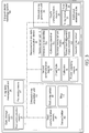
- Fig. 3 illustrates a block diagram of an intraoral dental radiology system (100) according to a first embodiment.
- the intraoral dental radiology system (100) has a measurement and data communication device (10), an x-ray data management unit (20), an intraoral x-ray generation unit (30), and an intraoral x-ray acquisition unit (40).
- the measurement and data communication device (10) has a measurement unit (11) which is arranged between the intraoral x-ray generation unit (30) and the intraoral x-ray acquisition unit (40).
- the intraoral x-ray generation unit (30) has an x-ray source (31) for exposing at least part of a patient jaw with x-rays.
- the intraoral x-ray acquisition unit (40) has a first x-ray sensor (41) for acquiring x-ray image data of at least part of the patient jaw.
- the measurement unit (11) is specifically arranged between the x-ray source (31) and the patient.
- the measurement unit (11) is adapted to measure features related to the x-rays used for exposing at least part of the patient.
- the x-ray data management unit (20) and the measurement and data communication device (10) have a first communication means (21) and a second communication means (12) respectively for mutual communication.
- the first communication means (21) and the second communication means (12) are adapted for wireless communication.
- the second communication means (12) is adapted to transmit x-ray exposure data which includes the measured features, to the x-ray data management unit (20).
- the first communication means (21) of the x-ray data management unit (20) is adapted to retrieve at least the x-ray exposure data.
- the x-ray data management unit (20) has a processing means (22) for processing at least the retrieved data such as the x-ray exposure data.
- the intraoral dental radiology (100) system is at least partly controlled by the processing means (22).
- the intraoral x-ray acquisition unit (40) has a third communication means (42) for transmitting the x-ray image data from the first x-ray sensor (41) to the x-ray data management unit (20).
- the first communication means (21) of the x-ray data management unit (20) is further adapted to retrieve the x-ray image data.
- the x-ray data management unit (20) has a data storage means (23) that is adapted to record, the retrieved x-ray image data, the x-ray exposure data, patient information and/or other information such as dose area products evaluated through the processing means (22) based on the x-ray exposure data.
- the intraoral x-ray generation unit (30) has a fourth communication means (34).
- the processing means (22) is further adapted to control the intraoral x-ray generation unit (30) based on the x-ray exposure data through the fourth communication means (34).
- the processing means (22) is further adapted to control the x-ray exposure duration, the x-ray tube voltage and/or x-ray tube current of the x-ray source (31) of the intraoral x-ray generation unit (30) based on the exposure data.
- the processing means (22) is further adapted to inhibit release of the x-ray exposure by the intraoral x-ray generation unit (30) if any abnormality through the x-ray exposure data is identified. Potential abnormalities will be explained later.
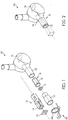
- Fig. 2 is a partial perspective view of the intraoral dental radiology system (100), especially of the intraoral x-ray generation unit (30) according to the first embodiment.
- the intraoral x-ray generation unit (30) is connectable from its upper part to a movable arm (not shown).
- the intraoral x-ray generation unit (30) has a tube head (32) which includes the x-ray source (31).
- the measurement and communication device (10) is detachably attachable to the tube head (32), preferably through complete insertion into the tube head (32).
- Fig. 2 illustrates the measurement and data communication device (10) after being attached to the tube head (32).
- Fig. 1 shows partial perspective views of the first embodiment, and also of an alternative second embodiment of the measurement and communication device (10) respectively arranged along the lower and upper dashed lines, which are both shown in the state detached from the tube head (32).
- the measurement and data communication device (10) according to the first embodiment has an insert tube (13) which accommodates the measurement unit (11) and the second communication means (12).
- the insert tube (13) can be detachably attached, specifically inserted into the tube head (32) without extending the length of the tube head (32).
- the x-ray exposure data transmitted to the first communication means (21) includes information on the presence/absence or also the type of the insert tube (13).
- the intraoral x-ray generation unit (30) according to the first embodiment has an additional extension tube (33) which is detachably attachable to the tube head (32) to extend the length of the tube head (32).
- the additional extension tube (33) is detachably attachable to the tube head (32) in the state when the insert tube (13) has been already attached to the tube head (32).
- the insert tube (13) can be attached to the additional extension tube (33), specifically through complete insertion into the large diameter portion of the additional extension tube (33) in the state when the additional extension tube (33) has been already attached to the tube head (32) through its low diameter portion.
- the measurement unit (11) has an extension tube identifier (11a) for identifying presence/absence or also type of the additional extension tube (33).
- the x-ray exposure data transmitted to the first communication means (21) includes information on the presence/absence or also type of the additional extension tube (33). As shown in Fig.
- the measurement and data communication device (10) has a user-exchangeable collimator (15) for limiting the x-ray field to the size of the first x-ray sensor (41).
- the measurement unit (11) has a collimator identifier (11b) for identifying presence/absence or also type of the collimator (15).
- the type of the collimator (15) is indicative of at least its size.
- the processing means (22) is further adapted to inhibit release of x-ray exposure by the intraoral x-ray generation unit (30) if the identified collimator (15) is not suitable for the first x-ray sensor (41).
- the x-ray exposure data transmitted to the first communication means (21) includes information on the presence/absence or also type of the collimator (15). Thereby, the x-ray exposure data includes the previously mentioned abnormality relating to the collimators (15).
- the intraoral x-ray acquisition unit (40) has a sensor holder (43) for holding the first x-ray sensor (41), and a ring (44) for positioning the tube head (32).
- the measurement unit (11) has a distance measurement means, (11c) for measuring the distance of the x-ray source (31) to the ring (44) or to the patient.
- the exposure data transmitted to the first communication means (21) includes information on the measured distance.
- the measurement unit (11) comprises a position and alignment measurement means (11d) for determining the position of the x-ray source (31) and the alignment of the central beam of the x-ray field relative to a horizontal plane.
- the alignment comprises the azimuthal angle and the polar angle.
- the x-ray exposure data transmitted to the first communication means (21) includes information on the determined position and alignment,
- the processing means (22) is adapted to inhibit release of x-ray exposure by the intraoral x-ray generation unit (30) if the determined position and alignment is not suitable for the targeted region.
- the x-ray exposure data includes the previously mentioned abnormality relating to the position and alignment.
- the measurement and data communication device (10) has a power supplying means (16) which has a motion sensor for activating the supply of power upon sensing any motion.
- the measurement unit (11) has a second x-ray sensor (11e) for measuring the duration of the x-ray exposure, intensity of the x-rays, and/or the beam characteristics of the x-rays.
- the x-ray exposure data transmitted to the first communication means (21) includes information on the duration of the x-ray exposure, the intensity of the x-rays, and/or the beam characteristics of the x-rays. It is also conceivable that the communication between the first communication means (21) and the second communication means (12) for transmitting the x-ray exposure data may also be realized indirectly via the third communication means (42) and/or the fourth communication means (34).
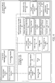
- Fig. 4 illustrates a block diagram of an intraoral dental radiology system (100) according to the second embodiment.
- the intraoral dental radiology system (100) of Fig. 4 differs from that of Fig. 3 only in that the measurement and communications device (10) has alternatively an extension tube (14) instead of the insert tube (13). Similar to the first embodiment, the extension tube (14) also accommodates the measurement unit (11) and the second communication means (12).
- the extension tube (14) can be detachably attached to the tube head (32) to extend the length of the tube head (32).
- the extension tube (14) may also be detachably attached to the tube head (32) via the additional extension tube (33).
- the collimators (15) used with the extension tube (14) is preferably rectangular shaped.
- the x-ray exposure data transmitted to the first communication means (21) includes information on the presence/absence or also the type of the extension tube (14).
Landscapes
- Health & Medical Sciences (AREA)
- Life Sciences & Earth Sciences (AREA)
- Engineering & Computer Science (AREA)
- Medical Informatics (AREA)
- Pathology (AREA)
- Heart & Thoracic Surgery (AREA)
- High Energy & Nuclear Physics (AREA)
- Physics & Mathematics (AREA)
- Nuclear Medicine, Radiotherapy & Molecular Imaging (AREA)
- Optics & Photonics (AREA)
- Veterinary Medicine (AREA)
- Radiology & Medical Imaging (AREA)
- Biomedical Technology (AREA)
- Biophysics (AREA)
- Molecular Biology (AREA)
- Surgery (AREA)
- Animal Behavior & Ethology (AREA)
- General Health & Medical Sciences (AREA)
- Public Health (AREA)
- Dentistry (AREA)
- Oral & Maxillofacial Surgery (AREA)
- Computer Networks & Wireless Communication (AREA)
- Apparatus For Radiation Diagnosis (AREA)
Abstract
Description
- The present invention relates to a radiology system. The present invention more particularly relates to an intraoral dental radiology system.
- In general, an intraoral dental radiology system comprises an intraoral x-ray generation unit, an intraoral x-ray acquisition unit, and an x-ray data management unit. The intraoral x-ray generation unit has an x-ray source for exposing at least part of a patient jaw with x-rays. The intraoral x-ray acquisition unit has an x-ray sensor for acquiring x-ray image data of at least part of the patient jaw. Different x-ray sensor technologies are available. The x-ray sensor might be a digital sensor or an analogue sensor such as a film or a phosphor plate. In general, a digital scanner is used to scan the analogue sensor and digitize the x-ray image. The x-ray sensor is usually held through a sensor holder which can be placed into the mouth of the patient. The sensor holder also has a ring for aligning the tube head of the x-ray generation unit with the x-ray sensor. The tube head includes the x-ray source. In the field of intraoral dental radiology, user-exchangeable collimators are optionally attached to the tube head of the intraoral X-ray generation unit for limiting the x-ray field. These collimators differ in the size of the usable x-ray field and are adapted to the size of the x-ray sensor. This avoids unnecessary exposure of the patient to x-rays that are not received by the x-ray sensor, and thereby reduces the patient dose. In the field of intraoral dental radiology, user-exchangeable additional extension tubes, e.g. from 20 cm (8 inches) to 30 cm (12 inches) are optionally attached to the tube head to extend the length of the tube head. With a larger source-sensor distance, the geometric aspects of the x-ray imaging can be improved, but in return the dose area product is reduced by a factor of the square of the distance.
- For entry in the x-ray control book which is optionally used for quality management purposes, default values are used to determine the x-ray exposure data such as the duration of the x-ray exposure, the intensity of the x-ray field, the beam characteristics of the x-ray field, the type of the collimator, the type of the additional extension tube, the sensor-source distance/alignment, and the dose area product.
- A problem with the prior art is that the default x-ray exposure data entered by the user may not always correspond the real x-ray exposure data because the intraoral dental radiology systems on the market do not recognize such x-ray exposure data directly.
-
US2006188065A1 discloses an intraoral dental radiology system according to prior art. - An objective of the present invention is to overcome the disadvantages of the prior art in an easy and cost-effective way and provide a measurement and data communication device for use in or with an intraoral dental radiology system.
- This objective has been achieved through the measurement and data communication device as defined in claim 1, and the intraoral dental radiology system as defined in
claim 12. The dependent claims relate to further developments. - The present invention provides a measurement and data communication device for use in or with an intraoral dental radiology system. The intraoral dental radiology system comprises: an x-ray data management unit having a communication means (hereinafter first communication means) for retrieving at least x-ray exposure data, and a processing means for processing at least the retrieved data; an intraoral x-ray generation unit having an x-ray source for exposing at least part of a patient jaw with x-rays; and an intraoral x-ray acquisition unit having an x-ray sensor (hereinafter first x-ray sensor) for acquiring x-ray image data of at least part of the patient jaw. The measurement and data communication device comprises: a measurement unit for measuring features related to the x-rays used for exposing at least part of the patient jaw, wherein the measurement unit is arranged between the x-ray source and the said at least part of the patient jaw; and a communication means (hereinafter second communication means) for transmitting the x-ray exposure data which includes the measured features to the x-ray data management unit.
- A major advantageous effect of the present invention is that the x-ray exposure data can be correctly determined through the measurement, and easily transmitted to the x-ray data management unit through the measurement and communication device. Thereby, the intraoral dental radiology system can be more securely operated. Another major advantageous effect of the present invention is that an existing intraoral dental radiology system can be equipped with the measurement and communication device to benefit from these advantages.
- According to an embodiment of the present invention, the intraoral x-ray generation unit preferably has a tube head which includes the x-ray source. And the measurement and communication device is preferably detachably attachable to the tube head. Thereby, an existing intraoral dental radiology system can be easily retrofitted with the measurement and communication device, and thus the x-ray exposure data can be generated and transmitted to the x-ray data management unit to keep a detailed log of the patient treatment and/or to control exposure of the patient.
- According to an embodiment of the present invention, the measurement and communication device preferably has an insert tube as the housing which accommodates the measurement unit and the second communication means. And the insert tube can be detachably attached to the tube head without extending the length of the tube head. In this embodiment, the x-ray exposure data transmitted by the second communication means preferably includes information on the presence/absence or also the type of the insert tube.
- According to an embodiment of the present invention, the intraoral x-ray generation unit preferably has an additional extension tube which is detachably attachable to the tube head to extend the length of the tube head. The above-mentioned insert tube is preferably usable with this additional extension tube. The additional extension tube is detachably attachable to the tube head in the state when the insert tube has already been attached to the tube head. In this embodiment, the measurement unit preferably has an extension tube identifier for identifying presence/absence or also type of the additional extension tube. In this embodiment, the exposure data which is transmitted by the second communication means preferably includes information on the presence/absence or also type of the additional extension tube i.e., the length of the additional extension tube. The extension tube identifier may have an electromechanical or optomechanical means for identifying a mechanical feature such as indents and/or projections or the like on the additional extension tube.
- According to an alternative embodiment of the present invention, the measurement and communication device preferably comprises an extension tube as the housing instead of the insert tube. The extension tube accommodates the measurement unit and the second communication means. And the extension tube can be detachably attached to the tube head to extend the length of the tube head. Thereby, the source-sensor distance can be optionally increased, and the geometric aspects of the x-ray imaging can be further improved. In this alternative embodiment, the x-ray exposure data transmitted by the second communication means includes information on the presence/absence or also the type of the extension tube, i.e., the length of the extension tube.
- According to an embodiment of the present invention, the measurement and communication device preferably has one or more user-exchangeable collimators for selectively limiting the x-ray field to the size of the first x-ray sensor. Thereby unnecessary exposure of the patient to x-rays not received by the x-ray sensor can be avoided or reduced as much as possible. The measurement unit preferably has a collimator identifier for identifying presence/absence or also type of the collimator. The type of the collimator is indicative of at least of its size. The diagonal of the collimator aperture is typically approx. 10 mm larger than the diagonal of the active area of the first x-ray sensor. In this embodiment, the exposure data transmitted by the second communication means includes information on the presence/absence or also type of the collimator. Thereby, the release of the x-ray exposure can be optionally inhibited if the collimator is not suitable for the first x-ray sensor. The user may be informed on the false collimator through a warning signal. This warning signal might be generated through a user interface comprising an acoustic means or an optical means such as an LED or the like. To avoid unnecessarily loading of the tube head, the collimators are preferably very light (<200g). The measurement and communication device is also manufactured as light as possible to allow easy manipulation of the tube head which is arranged on a movable arm.
- According to an embodiment of the present invention, the intraoral x-ray acquisition unit preferably has a sensor holder for holding the first x-ray sensor and a ring for positioning the tube head. The ring may also be used to align, the insert tube, the extension tube or the additional extension tube as mentioned above. The measurement and communication device is preferably rotatable around the center of the ring for facilitating the alignment. The ring may have a form fitting member for form-fittingly engaging with a counterpart form-fitting member on one of the tube head, the additional extension tube of the tube head, or the measurement and communication device. The form fitting-member may be round shaped or rectangular shaped. Other shapes may be alternatively used. The measurement unit preferably has a distance measurement means for measuring the distance of the x-ray source to the ring or to the patient. In this embodiment, the x-ray exposure data transmitted by the second communication means includes information on the measured distance. The distance measurement can be performed acoustically or optically with an ultrasound transceiver or infrared transceiver. The release of the x-ray exposure can be optionally inhibited if the distance is not suitable for the targeted region of the patient.
- According to an embodiment of the present invention, the measurement unit preferably has a position and alignment measurement means for determining the position of the x-ray source and the alignment of the central beam of the x-ray field relative to a horizontal plane. In this embodiment, the x-ray exposure data transmitted by the second communication means includes information on the determined position and alignment. This allows to register the targeted region of the patient to be exposed to x-rays into a control book as left, middle or right part of the jaw as well as upper or lower part of the jaw. The release of the x-ray exposure can be optionally inhibited if the determined position or alignment is not suitable for the targeted region of the patient. The user may be informed on the misalignment by a warning signal through the user interface. The determination of the position and the alignment can be performed for instance through a Hall angle sensor, a gyroscope or compass sensor.
- According to an embodiment of the present invention, the measurement and data communication device preferably has a separate power supplying means which has a motion sensor for activating the supply of power upon sensing motion. For instance, when the insert tube / extension tube is attached to the tube head or moved together with the tube head for alignment, then the power supply may be activated. To save energy, the supply of power may be deactivated upon elapse of a predetermined duration or through the x-ray data management unit via the first communication means when the exposure is terminated. The device may also have a user control panel. The power supplying means may include a mains power cord or one or more batteries, preferably rechargeable batteries. These batteries can be recharged with an external charging unit without having to be removed from the measurement and data communication device. The energy transfer between the external charging unit and the batteries can be realized through flexible cables, electrical pin contacts and/or wireless means such as inductive means. The external charging unit may be portable or installable on a surface, preferably on the wall surface in a practice. The tube head can be relatively moved together with the measurement and communication device to the external charging unit.
- According to an embodiment of the present invention, the measurement unit preferably has an x-ray sensor (hereinafter second x-ray sensor) for sensing the duration of the x-ray exposure, intensity of the x-rays, and/or the beam characteristics of the x-rays such as the x-ray tube voltage/current. In this embodiment, the exposure data transmitted by the second communication means preferably includes information on the duration of the x-ray exposure, the intensity of the x-rays, and/or the beam characteristics of the x-rays. The second x-ray sensor is arranged at a position where it does not obstruct x-rays exposing the targeted area of the patient.
- The present invention also provides an intraoral dental radiology system which comprises: the intraoral x-ray generation unit having the x-ray source for exposing at least part of a patient jaw with x-rays; and the intraoral x-ray acquisition unit having the first x-ray sensor for acquiring x-ray image data of at least part of the patient jaw, the measurement and data communication device; and the x-ray data management unit which comprises: the first communication means adapted to retrieve the exposure data transmitted by the measurement and data communication device; and the processing means adapted to process the retrieved data.
- According to an embodiment of the present invention, the first communication means of the x-ray data management unit preferably retrieves, in addition to the x-ray exposure data, the x-ray image data which is read from the first x-ray sensor. The intraoral x-ray acquisition unit has a communication means (hereinafter the third communication means) for transmitting the x-ray image data to the x-ray data management unit. If the first x-ray sensor is a digital sensor then the x-ray image data is read through a reading means, and transmitted. If the first x-ray sensor is an analogue sensor then the x-ray image is scanned and digitized through a digital scanning means and transmitted.
- According to an embodiment of the present invention, the intraoral x-ray generation unit preferably has a communication means (hereinafter the fourth communication means). The processing means of the x-ray management unit is preferably further adapted to control the intraoral x-ray generation unit based on the x-ray exposure data via the fourth communication means. Thereby, the intraoral x-ray generation unit can be more securely operated. For instance, the processing means is preferably further adapted to control the exposure duration, the tube voltage and/or tube current of the x=ray source based on the x-ray exposure data. Thereby, the patient can be more securely handled. For instance, the processing means is preferably further adapted to inhibit release of the x-ray exposure by the intraoral x-ray generation unit if the identified collimator is not suitable for the first x-ray sensor, or the determined position and alignment of the tube head, or the measured distance of the patient is not suitable for the targeted region. In addition, the user may be acoustically and/or optically informed on such abnormalities.
- According to an embodiment of the present invention, the x-ray data management unit is preferably adapted to evaluate the dose area product or any other information based on the x-ray exposure data.
- According to an embodiment of the present invention, the x-ray data management unit preferably has a data storage means. The x-ray data management unit is preferably further adapted to record the acquired x-ray image data, the x-ray exposure data, patient information and/or other information evaluated based on the x-ray exposure data into the data storage means.
- According to an embodiment of the present invention, the first to fourth communication means are preferably adapted for wireless communication. Alternatively, wired communication may be preferably used in any one of the first to fourth communication means. Alternatively, the communication between the first and the second communication means can be performed indirectly via either the third or the fourth communication means.
- In the subsequent description, further aspects and advantageous effects of the present invention will be described in more detail by using exemplary embodiments and referring to the drawings, wherein
-
Fig. 1 - are schematic perspectives views of a first embodiment and a second embodiment of the measurement and data communication device each prior to attachment to the tube head of the intraoral dental radiology system; -
Fig. 2 - is a schematic perspective view of the first embodiment of the measurement and data communication device ofFig. 1 after attachment to tube head of the intraoral dental radiology system; -
Fig. 3 - is a schematic block view of the intraoral dental radiology system retrofitted with the measurement and data communication device according to the first embodiment of the present invention; -
Fig. 4 - is a schematic block view of the intraoral dental radiology system retrofitted with the measurement and data communication device according to the second embodiment of the present invention. - The reference numbers shown in the drawings denote the elements as listed below and will be referred to in the subsequent description of the exemplary embodiments:
- 10.
- Measurement and data communication device
- 11.
- Measurement unit
- 11a.
- Extension tube identifier
- 11b
- Collimator identifier
- 11c.
- Distance measurement means
- 11d.
- Position and alignment measurement means
- 11e.
- Second x-ray sensor
- 12.
- Second communication means
- 13.
- Insert tube
- 14.
- Extension tube
- 15.
- Collimator
- 16.
- Power supplying means
- 20.
- X-ray data management unit
- 21.
- First communication means
- 22.
- Processing means
- 23.
- Data storage means
- 30.
- Intraoral x-ray generation unit
- 31.
- X-ray source
- 32.
- Tube head
- 33.
- Additional extension tube
- 34.
- Fourth communication means
- 40.
- Intraoral x-ray acquisition unit
- 41.
- First x-ray sensor
- 42.
- Third communication means
- 43.
- Sensor holder
- 44.
- Ring
- 100.
- Intraoral dental radiology system
-
Fig. 3 illustrates a block diagram of an intraoral dental radiology system (100) according to a first embodiment. The intraoral dental radiology system (100) has a measurement and data communication device (10), an x-ray data management unit (20), an intraoral x-ray generation unit (30), and an intraoral x-ray acquisition unit (40). The measurement and data communication device (10) has a measurement unit (11) which is arranged between the intraoral x-ray generation unit (30) and the intraoral x-ray acquisition unit (40). The intraoral x-ray generation unit (30) has an x-ray source (31) for exposing at least part of a patient jaw with x-rays. The intraoral x-ray acquisition unit (40) has a first x-ray sensor (41) for acquiring x-ray image data of at least part of the patient jaw. The measurement unit (11) is specifically arranged between the x-ray source (31) and the patient. The measurement unit (11) is adapted to measure features related to the x-rays used for exposing at least part of the patient. The x-ray data management unit (20) and the measurement and data communication device (10) have a first communication means (21) and a second communication means (12) respectively for mutual communication. The first communication means (21) and the second communication means (12) are adapted for wireless communication. The second communication means (12) is adapted to transmit x-ray exposure data which includes the measured features, to the x-ray data management unit (20). The first communication means (21) of the x-ray data management unit (20) is adapted to retrieve at least the x-ray exposure data. The x-ray data management unit (20) has a processing means (22) for processing at least the retrieved data such as the x-ray exposure data. The intraoral dental radiology (100) system is at least partly controlled by the processing means (22). The intraoral x-ray acquisition unit (40) has a third communication means (42) for transmitting the x-ray image data from the first x-ray sensor (41) to the x-ray data management unit (20). The first communication means (21) of the x-ray data management unit (20) is further adapted to retrieve the x-ray image data. The x-ray data management unit (20) has a data storage means (23) that is adapted to record, the retrieved x-ray image data, the x-ray exposure data, patient information and/or other information such as dose area products evaluated through the processing means (22) based on the x-ray exposure data. The intraoral x-ray generation unit (30) has a fourth communication means (34). The processing means (22) is further adapted to control the intraoral x-ray generation unit (30) based on the x-ray exposure data through the fourth communication means (34). The processing means (22) is further adapted to control the x-ray exposure duration, the x-ray tube voltage and/or x-ray tube current of the x-ray source (31) of the intraoral x-ray generation unit (30) based on the exposure data. The processing means (22) is further adapted to inhibit release of the x-ray exposure by the intraoral x-ray generation unit (30) if any abnormality through the x-ray exposure data is identified. Potential abnormalities will be explained later.Fig. 2 is a partial perspective view of the intraoral dental radiology system (100), especially of the intraoral x-ray generation unit (30) according to the first embodiment. The intraoral x-ray generation unit (30) is connectable from its upper part to a movable arm (not shown). The intraoral x-ray generation unit (30) has a tube head (32) which includes the x-ray source (31). The measurement and communication device (10) is detachably attachable to the tube head (32), preferably through complete insertion into the tube head (32).Fig. 2 illustrates the measurement and data communication device (10) after being attached to the tube head (32).Fig. 1 shows partial perspective views of the first embodiment, and also of an alternative second embodiment of the measurement and communication device (10) respectively arranged along the lower and upper dashed lines, which are both shown in the state detached from the tube head (32). As shown inFig. 1 , the measurement and data communication device (10) according to the first embodiment has an insert tube (13) which accommodates the measurement unit (11) and the second communication means (12). As shown inFig. 1 and Fig. 2 , the insert tube (13) can be detachably attached, specifically inserted into the tube head (32) without extending the length of the tube head (32). The x-ray exposure data transmitted to the first communication means (21) includes information on the presence/absence or also the type of the insert tube (13). As shown inFig, 1 , the intraoral x-ray generation unit (30) according to the first embodiment has an additional extension tube (33) which is detachably attachable to the tube head (32) to extend the length of the tube head (32). The additional extension tube (33) is detachably attachable to the tube head (32) in the state when the insert tube (13) has been already attached to the tube head (32). Alternatively, the insert tube (13) can be attached to the additional extension tube (33), specifically through complete insertion into the large diameter portion of the additional extension tube (33) in the state when the additional extension tube (33) has been already attached to the tube head (32) through its low diameter portion. As shown inFig. 3 , the measurement unit (11) has an extension tube identifier (11a) for identifying presence/absence or also type of the additional extension tube (33). The x-ray exposure data transmitted to the first communication means (21) includes information on the presence/absence or also type of the additional extension tube (33). As shown inFig. 1 , the measurement and data communication device (10) according to the first embodiment has a user-exchangeable collimator (15) for limiting the x-ray field to the size of the first x-ray sensor (41). As shown inFig. 3 , the measurement unit (11) has a collimator identifier (11b) for identifying presence/absence or also type of the collimator (15). The type of the collimator (15) is indicative of at least its size. The processing means (22) is further adapted to inhibit release of x-ray exposure by the intraoral x-ray generation unit (30) if the identified collimator (15) is not suitable for the first x-ray sensor (41). The x-ray exposure data transmitted to the first communication means (21) includes information on the presence/absence or also type of the collimator (15). Thereby, the x-ray exposure data includes the previously mentioned abnormality relating to the collimators (15). As shown inFig. 1 , the intraoral x-ray acquisition unit (40) has a sensor holder (43) for holding the first x-ray sensor (41), and a ring (44) for positioning the tube head (32). As shown inFig. 3 , the measurement unit (11) has a distance measurement means, (11c) for measuring the distance of the x-ray source (31) to the ring (44) or to the patient. The exposure data transmitted to the first communication means (21) includes information on the measured distance. The release of the x-ray exposure can be optionally inhibited if the distance is not suitable for the targeted region of the patient. As shown inFig. 3 , the measurement unit (11) comprises a position and alignment measurement means (11d) for determining the position of the x-ray source (31) and the alignment of the central beam of the x-ray field relative to a horizontal plane. The alignment comprises the azimuthal angle and the polar angle. The x-ray exposure data transmitted to the first communication means (21) includes information on the determined position and alignment, The processing means (22) is adapted to inhibit release of x-ray exposure by the intraoral x-ray generation unit (30) if the determined position and alignment is not suitable for the targeted region. Thereby, the x-ray exposure data includes the previously mentioned abnormality relating to the position and alignment. As shown inFig. 3 , the measurement and data communication device (10) has a power supplying means (16) which has a motion sensor for activating the supply of power upon sensing any motion. As shown inFig. 3 , the measurement unit (11) has a second x-ray sensor (11e) for measuring the duration of the x-ray exposure, intensity of the x-rays, and/or the beam characteristics of the x-rays. The x-ray exposure data transmitted to the first communication means (21) includes information on the duration of the x-ray exposure, the intensity of the x-rays, and/or the beam characteristics of the x-rays. It is also conceivable that the communication between the first communication means (21) and the second communication means (12) for transmitting the x-ray exposure data may also be realized indirectly via the third communication means (42) and/or the fourth communication means (34). -
Fig. 4 illustrates a block diagram of an intraoral dental radiology system (100) according to the second embodiment. The intraoral dental radiology system (100) ofFig. 4 differs from that ofFig. 3 only in that the measurement and communications device (10) has alternatively an extension tube (14) instead of the insert tube (13). Similar to the first embodiment, the extension tube (14) also accommodates the measurement unit (11) and the second communication means (12). The extension tube (14) can be detachably attached to the tube head (32) to extend the length of the tube head (32). The extension tube (14) may also be detachably attached to the tube head (32) via the additional extension tube (33). The collimators (15) used with the extension tube (14) is preferably rectangular shaped. Alternatively, round shaped collimators (15) may be used. The forward end of the extension tube (14) is rectangular shape. Alternatively, it may have a round shape as the insert tube (13). The x-ray exposure data transmitted to the first communication means (21) includes information on the presence/absence or also the type of the extension tube (14).
Claims (19)
- A measurement and data communication device (10) for use in or with an intraoral dental radiology system (100) comprising:an x-ray data management unit (20) having a first communication means (21) for retrieving at least x-ray exposure data, and a processing means (22) for processing at least the retrieved data;an intraoral x-ray generation unit (30) having an x-ray source (31) for exposing at least part of a patient jaw with x-rays; andan intraoral x-ray acquisition unit (40) having a first x-ray sensor (41) for acquiring x-ray image data of at least part of the patient jaw;said device (10) characterized by comprising:a measurement unit (11) for measuring features related to the x-rays used for exposing at least part of the patient, wherein the measurement unit (11) is arranged between the x-ray source (31) and the patient; anda second communication means (12) for transmitting the x-ray exposure data which includes the measured features to the x-ray data management unit (20).
- The measurement and data communication device (10) according to claim 1, wherein the intraoral x-ray generation unit (30) has a tube head (32) which includes the x-ray source (31), the device (10) being characterized in that it is detachably attachable to the tube head (32).
- The measurement and data communication device (10) according to claim 2, characterized in that the device (10) has an extension tube (14) which includes the measurement unit (11) and the second communication means (12), wherein the extension tube (14) can be detachably attached to the tube head (32) to extend the length of the tube head (32), wherein the x-ray exposure data further includes information on the presence/absence or also the type of the extension tube (14).
- The measurement and data communication device (10) according to claim 2, characterized in that the device (10) has an insert tube (13) which includes the measurement unit (11) and the second communication means (12), wherein the insert tube (13) can be detachably attached to the tube head (32) without extending the length of the tube head (32), wherein the x-ray exposure data further includes information on the presence/absence or also the type of the insert tube (13).
- The measurement and data communication device (10) according to claim 3 or 4, wherein the intraoral x-ray generation unit (30) has an additional extension tube (33) which is detachably attachable to the tube head (32) to extend the length of the tube head (32), the device (10) being characterized in that the additional extension tube (33) is detachably attachable to the tube head (32) in the state when the insert tube (13) or the extension tube (14) is also attached to the tube head (32).
- The measurement and data communication device (10) according to claim 5, characterized in that the measurement unit (11) has an extension tube identifier (11a) for identifying presence/absence or also type of the additional extension tube (33), wherein the x-ray exposure data further includes information on the presence/absence or also type of the additional extension tube (33).
- The measurement and data communication device (10) according to any one of claims 1 to 6, characterized by further comprising: a user-exchangeable collimator (15) for limiting the x-ray field to the size of the first x-ray sensor (41); and the measurement unit (11) comprises a collimator identifier (11b) for identifying presence/absence or also type of the collimator (15), wherein the type of the collimator (15) is indicative of at least its size, wherein the x-ray exposure data further includes information on the presence/absence or also type of the collimator (15).
- The measurement and data communication device (10) according to any one of claims 1 to 7, wherein the intraoral x-ray acquisition unit (40) has a sensor holder (43) for holding the first x-ray sensor (41) and a ring (44) for positioning the tube head (32), characterized in that the measurement unit (11) comprises a distance measurement means (11c) for measuring the distance of the x-ray source (31) to the ring (44) or to the patient, wherein the x-ray exposure data further includes information on the measured distance.
- The measurement and data communication device (10) according to any one of claims 1 to 8, characterized in that the measurement unit (11) comprises a position and alignment measurement means (11d) for determining the position of the x-ray source (31) and the alignment of the central beam of the x-ray field relative to a horizontal plane, wherein the x-ray exposure data further includes information on the determined position and alignment.
- The measurement and data communication device (10) according to any one of claims 1 to 9, characterized by further comprising a power supplying means (16) which has a motion sensor for activating the supply of power upon sensing motion.
- The measurement and data communication device (10) according to any one of claims 1 to 10, characterized in that the measurement unit (11) comprises a second x-ray sensor (11e) for measuring the duration of the x-ray exposure, intensity of the x-rays, and/or the beam characteristics of the x-rays, wherein the x-ray exposure data further includes information on the duration of the x-ray exposure, the intensity of the x-rays, and/or the beam characteristics of the x-rays.
- An intraoral dental radiology system (100) comprising:an intraoral x-ray generation unit (30) having an x-ray source (31) for exposing at least part of a patient jaw with x-rays; andan intraoral x-ray acquisition unit (40) having an first x-ray sensor (41) for acquiring x-ray image data of at least part of the patient jaw,characterized by further comprising:the measurement and data communication device (10) according to any one of claims 1 to 11;an x-ray data management unit (20) which comprises: a first communication means (21) adapted to retrieve the x-ray exposure data transmitted by the measurement and data communication device (10); and a processing means (22) adapted to process the retrieved data.
- The intraoral dental radiology system (100) according to claim 12, characterized in that the first communication means (21) and the second communication means (12) are adapted for wireless communication.
- The intraoral dental radiology system (100) according to claim 12 or 13, characterized in that the first communication means (21) of the x-ray data management unit (20) is further adapted to retrieve the x-ray image data, and the intraoral x-ray acquisition unit (40) has a third communication means (42) for transmitting the x-ray image data to the x-ray data management unit (20).
- The intraoral dental radiology system (100) according to any one of claims 12 to 14, characterized in that the intraoral x-ray generation unit (30) has a fourth communication means (34), and the processing means (22) is further adapted to control the intraoral x-ray generation unit (30) based on the x-ray exposure data.
- The intraoral dental radiology system (100) according to claim 15, characterized in that the processing means (22) is further adapted to control x-ray exposure duration, the tube voltage and/or the tube current of the x-ray source (31) of the intraoral x-ray generation unit (30) based on the x-ray exposure data.
- The intraoral dental radiology system (100) according to claim 15 or 16, characterized in that the processing means (22) is further adapted to inhibit release of x-ray exposure by the intraoral x-ray generation unit (30) if the identified collimator (15) is not suitable for first x-ray sensor (41), or the determined position and alignment, or the measured distance is not suitable for the targeted region.
- The intraoral dental radiology system (100) according to any one of claims 12 to 17, characterized in that the processing means (22) is further adapted to evaluate the dose area product based on the x-ray exposure data.
- The intraoral dental radiology system (100) according to any one of claims 12 to 18, characterized in that the x-ray data management unit (20) comprises a data storage means (23) adapted to record the acquired x-ray image data, the x-ray exposure data, patient information and/or other information evaluated based on the x-ray exposure data.
Priority Applications (6)
| Application Number | Priority Date | Filing Date | Title |
|---|---|---|---|
| EP19000371.5A EP3777693B1 (en) | 2019-08-12 | 2019-08-12 | Measurement and data communication device for an intraoral dental radiology system |
| JP2022508864A JP7551735B2 (en) | 2019-08-12 | 2020-08-12 | MEASUREMENT AND DATA COMMUNICATION DEVICE FOR AN INTRAORAL DENTAL RADIOLOGY SYSTEM - Patent application |
| KR1020227007912A KR20220047995A (en) | 2019-08-12 | 2020-08-12 | Measurement and data communication device for intraoral dental radiology systems |
| CN202080061504.8A CN114364319B (en) | 2019-08-12 | 2020-08-12 | Measurement and data communication equipment for intraoral dental radiology systems |
| US17/634,090 US12257097B2 (en) | 2019-08-12 | 2020-08-12 | Measurement and data communication device for an intraoral dental radiology system |
| PCT/EP2020/072666 WO2021028497A1 (en) | 2019-08-12 | 2020-08-12 | Measurement and data communication device for an intraoral dental radiology system |
Applications Claiming Priority (1)
| Application Number | Priority Date | Filing Date | Title |
|---|---|---|---|
| EP19000371.5A EP3777693B1 (en) | 2019-08-12 | 2019-08-12 | Measurement and data communication device for an intraoral dental radiology system |
Publications (2)
| Publication Number | Publication Date |
|---|---|
| EP3777693A1 true EP3777693A1 (en) | 2021-02-17 |
| EP3777693B1 EP3777693B1 (en) | 2022-03-16 |
Family
ID=67620240
Family Applications (1)
| Application Number | Title | Priority Date | Filing Date |
|---|---|---|---|
| EP19000371.5A Active EP3777693B1 (en) | 2019-08-12 | 2019-08-12 | Measurement and data communication device for an intraoral dental radiology system |
Country Status (6)
| Country | Link |
|---|---|
| US (1) | US12257097B2 (en) |
| EP (1) | EP3777693B1 (en) |
| JP (1) | JP7551735B2 (en) |
| KR (1) | KR20220047995A (en) |
| CN (1) | CN114364319B (en) |
| WO (1) | WO2021028497A1 (en) |
Cited By (1)
| Publication number | Priority date | Publication date | Assignee | Title |
|---|---|---|---|---|
| US12257097B2 (en) | 2019-08-12 | 2025-03-25 | Dentsply Sirona Inc. | Measurement and data communication device for an intraoral dental radiology system |
Citations (4)
| Publication number | Priority date | Publication date | Assignee | Title |
|---|---|---|---|---|
| US20060188065A1 (en) | 2005-02-23 | 2006-08-24 | Razzano Michael R | Image capture device and methods |
| DE102007033957A1 (en) * | 2007-07-19 | 2009-01-22 | Sirona Dental Systems Gmbh | Dental X-ray emitter's light exposure parameter e.g. light exposure time, generating method, involves performing correlation between light exposure parameters and photograph requirements based on generated photograph |
| US20160262715A1 (en) * | 2015-03-10 | 2016-09-15 | Dental Imaging Technologies Corporation | Automated control of image exposure parameters in an intra-oral x-ray system |
| US20170188987A1 (en) * | 2016-01-05 | 2017-07-06 | BAE Systems Imaging Solutions Inc. | System and Method for Adjusting Dental X-ray Exposure |
Family Cites Families (31)
| Publication number | Priority date | Publication date | Assignee | Title |
|---|---|---|---|---|
| JPS63115539A (en) | 1986-11-04 | 1988-05-20 | 松下電器産業株式会社 | X-ray diagnostic equipment |
| JP2006517439A (en) * | 2003-02-11 | 2006-07-27 | コーニンクレッカ フィリップス エレクトロニクス エヌ ヴィ | X-ray apparatus with collimator and method for setting collimator |
| US7006600B1 (en) * | 2004-01-15 | 2006-02-28 | Progeny, Inc. | Integrated digital dental x-ray system |
| JP4264382B2 (en) * | 2004-04-30 | 2009-05-13 | 株式会社モリタ製作所 | Automatic exposure control method for photographed image and automatic exposure control apparatus using the method |
| FR2881640B1 (en) * | 2005-02-09 | 2007-05-11 | Sopro Sa | OPTIMIZATION OF THE X-RAY QUANTITY RECEIVED BY A PATIENT IN A DENTAL RADIOLOGICAL IMAGE ACQUISITION SYSTEM |
| US7775713B2 (en) | 2005-05-16 | 2010-08-17 | Palodex Group Oy | Arrangement for dental imaging |
| US7519157B2 (en) * | 2005-07-23 | 2009-04-14 | General Electric Company | Systems, methods and apparatus for attachment of an X-ray tube to an X-ray tube collimator frame |
| EP2057637A2 (en) * | 2006-08-21 | 2009-05-13 | Philips Intellectual Property & Standards GmbH | Portable x-ray detector with grid sensing unit and x-ray imaging system for automatic exposure setting for the portable x-ray detector |
| JP4739278B2 (en) * | 2007-05-11 | 2011-08-03 | 株式会社モリタ製作所 | X-ray CT imaging system |
| JP5569951B2 (en) * | 2008-09-01 | 2014-08-13 | 学校法人日本大学 | X-ray CT imaging apparatus for head and imaging control method thereof |
| US8119990B2 (en) * | 2008-10-27 | 2012-02-21 | Imaging Sciences International Llc | System and method of X-ray detection with a sensor |
| CN101765291A (en) | 2008-12-19 | 2010-06-30 | 曹淑兰 | Development of photographic parameter test system of non-intrusive medical diagnostic X-ray apparatus |
| US9579073B2 (en) * | 2010-05-07 | 2017-02-28 | Apteryx, Inc. | System and method for dentition specific image enhancement |
| US8908162B2 (en) * | 2011-02-24 | 2014-12-09 | Idi Dental, Inc. | System for aligning a collimator and an alignment ring |
| FI125516B (en) | 2011-03-21 | 2015-11-13 | Planmeca Oy | Collimator device in connection with intraoral X-ray |
| FI123713B (en) * | 2011-03-21 | 2013-09-30 | Planmeca Oy | Arrangements in connection with intraoral X-rays |
| JP2013190310A (en) * | 2012-03-13 | 2013-09-26 | Toshiba Corp | X-ray detector, manufacturing method of x-ray detector, and x-ray ct apparatus |
| US20140169533A1 (en) * | 2012-12-19 | 2014-06-19 | Michael R. Razzano | System and method for capturing medical images |
| US9408581B2 (en) * | 2014-03-07 | 2016-08-09 | Elwha Llc | Systems, devices, and methods for lowering dental x-ray dosage including feedback sensors |
| DE102014223967A1 (en) * | 2014-11-25 | 2016-05-25 | Sirona Dental Systems Gmbh | Method for diagnosis in orthodontics |
| US9986964B2 (en) * | 2015-03-10 | 2018-06-05 | Dental Imaging Technologies Corporation | Intraoral sensor position detection for full mouth image |
| KR102377484B1 (en) * | 2015-04-20 | 2022-03-22 | 주식회사 바텍 | X-ray generating device having motionsensor and method for x-ray imaging using the same |
| US9649078B2 (en) * | 2015-07-28 | 2017-05-16 | Dental Imaging Technologies Corporation | Hybrid X-ray system with detachable radiation shield |
| US9801593B2 (en) * | 2015-12-14 | 2017-10-31 | Dental Imaging Technologies Corporation | Intraoral X-ray imaging sensor and readout |
| JP2017225592A (en) | 2016-06-22 | 2017-12-28 | コニカミノルタ株式会社 | Radiographic imaging system |
| US10049457B2 (en) * | 2016-08-29 | 2018-08-14 | CephX Technologies Ltd. | Automated cephalometric analysis using machine learning |
| US20180279976A1 (en) * | 2017-02-22 | 2018-10-04 | Todd C. Miller | Intraoral sensor positioning system |
| JP6734212B2 (en) * | 2017-03-02 | 2020-08-05 | 株式会社モリタ製作所 | X-ray tomography apparatus and X-ray tomography method |
| WO2018227523A1 (en) | 2017-06-16 | 2018-12-20 | Shanghai United Imaging Healthcare Co., Ltd. | Systems and methods for image data processing in computerized tomography |
| US10905386B2 (en) * | 2018-03-09 | 2021-02-02 | Suzanne Cano | Orienting X-ray projection for dental imagery |
| EP3777693B1 (en) | 2019-08-12 | 2022-03-16 | DENTSPLY SIRONA Inc. | Measurement and data communication device for an intraoral dental radiology system |
-
2019
- 2019-08-12 EP EP19000371.5A patent/EP3777693B1/en active Active
-
2020
- 2020-08-12 WO PCT/EP2020/072666 patent/WO2021028497A1/en not_active Ceased
- 2020-08-12 CN CN202080061504.8A patent/CN114364319B/en active Active
- 2020-08-12 US US17/634,090 patent/US12257097B2/en active Active
- 2020-08-12 KR KR1020227007912A patent/KR20220047995A/en not_active Ceased
- 2020-08-12 JP JP2022508864A patent/JP7551735B2/en active Active
Patent Citations (4)
| Publication number | Priority date | Publication date | Assignee | Title |
|---|---|---|---|---|
| US20060188065A1 (en) | 2005-02-23 | 2006-08-24 | Razzano Michael R | Image capture device and methods |
| DE102007033957A1 (en) * | 2007-07-19 | 2009-01-22 | Sirona Dental Systems Gmbh | Dental X-ray emitter's light exposure parameter e.g. light exposure time, generating method, involves performing correlation between light exposure parameters and photograph requirements based on generated photograph |
| US20160262715A1 (en) * | 2015-03-10 | 2016-09-15 | Dental Imaging Technologies Corporation | Automated control of image exposure parameters in an intra-oral x-ray system |
| US20170188987A1 (en) * | 2016-01-05 | 2017-07-06 | BAE Systems Imaging Solutions Inc. | System and Method for Adjusting Dental X-ray Exposure |
Cited By (1)
| Publication number | Priority date | Publication date | Assignee | Title |
|---|---|---|---|---|
| US12257097B2 (en) | 2019-08-12 | 2025-03-25 | Dentsply Sirona Inc. | Measurement and data communication device for an intraoral dental radiology system |
Also Published As
| Publication number | Publication date |
|---|---|
| JP2022544506A (en) | 2022-10-19 |
| WO2021028497A1 (en) | 2021-02-18 |
| US12257097B2 (en) | 2025-03-25 |
| CN114364319A (en) | 2022-04-15 |
| US20220346745A1 (en) | 2022-11-03 |
| KR20220047995A (en) | 2022-04-19 |
| EP3777693B1 (en) | 2022-03-16 |
| JP7551735B2 (en) | 2024-09-17 |
| CN114364319B (en) | 2024-08-06 |
Similar Documents
| Publication | Publication Date | Title |
|---|---|---|
| US10226164B2 (en) | Dental scanner device | |
| US7628537B2 (en) | Small dental X-ray apparatus and method for positioning an X-ray emitter | |
| EP1252859A2 (en) | Dental camera with mouthpiece | |
| KR20160110189A (en) | Intraoral sensor position detection for full mouth image | |
| EP1778090B1 (en) | Arrangement in connection with intra-oral x-ray imaging | |
| WO2006091712A2 (en) | Image capture device and methods | |
| US8908162B2 (en) | System for aligning a collimator and an alignment ring | |
| JP2002355263A (en) | Dental camera | |
| CN101359053A (en) | Radiation detection cassette and medical system | |
| US7038205B2 (en) | Probe apparatus with laser guiding for locating a source of radioactivity | |
| US20180220989A9 (en) | Positioning of a mobile x-ray detector | |
| US12257097B2 (en) | Measurement and data communication device for an intraoral dental radiology system | |
| KR20150070170A (en) | Computed radiography positioning method and system | |
| US20060274890A1 (en) | Image capture device and methods | |
| JP2017192412A (en) | Imaging unit of x-ray imaging apparatus in dental oral cavity, and x-ray imaging apparatus in dental oral cavity | |
| US20040213382A1 (en) | Methods and positioning and aiming assemblies for use with an x-ray source | |
| CN217548062U (en) | Wireless oral cavity imager | |
| US20230165547A1 (en) | Hand-grip type oral x-ray device having alignment function | |
| TWI516249B (en) | A dental x-ray photographic apparatus and system in the oral cavity | |
| JPWO2010140492A1 (en) | Intraoral imaging aid | |
| JP5896837B2 (en) | Dental intraoral X-ray imaging system | |
| KR200358845Y1 (en) | Intraoral fluoroscope for dental treatment which having optical fiber rod | |
| US20240252131A1 (en) | Digital dental xray sensor, method and system | |
| JP5881249B2 (en) | Dental intraoral X-ray imaging system |
Legal Events
| Date | Code | Title | Description |
|---|---|---|---|
| PUAI | Public reference made under article 153(3) epc to a published international application that has entered the european phase |
Free format text: ORIGINAL CODE: 0009012 |
|
| STAA | Information on the status of an ep patent application or granted ep patent |
Free format text: STATUS: THE APPLICATION HAS BEEN PUBLISHED |
|
| AK | Designated contracting states |
Kind code of ref document: A1 Designated state(s): AL AT BE BG CH CY CZ DE DK EE ES FI FR GB GR HR HU IE IS IT LI LT LU LV MC MK MT NL NO PL PT RO RS SE SI SK SM TR |
|
| AX | Request for extension of the european patent |
Extension state: BA ME |
|
| STAA | Information on the status of an ep patent application or granted ep patent |
Free format text: STATUS: REQUEST FOR EXAMINATION WAS MADE |
|
| 17P | Request for examination filed |
Effective date: 20210426 |
|
| RBV | Designated contracting states (corrected) |
Designated state(s): AL AT BE BG CH CY CZ DE DK EE ES FI FR GB GR HR HU IE IS IT LI LT LU LV MC MK MT NL NO PL PT RO RS SE SI SK SM TR |
|
| GRAP | Despatch of communication of intention to grant a patent |
Free format text: ORIGINAL CODE: EPIDOSNIGR1 |
|
| STAA | Information on the status of an ep patent application or granted ep patent |
Free format text: STATUS: GRANT OF PATENT IS INTENDED |
|
| INTG | Intention to grant announced |
Effective date: 20211019 |
|
| GRAS | Grant fee paid |
Free format text: ORIGINAL CODE: EPIDOSNIGR3 |
|
| GRAA | (expected) grant |
Free format text: ORIGINAL CODE: 0009210 |
|
| STAA | Information on the status of an ep patent application or granted ep patent |
Free format text: STATUS: THE PATENT HAS BEEN GRANTED |
|
| AK | Designated contracting states |
Kind code of ref document: B1 Designated state(s): AL AT BE BG CH CY CZ DE DK EE ES FI FR GB GR HR HU IE IS IT LI LT LU LV MC MK MT NL NO PL PT RO RS SE SI SK SM TR |
|
| REG | Reference to a national code |
Ref country code: GB Ref legal event code: FG4D |
|
| REG | Reference to a national code |
Ref country code: CH Ref legal event code: EP |
|
| REG | Reference to a national code |
Ref country code: DE Ref legal event code: R096 Ref document number: 602019012477 Country of ref document: DE |
|
| REG | Reference to a national code |
Ref country code: IE Ref legal event code: FG4D |
|
| REG | Reference to a national code |
Ref country code: AT Ref legal event code: REF Ref document number: 1475333 Country of ref document: AT Kind code of ref document: T Effective date: 20220415 |
|
| REG | Reference to a national code |
Ref country code: FI Ref legal event code: FGE |
|
| REG | Reference to a national code |
Ref country code: LT Ref legal event code: MG9D |
|
| REG | Reference to a national code |
Ref country code: NL Ref legal event code: MP Effective date: 20220316 |
|
| PG25 | Lapsed in a contracting state [announced via postgrant information from national office to epo] |
Ref country code: SE Free format text: LAPSE BECAUSE OF FAILURE TO SUBMIT A TRANSLATION OF THE DESCRIPTION OR TO PAY THE FEE WITHIN THE PRESCRIBED TIME-LIMIT Effective date: 20220316 Ref country code: RS Free format text: LAPSE BECAUSE OF FAILURE TO SUBMIT A TRANSLATION OF THE DESCRIPTION OR TO PAY THE FEE WITHIN THE PRESCRIBED TIME-LIMIT Effective date: 20220316 Ref country code: NO Free format text: LAPSE BECAUSE OF FAILURE TO SUBMIT A TRANSLATION OF THE DESCRIPTION OR TO PAY THE FEE WITHIN THE PRESCRIBED TIME-LIMIT Effective date: 20220616 Ref country code: LT Free format text: LAPSE BECAUSE OF FAILURE TO SUBMIT A TRANSLATION OF THE DESCRIPTION OR TO PAY THE FEE WITHIN THE PRESCRIBED TIME-LIMIT Effective date: 20220316 Ref country code: HR Free format text: LAPSE BECAUSE OF FAILURE TO SUBMIT A TRANSLATION OF THE DESCRIPTION OR TO PAY THE FEE WITHIN THE PRESCRIBED TIME-LIMIT Effective date: 20220316 Ref country code: BG Free format text: LAPSE BECAUSE OF FAILURE TO SUBMIT A TRANSLATION OF THE DESCRIPTION OR TO PAY THE FEE WITHIN THE PRESCRIBED TIME-LIMIT Effective date: 20220616 |
|
| REG | Reference to a national code |
Ref country code: AT Ref legal event code: MK05 Ref document number: 1475333 Country of ref document: AT Kind code of ref document: T Effective date: 20220316 |
|
| PG25 | Lapsed in a contracting state [announced via postgrant information from national office to epo] |
Ref country code: LV Free format text: LAPSE BECAUSE OF FAILURE TO SUBMIT A TRANSLATION OF THE DESCRIPTION OR TO PAY THE FEE WITHIN THE PRESCRIBED TIME-LIMIT Effective date: 20220316 Ref country code: GR Free format text: LAPSE BECAUSE OF FAILURE TO SUBMIT A TRANSLATION OF THE DESCRIPTION OR TO PAY THE FEE WITHIN THE PRESCRIBED TIME-LIMIT Effective date: 20220617 |
|
| PG25 | Lapsed in a contracting state [announced via postgrant information from national office to epo] |
Ref country code: NL Free format text: LAPSE BECAUSE OF FAILURE TO SUBMIT A TRANSLATION OF THE DESCRIPTION OR TO PAY THE FEE WITHIN THE PRESCRIBED TIME-LIMIT Effective date: 20220316 |
|
| PG25 | Lapsed in a contracting state [announced via postgrant information from national office to epo] |
Ref country code: SM Free format text: LAPSE BECAUSE OF FAILURE TO SUBMIT A TRANSLATION OF THE DESCRIPTION OR TO PAY THE FEE WITHIN THE PRESCRIBED TIME-LIMIT Effective date: 20220316 Ref country code: SK Free format text: LAPSE BECAUSE OF FAILURE TO SUBMIT A TRANSLATION OF THE DESCRIPTION OR TO PAY THE FEE WITHIN THE PRESCRIBED TIME-LIMIT Effective date: 20220316 Ref country code: RO Free format text: LAPSE BECAUSE OF FAILURE TO SUBMIT A TRANSLATION OF THE DESCRIPTION OR TO PAY THE FEE WITHIN THE PRESCRIBED TIME-LIMIT Effective date: 20220316 Ref country code: PT Free format text: LAPSE BECAUSE OF FAILURE TO SUBMIT A TRANSLATION OF THE DESCRIPTION OR TO PAY THE FEE WITHIN THE PRESCRIBED TIME-LIMIT Effective date: 20220718 Ref country code: ES Free format text: LAPSE BECAUSE OF FAILURE TO SUBMIT A TRANSLATION OF THE DESCRIPTION OR TO PAY THE FEE WITHIN THE PRESCRIBED TIME-LIMIT Effective date: 20220316 Ref country code: EE Free format text: LAPSE BECAUSE OF FAILURE TO SUBMIT A TRANSLATION OF THE DESCRIPTION OR TO PAY THE FEE WITHIN THE PRESCRIBED TIME-LIMIT Effective date: 20220316 Ref country code: CZ Free format text: LAPSE BECAUSE OF FAILURE TO SUBMIT A TRANSLATION OF THE DESCRIPTION OR TO PAY THE FEE WITHIN THE PRESCRIBED TIME-LIMIT Effective date: 20220316 Ref country code: AT Free format text: LAPSE BECAUSE OF FAILURE TO SUBMIT A TRANSLATION OF THE DESCRIPTION OR TO PAY THE FEE WITHIN THE PRESCRIBED TIME-LIMIT Effective date: 20220316 |
|
| PG25 | Lapsed in a contracting state [announced via postgrant information from national office to epo] |
Ref country code: PL Free format text: LAPSE BECAUSE OF FAILURE TO SUBMIT A TRANSLATION OF THE DESCRIPTION OR TO PAY THE FEE WITHIN THE PRESCRIBED TIME-LIMIT Effective date: 20220316 Ref country code: IS Free format text: LAPSE BECAUSE OF FAILURE TO SUBMIT A TRANSLATION OF THE DESCRIPTION OR TO PAY THE FEE WITHIN THE PRESCRIBED TIME-LIMIT Effective date: 20220716 Ref country code: AL Free format text: LAPSE BECAUSE OF FAILURE TO SUBMIT A TRANSLATION OF THE DESCRIPTION OR TO PAY THE FEE WITHIN THE PRESCRIBED TIME-LIMIT Effective date: 20220316 |
|
| REG | Reference to a national code |
Ref country code: DE Ref legal event code: R097 Ref document number: 602019012477 Country of ref document: DE |
|
| PLBE | No opposition filed within time limit |
Free format text: ORIGINAL CODE: 0009261 |
|
| STAA | Information on the status of an ep patent application or granted ep patent |
Free format text: STATUS: NO OPPOSITION FILED WITHIN TIME LIMIT |
|
| PG25 | Lapsed in a contracting state [announced via postgrant information from national office to epo] |
Ref country code: DK Free format text: LAPSE BECAUSE OF FAILURE TO SUBMIT A TRANSLATION OF THE DESCRIPTION OR TO PAY THE FEE WITHIN THE PRESCRIBED TIME-LIMIT Effective date: 20220316 |
|
| 26N | No opposition filed |
Effective date: 20221219 |
|
| PG25 | Lapsed in a contracting state [announced via postgrant information from national office to epo] |
Ref country code: SI Free format text: LAPSE BECAUSE OF FAILURE TO SUBMIT A TRANSLATION OF THE DESCRIPTION OR TO PAY THE FEE WITHIN THE PRESCRIBED TIME-LIMIT Effective date: 20220316 |
|
| PG25 | Lapsed in a contracting state [announced via postgrant information from national office to epo] |
Ref country code: MC Free format text: LAPSE BECAUSE OF FAILURE TO SUBMIT A TRANSLATION OF THE DESCRIPTION OR TO PAY THE FEE WITHIN THE PRESCRIBED TIME-LIMIT Effective date: 20220316 |
|
| REG | Reference to a national code |
Ref country code: CH Ref legal event code: PL |
|
| PG25 | Lapsed in a contracting state [announced via postgrant information from national office to epo] |
Ref country code: LU Free format text: LAPSE BECAUSE OF NON-PAYMENT OF DUE FEES Effective date: 20220812 Ref country code: LI Free format text: LAPSE BECAUSE OF NON-PAYMENT OF DUE FEES Effective date: 20220831 Ref country code: CH Free format text: LAPSE BECAUSE OF NON-PAYMENT OF DUE FEES Effective date: 20220831 |
|
| REG | Reference to a national code |
Ref country code: BE Ref legal event code: MM Effective date: 20220831 |
|
| PG25 | Lapsed in a contracting state [announced via postgrant information from national office to epo] |
Ref country code: IE Free format text: LAPSE BECAUSE OF NON-PAYMENT OF DUE FEES Effective date: 20220812 |
|
| PG25 | Lapsed in a contracting state [announced via postgrant information from national office to epo] |
Ref country code: BE Free format text: LAPSE BECAUSE OF NON-PAYMENT OF DUE FEES Effective date: 20220831 |
|
| REG | Reference to a national code |
Ref country code: DE Ref legal event code: R079 Ref document number: 602019012477 Country of ref document: DE Free format text: PREVIOUS MAIN CLASS: A61B0006140000 Ipc: A61B0006510000 |
|
| GBPC | Gb: european patent ceased through non-payment of renewal fee |
Effective date: 20230812 |
|
| PG25 | Lapsed in a contracting state [announced via postgrant information from national office to epo] |
Ref country code: CY Free format text: LAPSE BECAUSE OF FAILURE TO SUBMIT A TRANSLATION OF THE DESCRIPTION OR TO PAY THE FEE WITHIN THE PRESCRIBED TIME-LIMIT Effective date: 20220316 |
|
| PG25 | Lapsed in a contracting state [announced via postgrant information from national office to epo] |
Ref country code: MK Free format text: LAPSE BECAUSE OF FAILURE TO SUBMIT A TRANSLATION OF THE DESCRIPTION OR TO PAY THE FEE WITHIN THE PRESCRIBED TIME-LIMIT Effective date: 20220316 Ref country code: HU Free format text: LAPSE BECAUSE OF FAILURE TO SUBMIT A TRANSLATION OF THE DESCRIPTION OR TO PAY THE FEE WITHIN THE PRESCRIBED TIME-LIMIT; INVALID AB INITIO Effective date: 20190812 |
|
| PG25 | Lapsed in a contracting state [announced via postgrant information from national office to epo] |
Ref country code: GB Free format text: LAPSE BECAUSE OF NON-PAYMENT OF DUE FEES Effective date: 20230812 |
|
| PG25 | Lapsed in a contracting state [announced via postgrant information from national office to epo] |
Ref country code: GB Free format text: LAPSE BECAUSE OF NON-PAYMENT OF DUE FEES Effective date: 20230812 |
|
| PG25 | Lapsed in a contracting state [announced via postgrant information from national office to epo] |
Ref country code: MT Free format text: LAPSE BECAUSE OF FAILURE TO SUBMIT A TRANSLATION OF THE DESCRIPTION OR TO PAY THE FEE WITHIN THE PRESCRIBED TIME-LIMIT Effective date: 20220316 |
|
| P01 | Opt-out of the competence of the unified patent court (upc) registered |
Free format text: CASE NUMBER: UPC_APP_413137/2023 Effective date: 20230509 |
|
| PGFP | Annual fee paid to national office [announced via postgrant information from national office to epo] |
Ref country code: FI Payment date: 20250723 Year of fee payment: 7 |
|
| PGFP | Annual fee paid to national office [announced via postgrant information from national office to epo] |
Ref country code: DE Payment date: 20250724 Year of fee payment: 7 |
|
| PGFP | Annual fee paid to national office [announced via postgrant information from national office to epo] |
Ref country code: IT Payment date: 20250723 Year of fee payment: 7 |
|
| PGFP | Annual fee paid to national office [announced via postgrant information from national office to epo] |
Ref country code: FR Payment date: 20250723 Year of fee payment: 7 |
|
| PG25 | Lapsed in a contracting state [announced via postgrant information from national office to epo] |
Ref country code: TR Free format text: LAPSE BECAUSE OF FAILURE TO SUBMIT A TRANSLATION OF THE DESCRIPTION OR TO PAY THE FEE WITHIN THE PRESCRIBED TIME-LIMIT Effective date: 20220316 |