EP3772660A1 - Magnetresonanzbildrekonstruktion mit clusteranalyse - Google Patents
Magnetresonanzbildrekonstruktion mit clusteranalyse Download PDFInfo
- Publication number
- EP3772660A1 EP3772660A1 EP19190766.6A EP19190766A EP3772660A1 EP 3772660 A1 EP3772660 A1 EP 3772660A1 EP 19190766 A EP19190766 A EP 19190766A EP 3772660 A1 EP3772660 A1 EP 3772660A1
- Authority
- EP
- European Patent Office
- Prior art keywords
- data
- acquired
- signal data
- time points
- biological object
- Prior art date
- Legal status (The legal status is an assumption and is not a legal conclusion. Google has not performed a legal analysis and makes no representation as to the accuracy of the status listed.)
- Pending
Links
Images


Classifications
-
- G—PHYSICS
- G01—MEASURING; TESTING
- G01R—MEASURING ELECTRIC VARIABLES; MEASURING MAGNETIC VARIABLES
- G01R33/00—Arrangements or instruments for measuring magnetic variables
- G01R33/20—Arrangements or instruments for measuring magnetic variables involving magnetic resonance
- G01R33/44—Arrangements or instruments for measuring magnetic variables involving magnetic resonance using nuclear magnetic resonance [NMR]
- G01R33/48—NMR imaging systems
- G01R33/54—Signal processing systems, e.g. using pulse sequences ; Generation or control of pulse sequences; Operator console
- G01R33/56—Image enhancement or correction, e.g. subtraction or averaging techniques, e.g. improvement of signal-to-noise ratio and resolution
- G01R33/561—Image enhancement or correction, e.g. subtraction or averaging techniques, e.g. improvement of signal-to-noise ratio and resolution by reduction of the scanning time, i.e. fast acquiring systems, e.g. using echo-planar pulse sequences
- G01R33/5619—Image enhancement or correction, e.g. subtraction or averaging techniques, e.g. improvement of signal-to-noise ratio and resolution by reduction of the scanning time, i.e. fast acquiring systems, e.g. using echo-planar pulse sequences by temporal sharing of data, e.g. keyhole, block regional interpolation scheme for k-Space [BRISK]
-
- G—PHYSICS
- G01—MEASURING; TESTING
- G01R—MEASURING ELECTRIC VARIABLES; MEASURING MAGNETIC VARIABLES
- G01R33/00—Arrangements or instruments for measuring magnetic variables
- G01R33/20—Arrangements or instruments for measuring magnetic variables involving magnetic resonance
- G01R33/44—Arrangements or instruments for measuring magnetic variables involving magnetic resonance using nuclear magnetic resonance [NMR]
- G01R33/48—NMR imaging systems
- G01R33/54—Signal processing systems, e.g. using pulse sequences ; Generation or control of pulse sequences; Operator console
- G01R33/56—Image enhancement or correction, e.g. subtraction or averaging techniques, e.g. improvement of signal-to-noise ratio and resolution
- G01R33/563—Image enhancement or correction, e.g. subtraction or averaging techniques, e.g. improvement of signal-to-noise ratio and resolution of moving material, e.g. flow contrast angiography
- G01R33/56308—Characterization of motion or flow; Dynamic imaging
-
- A—HUMAN NECESSITIES
- A61—MEDICAL OR VETERINARY SCIENCE; HYGIENE
- A61B—DIAGNOSIS; SURGERY; IDENTIFICATION
- A61B5/00—Measuring for diagnostic purposes; Identification of persons
- A61B5/05—Detecting, measuring or recording for diagnosis by means of electric currents or magnetic fields; Measuring using microwaves or radio waves
- A61B5/055—Detecting, measuring or recording for diagnosis by means of electric currents or magnetic fields; Measuring using microwaves or radio waves involving electronic [EMR] or nuclear [NMR] magnetic resonance, e.g. magnetic resonance imaging
-
- A—HUMAN NECESSITIES
- A61—MEDICAL OR VETERINARY SCIENCE; HYGIENE
- A61B—DIAGNOSIS; SURGERY; IDENTIFICATION
- A61B5/00—Measuring for diagnostic purposes; Identification of persons
- A61B5/72—Signal processing specially adapted for physiological signals or for diagnostic purposes
- A61B5/7203—Signal processing specially adapted for physiological signals or for diagnostic purposes for noise prevention, reduction or removal
- A61B5/7207—Signal processing specially adapted for physiological signals or for diagnostic purposes for noise prevention, reduction or removal of noise induced by motion artifacts
- A61B5/7214—Signal processing specially adapted for physiological signals or for diagnostic purposes for noise prevention, reduction or removal of noise induced by motion artifacts using signal cancellation, e.g. based on input of two identical physiological sensors spaced apart, or based on two signals derived from the same sensor, for different optical wavelengths
-
- A—HUMAN NECESSITIES
- A61—MEDICAL OR VETERINARY SCIENCE; HYGIENE
- A61B—DIAGNOSIS; SURGERY; IDENTIFICATION
- A61B5/00—Measuring for diagnostic purposes; Identification of persons
- A61B5/74—Details of notification to user or communication with user or patient; User input means
- A61B5/742—Details of notification to user or communication with user or patient; User input means using visual displays
-
- G—PHYSICS
- G01—MEASURING; TESTING
- G01R—MEASURING ELECTRIC VARIABLES; MEASURING MAGNETIC VARIABLES
- G01R33/00—Arrangements or instruments for measuring magnetic variables
- G01R33/20—Arrangements or instruments for measuring magnetic variables involving magnetic resonance
- G01R33/44—Arrangements or instruments for measuring magnetic variables involving magnetic resonance using nuclear magnetic resonance [NMR]
- G01R33/48—NMR imaging systems
- G01R33/54—Signal processing systems, e.g. using pulse sequences ; Generation or control of pulse sequences; Operator console
- G01R33/56—Image enhancement or correction, e.g. subtraction or averaging techniques, e.g. improvement of signal-to-noise ratio and resolution
- G01R33/5608—Data processing and visualization specially adapted for MR, e.g. for feature analysis and pattern recognition on the basis of measured MR data, segmentation of measured MR data, edge contour detection on the basis of measured MR data, for enhancing measured MR data in terms of signal-to-noise ratio by means of noise filtering or apodization, for enhancing measured MR data in terms of resolution by means for deblurring, windowing, zero filling, or generation of gray-scaled images, colour-coded images or images displaying vectors instead of pixels
-
- G—PHYSICS
- G01—MEASURING; TESTING
- G01R—MEASURING ELECTRIC VARIABLES; MEASURING MAGNETIC VARIABLES
- G01R33/00—Arrangements or instruments for measuring magnetic variables
- G01R33/20—Arrangements or instruments for measuring magnetic variables involving magnetic resonance
- G01R33/44—Arrangements or instruments for measuring magnetic variables involving magnetic resonance using nuclear magnetic resonance [NMR]
- G01R33/48—NMR imaging systems
- G01R33/54—Signal processing systems, e.g. using pulse sequences ; Generation or control of pulse sequences; Operator console
- G01R33/56—Image enhancement or correction, e.g. subtraction or averaging techniques, e.g. improvement of signal-to-noise ratio and resolution
- G01R33/565—Correction of image distortions, e.g. due to magnetic field inhomogeneities
- G01R33/56509—Correction of image distortions, e.g. due to magnetic field inhomogeneities due to motion, displacement or flow, e.g. gradient moment nulling
-
- G—PHYSICS
- G06—COMPUTING OR CALCULATING; COUNTING
- G06T—IMAGE DATA PROCESSING OR GENERATION, IN GENERAL
- G06T12/00—Tomographic reconstruction from projections
- G06T12/10—Image preprocessing, e.g. calibration, positioning of sources or scatter correction
-
- G—PHYSICS
- G06—COMPUTING OR CALCULATING; COUNTING
- G06T—IMAGE DATA PROCESSING OR GENERATION, IN GENERAL
- G06T12/00—Tomographic reconstruction from projections
- G06T12/30—Image post-processing, e.g. metal artefact correction
-
- A—HUMAN NECESSITIES
- A61—MEDICAL OR VETERINARY SCIENCE; HYGIENE
- A61B—DIAGNOSIS; SURGERY; IDENTIFICATION
- A61B5/00—Measuring for diagnostic purposes; Identification of persons
- A61B5/0033—Features or image-related aspects of imaging apparatus, e.g. for MRI, optical tomography or impedance tomography apparatus; Arrangements of imaging apparatus in a room
- A61B5/004—Features or image-related aspects of imaging apparatus, e.g. for MRI, optical tomography or impedance tomography apparatus; Arrangements of imaging apparatus in a room adapted for image acquisition of a particular organ or body part
- A61B5/0044—Features or image-related aspects of imaging apparatus, e.g. for MRI, optical tomography or impedance tomography apparatus; Arrangements of imaging apparatus in a room adapted for image acquisition of a particular organ or body part for the heart
-
- G—PHYSICS
- G06—COMPUTING OR CALCULATING; COUNTING
- G06T—IMAGE DATA PROCESSING OR GENERATION, IN GENERAL
- G06T2207/00—Indexing scheme for image analysis or image enhancement
- G06T2207/10—Image acquisition modality
- G06T2207/10072—Tomographic images
- G06T2207/10088—Magnetic resonance imaging [MRI]
-
- G—PHYSICS
- G06—COMPUTING OR CALCULATING; COUNTING
- G06T—IMAGE DATA PROCESSING OR GENERATION, IN GENERAL
- G06T2207/00—Indexing scheme for image analysis or image enhancement
- G06T2207/30—Subject of image; Context of image processing
- G06T2207/30004—Biomedical image processing
- G06T2207/30048—Heart; Cardiac
-
- G—PHYSICS
- G06—COMPUTING OR CALCULATING; COUNTING
- G06T—IMAGE DATA PROCESSING OR GENERATION, IN GENERAL
- G06T2210/00—Indexing scheme for image generation or computer graphics
- G06T2210/41—Medical
Definitions
- the present disclosure is directed, in general, to imaging techniques for imaging biological objects, like tissues, and more specifically to techniques for imaging moving objects, like the heart, in the medical domain, in particular in the field of Magnetic Resonance Imaging (MRI).
- MRI Magnetic Resonance Imaging
- motion handling techniques are either directed to trigger or gate the acquisition to a quiescent motion period (e.g. mid diastole or end-expiration), or to extract the motion information and use it e.g. for rejecting or correcting the motion-corrupted data.
- motion-state-consistent bins bins where all the data correspond to a similar anatomical position / motion state
- the identification of such motion consistent bins is usually based on techniques that make prior assumptions about the physiological processes that give raise to the motion signals. Such signals can be either extracted from the data itself or from external sources as the ECG or a respiratory belt (or e.g. a camera, when it comes to head motion).
- contrast dynamics e.g. after a gadolinium injection
- need to be binned into "contrast-state-consistent” states where all data has roughly the same contrast
- k-space usually k-space center, a 1D readout or even a low-resolution 2D acquisition
- the motion information extracted with these techniques can be influenced at the same time by physiological changes with different frequency contents, contrast dynamics, acquisition imperfections and other factors and needs therefore to be heavily filtered.
- the raw signal is usually first decomposed by using e.g. principal component analysis (PCA) or independent component analysis (ICA). Afterwards, the components need to be filtered or selected based on how their frequencies relate to prior assumptions about the underlying phenomena: e.g. around 0.3 Hz for respiration, 1-2 Hz for cardiac, and around the DC component for slow contrast dynamics.
- PCA principal component analysis
- ICA independent component analysis
- the present invention allows to create motion-state- and/or contrast-state-consistent data bins from acquired MRI data without explicit knowledge of - or a priori assumptions on - the motion signals and/or of contrast dynamics.
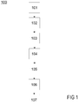
- the present invention proposes a method for automatically performing an image reconstruction for a biological object, said method comprising the following steps, each being carried out preferentially automatically:
- the present invention proposes also a system for automatically performing an image reconstruction of a biological object, the system comprising:
- the present invention enables to extract the largest motion-state-consistent subset from the acquired signal data and therefore to reconstruct a static image out of a motion-corrupted acquisition, or to sort the acquired signal data into several motion-state-consistent bins and use them as an input for a motion-resolved reconstruction.
- the system acquires MRI signal data of a biological object.
- the biological object may undergo some physiological motion and/or contrast changes, which need to be taken into consideration for reconstructing a sharp image of the biological object.
- one of the following techniques is used for acquiring the MRI signal data:
- the system performs, preferably automatically, a clustering of a set of data which comprises at least a part of the previously acquired MRI signal data and/or data obtained from or together with the acquired MRI signal data for each or a part of the different time points t_i.
- said set of data may comprise, with respect to the previously described techniques:
- the system automatically creates a matrix C based on the above-mentioned dataset.
- the matrix C comprises one dimension T associated to the time and at least one dimension associated to the data of the dataset.
- the matrix C may comprise a certain number of rows, wherein each row corresponds to a different time point t_i and is filled with data values of the dataset that have been acquired at said time point t_i.
- the system is configured for selecting,
- step 104 and optionally, if a large number of data is associated with each time point t_i, then the system is configured for performing a dimensionality reduction procedure in order to decrease the number of data available for each time point t_i.
- the system is configured for performing a clustering of the different time points, based on the similarity of their associated data as represented in the matrix C, in order to separate them into distinct clusters.
- the system is configured for automatically selecting one of the clusters and extracting all the MRI signal data that were performed in close adjacency to the time points t_i belonging to the selected cluster.
- the system is configured for automatically performing the MRI image reconstruction of the biological object using the MRI signal data that have been previously extracted at step 106 in connection with the selected cluster.
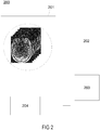
- Figure 2 illustrates a system 200 for automatically performing image reconstruction of a biological object.
- the system 200 comprises:
- the present invention proposes to skip altogether the steps related to the physiological, dynamic, or generic motion signal extraction and proposes instead to intrinsically separate the acquired signal data into motion and contrast consistent bins obtained from the clustering of the matrix elements by leveraging the similarities among said elements, i.e. the acquired signal data (e.g. among the amplitude/phase of the center of k-space that is sampled along all the MRI measurements or among the 1D readouts that are acquired regularly as mentioned as described in Stehning et al., Magn Reson Med (54), 476 (2005 )) without any prior knowledge about motion.
- the acquired signal data e.g. among the amplitude/phase of the center of k-space that is sampled along all the MRI measurements or among the 1D readouts that are acquired regularly as mentioned as described in Stehning et al., Magn Reson Med (54), 476 (2005 )
- the similarity within the acquired signal data can be explored in any n-dimensional space with respect to the matrix C and with any sort of clustering technique that may be suitable for such a task.
- PCA Principal Component Analysis
- n components can be then considered as the dimensions of an n-dimensional space, i.e. said matrix C, where each of the consistent 1D readouts corresponds to one single data point residing in that space.
- a clustering algorithm is then applied to obtain a binning of the data as shown in Fig. 3 .
- Figure 3 illustrates a clustering obtained when using such a clustering algorithm: in Fig. 3A , a matrix C is shown wherein all or some of the MRI signal data acquired from the coil elements for all coherently sampled readouts (e.g. superior-inferior (SI)) are stored within the matrix C and used as an input for the clustering algorithm.
- the time points t_i are related to the different interleaved acquisitions in function of the time.
- a dimensionality reduction using for instance PCA, is applied to the initial matrix C to create a n-dimensional space where each SI readout, or group of SI readouts from several coils but in the same time point t_i, is represented by a single point.
- Fig. 3A a matrix C is shown wherein all or some of the MRI signal data acquired from the coil elements for all coherently sampled readouts (e.g. superior-inferior (SI)) are stored within the matrix C and used as an input for the clustering algorithm.
- FIG. 3B shows the resulting clustering, wherein the largest cluster (or, e.g., the most compact one) formed by these points and detected with a binning/clustering algorithm (e.g. k-means) is chosen to create a motion-consistent static image reconstruction free of any knowledge of the underlying physiology.
- Fig. 3C shows an example of the obtained reconstructed image. Because of the temporally uniform sampling (in this case a 3D radial kooshball trajectory implementing a golden angle rotation between interleaves), a coherent image can be reconstructed where sharpness of the features and general appearance are visibly improved when compared to a reconstruction where all acquired data are grouped together.
- a binning/clustering algorithm e.g. k-means
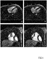
- the described technique can be used, for instance, either to intrinsically extract the largest motion-state-consistent subset of acquired signal data and therefore reconstruct a static image out of a motion-corrupted acquisition as shown in Figure 4 , or to sort the acquired signal data into several motion-state-consistent bins/clusters and use them as an input for a motion-resolved reconstruction as in described in Feng et al., Magn Reson Med (79), 826 (2018 ) and shown in Figure 5 in order to perform the final image reconstruction.
- Figure 4 presents for instance a comparison between images obtained from a classic reconstruction based on all the acquired signal data (A,C) and a reconstruction based on the present concept (B,D), which clearly show a better result.
- the concept of the invention as described for Fig. 3 is further used for identifying more than one motion consistent bin (or cluster) and exploit their similarity using and XD-GRASP reconstruction.
- the present invention has several advantages:
- the claimed technique does not rely on a specific signal extraction, but solely on similarities within the acquired signal data, wherein a binning/clustering of the acquired signal data takes place in a n-dimensional space with respect to the different time points t_i of acquisition and that is not necessarily directly related to the acquisition space (e.g. k-space) nor the image space.
- the described technique provides a very easy solution for obtaining a static image of a moving organ (e.g. the heart) without any complexity on the planning/protocol/sequence side, but just using a continuous acquisition.
- the present invention might be applied to coronary MR imaging without any triggering nor gating with almost instantaneous reconstruction at the scanner.
Landscapes
- Health & Medical Sciences (AREA)
- Physics & Mathematics (AREA)
- Engineering & Computer Science (AREA)
- Life Sciences & Earth Sciences (AREA)
- General Health & Medical Sciences (AREA)
- Nuclear Medicine, Radiotherapy & Molecular Imaging (AREA)
- Signal Processing (AREA)
- High Energy & Nuclear Physics (AREA)
- Radiology & Medical Imaging (AREA)
- General Physics & Mathematics (AREA)
- Condensed Matter Physics & Semiconductors (AREA)
- Animal Behavior & Ethology (AREA)
- Pathology (AREA)
- Surgery (AREA)
- Medical Informatics (AREA)
- Heart & Thoracic Surgery (AREA)
- Public Health (AREA)
- Veterinary Medicine (AREA)
- Biomedical Technology (AREA)
- Biophysics (AREA)
- Molecular Biology (AREA)
- Artificial Intelligence (AREA)
- Computer Vision & Pattern Recognition (AREA)
- Physiology (AREA)
- Psychiatry (AREA)
- Theoretical Computer Science (AREA)
- Vascular Medicine (AREA)
- Magnetic Resonance Imaging Apparatus (AREA)
Priority Applications (2)
| Application Number | Priority Date | Filing Date | Title |
|---|---|---|---|
| EP19190766.6A EP3772660A1 (de) | 2019-08-08 | 2019-08-08 | Magnetresonanzbildrekonstruktion mit clusteranalyse |
| US16/988,864 US11460529B2 (en) | 2019-08-08 | 2020-08-10 | Method and system for improving magnetic resonance images |
Applications Claiming Priority (1)
| Application Number | Priority Date | Filing Date | Title |
|---|---|---|---|
| EP19190766.6A EP3772660A1 (de) | 2019-08-08 | 2019-08-08 | Magnetresonanzbildrekonstruktion mit clusteranalyse |
Publications (1)
| Publication Number | Publication Date |
|---|---|
| EP3772660A1 true EP3772660A1 (de) | 2021-02-10 |
Family
ID=67658584
Family Applications (1)
| Application Number | Title | Priority Date | Filing Date |
|---|---|---|---|
| EP19190766.6A Pending EP3772660A1 (de) | 2019-08-08 | 2019-08-08 | Magnetresonanzbildrekonstruktion mit clusteranalyse |
Country Status (2)
| Country | Link |
|---|---|
| US (1) | US11460529B2 (de) |
| EP (1) | EP3772660A1 (de) |
Families Citing this family (2)
| Publication number | Priority date | Publication date | Assignee | Title |
|---|---|---|---|---|
| WO2022261742A1 (en) * | 2021-06-17 | 2022-12-22 | Elekta Limited | Image quality relative to machine learning data |
| US12597088B2 (en) * | 2021-07-08 | 2026-04-07 | Elekta Limited | Quality factor using reconstructed images |
Citations (2)
| Publication number | Priority date | Publication date | Assignee | Title |
|---|---|---|---|---|
| US20170199263A1 (en) * | 2016-01-13 | 2017-07-13 | Siemens Healthcare Gmbh | Method and apparatus for allocating acquired magnetic resonance data to respective movement states of the subject |
| US20170307707A1 (en) * | 2014-10-13 | 2017-10-26 | Koninklijke Philips N.V. | Mutli-shot magnetic resonance imaging system and method |
Family Cites Families (6)
| Publication number | Priority date | Publication date | Assignee | Title |
|---|---|---|---|---|
| GB9614407D0 (en) * | 1996-07-09 | 1996-09-04 | Secr Defence | Method for imaging artefact reduction |
| US20070118399A1 (en) * | 2005-11-22 | 2007-05-24 | Avinash Gopal B | System and method for integrated learning and understanding of healthcare informatics |
| WO2009050618A2 (en) * | 2007-10-15 | 2009-04-23 | Koninklijke Philips Electronics N.V. | Visualization of temporal data |
| JP5932612B2 (ja) * | 2012-11-16 | 2016-06-08 | 株式会社スクウェア・エニックス | 情報処理装置、制御方法、プログラム、及び記録媒体 |
| FR3015692B1 (fr) * | 2013-12-24 | 2017-03-03 | Univ Aix Marseille | Procede d'analyse par resonance magnetique nucleaire |
| US9990741B2 (en) * | 2015-09-28 | 2018-06-05 | Siemens Medical Solutions Usa, Inc. | Motion correction in a projection domain in time of flight positron emission tomography |
-
2019
- 2019-08-08 EP EP19190766.6A patent/EP3772660A1/de active Pending
-
2020
- 2020-08-10 US US16/988,864 patent/US11460529B2/en active Active
Patent Citations (2)
| Publication number | Priority date | Publication date | Assignee | Title |
|---|---|---|---|---|
| US20170307707A1 (en) * | 2014-10-13 | 2017-10-26 | Koninklijke Philips N.V. | Mutli-shot magnetic resonance imaging system and method |
| US20170199263A1 (en) * | 2016-01-13 | 2017-07-13 | Siemens Healthcare Gmbh | Method and apparatus for allocating acquired magnetic resonance data to respective movement states of the subject |
Non-Patent Citations (4)
| Title |
|---|
| FENG ET AL., MAGN RESON MED, vol. 79, 2018, pages 826 |
| JOHN HEERFORDT ET AL.: "A Similarity-Based Data Clustering Approach for Fast Reconstruction of Untriggered and Ungated Whole-Heart MRA", PROCEEDINGS OF THE 31ST ANNUAL CONFERENCE OF THE SOCIETY FOR MAGNETIC RESONANCE ANGIOGRAPHY, SMRA, NANTES, FRANCE, 28-30 AUGUST 2019, 29 August 2019 (2019-08-29), pages 40, XP055664589 * |
| STEHNING ET AL., MAGN RESON MED, vol. 54, 2005, pages 476 |
| ZIXIN DENG ET AL: "A post-processing method based on interphase motion correction and averaging to improve image quality of 4D magnetic resonance imaging: a clinical feasibility study", BRITISH JOURNAL OF RADIOLOGY., vol. 92, no. 1095, 3 January 2019 (2019-01-03), GB, XP055664621, ISSN: 0007-1285 * |
Also Published As
| Publication number | Publication date |
|---|---|
| US11460529B2 (en) | 2022-10-04 |
| US20210041519A1 (en) | 2021-02-11 |
Similar Documents
| Publication | Publication Date | Title |
|---|---|---|
| Menchón-Lara et al. | Reconstruction techniques for cardiac cine MRI | |
| US9271661B2 (en) | Method for free-breathing magnetic resonance imaging using iterative image-based respiratory motion correction | |
| Paul et al. | High‐resolution respiratory self‐gated golden angle cardiac MRI: comparison of self‐gating methods in combination with k‐t SPARSE SENSE | |
| US8427153B2 (en) | Method for motion correction in magnetic resonance imaging using radio frequency coil arrays | |
| Nguyen et al. | Free‐breathing diffusion tensor MRI of the whole left ventricle using second‐order motion compensation and multitasking respiratory motion correction | |
| Luo et al. | Nonrigid motion correction with 3D image‐based navigators for coronary MR angiography | |
| US10371779B2 (en) | Apparatus and method for magnetic resonance imaging with high spatial temporal resolutions | |
| US20180210058A1 (en) | Mr imaging with motion detection | |
| US11415655B2 (en) | Reduced field-of-view perfusion imaging with high spatiotemporal resolution | |
| US10531812B2 (en) | System and method for improved cardiac imaging of subjects with adverse cardiac conditions | |
| US10849561B2 (en) | Systems and methods for reducing respiratory-induced motion artifacts for accelerated imaging | |
| US10545208B2 (en) | System, method and computer-accessible medium for rapid real-time cardiac magnetic resonance imaging utilizing synchronized cardio-respiratory sparsity | |
| US10175328B2 (en) | System and method for reconstructing ghost-free images from data acquired using simultaneous multislice magnetic resonance imaging | |
| Heerfordt et al. | Similarity‐driven multi‐dimensional binning algorithm (SIMBA) for free‐running motion‐suppressed whole‐heart MRA | |
| CN106796274A (zh) | 具有伪迹抑制的propeller‑mr成像 | |
| US20230132314A1 (en) | Shot-wise inversion time adaptation for multi-shot inversion recovery imaging | |
| US11460529B2 (en) | Method and system for improving magnetic resonance images | |
| US7239136B2 (en) | Motion compensation for magnetic resonance imaging | |
| Gao et al. | High spatial‐resolution and acquisition‐efficiency cardiac MR T1 mapping based on radial bSSFP and a low‐rank tensor constraint | |
| EP4290265B1 (de) | Computer-implementiertes verfahren, computerprogramm und verarbeitungsgerät zum rekonstruieren einer dynamischen serie von magnetresonanzbildern | |
| Matsumoto et al. | Feasibility of free-breathing late gadolinium-enhanced cardiovascular MRI for assessment of myocardial infarction: navigator-gated versus single-shot imaging | |
| US10416265B2 (en) | Method and system for generating MR images of a moving object in its environment | |
| US10401459B2 (en) | Systems and methods for imaging vascular calcifications with magnetic resonance imaging | |
| WO2017167937A1 (en) | Dynamic mr imaging with increased temporal and spatial resolution | |
| US10314512B2 (en) | Magnetic resonance method and apparatus for determining deformation information from a cyclically moving examination subject |
Legal Events
| Date | Code | Title | Description |
|---|---|---|---|
| PUAI | Public reference made under article 153(3) epc to a published international application that has entered the european phase |
Free format text: ORIGINAL CODE: 0009012 |
|
| STAA | Information on the status of an ep patent application or granted ep patent |
Free format text: STATUS: THE APPLICATION HAS BEEN PUBLISHED |
|
| AK | Designated contracting states |
Kind code of ref document: A1 Designated state(s): AL AT BE BG CH CY CZ DE DK EE ES FI FR GB GR HR HU IE IS IT LI LT LU LV MC MK MT NL NO PL PT RO RS SE SI SK SM TR |
|
| AX | Request for extension of the european patent |
Extension state: BA ME |
|
| STAA | Information on the status of an ep patent application or granted ep patent |
Free format text: STATUS: REQUEST FOR EXAMINATION WAS MADE |
|
| 17P | Request for examination filed |
Effective date: 20210803 |
|
| RBV | Designated contracting states (corrected) |
Designated state(s): AL AT BE BG CH CY CZ DE DK EE ES FI FR GB GR HR HU IE IS IT LI LT LU LV MC MK MT NL NO PL PT RO RS SE SI SK SM TR |
|
| STAA | Information on the status of an ep patent application or granted ep patent |
Free format text: STATUS: EXAMINATION IS IN PROGRESS |
|
| 17Q | First examination report despatched |
Effective date: 20230314 |
|
| RAP1 | Party data changed (applicant data changed or rights of an application transferred) |
Owner name: CENTRE HOSPITALIER UNIVERSITAIRE VAUDOIS Owner name: SIEMENS HEALTHINEERS AG |