EP3766416B1 - Abbildung von vorhofflimmern unter verwendung von fragmentierungsindex - Google Patents
Abbildung von vorhofflimmern unter verwendung von fragmentierungsindex Download PDFInfo
- Publication number
- EP3766416B1 EP3766416B1 EP20185867.7A EP20185867A EP3766416B1 EP 3766416 B1 EP3766416 B1 EP 3766416B1 EP 20185867 A EP20185867 A EP 20185867A EP 3766416 B1 EP3766416 B1 EP 3766416B1
- Authority
- EP
- European Patent Office
- Prior art keywords
- peaks
- processor
- fragmentation
- local
- additional
- Prior art date
- Legal status (The legal status is an assumption and is not a legal conclusion. Google has not performed a legal analysis and makes no representation as to the accuracy of the status listed.)
- Active
Links
Images


Classifications
-
- A—HUMAN NECESSITIES
- A61—MEDICAL OR VETERINARY SCIENCE; HYGIENE
- A61B—DIAGNOSIS; SURGERY; IDENTIFICATION
- A61B5/00—Measuring for diagnostic purposes; Identification of persons
- A61B5/24—Detecting, measuring or recording bioelectric or biomagnetic signals of the body or parts thereof
- A61B5/316—Modalities, i.e. specific diagnostic methods
- A61B5/318—Heart-related electrical modalities, e.g. electrocardiography [ECG]
- A61B5/346—Analysis of electrocardiograms
- A61B5/349—Detecting specific parameters of the electrocardiograph cycle
- A61B5/361—Detecting fibrillation
-
- A—HUMAN NECESSITIES
- A61—MEDICAL OR VETERINARY SCIENCE; HYGIENE
- A61B—DIAGNOSIS; SURGERY; IDENTIFICATION
- A61B5/00—Measuring for diagnostic purposes; Identification of persons
- A61B5/24—Detecting, measuring or recording bioelectric or biomagnetic signals of the body or parts thereof
- A61B5/316—Modalities, i.e. specific diagnostic methods
- A61B5/318—Heart-related electrical modalities, e.g. electrocardiography [ECG]
- A61B5/339—Displays specially adapted therefor
- A61B5/343—Potential distribution indication
-
- A—HUMAN NECESSITIES
- A61—MEDICAL OR VETERINARY SCIENCE; HYGIENE
- A61B—DIAGNOSIS; SURGERY; IDENTIFICATION
- A61B5/00—Measuring for diagnostic purposes; Identification of persons
- A61B5/24—Detecting, measuring or recording bioelectric or biomagnetic signals of the body or parts thereof
- A61B5/316—Modalities, i.e. specific diagnostic methods
- A61B5/318—Heart-related electrical modalities, e.g. electrocardiography [ECG]
- A61B5/367—Electrophysiological study [EPS], e.g. electrical activation mapping or electro-anatomical mapping
Definitions
- the present invention relates generally to electrophysiological mapping, and particularly to methods, systems and processor-readable media for mapping atrial fibrillation using a fragmentation index.
- Atrial fibrillation is an irregular heart rhythm generated in the atria.
- Various techniques for mapping atrial fibrillation are known in the art.
- U.S. Patent Application Publication 2017/0367601 describes a method for identifying areas of the heart of a patient able to be involved in the perpetuation of atrial fibrillation. This method takes into account the reference cycle of the arrhythmia and has two variants: a local variant in which the areas of the heart are each analyzed separately and a regional variant in which several areas of the heart are analyzed together.
- U.S. Patent Application Publication 2015/0359430 describes a method of medical image processing for images of body structures, the method comprising: receiving anatomical data to reconstruct an anatomical image of a region of a body of a patient, said region comprises a portion of at least one internal body part which borders or is spaced apart from a target tissue; receiving functional data from a functional imaging modality which images at least said portion of the region of the body of the patient; processing said anatomical image to generate at least one image mask corresponding to the zone outside of the wall of said at least one internal body part; correlating the at least one generated image mask with the functional data for guiding a reconstruction of a functional image depicting said target tissue; and providing the reconstructed functional image.
- U.S. Patent Application Publication 2012/0078129 describes a method of displaying an image of the location of one or more low voltage structures in tissue.
- the method includes receiving electrical mapping data corresponding to a portion of the tissue, and generating an image using the electrical mapping data. Electrical mapping values within at least one voltage range having two endpoints that bound the upper and lower limits of the voltage range are distinguishable from electrical mapping values outside the at least one voltage range. The two endpoints are selected to distinguish the one or more low voltage structures of the tissue from other portions of the tissue.
- EP2851002A1 discloses a monitoring device for monitoring and evaluating electrogram signals representing electric activities of a heart chamber, said monitoring device comprising a signal input operatively connected to a mapping catheter and a signal processing and evaluation unit for processing and evaluating electrogram signals received at the signal input.
- the morphology of a detection complex is characterised by the evaluation device, including on the basis of the number of local peaks in the deflection complex.
- WO2010054409A1 discloses a method for visualization of electrical activity on a surface of an organ over a time period, including morphological analysis of complex fractionated electrograms.
- an embodiment of the present invention that is described herein provides a computer-implemented method for visualizing a map of atrial fibrillation (AF) in a heart, as specified in appended claim 1.
- the heart has a region that includes (i) the given position located at a given distance from a predefined location of the region, and (ii) at least an additional position having an additional FI and located at an additional distance from the predefined location, and the method includes calculating and visualizing, based on the given distance and the additional distance, and based on the local FI and the additional FI, a regional FI of the region.
- the predefined location includes a geometrical center of gravity (COG) of the region
- calculating and visualizing the regional FI includes calculating, based on the given distance and the additional distance, a weighted average of at least the local FI and the additional FI.
- COG geometrical center of gravity
- the method includes defining a window of interest (WOI) within the CL, and identifying the one or more secondary peaks within the WOI.
- the EGM signal includes multiple CLs, and calculating the local FI includes calculating, based on the multiple CLs, an average CL, and calculating an average number of the secondary peaks per the average CL.
- AF atrial fibrillation
- Some medical procedures are based on measuring electrogram (EGM) signals by disposing a plurality of electrodes at different respective sites on the cardiac tissue.
- EGM electrogram
- a physician may use the EGM signals to characterize the propagation of the wavefront of electrical activation through the cardiac tissue of a patient during a cardiac cycle. For each of the EGM signals, the physician may attempt to identify electrical activation points that correspond to an instance at which the wavefront passed through the site at which the signal was acquired.
- AF heart atrial fibrillation
- EGM signals typically contain regularly-spaced and sharp primary peaks that clearly indicate electrical activations.
- irregular EGMs may exhibit a wide variety of different forms, and may comprise numerous bursts of secondary peaks that are typically not indicative of regular electrical activation.
- Embodiments of the present invention that are described hereinbelow provide methods and systems for mapping AF by calculating local and regional fragmentation indexes (FIs), and visualizing the FIs on a map of a patient heart.
- a system for mapping AF in a patient heart comprises a processor and a display.
- the processor is configured to receive, from a catheter inserted into a patient heart, an EGM signal that exhibits the AF and acquired at a given position in the heart.
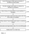
- the processor is further configured to identify, in the EGM signal, two or more primary activation peaks, also referred to herein as annotations, and to calculate a cycle length (CL) between adjacent annotations.
- the processor is further configured to hold predefined criteria for identifying a regular atrial fibrillation cycle length (AFCL), such as but not limited to, a cycle length between 120 ms (referred to herein as short AFCLs) and 250 ms (referred to herein as long AFCLs) having a standard deviation smaller than 30 ms.
- AFCL regular atrial fibrillation cycle length
- the processor is configured to define a window of interest (WOI) within the AFCL, and to identify within the WOI, one or more secondary activation peaks, also referred to herein as fragmentation peaks.
- the processor calculates at the given position, a local fragmentation index (FI) that is indicative of an average number of the fragmentation peaks per WOI.
- WOI window of interest
- FI local fragmentation index
- the processor is configured to merge, based on a predefined threshold and criteria, two or more adjacent fragmentation peaks.
- the EGM signal comprises multiple AFCLs
- the processor is configured to calculate, based on the multiple AFCLs, an average AFCL, and to calculate an average number of the fragmentation peaks per the average AFCL.
- the display is configured to display the calculated and visualized local FI on a map of at least part of the heart.
- a region of patient heart contains the given position, which is located at a given distance from a geometrical center-of-gravity (COG) of the region.
- the region additionally comprises multiple positions located at respective distances from the COG.
- the processor is configured to acquire, using the catheter, additional EGM signals at the additional respective positions, and to calculate, for each of the additional positions, a respective additional FI.
- the processor is configured to calculate, based on the given distance and the additional distances, and based on the local FI and the additional FIs, a regional FI of the region.
- the processor is further configured to output, to the display, a visualization of the regional FI and the AFCLs.
- the display is configured to display the calculated and visualized regional FI on a map of the heart showing at least the aforementioned region, and to display the short and the long AFCLs, overlaid on the regional FIs.
- the processor is configured to display, on the heart map, important regions, such as regions having short AFCLs and large regional FI.
- the disclosed techniques provide the physician with the features of detection and display of regions suspected to have irregular activation.
- the physician may apply ablation in one or more of the suspected regions, in order to reduce the arrhythmia in the patient heart.
- Fig. 1 is a schematic, pictorial illustration of a system 21 for annotating an electrogram (EGM) signal 22.
- EMM electrogram
- a physician 27 inserts a catheter 29 and navigates a distal end 31 of catheter 29 into a desired location in a heart 23 of a patient 25.
- EGM signals 22 generated by the tissue.
- Such signals may be sensed, for example, while heart 23 experiences atrial fibrillation (AF) or any other arrhythmia.
- AF atrial fibrillation
- the arrhythmia may be induced by the physician as part of the procedure.
- a processor 28 of system 21 receives EGM signals 22 from distal end 31, via an electrical interface 35, such as a socket or port, and processes these EGM signals as will be described in detail in Figs. 2 and 3 below.
- processor 28 in response to processing the EGM signals, is configured to generate an output, which typically comprises a visual output displayed on a display 26 of system 21.
- processor 28 is configured to annotate at least one peak of EGM signal 22 to show the activation points of the EGM signal, and to display the annotated signal on display 26.
- processor 28 may, for example, place a marker 24 over each activation point.
- the annotated signal shown by marker 24 is also referred to herein as a "primary peak” or as an “annotated activation signal.”
- each EGM signal 22 may be a bipolar signal, so that the signal represents the voltage between a respective pair of the electrodes at distal end 31.
- at least one of the acquired EGM signals may be a unipolar signal, so that the EGM signal represents the voltage between one of the electrodes and a reference electrode that is coupled externally to patient 25.
- processor 28 may comprise a single processor, or a cooperatively networked or clustered set of processors.
- the functionality of processor 28, as described herein may be implemented solely in hardware, e.g., using one or more Application-Specific Integrated Circuits (ASICs) or Field-Programmable Gate Arrays (FPGAs).
- ASICs Application-Specific Integrated Circuits
- FPGAs Field-Programmable Gate Arrays
- the functionality of processor 28 may be implemented at least partly in software.
- processor 28 may comprise a programmed digital computing device comprising at least a central processing unit (CPU) and random access memory (RAM).
- system 21 may comprise any suitable types of non-volatile memory devices.
- processor 28 may comprise a general-purpose processor, which is programmed in software to carry out the functions described herein.
- the software may be downloaded to the processor in electronic form, over a network, for example, or it may, alternatively or additionally, be provided and/or stored on non-transitory tangible media, such as magnetic, optical, or electronic memory.
- system 21 is shown by way of example, in order to illustrate certain problems that are addressed by embodiments of the present invention and to demonstrate the application of these embodiments in enhancing the performance of such a system.
- Embodiments of the present invention are by no means limited to this specific sort of example system, and the principles described herein may similarly be applied to other sorts of systems for mapping arrhythmia and to other sorts of systems for annotating and analyzing any suitable signals acquired from any human organ.
- Fig. 2 is a graph showing an identification of activation points in an EGM signal 100.
- EGM signal 100 may replace, for example, EGM signal 22 of Fig. 1 above.
- EGM signal 100 is acquired at a given position in heart 23 using one or more electrodes of distal end 31, and comprises a complex fractionated EGM signal measured in millivolts (mV) over time.
- processor 28 is configured to divide the total period that is spanned by the signal into successive smaller time periods, each being of a predefined length or within a range of lengths, e.g., between 100 milliseconds (ms) and 200 ms, or any other suitable period of time.
- processor 28 identifies a set of candidate fragmentation peaks, such as peaks 106, 106A, 106B, 106C, 107, 108A and 108B shown in Fig. 2 above.
- processor 28 may filter out at least some of the candidate fragmentation peaks, such as peak 107 which is within the noise threshold located between noise-threshold lines 103 and 105 shown in Fig. 2 above.
- processor identifies a group of peaks comprising two or more secondary peaks that are within a predefined time interval (e.g., 20 ms) from one another and having the same sign (positive or negative). For example, a group of peaks 106A and 108A, and another group of peaks 106B and 108B.
- processor 28 may select, within the group of peaks, the peak having the largest absolute electro-potential value and removes the other peaks from the set of secondary peaks. As depicted in Fig. 2 above, peaks 108A and 108B had the largest absolute electro-potential value and therefore, respective peaks 106A and 106B have been removed.
- processor calculates the local FI using equation (1), which is described in Fig. 2 above.
- Processor 28 applies to equation (1), the total number of primary and secondary peaks identified and verified after the filtering and merging processes described in steps 206 and 208 above, and the total actual and nominal WOI time durations used for identifying the primary and secondary peaks.
- the calculated local FI is indicative of the average number of secondary peaks per AFCL time duration.
- the calculated value of the local FI equals 2.334, which is indicative of the average number of secondary peaks per average AFCL time duration.
- display 26 displays the local FI on the map of heart 23 received from processor 28.
- display 26 is configured to display a visualization of the local FI using color coding or any other suitable visualization technique.
- processor 28 and/or display 26 may assign warm colors, e.g., red, to small values of FI, and cold colors, e.g., blue, to large values of FI.
- display 26 may also display the one or more AFCLs overlaid on the heart map and on the displayed local FI.
- An exemplary embodiment of the map is shown in Fig. 5 below.
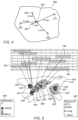
- Fig. 4 is a schematic, pictorial illustration of a region 300 of heart 23 for which a regional fragmentation index (FI) is calculated based on multiple local FIs.
- region 300 comprises points 302, 304, 306, 308 and 310, located at respective distances of 3 mm, 1 mm, 2 mm, 4 mm, and 5 mm, from a geometrical center-of-gravity (COG), referred to as COG 333 of region 300.
- COG geometrical center-of-gravity
- processor 28 is configured to calculate the regional FI of region 300 by calculating a weighted average over the local FIs of points 302, 304, 306, 308 and 310, using the respective distances from COG 333 for deriving the averaging weights.
- the respective local FI is weighted by a weight value that is proportional to the inverse value of the distance between the given point and COG 333.
- point 304 which is located 1 mm from COG 333, has larger weight (e.g., 1) relative to that of point 310 located 5 mm from COG 333, and therefore having a 5X smaller weight (e.g., 1/5).
- processor 28 calculates normalized weights for points 302, 304, 306, 308 and 310 and outputs the respective values of 0.146, 0.438, 0.219, 0.109 and 0.087, and then multiplies, for each point, the respective local FI by the respective normalized weight, and sums.
- processor 28 After calculating a sum of the weighted local FIs, processor 28 outputs the regional FI having a value of 6.628. Subsequently, processor 28 outputs the calculated regional FI and the calculated AFCLs of region 300, to display 26.
- map 400 further comprises AFCL markers 422, 423, 424, 425, 426 and 428, which are calculated by processor 28 (e.g., using the technique described in Fig. 2 above) in the sections represented by respective contours 402, 403, 404, 405, 406 and 408.
- map 400 comprises a legend 440 that provides another set of coding of the visualized values of the average AFCL markers.
- 120 ms represents "short" AFCL values and 250 ms represents "long" AFCL values.
- the local FI is indicative of the average number of fragmentation peaks (also referred to herein as secondary peaks) per regular AFCL.
- a short regular AFCL is indicative of a short time duration between two annotated activation signals (also referred to herein as primary peaks).
- a combination of a large value of a regional FI and one or more short regular AFCLs, as shown for example in contour 403 and AFCL marker 423, respectively, may be indicative of a section having high AF activity in region 410.
- processor 28 may highlight on display 26 outstanding combinations, e.g., of high regional FI and short regular AFCLs, so as to draw the attention of physician 27 during the EP procedure shown for example in Fig. 1 above.
- processor 28 may hold any set of thresholds for the regional FI and regular AFCLs, and for any combination thereof, so as to alert of any outstanding AF activities identified at specific sections or regions of heart 23.
- processor 28 may add any other suitable types of markers to be visualized on map 400, or may reduce at least one of the AFCLs or regional FIs shown on map 400 of Fig. 5 .
- map 400 is shown by way of example, in order to illustrate certain problems that are addressed by embodiments of the present invention and to demonstrate the application of these embodiments in enhancing the performance of a system for analyzing arrhythmia, such as system 21.
- Embodiments of the present invention are by no means limited to this specific sort of example mapping and visualizing configuration, and the principles described herein may similarly be applied to other sorts of visualizations of any medical systems.
- the embodiments described herein mainly address arrhythmia and particularly atrial fibrillation, the methods and systems described herein can also be used in other applications, such as in persistent atrial fibrillation or in any other type arrhythmia in a human heart.
Landscapes
- Health & Medical Sciences (AREA)
- Life Sciences & Earth Sciences (AREA)
- Cardiology (AREA)
- Heart & Thoracic Surgery (AREA)
- Molecular Biology (AREA)
- Pathology (AREA)
- Engineering & Computer Science (AREA)
- Biomedical Technology (AREA)
- Physics & Mathematics (AREA)
- Medical Informatics (AREA)
- Biophysics (AREA)
- Surgery (AREA)
- Animal Behavior & Ethology (AREA)
- General Health & Medical Sciences (AREA)
- Public Health (AREA)
- Veterinary Medicine (AREA)
- Physiology (AREA)
- Measurement And Recording Of Electrical Phenomena And Electrical Characteristics Of The Living Body (AREA)
- Medicines Containing Plant Substances (AREA)
Claims (11)
- Computerimplementiertes Verfahren zum Visualisieren einer Karte von Vorhofflimmern (AF) in einem Herzen, wobei das Verfahren umfasst:Empfangen eines Elektrogrammsignals (EGM-Signal), das die AF anzeigt und an einer bestimmten Position im Herzen erfasst wurde;Identifizieren (200) von zwei oder mehr primären Aktivierungs-Peaks im EGM-Signal und Berechnen (202) einer Zykluslänge (CL) zwischen benachbarten primären Aktivierungs-Peaks;Identifizieren (206) eines oder mehrerer Fragmentierungs-Peaks innerhalb des CL im EGM-Signal, wobei das Identifizieren des einen oder der mehreren Fragmentierungs-Peaks das Identifizieren eines oder mehrerer Kandidaten-Peaks umfasst, wobei jeder der einen oder mehreren Kandidaten-Peaks einen absoluten Wert aufweist, der größer ist als ein Rauschschwellenwert, und wobei das Identifizieren des einen oder der mehreren Fragmentierungs-Peaks das Zusammenführen von zwei oder mehreren benachbarten Kandidaten-Peaks im EGM-Signal umfasst, basierend darauf, dass sie innerhalb eines vordefinierten Zeitintervalls liegen und dasselbe Vorzeichen haben;Berechnen (210) eines lokalen Fragmentierungsindex (FI), der eine Anzahl der Fragmentierungs-Peaks pro CL angibt; undVisualisieren (212) des lokalen FI auf einer Karte von mindestens einem Teil des Herzens auf einer Anzeige.
- Computerimplementiertes Verfahren nach Anspruch 1, wobei das Herz eine Region umfasst, die (i) die vorgegebene Position umfasst, die sich in einer vorgegebenen Entfernung von einem vordefinierten Ort der Region befindet, und (ii) mindestens eine zusätzliche Position, die einen zusätzlichen FI aufweist und sich in einer zusätzlichen Entfernung von dem vordefinierten Ort befindet, und das Berechnen und Visualisieren eines regionalen FI der Region umfasst, basierend auf der vorgegebenen Entfernung und der zusätzlichen Entfernung und basierend auf dem lokalen FI und dem zusätzlichen FI.
- Computerimplementiertes Verfahren nach Anspruch 2, wobei der vordefinierte Standort einen geometrischen Schwerpunkt (COG) der Region umfasst, und wobei das Berechnen und Visualisieren des regionalen FI das Berechnen eines gewichteten Durchschnitts von mindestens dem lokalen FI und dem zusätzlichen FI basierend auf der vorgegebenen Entfernung und der zusätzlichen Entfernung umfasst.
- Computerimplementiertes Verfahren nach Anspruch 1, und umfassend das Definieren (204) eines Fensters von Interesse (WOI) innerhalb des CL und das Identifizieren der einen oder mehreren Fragmentierungs-Peaks innerhalb des WOI.
- Computerimplementiertes Verfahren nach Anspruch 1, wobei das EGM-Signal mehrere CL umfasst, und wobei das Berechnen des lokalen FI das Berechnen eines durchschnittlichen CL auf der Grundlage der mehreren CLs und das Berechnen einer durchschnittlichen Anzahl der Fragmentierungs-Peaks pro durchschnittlichem CL umfasst.
- System (21) zum Abbilden von Vorhofflimmern (AF) in einem Herzen (23), wobei das System umfasst:
einen Prozessor (28), der konfiguriert ist zum:(a) Empfangen eines Elektrogrammsignals (EGM-Signal) (100), das die AF anzeigt, die an einer vorgegebenen Position im Herzen erfasst wurde;(b) Identifizieren von zwei oder mehr primären Aktivierungs-Peaks (101, 111, 121) im EGM-Signal und Berechnen einer Zykluslänge (CL) (104) zwischen benachbarten primären Aktivierungs-Peaks;(c) Identifizieren eines oder mehrerer Fragmentierungs-Peaks (106, 106A, 106B, 106C, 107, 108A, 108B) in den EGM-Signalen innerhalb einer Dauer des CL, wobei das Identifizieren des einen oder der mehreren Fragmentierungs-Peaks das Identifizieren eines oder mehrerer Kandidaten-Peaks umfasst, wobei jeder der einen oder mehreren Kandidaten-Peaks einen absoluten Wert aufweist, der größer als ein Rauschschwellenwert ist, und wobei das Identifizieren des einen oder der mehreren Fragmentierungs-Peaks das Zusammenführen von zwei oder mehreren benachbarten Kandidaten-Peaks (106B, 108B) im EGM-Signal umfasst, basierend darauf, dass sie innerhalb eines vordefinierten Zeitintervalls liegen und dasselbe Vorzeichen haben; und(d) Berechnen eines lokalen Fragmentierungsindex (FI), der die Anzahl der Fragmentierungs-Peaks pro CL angibt; und wobei das System ferner umfasst:
eine Anzeige (26), die konfiguriert ist, um den lokalen FI auf einer Karte von mindestens einem Teil des Herzens anzuzeigen. - System nach Anspruch 6, wobei das Herz eine Region (300) aufweist, die (i) die vorgegebene Position (302), die sich in einer vorgegebenen Entfernung von einem vordefinierten Ort der Region befindet, und (ii) mindestens eine zusätzliche Position (304, 306, 308, 310) umfasst, die einen zusätzlichen FI aufweist und sich in einer zusätzlichen Entfernung von dem vordefinierten Ort befindet, und wobei der Prozessor konfiguriert ist, um basierend auf der vorgegebenen Entfernung und der zusätzlichen Entfernung und basierend auf dem lokalen FI und dem zusätzlichen FI einen regionalen FI des Bereichs zu berechnen und zu visualisieren.
- System nach Anspruch 7, wobei der vordefinierte Standort einen geometrischen Schwerpunkt (COG) der Region umfasst, und wobei der Prozessor konfiguriert ist, um den regionalen FI zu berechnen und zu visualisieren, indem er basierend auf der vorgegebenen Entfernung und der zusätzlichen Entfernung einen gewichteten Durchschnitt von mindestens dem lokalen FI und dem zusätzlichen FI berechnet.
- System nach Anspruch 6, wobei der Prozessor konfiguriert ist, um ein Fenster von Interesse (WOI) (104) innerhalb einer Dauer des CL zu definieren und die eine oder mehrere Fragmentierungs-Peaks innerhalb des WOI zu identifizieren.
- System nach Anspruch 6, wobei das EGM-Signal mehrere CLs umfasst, und wobei der Prozessor konfiguriert ist, um basierend auf den mehreren CLs einen durchschnittlichen CL zu berechnen und eine durchschnittliche Anzahl der Fragmentierungs-Peaks pro durchschnittlichem CL zu berechnen.
- Prozessorlesbares Medium, das Anweisungen umfasst, die bei Ausführung den an eine Anzeige gekoppelten Prozessor veranlassen, das Verfahren nach einem der Ansprüche 1 bis 5 auszuführen.
Applications Claiming Priority (1)
| Application Number | Priority Date | Filing Date | Title |
|---|---|---|---|
| US16/512,588 US10905349B1 (en) | 2019-07-16 | 2019-07-16 | Mapping atrial fibrillation using fragmentation index |
Publications (3)
| Publication Number | Publication Date |
|---|---|
| EP3766416A1 EP3766416A1 (de) | 2021-01-20 |
| EP3766416C0 EP3766416C0 (de) | 2025-04-02 |
| EP3766416B1 true EP3766416B1 (de) | 2025-04-02 |
Family
ID=71620241
Family Applications (1)
| Application Number | Title | Priority Date | Filing Date |
|---|---|---|---|
| EP20185867.7A Active EP3766416B1 (de) | 2019-07-16 | 2020-07-15 | Abbildung von vorhofflimmern unter verwendung von fragmentierungsindex |
Country Status (5)
| Country | Link |
|---|---|
| US (1) | US10905349B1 (de) |
| EP (1) | EP3766416B1 (de) |
| JP (1) | JP7588976B2 (de) |
| CN (1) | CN112315486B (de) |
| IL (1) | IL275666B2 (de) |
Families Citing this family (1)
| Publication number | Priority date | Publication date | Assignee | Title |
|---|---|---|---|---|
| EP4473910A1 (de) * | 2023-06-06 | 2024-12-11 | CathVision ApS | Verfahren zur analyse eines elektrogramms |
Family Cites Families (20)
| Publication number | Priority date | Publication date | Assignee | Title |
|---|---|---|---|---|
| US20050288599A1 (en) * | 2004-05-17 | 2005-12-29 | C.R. Bard, Inc. | High density atrial fibrillation cycle length (AFCL) detection and mapping system |
| US8229545B2 (en) * | 2005-09-15 | 2012-07-24 | St. Jude Medical, Atrial Fibrillation Division, Inc. | System and method for mapping complex fractionated electrogram information |
| US7792584B2 (en) * | 2006-04-25 | 2010-09-07 | Medtronic, Inc. | System and method for characterization of atrial wall using digital signal processing |
| US8359092B2 (en) * | 2007-11-29 | 2013-01-22 | Biosense Webster, Inc. | Determining locations of ganglia and plexi in the heart using complex fractionated atrial electrogram |
| JP5911726B2 (ja) * | 2008-11-10 | 2016-04-27 | カーディオインサイト テクノロジーズ インコーポレイテッド | 電気生理学データの視覚化 |
| US8965496B2 (en) | 2010-09-27 | 2015-02-24 | Steven J. Bailin | Method for determining the location of regions in tissue relevant to electrical propagation |
| US8626278B2 (en) * | 2010-10-08 | 2014-01-07 | Euljoon Park | Method and system for discriminating and monitoring atrial arrhythmia based on cardiogenic impedance |
| US9144382B2 (en) * | 2011-04-07 | 2015-09-29 | The Johns Hopkins University | Non-invasive methods and systems for producing cardiac electrogram characteristic maps for use with catheter ablation of ventricular tachycardia |
| US9314205B2 (en) * | 2011-04-28 | 2016-04-19 | Medtronic, Inc. | Measurement of cardiac cycle length and pressure metrics from pulmonary arterial pressure |
| JP6470185B2 (ja) | 2013-01-24 | 2019-02-13 | タイラートン インターナショナル ホールディングス インコーポレイテッドTylerton International Holdings Inc. | 身体構造イメージング |
| US9456759B2 (en) * | 2013-09-16 | 2016-10-04 | Biotronik Se & Co. Kg | Device for automatic mapping of complex fractionated atrial electrogram |
| US9554718B2 (en) * | 2014-01-29 | 2017-01-31 | Biosense Webster (Israel) Ltd. | Double bipolar configuration for atrial fibrillation annotation |
| CA2942904C (en) * | 2014-03-31 | 2023-08-29 | University Health Network | System and method for focal source identification |
| US10154794B2 (en) * | 2014-04-25 | 2018-12-18 | Medtronic, Inc. | Implantable cardioverter-defibrillator (ICD) tachyarrhythmia detection modifications responsive to detected pacing |
| CN107072573B (zh) * | 2014-10-15 | 2018-11-16 | 圣犹达医疗用品心脏病学部门有限公司 | 用于生成针对心律失常的集成的基质标测图的方法和系统 |
| FR3030217B1 (fr) | 2014-12-23 | 2021-09-03 | Clement Bars | Cartographie regionale haute densite du substrat de la fibrillation atriale |
| EP3451918B1 (de) * | 2016-05-03 | 2020-09-23 | CardioInsight Technologies, Inc. | Detektion und analyse von elektrogrammen |
| EP3528702B1 (de) * | 2016-10-18 | 2025-05-21 | Cardiac Pacemakers, Inc. | System zur arrhythmiedetektion |
| US10952793B2 (en) * | 2017-01-25 | 2021-03-23 | Biosense Webster (Israel) Ltd. | Method and system for eliminating a broad range of cardiac conditions by analyzing intracardiac signals providing a detailed map and determining potential ablation points |
| US10888379B2 (en) * | 2017-01-25 | 2021-01-12 | Biosense Webster (Israel) Ltd. | Analyzing and mapping ECG signals and determining ablation points to eliminate brugada syndrome |
-
2019
- 2019-07-16 US US16/512,588 patent/US10905349B1/en active Active
-
2020
- 2020-06-25 IL IL275666A patent/IL275666B2/en unknown
- 2020-07-15 EP EP20185867.7A patent/EP3766416B1/de active Active
- 2020-07-15 JP JP2020121139A patent/JP7588976B2/ja active Active
- 2020-07-16 CN CN202010688951.8A patent/CN112315486B/zh active Active
Also Published As
| Publication number | Publication date |
|---|---|
| IL275666B2 (en) | 2023-07-01 |
| JP7588976B2 (ja) | 2024-11-25 |
| EP3766416C0 (de) | 2025-04-02 |
| IL275666B1 (en) | 2023-03-01 |
| IL275666A (en) | 2021-01-31 |
| US20210015383A1 (en) | 2021-01-21 |
| EP3766416A1 (de) | 2021-01-20 |
| CN112315486B (zh) | 2026-01-13 |
| CN112315486A (zh) | 2021-02-05 |
| US10905349B1 (en) | 2021-02-02 |
| JP2021016788A (ja) | 2021-02-15 |
Similar Documents
| Publication | Publication Date | Title |
|---|---|---|
| US10004413B2 (en) | Signal analysis related to treatment sites | |
| EP2994044B1 (de) | Analyse und detektion von arrhythmieförderern | |
| EP2345024B1 (de) | Visualisierung elektrophysiologischer daten | |
| CN104271038B (zh) | 一种基于电流密度变化评估心肌损伤的方法和设备 | |
| EP3178391A1 (de) | Anzeige von mehrfachaktivierungsbereichen auf einer elektroanatomischen karte | |
| CN107358196A (zh) | 一种心搏类型的分类方法、装置及心电仪 | |
| EP3787492B1 (de) | Detektion von regionen mit irregulärer elektrophysiologischer aktivität | |
| US20140107519A1 (en) | Apparatus for measuring brain local activity | |
| EP4026497A1 (de) | Einbeziehung eines vertrauensniveaus in eine elektrophysiologische (ep) karte | |
| CN112057039A (zh) | 在电生理标测图上视觉地区分主要激活和次要激活 | |
| EP4091547B1 (de) | Medizinisches gerät zur diagnose und ortung von herzarrhythmien | |
| EP3766416A1 (de) | Abbildung von vorhofflimmern unter verwendung von fragmentierungsindex | |
| EP3571985B1 (de) | Identifizierung von aktivierungen in einem atrialen elektrogramm | |
| EP3441937B1 (de) | Computerimplementiertes verfahren zur berechnung von werten, die auf die lokale räumliche struktur von leitenden eigenschaften von herzmuskelgewebe hinweisen, und computerprogramme dafür | |
| US20200397327A1 (en) | Systems and methods for identifying split activation histograms in cardiac mapping procedures | |
| US11006886B2 (en) | Visualization of different cardiac rhythms using different timing-pattern displays | |
| WO2020242334A1 (en) | System and method of automated electrocardiogram analysis and interpretation | |
| US10806359B2 (en) | Connectivity analysis for arrhythmia drivers | |
| KR102483854B1 (ko) | 심전도 파형 클러스터링 방법 및 장치 | |
| US12011273B2 (en) | Method and device for the technical support of the analysis of signals acquired by measurement, the signals having a time- and space-dependent signal characteristic | |
| EP4389003A1 (de) | Verfahren zur analyse eines intrakardialen elektrogramms | |
| Ge | Detecting myocardial infraction using VCG leads | |
| JP2025003018A (ja) | 波形表示装置、波形表示システム、波形表示方法およびプログラム |
Legal Events
| Date | Code | Title | Description |
|---|---|---|---|
| PUAI | Public reference made under article 153(3) epc to a published international application that has entered the european phase |
Free format text: ORIGINAL CODE: 0009012 |
|
| STAA | Information on the status of an ep patent application or granted ep patent |
Free format text: STATUS: THE APPLICATION HAS BEEN PUBLISHED |
|
| AK | Designated contracting states |
Kind code of ref document: A1 Designated state(s): AL AT BE BG CH CY CZ DE DK EE ES FI FR GB GR HR HU IE IS IT LI LT LU LV MC MK MT NL NO PL PT RO RS SE SI SK SM TR |
|
| AX | Request for extension of the european patent |
Extension state: BA ME |
|
| STAA | Information on the status of an ep patent application or granted ep patent |
Free format text: STATUS: REQUEST FOR EXAMINATION WAS MADE |
|
| 17P | Request for examination filed |
Effective date: 20210611 |
|
| RBV | Designated contracting states (corrected) |
Designated state(s): AL AT BE BG CH CY CZ DE DK EE ES FI FR GB GR HR HU IE IS IT LI LT LU LV MC MK MT NL NO PL PT RO RS SE SI SK SM TR |
|
| RAP3 | Party data changed (applicant data changed or rights of an application transferred) |
Owner name: BIOSENSE WEBSTER (ISRAEL) LTD. |
|
| STAA | Information on the status of an ep patent application or granted ep patent |
Free format text: STATUS: EXAMINATION IS IN PROGRESS |
|
| 17Q | First examination report despatched |
Effective date: 20221221 |
|
| REG | Reference to a national code |
Free format text: PREVIOUS MAIN CLASS: A61B0005046000 Ipc: A61B0005361000 Ref country code: DE Ref legal event code: R079 Ref document number: 602020048614 Country of ref document: DE |
|
| GRAP | Despatch of communication of intention to grant a patent |
Free format text: ORIGINAL CODE: EPIDOSNIGR1 |
|
| STAA | Information on the status of an ep patent application or granted ep patent |
Free format text: STATUS: GRANT OF PATENT IS INTENDED |
|
| RIC1 | Information provided on ipc code assigned before grant |
Ipc: A61B 5/361 20210101AFI20240426BHEP |
|
| INTG | Intention to grant announced |
Effective date: 20240529 |
|
| GRAJ | Information related to disapproval of communication of intention to grant by the applicant or resumption of examination proceedings by the epo deleted |
Free format text: ORIGINAL CODE: EPIDOSDIGR1 |
|
| STAA | Information on the status of an ep patent application or granted ep patent |
Free format text: STATUS: EXAMINATION IS IN PROGRESS |
|
| GRAP | Despatch of communication of intention to grant a patent |
Free format text: ORIGINAL CODE: EPIDOSNIGR1 |
|
| INTC | Intention to grant announced (deleted) | ||
| STAA | Information on the status of an ep patent application or granted ep patent |
Free format text: STATUS: GRANT OF PATENT IS INTENDED |
|
| INTG | Intention to grant announced |
Effective date: 20241107 |
|
| GRAS | Grant fee paid |
Free format text: ORIGINAL CODE: EPIDOSNIGR3 |
|
| GRAA | (expected) grant |
Free format text: ORIGINAL CODE: 0009210 |
|
| STAA | Information on the status of an ep patent application or granted ep patent |
Free format text: STATUS: THE PATENT HAS BEEN GRANTED |
|
| AK | Designated contracting states |
Kind code of ref document: B1 Designated state(s): AL AT BE BG CH CY CZ DE DK EE ES FI FR GB GR HR HU IE IS IT LI LT LU LV MC MK MT NL NO PL PT RO RS SE SI SK SM TR |
|
| REG | Reference to a national code |
Ref country code: GB Ref legal event code: FG4D |
|
| REG | Reference to a national code |
Ref country code: CH Ref legal event code: EP |
|
| REG | Reference to a national code |
Ref country code: DE Ref legal event code: R096 Ref document number: 602020048614 Country of ref document: DE |
|
| REG | Reference to a national code |
Ref country code: IE Ref legal event code: FG4D |
|
| U01 | Request for unitary effect filed |
Effective date: 20250423 |
|
| U07 | Unitary effect registered |
Designated state(s): AT BE BG DE DK EE FI FR IT LT LU LV MT NL PT RO SE SI Effective date: 20250502 |
|
| PGFP | Annual fee paid to national office [announced via postgrant information from national office to epo] |
Ref country code: GB Payment date: 20250529 Year of fee payment: 6 |
|
| U20 | Renewal fee for the european patent with unitary effect paid |
Year of fee payment: 6 Effective date: 20250606 |
|
| PG25 | Lapsed in a contracting state [announced via postgrant information from national office to epo] |
Ref country code: ES Free format text: LAPSE BECAUSE OF FAILURE TO SUBMIT A TRANSLATION OF THE DESCRIPTION OR TO PAY THE FEE WITHIN THE PRESCRIBED TIME-LIMIT Effective date: 20250402 |
|
| PG25 | Lapsed in a contracting state [announced via postgrant information from national office to epo] |
Ref country code: NO Free format text: LAPSE BECAUSE OF FAILURE TO SUBMIT A TRANSLATION OF THE DESCRIPTION OR TO PAY THE FEE WITHIN THE PRESCRIBED TIME-LIMIT Effective date: 20250702 Ref country code: GR Free format text: LAPSE BECAUSE OF FAILURE TO SUBMIT A TRANSLATION OF THE DESCRIPTION OR TO PAY THE FEE WITHIN THE PRESCRIBED TIME-LIMIT Effective date: 20250703 |
|
| PG25 | Lapsed in a contracting state [announced via postgrant information from national office to epo] |
Ref country code: PL Free format text: LAPSE BECAUSE OF FAILURE TO SUBMIT A TRANSLATION OF THE DESCRIPTION OR TO PAY THE FEE WITHIN THE PRESCRIBED TIME-LIMIT Effective date: 20250402 |
|
| PG25 | Lapsed in a contracting state [announced via postgrant information from national office to epo] |
Ref country code: HR Free format text: LAPSE BECAUSE OF FAILURE TO SUBMIT A TRANSLATION OF THE DESCRIPTION OR TO PAY THE FEE WITHIN THE PRESCRIBED TIME-LIMIT Effective date: 20250402 |
|
| PG25 | Lapsed in a contracting state [announced via postgrant information from national office to epo] |
Ref country code: RS Free format text: LAPSE BECAUSE OF FAILURE TO SUBMIT A TRANSLATION OF THE DESCRIPTION OR TO PAY THE FEE WITHIN THE PRESCRIBED TIME-LIMIT Effective date: 20250702 |
|
| PG25 | Lapsed in a contracting state [announced via postgrant information from national office to epo] |
Ref country code: IS Free format text: LAPSE BECAUSE OF FAILURE TO SUBMIT A TRANSLATION OF THE DESCRIPTION OR TO PAY THE FEE WITHIN THE PRESCRIBED TIME-LIMIT Effective date: 20250802 |
|
| PG25 | Lapsed in a contracting state [announced via postgrant information from national office to epo] |
Ref country code: SM Free format text: LAPSE BECAUSE OF FAILURE TO SUBMIT A TRANSLATION OF THE DESCRIPTION OR TO PAY THE FEE WITHIN THE PRESCRIBED TIME-LIMIT Effective date: 20250402 |
|
| PG25 | Lapsed in a contracting state [announced via postgrant information from national office to epo] |
Ref country code: CZ Free format text: LAPSE BECAUSE OF FAILURE TO SUBMIT A TRANSLATION OF THE DESCRIPTION OR TO PAY THE FEE WITHIN THE PRESCRIBED TIME-LIMIT Effective date: 20250402 |
|
| PG25 | Lapsed in a contracting state [announced via postgrant information from national office to epo] |
Ref country code: SK Free format text: LAPSE BECAUSE OF FAILURE TO SUBMIT A TRANSLATION OF THE DESCRIPTION OR TO PAY THE FEE WITHIN THE PRESCRIBED TIME-LIMIT Effective date: 20250402 |
|
| PLBE | No opposition filed within time limit |
Free format text: ORIGINAL CODE: 0009261 |
|
| STAA | Information on the status of an ep patent application or granted ep patent |
Free format text: STATUS: NO OPPOSITION FILED WITHIN TIME LIMIT |
|
| REG | Reference to a national code |
Ref country code: CH Ref legal event code: L10 Free format text: ST27 STATUS EVENT CODE: U-0-0-L10-L00 (AS PROVIDED BY THE NATIONAL OFFICE) Effective date: 20260211 |
|
| REG | Reference to a national code |
Ref country code: CH Ref legal event code: H13 Free format text: ST27 STATUS EVENT CODE: U-0-0-H10-H13 (AS PROVIDED BY THE NATIONAL OFFICE) Effective date: 20260224 |
|
| 26N | No opposition filed |
Effective date: 20260105 |
|
| PG25 | Lapsed in a contracting state [announced via postgrant information from national office to epo] |
Ref country code: CH Free format text: LAPSE BECAUSE OF NON-PAYMENT OF DUE FEES Effective date: 20250731 |