EP3758606B1 - A method of selecting the intensity of a light source for monitoring an analyte in blood, and a device thereof - Google Patents
A method of selecting the intensity of a light source for monitoring an analyte in blood, and a device thereof Download PDFInfo
- Publication number
- EP3758606B1 EP3758606B1 EP19777510.9A EP19777510A EP3758606B1 EP 3758606 B1 EP3758606 B1 EP 3758606B1 EP 19777510 A EP19777510 A EP 19777510A EP 3758606 B1 EP3758606 B1 EP 3758606B1
- Authority
- EP
- European Patent Office
- Prior art keywords
- wavelength
- pulse
- blood
- analyte
- light
- Prior art date
- Legal status (The legal status is an assumption and is not a legal conclusion. Google has not performed a legal analysis and makes no representation as to the accuracy of the status listed.)
- Active
Links
- 210000004369 blood Anatomy 0.000 title claims description 109
- 239000008280 blood Substances 0.000 title claims description 109
- 239000012491 analyte Substances 0.000 title claims description 72
- 238000000034 method Methods 0.000 title claims description 30
- 238000012544 monitoring process Methods 0.000 title claims description 28
- 238000002835 absorbance Methods 0.000 claims description 50
- 230000003287 optical effect Effects 0.000 claims description 49
- 238000000862 absorption spectrum Methods 0.000 claims description 27
- 210000001367 artery Anatomy 0.000 claims description 11
- 206010012601 diabetes mellitus Diseases 0.000 claims description 9
- 206010018429 Glucose tolerance impaired Diseases 0.000 claims description 7
- 235000005911 diet Nutrition 0.000 claims description 5
- 208000001280 Prediabetic State Diseases 0.000 claims description 4
- 201000009104 prediabetes syndrome Diseases 0.000 claims description 4
- 230000037213 diet Effects 0.000 claims description 2
- 230000007774 longterm Effects 0.000 claims description 2
- WQZGKKKJIJFFOK-GASJEMHNSA-N Glucose Natural products OC[C@H]1OC(O)[C@H](O)[C@@H](O)[C@@H]1O WQZGKKKJIJFFOK-GASJEMHNSA-N 0.000 description 21
- 239000008103 glucose Substances 0.000 description 21
- 230000005540 biological transmission Effects 0.000 description 10
- 210000000707 wrist Anatomy 0.000 description 9
- 230000035515 penetration Effects 0.000 description 7
- 238000005259 measurement Methods 0.000 description 6
- 230000008859 change Effects 0.000 description 5
- 238000012360 testing method Methods 0.000 description 5
- 230000003245 working effect Effects 0.000 description 5
- 238000004458 analytical method Methods 0.000 description 4
- 239000000306 component Substances 0.000 description 4
- 230000007423 decrease Effects 0.000 description 4
- 230000003247 decreasing effect Effects 0.000 description 4
- 239000003480 eluent Substances 0.000 description 4
- 230000036252 glycation Effects 0.000 description 4
- 238000004445 quantitative analysis Methods 0.000 description 4
- 238000001228 spectrum Methods 0.000 description 4
- 238000013459 approach Methods 0.000 description 3
- 210000004027 cell Anatomy 0.000 description 3
- 230000000378 dietary effect Effects 0.000 description 3
- 230000000694 effects Effects 0.000 description 3
- 210000003743 erythrocyte Anatomy 0.000 description 3
- 238000010606 normalization Methods 0.000 description 3
- 230000000149 penetrating effect Effects 0.000 description 3
- 239000002953 phosphate buffered saline Substances 0.000 description 3
- 239000000047 product Substances 0.000 description 3
- 238000011002 quantification Methods 0.000 description 3
- 239000000243 solution Substances 0.000 description 3
- 239000012503 blood component Substances 0.000 description 2
- 230000000875 corresponding effect Effects 0.000 description 2
- 210000000624 ear auricle Anatomy 0.000 description 2
- 238000005516 engineering process Methods 0.000 description 2
- 238000000605 extraction Methods 0.000 description 2
- 230000006870 function Effects 0.000 description 2
- NOESYZHRGYRDHS-UHFFFAOYSA-N insulin Chemical compound N1C(=O)C(NC(=O)C(CCC(N)=O)NC(=O)C(CCC(O)=O)NC(=O)C(C(C)C)NC(=O)C(NC(=O)CN)C(C)CC)CSSCC(C(NC(CO)C(=O)NC(CC(C)C)C(=O)NC(CC=2C=CC(O)=CC=2)C(=O)NC(CCC(N)=O)C(=O)NC(CC(C)C)C(=O)NC(CCC(O)=O)C(=O)NC(CC(N)=O)C(=O)NC(CC=2C=CC(O)=CC=2)C(=O)NC(CSSCC(NC(=O)C(C(C)C)NC(=O)C(CC(C)C)NC(=O)C(CC=2C=CC(O)=CC=2)NC(=O)C(CC(C)C)NC(=O)C(C)NC(=O)C(CCC(O)=O)NC(=O)C(C(C)C)NC(=O)C(CC(C)C)NC(=O)C(CC=2NC=NC=2)NC(=O)C(CO)NC(=O)CNC2=O)C(=O)NCC(=O)NC(CCC(O)=O)C(=O)NC(CCCNC(N)=N)C(=O)NCC(=O)NC(CC=3C=CC=CC=3)C(=O)NC(CC=3C=CC=CC=3)C(=O)NC(CC=3C=CC(O)=CC=3)C(=O)NC(C(C)O)C(=O)N3C(CCC3)C(=O)NC(CCCCN)C(=O)NC(C)C(O)=O)C(=O)NC(CC(N)=O)C(O)=O)=O)NC(=O)C(C(C)CC)NC(=O)C(CO)NC(=O)C(C(C)O)NC(=O)C1CSSCC2NC(=O)C(CC(C)C)NC(=O)C(NC(=O)C(CCC(N)=O)NC(=O)C(CC(N)=O)NC(=O)C(NC(=O)C(N)CC=1C=CC=CC=1)C(C)C)CC1=CN=CN1 NOESYZHRGYRDHS-UHFFFAOYSA-N 0.000 description 2
- 230000002452 interceptive effect Effects 0.000 description 2
- 230000001678 irradiating effect Effects 0.000 description 2
- 235000012054 meals Nutrition 0.000 description 2
- 150000002894 organic compounds Chemical class 0.000 description 2
- 238000006213 oxygenation reaction Methods 0.000 description 2
- 238000012545 processing Methods 0.000 description 2
- 230000035945 sensitivity Effects 0.000 description 2
- 241000894007 species Species 0.000 description 2
- 238000004611 spectroscopical analysis Methods 0.000 description 2
- 235000000346 sugar Nutrition 0.000 description 2
- 102000004190 Enzymes Human genes 0.000 description 1
- 108090000790 Enzymes Proteins 0.000 description 1
- 208000013016 Hypoglycemia Diseases 0.000 description 1
- 102000004877 Insulin Human genes 0.000 description 1
- 108090001061 Insulin Proteins 0.000 description 1
- 241001465754 Metazoa Species 0.000 description 1
- 238000013473 artificial intelligence Methods 0.000 description 1
- QVGXLLKOCUKJST-UHFFFAOYSA-N atomic oxygen Chemical compound [O] QVGXLLKOCUKJST-UHFFFAOYSA-N 0.000 description 1
- 230000008033 biological extinction Effects 0.000 description 1
- 238000010241 blood sampling Methods 0.000 description 1
- 238000005341 cation exchange Methods 0.000 description 1
- 238000005277 cation exchange chromatography Methods 0.000 description 1
- 239000003086 colorant Substances 0.000 description 1
- 150000001875 compounds Chemical class 0.000 description 1
- 238000010276 construction Methods 0.000 description 1
- 230000001276 controlling effect Effects 0.000 description 1
- 230000002596 correlated effect Effects 0.000 description 1
- 238000013461 design Methods 0.000 description 1
- 238000002405 diagnostic procedure Methods 0.000 description 1
- 235000015872 dietary supplement Nutrition 0.000 description 1
- 230000003467 diminishing effect Effects 0.000 description 1
- LOKCTEFSRHRXRJ-UHFFFAOYSA-I dipotassium trisodium dihydrogen phosphate hydrogen phosphate dichloride Chemical compound P(=O)(O)(O)[O-].[K+].P(=O)(O)([O-])[O-].[Na+].[Na+].[Cl-].[K+].[Cl-].[Na+] LOKCTEFSRHRXRJ-UHFFFAOYSA-I 0.000 description 1
- 210000000613 ear canal Anatomy 0.000 description 1
- 235000006694 eating habits Nutrition 0.000 description 1
- 238000011067 equilibration Methods 0.000 description 1
- 125000002791 glucosyl group Chemical group C1([C@H](O)[C@@H](O)[C@H](O)[C@H](O1)CO)* 0.000 description 1
- 230000002641 glycemic effect Effects 0.000 description 1
- 229940088597 hormone Drugs 0.000 description 1
- 239000005556 hormone Substances 0.000 description 1
- 238000011065 in-situ storage Methods 0.000 description 1
- 238000002347 injection Methods 0.000 description 1
- 239000007924 injection Substances 0.000 description 1
- 229940125396 insulin Drugs 0.000 description 1
- 238000005342 ion exchange Methods 0.000 description 1
- 150000002500 ions Chemical class 0.000 description 1
- 210000002414 leg Anatomy 0.000 description 1
- 150000002632 lipids Chemical class 0.000 description 1
- 239000011159 matrix material Substances 0.000 description 1
- 239000012528 membrane Substances 0.000 description 1
- 230000005055 memory storage Effects 0.000 description 1
- 230000004048 modification Effects 0.000 description 1
- 238000012986 modification Methods 0.000 description 1
- 229910052760 oxygen Inorganic materials 0.000 description 1
- 239000001301 oxygen Substances 0.000 description 1
- 230000000737 periodic effect Effects 0.000 description 1
- 102000004169 proteins and genes Human genes 0.000 description 1
- 108090000623 proteins and genes Proteins 0.000 description 1
- 238000011155 quantitative monitoring Methods 0.000 description 1
- 230000005855 radiation Effects 0.000 description 1
- 230000004044 response Effects 0.000 description 1
- 230000004043 responsiveness Effects 0.000 description 1
- 230000002441 reversible effect Effects 0.000 description 1
- 210000004761 scalp Anatomy 0.000 description 1
- 235000021309 simple sugar Nutrition 0.000 description 1
- 210000000689 upper leg Anatomy 0.000 description 1
- 239000011782 vitamin Substances 0.000 description 1
- 229940088594 vitamin Drugs 0.000 description 1
- 229930003231 vitamin Natural products 0.000 description 1
- 235000013343 vitamin Nutrition 0.000 description 1
Images


Classifications
-
- A—HUMAN NECESSITIES
- A61—MEDICAL OR VETERINARY SCIENCE; HYGIENE
- A61B—DIAGNOSIS; SURGERY; IDENTIFICATION
- A61B5/00—Measuring for diagnostic purposes; Identification of persons
- A61B5/145—Measuring characteristics of blood in vivo, e.g. gas concentration or pH-value ; Measuring characteristics of body fluids or tissues, e.g. interstitial fluid or cerebral tissue
- A61B5/1455—Measuring characteristics of blood in vivo, e.g. gas concentration or pH-value ; Measuring characteristics of body fluids or tissues, e.g. interstitial fluid or cerebral tissue using optical sensors, e.g. spectral photometrical oximeters
-
- A—HUMAN NECESSITIES
- A61—MEDICAL OR VETERINARY SCIENCE; HYGIENE
- A61B—DIAGNOSIS; SURGERY; IDENTIFICATION
- A61B5/00—Measuring for diagnostic purposes; Identification of persons
- A61B5/02—Detecting, measuring or recording for evaluating the cardiovascular system, e.g. pulse, heart rate, blood pressure or blood flow
- A61B5/024—Measuring pulse rate or heart rate
- A61B5/02416—Measuring pulse rate or heart rate using photoplethysmograph signals, e.g. generated by infrared radiation
-
- A—HUMAN NECESSITIES
- A61—MEDICAL OR VETERINARY SCIENCE; HYGIENE
- A61B—DIAGNOSIS; SURGERY; IDENTIFICATION
- A61B5/00—Measuring for diagnostic purposes; Identification of persons
- A61B5/12—Audiometering
-
- A—HUMAN NECESSITIES
- A61—MEDICAL OR VETERINARY SCIENCE; HYGIENE
- A61B—DIAGNOSIS; SURGERY; IDENTIFICATION
- A61B5/00—Measuring for diagnostic purposes; Identification of persons
- A61B5/145—Measuring characteristics of blood in vivo, e.g. gas concentration or pH-value ; Measuring characteristics of body fluids or tissues, e.g. interstitial fluid or cerebral tissue
- A61B5/14532—Measuring characteristics of blood in vivo, e.g. gas concentration or pH-value ; Measuring characteristics of body fluids or tissues, e.g. interstitial fluid or cerebral tissue for measuring glucose, e.g. by tissue impedance measurement
-
- A—HUMAN NECESSITIES
- A61—MEDICAL OR VETERINARY SCIENCE; HYGIENE
- A61B—DIAGNOSIS; SURGERY; IDENTIFICATION
- A61B5/00—Measuring for diagnostic purposes; Identification of persons
- A61B5/145—Measuring characteristics of blood in vivo, e.g. gas concentration or pH-value ; Measuring characteristics of body fluids or tissues, e.g. interstitial fluid or cerebral tissue
- A61B5/14535—Measuring characteristics of blood in vivo, e.g. gas concentration or pH-value ; Measuring characteristics of body fluids or tissues, e.g. interstitial fluid or cerebral tissue for measuring haematocrit
-
- A—HUMAN NECESSITIES
- A61—MEDICAL OR VETERINARY SCIENCE; HYGIENE
- A61B—DIAGNOSIS; SURGERY; IDENTIFICATION
- A61B5/00—Measuring for diagnostic purposes; Identification of persons
- A61B5/145—Measuring characteristics of blood in vivo, e.g. gas concentration or pH-value ; Measuring characteristics of body fluids or tissues, e.g. interstitial fluid or cerebral tissue
- A61B5/14542—Measuring characteristics of blood in vivo, e.g. gas concentration or pH-value ; Measuring characteristics of body fluids or tissues, e.g. interstitial fluid or cerebral tissue for measuring blood gases
-
- A—HUMAN NECESSITIES
- A61—MEDICAL OR VETERINARY SCIENCE; HYGIENE
- A61B—DIAGNOSIS; SURGERY; IDENTIFICATION
- A61B5/00—Measuring for diagnostic purposes; Identification of persons
- A61B5/145—Measuring characteristics of blood in vivo, e.g. gas concentration or pH-value ; Measuring characteristics of body fluids or tissues, e.g. interstitial fluid or cerebral tissue
- A61B5/1495—Calibrating or testing of in-vivo probes
-
- A—HUMAN NECESSITIES
- A61—MEDICAL OR VETERINARY SCIENCE; HYGIENE
- A61B—DIAGNOSIS; SURGERY; IDENTIFICATION
- A61B5/00—Measuring for diagnostic purposes; Identification of persons
- A61B5/24—Detecting, measuring or recording bioelectric or biomagnetic signals of the body or parts thereof
-
- A—HUMAN NECESSITIES
- A61—MEDICAL OR VETERINARY SCIENCE; HYGIENE
- A61B—DIAGNOSIS; SURGERY; IDENTIFICATION
- A61B5/00—Measuring for diagnostic purposes; Identification of persons
- A61B5/48—Other medical applications
- A61B5/4866—Evaluating metabolism
-
- A—HUMAN NECESSITIES
- A61—MEDICAL OR VETERINARY SCIENCE; HYGIENE
- A61B—DIAGNOSIS; SURGERY; IDENTIFICATION
- A61B5/00—Measuring for diagnostic purposes; Identification of persons
- A61B5/68—Arrangements of detecting, measuring or recording means, e.g. sensors, in relation to patient
- A61B5/6801—Arrangements of detecting, measuring or recording means, e.g. sensors, in relation to patient specially adapted to be attached to or worn on the body surface
- A61B5/6802—Sensor mounted on worn items
- A61B5/681—Wristwatch-type devices
-
- A—HUMAN NECESSITIES
- A61—MEDICAL OR VETERINARY SCIENCE; HYGIENE
- A61B—DIAGNOSIS; SURGERY; IDENTIFICATION
- A61B5/00—Measuring for diagnostic purposes; Identification of persons
- A61B5/72—Signal processing specially adapted for physiological signals or for diagnostic purposes
- A61B5/7271—Specific aspects of physiological measurement analysis
- A61B5/7285—Specific aspects of physiological measurement analysis for synchronising or triggering a physiological measurement or image acquisition with a physiological event or waveform, e.g. an ECG signal
-
- A—HUMAN NECESSITIES
- A61—MEDICAL OR VETERINARY SCIENCE; HYGIENE
- A61B—DIAGNOSIS; SURGERY; IDENTIFICATION
- A61B2562/00—Details of sensors; Constructional details of sensor housings or probes; Accessories for sensors
- A61B2562/02—Details of sensors specially adapted for in-vivo measurements
- A61B2562/0233—Special features of optical sensors or probes classified in A61B5/00
- A61B2562/0238—Optical sensor arrangements for performing transmission measurements on body tissue
-
- A—HUMAN NECESSITIES
- A61—MEDICAL OR VETERINARY SCIENCE; HYGIENE
- A61B—DIAGNOSIS; SURGERY; IDENTIFICATION
- A61B2562/00—Details of sensors; Constructional details of sensor housings or probes; Accessories for sensors
- A61B2562/06—Arrangements of multiple sensors of different types
Definitions
- the invention relates to the field of wearable monitors.
- the invention relates to monitors executing non-invasive methods for monitoring analytes in blood, such as glycated-haemoglobin.
- Diabetic and pre-diabetic conditions are normally identified by blood glucose levels.
- the level of blood glucose is not conclusively indicative of the conditions.
- blood glucose level increases after a meal but it does not mean that the subject is diabetic.
- a low blood glucose level can be created after insulin injection but this does not mean the subject is no longer diabetic.
- measuring blood glucose levels accurately is troublesome.
- Most home testing kits require the subject to have fast before blood may be drawn for testing. Typically, these tests require pricking a finger. Doing this on a daily basis is a torment to many people.
- haemoglobin in blood is a better indicator of a diabetic condition then the level of free glucose in the blood.
- Glycation refers to the covalent bonding of a simple sugar to a protein or lipid molecule, without the controlling action of an enzyme.
- Glycated haemoglobin, HgbA1c in shorthand is a form of haemoglobin that is covalently bound to glucose. HgbA1c is formed by haemoglobin's exposure to glucose in blood.
- HgbA1c is a measure of the beta-N-1-deoxy fructosyl analyte of haemoglobin.
- the origin of the naming is derived from Haemoglobin Type A, which is extracted by cation exchange chromatography.
- the first fraction to elute from the ion exchange column was designated HgbA0, the following fractions were designated HgbA1a, HgbA1b, and HgbA1c and so on.
- HgbA1c HgbA1c
- HgbA1c Since red blood cells do not all die off at the same time, a measurement of HgbA1c can be taken to represent the overall glucose level in the past months. A monthly monitor of the level of HgbA1c has been taken in some studies as indicative of a three-month moving average of blood glucose level. Hence, monitoring of HgA1c obviates the need for daily blood sampling throughout the past three months just to get data to calculate the average glucose level in the same period. In other words, monitoring HgbA1c provides a sufficiently accurate accumulative indicator of whether blood sugar has been increasing or decreasing. HgbA1c measurement is now the preferred diagnostic test for diabetes and as an assessment test for glycaemic control.
- HgbA1c As shown in the table below, a level of less than 6% of HgbA1c has been correlated to normal blood conditions. In about 6% to 6.5% of HgbA1c, the subject is considered pre-diabetic. Any level beyond 7% means the subject is diabetic. Glucose level in blood (mg/dl) HgbA1c levels (%) 90 4.8 100 5.1 normal 117 5.7 prediabetes 137 6.4 diabetes 154 7 183 8
- HgbA1c can be measured using spectrometric methods in the laboratory, in which the absorbance or transmission of light of a specific wavelength through a sample of blood is measured.
- spectrometric methods in the laboratory, in which the absorbance or transmission of light of a specific wavelength through a sample of blood is measured.
- a sample of blood from the subject has to be drawn and the HgbA1c extracted by cation exchange column.
- the eluent is then placed in a transparent cell of specific dimensions, to be installed in the path of the spectrometer light.
- the dimensions of the cell have to be pre-determined in order that the path of light through the eluent is standardized. This is because the level of absorbance is directly related to the length of the transmission path through the eluent.
- the intensity of the incident light emitted into the eluent and the bandwidth of the wavelength have to be standardized. Without such standardizations, spectrometric results cannot be meaningfully compared and analysed.
- Photoplethysmocharty (PPG) sensors are wearable detectors that project a source of light into the flesh of a subject and detect the absorbance of the transmitted light.
- PPG sensors can be thought of as crude forms of spectrometers which are applied onto the body.
- PPG sensors tend to be used indiscriminately of specific blood components, that is, the whole body of blood is measured instead of a specific analyte. The result is a crude sinusoidal signal which shows the surges of blood as pumped by the heart.
- Such PPG sensors find use in monitoring the pulse.
- a PPG sensor it is not possible presently for a PPG sensor to be used to quantify any particular blood component or analyte in-situ. For one reason, it is hard to replicate the position of the PPG on the subject each time it is worn afresh and the transmission path may vary. Secondly, the depth of the tissue and the amount of blood therein through which the PPG emitted light may penetrate, i.e. the transmission path length, is uncertain.
- Light based oximeters measure oxygen level by irradiating both red and infrared light through a finger.
- the red and infrared light travel through oxygenated and deoxygenated haemoglobin differently, which is due to the difference in colour between oxygenated (bright red) and deoxygenated blood (dark red or blue). It is possible to quantify the extent of oxygenation because the transmission path through the finger is the same for both wavelengths. If the intensity of both the red and infrared light were known or calibrated in advance, the different levels of absorbance of the two wavelengths indicate the extent of oxygenation of the blood in the transmission path. However, these oximeters cannot be worn on many other parts of the body because of the need to transmit light to a detector.
- Light can only transmit though thin parts of the body such as the fingers or the ear lobes. Thicker parts of the body such as the biceps, thighs and wrists tend to absorb all the incident light and provide no transmission data. However, devices intended for monitoring the condition of a subject have to be wearable for prolonged periods of time, and tend be tied to such thicker parts instead of being clipped to the earlobe or finger.
- Oximeters are restricted to using light in the wavelength of red or infrared, as light in the wavelength of other colours tend not penetrate or transmit through the finger.
- the invention proposes a method of selecting the intensity of a light source for monitoring an analyte in blood, comprising the steps of:
- adjustment of the intensity of the light emitted by the light source and/or the intensity of the light emitted by the second light source is guided by the shape of the analyte-pulse and the shape of the blood-pulse.
- extent of similarity is set by manufacturers, and this depends on the quality of each product. Hence, 'similar' refers to being similar above a pre-determined similarity threshold.
- the analyte has an absorbance spectrum that shows an absorbance peak in a first wavelength and absence of any significant absorbance peak in a second wavelength; and blood has an absorbance spectrum that shows an absorbance peak in the second wavelength.
- the absorbance spectrum of blood does not have an absorbance peak in the first wavelength. This is because blood is the basis of reference, or the background of the measurement, and is present in such a high quantity that any change in the amount of blood affecting readings of the fraction of the analyte in blood is negligible.
- the extent of penetration into the tissue of both wavelengths can be deduced and adjusted.
- similarity is high or identical, it means that the light of both wavelengths have penetrated to the same extent into the same layers of tissue and, therefore, the amount of blood which the light of each wavelength passes through is virtually identical or similar.
- the amount of absorbance of the first wavelength by the analyte to produce the analyte-pulse can be referenced to the amount of absorbance by the subject's blood of the second wavelength to produce the blood-pulse, and the amount of the analyte in the blood can be estimated by the ratio of the absorbance of the two wavelengths.
- the method further comprises a step of comparing the size of the analyte-pulse with the size of the blood-pulse to monitor the analyte.
- the analyte in the blood in an artery of a life subject is glycated haemoglobin; and the first wavelength is selected from 1) about 275nm, 2) about 340nm to 350nm, 3) about 415nm to 420nm, 4) about 540nm; or 5) about 580nm; and the second wavelength being in the red or infrared range.
- infrared wavelength from 700nm to 1000nm or beyond can be used for most organic compounds.
- the second wavelength is about 940nm or 840nm for monitoring blood content in the artery.
- the second wavelength is not restricted to the infrared range and can be selected from any suitable ultraviolet or visible wavelength in 400nm to 700nm.
- the method comprises the further steps of: providing a third light source, the third light source emitting light in a third wavelength into the tissue of the subject to obtain a second-analyte-pulse; adjusting the intensity of the light emitted by the light source, the intensity of the light emitted by the second light source and/or the intensity of the light emitted by the third light source until the shape of the analyte-pulse, the shape of the second-analyte-pulse and the shape of the blood-pulse are similar, i.e. above a pre-determined similarity threshold.
- the method also provides the possibility to analyse two analytes in blood with overlapping absorbance peak wavelengths, that is, the absorbance spectrum of a first analyte has a peak at a first wavelength where the absorbance spectrum of the second analyte also has a peak, but it is only by this wavelength that the first analyte may be monitored. If the absorbance spectrum of the second analyte has another significant peak at the third wavelength, and the absorbance spectrum of the first analyte has no significant peak at this third wavelength, the two analytes can measured by an embodiment that has the equivalent of three PPG sensors.
- the method may comprise the step of: subtracting the amplitude of the second-analyte-pulse from the amplitude of the analyte-pulse to provide a reduced analyte-pulse; comparing the size of the reduced analyte-pulse with the size of the blood-pulse to monitor the analyte.
- the invention proposes a method of expressing a quantity of an analyte in blood, comprising performing the method of the first aspect; and determining a ratio of the size of a pulse obtained by measuring the absorbance by the analyte of light of the first wavelength to the size of a pulse obtained by measuring the absorbance by blood of light of the second wavelength; the analyte having an absorbance spectrum that shows the absence of any significant absorbance peak in the second wavelength; wherein the pulse of the first wavelength has the same shape as the pulse of the second wavelength.
- the ratio can be used consistently as a new quantification unit to monitor change in the amount of the analyte in the subject's blood. If the analyte is glycated-haemoglobin, in other words, it is possible to use the ratio to monitor if the glycation has decreased over time without needing to prick a finger to draw blood for testing.
- the analyte is glycated haemoglobin; the first wavelength being selected from 1) about 275nm, 2) about 340nm to 350nm, 3) about 415nm to 420nm, 4) about 540nm; or 5) about 580nm; and the second wavelength being in the red or infrared range.
- the method further comprises the step of providing calibration to convert the expression of the quantity glycated haemoglobin into physical quantity of glycated haemoglobin.
- the invention proposes a blood analyte monitor comprising a photoplethysmocharty sensor assembly having at least one light source and at least one optical sensor; and the photoplethysmocharty sensor assembly capable of generating light of a first wavelength and light of a second wavelength through the tissue of a subject to be detected by the at least one optical sensor; further comprising an adjustment module operative to:monitor the similarity of the shape of a pulse signal obtained by the first wavelength to the shape of a pulse signal obtained by the second wavelength; and adjust the intensity of the light of the first wavelength and/or the light of the second wavelength until the shape of the pulse signal obtained by the first wavelength and the shape of the pulse signal obtained by the second wavelength are similar.
- the adjustment module is capable of adjusting the intensity of the light of the first wavelength and/or the light of the second wavelength to provide that the shape of the pulse signal obtain by the first wavelength and the shape of the pulse signal obtained by the second wavelength are similar above a pre-determined similarity threshold.
- the adjustment module is possibly software, a pre-programmed microcontroller or circuitry capable of performing the aforementioned function.
- the photoplethysmocharty sensor assembly can variously comprise one polychromatic light source supplying light to two sensors, or two monochromatic light sources supplying light to one sensor.
- the photoplethysmocharty sensor assembly comprises a first photoplethysmocharty sensor and a second photoplethysmocharty sensor; the first photoplethysmocharty sensor having a light source which emits light in the first wavelength and an optical sensor which detects light in the first wavelength; the second photoplethysmocharty sensor having a light source which emits light in the second wavelength and an optical sensor which detects light in the second wavelength.
- the photoplethysmocharty sensor assembly is capable of further generating light of a third wavelength through the tissue of a subject to be detected by the at least one sensor; the adjustment module capable of adjusting the intensity of the light of the first wavelength, the light of the second wavelength and the light of the third wavelength to provide that the shape of the pulse signal obtain by the first wavelength, the shape of the pulse signal obtained by the second wavelength and the shape of a pulse signal obtain by the third wavelength are similar above a pre-determined similarity threshold.
- the invention provides the possibility to detect HgbA1c. Furthermore, the invention provides the possibility to determine that the quantity of glycated haemoglobin in a subject is indicative of pre-diabetes.
- the invention provides information for long term monitoring of diet control for diabetic or pre-diabetic subjects.
- Figure 1 shows a glycated-haemoglobin monitor 100 shaped like a watch.
- the glycated haemoglobin (HgbA1c) monitor is worn on one of the wrists of a subject.
- the underside of the glycated-haemoglobin monitor 100 comprises at least two pairs of photoplethysmocharty (PPG) sensors.
- PPG photoplethysmocharty
- Each of the two of PPG sensors comprises a light source 101 and an optical sensor 103.
- the light sources 101 are LEDs (light emitting diodes) and the optical sensors 103 are photodiodes.
- the skilled man understands that light sources suitable for irradiating into the subject's tissue besides LEDs may be used.
- other optical detectors besides optical diodes may also be used.
- each light source 101 is depicted by a circle.
- Each optical sensor 103 is placed next to the light source 101 and is depicted by a square.
- the broken lines represent the invisibility of the light sources and optical sensors when one views the display side of the glycated-haemoglobin monitor 100.
- the light source 101 of the first PPG sensor emits light in a first wavelength different from a second wavelength emitted in by the light source 101 of the second PPG sensor.
- the first wavelength is suitable for monitoring HgbA1c. This means that a significant or useful absorbance peak is seen in that wavelength in the absorbance spectrum of HgbA1c.
- the respective optical sensor 103 senses light of the first wavelength specifically, usually by means of an optical filter.
- An optical filter is typically a plastic membrane which allows only light of a specific wavelength to pass through.
- the optical filter is placed over the optical sensor 103 in the path of the incident light hitting the optical sensor 103, such that only light of the selected wavelength can pass through to excite the optical sensor 103.
- the light source 101 preferably emits light in a red or infrared wavelength as the second wavelength.
- the respective optical sensor 103 senses only light of the same wavelength, also by a suitable optical filter.
- the absorbance spectrum of HgbA1c does not show any significant absorbance peak in the second wavelength.
- Figure 2 shows schematically functional modules which are preferably provided in the glycated-haemoglobin monitor 100.
- first light source 201 and the first optical sensor 203 making up the first PPG sensor
- second light source 205 and second optical sensor 207 making up the second PPG sensor.
- a computing unit 209 and the required accompanying memory 211 for operating the glycated-haemoglobin monitor 100 there is a computing unit 209 and the required accompanying memory 211 for operating the glycated-haemoglobin monitor 100. Suitable software or firmware for the operation is typically installed in the memory.
- a display 213 is provided as part of the glycated-haemoglobin monitor 100 for showing results as obtained by the PPG sensors.
- the display 213 is omitted where the readings of the PPG sensors are intended to be downloaded into an external device only, instead of displayed on the glycated-haemoglobin monitor 100.
- the external device can be a memory storage such as a server or a computing device for making analysis based on the downloaded data.
- a wireless transceiver 215 is provided for communicating data to the external device.
- a physical connector 217 is provided for linking the glycated-haemoglobin monitor 100 to the external device by cable, for transmitting data to the external device.
- a user input unit 219 such as soft pad buttons, is provided for the user to manipulate the functions of the glycated-haemoglobin monitor 100.
- the user input unit 219 is an interactive graphical user input (GUI) generated on the display 213.
- GUI graphical user input
- an alarm device 221 is provided to alert the subject that readings produced by the glycated-haemoglobin monitor 100 might show a cause for alarm, such as if the level of HgbA1c is very high.
- the alarm device 221 can be as simple as a sonic device making beeping sounds or a software alarm device creating a flashing message on the display 213.
- the glycated-haemoglobin monitor 100 comprises an adjustment module 223 for adjusting the light intensity of either one or both of the first light source 201 and second light source 205.
- This adjustment module 223 is illustrated as separate module block in Figure 2 for the ease of the reader's comprehension of the glycated-haemoglobin monitor 100. The skilled reader, however, understands that this adjustment module 223 can be a firmware or software installed within the computing unit 209.
- a PPG sensor can be used to monitor blood content in a subject's arteries.
- Light emitted by the light source 101 of the PPG sensor into subject's tissue is scattered in all directions inside the tissue. A portion of the scattered light is reflected and propagates towards the optical sensor 103. Some of the light is absorbed by blood and tissue in the trajectory of the light towards the optical sensor 103.
- the amount of blood in the wrist pulsates as the heart pumps.
- the amount of light absorbed when the wrist is pumped full of blood by the heart is more than the amount of light absorbed when the wrist is relatively depleted of blood. Therefore, the output of a PPG sensor is a signal which has a sinusoidal waveform.
- the PPG signal has alternating peaks and troughs, like an alternative current (AC).
- Figure 3 shows a train of pulse which is typical of PPG signals 301, in which the peaks correlate to the R peak in the standard PQRST notation of an ECG (electrocardiogram) heart beat pattern.
- Both the first and second PPG sensors measure absorbance of components in blood, and therefore produce such sinusoidal signals.
- Figure 4 is an overlay of absorbance spectrums of five samples of human blood.
- the samples were taken from five people each having a known level of HgbA1c.
- the samples were diluted with Phosphate-buffered saline (abbreviated PBS) by 100 times, i.e. 1XPBS 100x.
- Absorbance of 100 ⁇ l of the diluted whole blood was measured in the ultraviolet-visible region (230nm-1000nm) using the Tecan Safire II Spectrophotometer. Background PBS levels were subtracted from the sample readings.
- PBS Phosphate-buffered saline
- Peak 1 has maximum absorbance at 275nm.
- Peak 2 has maximum absorbance from 340nm to 350nm.
- Peak 3 has maximum absorbance from 415nm to 420nm.
- Peak 4 has maximum absorbance at 540nm.
- Peak 5 has maximum absorbance at 580nm.
- the wavelengths of any one of the five peaks, whether at tip or the slopes of each of the peaks, can be used for monitoring HgbA1c. However, common vernacular when referring to the wavelength of an absorbance peak usually means the wavelength where the tip of the peak is.
- Table 1 below shows the absorbance data of Peak 3 for each of the blood samples.
- Table 1 Sample no. (not in order) HgbA1c concentration (known in advance, and sorted in order) Absorbance at 418nm 3 5.00% 2.14 4 5.90% 2 5 8.20% 2.52 1 11.50% 2.4 2 13.70% 2.7
- the absorbance reading at Peak 3 shows a trend with good general correlation with the HgbA1c values.
- the highest reading in Sample 2 corresponds to highest absorbance reading.
- Sample 3 and Sample 4 also correlate with the lowest readings.
- the plot is generally linear. This shows that it is possible to fit the Peak 3 readings of the five samples into a linear model for quantitative analysis of HgbA1c.
- HgbA1c respond to the wavelength of Peak 3 in accordance to the Beer-Lambert law.
- the Beer-Lambert law states that the amount of absorbance by a solution is directly proportional to both the concentration of the compound in the solution and the transmission path length through the solution.
- Peak 3 is in the violet colour region. A summary of colour wavelengths is provided below for the befit of the reader.
- Colour Wavelength interval (nm) Infrared >700 Red 700-635 Orange 635-590 Yellow 590-560 Green 560-490 Blue 490-450 Violet 450-400 Ultra-violet ⁇ 400
- the wavelength of Peak 4 is in the green colour region and the wavelength of Peak 5 is in the yellow colour region.
- Wavelengths towards the violet end of the spectrum have higher energy and wavelengths towards the red end of the spectrum have lower energy. Wavelengths of higher energy levels are more penetrative into different media than wavelengths of lower energy levels. Hence, blue light is less penetrative than green light, which is in turn less penetrative than red light.
- Peak 3 is the strongest peak in the HgbA1c absorbance spectrum
- the wavelength of Peak 3 does not penetrate as deeply into wrist tissue as the wavelengths of Peak 4 and Peak 5, given equal intensity.
- wavelengths of Peak 4 and Peak 5 have relatively poorer resolution for quantitative analysis than that of Peak 3.
- the preferred wavelength for the present embodiment is that of Peak 4 or Peak 5 for their better penetration into the subject's tissue.
- the choice of a red or infrared wavelength as the second wavelength in the second PPG sensor is useful for monitoring the amount of blood content in arteries, as much of the make-up of blood, such as the plasma, is organic. Virtually all organic compounds will absorb infrared wavelengths that correspond in energy to their molecular vibrations. Hence, the second wavelength can be selected from any wavelength starting from 700nm up, after giving due consideration to signal noise caused by ambient and body heat infrared radiation.
- the second wavelength is 940nm. This is because LEDs providing light of this wavelength are already commercially available. Furthermore, blood is capable of absorbing this wavelength but the absorbance spectrum of HgbA1c of Figure 4 shows no absorbance peak at this wavelength. LEDs of the wavelength 880nm are also available commercially, with the same advantages, and may also be used.
- FIG. 6 is an illustration of a typical PPG signal obtained from the heartbeats of the subject.
- the PPG signal 301 comprises an AC part 601 that is superimposed on a larger non-pulsating DC (direct current) part 603.
- the vertical-axis of Figure 6 shows absorbance.
- the photodiode of the PPG sensor detects light and outputs data in volts.
- the vertical axis can also show volts.
- the AC part 601 is caused by surges of blood in the arteries.
- the DC part 603, indicated as having a magnitude of y, is caused by relatively unchanging parts of the body, such as skin, tissue, and venous blood, which also absorb the light emitted by the PPG sensor. Hence, the DC part 603 forms a stable baseline in the PPG signal.
- both the signals read by the two PPG sensors are treated to extract their respective AC parts.
- the AC part 601 is extracted by subtracting the baseline, i.e. the DC part 603, from the PPG signal 301.
- the extracted AC part 601 is then divided by the DC part 603.
- the extracted AC waveform is normalized to its AC/DC ratio.
- Figure 7 shows an illustration of a pulse, which is the extracted and normalized AC part 601 centred about 0 volt.
- the pulses of the first and second PPG sensors obtained by extraction and normalization of the respective AC parts can be compared with each other by size.
- the size of the pulse of the first PPG sensor referenced to the size of the pulse of the second PPG sensor provides an indication of the amount of HgbA1c in blood. Without normalization of the AC part to the DC part, the pulses cannot be compared as the absolute amplitude of the PPG signal may vary with the quality of the PPG light source 101 and optical sensor 103, and the size of the merely extracted AC part vary accordingly.
- the transmission paths of the emissions of the two PPG sensors have to be similar or the same before the readings of the first PPG may be referenced to the readings of the second PPG. Having similar transmission path gives a similar effect as providing a standardized cell path and standardized incident light intensity in a laboratory spectrometer, allowing reproducible measurements.
- This is provided by adjusting the intensity of the light emitted by one or both of the PPG sensors, until the shape of the pulse obtained by the first PPG sensor is the same as the shape of the pulse obtained by the second PPG sensor.
- Producing the same pulse shape indicates that the light of both the PPG sensors, as detected by their respective optical sensor 103, have reached to the same depth in the tissue, and have passed through the same layers of tissues and same amount of arterial blood.
- the pulse obtained by the first PPG sensor is termed the HgbA1c-pulse
- the pulse obtained by the second PPG sensor is termed the blood-pulse for clarity.
- Figure 8a , Figure 9 and Figure 9a illustrate schematically the effect of varying the intensity of the light of the two PPG sensors.
- first light source 201 and first optical sensor 203 making up the first PPG sensor and the second light source 205 and second optical sensor 207 making up the second PPG sensor are shown.
- the first and second PPG sensors are shown placed on the skin 801 of a subject. Beneath the skin 801, in the tissue of the subject, are arteries 803 through which blood surges.
- Each of the light sources 201, 205 is illustrated as emitting light which is scattered or reflected by tissue layers and then detected by the respective optical sensor 203, 207, as shown by the arrow-headed lines.
- Figure 8a shows the HgbA1c-pulse 805 read by the first PPG sensor in solid lines, and the blood-pulse 807 read by the second PPG sensor in broken lines.
- the intensity of the light emitted by both PPG sensors is such that the HgbA1c-pulse 805 and the blood-pulse 807 have the same amplitude, as marked by a letter x.
- this may mean that the penetration by the infrared light of the second PPG sensor into the tissue is deeper than the penetration by the light of the first PPG sensor.
- the light of the two PPG sensors have passed though different layers of tissues and arteries, and measured different amounts of blood.
- the shape of the HgbA1c-pulse 805 and the shape of the blood-pulse 807 are not the same. This is illustrated in the HgbA1c-pulse 805 having two sub-peaks but the blood-pulse 807 having only one peak.
- the pulse of a person is generally a periodic signal most of the time.
- a single beat of the pulse can be obtained by truncating the beats before it and the beats after it in a train of pulse.
- the shape of a single beat of pulse can be characterised in many ways, and these includes counting the number of sub-peaks forming the pulse, measuring the angle of the slopes forming the sides of the pulse, measuring the spread of the base of the pulse and so on. All these aspects may be taken into consideration when comparing two pulses.
- Pulses of different sizes can have the same shape. Therefore, in order to mathematically or programmatically compare shape of two pulses, the pulses have to be scaled to the same size. After scaling to the same size, similarity of pulse shapes can be determined mathematically in one or more of the following ways, for example, by comparing a pulse with a shape template, by comparing the timing and position of the peaks and troughs making up the pulses, or by comparing variation of timing and so on. The timing of the tips and dips of two pulses can be mathematically identified and compared using their first derivative. This method is best accompanied by calculating and comparing the gradient of the slopes of the pulses. To be even more accurate, some variations of the embodiment require that two pulses should correlate in both shape and phase before they are considered similar.
- pulse shapes can be compared using signal processing techniques, particularly digital signal processing techniques, such as using signal correlation algorithms, using a matching filter to match the pulse shapes, or using signal deconvolution techniques.
- signal processing techniques particularly digital signal processing techniques, such as using signal correlation algorithms, using a matching filter to match the pulse shapes, or using signal deconvolution techniques.
- a pulse can be converted into the frequency domain to be compared to another pulse by frequency components.
- the threshold or standard to deem two pulses similar is a product-specific mathematical or statistical standard that can be determined differently by different manufacturers.
- the un-scaled version of the pulses can be compared for their sizes.
- Figure 9 is a similar drawing to Figure 8 but shows how, after adjusting either one or both of the intensity of the light emitted by the first PPG sensor and the second PPG sensor, the depth of penetration into the tissue by light of the first wavelength becomes the same as that of the second wavelength.
- the HgbA1c pulse 805 becomes the same as or highly similar to the shape of the corresponding blood-pulse 807. Having the same shape does not mean having the same size.
- Figure 9a shows that, the amplitude of the blood-pulse 807 is bigger than the amplitude of the HgbA1c-pulse 805.
- the adjustment module 233 causes the intensity of the light emitted by the second PPG sensor to be maximised and held constant while the intensity of the light emitted by the first PPG sensor is maximised at first and then slowly decreased.
- an HgbA1c-pulse 805 and the corresponding blood-pulse 807 are continually extracted from the PPG signals and compared by pulse shape. The shape of the HgbA1c-pulse 805 changes as the light intensity of the first PPG sensor changes.
- the adjustment module 233 decreases the light intensity of the second PPG sensor by one arbitrary unit.
- Decreasing the light intensity of the second PPG sensor changes the shape of the blood-pulse 807.
- the adjustment module 233 then maximises the light intensity of the first PPG sensor again, and slowly decreases it to find a pulse with a shape matching this changed shape of the blood-pulse 807. If the shape of the HgbA1c-pulse 805 is again unable to match the shape of the blood-pulse 807 across the entire range of light intensity of the first PPG sensor, the adjustment module 233 decreases the light intensity of the second PPG sensor by one further arbitrary unit, and the adjustment module 233 iterates through the different levels of light intensity of the first PPG sensor again.
- Figure 9b illustrates how the shapes of the HgbA1c-pulse 805 and blood-pulse 807 changes with different light intensities of the PPG sensors.
- the intensity levels of light emitted by a PPG sensor are shown on the leftmost column in an arbitrary unit.
- the middle column illustrates the change in shape of the HgbA1c-pulse obtained by the first PPG sensor.
- the rightmost column illustrates the change in shape of the blood-pulse obtained by the second PPG sensor.
- the shape of the pulses observed by each of the two PPG sensors is shown changing as the intensity of the light increases down the rows, from level 1 to level 5.
- the shape of the HgbA1c-pulse 805 at level 4 is the same as the shape of the blood-pulse 807 at intensity level 2, as indicated by the double headed arrow in Figure 9b .
- both PPG sensors are operated at the same time to collect all the different pulse shapes produced by different light intensities in advance, before comparing the pulse shapes.
- the adjustment module 223 can operate both the PPG sensors concurrently to record the different pulses. In this case, the adjustment module 223 maximises the intensity of the light of both the PPG sensors and then slowly decreases both the intensities by the arbitrary unit to obtain the PPG signals with different pulse shapes.
- the microcontroller compares the shapes of the pulse of the two different wavelengths at different light intensities to find the best shape match.
- the pulses having matching shapes are compared by size for evaluating the amount of HgbA1c.
- the microcontroller is not used to extract the AC part from the PPG signals to obtain the pulses. Instead, raw data as read by the optical sensors 103 of the PPG sensors is sent to an external computing device to perform the AC part extraction, normalization and comparison.
- the size of the HgbA1c-pulse 805 and blood-pulse 807 can be used to provide a quasi-quantification of HgbA1c in blood.
- Figure 10 shows how the HgbA1c-pulse 805 and the blood-pulse 807 can be used to quantify HgbA1c in the subject.
- Each set of pulses in Figure 10 parts (a), (i), (ii) and (iii), (1), (2) and (3) shows an HgbA1c-pulse 805 superimposed on a blood-pulse 807 to contrast their difference.
- an HgbA1c-pulse 805 and a blood-pulse 807 with similar amplitudes are superimposed on each other.
- the pulse in solid lines represents the HgbA1c-pulse 805 while the pulse in broken lines represents blood-pulse 807.
- the HgbA1c-pulse 805 has two smaller peaks while the blood-pulse 807 has only one peak.
- the shapes of the pulses are different, and the two pulses are not produced by PPG light which have penetrated into the same depth in the tissue. Accordingly, the size of these HgbA1c-pulse 805 and blood-pulse 807 are not comparable.
- the HgbA1c-pulse 805 and the blood-pulse 807 have significantly similar shapes. This means that the extent of penetration of the light of both the PPG sensors into the tissue is the same. Therefore, the size of the HgbA1c-pulse 805 and the blood-pulse 807 in each of part (i) to (iii), and (1) to (3) can be directly compared.
- the three sets of pulses in part (i) to (iii) have the same size ratio between the HgbA1c-pulse 805 to the blood-pulse 807, i.e. the HgbA1c-pulse 805 has a height which is 30% of the height of the blood-pulse 807.
- the HgbA1c-pulse 805 and the blood-pulse 807 in part (i) are bigger than the respective HgbA1c-pulse 805 and the blood-pulse 807 in part (ii), which are in turn bigger than the respective HgbA1c-pulse 805 and the blood-pulse 807 of part (iii).
- Such different pulse sizes can be read from the same subject if three different glycated-haemoglobin monitors 100 are used to measure the same subject's HgbA1c level.
- Each of the glycated-haemoglobin monitors 100 may have randomly varying light source efficiency and optical sensor sensitivity, leading to the different pulse sizes.
- Figure 10 parts (i), (ii) and (iii) show the same amount of HgbA1c in the subject's blood.
- Figure 10 parts (1), (2) and (3) show a progressively diminishing amount of HgbA1c in blood. This happens in the same subject over time if his dietary control of sugar intake is successful.
- the subject had a high level of HgbA1c, as the ratio of the amplitudes of the HgbA1c-pulse 805 to blood-pulse 807 is the biggest among parts (1), (2) and (3).
- the subject After a month of dietary control, for example, the subject tested his HgbA1c levels using the same glycated-haemoglobin monitor 100. If the glycated-haemoglobin monitor 100 is the same, the efficiency of the light sources and the sensitivity of the optical sensors in the PPG sensors are the same. Hence, the heights of the blood-pulse 807 in parts (1), (2) and (3) are the same. However, part (2) has a smaller HgbA1c-pulse 805 to blood-pulse 807 ratio, which means there is less HgbA1c in the subject's blood.
- Part (3) has an even smaller HgbA1c-pulse 805 to blood-pulse 807 ratio than part (2), signifying even lesser HgbA1c in the subject's blood.
- the area under the curve of the HgbA1c-pulse can be compared to the area under the curve of the blood-pulse. These methods are known and further examples do not need to be given here.
- the ratio of the HgbA1c-pulse size to the blood-pulse size may be used as a new standard for evaluating HgbA1c level in blood, i.e. a quasi-quantitative measurement, without need of calibrating the PPG signals to actual HgbA1c concentration.
- the subject can simply track the success of his dietary control merely by monitoring the HgbA1c-pulse 805 to blood-pulse 807 ratio.
- the size ratio of the HgbA1c-pulse 805 to the blood-pulse 807 can be used as a guide to suggest meals, restaurants, activities and nutritional supplements.
- Figure 11 shows the spectrum of glucose in the infrared region, showing three distinct peaks.
- the wavelength of anyone of these peaks can be used in the first PPG sensor as the first wavelength for monitoring glucose level, provided that the absorbance spectrum of glucose does not have any significant absorbance peak in the second wavelength, such as in 940nm.
- This example shows that the first wavelength is not restricted to the ultraviolet-visible wavelength range.
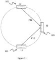
- Figure 12 shows a variation of the embodiment in which there is a PPG assembly having one light source 101, L1, emitting light to two optical sensors 103, S1 and S1. This is unlike the earlier described embodiment having two PPG sensors, providing a total of two light sources 201, 205 and two optical sensors 203, 207.
- the 'PPG assembly' refers to any combination of PPG light sources 101 and optical sensors 103, including the conventional configuration of one-to-one light source and optical sensor pair and more.
- the light source L1 emits polychromatic light into the wrist of the subject, as shown in Figure 1 , which includes both the first and second wavelengths required for detecting HgbA1c and for monitoring the amount of blood in the arteries.
- the optical sensors S1 and S2 are wavelength selective, however. Each of the optical sensors S1 and S2 has an optical filter to allow only passage of a pre-determined wavelength. Hence, the pulse obtained by the wavelength for monitoring HgbA1c and the pulse obtained by the wavelength for monitoring blood content in the arteries can be obtained using only one light source, L1.
- Figure 13 shows a further variation of the embodiment in which there is a PPG assembly having two monochromatic light sources 101, namely L1 and L2.
- One of the light sources 101 emits light in a wavelength for detecting HgbA1c while the other emits light in a wavelength for monitoring the amount of blood in the arteries.
- the light from the light sources L1 and L2 propagate through the tissue towards the same optical sensor 103 alternatively, changing over from L1 to L2 and back to L1 very quickly and periodically, such as every 10 milliseconds.
- the optical sensor S2 detects and records changes in pulse shape as the intensity of the light emitted by the monochromatic light source L1 changes, and the monochromatic light source L2 is switched off or waits in a standby mode.
- L2 is switched on and L1 is switched off or is put into standby, and S2 detects and records changes in pulse shape as the intensity of the light emitted by L2 changes. After that, the shapes of the pulses obtained at different levels of light intensity from L1 and L2 are compared for shape similarity.
- the embodiments as described can be further modified to analyse two analytes in blood with overlapping absorbance peaks. That is, the absorbance spectrum of a first analyte has a peak at a first wavelength where the absorbance spectrum of the second analyte also has a peak, but it is only by this first wavelength that the first analyte may be monitored. If the absorbance spectrum of the second analyte has another significant peak at a second wavelength, and the absorbance spectrum of the first analyte has no significant peak at this second wavelength, the two analytes can measured by an embodiment that has three PPG sensors. Typically, the absorbance spectrum of blood also does not have an absorbance peak in this second wavelength.
- one PPG sensor emits in the first wavelength and is used to monitor the first analyte.
- the absorbance spectrum of the second analyte also has an absorbance peak in the first wavelength and interferes with the reading of the first analyte.
- a second PPG sensor emits in a second wavelength and is used to monitor the second analyte.
- the absorbance spectrum of the first analyte does not have an absorbance peak in the second wavelength.
- a third PPG sensor emits in a third wavelength and is used to monitor arterial blood content.
- the absorbance spectrum of the first analyte and the second analyte do not have any significant absorbance peak in the third wavelength.
- the intensities of the light emitted by the three PPG sensors are adjusted until pulses obtained by all three wavelengths correspond in shape. When the shapes of the pulses are the same, the pulses may be compared by size.
- the height of the pulse obtained in the first wavelength for measuring the first analyte is firstly subtracted by the height of the pulse obtained in the second wavelength for measuring the second analyte. This removes the interference of the second analyte on the measurement of the first analyte. Subsequently, the remainder of the pulse of the first analyte is referenced to the blood-pulse 807 to quantify the amount of the first analyte in blood.
- pulses obtained by emitting light of two or more wavelengths into tissue are compared.
- the shapes of the pulses are the same, it means that the light of the wavelengths has penetrated into the same depth in the subject's tissue. If the shapes of the pulses are the same, the size of the pulses can be compared for quantification.
- the embodiments as described include a method of selecting the intensity of a light source for monitoring an analyte in blood, comprising the step(s) of: providing the light source the light source emitting light in a first wavelength into the tissue of the subject and obtaining an analyte-pulse 805; providing a second light source, the second light source emitting light in a second wavelength into the tissue of the subject to obtain a blood-pulse 807; adjusting the intensity of the light emitted by the light source and/or the intensity of the light emitted by the second light source until the shape of the analyte-pulse 805 and the shape of the blood-pulse 807 are similar.
- similarity means similarity above a pre-determined similarity threshold level and, although it is preferably but not necessarily identical.
- a blood analyte monitor 100 comprising: a photoplethysmocharty sensor assembly having at least one light source and at least one optical sensor; the photoplethysmocharty sensor assembly capable of generating light of a first wavelength and light of a second wavelength through the tissue of a subject to be detected by the at least one sensor; an adjustment module for adjusting the intensity of the light of the first wavelength and/or the light of the second wavelength in response to the shape of a pulse signal obtain by the first wavelength and the shape of a pulse signal obtained by the second wavelength.
- the adjustment module is capable of adjusting the intensity of the light of the first wavelength and/or the light of the second wavelength to provide that the shape of the pulse signal obtain by the first wavelength and the shape of the pulse signal obtained by the second wavelength are similar above a pre-determined similarity threshold.
- the two or more PPG sensors described above can be implemented in the form of wrist bands, watches, rings for fingers, earphones with sensing in the ear canal, an arm band for the upper arm.
- the PPG can be woven into clothes, such as T-shirt, brasseries, underwear.
- the PPG can be installed in shoes, socks, helmets, hats and eyewear such as on the spectacle legs that contact the scalp.
- Peak 4 or Peak 5 are the preferred choice, it is nevertheless possible to use stronger light source in the wavelength of Peak 3 remedy the shallow penetration.
- artificial intelligence can be used to improve the comparison of the pulse shapes and to identify and quantify the blood analyte.
- calibration includes, for example, applying the invention onto a different subject with known amount of glycated haemoglobin in his blood and taking the ratio of the size of a pulse obtained by measuring the absorbance by the analyte of light of a first wavelength to the size of a pulse obtained by measuring the absorbance by blood of light of a second wavelength. Subsequently, the ratio is deemed equivalent to the known amount of glycated haemoglobin. More than one subject with known amount of glycated haemoglobin in his blood may be used to provide multi-point calibration. After calibration, other readings of the ratio of the pulses of the first PPG sensor and the second PPG sensor can be converted by the calibration into absolute value of, or to a physical quantity of HgbA1.
- a subject can have the amount of his glycated haemoglobin tested, and the calibration of his glycated-haemoglobin monitor 100 can be calibrated by using his own glycated haemoglobin amount, as a one point calibration. Subsequently, this calibration can be used for a long period, up to several months, to convert subsequent readings of the ratio of the HgbA1c pulse to blood-pulse detected in this subject's blood into actual amount of HgbA1c.
Landscapes
- Health & Medical Sciences (AREA)
- Life Sciences & Earth Sciences (AREA)
- Physics & Mathematics (AREA)
- Engineering & Computer Science (AREA)
- Public Health (AREA)
- Surgery (AREA)
- Biophysics (AREA)
- Biomedical Technology (AREA)
- Heart & Thoracic Surgery (AREA)
- Medical Informatics (AREA)
- Molecular Biology (AREA)
- Pathology (AREA)
- Animal Behavior & Ethology (AREA)
- General Health & Medical Sciences (AREA)
- Veterinary Medicine (AREA)
- Optics & Photonics (AREA)
- Cardiology (AREA)
- Physiology (AREA)
- Spectroscopy & Molecular Physics (AREA)
- Emergency Medicine (AREA)
- Computer Vision & Pattern Recognition (AREA)
- Signal Processing (AREA)
- Psychiatry (AREA)
- Obesity (AREA)
- Artificial Intelligence (AREA)
- Acoustics & Sound (AREA)
- Audiology, Speech & Language Pathology (AREA)
- Multimedia (AREA)
- Measurement Of The Respiration, Hearing Ability, Form, And Blood Characteristics Of Living Organisms (AREA)
- Investigating Or Analysing Biological Materials (AREA)
Applications Claiming Priority (2)
| Application Number | Priority Date | Filing Date | Title |
|---|---|---|---|
| HK18104213 | 2018-03-27 | ||
| PCT/CN2019/079237 WO2019184812A1 (en) | 2018-03-27 | 2019-03-22 | A method of selecting the intensity of a light source for monitoring an analyte in blood, and a device thereof |
Publications (3)
| Publication Number | Publication Date |
|---|---|
| EP3758606A1 EP3758606A1 (en) | 2021-01-06 |
| EP3758606A4 EP3758606A4 (en) | 2021-10-13 |
| EP3758606B1 true EP3758606B1 (en) | 2022-11-30 |
Family
ID=68062479
Family Applications (1)
| Application Number | Title | Priority Date | Filing Date |
|---|---|---|---|
| EP19777510.9A Active EP3758606B1 (en) | 2018-03-27 | 2019-03-22 | A method of selecting the intensity of a light source for monitoring an analyte in blood, and a device thereof |
Country Status (8)
| Country | Link |
|---|---|
| US (1) | US20210000391A1 (ja) |
| EP (1) | EP3758606B1 (ja) |
| JP (1) | JP3233712U (ja) |
| CN (1) | CN112261904B (ja) |
| DK (1) | DK3758606T3 (ja) |
| FI (1) | FI3758606T3 (ja) |
| SG (1) | SG11202008701QA (ja) |
| WO (1) | WO2019184812A1 (ja) |
Families Citing this family (5)
| Publication number | Priority date | Publication date | Assignee | Title |
|---|---|---|---|---|
| KR102356154B1 (ko) * | 2020-04-13 | 2022-01-28 | 국민대학교산학협력단 | 비어램버트 법칙을 이용한 비침습적 당화혈색소 측정 시스템 및 방법 |
| CN112924660B (zh) * | 2021-01-26 | 2023-09-26 | 上海浩创亘永科技有限公司 | 一种扫描系统及其扫描方法 |
| WO2023185649A1 (en) * | 2022-03-28 | 2023-10-05 | Well Being Digital Limited | A photoplethysmography sensor having a novel arrangement |
| WO2024195820A1 (ja) * | 2023-03-23 | 2024-09-26 | 京セラ株式会社 | 指標推定装置、および指標推定方法 |
| CN117717334B (zh) * | 2024-02-07 | 2024-07-05 | 荣耀终端有限公司 | 数据获取方法及电子设备 |
Family Cites Families (12)
| Publication number | Priority date | Publication date | Assignee | Title |
|---|---|---|---|---|
| US5112124A (en) * | 1990-04-19 | 1992-05-12 | Worcester Polytechnic Institute | Method and apparatus for measuring the concentration of absorbing substances |
| DE60332094D1 (de) * | 2002-02-22 | 2010-05-27 | Masimo Corp | Aktive pulsspektrophotometrie |
| US8140147B2 (en) * | 2002-04-04 | 2012-03-20 | Veralight, Inc. | Determination of a measure of a glycation end-product or disease state using a flexible probe to determine tissue fluorescence of various sites |
| CA2696054A1 (en) * | 2007-11-14 | 2009-05-22 | Conmed Corporation | Method and apparatus for processing a pulsatile biometric signal |
| ES2682459T3 (es) * | 2008-07-30 | 2018-09-20 | Zambaiti Parati S.P.A. | Dispositivo de láser de diodo para la medición no invasiva de la glucemia |
| KR20110053993A (ko) * | 2008-08-07 | 2011-05-24 | 유니버시티 오브 매사추세츠 | 분광기 센서들 |
| JP5948836B2 (ja) * | 2011-12-09 | 2016-07-06 | ソニー株式会社 | 測定装置、測定方法、プログラム及び記録媒体 |
| US10244949B2 (en) * | 2012-10-07 | 2019-04-02 | Rhythm Diagnostic Systems, Inc. | Health monitoring systems and methods |
| WO2014143276A2 (en) * | 2012-12-31 | 2014-09-18 | Omni Medsci, Inc. | Short-wave infrared super-continuum lasers for natural gas leak detection, exploration, and other active remote sensing applications |
| WO2014125355A1 (en) * | 2013-02-13 | 2014-08-21 | Leman Micro Devices Sa | Non-invasive blood analysis |
| CN108024727B (zh) * | 2015-09-25 | 2021-10-12 | 三线性生物公司 | 生物传感器 |
| WO2017168432A1 (en) * | 2016-04-01 | 2017-10-05 | Antony Aneel Joseph | A non invasive hba1c meter for measuring blood glucose levels in diabetic patients without physical blood sample using nir (near infra red light) |
-
2019
- 2019-03-20 US US16/981,690 patent/US20210000391A1/en active Pending
- 2019-03-22 EP EP19777510.9A patent/EP3758606B1/en active Active
- 2019-03-22 JP JP2020600139U patent/JP3233712U/ja active Active
- 2019-03-22 WO PCT/CN2019/079237 patent/WO2019184812A1/en unknown
- 2019-03-22 DK DK19777510.9T patent/DK3758606T3/da active
- 2019-03-22 FI FIEP19777510.9T patent/FI3758606T3/fi active
- 2019-03-22 SG SG11202008701QA patent/SG11202008701QA/en unknown
- 2019-03-22 CN CN201980021876.5A patent/CN112261904B/zh active Active
Also Published As
| Publication number | Publication date |
|---|---|
| CN112261904B (zh) | 2024-08-20 |
| EP3758606A4 (en) | 2021-10-13 |
| WO2019184812A1 (en) | 2019-10-03 |
| JP3233712U (ja) | 2021-09-02 |
| DK3758606T3 (da) | 2022-12-19 |
| US20210000391A1 (en) | 2021-01-07 |
| FI3758606T3 (fi) | 2023-01-13 |
| CN112261904A (zh) | 2021-01-22 |
| SG11202008701QA (en) | 2020-10-29 |
| EP3758606A1 (en) | 2021-01-06 |
Similar Documents
| Publication | Publication Date | Title |
|---|---|---|
| EP3758606B1 (en) | A method of selecting the intensity of a light source for monitoring an analyte in blood, and a device thereof | |
| Joshi et al. | iGLU 2.0: A new wearable for accurate non-invasive continuous serum glucose measurement in IoMT framework | |
| US11147482B2 (en) | Method and system for non-invasive blood glucose measurement using signal change of the non-glucose components induced by the presence of glucose | |
| CN105307568B (zh) | 非侵入式血液分析 | |
| CN104224196B (zh) | 无创测量血液成分浓度的方法 | |
| US7183102B2 (en) | Apparatus using reference measurement for calibration | |
| US9222832B2 (en) | Device and method for detecting and monitoring ingredients or properties of a measurement medium, in particular of physiological blood values | |
| US8406839B2 (en) | Method and apparatus for determining blood analytes | |
| EP0650591A4 (en) | NON-INVASIVE TEST PROCEDURE. | |
| HU216847B (hu) | Eljárás és berendezés vérösszetétel-paraméterek gyors noninvazív meghatározására | |
| EP3556290A1 (en) | Method and system for non-invasive optical blood glucose detection utilizing spectral data analysis | |
| CN112568902A (zh) | 一种基于血糖值的无创血糖校准方法 | |
| Lee et al. | A Non-Invasive Blood Glucose Estimation System using Dual-channel PPGs and Pulse-Arrival Velocity | |
| Pande et al. | Non-invasive blood glucose measurement | |
| Shulei et al. | Non-invasive blood glucose measurement scheme based on near-infrared spectroscopy | |
| US6801316B2 (en) | Measurement of an analyte concentration in a scattering medium | |
| CN112587134A (zh) | 一种无创血糖检定方法 | |
| CN111803085A (zh) | 一种基于颜色特性的无创血红蛋白浓度水平测量装置 | |
| Yasuhiro et al. | Multivariate regression and classification models for estimation of blood glucose levels using a new non-invasive optical measurement technique named" Pulse Glucometry" | |
| Han | REAL-TIME MONITORING AND ANALYSIS OF ATHLETE MUSCLE ACTIVITY BASED ON WEARABLE SENSOR | |
| Joshi et al. | Smart Glucometer for Personalized Health Management of Diabetes Care | |
| Kanak et al. | Multi-Sensor Suite for Melanin-Adjusted Blood Oxygenation Measurement Progress Report | |
| CN115736913A (zh) | 一种无创血糖检测方法及系统 | |
| Sasono et al. | A Portable and USB-Powered Device for Heart Rate Extraction of Optical Plethysmography Signal |
Legal Events
| Date | Code | Title | Description |
|---|---|---|---|
| STAA | Information on the status of an ep patent application or granted ep patent |
Free format text: STATUS: THE INTERNATIONAL PUBLICATION HAS BEEN MADE |
|
| PUAI | Public reference made under article 153(3) epc to a published international application that has entered the european phase |
Free format text: ORIGINAL CODE: 0009012 |
|
| STAA | Information on the status of an ep patent application or granted ep patent |
Free format text: STATUS: REQUEST FOR EXAMINATION WAS MADE |
|
| 17P | Request for examination filed |
Effective date: 20200928 |
|
| AK | Designated contracting states |
Kind code of ref document: A1 Designated state(s): AL AT BE BG CH CY CZ DE DK EE ES FI FR GB GR HR HU IE IS IT LI LT LU LV MC MK MT NL NO PL PT RO RS SE SI SK SM TR |
|
| AX | Request for extension of the european patent |
Extension state: BA ME |
|
| DAV | Request for validation of the european patent (deleted) | ||
| DAX | Request for extension of the european patent (deleted) | ||
| A4 | Supplementary search report drawn up and despatched |
Effective date: 20210914 |
|
| RIC1 | Information provided on ipc code assigned before grant |
Ipc: A61B 5/1455 20060101AFI20210908BHEP |
|
| GRAP | Despatch of communication of intention to grant a patent |
Free format text: ORIGINAL CODE: EPIDOSNIGR1 |
|
| STAA | Information on the status of an ep patent application or granted ep patent |
Free format text: STATUS: GRANT OF PATENT IS INTENDED |
|
| INTG | Intention to grant announced |
Effective date: 20220808 |
|
| RIN1 | Information on inventor provided before grant (corrected) |
Inventor name: WONG, MING YIP WALLACE |
|
| GRAS | Grant fee paid |
Free format text: ORIGINAL CODE: EPIDOSNIGR3 |
|
| GRAA | (expected) grant |
Free format text: ORIGINAL CODE: 0009210 |
|
| STAA | Information on the status of an ep patent application or granted ep patent |
Free format text: STATUS: THE PATENT HAS BEEN GRANTED |
|
| AK | Designated contracting states |
Kind code of ref document: B1 Designated state(s): AL AT BE BG CH CY CZ DE DK EE ES FI FR GB GR HR HU IE IS IT LI LT LU LV MC MK MT NL NO PL PT RO RS SE SI SK SM TR |
|
| REG | Reference to a national code |
Ref country code: CH Ref legal event code: EP Ref country code: GB Ref legal event code: FG4D |
|
| REG | Reference to a national code |
Ref country code: AT Ref legal event code: REF Ref document number: 1534053 Country of ref document: AT Kind code of ref document: T Effective date: 20221215 |
|
| REG | Reference to a national code |
Ref country code: DK Ref legal event code: T3 Effective date: 20221216 |
|
| REG | Reference to a national code |
Ref country code: IE Ref legal event code: FG4D |
|
| REG | Reference to a national code |
Ref country code: DE Ref legal event code: R096 Ref document number: 602019022595 Country of ref document: DE |
|
| REG | Reference to a national code |
Ref country code: LT Ref legal event code: MG9D |
|
| REG | Reference to a national code |
Ref country code: NL Ref legal event code: MP Effective date: 20221130 |
|
| PG25 | Lapsed in a contracting state [announced via postgrant information from national office to epo] |
Ref country code: SE Free format text: LAPSE BECAUSE OF FAILURE TO SUBMIT A TRANSLATION OF THE DESCRIPTION OR TO PAY THE FEE WITHIN THE PRESCRIBED TIME-LIMIT Effective date: 20221130 Ref country code: PT Free format text: LAPSE BECAUSE OF FAILURE TO SUBMIT A TRANSLATION OF THE DESCRIPTION OR TO PAY THE FEE WITHIN THE PRESCRIBED TIME-LIMIT Effective date: 20230331 Ref country code: NO Free format text: LAPSE BECAUSE OF FAILURE TO SUBMIT A TRANSLATION OF THE DESCRIPTION OR TO PAY THE FEE WITHIN THE PRESCRIBED TIME-LIMIT Effective date: 20230228 Ref country code: LT Free format text: LAPSE BECAUSE OF FAILURE TO SUBMIT A TRANSLATION OF THE DESCRIPTION OR TO PAY THE FEE WITHIN THE PRESCRIBED TIME-LIMIT Effective date: 20221130 Ref country code: ES Free format text: LAPSE BECAUSE OF FAILURE TO SUBMIT A TRANSLATION OF THE DESCRIPTION OR TO PAY THE FEE WITHIN THE PRESCRIBED TIME-LIMIT Effective date: 20221130 |
|
| REG | Reference to a national code |
Ref country code: AT Ref legal event code: MK05 Ref document number: 1534053 Country of ref document: AT Kind code of ref document: T Effective date: 20221130 |
|
| PG25 | Lapsed in a contracting state [announced via postgrant information from national office to epo] |
Ref country code: RS Free format text: LAPSE BECAUSE OF FAILURE TO SUBMIT A TRANSLATION OF THE DESCRIPTION OR TO PAY THE FEE WITHIN THE PRESCRIBED TIME-LIMIT Effective date: 20221130 Ref country code: PL Free format text: LAPSE BECAUSE OF FAILURE TO SUBMIT A TRANSLATION OF THE DESCRIPTION OR TO PAY THE FEE WITHIN THE PRESCRIBED TIME-LIMIT Effective date: 20221130 Ref country code: LV Free format text: LAPSE BECAUSE OF FAILURE TO SUBMIT A TRANSLATION OF THE DESCRIPTION OR TO PAY THE FEE WITHIN THE PRESCRIBED TIME-LIMIT Effective date: 20221130 Ref country code: IS Free format text: LAPSE BECAUSE OF FAILURE TO SUBMIT A TRANSLATION OF THE DESCRIPTION OR TO PAY THE FEE WITHIN THE PRESCRIBED TIME-LIMIT Effective date: 20230330 Ref country code: HR Free format text: LAPSE BECAUSE OF FAILURE TO SUBMIT A TRANSLATION OF THE DESCRIPTION OR TO PAY THE FEE WITHIN THE PRESCRIBED TIME-LIMIT Effective date: 20221130 Ref country code: GR Free format text: LAPSE BECAUSE OF FAILURE TO SUBMIT A TRANSLATION OF THE DESCRIPTION OR TO PAY THE FEE WITHIN THE PRESCRIBED TIME-LIMIT Effective date: 20230301 |
|
| P01 | Opt-out of the competence of the unified patent court (upc) registered |
Effective date: 20230514 |
|
| PG25 | Lapsed in a contracting state [announced via postgrant information from national office to epo] |
Ref country code: NL Free format text: LAPSE BECAUSE OF FAILURE TO SUBMIT A TRANSLATION OF THE DESCRIPTION OR TO PAY THE FEE WITHIN THE PRESCRIBED TIME-LIMIT Effective date: 20221130 |
|
| PG25 | Lapsed in a contracting state [announced via postgrant information from national office to epo] |
Ref country code: SM Free format text: LAPSE BECAUSE OF FAILURE TO SUBMIT A TRANSLATION OF THE DESCRIPTION OR TO PAY THE FEE WITHIN THE PRESCRIBED TIME-LIMIT Effective date: 20221130 Ref country code: RO Free format text: LAPSE BECAUSE OF FAILURE TO SUBMIT A TRANSLATION OF THE DESCRIPTION OR TO PAY THE FEE WITHIN THE PRESCRIBED TIME-LIMIT Effective date: 20221130 Ref country code: EE Free format text: LAPSE BECAUSE OF FAILURE TO SUBMIT A TRANSLATION OF THE DESCRIPTION OR TO PAY THE FEE WITHIN THE PRESCRIBED TIME-LIMIT Effective date: 20221130 Ref country code: CZ Free format text: LAPSE BECAUSE OF FAILURE TO SUBMIT A TRANSLATION OF THE DESCRIPTION OR TO PAY THE FEE WITHIN THE PRESCRIBED TIME-LIMIT Effective date: 20221130 Ref country code: AT Free format text: LAPSE BECAUSE OF FAILURE TO SUBMIT A TRANSLATION OF THE DESCRIPTION OR TO PAY THE FEE WITHIN THE PRESCRIBED TIME-LIMIT Effective date: 20221130 |
|
| PG25 | Lapsed in a contracting state [announced via postgrant information from national office to epo] |
Ref country code: SK Free format text: LAPSE BECAUSE OF FAILURE TO SUBMIT A TRANSLATION OF THE DESCRIPTION OR TO PAY THE FEE WITHIN THE PRESCRIBED TIME-LIMIT Effective date: 20221130 Ref country code: AL Free format text: LAPSE BECAUSE OF FAILURE TO SUBMIT A TRANSLATION OF THE DESCRIPTION OR TO PAY THE FEE WITHIN THE PRESCRIBED TIME-LIMIT Effective date: 20221130 |
|
| REG | Reference to a national code |
Ref country code: DE Ref legal event code: R097 Ref document number: 602019022595 Country of ref document: DE |
|
| PLBE | No opposition filed within time limit |
Free format text: ORIGINAL CODE: 0009261 |
|
| STAA | Information on the status of an ep patent application or granted ep patent |
Free format text: STATUS: NO OPPOSITION FILED WITHIN TIME LIMIT |
|
| PG25 | Lapsed in a contracting state [announced via postgrant information from national office to epo] |
Ref country code: MC Free format text: LAPSE BECAUSE OF FAILURE TO SUBMIT A TRANSLATION OF THE DESCRIPTION OR TO PAY THE FEE WITHIN THE PRESCRIBED TIME-LIMIT Effective date: 20221130 |
|
| REG | Reference to a national code |
Ref country code: CH Ref legal event code: PL |
|
| 26N | No opposition filed |
Effective date: 20230831 |
|
| PG25 | Lapsed in a contracting state [announced via postgrant information from national office to epo] |
Ref country code: SI Free format text: LAPSE BECAUSE OF FAILURE TO SUBMIT A TRANSLATION OF THE DESCRIPTION OR TO PAY THE FEE WITHIN THE PRESCRIBED TIME-LIMIT Effective date: 20221130 |
|
| REG | Reference to a national code |
Ref country code: BE Ref legal event code: MM Effective date: 20230331 |
|
| PG25 | Lapsed in a contracting state [announced via postgrant information from national office to epo] |
Ref country code: LU Free format text: LAPSE BECAUSE OF NON-PAYMENT OF DUE FEES Effective date: 20230322 |
|
| REG | Reference to a national code |
Ref country code: IE Ref legal event code: MM4A |
|
| PG25 | Lapsed in a contracting state [announced via postgrant information from national office to epo] |
Ref country code: LI Free format text: LAPSE BECAUSE OF NON-PAYMENT OF DUE FEES Effective date: 20230331 Ref country code: IE Free format text: LAPSE BECAUSE OF NON-PAYMENT OF DUE FEES Effective date: 20230322 Ref country code: FR Free format text: LAPSE BECAUSE OF NON-PAYMENT OF DUE FEES Effective date: 20230331 Ref country code: CH Free format text: LAPSE BECAUSE OF NON-PAYMENT OF DUE FEES Effective date: 20230331 |
|
| PG25 | Lapsed in a contracting state [announced via postgrant information from national office to epo] |
Ref country code: BE Free format text: LAPSE BECAUSE OF NON-PAYMENT OF DUE FEES Effective date: 20230331 |
|
| PGFP | Annual fee paid to national office [announced via postgrant information from national office to epo] |
Ref country code: FI Payment date: 20240308 Year of fee payment: 6 Ref country code: DE Payment date: 20240313 Year of fee payment: 6 Ref country code: GB Payment date: 20240304 Year of fee payment: 6 |
|
| PG25 | Lapsed in a contracting state [announced via postgrant information from national office to epo] |
Ref country code: IT Free format text: LAPSE BECAUSE OF FAILURE TO SUBMIT A TRANSLATION OF THE DESCRIPTION OR TO PAY THE FEE WITHIN THE PRESCRIBED TIME-LIMIT Effective date: 20221130 |
|
| PGFP | Annual fee paid to national office [announced via postgrant information from national office to epo] |
Ref country code: DK Payment date: 20240319 Year of fee payment: 6 |