EP3754014A1 - Automated method for preparing retinal pigment epithelium cells - Google Patents
Automated method for preparing retinal pigment epithelium cells Download PDFInfo
- Publication number
- EP3754014A1 EP3754014A1 EP19305807.0A EP19305807A EP3754014A1 EP 3754014 A1 EP3754014 A1 EP 3754014A1 EP 19305807 A EP19305807 A EP 19305807A EP 3754014 A1 EP3754014 A1 EP 3754014A1
- Authority
- EP
- European Patent Office
- Prior art keywords
- cells
- rpe
- cell
- differentiating
- automated method
- Prior art date
- Legal status (The legal status is an assumption and is not a legal conclusion. Google has not performed a legal analysis and makes no representation as to the accuracy of the status listed.)
- Withdrawn
Links
Images


Classifications
-
- C—CHEMISTRY; METALLURGY
- C12—BIOCHEMISTRY; BEER; SPIRITS; WINE; VINEGAR; MICROBIOLOGY; ENZYMOLOGY; MUTATION OR GENETIC ENGINEERING
- C12N—MICROORGANISMS OR ENZYMES; COMPOSITIONS THEREOF; PROPAGATING, PRESERVING, OR MAINTAINING MICROORGANISMS; MUTATION OR GENETIC ENGINEERING; CULTURE MEDIA
- C12N5/00—Undifferentiated human, animal or plant cells, e.g. cell lines; Tissues; Cultivation or maintenance thereof; Culture media therefor
- C12N5/06—Animal cells or tissues; Human cells or tissues
- C12N5/0602—Vertebrate cells
- C12N5/0618—Cells of the nervous system
- C12N5/062—Sensory transducers, e.g. photoreceptors; Sensory neurons, e.g. for hearing, taste, smell, pH, touch, temperature, pain
-
- A—HUMAN NECESSITIES
- A61—MEDICAL OR VETERINARY SCIENCE; HYGIENE
- A61K—PREPARATIONS FOR MEDICAL, DENTAL OR TOILETRY PURPOSES
- A61K35/00—Medicinal preparations containing materials or reaction products thereof with undetermined constitution
- A61K35/12—Materials from mammals; Compositions comprising non-specified tissues or cells; Compositions comprising non-embryonic stem cells; Genetically modified cells
- A61K35/48—Reproductive organs
- A61K35/54—Ovaries; Ova; Ovules; Embryos; Foetal cells; Germ cells
- A61K35/545—Embryonic stem cells; Pluripotent stem cells; Induced pluripotent stem cells; Uncharacterised stem cells
-
- A—HUMAN NECESSITIES
- A61—MEDICAL OR VETERINARY SCIENCE; HYGIENE
- A61P—SPECIFIC THERAPEUTIC ACTIVITY OF CHEMICAL COMPOUNDS OR MEDICINAL PREPARATIONS
- A61P27/00—Drugs for disorders of the senses
-
- A—HUMAN NECESSITIES
- A61—MEDICAL OR VETERINARY SCIENCE; HYGIENE
- A61P—SPECIFIC THERAPEUTIC ACTIVITY OF CHEMICAL COMPOUNDS OR MEDICINAL PREPARATIONS
- A61P27/00—Drugs for disorders of the senses
- A61P27/02—Ophthalmic agents
-
- A—HUMAN NECESSITIES
- A61—MEDICAL OR VETERINARY SCIENCE; HYGIENE
- A61P—SPECIFIC THERAPEUTIC ACTIVITY OF CHEMICAL COMPOUNDS OR MEDICINAL PREPARATIONS
- A61P9/00—Drugs for disorders of the cardiovascular system
- A61P9/10—Drugs for disorders of the cardiovascular system for treating ischaemic or atherosclerotic diseases, e.g. antianginal drugs, coronary vasodilators, drugs for myocardial infarction, retinopathy, cerebrovascula insufficiency, renal arteriosclerosis
-
- C—CHEMISTRY; METALLURGY
- C12—BIOCHEMISTRY; BEER; SPIRITS; WINE; VINEGAR; MICROBIOLOGY; ENZYMOLOGY; MUTATION OR GENETIC ENGINEERING
- C12N—MICROORGANISMS OR ENZYMES; COMPOSITIONS THEREOF; PROPAGATING, PRESERVING, OR MAINTAINING MICROORGANISMS; MUTATION OR GENETIC ENGINEERING; CULTURE MEDIA
- C12N2500/00—Specific components of cell culture medium
- C12N2500/30—Organic components
- C12N2500/40—Nucleotides, nucleosides, bases
-
- C—CHEMISTRY; METALLURGY
- C12—BIOCHEMISTRY; BEER; SPIRITS; WINE; VINEGAR; MICROBIOLOGY; ENZYMOLOGY; MUTATION OR GENETIC ENGINEERING
- C12N—MICROORGANISMS OR ENZYMES; COMPOSITIONS THEREOF; PROPAGATING, PRESERVING, OR MAINTAINING MICROORGANISMS; MUTATION OR GENETIC ENGINEERING; CULTURE MEDIA
- C12N2500/00—Specific components of cell culture medium
- C12N2500/30—Organic components
- C12N2500/46—Amines, e.g. putrescine
-
- C—CHEMISTRY; METALLURGY
- C12—BIOCHEMISTRY; BEER; SPIRITS; WINE; VINEGAR; MICROBIOLOGY; ENZYMOLOGY; MUTATION OR GENETIC ENGINEERING
- C12N—MICROORGANISMS OR ENZYMES; COMPOSITIONS THEREOF; PROPAGATING, PRESERVING, OR MAINTAINING MICROORGANISMS; MUTATION OR GENETIC ENGINEERING; CULTURE MEDIA
- C12N2501/00—Active agents used in cell culture processes, e.g. differentation
- C12N2501/10—Growth factors
- C12N2501/15—Transforming growth factor beta (TGF-β)
-
- C—CHEMISTRY; METALLURGY
- C12—BIOCHEMISTRY; BEER; SPIRITS; WINE; VINEGAR; MICROBIOLOGY; ENZYMOLOGY; MUTATION OR GENETIC ENGINEERING
- C12N—MICROORGANISMS OR ENZYMES; COMPOSITIONS THEREOF; PROPAGATING, PRESERVING, OR MAINTAINING MICROORGANISMS; MUTATION OR GENETIC ENGINEERING; CULTURE MEDIA
- C12N2501/00—Active agents used in cell culture processes, e.g. differentation
- C12N2501/10—Growth factors
- C12N2501/155—Bone morphogenic proteins [BMP]; Osteogenins; Osteogenic factor; Bone inducing factor
-
- C—CHEMISTRY; METALLURGY
- C12—BIOCHEMISTRY; BEER; SPIRITS; WINE; VINEGAR; MICROBIOLOGY; ENZYMOLOGY; MUTATION OR GENETIC ENGINEERING
- C12N—MICROORGANISMS OR ENZYMES; COMPOSITIONS THEREOF; PROPAGATING, PRESERVING, OR MAINTAINING MICROORGANISMS; MUTATION OR GENETIC ENGINEERING; CULTURE MEDIA
- C12N2501/00—Active agents used in cell culture processes, e.g. differentation
- C12N2501/10—Growth factors
- C12N2501/16—Activin; Inhibin; Mullerian inhibiting substance
-
- C—CHEMISTRY; METALLURGY
- C12—BIOCHEMISTRY; BEER; SPIRITS; WINE; VINEGAR; MICROBIOLOGY; ENZYMOLOGY; MUTATION OR GENETIC ENGINEERING
- C12N—MICROORGANISMS OR ENZYMES; COMPOSITIONS THEREOF; PROPAGATING, PRESERVING, OR MAINTAINING MICROORGANISMS; MUTATION OR GENETIC ENGINEERING; CULTURE MEDIA
- C12N2501/00—Active agents used in cell culture processes, e.g. differentation
- C12N2501/10—Growth factors
- C12N2501/19—Growth and differentiation factors [GDF]
-
- C—CHEMISTRY; METALLURGY
- C12—BIOCHEMISTRY; BEER; SPIRITS; WINE; VINEGAR; MICROBIOLOGY; ENZYMOLOGY; MUTATION OR GENETIC ENGINEERING
- C12N—MICROORGANISMS OR ENZYMES; COMPOSITIONS THEREOF; PROPAGATING, PRESERVING, OR MAINTAINING MICROORGANISMS; MUTATION OR GENETIC ENGINEERING; CULTURE MEDIA
- C12N2501/00—Active agents used in cell culture processes, e.g. differentation
- C12N2501/40—Regulators of development
- C12N2501/415—Wnt; Frizzeled
-
- C—CHEMISTRY; METALLURGY
- C12—BIOCHEMISTRY; BEER; SPIRITS; WINE; VINEGAR; MICROBIOLOGY; ENZYMOLOGY; MUTATION OR GENETIC ENGINEERING
- C12N—MICROORGANISMS OR ENZYMES; COMPOSITIONS THEREOF; PROPAGATING, PRESERVING, OR MAINTAINING MICROORGANISMS; MUTATION OR GENETIC ENGINEERING; CULTURE MEDIA
- C12N2501/00—Active agents used in cell culture processes, e.g. differentation
- C12N2501/70—Enzymes
- C12N2501/72—Transferases (EC 2.)
- C12N2501/727—Kinases (EC 2.7.)
-
- C—CHEMISTRY; METALLURGY
- C12—BIOCHEMISTRY; BEER; SPIRITS; WINE; VINEGAR; MICROBIOLOGY; ENZYMOLOGY; MUTATION OR GENETIC ENGINEERING
- C12N—MICROORGANISMS OR ENZYMES; COMPOSITIONS THEREOF; PROPAGATING, PRESERVING, OR MAINTAINING MICROORGANISMS; MUTATION OR GENETIC ENGINEERING; CULTURE MEDIA
- C12N2506/00—Differentiation of animal cells from one lineage to another; Differentiation of pluripotent cells
- C12N2506/02—Differentiation of animal cells from one lineage to another; Differentiation of pluripotent cells from embryonic cells
-
- C—CHEMISTRY; METALLURGY
- C12—BIOCHEMISTRY; BEER; SPIRITS; WINE; VINEGAR; MICROBIOLOGY; ENZYMOLOGY; MUTATION OR GENETIC ENGINEERING
- C12N—MICROORGANISMS OR ENZYMES; COMPOSITIONS THEREOF; PROPAGATING, PRESERVING, OR MAINTAINING MICROORGANISMS; MUTATION OR GENETIC ENGINEERING; CULTURE MEDIA
- C12N2506/00—Differentiation of animal cells from one lineage to another; Differentiation of pluripotent cells
- C12N2506/45—Differentiation of animal cells from one lineage to another; Differentiation of pluripotent cells from artificially induced pluripotent stem cells
Definitions
- the present invention relates to methods for preparing retinal pigment epithelium (RPE) cells from pluripotent stem cells (PSCs). More particularly, the present invention relates to an automated method that combines in a sequential manner three differentiating agents to direct the differentiation of human PSCs into RPE cells.
- RPE retinal pigment epithelium
- the retinal pigment epithelium is a monolayer of pigmented cells localized between the neuroretina and the choroids.
- the RPE cells play roles in the maintenance and function of the retina and its photoreceptors. These include the formation of the blood-retinal barrier, absorption of light and protection against photooxidation, transport of nutrients to the neural retina, regeneration of visual pigment, and phagocytosis of shed photoreceptor membranes.
- hPSCs Human pluripotent stem cells
- hESCs human embryonic stem cells
- hiPSCs human induced pluripotent stem cells
- AMD is the leading cause of blindness in developed countries with more than 150 million people affected worldwide, a figure that will increase in the coming years. It can be classified into two groups, dry (atrophic) or wet (exudative), which is based on the presence of a choroidal neovascularization.
- hPSC-RPE human pluripotent stem cells
- hPSCs spontaneously differentiate into RPE cells after removal of basic fibroblast growth factor (bFGF), used to maintain the pluripotency state, from the culture medium.
- bFGF basic fibroblast growth factor
- the distinctive cobblestone morphology of RPE cells as well as their pigmentation allow to manually collect pigmented areas that appear upon differentiation of hPSCs to obtain a pure population of hPSC-RPE cells.
- Such approach of RPE cell production is used as cell replacement material in on going and planned clinical trials.
- this spontaneous method remains fastidious, inefficient and time consuming (8 to 12 weeks of hPSCs differentiation) making it incompatible with the industrial large-scale production which is required to treat the potential millions of patients.
- RPE and neural retina progenitors have the same embryonic origin, they combined a protocol allowing the efficient differentiation of NRPs with previously described RPE inducing factors such as Nicotinamide, Activin A, in addition to many others including bFGF, Noggin, DKK1 (Dickkopf WNT signaling pathway inhibitor 1), Insulin Growth Factor (IGF)-1.
- RPE inducing factors such as Nicotinamide, Activin A, in addition to many others including bFGF, Noggin, DKK1 (Dickkopf WNT signaling pathway inhibitor 1), Insulin Growth Factor (IGF)-1.
- the previous protocol was modified to include Chir99021 and SU5402 from day 8 to 14.
- WO2017021973 and WO2008129554 disclose two-step methods of generating retinal pigment epithelial (RPE) cells comprising culturing a population of human pluripotent stem cells in the presence of Nicotinamide; and further subjecting the cells to another stage of differentiation in the presence of activin A, with or without Nicotinamide.
- RPE retinal pigment epithelial
- the present invention provides the use of a protocol amenable for automation that combines in a sequential manner three differentiating agents to direct the differentiation of hPSCs into RPE cells.
- This novel differentiation protocol associated with the use of cell culture robots open new possibilities for the production of large batches of hPSC-RPE cells while maintaining a high cell purity and functionality.
- an object of the present invention is to provide methods for large-scale automated production of RPE cells derived from (human) pluripotent stem cells.
- the hPSCs in the culture system of the methods disclosed herein are differentiating hPSCs, i.e. a population of hPSCs essentially in an undifferentiated state, or wherein at least part of said cells have been induced to undergo initial stages of directed differentiation, the majority of said cells have been induced to undergo initial stages of directed differentiation.
- the initial stage of differentiation is achieved by exposing the cells to a first differentiating agent, in particular at least one Nicotinamide (NA) mimetic compound, then to a second differentiating agent, in particular at least one member of transforming growth factor ⁇ (TGF ⁇ ) superfamily and finally to a third differentiating agent, in particular at least one activator of the Wnt canonical pathway.
- a first differentiating agent in particular at least one Nicotinamide (NA) mimetic compound
- TGF ⁇ transforming growth factor ⁇
- TGF ⁇ transforming growth factor ⁇
- trimer refers to a compound having similar functional and/or structural properties to another known compound or a particular fragment of that known compound, such as a known compound of biological origin, e.g., a polypeptide or fragment thereof.
- Undifferentiated refers to cultured cells when a substantial proportion (at least 20%, and possibly over 50% or 80%) of the cells and their derivatives in the population display characteristic markers and morphological characteristics of undifferentiated cells, distinguishing them from differentiated cells of embryo or adult origin. Cells are recognized as proliferating in an undifferentiated state when they go through at least 1 population doubling during a cultivation period of at least 3 weeks, while retaining at least about 50%, or the same proportion of cells bearing characteristic markers or morphological characteristics of undifferentiated cells after said cultivation period.
- pluripotent stem cell embraces any cell having the capacity for self-renewal and the potential to differentiate into one or more other cell types.
- Pluripotent hSCs refer to precursor cells of human source that have the ability to form any adult cell. Such cells are true cell lines in that they: (i) are capable of extensive proliferation in vitro in an undifferentiated state; and (ii) are capable of differentiation to derivatives of all three embryonic germ layers (endoderm, mesoderm, and ectoderm) even after prolonged culture.
- Human embryonic stem cells (hESCs) are derived from fertilized embryos that are less than one week old (in the cleavage or blastocyte stage) or produced by artificial means (such as by nuclear transfer) that have equivalent characteristics.
- Other pluripotent hSCs include, without being limited thereto, multipotent adult progenitor cells (MAPs), induced pluripotent stem cells (iPS cells) and amniotic fluid stem cells.
- MAPs multipotent adult progenitor cells
- iPS cells induced pluripotent stem cells
- amniotic fluid stem cells amniotic fluid stem cells.
- hPSCs can be obtained using well-known cell-culture methods.
- hESC can be isolated from single blastomeres of the cleavage or morula stage human embryo, from cleavage stage and morula human embryos and human blastocysts.
- Human embryos may be obtained from in vivo preimplantation embryos or more typically from in vitro fertilized (IVF) embryos.
- IVF in vitro fertilized
- non-fertilized human oocyte can be parthenogenetically activated to cleave and develop to the blastocyst stage.
- a single cell human embryo can be expanded to the blastocyst stage.
- the zona pellucida is removed and the inner cell mass (ICM) is isolated by immunosurgery, in which the trophectoderm cells are lysed and removed from the intact ICM by gentle pipetting.
- the ICM is then plated in a tissue culture flask containing the appropriate medium which enables its outgrowth. Following 9 to 15 days, the ICM derived outgrowth is dissociated into clumps either by mechanical dissociation or by enzymatic digestion and the cells are then re-plated on a fresh tissue culture medium. Colonies demonstrating undifferentiated morphology are individually selected by micropipette, mechanically dissociated into clumps, and re-plated. Resulting ESCs are then routinely split every 1-2 weeks. For further details on methods of preparation of hESCs; see Thomson et al. [U.S. Pat. No. 5,843,780 ; Science 282:1145, 1998 ]
- ES cells are not limited to a primary cell line collected from the inner cell mass, but may also be an established ES cell line.
- Examples of such an established ES cell line include: a cell line furnished from a cell population obtained by allowing the already established ES cell line to grow; and an ES cell line obtained by thawing a freeze-dried cell line and then culturing it.
- Such an established ES cell line is available without going through a step of disintegrating a fertilized egg.
- the ES cells used in the present invention may be established from a single embryonic blastomere at the cleavage stage before the blastocyst stage, without impairing the generating ability of the embryo.
- Such ES cells can be obtained without destroying a fertilized egg ( Klimanskaya I. et al., (2006) Nature 444: 481-485 ; and Chung Y et al., (2008) Cell Stem Cell 2: 113-117 ).
- hPSCs can be also used in accordance with the invention.
- hPSCs can be purchased for example from the UK Stem Cell Banks or the NIH human embryonic stem cells registry.
- Non-limiting examples of commercially available embryonic stem cell lines are BG01, BG02, BG03, BG04, CY12, CY30, CY92, CY10, TE03 and TE32.
- human embryonic stem cells preferably refer to human embryonic stem cells which isolation has not involved the destruction of an embryo.
- human embryonic stem cells preferably exclude human embryonic stem cells isolated by techniques that involve the destruction of an embryo.
- the embryos used for obtaining human embryonic stem cells are preferably embryos that cannot give rise to a human being, such as embryos destined to be discarded following in vitro fertilization (IVF) and embryos created solely for the purpose of stem cell research.
- IVF in vitro fertilization
- human embryonic stem cells preferably refer to human embryonic stem cells isolated from discarded embryos, research embryos, or preferably isolated by techniques that do not involve the destruction of an embryo.
- the "induced pluripotent stem cell” in the present invention is a cell induced to have pluripotency by reprogramming a somatic cell by a known method and the like.
- a cell induced to have pluripotency by reprogramming differentiated somatic cells such as fibroblast, peripheral blood mononuclear cell and the like by the expression of a combination of a plurality of genes selected from the group consisting of reprogramming genes including Oct3/4, Sox2, Klf4, Myc (c-Myc, N-Myc, L-Myc), Glis1, Nanog, Sall4, lin28, Esrrb and the like can be mentioned.
- Examples of preferable combination of reprogramming factors include (1) Oct3/4, Sox2, Klf4, and Myc (c-Myc or L-Myc), and (2) Oct3/4, Sox2, Klf4, Lin28 and L-Myc.
- Induced pluripotent stem cell was established by Yamanaka et al. in mouse cell in 2006. In 2007, Induced pluripotent stem cell was also established from human fibroblast, and has pluripotency and self-renewal competence similar to those of embryonic stem cells.
- differentiated denotes a process by which an unspecialized or relatively less specialized cell becomes relatively more specialized.
- the adjective “differentiated” is a relative term.
- a “differentiated cell” is a cell that has progressed further down a certain developmental pathway than the cell it is being compared with.
- a relatively more specialized cell may differ from an unspecialized or relatively less specialized cell in one or more demonstrable phenotypic characteristics, such as, for example, the presence, absence or level of expression of particular cellular components or products, e.g., RNA, proteins or other substances, activity of certain biochemical pathways, morphological appearance, proliferation capacity and/or kinetics, differentiation potential and/or response to differentiation signals, etc., wherein such characteristics signify the progression of the differentiation towards the relatively more specialized cell.
- phenotypic characteristics such as, for example, the presence, absence or level of expression of particular cellular components or products, e.g., RNA, proteins or other substances, activity of certain biochemical pathways, morphological appearance, proliferation capacity and/or kinetics, differentiation potential and/or response to differentiation signals, etc.
- the method of the invention results in the progressive differentiation of (human) pluripotent stem cells and differentiating cells towards RPE cells.
- differentiated of differentiating cells to RPE cells may be considered synonymous to the term “obtaining" RPE cells from differentiating cells.
- the (human) pluripotent stem cells are subjected to directed differentiation using at least two, more preferably three differentiating agents.
- Cell marker refers to any phenotypic feature of a cell that can be used to characterize it or discriminate it from other cell types.
- a marker may be a protein (including secreted, cell surface, or internal proteins; either synthesized or taken up by the cell); a nucleic acid (such as an mRNA, or enzymatically active nucleic acid molecule) or a polysaccharide. Included are determinants of any such cell components that are detectable by antibody, lectin, probe or nucleic acid amplification reaction that are specific for the marker of the cell type of interest.
- the markers can also be identified by a biochemical or enzyme assay or biological response that depends on the function of the gene product.
- each marker is the gene that encodes the transcript, and the events that lead to marker expression.
- a marker is said to be preferentially expressed in an undifferentiated or differentiated cell population, if it is expressed at a level that is at least 50% higher (in terms of total gene product measured in an antibody or PCR assay) or 30% more frequently (in terms of positive cells in the population) than an acceptable control.
- retinal progenitor cells or RPE cells can be determined by a method known per se, for example, the expression of a retinal progenitor cell marker.
- a retinal progenitor cell marker Pax6 (neural retinal progenitor cells, retinal pigment epithelium progenitor cells), RAX (neural retinal progenitor cells), and MITF (retinal pigment epithelium progenitor cells) can be mentioned.
- the tissue/cell specific markers can be detected using immunological techniques. Examples include, but are not limited to, flow cytometry for membrane-bound or intracellular markers, immunohistochemistry for extracellular and intracellular markers and enzymatic immunoassay, for secreted molecular markers.
- a mixed cell population comprising both pigmented and non-pigmented cells.
- the mature RPE cells express significantly higher levels of transcripts of markers of mature RPE cells such as MITF, PAX6 as compared to their expression in RPE cells produced by spontaneous differentiation.
- the culture obtained by a method of the present invention contains RPE cells and/or the differentiating cells at high frequency (content amount).
- Cells obtained by a method of the present invention are PAX6, MITF-positive at high frequency, for example, at a frequency (colony frequency) of 5% or more, preferably 10 to 50%, more preferably 60 to 90%.
- the term “differentiation” refers to the process by which a cell acquires the features of a more differentiated (or “specialized”) cell.
- a differentiated cell or a differentiation-induced cell is one that has more specialized features, compared to an undifferentiated cell, wherein said features correspond to a more differentiated stage within the lineage of a cell.
- the lineage of a cell encompasses all of the discrete development stages of a cell within a scheme of development, that is to say from an undifferentiated stage to a differentiated stage.
- a lineage-specific marker refers to a marker specifically associated with the phenotype of cells of a lineage of interest and can be used to assess the differentiated status of a cell.
- sequential steps refers to a method in which each step (e.g steps a) to c)) is performed at a different point in time, in a successive way. Unless otherwise indicated herein, as used herein “sequentially (sequential)” refers to a normal order or sequence.
- Wnt signaling pathway denotes a signaling pathway which may be divided in two pathways: the “canonical Wnt/beta catenin signaling pathway” and the “Wnt/PCP signaling pathway”.
- the term “canonical Wnt/beta catenin signaling pathway” or “Wnt/PCP signaling pathway” in its general meaning denotes a network of proteins and other bioactive molecules (lipids, ions, sugars%) best known for their roles in embryogenesis and cancer, but also involved in normal physiological processes in adult animals.
- the "canonical Wnt/beta catenin signaling pathway” is characterized by a Wnt dependant inhibition of glycogen synthase kinase 3B (GSK-3B), leading to a subsequent stabilization of B-catenin, which then translocates to the nucleus to act as a transcription factor.
- the "Wnt/PCP signaling pathway” does not involve GSK-3 B or B-catenin, and comprises several signaling branches including Calcium dependant signaling, Planar Cell Polarity (PCP) molecules, small GTPases and C-Jun N-terminal kinases (JNK) signaling.
- activator denotes a substance that enhances Wnt signaling activity.
- the activator of the Wnt signaling pathway is an inhibitor of GSK-3 ⁇ .
- the present invention provides an automated method for promoting directed differentiation of human pluripotent stem cells into retinal pigment epithelium (RPE) cells, the method comprising or consisting in the sequential steps of:
- the (human) pluripotent stem cells may be obtained from various culture systems in which the (human) pluripotent stem cells are maintained in an undifferentiated pluripotent state.
- the hPSCs are cultivated in a feeder-free adherent or suspension system or on feeder cells.
- Commonly used feeder cells include a primary mouse embryonic fibroblast (PMEF), a mouse embryonic fibroblast (MEF), a murine fetal fibroblast (MFF) a human embryonic fibroblast (HEF), a human fibroblast obtained from the differentiation of human embryonic stem cells, a human fetal muscle cell (HFM), a human fetal skin cell (HFS), a human adult skin cell, a human foreskin fibroblast (HFF), a human cell obtained from the umbilical cord or placenta, a human adult fallopian tubal epithelial cell (HAFT) and human marrow stromal cells (hMSCs).
- PMEF primary mouse embryonic fibroblast
- MEF mouse embryonic fibroblast
- MFF murine fetal fibroblast
- HEF human embryonic fibroblast obtained from the differentiation of human embryonic stem cells
- HFM human fetal muscle cell
- HFS human fetal skin cell
- HFF human adult skin cell
- HFF
- the clusters of (human) pluripotent stem cells are obtained from an adherent cell culture by dissociation of the cells from the feeder layer or extracellular matrix to form a suspension of cells.
- the suspension of hPSCs comprises the free floating clusters or an essentially single cell suspension from which clusters of cells are outgrown to form the cell clusters.
- the step a) of the method is effected for at least 3 days, preferably for 3 to 10 days and more preferably for 7 days.
- the human pluripotent stem cells are cultured for 3-10 or 7 days in the presence of at least one Nicotinamide (NA) mimetic compound e.g. between 0.01-100 mM, 0.1 -100 mM, 0.1-50 mM, 5-50 mM, 5-20 mM, preferably 10mM.
- NA Nicotinamide
- the Nicotinamide (NA) mimetic compound is a Nicotinamide derivative or a Nicotinamide mimetic compound.
- the term "derivative of Nicotinamide (NA)" as used herein denotes a compound which is a chemically modified derivative of the natural NA.
- the Nicotinamide of the present invention includes a substituted or non-substituted Nicotinamide.
- the chemical modification may be a deletion or replacement of a single group, e.g. to form a thiobenzamide analog of NA, all of which being as appreciated by those versed in organic chemistry.
- the derivative in the context of the invention also includes the nucleoside derivative of NA (e.g. Nicotinamide adenine).
- a variety of derivatives of NA are described, some also in connection with an inhibitory activity of the PDE4 enzyme ( WO03/068233 ), or as VEGF-receptor tyrosine kinase inhibitors ( WO01/55114 ).
- the process of preparing 4-aryl-Nicotinamide derivatives WO05/014549 ).
- Other exemplary Nicotinamide derivatives are disclosed in WO01/55114 and EP2128244 .
- Nicotinamide mimetic compounds include modified forms of Nicotinamide, and chemical analogs of Nicotinamide which recapitulate the effects of Nicotinamide in the differentiation and maturation of RPE cells from pluripotent cells.
- Exemplary Nicotinamide mimetic compounds include benzoic acid, 3-aminobenzoic acid, and 6- aminoNicotinamide.
- Another class of compounds that may act as Nicotinamide mimetic compounds are inhibitors of poly(ADP-ribose) polymerase (PARP).
- PARP poly(ADP-ribose) polymerase
- Exemplary PARP inhibitors include 3-aminobenzamide, Iniparib (BSI 201), Olaparib (AZD-2281), Rucaparib (AG014699, PF- 01367338), Veliparib (ABT-888), CEP 9722, MK 4827, and BMN- 673.
- the Nicotinamide (NA) mimetic compound is the first differentiating agent of the present method.
- the Nicotinamide (NA) mimetic compound is Nicotimamide.
- the concentration of Nicotinamide is about 10 mM.
- the step b) of the method is effected for at least 3 days, preferably for 3 to 10 days and more preferably for 7 days.
- the method comprises treating the differentiating cells obtained in step a) with at least one member of the TGF ⁇ superfamily of growth factors after the hPSCs were cultured in the presence of at least one Nicotinamide (NA) mimetic compound.
- N Nicotinamide
- the at least one Nicotinamide (NA) mimetic compound acts as a differentiation inducer/promoter and that similarly, the at least one member of the TGF ⁇ superfamily act as an RPE differentiation promoting factor.
- the prior exposure of the hPSCs to the at least one Nicotinamide (NA) mimetic compound provides the differentiating cells with properties that enable their response to the RPE differentiation promoting effect of the at least one member of the, TGF ⁇ superfamily.
- the at least one member of transforming growth factor ⁇ (TGF ⁇ ) superfamily is selected from the group consisting of the transforming growth factor-like (TGF-like) group with the TGF ⁇ subfamily, Activin, Nodal and some growth and differentiation factors (GDF), the bone morphogenetic protein like (BMP-like) group with the BMP, GDF and anti-Mullerian hormone (AMH).
- the transforming growth factor- ⁇ (TGF ⁇ ) superfamily growth factor is a transforming growth factor- ⁇ proteins, such as the TGF ⁇ 1, TGF ⁇ 2, and TGF ⁇ 3 subtypes, as well as homologous ligands including activin, such as activin A, activin B, and activin AB
- TGF ⁇ transforming growth factor- ⁇
- AMH anti-mullerian hormone
- BMP bone morphogenetic proteins
- BMP7 growth and differentiation factors
- the at least one member of transforming growth factor ⁇ (TGF ⁇ ) superfamily is Activin A.
- the cells produced in step b) of the method of the present invention comprise a population of cells in which at least part or at least a majority of the human pluripotent stem cells have initiated differentiation.
- said member is presented in soluble form or affixed or associated to a matrix or cell added to the culture system or the element may be bound or complexed to other substances.
- the amount of said member of the TGF ⁇ superfamily in the medium is less than 20 ng/ml, 10 ng/ml, 1 ng/ml or even less than 0.1 ng/ml.
- the concentration of Activin A is about 10 ng/ml.
- the medium in step (b) is substantially or completely free of the first differentiating agent (the at least one Nicotinamide (NA) mimetic compound) used in step a) of the present method.
- the first differentiating agent the at least one Nicotinamide (NA) mimetic compound
- the step c) of the method is effected for at least 20 to 50 days, preferably 20, 21, 22, 23, 24, 25, 26, 27, 28, 29, 30, 31, 32, 33, 34, 35, 36, 37, 38, 39, 40, 41, 42, 43, 44, 45, 46, 47, 48, 49 or 50 days, more preferably 28 days.
- the at least one activator of the Wnt canonical pathway is a GSK-3 inhibitor selected from the group consisting of 3F8, 1 -Azakenpaullone, 10Z-Humenialdisine, Alsterpaullone, Al 070722, AR- A014418, AZD1080, AZD2858, Bikinin, BIO, Cazpaullone, CT98014, CT98023, CT99021 (Chir99021), Chir98014, Dibromocantharelline, GSKJ2, HMK-32, Hymenialdesine, Indirubin, Indirubin-3'-omime, IM-12, KenpauUone, L803, L803-mts, Lithium carbonate, LY2090314, Manzamine A, Meridianin, NCS693868, NP031115, Palinurine, SB216763, SB415286, TCS21311, TC-G-24, TCS2002, TDZD-8, Tideglusi
- the at least one activator of the Wnt canonical pathway is Chir99021.
- the concentration of Chir99021 is about 10 ng/ml.
- the medium in step (c) is substantially or completely free of the first and second differentiating agents) used in steps a) and b) of the present method.
- the medium in step (c) is substantially or completely free of the at least one Nicotinamide (NA) mimetic compound and the at least one compound of transforming growth factor ⁇ (TGF ⁇ ) superfamily respectively used in steps a) and b).
- NA Nicotinamide
- TGF ⁇ transforming growth factor ⁇
- the timing of addition of the second or the third differentiating agent to the medium is not particularly limited as long as the progressive differentiating effects can be measured or estimated by any technique known to a person skilled in the art.
- the presence of differentiating cells (i.e retinal progenitor cells) or RPE cells can be confirmed, for example, by detecting the presence of cells expressing RAX, PAX6, MITF or VSX2.
- the skilled person is able to follow the progression of the differentiation of the cells and is able to define the appropriate moment when to add the second and third differentiating agents.
- the step of culturing the differentiating cells obtained in step a) produces a population of differentiating cells comprising at least 50%, 60%, or preferably 70% of differentiating cells.
- the step of culturing the differentiating cells obtained in step b) produces a population of differentiating cells comprising more than 50%, 60%, or preferably 70% or 80% of differentiating cells.
- the automated method for promoting directed differentiation of human pluripotent stem cells into retinal pigment epithelium (RPE) cells comprises or consists in the sequential steps of:
- the medium is changed every 2-3 days.
- the method further comprises the step of (d) treating the population of cells obtained in step c) to remove the non-pigmented cells.
- the population of cells obtained in step c) comprises RPE cells and/or differentiating cells that differentiate toward RPE cells.
- the hPSC-derived RPE cells form a cohesive epithelium in culture that requires long incubation times with enzymatic dissociation reagents to trigger cell detachment for further replating and amplification.
- This characteristic is used to enrich the culture for RPE cells by performing a two-step enzymatic dissociation procedure comprising or consisting in a washing (first short incubation time) to remove non-RPE cells with weak adherence followed by an enzymatic treatment (second incubation) to generate a homogenous population of RPE cells.
- the step d) of the method is a two-step dissociation procedure comprising or consisting in washing and treating the cells enzymatically.
- this is effected enzymatically.
- the enzymatic treatment is effected with trypsin, TrypLE Select®, trypsin-EDTA, or Accutase®.
- step d) may comprise combinations of mechanical, enzymatic and chemical treatment- e.g. using a cell scraper or EDTA.
- enzymic and non-enzymic dissociation reagents are typically removed by centrifuging before the cells are transferred to fresh culture vessels.
- the passaging does not comprise a centrifugation step. This is, in part, due to the difficulty and considerable expense of integrating a centrifuge into an automated cell culture system.
- An advantage is to avoid exposing the cells to the shear forces that result from centrifugation.
- the method further comprises the step of (e) expanding the cells obtained in step d) over at least two passages.
- Expansion of the RPE cells and/or the differentiating cells that differentiate toward RPE cells can be effected on an extra cellular matrix, e.g. gelatin, collagen I, collagen IV, laminin (e.g. laminin 521), fibronectin and poly-D-lysine.
- the cells may be cultured in serum-free KOM, serum comprising medium (e.g. DMEM with 4 % human serum) or Nutristem medium. Under these culture conditions, after passaging under suitable conditions, the ratio of pigmented cells: non-pigmented cells increases such that a population of purified RPE cells is obtained. Such cells show the characteristic polygonal shape morphology and pigmentation of RPE cells.
- the RPE cells and/or the differentiating cells that differentiate toward RPE cells can be expanded in suspension or in a monolayer.
- the expansion of the RPE cells in monolayer cultures can be modified to large scale expansion in bioreactors by methods well known to those versed in the art.
- the differentiating cells and/or the RPE cells are removed from the culture vessel, in step d) of the method.
- the expansion phase is effected for at least one week, at least 2 weeks, at least 3 weeks, at least 4 weeks, at least 5 weeks, at least 6 weeks, at least 7 weeks, at least 8 weeks, at least 9 weeks or even 10 weeks.
- the expansion phase is effected for 1 week - 10 weeks, more preferably 2 weeks - 10 weeks, more preferably, 3 weeks - 10 weeks, more preferably 4 weeks - 10 weeks, or 4 weeks - 8 weeks.
- the medium is changed every 2-3 days.
- the RPE cells and/or the differentiating cells that differentiate toward RPE cells are passaged at least 1 time during the expansion phase, at least twice during the expansion phase, at least three times during the expansion phase, at least four times during the expansion phase, at least five times during the expansion phase, or at least six times during the expansion phase.
- Harvesting of the expanded population of the RPE cells and/or the differentiating cells may be effected using methods known in the art (e.g. using an enzyme such as trypsin, or chemically using EDTA).
- passaging requires dissociation of the differentiating cells
- the dissociation is conveniently carried out by adding a cell dissociation reagent to the first culture vessel.
- the cell dissociation reagent may be an enzymic cell dissociation reagent, such as trypsin-EDTA, or Accutase, or a non- enzymic cell dissociation reagent.
- the passaging includes counting the cells transferred from the first culture vessel, preferably using an automated cell counting device forming part of the robotic cell culture apparatus. Following counting, the predetermined number of cells is transferred to each of the further culture vessels.
- the actual number of cells transferred from the first culture vessel is not counted. Rather, the number of cells is estimated based on the size of the culture vessel and the growth characteristics of the RPE cells and/or the differentiating cells under the particular culture regime being used.
- the passaging can comprise calculating the number of cells transferred from the first culture vessel based on one or more of (i) the initial number of RPE cells and/or the differentiating cells in the first culture vessel, (ii) the population doubling time of the RPE cells and/or the differentiating cells, (iii) the culture area of the first culture vessel, and (iv) the culture volume or surface.
- the culture area of the first culture vessel is particularly relevant when calculating the number of adherent RPE cells and/or the differentiating cells obtained after a given period of culture, whereas the culture volume or surface will be particularly relevant when calculating the number of RPE cells growing in suspension
- RPE cells and/or the differentiating cells that differentiate toward RPE cells and/or the differentiating cells are passaged with a split ratio of from 1 :2 to 1 :10, preferably from 1:2 to 1:5.
- passaging is carried out when the RPE cells and/or the differentiating cells that differentiate toward RPE cells in the first culture vessel reach a predetermined percentage confluence (or, in the case of suspension cells, a predetermined cell density).
- the passaging is carried out when the RPE cells and/or the differentiating cells that differentiate toward RPE cells in the first culture vessel are 50 to 100% confluent, preferably 60 to 90% confluent, more preferably 70 to 80% confluent.
- the passaging is carried out when the RPE cells and/or the differentiating cells that differentiate toward RPE cells in the first culture vessel are 90, 91, 92, 93, 94, 95, 96, 97, 98, 99 or 100% confluent.
- the percentage confluence is calculated rather than being determined prior to each passage by the operator.
- the percentage confluence is calculated based on one or more of (i) the number of the RPE cells and/or the differentiating cells that differentiate toward RPE cells initially present in the culture vessel, (ii) the population doubling time of the RPE cells and/or the differentiating cells that differentiate toward RPE cells, (iii) the culture area of the first culture vessel, and (iv) the culture volume or surface.
- confluence is recorded and/or estimated automatically.
- the passages comprises (i) dissociating RPE cells and/or the differentiating cells that differentiate toward RPE cells in a first vessel to form a suspension; (ii) transferring the RPE cells and/or the differentiating cells that differentiate toward RPE cells to at least two further culture vessels; and (iii) culturing the RPE cells and/or the differentiating cells that differentiate toward RPE cells until the RPE cells and/or the differentiating cells that differentiate toward RPE cells are 50 to 100% confluent, wherein the passages does not comprise a centrifugation step.
- the passaging is repeated until either a predetermined number of culture vessels containing the RPE cells and/or the differentiating cells that differentiate toward RPE cells or a predetermined number of the RPE cells and/or the differentiating cells that differentiate toward RPE cells has been produced.
- the point at which the predetermined number of RPE cells has been produced will be estimated based on the growth characteristics of the RPE cells and/or the differentiating cells that differentiate toward RPE cells and the previous process steps (e.g. the number of passages). It is also possible to calculate the number of the differentiating cells or RPE cells obtained by calculating the yield per culture vessel and multiplying this value by the number of culture vessels containing the RPE cells and/or the differentiating cells that differentiate toward RPE cells that have been produced.
- the medium in which the hPSCs are differentiated is any known cell culture medium known in the art for supporting cell growth in vitro, typically, a medium comprising a defined base solution, which includes salts, sugars, amino acids and any other nutrients required for the maintenance of the cells in the culture in a viable state.
- the medium is not a conditioned medium.
- suitable basic media that may be utilized in accordance with the invention comprise Nutristem (without bFGF and TGFP for ESC differentiation, with bFGF and TGFp for ESC expansion), NeurobasalTM, KO-DMEM, DMEM, DMEM/F12, CellgroTM Stem Cell Growth Medium, or X-VivoTM.
- the basic medium may be supplemented with a variety of agents as known in the art dealing with cell cultures.
- the medium to be used in the present method is a serum-containing medium or serum-free medium.
- the medium to be used in the present method is a knock out serum replacement (KOSR) containing medium.
- KOSR knock out serum replacement
- a serum-free medium is preferably used in the present invention.
- a serum-free medium supplemented with an appropriate amount of a commercially available serum alternative such as KSR and so on e.g., medium of 1:1 mixture of IMDM and F-12, which is supplemented with 10% KSR, 450 ⁇ M 1-monothioglycerol and 1x Chemically Defined Lipid Concentrate, or GMEM medium supplemented with 5% - 20% KSR, NEAA, pyruvic acid, 2-mercaptoethanol
- the amount of KSR to be added to a serum-free medium in the case of human pluripotent stem cell is generally about 1% to about 30%, preferably about 2% to about 20% (e.g., about 5%, about 10%).
- a DMEM medium is supplemented with 20%KSR.
- a DMEM medium is supplemented with 20%KSR.
- a DMEM medium is supplemented with 20%KSR.
- a DMEM medium is supplemented with 4%KSR.
- steps (a), (b), (c) and (e) of the method include replacing periodically all or a portion of the culture medium.
- all or a proportion of the culture medium can be removed from the culture vessel by pipetting or by pouring used medium to waste and fresh medium can then be added. If medium is to be removed by pipetting, the culture vessel can be positioned to assist removal of the medium.
- the proportion of medium volume replaced or added will vary between different embodiments of the invention and may be 10%, 20%, 30%, 40%, 50%, 60%, 70%, 80%,90% or 100% of the culture volume or surface.
- the method of the invention can be adapted for use with any type of culture vessel, including tissue culture flasks, dishes and multi- well plates.
- any type of culture vessel including tissue culture flasks, dishes and multi- well plates.
- flasks when producing large numbers of RPE cells, as this advantageously reduces the number of processing steps required to obtain a given number of cells and thus reduces the potential for cell damage during handling.
- T75 or T175 tissue culture vessels are used.
- culture chambers e.g. CellStack®
- CellStack® e.g. CellStack®
- the cells in steps a) to e) are cultured on adherent substrate under normal atmospheric oxygen conditions.
- adherent substrates include but are not limited to fibronectin, laminin, polyD-lysine, collagen and gelatin.
- the proliferation/growth medium is free of xeno contaminants i.e. free of animal derived components such as serum, animal derived growth factors and albumin.
- the expanded population of the RPE cells and/or the differentiating cells may optionally be cryopreserved using methods known in the art.
- media suitable for cryopreservation include but are not limited to 90% Human Serum/10% DMSO, CryoStor 10%, 5% and 2%, and Stem Cell Banker.
- the differentiation of human pluripotent stem cells toward RPE cells is monitored throughout the process using an automated live-cell imaging system that resides within the controlled environment of the automated cell culture platform.
- This non-invasive cell imaging system provides cell confluence metrics in real-time as well as phase contrast images of processed culture vessels. Each step of the differentiation protocol is thus monitored to prevent deviations from specification limits.
- the RPE cells display morphological characteristics of mature RPE cells that are not demonstrated within RPE-like cells that were derived from spontaneously differentiating hPSC such as apical villi, tight junctions, and basal membrane.
- the RPE cells produced by the method of the present disclosure may be used for large scale and/or long term cultivation of such cells.
- the method of the invention is to be performed in bioreactors or robotic cell system suitable for large scale production of cells, and in which undifferentiated hPSCs are to be cultivated in accordance with the invention. General requirements for cultivation of cells in bioreactors are well known to those versed in the art.
- the population of RPE cells generated according to the methods described herein is characterized according to a number of different parameters.
- the RPE cells obtained are polygonal and pigmented.
- the cell populations disclosed herein are devoid of undifferentiated hPSCs. According to one embodiment, less than 1:250,000 cells are Oct4+ TRA-1-60+ cells, as measured for example by FACS.
- the RPE cells of this aspect of the present invention do not express pluripotent stem cell markers.
- Said one or more embryonic stem cell markers may comprise OCT- 4, NANOG, Rex- 1, alkaline phosphatase, Sox2, TDGF- beta, SSEA-3, SSEA-4, TRA- 1-60, and/or TRA-1-81.
- the RPE preparations may be substantially purified, with respect to non-RPE cells, comprising at least about 75%, 80%, 85%, 90%, 91 %, 92%, 93%, 94%, 95%, 96%, 97%, 98%, 99%, or 100% RPE cells.
- Another way of characterizing the cell populations disclosed herein is by marker expression.
- at least 80 %, 85 %, 90 %, 95 % or 100 % of the cells express Bestrophin 1, as measured by immuno staining.
- between 80-100 % of the cells express bestrophin 1.
- At least 80 %, 85 %, 87 %, 89 %, 90 %, 95%, 97 % or 100 % of the cells express Microphthalmia-associated transcription factor (MITF), as measured by immuno staining.
- MITF Microphthalmia-associated transcription factor
- At least 80 %, 85 %, 87 %, 89 %, 90 %, 95%, 97 % or 100 % of the cells express both Microphthalmia-associated transcription factor (MITF) and bestrophin 1, as measured by immuno staining.
- MITF Microphthalmia-associated transcription factor
- At least 50 %, 60 % 70 % 80 %, 85 %, 87 %, 89 %, 90 %, 95%, 97 % or 100 % of the cells express paired box gene 6 (PAX-6) as measured by immunostaining or FACS.
- PAX-6 paired box gene 6
- At least 80 %, 85 %, 87 %, 89 %, 90 %, 95%, 97 % or 100 % of the cells express cellular retinaldehyde binding protein (CRALBP), as measured by immunostaining.
- CRALBP retinaldehyde binding protein
- At least 80 %, 85 %, 87 %, 89 %, 90 %, 95%, 97 % or 100 % of the cells express retinal pigment epithelium- specific protein 65kDa (RPE65), as measured by immunostaining.
- RPE65 retinal pigment epithelium- specific protein 65kDa
- the RPE cells typically co-express markers indicative of terminal differentiation, e.g. bestrophin 1, CRALBP and/or RPE65.
- phase cell populations comprising RPE cells are obtained whereby at least 70 %, 71 %, 72 %, 73 %, 74 %, 75 %, 76 %, 77 %, 78 %, 79, 80 %, 81 %, 82 %, 83 %, 84 %, 85 %, 86 %, 87 %, 88 %, 89, 90 %, 91 %, 92 %, 93 %, 94 %, 95 %, 96 %, 97 %, 98 %, 99 or even 100 % thereof are CRALBP+, PMEL17+.
- the method further comprises the steps of (f) harvesting and banking the RPE cells.
- the apparatus used to implement the method of the present invention is selected from any of a number of automated platforms for cell culture that are available and adapted for large-scale production of stem cells or differentiated cells derived from stem cells.
- the invention provides apparatus adapted or arranged for carrying out the methods of the invention.
- the invention provides an apparatus for large-scale automated production of cells comprising: a) robotic means for handling culture vessels; b) means for inoculating cells into a culture; c) means for changing or adding medium to a culture; and d) programmable control means; wherein the apparatus is adapted to the phase of directed differentiation of hPSCs toward RPE cells and the phase of passage the cells.
- Such means are conveniently provided using an automated pipetting station, preferably using disposable pipettes, and, optionally, additional liquid pumps, thus permitting programmable medium selection and additive dispensing of different media and or reagents without risk of cross-contamination.
- the apparatus may further comprise means for adding further components to a culture.
- separate systems will be provided for adding or removing media, reagents and/or cells to and from culture vessels of different types.
- the apparatus may comprise separate dispensing stations for tissue culture flasks and multi-well plates. Additional means may also be supplied, e.g. for adding growth factors or cell dissociation reagents.
- the apparatus also includes incubators for any culture vessel format described herein, typically including at least one of an incubator for flasks and an incubator for tissue culture plates.
- the apparatus will typically provide control of one or more of the temperature, the CO2 level, the O2 level and the relative humidity at which the stem cells are cultured.
- the apparatus will also provide aseptic conditions to prevent contamination of cultures and ensure operator safety, suitably using a negative pressure laminar airflow hood.
- the apparatus also comprises means for automated cell counting to provide consistent and accurate cell densities when seeding new culture vessels.
- Means for automated determination and/or estimation of percentage confluence can also be included.
- the apparatus can also comprise imaging equipment or other detection means.
- imaging equipment can, for example, be used to detect the expression of fluorescent reporter genes (e.g. GFP) in the cells being cultured.
- the cells may express a reporter gene, optionally provided by means of a construct comprising an internal ribosome entry site (IRES).
- IRS internal ribosome entry site
- the percentage of reporter-positive cells can be used to determine when to passage or induce differentiation of the stem cells in a culture.
- Imaging equipment can also be used to assess when to harvest cells.
- the apparatus incorporates a small six-axis anthropomorphic robotic arm that can access 90 T175 culture vessels and 210 plate incubators.
- the system allows the automation of seeding, feeding and other cell culture processes in order to maintain cell lines in standard T175 cell culture vessels.
- Culture vessels are bar-coded for identification and cell process tracking.
- Two culture vessels decappers and flask holders, automated medium pumping and an automatic cell counter are integrated within a high-efficiency particulate air (HEPA) filtered cabinet to ensure sterility.
- HEPA particulate air
- the CompacT SelecT® has also been shown to be successful at preventing contamination when the GMP version of the CompacT SelecT® passed the sterile fill tests.
- the CompacT SelecT® allows activities during cell culture such as seeding, media changes and measurement cells in a controlled environment.
- this platform can be used to expand and differentiate batches of cells to a tighter specification than manual cell culture.
- the automation enables scale out for conventional formats with predictable process variation and quality outcome by removing manual interventions.
- the CompacT SelecT® is a preferred platform for development process friendly method of automating the culture of cells that grow in adherent conditions.
- the RPE cells obtained by the method of the present invention may also serve as an unlimited source of RPE cells for transplantation, replenishment and support of malfunctioning or degenerated RPE cells in retinal degenerations and other degenerative disorders.
- genetically modified RPE cells may serve as a vector to carry and express genes in the eye and retina after transplantation.
- Eye conditions for which the RPE cells may serve as therapeutics include, but are not limited to retinal diseases or disorders generally associated with retinal dysfunction, retinal injury, and/or loss of retinal pigment epithelium.
- a non-limiting list of conditions which may be treated in accordance with the invention comprises retinitis pigmentosa, lebers congenital amaurosis, hereditary or acquired macular degeneration, age related macular degeneration (AMD), dry AMD, Best disease, retinal detachment, gyrate atrophy, choroideremia, pattern dystrophy as well as other dystrophies of the RPE, Stargardt disease, RPE and retinal damage due to damage caused by any one of photic, laser, inflammatory, infectious, radiation, neo vascular or traumatic injury.
- the RPE cells generated as described herein may be transplanted to various target sites within a subject's eye or other locations (for example in the brain).
- the transplantation of the RPE cells is to the subretinal space of the eye, which is the normal anatomical location of the RPE (between the photoreceptor outer segments and the choroid).
- transplantation into additional ocular compartments can be considered including the vitreal space, inner or outer retina, the retinal periphery and within the choroids.
- the number of viable cells that may be administered to the subject are typically between 50,000-5x10 6 per injection.
- the cells are typically formulated in a carrier (e.g. an isotonic solution and/or a saline) such as BSS plusTM.
- a carrier e.g. an isotonic solution and/or a saline
- Other contemplated solutions include cryopreservation solutions such as Cryostor 5 or Cryostor 2.
- the transplantation may be performed by various techniques known in the art. Methods for performing RPE transplants are described in, for example, U.S. Patent Nos. 5,962,027 , 6,045,791 , and 5,941,250 .
- the step of administering may comprise intraocular administration of the RPE cells into an eye in need thereof.
- the intraocular administration may comprise injection of the RPE cells into the subretinal space.
- transplantation is performed via pars plana vitrectomy surgery followed by delivery of the cells through a small retinal opening into the sub-retinal space or by direct injection.
- the present invention provides a pharmaceutical composition containing an effective amount of a retinal tissue or retinal cells (e.g., retinal progenitor cell, retinal layer-specific neural cell) produced by the production method of the present invention.
- a retinal tissue or retinal cells e.g., retinal progenitor cell, retinal layer-specific neural cell
- the pharmaceutical composition contains an effective amount of a retinal tissue or retinal cells (e.g., retinal progenitor cell, retinal layer-specific neural cell) produced by the production method of the present invention, and a pharmaceutically acceptable carrier.
- a retinal tissue or retinal cells e.g., retinal progenitor cell, retinal layer-specific neural cell
- hESC line RC-0913 Clinical-grade hESC line RC-0913, was used and cultured in feeder free conditions using mTeSRTM Medium (StemCell technologies) and hESC qualified Matrigel (Corning). Cells were banked at passage 36 and used for RPE differentiation between passage 38 and 45.
- Cells were plated at 5x10 4 cells per cm 2 and grown until they reached 80 percent of confluence before switching to a differentiation medium composed of Dulbecco's modified Eagle's medium (high glucose, Thermo Fisher Scientific) supplemented with 50 ⁇ M ⁇ -mercaptoethanol, 1 ⁇ minimum essential medium-nonessential amino acids (Thermo Fisher Scientific) and 20% (D0-D42) or 4% (after passage 1) of knockout serum replacement (KSR, Thermo Fisher Scientific). During all the differentiation process the medium was changed every 2/3 days.
- Dulbecco's modified Eagle's medium high glucose, Thermo Fisher Scientific
- KSR knockout serum replacement
- hESC-RPE cells were obtained by spontaneous differentiation of hESCs. Briefly, hESCs were grown to confluence and switched to a bFGF deprived culture medium. Pigmented patches were then dissected under a stereomicroscope with a fine 15 ophthalmic knife and plated onto culture dishes coated with hESC qualified Matrigel (corning).
- RNAs were extracted using RNAeasy Plus Mini kit (Qiagen) and cDNA synthesized using SuperScript III (Invitrogen). Quantitative real-time RT-PCR was performed using a Quant Studio 12K flex (Applied Biosystems) with HiGreen qPCR Master Mix (Thermo Fisher Scientific). Primer sequences are listed in Table 1. Experiments were performed with at least three technical replicates per plate and expression levels were normalized to 18S. Relative expression compared to hESCs gene expression levels were determined by calculating the 2- ⁇ Ct. Table 1 : List of quantitative reverse transcriptase polymerase chain reaction (qRT PCR) primers.
- qRT PCR quantitative reverse transcriptase polymerase chain reaction
- hESC-RPE cells were grown on Matrigel-coated 96 or 24-well plates. Adherent cells were fixed in 4% PFA for 10 min at room temperature (RT) and rinsed 3 times with PBS. After 30 min in blocking solution (10% FBS in 0.1% Triton PBS) at RT, cells were incubated with primary antibodies overnight at 4°C (Antibodies are listed in Table 2). After 3 washes in PBS, appropriate Alexa Fluor-conjugated secondary antibodies (Invitrogen) were added at 1:500 for 1h at RT in presence of DAPI (Invitrogen). Table 2 : List of primary antibodies.
- Images were acquired with an Axio observer Z1 microscope (Zeiss) with a Hamamatsu ORCA-flash 4.0 camera and a spinning disk unit (Yokogawa CSU-X1-A1N-E; Camera evolve, EMCCD 512) with Metamorph software or with a LSM-800 confocal microscope (Zeiss) with Zen software. Images were exported, analyzed and processed with Fiji software. For zx images, xy stacks (0.33 ⁇ m z step size) covering cell width were resliced in zx. The quantification of pigmented areas was performed after manual delimitation of culture dish areas using Fiji software. Pictures were then binarized to 8-bit images using a fixed intensity threshold and the black area fraction was measured (not herein shown).
- Cells were detached from culture plates, fixed in 4% PFA for 10 min at RT and permeabilized with PBS containing 0.1% Triton for 30 min before labeling with TYRP1 antibody for 1 hr at RT. Labeling of the cell surface markers TRA-1-81 and SSEA4 was performed on freshly dissociated cells for 15 min at 4°C. Cells were then incubated with fluorochrome-conjugated primary antibody for 30min at RT and rinsed twice with PBS. The antibodies used and their working dilutions are listed in Supplementary Table 2. Cells were analyzed using a cell MACSquant analyzer (MiltenyiBiotec). Gates were drawn according to fluorescence minus one (FMO) controls or on samples labeled with isotype control antibodies. Data were analyzed using FlowJo software (Tree Star, Ashland, OR).
- hESC-RPE cells were exposed for 24 hours to purified FITC-labeled photoreceptor outer segments of pig (gift from Dr. E. Nandrot). After washing with PBS, cells were fixed in cold methanol and labelled with DAPI. Images were taken with LSM-800 confocal microscope (Zeiss). hESCs derived RPE cells were also exposed to pHrodo Green Zymosan Bioparticles (Thermo Fisher Scientific) overnight at 37°C. These particles are pH-sensitive and become fluorescent after cell entry and phagosome formation. As a negative control, phagocytosis assays were performed at 4°C to block the phagocytic process. Plates were then read using a microplate reader (Clariostar-BMG LABTECH) and values were normalized to DAPI intensities.
- VEGF measurements were done in triplicate using the human VEGF Quantikine ELISA kit (R&D System) according to manufacturer instruction.
- directed protocol In an effort to simplify previous directed differentiation protocols for automation, it was evaluated whether the simple use of Nicotinamide, Activin A and Chir99021 in a sequential manner (referred as "directed protocol") improves RPE cell differentiation of adherent hESCs enough to bypass manual enrichment. The efficiency of of the "directed protocol" was compared with the one of the classical spontaneous differentiation.
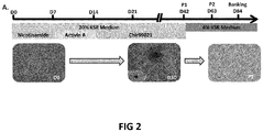
- NIC, Activin A and Chir99021 could recapitulate the main steps of retinal development by evaluating the expression of markers of the early eye field stage, optic vesicle stage and immature RPE cells at different time points during the differentiation ( Fig. 1A ).
- the use of Nicotinamide for the first 7 days of differentiation significantly enhanced the transient expression of the early eye field transcription factors SIX homeobox 3 (SIX3) and Retinal homeobox (RAX) concomitantly to a higher decrease of the expression of the pluripotency marker NANOG at mRNA level when compared to the spontaneous protocol (p ⁇ 0.01; Fig. 1B ).
- MITF expression is significantly upregulated between day 14 and day 30 in the directed protocol when compared to the spontaneous one (p ⁇ 0.01).
- Immunostaining assays confirmed the absence of VSX2 positive cells at day 21 and the increased number of MITF+ cells (87.5% ⁇ 12.5%).
- putative RPE precursors MITF-positive cells emerged and organized around 3D structures that did not express MITF and VSX2.
- Mature hESC-RPE cells were dissociated and cryopreserved in liquid nitrogen vapors with CryoStor CS10 medium (StemCell technologies) at passage 1 or 2.
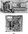
- the CompacT SelecT® (Sartorius) is a fully automated cell culture platform which allows the expansion and differentiation of large batches of adherent cells in a controlled environment ( Fig. 7 ).
- the system allows the automation of media changes and cell passaging as well as the monitoring of culture vessels with the automated live-cell imaging system IncuCyte (Sartorius). Contrary to the manual protocol, cells were not centrifuged after dissociation but were directly seeded into new flasks with enough medium to ensure that the final concentration of TryPLE® reagent in the daughter flasks did not exceed 5%.
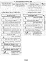
- the automated process is presented in Fig. 3 .
- the directed protocol allows obtaining a pure population of hESC-RPE cells without manual enrichment and is amenable to automation of the differentiation.
- This automated cell culture platform is composed of an incubator, bar-coded flasks for cell process tracking, multiple connected pumps to dispense culture media, a six-axis anthropomorphic robotic arm and a live-cell imaging system (Incucyte) ( Fig.3 and Fig. 7 ).
- a second enzyme incubation of 35 minutes then enabled the detachment and dissociation of RPE cells.
- hESC-RPE cells obtained by an automated differentiation are mature and functional.
- RPE cells One of the most important functions of RPE cells is the phagocytosis of the outer segments shed by the photoreceptors.
- FITC phagocyte pig fluorescein isothiocyanate
- hESC-RPE cells were able to phagocyte FITC-labeled photoreceptor outer segments as shown by the cytoplasmic localization of the FITC signal under the apical limit Ezrin positive.
- hESC-RPE cells incubated with pH-sensitive particles at 37°C had a fluorescence intensity 22.2 fold higher compared to cells incubated at 4°C, a temperature that inhibits the phagocytic process.
- Another indicator of RPE functionality is the ability to secrete a wide range of growth factors including the vascular endothelial growth factor (VEGF). The secretion of VEGF was quantified after several culture weeks and a progressive increased of VEGF secretion starting from 2 weeks of culture was observed.
- VEGF vascular endothelial growth factor
- hESC-RPE cells had a limited amplification potential before they undergo an epithelial-mesenchymal transition (EMT).
- EMT epithelial-mesenchymal transition
- hESC-RPE cells obtained with an automated process adopted a mesenchymal phenotype starting from passage 4 despite the maintenance of the gene expression of the RPE markers MITF and BEST ( Fig. 5B ).
- the cells switched from a classical cobblestone organization to elongated cell morphology. This microscopic observation is correlated with the rising expression of mesenchymal markers LUMICAN (LUM) and FIBRONECTIN 1 (FN1), two extracellular matrix proteins, starting at passage 5 when compared to passage 3 and 4 (p ⁇ 0.01; Fig. 5B ).
- hPSC-RPE cells have been already grafted in AMD patients either as a cell suspension or a polarized epithelium resting on a synthetic basement membrane.
- the use of cell suspension formulation considerably simplifies the logistical and surgical procedures but several studies made in animal models, suggest that the survival of the RPE cells and the visual benefits for the animal are improved when the cells are grafted as an epithelial tissue rather than a cell suspension. In human, these two approaches have shown both satisfactory safety results and promising efficacy results, even if the extent and the causes of visual improvement in transplant recipients remain ambiguous.
- the automated culture system for preparing RPE cells is expected to be qualified for clinical cell productions in accordance with Good Manufacturing Practices (GMP) (e.g., the preparations are GMP-compliant) and/or current Good Tissue Practices (GTP) (e.g., the preparations may be GTP- compliant).
- GMP Good Manufacturing Practices
- GTP Current Good Tissue Practices
Abstract
The present invention relates to methods for preparing retinal pigment epithelium (RPE) cells from pluripotent stem cells (PSCs). More particularly, the present invention relates to an automated method that combines in a sequential manner three differentiating agents to direct the differentiation of human PSCs into RPE cells.
Description
- The present invention relates to methods for preparing retinal pigment epithelium (RPE) cells from pluripotent stem cells (PSCs). More particularly, the present invention relates to an automated method that combines in a sequential manner three differentiating agents to direct the differentiation of human PSCs into RPE cells.
- The retinal pigment epithelium (RPE) is a monolayer of pigmented cells localized between the neuroretina and the choroids.
- The RPE cells play roles in the maintenance and function of the retina and its photoreceptors. These include the formation of the blood-retinal barrier, absorption of light and protection against photooxidation, transport of nutrients to the neural retina, regeneration of visual pigment, and phagocytosis of shed photoreceptor membranes.
- Human pluripotent stem cells (hPSCs), including human embryonic stem cells (hESCs) and human induced pluripotent stem cells (hiPSCs) are characterized by unlimited self-renewal and their ability to differentiate into any cell type. Due to these properties, extensive efforts have been done to use them as a source material for cell therapy to repair damaged tissues. At the forefront of cell therapy, the replacement of the retinal pigment epithelium, acts as a proof of concept. RPE cells play crucial roles in sight and their dysfunction or their loss may engender the secondary loss of photoreceptors. RPE cells are altered in 5-6% of Retinitis Pigmentosa cases and in Age-related Macular Degeneration (AMD). AMD is the leading cause of blindness in developed countries with more than 150 million people affected worldwide, a figure that will increase in the coming years. It can be classified into two groups, dry (atrophic) or wet (exudative), which is based on the presence of a choroidal neovascularization.
- There is still no treatment for dry AMD and for most of RPs.
- As such, the transplantation of RPE cells derived from human pluripotent stem cells (hPSC-RPE) represents an attractive strategy for treating retinal degenerative diseases.
- hPSCs spontaneously differentiate into RPE cells after removal of basic fibroblast growth factor (bFGF), used to maintain the pluripotency state, from the culture medium. The distinctive cobblestone morphology of RPE cells as well as their pigmentation allow to manually collect pigmented areas that appear upon differentiation of hPSCs to obtain a pure population of hPSC-RPE cells. Such approach of RPE cell production is used as cell replacement material in on going and planned clinical trials. However, this spontaneous method remains fastidious, inefficient and time consuming (8 to 12 weeks of hPSCs differentiation) making it incompatible with the industrial large-scale production which is required to treat the potential millions of patients.
- During the last ten years, several teams have developed improved differentiation protocols by combining the use of an increasing number of cytokines and small molecules selected on the basis of results obtained from developmental studies. One of the quickest and most efficient protocol was published in the publication "Canonical/β-catenin Wnt pathway activation improves retinal pigmented epithelium derivation from human embryonic stem cells. Invest. Ophthalmol. Vis. Sci. 56, 1002-1013 (2015). Following data demonstrating that RPE and neural retina progenitors (NRPs) have the same embryonic origin, they combined a protocol allowing the efficient differentiation of NRPs with previously described RPE inducing factors such as Nicotinamide, Activin A, in addition to many others including bFGF, Noggin, DKK1 (Dickkopf WNT signaling pathway inhibitor 1), Insulin Growth Factor (IGF)-1. The previous protocol was modified to include Chir99021 and SU5402 from day 8 to 14.
- Using this method, they obtained cells expressing the pigmentation marker PMEL17 after 14 days of differentiation allowing bypassing manual enrichment of pigmented cells.
- Background art also includes
WO2017021973 andWO2008129554 which disclose two-step methods of generating retinal pigment epithelial (RPE) cells comprising culturing a population of human pluripotent stem cells in the presence of Nicotinamide; and further subjecting the cells to another stage of differentiation in the presence of activin A, with or without Nicotinamide. - Although the differentiation of hPSCs into RPE cells became more efficient during the last years, it still remains a long and laborious process requiring meticulous manipulations from hPSCs thawing to hPSCRPE cell banking. Many cell culture parameters, such as seeding homogeneity, the time spent by the cells out of the incubator or the method used to isolate pigmented clumps, could impact on the proliferation and the differentiation of hPSCs. Thus, manual processing implies operator to operator variability and the quality of hPSCs and the efficiency of their differentiation into RPE cells are currently highly dependent on technical skills. In this regard, automation should not only allow scaling up the production of hPSC-RPE cells but should also increase its robustness. It could enable larger and more reliable cell production for clinical and disease modeling applications.
- Until recently, the requirement of a manual enrichment to obtain a pure population of hPSC-RPE cells prevented the use of these automated systems for the differentiation of this cell type.
- Thus, a need exists for developing a fully automated process allowing a large-scale production of hPSC-RPE cells.
- Considering that, in addition to considerably complicate the process, the use of numerous growth factors and small molecules on a large scale is very expensive, especially for an automated process which requires significant dead volumes, the Applicant answers this need by providing a simplified RPE differentiation protocol amenable for automation.
- In accordance with a first embodiment, the present invention provides the use of a protocol amenable for automation that combines in a sequential manner three differentiating agents to direct the differentiation of hPSCs into RPE cells. This novel differentiation protocol associated with the use of cell culture robots open new possibilities for the production of large batches of hPSC-RPE cells while maintaining a high cell purity and functionality.
- Thus, an object of the present invention is to provide methods for large-scale automated production of RPE cells derived from (human) pluripotent stem cells.
- The hPSCs in the culture system of the methods disclosed herein are differentiating hPSCs, i.e. a population of hPSCs essentially in an undifferentiated state, or wherein at least part of said cells have been induced to undergo initial stages of directed differentiation, the majority of said cells have been induced to undergo initial stages of directed differentiation. In accordance with one embodiment, the initial stage of differentiation is achieved by exposing the cells to a first differentiating agent, in particular at least one Nicotinamide (NA) mimetic compound, then to a second differentiating agent, in particular at least one member of transforming growth factor β (TGF β) superfamily and finally to a third differentiating agent, in particular at least one activator of the Wnt canonical pathway.
- In the following description and claims use will be made, with a variety of terms, and the meaning of such terms as they should be construed in accordance with the present teaching is as follows:
The term "mimetic" refers to a compound having similar functional and/or structural properties to another known compound or a particular fragment of that known compound, such as a known compound of biological origin, e.g., a polypeptide or fragment thereof. - "Undifferentiated", as used herein, refers to cultured cells when a substantial proportion (at least 20%, and possibly over 50% or 80%) of the cells and their derivatives in the population display characteristic markers and morphological characteristics of undifferentiated cells, distinguishing them from differentiated cells of embryo or adult origin. Cells are recognized as proliferating in an undifferentiated state when they go through at least 1 population doubling during a cultivation period of at least 3 weeks, while retaining at least about 50%, or the same proportion of cells bearing characteristic markers or morphological characteristics of undifferentiated cells after said cultivation period.
- It is intended, for the purposes of the present invention, that the term pluripotent stem cell embraces any cell having the capacity for self-renewal and the potential to differentiate into one or more other cell types.
- "Pluripotent hSCs" as used herein, refer to precursor cells of human source that have the ability to form any adult cell. Such cells are true cell lines in that they: (i) are capable of extensive proliferation in vitro in an undifferentiated state; and (ii) are capable of differentiation to derivatives of all three embryonic germ layers (endoderm, mesoderm, and ectoderm) even after prolonged culture. Human embryonic stem cells (hESCs) are derived from fertilized embryos that are less than one week old (in the cleavage or blastocyte stage) or produced by artificial means (such as by nuclear transfer) that have equivalent characteristics. Other pluripotent hSCs include, without being limited thereto, multipotent adult progenitor cells (MAPs), induced pluripotent stem cells (iPS cells) and amniotic fluid stem cells.
- hPSCs can be obtained using well-known cell-culture methods. For example, hESC can be isolated from single blastomeres of the cleavage or morula stage human embryo, from cleavage stage and morula human embryos and human blastocysts. Human embryos may be obtained from in vivo preimplantation embryos or more typically from in vitro fertilized (IVF) embryos. Alternatively, non-fertilized human oocyte can be parthenogenetically activated to cleave and develop to the blastocyst stage. In addition a single cell human embryo can be expanded to the blastocyst stage. For the isolation of hESCs from a blastocyst, the zona pellucida is removed and the inner cell mass (ICM) is isolated by immunosurgery, in which the trophectoderm cells are lysed and removed from the intact ICM by gentle pipetting. The ICM is then plated in a tissue culture flask containing the appropriate medium which enables its outgrowth. Following 9 to 15 days, the ICM derived outgrowth is dissociated into clumps either by mechanical dissociation or by enzymatic digestion and the cells are then re-plated on a fresh tissue culture medium. Colonies demonstrating undifferentiated morphology are individually selected by micropipette, mechanically dissociated into clumps, and re-plated. Resulting ESCs are then routinely split every 1-2 weeks. For further details on methods of preparation of hESCs; see
Thomson et al. [U.S. Pat. No. 5,843,780 ; Science 282:1145, 1998] - In the present invention, ES cells are not limited to a primary cell line collected from the inner cell mass, but may also be an established ES cell line. Examples of such an established ES cell line include: a cell line furnished from a cell population obtained by allowing the already established ES cell line to grow; and an ES cell line obtained by thawing a freeze-dried cell line and then culturing it. Such an established ES cell line is available without going through a step of disintegrating a fertilized egg.
- Otherwise, the ES cells used in the present invention may be established from a single embryonic blastomere at the cleavage stage before the blastocyst stage, without impairing the generating ability of the embryo. Such ES cells can be obtained without destroying a fertilized egg (Klimanskaya I. et al., (2006) Nature 444: 481-485 ; and Chung Y et al., (2008) Cell Stem Cell 2: 113-117).
- Commercially available hPSCs can be also used in accordance with the invention. hPSCs can be purchased for example from the UK Stem Cell Banks or the NIH human embryonic stem cells registry. Non-limiting examples of commercially available embryonic stem cell lines are BG01, BG02, BG03, BG04, CY12, CY30, CY92, CY10, TE03 and TE32.
- For ethical reasons, the present invention preferably does not pertain to objects that may be considered as contrary to"ordre public" or morality. Therefore, in the context of the invention, the terms "human embryonic stem cells" preferably refer to human embryonic stem cells which isolation has not involved the destruction of an embryo. In other words, the terms "human embryonic stem cells" preferably exclude human embryonic stem cells isolated by techniques that involve the destruction of an embryo.
- In the context of the invention, it is to be understood that any technique that does not involve the destruction of an embryo can be used, including those that are not described herein.
- Moreover, in the context of the invention, the embryos used for obtaining human embryonic stem cells are preferably embryos that cannot give rise to a human being, such as embryos destined to be discarded following in vitro fertilization (IVF) and embryos created solely for the purpose of stem cell research.
- Hence, in a yet preferred embodiment, the terms "human embryonic stem cells" (hESC) preferably refer to human embryonic stem cells isolated from discarded embryos, research embryos, or preferably isolated by techniques that do not involve the destruction of an embryo.
- The "induced pluripotent stem cell" in the present invention is a cell induced to have pluripotency by reprogramming a somatic cell by a known method and the like. Specifically, a cell induced to have pluripotency by reprogramming differentiated somatic cells such as fibroblast, peripheral blood mononuclear cell and the like by the expression of a combination of a plurality of genes selected from the group consisting of reprogramming genes including Oct3/4, Sox2, Klf4, Myc (c-Myc, N-Myc, L-Myc), Glis1, Nanog, Sall4, lin28, Esrrb and the like can be mentioned. Examples of preferable combination of reprogramming factors include (1) Oct3/4, Sox2, Klf4, and Myc (c-Myc or L-Myc), and (2) Oct3/4, Sox2, Klf4, Lin28 and L-Myc.
- Induced pluripotent stem cell was established by Yamanaka et al. in mouse cell in 2006. In 2007, Induced pluripotent stem cell was also established from human fibroblast, and has pluripotency and self-renewal competence similar to those of embryonic stem cells.
- The terms "differentiation", "differentiating" or "derivatives thereof" as used herein denote a process by which an unspecialized or relatively less specialized cell becomes relatively more specialized. In the context of cell ontogeny, the adjective "differentiated" is a relative term. Hence, a "differentiated cell" is a cell that has progressed further down a certain developmental pathway than the cell it is being compared with. A relatively more specialized cell may differ from an unspecialized or relatively less specialized cell in one or more demonstrable phenotypic characteristics, such as, for example, the presence, absence or level of expression of particular cellular components or products, e.g., RNA, proteins or other substances, activity of certain biochemical pathways, morphological appearance, proliferation capacity and/or kinetics, differentiation potential and/or response to differentiation signals, etc., wherein such characteristics signify the progression of the differentiation towards the relatively more specialized cell.
- In the present context, the method of the invention results in the progressive differentiation of (human) pluripotent stem cells and differentiating cells towards RPE cells. Thus, as used herein, the term "differentiating" of differentiating cells to RPE cells may be considered synonymous to the term "obtaining" RPE cells from differentiating cells.
- According to the present invention, the (human) pluripotent stem cells are subjected to directed differentiation using at least two, more preferably three differentiating agents.
- "Cell marker", as used herein, refers to any phenotypic feature of a cell that can be used to characterize it or discriminate it from other cell types. A marker may be a protein (including secreted, cell surface, or internal proteins; either synthesized or taken up by the cell); a nucleic acid (such as an mRNA, or enzymatically active nucleic acid molecule) or a polysaccharide. Included are determinants of any such cell components that are detectable by antibody, lectin, probe or nucleic acid amplification reaction that are specific for the marker of the cell type of interest. The markers can also be identified by a biochemical or enzyme assay or biological response that depends on the function of the gene product. Associated with each marker is the gene that encodes the transcript, and the events that lead to marker expression. A marker is said to be preferentially expressed in an undifferentiated or differentiated cell population, if it is expressed at a level that is at least 50% higher (in terms of total gene product measured in an antibody or PCR assay) or 30% more frequently (in terms of positive cells in the population) than an acceptable control.
- Whether or not the cells obtained are retinal progenitor cells or RPE cells can be determined by a method known per se, for example, the expression of a retinal progenitor cell marker. As examples of the retinal progenitor cell marker, Pax6 (neural retinal progenitor cells, retinal pigment epithelium progenitor cells), RAX (neural retinal progenitor cells), and MITF (retinal pigment epithelium progenitor cells) can be mentioned.
- In one embodiment, the tissue/cell specific markers can be detected using immunological techniques. Examples include, but are not limited to, flow cytometry for membrane-bound or intracellular markers, immunohistochemistry for extracellular and intracellular markers and enzymatic immunoassay, for secreted molecular markers.
- Following the stages of differentiation described herein above, a mixed cell population is obtained comprising both pigmented and non-pigmented cells.
- The mature RPE cells express significantly higher levels of transcripts of markers of mature RPE cells such as MITF, PAX6 as compared to their expression in RPE cells produced by spontaneous differentiation.
- The culture obtained by a method of the present invention contains RPE cells and/or the differentiating cells at high frequency (content amount). Cells obtained by a method of the present invention are PAX6, MITF-positive at high frequency, for example, at a frequency (colony frequency) of 5% or more, preferably 10 to 50%, more preferably 60 to 90%.
- According to the invention, the term "differentiation" refers to the process by which a cell acquires the features of a more differentiated (or "specialized") cell.
- Thus, in the context of the invention, a differentiated cell or a differentiation-induced cell is one that has more specialized features, compared to an undifferentiated cell, wherein said features correspond to a more differentiated stage within the lineage of a cell.
- According to the invention, the lineage of a cell encompasses all of the discrete development stages of a cell within a scheme of development, that is to say from an undifferentiated stage to a differentiated stage. In that regard, according to the invention, a lineage-specific marker refers to a marker specifically associated with the phenotype of cells of a lineage of interest and can be used to assess the differentiated status of a cell. (
Figure 1A ) - As used herein the term "sequential steps" refers to a method in which each step (e.g steps a) to c)) is performed at a different point in time, in a successive way. Unless otherwise indicated herein, as used herein "sequentially (sequential)" refers to a normal order or sequence.
- As used herein, the term "Wnt signaling pathway" denotes a signaling pathway which may be divided in two pathways: the "canonical Wnt/beta catenin signaling pathway" and the "Wnt/PCP signaling pathway". As used herein, the term "canonical Wnt/beta catenin signaling pathway" or "Wnt/PCP signaling pathway" in its general meaning denotes a network of proteins and other bioactive molecules (lipids, ions, sugars...) best known for their roles in embryogenesis and cancer, but also involved in normal physiological processes in adult animals. The "canonical Wnt/beta catenin signaling pathway" is characterized by a Wnt dependant inhibition of glycogen synthase kinase 3B (GSK-3B), leading to a subsequent stabilization of B-catenin, which then translocates to the nucleus to act as a transcription factor. The "Wnt/PCP signaling pathway" does not involve GSK-3 B or B-catenin, and comprises several signaling branches including Calcium dependant signaling, Planar Cell Polarity (PCP) molecules, small GTPases and C-Jun N-terminal kinases (JNK) signaling.
- As used herein the term "activator" denotes a substance that enhances Wnt signaling activity.
- In another embodiment, the activator of the Wnt signaling pathway is an inhibitor of GSK-3 β.
- Thus, according to one embodiment, the present invention provides an automated method for promoting directed differentiation of human pluripotent stem cells into retinal pigment epithelium (RPE) cells, the method comprising or consisting in the sequential steps of:
- (a) culturing human pluripotent stem cells in a medium supplemented with at least one Nicotinamide (NA) mimetic compound to generate differentiating cells;
- (b) culturing said differentiating cells obtained in step a) in a medium supplemented with at least one compound of transforming growth factor β (TGF β) superfamily to further differentiating said differentiating cells;
- (c) culturing said further differentiating cells obtained in step b) in a medium supplemented with at least one activator of the Wnt canonical pathway to induce said further differentiating cells to differentiate into a population of RPE cells.
- The (human) pluripotent stem cells may be obtained from various culture systems in which the (human) pluripotent stem cells are maintained in an undifferentiated pluripotent state. For example, the hPSCs are cultivated in a feeder-free adherent or suspension system or on feeder cells. Commonly used feeder cells include a primary mouse embryonic fibroblast (PMEF), a mouse embryonic fibroblast (MEF), a murine fetal fibroblast (MFF) a human embryonic fibroblast (HEF), a human fibroblast obtained from the differentiation of human embryonic stem cells, a human fetal muscle cell (HFM), a human fetal skin cell (HFS), a human adult skin cell, a human foreskin fibroblast (HFF), a human cell obtained from the umbilical cord or placenta, a human adult fallopian tubal epithelial cell (HAFT) and human marrow stromal cells (hMSCs). The clusters of (human) pluripotent stem cells are obtained from an adherent cell culture by dissociation of the cells from the feeder layer or extracellular matrix to form a suspension of cells. The suspension of hPSCs comprises the free floating clusters or an essentially single cell suspension from which clusters of cells are outgrown to form the cell clusters.
- In one embodiment, the step a) of the method is effected for at least 3 days, preferably for 3 to 10 days and more preferably for 7 days.
- In one embodiment, the human pluripotent stem cells are cultured for 3-10 or 7 days in the presence of at least one Nicotinamide (NA) mimetic compound e.g. between 0.01-100 mM, 0.1 -100 mM, 0.1-50 mM, 5-50 mM, 5-20 mM, preferably 10mM.
- According to a particular embodiment, the Nicotinamide (NA) mimetic compound is a Nicotinamide derivative or a Nicotinamide mimetic compound. The term "derivative of Nicotinamide (NA)" as used herein denotes a compound which is a chemically modified derivative of the natural NA.
- Thus, the Nicotinamide of the present invention includes a substituted or non-substituted Nicotinamide. In another embodiment, the chemical modification may be a deletion or replacement of a single group, e.g. to form a thiobenzamide analog of NA, all of which being as appreciated by those versed in organic chemistry. The derivative in the context of the invention also includes the nucleoside derivative of NA (e.g. Nicotinamide adenine). A variety of derivatives of NA are described, some also in connection with an inhibitory activity of the PDE4 enzyme (
WO03/068233 WO01/55114 WO05/014549 WO01/55114 EP2128244 . - Nicotinamide mimetic compounds include modified forms of Nicotinamide, and chemical analogs of Nicotinamide which recapitulate the effects of Nicotinamide in the differentiation and maturation of RPE cells from pluripotent cells. Exemplary Nicotinamide mimetic compounds include benzoic acid, 3-aminobenzoic acid, and 6- aminoNicotinamide. Another class of compounds that may act as Nicotinamide mimetic compounds are inhibitors of poly(ADP-ribose) polymerase (PARP). Exemplary PARP inhibitors include 3-aminobenzamide, Iniparib (BSI 201), Olaparib (AZD-2281), Rucaparib (AG014699, PF- 01367338), Veliparib (ABT-888), CEP 9722, MK 4827, and BMN- 673.
- In one embodiment, the Nicotinamide (NA) mimetic compound is the first differentiating agent of the present method.
- In a preferred embodiment, the Nicotinamide (NA) mimetic compound is Nicotimamide.
- In a preferred embodiment, the concentration of Nicotinamide is about 10 mM.
- In one embodiment, the step b) of the method is effected for at least 3 days, preferably for 3 to 10 days and more preferably for 7 days.
- According to one embodiment, the method comprises treating the differentiating cells obtained in step a) with at least one member of the TGF β superfamily of growth factors after the hPSCs were cultured in the presence of at least one Nicotinamide (NA) mimetic compound.
- Without being bound to theory, it is believed that the at least one Nicotinamide (NA) mimetic compound acts as a differentiation inducer/promoter and that similarly, the at least one member of the TGF β superfamily act as an RPE differentiation promoting factor. In addition, while not being bound by theory, it is believed that the prior exposure of the hPSCs to the at least one Nicotinamide (NA) mimetic compound provides the differentiating cells with properties that enable their response to the RPE differentiation promoting effect of the at least one member of the, TGF β superfamily.
- In one embodiment, the at least one member of transforming growth factor β (TGF β) superfamily is selected from the group consisting of the transforming growth factor-like (TGF-like) group with the TGFβ subfamily, Activin, Nodal and some growth and differentiation factors (GDF), the bone morphogenetic protein like (BMP-like) group with the BMP, GDF and anti-Mullerian hormone (AMH).
- In one embodiment the transforming growth factor-β (TGFβ) superfamily growth factor is a transforming growth factor-β proteins, such as the TGFβ1, TGFβ2, and TGFβ3 subtypes, as well as homologous ligands including activin, such as activin A, activin B, and activin AB
- In one embodiment the transforming growth factor-β (TGFβ) superfamily growth factor is nodal, anti-mullerian hormone (AMH), some bone morphogenetic proteins (BMP), such as BMP2, BMP3, BMP4, BMP5, BMP6, and BMP7, and growth and differentiation factors (GDF).
- In a preferred embodiment, the at least one member of transforming growth factor β (TGF β) superfamily is Activin A.
- In one embodiment, the cells produced in step b) of the method of the present invention comprise a population of cells in which at least part or at least a majority of the human pluripotent stem cells have initiated differentiation.
- With respect to the supplementation with at least one member of the TGF β superfamily of growth factors, said member is presented in soluble form or affixed or associated to a matrix or cell added to the culture system or the element may be bound or complexed to other substances.
- In another embodiment, the amount of said member of the TGF β superfamily in the medium is less than 20 ng/ml, 10 ng/ml, 1 ng/ml or even less than 0.1 ng/ml.
- In one embodiment the concentration of Activin A is about 10 ng/ml.
- In one embodiment, the medium in step (b) is substantially or completely free of the first differentiating agent (the at least one Nicotinamide (NA) mimetic compound) used in step a) of the present method.
- In one embodiment, the step c) of the method is effected for at least 20 to 50 days, preferably 20, 21, 22, 23, 24, 25, 26, 27, 28, 29, 30, 31, 32, 33, 34, 35, 36, 37, 38, 39, 40, 41, 42, 43, 44, 45, 46, 47, 48, 49 or 50 days, more preferably 28 days.
- In one embodiment, the at least one activator of the Wnt canonical pathway is a GSK-3 inhibitor selected from the group consisting of 3F8, 1 -Azakenpaullone, 10Z-Humenialdisine, Alsterpaullone, Al 070722, AR- A014418, AZD1080, AZD2858, Bikinin, BIO, Cazpaullone, CT98014, CT98023, CT99021 (Chir99021), Chir98014, Dibromocantharelline, GSKJ2, HMK-32, Hymenialdesine, Indirubin, Indirubin-3'-omime, IM-12, KenpauUone, L803, L803-mts, Lithium carbonate, LY2090314, Manzamine A, Meridianin, NCS693868, NP031115, Palinurine, SB216763, SB415286, TCS21311, TC-G-24, TCS2002, TDZD-8, Tideglusib, Tricantine and TWS119.
- In a preferred embodiment, the at least one activator of the Wnt canonical pathway is Chir99021.
- In one embodiment the concentration of Chir99021 is about 10 ng/ml.
- In one embodiment, the medium in step (c) is substantially or completely free of the first and second differentiating agents) used in steps a) and b) of the present method.
- In a preferred embodiment the medium in step (c) is substantially or completely free of the at least one Nicotinamide (NA) mimetic compound and the at least one compound of transforming growth factor β (TGF β) superfamily respectively used in steps a) and b).
- In one embodiment, the timing of addition of the second or the third differentiating agent to the medium is not particularly limited as long as the progressive differentiating effects can be measured or estimated by any technique known to a person skilled in the art. The presence of differentiating cells (i.e retinal progenitor cells) or RPE cells can be confirmed, for example, by detecting the presence of cells expressing RAX, PAX6, MITF or VSX2.
- The skilled person is able to follow the progression of the differentiation of the cells and is able to define the appropriate moment when to add the second and third differentiating agents.
- In one embodiment, the step of culturing the differentiating cells obtained in step a) produces a population of differentiating cells comprising at least 50%, 60%, or preferably 70% of differentiating cells.
- In one embodiment, the step of culturing the differentiating cells obtained in step b) produces a population of differentiating cells comprising more than 50%, 60%, or preferably 70% or 80% of differentiating cells.
- In one embodiment, the automated method for promoting directed differentiation of human pluripotent stem cells into retinal pigment epithelium (RPE) cells, comprises or consists in the sequential steps of:
- (a) culturing human pluripotent stem cells in a medium supplemented with Nicotinamide to generate differentiating cells;
- (b) culturing said differentiating cells obtained in step a) in a medium supplemented with Activin A to further differentiating said differentiating cells;
- (c) culturing said further differentiating cells obtained in step b) in a medium supplemented with CHIR99021 to induce said further differentiating cells to differentiate into RPE cells.
- In one embodiment, during the differentiation phase (i.e during steps a) to c)) the medium is changed every 2-3 days.
- In one embodiment, the method further comprises the step of
(d) treating the population of cells obtained in step c) to remove the non-pigmented cells. - Without being bound to theory, the population of cells obtained in step c) comprises RPE cells and/or differentiating cells that differentiate toward RPE cells.
- The hPSC-derived RPE cells form a cohesive epithelium in culture that requires long incubation times with enzymatic dissociation reagents to trigger cell detachment for further replating and amplification. This characteristic is used to enrich the culture for RPE cells by performing a two-step enzymatic dissociation procedure comprising or consisting in a washing (first short incubation time) to remove non-RPE cells with weak adherence followed by an enzymatic treatment (second incubation) to generate a homogenous population of RPE cells.
- Thus, in one preferred embodiment, the step d) of the method is a two-step dissociation procedure comprising or consisting in washing and treating the cells enzymatically.
- In one embodiment, this is effected enzymatically.
- In a preferred embodiment the enzymatic treatment is effected with trypsin, TrypLE Select®, trypsin-EDTA, or Accutase®.
- In another embodiment, step d) may comprise combinations of mechanical, enzymatic and chemical treatment- e.g. using a cell scraper or EDTA.
- The automated process of the method is presented in
Fig.3 . - In manual culture methods, enzymic and non-enzymic dissociation reagents are typically removed by centrifuging before the cells are transferred to fresh culture vessels. However, in the automated methods of the invention it is preferred that the passaging does not comprise a centrifugation step. This is, in part, due to the difficulty and considerable expense of integrating a centrifuge into an automated cell culture system. An advantage is to avoid exposing the cells to the shear forces that result from centrifugation.
- In one embodiment, the method further comprises the step of
(e) expanding the cells obtained in step d) over at least two passages. - Expansion of the RPE cells and/or the differentiating cells that differentiate toward RPE cells can be effected on an extra cellular matrix, e.g. gelatin, collagen I, collagen IV, laminin (e.g. laminin 521), fibronectin and poly-D-lysine. For expansion, the cells may be cultured in serum-free KOM, serum comprising medium (e.g. DMEM with 4 % human serum) or Nutristem medium. Under these culture conditions, after passaging under suitable conditions, the ratio of pigmented cells: non-pigmented cells increases such that a population of purified RPE cells is obtained. Such cells show the characteristic polygonal shape morphology and pigmentation of RPE cells.
- The RPE cells and/or the differentiating cells that differentiate toward RPE cells can be expanded in suspension or in a monolayer. The expansion of the RPE cells in monolayer cultures can be modified to large scale expansion in bioreactors by methods well known to those versed in the art.
- According to this aspect of the present invention, the differentiating cells and/or the RPE cells are removed from the culture vessel, in step d) of the method.
- According to one embodiment, the expansion phase is effected for at least one week, at least 2 weeks, at least 3 weeks, at least 4 weeks, at least 5 weeks, at least 6 weeks, at least 7 weeks, at least 8 weeks, at least 9 weeks or even 10 weeks. Preferably, the expansion phase is effected for 1 week - 10 weeks, more preferably 2 weeks - 10 weeks, more preferably, 3 weeks - 10 weeks, more preferably 4 weeks - 10 weeks, or 4 weeks - 8 weeks.
- In a preferred embodiment, during the expansion phase, the medium is changed every 2-3 days.
- The precise proportions and frequencies chosen in different embodiments will depend on the type of cells being cultured, the culture medium, the type of culture vessel, and other culture parameters, and can be readily determined by the user.
- According to still another embodiment, the RPE cells and/or the differentiating cells that differentiate toward RPE cells are passaged at least 1 time during the expansion phase, at least twice during the expansion phase, at least three times during the expansion phase, at least four times during the expansion phase, at least five times during the expansion phase, or at least six times during the expansion phase.
- Harvesting of the expanded population of the RPE cells and/or the differentiating cells may be effected using methods known in the art (e.g. using an enzyme such as trypsin, or chemically using EDTA).
- In one embodiment passaging requires dissociation of the differentiating cells, the dissociation is conveniently carried out by adding a cell dissociation reagent to the first culture vessel. The cell dissociation reagent may be an enzymic cell dissociation reagent, such as trypsin-EDTA, or Accutase, or a non- enzymic cell dissociation reagent.
- In one embodiment, the passaging includes counting the cells transferred from the first culture vessel, preferably using an automated cell counting device forming part of the robotic cell culture apparatus. Following counting, the predetermined number of cells is transferred to each of the further culture vessels.
- In other embodiment, the actual number of cells transferred from the first culture vessel is not counted. Rather, the number of cells is estimated based on the size of the culture vessel and the growth characteristics of the RPE cells and/or the differentiating cells under the particular culture regime being used. Thus, the passaging can comprise calculating the number of cells transferred from the first culture vessel based on one or more of (i) the initial number of RPE cells and/or the differentiating cells in the first culture vessel, (ii) the population doubling time of the RPE cells and/or the differentiating cells, (iii) the culture area of the first culture vessel, and (iv) the culture volume or surface. It will be appreciated that the culture area of the first culture vessel is particularly relevant when calculating the number of adherent RPE cells and/or the differentiating cells obtained after a given period of culture, whereas the culture volume or surface will be particularly relevant when calculating the number of RPE cells growing in suspension
- Alternatively, it is an option to passage the cells so that the cells from the first culture vessel are divided between a predetermined number of further culture vessels. For example, in preferred embodiments of the invention, RPE cells and/or the differentiating cells that differentiate toward RPE cells and/or the differentiating cells are passaged with a split ratio of from 1 :2 to 1 :10, preferably from 1:2 to 1:5.
- In further embodiments of the invention, passaging is carried out when the RPE cells and/or the differentiating cells that differentiate toward RPE cells in the first culture vessel reach a predetermined percentage confluence (or, in the case of suspension cells, a predetermined cell density). Typically, the passaging is carried out when the RPE cells and/or the differentiating cells that differentiate toward RPE cells in the first culture vessel are 50 to 100% confluent, preferably 60 to 90% confluent, more preferably 70 to 80% confluent. In a preferred embodiment, the passaging is carried out when the RPE cells and/or the differentiating cells that differentiate toward RPE cells in the first culture vessel are 90, 91, 92, 93, 94, 95, 96, 97, 98, 99 or 100% confluent.
- In the automated method of the invention, it is desirable, that the percentage confluence is calculated rather than being determined prior to each passage by the operator. Thus, in preferred embodiments, the percentage confluence is calculated based on one or more of (i) the number of the RPE cells and/or the differentiating cells that differentiate toward RPE cells initially present in the culture vessel, (ii) the population doubling time of the RPE cells and/or the differentiating cells that differentiate toward RPE cells, (iii) the culture area of the first culture vessel, and (iv) the culture volume or surface. In other embodiments, confluence is recorded and/or estimated automatically.
- In a preferred embodiment, the passages comprises (i) dissociating RPE cells and/or the differentiating cells that differentiate toward RPE cells in a first vessel to form a suspension; (ii) transferring the RPE cells and/or the differentiating cells that differentiate toward RPE cells to at least two further culture vessels; and (iii) culturing the RPE cells and/or the differentiating cells that differentiate toward RPE cells until the RPE cells and/or the differentiating cells that differentiate toward RPE cells are 50 to 100% confluent, wherein the passages does not comprise a centrifugation step.
- Preferably the passaging is repeated until either a predetermined number of culture vessels containing the RPE cells and/or the differentiating cells that differentiate toward RPE cells or a predetermined number of the RPE cells and/or the differentiating cells that differentiate toward RPE cells has been produced. In some embodiments, the point at which the predetermined number of RPE cells has been produced will be estimated based on the growth characteristics of the RPE cells and/or the differentiating cells that differentiate toward RPE cells and the previous process steps (e.g. the number of passages). It is also possible to calculate the number of the differentiating cells or RPE cells obtained by calculating the yield per culture vessel and multiplying this value by the number of culture vessels containing the RPE cells and/or the differentiating cells that differentiate toward RPE cells that have been produced.
- According to a further embodiment of the invention, the medium in which the hPSCs are differentiated is any known cell culture medium known in the art for supporting cell growth in vitro, typically, a medium comprising a defined base solution, which includes salts, sugars, amino acids and any other nutrients required for the maintenance of the cells in the culture in a viable state.
- According to a preferred embodiment of the invention, the medium is not a conditioned medium. Non-limiting examples of commercially available basic media that may be utilized in accordance with the invention comprise Nutristem (without bFGF and TGFP for ESC differentiation, with bFGF and TGFp for ESC expansion), Neurobasal™, KO-DMEM, DMEM, DMEM/F12, Cellgro™ Stem Cell Growth Medium, or X-Vivo™. The basic medium may be supplemented with a variety of agents as known in the art dealing with cell cultures.
- In one embodiment, the medium to be used in the present method is a serum-containing medium or serum-free medium.
- In one embodiment, the medium to be used in the present method is a knock out serum replacement (KOSR) containing medium.
- To avoid contamination of chemically-undefined components, a serum-free medium is preferably used in the present invention. To avoid complicated preparation, for example, a serum-free medium supplemented with an appropriate amount of a commercially available serum alternative such as KSR and so on (e.g., medium of 1:1 mixture of IMDM and F-12, which is supplemented with 10% KSR, 450 µM 1-monothioglycerol and 1x Chemically Defined Lipid Concentrate, or GMEM medium supplemented with 5% - 20% KSR, NEAA, pyruvic acid, 2-mercaptoethanol) is preferably used. The amount of KSR to be added to a serum-free medium in the case of human pluripotent stem cell is generally about 1% to about 30%, preferably about 2% to about 20% (e.g., about 5%, about 10%).
- In one embodiment, in step a) of the method, a DMEM medium is supplemented with 20%KSR.
- In one embodiment, in step b) of the method, a DMEM medium is supplemented with 20%KSR.
- In one embodiment, in step c) of the method, a DMEM medium is supplemented with 20%KSR.
- In one embodiment, in step e) of the method, a DMEM medium is supplemented with 4%KSR.
- According to some embodiments of the present application, steps (a), (b), (c) and (e) of the method include replacing periodically all or a portion of the culture medium. For example, all or a proportion of the culture medium can be removed from the culture vessel by pipetting or by pouring used medium to waste and fresh medium can then be added. If medium is to be removed by pipetting, the culture vessel can be positioned to assist removal of the medium.
- The proportion of medium volume replaced or added will vary between different embodiments of the invention and may be 10%, 20%, 30%, 40%, 50%, 60%, 70%, 80%,90% or 100% of the culture volume or surface.
- The method of the invention can be adapted for use with any type of culture vessel, including tissue culture flasks, dishes and multi- well plates. However, it is convenient to use flasks when producing large numbers of RPE cells, as this advantageously reduces the number of processing steps required to obtain a given number of cells and thus reduces the potential for cell damage during handling.
- In one embodiment, T75 or T175 tissue culture vessels are used.
- In another embodiment, culture chambers (e.g. CellStack®) are used.
- According to one embodiment, the cells in steps a) to e) are cultured on adherent substrate under normal atmospheric oxygen conditions.
- Examples of adherent substrates include but are not limited to fibronectin, laminin, polyD-lysine, collagen and gelatin.
- According to a preferred embodiment of the invention, the proliferation/growth medium is free of xeno contaminants i.e. free of animal derived components such as serum, animal derived growth factors and albumin.
- Following harvesting, the expanded population of the RPE cells and/or the differentiating cells may optionally be cryopreserved using methods known in the art. Examples of media suitable for cryopreservation include but are not limited to 90% Human Serum/10% DMSO,
CryoStor 10%, 5% and 2%, and Stem Cell Banker. - In one embodiment, the differentiation of human pluripotent stem cells toward RPE cells is monitored throughout the process using an automated live-cell imaging system that resides within the controlled environment of the automated cell culture platform. This non-invasive cell imaging system provides cell confluence metrics in real-time as well as phase contrast images of processed culture vessels. Each step of the differentiation protocol is thus monitored to prevent deviations from specification limits.
- In electron microscope (EM) analysis the RPE cells display morphological characteristics of mature RPE cells that are not demonstrated within RPE-like cells that were derived from spontaneously differentiating hPSC such as apical villi, tight junctions, and basal membrane. The RPE cells produced by the method of the present disclosure may be used for large scale and/or long term cultivation of such cells. To this end, the method of the invention is to be performed in bioreactors or robotic cell system suitable for large scale production of cells, and in which undifferentiated hPSCs are to be cultivated in accordance with the invention. General requirements for cultivation of cells in bioreactors are well known to those versed in the art.
- The population of RPE cells generated according to the methods described herein is characterized according to a number of different parameters. For example, the RPE cells obtained are polygonal and pigmented.
- It will be appreciated that the cell populations disclosed herein are devoid of undifferentiated hPSCs. According to one embodiment, less than 1:250,000 cells are Oct4+ TRA-1-60+ cells, as measured for example by FACS.
- The RPE cells of this aspect of the present invention do not express pluripotent stem cell markers. Said one or more embryonic stem cell markers may comprise OCT- 4, NANOG, Rex- 1, alkaline phosphatase, Sox2, TDGF- beta, SSEA-3, SSEA-4, TRA- 1-60, and/or TRA-1-81.
- The RPE preparations may be substantially purified, with respect to non-RPE cells, comprising at least about 75%, 80%, 85%, 90%, 91 %, 92%, 93%, 94%, 95%, 96%, 97%, 98%, 99%, or 100% RPE cells.
- Another way of characterizing the cell populations disclosed herein is by marker expression. Thus, for example, at least 80 %, 85 %, 90 %, 95 % or 100 % of the cells express
Bestrophin 1, as measured by immuno staining. According to one embodiment, between 80-100 % of the cells expressbestrophin 1. - According to another embodiment, at least 80 %, 85 %, 87 %, 89 %, 90 %, 95%, 97 % or 100 % of the cells express Microphthalmia-associated transcription factor (MITF), as measured by immuno staining. For example, between 80-100% of the cells express MITF.
- According to another embodiment, at least 80 %, 85 %, 87 %, 89 %, 90 %, 95%, 97 % or 100 % of the cells express both Microphthalmia-associated transcription factor (MITF) and
bestrophin 1, as measured by immuno staining. For example, between 80- 100% of the cells co-express MITF andbestrophin 1. - According to another embodiment, at least 50 %, 60 % 70 % 80 %, 85 %, 87 %, 89 %, 90 %, 95%, 97 % or 100 % of the cells express paired box gene 6 (PAX-6) as measured by immunostaining or FACS.
- According to another embodiment, at least 80 %, 85 %, 87 %, 89 %, 90 %, 95%, 97 % or 100 % of the cells express cellular retinaldehyde binding protein (CRALBP), as measured by immunostaining. For example, between 85-100% of the cells express CRALBP.
- According to another embodiment, at least 80 %, 85 %, 87 %, 89 %, 90 %, 95%, 97 % or 100 % of the cells express retinal pigment epithelium- specific protein 65kDa (RPE65), as measured by immunostaining. For example, between 80-100% of the cells express RPE65.
- The RPE cells typically co-express markers indicative of terminal differentiation,
e.g. bestrophin 1, CRALBP and/or RPE65. - Following the expansion phase cell populations comprising RPE cells are obtained whereby at least 70 %, 71 %, 72 %, 73 %, 74 %, 75 %, 76 %, 77 %, 78 %, 79, 80 %, 81 %, 82 %, 83 %, 84 %, 85 %, 86 %, 87 %, 88 %, 89, 90 %, 91 %, 92 %, 93 %, 94 %, 95 %, 96 %, 97 %, 98 %, 99 or even 100 % thereof are CRALBP+, PMEL17+.
- In some embodiments, the method further comprises the steps of
(f) harvesting and banking the RPE cells. - The apparatus used to implement the method of the present invention is selected from any of a number of automated platforms for cell culture that are available and adapted for large-scale production of stem cells or differentiated cells derived from stem cells.
- The Applicant has obtained good results using the CompacT SelecT® platform, manufactured by the Sartorius, but it will be understood that other systems can be adapted to provide apparatus according to the invention, which can be used to carry out the methods of the invention.
- In one embodiment, the invention provides apparatus adapted or arranged for carrying out the methods of the invention. Thus, the invention provides an apparatus for large-scale automated production of cells comprising: a) robotic means for handling culture vessels; b) means for inoculating cells into a culture; c) means for changing or adding medium to a culture; and d) programmable control means; wherein the apparatus is adapted to the phase of directed differentiation of hPSCs toward RPE cells and the phase of passage the cells.
- Such means are conveniently provided using an automated pipetting station, preferably using disposable pipettes, and, optionally, additional liquid pumps, thus permitting programmable medium selection and additive dispensing of different media and or reagents without risk of cross-contamination.
- Thus, the apparatus may further comprise means for adding further components to a culture. In some embodiments, separate systems will be provided for adding or removing media, reagents and/or cells to and from culture vessels of different types. For example, the apparatus may comprise separate dispensing stations for tissue culture flasks and multi-well plates. Additional means may also be supplied, e.g. for adding growth factors or cell dissociation reagents.
- The apparatus also includes incubators for any culture vessel format described herein, typically including at least one of an incubator for flasks and an incubator for tissue culture plates. In use, the apparatus will typically provide control of one or more of the temperature, the CO2 level, the O2 level and the relative humidity at which the stem cells are cultured.
- The apparatus will also provide aseptic conditions to prevent contamination of cultures and ensure operator safety, suitably using a negative pressure laminar airflow hood.
- In preferred embodiments the apparatus also comprises means for automated cell counting to provide consistent and accurate cell densities when seeding new culture vessels. Means for automated determination and/or estimation of percentage confluence can also be included.
- The apparatus can also comprise imaging equipment or other detection means. Such means can, for example, be used to detect the expression of fluorescent reporter genes (e.g. GFP) in the cells being cultured. For example, the cells may express a reporter gene, optionally provided by means of a construct comprising an internal ribosome entry site (IRES). The percentage of reporter-positive cells can be used to determine when to passage or induce differentiation of the stem cells in a culture. Imaging equipment can also be used to assess when to harvest cells.
- According to the invention, the apparatus incorporates a small six-axis anthropomorphic robotic arm that can access 90 T175 culture vessels and 210 plate incubators. The system allows the automation of seeding, feeding and other cell culture processes in order to maintain cell lines in standard T175 cell culture vessels. Culture vessels are bar-coded for identification and cell process tracking. Two culture vessels decappers and flask holders, automated medium pumping and an automatic cell counter are integrated within a high-efficiency particulate air (HEPA) filtered cabinet to ensure sterility.
- In one embodiment, the CompacT SelecT® has also been shown to be successful at preventing contamination when the GMP version of the CompacT SelecT® passed the sterile fill tests.
- In one embodiment, the CompacT SelecT® allows activities during cell culture such as seeding, media changes and measurement cells in a controlled environment. Thus this platform can be used to expand and differentiate batches of cells to a tighter specification than manual cell culture.
- The automation enables scale out for conventional formats with predictable process variation and quality outcome by removing manual interventions. The CompacT SelecT® is a preferred platform for development process friendly method of automating the culture of cells that grow in adherent conditions.
- The RPE cells obtained by the method of the present invention may also serve as an unlimited source of RPE cells for transplantation, replenishment and support of malfunctioning or degenerated RPE cells in retinal degenerations and other degenerative disorders. Furthermore, genetically modified RPE cells may serve as a vector to carry and express genes in the eye and retina after transplantation.
- Eye conditions for which the RPE cells may serve as therapeutics include, but are not limited to retinal diseases or disorders generally associated with retinal dysfunction, retinal injury, and/or loss of retinal pigment epithelium. A non-limiting list of conditions which may be treated in accordance with the invention comprises retinitis pigmentosa, lebers congenital amaurosis, hereditary or acquired macular degeneration, age related macular degeneration (AMD), dry AMD, Best disease, retinal detachment, gyrate atrophy, choroideremia, pattern dystrophy as well as other dystrophies of the RPE, Stargardt disease, RPE and retinal damage due to damage caused by any one of photic, laser, inflammatory, infectious, radiation, neo vascular or traumatic injury.
- The RPE cells generated as described herein may be transplanted to various target sites within a subject's eye or other locations (for example in the brain). In accordance with one embodiment, the transplantation of the RPE cells is to the subretinal space of the eye, which is the normal anatomical location of the RPE (between the photoreceptor outer segments and the choroid). In addition, dependent upon migratory ability and/or positive paracrine effects of the cells, transplantation into additional ocular compartments can be considered including the vitreal space, inner or outer retina, the retinal periphery and within the choroids.
- The number of viable cells that may be administered to the subject are typically between 50,000-5x10⁶ per injection.
- The cells are typically formulated in a carrier (e.g. an isotonic solution and/or a saline) such as BSS plus™. Other contemplated solutions include cryopreservation solutions such as
Cryostor 5 orCryostor 2. - The transplantation may be performed by various techniques known in the art. Methods for performing RPE transplants are described in, for example,
U.S. Patent Nos. 5,962,027 ,6,045,791 , and5,941,250 . - The step of administering may comprise intraocular administration of the RPE cells into an eye in need thereof. The intraocular administration may comprise injection of the RPE cells into the subretinal space.
- In accordance with one embodiment, transplantation is performed via pars plana vitrectomy surgery followed by delivery of the cells through a small retinal opening into the sub-retinal space or by direct injection.
- The present invention provides a pharmaceutical composition containing an effective amount of a retinal tissue or retinal cells (e.g., retinal progenitor cell, retinal layer-specific neural cell) produced by the production method of the present invention.
- The pharmaceutical composition contains an effective amount of a retinal tissue or retinal cells (e.g., retinal progenitor cell, retinal layer-specific neural cell) produced by the production method of the present invention, and a pharmaceutically acceptable carrier.
-
-
Figure 1 : (A) Schematic representation of the retinal development. H, Hypothalamus ; OV, Optic Vesicle ; L, Lens ; NR, Neural Retina ; RPE, Retinal Pigment Epithelium ; OS, Optic Stalk. (B) Real-Time PCR, analyzing the expression of RPE markers in the presence of NA. Relative gene expressions were quantified by RT-qPCR and normalized to mRNA expression at day 0 (n = 3, mean ± SD). Control condition corresponds toRPE 20% KSR medium. -
Figure 2 : Directed differentiation protocol improves RPE differentiation. (A) Schematic representation of the directed differentiation protocol (black star: cell contaminants). -
Figure 3 : Flowchart of the automated passaging of hESC-RPE cells using the Compact Select automation platform. -
Figure 4 : Automated differentiation and amplification of a pure population of hESC-RPE cells without manual selection. (A) Representative immunofluorescence and quantification for the RPE markers MITF and PAX6 atpassage 2 after 21 days of culture. Nuclei are stained with DAPI. (B) Relative gene expression of RPE markers quantified by RT-qPCR (n = 3, mean ± SD). (C) Representative flow cytometry histogram for the pigmentation marker TYRP1. -
Figure 5 : hESC-RPE cells obtained by automated differentiation can be maintained in culture untilpassage 3 before starting an EMT. (A) Light microscopy images of hESC-RPE cells atpassage -
Figure 6 : Schematic representation of the automated hESC RPE cells production process.Steps 2 to 5 can be performed using the CompacT SelecT systemautomation platform Step 6 can be performed using automated cryovial filing system Fill it and the controlled rate freezing system Cryomed -
Figure 7 : Overview of the CompacT SelecT® platform: (A) Flask carousel incubator, (B) Plate incubator, (C) Media pumps, (D) Decappers, (E) Robot arm,(F) Pipette head and (G) IncuCyte live cell analysis System. - Clinical-grade hESC line RC-0913, was used and cultured in feeder free conditions using mTeSR™ Medium (StemCell technologies) and hESC qualified Matrigel (Corning). Cells were banked at passage 36 and used for RPE differentiation between passage 38 and 45. Cells were plated at 5x10⁴ cells per cm2 and grown until they reached 80 percent of confluence before switching to a differentiation medium composed of Dulbecco's modified Eagle's medium (high glucose, Thermo Fisher Scientific) supplemented with 50 µM β-mercaptoethanol, 1× minimum essential medium-nonessential amino acids (Thermo Fisher Scientific) and 20% (D0-D42) or 4% (after passage 1) of knockout serum replacement (KSR, Thermo Fisher Scientific). During all the differentiation process the medium was changed every 2/3 days.
- hESC-RPE cells were obtained by spontaneous differentiation of hESCs. Briefly, hESCs were grown to confluence and switched to a bFGF deprived culture medium. Pigmented patches were then dissected under a stereomicroscope with a fine 15 ophthalmic knife and plated onto culture dishes coated with hESC qualified Matrigel (corning).
- For the referred "directed differentiation" protocol, 10mM Nicotinamide (Sigma), 100ng/ml Activin A
- (Peprotech) and 3µM CHIR99021 (Tocris) were added sequentially to the basal differentiation medium at specific time points (
Fig. 2A ). - Total RNAs were extracted using RNAeasy Plus Mini kit (Qiagen) and cDNA synthesized using SuperScript III (Invitrogen). Quantitative real-time RT-PCR was performed using a Quant Studio 12K flex (Applied Biosystems) with HiGreen qPCR Master Mix (Thermo Fisher Scientific). Primer sequences are listed in Table 1. Experiments were performed with at least three technical replicates per plate and expression levels were normalized to 18S. Relative expression compared to hESCs gene expression levels were determined by calculating the 2-ΔΔCt.
Table 1 : List of quantitative reverse transcriptase polymerase chain reaction (qRT PCR) primers. Gene SEQUENCE 18S FRW GAGGATGAGGTGGAACGTGT REV TCTTCAGTCGCTCCAGGTCT NANOG FRW CAAAGGCAAACAACCCACTT REV TCTGCTGGAGGCTGAGGTAT RAX FRW GGCAAGGTCAACCTACCAGAG REV CATGGAGGACACTTCCAGCTT SIX3 FRW CCTCCCACTTCTTGTTGCCA REV CGCTACTCGCCAGAAGTATGG PAX6 FRW GCCAGCAACACACCTAGTCA REV TGTGAGGGCTGTGTCTGTTC VSX2 FRW CTGCCGGAAGACAGGATACA REV TAGAGCCCATACTCCGCCA MITF FRW CCGGGTGCAGAATTGTAACT REV GGACAATTTTGGCATTTTGG RPE65 FRW AGCACTGAGTTGAGCAAGCA REV GGCCTGTCTCACAGAGGAAG CRALBP FRW CACGCTGCCCAAGTATGATG REV CCAGGACAGTTGAGGAGAGG TYROSYNASE FRW GTGTAGCCTTCTTCCAACTCAG REV GTTCCTCATTACCAAATAGCATCC BEST1 FRW GTCAGAGGCTCCTCCTTCCT REV TCTGCTCCACCAGTGTTCTG LUM FRW CTTCAATCAGATAGCCAGACTGC REV AGCCAGTTCGTTGTGAGATAAAC FN1 FRW GGAAAGTGTCCCTATCTCTGATACC REV AATGTTGGTGAATCGCAGGT - hESC-RPE cells were grown on Matrigel-coated 96 or 24-well plates. Adherent cells were fixed in 4% PFA for 10 min at room temperature (RT) and rinsed 3 times with PBS. After 30 min in blocking solution (10% FBS in 0.1% Triton PBS) at RT, cells were incubated with primary antibodies overnight at 4°C (Antibodies are listed in Table 2). After 3 washes in PBS, appropriate Alexa Fluor-conjugated secondary antibodies (Invitrogen) were added at 1:500 for 1h at RT in presence of DAPI (Invitrogen).
Table 2 : List of primary antibodies. Antibody Host Company Reference Dilution Application PAX6 Rabbit Biolegend PRB- 278P 1/500 Immunofluore scence MITF Mouse Dako M3621 1/250 Immunofluore scence VSX2 Goat Santa Cruz Biotechnology sc-21690 1/250 Immunofluore scence TYRP1 Mouse LifeSpan BioSciences MS-771- P1 1/500-1/100 Flow cytometry EZRIN Mouse Sigma E8897 1/250 Immunofluore scence ZO-1 Rabbit Invitrogen 402300 1/500 Immunofluore scence BEST Mouse Abcam ab2182 1/250 Immunofluore scence MERTK Rabbit Abcam Y323 1/500 Immunofluore scence NANOG Rabbit Abcam ab80892 1/500 Immunofluore scence OCT 3/4 Goat Santa Cruz Biotechnology sc-5279 1/500 Immunofluore scence SSEA4 Mouse R&D systems FAB1435A 1/100 Flow cytometry TRA1-60 Mouse Santa Cruz Biotechnology sc-21705 1/500 Immunofluore scence TRA1-81 Mouse Santa Cruz Biotechnology sc-21706 1/500 Immunofluore scence TRA1-81 Mouse eBioscience 12-8883-82 1/100 Flow cytometry - Images were acquired with an Axio observer Z1 microscope (Zeiss) with a Hamamatsu ORCA-flash 4.0 camera and a spinning disk unit (Yokogawa CSU-X1-A1N-E; Camera evolve, EMCCD 512) with Metamorph software or with a LSM-800 confocal microscope (Zeiss) with Zen software. Images were exported, analyzed and processed with Fiji software. For zx images, xy stacks (0.33 µm z step size) covering cell width were resliced in zx. The quantification of pigmented areas was performed after manual delimitation of culture dish areas using Fiji software. Pictures were then binarized to 8-bit images using a fixed intensity threshold and the black area fraction was measured (not herein shown).
- Cells were detached from culture plates, fixed in 4% PFA for 10 min at RT and permeabilized with PBS containing 0.1% Triton for 30 min before labeling with TYRP1 antibody for 1 hr at RT. Labeling of the cell surface markers TRA-1-81 and SSEA4 was performed on freshly dissociated cells for 15 min at 4°C. Cells were then incubated with fluorochrome-conjugated primary antibody for 30min at RT and rinsed twice with PBS. The antibodies used and their working dilutions are listed in Supplementary Table 2. Cells were analyzed using a cell MACSquant analyzer (MiltenyiBiotec). Gates were drawn according to fluorescence minus one (FMO) controls or on samples labeled with isotype control antibodies. Data were analyzed using FlowJo software (Tree Star, Ashland, OR).
- hESC-RPE cells were exposed for 24 hours to purified FITC-labeled photoreceptor outer segments of pig (gift from Dr. E. Nandrot). After washing with PBS, cells were fixed in cold methanol and labelled with DAPI. Images were taken with LSM-800 confocal microscope (Zeiss). hESCs derived RPE cells were also exposed to pHrodo Green Zymosan Bioparticles (Thermo Fisher Scientific) overnight at 37°C. These particles are pH-sensitive and become fluorescent after cell entry and phagosome formation. As a negative control, phagocytosis assays were performed at 4°C to block the phagocytic process. Plates were then read using a microplate reader (Clariostar-BMG LABTECH) and values were normalized to DAPI intensities.
- VEGF measurements were done in triplicate using the human VEGF Quantikine ELISA kit (R&D System) according to manufacturer instruction.
- All experiments were performed in triplicate. Summary statistical analyses were performed in XLSTAT software. Comparisons between experiments were performed using the unpaired t-test and statistical significance was established as *p < 0.05, **p < 0.01.
- In an effort to simplify previous directed differentiation protocols for automation, it was evaluated whether the simple use of Nicotinamide, Activin A and Chir99021 in a sequential manner (referred as "directed protocol") improves RPE cell differentiation of adherent hESCs enough to bypass manual enrichment. The efficiency of of the "directed protocol" was compared with the one of the classical spontaneous differentiation.
- It was checked whether the sequential use of NIC, Activin A and Chir99021 could recapitulate the main steps of retinal development by evaluating the expression of markers of the early eye field stage, optic vesicle stage and immature RPE cells at different time points during the differentiation (
Fig. 1A ). The use of Nicotinamide for the first 7 days of differentiation significantly enhanced the transient expression of the early eye field transcription factors SIX homeobox 3 (SIX3) and Retinal homeobox (RAX) concomitantly to a higher decrease of the expression of the pluripotency marker NANOG at mRNA level when compared to the spontaneous protocol (p<0.01;Fig. 1B ). This eye field specification was confirmed at the protein level with the co-expression of the LIM homeobox 2 (LHX2) and the Paired box 6 (PAX6) proteins by most cells at day 7 after Nicotinamide treatment (86.8% ± 4.3%, n = 3), while only 44.3% (± 2.2%, n = 3) of the non-treated cells express these two markers. Overall, these data suggested that the addition of Nicotinamide for 7 days promotes the exit of hESCs from their pluripotent state toward the eye field lineage with a better efficiency than the spontaneous differentiation. - Consecutive treatment with Activin A from day 7 to day 14 significantly increased the expression at mRNA levels of two transcription factors involved in optic vesicle patterning, the
visual system homeobox 2 gene (VSX2, also named CHX10) and the melanocyte inducing transcription factor (MITF), when compared to the spontaneous differentiation (Fig. 1B , p ≤ 0.05), with an expression peak at day 14 for VSX2. Concomitantly, both RAX and SIX3 mRNA levels were found decreased. Induction of the optic vesicle markers VSX2 and MITF was confirmed by immunofluorescence assays. Cell clusters co-expressing these two proteins were observed byday 10. By contrast on day 14, cells expressing VSX2 were distinct from those expressing MITF, suggesting rapid co-repression of these two genes. - Finally, activation of the canonical WNT signaling pathway by CHIR99021 treatment from day 14 to day 35-42 induced RPE commitment as seen by the acute decreased expression of VSX2 mRNA levels (
Fig. 1B ) and the continuous increased expression of MITF. MITF expression is significantly upregulated between day 14 andday 30 in the directed protocol when compared to the spontaneous one (p<0.01). Immunostaining assays confirmed the absence of VSX2 positive cells at day 21 and the increased number of MITF+ cells (87.5% ± 12.5%). At this stage putative RPE precursors MITF-positive cells emerged and organized around 3D structures that did not express MITF and VSX2. - The efficiency of RPE cell induction after 6 weeks of differentiation was determined. A large majority of the culture dish with cells exposed to the directed protocol (72.96% ± 1.94% of the culture area, n = 3) was covered by pigmented cells on day 42. By contrast, only isolated patches of pigmentation were visible with the spontaneous protocol (3.481% ± 1.12% of the growth area, p<0.01). Importantly, the vast majority of cells obtained after 42 days of differentiation with the directed protocol co-expressed PAX6 and MITF (82.2%% ± 3.2%, n = 3), two markers of RPE cells.
- Taken together these results indicate that the sequential use of Nicotinamide, Activin A and Chir99021 recapitulates the main steps of retinal development and efficiently directs the differentiation of hPSCs into a highly-enriched RPE population within 42 days compared to the spontaneous differentiation. Thus, cell differentiated through the directed protocol could be amplified directly while a prior manual selection of RPE clusters is required for the spontaneous protocol.
- On day 42, cells were incubated with TrypLE Reagent (Thermo Fisher Scientific) for 10 minutes to remove cell contaminants, then washed with PBS and re-incubated with TrypLE Reagent for 35 minutes to allow RPE dissociation. Cells were then seeded at a final dilution of 1/5 in dishes coated with hESC qualified Matrigel (Corning).
- Mature hESC-RPE cells were dissociated and cryopreserved in liquid nitrogen vapors with CryoStor CS10 medium (StemCell technologies) at
passage - The CompacT SelecT® (Sartorius) is a fully automated cell culture platform which allows the expansion and differentiation of large batches of adherent cells in a controlled environment (
Fig. 7 ). The system allows the automation of media changes and cell passaging as well as the monitoring of culture vessels with the automated live-cell imaging system IncuCyte (Sartorius). Contrary to the manual protocol, cells were not centrifuged after dissociation but were directly seeded into new flasks with enough medium to ensure that the final concentration of TryPLE® reagent in the daughter flasks did not exceed 5%. The automated process is presented inFig. 3 . - The directed protocol allows obtaining a pure population of hESC-RPE cells without manual enrichment and is amenable to automation of the differentiation.
- Using the "directed differentiation" protocol, a fully automated process was setted up by performing media changes and enzymatic passaging using the CompacT SelecT® automation platform. This automated cell culture platform is composed of an incubator, bar-coded flasks for cell process tracking, multiple connected pumps to dispense culture media, a six-axis anthropomorphic robotic arm and a live-cell imaging system (Incucyte) (
Fig.3 andFig. 7 ). - Automation starts from the seeding of hPSCs onto 75 cm2 flasks. Then, cell proliferation and differentiation initiation by medium switching were performed in the robot until day 42. At this stage, hESC-RPE cells form a cohesive epithelium in culture that requires long incubation times with dissociation reagents to trigger cell detachment for further replating and amplification. In order to eliminate a maximum of cell contaminants it was taken advantage of this characteristic by performing a differential dissociation treatment with TrypLE® (
Fig. 3 ). - One was able to remove the vast majority of unpigmented cells that have lower adherence to the flask than RPE cells on day 42 by applying a first short incubation of 10 minutes with TrypLE® Express followed by a rinse.
- A second enzyme incubation of 35 minutes then enabled the detachment and dissociation of RPE cells.
- As the automated system does not include any centrifuge, it was not possible to eliminate the TrypLE used to dissociate RPE cells. Thus, it was assessed if the final 5% of TrypLE® remaining in the medium after passaging did not affect the re-adherence and the growth of the cells. No difference between cells replated in presence of 5% of TrypLE® or after a centrifugation step was observed (data not shown). It was also checked that the presence of diluted TrypLE® did not affect RPE identity and once again no difference was detected in RPE gene expression between enzymatic passaging with or without centrifugation (data not shown).
- After 2 automatized passages, 94.7%± 0.2% (n = 3) of cells co-expressed the two transcription factors PAX6 and MITF indicating a homogenous population of hESC-RPE cells comparable with the one obtained after manual enrichment13. The gene expression of late RPE markers such as RPE65 and CRALBP was also detected by RTqPCR at a level similar to the cells obtained with the manual spontaneous differentiation protocol (
Fig. 4B ). The cell population was further characterized by flow cytometry and found that 96.8% ± 1.9 (n = 3) of the cells expressed the pigmentation marker tyrosinase related protein 1 (TYRP1) at passage 2(Fig. 4C ). - All together these data demonstrate that we were able to obtain pure bona fide hESC-RPE cells in an automated system with a quality similar to the cells obtained through the widely used spontaneous differentiation method.
- hESC-RPE cells obtained by an automated differentiation are mature and functional.
- Important issues concerning cells differentiated from hPSCs are their maturity and functionality. As an indicator of epithelial maturity, the apico-basal polarization of specific RPE markers was evaluated. As expected, hESC-RPE cells homogeneously expressed the microvilli protein EZRIN (95.0% ± 2.8%, n = 3), the tight junction marker Zonula Occludens-1 (ZO-1, 99.3% ± 0.4%, n = 3) and the MER proto-oncogene tyrosine kinase receptor (MERTK, 97.1% ± 1.1%, n = 3) at their apical membrane while the calcium activated chloride channel, BESTROPHIN (BEST, 89.4% ± 3.9%, n = 3) was localized at the baso-lateral compartment.
- One of the most important functions of RPE cells is the phagocytosis of the outer segments shed by the photoreceptors. To determine whether the cells differentiated according to this directed protocol on the automated cell culture platform were functional, we assessed their ability to phagocyte pig fluorescein isothiocyanate (FITC)-labeled photoreceptor outer segments and quantified the fluorescence signal of pH sensitive particles that become fluorescent after cell entry and phagosome formation. hESC-RPE cells were able to phagocyte FITC-labeled photoreceptor outer segments as shown by the cytoplasmic localization of the FITC signal under the apical limit Ezrin positive. hESC-RPE cells incubated with pH-sensitive particles at 37°C had a fluorescence intensity 22.2 fold higher compared to cells incubated at 4°C, a temperature that inhibits the phagocytic process. Another indicator of RPE functionality is the ability to secrete a wide range of growth factors including the vascular endothelial growth factor (VEGF). The secretion of VEGF was quantified after several culture weeks and a progressive increased of VEGF secretion starting from 2 weeks of culture was observed.
- All these results indicate that RPE cells differentiated from hPSCs using a fully automated protocol are functional in vitro. hESC-RPE cells differentiated through automation can be amplified until
passage 3 to produce large cell banks. - Previous studies showed that hESC-RPE cells had a limited amplification potential before they undergo an epithelial-mesenchymal transition (EMT). In line with these studies, hESC-RPE cells obtained with an automated process adopted a mesenchymal phenotype starting from
passage 4 despite the maintenance of the gene expression of the RPE markers MITF and BEST (Fig. 5B ). Indeed, the cells switched from a classical cobblestone organization to elongated cell morphology. This microscopic observation is correlated with the rising expression of mesenchymal markers LUMICAN (LUM) and FIBRONECTIN 1 (FN1), two extracellular matrix proteins, starting atpassage 5 when compared topassage 3 and 4 (p<0.01;Fig. 5B ). - This suggests an EMT transition of hESC-RPE cells, which however maintain an RPE identity. Consequently, it was decided to bank these cells at
passage 2 using an automated cell banking system (Fill-it, Sartorius) to obtain bona fide hESC-RPE cells atpassage 3 after thawing. - It was demonstrated in this application that most of these cytokines and supplements were not essential to trigger an efficient and pure RPE cell differentiation. Indeed, the use of only 3 compounds (Nicotinamide, Activin A and CHIR99021) in a sequential manner allowed obtaining a pure population of RPE cells without 3-dimensional culture and manual dissection of pigmented foci during the differentiation process. This optimized differentiation is thus amenable to automation.
- Using the automated differentiation process described in this application, it is possible to produce about 16 billion of hESC-RPE cells at
passage 2 per batch. A bank of this size is much larger than those previously described that range from 0.05 to 0.8×10⁹ cells, and could be produced by a single operator supervising the robot. Moreover, the use of HYPERflask® (Corning) with a growth area of 1720 cm2 (compared to 75cm2 flask used in this study) could even dramatically increase the number of cells produced per batch. Another way to increase the size of the bank would be to delay the EMT. Indeed, the number of passages without EMT might be extended as previously described by the addition of a ROCK inhibitor in the culture medium. - hPSC-RPE cells have been already grafted in AMD patients either as a cell suspension or a polarized epithelium resting on a synthetic basement membrane. The use of cell suspension formulation considerably simplifies the logistical and surgical procedures but several studies made in animal models, suggest that the survival of the RPE cells and the visual benefits for the animal are improved when the cells are grafted as an epithelial tissue rather than a cell suspension. In human, these two approaches have shown both satisfactory safety results and promising efficacy results, even if the extent and the causes of visual improvement in transplant recipients remain ambiguous. Nevertheless, considering that 1×10⁵ hESC-RPE cells are currently used to graft a human eye with the both methods, the automated process presented here should allow to produce enough cells to treat several thousands of patients with retinal degeneration even if some steps of the production, such as the simultaneous banking of a huge numbers of cryovials, remains challenging.
- In conclusion, following the previously published amplification of hPSCs using CompacT SelecT® automate, a fully automated RPE cell differentiation process from the hPSCs thawing to the banking of differentiated cells was described. Such automated process is a step towards the scale up and the industrialization of RPE differentiation that will be necessary to treat large numbers of patients. Finally, any differentiation protocol that doesn't require 3D culture or manual selection could theoretically be adapted to this automated culture system opening new perspectives concerning the scale up and the industrialization of the production of many cell types differentiated from hPSCs
- This protocol recapitulates the main steps of retinal development and is sufficient to obtain a pure population of RPE cells without manual enrichment. A culture robot was programmed to automate this protocol in order to upscale the production process. 16 billion of mature and functional RPE cells could now be produced within 12 weeks with only one round of production. Such efficient and reproducible automated protocol should be useful for the treatment of the millions of patients affected by RPE associated retinal degeneration. The automated culture system for preparing RPE cells is expected to be qualified for clinical cell productions in accordance with Good Manufacturing Practices (GMP) (e.g., the preparations are GMP-compliant) and/or current Good Tissue Practices (GTP) (e.g., the preparations may be GTP- compliant).


Claims (15)
- An automated method for promoting directed differentiation of human pluripotent stem cells into retinal pigment epithelium (RPE) cells, the method comprising or consisting in the sequential steps of:(a) culturing human pluripotent stem cells in a medium supplemented with at least one Nicotinamide (NA) mimetic compound to generate differentiating cells;(b) culturing said differentiating cells obtained in step a) in a medium supplemented with at least one member of transforming growth factor β (TGF β) superfamily to further differentiating said differentiating cells(c) culturing said further differentiating cells obtained in step b) in a medium supplemented with at least one activator of the Wnt canonical pathway to induce said further differentiating cells to differentiate into RPE cells.
- The automated method of claim 1, wherein culturing said human pluripotent stem cells in a medium supplemented with at least one Nicotinamide (NA) mimetic compound is effected for at least 3 days, preferably for 3 to 10 days and more preferably for 7 days.
- The automated method of claim 1 or 2, wherein the Nicotinamide (NA) mimetic compound is Nicotimamide.
- The automated method of any one of claims 1 to 3, wherein culturing said differentiating cells obtained in step a) in a medium supplemented with at least one member of transforming growth factor β (TGF β) superfamily is effected for at least 3 days, preferably for 3 to 10 days and more preferably for 7 days.
- The automated method of any one of claims 1 to 4, wherein the at least one member of transforming growth factor β (TGF β) superfamily is selected from the group consisting of the transforming growth factor-like (TGF-like) group with the TGFβ subfamily, Activin, Nodal and some growth and differentiation factors (GDF), the bone morphogenetic protein like (BMP-like) group with the BMP, GDF and antiMullerian hormone (AMH).
- The automated method of of any one of claims 1 to 5, wherein the medium in step (b) is substantially or completely free of the at least one Nicotinamide (NA) mimetic compound used in step a).
- The automated method of any one of claims 1 to 6, wherein culturing said further differentiating cells obtained in step b) in a medium supplemented with at least one activator of the Wnt canonical pathway is effected for at least 20 to 50 days.
- The automated method of any one of claims 1 to 7, wherein the at least one activator of the Wnt canonical pathway is a GSK-3 inhibitor selected from the group consisting of 3F8, 1 - Azakenpaullone, 10Z-Humenialdisine, Alsterpaullone, AI 070722, AR- A014418, AZD1080, AZD2858, Bikinin, BIO, Cazpaullone, CT98014, CT98023, CT99021 (Chir99021), Chir98014, Dibromocantharelline, GSKJ2, HMK-32, Hymenialdesine, Indirubin, Indirubin-3'-omime, IM-12, KenpauUone, L803, L803-mts, Lithium carbonate, LY2090314, Manzamine A, Meridianin, NCS693868, NP031115, Palinurine, SB216763, SB415286, TCS21311, TC-G-24, TCS2002, TDZD-8, Tideglusib, Tricantine and TWS119.
- The automated method of any one of claims 1 to 8, wherein the medium in step (c) is substantially or completely free of the at least one Nicotinamide (NA) mimetic compound and the at least one member of transforming growth factor β (TGF β) superfamily respectively used in steps a) and b).
- The automated method of any one of claims 1 to 9, wherein the method comprises or consists in the sequential steps of:(a) culturing human pluripotent stem cells in a medium supplemented with Nicotinamide to generate differentiating cells;(b) culturing said differentiating cells obtained in step a) in a medium supplemented with Activin A to further differentiating said differentiating cells;(c) culturing said further differentiating cells obtained in step b) in a medium supplemented with CHIR99021 to induce said further differentiating cells to differentiate into RPE cells.
- The automated method of any one of claims 1 to 10, wherein the method further comprises the step of:
(d) treating the population of cells obtained in step c) to remove the non-adherent cells. - The automated method of claim 11, wherein the step d) is a two-step dissociation procedure comprising or consisting in washing and treating the cells enzymatically.
- The automated method of any one of claims 1 to 12, wherein the method further comprises the step of
(e) expanding the cells obtained in step d) over at least two passages. - The automated method of claim 13, wherein the passages comprises (i) dissociating the RPE cells and/or the differentiating cells in a first vessel to form a suspension; (ii) transferring the RPE cells and/or the differentiating cells to at least two further culture vessels; and (iii) culturing the RPE cells and/or the differentiating cells until the RPE cells and/or the differentiating cells are 50 to 100% confluent, wherein the passages does not comprise a centrifugation step.
- The automated method of any one of claims 1 to 14, wherein the method is carried out with an apparatus for large-scale automated production of cells comprising: a) robotic means for handling culture vessels; b) means for inoculating cells into a culture; c) means for changing or adding medium to a culture; and d) programmable control means; wherein the apparatus is adapted to the phase of directed differentiation of hPSCs toward RPE cells and the phase of passage the cells when they reach a predetermined percentage confluence.
Priority Applications (9)
| Application Number | Priority Date | Filing Date | Title |
|---|---|---|---|
| EP19305807.0A EP3754014A1 (en) | 2019-06-21 | 2019-06-21 | Automated method for preparing retinal pigment epithelium cells |
| AU2020295728A AU2020295728A1 (en) | 2019-06-21 | 2020-06-19 | Automated method for preparing retinal pigment epithelium cells |
| US17/621,068 US20220333065A1 (en) | 2019-06-21 | 2020-06-19 | Automated method for preparing retinal pigment epithelium cells |
| PCT/EP2020/067177 WO2020254623A1 (en) | 2019-06-21 | 2020-06-19 | Automated method for preparing retinal pigment epithelium cells |
| CN202080044653.3A CN114207114A (en) | 2019-06-21 | 2020-06-19 | Automated method for preparing retinal pigment epithelial cells |
| JP2021576164A JP2022538066A (en) | 2019-06-21 | 2020-06-19 | Method for automated production of retinal pigment epithelial cells |
| CA3142086A CA3142086A1 (en) | 2019-06-21 | 2020-06-19 | Automated method for preparing retinal pigment epithelium cells |
| EP20732979.8A EP3987012A1 (en) | 2019-06-21 | 2020-06-19 | Automated method for preparing retinal pigment epithelium cells |
| IL289078A IL289078A (en) | 2019-06-21 | 2021-12-16 | Automated method for preparing retinal pigment epithelium cells |
Applications Claiming Priority (1)
| Application Number | Priority Date | Filing Date | Title |
|---|---|---|---|
| EP19305807.0A EP3754014A1 (en) | 2019-06-21 | 2019-06-21 | Automated method for preparing retinal pigment epithelium cells |
Publications (1)
| Publication Number | Publication Date |
|---|---|
| EP3754014A1 true EP3754014A1 (en) | 2020-12-23 |
Family
ID=67220754
Family Applications (2)
| Application Number | Title | Priority Date | Filing Date |
|---|---|---|---|
| EP19305807.0A Withdrawn EP3754014A1 (en) | 2019-06-21 | 2019-06-21 | Automated method for preparing retinal pigment epithelium cells |
| EP20732979.8A Pending EP3987012A1 (en) | 2019-06-21 | 2020-06-19 | Automated method for preparing retinal pigment epithelium cells |
Family Applications After (1)
| Application Number | Title | Priority Date | Filing Date |
|---|---|---|---|
| EP20732979.8A Pending EP3987012A1 (en) | 2019-06-21 | 2020-06-19 | Automated method for preparing retinal pigment epithelium cells |
Country Status (8)
| Country | Link |
|---|---|
| US (1) | US20220333065A1 (en) |
| EP (2) | EP3754014A1 (en) |
| JP (1) | JP2022538066A (en) |
| CN (1) | CN114207114A (en) |
| AU (1) | AU2020295728A1 (en) |
| CA (1) | CA3142086A1 (en) |
| IL (1) | IL289078A (en) |
| WO (1) | WO2020254623A1 (en) |
Families Citing this family (1)
| Publication number | Priority date | Publication date | Assignee | Title |
|---|---|---|---|---|
| WO2023220195A1 (en) * | 2022-05-11 | 2023-11-16 | R.P. Scherer Technologies, Llc | Method of differentiation of pluripotent stem cells to retinal pigment epithelium cells |
Citations (10)
| Publication number | Priority date | Publication date | Assignee | Title |
|---|---|---|---|---|
| US5843780A (en) | 1995-01-20 | 1998-12-01 | Wisconsin Alumni Research Foundation | Primate embryonic stem cells |
| US5941250A (en) | 1996-11-21 | 1999-08-24 | University Of Louisville Research Foundation Inc. | Retinal tissue implantation method |
| US5962027A (en) | 1989-08-14 | 1999-10-05 | Photogenesis, Inc. | Retinal cell transplant |
| US6045791A (en) | 1992-03-06 | 2000-04-04 | Photogenesis, Inc. | Retinal pigment epithelium transplantation |
| WO2001055114A1 (en) | 2000-01-27 | 2001-08-02 | Novartis Ag | 2-amino-nicotinamide derivatives and their use as vegf-receptor tyrosine kinase inhibitors |
| WO2003068233A1 (en) | 2002-02-11 | 2003-08-21 | Pfizer Limited | Nicotinamide derivatives and a tiotropium salt in combination for the treatment of e.g. inflammatory, allergic and respiratory diseases |
| WO2005014549A1 (en) | 2003-07-15 | 2005-02-17 | F. Hoffmann-La-Roche Ag | Process for preparation of 4-aryl-nicotinamide derivatives |
| WO2008129554A1 (en) | 2007-04-18 | 2008-10-30 | Hadasit Medical Research Services & Development Limited | Stem cell-derived retinal pigment epithelial cells |
| EP2128244A1 (en) | 2007-01-18 | 2009-12-02 | Riken | Method for induction/differentiation into photoreceptor cell |
| WO2017021973A1 (en) | 2015-08-05 | 2017-02-09 | Cell Cure Neurosciences Ltd. | Preparation of retinal pigment epithelium cells |
-
2019
- 2019-06-21 EP EP19305807.0A patent/EP3754014A1/en not_active Withdrawn
-
2020
- 2020-06-19 AU AU2020295728A patent/AU2020295728A1/en active Pending
- 2020-06-19 CA CA3142086A patent/CA3142086A1/en active Pending
- 2020-06-19 JP JP2021576164A patent/JP2022538066A/en active Pending
- 2020-06-19 WO PCT/EP2020/067177 patent/WO2020254623A1/en active Application Filing
- 2020-06-19 CN CN202080044653.3A patent/CN114207114A/en active Pending
- 2020-06-19 US US17/621,068 patent/US20220333065A1/en active Pending
- 2020-06-19 EP EP20732979.8A patent/EP3987012A1/en active Pending
-
2021
- 2021-12-16 IL IL289078A patent/IL289078A/en unknown
Patent Citations (10)
| Publication number | Priority date | Publication date | Assignee | Title |
|---|---|---|---|---|
| US5962027A (en) | 1989-08-14 | 1999-10-05 | Photogenesis, Inc. | Retinal cell transplant |
| US6045791A (en) | 1992-03-06 | 2000-04-04 | Photogenesis, Inc. | Retinal pigment epithelium transplantation |
| US5843780A (en) | 1995-01-20 | 1998-12-01 | Wisconsin Alumni Research Foundation | Primate embryonic stem cells |
| US5941250A (en) | 1996-11-21 | 1999-08-24 | University Of Louisville Research Foundation Inc. | Retinal tissue implantation method |
| WO2001055114A1 (en) | 2000-01-27 | 2001-08-02 | Novartis Ag | 2-amino-nicotinamide derivatives and their use as vegf-receptor tyrosine kinase inhibitors |
| WO2003068233A1 (en) | 2002-02-11 | 2003-08-21 | Pfizer Limited | Nicotinamide derivatives and a tiotropium salt in combination for the treatment of e.g. inflammatory, allergic and respiratory diseases |
| WO2005014549A1 (en) | 2003-07-15 | 2005-02-17 | F. Hoffmann-La-Roche Ag | Process for preparation of 4-aryl-nicotinamide derivatives |
| EP2128244A1 (en) | 2007-01-18 | 2009-12-02 | Riken | Method for induction/differentiation into photoreceptor cell |
| WO2008129554A1 (en) | 2007-04-18 | 2008-10-30 | Hadasit Medical Research Services & Development Limited | Stem cell-derived retinal pigment epithelial cells |
| WO2017021973A1 (en) | 2015-08-05 | 2017-02-09 | Cell Cure Neurosciences Ltd. | Preparation of retinal pigment epithelium cells |
Non-Patent Citations (11)
| Title |
|---|
| ACHBERGER KEVIN ET AL: "Stem cell-based retina models", ADVANCED DRUG DELIVERY REVIEWS, ELSEVIER, AMSTERDAM, NL, vol. 140, 17 May 2018 (2018-05-17), pages 33 - 50, XP085747226, ISSN: 0169-409X, [retrieved on 20180517], DOI: 10.1016/J.ADDR.2018.05.005 * |
| CHUNG Y ET AL., CELL STEM CELL, vol. 2, 2008, pages 113 - 117 |
| ERINO MATSUMOTO ET AL: "Fabricating retinal pigment epithelial cell sheets derived from human induced pluripotent stem cells in an automated closed culture system for regenerative medicine", PLOS ONE, vol. 14, no. 3, 13 March 2019 (2019-03-13), pages e0212369, XP055650874, DOI: 10.1371/journal.pone.0212369 * |
| FLORIAN REGENT ET AL: "Automation of human pluripotent stem cell differentiation toward retinal pigment epithelial cells for large-scale productions", SCIENTIFIC REPORTS, vol. 9, no. 1, 23 July 2019 (2019-07-23), XP055650651, DOI: 10.1038/s41598-019-47123-6 * |
| INVEST. OPHTHALMOL. VIS. SCI., vol. 56, 2015, pages 1002 - 1013 |
| KARIM BEN M'BAREK ET AL: "Cell Therapy for Retinal Dystrophies: From Cell Suspension Formulation to Complex Retinal Tissue Bioengineering", STEM CELLS INTERNATIONAL, vol. 2019, 23 January 2019 (2019-01-23), US, pages 1 - 14, XP055650902, ISSN: 1687-966X, DOI: 10.1155/2019/4568979 * |
| KLIMANSKAYA I. ET AL., NATURE, vol. 444, 2006, pages 481 - 485 |
| MARIA IDELSON ET AL: "Directed Differentiation of Human Embryonic Stem Cells into Functional Retinal Pigment Epithelium Cells", CELL STEM CELL, vol. 5, no. 4, 1 October 2009 (2009-10-01), pages 396 - 408, XP055046648, ISSN: 1934-5909, DOI: 10.1016/j.stem.2009.07.002 * |
| MEE-HAE KIM ET AL: "Development of an automated chip culture system with integrated on-line monitoring for maturation culture of retinal pigment epithelial cells", AIMS BIOENGINEERING, vol. 4, no. 4, 18 October 2017 (2017-10-18), pages 402 - 417, XP055650877, ISSN: 2375-1495, DOI: 10.3934/bioeng.2017.4.402 * |
| SCIENCE, vol. 282, 1998, pages 1145 |
| THOMAS ROB J ET AL: "Automated, scalable culture of human embryonic stem cells in feeder-free conditions", BIOTECHNOLOGY AND BIOENGINEERING,, vol. 102, no. 6, 15 April 2009 (2009-04-15), pages 1636 - 1644, XP009134746, ISSN: 0006-3592, [retrieved on 20081103], DOI: 10.1002/BIT.22187 * |
Also Published As
| Publication number | Publication date |
|---|---|
| WO2020254623A1 (en) | 2020-12-24 |
| US20220333065A1 (en) | 2022-10-20 |
| AU2020295728A1 (en) | 2021-12-23 |
| JP2022538066A (en) | 2022-08-31 |
| IL289078A (en) | 2022-02-01 |
| CA3142086A1 (en) | 2020-12-24 |
| CN114207114A (en) | 2022-03-18 |
| EP3987012A1 (en) | 2022-04-27 |
Similar Documents
| Publication | Publication Date | Title |
|---|---|---|
| JP7088496B2 (en) | Manufacturing method of retinal tissue | |
| US20220033770A1 (en) | Macs-based purification of stem cell-derived retinal pigment epithelium | |
| CN114807035B (en) | Reproducible differentiation method of clinical grade retinal pigment epithelial cells | |
| US20200370007A1 (en) | Methods of generating human inner ear sensory epithelia and sensory neurons | |
| KR20180135482A (en) | Method of manufacturing retinal tissue | |
| JP6868608B2 (en) | Preparation of photoreceptors for the treatment of retinal diseases | |
| JP2018531031A6 (en) | Methods for generating human inner ear sensory epithelium and sensory neurons | |
| US20210238546A1 (en) | Method for differentiation of ocular cells and use thereof | |
| US20220333065A1 (en) | Automated method for preparing retinal pigment epithelium cells | |
| US10542743B2 (en) | Isolation, expansion and characterization of wharton's jelly mesenchymal stem cells | |
| US20230081733A1 (en) | Methods for preparing keratinocytes | |
| Begentas et al. | Establishment of Human Induced Pluripotent Stem Cells from Multiple Sclerosis Patients | |
| EP3875581A1 (en) | Automated method for preparing keratinocytes | |
| WO2024035684A1 (en) | Organoids derived from dermal papilla and epithelial stem cells and production and uses thereof |
Legal Events
| Date | Code | Title | Description |
|---|---|---|---|
| PUAI | Public reference made under article 153(3) epc to a published international application that has entered the european phase |
Free format text: ORIGINAL CODE: 0009012 |
|
| STAA | Information on the status of an ep patent application or granted ep patent |
Free format text: STATUS: THE APPLICATION HAS BEEN PUBLISHED |
|
| AK | Designated contracting states |
Kind code of ref document: A1 Designated state(s): AL AT BE BG CH CY CZ DE DK EE ES FI FR GB GR HR HU IE IS IT LI LT LU LV MC MK MT NL NO PL PT RO RS SE SI SK SM TR |
|
| AX | Request for extension of the european patent |
Extension state: BA ME |
|
| STAA | Information on the status of an ep patent application or granted ep patent |
Free format text: STATUS: THE APPLICATION IS DEEMED TO BE WITHDRAWN |
|
| 18D | Application deemed to be withdrawn |
Effective date: 20210624 |