EP3728632B1 - Doppelsträngiges dna-molekül zur detektion und charakterisierung von molekularen wechselwirkungen - Google Patents
Doppelsträngiges dna-molekül zur detektion und charakterisierung von molekularen wechselwirkungen Download PDFInfo
- Publication number
- EP3728632B1 EP3728632B1 EP18842809.8A EP18842809A EP3728632B1 EP 3728632 B1 EP3728632 B1 EP 3728632B1 EP 18842809 A EP18842809 A EP 18842809A EP 3728632 B1 EP3728632 B1 EP 3728632B1
- Authority
- EP
- European Patent Office
- Prior art keywords
- double
- stranded dna
- dna molecule
- molecules
- molecule
- Prior art date
- Legal status (The legal status is an assumption and is not a legal conclusion. Google has not performed a legal analysis and makes no representation as to the accuracy of the status listed.)
- Active
Links
Images








Classifications
-
- G—PHYSICS
- G01—MEASURING; TESTING
- G01N—INVESTIGATING OR ANALYSING MATERIALS BY DETERMINING THEIR CHEMICAL OR PHYSICAL PROPERTIES
- G01N33/00—Investigating or analysing materials by specific methods not covered by groups G01N1/00 - G01N31/00
- G01N33/48—Biological material, e.g. blood, urine; Haemocytometers
- G01N33/50—Chemical analysis of biological material, e.g. blood, urine; Testing involving biospecific ligand binding methods; Immunological testing
- G01N33/53—Immunoassay; Biospecific binding assay; Materials therefor
- G01N33/543—Immunoassay; Biospecific binding assay; Materials therefor with an insoluble carrier for immobilising immunochemicals
- G01N33/54366—Apparatus specially adapted for solid-phase testing
- G01N33/54373—Apparatus specially adapted for solid-phase testing involving physiochemical end-point determination, e.g. wave-guides, FETS, gratings
-
- C—CHEMISTRY; METALLURGY
- C12—BIOCHEMISTRY; BEER; SPIRITS; WINE; VINEGAR; MICROBIOLOGY; ENZYMOLOGY; MUTATION OR GENETIC ENGINEERING
- C12Q—MEASURING OR TESTING PROCESSES INVOLVING ENZYMES, NUCLEIC ACIDS OR MICROORGANISMS; COMPOSITIONS OR TEST PAPERS THEREFOR; PROCESSES OF PREPARING SUCH COMPOSITIONS; CONDITION-RESPONSIVE CONTROL IN MICROBIOLOGICAL OR ENZYMOLOGICAL PROCESSES
- C12Q1/00—Measuring or testing processes involving enzymes, nucleic acids or microorganisms; Compositions therefor; Processes of preparing such compositions
- C12Q1/68—Measuring or testing processes involving enzymes, nucleic acids or microorganisms; Compositions therefor; Processes of preparing such compositions involving nucleic acids
- C12Q1/6806—Preparing nucleic acids for analysis, e.g. for polymerase chain reaction [PCR] assay
-
- C—CHEMISTRY; METALLURGY
- C12—BIOCHEMISTRY; BEER; SPIRITS; WINE; VINEGAR; MICROBIOLOGY; ENZYMOLOGY; MUTATION OR GENETIC ENGINEERING
- C12Q—MEASURING OR TESTING PROCESSES INVOLVING ENZYMES, NUCLEIC ACIDS OR MICROORGANISMS; COMPOSITIONS OR TEST PAPERS THEREFOR; PROCESSES OF PREPARING SUCH COMPOSITIONS; CONDITION-RESPONSIVE CONTROL IN MICROBIOLOGICAL OR ENZYMOLOGICAL PROCESSES
- C12Q1/00—Measuring or testing processes involving enzymes, nucleic acids or microorganisms; Compositions therefor; Processes of preparing such compositions
- C12Q1/68—Measuring or testing processes involving enzymes, nucleic acids or microorganisms; Compositions therefor; Processes of preparing such compositions involving nucleic acids
- C12Q1/6813—Hybridisation assays
- C12Q1/6816—Hybridisation assays characterised by the detection means
- C12Q1/6825—Nucleic acid detection involving sensors
-
- G—PHYSICS
- G01—MEASURING; TESTING
- G01N—INVESTIGATING OR ANALYSING MATERIALS BY DETERMINING THEIR CHEMICAL OR PHYSICAL PROPERTIES
- G01N33/00—Investigating or analysing materials by specific methods not covered by groups G01N1/00 - G01N31/00
- G01N33/48—Biological material, e.g. blood, urine; Haemocytometers
- G01N33/50—Chemical analysis of biological material, e.g. blood, urine; Testing involving biospecific ligand binding methods; Immunological testing
- G01N33/68—Chemical analysis of biological material, e.g. blood, urine; Testing involving biospecific ligand binding methods; Immunological testing involving proteins, peptides or amino acids
- G01N33/6803—General methods of protein analysis not limited to specific proteins or families of proteins
-
- G—PHYSICS
- G01—MEASURING; TESTING
- G01Q—SCANNING-PROBE TECHNIQUES OR APPARATUS; APPLICATIONS OF SCANNING-PROBE TECHNIQUES, e.g. SCANNING PROBE MICROSCOPY [SPM]
- G01Q60/00—Particular types of SPM [Scanning Probe Microscopy] or microscopes; Essential components thereof
- G01Q60/24—AFM [Atomic Force Microscopy] or apparatus therefor, e.g. AFM probes
Definitions
- the present invention relates to the field of the micromanipulation of molecules. More particularly, the present invention relates to molecules and methods for detecting and characterizing molecular interactions at the single molecule scale.
- Micromanipulation techniques make it possible to characterize the molecular interactions between different molecules to be tested and to determine thermodynamic and kinetic information at the scale of a single molecule, this in particular depending on of an applied force.
- These techniques are booming due to the fact that they require few reagents compared to more conventional methods (such as those involving " biosensors " based on surface plasmon resonance or optical interferometry), and that they allow to obtain better quality mechanistic information due to the individualization of molecular monitoring.
- tether a third molecule, called here a “tether ”
- a molecule used to study molecular interactions by micromanipulation generally corresponds to a molecule in which several “sub-molecules ” are combined, including at least two molecules to be tested.
- the molecules currently used to study molecular interactions can be grouped into four distinct categories depending on the composition of the molecule (e.g. comprising polypeptide and/or polynucleotide elements), as described below.
- the first category of molecules includes molecules that are composed solely of amino acids, including polypeptides and/or proteins. More precisely, the molecules to be tested are fixed to two different supports by means of globular proteins (see, for example, Wiita et al., 2006; Popa et al., 2011). Generally, the molecules to be tested are linked to globular proteins and/or to a polypeptide tether within a fusion protein. However, the use of fusion proteins limits the type of molecules to be tested which can be integrated with only molecules of protein nature. Moreover, these molecules require a significant preparation effort each time the molecules to be tested are modified because the loin and/or the globular proteins are expressed and purified at the same time as the molecules to be tested.
- the rate of positive interaction between the molecules to be tested can be artificially reduced due to conformational constraints (eg orientation, accessibility) related to the three-dimensional structure of the molecule.
- conformational constraints eg orientation, accessibility
- the length of the polypeptide tether is limited to approximately 100 amino acids and its sequence must be carefully chosen. This length limitation also increases the effective concentration of molecules to be tested, which is determined by the average distance between these two molecules.
- the effective concentration of the molecules to be tested is of the order of 1 mM when a polypeptide length of about fifty amino acids is used, which means that only the molecular associations having constants of sufficiently slow speeds can be studied.
- the limit value beyond which the rate constants can no longer be measured is 10 5 M -1 s - 1 .
- the second category of molecules includes double-stranded DNA molecules, linked together by a polypeptide tether (see, for example, Rognoni et al, 2012; Kim et al., 2010).
- the constraints associated with the use of a polypeptide tether therefore remain the same here as for the molecules of peptide nature of the first category.
- the molecules to be tested must be, once again, exclusively of protein nature and expressed at the same time as the loin (eg in fusion proteins).
- a third category of molecules includes molecules composed of single-stranded DNA coated with oligonucleotides (see, for example, WO 2013/067489 ; Halvorsen et al., 2011).
- the single-stranded DNA functions as a " carrier molecule " for the oligonucleotides, which hybridize and transform the single-stranded DNA into a discontinuous three-dimensional structure comprising only phosphodiester bonds, according to an approach of “ origami ” type synthesis (see, for example, Rothemund, 2006).
- the length of this type of molecule can hardly exceed the practical limit of about 25,000 nucleotides, which corresponds to the length of the largest single-stranded DNA genome present in nature, and which could potentially function as " support molecule ” (even if, for the moment, the technique has only been illustrated with “ support molecules ” whose length was, at most, about 7250 nucleotides).
- support molecules whose length was, at most, about 7250 nucleotides.
- the oligonucleotides hybridize to the single-stranded DNA molecule there are still many nicks left over the entire length of the resulting structure.
- the fourth category of molecules comprises two bundles of double-stranded DNA molecules linked to each other by a single-stranded DNA tether (Kilchherr et al., 2016), thus comprising only phosphodiester type bonds.
- a single-stranded DNA tether As the assembly of a bundle of DNA molecules also depends on the so-called “ origami ” technique , this molecule has the same disadvantages associated with this technique as those described above.
- these compositions are particularly rigid; the conformational freedom of the molecules to be tested is therefore very far from what it is in solution.
- the single-stranded DNA lanyard is flexible, composed of a simple succession of phosphodiester bonds.
- WO 2016/089588 describes a system for studying intermolecular interactions, which exploits the flexibility of single-stranded DNA lanyards and their ability to respond to external forces by changing their secondary structure. This method uses a single DNA molecule, which is totally different from a complex comprising two arms and a connecting lanyard.
- the subject of the present invention is a new molecule which makes it possible to meet all the needs mentioned above. More particularly, the present invention relates to a double-stranded DNA molecule comprising a first double-stranded DNA molecule (1) connected to a second double-stranded DNA molecule (2) by at least one covalent bond which is not a phosphodiester, phosphorothioate bond, phosphoramidate or phosphorodiamidate, advantageously by a lanyard, said lanyard advantageously being a double-stranded DNA molecule, said covalent bond not taking place at the ultimate nucleotides of the ends of the first double-stranded DNA molecule (1).
- the double-stranded DNA molecule according to the invention is more homogeneous than the molecules known hitherto, which makes it possible to improve the quality of the results obtained when such molecule is used to perform molecular interaction studies.
- the chemical homogeneity of the molecule makes it possible to eliminate a large number of potential artefacts linked to the differential interactions between proteins and different types of nucleic acids (single-strand for example).
- the chemical homogeneity of the molecule also enhances the stability of the molecule by reducing the number of ways the molecule can degrade.
- the double-stranded DNA molecule according to the invention comprises neither secondary structure nor tertiary structure.
- the double-stranded DNA molecule according to the invention can be used to study a large number of different interactions, with different types of molecules to be tested (e.g. proteins, polynucleotides , small chemical molecules) without the need to adapt the different elements of the molecule (e.g. the double-stranded DNA molecules (1) and (2) and/or the lanyard).
- different types of molecules to be tested e.g. proteins, polynucleotides , small chemical molecules
- This is particularly advantageous with respect to molecules comprising polypeptide elements as described above (according to the first or the second category of molecules).
- the inventors have also demonstrated that the double-stranded DNA molecule according to the invention is flexible, which advantageously makes it possible to minimize the conformational stresses undergone by the molecules to be tested, and thus to avoid a reduction artificial in the rate of interactions between the molecules to be tested.
- the inventors have demonstrated that the double-stranded DNA molecule is also sufficiently rigid in order to be able to characterize molecular interactions taking place at low forces starting below about 2 pN.
- the subject of the invention is a double-stranded DNA molecule in which said lanyard is attached by at least one covalent bond which is not a phosphodiester bond to the first double-stranded DNA molecule (1) and by at least one covalent bond which is not a phosphodiester bond to the second double-stranded DNA molecule (2).
- the subject of the invention is a double-stranded DNA molecule comprising a first double-stranded DNA molecule (1) connected to a second double-stranded DNA molecule (2) by a tether comprising double-stranded DNA, wherein said tether is attached by (i) at least one covalent bond to a nucleotide of the first double-stranded DNA molecule (1) and by (ii) at least one covalent bond to a nucleotide of the second double-stranded DNA molecule (2), wherein said covalent bond of (i) and said covalent bond of (ii) are not phosphodiester, phosphorothioate, phosphoramidates or phosphorodiamidates bonds, and said nucleotide of (i) and said nucleotide of (ii) are not ultimate nucleotides of said double-stranded DNA molecules (1) and (2).
- the subject of the invention is a double-stranded DNA molecule in which said lanyard is attached to the first double-stranded DNA molecule (1) by a first covalent bond between the first end of said lanyard and a region intermediate of the first double-stranded DNA molecule (1) and to the second double-stranded DNA molecule (2) by a second covalent bond between the second end of said lanyard and an intermediate region of the second molecule of Double-stranded DNA (2).
- the subject of the invention is a double-stranded DNA molecule in which a first molecule to be tested is linked to a first end of said first double-stranded DNA molecule (1) and a second molecule to be tested is bound to a first end of said second double-stranded DNA molecule (2).
- the subject of the invention is a double-stranded DNA molecule in which the second end of said first double-stranded DNA molecule (1) is linked to a first support and the second end of said second molecule of Double-stranded DNA (2) is linked to a second support, advantageously in which at least one of the two supports is a mobile support.
- the length of said first double-stranded DNA molecule (1) and/or said second double-stranded DNA molecule (2) and/or said lanyard can be easily adjusted. This makes it possible in particular to optimize the resolution of the extension measurements during the characterization of the interactions between molecules to be tested. This also makes it possible to facilitate the distinction between the signals which result from a specific interaction between two molecules to be tested and the signals which result from a non-specific interaction (eg between molecules to be tested and the surrounding surfaces or supports).
- the length of said lanyard can be easily adjusted in order to be able to modulate the effective concentrations of molecules to be tested (C eff ) and in particular make it possible to determine association rate constants greater than approximately 10 5 M -1 s -1 . This is moreover all the easier since the double-stranded DNA has a long persistence length (approximately 50 nm, or approximately 160 base pairs).
- the subject of the invention is a double-stranded DNA molecule in which said first and/or said second molecule to be tested is selected from the group consisting of the following molecules: polymers, amino acids, peptides, polypeptides, proteins, nucleosides , nucleotides, polynucleotides, oligonucleotides, sugars, polysaccharides, small molecules, drugs, aptamers, antigens, antibodies, lipids, lectins, hormones, vitamins, viruses, virus fragments, nanoparticles, cell surface molecules, and transcription factors.
- polymers amino acids, peptides, polypeptides, proteins, nucleosides , nucleotides, polynucleotides, oligonucleotides, sugars, polysaccharides, small molecules, drugs, aptamers, antigens, antibodies, lipids, lectins, hormones, vitamins, viruses, virus fragments, nanoparticles, cell surface molecules, and transcription factors
- the subject of the invention is a DNA molecule for use in characterizing the interactions between at least two molecules to be tested.
- the subject of the invention is a device comprising the double-stranded DNA molecule described here with its supports.
- the subject of the invention is a double-stranded DNA molecule comprising a first double-stranded DNA molecule (A) and a second double-stranded DNA molecule (B), said molecule (A) comprising a cleavage site which is present only in said double-stranded DNA molecule (A), said double-stranded DNA molecule (A) being connected to the double-stranded DNA molecule (B) by two covalent bonds which are not phosphodiester, phosphorothioate, phosphoramidate or phosphorodiamidate bonds, on either side of said cleavage site, in which said covalent bonds do not take place at the ultimate nucleotides of the ends of said double-stranded DNA molecule (A ).
- the double-stranded DNA molecule comprising a first double-stranded DNA molecule (1) linked to a second double-stranded DNA molecule (2) by at least one covalent bond which is not a phosphodiester bond, a phosphorothioate bond, a phosphoramidate bond or a phosphorodiamidate bond according to the present invention is advantageously made from the double-stranded DNA molecule comprising a first double-stranded DNA molecule (A) and a second double-stranded DNA molecule -strand (B) as described above, referred to as the " intermediate " DNA molecule .
- the “ intermediate ” molecule is obtained with a satisfactory yield (eg of at least 8%).
- the “ intermediate ” molecule opens onto a succession of synthesis steps which lead, with yields of more than 90%, to the double-stranded DNA molecule according to the invention (data not demonstrated).
- the subject of the invention is the double-stranded DNA molecule obtained by this process.
- the subject of the invention is a method according to which the physical force in step a) is from 0.01 picoNewton (pN) to 0.4 pN and/or according to which the physical force in step b) is from 0.5 to 70 pN, advantageously from 0.5 to 40 pN.
- the subject of the invention is a method according to which the characterization of the interaction comprises the determination of at least one of the characteristics chosen from: the characteristic association time, the characteristic dissociation time, the dissociation rate constant , the dissociation activation energy, the distance between the transition state of the complex during dissociation, and the dissociation equilibrium constant.
- the inventors have demonstrated that the double-stranded DNA molecule according to the invention is particularly suitable for use in devices and methods making it possible to characterize molecular interactions between at least two molecules to be tested.
- new double-stranded DNA molecules comprising a first double-stranded DNA molecule (1) linked to a second DNA molecule double-stranded (2) by at least one covalent bond which is not a phosphodiester bond, a phosphorothioate bond, a phosphoramidate bond or a phosphorodiamidate bond, advantageously by a lanyard.
- These new double-stranded molecules are very advantageous because they have improved stability, flexibility and homogeneity, thanks in particular to the use of double-stranded DNA molecules and to their configuration.
- a first aspect of the invention therefore relates to a double-stranded DNA molecule comprising a first double-stranded DNA molecule (1) linked, by at least one covalent bond which is not a phosphodiester bond, a phosphorothioate bond , a phosphoramidate bond or a phosphorodiamidate bond, to a second double-stranded DNA molecule (2).
- a double-stranded DNA molecule according to the invention may in particular comprise other molecules, including other double-stranded DNA molecules, in addition to the molecules (1) and (2).
- a restriction site may therefore comprise two or more restriction sites.
- nucleic acid By “ nucleic acid ”, “ deoxyribonucleic acid molecule ” or “ DNA molecule ”, is meant, within the meaning of the present invention, a polymer composed of deoxyribonucleotide monomers or analogues thereof. More particularly, the deoxyribonucleotide monomers described herein refer to monomers comprising a triphosphate group, the nitrogenous base adenine (" A "), cytosine (" C “), guanine (" G “), or thymine (“ T “), and a deoxyribose sugar. Generally, an analog of a nucleotide will have the same base-pairing specificity (eg an analog of “ A ” pairs with “ T ”).
- Modified nucleotides are also included herein, and may include, for example, modifications in the base, sugar, and/or phosphate moieties (ie phosphorothioate backbones).
- Said DNA molecule may also comprise one or more modified nucleotides, such as a blocked nucleic acid (LNA), which is a nucleotide in which the ribose moiety is modified by an additional bridge connecting the 2' oxygen and the 4 carbon ', a peptide nucleic acid (PNA), the skeleton being composed of repeated units of N-(2-aminoethyl)-glycine linked by peptide bonds, modified nucleotides such as hypoxanthine, xanthine, 7-methylguanine, or 5-methylcytosine, or analogs of the "morpholino" type.
- LNA blocked nucleic acid
- PNA peptide nucleic acid
- the double-stranded DNA molecule according to the invention is a polymer mainly composed of deoxyribonucleotides (i.e. more than 50%).
- the double-stranded DNA molecule according to the invention is a polymer composed of at least 50% of deoxyribonucleotides, more advantageously of at least 60%, of at least 70%, of at least 75%, at least 80%, at least 85%, even more advantageously at least 90%, at least 95%, at least 96%, at least 97%, at least 98 %, or at least 99% deoxyribonucleotides.
- the double-stranded DNA molecule according to the invention is a polymer composed entirely (i.e. 100%) of deoxyribonucleotides.
- the first double-stranded DNA molecule (1) and/or the second double-stranded DNA molecule (2), included in the double-stranded DNA molecule according to the invention are advantageously polymers composed of at least 50%, at least 60%, at least 70%, at least 75%, at least 80%, at least 85%, at least at least 90%, at least 95%, at least 96%, at least 97%, at least 98%, or at least 99%, more preferably composed entirely of deoxyribonucleotides.
- the first double-stranded DNA molecule (1) and the second double-stranded DNA molecule (2) can be polymers composed of different proportions of deoxyribonucleotides, for example according to the percentages described above.
- double-stranded DNA molecules are present in the composition (e.g. a DNA molecule (3)), they are also advantageously polymers composed of at least 50% deoxyribonucleotides or even more, according to the percentages described above, and have the same characteristics as the double-stranded DNA molecules (1) and/or (2) described here.
- the double-stranded nucleic acid will be a DNA molecule, but it is understood that the invention also applies to duplexes of two single-stranded DNA molecules, perfectly matched or not perfectly matched.
- the duplex can consist of the at least partial pairing of two single strands obtained from samples of different origins.
- the invention also applies to the secondary structures of a single single-stranded DNA, thus forming structures in double-stranded form.
- the double-stranded DNA molecule according to the invention can be of natural origin (eg of eukaryotic or prokaryotic origin), or artificial, or comprise a mixture of DNA of natural and artificial origin according to any ratio .
- the first double-stranded DNA molecule (1) and the second double-stranded DNA molecule (2) can therefore be composed of DNA of the same origin or of different origin. Most often, the origin of the DNA will depend on the experimental needs (eg need for a DNA molecule having a particular length, possessing certain restriction site(s), etc.). Likewise, the sequences of the first and second double-stranded DNA molecules (1) and (2) may be identical or different.
- the first double-stranded DNA molecule (1) and the second double-stranded DNA molecule (2) have different sequences.
- the lengths of the first and second double-stranded DNA molecules (1) and (2) can be chosen according to the experimental needs, as detailed below, and/or according to the other elements of the DNA molecule ( eg so that the two double-stranded DNA molecules (1) and (2) each have a particular length or according to the length of a loin).
- the lengths of the first double-stranded DNA molecule (1) and the second double-stranded DNA molecule (2) may be the same or different.
- the length of the first double-stranded DNA molecule (1) can, for example, be shorter than that of the second double-stranded DNA molecule (2), or longer.
- a difference in the length of the first double-stranded DNA molecule (1) compared to the second double-stranded DNA molecule (2) is particularly advantageous when the DNA molecule is used to detect and/or measure molecular interactions.
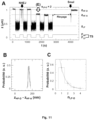
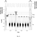
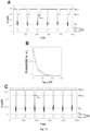
- the inventors have surprisingly demonstrated that, when the DNA molecule is used to detect and/or measure molecular interactions, a difference in length of the first double-stranded DNA molecule (1) with respect to the second molecule of double-stranded DNA (2) makes it possible to better differentiate the signals which result from a specific interaction between two molecules to be tested from the signals which result from a non-specific interaction (ie between a support and a molecule to test (see for example the Picture 16 and Example 11).
- the length of a DNA molecule is generally expressed in number of nucleotides or base pairs (bp), it can also be expressed according to its physical length, for example in nanometers or micrometers (1 base pair corresponds at about 0.32 nm).
- the length of a DNA molecule is expressed here in base pairs.
- the first double-stranded DNA molecule (1) and/or the second double-stranded DNA molecule (2) can be 300 to 5000 base pairs in length, 500 to 5000 bases, or 650 to 1500 base pairs.
- the total length of double-stranded DNA molecules (1) and (2) can be between 600 and 10,000 base pairs, between 1,000 and 10,000, or between about 1,300 and 3,000 base pairs. ( ⁇ 50 base pairs).
- the first double-stranded DNA molecule (1) and/or the second DNA molecule (2) has a length of 300 to 5000 base pairs, from preferably 650 to 1500 base pairs. According to a particular embodiment, the first double-stranded DNA molecule (1) and the second double-stranded DNA molecule (2) each have a length of 1500 base pairs. According to another particular embodiment, the first double-stranded DNA molecule (1) has a length of 650 base pairs while the second DNA molecule (2) has a length of 1350 base pairs.
- the total length of the double-stranded DNA molecules (1) and (2) is between 600 and 10,000 base pairs, between 1,000 and 10,000, or between 1,300 and 3,000 base pairs. According to a particular embodiment of the invention, the total length of the double-stranded DNA molecules (1) and (2) is between 2050 base pairs and 3000 base pairs, advantageously approximately 2050 or d approximately 3,000 base pairs ( ⁇ 50 base pairs).
- the DNA is selected so as not to include elements that could interact with one or more of the molecules to be tested.
- a molecule to be tested is a protein capable of interacting with a double-stranded DNA molecule having a particular sequence and/or structure (e.g. MutS, Z ⁇ )
- the double-stranded DNA molecule -strand according to the invention does not include these sequences and/or structures (e.g. absence of mismatch, no Z-DNA type structure) except on the site designed to accommodate the second molecule to be tested (e.g. sequence with a mismatch, Z -DNA).
- the double-stranded DNA molecule may in particular comprise part (eg 0.6, 1, 5, 20, 30, 40, 50, 60, 70, 80 or 90%) or all (100%) of a double-stranded DNA genome of a group I virus, such as a genome of a virus of the order Caudovirales, Herpesvirales , or Ligamenvirales, more particularly of the family Siphoviridae.
- a group I virus such as a genome of a virus of the order Caudovirales, Herpesvirales , or Ligamenvirales, more particularly of the family Siphoviridae.
- the double-stranded DNA molecule comprises part or all of a double-stranded DNA genome of a bacteriophage, such as lambda phage, Mycoplasma phage P1, phage c2 from Lactococcus phage c2, phage F108 from Pasteurella , or any other phage double-stranded DNA genome.
- the length of the genome incorporated into the double-stranded DNA molecule according to the invention can be between approximately 11,500 and approximately 50,000 base pairs ( ⁇ 50 base pairs).
- the double-stranded DNA molecule according to the invention comprises an entire genome, said genome is advantageously in linear form.
- a double-stranded DNA molecule is in circular form (eg a circular genome, a plasmid), it is advantageously linearized before being incorporated into the double-stranded DNA molecule according to the invention.
- the double-stranded DNA molecule according to the invention can be continuous or discontinuous.
- a DNA molecule is discontinuous when at least one phosphodiester, phosphorothioate, phosphoramidate or phosphorodiamidate bond linking two nucleotides located next to each other on a single strand is absent, or when the DNA molecule comprises a single-stranded DNA region.
- a DNA molecule may have one or more discontinuity sites on both strands, provided that the discontinuity sites are not present between two paired nucleotides on both strands.
- the double-stranded DNA of the first double-stranded DNA molecule (1) and/or of the second double-stranded DNA molecule (2) is advantageously continuous.
- the double-stranded DNA molecule according to the invention comprising a first double-stranded DNA molecule (1) linked to a second double-stranded DNA molecule (2) by a covalent bond which is not a phosphodiester, phosphorothioate, phosphoramidate or phosphorodiamidate bond is continuous.
- the continuity of the molecule advantageously makes it possible to guarantee the mechanical properties of the double-stranded DNA molecule, including its persistence length and its ability to be supercoiled .
- any site of discontinuity in the double-stranded DNA molecule according to the invention is closed, for example by a ligase during the manufacture of said double-stranded DNA molecule, before its use.
- the double-stranded DNA molecule according to the invention is perfectly matched.
- the double-stranded DNA molecule according to the invention more particularly comprises at least two distinct double-stranded DNA molecules, called here the double-stranded DNA molecules (1) and (2), which are connected to each other by at least one covalent bond.
- covalent bond is meant here a chemical bond in which at least one pair of electrons is shared between two atoms.
- at least one covalent bond is formed between an atom of a first functional group and an atom of a second functional group.
- a covalent bond can be one or more amide or ester bond(s), or a disulphide bridge.
- said covalent bond can also comprise other elements, such as one or more polymers, cyclic compounds, or particles, which can link the two subunits by forming a covalent bond with each sub- unit.
- said at least one covalent bond is not a phosphodiester bond.
- a phosphodiester bond is a bond between the 3' carbon of a first deoxyribose and the 5' carbon of a second deoxyribose by forming a phosphoester bond of each of the carbons with a phosphate group.
- a phosphodiester bond is more particularly a bond between two deoxyribonucleotides, as known by those skilled in the art.
- said at least one covalent bond is not a phosphorothioate bond.
- One such bond is a phosphodiester bond where a sulfur substitutes a non-binding oxygen in the phosphate group.
- said at least one covalent bond is not a phosphoramidate bond or a phosphorodiamidate bond.
- a phosphoramidate bond is a phosphodiester bond in which the phosphate group includes a phosphorus bonded to three oxygen atoms and one nitrogen atom.
- a phosphorodiamidate bond is a phosphodiester bond in which the phosphate group includes a phosphorus bonded to two oxygen atoms and two nitrogen atoms.
- the phosphorothioate, phosphoramidate and phosphorodiamidate linkages are mainly found in synthetic nucleic acids (such as those described above), to which they confer advantageous properties. By way of example, these bonds generally confer increased stability on the nucleic acids in which they are found.
- a covalent bond other than a phosphodiester bond, a phosphorothioate bond, a phosphoramidate bond or a phosphorodiamidate bond can be a bond formed on the basis of click chemistry, such as a bond formed by the cycloaddition of an azide to an alkyne (see, for example, Hein et al., 2008).
- said covalent bond linking the two double-stranded DNA molecules (1) and (2) is formed by a reaction between an azide function and an alkyne function, between an azide function and a dibenzocyclooctyne (DBCO) function, between a tetrazine function and a transcyclooctene function, between a thiol function and an alkyne function, between a thiol function and an alkene function, between a thiol function and a thiol function (thus generating a disulphide bridge), between a thiol function and a maleimide function, between an amine function and an activated acid (eg by reagents such as N-hydroxysuccinimide, N , N' -dicyclohexylcarbodiimide or 1-ethyl-3-(3-dimethylaminopropyl)carbodiimide), between a hydroxyl function and a maleimide, and/or between an DB
- said first double-stranded DNA molecule (1) is linked to said second double-stranded DNA molecule (2) by a tether.
- loin is meant the sense of the present invention a separate third molecule, which is linked to both the first (1) and the second (2) double-stranded DNA molecule.
- the lanyard makes it possible to keep the two molecules to be tested close to one of the another after their dissociation.
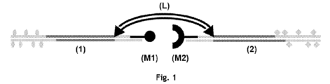
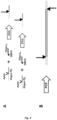
- the double-stranded DNA molecule according to the invention advantageously comprises at least two covalent bonds which are not phosphodiester, phosphorothioate, phosphoramidate or phosphorodiamidate bonds (see, for example, the Figure 1 ).
- said lanyard may be composed of several distinct molecules, which are themselves advantageously linked to each other by covalent bonds which are not phosphodiester, phosphorothioate, phosphoramidate or phosphorodiamidate bonds.
- said first double-stranded DNA molecule (1) is therefore linked to said second double-stranded DNA molecule (2) by a lanyard.
- the lanyard can be composed of any type of material and/or comprise any type of molecule, such as polypeptides, polynucleotides, such as double-stranded DNA, and/or polymers.
- the lanyard is a polymer composed of at least 50%, 60%, 70% of deoxyribonucleotides, advantageously at least 75%, 80%, 85%, more advantageously at least 90%, 95 %, 96%, 97%, 98%, even more advantageously at least 99% of deoxyribonucleotides.
- the lanyard is a polymer composed of deoxyribonucleotides and one or more other polymers such as polyethylene glycol (PEG), propylene glycol, a polyamide, and/or one or more nucleic acid regions single-stranded (eg single-stranded DNA).
- the lanyard is a polymer composed entirely (ie 100%) of deoxyribonucleotides.
- said double-stranded DNA may comprise any characteristic as detailed above.
- the double-stranded DNA lanyard is continuous.
- the length of said lanyard is at least 150 base pairs, more preferably at least 200, 300, 400, 500 or 600 base pairs.
- such a length in double-stranded DNA allows the molecules to be tested to meet with significant efficiency and limits the mechanical stresses applying to the complex once that has been formed.
- the loin comprises double-stranded DNA having a length of at least 150 base pairs, preferably at least 600 base pairs, said double-stranded DNA being continuous (ie without a nick).
- the loin When the loin is a double-stranded nucleic acid molecule (eg a double-stranded DNA or RNA molecule), it itself comprises phosphodiester, phosphorothioate, phosphoramidate or phosphorodiamidate bonds.
- the at least one covalent bond between the loin and said first or second double-stranded DNA molecule (1) or (2) is not a phosphodiester, phosphorothioate, phosphoramidate or phosphorodiamidate bond .
- said lanyard may itself comprise covalent bonds (eg “ crosslinking ” between the two strands of a double-stranded DNA molecule, when the lanyard is a double-stranded DNA polymer). This is particularly advantageous when forces greater than 70 pN will be applied to the double-stranded DNA molecule.
- the loin is a double-stranded DNA molecule.
- the double helix structure of a double-stranded DNA lanyard is only present in linear form. The lanyard cannot therefore fold into any tertiary structure.
- the loin is a double-stranded DNA molecule, it is stable and is therefore not very inclined to interact with one or more of the molecules to be tested.
- the length of the lanyard is also advantageously easily adjustable.
- double-stranded DNA molecules have a persistence length of approximately 50 nm under usual laboratory conditions.
- persistence length is meant a mechanical property making it possible to characterize the rigidity of a linear polymer (Doi, 1996, and Bouchiat et al., 1999). More specifically, it is the typical length over which a linear polymer can remain aligned despite deformations due to thermal agitation. With such a persistence length, a variation in the length of the loin translates into a consequent variation in the average distance between the junctions with the double-stranded DNA molecules (1) and (2), which makes it possible to adjust the effective concentration of molecules to be tested, C eff .
- the C eff is of the order of nM.
- ⁇ A characteristic times of association
- the persistence length of the double-stranded DNA lanyard also makes it possible, when the double-stranded DNA molecule is used to measure interactions between at least two molecules to be tested, to probe the dependence of the life time of a molecular complex (e.g. between two molecules to be tested) as a function of physical force. Indeed the double-stranded DNA molecule stretches easily, which makes it possible to measure the difference in extension between the associated and dissociated conformations of molecules to be tested when weak forces, below approximately 2 pN, are applied. .
- the length of the tether is an important parameter in the measurement of interactions between molecules and it can vary according to experimental needs (i.e. according to the purpose of the experiment, according to the lengths of the double-stranded DNA molecules (1) and (2) and/or molecules to be tested).
- a tether that is too short e.g. less than 300 bp
- an increase in tether length reduces the frequency at which interactions between test molecules are observed. This can be useful for the study of reactions where the association is made very quickly, but can be prohibitive for the study of reactions where the association is made slowly.
- the lanyard can have a length of about 300 to about 50,000 base pairs ( ⁇ 50 base pairs).
- the double-stranded DNA lanyard has a length of approximately 300 to approximately 50,000 base pairs, advantageously approximately 500 to 10,000 base pairs, advantageously 1,000 to 10,000 base pairs, from 600 to 3,000 base pairs ( ⁇ 50 base pairs), more preferably from 600 to 1,000 base pairs, even more preferably from about 700 base pairs ( ⁇ 50 base pairs) or from about 6,000 base pairs ( ⁇ 50 base pairs).
- the lanyard is sufficiently long, for example when its length is 6000 kbp, the LF-A z-distance can be easily distinguished from the LF-D z-distance.
- the lanyard may be attached to the first molecule (1) and/or to the second molecule (2) of double-stranded DNA by one or more covalent bonds which are not phosphodiester, phosphorothioate, phosphoramidate or phosphorodiamidate bonds.
- at least one non-phosphodiester, non-phosphorothioate, non-phosphoramidate or non-phosphorodiamidate covalent bond is formed between a nucleotide of the lanyard comprising a functional group and a nucleotide of the first molecule (1) and/or of the second molecule (2), comprising another functional group.
- the first double-stranded DNA molecule (1) can comprise an azide function which reacts with a DBCO function present on the lanyard by the click chemistry technique, thus forming a non-covalent covalent bond.
- a DBCO function present on the lanyard by the click chemistry technique
- the loin can be attached to the double-stranded DNA molecules (1) and (2) by its two ends, respectively, or by nucleotides located in the intermediate region of the loin.
- intermediate region is meant within the meaning of the present invention a segment of a molecule which is located between the two ends of said molecule.
- Such intermediate regions are notably schematized for the DNA molecules (1) and (2), by way of example, in the Figure 1 (segments in dark gray).
- end is meant, within the meaning of the present invention, the 30 to 150 base pairs located at the end of a molecule (eg of the loin, of the first molecule of double-stranded DNA (1), of the second double-stranded DNA molecule (2), advantageously the 30 to 50 base pairs located at the end of a molecule.
- Such ends are in particular schematized for the DNA molecules (1) and (2 ), for example, in the Figure 1 (segments in light gray).
- a lanyard or the first or second double-stranded DNA molecule (1) or (2)
- comprising 1,000 base pairs comprises two ends, a first end corresponding to the pairs 1 to 150 base pairs and a second end corresponding to 850 to 1000 base pairs.
- first end and second end should not be interpreted as corresponding to specific ends of the lanyard, but simply allow distinguish one end of the loin from the other. Thus, there is no concept of direction or orientation associated with said first and second ends. Those of skill in the art will easily be able to identify the ends of a double-stranded DNA molecule as defined herein.
- a bond between one end of a lanyard and a double-stranded DNA molecule (1) or (2) can therefore take place at one or more nucleotide bases of one end of a lanyard.
- at least one ultimate nucleotide, preferably an ultimate base, sugar or phosphate, of one end of the lanyard is attached by a non-phosphodiester, non-phosphorothioate, non-phosphoramidate or non-phosphorodiamidate covalent bond, to the first or second double-stranded DNA molecule (1) or (2).
- the lanyard is attached by its first end to the first molecule of double-stranded DNA (1) and by its second end to the second molecule of double-stranded DNA (2), advantageously by an ultimate nucleotide, preferably an ultimate base, sugar or phosphate, from each end of the loin.
- the lanyard is attached by at least two nucleotides, preferably at least two nucleotide bases, in each end to the first and the second double-stranded DNA molecule (1) and (2), respectively, said at least two nucleotide bases comprising, more advantageously, an ultimate nucleotide, preferably an ultimate base, sugar or phosphate, at each end of the lanyard.
- the lanyard can be attached to the first double-stranded DNA molecule (1) and/or to the first double-stranded DNA molecule (2) by at least one base located in the region intermediate of the lanyard, said bases being advantageously separated by at least 300 base pairs, more advantageously by at least 500 base pairs, even more advantageously by at least 600 base pairs.
- the lanyard can be attached to the first double-stranded DNA molecule (1) by at least one first base, said base being located between the central point of the lanyard and its first end and / or attached to the second double-stranded DNA molecule (2) by at least one second base, said base being located between the central point of the lanyard and its second end.
- central point is meant a location which is substantially equidistant between the first and the second end of the molecule. This is particularly advantageous when at least one end of the lanyard comprises another element of the double-stranded DNA molecule according to the invention (eg is linked to another molecule to be tested).
- the site of the covalent bond between the loin and the first double-stranded DNA molecule (1) and/or the second double-stranded DNA molecule (2) is located in the intermediate region of said molecule of double-stranded DNA (1) or (2), that is to say between the two ends of said DNA molecule (1) or (2).
- a non-phosphodiester, non-phosphorothioate, non-phosphoramidate or non-phosphorodiamidate covalent bond is formed between an end of the loin (or any other point of the loin which will be appropriate, as described above), and a central point of said double-stranded DNA molecule (1).
- the non-phosphodiester, non-phosphorothioate, non-phosphoramidate or non-phosphorodiamidate covalent bond between the loin and the said first molecule of double-stranded DNA (1) thus forms a junction with three branches (see for example the Figure 1 ).
- a covalent bond between the lanyard and the first double-stranded DNA molecule (1) and/or the second double-stranded DNA molecule (2) can be located in one end of said DNA molecule.
- double-stranded DNA (1) or (2), or to a nucleotide base which makes it possible to distinguish the end of the double-stranded DNA molecule (1) or (2) from its intermediate region (see, for example, the Figure 1 ).
- no covalent bond is advantageously formed between an ultimate nucleotide, preferably an ultimate base, sugar or phosphate, of the DNA molecule (1) or (2) and any base of the loin.
- the lanyard is attached to the first double-stranded DNA molecule (1) by a first covalent bond between the first end of said lanyard and an intermediate region of the first molecule of double-stranded DNA (1) and to the second molecule of double-stranded DNA (2) by a second covalent bond between the second end of said lanyard and an intermediate region of the second molecule of double-stranded DNA ( 2).
- the ends of the double-stranded DNA molecules (1) and (2) are not attached to the lanyard, they advantageously remain available for other purposes (e.g. for other interactions).
- the two ends of the first double-stranded DNA molecule (1) or of the second double-stranded DNA molecule (2) can in particular be bound respectively to a molecule to be tested and to a support .
- a first end of said first double-stranded DNA molecule (1) is linked to a first molecule to be tested and a first end of said second double-stranded DNA molecule (2) is linked to a second molecule to be tested.
- the molecule to be tested can be linked directly or indirectly to the end.
- the binding of a first or of a second molecule to be tested at a first end of the first or of the second molecule of double-stranded DNA (1) or (2) is advantageous because it makes it possible to distance the molecules to test the loin and the rest of the double-stranded DNA molecule (1) and/or (2).
- the molecules to be tested are in particular less likely to undergo steric hindrance and to see the characteristics of their interaction modified in this configuration.
- a double-stranded DNA molecule according to the invention makes it possible to leave the lanyard and the double-stranded DNA molecules (1) and (2) relatively free to reorient themselves in space independently of each other, such that the molecules to tested are not deprived of any degree of freedom of movement.
- the binding of the molecules to be tested to the ends of the double-stranded DNA molecules (1) and (2) also makes it possible to distance the molecules to be tested from the supports, which reduces non-specific interactions.
- a molecule to be tested can be linked directly or indirectly to an ultimate nucleotide, preferably an ultimate base, sugar or phosphate, of one end of the double-stranded DNA molecule (1) or ( 2).
- the spacer can correspond, for example, to a sequence of double-stranded or single-stranded nucleotide bases, to a polymer, a peptide, or a small molecule, and can be easily selected by those skilled in the art.
- the addition of spacers to the double-stranded DNA molecules (1) and (2) can be done with any of the methods commonly used in molecular biology.
- At least one molecule to be tested is linked directly or indirectly to an ultimate nucleotide, preferably an ultimate base, sugar or phosphate, of an end of a DNA molecule given double-stranded.
- a molecule to be tested is linked indirectly to an ultimate nucleotide, preferably an ultimate base, sugar or phosphate, of one end of a DNA molecule (1) or (2) , advantageously by a spacer.
- the two molecules to be tested are specifically bound to the first and second double-stranded DNA molecules (1) and (2), respectively.
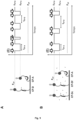
- one end of the first double-stranded DNA molecule (1) and one end of the second double-stranded DNA molecule (2) are digested with one or more nucleases, for example the enzyme Nb.BbvCI, in order to generate two different cohesive ends (see also the Figure 5A ).
- nucleases for example the enzyme Nb.BbvCI
- each molecule to be tested can be specifically linked to one end of the double-stranded DNA molecule (1) or (2) by a covalent bond, for example a non-phosphodiester, non-phosphorothioate covalent bond. , non-phosphoramidate or non-phosphorodiamidate, according to the methods described here, for example by the click chemistry technique.
- At least one of the molecules to be tested is selected from the group consisting of the following molecules: polymers, amino acids, peptides, polypeptides, proteins , nucleosides, nucleotides, polynucleotides, oligonucleotides, sugars, polysaccharides, small molecules, drugs, aptamers, antigens, antibodies, lipids, lectins, hormones, vitamins, viruses, virus fragments, nanoparticles, cell surface molecules, and transcription factors, or analogs or peptidomimetics of any of these.
- the first and the second molecule to be tested are of the same nature (e.g.
- the first molecule to be tested is of a different nature from the second molecule to be tested (e.g. antigen/antibody, virus/receptor, transcription factor/DNA, protein-small molecule, etc.).
- the second molecule to be tested e.g. antigen/antibody, virus/receptor, transcription factor/DNA, protein-small molecule, etc.
- said molecule is advantageously linked directly to the end of one of the DNA molecules (1) or (2).
- one or more other molecules to be tested can be linked to the double-stranded DNA molecule according to the invention.
- one or more other molecules to be tested can also be chosen from the group of molecules above.
- a third molecule to be tested can be linked to one end of a double-stranded DNA molecule (3) or to one end of the lanyard when the latter is free.
- at least one other molecule to be tested can be brought into contact with the double-stranded DNA molecule according to the invention comprising at least two molecules to be tested, for example in an aqueous suspension.
- said third molecule can be a cofactor of the first and/or of the second molecule to be tested.
- said first and/or said second molecule to be tested is selected from the group consisting of the following molecules: polymers, amino acids, peptides, polypeptides, proteins, nucleosides, nucleotides, polynucleotides, oligonucleotides, sugars, polysaccharides, small molecules, drugs, aptamers, antigens, antibodies, lipids, lectins, hormones, vitamins, viruses, virus fragments, nanoparticles, cell surface molecules, and transcription factors.
- said first molecule to be tested is of a different nature from said second molecule to be tested.
- said first molecule to be tested is of the same nature, or even is identical, to said second molecule to be tested.
- the first and second double-stranded DNA molecules (1) and (2) can be specifically attached to supports.
- support is meant any type of solid surface or substrate, said support possibly being advantageously functionalized in order to be able to react with a functionalized end of the double-stranded DNA molecule according to the invention.
- the support may be a bead (eg silica beads, controlled pore glass, magnetic beads, biomagnetic separation beads such as Dynabeads ® , Wang resin, Merrifield resin, chloromethylated copolystyrene resin - divinylbenzene (DVB), Sephadex ® / Sepharose ® beads, cellulose beads, etc.), a flat support such as a fiberglass filter, a dielectric surface (eg glass, silica, silicon nitride, alumina), a surface metal (eg steel, gold, silver, aluminum, and copper), a semiconductor surface (eg silicon, III-V semiconductor, II-VI semiconductor), plastic materials, including multiwell plates or membranes ( eg polyethylene, polypropylene, polyamide, polyvinylidene fluoride), a needle, a micropipette, or a cantilever used in atomic force microscopy.
- a ball according to the invention can have any three-dimensional structure and any size.
- the supports are advantageously solid substrates (for example a glass surface such as a microscope slide, a micropipette, a microparticle), which can be of the same type (i.e. two microparticles) or of different type (e.g. a glass surface and a microparticle).
- a glass surface such as a microscope slide, a micropipette, a microparticle
- the second end of said first DNA molecule double- strand (1) is bound to a first support and the second end of said second double-stranded DNA molecule (2) is bound to a second support.
- At least one of the two supports is a ball such as a microbead, a microparticle, a glass surface, a micropipette, or a cantilever used in atomic force microscopy.
- the DNA can be linked directly to a support, for example a microbead, which involves a functionalization of this support, for example by coating it with streptavidin, a polymer bearing COOH groups, etc., capable of reacting with a functionalized end of double-stranded DNA.
- a support for example a microbead, which involves a functionalization of this support, for example by coating it with streptavidin, a polymer bearing COOH groups, etc., capable of reacting with a functionalized end of double-stranded DNA.
- Such methods generally require functionalizing a double-stranded DNA molecule, in particular at one of its ends, that is to say grafting at least one appropriate functional group onto it.
- different procedures can be adopted.
- the simplest way is to functionalize, using synthetic oligonucleotides, one end of the first double-stranded DNA molecule (1) with a functional group and one end of the second molecule of double-stranded DNA (2) with a functional group.
- the end of the first DNA molecule (1) is functionalized with a first functional group and the second DNA molecule (2) is functionalized with a second functional group, which makes it possible to attach one end to each of the two supports which have been pretreated in a different way (e.g. a first end functionalized with biotin attaches to a support covered with streptavidin while a second end, functionalized with an amine group, attaches to a support coated with a polymer bearing COOH groups, respectively).
- the advantage of this method lies in its ability to functionalize any type of double-stranded DNA molecule having any length while keeping the same reagents.
- the two ends of the double-stranded DNA molecules (1) and (2) intended to be attached to supports are cleaved using for example two (or more) restriction enzymes, which makes it possible to obtain a first DNA molecule having a first restriction site at one of its ends and a second DNA molecule having a second restriction site at one of its ends. This allows the two ends to be treated differently.
- a spacer after which a functional group is added to one end of the first double-stranded DNA molecule (1) and/or to one end of the second molecule double-stranded DNA (2); the two spacer sequences thus giving each functional group additional space to bind to its dedicated support.
- the spacer may correspond to a double-stranded or single-stranded nucleotide base sequence, a polymer, a peptide, or a small molecule, as described above.
- the spacer advantageously comprises at least one functional group. Addition of spacers to double-stranded DNA molecules (1) and (2) can be done with any of the commonly used methods in molecular biology. These methods are well known to those skilled in the art and there is therefore no need to detail them here.
- attachment techniques themselves, they are numerous and they derive from techniques for attaching macromolecules (proteins, DNA, etc.) to pretreated supports which are commercially available or easily carried out in the laboratory. Most of these techniques have been developed for immunology tests, and link proteins (immunoglobulins) to supports bearing groups (-COOH, -NH2, -OH, etc.) capable of reacting with the carboxyl (-COOH ) or the amine ends (-NH2) of proteins.
- the attachment of the double-stranded DNA molecule according to the invention to a support can be carried out directly, via the free phosphate of the 5' end of the molecule, which reacts with a secondary amine (Covalink-NH surface marketed by Polylabo in France) to form a covalent bond. It is also possible to functionalize DNA with an amine group and then proceed as with a protein. As an alternative example, a DNA molecule functionalized with a thiol (S-H) group covalently binds to a gold support by the formation of an S-Au thiolate bond.
- S-H thiol
- the patent application WO 92/16659 proposes a method using a polymer to fix DNA.
- a DNA molecule can be attached directly to a support but, if necessary, and in particular with a view to limiting the influence of the supports, the DNA molecule can be attached to the end of an inert arm of a natural nature. peptide or other (in other words, a spacer), such as for example that is described in the patent EP 329 198 . Since the two double-stranded DNA molecules (1) and (2) are linked to different supports, they can be linked to their respective supports by different means.
- said second ends of said first and second double-stranded DNA molecules (1) and (2) are attached to said first and second supports directly or indirectly.
- at least one of the two supports is a mobile support, advantageously a ball, more advantageously a magnetic ball.
- said second ends of said first and second double-stranded DNA molecules (1) and (2) are attached to said first and second supports at a single point or at several points.
- said second ends of said first and second double-stranded DNA molecules (1) and (2) are attached to the first and to the second supports respectively by a nucleotide base, said base being advantageously functionalized.
- said second end of said first and second double-stranded DNA molecules (1) and (2) are attached by at least two nucleotide bases, said bases being more advantageously functionalized.
- the double-stranded DNA molecule according to the invention comprises two molecules to be tested.
- said molecule may comprise at least three , four, five, or six molecules to be tested.
- Said molecules to be tested could in particular be bound to the ends of the loin or to other molecules of double-stranded DNA (e.g. a third molecule of double-stranded DNA (3) , a fourth double-stranded DNA molecule (4), etc.
- said molecules are also attached to the lanyard, advantageously to different bases.
- the double-stranded DNA molecule as described here, for use in the detection and/or characterization of the interactions between at least two molecules to be tested, advantageously the determination of the thermodynamic properties and/or kinetics of these interactions.
- the characterization of the interaction comprises the determination of at least one of the characteristics chosen from: the characteristic association time, the characteristic dissociation time, the dissociation rate constant, the dissociation activation energy, the distance between the transition state of the complex during dissociation, and the dissociation equilibrium constant.
- the double-stranded DNA molecule is used for the characterization of at least one molecular interaction, advantageously chosen from the characteristics described above, between at least two molecules to be tested.
- Another object of the invention is a device comprising the double-stranded DNA molecule as described here with its supports.
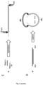
- An example of such a device is particularly illustrated in the Figure 2 .
- the device according to the invention is used to detect and/or measure interactions, advantageously to measure thermodynamic and/or kinetic properties, between at least two molecules to be tested, such as those described above.
- characterization of an interaction is carried out at the level of a single molecule, it is possible to measure individual interactions simultaneously, within at least two different double-stranded DNA molecules.
- a detection method having a sufficiently high level of resolution to distinguish between the interactions taking place within the different molecules of double-stranded DNA can in particular be used.
- several double-stranded DNA molecules can be bound to the same support.
- the distribution of the double-stranded DNA molecules can be done regularly, for example, on a network or chip type support, or randomly, preferably at a density allowing the interactions within each molecule to be measured. of double-stranded DNA separately.
- the double-stranded DNA molecules can be linked to the same fixed support and to the same mobile support (e.g. a multicantilevers system, comprising for example a glass surface as a fixed support and a network of microfabricated cantilevers on the same substrate as a mobile medium).
- the double-stranded DNA molecules can be bound to the same fixed support and to different mobile supports (e.g. magnetic tweezers, comprising for example a glass surface as a fixed support and beads as supports). mobile, each double-stranded DNA molecule being bound to a different bead).
- the double-stranded DNA molecules can be bound to different fixed supports and to different mobile supports (e.g. multiplexed optical tweezers, including for example beads as fixed supports and as mobile supports, each molecule of double-stranded DNA being linked to a different pair of beads).
- multiplexed optical tweezers including for example beads as fixed supports and as mobile supports, each molecule of double-stranded DNA being linked to a different pair of beads.
- the double-stranded DNA molecule according to the invention is attached in a specific manner between two solid supports, one of the ends of the first double-stranded DNA molecule (1) being able to be fixed directly or indirectly to a support, while one of the ends of the second double-stranded DNA molecule (2) is attached directly or indirectly to a mobile support.
- One of the ends of the first double-stranded DNA molecule (1) can more particularly be fixed directly or indirectly to a fixed or mobile support.
- Said device according to the invention advantageously comprises at least two double-stranded DNA molecules according to the invention, said molecules being linked to the supports, the different double-stranded DNA molecules being able to be linked to the same supports and/or to supports different.
- said molecules are bound at a density allowing each individual molecule to be individually resolved, advantageously wherein each individual molecule is or can become spatially addressable.
- the molecules to be tested and therefore the molecular interactions to be characterized in the at least two double-stranded DNA molecules may be different.
- the characterization of different interactions is particularly advantageous when a large number of molecules to be tested must be evaluated, for example during a screening intended to identify a molecule interacting with a particular receptor or modulating the interaction between two proteins.
- different molecules to be tested can be linked to one of the DNA molecules (1) or (2) or introduced into an aqueous solution to characterize the interactions between said molecule and the molecule(s) to be tested linked to the double-stranded DNA molecule. In some cases, it is possible that no molecular interaction is detected between the molecules to be tested.
- the DNA molecule according to the invention can be used in the context of high-throughput studies, for example, during pharmaceutical screening for the search for new molecules having particular interaction characteristics with a particular molecule (eg a receptor, a protein complex).
- a particular molecule eg a receptor, a protein complex
- different drugs can interact with the same receptor with different kinetics, causing different effects.
- different drugs can interact with protein assemblies and modulate their stability.
- the use of the present device is very advantageous for the determination of various kinetic and/or thermodynamic characteristics.
- the same molecular interaction can be characterized in parallel for each DNA molecule, when these comprise the same molecules to be tested.
- This makes it possible in particular to average the measurements, and thus advantageously to improve the precision.
- This also makes it possible to ensure the reproducibility of the measurements.
- the at least two molecules of double-stranded DNA comprise the same molecules to be tested, they are advantageously linked to the two supports according to the same orientation (e.g. the first molecule to be tested is linked to the first molecule of double-stranded DNA ( 1), itself bound to a first support for each double-stranded DNA molecule).
- Another aspect of the invention relates to an “ intermediate ” molecule from which the double-stranded DNA molecule according to the invention can be obtained.
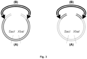
- Another subject of the invention is therefore a double-stranded DNA molecule comprising a first double-stranded DNA molecule (A) and a second double-stranded DNA molecule (B), said double-stranded DNA molecule strand (A) comprising a cleavage site which is present only in said double-stranded DNA molecule (A), said double-stranded DNA molecule (A) being joined to the double-stranded DNA molecule (B ) by two covalent bonds which are not phosphodiester, phosphorothioate, phosphoramidate or phosphorodiamidate bonds, on either side of said cleavage site. Examples of such an “ intermediate ” molecule are notably illustrated in the Figure 3 .
- first ends of the double-stranded DNA molecules (1) and (2) are different from the second ends of the double-stranded DNA molecules (1) and (2).
- first and second end we refer to the two different ends within the double-stranded DNA molecule (1) or (2).
- first and second ends should not be interpreted as corresponding to specific ends, but simply to make it possible to differentiate one end of a molecule from the other end. Thus, there is no concept of direction or orientation associated with said first and second ends.
- the four ends of the double-stranded DNA molecule are different.
- the double-stranded DNA molecule (A) advantageously has a length of between approximately 600 and 10,000 base pairs, advantageously between approximately 1,000 and 4,000 base pairs, advantageously between approximately 1,500 and 3,000 base pairs ( ⁇ 50 base pairs).
- the double-stranded DNA molecule (B) advantageously has a length of between about 300 to about 50,000 base pairs, advantageously chosen according to the purpose of the experiment and the molecules to be tested from about 500 to 10,000 base pairs. bases, preferably 600 to 3000 base pairs, more preferably 600 to 1000 base pairs, even more preferably about 700 base pairs ( ⁇ 50 base pairs).
- cleavage site is meant within the meaning of the present invention a polynucleotide structure or sequence which is capable of being cleaved in a specific manner by a cleavage agent, such as a restriction enzyme, a nuclease, a nickase, a ribozyme, DNAzyme, and fragments thereof.
- a cleavage agent such as a restriction enzyme, a nuclease, a nickase, a ribozyme, DNAzyme, and fragments thereof.
- the cleavage site can therefore be a restriction enzyme site, a ribozyme site, a nickase site, a DNAzyme site or a nuclease cleavage site.
- agents and sites are well known to those skilled in the art.
- restriction enzyme is meant more particularly an enzyme which cuts double-stranded DNA at or near a specific nucleotide sequence.
- the specificities of many restriction enzymes are well known in the art and many restriction enzymes are commercially available and their reaction conditions, the need for the presence of cofactors and other requirements established by the suppliers of enzymes are well known.
- the restriction enzyme can be a type II, type III or artificial restriction enzyme (such as zinc finger nuclease, transcription activator type effector nuclease (TALEN ), a mega-nuclease, or a CRISPR-like endonuclease), even more preferably a type II restriction enzyme.
- Type II restriction enzymes include categories IIP, IIS, IIC, IIT, IIG, IIE, IIF, IIG, IIM and IIB, as described for example in Pingoud and Jeltsch, 2001.
- the restriction enzyme can generate ends blunt ends (ie the two strands having the same length) or sticky ends (one strand being longer than the other strand, generally by a few nucleotides).
- the cleavage site in the double-stranded DNA molecule (A) of the " intermediate " double-stranded DNA molecule is a restriction enzyme site, advantageously a site of type II restriction enzyme, more advantageously two restriction sites, even more advantageously two restriction sites generating two different ends (eg a blunt end and a cohesive end, or two cohesive non-complementary ends).
- the cleavage site may consist of a SacI enzyme restriction site and an XbaI enzyme restriction site.
- the cleavage site comprises at least two different sites
- said sites may be separated from each other, for example by at least 1, at least 2, at least 3, at least 4, at least 5, at least 6, at least 12, at least 25, at least 50, at least 100, at least 200, at least 500, or at least 1000 base pairs.
- said sites are separated by at least 6 base pairs (“ Restriction Endonucleases Technical Guide ”, New England Biolabs).
- the cleavage step can be carried out by techniques well known to those skilled in the art, such as a double digestion with two restriction enzymes simultaneously or sequentially.
- the cleavage is carried out by at least one cleaving agent as defined above, more advantageously by at least one restriction enzyme as defined above.
- the cleavage is carried out by two different restriction enzymes making it possible to obtain two different cohesive ends, advantageously said cohesive ends being non-complementary.
- the two cleavage sites are separated from each other according to one of the embodiments described above.
- step a at least one additional step can be performed.
- a step could comprise, for example, a step of functionalizing one end of the first and/or of the second double-stranded DNA molecule (1) and (2), and/or the binding of a molecule to be tested at one end of the first and/or the second double-stranded DNA molecule (1) and (2).
- the process for manufacturing the double-stranded DNA molecule according to the invention further comprises, after step a), the following step: b) attaching at least one functionalized base to one end of said first double-stranded DNA molecule (1) and attaching another functional base to one end of said second double-stranded DNA molecule strand (2).
- the functionalized ends are those generated during step a).
- the functionalized ends are those already present in the double-stranded DNA molecule (A) of the “ intermediate ” double-stranded DNA molecule before step a).
- the end of the first double-stranded DNA molecule (1) and the end of the second double-stranded DNA molecule (2) are functionalized by two different functions, so that they can be linked to different supports.
- one end of the first double-stranded DNA molecule (1) is functionalized by the addition of at least one biotin group while one end of the second double-stranded DNA molecule -brin (2) is functionalized by the addition of at least one digoxigenin group.
- the biotinylated end can bind quasi-covalently to a surface or a support coated with streptavidin whereas the end functionalized by digoxigenin can bind to a surface or a support coated with antibodies directed against digoxigenin.
- the end of the first double-stranded DNA molecule (1) and/or the end of the second double-stranded DNA molecule (2) are functionalized by the addition of several groups in order to improve the attachment of the end to the support.
- the functional group(s) are present in a single-stranded or double-stranded DNA molecule which is incorporated at the end of the first DNA molecule.
- said at least one functionalized base is included in a third (3) and/or fourth double-stranded DNA molecule (4), said third double-stranded DNA molecule (3) advantageously being linked to the second end of said first double-stranded DNA molecule (1), and said fourth double-stranded DNA molecule (4) advantageously being linked to the second end of the second first double-stranded DNA molecule -strand (2).
- said at least one functionalized base of said double-stranded DNA molecule (3) is functionalized by a different functional group from said at least one functionalized base of said double-stranded DNA molecule (4).
- the double-stranded DNA molecule (1), (2), (3) and/or (4) has a length of between 600 and 10,000 base pairs, more advantageously between 650 and 1500 base pairs.
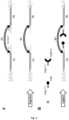
- the Figure 5 illustrates this step.
- the binding according to step c) comprises the binding of a fifth (5) DNA molecule comprising said first molecule to be tested to said first end of the first double-stranded DNA molecule (1), and/ or attaching a sixth (6) DNA molecule comprising said second test molecule to said first end of the second double-stranded DNA molecule (2).
- Said fifth (5) DNA molecule and/or sixth (6) DNA molecule comprising a molecule to be tested can advantageously be a double-stranded DNA molecule comprising an overhanging end or an oligonucleotide.
- the link according to step c) advantageously comprises hybridization and ligation steps in order to link the DNA molecules together.
- the molecules to be tested can be linked to said ends of the double-stranded DNA molecules (1) and (2) according to any technique known to those skilled in the art.
- Step c) can be carried out before or after step b).
- one of steps b) and c) is carried out with step a) (ie by adding the molecules to be tested or the functional groups to the ends already present in the precursor double-stranded DNA molecule ( A), and the other of steps b) and c) is carried out after step a), when the two double-stranded DNA molecules (1) and (2) as well as their second ends are generated.
- the first end of the double-stranded DNA molecule (1) and/or the first end of the double-stranded DNA molecule (2), included in the double-stranded DNA precursor molecule (A) have an identical end (ie the same cohesive or blunt end) at least one of the ends which is formed by the cleavage of the double-stranded DNA molecule (A).
- the ends to which the molecules to be tested will be attached are cleaved, more advantageously by a restriction enzyme, as described above. However, this step is not necessary when the ends concerned have already undergone a cleavage step beforehand (for example before step a) or before step b)).
- the molecules to be tested can be linked directly to the double-stranded DNA molecules (1) and (2) at blunt ends (for example according to the blunt-end ligation method).
- blunt end ligation comprises steps of dephosphorylation of the ends of the double-stranded DNA molecules (1) and (2) and of phosphorylation of the free end of the DNA molecules comprising the molecules to be tested before bringing the molecules into contact in an appropriate buffer and in the presence of a ligase.
- the attachment of at least one of the functionalized DNA molecules in step b) and/or the attachment of at least one of the molecules to be tested in step c) comprises steps of hybridization and ligation, advantageously by hybridization of two complementary cohesive ends (i.e. one being present on the double-stranded DNA molecule (1) or (2) and the other being present on a DNA molecule comprising at least one functionalized base or a molecule to be tested).
- the attachment of at least one of the functionalized DNA molecules in step b) and/or the attachment of at least one of the molecules to be tested in step c) comprises a conjugation step , advantageously between at least two functional groups, more advantageously according to methods of click chemistry.
- tag proteins makes superfluous the step of digestion of the ends, illustrated in Figure 5A .
- said first ends of the double-stranded DNA molecules (1) and (2) are different from the second ends of the double-stranded DNA molecules (1) and (2).
- the manufacturing process of the present invention uses biochemical synthesis techniques, and not an “ origami ” type assembly.
- the structure of the double-stranded DNA molecule according to the invention can be guaranteed to the base, and is therefore of very high quality.
- the inventors have shown in particular during supercoiling studies, that no phosphodiester, phosphorothioate, phosphoramidate or phosphorodiamidate bond is missing on the approximately 3600 bases which are included in the double-stranded DNA molecule exemplified in the 'Example 9 and the Picture 12 .
- the step of synthesizing the double-stranded DNA molecule (A) is carried out by PCR using the oligonucleotides (1) and (2) as primers.
- the molecule (A) is produced by PCR, advantageously followed by the enzymatic digestion of each of its ends by a restriction enzyme, advantageously by two different restriction enzymes.
- the step of synthesizing the double-stranded DNA molecule (A) comprises steps of hybridization and of ligation of the oligonucleotide (1) to the first end of a molecule of Double-stranded DNA, such as a phage genome, and oligonucleotide (2) at the second end.
- the double-stranded DNA molecule (A) comprises an overhang at each of its ends, advantageously comprises two different overhangs.
- the oligonucleotide (1) and the oligonucleotide (2) comprise DNA regions complementary to said overhangs.
- the attachment of the molecule (B) comprises the hybridization and the ligation of a fifth oligonucleotide on the junction of the oligonucleotide (1) and of a sixth oligonucleotide on the junction of the oligonucleotide (2), respectively, in order to generate overhangs on each junction (see for example, Figure 6C ).
- the double-stranded DNA molecule (B) is synthesized by PCR, more advantageously followed by the enzymatic digestion of each of its ends by a restriction enzyme, even more advantageously by two different restriction enzymes (see for example, Figure 6C ).
- the double-stranded DNA molecule (A) and/or the double-stranded DNA molecule (B) advantageously has at least one of the characteristics (e.g. length), as described above.
- another subject of the invention is the double-stranded DNA molecule comprising a first double-stranded DNA molecule (A) and a second double-stranded DNA molecule (B), said molecule (A) comprising a cleavage site which is present only in said molecule (A), said molecule (A) being linked to molecule (B) by two covalent bonds which are not phosphodiester, phosphorothioate, phosphoramidate or phosphorodiamidate bonds, on either side other of said cleavage site obtained by the method described above.
- the length of the first double-stranded DNA molecule (1), of the second DNA molecule (2), and of the lanyard can be easily adjusted during the manufacture of the double-stranded DNA molecule according to the manufacturing process as described here.
- adjusting the length of the lanyard makes it possible to modulate the effective concentration of molecules to be tested (also called “ C eff ”). This is also all the easier since the double-stranded DNA has a long persistence.
- the length of the lanyard can in particular be determined according to the length of the double-stranded DNA molecules (1) and (2), or vice versa, and according to the effective concentration of the molecules to be tested and/or the spatiotemporal resolution desired. Indeed, in certain cases, a reduction in the total length of the double-stranded DNA molecule according to the invention by a numerical factor N, increases the spatiotemporal resolution of the experiment by approximately the same factor N, said factor N being advantageously between 1 and 6.
- the loin may be advantageous to maintain a length of at least 300 bp. It may also be advantageous to maintain a length of the double-stranded DNA molecules (1) and (2) of at least 100 bp in order to avoid interactions between the supports and the molecules to be tested.
- a very short tether eg less than about 300 base pairs
- the only way to reduce the length of the construct is to shorten the length of the first and second DNA molecules (1) and ( 2).
- Another object of the invention is a method for detecting and/or characterizing at least one molecular interaction between at least two molecules to be tested, said molecules to be tested being advantageously linked to a double-stranded DNA molecule according to the invention.
- the determined characteristic can be, for example, of a thermodynamic or kinetic nature.
- a characteristic association time, a characteristic dissociation time, a dissociation rate constant, a dissociation activation energy, a distance separating the transition state of the complex during dissociation, and /or a dissociation equilibrium constant can be determined.
- the method will comprise a prior step of applying a physical force greater than the force F HF of step c).
- the method of the invention advantageously makes it possible to distinguish the extensions z LF-A and z LF-D .
- This is particularly the case when the lanyard has a length of at least 700 bp.
- it is particularly advantageous to use a lanyard of at least 6000 bp, which makes it possible to easily distinguish the extensions z LF-A and z LF-D .
- the lanyard has a length of at least 700 bp. Even more preferentially, the lanyard has a length of at least 6000 bp.
- steps a) to c) or a) to d) as described above can be repeated several times, in order to follow multiple cycles of dissociation/association.
- the repetition of the steps is very advantageous because it makes it possible to increase the number of measurements and thus to improve the statistics and the reliability of the characterization, in particular by filtering the results on reproducibility criteria.
- steps a) to c) of the method are repeated several times.
- the method for characterizing an interaction comprises adding at least one additional molecule in solution.
- Said molecule present in solution can be the same molecule as one of the molecules to be tested which are attached to the ends of the double-stranded DNA molecule according to the invention or a different molecule.
- the force applied during the characterization of an interaction by spontaneous fluctuation is at least 30 fN, preferably a force comprised between 30 and 60 fN.
- a “ physical force ” or “ force ” according to the invention corresponds to any influence which causes an object to undergo a certain change, whether as regards its movement, its direction or its geometric construction (eg its conformation). It will clearly appear to those skilled in the art that a force according to the invention is different from other physical parameters such as, for example, the temperature (which is a direct property of the material rather than an influence exerted on it). this).
- the physical forces according to the invention include forces such as friction, tension (also called “ tensile force "), rotation, normal force, fluid resistance force, applied force and elastic force. .
- the physical force according to the invention comprises a tensile force, advantageously the physical force according to the invention consists of a tensile force.
- the physical force according to the invention can also comprise a rotational force, advantageously a torque.
- the physical force can consist of a torque force.
- the physical force includes tension and rotation, preferably tension and torque.
- Strength is expressed here in picoNewtons (pN), unless explicitly stated otherwise.
- the " application " of such physical forces is well known to those skilled in the art, particularly in the context of the various devices in which the DNA molecule, or the device comprising the DNA molecule, according to the invention can be incorporated (see for example Woodside et al., 2006; patents US No. 7,052,650 And 7,244,391 ; WO 2011/147931 ; and Yang et al., 2016).
- extension is meant within the meaning of the present invention the distance between two ends of a polymer.
- the extension corresponds more particularly to the distance “z” measured between the supports to which the double-stranded DNA molecule according to the invention is attached.
- the extension is therefore less than or equal to the length of the double-stranded DNA molecule.
- the extension of the double-stranded DNA molecule is expressed in nm or ⁇ m.
- the distance z LF corresponds to the distance between two ends of the DNA molecule according to the invention, preferably the distance between the supports to which the double-stranded DNA molecule according to the invention is attached, when a low force F LF is applied.
- an applied force threshold which varies according to the molecules to be tested (generally between 0.01 and 0.4 pN)
- the distance z HF corresponds to the distance between two ends of the DNA molecule according to the invention, preferably the distance between the supports to which the double-stranded DNA molecule according to the invention is attached, when a high F HF force is applied.
- an applied force threshold which varies according to the molecules to be tested (generally between 0.5 and 70 pN)
- the supports are moved away from each other.
- two distances can be determined, corresponding to two states of extension, z HF-A and z HF-D .
- the distances z HF-A and z HF-D correspond to the distance between two ends of the DNA molecule according to the invention when a high force F HF is applied, and the molecules to be tested are associated ( z HF-A ) or dissociated (z HF-D ).
- the HF-D z-distance is greater than the HF-A z-distance, because the double-stranded DNA molecules (1) and (2) are linked to each other only by the tether (not by the molecules to be tested), see also diagram on Figure 8 , 10A .
- the conformation of the double-stranded DNA molecule can therefore be determined by direct measurements of its extension, for example, under a force, as described here.
- the different “z” distances are determined over time and as a function of the force applied. In some cases, it may be advantageous to measure the distances precisely (eg in nm).
- the extension of the DNA molecule double-strand when the molecules to be tested are associated corresponds approximately to the extension of a linear double-stranded DNA molecule of length the sum of the lengths of the double-stranded DNA molecules (1) and (2), to which a force F HF is applied.
- the extension of the double-stranded DNA molecule when the molecules to be tested are dissociated corresponds approximately to the extension of a linear double-stranded DNA molecule of length the sum of the length of the lanyard plus the sum of the lengths of the double-stranded DNA molecules (1) and (2) excluding the parts between the molecules to be tested and the junctions, to which is applied a force F HF .
- extension and length are almost proportional, which favors the establishment of correspondence between the different z.
- the extension can in particular be deduced from the measurement of the distance between the two supports (eg the fixed support and the mobile support), the latter being able, for example, to be located by video tracking, as in the case of magnetic balls, or by laser beam deflection, as in the case of certain optical traps or AFM.
- a physical force is advantageously applied to the two ends attached to the two supports when the supports are pulled apart.
- the transition between association and dissociation of the molecules under test is indicated by an increase in the extension of the double-stranded DNA molecule, the distance z HF-A corresponding to a state of association between the two molecules being smaller than the distance z HF-D corresponding to a state of dissociation between the two molecules.
- the determination of the different “z” distances is not obligatory. Indeed, in certain cases, it may be advantageous to simply determine whether each extension state is present, in order to determine whether the molecules to be tested are capable of interacting.
- a determination can be made by identifying, on a time trace of z HF , a change in the extension which is attributed to the passage between the measurement of the distance z HF-A and the measurement of the distance z HF-D .
- Such a change can, for example, be abrupt (eg in less than 3 acquisition points, i.e. 90 milliseconds for a video acquisition frequency of 30 Hz), as illustrated in Figure 10B .
- temporal trace is meant the monitoring of a molecular interaction (eg association/dissociation) as a function of an applied force and over time (for example, see the Figure 10B or the Figure 11A ).
- the measured stretch (eg z LF , z HF-A , and/or z HF-D ) can be compared with a stretch predicted by a theoretical model, such as the WLC worm-like elasticity model chain ” applied to the two conformations, A and D, for the two forces studied, F LF and F HF .
- a theoretical model such as the WLC worm-like elasticity model chain ” applied to the two conformations, A and D, for the two forces studied, F LF and F HF .
- This model is particularly advantageous for describing the elastic behavior of double-stranded DNA molecules (Bouchiat et al., 1999 and Sarkar and Rybenkov, 2016).
- other models, such as the freely joined chain may also be suitable (Doi, 1996, and Sarkar and Rybenkov, 2016).
- a physical force eg, tension
- the high physical force is greater than or equal to 0.5 pN, 1 pN, 5 pN, 10 pN, 15 pN, 20 pN, 30 pN, 40 pN, 50 pN, or 60 pN
- the force is low (eg less than 0.4 pN) the extension decreases and the molecules to be tested associate more easily because they are closer. However, at low force it is then impossible to determine the conformation ( Figure 8 ; Figure 10A ).
- the low force corresponds to a constant force (ie a single low force applied).
- the high force corresponds to a constant force (ie a single high force applied).
- the force applied is cycled between a constant high force (eg greater than or equal to 0.5 pN) and a constant low force (eg equal to or less than 0.4 pN), as illustrated in the Figure 8 .
- the force applied can be increased between a low force and a high force linearly in time (eg an increase in force by 0.01 pN/s) and the whole curve giving the extension depending on the force measured (see, for example, Kim et al., 2010, Halvorsen et al., 2011 and Kilchherr et al., 2016).
- the physical force applied in step a) is of the order of 0.01 pN to 0.4 pN, advantageously between 0.01 pN and 0.1 pN. Indeed, below a physical force of 0.01 pN, it becomes difficult to determine quite precisely z LF because of background noise.
- the physical force applied in step a) is constant.
- the physical force applied in step b) is greater than or equal to 0.5 pN, advantageously between 0.5 and 70 pN. Indeed, at a physical force beyond 70 pN, the lanyard risks opening because the points of attachment to the double-stranded DNA molecules (1) and (2) are located on opposite strands. However, in some cases, the physical force applied could be greater than 70 pN, in particular when stronger interactions must be studied and/or when the two strands of the lanyard have been cross-linked by chemical agents (for example psoralen or cisplatin). Advantageously, the physical force applied will remain below the plastic deformation threshold of the DNA (e.g. of 70 pN, in the absence of crosslinking of the lanyard). Advantageously, the physical force applied in step b) is constant.
- the physical force in step b) is advantageously between 0.5 and 70 pN.
- the physical force in step b) is advantageously between 0.5 and 40 pN. Indeed, beyond this threshold some proteins begin to denature.
- the physical force applied may vary with the temperature, the type of molecule to be tested and the buffer, but those skilled in the art will easily adapt said physical force with respect to these parameters in order to measure the molecular interactions.
- a couple can also be applied to the double-stranded DNA molecule.
- the double-stranded DNA molecule according to the invention is very advantageous because it can be used to carry out mechanical studies of the torsion response of the complex formed by the molecules to be tested ( Picture 12 ).
- the comparison of the distances z LF , z HF-A , and z HF-D as a function of time (t) advantageously makes it possible to determine at least one of the characteristics chosen from among: the dissociation rate constant, the energy dissociation activation constant, the dissociation equilibrium constant. It is also possible to determine, advantageously, the characteristic dissociation time, which is only the inverse of the dissociation rate constant, and the association equilibrium constant, which is only the inverse of the dissociation equilibrium constant. More precisely, the comparison of these distances over time gives the " time of life " of the associated and/or dissociated conformation of the molecules to be tested, denoted t LF-D for the association experiments and t HF-A for the dissociation experiments.
- the “ characteristic dissociation time ” corresponds more particularly to the length of time during which the extension z of the molecule, corresponding to a state of association, is detected.
- the “ characteristic time of association ” corresponds more particularly to the length of time during which the extension z of the molecule, corresponding to a dissociated state, is detected.
- the “t” values are advantageously gathered in the form of a histogram, in order to be able to be analyzed by exponential adjustment and to determine the characteristic dissociation time ⁇ D the characteristic dissociation time ⁇ A .
- the t LF-D histogram gives the characteristic association time ⁇ A using the formula Probability ⁇ exp[-t LF-D / ⁇ A ]
- the t HF-A histogram gives the characteristic time of association ⁇ D using the formula Probability ⁇ exp[-t HF-A / ⁇ D ].
- the “ distance separating the transition state from the complex during dissociation ” corresponds to the distance from which the configuration of the molecules to be tested (eg associated state) will always go towards another configuration (eg dissociated state).
- the transition state is a feature well known to those skilled in the art (see for example Pilling and Seakins, 1995).
- those skilled in the art will be able to determine the distance separating the transition state of the complex during dissociation, this by using the Arrhenius/Bell equation (Popa et al., 2011).
- those skilled in the art will be able to determine the activation energy of the dissociation reaction (Popa et al., 2011).
- the method further comprises a step of comparing said characteristic with a reference value.
- the method as described above may further comprise the following step: d) adding at least one third molecule.
- the third molecule When the third molecule is not incorporated into the double-stranded DNA molecule according to the invention, it is advantageously added in the environment of the device, more advantageously in aqueous solution.
- the method of characterizing an interaction takes place in an aqueous environment.
- the concentration of said third molecule can vary over time.
- the method according to the invention can in particular be used to characterize interactions in the presence or absence of at least one third molecule which has an agonist or antagonist activity on the interactions, such as a cofactor, an orthosteric or allosteric inhibitor. , etc.
- association and dissociation kinetics can be determined by measuring the lengths of the DNA molecule over a period of time, optionally under one or more experimental conditions (e.g. by changing the physical force applied to the step b), pH, salinity, buffer, temperature, etc.).
- the method according to the invention very advantageously makes it possible to carry out complex experiments on the same pair of molecules to be tested, by the introduction of other molecules interacting with the two molecules to be tested, and/or by changes in the experimental conditions (e.g. physical strength, pH, salinity).
- the method of the invention allows the measurement of the parameter ⁇ D , which makes it possible to evaluate the lifetime of the interactions between the molecules to be tested, this for example by plotting the lifetime histogram of the form associated (Example 7).
- ⁇ A the characteristic time of formation of the interactions, this for example by plotting the histogram of the lifetime of the dissociated form (Example 7).
- different experimental conditions can be compared: presence/absence of certain reagents (for example to understand recruitment mechanisms), pH, salinity, temperature.
- the methods for characterizing molecular interactions according to the invention involve the detection of changes in length of the double-stranded DNA molecules according to the invention. Interactions can be detected or determined using a number of techniques known in the art, including those which also allow micromanipulation and the application of force: atomic force microscopy, optical tweezers, magnetic tweezers, centrifugal force microscopy, biomembranes force probe, acoustic force spectroscopy, micromanipulation using cantilevers mechanical or micro-needles, etc.
- the detection of changes in length of the double-stranded DNA molecules according to the invention can also be carried out using techniques that do not allow micromanipulation: “ tethered particle motion ” , fluorescence microscopy (possibly in evanescent field), fluorescence spectroscopy (possibly resolved in space and/or time) according to quenching modes, resonant energy transfer, etc. Any of these techniques can be used in conjunction with the double-stranded DNA molecule, device, and methods described herein. These techniques are known in the art and some are described briefly below (see also, for example, Conroy, 2008).
- the force between two molecules to be tested incorporated into the double-stranded DNA molecule according to the invention can be measured by atomic force microscopy (AFM).
- AFM can be used to measure the stretching and breaking forces of the single molecule linker.
- the force measured can be of the order of a few pN.
- AFM is performed with static or dynamic modes.
- optical tweezer also called “ single beam gradient force trap ”.
- Optical tweezers use a highly focused laser beam to deliver an attractive or repulsive force (typically on the order of pN), depending on refractive index mismatch, to physically hold and move microscopic dielectric objects, such as DNA.
- optical tweezers are used to manipulate the double-stranded DNA molecule by exerting extremely low forces via a highly focused laser beam.
- optical traps can be used to detect DNA displacement as a measure of molecular strength.
- the force between two molecules to be tested incorporated into the double-stranded DNA molecule according to the invention can be measured using a magnetic clamp.
- the magnetic clamps exert a force and a torque on a molecule such as the double-stranded DNA molecule according to the invention.
- the extension of the molecule corresponds to its response to the applied stress.
- a magnetic gripper device is equipped with magnets which are used to manipulate the magnetic particles, the position of which is measured with video-microscopy.
- the force between two molecules to be tested incorporated into the double-stranded DNA molecule according to the invention can be measured using centrifugal force microscopy.
- CFM exerts a force on a molecule such as a nucleic acid complex of the invention using centrifugal force.
- the extension of the molecule corresponds to its response to the applied stress.
- a complex is attached at one end to a stationary support and at the other to a particle which can be visualized using, for example, light microscopy. The position of the particle and its movement relative to the fixed support can be observed and measured as a function of the centrifugal force applied to the double-stranded DNA molecule.
- the molecule, the device, and the method according to the invention can be used in a large number of applications, in particular jointly with one of the techniques described above. In addition to studies of mechanical properties and interactions between two molecules to be tested, they can also be used to detect analytes in solution, to perform competitive binding studies, to screen molecules, etc.
- competitive binding tests can be carried out, in particular by the introduction of molecules before or after the establishment of a bond between the at least two molecules to be tested.
- soluble forms or fragments of the first and/or second molecule to be tested can be added in excess. ci binding to the first and/or to the second molecule to be tested in order to enter into competition with their bound counterpart, before detecting a change in binding (for example by determining the extension z).
- the molecule, the device, and the method according to the invention can be used to detect the presence of an analyte of interest in a sample, for example for diagnostic purposes.
- the DNA molecule comprises at least two molecules to be tested which have a specificity for the same analyte.
- the at least two molecules to be tested may be identical, provided that they can simultaneously bind to the analyte.
- they can be identical antibodies, provided that the antigen to which they bind has several epitopes which can be bound by the different antibodies simultaneously and without interference.
- the at least two molecules to be tested can be different from each other but have a binding affinity for the same analyte.
- they can be antibodies that bind to different epitopes on the same antigen provided that they can bind the antigen simultaneously and without interference.
- the length of the DNA molecule when the test molecules bind the analyte or not can be used to determine the presence or absence of an analyte in a sample. If the analyte is present, the molecules to be tested will bind to the analyte. In the absence of the analyte, binding will not occur and the extension will be z HF-D .
- the molecule, the device, and the method according to the invention can be used in methods for screening molecules.
- only one or both molecules to be tested may be known to be able to interact.
- one of the two molecules to be tested is a member of a library of molecules identified as being able to interact with the other molecule to be tested in a putative way, and the method is designed to screen the different molecules of the library to identify molecules with affinity for a particular target (i.e. the other molecule to be tested).
- the molecules to be tested are known to have an affinity for each other, whereas in other embodiments, it is not known a priori whether they have a mutual affinity or whether the degree of affinity in a given pair is known.
- the kit comprises at least the double-stranded DNA molecule according to the invention, with or without the integration of the molecules to be tested into said DNA molecule.
- the kit comprises the device according to the invention, with or without the integration of the molecules to be tested in the double-stranded DNA molecule according to the invention which is included in said device.
- the kit may additionally contain at least any of the other molecules or reagents described herein (e.g. a polymerase, a ligase, dNTPs, one or more reaction buffers, one or more supports such as magnetic beads or glass coverslips) .
- the different components of the kit may be present in separate containers. Where certain components are compatible, they may be precombined in a single container, as desired.
- kits according to the invention may further comprise instructions for using said components of the kit, more particularly for carrying out the methods of the invention (e.g. instructions for synthesizing the DNA molecule double-strand according to the invention, instructions for generating the device according to the invention, etc.).
- EXAMPLE 1 PROCESS FOR MANUFACTURING THE DOUBLE-STRANDED DNA MOLECULE
- Example 10 (6 kbp loin)
- Example 12 SEQ ID NO of the molecule (A) 9 30 9 45 Size of the molecule (A) before the digestion of step A) 3,000 bp 2,100 bp 3,000 bp 2,100 bp SEQ ID NO of the molecule (B) 10 10 43 10 Molecule size (B) Approximately 700 bp (689 bp) Approximately 700 bp (689 bp) Approximately 6000bp (6058bp) Approximately 700 bp (689 bp) Total size of the DNA molecule before step A) 3,700 bps 2,800 bp 9,000 bp 2,800 bp Sizes of loin-bound fragments after digestion
- a DNA product of about 3.7 kbp (kilo base pairs) consisting of the molecule (B) of about 700 bp (corresponding to the loin) and of two linear double-stranded DNA molecules, (corresponding in part to double-stranded DNA molecules (1) and (2)) of 1.5 kbp is obtained.
- each end of the tether is attached to a nucleotide base which is located approximately 40-50 bp from the ends being opposite to those generated by XbaI and SacI digestion. .
- Digestion by XbaI and SacI of the intermediate DNA molecule is carried out simultaneously and for 7 to 8 hours at 37° C. in a total volume of 80 ⁇ l of aqueous solution, the ionic conditions of which are fixed by the use of the reaction buffer " CutSmart " according to the manufacturer's recommendations (New England Biolabs).
- the reaction includes 6 ⁇ g of the " intermediate " DNA molecule, 100 units of Xbal, and 50 units of Sacl.
- the reaction is stopped by inactivating the restriction enzymes by passage through a purification column of the “ Nucleospin Gel and PCR Cleanup ” kit (Macherey-Nagel).
- a first double-stranded DNA fragment approximately 2,300 base pairs long is synthesized by PCR by incorporating nucleotides modified by biotin, a molecule complementary to streptavidin which covers the magnetic bead acting as a support.
- the oligonucleotides used to amplify this first fragment (having the sequence of SEQ ID NO: 1, amplified from the phage ⁇ genome) have the sequences 5'-GCGTATTAGCGACCCATCGTCTTTCTG-3' and 5'-GATGCACGCAATGGTGTAGCAATAATTGC-3', respectively, corresponding to the sequences of SEQ ID NO: 2 and 3).
- the PCR reaction is carried out in a final volume of 100 ⁇ l of aqueous solution, distributed in two tubes of 50 ⁇ l each.
- 100 ⁇ l include 50 ng of template (the bacteriophage ⁇ genome), 30 pmol of the priming oligonucleotides, 10 nmol of each deoxyribonucleotide triphosphate, 3.5 units of the polymerase mixture “ Expand High Fidelity ” thermostables (Roche), 5 nmol of biotin-16-dUTP (Roche), and the reaction buffer supplied by the manufacturer (Roche) at a final concentration of 1x according to the protocol of the supplier (Roche).
- the PCR program employed consists of a first step of 2 min at 95°C, followed by a second step of 15 s at 95°C, followed by a third step of 30 s at 56°C, followed by a fourth step of 80 s at 72°C, followed by 25 repetitions of the second, third, and fourth step in succession.
- a last step of 3 min at 72°C is carried out, then the sample is brought back to 16°C.
- the reaction product is then purified with a “ PCR Cleanup ” kit (Macherey-Nagel).
- This first fragment is then digested with SacI in order to generate two fragments of approximately 1300 bp and of approximately 950 bp each having a cohesive end complementary to that of the molecule (1). More specifically, the PCR product is first quantified by UV spectrophotometry. The digestion is then carried out for 2 h at 37° C. in a reaction volume of 50 ⁇ l of aqueous solution comprising 5 ⁇ g of labeled DNA, 100 units of SacI (New England Biolabs), and the “ CutSmart ” reaction buffer supplied by the manufacturer (New England Biolabs) at a final concentration of 1x according to the supplier's protocol.
- the fragments obtained can then be purified by electrophoresis on agarose gel and extracted from the gel with a “ Nucleospin Gel and PCR Cleanup ” kit (Macherey-Nagel). Alternatively, the fragments can also be purified simply with a “ Nucleospin Gel and PCR Cleanup ” kit (Macherey-Nagel). The digested and purified fragments are finally quantified by UV spectrophotometry.
- a second double-stranded DNA fragment approximately 2,300 base pairs long is synthesized by PCR by incorporating nucleotides modified by digoxigenin, a molecule complementary to the antibody which will line the glass slide and which will act as a second support.
- the protocol followed for this PCR is the same as above, using the same oligonucleotides (according to the sequences of SEQ ID NO: 2 and 3) amplifying the same fragment of the phage ⁇ genome, the biotin-16-dUTP being just replaced by the stable form under alkaline conditions of digoxigenin-11-dUTP (Roche).
- This second double-stranded DNA fragment is then digested with XbaI in order to prepare two fragments of approximately 1050 bp and of approximately 1200 bp each carrying a cohesive end complementary to that of the molecule (2). More specifically, the PCR product is first quantified by spectrophotometry. The digestion is then carried out for 2 h at 37° C. in a reaction volume of 50 ⁇ l of aqueous solution comprising 5 ⁇ g of labeled DNA, 250 units of Xbal (New England Biolabs), and the “ CutSmart ” reaction buffer supplied by the manufacturer (New England Biolabs) at a final concentration of 1x according to the vendor protocol.
- the fragments obtained can then be purified by gel electrophoresis and extracted from the gel with a “ Nucleospin Gel and PCR Cleanup ” kit (Macherey Nagel). Alternatively, the fragments can also be purified simply with a " Nucleospin Gel and PCR Cleanup " kit (Macherey-Nagel). The digested and purified fragments are quantified by UV spectrophotometry.
- the substrate used for the PCR reactions here is the phage ⁇ genome, without this having any mandatory character.
- the length of the functionalized ends can vary and go up to 10,000 bp, but the value of approximately 1,000 bp is a good compromise between production yield and efficiency of attachment of the DNA molecules according to the invention under a microscope.
- functional groups other than biotin and digoxigenin can be employed, for example thiols which will bind to a surface covered with gold.
- the synthesis of the double-stranded DNA molecule is finalized by the ligation of the fragments labeled with biotin and with digoxigenin to the DNA molecules (1) and (2), respectively using the DNA ligase of the T4 phages ( Figure 4B ).
- a ligation reaction in a total volume of 10 ⁇ l was carried out under the conditions recommended by the manufacturer (T4 DNA Ligase 1x DNA buffer from New England Biolabs), the DNA molecule was at a concentration of 3 nM, the biotin- and digoxigenin-labeled fragments were each at 9 nM; 200 units (0.5 ⁇ l) of T4 DNA ligase was used to ligate the DNA for 3 hours at room temperature before thermally inactivating the ligase.
- Double-stranded DNA molecules (1) and (2) having the sequences according to SEQ ID NOs indicated in Table 1 above are obtained.
- sequences of the ends were chosen so as to be able to generate, by enzymatic digestion, cohesive non-complementary ends allowing the binding by specific hybridization then ligation of two conjugates oligonucleotide - molecule to be tested (see Figure 5 ).
- the enzyme Nb.BbvCl was used for endpoint digestion ( Figure 5A ).
- This enzyme is called " nicking " because it only cleaves one of the two strands of the double helix, which makes it possible to generate relatively long cohesive ends.
- the cohesive ends formed can possess any sequence desired by the designer, within the limit of a few immutable bases at the level of the recognition site. This is to be compared with the classic restriction enzymes with which only four nucleotides of fixed sequence are often released. In the present case, 5'-overhanging ends having 9 nucleotides are generated.
- This value of 9 nucleotides is advantageous because on the one hand large enough to allow stable and specific addressing of each of the ends by molecular recognition (the sequences produced are of course neither identical nor complementary) and on the other hand quite short to avoid any parasitic folding/hybridization problems.
- This digestion step can optionally be carried out at the same time as step A of Example 1.
- the digestion by Nb.BbvCI of the intermediate DNA molecule is carried out simultaneously with the digestion by XbaI and SacI of the intermediate DNA molecule, for 7 to 8 hours at 37° C. in a total volume of 80 ⁇ l of aqueous solution, the ionic conditions of which are fixed by the use of the “ CutSmart ” reaction buffer according to the manufacturer's recommendations (New England Biolabs).
- the reaction comprises 6 ⁇ g of the “ intermediate ” DNA molecule , and, in addition to the 100 units of Xbal, and 50 units of SacI employed, 30 units of Nb.BbvCI (New England Biolabs).
- the reaction is stopped by inactivating the restriction enzymes by passage through a purification column of the “ Nucleospin Gel and PCR Cleanup ” kit (Macherey-Nagel).
- the elimination of the complementary fragments after digestion is relatively easy; it is carried out by purification on a 1% electrophoresis gel at 37° C. overnight.
- Step B Binding of the molecules to be tested to the ends
- the molecules to be tested here the FRB and FKBP12 proteins, are conjugated to two oligonucleotides called oligo-T C .
- Each of the latter has at its 3' end a sequence complementary to one of those present at the non-complementary cohesive ends of the ends of the DNA molecules (1) and (2).
- the conjugation of the proteins to the oligonucleotides was carried out by click chemistry between a DBCO functional group carried at the 5' end of the oligo-T C and an azide functional group introduced into the sequence of the proteins by mutagenesis and incorporation of an artificial amino acid. , azido-phenylalanine, being expressed.
- the FRB protein having the sequence of SEQ ID NO: 6, and comprising an azide group at position 2020 by substitution of the amino acid alanine by 4-azido-L-phenylalanine is conjugated to Oligo-T C 1 , having the sequence 5'-TATATGAGGC-3' of SEQ ID NO: 4.
- the FKBP12 protein having the sequence of SEQ ID NO: 7, comprising an azide group at position 1 by substitution of the acid amino methionine by 4-azido-L-phenylalanine, is conjugated to Oligo-T C 2 , having the sequence 5'-TTGTAAGAGC-3' of SEQ ID NO: 5.
- the first "T" base in 5' of the oligos- T C stands for DBCO-DT.
- the first can thus carry, for example, a functional group maleimide, carboxylic, or benzylguanine which will react respectively with a functional group cysteine, amine, or SNAP-tag located on the seconds.
- These functional groups can also be located at the 3′ end of the oligo-T C on condition that the layout of the ends is reviewed so that hybridization of the oligonucleotides remains possible.
- Example 4 below describes a method for linking the molecules to be tested to the ends using such a conjugation system without having recourse to a “ nicking ” enzyme.
- the oligonucleotide is directly linked to the molecule to be tested.
- a spacer of the biopolymer type e.g. double-stranded DNA, single-stranded DNA, RNA, polypeptide, protein
- organic polymer e.g. PEG
- colloid e.g. nanoparticle of gold, quantum dot.
- the last step in the synthesis of the device consists in hybridizing the " oligonucleotide - protein " conjugates to the cohesive ends of the ends and performing a ligation using the DNA ligase of the T4 phage (see also the Figure 5B ). This ligation is carried out in a total volume of 50 ⁇ l of aqueous solution for 5 h at 16° C.
- the aqueous solution further contains DTT (2 mM), ATP (1 mM) and 200 units of T4 DNA ligase (New England Biolabs).
- the aqueous solution also contains a buffering agent (20 mM Tris, pH 7.5) and salt (300 mM NaCl). No heat inactivation or purification step is applied thereafter.
- EXAMPLE 3 METHOD FOR PREPARING THE “INTERMEDIATE” DOUBLE-STRANDED DNA MOLECULE
- This step consists in preparing, by covalent coupling of oligonucleotides, two Y structures (also called “ junctions ”) which will make the link between the DNA molecules (1) or (2) and the lanyard. More specifically, for each junction, a functional group carried by one of the ends of a first oligonucleotide called oligo-L is reacted with a functional group present in the middle of a second oligonucleotide called oligo-TS ( Figure 6A ). More particularly, in the present case, a DBCO functional group located at the 5' end of the oligo-L and an azide function located in the middle of the oligo-TS were used.
- the reactive 5' end of the oligo-L may consist of a maleimide-like group, in which case the oligo-TS must carry a thiol-like modification in the middle. It is also possible to use an NHS group in 5' of the oligo-L and an NH 2 group in the middle of the oligo-TS. Other chemistries may also be found or developed in the future that will be compatible with this assembly procedure.
- the two junctions are of the same functional nature.
- the two junctions can be of a different functional nature, for example, according to one of the variants which have just been listed.
- the sequences of the four oligonucleotides ie those of the two oligo-L and of the two oligo-TS are neither identical nor complementary, in order to be able to hybridize in a specific manner.
- the number of bases of each oligo-L should just allow the continuation of the protocol, it is typically equal to 20 to 30 bases.
- the number of bases separating the functional group from the 5' end of each oligo-TS conditions the length of the ends.
- the length of the end is equal to 30 to 60 bases, but the capacities in oligonucleotide synthesis make it possible to reach values around 150 bases.
- the present step may correspond to the initiation of the positioning of the functional groups or of the sequences (eg of the restriction sites) intended for the specific binding of the molecules to be tested at the ends. More specifically, two main binding modes are envisaged: either by hybridization of an oligonucleotide as detailed in Example 2 ( Figure 5 ), or by chemical reaction between specific functional groups as detailed in Example 4 ( Picture 7 ).
- the sequences will be chosen judiciously in order to be able to generate, when the time comes, ad hoc cohesive ends, ie capable of reacting orthogonally.
- the functional groups will be chosen judiciously in order to be able to attach, when the time comes, the molecules to be tested orthogonally.
- the oligo-L 1 and oligo-L 2 oligonucleotides where the first 5' thymine is a DBCO-dT base, and the oligo-TS and oligo-TS 2 oligonucleotides, comprising a base Azido-dT in the middle of the oligonucleotide, as defined below in Table 2, were resuspended at 10 ⁇ g/ ⁇ l in formamide at 37°C. 50 ⁇ g of each of Oligo-L 1 and Oligo-TS oligonucleotides were combined. Similarly, 50 ⁇ g each of Oligo-L 2 and Oligo-TS 2 were combined.
- the sequences of the oligonucleotides that are combined differ when the " intermediate " molecule is used downstream for the realization of Examples 8 and 9 or for the realization of Examples 10 or 9.
- the combined oligonucleotides were incubated 8 h at 37°C and the products corresponding to the covalently coupled oligonucleotides were separated on an 8% acrylamide:bisacrylamide (29:1) gel containing 8 M urea running at approximately 10V/cm in Tris buffer -borate-EDTA. Gel slices corresponding to the product were excised from the gel, using a sacrificial imaging pathway to perform UV shadowing to identify relevant bands.
- DNA was purified by first eluting DNA from the ground gel fragment overnight in 10 mM phosphate buffer pH 8.0 with shaking at 4°C.
- a SepPak C18 cartridge (Waters) was conditioned with acetonitrile and then water; the eluate was loaded onto the cartridge, washed with water, and the oligonucleotide was released from the cartridge by eluting with acetonitrile. The acetonitrile was evaporated by centrifugation under vacuum and the oligonucleotide was resuspended at approximately 10 ⁇ M.
- the two Y structures obtained above are used as primers in a PCR reaction intended to generate a double-stranded DNA molecule which will be transformed into DNA molecules (1) and (2) later ( Figure 6B ). More specifically, the sequences at the 3' end of the two oligo-TS will hybridize to the substrate and serve to initiate the polymerization. The two oligo-L do not intervene and are not modified by the PCR reaction. The double-stranded DNA produced therefore has at each end a junction leading on the one hand to an oligo-L and on the other hand to an end whose functionalization by a specific sequence or functional group has been initiated.
- this double-stranded DNA can be chosen by selection of the substrate: the Charomid 9-5 ⁇ Sbfl cloning vector (a Charomid 9-5 plasmid derivative from which the natural Sbfl site has been eliminated by Sbfl digestion, filling and re -ligation, having the sequence of SEQ ID NO: 8) has been retained here.
- the length of the product is determined by the position of the primers on the substrate: it is approximately 3,050 base pairs in the case of Examples 8 and 9, approximately 2,100 base pairs in the case of Examples 10 and 12. This figure can actually go up to about 10,000 base pairs, beyond which the yield of PCR reactions drops.
- the PCR reaction is assembled by combining 1.25 ⁇ g of template DNA (Charomid 9-5 ⁇ Sbfl; SEQ ID NO: 8), 90 pmol of the oligonucleotide in "Y" structure obtained by coupling Oligo-TS, (SEQ ID NO: 11) and Oligo-L 1 (SEQ ID NO: 12), 90 pmol of the oligonucleotide in "Y” structure obtained by coupling Oligo-TS 2 (SEQ ID NO: 13) and Oligo-L 2 (SEQ ID NO: 14), 125 nmoles of each of the four deoxyribonucleotide triphosphates, 5 ⁇ l of DMSO, 17.5 units of mixture of thermostable polymerases " Expand High Fidelity PCR System " (Roche), 25 ⁇ l of the reaction buffer supplied by the manufacturer ( Roche), and ultrapure water to achieve a total volume of 250 ⁇ l.
- This volume is divided into ten tubes, each containing 25 ⁇ l. These ten tubes are subjected to thermal cycles according to the following protocol.
- the tubes are heated for 2 minutes at 94°C.
- the tubes are then subjected to 8 thermal cycles, each cycle comprising a 15 second step at 94°C, followed by a 30 second step at 72°C, followed by a 4 minute step at 68°C.
- the temperature used for the 30-second step is reduced by 2°C compared to the previous iteration.
- the tubes are then subjected to 22 thermal cycles, each cycle comprising a 15 second step at 94°C, followed by a 30 second step at 56°C, followed by a 4 minute step at 68°C.
- the loin is produced by PCR then digestion by two restriction enzymes of the double-strand obtained, in order to generate two different cohesive ends ( Figure 6D ). Without imperative reason, it is the cloning vector Charomid 9-5 ⁇ Sbfl, having the sequence of SEQ ID NO: 8, which was once again used as substrate for the amplification.
- the loin (having the sequence of SEQ ID NO: 10) was obtained by PCR amplification of the Charomid C9-5 ⁇ Sbfl matrix with the oligonucleotides 5'-GAGAGAACGCGTTACCTGTCCGCCTTTCTCCCTTCGGG (SEQ ID NO: 19) and 5 '-GAGAGACCTGCAGGCCTCACTGATTAAGCATTGGTAACTGTCAGACC (SEQ ID NO: 20) (in order to generate a fragment having the sequence of SEQ ID NO: 25), digestion with Mlul and Sbfl, and purification by electrophoresis on agarose gel then extraction on a column using of the “ Nucleospin Gel and PCR Cleanup ” kit (Macherey-Nagel). These steps are carried out according to standard molecular biology protocols, following the manufacturers' instructions.
- the tether manufactured here has a length of approximately 700 base pairs, but the PCR method makes it possible to go up to values of 10,000 base pairs if this is desirable. To achieve even greater lengths, it will be possible to use “natural ” DNA as described here.
- the phage ⁇ genome is 48,502 base pairs long.
- a lanyard having a length of approximately 6 kbp can be manufactured by PCR amplification of the phage ⁇ template with oligonucleotides having the sequence of SEQ ID NOs 41 and 42 (5'-GAGAGAACGCGTTCCGGATGCGGAGTCTTATCCGTGGAAATC and 5'- GAGAGACCTGCAGGACCAGAGCGGAGATAATCGCGGTGACTCTG, respectively).
- the amplified product here blunt-ended, is then cloned into the vector pUC18 using the Smal restriction site.
- the plasmid is introduced into E. coli cells and a culture on a volume of 250 mL is carried out according to techniques well known in the art.
- the plasmid is purified using the NucleoBond Xtra maxiprep kit (Macherey-Nagel) following the manufacturer's instructions and then digested with the restriction enzymes Mlul and Sbfl. The digestion products are separated by electrophoresis on agarose gel and the band of interest (corresponding to the fragment of approximately 6 kbp having the sequence of SEQ ID NO: 46 which will be used as a lanyard) is recovered. More specifically, the band of interest is cut out of the agarose gel and purified on a column using the “ Nucleospin Gel and PCR Cleanup ” kit (Macherey-Nagel) following the manufacturer's instructions.
- Going through a cloning step advantageously makes it possible to amplify the matrix in the bacteria. Moreover, when the DNA is purified and the digested fragments then separated by electrophoresis on agarose gel, the bands corresponding to the DNA fragments are very well separated on the gel, advantageously making it possible to isolate the loin with efficiency.
- the lanyard is obtained by the following steps: a) insertion of a DNA fragment into a vector b) introduction of said vector into a host cell, for example a bacterium c) culture of the host cell advantageously making it possible to amplify the number of copies of the vector d) purification of the said vector from the host cell, and e) isolation of the said DNA fragment of the said vector.
- the vector is a plasmid vector, preferably with a high copy number.
- oligo-L C Two oligonucleotides called oligo-L C are hybridized on the oligo-L of the molecule in order to generate cohesive ends complementary to those flanking the loin ( Figure 6C ).
- a ligation reaction then makes it possible to assemble the molecule (B) corresponding to the loin with the molecule (A) and to obtain an " intermediate " DNA molecule ( Figure 6D ).
- one of the ends of the loin is digested with Sbfl, which creates a cohesive end 3'-overflowing with the 5'-TGCA-3' sequence preceded by the GG bases.
- the sequences of the oligo-L 2 and oligo-L C 2 are chosen so as to obtain a cohesive end 3'-overflowing with the 5'-TGCA-3' sequence preceded by the base A, which corresponds to the restriction site of Nsil .
- molecule (B)-molecule (B) molecule (A)-molecule (A) and molecule (B)-molecule (A) dimers because the two types of cohesive ends can pair with each other.
- the oligonucleotides oligo-L 1 and oligo-L 2 (sequences indicated above, in Table 2, depending on the embodiment), integrated into the molecule (A) according to steps A and B, were hybridized to oligo-L C 1 and oligo-L C 2 single-stranded DNA oligonucleotides, respectively, in equimolar ratio at a final concentration of approximately 300 nM each for one hour at room temperature in 1x " SureCut " buffer (New England Biolabs).
- the sequences of the oligonucleotides differ when the " intermediate " molecule is used downstream for the realization of Examples 8 and 9 or for the realization of Example 10.
- DNA molecules (A) and (B) prepared as above were combined to a final concentration of approximately 130 nM each in 30 ⁇ l of 1x " SureCut " buffer (New England Biolabs), with 0. 5 ⁇ l each of Sbfl-HF (i.e. 10 units), Mlul-HF (i.e. 10 units), Nsil-HF (i.e. 10 units) and Ascl (i.e. 5 units), HC-T4 DNA-ligase (i.e. 1000 units) (New England Biolabs) and 1 mM ATP and 2 mM DTT, and left overnight at room temperature. The reaction was stopped by inactivation for 20 minutes at 65°C.
- EXAMPLE 4 ALTERNATIVE METHOD OF LINKING THE MOLECULES TO BE TEST TO THE EXTREMITIES
- nucleic acids can be conjugated, for example, to a benzylcytosine (BC) or benzylguanine (BG) molecule which will react with a CLIP or SNAP tag protein fused to the proteins, respectively.
- BC or BG molecule is linked to one end of the nucleic acid.
- steps B and C Two examples are provided below for steps B and C, relating to the molecules to be tested FRB and FKP12, or to the ⁇ loop of the glycine receptor (also called the “ PHLoop ”) and gephyrin, respectively.
- the synthesis of the DNA molecule for this strategy is the same as previously used for Example 10 except for a few modifications.
- the TS 1 and TS 2 primers now have the sequences according to SEQ ID NOs: 39 and 40, respectively (see Table 2 above).
- the “nicking” step by the Nb.BbvCI enzyme is omitted.
- the assembly of the molecules to be tested on the DNA molecule no longer requires ligation but a simple bringing together.
- Step B Preparation of the molecules to be tested
- test molecules are bound and fused with the CLIP and SNAP tagging proteins, respectively, as described below.
- FRB and FKBP12 were cloned into a pGEX6P-1 vector, the GST tag of which was respectively replaced by the SNAP and CLIP tags, respectively, using engineering based on the Ncol and BamHI restriction sites.
- the proteins therefore comprise at the N-terminal a 10xHis tag followed by a SNAP or CLIP tag then a “ PreScission ” cleavage site and have the sequences according to SEQ ID NOs: 35 and 36, respectively.
- the genetic constructs were then introduced into E. coli BL21 cells (Invitrogen).
- the 10xHis-SNAP-FRB and 10xHis-CLIP-FKBP12 proteins (having the sequence according to SEQ ID NOs 35 and 36, respectively) were then purified as follows.
- BL21 cells were resuspended in lysis buffer (25 mM Tris-HCl, pH 8.0, 250 mM NaCl, 25 mM imidazole and protease inhibitor (Roche)) and lysed with Emulsiflex C5 (Avestin) .
- the lysate was cleared by centrifugation and loaded onto a HisTrap column (5mL, GE Healthcare) pre-equilibrated with the catching buffer (25 mM Tris-HCl, pH 8.0, 250 mM NaCl, 25 mM imidazole) and maintained at 4°C.
- the resin was then washed with the attachment buffer containing 50 mM of imidazole then the protein eluted with the attachment buffer containing 350 mM of imidazole.
- the protein was dialysed overnight in a buffer containing 50 mM Tris-HCl, pH 7.5 and 300 mM NaCl, concentrated, then purified again on a Superdex 75 16/600 column (“ preparative grade ”, GE Healthcare).
- Loop 6 of the glycine receptor (also called here the " PHloop "; Bedet et al., 2006) having the sequence of SEQ ID NO: 44 was cloned into a vector pGEX6P-1 whose GST tag has been replaced by a CLIP tag, using engineering based on the Ncol and BamHI restriction sites.
- the protein (SEQ ID NO: 37) therefore comprises at the N-terminal a 10xHis tag followed by a CLIP tag and then a PreScission cleavage site.
- the genetic construct was then introduced into E. coli BL21 cells (Invitrogen).
- the CLIP-PHloop protein was then purified as follows. BL21 cells were resuspended in lysis buffer (25 mM sodium phosphate buffer, pH 8.0, 250 mM NaCl, 25 mM imidazole, 12.5 mM 2-mercaptoethanol, 10% glycerol and protease inhibitor ( Roche)) and lysed with an Emulsiflex C5 (Avestin).
- lysis buffer 25 mM sodium phosphate buffer, pH 8.0, 250 mM NaCl, 25 mM imidazole, 12.5 mM 2-mercaptoethanol, 10% glycerol and protease inhibitor ( Roche)
- the lysate was cleared by centrifugation and loaded onto a HisTrap column (5mL, GE Healthcare) pre-equilibrated with the tie buffer (25 mM Tris-HCl, pH 8.0, 250 mM NaCl, 25 mM imidazole, 12.5 mM 2-mercaptoethanol and 10% glycerol) and maintained at 4°C.
- the resin was then washed with the binding buffer then the protein eluted with a 15-75% gradient of the binding buffer containing 500 mM of imidazole.
- the fractions of interest were then pooled, concentrated, and purified again on a Superdex 75 16/600 column (“ preparative grade ”, GE Healthcare) equilibrated with 20 mM Tris-HCl buffer, pH 8.0, 200 mM NaCl, 1.5 mM 2-mercaptoethanol and 10% glycerol. The samples were finally analyzed on 4-20% SDS-PAGE gel, concentrated, and stored at -80°C. Protein concentrations were determined using a Pierce Coomassie Plus (Bradford) test (ThermoFisher Scientific).
- the SNAP sequence was cloned into the Gephyrin construct rC4_pQE80L (Grunewald et al., 2018) using the SacI and KpnI restriction sites.
- the protein (SEQ ID NO: 38) comprises at the N-terminal a 6xHis-tag tag followed by a PreScission cleavage site and a SNAP tag.
- the genetic construct was then introduced into E. coli BL21 RIPL cells (Invitrogen). These cells were cultured in LB medium supplemented with 200 ⁇ g/mL ampicillin and 50 ⁇ g/mL chloramphenicol, this until an optical density at 600 nm of 0.7 was reached.
- IPTG was then added to a final concentration of 0.1 mM.
- the culture is then continued in order to obtain the expression of the proteins for 4 hours at 20°C.
- Proteins were purified by affinity and size exclusion chromatography as previously described (Grunewald et al., 2018).
- the samples were finally analyzed on 4-20% SDS-PAGE gel, concentrated, and stored at -80°C. Protein concentrations were determined using a Pierce Coomassie Plus (Bradford) test (ThermoFisher Scientific).
- the CLIP and SNAP tags react respectively with the small molecules BC and BG located at the ends of the DNA molecule (see also Figure 7B ). These reactions lead to the formation of a covalent thioether bond (Keppler et al, 2003; Gautier et al., 2008).
- a reaction volume of 15 ⁇ L contains 2 ⁇ M of each protein and 1 nM of DNA molecule in DB buffer (composition provided in Example 6 and corresponding to the experiments on FRB and FKBP12 described in Example 12). Incubation takes place overnight at 25°C.
- the reaction mixture is then diluted 20 times in DB buffer, glycerol is added to a final concentration of 10% and the sample is aliquoted, frozen in liquid nitrogen and then stored at -80°C.
- the DNA molecule functionalized with the molecules to be tested thanks to the protein labels can then be used in the same way as that functionalized thanks to the addressing oligonucleotide of Example 2. Such a use is in particular described in Example 5. More specifically, just before the measurements under a microscope, the functionalized DNA molecule is mixed with the molecules to be tested with magnetic beads coated with streptavidin. This mixture is then injected into the fluidic cell whose walls are covered with anti-digoxygenin, and the force spectroscopy experiments can
- EXAMPLE 5 ASSEMBLY OF THE DNA MOLECULE ON A MICROSCOPE
- the final reaction mixture is diluted to a nominal DNA concentration of 50 ⁇ M in Tris buffer (10 mM TrisCl, pH 8).
- Tris buffer 10 mM TrisCl, pH 8
- magnetic beads Distynal MyOne C1 type from Thermofisher
- RB buffer used for the micromanipulation experiments
- DB buffer used for the micromanipulation experiments
- the measurement cell consists of two glass slides of thickness n°1 (-180 ⁇ m) separated by two thicknesses of parafilm (-2 ⁇ 80 ⁇ m) in which a channel (1 mm ⁇ 50 mm) was cut out.
- Both coverslips are functionalized with antidigoxigenin and passivated as described in Duboc et al., 2017.
- One of the coverslips has 2 mm diameter holes located above each end of the channel which are used to fill and drain the channel filled with reagents.
- the surface, also called support, is mounted on a custom designed stage and placed above an oil immersion microscope objective (PlanA, 100x, NA 1.25, Olympus).
- This lens is part of a magnetic trap device (Lionnet et al., 2012 and Sarkar and Rybenkov, 2016) in which a pair of high quality permanent magnets above the sample can be translated vertically of the sample: the force applied to the magnetic balls is increased, alternately reduced, bringing the magnets closer, alternately moving away from the sample.
- the pair of permanent magnets can also perform a rotational movement around the optical axis of the microscope objective, which imposes a rotational movement on the ball and makes it possible to supercoil the DNA.
- the magnetic trap device makes it possible to modify the mechanical stresses applied to the DNA.
- a real-time particle tracking software, PicoJai (PicoTwist SARL), allows the tracking by video-microscopy of magnetic beads with nanometric resolution and in real time (in this realization at about 30 Hz, but potentially also at 10 kHz) .
- a typical sample consists of a microscope field of view containing approximately 30-50 individual double-stranded DNA molecules that can be monitored simultaneously.
- the operation of the devices according to the invention (the solidity of the attachment to the supports, the absence of non-specific interactions with the supports, etc.) are systematically checked before any characterization of the molecules to be tested.
- the appropriate change in the extension of the double-stranded DNA molecules according to the invention is monitored when the force applied is varied between 0.05 pN and approximately 1 to 3 pN (e.g. 1.4 pN).
- Examples 8 and 9 were carried out at 34°C in RB reaction buffer (20 mM K-Hepes pH 7.8, 100 mM KCl, 25 mM MgCl, 1 mM DTT, 0.05% Tween-20, BSA 0.5mg/ml).
- phage T4 DNA ligase (New England Biolabs) is used at a concentration of 200U/ml and the NHEJ components (laboratory preparation) are used at a concentration of 10 nM Ku70/80 (dimer), 100 pM DNA-PKcs, 20 nM PAXX, 20 nM XRCC4, 20 nM XLF and 20 nM ligase IV.
- Example 10 was performed at either 25°C or 30°C in DB reaction buffer (20 mM K-Hepes pH 7.8, 100 mM KCl, 25 mM MgCl, 2 mM DTT, Tween-20 0.1%, BSA 0.1mg/ml). Measurements were also made at 19.2°C, 21.7°C, 25.4°C and 29.1°C (see a Figure 15E ). Rapamycin (Sigma) is used diluted to 500 nM in the buffer. These conditions are also those which were used for example 12 with regard to the measurements involving FRB and FKBP12 ( Figure 18A-C ).
- Example 12 was performed at 19.2°C in GB reaction buffer (20 mM Tris pH 8.0, 250 mM NaCl, 25 mM MgCl, 0.1% Tween-20, 0.5 mg BSA /mL, 2-mercaptoethanol 5 mM) with regard to involving the " PHloop " and gephyrin ( Figure 18D ).
- the z extension of the DNA molecules according to the invention was determined by analysis of time traces, this during various cycles where the force applied F varies as a function of time according to a time program established ( Figure 8 ). More precisely, the measurement of the lifetime, t, for the molecules to be tested in their dissociated form "D" or in their associated form "A”, requires a variation of F between a low value, F LF , and a high value , F HF , the time cycles monitored being different depending on the type of measurement, eg association or association ( Figure 8 ).
- the lengths of the passages at low and high force, of respective length T LF and T HF are chosen according to hypotheses on the characteristic association and dissociation times, ⁇ A and ⁇ D .
- the objective here is to be able to detect, with the greatest possible precision, the greatest number of conformational transitions over the duration of an experiment.
- Each cycle described in the Figure 8 leads to the possible measurement of a lifetime t, denoted t LF-D for association experiments ( Figure 8A , 11A ) and t HF-A for dissociation experiments ( Figure 8B , 13A ). These cycles are reproduced in series on the same molecule and the data are acquired in parallel on about twenty molecules.
- the values of t collected are then gathered in the form of a histogram in order to be able to be analyzed by exponential fitting.
- the t LF-D histogram gives the characteristic association time ⁇ A using the formula Probability ⁇ exp[-t LF-D / ⁇ A ] ( Figure 11C ) and the t HF-A histogram gives the characteristic dissociation time ⁇ D using the formula Probability ⁇ exp[-t HF-A / ⁇ D ] ( Fig. 13C ).
- the characterization of the molecular interactions between molecules to be tested can be carried out by studying the spontaneous fluctuations of extension at constant force ( Figure 9A ).
- the value chosen for this force F CF must be high enough for the difference in extension “z CF-D - z CF-A ” to be measured; it must however be low enough for the proportion of molecules to be tested present in associated form to be sufficient to perform the measurements in a reasonable time.
- the z extension of the DNA molecules according to the invention is determined by analysis of the temporal traces.
- the events giving rise to a modification of the extension between forms A and D are identified and it is possible to determine a lifetime t, denoted t CF-D for the dissociated form and t CF-A for the associated form ( Figure 9A , 17A ).
- the t LF-D histogram gives the characteristic association time ⁇ A using the formula Probability ⁇ exp[-t LF-D / ⁇ A ] ( Figure 17C ) and the t HF-A histogram gives the characteristic dissociation time ⁇ D using the formula Probability ⁇ exp[-t HF-A / ⁇ D ] ( Fig. 17B ).
- EXAMPLE 8 COVALENT INTERACTION BETWEEN TWO HINGED ENDS OF DOUBLE-STRANDED DNA, IN THE PRESENCE OF PHAGE T4 DNA LIGASE THEN OF THE RESTRICTION ENZYME SMAI
- the double-stranded DNA molecule is made by following steps (A) through (C) of Example 3 ( Figure 6 ) followed by steps (A) and (B) of Example 1 ( Figure 4 ), in using a DNA molecule having the sequences of SEQ ID: 9 and SEQ ID: 10, for the DNA molecules (A) and (B) respectively.
- An enzymatic digestion is then carried out by XmaI in order to generate a cohesive end 5′-overflowing with sequence 5′-CCGG-3′ at each end.
- This digestion is carried out by combining 1 ⁇ l of a 3 nM solution of the DNA molecule according to the invention from step B of Example 1 and 10 units of Xmal (New England Biolabs) in a final volume of 30 ⁇ l buffered with CutSmart solution (New England Biolabs) according to the manufacturer's protocol. Digestion is carried out at 37°C overnight. The restriction enzyme was heat inactivated according to the manufacturer's recommendations, and the construct was diluted 30-fold to a nominal DNA concentration of 100 ⁇ M and stored at -20°C.
- the two complementary cohesive ends can therefore subsequently pair up by Watson-Crick interaction.
- the molecules to be tested are therefore here the cohesive ends of the ends themselves.
- the double-stranded DNA molecules according to the invention are then coupled to magnetic microbeads coated with streptavidin, with a diameter of 1 ⁇ m (as described, for example in Example 5). Everything is finally introduced into a capillary lined with anti-digoxigenin, which makes it possible to finalize the assembly of the device by fixing the double-stranded DNA molecules to the supports ( Fig. 2 ).
- the difference in position of the bead between the two “High Force” states corresponds to 170 nm, a value in agreement with the estimate that can be drawn from the WLC elasticity model (" worm-like chain “, cf (Bouchiat et al., 1999)) when a linear molecule of double-stranded DNA goes from 3,610 to 2,990 base pairs and the pull is 1.4 pN.
- WLC elasticity model worm-like chain ", cf (Bouchiat et al., 1999)
- double-stranded DNA molecules according to the invention as well as devices comprising said molecules and their supports, can be manufactured and are fully functional. It validates the principle of the detection of interaction between molecules to be tested located at the ends of the ends by measuring the distance between the ends of the DNA molecules (1) and (2) attached to the supports (e.g. the second end of the first double-stranded DNA molecule (1) and the second end of the second double-stranded DNA molecule (2)).
- leads of different lengths can be used, which very advantageously makes it possible to modulate the speed with which the molecules to be tested combine, as well as the difference in extension which is to be measured in order to detect the interaction.
- the double-stranded DNA molecule is made by following steps (A) through (C) of Example 3 ( Figure 6 ) followed by steps (A) and (B) of Example 1 ( Figure 4 ), using a DNA molecule having the sequences of SEQ ID: 9 and SEQ ID: 10, for the DNA molecules (A) and (B) respectively.
- An enzymatic digestion is then carried out by Smal in order to generate a blunt end at each end.
- This digestion is carried out by combining 1 ⁇ l of solution at 3 nM of construct from step B of Example 1 and 20 units of SmaI (New England Biolabs) in a final volume of 30 ⁇ l buffered with CutSmart solution (New England Biolabs) according to the manufacturer's protocol.
- the digestion is carried out at 25° C. overnight.
- the restriction enzyme was heat inactivated according to the manufacturer's recommendations, and the construct was diluted 2-fold to a nominal DNA concentration of 50 ⁇ M and stored at -20°C.
- the preparation protocol is stopped at this stage if it is desired to study the phenomenon of repair until the formation of the covalent bonds connecting each of the strands of the double helix.
- a step of dephosphorylation of the blunt ends of the ends is carried out using antarctic phosphatase if it is desired to study the transient interactions between the two DNA ends. More precisely, the blunt ends of the ends of the DNA molecule were dephosphorylated by combining, in a volume of 10 ⁇ l of ad hoc 1x buffer provided by the supplier, 2 units of Antarctic phosphatase (New England Biolabs) and 0.6 fmol of the digestion product obtained above. The reaction lasted at least 1 hour at 37°C before being thermally inactivated according to the manufacturer's recommendations.
- EXAMPLE 10 NON-COVALENT INTERACTION BETWEEN THE PROTEINS FKBP12 AND FRB, IN THE PRESENCE OF THE MEDICINE RAPAMYCIN
- This example relates to a relevant pharmaceutical system that would be very beneficial to characterize in the field of drug discovery/design.
- the double-stranded DNA molecule is that made according to the description given in Example 3, followed by Examples 1 and 2 (see also Figure 6 , Figure 4 And Figure 5 , respectively for an illustration of these steps), using a DNA molecule having the sequences of SEQ ID: 30 and SEQ ID: 10, for DNA molecules (A) and (B) respectively.
- This allows us to specifically graft the two molecules to be tested, at one end the FRB subunit of the mTOR protein complex and at the other the FKBP12 protein.
- the drug rapamycin disrupts the activity of the mTOR signaling pathway by binding to both FRB and FKBP12.
- first linear regressions on the curves giving ln[k D ] as a function of F HF at various temperatures give, after extraction of the slopes and the ordinates at the origin, X D and ln[k D 0 ] as a function of T ( Figures 15F-G ).
- the protocol for measuring the characteristic time of dissociation by force cycling is confirmed. It allows to easily determine the dissociation rate constant. By carrying out these measurements at different high forces and/or at different temperatures, it is also possible to determine the activation energy of the dissociation reaction and the distance separating the transition state from the complex during dissociation.
- EXAMPLE 11 DEMONSTRATION OF THE CAPACITY OF THE DEVICE TO DISTINGUISH SPECIFIC INTERACTIONS FROM NON-SPECIFIC INTERACTIONS
- the double-stranded DNA molecule is that made according to the description given in Example 3, followed by Examples 1 and 2 (see also Figure 6 , Figure 4 And Figure 5 , respectively for an illustration of these steps), using a DNA molecule having the sequences of SEQ ID: 30 and SEQ ID: 10, for DNA molecules (A) and (B) respectively.
- the FKBP12 and FRB proteins here constitute the two molecules to be tested, which are grafted to the ends of the DNA molecules (1) and (2) in a specific manner. The experiments are carried out at 25° C. in DB buffer.
- This example demonstrates the ability of the device to distinguish specific interactions from non-specific interactions (eg between one of the molecules to be tested and one of the supports can be highlighted, Picture 16 ).
- the use of DNA molecules (1) and (2) of different and judiciously chosen lengths with respect to that of the lanyard makes it possible to detect non-specific variations in high-force extension (eg between one of the molecules to be tested and one of the supports), and therefore not to analyze the events concerned.
- the DNA molecule according to the invention can be used to detect and characterize interactions molecules, in particular in order to identify, screen, and/or design new drugs, or to study the molecular interactions of current drugs.
- EXAMPLE 12 USE OF SNAP/CLIP-TYPE PROTEIN TAGS FOR ATTACHING THE MOLECULES TO BE TEST ON THE DNA MOLECULE
- the double-stranded DNA molecule is that produced according to the description given in Example 3, with the modifications and additions indicated in Examples 1 and 4 (See as well Figure 6 , Figure 4 And Picture 7 , respectively for an illustration of these steps).
- the two molecules to be tested which are grafted to the ends of the DNA molecules (1) and (2) in a specific manner, are either FRB and FKBP12, or “PHloop” and gephyrin.
- This example demonstrates the ability of the device to evaluate the interactions between different molecular partners attached to the ends of double-stranded DNA molecules (1) and (2) by different strategies (here by the use of SNAP and CLIP type tags).
Landscapes
- Life Sciences & Earth Sciences (AREA)
- Health & Medical Sciences (AREA)
- Chemical & Material Sciences (AREA)
- Engineering & Computer Science (AREA)
- Immunology (AREA)
- Molecular Biology (AREA)
- Analytical Chemistry (AREA)
- Organic Chemistry (AREA)
- Physics & Mathematics (AREA)
- Proteomics, Peptides & Aminoacids (AREA)
- Urology & Nephrology (AREA)
- Hematology (AREA)
- Biomedical Technology (AREA)
- General Health & Medical Sciences (AREA)
- Biotechnology (AREA)
- Microbiology (AREA)
- Biochemistry (AREA)
- Wood Science & Technology (AREA)
- Zoology (AREA)
- Bioinformatics & Cheminformatics (AREA)
- Biophysics (AREA)
- Medicinal Chemistry (AREA)
- Cell Biology (AREA)
- Food Science & Technology (AREA)
- General Physics & Mathematics (AREA)
- Pathology (AREA)
- Genetics & Genomics (AREA)
- General Engineering & Computer Science (AREA)
- Bioinformatics & Computational Biology (AREA)
- Chemical Kinetics & Catalysis (AREA)
- Measuring Or Testing Involving Enzymes Or Micro-Organisms (AREA)
Claims (15)
- Doppelsträngiges DNA-Molekül, umfassend ein erstes doppelsträngiges DNA-Molekül (1), das mit einem zweiten doppelsträngigen DNA-Molekül (2) durch eine Leine verbunden ist, die Doppelstrang-DNA umfasst, wobei die Leine befestigt ist durch(i) mindestens eine kovalente Bindung an ein Nukleotid des ersten doppelsträngigen DNA-Moleküls (1) und durch(ii) mindestens eine kovalente Bindung an ein Nukleotid des zweiten doppelsträngigen DNA-Moleküls (2),wobei die kovalente Bindung von (i) und die kovalente Bindung von (ii) keine Phosphodiester-Bindungen, Phosphorothioat-Bindungen, Phosphoramidat-Bindungen oder Phosphordiamidat-Bindungen sind und das Nukleotid von (i) und das Nukleotid von (ii) keine letzten Nukleotide jedes Endes der doppelsträngigen DNA-Moleküle (1) und (2) sind.
- Doppelsträngiges DNA-Molekül nach Anspruch 1, wobei die Leine ein doppelsträngiges DNA-Molekül ist.
- Doppelsträngiges DNA-Molekül nach Anspruch 1 oder 2, wobei die Leine am ersten doppelsträngigen DNA-Molekül (1) durch eine erste kovalente Bindung zwischen dem ersten Ende der Leine und einer Übergangsregion des ersten doppelsträngigen DNA-Moleküls (1) und am zweiten doppelsträngigen DNA-Molekül (2) durch eine zweite kovalente Bindung zwischen dem zweiten Ende der Leine und einer Übergangsregion des zweiten doppelsträngigen DNA-Moleküls (2) befestigt ist.
- Doppelsträngiges DNA-Molekül nach einem der Ansprüche 1 bis 3, wobei ein erstes zu testendes Molekül mit einem ersten Ende des ersten doppelsträngigen DNA-Moleküls (1) verbunden ist und ein zweites zu testendes Molekül mit einem ersten Ende des zweiten doppelsträngigen DNA-Moleküls (2) verbunden ist.
- Doppelsträngiges DNA-Molekül nach Anspruch 4, wobei das zweite Ende des ersten doppelsträngigen DNA-Moleküls (1) mit einem ersten Träger verbunden ist und das zweite Ende des zweiten doppelsträngigen DNA-Moleküls (2) mit einem zweiten Träger verbunden ist, wobei in vorteilhafter Weise mindestens einer der zwei Träger ein beweglicher Träger ist.
- Doppelsträngiges DNA-Molekül nach einem der Ansprüche 4 oder 5, wobei:• das erste doppelsträngige DNA-Molekül (1) und/oder das zweite doppelsträngige DNA-Molekül (2) eine Länge von 300 bis 5000 Basenpaaren, vorzugsweise von 650 bis 1500 Basenpaaren hat;• das erste Ende des ersten doppelsträngigen DNA-Moleküls (1) und/oder das erste Ende des zweiten doppelsträngigen DNA-Moleküls (2) eine Länge von 10 bis 150 Basenpaaren, in vorteilhafter Weise von 30 bis 50 Basenpaaren hat; und/oder• die Leine eine Länge von zirka 300 bis zirka 50000 Basenpaaren, in vorteilhafter Weise von zirka 500 bis 10000 Basenpaaren, in vorteilhafterer Weise von zirka 600 bis 3000 Basenpaaren hat.
- Doppelsträngiges DNA-Molekül nach einem der Ansprüche 4 bis 6, wobei das erste und/oder das zweite zu testende Molekül aus der Gruppe ausgewählt ist, die aus den folgenden Molekülen zusammengesetzt ist: Polymere, Aminosäuren, Peptide, Polypeptide, Proteine, Nukleoside, Nukleotide, Polynukleotide, Oligonukleotide, Zucker, Polysaccharide, kleine Moleküle, Drogen, Aptamere, Antigene, Antikörper, Lipide, Lektine, Hormone, Vitamine, Viren, Virusfragmente, Nanopartikel, Zelloberflächenmoleküle und Transkriptionsfaktoren.
- Doppelsträngiges DNA-Molekül nach einem der Ansprüche 1 bis 7 für eine Verwendung beim Messen der Interaktionen zwischen mindestens zwei zu testenden Molekülen.
- Vorrichtung, umfassend das doppelsträngige DNA-Molekül nach einem der Ansprüche 1 bis 8 mit seinen Trägern.
- Doppelsträngiges DNA-Molekül, das ein erstes doppelsträngiges DNA-Molekül (A) und ein zweites doppelsträngiges DNA-Molekül (B) umfasst, wobei das doppelsträngige DNA-Molekül (A) eine Spaltstelle umfasst, die nur im doppelsträngigen DNA-Molekül (A) vorhanden ist, wobei das doppelsträngige DNA-Molekül (A) mit dem doppelsträngigen DNA-Molekül (B) durch zwei kovalente Bindungen, die keine Phosphodiester-Bindungen, Phosphorothioat-Bindungen, Phosphoramidat-Bindungen oder Phosphordiamidat-Bindungen sind, beiderseits der Spaltstelle verbunden ist, wobei die kovalenten Bindungen nicht an den letzten Nukleotiden der Enden des doppelsträngigen DNA-Moleküls (A) stattfinden.
- Verfahren zur Herstellung des doppelsträngigen DNA-Moleküls nach einem der Ansprüche 1 bis 8, dadurch gekennzeichnet, dass es den folgenden Schritt umfasst:a) das Spalten des doppelsträngigen DNA-Moleküls (A) nach Anspruch 10 an der Spaltstelle, wodurch ein doppelsträngiges DNA-Molekül, das ein erstes doppelsträngiges DNA-Molekül (1) und ein zweites doppelsträngiges DNA-Molekül (2) umfasst, erzeugt wird.
- Verfahren zur Charakterisierung einer Interaktion zwischen mindestens zwei zu testenden Molekülen, die an ein doppelsträngiges DNA-Molekül nach einem der Ansprüche 1 bis 8 gebunden sind oder wie in der Vorrichtung von Anspruch 9 umfasst, umfassend:a) das Anwenden einer schwachen physikalischen Kraft FLF auf das doppelsträngige DNA-Molekül, die es den zu testenden Molekülen erlaubt, sich zu vereinen;b) das Anwenden einer starken physikalischen Kraft FHF auf das doppelsträngige DNA-Molekül, die erlaubt zu ermitteln, ob die zu testenden Moleküle vereint oder getrennt sind; undc) das Ermitteln einer Änderung in den Konformationsmerkmalen des DNA-Moleküls, umfassend:- das Bestimmen der Extension zLF zwischen dem zweiten Ende des ersten doppelsträngigen DNA-Moleküls (1) und dem zweiten Ende des zweiten doppelsträngigen DNA-Moleküls (2) in Schritt a);- das Bestimmen der Extensionen zHF-A und zHF-D zwischen dem zweiten Ende des ersten doppelsträngigen DNA-Moleküls (1) und dem zweiten Ende des zweiten doppelsträngigen DNA-Moleküls (2) in Schritt b), wobei zHF-A die Extension ist, wenn die Moleküle vereint sind, und zHF-D die Extension ist, wenn die Moleküle getrennt sind; und- das Vergleichen der Extensionen zLF, zHF-A und zHF-D in Abhängigkeit von der Zeit t.
- Verfahren nach Anspruch 12, wobei das Verfahren ferner den folgenden zusätzlichen Schritt umfasst:
d) das Ermitteln einer Änderung in den Konformationsmerkmalen des DNA-Moleküls, umfassend:- das Bestimmen der Extensionen zLF-A und zLF-D zwischen dem zweiten Ende des ersten doppelsträngigen DNA-Moleküls (1) und dem zweiten Ende des zweiten doppelsträngigen DNA-Moleküls (2) in Schritt a), wobei zLF-A die Extension ist, wenn die die Moleküle vereint sind, und zLF-D die Extension ist, wenn die Moleküle getrennt sind; und- das Bestimmen der Extensionen zHF-A und zHF-D zwischen dem zweiten Ende des ersten doppelsträngigen DNA-Moleküls (1) und dem zweiten Ende des zweiten doppelsträngigen DNA-Moleküls (2) in Schritt b), wobei zHF-A die Extension ist, wenn die Moleküle vereint sind und zHF-D die Extension ist, wenn die Moleküle getrennt sind; und- das Vergleichen der Extensionen zLF-A, zLF-D, zHF-A und zHF-D in Abhängigkeit von der Zeit t. - Verfahren zur Charakterisierung einer Interaktion zwischen mindestens zwei zu testenden Molekülen, die an ein doppelsträngiges DNA-Molekül nach einem der Ansprüche 1 bis 8 gebunden sind oder wie in der Vorrichtung von Anspruch 9 umfasst, umfassend:a) das Anwenden einer konstanten Kraft FCF auf das doppelsträngige DNA-Molekül, die es den zu testenden Molekülen erlaubt, sich zu vereinen und sich zu trennen; undb) das Ermitteln einer Änderung in den Konformationsmerkmalen des DNA-Moleküls, umfassend:- das Bestimmen der spontanen Trennung der zu testenden Moleküle am Ende der Zeit tCF-A, und/oder- das Bestimmen der spontanen Vereinigung am Ende der Zeit tCF-D.
- Verfahren nach einem der Ansprüche 12 bis 14, wobei die Charakterisierung der Interaktion das Bestimmen von mindestens einem der Merkmale umfasst, die ausgewählt sind aus: der charakteristischen Vereinigungszeit, der charakteristischen Trennungszeit, der Trennungsgeschwindigkeitskonstante, der Trennungsaktivierungsenergie, dem Abstand, der den Transitionszustand vom Komplex bei der Trennung trennt und der Trennungsgleichgewichtskonstante.
Applications Claiming Priority (2)
| Application Number | Priority Date | Filing Date | Title |
|---|---|---|---|
| FR1762848A FR3075820B1 (fr) | 2017-12-21 | 2017-12-21 | Molecule d'adn double-brin pour la detection et la caracterisation des interactions moleculaires |
| PCT/FR2018/053533 WO2019122793A1 (fr) | 2017-12-21 | 2018-12-21 | Molecule d'adn double-brin pour la detection et la caracterisation des interactions moleculaires |
Publications (3)
| Publication Number | Publication Date |
|---|---|
| EP3728632A1 EP3728632A1 (de) | 2020-10-28 |
| EP3728632B1 true EP3728632B1 (de) | 2023-07-12 |
| EP3728632C0 EP3728632C0 (de) | 2023-07-12 |
Family
ID=62091996
Family Applications (1)
| Application Number | Title | Priority Date | Filing Date |
|---|---|---|---|
| EP18842809.8A Active EP3728632B1 (de) | 2017-12-21 | 2018-12-21 | Doppelsträngiges dna-molekül zur detektion und charakterisierung von molekularen wechselwirkungen |
Country Status (4)
| Country | Link |
|---|---|
| US (1) | US12455284B2 (de) |
| EP (1) | EP3728632B1 (de) |
| FR (1) | FR3075820B1 (de) |
| WO (1) | WO2019122793A1 (de) |
Families Citing this family (1)
| Publication number | Priority date | Publication date | Assignee | Title |
|---|---|---|---|---|
| CN117191713B (zh) * | 2023-09-15 | 2024-03-15 | 天津大学 | 一种基于单分子磁镊测定非天然核酸链杂交热动力学性质的方法 |
Family Cites Families (10)
| Publication number | Priority date | Publication date | Assignee | Title |
|---|---|---|---|---|
| ATE48140T1 (de) | 1981-04-17 | 1989-12-15 | Univ Yale | Modifizierte nukleotide und verfahren zu ihrer herstellung und anwendung. |
| IL73577A (en) | 1983-12-12 | 1989-10-31 | Miles Lab | Method and reagent system for detecting dna or rna sequences in a test medium containing single stranded dna or rna using antibodies to intercalation complexes with double stranded nucleic acid |
| CA1223222A (en) | 1984-02-22 | 1987-06-23 | Nanibhushan Dattagupta | Immobilized nucleic acid-containing probes |
| EP0530357B1 (de) | 1991-03-21 | 1995-11-15 | Johnson & Johnson Clinical Diagnostics, Inc. | Element und verfahren für nuklein-säure amplifikation und nachweis unter verwendung von adhärierten sonden |
| US7052650B2 (en) | 1997-03-28 | 2006-05-30 | Center National De La Recherche Scientifique (Cnrs) | Apparatus and method for the manipulation and testing of molecules, and in particular of DNA |
| US7244391B2 (en) | 1997-03-28 | 2007-07-17 | Centre National De La Recherche Scientifique (Cnrs) | Apparatus and method for the manipulation and testing of molecules, and in particular of DNA |
| EP2390351A1 (de) | 2010-05-27 | 2011-11-30 | Centre National de la Recherche Scientifique (CNRS) | Verfahren zur DNA-Sequenzierung durch Hybridisierung |
| JP6430253B2 (ja) | 2011-11-05 | 2018-11-28 | プレジデント アンド フェローズ オブ ハーバード カレッジ | 相互作用を検出および測定するための核酸ベースのリンカー |
| WO2016089588A1 (en) * | 2014-12-06 | 2016-06-09 | Children's Medical Center Corporation | Nucleic acid-based linkers for detecting and measuring interactions |
| EP3356547B1 (de) * | 2015-10-01 | 2020-11-18 | Dynamic Biosensors GmbH | Verfahren zur detektion von molekularen wechselwirkungen unter verwendung einer immobilisierten nukleinsäure |
-
2017
- 2017-12-21 FR FR1762848A patent/FR3075820B1/fr active Active
-
2018
- 2018-12-21 EP EP18842809.8A patent/EP3728632B1/de active Active
- 2018-12-21 US US16/955,596 patent/US12455284B2/en active Active
- 2018-12-21 WO PCT/FR2018/053533 patent/WO2019122793A1/fr not_active Ceased
Also Published As
| Publication number | Publication date |
|---|---|
| EP3728632A1 (de) | 2020-10-28 |
| WO2019122793A1 (fr) | 2019-06-27 |
| US20230054300A1 (en) | 2023-02-23 |
| FR3075820B1 (fr) | 2022-12-30 |
| FR3075820A1 (fr) | 2019-06-28 |
| US12455284B2 (en) | 2025-10-28 |
| EP3728632C0 (de) | 2023-07-12 |
Similar Documents
| Publication | Publication Date | Title |
|---|---|---|
| US12509721B2 (en) | Methods and systems for characterizing analytes using nanopores | |
| US11920193B2 (en) | Method of characterizing a polynucleotide | |
| CN107002151B (zh) | 向跨膜孔输送分析物的方法 | |
| Eeftens et al. | Copper-free click chemistry for attachment of biomolecules in magnetic tweezers | |
| EP0693135B1 (de) | Schnelles verfahren fur die dns sequenzierung und diagnostische anwendungen | |
| JP2022177005A (ja) | 変異体ポア | |
| US12215384B2 (en) | Methods of characterising target polynucleotides | |
| Jahns et al. | Multifunctional inorganic-binding beads self-assembled inside engineered bacteria | |
| D’Agata et al. | Ultrasensitive detection of non-amplified genomic DNA by nanoparticle-enhanced surface plasmon resonance imaging | |
| JP2017503517A (ja) | 1つまたは複数のポリヌクレオチド結合タンパク質を標的ポリヌクレオチドに付着させる方法 | |
| KR20140048142A (ko) | 커플링 방법 | |
| WO2019122793A1 (fr) | Molecule d'adn double-brin pour la detection et la caracterisation des interactions moleculaires | |
| Dultsev et al. | QCM-based measurement of bond rupture forces in DNA double helices for complementarity sensing | |
| Liang et al. | Probing the mechanical stability of bridged DNA-H-NS protein complexes by single-molecule AFM pulling | |
| Dubrovin et al. | Evidence of (anti) metamorphic properties of modified graphitic surfaces obtained in real time at a single-molecule level | |
| FR2859218A1 (fr) | Microvesicules de membrane externe marquees, leur procede de production et leurs utilisations | |
| Urmann | Aptamer-based optical biosensors |
Legal Events
| Date | Code | Title | Description |
|---|---|---|---|
| STAA | Information on the status of an ep patent application or granted ep patent |
Free format text: STATUS: UNKNOWN |
|
| STAA | Information on the status of an ep patent application or granted ep patent |
Free format text: STATUS: THE INTERNATIONAL PUBLICATION HAS BEEN MADE |
|
| PUAI | Public reference made under article 153(3) epc to a published international application that has entered the european phase |
Free format text: ORIGINAL CODE: 0009012 |
|
| STAA | Information on the status of an ep patent application or granted ep patent |
Free format text: STATUS: REQUEST FOR EXAMINATION WAS MADE |
|
| 17P | Request for examination filed |
Effective date: 20200720 |
|
| AK | Designated contracting states |
Kind code of ref document: A1 Designated state(s): AL AT BE BG CH CY CZ DE DK EE ES FI FR GB GR HR HU IE IS IT LI LT LU LV MC MK MT NL NO PL PT RO RS SE SI SK SM TR |
|
| AX | Request for extension of the european patent |
Extension state: BA ME |
|
| DAV | Request for validation of the european patent (deleted) | ||
| DAX | Request for extension of the european patent (deleted) | ||
| RAP3 | Party data changed (applicant data changed or rights of an application transferred) |
Owner name: INSTITUT NATIONAL DE LA SANTE ET DE LA RECHERCHE MEDICALE (INSERM) Owner name: UNIVERSITE DE PARIS Owner name: ECOLE NORMALE SUPERIEURE Owner name: CENTRE NATIONAL DE LA RECHERCHE SCIENTIFIQUE (CNRS) Owner name: PARIS SCIENCES ET LETTRES |
|
| STAA | Information on the status of an ep patent application or granted ep patent |
Free format text: STATUS: EXAMINATION IS IN PROGRESS |
|
| 17Q | First examination report despatched |
Effective date: 20211005 |
|
| RAP3 | Party data changed (applicant data changed or rights of an application transferred) |
Owner name: INSTITUT NATIONAL DE LA SANTE ET DE LA RECHERCHE MEDICALE (INSERM) Owner name: UNIVERSITE PARIS CITE Owner name: ECOLE NORMALE SUPERIEURE Owner name: CENTRE NATIONAL DE LA RECHERCHE SCIENTIFIQUE (CNRS) Owner name: PARIS SCIENCES ET LETTRES |
|
| GRAP | Despatch of communication of intention to grant a patent |
Free format text: ORIGINAL CODE: EPIDOSNIGR1 |
|
| STAA | Information on the status of an ep patent application or granted ep patent |
Free format text: STATUS: GRANT OF PATENT IS INTENDED |
|
| INTG | Intention to grant announced |
Effective date: 20230202 |
|
| GRAS | Grant fee paid |
Free format text: ORIGINAL CODE: EPIDOSNIGR3 |
|
| GRAA | (expected) grant |
Free format text: ORIGINAL CODE: 0009210 |
|
| STAA | Information on the status of an ep patent application or granted ep patent |
Free format text: STATUS: THE PATENT HAS BEEN GRANTED |
|
| AK | Designated contracting states |
Kind code of ref document: B1 Designated state(s): AL AT BE BG CH CY CZ DE DK EE ES FI FR GB GR HR HU IE IS IT LI LT LU LV MC MK MT NL NO PL PT RO RS SE SI SK SM TR |
|
| REG | Reference to a national code |
Ref country code: CH Ref legal event code: EP |
|
| REG | Reference to a national code |
Ref country code: IE Ref legal event code: FG4D Free format text: LANGUAGE OF EP DOCUMENT: FRENCH |
|
| REG | Reference to a national code |
Ref country code: DE Ref legal event code: R096 Ref document number: 602018053351 Country of ref document: DE |
|
| U01 | Request for unitary effect filed |
Effective date: 20230810 |
|
| U07 | Unitary effect registered |
Designated state(s): AT BE BG DE DK EE FI FR IT LT LU LV MT NL PT SE SI Effective date: 20230926 |
|
| REG | Reference to a national code |
Ref country code: LT Ref legal event code: MG9D |
|
| PG25 | Lapsed in a contracting state [announced via postgrant information from national office to epo] |
Ref country code: GR Free format text: LAPSE BECAUSE OF FAILURE TO SUBMIT A TRANSLATION OF THE DESCRIPTION OR TO PAY THE FEE WITHIN THE PRESCRIBED TIME-LIMIT Effective date: 20231013 |
|
| PG25 | Lapsed in a contracting state [announced via postgrant information from national office to epo] |
Ref country code: ES Free format text: LAPSE BECAUSE OF FAILURE TO SUBMIT A TRANSLATION OF THE DESCRIPTION OR TO PAY THE FEE WITHIN THE PRESCRIBED TIME-LIMIT Effective date: 20230712 |
|
| PG25 | Lapsed in a contracting state [announced via postgrant information from national office to epo] |
Ref country code: IS Free format text: LAPSE BECAUSE OF FAILURE TO SUBMIT A TRANSLATION OF THE DESCRIPTION OR TO PAY THE FEE WITHIN THE PRESCRIBED TIME-LIMIT Effective date: 20231112 |
|
| PG25 | Lapsed in a contracting state [announced via postgrant information from national office to epo] |
Ref country code: RS Free format text: LAPSE BECAUSE OF FAILURE TO SUBMIT A TRANSLATION OF THE DESCRIPTION OR TO PAY THE FEE WITHIN THE PRESCRIBED TIME-LIMIT Effective date: 20230712 Ref country code: NO Free format text: LAPSE BECAUSE OF FAILURE TO SUBMIT A TRANSLATION OF THE DESCRIPTION OR TO PAY THE FEE WITHIN THE PRESCRIBED TIME-LIMIT Effective date: 20231012 Ref country code: IS Free format text: LAPSE BECAUSE OF FAILURE TO SUBMIT A TRANSLATION OF THE DESCRIPTION OR TO PAY THE FEE WITHIN THE PRESCRIBED TIME-LIMIT Effective date: 20231112 Ref country code: HR Free format text: LAPSE BECAUSE OF FAILURE TO SUBMIT A TRANSLATION OF THE DESCRIPTION OR TO PAY THE FEE WITHIN THE PRESCRIBED TIME-LIMIT Effective date: 20230712 Ref country code: GR Free format text: LAPSE BECAUSE OF FAILURE TO SUBMIT A TRANSLATION OF THE DESCRIPTION OR TO PAY THE FEE WITHIN THE PRESCRIBED TIME-LIMIT Effective date: 20231013 Ref country code: ES Free format text: LAPSE BECAUSE OF FAILURE TO SUBMIT A TRANSLATION OF THE DESCRIPTION OR TO PAY THE FEE WITHIN THE PRESCRIBED TIME-LIMIT Effective date: 20230712 |
|
| U20 | Renewal fee for the european patent with unitary effect paid |
Year of fee payment: 6 Effective date: 20231227 |
|
| PG25 | Lapsed in a contracting state [announced via postgrant information from national office to epo] |
Ref country code: PL Free format text: LAPSE BECAUSE OF FAILURE TO SUBMIT A TRANSLATION OF THE DESCRIPTION OR TO PAY THE FEE WITHIN THE PRESCRIBED TIME-LIMIT Effective date: 20230712 |
|
| REG | Reference to a national code |
Ref country code: DE Ref legal event code: R097 Ref document number: 602018053351 Country of ref document: DE |
|
| PG25 | Lapsed in a contracting state [announced via postgrant information from national office to epo] |
Ref country code: SM Free format text: LAPSE BECAUSE OF FAILURE TO SUBMIT A TRANSLATION OF THE DESCRIPTION OR TO PAY THE FEE WITHIN THE PRESCRIBED TIME-LIMIT Effective date: 20230712 Ref country code: RO Free format text: LAPSE BECAUSE OF FAILURE TO SUBMIT A TRANSLATION OF THE DESCRIPTION OR TO PAY THE FEE WITHIN THE PRESCRIBED TIME-LIMIT Effective date: 20230712 Ref country code: CZ Free format text: LAPSE BECAUSE OF FAILURE TO SUBMIT A TRANSLATION OF THE DESCRIPTION OR TO PAY THE FEE WITHIN THE PRESCRIBED TIME-LIMIT Effective date: 20230712 Ref country code: SK Free format text: LAPSE BECAUSE OF FAILURE TO SUBMIT A TRANSLATION OF THE DESCRIPTION OR TO PAY THE FEE WITHIN THE PRESCRIBED TIME-LIMIT Effective date: 20230712 |
|
| PLBE | No opposition filed within time limit |
Free format text: ORIGINAL CODE: 0009261 |
|
| STAA | Information on the status of an ep patent application or granted ep patent |
Free format text: STATUS: NO OPPOSITION FILED WITHIN TIME LIMIT |
|
| 26N | No opposition filed |
Effective date: 20240415 |
|
| PG25 | Lapsed in a contracting state [announced via postgrant information from national office to epo] |
Ref country code: MC Free format text: LAPSE BECAUSE OF FAILURE TO SUBMIT A TRANSLATION OF THE DESCRIPTION OR TO PAY THE FEE WITHIN THE PRESCRIBED TIME-LIMIT Effective date: 20230712 |
|
| PG25 | Lapsed in a contracting state [announced via postgrant information from national office to epo] |
Ref country code: MC Free format text: LAPSE BECAUSE OF FAILURE TO SUBMIT A TRANSLATION OF THE DESCRIPTION OR TO PAY THE FEE WITHIN THE PRESCRIBED TIME-LIMIT Effective date: 20230712 |
|
| U20 | Renewal fee for the european patent with unitary effect paid |
Year of fee payment: 7 Effective date: 20241227 |
|
| PG25 | Lapsed in a contracting state [announced via postgrant information from national office to epo] |
Ref country code: CY Free format text: LAPSE BECAUSE OF FAILURE TO SUBMIT A TRANSLATION OF THE DESCRIPTION OR TO PAY THE FEE WITHIN THE PRESCRIBED TIME-LIMIT; INVALID AB INITIO Effective date: 20181221 |
|
| PG25 | Lapsed in a contracting state [announced via postgrant information from national office to epo] |
Ref country code: HU Free format text: LAPSE BECAUSE OF FAILURE TO SUBMIT A TRANSLATION OF THE DESCRIPTION OR TO PAY THE FEE WITHIN THE PRESCRIBED TIME-LIMIT; INVALID AB INITIO Effective date: 20181221 |
|
| PG25 | Lapsed in a contracting state [announced via postgrant information from national office to epo] |
Ref country code: TR Free format text: LAPSE BECAUSE OF FAILURE TO SUBMIT A TRANSLATION OF THE DESCRIPTION OR TO PAY THE FEE WITHIN THE PRESCRIBED TIME-LIMIT Effective date: 20230712 |
|
| REG | Reference to a national code |
Ref country code: CH Ref legal event code: U11 Free format text: ST27 STATUS EVENT CODE: U-0-0-U10-U11 (AS PROVIDED BY THE NATIONAL OFFICE) Effective date: 20260101 |
|
| PGFP | Annual fee paid to national office [announced via postgrant information from national office to epo] |
Ref country code: GB Payment date: 20251219 Year of fee payment: 8 |
|
| PGFP | Annual fee paid to national office [announced via postgrant information from national office to epo] |
Ref country code: IE Payment date: 20251219 Year of fee payment: 8 |
|
| U20 | Renewal fee for the european patent with unitary effect paid |
Year of fee payment: 8 Effective date: 20251230 |
|
| PGFP | Annual fee paid to national office [announced via postgrant information from national office to epo] |
Ref country code: CH Payment date: 20260101 Year of fee payment: 8 |