EP3723096A1 - Détection globale de variations structurelles génétiques de cellule unique - Google Patents
Détection globale de variations structurelles génétiques de cellule unique Download PDFInfo
- Publication number
- EP3723096A1 EP3723096A1 EP19169090.8A EP19169090A EP3723096A1 EP 3723096 A1 EP3723096 A1 EP 3723096A1 EP 19169090 A EP19169090 A EP 19169090A EP 3723096 A1 EP3723096 A1 EP 3723096A1
- Authority
- EP
- European Patent Office
- Prior art keywords
- strand
- cell
- cells
- sequence
- haplotype
- Prior art date
- Legal status (The legal status is an assumption and is not a legal conclusion. Google has not performed a legal analysis and makes no representation as to the accuracy of the status listed.)
- Withdrawn
Links
Images


Classifications
-
- G—PHYSICS
- G16—INFORMATION AND COMMUNICATION TECHNOLOGY [ICT] SPECIALLY ADAPTED FOR SPECIFIC APPLICATION FIELDS
- G16B—BIOINFORMATICS, i.e. INFORMATION AND COMMUNICATION TECHNOLOGY [ICT] SPECIALLY ADAPTED FOR GENETIC OR PROTEIN-RELATED DATA PROCESSING IN COMPUTATIONAL MOLECULAR BIOLOGY
- G16B20/00—ICT specially adapted for functional genomics or proteomics, e.g. genotype-phenotype associations
- G16B20/20—Allele or variant detection, e.g. single nucleotide polymorphism [SNP] detection
-
- G—PHYSICS
- G16—INFORMATION AND COMMUNICATION TECHNOLOGY [ICT] SPECIALLY ADAPTED FOR SPECIFIC APPLICATION FIELDS
- G16B—BIOINFORMATICS, i.e. INFORMATION AND COMMUNICATION TECHNOLOGY [ICT] SPECIALLY ADAPTED FOR GENETIC OR PROTEIN-RELATED DATA PROCESSING IN COMPUTATIONAL MOLECULAR BIOLOGY
- G16B30/00—ICT specially adapted for sequence analysis involving nucleotides or amino acids
-
- G—PHYSICS
- G16—INFORMATION AND COMMUNICATION TECHNOLOGY [ICT] SPECIALLY ADAPTED FOR SPECIFIC APPLICATION FIELDS
- G16B—BIOINFORMATICS, i.e. INFORMATION AND COMMUNICATION TECHNOLOGY [ICT] SPECIALLY ADAPTED FOR GENETIC OR PROTEIN-RELATED DATA PROCESSING IN COMPUTATIONAL MOLECULAR BIOLOGY
- G16B50/00—ICT programming tools or database systems specially adapted for bioinformatics
Definitions
- the present invention provides a method for detecting structural variations (SV) within genomes of single cells or population of single cells by integrating a three-layered information of sequencing read depth, read strand orientation and haplotype phase.
- the method of the invention can detect deletions, duplications, polyploidies, translocations, inversions, and copy number neutral loss of heterozygosity (CNN-LOH), and more.
- the method of the invention can fully karyotype a genome comprehensively, and may be applied in research and clinical approaches.
- the methods of the invention are useful for analysing cellular samples of patients for diagnosing or aiding a diagnosis, in reproductive medicine to detect embryonic abnormalities, or during therapeutic approaches based on cellular therapies to quality control genetically engineered cells, such as in adoptive T cell therapy and others.
- the method of the invention may further be applied in research to decipher the karyotypes of cellular models (cell lines), patient samples, or to further unravel genetic and mechanistic pathways leading to the generation of any SV within genomes.
- SV Structural variation
- SV Structural variation
- Somatic structural variation plays key roles in health and disease 10,2 .
- Cancers for instance, exhibit vast differences in chromosome number and cytogenetic structure across individual tumor cells 79 .
- SVs in cancer show dynamic patterns of formation, and can arise as punctuated bursts in periods of genomic instability 4,5 leading to intra-tumor heterogeneity. They represent the leading class of genomic driver alteration in several cancer types 2,1 , and comprise copy-number aberrations (CNAs) and copy-balanced SVs which can have dramatic consequences by resulting in gene disruption, gene loss or amplification, gene fusion, enhancer hijacking and reorganized topologically-associating domains (TADs) 2,5 .
- CNAs copy-number aberrations
- TADs topologically-associating domains
- somatic/post-zygotic SVs also in normal tissues including brain, skin and blood 1 , where these variants may affect health through decline of tissue functions and/or promotion of disease processes including cancer and leukemia development.
- post-zygotic CNAs in the blood of ageing donors have been associated with leukemia, solid tumors, and common illnesses including type-2-diabetes and coronary heart disease.
- Post-zygotic SVs also arise during early development where the resultant mosaicism can cause genetic disorders, with repercussions for genetic counseling and testing 56 . Due to their dynamic nature, somatic SVs can profoundly affect disease course. In prostate cancer patients, diverse SV classes affecting the androgen receptor locus can gradually lead to therapy resistance.
- a punctuated burst resulting in complex SVs has been implicated in the spontaneous cure of WHIM syndrome, a congenital immune disorder.
- chromothripsis a punctuated burst resulting in complex SVs
- VAF low variant allele frequency
- Other SV classes, including translocations, inversions and complex SV classes typically escape detection, despite their relevance to a wide variety of disease processes.
- SVs represent a particularly difficult-to-identify class of variation. Due to their size which often exceeds DNA sequence read lengths by far, current detection methods depend partly on indirect inference including the interpretation of paired-ends, read-depth, and clipped or split-reads. These methods require extensive sequence coverage for confident SV calling ( ⁇ 20-fold or higher when bulk sequencing is used) 17 , which limits their utility for SV detection in heterogeneous contexts - with the exception of read-depth analysis, which can be pursued for variants with relatively low VAF (typically ⁇ 10% VAF), but which is limited to CNAs 10 .
- VAF typically ⁇ 10% VAF
- Single cell analyses by comparison, can enable detecting SVs down to the individual cell, and facilitate dissecting patterns of SV co-occurrence and cell-type specific SVs 17 .
- CNAs are already routinely analyzed in single cells, and scalable 16 as well as commercial applications (e.g ., the 10X Genomics "The Chromium Single Cell CNV Solution") are becoming available, the detection of additional SV classes such as balanced and complex SVs in single cells faces important challenges:
- Currently available SV detection methodology requires the identification of reads (or read pairs) traversing the SV's breakpoints 55 ; this remains challenging due to high coverage requirements of such approach, and low as well as uneven coverage levels including localised allelic drop outs in single cells 17 .
- Single-cell/single-strand genome sequencing (Strand-seq) 67,21 , a technique based on labelling nascent (i.e. non-template) DNA strands during replication with a nucleoside analogue (BrdU), followed by removal of the non-template strand, and subsequent short read sequencing of the remaining strand 67,21 .
- Strand-seq was previously shown to successfully map sister chromatid exchanges 21,71 , misoriented genomic contigs 21 , and heritable (germline) inversions 37 . It was further recently demonstrated that Strand-seq enables whole chromosome-length haplotyping 322,72 and guiding de novo genome assembly.
- the aim of the present invention was therefore to provide a means and methods to facilitate the comprehensive detection of complex genetic variation, complex structural variations within genomes and chromosomes, and to quantify cellular chromosome stability.
- the invention pertains to a method for analyzing sequencing data of at least one target chromosomal region by single cell tri-channel processing (scTRIP), comprising providing strand specific sequence date of at least one target chromosomal region of at least one single cell wherein the strand-specific sequencing data comprise a multitude of strand specific sequence reads obtained by sequencing of the target chromosomal region of at least one single cell, aligning the sequence reads, or if the sequence reads are equally fragmented, each portion of a sequence read, to a reference, and then assign in any given selected window the at least two of three layers of information: (i) number of total sequence reads, or portions thereof (also known as "read depth”); (ii) number of forward (or Watson) sequence reads, or portions thereof, and number of reverse (or Crick) sequence reads, or portions thereof; (iii) number of sequence reads, or portion thereof, assigned with a specific haplotype identity (for example, H1 or H2).
- scTRIP single cell tri-
- the first aspect of the invention pertains to the following methods steps, which may be carried out in any sequence technical possible or sensible:
- the present invention preferably applies the herein described methods in order to karyotype a candidate cell, tissue, or subject, as an example for diagnostic or quality control purposes.
- the invention alternatively or additionally, pertains to a method of karyotyping a genome of at least one single cell of interest, comprising: a) obtaining a plurality of (preferably non-overlapping) strand specific sequences from random locations of the genome of the at least one single cell; b) mapping said test strand specific sequences to a genomic reference scaffold to obtain a test distribution of mapped strand specific sequences; c) assigning to a predetermined sequence window within the reference scaffold (i) number of mapped sequence reads, (ii) number of mapped forward strand reads and number of reverse strand reads, preferably a ratio thereof, and (iii) haplotype identity (H1/H2), preferably the number of H1 and the number of H2 haploidentical reads, or portions thereof, to obtain a three layered test
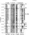
- the inventors developed a technique to integrate three types of valuable information to a sequenced target chromosomal region, such as complete chromosomes or genomes, which consist of read depth, template strand identity (the forward or reverse strand derived from the mother cell after replication), and the haplo-phase or -type, which indicates the identity of a sequence to be derived from the paternal or maternal chromosome present in all diploid organisms.
- a sequenced target chromosomal region such as complete chromosomes or genomes, which consist of read depth, template strand identity (the forward or reverse strand derived from the mother cell after replication), and the haplo-phase or -type, which indicates the identity of a sequence to be derived from the paternal or maternal chromosome present in all diploid organisms.
- the inventive approach exploits Strand-seq to perform haplotype-aware detection of somatic variation in single cells.
- Detected classes of variation include deletions, duplications, inversions, translocations, complex SV classes, copy-number neutral losses in heterozygosity (CNN-LOH) and cellular ploidy alterations.
- the inventive approach leverages patterns of mitotic segregation of template strands ( i.e . chromatid segregation patterns), which reflect a 'genetic signal' not previously considered for detecting SVs in cellular populations.
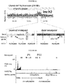
- the invention leverages this information by analyzing in each single cell three orthogonal data layers (or 'channels') - read depth, strand orientation and haplotype phase - the integration of which yields a set of discriminative SV diagnostic footprints via a novel approach according to the invention that is herein termed 'three-channel processing' ( Figure 1 ).
- the inventive approach surprisingly does not require read pairs traversing the SV breakpoints, which renders the approach amenable to scalable low pass sequencing strategies with low sequencing coverage as is the case in single cell sequencing, and enables the detection of SVs flanked by repeat sequence.
- the examples showcase utility through analysis of cell lines and primary leukemias, revealing previously unresolved or incompletely resolved variant classes in conjunction with repeat-associated and punctuated-equilibrium like SV formation, and resolving subclones defined through single cell SV profiles.
- the invention will open up a range of research opportunities by enabling scalable, cost-efficient analyses of a wide variety of SV classes in single cells.
- sequencing data shall refer to data obtained by sequencing a polynucleotide and wherein such sequencing data comprises a multiplicity of sequences reads, and each sequence read is derived from sequencing a template polynucleotide strand.
- the template polynucleotide strand is a forward or reverse (W or C) strand.
- sequence read refers to a nucleotide sequence obtained from or read from a nucleic acid molecule obtained from a biological cell or virus. Sequence reads can be obtained through various methods known in the art. Generally, sequence reads are obtained post- amplification (e.g., polymerase chain reaction, such as bridge amplification) of a nucleic acid fragment that is obtained or enriched from a test sample. The length of sequence reads may vary depending on the sequencing method used. Preferred lengths of a sequence read usable in context of the invention are 50 to 500 nucleotides long, preferably around 100 to 200 nucleotides.
- Sequencing methods usable in context of the invention are selected from any methods known to the skilled person.
- “next generation sequencing” approaches are preferred and include so-called parallelized sequencing-by-synthesis or sequencing-by-ligation platforms currently employed by for example Illumina, Life Technologies, and Roche, or electronic-detection based methods such as Ion Torrent technology commercialized by ThermoFisher, etc.
- Sequencing methods may also include so called “third generation sequencing (TGS)” technologies such as nanopore sequencing methods.
- TGS third generation sequencing
- Other approaches include “single molecule real-time (SMRT)” sequencing (for example by Pacific Biosciences), and so called “long-read sequencing” that is capable of obtaining sequence reads longer than 1kb. These both provide what's conventionally termed long-read sequence data (i.e. sequence reads >1000 base pairs)
- a sequence of a target chromosomal region (for example of a test cell) is provided as a strand-specific sequence read, or a portion thereof.
- sequence read, or portion thereof retains the strand-specific information of for example the template strand of the chromosomal region from which the read was sequenced, and which was inherited by the sequenced single cell following mitosis of the mother cell.
- template strands can either be a forward or reverse, or often also referred to as Watson or Crick.
- Any method that will allow for a retaining of the information of strand identity shall be comprised by, and suitable for, the methods of the present invention, as essential is only the strand specific information and not the method of how the information of strand identity is obtained.
- One way of retaining strand identity during sequencing is by strand-specific sequencing or "Strand-seq". The method is described in detail in Falconer et al. 2012 Nature Methods. 9 (11): 1107-1112 , which shall be incorporated herein by reference in its entirety. Specifically incorporated herein by reference is the methods section of the publication.
- Strand-seq involves the use of BrdU nucleotides for one synthesis phase (S-phase) of a cell so that before mitosis the newly-generated sister chromatids of each chromosomes are in one strand marked by the incorporated BrdU nucleotides and in the other strand (template strand) devoid of BrdU.
- the daughter cells are treated such that the BrdU strand is nicked and thus only the non BrdU-labelled strand can be amplified during PCR.
- specific adapters the original template strand information is retained in the amplified fragments such that only the strand identity of the template strand can be ascertained following sequencing. Aligning the so obtained sequence reads to a reference genome scaffold then indicates the direction of the read and from which strand - Watson or Crick - the read was obtained.
- karyotype refers to the genomic characteristics of an individual cell or cell line of a given species or test sample; e.g., as defined by both the number and morphology of the chromosomes.
- the karyotype is presented as a systematized array of prophase or metaphase (or otherwise condensed) chromosomes from a photomicrograph or computer-generated image.
- interphase chromosomes may be examined as histone- depleted DNA fibers released from interphase cell nuclei.
- the karyotyping methods of this invention are specifically suitable for the detection of copy-number neutral SVs.
- the methods of the invention may also be used to determine Copy-Number Polymorphisms (or also referred to "copy number variations") in a test cell or a test genome. Since the Sequence-Based Karyotyping methods may be performed on prokaryotic cells, the presence of chromosomes is not essential for the methods of the invention.
- SV structural variation
- chromosomal aberration or chromosome abnormality
- normal i. e., "non-aberrant”
- normal when referring to chromosomes or karyotypes, refer to the predominate karyotype or banding pattern found in healthy individuals of a particular species and gender.
- SVs detectable by the methods of the present invention are preferably large or medium sized SVs (200kb or larger).
- SVs can be numerical or structural in nature, and include aneuploidy, polyploidy, inversion, balanced or unbalanced translocation, deletion, duplication, inversion-duplication, and the like. SVs may be correlated with the presence of a pathological condition (e.g., trisomy 21 in Down syndrome, chromosome 5p deletion in the cri-du-chat syndrome, and a wide variety of unbalanced chromosomal rearrangements leading to dysmorphology and mental impairment, as well as proliferative disorders and in particular cancer) or with a predisposition to developing a pathological condition.
- Chromosome abnormality also refers to genomic abnormality for the purposes of this disclosure where the test organism (e.g., prokaryotic cell) may not have a classically defined chromosome.
- chromosome abnormality includes any sort of genetic abnormality including those that are not normally visible on a traditional karyotype using optical microscopes, traditional staining, of FISH.
- One advantage of the present invention is that chromosomal abnormality previously undetectable by optical methods or even sequencing methods (e.g., abnormalities involving 4 Mb, 600 kb, 200 kb, 40 kb or smaller) can be detected due to the integration of the three layers of information.
- CNVs copy-number variations
- CNVs refers to a form of structural variation of the DNA of a genome that results in the cell having an abnormal or, for certain genes, a normal variation in the number of copies of one or more sections of the DNA. CNVs correspond to relatively large regions of the genome that have been deleted (fewer than the normal number) or duplicated (more than the normal number) on certain chromosomes.
- copy number neutral shall denote a variation that does not result in the cell having unusual copy numbers of sequence elements such as genes.
- diagnostic footprint in context of the present invention shall mean a pattern of the three layered information of the invention that is specific or at least indicative for a SV.
- a diagnostic footprint is therefore characterized by an alteration of the data distribution expected for a specific experiment.
- the specific pattern that indicates a SV will vary depending on the analysed data. For example a diploid cell may be sequenced to contain for each chromosome a WW, CC or WC strand distribution. Depending on the strand distribution, the same SV may have a different diagnostic footprint.
- Such footprints or patterns are for example provided herein in table 1.
- target chromosomal region shall refer to a DNA sequence of one or more, full or partial, chromosomes of any organism or virus, which is the object of an inquiry in context of the invention.
- a target chromosomal region may refer to just one sequence of a part of a single chromosome, or to both the paternal and maternal region of any chromosome.
- the target chromosomal region which is the object of an inquiry according to the invention is a whole chromosome or a whole genome of a single cell, or a plurality of a single cell.
- single cell shall refer to one individual cell from which by for example strand-specific sequencing, a single cell library is generated.
- a single cell library in context of the invention describes the plurality of sequence reads obtained by sequencing the genome of said single cell.
- the invention in some aspects and embodiments refers to a plurality of single cells, or multiplicity of single cells, which in this case refers to the generation of a plurality of separate and independent sequence libraries for each single cell contained in the plurality of single cells.
- up to 96 single cells of a cell line are sequenced individually. Such embodiments are preferred as such assays can be performed in multiwall plates such as 96 well plates or 384 well plates.
- reference sequence of the at least one target chromosomal region refers to a database version of a fully sequenced reference of the target. Usually, such reference will be a full chromosome sequence. In some instances the reference sequence is also denoted as “reference scaffold” or “reference genomic scaffold” or “reference assembly” or similar expression.
- reference scaffold or “reference genomic scaffold” or “reference assembly” or similar expression.
- the Genome Reference Consortium frequently publishes and updates the reference sequence of the human genome, as well as other genomes such as mouse, zebrafish and chicken genomes (https://www.ncbi.nlm.nih.gov/grc).
- reference state in context of the present invention shall refer to state or distribution of sequencing data that is used as a reference for a comparison with a sample dataset, for example in order to identify aberrations.
- a reference state may be a real set of sequencing data used as a reference, or may be state of the data that is expected for a certain underlying sampled chromosomal region.
- a reference state in context of the invention shall pertain to the distribution of sequences within a chromosome, or set of chromosomes (genome), that is expected for a non-aberrant single cell or population of cells.
- a reference state of a usual diploid human genome would be a distribution of human chromosomes in somatic cells that is common to a majority of humans.
- the reference state may also contain unusual chromosomal architectures or aneuploidies - the reference state according to the invention is determined based on the samples analysed and questions to be answered with the methods of the invention.
- the sample analysed with the method of the invention may be derived from a trisomy 21 individually who is screened for other SVs.
- reference state in context of the invention shall not be confused with “reference sequence”, the latter being defined above and referring to an assembly of sequences that is used for aligning sequence reads.
- aligning or “alignment”, of a sequence in context of the herein disclosed invention shall denote the mapping of a strand-specific sequence to a reference scaffold, such as a herein described reference genome or reference chromosome matching the respective strand-specific sequence.
- Aligning sequence reads, or portions thereof, to the corresponding reference scaffold is well known in the art. Such methods may include Bowtie ( Genome Biol, 2009;10(3):R25 ) or Burrows Wheeler Alignment (BWA) ( Bioinformatics, 2009 Jul 15;25(14):1754-60. doi: 10.1093/bioinformatics/btp324 ). Aligning all sequence reads, or portions thereof, to a reference chromosome scaffold results in positional ordering of the sequence information along both strands of the reference, for example of the at least one target chromosomal region.
- phasing refers to the process of determining whether two or more nucleic acid sequences (typically comprising regions of sequence variation) are located on the same nucleic acid template, such as a chromosome or a chromosomal fragment. Phasing may refer to resolving two or more single-nucleotide variants or polymorphisms (SNPs) within a single sequencing read. Preferably, phasing may refer to resolving sequencing data over a large genomic region, or resolving a whole genome sequence.
- phased as used in the context of sequences for two or more polymorphic sites means the sequence present at those polymorphic sites are known whether to be derived from a single chromosome.
- phased nucleic acid sequence refers to nucleic acid sequence of a single chromosome where the nucleic acid sequence is obtained from sequencing of a single chromosome.
- phased nucleic acid sequence as used in the context of a single chromosomal fragment refers to nucleic acid sequence of a single chromosomal fragment where the nucleic acid sequence is obtained from sequencing of a single chromosomal fragment.
- haplotype is a contraction of the phrase "haploid genotype”, and is presently accepted to mean a set of nucleotide sequence polymorphisms or alleles present on a single maternal or paternal chromosome, usually inherited as a unit.
- haplotype may refer to a set of single-nucleotide polymorphisms (SNPs) that are linked or present together on a single chromosome.
- SNPs single-nucleotide polymorphisms
- haplotype may be used to refer to as few as two alleles or SNPs that are linked or present together on a single chromosome.
- haplotype identity is the correspondence of an observed haplotype in a sequence of interest to a known haplotype of a reference sequence, such as a chromosome.
- a haplotype identity may correspond to the identity of a sequence to either the maternal or paternal haplotype of a diploid organism.
- a haplotype identity "Hi” or "H2” can be assigned corresponding to the observed haplotype distribution of all sequences observed in the library or experiment.
- H1 is the haplotype sequenced on one strand
- H2 is the haplotype sequenced on the complementary strand.
- PCR Polymerase chain reaction
- PCR is a reaction for making multiple copies or replicates of a target nucleic acid flanked by primer binding sites, such reaction comprising one or more repetitions of the following steps: (i) denaturing the target nucleic acid, (ii) annealing primers to the primer binding sites, and (iii) extending the primers by a nucleic acid polymerase in the presence of nucleoside triphosphates.
- the reaction is cycled through different temperatures optimized for each step in a thermal cycler instrument. Particular temperatures, durations at each step, and rates of change between steps depend on many factors well-known to those of ordinary skill in the art.
- Complementary refers to the ability of polynucleotides to form base pairs with one another. Base pairs are typically formed by hydrogen bonds between nucleotide units in antiparallel polynucleotide strands. Complementary polynucleotide strands can base pair in the Watson-Crick manner (e.g., A to T, A to U, C to G), or in any other manner that allows for the formation of duplexes.
- the term “complementary” is also used to denote the respective complementary DNA strand. For example referring to the complementary strand of the Watson strand refers to the Crick strand, and vice versa.
- polynucleotide or “nucleic acid” refers to polymers of nucleotides of any length, and includes but is not limited to single stranded or double stranded molecule of DNA, RNA, or DNA/RNA hybrids including polynucleotide chains of regularly and irregularly alternating deoxyribosyl moieties and ribosyl moieties (i.e., wherein alternate nucleotide units have an -OH, then and -H, then an -OH, then an -H, and so on at the 2' position of a sugar moiety), and modifications of these kinds of polynucleotides wherein the substitution or attachment of various entities or moieties to the nucleotide units at any position, as well as naturally-occurring or non-naturally occurring backbones, are included.
- a polynucleotide may be further modified after polymerization, such as by conjugation with a labeling component.
- a "fragment” or “segment” of a nucleic acid is a small piece of that nucleic acid.
- the polynucleotides used or assayed in context the invention are DNA molecules, such as chromosomes or genomes of eukaryotes.
- Homozygous state means a genetic condition existing when identical alleles reside at corresponding loci on homologous chromosomes.
- heterozygous state means a genetic condition existing when different alleles reside at corresponding loci on homologous chromosomes.
- a “gene” refers to a polynucleotide containing at least one open reading frame that is capable of encoding a particular protein after being transcribed and translated.
- a “subject,” “individual” or “patient” is used interchangeably herein, which refers to a vertebrate, e.g., a mammal, e.g., a human.
- amplifying refers to generating one or more copies of a target nucleic acid, using the target nucleic acid as a template.
- the term "genome(s)” means the hereditary information of an individual typically encoded in nucleic acids, either DNA, or RNA, and including both genes and non-coding sequences.
- the genome may refer to the nucleic acids making up one set of chromosomes of an organism (haploid genome) or both sets of chromosomes of an organism (diploid genome) depending on the context in which it is used.
- a "target chromosome pair" as used herein refers to a pair of chromosomes of the same type, where a member of the pair is maternally inherited (inherited from the mother) and the other member of the pair is paternally inherited (inherited from the father).
- a target chromosome pair refers to a pair of chromosome 1, chromosome 2, chromosome 3, and including up to chromosome 21, chromosome 22, and chromosome X.
- One or more target chromosome pairs may be simultaneously analyzed by the methods disclosed herein to determine the sequence of the maternally and paternally inherited chromosome of the target chromosome pair.
- a “single copy” or “single copies” of a target chromosome pairs as used herein refers to a single physical DNA molecule, either the chromosome per se, or packaged (with the assistance of chromosomal proteins such as histones) in the form of a chromosome.
- chromosomes In a normal diploid human cell, there are 46 single chromosomes, 23 single chromosomes from the mother and 23 single chromosomes from the father.
- Single copies of a target chromosome are also referred to as single copies of a chromosome type.
- Single copies of one or multiple chromosome types are usually separated into individual containers in the method described herein.
- a "chromosome type” as used herein refers to a specific chromosome present in a cell.
- chromosome X In a normal diploid human cell of a female, there are 22 types of autosomal chromosomes and one type of sex chromosome (chromosome X).
- chromosomes X and Y In a normal diploid human cell of a male, there are 22 types of autosomal chromosomes and two types of sex chromosomes (chromosomes X and Y).
- polymorphic site or "polymorphism” as used herein refers to a localized region within a chromosome at which the nucleotide sequence varies from a reference sequence in at least one individual in a population. Sequence variations can be substitutions, insertions or deletions of one or more bases. Polymorphisms that alter the structure of a chromosome or a larger nucleic acid molecule are SV as described herein elsewhere.
- single nucleotide polymorphism(s) or SNP(s) means a polymorphic site at which the sequence variation is caused by substitution of a single base at a specific position.
- SNPs refer to nucleotide variations at a defined genomic position among a population.
- a SNP within a coding region, in which both forms lead to the same protein sequence, is termed synonymous; if different proteins are produced they are non-synonymous.
- SNPs may have consequences for gene splicing, transcription factor binding, or the sequence of non-coding RNA, for example, and/or may indicate the haplotype of the organism.
- hybridization means one or more processes for co-localizing complementary, single-stranded nucleic acids, and/or co-localizing complementary non-traditional molecules with single- or double-stranded nucleic acids through strand separation (e.g., by denaturation) and re-annealing, for example.
- complementary nucleic acid molecules optionally oligonucleotides, may hybridize to single- or double-stranded DNA.
- Methods for hybridization are known in the art, and include, but are not limited to, conditions for low and high stringency hybridization ( Sambrook and Russell. (2001) Molecular Cloning: A Laboratory Manual 3rd edition.
- Stringency of the hybridization may be controlled (e.g. by the washing conditions) to require up to 100% complementarity between the probe and the target sequence (high stringency), or to allow some mismatches between the probe and the target sequence (low stringency).
- Factors to determine the appropriate hybridization and wash conditions based on the target and the probe are known in the art. In illustrative embodiments, following the first wash using 0.2 ⁇ SSC/0.1% SDS for 10 minutes at 68° C., two additional washes with 0.2 ⁇ SSC/0.1% SDS for 15 minutes each at 68° C.
- allele refers to a particular form of a genetic locus, or a genomic region, or an entire chromosome, distinguished from other forms by its particular nucleotide sequence.
- locus refers to a location on a chromosome or DNA molecule corresponding to a gene or a physical or phenotypic feature.
- sample as used herein relates to a material or mixture of materials, typically, although not necessarily, in liquid form, containing one or more analytes of interest, which is in the present context of the invention a sample containing cellular material or at least genomic material of one or more cells.
- chromosomal sample as used herein relates to a material or mixture of materials, containing chromosomes from a subject. Similar the term “genomic sample” relates to a material or mixture of materials, containing genomic material from a subject or cell.
- assigning with regard to information in context of the present invention shall mean that any kind of information is connected to a certain sequence entity such as a predetermined or preselected window of the reference scaffold, or a sequence read.
- sequence entity such as a predetermined or preselected window of the reference scaffold, or a sequence read.
- numbers of observed or mapped reads or portions of reads are assigned as information in accordance with the herein disclosed three channels (i) to (iii).
- a “sequence window” means a section of the scaffold sequence into which one or more sequence reads, or portions thereof, can be mapped during the alignment.
- the size of the sequence window is selected depending on the coverage of the sequencing data, or arbitrarily chosen depending on the application of the methods of the invention.
- a sequence window may have a size of 1 to 50 kb, or preferably 1 to 10kb or most preferably about 1, 2, 3, 4, 5, 6, 7, 8, 9, 10, 11, 12, 13, 14, 15, 16, 17, 18, 19, or 20 kb. Windows of the invention may also be larger such as 50kb, 100kb, 200kb or 500kb.
- An example window in accordance with the herein presented examples is about 50kb.
- three layered information means in context of the invention the integration of three separate channels of information that can be derived from strand specific sequencing in combination with haplotype phasing of the sequence read information.
- the term "coverage,” refers to the average number of reads representing a given nucleotide in the reconstructed sequence. It can be calculated from the length of the original genome (G), the number of reads (N), and the average read length (L) as N ⁇ L/G. For example, a hypothetical genome with 2,000 base pairs reconstructed from 8 reads with an average length of 500 nucleotides will have about two times (2 ⁇ ) of redundancy. This parameter also enables one to estimate other quantities, such as the percentage of the genome covered by reads (sometimes also called coverage).
- One of the advantages of the present invention is a stable identification of SV within a target sequence of a single cell which sequenced with a coverage of only 0.01x compared to 30x which is usually a coverage obtained by sequencing the genomic material of over 1000 cells.
- sequence reads have an overall coverage of 0.001x to 100x, preferably about 0.01x to 0.05x, of the target chromosomal region.
- germ line refers to cells in an organism which can trace their eventual cell lineage to either the male or female reproductive cells of the organism.
- Other cells that are referred to as “somatic cells” are cells which do not directly give rise to gamete or germ line cells. Both germ line cells and somatic cells may be used in some embodiments of the invention depending on the application.
- chromosome instability and “genomic instability” and similar expressions as used herein, pertain to the number or degree of chromosome structural and numerical abnormalities, i.e. deletion or duplication of either whole chromosomes or parts of chromosomes, for example leading to aneuploidy (incorrect number of chromosomes).
- CIN chromosome instability
- genomic instability and similar expressions as used herein, pertain to the number or degree of chromosome structural and numerical abnormalities, i.e. deletion or duplication of either whole chromosomes or parts of chromosomes, for example leading to aneuploidy (incorrect number of chromosomes).
- read depth shall refer to the number of reads mapping into the predetermined or preselected sequence window.
- diagnostic signature or “diagnostic footprints” or similar expression shall in context of the invention refer to an expected difference or signal an SV or other aberration causes in the sequencing data analysed according to the invention compared to the reference state. Examples for SVs in diploid genomes are provided in table 1 herein. However, understanding the genetic patterns of inheritance one skilled in the art will be able to determine any other diagnostic signature or diagnostic footprint depending on the underlying situation.
- ground state when used in context of the invention denotes the distribution of parental template strands within a single cell or a population of single cells. Hence, in preferred embodiments of the invention a ground state shall denote whether a single cell comprises any number of W or C template strands. In a diploid scenario, as a non-limiting example, the ground state could be WW, CC, WC or CW (see also table 1).
- the strand-specific sequence data is provided in order to commence the method of the present invention.
- the method might include preparatory steps to prepare or filter the sequence data or even obtain the sequence data by strand sequencing of a sample comprising genetic material of the target chromosomal region.
- the strand specific sequence data may already include sequence reads, or portions thereof, that are mapped to a reference scaffold.
- sequence reads, or portions thereof are mapped or aligned to the corresponding reference scaffold using standard aligning tools known in the art.
- reads across each individual cell or experiment are assigned to windows ("binned") of a given width.
- the width of the window is selected depending on the coverage and the specific conditions of the data or application. Preferred lengths of windows are described herein elsewhere.
- the mapped reads are assigned to windows based on their start position; however, other reference positions might also be used.
- a strand state is assigned to each of said windows, which indicates the template strand distribution or relative abundance of W and C reads, for the chromosomal region.
- the strand state is indicated as WW (Watson-Watson), CC (Crick-Crick) or WC (Watson-Crick).
- the strand state assignment may be performed using a hidden Markov model (HMM).
- the invention may include various steps of quality control and data normalization according to the specific methods used in example 1 herein.
- the herein disclosed methods integrate all three channels of information, such as depth, orientation and haplo-phase. Therefore, preferably in step (d) all three channels of sequence information (i) to (iii) are assigned to the at least one predetermined sequence window.
- the strand-specific sequence data comprises sequence reads which are derived from one of at least two separate strands of the at least one target chromosomal region, preferably the strand-specific sequence data comprises further sequence reads which are derived from the other of the at least two separate strands, for example wherein one strand is from the paternal and the other strand is from the maternal chromosome (but could further comprise sequence reads derived from additional strands, as in the case of triploidy, etc.).
- each sequence read, or portion thereof is aligned with the direction forward or reverse which retains strand-specific sequence information.
- the method may comprise of identifying strand state and/or detecting sister chromatid exchanges (SCE) in the sequence data.
- SCE sister chromatid exchanges
- strand state detection it is in some embodiments preferred to simultaneously detect SCEs.
- each chromosomal homologue within a single cell is sequenced either on the W or the C strand (leading to the observed WW, WC, or CC strand patterns in the case of diploidy).
- Strand state detection and SV discovery is improved by detecting SCE events (with typically ⁇ 6 SCEs seen per diploid cellular genome), which can flip the strand state of a homologue along chromosomes.
- the methods of the invention may comprise a step of segmenting the at least one target chromosomal region, wherein the segmenting is performed on basis of the channels of sequence information (i) to (iii), each individually or together.
- segmenting seeks to identify breakpoints in the information distribution along the target chromosomal region, and thereby identify boundaries of candidate SVs. Since the invention uses also strand-specific sequence data, also the breakpoints of candidate SVs that are copy-number neutral can be detected during segmenting. In embodiments where a population of single cell sequence data is analysed, it is preferable to segment across all cells simultaneously. In some embodiments such segment is also referred to as a sub-region.
- the present invention includes a step of haplotype phasing of the sequences.
- haplotype phasing classifies WC regions into either WC or CW states, where the first position refers to H1 and the second to H2.
- Such a step is preferable as this distinction is then used during SV identification to predict SVs in a haplotype-aware manner, which is an advantage of the present invention.
- whole-chromosome haplotypes of at least a couple of dozen SNVs are used; these can be obtained from an external data source or will alternatively be identified in the strand-specific sequence data directly as a step of the method of the invention.
- the workflow of the present invention may include the StrandPhaseR algorithm ( Porubsky, D. et al. Dense and accurate whole-chromosome haplotyping of individual genomes. Nat. Commun. 8, 1293 (2017 )) to generate chromosome-scale haplotypes (see Methods section in the examples for details).
- step (c) involves that a chromosomal haplotype identity (H1/H2) along the at least one target chromosomal region is assigned to any of the given reads by assigning Single Nucleotide Polymorphisms (SNP), preferably wherein such SNPs do not have a disease association.
- SNP Single Nucleotide Polymorphisms
- haplotype-tagging Such assigning is referred to herein in some instances as "haplotype-tagging" the sequence read.
- haplotype identity of all reads derived from a single strand W or C
- haplotype identity H1/H2
- This embodiment allows to haplo-phase reads which do not contain or overlap with any SNPs.
- haplotype phasing is performed in a "strand-aware manner".
- such embodiment shall entail that the assigned haplotype identity of any given sequence read is connected to the information which orientation the same sequence read has. Therefore in a preferred embodiment of the invention the information of channel (ii) and (iii) for each sequence read, or portion thereof, are connected.
- the sequencing data comprises a multitude of non-overlapping and/or overlapping sequence reads.
- read duplicates which are often artefacts, for example by PCR, are removed.
- the strand-specific sequence data does not comprise overlapping sequence reads.
- the methods of the invention are useful for the detection of various SV. Therefore, preferably the method of the first aspect may include a step (e) of identifying a structural variation (SV) by performing step (d) for a multiplicity (at least two) of windows within the sequence data of the positional ordered and aligned sequence reads, and identifying within the multiplicity of windows a sub-region comprising one or more windows having an unusual/altered/changed distribution of the information of any one, or all of, or any combination of, channels (i) to (iii).
- the unusual/altered/changed distribution in this invention is preferably any of the herein disclosed diagnostic footprints which are indicative of one or more SVs. Such diagnostic footprints in accordance with the invention are described in the following:
- the diagnostic footprints for SV detection in the integrated data of the present invention take three data layers into account - read depth, read orientation and phase.
- a population of single cells may be analysed in order to increase detection and/or discrimination between two different SV classes achieving similar SV likelihoods, for example haplotype-tags, or "haplotagging" (phased reads containing a heterozygous SNPs) may preferably also be considered for classification.
- haplotype-tags phased reads containing a heterozygous SNPs
- haplotagging phased reads containing a heterozygous SNPs
- the SV discovery signatures developed for this invention depend on the underlying strand state of a target chromosomal region, and whether an SV is homozygous or heterozygous - i.e . they are different in WC, CW, WW or CC chromosomal regions, and for homozygous versus heterozygous duplications, for example.
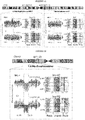
- Table 1 shows an overview of SV diagnostic footprints in the context of heterozygous and homozygous SVs, and for different patterns of mitotic strand segregation, and such footprints are preferred embodiments of the invention: Table 1: Diagnostic Footprints according to the invention SV diagnostic footprints in a WC ground state Haplotype tags SV state Depth W:C W C W cov C cov Reference state 2N 50% H1 H2 1N 1N Deletion of H1 1N 0% - H2 0N 1N Deletion (homozygous) 0N - - - 0N 0N Duplication of H1 3N 66% 2xH1 H2 2N 1N Duplication (homozygous) 4N 50% 2xH1 2xH2 2N 2N Inversion of H1 2N 0% - H1+H2 0N 2N Inversion (homozygous) 1 2N 50% H2 H1 1N 1N Inverted duplication of H1 2 3N 33% H1 H1
- said sub-region or segment may be defined by at least one but preferably two breakpoints, and wherein such breakpoints indicate a change of any one, or any combination, or all, of the information of channels (i) to (iii) compared to the reference state and/or compared to an overall distribution of said channel information within in the sequence data.
- said reference state of said chromosomal region is a state of the information of the channels which is expected for a non-aberrant distribution and/or predetermined state of the information of said chromosomal region.
- the reference state in a target diploid chromosomal region is in the event the diploid target chromosomal region comprises a first template strand derived from the first parental target chromosomal region and a second template strand derived from the second parental target chromosomal region; said reference state is:
- any of the SV mentioned in table 1 are detected based on the indicated diagnostic footprint such SV would display depending on the respective ground state of the cell.
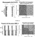
- the SV is an altered ploidy state
- the sequence data comprise a multiplicity of target chromosomal regions of different chromosomes
- the altered ploidy state is identified by a difference in overall distribution of any one, all of, or any combination of, the information of channels (i) to (iii), between a candidate polyploidy chromosomal region of one chromosome compared to one or more other chromosomal regions of other chromosomes.
- the method of the invention involves determining the distribution of W and C strands in a population of single cells and deriving therefrom, the ploidy state for each target chromosomal region, preferably target chromosome.
- a detailed description of the identification of an aneuploidy is provided in the example section.
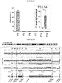
- the detection of ploidy states of a target chromosomal region of a single cell is based on the fact that in a diploid cell sequenced by Strand-seq, demonstrate random and independent mitotic segregation of replicated chromosomes to the resulting daughter cells. This implies that approximately 50% of all autosomes will show a characteristic pattern where one homolog is sequenced on the plus strand (here W, for Watson) and the other homolog is sequenced on the minus strand (C, for Crick) - hereafter termed WC-pattern.
- the remaining autosomes are sequenced either only on the C strand (approximately 25%; CC-pattern), or only on the W strand (approximately 25%; WW-pattern), respectively ( Fig. 2 ).
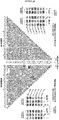
- a binomial distribution can be used to compute expected frequencies of autosomal strand patterns for different cellular ploidy states.
- the CCC-pattern all reads of an autosome map to the C-strand
- the WWW-pattern all reads map to the W-strand
- the CWW-pattern and the CCW-pattern, respectively, will each be seen for 37.5% of all autosomes.
- Tetraploidy and haploidy by comparison, will result in their own discernible strand patterns (Table 2).
- These distinct strand state patterns i.e. relative abundances of W and C reads
- expected frequencies of strand inheritance patterns for a given chromosomal region can be used to identify aneuploidies in the sample.
- these diagnostic footprints do not require additional data (such as the detection of additional somatic variants in a given cell) for making ploidy assignments, and as such are much more powerful and applicable for detecting potentially pathogenic ploidy alterations in cells.
- Diagnostic footprints characteristic for several cellular ploidy states are shown in table 2.
- a binomial distribution can be used to compute expected frequencies of autosomal strand patterns for different ploidy states.
- W Watson strand of the genome.
- C Crick strand.
- Table 2 Diagnostic strand patterns (footprints) for aneuploidies Ploidy state Strand patterns observed Haploid C W Strand-ratios: 1:0 50% 50% - - - Diploid CC CW WW Strand-ratios: 1:1, 2:0 25% 50% 25% - - Triploid CCC CCW WWC WWW - Strand-ratios: 2:1, 3:0 12,50% 37,50% 37,50% 12,50% Tetraploid CCCC CCCW CCWW CWWW WWW Strand-ratios: 4:0, 3:1, 2:2 6,25% 25% 37,50% 25% 6,25%
- the method of detecting cellular ploidies is preferably a method wherein at least strand-specific sequence data and read depth are used. More preferably also the haplotype phase is integrated.
- the detection of ploidies involves that a strand-specific sequence data comprises data derived from a population of individual cells to allow detection of the distribution of the W and/or C strands.
- the more single cell data included the more complex aneuploidies can be detected by the method of the present invention.
- the method of the invention is performed with strand-specific sequence data of the at least one target chromosomal region of at least two or more single cells, preferably 10 or more, more preferably 50 or more, most preferably 90 or more or 350 or more; and preferably, wherein the multiplicity of single cells is derived from the same or identical origin, such as the same individual and/or the same tissue or sample type.
- Such population or multiplicity of single cells are preferably of the same origin and expected to share said polyploidy and/or translocation.
- a polyploidy or translocation is preferably detected if the distribution of the strand-orientation within the population is altered from the expected pattern.
- a polyploidy is detected if the distribution of sequenced forward or reverse strands for each chromosome differs from the overall distribution expected for diploid chromosomal (autosomal) segregation, such as 50% WC, 25% WW and 25% CC.
- a cell, or a single cell, in context of the invention may be any biological cell, or cell-like structure, comprising a polynucleotide genome or parts thereof.
- a cell therefore may be a virus, a prokaryotic cell, or a eukaryotic cell, such as an animal or plant cell, preferably wherein the animal cell is a mammalian cell such as a mouse, rat or human cell. Any cell type or any cell of any tissue origin may be used for the present invention.
- the at least one single cell is obtained from a cellular sample of a patient, and wherein said single cell is either a cell associated with a disease, or is a healthy cell of said patient, preferably wherein the method is performed for a multiplicity of single cells associated with the disease and/or healthy cells.
- the methods of the invention are particular useful for diagnosing a disorder, or the probability of a subject to develop a disorder, and finally, in order to stage a disorder or monitor it, or even to estimate disease severity.
- a disorder or the probability of a subject to develop a disorder
- some preferred embodiments of the invention also encompass further step (f) diagnosing a condition based on the identity of, location of, or number of detected SV within the target chromosomal region.
- diagnostic applications is provided herein below.
- the detected SVs of said target chromosomal region may be compared with a known reference state of said chromosomal region, such as a known state of the chromosomal region of a healthy cell.
- the invention may include detecting SV-affected genes or genetic elements within the target chromosomal region. Since the invention identifies the chromosomal location of each detected SV, it may be a preferred embodiment to further identify genetic elements, preferably genes, that are affected by the SV, for example if their open reading frame is disrupted by the breakpoint of the SV, or by copy number alteration, or by impairment of any regulatory element in the gene region.
- Any method according to the herein disclosed invention is in some preferred embodiments an in-vitro method, and/or is an in-silico method.
- the method is performed with a multiplicity of single cell libraries.
- the method may further comprises a step of calculating a probability of occurrence of a SV at a given position, for example by using a Bayesian network of any one, any combination of or all channels (i) to (iii), of the analysed single cell population.
- Karyotyping a genome is a valuable method in both clinical practice and research. Either to diagnose genetic abnormalities in a patient, or a disease associated tissue, or embryonic cells in reproductive medicine. In research karyotyping allows the study of such SVs, evolutionary events and inheritance patterns of phenotypes. Traditional karyotyping is usually performed on lymphocytes and amniocytes using labor intensive methods such as Giemsa staining (G-banding). Because chromosomes are visualized on an optical microscope, the ability to resolve detailed mutations (involving only a small part of a chromosome) is limited.
- FISH fluorescent ire situ hybridization
- the object of the invention is solved in another aspect by a method of karyotyping a single cell, or a population of multiple single cells, or a subject from which such cells are obtained, the method comprising,
- the method includes performing the method of scTRIP with a population of cells in order to obtain a comprehensive karyotype for example including possible translocations and aneuploidies as well as the possibility to obtain an allelic frequency of all SVs that are found within the population of cells.
- a method of karyotyping a genome of at least one single cell of interest comprising: a) obtaining a plurality of (preferably non-overlapping) strand specific sequences from random locations of the genome of the at least one single cell; b) mapping said test strand specific sequences to a genomic reference scaffold to obtain a test distribution of mapped strand specific sequences; c) assigning to a predetermined sequence window within the reference scaffold (i) number of mapped sequence reads, (ii) number of mapped forward strand reads and number of reverse strand reads, preferably a ratio thereof, and (iii) assigning a haplotype identity (H1/H2) to the strand-specific reads, to obtain a three layered test distribution of mapped sequences; d) identifying a statistically significant alteration between an expected distribution, wherein such an alteration indicates a karyotypic abnormality in the genome of the at least one single cell; or e) comparing the three layered test distribution to
- the present invention also pertains to the output data of the method of karyotyping.
- chromosome instability CIN
- the invention pertains to a method of diagnosing a disease associated with unusual or increased CIN (such as cancer).
- the degree of chromosomal instability can be traditionally quantified in the prior art by determining the number of centromeres for one particular chromosome or several chromosomes.
- the present invention as described herein provides a much faster, cheaper and more comprehensive view on structural variations in any given sample, and thus allows for an improved quantification of CIN.
- the invention also may be used to study genetic stability in various contexts.
- the invention therefore pertains in another aspect to a method of diagnosing a disease in subject, the method comprising, providing strand-specific sequence data of one or more cells of the subject, performing a method of scTRIP as described herein, detecting within the one or more cells any SV, and comparing the detected SV with a reference state, wherein an altered number, type or location of one or more SV in the sample of the subject indicated the presence of a condition, such as a disease, for example cancer.
- the invention may include a quantification of CIN based on the type and number of SV detected in a sample.
- Disorders that can be diagnosed by the methods of the present invention are manifold and include any germ line encoded genetic disorders or disorders associated with somatic genetic events.
- Non limiting examples of human genetic disorders associated with an SV are including their genomic locations: 5q11-q13 (Angelman's syndrome), 5p15.2-p15.3 (Cri-du- chat syndrome), 22q11.2 (DiGeorge syndrome), 17p13.3 (Miller-Dieker syndrome), 15q11-q13 (Prader-Willi syndrome), 22q11.2 (Shprintzen syndrome), 17p11.2 (Smith -Magenis syndrome), 7q11.23 (Williams-Beuren syndrome), 4p16.3 (Wolf-Hirschhorn syndrome), 1q21.1 (microdeletion 1q21.1), 1q21.1 (microduplication 1q21.1), 1q41q42 (Microdeletion 1q41q42), 2p15p16.1 (microdeletion 2p15p16.1), 3q29 (microdeletion 3q29), 7q11.23 (microduplication 7q11.23), 9q22.3 (microdeletion 9q22.3), 12q14 (microd
- Cancer in general might therefore be diagnosed if a patient sample shows an unusual or increased CIN compared to a reference.
- Cancers in context of the invention that analysed, predicted, diagnosed or monitored are selected from the following non-limiting list of cancers:
- liver cancer e.g., hepatocellular cancer (HCC), malignant hepatoma
- lung cancer e.g., bronchogenic carcinoma, small cell lung cancer (SCLC), non-small cell lung cancer (NSCLC), adenocarcinoma of the lung
- leiomyosarcoma LMS
- mastocytosis e.g., systemic mastocytosis
- muscle cancer myelodysplastic syndrome (MDS); mesothelioma; myeloproliferative disorder (MPD) (e.g., polycythemia vera (PV), essential thrombocytosis (ET), agnogenic myeloid metaplasia (AMM) a.k.a.
- myelofibrosis MF
- chronic idiopathic myelofibrosis chronic myelocytic leukemia (CML), chronic neutrophilic leukemia (CNL), hypereosinophilic syndrome (HES)
- neuroblastoma e.g., neurofibromatosis (NF) type 1 or type 2, schwannomatosis
- neuroendocrine cancer e.g., gastroenteropancreatic neuroendoctrine tumor (GEP-NET), carcinoid tumor
- osteosarcoma e.g., bone cancer
- ovarian cancer e.g., cystadenocarcinoma, ovarian embryonal carcinoma, ovarian adenocarcinoma
- papillary adenocarcinoma pancreatic cancer
- pancreatic cancer e.g., pancreatic andenocarcinoma, intraductal papillary mucinous neoplasm (IPMN), Islet cell tumors
- the method of the invention for diagnosing a disorder is in preferred embodiments a purely in vitro or even in silico performed method.
- the diagnostics of the invention may include any one of or all of the following steps: obtaining a sample of a subject to be diagnosed.
- samples may be any biological sample comprising genomic material, preferably cellular samples of the subject.
- samples may be obtained from any source to analyse the general genomic status of the subject, or may be specifically obtained from a tissue or cell type suspected to be involved in a pathology.
- biological samples in addition to the general definition of samples provided herein, may include any biological tissue, organ, organ system or fluid.
- Such samples include, but are not limited to, sputum, blood, blood cells (e.g., white cells), amniotic fluid, plasma, semen, bone marrow, and tissue or core, fine or punch needle biopsy samples, urine, peritoneal fluid, and pleural fluid, or cells therefrom.
- Biological samples may also include sections of tissues such as frozen sections taken for histological purposes.
- a biological sample may also be referred to as a "patient sample.”
- a further step included in the diagnostics may be the isolation of the DNA to be analysed with the method of the invention. Such methods of obtaining DNA, purifying and preparing it for sequencing approaches are well known to the skilled artisan. Then further, the diagnostic method of the invention may include strand-specific sequencing to obtain the strand-specific sequence data.
- the invention provides a method for assessing the chromosomal stability of a single cell, or within a population of single cells, the method comprising performing a method according to any one of the preceding claims, and wherein an increased total number, or increased number of any one type or multiple types of, SV in the said single cell or population of single cells, indicates chromosomal instability.
- CIN is a general indicator of many diseases and in particular cancer.
- testing CIN with the scTRIP of the invention provides an application to easily access whether a cell population is of low quality as the cell shows an increased CIN.
- the method is for use in quality control of a genetically engineered cell or population of cells, wherein an increased instability indicates a loss of quality.
- the method entails detecting of SVs in a sample of the engineered cells, or cell line, and comparing it to a reference cell or reference state. An observed increase in CIN then would result in a decreased quality of the engineered cells. Also occurrence of certain types of problematic SVs might result in discarding the engineered cells.
- the single cell or population of single cells analysed are genetically engineered cells such as by gene editing, viral integration.
- Preferred engineered cells are immune cells, such as Chimeric Antigen Receptor (CAR) - T cells, T cell receptor (TCR) engineered cells, or antibody engineered cells.
- CAR Chimeric Antigen Receptor
- TCR T cell receptor
- any cell or cell line might be subject to quality control testing with the methods of the invention.
- Such applications include stem cell research, such as controlling induced pluripotent stem cells (iPSCs).
- stem cells preferably iPSC, are preferred single cells or population of cells, analysed in accordance with the various aspects and embodiments of the invention.
- the single cell or population of single cells are for use in a cellular therapy of a patient, such as autologous immune cell therapy.
- the invention also pertains to a method of screening a candidate compound for its effects on chromosomal stability.
- the method preferably involves contacting at least one single cell, or a population of cells, with the candidate compound, and thereafter, performing any method of scTRIP described herein before in order to obtain SVs in the treated cells.
- Another step in the method may include a comparison of the detected SVs in the treated cells with a reference, or with the cells before treatment, or with in parallel non-treated cells.
- the method for screening may be applied for example to test side effect of therapeutic compounds on genomic stability.
- Such compounds can be any compound that might be suspected to have an impact on genomic stability and preferably is selected from polypeptide, peptide, glycoprotein, a peptidomimetic, an antibody or antibody-like molecule; a nucleic acid such as a DNA or RNA, for example an antisense DNA or RNA, a ribozyme, an RNA or DNA aptamer, siRNA, shRNA and the like, including variants or derivatives thereof such as a peptide nucleic acid (PNA); a targeted gene editing construct, such as a CRISPR/Cas9 construct, a carbohydrate such as a polysaccharide or oligosaccharide and the like, including variants or derivatives thereof; a lipid such as a fatty acid and the like, including variants or derivatives thereof; or a small organic molecules including but not limited to small molecule ligands, small cell-permeable molecules,
- the invention pertains also to a computer readable medium comprising computer readable instructions stored thereon that when run on a computer perform a method according to the herein disclosed invention, preferably scTRIP.
- the embodiments may be implemented using hardware, software or a combination thereof.
- the software code can be executed on any suitable processor or collection of processors, whether provided in a single computer or distributed among multiple computers.
- any component or collection of components that perform the functions described above can be generically considered as one or more controllers that control the above-discussed functions.
- the one or more controllers can be implemented in numerous ways, such as with dedicated hardware, or with general purpose hardware (e.g., one or more processors) that is programmed using microcode or software to perform the functions recited above.
- one implementation comprises at least one computer-readable storage medium (i.e., at least one tangible, non-transitory computer-readable medium), such as a computer memory (e.g., hard drive, flash memory, processor working memory, etc.), a floppy disk, an optical disk, a magnetic tape, or other tangible, non-transitory computer-readable medium, encoded with a computer program (i.e., a plurality of instructions), which, when executed on one or more processors, performs above-discussed functions.
- the computer-readable storage medium can be transportable such that the program stored thereon can be loaded onto any computer resource to implement techniques discussed herein.
- references to a computer program which, when executed, performs above-discussed functions is not limited to an application program running on a host computer. Rather, the term "computer program” is used herein in a generic sense to reference any type of computer code (e.g., software or microcode) that can be employed to program one or more processors to implement above- techniques.
- computer program is used herein in a generic sense to reference any type of computer code (e.g., software or microcode) that can be employed to program one or more processors to implement above- techniques.
- the term “comprising” is to be construed as encompassing both “including” and “consisting of', both meanings being specifically intended, and hence individually disclosed embodiments in accordance with the present invention.
- “and/or” is to be taken as specific disclosure of each of the two specified features or components with or without the other.
- a and/or B is to be taken as specific disclosure of each of (i) A, (ii) B and (iii) A and B, just as if each is set out individually herein.
- the terms “about” and “approximately” denote an interval of accuracy that the person skilled in the art will understand to still ensure the technical effect of the feature in question.
- the term typically indicates deviation from the indicated numerical value by ⁇ 20%, ⁇ 15%, ⁇ 10%, and for example ⁇ 5%.
- the specific such deviation for a numerical value for a given technical effect will depend on the nature of the technical effect. For example, a natural or biological technical effect may generally have a larger such deviation than one for a man-made or engineering technical effect.
- an indefinite or definite article is used when referring to a singular noun, e.g. "a”, “an” or “the”, this includes a plural of that noun unless something else is specifically stated.
- hTERT RPE-1 cells were purchased from ATCC (CRL-4000) and checked for mycoplasma contamination.
- BM510 cells were generated using the CAST protocol and derived from the RPE-1 parental line (as previously-described in Mardin et al. 2015).
- C7 cells were acquired from Riches et al 2001.
- Cell lines were maintained in DMEM-F12 medium supplemented with 10% fetal bovine serum and antibiotics (Life Technologies).
- Ethics Statement The protocols used in this study received approval from the relevant institutional review boards and ethics committees.
- the T-ALL patient samples were approved by the University of Kiel ethics board, and obtained from clinical trials ALL-BFM 2000 (P33; age: 14 years at diagnosis) or AIEOP-BFM ALL 2009 (P1; age: 12 years at diagnosis).
- Written informed consent had been obtained from these patients, and experiments conformed to the principles set out in the WMA Declaration of Helsinki and the Department of Health and Human Services Belmont Report.
- the in vivo animal experiments were approved by the veterinary office of the Canton of Zurich, in compliance with ethical regulations for animal research.
- RPE cells and PDX-derived T-ALL cells were cultured using previously established protocols 28,66 .
- the inventors incorporated BrdU (40 ⁇ M; Sigma, B5002) into growing cells for 18-48 hours, single nuclei were then sorted into 96-well plates using the BD FACSMelody cell sorter, and strand-specific DNA sequencing libraries were generated using the previously described Strand-seq protocol 21,67 .
- the BrdU concentration used was recently shown to have no measurable effect on sister chromatid exchanges 24 , a sensitive measure of DNA integrity and genomic instability 24 .
- the Strand-seq protocol was implemented on a Biomek FX P liquid handling robotic system, which requires two days to produce 96 barcoded single cell libraries.
- Libraries were sequenced on a NextSeq5000 (MID-mode, 75bp paired-end protocol), demultiplexed and aligned to GRCh38 reference assembly (BWA 0.7.15).
- High quality libraries obtained from cells undergoing one complete round of DNA replication with BrdU incorporation) were selected as described in 21,67 . Briefly, libraries showing very low, uneven coverage, or an excess of background reads' yielding noisy single cell data were filtered prior to analysis. In a typical experiment, ⁇ 80% of cells yield high quality libraries reflecting BrdU incorporation in exactly a single cell cycle.
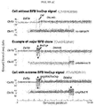
- Chromosome-length haplotype phasing of heterozygous SNPs The inventor's SV discovery framework as described herein phases template strands using StrandPhaseR 22 .
- the underlying rationale is that for 'WC chromosomes' (chromosomes where one parental homolog is inherited as W template strand and the other homolog is inherited as C template strand), heterozygous SNPs can be immediately phased into chromosome-length haplotypes (a feature unique to strand-specific DNA sequencing).
- the inventors developed the core workflow of the method of the inventors to enable single cell discovery of Dup, Del, Inv, and InvDup SVs.
- Input data to the workflow are a set of single-cell BAM files from a donor sample, aligned to a reference genome.
- the core workflow performs binned read counting, normalization of coverage, segmentation, strand state and sister chromatid exchange (SCE) detection, and haplotype-aware SV classification. A brief description of each step is provided below, and for additional details see Supplementary Information.
- Normalization of coverage Normalization was performed to adjust for systematic read depth fluctuations.
- the inventors performed an analysis of Strand-seq data from 1,058 single cells generated across nine 1000GP lymphoblastoid cell lines made available through the HGSVC project (http://ftp.1000genomes.ebi.ac.uk/vol1/ftp/data_collections/hgsv_sv_discovery/working/201 51203_strand_seq/), and pursued normalization with a linear model used to infer a scaling factor for each genomic bin.
- Segmentation was performed by jointly processing strand-resolved binned read depth data across all single cells of a sample, used as multivariate input signal with a squared-error assumption 70 .
- a dynamic programming algorithm was employed to identify the discrete positions of k change points with a minimal sum of squared error. Analyzing all cells jointly in this way rendered even relatively small SVs ( ⁇ 200kb) detectable once these are present with sufficient evidence in the single cell dataset ( e.g . seen in enough cells).
- the number of breakpoints was chosen separately for each chromosome as the minimal k, such that using k +1 breakpoints would only yield a marginal improvement, operationalized as the difference of squared error terms being below a pre-selected threshold.
- Strand-state and SCE detection in individual cells The interpretation of strand-specific binned read counts relies on the knowledge of the underlying state of template strands for a given chromosome (WW, CC, or WC). These "ground states" stay constant over the length of each chromosome in each single cell, unless they are altered through SCEs 21,71 .
- WW, CC, or WC chromosome
- These "ground states" stay constant over the length of each chromosome in each single cell, unless they are altered through SCEs 21,71 .
- To detect SCEs the inventors performed the same segmentation procedure described above in each cell separately (as opposed to jointly across all cells, as for the segmentation). The inventors then inferred putative SCEs by identifying changes in strand state in individual cells that are otherwise incompatible with breakpoints uncovered by the joint segmentation (Supplementary Information).
- the inventors Leveraging these putative SCEs, the inventors then assigned a ground state to each segment (Supplementary Information). To facilitate haplotype-resolved SV calling, the inventors employed StrandPhaseR 72 to distinguish segments with ground state WC, where Haplotype 1 is represented by Watson (W) reads and Haplotype 2 by Crick (C) reads, from ground state CW, where it is vice versa.
- W Watson
- C Crick
- Haplotype-aware SV classification The inventors developed a Bayesian framework to compute posterior probabilities for each SV diagnostic footprint, and derive haplotype-resolved SV genotype likelihoods. To this end, the inventors modeled strand-specific read counts using a negative binomial (NB) distribution, which captures the overdispersion typical for massively-parallel sequencing data 54 .
- the NB distribution has two parameters, p and r ; the parameter p controls the relationship of mean and variance and was estimated jointly across all cells, while r is proportional to the mean and hence varies from cell to cell to reflect the different total read counts per single-cell library.
- each SV diagnostic footprint translates into the expected number of copies sequenced in W and C orientation contributing to the genomic segment (Table S1), which gives rise to a likelihood with respect to the NB model.
- Table S1 The fact that the inventor's model distinguishes WC from CW ground states (see Strand-state and SCE detection above) renders the inventor's model implicitly whole-chromosome haplotype-aware - a key feature not met by any prior approach for somatic variant calling in single cells.
- the inventors also incorporated the count of W or C reads assignable to a single haplotype via overlapping SNPs in the likelihood calculation, and refer to this procedure as "haplotagging" (since it involves reads "tagged” by a particular haplotype).
- the inventors modeled the respective counts of tagged reads using a multinomial distribution.
- the output is a matrix of predicted SVs with probability scores for each single cell.
- the inventor's workflow estimates VAF levels for each SV and uses them to define prior probabilities for each SV (Empirical Bayes). In this way, the framework benefits from observing SVs in more than one cell, which leads to an increased prior and hence to more confident SV discoveries.
- the inventor's framework adjusts for the tradeoff between sensitively calling subclonal SVs, and accurately identifying SVs seen consistently among cells.
- the inventors parameterized this tradeoff into a 'strict' and 'lenient' SV caller, whereby the 'strict' caller optimizes precision for SVs seen with VAF ⁇ 5%, and the 'lenient' caller targets all SVs including those present in a single cell only.
- SV calls presented in this study were generated using the 'strict' parameterization, to achieve a callset that minimises false positive SVs.
- the inventors explored the limits of these parameterizations using simulations, by randomly implanting Dels, Dups and Invs into single cells in silico.
- the inventors analyzed 200 single cells per simulation, applying coverage levels typical for Strand-seq 21 (400,000 read fragments per cell).
- the inventors observed excellent recall and precisions for SVs ⁇ 1Mb in size when present with >40% VAF (Fig. S5). And while the inventors detected a decrease in recall and precision for events present with lower VAF, the inventors were able to recover smaller SVs and those with lower VAF down to individual cells.
- translocations Single cell dissection of translocations.
- the inventors discovered translocations in single cells by searching for segments exhibiting strand-states that are inconsistent with the chromosomes these segments originate from, while being consistent (correlated, or anti-correlated) in strand-state with another segment of the genome (i.e ., their translocation partner) (Supplementary Information).
- the inventors determined the strand states of each chromosome in a homolog-resolved manner. In cases where strand states appeared to change across a haplotype (because this haplotype exhibited SVs or SCEs), the inventors used the majority strand state (i.e . 'ground state', see above) to pursue translocation inference.
- the inventors examined template strand co-segregation by generating contingency tables tallying the number of cells with equivalent strand states versus those not having equivalent strand states (see Fig. 3B ).
- the inventors employed Fisher's exact test to infer the probability of the count distribution in the contingency table, followed by p-value adjustment 73 .
- BFB breakage-fusion bridge
- Single cell based CNN-LOH discovery Single cell based CNN-LOH discovery.
- the inventor's framework first assembles consensus haplotypes for each sample, by analyzing all single cell Strand-seq libraries available for a sample using StrandPhaseR 22 . Each single cell is then compared to these consensus haplotypes in a disomic context, to identify discrepancies matching the CNN-LOH footprint.
- the inventors used the 1000GP 68 reference SNP panel to re-genotype aggregated single cell libraries in each sample. These re-genotyped (observed) SNPs were then compared to the 1000GP reference sets to identify genomic regions showing marked depletion in heterozygous SNPs indicative for CNN-LOH.
- the inventors downsampled the 1000GP reference variants to the SNP numbers observed in the single cell data, and subsequently merged both data sets (observed and reference variants), sorting all SNPs by genomic position.
- R values of ⁇ 1 will be expected, whereas deviations are indicative of CNN-LOH.
- Window sizes (determined by the number of SNPs in a window) were defined as the median SNP count per 500kb window.
- the inventors employed circular binary segmentation (CBS) 74 to detect changes in R , and assigned each segment a state based on the mean value of R. Segments ⁇ 2Mb in size exhibiting mean values R ⁇ 0.15 were reported as CNN-LOH.
- CBS circular binary segmentation
- Genomic DNA was extracted using the DNA Blood Mini Kit (Qiagen, Hilden, Germany). 300 ng of high molecular weight genomic DNA was fragmented to 100 -700bp (300bp average size) with a Covaris S2 instrument (LGC Genomics) and cleaned up with Agencourt AMPure XP (Beckman Coulter, Brea, USA). DNA library preparation was performed using the NEBNext Ultra II DNA Library Prep Kit (New England Biolabs, Ipswich, USA). The inventors employed 15ng of adapter ligated DNA and performed amplification with 10 cycles of PCR. DNA was size selected on a 0.75% agarose gel, by picking the length range between 400 and 500 bp.
- RNA quality control was performed using the 2100 Bioanalyzer platform (Agilent Technologies, Santa Clara, USA). Library preparation was pursued with a Beckman Biomek FX automated liquid handling system (Beckman Coulter, Brea, USA), with 200 ng starting material using TruSeq Stranded mRNA HT chemistry (Illumina, San Diego, USA). Samples were prepared with custom 6 base pair barcodes to enable pooling. Library quantification and quality control were performed using a Fragment Analyzer (Advanced Analytics Technologies, Ames, USA).
- RNA-Seq was pursued on an Illumina HiSeq 2500 platform (Illumina, San Diego, USA), using 50 base pair single reads.
- total RNA was extracted using TRIzol (Invitrogen Life Technologies).
- the RNA was than treated with TURBO DNase (Thermo Fisher Scientific, Darmstadt, Germany) and purified using RNA Clean&Concentrator-5 (Zymo Research, Freiburg, Germany).
- Cytoplasmic ribosomal RNA was depleted by Ribo-Zero rRNA Removal Kit (Illumina, San Diego, CA) and the libraries were prepared from 1 ⁇ g of RNA using TruSeq RNA Library Prep (Illumina, San Diego, CA). These samples were sequenced on a Illumina HiSeq 2000 lane as 75 bp single ends. Fusion junctions were detected using the STAR aligner 76 .
- RNA from PDX-derived T-ALL samples was extracted using a RNeasy Mini kit according to manufacturer's instructions (cat 74106, Qiagen, Hombrechtikon, Switzerland), and cDNA was generated using High Capacity cDNA Reverse Transcription Kit (Applied BioSystems, Foster City, USA).
- qPCR was performed using a TaqMan Gene Expression Master Mix (Applied BioSystems) in triplicate using an ABI7900HT Analyzer with SDS2.2 software. Threshold cycle values were determined using the 2- ⁇ CT method, normalized to human-GAPDH (HS02786624_g1, Applied BioSystems).
- the examples show:
- Example 1 scTRIP enables systematic discovery of a wide variety of SV classes in single cells
- each class of SV can be identified via a specific 'diagnostic footprint'.
- These diagnostic footprints capture the co-segregation patterns of rearranged DNA segments made visible by sequencing single strands of each chromosome in a cell, as follows: During S-phase, the DNA double strand unwinds, and the two resulting single strands (Watson ['W'] and Crick ['C']) act as templates for DNA replication. In Strand-seq, newly replicated strands incorporate Bromodeoxyuridine (BrdU) 21 , which acts as a traceable label for these non-template strands (see Fig. 1A depicting the Strand-seq protocol) 24 .
- PrdU Bromodeoxyuridine
- each of the two daughter cells receive one copy of each chromosomal homolog through independent and random chromatid segregation 21 .
- the labeled nascent strand is then removed, and the segregation pattern of each chromosomal segment is analyzed following strand-specific sequencing ( Fig. 1B ).
- scTRIP combines this strand-specific segregation information with read depth and haplotype phase information to capture newly-defined diagnostic footprints that characterize each SV class ( Fig. 1C-F ).
- the diagnostic footprint of deletions is defined by read depth losses affecting a single haplotype, coupled with unaltered read orientation ( Fig. 1B ,D and Table 1 ).
- Duplications are characterized by haplotype-specific gains with unaltered orientation ( Fig. 1D , right panel ).
- Inv balanced inversions
- read orientation is altered with the re-oriented reads mapping to a single haplotype at constant read depth ( Fig. 1B ,E ).
- Re-oriented reads co-locating with a read depth gain on the re-oriented haplotype signify an inverted duplication (InvDup; Fig. 1E , right panel ).
- the framework first aligns, normalizes and places strand-specific read data into genomic bins, and assigns template strand states and chromosome-scale haplotypes for all cells. It then identifies putative SVs by segmentation ( Methods ), and using a Bayesian model estimates genotype likelihoods for each segment and each single cell ( Fig. 1G ). The model integrates read depth, strand and haplotype phase signals to predict the most probable SV class described by the diagnostic footprints.
- our joint calling framework By performing SV discovery in a haplotype-aware manner, our joint calling framework also combines signals across cells ( Methods ) to sensitively detect subclonal SVs in a heterogeneous cell population. Finally, by analyzing adjacent SVs arising on the same haplotype it can unravel complex rearrangements, an abundant class of somatic structural variation in cancer 25,26 . As a first benchmark, the inventors performed simulation experiments and observed excellent recall and precision after randomly placing SVs into cell populations in silico, even down to a single cell.
- the core computational framework described here in further detail has been developed for detecting Dup, Del, Inv, InvDup, and 'other/complex SV' classes in single cells, based on scTRIP's SV diagnostic footprints.
- Input data required by the framework are a set of single-cell (Strand-seq) BAM files from the same donor sample. In the present study, these data were aligned to build GRCh38 of the human reference genome ( GCA 000001405.15 GRCh.38 genomic.fna ). To later enable haplotype phasing and haplotype-resolved SV assignments, the framework performs re-genotyping of SNPs provided by the 1000 Genomes Project (1000GP; phase 3) to detect heterozygous sites from the single-cell input data. When using the framework a VCF file with these 1000GP SNP sites is to be provided as input.
- 1000GP 1000 Genomes Project
- the scTRIP pipeline is able to call SNPs directly from the single-cell data or to use externally generated SNP calls for a given sample, e.g. based on bulk WGS. Additionally, a tab-separated file with normalization factors (see below) per bin across the genome is used as input to the framework.
- the parameters p and r of the NB distribution were determined in the same manner as for SV classification (see respective section below).
- bins were excluded from the parameter estimation process if their mean coverage across all cells was very low ( ⁇ 0.1, where coverage was previously normalized to 1) or if they showed a highly abnormal WC/(WC+CC+WW) fraction (WC frac ) across cells. Bins were deemed abnormal if exhibiting either WC frac ⁇ 0.05 or WC frac > 0.95, reflecting bins that either never showed WC status, or those that exhibited always WC status, as for example often seen in regions within or near centromeres.
- the inventors utilised 1058 cells from these HGSVC samples sequenced via Strand-seq, obtained from ftp.1000genomes.ebi.ac.uk/vol1/ftp/data_collections/hgsv_sv_discovery/working/20151203-s trand_seq/, and subjected these cells to the same binning scheme described above. Analysis of several of these 1000GP samples showed that these do not carry any germline copy number variants (CNVs) ⁇ 200kb.
- CNVs germline copy number variants
- HGSVC Strand-seq data was aggregated, and first masked regions using any of the following 'exclusion criteria': observed mean coverage ⁇ 50%, observed mean coverage >200%, or observed standard deviation larger than the mean coverage. Then, using the remaining bins, the observed mean bin coverage in the test samples was modelled assuming a linear relationship to the mean HGSVC bin coverage, which explained 66% of the variance with a slope of ⁇ 0.6. This linear relationship was used to derive a scaling factor for each bin, which subsequently was applied to all cells of the present study.
- a "blacklist” was created of regions exhibiting strong sequencing/mapping abnormalities to avoid false positive somatic variant calling.
- To construct said blacklist one starts from the 'masked regions' with unusual coverage in the independent HGSVC samples (see previous paragraph). Then such intervals are progressively merged if they exhibited a distance of 500kb or less (which avoided generation of a highly fragmented blacklist).
- no known polymorphic inversion was accidentally masked by removing all intervals from our blacklist that overlapped with a germline inversion larger than 100kb in size reported by the HGSVC.
- the resulting blacklist was used in all following analyses, which considered regions outside of the blacklisted intervals for single cell SV calling.
- SSE k be the residual error associated to partitioning a chromosome into k segments. Then the smallest number k was selected such that SSE k - SSE k+1 is below a user-set parameter (default: 0.1, which is used in this study) to adjust the number of change points k for a chromosome.
- Changepoints in Strand-seq data that result from mitotic recombination events/SCEs represent a source of "noise" that the method of the invention is able to correct for.
- SCEs happen independently in each single cell 2 , and unlike SVs, SCEs are not transmitted clonally to daughter cells ( i.e . are only detectable in the cell they occur in 2 ).
- changepoints resulting from SCEs are very unlikely to recur at the same position in >1 cell of a sample 1,2 .
- the present invention uses changepoint recurrence as a key criterion for distinguishing SCEs from SVs.
- the threshold to select the number of breakpoints k was set to 0.5.
- the inventors assigned an observed state to each resulting segment by computing the fraction f WC W /( W + C ) and assigning state WW if f WC > 0.8, state CC if f WC ⁇ 0.2 and state WC/CW otherwise.
- the states of neighbouring segments were compared to each other and if the states were unchanged the intervening changepoint was discarded, while the remaining changepoints were subsequently further considered as putative SCEs. Note that herein "WC/CW” is used to indicated that no distinction is made between these two states in this step, distinguishing the two happens in the subsequent strand phasing step.
- changepoints detected in this way may correspond to SVs rather than SCEs.
- the following strategy was employed to select a high confidence list of SCEs: first select those changepoints far away (>500kb) from any breakpoint identified during the joint segmentation (see previous paragraph); these changepoints are likely to represent true SCEs.
- each of the three ground states WW, CC, WC/CW was considered to determine a plausible "ground state". The assumption was employed that a given state at the beginning of a chromosome and a set of SCE positions (which change the state) uniquely determine the state for every segment on the chromosome.
- the discordant length was computed, defined as the total length of genomic intervals for which the observed state differs from the predicted ground state. Although highly unlikely, in rare occasions, an SCE changepoint may appear to coincide with an SV breakpoint. In order to enable the method of the invention to recover such rare SCEs, all putative SCEs closer than 500kb to a breakpoint in the joint segmentation were analyzed. If adding one of these putative SCEs reduces the discordant length by 20Mb or more, the method of the invention assigns these SCE status.
- the method of the invention is able to avoid that missed SCEs result in an incorrectly assigned ground state along larger parts of a chromosome.
- adding at most one such additional SCE precludes masking most true SVs, which have two breakpoints, whereas SCEs lead typically only to a single "switch" (changepoint) in W and C states along a chromosome.
- SCEs since SCEs never associate with copy-number alteration, the chance that SCEs are confused with SVs is near "zero" for many SV classes - that is for, Del, Dup, InvDup, and complex rearrangements - even if these SV are present only in a single cell. Thus, in reality, SCEs are only very rarely incorrectly assigned SV status (as also evidenced by our experimental validation data).
- Chromosome-length haplotype phasing using single-cell sequencing data Chromosome-length haplotype phasing using single-cell sequencing data.
- the inventors phased all available chromosomes using StrandPhaseR. While building whole-chromosome haplotypes for a sample, regions were assigned represented by both W and C strands as either WC or CW for each cell. That is, reads were used overlapping heterozygous SNPs to determine whether haplotype H1 was represented by W reads and H2 by C reads (a situation we denote as WC), or vice versa (denoted CW) (see Methods).
- StrandPhaseR outputs the chromosome-wide haplotypes as a VCF file, which the inventors later utilized in the "haplotagging" step.
- This phasing step of the framework requires at least a few dozen SNPs per chromosome.
- the present framework can use externally provided SNPs. To boost the usable coverage for SNP calling, we performed a cell sorting experiment, independently sorting 100 cells (termed the '100 cell control') in each sample, followed by short-read whole genome sequencing to 1.9x mean coverage.
- the number of high throughput sequencing reads mapped to genomic windows (or bins) were previously shown to be in agreement with a negative binomial (NB) distribution 37 , which can account for overdispersion.
- the inventors employed the NB distribution as the basis for the Bayesian framework.
- the NB distribution has two parameters, p and r , which are estimated from the observed read counts as follows.
- n was denoted as the number of single cells analyzed in a sample.
- the assumption is that the number of reads sampled from each single-cell at a fixed bin size is an NB random variable. In reality, the coverage of single cells will be varying resulting in different NB parameters for each cell.
- the ratio of the mean to the variance is equal to 1- p . Having the same p parameter over all single cells implies that the ratio of mean to variance is constant across all single cells. Consequently, the mean and variance of binned read counts among single cells share a linear relationship in which the line connecting these mean-variance points for single cells passes the origin coordinate with a slope determining the p parameter. This relationship allows estimation of the shared p parameter: for each single-cell, the inventors compute the empirical mean and variance of the observed read counts in fixed-sized bins across the genome.
- the inventors employed a trimmed mean for estimating the dispersion parameters (with trim parameter set to 0.05), to remove the effect of abnormally high or zero read counts (e.g. seen in regions of low mappability).
- Each SV diagnostic footprint ( Fig. 1 ) can be translated into the expected number of copies sequenced in W and C orientation contributing to the genomic segment under consideration.
- Table 1 shows this relationship for each SV class, both for chromosomes where both haplotypes are represented by different template strands (here referred to as 'WC/CW chromosomes') and for such where both haplotypes are represented by the same template strand ('WW chromosomes' and 'CC chromosomes'). Every haplotype-resolved SV implies a particular segment strand pattern in WC, CW, WW, and CC chromosomes, respectively.
- the observed segment strand pattern will be WWC in this given single cell.
- the ground state is WC (W for the H1 haplotype)
- the SV status is deletion of the H1 haplotype
- the observed segment strand pattern is C (see Table 1 ).
- the inventors utilized a Bayesian model ( Fig. 1h ) to compute haplotype-resolved SV genotype likelihoods for each segment in each single cell.
- the inventors model V , the SV type to be inferred, as a pair ( C , C ), where C gives the number of copies of that segment in forward direction and C gives the number of copies of that segment in reverse direction (i.e. when an inversion is present). That is, the pair (1,0) encodes the reference state of a haplotype (one forward copy and zero inverted copies). As illustrated in Fig.
- each segment k ⁇ K and haplotype h ⁇ H ⁇ h 1 , h 2 ⁇ in single cell j ⁇ J comes with a variable V for this SV state, which we refer to as V j,k,h .
- V j,k,h a variable for this SV state
- each SV state V deterministically leads to a corresponding "copy number" observed in Crick direction N C and in Watson direction N W , as explained in the previous section on SV diagnostic signatures (also see Table 1 ).
- ⁇ is a parameter in our model indicating the fraction of "background reads", which represents noise in Strand-seq data (for example due to regions with incomplete BrdU incorporation or removal) 1,2 )
- background reads are taken into account by assuming.
- ⁇ 0.1, which reflects an upper bound for the abundance of such background reads observed in practice.
- the 1 2 coefficients in the above formula serve to scale the dispersion parameter to copy number 1( r j is estimated above to reflect a diploid state of copy number 2).
- every haplotype-resolved SV class ( V ) in a segment together with the ground state ( T ) define a Watson and Crick copy number ( N ) used to compute the NB likelihood of observed read counts.
- haplotype-specific sequencing reads ('haplotagging').
- One of the key advantages of scTRIP is the ability to utilize haplotype information made available through strand-specific sequencing. In the base model described in the previous paragraph, this haplotype-awareness is brought forth by distinguishing WC from CW ground states (also see Chromosome-length haplotype phasing using single-cell sequencing data ).
- the present framework is additionally able to make use of reads not directly assigned to a haplotype (i.e. those in WW and CC regions) owing to their overlap with a haplotype-phased SNP. This feature can further facilitate validation and falsification of putative SVs seen only in few or even individual cells.
- the inventors utilize the whole-chromosome haplotypes generated using StrandPhaseR 36 to tag reads by haplotype using the 'haplotag' command of WhatsHap 38,39 , resulting in one 'haplotagged' BAM file per single cell library. These BAM files are then used to compute the number of Watson/Crick reads that could be tagged by haplotype H1/H2, respectively, for each segment and each single cell. The resulting happlotagged read counts are incorporated in the Bayesian model as random variables X tag W and X tag C (see Fig. 1h ).
- the inventors employed a multinomial distribution to model the conditional distribution of these tagged read counts given the (haplotype- and strand-specific) copy numbers N C and N W . More precisely, we defined parameters of the multinomial distributions p j , k , h 1 C , p j , k , h 2 C , p j , k , h 1 W , and p j , k , h 2 W , for each segment k and single cell j , such that they are proportional to the corresponding copy numbers: p j , k , h d ⁇ max ⁇ N j , k , h d
- the tagged reads are multinomially distributed: X j , k , h 1 , tag C X j , k , h 2 , tag C X j , k , h 1 , tag W X j , k , h 2 , tag W ⁇ Multinomial p j , k , h 1 C p j , k , h 2 C p j , k , h 1 W p j , k , h 2 W
- Bayesian model for SV calling Employing the Bayesian model for SV calling.
- the inventors defined prior probabilities and combined them with the model-based likelihoods for each single cell and segment. The inventors started by regularizing the raw likelihoods, adding a small constant (set to 10 -6 ) to all likelihoods and renormalizing afterwards. This ensures that very small values (or hard zeros) are avoided and corresponds to the error assumption that every SV genotype is possible with this given small probability, no matter what the data suggests. Then, two forms of priors were used. First, biological knowledge was captured on the plausibility of observing certain event types.
- the second type of priors applied acts on each segment separately and uses the raw likelihoods computed by the model across all cells to compute a probability distribution over all SV types. That is, for each segment the likelihoods were summed up per SV type across all cells and normalized to one, which corresponds to estimating the frequency of each SV genotype for that segment.
- the intuition behind this procedure is that one needs to encourage the SV caller to prefer SV types present in many cells over those SV types present only on few cells - unless the evidence inherent to the genotype likelihoods is strong enough to overwhelm these priors.
- the prior of each SV genotype was set to zero if the estimated frequency of that genotype was below a threshold, which was termed GTCLTTOFF (set to 0.05 for the strict call set and set to o for the lenient call set). Effectively, this means that the strict parameterization only considers an SV genotype if the likelihoods across all cells suggest it to be present in the cell population at an expected frequency of at least 5%.
- the lenient call set in contrast, disables this cutoff by setting it to zero and hence readily permits SV genotypes present in individual cells only.
- the inventors used the resulting posterior probabilities to compute log odds ratios (of an SV genotype vs. the reference state), and accepted an SV call if the log odds ratio was at least 4. SV calls in segments with >20% blacklisted bins were discarded.
- Filtering A filtering routine was developed to be used only in conjunction with the strict parameterization, the main goal of which is to arrive at a high confidence SV callset for all SVs with VAF greater than 5%. This filtering routine removes rare inversions seen in only 1 or 2 cells, since rare inversions may occasionally correspond to SCEs. This routine further removes SV calls exhibiting particular biases, most importantly, those biased to occur largely in the context of a certain ground state. In particular, while SVs can be detected in the context of all four ground states (WW, CC, WC and CW; see Table 1 ).
- a merging routine was developed to be used in conjunction with the strict parameterization, which groups adjacent SVs with a similar VAF (where VAF ⁇ 0.1) into a single SV call to avoid over-segmentation and produce a final high confidence somatic SV sites list.
- the inventors considered VAFs of adjacent SVs to be similar if VAF SV 1 / VAF SV 2 ⁇ 0.75 (for cases where VAF SV 2 >VAF SV 1 ) or VAF SV 2 / VAF SV 1 ⁇ 0.75 (for cases where VAF SV 1 >VAF SV 2 ) , and grouped all immediately neighboring SVs selected by this similarity criterion.
- SVs merged by this routine will nearly always correspond to a single structural variation event in validation experiments.
- GTCUTOFF see Using the Bayesian model for SV calling
- haplotagged reads counts are incorporated
- Incorporating haplotype-specific sequencing reads see Incorporating haplotype-specific sequencing reads
- filtering is enabled (see Call set post-processing ).
- we disabled the haplotagging feature we enabled haplotagging for the lenient call set - with the reasoning that haplotagging is mostly valuable to resolve putative SVs with low VAF.
- Example 2 SV landscapes of RPE cells uncovered by scTRIP
- RPE retinal pigment epithelial
- RPE-1 telomerase-immortalized retinal pigment epithelial
- C7 RPE cells which show anchorage-independent growth used as an indicator for cellular transformation 30 .
- Both RPE-1 and C7 cells originate from the same anonymous female donor.
- the inventors first searched for Dels, Dups, Invs and InvDups. Following read normalization, 54 SVs in RPE-1 were identified, and 53 in C7 cells. 22 SVs were present only in RPE-1, and 21 were present only in C7, and thus likely correspond to sample-specific SVs (i.e . SVs that formed somatically or in the cultured cells rather than corresponding to germline variants; hereafter simply referred to as 'somatic SVs'). Two representative SVs are shown in Fig. 2A , including a 1.4 Megabase (Mb) somatic Dup seen in RPE-1, and a 800 kb somatic Del detected in C7.
- Mb Megabase
- Example 3 Dissecting complex cancer-related translocations in single cells
- the inventors subjected RPE-1 cells to the CAST protocol 28 : the inventors silenced the mitotic spindle machinery to construct an anchorage-independent line (BM510) likely to exhibit genome instability.
- the inventors sequenced 145 single BM510 cells detecting overall 67 SVs when searching for Dels, Dups, Invs and InvDups events. Additionally, several DNA segments did not segregate with the respective chromosomes they originated from, indicating inter-chromosomal SV formation ( Fig. 3A ).
- the inventors performed translocation detection with scTRIP searching for diagnostic co-segregation footprints ( Fig. 3B ), and identified four translocations in BM510 ( Fig. 3B ,C ).
- the inventors additionally subjected RPE-1 and C7 to translocation detection, identifying one translocation each ( Fig. 3D ).
- a de novo somatic inversion was additionally detected on the same 17p haplotype, which shared one of its breakpoints with the reciprocal translocation ( Fig. 3C ). Since these SVs shared one of their breakpoints, it is likely that both arose jointly, potentially involving a complex rearrangement process. Analysis of the locus revealed that the inversion encompassed the TP53 gene, and upon translocating fused the 5 ' exons of TP53 to coding regions of the NTRK3 oncogene 32 ( Fig. 3E ). This suggests that scTRIP can reveal fusion genes using single cell sequence data.
- WGS Bulk whole genome sequencing
- RNA-Seq Methods
- scTRIP enables the haplotype-resolved discovery of translocations by single cell sequencing with high accuracy and sensitivity, which included detection of a translocation missed by bulk WGS.
- Example 4 Single cell dissection of a complex DNA rearrangement process
- BFBs breakage-fusion-bridge cycles
- BFBs rising to high VAF can be inferred from bulk WGS by analyzing 'fold-back inversions' (read-pairs aligning close to one another in inverted orientation) 34 . Owing to high coverage requirements, fold-back inversions cannot be systematically tracked in single cells. But the inventors reasoned that scTRIP could provide the opportunity to directly study BFB formation in single cells.
- scTRIP located clustered InvDups on the 10p-arm in 152 out of 154 sequenced cells ( Fig. 4 ). Closer analysis of 10p showed an amplicon with 'stepwise' InvDup events with an adjacent DelTer on the same haplotype, consistent with BFBs ( Fig. 4A-C and Fig. S12 ). The remaining two cells lacking InvDups, notably, showed a larger DelTer affecting the same 10p segment ( Fig. 4C ).
- the inventors By aggregating sequencing reads across cells, the inventors identified 8 discernable segments along chromosome 10, which included the 10p amplicon (comprising six copy-number segments) and its adjacent regions (the 10p terminal region, and the centromere-proximal region) ( Fig. 4B ). To further characterize the genetic heterogeneity seen at 10p, the inventors inferred the cell-specific copy-number of all 8 segments ( Fig. 4D ). This revealed at least three distinct groups of cells with respect to 10p copy-number: (i) A large group presenting 'intermediate' copy-number with 100-130 copies detected for the highest copy-number segment (referred to as the 'major clone'). (ii) Two cells that lost the corresponding 10p region through a DelTer, (iii) A single cell exhibiting vastly higher copy-number ( ⁇ 440 copies), which may have undergone additional BFB cycles ( Fig. 4C ).
- SVs identified in C7 provided further insights into the rearrangements occurring in the major clone: Namely, the inventors detected an unbalanced translocation stitching a duplicated 15q segment to the 10p amplicon ( Fig. 4C ).
- the duplicated segment encompassed the 15q telomere ( Fig. 4C ), which may have stabilized the amplicon to terminate the BFB process.
- the unbalanced translocation was absent from the two cells harbouring the extended DelTer, whereas the translocated region became further amplified in the cell with excessive 10p copy-number ( Fig. 4C ).
- a model of the temporal sequence of rearrangements leading to the major clone is shown in Fig. 4E .
- InvDup-DelTer footprints coincided by chance by searching for structures where an InvDup on one haplotype was flanked by a DelTer on the other haplotype.
- InvDup-DelTer footprints always occurred on the same haplotype, consistent with the well-known BFB model 38 .
- 11 out of the 15 InvDup-DelTer events occurred in BM510 affecting 8% (11/145) of the sequenced cells and 4 occurred in C7 affecting 3% (4/154) of the cells.
- No InvDup-DelTer footprints occurred in RPE-1 cells (0%; 0/80) and thus BFBs occurred exclusively in the transformed, anchorage-independently growing cells. Copy-number estimates of the InvDup regions ranged from 3 to 9, indicating that up to three BFB cycles occurred in these cells ( Fig. 4F ).
- scTRIP enables systematic detection of de novo SV formation and discrimination of SV mutational processes, including BFBs and other complex rearrangements, in single cells.
- Example 6 Constructing the karyotype of a PDX-derived T-ALL sample from 41 single cells
- scTRIP patient-derived leukemic cells. Both balanced and complex SVs are abundant in leukemia, but largely escape detection in single cell studies geared towards CNAs 26,41,43 .
- the inventors characterized PDX-derived 44 samples from two T-cell acute lymphoblastic leukaemia (T-ALL) patients, to investigate the utility of scTRIP to characterize leukemic samples.
- T-ALL T-cell acute lymphoblastic leukaemia
- the inventors first focused on P 33 , a PDX-derived T-ALL relapse from a juvenile patient with Klinefelter Syndrome.
- the inventors sequenced 41 single cells and used these data to reconstruct the haplotype-resolved karyotype of the major clone at 200kb resolution ( Fig. 5A ). While most chromosomes were disomic, the inventors identified the typical XXY karyotype (Klinefelter Syndrome) and observed trisomies of chromosomes 7, 8, and 9. The inventors further detected 3 regions of CNN-LOH characterized by haplotype-losses in the presence of constant read depth and orientation.
- the inventors observed 6 focal CNAs, 5 of which affected genes previously reported to be genetically altered in and/or 'driving' T-ALL 43,45-47 , including deletions of PHF6, RPL2 and CTCF sized 300kb and larger, as well as homozygous deletions of CDKN2A and CDKN2B ( Fig. 5A ).
- the inventors also identified a t(5;14)(q35;q32) balanced translocation ( Fig 5A ) - a recurrent rearrangement in T-ALL known to target TLX 3 for oncogenic dysregulation 48 . While few individual cells exhibited karyotypic diversity, the majority of cells supported the karyotype of the major clone ( Fig. 5B ).
- the inventors attempted verification of this karyotype with classical (cytogenetic) karyotyping obtained from primary T-ALL during diagnosis - the current clinical standard to genetically characterize T-ALL. Although this verified the duplications of chromosomes X, 7, 8 and 9, classical karyotyping failed to detect all the focal CNAs, and failed to capture the t(5;14)(q35;q32) translocation previously designed as 'cryptic' ( i.e . 'not detectable by karyotyping') 49 . To verify the additional SVs detected by scTRIP, the inventors next employed CNA profiling by bulk capture sequencing P 33 at diagnosis, remission and relapse 50 , as well as expression measurements.
- Example 7 scTRIP uncovers previously unrecognized DNA rearrangements in a PDX-derived T-ALL
- the inventors next turned to a second T-ALL relapse sample obtained from a juvenile female patient (P1).
- P1 The inventors sequenced 79 single cells of P1 and discovered two subclones, each represented by at least 25 cells ( Fig. 5C ).
- the inventors first focused on the clonal SVs, which included a novel 2.6Mb balanced inversion at 14q32 ( Fig. 6A ).
- one of the inversion breakpoints fell into the exact same 14q region affected by the P33 t(5;14)(q35;q32) translocation ( Fig. 6B ).
- the inventors reasoned that if TCL1A dysregulation had resulted as a consequence of the inversion, then TCL1A overexpression would be restricted to the rearranged haplotype. Leveraging scTRIP's haplotype-resolved SV assignments, the inventors performed allele-specific expression analyses, which showed that TCL1A overexpression indeed arose only from the haplotype carrying the inversion ( Fig. 6C , right panel ).
- scTRIP enables the systematic detection of a wide variety of SVs in single cells using a joint calling framework that integrates read depth, strand, and haplotype phase. It can call subclonal SVs down to VAF ⁇ 1% and identify SV formation processes acting in single cells, addressing unmet needs of SV detection methods 10,13,26,55,56 . Previous single cell studies investigating distinct SV classes have done so by sequencing only relatively few selected cells deeply after WGA 10,17,57 .
- scTRIP is further able to resolve repeat-embedded SVs (exemplified by an unbalanced translocation exhibiting a breakpoint in telomeric DNA), a class of SV that remains largely inaccessible to standard WGS in bulk.
- SVs detected by scTRIP are haplotype-resolved, which helps to reduce false positive calls and allows integrating allele-specific gene expression data 57,58 .
- scTRIP The inventors showcase the ability of scTRIP to measure SV formation processes by identifying BFB cycles in up to 8% of cells from transformed RPE cells, indicating that SV formation via BFB cycles is markedly abundant in these cells.
- BFB cycles were the most abundant SV formation process identified after chromosomal arm-level and terminal loss/gain events, all of which can result from chromosome bridges 40,59 .
- BFB cycles occur in a wide variety of cancers 14 , can precipitate other mutation processes such as chromothripsis 37 , and correlate with disease prognosis 60 .
- BFB cycles have also been reported outside of somatic cells, that is, in cleavage-stage embryos following in vitro fertilization, as revealed by hybridization-based single cell analysis 58 . It is estimated that 20% of all somatic deletions, and >50% of the entirety of somatic SVs in cancer genomes 25,26 , arise as complex DNA rearrangements. By enabling direct and robust measurement of these rearrangement processes in single cells, scTRIP will facilitate future investigations on the role of complex SVs in clonal evolution.
- the inventor's study also exemplified a potential value for disease classification by surveying balanced and unbalanced SVs, complex SVs, and karyotypic heterogeneity in patient-derived leukemic cells.
- the inventors constructed the molecular karyotype of a T-ALL sample at 200kb resolution using 41 single cells, amounting to only 0.9x genomic coverage. This revealed submicroscopic CNAs and oncogenic DNA rearrangements invisible to cytogenetic methods currently used in the clinic.
- Classical cytogenetics is typically pursued for only a limited number of metaphase spreads per patient and normally fails to capture low levels of karyotypic heterogeneity accessible to scTRIP.
- chromothripsis has been associated with dismal outcome in leukemia 61 .
- Studies of aberrant clonal expansions in healthy individuals 10 and lineage tracing in cancer patients 62 may also be facilitated by scTRIP in the future.
- Another potential application area is in rare disease genetics, where scTRIP may help resolve "unclear cases" by widening the spectrum of accessible SVs leading to somatic mosaicism 56 .
- the inventor's framework can be used to assess genome integrity in conjunction with cell therapy, gene therapy, and therapeutic CRISPR-Cas9 editing, which can result in unanticipated (potentially pathogenic) SVs 63,64 .
- the ability of scTRIP to generate high-resolution karyotypes could be employed to detect the presence of such unwanted SVs to address safety concerns pertaining to these future therapies.
- scTRIP utilizes strand-specific data generated by Strand-seq, which requires labeling chromosomes during replication. Therefore, non-dividing, apoptotic, or fixed cells cannot be sequenced.
- many key cell types are naturally prone to divide or can be cultured, which for example includes fresh or frozen stem and progenitor cells, cancer cells, cells in regenerating or embryonic tissues, iPS cells and cells from diverse model systems including organoids.
- the computational framework underlying scTRIP could be used with strand-specific methods generating reads in the absence of cell division 65 .
- the inventor's approach enables systematic studies of somatic SV landscapes with much less sequence coverage than WGA-based single cell methods.
- Single cell sequencing to deep coverage, using WGA can enable mapping somatic SVs ⁇ 200kb in size, and thus will remain useful for detecting small CNAs or retrotransposons.
- WGA-based single cell analyses are subject to the limitations of paired-end analyses, including susceptibility to allelic dropouts, difficulties in detecting repeat-embedded SVs, limited scalability and high costs 17 .
- the joint calling framework of scTRIP enables systematic SV landscape studies in single cells to decipher derivative chromosomes, karyotypic diversity, and to directly investigate SV formation processes. This provides important value over existing methods, and opens up new possibilities in single cell sequencing and genetic heterogeneity studies.
Priority Applications (9)
| Application Number | Priority Date | Filing Date | Title |
|---|---|---|---|
| EP19169090.8A EP3723096A1 (fr) | 2019-04-12 | 2019-04-12 | Détection globale de variations structurelles génétiques de cellule unique |
| KR1020217036965A KR20220012849A (ko) | 2019-04-12 | 2020-04-09 | 단일 세포 유전 구조 변이의 포괄적인 검출 |
| PCT/EP2020/060245 WO2020208181A1 (fr) | 2019-04-12 | 2020-04-09 | Détection complète de variations structurales génétiques de cellule unique |
| EP20718310.4A EP3953942A1 (fr) | 2019-04-12 | 2020-04-09 | Détection complète de variations structurales génétiques de cellule unique |
| JP2021560077A JP2022528728A (ja) | 2019-04-12 | 2020-04-09 | 単一細胞遺伝的構造変異の包括的検出 |
| CA3136537A CA3136537A1 (fr) | 2019-04-12 | 2020-04-09 | Detection complete de variations structurales genetiques de cellule unique |
| AU2020271633A AU2020271633A1 (en) | 2019-04-12 | 2020-04-09 | Comprehensive detection of single cell genetic structural variations |
| CN202080042953.8A CN114026647A (zh) | 2019-04-12 | 2020-04-09 | 单细胞遗传结构变异的综合检测 |
| US17/602,895 US20220199196A1 (en) | 2019-04-12 | 2020-04-09 | Comprehensive detection of single cell genetic structural variations |
Applications Claiming Priority (1)
| Application Number | Priority Date | Filing Date | Title |
|---|---|---|---|
| EP19169090.8A EP3723096A1 (fr) | 2019-04-12 | 2019-04-12 | Détection globale de variations structurelles génétiques de cellule unique |
Publications (1)
| Publication Number | Publication Date |
|---|---|
| EP3723096A1 true EP3723096A1 (fr) | 2020-10-14 |
Family
ID=66175318
Family Applications (2)
| Application Number | Title | Priority Date | Filing Date |
|---|---|---|---|
| EP19169090.8A Withdrawn EP3723096A1 (fr) | 2019-04-12 | 2019-04-12 | Détection globale de variations structurelles génétiques de cellule unique |
| EP20718310.4A Withdrawn EP3953942A1 (fr) | 2019-04-12 | 2020-04-09 | Détection complète de variations structurales génétiques de cellule unique |
Family Applications After (1)
| Application Number | Title | Priority Date | Filing Date |
|---|---|---|---|
| EP20718310.4A Withdrawn EP3953942A1 (fr) | 2019-04-12 | 2020-04-09 | Détection complète de variations structurales génétiques de cellule unique |
Country Status (8)
| Country | Link |
|---|---|
| US (1) | US20220199196A1 (fr) |
| EP (2) | EP3723096A1 (fr) |
| JP (1) | JP2022528728A (fr) |
| KR (1) | KR20220012849A (fr) |
| CN (1) | CN114026647A (fr) |
| AU (1) | AU2020271633A1 (fr) |
| CA (1) | CA3136537A1 (fr) |
| WO (1) | WO2020208181A1 (fr) |
Cited By (1)
| Publication number | Priority date | Publication date | Assignee | Title |
|---|---|---|---|---|
| WO2022128918A1 (fr) * | 2020-12-14 | 2022-06-23 | Institut National De La Sante Et De La Recherche Medicale | Procédé de diagnostic pour la prédiction de l'évolution du cancer, et ses utilisations |
Families Citing this family (8)
| Publication number | Priority date | Publication date | Assignee | Title |
|---|---|---|---|---|
| AU2021270883A1 (en) * | 2020-05-15 | 2022-11-24 | Monsanto Technology Llc | Systems and methods for detecting genome edits |
| CN112349346A (zh) * | 2020-10-27 | 2021-02-09 | 广州燃石医学检验所有限公司 | 检测基因组区域中的结构变异的方法 |
| CN112509639B (zh) * | 2020-12-10 | 2022-05-31 | 北京大学 | 一种单细胞基因融合检测方法 |
| CN112768001A (zh) * | 2021-01-27 | 2021-05-07 | 湖南大学 | 一种基于流形学习和主曲线的单细胞轨迹推断方法 |
| WO2023129936A1 (fr) * | 2021-12-29 | 2023-07-06 | AiOnco, Inc. | Système et procédé de traitement d'informations biologiques à base de texte avec affinement d'analyse |
| CN115188413A (zh) * | 2022-06-17 | 2022-10-14 | 广州智睿医疗科技有限公司 | 一种染色体核型分析模块 |
| CN116343923B (zh) * | 2023-03-21 | 2023-12-08 | 哈尔滨工业大学 | 一种基因组结构变异同源性识别方法 |
| CN116030892B (zh) * | 2023-03-24 | 2023-06-09 | 北京大学第三医院(北京大学第三临床医学院) | 一种鉴定染色体相互易位断点位置的系统和方法 |
-
2019
- 2019-04-12 EP EP19169090.8A patent/EP3723096A1/fr not_active Withdrawn
-
2020
- 2020-04-09 US US17/602,895 patent/US20220199196A1/en active Pending
- 2020-04-09 WO PCT/EP2020/060245 patent/WO2020208181A1/fr unknown
- 2020-04-09 KR KR1020217036965A patent/KR20220012849A/ko unknown
- 2020-04-09 AU AU2020271633A patent/AU2020271633A1/en active Pending
- 2020-04-09 JP JP2021560077A patent/JP2022528728A/ja active Pending
- 2020-04-09 CA CA3136537A patent/CA3136537A1/fr active Pending
- 2020-04-09 EP EP20718310.4A patent/EP3953942A1/fr not_active Withdrawn
- 2020-04-09 CN CN202080042953.8A patent/CN114026647A/zh active Pending
Non-Patent Citations (86)
| Title |
|---|
| "The mitotic origin of chromosomal instability", CURR. BIOL., vol. 24, 2014, pages R148 - 9 |
| "The Stability of Broken Ends of Chromosomes in Zea Mays", GENETICS, vol. 26, 1941, pages 234 - 282 |
| 1000-GENOMES-PROJECT-CONSORTIUM ET AL.: "A global reference for human genetic variation", NATURE, vol. 526, 2015, pages 68 - 74, XP055566359, DOI: doi:10.1038/nature15393 |
| ALKAN, C.; COE, B. P.; EICHLER, E. E.: "Genome structural variation discovery and genotyping", NAT. REV. GENET., vol. 12, 2011, pages 363 - 376, XP055494818, DOI: doi:10.1038/nrg2958 |
| AMATU, A.; SARTORE-BIANCHI, A.; SIENA, S.: "NTRK gene fusions as novel targets of cancer therapy across multiple tumour types", ESMO OPEN 1, 2016, pages eoooo23 |
| APARICIO, S.; CALDAS, C.: "The implications of clonal genome evolution for cancer medicine", N. ENGL. J. MED., vol. 368, 2013, pages 842 - 851 |
| ASHLEY D SANDERS ET AL: "Single-cell template strand sequencing by Strand-seq enables the characterization of individual homologs", NATURE PROTOCOLS, vol. 12, no. 6, 11 May 2017 (2017-05-11), GB, pages 1151 - 1176, XP055631163, ISSN: 1754-2189, DOI: 10.1038/nprot.2017.029 * |
| BAKKER, B. ET AL.: "Single-cell sequencing reveals karyotype heterogeneity in murine and human malignancies", GENOME BIOL., vol. 17, 2016, pages 115 |
| BENJAMINI, Y.; HOCHBERG, Y: "Controlling the False Discovery Rate: A Practical and Powerful Approach to Multiple Testing", J. R. STAT. SOC. SERIES B STAT. METHODOL., vol. 57, no. 2, 1995, pages 89 - 300 |
| BERNARD, O. A. ET AL.: "A new recurrent and specific cryptic translocation, t(5;14)(q35;q32), is associated with expression of the Hox11L2 gene in T acute lymphoblastic leukemia", LEUKEMIA, vol. 15, 2001, pages 1495 - 1504 |
| BEROUKHIM, R.; ZHANG, X.; MEYERSON, M.: "Copy number alterations unmasked as enhancer hijackers", NAT. GENET, vol. 49, 2016, pages 5 - 6 |
| BIOINFORMATICS, vol. 25, no. 14, 15 July 2009 (2009-07-15), pages 1754 - 60 |
| CAMPBELL, I. M.; SHAW, C. A.; STANKIEWICZ, P.; LUPSKI, J. R.: "Somatic mosaicism: implications for disease and transmission genetics", TRENDS GENET., vol. 31, 2015, pages 382 - 392 |
| CAMPBELL, P. J. ET AL.: "The patterns and dynamics of genomic instability in metastatic pancreatic cancer", NATURE, vol. 467, 2010, pages 1109 - 1113, XP055553003, DOI: doi:10.1038/nature09460 |
| CIRIELLO, G. ET AL.: "Emerging landscape of oncogenic signatures across human cancers", NAT. GENET., vol. 45, 2013, pages 1127 - 1133 |
| CLAUSSIN, C. ET AL.: "Genome-wide mapping of sister chromatid exchange events in single yeast cells using Strand-seq", ELIFE, vol. 6, pages 2017 |
| DOBIN, A. ET AL.: "STAR: ultrafast universal RNA-seq aligner", BIOINFORMATICS, vol. 29, 2013, pages 15 - 21, XP055500895, DOI: doi:10.1093/bioinformatics/bts635 |
| DOU, Y.; GOLD, H. D.; LUQUETTE, L. J.; PARK, P. J.: "Detecting Somatic Mutations in Normal Cells", TRENDS GENET., vol. 34, 2018, pages 545 - 557 |
| FALCONER ET AL., NATURE METHODS, vol. 9, no. 11, 2012, pages 1107 - 1112 |
| FALCONER, E. ET AL.: "DNA template strand sequencing of single-cells maps genomic rearrangements at high resolution", NAT. METHODS, vol. 9, 2012, pages 1107 - 1112 |
| FAN ET AL.: "Linking transcriptional and genetic tumor heterogeneity through allele analysis of single-cell RNA-seq data", GENOME RES., vol. 28, 2018, pages 1217 - 1227 |
| FORSBERG, L. A.: "Mosaicism in health and disease - clones picking up speed", NAT. REV. GENET., vol. 18, 2017, pages 128 - 142 |
| FRISMANTAS, V. ET AL.: "Ex vivo drug response profiling detects recurrent sensitivity patterns in drug-resistant acute lymphoblastic leukemia", BLOOD, vol. 129, 2017, pages e26 - e37 |
| GARRISON, E.; MARTH, G.: "Haplotype-based variant detection from short-read sequencing", ARXIV [Q-BIO.GN], 2012 |
| GAWAD, C.; KOH, W.; QUAKE, S. R.: "Single-cell genome sequencing: current state of the science", NAT. REV. GENET., vol. 17, 2016, pages 175 - 188, XP055370794, DOI: doi:10.1038/nrg.2015.16 |
| GENOME BIOL, vol. 10, no. 3, 2009, pages R25 |
| GIRARDI, T.; VICENTE, C.; COOLS, J.; DE KEERSMAECKER, K.: "The genetics and molecular biology of T-ALL", BLOOD, vol. 129, 2017, pages 1113 - 1123 |
| GISSELSSON, D. ET AL.: "Chromosomal breakage-fusion-bridge events cause genetic intratumor heterogeneity", PROC. NATL. ACAD. SCI. U. S. A., vol. 97, 2000, pages 5357 - 5362 |
| HUBER, W.; TOEDLING, J.; STEINMETZ, L. M.: "Transcript mapping with high-density oligonucleotide tiling arrays", BIOINFORMATICS, vol. 22, 2006, pages 1963 - 1970 |
| JANSSEN, A.; VAN DER BURG, M.; SZUHAI, K.; KOPS, G. J.; MEDEMA, R. H.: "Chromosome segregation errors as a cause of DNA damage and structural chromosome aberrations.", SCIENCE, vol. 333, 2011, pages 1895 - 1898 |
| KIM, C. ET AL.: "Chemoresistance Evolution in Triple-Negative Breast Cancer Delineated by Single-Cell Sequencing", CELL, vol. 173, 2018, pages 879 - 893 |
| KLAMBAUER, G. ET AL.: "cn.MOPS: mixture of Poissons for discovering copy number variations in next-generation sequencing data with a low false discovery rate", NUCLEIC ACIDS RES., vol. 40, 2012, pages e69, XP055216800, DOI: doi:10.1093/nar/gks003 |
| KORBEL, J. O. ET AL.: "Paired-end mapping reveals extensive structural variation in the human genome", SCIENCE, vol. 318, 2007, pages 420 - 426, XP002523083, DOI: doi:10.1126/SCIENCE.1149504 |
| KORBEL, J. O.; CAMPBELL, P. J.: "Criteria for inference of chromothripsis in cancer genomes", CELL, vol. 2, 2013, pages 1226 - 1236, XP029001379, DOI: doi:10.1016/j.cell.2013.02.023 |
| KUNZ, J. B. ET AL.: "Pediatric T-cell lymphoblastic leukemia evolves into relapse by clonal selection, acquisition of mutations and promoter hypomethylation", HAEMATOLOGICA, vol. 100, 2015, pages 1442 - 1450 |
| LAPUNZINA, P.; MONK, D.: "The consequences of uniparental disomy and copy number neutral loss-of-heterozygosity during human development and cancer", BIOL. CELL, vol. 103, 2011, pages 303 - 317 |
| LAYER, R. M.; CHIANG, C.; QUINLAN, A. R.; HALL, I. M.: "LUMPY: a probabilistic framework for structural variant discovery", GENOME BIOL., vol. 15, 2014, pages R84, XP021195409, DOI: doi:10.1186/gb-2014-15-6-r84 |
| LEE, H.; KIM, J.-S.: "Unexpected CRISPR on-target effects", NAT. BIOTECHNOL., vol. 36, 2018, pages 703 - 704 |
| LEIBOWITZ, M. L.; ZHANG, C.-Z.; PELLMAN, D.: "Chromothripsis: A New Mechanism for Rapid Karyotype Evolution", ANNU. REV. GENET., vol. 49, 2015, pages 183 - 211 |
| LI, L. ET AL.: "A far downstream enhancer for murine Bcl11b controls its T-cell specific expression", BLOOD, vol. 122, 2013, pages 902 - 911 |
| LI, Y. ET AL.: "Constitutional and somatic rearrangement of chromosome 21 in acute lymphoblastic leukaemia", NATURE, vol. 508, 2014, pages 98 - 102 |
| LI, Y. ET AL.: "Patterns of structural variation in human cancer", BIORXIV. BIORXIV, 2017, pages 181339 |
| LIU, Y. ET AL.: "The genomic landscape of pediatric and young adult T-lineage acute lymphoblastic leukemia", NAT. GENET., vol. 49, 2017, pages 1211 - 1218 |
| LOVE, M. I.; HUBER, W.; ANDERS, S.: "Moderated estimation of fold change and dispersion for RNA-seq data with DESeq2", GENOME BIOL., vol. 15, 2014, pages 550, XP021210395, DOI: doi:10.1186/s13059-014-0550-8 |
| MACIEJOWSKI, J.; LI, Y.; BOSCO, N.; CAMPBELL, P. J.; E LANGE, T.: "Chromothripsis and Kataegis Induced by Telomere Crisis", CELL, vol. 163, 2015, pages 1641 - 1654, XP029360515, DOI: doi:10.1016/j.cell.2015.11.054 |
| MARDIN, B. R. ET AL.: "A cell-based model system links chromothripsis with hyperploidy", MOL. SYST. BIOL., vol. 11, 2015, pages 828 |
| MERTENS, F.; JOHANSSON, B.; FIORETOS, T.; MITELMAN, F.: "The emerging complexity of gene fusions in cancer", NAT. REV. CANCER, vol. 15, 2015, pages 371 - 381, XP055467151, DOI: doi:10.1038/nrc3947 |
| MOOIJMAN, D.; DEY, S. S.; BOISSET, J. C.; CROSETTO, N.; VAN OUDENAARDEN, A.: "Single-cell hmC sequencing reveals chromosome-wide cell-to-cell variability and enables lineage reconstruction", NAT. BIOTECHNOL., vol. 34, 2016, pages 852 - 856 |
| NAGEL, S. ET AL.: "Activation of TLX and NKX2-5 in t(5;14)(q35;q32) T-cell acute lymphoblastic leukemia by remote 3'-BCL11B enhancers and coregulation by PU.1 and HMGAi", CANCER RES., vol. 67, 2007, pages 1461 - 1471 |
| NAVIN, N. E.: "Cancer genomics: one cell at a time", GENOME BIOL., vol. 15, 2014, pages 452 |
| NAVIN, N. E.; HICKS, J.: "Tracing the tumor lineage", MOL. ONCOL., vol. 4, 2010, pages 267 - 283, XP027106322 |
| NORTHCOTT, P. A. ET AL.: "Enhancer hijacking activates GFIi family oncogenes in medulloblastoma", NATURE, vol. 511, 2014, pages 428 - 434 |
| NORTHCOTT, P. A. ET AL.: "The whole-genome landscape of medulloblastoma subtypes", NATURE, vol. 547, 2017, pages 311 - 317 |
| PORUBSKY, D. ET AL.: "Dense and accurate whole-chromosome haplotyping of individual genomes", NAT. COMMUN., vol. 8, 2017, pages 1293 |
| PORUBSKY, D. ET AL.: "Direct chromosome-length haplotyping by single-cell sequencing", GENOME RES., vol. 26, no. 1, 2016, pages 65 - 1574 |
| RAO, S. ET AL.: "Inactivation of ribosomal protein L22 promotes transformation by induction of the sternness factor", LIN28B. BLOOD, vol. 120, 2012, pages 3764 - 3773 |
| RAUSCH, T. ET AL.: "DELLY: structural variant discovery by integrated paired-end and split-read analysis", BIOINFORMATICS, vol. 28, 2012, pages i333 - i339, XP055340997, DOI: doi:10.1093/bioinformatics/bts378 |
| RAUSCH, T. ET AL.: "Genome sequencing of pediatric medulloblastoma links catastrophic DNA rearrangements with TP53 mutations", CELL, vol. 148, 2012, pages 59 - 71 |
| RICHES, A. ET AL.: "Neoplastic transformation and cytogenetic changes after Gamma irradiation of human epithelial cells expressing telomerase", RADIAT. RES., vol. 155, 2001, pages 222 - 229 |
| RICHTER PECHAŃSKA, P. ET AL.: "PDX models recapitulate the genetic and epigenetic landscape of pediatric TQcell leukemia", EMBO MOL. MED., 2018, pages e9443 |
| RODE, A.; MAASS, K. K.; WILLMUND, K. V.; LICHTER, P.; ERNST, A.: "Chromothripsis in cancer cells: An update", INT. J. CANCER, vol. 138, 2016, pages 2322 - 2333 |
| RÜCKER, F. G. ET AL.: "Chromothripsis is linked to TP53 alteration, cell cycle impairment, and dismal outcome in acute myeloid leukemia with complex karyotype", HAEMATOLOGICA, vol. 103, 2018, pages e17 - e20 |
| SAMBROOK; FRITSCH; MANIATIS: "Molecular Cloning: A Laboratory Manual" |
| SAMBROOK; RUSSELL: "Molecular Cloning: A Laboratory Manual", 2001, COLD SPRING HARBOR LABORATORY PRESS |
| SANDERS, A. D. ET AL.: "Characterizing polymorphic inversions in human genomes by single-cell sequencing", GENOME RES., vol. 26, 2016, pages 1575 - 1587 |
| SANDERS, A. D.; FALCONER, E.; HILLS, M.; SPIERINGS, D. C. J.; LANSDORP, P. M.: "Single-cell template strand sequencing by Strand-seq enables the characterization of individual homologs", NAT. PROTOC., vol. 12, 2017, pages 1151 - 1176 |
| SELVARAJAH, S. ET AL.: "The breakage-fusion-bridge (BFB) cycle as a mechanism for generating genetic heterogeneity in osteosarcoma", CHROMOSOMA, vol. 115, 2006, pages 459 - 467, XP019443012, DOI: doi:10.1007/s00412-006-0074-4 |
| SOTTORIVA, A. ET AL.: "A Big Bang model of human colorectal tumor growth.", NAT. GENET., vol. 47, 2015, pages 209 - 216 |
| STEPHENS, P. J. ET AL.: "Massive genomic rearrangement acquired in a single catastrophic event during cancer development", CELL, vol. 144, 2011, pages 27 - 40, XP028151087, DOI: doi:10.1016/j.cell.2010.11.055 |
| STRATTON, M. R.: "Exploring the genomes of cancer cells: progress and promise", SCIENCE, vol. 331, 2011, pages 1553 - 1558, XP055090618, DOI: doi:10.1126/science.1204040 |
| SUGIMOTO, K.-J. ET AL.: "T-cell lymphoblastic leukemia/lymphoma with t(7;14)(p15;q32) [TCRy-TCLiA translocation]: a case report and a review of the literature", INT. J. CLIN. EXP. PATHOL., vol. 7, 2014, pages 2615 - 2623 |
| TAMARA BORGONOVO ET AL: "Genetic evaluation of mesenchymal stem cells by G-banded karyotyping in a Cell Technology Center", REVISTA BRASILEIRA DE HEMATOLOGIA E HEMOTERAPIA, vol. 36, no. 3, 3 April 2014 (2014-04-03), pages 202 - 207, XP055631504, ISSN: 1516-8484, DOI: 10.1016/j.bjhh.2014.03.006 * |
| THOMPSON, S. L.; BAKHOUM, S. F.; COMPTON, D. A.: "Mechanisms of chromosomal instability", CURR. BIOL., vol. 20, 2010, pages R285 - 95, XP026977649 |
| TURAJLIC, S. ET AL.: "Tracking Cancer Evolution Reveals Constrained Routes to Metastases: TRACERx Renal", CELL, vol. 173, 2018, pages 581 - 594 |
| VAN WIETMARSCHEN; N. & LANSDORP: "P. M. Bromodeoxyuridine does not contribute to sister chromatid exchange events in normal or Bloom syndrome cells", NUCLEIC ACIDS RES., vol. 44, 2016, pages 6787 - 6793 |
| VIRGILIO, L. ET AL.: "Deregulated expression of TCL1 causes T cell leukemia in mice", PROC. NATL. ACAD. SCI. U. S.A., vol. 95, 1998, pages 3885 - 3889, XP002941172, DOI: doi:10.1073/pnas.95.7.3885 |
| VOET, T. ET AL.: "Breakage-fusion-bridge cycles leading to inv dup del occur in human cleavage stage embryos", HUM. MUTAT., vol. 32, 2011, pages 783 - 793 |
| VOET, T. ET AL.: "Single-cell paired-end genome sequencing reveals structural variation per cell cycle.", NUCLEIC ACIDS RES., vol. 41, 2013, pages 6119 - 6138, XP055096338, DOI: doi:10.1093/nar/gkt345 |
| VOGELSTEIN, B ET AL.: "Cancer genome landscapes", SCIENCE, vol. 339, 2013, pages 1546 - 1558, XP055364122, DOI: doi:10.1126/science.1235122 |
| WANG, Q. ET AL.: "Mutations of PHF6 are associated with mutations of NOTCHi, JAK and rearrangement of SET-NUP in T-cell acute lymphoblastic leukemia", HAEMATOLOGICA, vol. 96, 2011, pages 1808 - 1814 |
| WANG, Y. K. ET AL.: "Genomic consequences of aberrant DNA repair mechanisms stratify ovarian cancer histotypes", NAT. GENET., vol. 49, 2017, pages 856 - 865, XP055553009, DOI: doi:10.1038/ng.3849 |
| YANG, L. ET AL.: "Diverse mechanisms of somatic structural variations in human cancer genomes", CELL, vol. 153, 2013, pages 919 - 929, XP028538351, DOI: doi:10.1016/j.cell.2013.04.010 |
| YOSHIHARA, M.; HAYASHIZAKI, Y.; MURAKAWA, Y.: "Genomic Instability of iPSCs: Challenges Towards Their Clinical Applications", STEM CELL REV., vol. 13, 2017, pages 7 - 16, XP036178670, DOI: doi:10.1007/s12015-016-9680-6 |
| ZAHN, H. ET AL.: "Scalable whole-genome single-cell library preparation without preamplification", NAT. METHODS, vol. 14, 2017, pages 167 - 173 |
| ZHANG, C. Z. ET AL.: "Chromothripsis from DNA damage in micronuclei.", NATURE, vol. 522, 2015, pages 179 - 184 |
| ZHANG, C.-Z.; LEIBOWITZ, M. L.; PELLMAN, D.: "Chromothripsis and beyond: rapid genome evolution from complex chromosomal rearrangements", GENES DEV., vol. 27, 2013, pages 2513 - 2530 |
Cited By (1)
| Publication number | Priority date | Publication date | Assignee | Title |
|---|---|---|---|---|
| WO2022128918A1 (fr) * | 2020-12-14 | 2022-06-23 | Institut National De La Sante Et De La Recherche Medicale | Procédé de diagnostic pour la prédiction de l'évolution du cancer, et ses utilisations |
Also Published As
| Publication number | Publication date |
|---|---|
| KR20220012849A (ko) | 2022-02-04 |
| AU2020271633A1 (en) | 2021-11-11 |
| US20220199196A1 (en) | 2022-06-23 |
| CA3136537A1 (fr) | 2020-10-15 |
| JP2022528728A (ja) | 2022-06-15 |
| WO2020208181A1 (fr) | 2020-10-15 |
| CN114026647A (zh) | 2022-02-08 |
| EP3953942A1 (fr) | 2022-02-16 |
Similar Documents
| Publication | Publication Date | Title |
|---|---|---|
| US20220199196A1 (en) | Comprehensive detection of single cell genetic structural variations | |
| AU2019203491B2 (en) | Using cell-free DNA fragment size to determine copy number variations | |
| Sanders et al. | Single-cell analysis of structural variations and complex rearrangements with tri-channel processing | |
| EP3502273B1 (fr) | Fragment d'adn sans cellules | |
| Girirajan et al. | Human copy number variation and complex genetic disease | |
| US20220106642A1 (en) | Multiplexed Parallel Analysis Of Targeted Genomic Regions For Non-Invasive Prenatal Testing | |
| US20190385700A1 (en) | METHODS AND SYSTEMS FOR DETERMINING The CELLULAR ORIGIN OF CELL-FREE NUCLEIC ACIDS | |
| US20220228219A1 (en) | Target-enriched multiplexed parallel analysis for assessment of tumor biomarkers | |
| JP2024057050A (ja) | 対立遺伝子頻度に基づく機能喪失のコンピューターモデリング | |
| JP2021502072A (ja) | 脱アミノ化に誘導される配列エラーの補正 | |
| Sanders et al. | Single cell tri-channel-processing reveals structural variation landscapes and complex rearrangement processes | |
| Ip et al. | Molecular Techniques in the Diagnosis and Monitoring of Acute and Chronic Leukaemias | |
| Dorman | Interpretation of Mutations, Expression, Copy Number in Somatic Breast Cancer: Implications for Metastasis and Chemotherapy | |
| Esteki | Genetic abnormalities in premature ovarian failure patients |
Legal Events
| Date | Code | Title | Description |
|---|---|---|---|
| PUAI | Public reference made under article 153(3) epc to a published international application that has entered the european phase |
Free format text: ORIGINAL CODE: 0009012 |
|
| AK | Designated contracting states |
Kind code of ref document: A1 Designated state(s): AL AT BE BG CH CY CZ DE DK EE ES FI FR GB GR HR HU IE IS IT LI LT LU LV MC MK MT NL NO PL PT RO RS SE SI SK SM TR |
|
| AX | Request for extension of the european patent |
Extension state: BA ME |
|
| STAA | Information on the status of an ep patent application or granted ep patent |
Free format text: STATUS: THE APPLICATION HAS BEEN WITHDRAWN |
|
| 18W | Application withdrawn |
Effective date: 20201012 |