EP3719502A1 - Use of fatty acids in methods for detecting cancer - Google Patents
Use of fatty acids in methods for detecting cancer Download PDFInfo
- Publication number
- EP3719502A1 EP3719502A1 EP20176591.4A EP20176591A EP3719502A1 EP 3719502 A1 EP3719502 A1 EP 3719502A1 EP 20176591 A EP20176591 A EP 20176591A EP 3719502 A1 EP3719502 A1 EP 3719502A1
- Authority
- EP
- European Patent Office
- Prior art keywords
- tag
- pmg
- cancer
- bmp
- lipids
- Prior art date
- Legal status (The legal status is an assumption and is not a legal conclusion. Google has not performed a legal analysis and makes no representation as to the accuracy of the status listed.)
- Withdrawn
Links
Images


Classifications
-
- G—PHYSICS
- G01—MEASURING; TESTING
- G01N—INVESTIGATING OR ANALYSING MATERIALS BY DETERMINING THEIR CHEMICAL OR PHYSICAL PROPERTIES
- G01N33/00—Investigating or analysing materials by specific methods not covered by groups G01N1/00 - G01N31/00
- G01N33/48—Biological material, e.g. blood, urine; Haemocytometers
- G01N33/50—Chemical analysis of biological material, e.g. blood, urine; Testing involving biospecific ligand binding methods; Immunological testing
- G01N33/92—Chemical analysis of biological material, e.g. blood, urine; Testing involving biospecific ligand binding methods; Immunological testing involving lipids, e.g. cholesterol, lipoproteins, or their receptors
-
- G—PHYSICS
- G01—MEASURING; TESTING
- G01N—INVESTIGATING OR ANALYSING MATERIALS BY DETERMINING THEIR CHEMICAL OR PHYSICAL PROPERTIES
- G01N33/00—Investigating or analysing materials by specific methods not covered by groups G01N1/00 - G01N31/00
- G01N33/48—Biological material, e.g. blood, urine; Haemocytometers
- G01N33/50—Chemical analysis of biological material, e.g. blood, urine; Testing involving biospecific ligand binding methods; Immunological testing
- G01N33/53—Immunoassay; Biospecific binding assay; Materials therefor
- G01N33/574—Immunoassay; Biospecific binding assay; Materials therefor for cancer
- G01N33/57484—Immunoassay; Biospecific binding assay; Materials therefor for cancer involving compounds serving as markers for tumor, cancer, neoplasia, e.g. cellular determinants, receptors, heat shock/stress proteins, A-protein, oligosaccharides, metabolites
- G01N33/57488—Immunoassay; Biospecific binding assay; Materials therefor for cancer involving compounds serving as markers for tumor, cancer, neoplasia, e.g. cellular determinants, receptors, heat shock/stress proteins, A-protein, oligosaccharides, metabolites involving compounds identifable in body fluids
-
- G—PHYSICS
- G01—MEASURING; TESTING
- G01N—INVESTIGATING OR ANALYSING MATERIALS BY DETERMINING THEIR CHEMICAL OR PHYSICAL PROPERTIES
- G01N2570/00—Omics, e.g. proteomics, glycomics or lipidomics; Methods of analysis focusing on the entire complement of classes of biological molecules or subsets thereof, i.e. focusing on proteomes, glycomes or lipidomes
Definitions
- Cancer is a major cause of death and suffering and is a major cost to medical systems in the U.S. and across the world. Early detection is associated with better treatment options and improved outcome. Therefore, early detection of cancer can help minimize both the suffering and the cost, while typically increasing the chance of survival. Thus, some embodiments of the invention are methods to determine the presence of cancers in animals.
- Embodiments of the invention include methods for determining the presence or absence of at least one cancer type in an animal comprising determining lipids amounts of lipids in a lipid set in a sample from the animal, and determining the presence or absence of at least one cancer type in the animal with a predictive model.
- the lipid amounts of lipids in the lipid set comprise an input of the predictive model
- the sample comprises a bodily fluid or treatment thereof.
- the bodily fluid is selected from the group consisting of plasma vomit, cerumen, gastric juice, breast milk, mucus, saliva, sebum, semen, sweat, tears, vaginal secretion, blood serum, aqueous humor, vitreous humor, endolymph, perilymph, peritoneal fluid, pleural fluid, cerebrospinal fluid, blood, plasma, nipple aspirate fluid, urine, stool, and bronchioalveolar lavage fluid.
- the bodily fluid is blood or plasma.
- the sample comprises a lipid microvesicle fraction.
- the lipid set comprises at least 10 lipids, at least 50 lipids, at least 100 lipids, at least 200 lipids, or no more than 100,000 lipids.
- the lipid set comprises one or more lipids selected from the one or more classes of lipids selected from the group consisting of BMP, CE ,Cer, DAG, DH-LTB4, FA, GA2, GM3, HexCer, HexDHCer, LacCer, LysoPA, LysoPC, LysoPC-pmg, LysoPE, LysoPE-pmg, LysoPS, MAG, PC, PC-pmg, PE, PE-pmg, PGA1, PGB1, SM, Sphingosine, TAG, and TH-12-keto-LTB4.
- the lipid set comprises one or more lipids selected from the one or more classes of lipids selected from the group consisting of FA, MAG, DAG, TAG, PI, PE, PS, PI, PG, PA, LysoPC, LysoPE, LysoPS, LysoPI, LysoPG, LysoPA, LysoPC, LysoPE, BMP, SM, Cer, Cer-P, HexCer, GA1, GA2, GD1, GD2, GM1, GM2, GM3, GT1, and CE.
- one or more lipids in the lipid set are selected from the group consisting of BMP (30:1), BMP (32:1), BMP (34:1), BMP (35:4), BMP (36:3), BMP (37:1), BMP (37:7), BMP (38:1), BMP (38:2), BMP (38:4), BMP (39:1), BMP (39:4), BMP (40:1), BMP (40:2), BMP (40:3), BMP (40:4), BMP (40:7), BMP (42:10), BMP (42:2), BMP (42:5), BMP (44:8), CE (16:2), CE (18:2), CE (18:3), CE (18:4), CE (20:2), CE (20:4), CE (20:5), Cer (32:1), Cer (34:1), Cer (36:1), Cer (38:1), Cer (38:4), Cer (40:2), Cer (40:4), DAG (28:0), D
- the at least one cancer type comprises lung cancer and one or more lipids in the lipid set are selected from the group consisting of LysoPA (22:0), PE-pmg (42:9), FA (16:3), FA (19:1), CE (18:2), Cer (36:1), Cer (38:4), PC (38:5), Cer (38:1), and TAG (44:3).
- the at least one cancer type comprises lung cancer and one or more lipids in the lipid set are selected from the group consisting of TAG (44:3), PC (36:5), PC (38:5), Cer (38:4), PE-pmg (42:9), PC (38:7), LysoPA (22:0), Cer (38:1), Cer (34:1), Cer (36:1), PC (40:7), TAG (54:5), TAG (54:6), CE (18:2), PC (36:4), FA (16:3), PE-pmg (44:11), TAG (52:5), Cer (40:4), CE (20:5), PC (38:6), TAG (50:2), MAG (18:0), FA (19:1), TAG (52:2), LysoPA (22:1), MAG (24:2), TAG (54:7), TAG (50:3), TAG (50:1), DAG (36:3), PC (34:1), TAG (52:6), BMP
- the at least one cancer type comprises lung cancer and one or more lipids in the lipid set are selected from the group consisting of TAG (44:3), PC (36:5), PC (38:5), Cer (38:4), PE-pmg (42:9), PC (38:7), LysoPA (22:0), Cer (38:1), Cer (34:1), and Cer (36:1).
- the at least one cancer type comprises breast cancer and one or more lipids in the lipid set are selected from the group consisting of LysoPA (22:1), PE-pmg (42:9), CE (20:5), TAG (52:3), LysoPA (22:0), PC (36:3), PC (36:4), PC (36:2), PC (34:2), and PC (34:1).
- the at least one cancer type comprises breast cancer and one or more lipids in the lipid set are selected from the group consisting of PC (34:2), PC (34:1), PC (36:2), PC (36:4), PC (36:3), PC (38:4), LysoPA (22:1), PE-pmg (42:9), LysoPA (22:0), CE (20:5), Cer (36:1), CE (18:2), DAG (34:0), SM (34:1), DAG (32:0), PE-pmg (40:8), PC (38:3), DAG (36:0), PC (36:1), TAG (54:5), TAG (54:6), PE-pmg (44:11), PE-pmg (42:8), TAG (52:2), SM (42:2), PC (38:6), TAG (54:7), PC (40:6), PC (40:7), LysoPC (16:0), FA (16:3), TAG (52:5), TAG
- the at least one cancer type comprises breast cancer and one or more lipids in the lipid set are selected from the group consisting of PC (34:2), PC (34:1), PC (36:2), PC (36:4), PC (36:3), PC (38:4), LysoPA (22:1), PE-pmg (42:9), LysoPA (22:0), and CE (20:5).
- the at least one cancer type comprises lung cancer and breast cancer
- one or more lipids in the lipid set are selected from the group consisting of LysoPA (22:1), PC (36:5), TAG (52:3), PC (38:5), CE (20:5), TAG (50:2), BMP (39:1), PC (34:2), CE (18:2), and PC (34:1).
- the at least one cancer type comprises lung cancer and breast cancer
- one or more lipids in the lipid set are selected from the group consisting of PC (34:2), PC (36:2), TAG (44:3), CE (18:2), PC (34:1), LysoPA (22:1), PC (36:5), Cer (36:1), CE (20:5), PC (36:3), PC (38:4), PC (36:4), Cer (38:4), PC (38:5), PC (38:7), Cer (38:1), TAG (50:2), Cer (34:1), SM (34:1), Cer (40:4), MAG (18:0), MAG (24:2), PC (38:3), PE-pmg (40:8), PE-pmg (42:8), TAG (50:1), DAG (32:0), PC (36:1), DAG (34:0), LysoPC (16:0), PE-pmg (34:6), DAG (36:3), PC (36:9)
- the at least one cancer type comprises lung cancer and breast cancer
- one or more lipids in the lipid set are selected from the group consisting of PC (34:2), PC (36:2), TAG (44:3), CE (18:2), PC (34:1), LysoPA (22:1), PC (36:5), Cer (36:1), CE (20:5), and PC (36:3).
- the lipid amounts are determined using mass spectrometry, such as with a Fourier transform ion cyclotron resonance mass analyzer.
- the sample is a treatment of a bodily fluid.
- the sample can be a treatment of a bodily fluid that comprises one or more extractions using one or more solutions comprising acetonitrile, water, chloroform, methanol, butylated hydroxytoluene, trichloroacetic acid, or combinations thereof.
- the predictive model comprises one or more dimension reduction methods, such as one or more methods selected from the group consisting of principal component analysis (PCA), soft independent modeling of class analogy (SIMCA), partial least squares discriminant analysis (PLS-DA), and orthogonal partial least squares discriminant analysis (OPLS-DA).
- PCA principal component analysis
- SIMCA soft independent modeling of class analogy
- PLS-DA partial least squares discriminant analysis
- OPLS-DA orthogonal partial least squares discriminant analysis
- the animal is selected from the group consisting of human, dog, cat, horse, cow, pig, sheep, chicken, turkey, mouse, and rat.
- the at least one cancer type is selected from the group consisting of carcinomas, sarcomas, hematologic cancers, neurological malignancies, basal cell carcinoma, thyroid cancer, neuroblastoma, ovarian cancer, melanoma, renal cell carcinoma, hepatocellular carcinoma, breast cancer, colon cancer, lung cancer, pancreatic cancer, brain cancer, prostate cancer, chronic lymphocytic leukemia, acute lymphoblastic leukemia, rhabdomyosarcoma, Glioblastoma multiforme, meningioma, bladder cancer, gastric cancer, Glioma, oral cancer, nasopharyngeal carcinoma, kidney cancer, rectal cancer, lymph node cancer, bone marrow cancer, stomach cancer, uterine cancer, leukemia, basal cell carcinoma, cancers related to epithelial cells, cancers that can alter the regulation or activity of Pyruvate Carboxylase, and tumors associated with any of the aforementioned cancer types
- the method comprises determining the presence or absence of more than one cancer type.
- Embodiments of the invention include a method for determining the presence or absence of at least one cancer type in an animal comprising determining the presence or absence of at least one cancer type in the animal with a predictive model by analyzing lipid amounts of lipids in a lipid set in a sample from the animal.
- the lipid amounts of lipids in the lipid set comprise an input of the predictive model, and the sample comprises a bodily fluid or treatment thereof.
- Some embodiments of the invention include methods for detecting the presence or absence of one or more cancer types by determining the amount of lipids in as lipid set in a sample.
- the sample can be a bodily fluid (or treatment thereof) from an animal.
- the sample e.g., a bodily fluid extract
- the sample comprises a concentration of lipid microvesicles that is higher than normally found in a bodily fluid.
- the lipid amounts in the lipid set are analyzed using a predictive model to determine the presence or absence of one or more cancer types.
- Lipids are designated according to the following notation XXX (YY:ZZ).
- XXX is the abbreviation for the lipid group (in many instances indicating the lipid headgroup) as provided, for example in Table 1.
- YY is the number of carbons in the acyl chain.
- ZZ is the number of double bonds in the acyl chains.
- lipid is defined as a collection of one or more isomers.
- PC (36: 1) is a lipid and is the collection of one or more of the phosphatidylcholine isomers that have 36 carbons in the acyl chain and one double bond in either of the two acyl chains; these isomers have identical molecular weights.
- lipid can encompass the entire collection of isomers, the sample may, in fact, have only one isomer, several isomers, or any number of isomers less than the total number of all possible isomers in a collection. Accordingly, lipid can refer to one or more of the isomers that make up the entire collection of possible isomers.
- lipid set is defined to include one or more lipids.
- lipid amount (and similar phrases such as, amounts of lipids or amount of a lipid) is defined to encompass an absolute amount of a lipid (e.g., in mmoles) or a relative amount of a lipid (e.g., in % relative intensity).
- Table 1 provides lipid name abbreviations used in the data; other abbreviations are provided in the text as needed.
- Table 1 Abbreviation Name BMP Bis(monoacylglycero)phosphates CE Cholesterol Esters Cer Ceramides DAG Diacylglycerols DH-LTB4 Dihydroleukotriene b4 FA Fatty Acids GA2 Gangliosides A2 GM3 Gangliosides M3 HexCer Hexose Ceramides HexDHCer Hexosyl dihydroceramide LacCer Lactosylceramide LysoPA Lysophosphatidic acid LysoPC Lysophosphatidylcholines LysoPC-pmg Lysophosphatidylcholines-plasmalogens LysoPE Lysophosphatidylethanolamines LysoPE-pmg Lysophosphatidylethanolamines-plasmalogens LysoPS Lysophosphatidylserines
- Bodily fluids can be any suitable bodily fluid for the determination of cancer and include but are not limited to vomit, cerumen (earwax), gastric juice, breast milk, mucus (e.g., nasal drainage and phlegm), saliva, sebum (skin oil), semen (including prostatic fluid), sweat, tears, vaginal secretion, blood serum, aqueous humor, vitreous humor, endolymph, perilymph, peritoneal fluid, pleural fluid, cerebrospinal fluid, blood, plasma, nipple aspirate fluid, urine, stool, bronchioalveolar lavage fluid, peripheral blood, sera, plasma, ascites, cerebrospinal fluid (CSF), sputum, bone marrow, synovial fluid, aqueous humor, amniotic fluid, Cowper's fluid or pre-ejaculatory fluid, female ejaculate, fecal matter, hair, cyst fluid, pleural and peritoneal fluid, pericardial
- the bodily fluid can be obtained from any animal tissue (e.g., mammalian tissues) including, but not limited to connective tissue, muscle tissue, nervous tissue, adipose tissue, endothelial tissue, or epithelial tissue.
- tissue can be at least part of an organ or part of an organ system.
- Organs can include, but are not limited to heart, blood, blood vessels, salivary glands, esophagus, stomach, liver, gallbladder, pancreas, large intestines, small intestines, rectum, anus, colon, endocrine glands (e.g., hypothalamus, pituitary, pineal body, thyroid, parathyroids and adrenals), kidneys, ureters, bladder, urethraskin, hair, nails, lymph, lymph nodes, lymph vessels, leukocytes, tonsils, adenoids, thymus, spleen, muscles, brain, spinal cord, peripheral nerves, nerves, sex organs (e.g., ovaries, fallopian tubes, uterus, vagina, mammary glands (e.g., breasts), testes, vas deferens, seminal vesicles, prostate and penis), pharynx, larynx, trachea, bronchi,
- Bodily fluids can be removed from the animal by any suitable method, including but not limited to blood draw, manipulation of natural excretion points (e.g., by suction or by manual manipulation - such as of the breast nipple to obtain Nipple Aspirate Fluid), surgical methods (e.g., resection), biopsy methods (e.g., fine needle aspiration or core needle biopsy), or animal sacrifice followed by organ removal and dissection.
- Removed bodily fluids can be frozen in liquid nitrogen. Preparation of the removed bodily fluids can be performed in any suitable manner.
- the bodily fluid may be obtained through a third party, such as a party not performing the analysis.
- the bodily fluid may be obtained through a clinician, physician, or other health care manager of a subject from which the sample is derived.
- the bodily fluid may obtained by the same party doing the analyzing.
- the bodily fluid is the sample.
- the bodily fluid is treated to provide the sample.
- Treatment can include any suitable method, including but not limited to extraction, centrifugation (e.g., ultracentrifugation), lyophilization, fractionation, separation (e.g., using column or gel chromatography), or evaporation.
- this treatment can include one or more extractions with solutions comprising any suitable solvent or combinations of solvents, such as, but not limited to acetonitrile, water, chloroform, methanol, butylated hydroxytoluene, trichloroacetic acid, toluene, hexane, benzene, or combinations thereof.
- fractions from blood are extracted with a mixture comprising methanol and butylated hydroxytoluene.
- the sample e.g., a bodily fluid extract or a lipid microvesicle fraction of blood plasma
- the sample comprises a concentration of lipid microvesicles that is higher than normally found in a bodily fluid.
- the volume of the sample (e.g., the bodily fluid or treatment thereof) used for analyzing can be in the range of about 0.1 mL to about 20 mL, such as no more than about 20, about 15, about 10, about 9, about 8, about 7, about 6, about 5, about 4, about 3, about 2, about 1, or about 0.1 mL.
- lipids in the lipid set can come from one or more classes of lipids, such as, BMP, CE, Cer, DAG, DH-LTB4, FA, GA2, GM3, HexCer, HexDHCer, LacCer, LysoPA, LysoPC, LysoPC-pmg, LysoPE, LysoPE-pmg, LysoPS, MAG, PC, PC-pmg, PE, PE-pmg, PGA1, PGB1, SM, Sphingosine, TAG, or TH-12-keto-LTB4.
- lipids in the lipid set can come from one or more classes of lipids, such as, FA, MAG, DAG, TAG, PC, PE, PS, PI, PG, PA, LysoPC, LysoPE, LysoPS, LysoPI, LysoPG, LysoPA, LysoPC, LysoPE, BMP, SM, Cer, Cer-P, HexCer, GA1, GA2, GD1, GD2, GM1, GM2, GM3, GT1, or CE.
- the lipid set used in the predictive model is limited to those that have a higher probability to be found in human lipid pools.
- the lipid set excludes very short and very long acyl chains (e.g., less that 10 carbons and more than 26 carbons within a chain).
- the lipid set lipids e.g., GPLs or LGPL
- the lipid set included one or more lipids listed in Table 2.
- the lipids in the lipid set can include one or more of BMP (30:1), BMP (32:1), BMP (34:1), BMP (35:4), BMP (36:3), BMP (37:1), BMP (37:7), BMP (38:1), BMP (38:2), BMP (38:4), BMP (39:1), BMP (39:4), BMP (40:1), BMP (40:2), BMP (40:3), BMP (40:4), BMP (40:7), BMP (42:10), BMP (42:2), BMP (42:5), BMP (44:8), CE (16:2), CE (18:2), CE (18:3), CE (18:4), CE (20:2), CE (20:4), CE (20:5), Cer (32:1), Cer (34:1), Cer (36:1), Cer (38:1), Cer (38:4), Cer (40:2), Cer (40:4), DAG (28:0), DAG (32:1), BMP
- the lipids in the lipid set include, but are not limited to LysoPA (22:0), PE-pmg (42:9), FA (16:3), FA (19:1), CE (18:2), PE-pmg (44:11), BMP (30:1), PE-pmg (44:12), BMP (42:10), BMP (36:3), PC (34:6), BMP (40:3), TAG (50:2), BMP (39:1), DAG (38:3), BMP (37:1), PC-pmg (40:11), DAG (40:5), DAG (32:2), TAG (46:2), BMP (42:5), PE-pmg (42:8), PC (44:12), GA2 (35:2), TAG (50:1), CE (20:5), DAG (40:2), TAG (49:2), LysoPE (18:2), BMP (40:7), DAG (36:8), LysoPC (18:0), PE-pmg (42:9), FA (16
- the lipids in the lipid set include, but are not limited to LysoPA (22:0), PE-pmg (42:9), FA (16:3), FA (19:1), CE (18:2), Cer (36:1), Cer (38:4), PC (38:5), Cer (38:1), or TAG (44:3).
- the lipids in the lipid set include, but are not limited to LysoPA (22:0), PE-pmg (42:9), FA (16:3), FA (19:1), CE (18:2), PE-pmg (44:11), BMP (30:1), PE-pmg (44:12), BMP (42:10), BMP (36:3), PC (34:6), BMP (40:3), TAG (50:2), BMP (39:1), DAG (38:3), BMP (37:1), PC-pmg (40:11), DAG (40:5), DAG (32:2), TAG (46:2), BMP (42:5), PE-pmg (42:8), PC (44:12), GA2 (35:2), TAG (50:1), PE (36:7), PC (38:4), DAG (36:3), PC (36:2), PC (38:6), Cer (40:4), TAG (52:4), MAG
- the lipids in the lipid set include, but are not limited to TAG (44:3), PC (36:5), PC (38:5), Cer (38:4), PE-pmg (42:9), PC (38:7), LysoPA (22:0), Cer (38:1), Cer (34:1), Cer (36:1), PC (40:7), TAG (54:5), TAG (54:6), CE (18:2), PC (36:4), FA (16:3), PE-pmg (44:11), TAG (52:5), Cer (40:4), CE (20:5), PC (38:6), TAG (50:2), MAG (18:0), FA (19:1), TAG (52:2), LysoPA (22:1), MAG (24:2), TAG (54:7), TAG (50:3), TAG (50:1), DAG (36:3), PC (34:1), TAG (52:6), BMP (30:1),
- the lipids in the lipid set include, but are not limited to TAG (44:3), PC (36:5), PC (38:5), Cer (38:4), PE-pmg (42:9), PC (38:7), LysoPA (22:0), Cer (38:1), Cer (34:1), or Cer (36:1).
- the lipids in the lipid set include, but are not limited to LysoPA (22:1), PE-pmg (42:9), CE (20:5), TAG (52:3), LysoPA (22:0), TAG (52:2), DAG (32:0), TAG (54:4), DAG (34:0), DAG (36:0), TAG (54:3), PE-pmg (44:11), TAG (44:3), BMP (30:1), TAG (52:4), BMP (40:3), PC (36:5), PC (36:9), PC (34:6), PE-pmg (44:12), BMP (42:10), BMP (36:3), PC-pmg (40:11), FA (16:3), DAG (38:3), TAG (54:2), BMP (42:5), HexDHCer (34:0), DAG (40:5), Cer (40:4), PC-pmg (30:1),
- the lipids in the lipid set include, but are not limited to LysoPA (22:1), PE-pmg (42:9), CE (20:5), TAG (52:3), LysoPA (22:0), PC (36:3), PC (36:4), PC (36:2), PC (34:2), or PC (34:1).
- the lipids in the lipid set include, but are not limited to LysoPA (22:1), PE-pmg (42:9), CE (20:5), TAG (52:3), LysoPA (22:0), TAG (52:2), DAG (32:0), TAG (54:4), DAG (34:0), DAG (36:0), TAG (54:3), PE-pmg (44:11), TAG (44:3), BMP (30:1), TAG (52:4), BMP (40:3), PC (36:5), PC (36:9), PC (34:6), PE-pmg (44:12), BMP (42:10), BMP (36:3), PC-pmg (40:11), FA (16:3), DAG (38:3), PC (40:7), DAG (32:2), SM (42:2), SM (40:1), MAG (18:0), TAG (56:8), PE-pmg (22:1), PE-pmg (42:9), CE
- the lipids in the lipid set include, but are not limited to PC (34:2), PC (34:1), PC (36:2), PC (36:4), PC (36:3), PC (38:4), LysoPA (22:1), PE-pmg (42:9), LysoPA (22:0), CE (20:5), Cer (36:1), CE (18:2), DAG (34:0), SM (34:1), DAG (32:0), PE-pmg (40:8), PC (38:3), DAG (36:0), PC (36:1), TAG (54:5), TAG (54:6), PE-pmg (44:11), PE-pmg (42:8), TAG (52:2), SM (42:2), PC (38:6), TAG (54:7), PC (40:6), PC (40:7), LysoPC (16:0), FA (16:3), TAG (52:5), TAG (44:4), LysoPC (16:0), FA (16:3)
- the lipids in the lipid set include, but are not limited to PC (34:2), PC (34:1), PC (36:2), PC (36:4), PC (36:3), PC (38:4), LysoPA (22:1), PE-pmg (42:9), LysoPA (22:0), or CE (20:5).
- the lipids in the lipid set include, but are not limited to LysoPA (22:1), PC (36:5), TAG (52:3), PC (38:5), CE (20:5), TAG (44:3), PC (38:7), TAG (52:2), TAG (54:4), TAG (52:4), Cer (38:4), DAG (32:0), MAG (24:2), DAG (34:0), TAG (54:3), PC (38:6), Cer (40:4), TAG (52:6), PE-pmg (34:6), PE (36:5), DAG (36:0), Cer (36:1), CE (20:4), PC (36:9), PE-pmg (36:6), PE (38:5), PE (36:7), TAG (50:5), TAG (46:3), TAG (48:3), DAG (36:3), TAG (48:0), PC (40:7),
- the lipids in the lipid set include, but are not limited to LysoPA (22:1), PC (36:5), TAG (52:3), PC (38:5), CE (20:5), TAG (50:2), BMP (39:1), PC (34:2), CE (18:2), or PC (34:1).
- the lipids in the lipid set include, but are not limited to LysoPA (22:1), PC (36:5), TAG (52:3), PC (38:5), CE (20:5), TAG (44:3), PC (38:7), TAG (52:2), TAG (54:4), TAG (52:4), Cer (38:4), DAG (32:0), MAG (24:2), DAG (34:0), TAG (54:3), PC (38:6), Cer (40:4), TAG (52:6), PE-pmg (34:6), PE (36:5), DAG (36:0), Cer (36:1), CE (20:4), PC (36:9), PE-pmg (36:6), LysoPC (26:6), TAG (54:6), TAG (48:1), TAG (54:7), PE-pmg (42:8), DAG (36:8), PC (36:1),
- the lipids in the lipid set include, but are not limited to PC (34:2), PC (36:2), TAG (44:3), CE (18:2), PC (34:1), LysoPA (22:1), PC (36:5), Cer (36:1), CE (20:5), PC (36:3), PC (38:4), PC (36:4), Cer (38:4), PC (38:5), PC (38:7), Cer (38:1), TAG (50:2), Cer (34:1), SM (34:1), Cer (40:4), MAG (18:0), MAG (24:2), PC (38:3), PE-pmg (40:8), PE-pmg (42:8), TAG (50:1), DAG (32:0), PC (36:1), DAG (34:0), LysoPC (16:0), PE-pmg (34:6), DAG (36:3), PC (36:9), PE (36 (36:2), TAG (44:3), CE (18:2),
- the lipids in the lipid set include, but are not limited to PC (34:2), PC (36:2), TAG (44:3), CE (18:2), PC (34:1), LysoPA (22:1), PC (36:5), Cer (36:1), CE (20:5), or PC (36:3).
- the number of lipids in the lipid set can include at least 10, at least 50, at least 100, at least 150, at least 200, at least 500, or at least 1000 lipids. In some embodiments, the number of lipids in the lipid set can include no more than 200, no more than 500, no more than 1,000, no more than 5,000, no more than 10,000, or no more than 100,000 lipids.
- Animals include but are not limited to primates (e.g., humans), canine, equine, bovine, porcine, ovine, avian, or mammalian. Animals include those as pets or in zoos and include domesticated swine and horses (including race horses). In addition, any animal connected to commercial activities are also included such as those animals connected to agriculture and aquaculture and other activities in which disease monitoring, diagnosis, and therapy selection are routine practice in husbandry for economic productivity and/or safety of the food chain. In some embodiments, the animal is a human, dog, cat, horse, cow, pig, sheep, chicken, turkey, mouse, or rat.
- the cancer types can include but are not limited to carcinomas, sarcomas, hematologic cancers, neurological malignancies, basal cell carcinoma, thyroid cancer, neuroblastoma, ovarian cancer, melanoma, renal cell carcinoma, hepatocellular carcinoma, breast cancer, colon cancer, lung cancer, pancreatic cancer, brain cancer, prostate cancer, chronic lymphocytic leukemia, acute lymphoblastic leukemia, rhabdomyosarcoma, Glioblastoma multiforme, meningioma, bladder cancer, gastric cancer, Glioma, oral cancer, nasopharyngeal carcinoma, kidney cancer, rectal cancer, lymph node cancer, bone marrow cancer, stomach cancer, uterine cancer, leukemia, basal cell carcinoma, cancers related to epithelial cells, or cancers that can alter the regulation or activity of Pyruvate Carboxylase.
- Cancerous tumors include, for example, tumors associated with any of the above
- determining the presence or absence of one or more cancer types includes determining the presence or absence of each cancer type.
- the amount of a lipid can be determined using any suitable technique, including, for example, any of the mass spectrometry methods described herein.
- the mass spectrometry system can comprise the usual components of a mass spectrometer (e.g., ionization source, ion detector, mass analyzer, vacuum chamber, and pumping system) and other components, including but not limited to separation systems, such as interfaced chromatography systems.
- the mass spectrometer can be any suitable mass spectrometer for determining a lipid amount.
- the mass analyzer system can include any suitable system including but not limited to, time of flight analyzer, quadrupole analyzer, magnetic sector, Orbitrap, linear ion trap, or fourier transform ion cyclotron resonance (FTICR).
- the mass analyzer system (e.g., FTICR) has sufficient resolution to determine lipid identity without further experimental means.
- the ion source can include, but is not limited to electron impact ionization, electrospray, chemical ionization, photoionization, atmospheric pressure chemical ionization, collisional ionization, natural ionization, thermal ionization, fast atom bombardment ionization, particle-beam ionization, or matrix-assisted laser desorption ionization (MALDI).
- electrospray can be a non-ionizing ion source.
- Interfaced chromatography systems can include any suitable chromatography system, including but not limited to gas chromatography (GC), liquid chromatography (LC), or ion mobility (which can be combined with LC or GC methods). In some instances, direct infusion can be used. In some instances the mass spectrometry system is GC/MS or LC/MS.
- gas chromatography GC
- liquid chromatography LC
- ion mobility which can be combined with LC or GC methods
- direct infusion can be used.
- mass spectrometry system is GC/MS or LC/MS.
- the spectra can be analyzed to determine the identity and amount (e.g., the presence) of lipids in the sample.
- analysis can include any suitable analysis to determine the identity and/or amount of a lipid including analysis of one or more characteristics, which include but are not limited to comparison of masses to known masses, chromatographic retention times (e.g., for GC/MS or LC/MS), and mass fragmentation patterns.
- the analysis can include a comparison of characteristics with that of a database (e.g., a database of standards).
- a database e.g., a database of standards.
- PREMISE Lane, A.N., T.W.-M. Fan, X. Xie, H.N. Moseley, and R.M. Higashi, Stable isotope analysis of lipid biosynthesis by high resolution mass spectrometry and NMR Anal. Chim. Acta, 2009.
- the lipid amount (e.g., the relative amount) can be determined by any suitable method, including but not limited to the response function of the ion detector, by reference to spiked standards, or by isotope dilution.
- the data before the data are input into the predictive model, the data can be pre-processed.
- Pre-processing methods can include one or more of any suitable method such as normalization methods or scaling methods.
- scaling methods can include but are not limited to centering, unit variance, unit variance without centering, Pareto, and Pareto without centering.
- a predictive model can comprise any suitable model for determining the presence or absence of one or more cancer types.
- the predictive model can comprise one or more dimension-reduction methods.
- the predictive model comprises one or more of clustering methods (e.g, K-means clustering and K-nearest neighbor clustering), machine learning methods (e.g., artificial neural networks (ANN) and support vector machines (SVM)), principal component analysis (PCA), soft independent modeling of class analogy (SIMCA), partial least squares (PLS) regression, orthogonal least squares (OPLS) regression, partial least squares discriminant analysis (PLS-DA), or orthogonal partial least squares discriminant analysis (OPLS-DA).
- clustering methods e.g, K-means clustering and K-nearest neighbor clustering
- machine learning methods e.g., artificial neural networks (ANN) and support vector machines (SVM)
- PCA principal component analysis
- SIMCA soft independent modeling of class analogy
- PLS partial least squares
- OPLS orthogonal least squares
- the predictive model can be developed using a set of training data, where the training data is designed to produce a set of applicable parameters and coefficients.
- the set of parameters and coefficients can be used to determine if an animal has cancer and in some instances the type of cancer.
- the training data can include negative control data (e.g., when no cancer is present in the animal) and positive control data (where one or more cancer types are present in the animal).
- the training data set can be used to establish a predictive model that can determine two or more cancer types.
- Figure 25 shows some embodiments of the method. It illustrates that, in some instances, once a predictive model has been established using a training data set, determination of the presence of one or more cancer types can be made from the predictive model without any additional training.
- the methods of the present invention can further comprise the determination of the protein expression, gene expression, or both of proteins or their genes.
- Any suitable protein (or its gene) expression can be determined, including but not limited to pyruvate carboxylase, succinyl CoA synthetase, phosphoenolpyruvate carboxykinase, transketolase, transaldolase, pyruvate dehydrogenase, a dehydrogenase, glutaminase, isocitrate dehydrogenase, ⁇ -ketoglutarate dehydrogenase, mitochondrial malate dehydrogenase, succinate dehydrogenase, fumarate hydratase, hexokinase II, glyceraldehyde-3-phosphate dehydrogenase, phosphoglycerate kinase 1, lactate dehydrogenase 5, phosphofructokinase 1 and 2, glutathione peroxidase, or
- Protein expression can be determined by any suitable technique including, but not limited to techniques comprising gel electrophoresis techniques (e.g., Western blotting), chromatographic techniques, antibody-based techniques, centrifugation techniques, or combinations thereof.
- Gene expression can be determined by any suitable technique including, but not limited to techniques comprising PCR based techniques (e.g., real-time PCR), gel electrophoresis techniques, chromatographic techniques, antibody-based techniques, centrifugation techniques, or combinations thereof.
- Methods for measuring gene expression can comprise measuring amounts of cDNA made from tissue-isolated RNA.
- Plasma preparation Blood was drawn from humans diagnosed with breast cancer, diagnosed with the lung cancer non-small-cell lung carcinoma (NSCLC), and healthy (cancer-free) humans (also referred to as control or normal). Blood was collected into K-EDTA containing vacutainer tubes (purple top) for anti-coagulation and immediately placed on ice and centrifuged at 3500xg for 15 min at 4°C. The supernatant (plasma) was aliquoted before flash freezing in liquid N 2 and kept at -80°C until fraction preparation.
- NSCLC non-small-cell lung carcinoma
- Fraction preparation Plasma was thawed and 0.8 ml transferred to a 1 ml polyallomer ultracentrifuge tubes using cold PBS to adjust for exact volume (entire volume of ultracentrifuge tubes must be filled to within 5 mm of the top to avoid collapse upon centrifugation).
- the rotor SWTi55 with buckets was precooled to 4°C.
- Ultracentrifuge tubes plus cap in bucket was weighed for adjusting all sample masses to within 10 mg of variation using PBS. Samples were then centrifuged in SWTi55 rotor at 70,000xg (27,172 rpm) for 1 h at 4°C.
- the supernatant was centrifuged again in SWTi55 at 100,000xg (32,477 rpm) for 1 h at 4°C to pellet the lipid microvesicle fraction.
- the pellet from the first centrifugation (microvesicle fraction) and the lipid microvesicle fraction pellet were washed in cold PBS by resuspension and centrifuged in SWTi55 at 100,000xg (32,477 rpm) for 1 h at 4°C.
- the supernatant was removed and both tubes were inverted on paper towel to drain excess PBS.
- the pellets were resuspended in 2x100 ⁇ l 18 MOhm water for transfer to 2 ml microfuge tubes and lyophilized overnight.
- the lyophilized pellets were kept at -80°C until lipid extraction.
- the lipid microvesicle fraction pellet was extracted in 0.5 ml methanol (mass spectrometry-grade)+1mM butylated hydroxytoluene by homogenization with 3x3 mm glass beads in a mixer mill (e.g. MM200, Retsch) for 1 minute at 30 Hz. The homogenate was then shaken in a rocker for 30 min before centrifugation at 14,000 rpm for 30 min at 4°C in a microcentrifuge. The supernatant was transferred into a 1.5 ml screw-cap glass vial with Teflon-faced silicone septum and the extract weight is recorded. The lipid extract was stored at -80°C until FT-ICR-MS analysis.
- FT-ICR-MS analysis Samples were diluted 1:5 in methanol+1 mM BHT+ 1ng/nl reserpine before analysis on a hybrid linear ion trap-FT-ICR mass spectrometer (ThermoFisher LTQ FT, Thermo Electron, Bremen, Germany), as described previously ( Lane, A.N., T.W.-M. Fan, X. Xie, H.N. Moseley, and R.M. Higashi, Stable isotope analysis of lipid biosynthesis by high resolution mass spectrometry and NMR Anal. Chim. Acta, 2009. 651: p. 201-208 ).
- the FT-ICR-MS was equipped with a TriVersa NanoMate ion source (Advion BioSciences, Ithaca, NY) with an "A" electrospray chip (nozzle inner diameter 5.5 ⁇ m).
- the TriVersa NanoMate was operated by applying 2.0 kV with 0.1 psi head pressure in positive ion mode and 1.5 kV and 0.5 psi in the negative mode. MS runs were recorded over a mass range from 150 to 1600 Da. Initially, low resolution MS scans were acquired for 1 min to ensure the stability of ionization, after which high mass accuracy data were collected using the FT-ICR analyzer where MS scans were acquired for 8.5 min and at the target mass resolution of 200,000 at 400 m/z.
- the AGC (automatic gain control) maximum ion time was set to 500ms (but typically utilized ⁇ 10 ms) and five " ⁇ scans" were acquired for each saved spectrum; thus the cycle time for each transformed and saved spectrum was about 10 s.
- the LTQ-FT was tuned and calibrated according to the manufacturer's default standard recommendations, which achieved better than 1ppm mass accuracy.
- FT-ICR mass spectra were exported as exact mass lists into a spreadsheet file using QualBrowser 2.0 (Thermo Electron), typically exporting all of the observed peaks.
- Lipid species were assigned based on their accurate mass, by first applying a small (typically ⁇ 0.0005) linear correction based on the observed mass of the internal standard (reserpine), then using an in-house software tool PREMISE (PRecaculated Exact Mass Isotopologue Search Engine) ( Lane, A.N., T.W.-M. Fan, X. Xie, H.N. Moseley, and R.M. Higashi, Stable isotope analysis of lipid biosynthesis by high resolution mass spectrometry and NMR Anal. Chim. Acta, 2009. 651: p. 201-208 ) which was manually validated.
- PREMISE PRecaculated Exact Mass Isotopologue Search Engine
- PREMISE is a routine that matches observed with theoretical m/z values, subject to a selectable window that was 0.0014 Da or smaller.
- the exact masses of a large number (>3500) of possible GPLs and their ion forms (principally H+ and Na+ - positive mode and -H+ - negative mode) were pre-calculated into a spreadsheet lookup table. The overall method was sufficient to assign a GPL to a particular total acyl chain length, degree of saturation, and headgroup identity.
- Orthogonal partial least squares discriminant analysis (OPLS-DA) models were created from three subsets of data: normal (i.e., cancer-free) and lung cancer, normal (i.e., cancer-free) and breast cancer, and lung cancer and breast cancer.
- This analysis determined a dimension in multivariate space which maximizes group separation (for example, the control group and the lung cancer group) while removing one dimension orthogonal to the mentioned dimension which has maximum sample variance unrelated to class separation.
- This analysis determined which of the many variables are most different between the classes of data.
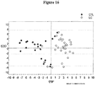
- OPLS-DA showed good separation of the total lipid profiles from the three classes of plasma. Some of the lipids were essentially the same in each source of lipid microvesicle fraction (the common components). The major classes that gave rise to the discrimination were obtained from the loading and coefficient plots, which are visualized in the 3D plots shown in Figures 1-13 . The OPLS-DA scores and coefficients plots are provided in Figures 16-24 .
- the intensities of the lipids species were normalized to the total lipid response to generate "mol fractions.”
- Different lipid classes varied in their abundances, and within a class some acyl chain length and number of double bonds also vary substantially, which was part of the classification. Most of the variance arose from intersubject variability rather than analytical variance. The difference in abundance between classes for discrimination was >4 fold with a coefficient of variation within a class of up to 50%.
- the PC (36:3) showed mean and sd of 1.38 ⁇ 0.34 (BrCA) versus 0.23 ⁇ 0.1 (healthy) versus 0.19 ⁇ 0.28 (NSCLC).
Landscapes
- Health & Medical Sciences (AREA)
- Life Sciences & Earth Sciences (AREA)
- Engineering & Computer Science (AREA)
- Molecular Biology (AREA)
- Immunology (AREA)
- Chemical & Material Sciences (AREA)
- Biomedical Technology (AREA)
- Urology & Nephrology (AREA)
- Hematology (AREA)
- Cell Biology (AREA)
- Food Science & Technology (AREA)
- General Health & Medical Sciences (AREA)
- Biotechnology (AREA)
- Pathology (AREA)
- General Physics & Mathematics (AREA)
- Medicinal Chemistry (AREA)
- Physics & Mathematics (AREA)
- Analytical Chemistry (AREA)
- Biochemistry (AREA)
- Microbiology (AREA)
- Biophysics (AREA)
- Endocrinology (AREA)
- Hospice & Palliative Care (AREA)
- Oncology (AREA)
- Investigating Or Analysing Biological Materials (AREA)
- Other Investigation Or Analysis Of Materials By Electrical Means (AREA)
- Medicines That Contain Protein Lipid Enzymes And Other Medicines (AREA)
- Medicinal Preparation (AREA)
- Peptides Or Proteins (AREA)
Abstract
Description
- This application claims the benefit of
U.S. Provisional Application No. 61/357,642, filed June 23, 2010 - This invention was made in part with government support under grant number EPS-0447479 awarded by National Science Foundation/Office of Experimental Program to Stimulate Competitive Research (EPSCoR). The U.S. Government has certain rights in the invention.
- Cancer is a major cause of death and suffering and is a major cost to medical systems in the U.S. and across the world. Early detection is associated with better treatment options and improved outcome. Therefore, early detection of cancer can help minimize both the suffering and the cost, while typically increasing the chance of survival. Thus, some embodiments of the invention are methods to determine the presence of cancers in animals.
- Embodiments of the invention include methods for determining the presence or absence of at least one cancer type in an animal comprising determining lipids amounts of lipids in a lipid set in a sample from the animal, and determining the presence or absence of at least one cancer type in the animal with a predictive model. In some of these methods, the lipid amounts of lipids in the lipid set comprise an input of the predictive model, and the sample comprises a bodily fluid or treatment thereof.
- In some embodiments, the bodily fluid is selected from the group consisting of plasma vomit, cerumen, gastric juice, breast milk, mucus, saliva, sebum, semen, sweat, tears, vaginal secretion, blood serum, aqueous humor, vitreous humor, endolymph, perilymph, peritoneal fluid, pleural fluid, cerebrospinal fluid, blood, plasma, nipple aspirate fluid, urine, stool, and bronchioalveolar lavage fluid. In still other embodiments, the bodily fluid is blood or plasma. In still other embodiments, the sample comprises a lipid microvesicle fraction.
- In some exemplary embodiments, the lipid set comprises at least 10 lipids, at least 50 lipids, at least 100 lipids, at least 200 lipids, or no more than 100,000 lipids.
- In some instances, the lipid set comprises one or more lipids selected from the one or more classes of lipids selected from the group consisting of BMP, CE ,Cer, DAG, DH-LTB4, FA, GA2, GM3, HexCer, HexDHCer, LacCer, LysoPA, LysoPC, LysoPC-pmg, LysoPE, LysoPE-pmg, LysoPS, MAG, PC, PC-pmg, PE, PE-pmg, PGA1, PGB1, SM, Sphingosine, TAG, and TH-12-keto-LTB4. In other instances, the lipid set comprises one or more lipids selected from the one or more classes of lipids selected from the group consisting of FA, MAG, DAG, TAG, PI, PE, PS, PI, PG, PA, LysoPC, LysoPE, LysoPS, LysoPI, LysoPG, LysoPA, LysoPC, LysoPE, BMP, SM, Cer, Cer-P, HexCer, GA1, GA2, GD1, GD2, GM1, GM2, GM3, GT1, and CE. In still other instances, one or more lipids in the lipid set are selected from the group consisting of BMP (30:1), BMP (32:1), BMP (34:1), BMP (35:4), BMP (36:3), BMP (37:1), BMP (37:7), BMP (38:1), BMP (38:2), BMP (38:4), BMP (39:1), BMP (39:4), BMP (40:1), BMP (40:2), BMP (40:3), BMP (40:4), BMP (40:7), BMP (42:10), BMP (42:2), BMP (42:5), BMP (44:8), CE (16:2), CE (18:2), CE (18:3), CE (18:4), CE (20:2), CE (20:4), CE (20:5), Cer (32:1), Cer (34:1), Cer (36:1), Cer (38:1), Cer (38:4), Cer (40:2), Cer (40:4), DAG (28:0), DAG (32:0), DAG (32:2), DAG (34:0), DAG (34:3), DAG (34:5), DAG (36:0), DAG (36:1), DAG (36:2), DAG (36:3), DAG (36:8), DAG (38:1), DAG (38:10), DAG (38:2), DAG (38:3), DAG (38:5), DAG (40:1), DAG (40:2), DAG (40:5), DH-LTB4 (20:3), FA (16:3), FA (19:1), GA2 (30:0), GA2 (33:2), GA2 (35:2), GA2 (37:2), GM3 (41:1), HexCer (32:1), HexDHCer (34:0), LacCer (30:0), LacCer (30:1), LacCer (32:2), LysoPA (16:2), LysoPA (16:3), LysoPA (18:1), LysoPA (22:0), LysoPA (22:1), LysoPC (16:0), LysoPC (18:0), LysoPC (18:1), LysoPC (18:4), LysoPC (20:4), LysoPC (20:5), LysoPC (26:6), LysoPC-pmg (12:0), LysoPC-pmg (18:3), LysoPC-pmg (24:4), LysoPC-pmg (26:0), LysoPE (10:1), LysoPE (16:2), LysoPE (18:2), LysoPE-pmg (18:4), LysoPS (24:1), MAG (18:0), MAG (20:3), MAG (24:2), PC (32:0), PC (32:1), PC (34:1), PC (34:1), PC (34:2), PC (34:3), PC (34:4), PC (34:6), PC (36:1), PC (36:2), PC (36:3), PC (36:4), PC (36:5), PC (36:6), PC (36:9), PC (38:2), PC (38:3), PC (38:4), PC (38:5), PC (38:6), PC (38:7), PC (38:8), PC (38:9), PC (40:5), PC (40:6), PC (40:7), PC (40:8), PC (40:9), PC (44:12), PC-pmg (30:1), PC-pmg (36:4), PC-pmg (38:5), PC-pmg (38:7), PC-pmg (40:11), PC-pmg (42:1), PE (34:7), PE (36:5), PE (36:7), PE (38:2), PE (38:3), PE (38:4), PE (38:5), PE (38:7), PE (40:4), PE (40:9), PE (42:12), PE (44:11), PE-pmg (28:2), PE-pmg (30:3), PE-pmg (34:6), PE-pmg (34:8), PE-pmg (36:5), PE-pmg (36:6), PE-pmg (40:7), PE-pmg (40:8), PE-pmg (42:10), PE-pmg (42:12), PE-pmg (42:4), PE-pmg (42:7), PE-pmg (42:8), PE-pmg (42:9), PE-pmg (44:10), PE-pmg (44:11), PE-pmg (44:12), PE-pmg (44:7), PE-pmg (44:8), PE-pmg (44:9), PGA1 (20:1), PGB1 (20:1), SM (34:1), SM (34:2), SM (36:1), SM (38:1), SM (40:1), SM (40:2), SM (42:1), SM (42:2), SM (42:3), Sphingosine (18:0), TAG (44:1), TAG (44:3), TAG (46:0), TAG (46:1), TAG (46:2), TAG (46:3), TAG (46:4), TAG (48:0), TAG (48:1), TAG (48:2), TAG (48:3), TAG (48:4), TAG (48:5), TAG (49:1), TAG (49:2), TAG (49:3), TAG (50:0), TAG (50:1), TAG (50:2), TAG (50:3), TAG (50:4), TAG (50:5), TAG (50:6), TAG (51:2), TAG (51:4), TAG (52:2), TAG (52:3), TAG (52:4), TAG (52:5), TAG (52:6), TAG (52:7), TAG (53:4), TAG (54:2), TAG (54:3), TAG (54:4), TAG (54:5), TAG (54:6), TAG (54:7), TAG (54:8), TAG (55:5), TAG (55:6), TAG (55:7), TAG (56:4), TAG (56:5), TAG (56:6), TAG (56:7), TAG (56:8), TAG (56:9), TAG (58:10), TAG (58:6), TAG (58:8), TAG (58:9), TAG (60:12), and TH-12-keto-LTB4(20:2).
- In some embodiments, the at least one cancer type comprises lung cancer and one or more lipids in the lipid set are selected from the group consisting of LysoPA (22:0), PE-pmg (42:9), FA (16:3), FA (19:1), CE (18:2), Cer (36:1), Cer (38:4), PC (38:5), Cer (38:1), and TAG (44:3). In still other embodiments, the at least one cancer type comprises lung cancer and one or more lipids in the lipid set are selected from the group consisting of TAG (44:3), PC (36:5), PC (38:5), Cer (38:4), PE-pmg (42:9), PC (38:7), LysoPA (22:0), Cer (38:1), Cer (34:1), Cer (36:1), PC (40:7), TAG (54:5), TAG (54:6), CE (18:2), PC (36:4), FA (16:3), PE-pmg (44:11), TAG (52:5), Cer (40:4), CE (20:5), PC (38:6), TAG (50:2), MAG (18:0), FA (19:1), TAG (52:2), LysoPA (22:1), MAG (24:2), TAG (54:7), TAG (50:3), TAG (50:1), DAG (36:3), PC (34:1), TAG (52:6), BMP (30:1), PE-pmg (44:12), CE (20:4), BMP (40:3), PE (44:11), PC (40:8), TAG (56:9), PE-pmg (34:6), PE (36:7), PE (36:5), TAG (56:7), TAG (56:8), DAG (34:3), TAG (56:6), BMP (42:10), TAG (52:3), BMP (39:4), BMP (36:3), TAG (54:3), TAG (56:5), TAG (54:8), PC (34:6), PC (40:6), DAG (36:0), LysoPE (10:1), DAG (40:5), Cer (32:1), TAG (50:5), TAG (50:4), PE-pmg (36:6), BMP (42:5), TAG (46:3), and PE (38:5). In further embodiments, the at least one cancer type comprises lung cancer and one or more lipids in the lipid set are selected from the group consisting of TAG (44:3), PC (36:5), PC (38:5), Cer (38:4), PE-pmg (42:9), PC (38:7), LysoPA (22:0), Cer (38:1), Cer (34:1), and Cer (36:1).
- In some embodiments of the invention the at least one cancer type comprises breast cancer and one or more lipids in the lipid set are selected from the group consisting of LysoPA (22:1), PE-pmg (42:9), CE (20:5), TAG (52:3), LysoPA (22:0), PC (36:3), PC (36:4), PC (36:2), PC (34:2), and PC (34:1). In other embodiments, the at least one cancer type comprises breast cancer and one or more lipids in the lipid set are selected from the group consisting of PC (34:2), PC (34:1), PC (36:2), PC (36:4), PC (36:3), PC (38:4), LysoPA (22:1), PE-pmg (42:9), LysoPA (22:0), CE (20:5), Cer (36:1), CE (18:2), DAG (34:0), SM (34:1), DAG (32:0), PE-pmg (40:8), PC (38:3), DAG (36:0), PC (36:1), TAG (54:5), TAG (54:6), PE-pmg (44:11), PE-pmg (42:8), TAG (52:2), SM (42:2), PC (38:6), TAG (54:7), PC (40:6), PC (40:7), LysoPC (16:0), FA (16:3), TAG (52:5), TAG (44:3), BMP (38:2), BMP (30:1), SM (40:1), PE-pmg (42:10), BMP (40:2), PE-pmg (40:7), SM (36:1), PE (38:2), PC (34:3), PC (36:5), PC (32:0), PC (32:1), BMP (37:1), BMP (40:3), PC (36:9), SM (42:3), PC-pmg (36:4), PC-pmg (38:5), PC (40:9), TAG (54:3), PE-pmg (44:12), BMP (36:3), FA (19:1), BMP (39:1), TAG (50:3), BMP (42:10), PC (34:6), GA2 (35:2), TAG (58:9), PE-pmg (42:7), and LysoPC (18:0). In still other embodiments, the at least one cancer type comprises breast cancer and one or more lipids in the lipid set are selected from the group consisting of PC (34:2), PC (34:1), PC (36:2), PC (36:4), PC (36:3), PC (38:4), LysoPA (22:1), PE-pmg (42:9), LysoPA (22:0), and CE (20:5).
- In some embodiments of the inventions, the at least one cancer type comprises lung cancer and breast cancer, and one or more lipids in the lipid set are selected from the group consisting of LysoPA (22:1), PC (36:5), TAG (52:3), PC (38:5), CE (20:5), TAG (50:2), BMP (39:1), PC (34:2), CE (18:2), and PC (34:1). In other embodiments, the at least one cancer type comprises lung cancer and breast cancer, and one or more lipids in the lipid set are selected from the group consisting of PC (34:2), PC (36:2), TAG (44:3), CE (18:2), PC (34:1), LysoPA (22:1), PC (36:5), Cer (36:1), CE (20:5), PC (36:3), PC (38:4), PC (36:4), Cer (38:4), PC (38:5), PC (38:7), Cer (38:1), TAG (50:2), Cer (34:1), SM (34:1), Cer (40:4), MAG (18:0), MAG (24:2), PC (38:3), PE-pmg (40:8), PE-pmg (42:8), TAG (50:1), DAG (32:0), PC (36:1), DAG (34:0), LysoPC (16:0), PE-pmg (34:6), DAG (36:3), PC (36:9), PE (36:5), TAG (52:6), FA (19:1), PE-pmg (44:11), BMP (38:2), PE (44:11), TAG (48:2), SM (42:2), BMP (40:2), PE-pmg (42:10), PE (36:7), PE-pmg (40:7), BMP (39:1), BMP (37:1), PE-pmg (36:6), PE (38:5), PC (32:0), PE (38:2), GA2 (35:2), DAG (34:3), PE-pmg (44:12), MAG (16:0), PC (32:1), LysoPE (10:1), SM (36:1), BMP (39:4), TAG (56:7), and PE-pmg (42:9). In still other embodiments, the at least one cancer type comprises lung cancer and breast cancer, and one or more lipids in the lipid set are selected from the group consisting of PC (34:2), PC (36:2), TAG (44:3), CE (18:2), PC (34:1), LysoPA (22:1), PC (36:5), Cer (36:1), CE (20:5), and PC (36:3).
- In some embodiments of the invention, the lipid amounts are determined using mass spectrometry, such as with a Fourier transform ion cyclotron resonance mass analyzer.
- In other embodiments, the sample is a treatment of a bodily fluid. For example, the sample can be a treatment of a bodily fluid that comprises one or more extractions using one or more solutions comprising acetonitrile, water, chloroform, methanol, butylated hydroxytoluene, trichloroacetic acid, or combinations thereof.
- In some embodiments, the predictive model comprises one or more dimension reduction methods, such as one or more methods selected from the group consisting of principal component analysis (PCA), soft independent modeling of class analogy (SIMCA), partial least squares discriminant analysis (PLS-DA), and orthogonal partial least squares discriminant analysis (OPLS-DA).
- In some embodiments of the invention, the animal is selected from the group consisting of human, dog, cat, horse, cow, pig, sheep, chicken, turkey, mouse, and rat.
- In other embodiments of the invention, the at least one cancer type is selected from the group consisting of carcinomas, sarcomas, hematologic cancers, neurological malignancies, basal cell carcinoma, thyroid cancer, neuroblastoma, ovarian cancer, melanoma, renal cell carcinoma, hepatocellular carcinoma, breast cancer, colon cancer, lung cancer, pancreatic cancer, brain cancer, prostate cancer, chronic lymphocytic leukemia, acute lymphoblastic leukemia, rhabdomyosarcoma, Glioblastoma multiforme, meningioma, bladder cancer, gastric cancer, Glioma, oral cancer, nasopharyngeal carcinoma, kidney cancer, rectal cancer, lymph node cancer, bone marrow cancer, stomach cancer, uterine cancer, leukemia, basal cell carcinoma, cancers related to epithelial cells, cancers that can alter the regulation or activity of Pyruvate Carboxylase, and tumors associated with any of the aforementioned cancer types.
- In further embodiments, the method comprises determining the presence or absence of more than one cancer type.
- Embodiments of the invention include a method for determining the presence or absence of at least one cancer type in an animal comprising determining the presence or absence of at least one cancer type in the animal with a predictive model by analyzing lipid amounts of lipids in a lipid set in a sample from the animal. In some instances, the lipid amounts of lipids in the lipid set comprise an input of the predictive model, and the sample comprises a bodily fluid or treatment thereof.
- The following drawings form part of the present specification and are included to further demonstrate certain aspects of the present invention. The invention may be better understood by reference to one or more of these drawings in combination with the description of specific embodiments presented herein.
-
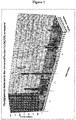
Figure 1 . Diacylglycerols in samples from humans with lung cancer, breast cancer, and no cancer (control). -
Figure 2 . Phosphatidylcholines in samples from humans with lung cancer, breast cancer, and no cancer (control). -
Figure 3 . Phosphatidylcholines in samples from humans with lung cancer, breast cancer, and no cancer (control). -
Figure 4 . Phosphatidylcholines in samples from humans with lung cancer, breast cancer, and no cancer (control). -
Figure 5 . Phosphatidylcholines in samples from humans with lung cancer, breast cancer, and no cancer (control). -
Figure 6 . Phosphatidylethanolamines in samples from humans with lung cancer, breast cancer, and no cancer (control). -
Figure 7 . Phosphatidylethanolamines in samples from humans with lung cancer, breast cancer, and no cancer (control). -
Figure 8 . Phosphatidylethanolamines in samples from humans with lung cancer, breast cancer, and no cancer (control). -
Figure 9 . Phosphatidylethanolamines-pmg in samples from humans with lung cancer, breast cancer, and no cancer (control). -
Figure 10 . Phosphatidylethanolamines-pmg in samples from humans with lung cancer, breast cancer, and no cancer (control). -
Figure 11 . Triacylglycerols in samples from humans with lung cancer, breast cancer, and no cancer (control). -
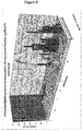
Figure 12 . Monoacylglycerols in samples from humans with lung cancer, breast cancer, and no cancer (control). -
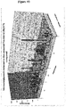
Figure 13 . Lipid classes in samples from humans comparing cancer type. -
Figure 14 . PC1-PC3 PCA scores plot for breast cancer, control, and lung cancer samples - 4 components, R2X = 0.475, Q2 = 0.296. -
Figure 15 . PC1-PC3 PCA loadings plot for breast cancer, control, and lung cancer samples - 4 components, R2X = 0.475, Q2 = 0.296. -
Figure 16 . OPLS-DA scores plot separating lung cancer and control samples -1+1 orthogonal components, R2X = 0.253, R2Y = 0.619, Q2 = 0.345. -
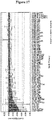
Figure 17 . OPLS-DA coefficients plot separating lung cancer and control samples - 1+1 orthogonal components, R2X = 0.253, R2Y = 0.619, Q2 = 0.345. The coefficients plot indicates lipids elevated in control samples. -
Figure 18 . OPLS-DA coefficients plot separating lung cancer and control samples - 1+1 orthogonal components, R2X = 0.253, R2Y = 0.619, Q2 = 0.345. The coefficients plot indicates lipids elevated in lung cancer samples. -
Figure 19 . OPLS-DA scores plot separating breast cancer and control samples - 1+1 orthogonal components, R2X = 0.281, R2Y = 0.762, Q2 = 0.625. -
Figure 20 . OPLS-DA coefficients plot separating breast cancer and control samples - 1+1 orthogonal components, R2X = 0.281, R2Y = 0.762, Q2 = 0.625. The coefficients plot indicates lipids elevated in control samples. -
Figure 21 . OPLS-DA coefficients plot separating breast cancer and control samples - 1+1 orthogonal components, R2X = 0.281, R2Y = 0.762, Q2 = 0.625. The coefficients plot indicates lipids elevated in breast cancer samples. -
Figure 22 . OPLS-DA scores plot separating lung cancer and breast cancer samples - 1+1 orthogonal components, R2X = 0.309, R2Y = 0.816, Q2 = 0.725. -
Figure 23 . OPLS-DA coefficients plot separating lung cancer and breast cancer samples - 1+1 orthogonal components, R2X = 0.309, R2Y = 0.816, Q2 = 0.725. The coefficients plot indicates lipids elevated in lung cancer samples. -
Figure 24 . OPLS-DA coefficients plot separating lung cancer and breast cancer samples -1+1 orthogonal components, R2X = 0.309, R2Y = 0.816, Q2 = 0.725. The coefficients plot indicates lipids elevated in breast cancer samples. -
Figure 25 . A flow chart representing an embodiment of one of the methods disclosed herein. Cancer status can include the presence or absence of one or more cancer types. - Some embodiments of the invention include methods for detecting the presence or absence of one or more cancer types by determining the amount of lipids in as lipid set in a sample. The sample can be a bodily fluid (or treatment thereof) from an animal. In some instances, the sample (e.g., a bodily fluid extract) comprises a concentration of lipid microvesicles that is higher than normally found in a bodily fluid. The lipid amounts in the lipid set are analyzed using a predictive model to determine the presence or absence of one or more cancer types.
- Lipids are designated according to the following notation XXX (YY:ZZ). XXX is the abbreviation for the lipid group (in many instances indicating the lipid headgroup) as provided, for example in Table 1. YY is the number of carbons in the acyl chain. ZZ is the number of double bonds in the acyl chains.
- The term lipid, as used herein, is defined as a collection of one or more isomers. For example, PC (36: 1) is a lipid and is the collection of one or more of the phosphatidylcholine isomers that have 36 carbons in the acyl chain and one double bond in either of the two acyl chains; these isomers have identical molecular weights. Although the term lipid can encompass the entire collection of isomers, the sample may, in fact, have only one isomer, several isomers, or any number of isomers less than the total number of all possible isomers in a collection. Accordingly, lipid can refer to one or more of the isomers that make up the entire collection of possible isomers.
- The term lipid set is defined to include one or more lipids.
- The term lipid amount (and similar phrases such as, amounts of lipids or amount of a lipid) is defined to encompass an absolute amount of a lipid (e.g., in mmoles) or a relative amount of a lipid (e.g., in % relative intensity).
- Table 1 provides lipid name abbreviations used in the data; other abbreviations are provided in the text as needed.
Table 1 Abbreviation Name BMP Bis(monoacylglycero)phosphates CE Cholesterol Esters Cer Ceramides DAG Diacylglycerols DH-LTB4 Dihydroleukotriene b4 FA Fatty Acids GA2 Gangliosides A2 GM3 Gangliosides M3 HexCer Hexose Ceramides HexDHCer Hexosyl dihydroceramide LacCer Lactosylceramide LysoPA Lysophosphatidic acid LysoPC Lysophosphatidylcholines LysoPC-pmg Lysophosphatidylcholines-plasmalogens LysoPE Lysophosphatidylethanolamines LysoPE-pmg Lysophosphatidylethanolamines-plasmalogens LysoPS Lysophosphatidylserines MAG Monoacylglycerols PC Phospahtidylcholines PC-pmg Phosphatidylcholines-plasmalogens PE Phosphatidylethanolamines PE-pmg Phosphatidylethanolamines-plasmalogens PGA1 Prostaglandin A 1 PGB1 Prostaglandin B 1 SM Sphingomyelins Sphingosine Sphingosine TAG Triacylglycerols TH-12-keto-LTB4 Tetrahydro-12-keto-leukotriene b4 - Bodily fluids can be any suitable bodily fluid for the determination of cancer and include but are not limited to vomit, cerumen (earwax), gastric juice, breast milk, mucus (e.g., nasal drainage and phlegm), saliva, sebum (skin oil), semen (including prostatic fluid), sweat, tears, vaginal secretion, blood serum, aqueous humor, vitreous humor, endolymph, perilymph, peritoneal fluid, pleural fluid, cerebrospinal fluid, blood, plasma, nipple aspirate fluid, urine, stool, bronchioalveolar lavage fluid, peripheral blood, sera, plasma, ascites, cerebrospinal fluid (CSF), sputum, bone marrow, synovial fluid, aqueous humor, amniotic fluid, Cowper's fluid or pre-ejaculatory fluid, female ejaculate, fecal matter, hair, cyst fluid, pleural and peritoneal fluid, pericardial fluid, lymph, chyme, chyle, bile, interstitial fluid, menses, pus, vaginal secretions, mucosal secretion, stool water, pancreatic juice, lavage fluids from sinus cavities, bronchopulmonary aspirates, or other lavage fluids. A bodily fluid may also include the blastocyl cavity, umbilical cord blood, or maternal circulation which may be of fetal or maternal origin.
- The bodily fluid can be obtained from any animal tissue (e.g., mammalian tissues) including, but not limited to connective tissue, muscle tissue, nervous tissue, adipose tissue, endothelial tissue, or epithelial tissue. The tissue can be at least part of an organ or part of an organ system. Organs can include, but are not limited to heart, blood, blood vessels, salivary glands, esophagus, stomach, liver, gallbladder, pancreas, large intestines, small intestines, rectum, anus, colon, endocrine glands (e.g., hypothalamus, pituitary, pineal body, thyroid, parathyroids and adrenals), kidneys, ureters, bladder, urethraskin, hair, nails, lymph, lymph nodes, lymph vessels, leukocytes, tonsils, adenoids, thymus, spleen, muscles, brain, spinal cord, peripheral nerves, nerves, sex organs (e.g., ovaries, fallopian tubes, uterus, vagina, mammary glands (e.g., breasts), testes, vas deferens, seminal vesicles, prostate and penis), pharynx, larynx, trachea, bronchi, lungs, diaphragm, bones, cartilage, ligaments, or tendons. Organ systems can include, but are not limited to circulatory system, digestive system, endocrine system, excretory system, integumentary system, lymphatic system, muscular system, nervous system, reproductive system, respiratory system, or skeletal system.
- Bodily fluids can be removed from the animal by any suitable method, including but not limited to blood draw, manipulation of natural excretion points (e.g., by suction or by manual manipulation - such as of the breast nipple to obtain Nipple Aspirate Fluid), surgical methods (e.g., resection), biopsy methods (e.g., fine needle aspiration or core needle biopsy), or animal sacrifice followed by organ removal and dissection. Removed bodily fluids can be frozen in liquid nitrogen. Preparation of the removed bodily fluids can be performed in any suitable manner.
- The bodily fluid may be obtained through a third party, such as a party not performing the analysis. For example, the bodily fluid may be obtained through a clinician, physician, or other health care manager of a subject from which the sample is derived. In some embodiments, the bodily fluid may obtained by the same party doing the analyzing.
- In some embodiments, the bodily fluid is the sample. In other embodiments, the bodily fluid is treated to provide the sample. Treatment can include any suitable method, including but not limited to extraction, centrifugation (e.g., ultracentrifugation), lyophilization, fractionation, separation (e.g., using column or gel chromatography), or evaporation. In some instances, this treatment can include one or more extractions with solutions comprising any suitable solvent or combinations of solvents, such as, but not limited to acetonitrile, water, chloroform, methanol, butylated hydroxytoluene, trichloroacetic acid, toluene, hexane, benzene, or combinations thereof. For instance, in some embodiments, fractions from blood are extracted with a mixture comprising methanol and butylated hydroxytoluene. In some instances, the sample (e.g., a bodily fluid extract or a lipid microvesicle fraction of blood plasma) comprises a concentration of lipid microvesicles that is higher than normally found in a bodily fluid.
- The volume of the sample (e.g., the bodily fluid or treatment thereof) used for analyzing can be in the range of about 0.1 mL to about 20 mL, such as no more than about 20, about 15, about 10, about 9, about 8, about 7, about 6, about 5, about 4, about 3, about 2, about 1, or about 0.1 mL.
- Broad classes of lipids that can be part of a lipid set include, but are not limited to, fatty acids (characterized by a carboxyl group and an acyl chain), Glycerolipids (characterized by the presence of a glycerol backbone with one - monoacylglycerols (MAG), two - diacylglycerols (DAG) or three - triacylglycerols (TAG) ester linked fatty acyl chains), glycerophospholipids (GPL) (characterized by a glyceryl backbone with two ester-linked acyl chains and a phosphate-linked polar headgroup - GPLs include phosphatidylcholines (PC), phosphatidylethanolamines (PE), phosphatidylserines (PS), phosphatidylglycerols (PG), phosphatidylinositols (PI), inositol, and phosphatidic acids), Lysoglycerophospholipids (LGPL) (LGPLs are missing one of the acyl chains at the glycerol backbone, e.g., at the C2 position - LGPLs include include lysophospahtidylcholines (LysoPC), lysophosphatidylethanolamines (LysoPE), lysophosphatidylserines (LysoPS), lysophosphatidylglycerols (LysoPG), lysophosphatidylinositols (LysoPI), lysoinositol, and lysophosphatidic acids), sphingolipids (SPL) (characterized by a sphingosine base backbone with a trans double bond between C4 and C5 of an acyl chain linked to the amino group via an amide linkage), Bis(monoacylglycero)phosphate (BMP), Ceramides, Gangliosides, Sterols, Prenols, Saccharolipids, and Polyketides. In some instances, the lipid group is based on the acyl chain composition which can vary in any number of ways including the number of carbons in the acyl chain and the number of double bonds in the acyl chain.
- In other embodiments, lipids in the lipid set can come from one or more classes of lipids, such as, BMP, CE, Cer, DAG, DH-LTB4, FA, GA2, GM3, HexCer, HexDHCer, LacCer, LysoPA, LysoPC, LysoPC-pmg, LysoPE, LysoPE-pmg, LysoPS, MAG, PC, PC-pmg, PE, PE-pmg, PGA1, PGB1, SM, Sphingosine, TAG, or TH-12-keto-LTB4. In still other embodiments, lipids in the lipid set can come from one or more classes of lipids, such as, FA, MAG, DAG, TAG, PC, PE, PS, PI, PG, PA, LysoPC, LysoPE, LysoPS, LysoPI, LysoPG, LysoPA, LysoPC, LysoPE, BMP, SM, Cer, Cer-P, HexCer, GA1, GA2, GD1, GD2, GM1, GM2, GM3, GT1, or CE.
- In some embodiments, the lipid set used in the predictive model is limited to those that have a higher probability to be found in human lipid pools. In other embodiments, the lipid set excludes very short and very long acyl chains (e.g., less that 10 carbons and more than 26 carbons within a chain). In still other embodiments, the lipid set lipids (e.g., GPLs or LGPL) are limited to those containing an even number of carbons in the acyl chain. In other embodiments, the lipid set included one or more lipids listed in Table 2.
Table 2 Lipid Class Lipid Group Abbrev Acyl Chain Range Unsaturated sites (# double bonds) Additional features or comments Total # of structures considered Fatty Acids Fatty Acids FA 10-26 0-6 79 Glycerolipids Monoacylglycerols MAG 10-26 0-6 Even # of carbons only 43 Glycerolipids Diacylglycerols DAG 22-46 0-12 Even # of carbons only 120 Glycerolipids Triacylglycerols TAG 44-66 0-18 316 Glycerophospholipids Phosphatidylcholines PC 28-44 0-12 Even # of carbons only 77 Glycerophospholipids Phosphatidylethanolamines PE 28-44 0-12 Even # of carbons only 77 Glycerophospholipids Phosphatidylserines PS 28-44 0-12 Even # of carbons only 77 Glycerophospholipids Phosphatidylinositols PI 28-44 0-12 Even # of carbons only 77 Glycerophospholipids Phosphatidylglycerols PG 28-44 0-12 Even # of carbons only 77 Glycerophospholipids Phosphatidicacid PA 28-44 0-12 Even # of carbons only 77 Lysoglycholipids Lysophosphatidylcholines LysoPC 10-26 0-6 Even # of carbons only 43 Lysoglycerophospholipids Lysophosphatidyl ethanolamines LysoPE 10-26 0-6 Even # of carbons only 43 Lysoglycerophospholipids Lysophosphatidylserines LysoPS 10-26 0-6 Even # of carbons only 43 Lysoglycerophospholipids Lysophosphatidylinositols LysoPI 10-26 0-6 Even # of carbons only 43 Lysoglycerophospholipids Lysophophatidylglycerols LysoPG 10-26 0-6 Even # of carbons only 43 Lysoglycerophospholipids Lysophosphatidic acid LysoPA 10-26 0-6 Even # of carbonsonly 43 Lysoglycerophospholipids Lysophosphatidylcholines-pmg LysoPC 10-26 0-6 Even # of carbons only 43 Lysoglycerophospholipids Lysophosphatidyl ethanolamines-pmg LysoPE 10-26 0-6 Even # of carbons only 43 Bis(monoacylglycero)phosphates BMP 28-48 0-12 211 Sphingolipids Sphingomyelins SM 30-42 0-3 25 Sphingolipids Ceramides Cer 30-44 0-3 33 Sphingolipids Ceramide phosphates Cer-P 30-44 0-3 33 Sphingolipids Hexose Ceramides HexCer 30-44 0-3 33 Sphingolipids Gangliosides A1 GA1 28-46 0-2 57 Sphingolipids Gangliosides A2 GA2 28-46 0-2 57 Sphingolipids Gangliosides D1 GD1 28-46 0-2 57 Sphingolipids Gangliosides D2 GD2 28-46 0-2 57 Sphingolipids Gangliosides M1 GM1 28-46 0-2 57 Sphingolipids Gangliosides M2 GM2 28-46 0-2 57 Sphingolipids Gangliosides M3 GM3 28-46 0-2 57 Sphingolipids Gangliosides A1 GT1 28-46 0-2 57 Cholester ol Esters Cholesterol Esters CE 10-26 0-6 79 - In some embodiments, the lipids in the lipid set can include one or more of BMP (30:1), BMP (32:1), BMP (34:1), BMP (35:4), BMP (36:3), BMP (37:1), BMP (37:7), BMP (38:1), BMP (38:2), BMP (38:4), BMP (39:1), BMP (39:4), BMP (40:1), BMP (40:2), BMP (40:3), BMP (40:4), BMP (40:7), BMP (42:10), BMP (42:2), BMP (42:5), BMP (44:8), CE (16:2), CE (18:2), CE (18:3), CE (18:4), CE (20:2), CE (20:4), CE (20:5), Cer (32:1), Cer (34:1), Cer (36:1), Cer (38:1), Cer (38:4), Cer (40:2), Cer (40:4), DAG (28:0), DAG (32:0), DAG (32:2), DAG (34:0), DAG (34:3), DAG (34:5), DAG (36:0), DAG (36:1), DAG (36:2), DAG (36:3), DAG (36:8), DAG (38:1), DAG (38:10), DAG (38:2), DAG (38:3), DAG (38:5), DAG (40:1), DAG (40:2), DAG (40:5), DH-LTB4 (20:3), FA (16:3), FA (19:1), GA2 (30:0), GA2 (33:2), GA2 (35:2), GA2 (37:2), GM3 (41:1), HexCer (32:1), HexDHCer (34:0), LacCer (30:0), LacCer (30:1), LacCer (32:2), LysoPA (16:2), LysoPA (16:3), LysoPA (18:1), LysoPA (22:0), LysoPA (22:1), LysoPC (16:0), LysoPC (18:0), LysoPC (18:1), LysoPC (18:4), LysoPC (20:4), LysoPC (20:5), LysoPC (26:6), LysoPC-pmg (12:0), LysoPC-pmg (18:3), LysoPC-pmg (24:4), LysoPC-pmg (26:0), LysoPE (10:1), LysoPE (16:2), LysoPE (18:2), LysoPE-pmg (18:4), LysoPS (24:1), MAG (18:0), MAG (20:3), MAG (24:2), PC (32:0), PC (32:1), PC (34:1), PC (34:1), PC (34:2), PC (34:3), PC (34:4), PC (34:6), PC (36:1), PC (36:2), PC (36:3), PC (36:4), PC (36:5), PC (36:6), PC (36:9), PC (38:2), PC (38:3), PC (38:4), PC (38:5), PC (38:6), PC (38:7), PC (38:8), PC (38:9), PC (40:5), PC (40:6), PC (40:7), PC (40:8), PC (40:9), PC (44:12), PC-pmg (30:1), PC-pmg (36:4), PC-pmg (38:5), PC-pmg (38:7), PC-pmg (40:11), PC-pmg (42:1), PE (34:7), PE (36:5), PE (36:7), PE (38:2), PE (38:3), PE (38:4), PE (38:5), PE (38:7), PE (40:4), PE (40:9), PE (42:12), PE (44:11), PE-pmg (28:2), PE-pmg (30:3), PE-pmg (34:6), PE-pmg (34:8), PE-pmg (36:5), PE-pmg (36:6), PE-pmg (40:7), PE-pmg (40:8), PE-pmg (42:10), PE-pmg (42:12), PE-pmg (42:4), PE-pmg (42:7), PE-pmg (42:8), PE-pmg (42:9), PE-pmg (44:10), PE-pmg (44:11), PE-pmg (44:12), PE-pmg (44:7), PE-pmg (44:8), PE-pmg (44:9), PGA1 (20:1), PGB1 (20:1), SM (34:1), SM (34:2), SM (36:1), SM (38:1), SM (40: 1), SM (40:2), SM (42:1), SM (42:2), SM (42:3), Sphingosine (18:0), TAG (44:1), TAG (44:3), TAG (46:0), TAG (46:1), TAG (46:2), TAG (46:3), TAG (46:4), TAG (48:0), TAG (48:1), TAG (48:2), TAG (48:3), TAG (48:4), TAG (48:5), TAG (49:1), TAG (49:2), TAG (49:3), TAG (50:0), TAG (50:1), TAG (50:2), TAG (50:3), TAG (50:4), TAG (50:5), TAG (50:6), TAG (51:2), TAG (51:4), TAG (52:2), TAG (52:3), TAG (52:4), TAG (52:5), TAG (52:6), TAG (52:7), TAG (53:4), TAG (54:2), TAG (54:3), TAG (54:4), TAG (54:5), TAG (54:6), TAG (54:7), TAG (54:8), TAG (55:5), TAG (55:6), TAG (55:7), TAG (56:4), TAG (56:5), TAG (56:6), TAG (56:7), TAG (56:8), TAG (56:9), TAG (58:10), TAG (58:6), TAG (58:8), TAG (58:9), TAG (60:12), or TH-12-keto-LTB4(20:2).
- In some embodiments (e.g., to determine lung cancer), the lipids in the lipid set include, but are not limited to LysoPA (22:0), PE-pmg (42:9), FA (16:3), FA (19:1), CE (18:2), PE-pmg (44:11), BMP (30:1), PE-pmg (44:12), BMP (42:10), BMP (36:3), PC (34:6), BMP (40:3), TAG (50:2), BMP (39:1), DAG (38:3), BMP (37:1), PC-pmg (40:11), DAG (40:5), DAG (32:2), TAG (46:2), BMP (42:5), PE-pmg (42:8), PC (44:12), GA2 (35:2), TAG (50:1), CE (20:5), DAG (40:2), TAG (49:2), LysoPE (18:2), BMP (40:7), DAG (36:8), LysoPC (18:1), PE-pmg (36:5), PE-pmg (42:7), DH-LTB4 (20:3), PGA1 (20:1), PGB1 (20:1), BMP (38:4), BMP (35:4), BMP (44:8), TAG (46:1), TAG (44:1), LysoPC (18:4), DAG (36:0), DAG (38:2), LysoPC (20:4), DAG (38:1), LysoPC (26:6), DAG (36:2), DAG (34:5), TAG (49:1), TAG (56:7), DAG (38:5), Cer (40:2), BMP (40:4), GA2 (30:0), LysoPC-pmg (12:0), LysoPC-pmg (26:0), PC-pmg (30:1), LysoPC (20:5), PE-pmg (44:10), PE-pmg (34:8), PE-pmg (44:7), GM3 (41:1), BMP (37:7), PC (38:9), CE (20:4), SM (36:1), LysoPC-pmg (18:3), TAG (54:2), PE (38:5), PC (34:4), PC (34:3), TAG (48:0), TAG (50:5), DAG (32:0), PC (36:3), LysoPA (18:1), TAG (48:3), TAG (50:4), TAG (54:3), LysoPA (16:3), PC (36:1), TAG (58:9), PE-pmg (36:6), TAG (54:7), TAG (56:5), SM (42:1), LysoPA (16:2), DAG (28:0), TAG (46:3), TAG (54:8), SM (42:2), PC (40:8), LysoPE (10:1), PE (44:11), TAG (56:9), PC (40:6), SM (40:1), PE (36:5), Cer (32:1), BMP (39:4), PE-pmg (34:6), DAG (34:3), TAG (54:4), TAG (54:6), TAG (52:6), PE (36:7), PC (38:4), DAG (36:3), PC (36:2), PC (38:6), Cer (40:4), TAG (52:4), MAG (24:2), TAG (54:5), PC (36:5), TAG (50:3), TAG (52:5), MAG (18:0), LysoPA (22:1), TAG (52:3), PC (36:4), PC (40:7), PC (34:2), PC (34:1), Cer (34:1), PC (38:7), Cer (36:1), Cer (38:4), PC (38:5), Cer (38:1), or TAG (44:3).
- In some embodiments (e.g., to determine lung cancer), the lipids in the lipid set include, but are not limited to LysoPA (22:0), PE-pmg (42:9), FA (16:3), FA (19:1), CE (18:2), Cer (36:1), Cer (38:4), PC (38:5), Cer (38:1), or TAG (44:3).
- In some embodiments (e.g., to determine lung cancer), the lipids in the lipid set include, but are not limited to LysoPA (22:0), PE-pmg (42:9), FA (16:3), FA (19:1), CE (18:2), PE-pmg (44:11), BMP (30:1), PE-pmg (44:12), BMP (42:10), BMP (36:3), PC (34:6), BMP (40:3), TAG (50:2), BMP (39:1), DAG (38:3), BMP (37:1), PC-pmg (40:11), DAG (40:5), DAG (32:2), TAG (46:2), BMP (42:5), PE-pmg (42:8), PC (44:12), GA2 (35:2), TAG (50:1), PE (36:7), PC (38:4), DAG (36:3), PC (36:2), PC (38:6), Cer (40:4), TAG (52:4), MAG (24:2), TAG (54:5), PC (36:5), TAG (50:3), TAG (52:5), MAG (18:0), LysoPA (22:1), TAG (52:3), PC (36:4), PC (40:7), PC (34:2), PC (34:1), Cer (34:1), PC (38:7), Cer (36:1), Cer (38:4), PC (38:5), Cer (38:1), or TAG (44:3).
- In some embodiments (e.g., to determine lung cancer), the lipids in the lipid set include, but are not limited to TAG (44:3), PC (36:5), PC (38:5), Cer (38:4), PE-pmg (42:9), PC (38:7), LysoPA (22:0), Cer (38:1), Cer (34:1), Cer (36:1), PC (40:7), TAG (54:5), TAG (54:6), CE (18:2), PC (36:4), FA (16:3), PE-pmg (44:11), TAG (52:5), Cer (40:4), CE (20:5), PC (38:6), TAG (50:2), MAG (18:0), FA (19:1), TAG (52:2), LysoPA (22:1), MAG (24:2), TAG (54:7), TAG (50:3), TAG (50:1), DAG (36:3), PC (34:1), TAG (52:6), BMP (30:1), PE-pmg (44:12), CE (20:4), BMP (40:3), PE (44:11), PC (40:8), TAG (56:9), PE-pmg (34:6), PE (36:7), PE (36:5), TAG (56:7), TAG (56:8), DAG (34:3), TAG (56:6), BMP (42:10), TAG (52:3), BMP (39:4), BMP (36:3), TAG (54:3), TAG (56:5), TAG (54:8), PC (34:6), PC (40:6), DAG (36:0), LysoPE (10:1), DAG (40:5), Cer (32:1), TAG (50:5), TAG (50:4), PE-pmg (36:6), BMP (42:5), TAG (46:3), or PE (38:5).
- In some embodiments (e.g., to determine lung cancer), the lipids in the lipid set include, but are not limited to TAG (44:3), PC (36:5), PC (38:5), Cer (38:4), PE-pmg (42:9), PC (38:7), LysoPA (22:0), Cer (38:1), Cer (34:1), or Cer (36:1).
- In some embodiments (e.g., to determine breast cancer), the lipids in the lipid set include, but are not limited to LysoPA (22:1), PE-pmg (42:9), CE (20:5), TAG (52:3), LysoPA (22:0), TAG (52:2), DAG (32:0), TAG (54:4), DAG (34:0), DAG (36:0), TAG (54:3), PE-pmg (44:11), TAG (44:3), BMP (30:1), TAG (52:4), BMP (40:3), PC (36:5), PC (36:9), PC (34:6), PE-pmg (44:12), BMP (42:10), BMP (36:3), PC-pmg (40:11), FA (16:3), DAG (38:3), TAG (54:2), BMP (42:5), HexDHCer (34:0), DAG (40:5), Cer (40:4), PC-pmg (30:1), PC (44:12), PE (38:5), PE (42:12), TAG (48:0), PE (36:5), PE-pmg (34:6), MAG (24:2), PE-pmg (36:6), TAG (56:4), TAG (49:3), CE (18:3), FA (19:1), DAG (38:5), TAG (50:0), BMP (38:4), TAG (46:3), TAG (51:4), BMP (35:4), PE-pmg (36:5), PE (44:11), DAG (38:2), TAG (48:3), CE (20:4), CE (18:4), LysoPC-pmg (18:3), BMP (44:8), LysoPC (20:4), TAG (60:12), LysoPC (18:4), PE-pmg (28:2), PE (40:4), PC-pmg (42:1), Sphingosine (18:0), LacCer (32:2), LysoPC (18:0), PE (38:3), MAG (20:3), SM (34:2), PC (40:5), SM (42:1), PE (38:7), LacCer (30:1), TAG (44:1), TAG (58:8), PC (40:9), CE (16:2), TAG (58:10), PE-pmg (44:10), SM (40:2), TAG (50:4), LysoPE-pmg (18:4), GA2 (37:2), PC-pmg (38:5), PC-pmg (36:4), PC (38:2), LacCer (30:0), GA2 (33:2), SM (42:3), PE (38:4), TAG (46:1), PC (32:1), BMP (42:2), LysoPC (16:0), SM (38:1), PE (38:2), TAG (50:3), TAG (58:9), PC (40:6), TAG (48:1), TAG (50:2), BMP (38:2), PE-pmg (40:7), PE-pmg (42:10), LysoPC-pmg (24:4), PC (34:3), PE-pmg (44:9), SM (36:1), PE-pmg (42:12), TAG (48:2), BMP (40:1), PE-pmg (44:8), DAG (36:1), TAG (56:7), LysoPC (26:6), PE-pmg (40:8), CE (18:2), PC (32:0), TAG (54:8), Cer (36:1), GA2 (35:2), TAG (56:6), TAG (56:9), DAG (36:8), PE-pmg (42:7), BMP (40:2), PC (38:3), PC (40:7), DAG (32:2), SM (42:2), SM (40:1), MAG (18:0), TAG (56:8), PE-pmg (42:8), TAG (52:5), DAG (40:1), PC (36:1), SM (34:1), DAG (38:1), TAG (54:7), Cer (38:1), BMP (39:1), BMP (37:1), Cer (34:1), TAG (54:6), PC (38:4), TAG (54:5), PC (36:3), PC (36:4), PC (36:2), PC (34:2), or PC (34:1).
- In some embodiments (e.g., to determine breast cancer), the lipids in the lipid set include, but are not limited to LysoPA (22:1), PE-pmg (42:9), CE (20:5), TAG (52:3), LysoPA (22:0), PC (36:3), PC (36:4), PC (36:2), PC (34:2), or PC (34:1).
- In some embodiments (e.g., to determine breast cancer), the lipids in the lipid set include, but are not limited to LysoPA (22:1), PE-pmg (42:9), CE (20:5), TAG (52:3), LysoPA (22:0), TAG (52:2), DAG (32:0), TAG (54:4), DAG (34:0), DAG (36:0), TAG (54:3), PE-pmg (44:11), TAG (44:3), BMP (30:1), TAG (52:4), BMP (40:3), PC (36:5), PC (36:9), PC (34:6), PE-pmg (44:12), BMP (42:10), BMP (36:3), PC-pmg (40:11), FA (16:3), DAG (38:3), PC (40:7), DAG (32:2), SM (42:2), SM (40:1), MAG (18:0), TAG (56:8), PE-pmg (42:8), TAG (52:5), DAG (40:1), PC (36:1), SM (34:1), DAG (38:1), TAG (54:7), Cer (38:1), BMP (39:1), BMP (37:1), Cer (34:1), TAG (54:6), PC (38:4), TAG (54:5), PC (36:3), PC (36:4), PC (36:2), PC (34:2), or PC (34:1).
- In some embodiments (e.g., to determine breast cancer), the lipids in the lipid set include, but are not limited to PC (34:2), PC (34:1), PC (36:2), PC (36:4), PC (36:3), PC (38:4), LysoPA (22:1), PE-pmg (42:9), LysoPA (22:0), CE (20:5), Cer (36:1), CE (18:2), DAG (34:0), SM (34:1), DAG (32:0), PE-pmg (40:8), PC (38:3), DAG (36:0), PC (36:1), TAG (54:5), TAG (54:6), PE-pmg (44:11), PE-pmg (42:8), TAG (52:2), SM (42:2), PC (38:6), TAG (54:7), PC (40:6), PC (40:7), LysoPC (16:0), FA (16:3), TAG (52:5), TAG (44:3), BMP (38:2), BMP (30:1), SM (40:1), PE-pmg (42:10), BMP (40:2), PE-pmg (40:7), SM (36:1), PE (38:2), PC (34:3), PC (36:5), PC (32:0), PC (32:1), BMP (37:1), BMP (40:3), PC (36:9), SM (42:3), PC-pmg (36:4), PC-pmg (38:5), PC (40:9), TAG (54:3), PE-pmg (44:12), BMP (36:3), FA (19:1), BMP (39:1), TAG (50:3), BMP (42:10), PC (34:6), GA2 (35:2), TAG (58:9), PE-pmg (42:7), or LysoPC (18:0).
- In some embodiments (e.g., to determine breast cancer), the lipids in the lipid set include, but are not limited to PC (34:2), PC (34:1), PC (36:2), PC (36:4), PC (36:3), PC (38:4), LysoPA (22:1), PE-pmg (42:9), LysoPA (22:0), or CE (20:5).
- In some embodiments (e.g., to determine breast cancer and lung cancer), the lipids in the lipid set include, but are not limited to LysoPA (22:1), PC (36:5), TAG (52:3), PC (38:5), CE (20:5), TAG (44:3), PC (38:7), TAG (52:2), TAG (54:4), TAG (52:4), Cer (38:4), DAG (32:0), MAG (24:2), DAG (34:0), TAG (54:3), PC (38:6), Cer (40:4), TAG (52:6), PE-pmg (34:6), PE (36:5), DAG (36:0), Cer (36:1), CE (20:4), PC (36:9), PE-pmg (36:6), PE (38:5), PE (36:7), TAG (50:5), TAG (46:3), TAG (48:3), DAG (36:3), TAG (48:0), PC (40:7), CE (18:3), LysoPE (10:1), TAG (56:5), TAG (52:7), PE (44:11), Cer (38:1), TAG (54:2), LysoPA (16:2), TAG (52:5), TAG (48:4), LysoPA (16:3), DAG (28:0), LysoPC-pmg (18:3), HexDHCer (34:0), Cer (32:1), DAG (34:3), TAG (50:3), BMP (39:4), LysoPA (18:1), TAG (49:3), DAG (38:3), MAG (18:0), TAG (56:4), PC (40:8), PE-pmg (42:4), TAG (50:0), TAG (48:5), TAG (50:6), DAG (38:10), BMP (34:1), PC (36:6), BMP (37:7), TAG (55:6), PC (34:4), BMP (32:1), PC (38:8), PC-pmg (38:7), TAG (46:0), TAG (46:4), PC (38:9), TAG (53:4), TAG (55:5), TAG (55:7), TAG (58:6), TAG (58:9), BMP (38:1), TH-12-keto-LTB4(20:2), PC (34:6), HexCer (32:1), LysoPE (16:2), PE (34:7), LysoPS (24:1), PC (40:5), LysoPC (18:0), TAG (51:2), PE (38:3), Sphingosine (18:0), PC-pmg (38:5), PC-pmg (36:4), BMP (40:4), LacCer (30:1), SM (40:2), BMP (30:1), PC-pmg (42:1), PE-pmg (28:2), PE-pmg (30:3), PE (38:2), CE (20:2), DAG (34:5), BMP (42:2), Cer (34:1), PC (32:1), PE-pmg (44:12), GA2 (37:2), GA2 (33:2), LysoPA (22:0), DAG (40:2), TAG (56:7), TAG (54:5), LysoPE (18:2), LysoPE-pmg (18:4), CE (16:2), TAG (56:6), BMP (40:7), PE-pmg (40:7), BMP (38:2), MAG (20:3), TAG (49:2), PE (38:4), TAG (49:1), PE-pmg (42:10), DAG (36:2), BMP (42:10), TAG (44:1), LysoPC (16:0), PC (38:2), SM (42:2), PE-pmg (44:9), BMP (40:1), PE-pmg (44:8), PE-pmg (44:11), TAG (46:2), LysoPC-pmg (24:4), SM (40:1), PE-pmg (42:9), DAG (40:5), PE (40:9), PE-pmg (40:8), PE-pmg (42:12), PC (38:3), TAG (46:1), BMP (40:2), PC (32:0), TAG (56:8), PE-pmg (42:7), DAG (36:1), GA2 (35:2), LysoPC (26:6), TAG (54:6), TAG (48:1), TAG (54:7), PE-pmg (42:8), DAG (36:8), PC (36:1), SM (34:1), TAG (48:2), DAG (40:1), DAG (32:2), TAG (50:1), FA (16:3), PC (36:4), DAG (38:1), PC (38:4), FA (19:1), PC (36:3), PC (36:2), BMP (37:1), TAG (50:2), BMP (39:1), PC (34:2), CE (18:2), or PC (34:1).
- In some embodiments (e.g., to determine breast cancer and lung cancer), the lipids in the lipid set include, but are not limited to LysoPA (22:1), PC (36:5), TAG (52:3), PC (38:5), CE (20:5), TAG (50:2), BMP (39:1), PC (34:2), CE (18:2), or PC (34:1).
- In some embodiments (e.g., to determine breast cancer and lung cancer), the lipids in the lipid set include, but are not limited to LysoPA (22:1), PC (36:5), TAG (52:3), PC (38:5), CE (20:5), TAG (44:3), PC (38:7), TAG (52:2), TAG (54:4), TAG (52:4), Cer (38:4), DAG (32:0), MAG (24:2), DAG (34:0), TAG (54:3), PC (38:6), Cer (40:4), TAG (52:6), PE-pmg (34:6), PE (36:5), DAG (36:0), Cer (36:1), CE (20:4), PC (36:9), PE-pmg (36:6), LysoPC (26:6), TAG (54:6), TAG (48:1), TAG (54:7), PE-pmg (42:8), DAG (36:8), PC (36:1), SM (34:1), TAG (48:2), DAG (40:1), DAG (32:2), TAG (50:1), FA (16:3), PC (36:4), DAG (38:1), PC (38:4), FA (19:1), PC (36:3), PC (36:2), BMP (37:1), TAG (50:2), BMP (39:1), PC (34:2), CE (18:2), or PC (34:1).
- In some embodiments (e.g., to determine breast cancer and lung cancer), the lipids in the lipid set include, but are not limited to PC (34:2), PC (36:2), TAG (44:3), CE (18:2), PC (34:1), LysoPA (22:1), PC (36:5), Cer (36:1), CE (20:5), PC (36:3), PC (38:4), PC (36:4), Cer (38:4), PC (38:5), PC (38:7), Cer (38:1), TAG (50:2), Cer (34:1), SM (34:1), Cer (40:4), MAG (18:0), MAG (24:2), PC (38:3), PE-pmg (40:8), PE-pmg (42:8), TAG (50:1), DAG (32:0), PC (36:1), DAG (34:0), LysoPC (16:0), PE-pmg (34:6), DAG (36:3), PC (36:9), PE (36:5), TAG (52:6), FA (19:1), PE-pmg (44:11), BMP (38:2), PE (44:11), TAG (48:2), SM (42:2), BMP (40:2), PE-pmg (42:10), PE (36:7), PE-pmg (40:7), BMP (39:1), BMP (37:1), PE-pmg (36:6), PE (38:5), PC (32:0), PE (38:2), GA2 (35:2), DAG (34:3), PE-pmg (44:12), MAG (16:0), PC (32:1), LysoPE (10:1), SM (36:1), BMP (39:4), TAG (56:7), or PE-pmg (42:9).
- In some embodiments (e.g., to determine breast cancer and lung cancer), the lipids in the lipid set include, but are not limited to PC (34:2), PC (36:2), TAG (44:3), CE (18:2), PC (34:1), LysoPA (22:1), PC (36:5), Cer (36:1), CE (20:5), or PC (36:3).
- In some embodiments, the number of lipids in the lipid set can include at least 10, at least 50, at least 100, at least 150, at least 200, at least 500, or at least 1000 lipids. In some embodiments, the number of lipids in the lipid set can include no more than 200, no more than 500, no more than 1,000, no more than 5,000, no more than 10,000, or no more than 100,000 lipids.
- Animals include but are not limited to primates (e.g., humans), canine, equine, bovine, porcine, ovine, avian, or mammalian. Animals include those as pets or in zoos and include domesticated swine and horses (including race horses). In addition, any animal connected to commercial activities are also included such as those animals connected to agriculture and aquaculture and other activities in which disease monitoring, diagnosis, and therapy selection are routine practice in husbandry for economic productivity and/or safety of the food chain. In some embodiments, the animal is a human, dog, cat, horse, cow, pig, sheep, chicken, turkey, mouse, or rat.
- The cancer types (including cancerous tumors) can include but are not limited to carcinomas, sarcomas, hematologic cancers, neurological malignancies, basal cell carcinoma, thyroid cancer, neuroblastoma, ovarian cancer, melanoma, renal cell carcinoma, hepatocellular carcinoma, breast cancer, colon cancer, lung cancer, pancreatic cancer, brain cancer, prostate cancer, chronic lymphocytic leukemia, acute lymphoblastic leukemia, rhabdomyosarcoma, Glioblastoma multiforme, meningioma, bladder cancer, gastric cancer, Glioma, oral cancer, nasopharyngeal carcinoma, kidney cancer, rectal cancer, lymph node cancer, bone marrow cancer, stomach cancer, uterine cancer, leukemia, basal cell carcinoma, cancers related to epithelial cells, or cancers that can alter the regulation or activity of Pyruvate Carboxylase. Cancerous tumors include, for example, tumors associated with any of the above mentioned cancers.
- In some embodiments, determining the presence or absence of one or more cancer types includes determining the presence or absence of each cancer type.
- The amount of a lipid can be determined using any suitable technique, including, for example, any of the mass spectrometry methods described herein.
- Using a mass spectrometry system, the mass spectrometry spectrum of the sample is obtained. The mass spectrometry system can comprise the usual components of a mass spectrometer (e.g., ionization source, ion detector, mass analyzer, vacuum chamber, and pumping system) and other components, including but not limited to separation systems, such as interfaced chromatography systems. The mass spectrometer can be any suitable mass spectrometer for determining a lipid amount. The mass analyzer system can include any suitable system including but not limited to, time of flight analyzer, quadrupole analyzer, magnetic sector, Orbitrap, linear ion trap, or fourier transform ion cyclotron resonance (FTICR). In some instances, the mass analyzer system (e.g., FTICR) has sufficient resolution to determine lipid identity without further experimental means. The ion source can include, but is not limited to electron impact ionization, electrospray, chemical ionization, photoionization, atmospheric pressure chemical ionization, collisional ionization, natural ionization, thermal ionization, fast atom bombardment ionization, particle-beam ionization, or matrix-assisted laser desorption ionization (MALDI). In some instances, electrospray can be a non-ionizing ion source. Interfaced chromatography systems can include any suitable chromatography system, including but not limited to gas chromatography (GC), liquid chromatography (LC), or ion mobility (which can be combined with LC or GC methods). In some instances, direct infusion can be used. In some instances the mass spectrometry system is GC/MS or LC/MS.
- In some instances, once the MS spectra are obtained, the spectra can be analyzed to determine the identity and amount (e.g., the presence) of lipids in the sample.
- For MS spectra, analysis can include any suitable analysis to determine the identity and/or amount of a lipid including analysis of one or more characteristics, which include but are not limited to comparison of masses to known masses, chromatographic retention times (e.g., for GC/MS or LC/MS), and mass fragmentation patterns. In some instances, the analysis can include a comparison of characteristics with that of a database (e.g., a database of standards). In some instances, PREMISE (Lane, A.N., T.W.-M. Fan, X. Xie, H.N. Moseley, and R.M. Higashi, Stable isotope analysis of lipid biosynthesis by high resolution mass spectrometry and NMR Anal. Chim. Acta, 2009. 651: p. 201-208.) can be used to identify lipids. The lipid amount (e.g., the relative amount) can be determined by any suitable method, including but not limited to the response function of the ion detector, by reference to spiked standards, or by isotope dilution.
- In some embodiments, before the data are input into the predictive model, the data can be pre-processed. Pre-processing methods can include one or more of any suitable method such as normalization methods or scaling methods. In some embodiments, scaling methods can include but are not limited to centering, unit variance, unit variance without centering, Pareto, and Pareto without centering.
- A predictive model can comprise any suitable model for determining the presence or absence of one or more cancer types. For example, the predictive model can comprise one or more dimension-reduction methods. In some embodiments, the predictive model comprises one or more of clustering methods (e.g, K-means clustering and K-nearest neighbor clustering), machine learning methods (e.g., artificial neural networks (ANN) and support vector machines (SVM)), principal component analysis (PCA), soft independent modeling of class analogy (SIMCA), partial least squares (PLS) regression, orthogonal least squares (OPLS) regression, partial least squares discriminant analysis (PLS-DA), or orthogonal partial least squares discriminant analysis (OPLS-DA). These techniques may, if desired, include or be supplemented with one or more suitable methods including mean centering, median centering, Pareto scaling, unit variance scaling, orthogonal signal correction, integration, differentiation, cross validation, and receiver operating characteristic (ROC) curves. In some embodiments, the predictive model can be developed using a set of training data, where the training data is designed to produce a set of applicable parameters and coefficients. In some embodiments, the set of parameters and coefficients can be used to determine if an animal has cancer and in some instances the type of cancer. The training data can include negative control data (e.g., when no cancer is present in the animal) and positive control data (where one or more cancer types are present in the animal). The training data set can be used to establish a predictive model that can determine two or more cancer types.
-
Figure 25 shows some embodiments of the method. It illustrates that, in some instances, once a predictive model has been established using a training data set, determination of the presence of one or more cancer types can be made from the predictive model without any additional training. - The methods of the present invention can further comprise the determination of the protein expression, gene expression, or both of proteins or their genes. Any suitable protein (or its gene) expression can be determined, including but not limited to pyruvate carboxylase, succinyl CoA synthetase, phosphoenolpyruvate carboxykinase, transketolase, transaldolase, pyruvate dehydrogenase, a dehydrogenase, glutaminase, isocitrate dehydrogenase, α-ketoglutarate dehydrogenase, mitochondrial malate dehydrogenase, succinate dehydrogenase, fumarate hydratase, hexokinase II, glyceraldehyde-3-phosphate dehydrogenase,
phosphoglycerate kinase 1,lactate dehydrogenase 5,phosphofructokinase - Plasma preparation: Blood was drawn from humans diagnosed with breast cancer, diagnosed with the lung cancer non-small-cell lung carcinoma (NSCLC), and healthy (cancer-free) humans (also referred to as control or normal). Blood was collected into K-EDTA containing vacutainer tubes (purple top) for anti-coagulation and immediately placed on ice and centrifuged at 3500xg for 15 min at 4°C. The supernatant (plasma) was aliquoted before flash freezing in liquid N2 and kept at -80°C until fraction preparation.
- Fraction preparation: Plasma was thawed and 0.8 ml transferred to a 1 ml polyallomer ultracentrifuge tubes using cold PBS to adjust for exact volume (entire volume of ultracentrifuge tubes must be filled to within 5 mm of the top to avoid collapse upon centrifugation). The rotor SWTi55 with buckets was precooled to 4°C. Ultracentrifuge tubes plus cap in bucket was weighed for adjusting all sample masses to within 10 mg of variation using PBS. Samples were then centrifuged in SWTi55 rotor at 70,000xg (27,172 rpm) for 1 h at 4°C. The supernatant was centrifuged again in SWTi55 at 100,000xg (32,477 rpm) for 1 h at 4°C to pellet the lipid microvesicle fraction. The pellet from the first centrifugation (microvesicle fraction) and the lipid microvesicle fraction pellet were washed in cold PBS by resuspension and centrifuged in SWTi55 at 100,000xg (32,477 rpm) for 1 h at 4°C. The supernatant was removed and both tubes were inverted on paper towel to drain excess PBS. The pellets were resuspended in
2x100 µl 18 MOhm water for transfer to 2 ml microfuge tubes and lyophilized overnight. The lyophilized pellets were kept at -80°C until lipid extraction. - Lipid Extraction: The lipid microvesicle fraction pellet was extracted in 0.5 ml methanol (mass spectrometry-grade)+1mM butylated hydroxytoluene by homogenization with 3x3 mm glass beads in a mixer mill (e.g. MM200, Retsch) for 1 minute at 30 Hz. The homogenate was then shaken in a rocker for 30 min before centrifugation at 14,000 rpm for 30 min at 4°C in a microcentrifuge. The supernatant was transferred into a 1.5 ml screw-cap glass vial with Teflon-faced silicone septum and the extract weight is recorded. The lipid extract was stored at -80°C until FT-ICR-MS analysis.
- FT-ICR-MS analysis: Samples were diluted 1:5 in methanol+1 mM BHT+ 1ng/nl reserpine before analysis on a hybrid linear ion trap-FT-ICR mass spectrometer (ThermoFisher LTQ FT, Thermo Electron, Bremen, Germany), as described previously (Lane, A.N., T.W.-M. Fan, X. Xie, H.N. Moseley, and R.M. Higashi, Stable isotope analysis of lipid biosynthesis by high resolution mass spectrometry and NMR Anal. Chim. Acta, 2009. 651: p. 201-208). The FT-ICR-MS was equipped with a TriVersa NanoMate ion source (Advion BioSciences, Ithaca, NY) with an "A" electrospray chip (nozzle inner diameter 5.5µm). The TriVersa NanoMate was operated by applying 2.0 kV with 0.1 psi head pressure in positive ion mode and 1.5 kV and 0.5 psi in the negative mode. MS runs were recorded over a mass range from 150 to 1600 Da. Initially, low resolution MS scans were acquired for 1 min to ensure the stability of ionization, after which high mass accuracy data were collected using the FT-ICR analyzer where MS scans were acquired for 8.5 min and at the target mass resolution of 200,000 at 400 m/z. The AGC (automatic gain control) maximum ion time was set to 500ms (but typically utilized <10 ms) and five "µscans" were acquired for each saved spectrum; thus the cycle time for each transformed and saved spectrum was about 10 s. The LTQ-FT was tuned and calibrated according to the manufacturer's default standard recommendations, which achieved better than 1ppm mass accuracy. FT-ICR mass spectra were exported as exact mass lists into a spreadsheet file using QualBrowser 2.0 (Thermo Electron), typically exporting all of the observed peaks. Lipid species were assigned based on their accurate mass, by first applying a small (typically <0.0005) linear correction based on the observed mass of the internal standard (reserpine), then using an in-house software tool PREMISE (PRecaculated Exact Mass Isotopologue Search Engine) (Lane, A.N., T.W.-M. Fan, X. Xie, H.N. Moseley, and R.M. Higashi, Stable isotope analysis of lipid biosynthesis by high resolution mass spectrometry and NMR Anal. Chim. Acta, 2009. 651: p. 201-208) which was manually validated. PREMISE is a routine that matches observed with theoretical m/z values, subject to a selectable window that was 0.0014 Da or smaller. For lipids, the exact masses of a large number (>3500) of possible GPLs and their ion forms (principally H+ and Na+ - positive mode and -H+ - negative mode) were pre-calculated into a spreadsheet lookup table. The overall method was sufficient to assign a GPL to a particular total acyl chain length, degree of saturation, and headgroup identity.
- Chemometrics Analysis: The normalized FT-ICR-MS data were imported into SimcaP (version 11.5, Umetrics, Umeå, Sweden). The data were mean centered and scaled to Pareto variance (1/√sd). Principal component analysis (PCA) was then performed on the resulting data. This was done to find outlier samples or variables. No samples were determined to be outliers and one variable was. The variable Sphingosine (18:1) was excluded from further analysis. The PCA scores and loadings plots are shown in
Figures 14 and15 . - Orthogonal partial least squares discriminant analysis (OPLS-DA) models were created from three subsets of data: normal (i.e., cancer-free) and lung cancer, normal (i.e., cancer-free) and breast cancer, and lung cancer and breast cancer. This analysis determined a dimension in multivariate space which maximizes group separation (for example, the control group and the lung cancer group) while removing one dimension orthogonal to the mentioned dimension which has maximum sample variance unrelated to class separation. This analysis determined which of the many variables are most different between the classes of data.
- OPLS-DA showed good separation of the total lipid profiles from the three classes of plasma. Some of the lipids were essentially the same in each source of lipid microvesicle fraction (the common components). The major classes that gave rise to the discrimination were obtained from the loading and coefficient plots, which are visualized in the 3D plots shown in
Figures 1-13 . The OPLS-DA scores and coefficients plots are provided inFigures 16-24 . - The intensities of the lipids species were normalized to the total lipid response to generate "mol fractions." Different lipid classes varied in their abundances, and within a class some acyl chain length and number of double bonds also vary substantially, which was part of the classification. Most of the variance arose from intersubject variability rather than analytical variance. The difference in abundance between classes for discrimination was >4 fold with a coefficient of variation within a class of up to 50%. For example, the PC (36:3) showed mean and sd of 1.38±0.34 (BrCA) versus 0.23±0.1 (healthy) versus 0.19±0.28 (NSCLC). This single instance provided statistical separation with p values of <0.0001 (BrCA versus healthy), <0.0001 (BrCa versus NSCLC). NSCLC versus healthy did not reach statistical significance. However, other lipids gave high statistical separation between NSCLC and healthy. Therefore, several classes together were used to discriminate among healthy individuals and those with cancer. Optimal segregation was achieved using sets of lipids where at least two of the subject classes differed with p values better than 0.01, and a minimum of ten such lipid classes were used for reliable discrimination
- It is noted that terms like "preferably," "commonly," and "typically" are not utilized herein to limit the scope of the claimed invention or to imply that certain features are critical, essential, or even important to the structure or function of the claimed invention. Rather, these terms are merely intended to highlight alternative or additional features that may or may not be utilized in a particular embodiment of the present invention.
- Detailed descriptions of one or more embodiments are provided herein. It is to be understood, however, that the present invention may be embodied in various forms. Therefore, specific details disclosed herein (even if designated as preferred or advantageous) are not to be interpreted as limiting, but rather are to be used as a basis for the claims and as a representative basis for teaching one skilled in the art to employ the present invention in any appropriate manner.
- Some further aspects of the invention can be found in the following paragraphs:
- 1. A method for determining the presence or absence of at least one cancer type in an animal comprising
- determining lipids amounts of lipids in a lipid set in a sample from the animal, and
- determining the presence or absence of at least one cancer type in the animal with a predictive model;
wherein- the lipid amounts of lipids in the lipid set comprise an input of the predictive model, and
- the sample comprises a bodily fluid or treatment thereof.
- 2. The method of
paragraph 1, wherein the bodily fluid is selected from the group consisting of plasma vomit, cerumen, gastric juice, breast milk, mucus, saliva, sebum, semen, sweat, tears, vaginal secretion, blood serum, aqueous humor, vitreous humor, endolymph, perilymph, peritoneal fluid, pleural fluid, cerebrospinal fluid, blood, plasma, nipple aspirate fluid, urine, stool, and bronchioalveolar lavage fluid. - 3. The method of
paragraph 1, wherein the bodily fluid is blood or plasma. - 4. The method of
paragraph 1, wherein the sample comprises a lipid micro vesicle fraction. - 5. The method of
paragraph 1, wherein the lipid set comprises at least 10 lipids. - 6. The method of
paragraph 1, wherein the lipid set comprises at least 50 lipids. - 7. The method of
paragraph 1, wherein the lipid set comprises at least 100 lipids. - 8. The method of
paragraph 1, wherein the lipid set comprises at least 200 lipids. - 9. The method of
paragraph 1, wherein the lipid set comprises no more than 100,000 lipids. - 10. The method of
paragraph 1, wherein the lipid set comprises one or more lipids selected from the one or more classes of lipids selected from the group consisting of BMP, CE, Cer, DAG, DH-LTB4, FA, GA2, GM3, HexCer, HexDHCer, LacCer, LysoPA, LysoPC, LysoPC-pmg, LysoPE, LysoPE-pmg, LysoPS, MAG, PC, PC-pmg, PE, PE-pmg, PGA1, PGB1, SM, Sphingosine, TAG, and TH-12-keto-LTB4. - 11. The method of
paragraph 1, wherein the lipid set comprises one or more lipids selected from the one or more classes of lipids selected from the group consisting of FA, MAG, DAG, TAG, PI, PE, PS, PI, PG, PA, LysoPC, LysoPE, LysoPS, LysoPI, LysoPG, LysoPA, LysoPC, LysoPE, BMP, SM, Cer, Cer-P, HexCer, GA1, GA2, GD1, GD2, GM1, GM2, GM3, GT1, and CE. - 12. The method of paragraph 1, wherein one or more lipids in the lipid set are selected from the group consisting of BMP (30:1), BMP (32:1), BMP (34:1), BMP (35:4), BMP (36:3), BMP (37:1), BMP (37:7), BMP (38:1), BMP (38:2), BMP (38:4), BMP (39:1), BMP (39:4), BMP (40:1), BMP (40:2), BMP (40:3), BMP (40:4), BMP (40:7), BMP (42:10), BMP (42:2), BMP (42:5), BMP (44:8), CE (16:2), CE (18:2), CE (18:3), CE (18:4), CE (20:2), CE (20:4), CE (20:5), Cer (32:1), Cer (34:1), Cer (36:1), Cer (38:1), Cer (38:4), Cer (40:2), Cer (40:4), DAG (28:0), DAG (32:0), DAG (32:2), DAG (34:0), DAG (34:3), DAG (34:5), DAG (36:0), DAG (36:1), DAG (36:2), DAG (36:3), DAG (36:8), DAG (38:1), DAG (38:10), DAG (38:2), DAG (38:3), DAG (38:5), DAG (40:1), DAG (40:2), DAG (40:5), DH-LTB4 (20:3), FA (16:3), FA (19:1), GA2 (30:0), GA2 (33:2), GA2 (35:2), GA2 (37:2), GM3 (41:1), HexCer (32:1), HexDHCer (34:0), LacCer (30:0), LacCer (30:1), LacCer (32:2), LysoPA (16:2), LysoPA (16:3), LysoPA (18:1), LysoPA (22:0), LysoPA (22:1), LysoPC (16:0), LysoPC (18:0), LysoPC (18:1), LysoPC (18:4), LysoPC (20:4), LysoPC (20:5), LysoPC (26:6), LysoPC-pmg (12:0), LysoPC-pmg (18:3), LysoPC-pmg (24:4), LysoPC-pmg (26:0), LysoPE (10:1), LysoPE (16:2), LysoPE (18:2), LysoPE-pmg (18:4), LysoPS (24:1), MAG (18:0), MAG (20:3), MAG (24:2), PC (32:0), PC (32:1), PC (34:1), PC (34:1) , PC (34:2), PC (34:3), PC (34:4), PC (34:6), PC (36:1), PC (36:2), PC (36:3), PC (36:4), PC (36:5), PC (36:6), PC (36:9), PC (38:2), PC (38:3), PC (38:4), PC (38:5), PC (38:6), PC (38:7), PC (38:8), PC (38:9), PC (40:5), PC (40:6), PC (40:7), PC (40:8), PC (40:9), PC (44:12), PC-pmg (30:1), PC-pmg (36:4), PC-pmg (38:5), PC-pmg (38:7), PC-pmg (40:11), PC-pmg (42:1), PE (34:7), PE (36:5), PE (36:7), PE (38:2), PE (38:3), PE (38:4), PE (38:5), PE (38:7), PE (40:4), PE (40:9), PE (42:12), PE (44:11), PE-pmg (28:2), PE-pmg (30:3), PE-pmg (34:6), PE-pmg (34:8), PE-pmg (36:5), PE-pmg (36:6), PE-pmg (40:7), PE-pmg (40:8), PE-pmg (42:10), PE-pmg (42:12), PE-pmg (42:4), PE-pmg (42:7), PE-pmg (42:8), PE-pmg (42:9), PE-pmg (44:10), PE-pmg (44:11), PE-pmg (44:12), PE-pmg (44:7), PE-pmg (44:8), PE-pmg (44:9), PGA1 (20:1), PGB1 (20:1), SM (34:1), SM (34:2), SM (36:1), SM (38:1), SM (40:1), SM (40:2), SM (42:1), SM (42:2), SM (42:3), Sphingosine (18:0), TAG (44:1), TAG (44:3), TAG (46:0), TAG (46:1), TAG (46:2), TAG (46:3), TAG (46:4), TAG (48:0), TAG (48:1), TAG (48:2), TAG (48:3), TAG (48:4), TAG (48:5), TAG (49:1), TAG (49:2), TAG (49:3), TAG (50:0), TAG (50:1), TAG (50:2), TAG (50:3), TAG (50:4), TAG (50:5), TAG (50:6), TAG (51:2), TAG (51:4), TAG (52:2), TAG (52:3), TAG (52:4), TAG (52:5), TAG (52:6), TAG (52:7), TAG (53:4), TAG (54:2), TAG (54:3), TAG (54:4), TAG (54:5), TAG (54:6), TAG (54:7), TAG (54:8), TAG (55:5), TAG (55:6), TAG (55:7), TAG (56:4), TAG (56:5), TAG (56:6), TAG (56:7), TAG (56:8), TAG (56:9), TAG (58:10), TAG (58:6), TAG (58:8), TAG (58:9), TAG (60:12), and TH-12-keto-LTB4(20:2).
- 13. The method of
paragraph 1, wherein the at least one cancer type comprises lung cancer and one or more lipids in the lipid set are selected from the group consisting of LysoPA (22:0), PE-pmg (42:9), FA (16:3), FA (19:1), CE (18:2), Cer (36:1), Cer (38:4), PC (38:5), Cer (38:1), and TAG (44:3). - 14. The method of paragraph 1, wherein the at least one cancer type comprises lung cancer and one or more lipids in the lipid set are selected from the group consisting of TAG (44:3), PC (36:5), PC (38:5), Cer (38:4), PE-pmg (42:9), PC (38:7), LysoPA (22:0), Cer (38:1), Cer (34:1), Cer (36:1), PC (40:7), TAG (54:5), TAG (54:6), CE (18:2), PC (36:4), FA (16:3), PE-pmg (44:11), TAG (52:5), Cer (40:4), CE (20:5), PC (38:6), TAG (50:2), MAG (18:0), FA (19:1), TAG (52:2), LysoPA (22:1), MAG (24:2), TAG (54:7), TAG (50:3), TAG (50:1), DAG (36:3), PC (34:1), TAG (52:6), BMP (30:1), PE-pmg (44:12), CE (20:4), BMP (40:3), PE (44:11), PC (40:8), TAG (56:9), PE-pmg (34:6), PE (36:7), PE (36:5), TAG (56:7), TAG (56:8), DAG (34:3), TAG (56:6), BMP (42:10), TAG (52:3), BMP (39:4), BMP (36:3), TAG (54:3), TAG (56:5), TAG (54:8), PC (34:6), PC (40:6), DAG (36:0), LysoPE (10:1), DAG (40:5), Cer (32:1), TAG (50:5), TAG (50:4), PE-pmg (36:6), BMP (42:5), TAG (46:3), and PE (38:5).
- 15. The method of
paragraph 1, wherein the at least one cancer type comprises lung cancer and one or more lipids in the lipid set are selected from the group consisting of TAG (44:3), PC (36:5), PC (38:5), Cer (38:4), PE-pmg (42:9), PC (38:7), LysoPA (22:0), Cer (38:1), Cer (34:1), and Cer (36:1). - 16. The method of
paragraph 1, wherein the at least one cancer type comprises breast cancer and one or more lipids in the lipid set are selected from the group consisting of LysoPA (22:1), PE-pmg (42:9), CE (20:5), TAG (52:3), LysoPA (22:0), PC (36:3), PC (36:4), PC (36:2), PC (34:2), and PC (34:1). - 17. The method of paragraph 1, wherein the at least one cancer type comprises breast cancer and one or more lipids in the lipid set are selected from the group consisting of PC (34:2), PC (34:1), PC (36:2), PC (36:4), PC (36:3), PC (38:4), LysoPA (22:1), PE-pmg (42:9), LysoPA (22:0), CE (20:5), Cer (36:1), CE (18:2), DAG (34:0), SM (34:1), DAG (32:0), PE-pmg (40:8), PC (38:3), DAG (36:0), PC (36:1), TAG (54:5), TAG (54:6), PE-pmg (44:11), PE-pmg (42:8), TAG (52:2), SM (42:2), PC (38:6), TAG (54:7), PC (40:6), PC (40:7), LysoPC (16:0), FA (16:3), TAG (52:5), TAG (44:3), BMP (38:2), BMP (30:1), SM (40:1), PE-pmg (42:10), BMP (40:2), PE-pmg (40:7), SM (36:1), PE (38:2), PC (34:3), PC (36:5), PC (32:0), PC (32:1), BMP (37:1), BMP (40:3), PC (36:9), SM (42:3), PC-pmg (36:4), PC-pmg (38:5), PC (40:9), TAG (54:3), PE-pmg (44:12), BMP (36:3), FA (19:1), BMP (39:1), TAG (50:3), BMP (42:10), PC (34:6), GA2 (35:2), TAG (58:9), PE-pmg (42:7), and LysoPC (18:0).
- 18. The method of
paragraph 1, wherein the at least one cancer type comprises breast cancer and one or more lipids in the lipid set are selected from the group consisting of PC (34:2), PC (34:1), PC (36:2), PC (36:4), PC (36:3), PC (38:4), LysoPA (22:1), PE-pmg (42:9), LysoPA (22:0), and CE (20:5). - 19. The method of
paragraph 1, wherein the at least one cancer type comprises lung cancer and breast cancer, and one or more lipids in the lipid set are selected from the group consisting of LysoPA (22:1), PC (36:5), TAG (52:3), PC (38:5), CE (20:5), TAG (50:2), BMP (39:1), PC (34:2), CE (18:2), and PC (34:1). - 20. The method of paragraph 1, wherein the at least one cancer type comprises lung cancer and breast cancer, and one or more lipids in the lipid set are selected from the group consisting of PC (34:2), PC (36:2), TAG (44:3), CE (18:2), PC (34:1), LysoPA (22:1), PC (36:5), Cer (36:1), CE (20:5), PC (36:3), PC (38:4), PC (36:4), Cer (38:4), PC (38:5), PC (38:7), Cer (38:1), TAG (50:2), Cer (34:1), SM (34:1), Cer (40:4), MAG (18:0), MAG (24:2), PC (38:3), PE-pmg (40:8), PE-pmg (42:8), TAG (50:1), DAG (32:0), PC (36:1), DAG (34:0), LysoPC (16:0), PE-pmg (34:6), DAG (36:3), PC (36:9), PE (36:5), TAG (52:6), FA (19:1), PE-pmg (44:11), BMP (38:2), PE (44:11), TAG (48:2), SM (42:2), BMP (40:2), PE-pmg (42:10), PE (36:7), PE-pmg (40:7), BMP (39:1), BMP (37:1), PE-pmg (36:6), PE (38:5), PC (32:0), PE (38:2), GA2 (35:2), DAG (34:3), PE-pmg (44:12), MAG (16:0), PC (32:1), LysoPE (10:1), SM (36:1), BMP (39:4), TAG (56:7), and PE-pmg (42:9).
- 21. The method of
paragraph 1, wherein the at least one cancer type comprises lung cancer and breast cancer, and one or more lipids in the lipid set are selected from the group consisting of PC (34:2), PC (36:2), TAG (44:3), CE (18:2), PC (34:1), LysoPA (22:1), PC (36:5), Cer (36:1), CE (20:5), and PC (36:3). - 22. The method of
paragraph 1, wherein the lipid amounts are determined using mass spectrometry. - 23. The method of
paragraph 1, wherein the lipid amounts are determined using a Fourier transform ion cyclotron resonance mass analyzer. - 24. The method of
paragraph 1, wherein the sample is a treatment of a bodily fluid. - 25. The method of
paragraph 1, wherein the sample is a treatment of a bodily fluid and the treatment comprises one or more extractions using one or more solutions comprising acetonitrile, water, chloroform, methanol, butylated hydroxytoluene, trichloroacetic acid, or combinations thereof. - 26. The method of
paragraph 1, wherein the predictive model comprises one or more dimension reduction methods. - 27. The method of
paragraph 1, wherein the predictive model comprises one or more methods selected from the group consisting of principal component analysis (PCA), soft independent modeling of class analogy (SIMCA), partial least squares discriminant analysis (PLS-DA), and orthogonal partial least squares discriminant analysis (OPLS-DA). - 28. The method of
paragraph 1, wherein the animal is selected from the group consisting of human, dog, cat, horse, cow, pig, sheep, chicken, turkey, mouse, and rat. - 29. The method of
paragraph 1, wherein the at least one cancer type is selected from the group consisting of carcinomas, sarcomas, hematologic cancers, neurological malignancies, basal cell carcinoma, thyroid cancer, neuroblastoma, ovarian cancer, melanoma, renal cell carcinoma, hepatocellular carcinoma, breast cancer, colon cancer, lung cancer, pancreatic cancer, brain cancer, prostate cancer, chronic lymphocytic leukemia, acute lymphoblastic leukemia, rhabdomyosarcoma, Glioblastoma multiforme, meningioma, bladder cancer, gastric cancer, Glioma, oral cancer, nasopharyngeal carcinoma, kidney cancer, rectal cancer, lymph node cancer, bone marrow cancer, stomach cancer, uterine cancer, leukemia, basal cell carcinoma, cancers related to epithelial cells, cancers that can alter the regulation or activity of Pyruvate Carboxylase, and tumors associated with any of the aforementioned cancer types. - 30. The method of
paragraph 1, comprising determining the presence or absence of more than one cancer type. - 31. A method for determining the presence or absence of at least one cancer type in an animal comprising
- determining the presence or absence of at least one cancer type in the animal with a predictive model by analyzing lipid amounts of lipids in a lipid set in a sample from the animal;
wherein- the lipid amounts of lipids in the lipid set comprise an input of the predictive model, and
- the sample comprises a bodily fluid or treatment thereof.
- determining the presence or absence of at least one cancer type in the animal with a predictive model by analyzing lipid amounts of lipids in a lipid set in a sample from the animal;
- 32. A method for determining the presence or absence of at least one cancer type in an animal comprising
- determining lipids amounts of lipids in a lipid set in a sample from the animal, and
- determining the presence or absence of at least one cancer type in the animal with a predictive model;
wherein- the lipid amounts of lipids in the lipid set comprise an input of the predictive model, and
- the sample comprises a bodily fluid or treatment thereof,
- the at least one cancer type is selected from the group consisting of carcinomas, sarcomas, hematologic cancers, neurological malignancies, thyroid cancer, neuroblastoma, melanoma, renal cell carcinoma, hepatocellular carcinoma, breast cancer, colon cancer, lung cancer, pancreatic cancer, brain cancer, prostate cancer, chronic lymphocytic leukemia, acute lymphoblastic leukemia, rhabdomyosarcoma, Glioblastoma multiforme, meningioma, bladder cancer, gastric cancer, Glioma, oral cancer, nasopharyngeal carcinoma, kidney cancer, rectal cancer, lymph node cancer, bone marrow cancer, stomach cancer, uterine cancer, leukemia, basal cell carcinoma, cancers related to epithelial cells, cancers that can alter the regulation or activity of Pyruvate Carboxylase, and tumors associated with any of the aforementioned cancer types,
- the lipid set comprises a lipid from the class of FA, preferably one or more FA lipids with an acyl chain range of 10-26 and a number of unsaturated sites of 0-6, more preferably one or more of FA (16:3), FA (19:1), FA (14:0), FA (15:0), or FA (18:0), and
- the predictive model comprises one or more of dimension reduction method, clustering method, machine learning method, principle component analysis, soft independent modeling of class analogy, partial least squares regression, orthogonal least squares regression, partial least squares discriminant analysis, orthogonal partial least squares discriminant analysis, mean centering, median centering, Pareto scaling, unit variance scaling, orthogonal signal correction, integration, differentiation, cross validation, or receiver operating characteristic curves.
- 33. The method of
paragraph 32, wherein the bodily fluid is selected from the group consisting of plasma vomit, cerumen, gastric juice, breast milk, mucus, saliva, sebum, semen, sweat, tears, vaginal secretion, blood serum, aqueous humor, vitreous humor, endolymph, perilymph, peritoneal fluid, pleural fluid, cerebrospinal fluid, blood, plasma, nipple aspirate fluid, urine, stool, and bronchioalveolar lavage fluid. - 34. The method of
paragraph - 35. The method of
paragraph - 36. The method of any preceding paragraph, wherein the lipid set further comprises one or more lipids selected from the one or more classes of lipids selected from the group consisting of BMP, CE, Cer, DAG, DH-LTB4, GA2, GM3, HexCer, HexDHCer, LacCer, LysoPA, LysoPC, LysoPC-pmg, LysoPE, LysoPE-pmg, LysoPS, MAG, PC, PC-pmg, PE, PE-pmg, PGA1, PGB1, SM, Sphingosine, TAG, and TH-12-keto-LTB4.
- 37. The method of any of
paragraphs 32 to 35, wherein the lipid set further comprises one or more lipids selected from the one or more classes of lipids selected from the group consisting of MAG, DAG, TAG, PI, PE, PS, PI, PG, PA, LysoPC, LysoPE, LysoPS, LysoPI, LysoPG, LysoPA, LysoPC, LysoPE, BMP, SM, Cer, Cer-P, HexCer, GA1, GA2, GD1, GD2, GM1, GM2, GM3, GT1, and CE. - 38. The method of any of paragraphs 32 to 35, wherein one or more lipids in the lipid set are further selected from the group consisting of BMP (30:1), BMP (32:1), BMP (34:1), BMP (35:4), BMP (36:3), BMP (37:1), BMP (37:7), BMP (38:1), BMP (38:2), BMP (38:4), BMP (39:1), BMP (39:4), BMP (40:1), BMP (40:2), BMP (40:3), BMP (40:4), BMP (40:7), BMP (42:10), BMP (42:2), BMP (42:5), BMP (44:8), CE (16:2), CE (18:2), CE (18:3), CE (18:4), CE (20:2), CE (20:4), CE (20:5), Cer (32:1), Cer (34:1), Cer (36:1), Cer (38:1), Cer (38:4), Cer (40:2), Cer (40:4), DAG (28:0), DAG (32:0), DAG (32:2), DAG (34:0), DAG (34:3), DAG (34:5), DAG (36:0), DAG (36:1), DAG (36:2), DAG (36:3), DAG (36:8), DAG (38:1), DAG (38:10), DAG (38:2), DAG (38:3), DAG (38:5), DAG (40:1), DAG (40:2), DAG (40:5), DH-LTB4 (20:3), GA2 (30:0), GA2 (33:2), GA2 (35:2), GA2 (37:2), GM3 (41:1), HexCer (32:1), HexDHCer (34:0), LacCer (30:0), LacCer (30:1), LacCer (32:2), LysoPA (16:2), LysoPA (16:3), LysoPA (18:1), LysoPA (22:0), LysoPA (22:1), LysoPC (16:0), LysoPC (18:0), LysoPC (18:1), LysoPC (18:4), LysoPC (20:4), LysoPC (20:5), LysoPC (26:6), LysoPC-pmg (12:0), LysoPC-pmg (18:3), LysoPC-pmg (24:4), LysoPC-pmg (26:0), LysoPE (10:1), LysoPE (16:2), LysoPE (18:2), LysoPE-pmg (18:4), LysoPS (24:1), MAG (18:0), MAG (20:3), MAG (24:2), PC (32:0), PC (32:1), PC (34:1), PC (34:1), PC (34:2), PC (34:3), PC (34:4), PC (34:6), PC (36:1), PC (36:2), PC (36:3), PC (36:4), PC (36:5), PC (36:6), PC (36:9), PC (38:2), PC (38:3), PC (38:4), PC (38:5), PC (38:6), PC (38:7), PC (38:8), PC (38:9), PC (40:5), PC (40:6), PC (40:7), PC (40:8), PC (40:9), PC (44:12), PC-pmg (30:1), PC-pmg (36:4), PC-pmg (38:5), PC-pmg (38:7), PC-pmg (40:11), PC-pmg (42:1), PE (34:7), PE (36:5), PE (36:7), PE (38:2), PE (38:3), PE (38:4), PE (38:5), PE (38:7), PE (40:4), PE (40:9), PE (42:12), PE (44:11), PE-pmg (28:2), PE-pmg (30:3), PE-pmg (34:6), PE-pmg (34:8), PE-pmg (36:5), PE-pmg (36:6), PE-pmg (40:7), PE-pmg (40:8), PE-pmg (42:10), PE-pmg (42:12), PE-pmg (42:4), PE-pmg (42:7), PE-pmg (42:8), PE-pmg (42:9), PE-pmg (44:10), PE-pmg (44:11), PE-pmg (44:12), PE-pmg (44:7), PE-pmg (44:8), PE-pmg (44:9), PGA1 (20:1), PGB1 (20:1), SM (34:1), SM (34:2), SM (36:1), SM (38:1), SM (40:1), SM (40:2), SM (42:1), SM (42:2), SM (42:3), Sphingosine (18:0), TAG (44:1), TAG (44:3), TAG (46:0), TAG (46:1), TAG (46:2), TAG (46:3), TAG (46:4), TAG (48:0), TAG (48:1), TAG (48:2), TAG (48:3), TAG (48:4), TAG (48:5), TAG (49:1), TAG (49:2), TAG (49:3), TAG (50:0), TAG (50:1), TAG (50:2), TAG (50:3), TAG (50:4), TAG (50:5), TAG (50:6), TAG (51:2), TAG (51:4), TAG (52:2), TAG (52:3), TAG (52:4), TAG (52:5), TAG (52:6), TAG (52:7), TAG (53:4), TAG (54:2), TAG (54:3), TAG (54:4), TAG (54:5), TAG (54:6), TAG (54:7), TAG (54:8), TAG (55:5), TAG (55:6), TAG (55:7), TAG (56:4), TAG (56:5), TAG (56:6), TAG (56:7), TAG (56:8), TAG (56:9), TAG (58:10), TAG (58:6), TAG (58:8), TAG (58:9), TAG (60:12), and TH-12-keto-LTB4(20:2).
- 39. The method of any one of
paragraphs 32 to 35, wherein the at least one cancer type comprises lung cancer and one or more lipids in the lipid set are further selected from the group consisting of LysoPA (22:0), PE-pmg (42:9), CE (18:2), Cer (36:1), Cer (38:4), PC (38:5), Cer (38:1), and TAG (44:3). - 40. The method of any one of paragraphs 32 to 35, wherein the at least one cancer type comprises lung cancer and one or more lipids in the lipid set are further selected from the group consisting of TAG (44:3), PC (36:5), PC (38:5), Cer (38:4), PE-pmg (42:9), PC (38:7), LysoPA (22:0), Cer (38:1), Cer (34:1), Cer (36:1), PC (40:7), TAG (54:5), TAG (54:6), CE (18:2), PC (36:4), PE-pmg (44:11), TAG (52:5), Cer (40:4), CE (20:5), PC (38:6), TAG (50:2), MAG (18:0), TAG (52:2), LysoPA (22:1), MAG (24:2), TAG (54:7), TAG (50:3), TAG (50:1), DAG (36:3), PC (34:1), TAG (52:6), BMP (30:1), PE-pmg (44:12), CE (20:4), BMP (40:3), PE (44:11), PC (40:8), TAG (56:9), PE-pmg (34:6), PE (36:7), PE (36:5), TAG (56:7), TAG (56:8), DAG (34:3), TAG (56:6), BMP (42:10), TAG (52:3), BMP (39:4), BMP (36:3), TAG (54:3), TAG (56:5), TAG (54:8), PC (34:6), PC (40:6), DAG (36:0), LysoPE (10:1), DAG (40:5), Cer (32:1), TAG (50:5), TAG (50:4), PE-pmg (36:6), BMP (42:5), TAG (46:3), and PE (38:5).
- 41. The method of any one of
paragraphs 32 to 35, wherein the at least one cancer type comprises lung cancer and one or more lipids in the lipid set are further selected from the group consisting of TAG (44:3), PC (36:5), PC (38:5), Cer (38:4), PE-pmg (42:9), PC (38:7), LysoPA (22:0), Cer (38:1), Cer (34:1), and Cer (36:1). - 42. The method of any one of
paragraphs 32 to 35, wherein the at least one cancer type comprises breast cancer and one or more lipids in the lipid set are further selected from the group consisting of LysoPA (22:1), PE-pmg (42:9), CE (20:5), TAG (52:3), LysoPA (22:0), PC (36:3), PC (36:4), PC (36:2), PC (34:2), and PC (34:1). - 43. The method of any one of paragraphs 32 to 35, wherein the at least one cancer type comprises breast cancer and one or more lipids in the lipid set are further selected from the group consisting of PC (34:2), PC (34:1), PC (36:2), PC (36:4), PC (36:3), PC (38:4), LysoPA (22:1), PE-pmg (42:9), LysoPA (22:0), CE (20:5), Cer (36:1), CE (18:2), DAG (34:0), SM (34:1), DAG (32:0), PE-pmg (40:8), PC (38:3), DAG (36:0), PC (36:1), TAG (54:5), TAG (54:6), PE-pmg (44:11), PE-pmg (42:8), TAG (52:2), SM (42:2), PC (38:6), TAG (54:7), PC (40:6), PC (40:7), LysoPC (16:0), TAG (52:5), TAG (44:3), BMP (38:2), BMP (30:1), SM (40:1), PE-pmg (42:10), BMP (40:2), PE-pmg (40:7), SM (36:1), PE (38:2), PC (34:3), PC (36:5), PC (32:0), PC (32:1), BMP (37:1), BMP (40:3), PC (36:9), SM (42:3), PC-pmg (36:4), PC-pmg (38:5), PC (40:9), TAG (54:3), PE-pmg (44:12), BMP (36:3), BMP (39:1), TAG (50:3), BMP (42:10), PC (34:6), GA2 (35:2), TAG (58:9), PE-pmg (42:7), and LysoPC (18:0).
- 44. The method of any one of
paragraphs 32 to 35, wherein the at least one cancer type comprises breast cancer and one or more lipids in the lipid set are further selected from the group consisting of PC (34:2), PC (34:1), PC (36:2), PC (36:4), PC (36:3), PC (38:4), LysoPA (22:1), PE-pmg (42:9), LysoPA (22:0), and CE (20:5). - 45. The method of any one of
paragraphs 32 to 35, wherein the at least one cancer type comprises lung cancer and breast cancer, and one or more lipids in the lipid set are further selected from the group consisting of LysoPA (22:1), PC (36:5), TAG (52:3), PC (38:5), CE (20:5), TAG (50:2), BMP (39:1), PC (34:2), CE (18:2), and PC (34:1). - 46. The method of any one of paragraphs 32 to 35, wherein the at least one cancer type comprises lung cancer and breast cancer, and one or more lipids in the lipid set are further selected from the group consisting of PC (34:2), PC (36:2), TAG (44:3), CE (18:2), PC (34:1), LysoPA (22:1), PC (36:5), Cer (36:1), CE (20:5), PC (36:3), PC (38:4), PC (36:4), Cer (38:4), PC (38:5), PC (38:7), Cer (38:1), TAG (50:2), Cer (34:1), SM (34:1), Cer (40:4), MAG (18:0), MAG (24:2), PC (38:3), PE-pmg (40:8), PE-pmg (42:8), TAG (50:1), DAG (32:0), PC (36:1), DAG (34:0), LysoPC (16:0), PE-pmg (34:6), DAG (36:3), PC (36:9), PE (36:5), TAG (52:6), PE-pmg (44:11), BMP (38:2), PE (44:11), TAG (48:2), SM (42:2), BMP (40:2), PE-pmg (42:10), PE (36:7), PE-pmg (40:7), BMP (39:1), BMP (37:1), PE-pmg (36:6), PE (38:5), PC (32:0), PE (38:2), GA2 (35:2), DAG (34:3), PE-pmg (44:12), MAG (16:0), PC (32:1), LysoPE (10:1), SM (36:1), BMP (39:4), TAG (56:7), and PE-pmg (42:9).
- 47. The method of any one of
paragraphs 32 to 35, wherein the at least one cancer type comprises lung cancer and breast cancer, and one or more lipids in the lipid set are further selected from the group consisting of PC (34:2), PC (36:2), TAG (44:3), CE (18:2), PC (34:1), LysoPA (22:1), PC (36:5), Cer (36:1), CE (20:5), and PC (36:3). - 48. The method of any preceding paragraph, wherein the lipid amounts are determined using mass spectrometry, such as a Fourier transform ion cyclotron resonance mass analyzer.
- 49. The method of any preceding paragraph, wherein the sample is a treatment of a bodily fluid and the treatment comprises one or more extractions using one or more solutions comprising acetonitrile, water, chloroform, methanol, butylated hydroxytoluene, trichloroacetic acid, or combinations thereof.
- 50. The method of any preceding paragraph, wherein the predictive model comprises one or more dimension reduction methods.
- 51. The method of any preceding paragraph, wherein the predictive model comprises one or more methods selected from the group consisting of principal component analysis (PCA), soft independent modeling of class analogy (SIMCA), partial least squares discriminant analysis (PLS-DA), and orthogonal partial least squares discriminant analysis (OPLS-DA).
- 52. The method of any preceding paragraph, wherein the animal is selected from the group consisting of human, dog, cat, horse, cow, pig, sheep, chicken, turkey, mouse, and rat.
- 53. The method of any preceding paragraph, wherein the at least one cancer type is lung cancer, breast cancer, or tumors associated with any of the aforementioned cancer types.
Claims (15)
- A method for determining the presence or absence of at least one cancer type in an animal comprising- determining lipids amounts of lipids in a lipid set in a sample from the animal, and- determining the presence or absence of at least one cancer type in the animal with a predictive model;
wherein- the lipid amounts of lipids in the lipid set comprise an input of the predictive model, and- the sample comprises a bodily fluid or treatment thereof,- the at least one cancer type is selected from the group consisting of lung cancer, breast cancer, and tumors associated with any of the aforementioned cancer types,- the lipid set comprises one or more FA lipids with an acyl chain range of 10-26 and a number of unsaturated sites of 0-6, and- the predictive model comprises one or more of dimension reduction method, clustering method, machine learning method, principle component analysis, soft independent modeling of class analogy, partial least squares regression, orthogonal least squares regression, partial least squares discriminant analysis, orthogonal partial least squares discriminant analysis, mean centering, median centering, Pareto scaling, unit variance scaling, orthogonal signal correction, integration, differentiation, cross validation, or receiver operating characteristic curves. - The method of claim 1, wherein the bodily fluid is selected from the group consisting of plasma, vomit, cerumen, gastric juice, breast milk, mucus, saliva, sebum, semen, sweat, tears, vaginal secretion, blood serum, aqueous humor, vitreous humor, endolymph, perilymph, peritoneal fluid, pleural fluid, cerebrospinal fluid, blood, plasma, nipple aspirate fluid, urine, stool, and bronchioalveolar lavage fluid.
- The method of claim 1 or 2, wherein the sample is an exosomal fraction.
- The method of any of claims 1 to 3, wherein the lipid set comprises at least 10, at least 50, at least 100, at least 200 or no more than 100,000 lipids.
- The method of any of claims 1 to 4, wherein the lipid set comprises one or more FA lipids with an acyl chain range of 10-26 and a number of unsaturated sites of 0.
- The method of any of claims 1 to 5, wherein the lipid set further comprises one or more lipids selected from the one or more classes of lipids selected from the group consisting of BMP, CE, Cer, DAG, DH-LTB4, FA, GA2, GM3, HexCer, HexDHCer, LacCer, LysoPA, LysoPC, LysoPC-pmg, LysoPE, LysoPE-pmg, LysoPS, MAG, PC, PC-pmg, PE, PE-pmg, PGA1, PGB1, SM, Sphingosine, TAG, and TH-12-keto-LTB4.
- The method of any of claims 1 to 6, wherein the lipid set further comprises one or more lipids selected from the one or more classes of lipids selected from the group consisting of FA, MAG, DAG, TAG, PI, PE, PS, PI, PG, PA, LysoPC, LysoPE, LysoPS, LysoPI, LysoPG, LysoPA, LysoPC, LysoPE, BMP, SM, Cer, Cer-P, HexCer, GA1, GA2, GD1, GD2, GM1, GM2, GM3, GT1, and CE.
- The method of any of claims 1 to 7, wherein the at least one cancer type comprises lung cancer.
- The method of any of claims 1 to 7, wherein the at least one cancer type comprises breast cancer.
- The method of any preceding claim, wherein the lipid amounts are determined using mass spectrometry, such as a Fourier transform ion cyclotron resonance mass analyzer.
- The method of any preceding claim, wherein the sample is a treatment of a bodily fluid and the treatment comprises one or more extractions using one or more solutions comprising acetonitrile, water, chloroform, methanol, butylated hydroxytoluene, trichloroacetic acid, or combinations thereof.
- The method of any preceding claim, wherein the predictive model comprises one or more dimension reduction methods.
- The method of any preceding claim, wherein the predictive model comprises one or more methods selected from the group consisting of principal component analysis (PCA), soft independent modeling of class analogy (SIMCA), partial least squares discriminant analysis (PLS-DA), and orthogonal partial least squares discriminant analysis (OPLS-DA).
- The method of any preceding claim, wherein the animal is selected from the group consisting of human, dog, cat, horse, cow, pig, sheep, chicken, turkey, mouse, and rat.
- A method for determining the presence or absence of at least one cancer type in an animal comprising- determining lipids amounts of lipids in a lipid set in a sample from the animal, and- determining the presence or absence of at least one cancer type in the animal with a predictive model;
wherein- the lipid amounts of lipids in the lipid set comprise an input of the predictive model, and- the sample comprises a bodily fluid or treatment thereof,- the at least one cancer type is selected from the group consisting of carcinomas, sarcomas, hematologic cancers, neurological malignancies, thyroid cancer, neuroblastoma, melanoma, renal cell carcinoma, hepatocellular carcinoma, breast cancer, colon cancer, lung cancer, pancreatic cancer, brain cancer, prostate cancer, chronic lymphocytic leukemia, acute lymphoblastic leukemia, rhabdomyosarcoma, Glioblastoma multiforme, meningioma, bladder cancer, gastric cancer, Glioma, oral cancer, nasopharyngeal carcinoma, kidney cancer, rectal cancer, lymph node cancer, bone marrow cancer, stomach cancer, uterine cancer, leukemia, basal cell carcinoma, cancers related to epithelial cells, cancers that can alter the regulation or activity of Pyruvate Carboxylase, and tumors associated with any of the aforementioned cancer types,- the lipid set comprises one or more FA lipids with an acyl chain range of 10-26 and a number of unsaturated sites of 0-6, and- the predictive model comprises one or more of dimension reduction method, clustering method, machine learning method, principle component analysis, soft independent modeling of class analogy, partial least squares regression, orthogonal least squares regression, partial least squares discriminant analysis, orthogonal partial least squares discriminant analysis, mean centering, median centering, Pareto scaling, unit variance scaling, orthogonal signal correction, integration, differentiation, cross validation, or receiver operating characteristic curves.
Priority Applications (1)
| Application Number | Priority Date | Filing Date | Title |
|---|---|---|---|
| EP21200018.6A EP3955004B1 (en) | 2010-06-23 | 2011-06-22 | Use of fatty acids in methods for detecting cancer |
Applications Claiming Priority (4)
| Application Number | Priority Date | Filing Date | Title |
|---|---|---|---|
| US35764210P | 2010-06-23 | 2010-06-23 | |
| EP17163073.4A EP3206034B1 (en) | 2010-06-23 | 2011-06-22 | Use of fatty acids in methods for detecting cancer |
| EP11798823.8A EP2585833B1 (en) | 2010-06-23 | 2011-06-22 | Methods for detecting cancer |
| PCT/US2011/041399 WO2011163332A2 (en) | 2010-06-23 | 2011-06-22 | Methods for detecting cancer |
Related Parent Applications (2)
| Application Number | Title | Priority Date | Filing Date |
|---|---|---|---|
| EP17163073.4A Division EP3206034B1 (en) | 2010-06-23 | 2011-06-22 | Use of fatty acids in methods for detecting cancer |
| EP11798823.8A Division EP2585833B1 (en) | 2010-06-23 | 2011-06-22 | Methods for detecting cancer |
Related Child Applications (1)
| Application Number | Title | Priority Date | Filing Date |
|---|---|---|---|
| EP21200018.6A Division EP3955004B1 (en) | 2010-06-23 | 2011-06-22 | Use of fatty acids in methods for detecting cancer |
Publications (1)
| Publication Number | Publication Date |
|---|---|
| EP3719502A1 true EP3719502A1 (en) | 2020-10-07 |
Family
ID=45372067
Family Applications (4)
| Application Number | Title | Priority Date | Filing Date |
|---|---|---|---|
| EP21200018.6A Active EP3955004B1 (en) | 2010-06-23 | 2011-06-22 | Use of fatty acids in methods for detecting cancer |
| EP17163073.4A Active EP3206034B1 (en) | 2010-06-23 | 2011-06-22 | Use of fatty acids in methods for detecting cancer |
| EP20176591.4A Withdrawn EP3719502A1 (en) | 2010-06-23 | 2011-06-22 | Use of fatty acids in methods for detecting cancer |
| EP11798823.8A Active EP2585833B1 (en) | 2010-06-23 | 2011-06-22 | Methods for detecting cancer |
Family Applications Before (2)
| Application Number | Title | Priority Date | Filing Date |
|---|---|---|---|
| EP21200018.6A Active EP3955004B1 (en) | 2010-06-23 | 2011-06-22 | Use of fatty acids in methods for detecting cancer |
| EP17163073.4A Active EP3206034B1 (en) | 2010-06-23 | 2011-06-22 | Use of fatty acids in methods for detecting cancer |
Family Applications After (1)
| Application Number | Title | Priority Date | Filing Date |
|---|---|---|---|
| EP11798823.8A Active EP2585833B1 (en) | 2010-06-23 | 2011-06-22 | Methods for detecting cancer |
Country Status (8)
| Country | Link |
|---|---|
| US (2) | US20130109592A1 (en) |
| EP (4) | EP3955004B1 (en) |
| JP (3) | JP5944385B2 (en) |
| AU (3) | AU2011270968C1 (en) |
| CA (1) | CA2803865A1 (en) |
| DK (1) | DK2585833T3 (en) |
| ES (2) | ES2638522T3 (en) |
| WO (1) | WO2011163332A2 (en) |
Families Citing this family (29)
| Publication number | Priority date | Publication date | Assignee | Title |
|---|---|---|---|---|
| AU2012343843B2 (en) * | 2011-11-30 | 2018-03-08 | Metanomics Health Gmbh | Device and methods to diagnose pancreatic cancer |
| WO2014043633A1 (en) * | 2012-09-17 | 2014-03-20 | Agios Pharmaceuticals, Inc. | Use of e-cadherin and vimentin for selection of treatment responsive patients |
| JP6333280B2 (en) | 2012-11-21 | 2018-05-30 | アジオス ファーマシューティカルズ, インコーポレイテッド | Glutaminase inhibitors and methods of use thereof |
| WO2014079011A1 (en) | 2012-11-22 | 2014-05-30 | Agios Pharmaceuticals, Inc. | Heterocyclic compounds for inhibiting glutaminase and their methods of use |
| US9029531B2 (en) | 2012-11-22 | 2015-05-12 | Agios Pharmaceuticals, Inc. | Compounds and their methods of use |
| US11353467B2 (en) | 2013-01-24 | 2022-06-07 | The Regents Of The University Of California | Use of lipid particles in medical diagnostics |
| KR101408217B1 (en) * | 2013-05-22 | 2014-06-17 | 국립암센터 | Differential diagnosis of intrahepatic cholangiocarcinoma using lipid and protein profiles |
| WO2015091962A1 (en) * | 2013-12-20 | 2015-06-25 | Metanomics Health Gmbh | Means and methods for diagnosing pancreatic cancer in a subject based on a metabolite panel |
| EA201691896A1 (en) | 2014-03-21 | 2017-01-30 | Аджиос Фармасьютикалз, Инк. | CONNECTIONS AND METHODS OF THEIR APPLICATION |
| EP2963608A1 (en) * | 2014-07-04 | 2016-01-06 | Tata Consultancy Services Limited | System and method for prescriptive analytics |
| US10758133B2 (en) * | 2014-08-07 | 2020-09-01 | Apple Inc. | Motion artifact removal by time domain projection |
| WO2017155473A1 (en) * | 2016-03-10 | 2017-09-14 | Agency For Science, Technology And Research | Lipid biomarkers for the diagnosis of cancer |
| JP6520893B2 (en) | 2016-04-01 | 2019-05-29 | 株式会社デンソー | Driving support device and driving support program |
| KR101812205B1 (en) * | 2016-04-11 | 2017-12-26 | 연세대학교 산학협력단 | Method for predicting pharyngolaryngeal cancer using serum metabolites |
| WO2018013678A2 (en) * | 2016-07-13 | 2018-01-18 | Board Of Regents, The University Of Texas System | Molecular markers and methods for sample analysis via mass spectrometry |
| AU2017318661B2 (en) | 2016-09-02 | 2022-01-27 | Board Of Regents, The University Of Texas System | Collection probe and methods for the use thereof |
| CA2964878A1 (en) * | 2016-10-14 | 2018-04-14 | Youping Deng | Lipid markers for early diagnosis of breast cancer |
| US20180246112A1 (en) * | 2017-02-28 | 2018-08-30 | University Of Kentucky Research Foundation | Biomarkers of Breast and Lung Cancer |
| CA3062736A1 (en) * | 2017-05-11 | 2018-11-15 | Servizo Galego De Saude (Sergas) | Cholesteryl linoleate (18:2) in feces samples as biomarker for colorectal cancer |
| AU2018373391A1 (en) | 2017-11-27 | 2020-06-11 | Board Of Regents, The University Of Texas System | Minimally invasive collection probe and methods for the use thereof |
| DK3514545T3 (en) * | 2018-01-22 | 2020-12-21 | Univ Pardubice | PROCEDURE FOR DIAGNOSIS OF PANCREASCANCER BASED ON LIPIDOMA ANALYSIS OF A BODY LIQUID |
| JP7086417B2 (en) * | 2018-01-22 | 2022-06-20 | ウニベルシタ パルドゥビツェ | How to diagnose cancer based on lipidomics analysis of body fluids |
| CN109613131B (en) * | 2018-11-27 | 2021-08-27 | 北京大学第三医院 | Early diagnosis marker for renal injury based on urine phosphatidomics and application |
| KR102156216B1 (en) * | 2019-03-05 | 2020-09-15 | 연세대학교 산학협력단 | Lipid biomarker composition for diagnosing of gastric cancer and uses thereof |
| KR102156214B1 (en) * | 2019-03-05 | 2020-09-15 | 연세대학교 산학협력단 | Lipid biomarker composition for diagnosing of lung cancer and uses thereof |
| KR102156215B1 (en) * | 2019-03-05 | 2020-09-15 | 연세대학교 산학협력단 | Lipid biomarker composition for diagnosing of thyroid cancer and uses thereof |
| KR102156217B1 (en) * | 2019-03-05 | 2020-09-15 | 연세대학교 산학협력단 | Lipid biomarker composition for diagnosing of colorectal cancer and uses thereof |
| KR102421466B1 (en) * | 2021-10-06 | 2022-07-18 | (주)이노베이션바이오 | Biomarker for diagnosis of cancer and use thereof |
| CN117233367B (en) * | 2023-11-16 | 2024-02-09 | 哈尔滨脉图精准技术有限公司 | Metabolic marker for pregnancy hypertension risk assessment |
Family Cites Families (8)
| Publication number | Priority date | Publication date | Assignee | Title |
|---|---|---|---|---|
| US5824555A (en) * | 1996-05-30 | 1998-10-20 | The Cleveland Clinic Foundation | Method of detecting gynecological carcinomas |
| IL131985A0 (en) * | 1997-03-21 | 2001-03-19 | Atairgin Biotechnologies Inc | Method for detecting cancer associated with altered concentrations of lysophospholipids |
| US7964408B1 (en) * | 2004-05-13 | 2011-06-21 | University Of South Florida | Lysophospholipids as biomarkers of ovarian cancer |
| WO2006044680A1 (en) * | 2004-10-14 | 2006-04-27 | The Cleveland Clinic Foundation | Methods of detecting colorectal cancer |
| EP2674435A3 (en) * | 2006-03-24 | 2014-04-30 | Phenomenome Discoveries Inc. | Biomarkers useful for diagnosing prostate cancer, and methods thereof |
| WO2009096903A1 (en) * | 2008-01-28 | 2009-08-06 | National University Of Singapore | Lipid tumour profile |
| EP2310529A2 (en) * | 2008-06-20 | 2011-04-20 | Proxy Life Science Holdings, Inc. | Microvesicle-based compositions and methods |
| BR112012019894A2 (en) * | 2010-02-11 | 2016-05-03 | Univ Leuven Kath | "phospholipid profile and cancer" |
-
2011
- 2011-06-22 EP EP21200018.6A patent/EP3955004B1/en active Active
- 2011-06-22 JP JP2013516721A patent/JP5944385B2/en active Active
- 2011-06-22 EP EP17163073.4A patent/EP3206034B1/en active Active
- 2011-06-22 US US13/805,352 patent/US20130109592A1/en not_active Abandoned
- 2011-06-22 CA CA2803865A patent/CA2803865A1/en active Pending
- 2011-06-22 EP EP20176591.4A patent/EP3719502A1/en not_active Withdrawn
- 2011-06-22 ES ES11798823.8T patent/ES2638522T3/en active Active
- 2011-06-22 ES ES17163073T patent/ES2812457T3/en active Active
- 2011-06-22 EP EP11798823.8A patent/EP2585833B1/en active Active
- 2011-06-22 DK DK11798823.8T patent/DK2585833T3/en active
- 2011-06-22 AU AU2011270968A patent/AU2011270968C1/en active Active
- 2011-06-22 WO PCT/US2011/041399 patent/WO2011163332A2/en active Application Filing
-
2015
- 2015-05-29 JP JP2015109485A patent/JP6092302B2/en active Active
-
2016
- 2016-08-12 AU AU2016213855A patent/AU2016213855B2/en active Active
- 2016-09-23 JP JP2016185269A patent/JP2016218085A/en active Pending
-
2018
- 2018-09-28 AU AU2018236876A patent/AU2018236876A1/en not_active Abandoned
-
2021
- 2021-07-15 US US17/305,824 patent/US20220003792A1/en active Pending
Non-Patent Citations (8)
| Title |
|---|
| CHARLES MARIE ALINE ET AL: "High plasma nonesterified fatty acids are predictive of cancer mortality but not of coronary heart disease mortality: Results from the Paris Prospective Study", AMERICAN JOURNAL OF EPIDEMIOLOGY, vol. 153, no. 3, 1 February 2001 (2001-02-01), pages 292 - 298, XP008184460, ISSN: 0002-9262 * |
| DAVID E. LAAKSONEN ET AL: "Serum linoleic and total polyunsaturated fatty acids in relation to prostate and other cancers: A population-based cohort study : Polyunsaturated Fatty Acids and Prostate Cancer", INTERNATIONAL JOURNAL OF CANCER, vol. 111, no. 3, 23 April 2004 (2004-04-23), US, pages 444 - 450, XP055370074, ISSN: 0020-7136, DOI: 10.1002/ijc.11614 * |
| EWA PRZYBYTKOWSKI ET AL: "Upregulation of cellular triacylglycerol - free fatty acid cycling by oleate is associated with long-term serum-free survival of human breast cancer cells", BIOCHEMISTRY AND CELL BIOLOGY. BIOCHIMIE ET BIOLOGIE CELLULAIRE., vol. 85, no. 3, 1 June 2007 (2007-06-01), CA, pages 301 - 310, XP055370049, ISSN: 0829-8211, DOI: 10.1139/O07-001 * |
| HARRI RISSANEN ET AL: "Serum fatty acids and breast cancer incidence", NUTRITION AND CANCER, vol. 45, no. 2, 1 January 2003 (2003-01-01), pages 168 - 175, XP055369995, ISSN: 0163-5581 * |
| L BAR61 ET AL: "Abnormalities in plasma and red blood cell fatty acid profiles of patients with colorectal cancer", BRITISH JOUMAL OF CANCER CANCER RESEARCH CAMPAIGN, 1 January 1998 (1998-01-01), pages 1978 - 1983, XP055370066, Retrieved from the Internet <URL:https://www.ncbi.nlm.nih.gov/pmc/articles/PMC2150320/pdf/brjcancer00087-0270.pdf> * |
| LANE, A.N.T.W.-M. FANX. XIEH.N. MOSELEYR.M. HIGASHI: "Stable isotope analysis of lipid biosynthesis by high resolution mass spectrometry and NMR", ANAL. CHIM. ACTA, vol. 651, 2009, pages 201 - 208 |
| SATU MÄNNISTÖ ET AL: "Fatty Acids and Risk of Prostate Cancer in a Nested Case-Control Study in Male Smokers", CANCER EPIDEMIOLOGY BIOMARKERS & PREVENTION, 1 December 2003 (2003-12-01), United States, pages 1422, XP055370328, Retrieved from the Internet <URL:http://cebp.aacrjournals.org/content/cebp/12/12/1422.full.pdf> * |
| V�RONIQUE CHAJ�S ET AL: "Fatty-acid composition in serum phospholipids and risk of breast cancer: An incident case-control study in Sweden", INTERNATIONAL JOURNAL OF CANCER, vol. 83, no. 5, 26 November 1999 (1999-11-26), US, pages 585 - 590, XP055370076, ISSN: 0020-7136, DOI: 10.1002/(SICI)1097-0215(19991126)83:5<585::AID-IJC2>3.0.CO;2-Z * |
Also Published As
| Publication number | Publication date |
|---|---|
| WO2011163332A9 (en) | 2012-04-26 |
| EP2585833A2 (en) | 2013-05-01 |
| EP3955004A2 (en) | 2022-02-16 |
| AU2016213855B2 (en) | 2018-07-05 |
| EP2585833B1 (en) | 2017-05-31 |
| DK2585833T3 (en) | 2017-09-18 |
| AU2011270968A1 (en) | 2013-01-24 |
| JP2015158512A (en) | 2015-09-03 |
| WO2011163332A2 (en) | 2011-12-29 |
| EP3206034A2 (en) | 2017-08-16 |
| EP3955004B1 (en) | 2024-05-29 |
| ES2812457T3 (en) | 2021-03-17 |
| JP2016218085A (en) | 2016-12-22 |
| EP3206034B1 (en) | 2020-05-27 |
| CA2803865A1 (en) | 2011-12-29 |
| ES2638522T8 (en) | 2021-02-11 |
| EP3206034A3 (en) | 2017-10-11 |
| JP6092302B2 (en) | 2017-03-08 |
| US20130109592A1 (en) | 2013-05-02 |
| AU2011270968C1 (en) | 2017-01-19 |
| EP3955004C0 (en) | 2024-05-29 |
| EP2585833A4 (en) | 2013-11-13 |
| US20220003792A1 (en) | 2022-01-06 |
| ES2638522T3 (en) | 2017-10-23 |
| AU2016213855A1 (en) | 2016-09-01 |
| AU2018236876A1 (en) | 2018-10-18 |
| EP3955004A3 (en) | 2022-05-11 |
| JP2013529784A (en) | 2013-07-22 |
| JP5944385B2 (en) | 2016-07-05 |
Similar Documents
| Publication | Publication Date | Title |
|---|---|---|
| US20220003792A1 (en) | Methods related to cancer | |
| AU2011270968B2 (en) | Methods for detecting cancer | |
| US20220367166A1 (en) | Rapid Evaporative Ionisation Mass Spectrometry ("REIMS") and Desorption Electrospray Ionisation Mass Spectrometry ("DESI-MS") Analysis of Swabs and Biopsy Samples | |
| US8409794B2 (en) | Biomarkers useful for diagnosing prostate cancer, and methods thereof | |
| Chughtai et al. | Mass spectrometry images acylcarnitines, phosphatidylcholines, and sphingomyelin in MDA-MB-231 breast tumor models [S] | |
| Gromov et al. | Tumor interstitial fluid—A treasure trove of cancer biomarkers | |
| Dill et al. | Lipid profiles of canine invasive transitional cell carcinoma of the urinary bladder and adjacent normal tissue by desorption electrospray ionization imaging mass spectrometry | |
| ES2516866T3 (en) | Means and methods for the diagnosis of prostate carcinomas | |
| López-Gonzálvez et al. | Capillary electrophoresis mass spectrometry as a tool for untargeted metabolomics | |
| Arendowski et al. | Screening of urinary renal cancer metabolic biomarkers with gold nanoparticles-assisted laser desorption/ionization mass spectrometry | |
| US11353467B2 (en) | Use of lipid particles in medical diagnostics | |
| Zhang et al. | Altered phosphatidylcholines expression in sputum for diagnosis of non-small cell lung cancer | |
| Cao et al. | A metabonomics study of Chinese miniature pigs with acute liver failure treated with transplantation of placental mesenchymal stem cells | |
| Buragohain et al. | Application of Proteomics and Metabolomics in Disease Diagnosis | |
| Eggers | Systematic investigation of lipid profiles from human lung tissues reveals specific lipidome alterations in lung cancer and pulmonary emphysema | |
| Bailey | Molecular Biological Changes in a Rabbit Model of Vocal Fold Dehydration | |
| CN118409021A (en) | Serum specific lipidomic markers for adult growth hormone deficiency patients | |
| CN116183922A (en) | Construction method of oral squamous cell carcinoma diagnosis model, marker and application thereof | |
| CN118465118A (en) | Lipidomic markers for predicting the incidence of nonalcoholic fatty liver disease in adult growth hormone deficient patients | |
| II | Targeted tandem mass spectrometry strategies to quantify proteins biomarkers of inflammatory diseases | |
| Paulo et al. | Proteomic analysis (GeLC-MS/MS) of endoscopically (ePFT) collected pancreatic fluid |
Legal Events
| Date | Code | Title | Description |
|---|---|---|---|
| PUAI | Public reference made under article 153(3) epc to a published international application that has entered the european phase |
Free format text: ORIGINAL CODE: 0009012 |
|
| STAA | Information on the status of an ep patent application or granted ep patent |
Free format text: STATUS: THE APPLICATION HAS BEEN PUBLISHED |
|
| AC | Divisional application: reference to earlier application |
Ref document number: 3206034 Country of ref document: EP Kind code of ref document: P Ref document number: 2585833 Country of ref document: EP Kind code of ref document: P |
|
| AK | Designated contracting states |
Kind code of ref document: A1 Designated state(s): AL AT BE BG CH CY CZ DE DK EE ES FI FR GB GR HR HU IE IS IT LI LT LU LV MC MK MT NL NO PL PT RO RS SE SI SK SM TR |
|
| STAA | Information on the status of an ep patent application or granted ep patent |
Free format text: STATUS: REQUEST FOR EXAMINATION WAS MADE |
|
| 17P | Request for examination filed |
Effective date: 20210401 |
|
| RBV | Designated contracting states (corrected) |
Designated state(s): AL AT BE BG CH CY CZ DE DK EE ES FI FR GB GR HR HU IE IS IT LI LT LU LV MC MK MT NL NO PL PT RO RS SE SI SK SM TR |
|
| STAA | Information on the status of an ep patent application or granted ep patent |
Free format text: STATUS: EXAMINATION IS IN PROGRESS |
|
| 17Q | First examination report despatched |
Effective date: 20210521 |
|
| STAA | Information on the status of an ep patent application or granted ep patent |
Free format text: STATUS: THE APPLICATION IS DEEMED TO BE WITHDRAWN |
|
| 18D | Application deemed to be withdrawn |
Effective date: 20211001 |