EP3714936A1 - Cerclage-vorhofdefibrillator - Google Patents
Cerclage-vorhofdefibrillator Download PDFInfo
- Publication number
- EP3714936A1 EP3714936A1 EP18880318.3A EP18880318A EP3714936A1 EP 3714936 A1 EP3714936 A1 EP 3714936A1 EP 18880318 A EP18880318 A EP 18880318A EP 3714936 A1 EP3714936 A1 EP 3714936A1
- Authority
- EP
- European Patent Office
- Prior art keywords
- cerclage
- defibrillator
- tube
- coronary sinus
- lead
- Prior art date
- Legal status (The legal status is an assumption and is not a legal conclusion. Google has not performed a legal analysis and makes no representation as to the accuracy of the status listed.)
- Granted
Links
- 230000001746 atrial effect Effects 0.000 title claims abstract description 57
- 210000003748 coronary sinus Anatomy 0.000 claims abstract description 90
- 210000000591 tricuspid valve Anatomy 0.000 claims abstract description 65
- 210000005246 left atrium Anatomy 0.000 claims abstract description 40
- 206010003658 Atrial Fibrillation Diseases 0.000 claims abstract description 36
- 210000004375 bundle of his Anatomy 0.000 claims abstract description 11
- 210000002620 vena cava superior Anatomy 0.000 claims description 36
- 238000003780 insertion Methods 0.000 claims description 28
- 230000037431 insertion Effects 0.000 claims description 28
- 210000002216 heart Anatomy 0.000 claims description 14
- 210000004204 blood vessel Anatomy 0.000 claims description 2
- 208000005907 mitral valve insufficiency Diseases 0.000 abstract description 8
- 238000011282 treatment Methods 0.000 description 18
- 230000000903 blocking effect Effects 0.000 description 11
- 210000002837 heart atrium Anatomy 0.000 description 10
- 210000005245 right atrium Anatomy 0.000 description 7
- 210000001519 tissue Anatomy 0.000 description 7
- 210000005241 right ventricle Anatomy 0.000 description 6
- 210000003462 vein Anatomy 0.000 description 5
- 201000001943 Tricuspid Valve Insufficiency Diseases 0.000 description 4
- 238000000034 method Methods 0.000 description 4
- 210000004115 mitral valve Anatomy 0.000 description 4
- 210000001631 vena cava inferior Anatomy 0.000 description 4
- 238000000338 in vitro Methods 0.000 description 3
- 208000003663 ventricular fibrillation Diseases 0.000 description 3
- 206010019280 Heart failures Diseases 0.000 description 2
- 208000001871 Tachycardia Diseases 0.000 description 2
- 206010003119 arrhythmia Diseases 0.000 description 2
- 230000006793 arrhythmia Effects 0.000 description 2
- 208000006218 bradycardia Diseases 0.000 description 2
- 230000036471 bradycardia Effects 0.000 description 2
- 238000013153 catheter ablation Methods 0.000 description 2
- 238000004891 communication Methods 0.000 description 2
- 210000004351 coronary vessel Anatomy 0.000 description 2
- 239000003814 drug Substances 0.000 description 2
- 229940079593 drug Drugs 0.000 description 2
- 230000000694 effects Effects 0.000 description 2
- 238000002513 implantation Methods 0.000 description 2
- 239000012212 insulator Substances 0.000 description 2
- 230000001788 irregular Effects 0.000 description 2
- 210000005240 left ventricle Anatomy 0.000 description 2
- 239000012528 membrane Substances 0.000 description 2
- 238000011084 recovery Methods 0.000 description 2
- 206010033799 Paralysis Diseases 0.000 description 1
- 206010067171 Regurgitation Diseases 0.000 description 1
- 206010049447 Tachyarrhythmia Diseases 0.000 description 1
- 230000002159 abnormal effect Effects 0.000 description 1
- 230000002763 arrhythmic effect Effects 0.000 description 1
- 238000010009 beating Methods 0.000 description 1
- 239000008280 blood Substances 0.000 description 1
- 210000004369 blood Anatomy 0.000 description 1
- 230000017531 blood circulation Effects 0.000 description 1
- 230000000747 cardiac effect Effects 0.000 description 1
- 210000004413 cardiac myocyte Anatomy 0.000 description 1
- 210000004027 cell Anatomy 0.000 description 1
- 230000008602 contraction Effects 0.000 description 1
- 230000008878 coupling Effects 0.000 description 1
- 238000010168 coupling process Methods 0.000 description 1
- 238000005859 coupling reaction Methods 0.000 description 1
- 201000010099 disease Diseases 0.000 description 1
- 208000037265 diseases, disorders, signs and symptoms Diseases 0.000 description 1
- 230000005611 electricity Effects 0.000 description 1
- 230000007831 electrophysiology Effects 0.000 description 1
- 238000002001 electrophysiology Methods 0.000 description 1
- 239000000203 mixture Substances 0.000 description 1
- 210000000056 organ Anatomy 0.000 description 1
- 230000000149 penetrating effect Effects 0.000 description 1
- 230000033764 rhythmic process Effects 0.000 description 1
- 230000000638 stimulation Effects 0.000 description 1
- 208000024891 symptom Diseases 0.000 description 1
- 230000006794 tachycardia Effects 0.000 description 1
Images


Classifications
-
- A—HUMAN NECESSITIES
- A61—MEDICAL OR VETERINARY SCIENCE; HYGIENE
- A61N—ELECTROTHERAPY; MAGNETOTHERAPY; RADIATION THERAPY; ULTRASOUND THERAPY
- A61N1/00—Electrotherapy; Circuits therefor
- A61N1/18—Applying electric currents by contact electrodes
- A61N1/32—Applying electric currents by contact electrodes alternating or intermittent currents
- A61N1/38—Applying electric currents by contact electrodes alternating or intermittent currents for producing shock effects
- A61N1/39—Heart defibrillators
- A61N1/3956—Implantable devices for applying electric shocks to the heart, e.g. for cardioversion
- A61N1/3962—Implantable devices for applying electric shocks to the heart, e.g. for cardioversion in combination with another heart therapy
- A61N1/39622—Pacing therapy
-
- A—HUMAN NECESSITIES
- A61—MEDICAL OR VETERINARY SCIENCE; HYGIENE
- A61N—ELECTROTHERAPY; MAGNETOTHERAPY; RADIATION THERAPY; ULTRASOUND THERAPY
- A61N1/00—Electrotherapy; Circuits therefor
- A61N1/02—Details
- A61N1/04—Electrodes
- A61N1/05—Electrodes for implantation or insertion into the body, e.g. heart electrode
- A61N1/056—Transvascular endocardial electrode systems
- A61N1/057—Anchoring means; Means for fixing the head inside the heart
-
- A—HUMAN NECESSITIES
- A61—MEDICAL OR VETERINARY SCIENCE; HYGIENE
- A61B—DIAGNOSIS; SURGERY; IDENTIFICATION
- A61B17/00—Surgical instruments, devices or methods, e.g. tourniquets
- A61B17/34—Trocars; Puncturing needles
- A61B17/3468—Trocars; Puncturing needles for implanting or removing devices, e.g. prostheses, implants, seeds, wires
-
- A—HUMAN NECESSITIES
- A61—MEDICAL OR VETERINARY SCIENCE; HYGIENE
- A61B—DIAGNOSIS; SURGERY; IDENTIFICATION
- A61B5/00—Measuring for diagnostic purposes; Identification of persons
- A61B5/24—Detecting, measuring or recording bioelectric or biomagnetic signals of the body or parts thereof
- A61B5/25—Bioelectric electrodes therefor
- A61B5/279—Bioelectric electrodes therefor specially adapted for particular uses
- A61B5/28—Bioelectric electrodes therefor specially adapted for particular uses for electrocardiography [ECG]
- A61B5/283—Invasive
- A61B5/29—Invasive for permanent or long-term implantation
-
- A—HUMAN NECESSITIES
- A61—MEDICAL OR VETERINARY SCIENCE; HYGIENE
- A61B—DIAGNOSIS; SURGERY; IDENTIFICATION
- A61B5/00—Measuring for diagnostic purposes; Identification of persons
- A61B5/48—Other medical applications
- A61B5/4887—Locating particular structures in or on the body
- A61B5/4893—Nerves
-
- A—HUMAN NECESSITIES
- A61—MEDICAL OR VETERINARY SCIENCE; HYGIENE
- A61B—DIAGNOSIS; SURGERY; IDENTIFICATION
- A61B5/00—Measuring for diagnostic purposes; Identification of persons
- A61B5/68—Arrangements of detecting, measuring or recording means, e.g. sensors, in relation to patient
- A61B5/6846—Arrangements of detecting, measuring or recording means, e.g. sensors, in relation to patient specially adapted to be brought in contact with an internal body part, i.e. invasive
- A61B5/6867—Arrangements of detecting, measuring or recording means, e.g. sensors, in relation to patient specially adapted to be brought in contact with an internal body part, i.e. invasive specially adapted to be attached or implanted in a specific body part
- A61B5/6869—Heart
-
- A—HUMAN NECESSITIES
- A61—MEDICAL OR VETERINARY SCIENCE; HYGIENE
- A61F—FILTERS IMPLANTABLE INTO BLOOD VESSELS; PROSTHESES; DEVICES PROVIDING PATENCY TO, OR PREVENTING COLLAPSING OF, TUBULAR STRUCTURES OF THE BODY, e.g. STENTS; ORTHOPAEDIC, NURSING OR CONTRACEPTIVE DEVICES; FOMENTATION; TREATMENT OR PROTECTION OF EYES OR EARS; BANDAGES, DRESSINGS OR ABSORBENT PADS; FIRST-AID KITS
- A61F2/00—Filters implantable into blood vessels; Prostheses, i.e. artificial substitutes or replacements for parts of the body; Appliances for connecting them with the body; Devices providing patency to, or preventing collapsing of, tubular structures of the body, e.g. stents
- A61F2/02—Prostheses implantable into the body
- A61F2/24—Heart valves ; Vascular valves, e.g. venous valves; Heart implants, e.g. passive devices for improving the function of the native valve or the heart muscle; Transmyocardial revascularisation [TMR] devices; Valves implantable in the body
- A61F2/2442—Annuloplasty rings or inserts for correcting the valve shape; Implants for improving the function of a native heart valve
- A61F2/2451—Inserts in the coronary sinus for correcting the valve shape
-
- A—HUMAN NECESSITIES
- A61—MEDICAL OR VETERINARY SCIENCE; HYGIENE
- A61N—ELECTROTHERAPY; MAGNETOTHERAPY; RADIATION THERAPY; ULTRASOUND THERAPY
- A61N1/00—Electrotherapy; Circuits therefor
- A61N1/02—Details
- A61N1/04—Electrodes
- A61N1/05—Electrodes for implantation or insertion into the body, e.g. heart electrode
- A61N1/056—Transvascular endocardial electrode systems
- A61N1/0563—Transvascular endocardial electrode systems specially adapted for defibrillation or cardioversion
-
- A—HUMAN NECESSITIES
- A61—MEDICAL OR VETERINARY SCIENCE; HYGIENE
- A61N—ELECTROTHERAPY; MAGNETOTHERAPY; RADIATION THERAPY; ULTRASOUND THERAPY
- A61N1/00—Electrotherapy; Circuits therefor
- A61N1/02—Details
- A61N1/08—Arrangements or circuits for monitoring, protecting, controlling or indicating
-
- A—HUMAN NECESSITIES
- A61—MEDICAL OR VETERINARY SCIENCE; HYGIENE
- A61N—ELECTROTHERAPY; MAGNETOTHERAPY; RADIATION THERAPY; ULTRASOUND THERAPY
- A61N1/00—Electrotherapy; Circuits therefor
- A61N1/18—Applying electric currents by contact electrodes
- A61N1/32—Applying electric currents by contact electrodes alternating or intermittent currents
- A61N1/36—Applying electric currents by contact electrodes alternating or intermittent currents for stimulation
- A61N1/372—Arrangements in connection with the implantation of stimulators
-
- A—HUMAN NECESSITIES
- A61—MEDICAL OR VETERINARY SCIENCE; HYGIENE
- A61N—ELECTROTHERAPY; MAGNETOTHERAPY; RADIATION THERAPY; ULTRASOUND THERAPY
- A61N1/00—Electrotherapy; Circuits therefor
- A61N1/18—Applying electric currents by contact electrodes
- A61N1/32—Applying electric currents by contact electrodes alternating or intermittent currents
- A61N1/38—Applying electric currents by contact electrodes alternating or intermittent currents for producing shock effects
- A61N1/39—Heart defibrillators
- A61N1/3956—Implantable devices for applying electric shocks to the heart, e.g. for cardioversion
- A61N1/3962—Implantable devices for applying electric shocks to the heart, e.g. for cardioversion in combination with another heart therapy
-
- A—HUMAN NECESSITIES
- A61—MEDICAL OR VETERINARY SCIENCE; HYGIENE
- A61N—ELECTROTHERAPY; MAGNETOTHERAPY; RADIATION THERAPY; ULTRASOUND THERAPY
- A61N1/00—Electrotherapy; Circuits therefor
- A61N1/18—Applying electric currents by contact electrodes
- A61N1/32—Applying electric currents by contact electrodes alternating or intermittent currents
- A61N1/38—Applying electric currents by contact electrodes alternating or intermittent currents for producing shock effects
- A61N1/39—Heart defibrillators
- A61N1/3968—Constructional arrangements, e.g. casings
-
- A—HUMAN NECESSITIES
- A61—MEDICAL OR VETERINARY SCIENCE; HYGIENE
- A61N—ELECTROTHERAPY; MAGNETOTHERAPY; RADIATION THERAPY; ULTRASOUND THERAPY
- A61N1/00—Electrotherapy; Circuits therefor
- A61N1/02—Details
- A61N1/04—Electrodes
- A61N1/05—Electrodes for implantation or insertion into the body, e.g. heart electrode
- A61N1/0587—Epicardial electrode systems; Endocardial electrodes piercing the pericardium
- A61N1/059—Anchoring means
-
- A—HUMAN NECESSITIES
- A61—MEDICAL OR VETERINARY SCIENCE; HYGIENE
- A61N—ELECTROTHERAPY; MAGNETOTHERAPY; RADIATION THERAPY; ULTRASOUND THERAPY
- A61N1/00—Electrotherapy; Circuits therefor
- A61N1/18—Applying electric currents by contact electrodes
- A61N1/32—Applying electric currents by contact electrodes alternating or intermittent currents
- A61N1/36—Applying electric currents by contact electrodes alternating or intermittent currents for stimulation
- A61N1/362—Heart stimulators
- A61N1/365—Heart stimulators controlled by a physiological parameter, e.g. heart potential
- A61N1/36514—Heart stimulators controlled by a physiological parameter, e.g. heart potential controlled by a physiological quantity other than heart potential, e.g. blood pressure
-
- A—HUMAN NECESSITIES
- A61—MEDICAL OR VETERINARY SCIENCE; HYGIENE
- A61N—ELECTROTHERAPY; MAGNETOTHERAPY; RADIATION THERAPY; ULTRASOUND THERAPY
- A61N1/00—Electrotherapy; Circuits therefor
- A61N1/18—Applying electric currents by contact electrodes
- A61N1/32—Applying electric currents by contact electrodes alternating or intermittent currents
- A61N1/36—Applying electric currents by contact electrodes alternating or intermittent currents for stimulation
- A61N1/372—Arrangements in connection with the implantation of stimulators
- A61N1/375—Constructional arrangements, e.g. casings
-
- A—HUMAN NECESSITIES
- A61—MEDICAL OR VETERINARY SCIENCE; HYGIENE
- A61N—ELECTROTHERAPY; MAGNETOTHERAPY; RADIATION THERAPY; ULTRASOUND THERAPY
- A61N1/00—Electrotherapy; Circuits therefor
- A61N1/02—Details
- A61N1/04—Electrodes
- A61N1/05—Electrodes for implantation or insertion into the body, e.g. heart electrode
- A61N1/056—Transvascular endocardial electrode systems
- A61N1/057—Anchoring means; Means for fixing the head inside the heart
- A61N2001/058—Fixing tools
-
- A—HUMAN NECESSITIES
- A61—MEDICAL OR VETERINARY SCIENCE; HYGIENE
- A61N—ELECTROTHERAPY; MAGNETOTHERAPY; RADIATION THERAPY; ULTRASOUND THERAPY
- A61N1/00—Electrotherapy; Circuits therefor
- A61N1/02—Details
- A61N1/04—Electrodes
- A61N1/05—Electrodes for implantation or insertion into the body, e.g. heart electrode
- A61N1/056—Transvascular endocardial electrode systems
- A61N2001/0585—Coronary sinus electrodes
Definitions
- the present invention relates to a cerclage atrial defibrillator and, more particularly, is for treating mitral valve regurgitation and atrial fibrillation by reducing the size of the left atrium through a cerclage wire and treating residual atrial fibrillation by including a defibrillator lead.
- the heart consists of two atria and two ventricles.
- the heart creates an electrical stimulus from electrical cells capable of beating by themselves, and the stimulus is transmitted to the cardiac muscle cells to enable the atria and the ventricles to repeat regular contractions and relaxations, whereby the necessary blood is supplied from the atria to the ventricles, and then from the ventricles to each organ and tissue.
- Atrial fibrillation is a type of tachycardia that occurs in the atrium, and refers to an arrhythmic disease in which various parts of the atrium vibrate finely, very rapidly, and irregularly, thereby generating irregular pulses.
- a regular electrical signal is transmitted into the atrium and the atrium contracts and expands regularly.
- an abnormal electrical signal into the atrium is transmitted very rapidly and arbitrarily, so that the atrium is unable to contract normally, and the very rapid electrical signal in the atrium is irregularly transmitted to the ventricle, whereby the heartbeat is fast and its intervals are very irregular.
- the methods for treating the atrial fibrillation include drug treatment, radiofreqency catheter ablation, and implantable cardioverter defibrillators (ICD) implantation. Although 70% of patients with atrial fibrillation receive drug treatment, this treatment is not for complete recovery, but for reduction of the likelihood of paralysis caused by arrhythmia and for relief of the symptom.
- the radiofreqency catheter ablation is a treatment in which the tissue that causes arrhythmia is identified using a cardiac electrophysiology study and high radiofrequency is emitted to the tissue to destroy the causative tissue.
- an implantable cardioverter defibrillator monitors whether atrial fibrillation occurs, by sensing an electrocardiogram, and transmits an electric current to perform defibrillation when atrial fibrillation occurs, thereby reducing the likelihood of recurrence.
- the defibrillator is implanted into the patient's body in the form of a pacemaker, and is composed of a defibrillator and a lead, which are similar composition to that of the pacemaker.
- the United States Patent No. 6,745,081 has filed a patent application for an intracardiac defibrillator to treat atrial fibrillation and ventricular fibrillation.
- the intracardiac defibrillator is a device for treatment when atrial fibrillation and ventricular fibrillation occur, there is a problem in that atrial fibrillation and ventricular fibrillation may not be fundamentally eliminated.
- An objective of the present invention is to treat atrial fibrillation, and to maximally increase a recovery effect to normal heartbeat by treating the recurrent atrial fibrillation.
- Another objective of the present invention is to treat mitral valve regurgitation and tricuspid valve regurgitation by using a single device, bradycardia by fixing a pacemaker lead, and atrial fibrillation by fixing a defibrillator lead.
- a cerclage atrial defibrillator includes: a cerclage wire; a coronary sinus tube provided with a lumen formed therein into which the cerclage wire is inserted, the coronary sinus tube being inserted into the coronary sinus; a tricuspid valve tube provided with a lumen formed therein, the tricuspid valve tube traversing the tricuspid valve and protecting the tricuspid valve and tissue of the interventricular septum; a pacemaker lead having an end thereof inserted into the bundle of His; and a defibrillator lead positioned in the superior vena cava or the left atrium to treat atrial fibrillation.
- the defibrillator lead may include a first defibrillation electrode positioned in the superior vena cava and a second defibrillation electrode positioned in the left atrium.
- the defibrillator lead may include at a lower part thereof an electrocardiogram sensing electrode for sensing an electrical stimulus of the heart.
- the defibrillator lead may be coupled to the coronary sinus tube.
- a lower part of the coronary sinus tube may be coupled to a left atrium branch tube into which the second defibrillation electrode is to be inserted.
- a cerclage atrial defibrillator includes: a cerclage wire; a coronary sinus tube provided with a lumen formed therein into which the cerclage wire is inserted, the coronary sinus tube being inserted into the coronary sinus; a tricuspid valve tube provided with a lumen formed therein for inserting the cerclage wire, the tricuspid valve tube traversing the tricuspid valve and protecting the tricuspid valve and tissue of the interventricular septum; a pacemaker lead having an end thereof inserted into the bundle of His; a defibrillator lead insertion tube coupled to the coronary sinus tube and having an end thereof facing towards a wall of the superior vena cava; and a defibrillator lead positioned in the superior vena cava or the left atrium to treat atrial fibrillation.
- the defibrillator lead may include a first defibrillation electrode positioned in the superior vena cava and a second defibrillation electrode positioned in the left atrium.
- the defibrillator lead may include an electrocardiogram sensing electrode for sensing an electrical stimulus of the heart.
- the first defibrillation electrode may be coupled to the defibrillator lead insertion tube.
- the second defibrillation electrode may be coupled to the coronary sinus tube.
- a lower part of the coronary sinus tube may be coupled to a left atrium branch tube into which the second defibrillation electrode is inserted.
- the cerclage atrial defibrillator includes: a cerclage wire; a coronary sinus tube provided with a lumen formed therein into which the cerclage wire is inserted, the coronary sinus tube being inserted into the coronary sinus; a tricuspid valve tube provided with a lumen formed therein, the tricuspid valve tube traversing the tricuspid valve and protecting the tricuspid valve and tissue of the interventricular septum; and a defibrillator lead positioned in the superior vena cava or the left atrium to treat atrial fibrillation.
- the defibrillator lead may include a first defibrillation electrode positioned in the superior vena cava and a second defibrillation electrode positioned in the left atrium.
- the defibrillator lead may include an electrocardiogram sensing electrode for sensing an electrical stimulus of the heart.
- the defibrillator lead may be coupled to the coronary sinus tube.
- the cerclage atrial defibrillator may further include: a defibrillator lead insertion tube coupled to the coronary sinus tube and having an end thereof facing towards a blood vessel wall of the superior vena cava.
- the first defibrillation electrode may be coupled to the defibrillation lead insertion tube.
- a lower part of the coronary sinus tube may be coupled to a left atrium branch tube into which the second defibrillation electrode is inserted.
- the cerclage atrial defibrillator may further include: a pacemaker lead having an end thereof inserted into the bundle of His.
- the cerclage atrial defibrillator according to the present invention may not only simply relieve atrial fibrillation that occurs, but may also reduce the occurrence of the atrial fibrillation by tightening the mitral valve by using a cerclage wire, whereby atrial fibrillation that often recurs may be recovered to normal heartbeat by treatment with the defibrillator.
- the cerclage atrial defibrillator according to the present invention may be used for various purposes for mitral valve regurgitation treatment, tricuspid valve regurgitation treatment, fixation of a pacemaker lead for heart failure treatment, and fixation of a defibrillator lead for atrial fibrillation treatment.
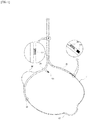
- FIG. 1 is a perspective view of a cerclage atrial defibrillator according to a preferred exemplary embodiment of the present invention.
- the cerclage atrial defibrillator fundamentally includes a cerclage wire 10, a coronary sinus tube 20, a tricuspid valve tube 30, a pacemaker lead 50, and a defibrillation lead 80.
- the cerclage wire 10 is a single thread that is inserted into the coronary vein, is passed through the interventricular septum, and is passed through the tricuspid valve, whereby the name is given in the sense that the single thread passes through a part of the coronary sinus, the interventricular septum, and a part of the tricuspid valve in sequence, and then circling back like drawing a circle, and is discharged from a patient's body.
- the cerclage wire serves to maintain proper tension force according to the size and shape of a patient's heart, and to tighten the mitral annulus. Subsequently, mitral valve regurgitation may be treated. According to the results of clinical trials of the present inventor, when the mitral valve is tightened, the occurrence of atrial fibrillation in a patient with the atrial fibrillation is reduced and may be treated.
- the cerclage wire 10 further includes an arch part 12, and the arch part 12 is formed on one side of the cerclage wire 10.
- the arch part is to prevent the external pressure applied to the coronary artery positioned below the coronary sinus and to protect the coronary artery.
- the coronary sinus tube 20 is provided with a lumen formed therein into which the cerclage wire 10 may be inserted.
- the upper part of the coronary sinus tube 20 is connected to the tricuspid valve tube 30 and is formed in a straight line, and the lower part thereof is in a curved shape to be inserted into the coronary sinus.
- the coronary sinus tube 20 is inserted into the coronary sinus to protect the coronary sinus.
- the tricuspid valve tube 30 is provided with a lumen formed therein into which the cerclage wire 10 may be inserted.
- the tricuspid valve tube 30 is passed through an orifice generated due to an incomplete closing of the tricuspid valve and reaches the interventricular septum, and is provided with a stopper 60 installed at the lower part thereof to prevent the end of the tricuspid valve tube 30 from penetrating into the interventricular septum.
- the stopper 60 is able to prevent damage to the tricuspid valve due to the tricuspid valve tube 30.
- the tricuspid valve tube 30 includes a blocking part 32.
- the blocking part 32 is a part that blocks the orifice generated due to an incomplete closing of the tricuspid valve, and may be a blocking membrane, a blocking balloon, or the like. By blocking the orifice of the tricuspid valve with the blocking part, tricuspid valve regurgitation may be treated.
- the pacemaker lead 50 is inserted into the tricuspid valve tube 30 or coupled to the tricuspid valve tube 30, and is provided with the lower part thereof facing the interventricular septum.
- the pacemaker lead 50 may perform sensing and pacing of an electrical signal of the heart.
- the pacemaker lead 50 includes a direction adjustment device (not shown) at the upper part thereof, moves the lower part of the pacemaker lead 50 by adjusting the direction adjustment device (not shown), may detect an electrocardiogram of the interventricular septum to identify the position of the bundle of His, and fixes the end of the pacemaker lead 50 to the position of the detected bundle of His.
- the pacemaker lead 50 may not be included depending on the purpose of using the device of the present invention.
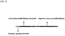
- FIG. 2 is a perspective view of a defibrillator lead according to the preferred exemplary embodiment of the present invention.
- the defibrillator lead 80 includes a first defibrillation electrode 82, a second defibrillation electrode 84, and an electrocardiogram sensing electrode 86.
- the outer part of the defibrillator lead 80 is made of an insulator excluding the first defibrillation electrode 82, the second defibrillation electrode 84, and the electrocardiogram sensing electrode 86, and thus current does not flow therethrough.
- the first defibrillation electrode 82 and the second defibrillation electrode 84 are preferably formed in a coil shape, but are not limited thereto.
- the electrocardiogram sensing electrode 86 is coupled to the lower end of the defibrillator lead 80 so as to perform sensing that detects the electrical signal of the heart and transmits the signal to the defibrillator, and to perform a function of pacing that transmits the current received from the defibrillator to the heart as needed.
- the first defibrillation electrode 82 is positioned in the superior vena cava
- the second defibrillation electrode 84 is positioned in the coronary sinus in the left atrium to perform defibrillation when atrial fibrillation occurs. More particularly, the defibrillator lead 80 is inserted into and coupled to the coronary sinus tube 20, the first defibrillation electrode 82 is positioned at the upper part of the coronary sinus tube 20 to contact the superior vena cava, and the second defibrillation electrode 84 is positioned at the lower part of the coronary sinus tube 20 to contact the left atrium.
- the coronary sinus tube 20 is able to transmit electricity when in contact with the superior vena cava and the left atrium, even when the first defibrillation electrode 82 and the second defibrillation electrode 84 are positioned inside the coronary sinus tube 20. It is apparent that the defibrillator lead 80 may be coupled to the outer circumferential surface of the coronary sinus tube 20.
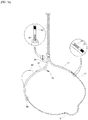
- FIG. 3 is a perspective view of a cerclage atrial defibrillator according to a second preferred exemplary embodiment of the present invention.
- the cerclage atrial defibrillator includes a cerclage wire 10, a coronary sinus tube 20, a tricuspid valve tube 30, a left atrium branch tube 40, a pacemaker lead 50, and a defibrillator lead 80.
- the defibrillator lead 80 is as described above in FIG. 2 .
- the lower part of the defibrillator lead 80 is inserted into the left atrium branch tube 40.
- the second defibrillation electrode 84 is positioned inside the left atrium branch tube 40, or is positioned outside thereof by passing through the left atrium branch tube 40 and is contacted with the left atrium, thereby performing defibrillation to the left atrium when atrial fibrillation occurs.
- Configurations other than the left atrium branch tube 40 are as described above with reference to FIGS. 1 and 2 .
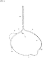
- FIG. 4 is a perspective view of a cerclage atrial defibrillator according to a third preferred exemplary embodiment of the present invention.
- the cerclage atrial defibrillator includes a cerclage wire 10, a coronary sinus tube 20, a tricuspid valve tube 30, a pacemaker lead 50, and a defibrillator lead 80.
- the defibrillator lead 80 is the same as described above in FIG. 2 .
- the second defibrillation electrode 84 is inserted into the coronary sinus tube 20, and may be passed through the coronary sinus tube 20 to directly contact the coronary sinus.
- the defibrillator lead 80 may further include a fixation device (not shown) for fixing the defibrillator lead 80 at an end thereof, which may be in the form of a screw, but is not limited thereto.
- the fixation device (not shown) is for fixing the second defibrillation electrode 84, which is in the coronary sinus, to reach a desired position.
- the configuration other than those described above is the same as described above in FIGS. 1 and 2 .
- FIG. 5 is a perspective view of a cerclage atrial defibrillator according to a fourth preferred exemplary embodiment of the present invention
- FIG. 5(a) is a perspective view showing a case where a lower part of a defibrillator lead insertion tube is positioned in the right atrium
- FIG. 5(b) is a perspective view showing a case where the lower part of the defibrillator lead insertion tube is positioned in the superior vena cava.
- the cerclage atrial defibrillator includes a cerclage wire 10, a coronary sinus tube 20, a tricuspid valve tube 30, a pacemaker lead 50, a defibrillator lead insertion tube 70, and a defibrillator lead 80.
- the defibrillator lead insertion tube 70 is provided with a lumen formed therein into which the defibrillator lead 80 may be inserted, is provided with the upper part thereof connected to the coronary sinus tube 20, and is provided with the lower part thereof branched so that the end thereof is curved to face the wall of the superior vena cava or the right atrium.
- the defibrillator lead 80 of the cerclage atrial defibrillator according to the fourth preferred exemplary embodiment of the present invention is composed of two leads including a first defibrillation electrode 82 and a second defibrillation electrode 84, in which each lead includes an electrocardiogram sensing electrode 86 at each of the lower part thereof.
- the lead including the first defibrillation electrode 82 is inserted into and fixed to the defibrillator lead insertion tube 70.
- the first defibrillation electrode 82 is positioned at the lower part of the defibrillator lead insertion tube 70, or is passed through the defibrillator lead insertion tube 70, and is fixed to contact a position of the wall of the superior vena cava or the right atrium that a surgeon desires.
- the end of the lead including the first defibrillation electrode 82 may further include a fixation device (not shown) for fixing the lead, and the fixation device may be in the form of a screw, but is not limited thereto.
- the lead including the second defibrillation electrode 84 is inserted into and fixed to the coronary sinus tube 20, and the second defibrillation electrode 84 is positioned at the lower part of the coronary sinus tube 20.
- the configuration other than those described about the cerclage atrial defibrillator according to the fourth preferred exemplary embodiment of the present invention is the same as described above in FIG. 1 .
- FIG. 6 is a perspective view of a cerclage atrial defibrillator according to a fifth preferred exemplary embodiment of the present invention.
- a cerclage atrial defibrillator includes a cerclage wire 10, a coronary sinus tube 20, a tricuspid valve tube 30, a left atrium branch tube 40, a pacemaker lead 50, a defibrillator lead insertion tube 70, and a defibrillator lead 80.
- One end of the left atrium branch tube 40 is coupled to one side of the coronary sinus tube 20, and the other end thereof is directed toward the coronary sinus and in communication with the coronary sinus tube 20.
- the defibrillator lead insertion tube 70 is as described above in FIG. 5 .
- Two defibrillator leads 80 are configured as described above in FIG. 5 , and the lead including the first defibrillation electrode 82 is inserted into and fixed to the defibrillator lead insertion tube 70.
- the first defibrillation electrode 82 is positioned at the lower part of the defibrillator lead insertion tube 70, or is passed through the defibrillator lead insertion tube 70, and is fixed to contact a position of the wall of the superior vena cava or the right atrium that a surgeon desires.
- the lead including the second defibrillation electrode 84 is inserted into the left atrium branch tube 40, and the second defibrillation electrode 84 is positioned in the left atrium branch tube 40, or is passed through the left atrium branch tube 40, and is fixed to contact a position of the coronary sinus that a surgeon desires.
- a fixation device (not shown) may be coupled to the end of the lead including the left atrium branch tube 40 to fix the defibrillator lead 80, and the fixation device may be in the form of a screw, but is not limited thereto.
- the configuration other than those described about the cerclage atrial defibrillator according to the fifth preferred exemplary embodiment of the present invention is the same as described above in FIG. 1 .
- FIG. 7 is a perspective view of a cerclage atrial defibrillator according to a sixth preferred exemplary embodiment of the present invention.
- the cerclage atrial defibrillator includes a cerclage wire 10, a coronary sinus tube 20, a tricuspid valve tube 30, a pacemaker lead 50, a defibrillator lead insertion tube 70, and a defibrillator lead 80.
- the defibrillator lead insertion tube 70 is as described above in FIGS. 5 and 6 .
- Two defibrillator lead 80 are composed of a lead including the first defibrillation electrode 82 and a lead including the second defibrillation electrode 84, as described above with reference to FIG. 5 , and each of the electrocardiogram sensing electrodes 86 is coupled to each of the lower part of the two leads.
- the first defibrillation electrode 82 is positioned at the lower part of the defibrillator lead insertion tube 70, or is passed through the defibrillator lead insertion tube 70, and is fixed to contact a position of the wall of the superior vena cava or the right atrium that a surgeon desires.
- the end of the lead including the first defibrillation electrode 82 may further include a fixation device (not shown) for fixing the lead, and the fixation device may be in the form of a screw, but is not limited thereto.
- the lead including the second defibrillation electrode 84 is inserted into the coronary sinus tube 20, and the second defibrillation electrode 84 is passed through the coronary sinus tube 20, and is fixed to contact a position of the coronary sinus that a surgeon desires.
- the end of the lead including the second defibrillation electrode 84 may further include a fixation device (not shown) for fixing the lead, and the fixation device may be in the form of a screw, but is not limited thereto.
- the configuration other than those described about the cerclage atrial defibrillator according to the sixth preferred exemplary embodiment of the present invention is the same as described above in FIG. 1 .
- the cerclage atrial defibrillator may not only simply relieve atrial fibrillation that occurs, but may also reduce the occurrence of atrial fibrillation by tightening the mitral valve by using a cerclage wire, whereby atrial fibrillation that often recurs may be restored to normal heartbeat by treatment with the defibrillator.
- mitral valve regurgitation treatment may be performed.
- tricuspid valve regurgitation treatment may be performed.
- fixation of the pacemaker lead for bradycardia heart failure treatment may be performed.
- insertion of the defibrillator lead for atrial fibrillation treatment may be performed.
- the device may also be used for at least one of the four treatments described above.
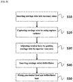
- FIG. 8 is a flowchart for a treatment method using the cerclage atrial defibrillator according to the preferred exemplary embodiments of the present invention.
- a step S10 is for inserting the cerclage wire into the coronary sinus is for moving the cerclage wire 10 through the superior vena cava, the coronary sinus, the septal vein, and the right ventricle in sequence.
- a balloon catheter (not shown) may be included.
- the balloon catheter (not shown) blocks blood flow in the coronary sinus to make the coronary sinus to be swollen, thereby allowing the septal vein, which is generally difficult to identify, to be easily seen.
- the cerclage wire 10 is passed through the interventricular septum via the septal vein, and moves to the right ventricle.
- the cerclage wire 10 is provided with a sharp end thereof, but when it is difficult to penetrate the interventricular septum with the cerclage wire 10 by itself, a perforating catheter (not shown) is required.
- the perforating catheter serves to support when the cerclage wire 10 perforates the interventricular septum, thereby allowing the cerclage wire 10 to pass through the interventricular septum more easily.
- a step S20 is for capturing the cerclage wire by using a capture catheter.
- the capture catheter (not shown) is provided with a mesh net formed thereon, and the mesh net may be folded or unfolded by force applied to the capture catheter (not shown) in vitro.
- the capture catheter (not shown) moves through the inferior vena cava, the tricuspid valve, and the right ventricle, and may further include a safe-zone-passing catheter (not shown), in order for the capture catheter (not shown) to safely move in the patient's body.
- the mesh net is folded and then unfolded in the right ventricle, and the cerclage wire 10 is inserted into the mesh net.
- the mesh net is folded again, and after capturing the cerclage wire 10, the mesh net is extracted out of the patient's body through the inferior vena cava. At this time, one end of the cerclage wire 10 is positioned in the superior vena cava, and the other end thereof is positioned in the inferior vena cava.
- a step S30 is for adjusting tension force by guiding the cerclage wire to the superior vena cava.
- the cerclage wire 10 guided to the inferior vena cava is guided back to the superior vena cava by a snare, forceps, or the like, which is inserted from the superior vena cava, and both ends of the cerclage wire are positioned in vitro through the superior vena cava. Therefore, the cerclage wire 10 is passed through the superior vena cava, the coronary sinus, the septal vein, the interventricular septum, and the right ventricle in sequence, and is positioned in the superior vena cava again, thereby coming back like drawing a circular shape, so that both ends of the cerclage wire are positioned in vitro. While pushing or pulling the cerclage wire 10, appropriate tension force is maintained according to the size and shape of the patient's heart, and the mitral annulus is tightened to hold, and thus mitral valve regurgitation is treated by enabling the mitral valve to be completely closed.
- a step S40 is for inserting a cerclage atrial defibrillator. After respectively inserting both ends of the cerclage wire 10 outside the patient's body into the coronary sinus tube 20 and the tricuspid valve tube 30, the coronary sinus tube 20 and the tricuspid valve tube 30 are inserted into the patient's body along the cerclage wire 10. At this time, the pacemaker lead 50 coupled to the tricuspid valve tube 30 is inserted in the right atrium direction together with the tricuspid valve tube 30, and the defibrillator lead 80 coupled to the coronary sinus tube 20 is inserted into the superior vena cava and the coronary sinus.
- the stopper 60 is coupled to the lower end of the tricuspid valve tube 30, and may be coupled to the lower part of the tricuspid valve tube 30. This allows the tricuspid valve tube 30 to float around the tricuspid valve without being in close contact therewith, thereby preventing damage to the tricuspid valve.
- the tricuspid valve tube 30 includes the blocking part 32, which penetrates into the orifice generated due to an incomplete closing phenomenon of the tricuspid valve to completely close the tricuspid valve.
- the blocking part 32 may be in the form of a blocking membrane, a blocking balloon, or the like.
- a step S50 is for fixing the pacemaker lead and the defibrillator lead is performed.
- the pacemaker lead 50 may be adjusted to bend or unfold the lower part thereof by installing a direction adjustment device 54 at the upper part thereof.
- the lower part of the pacemaker lead 50 is moved by the direction adjustment device 54, detects an electrocardiogram of the interventricular septum to identify the position of the bundle of His, and fixes the end of the pacemaker lead 50 to the detected position of the bundle of His.
- the fixation device (not shown) may be further included in order to fix the electrodes to positions at which a surgeon desires.
Landscapes
- Health & Medical Sciences (AREA)
- Cardiology (AREA)
- Life Sciences & Earth Sciences (AREA)
- Heart & Thoracic Surgery (AREA)
- Veterinary Medicine (AREA)
- Engineering & Computer Science (AREA)
- Biomedical Technology (AREA)
- Animal Behavior & Ethology (AREA)
- General Health & Medical Sciences (AREA)
- Public Health (AREA)
- Nuclear Medicine, Radiotherapy & Molecular Imaging (AREA)
- Radiology & Medical Imaging (AREA)
- Surgery (AREA)
- Vascular Medicine (AREA)
- Pathology (AREA)
- Medical Informatics (AREA)
- Molecular Biology (AREA)
- Biophysics (AREA)
- Physics & Mathematics (AREA)
- Oral & Maxillofacial Surgery (AREA)
- Transplantation (AREA)
- Neurology (AREA)
- Electrotherapy Devices (AREA)
- Hematology (AREA)
- Physiology (AREA)
Applications Claiming Priority (2)
| Application Number | Priority Date | Filing Date | Title |
|---|---|---|---|
| KR1020170158427A KR102053451B1 (ko) | 2017-11-24 | 2017-11-24 | 서클라지 심방 제세동장치 |
| PCT/KR2018/014556 WO2019103540A1 (ko) | 2017-11-24 | 2018-11-23 | 서클라지 심방 제세동장치 |
Publications (3)
| Publication Number | Publication Date |
|---|---|
| EP3714936A1 true EP3714936A1 (de) | 2020-09-30 |
| EP3714936A4 EP3714936A4 (de) | 2021-08-18 |
| EP3714936B1 EP3714936B1 (de) | 2022-03-30 |
Family
ID=66632071
Family Applications (1)
| Application Number | Title | Priority Date | Filing Date |
|---|---|---|---|
| EP18880318.3A Active EP3714936B1 (de) | 2017-11-24 | 2018-11-23 | Cerclage-vorhofdefibrillator |
Country Status (4)
| Country | Link |
|---|---|
| US (1) | US20200346001A1 (de) |
| EP (1) | EP3714936B1 (de) |
| KR (1) | KR102053451B1 (de) |
| WO (1) | WO2019103540A1 (de) |
Cited By (15)
| Publication number | Priority date | Publication date | Assignee | Title |
|---|---|---|---|---|
| US11311376B2 (en) | 2019-06-20 | 2022-04-26 | Neovase Tiara Inc. | Low profile prosthetic mitral valve |
| US11357622B2 (en) | 2016-01-29 | 2022-06-14 | Neovase Tiara Inc. | Prosthetic valve for avoiding obstruction of outflow |
| EP3884993A4 (de) * | 2018-11-23 | 2022-06-29 | Tau-PNU Medical Co., Ltd. | Vorrichtung zur klappenregurgitationschirurgie und zur fixierung von herzschrittmacherleitungen |
| US11389291B2 (en) | 2013-04-04 | 2022-07-19 | Neovase Tiara Inc. | Methods and apparatus for delivering a prosthetic valve to a beating heart |
| US11389294B2 (en) | 2012-05-30 | 2022-07-19 | Neovasc Tiara Inc. | Methods and apparatus for loading a prosthesis onto a delivery system |
| US11413139B2 (en) | 2011-11-23 | 2022-08-16 | Neovasc Tiara Inc. | Sequentially deployed transcatheter mitral valve prosthesis |
| US11419720B2 (en) | 2010-05-05 | 2022-08-23 | Neovasc Tiara Inc. | Transcatheter mitral valve prosthesis |
| US11464631B2 (en) | 2016-11-21 | 2022-10-11 | Neovasc Tiara Inc. | Methods and systems for rapid retraction of a transcatheter heart valve delivery system |
| US11491006B2 (en) | 2019-04-10 | 2022-11-08 | Neovasc Tiara Inc. | Prosthetic valve with natural blood flow |
| US11497602B2 (en) | 2012-02-14 | 2022-11-15 | Neovasc Tiara Inc. | Methods and apparatus for engaging a valve prosthesis with tissue |
| US11602429B2 (en) | 2019-04-01 | 2023-03-14 | Neovasc Tiara Inc. | Controllably deployable prosthetic valve |
| US11737872B2 (en) | 2018-11-08 | 2023-08-29 | Neovasc Tiara Inc. | Ventricular deployment of a transcatheter mitral valve prosthesis |
| US11779742B2 (en) | 2019-05-20 | 2023-10-10 | Neovasc Tiara Inc. | Introducer with hemostasis mechanism |
| US11793640B2 (en) | 2017-08-25 | 2023-10-24 | Neovasc Tiara Inc. | Sequentially deployed transcatheter mitral valve prosthesis |
| US11998447B2 (en) | 2019-03-08 | 2024-06-04 | Neovasc Tiara Inc. | Retrievable prosthesis delivery system |
Families Citing this family (5)
| Publication number | Priority date | Publication date | Assignee | Title |
|---|---|---|---|---|
| US9943409B2 (en) * | 2006-11-14 | 2018-04-17 | The United States Of America, As Represented By The Secretary, Department Of Health And Human Services | Transcatheter coronary sinus mitral valve annuloplasty procedure and coronary artery and myocardial protection device |
| WO2021064689A1 (en) * | 2019-10-03 | 2021-04-08 | Tau-Pnu Medical Co., Ltd. | Device, system, and method for reducing mitral valve regurgitation |
| KR102240805B1 (ko) | 2019-07-12 | 2021-04-15 | (주) 타우피엔유메디칼 | 위치고정장치를 구비한 서클라지 시술장치 |
| EP4037614A4 (de) * | 2019-10-03 | 2023-10-11 | Tau Medical Inc. | Vorrichtung, system und verfahren zum reduzieren der mitralklappenregurgitation |
| KR20220157391A (ko) * | 2020-02-20 | 2022-11-29 | (주)타우메디칼 | 삼첨판 역류를 감소시키기 위한 트랜스카테터 시스템 및 방법 |
Family Cites Families (14)
| Publication number | Priority date | Publication date | Assignee | Title |
|---|---|---|---|---|
| US5978705A (en) * | 1997-03-14 | 1999-11-02 | Uab Research Foundation | Method and apparatus for treating cardiac arrhythmia using auxiliary pulse |
| US6402781B1 (en) * | 2000-01-31 | 2002-06-11 | Mitralife | Percutaneous mitral annuloplasty and cardiac reinforcement |
| US6745081B1 (en) | 2001-08-31 | 2004-06-01 | Pacesetter, Inc. | Coronary Sinus Cardiac Lead For Stimulating and Sensing The Atria of the Right and Left Heart and System |
| US9031646B2 (en) * | 2007-10-09 | 2015-05-12 | University of Pittsburgh—of the Commonwealth System of Higher Education | Automated assessment of atrioventricular and ventriculoatrial conduction |
| US9199075B1 (en) * | 2008-02-07 | 2015-12-01 | Respicardia, Inc. | Transvascular medical lead |
| EP2349468B1 (de) * | 2008-10-03 | 2014-08-13 | Cardiac Pacemakers, Inc. | Geräte für die auswahl des therapiemodus bei der herzresynchronisation auf basis von eigenleitung |
| US8644927B2 (en) * | 2009-04-21 | 2014-02-04 | Incube Labs, Llc | Apparatus and method for the detection and treatment of atrial fibrillation |
| KR101116867B1 (ko) | 2009-08-28 | 2012-03-06 | 김준홍 | 관상정맥동과 삼천판막의 조직보호기구, 매듭 전달 기구 및 이들을 포함하는 승모판막 서클라지 시술용 장치 |
| KR101563172B1 (ko) | 2014-05-20 | 2015-10-27 | (주) 타우피엔유메디칼 | 승모판막 서클라지 시술용 조직보호기구 |
| KR101581021B1 (ko) | 2014-05-28 | 2015-12-29 | (주) 타우피엔유메디칼 | 코일 스프링을 이용한 승모판막 서클라지 시술용 조직보호기구 및 이의 제조방법 |
| KR20150144568A (ko) * | 2014-06-17 | 2015-12-28 | (주) 타우피엔유메디칼 | 삼천판막 역류증 시술용 조직보호기구 |
| KR20160020887A (ko) * | 2014-08-14 | 2016-02-24 | 부산대학교 산학협력단 | 중간 또는 첨부의 심실 중격 내에 관상정맥동을 통과한 심박동기의 리드 끝단을 위치시키는 방법, 장치, 및 카테터 |
| KR101805679B1 (ko) | 2017-02-01 | 2017-12-06 | (주) 타우피엔유메디칼 | 삼첨판막 역류증 시술용 기구 |
| EP3672532B1 (de) * | 2017-08-26 | 2022-08-03 | Transmural Systems LLC | Implantierbares herzschrittmachersystem |
-
2017
- 2017-11-24 KR KR1020170158427A patent/KR102053451B1/ko active IP Right Grant
-
2018
- 2018-11-23 WO PCT/KR2018/014556 patent/WO2019103540A1/ko unknown
- 2018-11-23 EP EP18880318.3A patent/EP3714936B1/de active Active
- 2018-11-24 US US16/765,164 patent/US20200346001A1/en not_active Abandoned
Cited By (19)
| Publication number | Priority date | Publication date | Assignee | Title |
|---|---|---|---|---|
| US11419720B2 (en) | 2010-05-05 | 2022-08-23 | Neovasc Tiara Inc. | Transcatheter mitral valve prosthesis |
| US12053369B2 (en) | 2011-11-23 | 2024-08-06 | Neovasc Tiara Inc. | Sequentially deployed transcatheter mitral valve prosthesis |
| US11413139B2 (en) | 2011-11-23 | 2022-08-16 | Neovasc Tiara Inc. | Sequentially deployed transcatheter mitral valve prosthesis |
| US11497602B2 (en) | 2012-02-14 | 2022-11-15 | Neovasc Tiara Inc. | Methods and apparatus for engaging a valve prosthesis with tissue |
| US11617650B2 (en) | 2012-05-30 | 2023-04-04 | Neovasc Tiara Inc. | Methods and apparatus for loading a prosthesis onto a delivery system |
| US11389294B2 (en) | 2012-05-30 | 2022-07-19 | Neovasc Tiara Inc. | Methods and apparatus for loading a prosthesis onto a delivery system |
| US11389291B2 (en) | 2013-04-04 | 2022-07-19 | Neovase Tiara Inc. | Methods and apparatus for delivering a prosthetic valve to a beating heart |
| US11357622B2 (en) | 2016-01-29 | 2022-06-14 | Neovase Tiara Inc. | Prosthetic valve for avoiding obstruction of outflow |
| US11464631B2 (en) | 2016-11-21 | 2022-10-11 | Neovasc Tiara Inc. | Methods and systems for rapid retraction of a transcatheter heart valve delivery system |
| US11793640B2 (en) | 2017-08-25 | 2023-10-24 | Neovasc Tiara Inc. | Sequentially deployed transcatheter mitral valve prosthesis |
| US11737872B2 (en) | 2018-11-08 | 2023-08-29 | Neovasc Tiara Inc. | Ventricular deployment of a transcatheter mitral valve prosthesis |
| EP3884993A4 (de) * | 2018-11-23 | 2022-06-29 | Tau-PNU Medical Co., Ltd. | Vorrichtung zur klappenregurgitationschirurgie und zur fixierung von herzschrittmacherleitungen |
| US11998447B2 (en) | 2019-03-08 | 2024-06-04 | Neovasc Tiara Inc. | Retrievable prosthesis delivery system |
| US11602429B2 (en) | 2019-04-01 | 2023-03-14 | Neovasc Tiara Inc. | Controllably deployable prosthetic valve |
| US11491006B2 (en) | 2019-04-10 | 2022-11-08 | Neovasc Tiara Inc. | Prosthetic valve with natural blood flow |
| US12036117B2 (en) | 2019-04-10 | 2024-07-16 | Neovasc Tiara Inc. | Prosthetic valve with natural blood flow |
| US11779742B2 (en) | 2019-05-20 | 2023-10-10 | Neovasc Tiara Inc. | Introducer with hemostasis mechanism |
| US11311376B2 (en) | 2019-06-20 | 2022-04-26 | Neovase Tiara Inc. | Low profile prosthetic mitral valve |
| US11931254B2 (en) | 2019-06-20 | 2024-03-19 | Neovasc Tiara Inc. | Low profile prosthetic mitral valve |
Also Published As
| Publication number | Publication date |
|---|---|
| WO2019103540A1 (ko) | 2019-05-31 |
| KR20190060334A (ko) | 2019-06-03 |
| EP3714936B1 (de) | 2022-03-30 |
| KR102053451B1 (ko) | 2020-01-08 |
| EP3714936A4 (de) | 2021-08-18 |
| US20200346001A1 (en) | 2020-11-05 |
Similar Documents
| Publication | Publication Date | Title |
|---|---|---|
| EP3714936B1 (de) | Cerclage-vorhofdefibrillator | |
| EP2291212B1 (de) | Abgabekatheter mit seitenöffnung und elektroden | |
| JP4850899B2 (ja) | 経中隔/経心筋心室ペーシングリード | |
| US9039594B2 (en) | Signal transmitting and lesion excluding heart implants for pacing, defibrillating, and/or sensing of heart beat | |
| US7418298B2 (en) | Myocardial lead with fixation mechanism | |
| EP0965359A2 (de) | Transvenöse Defibrillierungsleitung zur Verwendung in einer Vena cardiaca media | |
| US9884184B2 (en) | Wire hook coupling for lead extension and extraction | |
| JP2008540052A (ja) | 心臓リード線による体腔整形器具 | |
| US8942829B2 (en) | Trans-septal lead anchoring | |
| US20180326204A1 (en) | Shaped epicardial lead and placement system and method | |
| US9242098B2 (en) | Devices, systems, and methods for treating cardiac arrhythmias | |
| JP4716516B2 (ja) | 中隔プレースメントのための頻脈性リード・システム | |
| US20230264017A1 (en) | Advanced implantable endovascular, low profile intracardiac left atrial restraining devices for low energy atrial cardioversion, pacing and sensing | |
| KR101955291B1 (ko) | 판막 역류증 시술 및 심박동기 리드 고정장치 | |
| US20210022634A1 (en) | Capture catheter for sensing myocardial electrical signal | |
| US20230020426A1 (en) | Implantable endovascular, low profile intracardiac left atrial restraining devices for low energy atrial cardioversion, pacing and sensing | |
| EP3884993A1 (de) | Vorrichtung zur klappenregurgitationschirurgie und zur fixierung von herzschrittmacherleitungen | |
| US20210346684A1 (en) | Snare-integrated myocardial electrical signal-detecting catheter | |
| Vaidya et al. | Percutaneous epicardial pacing using a novel transverse sinus device | |
| Burger et al. | Device Therapy of Rhythm Disorders | |
| US20240316341A1 (en) | Implantable endovascular, low profile intracardiac left atrial restraining devices for low energy atrial cardioversion, pacing and sensing | |
| Rajamani et al. | Implantation of Pacemakers and ICDs | |
| Khan et al. | POCKET FORMATION• Most devices are placed in a left prepectoral pocket due to the fact that most patients are right-handed and due to easier manipulation of leads from the left versus the right side. Other sites include right prepectoral, abdominal, infra-mammary, and under the pectoralis major muscle positions. The latter two positions may be considered for cosmetic reasons. The pocket should not be | |
| Khan et al. | Implantation of pacemakers and ICDs | |
| Stellbrink | Complications of cardiac resynchronization therapy |
Legal Events
| Date | Code | Title | Description |
|---|---|---|---|
| STAA | Information on the status of an ep patent application or granted ep patent |
Free format text: STATUS: THE INTERNATIONAL PUBLICATION HAS BEEN MADE |
|
| PUAI | Public reference made under article 153(3) epc to a published international application that has entered the european phase |
Free format text: ORIGINAL CODE: 0009012 |
|
| STAA | Information on the status of an ep patent application or granted ep patent |
Free format text: STATUS: REQUEST FOR EXAMINATION WAS MADE |
|
| 17P | Request for examination filed |
Effective date: 20200624 |
|
| AK | Designated contracting states |
Kind code of ref document: A1 Designated state(s): AL AT BE BG CH CY CZ DE DK EE ES FI FR GB GR HR HU IE IS IT LI LT LU LV MC MK MT NL NO PL PT RO RS SE SI SK SM TR |
|
| AX | Request for extension of the european patent |
Extension state: BA ME |
|
| DAV | Request for validation of the european patent (deleted) | ||
| DAX | Request for extension of the european patent (deleted) | ||
| A4 | Supplementary search report drawn up and despatched |
Effective date: 20210716 |
|
| RIC1 | Information provided on ipc code assigned before grant |
Ipc: A61N 1/05 20060101AFI20210712BHEP Ipc: A61N 1/08 20060101ALI20210712BHEP Ipc: A61F 2/24 20060101ALI20210712BHEP Ipc: A61B 17/34 20060101ALI20210712BHEP Ipc: A61B 17/04 20060101ALI20210712BHEP Ipc: A61N 1/39 20060101ALI20210712BHEP Ipc: A61N 1/372 20060101ALI20210712BHEP Ipc: A61N 1/375 20060101ALN20210712BHEP |
|
| GRAP | Despatch of communication of intention to grant a patent |
Free format text: ORIGINAL CODE: EPIDOSNIGR1 |
|
| STAA | Information on the status of an ep patent application or granted ep patent |
Free format text: STATUS: GRANT OF PATENT IS INTENDED |
|
| RIC1 | Information provided on ipc code assigned before grant |
Ipc: A61N 1/375 20060101ALN20210928BHEP Ipc: A61N 1/372 20060101ALI20210928BHEP Ipc: A61N 1/39 20060101ALI20210928BHEP Ipc: A61B 17/04 20060101ALI20210928BHEP Ipc: A61B 17/34 20060101ALI20210928BHEP Ipc: A61F 2/24 20060101ALI20210928BHEP Ipc: A61N 1/08 20060101ALI20210928BHEP Ipc: A61N 1/05 20060101AFI20210928BHEP |
|
| INTG | Intention to grant announced |
Effective date: 20211018 |
|
| RAP3 | Party data changed (applicant data changed or rights of an application transferred) |
Owner name: TAU-PNU MEDICAL CO., LTD. |
|
| GRAS | Grant fee paid |
Free format text: ORIGINAL CODE: EPIDOSNIGR3 |
|
| GRAA | (expected) grant |
Free format text: ORIGINAL CODE: 0009210 |
|
| STAA | Information on the status of an ep patent application or granted ep patent |
Free format text: STATUS: THE PATENT HAS BEEN GRANTED |
|
| AK | Designated contracting states |
Kind code of ref document: B1 Designated state(s): AL AT BE BG CH CY CZ DE DK EE ES FI FR GB GR HR HU IE IS IT LI LT LU LV MC MK MT NL NO PL PT RO RS SE SI SK SM TR |
|
| REG | Reference to a national code |
Ref country code: GB Ref legal event code: FG4D |
|
| REG | Reference to a national code |
Ref country code: CH Ref legal event code: EP |
|
| REG | Reference to a national code |
Ref country code: AT Ref legal event code: REF Ref document number: 1478609 Country of ref document: AT Kind code of ref document: T Effective date: 20220415 |
|
| REG | Reference to a national code |
Ref country code: DE Ref legal event code: R096 Ref document number: 602018033163 Country of ref document: DE |
|
| REG | Reference to a national code |
Ref country code: IE Ref legal event code: FG4D |
|
| REG | Reference to a national code |
Ref country code: LT Ref legal event code: MG9D |
|
| PG25 | Lapsed in a contracting state [announced via postgrant information from national office to epo] |
Ref country code: SE Free format text: LAPSE BECAUSE OF FAILURE TO SUBMIT A TRANSLATION OF THE DESCRIPTION OR TO PAY THE FEE WITHIN THE PRESCRIBED TIME-LIMIT Effective date: 20220330 Ref country code: RS Free format text: LAPSE BECAUSE OF FAILURE TO SUBMIT A TRANSLATION OF THE DESCRIPTION OR TO PAY THE FEE WITHIN THE PRESCRIBED TIME-LIMIT Effective date: 20220330 Ref country code: NO Free format text: LAPSE BECAUSE OF FAILURE TO SUBMIT A TRANSLATION OF THE DESCRIPTION OR TO PAY THE FEE WITHIN THE PRESCRIBED TIME-LIMIT Effective date: 20220630 Ref country code: LT Free format text: LAPSE BECAUSE OF FAILURE TO SUBMIT A TRANSLATION OF THE DESCRIPTION OR TO PAY THE FEE WITHIN THE PRESCRIBED TIME-LIMIT Effective date: 20220330 Ref country code: HR Free format text: LAPSE BECAUSE OF FAILURE TO SUBMIT A TRANSLATION OF THE DESCRIPTION OR TO PAY THE FEE WITHIN THE PRESCRIBED TIME-LIMIT Effective date: 20220330 Ref country code: BG Free format text: LAPSE BECAUSE OF FAILURE TO SUBMIT A TRANSLATION OF THE DESCRIPTION OR TO PAY THE FEE WITHIN THE PRESCRIBED TIME-LIMIT Effective date: 20220630 |
|
| REG | Reference to a national code |
Ref country code: NL Ref legal event code: MP Effective date: 20220330 |
|
| REG | Reference to a national code |
Ref country code: AT Ref legal event code: MK05 Ref document number: 1478609 Country of ref document: AT Kind code of ref document: T Effective date: 20220330 |
|
| PG25 | Lapsed in a contracting state [announced via postgrant information from national office to epo] |
Ref country code: LV Free format text: LAPSE BECAUSE OF FAILURE TO SUBMIT A TRANSLATION OF THE DESCRIPTION OR TO PAY THE FEE WITHIN THE PRESCRIBED TIME-LIMIT Effective date: 20220330 Ref country code: GR Free format text: LAPSE BECAUSE OF FAILURE TO SUBMIT A TRANSLATION OF THE DESCRIPTION OR TO PAY THE FEE WITHIN THE PRESCRIBED TIME-LIMIT Effective date: 20220701 Ref country code: FI Free format text: LAPSE BECAUSE OF FAILURE TO SUBMIT A TRANSLATION OF THE DESCRIPTION OR TO PAY THE FEE WITHIN THE PRESCRIBED TIME-LIMIT Effective date: 20220330 |
|
| PG25 | Lapsed in a contracting state [announced via postgrant information from national office to epo] |
Ref country code: NL Free format text: LAPSE BECAUSE OF FAILURE TO SUBMIT A TRANSLATION OF THE DESCRIPTION OR TO PAY THE FEE WITHIN THE PRESCRIBED TIME-LIMIT Effective date: 20220330 |
|
| PG25 | Lapsed in a contracting state [announced via postgrant information from national office to epo] |
Ref country code: SM Free format text: LAPSE BECAUSE OF FAILURE TO SUBMIT A TRANSLATION OF THE DESCRIPTION OR TO PAY THE FEE WITHIN THE PRESCRIBED TIME-LIMIT Effective date: 20220330 Ref country code: SK Free format text: LAPSE BECAUSE OF FAILURE TO SUBMIT A TRANSLATION OF THE DESCRIPTION OR TO PAY THE FEE WITHIN THE PRESCRIBED TIME-LIMIT Effective date: 20220330 Ref country code: RO Free format text: LAPSE BECAUSE OF FAILURE TO SUBMIT A TRANSLATION OF THE DESCRIPTION OR TO PAY THE FEE WITHIN THE PRESCRIBED TIME-LIMIT Effective date: 20220330 Ref country code: PT Free format text: LAPSE BECAUSE OF FAILURE TO SUBMIT A TRANSLATION OF THE DESCRIPTION OR TO PAY THE FEE WITHIN THE PRESCRIBED TIME-LIMIT Effective date: 20220801 Ref country code: ES Free format text: LAPSE BECAUSE OF FAILURE TO SUBMIT A TRANSLATION OF THE DESCRIPTION OR TO PAY THE FEE WITHIN THE PRESCRIBED TIME-LIMIT Effective date: 20220330 Ref country code: EE Free format text: LAPSE BECAUSE OF FAILURE TO SUBMIT A TRANSLATION OF THE DESCRIPTION OR TO PAY THE FEE WITHIN THE PRESCRIBED TIME-LIMIT Effective date: 20220330 Ref country code: CZ Free format text: LAPSE BECAUSE OF FAILURE TO SUBMIT A TRANSLATION OF THE DESCRIPTION OR TO PAY THE FEE WITHIN THE PRESCRIBED TIME-LIMIT Effective date: 20220330 Ref country code: AT Free format text: LAPSE BECAUSE OF FAILURE TO SUBMIT A TRANSLATION OF THE DESCRIPTION OR TO PAY THE FEE WITHIN THE PRESCRIBED TIME-LIMIT Effective date: 20220330 |
|
| PG25 | Lapsed in a contracting state [announced via postgrant information from national office to epo] |
Ref country code: PL Free format text: LAPSE BECAUSE OF FAILURE TO SUBMIT A TRANSLATION OF THE DESCRIPTION OR TO PAY THE FEE WITHIN THE PRESCRIBED TIME-LIMIT Effective date: 20220330 Ref country code: IS Free format text: LAPSE BECAUSE OF FAILURE TO SUBMIT A TRANSLATION OF THE DESCRIPTION OR TO PAY THE FEE WITHIN THE PRESCRIBED TIME-LIMIT Effective date: 20220730 Ref country code: AL Free format text: LAPSE BECAUSE OF FAILURE TO SUBMIT A TRANSLATION OF THE DESCRIPTION OR TO PAY THE FEE WITHIN THE PRESCRIBED TIME-LIMIT Effective date: 20220330 |
|
| REG | Reference to a national code |
Ref country code: DE Ref legal event code: R097 Ref document number: 602018033163 Country of ref document: DE |
|
| PG25 | Lapsed in a contracting state [announced via postgrant information from national office to epo] |
Ref country code: DK Free format text: LAPSE BECAUSE OF FAILURE TO SUBMIT A TRANSLATION OF THE DESCRIPTION OR TO PAY THE FEE WITHIN THE PRESCRIBED TIME-LIMIT Effective date: 20220330 |
|
| PLBE | No opposition filed within time limit |
Free format text: ORIGINAL CODE: 0009261 |
|
| STAA | Information on the status of an ep patent application or granted ep patent |
Free format text: STATUS: NO OPPOSITION FILED WITHIN TIME LIMIT |
|
| 26N | No opposition filed |
Effective date: 20230103 |
|
| PG25 | Lapsed in a contracting state [announced via postgrant information from national office to epo] |
Ref country code: SI Free format text: LAPSE BECAUSE OF FAILURE TO SUBMIT A TRANSLATION OF THE DESCRIPTION OR TO PAY THE FEE WITHIN THE PRESCRIBED TIME-LIMIT Effective date: 20220330 |
|
| REG | Reference to a national code |
Ref country code: DE Ref legal event code: R119 Ref document number: 602018033163 Country of ref document: DE |
|
| PG25 | Lapsed in a contracting state [announced via postgrant information from national office to epo] |
Ref country code: MC Free format text: LAPSE BECAUSE OF FAILURE TO SUBMIT A TRANSLATION OF THE DESCRIPTION OR TO PAY THE FEE WITHIN THE PRESCRIBED TIME-LIMIT Effective date: 20220330 |
|
| REG | Reference to a national code |
Ref country code: CH Ref legal event code: PL |
|
| GBPC | Gb: european patent ceased through non-payment of renewal fee |
Effective date: 20221123 |
|
| REG | Reference to a national code |
Ref country code: BE Ref legal event code: MM Effective date: 20221130 |
|
| PG25 | Lapsed in a contracting state [announced via postgrant information from national office to epo] |
Ref country code: LI Free format text: LAPSE BECAUSE OF NON-PAYMENT OF DUE FEES Effective date: 20221130 Ref country code: IT Free format text: LAPSE BECAUSE OF FAILURE TO SUBMIT A TRANSLATION OF THE DESCRIPTION OR TO PAY THE FEE WITHIN THE PRESCRIBED TIME-LIMIT Effective date: 20220330 Ref country code: CH Free format text: LAPSE BECAUSE OF NON-PAYMENT OF DUE FEES Effective date: 20221130 |
|
| PG25 | Lapsed in a contracting state [announced via postgrant information from national office to epo] |
Ref country code: LU Free format text: LAPSE BECAUSE OF NON-PAYMENT OF DUE FEES Effective date: 20221123 |
|
| PG25 | Lapsed in a contracting state [announced via postgrant information from national office to epo] |
Ref country code: IE Free format text: LAPSE BECAUSE OF NON-PAYMENT OF DUE FEES Effective date: 20221123 Ref country code: GB Free format text: LAPSE BECAUSE OF NON-PAYMENT OF DUE FEES Effective date: 20221123 Ref country code: DE Free format text: LAPSE BECAUSE OF NON-PAYMENT OF DUE FEES Effective date: 20230601 |
|
| PG25 | Lapsed in a contracting state [announced via postgrant information from national office to epo] |
Ref country code: FR Free format text: LAPSE BECAUSE OF NON-PAYMENT OF DUE FEES Effective date: 20221130 Ref country code: BE Free format text: LAPSE BECAUSE OF NON-PAYMENT OF DUE FEES Effective date: 20221130 |
|
| PG25 | Lapsed in a contracting state [announced via postgrant information from national office to epo] |
Ref country code: CY Free format text: LAPSE BECAUSE OF FAILURE TO SUBMIT A TRANSLATION OF THE DESCRIPTION OR TO PAY THE FEE WITHIN THE PRESCRIBED TIME-LIMIT Effective date: 20220330 |
|
| PG25 | Lapsed in a contracting state [announced via postgrant information from national office to epo] |
Ref country code: MK Free format text: LAPSE BECAUSE OF FAILURE TO SUBMIT A TRANSLATION OF THE DESCRIPTION OR TO PAY THE FEE WITHIN THE PRESCRIBED TIME-LIMIT Effective date: 20220330 Ref country code: HU Free format text: LAPSE BECAUSE OF FAILURE TO SUBMIT A TRANSLATION OF THE DESCRIPTION OR TO PAY THE FEE WITHIN THE PRESCRIBED TIME-LIMIT; INVALID AB INITIO Effective date: 20181123 |
|
| PG25 | Lapsed in a contracting state [announced via postgrant information from national office to epo] |
Ref country code: TR Free format text: LAPSE BECAUSE OF FAILURE TO SUBMIT A TRANSLATION OF THE DESCRIPTION OR TO PAY THE FEE WITHIN THE PRESCRIBED TIME-LIMIT Effective date: 20220330 |