EP3689281A1 - Systems and methods facilitating pre-operative prediction of post-operative tissue function - Google Patents
Systems and methods facilitating pre-operative prediction of post-operative tissue function Download PDFInfo
- Publication number
- EP3689281A1 EP3689281A1 EP20155199.1A EP20155199A EP3689281A1 EP 3689281 A1 EP3689281 A1 EP 3689281A1 EP 20155199 A EP20155199 A EP 20155199A EP 3689281 A1 EP3689281 A1 EP 3689281A1
- Authority
- EP
- European Patent Office
- Prior art keywords
- resection
- model
- tissue
- post
- lung
- Prior art date
- Legal status (The legal status is an assumption and is not a legal conclusion. Google has not performed a legal analysis and makes no representation as to the accuracy of the status listed.)
- Pending
Links
Images


Classifications
-
- A—HUMAN NECESSITIES
- A61—MEDICAL OR VETERINARY SCIENCE; HYGIENE
- A61B—DIAGNOSIS; SURGERY; IDENTIFICATION
- A61B34/00—Computer-aided surgery; Manipulators or robots specially adapted for use in surgery
- A61B34/10—Computer-aided planning, simulation or modelling of surgical operations
-
- G—PHYSICS
- G06—COMPUTING OR CALCULATING; COUNTING
- G06T—IMAGE DATA PROCESSING OR GENERATION, IN GENERAL
- G06T7/00—Image analysis
- G06T7/0002—Inspection of images, e.g. flaw detection
- G06T7/0012—Biomedical image inspection
-
- A—HUMAN NECESSITIES
- A61—MEDICAL OR VETERINARY SCIENCE; HYGIENE
- A61B—DIAGNOSIS; SURGERY; IDENTIFICATION
- A61B17/00—Surgical instruments, devices or methods
- A61B2017/00743—Type of operation; Specification of treatment sites
- A61B2017/00809—Lung operations
-
- A—HUMAN NECESSITIES
- A61—MEDICAL OR VETERINARY SCIENCE; HYGIENE
- A61B—DIAGNOSIS; SURGERY; IDENTIFICATION
- A61B34/00—Computer-aided surgery; Manipulators or robots specially adapted for use in surgery
- A61B34/10—Computer-aided planning, simulation or modelling of surgical operations
- A61B2034/101—Computer-aided simulation of surgical operations
- A61B2034/102—Modelling of surgical devices, implants or prosthesis
- A61B2034/104—Modelling the effect of the tool, e.g. the effect of an implanted prosthesis or for predicting the effect of ablation or burring
-
- A—HUMAN NECESSITIES
- A61—MEDICAL OR VETERINARY SCIENCE; HYGIENE
- A61B—DIAGNOSIS; SURGERY; IDENTIFICATION
- A61B34/00—Computer-aided surgery; Manipulators or robots specially adapted for use in surgery
- A61B34/10—Computer-aided planning, simulation or modelling of surgical operations
- A61B2034/101—Computer-aided simulation of surgical operations
- A61B2034/105—Modelling of the patient, e.g. for ligaments or bones
-
- A—HUMAN NECESSITIES
- A61—MEDICAL OR VETERINARY SCIENCE; HYGIENE
- A61B—DIAGNOSIS; SURGERY; IDENTIFICATION
- A61B34/00—Computer-aided surgery; Manipulators or robots specially adapted for use in surgery
- A61B34/10—Computer-aided planning, simulation or modelling of surgical operations
- A61B2034/107—Visualisation of planned trajectories or target regions
-
- A—HUMAN NECESSITIES
- A61—MEDICAL OR VETERINARY SCIENCE; HYGIENE
- A61B—DIAGNOSIS; SURGERY; IDENTIFICATION
- A61B34/00—Computer-aided surgery; Manipulators or robots specially adapted for use in surgery
- A61B34/10—Computer-aided planning, simulation or modelling of surgical operations
- A61B2034/108—Computer aided selection or customisation of medical implants or cutting guides
-
- G—PHYSICS
- G06—COMPUTING OR CALCULATING; COUNTING
- G06T—IMAGE DATA PROCESSING OR GENERATION, IN GENERAL
- G06T19/00—Manipulating three-dimensional [3D] models or images for computer graphics
- G06T19/003—Navigation within 3D models or images
-
- G—PHYSICS
- G06—COMPUTING OR CALCULATING; COUNTING
- G06T—IMAGE DATA PROCESSING OR GENERATION, IN GENERAL
- G06T2207/00—Indexing scheme for image analysis or image enhancement
- G06T2207/30—Subject of image; Context of image processing
- G06T2207/30004—Biomedical image processing
- G06T2207/30061—Lung
Definitions
- the disclosure relates to surgical systems and methods, and more particularly, to systems and methods facilitating pre-operative prediction of post-operative tissue function and lung modeling.
- CT computerized axial tomography
- PET positron emission tomography
- SPECT single-photon emission CT
- a clinician is able to accurately identify lesions or other medical conditions without the need for invasive surgeries (such as an open approach or thoracotomy).
- Three-dimensional reconstructions or models of organs, anatomical structures, or the like are developed using the images obtained using one of the above noted imaging modalities.
- a clinician is able to segment various tissues from one another and assess an accurate location of the lesion within the thoracic cavity, or in one particular example, within the lungs.
- VATS Video-Assisted Thoracoscopic Surgery
- the three-dimensional model and precise identification of the lesion within the lung and its associated vessel and bronchial branches enables clinicians to identify an ideal location for port placement and develop a pathway through which surgical instruments may be guided during the thoracoscopic procedure.
- a fiducial or other marker e.g., a coil or wire
- the location of the fiducial relative to the lesion is checked using imaging and the VATS procedure is performed.
- aspects of the disclosure relate to systems and methods that facilitate the pre-operative prediction of post-operative tissue function, e.g., post-resection lung tissue function, thus assisting a clinician in planning for and carrying out a surgical procedure, e.g., determining the location(s) and volume(s) of lung tissue to be resected.
- post-operative tissue function e.g., post-resection lung tissue function
- the term “clinician” refers to a doctor, a nurse, or any other care provider and may include support personnel.
- proximal refers to the portion of the device or component that is closer to the clinician and the term “distal” refers to the portion of the device or component that is farther from the clinician.
- distal refers to the portion of the device or component that is farther from the clinician.
- front, rear, upper, lower, top, bottom, and similar directional terms are used as relative terms simply for convenience of description and are not limiting in nature. Further, to the extent consistent, any of the aspects and features detailed herein may be utilized in conjunction with any of the other aspects and features detailed herein.
- a method of pre-operative surgical planning and a non-transitory computer-readable storage medium storing a program that, when executed by a processor, performs the method of pre-operative surgical planning.
- the method includes generating a pre-resection 3D model of a tissue structure, determining tissue function information for the pre-resection 3D model of the tissue structure, simulating, on the pre-resection 3D model, removal of a selected portion of tissue to produce a post-resection 3D model, and determining tissue function information for the post-resection 3D model of the tissue structure.
- a method includes determining the selected portion of tissue to be removed prior to the simulation. Determining the selected portion of tissue to be removed is based upon at least one of: an automatically-generated tissue resection line provided on the pre-resection 3D model or an input tissue resection line provided on pre-resection 3D model.
- the method may further include displaying the pre-resection 3D model of the tissue structure on a user interface.
- the method may also include representing at least a portion of the determined tissue function information for the pre-resection 3D model of the tissue structure on the user interface with the pre-resection 3D model.
- the method may further includes displaying the post-resection 3D model of the tissue structure on a user interface.
- the method may also include representing at least a portion of the determined tissue function for the post-resection 3D model of the tissue structure on the user interface with the post-resection 3D model.
- the tissue function information for the pre-resection 3D model or the post-resection 3D model may include at least one of: a level of tissue function; a metric indicative of tissue function; an abnormality affecting tissue function; or an indication of a disease affecting tissue function.
- tissue function information for the pre-resection 3D model or the post-resection 3D model may include at least one of: tissue density data; metabolic activity data; or pulmonary function test information.
- Another method and/or non-transitory computer-readable storage medium for pre-operative surgical planning includes displaying a pre-resection 3D model of a tissue structure on a user interface and displaying one or more resection lines on the pre-resection 3D model.
- the method/storage medium further includes modifying and/or confirming the resection line(s), simulating, on the pre-resection 3D model, removal of a selected portion of tissue based upon the resection line(s) to produce a post-resection 3D model, and displaying the post-resection 3D model on the user interface.
- the post-resection 3D model represents remaining tissue differently from the selected portion of tissue removed.
- the method may further include determining tissue function information for the pre-resection 3D model of the tissue structure, and representing at least a portion of the determined tissue function information on the pre-resection 3D model.
- the method may further include determining tissue function information for the post-resection 3D model of the tissue structure, and representing at least a portion of the determined tissue function information on the post-resection 3D model.
- the method may further include displaying, on the user interface, the post-resection 3D model and another post-resection 3D model.
- the another post-resection 3D model is the result of a simulation of removal of a different selected portion of tissue based upon at least one different resection line.
- a system for modeling a lung includes a display and a computer.
- the computer has a processor and a memory coupled to the processor.
- the memory has instructions stored thereon which, when executed by the processor, cause the computer to generate a 3-D model of a lung based on imaging data of the lung, display on the display the 3-D model of the lung in a collapsed state, remove a section of the lung selected for excision, and display the 3-D model of the lung without the section of the lung selected for excision.
- the displayed 3-D model of the lung without the section of the lung selected for excision may be displayed in an inflated state.
- the computer may be configured to determine an effect that the removal of the section of the lung has on the lung, modify the displayed 3-D model of the lung in the inflated state based on the determined effect, and display the modified 3-D model of the lung.
- the displayed 3-D model of the lung without the section of the lung selected for excision may be displayed in the collapsed state.
- the computer may be configured to display a real-time image of a surface of a patient's lung, and display over the displayed surface of the patient's lung a 3-D model of the section of the lung selected for excision.
- the section of the lung selected for excision may be highlighted to differentiate the section of the lung selected for excision from the displayed surface of the patient's lung.
- Removing the section of the lung may include modifying the 3-D model of the lung in the collapsed state by removing the section of the lung from the 3-D model of the lung.
- the computer may be configured to highlight on the display the section of the lung selected for excision prior to removing the section of the lung from the displayed 3-D model.
- a method of modeling a lung includes generating a 3-D model of a lung based on imaging data of the lung, displaying on the display the 3-D model of the lung in a collapsed state, and modifying the displayed 3-D model of the lung in response to a removal of a section of the lung selected for excision, such that the section of the lung selected for excision is not shown in the displayed 3-D model of the lung in the collapsed state.
- the method may further include displaying the 3-D model of the lung in an inflated state. Additionally or alternatively, the 3-D model of the lung in the inflated state may be displayed without the section of the lung.
- the method may further include determining an effect that the removal of the section of the lung has on the lung, and displaying the 3-D model of the lung in the inflated state based on the determined effect. Additionally he method may further include displaying a real-time image of a surface of the patient's lungs, and displaying over the displayed surface of the patient's lungs the section of the lung selected for excision. Further the method may include highlighting on the display the section of the lung selected for excision, thereby differentiating the section of the lung from the displayed surface of the lung.
- the imaging data may be computerized tomography (CT) scan data of the patient's lungs.
- CT computerized tomography
- a system for modeling a lung includes a display and a computer.
- the computer has a processor and a memory coupled to the processor.
- the memory has instructions stored thereon which, when executed by the processor, cause the computer to display a 3-D model of a lung in a collapsed state, highlight a section of the 3-D model of the lung selected for excision, and display the 3-D model of the lung without the section of the lung selected for excision.
- the computer may be configured to display the 3-D model of the lung in an inflated state.
- the 3-D model of the lung in the inflated state may be displayed without the section of the lung selected for excision.
- the computer may be configured to determine an effect that a removal of the section of the lung has on the remaining portion of the lung, and modify the displayed 3-D model of the remaining portion of the lung in the inflated state based on the determined effect.
- the computer may be configured to display a real-time image of a surface of a patient's lung, and display over the displayed surface of the patient's lung only the selected section of the 3-D model of the lung.
- the systems and methods discussed herein facilitate the pre-operative prediction of post-resection lung tissue function, thus assisting a clinician in determining the location(s) and volume(s) of lung tissue to be resected.
- pre-operative prediction of lung function after a lung resection procedure the aspects and features of the disclosure may similarly be utilized with respect to pre-operative prediction of post-operative function of other tissue structures and/or after other surgical procedures.
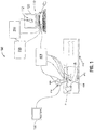
- FIG. 1 illustrates a system 100 that includes a computer 110 and a user interface 112 displayed on a suitable display 120 associated with computer 110 or any suitable monitoring equipment, e.g., an operating room monitor 130.
- user interface 112 may be remote from computer 110 and/or wirelessly connected to computer 110 or other component(s) of system 100.
- computer 110 may be any suitable computing device, such as a desktop computer, laptop computer, tablet, smartphone, etc.
- System 100 further includes a Hospital Information System (HIS) 200, a synthesizer 300, and an operative surgical system 404.
- HIS 200 and synthesizer 300 are detailed below.
- the operative surgical system 404 is utilized during a surgical operation and may include, for example, a monitoring system, a visualization system, a tracking system, one or more surgical tools, etc.
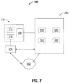
- computer 110 includes one or more processors 114 associated with one or more memories 116.
- Memory 116 may include any non-transitory computer-readable storage media for storing one or more software applications that are executable by processor 114.
- a network module 108 of computer 110 enables communication between computer 110 and a network to which HIS 200 and synthesizer 300 are also connected.
- HIS 200 interfaces with a Picture Archiving and Communication System (PACS) 202, a Radiology Information System (RIS) 204, an Electronic Medical Records System (EMR) 206, and a Laboratory Information System (LIS) 208.
- PACS 202 stores and/or archives images of patients obtained from imaging systems such as, for example, X-ray CT, computerized axial tomography (CAT) scan, positron emission tomography (PET), single-photon emission CT (SPECT), Magnetic Resonant Imaging (MRI), Ultrasound (US), etc.
- imaging systems such as, for example, X-ray CT, computerized axial tomography (CAT) scan, positron emission tomography (PET), single-photon emission CT (SPECT), Magnetic Resonant Imaging (MRI), Ultrasound (US), etc.
- RIS 204 complements HIS 200 and PACS 202 and serves as an electronic management system for an imaging department of a hospital, e.g., allowing a clinician to access digital images of a patient and to associate patient information from EMR 206 with the digital images stored in PACS 202.
- LIS 208 supports data exchange between a hospital laboratory and HIS 200 and, in particular, EMR 206.
- Synthesizer 300 includes a software application stored in a memory, e.g., memory 116, a memory of synthesizer 300, or another suitable memory, that is executable by a processor, e.g., processor 114, a processor of synthesizer 300, or another suitable processor.
- the software application of synthesizer 300 enables a clinician to access HIS 200 through network module 108 of computer 110 or via any other suitable computing device. More specifically, synthesizer 300 communicates with HIS 200 and provides a medium by which the clinician is able to gather data and utilize such data to, for example, pre-operatively predict lung function following a lung resection procedure, as detailed below.
- Synthesizer 300 may interface with a synthesizer cloud 302, e.g., using a hardwired connection or wirelessly, such that the synthesizer 300 may access HIS 200 remotely, e.g., via a device not connected to the intranet, or may interface directly with HIS 200 to provide local access, e.g., within the intranet.
- a synthesizer cloud 302 e.g., using a hardwired connection or wirelessly, such that the synthesizer 300 may access HIS 200 remotely, e.g., via a device not connected to the intranet, or may interface directly with HIS 200 to provide local access, e.g., within the intranet.
- synthesizer 300 uses information gathered from HIS 200 and/or other sources, synthesizer 300 produces a model of the lung(s), e.g., displayed on a user interface, e.g., user interface 112, to enable the clinician to visualize lung features and structures. More specifically, pre-operative image data gathered from HIS 200 is processed by the software application of synthesizer 300 to generate a three-dimensional (3D) model of the patient's lung(s) that is displayed to the clinician, e.g., on user interface 112. Additional medical information such as, for example, prior surgical procedure data, the location of common structures within the patient's body cavity, etc., may also be utilized in the generation of the 3D model. The pre-operative image data and/or additional medical information may be obtained from PACS 202, RIS 204, EMR 206, LIS 208, and/or any other suitable source(s) of information.
- Synthesizer 300 is further configured to gather information indicative of lung function and determine an estimated lung function of one or more portions of the lung based upon the indicative information.
- the indicative information and/or estimated lung function information may be integrated into the 3D model to enable the clinician to visualize, on the 3D model, the lung function (or information indicative thereof) of a particular volume or volumes of the lung, as detailed below.
- the clinician is provided with information in a readily digestible manner, e.g., integrated into a 3D model, to facilitate pre-operative planning and, more specifically, the determination of the location and extent to which the lung is to be resected during a lung resection procedure.
- the estimated lung function information may further be integrated into the 3D model, to enable prediction of the estimated lung function of the portion of the lung remaining after resection of a selected volume of lung, as also detailed below. As such, the clinician can confirm whether the selected volume of lung to be resected is appropriate or whether modification is necessary.
- the information indicative of lung function may be obtained from HIS 200, e.g., from PACS 202, RIS 204, EMR 206, LIS 208, and/or any other suitable source(s) of information.
- the information indicative of lung function may include lung density information, which may be extracted from CT scan data or otherwise obtained. Lung density information can indicate volumes of hypodensity, where the density of the tissue is less than the surrounding tissue. Hypodense volumes of tissue may indicate that these portions are largely air with little to no tissue and, thus, provide poor to no lung function.
- Densities may be measured in Hounsfield units of HU, and may be represented directly on the 3D model either continuously, e.g., in a manner similar to a topographic map; utilizing one or more thresholds, e.g., indicating areas where the density is below a certain threshold (for example, 950 HU); or in incremental levels, e.g., identifying each volume of the lung as having low density, average density, or high density. Alternatively, the density information of a volume may be factored into the determination of estimated lung function of that volume.
- the information indicative of lung function may additionally or alternatively include, for example, the presence, size, and location of an abnormality, e.g., adhesions, lesions, and/or the like, on the lung.
- an abnormality e.g., adhesions, lesions, and/or the like
- Such information may be represented directly on the 3D model in corresponding size, shape, and location, and/or may be factored into the determination of estimated lung function of the volume where the abnormality is located.
- the information indicative of lung function may additionally or alternatively include metabolic activity information. More specifically, PET imaging data may be utilized to identify volumes of low metabolic activity, indicating such volumes are mostly comprised of air with little to no tissue and, thus, provide poor to no lung function. Metabolic activity data may be represented directly on the 3D model either continuously, e.g., in a manner similar to a topographic map; utilizing one or more thresholds, e.g., indicating areas where the metabolic activity is below a certain threshold; or in incremental levels, e.g., identifying each volume of the lung as having low metabolic activity, average metabolic activity, or high metabolic activity. Alternatively, the metabolic activity data for a volume may be factored into the determination of estimated lung function of that volume.
- Additional or alternative information indicative of lung function may include information regarding diseased areas of the lung, e.g., from conditions such as COPD, emphysema, COPD, ILD, fibrosis, asthma, cancer, etc. Such information may be recognized from CT image data obtained from PACS 202 of HIS 200, or may be obtained from another source such as, for example, previous determinations of lung disease indicated in EMR 206 of HIS 200.
- PFT Pulmonary Function Test
- FEVi Forced Expiratory Volume in the first one second of expiration
- FEC Forced Vital Capacity
- TLC Total Lung Capacity
- FRC Functional Residual Capacity
- Lung function information may be provided on the 3D model in one or more clinician-selectable layers such as for example, a layer incorporating the tissue density data, a layer incorporating the abnormality data, a layer incorporating the metabolic activity data, a layer incorporating the diseased areas information, etc.
- the clinician may select the information to be provided (and also that to be excluded).
- levels of lung function determined using the above-noted lung function information may be represented on the 3D model using, for example, indicia, color coding, etc.
- lung function levels may be provided continuously, e.g., using a progressive shading scheme, or may be provided discretely, e.g., using discrete lung function "ratings" each having a color or pattern associated therewith.
- lung function may be indicated continuously across the lung, or the lung may be sectioned into volume segments or regions with a lung function level assigned to each segment or region.
- the software application of synthesizer 300 may automatically select, utilizing the above-noted lung function information and/or other information gathered from EMR 204 of HIS 200, and display, e.g., on user interface 112, suggested resection lines on the 3D model.
- the clinician may move or otherwise modify the suggested resection lines, e.g., utilizing the above-noted lung function information and/or other information as a guide.
- the clinician may "draw" resection lines on the 3D model using any suitable input device, e.g., a mouse, joy-stick, touch-screen display, etc.
- the software application of synthesizer 300 may further analyze the placement of the resection lines on the 3D model to identify potential placement issues based upon information gathered from EMR 204 or based upon medical information regarding sensitive formations and/or vasculature within the lung.
- the resection lines may then be further modified and, finally, set for simulation and the simulation run.
- FIGS. 3 and 4 illustrate displays of the 3D model on a user interface, e.g., user interface 112, including a representation of the lung after the resection simulation has been run, based upon the selected resected lines.
- the lung volume removed may be illustrated in a first manner while the lung volume that remains is illustrated in a different manner, e.g., using different coloring, indicia, shading, line styles, etc. In this manner, each lung volume can be readily identified by the clinician.
- a rendering of the 3D model from the resultant simulation is provided at one portion of the user interface (e.g., at a center portion), with the remaining lung volume 508 illustrated solidly and/or in color and the removed lung volume 506 illustrated in phantom and/or grayscale.
- a depiction of the full 3D model (before the simulated resection) including the lung function information may be provided at another portion of the user interface (e.g., at a corner or the like).
- the lung function information may likewise be represented in the simulation resultant 3D model such as, for example, as illustrated in FIG. 4 .
- predicted post-operative lung function details may likewise be displayed (or otherwise output).
- the software application of synthesizer 300 may predict and display one or more estimated post-operative metrics, e.g., FEV 1 , FEC, TLC, FRC, the level of lung deflation during single lung ventilation surgery, etc.
- the metric is updated as the resection lines and, thus, the volume to be resected, is altered.
- the predicted FEV 1 based upon the volume removed as indicated in FIG. 3 is different from the predicted FEV 1 based upon the volume removed as indicated in FIG. 4 .
- the estimated post-operative metric(s) may be provided in absolute terms, e.g., in their respective units, and/or may be provide relatively, e.g., as a percentage of the pre-operative values.
- a comparison mode is also contemplated, enabling the clinician to compare the predicted post-operative lung function after resection of a first volume of lung versus that after a resection of a second, different volume of lung. This may be provided, for example, by providing a split-screen display on user interface 112, whereby both the first simulated resection and the second simulated resection are provided, e.g., displaying both the illustrations of FIGS. 3 and 4 side-by-side, top- and-bottom, etc., on user interface 112.
- the resection lines corresponding thereto may be utilized in preparation for and/or during the lung resection procedure to achieve the desired result. If not satisfied, the clinician may modify one or more of the resection lines and re-run the simulation as many times as necessary until the clinician is satisfied with the predicted post-operative lung function.
- a pre-resection 3D model of a tissue structure e.g., a patient's lung
- S510 a pre-resection 3D model of a tissue structure
- a clinician e.g., a clinician
- Tissue function information for the tissue structure is determined from the pre-resection 3D model (S530) and provided (S540), e.g., displayed.
- a portion of tissue on the pre-resection 3D model is selected for removal (S550), automatically or manually, based upon, for example, the pre-resection tissue function information. This may be accomplished via one or more resection lines, as detailed above.
- removal of that portion of tissue is simulated on the pre-resection 3D model to produce a post-resection 3D model (S560) that, in embodiments, is provided, e.g., displayed, to the clinician (S570).
- Tissue function information for the post-resection 3D model is then determined (S580) and provided, e.g., displayed, to the clinician (S590) such that, for example, the clinician may determine whether or not the predicted post-operative lung function is satisfactory (S600). If the clinician is satisfied ("YES” at S600), the pre-operative surgical planning method ends ("END"). If the clinician is not satisfied ("NO” at S600), the method returns to S550, allowing a different portion of tissue on the pre-resection 3D model to be selected for removal, based upon which a post-resection 3D model may be generated, a simulation may be run, and tissue function information determined and provided (see S560-S590). Where multiple simulations and post-resection 3D models are run, the clinician may compare results from each, as noted above, to facilitate determining the appropriate portion of tissue to be resected.
- a further aspect of the disclosure is directed to improving the outcomes of the lung surgeries modeled above.
- the lung surgeries seek to limit the excised material of the lung through removal of only selected anatomical sub-lobar segments (in multiple or single configuration) or a non-anatomical sub-lobar wedge.
- Lung surgeries are generally performed when the lung is in a collapsed state. However, in its collapsed state the lung does not appear to the surgeon in the same state as in preoperative CT scans, described above. Small landmarks such as fissures on a deflated lung may be difficult for the surgeon to find when the lung is in the deflated state.
- the surgeon's success rate can be increased through better situational awareness by providing new landmarks to replace or augment the natural landmarks to which the surgeon may be accustomed.
- the disclosure provides systems and methods for modeling an excision of lung tissue before the lung is re-inflated.
- the 3-D model of the lung is displayed in a collapsed state.
- the surgeon can cycle through all lobes, segments, and wedges, with each independently displayed in the 3-D model through a color change or any other suitable method.
- the surgeon can use the 3-D model of the collapsed lung to identify the minimal amount of lung to excise that will still remove the lesion within the required margin.
- the surgeon may toggle on and off various segments and wedges of the lung to identify those that will meet the surgical criteria.
- the display system may recommend appropriate dissection points and give the surgeon feedback on the current selection's margin.
- the system may also calculate the size of the excised material and provide information on the size of the port or incision, specimen bag, nearest extraction location/incision required for removal from the body.
- the excised material is removed from the model and the lung is re-inflated to show how the removed material will affect the patient's overall physiology, including the remaining lung tissue and adjacent structures, similar to the methods described above.
- the system may interface with a database that stores data of prior procedures involving removal of a lung section and how the remaining lung responded to the removal.
- the surgical plan is projected into the surgeon's view highlighting the lobe, segments, and wedges that were selected for excision. The highlighted lung tissue would overlay the now-inflated lung.
- the methods described below utilize the surgical system 100 including a navigation system capable of guiding a surgical tool 406 within the thoracic cavity and the patient's "P" lungs “L” to an area of interest, such as, for example, a section "S" of lung tissue selected for excision.
- the operative surgical system 404 includes a tracking application that is configured for use with the surgical tool 406 and enables monitoring of the position and orientation of a distal portion of the surgical tool 406.
- operative surgical system 404 may be an electromagnetic navigation system utilized to guide the surgical tool 406 through the thoracic cavity and/or lungs "L" of the patient "P.”
- the operative surgical system 404 includes a plurality of reference sensors 408, and a transmitter mat 410.
- the surgical tool 406 has a sensor (not shown) disposed at a distal portion thereof that enables monitoring of the position and orientation of the distal portion of the surgical tool 406, in six degrees of freedom, relative to a reference coordinate system.
- Patient "P” is shown lying on an operating table 412 with the surgical tool 406 advanced within the thoracic cavity using any suitable surgical device capable of permitting a surgical instrument to pass through a patient's "P" chest, such as an access port, trocar, or the like (not shown).
- the preoperative portion of a method for modeling the lungs of a patient "P” using CT imaging data will now be described.
- the lungs (while in an inflated state) of a patient "P” are imaged using any suitable imaging device, such as, for example, a CT device (not shown).
- the CT device may include X-ray CT, computerized axial tomography (CAT) scan, positron emission tomography (PET), or single-photon emission CT (SPECT).
- the imaging data is stored within the memory 116 ( FIG. 1 ) of the computer 110.
- the image data may be stored within a memory associated with a remote computer or network (not shown) such as a distributed network or the internet via a wired or wireless connection for the transmission and reception of data to and from other sources.
- a software application stored within the memory 116 is executed by the processor 114 to enable review of the image data.
- An area of interest illustrating the effects of lung disease e.g., emphysema, COPD, asthma, cancer, or the like
- An area of interest illustrating the effects of lung disease is identified in the images and its location determined within the lungs “L" of the patient "P.”
- imaging methods used for identifying an area of interest are contemplated such as ultrasound, CT scan, metabolic scanning, or the like.
- the results of images generated from a previously acquired CT scan can be analyzed to identify areas of hypodensity.
- PET imaging may be utilized to identify areas of low metabolic activity within the lungs "L".
- the location of the area of interest within the lungs may be identified and its location stored within the memory 116 ( FIG. 1 ) of the computer 110.
- FIG. 6A shows a flow chart illustrating a pre-operative portion of a method of modeling a lung in accordance with the disclosure.
- the image data is processed by the software application stored within the memory 116 to generate a 3-D model reconstruction of the CT images of the patient's lung using any suitable method.
- the software application employs one of a variety of rendering techniques and processing algorithms to isolate, identify, and/or generate a 3-D volumetric rendering of the lungs (in an inflated state) for presentation to the clinician on the display 120.
- a segmentation algorithm is applied to the 3-D model reconstruction to define the boundaries of various types of tissue by comparing the values of each data element of the 3-D reconstruction to a series of thresholds of other similar criteria, such as using density and continuity.
- the segmentation algorithm groups together similar types of tissue based upon the outcome of the comparison, and in one embodiment, may differentiate low contrast density details of the lung (e.g., lung parenchyma, pleura fissure lines, bronchi, and the like) from high contrast density details of the lung (e.g., luminal structures, hilar structures, bronchopulmonary lymph nodes, and the like).
- the software application may utilize any suitable segmentation algorithm known in the art.
- the software application may present each segmented group as a different color or different transparency level that may be selectively adjusted by the clinician in order to enable the clinician to better identify each segmented or differentiated group. It is further contemplated that the software application may illustrate identified structures as opaque and unidentified structures as translucent, or vice versa.
- step S624 the displacement of the collapsed lung is calculated in preparation for transforming the 3-D construction of the inflated lungs into a 3-D construction of the lungs in a collapsed state, as the lungs would typically appear during a surgical procedure.
- the various structures of the lungs deform or compress at differing rates and by differing volumes.
- the clinician assigns each identified volume an elastic modulus, which is utilized by the software application when calculating the deformed (e.g., compressed) model of the lung. Additional properties, such as Poisson's ratio or the like, may be assigned to the differentiated structures, and in particular, to the lung parenchyma, which has a variable compressibility stemming from removal of air during the deflation of the lung.
- the software application may assign higher or lower elastic properties to non-structural portions of the pre-deflation lung volume based on the CT data or by manual input by the clinician. In this manner, tumor (or other lesion) volumes can be assigned a more rigid behavior (e.g., less prone to being compressed) and the displacement of the model will be recalculated accordingly. Additionally, it is contemplated that the software application may use information from patient electronic medical records (EMR) to estimate a more likely level of lung deflation as the elastic properties of the lung tissues will be affected by common lung conditions such as chronic obstruction pulmonary disorder (COPD).
- EMR patient electronic medical records
- COPD chronic obstruction pulmonary disorder
- the computer 110 may be configured to apply the directional effect of gravity on the 3-D model to more accurately calculate the displacement of the collapsed lung (e.g., determine the overall shape and structure of the lungs in a collapsed state). It is envisioned that any suitable algorithm for applying the directional effect of gravity may be utilized.
- the orientation of the 3-D model shown on the display 120 ( FIG. 1 ) should be known in order to accurately apply the directional effect of gravity. Accordingly, if the clinician has already rotated the 3-D model to appear in the lateral decubitus position, the directional effect of gravity will be applied to the 3-D model such that the gravitational force acts in a lateral direction with respect to the 3-D model.
- the 3-D model of the lung in the collapsed state is generated and displayed on the display 20 in step S626, as illustrated by way of example in FIG. 7A .
- any suitable model may be employed to calculate the displacement of the collapsed lung, such as a linear elastic model or a hyperelastic model (e.g., the Saint Venant-Kirchhoff model, the Fung model, or the Mooney-Rivlin model), although other suitable hyperelastic models may also be employed.
- the type of displacement model employed may be selected depending upon the type of tissue being analyzed.
- the software application may apply a secondary smoothing algorithm to prepare the 3-D reconstruction for meshing.
- segmentation and meshing may be completed using the EmprintTM ablation system or superDimensionTM navigation system marketed and sold by Medtronic PLC.
- the resulting meshed 3-D model forms a Computational Lung Model (CLM), which is the 3-D model of the collapsed lung that is displayed to the clinician on the display 120 or the monitoring equipment 130 during the surgical procedure.
- CLM Computational Lung Model
- step S628 the surgeon selects a section “S" ( FIG. 7A ) of the lung (e.g., diseased tissue) for excision or removal.
- a section “S” of lung e.g., diseased tissue
- the section of the lung selected for excision is shown highlighted on the display 120 in a manner to differentiate the selected section "S" from the remainder of the displayed lung "L".
- the selected section "S” may be shown in a different color from the remainder of the lung “L” or be shown darker or lighter than the remainder of the lung "L”.
- step S632 the displayed 3-D model of the collapsed lung "L” is modified.
- the highlighted section of the lung “L” disappears from the displayed 3-D model of the lung, as illustrated by way of example in FIG. 7B .
- modifying the displayed 3-D model of the collapsed lung “L” may include the system 100 generating and displaying an entirely new visual representation (e.g., a 3-D model) of the lung in a collapsed state without the section "S.”
- modifying the displayed 3-D model of the collapsed lung “L” may include modifying the same visual representation (e.g., 3-D model) of the lung in the collapsed state by removing the section "S" from the displayed 3-D model of the lung.
- the displayed 3-D model may be modified by passing a slicing plane through the 3-D model, thereby separating the underlying mesh of the 3-D model into two parts. A hollow interior of the original mesh may be exposed where the two parts were originally connected so a new mesh for each part is used to place a solid surface where the segmenting plane passed through.
- the original 3-D model may be saved to allow toggling back and forth or deleted to save memory.
- the computer 110 is configured to toggle on and off various segments and wedges of the lung "L" in response to a selection of the respective segments or wedges of the lung by the surgeon.
- the computer 10 may calculate a size of the section "S" of the lung "L" selected for excision, which may be used to determine the size of the port or incision through which the section "S" will be withdrawn from the surgical site.
- the calculation may be done by volumetric computation based on the shape of the removed section. For example, the calculation may include an approximation of the curved surfaces of the section "S" or integration over a surface equation that approximates the actual surface.
- the computer may determine an effect that the removal of the section of the lung “L” has on an overall shape or structure of the remaining lung “L”.
- the 3D-model of the lung “L” is shown in the inflated state without the section selected for removal.
- the shape or structure of the displayed 3-D model of the lung “L” in the inflated state is modified based on the determination of the effect that the removal of the section of lung “L” would have on the remaining lung “L”. In this way, the display 120 provides the clinician an estimation of how the overall physiology of the patient's lungs “L” will change after removing the selected section of the lung “L”.
- patient data may be input into the system 100 corresponding to the patient's vitals, such as pulmonary function (through a Pulmonary Function Test, blood gas analysis, or the like), cardiac function (i.e., heart rate or the like), respiration rate, oxygen saturation, and breathing effort.
- This information may also be input into a database (not shown) associated with the system 100.
- identifying information such as age, sex, height, weight, race or national origin, and other information such as disease type, planned procedure, and patient's medical history may be input into the system 100. Over time, a database may be built using the data obtained during each procedure.
- This database may be indexed such that clinicians may review data obtained from similar patients to better predict the outcome of the procedure. Images of the patient obtained by a suitable imaging device may be correlated or indexed to the patient data to enable a clinician to look up similarly situated patients. In this manner, the system 100 of the disclosure may better predict the outcome of removing a selected section of the lung "L".
- the type of treatment being proposed, the volume of the treatment area including the amount of tissue being treated, the type of tissue being targeted, dynamic measures of integrated function (e.g., the six minute walk test), and qualitative metrics (e.g., St. Georges Respiratory Questionnaire) may be recorded and correlated to the patient data.
- the above described information gathered from other patients may be used to predict the effect of removal of the selected section of the lung "L".
- the software application compares the current patient's characteristics and the location of the selected section to the entries populating the database, and using a mathematical computation or other suitable algorithm, identifies a particular patient or set of patients to which the current patient substantially corresponds. Using this information, the computer determines the effect of removing that particular section of the lung "L” on the remaining lung “L” (e.g., which portions of the remaining lung “L” will expand or otherwise shift and to what extent).
- the preoperative constructs of the CLM are complete, and the clinician may proceed to preoperative planning utilizing the CLM generated as a result of the procedure described hereinabove. After preoperative planning is complete, the procedure begins.
- a thoracoscope 414 ( FIG. 1 ) is advanced within the thoracic cavity and video images are captured and transmitted to the display 120, providing a real time view of the patient's lung "L”.
- the 3-D model of the inflated lung "L” generated in step S618 is then superimposed over the real-time view of the lungs “L.”
- the section of the lung "L” selected for excision is highlighted on the display 20 or 30, thereby differentiating the section of the lung "L” from the displayed real-time view of the patient's lungs “L”.
- VATS or iVATS may be utilized to navigate the surgical tool 406 to the selected section "S" of the lung "L". Once the surgical tool 406 has been successfully navigated to the selected section “S” of the lung “L”, the clinician may remove or otherwise treat the selected section "S" of the lung “L” using the surgical tool 406.
- the term "clinician” refers to a doctor, a nurse, or any other care provider and may include support personnel. Although generally described herein as the various determination and/or selection steps being performed by a clinician, it is contemplated that the determination and/or selection steps described herein may be performed by the software application, or a combination of clinician and software application input. As can be appreciated, in certain instances, it may be necessary for the software application to make certain determinations, whereas in other instances it may be necessary for the clinician to make certain determinations. In embodiments, the software application may make a determination and present the determination to the clinician for selection and/or confirmation.
Landscapes
- Engineering & Computer Science (AREA)
- Health & Medical Sciences (AREA)
- Surgery (AREA)
- Life Sciences & Earth Sciences (AREA)
- Medical Informatics (AREA)
- Nuclear Medicine, Radiotherapy & Molecular Imaging (AREA)
- General Health & Medical Sciences (AREA)
- Animal Behavior & Ethology (AREA)
- Heart & Thoracic Surgery (AREA)
- Molecular Biology (AREA)
- Biomedical Technology (AREA)
- Robotics (AREA)
- Public Health (AREA)
- Veterinary Medicine (AREA)
- Radiology & Medical Imaging (AREA)
- Quality & Reliability (AREA)
- Computer Vision & Pattern Recognition (AREA)
- Physics & Mathematics (AREA)
- General Physics & Mathematics (AREA)
- Theoretical Computer Science (AREA)
- Apparatus For Radiation Diagnosis (AREA)
Abstract
Description
- The disclosure relates to surgical systems and methods, and more particularly, to systems and methods facilitating pre-operative prediction of post-operative tissue function and lung modeling.
- Advances in medical imaging have enabled clinicians to more accurately depict the anatomy of a patient, and therefore, more accurately identify diseases and the location of any diseased tissue. For example, medical imaging such as CT (including X-ray CT, computerized axial tomography (CAT) scan, positron emission tomography (PET), and single-photon emission CT (SPECT)) enable the development of three-dimensional reconstructions of anatomical structures. These three-dimensional models enable a clinician to identify and assess features of the modeled anatomical structure, thus facilitating the preoperative planning process.
- Using medical imaging, such as CT, a clinician is able to accurately identify lesions or other medical conditions without the need for invasive surgeries (such as an open approach or thoracotomy). Three-dimensional reconstructions or models of organs, anatomical structures, or the like are developed using the images obtained using one of the above noted imaging modalities. Using the three-dimensional model, a clinician is able to segment various tissues from one another and assess an accurate location of the lesion within the thoracic cavity, or in one particular example, within the lungs. This segmentation further enables a clinician to determine the precise tissue segment with its affiliated vessel and bronchial branches and determine the ideal incision level for Video-Assisted Thoracoscopic Surgery (VATS) procedures (such as a segmentectomy, loboctomy, pneumonectomy, or the like).
- The three-dimensional model and precise identification of the lesion within the lung and its associated vessel and bronchial branches enables clinicians to identify an ideal location for port placement and develop a pathway through which surgical instruments may be guided during the thoracoscopic procedure. Typically, a fiducial or other marker (e.g., a coil or wire) is implanted within the affected tissue with the aid of fluoroscopy or other imaging modalities. Thereafter, the location of the fiducial relative to the lesion is checked using imaging and the VATS procedure is performed.
- Aspects of the disclosure relate to systems and methods that facilitate the pre-operative prediction of post-operative tissue function, e.g., post-resection lung tissue function, thus assisting a clinician in planning for and carrying out a surgical procedure, e.g., determining the location(s) and volume(s) of lung tissue to be resected.
- As used herein, the term "clinician" refers to a doctor, a nurse, or any other care provider and may include support personnel. The term "proximal" refers to the portion of the device or component that is closer to the clinician and the term "distal" refers to the portion of the device or component that is farther from the clinician. Additionally, terms such as front, rear, upper, lower, top, bottom, and similar directional terms are used as relative terms simply for convenience of description and are not limiting in nature. Further, to the extent consistent, any of the aspects and features detailed herein may be utilized in conjunction with any of the other aspects and features detailed herein.
- Provided in accordance with aspects of the disclosure is a method of pre-operative surgical planning and a non-transitory computer-readable storage medium storing a program that, when executed by a processor, performs the method of pre-operative surgical planning. The method includes generating a pre-resection 3D model of a tissue structure, determining tissue function information for the pre-resection 3D model of the tissue structure, simulating, on the pre-resection 3D model, removal of a selected portion of tissue to produce a post-resection 3D model, and determining tissue function information for the post-resection 3D model of the tissue structure.
- In accordance with the disclosure, a method includes determining the selected portion of tissue to be removed prior to the simulation. Determining the selected portion of tissue to be removed is based upon at least one of: an automatically-generated tissue resection line provided on the pre-resection 3D model or an input tissue resection line provided on pre-resection 3D model.
- The method may further include displaying the pre-resection 3D model of the tissue structure on a user interface. The method may also include representing at least a portion of the determined tissue function information for the pre-resection 3D model of the tissue structure on the user interface with the pre-resection 3D model.
- The method may further includes displaying the post-resection 3D model of the tissue structure on a user interface. The method may also include representing at least a portion of the determined tissue function for the post-resection 3D model of the tissue structure on the user interface with the post-resection 3D model.
- The tissue function information for the pre-resection 3D model or the post-resection 3D model may include at least one of: a level of tissue function; a metric indicative of tissue function; an abnormality affecting tissue function; or an indication of a disease affecting tissue function.
- Additionally or alternatively, the tissue function information for the pre-resection 3D model or the post-resection 3D model may include at least one of: tissue density data; metabolic activity data; or pulmonary function test information.
- Another method and/or non-transitory computer-readable storage medium for pre-operative surgical planning provided in accordance with aspects of the disclosure includes displaying a pre-resection 3D model of a tissue structure on a user interface and displaying one or more resection lines on the pre-resection 3D model. The method/storage medium further includes modifying and/or confirming the resection line(s), simulating, on the pre-resection 3D model, removal of a selected portion of tissue based upon the resection line(s) to produce a post-resection 3D model, and displaying the post-resection 3D model on the user interface. The post-resection 3D model represents remaining tissue differently from the selected portion of tissue removed.
- The method may further include determining tissue function information for the pre-resection 3D model of the tissue structure, and representing at least a portion of the determined tissue function information on the pre-resection 3D model.
- Additionally, the method may further include determining tissue function information for the post-resection 3D model of the tissue structure, and representing at least a portion of the determined tissue function information on the post-resection 3D model.
- In aspects of the present disclosure, the method may further include displaying, on the user interface, the post-resection 3D model and another post-resection 3D model. The another post-resection 3D model is the result of a simulation of removal of a different selected portion of tissue based upon at least one different resection line.
- According to another aspect of the disclosure, a system for modeling a lung is provided and includes a display and a computer. The computer has a processor and a memory coupled to the processor. The memory has instructions stored thereon which, when executed by the processor, cause the computer to generate a 3-D model of a lung based on imaging data of the lung, display on the display the 3-D model of the lung in a collapsed state, remove a section of the lung selected for excision, and display the 3-D model of the lung without the section of the lung selected for excision.
- The displayed 3-D model of the lung without the section of the lung selected for excision may be displayed in an inflated state. Additionally or alternatively, the computer may be configured to determine an effect that the removal of the section of the lung has on the lung, modify the displayed 3-D model of the lung in the inflated state based on the determined effect, and display the modified 3-D model of the lung. The displayed 3-D model of the lung without the section of the lung selected for excision may be displayed in the collapsed state.
- The computer may be configured to display a real-time image of a surface of a patient's lung, and display over the displayed surface of the patient's lung a 3-D model of the section of the lung selected for excision. The section of the lung selected for excision may be highlighted to differentiate the section of the lung selected for excision from the displayed surface of the patient's lung. Removing the section of the lung may include modifying the 3-D model of the lung in the collapsed state by removing the section of the lung from the 3-D model of the lung. The computer may be configured to highlight on the display the section of the lung selected for excision prior to removing the section of the lung from the displayed 3-D model.
- In accordance with another aspect of the disclosure, a method of modeling a lung is provided and includes generating a 3-D model of a lung based on imaging data of the lung, displaying on the display the 3-D model of the lung in a collapsed state, and modifying the displayed 3-D model of the lung in response to a removal of a section of the lung selected for excision, such that the section of the lung selected for excision is not shown in the displayed 3-D model of the lung in the collapsed state.
- The method may further include displaying the 3-D model of the lung in an inflated state. Additionally or alternatively, the 3-D model of the lung in the inflated state may be displayed without the section of the lung.
- The method may further include determining an effect that the removal of the section of the lung has on the lung, and displaying the 3-D model of the lung in the inflated state based on the determined effect. Additionally he method may further include displaying a real-time image of a surface of the patient's lungs, and displaying over the displayed surface of the patient's lungs the section of the lung selected for excision. Further the method may include highlighting on the display the section of the lung selected for excision, thereby differentiating the section of the lung from the displayed surface of the lung. The imaging data may be computerized tomography (CT) scan data of the patient's lungs.
- In accordance with yet another aspect of the disclosure, a system for modeling a lung is provided and includes a display and a computer. The computer has a processor and a memory coupled to the processor. The memory has instructions stored thereon which, when executed by the processor, cause the computer to display a 3-D model of a lung in a collapsed state, highlight a section of the 3-D model of the lung selected for excision, and display the 3-D model of the lung without the section of the lung selected for excision.
- In aspects, the computer may be configured to display the 3-D model of the lung in an inflated state. The 3-D model of the lung in the inflated state may be displayed without the section of the lung selected for excision. Further, the computer may be configured to determine an effect that a removal of the section of the lung has on the remaining portion of the lung, and modify the displayed 3-D model of the remaining portion of the lung in the inflated state based on the determined effect. Additionally or alternatively, the computer may be configured to display a real-time image of a surface of a patient's lung, and display over the displayed surface of the patient's lung only the selected section of the 3-D model of the lung.
- Various aspects and features of the disclosure are described hereinbelow with reference to the accompanying drawings wherein like reference numerals identify similar or identical elements.
-
FIG. 1 is a perspective view of a surgical system provided in accordance with aspects of the disclosure; -
FIG. 2 is a block diagram illustrating a portion of the surgical system ofFIG. 1 ; -
FIG. 3 is one illustration of the user interface of the system ofFIG. 1 , displaying a 3D model of a patient's lung indicating portions of the patient's lung selected for removal; -
FIG. 4 is another illustration of the user interface of the system ofFIG. 1 , displaying a 3D model of a patient's lung including lung function information and indicating portions of the patient's lung selected for removal; -
FIG. 5 is a flow diagram illustrating a method in accordance with aspects of the disclosure. -
FIG. 6A is a flow chart showing a pre-operative portion of a method of modeling a lung; -
FIG. 6B is a flow chart showing an intra-operative portion of the method of modeling the lung; -
FIG. 7A is a side, cross-sectional view of a 3-D model of the patient's lungs showing the lungs in a collapsed state; and -
FIG. 7B is a side, cross-sectional view of the 3-D model shown inFIG. 3A with a section of lung tissue removed. - There is a continuing need for systems and methods that facilitate the pre-operative planning process. In particular, there is a need for systems and methods that facilitate the pre-operative prediction of post-operative tissue function to assist the clinician in planning for and carrying out a surgical procedure.
- As such, the systems and methods discussed herein facilitate the pre-operative prediction of post-resection lung tissue function, thus assisting a clinician in determining the location(s) and volume(s) of lung tissue to be resected. However, although detailed herein with respect to pre-operative prediction of lung function after a lung resection procedure, the aspects and features of the disclosure may similarly be utilized with respect to pre-operative prediction of post-operative function of other tissue structures and/or after other surgical procedures.
-
FIG. 1 illustrates asystem 100 that includes acomputer 110 and auser interface 112 displayed on asuitable display 120 associated withcomputer 110 or any suitable monitoring equipment, e.g., anoperating room monitor 130. In embodiments,user interface 112 may be remote fromcomputer 110 and/or wirelessly connected tocomputer 110 or other component(s) ofsystem 100. Although illustrated as a desktop computer inFIG. 1 ,computer 110 may be any suitable computing device, such as a desktop computer, laptop computer, tablet, smartphone, etc.System 100 further includes a Hospital Information System (HIS) 200, asynthesizer 300, and an operativesurgical system 404. HIS 200 andsynthesizer 300 are detailed below. The operativesurgical system 404 is utilized during a surgical operation and may include, for example, a monitoring system, a visualization system, a tracking system, one or more surgical tools, etc. - Referring also to
FIG. 2 ,computer 110 includes one ormore processors 114 associated with one ormore memories 116.Memory 116 may include any non-transitory computer-readable storage media for storing one or more software applications that are executable byprocessor 114. Anetwork module 108 ofcomputer 110 enables communication betweencomputer 110 and a network to which HIS 200 andsynthesizer 300 are also connected. - HIS 200 interfaces with a Picture Archiving and Communication System (PACS) 202, a Radiology Information System (RIS) 204, an Electronic Medical Records System (EMR) 206, and a Laboratory Information System (LIS) 208.
PACS 202 stores and/or archives images of patients obtained from imaging systems such as, for example, X-ray CT, computerized axial tomography (CAT) scan, positron emission tomography (PET), single-photon emission CT (SPECT), Magnetic Resonant Imaging (MRI), Ultrasound (US), etc.RIS 204 complements HIS 200 andPACS 202 and serves as an electronic management system for an imaging department of a hospital, e.g., allowing a clinician to access digital images of a patient and to associate patient information fromEMR 206 with the digital images stored inPACS 202.LIS 208 supports data exchange between a hospital laboratory and HIS 200 and, in particular,EMR 206. -
Synthesizer 300 includes a software application stored in a memory, e.g.,memory 116, a memory ofsynthesizer 300, or another suitable memory, that is executable by a processor, e.g.,processor 114, a processor ofsynthesizer 300, or another suitable processor. The software application ofsynthesizer 300 enables a clinician to access HIS 200 throughnetwork module 108 ofcomputer 110 or via any other suitable computing device. More specifically,synthesizer 300 communicates with HIS 200 and provides a medium by which the clinician is able to gather data and utilize such data to, for example, pre-operatively predict lung function following a lung resection procedure, as detailed below.Synthesizer 300 may interface with asynthesizer cloud 302, e.g., using a hardwired connection or wirelessly, such that thesynthesizer 300 may access HIS 200 remotely, e.g., via a device not connected to the intranet, or may interface directly with HIS 200 to provide local access, e.g., within the intranet. - Referring still to
FIGS. 1 and2 , using information gathered from HIS 200 and/or other sources,synthesizer 300 produces a model of the lung(s), e.g., displayed on a user interface, e.g.,user interface 112, to enable the clinician to visualize lung features and structures. More specifically, pre-operative image data gathered from HIS 200 is processed by the software application ofsynthesizer 300 to generate a three-dimensional (3D) model of the patient's lung(s) that is displayed to the clinician, e.g., onuser interface 112. Additional medical information such as, for example, prior surgical procedure data, the location of common structures within the patient's body cavity, etc., may also be utilized in the generation of the 3D model. The pre-operative image data and/or additional medical information may be obtained fromPACS 202,RIS 204,EMR 206,LIS 208, and/or any other suitable source(s) of information. -
Synthesizer 300 is further configured to gather information indicative of lung function and determine an estimated lung function of one or more portions of the lung based upon the indicative information. The indicative information and/or estimated lung function information may be integrated into the 3D model to enable the clinician to visualize, on the 3D model, the lung function (or information indicative thereof) of a particular volume or volumes of the lung, as detailed below. As such, the clinician is provided with information in a readily digestible manner, e.g., integrated into a 3D model, to facilitate pre-operative planning and, more specifically, the determination of the location and extent to which the lung is to be resected during a lung resection procedure. The estimated lung function information may further be integrated into the 3D model, to enable prediction of the estimated lung function of the portion of the lung remaining after resection of a selected volume of lung, as also detailed below. As such, the clinician can confirm whether the selected volume of lung to be resected is appropriate or whether modification is necessary. - The information indicative of lung function may be obtained from HIS 200, e.g., from
PACS 202,RIS 204,EMR 206,LIS 208, and/or any other suitable source(s) of information. The information indicative of lung function may include lung density information, which may be extracted from CT scan data or otherwise obtained. Lung density information can indicate volumes of hypodensity, where the density of the tissue is less than the surrounding tissue. Hypodense volumes of tissue may indicate that these portions are largely air with little to no tissue and, thus, provide poor to no lung function. Densities may be measured in Hounsfield units of HU, and may be represented directly on the 3D model either continuously, e.g., in a manner similar to a topographic map; utilizing one or more thresholds, e.g., indicating areas where the density is below a certain threshold (for example, 950 HU); or in incremental levels, e.g., identifying each volume of the lung as having low density, average density, or high density. Alternatively, the density information of a volume may be factored into the determination of estimated lung function of that volume. - The information indicative of lung function may additionally or alternatively include, for example, the presence, size, and location of an abnormality, e.g., adhesions, lesions, and/or the like, on the lung. Such information may be represented directly on the 3D model in corresponding size, shape, and location, and/or may be factored into the determination of estimated lung function of the volume where the abnormality is located.
- The information indicative of lung function may additionally or alternatively include metabolic activity information. More specifically, PET imaging data may be utilized to identify volumes of low metabolic activity, indicating such volumes are mostly comprised of air with little to no tissue and, thus, provide poor to no lung function. Metabolic activity data may be represented directly on the 3D model either continuously, e.g., in a manner similar to a topographic map; utilizing one or more thresholds, e.g., indicating areas where the metabolic activity is below a certain threshold; or in incremental levels, e.g., identifying each volume of the lung as having low metabolic activity, average metabolic activity, or high metabolic activity. Alternatively, the metabolic activity data for a volume may be factored into the determination of estimated lung function of that volume.
- Additional or alternative information indicative of lung function may include information regarding diseased areas of the lung, e.g., from conditions such as COPD, emphysema, COPD, ILD, fibrosis, asthma, cancer, etc. Such information may be recognized from CT image data obtained from
PACS 202 of HIS 200, or may be obtained from another source such as, for example, previous determinations of lung disease indicated inEMR 206 of HIS 200. - Information indicative of lung function may also include Pulmonary Function Test (PFT) information such as, for example, Forced Expiratory Volume in the first one second of expiration (FEVi), Forced Vital Capacity (FEC), Total Lung Capacity (TLC), Functional Residual Capacity (FRC), etc.).
- Lung function information may be provided on the 3D model in one or more clinician-selectable layers such as for example, a layer incorporating the tissue density data, a layer incorporating the abnormality data, a layer incorporating the metabolic activity data, a layer incorporating the diseased areas information, etc. As such, in embodiments, the clinician may select the information to be provided (and also that to be excluded). Alternatively or additionally, levels of lung function determined using the above-noted lung function information may be represented on the 3D model using, for example, indicia, color coding, etc. Further, lung function levels may be provided continuously, e.g., using a progressive shading scheme, or may be provided discretely, e.g., using discrete lung function "ratings" each having a color or pattern associated therewith. Likewise, lung function may be indicated continuously across the lung, or the lung may be sectioned into volume segments or regions with a lung function level assigned to each segment or region.
- The software application of
synthesizer 300 may automatically select, utilizing the above-noted lung function information and/or other information gathered fromEMR 204 of HIS 200, and display, e.g., onuser interface 112, suggested resection lines on the 3D model. The clinician may move or otherwise modify the suggested resection lines, e.g., utilizing the above-noted lung function information and/or other information as a guide. Alternatively or additionally, the clinician may "draw" resection lines on the 3D model using any suitable input device, e.g., a mouse, joy-stick, touch-screen display, etc. The software application ofsynthesizer 300 may further analyze the placement of the resection lines on the 3D model to identify potential placement issues based upon information gathered fromEMR 204 or based upon medical information regarding sensitive formations and/or vasculature within the lung. The resection lines may then be further modified and, finally, set for simulation and the simulation run. -
FIGS. 3 and 4 illustrate displays of the 3D model on a user interface, e.g.,user interface 112, including a representation of the lung after the resection simulation has been run, based upon the selected resected lines. The lung volume removed may be illustrated in a first manner while the lung volume that remains is illustrated in a different manner, e.g., using different coloring, indicia, shading, line styles, etc. In this manner, each lung volume can be readily identified by the clinician. In embodiments, a rendering of the 3D model from the resultant simulation, as detailed above, is provided at one portion of the user interface (e.g., at a center portion), with the remaininglung volume 508 illustrated solidly and/or in color and the removedlung volume 506 illustrated in phantom and/or grayscale. In addition, a depiction of the full 3D model (before the simulated resection) including the lung function information, as detailed above, may be provided at another portion of the user interface (e.g., at a corner or the like). The lung function information may likewise be represented in the simulation resultant 3D model such as, for example, as illustrated inFIG. 4 . - In addition to the simulations and representations of the 3D model, predicted post-operative lung function details may likewise be displayed (or otherwise output). For example, as also illustrated in
FIGS. 3 and 4 , based upon the volume to be resected and the estimated lung function and other information relating to the removed and/or remaining volume, the software application ofsynthesizer 300 may predict and display one or more estimated post-operative metrics, e.g., FEV1, FEC, TLC, FRC, the level of lung deflation during single lung ventilation surgery, etc. The metric is updated as the resection lines and, thus, the volume to be resected, is altered. Thus, for example, the predicted FEV1 based upon the volume removed as indicated inFIG. 3 is different from the predicted FEV1 based upon the volume removed as indicated inFIG. 4 . - The estimated post-operative metric(s) may be provided in absolute terms, e.g., in their respective units, and/or may be provide relatively, e.g., as a percentage of the pre-operative values. A comparison mode is also contemplated, enabling the clinician to compare the predicted post-operative lung function after resection of a first volume of lung versus that after a resection of a second, different volume of lung. This may be provided, for example, by providing a split-screen display on
user interface 112, whereby both the first simulated resection and the second simulated resection are provided, e.g., displaying both the illustrations ofFIGS. 3 and 4 side-by-side, top- and-bottom, etc., onuser interface 112. - Once the clinician is satisfied with the predicted post-operative lung function, the resection lines corresponding thereto may be utilized in preparation for and/or during the lung resection procedure to achieve the desired result. If not satisfied, the clinician may modify one or more of the resection lines and re-run the simulation as many times as necessary until the clinician is satisfied with the predicted post-operative lung function.
- Turning to
FIG. 5 , in use, a pre-resection 3D model of a tissue structure, e.g., a patient's lung, is initially generated (S510) and, in embodiments, is provided, e.g., displayed, to a clinician (S520). Tissue function information for the tissue structure is determined from the pre-resection 3D model (S530) and provided (S540), e.g., displayed. - A portion of tissue on the pre-resection 3D model is selected for removal (S550), automatically or manually, based upon, for example, the pre-resection tissue function information. This may be accomplished via one or more resection lines, as detailed above. Once the portion of tissue to be removed is selected (and confirmed), removal of that portion of tissue is simulated on the pre-resection 3D model to produce a post-resection 3D model (S560) that, in embodiments, is provided, e.g., displayed, to the clinician (S570).
- Tissue function information for the post-resection 3D model, e.g., for the remaining tissue, is then determined (S580) and provided, e.g., displayed, to the clinician (S590) such that, for example, the clinician may determine whether or not the predicted post-operative lung function is satisfactory (S600). If the clinician is satisfied ("YES" at S600), the pre-operative surgical planning method ends ("END"). If the clinician is not satisfied ("NO" at S600), the method returns to S550, allowing a different portion of tissue on the pre-resection 3D model to be selected for removal, based upon which a post-resection 3D model may be generated, a simulation may be run, and tissue function information determined and provided (see S560-S590). Where multiple simulations and post-resection 3D models are run, the clinician may compare results from each, as noted above, to facilitate determining the appropriate portion of tissue to be resected.
- A further aspect of the disclosure is directed to improving the outcomes of the lung surgeries modeled above. Typically, the lung surgeries seek to limit the excised material of the lung through removal of only selected anatomical sub-lobar segments (in multiple or single configuration) or a non-anatomical sub-lobar wedge. Lung surgeries are generally performed when the lung is in a collapsed state. However, in its collapsed state the lung does not appear to the surgeon in the same state as in preoperative CT scans, described above. Small landmarks such as fissures on a deflated lung may be difficult for the surgeon to find when the lung is in the deflated state. The surgeon's success rate can be increased through better situational awareness by providing new landmarks to replace or augment the natural landmarks to which the surgeon may be accustomed. The disclosure provides systems and methods for modeling an excision of lung tissue before the lung is re-inflated.
- To improve success rates, during preoperative planning the 3-D model of the lung is displayed in a collapsed state. Once collapsed, the surgeon can cycle through all lobes, segments, and wedges, with each independently displayed in the 3-D model through a color change or any other suitable method. The surgeon can use the 3-D model of the collapsed lung to identify the minimal amount of lung to excise that will still remove the lesion within the required margin. The surgeon may toggle on and off various segments and wedges of the lung to identify those that will meet the surgical criteria. The display system may recommend appropriate dissection points and give the surgeon feedback on the current selection's margin. The system may also calculate the size of the excised material and provide information on the size of the port or incision, specimen bag, nearest extraction location/incision required for removal from the body. Once a decision on what to excise has been made, the excised material is removed from the model and the lung is re-inflated to show how the removed material will affect the patient's overall physiology, including the remaining lung tissue and adjacent structures, similar to the methods described above. The system may interface with a database that stores data of prior procedures involving removal of a lung section and how the remaining lung responded to the removal. During surgery, the surgical plan is projected into the surgeon's view highlighting the lobe, segments, and wedges that were selected for excision. The highlighted lung tissue would overlay the now-inflated lung.
- As illustrated in
FIG. 1 , the methods described below utilize thesurgical system 100 including a navigation system capable of guiding asurgical tool 406 within the thoracic cavity and the patient's "P" lungs "L" to an area of interest, such as, for example, a section "S" of lung tissue selected for excision. The operativesurgical system 404 includes a tracking application that is configured for use with thesurgical tool 406 and enables monitoring of the position and orientation of a distal portion of thesurgical tool 406. operativesurgical system 404 may be an electromagnetic navigation system utilized to guide thesurgical tool 406 through the thoracic cavity and/or lungs "L" of the patient "P." The operativesurgical system 404 includes a plurality ofreference sensors 408, and atransmitter mat 410. Thesurgical tool 406 has a sensor (not shown) disposed at a distal portion thereof that enables monitoring of the position and orientation of the distal portion of thesurgical tool 406, in six degrees of freedom, relative to a reference coordinate system. Patient "P" is shown lying on an operating table 412 with thesurgical tool 406 advanced within the thoracic cavity using any suitable surgical device capable of permitting a surgical instrument to pass through a patient's "P" chest, such as an access port, trocar, or the like (not shown). - The preoperative portion of a method for modeling the lungs of a patient "P" using CT imaging data will now be described. Initially, the lungs (while in an inflated state) of a patient "P" are imaged using any suitable imaging device, such as, for example, a CT device (not shown). The CT device may include X-ray CT, computerized axial tomography (CAT) scan, positron emission tomography (PET), or single-photon emission CT (SPECT). The imaging data is stored within the memory 116 (
FIG. 1 ) of thecomputer 110. In some embodiments, the image data may be stored within a memory associated with a remote computer or network (not shown) such as a distributed network or the internet via a wired or wireless connection for the transmission and reception of data to and from other sources. - Following imaging of the patient, a software application stored within the
memory 116 is executed by theprocessor 114 to enable review of the image data. An area of interest illustrating the effects of lung disease (e.g., emphysema, COPD, asthma, cancer, or the like) is identified in the images and its location determined within the lungs "L" of the patient "P." Several imaging methods used for identifying an area of interest are contemplated such as ultrasound, CT scan, metabolic scanning, or the like. In one embodiment, where the patient "P" is not suffering from easily identified lesions or cancers of the lungs, the results of images generated from a previously acquired CT scan can be analyzed to identify areas of hypodensity. In an alternative embodiment, PET imaging may be utilized to identify areas of low metabolic activity within the lungs "L". After analysis of the image data, using one of the above described techniques, the location of the area of interest within the lungs may be identified and its location stored within the memory 116 (FIG. 1 ) of thecomputer 110. -
FIG. 6A shows a flow chart illustrating a pre-operative portion of a method of modeling a lung in accordance with the disclosure. In step S622, the image data is processed by the software application stored within thememory 116 to generate a 3-D model reconstruction of the CT images of the patient's lung using any suitable method. The software application employs one of a variety of rendering techniques and processing algorithms to isolate, identify, and/or generate a 3-D volumetric rendering of the lungs (in an inflated state) for presentation to the clinician on thedisplay 120. A segmentation algorithm is applied to the 3-D model reconstruction to define the boundaries of various types of tissue by comparing the values of each data element of the 3-D reconstruction to a series of thresholds of other similar criteria, such as using density and continuity. The segmentation algorithm groups together similar types of tissue based upon the outcome of the comparison, and in one embodiment, may differentiate low contrast density details of the lung (e.g., lung parenchyma, pleura fissure lines, bronchi, and the like) from high contrast density details of the lung (e.g., luminal structures, hilar structures, bronchopulmonary lymph nodes, and the like). It is envisioned that the software application may utilize any suitable segmentation algorithm known in the art. The software application may present each segmented group as a different color or different transparency level that may be selectively adjusted by the clinician in order to enable the clinician to better identify each segmented or differentiated group. It is further contemplated that the software application may illustrate identified structures as opaque and unidentified structures as translucent, or vice versa. - In step S624, the displacement of the collapsed lung is calculated in preparation for transforming the 3-D construction of the inflated lungs into a 3-D construction of the lungs in a collapsed state, as the lungs would typically appear during a surgical procedure. When subjected to compression, the various structures of the lungs deform or compress at differing rates and by differing volumes. To account for differences in elasticity, the clinician assigns each identified volume an elastic modulus, which is utilized by the software application when calculating the deformed (e.g., compressed) model of the lung. Additional properties, such as Poisson's ratio or the like, may be assigned to the differentiated structures, and in particular, to the lung parenchyma, which has a variable compressibility stemming from removal of air during the deflation of the lung. The software application may assign higher or lower elastic properties to non-structural portions of the pre-deflation lung volume based on the CT data or by manual input by the clinician. In this manner, tumor (or other lesion) volumes can be assigned a more rigid behavior (e.g., less prone to being compressed) and the displacement of the model will be recalculated accordingly. Additionally, it is contemplated that the software application may use information from patient electronic medical records (EMR) to estimate a more likely level of lung deflation as the elastic properties of the lung tissues will be affected by common lung conditions such as chronic obstruction pulmonary disorder (COPD).
- The computer 110 (
FIG. 1 ) may be configured to apply the directional effect of gravity on the 3-D model to more accurately calculate the displacement of the collapsed lung (e.g., determine the overall shape and structure of the lungs in a collapsed state). It is envisioned that any suitable algorithm for applying the directional effect of gravity may be utilized. The orientation of the 3-D model shown on the display 120 (FIG. 1 ) should be known in order to accurately apply the directional effect of gravity. Accordingly, if the clinician has already rotated the 3-D model to appear in the lateral decubitus position, the directional effect of gravity will be applied to the 3-D model such that the gravitational force acts in a lateral direction with respect to the 3-D model. Once the directional effect of gravity has been applied to the 3-D model, as the clinician rotates the 3-D model within thedisplay 120, the direction of the gravitational force will readjust with respect to the 3-D model to ensure that the gravitational force is acting upon the 3-D model in the correct direction. - Following calculation of the displacement of the collapsed lungs, the 3-D model of the lung in the collapsed state is generated and displayed on the display 20 in step S626, as illustrated by way of example in
FIG. 7A . It is contemplated that any suitable model may be employed to calculate the displacement of the collapsed lung, such as a linear elastic model or a hyperelastic model (e.g., the Saint Venant-Kirchhoff model, the Fung model, or the Mooney-Rivlin model), although other suitable hyperelastic models may also be employed. The type of displacement model employed may be selected depending upon the type of tissue being analyzed. - Once the 3-D reconstruction is generated and segmentation has been completed, the software application may apply a secondary smoothing algorithm to prepare the 3-D reconstruction for meshing. In embodiments, segmentation and meshing may be completed using the Emprint™ ablation system or superDimension™ navigation system marketed and sold by Medtronic PLC. The resulting meshed 3-D model forms a Computational Lung Model (CLM), which is the 3-D model of the collapsed lung that is displayed to the clinician on the
display 120 or themonitoring equipment 130 during the surgical procedure. - In step S628, the surgeon selects a section "S" (
FIG. 7A ) of the lung (e.g., diseased tissue) for excision or removal. Upon selecting the section "S" of lung for excision, in step S630, the section of the lung selected for excision is shown highlighted on thedisplay 120 in a manner to differentiate the selected section "S" from the remainder of the displayed lung "L". For example, the selected section "S" may be shown in a different color from the remainder of the lung "L" or be shown darker or lighter than the remainder of the lung "L". - The surgeon may indicate that the selected section is to be removed, whereby in step S632, the displayed 3-D model of the collapsed lung "L" is modified. In particular, in response to a decision to remove the section of the lung "L" selected for excision, the highlighted section of the lung "L" disappears from the displayed 3-D model of the lung, as illustrated by way of example in
FIG. 7B . In one aspect, modifying the displayed 3-D model of the collapsed lung "L" may include thesystem 100 generating and displaying an entirely new visual representation (e.g., a 3-D model) of the lung in a collapsed state without the section "S." In another aspect, modifying the displayed 3-D model of the collapsed lung "L" may include modifying the same visual representation (e.g., 3-D model) of the lung in the collapsed state by removing the section "S" from the displayed 3-D model of the lung. In either of these aspects, the displayed 3-D model may be modified by passing a slicing plane through the 3-D model, thereby separating the underlying mesh of the 3-D model into two parts. A hollow interior of the original mesh may be exposed where the two parts were originally connected so a new mesh for each part is used to place a solid surface where the segmenting plane passed through. The original 3-D model may be saved to allow toggling back and forth or deleted to save memory. - In some aspects of the disclosure, the
computer 110 is configured to toggle on and off various segments and wedges of the lung "L" in response to a selection of the respective segments or wedges of the lung by the surgeon. - In step S634, the computer 10 may calculate a size of the section "S" of the lung "L" selected for excision, which may be used to determine the size of the port or incision through which the section "S" will be withdrawn from the surgical site. The calculation may be done by volumetric computation based on the shape of the removed section. For example, the calculation may include an approximation of the curved surfaces of the section "S" or integration over a surface equation that approximates the actual surface.
- Upon processing the decision to remove the selected section "S" of the lung "L", the computer, in step S636, may determine an effect that the removal of the section of the lung "L" has on an overall shape or structure of the remaining lung "L". In step S638, the 3D-model of the lung "L" is shown in the inflated state without the section selected for removal. In step S640, the shape or structure of the displayed 3-D model of the lung "L" in the inflated state is modified based on the determination of the effect that the removal of the section of lung "L" would have on the remaining lung "L". In this way, the
display 120 provides the clinician an estimation of how the overall physiology of the patient's lungs "L" will change after removing the selected section of the lung "L". - To assist in predicting the effect of the removal of the selected section of the lung "L", patient data may be input into the
system 100 corresponding to the patient's vitals, such as pulmonary function (through a Pulmonary Function Test, blood gas analysis, or the like), cardiac function (i.e., heart rate or the like), respiration rate, oxygen saturation, and breathing effort. This information may also be input into a database (not shown) associated with thesystem 100. Additionally, identifying information such as age, sex, height, weight, race or national origin, and other information such as disease type, planned procedure, and patient's medical history may be input into thesystem 100. Over time, a database may be built using the data obtained during each procedure. This database may be indexed such that clinicians may review data obtained from similar patients to better predict the outcome of the procedure. Images of the patient obtained by a suitable imaging device may be correlated or indexed to the patient data to enable a clinician to look up similarly situated patients. In this manner, thesystem 100 of the disclosure may better predict the outcome of removing a selected section of the lung "L". The type of treatment being proposed, the volume of the treatment area including the amount of tissue being treated, the type of tissue being targeted, dynamic measures of integrated function (e.g., the six minute walk test), and qualitative metrics (e.g., St. Georges Respiratory Questionnaire) may be recorded and correlated to the patient data. The above described information gathered from other patients may be used to predict the effect of removal of the selected section of the lung "L". The software application compares the current patient's characteristics and the location of the selected section to the entries populating the database, and using a mathematical computation or other suitable algorithm, identifies a particular patient or set of patients to which the current patient substantially corresponds. Using this information, the computer determines the effect of removing that particular section of the lung "L" on the remaining lung "L" (e.g., which portions of the remaining lung "L" will expand or otherwise shift and to what extent). - At this point, the preoperative constructs of the CLM are complete, and the clinician may proceed to preoperative planning utilizing the CLM generated as a result of the procedure described hereinabove. After preoperative planning is complete, the procedure begins.
- With reference to the flow chart depicted in
FIG. 6B , in step S642, a thoracoscope 414 (FIG. 1 ) is advanced within the thoracic cavity and video images are captured and transmitted to thedisplay 120, providing a real time view of the patient's lung "L". In step S644, the 3-D model of the inflated lung "L" generated in step S618 is then superimposed over the real-time view of the lungs "L." In step S646, the section of the lung "L" selected for excision is highlighted on the display 20 or 30, thereby differentiating the section of the lung "L" from the displayed real-time view of the patient's lungs "L". VATS or iVATS may be utilized to navigate thesurgical tool 406 to the selected section "S" of the lung "L". Once thesurgical tool 406 has been successfully navigated to the selected section "S" of the lung "L", the clinician may remove or otherwise treat the selected section "S" of the lung "L" using thesurgical tool 406. - As used herein, the term "clinician" refers to a doctor, a nurse, or any other care provider and may include support personnel. Although generally described herein as the various determination and/or selection steps being performed by a clinician, it is contemplated that the determination and/or selection steps described herein may be performed by the software application, or a combination of clinician and software application input. As can be appreciated, in certain instances, it may be necessary for the software application to make certain determinations, whereas in other instances it may be necessary for the clinician to make certain determinations. In embodiments, the software application may make a determination and present the determination to the clinician for selection and/or confirmation. In other embodiments, it may be necessary for the software application to provide a prompt or other warning to the clinician regarding the consequences of the clinician's decision, or to provide an alternative selection to the clinician, or combinations thereof. In the following description, well-known functions or constructions are not described in detail to avoid obscuring the disclosure in unnecessary detail.
- Although the systems and methods detailed herein are generally described with respect to the lungs, it is contemplated that the following systems and methods may be applied to other organs such as, for example, the liver or the spleen. Further, while several embodiments of the disclosure have been shown in the drawings, it is not intended that the disclosure be limited thereto, as it is intended that the disclosure be as broad in scope as the art will allow and that the specification be read likewise. Therefore, the above description should not be construed as limiting, but merely as exemplifications of particular embodiments.
- The invention may be described by reference to the following numbered paragraphs:-
- 1. A method of pre-operative surgical planning, comprising:
- generating a pre-resection 3D model of a tissue structure;
- displaying the pre-resection 3D model of the tissue structure on a user interface;
- determining tissue function information for the pre-resection 3D model of the tissue structure;
- simulating, on the pre-resection 3D model, removal of a selected portion of tissue to produce a post-resection 3D model;
- displaying the post-resection 3D model; and
- determining tissue function information for the post-resection 3D model of the tissue structure.
- 2. The method according to paragraph 1, further comprising:
determining the selected portion of tissue to be removed prior to the simulation, wherein determining the selected portion of tissue to be removed is based upon an automatically-generated tissue resection line provided on the pre-resection 3D model. - 3. The method according to paragraph 2, further comprising:
modifying the automatically-generated tissue resection line, and wherein determining the selected portion of tissue to be removed is based upon the modified automatically-generated tissue resection line. - 4. The method according to paragraph 1, further comprising:
representing at least a portion of the determined tissue function information for the pre-resection 3D model of the tissue structure on the user interface with the pre-resection 3D model. - 5. The method according to paragraph 1, further comprising:
representing at least a portion of the determined tissue function for the post-resection 3D model of the tissue structure on the user interface with the post-resection 3D model. - 6. The method according to paragraph 1, further comprising:
determining the selected portion of tissue to be removed prior to the simulation, wherein determining the selected portion of tissue to be removed is based upon an input tissue resection line provided on the pre-resection 3D model. - 7. The method according to paragraph 1, wherein the tissue function information for the pre-resection 3D model or the post-resection 3D model includes at least one of:
- a level of tissue function;
- a metric indicative of tissue function;
- an abnormality affecting tissue function; or
- an indication of a disease affecting tissue function.
- 8. The method according to paragraph 1, wherein the tissue function information for the pre-resection 3D model or the post-resection 3D model includes at least one of:
- tissue density data;
- metabolic activity data; or
- pulmonary function test information.
- 9. A non-transitory computer-readable storage medium storing a program that, when executed by a processor, performs the steps of:
- generating a pre-resection 3D model of a tissue structure;
- displaying the pre-resection 3D model of the tissue structure on a user interface;
- determining tissue function information for the pre-resection 3D model of the tissue structure;
- determining a selected portion of tissue to be removed based upon at least one resection line represented on the pre-resection 3D model;
- simulating, on the pre-resection 3D model, removal of the selected portion of tissue to produce a post-resection 3D model; and
- determining tissue function information for the post-resection 3D model of the tissue structure.
- 10. The non-transitory computer-readable storage medium according to paragraph 9, wherein the at least one resection line is an automatically-generated tissue resection line provided on the pre-resection 3D model.
- 11. The non-transitory computer-readable storage medium according to paragraph 9, wherein the at least one resection line is an input tissue resection line provided on pre-resection 3D model.
- 12. The non-transitory computer-readable storage medium according to paragraph 9, further comprising:
representing at least a portion of the determined tissue function information for the pre-resection 3D model of the tissue structure on the user interface with the pre-resection 3D model. - 13. The non-transitory computer-readable storage medium according to paragraph 9, further comprising displaying the post-resection 3D model of the tissue structure on a user interface.
- 14. The non-transitory computer-readable storage medium according to paragraph 13, further comprising:
representing at least a portion of the determined tissue function for the post-resection 3D model of the tissue structure on the user interface with the post-resection 3D model. - 15. The non-transitory computer-readable storage medium according to paragraph 9, wherein the tissue function information for the pre-resection 3D model or the post-resection 3D model includes at least one of:
- a level of tissue function;
- a metric indicative of tissue function;
- an abnormality affecting tissue function; or
- an indication of a disease affecting tissue function.
- 16. The non-transitory computer-readable storage medium according to paragraph 9, wherein the tissue function information for the pre-resection 3D model or the post-resection 3D model includes at least one of:
- tissue density data;
- metabolic activity data; or
- pulmonary function test information.
- 17. A method of pre-operative surgical planning, comprising:
- displaying a pre-resection 3D model of a tissue structure on a user interface;
- displaying at least one resection line on the pre-resection 3D model;
- at least one of modifying or confirming the at least one resection line;
- simulating, on the pre-resection 3D model, removal of a selected portion of tissue based upon the at least one resection line to produce a post-resection 3D model; and
- displaying the post-resection 3D model on the user interface, the post-resection 3D model representing remaining tissue differently from the selected portion of tissue removed.
- 18. The method according to paragraph 17, further comprising:
- determining tissue function information for the pre-resection 3D model of the tissue structure; and
- representing at least a portion of the determined tissue function information on the pre-resection 3D model.
- 19. The method according to paragraph 17, further comprising:
- determining tissue function information for the post-resection 3D model of the tissue structure; and
- representing at least a portion of the determined tissue function information on the post-resection 3D model.
- 20. The method according to paragraph, 17, further comprising:
displaying, on the user interface, the post-resection 3D model and another post-resection 3D model, wherein the another post-resection 3D model is the result of a simulation of removal of a different selected portion of tissue based upon at least one different resection line.
Claims (15)
- A method of pre-operative surgical planning, comprising:generating a pre-resection 3D model of a tissue structure;displaying the pre-resection 3D model of the tissue structure on a user interface;determining tissue function information for the pre-resection 3D model of the tissue structure;simulating, on the pre-resection 3D model, removal of a selected portion of tissue to produce a post-resection 3D model;displaying the post-resection 3D model; anddetermining tissue function information for the post-resection 3D model of the tissue structure.
- The method according to claim 1, further comprising:
determining the selected portion of tissue to be removed prior to the simulation, wherein determining the selected portion of tissue to be removed is based upon an automatically-generated tissue resection line provided on the pre-resection 3D model. - The method according to claim 2, further comprising:
modifying the automatically-generated tissue resection line, and wherein determining the selected portion of tissue to be removed is based upon the modified automatically-generated tissue resection line. - The method according to any preceding claim, further comprising:
representing at least a portion of the determined tissue function information for the pre-resection 3D model of the tissue structure on the user interface with the pre-resection 3D model and/or representing at least a portion of the determined tissue function for the post-resection 3D model of the tissue structure on the user interface with the post-resection 3D model. - The method according to any preceding claim, further comprising:
determining the selected portion of tissue to be removed prior to the simulation, wherein determining the selected portion of tissue to be removed is based upon an input tissue resection line provided on the pre-resection 3D model. - The method according to any preceding claim, wherein the tissue function information for the pre-resection 3D model or the post-resection 3D model includes at least one of:a level of tissue function;a metric indicative of tissue function;an abnormality affecting tissue function; oran indication of a disease affecting tissue function.;and/or wherein the tissue function information for the pre-resection 3D model or the post-resection 3D model includes at least one of:tissue density data;metabolic activity data; orpulmonary function test information.
- A non-transitory computer-readable storage medium storing a program that, when executed by a processor, performs the steps of:generating a pre-resection 3D model of a tissue structure;displaying the pre-resection 3D model of the tissue structure on a user interface;determining tissue function information for the pre-resection 3D model of the tissue structure;determining a selected portion of tissue to be removed based upon at least one resection line represented on the pre-resection 3D model;simulating, on the pre-resection 3D model, removal of the selected portion of tissue to produce a post-resection 3D model; anddetermining tissue function information for the post-resection 3D model of the tissue structure.
- The non-transitory computer-readable storage medium according to claim 7, wherein the at least one resection line is an automatically-generated tissue resection line provided on the pre-resection 3D model.
- The non-transitory computer-readable storage medium according to claim 7 or claim 8, wherein the at least one resection line is an input tissue resection line provided on pre-resection 3D model.
- The non-transitory computer-readable storage medium according to any of claims 7 to 9, further comprising:
representing at least a portion of the determined tissue function information for the pre-resection 3D model of the tissue structure on the user interface with the pre-resection 3D model. - The non-transitory computer-readable storage medium according to any of claims 7 to 10, further comprising displaying the post-resection 3D model of the tissue structure on a user interface; preferably further comprising:
representing at least a portion of the determined tissue function for the post-resection 3D model of the tissue structure on the user interface with the post-resection 3D model. - The non-transitory computer-readable storage medium according to any of claims 7 to 11, wherein the tissue function information for the pre-resection 3D model or the post-resection 3D model includes at least one of:a level of tissue function;a metric indicative of tissue function;an abnormality affecting tissue function; oran indication of a disease affecting tissue function.
- The non-transitory computer-readable storage medium according to any of claims 7 to 12, wherein the tissue function information for the pre-resection 3D model or the post-resection 3D model includes at least one of:tissue density data;metabolic activity data; orpulmonary function test information.
- A method of pre-operative surgical planning, comprising:displaying a pre-resection 3D model of a tissue structure on a user interface;displaying at least one resection line on the pre-resection 3D model;at least one of modifying or confirming the at least one resection line;simulating, on the pre-resection 3D model, removal of a selected portion of tissue based upon the at least one resection line to produce a post-resection 3D model; anddisplaying the post-resection 3D model on the user interface, the post-resection 3D model representing remaining tissue differently from the selected portion of tissue removed.
- The method according to claim 14, further comprising:determining tissue function information for the pre-resection 3D model of the tissue structure; andrepresenting at least a portion of the determined tissue function information on the pre-resection 3D model; and/or further comprising:determining tissue function information for the post-resection 3D model of the tissue structure; andrepresenting at least a portion of the determined tissue function information on the post-resection 3D model; and/or further comprising:
displaying, on the user interface, the post-resection 3D model and another post-resection 3D model, wherein the another post-resection 3D model is the result of a simulation of removal of a different selected portion of tissue based upon at least one different resection line.
Applications Claiming Priority (3)
| Application Number | Priority Date | Filing Date | Title |
|---|---|---|---|
| US201962800670P | 2019-02-04 | 2019-02-04 | |
| US201962801191P | 2019-02-05 | 2019-02-05 | |
| US16/719,172 US11744643B2 (en) | 2019-02-04 | 2019-12-18 | Systems and methods facilitating pre-operative prediction of post-operative tissue function |
Publications (1)
| Publication Number | Publication Date |
|---|---|
| EP3689281A1 true EP3689281A1 (en) | 2020-08-05 |
Family
ID=69467456
Family Applications (1)
| Application Number | Title | Priority Date | Filing Date |
|---|---|---|---|
| EP20155199.1A Pending EP3689281A1 (en) | 2019-02-04 | 2020-02-03 | Systems and methods facilitating pre-operative prediction of post-operative tissue function |
Country Status (3)
| Country | Link |
|---|---|
| US (2) | US11744643B2 (en) |
| EP (1) | EP3689281A1 (en) |
| CN (1) | CN111513845B (en) |
Cited By (4)
| Publication number | Priority date | Publication date | Assignee | Title |
|---|---|---|---|---|
| WO2021136998A1 (en) * | 2019-12-30 | 2021-07-08 | Ethicon Llc | Surgical systems for proposing and corroborating organ portion removals |
| WO2021137000A1 (en) * | 2019-12-30 | 2021-07-08 | Ethicon Llc | Surgical systems for generating three dimensional constructs of anatomical organs and coupling identified anatomical structures thereto |
| CN113409286A (en) * | 2021-06-28 | 2021-09-17 | 杭州佳量医疗科技有限公司 | Laser ablation evaluation system based on magnetic resonance guidance |
| EP4066772A1 (en) * | 2021-03-29 | 2022-10-05 | Cilag GmbH International | System and method for determining, adjusting, and managing resection margin about a subject tissue |
Families Citing this family (15)
| Publication number | Priority date | Publication date | Assignee | Title |
|---|---|---|---|---|
| US11559298B2 (en) | 2018-07-16 | 2023-01-24 | Cilag Gmbh International | Surgical visualization of multiple targets |
| US12257013B2 (en) | 2019-03-15 | 2025-03-25 | Cilag Gmbh International | Robotic surgical systems with mechanisms for scaling camera magnification according to proximity of surgical tool to tissue |
| US12605210B2 (en) | 2019-10-23 | 2026-04-21 | Edda Technology, Inc. | System and method for planning surgical resection of lesions by a linear cutting stapler |
| US11744667B2 (en) | 2019-12-30 | 2023-09-05 | Cilag Gmbh International | Adaptive visualization by a surgical system |
| US11832996B2 (en) | 2019-12-30 | 2023-12-05 | Cilag Gmbh International | Analyzing surgical trends by a surgical system |
| US12002571B2 (en) | 2019-12-30 | 2024-06-04 | Cilag Gmbh International | Dynamic surgical visualization systems |
| US12207881B2 (en) | 2019-12-30 | 2025-01-28 | Cilag Gmbh International | Surgical systems correlating visualization data and powered surgical instrument data |
| US12053223B2 (en) | 2019-12-30 | 2024-08-06 | Cilag Gmbh International | Adaptive surgical system control according to surgical smoke particulate characteristics |
| US12453592B2 (en) | 2019-12-30 | 2025-10-28 | Cilag Gmbh International | Adaptive surgical system control according to surgical smoke cloud characteristics |
| US11776144B2 (en) | 2019-12-30 | 2023-10-03 | Cilag Gmbh International | System and method for determining, adjusting, and managing resection margin about a subject tissue |
| US11284963B2 (en) | 2019-12-30 | 2022-03-29 | Cilag Gmbh International | Method of using imaging devices in surgery |
| KR102779819B1 (en) * | 2022-03-16 | 2025-03-10 | 고려대학교 산학협력단 | Thoracoscopic surgery simulation apparatus and method based on 3-dimensional collapsed lung model |
| US12226164B2 (en) * | 2022-12-30 | 2025-02-18 | Depuy Ireland Unlimited Company | Systems and methods for planning and assisting orthopaedic surgical procedures |
| US12564446B2 (en) | 2022-12-31 | 2026-03-03 | Depuy Ireland Unlimited Company | Systems and methods for planning and assisting orthopaedic surgical procedures |
| WO2026024546A1 (en) * | 2024-07-22 | 2026-01-29 | Covidien Lp | Methods and systems for preoperative visualization of resection areas in thoracic surgeries using laparoscopic and endoluminal robotic systems |
Citations (8)
| Publication number | Priority date | Publication date | Assignee | Title |
|---|---|---|---|---|
| US20110093243A1 (en) * | 2007-11-14 | 2011-04-21 | Tawhai Merryn H | Method for multi-scale meshing of branching biological structures |
| US20140226884A1 (en) * | 2013-02-13 | 2014-08-14 | Mitsubishi Electric Research Laboratories, Inc. | Method for Simulating Thoracic 4DCT |
| US20150238270A1 (en) * | 2014-02-24 | 2015-08-27 | Vida Diagnostics, Inc. | Treatment outcome prediction for lung volume reduction procedures |
| US20160038247A1 (en) * | 2014-08-11 | 2016-02-11 | Covidien Lp | Treatment procedure planning system and method |
| US20160143697A1 (en) * | 2013-06-11 | 2016-05-26 | Agency For Science, Technology And Research | Computer-aided planning of liver surgery |
| EP3298976A1 (en) * | 2016-09-27 | 2018-03-28 | Covidien LP | Systems and methods for detecting pleural invasion for surgical and interventional planning |
| US20180161102A1 (en) * | 2014-10-30 | 2018-06-14 | Edda Technology, Inc. | Method and system for estimating a deflated lung shape for video assisted thoracic surgery in augmented and mixed reality |
| EP3398552A1 (en) * | 2017-05-03 | 2018-11-07 | Covidien LP | Medical image viewer control from surgeon's camera |
Family Cites Families (224)
| Publication number | Priority date | Publication date | Assignee | Title |
|---|---|---|---|---|
| US5057494A (en) | 1988-08-03 | 1991-10-15 | Ethicon, Inc. | Method for preventing tissue damage after an ischemic episode |
| US5321113A (en) | 1993-05-14 | 1994-06-14 | Ethicon, Inc. | Copolymers of an aromatic anhydride and aliphatic ester |
| US6327490B1 (en) | 1998-02-27 | 2001-12-04 | Varian Medical Systems, Inc. | Brachytherapy system for prostate cancer treatment with computer implemented systems and processes to facilitate pre-implantation planning and post-implantation evaluations with storage of multiple plan variations for a single patient |
| US6360116B1 (en) | 1998-02-27 | 2002-03-19 | Varian Medical Systems, Inc. | Brachytherapy system for prostate cancer treatment with computer implemented systems and processes to facilitate pre-operative planning and post-operative evaluations |
| US6003517A (en) | 1998-04-30 | 1999-12-21 | Ethicon Endo-Surgery, Inc. | Method for using an electrosurgical device on lung tissue |
| US6608081B2 (en) | 1999-08-12 | 2003-08-19 | Ortho-Mcneil Pharmaceutical, Inc. | Bicyclic heterocyclic substituted phenyl oxazolidinone antibacterials, and related compositions and methods |
| US6413981B1 (en) | 1999-08-12 | 2002-07-02 | Ortho-Mcneil Pharamceutical, Inc. | Bicyclic heterocyclic substituted phenyl oxazolidinone antibacterials, and related compositions and methods |
| US6750034B1 (en) | 2000-06-30 | 2004-06-15 | Ortho-Mcneil Pharmaceutical, Inc. | DNA encoding human serine protease D-G |
| CA2421638A1 (en) | 2000-09-11 | 2002-03-21 | Closure Medical Corporation | Bronchial occlusion method and apparatus |
| US6472372B1 (en) | 2000-12-06 | 2002-10-29 | Ortho-Mcneil Pharmaceuticals, Inc. | 6-O-Carbamoyl ketolide antibacterials |
| EP1407004B1 (en) | 2001-05-15 | 2009-08-05 | Ortho-McNeil-Janssen Pharmaceuticals, Inc. | Ex-vivo priming for generating cd40-ligand specific cytotoxic t lymphocytes to treat autoimmune and allergic disease |
| US20030013972A1 (en) | 2001-05-29 | 2003-01-16 | Makin Inder Raj. S. | Treatment of lung lesions using ultrasound |
| US7607440B2 (en) | 2001-06-07 | 2009-10-27 | Intuitive Surgical, Inc. | Methods and apparatus for surgical planning |
| ATE312648T1 (en) | 2002-06-17 | 2005-12-15 | Nucletron Bv | SYSTEM FOR REAL-TIME PLANNING OF RADIATION THERAPY |
| MXPA03006874A (en) | 2002-07-31 | 2004-09-03 | Johnson & Johnson | Long term oxygen therapy system. |
| US20040120981A1 (en) | 2002-12-20 | 2004-06-24 | Aruna Nathan | Crosslinked alkyd polyesters for medical applications |
| JP2007525946A (en) | 2003-04-30 | 2007-09-13 | セントカー・インコーポレーテツド | CNGH0010-specific polynucleotides, polypeptides, antibodies, compositions, methods and uses |
| KR20060029276A (en) | 2003-07-04 | 2006-04-05 | 존슨 앤드 존슨 리서치 피티와이 리미티드 | Method for detecting alkylated cytosine in DNA |
| US8484001B2 (en) | 2003-08-26 | 2013-07-09 | Voyant Health Ltd. | Pre-operative medical planning system and method for use thereof |
| US7815561B2 (en) | 2003-09-25 | 2010-10-19 | Xoft, Inc. | Brachytherapy applicator |
| CA2540602A1 (en) | 2003-10-07 | 2005-04-21 | Nomos Corporation | Planning system, method and apparatus for conformal radiation therapy |
| US8209027B2 (en) | 2004-07-07 | 2012-06-26 | The Cleveland Clinic Foundation | System and method to design structure for delivering electrical energy to tissue |
| US8437449B2 (en) | 2004-07-23 | 2013-05-07 | Varian Medical Systems, Inc. | Dynamic/adaptive treatment planning for radiation therapy |
| CA2587909C (en) | 2004-11-16 | 2016-01-05 | Robert L. Barry | Device and method for lung treatment |
| WO2006060653A2 (en) | 2004-11-30 | 2006-06-08 | Veridex Llc | Lung cancer prognostics |
| GB0507204D0 (en) | 2005-04-08 | 2005-05-18 | Leuven K U Res & Dev | Maxillofacial and plastic surgery |
| US7831293B2 (en) | 2005-05-10 | 2010-11-09 | Advanced Clinical Solutions, Inc. | Method of defining a biological target for treatment |
| CA2616316A1 (en) | 2005-07-22 | 2007-02-01 | Tomotherapy Incorporated | Method and system for adapting a radiation therapy treatment plan based on a biological model |
| JP2009502255A (en) | 2005-07-22 | 2009-01-29 | トモセラピー・インコーポレーテッド | Method and system for assessing quality assurance criteria in the delivery of treatment plans |
| WO2007014093A2 (en) | 2005-07-22 | 2007-02-01 | Tomotherapy Incorporated | Method and system for processing data relating to a radiation therapy treatment plan |
| US8377066B2 (en) | 2006-02-27 | 2013-02-19 | Biomet Manufacturing Corp. | Patient-specific elbow guides and associated methods |
| US8070752B2 (en) | 2006-02-27 | 2011-12-06 | Biomet Manufacturing Corp. | Patient specific alignment guide and inter-operative adjustment |
| US9345548B2 (en) | 2006-02-27 | 2016-05-24 | Biomet Manufacturing, Llc | Patient-specific pre-operative planning |
| US9907659B2 (en) | 2007-04-17 | 2018-03-06 | Biomet Manufacturing, Llc | Method and apparatus for manufacturing an implant |
| US9289253B2 (en) | 2006-02-27 | 2016-03-22 | Biomet Manufacturing, Llc | Patient-specific shoulder guide |
| US9113971B2 (en) | 2006-02-27 | 2015-08-25 | Biomet Manufacturing, Llc | Femoral acetabular impingement guide |
| US8241293B2 (en) | 2006-02-27 | 2012-08-14 | Biomet Manufacturing Corp. | Patient specific high tibia osteotomy |
| US9918740B2 (en) | 2006-02-27 | 2018-03-20 | Biomet Manufacturing, Llc | Backup surgical instrument system and method |
| US8473305B2 (en) | 2007-04-17 | 2013-06-25 | Biomet Manufacturing Corp. | Method and apparatus for manufacturing an implant |
| US7616735B2 (en) | 2006-03-28 | 2009-11-10 | Case Western Reserve University | Tomosurgery |
| US9055906B2 (en) | 2006-06-14 | 2015-06-16 | Intuitive Surgical Operations, Inc. | In-vivo visualization systems |
| US11389235B2 (en) | 2006-07-14 | 2022-07-19 | Neuwave Medical, Inc. | Energy delivery systems and uses thereof |
| US8150497B2 (en) | 2006-09-08 | 2012-04-03 | Medtronic, Inc. | System for navigating a planned procedure within a body |
| US7655004B2 (en) | 2007-02-15 | 2010-02-02 | Ethicon Endo-Surgery, Inc. | Electroporation ablation apparatus, system, and method |
| EP1980201B1 (en) | 2007-04-13 | 2010-12-15 | BrainLAB AG | Method and device for planning of local drug delivery |
| US8335359B2 (en) | 2007-07-20 | 2012-12-18 | General Electric Company | Systems, apparatus and processes for automated medical image segmentation |
| US8414123B2 (en) | 2007-08-13 | 2013-04-09 | Novartis Ag | Toric lenses alignment using pre-operative images |
| US9655775B2 (en) | 2007-08-13 | 2017-05-23 | Novartis Ag | Toric lenses alignment using pre-operative images |
| US8265949B2 (en) | 2007-09-27 | 2012-09-11 | Depuy Products, Inc. | Customized patient surgical plan |
| WO2009061942A1 (en) | 2007-11-06 | 2009-05-14 | The Cleveland Clinic Foundation | Automated 3d brain atlas fitting using intra-operative neurophysiological data |
| US10575875B2 (en) | 2007-12-18 | 2020-03-03 | Howmedica Osteonics Corporation | Systems and methods for surgical planning of arthroplasty procedures |
| US7801271B2 (en) | 2007-12-23 | 2010-09-21 | Oraya Therapeutics, Inc. | Methods and devices for orthovoltage ocular radiotherapy and treatment planning |
| US8406860B2 (en) | 2008-01-25 | 2013-03-26 | Novadaq Technologies Inc. | Method for evaluating blush in myocardial tissue |
| EP2269693A4 (en) | 2008-04-14 | 2015-07-08 | Gmv Aerospace And Defence S A | INTRAOPERATIVE RADIOTHERAPY PLANNING SYSTEM AND METHOD FOR IMPLEMENTING SUCH PLANNING |
| US9198733B2 (en) | 2008-04-29 | 2015-12-01 | Virginia Tech Intellectual Properties, Inc. | Treatment planning for electroporation-based therapies |
| SE532644C2 (en) | 2008-07-03 | 2010-03-09 | Aamic Ab | Procedure for analyzing circulating antibodies |
| US8361066B2 (en) | 2009-01-12 | 2013-01-29 | Ethicon Endo-Surgery, Inc. | Electrical ablation devices |
| US8538109B2 (en) * | 2009-03-18 | 2013-09-17 | Siemens Aktiengesellschaft | Method and system for dynamic pulmonary trunk modeling and intervention planning |
| US20180009767A9 (en) | 2009-03-19 | 2018-01-11 | The Johns Hopkins University | Psma targeted fluorescent agents for image guided surgery |
| US10004387B2 (en) | 2009-03-26 | 2018-06-26 | Intuitive Surgical Operations, Inc. | Method and system for assisting an operator in endoscopic navigation |
| WO2010120534A1 (en) | 2009-03-31 | 2010-10-21 | Whitten Matthew R | System and method for radiation therapy treatment planning using a memetic optimization algorithm |
| EP2438584A4 (en) | 2009-05-12 | 2014-04-30 | Edda Technology Inc | SYSTEM, METHOD, APPARATUS AND COMPUTER PROGRAM FOR INTERACTIVE PREOPERATIVE ASSESSMENT |
| US8979871B2 (en) | 2009-08-13 | 2015-03-17 | Monteris Medical Corporation | Image-guided therapy of a tissue |
| US8706184B2 (en) | 2009-10-07 | 2014-04-22 | Intuitive Surgical Operations, Inc. | Methods and apparatus for displaying enhanced imaging data on a clinical image |
| US8784443B2 (en) | 2009-10-20 | 2014-07-22 | Truevision Systems, Inc. | Real-time surgical reference indicium apparatus and methods for astigmatism correction |
| US9308254B2 (en) | 2010-02-17 | 2016-04-12 | Imperial Innovations Limited | Method for treatment of post operative cognitive dysfunction |
| EP2558968B1 (en) | 2010-04-14 | 2021-02-17 | Koninklijke Philips N.V. | System and method for planning a neurosurgical operation |
| JP6153865B2 (en) | 2010-05-03 | 2017-06-28 | ニューウェーブ メディカル, インコーポレイテッドNeuwave Medical, Inc. | Energy delivery system |
| EP2583244B1 (en) | 2010-06-16 | 2019-07-24 | A² Surgical | Method of determination of access areas from 3d patient images |
| CN103338714B (en) | 2010-09-07 | 2015-11-25 | 克里夫兰诊所基金会 | Positioning devices for prosthetic implants |
| US8968335B2 (en) | 2010-10-27 | 2015-03-03 | Mitralign, Inc. | Hand operated device for controlled deployment of a tissue anchor and method of using the same |
| US20120276509A1 (en) * | 2010-10-29 | 2012-11-01 | The Cleveland Clinic Foundation | System of preoperative planning and provision of patient-specific surgical aids |
| CA2816337C (en) | 2010-10-29 | 2019-10-22 | The Cleveland Clinic Foundation | System and method for association of a guiding aid with a patient tissue |
| US10583039B2 (en) | 2010-12-30 | 2020-03-10 | Amo Wavefront Sciences, Llc | Method and system for eye measurements and cataract surgery planning using vector function derived from prior surgeries |
| US10582846B2 (en) | 2010-12-30 | 2020-03-10 | Amo Wavefront Sciences, Llc | Method and system for eye measurements and cataract surgery planning using vector function derived from prior surgeries |
| US10582847B2 (en) | 2010-12-30 | 2020-03-10 | Amo Wavefront Sciences, Llc | Method and system for eye measurements and cataract surgery planning using vector function derived from prior surgeries |
| EP2673738A4 (en) | 2011-02-11 | 2017-08-23 | E-4 Endeavors, Inc. | System and method for modeling a biopsy specimen |
| US8827934B2 (en) | 2011-05-13 | 2014-09-09 | Intuitive Surgical Operations, Inc. | Method and system for determining information of extrema during expansion and contraction cycles of an object |
| US8900131B2 (en) | 2011-05-13 | 2014-12-02 | Intuitive Surgical Operations, Inc. | Medical system providing dynamic registration of a model of an anatomical structure for image-guided surgery |
| US8532807B2 (en) | 2011-06-06 | 2013-09-10 | Biomet Manufacturing, Llc | Pre-operative planning and manufacturing method for orthopedic procedure |
| WO2013109778A1 (en) | 2012-01-17 | 2013-07-25 | Sigma Instruments Holdings, Llc | System and method for treating soft tissue with force impulse and electrical stimulation |
| US9167989B2 (en) | 2011-09-16 | 2015-10-27 | Mako Surgical Corp. | Systems and methods for measuring parameters in joint replacement surgery |
| US20130303944A1 (en) | 2012-05-14 | 2013-11-14 | Intuitive Surgical Operations, Inc. | Off-axis electromagnetic sensor |
| US9089382B2 (en) | 2012-01-23 | 2015-07-28 | Biomet 3I, Llc | Method and apparatus for recording spatial gingival soft tissue relationship to implant placement within alveolar bone for immediate-implant placement |
| EP2809249B1 (en) | 2012-02-03 | 2018-12-26 | Intuitive Surgical Operations, Inc. | Steerable flexible needle with embedded shape sensing |
| KR102171520B1 (en) | 2012-05-14 | 2020-10-29 | 인튜어티브 서지컬 오퍼레이션즈 인코포레이티드 | Systems and methods for deformation compensation using shape sensing |
| US20130303945A1 (en) | 2012-05-14 | 2013-11-14 | Intuitive Surgical Operations, Inc. | Electromagnetic tip sensor |
| EP2849668B1 (en) | 2012-05-14 | 2018-11-14 | Intuitive Surgical Operations Inc. | Systems and methods for registration of a medical device using rapid pose search |
| US10039473B2 (en) | 2012-05-14 | 2018-08-07 | Intuitive Surgical Operations, Inc. | Systems and methods for navigation based on ordered sensor records |
| EP3524184B1 (en) | 2012-05-14 | 2021-02-24 | Intuitive Surgical Operations Inc. | Systems for registration of a medical device using a reduced search space |
| US9439627B2 (en) | 2012-05-22 | 2016-09-13 | Covidien Lp | Planning system and navigation system for an ablation procedure |
| US9439623B2 (en) | 2012-05-22 | 2016-09-13 | Covidien Lp | Surgical planning system and navigation system |
| US9429696B2 (en) | 2012-06-25 | 2016-08-30 | Intuitive Surgical Operations, Inc. | Systems and methods for reducing measurement error in optical fiber shape sensors |
| US9277970B2 (en) | 2012-07-19 | 2016-03-08 | Siemens Aktiengesellschaft | System and method for patient specific planning and guidance of ablative procedures for cardiac arrhythmias |
| US9801551B2 (en) | 2012-07-20 | 2017-10-31 | Intuitive Sugical Operations, Inc. | Annular vision system |
| JP6074587B2 (en) | 2012-08-06 | 2017-02-08 | 株式会社Joled | Display panel, display device and electronic device |
| EP2882368A4 (en) | 2012-08-08 | 2016-03-16 | Ortoma Ab | METHOD AND SYSTEM FOR COMPUTER-AIDED SURGERY |
| CN106562757B (en) | 2012-08-14 | 2019-05-14 | 直观外科手术操作公司 | The system and method for registration for multiple vision systems |
| US20140081659A1 (en) | 2012-09-17 | 2014-03-20 | Depuy Orthopaedics, Inc. | Systems and methods for surgical and interventional planning, support, post-operative follow-up, and functional recovery tracking |
| EP2906133B1 (en) | 2012-10-12 | 2022-03-23 | Intuitive Surgical Operations, Inc. | Determining position of medical device in branched anatomical structure |
| US9761014B2 (en) | 2012-11-15 | 2017-09-12 | Siemens Healthcare Gmbh | System and method for registering pre-operative and intra-operative images using biomechanical model simulations |
| US20140188440A1 (en) | 2012-12-31 | 2014-07-03 | Intuitive Surgical Operations, Inc. | Systems And Methods For Interventional Procedure Planning |
| EP2996602A2 (en) * | 2013-03-14 | 2016-03-23 | Vida Diagnostics, Inc. | Treatment planning for lung volume reduction procedures |
| CA2897086C (en) | 2013-03-15 | 2016-07-05 | Synaptive Medical (Barbados) Inc. | System and method for detecting tissue and fiber tract deformation |
| US9600138B2 (en) | 2013-03-15 | 2017-03-21 | Synaptive Medical (Barbados) Inc. | Planning, navigation and simulation systems and methods for minimally invasive therapy |
| WO2014139022A1 (en) | 2013-03-15 | 2014-09-18 | Synaptive Medical (Barbados) Inc. | Systems and methods for navigation and simulation of minimally invasive therapy |
| JP6363165B2 (en) | 2013-03-15 | 2018-07-25 | インテュイティブ サージカル オペレーションズ, インコーポレイテッド | Shape sensor system for tracking interventional instruments and method of using the system |
| US9259287B2 (en) | 2013-04-02 | 2016-02-16 | Siemens Aktiengesellschaft | Patient specific planning and simulation of ablative procedures |
| CN114343608B (en) | 2013-07-29 | 2024-07-26 | 直观外科手术操作公司 | Shape sensor system with redundant sensing |
| CN105473098B (en) | 2013-08-15 | 2019-03-26 | 直观外科手术操作公司 | System and method for medical procedure validation |
| KR102356881B1 (en) | 2013-08-15 | 2022-02-03 | 인튜어티브 서지컬 오퍼레이션즈 인코포레이티드 | Graphical user interface for catheter positioning and insertion |
| CN105939647B (en) | 2013-10-24 | 2020-01-21 | 奥瑞斯健康公司 | Robot-assisted endovascular surgery system and related methods |
| WO2015068035A1 (en) | 2013-11-08 | 2015-05-14 | Imascap | Methods, systems and devices for pre-operatively planned adaptive glenoid implants |
| KR102337440B1 (en) | 2013-12-09 | 2021-12-10 | 인튜어티브 서지컬 오퍼레이션즈 인코포레이티드 | Systems and methods for device-aware flexible tool registration |
| AU2015204201B2 (en) | 2014-01-06 | 2019-11-28 | Body Vision Medical Ltd. | Surgical devices and methods of use thereof |
| CN106170265B (en) | 2014-02-04 | 2020-06-30 | 直观外科手术操作公司 | System and method for non-rigid deformation of tissue for virtual navigation of interventional tools |
| US20150223765A1 (en) | 2014-02-07 | 2015-08-13 | Intuitive Surgical Operations, Inc. | Systems and methods for using x-ray field emission to determine instrument position and orientation |
| US10402535B2 (en) | 2014-02-26 | 2019-09-03 | Siemens Healthcare Gmbh | System and method for personalized computation of tissue ablation extent based on medical images |
| CA2941017A1 (en) | 2014-02-28 | 2015-09-03 | Blue Belt Technologies, Inc. | System and methods for positioning bone cut guide |
| KR102521043B1 (en) | 2014-03-17 | 2023-04-14 | 인튜어티브 서지컬 오퍼레이션즈 인코포레이티드 | Surgical system including a non-white light general illuminator |
| US10912523B2 (en) | 2014-03-24 | 2021-02-09 | Intuitive Surgical Operations, Inc. | Systems and methods for anatomic motion compensation |
| CN106535812B (en) | 2014-03-28 | 2020-01-21 | 直观外科手术操作公司 | Surgical system with haptic feedback based on quantitative 3D imaging |
| US9999772B2 (en) | 2014-04-03 | 2018-06-19 | Pacesetter, Inc. | Systems and method for deep brain stimulation therapy |
| CN105992996B (en) * | 2014-04-04 | 2019-11-26 | 外科手术室公司 | Dynamic and interactive navigation in surgical environment |
| TW201618795A (en) | 2014-04-15 | 2016-06-01 | 波泰里斯股份有限公司 | Systems and methods to improve organ function and organ transplant longevity |
| US9877697B2 (en) | 2014-04-30 | 2018-01-30 | Emory University | Systems, methods and computer readable storage media storing instructions for generating planning images based on HDR applicators |
| AU2015290043B2 (en) | 2014-07-18 | 2019-08-22 | Ethicon, Inc. | Methods and devices for controlling the size of emphysematous bullae |
| ES2797748T3 (en) | 2014-07-18 | 2020-12-03 | Ethicon Inc | Mechanical retraction by anchoring to reduce lung volume |
| KR20220065894A (en) | 2014-07-28 | 2022-05-20 | 인튜어티브 서지컬 오퍼레이션즈 인코포레이티드 | Systems and methods for intraoperative segmentation |
| EP3174490B1 (en) | 2014-07-28 | 2020-04-15 | Intuitive Surgical Operations, Inc. | Systems and methods for planning multiple interventional procedures |
| US9700390B2 (en) | 2014-08-22 | 2017-07-11 | Biomet 3I, Llc | Soft-tissue preservation arrangement and method |
| KR102542848B1 (en) | 2014-08-23 | 2023-06-14 | 인튜어티브 서지컬 오퍼레이션즈 인코포레이티드 | Systems and methods for display of pathological data in an image guided procedure |
| US10373719B2 (en) | 2014-09-10 | 2019-08-06 | Intuitive Surgical Operations, Inc. | Systems and methods for pre-operative modeling |
| US9833245B2 (en) | 2014-09-29 | 2017-12-05 | Biomet Sports Medicine, Llc | Tibial tubercule osteotomy |
| US10314513B2 (en) | 2014-10-10 | 2019-06-11 | Intuitive Surgical Operations, Inc. | Systems and methods for reducing measurement error using optical fiber shape sensors |
| EP3206556B1 (en) | 2014-10-17 | 2026-03-25 | Intuitive Surgical Operations, Inc. | Systems and methods for reducing measurement error using optical fiber shape sensors |
| CA2965091A1 (en) | 2014-10-20 | 2016-05-06 | Dorian Averbuch | Surgical devices and methods of use thereof |
| US9972081B2 (en) * | 2014-10-30 | 2018-05-15 | Edda Technology, Inc. | Method and system for estimating a deflated lung shape for video assisted thoracic surgery |
| CN107111684A (en) | 2014-11-13 | 2017-08-29 | 直观外科手术操作公司 | System and method for filtering localization data |
| WO2016106114A1 (en) | 2014-12-22 | 2016-06-30 | Intuitive Surgical Operations, Inc. | Flexible electromagnetic sensor |
| US9821174B1 (en) | 2015-02-06 | 2017-11-21 | Gammatile Llc | Radioactive implant planning system and placement guide system |
| EP3280312A4 (en) | 2015-04-06 | 2019-02-20 | Intuitive Surgical Operations Inc. | SYSTEMS AND METHODS FOR IMAGE-GUIDED SURGICAL RECORDING COMPENSATION |
| CN107624192B (en) | 2015-05-11 | 2020-10-27 | 西门子公司 | System and method for surgical guidance and intraoperative pathology by endoscopic tissue differentiation |
| CN107660134B (en) | 2015-05-22 | 2021-06-29 | 直观外科手术操作公司 | System and method for image-guided surgical recording |
| WO2017030915A1 (en) | 2015-08-14 | 2017-02-23 | Intuitive Surgical Operations, Inc. | Systems and methods of registration for image-guided surgery |
| WO2017030913A2 (en) | 2015-08-14 | 2017-02-23 | Intuitive Surgical Operations, Inc. | Systems and methods of registration for image-guided surgery |
| US10569071B2 (en) | 2015-08-31 | 2020-02-25 | Ethicon Llc | Medicant eluting adjuncts and methods of using medicant eluting adjuncts |
| US10245034B2 (en) | 2015-08-31 | 2019-04-02 | Ethicon Llc | Inducing tissue adhesions using surgical adjuncts and medicants |
| CN108024693B (en) | 2015-09-10 | 2021-07-09 | 直观外科手术操作公司 | Systems and methods for utilizing tracking in image-guided medical procedures |
| AU2016323982A1 (en) | 2015-09-18 | 2018-04-12 | Auris Health, Inc. | Navigation of tubular networks |
| US10716457B2 (en) * | 2015-10-14 | 2020-07-21 | Siemens Aktiengesellschaft | Method and system for calculating resected tissue volume from 2D/2.5D intraoperative image data |
| WO2017075067A1 (en) | 2015-10-26 | 2017-05-04 | Neuwave Medical, Inc. | Energy delivery systems and uses thereof |
| US10405753B2 (en) | 2015-11-10 | 2019-09-10 | Intuitive Surgical Operations, Inc. | Pharmaceutical compositions of near IR closed chain, sulfo-cyanine dyes |
| EP3391030B1 (en) | 2015-12-14 | 2025-02-12 | Intuitive Surgical Operations, Inc. | Apparatus and method for generating 3-d data for an anatomical target using optical fiber shape sensing |
| US9996361B2 (en) | 2015-12-23 | 2018-06-12 | Intel Corporation | Byte and nibble sort instructions that produce sorted destination register and destination index mapping |
| US10896506B2 (en) | 2016-02-12 | 2021-01-19 | Intuitive Surgical Operations, Inc | Systems and methods for using registered fluoroscopic images in image-guided surgery |
| US11399895B2 (en) | 2016-02-12 | 2022-08-02 | Intuitive Surgical Operations, Inc. | Systems and methods of pose estimation and calibration of perspective imaging system in image guided surgery |
| WO2017153839A1 (en) | 2016-03-10 | 2017-09-14 | Body Vision Medical Ltd. | Methods and systems for using multi view pose estimation |
| US10702137B2 (en) | 2016-03-14 | 2020-07-07 | Intuitive Surgical Operations, Inc.. | Endoscopic instrument with compliant thermal interface |
| US20170296679A1 (en) | 2016-04-18 | 2017-10-19 | Intuitive Surgical Operations, Inc. | Compositions of Near IR Closed Chain, Sulfo-Cyanine Dyes and Prostate Specific Membrane Antigen Ligands |
| US10561360B2 (en) | 2016-06-15 | 2020-02-18 | Biomet Manufacturing, Llc | Implants, systems and methods for surgical planning and assessment |
| WO2017218552A1 (en) | 2016-06-15 | 2017-12-21 | Intuitive Surgical Operations, Inc. | Systems and methods of integrated real-time visualization |
| EP4374768B1 (en) | 2016-06-30 | 2026-03-25 | Intuitive Surgical Operations, Inc. | Graphical user interface for displaying guidance information in a plurality of modes during an image-guided procedure |
| EP4238490B1 (en) | 2016-06-30 | 2025-09-17 | Intuitive Surgical Operations, Inc. | Graphical user interface for displaying guidance information during an image-guided procedure |
| EP3481318B1 (en) | 2016-07-08 | 2022-09-14 | Biomet Manufacturing, LLC | Reverse shoulder pre-operative planning |
| WO2018035122A1 (en) | 2016-08-16 | 2018-02-22 | Intuitive Surgical Operations, Inc. | Augmented accuracy using large diameter shape fiber |
| CN109561934B (en) | 2016-08-23 | 2022-03-08 | 直观外科手术操作公司 | System and method for monitoring patient movement during medical procedure |
| EP4623797A3 (en) | 2016-09-21 | 2025-12-03 | Intuitive Surgical Operations, Inc. | System for instrument buckling detection |
| US10542953B2 (en) * | 2016-09-27 | 2020-01-28 | Covidien Lp | Fissural assessment and surgical and interventional planning |
| WO2018064566A1 (en) | 2016-09-30 | 2018-04-05 | Intuitive Surgical Operations, Inc. | Systems and methods for entry point localization |
| WO2018075911A1 (en) | 2016-10-21 | 2018-04-26 | Intuitive Surgical Operations, Inc. | Shape sensing with multi-core fiber sensor |
| EP3534818B1 (en) | 2016-11-02 | 2023-09-20 | Intuitive Surgical Operations, Inc. | Systems for continuous registration for image-guided surgery |
| US20180144092A1 (en) | 2016-11-21 | 2018-05-24 | Johnson & Johnson Vision Care, Inc. | Biomedical sensing methods and apparatus for the detection and prevention of lung cancer states |
| CN106806021A (en) * | 2016-11-21 | 2017-06-09 | 厦门强本宇康科技有限公司 | A kind of VR surgery simulation systems and method based on human organ 3D models |
| WO2018106950A1 (en) | 2016-12-08 | 2018-06-14 | Intuitive Surgical Operations, Inc. | Systems and methods for navigation in image-guided medical procedures |
| JP7097883B2 (en) | 2016-12-09 | 2022-07-08 | インテュイティブ サージカル オペレーションズ, インコーポレイテッド | Systems and methods for sensing heat flux distribution in body tissues |
| US10426551B2 (en) | 2016-12-09 | 2019-10-01 | Microsoft Technology Licensing, Llc | Personalized refractive surgery recommendations for eye patients |
| CN110087576B (en) | 2017-01-09 | 2023-03-17 | 直观外科手术操作公司 | System and method for registering an elongated device to a three-dimensional image in an image-guided procedure |
| EP3576662B1 (en) | 2017-02-01 | 2026-01-14 | Intuitive Surgical Operations, Inc. | Systems for data filtering of passageway sensor data |
| WO2018144698A1 (en) | 2017-02-01 | 2018-08-09 | Intuitive Surgical Operations, Inc. | Systems and methods of registration for image-guided procedures |
| KR102503033B1 (en) | 2017-02-01 | 2023-02-23 | 인튜어티브 서지컬 오퍼레이션즈 인코포레이티드 | Fixation systems and methods for image-guided procedures |
| JP7282685B2 (en) | 2017-03-31 | 2023-05-29 | オーリス ヘルス インコーポレイテッド | A robotic system for navigation of luminal networks with compensation for physiological noise |
| EP4539056A3 (en) | 2017-04-18 | 2025-06-25 | Intuitive Surgical Operations, Inc. | Graphical user interface for planning a procedure |
| US11937880B2 (en) | 2017-04-18 | 2024-03-26 | Intuitive Surgical Operations, Inc. | Graphical user interface for monitoring an image-guided procedure |
| JP7165668B2 (en) | 2017-04-21 | 2022-11-04 | メディクレア インターナショナル | System for developing one or more patient-specific spinal implants |
| WO2018215832A2 (en) | 2017-05-24 | 2018-11-29 | Dorian Averbuch | Methods for using radial endobronchial ultrasound probes for three-dimensional reconstruction of images and improved target localization |
| US10022192B1 (en) | 2017-06-23 | 2018-07-17 | Auris Health, Inc. | Automatically-initialized robotic systems for navigation of luminal networks |
| WO2018237187A2 (en) | 2017-06-23 | 2018-12-27 | Intuitive Surgical Operations, Inc. | SYSTEMS AND METHODS FOR NAVIGATING A TARGET LOCATION DURING A MEDICAL ACT |
| CN118121324A (en) | 2017-06-28 | 2024-06-04 | 奥瑞斯健康公司 | Systems for detecting electromagnetic distortion |
| CN116725667A (en) | 2017-06-28 | 2023-09-12 | 奥瑞斯健康公司 | System for providing positioning information and method for positioning an instrument within an anatomical structure |
| RU2685961C2 (en) * | 2017-07-17 | 2019-04-23 | Общество с ограниченной ответственностью "ЭНСИМ" | Surgical procedure preoperative modeling method and system |
| US12004830B2 (en) | 2017-08-16 | 2024-06-11 | Intuitive Surgical Operations , Inc. | Systems and methods for monitoring patient motion during a medical procedure |
| WO2019051464A1 (en) * | 2017-09-11 | 2019-03-14 | Lang Philipp K | Augmented reality display for vascular and other interventions, compensation for cardiac and respiratory motion |
| US11058493B2 (en) | 2017-10-13 | 2021-07-13 | Auris Health, Inc. | Robotic system configured for navigation path tracing |
| US10555778B2 (en) | 2017-10-13 | 2020-02-11 | Auris Health, Inc. | Image-based branch detection and mapping for navigation |
| CN107895364B (en) * | 2017-10-31 | 2019-06-14 | 哈尔滨理工大学 | A 3D reconstruction system for preoperative planning of virtual surgery |
| WO2019099396A2 (en) | 2017-11-14 | 2019-05-23 | Intuitive Surgical Operations, Inc. | Systems and methods for cleaning endoscopic instruments |
| EP4467171A3 (en) | 2017-12-08 | 2025-02-26 | Auris Health, Inc. | Systems employing directed fluidics |
| AU2018378810B2 (en) | 2017-12-08 | 2024-02-22 | Auris Health, Inc. | System and method for medical instrument navigation and targeting |
| JP7322026B2 (en) | 2017-12-14 | 2023-08-07 | オーリス ヘルス インコーポレイテッド | System and method for instrument localization |
| CN110809453B (en) | 2017-12-18 | 2023-06-06 | 奥瑞斯健康公司 | Methods and systems for instrument tracking and navigation within a luminal network |
| EP3749239B1 (en) | 2018-02-05 | 2024-08-07 | Broncus Medical Inc. | Image-guided lung tumor planning and ablation system |
| US10885630B2 (en) | 2018-03-01 | 2021-01-05 | Intuitive Surgical Operations, Inc | Systems and methods for segmentation of anatomical structures for image-guided surgery |
| US10980913B2 (en) | 2018-03-05 | 2021-04-20 | Ethicon Llc | Sealant foam compositions for lung applications |
| US20190269819A1 (en) | 2018-03-05 | 2019-09-05 | Ethicon Llc | Sealant foam compositions for lung applications |
| US20190298451A1 (en) | 2018-03-27 | 2019-10-03 | Intuitive Surgical Operations, Inc. | Systems and methods for delivering targeted therapy |
| CN110913791B (en) | 2018-03-28 | 2021-10-08 | 奥瑞斯健康公司 | System and method for displaying estimated instrument location |
| JP7214747B2 (en) | 2018-03-28 | 2023-01-30 | オーリス ヘルス インコーポレイテッド | System and method for position sensor alignment |
| MX2020012902A (en) | 2018-05-30 | 2021-02-26 | Auris Health Inc | Systems and methods for location sensor-based branch prediction. |
| MX2020012898A (en) | 2018-05-31 | 2021-02-26 | Auris Health Inc | Path-based navigation of tubular networks. |
| KR102455671B1 (en) | 2018-05-31 | 2022-10-20 | 아우리스 헬스, 인코포레이티드 | Image-Based Airway Analysis and Mapping |
| CN108670409B (en) * | 2018-06-04 | 2020-01-14 | 冯原 | Three-dimensional lung tissue reconstruction and visualization device for surgical planning |
| US11117054B2 (en) | 2018-08-01 | 2021-09-14 | Sony Interactive Entertainment LLC | Player induced counter-balancing of loads on a character in a virtual environment |
| US11080902B2 (en) | 2018-08-03 | 2021-08-03 | Intuitive Surgical Operations, Inc. | Systems and methods for generating anatomical tree structures |
| AU2019322080A1 (en) | 2018-08-13 | 2021-03-11 | Body Vision Medical Ltd. | Methods and systems for multi view pose estimation using digital computational tomography |
| WO2020036685A1 (en) | 2018-08-15 | 2020-02-20 | Auris Health, Inc. | Medical instruments for tissue cauterization |
| WO2020036686A1 (en) | 2018-08-17 | 2020-02-20 | Auris Health, Inc. | Bipolar medical instrument |
| US11896316B2 (en) | 2018-08-23 | 2024-02-13 | Intuitive Surgical Operations, Inc. | Systems and methods for generating anatomic tree structures using backward pathway growth |
| US11737823B2 (en) | 2018-10-31 | 2023-08-29 | Intuitive Surgical Operations, Inc. | Antenna systems and methods of use |
| US11637378B2 (en) | 2018-11-02 | 2023-04-25 | Intuitive Surgical Operations, Inc. | Coiled dipole antenna |
| US11280863B2 (en) | 2018-11-02 | 2022-03-22 | Intuitive Surgical Operations, Inc. | Coiled antenna with fluid cooling |
| US20200138514A1 (en) | 2018-11-02 | 2020-05-07 | Intuitive Surgical Operations, Inc. | Tissue penetrating device tips |
| US11730537B2 (en) | 2018-11-13 | 2023-08-22 | Intuitive Surgical Operations, Inc. | Cooled chokes for ablation systems and methods of use |
| US11633623B2 (en) | 2019-04-19 | 2023-04-25 | University Of Maryland, Baltimore | System and method for radiation therapy using spatial-functional mapping and dose sensitivity of branching structures and functional sub-volumes |
-
2019
- 2019-12-18 US US16/719,172 patent/US11744643B2/en active Active
-
2020
- 2020-02-03 EP EP20155199.1A patent/EP3689281A1/en active Pending
- 2020-02-04 CN CN202010079339.0A patent/CN111513845B/en active Active
-
2023
- 2023-08-02 US US18/229,352 patent/US12167893B2/en active Active
Patent Citations (9)
| Publication number | Priority date | Publication date | Assignee | Title |
|---|---|---|---|---|
| US20110093243A1 (en) * | 2007-11-14 | 2011-04-21 | Tawhai Merryn H | Method for multi-scale meshing of branching biological structures |
| US20140226884A1 (en) * | 2013-02-13 | 2014-08-14 | Mitsubishi Electric Research Laboratories, Inc. | Method for Simulating Thoracic 4DCT |
| US20160143697A1 (en) * | 2013-06-11 | 2016-05-26 | Agency For Science, Technology And Research | Computer-aided planning of liver surgery |
| US20150238270A1 (en) * | 2014-02-24 | 2015-08-27 | Vida Diagnostics, Inc. | Treatment outcome prediction for lung volume reduction procedures |
| WO2015127355A1 (en) * | 2014-02-24 | 2015-08-27 | Vida Diagnostics, Inc. | Treatment outcome prediction for lung volume reduction procedures |
| US20160038247A1 (en) * | 2014-08-11 | 2016-02-11 | Covidien Lp | Treatment procedure planning system and method |
| US20180161102A1 (en) * | 2014-10-30 | 2018-06-14 | Edda Technology, Inc. | Method and system for estimating a deflated lung shape for video assisted thoracic surgery in augmented and mixed reality |
| EP3298976A1 (en) * | 2016-09-27 | 2018-03-28 | Covidien LP | Systems and methods for detecting pleural invasion for surgical and interventional planning |
| EP3398552A1 (en) * | 2017-05-03 | 2018-11-07 | Covidien LP | Medical image viewer control from surgeon's camera |
Cited By (9)
| Publication number | Priority date | Publication date | Assignee | Title |
|---|---|---|---|---|
| WO2021136998A1 (en) * | 2019-12-30 | 2021-07-08 | Ethicon Llc | Surgical systems for proposing and corroborating organ portion removals |
| WO2021137000A1 (en) * | 2019-12-30 | 2021-07-08 | Ethicon Llc | Surgical systems for generating three dimensional constructs of anatomical organs and coupling identified anatomical structures thereto |
| EP3845187A3 (en) * | 2019-12-30 | 2021-10-13 | Ethicon LLC | Surgical systems for generating three dimensional constructs of anatomical organs and coupling identified anatomical structures thereto |
| EP3845188A3 (en) * | 2019-12-30 | 2021-11-03 | Ethicon LLC | Surgical systems for proposing and corroborating organ portion removals |
| EP4480384A3 (en) * | 2019-12-30 | 2025-02-26 | Ethicon LLC | Surgical systems for generating three dimensional constructs of anatomical organs and coupling identified anatomical structures thereto |
| EP4542569A3 (en) * | 2019-12-30 | 2025-10-08 | Ethicon LLC | Surgical systems for proposing and corroborating organ portion removals |
| EP4066772A1 (en) * | 2021-03-29 | 2022-10-05 | Cilag GmbH International | System and method for determining, adjusting, and managing resection margin about a subject tissue |
| CN113409286A (en) * | 2021-06-28 | 2021-09-17 | 杭州佳量医疗科技有限公司 | Laser ablation evaluation system based on magnetic resonance guidance |
| CN113409286B (en) * | 2021-06-28 | 2023-08-25 | 杭州佳量医疗科技有限公司 | Laser ablation evaluation system based on magnetic resonance guidance |
Also Published As
| Publication number | Publication date |
|---|---|
| US11744643B2 (en) | 2023-09-05 |
| US12167893B2 (en) | 2024-12-17 |
| US20200246073A1 (en) | 2020-08-06 |
| US20230404674A1 (en) | 2023-12-21 |
| CN111513845B (en) | 2024-07-05 |
| CN111513845A (en) | 2020-08-11 |
Similar Documents
| Publication | Publication Date | Title |
|---|---|---|
| US12167893B2 (en) | Systems and methods facilitating pre-operative prediction of post-operative tissue function | |
| US12243634B2 (en) | Systems and methods for providing assistance during surgery | |
| JP4818846B2 (en) | Medical image processing apparatus and medical image processing program | |
| US11213270B2 (en) | Fissural assessment and surgical and interventional planning | |
| US20120249546A1 (en) | Methods and systems for visualization and analysis of sublobar regions of the lung | |
| US20190021677A1 (en) | Methods and systems for classification and assessment using machine learning | |
| US20050107679A1 (en) | System and method for endoscopic path planning | |
| CN107865692B (en) | System and method for detecting pleural invasion in surgical and interventional planning | |
| US12004815B2 (en) | Modeling a collapsed lung using CT data | |
| CN105992996A (en) | Dynamic and interactive navigation in a surgical environment | |
| JP2012521030A (en) | System and method for performing computerized simulation for image-guided treatment using patient-specific models | |
| CA3105430A1 (en) | System and method for linking a segmentation graph to volumetric data | |
| JP2007144177A (en) | Method and apparatus for semi-automatic segmentation technique for low-contrast tube-shaped objects | |
| CN114127781B (en) | Detecting and representing anatomical features of anatomical structures | |
| CN113876420B (en) | Path planning method, system, device and medium for planning surgical path | |
| US20230394672A1 (en) | Medical image processing apparatus, medical image processing method, and program | |
| HK1221141B (en) | Planning, navigation and simulation systems and methods for minimally invasive therapy |
Legal Events
| Date | Code | Title | Description |
|---|---|---|---|
| PUAI | Public reference made under article 153(3) epc to a published international application that has entered the european phase |
Free format text: ORIGINAL CODE: 0009012 |
|
| STAA | Information on the status of an ep patent application or granted ep patent |
Free format text: STATUS: THE APPLICATION HAS BEEN PUBLISHED |
|
| AK | Designated contracting states |
Kind code of ref document: A1 Designated state(s): AL AT BE BG CH CY CZ DE DK EE ES FI FR GB GR HR HU IE IS IT LI LT LU LV MC MK MT NL NO PL PT RO RS SE SI SK SM TR |
|
| AX | Request for extension of the european patent |
Extension state: BA ME |
|
| STAA | Information on the status of an ep patent application or granted ep patent |
Free format text: STATUS: REQUEST FOR EXAMINATION WAS MADE |
|
| 17P | Request for examination filed |
Effective date: 20210202 |
|
| RBV | Designated contracting states (corrected) |
Designated state(s): AL AT BE BG CH CY CZ DE DK EE ES FI FR GB GR HR HU IE IS IT LI LT LU LV MC MK MT NL NO PL PT RO RS SE SI SK SM TR |
|
| STAA | Information on the status of an ep patent application or granted ep patent |
Free format text: STATUS: EXAMINATION IS IN PROGRESS |
|
| 17Q | First examination report despatched |
Effective date: 20230317 |