EP3679069B1 - Antikörper zur krebsdiagnose - Google Patents
Antikörper zur krebsdiagnose Download PDFInfo
- Publication number
- EP3679069B1 EP3679069B1 EP18765858.8A EP18765858A EP3679069B1 EP 3679069 B1 EP3679069 B1 EP 3679069B1 EP 18765858 A EP18765858 A EP 18765858A EP 3679069 B1 EP3679069 B1 EP 3679069B1
- Authority
- EP
- European Patent Office
- Prior art keywords
- antibody
- seq
- amino acid
- acid sequence
- set forth
- Prior art date
- Legal status (The legal status is an assumption and is not a legal conclusion. Google has not performed a legal analysis and makes no representation as to the accuracy of the status listed.)
- Active
Links
Images


Classifications
-
- C—CHEMISTRY; METALLURGY
- C07—ORGANIC CHEMISTRY
- C07K—PEPTIDES
- C07K16/00—Immunoglobulins [IGs], e.g. monoclonal or polyclonal antibodies
- C07K16/18—Immunoglobulins [IGs], e.g. monoclonal or polyclonal antibodies against material from animals or humans
- C07K16/28—Immunoglobulins [IGs], e.g. monoclonal or polyclonal antibodies against material from animals or humans against receptors, cell surface antigens or cell surface determinants
-
- A—HUMAN NECESSITIES
- A61—MEDICAL OR VETERINARY SCIENCE; HYGIENE
- A61K—PREPARATIONS FOR MEDICAL, DENTAL OR TOILETRY PURPOSES
- A61K47/00—Medicinal preparations characterised by the non-active ingredients used, e.g. carriers or inert additives; Targeting or modifying agents chemically bound to the active ingredient
- A61K47/50—Medicinal preparations characterised by the non-active ingredients used, e.g. carriers or inert additives; Targeting or modifying agents chemically bound to the active ingredient the non-active ingredient being chemically bound to the active ingredient, e.g. polymer-drug conjugates
- A61K47/51—Medicinal preparations characterised by the non-active ingredients used, e.g. carriers or inert additives; Targeting or modifying agents chemically bound to the active ingredient the non-active ingredient being chemically bound to the active ingredient, e.g. polymer-drug conjugates the non-active ingredient being a modifying agent
- A61K47/68—Medicinal preparations characterised by the non-active ingredients used, e.g. carriers or inert additives; Targeting or modifying agents chemically bound to the active ingredient the non-active ingredient being chemically bound to the active ingredient, e.g. polymer-drug conjugates the non-active ingredient being a modifying agent the modifying agent being an antibody, an immunoglobulin or a fragment thereof, e.g. an Fc-fragment
- A61K47/6835—Medicinal preparations characterised by the non-active ingredients used, e.g. carriers or inert additives; Targeting or modifying agents chemically bound to the active ingredient the non-active ingredient being chemically bound to the active ingredient, e.g. polymer-drug conjugates the non-active ingredient being a modifying agent the modifying agent being an antibody, an immunoglobulin or a fragment thereof, e.g. an Fc-fragment the modifying agent being an antibody or an immunoglobulin bearing at least one antigen-binding site
- A61K47/6849—Medicinal preparations characterised by the non-active ingredients used, e.g. carriers or inert additives; Targeting or modifying agents chemically bound to the active ingredient the non-active ingredient being chemically bound to the active ingredient, e.g. polymer-drug conjugates the non-active ingredient being a modifying agent the modifying agent being an antibody, an immunoglobulin or a fragment thereof, e.g. an Fc-fragment the modifying agent being an antibody or an immunoglobulin bearing at least one antigen-binding site the antibody targeting a receptor, a cell surface antigen or a cell surface determinant
-
- G—PHYSICS
- G01—MEASURING; TESTING
- G01N—INVESTIGATING OR ANALYSING MATERIALS BY DETERMINING THEIR CHEMICAL OR PHYSICAL PROPERTIES
- G01N33/00—Investigating or analysing materials by specific methods not covered by groups G01N1/00 - G01N31/00
- G01N33/48—Biological material, e.g. blood, urine; Haemocytometers
- G01N33/50—Chemical analysis of biological material, e.g. blood, urine; Testing involving biospecific ligand binding methods; Immunological testing
- G01N33/53—Immunoassay; Biospecific binding assay; Materials therefor
- G01N33/558—Immunoassay; Biospecific binding assay; Materials therefor using diffusion or migration of antigen or antibody
-
- G—PHYSICS
- G01—MEASURING; TESTING
- G01N—INVESTIGATING OR ANALYSING MATERIALS BY DETERMINING THEIR CHEMICAL OR PHYSICAL PROPERTIES
- G01N33/00—Investigating or analysing materials by specific methods not covered by groups G01N1/00 - G01N31/00
- G01N33/48—Biological material, e.g. blood, urine; Haemocytometers
- G01N33/50—Chemical analysis of biological material, e.g. blood, urine; Testing involving biospecific ligand binding methods; Immunological testing
- G01N33/53—Immunoassay; Biospecific binding assay; Materials therefor
- G01N33/569—Immunoassay; Biospecific binding assay; Materials therefor for microorganisms, e.g. protozoa, bacteria, viruses
- G01N33/56966—Animal cells
-
- G—PHYSICS
- G01—MEASURING; TESTING
- G01N—INVESTIGATING OR ANALYSING MATERIALS BY DETERMINING THEIR CHEMICAL OR PHYSICAL PROPERTIES
- G01N33/00—Investigating or analysing materials by specific methods not covered by groups G01N1/00 - G01N31/00
- G01N33/48—Biological material, e.g. blood, urine; Haemocytometers
- G01N33/50—Chemical analysis of biological material, e.g. blood, urine; Testing involving biospecific ligand binding methods; Immunological testing
- G01N33/53—Immunoassay; Biospecific binding assay; Materials therefor
- G01N33/574—Immunoassay; Biospecific binding assay; Materials therefor for cancer
- G01N33/57484—Immunoassay; Biospecific binding assay; Materials therefor for cancer involving compounds serving as markers for tumor, cancer, neoplasia, e.g. cellular determinants, receptors, heat shock/stress proteins, A-protein, oligosaccharides, metabolites
- G01N33/57492—Immunoassay; Biospecific binding assay; Materials therefor for cancer involving compounds serving as markers for tumor, cancer, neoplasia, e.g. cellular determinants, receptors, heat shock/stress proteins, A-protein, oligosaccharides, metabolites involving compounds localized on the membrane of tumor or cancer cells
-
- G—PHYSICS
- G01—MEASURING; TESTING
- G01N—INVESTIGATING OR ANALYSING MATERIALS BY DETERMINING THEIR CHEMICAL OR PHYSICAL PROPERTIES
- G01N33/00—Investigating or analysing materials by specific methods not covered by groups G01N1/00 - G01N31/00
- G01N33/48—Biological material, e.g. blood, urine; Haemocytometers
- G01N33/50—Chemical analysis of biological material, e.g. blood, urine; Testing involving biospecific ligand binding methods; Immunological testing
- G01N33/53—Immunoassay; Biospecific binding assay; Materials therefor
- G01N33/577—Immunoassay; Biospecific binding assay; Materials therefor involving monoclonal antibodies binding reaction mechanisms characterised by the use of monoclonal antibodies; monoclonal antibodies per se are classified with their corresponding antigens
-
- G—PHYSICS
- G01—MEASURING; TESTING
- G01N—INVESTIGATING OR ANALYSING MATERIALS BY DETERMINING THEIR CHEMICAL OR PHYSICAL PROPERTIES
- G01N33/00—Investigating or analysing materials by specific methods not covered by groups G01N1/00 - G01N31/00
- G01N33/48—Biological material, e.g. blood, urine; Haemocytometers
- G01N33/50—Chemical analysis of biological material, e.g. blood, urine; Testing involving biospecific ligand binding methods; Immunological testing
- G01N33/68—Chemical analysis of biological material, e.g. blood, urine; Testing involving biospecific ligand binding methods; Immunological testing involving proteins, peptides or amino acids
- G01N33/6893—Chemical analysis of biological material, e.g. blood, urine; Testing involving biospecific ligand binding methods; Immunological testing involving proteins, peptides or amino acids related to diseases not provided for elsewhere
-
- C—CHEMISTRY; METALLURGY
- C07—ORGANIC CHEMISTRY
- C07K—PEPTIDES
- C07K2317/00—Immunoglobulins specific features
- C07K2317/20—Immunoglobulins specific features characterized by taxonomic origin
- C07K2317/21—Immunoglobulins specific features characterized by taxonomic origin from primates, e.g. man
-
- C—CHEMISTRY; METALLURGY
- C07—ORGANIC CHEMISTRY
- C07K—PEPTIDES
- C07K2317/00—Immunoglobulins specific features
- C07K2317/20—Immunoglobulins specific features characterized by taxonomic origin
- C07K2317/24—Immunoglobulins specific features characterized by taxonomic origin containing regions, domains or residues from different species, e.g. chimeric, humanized or veneered
-
- C—CHEMISTRY; METALLURGY
- C07—ORGANIC CHEMISTRY
- C07K—PEPTIDES
- C07K2317/00—Immunoglobulins specific features
- C07K2317/30—Immunoglobulins specific features characterized by aspects of specificity or valency
- C07K2317/31—Immunoglobulins specific features characterized by aspects of specificity or valency multispecific
-
- C—CHEMISTRY; METALLURGY
- C07—ORGANIC CHEMISTRY
- C07K—PEPTIDES
- C07K2317/00—Immunoglobulins specific features
- C07K2317/30—Immunoglobulins specific features characterized by aspects of specificity or valency
- C07K2317/33—Crossreactivity, e.g. for species or epitope, or lack of said crossreactivity
-
- C—CHEMISTRY; METALLURGY
- C07—ORGANIC CHEMISTRY
- C07K—PEPTIDES
- C07K2317/00—Immunoglobulins specific features
- C07K2317/30—Immunoglobulins specific features characterized by aspects of specificity or valency
- C07K2317/34—Identification of a linear epitope shorter than 20 amino acid residues or of a conformational epitope defined by amino acid residues
-
- C—CHEMISTRY; METALLURGY
- C07—ORGANIC CHEMISTRY
- C07K—PEPTIDES
- C07K2317/00—Immunoglobulins specific features
- C07K2317/50—Immunoglobulins specific features characterized by immunoglobulin fragments
- C07K2317/56—Immunoglobulins specific features characterized by immunoglobulin fragments variable (Fv) region, i.e. VH and/or VL
- C07K2317/565—Complementarity determining region [CDR]
-
- C—CHEMISTRY; METALLURGY
- C12—BIOCHEMISTRY; BEER; SPIRITS; WINE; VINEGAR; MICROBIOLOGY; ENZYMOLOGY; MUTATION OR GENETIC ENGINEERING
- C12R—INDEXING SCHEME ASSOCIATED WITH SUBCLASSES C12C - C12Q, RELATING TO MICROORGANISMS
- C12R2001/00—Microorganisms ; Processes using microorganisms
- C12R2001/91—Cell lines ; Processes using cell lines
-
- G—PHYSICS
- G01—MEASURING; TESTING
- G01N—INVESTIGATING OR ANALYSING MATERIALS BY DETERMINING THEIR CHEMICAL OR PHYSICAL PROPERTIES
- G01N2333/00—Assays involving biological materials from specific organisms or of a specific nature
- G01N2333/435—Assays involving biological materials from specific organisms or of a specific nature from animals; from humans
- G01N2333/705—Assays involving receptors, cell surface antigens or cell surface determinants
Definitions
- Claudins are integral membrane proteins located within the tight junctions of epithelia and endothelia. Claudins are predicted to have four transmembrane segments with two extracellular loops, and N- and C-termini located in the cytoplasm.
- the claudin (CLDN) family of transmembrane proteins plays a critical role in the maintenance of epithelial and endothelial tight junctions and might also play a role in the maintenance of the cytoskeleton and in cell signaling.
- Claudin-6 is an oncofetal gene expressed in murine and human stem cells as well as embryoid bodies committed to the epithelia cell fate ( Turksen, K. et al. (2001) Dev Dyn 222, 292-300 ; Anderson WJ. et al. (2008) Dev Dyn 237, 504-12 ; Turksen K. et al. (2002) Development, 129, 1775-84 ; Assou S. et al. (2007) Stem Cells 25, 961-73 ). As a tumor-associated antigen it can be classified as a differentiation antigen due to its expression during early stage of epidermal morphogenesis where it is crucial for epidermal differentiation and barrier formation.
- CLDN6 has been demonstrated to be overexpressed in tumors, including pediatric brain tumors, gastric adenocarcinomas and germ cell tumors as well as visceral carcinomas such as ovarian carcinomas. It has also been demonstrated that overexpression of CLDN6 in gastric cancer cells results in increased invasiveness, migration and proliferation suggesting that CLDN6 is a marker for poor prognosis and may play a potential role in maintaining the malignant phenotype. In addition, it has been shown that CLDN6 functions as cancer suppressor via inhibition of cell proliferation and induction of apoptosis in breast cancer cell lines.
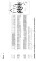
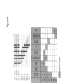
- the sequence alignment of CLDN3, CLDN4, CLDN6 and CLDN9 shown in Fig. 1B illustrates that there is a high degree of conservation of CLDN6 to other claudin proteins.
- This high homology of CLDN6 with other claudin proteins, in particular CLDN9 and CLDN4 render it difficult to provide CLDN6 antibodies which have properties such as specificity and affinity suitable for diagnostic purposes.
- the present inventors found that antibodies directed against a certain epitope located within the C-terminal portion of CLDN6 fulfill the criteria for the diagnostic applicability of antibodies, in particular for detecting and identifying cells expressing CLDN6.
- Antibodies against CLDN6 are known in the art, as illustrated by HOPCRAFT SHARON E ET AL: "Selection of a hepatitis C virus with altered entry factor requirements reveals a genetic interaction between the E1 glycoprotein and claudins.”,HEPATOLOGY, vol. 62, no. 4, October 2015 (2015-10), pages 1059-1069 .
- the antibodies of the teaching are useful, for example, in diagnosing cancer and/or in determining whether cancer cells express CLDN6.
- a cancer disease or a cancer cell is characterized by surface expression of CLDN6.
- Cancer cells expressing CLDN6 are suitable targets for therapies targeting CLDN6 such as therapy with antibodies directed against CLDN6.
- cancer cells express or aberrantly express CLDN6 while the corresponding normal cells do not express CLDN6 or express CLDN6 at a lower level.
- the present invention relates to an antibody or antigen-binding fragment thereof, wherein the antibody or antigen-binding fragment comprises: an antibody heavy chain variable region (VH) comprising:
- the present invention also relates to a method for detecting CLDN6 or the quantity of CLDN6 in a sample comprising the steps of:
- the present invention also relates to a method for detecting cells that express CLDN6 comprising the steps of:
- the present invention also relates to a method for diagnosis, detection or monitoring of CLDN6-expressing cancer cells comprising the steps of:
- the antibody or antigen-binding fragment thereof comprises: an antibody heavy chain variable region (VH) comprising:
- the antibody or antigen-binding fragment thereof comprises: (i) an antibody produced by or obtainable from a clone deposited under the accession no. DSM ACC3313 (58-1B), DSM ACC3312 (58-3A) or DSM ACC3311 (58-4B-2), or (ii) an antibody which is a chimerized or humanized form of the antibody under (i), or an antigen-binding fragment of the antibody of (i) or (ii).
- the antibody or antigen-binding fragment thereof comprises an antibody produced by or obtainable from a clone deposited under the accession no. DSM ACC3313 (58-1B), DSM ACC3312 (58-3A) or DSM ACC3311 (58-4B-2).
- the antibody or antigen-binding fragment thereof comprises:
- the antibody or antigen-binding fragment thereof comprises: (i) an antibody produced by or obtainable from a clone deposited under the accession no. DSM ACC3313 (58-1B), DSM ACC3312 (58-3A) or DSM ACC3311 (58-4B-2), or (ii) an antibody which is a chimerized or humanized form of the antibody under (i), or an antigen-binding fragment of the antibody of (i) or (ii).
- the antibody or antigen-binding fragment thereof comprises an antibody produced by or obtainable from a clone deposited under the accession no. DSM ACC3313 (58-1B), DSM ACC3312 (58-3A) or DSM ACC3311 (58-4B-2).
- the antibody or antigen-binding fragment of the invention comprises:
- the antibody or antigen-binding fragment of the invention thereof comprises: (i) an antibody produced by or obtainable from a clone deposited under the accession no. DSM ACC3313 (58-1B), DSM ACC3312 (58-3A) or DSM ACC3311 (58-4B-2), or (ii) an antibody which is a chimerized or humanized form of the antibody under (i), or an antigen-binding fragment of the antibody of (i) or (ii).
- the antibody or antigen-binding fragment of the invention comprises an antibody produced by or obtainable from a clone deposited under the accession no. DSM ACC3313 (58-1B), DSM ACC3312 (58-3A) or DSM ACC3311 (58-4B-2).
- the present teaching relates to an an antibody or antigen-binding fragment thereof, which binds:
- the present teaching relates to a monoclonal antibody or antigen-binding fragment thereof which
- the present teaching relates to an antibody or antigen-binding fragment thereof, which binds to one or more, preferably all, of the following peptides:
- PAISRGPSEYPTKNY (SEQ ID NO: 22), AISRGPSEYPTKNYV (SEQ ID NO: 15), ISRGPSEYPTKNYV (SEQ ID NO: 23), SRGPSEYPTKNYV (SEQ ID NO: 24), RGPSEYPTKNYV (SEQ ID NO: 25), GPSEYPTKNYV (SEQ ID NO: 26), PSEYPTKNYV (SEQ ID NO: 27), SEYPTKNYV (SEQ ID NO: 28), and EYPTKNYV (SEQ ID NO: 29), wherein the antibody or antigen-binding fragment thereof does not bind to a peptide having the amino acid sequence TSAPAISRGPSEYPT (SEQ ID NO: 14).
- the difference in binding affinity to the peptide to which the antibody or antigen binding fragment binds with the lowest affinity and to the peptide to which the antibody or antigen binding fragment binds with the highest affinity is 50% or less, 40% or less, 30% or less, 20% or less, or 10% or less.
- Antibodies or antigen-binding fragments described herein preferably do not bind to a peptide comprising the amino acid sequence EYPTK (SEQ ID NO: 59) and/or the amino acid sequence EYPTKN (SEQ ID NO: 60) but not comprising the amino acid sequence EYPTKNY (SEQ ID NO: 38).
- an antibody or antigen-binding fragment described herein which binds to a peptide having the amino acid sequence EYPTKNY (SEQ ID NO: 38) and/or a peptide having the amino acid sequence EYPTKNYV (SEQ ID NO: 29) binds to said peptide(s) due to the presence of the second tyrosine which is missing in the amino acid sequence EYPTK (SEQ ID NO: 59) and/or the amino acid sequence EYPTKN (SEQ ID NO: 60).
- preferred antibodies or antigen-binding fragments do not bind to a peptide comprising the amino acid sequence YPTKNY (SEQ ID NO: 61) and/or EYPTKN (SEQ ID NO: 60) but not comprising the amino acid sequence EYPTKNY (SEQ ID NO: 38) suggesting that the amino acid sequence EYPTKNY (SEQ ID NO: 38) is a sequence that can be considered to be a minimal epitope for binding of these antibodies.
- the present teaching relates to an antibody produced by or obtainable from a hybridoma deposited at the DSMZ (Inhoffenstr. 7B, 38124 Braunschweig, Germany) and having one of the following designations and accession numbers:
- Antibodies of the teaching are designated herein by referring to the designation of the antibody and/or by referring to the clone producing the antibody.
- the present teaching also relates to an antibody which competes for CLDN6 binding with an antibody produced by and obtainable from the above-described hybridomas and/or has the specificity for CLDN6 of an antibody produced by or obtainable from the above-described hybridomas.
- the present teaching also relates to an antibody comprising an antigen binding portion or antigen binding site, in particular a variable region, identical or highly homologous to that of the antibodies produced by or obtainable from the above-described hybridomas. It is contemplated that preferred antibodies are those having CDR regions either identical or highly homologous to the CDR regions of antibodies produced by or obtainable from the above-described hybridomas.
- highly homologous it is contemplated that from 1 to 5, preferably from 1 to 4, such as 1 to 3 or 1 or 2 substitutions may be made in each CDR region.
- Particularly preferred antibodies are the chimerized and humanized forms of the antibodies produced by or obtainable from the above-described hybridomas.
- the present teaching relates to an antibody selected from the group consisting of:
- the antigen binding portion or antigen binding site of the antibody under (i) comprises the variable region of the antibody under (i).
- the present teaching relates to an antibody selected from the group consisting of:
- the present teaching relates to an antibody selected from the group consisting of:
- the present teaching relates to an antibody selected from the group consisting of:
- the present teaching relates to an antibody selected from the group consisting of:
- the present teaching relates to an antibody selected from the group consisting of:
- the present teaching relates to an antibody selected from the group consisting of:
- the present teaching relates to an antibody selected from the group consisting of:
- the present teaching relates to an antibody selected from the group consisting of:
- the present teaching relates to an antibody selected from the group consisting of:
- the present teaching relates to an antibody selected from the group consisting of:
- the present teaching relates to an antibody selected from the group consisting of:
- the present teaching relates to an antibody selected from the group consisting of:
- an antibody of the teaching comprises an antibody heavy chain comprising a gamma-2a heavy chain constant region, preferably a human gamma-2a heavy chain constant region and/or comprises an antibody light chain comprising a kappa light chain constant region.
- an antibody or antigen-binding fragment of the teaching is preferably able to bind to CLDN6 in its native, i.e. naturally occurring or non-denatured state, or in its denatured state.
- an antibody or antigen-binding fragment thereof of the teaching binds to CLDN6 but not to CLDN9 and preferably does not bind to CLDN4 and/or CLDN3.
- an antibody or antigen-binding fragment thereof of the teaching does not substantially bind to a CLDN protein other than CLDN6.
- an antibody or antigen-binding fragment thereof of the teaching is specific for CLDN6.
- CLDN6 is cell surface membrane-bound CLDN6.
- CLDN6 is present on cancer cells, wherein said cancer cells are preferably CLDN6 expressing cancer cells.
- said cancer cells are cells from a cancer selected from the group consisting of ovarian cancer, in particular ovarian adenocarcinoma and ovarian teratocarcinoma, lung cancer, including small cell lung cancer (SCLC) and non-small cell lung cancer (NSCLC), in particular squamous cell lung carcinoma and adenocarcinoma, gastric cancer, breast cancer, hepatic cancer, pancreatic cancer, skin cancer, in particular basal cell carcinoma and squamous cell carcinoma, malignant melanoma, head and neck cancer, in particular malignant pleomorphic adenoma, sarcoma, in particular synovial sarcoma and carcinosarcoma, bile duct cancer, cancer of the urinary bladder, in particular transitional cell carcinoma and papillary carcinoma,
- SCLC small cell lung
- an antibody of the teaching is a chimeric, human or humanized antibody. In one embodiment, an antibody of the teaching is a monoclonal antibody.
- an antibody of the teaching is obtainable by a method comprising the step of immunizing an animal with a peptide comprising, preferably consisting of the amino acid sequence of SEQ ID NO: 49, or an immunologically equivalent peptide, or a nucleic acid or host cell expressing said peptide.
- a peptide comprising, preferably consisting of the amino acid sequence of SEQ ID NO: 49, or an immunologically equivalent peptide, or a nucleic acid or host cell expressing said peptide.
- said peptide comprises not more than 110, 100, 90, 80, 70, 60, 50, or 40 contiguous amino acids of CLDN6.
- Antibodies or antigen-binding fragments of the teaching may be coupled, i.e. covalently or non-covalently linked, to other moieties such as detectable labels.
- the present teaching relates to a conjugate comprising an antibody or antigen-binding fragment described herein coupled to at least one detectable label.
- the present teaching also relates to a cell such as a hybridoma producing an antibody as described herein.
- Preferred hybridomas are those deposited at the DSMZ (Inhoffenstr. 7B, 38124 Braunschweig, Germany) and having one of the following designations and accession numbers:
- the present teaching also relates to a peptide comprising, preferably consisting of the amino acid sequence of SEQ ID NO: 49, or an immunologically equivalent peptide.
- a peptide comprising, preferably consisting of the amino acid sequence of SEQ ID NO: 49, or an immunologically equivalent peptide.
- said peptide comprises not more than 110, 100, 90, 80, 70, 60, 50, or 40 contiguous amino acids of CLDN6.
- the present teaching also relates to nucleic acids encoding antibodies or parts thereof, e.g. an antibody chain, or antigen-binding fragments, or peptides as described herein.
- a nucleic acid of the teaching is operatively attached to one or more expression control elements allowing expression in eukaryotic or prokaryotic cells. Control elements ensuring expression in eukaryotic or prokaryotic cells are well known to those skilled in the art.
- a nucleic acid of the teaching may be comprised in a vector, e.g., a plasmid, cosmid, virus, bacteriophage or another vector used e.g. conventionally in genetic engineering.
- the vector may comprise further genes such as marker genes which allow for the selection of the vector in a suitable host cell and under suitable conditions.
- the vector may comprise expression control elements allowing proper expression of the coding regions in suitable hosts. Such control elements are known to the artisan and may include a promoter, a splice cassette, and a translation initiation codon.
- nucleic acid molecules for construction of vectors comprising nucleic acid molecules, for introduction of vectors into appropriately chosen host cells, or for causing or achieving expression of nucleic acid molecules are well-known in the art.
- a further aspect of the present teaching relates to a host cell comprising a nucleic acid or vector as disclosed herein.
- a further aspect the present teaching relates to the detection of CLDN6 or CLDN6-expressing cells or determination of the quantity of CLDN6 or CLDN6-expressing cells using an antibody or antigen-binding fragment of the teaching.
- CLDN6 or CLDN6-expressing cells are detected or the quantity of CLDN6 or CLDN6-expressing cells is determined by detecting or determining the amount of a complex between CLDN6 and an antibody or antigen-binding fragment of the teaching. Formation of a complex indicates the presence of CLDN6 or CLDN6-expressing cells.
- detection or determination of the amount may be carried out in a number of ways, including but not limited to immunodetection using an antibody or antigen-binding fragment of the teaching.
- Methods for using antibodies to detect peptides or proteins are well known and include ELISA, competitive binding assays, and the like.
- assays use an antibody or antibody fragment that specifically binds the target peptide or protein directly or indirectly bound to a label that provides for detection, e.g. indicator enzymes, radiolabels, fluorophores, or paramagnetic particles.
- the methods of the teaching allow quantitative and/or qualitative evaluations, e.g., absolute and/or relative evaluations, of CLDN6 levels or of levels of CLDN6-expressing cells.
- the present teaching relates to a method for detecting CLDN6 or determining the quantity of CLDN6 in a sample comprising the steps of:
- the sample is a cellular sample, i.e. a sample comprising cells such as cancer cells.
- the complex is preferably formed between the antibody, the antigen-binding fragment or the conjugate and CLDN6 expressed by cells in said sample.
- the present teaching relates to a method for determining whether cells express CLDN6 comprising the steps of:
- the cells in the sample are cancer cells.
- the complex is preferably formed between the antibody, the antigen-binding fragment or the conjugate and CLDN6 expressed by cells in said sample.
- Further aspects of the present teaching relate to methods of diagnosing or classifying diseases by targeting CLDN6 using an antibody or antigen-binding fragment of the teaching. These methods provide for the selective detection of cells that express CLDN6 thereby differentiating these cells from normal cells not expressing CLDN6 or diseased cells not expressing CLDN6.
- Diseases characterized by diseased cells expressing CLDN6 are treatable by a therapy targeting CLDN6 such as therapy with therapeutic antibodies directed against CLDN6.
- Preferred diseases for a therapy or diagnosis are those in which CLDN6 is expressed or aberrantly expressed, in particular cancer diseases, such as those described herein.
- the present teaching relates to methods for diagnosis, detection or monitoring, i.e. determining the regression, progression, course and/or onset, of a cancer disease comprising the detection of CLDN6 or CLDN6-expressing cells and/or determination of the quantity of CLDN6 or CLDN6-expressing cells in a biological sample isolated from a patient using an antibody or antigen-binding fragment of the teaching.
- Such methods may be used to detect whether a subject has a cancer disease or is at (increased) risk of developing a cancer disease or, for instance, whether a treatment regimen is efficient.
- the present teaching relates to a method for diagnosis, detection or monitoring of cancer comprising the steps of:
- the biological sample is a cellular sample, i.e. a sample comprising cells such as cancer cells.
- the complex is preferably formed between the antibody, the antigen-binding fragment or the conjugate and CLDN6 expressed by cells in said sample.
- the methods of monitoring preferably comprise a detection of and/or determination of the quantity of CLDN6 or CLDN6-expressing cells in a first sample at a first point in time and in a further sample at a second point in time, wherein the regression, progression, course and/or onset of a tumor disease may be determined by comparing the two samples.
- the level of CLDN6 or level of CLDN6-expressing cells in a biological sample is compared to a reference level, wherein a deviation from said reference level is indicative of the presence and/or stage of a cancer disease in a subject.
- the reference level may be a level as determined in a control sample (e.g., from a healthy tissue or subject, in particular a patient without a cancer disease) or a median level from healthy subjects.
- a "deviation" from said reference level designates any significant change, such as an increase by at least 10%, 20%, or 30%, preferably by at least 40% or 50%, or even more.
- the presence of CLDN6 or CLDN6-expressing cells and/or a quantity of CLDN6 or CLDN6-expressing cells which is increased compared to a reference level, e.g. compared to a patient without a cancer disease, indicates the presence of or risk for (i.e. a potential for a development of) a cancer disease in the patient.
- a quantity of CLDN6 or CLDN6-expressing cells which is decreased compared to a biological sample taken earlier from a patient may indicate a regression, a positive course, e.g. a successful treatment, or a reduced risk for an onset of a cancer disease in a patient.
- a quantity of CLDN6 or CLDN6-expressing cells which is increased compared to a biological sample taken earlier from a patient may indicate a progression, a negative course, e.g. an unsuccessful treatment, recurrence or metastatic behavior, an onset or a risk for an onset of a cancer disease in said patient.
- the present teaching relates to a method for determining whether a cancer is treatable by a cancer therapy targeting CLDN6 comprising the steps of:
- the complex is preferably formed between the antibody, the antigen-binding fragment or the conjugate and CLDN6 expressed by cancer cells in said sample.
- Such methods may be used to detect whether a patient is suitable for a therapy involving the targeting of cells expressing CLDN6 such as a therapy using antibodies exerting one or more immune effector functions such as cytotoxic CLDN6 specific antibodies, e.g. antibodies labeled with a cytotoxic substance such as a toxin or a radiolabel or inducing a cell killing mechanism such as CDC or ADCC.
- cytotoxic CLDN6 specific antibodies e.g. antibodies labeled with a cytotoxic substance such as a toxin or a radiolabel or inducing a cell killing mechanism such as CDC or ADCC.
- Diseases characterized by diseased cells expressing CLDN6 are treatable by a therapy targeting CLDN6 such as cancer diseases, in particular those described herein.
- the sample, cellular sample or biological sample is from a patient having a cancer disease, being suspected of having or falling ill with a cancer disease or having a potential for a cancer disease.
- the sample, cellular sample or biological sample is from a tissue or organ wherein the cells when the tissue or organ is free of cancer do not substantially express CLDN6.
- said tissue is a tissue other than placenta tissue.
- said tissue has already been diagnosed as being affected by a cancer disease, e.g. by visual inspection or culture testing of cells of said tissue or organ.
- the presence of CLDN6 or CLDN6-expressing cells and/or a quantity of CLDN6 or CLDN6-expressing cells which is increased compared to a reference level, e.g. compared to a patient without a tumor disease, may indicate that a patient is suitable for a therapy involving the targeting of cells expressing CLDN6.
- the teaching provides compositions, e.g., diagnostic compositions, or kits, comprising an antibody or antigen-binding fragment or a combination of antibodies and/or or antigen-binding fragments described herein.
- diagnostic compositions or test kits are useful in the methods of the teaching such as the methods for diagnosis, detection or monitoring of the teaching.
- kits may optionally comprise a detectable label, e.g. indicator enzymes, radiolabels, fluorophores, or paramagnetic particles. Kits may include informative pamphlets, for example, pamphlets informing on how to use reagents to practice a method disclosed herein.
- naturally occurring refers to the fact that an object can be found in nature.
- a peptide or nucleic acid that is present in an organism (including viruses) and can be isolated from a source in nature and which has not been intentionally modified by man in the laboratory is naturally occurring.
- an antigen relates to an agent comprising an epitope against which an immune response is directed and/or is to be generated.
- an antigen in the context of the present teaching is a molecule which, optionally after processing, induces an immune reaction, which is preferably specific for the antigen.
- the term “antigen” includes in particular proteins, peptides, polysaccharides, nucleic acids, especially RNA and DNA, and nucleotides.
- epitope refers to an antigenic determinant in a molecule such as an antigen, i.e., to the part in a molecule that is recognized by the immune system, for example, that is recognized by an antibody.
- epitopes are the discrete, three-dimensional sites on an antigen, which are recognized by the immune system.
- Epitopes usually consist of chemically active surface groupings of molecules such as amino acids or sugar side chains and usually have specific three dimensional structural characteristics, as well as specific charge characteristics. Conformational and non-conformational epitopes are distinguished in that the binding to the former but not the latter is lost in the presence of denaturing solvents.
- An epitope of a protein such as a CLDN preferably comprises a continuous or discontinuous portion of said protein and is preferably between 5 and 100, preferably between 5 and 50, more preferably between 8 and 30, most preferably between 10 and 25 amino acids in length, for example, the epitope may be preferably 8, 9, 10, 11, 12, 13, 14, 15, 16, 17, 18, 19, 20, 21, 22, 23, 24, or 25 amino acids in length.
- discontinuous epitope means a conformational epitope on a protein antigen which is formed from at least two separate regions in the primary sequence of the protein.
- Antigens include tumor-associated antigens, such as CLDN6, i.e., constituents of cancer cells which may be derived from the cytoplasm, the cell surface and the cell nucleus, in particular those antigens which are produced, preferably in large quantity, intracellularly or as surface antigens on cancer cells.
- CLDN6 tumor-associated antigens
- constituents of cancer cells which may be derived from the cytoplasm, the cell surface and the cell nucleus, in particular those antigens which are produced, preferably in large quantity, intracellularly or as surface antigens on cancer cells.
- tumor-associated antigen or “tumor antigen” relate to proteins that are under normal conditions specifically expressed in a limited number of tissues and/or organs or in specific developmental stages, for example, the tumor-associated antigen may be under normal conditions specifically expressed in stomach tissue, preferably in the gastric mucosa, in reproductive organs, e.g., in testis, in trophoblastic tissue, e.g., in placenta, or in germ line cells, and are expressed or aberrantly expressed in one or more tumor or cancer tissues.
- a limited number preferably means not more than 3, more preferably not more than 2.
- the tumor-associated antigens in the context of the present teaching include, for example, differentiation antigens, preferably cell type specific differentiation antigens, i.e., proteins that are under normal conditions specifically expressed in a certain cell type at a certain differentiation stage, cancer/testis antigens, i.e., proteins that are under normal conditions specifically expressed in testis and sometimes in placenta, and germ line specific antigens.
- the tumor-associated antigen is preferably associated with the cell surface of a cancer cell and is preferably not or only rarely expressed in normal tissues.
- the tumor-associated antigen or the aberrant expression of the tumor-associated antigen identifies cancer cells.
- the tumor-associated antigen that is expressed by a cancer cell in a subject is preferably a self-protein in said subject.
- the tumor-associated antigen in the context of the present teaching is expressed under normal conditions specifically in a tissue or organ that is non-essential, i.e., tissues or organs which when damaged by the immune system do not lead to death of the subject, or in organs or structures of the body which are not or only hardly accessible by the immune system.
- the amino acid sequence of the tumor-associated antigen is identical between the tumor-associated antigen which is expressed in normal tissues and the tumor-associated antigen which is expressed in cancer tissues.
- Examples for differentiation antigens which ideally fulfill the criteria for tumor-associated antigens as target structures in tumor immunotherapy, in particular, in tumor vaccination are the cell surface proteins of the claudin family, such as CLDN6.
- Claudins are a family of proteins that are the most important components of tight junctions, where they establish the paracellular barrier that controls the flow of molecules in the intercellular space between cells of an epithelium.
- Claudins are transmembrane proteins spanning the membrane 4 times with the N-terminal and the C-terminal end both located in the cytoplasm.
- CLDN6 preferably relates to human CLDN6, and, in particular, to a protein comprising the amino acid sequence according to SEQ ID NO: 1 of the sequence listing or a variant of said amino acid sequence.
- variant in particular refers to a protein comprising the amino acid sequence according to SEQ ID NO: 1 of the sequence listing wherein the Ile at position 143 is replaced by Val.
- CLDN6 includes any CLDN6 variants such as posttranslationally modified variants and conformation variants.
- CLDN9 preferably relates to human CLDN9, and, in particular, to a protein comprising the amino acid sequence according to SEQ ID NO: 2 of the sequence listing or a variant of said amino acid sequence.
- CLDN4 preferably relates to human CLDN4, and, in particular, to a protein comprising the amino acid sequence according to SEQ ID NO: 4 of the sequence listing or a variant of said amino acid sequence.
- CLDN3 preferably relates to human CLDN3, and, in particular, to a protein comprising the amino acid sequence according to SEQ ID NO: 3 of the sequence listing or a variant of said amino acid sequence.
- CLDN6 has been found to be expressed, for example, in ovarian cancer, lung cancer, gastric cancer, breast cancer, hepatic cancer, pancreatic cancer, skin cancer, melanomas, head neck cancer, sarcomas, bile duct cancer, renal cell cancer, and urinary bladder cancer.
- CLDN6 is detectable and can be targeted in ovarian cancer, in particular ovarian adenocarcinoma and ovarian teratocarcinoma, lung cancer, including small cell lung cancer (SCLC) and non-small cell lung cancer (NSCLC), in particular squamous cell lung carcinoma and adenocarcinoma, gastric cancer, breast cancer, hepatic cancer, pancreatic cancer, skin cancer, in particular basal cell carcinoma and squamous cell carcinoma, malignant melanoma, head and neck cancer, in particular malignant pleomorphic adenoma, sarcoma, in particular synovial sarcoma and carcinosarcoma, bile duct cancer, cancer of the urinary bladder, in particular transitional cell carcinoma and papillary carcinoma, kidney cancer, in particular renal cell carcinoma including clear cell renal cell carcinoma and papillary renal cell carcinoma, colon cancer, small bowel cancer, including cancer of the ileum, in particular small bowel adenocarcinoma and a
- the cancer disease associated with CLDN6 expression is selected from the group consisting of ovarian cancer, lung cancer, metastatic ovarian cancer and metastatic lung cancer.
- the ovarian cancer is a carcinoma or an adenocarcinoma.
- the lung cancer is a carcinoma or an adenocarcinoma, and preferably is bronchiolar cancer such as a bronchiolar carcinoma or bronchiolar adenocarcinoma.
- a cell expressing CLDN6 is preferably characterized by cell-surface membrane-bound CLDN6, i.e. CLDN6 is associated with the cell surface.
- cellular CLDN6 is preferably cell-surface membrane-bound CLDN6.
- a cell expressing CLDN6 or a cell characterized by association of CLDN6 with its cell surface preferably is a cancer cell, preferably a cancer cell from a cancer described herein.
- a tumor-associated antigen such as CLDN6 is associated with and located at the plasma membrane of a cell, wherein at least a part of the tumor-associated antigen faces the extracellular space of said cell and is accessible from the outside of said cell, e.g., by antibodies located outside the cell.
- a part is preferably at least 4, preferably at least 8, preferably at least 12, more preferably at least 20 amino acids.
- the association may be direct or indirect.
- the association may be by one or more transmembrane domains, one or more lipid anchors, or by the interaction with any other protein, lipid, saccharide, or other structure that can be found on the outer leaflet of the plasma membrane of a cell.
- a tumor-associated antigen associated with the surface of a cell may be a transmembrane protein having an extracellular portion or may be a protein associated with the surface of a cell by interacting with another protein that is a transmembrane protein.
- Cell surface or “surface of a cell” is used in accordance with its normal meaning in the art, and thus includes the outside of the cell which is accessible to binding by proteins and other molecules.
- CLDN6 is not substantially expressed in a cell if the level of expression is lower compared to expression in placenta cells or placenta tissue.
- the level of expression is less than 10%, preferably less than 5%, 3%, 2%, 1%, 0.5%, 0.1% or 0.05% of the expression in placenta cells or placenta tissue or even lower.
- CLDN6 is not substantially expressed in a cell if the level of expression exceeds the level of expression in non-cancerous tissue other than placenta by no more than 2-fold, preferably 1.5-fold, and preferably does not exceed the level of expression in said non-cancerous tissue.
- CLDN6 is not substantially expressed in a cell if the level of expression is below the detection limit and/or if the level of expression is too low to allow binding by CLDN6-specific antibodies added to the cells.
- CLDN6 is expressed in a cell if the level of expression exceeds the level of expression in non-cancerous tissue other than placenta preferably by more than 2-fold, preferably 10-fold, 100-fold, 1000-fold, or 10000-fold.
- CLDN6 is expressed in a cell if the level of expression is above the detection limit and/or if the level of expression is high enough to allow binding by CLDN6-specific antibodies added to the cells.
- CLDN6 expressed in a cell is expressed or exposed on the surface of said cell.
- antibody refers to a glycoprotein comprising at least two heavy (H) chains and two light (L) chains inter-connected by disulfide bonds, and includes any molecule comprising an antigen binding portion thereof.
- antibody includes monoclonal antibodies and fragments or derivatives of antibodies, including, without limitation, human antibodies, humanized antibodies, chimeric antibodies, single chain antibodies, e.g., scFv's and antigen-binding antibody fragments such as Fab and Fab' fragments and also includes all recombinant forms of antibodies, e.g., antibodies expressed in prokaryotes, unglycosylated antibodies, and any antigen-binding antibody fragments and derivatives as described herein.
- Each heavy chain is comprised of a heavy chain variable region (abbreviated herein as VH) and a heavy chain constant region.
- Each light chain is comprised of a light chain variable region (abbreviated herein as VL) and a light chain constant region.
- the VH and VL regions can be further subdivided into regions of hypervariability, termed complementarity determining regions (CDR), interspersed with regions that are more conserved, termed framework regions (FR).
- CDR complementarity determining regions
- FR framework regions
- Each VH and VL is composed of three CDRs and four FRs, arranged from amino-terminus to carboxy-terminus in the following order: FR1, CDR1, FR2, CDR2, FR3, CDR3, FR4.
- variable regions of the heavy and light chains contain a binding domain that interacts with an antigen.
- the constant regions of the antibodies may mediate the binding of the immunoglobulin to host tissues or factors, including various cells of the immune system (e.g., effector cells) and the first component (Clq) of the classical complement system.
- the antibodies described herein may be human antibodies.
- the term "human antibody”, as used herein, is intended to include antibodies having variable and constant regions derived from human germline immunoglobulin sequences.
- the human antibodies of the teaching may include amino acid residues not encoded by human germline immunoglobulin sequences (e.g., mutations introduced by random or site-specific mutagenesis in vitro or by somatic mutation in vivo ) .
- humanized antibody refers to a molecule having an antigen binding site that is substantially derived from an immunoglobulin from a non-human species, wherein the remaining immunoglobulin structure of the molecule is based upon the structure and/or sequence of a human immunoglobulin.
- the antigen binding site may either comprise complete variable domains fused onto constant domains or only the complementarity determining regions (CDR) grafted onto appropriate framework regions in the variable domains.
- Antigen binding sites may be wild-type or modified by one or more amino acid substitutions, e.g. modified to resemble human immunoglobulins more closely.
- Some forms of humanized antibodies preserve all CDR sequences (for example a humanized mouse antibody which contains all six CDRs from the mouse antibody). Other forms have one or more CDRs which are altered with respect to the original antibody.
- chimeric antibody refers to those antibodies wherein one portion of each of the amino acid sequences of heavy and light chains is homologous to corresponding sequences in antibodies derived from a particular species or belonging to a particular class, while the remaining segment of the chain is homologous to corresponding sequences in another.
- the variable region of both light and heavy chains mimics the variable regions of antibodies derived from one species of mammals, while the constant portions are homologous to sequences of antibodies derived from another.
- One clear advantage to such chimeric forms is that the variable region can conveniently be derived from presently known sources using readily available B-cells or hybridomas from non-human host organisms in combination with constant regions derived from, for example, human cell preparations.
- variable region has the advantage of ease of preparation and the specificity is not affected by the source, the constant region being human, is less likely to elicit an immune response from a human subject when the antibodies are injected than would the constant region from a non human source.
- definition is not limited to this particular example.
- antigen-binding portion of an antibody (or simply “binding portion") or "antigen-binding fragment” of an antibody (or simply “binding fragment”) refer to one or more fragments of an antibody that retain the ability to specifically bind to an antigen. It has been shown that the antigen-binding function of an antibody can be performed by fragments of a full-length antibody.
- binding fragments encompassed within the term "antigen-binding portion" of an antibody include (i) Fab fragments, monovalent fragments consisting of the VL, VH, CL and CH domains; (ii) F(ab') 2 fragments, bivalent fragments comprising two Fab fragments linked by a disulfide bridge at the hinge region; (iii) Fd fragments consisting of the VH and CH domains; (iv) Fv fragments consisting of the VL and VH domains of a single arm of an antibody, (v) dAb fragments ( Ward et al., (1989) Nature 341: 544-546 ), which consist of a VH domain; (vi) isolated complementarity determining regions (CDR), and (vii) combinations of two or more isolated CDRs which may optionally be joined by a synthetic linker.
- Fab fragments monovalent fragments consisting of the VL, VH, CL and CH domains
- F(ab') 2 fragments bivalent fragments compris
- the two domains of the Fv fragment, VL and VH are coded for by separate genes, they can be joined, using recombinant methods, by a synthetic linker that enables them to be made as a single protein chain in which the VL and VH regions pair to form monovalent molecules (known as single chain Fv (scFv); see e.g., Bird et al. (1988) Science 242: 423-426 ; and Huston et al. (1988) Proc. Natl. Acad. Sci. USA 85: 5879-5883 ).
- single chain Fv single chain Fv
- Such single chain antibodies are also intended to be encompassed within the term "antigen-binding fragment" of an antibody.
- a further example is binding-domain immunoglobulin fusion proteins comprising (i) a binding domain polypeptide that is fused to an immunoglobulin hinge region polypeptide, (ii) an immunoglobulin heavy chain CH2 constant region fused to the hinge region, and (iii) an immunoglobulin heavy chain CH3 constant region fused to the CH2 constant region.
- the binding domain polypeptide can be a heavy chain variable region or a light chain variable region.
- the binding-domain immunoglobulin fusion proteins are further disclosed in US 2003/0118592 and US 2003/0133939 . These antibody fragments are obtained using conventional techniques known to those with skill in the art, and the fragments are screened for utility in the same manner as are intact antibodies.
- the antibodies described herein may be monoclonal antibodies.
- the term "monoclonal antibody” as used herein refers to a preparation of antibody molecules of single molecular composition.
- a monoclonal antibody displays a single binding specificity and affinity.
- the monoclonal antibodies are produced by a hybridoma which includes a B cell obtained from a non-human animal, e.g., mouse, fused to an immortalized cell.
- the antibodies described herein may be recombinant antibodies.
- the term "recombinant antibody”, as used herein, includes all antibodies that are prepared, expressed, created or isolated by recombinant means, such as (a) antibodies isolated from an animal (e.g., a mouse) that is transgenic or transchromosomal with respect to the immunoglobulin genes or a hybridoma prepared therefrom, (b) antibodies isolated from a host cell transformed to express the antibody, e.g., from a transfectoma, (c) antibodies isolated from a recombinant, combinatorial antibody library, and (d) antibodies prepared, expressed, created or isolated by any other means that involve splicing of immunoglobulin gene sequences to other DNA sequences.
- transfectoma includes recombinant eukaryotic host cells expressing an antibody, such as CHO cells, NS/0 cells, HEK293 cells, HEK293T cells, plant cells, or fungi, including yeast cells.
- a heterologous antibody is defined in relation to a transgenic organism producing such an antibody. This term refers to an antibody having an amino acid sequence or an encoding nucleic acid sequence corresponding to that found in an organism not consisting of the transgenic organism, and being generally derived from a species other than the transgenic organism.
- heterohybrid antibody refers to an antibody having light and heavy chains of different organismal origins.
- an antibody having a human heavy chain associated with a murine light chain is a heterohybrid antibody.
- antibody derivatives refers to any modified form of an antibody, e.g., a conjugate of the antibody and another agent or antibody, or an antibody fragment.
- the antibodies described herein are preferably isolated.
- An "isolated antibody” as used herein, is intended to refer to an antibody which is substantially free of other antibodies having different antigenic specificities. Moreover, an isolated antibody may be substantially free of other cellular material and/or chemicals.
- an antibody is capable of binding to a predetermined target if it has a significant affinity for said predetermined target and binds to said predetermined target in standard assays.
- "Affinity” or “binding affinity” is often measured by equilibrium dissociation constant (K D ).
- the term "significant affinity” refers to the binding to a predetermined target with a dissociation constant (K D ) of 10 -5 M or lower, 10 -6 M or lower, 10 -7 M or lower, 10 -8 M or lower, 10 -9 M or lower, 10 -10 M or lower, 10 -11 M or lower, or 10 -12 M or lower.
- an antibody is not (substantially) capable of binding to a target if it has no significant affinity for said target and does not bind significantly, in particular does not bind detectably, to said target in standard assays.
- the antibody does not detectably bind to said target if present in a concentration of up to 2, preferably 10, more preferably 20, in particular 50 or 100 ⁇ g/ml or higher.
- an antibody has no significant affinity for a target if it binds to said target with a K D that is at least 10-fold, 100-fold, 10 3 -fold, 10 4 -fold, 10 5 -fold, or 10 6 -fold higher than the K D for binding to the predetermined target to which the antibody is capable of binding.
- the K D for binding of an antibody to the target to which the antibody is capable of binding is 10 -7 M
- the K D for binding to a target for which the antibody has no significant affinity would be is at least 10 -6 M, 10 -5 M, 10 -4 M, 10 -3 M, 10 -2 M, or 10 -1 M.
- an antibody is specific for a predetermined target if it is capable of binding to said predetermined target while it is not capable of binding to other targets, i.e. has no significant affinity for other targets and does not significantly bind to other targets in standard assays.
- an antibody is specific for CLDN6 if it is capable of binding to CLDN6 but is not (substantially) capable of binding to other targets, in particular other CLDN proteins such as CLDN9, CLDN4 and/or CLDN3 and/or proteins other than claudin proteins, preferably proteins other than CLDN6.
- an antibody is specific for CLDN6 if the affinity for and the binding to such other targets does not significantly exceed the affinity for or binding to claudin-unrelated proteins such as bovine serum albumin (BSA), casein, human serum albumin (HSA) or non-claudin transmembrane proteins such as MHC molecules or transferrin receptor or any other specified polypeptide.
- BSA bovine serum albumin
- HSA human serum albumin
- non-claudin transmembrane proteins such as MHC molecules or transferrin receptor or any other specified polypeptide.
- an antibody is specific for a predetermined target if it binds to said target with a K D that is at least 10-fold, 100-fold, 10 3 -fold, 10 4 -fold, 10 5 -fold, or 10 6 -fold lower than the K D for binding to a target for which it is not specific.
- Compet refers to the competition between two antibodies for binding to a target antigen. If two antibodies do not block each other for binding to a target antigen, such antibodies are non-competing and this is an indication that said antibodies do not bind to the same part, i.e. epitope, of the target antigen. It is well known to a person skilled in the art how to test for competition of antibodies for binding to a target antigen. An example of such a method is a so-called cross-competition assay, which may e.g. be performed as an ELISA or by flow-cytometry.
- an ELISA-based assay may be performed by coating ELISA plate wells with one of the antibodies; adding the competing antibody and His-tagged antigen/target and detecting whether the added antibody inhibited binding of the His-tagged antigen to the coated antibody, e.g., by adding biotinylated anti-His antibody, followed by Streptavidin-poly-HRP, and further developing the reaction with ABTS and measuring the absorbance at 405 nm.
- a flow-cytometry assay may be performed by incubating cells expressing the antigen/target with an excess of unlabeled antibody, incubating the cells with a sub-optimal concentration of biotin-labelled antibody, followed by incubation with fluorescently labeled streptavidin and analyzing by flow cytometry.
- Two antibodies have the "same specificity" if they bind to the same antigen and to the same epitope. Whether an antibody to be tested recognizes the same epitope as a certain antigen-binding antibody, i.e., the antibodies bind to the same epitope, can be tested by different methods known to the skilled person, e.g., based on the competition of the antibodies for the same epitope.
- the competition between the antibodies can be detected by a cross-blocking assay.
- a competitive ELISA assay may be used as a cross-blocking assay.
- target antigen may be coated on the wells of a microtiter plate and antigen-binding antibody and candidate competing test antibody may be added.
- the amount of the antigen-binding antibody bound to the antigen in the well indirectly correlates with the binding ability of the candidate competing test antibody that competes therewith for binding to the same epitope. Specifically, the larger the affinity of the candidate competing test antibody is for the same epitope, the smaller the amount of the antigen-binding antibody bound to the antigen-coated well.
- the amount of the antigen-binding antibody bound to the well can be measured by labeling the antibody with detectable or measurable labeling substances.
- An antibody competing for binding to an antigen with another antibody e.g., an antibody comprising heavy and light chain variable regions as described herein, or an antibody having the specificity for an antigen of another antibody, e.g., an antibody comprising heavy and light chain variable regions as described herein, may be an antibody comprising variants of said heavy and/or light chain variable regions as described herein, e.g. modifications in the CDRs and/or a certain degree of identity as described herein.
- isotype refers to the antibody class (e.g., IgM or IgG1) that is encoded by heavy chain constant region genes.
- Antibodies according to the teaching include polyclonal and monoclonal antibodies and include IgG2a (e.g. IgG2a, ⁇ , ⁇ ), IgG2b (e.g. IgG2b, ⁇ , ⁇ ), IgG3 (e.g. IgG3, ⁇ , ⁇ ) and IgM antibodies.
- IgG2a e.g. IgG2a, ⁇ , ⁇
- IgG2b e.g. IgG2b, ⁇ , ⁇
- IgG3 e.g. IgG3, ⁇ , ⁇
- isotype switching refers to the phenomenon by which the class, or isotype, of an antibody changes from one Ig class to one of the other Ig classes.
- rearranged refers to a configuration of a heavy chain or light chain immunoglobulin locus wherein a V segment is positioned immediately adjacent to a D-J or J segment in a conformation encoding essentially a complete VH or VL domain, respectively.
- a rearranged immunoglobulin (antibody) gene locus can be identified by comparison to germline DNA; a rearranged locus will have at least one recombined heptamer/nonamer homology element.
- V segment configuration refers to the configuration wherein the V segment is not recombined so as to be immediately adjacent to a D or J segment.
- antibodies may be derived from different species, including but not limited to mouse, rat, rabbit, guinea pig and human.
- Antibodies also include chimeric molecules in which an antibody constant region derived from one species, preferably human, is combined with the antigen binding site derived from another species.
- antibodies include humanized molecules in which the antigen binding sites of an antibody derived from a non-human species are combined with constant and framework regions of human origin.
- Antibodies can be produced by a variety of techniques, including conventional monoclonal antibody methodology, e.g., the standard somatic cell hybridization technique of Kohler and Milstein, Nature 256: 495 (1975 ). Although somatic cell hybridization procedures are preferred, in principle, other techniques for producing monoclonal antibodies can be employed, e.g., viral or oncogenic transformation of B-lymphocytes or phage display techniques using libraries of antibody genes.
- the preferred animal system for preparing hybridomas that secrete monoclonal antibodies is the murine system.
- Hybridoma production in the mouse is a very well established procedure. Immunization protocols and techniques for isolation of immunized splenocytes for fusion are known in the art. Fusion partners (e.g., murine myeloma cells) and fusion procedures are also known.
- hybridomas that secrete monoclonal antibodies are the rat and the rabbit system (e.g. described in Spieker-Polet et al., Proc. Natl. Acad. Sci. U.S.A. 92:9348 (1995 ), see also Rossi et al., Am. J. Clin. Pathol. 124: 295 (2005 )).
- human monoclonal antibodies directed against CLDN6 can be generated using transgenic or transchromosomal mice carrying parts of the human immune system rather than the mouse system.
- transgenic and transchromosomic mice include mice known as HuMAb mice and KM mice, respectively, and are collectively referred to herein as "transgenic mice.”
- the production of human antibodies in such transgenic mice can be performed as described in detail for CD20 in WO2004 035607
- Yet another strategy for generating monoclonal antibodies is to directly isolate genes encoding antibodies from lymphocytes producing antibodies of defined specificity; see e.g. Babcock et al., 1996; A novel strategy for generating monoclonal antibodies from single, isolated lymphocytes producing antibodies of defined specificities.
- a novel strategy for generating monoclonal antibodies from single, isolated lymphocytes producing antibodies of defined specificities For details of recombinant antibody engineering see also Welschof and Kraus, Recombinant antibodes for cancer therapy ISBN-0-89603-918-8 and Benny K.C. Lo Antibody Engineering ISBN 1-58829-092-1 .
- mice can be immunized with carrier-conjugated peptides derived from the CLDN6 sequence, an enriched preparation of recombinantly expressed CLDN6 antigen or fragments thereof and/or cells expressing CLDN6 or fragments thereof, as described.
- mice can be immunized with DNA encoding full length human CLDN6 or fragments thereof.
- mice can also be immunized with cells expressing CLDN6, e.g., a cell line, to promote immune responses.
- the immune response can be monitored over the course of the immunization protocol with plasma and serum samples being obtained by tail vein or retroorbital bleeds. Mice with sufficient titers of anti-CLDN6 immunoglobulin can be used for fusions. Mice can be boosted intraperitonealy or intravenously with CLDN6 expressing cells 3-5 days before sacrifice and removal of the spleen to increase the rate of specific antibody secreting hybridomas.
- hybridomas producing monoclonal antibodies to CLDN6 cells from lymph nodes, spleens or bone marrow obtained from immunized mice can be isolated and fused to an appropriate immortalized cell line, such as a mouse myeloma cell line. The resulting hybridomas can then be screened for the production of antigen-specific antibodies. Individual wells can then be screened by ELISA for antibody secreting hybridomas. By Immunofluorescence and FACS analysis using CLDN6 expressing cells, antibodies with specificity for CLDN6 can be identified. The antibody secreting hybridomas can be replated, screened again, and if still positive for anti-CLDN6 monoclonal antibodies can be subcloned by limiting dilution. The stable subclones can then be cultured in vitro to generate antibody in tissue culture medium for characterization.
- Antibodies of the teaching can also be produced in a host cell transfectoma using, for example, a combination of recombinant DNA techniques and gene transfection methods as are well known in the art ( Morrison, S. (1985) Science 229: 1202 ).
- the gene(s) of interest e.g., antibody genes
- an expression vector such as a eukaryotic expression plasmid such as used by the GS gene expression system disclosed in WO 87/04462 , WO 89/01036 and EP 338 841 or other expression systems well known in the art.
- the purified plasmid with the cloned antibody genes can be introduced in eukaryotic host cells such as CHO cells, NS/0 cells, HEK293T cells or HEK293 cells or alternatively other eukaryotic cells like plant derived cells, fungal or yeast cells.
- the method used to introduce these genes can be methods described in the art such as electroporation, lipofectine, lipofectamine or others. After introduction of these antibody genes in the host cells, cells expressing the antibody can be identified and selected. These cells represent the transfectomas which can then be amplified for their expression level and upscaled to produce antibodies. Recombinant antibodies can be isolated and purified from these culture supernatants and/or cells.
- the cloned antibody genes can be expressed in other expression systems, including prokaryotic cells, such as microorganisms, e.g. E. coli.
- the antibodies can be produced in transgenic non-human animals, such as in milk from sheep and rabbits or in eggs from hens, or in transgenic plants; see e.g. Verma, R., et al. (1998) J. Immunol. Meth. 216: 165-181 ; Pollock, et al. (1999) J. Immunol. Meth. 231: 147-157 ; and Fischer, R., et al. (1999) Biol. Chem. 380: 825-839 .
- Chimeric antibodies are antibodies, the different portions of which are derived from different animal species, such as those having a variable region derived from a murine antibody and a human immunoglobulin constant region. Chimerisation of antibodies is achieved by joining of the variable regions of the murine antibody heavy and light chain with the constant region of human heavy and light chain (e.g. as described by Kraus et al., in Methods in Molecular Biology series, Recombinant antibodies for cancer therapy ISBN-0-89603-918-8 ). In a preferred embodiment, chimeric antibodies are generated by joining human kappa-light chain constant region to murine light chain variable region. In an also preferred embodiment, chimeric antibodies can be generated by joining human lambda-light chain constant region to murine light chain variable region.
- the preferred heavy chain constant regions for generation of chimeric antibodies are IgG1, IgG3 and IgG4. Other preferred heavy chain constant regions for generation of chimeric antibodies are IgG2, IgA, IgD and IgM.
- Antibodies interact with target antigens predominantly through amino acid residues that are located in the six heavy and light chain complementarity determining regions (CDRs). For this reason, the amino acid sequences within CDRs are more diverse between individual antibodies than sequences outside of CDRs. Because CDR sequences are responsible for most antibody-antigen interactions, it is possible to express recombinant antibodies that mimic the properties of specific naturally occurring antibodies by constructing expression vectors that include CDR sequences from the specific naturally occurring antibody grafted onto framework sequences from a different antibody with different properties (see, e.g., Riechmann, L. et al. (1998) Nature 332: 323-327 ; Jones, P. et al. (1986) Nature 321: 522-525 ; and Queen, C.
- Riechmann, L. et al. 1998 Nature 332: 323-327 ; Jones, P. et al. (1986) Nature 321: 522-525 ; and Queen, C.
- Such framework sequences can be obtained from public DNA databases that include germline antibody gene sequences. These germline sequences will differ from mature antibody gene sequences because they will not include completely assembled variable genes, which are formed by V (D) J joining during B cell maturation. Germline gene sequences will also differ from the sequences of a high affinity secondary repertoire antibody at individual positions evenly across the variable region. For example, somatic mutations are relatively infrequent in the amino terminal portion of framework region 1 and in the carboxy- terminal portion of framework region 4. Furthermore, many somatic mutations do not significantly alter the binding properties of the antibody.
- Partial heavy and light chain sequences spanning the CDR regions are typically sufficient for this purpose.
- the partial sequence is used to determine which germline variable and joining gene segments contributed to the recombined antibody variable genes.
- the germline sequence is then used to fill in missing portions of the variable regions.
- Heavy and light chain leader sequences are cleaved during protein maturation and do not contribute to the properties of the final antibody.
- cloned cDNA sequences can be combined with synthetic oligonucleotides by ligation or PCR amplification.
- variable region can be synthesized as a set of short, overlapping, oligonucleotides and combined by PCR amplification to create an entirely synthetic variable region clone.
- This process has certain advantages such as elimination or inclusion or particular restriction sites, or optimization of particular codons.
- the nucleotide sequences of heavy and light chain transcripts from hybridomas may be used to design an overlapping set of synthetic oligonucleotides to create synthetic V sequences with identical amino acid coding capacities as the natural sequences.
- the synthetic heavy and kappa chain sequences can differ from the natural sequences in three ways: strings of repeated nucleotide bases are interrupted to facilitate oligonucleotide synthesis and PCR amplification; optimal translation initiation sites are incorporated according to Kozak's rules ( Kozak, 1991, J. Biol. Chem. 266: 19867-19870 ); and Hindlll sites are engineered upstream of the translation initiation sites.
- the optimized coding and corresponding non-coding, strand sequences are broken down into 30-50 nucleotides approximately at the midpoint of the corresponding non-coding oligonucleotide.
- the oligonucleotides can be assembled into overlapping double stranded sets that span segments of 150-400 nucleotides.
- the pools are then used as templates to produce PCR amplification products of 150-400 nucleotides.
- a single variable region oligonucleotide set will be broken down into two pools which are separately amplified to generate two overlapping PCR products. These overlapping products are then combined by PCR amplification to form the complete variable region. It may also be desirable to include an overlapping fragment of the heavy or light chain constant region in the PCR amplification to generate fragments that can easily be cloned into the expression vector constructs.
- the reconstructed chimerized or humanized heavy and light chain variable regions are then combined with cloned promoter, leader, translation initiation, constant region, 3' untranslated, polyadenylation, and transcription termination sequences to form expression vector constructs.
- the heavy and light chain expression constructs can be combined into a single vector, co-transfected, serially transfected, or separately transfected into host cells which are then fused to form a host cell expressing both chains. Plasmids for use in construction of expression vectors for human IgG ⁇ are described.
- the plasmids can be constructed so that PCR amplified V heavy and V kappa light chain cDNA sequences can be used to reconstruct complete heavy and light chain minigenes.
- plasmids can be used to express completely human, or chimeric IgG1, Kappa or IgG4, Kappa antibodies. Similar plasmids can be constructed for expression of other heavy chain isotypes, or for expression of antibodies comprising lambda light chains.
- the structural features of the anti-CLDN6 antibodies described herein are used to create structurally related humanized anti-CLDN6 antibodies that retain at least one functional property of the antibodies of the teaching, such as binding to CLDN6. More specifically, one or more CDR regions of mouse monoclonal antibodies can be combined recombinantly with known human framework regions and CDRs to create additional, recombinantly-engineered, humanized anti-CLDN6 antibodies.
- the ability of an antibody to bind CLDN6 can be determined using standard binding assays, e.g., ELISA, Western Blot, Immunofluorescence and Flow cytometric analysis.
- ELISA can be used to demonstrate the presence of antibodies in sera of immunized mice or binding of monoclonal antibodies to CLDN6 protein or peptides.
- Peptides or protein used for immunization may be used for determining the specificity of hybridoma supernatants or analysing serum titers.
- flow cytometry can be used.
- Cell lines expressing naturally or after transfection antigen and negative controls lacking antigen expression grown under standard growth conditions
- the APC- or Alexa647-labeled anti IgG antibody can bind to antigen-bound monoclonal antibody under the same conditions as the primary antibody staining.
- the samples can be analyzed by flow cytometry with a FACS instrument using light and side scatter properties to gate on single, living cells.
- the method of co-transfection can be employed.
- Cells transiently transfected with plasmids encoding antigen and a fluorescent marker can be stained as described above.
- Transfected cells can be detected in a different fluorescence channel than antibody-stained cells.
- An alternative assay using fluorescence microscopy may be used in addition to or instead of the flow cytometry assay.
- Cells can be stained exactly as described above and examined by fluorescence microscopy.
- immunofluorescence microscopy analysis can be used.
- cell lines expressing either spontaneously or after transfection CLDN6 and negative controls lacking CLDN6 expression are grown in chamber slides under standard growth conditions in DMEM/F12 medium, supplemented with 10 % fetal calf serum (FCS), 2 mM L-glutamine, 100 IU/ml penicillin and I00 ⁇ g/ml streptomycin.
- FCS fetal calf serum
- Cells can then be fixed with methanol or paraformaldehyde or left untreated.
- Cells can then be reacted with monoclonal antibodies against CLDN6 for 30 min. at 25°C. After washing, cells can be reacted with an Alexa555-labelled anti-mouse IgG secondary antibody (Molecular Probes) under the same conditions. Cells can then be examined by fluorescence microscopy.
- Anti-CLDN6 IgG can be further tested for reactivity with CLDN6 antigen by Western Blotting. Briefly, cell extracts from cells expressing CLDN6 and appropriate negative controls can be prepared and subjected to sodium dodecyl sulfate (SDS) polyacrylamide gel electrophoresis. After electrophoresis, the separated antigens will be transferred to nitrocellulose membranes, blocked, and probed with the monoclonal antibodies to be tested. IgG binding can be detected using anti-mouse IgG peroxidase and developed with ECL substrate.
- SDS sodium dodecyl sulfate
- Anti-CLDN6 mouse IgGs can be further tested for reactivity with CLDN6 antigen by Immunohistochemistry in a manner well known to the skilled person, e.g. using paraformaldehyde or acetone fixed cryosections or paraffin embedded tissue sections fixed with paraformaldehyde from non-cancer tissue or cancer tissue samples obtained from patients during routine surgical procedures or from mice carrying xenografted tumors inoculated with cell lines expressing spontaneously or after transfection with CLDN6.
- antibodies reactive to CLDN6 can be incubated followed by horseradish-peroxidase conjugated goat anti-mouse or goat anti-rabbit antibodies according to the vendors instructions.
- Immunohistochemistry or IHC refers to the process of detecting antigens (e.g., proteins) in cells of a tissue section, e.g. cells of the tissues mentioned herein. Immunohistochemical staining is widely used in the diagnosis of abnormal cells such as those found in cancerous tumors. Visualising an antibody-antigen interaction can be accomplished in a number of ways. In the most common instance, an antibody is conjugated to an enzyme, such as peroxidase, that can catalyse a colour-producing reaction. Alternatively, the antibody can also be tagged to a fluorophore, such as fluorescein or rhodamine.
- an enzyme such as peroxidase
- the antibody can also be tagged to a fluorophore, such as fluorescein or rhodamine.
- the direct method of immunohistochemical staining uses one labelled antibody, which binds directly to the antigen being stained for.
- the indirect method of immunohistochemical staining which is more common uses one antibody against the antigen being probed for, and a second, labelled, antibody against the first.
- the samples are incubated with a buffer that blocks the reactive sites to which the primary or secondary antibodies may otherwise bind.
- Primary antibodies are raised against an antigen of interest and are typically unconjugated (unlabelled), while secondary antibodies are raised against immunoglobulins of the primary antibody species.
- the secondary antibody is usually conjugated to a linker molecule, such as biotin, that then recruits reporter molecules, or the secondary antibody is directly bound to the reporter molecule itself.
- Common blocking buffers include normal serum, non-fat dry milk, BSA or gelatin, and commercial blocking buffers.
- Reporter molecules vary based on the nature of the detection method, and the most popular methods of detection are with enzyme- and fluorophore-mediated chromogenic and fluorescence detection, respectively.
- chromogenic reporters an enzyme label is reacted with a substrate to yield an intensely colored product that can be analyzed with an ordinary light microscope. While the list of enzyme substrates is extensive, alkaline phosphatase (AP) and horseradish peroxidase (HRP) are the two enzymes used most extensively as labels for protein detection.
- AP alkaline phosphatase
- HRRP horseradish peroxidase
- An array of chromogenic, fluorogenic and chemiluminescent substrates is available for use with either enzyme, including DAB or BCIP/NBT.
- Fluorescent reporters are small, organic molecules used for IHC detection. For chromogenic and fluorescent detection methods, densitometric analysis of the signal can provide semi- and fully-quantitative data, respectively, to correlate the level of reporter signal to the level of
- a second stain is often applied to provide contrast that helps the primary stain stand out.
- Many of these stains show specificity for discrete cellular compartments or antigens, while others will stain the whole cell.
- Both chromogenic and fluorescent dyes are available for IHC to provide a vast array of reagents to fit every experimental design. Hematoxylin, Hoechst stain and DAPI are commonly used.
- immune effector functions in the context of the present teaching includes any functions mediated by components of the immune system that result in the inhibition of tumor growth and/or inhibition of tumor development, including inhibition of tumor dissemination and metastasis.
- immune effector functions result in killing of cancer cells.
- the immune effector functions in the context of the present teaching are antibody-mediated effector functions.
- Such functions comprise complement dependent cytotoxicity (CDC), antibody-dependent cell-mediated cytotoxicity (ADCC), antibody-dependent cell-mediated phagocytosis (ADCP), induction of apoptosis in the cells carrying the tumor-associated antigen, for example, by binding of the antibody to a surface antigen, inhibition of CD40L-mediated signal transduction, for example, by binding of the antibody to the CD40 receptor or CD40 ligand (CD40L), and/or inhibition of proliferation of the cells carrying the tumor-associated antigen, preferably ADCC and/or CDC.
- CDC complement dependent cytotoxicity
- ADCC antibody-dependent cell-mediated cytotoxicity
- ADCP antibody-dependent cell-mediated phagocytosis
- induction of apoptosis in the cells carrying the tumor-associated antigen for example, by binding of the antibody to a surface antigen
- inhibition of CD40L-mediated signal transduction for example, by binding of the antibody to the CD40 receptor or CD40 ligand (CD40L)
- CD40L CD40
- antibodies that are capable of mediating one or more immune effector functions are preferably able to mediate killing of cells by inducing CDC-mediated lysis, ADCC-mediated lysis, apoptosis, homotypic adhesion, and/or phagocytosis, preferably by inducing CDC-mediated lysis and/or ADCC-mediated lysis.
- Antibodies may also exert an effect simply by binding to tumor-associated antigens on the surface of a cancer cell. For example, antibodies may block the function of the tumor-associated antigen or induce apoptosis just by binding to the tumor-associated antigen on the surface of a cancer cell.
- ADCC describes the cell-killing ability of effector cells, in particular lymphocytes, which preferably requires the target cell being marked by an antibody.
- ADCC preferably occurs when antibodies bind to antigens on cancer cells and the antibody Fc domains engage Fc receptors (FcR) on the surface of immune effector cells.
- FcR Fc receptors
- Several families of Fc receptors have been identified, and specific cell populations characteristically express defined Fc receptors.
- ADCC can be viewed as a mechanism to directly induce a variable degree of immediate tumor destruction that also leads to antigen presentation and the induction of tumor-directed T-cell responses.
- in vivo induction of ADCC will lead to tumor-directed T-cell responses and further host-derived antibody responses.
- these uncloaked C1q binding sites convert the previously low-affinity C1q-IgG interaction to one of high avidity, which triggers a cascade of events involving a series of other complement proteins and leads to the proteolytic release of the effector-cell chemotactic/activating agents C3a and C5a.
- the complement cascade ends in the formation of a membrane attack complex, which creates pores in the cell membrane that facilitate free passage of water and solutes into and out of the cell and may lead to apoptosis.
- immune effector cells in the context of the present teaching relates to cells which exert effector functions during an immune reaction.
- such cells secrete cytokines and/or chemokines, kill microbes, secrete antibodies, recognize cancerous cells, and optionally eliminate such cells.
- immune effector cells comprise T-cells (cytotoxic T-cells, helper T-cells, tumor infiltrating T-cells), B-cells, natural killer cells, neutrophils, macrophages, and dendritic cells.
- a nucleic acid is according to the teaching preferably deoxyribonucleic acid (DNA) or ribonucleic acid (RNA), more preferably RNA, most preferably in vitro transcribed RNA (IVT RNA).
- Nucleic acids include according to the teaching genomic DNA, cDNA, mRNA, recombinantly prepared and chemically synthesized molecules.
- a nucleic acid may according to the teaching be in the form of a molecule which is single stranded or double stranded and linear or closed covalently to form a circle.
- a nucleic can be employed for introduction into, i.e. transfection of, cells, for example, in the form of RNA which can be prepared by in vitro transcription from a DNA template. The RNA can moreover be modified before application by stabilizing sequences, capping, and polyadenylation.
- the nucleic acids described herein may be comprised in a vector.
- vector includes any vectors known to the skilled person including plasmid vectors, cosmid vectors, phage vectors such as lambda phage, viral vectors such as adenoviral or baculoviral vectors, or artificial chromosome vectors such as bacterial artificial chromosomes (BAC), yeast artificial chromosomes (YAC), or P1 artificial chromosomes (PAC).
- Said vectors include expression as well as cloning vectors.
- Expression vectors comprise plasmids as well as viral vectors and generally contain a desired coding sequence and appropriate DNA sequences necessary for the expression of the operably linked coding sequence in a particular host organism (e.g., bacteria, yeast, plant, insect, or mammal) or in in vitro expression systems.
- Cloning vectors are generally used to engineer and amplify a certain desired DNA fragment and may lack functional sequences needed for expression of the desired DNA fragments.
- the vector for expression of an antibody either of a vector type in which the antibody chains are present in different vectors or a vector type in which the antibody chains are present in the same vector can be used.
- RNA means a molecule comprising ribonucleotide residues.
- ribonucleotide is meant a nucleotide with a hydroxyl group at the 2'-position of a beta-D-ribo-furanose moiety.
- the term includes double stranded RNA, single stranded RNA, isolated RNA such as partially purified RNA, essentially pure RNA, synthetic RNA, recombinantly produced RNA, as well as altered RNA that differs from naturally occurring RNA by the addition, deletion, substitution and/or alteration of one or more nucleotides.
- Such alterations can include addition of non-nucleotide material, such as to the end(s) of a RNA or internally, for example at one or more nucleotides of the RNA.
- Nucleotides in RNA molecules can also comprise non-standard nucleotides, such as non-naturally occurring nucleotides or chemically synthesized nucleotides or deoxynucleotides. These altered RNAs can be referred to as analogs or analogs of naturally-occurring RNA.
- RNA includes and preferably relates to "mRNA” which means “messenger RNA” and relates to a “transcript” which may be produced using DNA as template and encodes a peptide or protein.
- mRNA typically comprises a 5' non translated region, a protein or peptide coding region and a 3' non translated region.
- mRNA has a limited halftime in cells and in vitro.
- transcription relates to a process, wherein the genetic code in a DNA sequence is transcribed into RNA.
- isolated nucleic acid means according to the teaching that the nucleic acid was (i) amplified in vitro, for example by polymerase chain reaction (PCR), (ii) recombinantly produced by cloning, (iii) purified, for example by cleavage and gel-electrophoretic fractionation, or (iv) synthesized, for example by chemical synthesis.
- An isolated nucleic acid is a nucleic acid which is available for manipulation by recombinant DNA techniques.
- Nucleic acids may, according to the teaching, be present alone or in combination with other nucleic acids, which may be homologous or heterologous.
- a nucleic acid is functionally linked to expression control sequences which may be homologous or heterologous with respect to said nucleic acid.
- homologous means that the nucleic acids are also functionally linked naturally and the term “heterologous” means that the nucleic acids are not functionally linked naturally.
- a nucleic acid and an expression control sequence are "functionally” linked to one another, if they are covalently linked to one another in such a way that expression or transcription of said nucleic acid is under the control or under the influence of said expression control sequence. If the nucleic acid is to be translated into a functional protein, then, with an expression control sequence functionally linked to a coding sequence, induction of said expression control sequence results in transcription of said nucleic acid, without causing a frame shift in the coding sequence or said coding sequence not being capable of being translated into the desired protein or peptide.
- expression control sequence or "expression control element” comprises according to the teaching promoters, ribosome binding sites, enhancers and other control elements which regulate transcription of a gene or translation of a mRNA.
- the expression control sequences can be regulated.
- the exact structure of expression control sequences may vary as a function of the species or cell type, but generally comprises 5'-untranscribed and 5'- and 3'-untranslated sequences which are involved in initiation of transcription and translation, respectively, such as TATA box, capping sequence, CAAT sequence, and the like. More specifically, 5'-untranscribed expression control sequences comprise a promoter region which includes a promoter sequence for transcriptional control of the functionally linked nucleic acid. Expression control sequences may also comprise enhancer sequences or upstream activator sequences.
- promoter or “promoter region” relates to a nucleic acid sequence which is located upstream (5') to the nucleic acid sequence being expressed and controls expression of the sequence by providing a recognition and binding site for RNA-polymerase.
- the "promoter region” may include further recognition and binding sites for further factors which are involved in the regulation of transcription of a gene.
- a promoter may control the transcription of a prokaryotic or eukaryotic gene.
- a promoter may be "inducible” and may initiate transcription in response to an inducing agent or may be “constitutive” if transcription is not controlled by an inducing agent.
- a gene which is under the control of an inducible promoter is not expressed or only expressed to a small extent if an inducing agent is absent. In the presence of the inducing agent the gene is switched on or the level of transcription is increased. This is mediated, in general, by binding of a specific transcription factor.
- Promoters which are preferred according to the teaching include promoters for SP6, T3 and T7 polymerase, human U6 RNA promoter, CMV promoter, and artificial hybrid promoters thereof (e.g. CMV) where a part or parts are fused to a part or parts of promoters of genes of other cellular proteins such as e.g. human GAPDH (glyceraldehyde-3-phosphate dehydrogenase), and including or not including (an) additional intron(s).
- CMV artificial hybrid promoters thereof
- the proteins and peptides described according to the teaching have been isolated.
- An isolated protein or peptide may be in an essentially purified state.
- essentially purified means that the protein or peptide is essentially free of other substances with which it is associated in nature or in vivo.
- immunotherapy relates to a treatment involving a specific immune reaction.
- terms such as “protect”, “prevent”, “prophylactic”, “preventive”, or “protective” relate to the prevention or treatment or both of the occurrence and/or the propagation of a disease in a subject and, in particular, to minimizing the chance that a subject will develop a disease or to delaying the development of a disease.
- a person at risk for a tumor as described above, would be a candidate for therapy to prevent a tumor.
- Immunotherapy may be performed using any of a variety of techniques, in which agents function to remove antigen-expressing cells from a patient.
- in vivo relates to the situation in a subject.
- subject means “subject”, “individual”, “organism” or “patient” are used interchangeably and relate to vertebrates, preferably mammals.
- mammals in the context of the present teaching are humans, non-human primates, domesticated animals such as dogs, cats, sheep, cattle, goats, pigs, horses etc., laboratory animals such as mice, rats, rabbits, guinea pigs, etc. as well as animals in captivity such as animals of zoos.
- animal as used herein also includes humans.
- subject may also include a patient, i.e., an animal, preferably a human having a disease, preferably a disease as described herein.
- a “sample” may be any sample useful according to the present teaching, in particular a biological sample such a tissue sample, including body fluids, and/or a cellular sample and may be obtained in the conventional manner such as by tissue biopsy, including punch biopsy, and by taking blood, bronchial aspirate, sputum, urine, feces or other body fluids.
- the term "sample” also includes processed samples such as fractions or isolates of biological samples, e.g. nucleic acid and peptide/protein isolates.
- a sample contains cells or tissue of the organ which is to be examined, e.g. which is to be diagnosed for cancer. For example, if the cancer to be diagnosed is lung cancer a sample may contain cells or tissue obtained from lung.
- a Peptide-ELISA was performed to identify the antibody-binding site of monoclonal murine lead antibodies and polyclonal rb-serum from IBL. Biotinylated overlapping peptides covering the C-terminal sequence of CLDN6 were coupled to SA coated plates. Purified mumAB (1 ⁇ g/ml) or IBL rb-serum (1 ⁇ g/ml) were applied to antigen coated ELISA-plates and unbound antibodies were washed off.
- Bound antibodies were detected with corresponding enzyme-labeled secondary antibody (alkaline phosphatase goat anti-mouse IgG(1+2a+2b+3) or alkaline phosphatase goat anti-rabbit IgG F(ab)2) and ABTS enzyme substrate and signal intensities are analysed.
- Binding intensity of each antibody to one peptide was calculated in relation to maximal binding for each test system (mouse or rabbit) independently.
- the IsoStrip Mouse Monoclonal Antibody Isotyping Kit (Roche, Cat. No. 1493027) was used as described by the manufacturer.
- Newly generated Anti-CLDN6 IgG can be further tested for specific binding to CLDN6 antigen by Western Blotting. Briefly, cell extracts from cells expressing CLDN3, 4, 6 or 9 and appropriate negative controls can be prepared and subjected to sodium dodecyl sulfate (SDS) polyacrylamide gel electrophoresis. After electrophoresis, the separated antigens will be transferred to nitrocellulose membranes, blocked, and probed with the monoclonal antibodies to be tested. IgG binding can be detected using anti-mouse IgG peroxidase and developed with ECL substrate.
- SDS sodium dodecyl sulfate
- Immunohistochemistry was performed on slides of 4% buffered formalin fixed paraffin embedded tissue samples. Paraffin embedding was performed according to standard protocols.
- Antibody binding was visualized using ready-to-use solution containing a polymer-based horseradish-peroxidase-labeled secondary antibody (Histofine MAX PO (M)/ UIO MAX PO (M), Nichirei, Japan). Sections were subsequently counter-stained with Mayer's haematoxylin (Carl Roth GmbH, Düsseldorf, Germany) and subjected to evaluation by the raters.
- Histofine MAX PO (M)/ UIO MAX PO (M) Nichirei, Japan
- the aim of this project was to generate murine monoclonal CLDN6-specific antibodies capable of detecting CLDN6 expressing tumour cells in ovarian cancer or any other cancer tissue of any histology including primary peritoneal or fallopian tube tumours FFPE tissues.
- mouse splenocytes were isolated and fused to the mouse myeloma cell line P3X63Ag8.653 (ATCC).
- P3X63Ag8.653 ATCC
- For fusion of mouse cells to the myeloma cell line we followed the standard protocol published by Köhler and Milstein 1975. After hypoxanthine-aminopterin-thymidine (HAT) selection, supernatants were tested in ELISA for secretion of antibodies recognizing the antigen used for immunizations. 69 fusions were performed and more than 25 000 hybridoma cell clones were screened. The hybridoma cells of ELISA positive supernatants (402) were subcloned to generate monoclonal hybridomas.
Landscapes
- Health & Medical Sciences (AREA)
- Life Sciences & Earth Sciences (AREA)
- Immunology (AREA)
- Chemical & Material Sciences (AREA)
- Engineering & Computer Science (AREA)
- Molecular Biology (AREA)
- Cell Biology (AREA)
- Biomedical Technology (AREA)
- Hematology (AREA)
- Urology & Nephrology (AREA)
- General Health & Medical Sciences (AREA)
- Medicinal Chemistry (AREA)
- Biochemistry (AREA)
- General Physics & Mathematics (AREA)
- Food Science & Technology (AREA)
- Microbiology (AREA)
- Physics & Mathematics (AREA)
- Analytical Chemistry (AREA)
- Biotechnology (AREA)
- Pathology (AREA)
- Organic Chemistry (AREA)
- Oncology (AREA)
- Proteomics, Peptides & Aminoacids (AREA)
- Biophysics (AREA)
- Genetics & Genomics (AREA)
- Hospice & Palliative Care (AREA)
- Veterinary Medicine (AREA)
- Bioinformatics & Cheminformatics (AREA)
- Epidemiology (AREA)
- Animal Behavior & Ethology (AREA)
- Pharmacology & Pharmacy (AREA)
- Public Health (AREA)
- Virology (AREA)
- Chemical Kinetics & Catalysis (AREA)
- Tropical Medicine & Parasitology (AREA)
- Zoology (AREA)
- Peptides Or Proteins (AREA)
- Micro-Organisms Or Cultivation Processes Thereof (AREA)
- Measuring Or Testing Involving Enzymes Or Micro-Organisms (AREA)
- Medicines Containing Antibodies Or Antigens For Use As Internal Diagnostic Agents (AREA)
Claims (16)
- Antikörper oder antigenbindendes Fragment davon, wobei der Antikörper oder das antigenbindende Fragment
eine variable Region der schweren Kette eines Antikörpers (VH), umfassend:eine VHCDR1 mit der in SEQ ID NO: 51 dargestellten Aminosäuresequenz oder der in SEQ ID NO: 57 dargestellten Aminosäuresequenz,eine VHCDR2 mit der in SEQ ID NO: 52 dargestellten Aminosäuresequenz oder der in SEQ ID NO: 58 dargestellten Aminosäuresequenz undeine VHCDR3 mit der in SEQ ID NO: 53 dargestellten Aminosäuresequenz, undeine variable Region der leichten Kette eines Antikörpers (VL), umfassend:eine VLCDR1 mit der in SEQ ID NO: 54 dargestellten Aminosäuresequenz,eine VLCDR2 mit der in SEQ ID NO: 55 dargestellten Aminosäuresequenz undeine VLCDR3 mit der in SEQ ID NO: 56 dargestellten Aminosäuresequenz,umfasst,
und der/das(i) an ein Peptid mit der Aminosäuresequenz EYPTKNY (SEQ ID NO: 38) und/oder(ii) an ein Peptid mit der Aminosäuresequenz EYPTKNYV (SEQ ID NO: 29) bindet. - Verfahren zum Nachweis von CLDN6 oder der Menge von CLDN6 in einer Probe, das die Schritte umfasst:(i) Inkontaktbringen einer Probe mit dem Antikörper oder antigenbindenden Fragment nach Anspruch 1 und(ii) Nachweisen der Bildung eines Komplexes oder der Menge eines Komplexes zwischen dem Antikörper oder dem antigenbindenden Fragment und CLDN6.
- Verfahren zum Nachweis von Zellen, die CLDN6 exprimieren, das die Schritte umfasst:(i) Inkontaktbringen einer zellulären Probe mit dem Antikörper oder antigenbindenden Fragment nach Anspruch 1
und(ii) Nachweisen der Bildung eines Komplexes zwischen dem Antikörper oder dem antigenbindenden Fragment und CLDN6, das von Zellen in der Probe exprimiert wird. - Verfahren zur Diagnose, zum Nachweis oder zur Überwachung von CLDN6-exprimierenden Krebszellen, das die Schritte umfasst:(i) Inkontaktbringen einer biologischen Probe mit dem Antikörper oder antigenbindenden Fragment nach Anspruch 1 und(ii) Nachweisen der Bildung eines Komplexes und/oder der Menge eines Komplexes zwischen dem Antikörper oder dem antigenbindenden Fragment und CLDN6.
- Verfahren nach Anspruch 2, wobei der Antikörper oder das antigenbindende Fragment davon umfasst:eine variable Region der schweren Kette eines Antikörpers (VH), umfassend:eine VHCDR1 mit der in SEQ ID NO: 51 dargestellten Aminosäuresequenz,eine VHCDR2 mit der in SEQ ID NO: 52 dargestellten Aminosäuresequenz undeine VHCDR3 mit der in SEQ ID NO: 53 dargestellten Aminosäuresequenz, undeine variable Region der leichten Kette eines Antikörpers (VL), umfassend:eine VLCDR1 mit der in SEQ ID NO: 54 dargestellten Aminosäuresequenz,eine VLCDR2 mit der in SEQ ID NO: 55 dargestellten Aminosäuresequenz undeine VLCDR3 mit der in SEQ ID NO: 56 dargestellten Aminosäuresequenz, odereine variable Region der schweren Kette eines Antikörpers (VH), umfassend:eine VHCDR1 mit der in SEQ ID NO: 57 dargestellten Aminosäuresequenz,eine VHCDR2 mit der in SEQ ID NO: 58 dargestellten Aminosäuresequenz undeine VHCDR3 mit der in SEQ ID NO: 53 dargestellten Aminosäuresequenz, undeine variable Region der leichten Kette eines Antikörpers (VL), umfassend:eine VLCDR1 mit der in SEQ ID NO: 54 dargestellten Aminosäuresequenz,eine VLCDR2 mit der in SEQ ID NO: 55 dargestellten Aminosäuresequenz undein VLCDR3 mit der in SEQ ID NO: 56 dargestellten Aminosäuresequenz.
- Verfahren nach Anspruch 2, wobei der Antikörper oder das antigenbindende Fragment davon umfasst:
(i) einen Antikörper, der von einem Klon produziert wird oder von einem Klon erhältlich ist, der unter der Hinterlegungsnummer DSM ACC3313 (58-1B), DSM ACC3312 (58-3A) oder DSM ACC3311 (58-4B-2) hinterlegt ist, oder (ii) einen Antikörper, der eine chimärisierte oder humanisierte Form des Antikörpers unter (i) ist, oder ein antigenbindendes Fragment des Antikörpers von (i) oder (ii). - Verfahren nach Anspruch 2, wobei der Antikörper oder das antigenbindende Fragment davon einen Antikörper umfasst, der von einem Klon produziert wird oder von einem Klon erhältlich ist, der unter der Hinterlegungsnummer DSM ACC3313 (58-1B), DSM ACC3312 (58-3A) oder DSM ACC3311 (58-4B-2) hinterlegt ist.
- Verfahren nach Anspruch 3, wobei der Antikörper oder das antigenbindende Fragment davon umfasst:eine variable Region der schweren Kette eines Antikörpers (VH), umfassend:eine VHCDR1 mit der in SEQ ID NO: 51 dargestellten Aminosäuresequenz,eine VHCDR2 mit der in SEQ ID NO: 52 dargestellten Aminosäuresequenz undeine VHCDR3 mit der in SEQ ID NO: 53 dargestellten Aminosäuresequenz, undeine variable Region der leichten Kette eines Antikörpers (VL), umfassend:eine VLCDR1 mit der in SEQ ID NO: 54 dargestellten Aminosäuresequenz,eine VLCDR2 mit der in SEQ ID NO: 55 dargestellten Aminosäuresequenz undeine VLCDR3 mit der in SEQ ID NO: 56 dargestellten Aminosäuresequenz, odereine variable Region der schweren Kette eines Antikörpers (VH), umfassend:eine VHCDR1 mit der in SEQ ID NO: 57 dargestellten Aminosäuresequenz,eine VHCDR2 mit der in SEQ ID NO: 58 dargestellten Aminosäuresequenz undeine VHCDR3 mit der in SEQ ID NO: 53 dargestellten Aminosäuresequenz, undeine variable Region der leichten Kette eines Antikörpers (VL), umfassend:eine VLCDR1 mit der in SEQ ID NO: 54 dargestellten Aminosäuresequenz,eine VLCDR2 mit der in SEQ ID NO: 55 dargestellten Aminosäuresequenz undein VLCDR3 mit der in SEQ ID NO: 56 dargestellten Aminosäuresequenz.
- Verfahren nach Anspruch 3, wobei der Antikörper oder das antigenbindende Fragment davon umfasst:
(i) einen Antikörper, der von einem Klon produziert wird oder von einem Klon erhältlich ist, der unter der Hinterlegungsnummer DSM ACC3313 (58-1B), DSM ACC3312 (58-3A) oder DSM ACC3311 (58-4B-2) hinterlegt ist, oder (ii) einen Antikörper, der eine chimärisierte oder humanisierte Form des Antikörpers unter (i) ist, oder ein antigenbindendes Fragment des Antikörpers von (i) oder (ii). - Verfahren nach Anspruch 3, wobei der Antikörper oder das antigenbindende Fragment davon einen Antikörper umfasst, der von einem Klon produziert wird oder von einem Klon erhältlich ist, der unter der Hinterlegungsnummer DSM ACC3313 (58-1B), DSM ACC3312 (58-3A) oder DSM ACC3311 (58-4B-2) hinterlegt ist.
- Verfahren nach Anspruch 4, wobei der Antikörper oder das antigenbindende Fragment davon umfasst:eine variable Region der schweren Kette eines Antikörpers (VH), umfassend:eine VHCDR1 mit der in SEQ ID NO: 51 dargestellten Aminosäuresequenz,eine VHCDR2 mit der in SEQ ID NO: 52 dargestellten Aminosäuresequenz undeine VHCDR3 mit der in SEQ ID NO: 53 dargestellten Aminosäuresequenz, undeine variable Region der leichten Kette eines Antikörpers (VL), umfassend:eine VLCDR1 mit der in SEQ ID NO: 54 dargestellten Aminosäuresequenz,eine VLCDR2 mit der in SEQ ID NO: 55 dargestellten Aminosäuresequenz undeine VLCDR3 mit der in SEQ ID NO: 56 dargestellten Aminosäuresequenz, odereine variable Region der schweren Kette eines Antikörpers (VH), umfassend:eine VHCDR1 mit der in SEQ ID NO: 57 dargestellten Aminosäuresequenz,eine VHCDR2 mit der in SEQ ID NO: 58 dargestellten Aminosäuresequenz undeine VHCDR3 mit der in SEQ ID NO: 53 dargestellten Aminosäuresequenz, undeine variable Region der leichten Kette eines Antikörpers (VL), umfassend:eine VLCDR1 mit der in SEQ ID NO: 54 dargestellten Aminosäuresequenz,eine VLCDR2 mit der in SEQ ID NO: 55 dargestellten Aminosäuresequenz undein VLCDR3 mit der in SEQ ID NO: 56 dargestellten Aminosäuresequenz.
- Verfahren nach Anspruch 4, wobei der Antikörper oder das antigenbindende Fragment davon umfasst:
(i) einen Antikörper, der von einem Klon produziert wird oder von einem Klon erhältlich ist, der unter der Hinterlegungsnummer DSM ACC3313 (58-1B), DSM ACC3312 (58-3A) oder DSM ACC3311 (58-4B-2) hinterlegt ist, oder (ii) einen Antikörper, der eine chimärisierte oder humanisierte Form des Antikörpers unter (i) ist, oder ein antigenbindendes Fragment des Antikörpers von (i) oder (ii). - Verfahren nach Anspruch 4, wobei der Antikörper oder das antigenbindende Fragment davon einen Antikörper umfasst, der von einem Klon produziert wird oder von einem Klon erhältlich ist, der unter der Hinterlegungsnummer DSM ACC3313 (58-1B), DSM ACC3312 (58-3A) oder DSM ACC3311 (58-4B-2) hinterlegt ist.
- Antikörper oder antigenbindendes Fragment nach Anspruch 1, wobei der Antikörper oder das antigenbindende Fragment davon umfasst:eine variable Region der schweren Kette eines Antikörpers (VH), umfassend:eine VHCDR1 mit der in SEQ ID NO: 51 dargestellten Aminosäuresequenz,eine VHCDR2 mit der in SEQ ID NO: 52 dargestellten Aminosäuresequenz undeine VHCDR3 mit der in SEQ ID NO: 53 dargestellten Aminosäuresequenz, undeine variable Region der leichten Kette eines Antikörpers (VL), umfassend:eine VLCDR1 mit der in SEQ ID NO: 54 dargestellten Aminosäuresequenz,eine VLCDR2 mit der in SEQ ID NO: 55 dargestellten Aminosäuresequenz undeine VLCDR3 mit der in SEQ ID NO: 56 dargestellten Aminosäuresequenz, odereine variable Region der schweren Kette eines Antikörpers (VH), umfassend:eine VHCDR1 mit der in SEQ ID NO: 57 dargestellten Aminosäuresequenz,eine VHCDR2 mit der in SEQ ID NO: 58 dargestellten Aminosäuresequenz undeine VHCDR3 mit der in SEQ ID NO: 53 dargestellten Aminosäuresequenz, undeine variable Region der leichten Kette eines Antikörpers (VL), umfassend:eine VLCDR1 mit der in SEQ ID NO: 54 dargestellten Aminosäuresequenz,eine VLCDR2 mit der in SEQ ID NO: 55 dargestellten Aminosäuresequenz undein VLCDR3 mit der in SEQ ID NO: 56 dargestellten Aminosäuresequenz.
- Antikörper oder antigenbindendes Fragment nach Anspruch 1, wobei der Antikörper oder das antigenbindende Fragment davon umfasst:
(i) einen Antikörper, der von einem Klon produziert wird oder von einem Klon erhältlich ist, der unter der Hinterlegungsnummer DSM ACC3313 (58-1B), DSM ACC3312 (58-3A) oder DSM ACC3311 (58-4B-2) hinterlegt ist, oder (ii) einen Antikörper, der eine chimärisierte oder humanisierte Form des Antikörpers unter (i) ist, oder ein antigenbindendes Fragment des Antikörpers von (i) oder (ii). - Antikörper oder antigenbindendes Fragment nach Anspruch 1, wobei der Antikörper oder das antigenbindende Fragment davon einen Antikörper umfasst, der von einem Klon produziert wird oder von einem Klon erhältlich ist, der unter der Hinterlegungsnummer DSM ACC3313 (58-1B), DSM ACC3312 (58-3A) oder DSM ACC3311 (58-4B-2) hinterlegt ist.
Priority Applications (1)
| Application Number | Priority Date | Filing Date | Title |
|---|---|---|---|
| EP24212659.7A EP4512830A3 (de) | 2017-09-06 | 2018-09-05 | Antikörper zur krebsdiagnose |
Applications Claiming Priority (2)
| Application Number | Priority Date | Filing Date | Title |
|---|---|---|---|
| PCT/EP2017/072386 WO2019048040A1 (en) | 2017-09-06 | 2017-09-06 | ANTIBODIES USEFUL IN THE DIAGNOSIS OF CANCER |
| PCT/EP2018/073883 WO2019048489A1 (en) | 2017-09-06 | 2018-09-05 | ANTIBODIES USEFUL IN THE DIAGNOSIS OF CANCER |
Related Child Applications (1)
| Application Number | Title | Priority Date | Filing Date |
|---|---|---|---|
| EP24212659.7A Division EP4512830A3 (de) | 2017-09-06 | 2018-09-05 | Antikörper zur krebsdiagnose |
Publications (3)
| Publication Number | Publication Date |
|---|---|
| EP3679069A1 EP3679069A1 (de) | 2020-07-15 |
| EP3679069C0 EP3679069C0 (de) | 2024-11-20 |
| EP3679069B1 true EP3679069B1 (de) | 2024-11-20 |
Family
ID=59858709
Family Applications (2)
| Application Number | Title | Priority Date | Filing Date |
|---|---|---|---|
| EP18765858.8A Active EP3679069B1 (de) | 2017-09-06 | 2018-09-05 | Antikörper zur krebsdiagnose |
| EP24212659.7A Pending EP4512830A3 (de) | 2017-09-06 | 2018-09-05 | Antikörper zur krebsdiagnose |
Family Applications After (1)
| Application Number | Title | Priority Date | Filing Date |
|---|---|---|---|
| EP24212659.7A Pending EP4512830A3 (de) | 2017-09-06 | 2018-09-05 | Antikörper zur krebsdiagnose |
Country Status (16)
| Country | Link |
|---|---|
| US (3) | US11279757B2 (de) |
| EP (2) | EP3679069B1 (de) |
| JP (2) | JP7277439B2 (de) |
| KR (2) | KR102550489B1 (de) |
| CN (3) | CN115433283B (de) |
| AU (2) | AU2018327587B2 (de) |
| BR (1) | BR112020003694B1 (de) |
| CA (1) | CA3073157A1 (de) |
| ES (1) | ES2998108T3 (de) |
| IL (2) | IL293017A (de) |
| MX (2) | MX393242B (de) |
| NZ (1) | NZ761712A (de) |
| PL (1) | PL3679069T3 (de) |
| SG (1) | SG11202000961SA (de) |
| WO (2) | WO2019048040A1 (de) |
| ZA (1) | ZA202000851B (de) |
Families Citing this family (7)
| Publication number | Priority date | Publication date | Assignee | Title |
|---|---|---|---|---|
| WO2019048040A1 (en) | 2017-09-06 | 2019-03-14 | Ganymed Pharmaceuticals Gmbh | ANTIBODIES USEFUL IN THE DIAGNOSIS OF CANCER |
| MX2020003125A (es) | 2017-09-29 | 2020-10-01 | Daiichi Sankyo Co Ltd | Conjugado de anticuerpo-derivado de pirrolobenzodiazepina. |
| JP7570338B2 (ja) | 2019-02-15 | 2024-10-21 | インテグラル・モレキュラー・インコーポレイテッド | クローディン6抗体及びその使用 |
| IL307744A (en) * | 2021-04-22 | 2023-12-01 | Astellas Pharma Inc | Anti-CLDN4/anti-CD137 bispecific antibody |
| TW202430574A (zh) | 2022-11-30 | 2024-08-01 | 美商積分分子股份有限公司 | 針對密連蛋白6之抗體,包括其雙特異性格式 |
| WO2024243561A1 (en) | 2023-05-25 | 2024-11-28 | The Regents Of The University Of California | Methods of treating cancer |
| WO2025146161A1 (en) * | 2024-01-04 | 2025-07-10 | Hansoh Bio Llc | Antibody, antigen-binding fragment and medical use thereof |
Family Cites Families (20)
| Publication number | Priority date | Publication date | Assignee | Title |
|---|---|---|---|---|
| GB8601597D0 (en) | 1986-01-23 | 1986-02-26 | Wilson R H | Nucleotide sequences |
| GB8717430D0 (en) | 1987-07-23 | 1987-08-26 | Celltech Ltd | Recombinant dna product |
| GB8809129D0 (en) | 1988-04-18 | 1988-05-18 | Celltech Ltd | Recombinant dna methods vectors and host cells |
| US6255458B1 (en) | 1990-08-29 | 2001-07-03 | Genpharm International | High affinity human antibodies and human antibodies against digoxin |
| CN101940189A (zh) | 2000-11-30 | 2011-01-12 | 米德列斯公司 | 用于生产人类抗体的转基因转染色体啮齿动物 |
| US20030133939A1 (en) | 2001-01-17 | 2003-07-17 | Genecraft, Inc. | Binding domain-immunoglobulin fusion proteins |
| US7754208B2 (en) | 2001-01-17 | 2010-07-13 | Trubion Pharmaceuticals, Inc. | Binding domain-immunoglobulin fusion proteins |
| CN101928344B (zh) | 2002-10-17 | 2014-08-13 | 根马布股份公司 | 抗cd20的人单克隆抗体 |
| CA2538763C (en) * | 2003-09-11 | 2015-05-05 | Critical Therapeutics, Inc. | Monoclonal antibodies against hmgb1 |
| WO2008013948A2 (en) * | 2006-07-27 | 2008-01-31 | Cell Signaling Technology, Inc. | Tyrosine phosphorylation sites |
| KR101493779B1 (ko) * | 2006-10-12 | 2015-02-16 | 츄가이 세이야꾸 가부시키가이샤 | 항 ereg 항체를 이용하는 암의 진단 및 치료 방법 |
| WO2009028663A1 (ja) * | 2007-08-30 | 2009-03-05 | Kyowa Hakko Kirin Co., Ltd. | 抗Claudin-3抗体 |
| CA2711557A1 (en) * | 2008-01-11 | 2009-07-16 | The University Of Tokyo | Anti-cldn6 antibody |
| EP2322555A1 (de) * | 2009-11-11 | 2011-05-18 | Ganymed Pharmaceuticals AG | Antikörper spezifisch für Claudin 6 (CLDN6) |
| ME02943B (de) | 2009-11-11 | 2018-10-20 | Ganymed Pharmaceuticals Gmbh | Für claudin 6 (cldn6) spezifische antikörper |
| EP2404936A1 (de) | 2010-07-06 | 2012-01-11 | Ganymed Pharmaceuticals AG | Krebstherapie mittels CLDN6-zielgerichteter Antikörper im lebenden Organismus |
| SI3026064T1 (sl) | 2011-05-13 | 2019-03-29 | Ganymed Pharmaceuticals Gmbh | Protitelesa za zdravljenje raka, ki eksprimira klavdin 6 |
| WO2015014376A1 (en) | 2013-07-31 | 2015-02-05 | Biontech Ag | Diagnosis and therapy of cancer involving cancer stem cells |
| GB201607534D0 (en) * | 2016-04-29 | 2016-06-15 | Immunocore Ltd & Adaptimmune Ltd | Peptides |
| WO2019048040A1 (en) | 2017-09-06 | 2019-03-14 | Ganymed Pharmaceuticals Gmbh | ANTIBODIES USEFUL IN THE DIAGNOSIS OF CANCER |
-
2017
- 2017-09-06 WO PCT/EP2017/072386 patent/WO2019048040A1/en not_active Ceased
-
2018
- 2018-09-05 AU AU2018327587A patent/AU2018327587B2/en active Active
- 2018-09-05 US US16/641,160 patent/US11279757B2/en active Active
- 2018-09-05 MX MX2020002243A patent/MX393242B/es unknown
- 2018-09-05 JP JP2020513799A patent/JP7277439B2/ja active Active
- 2018-09-05 CN CN202210691319.8A patent/CN115433283B/zh active Active
- 2018-09-05 CA CA3073157A patent/CA3073157A1/en active Pending
- 2018-09-05 BR BR112020003694-3A patent/BR112020003694B1/pt active IP Right Grant
- 2018-09-05 EP EP18765858.8A patent/EP3679069B1/de active Active
- 2018-09-05 KR KR1020207005756A patent/KR102550489B1/ko active Active
- 2018-09-05 IL IL293017A patent/IL293017A/en unknown
- 2018-09-05 NZ NZ761712A patent/NZ761712A/en unknown
- 2018-09-05 EP EP24212659.7A patent/EP4512830A3/de active Pending
- 2018-09-05 KR KR1020237021589A patent/KR20230107371A/ko active Pending
- 2018-09-05 PL PL18765858.8T patent/PL3679069T3/pl unknown
- 2018-09-05 CN CN201880058261.5A patent/CN111406070A/zh active Pending
- 2018-09-05 ES ES18765858T patent/ES2998108T3/es active Active
- 2018-09-05 CN CN202410880523.3A patent/CN118852435A/zh active Pending
- 2018-09-05 SG SG11202000961SA patent/SG11202000961SA/en unknown
- 2018-09-05 WO PCT/EP2018/073883 patent/WO2019048489A1/en not_active Ceased
-
2020
- 2020-02-05 IL IL272487A patent/IL272487B/en unknown
- 2020-02-10 ZA ZA2020/00851A patent/ZA202000851B/en unknown
- 2020-02-27 MX MX2022007706A patent/MX2022007706A/es unknown
-
2022
- 2022-02-07 US US17/666,122 patent/US11919951B2/en active Active
-
2023
- 2023-05-08 JP JP2023076556A patent/JP2023103315A/ja active Pending
- 2023-07-28 AU AU2023208203A patent/AU2023208203A1/en active Pending
-
2024
- 2024-01-26 US US18/423,761 patent/US20240209079A1/en active Pending
Also Published As
Similar Documents
| Publication | Publication Date | Title |
|---|---|---|
| US20240309108A1 (en) | Antibodies against claudin 18.2 useful in cancer diagnosis | |
| US11919951B2 (en) | Antibodies useful in cancer diagnosis | |
| RU2815883C1 (ru) | Антитела, пригодные в диагностике рака | |
| HK40077698A (en) | Antibodies against claudin 18.2 useful in cancer diagnosis | |
| HK40085652A (en) | Antibodies useful in cancer diagnosis | |
| HK40020085A (en) | Antibodies against claudin 18.2 useful in cancer diagnosis | |
| NZ787569A (en) | Antibodies useful in cancer diagnosis | |
| HK1202557B (en) | Antibodies against claudin 18.2 useful in cancer diagnosis |
Legal Events
| Date | Code | Title | Description |
|---|---|---|---|
| STAA | Information on the status of an ep patent application or granted ep patent |
Free format text: STATUS: UNKNOWN |
|
| STAA | Information on the status of an ep patent application or granted ep patent |
Free format text: STATUS: THE INTERNATIONAL PUBLICATION HAS BEEN MADE |
|
| PUAI | Public reference made under article 153(3) epc to a published international application that has entered the european phase |
Free format text: ORIGINAL CODE: 0009012 |
|
| STAA | Information on the status of an ep patent application or granted ep patent |
Free format text: STATUS: REQUEST FOR EXAMINATION WAS MADE |
|
| 17P | Request for examination filed |
Effective date: 20200403 |
|
| AK | Designated contracting states |
Kind code of ref document: A1 Designated state(s): AL AT BE BG CH CY CZ DE DK EE ES FI FR GB GR HR HU IE IS IT LI LT LU LV MC MK MT NL NO PL PT RO RS SE SI SK SM TR |
|
| AX | Request for extension of the european patent |
Extension state: BA ME |
|
| REG | Reference to a national code |
Ref country code: HK Ref legal event code: DE Ref document number: 40022357 Country of ref document: HK |
|
| DAV | Request for validation of the european patent (deleted) | ||
| DAX | Request for extension of the european patent (deleted) | ||
| STAA | Information on the status of an ep patent application or granted ep patent |
Free format text: STATUS: EXAMINATION IS IN PROGRESS |
|
| 17Q | First examination report despatched |
Effective date: 20220617 |
|
| GRAP | Despatch of communication of intention to grant a patent |
Free format text: ORIGINAL CODE: EPIDOSNIGR1 |
|
| STAA | Information on the status of an ep patent application or granted ep patent |
Free format text: STATUS: GRANT OF PATENT IS INTENDED |
|
| INTG | Intention to grant announced |
Effective date: 20240515 |
|
| GRAS | Grant fee paid |
Free format text: ORIGINAL CODE: EPIDOSNIGR3 |
|
| GRAA | (expected) grant |
Free format text: ORIGINAL CODE: 0009210 |
|
| STAA | Information on the status of an ep patent application or granted ep patent |
Free format text: STATUS: THE PATENT HAS BEEN GRANTED |
|
| AK | Designated contracting states |
Kind code of ref document: B1 Designated state(s): AL AT BE BG CH CY CZ DE DK EE ES FI FR GB GR HR HU IE IS IT LI LT LU LV MC MK MT NL NO PL PT RO RS SE SI SK SM TR |
|
| REG | Reference to a national code |
Ref country code: GB Ref legal event code: FG4D |
|
| REG | Reference to a national code |
Ref country code: CH Ref legal event code: EP |
|
| REG | Reference to a national code |
Ref country code: DE Ref legal event code: R096 Ref document number: 602018076735 Country of ref document: DE |
|
| REG | Reference to a national code |
Ref country code: IE Ref legal event code: FG4D |
|
| U01 | Request for unitary effect filed |
Effective date: 20241217 |
|
| REG | Reference to a national code |
Ref country code: GR Ref legal event code: EP Ref document number: 20240402889 Country of ref document: GR Effective date: 20250120 |
|
| U07 | Unitary effect registered |
Designated state(s): AT BE BG DE DK EE FI FR IT LT LU LV MT NL PT RO SE SI Effective date: 20250103 |
|
| REG | Reference to a national code |
Ref country code: ES Ref legal event code: FG2A Ref document number: 2998108 Country of ref document: ES Kind code of ref document: T3 Effective date: 20250219 |
|
| PG25 | Lapsed in a contracting state [announced via postgrant information from national office to epo] |
Ref country code: IS Free format text: LAPSE BECAUSE OF FAILURE TO SUBMIT A TRANSLATION OF THE DESCRIPTION OR TO PAY THE FEE WITHIN THE PRESCRIBED TIME-LIMIT Effective date: 20250320 Ref country code: HR Free format text: LAPSE BECAUSE OF FAILURE TO SUBMIT A TRANSLATION OF THE DESCRIPTION OR TO PAY THE FEE WITHIN THE PRESCRIBED TIME-LIMIT Effective date: 20241120 |
|
| PG25 | Lapsed in a contracting state [announced via postgrant information from national office to epo] |
Ref country code: NO Free format text: LAPSE BECAUSE OF FAILURE TO SUBMIT A TRANSLATION OF THE DESCRIPTION OR TO PAY THE FEE WITHIN THE PRESCRIBED TIME-LIMIT Effective date: 20250220 |
|
| PG25 | Lapsed in a contracting state [announced via postgrant information from national office to epo] |
Ref country code: RS Free format text: LAPSE BECAUSE OF FAILURE TO SUBMIT A TRANSLATION OF THE DESCRIPTION OR TO PAY THE FEE WITHIN THE PRESCRIBED TIME-LIMIT Effective date: 20250220 |
|
| PG25 | Lapsed in a contracting state [announced via postgrant information from national office to epo] |
Ref country code: SM Free format text: LAPSE BECAUSE OF FAILURE TO SUBMIT A TRANSLATION OF THE DESCRIPTION OR TO PAY THE FEE WITHIN THE PRESCRIBED TIME-LIMIT Effective date: 20241120 |
|
| PG25 | Lapsed in a contracting state [announced via postgrant information from national office to epo] |
Ref country code: SK Free format text: LAPSE BECAUSE OF FAILURE TO SUBMIT A TRANSLATION OF THE DESCRIPTION OR TO PAY THE FEE WITHIN THE PRESCRIBED TIME-LIMIT Effective date: 20241120 |
|
| PG25 | Lapsed in a contracting state [announced via postgrant information from national office to epo] |
Ref country code: CZ Free format text: LAPSE BECAUSE OF FAILURE TO SUBMIT A TRANSLATION OF THE DESCRIPTION OR TO PAY THE FEE WITHIN THE PRESCRIBED TIME-LIMIT Effective date: 20241120 |
|
| PLBE | No opposition filed within time limit |
Free format text: ORIGINAL CODE: 0009261 |
|
| STAA | Information on the status of an ep patent application or granted ep patent |
Free format text: STATUS: NO OPPOSITION FILED WITHIN TIME LIMIT |
|
| REG | Reference to a national code |
Ref country code: CH Ref legal event code: U11 Free format text: ST27 STATUS EVENT CODE: U-0-0-U10-U11 (AS PROVIDED BY THE NATIONAL OFFICE) Effective date: 20251001 |
|
| PGFP | Annual fee paid to national office [announced via postgrant information from national office to epo] |
Ref country code: GR Payment date: 20250926 Year of fee payment: 8 |
|
| PGFP | Annual fee paid to national office [announced via postgrant information from national office to epo] |
Ref country code: TR Payment date: 20250822 Year of fee payment: 8 Ref country code: PL Payment date: 20250821 Year of fee payment: 8 |
|
| PGFP | Annual fee paid to national office [announced via postgrant information from national office to epo] |
Ref country code: GB Payment date: 20250929 Year of fee payment: 8 |
|
| 26N | No opposition filed |
Effective date: 20250821 |
|
| PGFP | Annual fee paid to national office [announced via postgrant information from national office to epo] |
Ref country code: IE Payment date: 20250929 Year of fee payment: 8 |
|
| U20 | Renewal fee for the european patent with unitary effect paid |
Year of fee payment: 8 Effective date: 20250929 |