EP3669190B1 - Procédé d'industrialisation fiable et reproductible pour l'élimination de bulles d'air dans la production d'un tissu vasculaire reconstruit - Google Patents
Procédé d'industrialisation fiable et reproductible pour l'élimination de bulles d'air dans la production d'un tissu vasculaire reconstruit Download PDFInfo
- Publication number
- EP3669190B1 EP3669190B1 EP18765185.6A EP18765185A EP3669190B1 EP 3669190 B1 EP3669190 B1 EP 3669190B1 EP 18765185 A EP18765185 A EP 18765185A EP 3669190 B1 EP3669190 B1 EP 3669190B1
- Authority
- EP
- European Patent Office
- Prior art keywords
- scaffold
- bioreactor
- culture medium
- lumen
- connector
- Prior art date
- Legal status (The legal status is an assumption and is not a legal conclusion. Google has not performed a legal analysis and makes no representation as to the accuracy of the status listed.)
- Active
Links
- 238000000034 method Methods 0.000 title claims description 139
- 230000008569 process Effects 0.000 title claims description 40
- 230000002792 vascular Effects 0.000 title claims description 27
- 238000004519 manufacturing process Methods 0.000 title claims description 10
- 230000008030 elimination Effects 0.000 title description 2
- 238000003379 elimination reaction Methods 0.000 title description 2
- 230000010412 perfusion Effects 0.000 claims description 126
- 210000002889 endothelial cell Anatomy 0.000 claims description 75
- 239000001963 growth medium Substances 0.000 claims description 68
- 238000010899 nucleation Methods 0.000 claims description 44
- 210000004027 cell Anatomy 0.000 claims description 42
- 238000011144 upstream manufacturing Methods 0.000 claims description 27
- 210000003038 endothelium Anatomy 0.000 claims description 22
- 239000006285 cell suspension Substances 0.000 claims description 21
- 210000001519 tissue Anatomy 0.000 claims description 20
- 238000004113 cell culture Methods 0.000 claims description 15
- 238000012360 testing method Methods 0.000 claims description 12
- 229920000954 Polyglycolide Polymers 0.000 claims description 10
- 108010022355 Fibroins Proteins 0.000 claims description 9
- 229920000642 polymer Polymers 0.000 claims description 8
- 229920001577 copolymer Polymers 0.000 claims description 7
- 230000003511 endothelial effect Effects 0.000 claims description 6
- 229920000747 poly(lactic acid) Polymers 0.000 claims description 5
- 229920001610 polycaprolactone Polymers 0.000 claims description 5
- 239000004633 polyglycolic acid Substances 0.000 claims description 5
- 239000004626 polylactic acid Substances 0.000 claims description 5
- XUJNEKJLAYXESH-REOHCLBHSA-N L-Cysteine Chemical compound SC[C@H](N)C(O)=O XUJNEKJLAYXESH-REOHCLBHSA-N 0.000 claims description 4
- DFPAKSUCGFBDDF-UHFFFAOYSA-N Nicotinamide Chemical compound NC(=O)C1=CC=CN=C1 DFPAKSUCGFBDDF-UHFFFAOYSA-N 0.000 claims description 4
- WCUXLLCKKVVCTQ-UHFFFAOYSA-M Potassium chloride Chemical compound [Cl-].[K+] WCUXLLCKKVVCTQ-UHFFFAOYSA-M 0.000 claims description 4
- PMZURENOXWZQFD-UHFFFAOYSA-L Sodium Sulfate Chemical compound [Na+].[Na+].[O-]S([O-])(=O)=O PMZURENOXWZQFD-UHFFFAOYSA-L 0.000 claims description 4
- JYGXADMDTFJGBT-VWUMJDOOSA-N hydrocortisone Chemical compound O=C1CC[C@]2(C)[C@H]3[C@@H](O)C[C@](C)([C@@](CC4)(O)C(=O)CO)[C@@H]4[C@@H]3CCC2=C1 JYGXADMDTFJGBT-VWUMJDOOSA-N 0.000 claims description 4
- 239000000725 suspension Substances 0.000 claims description 4
- 210000003606 umbilical vein Anatomy 0.000 claims description 4
- OUYCCCASQSFEME-QMMMGPOBSA-N L-tyrosine Chemical compound OC(=O)[C@@H](N)CC1=CC=C(O)C=C1 OUYCCCASQSFEME-QMMMGPOBSA-N 0.000 claims description 3
- 210000002403 aortic endothelial cell Anatomy 0.000 claims description 3
- 210000004351 coronary vessel Anatomy 0.000 claims description 3
- 230000002500 effect on skin Effects 0.000 claims description 3
- 210000004925 microvascular endothelial cell Anatomy 0.000 claims description 3
- NYLGITXFVVEBLZ-UHFFFAOYSA-N 1-methylindazol-3-amine Chemical compound C1=CC=C2N(C)N=C(N)C2=C1 NYLGITXFVVEBLZ-UHFFFAOYSA-N 0.000 claims description 2
- GFFGJBXGBJISGV-UHFFFAOYSA-N Adenine Chemical compound NC1=NC=NC2=C1N=CN2 GFFGJBXGBJISGV-UHFFFAOYSA-N 0.000 claims description 2
- 229930024421 Adenine Natural products 0.000 claims description 2
- APKFDSVGJQXUKY-KKGHZKTASA-N Amphotericin-B Natural products O[C@H]1[C@@H](N)[C@H](O)[C@@H](C)O[C@H]1O[C@H]1C=CC=CC=CC=CC=CC=CC=C[C@H](C)[C@@H](O)[C@@H](C)[C@H](C)OC(=O)C[C@H](O)C[C@H](O)CC[C@@H](O)[C@H](O)C[C@H](O)C[C@](O)(C[C@H](O)[C@H]2C(O)=O)O[C@H]2C1 APKFDSVGJQXUKY-KKGHZKTASA-N 0.000 claims description 2
- UXVMQQNJUSDDNG-UHFFFAOYSA-L Calcium chloride Chemical compound [Cl-].[Cl-].[Ca+2] UXVMQQNJUSDDNG-UHFFFAOYSA-L 0.000 claims description 2
- CKLJMWTZIZZHCS-UHFFFAOYSA-N D-OH-Asp Natural products OC(=O)C(N)CC(O)=O CKLJMWTZIZZHCS-UHFFFAOYSA-N 0.000 claims description 2
- HTTJABKRGRZYRN-UHFFFAOYSA-N Heparin Chemical compound OC1C(NC(=O)C)C(O)OC(COS(O)(=O)=O)C1OC1C(OS(O)(=O)=O)C(O)C(OC2C(C(OS(O)(=O)=O)C(OC3C(C(O)C(O)C(O3)C(O)=O)OS(O)(=O)=O)C(CO)O2)NS(O)(=O)=O)C(C(O)=O)O1 HTTJABKRGRZYRN-UHFFFAOYSA-N 0.000 claims description 2
- CKLJMWTZIZZHCS-UWTATZPHSA-N L-Aspartic acid Natural products OC(=O)[C@H](N)CC(O)=O CKLJMWTZIZZHCS-UWTATZPHSA-N 0.000 claims description 2
- CKLJMWTZIZZHCS-REOHCLBHSA-N L-aspartic acid Chemical compound OC(=O)[C@@H](N)CC(O)=O CKLJMWTZIZZHCS-REOHCLBHSA-N 0.000 claims description 2
- 235000013878 L-cysteine Nutrition 0.000 claims description 2
- 239000004201 L-cysteine Substances 0.000 claims description 2
- 229910021586 Nickel(II) chloride Inorganic materials 0.000 claims description 2
- 229930182555 Penicillin Natural products 0.000 claims description 2
- JGSARLDLIJGVTE-MBNYWOFBSA-N Penicillin G Chemical compound N([C@H]1[C@H]2SC([C@@H](N2C1=O)C(O)=O)(C)C)C(=O)CC1=CC=CC=C1 JGSARLDLIJGVTE-MBNYWOFBSA-N 0.000 claims description 2
- 239000004115 Sodium Silicate Substances 0.000 claims description 2
- 239000002253 acid Substances 0.000 claims description 2
- 229960000643 adenine Drugs 0.000 claims description 2
- 239000011609 ammonium molybdate Substances 0.000 claims description 2
- 235000018660 ammonium molybdate Nutrition 0.000 claims description 2
- APUPEJJSWDHEBO-UHFFFAOYSA-P ammonium molybdate Chemical compound [NH4+].[NH4+].[O-][Mo]([O-])(=O)=O APUPEJJSWDHEBO-UHFFFAOYSA-P 0.000 claims description 2
- 229940010552 ammonium molybdate Drugs 0.000 claims description 2
- APKFDSVGJQXUKY-INPOYWNPSA-N amphotericin B Chemical compound O[C@H]1[C@@H](N)[C@H](O)[C@@H](C)O[C@H]1O[C@H]1/C=C/C=C/C=C/C=C/C=C/C=C/C=C/[C@H](C)[C@@H](O)[C@@H](C)[C@H](C)OC(=O)C[C@H](O)C[C@H](O)CC[C@@H](O)[C@H](O)C[C@H](O)C[C@](O)(C[C@H](O)[C@H]2C(O)=O)O[C@H]2C1 APKFDSVGJQXUKY-INPOYWNPSA-N 0.000 claims description 2
- 229960003942 amphotericin b Drugs 0.000 claims description 2
- 229960005261 aspartic acid Drugs 0.000 claims description 2
- UNTBPXHCXVWYOI-UHFFFAOYSA-O azanium;oxido(dioxo)vanadium Chemical compound [NH4+].[O-][V](=O)=O UNTBPXHCXVWYOI-UHFFFAOYSA-O 0.000 claims description 2
- XXWCODXIQWIHQN-UHFFFAOYSA-N butane-1,4-diamine;hydron;dichloride Chemical compound Cl.Cl.NCCCCN XXWCODXIQWIHQN-UHFFFAOYSA-N 0.000 claims description 2
- 239000001110 calcium chloride Substances 0.000 claims description 2
- 229910001628 calcium chloride Inorganic materials 0.000 claims description 2
- KVUAALJSMIVURS-ZEDZUCNESA-L calcium folinate Chemical compound [Ca+2].C1NC=2NC(N)=NC(=O)C=2N(C=O)C1CNC1=CC=C(C(=O)N[C@@H](CCC([O-])=O)C([O-])=O)C=C1 KVUAALJSMIVURS-ZEDZUCNESA-L 0.000 claims description 2
- 239000013553 cell monolayer Substances 0.000 claims description 2
- SGMZJAMFUVOLNK-UHFFFAOYSA-M choline chloride Chemical compound [Cl-].C[N+](C)(C)CCO SGMZJAMFUVOLNK-UHFFFAOYSA-M 0.000 claims description 2
- ARUVKPQLZAKDPS-UHFFFAOYSA-L copper(II) sulfate Chemical compound [Cu+2].[O-][S+2]([O-])([O-])[O-] ARUVKPQLZAKDPS-UHFFFAOYSA-L 0.000 claims description 2
- BVTBRVFYZUCAKH-UHFFFAOYSA-L disodium selenite Chemical compound [Na+].[Na+].[O-][Se]([O-])=O BVTBRVFYZUCAKH-UHFFFAOYSA-L 0.000 claims description 2
- 239000012909 foetal bovine serum Substances 0.000 claims description 2
- 229960002897 heparin Drugs 0.000 claims description 2
- 229920000669 heparin Polymers 0.000 claims description 2
- 229960000890 hydrocortisone Drugs 0.000 claims description 2
- QMMRZOWCJAIUJA-UHFFFAOYSA-L nickel dichloride Chemical compound Cl[Ni]Cl QMMRZOWCJAIUJA-UHFFFAOYSA-L 0.000 claims description 2
- 229960003966 nicotinamide Drugs 0.000 claims description 2
- 235000005152 nicotinamide Nutrition 0.000 claims description 2
- 239000011570 nicotinamide Substances 0.000 claims description 2
- FWFGVMYFCODZRD-UHFFFAOYSA-N oxidanium;hydrogen sulfate Chemical compound O.OS(O)(=O)=O FWFGVMYFCODZRD-UHFFFAOYSA-N 0.000 claims description 2
- 229940049954 penicillin Drugs 0.000 claims description 2
- 239000004632 polycaprolactone Substances 0.000 claims description 2
- 239000001103 potassium chloride Substances 0.000 claims description 2
- 235000011164 potassium chloride Nutrition 0.000 claims description 2
- ZUFQODAHGAHPFQ-UHFFFAOYSA-N pyridoxine hydrochloride Chemical compound Cl.CC1=NC=C(CO)C(CO)=C1O ZUFQODAHGAHPFQ-UHFFFAOYSA-N 0.000 claims description 2
- 229960004172 pyridoxine hydrochloride Drugs 0.000 claims description 2
- 235000019171 pyridoxine hydrochloride Nutrition 0.000 claims description 2
- 239000011764 pyridoxine hydrochloride Substances 0.000 claims description 2
- 235000019795 sodium metasilicate Nutrition 0.000 claims description 2
- 239000011781 sodium selenite Substances 0.000 claims description 2
- 235000015921 sodium selenite Nutrition 0.000 claims description 2
- 229960001471 sodium selenite Drugs 0.000 claims description 2
- NTHWMYGWWRZVTN-UHFFFAOYSA-N sodium silicate Chemical compound [Na+].[Na+].[O-][Si]([O-])=O NTHWMYGWWRZVTN-UHFFFAOYSA-N 0.000 claims description 2
- 229910052911 sodium silicate Inorganic materials 0.000 claims description 2
- 229910052938 sodium sulfate Inorganic materials 0.000 claims description 2
- 235000011152 sodium sulphate Nutrition 0.000 claims description 2
- QTENRWWVYAAPBI-YCRXJPFRSA-N streptomycin sulfate Chemical compound OS(O)(=O)=O.OS(O)(=O)=O.OS(O)(=O)=O.CN[C@H]1[C@H](O)[C@@H](O)[C@H](CO)O[C@H]1O[C@@H]1[C@](C=O)(O)[C@H](C)O[C@H]1O[C@@H]1[C@@H](N=C(N)N)[C@H](O)[C@@H](N=C(N)N)[C@H](O)[C@H]1O.CN[C@H]1[C@H](O)[C@@H](O)[C@H](CO)O[C@H]1O[C@@H]1[C@](C=O)(O)[C@H](C)O[C@H]1O[C@@H]1[C@@H](N=C(N)N)[C@H](O)[C@@H](N=C(N)N)[C@H](O)[C@H]1O QTENRWWVYAAPBI-YCRXJPFRSA-N 0.000 claims description 2
- 229960000344 thiamine hydrochloride Drugs 0.000 claims description 2
- 235000019190 thiamine hydrochloride Nutrition 0.000 claims description 2
- 239000011747 thiamine hydrochloride Substances 0.000 claims description 2
- DPJRMOMPQZCRJU-UHFFFAOYSA-M thiamine hydrochloride Chemical compound Cl.[Cl-].CC1=C(CCO)SC=[N+]1CC1=CN=C(C)N=C1N DPJRMOMPQZCRJU-UHFFFAOYSA-M 0.000 claims description 2
- NWONKYPBYAMBJT-UHFFFAOYSA-L zinc sulfate Chemical compound [Zn+2].[O-]S([O-])(=O)=O NWONKYPBYAMBJT-UHFFFAOYSA-L 0.000 claims description 2
- 239000011686 zinc sulphate Substances 0.000 claims description 2
- 235000009529 zinc sulphate Nutrition 0.000 claims description 2
- 239000012530 fluid Substances 0.000 description 11
- 238000000338 in vitro Methods 0.000 description 11
- 241001465754 Metazoa Species 0.000 description 9
- PLXBWHJQWKZRKG-UHFFFAOYSA-N Resazurin Chemical compound C1=CC(=O)C=C2OC3=CC(O)=CC=C3[N+]([O-])=C21 PLXBWHJQWKZRKG-UHFFFAOYSA-N 0.000 description 9
- 230000036512 infertility Effects 0.000 description 9
- 229940127554 medical product Drugs 0.000 description 9
- 230000002572 peristaltic effect Effects 0.000 description 9
- 238000011156 evaluation Methods 0.000 description 8
- 230000015572 biosynthetic process Effects 0.000 description 7
- 239000002356 single layer Substances 0.000 description 7
- 108020004414 DNA Proteins 0.000 description 6
- 238000003556 assay Methods 0.000 description 6
- 238000010171 animal model Methods 0.000 description 5
- 238000002347 injection Methods 0.000 description 5
- 239000007924 injection Substances 0.000 description 5
- 239000010410 layer Substances 0.000 description 5
- 108010000134 Vascular Cell Adhesion Molecule-1 Proteins 0.000 description 4
- 102100023543 Vascular cell adhesion protein 1 Human genes 0.000 description 4
- 238000011161 development Methods 0.000 description 4
- 239000000243 solution Substances 0.000 description 4
- 108010047303 von Willebrand Factor Proteins 0.000 description 4
- 102100036537 von Willebrand factor Human genes 0.000 description 4
- 229960001134 von willebrand factor Drugs 0.000 description 4
- FWBHETKCLVMNFS-UHFFFAOYSA-N 4',6-Diamino-2-phenylindol Chemical compound C1=CC(C(=N)N)=CC=C1C1=CC2=CC=C(C(N)=N)C=C2N1 FWBHETKCLVMNFS-UHFFFAOYSA-N 0.000 description 3
- 238000004458 analytical method Methods 0.000 description 3
- 230000014509 gene expression Effects 0.000 description 3
- 238000011160 research Methods 0.000 description 3
- 210000004369 blood Anatomy 0.000 description 2
- 239000008280 blood Substances 0.000 description 2
- 230000010261 cell growth Effects 0.000 description 2
- 230000001419 dependent effect Effects 0.000 description 2
- 230000004069 differentiation Effects 0.000 description 2
- 238000009826 distribution Methods 0.000 description 2
- 230000002526 effect on cardiovascular system Effects 0.000 description 2
- 230000000694 effects Effects 0.000 description 2
- 210000003989 endothelium vascular Anatomy 0.000 description 2
- 238000002474 experimental method Methods 0.000 description 2
- 230000012010 growth Effects 0.000 description 2
- 210000003709 heart valve Anatomy 0.000 description 2
- 230000003993 interaction Effects 0.000 description 2
- 230000002093 peripheral effect Effects 0.000 description 2
- 230000001737 promoting effect Effects 0.000 description 2
- 230000009467 reduction Effects 0.000 description 2
- 108700038288 rhodamine-phalloidin Proteins 0.000 description 2
- 239000000523 sample Substances 0.000 description 2
- 238000010186 staining Methods 0.000 description 2
- 230000003068 static effect Effects 0.000 description 2
- WZUVPPKBWHMQCE-XJKSGUPXSA-N (+)-haematoxylin Chemical compound C12=CC(O)=C(O)C=C2C[C@]2(O)[C@H]1C1=CC=C(O)C(O)=C1OC2 WZUVPPKBWHMQCE-XJKSGUPXSA-N 0.000 description 1
- 102000007469 Actins Human genes 0.000 description 1
- 108010085238 Actins Proteins 0.000 description 1
- 102000053602 DNA Human genes 0.000 description 1
- 101150117843 DYN3 gene Proteins 0.000 description 1
- -1 Dihydrochloride Chemical compound 0.000 description 1
- 102100021238 Dynamin-2 Human genes 0.000 description 1
- WZUVPPKBWHMQCE-UHFFFAOYSA-N Haematoxylin Natural products C12=CC(O)=C(O)C=C2CC2(O)C1C1=CC=C(O)C(O)=C1OC2 WZUVPPKBWHMQCE-UHFFFAOYSA-N 0.000 description 1
- 101000817607 Homo sapiens Dynamin-2 Proteins 0.000 description 1
- 208000031481 Pathologic Constriction Diseases 0.000 description 1
- 101100117930 Rattus norvegicus Dnm3 gene Proteins 0.000 description 1
- 239000004809 Teflon Substances 0.000 description 1
- 229920006362 Teflon® Polymers 0.000 description 1
- 208000007536 Thrombosis Diseases 0.000 description 1
- 230000004075 alteration Effects 0.000 description 1
- 239000000560 biocompatible material Substances 0.000 description 1
- 210000001715 carotid artery Anatomy 0.000 description 1
- 230000021164 cell adhesion Effects 0.000 description 1
- 230000010237 cellular component organization Effects 0.000 description 1
- 230000001413 cellular effect Effects 0.000 description 1
- 238000006243 chemical reaction Methods 0.000 description 1
- 239000003153 chemical reaction reagent Substances 0.000 description 1
- 239000002299 complementary DNA Substances 0.000 description 1
- 230000006835 compression Effects 0.000 description 1
- 238000007906 compression Methods 0.000 description 1
- 239000000470 constituent Substances 0.000 description 1
- 230000009089 cytolysis Effects 0.000 description 1
- 238000013461 design Methods 0.000 description 1
- 238000001514 detection method Methods 0.000 description 1
- 229940079593 drug Drugs 0.000 description 1
- 239000003814 drug Substances 0.000 description 1
- 101150093683 dyn-1 gene Proteins 0.000 description 1
- 238000010195 expression analysis Methods 0.000 description 1
- 239000007850 fluorescent dye Substances 0.000 description 1
- 238000003633 gene expression assay Methods 0.000 description 1
- 239000011521 glass Substances 0.000 description 1
- 238000007490 hematoxylin and eosin (H&E) staining Methods 0.000 description 1
- 238000010166 immunofluorescence Methods 0.000 description 1
- 238000002955 isolation Methods 0.000 description 1
- 238000011031 large-scale manufacturing process Methods 0.000 description 1
- 239000002346 layers by function Substances 0.000 description 1
- 238000012423 maintenance Methods 0.000 description 1
- 239000000463 material Substances 0.000 description 1
- 239000002609 medium Substances 0.000 description 1
- 238000006241 metabolic reaction Methods 0.000 description 1
- 210000003632 microfilament Anatomy 0.000 description 1
- 239000000203 mixture Substances 0.000 description 1
- 239000003068 molecular probe Substances 0.000 description 1
- 230000000877 morphologic effect Effects 0.000 description 1
- 108020004707 nucleic acids Proteins 0.000 description 1
- 102000039446 nucleic acids Human genes 0.000 description 1
- 150000007523 nucleic acids Chemical class 0.000 description 1
- 235000015097 nutrients Nutrition 0.000 description 1
- 229920003023 plastic Polymers 0.000 description 1
- 239000004033 plastic Substances 0.000 description 1
- 229920000307 polymer substrate Polymers 0.000 description 1
- 238000002360 preparation method Methods 0.000 description 1
- 230000002265 prevention Effects 0.000 description 1
- 230000035755 proliferation Effects 0.000 description 1
- 238000011002 quantification Methods 0.000 description 1
- 239000000376 reactant Substances 0.000 description 1
- 238000003753 real-time PCR Methods 0.000 description 1
- 238000006479 redox reaction Methods 0.000 description 1
- 230000002829 reductive effect Effects 0.000 description 1
- 238000012827 research and development Methods 0.000 description 1
- 230000036262 stenosis Effects 0.000 description 1
- 208000037804 stenosis Diseases 0.000 description 1
- 239000000126 substance Substances 0.000 description 1
- 230000017105 transposition Effects 0.000 description 1
- 229960004441 tyrosine Drugs 0.000 description 1
- 238000009827 uniform distribution Methods 0.000 description 1
Images


Classifications
-
- G—PHYSICS
- G01—MEASURING; TESTING
- G01N—INVESTIGATING OR ANALYSING MATERIALS BY DETERMINING THEIR CHEMICAL OR PHYSICAL PROPERTIES
- G01N33/00—Investigating or analysing materials by specific methods not covered by groups G01N1/00 - G01N31/00
- G01N33/48—Biological material, e.g. blood, urine; Haemocytometers
- G01N33/50—Chemical analysis of biological material, e.g. blood, urine; Testing involving biospecific ligand binding methods; Immunological testing
- G01N33/5005—Chemical analysis of biological material, e.g. blood, urine; Testing involving biospecific ligand binding methods; Immunological testing involving human or animal cells
- G01N33/5008—Chemical analysis of biological material, e.g. blood, urine; Testing involving biospecific ligand binding methods; Immunological testing involving human or animal cells for testing or evaluating the effect of chemical or biological compounds, e.g. drugs, cosmetics
- G01N33/5082—Supracellular entities, e.g. tissue, organisms
-
- C—CHEMISTRY; METALLURGY
- C12—BIOCHEMISTRY; BEER; SPIRITS; WINE; VINEGAR; MICROBIOLOGY; ENZYMOLOGY; MUTATION OR GENETIC ENGINEERING
- C12N—MICROORGANISMS OR ENZYMES; COMPOSITIONS THEREOF; PROPAGATING, PRESERVING, OR MAINTAINING MICROORGANISMS; MUTATION OR GENETIC ENGINEERING; CULTURE MEDIA
- C12N5/00—Undifferentiated human, animal or plant cells, e.g. cell lines; Tissues; Cultivation or maintenance thereof; Culture media therefor
- C12N5/06—Animal cells or tissues; Human cells or tissues
- C12N5/0602—Vertebrate cells
- C12N5/069—Vascular Endothelial cells
- C12N5/0691—Vascular smooth muscle cells; 3D culture thereof, e.g. models of blood vessels
-
- C—CHEMISTRY; METALLURGY
- C12—BIOCHEMISTRY; BEER; SPIRITS; WINE; VINEGAR; MICROBIOLOGY; ENZYMOLOGY; MUTATION OR GENETIC ENGINEERING
- C12M—APPARATUS FOR ENZYMOLOGY OR MICROBIOLOGY; APPARATUS FOR CULTURING MICROORGANISMS FOR PRODUCING BIOMASS, FOR GROWING CELLS OR FOR OBTAINING FERMENTATION OR METABOLIC PRODUCTS, i.e. BIOREACTORS OR FERMENTERS
- C12M21/00—Bioreactors or fermenters specially adapted for specific uses
- C12M21/08—Bioreactors or fermenters specially adapted for specific uses for producing artificial tissue or for ex-vivo cultivation of tissue
-
- C—CHEMISTRY; METALLURGY
- C12—BIOCHEMISTRY; BEER; SPIRITS; WINE; VINEGAR; MICROBIOLOGY; ENZYMOLOGY; MUTATION OR GENETIC ENGINEERING
- C12M—APPARATUS FOR ENZYMOLOGY OR MICROBIOLOGY; APPARATUS FOR CULTURING MICROORGANISMS FOR PRODUCING BIOMASS, FOR GROWING CELLS OR FOR OBTAINING FERMENTATION OR METABOLIC PRODUCTS, i.e. BIOREACTORS OR FERMENTERS
- C12M25/00—Means for supporting, enclosing or fixing the microorganisms, e.g. immunocoatings
- C12M25/14—Scaffolds; Matrices
-
- C—CHEMISTRY; METALLURGY
- C12—BIOCHEMISTRY; BEER; SPIRITS; WINE; VINEGAR; MICROBIOLOGY; ENZYMOLOGY; MUTATION OR GENETIC ENGINEERING
- C12M—APPARATUS FOR ENZYMOLOGY OR MICROBIOLOGY; APPARATUS FOR CULTURING MICROORGANISMS FOR PRODUCING BIOMASS, FOR GROWING CELLS OR FOR OBTAINING FERMENTATION OR METABOLIC PRODUCTS, i.e. BIOREACTORS OR FERMENTERS
- C12M29/00—Means for introduction, extraction or recirculation of materials, e.g. pumps
- C12M29/10—Perfusion
-
- C—CHEMISTRY; METALLURGY
- C12—BIOCHEMISTRY; BEER; SPIRITS; WINE; VINEGAR; MICROBIOLOGY; ENZYMOLOGY; MUTATION OR GENETIC ENGINEERING
- C12M—APPARATUS FOR ENZYMOLOGY OR MICROBIOLOGY; APPARATUS FOR CULTURING MICROORGANISMS FOR PRODUCING BIOMASS, FOR GROWING CELLS OR FOR OBTAINING FERMENTATION OR METABOLIC PRODUCTS, i.e. BIOREACTORS OR FERMENTERS
- C12M33/00—Means for introduction, transport, positioning, extraction, harvesting, peeling or sampling of biological material in or from the apparatus
-
- C—CHEMISTRY; METALLURGY
- C12—BIOCHEMISTRY; BEER; SPIRITS; WINE; VINEGAR; MICROBIOLOGY; ENZYMOLOGY; MUTATION OR GENETIC ENGINEERING
- C12N—MICROORGANISMS OR ENZYMES; COMPOSITIONS THEREOF; PROPAGATING, PRESERVING, OR MAINTAINING MICROORGANISMS; MUTATION OR GENETIC ENGINEERING; CULTURE MEDIA
- C12N5/00—Undifferentiated human, animal or plant cells, e.g. cell lines; Tissues; Cultivation or maintenance thereof; Culture media therefor
- C12N5/06—Animal cells or tissues; Human cells or tissues
- C12N5/0602—Vertebrate cells
- C12N5/069—Vascular Endothelial cells
-
- C—CHEMISTRY; METALLURGY
- C12—BIOCHEMISTRY; BEER; SPIRITS; WINE; VINEGAR; MICROBIOLOGY; ENZYMOLOGY; MUTATION OR GENETIC ENGINEERING
- C12N—MICROORGANISMS OR ENZYMES; COMPOSITIONS THEREOF; PROPAGATING, PRESERVING, OR MAINTAINING MICROORGANISMS; MUTATION OR GENETIC ENGINEERING; CULTURE MEDIA
- C12N2500/00—Specific components of cell culture medium
- C12N2500/05—Inorganic components
-
- C—CHEMISTRY; METALLURGY
- C12—BIOCHEMISTRY; BEER; SPIRITS; WINE; VINEGAR; MICROBIOLOGY; ENZYMOLOGY; MUTATION OR GENETIC ENGINEERING
- C12N—MICROORGANISMS OR ENZYMES; COMPOSITIONS THEREOF; PROPAGATING, PRESERVING, OR MAINTAINING MICROORGANISMS; MUTATION OR GENETIC ENGINEERING; CULTURE MEDIA
- C12N2500/00—Specific components of cell culture medium
- C12N2500/30—Organic components
- C12N2500/32—Amino acids
-
- C—CHEMISTRY; METALLURGY
- C12—BIOCHEMISTRY; BEER; SPIRITS; WINE; VINEGAR; MICROBIOLOGY; ENZYMOLOGY; MUTATION OR GENETIC ENGINEERING
- C12N—MICROORGANISMS OR ENZYMES; COMPOSITIONS THEREOF; PROPAGATING, PRESERVING, OR MAINTAINING MICROORGANISMS; MUTATION OR GENETIC ENGINEERING; CULTURE MEDIA
- C12N2501/00—Active agents used in cell culture processes, e.g. differentation
- C12N2501/30—Hormones
-
- C—CHEMISTRY; METALLURGY
- C12—BIOCHEMISTRY; BEER; SPIRITS; WINE; VINEGAR; MICROBIOLOGY; ENZYMOLOGY; MUTATION OR GENETIC ENGINEERING
- C12N—MICROORGANISMS OR ENZYMES; COMPOSITIONS THEREOF; PROPAGATING, PRESERVING, OR MAINTAINING MICROORGANISMS; MUTATION OR GENETIC ENGINEERING; CULTURE MEDIA
- C12N2501/00—Active agents used in cell culture processes, e.g. differentation
- C12N2501/90—Polysaccharides
- C12N2501/91—Heparin
-
- C—CHEMISTRY; METALLURGY
- C12—BIOCHEMISTRY; BEER; SPIRITS; WINE; VINEGAR; MICROBIOLOGY; ENZYMOLOGY; MUTATION OR GENETIC ENGINEERING
- C12N—MICROORGANISMS OR ENZYMES; COMPOSITIONS THEREOF; PROPAGATING, PRESERVING, OR MAINTAINING MICROORGANISMS; MUTATION OR GENETIC ENGINEERING; CULTURE MEDIA
- C12N2513/00—3D culture
-
- C—CHEMISTRY; METALLURGY
- C12—BIOCHEMISTRY; BEER; SPIRITS; WINE; VINEGAR; MICROBIOLOGY; ENZYMOLOGY; MUTATION OR GENETIC ENGINEERING
- C12N—MICROORGANISMS OR ENZYMES; COMPOSITIONS THEREOF; PROPAGATING, PRESERVING, OR MAINTAINING MICROORGANISMS; MUTATION OR GENETIC ENGINEERING; CULTURE MEDIA
- C12N2533/00—Supports or coatings for cell culture, characterised by material
- C12N2533/30—Synthetic polymers
- C12N2533/40—Polyhydroxyacids, e.g. polymers of glycolic or lactic acid (PGA, PLA, PLGA); Bioresorbable polymers
Definitions
- the present invention refers to a reliable and reproducible industrialisation method for the elimination of air bubbles in the production of an engineered vascular tissue for in vitro testing of medical products for human use and veterinary products for animal use.
- the evaluation of the biological safety and performance of the medical product is wide-ranging and complex, and the evaluation of its interaction with the tissues of a single constituent material may not be considered in isolation from the overall design of the medical or veterinary product, which must be evaluated in its totality and in a manner which reproduces its conditions of use as faithfully as possible.
- the evaluation of the biological safety and performance of a medical or veterinary product is based on testing in vitro, ex vivo and on animal model which have significantly differences with respect to the conditions of final use. In particular, the animal models have significant physiological differences which make difficult the transposition of the validity of the results to human subjects.
- the production of a continuous and functional endothelium is a critical factor in promoting scaffold endothelisation and assuring adequate performance of the engineered vascular constructs (composed of scaffold and cells) (tissue-engineered constructs), including prevention of thrombosis and stenosis once said constructs are implanted.
- the generation in vitro of a vascular endothelium or engineered vascular construct/tissue comprises, in brief, the following the steps:
- the simpler static method consists in pipetting a suspension of cells directly onto the luminal surface of the scaffold and then incubating them for a short time in a Petri dish. This method is highly operator dependent, and is aggravated by the difficulty of obtaining a uniform layer of endothelial cells.
- the dynamic methods mostly based on rotation, vacuum, electrostatic and magnetic fields, increase the efficacy of the cellular seeding, along with its uniformity and adhesion.
- Some of these dynamic methods may be transferred directly to and coupled with perfusion bioreactors, thus reducing the need to handle the scaffold.
- the scaffolds may be seeded as soon as they are seated inside the bioreactor chamber.
- a completely different method from those discussed above is that of dripping the cell suspension into the lumen of a scaffold.
- the principle disadvantage consists in the low initial adhesion of the cells and low reproducibility of the method since it is highly operator dependent.
- a cell suspension dripping method is disclosed by McFetridge and coworkers (Bioreactors", ASAIO JOURNAL, vol. 50, no. 6, 1st of November 2004, pages 591-600 ).
- a method of producing ex vivo arterial scaffolds using decellularized scaffold of carotid arteries was used. Endothelial cells were seeded into the lumen of the scaffold.
- continuous and functional endothelium is meant an endothelium, with physiological-like behaviour, in which the endothelial cells are adjacent to each other, adhering to the scaffold and express the typical markers of endothelial cells, such as Von Willebrand factor (VWF), cluster of differentiation 31 (CD31), and vascular cell adhesion molecule 1(VCAM-1).
- VWF Von Willebrand factor
- CD31 cluster of differentiation 31
- VCAM-1 vascular cell adhesion molecule 1
- continuous endothelium is meant an endothelium having a monolayer of confluent cells.
- the polymer scaffold may be of synthetic or natural origin and formed of a single polymer or copolymers (set of polymers), such as electrospun silk fibroin or copolymers of PGA/PLA (polyglycolic acid/polylactic acid) or PGA/PCL (polyglycolic acid/polycaprolactone).
- endothelial cells are meant the cells constituting the endothelium of a vascular tissue.
- HAOECs human aortic endothelial cells
- HCAECs human coronary artery endothelial cells
- HMEVECs human dermal microvascular endothelial cells
- HUVECs human umbilical vein endothelial cells
- engineered vascular construct a scaffold having a lumen coated principally by functional endothelial cells following a process of in vitro endothelisation.
- Said endothelial cells which cover the lumen of the scaffold constitute a continuous endothelium, i.e. an endothelium having a monolayer of confluent cells.
- culture medium is meant the fluid of cell growth and maintenance specific to each type of cell.
- the culture medium is Endothelial Growth Medium (EGM, Sigma Aldrich, 211-500).
- the EGM contains foetal bovine serum (2%), adenine (0.2 ⁇ g/ml), ammonium metavanadate (0.0006 ⁇ g/ml), amphotericin B (0.3 ⁇ g/ml), calcium chloride 2H 2 0 (300 ⁇ g/ml), choline hydrochloride (20 ⁇ g/ml), copper sulphate 5H 2 0 (0.002 ⁇ g/ml), trioptic acid DL-6,8 (0.003 ⁇ g/ml), folinic acid (calcium) (0.6 ⁇ g/ml), heparin (4 ⁇ g/ml), hydrocortisone (2 ⁇ g/ml), L-aspartic acid (15 ⁇ g/ml), L-cysteine (30 ⁇ g/ml), L-tyrosine (20 ⁇ g/ml), manganous sulphate monohydrate (0.0002 ⁇ g/ml), ammonium molybdate 4H 2 0 (0.004 ⁇ g/ml),
- the process considered in the present invention comprises a seeding method and a method for connecting a bioreactor to a perfusion circuit for scaffolds (perfusion method), preferably tubular scaffolds, for the engineering of vascular tissue with consequent production of vascular grafts (engineered vascular constructs/tissues) for testing medical products.
- perfusion method preferably tubular scaffolds
- Said process comprising the seeding method and the method for connecting a perfusion circuit for bioreactor-scaffold systems (perfusion method), advantageously assures the accurate removal of air bubbles from the system described below and thus optimises the reproducibility of the process.
- said method of seeding and connecting a perfusion circuit for scaffolds applies to a preferably tubular scaffold of electrospun silk fibroin in a bioreactor for perfusion.
- the process according to the present invention overcomes the limitations of currently available models and satisfies the 3R criteria inasmuch as it offers a valid alternative to the use of animal models.
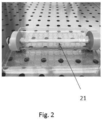
- the present invention relates to a process for the production of an engineered vascular tissue or construct, preferably a scaffold ( Fig. 2 , 21 ) having a lumen coated with a functional and continuous endothelium having a monolayer of confluent cells, preferably usable for testing medical or veterinary models, wherein said process includes the application of:
- the proposed technical solution represented by the process which the subject matter of the present invention is, for ease of comprehension, divided into the two methods described separately below in detail: (1) method for seeding a cell culture into the lumen of a scaffold, preferably a tubular scaffold; (2) method for connecting a perfusion circuit to a bioreactor-scaffold system (perfusion method).
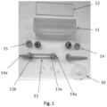
- the method for uniformly seeding endothelial cells for a bioreactor-scaffold system comprises a plurality of steps preformed sequentially and in sterile conditions: 1.1 A scaffold ( Fig. 2 ; 21 ), preferably a tubular scaffold, for example a tubular scaffold of an electrospun silk fibroin, mounted on the mountings of a scaffold support ( Fig. 1 ; 13 , 13a, 13b) is seated inside the chamber of the bioreactor, to yield a bioreactor-scaffold system.
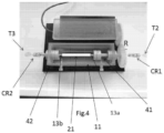
- rotary connectors Fig. 4 ; CR1, CR2
- T connectors Fig. 4 ; T2, T3
- bioreactor-scaffold system the assembly of the bioreactor and the scaffold ( Fig. 3 ; 11 , 21 ), preferably a tubular scaffold, for example a tubular scaffold of an electrospun silk fibroin, seated and mounted by means of the mountings of the scaffold support ( Fig. 1 ; 13 , 13a, 13b) and then located inside the bioreactor.
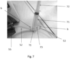
- the scaffold preferably tubular, is held by the mountings of the scaffold support (which is hollow to enable perfusion of the scaffold) with self-tightening straps, after having protected the scaffold, in this case made of electrospun silk fibroin, with sterilised teflon tape.
- the scaffold support 13 is inserted into the bioreactor 11 in such a way that the inlet upstream of the chamber of the bioreactor coincides ( Fig. 4 ; CR1, 41) with one end of the scaffold ( Fig. 4 ; 13a) and the downstream opening of the bioreactor chamber ( Fig. 4 ; CR2, 42) coincides with the other end of the scaffold ( Fig. 4 ; 13b).
- the longitudinal axis of the bioreactor is defined as the major axis along which the scaffold installed inside the bioreactor is oriented.
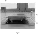
- the scaffold is preconditioned with fresh culture medium injected into the lumen of the scaffold seated inside the bioreactor, using a syringe with luer-lock attachment engaged with one of the two ends of the bioreactor via T connector ( Fig. 9 ; T2).
- the open ends of the connectors located upstream and downstream of the bioreactor are then closed with caps to prevent the lumen of the scaffold emptying.
- fresh culture medium is inserted into the chamber of the bioreactor until the scaffold seated inside it is completely covered. In this way the scaffold seated inside the chamber of the bioreactor is preconditioned, preferably for 1 hour at 25°C, with fresh culture medium both inside (in the lumen) and outside.
- the bioreactor preconditioned with culture medium injected into the lumen, it is emptied preferably with a sterile pipette, and the culture medium previously inserted into the chamber is removed preferably with a sterile pipette.
- the residual culture medium in the connectors (rotary and T) upstream and downstream of the bioreactor is removed by vacuum using a pipette, without collapsing the scaffold.
- T connectors located upstream ( Fig. 9 , T2) and downstream ( Fig. 9 ; T3) of the ends of the bioreactor, containing the scaffold supported on the mountings, are turned so that the upper opening turned upwards (by 90° relative to the plane in which the bioreactor-scaffold system lies).
- the lateral openings of the T connectors located downstream (T3) and upstream (T2) of the bioreactor is then closed with a cap.
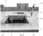
- a container, preferably a syringe, is then mounted on the opening of the T connector turned upwards ( Fig. 9 , T2) located upstream of the bioreactor, ( Fig.
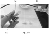
- a cell suspension consisting of fresh culture medium and endothelial cells (e.g. HUVECs) is drawn from a container in which it has been prepared. Subsequently, the drawn cell suspension is released into the container or syringe ( Fig 10 ; 91) mounted on the T connector ( Fig. 10 ; T2) upstream of the bioreactor by means of the element ( Fig. 10 ; CR1). The cell suspension must be released, with the pipette ( Fig.
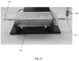
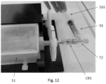
- the syringe (91) containing the residual cell suspension is then rotated by around 90° relative to the plane ( Figure 12 ) in which it lies; in this position, the plunger of the syringe (102) is re-inserted into the open end of the syringe ( Figure 12 ) so that only the black insulating extremity is inserted so as not to create pressure inside the scaffold.
- the syringe (91) may then be unscrewed from the T connector T2 located upstream of the bioreactor without forming air bubbles ( Figure 13 ) and the end of the connector closed with a cap ( Figure 14 ).
- the scaffold is then rotated continuously around its longitudinal axis, for instance with a speed of rotation between 1.5 and 2 rpm, for 24 hours.
- the rotation enables uniform adhesion of the cells to the lumen of the scaffold, keeps the scaffold itself continually wet with the culture medium inside the chamber of the bioreactor and allows nutrients to flow from the culture medium inside the chamber (outside the scaffold) to the cell suspension seeded inside the lumen of the scaffold.
- the seeding method developed by the Applicant maintains the sterility of the system. Furthermore, this seeding method which is the subject matter of the present invention is fast, reproducible, and advantageously enables preparation of a scaffold having the surface of the lumen (internal) with endothelial cells adhering uniformly and homogeneously along the entire length of the scaffold (from the proximal to the medial up to the distal part).
- the seeding method according to the present invention allows the cells to be seeded while both eliminating any air bubbles already present in the bioreactor-scaffold system and preventing the formation of new ones, thus preventing damage to the cells.
- each step of the present method is standardised and reproducible, and optimises the time and cost of the procedure.
- the vitality of the cells adhering to the scaffold was assayed with resazurin as reactant (tradename Alamar Blue, IUPAC name 7-hydroxy-10-oxido phenoxazine-10-ium-3-one, CAS 550-82-3).
- This assay consists in a metabolic reaction which makes it possible to quantify the vitality of the cells due to the redox reaction of the indicator (resazurin) which reduces to resofluorine, a fluorescent compound which turns pink in the presence of the reductive environment of a living cell.
- the seeded scaffold is removed from the mountings.
- the scaffold is then sectioned (cut) in three zones of 2cm length each, in relation to the site of injection of the cell suspension: proximal, medial and distal. Each section is then divided into 4 parts of approximately 1cm 2 each.
- 3 samples are selected, each representative of each region (proximal, medial and distal) of the scaffold with adhering endothelial cells. Each sample is placed in a well of a 24 well plate and incubated with 1ml of a 0.02 mg/ml solution of resazurin sodium salt (Sigma Aldrich, R7017) with fresh culture medium preferably for 3 hours at 37°C with 5% CO 2 .
- the reaction between the 0.02 mg/ml solution of resazurin sodium salt with fresh culture medium and the scaffold sample (with the adhering endothelial cells) is analysed by measuring the A.U. (arbitrary unit of fluorescence) at 590 nm with a spectrofluorometer.
- a further analysis is the amount evaluation of the genomic DNA present in the cells adhering to the same scaffold samples previously used for the resazurin assay.
- the genomic DNA is extracted from the adhering cells of the scaffold by means of lysis and quantified using the Quant-iT TM PicoGreen TM dsDNA Assay (P7589, Invitrogen, Molecular Probes) in which a fluorescent colourant of the nucleic acids (PicoGreen) allows by a standard reference curve to determine the concentration of genomic DNA in the solution.
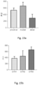
- Figures 15A and 15B show graphically the values obtained from the resazurin assay for the samples representative of each region (proximal, medial and distal) of a scaffold seeded with endothelial cells and incubated for 24 hours in three separate experiments (denominated DYN1, DYN2 e DYN3).
- the graphs in Figures 15A and 15B show good adhesion and vitality of the endothelial cells.
- VWF Von Willebrand factor
- CD31 cluster of differentiation 31
- VCAM-1 vascular cell adhesion molecule 1
- H&E staining "Haematoxylin and Eeosin staining" was run to evaluate the distribution of the cells and their morphology, and an immunofluorescence assay was run for the specific endothelial functionality markers.
- the seeding method according to the present invention has been shown to be efficacious inasmuch as it guarantees homogeneous, uniform and reproducible seeding of vital endothelial cells over the entire length of the lumen.
- connection method is based on the following sequence of steps, following seeding (method for seeding a cell culture in the lumen of a scaffold according to the embodiment described above at point (1)) after 24 hours of adhesion of the endothelial cells:
- connection method is based on the following sequence of steps, following seeding (method for seeding a cell culture in the lumen of a scaffold according to the embodiment described above at point (1)) after 24 hours of adhesion of the endothelial cells:
- connection method (perfusion method) described herein, both in the first embodiment and in the second embodiment described above, enable connection of a perfusion circuit of a scaffold, preferably tubular, to the seeded bioreactor-scaffold system.
- This procedure prevents the formation of air bubbles and prevents the bubbles, should they form, reaching the scaffold seeded with endothelial cells of the bioreactor-scaffold system.
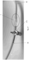
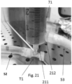
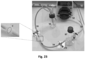
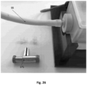
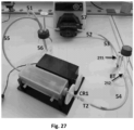
- any air bubbles already in the perfusion circuit do not reach the scaffold thanks to the presence of the bubble trap ( Fig. 21 , 71; Fig. 27 , BT) in the circuit itself. In this way, the method developed by the applicant assures a complete lack of air bubbles in the lumen of the scaffold.
- This system satisfies the requisites imposed by the configuration of the bioreactor. All the details described herein are necessary to render the method operator independent and hence to assure reproducibility of the results during the industrial generation in vitro of a construct with functional endothelium. Furthermore, this methodology makes it possible to work in conditions of sterility, since all actions are simple. Furthermore, the fast, traceable procedure reduces the risk that air bubbles come into contact with the cells, thus preventing damage to the endothelial cells adhering to the lumen of the scaffold, preferably tubular. Said perfusion method makes it possible to obtain engineered vascular tissues/constructs having a scaffold with a lumen coated with an endothelium which is both continuous (i.e. having a monolayer of confluent cells) and functional.
- the experimental analyses relative to the evaluation of the method for connecting the perfusion circuit to the bioreactor-scaffold system are the same as those applied to the evaluation of the seeding method for a cell culture in a scaffold, preferably tubular.
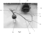
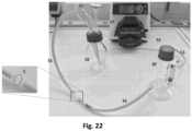
- perfusion circuit Fig. 5 and Fig. 22
- tubes Fig. 5 ; 51- 54 or Fig. 22 ; 51-55
- a reservoir Fig. 5 or Fig. 22 ; 56
- a peristaltic pump Fig. 5 ; 55 or Fig. 22 ; 57
- an element for removing air bubbles Fig. 21 , 71 or Fig. 22 , BT.
- Said element for the removal of air bubbles may be present in the perfusion circuit prior to connection of the perfusion circuit to the seeded bioreactor-scaffold system (second embodiment of the perfusion method) or alternatively may be inserted into the perfusion circuit after connection of the perfusion circuit to the seeded bioreactor-scaffold system (first embodiment of the perfusion method).
- Said tubes are of biocompatible material and connected to each other in such a way as to permit perfusion of the scaffold ( Fig. 21 and Fig. 27 ), preferably tubular, seated inside the bioreactor 11, by means of the peristaltic pump ( Fig. 5 ; 55, Fig.
- the perfusion circuit principally consists of five tubes with internal diameter of 3/16": first aspiration tube 51 exiting from the reservoir, second tube 52 pump loop tubing, third tube 53 connecting the circuit to the bubble trap BT, fourth tube 54 connecting the BT to the T connector T2 upstream of the bioreactor-scaffold system, fifth tube 55 return to the reservoir 56 connected to the T connector T3 downstream of the bioreactor-scaffold system.
- the tube 51 is connected to the pump loop tubing 52, the pump loop tubing 52 to the BT, the BT to the tube 53, the tube 53 to the T connector T2 upstream of the bioreactor-scaffold system, the tube 54 to the T connector T3 downstream of the bioreactor-scaffold system and to the reservoir 56.
- the reservoir ( Fig. 5 or Fig. 22 , 56) is the element containing the warm fresh culture medium (for example, Endothelial Growth Medium EGM, Sigma Aldrich) from which the tube 51 ( Fig. 5 or Fig. 22 ) aspirates and to which the tube 54 ( Fig. 5 ) or 55 ( Fig. 22 ) returns, maintaining the entire circuit and the seeded bioreactor-scaffold system in a closed loop.
- the reservoir ( Fig. 5 or Fig. 22 , 56) is at atmospheric pressure, thanks to a 0.22 ⁇ m filter present on the cap of the reservoir, which guarantees sterility of the air.
- the scaffold preferably a tubular scaffold
- the scaffold is selected among natural or synthetic origin polymer scaffolds, formed of a single polymer or copolymers, such as for example electrospun silk fibroin or copolymers of PGA/PLA (polyglycolic acid/polylactic acid) or PGA/PCL (polyglycolic acid/polyprolactone).
- PGA/PLA polyglycolic acid/polylactic acid
- PGA/PCL polyglycolic acid/polyprolactone
- RP6 The process according to any one of RP1-5, wherein the endothelial cells are selected among the cells constituting an endothelium of a vascular tissue, such as for example HAOECs (human aortic endothelial cells), HCAECs (human coronary artery endothelial cells), HMEVECs (human dermal microvascular endothelial cells) or HUVECs (human umbilical vein endothelial cells).
- HAOECs human aortic endothelial cells
- HCAECs human coronary artery endothelial cells
- HMEVECs human dermal microvascular endothelial cells
- HUVECs human umbilical vein endothelial cells
- RP7 The process according to any one of RP1-6, wherein the culture medium is Endothelial Growth Medium (EGM, Sigma Aldrich, 211-500), preferably heated to 37°C.
- EMM Endothelial Growth Medium
- a scaffold having a lumen coated with a continuous functional endothelium obtained by means of the process according to any of RP1-7.
- RP9 Use of the scaffold according to RP8, to perform in vitro preclinical tests and clinical trials of medical products for use in the cardiovascular and peripheral vascular area, such as for instance heart valves, stents, grafts and catheters.
- the first step to perform and optimise is the cell seeding step, preferably endothelial cells, followed by the second critical step of connecting the perfusion circuit to the system comprising the bioreactor and the scaffold, so as to assure reliability, efficacy and reproducibility of the industrialisation process for the in vitro generation of a continuous functional endothelium.
- the success of the industrialisation process according to this invention for the production of engineered vascular tissue/construct preferably a scaffold having a lumen coated with a functional continuous endothelium having a confluent cell monolayer, consists principally in the success of the seeding and the connection of the perfusion circuit to the bioreactor-scaffold system, so that the air bubbles are reliably and reproducibly eliminated from the entire system.
- the seeding method depends on the cell source and the density of the cells, the chemical properties and porosity of the scaffold and the total removal of air bubbles from the lumen of the scaffold during injection of the cell suspension.
- the method for connecting the perfusion circuit to the bioreactor-scaffold system is based on the maintained sterility of the assembled system and the guarantee that no air bubbles are able to come into contact with the seeded scaffold. Note that the formation of air bubbles must be avoided inasmuch as the air bubbles can damage the cells, compromising their vitality and hence preventing the endothelisation of the scaffold.
- the description of the present invention shows that the choice of the method for connecting the perfusion circuit to the bioreactor-scaffold system depends on the previously optimised method for seeding the endothelial cells since said method of connection must be adapted to the experimental setup and the requirements of perfusion, and on the choice of the position chosen for this system in the incubator.
- the process for seeding a scaffold is one of the crucial factors in the generation in vitro of functional engineered vascular constructs with confluent endothelium, as shown in the description of the present invention.
- This process is responsible for the uniform and homogeneous distribution of the endothelial cells in the lumen, as well as for the adhesion of said cells to the surface.
- the description of the present invention shows that the choice of the most suitable seeding method, adapted to any bioreactor-scaffold system, defines the reproducibility of the process, and demonstrates its advantages in the reproducibility of the results.
- the seeding method used in the present invention assures a highly uniform distribution in the adhesion of the endothelial cells and a good reproducibility of the results, necessary for a laboratory the work of which is focused on the production in vitro of vascular constructs as models for industrial testing, not only preclinical.
- the cells adhering to the lumen of the scaffold principally tubular, must maintain their morphology and vitality to enable the formation of a homogeneous vascular endothelium. It follows that it is important to prevent any cell alteration during the connection process capable of modifying the adhesion of the cells, thus losing the growing vascular layer (as it is forming).
- the known methods for connecting the bioreactor with the perfusion circuit compromise the endothelial cells and cause them to detach, even partially, from the luminal surface, so that the formation of a functional endothelial layer is slowed down or impeded. Furthermore, the known methods for connecting the bioreactor to the perfusion circuit do not assure the absence of any air bubbles, which may be formed by the compression or twisting (total or partial) of the connection tubes during perfusion. It is important to totally avoid contact between said air bubbles and the seeded scaffold by introducing an element, for example a bubble-trap, capable of eliminating the air bubbles before they reach the seeded scaffold. This disadvantage has been overcome successfully by the process according to the present invention, which obtains an internal surface of the scaffold coated with a uniform and functional layer of endothelial cells, in particular a confluent cell layer.
Landscapes
- Health & Medical Sciences (AREA)
- Engineering & Computer Science (AREA)
- Life Sciences & Earth Sciences (AREA)
- Biomedical Technology (AREA)
- Chemical & Material Sciences (AREA)
- Bioinformatics & Cheminformatics (AREA)
- Organic Chemistry (AREA)
- Zoology (AREA)
- Wood Science & Technology (AREA)
- Biotechnology (AREA)
- Genetics & Genomics (AREA)
- Biochemistry (AREA)
- Microbiology (AREA)
- General Health & Medical Sciences (AREA)
- Immunology (AREA)
- General Engineering & Computer Science (AREA)
- Molecular Biology (AREA)
- Cell Biology (AREA)
- Sustainable Development (AREA)
- Hematology (AREA)
- Urology & Nephrology (AREA)
- Tropical Medicine & Parasitology (AREA)
- Pathology (AREA)
- Vascular Medicine (AREA)
- General Physics & Mathematics (AREA)
- Analytical Chemistry (AREA)
- Physics & Mathematics (AREA)
- Medicinal Chemistry (AREA)
- Food Science & Technology (AREA)
- Toxicology (AREA)
- Apparatus Associated With Microorganisms And Enzymes (AREA)
- Micro-Organisms Or Cultivation Processes Thereof (AREA)
- Prostheses (AREA)
Claims (7)
- Procédé de production d'un tissu ou d'une construction vasculaire reconstruit(e), comprenant un échafaudage (21) comportant une lumière recouverte d'un endothélium fonctionnel et continu comportant une monocouche cellulaire confluente, pour tester des produits médicaux ou vétérinaires, ledit procédé comprenant l'application :- d'une méthode d'ensemencement d'une culture de cellules endothéliales dans la lumière d'un échafaudage (21) pour obtenir un échafaudage ensemencé (21) ; ledit échafaudage ensemencé (21) étant présent à l'intérieur d'un bioréacteur (11), pour produire un système bioréacteur (11)-échafaudage (21) ensemencé ;dans lequel ladite méthode d'ensemencement comprend les étapes de :- libérer ladite culture de cellules endothéliales sous la forme d'une suspension cellulaire comprenant un milieu de culture frais et des cellules endothéliales à l'intérieur d'un récipient (91) monté sur un connecteur en « T » (T2) situé en amont du bioréacteur (11) par un connecteur rotatif (CR1) ; puis- libérer ladite culture de cellules endothéliales dans la lumière de l'échafaudage (21) à l'intérieur de la chambre du bioréacteur (11) avec un flux continu tel que la vitesse du flux permet à ladite suspension cellulaire de descendre dans le connecteur en « T » (T2) sans générer de bulles d'air et de pousser les bulles d'air à l'intérieur de la lumière de l'échafaudage (21) vers une ouverture d'un connecteur en « T » (T3) situé en aval du bioréacteur (11) pour leur permettre de sortir ; et successivement- une méthode de perfusion avec un milieu de culture frais ayant une température comprise entre 30 et 45 °C, de préférence 37 °C, des cellules endothéliales présentes dans la lumière dudit échafaudage ensemencé (21) ; ladite méthode de perfusion étant réalisée par le raccordement d'un circuit de perfusion (51-56) ou (51-57 et BT) audit système bioréacteur ensemencé(11)-échafaudage (21) ;dans lequel ladite méthode de perfusion comprend une étape de :- remplir partiellement un élément pour l'élimination des bulles d'air (71) ou (BT) présentes dans le circuit de perfusion avec ledit milieu de culture frais, dans lequel ledit élément pour l'élimination des bulles (71) ou (BT) comprend une chambre, un bouchon fermant ladite chambre, un accès d'entrée (211) et un accès de sortie (212), dans lequel ledit élément pour l'élimination des bulles d'air (71 ou BT) a un volume et dans lequel une première partie dudit volume est remplie avec ledit milieu de culture frais et dans lequel une deuxième partie dudit volume est remplie d'air, ladite deuxième partie dudit volume ayant pour fonction de piéger les bulles d'air présentes dans ledit milieu de culture frais qui s'écoule à travers ledit accès d'entrée (211) et ledit accès de sortie (212).
- Procédé selon la revendication 1, dans lequel ladite méthode d'ensemencement de ladite culture de cellules endothéliales dans la lumière dudit échafaudage (21) comprend :- monter l'échafaudage (21), de préférence un échafaudage tubulaire de fibroïne de soie électrofilée, sur les montages d'un support d'échafaudage (13, 13a, 13b) et placer ledit support d'échafaudage (13, 13a, 13b) avec l'échafaudage (21) à l'intérieur de la chambre du bioréacteur (11), pour produire un système bioréacteur (11)-échafaudage (21) ;
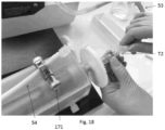
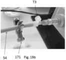
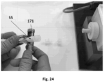
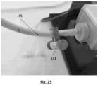
puis- injecter le milieu de culture frais dans la lumière dudit échafaudage (21) fixé sur ledit support d'échafaudage (13) à l'intérieur de la chambre du bioréacteur (11) ; puis- ajouter ledit milieu de culture frais à l'intérieur de la chambre du bioréacteur (11) dans laquelle se trouve ledit support d'échafaudage (13, 13a, 13b) avec l'échafaudage (21) injecté avec ledit milieu de culture ; puis- laisser, pendant un intervalle de temps compris entre 1 et 18 heures à une température comprise entre 20 et 30 °C, de préférence 25 °C, ledit milieu de culture à l'intérieur de la lumière de l'échafaudage (21) et à l'intérieur de la chambre du bioréacteur (11) dans laquelle se trouve ledit support d'échafaudage (13) avec l'échafaudage (21) injecté avec ledit milieu de culture ; puis- nettoyer l'intérieur de la lumière de l'échafaudage (21) et de la chambre du bioréacteur (11) du milieu de culture ; puis- libérer ladite culture de cellules endothéliales à l'intérieur dudit récipient (91) selon la revendication 1, de préférence ledit récipient (91) étant une seringue ; puis- libérer ladite suspension cellulaire dans la lumière de l'échafaudage (21) selon la revendication 1 ; puis- ajouter ledit milieu de culture frais à l'intérieur de la chambre du bioréacteur (11) dans laquelle se trouve ledit support d'échafaudage (13) avec l'échafaudage ensemencé (21) contenant ladite suspension de cellules dans sa lumière ; et puis- incuber, de préférence pendant 24 heures à 37 °C en présence de 5 % de CO2, de l'échafaudage (21) placé à l'intérieur de la chambre du bioréacteur (11). - Procédé selon les revendications 1 ou 2, dans lequel ladite méthode de perfusion des cellules endothéliales dans la lumière dudit échafaudage ensemencé (21) comprend :- fournir ledit circuit de perfusion fermé comprenant des tubes (51), (52), (53), (54) et éventuellement (55) ;- obstruer le tube (54) ou (55) du circuit de perfusion à l'aide d'un élément de fermeture (171) situé à proximité d'un connecteur (C), de préférence ledit élément de fermeture est une pince ou un dispositif analogue ; puis- dévisser le connecteur (C) situé entre le tube (53) ou (54) et le tube (54) ou (55) respectivement du circuit de perfusion ;- visser le tube (53) ou (54) du circuit de perfusion à une extrémité latérale ouverte du connecteur en « T » (T2) en amont du bioréacteur (11) en correspondance d'un accès latéral de celui-ci ; puis- ouvrir le connecteur en « T » (T3) en aval du bioréacteur (11) et dévisser un capuchon d'une ouverture latérale du connecteur en « T » (T3) ; puis- raccorder le tube (54) ou (55) du circuit de perfusion à l'ouverture latérale du connecteur en « T » (T3) situé en aval du bioréacteur (11) et retirer l'élément de fermeture (171) ;- insérer, entre le tube (53) et la tubulure de la boucle de pompe (52) du circuit de perfusion, l'élément pour l'élimination des bulles d'air (71).
- Procédé selon l'une quelconque des revendications précédentes, dans lequel l'élément pour l'élimination des bulles d'air (71) ou (BT) est un piège à bulles ou un dispositif analogue.
- Procédé selon l'une quelconque des revendications précédentes, dans lequel l'échafaudage (21), de préférence un échafaudage tubulaire, est choisi parmi les échafaudages polymères synthétiques ou d'origine naturelle, lesdits échafaudages polymères étant formés d'un seul polymère ou de copolymères, de préférence de la fibroïne de soie électrofilée ou des copolymères d'acide polyglycolique / acide polylactique (PGA/PLA) ou des copolymères diacides polyglycoliques / polycaprolactone (PGA/PCL).
- Procédé selon l'une quelconque des revendications précédentes, dans lequel les cellules endothéliales sont choisies parmi les cellules constituant un endothélium d'un tissu vasculaire, de préférence les HAOEC (cellules endothéliales de l'aorte humaine), les HCAEC (cellules endothéliales de l'artère coronaire humaine), les HMEVEC (cellules endothéliales microvasculaires dermiques humaines) ou les HUVEC (cellules endothéliales de la veine ombilicale humaine).
- Procédé selon l'une quelconque des revendications précédentes, dans lequel le milieu de culture est un milieu de croissance endothéliale comprenant du sérum bovin fætal (2 %), de l'adénine (0,2 µg/ml), du métavanadate d'ammonium (0,0006 µg/ml), de l'amphotéricine B (0,3 µg/ml), du chlorure de calcium 2H2O (300 µg/ml), du chlorhydrate de choline (20 µg/ml), du sulfate de cuivre 5H2O (0,002 µg/ml), de l'acide trioptique DL-6,8(0,003 µg/ml), de l'acide folinique (calcium) (0,6 µg/ml), de l'héparine (4 µg/ml), de l'hydrocortisone (2 µg/ml), de l'acide L-aspartique (15 µg/ml), L-cystéine (30 µg/ml), L-tyrosine (20 µg/ml), du sulfate de manganèse monohydraté (0,0002 µg/ml), du molybdate d'ammonium 4H2O (0,004 µg/ml), du nicotinamide (8 µg/ml), du chlorure de nickel 6H2O (0,0001 µg/ml), de la pénicilline (60 µg/ml), du sel de sodium de rouge de phénol (15 µg/ml), du chlorure de potassium (300 µg/ml), du dihydrochlorure de putrescine (0,0002 µg/ml), du chlorhydrate de pyridoxine (3 µg/ml), du métasilicate de sodium 9H2O (3 µg/ml), du sulfate de sodium 7H2O (200 µg/ml), de la sélénite de sodium (0,01 µg/ml), du sulfate de streptomycine (100 µg/ml), du chlorhydrate de thiamine (4 µg/ml) et du sulfate de zinc 7H2O (0,0003 µg/ml), de préférence chauffés à 37 °C.
Applications Claiming Priority (2)
| Application Number | Priority Date | Filing Date | Title |
|---|---|---|---|
| IT102017000093925A IT201700093925A1 (it) | 2017-08-16 | 2017-08-16 | Un processo di industrializzazione affidabile e riproducibile di eliminazione di bolle d'aria per la produzione di un tessuto vascolare ingegnerizzato. |
| PCT/IB2018/055944 WO2019034966A1 (fr) | 2017-08-16 | 2018-08-07 | Procédé d'industrialisation fiable et reproductible pour l'élimination de bulles d'air dans la production d'un tissu vasculaire modifié |
Publications (3)
| Publication Number | Publication Date |
|---|---|
| EP3669190A1 EP3669190A1 (fr) | 2020-06-24 |
| EP3669190B1 true EP3669190B1 (fr) | 2023-11-01 |
| EP3669190B8 EP3669190B8 (fr) | 2024-03-06 |
Family
ID=60991183
Family Applications (1)
| Application Number | Title | Priority Date | Filing Date |
|---|---|---|---|
| EP18765185.6A Active EP3669190B8 (fr) | 2017-08-16 | 2018-08-07 | Procédé d'industrialisation fiable et reproductible pour l'élimination de bulles d'air dans la production d'un tissu vasculaire reconstruit |
Country Status (8)
| Country | Link |
|---|---|
| US (1) | US20200208117A1 (fr) |
| EP (1) | EP3669190B8 (fr) |
| JP (2) | JP7322024B2 (fr) |
| CA (1) | CA3072697A1 (fr) |
| ES (1) | ES2970422T3 (fr) |
| IL (1) | IL272577A (fr) |
| IT (1) | IT201700093925A1 (fr) |
| WO (1) | WO2019034966A1 (fr) |
Families Citing this family (3)
| Publication number | Priority date | Publication date | Assignee | Title |
|---|---|---|---|---|
| US11788042B2 (en) * | 2015-07-31 | 2023-10-17 | Redwire Space Technologies, Inc. | Biomanufacturing system, method, and 3D bioprinting hardware in a reduced gravity environment |
| IT201800007946A1 (it) * | 2018-08-07 | 2020-02-07 | 1Lab Sa | Modello per simulare in-vitro il comportamento di vasi disfunzionali |
| IT201900002193A1 (it) * | 2019-02-14 | 2020-08-14 | 1Lab Sa | Metodo di caratterizzazione di un costrutto vascolare ingegnerizzato |
Family Cites Families (11)
| Publication number | Priority date | Publication date | Assignee | Title |
|---|---|---|---|---|
| US4329743A (en) * | 1979-04-27 | 1982-05-18 | College Of Medicine And Dentistry Of New Jersey | Bio-absorbable composite tissue scaffold |
| US4918019A (en) * | 1986-05-12 | 1990-04-17 | C. D. Medical, Incorporated | Bioreactor system with plasticizer removal |
| US5558984A (en) * | 1994-06-03 | 1996-09-24 | Clemson University | Automated system and process for heterotrophic growth of plant tissue |
| US7759113B2 (en) * | 1999-04-30 | 2010-07-20 | The General Hospital Corporation | Fabrication of tissue lamina using microfabricated two-dimensional molds |
| US6902909B2 (en) | 2002-07-09 | 2005-06-07 | Synthecon, Inc. | Methods for efficient production of mammalian recombinant proteins |
| US7875448B2 (en) * | 2004-01-12 | 2011-01-25 | Single Use Brx, Llc | Bioreactor systems and disposable bioreactor |
| GB0410177D0 (en) * | 2004-05-07 | 2004-06-09 | Univ Wales Medicine | Engineered tubular tissue structures |
| CA2723141A1 (fr) * | 2008-04-30 | 2009-11-05 | Ethicon, Inc. | Vaisseau sanguin obtenu par ingenierie tissulaire |
| US10078075B2 (en) | 2011-12-09 | 2018-09-18 | Vanderbilt University | Integrated organ-on-chip systems and applications of the same |
| WO2014019603A1 (fr) | 2012-07-30 | 2014-02-06 | Nmi Naturwissenschaftliches Und Medizinisches Institut An Der Universitaet Tuebingen | Plaque de connexion pour puce de test microfluidique, puce de test microfluidique et procédé d'analyse au moyen d'une zone d'un agencement de test microfluidique |
| US10780198B2 (en) * | 2014-03-06 | 2020-09-22 | University of Pittsburgh—of the Commonwealth System of Higher Education | Electrospinning with sacrificial template for patterning fibrous constructs |
-
2017
- 2017-08-16 IT IT102017000093925A patent/IT201700093925A1/it unknown
-
2018
- 2018-08-07 US US16/639,090 patent/US20200208117A1/en not_active Abandoned
- 2018-08-07 WO PCT/IB2018/055944 patent/WO2019034966A1/fr unknown
- 2018-08-07 EP EP18765185.6A patent/EP3669190B8/fr active Active
- 2018-08-07 ES ES18765185T patent/ES2970422T3/es active Active
- 2018-08-07 CA CA3072697A patent/CA3072697A1/fr active Pending
- 2018-08-07 JP JP2020530740A patent/JP7322024B2/ja active Active
-
2020
- 2020-02-10 IL IL272577A patent/IL272577A/en unknown
-
2022
- 2022-11-17 JP JP2022184203A patent/JP2023014146A/ja active Pending
Non-Patent Citations (1)
| Title |
|---|
| ZHANG X ET AL: "Dynamic culture conditions to generate silk-based tissue-engineered vascular grafts", BIOMATERIALS, ELSEVIER, AMSTERDAM, NL, vol. 30, no. 19, 1 July 2009 (2009-07-01), pages 3213 - 3223, XP026130537, ISSN: 0142-9612, [retrieved on 20090220], DOI: 10.1016/J.BIOMATERIALS.2009.02.002 * |
Also Published As
| Publication number | Publication date |
|---|---|
| ES2970422T3 (es) | 2024-05-28 |
| CA3072697A1 (fr) | 2019-02-21 |
| IT201700093925A1 (it) | 2019-02-16 |
| WO2019034966A1 (fr) | 2019-02-21 |
| JP2020531049A (ja) | 2020-11-05 |
| IL272577A (en) | 2020-03-31 |
| EP3669190A1 (fr) | 2020-06-24 |
| EP3669190B8 (fr) | 2024-03-06 |
| JP2023014146A (ja) | 2023-01-26 |
| JP7322024B2 (ja) | 2023-08-07 |
| US20200208117A1 (en) | 2020-07-02 |
Similar Documents
| Publication | Publication Date | Title |
|---|---|---|
| EP3669190B1 (fr) | Procédé d'industrialisation fiable et reproductible pour l'élimination de bulles d'air dans la production d'un tissu vasculaire reconstruit | |
| Sonnaert et al. | Quantitative validation of the presto blue™ metabolic assay for online monitoring of cell proliferation in a 3D perfusion bioreactor system | |
| Bijonowski et al. | Bioreactor design for perfusion-based, highly vascularized organ regeneration | |
| Narita et al. | Novel pulse duplicating bioreactor system for tissue-engineered vascular construct | |
| Baptista et al. | Human liver bioengineering using a whole liver decellularized bioscaffold | |
| CN103468634A (zh) | 生成微脉管网络、内皮原始脉管、组织和人冠状动脉替代品的方法 | |
| WO2004011593A1 (fr) | Appareil de culture automatique de cellules ou de tissus d'origine biologique | |
| Schlüter et al. | Adult ventricular cardiomyocytes: isolation and culture | |
| US20210222130A1 (en) | Model for in-vitro simulation of the behaviour of dysfunctional vessels | |
| Yang et al. | Decellularized liver scaffold for liver regeneration | |
| JP5727174B2 (ja) | 細胞培養用中空糸モジュールおよび細胞培養方法 | |
| CN112915264A (zh) | 用于明胶-海藻酸钠-prp复合材料的制作方法 | |
| EP2130905A1 (fr) | Procédé de culture de cellules eucaryotes | |
| EP3775155A1 (fr) | Dispositif de bioréacteur à flux permettant de surveiller la dynamique cellulaire | |
| JP2003339368A (ja) | 中空糸付き器具及びその使用方法 | |
| CN106823014A (zh) | 植入性生物可降解微孔氧化铁支架 | |
| US20220098549A1 (en) | Method for characterising a tissue-engineered construct | |
| JP2006345778A (ja) | 細胞培養用中空糸モジュールおよび細胞培養方法 | |
| Falkner et al. | The mandatory CAM testing of cells and scaffolds for tissue engineering: benefits for the three Rs of cooperation with the vaccine industry | |
| Migliore et al. | Controlled in vitro growth of cell microtubes: towards the realisation of artificial microvessels | |
| Wacker et al. | Bacterial nanocellulose-based grafts for cell colonization studies: An in vitro bioreactor perfusion model | |
| JP4977854B2 (ja) | 組織形成用複合材料およびその製造方法 | |
| JP2011062216A (ja) | 細胞培養用中空糸モジュールおよび細胞培養方法 | |
| Lee | Establishing a biomimetic bioreactor: Recapitulating physiological niches in vitro to derive tailored bone-like constructs with human mesenchymal stem cells | |
| Ortiz et al. | Design and Testing of a Novel Cell Seeding and Bioreactor System for the In-Vitro Growth of Scaffold-Free Tissue Tubes for Tissue Engineered Blood Vessels |
Legal Events
| Date | Code | Title | Description |
|---|---|---|---|
| STAA | Information on the status of an ep patent application or granted ep patent |
Free format text: STATUS: UNKNOWN |
|
| STAA | Information on the status of an ep patent application or granted ep patent |
Free format text: STATUS: THE INTERNATIONAL PUBLICATION HAS BEEN MADE |
|
| PUAI | Public reference made under article 153(3) epc to a published international application that has entered the european phase |
Free format text: ORIGINAL CODE: 0009012 |
|
| STAA | Information on the status of an ep patent application or granted ep patent |
Free format text: STATUS: REQUEST FOR EXAMINATION WAS MADE |
|
| 17P | Request for examination filed |
Effective date: 20200310 |
|
| AK | Designated contracting states |
Kind code of ref document: A1 Designated state(s): AL AT BE BG CH CY CZ DE DK EE ES FI FR GB GR HR HU IE IS IT LI LT LU LV MC MK MT NL NO PL PT RO RS SE SI SK SM TR |
|
| AX | Request for extension of the european patent |
Extension state: BA ME |
|
| DAV | Request for validation of the european patent (deleted) | ||
| DAX | Request for extension of the european patent (deleted) | ||
| STAA | Information on the status of an ep patent application or granted ep patent |
Free format text: STATUS: EXAMINATION IS IN PROGRESS |
|
| 17Q | First examination report despatched |
Effective date: 20220307 |
|
| GRAP | Despatch of communication of intention to grant a patent |
Free format text: ORIGINAL CODE: EPIDOSNIGR1 |
|
| STAA | Information on the status of an ep patent application or granted ep patent |
Free format text: STATUS: GRANT OF PATENT IS INTENDED |
|
| INTG | Intention to grant announced |
Effective date: 20230509 |
|
| GRAS | Grant fee paid |
Free format text: ORIGINAL CODE: EPIDOSNIGR3 |
|
| GRAA | (expected) grant |
Free format text: ORIGINAL CODE: 0009210 |
|
| STAA | Information on the status of an ep patent application or granted ep patent |
Free format text: STATUS: THE PATENT HAS BEEN GRANTED |
|
| AK | Designated contracting states |
Kind code of ref document: B1 Designated state(s): AL AT BE BG CH CY CZ DE DK EE ES FI FR GB GR HR HU IE IS IT LI LT LU LV MC MK MT NL NO PL PT RO RS SE SI SK SM TR |
|
| REG | Reference to a national code |
Ref country code: GB Ref legal event code: FG4D |
|
| REG | Reference to a national code |
Ref country code: CH Ref legal event code: EP |
|
| REG | Reference to a national code |
Ref country code: IE Ref legal event code: FG4D |
|
| REG | Reference to a national code |
Ref country code: DE Ref legal event code: R096 Ref document number: 602018060414 Country of ref document: DE |
|
| REG | Reference to a national code |
Ref country code: CH Ref legal event code: PK Free format text: BERICHTIGUNG B8 |
|
| RAP2 | Party data changed (patent owner data changed or rights of a patent transferred) |
Owner name: 1MED SA |
|
| REG | Reference to a national code |
Ref country code: LT Ref legal event code: MG9D |
|
| REG | Reference to a national code |
Ref country code: NL Ref legal event code: MP Effective date: 20231101 |
|
| PG25 | Lapsed in a contracting state [announced via postgrant information from national office to epo] |
Ref country code: GR Free format text: LAPSE BECAUSE OF FAILURE TO SUBMIT A TRANSLATION OF THE DESCRIPTION OR TO PAY THE FEE WITHIN THE PRESCRIBED TIME-LIMIT Effective date: 20240202 |
|
| PG25 | Lapsed in a contracting state [announced via postgrant information from national office to epo] |
Ref country code: IS Free format text: LAPSE BECAUSE OF FAILURE TO SUBMIT A TRANSLATION OF THE DESCRIPTION OR TO PAY THE FEE WITHIN THE PRESCRIBED TIME-LIMIT Effective date: 20240301 |
|
| PG25 | Lapsed in a contracting state [announced via postgrant information from national office to epo] |
Ref country code: LT Free format text: LAPSE BECAUSE OF FAILURE TO SUBMIT A TRANSLATION OF THE DESCRIPTION OR TO PAY THE FEE WITHIN THE PRESCRIBED TIME-LIMIT Effective date: 20231101 |
|
| REG | Reference to a national code |
Ref country code: AT Ref legal event code: MK05 Ref document number: 1627774 Country of ref document: AT Kind code of ref document: T Effective date: 20231101 |
|
| PG25 | Lapsed in a contracting state [announced via postgrant information from national office to epo] |
Ref country code: NL Free format text: LAPSE BECAUSE OF FAILURE TO SUBMIT A TRANSLATION OF THE DESCRIPTION OR TO PAY THE FEE WITHIN THE PRESCRIBED TIME-LIMIT Effective date: 20231101 |
|
| PG25 | Lapsed in a contracting state [announced via postgrant information from national office to epo] |
Ref country code: AT Free format text: LAPSE BECAUSE OF FAILURE TO SUBMIT A TRANSLATION OF THE DESCRIPTION OR TO PAY THE FEE WITHIN THE PRESCRIBED TIME-LIMIT Effective date: 20231101 |
|
| PG25 | Lapsed in a contracting state [announced via postgrant information from national office to epo] |
Ref country code: NL Free format text: LAPSE BECAUSE OF FAILURE TO SUBMIT A TRANSLATION OF THE DESCRIPTION OR TO PAY THE FEE WITHIN THE PRESCRIBED TIME-LIMIT Effective date: 20231101 Ref country code: LT Free format text: LAPSE BECAUSE OF FAILURE TO SUBMIT A TRANSLATION OF THE DESCRIPTION OR TO PAY THE FEE WITHIN THE PRESCRIBED TIME-LIMIT Effective date: 20231101 Ref country code: IS Free format text: LAPSE BECAUSE OF FAILURE TO SUBMIT A TRANSLATION OF THE DESCRIPTION OR TO PAY THE FEE WITHIN THE PRESCRIBED TIME-LIMIT Effective date: 20240301 Ref country code: GR Free format text: LAPSE BECAUSE OF FAILURE TO SUBMIT A TRANSLATION OF THE DESCRIPTION OR TO PAY THE FEE WITHIN THE PRESCRIBED TIME-LIMIT Effective date: 20240202 Ref country code: BG Free format text: LAPSE BECAUSE OF FAILURE TO SUBMIT A TRANSLATION OF THE DESCRIPTION OR TO PAY THE FEE WITHIN THE PRESCRIBED TIME-LIMIT Effective date: 20240201 Ref country code: AT Free format text: LAPSE BECAUSE OF FAILURE TO SUBMIT A TRANSLATION OF THE DESCRIPTION OR TO PAY THE FEE WITHIN THE PRESCRIBED TIME-LIMIT Effective date: 20231101 Ref country code: PT Free format text: LAPSE BECAUSE OF FAILURE TO SUBMIT A TRANSLATION OF THE DESCRIPTION OR TO PAY THE FEE WITHIN THE PRESCRIBED TIME-LIMIT Effective date: 20240301 |
|
| REG | Reference to a national code |
Ref country code: ES Ref legal event code: FG2A Ref document number: 2970422 Country of ref document: ES Kind code of ref document: T3 Effective date: 20240528 |