EP3668898B1 - Humanized antibodies for cd3 - Google Patents
Humanized antibodies for cd3 Download PDFInfo
- Publication number
- EP3668898B1 EP3668898B1 EP18765358.9A EP18765358A EP3668898B1 EP 3668898 B1 EP3668898 B1 EP 3668898B1 EP 18765358 A EP18765358 A EP 18765358A EP 3668898 B1 EP3668898 B1 EP 3668898B1
- Authority
- EP
- European Patent Office
- Prior art keywords
- antibody
- seq
- present disclosure
- antibody fragment
- cell
- Prior art date
- Legal status (The legal status is an assumption and is not a legal conclusion. Google has not performed a legal analysis and makes no representation as to the accuracy of the status listed.)
- Active
Links
Images


Classifications
-
- C—CHEMISTRY; METALLURGY
- C07—ORGANIC CHEMISTRY
- C07K—PEPTIDES
- C07K16/00—Immunoglobulins [IG], e.g. monoclonal or polyclonal antibodies
- C07K16/18—Immunoglobulins [IG], e.g. monoclonal or polyclonal antibodies against material from animals or humans
- C07K16/28—Immunoglobulins [IG], e.g. monoclonal or polyclonal antibodies against material from animals or humans against receptors, cell surface antigens or cell surface determinants
- C07K16/2803—Immunoglobulins [IG], e.g. monoclonal or polyclonal antibodies against material from animals or humans against receptors, cell surface antigens or cell surface determinants against the immunoglobulin superfamily
- C07K16/2809—Immunoglobulins [IG], e.g. monoclonal or polyclonal antibodies against material from animals or humans against receptors, cell surface antigens or cell surface determinants against the immunoglobulin superfamily against the T-cell receptor (TcR)-CD3 complex
-
- C—CHEMISTRY; METALLURGY
- C07—ORGANIC CHEMISTRY
- C07K—PEPTIDES
- C07K16/00—Immunoglobulins [IG], e.g. monoclonal or polyclonal antibodies
- C07K16/18—Immunoglobulins [IG], e.g. monoclonal or polyclonal antibodies against material from animals or humans
- C07K16/32—Immunoglobulins [IG], e.g. monoclonal or polyclonal antibodies against material from animals or humans against translation products of oncogenes
-
- C—CHEMISTRY; METALLURGY
- C07—ORGANIC CHEMISTRY
- C07K—PEPTIDES
- C07K2317/00—Immunoglobulins specific features
- C07K2317/20—Immunoglobulins specific features characterized by taxonomic origin
- C07K2317/24—Immunoglobulins specific features characterized by taxonomic origin containing regions, domains or residues from different species, e.g. chimeric, humanized or veneered
-
- C—CHEMISTRY; METALLURGY
- C07—ORGANIC CHEMISTRY
- C07K—PEPTIDES
- C07K2317/00—Immunoglobulins specific features
- C07K2317/30—Immunoglobulins specific features characterized by aspects of specificity or valency
- C07K2317/31—Immunoglobulins specific features characterized by aspects of specificity or valency multispecific
-
- C—CHEMISTRY; METALLURGY
- C07—ORGANIC CHEMISTRY
- C07K—PEPTIDES
- C07K2317/00—Immunoglobulins specific features
- C07K2317/30—Immunoglobulins specific features characterized by aspects of specificity or valency
- C07K2317/33—Crossreactivity, e.g. for species or epitope, or lack of said crossreactivity
-
- C—CHEMISTRY; METALLURGY
- C07—ORGANIC CHEMISTRY
- C07K—PEPTIDES
- C07K2317/00—Immunoglobulins specific features
- C07K2317/40—Immunoglobulins specific features characterized by post-translational modification
- C07K2317/41—Glycosylation, sialylation, or fucosylation
-
- C—CHEMISTRY; METALLURGY
- C07—ORGANIC CHEMISTRY
- C07K—PEPTIDES
- C07K2317/00—Immunoglobulins specific features
- C07K2317/50—Immunoglobulins specific features characterized by immunoglobulin fragments
- C07K2317/55—Fab or Fab'
-
- C—CHEMISTRY; METALLURGY
- C07—ORGANIC CHEMISTRY
- C07K—PEPTIDES
- C07K2317/00—Immunoglobulins specific features
- C07K2317/50—Immunoglobulins specific features characterized by immunoglobulin fragments
- C07K2317/56—Immunoglobulins specific features characterized by immunoglobulin fragments variable (Fv) region, i.e. VH and/or VL
- C07K2317/567—Framework region [FR]
-
- C—CHEMISTRY; METALLURGY
- C07—ORGANIC CHEMISTRY
- C07K—PEPTIDES
- C07K2317/00—Immunoglobulins specific features
- C07K2317/60—Immunoglobulins specific features characterized by non-natural combinations of immunoglobulin fragments
- C07K2317/62—Immunoglobulins specific features characterized by non-natural combinations of immunoglobulin fragments comprising only variable region components
- C07K2317/622—Single chain antibody (scFv)
-
- C—CHEMISTRY; METALLURGY
- C07—ORGANIC CHEMISTRY
- C07K—PEPTIDES
- C07K2317/00—Immunoglobulins specific features
- C07K2317/60—Immunoglobulins specific features characterized by non-natural combinations of immunoglobulin fragments
- C07K2317/64—Immunoglobulins specific features characterized by non-natural combinations of immunoglobulin fragments comprising a combination of variable region and constant region components
-
- C—CHEMISTRY; METALLURGY
- C07—ORGANIC CHEMISTRY
- C07K—PEPTIDES
- C07K2317/00—Immunoglobulins specific features
- C07K2317/70—Immunoglobulins specific features characterized by effect upon binding to a cell or to an antigen
-
- C—CHEMISTRY; METALLURGY
- C07—ORGANIC CHEMISTRY
- C07K—PEPTIDES
- C07K2317/00—Immunoglobulins specific features
- C07K2317/70—Immunoglobulins specific features characterized by effect upon binding to a cell or to an antigen
- C07K2317/71—Decreased effector function due to an Fc-modification
-
- C—CHEMISTRY; METALLURGY
- C07—ORGANIC CHEMISTRY
- C07K—PEPTIDES
- C07K2317/00—Immunoglobulins specific features
- C07K2317/70—Immunoglobulins specific features characterized by effect upon binding to a cell or to an antigen
- C07K2317/73—Inducing cell death, e.g. apoptosis, necrosis or inhibition of cell proliferation
-
- C—CHEMISTRY; METALLURGY
- C07—ORGANIC CHEMISTRY
- C07K—PEPTIDES
- C07K2317/00—Immunoglobulins specific features
- C07K2317/90—Immunoglobulins specific features characterized by (pharmaco)kinetic aspects or by stability of the immunoglobulin
- C07K2317/92—Affinity (KD), association rate (Ka), dissociation rate (Kd) or EC50 value
Definitions
- the present disclosure relates to humanized antibodies and antibody fragments which interact with the human epsilon chain of CD3.
- the present disclosure also provides bispecific antibodies comprising such humanized antibodies.
- the disclosure also relates to nucleic acids, vectors and host cells capable of expressing such antibodies, pharmaceutical compositions comprising said antibodies and uses of said antibodies and pharmaceutical compositions for the treatment of specific diseases.
- CD3 is a homodimeric or heterodimeric antigen expressed on T cells in association with the T cell receptor complex (TCR) and is required for T cell activation.
- CD3 is formed by the dimeric association of two of four different chains: epsilon, zeta, delta and gamma.
- the CD3 dimeric arrangements include gamma/epsilon, delta/epsilon and zeta/zeta.
- Antibodies against CD3 have been shown to cluster CD3 on T cells, thereby causing T cell activation in a manner similar to the engagement of the TCR by peptide-loaded MHC molecules. Thus, CD3 specific antibodies have been proposed for therapies involving the activation of T cells.
- bispecific antibodies that co-engage CD3 and a tumor antigen target have been generated to redirect T cells to attack and lyse targeted tumor cells.
- Examples include the BiTE and DART formats, which monovalently engage CD3 and a tumor antigen but also IgG like formats, such as BEAT antibodies, where one binding arm binds to the tumor antigen and the second arm binds to CD3 on T-cells.
- cytokines e.g., tumor necrosis factor alpha (TNF ⁇ ), interferon- ⁇ , interleukins IL-2, IL-3, IL-4, IL-6, IL-10 and granulocyte-macrophage colony-stimulating factor ( Masharani, U.B. et al.
- TNF ⁇ tumor necrosis factor alpha
- interferon- ⁇ interleukins IL-2, IL-3, IL-4, IL-6, IL-10
- Masharani U.B. et al.
- One way to minimize cytokine production while retaining T cell activation and subsequent target cell depletion is the reduction of the affinity of the CD3 specific binding domain while retaining the affinity of the tumor antigen specific binding domain.
- bispecific antibodies comprising CD3 specific binding domains with reduced affinity towards CD3 may have limited capability to mediate redirected T cell cytotoxic killing of target antigen low expressing cells such as present on healthy tissue but high potency in depleting target antigen high expressing cancer cells.
- formats such as BiTE and DART do not contain Fc domains and therefore show very short serum half-lives in patients.
- CD3 specific antibodies having reduced side effects while maintaining efficacy and desirable pharmacokinetic (PK) are advantageous in particular when employed in a bispecific therapy.
- An antibody specific for human CD3 with cross reactivity to non-human primate CD3 is the mouse monoclonal antibody SP34 ( Yoshino N. et al., Exp. Anim 49:97-110, 2000 ; Conrad ML. et al., Cytometry 71A:925-33, 2007 ), which binds specifically to human CD3 in denatured form and in native form ( Pressano, S. The EMBO J. 4:337-344, 1985 ; Alarcon, B. EMBO J. 10:903-912, 1991 ).
- SP34 also binds to CD3 epsilon singly transfected COS cells as well as CD3epsilon/gamma or CD3c/8 double transfectants ( Salmeron A. et al., J. Immunol. 147:3047-52, 1991 ). SP34 recognizes an N-terminal 1-27 amino acid residue polypeptide fragment of the extracellular domain of CD3 epsilon. Because of its cross reactivity to non-human primate CD3, humanized variants of SP34 can be used both for preclinical evaluation of safety, activity and/or pharmacokinetic profile of these in primates and - in the identical form - as drugs in humans. SP34 activate T cells when being cross-linked ( Yang et al., J. Immunol.
- one aspect of the present invention relates to humanized SP34 antibodies having reduced immunogenicity in humans.
- WO 2016/110576 describes humanized antibodies of SP34 with improved stability and expression.
- the humanized variants disclosed in WO2016/110576 revealed stronger binding affinities towards CD3 when compared to the CD3 specific antibody of the present invention and generated bispecific antibodies with monovalent binding to CD3 were not able to discriminate between target antigen high, moderate or low expressing cells.
- the bispecific antibody constructs induced anti-drug antibody responses (ADA) in preclinical studies in cynomolgus monkeys.
- US2017/0157251 discloses humanized variants of SP34 which were used to generate bispecific diabody molecules with stronger monovalent binding to CD3 when compared to the CD3 specific antibody of the present invention.
- the bispecific diabody molecules induced unwanted but transient dose-dependent reduction of total T lymphocytes in preclinical toxicology studies in cynomolgus monkeys.
- WO 2016/020444 describes humanized antibodies of SP34 with improved stability and expression.
- the humanized CD3 antibodies were used in bispecific tandem diabody molecules with monovalent binding to CD3 in the single digit nanomolar range.
- WO 2016/020444 provides no working examples in respect of the assessment of immunogenicity or undesired T-cell activation, the relative strong binding affinity towards CD3 suggests that generated bispecific antibodies would probably not able to discriminate between target antigen high, moderate or low expressing cells in terms of cytotoxic killing activity.
- WO 2017/136659 discloses humanized and deimmunized antibody variants of SP34 with equivalent binding affinity towards human and cynomolgus CD3 when compared to the parental antibody SP34. Generated bispecific antibodies with monovalent binding to CD3 were not able to discriminate between target antigen high and moderate expressing cells in terms of killing activity. In addition, minimal non-specific killing of target negative cells and T cell proliferation in presence of target-negative cells was observed.
- WO 2015/063339 describes humanized antibodies derived from SP34 which were used to generate various bispecific BEAT antibodies with monovalent binding to CD3.
- Preferred humanized CD3 specific VH and VL combinations were selected among others to retain the binding affinity of the parental antibody SP34. Accordingly, bispecific antibody constructs comprising such "high affinity" binding domains may less favorable in terms of inducing potential side effects.
- the humanized SP34 binding domains describe in the art were designed to exhibit an equivalent or even better binding affinity towards human and cynomolgus CD3 when compared to the parental antibody SP34 and/or to preserve its productivity and stability. This is typically achieved by an iterative process of back-mutating human residues with the amino acids at the same position in the donor antibody. The back mutation process can result in further non-human amino acid residues being reintroduced into the humanised antibody and as such, the risk for induction of an immunogenic reaction in humans is increased again. Such immunogenic reaction can result in the restoration of an unwanted bivalent or multivalent CD3 binding for antibody formats employing only monovalent CD3 binding.
- bispecific antibody constructs comprising such "high affinity" binding domains reveal a T cell mediated killing of target antigen low expressing cells (such as present on healthy tissue) similar or comparable to the killing of target antigen high expressing cells rendering such constructs less favourable in terms of inducing potential side effects.
- the present disclosure provides novel humanized antibodies and antibody fragments specific for CD3, which are superior to the humanized antibodies described in the art.
- the humanized antibodies of the present disclosure display weaker binding affinities to human and non-human primate CD3 when compared to the parental murine antibody SP34 resulting in the reduced activation of T cells and associated release of inflammatory cytokines.
- bispecific antibodies comprising the humanized SP34 binding domains of the present disclosure have limited capability to mediate redirected T cell killing of target antigen low expressing cells such as present on healthy tissue. Together with their low immunogenicity risk in humans, the antibodies of the present disclosure combine favorable functional and safety properties never observed before.
- the present disclosure provides humanized antibodies and antigen-binding fragments that specifically bind to human CD3.
- the present invention provides an isolated antibody or antibody fragment specific for cluster of differentiation 3 (CD3), wherein said antibody or antibody fragment specifically binds to human CD3 and to non-human primate CD3, wherein said antibody or antibody fragment comprises the variable heavy chain having SEQ ID NO: 7 and the variable light chain having SEQ ID NO: 17.
- CD3 cluster of differentiation 3
- said antibody is a full-length IgG.
- said antibody fragment is a Fab fragment.
- the present invention provides a bispecific antibody comprising a first antigen binding domain of an antibody or antibody fragment specific for cluster of differentiation 3 (CD3) according to the present invention, and a second antigen binding domain which binds a different target antigen than said first antigen binding region.
- CD3 cluster of differentiation 3
- said second binding region specifically binds a cell surface target antigen.
- the present invention provides a nucleic acid composition comprising a nucleic acid sequence or a plurality of nucleic acid sequences encoding the antibody or antibody fragment specific for cluster of differentiation 3 (CD3) according to the present invention.
- the present invention provides a vector composition comprising a vector or a plurality of vectors comprising the nucleic acid sequence or plurality of nucleic acid sequences encoding the antibody or antibody fragment specific for cluster of differentiation 3 (CD3) according to the present invention.
- a vector composition comprising a vector or a plurality of vectors comprising the nucleic acid sequence or plurality of nucleic acid sequences encoding the antibody or antibody fragment specific for cluster of differentiation 3 (CD3) according to the present invention.
- the present invention provides a host cell comprising the vector composition according to the present invention.
- the present invention provides the isolated antibody or antibody fragment specific for cluster of differentiation 3 (CD3) according to the present invention for use as a medicament.
- CD3 cluster of differentiation 3
- the present invention provides a pharmaceutical composition
- a pharmaceutical composition comprising the antibody or antibody fragment specific for cluster of differentiation 3 (CD3) according to the present invention and a pharmaceutically acceptable carrier or excipient.
- CD3 cluster of differentiation 3
- the humanized antibodies or antibody fragments according to the present disclosure are used for targeting T cells expressing CD3 (CD3 expressing T cells) and stimulating T cell activation, e.g., under circumstances where T cell-mediated killing is beneficial or desirable.
- CD3 specific antibodies or antibody fragments of the present disclosure or antigen-binding fragments may be used in bispecific formats that direct CD3 mediated T cell activation to specific cell types such as tumor cells.
- the disclosure also relates to methods of reducing or eliminating tumor burden and controlling the toxic side effects that may be associated with tumor immunotherapy.
- any reference to the methods of treatment practised on the human or animal body is to be interpreted as the substance of composition for use in such treatments.
- the present disclosure provides humanized CD3 antibodies or antibody fragments, which specifically bind to CD3 with an optimized affinity compared to the murine antibody SP34 with K D values typically in the double digit nanomolar range as determined in an in vitro affinity binding assay.
- the present disclosure relates to humanized antibodies or antibody fragments specifically binding to human and non-human CD3, and in particular to such antibodies or antibody fragments that are cross-reactive with CD3 epsilon of a non-human primate such as cynomolgus monkey.
- the disclosed humanized antibodies are superior to the CD3 specific antibodies described in the prior art in terms of safety and pharmacokinetic (PK) properties and provide well suited and promising compounds for the treatment of humans suffering particular from diseases such like cancer.
- PK pharmacokinetic
- the present disclosure provides humanized antibodies or antibody fragments comprising CD3 specific binding domains that are "optimized affinity" binding domains to CD3 as measured using for instance a Biacore assay.
- the present disclosure provides antibodies comprising humanized CD3 specific binding domains that have an "optimized affinity” CD3 epsilon binding when compared to the murine antibody SP34.
- the present disclosure provides bispecific antibodies comprising humanized CD3 specific binding domains that have an "optimized affinity" CD3 epsilon binding when compared to the murine antibody SP34 and consequently display a weaker potency in killing target antigen low expressing cells but an equivalent, similar or comparable potency in killing target antigen high expressing cancer cells.
- the bispecific antibodies of the present disclosure may not mediate redirected T cell cytotoxicity (RTCC) killing of target antigens expressed on healthy tissue.
- the present disclosure also comprises full length IgGs for improving the pharmacokinetics (PK) and potentially lowering the immunogenicity of the molecules.
- the present disclosure provides an antibody or antibody fragment specific for cluster of differentiation 3 (CD3), wherein said antibody or antibody fragment specifically binds to human CD3 and to non-human primate CD3, wherein said antibody or antibody fragment comprises the HCDR1 region of SEQ ID NO: 23, the HCDR2 region of SEQ ID NO: 24, the HCDR3 region of SEQ ID NO: 25, the LCDR1 region of SEQ ID NO: 26, the LCDR2 region of SEQ ID NO: 65 and the LCDR3 region of SEQ ID NO: 28.
- CD3 antibody or antibody fragment specific for cluster of differentiation 3
- the present disclosure provides an antibody or antibody fragment specific for CD3, wherein said antibody or antibody fragment comprises
- variable heavy chain and variable light chain are selected from the group consisting of: the variable heavy chain having SEQ ID NO: 7 and the variable light chain having SEQ ID NO: 17.
- variable heavy chain and variable light chain are selected from the group consisting of: the variable heavy chain domain having SEQ ID NO: 7 and the variable light chain having SEQ ID NO: 17.
- the antibody or antibody fragment comprises the variable heavy chain having SEQ ID NO: 7 and the variable light chain having SEQ ID NO: 17.
- said antibody or antibody fragment specific for CD3 is an isolated antibody or antibody fragment.
- said antibody or antibody fragment specific for CD3 is a recombinant antibody or antibody fragment.
- the antibody or antibody fragment is a monoclonal antibody or antibody fragment.
- said antibody or antibody fragment specific for CD3 is a full-length IgG.
- said antibody or antibody fragment specific for CD3 is a full-length IgG of an isotype selected from the group consisting of IgG1, IgG2, IgG3, and IgG4.
- the present disclosure provides an antibody or antibody fragment specific for CD3, wherein the full-length IgG comprises an Fc region that has reduced effector function relative to that of a wild type Fc-receptor.
- the present disclosure provides an antibody or antibody fragment specific for CD3, wherein said antibody comprises a Fc region, wherein in at least 5 amino acids in the positions corresponding to positions L234, L235, D237, N330, P331 in a human IgG1 heavy chain, are mutated to A, E, A, S, and S, respectively.
- the present disclosure provides an antibody or antibody fragment specific for CD3, wherein said antibody fragment is a Fab fragment.
- the present disclosure provides an antibody or antibody fragment specific for CD3, wherein the antibody is a single chain antibody.
- the present disclosure provides a bispecific antibody comprising a first antigen binding domain of an antibody or antibody fragment specific for CD3 according to the present disclosure, and a second antigen binding domain which binds a different target antigen than said first antigen binding domain.
- the present disclosure provides a bispecific antibody, wherein said second binding domain specifically binds a cell surface target antigen.
- the present disclosure provides a bispecific antibody, wherein said cell surface target antigen is a tumor antigen.
- the present disclosure provides a bispecific antibody, wherein said bispecific antibody comprises a Fc region modified according to the present disclosure.
- the present disclosure provides a bispecific antibody, wherein said bispecific antibody comprises an Fc region that has reduced effector function relative to that of a wild type Fc-receptor.
- the present disclosure provides a bispecific antibody, wherein said bispecific antibody comprises an Fc region, wherein in at least 5 amino acids in the positions corresponding to positions L234, L235, D237, N330, P331 in a human IgG1 heavy chain, are mutated to A, E, A, S, and S, respectively.
- the present disclosure provides a nucleic acid composition comprising a nucleic acid sequence or a plurality of nucleic acid sequences encoding the antibody or antibody fragment according to any one of the preceding claims.
- the present disclosure provides a vector composition comprising a vector or a plurality of vectors comprising the nucleic acid sequence or plurality of nucleic acid sequences according to the present disclosure.
- the present disclosure provides a host cell comprising the vector composition according to the present disclosure.
- said host cell is mammalian cell.
- said host cell is prokaryotic cell.
- the present disclosure provides an antibody or antibody fragment specific for CD3 for use in the treatment of a subject in need thereof.
- the present disclosure provides an antibody or antibody fragment specific for CD3 for use as a medicament.
- the present disclosure provides a pharmaceutical composition
- a pharmaceutical composition comprising the antibody or antibody fragment according to the present disclosure and a pharmaceutically acceptable carrier or excipient.
- Utilization of the claimed antibodies or antibody fragments is to target T cells expressing CD3, and for stimulating T cell activation, e.g., under circumstances where T cell-mediated killing is beneficial or desirable.
- the claimed antibodies or antibody fragments are for therapeutic use, such as the treatment of cancer.
- CD3 refers to an antigen which is expressed on T cells as part of the multimolecular T cell receptor (TCR) and which consists of a homodimer or heterodimer formed from the association of two of four receptor chains: CD3-epsilon, CD3-delta, CD3-zeta, and CD3-gamma.
- TCR multimolecular T cell receptor
- Human CD3 epsilon has the amino acid sequence of UniProt P07766
- the extracellular domain of human CD3 epsilon without the signal sequence comprises amino acid residues 23-126 and has the amino acid sequence of as set forth in SEQ ID NO: 1 of TABLE 1. Cynomolgus CD3 epsilon has the amino acid sequence of UniProt Q95LI5
- the extracellular domain of cynomolgus monkey CD3 epsilon without the signal sequence comprises amino acid residues 22-117 and has the amino acid sequence of as set forth in SEQ ID NO: 2 of TABLE 1.
- antibody refers to a protein comprising at least two heavy (H) chains and two light (L) chains inter-connected by disulfide bonds which interacts with an antigen.
- Each heavy chain is comprised of a heavy chain variable region or domain (abbreviated herein as VH) and a heavy chain constant region.
- the heavy chain constant region is comprised of three domains, CH1, CH2 and CH3.
- Each light chain is comprised of a light chain variable region or domain (abbreviated herein as VL) and a light chain constant region.
- the light chain constant region is comprised of one domain, CL.
- VH and VL regions can be further subdivided into regions of hypervariability, termed complementarity determining regions (CDR), interspersed with regions that are more conserved, termed framework regions (FR).
- CDR complementarity determining regions
- FR framework regions
- Each VH and VL is composed of three CDRs and four FR's arranged from amino-terminus to carboxy-terminus in the following order: FR1, CDR1, FR2, CDR2, FR3, CDR3, and FR4.
- the variable regions of the heavy and light chains contain a binding domain that interacts with an antigen.
- the constant regions of the antibodies may mediate the binding of the immunoglobulin to host tissues or factors, including various cells of the immune system (e.g., effector cells) and the first component (Clq) of the classical complement system.
- antibody includes for example, monoclonal antibodies, human antibodies, humanized antibodies, camelised antibodies and chimeric antibodies.
- the antibodies can be of any isotype (e.g., IgG, IgE, IgM, IgD, IgA and IgY), class (e.g., IgG1, IgG2, IgG3, IgG4, IgA1 and IgA2) or subclass. Both the light and heavy chains are divided into regions of structural and functional homology.
- antibody fragment refers to one or more portions of an antibody that retain the ability to specifically interact with (e.g., by binding, steric hindrance, stabilizing spatial distribution) an antigen.
- binding fragments include, but are not limited to, a Fab fragment, a monovalent fragment consisting of the VL, VH, CL and CH1 domains; a F(ab)2 fragment, a bivalent fragment comprising two Fab fragments linked by a disulfide bridge at the hinge region; a Fd fragment consisting of the VH and CH1 domains; a Fv fragment consisting of the VL and VH domains of a single arm of an antibody; a dAb fragment ( Ward et al., (1989) Nature 341:544-546 ), which consists of a VH domain; and an isolated complementarity determining region (CDR).
- a Fab fragment a monovalent fragment consisting of the VL, VH, CL and CH1 domains
- F(ab)2 fragment a bivalent
- the two domains of the Fv fragment, VL and VH are coded for by separate genes, they can be joined, using recombinant methods, by a synthetic linker that enables them to be made as a single protein chain in which the VL and VH regions pair to form monovalent molecules (known as single chain Fv (scFv); see e.g., Bird et al., (1988) Science 242:423-426 ; and Huston et al., (1988) Proc. Natl. Acad. Sci. 85:5879-5883 ).
- single chain Fv single chain Fv
- Such single chain antibodies are also intended to be encompassed within the term "antibody fragment”.
- Antibody fragments are obtained using conventional techniques known to those of skill in the art, and the fragments are screened for utility in the same manner as are intact antibodies.
- Antibody fragments can also be incorporated into single domain antibodies, maxibodies, minibodies, intrabodies, diabodies, triabodies, tetrabodies, v-NAR and bis-scFv (see, e.g., Hollinger and Hudson, (2005) Nature Biotechnology 23:1126-1136 ).
- Antibody fragments can be grafted into scaffolds based on polypeptides such as Fibronectin type III (Fn3) (see U.S. Pat. No. 6,703,199 , which describes fibronectin polypeptide monobodies).
- Fn3 Fibronectin type III
- Antibody fragments can be incorporated into single chain molecules comprising a pair of tandem Fv segments (VH-CH1-VH-CH1) which, together with complementary light chain polypeptides, form a pair of antigen-binding sites ( Zapata et al., (1995) Protein Eng. 8:1057-1062 ; and U.S. Pat. No. 5,641,870 ).
- a “human antibody” or " human antibody fragment” includes antibodies and antibody fragments having variable regions in which both the framework and CDR regions are derived from sequences of human origin. Furthermore, if the antibody contains a constant region, the constant region also is derived from such sequences.
- Human origin includes, e.g., human germline sequences, or mutated versions of human germline sequences or antibody containing consensus framework sequences derived from human framework sequences analysis, for example, as described in Knappik et al., (2000) J Mol Biol 296:57-86 ).
- said human antibody can be obtained from technology platforms which comprise antibodies derived from human germline genes either generated by PCR-amplification of VHA/L repertoire isolated from B-cells or are generated synthetically.
- Technology platforms include library based approaches comprising human immunoglobulin genes displayed on phage, ribosome or yeast. Respective display technologies are standard in the scientific community. Furthermore immunization of a transgenic mouse carrying human immunoglobulin repertoire is another approach to generate human antibodies against an antigen of interest. Antibodies or fragments thereof selected from an antibody library based on the MorphoSys HuCAL ® concept ( Knappik et al., (2000) J Mol Biol 296:57-86 ) are considered as fully human.
- a “humanized antibody” or “humanized antibody fragment” is defined herein as an antibody or antibody fragment which has constant antibody regions derived from sequences of human origin and the variable antibody regions or parts thereof or only the CDRs are derived from another species.
- a humanized antibody can be CDR-grafted, wherein the CDRs of the variable domain are from a non-human origin, while one or more frameworks of the variable domain are of human origin and the constant domain (if any) is of human origin.
- chimeric antibody or " chimeric antibody fragment” is defined herein as an antibody molecule which has constant antibody regions derived from, or corresponding to, sequences found in one species and variable antibody regions derived from another species.
- the constant antibody regions are derived from, or corresponding to, sequences found in humans
- the variable antibody regions are derived from sequences found in a non-human animal, e.g. a mouse, rat, rabbit or hamster.
- immunoglobulin variable domains e.g., CDRs
- CDRs may be defined using well known numbering schemes, e.g., the Kabat numbering scheme, the Chothia numbering scheme, or a combination of Kabat and Chothia (see, e.g., Sequences of Proteins of Immunological Interest, U.S. Department of Health and Human Services (1991), eds. Kab at et al.; Lazikani et al., (1997) J. Mol. Bio. 273:927-948 ); Kabat et al., (1991) Sequences of Proteins of Immunological Interest, 5th edit., NIH Publication no. 91-3242 U.S.
- isolated refers to a compound, which can be e.g. an antibody or antibody fragment, that is substantially free of other antibodies or antibody fragments having different antigenic specificities.
- antibodies provided are isolated antibodies which have been separated from antibodies with a different specificity.
- An isolated antibody may be a monoclonal antibody.
- An isolated antibody may be a recombinant monoclonal antibody.
- An isolated antibody that specifically binds to an epitope, isoform or variant of a target may, however, have cross-reactivity to other related antigens, e.g., from other species (e.g., species homologs).
- recombinant antibody includes all antibodies that are prepared, expressed, created or segregated by means not existing in nature. For example antibodies isolated from a host cell transformed to express the antibody, antibodies selected and isolated from a recombinant, combinatorial human antibody library, and antibodies prepared, expressed, created or isolated by any other means that involve splicing of all or a portion of a human immunoglobulin gene, sequences to other DNA sequences or antibodies isolated from an animal (e.g., a mouse) that is transgenic or transchromosomal for human immunoglobulin genes or a hybridoma prepared therefrom.
- an animal e.g., a mouse
- such recombinant antibodies have variable regions in which the framework and CDR regions are derived from human germline immunoglobulin sequences.
- such recombinant human antibodies can be subjected to in vitro mutagenesis (or, when an animal transgenic for human Ig sequences is used, in vivo somatic mutagenesis) and thus the amino acid sequences of the VH and VL regions of the recombinant antibodies are sequences that, while derived from and related to human germline VH and VL sequences, may not naturally exist within the human antibody germline repertoire in vivo.
- a recombinant antibody may be a monoclonal antibody.
- monoclonal antibody refers to an antibody that is derived from a single clone, including any eukaryotic, prokaryotic, or phage clone, and not the method by which it is producede.
- Monoclonal antibodies as disclosed herein may be made by the hybridoma method as described in Kohler et a/.; Nature, 256:495 (1975 ) or may be isolated from phage libraries using the techniques as described herein, for example.
- an antibody " binds specifically to ", " specifically binds to “, is “ specific to/for " or “ specifically recognizes " an antigen if such antibody is able to discriminate between such antigen and one or more reference antigen(s), since binding specificity is not an absolute, but a relative property.
- determination of binding specificity is performed by using not a single reference antigen, but a set of about three to five unrelated antigens, such as milk powder, BSA, transferrin or the like.
- an antibody that binds to CD3 or an "anti-CD3 antibody” or “antibody specific for CD3” includes antibodies and antibody fragments that specifically recognize one or more CD3 subunit (e.g., epsilon, delta, gamma or zeta), as well as antibodies and antibody fragments that specifically recognize a dimeric complex of two CD3 subunits (e.g., gamma/epsilon, delta/epsilon, and zeta/zeta CD3 dimers).
- the antibodies and antibody fragments of the present disclosure may bind soluble CD3 and/or cell surface expressed CD3.
- Soluble CD3 includes natural CD3 proteins as well as recombinant CD3 protein variants such as, e.g., monomeric and dimeric CD3 constructs, that lack a transmembrane domain or are otherwise unassociated with a cell membrane.
- cell surface-expressed CD3 means one or more CD3 protein(s) that is/are expressed on the surface of a cell in vitro or in vivo, such that at least a portion of a CD3 protein is exposed to the extracellular side of the cell membrane and is accessible to an antigen-binding portion of an antibody.
- Cell surface-expressed CD3 includes CD3 proteins contained within the context of a functional T cell receptor in the membrane of a cell.
- cell surface-expressed CD3 includes CD3 protein expressed as part of a homodimer or heterodimer on the surface of a cell (e.g., gamma/epsilon, delta/epsilon, and zeta/zeta CD3 dimers).
- the expression, “cell surface-expressed CD3” also includes a CD3 chain (e.g., CD3-epsilon, CD3-delta or CD3-gamma) that is expressed by itself, without other CD3 chain types, on the surface of a cell.
- a “cell surface-expressed CD3” can comprise or consist of a CD3 protein expressed on the surface of a cell which normally expresses CD3 protein.
- “cell surface-expressed CD3” can comprise or consist of CD3 protein expressed on the surface of a cell that normally does not express human CD3 on its surface but has been artificially engineered to express CD3 on its surface.
- affinity refers to the strength of interaction between the polypeptide and its target at a single site. Within each site, the binding region of the polypeptide interacts through weak non-covalent forces with its target at numerous sites; the more interactions, the stronger the affinity.
- K D refers to the dissociation constant, which is obtained from the ratio of K d to K a (i.e. K d /K a ) and is expressed as a molar concentration (M).
- K D values for antigen binding moieties like e.g. monoclonal antibodies can be determined using methods well established in the art. Methods for determining the K D of an antigen binding moiety like e.g. a monoclonal antibody are SET (soluble equilibrium titration) or surface plasmon resonance using a biosensor system such as a Biacore ® system.
- an antibody specific to the CD3 epsilon polypeptide typically has a dissociation rate constant (K D ) (k off /k on ) of less than 5 ⁇ 10 -2 M, less than 10 -2 M, less than 5 ⁇ 10 -3 M, less than 10 -3 M, less than 5 ⁇ 10 -4 M, less than 10 -4 M, less than 5 ⁇ 10 -5 M, less than 10 -5 M, less than 5 ⁇ 10 -6 M, less than 10 -6 M, less than 5 ⁇ 10 -7 M, less than 10 -7 M, less than 5 ⁇ 10 -8 M, less than 10 -8 M, less than 5 ⁇ 10 -9 M, less than 10 -9 M, less than 5 ⁇ 10 -10 M, less than 10 -10 M, less than 5 ⁇ 10 -11 M, less than 10 -11 M, less than 5 ⁇ 10 -12 M, less than 10 -12 M, less than 5 ⁇ 10 -13 M, less than 10 -13 M, less than 5 ⁇ 10 -14 M, less than 10 -14 M, less than 5 than 5 ⁇
- epitope includes any proteinacious region which is specifically recognized by an antibody or fragment thereof or a T-cell receptor or otherwise interacts with a molecule.
- epitopes are of chemically active surface groupings of molecules such as amino acids or carbohydrate or sugar side chains and generally may have specific three-dimensional structural characteristics, as well as specific charge characteristics. As will be appreciated by one of skill in the art, practically anything to which an antibody can specifically bind could be an epitope.
- Binds the same epitope as means the ability of an antibody, antibody fragment or other antigen-binding moiety to bind to a specific antigen and binding to the same epitope as the exemplified antibody when using the same epitope mapping technique for comparing the antibodies.
- the epitopes of the exemplified antibody and other antibodies can be determined using epitope mapping techniques.
- Epitope mapping techniques are well known in the art. For example, conformational epitopes are readily identified by determining spatial conformation of amino acids such as by, e.g., hydrogen/deuterium exchange, x-ray crystallography and two-dimensional nuclear magnetic resonance.
- compositions of the present disclosure may be used for therapeutic or prophylactic applications.
- the present disclosure therefore, includes a pharmaceutical composition containing an antibody (or functional antibody fragment) as disclosed herein and a pharmaceutically acceptable carrier or excipient therefor.
- the present disclosure provides a method for treating cancer or an inflammatory disorder. Such method contains the steps of administering to a subject in need thereof an effective amount of the pharmaceutical composition that contains an antibody (or functional antibody fragment) as described or contemplated herein.
- the present disclosure provides therapeutic methods comprising the administration of a therapeutically effective amount of a humanized CD3 specific antibody or antibody fragment as disclosed to a subject in need of such treatment.
- a “therapeutically effective amount” or “effective amount”, as used herein, refers to the amount of an anti-CD3 antibody necessary to elicit the desired biological response.
- the therapeutic effective amount is the amount of a CD3 specific antibody or antibody fragment necessary to treat and/or prevent a disease.
- Species refers to any mammal, including rodents, such as mouse or rat, and primates, such as cynomolgus monkey (Macaca fascicularis ), rhesus monkey (Macaca mulatta ) or humans ( Homo sapiens ).
- rodents such as mouse or rat
- primates such as cynomolgus monkey (Macaca fascicularis ), rhesus monkey (Macaca mulatta ) or humans ( Homo sapiens ).
- the subject is a primate, most preferably a human.
- non-chimpanzee primate species may be understood within the meaning of the disclosure to be a lemur, a tarsier, a gibbon, a marmoset (belonging to New World Monkeys of the family Cebidae) or an Old-World Monkey (belonging to the superfamily Cercopithecoidea).
- an " Old-World Monkey” comprises any monkey falling in the superfamily Cercopithecoidea, itself subdivided into the families: the Cercopithecinae, which are mainly African but include the diverse genus of macaques which are Asian and North African; and the Colobinae, which include most of the Asian genera but also the African colobus monkeys.
- acceptor human framework for the purposes herein is a framework comprising the amino acid sequence of a light chain variable domain (VL) framework or a heavy chain variable domain (VH) framework derived from a human immunoglobulin framework or a human consensus framework, as defined below.
- An acceptor human framework "derived from” a human immunoglobulin framework or a human consensus framework may comprise the same amino acid sequence thereof, or it may contain amino acid sequence changes. In some aspects, the number of amino acid changes are 10 or less, 9 or less, 8 or less, 7 or less, 6 or less, 5 or less, 4 or less, 3 or less, or 2 or less.
- the VL acceptor human framework is identical in sequence to the VL human immunoglobulin framework sequence or human consensus framework sequence.
- a "human consensus framework" is a framework which represents the most commonly occurring amino acid residues in a selection of human immunoglobulin VL or VH framework sequences.
- the selection of human immunoglobulin VL or VH sequences is from a subgroup of variable domain sequences.
- the subgroup of sequences is a subgroup as in Kabat et at., Sequences of Proteins of Immunological Interest, Fifth Edition, NIH Publication 9 1-3242, Bethesda MD (1991), vols. 1-3 .
- a " disorder" is any condition that would benefit from treatment including, but not limited to, chronic and acute disorders or diseases including those pathological conditions which predispose the mammal to the disorder in question.
- cell proliferative disorder and “proliferative disorder” refer to disorders that are associated with some degree of abnormal cell proliferation.
- the cell proliferative disorder is cancer.
- the cell proliferative disorder is a tumor.
- cancer and “ cancerous” refer to or describe the physiological condition in mammals that is typically characterized by unregulated cell growth.
- Effector functions refer to those biological activities attributable to the Fc region of an antibody, which vary with the antibody isotype. Examples of antibody effector functions include: C1q binding and complement dependent cytotoxicity (CDC); Fc receptor binding; antibody-dependent cell-mediated cytotoxicity (ADCC); phagocytosis; down regulation of cell surface receptors (e.g. B cell receptor); and B cell activation.
- an "effective amount" of a compound for example, an CD3 specific antibody of the disclosure or a composition (e.g., pharmaceutical composition) thereof, is at least the minimum amount required to achieve the desired therapeutic or prophylactic result, such as a measurable improvement or prevention of a particular disorder (e.g., a cell proliferative disorder, e.g., cancer).
- An effective amount herein may vary according to factors such as the disease state, age, sex, and weight of the patient, and the ability of the antibody to elicit a desired response in the individual.
- An effective amount is also one in which any toxic or detrimental effects of the treatment are outweighed by the therapeutically beneficial effects.
- beneficial or desired results include results such as eliminating or reducing the risk, lessening the severity, or delaying the onset of the disease, including biochemical, histological and/or behavioral symptoms of the disease, its complications and intermediate pathological phenotypes presenting during development of the disease.
- beneficial or desired results include clinical results such as decreasing one or more symptoms resulting from the disease, increasing the quality of life of those suffering from the disease, decreasing the dose of other medications required to treat the disease, enhancing effect of another medication such as via targeting, delaying the progression of the disease, and/or prolonging survival.
- an effective amount of the drug may have the effect in reducing the number of cancer cells; reducing the tumor size; inhibiting (i.e., slow to some extent or desirably stop) cancer cell infiltration into peripheral organs; inhibit (i.e., slow to some extent and desirably stop) tumor metastasis; inhibiting to some extent tumor growth; and/or relieving to some extent one or more of the symptoms associated with the disorder.
- An effective amount can be administered in one or more administrations.
- an effective amount of drug, compound, or pharmaceutical composition is an amount sufficient to accomplish prophylactic or therapeutic treatment either directly or indirectly.
- an effective amount of a drug, compound, or pharmaceutical composition may or may not be achieved in conjunction with another drug, compound, or pharmaceutical composition.
- an "effective amount" may be considered in the context of administering one or more therapeutic agents, and a single agent may be considered to be given in an effective amount if, in conjunction with one or more other agents, a desirable result may be or is achieved.
- delaying progression of a disorder or disease means to defer, hinder, slow, retard, stabilize, and/or postpone development of the disease or disorder (e.g., a cell proliferative disorder, e.g., cancer).
- This delay can be of varying lengths of time, depending on the history of the disease and/or individual being treated.
- a sufficient or significant delay can, in effect, encompass prevention, in that the individual does not develop the disease.
- a late stage cancer such as development of metastasis, may be delayed.
- reduce or “inhibit” is meant the ability to cause an overall decrease, for example, of 20% or greater, of 50% or greater, or of 75%, 85%, 90%, 95%, or greater.
- increase is meant the ability to cause an overall increase, for example, of 20% or greater, of 50% or greater, or of 75%, 85%, 90%, 95%, or greater.
- EC 50 refers to the concentration of an antibody or an antibody fragment which induces a response in an assays half way between the baseline and maximum. It therefore represents the antibody concentration at which 50% of the maximal effect is observed.
- IC 50 refers to the concentration of an inhibitor (e.g. an antibody or antibody fragment) that inhibits a response in an assay half way between the maximal response and the baseline. It represents the antibody concentration that reduces a given response by 50%.
- an inhibitor e.g. an antibody or antibody fragment
- inhibition or “ inhibit” or “ reduction” or “ reduce” or “ neutralization” or “ neutralize” refer to a decrease or cessation of any phenotypic characteristic (such as binding, a biological activity or function) or to the decrease or cessation in the incidence, degree, or likelihood of that characteristic.
- the “inhibition”, “reduction” or “neutralization” needs not to be complete as long as it is detectable using an appropriate assay.
- by “reduce” or “inhibit” is meant the ability to cause a decrease of 20% or greater.
- by “reduce” or “inhibit” is meant the ability to cause a decrease of 50% or greater.
- by “reduce” or “inhibit” is meant the ability to cause an overall decrease of 75%, 85%, 90%, 95%, or greater.
- Administration " and “ treatment” can refer, e.g., to therapeutic, pharmacokinetic, diagnostic, research, and experimental methods. Treatment of a cell encompasses contact of a reagent to the cell, as well as contact of a reagent to a fluid, where the fluid is in contact with the cell.
- Administration and “treatment” also means in vitro and ex vivo treatments, e.g., of a cell, by a reagent, diagnostic, binding composition, or by another cell.
- Treatment refers to therapeutic treatment, prophylactic or preventative measures, to research and diagnostic applications.
- Treatment as it applies to a human, veterinary, or research subject, or cell, tissue, or organ, encompasses contact of an agent with animal subject, a cell, tissue, physiological compartment, or physiological fluid.
- Treatment of a cell also encompasses situations where the agent contacts PILR, e.g., in the fluid phase or colloidal phase, but also situations where the agonist or antagonist does not contact the cell or the receptor.
- the CD3 specific antibodies or antibody fragments of the present disclosure can be linked to or co-expressed with another functional molecule, e.g., another peptide or protein.
- another functional molecule e.g., another peptide or protein.
- an antibody or antibody fragment thereof can be functionally linked (e.g., by chemical coupling, genetic fusion, noncovalent association or otherwise) to one or more other molecular entities, such as another antibody or antibody fragment to produce a bispecific antibody with a second binding specificity.
- the antibodies of the present disclosure may be monospecific or bispecific antibodies.
- a bispecific antibody may be specific for different epitopes of one target antigen or may contain antigen-binding domains specific for more than one target antigen. See, e.g., Tutt et al., 1991, J. Immunol. 147:60-69 ; Kufer et al., 2004, Trends Biotechnol. 22:238-244 .
- bispecific antibodies are intended to include both monospecific CD3 specific antibodies as well as bispecific antibodies.
- Bispecific antibodies according to the present disclosure were designed using an IgG-scFv format comprising a monoclonal IgG1 binding moiety and a scFv binding moiety, with the N-terminus of each scFv VL domain fused to the C-terminal end of each IgG heavy chain via a peptide linker. Both Fabs arms of the IgG may bind to the cell surface target antigen while the scFv is specific for CD3 (both scFv are identical and display the same specificity for CD3).
- bispecific antibodies can be generated, wherein the two scFv molecules were specific for cell surface target antigen and the Fab arms of the IgG1 portion are specific for CD3.
- the cell surface target antigen can be a cancer-associated antigen.
- cancer related antigens include, e.g., an antigen that is expressed on the surface of a tumor.
- bispecific antibody format or technology may be used to make the bispecific antigen-binding molecules of the present disclosure.
- an antibody or fragment thereof having a first antigen binding specificity can be functionally linked (e.g., by chemical coupling, genetic fusion, noncovalent association or otherwise) to one or more other molecular entities, such as another antibody or antibody fragment having a second antigen-binding specificity to produce a bispecific antigen-binding molecule.
- bispecific formats that can be used in the context of the present disclosure include, without limitation, e.g., scFv-based or diabody bispecific formats, IgG-scFv fusions, dual variable domain (DVD)-lg, Quadroma, knobs-into-holes, common light chain (e.g., common light chain with knobs-into-holes, etc.), CrossMab, CrossFab, (SEED)body, leucine zipper, Duobody, IgG1/IgG2, dual acting Fab (DAF)-lgG, and Mab.sup.2 bispecific formats (see, e.g., Klein et al. 2012, mAbs 4:6, 1-11 , and references cited therein, for a review of the foregoing formats).
- the present disclosure provides an antibody or antibody fragment specific for cluster of differentiation 3 (CD3), wherein said antibody or antibody fragment specifically binds to human CD3 and to non-human primate CD3, wherein said antibody or antibody fragment comprises the HCDR1 region of SEQ ID NO: 23, the HCDR2 region of SEQ ID NO: 24, the HCDR3 region of SEQ ID NO: 25, the LCDR1 region of SEQ ID NO: 26, the LCDR2 region of SEQ ID NO: 65 and the LCDR3 region of SEQ ID NO: 28.
- CD3 antibody or antibody fragment specific for cluster of differentiation 3
- the present disclosure provides an antibody or antibody fragment specific for CD3, wherein said antibody or antibody fragment specifically binds to human CD3 epsilon and to non-human primate CD3 epsilon, wherein said antibody or antibody fragment comprises the HCDR1 region of SEQ ID NO: 23, the HCDR2 region of SEQ ID NO: 24, the HCDR3 region of SEQ ID NO: 25, the LCDR1 region of SEQ ID NO: 26, the LCDR2 region of SEQ ID NO: 65 and the LCDR3 region of SEQ ID NO: 28.
- the present disclosure provides an antibody or antibody fragment specific for CD3, wherein said antibody or antibody fragment specifically binds to human CD3 and to non-human primate CD3, wherein said antibody or antibody fragment comprises the HCDR1 region of SEQ ID NO: 23, the HCDR2 region of SEQ ID NO: 24, the HCDR3 region of SEQ ID NO: 25, the LCDR1 region of SEQ ID NO: 26, the LCDR2 region of SEQ ID NO: 65 and the LCDR3 region of SEQ ID NO: 28.
- the present disclosure provides an antibody or antibody fragment specific for cluster of differentiation 3 (CD3), wherein said antibody or antibody fragment specifically binds to human CD3 and to non-human primate CD3, wherein said antibody or antibody fragment comprises the HCDR1 region comprising the amino acid sequence of SEQ ID NO: 23, the HCDR2 region comprising the amino acid sequence of SEQ ID NO: 24, the HCDR3 region comprising the amino acid sequence of SEQ ID NO: 25, the LCDR1 region comprising the amino acid sequence of SEQ ID NO: 26, the LCDR2 region comprising the amino acid sequence of SEQ ID NO: 65 and the LCDR3 region comprising the amino acid sequence of SEQ ID NO: 28.
- CD3 specific for cluster of differentiation 3
- the antibody or antibody fragment according to the present disclosure is an isolated antibody or antibody fragment. In an aspect, the antibody or antibody fragment according to the present disclosure is a recombinant antibody or antibody fragment. In another aspect of the present disclosure the antibody or antibody fragment is a monoclonal antibody or antibody fragment. In an aspect, said antibody or antibody fragment is a humanized monoclonal human antibody or antibody fragment.
- the antibody of the present disclosure is a full-length IgG.
- the antibody of the present disclosure is a full-length IgG of an isotype selected from the group consisting of IgG1, IgG2, IgG3, and IgG4.
- the antibody is of the IgG1 isotype.
- the antibody is of the human IgG1 isotype.
- the antibody of the present disclosure comprises a Fc region that has reduced effector function relative to that of a wild type Fc-receptor.
- the antibody of the present disclosure comprises a Fc region, wherein in at least 5 amino acids in the positions corresponding to positions L234, L235, D237, N330, P331 in a human IgG1 heavy chain, are mutated to A, E, A, S, and S, respectively.
- the antibody fragment is selected from the group consisting of a Fab, a Fab', a Fv, a scFv.
- the antibody or antibody fragment of the present disclosure is a Fab fragment.
- the antibody or antibody fragment of the present disclosure is a single chain antibody.
- the present disclosure provides an antibody or antibody fragment specific for CD3, wherein said antibody or antibody fragment specifically binds to human CD3 and to non-human primate CD3, wherein said antibody or antibody fragment comprises the HCDR1 region of SEQ ID NO: 23, the HCDR2 region of SEQ ID NO: 24, the HCDR3 region of SEQ ID NO: 25, the LCDR1 region of SEQ ID NO: 26, the LCDR2 region of SEQ ID NO: 65 and the LCDR3 region of SEQ ID NO: 28, and further comprises the heavy chain variable region having SEQ ID NO: 68 and a light chain variable region having SEQ ID NO: 67.
- the present disclosure provides an antibody or antibody fragment specific for CD3, wherein said antibody or antibody fragment specifically binds to human CD3 and to non-human primate CD3, wherein said antibody or antibody fragment comprises the HCDR1 region of SEQ ID NO: 23, the HCDR2 region of SEQ ID NO: 24, the HCDR3 region of SEQ ID NO: 25, the LCDR1 region of SEQ ID NO: 26, the LCDR2 region of SEQ ID NO: 65 and the LCDR3 region of SEQ ID NO: 28, and further comprises the heavy chain variable region having SEQ ID NO: 7 and a light chain variable region having SEQ ID NO: 17
- the present disclosure provides an antibody or antibody fragment specific for CD3, wherein said antibody or antibody fragment specifically binds to human CD3 and to non-human primate CD3, wherein said antibody or antibody fragment comprises the HCDR1 region of SEQ ID NO: 23, the HCDR2 region of SEQ ID NO: 24, the HCDR3 region of SEQ ID NO: 25, the LCDR1 region of SEQ ID NO: 26, the LCDR2 region of SEQ ID NO: 65 and the LCDR3 region of SEQ ID NO: 28, and further comprises the heavy chain variable region having SEQ ID NO: 68 and a light chain variable region having SEQ ID NO: 67.
- the present disclosure provides an antibody or antibody fragment specific for CD3, wherein said antibody or antibody fragment specifically binds to human CD3 and to non-human primate CD3, wherein said antibody or antibody fragment comprises the HCDR1 region of SEQ ID NO: 23, the HCDR2 region of SEQ ID NO: 24, the HCDR3 region of SEQ ID NO: 25, the LCDR1 region of SEQ ID NO: 26, the LCDR2 region of SEQ ID NO: 65 and the LCDR3 region of SEQ ID NO: 28, and further comprises the heavy chain variable region having SEQ ID NO: 7 and a light chain variable region having SEQ ID NO: 17.
- the present disclosure provides an antibody or antibody fragment specific for CD3, wherein said antibody or antibody fragment comprises
- the present disclosure refers to an antibody or antibody fragment specific for CD3, wherein said antibody or antibody fragment comprises
- the present disclosure provides an antibody or antibody fragment specific for CD3, wherein the variable heavy chain consists of SEQ ID NO: 7 and the variable light chain consist of SEQ ID NO 17.
- the present disclosure provides an antibody or antibody fragment specific for CD3, wherein the variable heavy chain and variable light chain are selected from the group consisting of: the variable heavy chain domain having SEQ ID NO: 7 and the variable light chain having SEQ ID NO: 17.
- the present disclosure provides an antibody or antibody fragment specific for CD3, wherein the variable heavy chain comprises SEQ ID NO: 7 and the variable light chain comprises SEQ ID NO 17.
- the present disclosure provides an antibody or antibody fragment specific for CD3, wherein the variable heavy chain comprises SEQ ID NO: 7 and the variable light chain comprises SEQ ID NO 17.
- the present disclosure provides a bispecific antibody comprising a first antigen binding domain specific for CD3 and a second binding domain which binds a different target than said first antigen binding region, wherein said first binding domain specific for CD3 comprises the HCDR1 region of SEQ ID NO: 23, the HCDR2 region of SEQ ID NO: 24, the HCDR3 region of SEQ ID NO: 25, the LCDR1 region of SEQ ID NO: 26, the LCDR2 region of SEQ ID NO: 65 and the LCDR3 region of SEQ ID NO: 28.
- the present disclosure provides a bispecific antibody comprising a first antigen binding domain specific for CD3 and a second binding domain, wherein said second binding domain specifically binds a cell surface target antigen and wherein said binding domain specific for CD3 comprises the HCDR1 region of SEQ ID NO: 23, the HCDR2 region of SEQ ID NO: 24, the HCDR3 region of SEQ ID NO: 25, the LCDR1 region of SEQ ID NO: 26, the LCDR2 region of SEQ ID NO: 65 and the LCDR3 region of SEQ ID NO: 28.
- the bispecific antibody of the present disclosure binds to CD3 located on an immune effector cell and a cell surface target antigen that is expressed on a target cell other than the immune effector cell, wherein the bispecific antibody comprises an binding domain specific for CD3 comprising the HCDR1 region of SEQ ID NO: 23, the HCDR2 region of SEQ ID NO: 24, the HCDR3 region of SEQ ID NO: 25, the LCDR1 region of SEQ ID NO: 26, the LCDR2 region of SEQ ID NO: 65 and the LCDR3 region of SEQ ID NO: 28.
- the cell surface target antigen is a tumor antigen.
- said immune effector cell is a cytotoxic T-lymphocytes.
- the present disclosure provides a bispecific antibody comprising a first antigen binding domain specific for CD3 and a second binding domain, wherein said second binding domain specifically binds a cell surface target antigen and wherein said bispecific antibody comprises a modified Fc region wherein in at least 5 amino acids in the positions corresponding to positions L234, L235, D237, N330, P331 in a human IgG1 heavy chain, are mutated to A, E, A, S, and S, respectively.
- the present disclosure provides a bispecific antibody comprising a first antigen binding domain specific for CD3 and a second binding domain, wherein said second binding domain specifically binds a cell surface target antigen and wherein said bispecific antibody comprises a modified Fc region wherein in at least 5 amino acids in the positions corresponding to positions L234, L235, D237, N330, P331 in a human IgG1 heavy chain, are mutated to A, E, A, S, and S, respectively and wherein said binding domain specific for CD3 comprises the HCDR1 region of SEQ ID NO: 23, the HCDR2 region of SEQ ID NO: 24, the HCDR3 region of SEQ ID NO: 25, the LCDR1 region of SEQ ID NO: 26, the LCDR2 region of SEQ ID NO: 65 and the LCDR3 region of SEQ ID NO: 28.
- said bispecific antibody according to the present comprises a first antigen binding domain, wherein said first antigen binding domain comprises an antibody or antibody fragment specific for CD3, wherein said antibody or antibody fragment comprises the variable heavy chain of SEQ ID NO: 7 and the variable light chain of SEQ ID NO: 17.
- said bispecific antibody according to the present disclosure comprises a first antigen binding domain, wherein said first antigen binding domain comprises an antibody or antibody fragment specific for CD3, wherein said antibody or antibody fragment comprises the variable heavy chain of SEQ ID NO: 7 and the variable light chain of SEQ ID NO: 17.
- the present disclosure provides a nucleic acid sequence or a plurality of nucleic acid sequences encoding an antibody or antibody fragment according to the present disclosure which specifically binds to CD3.
- the present disclosure provides a nucleic acid composition comprising a nucleic acid sequence or a plurality of nucleic acid sequences encoding the antibody or antibody fragment specific for CD3 according to the present disclosure, wherein said antibody or antibody fragment comprises the the HCDR1 region of SEQ ID NO: 23, the HCDR2 region of SEQ ID NO: 24, the HCDR3 region of SEQ ID NO: 25, the LCDR1 region of SEQ ID NO: 26, the LCDR2 region of SEQ ID NO: 65 and the LCDR3 region of SEQ ID NO: 28.
- the present disclosure refers to an isolated nucleic acid or a plurality of nucleic acid sequences encoding a heavy chain sequence and/or light chain sequence of an antibody or antibody fragment specific for CD3, the nucleic acid comprising the HCDR1 region of SEQ ID NO: 33, the HCDR2 region of SEQ ID NO: 34, the HCDR3 region of SEQ ID NO: 35, the LCDR1 region of SEQ ID NO: 36, the LCDR2 region of SEQ ID NO: 66 and the LCDR3 region of SEQ ID NO: 38,
- the present disclosure refers to an isolated nucleic acid or a plurality of nucleic acid sequences encoding a heavy chain sequence and/or light chain sequence of an antibody or antibody fragment specific for CD3, the nucleic acid comprising the HCDR1 region comprising the nucleic acid sequence of SEQ ID NO: 33, the HCDR2 region comprising the nucleic acid sequence of SEQ ID NO: 34, the HCDR3 region comprising the nucleic acid sequence of SEQ ID NO: 35, the LCDR1 region comprising the nucleic acid sequence of SEQ ID NO: 36, the LCDR2 region comprising the nucleic acid sequence of SEQ ID NO: 66 and the LCDR3 region comprising the nucleic acid sequence of SEQ ID NO: 38,
- the present disclosure refers to a nucleic acid sequence or a plurality of nucleic acid sequences encoding an antibody or antibody fragment specific for CD3, wherein the nucleic acid sequence or plurality of nucleic acid sequences comprises the VH of SEQ ID NO: 70 and the VL of SEQ ID NO: 69.
- the present disclosure refers to a nucleic acid sequence or a plurality of nucleic acid sequences encoding an antibody or antibody fragment specific for CD3, wherein the nucleic acid sequence or plurality of nucleic acid sequences comprises the VH of SEQ ID NO: 8 and the VL of SEQ ID NO: 18
- the present disclosure provides a host cell comprising a vector comprising a nucleic acid encoding an antibody or antibody fragment specific for CD3 according to the present disclosure. In an embodiment, the present disclosure provides a host cell comprising a nucleic acid encoding an antibody or antibody fragment specific for CD3 of the present disclosure. In an aspect, the host cell of the present disclosure is a mammalian cell. In an aspect, the host cell of the present disclosure is a prokaryotic cell.
- additional amino acid residues, polypepetides or moieties are added to the antibody or antibody fragment of the present disclosure, for example to aid in the expression or purification or to increase the stability of the antibody or antibody fragment of the present disclosure.
- the present disclosure provides an antibody or antibody fragment specific for CD3 for use in the treatment of a subject in need thereof, wherein said antibody or antibody fragment comprises the HCDR1 region of SEQ ID NO: 23, the HCDR2 region of SEQ ID NO: 24, the HCDR3 region of SEQ ID NO: 25, the LCDR1 region of SEQ ID NO: 26, the LCDR2 region of SEQ ID NO: 65 and the LCDR3 region of SEQ ID NO: 28.
- said antibody or antibody fragment specific for CD3 comprises a variable heavy chain of SEQ ID NO: 7 and the variable light chain of SEQ ID NO 17.
- the present disclosure provides an antibody or antibody fragment specific for CD3 for use as a medicament, wherein said antibody or antibody fragment comprises the HCDR1 region of SEQ ID NO: 23, the HCDR2 region of SEQ ID NO: 24, the HCDR3 region of SEQ ID NO: 25, the LCDR1 region of SEQ ID NO: 26, the LCDR2 region of SEQ ID NO: 65 and the LCDR3 region of SEQ ID NO: 28.
- said antibody or antibody fragment specific for CD3 comprises a variable heavy chain of SEQ ID NO: 7 and the variable light chain of SEQ ID NO 17.
- the present disclosure provides an antibody or antibody fragment specific for CD3 for use in treating or delaying progression of a cell proliferative disorder or an autoimmune disorder in a subject in need thereof.
- the present disclosure provides an antibody or antibody fragment specific for CD3 for use in treating or delaying progression of a cell proliferative disorder or an autoimmune disorder in a subject in need thereof, wherein said antibody or antibody fragment comprises the HCDR1 region of SEQ ID NO: 23, the HCDR2 region of SEQ ID NO: 24, the HCDR3 region of SEQ ID NO: 25, the LCDR1 region of SEQ ID NO: 26, the LCDR2 region of SEQ ID NO: 65 and the LCDR3 region of SEQ ID NO: 28.
- said antibody or antibody fragment specific for CD3 comprises a variable heavy chain of SEQ ID NO: 7 and the variable light chain of SEQ ID NO 17.
- the present disclosure provides an antibody or antibody fragment specific for CD3 for use in enhancing immune function in a subject having a cell proliferative disorder or an autoimmune disorder.
- the present disclosure provides an antibody or antibody fragment specific for CD3 for use in enhancing immune function in a subject having a cell proliferative disorder wherein the cell proliferative disorder is a cancer.
- the cancer is selected from the group consisting of esophageal cancer, stomach cancer, small intestine cancer, large intestine cancer, colorectal cancer, breast cancer, non-small cell lung cancer, non-Hodgkin's lymphoma (NHL), B cell lymphoma, B cell leukemia, multiple myeloma, renal cancer, prostate cancer, liver cancer, head and neck cancer, melanoma, ovarian cancer, mesothelioma, glioblastoma, germinal-center B-cell-like (GCB) DLBCL, activated B-cell-like (ABC) DLBCL, follicular lymphoma (FL), mantle cell lymphoma (MCL), acute myeloid leukemia (AML), chronic lymphoid leukemia (CLL), marginal zone lymphoma (MZL), small lymphocytic leukemia (SLL), lymphoplasmacytic lymphoma (LL), Waldenstrom macroglobulinemia (NHL),
- the autoimmune disorder is selected from the group consisting of rheumatoid arthritis, juvenile rheumatoid arthritis, systemic lupus erythematosus (SUE), Wegener's disease, inflammatory bowel disease, idiopathic thrombocytopenic purpura (ITP), thrombotic thrombocytopenic purpura (TTP), autoimmune thrombocytopenia, multiple sclerosis, psoriasis, IgA nephropathy, IgM polyneuropathies, myasthenia gravis, vasculitis, diabetes mellitus, Reynaud's syndrome, Sjorgen's syndrome, glomerulonephritis, Neuromyelitis Optica (NMO) and IgG neuropathy.
- SUE systemic lupus erythematosus
- ITP thrombotic thrombocytopenic purpura
- autoimmune thrombocytopenia multiple sclerosis, psoriasis
- the present disclosure also provides methods for treating a subject suffering from a disorder, such as cancer, by administering to said subject an effective amount of an antibody or antibody according to the present disclosure.
- a subject is a human.
- said antibody or antibody fragment specific for CD3 comprises a variable heavy chain of SEQ ID NO: 7 and the variable light chain of SEQ ID NO 17.
- the present disclosure also provides methods for treating a subject suffering from a disorder, such as cancer or an autoimmune disorder, by administering to said subject an effective amount of an antibody or antibody fragment as disclosed in Table 5-7.
- said antibody or antibody fragment specific for CD3 comprises a variable heavy chain of SEQ ID NO: 7 and the variable light chain of SEQ ID NO 17.
- said subject is a human.
- the present disclosure provides an antibody or antibody fragment which specifically binds to human or cynomolgus CD3 epsilon with an EC 50 concentration of greater than 1pM, 2pM, 3pM, 4pM, 5pM, 6pM, 7pM, 8pM, 9pM, 10pM, 20pM, 30pM, 40pM, 50pM, 60pM, 70pM, 80pM, 90pM, 100pM, 1nM, 2nM, 3nM, 4nM, 5nM, 6nM, 7nM, 8nM, 9nM, 10nM, 20nM, 30nM, 40nM, 50nM, 60nM, 70nM, 80nM, 90nM, 100nM as determined in an ELISA assay.
- said antibody or antibody fragment specifically binds to human CD3 epsilon with an EC 50 of concentration of greater 500pM. In an embodiment, said antibody or antibody fragment specifically binds to cynomolgus CD3 epsilon with an EC 50 of concentration of greater 1nM as determined in an ELISA assay.
- said antibody or antibody fragment specifically binds to human CD3 epsilon with an EC 50 of concentration of greater 500pM and to cynomolgus CD3 epsilon with an EC 50 concentration of greater 1 nM as determined in an ELISA assay.
- said antibody or antibody fragment specifically binds to human or cynomolgus CD3 epsilon with an EC 50 of concentration of greater 100pM, 200pM, 300pM, 400pM, 500pM, 600pM, 700pM, 800pM, 900pM, 1nM or 2nM as determined in an ELISA assay.
- said antibody or antibody fragment specifically binds to human or cynomolgus CD3 epsilon with an EC 50 of concentration of greater 500pM as determined in an ELISA assay.
- said antibody or antibody fragment specifically binds to human or cynomolgus CD3 epsilon with an EC 50 of concentration of greater 100pm as determined in an ELISA assay.
- the present disclosure provides an antibody or antibody fragment which specifically binds to human or cynomolgus CD3 epsilon with an EC 50 concentration which is higher compared to the EC 50 concentration of any one of reference antibodies RefMAb#1 or SP34 as determined in an ELISA assay.
- the present disclosure provides an antibody or antibody fragment which specifically binds to human or cynomolgus CD3 epsilon with an EC 50 concentration which is at least 0.5 fold or higher, 1 fold or higher, 1.5 fold or higher, 2 fold or higher, 3 fold or higher, 4 fold or higher, 5 fold or higher, 6 fold or higher, 7 fold or higher. 8 fold or higher, 9 fold or higher, 10 fold or higher, 20 fold or higher, 50 fold or higher, 100 fold or higher as the EC 50 concentration of any one of reference antibodies RefMAb#1 or SP34 as determined in an ELISA assay.
- the present disclosure provides an antibody or antibody fragment which specifically binds to recombinant human or cynomolgus CD3 epsilon with an EC 50 concentration of greater than 1pM, 2pM, 3pM, 4pM, 5pM, 6pM, 7pM, 8pM, 9pM, 10pM, 20pM, 30pM, 40pM, 50pM, 60pM, 70pM, 80pM, 90pM, 100pM, 1nM, 2nM, 3nM, 4nM, 5nM, 6nM, 7nM, 8nM, 9nM, 10nM, 20nM, 30nM, 40nM, 50nM, 60nM, 70nM, 80nM, 90nM, 100nM as determined in an ELISA assay.
- said antibody or antibody fragment specifically binds to recombinant human or cynomolgus CD3 epsilon with an EC 50 of concentration of greater 100pM, 200pM, 300pM, 400pM, 500pM, 600pM, 700pM, 800pM, 900pM, 1nM or 2nM as determined in an ELISA assay.
- said antibody or antibody fragment specifically binds to recombinant human or cynomolgus CD3 epsilon with an EC 50 of concentration of greater 100pm as determined in an ELISA assay.
- the present disclosure provides an antibody or antibody fragment which specifically binds to recombinant human or cynomolgus CD3 epsilon with an EC 50 concentration which is at least 0.5 fold or higher, 1 fold or higher, 1.5 fold or higher, 2 fold or higher, 3 fold or higher, 4 fold or higher, 5 fold or higher, 10 fold or higher, 20 fold or higher, 50 fold or higher, 100 fold or higher as the EC 50 concentration of any one of reference antibodies RefMAb#1 or SP34 as determined in an ELISA assay.
- said antibody or antibody fragment specific for CD3 comprises the HCDR1 region of SEQ ID NO: 23, the HCDR2 region of SEQ ID NO: 24, the HCDR3 region of SEQ ID NO: 25, the LCDR1 region of SEQ ID NO: 26, the LCDR2 region of SEQ ID NO: 65 and the LCDR3 region of SEQ ID NO: 28.
- said antibody or antibody fragment specific for CD3 comprises the variable heavy chain of SEQ ID NO: 7 and the variable light chain of SEQ ID NO: 17.
- said human CD3 epsilon is hCD3 epsilon 22-49/Fc.
- said cynomolgus CD3 epsilon is cyCD3 epsilon 22-49/Fc.
- said EC 50 concentration is determined by an ELISA assay as described herein in Example 4 using soluble CD3 epsilon/Fc.
- the present disclosure provides an antibody or antibody fragment which specifically binds to CD3 expressed on cells with an EC 50 concentration of greater than 1pM, 2pM, 3pM, 4pM, 5pM, 6pM, 7pM, 8pM, 9pM, 10pM, 20pM, 30pM, 40pM, 50pM, 60pM, 70pM, 80pM, 90pM, 100pM, 1nM, 2nM, 3nM, 4nM, 5nM, 6nM, 7nM, 8nM, 9nM, 10nM, 20nM, 30nM, 40nM, 50nM, 60nM, 70nM, 80nM, 90nM, 100nM as determined in an FACS assay.
- said antibody or antibody fragment which specifically binds to CD3 expressed on cells with an EC 50 of concentration of greater 0.5nM, 1nM, 2.5nM, 5nM, 10nM or 20nM or 100nM as determined in a FACS assay.
- said antibody or antibody fragment specifically binds to CD3 expressed on cells with an EC 50 of concentration of greater 0.9 nM as determined in an FACS assay.
- said antibody or antibody fragment specifically binds to CD3 expressed on cells with an EC 50 of concentration of greater 40nM as determined in an FACS assay.
- said CD3 is human CD3. In other aspect said CD3 is cynomolgus CD3. In other aspects said cell is an immune cell. In an aspect, said cells is a Jurkat cells. In aspects, said EC 50 concentration is determined by an FACS assay as described herein in Example 5 or Example 9 using Jurkat cells.
- the present disclosure provides an antibody or antibody fragment which specifically binds to CD3 expressed on cells with an EC 50 concentration which is higher as the EC 50 concentration of any one of the control antibodies RefMab#1, BsAB#RefMab#1 or SP34 as determined in a FACS assay.
- the present disclosure provides an antibody or antibody fragment which specifically binds to CD3 expressed on cells with an EC 50 concentration which is at least 0.4 fold or higher, 1 fold or higher, 2 fold or higher, 3 fold or higher, 4 fold or higher, 5 fold or higher, 10 fold or higher, 20 fold or higher, 50 fold or higher, 100 fold or higher as the EC 50 concentration of any one of the control antibodies RefMab#1, BsAB#RefMab#1 or SP34 as determined in a FACS assay.
- said antibody or antibody fragment specific for CD3, comprises the variable heavy chain of SEQ ID NO: 7 and the variable light chain of SEQ ID NO: 17.
- said CD3 is human CD3.
- said CD3 is human CD3 epsilon.
- said EC 50 concentration is determined by an FACS assay as described herein in Example 5 using Jurkat cells.
- the present disclosure provides an antibody or antibody fragment which specifically binds to CD3 with a K D of 1 nM or higher, 10 nM or higher, 20 nM or higher, 30 nM or higher, 40 nM or higher, 50 nM or higher, 60 nM or higher, 70 nM or higher, 80 nM or higher, 90 nM or higher, 100 nM or higher, 250 nM or higher, 500 nM or higher.
- the present disclosure provides an antibody or antibody fragment which specifically binds to CD3 with a K D of 1 nM or higher. In certain aspects, the present disclosure provides an antibody or antibody fragment which specifically binds to CD3 with a K D of 50 nM or higher.
- the present disclosure provides an antibody or antibody fragment which specifically binds to CD3 with a K D which is higher when compared to the K D of any one of the control antibodies RefMab#1, BsAB#RefMab#1 or SP34.
- the present disclosure provides an antibody or antibody fragment which specifically binds to CD3 with a K D which is at least 0 fold or higher, 1 fold or higher, 2 fold or higher, 3 fold or higher, 4 fold or higher, 5 fold or higher, 10 fold or higher, 20 fold or higher, 30 fold or higher, 40 fold or higher, 50 fold or higher, 100 fold or higher of any one of the control antibodies RefMab#1, BsAB#RefMab#1 or SP34.
- the K D is determined by Biacore. In some aspects, said K D is determined by Biacore as described herein in Example 6 or Example 14.
- the human CD3 is human CD3 epsilon. In some aspects, the human CD3 is hCD epsilon 22-49 Fc. In some aspects, the human CD3 is hCD3e(22-118)_F-chLys_avi.
- said antibody or antibody fragment specific for CD3 comprises the variable heavy chain of SEQ ID NO: 7 and the variable light chain of SEQ ID NO: 17.
- the present disclosure provides an antibody or antibody fragment which specifically binds to CD3 expressed on immune effector cells and does not activate T-cells.
- the present disclosure provides an antibody or antibody fragment which specifically binds to CD3 expressed on immune effector cells and does not activate T-cells in the absence of a cell surface target antigen expressing cell.
- said antibody or antibody fragment binds bivalently to CD3 expressed on immune effector cells.
- said antibody or antibody fragment binds bivalently to CD3 expressed on immune effector cells.
- the activation of T-cells can be determined by the method as described herein in Example 11.
- the present disclosure provides an antibody or antibody fragment which specifically binds to CD3 expressed on immune effector cells and does not induce upregulation of CD69 expression on CD4 positive and/or CD8 positive T cells
- the present disclosure provides an antibody or antibody fragment which specifically binds to CD3 expressed on immune effector cells and does not induce upregulation of CD69 expression on CD4 positive and CD8 positive T cells in the absence of a cell surface target antigen expressing cell. In certain aspects, the present disclosure provides an antibody or antibody fragment which specifically binds to CD3 expressed on immune effector cells and does not induce upregulation of CD69 expression on CD4 positive T cells in the absence of a cell surface target antigen expressing cell. In certain aspects, the present disclosure provides an antibody or antibody fragment which specifically binds to CD3 expressed on immune effector cells and does not induce upregulation of CD69 expression on CD8 positive T cells in the absence of a cell surface target antigen expressing cell. In certain aspects, said antibody or antibody fragment binds bivalently to CD3 expressed said immune effector cells In certain aspects, said antibody or antibody fragment binds monovalently to CD3 expressed on said immune effector cells
- the present disclosure provides an antibody or antibody fragment which specifically binds to CD3 expressed on immune effector cells and induces lower extent of upregulation of CD69 expression on CD4 positive and/or CD8 positive T cells in the absence of a cell surface target antigen expressing cell when compared to any one of the control antibodies RefMab#1, BsAB#RefMab#1 or SP34.
- said antibody or antibody fragment binds bivalently and/or monovalently to CD3 expressed on said immune effector cells
- the present disclosure provides an antibody or antibody fragment which specifically binds to CD3 expressed on immune effector cells and induces upregulation of CD69 expression in a lower number of CD4 positive and/or CD8 positive T cells in the absence of a cell surface target antigen expressing cell when compared to any one of the control antibodies RefMab#1, BsAB#RefMab#1 or SP34.
- said antibody or antibody fragment binds bivalently and/or monovalently to CD3 expressed on said immune effector cells
- the upregulation of CD69 expression can be determined by the method as described herein in Example 11.
- said antibody is a bispecific antibody.
- said bispecific antibody binds bivalently and/or monovalently to CD3 expressed on immune effector cells
- said bispecific antibody binds bivalently to CD3 expressed on immune effector cells
- the present disclosure provides a bispecific antibody comprising an antibody or antibody fragment which specifically binds to CD3 expressed on immune effector cells and wherein the bispecific antibody does not activate T-cells in the absence of a target antigen expressing cells.
- said antibody or antibody fragment binds bivalently and/or monovalently to CD3 expressed on immune effector cells
- said antibody or antibody fragment binds bivalently to CD3 expressed on immune effector cells.
- the present disclosure provides a bispecific antibody comprising an antibody or antibody fragment which specifically binds to CD3 expressed on immune effector cells and wherein the bispecific antibody does not induce upregulation of CD69 expression on CD4 positive and CD8 positive T cells in the absence of a target antigen expressing cell.
- the present disclosure provides a bispecific antibody comprising an antibody or antibody fragment, which specifically binds to CD3 expressed on immune effector cells and wherein the bispecific antibody does not induce upregulation of CD69 expression on CD4 positive and/or CD8 positive T cells in the absence of a target antigen expressing cell.
- the present disclosure provides a bispecific antibody which specifically binds to CD3 expressed on immune effector cells and induces lower extent of upregulation of CD69 expression on CD4 positive and/or CD8 positive T cells in the absence of a cell surface target antigen expressing cell when compared to any one of the control antibodies RefMab#1, BsAB#RefMab#1 or SP34.
- said antibody or antibody fragment binds bivalently or monovalently to CD3 expressed on immune effector cells
- said antibody or antibody fragment binds bivalently to CD3 expressed on immune effector cells
- the present disclosure provides a bispecific antibody which specifically binds to CD3 expressed on immune effector cells and induces upregulation of CD69 expression in a lower number of CD4 positive and/or CD8 positive T cells in the absence of a cell surface target antigen expressing cell when compared to any one of the control antibodies RefMab#1, BsAB#RefMab#1 or SP34.
- said antibody or antibody fragment binds bivalently and/or monovalently to CD3 expressed on said immune effector cells
- said bispecific antibody is a bispecific antibody as disclosed in TABLE 6 and TABLE 7.
- said antibody or antibody fragment specific for CD3 comprises the variable heavy chain of SEQ ID NO: 7 and the variable light chain of SEQ ID NO: 17.
- the upregulation of CD69 expression can be determined by the method as described herein in Example 11.
- the present disclosure provides a bispecific antibody comprising an antibody or antibody fragment which specifically binds to CD3 expressed on immune effector cells and wherein said bispecific antibody induces human T cell proliferation in the presence of a cell surface target antigen expressing cell.
- the present disclosure provides a bispecific antibody comprising an antibody or antibody fragment which specifically binds to CD3 expressed on immune effector cells wherein the bispecific antibody mediates target cell killing of target antigen high expressing cells.
- the present disclosure provides a bispecific antibody comprising a first antigen binding domain, wherein said first antigen binding domain specifically binds to CD3 expressed on immune effector cells and a second antigen binding domain, wherein said second antigen binding domain binds a cell surface target antigen, wherein said bispecific antibody mediates target cell killing, wherein the target cell killing of target antigen low expressing cells is significant (at least 2 fold) weaker when compared to the target cell killing of target antigen high expressing cells.
- said target cell killing of target antigen low expressing cells is at least 1.5 fold weaker, at least 2 fold weaker, at least 3 fold weaker, at least 5 fold weaker when compared to the target cell killing mediated by BsAB#RefMab#1 or to a bispecific antibody comprising the CD3 specific antigen binding domains of SP34.
- the present disclosure provides a bispecific antibody comprising an antibody or antibody fragment which specifically binds to CD3 expressed on immune effector cells wherein the bispecific antibody mediates target cell killing of target antigen high expressing cells similar to BsAB#RefMab#1 or a bispecific antibody comprising the CD3 specific antigen binding domains of SP34 but which mediates a significant weaker target cell killing of target antigen low expressing cells compared to BsAB#RefMab#1 or to a bispecific antibody comprising the CD3 specific antigen binding domains of SP34.
- target cell killing can be determined by the method as described herein in Example 12.
- the present disclosure provides a bispecific antibody comprising an antibody or antibody fragment which specifically binds to CD3 expressed on immune effector cells and wherein the bispecific antibody mediates killing of target antigen low expressing cells with an IC 50 concentration which is at least 100 fold or lower, 50 fold or lower, 40 fold or lower, 30 fold or lower, 20 fold or lower, 10 fold or lower, 5 fold or lower, 4 fold or lower, 3 fold or lower, 2 fold or lower, 1 fold lower as the IC 50 concentration of any one of control antibodies RefMab#1, BsAB#RefMab#1 or SP34 as determined in Example 12 or Example 13.
- the present disclosure provides a bispecific antibody comprising an antibody or antibody fragment which specifically binds to CD3 expressed on immune effector cells and wherein the bispecific antibody mediates killing of target antigen low expressing cells with an IC 50 concentration which is at least 1 fold higher, 2 fold higher, 3 fold higher, 4 fold higher, 5 fold or higher, 10 fold or higher, 15 fold higher, 20 fold higher, 30 fold higher, 40 fold higher, 50 fold higher, or 100 fold higher as the IC 50 concentration of any one of control antibodies RefMab#1, BsAB#RefMab#1 or SP34 as determined in Example 12 or Example 13.
- the present disclosure provides a bispecific antibody comprising an antibody or antibody fragment which specifically binds to CD3 expressed on immune effector cells, wherein said bispecific antibody induces interferon gamma release by T-cells in the presence target high expressing cells but not in the presence of target low expressing cells.
- the present disclosure provides a bispecific antibody comprising an antibody or antibody fragment which specifically binds to CD3 expressed on immune effector cells and wherein the bispecific antibody induces interferon gamma release by T-cells in the presence of target antigen low expressing cells with an IC 50 concentration which is at least 100 fold or lower, 50 fold or lower, 40 fold or lower, 30 fold or lower, 20 fold or lower, 10 fold or lower, 5 fold or lower, 4 fold or lower, 3 fold or lower, 2 fold or lower, 1 fold lower as the IC 50 concentration of any one of control antibodies RefMab#1, BsAB#RefMab#1 or SP34 as determined in Example 12.
- said bispecific antibody comprises an antibody or antibody fragment which specifically binds to CD3 expressed on immune effector cells comprising the the HCDR1 region of SEQ ID NO: 23, the HCDR2 region of SEQ ID NO: 24, the HCDR3 region of SEQ ID NO: 25, the LCDR1 region of SEQ ID NO: 26, the LCDR2 region of SEQ ID NO: 65 and the LCDR3 region of SEQ ID NO: 28.
- said bispecific antibody comprises an antibody or antibody fragment which specifically binds to CD3 expressed on immune effector cells comprising a variable heavy chain selected from the group of SEQ ID NO: 7 and a variable light chain selected from the group of SEQ ID NO: 17.
- the disclosed antibody or antibody fragment is specific for human CD3 epsilon encoded by the amino acid sequence of SEQ ID NO: 1. In one aspect the disclosed antibody or antibody fragment is specific for a polypeptide comprising the amino acid sequence of SEQ ID NO: 1. In a further aspect said monoclonal antibody or antibody fragment is a monoclonal antibody or antibody fragment specific for a polypeptide consisting of the amino acid sequence of SEQ ID NO: 1.
- the disclosed antibody or antibody fragment specific for CD3 epsilon is a monoclonal antibody or antibody fragment.
- said antibody or antibody fragment specific for CD3 epsilon is an isolated antibody or antibody fragment.
- said antibody or antibody fragment is a recombinant antibody or antibody fragment.
- said antibody or antibody fragment is a recombinant human antibody or antibody fragment.
- said recombinant human antibody or antibody fragment is an isolated recombinant human antibody or antibody fragment.
- said recombinant human antibody or antibody fragment or isolated recombinant human antibody or antibody fragment is monoclonal.
- CDRs within heavy chain variable regions and light chain variable region amino acid sequences are well known in the art and can be used to identify CDRs within the specified within heavy chain variable regions and light chain variable region amino acid sequences disclosed herein.
- Exemplary conventions that can be used to identify the boundaries of CDRs include, e.g., the Kabat definition, the Chothia definition, and the AbM definition.
- the Kabat definition is based on sequence variability
- the Chothia definition is based on the location of the structural loop regions
- the AbM definition is a compromise between the Kabat and Chothia approaches. See, e.g., Kabat, "Sequences of Proteins of Immunological Interest," National Institutes of Health, Bethesda, Md.
- the coding sequences for the heavy and light chains of the antibody or antibody fragment of the present disclosure can be recombinant DNA molecules, which are introduced into expression vectors by operatively linking the DNA to the necessary expression control regions (e.g. regulatory regions) required for gene expression
- the polynucleotides encoding the heavy or light chain can be cloned into different vectors or in the same vector. In a preferred aspect, said polynucleotides are cloned in the same vector.
- the vectors can be introduced into the appropriate host cells such as prokaryotic (e.g., bacterial) or eukaryotic (e.g., yeast or mammalian) cells by methods well known in the art (see e.g., " Current Protocol in Molecular Biology", Ausubel et al. (eds.), Greene Publishing Assoc. and John Wiley Interscience, New York, 1989 and 1992 ). Numerous cloning vectors are known to those of skill in the art, and the selection of an appropriate cloning vector is a matter of choice.
- prokaryotic e.g., bacterial
- eukaryotic e.g., yeast or mammalian cells
- Numerous cloning vectors are known to those of skill in the art, and the selection of an appropriate cloning vector is a matter of choice.
- the gene can be placed under the control of a promoter, ribosome binding site (for bacterial expression) and, optionally, an operator (collectively referred to herein as "control" elements), so that the DNA sequence encoding the desired protein is transcribed into RNA in the host cell transformed by a vector containing this expression construction.
- the coding sequence may or may not contain a signal peptide or leader sequence.
- steps can be achieved in different ways, as will be known by the person skilled in the art.
- steps typically include transforming or transfecting a suitable host cell with a nucleic acid or vector or an infectious particle which encodes the Fab molecule.
- steps typically include culturing said host cells under conditions suitable for the proliferation (multiplication, growth) of said host cells and a culturing step under conditions suitable for the production (expression, synthesis) of the encoded antibody or antibody fragments.
- the culturing of host cells under conditions suitable for proliferation or expression is typically accomplished in the presence of media comprising components suitable for cell growth or induction of expression.
- the methods for the production of antibody or antibody fragments of the present disclosure further comprise the step of isolating the produced antibody or antibody fragments from the host cells or medium.
- the antibody or antibody fragment of the present disclosure are produced by growing host cells transformed by an expression vector described above under conditions whereby the protein of interest is expressed. The protein is then isolated from the host cells and purified. If the expression system secretes the protein into growth media, the protein can be purified directly from the media. If the protein is not secreted, it is isolated from cell lysates or recovered from the cell membrane fraction. The selection of the appropriate growth conditions and recovery methods are within the skill of the art.
- the antibody or antibody fragment of the present disclosure can then be purified by a number of techniques as known to the person skilled in the art. It should be noted that Fabs of the disclosure are not naturally occurring proteins. Typically, the antibody or antibody fragment of the present disclosure is recombinant, synthetic or semi-synthetic amino acid sequence, polypeptide or protein.
- the present disclosure also provides recombinant expression vectors capable of expressing a polypeptide comprising a heavy or light chain variable region of an antibody or antibody fragment specific for CD3 according to the present disclosure.
- the present disclosure includes recombinant expression vectors comprising any of the nucleic acid molecules mentioned in Table 5-7, i.e., nucleic acid molecules encoding any of the VH, VL, and/or CDR sequences as set forth in Table 5.
- host cells into which such vectors have been introduced, as well as methods of producing the antibodies or portions thereof by culturing the host cells under conditions permitting production of the antibodies or antibody fragments, and recovering the antibodies and antibody fragments so produced.
- the present disclosure includes antibodies or antibody fragments specific for CD3 having a modified glycosylation pattern.
- modification to remove undesirable glycosylation sites may be useful, or an antibody lacking a fucose moiety present on the oligosaccharide chain, for example, to increase antibody dependent cellular cytotoxicity (ADCC) function (see Shield et al. (2002) JBC 277:26733 ).
- ADCC antibody dependent cellular cytotoxicity
- modification of galactosylation can be made in order to modify complement dependent cytotoxicity (CDC).
- the disclosure provides a pharmaceutical composition
- a pharmaceutical composition comprising a humanized antibody or fragment which specifically binds to CD3 and a pharmaceutically acceptable carrier.
- the disclosure features a composition which is a combination of an antibody or antibody fragment which specifically binds to CD3 and a second therapeutic agent.
- the second therapeutic agent is any agent that is advantageously combined with an antibody which specifically binds to CD3.
- agents that may be advantageously combined with an antibody or antibody fragment of the present disclosure include, without limitation, other agents that bind and/or activate CD3 signaling (including other antibodies or antigen-binding fragments thereof, etc.) and/or agents which do not directly bind CD3 but nonetheless activate or stimulate immune cell activation. Additional combination therapies and co-formulations involving the CD3 specific antibodies of the present disclosure are disclosed elsewhere herein.
- the disclosure provides therapeutic methods for stimulating T cell activation using an antibody or antibody fragment which specifically binds to CD3, wherein the therapeutic methods comprise administering a therapeutically effective amount of a pharmaceutical composition comprising an antibody or antibody fragment according to the present disclosure to a subject in need thereof.
- the disorder treated is any disease or condition which is improved, ameliorated, inhibited or prevented by stimulation of CD3 activity or signaling.
- the antibody or antibody fragment of the present disclosure which can be for use as a medicament.
- the present disclosure refers to an antibody or antibody fragments according to the present disclosure for use in medicine.
- the present disclosure refers to an antibody or antibody fragment according to the present disclosure for use in the preparation of a medicament.
- the antibody or antibody fragment of the present disclosure can be for use in treating or delaying progression of a cell proliferative disorder or an autoimmune disorder in a subject in need thereof.
- the antibody or antibody fragment of the present disclosure can used in enhancing immune function in a subject having a cell proliferative disorder or an autoimmune disorder.
- the present disclosure provides the use of an antibody or antibody fragment as disclosed herein for the manufacture of a medicament for treating or delaying progression of a cell proliferative disorder or an autoimmune disorder.
- the present disclosure provides the use of an antibody or antibody fragment as discloses herein for the manufacture of a medicament for enhancing immune function in a subject having a cell proliferative disorder or an autoimmune disorder
- the present disclosure provides the use of an antibody or antibody fragment as disclosed herein for the manufacture of a medicament.
- the disclosure provides a method of treating a subject in need thereof with an antibody or antibody fragment as disclosed herein.
- a further aspect of the disclosure provides a method of treating or delaying the progression of a cell proliferative disorder or an autoimmune disorder in a subject in need thereof, the method comprising administering to the subject an effective amount any one of the antibody or antibody fragment as disclosed herein.
- a further aspect of the disclosure provides a method of enhancing immune function in a subject having a cell proliferative disorder or an autoimmune disorder, the method comprising administering to the subject any one of the antibody or antibody fragment as disclosed herein.
- bispecific antibody of the present disclosure binds to a) a CD3 molecule expressed on an immune effector cell and b) a second molecule located on a target cell other than the immune effector cell.
- said bispecific antibody activates the immune effector cell following binding to (a) and (b).
- the activated immune effector cell is capable of exerting a cytotoxic effect and/or an apoptotic effect on the target cell.
- the antibody or antibody fragment according to the present disclosure is administered to the subject in a dosage of 0.01 mg/kg to 10 mg/kg. In some aspects, the antibody or antibody fragment according to the present disclosure is administered to the subject in a dosage of 0.1 mg/kg to 10 mg/kg. In some aspects, the antibody or antibody fragment is administered to the subject in a dosage of 1 mg/kg.
- the antibody or antibody fragment according to the present disclosure is administered subcutaneously, intravenously, intramuscularly, topically, orally, transdermally, intraperitoneally, intraorbitally, by implantation, by inhalation, intrathecally, intraventricularly, or intranasally.
- the antibody or antibody fragment according to the present disclosure is administered subcutaneously.
- the antibody specific for CD3 is administered intravenously.
- compositions of the present disclosure are preferably pharmaceutical compositions comprising an antibody or antibody fragment specific for CD3 as disclosed herein and a pharmaceutically acceptable carrier, diluent or excipient, for the treatment of an autoimmune disorders or cancer.
- a pharmaceutically acceptable carrier, diluent or excipient for the treatment of an autoimmune disorders or cancer.
- Such carriers, diluents and excipients are well known in the art, and the skilled artisan will find a formulation and a route of administration best suited to treat a subject with the antibodies or antibody fragments specific for CD3 of the present disclosure.
- Table 1 Amino acid sequences of the extracellular domain of human and cynomolgus CD3 epsilon without signal sequences.
- Target protein [aa] Mature human CD3e (23-126) SEQ ID NO: 1 Mature cynomolgus CD3e (22-117) SEQ ID NO: 2 Human CD3e (22-49)-Fc2 (K105-K330) SEQ ID NO: 149 Cyno CD3e (22-49-Fc2 (K105-K330) SEQ ID NO: 150 Human CD3e (22-118) Flag_chLys_avi SEQ ID NO: 151 Table 2: Amino acid sequences of the VH and VL of SP34 and RefMab#1 Antibody Chain SEQ ID NO: [aa] SP34_VH VH SEQ ID NO: 3 SP34_VL VL SEQ ID NO: 4 RefmAb#1_VH VH SEQ ID NO: 5 RefmAb#1_VL VL SEQ ID NO: 6 RefmAb_2_VH VH SEQ ID NO: 152 RefmAb_2_VL VL SEQ ID NO:
- Antibody# SEQ ID NO: [aa] / [nt] Mainz HCDR1(Kabat) SEQ ID NO: 23 TYAMN HCDR2(Kabat) SEQ ID NO: 24 RIRSKYNNYATYYADSVKD HCDR3(Kabat) SEQ ID NO: 25 HGNFGNSYVSWFAY LCDR1 (Kabat) SEQ ID NO: 26 RSSTGAVTTSNYAN LCDR2 (Kabat) SEQ ID NO: 27 GTNKRAP LCDR3 (Kabat) SEQ ID NO: 28 ALWYSNLWV VL SEQ ID NO: 29 SEQ ID NO: 11 VH SEQ ID NO: 30 SEQ ID NO: 9 VL (DNA) SEQ ID NO: 31 SEQ ID NO: 12 VH (DNA) SEQ ID NO: 32 SEQ ID NO: 10 HCDR1 (Kabat) (DNA) SEQ ID NO: 33 acctacgccatg HCDR2 (Kabat) (DNA) SEQ ID
- the amino acid sequence of either the murine variable heavy chain (SEQ ID NO: 3) and the murine variable light chain (SEQ ID NO: 4) of SP34 were aligned to the repertoire of human VL or VH germline sequences (https://www.ncbi.nlm.nih.gov/igblast/) using the Vector NTI DNA analysis software.
- the human germline segments VL7-43 and VL7-46 were chosen as template sequences for the humanized SP34 VL variants.
- the human germline sequences VH3-15 and VH3-73 were chosen as template sequences.
- FIGURE 1 and FIGURE 2 depict the amino acid sequence alignments of the humanized VL and VH amino acid sequences in comparison to the corresponding murine SP34 sequences.
- Example 2 Gene synthesis and cloning of the humanized VH and VL region of SP34 into human IgG backbones
- the afore-mentioned humanized VH and VL regions of SP34 were gene synthesized by an external provider (GeneArt, ThermoFisher Scientific) and in-house cloned into a set of suitable prepared mammalian IgG expression vectors (two vector IgG expression system).
- Eukaryotic HKB-11 cells were transfected with the mammalian expression vector DNAs encoding the heavy and light chains of IgG, respectively.
- Cell culture supernatants were harvested on day 3 or 6 post transfection and subjected to standard Protein A affinity chromatography (MabSelect SURE
- Protein concentrations were determined by UV-spectrophotometry and purities of IgG were analyzed under denaturing, reducing and non-reducing conditions using CE-SDS (LabChip GXII
- Nucleotide sequences encoding VH and VL domains from "SP34" a murine monoclonal antibody as described by Yoshino et al. (Exp. Anim. 49(2), 97-110, 2000 ) as well as RefMAb#1, an antibody disclosed in WO2008119566A2 . Both antibodies are reactive against the N-terminus of the epsilon chain of the T3 complex on human T lymphocyte cells.
- nucleotide sequences were gene synthesized as linear DNA fragments with appropriate flanking regions (e.g. suitable restriction enzyme recognition sites, linker sequences) either in-house or by an external provider. All synthesized DNA fragments were cloned into suited mammalian IgG expression vectors using standard molecular biology methods.
- the murine monoclonal antibody "OKT-3" J Pediatr. 1982 Apr; 100(4):665-8 ) which also binds to the human epsilon chain of CD3 but to a distinct epitope as RefMab#1 and SP34 is commercially available and was purchased from Thermo Fisher (Catalog Nr.16-0037-85).
- Table 8 ELISA binding EC50 [nM] IgGs hCD3e(22-49)_ Fc2(K105-K330) cyCD3e(22-49)_ Fc2(K105-K330) SP-34 (m/h chimeric) 0,39 0,15 RefmAb#1 0,38 0,12 mAb#Mainz 1,21 1,53 mAb#Köln 0,31 0,20 mAb#Freiburg 0,91 1,26 mAb#München 0,60 1,43 mAb#Bremen 1,10 0,80 mAb#Gladbach 0,38 0,21 mAb#Nürnberg 0,34 0,21
- CD3 epsilon specific IgGs of the present disclosure were tested for their ability to bind to Jurkat cells (a CD3 positive human T-cell line) and to the corresponding CD3 negative cell line J.RT3-T3.5., a derivative mutant of the parental Jurkat leukemia cell.
- Jurkat and J.RT3-T3.5 cells were resuspended and counted in Superblock (ThermoScientific, #37515) and blocked for 1 h on ice. Blocked cells were resuspended in anti-CD3 antibodies serially diluted in Superblock and incubated for 1 h on ice. Cells were washed 2 times in D-PBS (Gibco) containing 3% fetal bovine serum (Sigma, #F7524). Bound antibodies were detected using R-Phycoerythrin-conjugated detection antibody directed against human F(ab') 2 fragment (Jackson Immuno Research, #109-116-097).
- Antibody staining was measured using FACS Array (Beckton Dickinson) or IntelliCyt iQue flow cytometer and analyzed in FlowJo (version 7.6.5) or ForeCyt (version 4.1.5379, IntelliCyt) softwares, respectively.
- EC 50 values were calculated using 4-parameter non-linear regression analysis in Prism software (GraphPad Software Inc., version 5.04).
- High-capacity capture surfaces were prepared by covalently immobilizing MabSelect SuRe ligand (GE Healthcare, 28-4018-60) onto all flowcells of a CM5 chip (GE Healthcare, BR-100530) using EDC/NHS chemistry.
- Each cycle of the kinetic experiment consisted of capture steps (of different antibody ligands on the specific flow cells 2, 3 and 4), followed by an analyte injection (on all flow cells, including reference, or flow cell 1).
- the sensor surface was regenerated by 2 consecutive injections of 10 mM Glycine/HCl pH1.5 (GE Healthcare, BR-100354) to remove captured antibody/antigen complexes, while maintaining the integrity of the capture surface.
- a blank injection of running buffer was used for referencing, i.e. correcting for dissociation of captured antibody. Association was recorded for 180 s and dissociation for 300 s at a flow rate of 30pL/min.
- HBS-EP+ GE Healthcare, BR-100669; 10fold stock diluted to 1x with H 2 O was used as running buffer.
- Capture levels of antibodies were adjusted to approx. 75 - 185 RU to achieve saturation levels Rmax of approx. 20-60 RU by the hCD3e analyte.
- Six different analyte concentrations were used for analysis during kinetic experiments (pMAX_hCD3e(1-118)_F-chLys_avi; applied molarities 4.1 - 999 nM, in a 3fold serial dilution series).
- Example 7 Generation of bispecific antibodies (bsAbs) that specifically bind to CD3 epsilon and HER2
- Bispecific antibodies according to the present disclosure were designed using an IgG-scFv format comprising an aglycosylated monoclonal IgG1 binding moiety and a scFv binding moiety, with the N-terminus of each scFv VL domain fused to the C-terminal end of each IgG heavy chain via a peptide linker. Both Fabs arms of the IgG bind to HER2 while the scFv is specific for CD3 (both scFv are identical and display the same specificity for CD3). Alternatively, bispecific antibodies were generated, wherein the two scFv molecules were specific for HER2 and the Fab arms of the IgG1 portion were specific for CD3.
- Eukaryotic HKB-11 cells were transfected with mammalian expression vectors with DNA encoding all component of the various bispecific antibodies.
- Cell culture supernatants were harvested on day 3 or 6 post transfection and subjected to standard Protein A affinity chromatography (MabSelect SURE
- Protein concentrations were determined by UV-spectrophotometry and purities of IgG were analyzed under denaturing, reducing and non-reducing conditions using CE-SDS (LabChip GXII
- Example 8 Isolation of human and cynomolgus PBMCs
- Example 9 Cell binding of HER2 ⁇ CD3 bsAbs to CD3 or HER2 expressing cell lines
- HER2 IgGxCD3 scFv bispecific antibodies of the present disclosure and one control bispecific antibody comprising the variable domains of RefmAb#1 were tested for their ability to bind to Jurkat cells expressing CD3 or to SKBR3 cells expressing high levels of the target antigen HER2. Experiments were done as described in Example 5.
- Example 10 Cell binding of CD3 specific IgGs and HER2 ⁇ CD3 bispecific antibodies to lymphocytes expressing CD3
- HER2 IgGxCD3 scFv bispecific antibodies of the present disclosure and a control bispecific antibody comprising the variable domains of RefmAb#1 were tested by FACS for their ability to bind to human and cynomolgus lymphocytes expressing CD3.
- PBMCs peripheral blood mononuclear cells
- D-PBS D-PBS
- fetal bovine serum Sigma, #F7524
- Bound antibodies were detected using R-Phycoerythrin-conjugated detection antibody directed against human F(ab')2 fragment (Jackson Immuno Research, #109-116-097).
- Antibody staining was measured using a FACS Array flow cytometer (Beckton Dickinson) and analyzed using FlowJo software (version 7.6.5). Lymphocytes were identified by morphological gating of forward and side scatters.
- EC 50 values were calculated using 4-parameter non-linear regression analysis in Prism software (GraphPad Software Inc., version 5.04).
- FIGURE 4B The results of the experiments are shown in FIGURE 4B , FIGURE 5A and FIGURE 5B and reveal that the anti-CD3 IgGs and bispecific antibodies comprising the humanized SP34 variants of the present disclosure exhibited significant weaker binding to CD3 expressed on human or cynomolgus lymphocytes when compared to SP34 or to the control antibody comprising the single chain variable domains of RefmAb#1.
- Example 11 T cell activation by HER2 ⁇ CD3 bsAbs in the absence of target cancer cells
- HER2 IgGxCD3 scFv bispecific antibodies of the present disclosure were tested for their ability to activate human T cells derived from different from human blood samples of different donors in the absence of target cancer cells.
- Human PBMCs were purified as described above. Cells were resuspended to a density of 1E+07 cells/mL in RPMI 1640 medium (Gibco, #31870-025) supplemented with GlutaMax (Gibco, #35050-038), non-essential amino acids (Gibco, #11140-035), HEPES buffer bolution (Gibco #15630-056), sodium pyruvate (Gibco, #11360-039), ⁇ -mercaptoethanol, Penicillin/Streptomycin (Gibco #15140-122) and human serum (Sigma, #H4522) and incubated for 48 h at 37°C and 5% CO 2 .
- 200,000 PBMCs in medium were mixed with an equal volume of serially diluted antibodies (final concentration: 10nM, 0.1nM, 0.001nM) in medium and incubated for 24 h at 37°C and 5% CO 2 .
- Activation of T cells was assessed by evaluation of upregulation of CD69 expression on CD4 positive or CD8 positive T cells.
- PBMCs were stained with antibodies to CD69, CD4 and CD8 conjugated with APC, APC/Cy7 and PE/Cy7, respectively (Biolegend, #310910/#300518/#344712).
- Antibody staining was measured using NovoCyte 3000 flow cytometer (Acea Biosciences, Inc.) and analyzed using FlowJo software (version 7.6.5).
- FIGURE 6A CD4 negative/CD8 positive T-cells
- FIGURE 6B CD4 positive/CD8 negative T-cells
- HER2 IgGxCD3 scFv bsAbs of the present disclosure were tested for their ability to mediate T cell dependent killing of HER2 tumor cell lines.
- HER2-expressing SKBR3, A498 cells and HER2-negative MDA-MB468 cells were suspended in culture medium (SKBR3: McCoy's 5A Medium (Gibco, #26600), 10% FCS (Sigma, #F7524); MDA-MB468: DMEM (Gibco, #10938), GlutaMax (Gibco, #35050), 10% FCS, 1x Sodium Pyruvate (Gibco, #11360-039)), seeded in black 96 well assay plates (Corning, #3340) and incubated over night at 37°C and 5% CO 2 .
- SKBR3 McCoy's 5A Medium (Gibco, #26600), 10% FCS (Sigma, #F7524)
- MDA-MB468 DMEM (Gibco, #10938), GlutaMax (Gibco, #35050), 10% FCS, 1x Sodium Pyruvate (Gibco, #11360-039)
- CellToxGreen dye Promega, #G8731
- serially diluted bispecific antibody constructs final concentration: 0.1 - 100 pM
- 100,000 purified PBMCs all diluted in assay medium comprising RPMI 1640 w/o Phenol red (Gibco, #32404-014), GlutaMax and 10% fetal bovine serum, were added to the cells and incubated for 48 h at 37°C and 5% CO 2 .
- Cytotoxic activity was assessed by measuring incorporated CellToxGreen fluorescence at 485 nm excitation and 535 nm emission using a Tecan Infinite F500 device. EC 50 values were calculated using 4-parameter non-linear regression analysis in Prism software (GraphPad Software Inc., version 5.04).
- T cell-mediated target cell killing was assessed by determined by secretion of the pro-inflammatory cytokine IFN-gamma using a commercially available kit (Human IFN-gamma Duo Set ELISA, R&D Systems, DY285).
- Results of the experiments for one exemplified doner are shown in FIGURE 7 A-C and reveal T-cell mediated and antibody dose dependent killing of HER2 high expressing SKBR3 cells by the bispecific antibodies of the present disclosure equivalent to that of the control antibody bsAb_RefMab#1.
- bispecific constructs of the present disclosure displayed a much weaker antibody dose dependent killing of HER2 low expressing A498 cells compared to that of the control antibody bsAb_RefMab#1. This clearly indicates a favorable safety profile for the antibodies of the present disclosure.
- the IC 50 values were higher for the humanized antibodies displaying weaker binding to CD3 as compared to the control antibody bsAb_RefMab#1, most likely because of a decreased activation of CD8+ T cells ( FIGURE 7B ).
- FIGURE 8 illustrates the antibody dose-dependent IFN-gamma release by T-cells in the presence of SKBR3 cells.
- the ELISA signals for bsAb#Freiburg and BsAb#refMAb#1 at an antibody concentration of 100pM exceeded the detection limit. No IFN-gamma release could be observed in the presence of HER2 low expressing A498 cells (data not shown). This finding further supports the favorable safety profile of the humanized CD3 specific antibodies of the present disclosure.
- CD3 IgGxHER2 scFv bispecific antibodies of the present disclosure were tested for their ability to induce T-cell directed target cell killing as described in Example 12.
- results of the experiments are depicted in FIGURE 9A and FIGURE 9B and reveal that the T-cell-mediated dose dependent killing of HER2 high expressing SKBR3 cells is equivalent to that mediated by the control antibody bsAb_RefMab#1 but significant weaker on HER2 low expressing A498 cells. Again, no IFN-gamma release could be observed in the presence of HER2 low expressing A498 cells (data not shown).
- the bispecific antibodies comprising humanized CD3 specific binding domains according to the present disclosure bind to human CD3 epsilon with significant weaker affinity when compared to the affinity of control antibody bsAb_RefMab#1.
- Table 12 KD values of HER2 [IgG] ⁇ CD3 [scFv] bispecific antibodies determined on CD3 antigen pMAX_hCD3e(22-118)_F-chLys_avi Construct k on [1/Ms] k off [1/s] K D [nM] Comment K D restriction (yes/no) bsAb_RefMab#1 3,63E+05 3,67E-04 1,0 no BsAb#Freiburg 3,13E + 04 3,36E-03 110 heterogeneous binding yes BsAb#München 3,65E+04 1,80E-03 49 slightly heterogeneous binding no BsAb#Bremen 6,15E+04 2,07E-03 34 slightly heterogeneous binding no
Landscapes
- Health & Medical Sciences (AREA)
- Chemical & Material Sciences (AREA)
- Immunology (AREA)
- Organic Chemistry (AREA)
- Biophysics (AREA)
- Biochemistry (AREA)
- Life Sciences & Earth Sciences (AREA)
- General Health & Medical Sciences (AREA)
- Genetics & Genomics (AREA)
- Medicinal Chemistry (AREA)
- Molecular Biology (AREA)
- Proteomics, Peptides & Aminoacids (AREA)
- Oncology (AREA)
- Peptides Or Proteins (AREA)
Description
- The present disclosure relates to humanized antibodies and antibody fragments which interact with the human epsilon chain of CD3. The present disclosure also provides bispecific antibodies comprising such humanized antibodies. The disclosure also relates to nucleic acids, vectors and host cells capable of expressing such antibodies, pharmaceutical compositions comprising said antibodies and uses of said antibodies and pharmaceutical compositions for the treatment of specific diseases.
- CD3 is a homodimeric or heterodimeric antigen expressed on T cells in association with the T cell receptor complex (TCR) and is required for T cell activation. CD3 is formed by the dimeric association of two of four different chains: epsilon, zeta, delta and gamma. The CD3 dimeric arrangements include gamma/epsilon, delta/epsilon and zeta/zeta.
- Antibodies against CD3 have been shown to cluster CD3 on T cells, thereby causing T cell activation in a manner similar to the engagement of the TCR by peptide-loaded MHC molecules. Thus, CD3 specific antibodies have been proposed for therapies involving the activation of T cells.
- In addition, bispecific antibodies that co-engage CD3 and a tumor antigen target have been generated to redirect T cells to attack and lyse targeted tumor cells. Examples include the BiTE and DART formats, which monovalently engage CD3 and a tumor antigen but also IgG like formats, such as BEAT antibodies, where one binding arm binds to the tumor antigen and the second arm binds to CD3 on T-cells.
- While the CD3 targeting approach has shown considerable promise, a common side effect of such therapies is the associated production of cytokines, often leading to toxic cytokine release syndrome causing pyrexia, nausea, vomiting, chills, tachycardia and hypotension. This syndrome is believed to reflect anti-CD3 mediated cross-linking of the TCR/CD3 complex on the T cell surface and the resultant release of cytokines (e.g., tumor necrosis factor alpha (TNFα), interferon-γ, interleukins IL-2, IL-3, IL-4, IL-6, IL-10 and granulocyte-macrophage colony-stimulating factor (Masharani, U.B. et al. (2010) "Teplizumab Therapy For Type I Diabetes" Expert Opin. Biol. Ther. 10(3):459-465; Abramowicz, D. et al. (1989) "Release Of Tumor Necrosis Factor, Interleukin-2, And Gamma-Inteferon In Serum After Injection Of OKT3 Monoclonal Antibody In Kidney Transplant Recipients" Transplantation 47:606-608; Ferran, C. et al. (1990) "Cytokine-Related Syndrome Following Injection Of Anti-CD 3 Monoclonal Antibody: Further Evidence For Transient In Vivo T Cell Activation" Eur. J. Immunol. 20:509-515; Hirsch, R. et al. (12989) "Effects OF In Vivo Administration Of Anti-CD3 Monoclonal Antibody On T Cell Function In Mice. II. In Vivo Activation Of T Cells" J. Immunol. 142:737-743). Because the CD3 specific binding domain of the bispecific antibody engages all T cells, the high cytokine-producing CD4 T cell subset is recruited. Moreover, the CD4 T cell subset includes regulatory T cells, whose recruitment and expansion can potentially lead to immune suppression and have a negative impact on long-term tumor suppression.
- One way to minimize cytokine production while retaining T cell activation and subsequent target cell depletion is the reduction of the affinity of the CD3 specific binding domain while retaining the affinity of the tumor antigen specific binding domain.
- Moreover, bispecific antibodies comprising CD3 specific binding domains with reduced affinity towards CD3 may have limited capability to mediate redirected T cell cytotoxic killing of target antigen low expressing cells such as present on healthy tissue but high potency in depleting target antigen high expressing cancer cells.
- It should be further noted that formats such as BiTE and DART do not contain Fc domains and therefore show very short serum half-lives in patients. As such, CD3 specific antibodies having reduced side effects while maintaining efficacy and desirable pharmacokinetic (PK) are advantageous in particular when employed in a bispecific therapy.
- Consequently, there exists a need for alternative bispecific antigen-binding molecules having controlled cytotoxicity and better PK properties. Such cancer therapies would be quite useful in therapeutic settings.
- An antibody specific for human CD3 with cross reactivity to non-human primate CD3 is the mouse monoclonal antibody SP34 (Yoshino N. et al., Exp. Anim 49:97-110, 2000; Conrad ML. et al., Cytometry 71A:925-33, 2007), which binds specifically to human CD3 in denatured form and in native form (Pressano, S. The EMBO J. 4:337-344, 1985; Alarcon, B. EMBO J. 10:903-912, 1991). SP34 also binds to CD3 epsilon singly transfected COS cells as well as CD3epsilon/gamma or CD3c/8 double transfectants (Salmeron A. et al., J. Immunol. 147:3047-52, 1991). SP34 recognizes an N-terminal 1-27 amino acid residue polypeptide fragment of the extracellular domain of CD3 epsilon. Because of its cross reactivity to non-human primate CD3, humanized variants of SP34 can be used both for preclinical evaluation of safety, activity and/or pharmacokinetic profile of these in primates and - in the identical form - as drugs in humans. SP34 activate T cells when being cross-linked (Yang et al., J. Immunol. 137:1097-1100, 1986). Because of its murine origin, SP34 induces an immunogenic reaction in humans, which limits its effectiveness and also can cause dangerous allergic reactions. Accordingly, any immunogenic uncontrollable reaction directed to this murine antibody bears a significant safety risk for humans. Accordingly, one aspect of the present invention relates to humanized SP34 antibodies having reduced immunogenicity in humans.
- SP34 has been subjected to a large number of humanization approaches to generate antibody variants suited for use in human and monkey. Such antibodies have been described in
WO 2007/042261 (MICROMET AG) ,WO 2008/119567 (MICROMET AG) ,WO 2012/158818 (FABION PHARMACEUTICALS, INC) ,WO2012/162067 (MACROGENICS, INC.) ,WO 2016/020444 (AFFIMED GMBH) ;WO 2015/001085 (GENMAB A/S) ,WO 2014/110601 (XENCOR, INC.) ,WO 2015/063339 (GLENMARK PHARMACEUTICALS S.A.) ,WO 2016/036937(JANSSEN PHARMACEUTICA NV) ,US2017/0157251 (MacroGenics, Inc.) ,WO2017/136659 (THE CALIFORNIA INSTITUTE FOR BIOMEDICAL RESEARCH) . -
WO 2016/110576 describes humanized antibodies of SP34 with improved stability and expression. However, the humanized variants disclosed inWO2016/110576 revealed stronger binding affinities towards CD3 when compared to the CD3 specific antibody of the present invention and generated bispecific antibodies with monovalent binding to CD3 were not able to discriminate between target antigen high, moderate or low expressing cells. In addition, the bispecific antibody constructs induced anti-drug antibody responses (ADA) in preclinical studies in cynomolgus monkeys. -
US2017/0157251 discloses humanized variants of SP34 which were used to generate bispecific diabody molecules with stronger monovalent binding to CD3 when compared to the CD3 specific antibody of the present invention. The bispecific diabody molecules induced unwanted but transient dose-dependent reduction of total T lymphocytes in preclinical toxicology studies in cynomolgus monkeys. -
WO 2016/020444 describes humanized antibodies of SP34 with improved stability and expression. The humanized CD3 antibodies were used in bispecific tandem diabody molecules with monovalent binding to CD3 in the single digit nanomolar range. AlthoughWO 2016/020444 provides no working examples in respect of the assessment of immunogenicity or undesired T-cell activation, the relative strong binding affinity towards CD3 suggests that generated bispecific antibodies would probably not able to discriminate between target antigen high, moderate or low expressing cells in terms of cytotoxic killing activity. -
WO 2017/136659 discloses humanized and deimmunized antibody variants of SP34 with equivalent binding affinity towards human and cynomolgus CD3 when compared to the parental antibody SP34. Generated bispecific antibodies with monovalent binding to CD3 were not able to discriminate between target antigen high and moderate expressing cells in terms of killing activity. In addition, minimal non-specific killing of target negative cells and T cell proliferation in presence of target-negative cells was observed. -
WO 2015/063339 describes humanized antibodies derived from SP34 which were used to generate various bispecific BEAT antibodies with monovalent binding to CD3. Preferred humanized CD3 specific VH and VL combinations were selected among others to retain the binding affinity of the parental antibody SP34. Accordingly, bispecific antibody constructs comprising such "high affinity" binding domains may less favorable in terms of inducing potential side effects. - One significant drawback associated with humanisation is that it often results in a significant reduction in the binding affinity of the resulting humanized antibody over that exhibited by the non-humanised donor antibody. Other relevant drawbacks associated with humanization are reduction of productivity and stability. The humanized SP34 binding domains describe in the art were designed to exhibit an equivalent or even better binding affinity towards human and cynomolgus CD3 when compared to the parental antibody SP34 and/or to preserve its productivity and stability. This is typically achieved by an iterative process of back-mutating human residues with the amino acids at the same position in the donor antibody. The back mutation process can result in further non-human amino acid residues being reintroduced into the humanised antibody and as such, the risk for induction of an immunogenic reaction in humans is increased again. Such immunogenic reaction can result in the restoration of an unwanted bivalent or multivalent CD3 binding for antibody formats employing only monovalent CD3 binding.
- In addition, by retaining or even improving the binding affinity of SP34, bispecific antibody constructs comprising such "high affinity" binding domains reveal a T cell mediated killing of target antigen low expressing cells (such as present on healthy tissue) similar or comparable to the killing of target antigen high expressing cells rendering such constructs less favourable in terms of inducing potential side effects.
- Finally, most of the described humanized SP34 binding domains in the art are used in antibody formats employing monovalent binding to CD3 in order to prevent transient activation of T cells caused by cross-linking of CD3 molecules through bivalent binding to CD3. However, bispecific antibody constructs with monovalent binding to CD3 comprising humanized SP34 binding domains described in the art still revealed an undesired activation of T cells in the absence of target antigen expressing cells in vitro, as shown by the induction of the expression of the T cell activation marker CD69. Even more, such in vitro assays are usually not carried out under high PBMC density pre-culture conditions as suggested by Römer and colleagues (Römer et al., BLOOD, 22 DECEMBER 2011, VOLUME 118, NUMBER 26, PAGE 6772 - 6781) and as sucg might underestimate the risk of triggering a cytokine release syndrome in human patients.
- Accordingly, the present disclosure provides novel humanized antibodies and antibody fragments specific for CD3, which are superior to the humanized antibodies described in the art. In particular, the humanized antibodies of the present disclosure display weaker binding affinities to human and non-human primate CD3 when compared to the parental murine antibody SP34 resulting in the reduced activation of T cells and associated release of inflammatory cytokines. Moreover, bispecific antibodies comprising the humanized SP34 binding domains of the present disclosure have limited capability to mediate redirected T cell killing of target antigen low expressing cells such as present on healthy tissue. Together with their low immunogenicity risk in humans, the antibodies of the present disclosure combine favorable functional and safety properties never observed before. These features makes the antibodies and antibody fragments of the present disclosure highly desirable for therapeutic use.
- In one aspect, the present disclosure provides humanized antibodies and antigen-binding fragments that specifically bind to human CD3.
- In an embodiment, the present invention provides an isolated antibody or antibody fragment specific for cluster of differentiation 3 (CD3), wherein said antibody or antibody fragment specifically binds to human CD3 and to non-human primate CD3, wherein said antibody or antibody fragment comprises the variable heavy chain having SEQ ID NO: 7 and the variable light chain having SEQ ID NO: 17.
- In an embodiment of the present invention, said antibody is a full-length IgG.
- In an embodiment of the present invention, said antibody fragment is a Fab fragment.
- In an embodiment, the present invention provides a bispecific antibody comprising a first antigen binding domain of an antibody or antibody fragment specific for cluster of differentiation 3 (CD3) according to the present invention, and a second antigen binding domain which binds a different target antigen than said first antigen binding region.
- In an embodiment of the present invention, said second binding region specifically binds a cell surface target antigen.
- In an embodiment, the present invention provides a nucleic acid composition comprising a nucleic acid sequence or a plurality of nucleic acid sequences encoding the antibody or antibody fragment specific for cluster of differentiation 3 (CD3) according to the present invention.
- In an embodiment, the present invention provides a vector composition comprising a vector or a plurality of vectors comprising the nucleic acid sequence or plurality of nucleic acid sequences encoding the antibody or antibody fragment specific for cluster of differentiation 3 (CD3) according to the present invention.
- In an embodiment, the present invention provides a host cell comprising the vector composition according to the present invention.
- In an embodiment, the present invention provides the isolated antibody or antibody fragment specific for cluster of differentiation 3 (CD3) according to the present invention for use as a medicament.
- In an embodiment, the present invention provides a pharmaceutical composition comprising the antibody or antibody fragment specific for cluster of differentiation 3 (CD3) according to the present invention and a pharmaceutically acceptable carrier or excipient.
- In another aspect, the humanized antibodies or antibody fragments according to the present disclosure are used for targeting T cells expressing CD3 (CD3 expressing T cells) and stimulating T cell activation, e.g., under circumstances where T cell-mediated killing is beneficial or desirable. The CD3 specific antibodies or antibody fragments of the present disclosure or antigen-binding fragments may be used in bispecific formats that direct CD3 mediated T cell activation to specific cell types such as tumor cells. The disclosure also relates to methods of reducing or eliminating tumor burden and controlling the toxic side effects that may be associated with tumor immunotherapy. However, any reference to the methods of treatment practised on the human or animal body is to be interpreted as the substance of composition for use in such treatments.
- In another aspect, the present disclosure provides humanized CD3 antibodies or antibody fragments, which specifically bind to CD3 with an optimized affinity compared to the murine antibody SP34 with KD values typically in the double digit nanomolar range as determined in an in vitro affinity binding assay.
- In another aspect, the present disclosure relates to humanized antibodies or antibody fragments specifically binding to human and non-human CD3, and in particular to such antibodies or antibody fragments that are cross-reactive with CD3 epsilon of a non-human primate such as cynomolgus monkey.
- Consequently, the disclosed humanized antibodies are superior to the CD3 specific antibodies described in the prior art in terms of safety and pharmacokinetic (PK) properties and provide well suited and promising compounds for the treatment of humans suffering particular from diseases such like cancer.
- Accordingly, in another aspect, the present disclosure provides humanized antibodies or antibody fragments comprising CD3 specific binding domains that are "optimized affinity" binding domains to CD3 as measured using for instance a Biacore assay. In particular, the present disclosure provides antibodies comprising humanized CD3 specific binding domains that have an "optimized affinity" CD3 epsilon binding when compared to the murine antibody SP34.
- In another aspect, the present disclosure provides bispecific antibodies comprising humanized CD3 specific binding domains that have an "optimized affinity" CD3 epsilon binding when compared to the murine antibody SP34 and consequently display a weaker potency in killing target antigen low expressing cells but an equivalent, similar or comparable potency in killing target antigen high expressing cancer cells. As such, the bispecific antibodies of the present disclosure may not mediate redirected T cell cytotoxicity (RTCC) killing of target antigens expressed on healthy tissue.
- In another aspect, the present disclosure also comprises full length IgGs for improving the pharmacokinetics (PK) and potentially lowering the immunogenicity of the molecules.
- In an aspect, the present disclosure provides an antibody or antibody fragment specific for cluster of differentiation 3 (CD3), wherein said antibody or antibody fragment specifically binds to human CD3 and to non-human primate CD3, wherein said antibody or antibody fragment comprises the HCDR1 region of SEQ ID NO: 23, the HCDR2 region of SEQ ID NO: 24, the HCDR3 region of SEQ ID NO: 25, the LCDR1 region of SEQ ID NO: 26, the LCDR2 region of SEQ ID NO: 65 and the LCDR3 region of SEQ ID NO: 28.
- The present disclosure provides an antibody or antibody fragment specific for CD3, wherein said antibody or antibody fragment comprises
- a) a variable light chain selected from the group consisting of SEQ ID NO: 17 and
- b) a variable heavy chain selected from the group consisting of SEQ ID NO: 7.
- The present disclosure provides an antibody or antibody fragment specific for CD3, wherein the variable heavy chain and variable light chain are selected from the group consisting of: the variable heavy chain having SEQ ID NO: 7 and the variable light chain having SEQ ID NO: 17.
- The present disclosure provides an antibody or antibody fragment specific for CD3, wherein the variable heavy chain and variable light chain are selected from the group consisting of: the variable heavy chain domain having SEQ ID NO: 7 and the variable light chain having SEQ ID NO: 17.
- The antibody or antibody fragment comprises the variable heavy chain having SEQ ID NO: 7 and the variable light chain having SEQ ID NO: 17.
- In certain embodiments of the present disclosure, said antibody or antibody fragment specific for CD3 is an isolated antibody or antibody fragment.
- In certain aspects of the present disclosure, said antibody or antibody fragment specific for CD3 is a recombinant antibody or antibody fragment.
- In another aspect of the present disclosure the antibody or antibody fragment is a monoclonal antibody or antibody fragment.
- In certain embodiments of the present disclosure, said antibody or antibody fragment specific for CD3 is a full-length IgG.
- In certain aspects of the present disclosure, said antibody or antibody fragment specific for CD3 is a full-length IgG of an isotype selected from the group consisting of IgG1, IgG2, IgG3, and IgG4.
- In further aspects, the present disclosure provides an antibody or antibody fragment specific for CD3, wherein the full-length IgG comprises an Fc region that has reduced effector function relative to that of a wild type Fc-receptor.
- In further aspects, the present disclosure provides an antibody or antibody fragment specific for CD3, wherein said antibody comprises a Fc region, wherein in at least 5 amino acids in the positions corresponding to positions L234, L235, D237, N330, P331 in a human IgG1 heavy chain, are mutated to A, E, A, S, and S, respectively.
- In further embodiments, the present disclosure provides an antibody or antibody fragment specific for CD3, wherein said antibody fragment is a Fab fragment.
- In further aspects, the present disclosure provides an antibody or antibody fragment specific for CD3, wherein the antibody is a single chain antibody.
- In an aspect, the present disclosure provides a bispecific antibody comprising a first antigen binding domain of an antibody or antibody fragment specific for CD3 according to the present disclosure, and a second antigen binding domain which binds a different target antigen than said first antigen binding domain.
- In an embodiment, the present disclosure provides a bispecific antibody, wherein said second binding domain specifically binds a cell surface target antigen.
- In an aspect, the present disclosure provides a bispecific antibody, wherein said cell surface target antigen is a tumor antigen.
- In an aspect, the present disclosure provides a bispecific antibody, wherein said bispecific antibody comprises a Fc region modified according to the present disclosure.
- In an aspect, the present disclosure provides a bispecific antibody, wherein said bispecific antibody comprises an Fc region that has reduced effector function relative to that of a wild type Fc-receptor.
- In an aspect, the present disclosure provides a bispecific antibody, wherein said bispecific antibody comprises an Fc region, wherein in at least 5 amino acids in the positions corresponding to positions L234, L235, D237, N330, P331 in a human IgG1 heavy chain, are mutated to A, E, A, S, and S, respectively.
- In certain aspects, the present disclosure provides a nucleic acid composition comprising a nucleic acid sequence or a plurality of nucleic acid sequences encoding the antibody or antibody fragment according to any one of the preceding claims.
- In certain aspects, the present disclosure provides a vector composition comprising a vector or a plurality of vectors comprising the nucleic acid sequence or plurality of nucleic acid sequences according to the present disclosure.
- In yet another aspect, the present disclosure provides a host cell comprising the vector composition according to the present disclosure.
In an aspect, said host cell is mammalian cell.
In an aspect, said host cell is prokaryotic cell. - In further aspects, the present disclosure provides an antibody or antibody fragment specific for CD3 for use in the treatment of a subject in need thereof.
- In further embodiments, the present disclosure provides an antibody or antibody fragment specific for CD3 for use as a medicament.
- In further embodiments, the present disclosure provides a pharmaceutical composition comprising the antibody or antibody fragment according to the present disclosure and a pharmaceutically acceptable carrier or excipient.
- There is utility in the claimed antibodies or antibody fragments. Furthermore, there is utility in the claimed method to generate such antibodies or fragments.
- Utilization of the claimed antibodies or antibody fragments is to target T cells expressing CD3, and for stimulating T cell activation, e.g., under circumstances where T cell-mediated killing is beneficial or desirable. In particular the claimed antibodies or antibody fragments are for therapeutic use, such as the treatment of cancer.
-
-
Figure 1 : Amino acid sequence alignments of the variable light chain of the murine antibody SP34 and 6 generated humanized VL variants thereof. -
Figure 2 : Amino acid sequence alignments of the variable heavy chain of the murine antibody SP34 and 2 generated humanized VH variants thereof. -
Figure 3 : ELISA binding of 7 mammalian produced IgGs comprising humanized variable heavy and light chains of SP34 according to the present disclosure.FIGURE 3A depicts binding to human CD3 epsilon andFIGURE 3B binding to cynomolgus monkey CD3 epsilon in comparison to control IgG SP34 andRefmAb# 1. -
Figure 4 : Cell binding of 7 mammalian produced IgGs comprising humanized variable heavy and light chains of SP34 according to the present disclosure.FIGURE 4A shows binding as a function of antibody concentration as determined on CD3 positive Jurkat cells by flow cytometry. As positive controls, SP34 IgG (m/h chimera) andRefmAb# 1 were included.FIGURE 4B depicts the same asFIGURE 4A with the difference that binding on cynomolgus derived PBMCs is shown. -
Figure 5 : Cell binding of 3 HER2-!gGxCD3-scFv bispecific antibodies comprising humanized variable heavy and light chains of SP34 according to the present disclosure in comparison to positive control BsAb#RefMab# 1 and negative control bsAbs#neg.control.FIGURE 5A depicts binding to human PBMCs derived from one donor as a function of antibody concentration as determined by flow cytometry.FIGURE 5B depicts binding to cynomolgus derived PBMCs. -
Figure 6 : T-cell activation assay for 3 HER2-!gGxCD3-scFv bispecific antibodies comprising humanized variable heavy and light chains of SP34 according to the present disclosure. Activation of T-cells is determined by evaluation of CD69 expression on CD4 positive T-cells (FIGURE 6B ) or CD8 positive T-cells (FIGURE 6A ) as assessed by flow cytometry.FIGURE 6A is a graph showing the percentage of CD69+ activated CD8+ T cells derived from 7 different donors as a function of antibody concentration.FIGURE 6B is a graph showing the percentage of CD69+ activated CD4+ T cells derived from 7 different donors as a function of antibody concentration. -
Figure 7 : Cytotoxicity assay for 3 HER2-!gGxCD3-scFv bispecific antibodies comprising humanized variable heavy and light chains of SP34 according to the present disclosure on HER2 high expressing SKBR3 cells, HER2 low expressing A498 cells and HER2 negative cell line MDA-MB-468 in presence of human derived PBMCs. Cytotoxic activity of PBMCs is assessed by measuring incorporated CellToxGreen fluorescence.FIGURE 7A is a graph showing the relative fluorescence of HER2 high expressing SKBR3 cells as a function of antibody concentration.FIGURE 7B indicates the same asFIGURE 7A but with results obtained from HER2 low expressing A498 cells.FIGURE 7C indicates the same asFIGURE 7A but with results obtained from HER2 negative MDA-MB-468 cells. -
Figure 8 : Cytotoxicity assay for 3 HER2-!gGxCD3-scFv bispecific antibodies comprising humanized variable heavy and light chains of SP34 according to the present disclosure in the presence of HER2 high expressing SKBR3 cells. Cytotoxic activity is assessed by measuring release of Interferon-gamma from T-cells using the commercially available Human IFN-gamma Duo SET ELISA purchased form R&D Systems.FIGURE 8 is a graph indicating the concentration of released Interferon-gamma from T-cells derived from one donor as a function of antibody concentration. As positive control BsAb#RefMAb# 1 was included. -
Figure 9 : Cytotoxicity assay for 7 CD3-lgGxHER2-scFv bispecific antibodies comprising humanized variable heavy and light chains of SP34 according to the present disclosure tested on HER2 high expressing SKBR3 cells (FIGURE 9A ) and HER2 low expressing A498 cells (FIGURE 9B ). Cytotoxic activity of PBMCs is assessed by measuring incorporated CellToxGreen fluorescence. - The term "CD3" refers to an antigen which is expressed on T cells as part of the multimolecular T cell receptor (TCR) and which consists of a homodimer or heterodimer formed from the association of two of four receptor chains: CD3-epsilon, CD3-delta, CD3-zeta, and CD3-gamma.
Human CD3 epsilon has the amino acid sequence of UniProt P07766
- The extracellular domain of human CD3 epsilon without the signal sequence comprises amino acid residues 23-126 and has the amino acid sequence of as set forth in SEQ ID NO: 1 of TABLE 1.
Cynomolgus CD3 epsilon has the amino acid sequence of UniProt Q95LI5
- The extracellular domain of cynomolgus monkey CD3 epsilon without the signal sequence comprises amino acid residues 22-117 and has the amino acid sequence of as set forth in SEQ ID NO: 2 of TABLE 1.
- The term "antibody" as used herein refers to a protein comprising at least two heavy (H) chains and two light (L) chains inter-connected by disulfide bonds which interacts with an antigen. Each heavy chain is comprised of a heavy chain variable region or domain (abbreviated herein as VH) and a heavy chain constant region. The heavy chain constant region is comprised of three domains, CH1, CH2 and CH3. Each light chain is comprised of a light chain variable region or domain (abbreviated herein as VL) and a light chain constant region. The light chain constant region is comprised of one domain, CL. The VH and VL regions can be further subdivided into regions of hypervariability, termed complementarity determining regions (CDR), interspersed with regions that are more conserved, termed framework regions (FR). Each VH and VL is composed of three CDRs and four FR's arranged from amino-terminus to carboxy-terminus in the following order: FR1, CDR1, FR2, CDR2, FR3, CDR3, and FR4. The variable regions of the heavy and light chains contain a binding domain that interacts with an antigen. The constant regions of the antibodies may mediate the binding of the immunoglobulin to host tissues or factors, including various cells of the immune system (e.g., effector cells) and the first component (Clq) of the classical complement system. The term "antibody" includes for example, monoclonal antibodies, human antibodies, humanized antibodies, camelised antibodies and chimeric antibodies. The antibodies can be of any isotype (e.g., IgG, IgE, IgM, IgD, IgA and IgY), class (e.g., IgG1, IgG2, IgG3, IgG4, IgA1 and IgA2) or subclass. Both the light and heavy chains are divided into regions of structural and functional homology.
- The phrase "antibody fragment", as used herein, refers to one or more portions of an antibody that retain the ability to specifically interact with (e.g., by binding, steric hindrance, stabilizing spatial distribution) an antigen. Examples of binding fragments include, but are not limited to, a Fab fragment, a monovalent fragment consisting of the VL, VH, CL and CH1 domains; a F(ab)2 fragment, a bivalent fragment comprising two Fab fragments linked by a disulfide bridge at the hinge region; a Fd fragment consisting of the VH and CH1 domains; a Fv fragment consisting of the VL and VH domains of a single arm of an antibody; a dAb fragment (Ward et al., (1989) Nature 341:544-546), which consists of a VH domain; and an isolated complementarity determining region (CDR). Furthermore, although the two domains of the Fv fragment, VL and VH, are coded for by separate genes, they can be joined, using recombinant methods, by a synthetic linker that enables them to be made as a single protein chain in which the VL and VH regions pair to form monovalent molecules (known as single chain Fv (scFv); see e.g., Bird et al., (1988) Science 242:423-426; and Huston et al., (1988) Proc. Natl. Acad. Sci. 85:5879-5883). Such single chain antibodies are also intended to be encompassed within the term "antibody fragment". These antibody fragments are obtained using conventional techniques known to those of skill in the art, and the fragments are screened for utility in the same manner as are intact antibodies. Antibody fragments can also be incorporated into single domain antibodies, maxibodies, minibodies, intrabodies, diabodies, triabodies, tetrabodies, v-NAR and bis-scFv (see, e.g., Hollinger and Hudson, (2005) Nature Biotechnology 23:1126-1136). Antibody fragments can be grafted into scaffolds based on polypeptides such as Fibronectin type III (Fn3) (see
U.S. Pat. No. 6,703,199 , which describes fibronectin polypeptide monobodies). Antibody fragments can be incorporated into single chain molecules comprising a pair of tandem Fv segments (VH-CH1-VH-CH1) which, together with complementary light chain polypeptides, form a pair of antigen-binding sites (Zapata et al., (1995) Protein Eng. 8:1057-1062; andU.S. Pat. No. 5,641,870 ). - A "human antibody" or "human antibody fragment", as used herein, includes antibodies and antibody fragments having variable regions in which both the framework and CDR regions are derived from sequences of human origin. Furthermore, if the antibody contains a constant region, the constant region also is derived from such sequences. Human origin includes, e.g., human germline sequences, or mutated versions of human germline sequences or antibody containing consensus framework sequences derived from human framework sequences analysis, for example, as described in Knappik et al., (2000) J Mol Biol 296:57-86). Thereby said human antibody can be obtained from technology platforms which comprise antibodies derived from human germline genes either generated by PCR-amplification of VHA/L repertoire isolated from B-cells or are generated synthetically. Technology platforms include library based approaches comprising human immunoglobulin genes displayed on phage, ribosome or yeast. Respective display technologies are standard in the scientific community. Furthermore immunization of a transgenic mouse carrying human immunoglobulin repertoire is another approach to generate human antibodies against an antigen of interest. Antibodies or fragments thereof selected from an antibody library based on the MorphoSys HuCAL® concept (Knappik et al., (2000) J Mol Biol 296:57-86) are considered as fully human.
- A "humanized antibody" or "humanized antibody fragment" is defined herein as an antibody or antibody fragment which has constant antibody regions derived from sequences of human origin and the variable antibody regions or parts thereof or only the CDRs are derived from another species. For example a humanized antibody can be CDR-grafted, wherein the CDRs of the variable domain are from a non-human origin, while one or more frameworks of the variable domain are of human origin and the constant domain (if any) is of human origin.
- The term "chimeric antibody" or "chimeric antibody fragment" is defined herein as an antibody molecule which has constant antibody regions derived from, or corresponding to, sequences found in one species and variable antibody regions derived from another species. Preferably, the constant antibody regions are derived from, or corresponding to, sequences found in humans, and the variable antibody regions (e.g. VH, VL, CDR or FR regions) are derived from sequences found in a non-human animal, e.g. a mouse, rat, rabbit or hamster.
- The structures and locations of immunoglobulin variable domains, e.g., CDRs, may be defined using well known numbering schemes, e.g., the Kabat numbering scheme, the Chothia numbering scheme, or a combination of Kabat and Chothia (see, e.g., Sequences of Proteins of Immunological Interest, U.S. Department of Health and Human Services (1991), eds. Kabat et al.; Lazikani et al., (1997) J. Mol. Bio. 273:927-948); Kabat et al., (1991) Sequences of Proteins of Immunological Interest, 5th edit., NIH Publication no. 91-3242 U.S. Department of Health and Human Services; Chothia et al., (1987) J. Mol. Biol. 196:901-917; Chothia et al., (1989) Nature 342:877-883; and Al-Lazikani et al., (1997) J. Mol. Biol. 273:927-948.
- The term "isolated" refers to a compound, which can be e.g. an antibody or antibody fragment, that is substantially free of other antibodies or antibody fragments having different antigenic specificities. Thus, in some aspects, antibodies provided are isolated antibodies which have been separated from antibodies with a different specificity. An isolated antibody may be a monoclonal antibody. An isolated antibody may be a recombinant monoclonal antibody. An isolated antibody that specifically binds to an epitope, isoform or variant of a target may, however, have cross-reactivity to other related antigens, e.g., from other species (e.g., species homologs).
- The term "recombinant antibody", as used herein, includes all antibodies that are prepared, expressed, created or segregated by means not existing in nature. For example antibodies isolated from a host cell transformed to express the antibody, antibodies selected and isolated from a recombinant, combinatorial human antibody library, and antibodies prepared, expressed, created or isolated by any other means that involve splicing of all or a portion of a human immunoglobulin gene, sequences to other DNA sequences or antibodies isolated from an animal (e.g., a mouse) that is transgenic or transchromosomal for human immunoglobulin genes or a hybridoma prepared therefrom. Preferably, such recombinant antibodies have variable regions in which the framework and CDR regions are derived from human germline immunoglobulin sequences. In certain aspects, however, such recombinant human antibodies can be subjected to in vitro mutagenesis (or, when an animal transgenic for human Ig sequences is used, in vivo somatic mutagenesis) and thus the amino acid sequences of the VH and VL regions of the recombinant antibodies are sequences that, while derived from and related to human germline VH and VL sequences, may not naturally exist within the human antibody germline repertoire in vivo. A recombinant antibody may be a monoclonal antibody.
- As used herein, the term "monoclonal antibody" refers to an antibody that is derived from a single clone, including any eukaryotic, prokaryotic, or phage clone, and not the method by which it is producede. Monoclonal antibodies as disclosed herein may be made by the hybridoma method as described in Kohler et a/.; Nature, 256:495 (1975) or may be isolated from phage libraries using the techniques as described herein, for example. Other methods for the preparation of clonal cell lines and of monoclonal antibodies expressed thereby are well known in the art (see, for example,
Chapter 11 in: Short Protocols in Molecular Biology, (2002) 5th Ed., Ausubel et al., eds., John Wiley and Sons, New York - As used herein, an antibody "binds specifically to", "specifically binds to", is "specific to/for" or "specifically recognizes" an antigen if such antibody is able to discriminate between such antigen and one or more reference antigen(s), since binding specificity is not an absolute, but a relative property. For example, a standard ELISA assay can be carried out. The scoring may be carried out by standard color development (e.g. secondary antibody with horseradish peroxide and tetramethyl benzidine with hydrogen peroxide). The reaction in certain wells is scored by the optical density, for example, at 450 nm. Typical background (=negative reaction) may be 0.1 OD; typical positive reaction may be 1 OD. This means the difference positive/negative can be more than 10-fold. Typically, determination of binding specificity is performed by using not a single reference antigen, but a set of about three to five unrelated antigens, such as milk powder, BSA, transferrin or the like.
- As used herein, "an antibody that binds to CD3" or an "anti-CD3 antibody" or "antibody specific for CD3" includes antibodies and antibody fragments that specifically recognize one or more CD3 subunit (e.g., epsilon, delta, gamma or zeta), as well as antibodies and antibody fragments that specifically recognize a dimeric complex of two CD3 subunits (e.g., gamma/epsilon, delta/epsilon, and zeta/zeta CD3 dimers). The antibodies and antibody fragments of the present disclosure may bind soluble CD3 and/or cell surface expressed CD3. Soluble CD3 includes natural CD3 proteins as well as recombinant CD3 protein variants such as, e.g., monomeric and dimeric CD3 constructs, that lack a transmembrane domain or are otherwise unassociated with a cell membrane.
- As used herein, the term "cell surface-expressed CD3" means one or more CD3 protein(s) that is/are expressed on the surface of a cell in vitro or in vivo, such that at least a portion of a CD3 protein is exposed to the extracellular side of the cell membrane and is accessible to an antigen-binding portion of an antibody. "Cell surface-expressed CD3" includes CD3 proteins contained within the context of a functional T cell receptor in the membrane of a cell. The expression "cell surface-expressed CD3" includes CD3 protein expressed as part of a homodimer or heterodimer on the surface of a cell (e.g., gamma/epsilon, delta/epsilon, and zeta/zeta CD3 dimers). The expression, "cell surface-expressed CD3" also includes a CD3 chain (e.g., CD3-epsilon, CD3-delta or CD3-gamma) that is expressed by itself, without other CD3 chain types, on the surface of a cell. A "cell surface-expressed CD3" can comprise or consist of a CD3 protein expressed on the surface of a cell which normally expresses CD3 protein. Alternatively, "cell surface-expressed CD3" can comprise or consist of CD3 protein expressed on the surface of a cell that normally does not express human CD3 on its surface but has been artificially engineered to express CD3 on its surface.
- As used herein, the term "affinity" refers to the strength of interaction between the polypeptide and its target at a single site. Within each site, the binding region of the polypeptide interacts through weak non-covalent forces with its target at numerous sites; the more interactions, the stronger the affinity.
- The term "KD ", as used herein, refers to the dissociation constant, which is obtained from the ratio of Kd to Ka (i.e. Kd/Ka) and is expressed as a molar concentration (M). KD values for antigen binding moieties like e.g. monoclonal antibodies can be determined using methods well established in the art. Methods for determining the KD of an antigen binding moiety like e.g. a monoclonal antibody are SET (soluble equilibrium titration) or surface plasmon resonance using a biosensor system such as a Biacore® system. In the present disclosure an antibody specific to the CD3 epsilon polypeptide typically has a dissociation rate constant (KD) (koff/kon) of less than 5×10⁻²M, less than 10-2M, less than 5×10⁻³M, less than 10-3M, less than 5×10⁻⁴M, less than 10-4M, less than 5×10⁻⁵M, less than 10-5M, less than 5×10⁻⁶M, less than 10-6M, less than 5×10⁻⁷M, less than 10-7M, less than 5×10⁻⁸M, less than 10-8M, less than 5×10⁻⁹M, less than 10-9M, less than 5×10⁻¹⁰M, less than 10-10M, less than 5×10-11M, less than 10-11M, less than 5×10-12M, less than 10-12M, less than 5×10-13M, less than 10-13M, less than 5×10-14M, less than 10-14M, less than 5×10-15M, or less than 10-15M or lower.
- The term "epitope" includes any proteinacious region which is specifically recognized by an antibody or fragment thereof or a T-cell receptor or otherwise interacts with a molecule. Generally epitopes are of chemically active surface groupings of molecules such as amino acids or carbohydrate or sugar side chains and generally may have specific three-dimensional structural characteristics, as well as specific charge characteristics. As will be appreciated by one of skill in the art, practically anything to which an antibody can specifically bind could be an epitope.
- "Binds the same epitope as" means the ability of an antibody, antibody fragment or other antigen-binding moiety to bind to a specific antigen and binding to the same epitope as the exemplified antibody when using the same epitope mapping technique for comparing the antibodies. The epitopes of the exemplified antibody and other antibodies can be determined using epitope mapping techniques. Epitope mapping techniques are well known in the art. For example, conformational epitopes are readily identified by determining spatial conformation of amino acids such as by, e.g., hydrogen/deuterium exchange, x-ray crystallography and two-dimensional nuclear magnetic resonance.
- Compositions of the present disclosure may be used for therapeutic or prophylactic applications. The present disclosure, therefore, includes a pharmaceutical composition containing an antibody (or functional antibody fragment) as disclosed herein and a pharmaceutically acceptable carrier or excipient therefor. In a related aspect, the present disclosure provides a method for treating cancer or an inflammatory disorder. Such method contains the steps of administering to a subject in need thereof an effective amount of the pharmaceutical composition that contains an antibody (or functional antibody fragment) as described or contemplated herein.
- In certain aspects, the present disclosure provides therapeutic methods comprising the administration of a therapeutically effective amount of a humanized CD3 specific antibody or antibody fragment as disclosed to a subject in need of such treatment. A "therapeutically effective amount" or "effective amount", as used herein, refers to the amount of an anti-CD3 antibody necessary to elicit the desired biological response. In accordance with the disclosure, the therapeutic effective amount is the amount of a CD3 specific antibody or antibody fragment necessary to treat and/or prevent a disease.
- "Species", as used in this context refers to any mammal, including rodents, such as mouse or rat, and primates, such as cynomolgus monkey (Macaca fascicularis), rhesus monkey (Macaca mulatta) or humans (Homo sapiens). Preferably the subject is a primate, most preferably a human.
- The "non-chimpanzee primate" species may be understood within the meaning of the disclosure to be a lemur, a tarsier, a gibbon, a marmoset (belonging to New World Monkeys of the family Cebidae) or an Old-World Monkey (belonging to the superfamily Cercopithecoidea).
- As used herein, an "Old-World Monkey" comprises any monkey falling in the superfamily Cercopithecoidea, itself subdivided into the families: the Cercopithecinae, which are mainly African but include the diverse genus of macaques which are Asian and North African; and the Colobinae, which include most of the Asian genera but also the African colobus monkeys.
- An "acceptor human framework" for the purposes herein is a framework comprising the amino acid sequence of a light chain variable domain (VL) framework or a heavy chain variable domain (VH) framework derived from a human immunoglobulin framework or a human consensus framework, as defined below. An acceptor human framework "derived from" a human immunoglobulin framework or a human consensus framework may comprise the same amino acid sequence thereof, or it may contain amino acid sequence changes. In some aspects, the number of amino acid changes are 10 or less, 9 or less, 8 or less, 7 or less, 6 or less, 5 or less, 4 or less, 3 or less, or 2 or less. In some aspects, the VL acceptor human framework is identical in sequence to the VL human immunoglobulin framework sequence or human consensus framework sequence.
- A "human consensus framework" is a framework which represents the most commonly occurring amino acid residues in a selection of human immunoglobulin VL or VH framework sequences. Generally, the selection of human immunoglobulin VL or VH sequences is from a subgroup of variable domain sequences. Generally, the subgroup of sequences is a subgroup as in Kabat et at., Sequences of Proteins of Immunological Interest, Fifth Edition, .
- A "disorder" is any condition that would benefit from treatment including, but not limited to, chronic and acute disorders or diseases including those pathological conditions which predispose the mammal to the disorder in question.
- The terms "cell proliferative disorder" and "proliferative disorder" refer to disorders that are associated with some degree of abnormal cell proliferation. In one aspect, the cell proliferative disorder is cancer. In one aspect, the cell proliferative disorder is a tumor.
- The terms "cancer" and "cancerous" refer to or describe the physiological condition in mammals that is typically characterized by unregulated cell growth.
- "Effector functions" refer to those biological activities attributable to the Fc region of an antibody, which vary with the antibody isotype. Examples of antibody effector functions include: C1q binding and complement dependent cytotoxicity (CDC); Fc receptor binding; antibody-dependent cell-mediated cytotoxicity (ADCC); phagocytosis; down regulation of cell surface receptors (e.g. B cell receptor); and B cell activation.
- An "effective amount" of a compound, for example, an CD3 specific antibody of the disclosure or a composition (e.g., pharmaceutical composition) thereof, is at least the minimum amount required to achieve the desired therapeutic or prophylactic result, such as a measurable improvement or prevention of a particular disorder (e.g., a cell proliferative disorder, e.g., cancer). An effective amount herein may vary according to factors such as the disease state, age, sex, and weight of the patient, and the ability of the antibody to elicit a desired response in the individual. An effective amount is also one in which any toxic or detrimental effects of the treatment are outweighed by the therapeutically beneficial effects. For prophylactic use, beneficial or desired results include results such as eliminating or reducing the risk, lessening the severity, or delaying the onset of the disease, including biochemical, histological and/or behavioral symptoms of the disease, its complications and intermediate pathological phenotypes presenting during development of the disease. For therapeutic use, beneficial or desired results include clinical results such as decreasing one or more symptoms resulting from the disease, increasing the quality of life of those suffering from the disease, decreasing the dose of other medications required to treat the disease, enhancing effect of another medication such as via targeting, delaying the progression of the disease, and/or prolonging survival. In the case of cancer or tumor, an effective amount of the drug may have the effect in reducing the number of cancer cells; reducing the tumor size; inhibiting (i.e., slow to some extent or desirably stop) cancer cell infiltration into peripheral organs; inhibit (i.e., slow to some extent and desirably stop) tumor metastasis; inhibiting to some extent tumor growth; and/or relieving to some extent one or more of the symptoms associated with the disorder. An effective amount can be administered in one or more administrations. For purposes of this disclosure, an effective amount of drug, compound, or pharmaceutical composition is an amount sufficient to accomplish prophylactic or therapeutic treatment either directly or indirectly. As is understood in the clinical context, an effective amount of a drug, compound, or pharmaceutical composition may or may not be achieved in conjunction with another drug, compound, or pharmaceutical composition. Thus, an "effective amount" may be considered in the context of administering one or more therapeutic agents, and a single agent may be considered to be given in an effective amount if, in conjunction with one or more other agents, a desirable result may be or is achieved.
- As used herein, "delaying progression" of a disorder or disease means to defer, hinder, slow, retard, stabilize, and/or postpone development of the disease or disorder (e.g., a cell proliferative disorder, e.g., cancer). This delay can be of varying lengths of time, depending on the history of the disease and/or individual being treated. As is evident to one skilled in the art, a sufficient or significant delay can, in effect, encompass prevention, in that the individual does not develop the disease. For example, a late stage cancer, such as development of metastasis, may be delayed.
- By "reduce" or "inhibit" is meant the ability to cause an overall decrease, for example, of 20% or greater, of 50% or greater, or of 75%, 85%, 90%, 95%, or greater.
- By "increase" is meant the ability to cause an overall increase, for example, of 20% or greater, of 50% or greater, or of 75%, 85%, 90%, 95%, or greater.
- The term "EC50", as used herein, refers to the concentration of an antibody or an antibody fragment which induces a response in an assays half way between the baseline and maximum. It therefore represents the antibody concentration at which 50% of the maximal effect is observed.
- The term "IC50", as used herein, refers to the concentration of an inhibitor (e.g. an antibody or antibody fragment) that inhibits a response in an assay half way between the maximal response and the baseline. It represents the antibody concentration that reduces a given response by 50%.
- The terms "inhibition" or "inhibit" or "reduction" or "reduce" or "neutralization" or "neutralize" refer to a decrease or cessation of any phenotypic characteristic (such as binding, a biological activity or function) or to the decrease or cessation in the incidence, degree, or likelihood of that characteristic. The "inhibition", "reduction" or "neutralization" needs not to be complete as long as it is detectable using an appropriate assay. In some aspects, by "reduce" or "inhibit" is meant the ability to cause a decrease of 20% or greater. In another aspect, by "reduce" or "inhibit" is meant the ability to cause a decrease of 50% or greater. In yet another aspect, by "reduce" or "inhibit" is meant the ability to cause an overall decrease of 75%, 85%, 90%, 95%, or greater.
- "Administration" and "treatment," as it applies to an animal, human, experimental subject, cell, tissue, organ, or biological fluid, refers to contact of an exogenous pharmaceutical, therapeutic, diagnostic agent, or composition to the animal, human, subject, cell, tissue, organ, or biological fluid. "Administration" and "treatment" can refer, e.g., to therapeutic, pharmacokinetic, diagnostic, research, and experimental methods. Treatment of a cell encompasses contact of a reagent to the cell, as well as contact of a reagent to a fluid, where the fluid is in contact with the cell. "Administration" and "treatment" also means in vitro and ex vivo treatments, e.g., of a cell, by a reagent, diagnostic, binding composition, or by another cell. "Treatment," as it applies to a human, veterinary, or research subject, refers to therapeutic treatment, prophylactic or preventative measures, to research and diagnostic applications. "Treatment" as it applies to a human, veterinary, or research subject, or cell, tissue, or organ, encompasses contact of an agent with animal subject, a cell, tissue, physiological compartment, or physiological fluid. "Treatment of a cell" also encompasses situations where the agent contacts PILR, e.g., in the fluid phase or colloidal phase, but also situations where the agonist or antagonist does not contact the cell or the receptor.
- The CD3 specific antibodies or antibody fragments of the present disclosure can be linked to or co-expressed with another functional molecule, e.g., another peptide or protein. For example, an antibody or antibody fragment thereof can be functionally linked (e.g., by chemical coupling, genetic fusion, noncovalent association or otherwise) to one or more other molecular entities, such as another antibody or antibody fragment to produce a bispecific antibody with a second binding specificity.
- The antibodies of the present disclosure may be monospecific or bispecific antibodies. A bispecific antibody may be specific for different epitopes of one target antigen or may contain antigen-binding domains specific for more than one target antigen. See, e.g., Tutt et al., 1991, J. Immunol. 147:60-69; Kufer et al., 2004, Trends Biotechnol. 22:238-244.
- Use of the expression "anti-CD3 antibody" or "CD3 specific antibody" herein is intended to include both monospecific CD3 specific antibodies as well as bispecific antibodies. Bispecific antibodies according to the present disclosure were designed using an IgG-scFv format comprising a monoclonal IgG1 binding moiety and a scFv binding moiety, with the N-terminus of each scFv VL domain fused to the C-terminal end of each IgG heavy chain via a peptide linker. Both Fabs arms of the IgG may bind to the cell surface target antigen while the scFv is specific for CD3 (both scFv are identical and display the same specificity for CD3). Alternatively, bispecific antibodies can be generated, wherein the two scFv molecules were specific for cell surface target antigen and the Fab arms of the IgG1 portion are specific for CD3.
- In the context of bispecific antibodies according to the present disclosure, the cell surface target antigen can be a cancer-associated antigen. Non-limiting examples of cancer related antigens include, e.g., an antigen that is expressed on the surface of a tumor.
- Any bispecific antibody format or technology may be used to make the bispecific antigen-binding molecules of the present disclosure. For example, an antibody or fragment thereof having a first antigen binding specificity can be functionally linked (e.g., by chemical coupling, genetic fusion, noncovalent association or otherwise) to one or more other molecular entities, such as another antibody or antibody fragment having a second antigen-binding specificity to produce a bispecific antigen-binding molecule.
- Specific exemplary bispecific formats that can be used in the context of the present disclosure include, without limitation, e.g., scFv-based or diabody bispecific formats, IgG-scFv fusions, dual variable domain (DVD)-lg, Quadroma, knobs-into-holes, common light chain (e.g., common light chain with knobs-into-holes, etc.), CrossMab, CrossFab, (SEED)body, leucine zipper, Duobody, IgG1/IgG2, dual acting Fab (DAF)-lgG, and Mab.sup.2 bispecific formats (see, e.g., Klein et al. 2012, mAbs 4:6, 1-11, and references cited therein, for a review of the foregoing formats).
- In an aspect, the present disclosure provides an antibody or antibody fragment specific for cluster of differentiation 3 (CD3), wherein said antibody or antibody fragment specifically binds to human CD3 and to non-human primate CD3, wherein said antibody or antibody fragment comprises the HCDR1 region of SEQ ID NO: 23, the HCDR2 region of SEQ ID NO: 24, the HCDR3 region of SEQ ID NO: 25, the LCDR1 region of SEQ ID NO: 26, the LCDR2 region of SEQ ID NO: 65 and the LCDR3 region of SEQ ID NO: 28.
- In an aspect, the present disclosure provides an antibody or antibody fragment specific for CD3, wherein said antibody or antibody fragment specifically binds to human CD3 epsilon and to non-human primate CD3 epsilon, wherein said antibody or antibody fragment comprises the HCDR1 region of SEQ ID NO: 23, the HCDR2 region of SEQ ID NO: 24, the HCDR3 region of SEQ ID NO: 25, the LCDR1 region of SEQ ID NO: 26, the LCDR2 region of SEQ ID NO: 65 and the LCDR3 region of SEQ ID NO: 28.
- In an aspect, the present disclosure provides an antibody or antibody fragment specific for CD3, wherein said antibody or antibody fragment specifically binds to human CD3 and to non-human primate CD3, wherein said antibody or antibody fragment comprises the HCDR1 region of SEQ ID NO: 23, the HCDR2 region of SEQ ID NO: 24, the HCDR3 region of SEQ ID NO: 25, the LCDR1 region of SEQ ID NO: 26, the LCDR2 region of SEQ ID NO: 65 and the LCDR3 region of SEQ ID NO: 28.
- In an aspect, the present disclosure provides an antibody or antibody fragment specific for cluster of differentiation 3 (CD3), wherein said antibody or antibody fragment specifically binds to human CD3 and to non-human primate CD3, wherein said antibody or antibody fragment comprises the HCDR1 region comprising the amino acid sequence of SEQ ID NO: 23, the HCDR2 region comprising the amino acid sequence of SEQ ID NO: 24, the HCDR3 region comprising the amino acid sequence of SEQ ID NO: 25, the LCDR1 region comprising the amino acid sequence of SEQ ID NO: 26, the LCDR2 region comprising the amino acid sequence of SEQ ID NO: 65 and the LCDR3 region comprising the amino acid sequence of SEQ ID NO: 28.
- In an embodiment, the antibody or antibody fragment according to the present disclosure is an isolated antibody or antibody fragment. In an aspect, the antibody or antibody fragment according to the present disclosure is a recombinant antibody or antibody fragment. In another aspect of the present disclosure the antibody or antibody fragment is a monoclonal antibody or antibody fragment. In an aspect, said antibody or antibody fragment is a humanized monoclonal human antibody or antibody fragment.
- In an embodiment, the antibody of the present disclosure is a full-length IgG. In an aspect, the antibody of the present disclosure is a full-length IgG of an isotype selected from the group consisting of IgG1, IgG2, IgG3, and IgG4. In another aspect the antibody is of the IgG1 isotype. In an aspect, the antibody is of the human IgG1 isotype.
- In an aspect, the antibody of the present disclosure comprises a Fc region that has reduced effector function relative to that of a wild type Fc-receptor. In an aspect, the antibody of the present disclosure comprises a Fc region, wherein in at least 5 amino acids in the positions corresponding to positions L234, L235, D237, N330, P331 in a human IgG1 heavy chain, are mutated to A, E, A, S, and S, respectively.
- In one aspect, the antibody fragment is selected from the group consisting of a Fab, a Fab', a Fv, a scFv. In an embodiment, the antibody or antibody fragment of the present disclosure is a Fab fragment. In an aspect, the antibody or antibody fragment of the present disclosure is a single chain antibody.
- In an aspect, the present disclosure provides an antibody or antibody fragment specific for CD3, wherein said antibody or antibody fragment specifically binds to human CD3 and to non-human primate CD3, wherein said antibody or antibody fragment comprises
the HCDR1 region of SEQ ID NO: 23, the HCDR2 region of SEQ ID NO: 24, the HCDR3 region of SEQ ID NO: 25, the LCDR1 region of SEQ ID NO: 26, the LCDR2 region of SEQ ID NO: 65 and the LCDR3 region of SEQ ID NO: 28, and further comprises the heavy chain variable region having SEQ ID NO: 68 and a light chain variable region having SEQ ID NO: 67. - In an aspect, the present disclosure provides an antibody or antibody fragment specific for CD3, wherein said antibody or antibody fragment specifically binds to human CD3 and to non-human primate CD3, wherein said antibody or antibody fragment comprises
the HCDR1 region of SEQ ID NO: 23, the HCDR2 region of SEQ ID NO: 24, the HCDR3 region of SEQ ID NO: 25, the LCDR1 region of SEQ ID NO: 26, the LCDR2 region of SEQ ID NO: 65 and the LCDR3 region of SEQ ID NO: 28, and further comprises the heavy chain variable region having SEQ ID NO: 7 and a light chain variable region having SEQ ID NO: 17 - In an aspect, the present disclosure provides an antibody or antibody fragment specific for CD3, wherein said antibody or antibody fragment specifically binds to human CD3 and to non-human primate CD3, wherein said antibody or antibody fragment comprises the HCDR1 region of SEQ ID NO: 23, the HCDR2 region of SEQ ID NO: 24, the HCDR3 region of SEQ ID NO: 25, the LCDR1 region of SEQ ID NO: 26, the LCDR2 region of SEQ ID NO: 65 and the LCDR3 region of SEQ ID NO: 28, and further comprises the heavy chain variable region having SEQ ID NO: 68 and a light chain variable region having SEQ ID NO: 67.
- In an aspect, the present disclosure provides an antibody or antibody fragment specific for CD3, wherein said antibody or antibody fragment specifically binds to human CD3 and to non-human primate CD3, wherein said antibody or antibody fragment comprises the HCDR1 region of SEQ ID NO: 23, the HCDR2 region of SEQ ID NO: 24, the HCDR3 region of SEQ ID NO: 25, the LCDR1 region of SEQ ID NO: 26, the LCDR2 region of SEQ ID NO: 65 and the LCDR3 region of SEQ ID NO: 28, and further comprises the heavy chain variable region having SEQ ID NO: 7 and a light chain variable region having SEQ ID NO: 17.
- In further embodiments, the present disclosure provides an antibody or antibody fragment specific for CD3, wherein said antibody or antibody fragment comprises
- a) a variable light chain selected from the group consisting of SEQ ID NO: 17
and - b) a variable heavy chain selected from the group consisting of SEQ ID NO: 7.
- In an embodiment, the present disclosure refers to an antibody or antibody fragment specific for CD3, wherein said antibody or antibody fragment comprises
- a) the variable heavy chain of SEQ ID NO: 7 and the variable light chain of SEQ ID NO: 17.
- In further embodiments, the present disclosure provides an antibody or antibody fragment specific for CD3, wherein the variable heavy chain consists of SEQ ID NO: 7 and the variable light chain consist of
SEQ ID NO 17. - In further embodiments, the present disclosure provides an antibody or antibody fragment specific for CD3, wherein the variable heavy chain and variable light chain are selected from the group consisting of:
the variable heavy chain domain having SEQ ID NO: 7 and the variable light chain having SEQ ID NO: 17. - In an embodiment, the present disclosure provides an antibody or antibody fragment specific for CD3, wherein the variable heavy chain comprises SEQ ID NO: 7 and the variable light chain comprises
SEQ ID NO 17. - In an embodiment, the present disclosure provides an antibody or antibody fragment specific for CD3, wherein the variable heavy chain comprises SEQ ID NO: 7 and the variable light chain comprises
SEQ ID NO 17. - In an aspect, the present disclosure provides a bispecific antibody comprising a first antigen binding domain specific for CD3 and a second binding domain which binds a different target than said first antigen binding region, wherein said first binding domain specific for CD3 comprises the HCDR1 region of SEQ ID NO: 23, the HCDR2 region of SEQ ID NO: 24, the HCDR3 region of SEQ ID NO: 25, the LCDR1 region of SEQ ID NO: 26, the LCDR2 region of SEQ ID NO: 65 and the LCDR3 region of SEQ ID NO: 28.
- In an aspect, the present disclosure provides a bispecific antibody comprising a first antigen binding domain specific for CD3 and a second binding domain, wherein said second binding domain specifically binds a cell surface target antigen and wherein said binding domain specific for CD3 comprises the HCDR1 region of SEQ ID NO: 23, the HCDR2 region of SEQ ID NO: 24, the HCDR3 region of SEQ ID NO: 25, the LCDR1 region of SEQ ID NO: 26, the LCDR2 region of SEQ ID NO: 65 and the LCDR3 region of SEQ ID NO: 28.
- In an aspect, the bispecific antibody of the present disclosure binds to CD3 located on an immune effector cell and a cell surface target antigen that is expressed on a target cell other than the immune effector cell, wherein the bispecific antibody comprises an binding domain specific for CD3 comprising
the HCDR1 region of SEQ ID NO: 23, the HCDR2 region of SEQ ID NO: 24, the HCDR3 region of SEQ ID NO: 25, the LCDR1 region of SEQ ID NO: 26, the LCDR2 region of SEQ ID NO: 65 and the LCDR3 region of SEQ ID NO: 28.
In an embodiment, the cell surface target antigen is a tumor antigen. In an aspect, said immune effector cell is a cytotoxic T-lymphocytes. - In an aspect, the present disclosure provides a bispecific antibody comprising a first antigen binding domain specific for CD3 and a second binding domain, wherein said second binding domain specifically binds a cell surface target antigen and wherein said bispecific antibody comprises a modified Fc region wherein in at least 5 amino acids in the positions corresponding to positions L234, L235, D237, N330, P331 in a human IgG1 heavy chain, are mutated to A, E, A, S, and S, respectively.
- In an aspect, the present disclosure provides a bispecific antibody comprising a first antigen binding domain specific for CD3 and a second binding domain, wherein said second binding domain specifically binds a cell surface target antigen and wherein said bispecific antibody comprises a modified Fc region wherein in at least 5 amino acids in the positions corresponding to positions L234, L235, D237, N330, P331 in a human IgG1 heavy chain, are mutated to A, E, A, S, and S, respectively and wherein said binding domain specific for CD3 comprises the HCDR1 region of SEQ ID NO: 23, the HCDR2 region of SEQ ID NO: 24, the HCDR3 region of SEQ ID NO: 25, the LCDR1 region of SEQ ID NO: 26, the LCDR2 region of SEQ ID NO: 65 and the LCDR3 region of SEQ ID NO: 28.
- In further embodiment, said bispecific antibody according to the present comprises a first antigen binding domain, wherein said first antigen binding domain comprises an antibody or antibody fragment specific for CD3, wherein said antibody or antibody fragment comprises the variable heavy chain of SEQ ID NO: 7 and the variable light chain of SEQ ID NO: 17.
- In further embodiment, said bispecific antibody according to the present disclosure comprises a first
antigen binding domain, wherein said first antigen binding domain comprises an antibody or antibody fragment specific for CD3, wherein said antibody or antibody fragment comprises the variable heavy chain of SEQ ID NO: 7 and the variable light chain of SEQ ID NO: 17. - In an embodiment, the present disclosure provides a nucleic acid sequence or a plurality of nucleic acid sequences encoding an antibody or antibody fragment according to the present disclosure which specifically binds to CD3.
- In an aspect, the present disclosure provides a nucleic acid composition comprising a nucleic acid sequence or a plurality of nucleic acid sequences encoding the antibody or antibody fragment specific for CD3 according to the present disclosure, wherein said antibody or antibody fragment comprises the
the HCDR1 region of SEQ ID NO: 23, the HCDR2 region of SEQ ID NO: 24, the HCDR3 region of SEQ ID NO: 25, the LCDR1 region of SEQ ID NO: 26, the LCDR2 region of SEQ ID NO: 65 and the LCDR3 region of SEQ ID NO: 28. - In another aspect, the present disclosure refers to an isolated nucleic acid or a plurality of nucleic acid sequences encoding a heavy chain sequence and/or light chain sequence of an antibody or antibody fragment specific for CD3, the nucleic acid comprising
the HCDR1 region of SEQ ID NO: 33, the HCDR2 region of SEQ ID NO: 34, the HCDR3 region of SEQ ID NO: 35, the LCDR1 region of SEQ ID NO: 36, the LCDR2 region of SEQ ID NO: 66 and the LCDR3 region of SEQ ID NO: 38, - In another aspect, the present disclosure refers to an isolated nucleic acid or a plurality of nucleic acid sequences encoding a heavy chain sequence and/or light chain sequence of an antibody or antibody fragment specific for CD3, the nucleic acid comprising
the HCDR1 region comprising the nucleic acid sequence of SEQ ID NO: 33, the HCDR2 region comprising the nucleic acid sequence of SEQ ID NO: 34, the HCDR3 region comprising the nucleic acid sequence of SEQ ID NO: 35, the LCDR1 region comprising the nucleic acid sequence of SEQ ID NO: 36, the LCDR2 region comprising the nucleic acid sequence of SEQ ID NO: 66 and the LCDR3 region comprising the nucleic acid sequence of SEQ ID NO: 38, - In an aspect, the present disclosure refers to a nucleic acid sequence or a plurality of nucleic acid sequences encoding an antibody or antibody fragment specific for CD3, wherein the nucleic acid sequence or plurality of nucleic acid sequences comprises the VH of SEQ ID NO: 70 and the VL of SEQ ID NO: 69.
- In an aspect, the present disclosure refers to a nucleic acid sequence or a plurality of nucleic acid sequences encoding an antibody or antibody fragment specific for CD3, wherein the nucleic acid sequence or plurality of nucleic acid sequences comprises the VH of SEQ ID NO: 8 and the VL of SEQ ID NO: 18
- In an embodiment, the present disclosure provides a host cell comprising a vector comprising a nucleic acid encoding an antibody or antibody fragment specific for CD3 according to the present disclosure. In an embodiment, the present disclosure provides a host cell comprising a nucleic acid encoding an antibody or antibody fragment specific for CD3 of the present disclosure. In an aspect, the host cell of the present disclosure is a mammalian cell. In an aspect, the host cell of the present disclosure is a prokaryotic cell.
- In certain aspects of the present disclosure, additional amino acid residues, polypepetides or moieties are added to the antibody or antibody fragment of the present disclosure, for example to aid in the expression or purification or to increase the stability of the antibody or antibody fragment of the present disclosure.
- In an aspect, the present disclosure provides an antibody or antibody fragment specific for CD3 for use in the treatment of a subject in need thereof, wherein said antibody or antibody fragment comprises the HCDR1 region of SEQ ID NO: 23, the HCDR2 region of SEQ ID NO: 24, the HCDR3 region of SEQ ID NO: 25, the LCDR1 region of SEQ ID NO: 26, the LCDR2 region of SEQ ID NO: 65 and the LCDR3 region of SEQ ID NO: 28.
- In an embodiment, said antibody or antibody fragment specific for CD3 comprises a variable heavy chain of SEQ ID NO: 7 and the variable light chain of
SEQ ID NO 17. - In an aspect, the present disclosure provides an antibody or antibody fragment specific for CD3 for use as a medicament, wherein said antibody or antibody fragment comprises the HCDR1 region of SEQ ID NO: 23, the HCDR2 region of SEQ ID NO: 24, the HCDR3 region of SEQ ID NO: 25, the LCDR1 region of SEQ ID NO: 26, the LCDR2 region of SEQ ID NO: 65 and the LCDR3 region of SEQ ID NO: 28.
- In an embodiment, said antibody or antibody fragment specific for CD3 comprises a variable heavy chain of SEQ ID NO: 7 and the variable light chain of
SEQ ID NO 17. - In an aspect, the present disclosure provides an antibody or antibody fragment specific for CD3 for use in treating or delaying progression of a cell proliferative disorder or an autoimmune disorder in a subject in need thereof.
- In an aspect, the present disclosure provides an antibody or antibody fragment specific for CD3 for use in treating or delaying progression of a cell proliferative disorder or an autoimmune disorder in a subject in need thereof, wherein said antibody or antibody fragment comprises the HCDR1 region of SEQ ID NO: 23, the HCDR2 region of SEQ ID NO: 24, the HCDR3 region of SEQ ID NO: 25, the LCDR1 region of SEQ ID NO: 26, the LCDR2 region of SEQ ID NO: 65 and the LCDR3 region of SEQ ID NO: 28.
- In an embodiment, said antibody or antibody fragment specific for CD3 comprises a variable heavy chain of SEQ ID NO: 7 and the variable light chain of
SEQ ID NO 17. - In an aspect, the present disclosure provides an antibody or antibody fragment specific for CD3 for use in enhancing immune function in a subject having a cell proliferative disorder or an autoimmune disorder.
- In an aspect, the present disclosure provides an antibody or antibody fragment specific for CD3 for use in enhancing immune function in a subject having a cell proliferative disorder wherein the cell proliferative disorder is a cancer.
- In an aspect, the cancer is selected from the group consisting of esophageal cancer, stomach cancer, small intestine cancer, large intestine cancer, colorectal cancer, breast cancer, non-small cell lung cancer, non-Hodgkin's lymphoma (NHL), B cell lymphoma, B cell leukemia, multiple myeloma, renal cancer, prostate cancer, liver cancer, head and neck cancer, melanoma, ovarian cancer, mesothelioma, glioblastoma, germinal-center B-cell-like (GCB) DLBCL, activated B-cell-like (ABC) DLBCL, follicular lymphoma (FL), mantle cell lymphoma (MCL), acute myeloid leukemia (AML), chronic lymphoid leukemia (CLL), marginal zone lymphoma (MZL), small lymphocytic leukemia (SLL), lymphoplasmacytic lymphoma (LL), Waldenstrom macroglobulinemia (WM), central nervous system lymphoma (CNSL), Burkitt's lymphoma (BL), B-cell prolymphocytic leukemia, Splenic marginal zone lymphoma, Hairy cell leukemia, Splenic lymphoma/leukemia, unclassifiable, Splenic diffuse red pulp small B-cell lymphoma, Hairy cell leukemia variant, Waldenstrom macroglobulinemia, Plasma cell myeloma, Solitary plasmacytoma of bone, Extraosseous plasmacytoma, Extranodal marginal zone lymphoma of mucosa-associated lymphoid tissue (MALT lymphoma), Nodal marginal zone lymphoma, Pediatric nodal marginal zone lymphoma, Pediatric follicular lymphoma, Primary cutaneous follicle centre lymphoma, T-cell/histiocyte rich large B-cell lymphoma, Primary DLBCL of the CNS, Primary cutaneous DLBCL, leg type, EBV-positive DLBCL of the elderly, DLBCL associated with chronic inflammation, Lymphomatoid granulomatosis, Primary mediastinal (thymic) large B-cell lymphoma, Intravascular large B-cell lymphoma, ALK-positive large B-cell lymphoma, Plasmablastic lymphoma, Large B-cell lymphoma arising in HHV8-associated multicentric Castleman disease, Primary effusion lymphoma: B-cell lymphoma, unclassifiable, with features intermediate between diffuse large B-cell lymphoma and Burkitt lymphoma, and B-cell lymphoma, unclassifiable, with features intermediate between diffuse large B-cell lymphoma and classical Hodgkin lymphoma.
- In another aspect, the autoimmune disorder is selected from the group consisting of rheumatoid arthritis, juvenile rheumatoid arthritis, systemic lupus erythematosus (SUE), Wegener's disease, inflammatory bowel disease, idiopathic thrombocytopenic purpura (ITP), thrombotic thrombocytopenic purpura (TTP), autoimmune thrombocytopenia, multiple sclerosis, psoriasis, IgA nephropathy, IgM polyneuropathies, myasthenia gravis, vasculitis, diabetes mellitus, Reynaud's syndrome, Sjorgen's syndrome, glomerulonephritis, Neuromyelitis Optica (NMO) and IgG neuropathy.
- In certain aspects, the present disclosure also provides methods for treating a subject suffering from a disorder, such as cancer, by administering to said subject an effective amount of an antibody or antibody according to the present disclosure. Preferably said subject is a human.
- In an embodiment, said antibody or antibody fragment specific for CD3 comprises a variable heavy chain of SEQ ID NO: 7 and the variable light chain of
SEQ ID NO 17. - In certain aspects, the present disclosure also provides methods for treating a subject suffering from a disorder, such as cancer or an autoimmune disorder, by administering to said subject an effective amount of an antibody or antibody fragment as disclosed in Table 5-7. In an embodiment, said antibody or antibody fragment specific for CD3 comprises a variable heavy chain of SEQ ID NO: 7 and the variable light chain of
SEQ ID NO 17.
Preferably said subject is a human. - In certain aspects, the present disclosure provides an antibody or antibody fragment which specifically binds to human or cynomolgus CD3 epsilon with an EC50 concentration of greater than 1pM, 2pM, 3pM, 4pM, 5pM, 6pM, 7pM, 8pM, 9pM, 10pM, 20pM, 30pM, 40pM, 50pM, 60pM, 70pM, 80pM, 90pM, 100pM, 1nM, 2nM, 3nM, 4nM, 5nM, 6nM, 7nM, 8nM, 9nM, 10nM, 20nM, 30nM, 40nM, 50nM, 60nM, 70nM, 80nM, 90nM, 100nM as determined in an ELISA assay.
- In an aspect, said antibody or antibody fragment specifically binds to human CD3 epsilon with an EC50 of concentration of greater 500pM. In an embodiment, said antibody or antibody fragment specifically binds to cynomolgus CD3 epsilon with an EC50 of concentration of greater 1nM as determined in an ELISA assay.
- In an aspect, said antibody or antibody fragment specifically binds to human CD3 epsilon with an EC50 of concentration of greater 500pM and to cynomolgus CD3 epsilon with an EC50 concentration of greater 1 nM as determined in an ELISA assay.
- In alternative aspects said antibody or antibody fragment specifically binds to human or cynomolgus CD3 epsilon with an EC50 of concentration of greater 100pM, 200pM, 300pM, 400pM, 500pM, 600pM, 700pM, 800pM, 900pM, 1nM or 2nM as determined in an ELISA assay. In an aspect, said antibody or antibody fragment specifically binds to human or cynomolgus CD3 epsilon with an EC50 of concentration of greater 500pM as determined in an ELISA assay.
- In one aspect said antibody or antibody fragment specifically binds to human or cynomolgus CD3 epsilon with an EC50 of concentration of greater 100pm as determined in an ELISA assay.
- In an aspect, the present disclosure provides an antibody or antibody fragment which specifically binds to human or cynomolgus CD3 epsilon with an EC50 concentration which is higher compared to the EC50 concentration of any one of reference
antibodies RefMAb# 1 or SP34 as determined in an ELISA assay. - In certain aspects, the present disclosure provides an antibody or antibody fragment which specifically binds to human or cynomolgus CD3 epsilon with an EC50 concentration which is at least 0.5 fold or higher, 1 fold or higher, 1.5 fold or higher, 2 fold or higher, 3 fold or higher, 4 fold or higher, 5 fold or higher, 6 fold or higher, 7 fold or higher. 8 fold or higher, 9 fold or higher, 10 fold or higher, 20 fold or higher, 50 fold or higher, 100 fold or higher as the EC50 concentration of any one of reference
antibodies RefMAb# 1 or SP34 as determined in an ELISA assay. - In certain aspects, the present disclosure provides an antibody or antibody fragment which specifically binds to recombinant human or cynomolgus CD3 epsilon with an EC50 concentration of greater than 1pM, 2pM, 3pM, 4pM, 5pM, 6pM, 7pM, 8pM, 9pM, 10pM, 20pM, 30pM, 40pM, 50pM, 60pM, 70pM, 80pM, 90pM, 100pM, 1nM, 2nM, 3nM, 4nM, 5nM, 6nM, 7nM, 8nM, 9nM, 10nM, 20nM, 30nM, 40nM, 50nM, 60nM, 70nM, 80nM, 90nM, 100nM as determined in an ELISA assay.
- In alternative aspects said antibody or antibody fragment specifically binds to recombinant human or cynomolgus CD3 epsilon with an EC50 of concentration of greater 100pM, 200pM, 300pM, 400pM, 500pM, 600pM, 700pM, 800pM, 900pM, 1nM or 2nM as determined in an ELISA assay.
- In one aspect said antibody or antibody fragment specifically binds to recombinant human or cynomolgus CD3 epsilon with an EC50 of concentration of greater 100pm as determined in an ELISA assay.
- In certain aspects, the present disclosure provides an antibody or antibody fragment which specifically binds to recombinant human or cynomolgus CD3 epsilon with an EC50 concentration which is at least 0.5 fold or higher, 1 fold or higher, 1.5 fold or higher, 2 fold or higher, 3 fold or higher, 4 fold or higher, 5 fold or higher, 10 fold or higher, 20 fold or higher, 50 fold or higher, 100 fold or higher as the EC50 concentration of any one of reference
antibodies RefMAb# 1 or SP34 as determined in an ELISA assay. - In an aspect, said antibody or antibody fragment specific for CD3 comprises the HCDR1 region of SEQ ID NO: 23, the HCDR2 region of SEQ ID NO: 24, the HCDR3 region of SEQ ID NO: 25, the LCDR1 region of SEQ ID NO: 26, the LCDR2 region of SEQ ID NO: 65 and the LCDR3 region of SEQ ID NO: 28.
- In an embodiment, said antibody or antibody fragment specific for CD3, comprises the variable heavy chain of SEQ ID NO: 7 and the variable light chain of SEQ ID NO: 17.
- In some aspects, said human CD3 epsilon is hCD3 epsilon 22-49/Fc. In some aspects, said cynomolgus CD3 epsilon is cyCD3 epsilon 22-49/Fc. In aspects, said EC50 concentration is determined by an ELISA assay as described herein in Example 4 using soluble CD3 epsilon/Fc.
- In certain aspects, the present disclosure provides an antibody or antibody fragment which specifically binds to CD3 expressed on cells with an EC50 concentration of greater than 1pM, 2pM, 3pM, 4pM, 5pM, 6pM, 7pM, 8pM, 9pM, 10pM, 20pM, 30pM, 40pM, 50pM, 60pM, 70pM, 80pM, 90pM, 100pM, 1nM, 2nM, 3nM, 4nM, 5nM, 6nM, 7nM, 8nM, 9nM, 10nM, 20nM, 30nM, 40nM, 50nM, 60nM, 70nM, 80nM, 90nM, 100nM as determined in an FACS assay.
- In alternative aspects, said antibody or antibody fragment which specifically binds to CD3 expressed on cells with an EC50 of concentration of greater 0.5nM, 1nM, 2.5nM, 5nM, 10nM or 20nM or 100nM as determined in a FACS assay.
- In one aspect said antibody or antibody fragment specifically binds to CD3 expressed on cells with an EC50 of concentration of greater 0.9 nM as determined in an FACS assay.
- In one aspect said antibody or antibody fragment specifically binds to CD3 expressed on cells with an EC50 of concentration of greater 40nM as determined in an FACS assay.
- In certain aspect said CD3 is human CD3. In other aspect said CD3 is cynomolgus CD3. In other aspects said cell is an immune cell. In an aspect, said cells is a Jurkat cells. In aspects, said EC50 concentration is determined by an FACS assay as described herein in Example 5 or Example 9 using Jurkat cells.
- In certain aspects, the present disclosure provides an antibody or antibody fragment which specifically binds to CD3 expressed on cells with an EC50 concentration which is higher as the EC50 concentration of any one of the control
antibodies RefMab# 1, BsAB#RefMab# 1 or SP34 as determined in a FACS assay. - In certain aspects the present disclosure provides an antibody or antibody fragment which specifically binds to CD3 expressed on cells with an EC50 concentration which is at least 0.4 fold or higher, 1 fold or higher, 2 fold or higher, 3 fold or higher, 4 fold or higher, 5 fold or higher, 10 fold or higher, 20 fold or higher, 50 fold or higher, 100 fold or higher as the EC50 concentration of any one of the control
antibodies RefMab# 1, BsAB#RefMab# 1 or SP34 as determined in a FACS assay. - In an embodiment, said antibody or antibody fragment specific for CD3, comprises the variable heavy chain of SEQ ID NO: 7 and the variable light chain of SEQ ID NO: 17. In certain aspects, said CD3 is human CD3. In certain aspects, said CD3 is human CD3 epsilon. In aspects, said EC50 concentration is determined by an FACS assay as described herein in Example 5 using Jurkat cells.
- In some aspects, the present disclosure provides an antibody or antibody fragment which specifically binds to CD3 with a KD of 1 nM or higher, 10 nM or higher, 20 nM or higher, 30 nM or higher, 40 nM or higher, 50 nM or higher, 60 nM or higher, 70 nM or higher, 80 nM or higher, 90 nM or higher, 100 nM or higher, 250 nM or higher, 500 nM or higher.
- In certain aspects, the present disclosure provides an antibody or antibody fragment which specifically binds to CD3 with a KD of 1 nM or higher. In certain aspects, the present disclosure provides an antibody or antibody fragment which specifically binds to CD3 with a KD of 50 nM or higher.
- In certain aspects, the present disclosure provides an antibody or antibody fragment which specifically binds to CD3 with a KD which is higher when compared to the KD of any one of the control
antibodies RefMab# 1, BsAB#RefMab# 1 or SP34. - In certain aspects, the present disclosure provides an antibody or antibody fragment which specifically binds to CD3 with a KD which is at least 0 fold or higher, 1 fold or higher, 2 fold or higher, 3 fold or higher, 4 fold or higher, 5 fold or higher, 10 fold or higher, 20 fold or higher, 30 fold or higher, 40 fold or higher, 50 fold or higher, 100 fold or higher of any one of the control
antibodies RefMab# 1, BsAB#RefMab# 1 or SP34. - In some aspects, the KD is determined by Biacore. In some aspects, said KD is determined by Biacore as described herein in Example 6 or Example 14. In some aspects, the human CD3 is human CD3 epsilon. In some aspects, the human CD3 is hCD epsilon 22-49 Fc. In some aspects, the human CD3 is hCD3e(22-118)_F-chLys_avi.
- In one embodiment, said antibody or antibody fragment specific for CD3, comprises the variable heavy chain of SEQ ID NO: 7 and the variable light chain of SEQ ID NO: 17.
- In certain aspects, the present disclosure provides an antibody or antibody fragment which specifically binds to CD3 expressed on immune effector cells and does not activate T-cells.
- In certain aspects, the present disclosure provides an antibody or antibody fragment which specifically binds to CD3 expressed on immune effector cells and does not activate T-cells in the absence of a cell surface target antigen expressing cell. In certain aspects, said antibody or antibody fragment binds bivalently to CD3 expressed on immune effector cells. In certain aspects, said antibody or antibody fragment binds bivalently to CD3 expressed on immune effector cells.
- In certain aspects of the disclosure, the activation of T-cells can be determined by the method as described herein in Example 11.
- In certain aspects, the present disclosure provides an antibody or antibody fragment which specifically binds to CD3 expressed on immune effector cells and does not induce upregulation of CD69 expression on CD4 positive and/or CD8 positive T cells
- In certain aspects, the present disclosure provides an antibody or antibody fragment which specifically binds to CD3 expressed on immune effector cells and does not induce upregulation of CD69 expression on CD4 positive and CD8 positive T cells in the absence of a cell surface target antigen expressing cell. In certain aspects, the present disclosure provides an antibody or antibody fragment which specifically binds to CD3 expressed on immune effector cells and does not induce upregulation of CD69 expression on CD4 positive T cells in the absence of a cell surface target antigen expressing cell. In certain aspects, the present disclosure provides an antibody or antibody fragment which specifically binds to CD3 expressed on immune effector cells and does not induce upregulation of CD69 expression on CD8 positive T cells in the absence of a cell surface target antigen expressing cell. In certain aspects, said antibody or antibody fragment binds bivalently to CD3 expressed said immune effector cells In certain aspects, said antibody or antibody fragment binds monovalently to CD3 expressed on said immune effector cells
- In certain aspects, the present disclosure provides an antibody or antibody fragment which specifically binds to CD3 expressed on immune effector cells and induces lower extent of upregulation of CD69 expression on CD4 positive and/or CD8 positive T cells in the absence of a cell surface target antigen expressing cell when compared to any one of the control
antibodies RefMab# 1, BsAB#RefMab# 1 or SP34. In certain aspects, said antibody or antibody fragment binds bivalently and/or monovalently to CD3 expressed on said immune effector cells - In certain aspects, the present disclosure provides an antibody or antibody fragment which specifically binds to CD3 expressed on immune effector cells and induces upregulation of CD69 expression in a lower number of CD4 positive and/or CD8 positive T cells in the absence of a cell surface target antigen expressing cell when compared to any one of the control
antibodies RefMab# 1, BsAB#RefMab# 1 or SP34. In certain aspects, said antibody or antibody fragment binds bivalently and/or monovalently to CD3 expressed on said immune effector cells - In certain aspects of the disclosure, the upregulation of CD69 expression can be determined by the method as described herein in Example 11.
- In certain aspects, said antibody is a bispecific antibody. In certain aspects, said bispecific antibody binds bivalently and/or monovalently to CD3 expressed on immune effector cells In certain aspects, said bispecific antibody binds bivalently to CD3 expressed on immune effector cells
- In certain aspects, the present disclosure provides a bispecific antibody comprising an antibody or antibody fragment which specifically binds to CD3 expressed on immune effector cells and wherein the bispecific antibody does not activate T-cells in the absence of a target antigen expressing cells. In certain aspects, said antibody or antibody fragment binds bivalently and/or monovalently to CD3 expressed on immune effector cells In certain aspects, said antibody or antibody fragment binds bivalently to CD3 expressed on immune effector cells.
- In certain aspects, the present disclosure provides a bispecific antibody comprising an antibody or antibody fragment which specifically binds to CD3 expressed on immune effector cells and wherein the bispecific antibody does not induce upregulation of CD69 expression on CD4 positive and CD8 positive T cells in the absence of a target antigen expressing cell. In certain aspects, the present disclosure provides a bispecific antibody comprising an antibody or antibody fragment, which specifically binds to CD3 expressed on immune effector cells and wherein the bispecific antibody does not induce upregulation of CD69 expression on CD4 positive and/or CD8 positive T cells in the absence of a target antigen expressing cell.
- In certain aspects, the present disclosure provides a bispecific antibody which specifically binds to CD3 expressed on immune effector cells and induces lower extent of upregulation of CD69 expression on CD4 positive and/or CD8 positive T cells in the absence of a cell surface target antigen expressing cell when compared to any one of the control
antibodies RefMab# 1, BsAB#RefMab# 1 or SP34. In certain aspects, said antibody or antibody fragment binds bivalently or monovalently to CD3 expressed on immune effector cells In certain aspects, said antibody or antibody fragment binds bivalently to CD3 expressed on immune effector cells - In certain aspects, the present disclosure provides a bispecific antibody which specifically binds to CD3 expressed on immune effector cells and induces upregulation of CD69 expression in a lower number of CD4 positive and/or CD8 positive T cells in the absence of a cell surface target antigen expressing cell when compared to any one of the control
antibodies RefMab# 1, BsAB#RefMab# 1 or SP34. In certain aspects, said antibody or antibody fragment binds bivalently and/or monovalently to CD3 expressed on said immune effector cells - In certain aspects, said bispecific antibody is a bispecific antibody as disclosed in TABLE 6 and TABLE 7.
- In one embodiment, said antibody or antibody fragment specific for CD3, comprises the variable heavy chain of SEQ ID NO: 7 and the variable light chain of SEQ ID NO: 17.
- In certain aspects of the disclosure, the upregulation of CD69 expression can be determined by the method as described herein in Example 11.
- In certain aspects, the present disclosure provides a bispecific antibody comprising an antibody or antibody fragment which specifically binds to CD3 expressed on immune effector cells and wherein said bispecific antibody induces human T cell proliferation in the presence of a cell surface target antigen expressing cell.
- In certain aspects, the present disclosure provides a bispecific antibody comprising an antibody or antibody fragment which specifically binds to CD3 expressed on immune effector cells wherein the bispecific antibody mediates target cell killing of target antigen high expressing cells.
- In certain aspects, the present disclosure provides a bispecific antibody comprising a first antigen binding domain, wherein said first antigen binding domain specifically binds to CD3 expressed on immune effector cells and a second antigen binding domain, wherein said second antigen binding domain binds a cell surface target antigen, wherein said bispecific antibody mediates target cell killing, wherein the target cell killing of target antigen low expressing cells is significant (at least 2 fold) weaker when compared to the target cell killing of target antigen high expressing cells.
- In certain aspects, said target cell killing of target antigen low expressing cells is at least 1.5 fold weaker, at least 2 fold weaker, at least 3 fold weaker, at least 5 fold weaker when compared to the target cell killing mediated by BsAB
#RefMab# 1 or to a bispecific antibody comprising the CD3 specific antigen binding domains of SP34. - In certain aspects, the present disclosure provides a bispecific antibody comprising an antibody or antibody fragment which specifically binds to CD3 expressed on immune effector cells wherein the bispecific antibody mediates target cell killing of target antigen high expressing cells similar to BsAB
#RefMab# 1 or a bispecific antibody comprising the CD3 specific antigen binding domains of SP34 but which mediates a significant weaker target cell killing of target antigen low expressing cells compared to BsAB#RefMab# 1 or to a bispecific antibody comprising the CD3 specific antigen binding domains of SP34. - In certain aspects, target cell killing can be determined by the method as described herein in Example 12.
- In certain aspects, the present disclosure provides a bispecific antibody comprising an antibody or antibody fragment which specifically binds to CD3 expressed on immune effector cells and wherein the bispecific antibody mediates killing of target antigen low expressing cells with an IC50 concentration which is at least 100 fold or lower, 50 fold or lower, 40 fold or lower, 30 fold or lower, 20 fold or lower, 10 fold or lower, 5 fold or lower, 4 fold or lower, 3 fold or lower, 2 fold or lower, 1 fold lower as the IC50 concentration of any one of control
antibodies RefMab# 1, BsAB#RefMab# 1 or SP34 as determined in Example 12 or Example 13. - In certain aspects, the present disclosure provides a bispecific antibody comprising an antibody or antibody fragment which specifically binds to CD3 expressed on immune effector cells and wherein the bispecific antibody mediates killing of target antigen low expressing cells with an IC50 concentration which is at least 1 fold higher, 2 fold higher, 3 fold higher, 4 fold higher, 5 fold or higher, 10 fold or higher, 15 fold higher, 20 fold higher, 30 fold higher, 40 fold higher, 50 fold higher, or 100 fold higher as the IC50 concentration of any one of control
antibodies RefMab# 1, BsAB#RefMab# 1 or SP34 as determined in Example 12 or Example 13. - In certain aspects, the present disclosure provides a bispecific antibody comprising an antibody or antibody fragment which specifically binds to CD3 expressed on immune effector cells, wherein said bispecific antibody induces interferon gamma release by T-cells in the presence target high expressing cells but not in the presence of target low expressing cells.
- In certain aspects, the present disclosure provides a bispecific antibody comprising an antibody or antibody fragment which specifically binds to CD3 expressed on immune effector cells and wherein the bispecific antibody induces interferon gamma release by T-cells in the presence of target antigen low expressing cells with an IC50 concentration which is at least 100 fold or lower, 50 fold or lower, 40 fold or lower, 30 fold or lower, 20 fold or lower, 10 fold or lower, 5 fold or lower, 4 fold or lower, 3 fold or lower, 2 fold or lower, 1 fold lower as the IC50 concentration of any one of control
antibodies RefMab# 1, BsAB#RefMab# 1 or SP34 as determined in Example 12. - In another aspect said bispecific antibody comprises an antibody or antibody fragment which specifically binds to CD3 expressed on immune effector cells comprising the
the HCDR1 region of SEQ ID NO: 23, the HCDR2 region of SEQ ID NO: 24, the HCDR3 region of SEQ ID NO: 25, the LCDR1 region of SEQ ID NO: 26, the LCDR2 region of SEQ ID NO: 65 and the LCDR3 region of SEQ ID NO: 28. - In other aspects, said bispecific antibody comprises an antibody or antibody fragment which specifically binds to CD3 expressed on immune effector cells comprising a variable heavy chain selected from the group of SEQ ID NO: 7 and a variable light chain selected from the group of SEQ ID NO: 17.
- In an aspect the disclosed antibody or antibody fragment is specific for human CD3 epsilon encoded by the amino acid sequence of SEQ ID NO: 1. In one aspect the disclosed antibody or antibody fragment is specific for a polypeptide comprising the amino acid sequence of SEQ ID NO: 1. In a further aspect said monoclonal antibody or antibody fragment is a monoclonal antibody or antibody fragment specific for a polypeptide consisting of the amino acid sequence of SEQ ID NO: 1.
- In one aspect the disclosed antibody or antibody fragment specific for CD3 epsilon is a monoclonal antibody or antibody fragment. In certain aspects, said antibody or antibody fragment specific for CD3 epsilon is an isolated antibody or antibody fragment. In another aspect said antibody or antibody fragment is a recombinant antibody or antibody fragment. In a further aspect said antibody or antibody fragment is a recombinant human antibody or antibody fragment. In a further aspect said recombinant human antibody or antibody fragment is an isolated recombinant human antibody or antibody fragment. In a further aspect said recombinant human antibody or antibody fragment or isolated recombinant human antibody or antibody fragment is monoclonal.
- Methods and techniques for identifying CDRs within heavy chain variable regions and light chain variable region amino acid sequences are well known in the art and can be used to identify CDRs within the specified within heavy chain variable regions and light chain variable region amino acid sequences disclosed herein. Exemplary conventions that can be used to identify the boundaries of CDRs include, e.g., the Kabat definition, the Chothia definition, and the AbM definition. In general terms, the Kabat definition is based on sequence variability, the Chothia definition is based on the location of the structural loop regions, and the AbM definition is a compromise between the Kabat and Chothia approaches. See, e.g., Kabat, "Sequences of Proteins of Immunological Interest," National Institutes of Health, Bethesda, Md. (1991); Al-Lazikani et al., J. Mol. Biol. 273:927-948 (1997); and Martin et al., Proc. Natl. Acad. Sci. USA 86:9268-9272 (1989). Public databases are also available for identifying CDR sequences within an antibody.
- The coding sequences for the heavy and light chains of the antibody or antibody fragment of the present disclosure can be recombinant DNA molecules, which are introduced into expression vectors by operatively linking the DNA to the necessary expression control regions (e.g. regulatory regions) required for gene expression The skilled man will realize that the polynucleotides encoding the heavy or light chain can be cloned into different vectors or in the same vector. In a preferred aspect, said polynucleotides are cloned in the same vector. The vectors can be introduced into the appropriate host cells such as prokaryotic (e.g., bacterial) or eukaryotic (e.g., yeast or mammalian) cells by methods well known in the art (see e.g., "Current Protocol in Molecular Biology", Ausubel et al. (eds.), Greene Publishing Assoc. and John Wiley Interscience, New York, 1989 and 1992). Numerous cloning vectors are known to those of skill in the art, and the selection of an appropriate cloning vector is a matter of choice. The gene can be placed under the control of a promoter, ribosome binding site (for bacterial expression) and, optionally, an operator (collectively referred to herein as "control" elements), so that the DNA sequence encoding the desired protein is transcribed into RNA in the host cell transformed by a vector containing this expression construction. The coding sequence may or may not contain a signal peptide or leader sequence. Upon expression in host cells, the antibody or antibody fragment of the present disclosure is obtained.
- These steps can be achieved in different ways, as will be known by the person skilled in the art. In general, such steps typically include transforming or transfecting a suitable host cell with a nucleic acid or vector or an infectious particle which encodes the Fab molecule. Further, such steps typically include culturing said host cells under conditions suitable for the proliferation (multiplication, growth) of said host cells and a culturing step under conditions suitable for the production (expression, synthesis) of the encoded antibody or antibody fragments. The culturing of host cells under conditions suitable for proliferation or expression is typically accomplished in the presence of media comprising components suitable for cell growth or induction of expression. In particular aspects, the methods for the production of antibody or antibody fragments of the present disclosure further comprise the step of isolating the produced antibody or antibody fragments from the host cells or medium. Depending on the expression system and host selected, the antibody or antibody fragment of the present disclosure are produced by growing host cells transformed by an expression vector described above under conditions whereby the protein of interest is expressed. The protein is then isolated from the host cells and purified. If the expression system secretes the protein into growth media, the protein can be purified directly from the media. If the protein is not secreted, it is isolated from cell lysates or recovered from the cell membrane fraction. The selection of the appropriate growth conditions and recovery methods are within the skill of the art.
- The antibody or antibody fragment of the present disclosure can then be purified by a number of techniques as known to the person skilled in the art. It should be noted that Fabs of the disclosure are not naturally occurring proteins. Typically, the antibody or antibody fragment of the present disclosure is recombinant, synthetic or semi-synthetic amino acid sequence, polypeptide or protein.
- The present disclosure also provides recombinant expression vectors capable of expressing a polypeptide comprising a heavy or light chain variable region of an antibody or antibody fragment specific for CD3 according to the present disclosure. For example, the present disclosure includes recombinant expression vectors comprising any of the nucleic acid molecules mentioned in Table 5-7, i.e., nucleic acid molecules encoding any of the VH, VL, and/or CDR sequences as set forth in Table 5.
- Also included within the scope of the present disclosure are host cells into which such vectors have been introduced, as well as methods of producing the antibodies or portions thereof by culturing the host cells under conditions permitting production of the antibodies or antibody fragments, and recovering the antibodies and antibody fragments so produced.
- The present disclosure includes antibodies or antibody fragments specific for CD3 having a modified glycosylation pattern. In some aspects, modification to remove undesirable glycosylation sites may be useful, or an antibody lacking a fucose moiety present on the oligosaccharide chain, for example, to increase antibody dependent cellular cytotoxicity (ADCC) function (see Shield et al. (2002) JBC 277:26733). In other applications, modification of galactosylation can be made in order to modify complement dependent cytotoxicity (CDC).
- In another aspect, the disclosure provides a pharmaceutical composition comprising a humanized antibody or fragment which specifically binds to CD3 and a pharmaceutically acceptable carrier.
- In a related aspect, the disclosure features a composition which is a combination of an antibody or antibody fragment which specifically binds to CD3 and a second therapeutic agent. In one aspect, the second therapeutic agent is any agent that is advantageously combined with an antibody which specifically binds to CD3. Exemplary agents that may be advantageously combined with an antibody or antibody fragment of the present disclosure include, without limitation, other agents that bind and/or activate CD3 signaling (including other antibodies or antigen-binding fragments thereof, etc.) and/or agents which do not directly bind CD3 but nonetheless activate or stimulate immune cell activation. Additional combination therapies and co-formulations involving the CD3 specific antibodies of the present disclosure are disclosed elsewhere herein.
- In yet another aspect, the disclosure provides therapeutic methods for stimulating T cell activation using an antibody or antibody fragment which specifically binds to CD3, wherein the therapeutic methods comprise administering a therapeutically effective amount of a pharmaceutical composition comprising an antibody or antibody fragment according to the present disclosure to a subject in need thereof.
- The disorder treated is any disease or condition which is improved, ameliorated, inhibited or prevented by stimulation of CD3 activity or signaling.
- In some aspects, the antibody or antibody fragment of the present disclosure which can be for use as a medicament.
- In an aspect, the present disclosure refers to an antibody or antibody fragments according to the present disclosure for use in medicine.In an aspect, the present disclosure refers to an antibody or antibody fragment according to the present disclosure for use in the preparation of a medicament.
- In some aspects, the antibody or antibody fragment of the present disclosure can be for use in treating or delaying progression of a cell proliferative disorder or an autoimmune disorder in a subject in need thereof.
- In some aspects, the antibody or antibody fragment of the present disclosure can used in enhancing immune function in a subject having a cell proliferative disorder or an autoimmune disorder.
- In some aspects, the present disclosure provides the use of an antibody or antibody fragment as disclosed herein for the manufacture of a medicament for treating or delaying progression of a cell proliferative disorder or an autoimmune disorder.
- In some aspects, the present disclosure provides the use of an antibody or antibody fragment as discloses herein for the manufacture of a medicament for enhancing immune function in a subject having a cell proliferative disorder or an autoimmune disorder
- In certain aspects, the present disclosure provides the use of an antibody or antibody fragment as disclosed herein for the manufacture of a medicament.
- In certain aspect, the disclosure provides a method of treating a subject in need thereof with an antibody or antibody fragment as disclosed herein.
- A further aspect of the disclosure provides a method of treating or delaying the progression of a cell proliferative disorder or an autoimmune disorder in a subject in need thereof, the method comprising administering to the subject an effective amount any one of the antibody or antibody fragment as disclosed herein.
- A further aspect of the disclosure provides a method of enhancing immune function in a subject having a cell proliferative disorder or an autoimmune disorder, the method comprising administering to the subject any one of the antibody or antibody fragment as disclosed herein.
- In some aspects, bispecific antibody of the present disclosure binds to a) a CD3 molecule expressed on an immune effector cell and b) a second molecule located on a target cell other than the immune effector cell. In some aspects, said bispecific antibody activates the immune effector cell following binding to (a) and (b). In some aspects, the activated immune effector cell is capable of exerting a cytotoxic effect and/or an apoptotic effect on the target cell.
- In some aspects, the antibody or antibody fragment according to the present disclosure is administered to the subject in a dosage of 0.01 mg/kg to 10 mg/kg. In some aspects, the antibody or antibody fragment according to the present disclosure is administered to the subject in a dosage of 0.1 mg/kg to 10 mg/kg. In some aspects, the antibody or antibody fragment is administered to the subject in a dosage of 1 mg/kg.
- In some aspects, the antibody or antibody fragment according to the present disclosure is administered subcutaneously, intravenously, intramuscularly, topically, orally, transdermally, intraperitoneally, intraorbitally, by implantation, by inhalation, intrathecally, intraventricularly, or intranasally. In some aspects, the antibody or antibody fragment according to the present disclosure is administered subcutaneously. In some aspects, the antibody specific for CD3 is administered intravenously.
- The compositions of the present disclosure are preferably pharmaceutical compositions comprising an antibody or antibody fragment specific for CD3 as disclosed herein and a pharmaceutically acceptable carrier, diluent or excipient, for the treatment of an autoimmune disorders or cancer. Such carriers, diluents and excipients are well known in the art, and the skilled artisan will find a formulation and a route of administration best suited to treat a subject with the antibodies or antibody fragments specific for CD3 of the present disclosure.
Table 1: Amino acid sequences of the extracellular domain of human and cynomolgus CD3 epsilon without signal sequences. Target protein SEQ ID NO: [aa] Mature human CD3e (23-126) SEQ ID NO: 1 
Mature cynomolgus CD3e (22-117) SEQ ID NO: 2 
Human CD3e (22-49)-Fc2 (K105-K330) SEQ ID NO: 149 
Cyno CD3e (22-49-Fc2 (K105-K330) SEQ ID NO: 150 
Human CD3e (22-118) Flag_chLys_avi SEQ ID NO: 151 
Table 2: Amino acid sequences of the VH and VL of SP34 and RefMab#1 Antibody Chain SEQ ID NO: [aa] SP34_VH VH SEQ ID NO: 3 
SP34_VL VL SEQ ID NO: 4 
RefmAb#1_VH VH SEQ ID NO: 5 
RefmAb#1_VL VL SEQ ID NO: 6 
RefmAb_2_VH VH SEQ ID NO: 152 
RefmAb_2_VL VL SEQ ID NO: 153 
Table 3: Humanized variable heavy chain (VH) amino acid and nucleotide sequences of SP34 Construct Chain SEQ ID NO: [aa] / [nt] mAb#2_VH VH SEQ ID NO: 7 
mAb#2_VH (DNA) VH SEQ ID NO: 8 
mAb#3_VH VH SEQ ID NO: 9 
mAb#3_VH (DNA) VH SEQ ID NO: 10 
Table 4: Humanized VL amino acid sequences of SP34 Construct Chai n SEQ ID NO: [aa] / [nt] mAb#4_VL VL SEQ ID NO: 11 
mAb#4_VL (DNA) VL SEQ ID NO: 12 
mAb#5_VL VL SEQ ID NO: 13 
mAb#5_VL (DNA) VL SEQ ID NO: 14 
mAb#6_VL VL SEQ ID NO: 15 
mAb#6_VL (DNA) VL SEQ ID NO: 16 
mAb#7_VL VL SEQ ID NO: 17 
mAb#7_VL (DNA) VL SEQ ID NO: 18 

mAb#8_VL VL SEQ ID NO: 19 
mAb#8_VL (DNA) VL SEQ ID NO: 20 
mAb#9_VL VL SEQ ID NO: 21 
mAb#9_VL (DNA) VL SEQ ID NO: 22 
Table 14: Concordance table of the antibody names and the SEQ ID NO identifiers used in Table 3 and Table 5 for the humanized variable heavy chains of the present disclosure Antibody VH according Table 3 VH SEQ ID NO Protein according Table 3 VH SEQ ID NO Protein according Table 5 VH SEQ ID NO DNA according Table 3 VH SEQ ID NO DNA according Table 5 Mainz mAb#3_VH 9 30 10 32 Köln mAb#3_VH 9 46 10 48 Freiburg mAb#2_VH 7 56 8 58 München mAb#2_VH 7 68 8 70 Bremen mAb#2_VH 7 78 8 80 Gladbach mAb#3_VH 9 86 10 88 Nürnberg mAb#3_VH 9 96 10 98 Table 15: Concordance table of the antibody names and the SEQ ID NO identifiers used in Table 4 and Table 5 for the humanized variable light chains of the present disclosure Antibody VL according Table 4 VL SEQ ID NO Protein according Table 4 VL SEQ ID NO Protein according Table 5 VL SEQ ID NO DNA according Table 4 VL SEQ ID NO DNA according Table 5 Mainz mAb#4_VL 11 29 12 31 Köln mAb#5_VL 13 45 14 47 Freiburg mAb#6_VL 15 55 16 57 München mAb#7_VL 17 67 18 69 Bremen mAb#8_VL 19 77 20 79 Gladbach mAb#8_VL 19 85 20 87 Nürnberg mAb#9_VL 21 95 22 97 Table 5: Amino acid and nucleic acid sequence identifiers of the heavy and light chain variable regions and CDRs of selected CD3 specific humanized antibodies of the invention. Antibody# SEQ ID NO: [aa] / [nt] Mainz HCDR1(Kabat) SEQ ID NO: 23 TYAMN HCDR2(Kabat) SEQ ID NO: 24 RIRSKYNNYATYYADSVKD HCDR3(Kabat) SEQ ID NO: 25 HGNFGNSYVSWFAY LCDR1 (Kabat) SEQ ID NO: 26 RSSTGAVTTSNYAN LCDR2 (Kabat) SEQ ID NO: 27 GTNKRAP LCDR3 (Kabat) SEQ ID NO: 28 ALWYSNLWV VL SEQ ID NO: 29 
SEQ ID NO: 11 VH SEQ ID NO: 30 
SEQ ID NO: 9 VL (DNA) SEQ ID NO: 31 
SEQ ID NO: 12 VH (DNA) SEQ ID NO: 32 
SEQ ID NO: 10 HCDR1 (Kabat) (DNA) SEQ ID NO: 33 acctacgccatg HCDR2 (Kabat) (DNA) SEQ ID NO: 34 
HCDR3 (Kabat) (DNA) SEQ ID NO: 35 
LCDR1 (Kabat) (DNA) SEQ ID NO: 36 
LCDR2 (Kabat) (DNA) SEQ ID NO: 37 ggcacaaacaaaagagcccct LCDR3 (Kabat) (DNA) SEQ ID NO: 38 gccctgtggtacagcaatctgtgggtg Light chain (VL+CL) SEQ ID NO: 39 
Heavy chain (IgG1) SEQ ID NO: 40 
Light chain (VL+CL) (DNA) SEQ ID NO: 41 
Heavy chain (IgG1, DNA) SEQ ID NO: 42 

scFv (VL-linker-VH) SEQ ID NO: 43 
scFv (VL-linker-VH) (DNA) SEQ ID NO: 44 
Köln HCDR1 (Kabat) SEQ ID NO: 23 TYAMN HCDR2 (Kabat) SEQ ID NO: 24 RIRSKYNNYATYYADSVKD HCDR3 (Kabat) SEQ ID NO: 25 HGNFGNSYVSWFAY LCDR1 (Kabat) SEQ ID NO: 26 RSSTGAVTTSNYAN LCDR2 (Kabat) SEQ ID NO: 27 GTNKRAP LCDR3 (Kabat) SEQ ID NO: 28 ALWYSNLWV VL SEQ ID NO: 45 
SEQ ID NO: 13 VH SEQ ID NO: 46 
SEQ ID NO: 9 VL (DNA) SEQ ID NO: 47 
SEQ ID NO: 14 VH (DNA) SEQ ID NO: 48 
SEQ ID NO: 10 HCDR1 (Kabat) (DNA) SEQ ID NO: 33 acctacgccatg HCDR2 (Kabat) (DNA) SEQ ID NO: 34 
HCDR3 (Kabat) (DNA) SEQ ID NO: 35 
LCDR1 (Kabat) (DNA) SEQ ID NO: 36 
LCDR2 (Kaba) (DNA) SEQ ID NO: 37 ggcacaaacaaaagagcccct LCDR3 (Kabat) (DNA) SEQ ID NO: 38 gccctgtggtacagcaatctgtgggtg Light chain (VL+CL) SEQ ID NO:49 

Heavy chain (IgG1) SEQ ID NO:50 
Light chain (DNA) SEQ ID NO:51 
Heavy chain (DNA, IgG1) SEQ ID NO: 52 

scFv (VL-linker-VH) (aa) SEQ ID NO: 53 
scFv (VL-linker-VH) (DNA) SEQ ID NO: 54 

Freiburg HCDR1 (Kabat) SEQ ID NO: 23 TYAMN HCDR2 (Kabat) SEQ ID NO: 24 RIRSKYNNYATYYADSVKD HCDR3 (Kabat) SEQ ID NO: 25 HGNFGNSYVSWFAY LCDR1 (Kabat) SEQ ID NO: 26 RSSTGAVTTSNYAN LCDR2 (Kabat) SEQ ID NO: 27 GTNKRAP LCDR3 (Kabat) SEQ ID NO: 28 ALWYSNLWV VL SEQ ID NO: 55 
SEQ ID NO: 15 VH SEQ ID NO: 56 
SEQ ID NO: 7 VL (DNA) SEQ ID NO: 57 
SEQ ID NO: 16 VH (DNA) SEQ ID NO: 58 
SEQ ID NO: 8 HCDR1 (Kabat) (DNA) SEQ ID NO: 33 acctacgccatg HCDR2 (Kabat) (DNA) SEQ ID NO: 34 
HCDR3 (Kabat) (DNA) SEQ ID NO: 35 
LCDR1 (Kabat) (DNA) SEQ ID NO: 36 
LCDR2 (Kabat) (DNA) SEQ ID NO: 37 ggcacaaacaaaagagcccct LCDR3 (Kabat) (DNA) SEQ ID NO: 38 gccctgtggtacagcaatctgtgggtg Light chain (IgG) SEQ ID NO: 59 
Heavy chain (lgG1) SEQ ID NO: 60 
Light chain (DNA) SEQ ID NO: 61 

Heavy chain (DNA, IgG1) SEQ ID NO: 62 
scFv (VL-linker-VH) SEQ ID NO: 63 

scFv (VL-linker-VH) (DNA) SEQ ID NO: 64 
München HCDR1 (Kabat) SEQ ID NO: 23 TYAMN HCDR2 (Kabat) SEQ ID NO: 24 RIRSKYNNYATYYADSVKD HCDR3 (Kabat) SEQ ID NO: 25 HGNFGNSYVSWFAY LCDR1 (Kabat) SEQ ID NO: 26 RSSTGAVTTSNYAN LCDR2 (Kabat) SEQ ID NO: 65 GTKFLAP LCDR3 (Kabat) SEQ ID NO: 28 ALWYSNLWV VL SEQ ID NO: 67 
SEQ ID NO: 17 VH SEQ ID NO: 68 
SEQ ID NO: 7 VL (DNA) SEQ ID NO: 69 
SEQ ID NO: 18 
VH (DNA) SEQ ID NO: 70 
SEQ ID NO: 8 HCDR1 (Kabat) (DNA) SEQ ID NO: 33 acctacgccatg HCDR2 (Kabat) (DNA) SEQ ID NO: 34 
HCDR3 (Kabat) (DNA) SEQ ID NO: 35 
LCDR1 (Kabat) (DNA) SEQ ID NO: 36 
LCDR2 (Kabat) (DNA) SEQ ID NO: 66 ggcacaaaatttctggcccct LCDR3 (Kabat) (DNA) SEQ ID NO: 38 gccctgtggtacagcaatctgtgggtg Light chain (IgG) SEQ ID NO: 71 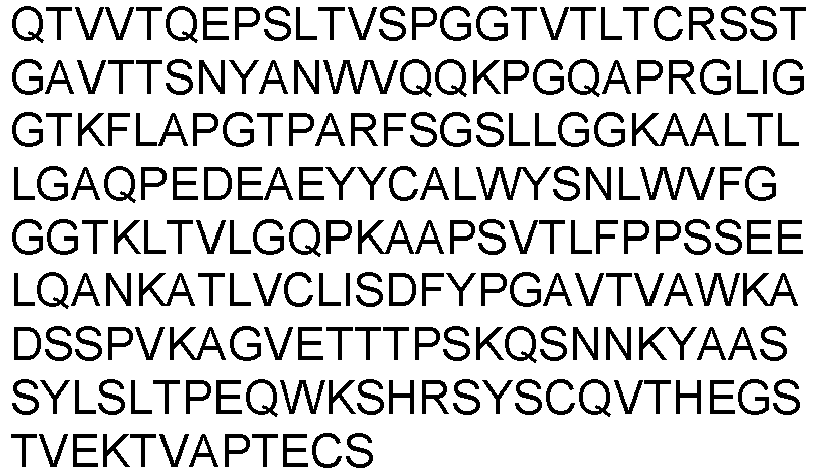
Heavy chain (lgG1) SEQ ID NO: 72 

Light chain (DNA) SEQ ID NO: 73 
Heavy chain (DNA, IgG1) SEQ ID NO: 74 
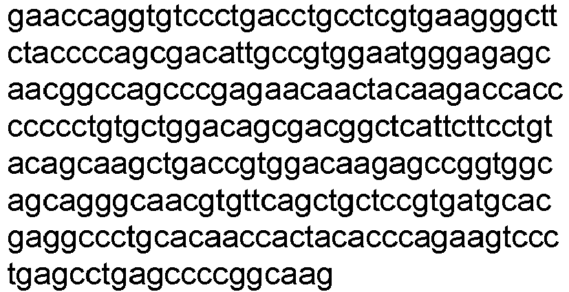
scFv (VL-linker-VH) (aa) SEQ ID NO: 75 
scFv (VL-linker-VH) (DNA) SEQ ID NO: 76 
Bremen HCDR1 (Kabat) SEQ ID NO: 23 TYAMN HCDR2 (Kabat) SEQ ID NO: 24 RIRSKYNNYATYYADSVKD HCDR3 (Kabat) SEQ ID NO: 25 HGNFGNSYVSWFAY LCDR1 (Kabat) SEQ ID NO: 26 RSSTGAVTTSNYAN LCDR2 (Kabat) SEQ ID NO: 65 GTKFLAP LCDR3 (Kabat) SEQ ID NO: 28 ALWYSNLWV VL SEQ ID NO: 77 
SEQ ID NO: 19 VH SEQ ID NO: 78 
SEQ ID NO: 7 VL (DNA) SEQ ID NO: 79 
SEQ ID NO: 20 VH (DNA) SEQ ID NO: 80 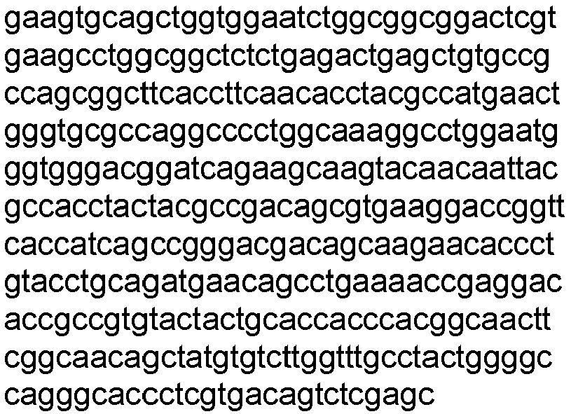
SEQ ID NO: 8 HCDR1 (Kabat) (DNA) SEQ ID NO: 33 acctacgccatg HCDR2 (Kabat) (DNA) SEQ ID NO: 34 
HCDR3 (Kabat) (DNA) SEQ ID NO: 35 
LCDR1 (Kabat (DNA) SEQ ID NO: 36 
LCDR2 (Kabat) (DNA) SEQ ID NO: 66 ggcacaaaatttctggcccct LCDR3 (Kabat) (DNA) SEQ ID NO: 38 gccctgtggtacagcaatctgtgggtg Light chain (IgG) SEQ ID NO: 81 

Heavy chain (IgG1) SEQ ID NO: 82 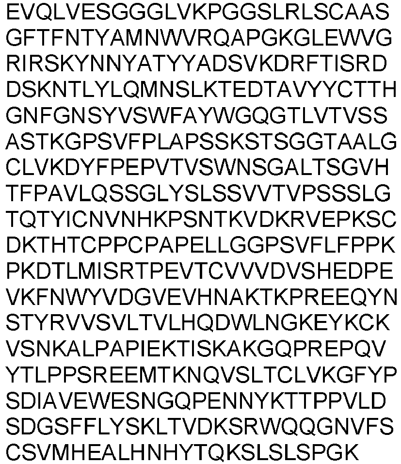
Light chain (DNA, IgG1) SEQ ID NO: 83 
Heavy chain (DNA, IgG1) SEQ ID NO: 84 
scFv (VL-linker-VH) SEQ ID NO: 156 
scFv (VL-linker-VH) DNA SEQ ID NO: 157 
Gladbach HCDR1 (Kabat) SEQ ID NO: 23 TYAMN HCDR2 (Kabat) SEQ ID NO: 24 RIRSKYNNYATYYADSVKD HCDR3 (Kabat) SEQ ID NO: 25 HGNFGNSYVSWFAY LCDR1 (Kabat) SEQ ID NO: 26 RSSTGAVTTSNYAN LCDR2 (Kabat) SEQ ID NO: 65 GTKFLAP LCDR3 (Kabat) SEQ ID NO: 28 ALWYSNLWV VL SEQ ID NO: 85 
SEQ ID NO: 19 VH SEQ ID NO: 86 
SEQ ID NO: 9 VL (DNA) SEQ ID NO: 87 
SEQ ID NO: 20 VH (DNA) SEQ ID NO: 88 
SEQ ID NO: 10 HCDR1 (Kabat) SEQ ID NO: 33 acctacgccatg HCDR2 (Kabat) SEQ ID NO: 34 
HCDR3 (Kabat) SEQ ID NO: 35 
LCDR1 (Kabat) SEQ ID NO: 36 
LCDR2 (Kabat) SEQ ID NO: 66 ggcacaaaatttctggcccct LCDR3 (Kabat) SEQ ID NO: 38 gccctgtggtacagcaatctgtgggtg Light chain (IgG) SEQ ID NO: 89 
Heavy chain (IgG1) SEQ ID NO: 90 
Light chain (DNA) SEQ ID NO: 91 

Heavy chain (DNA, IgG1) SEQ ID NO: 92 
scFv (VL-linker-VH) SEQ ID NO: 93 
scFv (VL-linker-VH) (DNA) SEQ ID NO: 94 
Nürnberg HCDR1 (Kabat) SEQ ID NO: 23 TYAMN HCDR2 (Kabat) SEQ ID NO: 24 RIRSKYNNYATYYADSVKD HCDR3 (Kabat) SEQ ID NO: 25 HGNFGNSYVSWFAY LCDR1 (Kabat) SEQ ID NO: 26 RSSTGAVTTSNYAN LCDR2 (Kabat) SEQ ID NO: 27 GTNKRAP LCDR3 (Kabat) SEQ ID NO: 28 ALWYSNLWV VL SEQ ID NO: 95 
SEQ ID NO: 21 VH SEQ ID NO: 96 
SEQ ID NO: 9 VL (DNA) SEQ ID NO: 97 
SEQ ID NO: 22 
VH (DNA) SEQ ID NO: 98 
SEQ ID NO: 10 HCDR1 (Kabat) SEQ ID NO: 33 acctacgccatg HCDR2 (Kabat) SEQ ID NO: 34 
HCDR3 (Kabat) SEQ ID NO: 35 
LCDR1 (Kabat) SEQ ID NO: 36 
LCDR2 (Kabat) SEQ ID NO: 37 ggcacaaacaaaagagcccct LCDR3 (Kabat) SEQ ID NO: 38 gccctgtggtacagcaatctgtgggtg Light chain (IgG) SEQ ID NO: 99 
Heavy chain (lgG1) SEQ ID NO: 100 
Light chain (DNA) SEQ ID NO: 101 
Heavy chain (DNA, IgG1) SEQ ID NO: 102 
scFv (VL-linker-VH) SEQ ID NO: 103 
scFv (VL-linker-VH) (DNA) SEQ ID NO: 104 
Table 6: Bispecific antibody sequences. Each of the listed antibodies shares the same IgG light chain of RefmAb#2_h_lgkappa (SEQ ID NO: 119 / 120) Antibody anti-Her2 [lgG1-AEASS] x anti-CD3 [scFv] SEQ ID NO: [aa] / DNA BsAb#Mainz RefmAb#2 (IgG1-AEASS_HC) x mAb#Mainz (scFv) SEQ ID NO: 105 
RefmAb#2 (IgG1-AEASS_HC) x mAb#Mainz (scFv) (DNA) SEQ ID NO: 106 

BsAb#Köln RefmAb#2 (IgG1f_AEASS_ HC) x mAb#Köln (scFv) SEQ ID NO: 107 
RefmAb#2 (IgG1f AEASS_ HC) x mAb#Köln (scFv) (DNA) SEQ ID NO: 108 

BsAb# Freiburg RefmAb#2 (IgG1f AEASS_ HC)x mAb#Freiburg (scFv) SEQ ID NO: 109 

RefmAb#2 (IgG1f AEASS_ HC) x mAb#Freiburg (scFv) (DNA) SEQ ID NO: 110 

BsAb# München RefmAb#2 (IgG1f AEASS_ HC) x mAb#München (scFv) SEQ ID NO: 111 
RefmAb#2 (IgG1f AEASS_ HC) x mAb#München (scFv) (DNA) SEQ ID NO: 112 

BsAb# Bremen RefmAb#2 (IgG1f AEASS_ HC) x SEQ ID NO: 113 
mAb#Bremen (scFv) 
RefmAb#2 (IgG1f AEASS_ HC) x mAb#Bremen (scFv) (DNA) SEQ ID NO: 114 

BsAb# Gladbach RefmAb#2 (IgG1f AEASS_ HC) x mAb#Gladbach (scFv) SEQ ID NO: 115 

RefmAb#2 (IgG1f AEASS_ HC) x mAb#Gladbach (scFv) (DNA) SEQ ID NO: 116 

BsAb# Nürnberg RefmAb#2 (IgG1f AEASS_ HC) x mAb#Nürnberg (scFv) SEQ ID NO: 117 
RefmAb#2 (IgG1f AEASS_ HC) x SEQ ID NO: 118 
mAb#Nürnberg (scFv) (DNA) 

IgG light chain RefmAb#2_h_lgk appa SEQ ID NO: 119 
RefmAb#2_h_lgk appa (DNA) SEQ ID NO: 120 
Table 7: Bispecific antibody sequences Antibody# anti-CD3 [IgG1f-AEASS_HC] x anti-HER2 [scFv] SEQ ID NO: [aa] / DNA BsAb# München_2 mAb# München (IgG1f_AEAS S_HC) x RefmAb#2 (scFv) SEQ ID NO: 121 
mAb# München_2 (IgG1f_AEAS S_HC) x RefmAb#2 (scFv) (DNA) SEQ ID NO: 122 

mAb# München (h_Iglambda) SEQ ID NO: 123 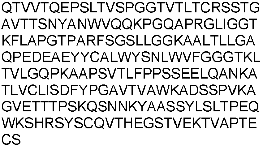
mAb# SEQ ID NO: 124 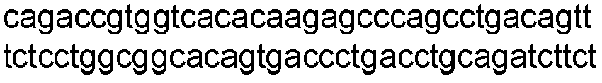
München (h_Iglambda) (DNA) 
BsAb# Bremen_2 mAb#Bremen (IgG1f_AEAS S_HC) x RefmAb#2 (scFv) SEQ ID NO: 125 
mAb#Bremen (IgG1f_AEAS S_HC) x RefmAb#2 (scFv) SEQ ID NO: 126 


mAb#Bremen (h_Iglambda) SEQ ID NO: 127 
mAb#Bremen (h_Iglambda) SEQ ID NO: 128 
BsAb# Mainz_2 mAb#Mainz (IgG1f_AEAS S_HC) x RefmAb#2 (scFv) SEQ ID NO: 129 
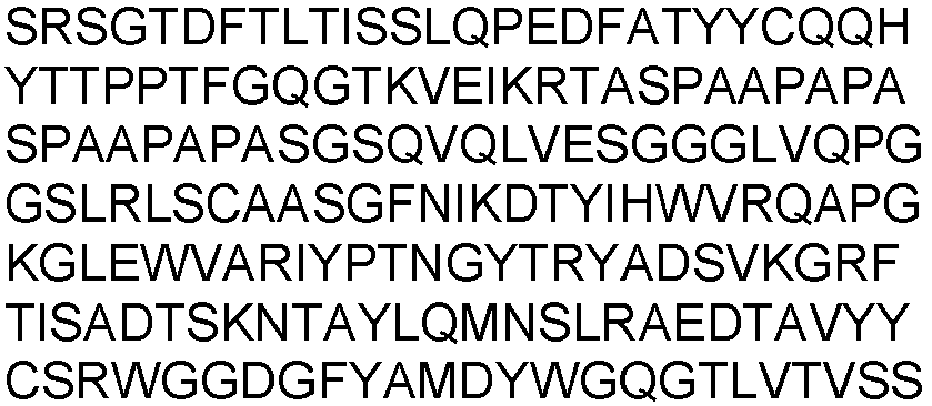
mAb#Mainz (IgG1f_AEAS S_HC) x RefmAb#2 (scFv) (DNA) SEQ ID NO: 130 
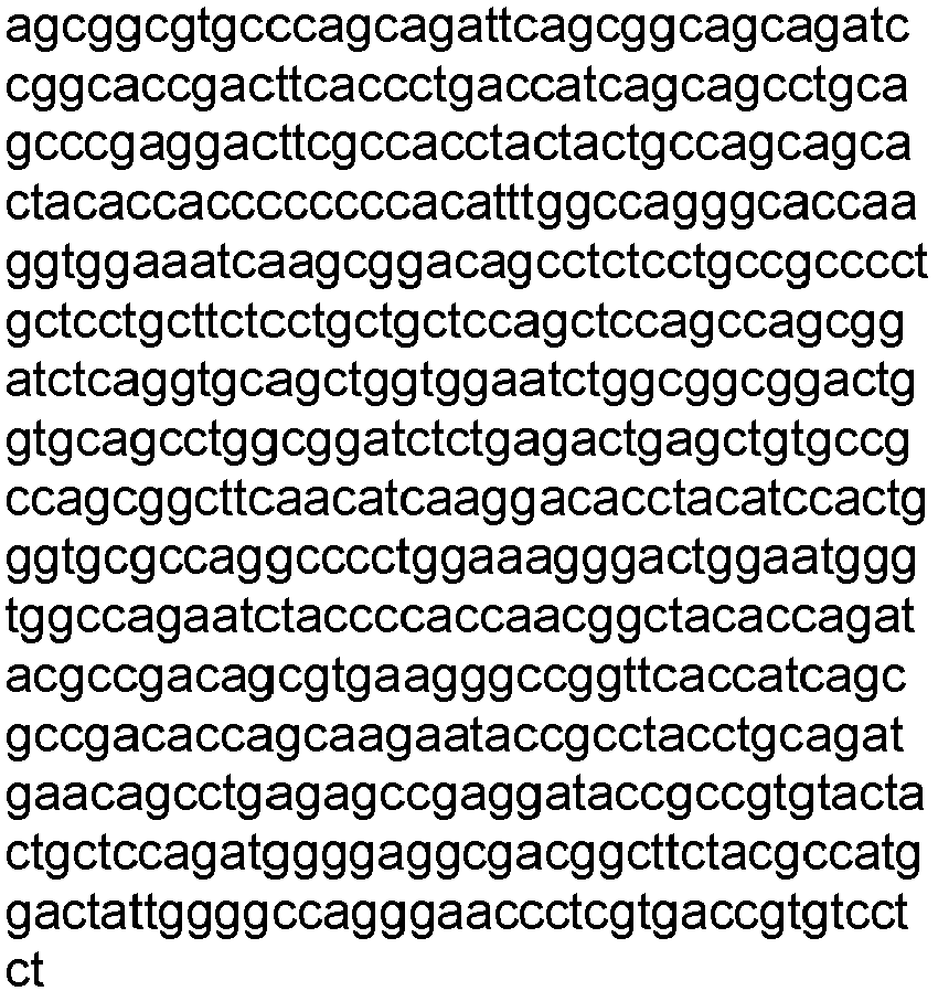
mAb#Mainz (h_Iglambda_ LC) SEQ ID NO: 131 
mAb#Mainz (h_lglambda_ LC) SEQ ID NO: 132 
BsAb# Köln_2 mAb#Köln (IgG1f_AEAS S_HC) x RefmAb#2 (scFv) SEQ ID NO: 133 
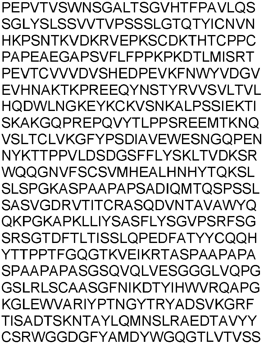
mAb#Köln (IgG1f_AEAS S_HC) x RefmAb#2 (scFv) (DNA) SEQ ID NO: 134 

mAb#Köln (h_Iglambda_ LC) SEQ ID NO: 135 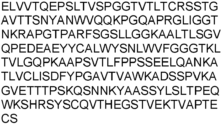
mAb#Köln (h_Iglambda_ LC) (DNA) SEQ ID NO: 136 

BsAb# Gladbach_2 mAb#Gladbac h(IgG1f_AEA SS_HC) x RefmAb#2 (scFv) SEQ ID NO: 137 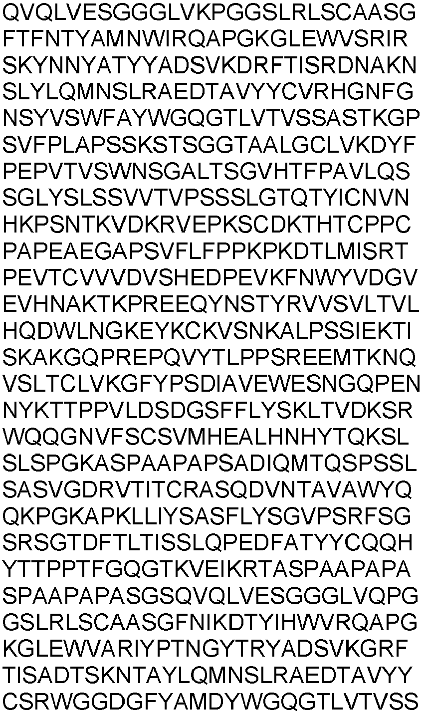
mAb#Gladbac h(IgG1f_AEA SS_HC) x RefmAb#2 (scFv) SEQ ID NO: 138 

mAb# Gladbach (h_Iglambda_ LC) SEQ ID NO: 139 

mAb# Gladbach (h_Iglambda_ LC) SEQ ID NO: 140 
BsAb# Nürnberg_2 mAb# Nürnberg (IgG1f_AEAS S_HC) x RefmAb#2 (scFv) SEQ ID NO: 141 
mAb# Nürnberg (IgG1f_AEAS S_HC) x RefmAb#2 (scFv) SEQ ID NO: 142 

mAb# Nürnberg (h_lglambda_ LC) SEQ ID NO: 143 
SEQ ID NO: 144 
BsAb# Freiburg_2 mAb# Freiburg (IgG1f_AEAS S_HC) x RefmAb#2 (scFv) SEQ ID NO: 145 
mAb#Freiburg (IgG1f_AEAS S_HC) x RefmAb#2 (scFv) SEQ ID NO: 146 

mAb# Freiburg (h_lglambda_ LC) SEQ ID NO: 147 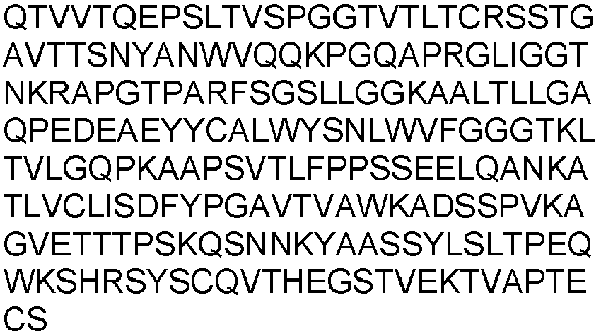
mAb#Freiburg (h_lglambda_ LC) SEQ ID NO: 148 
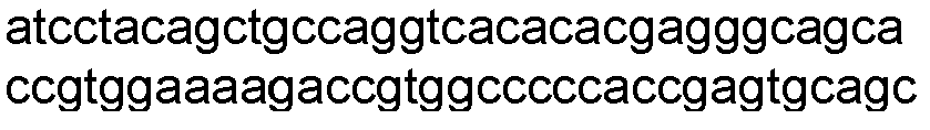
- Humanized derivatives of SP34 and variants thereof were prepared.
- The amino acid sequence of either the murine variable heavy chain (SEQ ID NO: 3) and the murine variable light chain (SEQ ID NO: 4) of SP34 were aligned to the repertoire of human VL or VH germline sequences (https://www.ncbi.nlm.nih.gov/igblast/) using the Vector NTI DNA analysis software.
- On the basis of this analysis, the human germline segments VL7-43 and VL7-46 were chosen as template sequences for the humanized SP34 VL variants. For the human VH, the human germline sequences VH3-15 and VH3-73 were chosen as template sequences.
- Selected amino acid residues that differed between the murine VL chain and the human Vlambda segment VL7-43 and VL7-46 or the murine VH chain and the human VH3-15 and VH3-73 chain within the framework regions were mutated on the DNA level towards the human residues. Potentially crucial framework residues of the original murine Vlambda or VH sequence were maintained.
- In sum, 4 distinct humanized VL amino acid sequences and 2 distinct humanized VH amino acid sequences were designed, whose CDRs were identical to the CDRs from the murine antibody SP34 VL and VH chain, respectively.
- In addition, for two of the designed VL sequences, additional amino acid substitutions at Kabat Positions 52-54 within the LCDR2 were introduced, namely N52K, K53F, R54L.
- An in silico T cell epitope screening for the obtained VH and VL sequences was done (Lonza, The Epibase™ In Silico tool). The screening tool allows for the identification of potential T cell epitopes in biotherapeutic protein and antibody targets. The tool uses structural characteristics of the HLA receptor along with experimentally determined binding affinities to predict potential peptide/HLA binding, a condition necessary for T cell activation
- An exemplary result of the in silico immunogenicity analysis for the VH and VL sequences of the humanized SP34 variant "München" is shown in Table 13. The analysis revealed a significant lower immunogenicity score for the VH and VL of the humanized variant in view of the parental murine antibody SP34.
Table 13: Immunogenic risk scores for Caucasian DRB1 alleles Sequence Risk Score (Caucasian) SP34_VL 621,4 München_VL 333,0 SP34_VH 953,0 München_VH 549,1 -
FIGURE 1 andFIGURE 2 depict the amino acid sequence alignments of the humanized VL and VH amino acid sequences in comparison to the corresponding murine SP34 sequences. - The amino acid sequences and encoding polynucleotide sequences of these humanized derivatives are shown in TABLE 3 and TABLE 4.
- The afore-mentioned humanized VH and VL regions of SP34 were gene synthesized by an external provider (GeneArt, ThermoFisher Scientific) and in-house cloned into a set of suitable prepared mammalian IgG expression vectors (two vector IgG expression system).
- Subsequently, Eukaryotic HKB-11 cells were transfected with the mammalian expression vector DNAs encoding the heavy and light chains of IgG, respectively. Cell culture supernatants were harvested on
day - Protein concentrations were determined by UV-spectrophotometry and purities of IgG were analyzed under denaturing, reducing and non-reducing conditions using CE-SDS (LabChip GXII | Perkin Elmer | USA). HP-SEC was performed to analyze IgG preparations in native state.
- Various control constructs (CD3 specific IgGs or HER2×CD3 bispecific Abs) were included in the following experiments for comparative purposes:
Nucleotide sequences encoding the VH and VL domains from "Trastuzumab" ((RefmAb#2); an antibody specific for HER2) as described by Baselga et al. 1998, Cancer Res 58(13): 2825-2831). - Nucleotide sequences encoding VH and VL domains from "SP34" a murine monoclonal antibody as described by Yoshino et al. (Exp. Anim. 49(2), 97-110, 2000) as well as
RefMAb# 1, an antibody disclosed inWO2008119566A2 . Both antibodies are reactive against the N-terminus of the epsilon chain of the T3 complex on human T lymphocyte cells. - All nucleotide sequences were gene synthesized as linear DNA fragments with appropriate flanking regions (e.g. suitable restriction enzyme recognition sites, linker sequences) either in-house or by an external provider. All synthesized DNA fragments were cloned into suited mammalian IgG expression vectors using standard molecular biology methods.
- The murine monoclonal antibody "OKT-3" (J Pediatr. 1982 Apr; 100(4):665-8) which also binds to the human epsilon chain of CD3 but to a distinct epitope as
RefMab# 1 and SP34 is commercially available and was purchased from Thermo Fisher (Catalog Nr.16-0037-85). - 7 humanized CD3 epsilon specific IgGs according to the present disclosure and two CD3 specific reference antibodies were tested for their ability to bind to recombinant human and cynomolgus CD3 epsilon in ELISA.
- 5 nM of recombinant human CD3e (22-49)-Fc2 (K105-K330) (SEQ ID NO: 149) or 1 nM of recombinant cynomolgus CD3e (22-49)-Fc2(K105-K330) (SEQ ID NO: 150) were coated on Maxisorp plates (Nunc, #460518). Coated plates were blocked with 5% skim milk in PBS. Antibodies were serially diluted in PBS containing 0.5% skim milk and 0.5% Tween-20. Bound antibodies were detected using an alkaline phosphatase-conjugated detection antibody directed against human F(ab')2 fragment (Jackson Immuno Research, #109-055-097). EC50 values were calculated using 4-parameter non-linear regression analysis in Prism software (GrapPad Software Inc., version 5.04).
- The results of the experiment are summarized in TABLE 8 and
FIGURE 3A and FIGURE 3B , and reveal that the humanized variants of the present disclosure exhibited equivalent or weaker binding to human or cynomolgus monkey CD3 epsilon when compared to SP34 orRefMab# 1.Table 8: ELISA binding EC50 [nM] IgGs hCD3e(22-49)_ Fc2(K105-K330) cyCD3e(22-49)_ Fc2(K105-K330) SP-34 (m/h chimeric) 0,39 0,15 RefmAb# 10,38 0,12 mAb#Mainz 1,21 1,53 mAb#Köln 0,31 0,20 mAb#Freiburg 0,91 1,26 mAb#München 0,60 1,43 mAb#Bremen 1,10 0,80 mAb#Gladbach 0,38 0,21 mAb#Nürnberg 0,34 0,21 - 7 humanized CD3 epsilon specific IgGs of the present disclosure were tested for their ability to bind to Jurkat cells (a CD3 positive human T-cell line) and to the corresponding CD3 negative cell line J.RT3-T3.5., a derivative mutant of the parental Jurkat leukemia cell.
- Jurkat and J.RT3-T3.5. cells were resuspended and counted in Superblock (ThermoScientific, #37515) and blocked for 1 h on ice. Blocked cells were resuspended in anti-CD3 antibodies serially diluted in Superblock and incubated for 1 h on ice. Cells were washed 2 times in D-PBS (Gibco) containing 3% fetal bovine serum (Sigma, #F7524). Bound antibodies were detected using R-Phycoerythrin-conjugated detection antibody directed against human F(ab')2 fragment (Jackson Immuno Research, #109-116-097). Antibody staining was measured using FACS Array (Beckton Dickinson) or IntelliCyt iQue flow cytometer and analyzed in FlowJo (version 7.6.5) or ForeCyt (version 4.1.5379, IntelliCyt) softwares, respectively. EC50 values were calculated using 4-parameter non-linear regression analysis in Prism software (GraphPad Software Inc., version 5.04).
- The results of the experiment are shown in TABLE 9 and
FIGURE 4A , and reveal that the humanized variants exhibited equivalent or weaker binding to Jurkat cells when compared to SP34 (m/h chimeric) or RefmAb#01. Furthermore, no or only very weak binding to the CD3 negative cell line J.RT3-T3.5. was observable (data no shown).Table 9: Cell binding IgG Jurkat EC50 [nM] SP34 (m/h chimeric) 1,4 RefmAb#01 1,1 mAb#Mainz 9,4 mAb#Köln 1,8 mAb#Freiburg 10,7 mAb#München 6,6 mAb#Bremen 6,3 mAb#Gladbach 1,4 mAb#Nürnberg 1,0 - High-capacity capture surfaces were prepared by covalently immobilizing MabSelect SuRe ligand (GE Healthcare, 28-4018-60) onto all flowcells of a CM5 chip (GE Healthcare, BR-100530) using EDC/NHS chemistry.
- Each cycle of the kinetic experiment consisted of capture steps (of different antibody ligands on the
specific flow cells - Capture levels of antibodies were adjusted to approx. 75 - 185 RU to achieve saturation levels Rmax of approx. 20-60 RU by the hCD3e analyte. Six different analyte concentrations were used for analysis during kinetic experiments (pMAX_hCD3e(1-118)_F-chLys_avi; applied molarities 4.1 - 999 nM, in a 3fold serial dilution series).
- Sensorgrams were evaluated with Biacore T200 Evaluation Software 3.0 (Biacore, GE Healthcare). All sensorgrams were fitted to a 1:1 binding model to determine kon and koff rate constants, which were used to calculate the KD value. For kinetic profiles deviating from the expected 1:1 binding, the sensorgrams were evaluated using a best approximation to the monovalent kinetics, and results marked with comment "heterogeneous binding". These results are considered less precise than kinetic profiles completely following the expected monovalent binding kinetics, but are assumed to be good approximations for KD.
- The results of this experiment are shown in TABLE 10 and reveal that SP34 and the humanized CD3 variants of the present disclosure exhibited equivalent or lower affinity binding to recombinant human CD3 epsilon.
Table 10: KD values of IgGs as determined on CD3 antigen pMAX_hCD3e(22-118)_F-chLys_avi IgG kon [1/Ms] koff [1/s] KD [nM] Comment KD restriction (yes/no) SP34 (m/h chimeric) 4,87E+05 8,35E-04 1,7 no RefmAb# 13,74E+05 4,91E-04 1,3 no mAb#Mainz 1,42E+04 8,47E-03 600 weak binding signals, only 333nM & 999 nM evaluated yes mAb#Köln 7,62E+04 3,86E-03 51 slightly heterogeneous binding no mAb#Freiburg 3,52E+04 4,67E-03 130 heterogeneous binding yes mAb#München 8,40E+04 6,58E-03 78 slightly heterogeneous binding no mAb#Bremen 6, 50E +04 4,79E-03 74 heterogeneous binding yes mAb#Gladbach 7,18E+04 3,85E-03 54 slightly heterogeneous binding no mAb#Nurnberg 8,00E+04 3,87E-03 48 only conc. 37 - 333nM used for evaluation yes - Bispecific antibodies according to the present disclosure were designed using an IgG-scFv format comprising an aglycosylated monoclonal IgG1 binding moiety and a scFv binding moiety, with the N-terminus of each scFv VL domain fused to the C-terminal end of each IgG heavy chain via a peptide linker. Both Fabs arms of the IgG bind to HER2 while the scFv is specific for CD3 (both scFv are identical and display the same specificity for CD3). Alternatively, bispecific antibodies were generated, wherein the two scFv molecules were specific for HER2 and the Fab arms of the IgG1 portion were specific for CD3.
- All constructs were generated using standard molecular methodologies as described in Example 3.
- A summary of the components of the antigen-binding domains of the various bispecific antibodies and of the bispecific antibodies made in accordance with this example is set forth in TABLE 5, 6 and 7.
- Subsequently, Eukaryotic HKB-11 cells were transfected with mammalian expression vectors with DNA encoding all component of the various bispecific antibodies. Cell culture supernatants were harvested on
day - Protein concentrations were determined by UV-spectrophotometry and purities of IgG were analyzed under denaturing, reducing and non-reducing conditions using CE-SDS (LabChip GXII | Perkin Elmer | USA). HP-SEC was performed to analyze IgG preparations in native state.
- Human whole blood from healthy donors (in house) and whole blood from cynomolgus monkey (retrieved from LPT Laboratory of Pharmacology and Toxicology, Hamburg, Germany) was collected in Li-Heparin containing S-Monovette containers (Sarstedt). Blood was transferred to 50ml conical tubes and mixed with an equal volume of PBS containing 2% fetal bovine serum (Sigma, #F7524) and 2 mM EDTA. Diluted blood was transferred to SepMate-50 tubes (StemCell Technologies, #86450) containing 15 ml Biocoll solution (Biochrom, #L6115) and centrifuged for 10 min at 1200 xg. Supernatant was transferred into a 50 ml conical tube, diluted to 45 ml with PBS and centrifuged for 8 min at 300 xg. Supernatant was discarded, cell pellet resuspended in 1 ml PBS and cells counted using a Neubauer chamber.
- 3 HER2 IgGxCD3 scFv bispecific antibodies of the present disclosure and one control bispecific antibody comprising the variable domains of
RefmAb# 1 were tested for their ability to bind to Jurkat cells expressing CD3 or to SKBR3 cells expressing high levels of the target antigen HER2. Experiments were done as described in Example 5. - The results of this experiment are summarized in TABLE 11 and reveal that the humanized variants of the present disclosure exhibited equivalent or weaker binding to Jurkat cells when compared to the control bispecific antibody comprising the Fab binding domains of
RefMab# 2 and the scFv binding domains ofRefMab# 1. In contrast, binding to SKBR3 cells remained unaffected.Table 11: Cell binding of HER2 × CD3 bsABs to Jurkat and SKBR3 cells FACS EC50 [nM] Bispecific antibody Jurkat SKBR3 BsAb#RefMab1 23 21 BsAb#Freiburg >100 15 BsAb#München 52 10 BsAb#Bremen 34 12 - 3 HER2 IgGxCD3 scFv bispecific antibodies of the present disclosure and a control bispecific antibody comprising the variable domains of
RefmAb# 1 were tested by FACS for their ability to bind to human and cynomolgus lymphocytes expressing CD3. - In addition, 7 humanized anti-CD3 epsilon specific IgGs of the present disclosure were tested for their ability to bind to human derived (n=3) and cynomolgus derived (n=1) PBMCs.
- 200,000 purified human or cynomolgus PBMCs were mixed with antibodies serially diluted (final concentration: 0.1 nM - 400 nM or 0.01 - 200 nM) in D-PBS (Gibco) containing 3% fetal bovine serum (Sigma, #F7524) and incubated for 1 h on ice. Bound antibodies were detected using R-Phycoerythrin-conjugated detection antibody directed against human F(ab')2 fragment (Jackson Immuno Research, #109-116-097). Antibody staining was measured using a FACS Array flow cytometer (Beckton Dickinson) and analyzed using FlowJo software (version 7.6.5). Lymphocytes were identified by morphological gating of forward and side scatters. EC50 values were calculated using 4-parameter non-linear regression analysis in Prism software (GraphPad Software Inc., version 5.04).
- The results of the experiments are shown in
FIGURE 4B ,FIGURE 5A and FIGURE 5B and reveal that the anti-CD3 IgGs and bispecific antibodies comprising the humanized SP34 variants of the present disclosure exhibited significant weaker binding to CD3 expressed on human or cynomolgus lymphocytes when compared to SP34 or to the control antibody comprising the single chain variable domains ofRefmAb# 1. - 3 HER2 IgGxCD3 scFv bispecific antibodies of the present disclosure were tested for their ability to activate human T cells derived from different from human blood samples of different donors in the absence of target cancer cells.
- Human PBMCs were purified as described above. Cells were resuspended to a density of 1E+07 cells/mL in RPMI 1640 medium (Gibco, #31870-025) supplemented with GlutaMax (Gibco, #35050-038), non-essential amino acids (Gibco, #11140-035), HEPES buffer bolution (Gibco #15630-056), sodium pyruvate (Gibco, #11360-039), β-mercaptoethanol, Penicillin/Streptomycin (Gibco #15140-122) and human serum (Sigma, #H4522) and incubated for 48 h at 37°C and 5% CO2. Following high-density preincubation, 200,000 PBMCs in medium were mixed with an equal volume of serially diluted antibodies (final concentration: 10nM, 0.1nM, 0.001nM) in medium and incubated for 24 h at 37°C and 5% CO2.
- Activation of T cells was assessed by evaluation of upregulation of CD69 expression on CD4 positive or CD8 positive T cells. For this, PBMCs were stained with antibodies to CD69, CD4 and CD8 conjugated with APC, APC/Cy7 and PE/Cy7, respectively (Biolegend, #310910/#300518/#344712). Antibody staining was measured using NovoCyte 3000 flow cytometer (Acea Biosciences, Inc.) and analyzed using FlowJo software (version 7.6.5).
- Results of the T cell activation experiments are shown in
FIGURE 6A (CD4 negative/CD8 positive T-cells) andFIGURE 6B (CD4 positive/CD8 negative T-cells) at an antibody concentration of 10 nM. The results and reveal a donor-dependent upregulation of CD69 expression on CD4 positive or on CD8 positive T-cells by the positive murine control IgG OKT-3 and the bispecific control constructbsAb_RefMab# 1 comprising scFv domains with the variable domains ofRefmAb# 1. - In contrast, no or very weak activation of T-cells was observable for the 3 HER2 IgGxCD3 scFv bispecific antibodies (BsAb#Freiburg, BsAb#München, BsAb#Bremen) in the absence of HER2 expressing target cells. This finding clearly demonstrating the preferred safety profile of the humanized CD3 specific antibodies of the present disclosure even when bivalently binding to CD3.
- 3 HER2 IgGxCD3 scFv bsAbs of the present disclosure were tested for their ability to mediate T cell dependent killing of HER2 tumor cell lines.
- 5,000 HER2-expressing SKBR3, A498 cells and HER2-negative MDA-MB468 cells were suspended in culture medium (SKBR3: McCoy's 5A Medium (Gibco, #26600), 10% FCS (Sigma, #F7524); MDA-MB468: DMEM (Gibco, #10938), GlutaMax (Gibco, #35050), 10% FCS, 1x Sodium Pyruvate (Gibco, #11360-039)), seeded in black 96 well assay plates (Corning, #3340) and incubated over night at 37°C and 5% CO2. CellToxGreen dye (Promega, #G8731), serially diluted bispecific antibody constructs (final concentration: 0.1 - 100 pM) and 100,000 purified PBMCs, all diluted in assay medium comprising RPMI 1640 w/o Phenol red (Gibco, #32404-014), GlutaMax and 10% fetal bovine serum, were added to the cells and incubated for 48 h at 37°C and 5% CO2.
- Cytotoxic activity was assessed by measuring incorporated CellToxGreen fluorescence at 485 nm excitation and 535 nm emission using a Tecan Infinite F500 device. EC50 values were calculated using 4-parameter non-linear regression analysis in Prism software (GraphPad Software Inc., version 5.04).
- Further evidence for T cell-mediated target cell killing was assessed by determined by secretion of the pro-inflammatory cytokine IFN-gamma using a commercially available kit (Human IFN-gamma Duo Set ELISA, R&D Systems, DY285).
- Results of the experiments for one exemplified doner are shown in
FIGURE 7 A-C and reveal T-cell mediated and antibody dose dependent killing of HER2 high expressing SKBR3 cells by the bispecific antibodies of the present disclosure equivalent to that of the controlantibody bsAb_RefMab# 1. - However, bispecific constructs of the present disclosure displayed a much weaker antibody dose dependent killing of HER2 low expressing A498 cells compared to that of the control
antibody bsAb_RefMab# 1. This clearly indicates a favorable safety profile for the antibodies of the present disclosure. The IC50 values were higher for the humanized antibodies displaying weaker binding to CD3 as compared to the controlantibody bsAb_RefMab# 1, most likely because of a decreased activation of CD8+ T cells (FIGURE 7B ). -
FIGURE 8 illustrates the antibody dose-dependent IFN-gamma release by T-cells in the presence of SKBR3 cells. The ELISA signals for bsAb#Freiburg and BsAb#refMAb# 1 at an antibody concentration of 100pM exceeded the detection limit. No IFN-gamma release could be observed in the presence of HER2 low expressing A498 cells (data not shown). This finding further supports the favorable safety profile of the humanized CD3 specific antibodies of the present disclosure. - 7 CD3 IgGxHER2 scFv bispecific antibodies of the present disclosure were tested for their ability to induce T-cell directed target cell killing as described in Example 12.
- Results of the experiments are depicted in
FIGURE 9A and FIGURE 9B and reveal that the T-cell-mediated dose dependent killing of HER2 high expressing SKBR3 cells is equivalent to that mediated by the controlantibody bsAb_RefMab# 1 but significant weaker on HER2 low expressing A498 cells. Again, no IFN-gamma release could be observed in the presence of HER2 low expressing A498 cells (data not shown). - These results clearly demonstrates that the favorable killing and safety properties of the bispecific antibodies of the present disclosure are independent from a particular CD3 format.
- Affinities for 3 HER2 IgGxCD3 scFv bispecific antibodies on recombinant human CD3 epsilon was determined as described in Example 6.
- As shown in TABLE 12, the bispecific antibodies comprising humanized CD3 specific binding domains according to the present disclosure bind to human CD3 epsilon with significant weaker affinity when compared to the affinity of control
antibody bsAb_RefMab# 1.Table 12: KD values of HER2 [IgG] × CD3 [scFv] bispecific antibodies determined on CD3 antigen pMAX_hCD3e(22-118)_F-chLys_avi Construct kon [1/Ms] koff [1/s] KD [nM] Comment KD restriction (yes/no) bsAb_RefMab# 13,63E+05 3,67E-04 1,0 no BsAb#Freiburg 3, 13E +04 3,36E-03 110 heterogeneous binding yes BsAb#München 3,65E+04 1,80E-03 49 slightly heterogeneous binding no BsAb#Bremen 6,15E+04 2,07E-03 34 slightly heterogeneous binding no
Claims (10)
- An isolated antibody or antibody fragment specific for cluster of differentiation 3 (CD3), wherein said antibody or antibody fragment specifically binds to human CD3 and to non-human primate CD3, wherein said antibody or antibody fragment comprises the variable heavy chain having SEQ ID NO: 7 and the variable light chain having SEQ ID NO: 17.
- The antibody or antibody fragment according to claim 1, wherein the antibody is a full-length IgG.
- The antibody or antibody fragment according to claims 1-2, wherein the antibody fragment is a Fab fragment.
- A bispecific antibody comprising a first antigen binding domain of an antibody or antibody fragment according to any of the preceding claims, and a second antigen binding domain which binds a different target antigen than said first antigen binding region.
- A bispecific antibody according claim 4, wherein the second binding region specifically binds a cell surface target antigen.
- A nucleic acid composition comprising a nucleic acid sequence or a plurality of nucleic acid sequences encoding the antibody or antibody fragment according to any one of the preceding claims.
- A vector composition comprising a vector or a plurality of vectors comprising the nucleic acid sequence or plurality of nucleic acid sequences of claim 6.
- A host cell comprising the vector composition of claim 7
- The antibody or antibody fragment according to any one of the preceding claims for use as a medicament.
- A pharmaceutical composition comprising the antibody or antibody fragment according to any one of the preceding claims and a pharmaceutically acceptable carrier or excipient.
Applications Claiming Priority (2)
| Application Number | Priority Date | Filing Date | Title |
|---|---|---|---|
| EP17186128 | 2017-08-14 | ||
| PCT/EP2018/071872 WO2019034580A1 (en) | 2017-08-14 | 2018-08-13 | Humanized antibodies for cd3 |
Publications (2)
| Publication Number | Publication Date |
|---|---|
| EP3668898A1 EP3668898A1 (en) | 2020-06-24 |
| EP3668898B1 true EP3668898B1 (en) | 2023-07-05 |
Family
ID=59626511
Family Applications (1)
| Application Number | Title | Priority Date | Filing Date |
|---|---|---|---|
| EP18765358.9A Active EP3668898B1 (en) | 2017-08-14 | 2018-08-13 | Humanized antibodies for cd3 |
Country Status (3)
| Country | Link |
|---|---|
| US (1) | US11472880B2 (en) |
| EP (1) | EP3668898B1 (en) |
| WO (1) | WO2019034580A1 (en) |
Families Citing this family (19)
| Publication number | Priority date | Publication date | Assignee | Title |
|---|---|---|---|---|
| ME03675B (en) * | 2013-07-05 | 2020-10-20 | Genmab As | HUMANIZED OR CHIMERAL ANTI-CD3 ANTIBODIES |
| US10544220B2 (en) * | 2015-01-08 | 2020-01-28 | Genmab A/S | Bispecific antibodies against CD3 and CD20 |
| KR102239781B1 (en) * | 2019-04-08 | 2021-04-13 | 주식회사 녹십자 | Bispecific antibody that specifically binds to gpnmb and cd3 and use thereof |
| MX2021014973A (en) * | 2019-06-07 | 2022-04-18 | Adimab Llc | HIGH AFFINITY ANTI-CD3 ANTIBODIES AND METHODS FOR THEIR GENERATION AND USE. |
| WO2021141996A1 (en) * | 2020-01-06 | 2021-07-15 | Cytomx Therapeutics, Inc. | Single- and multi-chain polypeptides that bind specifically to cd3 epsilon |
| MX2022013797A (en) | 2020-05-08 | 2022-11-30 | Genmab As | Bispecific antibodies against cd3 and cd20. |
| CN116249714A (en) | 2020-05-27 | 2023-06-09 | 詹森生物科技公司 | Proteins comprising CD3 antigen-binding domains and uses thereof |
| JP7518286B2 (en) | 2020-08-25 | 2024-07-17 | ギリアード サイエンシーズ, インコーポレイテッド | Multispecific antigen-binding molecules targeting HIV and methods of use |
| US20230357398A1 (en) * | 2020-09-24 | 2023-11-09 | Morphosys Ag | Novel human antibodies binding to human cd3 epsilon |
| WO2022135536A1 (en) * | 2020-12-25 | 2022-06-30 | 江苏先声药业有限公司 | Cd3 humanized antibody and application thereof |
| EP4380631A4 (en) * | 2021-08-04 | 2025-06-25 | Abpro Corporation | Antibodies directed against CD3 and their uses |
| CN117980888A (en) * | 2021-08-04 | 2024-05-03 | 阿布普罗公司 | Anti-HER2 antibodies and their uses |
| KR20240109605A (en) * | 2021-09-13 | 2024-07-11 | 산둥심시어 바이오파마슈티칼 씨오., 엘티디. | Anti-human CD3 antibody and its applications |
| WO2023164513A2 (en) * | 2022-02-23 | 2023-08-31 | Janux Therapeutics, Inc. | Optimized antibodies targeting trop2 and uses thereof |
| WO2023180346A1 (en) * | 2022-03-22 | 2023-09-28 | Morphosys Ag | Deimmunized antibodies specific for cd3 |
| WO2024152963A1 (en) * | 2023-01-18 | 2024-07-25 | 贝达药业股份有限公司 | Humanized anti-cd3 antibody and use thereof |
| WO2024236167A1 (en) | 2023-05-17 | 2024-11-21 | Morphosys Ag | Il-13 receptor alpha 2 binding polypeptides |
| CN118027196B (en) * | 2023-06-25 | 2025-12-02 | 合肥天港免疫药物有限公司 | Anti-CD3 antibodies and their applications |
| WO2026006494A1 (en) * | 2024-06-25 | 2026-01-02 | Alloy Therapeutics, Inc. | Anti-cd3 antibodies and uses thereof |
Family Cites Families (18)
| Publication number | Priority date | Publication date | Assignee | Title |
|---|---|---|---|---|
| US5641870A (en) | 1995-04-20 | 1997-06-24 | Genentech, Inc. | Low pH hydrophobic interaction chromatography for antibody purification |
| EP1958962A3 (en) | 1997-06-12 | 2013-05-01 | Novartis International Pharmaceutical Ltd. | Artificial antibody polypeptides |
| EP1940881B1 (en) | 2005-10-11 | 2016-11-30 | Amgen Research (Munich) GmbH | Compositions comprising cross-species-specific antibodies and uses thereof |
| RS53008B2 (en) | 2007-04-03 | 2022-12-30 | Amgen Res Munich Gmbh | Cross-species-specific cd3-epsilon binding domain |
| MX2009010611A (en) | 2007-04-03 | 2010-03-26 | Micromet Ag | Cross-species-specific bispecific binders. |
| WO2012158818A2 (en) | 2011-05-16 | 2012-11-22 | Fabion Pharmaceuticals, Inc. | Multi-specific fab fusion proteins and methods of use |
| KR102060389B1 (en) | 2011-05-21 | 2019-12-31 | 마크로제닉스, 인크. | Cd3-binding molecules capable of binding to human and non-human cd3 |
| DK3424953T3 (en) * | 2011-06-06 | 2020-11-02 | Novo Nordisk As | Terapeutiske antistoffer |
| US10968276B2 (en) * | 2013-03-12 | 2021-04-06 | Xencor, Inc. | Optimized anti-CD3 variable regions |
| US10131710B2 (en) * | 2013-01-14 | 2018-11-20 | Xencor, Inc. | Optimized antibody variable regions |
| KR102211837B1 (en) | 2013-01-14 | 2021-02-03 | 젠코어 인코포레이티드 | Novel heterodimeric proteins |
| ME03675B (en) | 2013-07-05 | 2020-10-20 | Genmab As | HUMANIZED OR CHIMERAL ANTI-CD3 ANTIBODIES |
| JP2016538275A (en) | 2013-11-04 | 2016-12-08 | グレンマーク ファーマシューティカルズ, エセ.アー. | Production of T-cell retargeting heterodimeric immunoglobulins |
| EP2982693A1 (en) | 2014-08-07 | 2016-02-10 | Affimed Therapeutics AG | CD3 binding domain |
| MA40609B1 (en) | 2014-09-05 | 2020-05-29 | Janssen Pharmaceutica Nv | Cd123 binding agents and their uses |
| US10544220B2 (en) * | 2015-01-08 | 2020-01-28 | Genmab A/S | Bispecific antibodies against CD3 and CD20 |
| TW201720458A (en) | 2015-12-04 | 2017-06-16 | 宏觀基因股份有限公司 | Bi-specific monovalent diabodies that are capable of binding CD19 and CD3, and uses thereof |
| KR102891788B1 (en) | 2016-02-04 | 2025-11-26 | 더 스크립스 리서치 인스티튜트 | Humanized anti-CD3 antibodies, conjugates and uses thereof |
-
2018
- 2018-08-13 WO PCT/EP2018/071872 patent/WO2019034580A1/en not_active Ceased
- 2018-08-13 EP EP18765358.9A patent/EP3668898B1/en active Active
- 2018-08-13 US US16/635,303 patent/US11472880B2/en active Active
Also Published As
| Publication number | Publication date |
|---|---|
| US20200377594A1 (en) | 2020-12-03 |
| US11472880B2 (en) | 2022-10-18 |
| WO2019034580A1 (en) | 2019-02-21 |
| EP3668898A1 (en) | 2020-06-24 |
Similar Documents
| Publication | Publication Date | Title |
|---|---|---|
| EP3668898B1 (en) | Humanized antibodies for cd3 | |
| JP2023504016A (en) | Antibodies to CD3 and BCMA and bispecific binding proteins made therefrom | |
| JP7596263B2 (en) | Antibody constructs that bind to 4-1BB and tumor-associated antigens and uses thereof | |
| JPWO2018117237A1 (en) | Anti-CD3 antibody and molecule containing the antibody | |
| KR20230170672A (en) | Anti-CD122 antibodies and uses thereof | |
| US20230357398A1 (en) | Novel human antibodies binding to human cd3 epsilon | |
| TW202304997A (en) | Novel anti-cd4 antibody | |
| CN115461366A (en) | Anti-OX40 antibody and use thereof | |
| AU2023240297A1 (en) | Deimmunized antibodies specific for cd3 | |
| US20250382362A1 (en) | Antibodies targeting il3 | |
| US20230265202A1 (en) | Antibody constructs binding 4-1bb and folate receptor alpha and uses thereof | |
| WO2024133890A1 (en) | Antibodies targeting cd45 | |
| RU2852663C1 (en) | Antibodies against cd3 and bcma and bisspecific binding proteins derived from them |
Legal Events
| Date | Code | Title | Description |
|---|---|---|---|
| STAA | Information on the status of an ep patent application or granted ep patent |
Free format text: STATUS: UNKNOWN |
|
| STAA | Information on the status of an ep patent application or granted ep patent |
Free format text: STATUS: THE INTERNATIONAL PUBLICATION HAS BEEN MADE |
|
| PUAI | Public reference made under article 153(3) epc to a published international application that has entered the european phase |
Free format text: ORIGINAL CODE: 0009012 |
|
| STAA | Information on the status of an ep patent application or granted ep patent |
Free format text: STATUS: REQUEST FOR EXAMINATION WAS MADE |
|
| 17P | Request for examination filed |
Effective date: 20200313 |
|
| AK | Designated contracting states |
Kind code of ref document: A1 Designated state(s): AL AT BE BG CH CY CZ DE DK EE ES FI FR GB GR HR HU IE IS IT LI LT LU LV MC MK MT NL NO PL PT RO RS SE SI SK SM TR |
|
| AX | Request for extension of the european patent |
Extension state: BA ME |
|
| DAV | Request for validation of the european patent (deleted) | ||
| DAX | Request for extension of the european patent (deleted) | ||
| STAA | Information on the status of an ep patent application or granted ep patent |
Free format text: STATUS: EXAMINATION IS IN PROGRESS |
|
| 17Q | First examination report despatched |
Effective date: 20210721 |
|
| GRAP | Despatch of communication of intention to grant a patent |
Free format text: ORIGINAL CODE: EPIDOSNIGR1 |
|
| STAA | Information on the status of an ep patent application or granted ep patent |
Free format text: STATUS: GRANT OF PATENT IS INTENDED |
|
| INTG | Intention to grant announced |
Effective date: 20230131 |
|
| GRAS | Grant fee paid |
Free format text: ORIGINAL CODE: EPIDOSNIGR3 |
|
| GRAA | (expected) grant |
Free format text: ORIGINAL CODE: 0009210 |
|
| STAA | Information on the status of an ep patent application or granted ep patent |
Free format text: STATUS: THE PATENT HAS BEEN GRANTED |
|
| P01 | Opt-out of the competence of the unified patent court (upc) registered |
Effective date: 20230516 |
|
| AK | Designated contracting states |
Kind code of ref document: B1 Designated state(s): AL AT BE BG CH CY CZ DE DK EE ES FI FR GB GR HR HU IE IS IT LI LT LU LV MC MK MT NL NO PL PT RO RS SE SI SK SM TR |
|
| REG | Reference to a national code |
Ref country code: CH Ref legal event code: EP |
|
| REG | Reference to a national code |
Ref country code: AT Ref legal event code: REF Ref document number: 1584782 Country of ref document: AT Kind code of ref document: T Effective date: 20230715 |
|
| REG | Reference to a national code |
Ref country code: DE Ref legal event code: R096 Ref document number: 602018052865 Country of ref document: DE |
|
| REG | Reference to a national code |
Ref country code: IE Ref legal event code: FG4D |
|
| REG | Reference to a national code |
Ref country code: LT Ref legal event code: MG9D |
|
| REG | Reference to a national code |
Ref country code: NL Ref legal event code: MP Effective date: 20230705 |
|
| REG | Reference to a national code |
Ref country code: AT Ref legal event code: MK05 Ref document number: 1584782 Country of ref document: AT Kind code of ref document: T Effective date: 20230705 |
|
| PG25 | Lapsed in a contracting state [announced via postgrant information from national office to epo] |
Ref country code: NL Free format text: LAPSE BECAUSE OF FAILURE TO SUBMIT A TRANSLATION OF THE DESCRIPTION OR TO PAY THE FEE WITHIN THE PRESCRIBED TIME-LIMIT Effective date: 20230705 |
|
| PG25 | Lapsed in a contracting state [announced via postgrant information from national office to epo] |
Ref country code: GR Free format text: LAPSE BECAUSE OF FAILURE TO SUBMIT A TRANSLATION OF THE DESCRIPTION OR TO PAY THE FEE WITHIN THE PRESCRIBED TIME-LIMIT Effective date: 20231006 |
|
| PG25 | Lapsed in a contracting state [announced via postgrant information from national office to epo] |
Ref country code: ES Free format text: LAPSE BECAUSE OF FAILURE TO SUBMIT A TRANSLATION OF THE DESCRIPTION OR TO PAY THE FEE WITHIN THE PRESCRIBED TIME-LIMIT Effective date: 20230705 |
|
| PG25 | Lapsed in a contracting state [announced via postgrant information from national office to epo] |
Ref country code: IS Free format text: LAPSE BECAUSE OF FAILURE TO SUBMIT A TRANSLATION OF THE DESCRIPTION OR TO PAY THE FEE WITHIN THE PRESCRIBED TIME-LIMIT Effective date: 20231105 |
|
| PG25 | Lapsed in a contracting state [announced via postgrant information from national office to epo] |
Ref country code: SE Free format text: LAPSE BECAUSE OF FAILURE TO SUBMIT A TRANSLATION OF THE DESCRIPTION OR TO PAY THE FEE WITHIN THE PRESCRIBED TIME-LIMIT Effective date: 20230705 Ref country code: RS Free format text: LAPSE BECAUSE OF FAILURE TO SUBMIT A TRANSLATION OF THE DESCRIPTION OR TO PAY THE FEE WITHIN THE PRESCRIBED TIME-LIMIT Effective date: 20230705 Ref country code: PT Free format text: LAPSE BECAUSE OF FAILURE TO SUBMIT A TRANSLATION OF THE DESCRIPTION OR TO PAY THE FEE WITHIN THE PRESCRIBED TIME-LIMIT Effective date: 20231106 Ref country code: NO Free format text: LAPSE BECAUSE OF FAILURE TO SUBMIT A TRANSLATION OF THE DESCRIPTION OR TO PAY THE FEE WITHIN THE PRESCRIBED TIME-LIMIT Effective date: 20231005 Ref country code: LV Free format text: LAPSE BECAUSE OF FAILURE TO SUBMIT A TRANSLATION OF THE DESCRIPTION OR TO PAY THE FEE WITHIN THE PRESCRIBED TIME-LIMIT Effective date: 20230705 Ref country code: LT Free format text: LAPSE BECAUSE OF FAILURE TO SUBMIT A TRANSLATION OF THE DESCRIPTION OR TO PAY THE FEE WITHIN THE PRESCRIBED TIME-LIMIT Effective date: 20230705 Ref country code: IS Free format text: LAPSE BECAUSE OF FAILURE TO SUBMIT A TRANSLATION OF THE DESCRIPTION OR TO PAY THE FEE WITHIN THE PRESCRIBED TIME-LIMIT Effective date: 20231105 Ref country code: HR Free format text: LAPSE BECAUSE OF FAILURE TO SUBMIT A TRANSLATION OF THE DESCRIPTION OR TO PAY THE FEE WITHIN THE PRESCRIBED TIME-LIMIT Effective date: 20230705 Ref country code: GR Free format text: LAPSE BECAUSE OF FAILURE TO SUBMIT A TRANSLATION OF THE DESCRIPTION OR TO PAY THE FEE WITHIN THE PRESCRIBED TIME-LIMIT Effective date: 20231006 Ref country code: FI Free format text: LAPSE BECAUSE OF FAILURE TO SUBMIT A TRANSLATION OF THE DESCRIPTION OR TO PAY THE FEE WITHIN THE PRESCRIBED TIME-LIMIT Effective date: 20230705 Ref country code: ES Free format text: LAPSE BECAUSE OF FAILURE TO SUBMIT A TRANSLATION OF THE DESCRIPTION OR TO PAY THE FEE WITHIN THE PRESCRIBED TIME-LIMIT Effective date: 20230705 Ref country code: AT Free format text: LAPSE BECAUSE OF FAILURE TO SUBMIT A TRANSLATION OF THE DESCRIPTION OR TO PAY THE FEE WITHIN THE PRESCRIBED TIME-LIMIT Effective date: 20230705 |
|
| PG25 | Lapsed in a contracting state [announced via postgrant information from national office to epo] |
Ref country code: PL Free format text: LAPSE BECAUSE OF FAILURE TO SUBMIT A TRANSLATION OF THE DESCRIPTION OR TO PAY THE FEE WITHIN THE PRESCRIBED TIME-LIMIT Effective date: 20230705 |
|
| REG | Reference to a national code |
Ref country code: DE Ref legal event code: R097 Ref document number: 602018052865 Country of ref document: DE |
|
| PG25 | Lapsed in a contracting state [announced via postgrant information from national office to epo] |
Ref country code: LU Free format text: LAPSE BECAUSE OF NON-PAYMENT OF DUE FEES Effective date: 20230813 |
|
| PG25 | Lapsed in a contracting state [announced via postgrant information from national office to epo] |
Ref country code: SM Free format text: LAPSE BECAUSE OF FAILURE TO SUBMIT A TRANSLATION OF THE DESCRIPTION OR TO PAY THE FEE WITHIN THE PRESCRIBED TIME-LIMIT Effective date: 20230705 Ref country code: RO Free format text: LAPSE BECAUSE OF FAILURE TO SUBMIT A TRANSLATION OF THE DESCRIPTION OR TO PAY THE FEE WITHIN THE PRESCRIBED TIME-LIMIT Effective date: 20230705 Ref country code: LU Free format text: LAPSE BECAUSE OF NON-PAYMENT OF DUE FEES Effective date: 20230813 Ref country code: EE Free format text: LAPSE BECAUSE OF FAILURE TO SUBMIT A TRANSLATION OF THE DESCRIPTION OR TO PAY THE FEE WITHIN THE PRESCRIBED TIME-LIMIT Effective date: 20230705 Ref country code: DK Free format text: LAPSE BECAUSE OF FAILURE TO SUBMIT A TRANSLATION OF THE DESCRIPTION OR TO PAY THE FEE WITHIN THE PRESCRIBED TIME-LIMIT Effective date: 20230705 Ref country code: CZ Free format text: LAPSE BECAUSE OF FAILURE TO SUBMIT A TRANSLATION OF THE DESCRIPTION OR TO PAY THE FEE WITHIN THE PRESCRIBED TIME-LIMIT Effective date: 20230705 Ref country code: MC Free format text: LAPSE BECAUSE OF FAILURE TO SUBMIT A TRANSLATION OF THE DESCRIPTION OR TO PAY THE FEE WITHIN THE PRESCRIBED TIME-LIMIT Effective date: 20230705 Ref country code: SK Free format text: LAPSE BECAUSE OF FAILURE TO SUBMIT A TRANSLATION OF THE DESCRIPTION OR TO PAY THE FEE WITHIN THE PRESCRIBED TIME-LIMIT Effective date: 20230705 |
|
| REG | Reference to a national code |
Ref country code: BE Ref legal event code: MM Effective date: 20230831 |
|
| PLBE | No opposition filed within time limit |
Free format text: ORIGINAL CODE: 0009261 |
|
| STAA | Information on the status of an ep patent application or granted ep patent |
Free format text: STATUS: NO OPPOSITION FILED WITHIN TIME LIMIT |
|
| REG | Reference to a national code |
Ref country code: IE Ref legal event code: MM4A |
|
| PG25 | Lapsed in a contracting state [announced via postgrant information from national office to epo] |
Ref country code: IT Free format text: LAPSE BECAUSE OF FAILURE TO SUBMIT A TRANSLATION OF THE DESCRIPTION OR TO PAY THE FEE WITHIN THE PRESCRIBED TIME-LIMIT Effective date: 20230705 |
|
| 26N | No opposition filed |
Effective date: 20240408 |
|
| PG25 | Lapsed in a contracting state [announced via postgrant information from national office to epo] |
Ref country code: IE Free format text: LAPSE BECAUSE OF NON-PAYMENT OF DUE FEES Effective date: 20230813 |
|
| PG25 | Lapsed in a contracting state [announced via postgrant information from national office to epo] |
Ref country code: IE Free format text: LAPSE BECAUSE OF NON-PAYMENT OF DUE FEES Effective date: 20230813 Ref country code: SI Free format text: LAPSE BECAUSE OF FAILURE TO SUBMIT A TRANSLATION OF THE DESCRIPTION OR TO PAY THE FEE WITHIN THE PRESCRIBED TIME-LIMIT Effective date: 20230705 |
|
| PG25 | Lapsed in a contracting state [announced via postgrant information from national office to epo] |
Ref country code: BE Free format text: LAPSE BECAUSE OF NON-PAYMENT OF DUE FEES Effective date: 20230831 |
|
| PG25 | Lapsed in a contracting state [announced via postgrant information from national office to epo] |
Ref country code: BG Free format text: LAPSE BECAUSE OF FAILURE TO SUBMIT A TRANSLATION OF THE DESCRIPTION OR TO PAY THE FEE WITHIN THE PRESCRIBED TIME-LIMIT Effective date: 20230705 |
|
| PG25 | Lapsed in a contracting state [announced via postgrant information from national office to epo] |
Ref country code: BG Free format text: LAPSE BECAUSE OF FAILURE TO SUBMIT A TRANSLATION OF THE DESCRIPTION OR TO PAY THE FEE WITHIN THE PRESCRIBED TIME-LIMIT Effective date: 20230705 |
|
| PG25 | Lapsed in a contracting state [announced via postgrant information from national office to epo] |
Ref country code: CY Free format text: LAPSE BECAUSE OF FAILURE TO SUBMIT A TRANSLATION OF THE DESCRIPTION OR TO PAY THE FEE WITHIN THE PRESCRIBED TIME-LIMIT; INVALID AB INITIO Effective date: 20180813 |
|
| PG25 | Lapsed in a contracting state [announced via postgrant information from national office to epo] |
Ref country code: HU Free format text: LAPSE BECAUSE OF FAILURE TO SUBMIT A TRANSLATION OF THE DESCRIPTION OR TO PAY THE FEE WITHIN THE PRESCRIBED TIME-LIMIT; INVALID AB INITIO Effective date: 20180813 |
|
| PGFP | Annual fee paid to national office [announced via postgrant information from national office to epo] |
Ref country code: DE Payment date: 20250722 Year of fee payment: 8 |
|
| PGFP | Annual fee paid to national office [announced via postgrant information from national office to epo] |
Ref country code: GB Payment date: 20250717 Year of fee payment: 8 |
|
| PGFP | Annual fee paid to national office [announced via postgrant information from national office to epo] |
Ref country code: FR Payment date: 20250721 Year of fee payment: 8 |
|
| PGFP | Annual fee paid to national office [announced via postgrant information from national office to epo] |
Ref country code: CH Payment date: 20250901 Year of fee payment: 8 |
|
| PG25 | Lapsed in a contracting state [announced via postgrant information from national office to epo] |
Ref country code: TR Free format text: LAPSE BECAUSE OF FAILURE TO SUBMIT A TRANSLATION OF THE DESCRIPTION OR TO PAY THE FEE WITHIN THE PRESCRIBED TIME-LIMIT Effective date: 20230705 |