EP3663975B1 - Verfahren und system zum lernen von visuellem pixelkontext aus objekteigenschaften zur erzeugung reicher semantischer bilder - Google Patents
Verfahren und system zum lernen von visuellem pixelkontext aus objekteigenschaften zur erzeugung reicher semantischer bilder Download PDFInfo
- Publication number
- EP3663975B1 EP3663975B1 EP19213622.4A EP19213622A EP3663975B1 EP 3663975 B1 EP3663975 B1 EP 3663975B1 EP 19213622 A EP19213622 A EP 19213622A EP 3663975 B1 EP3663975 B1 EP 3663975B1
- Authority
- EP
- European Patent Office
- Prior art keywords
- pixel
- pixels
- tiles
- objects
- class
- Prior art date
- Legal status (The legal status is an assumption and is not a legal conclusion. Google has not performed a legal analysis and makes no representation as to the accuracy of the status listed.)
- Active
Links
Images








Classifications
-
- G—PHYSICS
- G06—COMPUTING OR CALCULATING; COUNTING
- G06V—IMAGE OR VIDEO RECOGNITION OR UNDERSTANDING
- G06V20/00—Scenes; Scene-specific elements
- G06V20/60—Type of objects
- G06V20/69—Microscopic objects, e.g. biological cells or cellular parts
- G06V20/695—Preprocessing, e.g. image segmentation
-
- G—PHYSICS
- G06—COMPUTING OR CALCULATING; COUNTING
- G06F—ELECTRIC DIGITAL DATA PROCESSING
- G06F18/00—Pattern recognition
- G06F18/20—Analysing
- G06F18/24—Classification techniques
- G06F18/243—Classification techniques relating to the number of classes
- G06F18/24323—Tree-organised classifiers
-
- G—PHYSICS
- G06—COMPUTING OR CALCULATING; COUNTING
- G06T—IMAGE DATA PROCESSING OR GENERATION, IN GENERAL
- G06T7/00—Image analysis
- G06T7/0002—Inspection of images, e.g. flaw detection
- G06T7/0012—Biomedical image inspection
-
- G—PHYSICS
- G06—COMPUTING OR CALCULATING; COUNTING
- G06T—IMAGE DATA PROCESSING OR GENERATION, IN GENERAL
- G06T7/00—Image analysis
- G06T7/10—Segmentation; Edge detection
- G06T7/162—Segmentation; Edge detection involving graph-based methods
-
- G—PHYSICS
- G06—COMPUTING OR CALCULATING; COUNTING
- G06V—IMAGE OR VIDEO RECOGNITION OR UNDERSTANDING
- G06V10/00—Arrangements for image or video recognition or understanding
- G06V10/20—Image preprocessing
- G06V10/25—Determination of region of interest [ROI] or a volume of interest [VOI]
-
- G—PHYSICS
- G06—COMPUTING OR CALCULATING; COUNTING
- G06V—IMAGE OR VIDEO RECOGNITION OR UNDERSTANDING
- G06V10/00—Arrangements for image or video recognition or understanding
- G06V10/40—Extraction of image or video features
- G06V10/46—Descriptors for shape, contour or point-related descriptors, e.g. scale invariant feature transform [SIFT] or bags of words [BoW]; Salient regional features
- G06V10/462—Salient features, e.g. scale invariant feature transforms [SIFT]
-
- G—PHYSICS
- G06—COMPUTING OR CALCULATING; COUNTING
- G06V—IMAGE OR VIDEO RECOGNITION OR UNDERSTANDING
- G06V10/00—Arrangements for image or video recognition or understanding
- G06V10/40—Extraction of image or video features
- G06V10/56—Extraction of image or video features relating to colour
-
- G—PHYSICS
- G06—COMPUTING OR CALCULATING; COUNTING
- G06V—IMAGE OR VIDEO RECOGNITION OR UNDERSTANDING
- G06V10/00—Arrangements for image or video recognition or understanding
- G06V10/70—Arrangements for image or video recognition or understanding using pattern recognition or machine learning
- G06V10/764—Arrangements for image or video recognition or understanding using pattern recognition or machine learning using classification, e.g. of video objects
-
- G—PHYSICS
- G06—COMPUTING OR CALCULATING; COUNTING
- G06V—IMAGE OR VIDEO RECOGNITION OR UNDERSTANDING
- G06V10/00—Arrangements for image or video recognition or understanding
- G06V10/70—Arrangements for image or video recognition or understanding using pattern recognition or machine learning
- G06V10/77—Processing image or video features in feature spaces; using data integration or data reduction, e.g. principal component analysis [PCA] or independent component analysis [ICA] or self-organising maps [SOM]; Blind source separation
- G06V10/772—Determining representative reference patterns, e.g. averaging or distorting patterns; Generating dictionaries
-
- G—PHYSICS
- G06—COMPUTING OR CALCULATING; COUNTING
- G06V—IMAGE OR VIDEO RECOGNITION OR UNDERSTANDING
- G06V20/00—Scenes; Scene-specific elements
- G06V20/60—Type of objects
- G06V20/69—Microscopic objects, e.g. biological cells or cellular parts
- G06V20/698—Matching; Classification
-
- G—PHYSICS
- G06—COMPUTING OR CALCULATING; COUNTING
- G06V—IMAGE OR VIDEO RECOGNITION OR UNDERSTANDING
- G06V10/00—Arrangements for image or video recognition or understanding
- G06V10/40—Extraction of image or video features
- G06V10/44—Local feature extraction by analysis of parts of the pattern, e.g. by detecting edges, contours, loops, corners, strokes or intersections; Connectivity analysis, e.g. of connected components
- G06V10/443—Local feature extraction by analysis of parts of the pattern, e.g. by detecting edges, contours, loops, corners, strokes or intersections; Connectivity analysis, e.g. of connected components by matching or filtering
- G06V10/449—Biologically inspired filters, e.g. difference of Gaussians [DoG] or Gabor filters
- G06V10/451—Biologically inspired filters, e.g. difference of Gaussians [DoG] or Gabor filters with interaction between the filter responses, e.g. cortical complex cells
- G06V10/454—Integrating the filters into a hierarchical structure, e.g. convolutional neural networks [CNN]
-
- G—PHYSICS
- G06—COMPUTING OR CALCULATING; COUNTING
- G06V—IMAGE OR VIDEO RECOGNITION OR UNDERSTANDING
- G06V10/00—Arrangements for image or video recognition or understanding
- G06V10/40—Extraction of image or video features
- G06V10/50—Extraction of image or video features by performing operations within image blocks; by using histograms, e.g. histogram of oriented gradients [HoG]; by summing image-intensity values; Projection analysis
Definitions
- the present invention relates generally to analyzing target patterns in digital images, and more specifically to a method and computer-implemented system for detecting and measuring those target patterns.
- Cancer is typically diagnosed by analyzing stained samples of tissue from cancer patients and then correlating target patterns in the tissue samples with grading and scoring methods for different kinds of cancers.
- the Gleason grading system indicates the malignancy of prostate cancer based on the architectural pattern of the glands of a stained prostate tumor.
- breast cancer can be diagnosed by grading stained breast tissue using the Allred score, the Elston-Ellis score or the HercepTestTM score.
- the Allred score indicates the severity of cancer based on the percentage of cells that have been stained to a certain intensity by the estrogen receptor (ER) antibody.
- the Elston-Ellis score indicates the severity of cancer based on the proportion of tubules in the tissue sample, the similarity of nucleus sizes and the number of dividing cells per high power field of 400x magnification.
- the HercepTestTM score indicates of the severity of cancer based on the level of HER2 protein overexpresssion as indicated by the degree of membrane staining.
- the Fuhrman nuclear grading system indicates the severity of renal cell carcinoma (RCC) based on the morphology of the nuclei of kidney cells.
- the various cancer scoring and grading systems can deliver inconsistent results because even an experienced pathologist may misjudge the target patterns and structures in the stained tissue due to fatigue and loss of concentration. Therefore, computer-assisted image analysis systems have been developed to support pathologists in the tedious task of grading and scoring digital images of the stained tissue samples.
- the digital images are rectangular arrays of pixels. Each pixel is characterized by its position in the array and a plurality of numerical pixel values associated with the pixel.
- the pixel values represent color or grayscale information for various image layers. For example, grayscale digital images are represented by a single image layer, whereas RGB images are represented by three color image layers.
- semantic network systems segment, classify and quantify objects present in the images by generating semantic networks that link pixel values to data objects according to class networks.
- the image analysis systems that apply semantic networks perform object-oriented analysis, as opposed to solely statistical pixel-oriented analysis. Consequently, semantic network systems classify not just pixels, but also the data objects linked to the pixels.
- the data objects that are linked to the pixels and to one another represent information about the digital images.
- US 2014 / 0 140 610 A1 discloses techniques for unsupervised object class discovery and the techniques may retrieve a set of training images for object recognition model learning.
- object-oriented image analysis can provide better results for cancer scoring and grading systems than can pixel-oriented analysis alone, object-oriented analysis is also more computationally involved. Therefore, object-oriented analysis is often slower than statistical pixel-oriented analysis alone. Particularly in digital pathology where each tissue slide can generate gigapixels of data, performing a full-scale object-oriented analysis is too time-consuming.
- a method is sought that retains the advantages of object-oriented analysis, yet enhances the performance of analysis systems based on computer-implemented semantic networks. Such a method would efficiently manage the computational resources of the object-oriented image analysis systems.
- An image analysis method in accordance with an aspect of the present invention uses both object-oriented analysis and the faster pixel-oriented analysis to recognize patterns in a digital image of a stained tissue slice.
- Object-oriented image analysis is used to segment a small portion of the image into object classes. Then the object class to which each pixel in the remainder of the image most probably belongs is determined using decision trees that include pixelwise descriptors. The pixels of the remainder of the image are assigned object classes without segmenting the remainder of the image into objects of a semantic network. After the small portion of the image is segmented into object classes, class characteristics of each of the classes of objects are determined.
- the pixelwise descriptors as applied in decision trees describe which pixels are associated with particular classes of objects by matching the characteristics of the object classes to the comparison between pixels at predetermined offsets.
- a pixel heat map is generated by giving each pixel the color assigned to the class of objects that the pixelwise descriptors indicate is most probably associated with that pixel.
- An image analysis method trains decision trees of pixelwise descriptors to indicate the probability that individual pixels in one portion of an image exhibit object characteristics determined by segmenting another portion of the image using the more computationally intensive object-oriented image analysis.
- a high-resolution digital image of stained tissue is first divided into tiles, and the degree of local contrast in each of the tiles is determined.
- a first plurality of the tiles is selected that exhibits the greatest degree of local contrast.
- the average color of each of the first plurality of tiles is determined.
- the first plurality of tiles is then divided into clusters of tiles having similar colors.
- a learning tile from each color cluster of tiles is selected. Each learning tile has the greatest degree of local contrast from among the tiles of the color cluster to which the learning tile belongs.
- the learning tiles are then segmented into objects using computationally intensive object-oriented image analysis that generates rich semantic images.
- the objects are classified into classes of objects, and a color is associated with each class of objects.
- Characteristics of the objects that belong to distinct object classes are determined. Some examples of the object characteristics are: the average number of concavities of the objects of the class, the average size of the objects of the class, the variation in sizes among the objects of the class, the amount of intensity variation within the objects of the class, the elliptical fit of the objects of the class, and the average intensity of the objects of the class.
- Pixelwise descriptors in decision trees are generated that indicate the class of objects to which each characterized pixel of the learning tiles most probably belongs by matching the object characteristics to a comparison between the characterized pixel and a second pixel at a predetermined offset.
- the comparison between pixels provides the visual context for matching each pixel to an object class.
- Examples of the pixelwise descriptors include: the difference between a color value of the characterized pixel and of a second pixel at a predetermined offset, the average of the color values of pixels in a box of predetermined size at a predetermined offset from the characterized pixel, the standard deviation among the color values of pixels in a box of predetermined size at a predetermined offset from the characterized pixel, the sum of gradient magnitude of the color values of pixels in a box of predetermined size at a predetermined offset from the characterized pixel, the orientation of a gradient edge at a predetermined offset, and the color value of a second pixel at a predetermined offset.
- a pixel heat map is generated in which each pixel is assigned the color associated with the class of objects to which that pixel most probably belongs.
- the pixel heat map is generated without again segmenting the digital image into objects.
- the pixel heat map is then displayed on a graphical user interface.
- object characteristics are determined of objects segmented in a first portion of an image using object-oriented image analysis.
- Pixelwise descriptors then describe which of the object characteristics that a characterized pixel most probably exhibits based on a quality of a second pixel at a predetermined offset from the characterized pixel.
- each characterized pixel is matched to the most probable of selected object characteristics as opposed to the most probable of selected object classes.
- a pixel heat map is generated by applying the pixelwise descriptors to each pixel in a second portion of the image without segmenting the second portion of the image into objects.
- Each pixel of the second portion of the image has the color assigned to the object characteristic most probably exhibited by that pixel.
- a higher level heat map is generated from the pixel heat map that assigns a color to each pixel depending on the degree to which an object characteristic is most likely exhibited at the location of the pixel.
- An average value of multiple pixels of the pixel heat map can be combined to form a single pixel of the higher level heat map.
- the shade of each pixel of the higher level heat map indicates an area of the original image in which the objects exhibit an object characteristic to a particular degree. For example, the shade of each pixel of the higher level heat map could indicate a location in the original image in which cells have a higher proportion of their membranes immunohistochemically stained.
- the shade of each pixel of the higher level heat map could indicate a location in the original image in which the nuclei have an above-average size.
- pixels of the higher level heat map most probably belonging to areas in the original image containing larger nuclei are assigned a particular color.
- the present invention provides methods as defined in any one of claims 1 to 8.
- the present invention also provides a system according to claim 9 and a computer readable medium according to claim 10.
- FIG. 1 shows a system 10 for analyzing digital images using both statistical pixel-oriented analysis and object-oriented analysis that links pixels to data objects forming hierarchical networks.
- System 10 is used to analyze images of tissue slices stained with various biomarkers, such as tissue stained with hematoxylin and eosin (H&E) or tissue stained with a protein-specific antibody using immunohistochemistry (IHC).
- Digital images 11 of the stained tissue slices are acquired at high magnification.
- a typical digital image of a tissue slice has a resolution of 100,000 x 200,000 pixels, or 20 billion pixels.
- the acquired digital images 11 are stored in a database 12 of digital images.
- Image analysis software executing on a data analysis server 13 then performs intelligent image processing and automated classification and quantification.
- the image analysis software is a computer program product tangibly embodied on a computer-readable storage medium in server 13 and comprises computer readable and executable program instructions that when executed by a processor on server 13 provide a visual display on a graphical user interface 14 of an interconnected display device 15, such as a personal computer.
- the image analysis software transforms unlinked input data in the form of pixels into a hierarchical semantic network of objects.
- System 10 analyzes, grades, scores and displays the digital images 11 of tissue slices that have been stained with various biomarkers.
- the image analysis program segments and classifies objects in the digital images 11.
- the program prepares links between some objects and thereby generates higher hierarchically ranked objects.
- the image analysis program provides the higher hierarchically ranked objects with properties, classifies them, and then links those objects again at a still higher level to other objects.
- the higher hierarchically ranked objects are used to find target patterns in the images. More easily detected starting data objects are first found and then used to identify harder-to-find data objects in the hierarchical data structure.
- FIG. 2 illustrates exemplary hierarchical network 16 that is generated by image analysis system 10.
- System 10 generates first objects 17 from a digital image 18 based on the stained tissue.
- the image analysis program of system 10 uses object-oriented image analysis to generate data objects of hierarchical semantic network 16 by linking selected pixels 19 to data objects according to a classification network and according to a process hierarchy of steps and algorithms.
- Each digital image comprises pixel values associated with the locations of each of the pixels 19.
- the image analysis program operates on the digital pixel values and links the pixels to form objects. Each object is linked to a set of pixel locations based on the associated pixel values.
- An object is generated by linking to the object those pixels having similar characteristics, such as hue, saturation and brightness as defined by the pixel values.
- the pixel values can be expressed in a 3-value color space.
- three 3 digit numbers in the range from zero to 255 define the color.
- the three numbers represent the amounts of red, green and blue in the represented color.
- red is represented as 255-0-0
- dark green is represented as 0-100-0
- royal blue is designated as 65-105-225
- white is represented as 255-255-255
- black is represented as 0-0-0.
- Smaller numbers represent darker colors, so 100-100-100 is a darker gray than 200-200-200, and 0-0-128 is a darker blue (navy) than straight blue 0-0-255.
- CMYK cyan, magenta, yellow, black
- CIE 1931 color space
- 1964 xyz color space the 1964 xyz color space
- HSV and HSL representation of the RGB color space.
- Thresholds of brightness at pixel locations that are grouped together can be obtained from a histogram of the pixel values in the digital image.
- the pixels form the lowest hierarchical level of hierarchical network 16.
- pixels having the color and intensity imparted by the biomarker stain are identified and linked to first objects 17.
- the first objects 17 form the second hierarchical level of hierarchical network 16.
- data objects are linked together into classes according to membership functions of the classes defined in the class network.
- objects representing nuclei are linked together to form objects 20-21 in a third hierarchical level of hierarchical network 16.
- some of the first objects 17 correspond to stained pixels of the nuclear membrane of a nucleus corresponding to object 20.
- another of the first objects 17 corresponds to stained pixels on the nuclear membrane of a separate nucleus represented by object 21.
- An additional object 22 is generated in a fourth hierarchical level of hierarchical network 16 and is linked to all of the objects that represent stained nuclei.
- the objects 20-21 corresponding to stained nuclei are linked to object 22.
- the knowledge and the program flow of the image analysis program are separated in the software structure.
- the parameters by which the image analysis is performed for example thresholds of size or brightness, can be changed without having to revise the process hierarchy of software steps.
- the image analysis software displays both the original digital images 11 as well as the corresponding processed images and heat maps on the graphical user interface 14. Pixels corresponding to classified and segmented objects in the digital images are colored, marked or highlighted to correspond to their classification. For example, the pixels of objects that are members of the same class are depicted in the same color.
- FIG. 3 is a flowchart of steps 25-38 of a method 24 by which analysis system 10 recognizes patterns in digital images of stained tissue slices using both object-oriented analysis and pixel-oriented analysis.
- a high-resolution digital image is acquired of a tissue slice that has been stained with a biomarker.
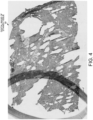
- FIG. 4 shows an exemplary digital image 46 of kidney tissue that has been stained with hematoxylin and eosin (H&E). A slice of the kidney tissue has been placed on a slide before the digital image was scanned. The image includes an artifact 47 of a pen mark in which an area of the tissue has been circled.
- H&E hematoxylin and eosin
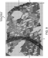
- step 26 high-resolution digital image 46 is divided into tiles 48.
- FIG. 5 shows how digital image 46 is displayed in tiled sections 48 on graphical user interface 14 of system 10 after step 26 is performed.
- system 10 determines the degree of local contrast in each of the tiles 48.
- an intensity threshold is set at the average intensity of the pixels in each tile. The pixels are divided into a first group whose intensities are above the threshold and a second group whose intensities are below the threshold. Regions of contiguous pixels in the first and in the second groups are then identified. Then the intensity threshold is incrementally changed and the regions of contiguous pixels in the two groups are again identified. The regions that remain stable despite the change in the intensity threshold are defined as maximally stable extremal regions (MSER). The ratio of the area covered by stable regions (MSER) in each tile is determined. A higher ratio of stable regions indicates a greater degree of local contrast.
- FIG. 6 shows how the tiles 48 are displayed on graphical user interface 14 to represent the degree of local contrast in each tile.
- the darker areas 49 in FIG. 6 indicate the location of maximally stable extremal regions (MSER) that remain despite an incremental change in the intensity threshold.
- MSER maximally stable extremal regions
- the darker areas 49 indicating a greater degree of local contrast tend to contain more relevant and a wider variety of tissue types.
- a first plurality 50 of the tiles 48 are selected that exhibit the greatest degree of local contrast.
- the tiles covered by the greatest ratio of darker areas 39 and that contain the darkest darker areas are selected as the first plurality of tiles 50.
- 212 of the tiles are marked with an "X" to indicate the first plurality of tiles.
- the remaining unmarked tiles contain less interesting tissue that is less likely to include representative amounts of all tissue types.
- the average color of each of the first plurality of tiles 50 is determined.
- Each of the tiles 50 is a matrix of pixel locations associated with numerical pixel values.
- the pixel values represent the color of each pixel in the RGB color space and include three 3 digit numbers in the range from zero to 255.
- the three numbers represent the amounts of red, green and blue in the color of the pixel.
- the average red number, green number and blue number are calculated.
- the three average numbers represent a point in RGB space.
- a point in RGB space is determined for each of the 212 tiles in the first plurality of tiles 50.
- the 212 points in RGB space form a cloud.
- the colors of the tiles can be determined using lower resolution versions of the first plurality of tiles 50. Multiple pixels of each of the tiles 50 are represented by a single pixel of each lower resolution tile.
- the first plurality of tiles 50 (or the lower resolution versions of these tiles) is divided into clusters of tiles with similar colors.
- the cloud of points in RGB space is divided into five areas of concentration of the points.
- the points in each of the five areas correspond to color clusters of the tiles 50.
- FIG. 8 shows a matrix of tile locations in which each of the first plurality of tiles 50 is displayed in a shade of gray corresponding to one of the five color clusters. For example, sixteen of the 212 first plurality of tiles 50 are in the color cluster represented by white.
- a learning tile is selected from each cluster of tiles. From each color cluster of tiles, the tile is selected that has the highest proportion of stable regions. Thus, each learning tile exhibits the greatest degree of local contrast from among the tiles of the cluster to which the learning tile belongs.
- the five learning tiles from each of the five color clusters are labeled 51-55.
- tile 55 is the tile that exhibits the greatest degree of local contrast from among the tiles in the color cluster represented by white.
- FIG. 9 shows the locations on digital image 46 of learning tiles 51-55 as well as the locations of the remainder of the first plurality of tiles 50, which are marked with Xs.
- step 32 the learning tiles 51-55 are segmented into data objects using object-oriented image analysis.
- step 33 the data objects are classified into classes of objects.
- a color is associated with each class of objects.
- a class network is first defined, and colors are associated with the defined classes. Then the pixels are linked to subobjects, and the subobjects are combined into objects that correspond with the classes in the class network.
- the image analysis program analyzes and measures patterns present in the pixels using a computer-implemented network structure.
- the network structure includes a hierarchical data network, a class network and a process hierarchy.
- the data objects of the hierarchical data network are generated by linking selected pixels of the learning tiles 51-55 to the data objects according to the classification network using the steps and algorithms of the process hierarchy.
- object-oriented processing a data object is formed based on whether a condition would be satisfied if a particular pixel were to be linked to the object. For example, whether a pixel is linked to a data object can depend on the shape or size of the object that would result if the pixel were included in the object.
- Whether a pixel is linked to an object can also depend on the average brightness of all of the pixels associated with the object that would result if the particular pixel were to be included in the object.
- the objects are combined in the hierarchical data network to form higher level objects, which are classified into classes of the class network.
- Object-oriented image analysis can produce better segmentation than pixel-oriented analysis because subobjects can be combined in alternative ways, and the best result can then be chosen.
- Multiple segmentation strategies can be simultaneously pursued by concurrently computing alternative object classifications (segmentation of objects into classes) using several cores of a multi-core computer. Combining subobjects in alternative ways to produce the best overall segmentation is not possible with solely pixel-oriented processing in which no hierarchical data network is generated.
- FIG. 10 is a screenshot of graphical user interface 14 on which the five learning tiles 51-55 are displayed side-by-side in the top row.
- Object-oriented image analysis has been performed on the learning tiles 51-55, and data objects have been segmented and classified into the classes in the class network 56 at the right side of graphical user interface 14. The results of the segmentation are shown in the row of images below the row of learning tiles.
- pixels are linked to subobjects, which are combined to form data objects that are classified into morphologically accurate categories.
- the class network 56 defines a subclass of nuclei as being "TexturedNuclei" 57.
- System 10 has accurately identified particular data objects as belonging to the class of textured nuclei 57.
- System 10 has associated a color with each of the classes and subclasses in the class network 56 at the right side of graphical user interface 14.
- Each of the data objects that belongs to the class of textured nuclei 57 is depicted in the lower row of images with the color of the subclass assigned by the class network.
- FIG. 10 also shows objects in the lower row of images that are displayed with the color of the subclass "HomogeneousNuclei" 58 assigned by the class network 57.
- system 10 has also accurately identified particular data objects as belonging to the class of homogeneous nuclei 58. It is very difficult with the naked eye to distinguish the nuclei in learning tile 55 that contain textured DNA from those whose DNA is more homogeneously disbursed. However, the homogeneous and textured nuclei can easily be distinguished from one another in the segmented image below learning tile 55 because the two subclasses of nuclei are assigned different colors.
- the elliptical fit of an object is the deviation from a circular or elliptical shape of the object.
- a long object would have a poor elliptical fit.
- An object with multiple branches would also have a poor elliptical fit, but that object would also be characterized by its number of concavities or indentations between the branches.
- Object-oriented image analysis can be programmed to recognize objects having subobjects with a selected color. For example, objects can be segmented into nuclei whose perimeters have the selected color of a biomarker or stain.
- system 10 generates pixelwise descriptors that indicate the object class to which each pixel of the learning tiles 51-55 most probably belongs.
- the pixelwise descriptors indicate the most likely object class associated with each pixel without referencing any data objects. Instead, purely pixel-oriented image analysis is performed using the descriptors.
- the pixelwise descriptors indicate the probability that a characterized pixel belongs to a class of objects based on a characteristic of a second pixel or group of pixels at a predetermined offset from the characterized pixel.
- the pixelwise descriptors are used in random forest decision trees to indicate the probability that each pixel of each learning tile belongs to a particular class of objects.
- FIG. 11 is a schematic diagram illustrating how pixelwise descriptors 59-64 are applied in one of the random forest decision trees to determine the probability that a pixel belongs to one of three exemplary object classes.
- the selected classes are: nuclei with homogeneous contents (h), nuclei with textured contents (tx) (due to dense regions of chromatin) and background objects (b).
- System 10 trains on random pixels from the learning tiles 51-55 in order to match the class indicated by object-oriented analysis by choosing the appropriate pixelwise descriptors and coefficients of those descriptors to use in pixel-oriented analysis.
- System 10 matches the class indicated by object-oriented analysis by choosing the type of pixelwise descriptors, the order in which those descriptors are applied in the decision trees, the location of the pixels that are being compared and the comparison threshold used to make each decision.
- each pixel is first analyzed by pixelwise descriptor 59.
- Descriptor 59 determines the average red value of the pixels in a 6x13 box of pixels that is offset from the characterized pixel by two pixels in the y dimension (0,2).
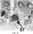
- FIG. 12 illustrates the characterized pixel 66 and the box 67 of pixels whose lower left corner is offset from characterized pixel 66 by zero pixels in the x dimension and two pixels in the y dimension.
- Pixel 66 belongs to one of the textured nuclei 57 in the segmented image below learning tile 55 in FIG. 10 .
- the average red value of the pixels in box 67 is less than the threshold value of 142.9. Therefore, the analysis proceeds along the branch of the decision tree to pixelwise descriptor 60.
- Descriptor 60 determines the average blue value of the pixels in a 2x1 box 68 of pixels that is offset from characterized pixel 66 by two pixels in the x dimension and one pixel in the y dimension.
- FIG. 12 also shows box 68 used for the determination of the blue value of the pixels.
- the average blue value of the pixels in box 68 is less than the threshold value of 119.1, so the analysis proceeds along the branch of the decision tree to pixelwise descriptor 62.
- Descriptor 62 determines the average green value of the pixels in a 1x4 box 69 of pixels that is offset from characterized pixel 66 by one pixel in the x dimension and four pixels in the y dimension.
- the average green value of the pixels in box 69 is greater than the threshold value of 39.1, so the decision tree of pixelwise descriptors indicates that characterized pixel 66 most probably belongs to the class of objects associated with nuclei that have textured contents.
- the decision tree has been correctly trained to indicate the object class determined by object-oriented segmentation.
- the decision tree of pixelwise descriptors outputs the posterior probabilities that each pixel belongs to a selected group of object classes, in this example textured nuclei (tx), homogeneous nuclei (h) and background objects (b).
- the output probabilities are normalized so that the sum of the probabilities of belonging to a class within the selected object classes is 100%.
- the decision tree indicates that the probability P(tx) that characterized pixel 66 belongs to the object class of textured nuclei 57 is 60%.
- This pixel-oriented classification corresponds to the result of the object-oriented segmentation shown in FIG. 10 in the segmented image below learning tile 55. Characterized pixel 66 belongs to the textured nucleus 57 at the end of an arrow in FIG. 10 .
- the decision tree predicts that characterized pixel 66 has a 38% probability P(h) of belonging to the object class of homogeneous nuclei and a 2% probability P(b) of belonging to an object class of background objects.
- nineteen other decision trees of pixelwise descriptors are also trained to predict that other random training pixels in the learning tiles have the greatest probability of belonging to those object classes indicated by object-oriented segmentation.
- Each random forest decision tree of pixelwise descriptors is trained so that, for all of the training pixels of the learning tiles, the same order of descriptors with the same offsets, boxes, thresholds and other coefficients output a highest probability object class that matches the object class determined through object-oriented image analysis in steps 32-33.
- the parameters of each decision tree are modified during the training mode until the predicted object class for each randomly selected training pixel matches the class of the object to which the training pixel belongs in the hierarchical network 16 generated by object-oriented pixel analysis.
- the best match is achieved when the highest probability class for all of the selected training pixels is correct, and those indicated probabilities are closest to 100%.
- the parameters that are modified to achieve the best match are (i) the comparison threshold at each pixelwise descriptor, (ii) the offset of the pixels being compared, (iii) the size and shape of the box of pixels being compared, (iv) the quality of the pixels that is being compared (e.g., average color value), and (v) the order in which the pixelwise descriptors are placed in each decision tree.
- the pixelwise descriptors can be more complex than merely comparing an average color value to a threshold.
- pixelwise descriptor 64 calculates the difference of the average color values in two offset boxes and then compares the difference to a threshold.
- pixelwise descriptors compare a threshold to other pixel values, such as (i) the color value of a second pixel at a predetermined offset, (ii) the difference between the color value of the characterized pixel and the color value of a second pixel at a predetermined offset, (iii) the standard deviation among the color values of pixels in a box of predetermined size at a predetermined offset from the characterized pixel, (iv) the difference between the standard deviations of the pixels in two boxes, (v) the sum of the gradient magnitude of the color values of pixels in a box of predetermined size at a predetermined offset from the characterized pixel and at a predetermined orientation, and (vi) the orientation of the gradient edge of the color values of pixels in a box of predetermined size at
- step 37 the image analysis program then applies the pixel-oriented image analysis of the decision trees of pixelwise descriptors to each of the pixels of the original digital image 46 of stained tissue, including the pixels that are not in learning tiles 51-55 on which object-oriented image analysis was performed.
- a pixel heat map is generated by applying the pixelwise descriptors to each pixel of the digital image and by assigning to each pixel the color associated with the class of objects to which that pixel most probably belongs.
- FIG. 13 is a screenshot of graphical user interface 14 showing a pixel heat map 70 in the bottom left frame.
- Heat map 70 was generated by applying pixelwise descriptors to the original image 71 of stained tissue, a portion of which is shown in the upper left frame.
- the pixels of heat map 70 are assigned the colors associated with only three object classes: the lighter homogeneous nuclei 57, the darker textured nuclei 58, and white background objects.
- the background objects are assigned the color white so that the textured nuclei can more easily be distinguished from the homogeneous nuclei.
- the textured nuclei result from chaotically organized chromatin within the nucleus, which is indicative of renal cell carcinoma (RCC) within the stained tissue.
- RRC renal cell carcinoma
- the pixels of heat map 70 that have been identified as belonging to the object class "textured nuclei" are associated with nuclei based on characteristics in addition to just the texture of the chromatin.
- the pixelwise descriptors were trained to match the object-oriented classification of textured nuclei, which also was based on characteristics of objects as opposed to pixels, such as the size of the object, the elliptical fit of the object and the regularity of the object perimeter (concavities in the perimeter). These object characteristics are reflected in the pixelwise descriptors and are recognized in individual pixels. Because the analysis of each pixel is performed independently in the pixel-oriented analysis, segmentation errors are not incorporated into subsequent steps as they are in object-oriented analysis, and the overall classification error rate in the image analysis is reduced.
- the image frame 72 to the right of original image 71 shows the result of object-oriented segmentation on the original image 71.
- To the right of image frame 72 is the process hierarchy 73 that lists the steps of the object-oriented analysis used in the segmentation.
- the class network 56 is shown in the upper right frame of the graphical user interface 14 of FIG. 13 .
- the colors assigned to the classes of objects have not been specially chosen to distinguish the textured nuclei class from the homogeneous nuclei class, so both types of nuclei appear as darker objects.
- the lower right image frame shows the object-oriented segmentation of frame 72 with a white background and added contrast. It is apparent that textured nuclei 57 can more easily be distinguished from homogeneous nuclei 58 in heatmap 70 than in the lower right image frame.
- FIG. 14 shows a digital image 74 of a slice of breast tissue stained with hematoxylin and eosin (H&E).
- FIG. 15 is a heat map 75 as displayed on graphical user interface 14 after being generated from image 74 using method 24 of FIG. 3 .
- Object-oriented image analysis is used to segment a small portion of image 74 into the class of textured nuclei 57 and the class of homogeneous nuclei 58.
- multiple random forest decision trees are generated with pixelwise descriptors that indicate the class of objects to which each of the pixels in the remainder of image 74 most probably belongs.
- the pixels of the remainder of image 74 are assigned object classes without segmenting the remainder of the image into objects.
- FIG. 16 is a flowchart of steps 77-82 of another method 76, which is not covered by the claimed invention and is present for illustration purposes only, in which pixelwise descriptors are trained to indicate the probability that individual pixels in one portion of an image exhibit object characteristics determined by segmenting another portion of the image using object-oriented image analysis. Instead of indicating the probability that each characterized pixel belongs to a selected object class, as performed by method 24 of FIG. 3 , method 76 generates decision trees with pixelwise descriptors that indicate (i) the object characteristic most probably exhibited by the characterized pixel or (ii) the degree to which the characterized pixel exhibits a selected object characteristic.
- a first portion of an image is segmented into data objects using object-oriented image analysis.
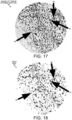
- FIG. 17 shows a high-resolution image 83 of breast tissue that has been stained with a protein-specific antibody using immunohistochemistry (IHC). For example, cells whose membranes have the HER2 protein are stained brown when the antibody with a brown dye binds to the transmembrane protein HER2/neu. Cells are stained to differing degrees depending on the amount of HER2 protein in their membranes.
- a small first portion of image 83 is segmented using object-oriented image analysis into data objects classified into various classes, including positively stained cells with HER2 overexpression, cells that do not exhibit HER2 overexpression and background objects.
- system 10 determines object characteristics of the segmented objects in the first portion of image 83.
- the object-oriented image analysis determines the elliptical fit of the positively stained cells, the average number of concavities in the perimeters of the positively stained cells, the average size of the positively stained cells, the variation in the sizes of the positively stained cells, the average color of the positively stained cells, the average color of the membranes of the positively stained cells, the average intensity of the positively stained cells, the amount of intensity variation within each of the positively stained cells, and the amount of variation of the average intensity of the positively stained cells.
- the object-oriented image analysis quantifies the degree to which the membranes of the positively stained cells have the HER2 protein.
- step 79 system 10 assigns a color to each of the object characteristics determined in step 78. For example, large positively stained cells can be assigns the color orange, while small positively stained cells can be assigned the color green. In addition, positively stained cells with a higher proportion of their membranes stained can be assigned a darker shade of a color, while positively stained cells with a lower proportion of their membranes stained can be assigned a lighter shade of that color.
- step 80 pixelwise descriptors are generated that describe which of the object characteristics a characterized pixel most probably exhibits based on a quality of a second pixel or box of pixels at a predetermined offset from the characterized pixel.
- the pixelwise descriptors indicate the degree to which the characterized pixel exhibits a selected object characteristic.
- a pixel heat map is generated by applying the pixelwise descriptors to each pixel in a second portion of the image without segmenting the second portion of the image into objects.
- Each pixel of the second portion of the image has the color assigned to the object characteristic most probably exhibited by that pixel.
- each pixel of the second portion of the image has a shade of color indicative of the degree to which the characterized pixel exhibits a selected object characteristic.
- step 82 system 10 displays the pixel heat map on graphical user interface 14.
- FIG. 18 shows a pixel heat map 84 generated by applying decision trees with pixelwise descriptors to the pixels of a second portion of image 83 that has not been segmented into objects. Segmenting using object-oriented image analysis was performed only on a small number of learning tiles (not shown) in FIG. 17 . The pixel-oriented analysis using decision trees with pixelwise descriptors is then applied to the entire image 83, including the second portion that was not included in the learning tiles.
- heat map 84 includes darker shade pixels, lighter shade pixels, and white background pixels.
- the darker shade pixels have been assigned the color associated with the object characteristic of more membrane staining.
- the darker shade indicates that the pixel is most probably associated with a positively stained cell having a higher proportion of its membrane stained.
- the lighter shade pixels have been assigned the color associated with the object characteristic of less membrane staining.
- the lighter shade indicates that the pixel is most probably associated with a positively stained cell with a lower proportion of its membrane stained.
- the pixels in heat map 84 at the location of cell 86 have a lighter shade indicating that the pixels most probably belong to a cell with a lower proportion of its membrane stained. But not all pixels at the location of cell 87 have the same shade. Some of the pixels have a darker shade and have a higher probability of belonging to a cell with more of its membrane stained, while other pixels at the location of cell 87 have a lighter shade and higher probability of belonging to a cell with less of its membrane stained. Thus, the color shades in heat map 84 do not indicate pixels that belong to the object class of positively stained cells and the object class of unstained cells. Instead, the color shades in heat map 84 indicate the degree of a characteristic exhibited by a positively stained cell to which each pixel is most likely associated.
- a higher level heat map can easily be generated from a heat map that assigns a color to each pixel depending on the most likely degree to which an object characteristic is exhibited at the location of the pixel. For example, the average value of multiple pixels in heat map 84 can be combined to form a single pixel of the higher level heat map.
- the shade of each pixel of the higher level heat map indicates an area of the original image 83 in which the cells have a higher or lower proportion of their membranes immunohistochemically stained.
- a pathologist could use the higher level heat map to navigate to the locations in image 83 that include the highest proportion of cancerous breast tissue cells that exhibit a high degree of HER2 overexpression.
- the object characteristic indicated by the decision trees of pixelwise descriptors is the size of the nuclei. Pixels most probably belonging to larger nuclei are assigned a different color than pixels most probably belonging to smaller nuclei. Pixels in the higher level heat map indicate the locations in the original image in which the nuclei are larger and more likely to be cancerous.
- Data analysis server 13 includes a computer-readable storage medium having program instructions thereon for performing method 24 and method 76.
- a computer-readable storage medium includes instructions of the image analysis program for generating data objects corresponding to patterns in digital images that have been stained by a particular biomarker.
- the computer-readable storage medium also includes instructions for generating decision trees of pixelwise descriptors that indicate the probability that a pixel belongs to an object class without segmenting the portion of the image in which the pixel is located.
Landscapes
- Engineering & Computer Science (AREA)
- Theoretical Computer Science (AREA)
- Physics & Mathematics (AREA)
- General Physics & Mathematics (AREA)
- Multimedia (AREA)
- Computer Vision & Pattern Recognition (AREA)
- Health & Medical Sciences (AREA)
- General Health & Medical Sciences (AREA)
- Life Sciences & Earth Sciences (AREA)
- Evolutionary Computation (AREA)
- Artificial Intelligence (AREA)
- Medical Informatics (AREA)
- Computing Systems (AREA)
- Databases & Information Systems (AREA)
- Molecular Biology (AREA)
- Biomedical Technology (AREA)
- Software Systems (AREA)
- Data Mining & Analysis (AREA)
- Bioinformatics & Computational Biology (AREA)
- Bioinformatics & Cheminformatics (AREA)
- Evolutionary Biology (AREA)
- General Engineering & Computer Science (AREA)
- Nuclear Medicine, Radiotherapy & Molecular Imaging (AREA)
- Radiology & Medical Imaging (AREA)
- Quality & Reliability (AREA)
- Image Analysis (AREA)
- Image Processing (AREA)
Claims (10)
- Computerimplementiertes Verfahren zur Erkennung von Mustern in einem digitalen Bild eines gefärbten Gewebeschnitts, wobei das Verfahren umfasst:Teilen eines digitalen Bildes in Kacheln, wobei das digitale Bild ein Bild eines mit einem oder mehreren Biomarkern gefärbten Gewebeschnitts ist;Bestimmen eines Grades lokalen Kontrasts in jeder der Kacheln;Auswählen einer ersten Mehrzahl von Kacheln, die einen höchsten Grad lokalen Kontrasts aufweist;Bestimmen einer Durchschnittsfarbe jeder der ersten Mehrzahl von Kacheln;Teilen der ersten Mehrzahl von Kacheln in Cluster von Kacheln mit ähnlichen Durchschnittsfarben;Auswählen einer Lernkachel aus jedem Cluster von Kacheln, wobei jede Lernkachel den höchsten Grad lokalen Kontrasts unter den Kacheln des Clusters aufweist, zu dem die Lernkachel gehört;Segmentieren der Lernkacheln in Datenobjekte unter Verwendung objektorientierter Bildanalyse, wobei ein Datenobjekt erzeugt wird, indem jene Pixel, die ähnliche, durch die Pixelwerte definierte Charakteristiken aufweisen, mit dem Datenobjekt verknüpft werden;Einteilen der Datenobjekte in Klassen von Objekten;Assoziieren einer Farbe mit jeder Klasse von Objekten;Bestimmen von Charakteristiken der Datenobjekte, die zu unterschiedlichen Klassen von Objekten gehören;Erstellen pixelweiser Deskriptoren für jedes Pixel der Lernkacheln, wobei die pixelweise Deskriptoren eine Charakteristik eines oder mehrerer Pixel in einem vorgegebenen Abstand von dem Pixel bestimmen, das charakterisiert wird, wobei die pixelweisen Deskriptoren in einem Random-Forest-Entscheidungsbaum angewendet werden, um die Wahrscheinlichkeit anzugeben, dass jedes Pixel jeder Lernkachel zu einer Klasse von Objekten gehört, wobei eine Klassenwahrscheinlichkeit jedes Pixels unter Verwendung mehrerer Random-Forest-Entscheidungsbäume berechnet wird, die auf pixelweise Deskriptoren angewendet werden, und ein Durchschnitt der Wahrscheinlichkeiten als das Ergebnis der Random-Forest-Entscheidungsbäume genommen wird, wobei ein oder mehrere Parameter jedes Random-Forst-Entscheidungsbaums während des Trainings modifiziert werden, sodass die vorhergesagte Klasse, zu der jedes Trainingspixel einer Mehrzahl von Trainingspixeln höchstwahrscheinlich gehört, mit der Objektklasse übereinstimmt, die unter Verwendung der objektorientierten Bildanalyse bestimmt wurde;Erstellen einer Pixel-Heatmap, ohne das digitale Bild erneut in Datenobjekte zu segmentieren, durch:Anwenden einer pixelorientierten Bildanalyse, die ein Anwenden der Rand-Forest-Entscheidungsbäume auf die pixelweisen Deskriptoren jedes Pixels des digitalen Bildes einschließlich der Pixel umfasst, die nicht in den Lernkacheln sind, an welchen die objektorientierte Bildanalyse durchgeführt wurde, um dadurch die Klasse von Objekten zu bestimmen, zu der jedes Pixel höchstwahrscheinlich gehört, undZuordnen der Farbe zu jedem Pixel des digitalen Bildes, die mit der Klasse von Objekten assoziiert ist, zu der dieses Pixel höchstwahrscheinlich gehört; undAnzeigen der Pixel-Heatmap auf einer grafischen Benutzeroberfläche.
- Verfahren nach Anspruch 1, wobei die pixelweisen Deskriptoren die Wahrscheinlichkeit, dass ein charakterisiertes Pixel zu einer Klasse von Objekten gehört, basierend auf einer Charakteristik eines zweiten Pixels in einem vorgegebenen Abstand von dem charakterisierten Pixel angeben.
- Verfahren nach einem der vorhergehenden Ansprüche, wobei die pixelweisen Deskriptoren ausgewählt werden aus: einem Durchschnittsfarbwert von Pixeln in einem Feld einer vorgegebenen Größe in einem vorgegebenen Abstand vom charakterisierten Pixel, der Differenz der Durchschnittsfarbwerte in zwei versetzten Pixelfeldern, dem Farbwert eines zweiten Pixels in einem vorgegebenen Abstand von dem Pixel, das charakterisiert wird, der Differenz zwischen dem Farbwert des charakterisierten Pixels und dem Farbwert eines zweiten Pixels in einem vorgegebenen Abstand vom charakterisierten Pixel, der Standardabweichung zwischen den Farbwerten von Pixeln in einem Feld einer vorgegebenen Größe in einem vorgegebenen Abstand vom charakterisierten Pixel, der Differenz zwischen den Standardabweichungen der Pixel in zwei Feldern, der Summe der Gradientengröße der Farbwerte von Pixeln in einem Feld einer vorgegebenen Größe in einem vorgegebenen Abstand vom charakterisierten Pixel und in einer vorgegebenen Ausrichtung sowie der Ausrichtung der Gradientenkante der Farbwerte von Pixeln in einem Feld einer vorgegebenen Größe in einem vorgegebenen Abstand vom charakterisierten Pixel.
- Verfahren nach einem der vorhergehenden Ansprüche, wobei das Bestimmen des Grades lokalen Kontrasts in jeder Kachel erfolgt, indem ein Flächenanteil jeder Kachel bestimmt wird, der maximal stabile Extremalregionen aufweist.
- Verfahren nach einem der vorhergehenden Ansprüche, wobei das Segmentieren der Lernkacheln in Datenobjekte ein Erstellen eines Datennetzwerks umfasst, in dem Datenobjekte, die eine zweite Hierarchieebene des Datennetzwerks bilden, mit ausgewählten Pixeln der Lernkacheln verknüpft werden, die die niedrigste Hierarchieebene des Datennetzwerks bilden.
- Verfahren nach einem der vorhergehenden Ansprüche, wobei das digitale Bild ein Bild eines mit Hämatoxylin oder Eosin gefärbten Gewebeschnitts oder eines unter Verwendung von Immunhistochemie mit einem proteinspezifischen Antikörper gefärbten Gewebeschnitts ist.
- Verfahren nach einem der vorhergehenden Ansprüche, wobei jeder Entscheidungsbaum mit zufälligen unterschiedlichen Pixeln aus den Lernkacheln trainiert wird.
- Verfahren nach einem der vorhergehenden Ansprüche, wobei der eine oder die mehreren Parameter der Entscheidungsbäume ausgewählt werden aus: einem auf einen pixelweisen Deskriptor angewendeten Vergleichsschwellenwert, einem Abstand von Pixeln, die in einem pixelweisen Deskriptor verglichen werden, einem Wert eines Pixels, das verglichen wird, dem Abstand eines Pixels oder Pixelfelds, mit dem ein Pixel oder Pixelfeld verglichen wird, der Größe und Form eines Feldes zu vergleichender Pixel und der Reihenfolge, in der die pixelweisen Deskriptoren in einem Entscheidungsbaum ausgewertet werden.
- System, umfassend einen Prozessor und einen Speicher, der Anweisungen speichert, die bei Ausführung durch den Prozessor den Prozessor zum Implementieren des Verfahrens nach einem der vorhergehenden Ansprüche veranlassen.
- Computerlesbares Medium, das Anweisungen speichert, die bei Ausführung durch einen Prozessor den Prozessor zum Implementieren des Verfahrens nach einem der Ansprüche 1 bis 8 veranlassen.
Priority Applications (1)
| Application Number | Priority Date | Filing Date | Title |
|---|---|---|---|
| DE19213622.4T DE19213622T1 (de) | 2014-08-29 | 2015-07-29 | Verfahren und system zum lernen von visuellem pixelkontext aus objekteigenschaften zur erzeugung reicher semantischer bilder |
Applications Claiming Priority (2)
| Application Number | Priority Date | Filing Date | Title |
|---|---|---|---|
| US14/473,096 US9740957B2 (en) | 2014-08-29 | 2014-08-29 | Learning pixel visual context from object characteristics to generate rich semantic images |
| EP15178864.3A EP3023910A1 (de) | 2014-08-29 | 2015-07-29 | Verfahren und system zum lernen von visuellem pixelkontext aus objekteigenschaften zur erzeugung reicher semantischer bilder |
Related Parent Applications (1)
| Application Number | Title | Priority Date | Filing Date |
|---|---|---|---|
| EP15178864.3A Division EP3023910A1 (de) | 2014-08-29 | 2015-07-29 | Verfahren und system zum lernen von visuellem pixelkontext aus objekteigenschaften zur erzeugung reicher semantischer bilder |
Publications (2)
| Publication Number | Publication Date |
|---|---|
| EP3663975A1 EP3663975A1 (de) | 2020-06-10 |
| EP3663975B1 true EP3663975B1 (de) | 2024-10-02 |
Family
ID=53758125
Family Applications (2)
| Application Number | Title | Priority Date | Filing Date |
|---|---|---|---|
| EP15178864.3A Withdrawn EP3023910A1 (de) | 2014-08-29 | 2015-07-29 | Verfahren und system zum lernen von visuellem pixelkontext aus objekteigenschaften zur erzeugung reicher semantischer bilder |
| EP19213622.4A Active EP3663975B1 (de) | 2014-08-29 | 2015-07-29 | Verfahren und system zum lernen von visuellem pixelkontext aus objekteigenschaften zur erzeugung reicher semantischer bilder |
Family Applications Before (1)
| Application Number | Title | Priority Date | Filing Date |
|---|---|---|---|
| EP15178864.3A Withdrawn EP3023910A1 (de) | 2014-08-29 | 2015-07-29 | Verfahren und system zum lernen von visuellem pixelkontext aus objekteigenschaften zur erzeugung reicher semantischer bilder |
Country Status (3)
| Country | Link |
|---|---|
| US (2) | US9740957B2 (de) |
| EP (2) | EP3023910A1 (de) |
| DE (1) | DE19213622T1 (de) |
Families Citing this family (21)
| Publication number | Priority date | Publication date | Assignee | Title |
|---|---|---|---|---|
| US20150016668A1 (en) * | 2013-07-12 | 2015-01-15 | Ut-Battelle, Llc | Settlement mapping systems |
| US9740957B2 (en) * | 2014-08-29 | 2017-08-22 | Definiens Ag | Learning pixel visual context from object characteristics to generate rich semantic images |
| WO2016120418A1 (en) * | 2015-01-31 | 2016-08-04 | Ventana Medical Systems, Inc. | Quality control of automated whole-slide analysis |
| US10410398B2 (en) * | 2015-02-20 | 2019-09-10 | Qualcomm Incorporated | Systems and methods for reducing memory bandwidth using low quality tiles |
| US10876933B2 (en) | 2016-11-09 | 2020-12-29 | Ventana Medical Systems, Inc. | Automated tissue dissection instrument and methods of using the same |
| US10438096B2 (en) * | 2016-12-27 | 2019-10-08 | Definiens Ag | Identifying and excluding blurred areas of images of stained tissue to improve cancer scoring |
| US10346717B1 (en) * | 2017-04-19 | 2019-07-09 | The United States Of America As Represented By The Secretary Of The Navy | System and method for thresholding of local image descriptors |
| CA3034654C (en) * | 2017-07-22 | 2023-10-31 | Intelligent Virus Imaging Inc. | Method for automated unsupervised ontological investigation of structural appearances in electron micrographs |
| GB2569103A (en) * | 2017-11-16 | 2019-06-12 | Univ Oslo Hf | Histological image analysis |
| US20190266429A1 (en) * | 2018-02-23 | 2019-08-29 | Qualcomm Incorporated | Constrained random decision forest for object detection |
| CN108280236B (zh) * | 2018-02-28 | 2022-03-15 | 福州大学 | 一种基于LargeVis的随机森林可视化数据分析方法 |
| CN108694315A (zh) * | 2018-05-15 | 2018-10-23 | 浙江大学 | 一种基于交互行为的儿童手机用户识别方法 |
| US11011257B2 (en) * | 2018-11-21 | 2021-05-18 | Enlitic, Inc. | Multi-label heat map display system |
| CN109978889A (zh) * | 2019-02-20 | 2019-07-05 | 广州视源电子科技股份有限公司 | 一种图像处理方法、系统、装置、存储介质及黑板装置 |
| CN110232696B (zh) * | 2019-06-20 | 2024-03-08 | 腾讯科技(深圳)有限公司 | 一种图像区域分割的方法、模型训练的方法及装置 |
| CN110910400A (zh) * | 2019-10-29 | 2020-03-24 | 北京三快在线科技有限公司 | 图像处理方法、装置、存储介质及电子设备 |
| US11977723B2 (en) * | 2019-12-17 | 2024-05-07 | Palantir Technologies Inc. | Image tiling and distributive modification |
| CN111401135B (zh) * | 2020-02-20 | 2023-09-26 | 长沙智能驾驶研究院有限公司 | 开门防撞方法、装置、计算机设备和存储介质 |
| CN111290685A (zh) * | 2020-02-28 | 2020-06-16 | 杭州依图医疗技术有限公司 | 细胞染色图像的显示方法、病理图像分析系统及存储介质 |
| CN113140005B (zh) * | 2021-04-29 | 2024-04-16 | 上海商汤科技开发有限公司 | 目标对象定位方法、装置、设备及存储介质 |
| CN113408573B (zh) * | 2021-05-11 | 2023-02-21 | 广东工业大学 | 基于机器学习的瓷砖色号自动分类归类的方法及装置 |
Family Cites Families (24)
| Publication number | Priority date | Publication date | Assignee | Title |
|---|---|---|---|---|
| JPS6024463A (ja) | 1983-07-20 | 1985-02-07 | Toshiba Corp | 核磁気共鳴映像法 |
| JPH0262671A (ja) * | 1988-08-30 | 1990-03-02 | Toshiba Corp | カラー編集処理装置 |
| JPH11102414A (ja) * | 1997-07-25 | 1999-04-13 | Kuraritec Corp | ヒートマップを用いて光学式文字認識の訂正を行うための方法および装置、並びに、ocr出力の誤りを発見するための一連の命令を記録したコンピュータ読み取り可能な記録媒体 |
| US6173083B1 (en) * | 1998-04-14 | 2001-01-09 | General Electric Company | Method and apparatus for analyzing image structures |
| US6956373B1 (en) | 2002-01-02 | 2005-10-18 | Hugh Keith Brown | Opposed orthogonal fusion system and method for generating color segmented MRI voxel matrices |
| US7801361B2 (en) | 2002-10-15 | 2010-09-21 | Definiens Ag | Analyzing pixel data using image, thematic and object layers of a computer-implemented network structure |
| US7873223B2 (en) * | 2002-10-15 | 2011-01-18 | Definiens Ag | Cognition integrator and language |
| EP2095332B1 (de) | 2006-11-16 | 2010-08-11 | Visiopharm A/s | Auf merkmalen basierende registrierung von schnittbildern |
| US20080144013A1 (en) | 2006-12-01 | 2008-06-19 | Institute For Technology Development | System and method for co-registered hyperspectral imaging |
| US8139831B2 (en) | 2007-12-06 | 2012-03-20 | Siemens Aktiengesellschaft | System and method for unsupervised detection and gleason grading of prostate cancer whole mounts using NIR fluorscence |
| US8311344B2 (en) * | 2008-02-15 | 2012-11-13 | Digitalsmiths, Inc. | Systems and methods for semantically classifying shots in video |
| US9779213B2 (en) * | 2008-07-25 | 2017-10-03 | Fundacao D. Anna Sommer Champalimaud E Dr. Carlos Montez Champalimaud | System for evaluating a pathological stage of prostate cancer |
| US8319793B2 (en) | 2009-04-17 | 2012-11-27 | Definiens Ag | Analyzing pixel data by imprinting objects of a computer-implemented network structure into other objects |
| EP2553632A1 (de) * | 2010-03-29 | 2013-02-06 | Galderma Research & Development | Fuzzy-clustering-alogrithmus und seine anwendung auf karzinomgewebe |
| US8396269B2 (en) * | 2010-04-08 | 2013-03-12 | Digital Pathco LLC | Image quality assessment including comparison of overlapped margins |
| US8345976B2 (en) * | 2010-08-06 | 2013-01-01 | Sony Corporation | Systems and methods for segmenting digital images |
| US8351676B2 (en) * | 2010-10-12 | 2013-01-08 | Sony Corporation | Digital image analysis using multi-step analysis |
| US20120127297A1 (en) * | 2010-11-24 | 2012-05-24 | Baxi Vipul A | Digital microscopy with focus grading in zones distinguished for comparable image structures |
| US9779283B2 (en) | 2011-01-05 | 2017-10-03 | The Board Of Trustees Of The University Of Illinois | Automated prostate tissue referencing for cancer detection and diagnosis |
| US8699769B2 (en) | 2011-07-12 | 2014-04-15 | Definiens Ag | Generating artificial hyperspectral images using correlated analysis of co-registered images |
| US9224071B2 (en) * | 2012-11-19 | 2015-12-29 | Microsoft Technology Licensing, Llc | Unsupervised object class discovery via bottom up multiple class learning |
| US9619908B1 (en) * | 2014-03-24 | 2017-04-11 | Google Inc. | Identification of lines in a heat map |
| US9740957B2 (en) * | 2014-08-29 | 2017-08-22 | Definiens Ag | Learning pixel visual context from object characteristics to generate rich semantic images |
| US9805248B2 (en) * | 2014-08-29 | 2017-10-31 | Definiens Ag | Applying pixelwise descriptors to a target image that are generated by segmenting objects in other images |
-
2014
- 2014-08-29 US US14/473,096 patent/US9740957B2/en active Active
-
2015
- 2015-07-29 EP EP15178864.3A patent/EP3023910A1/de not_active Withdrawn
- 2015-07-29 DE DE19213622.4T patent/DE19213622T1/de active Pending
- 2015-07-29 EP EP19213622.4A patent/EP3663975B1/de active Active
-
2017
- 2017-08-03 US US15/668,695 patent/US10445557B2/en active Active
Also Published As
| Publication number | Publication date |
|---|---|
| US9740957B2 (en) | 2017-08-22 |
| US20160063308A1 (en) | 2016-03-03 |
| EP3023910A1 (de) | 2016-05-25 |
| US20170337415A1 (en) | 2017-11-23 |
| EP3663975A1 (de) | 2020-06-10 |
| US10445557B2 (en) | 2019-10-15 |
| DE19213622T1 (de) | 2020-08-27 |
Similar Documents
| Publication | Publication Date | Title |
|---|---|---|
| EP3663975B1 (de) | Verfahren und system zum lernen von visuellem pixelkontext aus objekteigenschaften zur erzeugung reicher semantischer bilder | |
| US10474874B2 (en) | Applying pixelwise descriptors to a target image that are generated by segmenting objects in other images | |
| US10565479B1 (en) | Identifying and excluding blurred areas of images of stained tissue to improve cancer scoring | |
| US11669971B2 (en) | Colony contrast gathering | |
| US11977984B2 (en) | Using a first stain to train a model to predict the region stained by a second stain | |
| CN103518224B (zh) | 用于分析微生物生长的方法 | |
| US10121245B2 (en) | Identification of inflammation in tissue images | |
| US11748981B2 (en) | Deep learning method for predicting patient response to a therapy | |
| US20090003691A1 (en) | Segmentation of tissue images using color and texture | |
| WO2011034596A1 (en) | High-throughput biomarker segmentation utilizing hierarchical normalized cuts | |
| US20210214765A1 (en) | Methods and systems for automated counting and classifying microorganisms | |
| US10769432B2 (en) | Automated parameterization image pattern recognition method | |
| US9852354B2 (en) | Method and apparatus for image scoring and analysis | |
| CN119811636A (zh) | 基于人工智能的肺癌表型预测辅助分析方法及系统 | |
| Fedele et al. | Explaining Relation Networks for Few-Shot Learning | |
| CN120912520A (zh) | 一种基于多模态图像的细胞计数方法及系统 | |
| CN121564706A (zh) | 一种基于机器视觉的双孢蘑菇自动化表型方法 | |
| HK40053751A (en) | Machine learning using distance-based similarity labels | |
| Abdulsadig | IMPACT OF TRANSFER LEARNING ON THE CLASSIFICATION OF MICROSCOPE SLIDE IMAGES FOR IMPROVING BREAST CANCER TREATMENT USING DEEP LEARNING |
Legal Events
| Date | Code | Title | Description |
|---|---|---|---|
| PUAI | Public reference made under article 153(3) epc to a published international application that has entered the european phase |
Free format text: ORIGINAL CODE: 0009012 |
|
| STAA | Information on the status of an ep patent application or granted ep patent |
Free format text: STATUS: THE APPLICATION HAS BEEN PUBLISHED |
|
| AC | Divisional application: reference to earlier application |
Ref document number: 3023910 Country of ref document: EP Kind code of ref document: P |
|
| AK | Designated contracting states |
Kind code of ref document: A1 Designated state(s): AL AT BE BG CH CY CZ DE DK EE ES FI FR GB GR HR HU IE IS IT LI LT LU LV MC MK MT NL NO PL PT RO RS SE SI SK SM TR |
|
| REG | Reference to a national code |
Ref country code: DE Ref legal event code: R210 Ref country code: DE Ref legal event code: R210 Ref document number: 602015090062 Country of ref document: DE |
|
| STAA | Information on the status of an ep patent application or granted ep patent |
Free format text: STATUS: REQUEST FOR EXAMINATION WAS MADE |
|
| 17P | Request for examination filed |
Effective date: 20201119 |
|
| RBV | Designated contracting states (corrected) |
Designated state(s): AL AT BE BG CH CY CZ DE DK EE ES FI FR GB GR HR HU IE IS IT LI LT LU LV MC MK MT NL NO PL PT RO RS SE SI SK SM TR |
|
| RAP3 | Party data changed (applicant data changed or rights of an application transferred) |
Owner name: ASTRAZENECA COMPUTATIONAL PATHOLOGY GMBH |
|
| STAA | Information on the status of an ep patent application or granted ep patent |
Free format text: STATUS: EXAMINATION IS IN PROGRESS |
|
| 17Q | First examination report despatched |
Effective date: 20220922 |
|
| P01 | Opt-out of the competence of the unified patent court (upc) registered |
Effective date: 20230522 |
|
| REG | Reference to a national code |
Ref country code: DE Ref legal event code: R079 Ref document number: 602015090062 Country of ref document: DE Free format text: PREVIOUS MAIN CLASS: G06K0009000000 Ipc: G06V0020690000 Ref country code: DE Ref legal event code: R079 Free format text: PREVIOUS MAIN CLASS: G06K0009000000 Ipc: G06V0020690000 |
|
| GRAP | Despatch of communication of intention to grant a patent |
Free format text: ORIGINAL CODE: EPIDOSNIGR1 |
|
| STAA | Information on the status of an ep patent application or granted ep patent |
Free format text: STATUS: GRANT OF PATENT IS INTENDED |
|
| RIC1 | Information provided on ipc code assigned before grant |
Ipc: G06F 18/243 20230101ALI20240308BHEP Ipc: G06V 10/46 20220101ALI20240308BHEP Ipc: G06V 10/772 20220101ALI20240308BHEP Ipc: G06V 10/764 20220101ALI20240308BHEP Ipc: G06V 10/56 20220101ALI20240308BHEP Ipc: G06V 10/25 20220101ALI20240308BHEP Ipc: G06V 20/69 20220101AFI20240308BHEP |
|
| INTG | Intention to grant announced |
Effective date: 20240328 |
|
| GRAS | Grant fee paid |
Free format text: ORIGINAL CODE: EPIDOSNIGR3 |
|
| GRAA | (expected) grant |
Free format text: ORIGINAL CODE: 0009210 |
|
| STAA | Information on the status of an ep patent application or granted ep patent |
Free format text: STATUS: THE PATENT HAS BEEN GRANTED |
|
| REG | Reference to a national code |
Ref country code: DE Ref legal event code: R082 Ref document number: 602015090062 Country of ref document: DE Ref country code: DE Ref legal event code: R081 Ref document number: 602015090062 Country of ref document: DE Owner name: ASTRAZENECA COMPUTATIONAL PATHOLOGY GMBH, DE Free format text: FORMER OWNER: DEFINIENS GMBH, 80636 MUENCHEN, DE |
|
| AC | Divisional application: reference to earlier application |
Ref document number: 3023910 Country of ref document: EP Kind code of ref document: P |
|
| AK | Designated contracting states |
Kind code of ref document: B1 Designated state(s): AL AT BE BG CH CY CZ DE DK EE ES FI FR GB GR HR HU IE IS IT LI LT LU LV MC MK MT NL NO PL PT RO RS SE SI SK SM TR |
|
| REG | Reference to a national code |
Ref country code: GB Ref legal event code: FG4D |
|
| REG | Reference to a national code |
Ref country code: CH Ref legal event code: EP |
|
| REG | Reference to a national code |
Ref country code: DE Ref legal event code: R096 Ref document number: 602015090062 Country of ref document: DE |
|
| REG | Reference to a national code |
Ref country code: IE Ref legal event code: FG4D |
|
| REG | Reference to a national code |
Ref country code: LT Ref legal event code: MG9D |
|
| REG | Reference to a national code |
Ref country code: NL Ref legal event code: MP Effective date: 20241002 |
|
| REG | Reference to a national code |
Ref country code: AT Ref legal event code: MK05 Ref document number: 1729035 Country of ref document: AT Kind code of ref document: T Effective date: 20241002 |
|
| PG25 | Lapsed in a contracting state [announced via postgrant information from national office to epo] |
Ref country code: NL Free format text: LAPSE BECAUSE OF FAILURE TO SUBMIT A TRANSLATION OF THE DESCRIPTION OR TO PAY THE FEE WITHIN THE PRESCRIBED TIME-LIMIT Effective date: 20241002 |
|
| PG25 | Lapsed in a contracting state [announced via postgrant information from national office to epo] |
Ref country code: NL Free format text: LAPSE BECAUSE OF FAILURE TO SUBMIT A TRANSLATION OF THE DESCRIPTION OR TO PAY THE FEE WITHIN THE PRESCRIBED TIME-LIMIT Effective date: 20241002 |
|
| PG25 | Lapsed in a contracting state [announced via postgrant information from national office to epo] |
Ref country code: PT Free format text: LAPSE BECAUSE OF FAILURE TO SUBMIT A TRANSLATION OF THE DESCRIPTION OR TO PAY THE FEE WITHIN THE PRESCRIBED TIME-LIMIT Effective date: 20250203 Ref country code: HR Free format text: LAPSE BECAUSE OF FAILURE TO SUBMIT A TRANSLATION OF THE DESCRIPTION OR TO PAY THE FEE WITHIN THE PRESCRIBED TIME-LIMIT Effective date: 20241002 Ref country code: IS Free format text: LAPSE BECAUSE OF FAILURE TO SUBMIT A TRANSLATION OF THE DESCRIPTION OR TO PAY THE FEE WITHIN THE PRESCRIBED TIME-LIMIT Effective date: 20250202 |
|
| PG25 | Lapsed in a contracting state [announced via postgrant information from national office to epo] |
Ref country code: FI Free format text: LAPSE BECAUSE OF FAILURE TO SUBMIT A TRANSLATION OF THE DESCRIPTION OR TO PAY THE FEE WITHIN THE PRESCRIBED TIME-LIMIT Effective date: 20241002 |
|
| PG25 | Lapsed in a contracting state [announced via postgrant information from national office to epo] |
Ref country code: BG Free format text: LAPSE BECAUSE OF FAILURE TO SUBMIT A TRANSLATION OF THE DESCRIPTION OR TO PAY THE FEE WITHIN THE PRESCRIBED TIME-LIMIT Effective date: 20241002 |
|
| PG25 | Lapsed in a contracting state [announced via postgrant information from national office to epo] |
Ref country code: ES Free format text: LAPSE BECAUSE OF FAILURE TO SUBMIT A TRANSLATION OF THE DESCRIPTION OR TO PAY THE FEE WITHIN THE PRESCRIBED TIME-LIMIT Effective date: 20241002 |
|
| PG25 | Lapsed in a contracting state [announced via postgrant information from national office to epo] |
Ref country code: NO Free format text: LAPSE BECAUSE OF FAILURE TO SUBMIT A TRANSLATION OF THE DESCRIPTION OR TO PAY THE FEE WITHIN THE PRESCRIBED TIME-LIMIT Effective date: 20250102 |
|
| PG25 | Lapsed in a contracting state [announced via postgrant information from national office to epo] |
Ref country code: AT Free format text: LAPSE BECAUSE OF FAILURE TO SUBMIT A TRANSLATION OF THE DESCRIPTION OR TO PAY THE FEE WITHIN THE PRESCRIBED TIME-LIMIT Effective date: 20241002 Ref country code: LV Free format text: LAPSE BECAUSE OF FAILURE TO SUBMIT A TRANSLATION OF THE DESCRIPTION OR TO PAY THE FEE WITHIN THE PRESCRIBED TIME-LIMIT Effective date: 20241002 Ref country code: GR Free format text: LAPSE BECAUSE OF FAILURE TO SUBMIT A TRANSLATION OF THE DESCRIPTION OR TO PAY THE FEE WITHIN THE PRESCRIBED TIME-LIMIT Effective date: 20250103 |
|
| PG25 | Lapsed in a contracting state [announced via postgrant information from national office to epo] |
Ref country code: PL Free format text: LAPSE BECAUSE OF FAILURE TO SUBMIT A TRANSLATION OF THE DESCRIPTION OR TO PAY THE FEE WITHIN THE PRESCRIBED TIME-LIMIT Effective date: 20241002 Ref country code: CZ Free format text: LAPSE BECAUSE OF FAILURE TO SUBMIT A TRANSLATION OF THE DESCRIPTION OR TO PAY THE FEE WITHIN THE PRESCRIBED TIME-LIMIT Effective date: 20241002 |
|
| PG25 | Lapsed in a contracting state [announced via postgrant information from national office to epo] |
Ref country code: RS Free format text: LAPSE BECAUSE OF FAILURE TO SUBMIT A TRANSLATION OF THE DESCRIPTION OR TO PAY THE FEE WITHIN THE PRESCRIBED TIME-LIMIT Effective date: 20250102 |
|
| PG25 | Lapsed in a contracting state [announced via postgrant information from national office to epo] |
Ref country code: SM Free format text: LAPSE BECAUSE OF FAILURE TO SUBMIT A TRANSLATION OF THE DESCRIPTION OR TO PAY THE FEE WITHIN THE PRESCRIBED TIME-LIMIT Effective date: 20241002 |
|
| REG | Reference to a national code |
Ref country code: DE Ref legal event code: R097 Ref document number: 602015090062 Country of ref document: DE |
|
| PG25 | Lapsed in a contracting state [announced via postgrant information from national office to epo] |
Ref country code: DK Free format text: LAPSE BECAUSE OF FAILURE TO SUBMIT A TRANSLATION OF THE DESCRIPTION OR TO PAY THE FEE WITHIN THE PRESCRIBED TIME-LIMIT Effective date: 20241002 |
|
| PGFP | Annual fee paid to national office [announced via postgrant information from national office to epo] |
Ref country code: GB Payment date: 20250619 Year of fee payment: 11 |
|
| PG25 | Lapsed in a contracting state [announced via postgrant information from national office to epo] |
Ref country code: EE Free format text: LAPSE BECAUSE OF FAILURE TO SUBMIT A TRANSLATION OF THE DESCRIPTION OR TO PAY THE FEE WITHIN THE PRESCRIBED TIME-LIMIT Effective date: 20241002 |
|
| PGFP | Annual fee paid to national office [announced via postgrant information from national office to epo] |
Ref country code: FR Payment date: 20250620 Year of fee payment: 11 |
|
| PG25 | Lapsed in a contracting state [announced via postgrant information from national office to epo] |
Ref country code: RO Free format text: LAPSE BECAUSE OF FAILURE TO SUBMIT A TRANSLATION OF THE DESCRIPTION OR TO PAY THE FEE WITHIN THE PRESCRIBED TIME-LIMIT Effective date: 20241002 |
|
| PG25 | Lapsed in a contracting state [announced via postgrant information from national office to epo] |
Ref country code: SK Free format text: LAPSE BECAUSE OF FAILURE TO SUBMIT A TRANSLATION OF THE DESCRIPTION OR TO PAY THE FEE WITHIN THE PRESCRIBED TIME-LIMIT Effective date: 20241002 |
|
| PG25 | Lapsed in a contracting state [announced via postgrant information from national office to epo] |
Ref country code: IT Free format text: LAPSE BECAUSE OF FAILURE TO SUBMIT A TRANSLATION OF THE DESCRIPTION OR TO PAY THE FEE WITHIN THE PRESCRIBED TIME-LIMIT Effective date: 20241002 |
|
| PLBE | No opposition filed within time limit |
Free format text: ORIGINAL CODE: 0009261 |
|
| STAA | Information on the status of an ep patent application or granted ep patent |
Free format text: STATUS: NO OPPOSITION FILED WITHIN TIME LIMIT |
|
| PG25 | Lapsed in a contracting state [announced via postgrant information from national office to epo] |
Ref country code: SE Free format text: LAPSE BECAUSE OF FAILURE TO SUBMIT A TRANSLATION OF THE DESCRIPTION OR TO PAY THE FEE WITHIN THE PRESCRIBED TIME-LIMIT Effective date: 20241002 |
|
| 26N | No opposition filed |
Effective date: 20250703 |
|
| PGFP | Annual fee paid to national office [announced via postgrant information from national office to epo] |
Ref country code: DE Payment date: 20250620 Year of fee payment: 11 |
|
| REG | Reference to a national code |
Ref country code: CH Ref legal event code: H13 Free format text: ST27 STATUS EVENT CODE: U-0-0-H10-H13 (AS PROVIDED BY THE NATIONAL OFFICE) Effective date: 20260224 |
|
| PG25 | Lapsed in a contracting state [announced via postgrant information from national office to epo] |
Ref country code: LU Free format text: LAPSE BECAUSE OF NON-PAYMENT OF DUE FEES Effective date: 20250729 |
|
| REG | Reference to a national code |
Ref country code: BE Ref legal event code: MM Effective date: 20250731 |
|
| PG25 | Lapsed in a contracting state [announced via postgrant information from national office to epo] |
Ref country code: BE Free format text: LAPSE BECAUSE OF NON-PAYMENT OF DUE FEES Effective date: 20250731 |
|
| PG25 | Lapsed in a contracting state [announced via postgrant information from national office to epo] |
Ref country code: CH Free format text: LAPSE BECAUSE OF NON-PAYMENT OF DUE FEES Effective date: 20250731 |