EP3663764A1 - Proteinbiomarker für akute, subakute und chronische traumatische verletzungen des zentralen nervensystems - Google Patents
Proteinbiomarker für akute, subakute und chronische traumatische verletzungen des zentralen nervensystems Download PDFInfo
- Publication number
- EP3663764A1 EP3663764A1 EP20153859.2A EP20153859A EP3663764A1 EP 3663764 A1 EP3663764 A1 EP 3663764A1 EP 20153859 A EP20153859 A EP 20153859A EP 3663764 A1 EP3663764 A1 EP 3663764A1
- Authority
- EP
- European Patent Office
- Prior art keywords
- tau
- injury
- tbi
- sci
- levels
- Prior art date
- Legal status (The legal status is an assumption and is not a legal conclusion. Google has not performed a legal analysis and makes no representation as to the accuracy of the status listed.)
- Granted
Links
Images















Classifications
-
- G—PHYSICS
- G01—MEASURING; TESTING
- G01N—INVESTIGATING OR ANALYSING MATERIALS BY DETERMINING THEIR CHEMICAL OR PHYSICAL PROPERTIES
- G01N33/00—Investigating or analysing materials by specific methods not covered by groups G01N1/00 - G01N31/00
- G01N33/48—Biological material, e.g. blood, urine; Haemocytometers
- G01N33/50—Chemical analysis of biological material, e.g. blood, urine; Testing involving biospecific ligand binding methods; Immunological testing
- G01N33/68—Chemical analysis of biological material, e.g. blood, urine; Testing involving biospecific ligand binding methods; Immunological testing involving proteins, peptides or amino acids
- G01N33/6893—Chemical analysis of biological material, e.g. blood, urine; Testing involving biospecific ligand binding methods; Immunological testing involving proteins, peptides or amino acids related to diseases not provided for elsewhere
- G01N33/6896—Neurological disorders, e.g. Alzheimer's disease
-
- G—PHYSICS
- G01—MEASURING; TESTING
- G01N—INVESTIGATING OR ANALYSING MATERIALS BY DETERMINING THEIR CHEMICAL OR PHYSICAL PROPERTIES
- G01N33/00—Investigating or analysing materials by specific methods not covered by groups G01N1/00 - G01N31/00
- G01N33/48—Biological material, e.g. blood, urine; Haemocytometers
- G01N33/50—Chemical analysis of biological material, e.g. blood, urine; Testing involving biospecific ligand binding methods; Immunological testing
- G01N33/68—Chemical analysis of biological material, e.g. blood, urine; Testing involving biospecific ligand binding methods; Immunological testing involving proteins, peptides or amino acids
- G01N33/6893—Chemical analysis of biological material, e.g. blood, urine; Testing involving biospecific ligand binding methods; Immunological testing involving proteins, peptides or amino acids related to diseases not provided for elsewhere
-
- G—PHYSICS
- G01—MEASURING; TESTING
- G01N—INVESTIGATING OR ANALYSING MATERIALS BY DETERMINING THEIR CHEMICAL OR PHYSICAL PROPERTIES
- G01N2800/00—Detection or diagnosis of diseases
- G01N2800/28—Neurological disorders
-
- G—PHYSICS
- G01—MEASURING; TESTING
- G01N—INVESTIGATING OR ANALYSING MATERIALS BY DETERMINING THEIR CHEMICAL OR PHYSICAL PROPERTIES
- G01N2800/00—Detection or diagnosis of diseases
- G01N2800/40—Disorders due to exposure to physical agents, e.g. heat disorders, motion sickness, radiation injuries, altitude sickness, decompression illness
-
- G—PHYSICS
- G01—MEASURING; TESTING
- G01N—INVESTIGATING OR ANALYSING MATERIALS BY DETERMINING THEIR CHEMICAL OR PHYSICAL PROPERTIES
- G01N2800/00—Detection or diagnosis of diseases
- G01N2800/56—Staging of a disease; Further complications associated with the disease
-
- G—PHYSICS
- G01—MEASURING; TESTING
- G01N—INVESTIGATING OR ANALYSING MATERIALS BY DETERMINING THEIR CHEMICAL OR PHYSICAL PROPERTIES
- G01N2800/00—Detection or diagnosis of diseases
- G01N2800/60—Complex ways of combining multiple protein biomarkers for diagnosis
Definitions
- the invention relates generally to the field of biological diagnostics, particularly to methods for diagnosis, prognosis and management of injury or chronic dysfunctions of the central nervous system.
- Traumatic spinal cord injury is an injury resulting from an insult inflicted on the spinal cord. It can lead to the loss of sensory and motor after injury, and is an important cause of neurologic disability after trauma, such as lifelong paralysis for SCI patients. It is estimated that approximately 300,000 people in North America are living with a spinal cord injury (SCI), and 12,000 to 20,000 new cases occur annually.
- the pathologic process that leads to acute traumatic spinal cord injury consists of two steps; the primary injury results from the physical and mechanical damages that occur as a result of direct impact to the spinal cord.
- the secondary injury is the cascade of biochemical events such as proteolysis of cytoskeletal, membrane, and myelin proteins due to the elevation in intracellular Ca 2+ that activates cysteine proteases (e.g. calpain).
- the proteolysis damages the spinal cord by progressive tissue degeneration, including neuronal cell death, axonal degeneration, and demyelination in the spinal cord.
- TBI traumatic brain injury
- CTE Chronic Traumatic Encephalopathy
- This type of injury was previously called punch-drunk syndrome or dementia pugilistica.
- CTE is commonly found in professional athletes participating in contact sports such as boxing, Ruby, American football, ice hockey, and professional wrestling. It has also been found in soldiers exposed to blast or concussive injury.
- Symptoms associated with CTE may include dementia such as memory loss, aggression, confusion and depression, which generally appear years or decades after the trauma.
- Post-mortem findings have found Tau protein (MAPT), TAR DNA-binding protein 43 (TDP-43) and other protein deposits in the brain.
- Biomarker is defined according to the National Academy of Sciences, as an indicator that signals events in biological samples or systems. Biomarkers are considered to be extremely valuable unbiased tools to define the severity of SCI because they reflect the extent of the spinal cord damage and predict neurologic recovery. There are some metabolite candidates such as N-acetyl aspartate (NAA, neuronal/axonal marker), creatine (gliosis marker), and choline (indicator of cellular turnover related to both membrane synthesis and degradation) that can be used as biomarkers for monitoring the pathobiological changes of primary and secondary damage in SCI using proton magnetic resonance spectroscopy (1H-MRS).
- NAA N-acetyl aspartate
- creatine gliosis marker
- choline indicator of cellular turnover related to both membrane synthesis and degradation
- the present invention identifies and analyzes changes in several protein biomarkers which are associated with acute and chronic injuries to the brain and spinal cord. Differential values of selected proteins for example can distinguish chronic from acute traumatic brain and central nervous system injury. Generally, the invention demonstrates that changes in several different protein levels as well as the ratios of some of the proteins, can provide biomarker based tools for diagnosis, prognosis and management of central nervous system injuries.
- Table 1 is a list of new protein biomarkers for acute, subacute and/or chronic traumatic brain injury and spinal cord injury described in this document as identified by proteomics, systems biology and immunological assay methods. Markers include protein short name, protein full name and protein accession number.
- Tau and P-Tau (Microtubule associated Tau protein (Phosphorylated Tau protein)
- TDP-43 & BDPs TAR DNA-binding protein 43 (and its breakdown products)
- SBDPs SBDP150, SBDP145, SBDP120, SNTF
- GFAP Glial fibrillary acidic protein (and its breakdown products)
- Gfap- ⁇ Isoform 1
- Gfap- ⁇ /GFAP- ⁇ Isoform 2
- Gfap- ⁇ Isoform 3
- S100b Glial S100b protein
- MBP Myelin basic proteins (21K, 18.5, 17K, 14K
- IL-6 Interleukin-6
- Iba-1 ionized calcium-binding adapter molecule-1 (Other name: AIF1 Allograft
- the invention is a method for diagnosing traumatic spinal cord injury (SCI) severity in a subject.
- the method encompasses the steps of obtaining a preliminary diagnosis of acute traumatic spinal cord injury; selecting a panel of differentially expressed proteins in spinal cord injured subjects compared to uninjured subjects; measuring levels of said proteins in a first tissue or blood sample from the subject at an initial time up to 1-3 days post injury; repeating measurement of the proteins in tissue or blood samples of the injured subject at selected times post the initial measurements; and comparing the differentially expressed levels of proteins in the injured subject at different times post injury to levels of the proteins in uninjured subjects.
- SCI traumatic spinal cord injury
- Acute or subacute CNS injury is indicated when one or more of the following protein levels compared to healthy subjects is increased within 1-10 days of CNS injury: fatty acid synthase, NME/NM23 nucleoside diphosphate kinase 1, eukaryotic translation elongation factor 2, annexin A1, annexin A2, cathepsin D, phosphoglucomutase 1, glutamic-oxaloacetic transaminase 2, mitochondrial (aspartate aminotransferase 2), aldehyde dehydrogenase-4, Aldh4a1 fragment, LOC367586 protein, aminoacylase 1A, gamma-enolase, elongation factor2, protein Tln1 peroxiredoxin-2, aldo-keto reductase family 1, member B10 (aldose reductase), pyruvate kinase PKM, Acyl-CoA synthetase family member 2, mitochondrial, and protein-
- Such injury is also indicated by decreased levels of isocitrate dehydrogenase (NADP), mannose-6-phosphate isomerase, pyridoxal kinase, stathmin and peripheral myelin protein 2 (Pmp2), alcohol dehydrogenase [NADP(+)], L-lactate dehydrogenase B chain, ribosyldihydronicotinamide dehydrogenase [quinone], and ATP citrate lyase-CRA a isoform when compared to levels of the same proteins in healthy subjects.
- NADP isocitrate dehydrogenase
- mannose-6-phosphate isomerase pyridoxal kinase
- stathmin and peripheral myelin protein 2 (Pmp2) peripheral myelin protein 2
- alcohol dehydrogenase [NADP(+)] L-lactate dehydrogenase B chain
- the claimed method also allows distinguishing acute and chronic central nervous system (CNS) injury in a subject from an un-injured control subject by measuring the level of total Tau (T-Tau), phosphorylated (P-Tau) and P-Tau/T-Tau ratio in a biofluid sample such as CSF, blood, serum, or plasma at least once during days 1-3 post trauma; measuring the level of T-Tau, P-Tau and P-Tau/T-Tau ratio in a second sample on or about day 30 to about 180 days post CNS-injury; and comparing the levels of T-Tau, P-Tau and P-Tau/T-Tau ratio in the trauma samples in days 1-3 with a sample from an uninjured or healthy subject. Higher levels of T-Tau, P-Tau and P-Tau/T-Tau ratio in days 1-3 compared to levels in a control sample is indicative of acute CNS injury.
- CNS central nervous system
- the CNS injury can be traumatic brain injury (TBI), acute spinal cord injury or chronic traumatic encephalopathy (CTE).
- TBI traumatic brain injury
- CTE chronic traumatic encephalopathy
- the method is particularly useful for diagnosis of central nervous system injury in a subject.
- Biofluid e.g. CSF, blood, serum, plasma
- levels of one or more of a differentially expressed group of proteins are measured.
- the proteins are selected from TAR DNA-binding protein 43 (TDP-43) and its breakdown products, Visinin-Like Proteins (VILIP-1, VILIP-2, VILIP-3), Brain-derived neurotrophic factor (BDNF) and precursor of BDNF (Pro-BDNF), ionized calcium-binding adapter molecule-1 (iba-1), total Tau (T-Tau), phosphorylated Tau (P-Tau) and the P-Tau/T-Tau ratio, and transferrin, cathepsin D, TIP-1, and PEA-15.
- TBI traumatic brain injury
- one or more proteins with higher levels are selected from the group of TDP-43, Visinin-Like Proteins, BDNF, Pro-BDNF, iba-1, Tau, P-Tau, P-Tau/T-Tau ratio, transferrin, cathepsin D, TIP-1, and PEA-15 and are measured in combination with one or more proteins selected from the group of GFAP, GFAP-BDP and alphaII-spectrin breakdown products (SBDPs), MBP (21K, 18.5, 17K, 14K isoforms), IL-6, UCH-L1 and S100b. Higher levels of the proteins are compared with levels in the healthy subject and are indicative of CNS-injury (spinal cord injury or TBI).
- CNS-injury spinal cord injury or TBI
- the claimed method is useful for early determination of traumatic brain injury (TBI) or spinal cord injury (SCI). Detection of TDP-43 and TDP-43-BDP in CSF, blood, serum or plasma at levels at least twice as high as in controls is indicative of TBI when measured up to 3 days post injury.
- the injury can be from cortical impact (severe TBI), concussive closed head injury or blast overpressure-induced brain injury or spinal cord injury.
- the protein levels can be measured also from one or more biofluids including not only cerebrospinal fluid (CSF), blood, serum, and plasma, but also saliva, urine nasal fluid and sweat, or from solid biosamples selected from biopsy or autopsy brain or spinal cord tissue samples.
- CSF cerebrospinal fluid
- saliva urine nasal fluid and sweat
- solid biosamples selected from biopsy or autopsy brain or spinal cord tissue samples.
- a preferred embodiment is a diagnostic kit which includes a collection of tables showing CNS biosample or biofluid levels of differentially expressed proteins in subjects with central nervous system injuries.
- the tables are organized with respect to severity of injury and patterns associated with time post injury.
- the kit also includes instructions for use of each table for type of central nervous system injury, and approximate time post injury for making measurements.
- a diagnosis of level of CNS severity is determined by comparison of levels of said proteins in an injured patient to the protein levels in the tables.
- the central nervous system injury is traumatic brain injury (TBI), traumatic spinal cord injury (SCI) or chronic traumatic encephalopathy (CTE).
- TBI traumatic brain injury
- SCI traumatic spinal cord injury
- CTE chronic traumatic encephalopathy
- the biofluids for analysis can be selected from one or more cerebrospinal fluid (CSF), blood, serum, plasma, saliva, urine nasal fluid, and sweat.
- CSF cerebrospinal fluid
- Solid biosamples can be collected from biopsy or autopsy brain and/or spinal cord tissue samples.
- the one or more of the differentially expressed proteins for diagnosis can be TAR DNA-binding protein 43 (TDP-43) and its breakdown products, Visinin-Like Proteins (VILIP-1, VILIP-2, VILIP-3), Brain-derived neurotrophic factor (BDNF) and precursor of BDNF (Pro-BDNF), ionized calcium-binding adapter molecule-1 (iba-1), total Tau (T-Tau), phosphorylated Tau (P-Tau) and the P-Tau/T-Tau ratio, Transferrin, cathepsin D, phosphoprotein enriched in astrocytes 15 (PEA 15), triosephosphate isomerase 1 (TPI-1).
- VILIP-1 Visinin-Like Proteins
- BDNF Brain-derived neurotrophic factor
- Pro-BDNF ionized calcium-binding adapter molecule-1
- iba-1 total Tau
- P-Tau phosphorylated Tau
- TPI-1 triosephosphate isomerase 1
- the one or more proteins analyzed can be selected from TDP-43, Visinin-Like Proteins, BDNF, Pro-BDNF, iba-1, Tau, P-Tau, P-Tau/T-Tau ratio, transferrin, cathepsin D, TIP-1, and PEA-15, in combination with one or more proteins selected from GFAP, GFAP-BDP and alphaII-spectrin breakdown products (SBDPs), MBP (21K, 18.5, 17K, 14K isoforms), IL-6, UCH-L1 and S100b.
- TBI biomarkers Calcium binding protein S100 beta (S100 ⁇ ), glial fibrillary acidic protein (GFAP), myelin basic protein (MBP), neuron specific enolase (NSE), neurofilament protein (NFL), SBDP150/SBDP145/SBDP120, ubiquitin C-terminal hydrolase-Ll (UCH-L1), microtubule-associated 2 (MAP-2), and interleukins 6 and 8 (IL-6 and IL-8) have been identified as TBI biomarkers and some suggested similar SCI biomarkers as well. It was reasoned that TBI biomarkers might also be potential markers of spinal cord damage. These biomarkers can be validated for their potential use in clinical settings to determine the severity of SCI. However, due to the complexity of spinal cord injury, multiple interventions targeting different complications of damage may be required. So, it is unlikely that a single biomarker can be successfully used to diagnose and determine the severity of SCI patients.
- SCI markers have been identified by using a CAX-PAGE-LC-MS/MS proteomic platform that can provide a better resolving and separation power and characterize proteome changes associated with SCI injury.
- the analysis has been done on SCI injured samples collected from both an SCI animal model (weight-drop) and human clinical studies of SCI patients. Comparing the molecular differences between the injured and non-injured regions produced a panel of SCI candidate biomarkers that can be used to correlate with disease progression.
- the identification of novel biomarkers implicated in SCI is important to delineate the severity of the injury and provide a new avenue for therapeutic intervention.
- the identified new SCI biomarkers will also be elevated in biofluids in TBI patients (such as CSF, blood) and can serve as TBI biomarkers.
- biomarker proteins depended on the type and severity of the trauma, whether or not the trauma was repetitive and depending on the time line from the initial injury.
- proteins with the more detectable changes include TDP-43 and its breakdown products BDPs (39kDa, 35 kDa, 25 kDa).
- Total-Tau (T-Tau) and phospho-Tau (P-Tau); for example at sites Thr181, Ser202, Ser202/Thr205, Thr231, Ser396/Thr401 and in particular P-Tau/T-Tau ratio are useful as acute and subacute/chronic biomarkers for central nervous system injuries and CTE formation detection.
- Tau and P-tau were assayed with available sw-ELISA methods.
- CNS injury-dependent T-Tau, P-Tau or P-Tau/T-Tau ratio elevations were confirmed from biofluid (CSF, serum, or plasma) from TBI patients or animals.
- Triosephosphate isomerase Triosephosphate isomerase (TPI-1), transferrin (TF), phosphoprotein enriched in astrocytes 15 (PEA-15), and cathepsin D (CathD) are also likely to be found as biomarkers for central nervous system injuries (TBI and SCI).
- TBI and SCI central nervous system injuries
- IL-6 Interleukin-6
- Iba-1 ionized calcium-binding adapter molecule-1
- visinin-like proteins including VSNL1 / VISL1 (Visinin-Like Protein 1, Hippocalcin-like protein 3, VSNL2/ HPCAL4 (Hippocalcin-like protein 4) and VSNL3/VILIP-3/HACAL1 (Hippocalcin-like protein 1, Pro-BDNF (Proprotein form) and mature form of Brain-derived neurotrophic factor (BDNF) were identified based on CNS injury neuro-systems biology/interactome study and their CNS injury-dependent level elevations confirmed by immunblotting and/or sw-ELISA using biofluid (CSF, serum, or plasma) from TBI and SCI patients or animals.
- CSF CSF, serum, or plasma
- Combination biomarkers are particularly notable; for example, TPI-1, Transferrin, PEA-15 and Cathepsin D in combination with GFAP, GFAP breakdown products (GFAP-BDPs) and alphaII spectrin breakdown products and UCH-L1 and MBP (Myelin basic proteins (21K, 18.5, 17K, 14K).
- biomarkers is fatty acid synthase, NME/NM23 nucleoside diophosphate kinase 1, Stathmin 1, Eukaryotic translation elongation factor 2, Annexin A1, Annexin A2, Cathepsin D, Phosphoglucomutse 1, Glutamic oxaloacetic transaminase 2, mitochondrial (aspartate aminotransferase 2).
- CNS injury biomarkers are also noted; for example, Visinin-like proteins (e.g. VISL1, VSNL2, VSNL3), BDNF, Pro-BDNF, S100b, IL-6 and iba-1, in combination with TPI-1, Transferrin, PEA-15 and Cathepsin D or with GFAP, GFAP breakdown products (GFAP-BDPs) and alphaII spectrin breakdown products and UCH-L1 and MBP.
- Visinin-like proteins e.g. VISL1, VSNL2, VSNL3
- BDNF e.g. VISL1, VSNL2, VSNL3
- Pro-BDNF Pro-BDNF
- S100b IL-6
- IL-6 interleukin-6
- iba-1 in combination with TPI-1, Transferrin, PEA-15 and Cathepsin D or with GFAP
- GFAP GFAP breakdown products
- alphaII spectrin breakdown products UCH-L1 and
- Proteomic analysis is a useful technique for simultaneously determining multiple proteins in a biological system; it provides robust methods to study protein abundance, expression patterns, interactions, and subcellular localization in the blood, organelle, cell, tissue, organ or organism that can be studied to provide accurate and comprehensive data about that system.
- Proteomics is the most powerful technique which can aid in the discovery of novel biomarker candidates; it utilizes extensive sample procedure and Data Dependent Acquisition to follow disease-specific proteins (identity and concentration). It facilitates the identification of all differentially expressed proteins at any given time in a proteome and correlates these patterns with the healthy ones during disease progression. It has been used to study protein expression at the molecular level with a dynamic perspective that helps to understand the mechanisms of the disease.
- IEC Ion-exchange chromatography
- IEC's stationary phase is either an anion or a cation exchanger, prepared by immobilization of positively or negatively charged functional groups on the surface of chromatographic media, respectively.
- Proteins or peptide separation occurs by linear change of the mobile-phase composition (salt concentration or pH) that decreases the interactions of proteins with the stationary phase, resulting in finally eluting the proteins.
- SDS-PAGE can be used for further protein separation by apparent molecular weight with the resolving distance optimized for the proteome of interest.
- peptides can be separated by their hydrophobicity using a reversed phase C18 column, which can be directly coupled to the electrospray mass spectrometer (ESI-LC-MS/MS).
- ESI-LC-MS/MS electrospray mass spectrometer
- RPLC Reversed-phase liquid chromatography
- Proteomics has two approaches: “bottom-up”, which involves direct digestion of a biological sample using a proteolytic enzyme (such as trypsin) that cleaves at well-defined sites to create a complex peptide mixture, followed by analysis of the digested samples on platforms that include liquid chromatography prior to tandem mass spectrometry (LC-MS/MS); and “top-down”, that involves separating intact proteins from complex biological samples using techniques such as liquid chromatography or 2-D gel electrophoresis, followed by differential expression analysis using spectrum analysis or gel imaging platforms.
- a proteolytic enzyme such as trypsin
- top-down that involves separating intact proteins from complex biological samples using techniques such as liquid chromatography or 2-D gel electrophoresis, followed by differential expression analysis using spectrum analysis or gel imaging platforms.
- top-down analysis preserves protein-protein interactions and does not lose the protein information, it has been limited because of the difficulty in intact protein processing due to the lack of separation methods (only gel based) as well as the challenges of performing mass spectrometry on intact high-molecular weight proteins.
- Electrospray ionization is considered as an ideal ionization source for protein analysis due to two characteristics: first, the ability to produce multiply charged ions from large molecules (producing ions of lower m/z that are readily separated by mass analyzers such as quadrupoles and ion traps), and second, the ease of interfacing with chromatographic liquid-phase separation techniques.
- Electrospray ionization followed by tandem mass spectrometry is one of the most commonly used approaches for protein identification and sequence analysis.
- Proteomic analysis has been conducted previously by others to identify biomarkers and assess severity of SCI utilizing traumatic animals, but these methods have not been used to identify biomarkers in CSF from patients with traumatic SCI because CSF sampling is challenging; for example, here differential neuroproteomics analysis of spinal cord injury has been performed to show that not only can injury be demonstrated but severity can also be assessed.
- cerebrospinal fluid can be analyzed because it is proximal to injury site and blood sample (such as serum, plasma) due to their accessibility), from human TBI or SCI patients or animal model of TBI and SCI. It can be compared with normal control biofluid samples).
- the candidate biomarker is then quantified in such biofluid using immunological methods such as immunblotting and sandwich enzyme-linked immunosorbent assay (sw-ELISA) methods and the data analyzed.
- mice Male, 3-4 months old, Charles River Laboratories
- mice were anesthetized with 4% isoflurane in oxygen as a carrier gas for 4 minutes, followed by maintenance anesthesia of 2-3% isoflurane.
- mice were mounted in a stereotactic frame in a prone position, and secured by ear and incisor bars. Core body temperature will be monitored continuously by a rectal thermistor probe and maintained at 37 ⁇ 1°C by placing an adjustable temperature controlled heating pad beneath the rats.
- An integrated Impactor One Stereotaxic impactor device for CCI (Leica inc.) is used.
- the impactor is electromagnetically controlled. Animals are mounted in a stereotactic frame in a prone position and secured by ear and incisor bars. A midline cranial incision was made and a unilateral (ipsilateral) craniotomy (3 mm diameter) was performed adjacent to the central suture, midway between the bregma and the lambda. The dura mater was kept intact over the cortex. Brain trauma was induced using a mounted impactor tip (with 2 mm diameter) by impacting the right cortex (ipsilateral cortex) at a velocity of 3.5 m/s, 1.5 mm compression depth (for severe injury) and a 200 ms dwell time (compression duration). Sham injured control animals (if performed) underwent identical surgical procedures, but did not receive an impact injury.
- Mouse single and repetitive mild closed head injury to model concussive TBI Mouse single of repetitive closed head injury is fashioned after a previous study ( Shitaka et la. J Neuropathol Exp Neurol 2011 Jul;70(7):551-67 ). The mice were selected at random to be exposed to single mCHI or rmCHI (four impacts with a 72 hr interval between impacts) and no injury. On day 1, mice were anesthetized with 4% isoflurane in oxygen as a carrier gas for 4 min followed by maintenance anesthesia of 2% to 3% isoflurane. After reaching a deep level of anesthesia, the mice were shaved and mounted in a stereotactic frame in a prone position.
- Brain trauma was produced using a Impactor One Stereotaxic impactor device (Leica Inc.) by impacting the sagittal suture midway with a commercial rubber impactor tip (1 cm in thickness, 9 mm in diameter) at a velocity of 4.0 m/s, 3.8 mm compression depth and a 200 ms dwell time (compression duration).
- the center of impact corresponds to the central sagittal suture midway between coronal and lambdoid sutures.
- the deformation of the rubber tip spread the impact force over the skull.
- Overpressure blast-wave induced brain injury model Overpressure blast-wave induced brain injury (OBI) experienced by exposure to improvised explosive device (IED), roadside bombing or during breacher exercises or operations is becoming an emerging challenge in the field of TBI in both research and clinical management and rehabilitation standpoints because very little is known about its pathological manifestation and consequences.
- the shocktube has 4' inner diameter, and is 7 ft long with realistic overpressure and underpressure blast waves. Briefly, mice are anesthetized with 3 - 5% isoflurane in a carrier gas of O 2 for approximately 3 - 5 minutes.
- the design features a 4 inch inner diameter shock tube (total 7 ft in length) that contains the high pressure driver section with its end collected to nitrogen gas tank and the opposing side a partition for Mylar membrane (diaphragm) insertion followed by a modular driven section (lower pressure) with an open end (see FIG. 1 ).
- Mylar membrane with thickness (0.05 -0.07 in) was used that provide mean peak overpressure between 100 to 300 kPa (from mild to high overpressure impact) peak pressure, lasting less than 1.0 ms with rapid exponential decay.
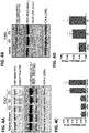
- FIG. 4 shows the detection of TAR DNA-binding protein 43 (TDP-43, transactive response DNA binding protein 43 kDa and its breakdown products (BDPs-39 kDa and 35, 25 kDa) ( FIG 3 ) in brain (cortex) tissue lysate in mice following TBI (controlled cortical impact) and blast overpressure brain injury).
- the detection method shown is quantitative immunblotting (western blots). Other methods such as sandwich ELISA method or amplified sandwich ELISA can also be used.
- the data indicate that TDP-43 BDPs are released from injured brain cells and tissue into biofluids such as cerebrospinal fluid (CSF) ( FIG 5 ) and/or blood, including serum and plasma).
- CSF cerebrospinal fluid
- This TDP-43 and BDP release pattern is comparable to alphaII-spectrin BDP (SBDP150, 145) release into CSF after TBI in humans.
- the detection method used is quantitative immunblotting (western blots). Other methods such as sandwich ELISA method or amplified sandwich ELISA can also be used, as demonstrated by SBDP.
- TDP-43 and its BDPs are likely released from injured brain cells and tissue into biofluids such as cerebrospinal fluid (CSF) and blood, including serum and plasma, causing the biofluid levels of TDP-43 and its BDPs to rise after CNS-injury.
- CSF cerebrospinal fluid
- This example also demonstrates that TDP-43 and BDP quantification tests can distinguish TBI from control subjects using biofluids with the first 24 h, and possibly longer.
- FIGs. 6 and 7 show the detection of serum total microtubule-associated protein Tau (T-Tau) and phosphorylated form of Tau; i.e., P-Tau (at Serine-202) and P-Tau/T-Tau ratio ( FIG 2 ) elevations after both single and repetitive closed head injury (CHI, rCHI) and single and repetitive blast overpressure brain injury (OBI, rOBI) in mice at different time points post injury (day 1, 3, 7, 10, 14, and/or 30).
- the detection method used is quantitative amplified sandwich ELISA. Other sensitive immunoassay formats can be used.
- FIG. 8 shows the detection of total Tau and P-Tau markers in human acute severe TBI CSF samples.
- total Tau is elevated in acute TBI CSF groups in comparison to control CSF, P-Tau and P-Tau/T-Tau ratio elevations are even more robust in the TBI SCI group.
- This example demonstrates that Tau, P-Tau tests can distinguish TBI from control subjects using biofluids with the first five days. CSF Tau and P-Tau assays were run as described above.
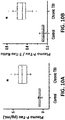
- FIG. 9 shows the detection of total Tau, P-Tau, and P-Tau/T-Tau ratio in Day 1-3 and Day 30 (range 19-50 days) severe TBI serum samples that are higher than the levels in the normal control serum. It further shows that in addition to differential P-Tau, Tau levels, the use of P-Tau/T-Tau ratio is a useful diagnostic method to differentiate acute (Day 1-3) and chronic (average Day 30) phases of severe human TBI serum samples versus normal controls. Serum/plasma Tau and P-Tau assays were run as described above.
- the T-Tau, P-Tau and P-Tau/T-Tau ratio can differentiate control vs. different TBI severity grouped - GCS 13-15 (mild), GCS 9-12 (moderate) and GCS 3-8 (severe).
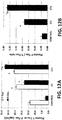
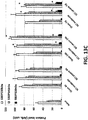
- FIG. 12 shows the Tau, P-tau and P-Tau/T-Tau ratio can differentiate control plasma samples vs. plasma samples from CT normal and CT abnormal TBI patients.
- CT- or cranial Computed Tomography scan normal
- TBI CT+; or cranial CT scan showing pathology
- Rat Fischer rats (220-250 g) were housed according to the National Institutes of Health and United States Department of Agriculture guidelines. Institutional Animal Care and Use Committee of the University of Miami approved all animal procedures. Prior to surgery, rats were anesthetized (45 mg ketamine /kg, 5mg xylazine /kg) by intraperitoneal injection. An adequate level of anesthesia was determined by monitoring the corneal reflex and withdrawal to painful stimuli for hind limbs. All animals underwent T9-T10 spinal laminectomy. During surgery, rats were placed on a warming pad to maintain body temperature at 37° ⁇ 0.5°C.
- the rat was placed ventrally on top of a small bit of sterile gauze to evaluate the surgical site presenting an adequate exposure of the back anatomy.
- a 2 cm longitudinal skin incision was centered over the T9 spinus process along the midline.
- perispinal nerves and ligaments were laterally dissected and retracted followed by removal of bony elements of the posterior spine including the spinus process in lamina using a micro Rongiers.
- the ninth thoracic spinal segment was then exposed without removing the dura mata by removing the dorsal part of the vertebra.
- the exposed cord was next contused by a 10 gm weight dropped from a height of either 12.5mm (moderate), or 25mm (severe) by using the New York University (NYU)-MASCIS impactor ( FIG. 16 ).
- This model and injury severities have been shown in Dr. Dalton Dietric's laboratory at University of Miami as well as others to produce well-described pattern of electrophysiological, behavioral and histopathological consequences.
- CSF samples were drawn at 4, 24 or 48 hrs post-trauma. CSF was collected as previously described. At appropriate time points, injured, sham-injured and naive animals were anesthetized as described above and secured in a stereotactic frame with the head allowed to move freely along the longitudinal axis. The head was flexed so that the external occipital protuberance in the neck was prominent and a dorsal midline incision was made over the cervical vertebrae and occiput. The atlanto-occipital membrane was exposed by blunt dissection and a 25 gauge needle attached to polyethylene tubing was inserted into the cisterna magna.
- CSF samples were centrifuged at 4,000xg for 4 min. at 4°C to clear any contaminating erythrocytes.
- Lysis was conducted for 3 hours at 4 °C with hourly vortexing. Lysates were then centrifuged to remove DNA, lipids, and particulates at 15,000 g for 10 min at 4 °C. The supernatant was collected and protein content was determined using a DC Protein Assay (BioRad) then, the protein concentration was standardized to 1 ⁇ g/ ⁇ l for immunoblotting analysis. Pooled 1-mg SCI and control samples were prepared for differential analysis using CAX-PAGE.
- CSF samples from traumatic brain injury (TBI) and spinal cord injury (SCI) were used.
- CSF from a severe TBI study was collected from consenting adult subjects presenting to the Emergency Department.
- GCS Glasgow coma scale
- IVC intraventriculostomy
- CSF was sampled from the buretrol of the CSF drainage system by a qualified and trained hospital employee according to the hospital's standard procedures.
- timed CSF samples (10 mL) with a total collection time not exceeding 1 hr were collected into 15-mL conical polypropylene centrifuge tubes (BD Falcon).
- the CSF samples (5-10-mL) were then centrifuged at 4,000 RPM with a tabletop centrifuge at room temperature for 5 - 7 minutes to remove loose cells and debris.
- One mL aliquots of cleared CSF (supernatant) were pipetted into 2 mL cryogenic tubes and snap-frozen and stored at -80 °C in an ultralow temperature freezer until use.
- AIS Grades A, B & C spinal cord injury subjects
- AIS Grades A, B & C spinal cord injury subjects
- AIS on discharge improvement
- urodynamic test bladedder control
- somatosensory evoked potential test somatosensory evoked potential test
- MRI magnetic resonance
- CSF collection strict aseptic techniques were conducted when a lumbar puncture was performed at L2-3 or L3-4 and an intrathecal catheter inserted for intrathecal drainage of CSF.
- Timed CSF samples were diverted to glass tubes (no preservative, heparin or EDTA) for the specified vol. (3 cc), spun at 4,000 RPM at room temperature (to remove loose cells and debris). Aliquots of 500 ⁇ L of cleared CSF (supernatant) were placed into 1.2 ml cryovials and stored at - 80 °C in an ultralow temperature freezer until used. In this study, CSF samples were collected within 48h.
- the fractionated proteins were transferred to a polyvinylidene difluoride (PVDF) membrane (Invitrogen) by electroblotting using the iBlot Gel Transfer Device (Invitrogen) for seven minutes. Following the transfer, the membranes were blocked in 5% nonfat dry milk in TBST (20 mM Tris-HCl, 150 mM NaCl and 0.003% Tween-20, pH 7.5) for an hour.
- PVDF polyvinylidene difluoride
- Immunoblotting membranes containing tissue protein were incubated with the primary antibody overnight at 4 °C with shaking.
- Monoclonal anti-mouse ⁇ -spectrin (Enzo Life Sciences NY, USA), polyclonal anti-rabbit GFAP (Abcam, MA, USA), and monoclonal anti-mouse UCHL-1 (EMD Milipore, MA, USA) were used at a dilution of 1:1000 in 5% milk.
- Polyclonal anti-rabbit transferrin (Abcam, MA, USA), polyclonal anti-goat cathepsin D (Santa Cruz Biotechnology, TX, USA), polyclonal anti-rabbit triosephosphate isomerase (TIM) (Santa Cruz Biotechnology, TX, USA), and polyclonal anti-rabbit astrocytic phosphoprotein (PEA-15) (Cell Signaling, MA, USA) were used at a dilution of 1:500, 1:200, and 1:1000, respectively in 5% milk.
- TIM polyclonal anti-rabbit triosephosphate isomerase
- PEA-15 polyclonal anti-rabbit astrocytic phosphoprotein
- the membranes were washed three times with TBST and probed with an alkaline phosphatase-conjugate goat secondary antibody (EMD Milipore, MA, USA) at a dilution of 1:5000 in 5% milk for an hour, followed by TBST washing.
- Immunoreactivity was detected using 5-bromo-4-chloro-3-indolylphosphate (BCIP)/nitroblue tetrazolium phosphatase substrate (Kirkegaard & Perry Laboratories, Gaithersburg, MD, USA). Immunoreactive bands are imaged by digital scanner and protein band density is then quantified by NIH ImageJ software.
- Serum/plasma (30-100 uL), the CSF (0.5-10 uL) and/or brain tissue lysate (2- 20 ug total protein equivalent) samples will be processed for 96-well plate sandwich ELISA biomarker analysis format, together with different amount (0.020 ng to 200 ng) of standard biomarker protein (for standard curve generation).
- This method is used to measure TDP-43 and BDPs, Tau, P-Tau, GFAP and BDP, UCH-L1, cathepsin D, Transferrin, PEA-15, and TPI-1 (TIM).
- sandwich ELISA contains one capture antibody and one detection antibody for the target protein. These antibodies can be polyclonal (rabbit) or monoclonal (mouse).
- the detection antibody is either labeled with florescent dye or coupled to horse radish peroxidase (HRP) or with biotin for streptavidin- HRP based amplification. Assays will be performed on a Spectamax190-Pro microplate reader. Alternately, include P-tau and total Tau (commercially total tau ELISA kits were purchased from called Innotest hTau Ag (#80226) and INNOTEST® PHOSPHO-TAU (181P) (#80323) (Innogenetics). NF-H, GFAP ELISA from Biovendor, UCH-L1 can be from USCNcompany (www.uscnk.us). Alternatively for high sensitivity, P-Tau and Tau, established surround sound fiber optic immunoassay (SOFIA), rolling circle-amplification ELISA method or digital ELISA (Quanterix SIOMA system) are used.
- SOFIA surround sound fiber optic immunoassay
- MGIMA digital ELISA
- CAX-PAGE RPLC-Tandem Mass spectrometry was based on an established method. It includes a comprehensive multi-dimensional separation scheme, with cationic/anionic-exchange (CAX) chromatography, followed by 1D polyacrylamide gel electrophoresis (PAGE) to reduce protein complexity. A reversed phase liquid chromatography-tandem mass spectrometry (LC-MS/MS) proteomic platform is then utilized to examine and characterizes proteome changes associated with SCI.
- CAX cationic/anionic-exchange
- PAGE polyacrylamide gel electrophoresis
- a reversed phase liquid chromatography-tandem mass spectrometry (LC-MS/MS) proteomic platform is then utilized to examine and characterizes proteome changes associated with SCI.
- the CAX chromatography was performed on a BioRad Biologic DuoFlow system with sulfoproyl SCX (S1) and quaternary ammonium SAX (Q1) modified Sepharose prepacked ion-exchange columns (BioRad) that were placed in series, and connected in tandem along with a QuadTec UV detector and BioFrac fraction collector.
- Buffers consisted of 20 mM Tris-HCl (pH 7.5 molecular biology grade, Fisher Scientific) in double distilled water (mobile phase A) and 20 mM Tris-HCl with 1 M NaCl (Fisher Scientific, crystalline 99.8% certified) in double distilled water (mobile phase B).
- Samples of 1mg proteins from pooled lysates were injected with an optimized method for differential analysis.
- the first and second linear gradient steps were from 0 to 5% B, and 5 to 10% in 5 mL each at a flow rate of 1 mL/min, and followed by a six-step gradient, each step increased by 5% from 10 to 40% in 1 mL, the last gradient step was from 40 to 50% in 1 ml.
- the composition was held at 50% B for 1 mL and reequilibrated to 0% B in 3 mL.
- the UV chromatograms were monitored at a wavelength of 280 and 214 nm for each run. Forty four 1-mL fractions were autonomously collected via the BioFrac fraction collector into 1.5-mL screw-cap microfuge tubes kept on ice.
- Nano-reversed-phase liquid chromatography tandem mass spectrometry was employed for protein separation and identification. Nanoflow was performed on a NanoAcquity UPLC (Waters, Milford, MA, USA); the autosampler was used to load two microliters of each sample onto a 5 ⁇ m particle size Symmetry 180 ⁇ m x 20 mm C18 trapping column at 4 ⁇ L/min for 10 min. Then, the sample plug was loaded onto a 1.7 ⁇ M particle size BEH130 C18 100 ⁇ m x 100 mm analytical column at 300 nL/min. The mobile phase consisted of solvent A (water with 0.1% formic acid) and solvent B (acetonitrile with 0.1% formic acid).
- Tandem mass spectra were collected on LTQ-XL (Thermo, San Jose, CA, USA) using a Data Dependent Acquisition method in Xcalibur 2.0.7(Thermo), in which data dependent scanning was specified as a criteria to select the top 10 most abundant ions using 11 separate scan events at a given chromatographic time point (115 min) for subsequent analysis.
- the mass spectrometer was set to perform a full-scan and subsequently MS/MS scans on the ten most intense ions in the full-scan spectrum MS (scan event 1) with Dynamic Exclusion enabled. Dynamic Exclusion temporarily puts a mass into an exclusion list after its MS/MS spectrum is acquired, providing the opportunity to collect MS/MS information on the second most intense ion from the full-scan spectrum MS (scan event 1). All MS/MS spectra were analyzed using Proteome Discoverer 1.3 (Thermo). SEQUEST (version: 1.3.0.339) and X! Tandem (version: CYCLONE (2010.12.01.1)).
- Database search engines were set up to search a trypsin-Indexed uniprot-Rattus+norvegicus.fasta (unknown version, 35126 entries). The search was achieved using the average mass for matching the precursor with a fragment ion mass tolerance of 0.8 Da and a parent ion tolerance of 2.00 Da. Carbamidomethylation of cysteine was selected as a static modification, while the oxidation of methionine was selected as a dynamic modification. Using the output from SEQUEST and X! Tandem, Scaffold (version: Scaffold_3.3.3, Proteome Software) was used to validate, organize, and interpret mass spectrometry data. Peptide identifications were accepted if they could be established at greater than 95.0% probability as specified by the Peptide Prophet algorithm. Protein identifications were accepted if they could be established at greater than 99.9% probability and contained at least 2 identified peptides.
- rat SCI tissue (caudal segment from epicenter, 4 hours, day 1 and day 7 post-injury), and CAX-PAGE RPLC-tandem mass spectrometry proteomic method
- protein biomarker candidates could be identified.
- Additional spinal cord tissue (caudal, and rostral segment and epicenter segment, Cerebrospinal fluid (CSF) and blood samples) form the rat study and CSF and serum/plasma from injured human patients was also used for clinical studies of SCI patients.
- Panels of SCI candidate biomarkers were identified that can be used to correlate with disease progression or to identify potential SCI therapeutic targets (shown in Tables 2, 3). This was followed by cross-validating these biomarker candidates in biofluid from human TBI subjects.
- the identified biomarkers have diagnostic, prognostic and managing utilities for all neurotrauma types (including both TBI and SCI).
- CAX chromatography was employed to fractionate SCI samples and control proteins according to their surface charges followed by 1D gel electrophoresis to separate proteins based on their molecular weight.
- CAX separation has the ability of retaining both positively and negatively charged proteins.
- Both sham and SCI proteins were distributed into 28 fractions (1ml) and collected after using tandem CAX columns. Protein fractions were analyzed side-by-side on a ID-gel for differential comparison, 24 h sham vs. 24 h SCI and 7 d sham vs. 7 d SCI. Differential bands were boxed and labeled according to their 2D position with number and letter (e.g. the top band excised from the lane of fraction X was labeled XA). ImageJ densitometry software was used for quantification of selected differential gel band intensities to derive the relative fold increase or decrease.
- Differentially displayed bands were selected from the ID-gels and cut into pieces for proteomic analysis, then trypsinized. Trypsinized peptides were separated by reversed-phase liquid chromatography online with tandem mass spectrometry (RPLC-MS/MS) for protein identification. All MS/MS spectra were analyzed using Proteome Discoverer 1.3 (Thermo), SEQUEST and X! Tandem database search engines have been utilized to search the experimental spectra against a rat indexed database and validated by Scaffold 3. Based on identified unique peptides, sequence coverage, and molecular weight gel band, the protein biomarker candidates were isolated in each band.
- the proteins with increased levels post-SCI included aldehyde dehydrogenase 4 family, member A1 (Aldh4a1) protein (Fragment), LOC367586 protein, aminoacylase-lA, transketolase, Gamma-enolase, elongation factor 2, protein Tln1, and peroxiredoxin-2.
- aldo-keto reductase family 1, member B10 aldo-keto reductase
- pyruvate kinase PKM pyruvate kinase PKM
- Acyl-CoA synthetase family member 2 mitochondrial
- protein-L-isoaspartate D-aspartate O-methyltransferase
- proteins with decreased levels post-SCI 24 h were isocitrate dehydrogenase [NADP], mannose-6-phosphate isomerase, pyridoxal kinase, stathmin, and peripheral myelin protein 2 (Pmp2).
- alcohol dehydrogenase [NADP(+)], L-lactate dehydrogenase B chain, ribosyldihydronicotinamide dehydrogenase [quinone], and ATP citrate lyase-CRA a isoform.
- Some differentially expressed proteins are common in both 24 h and 7 d time points, annexin A1& A2, glyceraldehyde-3-phosphate dehydrogenase, Pgm1 protein (Fragment), glutathione S-transferase Mu 5, triosephosphate isomerase, and transferrin (serotransferrin). Also, the presence of the same proteins were identified in both up and down regulated bands.
- SBDP ⁇ II- spectrin breakdown products
- biomarker candidates Five biomarker candidates were selected, blood brain barrier (BBB) located transferrin, lysosomal protease cathepsin D, triosephosphate isomerase (TPI-1), astrocytic phosphoprotein-15 kDa (PEA-15), and potential stroke biomarker nucleoside diphosphate kinase A to confirm their protein alterations associated with SCI.
- BBB blood brain barrier
- TPI-1 triosephosphate isomerase
- PEA-15 astrocytic phosphoprotein-15 kDa
- potential stroke biomarker nucleoside diphosphate kinase A to confirm their protein alterations associated with SCI.
- the proteolytic damage markers to the spinal cord structural and cellular components were validated by quantitative immunblotting using specific antibodies for each protein.
- FIG. 13 shows the SCI-tissue immunoblotting validation of representative protein biomarkers.
- time course of post-SCI biomarkers validation illustrated by Western blot of rat spinal cord tissue (epicenter) lysate of rostral section (sham, and two SCI severity) collected at three time points (4h, 24h and 7 days), the known markers probed are alphaII-spectrin and GFAP and MBP.
- immunoblotting quantification of spinal cord tissue lysate samples sham and two SCI severity levels at 3 time points
- alphaII-spectrin breakdown product SBDP150, 145, 120, GFAP and GFAP-BDP 44 and 40 KDa
- All biomarkers are elevated after injury except that MBP marker is reduced in tissue at 7 day after SCI.
- FIG. 14 shows the SCI-tissue immunoblotting validation of representative protein biomarkers.
- time course of post-SCI new biomarker candidates validation illustrated by Western blot of rat spinal cord tissue (rostral) lysate of rostral section (sham, and two SCI severity) collected at three time points (4h, 24h and 7 days), Novel candidate markers probed include transferrin, cathepsin D, TPI-1 and PEA-15.
- Western blot of carbonic anhydrase II served as a loading control (not shown)
- bottom panel immunoblotting quantification of spinal cord tissue lysate samples for post-TBI increases of cathepsin D, TPI-1 and PEA-15 biomarkers. All markers are elevated at 4 h and 24 h post-SCI, except that CathD has a delayed increase after injury (at day 7).
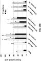
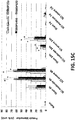
- FIG. 15 further illustrates the detectability of representative SCI biomarker elevations in rat CSF after SCI by immunoblotting method.
- panel (A) Time course of post-SCI biomarkers release and new candidate marker validation illustrated by Western blot quantification of rat spinal cord CSF samples (sham, and two SCI severity) collected at three time points (4h, 24h and 7 days), the markers probed are alphaII-spectrin (SBDPs), Transferrin, GFAP, GFAP-BDPs and TPI-1.
- the detection method shown is quantitative immunblotting (western blots). Other methods such as sandwich ELISA method or amplified sandwich ELISA can also be used.
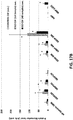
- FIG. 16 shows the time course of alphaII-spectrin-BDPs (SBDPs), Transferrin, GFAP, GFAP-BDPs and TPI-1, CathD and PEA-15 biomarker release into human CSF (2 patients) after SCI, detected by immunoblotting.
- SCI biomarkers alphaII-spectrin-BDPs (SBDPs), Transferrin, GFAP, GFAP-BDPs and TPI-1, CathD and PEA-15
- SCI biomarkers alphaII-spectrin-BDPs (SBDPs), Transferrin, GFAP, GFAP-BDPs and TPI-1, CathD and PEA-15
- SCI biomarkers alphaII-spectrin-BDPs (SBDPs), Transferrin, GFAP, GFAP-BDPs and TPI-1, CathD and PEA-15
- the detection method shown is quantitative imm
- blood e.g. serum, plasma samples
- Pathway Studio software 9.0 (Ariadne Genomics Inc. MD, USA) was employed to identify significant pathways and interactome across individual samples as a signature to predict clinical outcomes.
- Pathways of brain injury systems and SCI identified candidate biomarkers were generated.
- Various brain systems biomarkers including acute injury, neuroinflammation, neuroregeneration, and neurodegeneration markers (Table 2) are mined from literature and then used to generate brain injury systems biomarker list (Table 3).
- SCI candidate biomarkers can be identified (Table 4) and input separately into Pathway Studio for SB analysis.
- Pathway Studio 9.0 has been utilized to search for possible protein-protein interactions, common regulators, cell processes, and related pathways for associations among the various brain injury systems biomarker proteins (Table 4, Table 5).
- the network was generated using the "Shortest Path” algorithm to map interactions between altered proteins.
- Several processes believed to be central to the pathogenesis of TBI disorders were identified using this search. Some examples of those processes include cell death, inflammation, oxidation, and apoptosis ( FIG. 19 ).
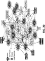
- SCI candidate biomarkers were identified using a rat model of SCI (Table 6) and these candidate biomarker proteins are input into Pathway Studio for SB analysis. Using "Shortest Path” algorithm, potential interactions among these protein components were mapped ( FIG. 20 ). Several Non-redundant converging pathways were identified - they highlight the potential role of neuronal death mechanism, cell invasion, oxidative stress, and cell differentiation pathway as main players on SCI. Additional data and literature-mining and new experiments can be done to further understand the pathological pathways involved in spinal cord structural and functional changes, SCI recovery, complications and other SCI-related co-morbidities. Such maps can also have utilities in the diagnosis, prognosis and management of SCI, TBI or similar CNS injury.
- FIG. 21 shows that human spinal cord injury CSF samples show significant elevations of novel SCI biomarker S100b and IL-6.
- Transferrin, CathD, TPI and PEA-15 were identified as novel neuroinjury biomarkers. Since both SCI and TBI are similar and related neurotrauma, it is likely that Transferrin, CathD, TPI-1 and PEA-15 will also be elevated in biofluids in TBI patients (such as CSF).
- TBI CSF the same markers Transferrin, CathD, TPI-1 and PEA-15 elevation in fact supports this notion (GFAP and GFAP-BDP were assayed also as benchmark).
- FIG. 23 shows that human traumatic brain injury and spinal cord serum samples show elevations of novel biomarker BDNF (mature form) when compared to control serum samples.
- BDNF is a member of the neurotrophin family and related to Nerve Growth Factor. BDNF are found in the brain and the periphery, BDNF acts on neurons of CNS and the peripheral nervous system (PNS), supporting the survival of existing neurons, and facilitating the growth and differentiation of new neurons and synapses. In the brain, it is active in the hippocampus, cortex, and basal forebrain. It is also expressed in the retina, motor neurons, and also present in the kidneys, prostate and saliva.
- PNS peripheral nervous system
- FIG. 24 shows human traumatic brain injury serum compared to a control serum profile of biomarker pro-BDNF.
- BDNF also exists as pro-BDNF form (precursor).
- TBI patient samples used were collected at 24 h and at 240 h (10 days) after injury.
- Pro-BDNF levels were detectably higher at 24 h and at 240 h after TBI.
- the combined use of assaying Pro-BDNF and BNDF might be used as tandem biomarkers for TBI or SCI.
- FIG. 25 shows human and mouse traumatic brain injury serum compared to control serum profile for novel microglial biomarker Iba-1.
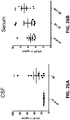
- FIG. 26 shows Visinin-like protien-1 (VSNL1) elevations in human TBI CSF and human spinal cord injury and TBI serum as compared to respective controls.
- Left panel shows CSF samples from control or TBI (day 1) were assayed for VILIP-1 levels.
- VILIP-1 was suggested to be a possible marker for Alzheimer's disease ( Lee et al. Clin Chem. 2008 Aug 14;54(10): 1617-23 ), there are no prior data to suggest that it might be a TBI or SCI biomarkers.
- VSNL1 is a member of the visinin/recoverin subfamily of neuronal calcium sensor proteins. The encoded protein is strongly expressed in granule cells of the cerebellum and also in other neurons in the rest of the brain.
- VILIP-1 is a TBI/SCI biomarker, but also that other visinin-like proteins that are abundant in the CNS (VSNL2, VSNL3) (Table 1) are also possible biomarkers for CNS injury. Table 1.
- Marker Type Biomarkers Acute neuronal injury markers Neurite injury markers (SBDP/MAP2, NF-H, c-Tau, VSNL-1, BDNF, Pro-BDNF), Cell body injury: UCH-L1, NSE,NMDA-R fragment, TPI-1, Cathepsin D), Demyelination (MBP & fragment), and Gliosis/ Glial cell injury (GFAP & BDP; S100b, PEA-15), Blood brain barrier marker (Transferrin, Aquoporin-4) Neurodegeneration markers AD / CTE markers (P-tau e.g.T181, S202, T231 and Tau, TDP-43), AD markers (A ⁇ 1-40, and A ⁇ 1-42), Alpha-synuclein, and Park-7 (DJ-1) Neuroinflammatory markers Cytokines (IL-6, IL-8; TNF-alpha, and IL-10), Microgliosis (iba)
Landscapes
- Life Sciences & Earth Sciences (AREA)
- Health & Medical Sciences (AREA)
- Engineering & Computer Science (AREA)
- Biomedical Technology (AREA)
- Molecular Biology (AREA)
- Chemical & Material Sciences (AREA)
- Urology & Nephrology (AREA)
- Hematology (AREA)
- Immunology (AREA)
- Cell Biology (AREA)
- General Health & Medical Sciences (AREA)
- Biotechnology (AREA)
- Proteomics, Peptides & Aminoacids (AREA)
- Food Science & Technology (AREA)
- Medicinal Chemistry (AREA)
- Physics & Mathematics (AREA)
- Analytical Chemistry (AREA)
- Biochemistry (AREA)
- Microbiology (AREA)
- General Physics & Mathematics (AREA)
- Pathology (AREA)
- Neurology (AREA)
- Neurosurgery (AREA)
- Investigating Or Analysing Biological Materials (AREA)
- Measuring Or Testing Involving Enzymes Or Micro-Organisms (AREA)
- Peptides Or Proteins (AREA)
Applications Claiming Priority (3)
| Application Number | Priority Date | Filing Date | Title |
|---|---|---|---|
| US201461976733P | 2014-04-08 | 2014-04-08 | |
| EP15776837.5A EP3129788A4 (de) | 2014-04-08 | 2015-04-08 | Proteinbiomarker für akute, subakute und chronische traumatische verletzungen des zentralen nervensystems |
| PCT/US2015/024880 WO2015157390A1 (en) | 2014-04-08 | 2015-04-08 | Protein biomarkers for acute, subacute and chronic traumatic injuries of the central nervous system |
Related Parent Applications (1)
| Application Number | Title | Priority Date | Filing Date |
|---|---|---|---|
| EP15776837.5A Division EP3129788A4 (de) | 2014-04-08 | 2015-04-08 | Proteinbiomarker für akute, subakute und chronische traumatische verletzungen des zentralen nervensystems |
Publications (2)
| Publication Number | Publication Date |
|---|---|
| EP3663764A1 true EP3663764A1 (de) | 2020-06-10 |
| EP3663764B1 EP3663764B1 (de) | 2023-11-01 |
Family
ID=54288363
Family Applications (2)
| Application Number | Title | Priority Date | Filing Date |
|---|---|---|---|
| EP15776837.5A Withdrawn EP3129788A4 (de) | 2014-04-08 | 2015-04-08 | Proteinbiomarker für akute, subakute und chronische traumatische verletzungen des zentralen nervensystems |
| EP20153859.2A Active EP3663764B1 (de) | 2014-04-08 | 2015-04-08 | Proteinbiomarker für akute, subakute und chronische traumatische verletzungen des zentralen nervensystems |
Family Applications Before (1)
| Application Number | Title | Priority Date | Filing Date |
|---|---|---|---|
| EP15776837.5A Withdrawn EP3129788A4 (de) | 2014-04-08 | 2015-04-08 | Proteinbiomarker für akute, subakute und chronische traumatische verletzungen des zentralen nervensystems |
Country Status (7)
| Country | Link |
|---|---|
| US (1) | US10725054B2 (de) |
| EP (2) | EP3129788A4 (de) |
| CN (1) | CN106605146B (de) |
| AU (1) | AU2015243926A1 (de) |
| CA (1) | CA2948478A1 (de) |
| GB (1) | GB2548942A (de) |
| WO (1) | WO2015157390A1 (de) |
Families Citing this family (19)
| Publication number | Priority date | Publication date | Assignee | Title |
|---|---|---|---|---|
| US8492107B2 (en) | 2004-04-15 | 2013-07-23 | University Of Florida Research Foundation, Inc. | Neural proteins as biomarkers for nervous system injury and other neural disorders |
| US12601749B2 (en) | 2008-08-11 | 2026-04-14 | Banyan Biomarkers, Inc. | Biomarker detection process and assay of neurological condition |
| JP5781436B2 (ja) | 2008-08-11 | 2015-09-24 | バンヤン・バイオマーカーズ・インコーポレーテッド | 神経学的状態のバイオマーカー検出方法およびアッセイ |
| HK1206425A1 (en) | 2012-03-13 | 2016-01-08 | The Johns Hopkins University | Citrullinated brain and neurological proteins as biomarkers of brain injury or neurodegeneration |
| US11064913B2 (en) | 2013-10-25 | 2021-07-20 | Force Impact Technologies, Inc. | Impact sensing wearable device and method |
| US20180024145A1 (en) | 2015-02-05 | 2018-01-25 | Immunarray USA, Inc. | Methods and compositions for diagnosing brain injury or neurodegeneration |
| US11078298B2 (en) | 2016-10-28 | 2021-08-03 | Banyan Biomarkers, Inc. | Antibodies to ubiquitin C-terminal hydrolase L1 (UCH-L1) and glial fibrillary acidic protein (GFAP) and related methods |
| JP7379165B2 (ja) * | 2017-12-09 | 2023-11-14 | アボット・ラボラトリーズ | Gfapとuch-l1との組合せを使用する、ヒト対象における外傷性脳損傷を診断及び査定する一助となるための方法 |
| CN111699391A (zh) | 2017-12-29 | 2020-09-22 | 雅培实验室 | 用于诊断和评估创伤性脑损伤的新型生物标志物和方法 |
| US20210270847A1 (en) * | 2018-08-17 | 2021-09-02 | University Of Florida Research Foundation, Incorporated | Protein and peptide biomarkers for traumatic injury to the central nervous system |
| WO2020124013A1 (en) * | 2018-12-13 | 2020-06-18 | Gryphon Bio, Inc. | Combinatorial temporal biomarkers and precision medicines with detection and treatment methods for use in neuro injury, neuro disease, and neuro repair |
| US11389113B2 (en) | 2018-12-20 | 2022-07-19 | Force Impact Technologies, Inc. | Mouth guard having user-notification feature of impact force |
| CN110031625A (zh) * | 2019-03-31 | 2019-07-19 | 苏州市立医院 | 包含葡萄糖磷酸变位酶5抗体的检测试剂及其应用 |
| EP3969913A1 (de) * | 2019-05-16 | 2022-03-23 | Centre National De La Recherche Scientifique (Cnrs) | Biomarker zur beurteilung einer rückenmarksverletzung |
| US11738340B2 (en) | 2019-10-04 | 2023-08-29 | Javelin Biotech, Inc. | Neurodegenerative target discovery platform |
| CN111999403A (zh) * | 2020-08-10 | 2020-11-27 | 新乡医学院 | 一种瓦斯爆炸肺损伤诊断系统、血清标志物筛选方法、肺损伤作用机制研究方法 |
| CN114250291A (zh) * | 2021-11-22 | 2022-03-29 | 四川大学 | 基因Nrgn在诊断爆炸性脑损伤的产品中的应用 |
| CN118707112B (zh) * | 2024-08-28 | 2024-12-13 | 浙江格物致知生物科技有限公司 | 一种gfap、nfl和uch-l1联合检测试剂盒及其用于脑创伤早期检测 |
| CN119470922B (zh) * | 2024-11-29 | 2026-04-21 | 长沙纽康度生物科技有限公司 | 一种用于创伤性脑损伤检测的生物标记物及其应用 |
Citations (2)
| Publication number | Priority date | Publication date | Assignee | Title |
|---|---|---|---|---|
| WO2001055725A2 (en) * | 2000-01-24 | 2001-08-02 | Innogenetics N.V. | Diagnosis of tauopathies determining tau/phospho-tau ratio |
| WO2011032155A2 (en) * | 2009-09-14 | 2011-03-17 | Banyan Biomarkers, Inc. | Micro-rna, autoantibody and protein markers for diagnosis of neuronal injury |
Family Cites Families (9)
| Publication number | Priority date | Publication date | Assignee | Title |
|---|---|---|---|---|
| AU8410698A (en) * | 1997-07-17 | 1999-02-10 | General Hospital Corporation, The | Lipid hydroperoxides to detect brain injury |
| CA2351562A1 (en) * | 1998-12-09 | 2000-06-15 | Frank P. Zemlan | A method for detecting neuronal cell damage by quantification of map-2 levels in biological fluids |
| WO2002030261A2 (en) * | 2000-10-09 | 2002-04-18 | Brainz Instruments Limited | Sensor assembly for monitoring an infant brain |
| DE102004025726B4 (de) * | 2004-05-26 | 2006-07-06 | Roder, Hanno, Dr. | Verwendung eines spezifischen K252a-Derivats zur Verhinderung oder Behandlung der Alzheimerschen Krankheit |
| US8354236B2 (en) * | 2006-09-29 | 2013-01-15 | The Trustees Of The University Of Pennsylvania | Detection of neurodegenerative disease |
| WO2008151055A1 (en) * | 2007-06-01 | 2008-12-11 | Mayo Foundation For Medical Education And Research | Diagnosing neurodegenerative diseases |
| JP5781436B2 (ja) * | 2008-08-11 | 2015-09-24 | バンヤン・バイオマーカーズ・インコーポレーテッド | 神経学的状態のバイオマーカー検出方法およびアッセイ |
| EP2812697B1 (de) * | 2012-02-06 | 2017-03-22 | University of Miami | Angeborene immunproteine als biomarker für läsionen des zentralen nervensystems |
| TWI429908B (zh) * | 2012-02-17 | 2014-03-11 | Univ Taipei Medical | 腦外傷神經傷害之體外檢測方法 |
-
2015
- 2015-04-08 CN CN201580030441.9A patent/CN106605146B/zh not_active Expired - Fee Related
- 2015-04-08 EP EP15776837.5A patent/EP3129788A4/de not_active Withdrawn
- 2015-04-08 WO PCT/US2015/024880 patent/WO2015157390A1/en not_active Ceased
- 2015-04-08 GB GB1618825.2A patent/GB2548942A/en not_active Withdrawn
- 2015-04-08 CA CA2948478A patent/CA2948478A1/en not_active Abandoned
- 2015-04-08 EP EP20153859.2A patent/EP3663764B1/de active Active
- 2015-04-08 AU AU2015243926A patent/AU2015243926A1/en not_active Abandoned
- 2015-04-08 US US15/308,934 patent/US10725054B2/en active Active
Patent Citations (2)
| Publication number | Priority date | Publication date | Assignee | Title |
|---|---|---|---|---|
| WO2001055725A2 (en) * | 2000-01-24 | 2001-08-02 | Innogenetics N.V. | Diagnosis of tauopathies determining tau/phospho-tau ratio |
| WO2011032155A2 (en) * | 2009-09-14 | 2011-03-17 | Banyan Biomarkers, Inc. | Micro-rna, autoantibody and protein markers for diagnosis of neuronal injury |
Non-Patent Citations (5)
| Title |
|---|
| LEE ET AL., CLIN CHEM., vol. 54, no. 10, 14 August 2008 (2008-08-14), pages 1617 - 23 |
| S. MAGNONI ET AL: "Tau elevations in the brain extracellular space correlate with reduced amyloid-? levels and predict adverse clinical outcomes after severe traumatic brain injury", BRAIN, vol. 135, no. 4, 23 November 2011 (2011-11-23), pages 1268 - 1280, XP055174653, ISSN: 0006-8950, DOI: 10.1093/brain/awr286 * |
| SHITAKA, J NEUROPATHOL EXP NEUROL, vol. 70, no. 7, July 2011 (2011-07-01), pages 551 - 67 |
| YAMASHITA T ET AL., NATURE COMMUNICATIONS, vol. 3, 2012, pages 1307 |
| ZHANG YJ ET AL., JOURNAL OF NEUROSCIENCE, vol. 27, 2007, pages 10530 - 10534 |
Also Published As
| Publication number | Publication date |
|---|---|
| CA2948478A1 (en) | 2015-10-15 |
| WO2015157390A9 (en) | 2016-12-15 |
| US20170146555A1 (en) | 2017-05-25 |
| EP3663764B1 (de) | 2023-11-01 |
| GB2548942A (en) | 2017-10-04 |
| AU2015243926A9 (en) | 2019-08-01 |
| EP3129788A1 (de) | 2017-02-15 |
| US10725054B2 (en) | 2020-07-28 |
| WO2015157390A1 (en) | 2015-10-15 |
| AU2015243926A1 (en) | 2016-11-24 |
| EP3129788A4 (de) | 2018-04-04 |
| CN106605146B (zh) | 2020-07-28 |
| CN106605146A (zh) | 2017-04-26 |
Similar Documents
| Publication | Publication Date | Title |
|---|---|---|
| EP3663764B1 (de) | Proteinbiomarker für akute, subakute und chronische traumatische verletzungen des zentralen nervensystems | |
| US11221342B2 (en) | Neural proteins as biomarkers for nervous system injury and other neural disorders | |
| US7396654B2 (en) | Neural proteins as biomarkers for traumatic brain injury | |
| Moghieb et al. | Differential neuroproteomic and systems biology analysis of spinal cord injury | |
| JP6021187B2 (ja) | 自閉症の代謝バイオマーカー | |
| US20210270847A1 (en) | Protein and peptide biomarkers for traumatic injury to the central nervous system | |
| Ramström et al. | Cerebrospinal fluid protein patterns in neurodegenerative disease revealed by liquid chromatography‐Fourier transform ion cyclotron resonance mass spectrometry | |
| Gonzalez et al. | Identification of novel candidate protein biomarkers for the post-polio syndrome—implications for diagnosis, neurodegeneration and neuroinflammation | |
| Laila et al. | Role of proteomics in the discovery of autism biomarkers | |
| CN101661032A (zh) | 帕金森病的生物标志物 | |
| Heineman et al. | Challenges and Opportunities of Early Parkinson’s Disease Biomarkers: α-Synuclein, Leucine-Rich Repeat Kinase 2 (LRRK2), DJ-1, and microRNAs | |
| CN109870583B (zh) | 急性胰腺炎相关的代谢物及其应用 | |
| Wang | Ubiquitin C‐terminal hydrolase‐L1 as a biomarker for ischemic and traumatic brain injury in rats |
Legal Events
| Date | Code | Title | Description |
|---|---|---|---|
| PUAI | Public reference made under article 153(3) epc to a published international application that has entered the european phase |
Free format text: ORIGINAL CODE: 0009012 |
|
| STAA | Information on the status of an ep patent application or granted ep patent |
Free format text: STATUS: THE APPLICATION HAS BEEN PUBLISHED |
|
| AC | Divisional application: reference to earlier application |
Ref document number: 3129788 Country of ref document: EP Kind code of ref document: P |
|
| AK | Designated contracting states |
Kind code of ref document: A1 Designated state(s): AL AT BE BG CH CY CZ DE DK EE ES FI FR GB GR HR HU IE IS IT LI LT LU LV MC MK MT NL NO PL PT RO RS SE SI SK SM TR |
|
| STAA | Information on the status of an ep patent application or granted ep patent |
Free format text: STATUS: REQUEST FOR EXAMINATION WAS MADE |
|
| 17P | Request for examination filed |
Effective date: 20201208 |
|
| RBV | Designated contracting states (corrected) |
Designated state(s): AL AT BE BG CH CY CZ DE DK EE ES FI FR GB GR HR HU IE IS IT LI LT LU LV MC MK MT NL NO PL PT RO RS SE SI SK SM TR |
|
| GRAP | Despatch of communication of intention to grant a patent |
Free format text: ORIGINAL CODE: EPIDOSNIGR1 |
|
| STAA | Information on the status of an ep patent application or granted ep patent |
Free format text: STATUS: GRANT OF PATENT IS INTENDED |
|
| INTG | Intention to grant announced |
Effective date: 20230330 |
|
| RIN1 | Information on inventor provided before grant (corrected) |
Inventor name: RUBENSTEIN, RICHARD Inventor name: MOGHIEB, AHMED Inventor name: YANG, ZHIHUI Inventor name: WANG, KEVIN KA-WANG |
|
| P01 | Opt-out of the competence of the unified patent court (upc) registered |
Effective date: 20230417 |
|
| GRAS | Grant fee paid |
Free format text: ORIGINAL CODE: EPIDOSNIGR3 |
|
| GRAA | (expected) grant |
Free format text: ORIGINAL CODE: 0009210 |
|
| STAA | Information on the status of an ep patent application or granted ep patent |
Free format text: STATUS: THE PATENT HAS BEEN GRANTED |
|
| RAP1 | Party data changed (applicant data changed or rights of an application transferred) |
Owner name: THE RESEARCH FOUNDATION FOR THE STATE UNIVERSITY OF NEW YORK Owner name: UNIVERSITY OF FLORIDA RESEARCH FOUNDATION, INC. |
|
| AC | Divisional application: reference to earlier application |
Ref document number: 3129788 Country of ref document: EP Kind code of ref document: P |
|
| AK | Designated contracting states |
Kind code of ref document: B1 Designated state(s): AL AT BE BG CH CY CZ DE DK EE ES FI FR GB GR HR HU IE IS IT LI LT LU LV MC MK MT NL NO PL PT RO RS SE SI SK SM TR |
|
| REG | Reference to a national code |
Ref country code: GB Ref legal event code: FG4D |
|
| REG | Reference to a national code |
Ref country code: CH Ref legal event code: EP |
|
| REG | Reference to a national code |
Ref country code: IE Ref legal event code: FG4D |
|
| REG | Reference to a national code |
Ref country code: DE Ref legal event code: R096 Ref document number: 602015086424 Country of ref document: DE |
|
| REG | Reference to a national code |
Ref country code: LT Ref legal event code: MG9D |
|
| REG | Reference to a national code |
Ref country code: NL Ref legal event code: MP Effective date: 20231101 |
|
| PG25 | Lapsed in a contracting state [announced via postgrant information from national office to epo] |
Ref country code: GR Free format text: LAPSE BECAUSE OF FAILURE TO SUBMIT A TRANSLATION OF THE DESCRIPTION OR TO PAY THE FEE WITHIN THE PRESCRIBED TIME-LIMIT Effective date: 20240202 |
|
| PG25 | Lapsed in a contracting state [announced via postgrant information from national office to epo] |
Ref country code: IS Free format text: LAPSE BECAUSE OF FAILURE TO SUBMIT A TRANSLATION OF THE DESCRIPTION OR TO PAY THE FEE WITHIN THE PRESCRIBED TIME-LIMIT Effective date: 20240301 |
|
| PG25 | Lapsed in a contracting state [announced via postgrant information from national office to epo] |
Ref country code: LT Free format text: LAPSE BECAUSE OF FAILURE TO SUBMIT A TRANSLATION OF THE DESCRIPTION OR TO PAY THE FEE WITHIN THE PRESCRIBED TIME-LIMIT Effective date: 20231101 |
|
| REG | Reference to a national code |
Ref country code: AT Ref legal event code: MK05 Ref document number: 1627783 Country of ref document: AT Kind code of ref document: T Effective date: 20231101 |
|
| PG25 | Lapsed in a contracting state [announced via postgrant information from national office to epo] |
Ref country code: NL Free format text: LAPSE BECAUSE OF FAILURE TO SUBMIT A TRANSLATION OF THE DESCRIPTION OR TO PAY THE FEE WITHIN THE PRESCRIBED TIME-LIMIT Effective date: 20231101 |
|
| PG25 | Lapsed in a contracting state [announced via postgrant information from national office to epo] |
Ref country code: AT Free format text: LAPSE BECAUSE OF FAILURE TO SUBMIT A TRANSLATION OF THE DESCRIPTION OR TO PAY THE FEE WITHIN THE PRESCRIBED TIME-LIMIT Effective date: 20231101 |
|
| PG25 | Lapsed in a contracting state [announced via postgrant information from national office to epo] |
Ref country code: ES Free format text: LAPSE BECAUSE OF FAILURE TO SUBMIT A TRANSLATION OF THE DESCRIPTION OR TO PAY THE FEE WITHIN THE PRESCRIBED TIME-LIMIT Effective date: 20231101 |
|
| PG25 | Lapsed in a contracting state [announced via postgrant information from national office to epo] |
Ref country code: NL Free format text: LAPSE BECAUSE OF FAILURE TO SUBMIT A TRANSLATION OF THE DESCRIPTION OR TO PAY THE FEE WITHIN THE PRESCRIBED TIME-LIMIT Effective date: 20231101 Ref country code: LT Free format text: LAPSE BECAUSE OF FAILURE TO SUBMIT A TRANSLATION OF THE DESCRIPTION OR TO PAY THE FEE WITHIN THE PRESCRIBED TIME-LIMIT Effective date: 20231101 Ref country code: IS Free format text: LAPSE BECAUSE OF FAILURE TO SUBMIT A TRANSLATION OF THE DESCRIPTION OR TO PAY THE FEE WITHIN THE PRESCRIBED TIME-LIMIT Effective date: 20240301 Ref country code: GR Free format text: LAPSE BECAUSE OF FAILURE TO SUBMIT A TRANSLATION OF THE DESCRIPTION OR TO PAY THE FEE WITHIN THE PRESCRIBED TIME-LIMIT Effective date: 20240202 Ref country code: ES Free format text: LAPSE BECAUSE OF FAILURE TO SUBMIT A TRANSLATION OF THE DESCRIPTION OR TO PAY THE FEE WITHIN THE PRESCRIBED TIME-LIMIT Effective date: 20231101 Ref country code: BG Free format text: LAPSE BECAUSE OF FAILURE TO SUBMIT A TRANSLATION OF THE DESCRIPTION OR TO PAY THE FEE WITHIN THE PRESCRIBED TIME-LIMIT Effective date: 20240201 Ref country code: AT Free format text: LAPSE BECAUSE OF FAILURE TO SUBMIT A TRANSLATION OF THE DESCRIPTION OR TO PAY THE FEE WITHIN THE PRESCRIBED TIME-LIMIT Effective date: 20231101 Ref country code: PT Free format text: LAPSE BECAUSE OF FAILURE TO SUBMIT A TRANSLATION OF THE DESCRIPTION OR TO PAY THE FEE WITHIN THE PRESCRIBED TIME-LIMIT Effective date: 20240301 |
|
| PG25 | Lapsed in a contracting state [announced via postgrant information from national office to epo] |
Ref country code: SE Free format text: LAPSE BECAUSE OF FAILURE TO SUBMIT A TRANSLATION OF THE DESCRIPTION OR TO PAY THE FEE WITHIN THE PRESCRIBED TIME-LIMIT Effective date: 20231101 Ref country code: RS Free format text: LAPSE BECAUSE OF FAILURE TO SUBMIT A TRANSLATION OF THE DESCRIPTION OR TO PAY THE FEE WITHIN THE PRESCRIBED TIME-LIMIT Effective date: 20231101 Ref country code: PL Free format text: LAPSE BECAUSE OF FAILURE TO SUBMIT A TRANSLATION OF THE DESCRIPTION OR TO PAY THE FEE WITHIN THE PRESCRIBED TIME-LIMIT Effective date: 20231101 Ref country code: NO Free format text: LAPSE BECAUSE OF FAILURE TO SUBMIT A TRANSLATION OF THE DESCRIPTION OR TO PAY THE FEE WITHIN THE PRESCRIBED TIME-LIMIT Effective date: 20240201 Ref country code: LV Free format text: LAPSE BECAUSE OF FAILURE TO SUBMIT A TRANSLATION OF THE DESCRIPTION OR TO PAY THE FEE WITHIN THE PRESCRIBED TIME-LIMIT Effective date: 20231101 Ref country code: HR Free format text: LAPSE BECAUSE OF FAILURE TO SUBMIT A TRANSLATION OF THE DESCRIPTION OR TO PAY THE FEE WITHIN THE PRESCRIBED TIME-LIMIT Effective date: 20231101 |
|
| PG25 | Lapsed in a contracting state [announced via postgrant information from national office to epo] |
Ref country code: DK Free format text: LAPSE BECAUSE OF FAILURE TO SUBMIT A TRANSLATION OF THE DESCRIPTION OR TO PAY THE FEE WITHIN THE PRESCRIBED TIME-LIMIT Effective date: 20231101 |
|
| PG25 | Lapsed in a contracting state [announced via postgrant information from national office to epo] |
Ref country code: CZ Free format text: LAPSE BECAUSE OF FAILURE TO SUBMIT A TRANSLATION OF THE DESCRIPTION OR TO PAY THE FEE WITHIN THE PRESCRIBED TIME-LIMIT Effective date: 20231101 |
|
| PG25 | Lapsed in a contracting state [announced via postgrant information from national office to epo] |
Ref country code: SK Free format text: LAPSE BECAUSE OF FAILURE TO SUBMIT A TRANSLATION OF THE DESCRIPTION OR TO PAY THE FEE WITHIN THE PRESCRIBED TIME-LIMIT Effective date: 20231101 |
|
| PG25 | Lapsed in a contracting state [announced via postgrant information from national office to epo] |
Ref country code: SM Free format text: LAPSE BECAUSE OF FAILURE TO SUBMIT A TRANSLATION OF THE DESCRIPTION OR TO PAY THE FEE WITHIN THE PRESCRIBED TIME-LIMIT Effective date: 20231101 Ref country code: SK Free format text: LAPSE BECAUSE OF FAILURE TO SUBMIT A TRANSLATION OF THE DESCRIPTION OR TO PAY THE FEE WITHIN THE PRESCRIBED TIME-LIMIT Effective date: 20231101 Ref country code: IT Free format text: LAPSE BECAUSE OF FAILURE TO SUBMIT A TRANSLATION OF THE DESCRIPTION OR TO PAY THE FEE WITHIN THE PRESCRIBED TIME-LIMIT Effective date: 20231101 Ref country code: EE Free format text: LAPSE BECAUSE OF FAILURE TO SUBMIT A TRANSLATION OF THE DESCRIPTION OR TO PAY THE FEE WITHIN THE PRESCRIBED TIME-LIMIT Effective date: 20231101 Ref country code: DK Free format text: LAPSE BECAUSE OF FAILURE TO SUBMIT A TRANSLATION OF THE DESCRIPTION OR TO PAY THE FEE WITHIN THE PRESCRIBED TIME-LIMIT Effective date: 20231101 Ref country code: CZ Free format text: LAPSE BECAUSE OF FAILURE TO SUBMIT A TRANSLATION OF THE DESCRIPTION OR TO PAY THE FEE WITHIN THE PRESCRIBED TIME-LIMIT Effective date: 20231101 |
|
| REG | Reference to a national code |
Ref country code: DE Ref legal event code: R097 Ref document number: 602015086424 Country of ref document: DE |
|
| PLBE | No opposition filed within time limit |
Free format text: ORIGINAL CODE: 0009261 |
|
| STAA | Information on the status of an ep patent application or granted ep patent |
Free format text: STATUS: NO OPPOSITION FILED WITHIN TIME LIMIT |
|
| 26N | No opposition filed |
Effective date: 20240802 |
|
| PG25 | Lapsed in a contracting state [announced via postgrant information from national office to epo] |
Ref country code: SI Free format text: LAPSE BECAUSE OF FAILURE TO SUBMIT A TRANSLATION OF THE DESCRIPTION OR TO PAY THE FEE WITHIN THE PRESCRIBED TIME-LIMIT Effective date: 20231101 |
|
| PG25 | Lapsed in a contracting state [announced via postgrant information from national office to epo] |
Ref country code: SI Free format text: LAPSE BECAUSE OF FAILURE TO SUBMIT A TRANSLATION OF THE DESCRIPTION OR TO PAY THE FEE WITHIN THE PRESCRIBED TIME-LIMIT Effective date: 20231101 |
|
| PG25 | Lapsed in a contracting state [announced via postgrant information from national office to epo] |
Ref country code: MC Free format text: LAPSE BECAUSE OF FAILURE TO SUBMIT A TRANSLATION OF THE DESCRIPTION OR TO PAY THE FEE WITHIN THE PRESCRIBED TIME-LIMIT Effective date: 20231101 |
|
| PG25 | Lapsed in a contracting state [announced via postgrant information from national office to epo] |
Ref country code: MC Free format text: LAPSE BECAUSE OF FAILURE TO SUBMIT A TRANSLATION OF THE DESCRIPTION OR TO PAY THE FEE WITHIN THE PRESCRIBED TIME-LIMIT Effective date: 20231101 |
|
| REG | Reference to a national code |
Ref country code: CH Ref legal event code: PL |
|
| PG25 | Lapsed in a contracting state [announced via postgrant information from national office to epo] |
Ref country code: LU Free format text: LAPSE BECAUSE OF NON-PAYMENT OF DUE FEES Effective date: 20240408 |
|
| REG | Reference to a national code |
Ref country code: BE Ref legal event code: MM Effective date: 20240430 |
|
| PG25 | Lapsed in a contracting state [announced via postgrant information from national office to epo] |
Ref country code: LU Free format text: LAPSE BECAUSE OF NON-PAYMENT OF DUE FEES Effective date: 20240408 |
|
| PG25 | Lapsed in a contracting state [announced via postgrant information from national office to epo] |
Ref country code: BE Free format text: LAPSE BECAUSE OF NON-PAYMENT OF DUE FEES Effective date: 20240430 |
|
| PG25 | Lapsed in a contracting state [announced via postgrant information from national office to epo] |
Ref country code: BE Free format text: LAPSE BECAUSE OF NON-PAYMENT OF DUE FEES Effective date: 20240430 Ref country code: CH Free format text: LAPSE BECAUSE OF NON-PAYMENT OF DUE FEES Effective date: 20240430 |
|
| PG25 | Lapsed in a contracting state [announced via postgrant information from national office to epo] |
Ref country code: IE Free format text: LAPSE BECAUSE OF NON-PAYMENT OF DUE FEES Effective date: 20240408 |
|
| PGFP | Annual fee paid to national office [announced via postgrant information from national office to epo] |
Ref country code: DE Payment date: 20250429 Year of fee payment: 11 |
|
| PGFP | Annual fee paid to national office [announced via postgrant information from national office to epo] |
Ref country code: GB Payment date: 20250428 Year of fee payment: 11 |
|
| PG25 | Lapsed in a contracting state [announced via postgrant information from national office to epo] |
Ref country code: RO Free format text: LAPSE BECAUSE OF FAILURE TO SUBMIT A TRANSLATION OF THE DESCRIPTION OR TO PAY THE FEE WITHIN THE PRESCRIBED TIME-LIMIT Effective date: 20231101 |
|
| PGFP | Annual fee paid to national office [announced via postgrant information from national office to epo] |
Ref country code: FR Payment date: 20250425 Year of fee payment: 11 |
|
| PG25 | Lapsed in a contracting state [announced via postgrant information from national office to epo] |
Ref country code: CY Free format text: LAPSE BECAUSE OF FAILURE TO SUBMIT A TRANSLATION OF THE DESCRIPTION OR TO PAY THE FEE WITHIN THE PRESCRIBED TIME-LIMIT; INVALID AB INITIO Effective date: 20150408 |
|
| PG25 | Lapsed in a contracting state [announced via postgrant information from national office to epo] |
Ref country code: HU Free format text: LAPSE BECAUSE OF FAILURE TO SUBMIT A TRANSLATION OF THE DESCRIPTION OR TO PAY THE FEE WITHIN THE PRESCRIBED TIME-LIMIT; INVALID AB INITIO Effective date: 20150408 |
|
| PG25 | Lapsed in a contracting state [announced via postgrant information from national office to epo] |
Ref country code: FI Free format text: LAPSE BECAUSE OF FAILURE TO SUBMIT A TRANSLATION OF THE DESCRIPTION OR TO PAY THE FEE WITHIN THE PRESCRIBED TIME-LIMIT Effective date: 20231101 |