EP3643723B1 - Novel peptide and pharmaceutical composition for treatment of eye diseases comprising same novel peptide as active ingredient - Google Patents
Novel peptide and pharmaceutical composition for treatment of eye diseases comprising same novel peptide as active ingredient Download PDFInfo
- Publication number
- EP3643723B1 EP3643723B1 EP18812841.7A EP18812841A EP3643723B1 EP 3643723 B1 EP3643723 B1 EP 3643723B1 EP 18812841 A EP18812841 A EP 18812841A EP 3643723 B1 EP3643723 B1 EP 3643723B1
- Authority
- EP
- European Patent Office
- Prior art keywords
- yde
- present
- prepared according
- diagram confirming
- hplc
- Prior art date
- Legal status (The legal status is an assumption and is not a legal conclusion. Google has not performed a legal analysis and makes no representation as to the accuracy of the status listed.)
- Active
Links
- 208000030533 eye disease Diseases 0.000 title claims description 19
- 239000008194 pharmaceutical composition Substances 0.000 title claims description 12
- 238000011282 treatment Methods 0.000 title description 31
- 108090000765 processed proteins & peptides Proteins 0.000 title description 26
- 239000004480 active ingredient Substances 0.000 title 1
- 150000001875 compounds Chemical class 0.000 claims description 35
- 206010013774 Dry eye Diseases 0.000 claims description 29
- 208000003556 Dry Eye Syndromes Diseases 0.000 claims description 26
- 239000008186 active pharmaceutical agent Substances 0.000 claims description 23
- 208000037265 diseases, disorders, signs and symptoms Diseases 0.000 claims description 8
- 206010064930 age-related macular degeneration Diseases 0.000 claims description 6
- 206010023332 keratitis Diseases 0.000 claims description 6
- 208000017442 Retinal disease Diseases 0.000 claims description 5
- 206010038923 Retinopathy Diseases 0.000 claims description 5
- 210000000981 epithelium Anatomy 0.000 claims description 4
- 208000008069 Geographic Atrophy Diseases 0.000 claims description 3
- 208000000208 Wet Macular Degeneration Diseases 0.000 claims description 3
- 208000009319 Keratoconjunctivitis Sicca Diseases 0.000 claims description 2
- 238000010586 diagram Methods 0.000 description 165
- 238000004128 high performance liquid chromatography Methods 0.000 description 66
- 210000002919 epithelial cell Anatomy 0.000 description 31
- 210000001508 eye Anatomy 0.000 description 31
- 230000028327 secretion Effects 0.000 description 31
- 230000010261 cell growth Effects 0.000 description 28
- 238000012360 testing method Methods 0.000 description 25
- 239000000243 solution Substances 0.000 description 19
- 241000700159 Rattus Species 0.000 description 16
- 239000007850 fluorescent dye Substances 0.000 description 16
- 238000011552 rat model Methods 0.000 description 13
- 230000035699 permeability Effects 0.000 description 12
- 239000002504 physiological saline solution Substances 0.000 description 12
- 239000000126 substance Substances 0.000 description 11
- IAZDPXIOMUYVGZ-UHFFFAOYSA-N Dimethylsulphoxide Chemical compound CS(C)=O IAZDPXIOMUYVGZ-UHFFFAOYSA-N 0.000 description 9
- 241001465754 Metazoa Species 0.000 description 8
- 230000003247 decreasing effect Effects 0.000 description 8
- 239000003814 drug Substances 0.000 description 8
- 238000000034 method Methods 0.000 description 8
- 102000004196 processed proteins & peptides Human genes 0.000 description 8
- GVPFVAHMJGGAJG-UHFFFAOYSA-L cobalt dichloride Chemical compound [Cl-].[Cl-].[Co+2] GVPFVAHMJGGAJG-UHFFFAOYSA-L 0.000 description 7
- 231100000478 corneal permeability Toxicity 0.000 description 7
- 230000006378 damage Effects 0.000 description 7
- 230000000694 effects Effects 0.000 description 7
- 210000004087 cornea Anatomy 0.000 description 6
- 208000028006 Corneal injury Diseases 0.000 description 5
- PMMYEEVYMWASQN-DMTCNVIQSA-N Hydroxyproline Chemical compound O[C@H]1CN[C@H](C(O)=O)C1 PMMYEEVYMWASQN-DMTCNVIQSA-N 0.000 description 5
- 150000001413 amino acids Chemical class 0.000 description 5
- 208000035475 disorder Diseases 0.000 description 5
- 229940079593 drug Drugs 0.000 description 5
- UKAUYVFTDYCKQA-VKHMYHEASA-N L-homoserine Chemical compound OC(=O)[C@@H](N)CCO UKAUYVFTDYCKQA-VKHMYHEASA-N 0.000 description 4
- 235000001014 amino acid Nutrition 0.000 description 4
- 210000004027 cell Anatomy 0.000 description 4
- 210000004561 lacrimal apparatus Anatomy 0.000 description 4
- 230000007774 longterm Effects 0.000 description 4
- 238000011084 recovery Methods 0.000 description 4
- 125000001424 substituent group Chemical group 0.000 description 4
- 238000004458 analytical method Methods 0.000 description 3
- 239000007864 aqueous solution Substances 0.000 description 3
- 230000037396 body weight Effects 0.000 description 3
- 238000012790 confirmation Methods 0.000 description 3
- 238000011156 evaluation Methods 0.000 description 3
- 239000000203 mixture Substances 0.000 description 3
- 210000003097 mucus Anatomy 0.000 description 3
- 238000002360 preparation method Methods 0.000 description 3
- 235000018102 proteins Nutrition 0.000 description 3
- 102000004169 proteins and genes Human genes 0.000 description 3
- 108090000623 proteins and genes Proteins 0.000 description 3
- 208000024891 symptom Diseases 0.000 description 3
- 230000002194 synthesizing effect Effects 0.000 description 3
- 229940124597 therapeutic agent Drugs 0.000 description 3
- XLYOFNOQVPJJNP-UHFFFAOYSA-N water Substances O XLYOFNOQVPJJNP-UHFFFAOYSA-N 0.000 description 3
- 208000002111 Eye Abnormalities Diseases 0.000 description 2
- 206010038933 Retinopathy of prematurity Diseases 0.000 description 2
- 206010038934 Retinopathy proliferative Diseases 0.000 description 2
- 239000000607 artificial tear Substances 0.000 description 2
- 239000007640 basal medium Substances 0.000 description 2
- 210000005252 bulbus oculi Anatomy 0.000 description 2
- 229910052799 carbon Inorganic materials 0.000 description 2
- 125000004432 carbon atom Chemical group C* 0.000 description 2
- 230000004663 cell proliferation Effects 0.000 description 2
- 230000008859 change Effects 0.000 description 2
- 239000003795 chemical substances by application Substances 0.000 description 2
- 229950003529 diquafosol Drugs 0.000 description 2
- 201000010099 disease Diseases 0.000 description 2
- 239000003889 eye drop Substances 0.000 description 2
- GNBHRKFJIUUOQI-UHFFFAOYSA-N fluorescein Chemical compound O1C(=O)C2=CC=CC=C2C21C1=CC=C(O)C=C1OC1=CC(O)=CC=C21 GNBHRKFJIUUOQI-UHFFFAOYSA-N 0.000 description 2
- 238000009472 formulation Methods 0.000 description 2
- 125000002887 hydroxy group Chemical group [H]O* 0.000 description 2
- 230000008569 process Effects 0.000 description 2
- 238000000746 purification Methods 0.000 description 2
- 230000000638 stimulation Effects 0.000 description 2
- 238000001356 surgical procedure Methods 0.000 description 2
- OWTGMPPCCUSXIP-FNXFGIETSA-J tetrasodium;[[(2r,3s,4r,5r)-5-(2,4-dioxopyrimidin-1-yl)-3,4-dihydroxyoxolan-2-yl]methoxy-oxidophosphoryl] [[[(2r,3s,4r,5r)-5-(2,4-dioxopyrimidin-1-yl)-3,4-dihydroxyoxolan-2-yl]methoxy-oxidophosphoryl]oxy-oxidophosphoryl] phosphate Chemical compound [Na+].[Na+].[Na+].[Na+].N1([C@@H]2O[C@@H]([C@H]([C@H]2O)O)COP([O-])(=O)OP([O-])(=O)OP([O-])(=O)OP([O-])(=O)OC[C@@H]2[C@H]([C@H]([C@@H](O2)N2C(NC(=O)C=C2)=O)O)O)C=CC(=O)NC1=O OWTGMPPCCUSXIP-FNXFGIETSA-J 0.000 description 2
- 238000002560 therapeutic procedure Methods 0.000 description 2
- UKAUYVFTDYCKQA-UHFFFAOYSA-N -2-Amino-4-hydroxybutanoic acid Natural products OC(=O)C(N)CCO UKAUYVFTDYCKQA-UHFFFAOYSA-N 0.000 description 1
- LSIXBBPOJBJQHN-UHFFFAOYSA-N 2,3-Dimethylbicyclo[2.2.1]hept-2-ene Chemical compound C1CC2C(C)=C(C)C1C2 LSIXBBPOJBJQHN-UHFFFAOYSA-N 0.000 description 1
- 206010001257 Adenoviral conjunctivitis Diseases 0.000 description 1
- 206010002091 Anaesthesia Diseases 0.000 description 1
- 206010003645 Atopy Diseases 0.000 description 1
- 208000005590 Choroidal Neovascularization Diseases 0.000 description 1
- 206010060823 Choroidal neovascularisation Diseases 0.000 description 1
- 102100029136 Collagen alpha-1(II) chain Human genes 0.000 description 1
- 229920000742 Cotton Polymers 0.000 description 1
- 206010012689 Diabetic retinopathy Diseases 0.000 description 1
- 108010016626 Dipeptides Proteins 0.000 description 1
- 241000196324 Embryophyta Species 0.000 description 1
- 206010056474 Erythrosis Diseases 0.000 description 1
- 102000010834 Extracellular Matrix Proteins Human genes 0.000 description 1
- 108010037362 Extracellular Matrix Proteins Proteins 0.000 description 1
- 208000010412 Glaucoma Diseases 0.000 description 1
- 201000002563 Histoplasmosis Diseases 0.000 description 1
- 101000771163 Homo sapiens Collagen alpha-1(II) chain Proteins 0.000 description 1
- 208000010038 Ischemic Optic Neuropathy Diseases 0.000 description 1
- PIWKPBJCKXDKJR-UHFFFAOYSA-N Isoflurane Chemical compound FC(F)OC(Cl)C(F)(F)F PIWKPBJCKXDKJR-UHFFFAOYSA-N 0.000 description 1
- FFEARJCKVFRZRR-BYPYZUCNSA-N L-methionine Chemical compound CSCC[C@H](N)C(O)=O FFEARJCKVFRZRR-BYPYZUCNSA-N 0.000 description 1
- AYFVYJQAPQTCCC-GBXIJSLDSA-N L-threonine Chemical compound C[C@@H](O)[C@H](N)C(O)=O AYFVYJQAPQTCCC-GBXIJSLDSA-N 0.000 description 1
- 206010025421 Macule Diseases 0.000 description 1
- 241000124008 Mammalia Species 0.000 description 1
- 108010038807 Oligopeptides Proteins 0.000 description 1
- 102000015636 Oligopeptides Human genes 0.000 description 1
- 206010030924 Optic ischaemic neuropathy Diseases 0.000 description 1
- 208000002158 Proliferative Vitreoretinopathy Diseases 0.000 description 1
- ONIBWKKTOPOVIA-UHFFFAOYSA-N Proline Natural products OC(=O)C1CCCN1 ONIBWKKTOPOVIA-UHFFFAOYSA-N 0.000 description 1
- 201000002154 Pterygium Diseases 0.000 description 1
- ALLWOAVDORUJLA-UHFFFAOYSA-N Rebamipida Chemical compound C=1C(=O)NC2=CC=CC=C2C=1CC(C(=O)O)NC(=O)C1=CC=C(Cl)C=C1 ALLWOAVDORUJLA-UHFFFAOYSA-N 0.000 description 1
- 208000007014 Retinitis pigmentosa Diseases 0.000 description 1
- 241000283984 Rodentia Species 0.000 description 1
- 206010039705 Scleritis Diseases 0.000 description 1
- 208000021386 Sjogren Syndrome Diseases 0.000 description 1
- FAPWRFPIFSIZLT-UHFFFAOYSA-M Sodium chloride Chemical compound [Na+].[Cl-] FAPWRFPIFSIZLT-UHFFFAOYSA-M 0.000 description 1
- 229920002385 Sodium hyaluronate Polymers 0.000 description 1
- 206010042033 Stevens-Johnson syndrome Diseases 0.000 description 1
- 231100000168 Stevens-Johnson syndrome Toxicity 0.000 description 1
- AYFVYJQAPQTCCC-UHFFFAOYSA-N Threonine Natural products CC(O)C(N)C(O)=O AYFVYJQAPQTCCC-UHFFFAOYSA-N 0.000 description 1
- 239000004473 Threonine Substances 0.000 description 1
- 241000746998 Tragus Species 0.000 description 1
- 206010052779 Transplant rejections Diseases 0.000 description 1
- 239000000654 additive Substances 0.000 description 1
- 230000000996 additive effect Effects 0.000 description 1
- 150000001370 alpha-amino acid derivatives Chemical class 0.000 description 1
- 235000008206 alpha-amino acids Nutrition 0.000 description 1
- XAGFODPZIPBFFR-UHFFFAOYSA-N aluminium Chemical compound [Al] XAGFODPZIPBFFR-UHFFFAOYSA-N 0.000 description 1
- 229910052782 aluminium Inorganic materials 0.000 description 1
- 125000000539 amino acid group Chemical group 0.000 description 1
- 230000037005 anaesthesia Effects 0.000 description 1
- 230000003444 anaesthetic effect Effects 0.000 description 1
- 201000007058 anterior ischemic optic neuropathy Diseases 0.000 description 1
- 239000003963 antioxidant agent Substances 0.000 description 1
- 230000003078 antioxidant effect Effects 0.000 description 1
- 125000004429 atom Chemical group 0.000 description 1
- 239000011230 binding agent Substances 0.000 description 1
- 230000015572 biosynthetic process Effects 0.000 description 1
- 210000001124 body fluid Anatomy 0.000 description 1
- 239000010839 body fluid Substances 0.000 description 1
- 238000012054 celltiter-glo Methods 0.000 description 1
- 201000005667 central retinal vein occlusion Diseases 0.000 description 1
- 239000003153 chemical reaction reagent Substances 0.000 description 1
- 210000001612 chondrocyte Anatomy 0.000 description 1
- 239000011248 coating agent Substances 0.000 description 1
- 239000000470 constituent Substances 0.000 description 1
- 201000001891 corneal deposit Diseases 0.000 description 1
- 210000004748 cultured cell Anatomy 0.000 description 1
- 230000008021 deposition Effects 0.000 description 1
- 238000011161 development Methods 0.000 description 1
- 206010012601 diabetes mellitus Diseases 0.000 description 1
- 235000005911 diet Nutrition 0.000 description 1
- 230000037213 diet Effects 0.000 description 1
- 239000003085 diluting agent Substances 0.000 description 1
- 239000007884 disintegrant Substances 0.000 description 1
- 239000002270 dispersing agent Substances 0.000 description 1
- PMMYEEVYMWASQN-UHFFFAOYSA-N dl-hydroxyproline Natural products OC1C[NH2+]C(C([O-])=O)C1 PMMYEEVYMWASQN-UHFFFAOYSA-N 0.000 description 1
- 239000000975 dye Substances 0.000 description 1
- 239000000839 emulsion Substances 0.000 description 1
- 208000021373 epidemic keratoconjunctivitis Diseases 0.000 description 1
- 210000002744 extracellular matrix Anatomy 0.000 description 1
- 229940012356 eye drops Drugs 0.000 description 1
- 239000000796 flavoring agent Substances 0.000 description 1
- 235000019634 flavors Nutrition 0.000 description 1
- 229960002143 fluorescein Drugs 0.000 description 1
- 235000003599 food sweetener Nutrition 0.000 description 1
- 238000007429 general method Methods 0.000 description 1
- 239000011521 glass Substances 0.000 description 1
- 210000002175 goblet cell Anatomy 0.000 description 1
- 230000036541 health Effects 0.000 description 1
- 229960002591 hydroxyproline Drugs 0.000 description 1
- 239000004615 ingredient Substances 0.000 description 1
- 208000014674 injury Diseases 0.000 description 1
- 238000001361 intraarterial administration Methods 0.000 description 1
- 238000007918 intramuscular administration Methods 0.000 description 1
- 238000007912 intraperitoneal administration Methods 0.000 description 1
- 238000001990 intravenous administration Methods 0.000 description 1
- 208000023343 iris disease Diseases 0.000 description 1
- 230000000302 ischemic effect Effects 0.000 description 1
- 229960002725 isoflurane Drugs 0.000 description 1
- 230000002197 limbic effect Effects 0.000 description 1
- 239000000314 lubricant Substances 0.000 description 1
- 238000005461 lubrication Methods 0.000 description 1
- 238000004020 luminiscence type Methods 0.000 description 1
- 208000002780 macular degeneration Diseases 0.000 description 1
- 238000004949 mass spectrometry Methods 0.000 description 1
- 210000001352 masseter muscle Anatomy 0.000 description 1
- 238000005259 measurement Methods 0.000 description 1
- 229930182817 methionine Natural products 0.000 description 1
- RFNODQARGNZURK-UHFFFAOYSA-N methyl 2-acetamidoacetate Chemical compound COC(=O)CNC(C)=O RFNODQARGNZURK-UHFFFAOYSA-N 0.000 description 1
- 244000005700 microbiome Species 0.000 description 1
- 208000001491 myopia Diseases 0.000 description 1
- 230000004379 myopia Effects 0.000 description 1
- 201000003142 neovascular glaucoma Diseases 0.000 description 1
- 208000021971 neovascular inflammatory vitreoretinopathy Diseases 0.000 description 1
- 235000015097 nutrients Nutrition 0.000 description 1
- 201000005111 ocular hyperemia Diseases 0.000 description 1
- 210000001328 optic nerve Anatomy 0.000 description 1
- 238000007911 parenteral administration Methods 0.000 description 1
- 230000035515 penetration Effects 0.000 description 1
- 239000000546 pharmaceutical excipient Substances 0.000 description 1
- 201000001757 phlyctenulosis Diseases 0.000 description 1
- 229920001184 polypeptide Polymers 0.000 description 1
- 230000002980 postoperative effect Effects 0.000 description 1
- 230000035755 proliferation Effects 0.000 description 1
- 230000006785 proliferative vitreoretinopathy Effects 0.000 description 1
- 125000001500 prolyl group Chemical group [H]N1C([H])(C(=O)[*])C([H])([H])C([H])([H])C1([H])[H] 0.000 description 1
- 229950004535 rebamipide Drugs 0.000 description 1
- 208000004644 retinal vein occlusion Diseases 0.000 description 1
- 230000003248 secreting effect Effects 0.000 description 1
- 210000002966 serum Anatomy 0.000 description 1
- 201000006476 shipyard eye Diseases 0.000 description 1
- 239000012748 slip agent Substances 0.000 description 1
- 239000011780 sodium chloride Substances 0.000 description 1
- 229940010747 sodium hyaluronate Drugs 0.000 description 1
- YWIVKILSMZOHHF-QJZPQSOGSA-N sodium;(2s,3s,4s,5r,6r)-6-[(2s,3r,4r,5s,6r)-3-acetamido-2-[(2s,3s,4r,5r,6r)-6-[(2r,3r,4r,5s,6r)-3-acetamido-2,5-dihydroxy-6-(hydroxymethyl)oxan-4-yl]oxy-2-carboxy-4,5-dihydroxyoxan-3-yl]oxy-5-hydroxy-6-(hydroxymethyl)oxan-4-yl]oxy-3,4,5-trihydroxyoxane-2- Chemical compound [Na+].CC(=O)N[C@H]1[C@H](O)O[C@H](CO)[C@@H](O)[C@@H]1O[C@H]1[C@H](O)[C@@H](O)[C@H](O[C@H]2[C@@H]([C@@H](O[C@H]3[C@@H]([C@@H](O)[C@H](O)[C@H](O3)C(O)=O)O)[C@H](O)[C@@H](CO)O2)NC(C)=O)[C@@H](C(O)=O)O1 YWIVKILSMZOHHF-QJZPQSOGSA-N 0.000 description 1
- 238000006467 substitution reaction Methods 0.000 description 1
- 239000013589 supplement Substances 0.000 description 1
- 239000000829 suppository Substances 0.000 description 1
- 239000004094 surface-active agent Substances 0.000 description 1
- 239000000725 suspension Substances 0.000 description 1
- 239000003765 sweetening agent Substances 0.000 description 1
- 230000008961 swelling Effects 0.000 description 1
- 230000000699 topical effect Effects 0.000 description 1
- FGMPLJWBKKVCDB-UHFFFAOYSA-N trans-L-hydroxy-proline Natural products ON1CCCC1C(O)=O FGMPLJWBKKVCDB-UHFFFAOYSA-N 0.000 description 1
- 230000008733 trauma Effects 0.000 description 1
- WFKWXMTUELFFGS-UHFFFAOYSA-N tungsten Chemical compound [W] WFKWXMTUELFFGS-UHFFFAOYSA-N 0.000 description 1
- 239000010937 tungsten Substances 0.000 description 1
- 229910052721 tungsten Inorganic materials 0.000 description 1
- 208000006542 von Hippel-Lindau disease Diseases 0.000 description 1
Images


Classifications
-
- C—CHEMISTRY; METALLURGY
- C07—ORGANIC CHEMISTRY
- C07K—PEPTIDES
- C07K7/00—Peptides having 5 to 20 amino acids in a fully defined sequence; Derivatives thereof
- C07K7/02—Linear peptides containing at least one abnormal peptide link
-
- C—CHEMISTRY; METALLURGY
- C07—ORGANIC CHEMISTRY
- C07K—PEPTIDES
- C07K7/00—Peptides having 5 to 20 amino acids in a fully defined sequence; Derivatives thereof
- C07K7/04—Linear peptides containing only normal peptide links
- C07K7/06—Linear peptides containing only normal peptide links having 5 to 11 amino acids
-
- A—HUMAN NECESSITIES
- A61—MEDICAL OR VETERINARY SCIENCE; HYGIENE
- A61P—SPECIFIC THERAPEUTIC ACTIVITY OF CHEMICAL COMPOUNDS OR MEDICINAL PREPARATIONS
- A61P27/00—Drugs for disorders of the senses
- A61P27/02—Ophthalmic agents
-
- A—HUMAN NECESSITIES
- A61—MEDICAL OR VETERINARY SCIENCE; HYGIENE
- A61K—PREPARATIONS FOR MEDICAL, DENTAL OR TOILETRY PURPOSES
- A61K38/00—Medicinal preparations containing peptides
-
- A—HUMAN NECESSITIES
- A61—MEDICAL OR VETERINARY SCIENCE; HYGIENE
- A61K—PREPARATIONS FOR MEDICAL, DENTAL OR TOILETRY PURPOSES
- A61K38/00—Medicinal preparations containing peptides
- A61K38/04—Peptides having up to 20 amino acids in a fully defined sequence; Derivatives thereof
- A61K38/08—Peptides having 5 to 11 amino acids
Definitions
- the present invention relates to novel peptides and pharmaceutical compositions for treating eye diseases that comprises the same as an active pharmaceutical ingredient.
- Dry eye syndrome or keratoconjunctivitis sicca may be defined, in a broad sense, as damage to the ocular surface due to tear secretion disorders ( Joossen C et al., Exp. Eye Res., 146:172-8, 2016 ). Dry eye syndrome is known to cause tear secretion disorders and damage and discomfort to the eyeball due to a combination of various factors. Although the onset of dry eye syndrome is closely related to age, the incidence thereof is increasing in younger age groups due to a long-term exposure to a dry environment as the use of contact lenses, computers, and smart devices ( Stem ME et al., Int. Rev. Immunol., 32: 19-41, 2013 ).
- dry eye syndrome reduces the mucus secretion of the corneal and conjunctival epithelia and that of the mucus-secreting goblet cells, resulting in a sharp decrease in the lubrication of the eyeball.
- dry eye syndrome causes damage to the corneal surface, thereby increasing the penetration of a fluorescein dye into the cornea.
- These symptoms of dry eye syndrome can be evaluated as changes in the tear secretion through the Schirmer test, which uses cobalt chloride paper.
- the damage to the cornea that may accompany dry eye syndrome can be easily evaluated using a general fluorescent dye and a slit-lamp fluorophotometer.
- WO 2017/018613 describes pharmaceutical compositions for preventing or treating dry eyes.
- the present inventors have endeavored to develop safe and effective therapeutic agents for treating an eye disease and, as a result, completed the present invention by way of synthesizing new peptides, administering them to the eyes of rats with dry eye syndrome, and confirming the eye protection effect through the Schirmer test and the fluorescent dye deposition test.
- one aspect of the present invention provides a compound selected from the following table: YDE-001 YDE-008 YDE-011 YDE-015 YDE-016 YDE-023 YDE-026 YDE-038 YDE-042 YDE-043 YDE-044 YDE-045 YDE-048 YDE-049 YDE-054 YDE-057 YDE-058 YDE-059 YDE-060
- Still another aspect of the present invention provides a pharmaceutical composition for treating an eye disease, which comprises the compound or the peptide as defined above an active pharmaceutical ingredient.
- Still another aspect of the present invention provides a compound or the peptide as defined above for use in treating an eye disease.
- a novel peptide of the present invention When a novel peptide of the present invention is administered to the eye, it increases the amount of tear secretion and recovers promotes recovery of the damaged cornea. Hence, they can be advantageously used as therapeutic agents for treating eye diseases.
- An aspect of the present invention provides a compound selected from the following table: YDE-001 YDE-008 YDE-011 YDE-015 YDE-016 YDE-023 YDE-026 YDE-038 YDE-042 YDE-043 YDE-044 YDE-045 YDE-048 YDE-049 YDE-054 YDE-057 YDE-058 YDE-059 YDE-060
- peptide used in the present invention refers to a compound in which two or more amino acids are linked by a peptide bond. Further, it is classified into dipeptide, tripeptide, tetrapeptide, and the like according to the number of constituent amino acids.
- An oligopeptide has about 10 or fewer peptide bonds, and a polypeptide has a plurality of peptide bonds.
- a peptide in the present invention includes a mutated peptide in which its amino acid residue is substituted.
- HyP used in the present invention refers to an amino acid called hydroxyproline, in which a hydroxyl group (-OH) is bonded to the carbon atom at the 4-position of proline.
- HyP has a structure of C 5 H 9 NO 3 and may be represented by the following Formula 2.
- HyP may include all isomers.
- HyP may be an isomer represented by the stereochemistry of "2S,4R" unless otherwise specified.
- the term "2S,4R" is represented by R and S that indicate a stereochemical configuration of a chiral molecule.
- a typical chiral molecule has a chiral center such as an asymmetric carbon atom. Since the chiral center has four different substituent groups (or substitution atoms), their priority is determined by a predetermined procedure. Once the order of the four substituents is determined by (1), (2), (3), and (4), the lowest order substituent (4) is placed farthest away from the eye direction, and the remaining substituents are arranged from the higher order to the lower order.
- R or rectus in Latin, right
- S or sinister, left indicates the arrangement in which this sequence turns left.
- homo-Ser used in the present invention is called homoserine and refers to an ⁇ -amino acid having a hydroxyl group in the side chain.
- Homo-Ser is not an amino acid that constitutes a protein and is an intermediate present in the biosynthesis of threonine and methionine in microorganisms and plants.
- Homo-Ser may have the following Formula 3.
- the present invention provides a pharmaceutical composition for treating an eye disease, which comprises the compound or the peptide as an active pharmaceutical ingredient.
- the eye disease may be one selected from the group consisting of retinopathy, keratitis, dry-macular degeneration, wet-macular degeneration, dry eye syndrome, keratoconjunctival epithelium disorder, proliferative vitreoretinopathy, pigmentary retinopathy, diabetic retinopathy, retinopathy of prematurity, retinopathy of immaturity, proliferative retinopathy, ischemic retinopathy, epidemic keratoconjunctivitis, atopic keratitis, superior limbic keratitis, pterygium keratitis sicca, phlyctenular keratoconjunctivitis, scleritis, corneal transplant rejection, choroidal neovascularization, neovascular glaucoma, ischemic optic neuropathy, retrolental fibroplasias, diabetic macula, neovascular iris disease, erythros
- the keratoconjunctival epithelium disorder may be due to post-operative surgery, drug, trauma or contact lens wear.
- the composition for treating an eye disease which comprises the compound or the peptide as an active pharmaceutical ingredient, may further comprise at least one additive selected from the group consisting of a carrier, an excipient, a disintegrant, a sweetener, a coating agent, a swelling agent, a lubricant, a slip agent, a flavor, an antioxidant, a buffer, a bacteriostat, a diluent, a dispersant, a surfactant, and a binder.
- a formulation for parenteral administration may be a sterilized aqueous solution, a non-aqueous solution, a suspension, an emulsion, a lyophilized preparation, a suppository, or the like.
- Still another aspect of the present invention provides a compound or the peptide as defined herein for use in treating an eye disease.
- the dose of the compound or the peptide may be adjusted depending on such various factors as the kind of the disease, the severity of the disease, the kinds and amounts of the active pharmaceutical ingredient and other ingredients contained in the pharmaceutical composition, the type of the formulation, the age, body weight, general health condition, sex, and diet of the patient, the time and the route of administration, the duration of treatment, and the drugs concurrently used.
- the effective amount of the compound or the peptide contained in the pharmaceutical composition may be 0.0001 ⁇ g/day to 100 ⁇ g/day.
- the administration may be carried out once a day, or divided into several doses.
- the concentration of the compound or the peptide contained in the pharmaceutical composition may be 1000 ⁇ M to 0.001 ⁇ M.
- the concentration of the compound or the peptide contained in the pharmaceutical composition may be 100 ⁇ M to 0.005 ⁇ M or 50 ⁇ M to 0.02 ⁇ M.
- the concentration of the compound or the peptide contained in the pharmaceutical composition may be 30 ⁇ M to 1 ⁇ M. Further, the concentration of the compound or the peptide contained in the pharmaceutical composition may be 0.01 ⁇ M to 1 ⁇ M.
- the subject may be a mammal, particularly a human.
- the administration route may be appropriately selected by a person skilled in the art in consideration of the administration method, the volume and viscosity of the body fluid, and the like.
- the administration may be carried out through any one route selected from the group consisting of an application, intravenous, intraarterial, intraperitoneal, intramuscular, intrasternal, percutaneous, intranasal, inhalation, topical, rectal, oral, intraocular, and intradermal.
- it may preferably be applied to the eye for use as an eye drop.
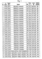
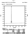
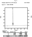
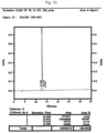
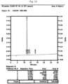
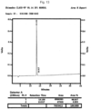
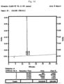
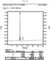
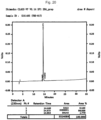
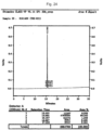
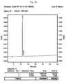
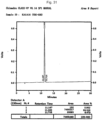
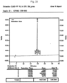
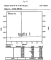
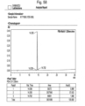
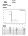
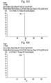
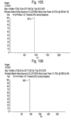
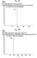
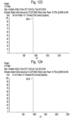
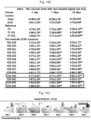
- the YDE derivatives prepared in Working Example 1 were analyzed by HPLC. As a result, it was confirmed that the purities of synthesized YDE-001, YDE-002, YDE-003, YDE-004, YDE-005, YDE-006, YDE-007, YDE-008, YDE-009, YDE-010, YDE-011, YDE-012, YDE-013, YDE-014, YDE-015, YDE-016, YDE-017, YDE-018, YDE-019, YDE-020, YDE-021, YDE-022, YDE-023, YDE-024, YDE-025, YDE-026, YDE-027, YDE-028, YDE-029, YDE-030, YDE-031, YDE-032, YDE-033, YDE-034, YDE-035, YDE-036, YDE-0
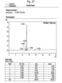
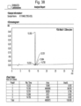
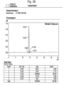
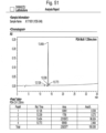
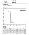
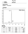
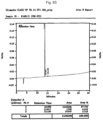
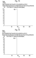
- YDE derivatives prepared in Working Example 1 were analyzed by Ion-Mass. As a result, it was confirmed that the molecular weights of synthesized YDE-001, YDE-002, YDE-003, YDE-004, YDE-005, YDE-006, YDE-007, YDE-008, YDE-009, YDE-010, YDE-011, YDE-012, YDE-013, YDE-014, YDE-015, YDE-016, YDE-017, YDE-018, YDE-019, YDE-020, YDE-021, YDE-022, YDE-023, YDE-024, YDE-025, YDE-026, YDE-027, YDE-028, YDE-029, YDE-030, YDE-031, YDE-032, YDE-033, YDE-034, YDE-035, YDE-
- the rats were systemically anesthetized by inhaling a mixed gas of 2% to 3% of isoflurane (Hana Pharm. Co., Hwasung, Korea), 70% of N 2 O, and 28.6% of O 2 using a rodent anesthesia machine (Surgivet, Waukesha, Wis., USA) and a ventilator (Model 687, Harvard Apparatus, Cambridge, UK). Thereafter, the extraorbital lacrimal gland located in the subdermal area above the masseter muscle and under the optic nerve was excised through a transverse incision in a size of 10 mm on the anterior part of the left ear tragus. The skin was sutured by a general method.
- the ELGE operation time did not exceed 5 minutes for each rat. After 6 days following the ELGE operation, confirmation was performed through the Schirmer test by measuring the amount of tear secretion whether dry eye syndrome had been induced. Meanwhile, each rat of the control group with the sham operation was checked for the presence and location of the extraorbital lacrimal gland through a skin incision, and the skin was then sutured without the excision thereof ( Fig. 134 ).
- the average weight of the ELGE test group measured before the ELGE operation was 241.59 ⁇ 13.56 g, and the average weight measured after 6 days from the ELGE operation was 297.38 ⁇ 34.02 g.
- the average weight of the control group measured before the sham operation was 240.13 ⁇ 25.63 g, and the average weight measured after 6 days from the sham operation was 297.38 ⁇ 34.02 g ( Fig. 135 ).
- the average amount of tear secretion of the control group was 8.34 ⁇ 0.73 mm 3
- the average amount of tear secretion of the ELGE test group was 3.55 ⁇ 0.70 mm 3 . 8 rats per group and a total of 32 groups were selected based on the average amount of tear secretion.
- the average weight of the ELGE test group measured before the ELGE operation was 264.09 ⁇ 11.53 g, and the average weight measured after 6 days from the ELGE operation was 316.13 ⁇ 15.77 g.
- the average weight of the control group measured before the sham operation was 263.50 ⁇ 9.24 g, and the average weight measured after 6 days from the sham operation was 315.25 ⁇ 10.85 g ( Fig. 136 ).
- the average amount of tear secretion of the control group was 10.90 ⁇ 1.69 mm 3
- the average amount of tear secretion of the ELGE test group was 4.83 ⁇ 0.99 mm 3 . 8 rats per group and a total of 20 groups were selected based on the average amount of tear secretion.
- YY-102 and the 28 YDE-series were each dissolved in physiological saline at a concentration of 3 mg/ml and administered at a dose of 5 ⁇ l/eye at 9:30 am and 3:30 pm daily for 14 days after 7 days from the ELGE operation for a total of 28 times.
- the DS solution was dissolved in physiological saline at a concentration of 30 mg/ml and administered at a dose of 5 ⁇ l/eye twice a day for 14 days after 7 days from the ELGE operation for a total of 28 times.
- the same stimulation as the administration was applied.
- the same volume of physiological saline was applied in the same manner in place of the test substances.
- YDE-029 to YDE-043, YY-102 and the 15 YDE-series were each dissolved in physiological saline at a concentration of 3 mg/ml and administered at a dose of 5 ⁇ l/eye at 9:30 am and 3:30 pm daily for 14 days after 7 days from the ELGE operation for a total of 28 times.
- the DS solution was dissolved in physiological saline at a concentration of 30 mg/ml and administered at a dose of 5 ⁇ l/eye twice a day for 14 days after 7 days from the ELGE operation for a total of 28 times.
- the same stimulation as the administration was applied.
- the same volume of physiological saline was applied in the same manner in place of the test substances ( Fig. 137 ).
- Test Example 2.3 Confirmation of the changes in the amount of tear secretion by the YDE derivatives
- the changes in the amount of tear secretion were measured at day 7 and day 14 after the administration of YDE-001 to YDE-043.
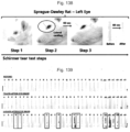
- the amount of tear secretion was measured by the decrease in the travel distance of tears absorbed by cobalt chloride paper in a size of 1 ⁇ 15 mm (Toyo Roshi Kaisha, Japan).
- the cobalt chloride paper was placed in the lateral canthus of a rat for 60 seconds to absorb tears ( Fig. 139 ).
- the length of the area absorbed from the corner of the cobalt chloride paper was measured with an electronic digital caliper (Mytutoyo, Tokyo, Japan) ( Fig. 138 ).
- Fig. 139 shows the results of the test, wherein A is for the sham control group, B is for the ELGE control group, C is for the DS reference group, D is for the YY-102 administered group, and E to AF are for the YDE-001 to YDE-028 administered groups in order.
- the amount of tear secretion was decreased after 6 days from the ELGE operation at days 7 and 14 after the application of physiological saline in the ELGE control group as compared with the sham control group.
- the amount of tear secretion was increased as compared with the ELGE control group, except for the groups treated with a 0.3% solution of YDE-9, YDE-10, YDE-17, YDE-19, YDE-20, YDE-21, YDE-22, YDE-25, YDE-27, and YDE-28, which did not show any significant changes in the amount of tear secretion after the administration thereof for 14 days.
- the amount of tear secretion was increased by more than 20% in the groups treated with a 0.3% solution of YDE-15, YDE-11, YDE-08, YDE-26, YDE-16, YDE-01, YDE-23, and YY-102 as compared with the DS reference group.
- Fig. 141 shows the results of the test, wherein A is for the sham control group, B is for the ELGE control group, C is for the DS reference group, D is for the YY-102 administered group, and E to S are for the YDE-029 to YDE-043 administered groups in order.
- the amount of tear secretion was decreased after 6 days from the ELGE operation at days 7 and 14 after the application of physiological saline in the ELGE control group as compared with the sham control group.
- the amount of tear secretion was increased as compared with the ELGE control group, except for the groups treated with a 0.3% solution of YDE-029, YDE-030, YDE-032, YDE-033, YDE-034, YDE-036, and YDE-41, which did not show any significant changes in the amount of tear secretion after the administration thereof for 14 days.
- the amount of tear secretion increased by more than 20% in the groups treated with a 0.3% solution of YDE-040, YDE-043, and YDE-042 in order as compared with the DS reference group.
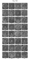
- the corneal permeability was measured using a blue light tungsten lamp and an ophthalmic slit lamp table top model biomicroscope (Model SM-70N; Takaci Seiko Co., Nakano, Japan) ( Fig. 143 ).
- Fig. 144 shows the results of the test, wherein A is for the sham control group, B is for the ELGE control group, C is for the DS reference group, D is for the YY-102 administered group, and E to AF are for the YDE-001 to YDE-028 administered groups in order.
- the permeability of the fluorescent dye was increased in the ELGE control group as compared with the sham control group.
- the permeability of the fluorescent dye was not decreased in the groups treated with a 0.3% solution of YDE-10, YDE-20, YDE-22, YDE-25, YDE-27, and YDE-28 as compared with the ELGE control group at day 14 after the administration.
- the corneal permeability of the fluorescent dye was decreased as compared with the ELGE control group, except for the groups treated with a 0.3% solution of YDE-10, YDE-20, YDE-22, YDE-25, YDE-27, and YDE-28.
- the permeability of the fluorescent dye was decreased by more than 20% in the groups treated with a 0.3% solution of YDE-15, YDE-11, YDE-08, YDE-26, YDE-16, YDE-01, YDE-23, and YY- 102, as compared with the DS reference group.
- YDE-029 to YDE-043 were each administered to the eyes 14 times, and the changes in the corneal permeability were then checked. The measurement of the corneal permeability was carried out in the same manner as described above ( Fig. 146 ).
- the permeability of the fluorescent dye was increased in the ELGE control group as compared with the sham control group.
- the permeability of the fluorescent dye was not decreased in the groups treated with a 0.3% solution of YDE-29, YDE-32, YDE-33, YDE-36, and YDE-41 as compared with the ELGE control group at day 14 after the administration.
- the corneal permeability of the fluorescent dye was decreased as compared with the ELGE control group, except for the groups treated with a 0.3% solution of YDE-29, YDE-32, YDE-33, YDE-36, and YDE-41.
- the permeability of the fluorescent dye was decreased by more than 20% in the groups treated with a 0.3% solution of YDE-40, YDE-43, and YDE-42, as compared with the DS reference group.
- each test substance in an aqueous solution, 10 mg of each sample was dissolved in 1 ml of water to a concentration of 1 mg/ml, which was then charged to a glass vial, plugged with a rubber cap, sealed with an aluminum cap, and stored under long-term storage conditions (25°C, 75% RH).
- the stability of the test substance was evaluated by measuring the amount of related substances at the time of one week, two weeks, four weeks, eight weeks, and twelve weeks under the long-term storage conditions.
- Amount of related substances (%; after 12 weeks) YY-101 66.51 (after 2 weeks) YDE-001 3.92 YDE-002 4.93 YDE-003 6.86 YDE-004 2.11 YDE-005 2.97 YDE-006 3.67 YDE-007 3.76 YDE-008 4.42 YDE-009 4.71 YDE-010 4.39 YDE-011 3.83 YDE-012 3.57 YDE-013 5.92 YDE-014 6.72 YDE-015 13.05 YDE-016 11.33 YDE-017 11.88 YDE-018 25.39 YDE-019 13.43 YDE-020 21.54 YDE-021 21.33 YDE-022 19.23 YDE-023 30.66 YDE-024 20.59 YDE-025 5.17 YDE-026 10.15 YDE-027 12.74 YDE-028 1.15
- Test Example 4 Evaluation of recovery of corneal damage by the YDE derivatives
- primary corneal epithelial cells (ATCC, ATCC PCS-700-010) were seeded on a 96-well culture plate (Perkin Elmer, 6005680) containing the Corneal Epithelial Cell Basal Medium (ATCC, ATCC PCS-700-030) in the Corneal Epithelial Cell Growth Kit (ATCC, ATCC PCS-700-040) in an amount of 5 ⁇ 10 3 cells per well, which was then cultured for 24 hours under the conditions of 37°C and 5% CO 2 .
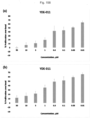
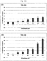
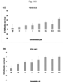
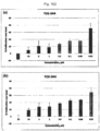
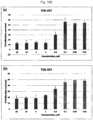
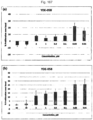
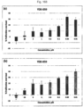
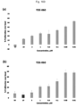
- YDE-001 to YDE-075 were each dissolved in 100% DMSO (Sigma, D2660) to a concentration of 10 mM, which was then diluted with 100% DMSO to a concentration of the compound of 6, 1.9, 0.6, 0.2, 0.06, 0.02, 0.006, and 0.002 mM.
- 20 ⁇ l of the diluted YDE derivative was added to a 96-well microplate (Greiner Bio-One, 651201) containing 380 ⁇ l of the Corneal Epithelial Cell Basal Medium such that the concentration of DMSO was diluted to 5%.
- hEGF Sigma, E9644
- the cells treated with the YDE-derivatives or hEGF were cultured for 48 hours and 72 hours under the conditions of 37°C and 5% CO 2 ( Figs. 148 to 155 ).
- the cultured cells were treated with the CellTiter-Glo luminescent reagent (Promega, G7573) according to the manufacturer's instructions and reacted for 30 minutes at room temperature. Thereafter, the fluorescent signal (or luminescence signal) was checked using an Envision 2014 Multi-label plate reader. The measured values were normalized using a vehicle control (100% proliferation cell).
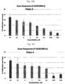
- the cell proliferation was observed at concentrations of 0.3 ⁇ M or less in YY-101, YY-102, YDE-011, YDE-038, YDE-042, YDE-043, YDE-044, YDE-045, YDE-049, YDE-054, YDE-057, YDE-058, YDE-059, and YDE-060.
- a high cell proliferation rate was shown in YY- 102, YDE-011, YDE-045, YDE-057, and YDE-060 ( Figs. 156 to 173 ).
Landscapes
- Health & Medical Sciences (AREA)
- Chemical & Material Sciences (AREA)
- Life Sciences & Earth Sciences (AREA)
- Organic Chemistry (AREA)
- Medicinal Chemistry (AREA)
- General Health & Medical Sciences (AREA)
- Proteomics, Peptides & Aminoacids (AREA)
- Public Health (AREA)
- Engineering & Computer Science (AREA)
- Bioinformatics & Cheminformatics (AREA)
- Pharmacology & Pharmacy (AREA)
- Veterinary Medicine (AREA)
- Animal Behavior & Ethology (AREA)
- Immunology (AREA)
- Epidemiology (AREA)
- Biochemistry (AREA)
- Biophysics (AREA)
- Genetics & Genomics (AREA)
- Molecular Biology (AREA)
- Ophthalmology & Optometry (AREA)
- Chemical Kinetics & Catalysis (AREA)
- General Chemical & Material Sciences (AREA)
- Nuclear Medicine, Radiotherapy & Molecular Imaging (AREA)
- Gastroenterology & Hepatology (AREA)
- Medicines That Contain Protein Lipid Enzymes And Other Medicines (AREA)
- Peptides Or Proteins (AREA)
Description
- The present invention relates to novel peptides and pharmaceutical compositions for treating eye diseases that comprises the same as an active pharmaceutical ingredient.
- Dry eye syndrome or keratoconjunctivitis sicca may be defined, in a broad sense, as damage to the ocular surface due to tear secretion disorders (Joossen C et al., Exp. Eye Res., 146:172-8, 2016). Dry eye syndrome is known to cause tear secretion disorders and damage and discomfort to the eyeball due to a combination of various factors. Although the onset of dry eye syndrome is closely related to age, the incidence thereof is increasing in younger age groups due to a long-term exposure to a dry environment as the use of contact lenses, computers, and smart devices (Stem ME et al., Int. Rev. Immunol., 32: 19-41, 2013).
- Specifically, dry eye syndrome reduces the mucus secretion of the corneal and conjunctival epithelia and that of the mucus-secreting goblet cells, resulting in a sharp decrease in the lubrication of the eyeball. In addition, dry eye syndrome causes damage to the corneal surface, thereby increasing the penetration of a fluorescein dye into the cornea. These symptoms of dry eye syndrome can be evaluated as changes in the tear secretion through the Schirmer test, which uses cobalt chloride paper. Further, the damage to the cornea that may accompany dry eye syndrome can be easily evaluated using a general fluorescent dye and a slit-lamp fluorophotometer.
- In the meantime, most of the treatments for dry eye syndrome are confined to symptom therapies, the treatment efficiency of which is often very low. Currently, artificial tears are the first choice for the treatment of dry eye syndrome. Since artificial tears as a representative symptom therapy merely supplement the insufficient tears; moreover, they suffer from the disadvantage that they need to be administered to the eyes frequently (Kim CS et al., ). Sodium hyaluronate and eye drops derived from autologous serum have been developed and used in patients suffering from dry eye syndrome. In addition, such synthetic compounds as rebamipide (OPC-127959) and diquafosol sodium, which promote the secretion of tears and mucus, have been developed and used. Long-term use of these drugs, however, may give rise to various side effects such as ocular hyperemia and corneal calcification (Bernauer W et al., Br. J. Ophthalmol., 90:285-8, 2006). Therefore, there has been a demand for the development of a safe and effective therapeutic agent for treating dry eye syndrome.
-
WO 2017/018613 describes pharmaceutical compositions for preventing or treating dry eyes. - Therefore, the present inventors have endeavored to develop safe and effective therapeutic agents for treating an eye disease and, as a result, completed the present invention by way of synthesizing new peptides, administering them to the eyes of rats with dry eye syndrome, and confirming the eye protection effect through the Schirmer test and the fluorescent dye deposition test.
- In order to achieve the object of the present invention, one aspect of the present invention provides a compound selected from the following table:
YDE-001 
YDE-008 
YDE-011 
YDE-015 
YDE-016 
YDE-023 
YDE-026 
YDE-038 
YDE-042 
YDE-043 
YDE-044 
YDE-045 
YDE-048 
YDE-049 
YDE-054 
YDE-057 
YDE-058 
YDE-059 
YDE-060 
- In addition, still another aspect of the present invention provides a pharmaceutical composition for treating an eye disease, which comprises the compound or the peptide as defined above an active pharmaceutical ingredient.
- Further, still another aspect of the present invention provides a compound or the peptide as defined above for use in treating an eye disease.
- When a novel peptide of the present invention is administered to the eye, it increases the amount of tear secretion and recovers promotes recovery of the damaged cornea. Hence, they can be advantageously used as therapeutic agents for treating eye diseases.
-
-
Fig. 1 is a diagram showing the sequence and characteristics of the peptides prepared according to an embodiment of the present invention. -
Fig. 2 is a diagram showing a process for synthesizing the peptides prepared according to an embodiment of the present invention. -
Fig. 3 is a diagram showing a purification procedure of the peptides prepared according to an embodiment of the present invention. -
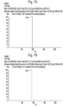
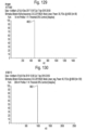
Fig. 4 is a diagram confirming the purity of YDE-001 prepared according to an embodiment of the present invention through HPLC. -
Fig. 5 is a diagram confirming the purity of YDE-002 prepared according to an embodiment of the present invention through HPLC. -
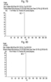
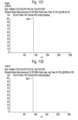
Fig. 6 is a diagram confirming the purity of YDE-003 prepared according to an embodiment of the present invention through HPLC. -
Fig. 7 is a diagram confirming the purity of YDE-004 prepared according to an embodiment of the present invention through HPLC. -
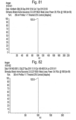
Fig. 8 is a diagram confirming the purity of YDE-005 prepared according to an embodiment of the present invention through HPLC. -
Fig. 9 is a diagram confirming the purity of YDE-006 prepared according to an embodiment of the present invention through HPLC. -
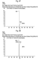
Fig. 10 is a diagram confirming the purity of YDE-007 prepared according to an embodiment of the present invention through HPLC. -
Fig. 11 is a diagram confirming the purity of YDE-008 prepared according to an embodiment of the present invention through HPLC. -
Fig. 12 is a diagram confirming the purity of YDE-009 prepared according to an embodiment of the present invention through HPLC. -
Fig. 13 is a diagram confirming the purity of YDE-010 prepared according to an embodiment of the present invention through HPLC. -
Fig. 14 is a diagram confirming the purity of YDE-011 prepared according to an embodiment of the present invention through HPLC. -
Fig. 15 is a diagram confirming the purity of YDE-012 prepared according to an embodiment of the present invention through HPLC. -
Fig. 16 is a diagram confirming the purity of YDE-013 prepared according to an embodiment of the present invention through HPLC. -
Fig. 17 is a diagram confirming the purity of YDE-014 prepared according to an embodiment of the present invention through HPLC. -
Fig. 18 is a diagram confirming the purity of YDE-015 prepared according to an embodiment of the present invention through HPLC. -
Fig. 19 is a diagram confirming the purity of YDE-016 prepared according to an embodiment of the present invention through HPLC. -
Fig. 20 is a diagram confirming the purity of YDE-017 prepared according to an embodiment of the present invention through HPLC. -
Fig. 21 is a diagram confirming the purity of YDE-018 prepared according to an embodiment of the present invention through HPLC. -
Fig. 22 is a diagram confirming the purity of YDE-019 prepared according to an embodiment of the present invention through HPLC. -
Fig. 23 is a diagram confirming the purity of YDE-020 prepared according to an embodiment of the present invention through HPLC. -
Fig. 24 is a diagram confirming the purity of YDE-021 prepared according to an embodiment of the present invention through HPLC. -
Fig. 25 is a diagram confirming the purity of YDE-022 prepared according to an embodiment of the present invention through HPLC. -
Fig. 26 is a diagram confirming the purity of YDE-023 prepared according to an embodiment of the present invention through HPLC. -
Fig. 27 is a diagram confirming the purity of YDE-024 prepared according to an embodiment of the present invention through HPLC. -
Fig. 28 is a diagram confirming the purity of YDE-025 prepared according to an embodiment of the present invention through HPLC. -
Fig. 29 is a diagram confirming the purity of YDE-026 prepared according to an embodiment of the present invention through HPLC. -
Fig. 30 is a diagram confirming the purity of YDE-027 prepared according to an embodiment of the present invention through HPLC. -
Fig. 31 is a diagram confirming the purity of YDE-028 prepared according to an embodiment of the present invention through HPLC. -
Fig. 32 is a diagram confirming the purity of YDE-029 prepared according to an embodiment of the present invention through HPLC. -
Fig. 33 is a diagram confirming the purity of YDE-030 prepared according to an embodiment of the present invention through HPLC. -
Fig. 34 is a diagram confirming the purity of YDE-031 prepared according to an embodiment of the present invention through HPLC. -
Fig. 35 is a diagram confirming the purity of YDE-032 prepared according to an embodiment of the present invention through HPLC. -
Fig. 36 is a diagram confirming the purity of YDE-033 prepared according to an embodiment of the present invention through HPLC. -
Fig. 37 is a diagram confirming the purity of YDE-034 prepared according to an embodiment of the present invention through HPLC. -
Fig. 38 is a diagram confirming the purity of YDE-035 prepared according to an embodiment of the present invention through HPLC. -
Fig. 39 is a diagram confirming the purity of YDE-036 prepared according to an embodiment of the present invention through HPLC. -
Fig. 40 is a diagram confirming the purity of YDE-037 prepared according to an embodiment of the present invention through HPLC. -
Fig. 41 is a diagram confirming the purity of YDE-038 prepared according to an embodiment of the present invention through HPLC. -
Fig. 42 is a diagram confirming the purity of YDE-039 prepared according to an embodiment of the present invention through HPLC. -
Fig. 43 is a diagram confirming the purity of YDE-040 prepared according to an embodiment of the present invention through HPLC. -
Fig. 44 is a diagram confirming the purity of YDE-041 prepared according to an embodiment of the present invention through HPLC. -
Fig. 45 is a diagram confirming the purity of YDE-042 prepared according to an embodiment of the present invention through HPLC. -
Fig. 46 is a diagram confirming the purity of YDE-043 prepared according to an embodiment of the present invention through HPLC. -
Fig. 47 is a diagram confirming the purity of YDE-044 prepared according to an embodiment of the present invention through HPLC. -
Fig. 48 is a diagram confirming the purity of YDE-045 prepared according to an embodiment of the present invention through HPLC. -
Fig. 49 is a diagram confirming the purity of YDE-047 prepared according to an embodiment of the present invention through HPLC. -
Fig. 50 is a diagram confirming the purity of YDE-048 prepared according to an embodiment of the present invention through HPLC. -
Fig. 51 is a diagram confirming the purity of YDE-049 prepared according to an embodiment of the present invention through HPLC. -
Fig. 52 is a diagram confirming the purity of YDE-050 prepared according to an embodiment of the present invention through HPLC. -
Fig. 53 is a diagram confirming the purity of YDE-051 prepared according to an embodiment of the present invention through HPLC. -
Fig. 54 is a diagram confirming the purity of YDE-052 prepared according to an embodiment of the present invention through HPLC. -
Fig. 55 is a diagram confirming the purity of YDE-053 prepared according to an embodiment of the present invention through HPLC. -
Fig. 56 is a diagram confirming the purity of YDE-054 prepared according to an embodiment of the present invention through HPLC. -
Fig. 57 is a diagram confirming the purity of YDE-055 prepared according to an embodiment of the present invention through HPLC. -
Fig. 58 is a diagram confirming the purity of YDE-056 prepared according to an embodiment of the present invention through HPLC. -
Fig. 59 is a diagram confirming the purity of YDE-057 prepared according to an embodiment of the present invention through HPLC. -
Fig. 60 is a diagram confirming the purity of YDE-058 prepared according to an embodiment of the present invention through HPLC. -
Fig. 61 is a diagram confirming the purity of YDE-059 prepared according to an embodiment of the present invention through HPLC. -
Fig. 62 is a diagram confirming the purity of YDE-060 prepared according to an embodiment of the present invention through HPLC. -
Fig. 63 is a diagram confirming the purity of YDE-064 prepared according to an embodiment of the present invention through HPLC. -
Fig. 64 is a diagram confirming the purity of YDE-066 prepared according to an embodiment of the present invention through HPLC. -
Fig. 65 is a diagram confirming the purity of YDE-072 prepared according to an embodiment of the present invention through HPLC. -
Fig. 66 is a diagram confirming the purity of YDE-073 prepared according to an embodiment of the present invention through HPLC. -
Fig. 67 is a diagram confirming the purity of YDE-074 prepared according to an embodiment of the present invention through HPLC. -
Fig. 68 is a diagram confirming the purity of YDE-075 prepared according to an embodiment of the present invention through HPLC. -
Fig. 69 is a diagram confirming the molecular weight of YDE-001 prepared according to an embodiment of the present invention through Ion-Mass. -
Fig. 70 is a diagram confirming the molecular weight of YDE-002 prepared according to an embodiment of the present invention through Ion-Mass. -
Fig. 71 is a diagram confirming the molecular weight of YDE-003 prepared according to an embodiment of the present invention through Ion-Mass. -
Fig. 72 is a diagram confirming the molecular weight of YDE-004 prepared according to an embodiment of the present invention through Ion-Mass. -
Fig. 73 is a diagram confirming the molecular weight of YDE-005 prepared according to an embodiment of the present invention through Ion-Mass. -
Fig. 74 is a diagram confirming the molecular weight of YDE-006 prepared according to an embodiment of the present invention through Ion-Mass. -
Fig. 75 is a diagram confirming the molecular weight of YDE-007 prepared according to an embodiment of the present invention through Ion-Mass. -
Fig. 76 is a diagram confirming the molecular weight of YDE-008 prepared according to an embodiment of the present invention through Ion-Mass. -
Fig. 77 is a diagram confirming the molecular weight of YDE-009 prepared according to an embodiment of the present invention through Ion-Mass. -
Fig. 78 is a diagram confirming the molecular weight of YDE-010 prepared according to an embodiment of the present invention through Ion-Mass. -
Fig. 79 is a diagram confirming the molecular weight of YDE-011 prepared according to an embodiment of the present invention through Ion-Mass. -
Fig. 80 is a diagram confirming the molecular weight of YDE-012 prepared according to an embodiment of the present invention through Ion-Mass. -
Fig. 81 is a diagram confirming the molecular weight of YDE-013 prepared according to an embodiment of the present invention through Ion-Mass. -
Fig. 82 is a diagram confirming the molecular weight of YDE-014 prepared according to an embodiment of the present invention through Ion-Mass. -
Fig. 83 is a diagram confirming the molecular weight of YDE-015 prepared according to an embodiment of the present invention through Ion-Mass. -
Fig. 84 is a diagram confirming the molecular weight of YDE-016 prepared according to an embodiment of the present invention through Ion-Mass. -
Fig. 85 is a diagram confirming the molecular weight of YDE-017 prepared according to an embodiment of the present invention through Ion-Mass. -
Fig. 86 is a diagram confirming the molecular weight of YDE-018 prepared according to an embodiment of the present invention through Ion-Mass. -
Fig. 87 is a diagram confirming the molecular weight of YDE-019 prepared according to an embodiment of the present invention through Ion-Mass. -
Fig. 88 is a diagram confirming the molecular weight of YDE-020 prepared according to an embodiment of the present invention through Ion-Mass. -
Fig. 89 is a diagram confirming the molecular weight of YDE-021 prepared according to an embodiment of the present invention through Ion-Mass. -
Fig. 90 is a diagram confirming the molecular weight of YDE-022 prepared according to an embodiment of the present invention through Ion-Mass. -
Fig. 91 is a diagram confirming the molecular weight of YDE-023 prepared according to an embodiment of the present invention through Ion-Mass. -
Fig. 92 is a diagram confirming the molecular weight of YDE-024 prepared according to an embodiment of the present invention through Ion-Mass. -
Fig. 93 is a diagram confirming the molecular weight of YDE-025 prepared according to an embodiment of the present invention through Ion-Mass. -
Fig. 94 is a diagram confirming the molecular weight of YDE-026 prepared according to an embodiment of the present invention through Ion-Mass. -
Fig. 95 is a diagram confirming the molecular weight of YDE-027 prepared according to an embodiment of the present invention through Ion-Mass. -
Fig. 96 is a diagram confirming the molecular weight of YDE-028 prepared according to an embodiment of the present invention through Ion-Mass. -
Fig. 97 is a diagram confirming the molecular weight of YDE-029 prepared according to an embodiment of the present invention through Ion-Mass. -
Fig. 98 is a diagram confirming the molecular weight of YDE-030 prepared according to an embodiment of the present invention through Ion-Mass. -
Fig. 99 is a diagram confirming the molecular weight of YDE-031 prepared according to an embodiment of the present invention through Ion-Mass. -
Fig. 100 is a diagram confirming the molecular weight of YDE-032 prepared according to an embodiment of the present invention through Ion-Mass. -
Fig. 101 is a diagram confirming the molecular weight of YDE-033 prepared according to an embodiment of the present invention through Ion-Mass. -
Fig. 102 is a diagram confirming the molecular weight of YDE-034 prepared according to an embodiment of the present invention through Ion-Mass. -
Fig. 103 is a diagram confirming the molecular weight of YDE-035 prepared according to an embodiment of the present invention through Ion-Mass. -
Fig. 104 is a diagram confirming the molecular weight of YDE-036 prepared according to an embodiment of the present invention through Ion-Mass. -
Fig. 105 is a diagram confirming the molecular weight of YDE-037 prepared according to an embodiment of the present invention through Ion-Mass. -
Fig. 106 is a diagram confirming the molecular weight of YDE-038 prepared according to an embodiment of the present invention through Ion-Mass. -
Fig. 107 is a diagram confirming the molecular weight of YDE-039 prepared according to an embodiment of the present invention through Ion-Mass. -
Fig. 108 is a diagram confirming the molecular weight of YDE-040 prepared according to an embodiment of the present invention through Ion-Mass. -
Fig. 109 is a diagram confirming the molecular weight of YDE-041 prepared according to an embodiment of the present invention through Ion-Mass. -
Fig. 110 is a diagram confirming the molecular weight of YDE-042 prepared according to an embodiment of the present invention through Ion-Mass. -
Fig. 111 is a diagram confirming the molecular weight of YDE-043 prepared according to an embodiment of the present invention through Ion-Mass. -
Fig. 112 is a diagram confirming the molecular weight of YDE-044 prepared according to an embodiment of the present invention through Ion-Mass. -
Fig. 113 is a diagram confirming the molecular weight of YDE-045 prepared according to an embodiment of the present invention through Ion-Mass. -
Fig. 114 is a diagram confirming the molecular weight of YDE-047 prepared according to an embodiment of the present invention through Ion-Mass. -
Fig. 115 is a diagram confirming the molecular weight of YDE-048 prepared according to an embodiment of the present invention through Ion-Mass. -
Fig. 116 is a diagram confirming the molecular weight of YDE-049 prepared according to an embodiment of the present invention through Ion-Mass. -
Fig. 117 is a diagram confirming the molecular weight of YDE-050 prepared according to an embodiment of the present invention through Ion-Mass. -
Fig. 118 is a diagram confirming the molecular weight of YDE-051 prepared according to an embodiment of the present invention through Ion-Mass. -
Fig. 119 is a diagram confirming the molecular weight of YDE-052 prepared according to an embodiment of the present invention through Ion-Mass. -
Fig. 120 is a diagram confirming the molecular weight of YDE-053 prepared according to an embodiment of the present invention through Ion-Mass. -
Fig. 121 is a diagram confirming the molecular weight of YDE-054 prepared according to an embodiment of the present invention through Ion-Mass. -
Fig. 122 is a diagram confirming the molecular weight of YDE-055 prepared according to an embodiment of the present invention through Ion-Mass. -
Fig. 123 is a diagram confirming the molecular weight of YDE-056 prepared according to an embodiment of the present invention through Ion-Mass. -
Fig. 124 is a diagram confirming the molecular weight of YDE-057 prepared according to an embodiment of the present invention through Ion-Mass. -
Fig. 125 is a diagram confirming the molecular weight of YDE-058 prepared according to an embodiment of the present invention through Ion-Mass. -
Fig. 126 is a diagram confirming the molecular weight of YDE-059 prepared according to an embodiment of the present invention through Ion-Mass. -
Fig. 127 is a diagram confirming the molecular weight of YDE-060 prepared according to an embodiment of the present invention through Ion-Mass. -
Fig. 128 is a diagram confirming the molecular weight of YDE-064 prepared according to an embodiment of the present invention through Ion-Mass. -
Fig. 129 is a diagram confirming the molecular weight of YDE-066 prepared according to an embodiment of the present invention through Ion-Mass. -
Fig. 130 is a diagram confirming the molecular weight of YDE-072 prepared according to an embodiment of the present invention through Ion-Mass. -
Fig. 131 is a diagram confirming the molecular weight of YDE-073 prepared according to an embodiment of the present invention through Ion-Mass. -
Fig. 132 is a diagram confirming the molecular weight of YDE-074 prepared according to an embodiment of the present invention through Ion-Mass. -
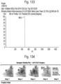
Fig. 133 is a diagram confirming the molecular weight of YDE-075 prepared according to an embodiment of the present invention through Ion-Mass. -
Fig. 134 is a photograph showing a procedure of extraorbital lacrimal gland excision. -
Fig. 135 is a diagram showing a change in the body weight of a rat model whose eyes have been administered with YDE-001 to YDE-028. -
Fig. 136 is a diagram showing a change in the body weight of a rat model whose eyes have been administered with YDE-029 to YDE-043. -
Fig. 137 is a photograph showing a procedure of administering an agent to the eyes of a rat model. -
Fig. 138 is a photograph showing a procedure of measuring the amount of tear secretion of a rat model using cobalt chloride paper. -
Fig. 139 is a photograph showing the results of measuring the amount of tear secretion of a rat model whose eyes have been administered with YDE-001 to YDE-028 using cobalt chloride paper. -
Fig. 140 is a diagram showing the changes in the amount of tear secretion of a rat model whose eyes have been administered with YDE-001 to YDE-028. -
Fig. 141 is a photograph showing the results of measuring the amount of tear secretion of a rat model whose eyes have been administered with YDE-029 to YDE-043 using cobalt chloride paper. -
Fig. 142 is a diagram showing the changes in the amount of tear secretion of a rat model whose eyes have been administered with YDE-029 to YDE-043. -
Fig. 143 is a photograph showing a procedure of administering a fluorescent substance to the eyes of a rat model for confirming damage to the cornea thereof. -
Fig. 144 is a photograph showing the results of measuring damage to the cornea of a rat model whose eyes have been administered with YDE-001 to YDE-028 using a fluorescent substance. -
Fig. 145 is a diagram showing the permeability of a fluorescence dye to confirm the recovery of corneal damage of a rat model whose eyes have been administered with YDE-001 to YDE-028. -
Fig. 146 is a photograph showing the results of measuring damage to the cornea of a rat model whose eyes have been administered with YDE-029 to YDE-043 using a fluorescent substance. -
Fig. 147 is a diagram showing the permeability of a fluorescence dye to confirm the recovery of corneal damage of a rat model whose eyes have been administered with YDE-029 to YDE-043. -
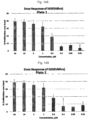
FIG. 148 is a diagram showing the cell growth rate after 48 hours from the treatment of hEGF on human corneal epithelial cells of plate No. 1. -
FIG. 149 is a diagram showing the cell growth rate after 48 hours from the treatment of hEGF on human corneal epithelial cells of plate No. 2. -
FIG. 150 is a diagram showing the cell growth rate after 48 hours from the treatment of hEGF on human corneal epithelial cells of plate No. 3. -
FIG. 151 is a diagram showing the cell growth rate after 48 hours from the treatment of hEGF on human corneal epithelial cells of plate No. 4. -
FIG. 152 is a diagram showing the cell growth rate after 72 hours from the treatment of hEGF on human corneal epithelial cells of plate No. 1. -
FIG. 153 is a diagram showing the cell growth rate after 72 hours from the treatment of hEGF on human corneal epithelial cells of plate No. 2. -
FIG. 154 is a diagram showing the cell growth rate after 72 hours from the treatment of hEGF on human corneal epithelial cells of plate No. 3. -
FIG. 155 is a diagram showing the cell growth rate after 48 hours from the treatment of hEGF on human corneal epithelial cells of plate No. 4. -
FIG. 156 is a diagram showing the cell growth rate after (a) 48 hours or (b) 72 hours from the treatment of YY-101 on human corneal epithelial cells. -
FIG. 157 is a diagram showing the cell growth rate after (a) 48 hours or (b) 72 hours from the treatment of YY-102 on human corneal epithelial cells. -
FIG. 158 is a diagram showing the cell growth rate after (a) 48 hours or (b) 72 hours from the treatment of YDE-011 on human corneal epithelial cells. -
FIG. 159 is a diagram showing the cell growth rate after (a) 48 hours or (b) 72 hours from the treatment of YDE-038 on human corneal epithelial cells. -
FIG. 160 is a diagram showing the cell growth rate after (a) 48 hours or (b) 72 hours from the treatment of YDE-042 on human corneal epithelial cells. -
FIG. 161 is a diagram showing the cell growth rate after (a) 48 hours or (b) 72 hours from the treatment of YDE-043 on human corneal epithelial cells. -
FIG. 162 is a diagram showing the cell growth rate after (a) 48 hours or (b) 72 hours from the treatment of YDE-044 on human corneal epithelial cells. -
FIG. 163 is a diagram showing the cell growth rate after (a) 48 hours or (b) 72 hours from the treatment of YDE-045 on human corneal epithelial cells. -
FIG. 164 is a diagram showing the cell growth rate after (a) 48 hours or (b) 72 hours from the treatment of YDE-049 on human corneal epithelial cells. -
FIG. 165 is a diagram showing the cell growth rate after (a) 48 hours or (b) 72 hours from the treatment of YDE-054 on human corneal epithelial cells. -
FIG. 166 is a diagram showing the cell growth rate after (a) 48 hours or (b) 72 hours from the treatment of YDE-057 on human corneal epithelial cells. -
FIG. 167 is a diagram showing the cell growth rate after (a) 48 hours or (b) 72 hours from the treatment of YDE-058 on human corneal epithelial cells. -
FIG. 168 is a diagram showing the cell growth rate after (a) 48 hours or (b) 72 hours from the treatment of YDE-059 on human corneal epithelial cells. -
FIG. 169 is a diagram showing the cell growth rate after (a) 48 hours or (b) 72 hours from the treatment of YDE-060 on human corneal epithelial cells. -
FIG. 170 is a diagram showing the cell growth rate after (a) 48 hours or (b) 72 hours from the treatment of YDE-072 on human corneal epithelial cells. -
FIG. 171 is a diagram showing the cell growth rate after (a) 48 hours or (b) 72 hours from the treatment of YDE-073 on human corneal epithelial cells. -
FIG. 172 is a diagram showing the cell growth rate after (a) 48 hours or (b) 72 hours from the treatment of YDE-074 on human corneal epithelial cells. -
FIG. 173 is a diagram showing the cell growth rate after (a) 48 hours or (b) 72 hours from the treatment of YDE-075 on human corneal epithelial cells. - Hereinafter, the present invention will be described in detail.
- An aspect of the present invention provides a compound selected from the following table:
YDE-001 
YDE-008 
YDE-011 
YDE-015 
YDE-016 
YDE-023 
YDE-026 
YDE-038 
YDE-042 
YDE-043 
YDE-044 
YDE-045 
YDE-048 
YDE-049 
YDE-054 
YDE-057 
YDE-058 
YDE-059 
YDE-060 
- The term "peptide" used in the present invention refers to a compound in which two or more amino acids are linked by a peptide bond. Further, it is classified into dipeptide, tripeptide, tetrapeptide, and the like according to the number of constituent amino acids. An oligopeptide has about 10 or fewer peptide bonds, and a polypeptide has a plurality of peptide bonds. In addition, a peptide in the present invention includes a mutated peptide in which its amino acid residue is substituted.
- The term "HyP" used in the present invention refers to an amino acid called hydroxyproline, in which a hydroxyl group (-OH) is bonded to the carbon atom at the 4-position of proline. HyP has a structure of C5H9NO3 and may be represented by the following
Formula 2.
- HyP may include all isomers. In addition, HyP may be an isomer represented by the stereochemistry of "2S,4R" unless otherwise specified.
- The term "2S,4R" is represented by R and S that indicate a stereochemical configuration of a chiral molecule. A typical chiral molecule has a chiral center such as an asymmetric carbon atom. Since the chiral center has four different substituent groups (or substitution atoms), their priority is determined by a predetermined procedure. Once the order of the four substituents is determined by (1), (2), (3), and (4), the lowest order substituent (4) is placed farthest away from the eye direction, and the remaining substituents are arranged from the higher order to the lower order. R (or rectus in Latin, right) indicate the arrangement in which the sequence of (1) to (2) to (3) turns right. S (or sinister, left) indicates the arrangement in which this sequence turns left.
- The term "homo-Ser" used in the present invention is called homoserine and refers to an α-amino acid having a hydroxyl group in the side chain. Homo-Ser is not an amino acid that constitutes a protein and is an intermediate present in the biosynthesis of threonine and methionine in microorganisms and plants. Homo-Ser may have the following
Formula 3.
- Further, the present invention provides a pharmaceutical composition for treating an eye disease, which comprises the compound or the peptide as an active pharmaceutical ingredient.
- Specifically, the eye disease may be one selected from the group consisting of retinopathy, keratitis, dry-macular degeneration, wet-macular degeneration, dry eye syndrome, keratoconjunctival epithelium disorder, proliferative vitreoretinopathy, pigmentary retinopathy, diabetic retinopathy, retinopathy of prematurity, retinopathy of immaturity, proliferative retinopathy, ischemic retinopathy, epidemic keratoconjunctivitis, atopic keratitis, superior limbic keratitis, pterygium keratitis sicca, phlyctenular keratoconjunctivitis, scleritis, corneal transplant rejection, choroidal neovascularization, neovascular glaucoma, ischemic optic neuropathy, retrolental fibroplasias, diabetic macula, neovascular iris disease, erythrosis, myopia, Von Hippel-Lindau syndrome, ocular histoplasmosis, central retinal vein occlusion, Sjogren syndrome and Stevens-Johnson syndrome. Preferably, the eye disease may be one selected from the group consisting of retinopathy, keratitis, macular degeneration, dry eye syndrome and keratoconjunctival epithelium disorder.
- The keratoconjunctival epithelium disorder may be due to post-operative surgery, drug, trauma or contact lens wear.
- Specifically, the composition for treating an eye disease, which comprises the compound or the peptide as an active pharmaceutical ingredient, may further comprise at least one additive selected from the group consisting of a carrier, an excipient, a disintegrant, a sweetener, a coating agent, a swelling agent, a lubricant, a slip agent, a flavor, an antioxidant, a buffer, a bacteriostat, a diluent, a dispersant, a surfactant, and a binder. Specifically, a formulation for parenteral administration may be a sterilized aqueous solution, a non-aqueous solution, a suspension, an emulsion, a lyophilized preparation, a suppository, or the like.
- Still another aspect of the present invention provides a compound or the peptide as defined herein for use in treating an eye disease.
- The dose of the compound or the peptide may be adjusted depending on such various factors as the kind of the disease, the severity of the disease, the kinds and amounts of the active pharmaceutical ingredient and other ingredients contained in the pharmaceutical composition, the type of the formulation, the age, body weight, general health condition, sex, and diet of the patient, the time and the route of administration, the duration of treatment, and the drugs concurrently used.
- However, for the desired effect, the effective amount of the compound or the peptide contained in the pharmaceutical composition may be 0.0001 µg/day to 100 µg/day. In such event, the administration may be carried out once a day, or divided into several doses. Specifically, the concentration of the compound or the peptide contained in the pharmaceutical composition may be 1000 µM to 0.001 µM. Also, the concentration of the compound or the peptide contained in the pharmaceutical composition may be 100 µM to 0.005 µM or 50 µM to 0.02 µM.
- In addition, if necessary, the concentration of the compound or the peptide contained in the pharmaceutical composition may be 30 µM to 1 µM. Further, the concentration of the compound or the peptide contained in the pharmaceutical composition may be 0.01 µM to 1 µM.
- In addition, the subject may be a mammal, particularly a human. The administration route may be appropriately selected by a person skilled in the art in consideration of the administration method, the volume and viscosity of the body fluid, and the like. Specifically, the administration may be carried out through any one route selected from the group consisting of an application, intravenous, intraarterial, intraperitoneal, intramuscular, intrasternal, percutaneous, intranasal, inhalation, topical, rectal, oral, intraocular, and intradermal. In particular, it may preferably be applied to the eye for use as an eye drop.
- Hereinafter, the present invention is explained in more detail by the following working examples. However, the following working examples are intended to further illustrate the present invention. The scope of the present invention is not limited thereby.
- A protein analysis of the extracellular matrix derived from animal chondrocytes was performed in Baek's group of Center of Biomedical Mass Spectrometry (Diatech Korea Co., Ltd., Seoul, Korea). Proline-GQDGLAGPK (P-GQDGLAGPK), which is a part of the amino acid sequence of the collagen type II α1 protein, was obtained through the above protein analysis. YDE-001 to YDE-075 peptides were synthesized by ANYGEN (Gwangju, Korea) by substituting a part of the above peptide (
Fig. 1 and Table 11). - The process for synthesizing the YDE-001 to YDE-075 peptides and the purification procedure thereof conducted by ANYGEN were depicted in
Figs. 2 and3 . - Compounds marked with an asterisk(*) are included for references purposes only.
[Table 11] No. Structure of compound No. Structure of compound YY-101* 
YY-102* 
YDE - 001 
YDE 002* 
YDE 003*
YDE 004*
YDE 005*
YDE 006*
YDE 007*
YDE -008 
YDE 009*
YDE 010*
YDE - 011 
YDE 012* 
YDE 013*
YDE 014*
YDE -015 
YDE - 016 
YDE 017* 
YDE 018*
YDE 019*
YDE 020*
YDE 021*
YDE 022*
YDE - 023 
YDE 024* 
YDE 025*
YDE -026 
YDE 027*
YDE 028*
YDE 029*
YDE 030*
YDE 031*
YDE 032*
YDE 033*
YDE 034*
YDE 035*
YDE 036*
YDE 037*
YDE -038 
YDE 039*
YDE 040*
YDE 041*
YDE -042 
YDE -043 
YDE -044 
YDE -045 
YDE 047* 
YDE -048 
YDE -049 
YDE 050* 
YDE 051* 
YDE 052* 
YDE 053* 
YDE -054 
YDE 055* 
YDE 056* 
YDE -057 
YDE -058 
YDE -059 
YDE -060 
YDE 064* 
YDE 066* 
YDE 072*
YDE 073*
YDE 074*
YDE 075*
- The YDE derivatives prepared in Working Example 1 were analyzed by HPLC. As a result, it was confirmed that the purities of synthesized YDE-001, YDE-002, YDE-003, YDE-004, YDE-005, YDE-006, YDE-007, YDE-008, YDE-009, YDE-010, YDE-011, YDE-012, YDE-013, YDE-014, YDE-015, YDE-016, YDE-017, YDE-018, YDE-019, YDE-020, YDE-021, YDE-022, YDE-023, YDE-024, YDE-025, YDE-026, YDE-027, YDE-028, YDE-029, YDE-030, YDE-031, YDE-032, YDE-033, YDE-034, YDE-035, YDE-036, YDE-037, YDE-038, YDE-039, YDE-040, YDE-041, YDE-042, YDE-043, YDE-044, YDE-045, YDE-047, YDE-048, YDE-049, YDE-050, YDE-051, YDE-052, YDE-053, YDE-054, YDE-055, YDE-056, YDE-057, YDE-058, YDE-059, YDE-060, YDE-064, YDE-066, YDE-072, YDE-073, YDE-074, and YDE-075 were 99.7%, 99.7%, 99.7%, 99.7%, 99.5%, 98.9%, 98.0%, 98.8%, 98.1%, 99.0%, 98.3%, 98.9%, 98.7%, 98.5%, 99.1%, 99.4%, 98.0%, 99.6%, 99.6%, 99.2%, 98.1%, 98.3%, 96.1%, 98.9%, 95.1%, 98.6%, 96.9%, 99.5%, 98.0%, 98.1%, 98.8%, 98.2%, 97.2%, 98.6%, 98.8%, 98.7%, 99.2%, 98.7%, 98.1%, 97.5%, 96.5%, 97.4%, 98.7%, 97.8%, 95.5%, 97.5%, 97.2%, 96.9%, 99.3%, 98.0%, 99.4%, 96.4%, 95.1%, 98.6%, 97.4%, 98.8%, 97.4%, 95.8%, 98.9%, 96.9%, 98.8%, 97.7%, 95.0%, 97.9%, and 96.3%, respectively (
Figs. 4 to 68 ). - In addition, the YDE derivatives prepared in Working Example 1 were analyzed by Ion-Mass. As a result, it was confirmed that the molecular weights of synthesized YDE-001, YDE-002, YDE-003, YDE-004, YDE-005, YDE-006, YDE-007, YDE-008, YDE-009, YDE-010, YDE-011, YDE-012, YDE-013, YDE-014, YDE-015, YDE-016, YDE-017, YDE-018, YDE-019, YDE-020, YDE-021, YDE-022, YDE-023, YDE-024, YDE-025, YDE-026, YDE-027, YDE-028, YDE-029, YDE-030, YDE-031, YDE-032, YDE-033, YDE-034, YDE-035, YDE-036, YDE-037, YDE-038, YDE-039, YDE-040, YDE-041, YDE-042, YDE-043, YDE-044, YDE-045, YDE-047, YDE-048, YDE-049, YDE-050, YDE-051, YDE-052, YDE-053, YDE-054, YDE-055, YDE-056, YDE-057, YDE-058, YDE-059, YDE-060, YDE-064, YDE-066, YDE-072, YDE-073, YDE-074, and YDE-075 were 969.6, 954.8, 967.7, 977.1, 968.1, 926.9, 941.1, 910.7, 939.7, 953.0, 953.7, 987.8, 1003.8, 1025.9, 996.7, 1011.0, 1011.4, 968.7, 1044.4, 1061.4, 1084.5, 1035.0, 984.9, 999.1, 969.7, 942.0, 937.6, 967.3, 988.1, 960.6, 954.2, 991.1, 954.4, 990.7, 950.9, 937.6, 968.1, 955.4, 966.0, 709.3, 622.2, 486.8, 951.3, 951.3, 911.4, 967.5, 896.5, 911.0, 967.3, 911.2, 953.2, 967.2, 927.4, 896.4, 952.8, 953.4, 670.1, 953.3, 599.7, 486.5, 966.1, 895.8, 909.1, and 486.4, respectively (
Figs. 69 to 133 ). - In order to evaluate the eye protection effect on dry eye syndrome by YDE-001 to YDE-028 prepared in Working Example 1, a total of 320 Sprague-Dawley-type male rats (OrientBio, Seungnam, Korea) were adapted for 7 days. Thereafter, dry eye syndrome was induced in 264 test rats through extraorbital lacrimal gland excision (hereinafter, ELGE). 8 test rats without the eye abnormality were subjected to a sham operation as a control group.
- The rats were systemically anesthetized by inhaling a mixed gas of 2% to 3% of isoflurane (Hana Pharm. Co., Hwasung, Korea), 70% of N2O, and 28.6% of O2 using a rodent anesthesia machine (Surgivet, Waukesha, Wis., USA) and a ventilator (Model 687, Harvard Apparatus, Cambridge, UK). Thereafter, the extraorbital lacrimal gland located in the subdermal area above the masseter muscle and under the optic nerve was excised through a transverse incision in a size of 10 mm on the anterior part of the left ear tragus. The skin was sutured by a general method. The ELGE operation time did not exceed 5 minutes for each rat. After 6 days following the ELGE operation, confirmation was performed through the Schirmer test by measuring the amount of tear secretion whether dry eye syndrome had been induced. Meanwhile, each rat of the control group with the sham operation was checked for the presence and location of the extraorbital lacrimal gland through a skin incision, and the skin was then sutured without the excision thereof (
Fig. 134 ). - The average weight of the ELGE test group measured before the ELGE operation was 241.59 ± 13.56 g, and the average weight measured after 6 days from the ELGE operation was 297.38 ± 34.02 g. The average weight of the control group measured before the sham operation was 240.13±25.63 g, and the average weight measured after 6 days from the sham operation was 297.38±34.02 g (
Fig. 135 ). - The average amount of tear secretion of the control group was 8.34 ± 0.73 mm3, and the average amount of tear secretion of the ELGE test group was 3.55 ± 0.70 mm3. 8 rats per group and a total of 32 groups were selected based on the average amount of tear secretion.
- As a reference drug, 3% diquafosol sodium (Santen, Tokyo, Japan; hereinafter, DS), which is currently on the market, was used.
- The present animal tests were conducted with a prior approval of the Animal Experimental Ethics Committee of Daegu Haany University (Approval No. DHU2017-003, January 12, 2017). All test animals were caused to fast for 18 hours before the ELGE operation and final sacrifice except for feeding water.
- The 32 groups were summarized in Table 12.
[Table 12] Group classification 32 groups in total; 8 rats per group Sham control group Group administered with physiological saline after the sham operation ELGE control group Group administered with physiological saline after the ELGE operation Reference Group administered with DS after the ELGE operation YY-102 Group administered with a 0.3% YY-102 solution after the ELGE operation YDE series Group administered with any of 0.3% YDE-01 to YDE-28 solutions (28 groups in total) - In addition, in order to evaluate the eye protection effect on dry eye syndrome by YDE-029 to YDE-043 prepared in Working Example 1, a total of 200 Sprague-Dawley-type male rats (OrientBio, Seungnam, Korea) were adapted for 7 days. Dry eye syndrome was induced in 165 test rats through the ELGE. 8 test rats without the eye abnormality were subjected to the sham operation as a control group. The ELGE was carried out as described above.
- The average weight of the ELGE test group measured before the ELGE operation was 264.09±11.53 g, and the average weight measured after 6 days from the ELGE operation was 316.13±15.77 g. The average weight of the control group measured before the sham operation was 263.50±9.24 g, and the average weight measured after 6 days from the sham operation was 315.25±10.85 g (
Fig. 136 ). - The average amount of tear secretion of the control group was 10.90±1.69 mm3, and the average amount of tear secretion of the ELGE test group was 4.83±0.99 mm3. 8 rats per group and a total of 20 groups were selected based on the average amount of tear secretion.
- As a reference drug, 3% DS, which is currently on the market, was used.
- The present animal tests were conducted with prior approval of the Animal Experimental Ethics Committee of Daegu Haany University (Approval No. DHU2017-050, June 08, 2017). All test animals were caused to fast for 18 hours before the ELGE operation and final sacrifice except for feeding water.
- The 20 groups were summarized in Table 13.
[Table 13] Group classification 20 groups in total; 8 rats per group Sham control group Group administered with physiological saline after the sham operation ELGE control group Group administered with physiological saline after the ELGE operation Reference Group administered with DS after the ELGE operation YY-101 Group administered with a 0.3% YY-101 solution after the ELGE operation YY-102 Group administered with a 0.3% YY-102 solution after the ELGE operation YDE series Group administered with any of 0.3% YDE-01 to YDE-28 solutions (15 groups in total) - For YDE-001 to YDE-028, YY-102 and the 28 YDE-series were each dissolved in physiological saline at a concentration of 3 mg/ml and administered at a dose of 5 µl/eye at 9:30 am and 3:30 pm daily for 14 days after 7 days from the ELGE operation for a total of 28 times. The DS solution was dissolved in physiological saline at a concentration of 30 mg/ml and administered at a dose of 5 µl/eye twice a day for 14 days after 7 days from the ELGE operation for a total of 28 times. For the sham control and the ELGE control groups, the same stimulation as the administration was applied. In order to prevent excessive eye dryness, the same volume of physiological saline was applied in the same manner in place of the test substances.
- Further, For YDE-029 to YDE-043, YY-102 and the 15 YDE-series were each dissolved in physiological saline at a concentration of 3 mg/ml and administered at a dose of 5 µl/eye at 9:30 am and 3:30 pm daily for 14 days after 7 days from the ELGE operation for a total of 28 times. The DS solution was dissolved in physiological saline at a concentration of 30 mg/ml and administered at a dose of 5 µl/eye twice a day for 14 days after 7 days from the ELGE operation for a total of 28 times. For the sham control and the ELGE control groups, the same stimulation as the administration was applied. In order to prevent excessive eye dryness, the same volume of physiological saline was applied in the same manner in place of the test substances (
Fig. 137 ). - After 6 days from the ELGE surgery, the changes in the amount of tear secretion were measured at
day 7 andday 14 after the administration of YDE-001 to YDE-043. The amount of tear secretion was measured by the decrease in the travel distance of tears absorbed by cobalt chloride paper in a size of 1 × 15 mm (Toyo Roshi Kaisha, Japan). - The cobalt chloride paper was placed in the lateral canthus of a rat for 60 seconds to absorb tears (
Fig. 139 ). The length of the area absorbed from the corner of the cobalt chloride paper was measured with an electronic digital caliper (Mytutoyo, Tokyo, Japan) (Fig. 138 ). -
Fig. 139 shows the results of the test, wherein A is for the sham control group, B is for the ELGE control group, C is for the DS reference group, D is for the YY-102 administered group, and E to AF are for the YDE-001 to YDE-028 administered groups in order. - As a result, it was confirmed that the amount of tear secretion was decreased after 6 days from the ELGE operation at
days - The specific amounts of tear secretion are shown in
Fig. 140 and Table 14.[Table 14] No. Tear Volumes (mm3) Day 7 Day 14 YY-101 7.66±0.61 6.00±0.69 YY-102 4.59±1.43 5.77±1.99 YDE-001 4.88±1.62 5.92±2.19 YDE-002 3.84±1.16 5.01±1.67 YDE-003 4.13±1.76 4.88±1.57 YDE-004 3.42±1.06 5.19±1.84 YDE-005 3.85±0.93 5.08±1.91 YDE-006 3.44±1.69 5.35±1.68 YDE-007 3.91±1.28 5.45±1.26 YDE-008 4.57±1.25 6.10±2.36 YDE-009 3.76±1.21 4.54±1.11 YDE-010 3.42±1.31 4.35±1.36 YDE-011 4.22±1.45 6.16±2.16 YDE-012 3.68±0.99 5.67±1.86 YDE-013 5.27±1.50 5.49±1.92 YDE-014 3.81±1.21 5.62±1.85 YDE-015 4.03±2.19 6.65±2.13 YDE-016 4.59±1.13 5.98±2.27 YDE-017 4.00±1.22 4.89±1.50 YDE-018 3.75±1.54 4.99±1.60 YDE-019 4.84±1.39 4.52±1.07 YDE-020 3.41±1.47 4.20±1.35 YDE-021 4.08±1.33 4.90±1.13 YDE-022 3.19±0.67 4.10±0.95 YDE-023 5.32±2.30 5.78±2.23 YDE-024 3.85±1.30 5.72±1.36 YDE-025 3.21±0.72 4.72±2.19 YDE-026 4.32±1.47 6.01±1.83 YDE-027 2.82±0.86 3.95±1.52 YDE-028 4.04±0.99 4.73±1.18 -
Fig. 141 shows the results of the test, wherein A is for the sham control group, B is for the ELGE control group, C is for the DS reference group, D is for the YY-102 administered group, and E to S are for the YDE-029 to YDE-043 administered groups in order. - As a result, it was confirmed that the amount of tear secretion was decreased after 6 days from the ELGE operation at
days - The specific amounts of tear secretion are shown in
Fig. 142 and Table 15.[Table 15] No. Tear Volumes (mm3) Day 7Day 14YY-101 5.36±0.68 6.25±0.68 YY-102 5.77±1.01 6.60±0.64 YDE-029 5.33±1.43 6.03±1.71 YDE-030 5.69±1.79 6.65±2.17 YDE-031 5.63±1.97 5.91±0.85 YDE-032 5.58±0.80 5.03±0.93 YDE-033 4.99±1.20 4.54±1.16 YDE-034 6.16±1.01 6.43±1.86 YDE-035 4.96±0.96 6.25±0.79 YDE-036 4.95±1.05 5.13±1.03 YDE-037 4.98±0.66 5.80±0.90 YDE-039 6.04±1.01 6.44±1.96 YDE-040 5.77±1.05 8.63±1.53 YDE-041 5.01±1.26 6.25±2.15 YDE-042 6.30±1.08 7.97±1.48 YDE-043 5.90±1.06 8.16±1.42 - After YDE-001 to YDE-028 were each administered to the
eyes 14 times, the changes in the corneal permeability were checked. - In order to measure the corneal permeability, Zolethyl 50™ (Virbac Lab., Carros, France), an animal anesthetic, was intraperitoneally injected at a dose of 25 mg/kg. Thereafter, saline containing a 1% (v/v) fluorescent solution (fluorescein sodium salt, Tokyo Kasei Kogyo Co., Tokyo, Japan) was applied to the eyes at a dose of 5 µl/eye. The eyes thus treated were closed and fixed with a tape. After 1 hour, the remaining fluorescent solution was removed using a cotton swab (
Fig. 142 ). After 12 hours to 24 hours, the corneal permeability was measured using a blue light tungsten lamp and an ophthalmic slit lamp table top model biomicroscope (Model SM-70N; Takaci Seiko Co., Nakano, Japan) (Fig. 143 ). -
Fig. 144 shows the results of the test, wherein A is for the sham control group, B is for the ELGE control group, C is for the DS reference group, D is for the YY-102 administered group, and E to AF are for the YDE-001 to YDE-028 administered groups in order. - As a result, the permeability of the fluorescent dye was increased in the ELGE control group as compared with the sham control group. The permeability of the fluorescent dye was not decreased in the groups treated with a 0.3% solution of YDE-10, YDE-20, YDE-22, YDE-25, YDE-27, and YDE-28 as compared with the ELGE control group at
day 14 after the administration. In the groups treated with YDE derivatives and the DS reference group, the corneal permeability of the fluorescent dye was decreased as compared with the ELGE control group, except for the groups treated with a 0.3% solution of YDE-10, YDE-20, YDE-22, YDE-25, YDE-27, and YDE-28. Especially, the permeability of the fluorescent dye was decreased by more than 20% in the groups treated with a 0.3% solution of YDE-15, YDE-11, YDE-08, YDE-26, YDE-16, YDE-01, YDE-23, and YY- 102, as compared with the DS reference group. - The specific permeabilities of the fluorescent dye are shown in
Fig. 145 and Table 16.[Table 16] No. Permeability of fluorescent dye (%) YY-101 27.53±5.62 YY-102 27.48±14.37 YDE-001 25.49±11.62 YDE-002 38.26±11.25 YDE-003 40.45±6.46 YDE-004 35.05±11.74 YDE-005 37.98±11.53 YDE-006 33.23±13.26 YDE-007 32.79±10.77 YDE-008 20.32±11.87 YDE-009 41.50±7.86 YDE-010 49.29±12.06 YDE-011 18.11±11.61 YDE-012 31.01±11.38 YDE-013 32.24±7.84 YDE-014 31.15±10.87 YDE-015 15.95±6.48 YDE-016 24.57±10.34 YDE-017 39.76±7.42 YDE-018 38.19±10.96 YDE-019 40.39±12.57 YDE-020 47.84±13.47 YDE-021 37.00±10.49 YDE-022 47.82±10.01 YDE-023 26.51±8.18 YDE-024 30.63±10.41 YDE-025 47.10±11.45 YDE-026 22.63±11.23 YDE-027 50.24±11.94 YDE-028 41.17±10.25 - In addition, YDE-029 to YDE-043 were each administered to the
eyes 14 times, and the changes in the corneal permeability were then checked. The measurement of the corneal permeability was carried out in the same manner as described above (Fig. 146 ). - As a result, the permeability of the fluorescent dye was increased in the ELGE control group as compared with the sham control group. The permeability of the fluorescent dye was not decreased in the groups treated with a 0.3% solution of YDE-29, YDE-32, YDE-33, YDE-36, and YDE-41 as compared with the ELGE control group at
day 14 after the administration. In the groups treated with YDE derivatives and the DS reference group, the corneal permeability of the fluorescent dye was decreased as compared with the ELGE control group, except for the groups treated with a 0.3% solution of YDE-29, YDE-32, YDE-33, YDE-36, and YDE-41. Especially, the permeability of the fluorescent dye was decreased by more than 20% in the groups treated with a 0.3% solution of YDE-40, YDE-43, and YDE-42, as compared with the DS reference group. - The specific permeabilities of the fluorescent dye are shown in
Fig. 147 and Table 17.[Table 17] No. Permeability of fluorescent dye (%) YY-101 33.80±11.11 YY-102 27.89±7.10 YDE-029 63.45±11.57 YDE-030 30.60±13.61 YDE-031 33.35±11.01 YDE-032 58.90±19.81 YDE-033 60.55±21.22 YDE-034 32.17±12.94 YDE-035 27.62±6.51 YDE-036 57.87±22.91 YDE-037 36.30±9.75 YDE-039 29.94±11.40 YDE-040 18.33±9.41 YDE-041 46.38±26.65 YDE-042 20.72±11.37 YDE-043 19.04±7.36 - In order to confirm the stability of each test substance in an aqueous solution, 10 mg of each sample was dissolved in 1 ml of water to a concentration of 1 mg/ml, which was then charged to a glass vial, plugged with a rubber cap, sealed with an aluminum cap, and stored under long-term storage conditions (25°C, 75% RH). The stability of the test substance was evaluated by measuring the amount of related substances at the time of one week, two weeks, four weeks, eight weeks, and twelve weeks under the long-term storage conditions.
- As a result, 66.5% of related substances was generated in YY-101 after two weeks. In contrast, 1.1% to 30.6% of related substances was generated in YDE-001 to YDE-028 after 12 weeks. The specific amounts are shown in Table 18.
[Table 18] No. Amount of related substances (%; after 12 weeks) YY-101 66.51 (after 2 weeks) YDE-001 3.92 YDE-002 4.93 YDE-003 6.86 YDE-004 2.11 YDE-005 2.97 YDE-006 3.67 YDE-007 3.76 YDE-008 4.42 YDE-009 4.71 YDE-010 4.39 YDE-011 3.83 YDE-012 3.57 YDE-013 5.92 YDE-014 6.72 YDE-015 13.05 YDE-016 11.33 YDE-017 11.88 YDE-018 25.39 YDE-019 13.43 YDE-020 21.54 YDE-021 21.33 YDE-022 19.23 YDE-023 30.66 YDE-024 20.59 YDE-025 5.17 YDE-026 10.15 YDE-027 12.74 YDE-028 1.15 - In order to confirm whether the YDE derivatives could recover corneal damage, the cellular growth rate of human primary corneal epithelial cells was checked.
- Specifically, primary corneal epithelial cells (ATCC, ATCC PCS-700-010) were seeded on a 96-well culture plate (Perkin Elmer, 6005680) containing the Corneal Epithelial Cell Basal Medium (ATCC, ATCC PCS-700-030) in the Corneal Epithelial Cell Growth Kit (ATCC, ATCC PCS-700-040) in an amount of 5×10³ cells per well, which was then cultured for 24 hours under the conditions of 37°C and 5% CO2.
- YDE-001 to YDE-075 were each dissolved in 100% DMSO (Sigma, D2660) to a concentration of 10 mM, which was then diluted with 100% DMSO to a concentration of the compound of 6, 1.9, 0.6, 0.2, 0.06, 0.02, 0.006, and 0.002 mM. 20 µl of the diluted YDE derivative was added to a 96-well microplate (Greiner Bio-One, 651201) containing 380 µl of the Corneal Epithelial Cell Basal Medium such that the concentration of DMSO was diluted to 5%.
- After 24 hours, 20 µl of each of the YDE derivatives diluted in the 96-well microplate was added to the 96-well culture plate containing the cells. As a control group, hEGF (Sigma, E9644) was treated at the same concentration as the YDE derivatives. The cells treated with the YDE-derivatives or hEGF were cultured for 48 hours and 72 hours under the conditions of 37°C and 5% CO2 (
Figs. 148 to 155 ). - The cultured cells were treated with the CellTiter-Glo luminescent reagent (Promega, G7573) according to the manufacturer's instructions and reacted for 30 minutes at room temperature. Thereafter, the fluorescent signal (or luminescence signal) was checked using an Envision 2014 Multi-label plate reader. The measured values were normalized using a vehicle control (100% proliferation cell).
- As a result, the cell proliferation was observed at concentrations of 0.3 µM or less in YY-101, YY-102, YDE-011, YDE-038, YDE-042, YDE-043, YDE-044, YDE-045, YDE-049, YDE-054, YDE-057, YDE-058, YDE-059, and YDE-060. Especially, a high cell proliferation rate was shown in YY- 102, YDE-011, YDE-045, YDE-057, and YDE-060 (
Figs. 156 to 173 ).
Claims (16)
- A compound selected from the following table:
YDE-001 
YDE-008 
YDE-011 
YDE-015 
YDE-016 
YDE-023 
YDE-026 
YDE-038 
YDE-042 
YDE-043 
YDE-044 
YDE-045 
YDE-048 
YDE-049 
YDE-054 
YDE-057 
YDE-058 
YDE-059 
YDE-060 
- The compound of claim 1, selected from the following table:
YDE-001 
YDE-008 
YDE-011 
YDE-015 
YDE-016 
YDE-023 
YDE-026 
YDE-038 
YDE-042 
YDE-043 
YDE-044 
YDE-045 
YDE-049 
YDE-054 
YDE-057 
YDE-058 
YDE-059 
YDE-060 
- The compound of claim 2, selected from the following table:
YDE-001 
YDE-008 
YDE-011 
YDE-015 
YDE-016 
YDE-023 
YDE-026 
YDE-038 
YDE-043 
YDE-060 
- The compound of claim 3, selected from the following table:
YDE-011 
YDE-043 
YDE-060 
- The compound of claim 4, having the structure:

- The compound of claim 4, having the structure:

- The compound of claim 4, having the structure:

- The compound of claim 1, selected from the following table:
YDE-042 
YDE-044 
YDE-045 
YDE-048 
YDE-049 
YDE-054 
YDE-057 
YDE-058 
YDE-059 
- The compound of claim 1, selected from the following table:
YDE-011 
YDE-043 
YDE-048 
YDE-060 
- The compound of claim 9, having the structure:

- A pharmaceutical composition for treating an eye disease, which comprises the compound according to any one of claims 1 to 10, as an active pharmaceutical ingredient.
- A compound according to any one of claims 1 to 10, for use in treating an eye disease.
- A compound for use according to claim 12, wherein the eye disease is selected from the group consisting of retinopathy, keratitis, dry-macular degeneration, wet-macular degeneration, dry eye syndrome, keratoconjunctivitis sicca and keratoconjunctival epithelium disorder.
- A compound for use according to claim 13, wherein the eye disease is dry eye syndrome.
- A compound for use according to claim 13, wherein the eye disease is dry-macular degeneration.
- A compound for use according to claim 13, wherein the eye disease is wet-macular degeneration.
Applications Claiming Priority (2)
| Application Number | Priority Date | Filing Date | Title |
|---|---|---|---|
| KR20170061250 | 2017-05-17 | ||
| PCT/KR2018/005673 WO2018225961A1 (en) | 2017-05-17 | 2018-05-17 | Novel peptide and pharmaceutical composition for treatment of eye diseases comprising same novel peptide as active ingredient |
Publications (3)
| Publication Number | Publication Date |
|---|---|
| EP3643723A1 EP3643723A1 (en) | 2020-04-29 |
| EP3643723A4 EP3643723A4 (en) | 2021-03-31 |
| EP3643723B1 true EP3643723B1 (en) | 2024-05-15 |
Family
ID=64566301
Family Applications (1)
| Application Number | Title | Priority Date | Filing Date |
|---|---|---|---|
| EP18812841.7A Active EP3643723B1 (en) | 2017-05-17 | 2018-05-17 | Novel peptide and pharmaceutical composition for treatment of eye diseases comprising same novel peptide as active ingredient |
Country Status (12)
| Country | Link |
|---|---|
| US (1) | US20210024577A1 (en) |
| EP (1) | EP3643723B1 (en) |
| JP (2) | JP2020520372A (en) |
| KR (2) | KR102287226B1 (en) |
| CN (1) | CN110945012A (en) |
| AU (1) | AU2018281617B2 (en) |
| BR (1) | BR112019024040A2 (en) |
| CA (1) | CA3063842A1 (en) |
| MX (1) | MX2019013799A (en) |
| RU (1) | RU2766160C2 (en) |
| SG (1) | SG11201912110YA (en) |
| WO (1) | WO2018225961A1 (en) |
Families Citing this family (9)
| Publication number | Priority date | Publication date | Assignee | Title |
|---|---|---|---|---|
| SG11201912110YA (en) * | 2017-05-17 | 2020-01-30 | Yuyu Pharma Inc | Novel peptide and pharmaceutical composition for treatment of eye diseases comprising same novel peptide as active ingredient |
| TW202031675A (en) * | 2018-11-14 | 2020-09-01 | 南韓商柳柳製藥股份有限公司 | Peptides and pharmaceutical compositions for treating eye diseases |
| US11613558B2 (en) | 2018-11-14 | 2023-03-28 | Yuyu Pharma, Inc. | Peptides and pharmaceutical compositions for treating eye diseases |
| AU2020277930A1 (en) * | 2019-05-21 | 2021-12-09 | Eyebio Korea | Novel peptide compound or pharmaceutically acceptable salt thereof |
| KR20210104460A (en) * | 2020-02-17 | 2021-08-25 | 주식회사 아이바이오코리아 | A Method for preparing peptides using by solid phase synthesis |
| US20230146455A1 (en) * | 2020-03-05 | 2023-05-11 | Yuyu Pharma, Inc. | Treatment of inflammatory diseases with peptides and pharmaceutical compositions |
| KR102593936B1 (en) * | 2021-12-13 | 2023-10-27 | 주식회사 아이바이오코리아 | Composition for treating macular degeneration comprising novel peptides |
| WO2023113300A1 (en) * | 2021-12-13 | 2023-06-22 | 주식회사 아이바이오코리아 | Composition for treating macular degeneration comprising novel peptide |
| KR102587729B1 (en) * | 2023-02-27 | 2023-10-12 | 주식회사 아이바이오코리아 | Composition for treating diabetic retinopathy comprising peptides |
Family Cites Families (15)
| Publication number | Priority date | Publication date | Assignee | Title |
|---|---|---|---|---|
| US6037135A (en) * | 1992-08-07 | 2000-03-14 | Epimmune Inc. | Methods for making HLA binding peptides and their uses |
| WO2007117444A2 (en) * | 2006-03-31 | 2007-10-18 | Yinghe Hu | Protein detection by aptamers |
| US20090131303A1 (en) * | 2007-11-16 | 2009-05-21 | Bor-Shyue Hong | Methods and compositions for treating dry eye |
| JP4490498B2 (en) * | 2008-09-30 | 2010-06-23 | 新田ゼラチン株式会社 | Disease inhibitor |
| MY180581A (en) * | 2011-05-27 | 2020-12-02 | Allergan Inc | A crystalline form of cyclosporine a, methods of preparation, and methods for use thereof |
| SG11201402347YA (en) * | 2011-11-30 | 2014-06-27 | Xigen Inflammation Ltd | Use of cell-permeable peptide inhibitors of the jnk signal transduction pathway for the treatment of dry eye syndrome |
| KR101438744B1 (en) * | 2012-08-02 | 2014-09-15 | 전남대학교산학협력단 | Compositions for Preventing or Treating of Dry Eyes or Inflammatory Eye Surface Diseases Comprising Adiponectin as Active Ingredient |
| KR101721059B1 (en) * | 2014-12-26 | 2017-03-30 | 주식회사 아이바이오코리아 | Pharmaceutical composition for preventing or treating ocular surface disease |
| KR101690539B1 (en) * | 2015-07-30 | 2016-12-29 | 주식회사 아이바이오코리아 | Pharmaceutical composition for preventing or treating dry eye |
| KR101798183B1 (en) * | 2016-04-08 | 2017-11-15 | 주식회사 아이바이오코리아 | Pharmaceutical composition for preventing or treating dry eye |
| WO2017175963A1 (en) * | 2016-04-08 | 2017-10-12 | 주식회사 아이바이오코리아 | Chondrocyte extracellular matrix-derived peptide |
| KR101795653B1 (en) * | 2016-05-19 | 2017-11-09 | 인제대학교 산학협력단 | Composition for inhibiting angiogenesis comprising chimeric protein of collagen type II peptide-Aflibercept |
| SG11201912110YA (en) * | 2017-05-17 | 2020-01-30 | Yuyu Pharma Inc | Novel peptide and pharmaceutical composition for treatment of eye diseases comprising same novel peptide as active ingredient |
| TW202031675A (en) * | 2018-11-14 | 2020-09-01 | 南韓商柳柳製藥股份有限公司 | Peptides and pharmaceutical compositions for treating eye diseases |
| US20230146455A1 (en) * | 2020-03-05 | 2023-05-11 | Yuyu Pharma, Inc. | Treatment of inflammatory diseases with peptides and pharmaceutical compositions |
-
2018
- 2018-05-17 SG SG11201912110YA patent/SG11201912110YA/en unknown
- 2018-05-17 MX MX2019013799A patent/MX2019013799A/en unknown
- 2018-05-17 RU RU2019141646A patent/RU2766160C2/en active
- 2018-05-17 JP JP2019563617A patent/JP2020520372A/en active Pending
- 2018-05-17 CA CA3063842A patent/CA3063842A1/en active Pending
- 2018-05-17 AU AU2018281617A patent/AU2018281617B2/en active Active
- 2018-05-17 WO PCT/KR2018/005673 patent/WO2018225961A1/en unknown
- 2018-05-17 CN CN201880047540.1A patent/CN110945012A/en active Pending
- 2018-05-17 EP EP18812841.7A patent/EP3643723B1/en active Active
- 2018-05-17 US US16/614,123 patent/US20210024577A1/en active Pending
- 2018-05-17 BR BR112019024040-3A patent/BR112019024040A2/en unknown
- 2018-05-17 KR KR1020180056745A patent/KR102287226B1/en active IP Right Grant
-
2021
- 2021-07-30 KR KR1020210100394A patent/KR102498428B1/en active IP Right Grant
-
2023
- 2023-04-05 JP JP2023061251A patent/JP2023098936A/en active Pending
Also Published As
| Publication number | Publication date |
|---|---|
| SG11201912110YA (en) | 2020-01-30 |
| KR102498428B1 (en) | 2023-02-20 |
| KR20180126406A (en) | 2018-11-27 |
| MX2019013799A (en) | 2020-08-13 |
| AU2018281617B2 (en) | 2022-06-30 |
| CN110945012A (en) | 2020-03-31 |
| KR102287226B1 (en) | 2021-08-11 |
| WO2018225961A1 (en) | 2018-12-13 |
| KR20210097670A (en) | 2021-08-09 |
| RU2019141646A (en) | 2021-06-17 |
| JP2020520372A (en) | 2020-07-09 |
| JP2023098936A (en) | 2023-07-11 |
| AU2018281617A1 (en) | 2019-12-05 |
| RU2019141646A3 (en) | 2021-07-28 |
| EP3643723A4 (en) | 2021-03-31 |
| BR112019024040A2 (en) | 2020-09-24 |
| CA3063842A1 (en) | 2019-12-09 |
| US20210024577A1 (en) | 2021-01-28 |
| RU2766160C2 (en) | 2022-02-08 |
| EP3643723A1 (en) | 2020-04-29 |
Similar Documents
| Publication | Publication Date | Title |
|---|---|---|
| EP3643723B1 (en) | Novel peptide and pharmaceutical composition for treatment of eye diseases comprising same novel peptide as active ingredient | |
| JP7234283B2 (en) | Compositions and methods for treating pterygium | |
| CN102695511A (en) | Use of transforming growth factor-Beta receptor inhibitors to suppress ocular scarring | |
| US20150133455A1 (en) | Folic acid – ramipril combination: cell protective, neuroprotective and retinoprotective ophtalmoligic compositions | |
| US20130189246A1 (en) | Treatment of ophthalmic conditions with fluorenone derivatives | |
| JP2023553414A (en) | Composition for treating dry age-related macular degeneration (AMD) | |
| JP2021525710A (en) | Compositions for treating red eye and methods for treating red eye with it | |
| US20230312648A1 (en) | Peptides and pharmaceutical compositions for treating eye diseases | |
| US9901580B2 (en) | Methods of eye treatment using therapeutic compositions containing dipyridamole | |
| US20170246176A1 (en) | Phacosclerosis inhibitor | |
| JP2021512847A (en) | Formulation for treating ophthalmic diseases containing chelating agent, penetration promoter and hydroxyethyl cellulose | |
| US20100113456A1 (en) | Therapeutic agent for corneal disease | |
| JP6708630B2 (en) | Agent for preventing or treating corneal disorders | |
| WO2022249942A1 (en) | Protective agent for retina neurons | |
| JP7429500B2 (en) | Corneal epithelial disorder therapeutic agent | |
| RU2788097C2 (en) | Therapeutic and neuroprotective peptides | |
| US6248759B1 (en) | Method for treatment of light-injured retinal degeneration disease | |
| JP2016034936A (en) | Combination of corneal epithelium disorder therapeutic agent | |
| WO2016199871A1 (en) | Therapeutic agent for neurotrophic keratopathy | |
| TW202033213A (en) | Adiponectin peptidomimetics formulations | |
| BR112017025122B1 (en) | USE OF NINTEDANIB TO TREAT PTERYGIUM |
Legal Events
| Date | Code | Title | Description |
|---|---|---|---|
| STAA | Information on the status of an ep patent application or granted ep patent |
Free format text: STATUS: THE INTERNATIONAL PUBLICATION HAS BEEN MADE |
|
| PUAI | Public reference made under article 153(3) epc to a published international application that has entered the european phase |
Free format text: ORIGINAL CODE: 0009012 |
|
| STAA | Information on the status of an ep patent application or granted ep patent |
Free format text: STATUS: REQUEST FOR EXAMINATION WAS MADE |
|
| 17P | Request for examination filed |
Effective date: 20191213 |
|
| AK | Designated contracting states |
Kind code of ref document: A1 Designated state(s): AL AT BE BG CH CY CZ DE DK EE ES FI FR GB GR HR HU IE IS IT LI LT LU LV MC MK MT NL NO PL PT RO RS SE SI SK SM TR |
|
| AX | Request for extension of the european patent |
Extension state: BA ME |
|
| DAV | Request for validation of the european patent (deleted) | ||
| DAX | Request for extension of the european patent (deleted) | ||
| A4 | Supplementary search report drawn up and despatched |
Effective date: 20210303 |
|
| RIC1 | Information provided on ipc code assigned before grant |
Ipc: A61K 38/00 20060101ALI20210225BHEP Ipc: C07K 7/06 20060101AFI20210225BHEP |
|
| STAA | Information on the status of an ep patent application or granted ep patent |
Free format text: STATUS: EXAMINATION IS IN PROGRESS |
|
| 17Q | First examination report despatched |
Effective date: 20221004 |
|
| P01 | Opt-out of the competence of the unified patent court (upc) registered |
Effective date: 20230330 |
|
| GRAP | Despatch of communication of intention to grant a patent |
Free format text: ORIGINAL CODE: EPIDOSNIGR1 |
|
| STAA | Information on the status of an ep patent application or granted ep patent |
Free format text: STATUS: GRANT OF PATENT IS INTENDED |
|
| INTG | Intention to grant announced |
Effective date: 20231214 |
|
| GRAS | Grant fee paid |
Free format text: ORIGINAL CODE: EPIDOSNIGR3 |
|
| GRAA | (expected) grant |
Free format text: ORIGINAL CODE: 0009210 |
|
| STAA | Information on the status of an ep patent application or granted ep patent |
Free format text: STATUS: THE PATENT HAS BEEN GRANTED |
|
| AK | Designated contracting states |
Kind code of ref document: B1 Designated state(s): AL AT BE BG CH CY CZ DE DK EE ES FI FR GB GR HR HU IE IS IT LI LT LU LV MC MK MT NL NO PL PT RO RS SE SI SK SM TR |
|
| REG | Reference to a national code |
Ref country code: CH Ref legal event code: EP |
|
| REG | Reference to a national code |
Ref country code: DE Ref legal event code: R096 Ref document number: 602018069569 Country of ref document: DE |