EP3622884A1 - Stent and vessel visualization and diagnostic systems, devices, and methods - Google Patents
Stent and vessel visualization and diagnostic systems, devices, and methods Download PDFInfo
- Publication number
- EP3622884A1 EP3622884A1 EP19186267.1A EP19186267A EP3622884A1 EP 3622884 A1 EP3622884 A1 EP 3622884A1 EP 19186267 A EP19186267 A EP 19186267A EP 3622884 A1 EP3622884 A1 EP 3622884A1
- Authority
- EP
- European Patent Office
- Prior art keywords
- stent
- computing device
- profile
- blood vessel
- target
- Prior art date
- Legal status (The legal status is an assumption and is not a legal conclusion. Google has not performed a legal analysis and makes no representation as to the accuracy of the status listed.)
- Pending
Links
Images


Classifications
-
- A—HUMAN NECESSITIES
- A61—MEDICAL OR VETERINARY SCIENCE; HYGIENE
- A61B—DIAGNOSIS; SURGERY; IDENTIFICATION
- A61B5/00—Measuring for diagnostic purposes; Identification of persons
- A61B5/48—Other medical applications
- A61B5/4851—Prosthesis assessment or monitoring
-
- A—HUMAN NECESSITIES
- A61—MEDICAL OR VETERINARY SCIENCE; HYGIENE
- A61B—DIAGNOSIS; SURGERY; IDENTIFICATION
- A61B5/00—Measuring for diagnostic purposes; Identification of persons
- A61B5/68—Arrangements of detecting, measuring or recording means, e.g. sensors, in relation to patient
- A61B5/6846—Arrangements of detecting, measuring or recording means, e.g. sensors, in relation to patient specially adapted to be brought in contact with an internal body part, i.e. invasive
- A61B5/6847—Arrangements of detecting, measuring or recording means, e.g. sensors, in relation to patient specially adapted to be brought in contact with an internal body part, i.e. invasive mounted on an invasive device
- A61B5/6862—Stents
-
- A—HUMAN NECESSITIES
- A61—MEDICAL OR VETERINARY SCIENCE; HYGIENE
- A61B—DIAGNOSIS; SURGERY; IDENTIFICATION
- A61B5/00—Measuring for diagnostic purposes; Identification of persons
- A61B5/0059—Measuring for diagnostic purposes; Identification of persons using light, e.g. diagnosis by transillumination, diascopy, fluorescence
- A61B5/0062—Arrangements for scanning
- A61B5/0066—Optical coherence imaging
-
- A—HUMAN NECESSITIES
- A61—MEDICAL OR VETERINARY SCIENCE; HYGIENE
- A61B—DIAGNOSIS; SURGERY; IDENTIFICATION
- A61B5/00—Measuring for diagnostic purposes; Identification of persons
- A61B5/02—Detecting, measuring or recording for evaluating the cardiovascular system, e.g. pulse, heart rate, blood pressure or blood flow
- A61B5/02007—Evaluating blood vessel condition, e.g. elasticity, compliance
-
- A—HUMAN NECESSITIES
- A61—MEDICAL OR VETERINARY SCIENCE; HYGIENE
- A61B—DIAGNOSIS; SURGERY; IDENTIFICATION
- A61B5/00—Measuring for diagnostic purposes; Identification of persons
- A61B5/02—Detecting, measuring or recording for evaluating the cardiovascular system, e.g. pulse, heart rate, blood pressure or blood flow
- A61B5/021—Measuring pressure in heart or blood vessels
- A61B5/0215—Measuring pressure in heart or blood vessels by means inserted into the body
- A61B5/02154—Measuring pressure in heart or blood vessels by means inserted into the body by optical transmission
-
- A—HUMAN NECESSITIES
- A61—MEDICAL OR VETERINARY SCIENCE; HYGIENE
- A61B—DIAGNOSIS; SURGERY; IDENTIFICATION
- A61B5/00—Measuring for diagnostic purposes; Identification of persons
- A61B5/02—Detecting, measuring or recording for evaluating the cardiovascular system, e.g. pulse, heart rate, blood pressure or blood flow
- A61B5/026—Measuring blood flow
- A61B5/0261—Measuring blood flow using optical means, e.g. infrared light
-
- A—HUMAN NECESSITIES
- A61—MEDICAL OR VETERINARY SCIENCE; HYGIENE
- A61B—DIAGNOSIS; SURGERY; IDENTIFICATION
- A61B5/00—Measuring for diagnostic purposes; Identification of persons
- A61B5/103—Measuring devices for testing the shape, pattern, colour, size or movement of the body or parts thereof, for diagnostic purposes
- A61B5/107—Measuring physical dimensions, e.g. size of the entire body or parts thereof
- A61B5/1076—Measuring physical dimensions, e.g. size of the entire body or parts thereof for measuring dimensions inside body cavities, e.g. using catheters
-
- A—HUMAN NECESSITIES
- A61—MEDICAL OR VETERINARY SCIENCE; HYGIENE
- A61B—DIAGNOSIS; SURGERY; IDENTIFICATION
- A61B5/00—Measuring for diagnostic purposes; Identification of persons
- A61B5/48—Other medical applications
- A61B5/4848—Monitoring or testing the effects of treatment, e.g. of medication
-
- A—HUMAN NECESSITIES
- A61—MEDICAL OR VETERINARY SCIENCE; HYGIENE
- A61B—DIAGNOSIS; SURGERY; IDENTIFICATION
- A61B5/00—Measuring for diagnostic purposes; Identification of persons
- A61B5/72—Signal processing specially adapted for physiological signals or for diagnostic purposes
- A61B5/7235—Details of waveform analysis
- A61B5/7246—Details of waveform analysis using correlation, e.g. template matching or determination of similarity
-
- A—HUMAN NECESSITIES
- A61—MEDICAL OR VETERINARY SCIENCE; HYGIENE
- A61B—DIAGNOSIS; SURGERY; IDENTIFICATION
- A61B5/00—Measuring for diagnostic purposes; Identification of persons
- A61B5/74—Details of notification to user or communication with user or patient; User input means
- A61B5/742—Details of notification to user or communication with user or patient; User input means using visual displays
-
- A—HUMAN NECESSITIES
- A61—MEDICAL OR VETERINARY SCIENCE; HYGIENE
- A61B—DIAGNOSIS; SURGERY; IDENTIFICATION
- A61B2560/00—Constructional details of operational features of apparatus; Accessories for medical measuring apparatus
- A61B2560/04—Constructional details of apparatus
- A61B2560/0475—Special features of memory means, e.g. removable memory cards
-
- A—HUMAN NECESSITIES
- A61—MEDICAL OR VETERINARY SCIENCE; HYGIENE
- A61B—DIAGNOSIS; SURGERY; IDENTIFICATION
- A61B2576/00—Medical imaging apparatus involving image processing or analysis
-
- A—HUMAN NECESSITIES
- A61—MEDICAL OR VETERINARY SCIENCE; HYGIENE
- A61F—FILTERS IMPLANTABLE INTO BLOOD VESSELS; PROSTHESES; DEVICES PROVIDING PATENCY TO, OR PREVENTING COLLAPSING OF, TUBULAR STRUCTURES OF THE BODY, e.g. STENTS; ORTHOPAEDIC, NURSING OR CONTRACEPTIVE DEVICES; FOMENTATION; TREATMENT OR PROTECTION OF EYES OR EARS; BANDAGES, DRESSINGS OR ABSORBENT PADS; FIRST-AID KITS
- A61F2/00—Filters implantable into blood vessels; Prostheses, i.e. artificial substitutes or replacements for parts of the body; Appliances for connecting them with the body; Devices providing patency to, or preventing collapsing of, tubular structures of the body, e.g. stents
- A61F2/82—Devices providing patency to, or preventing collapsing of, tubular structures of the body, e.g. stents
-
- A—HUMAN NECESSITIES
- A61—MEDICAL OR VETERINARY SCIENCE; HYGIENE
- A61F—FILTERS IMPLANTABLE INTO BLOOD VESSELS; PROSTHESES; DEVICES PROVIDING PATENCY TO, OR PREVENTING COLLAPSING OF, TUBULAR STRUCTURES OF THE BODY, e.g. STENTS; ORTHOPAEDIC, NURSING OR CONTRACEPTIVE DEVICES; FOMENTATION; TREATMENT OR PROTECTION OF EYES OR EARS; BANDAGES, DRESSINGS OR ABSORBENT PADS; FIRST-AID KITS
- A61F2/00—Filters implantable into blood vessels; Prostheses, i.e. artificial substitutes or replacements for parts of the body; Appliances for connecting them with the body; Devices providing patency to, or preventing collapsing of, tubular structures of the body, e.g. stents
- A61F2/95—Instruments specially adapted for placement or removal of stents or stent-grafts
Definitions
- the disclosure relates to systems, methods and devices to evaluate a deployed stent in a blood vessel relative to one or more planning stages and profiles obtained with respect to the blood vessel or stent.
- the profiles and information relating to the blood vessel or stent can be obtained using an intravascular data collection system and related probes, such as an optical coherence tomography (OCT) or intravascular ultrasound (IVUS) or other intravascular data collection modalities.
- OCT optical coherence tomography
- IVUS intravascular ultrasound
- the disclosure provides an automated method for a user, such as a clinician or other person, to evaluate whether a stent placed in a blood vessel has been expanded to a suitable level such as a substantially optimal level or other suitability metrics. These metrics can be user specified in one embodiment. If the stent is overexpanded, improperly positioned, or is jailing sidebranches, these states can also be evaluated using the methods and system described herein.
- the disclosure relates to a method of evaluating stent deployment in a blood vessel.
- the method includes generating, using one or more computing devices, a representation of a segment of the blood vessel using a first set of intravascular data obtained with respect to the blood vessel; receiving a selection of a distal or first reference relative to a representation of a segment of the blood vessel; receiving a selection of a proximal or second reference relative to a representation of a segment of the blood vessel; generating a target stent profile, using one or more computing devices, based upon the distal reference and the proximal reference; generating, using one or more computing devices, a blood vessel lumen profile after deployment of a stent in the blood vessel; comparing the target stent profile to one or more regions of the blood vessel lumen profile; and generating an output using a user interface including one or more differences resulting from comparing the target stent profile to the one or more regions.
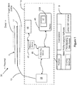
- the computing device 40 includes or accesses software modules or programs 44, such as a side branch detection module, a guide wire detection module, a lumen detection module, a stent detection module, a median mask clearing module, an intensity averaging module, a stent malapposition detection module, a stent planning module, a stent analysis module, FFR measurement module, VRR measurement module, and other software modules.
- the software modules or programs 44 can include an image data processing pipeline or component modules thereof and one or more graphical user interfaces (GUI).
- GUI graphical user interfaces
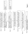
- An exemplary image processing pipeline 50 for transforming collected OCT data into two dimensional and three dimensional views of blood vessels and stents is depicted in Figure 5 .
- the image data processing pipeline or any of the methods described herein are stored in memory and executed using one or more computing devices such as a processor, device, or other integrated circuit.
- the disclosure relates to computer-based methods, systems and devices for visualizing stents.
- the disclosure relates to methods to evaluate the expansion of a stented vessel and whether further expansion is needed or if stent repositioning is needed. These methods of evaluation can include displaying one or more views of a stent in various states such as an expanded state relative to the blood vessel and comparing the imaged stent relative to one or more selected stent profiles. In one embodiment, the method is performed automatically.
- the methods of the disclosure can be implemented using software such as stent profile analysis software which can include stent planning software and lesion preparation software (or vice versa).
- a stent plan can include a first location such as a start frame and a second location such as an end frame.
- a stent plan can include a plurality of stent diameters.
- the stent plan includes a first and a second location along an imaged vessel and the stent diameter at the first and the second location.
- the stent plan can include a plurality of frames wherein the frames are generated using intravascular optically or acoustically measured data and parameters such as optical coherence tomography and IVUS data and parameters.
- the frames can include longitudinal, cross-sectional, or other views or images or data obtained relative to a blood vessel at one or more time periods.
- a first time period can include a first pullback during which the vessel is imaged.
- the target stent profile is generated using one or more method described herein which includes one or more user provided reference frames or other selections relative to identifying a section of a blood vessel as inputs.
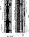
- the target stent profile is automatically placed over or overlaid upon the region of the lumen profile that corresponds to the detected stent struts such as shown in Figure 5 .
- the stent detection algorithm locates the stented lumen region.
- the lumen profile in the stented region is compared with the pre-stent target. Any over or under expansion can be highlighted automatically for the user to analyze.
- a quantified error measure can be generated that shows the error per frame or per segment between the post-stent lumen profile and the pre-stent target profile. This error measure can help the user select a suitable sized balloon when performing post stenting expansion.
- This error measure can include the distance by which the post-stent lumen profile and the pre-stent target profile differ.
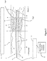
- Pre-stent planning Physician decides on the segment of blood vessel, with a lesion or other stenosis, to stent and the stent size using the stent planning tool which can include a user interface display such as shown in Figure 3 .
- the selection of the references specifies the range of stent sizes possible.
- a first user selected reference R1 is shown on the left or distal side of the interface.
- a second user selected reference R2 is shown on the right or proximal side of the interface.
- This process is performed using one or more user interfaces that can include displays and metrics relating to one or more intravascular data sets.
- the target stent profile based on user provided reference frames TSP is shown as narrowing relative to the lumen profiles contours with an upper and lower boundary shown by the horizontal lines.
- the lumen profile LP is shown on either side of the target stent plan along the center of the vessel with the top and bottom counters bounded by the TSP such as shown in Figure 3 .
- the OCT data is obtained with a probe and stored in memory; it can be processed to generate information such as a cross-sectional, a longitudinal, and/or a three-dimensional view of the blood vessel along the length of the pullback region or a subset thereof. These views can be depicted as part of a user interface as shown in Figures 3, 4 and 5 .
- the images of the blood vessel generated using the distances measurements obtained from the OCT system provide information about the blood vessel and objects disposed therein.
- the present disclosure also relates to apparatus for performing the operations herein.
- This apparatus may be specially constructed for the required purposes, or it may comprise a general purpose computer selectively activated or reconfigured by a computer program stored in the computer.
- some or all of the processing of the data collected using an OCT probe and the processor-based system is implemented as a set of computer program instructions that is converted into a computer executable form, stored as such in a computer readable medium, and executed by a microprocessor under the control of an operating system.
- query response and input data are transformed into processor understandable instructions suitable for generating OCT data, detecting lumen borders, detecting stent struts, comparing measured perpendicular distances relative to set thresholds, and otherwise performing image comparison, signal processing, artifact removal, and other features and embodiments described above.
- the computer program may be fixed in any form (e.g., source code form, computer executable form, or an intermediate form) either permanently or transitorily in a tangible storage medium, such as a semiconductor memory device (e.g., a RAM, ROM, PROM, EEPROM, or Flash-Programmable RAM), a magnetic memory device (e.g., a diskette or fixed disk), an optical memory device (e.g., a CD-ROM), a PC card (e.g., PCMCIA card), or other memory device.
- a semiconductor memory device e.g., a RAM, ROM, PROM, EEPROM, or Flash-Programmable RAM
- a magnetic memory device e.g., a diskette or fixed disk
- an optical memory device e.g., a CD-ROM
- PC card e.g., PCMCIA card
- a module refers to software, hardware, or firmware suitable for performing a specific data processing or data transmission task.
- a module refers to a software routine, program, or other memory resident application suitable for receiving, transforming, routing and processing instructions, or various types of data such as OCT scan data, IVUS scan data, interferometer signal data, target stent profiles, post-stent deployment lumen profiles and images, side branch locations, side branch diameters, jailed side branches, pre-stenting FFR values, post-stenting FFR values, and other pre and post stenting values and other information of interest.
- the instructions executable by the computing device cause the computing device to: display an FFR value or a VRR value measured after stent deployment.
Landscapes
- Health & Medical Sciences (AREA)
- Life Sciences & Earth Sciences (AREA)
- Engineering & Computer Science (AREA)
- Biomedical Technology (AREA)
- Public Health (AREA)
- Heart & Thoracic Surgery (AREA)
- Animal Behavior & Ethology (AREA)
- General Health & Medical Sciences (AREA)
- Veterinary Medicine (AREA)
- Pathology (AREA)
- Biophysics (AREA)
- Physics & Mathematics (AREA)
- Medical Informatics (AREA)
- Molecular Biology (AREA)
- Surgery (AREA)
- Cardiology (AREA)
- Physiology (AREA)
- Vascular Medicine (AREA)
- Oral & Maxillofacial Surgery (AREA)
- Nuclear Medicine, Radiotherapy & Molecular Imaging (AREA)
- Radiology & Medical Imaging (AREA)
- Dentistry (AREA)
- Transplantation (AREA)
- Artificial Intelligence (AREA)
- Computer Vision & Pattern Recognition (AREA)
- Psychiatry (AREA)
- Signal Processing (AREA)
- Hematology (AREA)
- Endoscopes (AREA)
- Ultra Sonic Daignosis Equipment (AREA)
- Media Introduction/Drainage Providing Device (AREA)
Applications Claiming Priority (3)
| Application Number | Priority Date | Filing Date | Title |
|---|---|---|---|
| US201462028711P | 2014-07-24 | 2014-07-24 | |
| PCT/US2015/042083 WO2016014991A1 (en) | 2014-07-24 | 2015-07-24 | Stent and vessel visualization and diagnostic systems, devices, and methods |
| EP15749906.2A EP3171763B1 (en) | 2014-07-24 | 2015-07-24 | Stent and vessel visualization and diagnostic methods |
Related Parent Applications (1)
| Application Number | Title | Priority Date | Filing Date |
|---|---|---|---|
| EP15749906.2A Division EP3171763B1 (en) | 2014-07-24 | 2015-07-24 | Stent and vessel visualization and diagnostic methods |
Publications (1)
| Publication Number | Publication Date |
|---|---|
| EP3622884A1 true EP3622884A1 (en) | 2020-03-18 |
Family
ID=53835502
Family Applications (2)
| Application Number | Title | Priority Date | Filing Date |
|---|---|---|---|
| EP19186267.1A Pending EP3622884A1 (en) | 2014-07-24 | 2015-07-24 | Stent and vessel visualization and diagnostic systems, devices, and methods |
| EP15749906.2A Active EP3171763B1 (en) | 2014-07-24 | 2015-07-24 | Stent and vessel visualization and diagnostic methods |
Family Applications After (1)
| Application Number | Title | Priority Date | Filing Date |
|---|---|---|---|
| EP15749906.2A Active EP3171763B1 (en) | 2014-07-24 | 2015-07-24 | Stent and vessel visualization and diagnostic methods |
Country Status (5)
| Country | Link |
|---|---|
| US (2) | US11166668B2 (cg-RX-API-DMAC7.html) |
| EP (2) | EP3622884A1 (cg-RX-API-DMAC7.html) |
| JP (4) | JP6682526B2 (cg-RX-API-DMAC7.html) |
| ES (1) | ES2744900T3 (cg-RX-API-DMAC7.html) |
| WO (1) | WO2016014991A1 (cg-RX-API-DMAC7.html) |
Families Citing this family (54)
| Publication number | Priority date | Publication date | Assignee | Title |
|---|---|---|---|---|
| AU2015360265B2 (en) * | 2014-12-12 | 2020-08-27 | Desmond Adler | Systems and methods to detect and display endovascular features |
| US11278206B2 (en) | 2015-04-16 | 2022-03-22 | Gentuity, Llc | Micro-optic probes for neurology |
| US10140712B2 (en) | 2015-05-17 | 2018-11-27 | Lightlab Imaging, Inc. | Detection of stent struts relative to side branches |
| US10109058B2 (en) | 2015-05-17 | 2018-10-23 | Lightlab Imaging, Inc. | Intravascular imaging system interfaces and stent detection methods |
| US10646198B2 (en) | 2015-05-17 | 2020-05-12 | Lightlab Imaging, Inc. | Intravascular imaging and guide catheter detection methods and systems |
| US10222956B2 (en) | 2015-05-17 | 2019-03-05 | Lightlab Imaging, Inc. | Intravascular imaging user interface systems and methods |
| US9996921B2 (en) | 2015-05-17 | 2018-06-12 | LIGHTLAB IMAGING, lNC. | Detection of metal stent struts |
| EP4159121B1 (en) | 2015-07-25 | 2026-07-15 | Lightlab Imaging, Inc. | Intravascular data visualization method and device |
| US10631718B2 (en) | 2015-08-31 | 2020-04-28 | Gentuity, Llc | Imaging system includes imaging probe and delivery devices |
| CN108348170B (zh) | 2015-11-18 | 2021-04-02 | 光学实验室成像公司 | 与侧支有关的支架撑杆检测 |
| CA3005296A1 (en) | 2015-11-23 | 2017-06-01 | Lightlab Imaging, Inc. | Detection of and validation of shadows in intravascular images |
| ES2908571T3 (es) | 2016-04-14 | 2022-05-03 | Lightlab Imaging Inc | Identificación de ramas de un vaso sanguíneo |
| US10631754B2 (en) * | 2016-05-16 | 2020-04-28 | Lightlab Imaging, Inc. | Intravascular absorbable stent detection and diagnostic methods and systems |
| CN105919577A (zh) * | 2016-06-06 | 2016-09-07 | 任勇 | 基于静脉支架的压强差监测装置及其方法 |
| CN105919559A (zh) * | 2016-06-06 | 2016-09-07 | 任勇 | 基于腹主动脉支架的瘤体阻抗和血压监测装置及其方法 |
| CN106073749A (zh) * | 2016-06-06 | 2016-11-09 | 任勇 | 基于静脉支架的血液流速监测装置及其方法 |
| CN105942995A (zh) * | 2016-06-06 | 2016-09-21 | 任勇 | 基于胸降主动脉支架的血管壁压力监测装置及其方法 |
| CN105902260A (zh) * | 2016-06-06 | 2016-08-31 | 任勇 | 基于腹主动脉支架的血液流变监测装置及其方法 |
| CN105942997A (zh) * | 2016-06-06 | 2016-09-21 | 任勇 | 基于腹主动脉支架的综合参数监测装置及其方法 |
| CN105902261A (zh) * | 2016-06-06 | 2016-08-31 | 任勇 | 基于胸降主动脉支架的血液流速监测装置及其方法 |
| US10806516B2 (en) * | 2016-06-20 | 2020-10-20 | General Electric Company | Virtual 4D stent implantation path assessment |
| CN117398183A (zh) | 2016-09-28 | 2024-01-16 | 光学实验室成像公司 | 利用血管表象的支架规划系统及方法 |
| US10568696B2 (en) * | 2017-07-17 | 2020-02-25 | International Business Machines Corporation | Apparatus for supporting personalized coronary stents |
| US20190099237A1 (en) | 2017-10-02 | 2019-04-04 | Lightlab Imaging, Inc. | Intravascular Data Collection Probes and Related Assemblies |
| US11571129B2 (en) | 2017-10-03 | 2023-02-07 | Canon U.S.A., Inc. | Detecting and displaying stent expansion |
| US10621748B2 (en) | 2017-10-03 | 2020-04-14 | Canon U.S.A., Inc. | Detecting and displaying stent expansion |
| JP7160935B2 (ja) | 2017-11-28 | 2022-10-25 | ジェンテュイティ・リミテッド・ライアビリティ・カンパニー | 撮像システム |
| WO2019175032A1 (en) * | 2018-03-14 | 2019-09-19 | Koninklijke Philips N.V. | Scoring intravascular lesions and stent deployment in medical intraluminal ultrasound imaging |
| ES3013537T3 (en) * | 2018-05-29 | 2025-04-14 | Lightlab Imaging Inc | Stent expansion display, systems, and methods |
| WO2020025352A1 (en) * | 2018-07-30 | 2020-02-06 | Koninklijke Philips N.V. | Intravascular imaging procedure-specific workflow guidance and associated devices, systems, and methods |
| US20200029932A1 (en) * | 2018-07-30 | 2020-01-30 | Koninklijke Philips N.V. | Systems, devices, and methods for displaying multiple intraluminal images in luminal assessment with medical imaging |
| JP2022500174A (ja) | 2018-09-17 | 2022-01-04 | ジェンテュイティ・リミテッド・ライアビリティ・カンパニーGentuity, LLC | 光学経路を有する撮像システム |
| CN113473917B (zh) * | 2018-10-26 | 2025-02-11 | 皇家飞利浦有限公司 | 具有自动和辅助标签和书签的腔内超声成像 |
| CN113520353B (zh) * | 2018-12-31 | 2023-06-16 | 深圳北芯生命科技股份有限公司 | 导管模拟器的使用方法 |
| WO2020190976A2 (en) * | 2019-03-17 | 2020-09-24 | Lightlab Imaging, Inc. | Arterial imaging and assessment systems and methods and related user interface based-workflows |
| EP3955795B1 (en) * | 2019-04-17 | 2024-08-21 | University of Pittsburgh - of the Commonwealth System of Higher Education | Device for endovascular orifice detection for fenestrated stent graft deployment |
| EP3962346A4 (en) | 2019-04-30 | 2023-04-19 | Gentuity LLC | IMAGING PROBE WITH FLUID PRESSURIZATION ELEMENT |
| WO2020237024A1 (en) | 2019-05-21 | 2020-11-26 | Gentuity, Llc | Systems and methods for oct-guided treatment of a patient |
| CN110060254A (zh) * | 2019-05-29 | 2019-07-26 | 深圳华声医疗技术股份有限公司 | 交互式血管测量方法、装置、计算机可读存储介质及系统 |
| US11436731B2 (en) * | 2019-08-05 | 2022-09-06 | Lightlab Imaging, Inc. | Longitudinal display of coronary artery calcium burden |
| US20230045488A1 (en) * | 2020-01-06 | 2023-02-09 | Philips Image Guided Therapy Corporation | Intraluminal imaging based detection and visualization of intraluminal treatment anomalies |
| JP7737357B2 (ja) * | 2020-03-30 | 2025-09-10 | テルモ株式会社 | プログラム、情報処理方法、および情報処理システム |
| US11922633B2 (en) | 2020-06-30 | 2024-03-05 | Canon U.S.A., Inc. | Real-time lumen distance calculation based on three-dimensional (3D) A-line signal data |
| WO2022202310A1 (ja) * | 2021-03-26 | 2022-09-29 | テルモ株式会社 | プログラム、画像処理方法及び画像処理装置 |
| EP4074261A1 (en) | 2021-04-16 | 2022-10-19 | Koninklijke Philips N.V. | Determining end point locations for a stent |
| USD1083949S1 (en) | 2021-10-15 | 2025-07-15 | Lightlab Imaging, Inc. | Display screen or portion thereof with graphical user interface |
| US12609207B2 (en) * | 2022-01-11 | 2026-04-21 | Case Western Reserve University | Prediction of stent expansion for treatments |
| WO2023189308A1 (ja) * | 2022-03-28 | 2023-10-05 | テルモ株式会社 | コンピュータプログラム、画像処理方法及び画像処理装置 |
| US12514721B2 (en) * | 2022-05-18 | 2026-01-06 | Olympus Corporation | Stent delivery system, control apparatus, and method |
| US12690843B2 (en) | 2022-09-14 | 2026-07-28 | Boston Scientific Scimed, Inc. | Key frame identification for intravascular ultrasound based on plaque burden |
| WO2024059136A1 (en) * | 2022-09-14 | 2024-03-21 | Boston Scientific Scimed, Inc. | Graphical user interface for intravascular ultrasound stent display |
| WO2024202466A1 (ja) * | 2023-03-30 | 2024-10-03 | テルモ株式会社 | コンピュータプログラム、情報処理方法及び情報処理装置 |
| EP4523620A1 (de) * | 2023-09-15 | 2025-03-19 | Siemens Healthineers AG | Bestimmung einer länge eines stents |
| CN119624853B (zh) * | 2024-09-30 | 2025-11-25 | 博氏(广州)医学技术有限公司 | 一种基于图像分割的冠状动脉分流栓评价方法及系统 |
Citations (4)
| Publication number | Priority date | Publication date | Assignee | Title |
|---|---|---|---|---|
| US20100094127A1 (en) * | 2008-10-14 | 2010-04-15 | Lightlab Imaging, Inc. | Methods for stent strut detection and related measurement and display using optical coherence tomography |
| US20110071404A1 (en) * | 2009-09-23 | 2011-03-24 | Lightlab Imaging, Inc. | Lumen Morphology and Vascular Resistance Measurements Data Collection Systems, Apparatus and Methods |
| US20140100449A1 (en) * | 2012-10-05 | 2014-04-10 | Volcano Corporation | Automatic stent detection |
| WO2014092755A1 (en) * | 2012-12-12 | 2014-06-19 | Lightlab Imaging, Inc. | Method and apparatus for automated determination of a lumen contour of a blood vessel |
Family Cites Families (94)
| Publication number | Priority date | Publication date | Assignee | Title |
|---|---|---|---|---|
| US5477858A (en) | 1986-07-30 | 1995-12-26 | Siemens Medical Systems, Inc. | Ultrasound blood flow/tissue imaging system |
| US5662109A (en) | 1990-12-14 | 1997-09-02 | Hutson; William H. | Method and system for multi-dimensional imaging and analysis for early detection of diseased tissue |
| US5054492A (en) | 1990-12-17 | 1991-10-08 | Cardiovascular Imaging Systems, Inc. | Ultrasonic imaging catheter having rotational image correlation |
| US5956355A (en) | 1991-04-29 | 1999-09-21 | Massachusetts Institute Of Technology | Method and apparatus for performing optical measurements using a rapidly frequency-tuned laser |
| US6564087B1 (en) | 1991-04-29 | 2003-05-13 | Massachusetts Institute Of Technology | Fiber optic needle probes for optical coherence tomography imaging |
| US5748598A (en) | 1995-12-22 | 1998-05-05 | Massachusetts Institute Of Technology | Apparatus and methods for reading multilayer storage media using short coherence length sources |
| US6501551B1 (en) | 1991-04-29 | 2002-12-31 | Massachusetts Institute Of Technology | Fiber optic imaging endoscope interferometer with at least one faraday rotator |
| US6111645A (en) | 1991-04-29 | 2000-08-29 | Massachusetts Institute Of Technology | Grating based phase control optical delay line |
| US5465147A (en) | 1991-04-29 | 1995-11-07 | Massachusetts Institute Of Technology | Method and apparatus for acquiring images using a ccd detector array and no transverse scanner |
| US6485413B1 (en) | 1991-04-29 | 2002-11-26 | The General Hospital Corporation | Methods and apparatus for forward-directed optical scanning instruments |
| US6134003A (en) | 1991-04-29 | 2000-10-17 | Massachusetts Institute Of Technology | Method and apparatus for performing optical measurements using a fiber optic imaging guidewire, catheter or endoscope |
| US5321501A (en) | 1991-04-29 | 1994-06-14 | Massachusetts Institute Of Technology | Method and apparatus for optical imaging with means for controlling the longitudinal range of the sample |
| US5325449A (en) | 1992-05-15 | 1994-06-28 | David Sarnoff Research Center, Inc. | Method for fusing images and apparatus therefor |
| US5509093A (en) | 1993-10-13 | 1996-04-16 | Micron Optics, Inc. | Temperature compensated fiber fabry-perot filters |
| US5531227A (en) | 1994-01-28 | 1996-07-02 | Schneider Medical Technologies, Inc. | Imaging device and method |
| US5797849A (en) | 1995-03-28 | 1998-08-25 | Sonometrics Corporation | Method for carrying out a medical procedure using a three-dimensional tracking and imaging system |
| US5619368A (en) | 1995-05-16 | 1997-04-08 | Massachusetts Inst. Of Technology | Optical frequency shifter |
| WO1997001167A1 (en) | 1995-06-21 | 1997-01-09 | Massachusetts Institute Of Technology | Apparatus and method for accessing data on multilayered optical media |
| US5810007A (en) | 1995-07-26 | 1998-09-22 | Associates Of The Joint Center For Radiation Therapy, Inc. | Ultrasound localization and image fusion for the treatment of prostate cancer |
| US5965355A (en) | 1995-09-21 | 1999-10-12 | Agritope, Inc. | Antibodies and proteins useful for assaying virus infection in grape plants |
| US5771895A (en) | 1996-02-12 | 1998-06-30 | Slager; Cornelis J. | Catheter for obtaining three-dimensional reconstruction of a vascular lumen and wall |
| US6148095A (en) | 1997-09-08 | 2000-11-14 | University Of Iowa Research Foundation | Apparatus and method for determining three-dimensional representations of tortuous vessels |
| US5989189A (en) | 1997-10-24 | 1999-11-23 | Mentor Corporation | Ophthalmic ultrasound imaging |
| US20020161351A1 (en) | 1998-09-01 | 2002-10-31 | Samson Wilfred J. | Method and apparatus for treating acute myocardial infarction with selective hypothermic perfusion |
| US6191862B1 (en) | 1999-01-20 | 2001-02-20 | Lightlab Imaging, Llc | Methods and apparatus for high speed longitudinal scanning in imaging systems |
| US6445939B1 (en) | 1999-08-09 | 2002-09-03 | Lightlab Imaging, Llc | Ultra-small optical probes, imaging optics, and methods for using same |
| DE60141090D1 (de) | 2000-10-30 | 2010-03-04 | Gen Hospital Corp | Optische systeme zur gewebeanalyse |
| US20020115931A1 (en) | 2001-02-21 | 2002-08-22 | Strauss H. William | Localizing intravascular lesions on anatomic images |
| US6768756B2 (en) | 2001-03-12 | 2004-07-27 | Axsun Technologies, Inc. | MEMS membrane with integral mirror/lens |
| US6570659B2 (en) | 2001-03-16 | 2003-05-27 | Lightlab Imaging, Llc | Broadband light source system and method and light source combiner |
| US6552796B2 (en) | 2001-04-06 | 2003-04-22 | Lightlab Imaging, Llc | Apparatus and method for selective data collection and signal to noise ratio enhancement using optical coherence tomography |
| US6706004B2 (en) | 2001-05-31 | 2004-03-16 | Infraredx, Inc. | Balloon catheter |
| US6879851B2 (en) | 2001-06-07 | 2005-04-12 | Lightlab Imaging, Llc | Fiber optic endoscopic gastrointestinal probe |
| US7937660B2 (en) * | 2002-04-19 | 2011-05-03 | Medtronic Vascular, Inc. | Computer-based methods and structures for stent-graft selection |
| US6891984B2 (en) | 2002-07-25 | 2005-05-10 | Lightlab Imaging, Llc | Scanning miniature optical probes with optical distortion correction and rotational control |
| US8465452B2 (en) * | 2003-02-21 | 2013-06-18 | 3Dt Holdings, Llc | Devices, systems, and methods for removing stenotic lesions from vessels |
| US20060165270A1 (en) | 2003-02-25 | 2006-07-27 | Jorn Borgert | Intravascular imaging |
| US7241286B2 (en) | 2003-04-25 | 2007-07-10 | Lightlab Imaging, Llc | Flush catheter with flow directing sheath |
| US20100076320A1 (en) | 2003-04-25 | 2010-03-25 | Lightlab Imaging, Llc | Flush catheter with flow directing sheath |
| US7998188B2 (en) * | 2003-04-28 | 2011-08-16 | Kips Bay Medical, Inc. | Compliant blood vessel graft |
| WO2005047813A1 (en) | 2003-10-27 | 2005-05-26 | The General Hospital Corporation | Method and apparatus for performing optical imaging using frequency-domain interferometry |
| US20050238067A1 (en) | 2004-04-26 | 2005-10-27 | Choi Youngmin A | Simple fiber optic cavity |
| US7397935B2 (en) | 2004-05-10 | 2008-07-08 | Mediguide Ltd. | Method for segmentation of IVUS image sequences |
| WO2006037001A1 (en) | 2004-09-24 | 2006-04-06 | Lightlab Imaging, Inc. | Fluid occluding devices and methods |
| US7930014B2 (en) | 2005-01-11 | 2011-04-19 | Volcano Corporation | Vascular image co-registration |
| EP2264841B1 (en) | 2005-01-20 | 2016-01-20 | Massachusetts Institute of Technology (MIT) | Mode locking methods and apparatus |
| US8315282B2 (en) | 2005-01-20 | 2012-11-20 | Massachusetts Institute Of Technology | Fourier domain mode locking: method and apparatus for control and improved performance |
| EP1856777A4 (en) | 2005-01-24 | 2009-04-29 | Thorlabs Inc | COMPACT MULTIMODE LASER WITH FAST WAVELENGTH PATTERN |
| US8054075B2 (en) | 2005-02-03 | 2011-11-08 | The Johns Hopkins University | Method for magnetic resonance imaging using inversion recovery with on-resonant water suppression including MRI systems and software embodying same |
| JP2008529669A (ja) | 2005-02-10 | 2008-08-07 | ライトラボ・イメージング・インコーポレーテッド | 光学コヒーレンス断層撮影法の装置及び方法 |
| US7415049B2 (en) | 2005-03-28 | 2008-08-19 | Axsun Technologies, Inc. | Laser with tilted multi spatial mode resonator tuning element |
| WO2007002685A2 (en) | 2005-06-24 | 2007-01-04 | Volcano Corporation | Co-registration of graphical image data representing three-dimensional vascular features |
| US7729746B2 (en) | 2005-11-04 | 2010-06-01 | Siemens Aktiengesellschaft | Three-dimensional co-registration between intravascular and angiographic data |
| US7593559B2 (en) | 2005-11-18 | 2009-09-22 | Duke University | Method and system of coregistrating optical coherence tomography (OCT) with other clinical tests |
| US7801343B2 (en) | 2005-11-29 | 2010-09-21 | Siemens Medical Solutions Usa, Inc. | Method and apparatus for inner wall extraction and stent strut detection using intravascular optical coherence tomography imaging |
| US7650179B2 (en) * | 2005-12-09 | 2010-01-19 | Siemens Aktiengesellschaft | Computerized workflow method for stent planning and stenting procedure |
| US7619646B2 (en) | 2006-03-09 | 2009-11-17 | Visicon Inspection Technologies Llc | System and method to illuminate and image the inside diameter of a stent |
| US8223143B2 (en) | 2006-10-27 | 2012-07-17 | Carl Zeiss Meditec, Inc. | User interface for efficiently displaying relevant OCT imaging data |
| EP2081486B1 (en) | 2006-11-08 | 2014-04-09 | Lightlab Imaging, Inc. | Opto-acoustic imaging device |
| EP3785615B1 (en) | 2007-01-10 | 2024-12-04 | Lightlab Imaging, Inc. | Methods and apparatus for swept-source optical coherence tomography |
| US8582934B2 (en) | 2007-11-12 | 2013-11-12 | Lightlab Imaging, Inc. | Miniature optical elements for fiber-optic beam shaping |
| US7813609B2 (en) | 2007-11-12 | 2010-10-12 | Lightlab Imaging, Inc. | Imaging catheter with integrated reference reflector |
| US8275201B2 (en) * | 2008-03-06 | 2012-09-25 | Tyco Healthcare Group Lp | Image enhancement and application functionality for medical and other uses |
| US20110190586A1 (en) | 2008-03-28 | 2011-08-04 | Volcano Corporation | Methods and systems for intravascular imaging and flushing |
| JP4746068B2 (ja) * | 2008-03-28 | 2011-08-10 | 一史 浦澤 | 画像解析装置及び画像解析方法 |
| EP2293714B1 (en) | 2008-06-02 | 2014-08-13 | Lightlab Imaging, Inc. | Quantitative methods for obtaining tissue characteristics from optical coherence tomography images |
| DE102008045634A1 (de) | 2008-09-03 | 2010-03-04 | Ludwig-Maximilians-Universität München | Wellenlängenabstimmbare Lichtquelle |
| US12426789B2 (en) | 2009-09-23 | 2025-09-30 | Lightlab Imaging, Inc. | Blood vessel lumen morphology and minimum lumen area measurements data collection by intravascular imaging systems for stenosis or stent planning |
| US8412312B2 (en) | 2009-09-23 | 2013-04-02 | Lightlab Imaging, Inc. | Apparatus, systems, and methods of in-vivo blood clearing in a lumen |
| US8206377B2 (en) | 2009-12-22 | 2012-06-26 | Lightlab Imaging, Inc. | Torque limiter for an OCT catheter |
| US8926590B2 (en) | 2009-12-22 | 2015-01-06 | Lightlab Imaging, Inc. | Torque limiter for an OCT catheter |
| US8478384B2 (en) | 2010-01-19 | 2013-07-02 | Lightlab Imaging, Inc. | Intravascular optical coherence tomography system with pressure monitoring interface and accessories |
| US8948613B2 (en) | 2010-03-17 | 2015-02-03 | Lightlab Imaging, Inc. | Intensity noise reduction methods and apparatus for interferometric sensing and imaging systems |
| US20110257545A1 (en) | 2010-04-20 | 2011-10-20 | Suri Jasjit S | Imaging based symptomatic classification and cardiovascular stroke risk score estimation |
| JP5562775B2 (ja) | 2010-09-15 | 2014-07-30 | 株式会社東芝 | 医用画像表示システム |
| WO2012042413A1 (en) | 2010-09-30 | 2012-04-05 | Koninklijke Philips Electronics N.V. | Detection of bifurcations using traceable imaging device and imaging tool |
| US8582619B2 (en) | 2011-03-15 | 2013-11-12 | Lightlab Imaging, Inc. | Methods, systems, and devices for timing control in electromagnetic radiation sources |
| US9164240B2 (en) | 2011-03-31 | 2015-10-20 | Lightlab Imaging, Inc. | Optical buffering methods, apparatus, and systems for increasing the repetition rate of tunable light sources |
| JP6141264B2 (ja) * | 2011-05-27 | 2017-06-07 | ライトラボ・イメージング・インコーポレーテッド | 光コヒーレンス断層撮影法及び圧力に基づくシステム及び方法 |
| WO2012166829A2 (en) | 2011-05-31 | 2012-12-06 | Lightlab Imaging, Inc. | Multimodal imaging system, apparatus, and methods |
| WO2013003267A1 (en) | 2011-06-30 | 2013-01-03 | Lightlab Imaging, Inc. | Catheter with flush valve and related systems and methods |
| US8582109B1 (en) | 2011-08-01 | 2013-11-12 | Lightlab Imaging, Inc. | Swept mode-hopping laser system, methods, and devices for frequency-domain optical coherence tomography |
| WO2013019840A1 (en) * | 2011-08-03 | 2013-02-07 | Lightlab Imaging, Inc. | Systems, methods and apparatus for determining a fractional flow reserve |
| US20130051728A1 (en) | 2011-08-31 | 2013-02-28 | Lightlab Imaging, Inc. | Optical Imaging Probes and Related Methods |
| WO2013042231A1 (ja) | 2011-09-21 | 2013-03-28 | 医療法人澄心会豊橋ハートセンター | 血管断面形状数値化装置及びそれを用いた血管断面形状数値化方法 |
| US8581643B1 (en) | 2011-10-28 | 2013-11-12 | Lightlab Imaging, Inc. | Phase-lock loop-based clocking system, methods and apparatus |
| US8953911B1 (en) | 2011-10-28 | 2015-02-10 | Lightlab Imaging, Inc. | Spectroscopic imaging probes, devices, and methods |
| US8831321B1 (en) | 2011-11-07 | 2014-09-09 | Lightlab Imaging, Inc. | Side branch detection methods, systems and devices |
| US20140024931A1 (en) | 2012-07-20 | 2014-01-23 | Lightlab Imaging, Inc. | Data Encoders for Medical Devices and Related Methods |
| US8687201B2 (en) | 2012-08-31 | 2014-04-01 | Lightlab Imaging, Inc. | Optical coherence tomography control systems and methods |
| US10568586B2 (en) | 2012-10-05 | 2020-02-25 | Volcano Corporation | Systems for indicating parameters in an imaging data set and methods of use |
| WO2014077880A1 (en) | 2012-11-16 | 2014-05-22 | Lightlab Imaging, Inc. | Automated fluid delivery catheter and system |
| WO2014077870A1 (en) | 2012-11-19 | 2014-05-22 | Lightlab Imaging, Inc. | Multimodel imaging systems, probes and methods |
| US9702762B2 (en) | 2013-03-15 | 2017-07-11 | Lightlab Imaging, Inc. | Calibration and image processing devices, methods, and systems |
-
2015
- 2015-07-24 WO PCT/US2015/042083 patent/WO2016014991A1/en not_active Ceased
- 2015-07-24 EP EP19186267.1A patent/EP3622884A1/en active Pending
- 2015-07-24 ES ES15749906T patent/ES2744900T3/es active Active
- 2015-07-24 EP EP15749906.2A patent/EP3171763B1/en active Active
- 2015-07-24 US US14/808,901 patent/US11166668B2/en active Active
- 2015-07-24 JP JP2017525323A patent/JP6682526B2/ja active Active
-
2020
- 2020-03-25 JP JP2020053738A patent/JP7325368B2/ja active Active
-
2021
- 2021-11-05 US US17/519,764 patent/US20220117551A1/en active Pending
-
2023
- 2023-05-10 JP JP2023077688A patent/JP7568777B2/ja active Active
-
2024
- 2024-10-03 JP JP2024173949A patent/JP2024177359A/ja active Pending
Patent Citations (4)
| Publication number | Priority date | Publication date | Assignee | Title |
|---|---|---|---|---|
| US20100094127A1 (en) * | 2008-10-14 | 2010-04-15 | Lightlab Imaging, Inc. | Methods for stent strut detection and related measurement and display using optical coherence tomography |
| US20110071404A1 (en) * | 2009-09-23 | 2011-03-24 | Lightlab Imaging, Inc. | Lumen Morphology and Vascular Resistance Measurements Data Collection Systems, Apparatus and Methods |
| US20140100449A1 (en) * | 2012-10-05 | 2014-04-10 | Volcano Corporation | Automatic stent detection |
| WO2014092755A1 (en) * | 2012-12-12 | 2014-06-19 | Lightlab Imaging, Inc. | Method and apparatus for automated determination of a lumen contour of a blood vessel |
Also Published As
| Publication number | Publication date |
|---|---|
| ES2744900T3 (es) | 2020-02-26 |
| JP2017527418A (ja) | 2017-09-21 |
| US20220117551A1 (en) | 2022-04-21 |
| WO2016014991A1 (en) | 2016-01-28 |
| JP2024177359A (ja) | 2024-12-19 |
| EP3171763B1 (en) | 2019-07-17 |
| JP7325368B2 (ja) | 2023-08-14 |
| US20160022208A1 (en) | 2016-01-28 |
| JP6682526B2 (ja) | 2020-04-15 |
| JP2020114423A (ja) | 2020-07-30 |
| JP2023099605A (ja) | 2023-07-13 |
| EP3171763A1 (en) | 2017-05-31 |
| US11166668B2 (en) | 2021-11-09 |
| JP7568777B2 (ja) | 2024-10-16 |
Similar Documents
| Publication | Publication Date | Title |
|---|---|---|
| JP7568777B2 (ja) | ステント及び血管の可視化及び診断のシステム、装置、及び方法 | |
| US12561810B2 (en) | Stent detection methods and imaging system interfaces | |
| JP7375102B2 (ja) | 血管内画像化システムの作動方法 | |
| JP7566084B2 (ja) | ステント拡張を評価するシステム及びその作動方法 | |
| US9173591B2 (en) | Stent visualization and malapposition detection systems, devices, and methods | |
| EP3361933B1 (en) | Intravascular imaging system and methods to determine cut plane view angle of side branch | |
| EP2964089B1 (en) | Stent visualization and malapposition detection systems, devices, and methods | |
| JP2024054120A (ja) | 血管内のステントストラットのカバレッジを表示するシステム及び方法 | |
| US10222956B2 (en) | Intravascular imaging user interface systems and methods | |
| CA2765407A1 (en) | Lumen morphology and vascular resistance measurements data collection systems, apparatus and methods |
Legal Events
| Date | Code | Title | Description |
|---|---|---|---|
| PUAI | Public reference made under article 153(3) epc to a published international application that has entered the european phase |
Free format text: ORIGINAL CODE: 0009012 |
|
| STAA | Information on the status of an ep patent application or granted ep patent |
Free format text: STATUS: THE APPLICATION HAS BEEN PUBLISHED |
|
| AC | Divisional application: reference to earlier application |
Ref document number: 3171763 Country of ref document: EP Kind code of ref document: P |
|
| AK | Designated contracting states |
Kind code of ref document: A1 Designated state(s): AL AT BE BG CH CY CZ DE DK EE ES FI FR GB GR HR HU IE IS IT LI LT LU LV MC MK MT NL NO PL PT RO RS SE SI SK SM TR |
|
| STAA | Information on the status of an ep patent application or granted ep patent |
Free format text: STATUS: REQUEST FOR EXAMINATION WAS MADE |
|
| 17P | Request for examination filed |
Effective date: 20200910 |
|
| RBV | Designated contracting states (corrected) |
Designated state(s): AL AT BE BG CH CY CZ DE DK EE ES FI FR GB GR HR HU IE IS IT LI LT LU LV MC MK MT NL NO PL PT RO RS SE SI SK SM TR |
|
| STAA | Information on the status of an ep patent application or granted ep patent |
Free format text: STATUS: EXAMINATION IS IN PROGRESS |
|
| 17Q | First examination report despatched |
Effective date: 20220905 |
|
| P01 | Opt-out of the competence of the unified patent court (upc) registered |
Effective date: 20230616 |