EP3604540A1 - Norovirus antibody - Google Patents
Norovirus antibody Download PDFInfo
- Publication number
- EP3604540A1 EP3604540A1 EP18775369.4A EP18775369A EP3604540A1 EP 3604540 A1 EP3604540 A1 EP 3604540A1 EP 18775369 A EP18775369 A EP 18775369A EP 3604540 A1 EP3604540 A1 EP 3604540A1
- Authority
- EP
- European Patent Office
- Prior art keywords
- seq
- amino acid
- acid sequence
- set forth
- nanoantibody
- Prior art date
- Legal status (The legal status is an assumption and is not a legal conclusion. Google has not performed a legal analysis and makes no representation as to the accuracy of the status listed.)
- Withdrawn
Links
Images






Classifications
-
- C—CHEMISTRY; METALLURGY
- C12—BIOCHEMISTRY; BEER; SPIRITS; WINE; VINEGAR; MICROBIOLOGY; ENZYMOLOGY; MUTATION OR GENETIC ENGINEERING
- C12N—MICROORGANISMS OR ENZYMES; COMPOSITIONS THEREOF; PROPAGATING, PRESERVING, OR MAINTAINING MICROORGANISMS; MUTATION OR GENETIC ENGINEERING; CULTURE MEDIA
- C12N5/00—Undifferentiated human, animal or plant cells, e.g. cell lines; Tissues; Cultivation or maintenance thereof; Culture media therefor
- C12N5/06—Animal cells or tissues; Human cells or tissues
- C12N5/0602—Vertebrate cells
- C12N5/0679—Cells of the gastro-intestinal tract
-
- A—HUMAN NECESSITIES
- A23—FOODS OR FOODSTUFFS; TREATMENT THEREOF, NOT COVERED BY OTHER CLASSES
- A23L—FOODS, FOODSTUFFS OR NON-ALCOHOLIC BEVERAGES, NOT OTHERWISE PROVIDED FOR; PREPARATION OR TREATMENT THEREOF
- A23L7/00—Cereal-derived products; Malt products; Preparation or treatment thereof
- A23L7/10—Cereal-derived products
-
- A—HUMAN NECESSITIES
- A61—MEDICAL OR VETERINARY SCIENCE; HYGIENE
- A61K—PREPARATIONS FOR MEDICAL, DENTAL OR TOILETRY PURPOSES
- A61K39/00—Medicinal preparations containing antigens or antibodies
- A61K39/395—Antibodies; Immunoglobulins; Immune serum, e.g. antilymphocytic serum
-
- A—HUMAN NECESSITIES
- A61—MEDICAL OR VETERINARY SCIENCE; HYGIENE
- A61P—SPECIFIC THERAPEUTIC ACTIVITY OF CHEMICAL COMPOUNDS OR MEDICINAL PREPARATIONS
- A61P31/00—Antiinfectives, i.e. antibiotics, antiseptics, chemotherapeutics
- A61P31/12—Antivirals
- A61P31/14—Antivirals for RNA viruses
-
- C—CHEMISTRY; METALLURGY
- C07—ORGANIC CHEMISTRY
- C07K—PEPTIDES
- C07K16/00—Immunoglobulins [IG], e.g. monoclonal or polyclonal antibodies
- C07K16/08—Immunoglobulins [IG], e.g. monoclonal or polyclonal antibodies against material from viruses
- C07K16/10—RNA viruses
-
- C—CHEMISTRY; METALLURGY
- C12—BIOCHEMISTRY; BEER; SPIRITS; WINE; VINEGAR; MICROBIOLOGY; ENZYMOLOGY; MUTATION OR GENETIC ENGINEERING
- C12N—MICROORGANISMS OR ENZYMES; COMPOSITIONS THEREOF; PROPAGATING, PRESERVING, OR MAINTAINING MICROORGANISMS; MUTATION OR GENETIC ENGINEERING; CULTURE MEDIA
- C12N15/00—Mutation or genetic engineering; DNA or RNA concerning genetic engineering, vectors, e.g. plasmids, or their isolation, preparation or purification; Use of hosts therefor
- C12N15/09—Recombinant DNA-technology
- C12N15/63—Introduction of foreign genetic material using vectors; Vectors; Use of hosts therefor; Regulation of expression
- C12N15/79—Vectors or expression systems specially adapted for eukaryotic hosts
- C12N15/82—Vectors or expression systems specially adapted for eukaryotic hosts for plant cells, e.g. plant artificial chromosomes (PACs)
- C12N15/8241—Phenotypically and genetically modified plants via recombinant DNA technology
- C12N15/8242—Phenotypically and genetically modified plants via recombinant DNA technology with non-agronomic quality (output) traits, e.g. for industrial processing; Value added, non-agronomic traits
- C12N15/8257—Phenotypically and genetically modified plants via recombinant DNA technology with non-agronomic quality (output) traits, e.g. for industrial processing; Value added, non-agronomic traits for the production of primary gene products, e.g. pharmaceutical products, interferon
- C12N15/8258—Phenotypically and genetically modified plants via recombinant DNA technology with non-agronomic quality (output) traits, e.g. for industrial processing; Value added, non-agronomic traits for the production of primary gene products, e.g. pharmaceutical products, interferon for the production of oral vaccines (antigens) or immunoglobulins
-
- A—HUMAN NECESSITIES
- A61—MEDICAL OR VETERINARY SCIENCE; HYGIENE
- A61K—PREPARATIONS FOR MEDICAL, DENTAL OR TOILETRY PURPOSES
- A61K39/00—Medicinal preparations containing antigens or antibodies
- A61K2039/55—Medicinal preparations containing antigens or antibodies characterised by the host/recipient, e.g. newborn with maternal antibodies
- A61K2039/552—Veterinary vaccine
-
- C—CHEMISTRY; METALLURGY
- C12—BIOCHEMISTRY; BEER; SPIRITS; WINE; VINEGAR; MICROBIOLOGY; ENZYMOLOGY; MUTATION OR GENETIC ENGINEERING
- C12N—MICROORGANISMS OR ENZYMES; COMPOSITIONS THEREOF; PROPAGATING, PRESERVING, OR MAINTAINING MICROORGANISMS; MUTATION OR GENETIC ENGINEERING; CULTURE MEDIA
- C12N2506/00—Differentiation of animal cells from one lineage to another; Differentiation of pluripotent cells
- C12N2506/45—Differentiation of animal cells from one lineage to another; Differentiation of pluripotent cells from artificially induced pluripotent stem cells
Definitions
- the present invention relates to a norovirus antibody. More specifically, the present invention relates to the variable domain of the heavy chain of a heavy-chain antibody against a norovirus.
- Norovirus is a pathogenic virus that invades into cells existing from the duodenum to the upper small intestine and causes nonbacterial acute gastroenteritis.
- Norovirus belongs to Caliciviridae lacking envelopes.
- the genome of norovirus is a single-stranded plus-sense RNA of approximately 7.7 kb.
- a viral protein called VPg covalently binds to the 5'-terminus of the genome, whereas the 3'-terminus thereof is polyadenylated.
- the coding regions of three proteins, namely, ORF1, VP1, and VP2 are present, and individual coding regions encode a non-structural protein, a structural protein 1, and a structural protein 2, respectively.
- VP1 monomers are divided into two domains, namely, a shell domain (S domain) and a protruding domain (P domain), and the P domain is further divided into P1 and P2 subdomains.
- P2 recognizes blood group antigens functioning as a receptor and a host infectious factor upon infection. As a result of a mutation occurring in the P2 subdomain, a change is generated in the binding of P2 with a blood group antigen (Non Patent Literature 1).
- Human noroviruses are classified into three gene groups (GI, GII and GIV) based on the genomic sequences, and reportedly, there are at least 25 types of genotypes. Human norovirus is a norovirus exhibiting high diversity. However, in the past few years, only a small number of strains, such as GII, in particular, GII genotype 4 (GII.4), have caused the epidemicity of the infection (Non Patent Literature 1).
- Patent Literature 1 an antiviral therapeutic agent, which is effective for human norovirus, has not been present, and the development of a vaccine has been merely promoted (Patent Literature 1).
- Patent Literature 1 In view of the foregoing, in the medical field regarding infectious disease, it has been strongly desired to develop and practicalize drugs effective for the treatment and prevention of human norovirus.
- Patent Literature 1 JP Patent Publication (Kokai) No. 2015-171363 A
- HuNoV human norovirus
- the present inventors have immunized llamas with VP1 of the HuNoV GII.4 strain as an antigen, and as a result, have succeeded in producing a nanoantibody that inhibits invasion of norovirus VLP (virus-like particle) into intestinal epithelial cells. Moreover, this antibody also inhibited infection of intestinal epithelial cells with a norovirus.
- a human antibody is composed of a heavy chain and a light chain.
- camelids such as llamas, alpacas and camels
- a single-stranded antibody only consisting of a heavy chain i.e., a heavy-chain antibody
- Such a heavy-chain antibody can recognize a target antigen and can bind to the antigen, as in the case of an ordinary antibody consisting of a heavy chain and a light chain (Non Patent Literature 2).
- variable domain of the heavy chain of a heavy-chain antibody is a minimum unit having binding affinity for an antigen, and this variable domain fragment is referred to as a "nanoantibody.”
- the nanoantibody has high heat resistance, digestion resistance and normal temperature resistance, and thus, it is possible to more easily prepare a large amount of the nanoantibody according to a genetic engineering method.
- the present invention includes the following (1) to (15).
- a nanoantibody that inhibits infection of intestinal epithelial cells with HuNoV is provided for the first time.
- a medicament or a pharmaceutical composition comprising the nanoantibody or the heavy-chain antibody according to the present invention exhibits the effect of treating and/or preventing infection of HuNoV and various symptoms associated with the infection.
- a first embodiment of the present invention relates to a nanoantibody that binds to a norovirus and inhibits infection of intestinal epithelial cells (for example, small intestinal epithelial cells) with the norovirus (hereinafter also referred to as "the nanoantibody of the present invention"), or a heavy-chain antibody comprising the nanoantibody.
- the nanovirus is preferably HuNoV
- examples of the nanovirus may include, but are not limited to, viruses classified into the groups GI, GII and GIV, and preferably, viruses classified into the groups GII.3 and GII.4.
- VLP virus-like particle mentioned in the present description has a capsid shell consisting of a VP1 protein (i.e., a nanoantibody binding to the VP1 protein also binds to VLP) and also has the same outer shell structure as viruses.
- VLP does not have a gene therein.
- the VP1 protein means a structural protein 1 that forms the structure of a norovirus.
- the nanoantibody of the present invention means a polypeptide that consists of the variable domain of the heavy chain of a heavy-chain antibody (VHH) and can inhibit infection of intestinal epithelial cells with a norovirus.
- VHH heavy-chain antibody
- the nanoantibody of the present invention is, for example, a polypeptide described in the following (a), (b), or (c):
- an amino acid sequence comprising a substitution, deletion, insertion and/or addition of one or several amino acids the number of substituted, deleted, inserted and/or added amino acids is not particularly limited. For example, approximately 1, 2, 3, 4, 5, 6, 7, 8, 9 or 10 amino acids are preferable.
- an amino acid sequence having a sequence identity of 80% or more the percentage of the sequence identity is not particularly limited, as long as the amino acid sequence has a sequence identity of 80% or more.
- the sequence identity may be, for example, 90% or more, 91% or more, 92% or more, 93% or more, 94% or more, 95% or more, 96% or more, 97% or more, 98% or more, or 99% or more.
- the site of SEQ ID NO: 1, SEQ ID NO: 5, SEQ ID NO: 9 or SEQ ID NO: 13, on which a substitution, deletion, insertion and/or addition of an amino acid(s) is performed is desirably a sequence other than the CDR1, CDR2 and CDR3 of each sequence.
- the above-described substitution, deletion, insertion and/or addition of an amino acid(s) may be a mutation that is originally present in a nucleic acid encoding a protein, or may also be a mutation that is newly introduced by modifying the nucleic acid according to a method known in the present technical field.
- Such modification for example, substitution of a specific amino acid residue(s) can be carried out using a commercially available kit (e.g., MutanTM-G (TaKaRa)), etc., by performing substitution of nucleotides according to a known method such as a Gapped Duplex method or a Kunkel method, or a method equivalent thereto.
- a commercially available kit e.g., MutanTM-G (TaKaRa)
- substitution of nucleotides e.g., MutanTM-G (TaKaRa)
- a known method such as a Gapped Duplex method or a Kunkel method, or a method equivalent thereto.
- polypeptide consisting of the amino acid sequence as set forth in SEQ ID NO: 1, SEQ ID NO: 5, SEQ ID NO: 9 or SEQ ID NO: 13 is the nanoantibody of the present invention. These sequences each have the following CDR1 to CDR3.
- the binding ability of the polypeptides described in the above (b) and (c) to VP1 (VLP) can be easily confirmed according to a Western blotting method or an ELISA method. Also, the presence or absence of the activity of inhibiting infection of intestinal epithelial cells (including cells induced from iPS cells and the like) with a norovirus can be easily confirmed by using an intestinal epithelial cell layer prepared as shown in Examples later.
- the nanoantibody of the present invention also includes a polypeptide, the amino acid sequences of complementarity-determining regions 1 to 3 (CDR1, CDR2 and CDR3) of which are the following (A), (B), (C), or (D):
- the embodiment of the present invention includes a heavy-chain antibody comprising the aforementioned "nanoantibody” according to the present invention as a variable domain (i.e., an antibody formed only with a heavy chain).
- the "heavy-chain antibody” of the present invention may be produced according to a genetic engineering method, or may also be generated by animals generating heavy-chain antibodies, such as camelids.
- the nanoantibody of the present invention includes not only VHH monomers, but also multimeric “nanoantibodies” ("nanoantibody multimers") prepared by binding a plurality of "the nanoantibodies of the present invention,” which are identical to or different from one another, to one another, using a peptide linker or the like.
- nanoantibody multimer in which one or a plurality of "the nanoantibodies of the present invention” are combined with one or a plurality of other nanoantibodies having different antigen specificity, such as, for example, nanoantibodies reacting against rotavirus (e.g., Tokuhara et al., J Clin Invest. Doi: 10.1172/JCI70266 ), nanoantibodies binding to norovirus GI, or nanoantibodies reacting against bacterial toxin.
- the nanoantibody multimer can be produced with reference to, for example, the method described in Schmidt et al., Clin Vaccine Immuno. 23:774-784 2016 .
- the nanoantibody of the present invention can be easily produced according to techniques known in the present technical field. Otherwise, the present nanoantibody may also be produced by combining a peptide solid-phase synthesis method with a Native Chemical Ligation (NCL) method.
- NCL Native Chemical Ligation
- DNA consisting of a nucleic acid sequence encoding a polypeptide that is the nanoantibody of the present invention e.g., the sequence as set forth in SEQ ID NO: 2, SEQ ID NO: 6, SEQ ID NO: 10, or SEQ ID NO: 14
- a suitable expression vector e.g., the sequence as set forth in SEQ ID NO: 2, SEQ ID NO: 6, SEQ ID NO: 10, or SEQ ID NO: 14
- the polypeptide is expressed according to an ordinary method, and is then isolated and purified, thereby preparing the nanoantibody of the present invention.
- bacterial cells e.g., Escherichia coli B strain , E. coli Kl2 strain , Corynebacterium ammoniagenes, C. glutamicum, Serratia liquefaciens , Streptomyces lividans, Pseudomonas putida, etc.
- molds e.g., Penicillium camembertii , Acremonium chrysogenum , etc.
- animal cells e.g., plant cells, baculoviruses/insect cells, or yeast cells (e.g., Saccharomyces cerevisiae , Pichia pastoris, etc.)
- yeast cells e.g., Saccharomyces cerevisiae , Pichia pastoris, etc.
- expression vectors for expressing nanoantibodies vectors suitable for various types of host cells can be used.
- the expression vector that can be used herein may include: pBR322, pBR325, pUC118, pET, etc. (Escherichia coli hosts); pEGF-C, pEGF-N, etc. (animal cell hosts); pVL1392, pVL1393, etc. (insect cell hosts, baculovirus vectors); and pG-1, Yep13, pPICZ, etc. (yeast cell hosts).
- These expression vectors each have a replication origin, a selective marker, and a promoter, which are suitable for each vector.
- These expression vectors may also have an enhancer, a transcription termination sequence (terminator), a ribosome binding site, a polyadenylation signal, etc., as necessary. Further, in order to facilitate purification of the expressed polypeptide, a nucleotide sequence for fusing a FLAG tag, a His tag, an HA tag, a GST tag, etc. with the polypeptide to express it may be inserted into such an expression vector.
- Such an expression vector can be produced by a method known to a person skilled in the art, using a commercially available kit, as appropriate.
- the cell masses or the cultured cells are collected by a known method after completion of the culture, and the collected cell masses or cells are then suspended in a suitable buffer. Thereafter, the suspension is subjected to ultrasonic wave, lysozyme treatment and/or freezing-thawing, etc., so that the cell masses or cells are disintegrated. Thereafter, the resultant is subjected to centrifugation or filtration to obtain a soluble extract. An appropriate combination of known separation and/or purification methods is applied to the obtained extract, so as to obtain a polypeptide of interest.
- Examples of the known separation and/or purification methods that can be used herein may include: methods of utilizing solubility, such as salting-out or a solvent precipitation method; methods of mainly utilizing a difference in molecular weights, such as a dialysis method, an ultrafiltration method, a gel filtration method, or SDS-PAGE; methods of utilizing a difference in electric charges, such as ion exchange chromatography; methods of utilizing specific affinity, such as affinity chromatography (for example, methods, in which when a polypeptide is expressed together with a GST tag, a glutathione-bound carrier resin is used, when a polypeptide is expressed together with a His tag, a Ni-NTA resin or a Co-based resin is used, when a polypeptide is expressed together with a HA tag, an anti-HA antibody resin is used, and when a polypeptide is expressed together with a FLAG tag, an anti-FLAG antibody-bound resin or the like is used); methods of utilizing a difference in hydro
- a second embodiment of the present invention relates to a medicament comprising the nanoantibody of the present invention or the heavy-chain antibody of the present invention.
- the medicament of the present invention may have an embodiment in which the nanoantibody or heavy-chain antibody of the present invention itself, which is used as an active ingredient, is directly administered.
- the medicament of the present invention is desirably administered in the form of a pharmaceutical composition comprising one or two or more formulation additives, as well as the nanoantibody or the heavy-chain antibody used as an active ingredient.
- one or more can be selected from the nanoantibodies or heavy-chain antibodies of the present invention, and they can be then used in combination.
- nanoantibody multimer in which the nanoantibody of the present invention is combined with one or a plurality of other nanoantibodies having different antigen specificity, such as, for example, nanoantibodies reacting against rotavirus (e.g., Tokuhara et al., J Clin Invest. Doi: 10.1172/JCI70266 ), nanoantibodies binding to norovirus GI, or nanoantibodies reacting against bacterial toxin.
- known other drugs may be combined and mixed into the pharmaceutical composition.
- the medicament or pharmaceutical composition according to the embodiment of the present invention is not particularly limited.
- the dosage form thereof may include a tablet, a capsule, a granule, a powder agent, a syrup agent, a suspending agent, a suppository, an ointment, a cream agent, a gelling agent, a patch, an inhalant, and an injection.
- These formulations are prepared according to ordinary methods. In the case of a liquid formulation, it may be dissolved or suspended in water or other suitable solvents upon use.
- a tablet or a granule may be coated according to publicly known methods.
- an injection it is prepared by dissolving the compound of the present invention in water. Such an injection may also be prepared by dissolving the present compound in a normal saline or a glucose solution, as necessary. Otherwise, a buffer or a preservative may be added to the injection.
- a formulation for use in oral administration or parenteral administration is provided in the form of any given formulation.
- the formulation form may include: agents for oral administration, such as a granule, a fine granule, a powder agent, a hard capsule, a soft capsule, a syrup agent, an emulsion, a suspending agent, or a liquid agent; injections for intravenous administration, intramuscular administration, subcutaneous administration, etc.; and forms such as an infusion, a transdermal absorbent, a transmucosal absorbent, nasal drops, an inhalant, or a suppository.
- An injection, an infusion, or the like can also be used by being prepared in the dosage form of powders, such as a freeze-dried form, and then dissolving the powders in a suitable aqueous medium such as a normal saline, upon use.
- a suitable aqueous medium such as a normal saline
- formulation additives can be selected, as appropriate, by a person skilled in the art, depending on the form.
- formulation additives inorganic or organic substances, or solid or liquid substances can be used.
- such formulation additives can be mixed into the present medicament or pharmaceutical composition, in an amount of, for example, 0.1% by weight to 99.9% by weight, 1% by weight to 95.0% by weight, or 1% by weight to 90.0% by weight, based on the weight of the active ingredient.
- formulation additives may include lactose, glucose, mannitol, dextrin, cyclodextrin, starch, sucrose, magnesium aluminometasilicate, synthetic aluminum silicate, carboxymethyl cellulose sodium, hydroxypropyl starch, carboxymethyl cellulose calcium, ion exchange resin, methyl cellulose, gelatin, gum Arabic, hydroxypropyl cellulose, hydroxypropylmethyl cellulose, polyvinyl pyrrolidone, polyvinyl alcohol, light anhydrous silicic acid, magnesium stearate, talc, tragacanth, bentonite, beegum, titanium oxide, sorbitan fatty acid ester, sodium lauryl sulfate, glycerin, fatty acid glycerin ester, purified lanolin, glycerogelatin, polysorbate, macrogol, vegetable oil, wax, liquid paraffin, white petrolatum, fluorocarbon, nonionic surfactant, propy
- the active ingredient is mixed with excipient components such as, for example, lactose, starch, crystalline cellulose, calcium lactate, or anhydrous silicic acid to prepare a powder agent.
- excipient components such as, for example, lactose, starch, crystalline cellulose, calcium lactate, or anhydrous silicic acid
- binders such as white sugar, hydroxypropyl cellulose or polyvinyl pyrrolidone, disintegrators such as carboxymethyl cellulose or carboxymethyl cellulose calcium, and the like are further added to the aforementioned mixture, and the thus obtained mixture is then subjected to wet or dry granulation, so as to prepare a granule.
- these powder agents and granules may be directly subjected to tablet making, or these powder agents and granules, together with a lubricant such as magnesium stearate or talc, may be subjected to tablet making.
- a lubricant such as magnesium stearate or talc
- These granules or tablets can be processed into enteric-coated formulations by being coated with enteric-coating base agents such as hydroxypropylmethyl cellulose phthalate or a methacrylic acid-methyl methacrylate polymer, or can also be processed into sustained release formulations by being coated with ethyl cellulose, carnauba wax, hydrogenated oil, or the like.
- a powder agent or a granule is filled into a hard capsule, or the active ingredient is directly coated with gelatin, or is first dissolved in glycerin, polyethylene glycol, sesame oil, olive oil or the like, and is then coated with gelatin, so as to prepare a soft capsule.
- the active ingredient is dissolved in distilled water for injection, as necessary, together with a pH adjuster such as hydrochloric acid, sodium hydroxide, lactose, lactic acid, sodium, sodium monohydrogen phosphate or sodium dihydrogen phosphate, and a tonicity agent such as sodium chloride or glucose, and thereafter, the obtained solution is subjected to aseptic filtration, and is then filled into an ampule. Otherwise, mannitol, dextrin, cyclodextrin, gelatin or the like is further added to the resulting solution, followed by vacuum freeze drying, so as to prepare an injection that is soluble at the time of use. Alternatively, lecithin, polysorbate 80, polyoxyethylene hydrogenated castor oil, or the like is added to the active ingredient for emulsification in water, so as to prepare an emulsion for injection.
- a pH adjuster such as hydrochloric acid, sodium hydroxide, lactose, lactic acid, sodium, sodium monohydrogen phosphate or sodium di
- the active ingredient In order to produce a rectal administration agent, the active ingredient, together with a suppository base agent such as cacao butter, fatty acid tri-, di- and mono-glyceride, or polyethylene glycol, is humidified and is dissolved, and the resultant is then poured into a mold, followed by cooling. Otherwise, the active ingredient may be dissolved in polyethylene glycol, soybean oil, or the like, and may be then coated with a gelatin film.
- a suppository base agent such as cacao butter, fatty acid tri-, di- and mono-glyceride, or polyethylene glycol
- the applied dose and the number of doses of the medicament or pharmaceutical composition according to the embodiment of the present invention are not particularly limited.
- the applied dose and the number of doses can be appropriately selected according to the judgment of a doctor or a pharmacist, depending on conditions such as the purpose of preventing and/or treating deterioration and/or progression of a treatment target disease, the type of the disease, and the body weight and age of a patient.
- the dose applied for an adult per day by oral administration is approximately 0.01 to 1,000 mg (the weight of the active ingredient), and this dose can be administered once or divided over several administrations per day, or every several days.
- the injection is desirably administered to an adult continuously or intermittently at a daily dose of 0.001 to 100 mg (the weight of the active ingredient).
- the medicament or pharmaceutical composition according to the embodiment of the present invention can be prepared as a sustained release formulation, such as an implant tablet and a delivery system encapsulated into a microcapsule, by using a carrier capable of preventing the prompt removal of the agent from the body.
- a carrier capable of preventing the prompt removal of the agent from the body.
- biodegradable and biocompatible polymers such as ethylene vinyl acetate, polyanhydride, polyglycolic acid, collagen, polyorthoester and polylactic acid, can be used. These materials can be easily prepared by a person skilled in the art.
- a liposome suspension can also be used as a pharmaceutically acceptable carrier.
- Liposome is prepared as a lipid composition comprising, but are not limited thereto, phosphatidylcholine, cholesterol and PEG-derived phosphatidylethanol (PEG-PE), by being passed through a filter with a suitable pore size, so that it can have a size suitable for the use thereof, and it is then purified by a reverse phase evaporation method.
- PEG-PE PEG-derived phosphatidylethanol
- the medicament or pharmaceutical composition according to the embodiment of the present invention may be provided in the form of a kit, together with an instruction manual regarding an administration method and the like.
- the medicament or the pharmaceutical composition included in the kit is supplied with a vessel, in which the activity of the active ingredient is effectively sustained for a long period of time, the agent and the like are not absorbed on the inside thereof, and the vessel is produced from materials that do not degrade the components.
- a sealed glass ampule may comprise a buffer or the like that has been enclosed in the presence of neutral and unreactive gas such as nitrogen gas.
- an instruction manual may be included with the kit.
- the instruction manual of the present kit may be printed on a paper or the like, or may also be stored in an electromagnetically readable medium such as CD-ROM or DVD-ROM, and may be then supplied.
- a third embodiment of the present invention relates to a transgenic rice plant, in which the nanoantibody of the present invention is expressed in rice grains thereof, and the rice grains.
- the nanoantibody can be accumulated in rice grains, without losing the properties thereof.
- the method for producing such a transgenic rice plant, in which the nanoantibody is expressed in rice grains thereof, has already been known.
- the embodiment of the present invention encompasses a method for treating or preventing norovirus infection or other infections and various symptoms associated with norovirus infection or other infections, wherein the method comprises administering a medicament or a pharmaceutical composition comprising the nanoantibody or heavy-chain antibody of the present invention to a patient and the like.
- treatment is used to mean prevention or alleviation of the progression and deterioration of the pathologic conditions of a mammal affected with a disease or the like, by which the progression and deterioration of the disease is prevented or alleviated.
- prevention is used to mean that the onset or development of a disease in a mammal that will be likely to be affected with the disease or the like is prevented in advance, and that the onset of various symptoms of the disease is thereby prevented in advance.
- mammal as a therapeutic target means any given animal classified into mammals, and thus, is not particularly limited. Examples of the mammal used herein may include: humans; pet animals such as dogs, cats and rabbits; and livestock animals such as bovines, pigs, sheep and horses. A particularly preferable "mammal” is a human.
- VP1 antigen solution SEQ ID NO: 29
- the content of VP1 in the antigen solution was 10%, and thus, it means that the llama was immunized with approximately 30 ⁇ g of VP1 for a single immunization.
- Immunization was carried out on the 0 th (initial immunization day), 14 th , 28 th and 35 th days from the initial immunization day. On the 40 th day from the initial immunization, the immunization was completed, and approximately 200 ml of a blood sample was then collected to prepare peripheral blood lymphocytes (PBL).
- PBL peripheral blood lymphocytes
- the serums collected on 0 th and 44 th days of the immunization were used.
- Each well of an immunoplate was coated with 200 ng of VP1 in 50 ⁇ l of PBS (4°C, overnight). Thereafter, the plate was washed, and was then blocked with 4% Marvel (in PBS), and the serum was diluted stepwise with 2% Marvel (in PBS) and was then added to each well. Thereafter, each well was washed, and an antibody reacting against VP1 was detected by using a monoclonal antibody reacting against the llamas IgG.
- RNA was then prepared from the obtained PBLs according to a phenol-chloroform method.
- the obtained RNA was reversely transcribed into cDNA, using a reverse transcription enzyme kit (Invitrogen).
- IGH antibody heavy chain
- a DNA fragment encoding IGH (antibody heavy chain) was amplified by using primers binding to leader sequence regions.
- a DNA fragment encoding VHH with approximately 700 bp was amplified. This approximately 700 bp DNA fragment was electrophoresed, and was cut out from agarose, followed by extraction.
- nested PCR was carried out.
- the amplified fragment was cleaved with BstE II and Sfi I, and an approximately 350 bp fragment was then isolated and purified.
- the purified approximately 330 ng DNA fragment was inserted into the phagemid pUR8100, and was then introduced into Escherichia coli TG1 to produce a VHH phagemid library. Subsequently, from the VHH phagemid library, phages were recovered, so as to prepare a VHH phage library.
- human VP1 was used as an antigen (i.e., it was immobilized on the wells of a microplate via an existing antibody, VP1 antibody), and two rounds of biopanning were performed according to an ELISA method, so that phage clones binding to the human VP1 were selected.
- VHH DNA was amplified according to PCR, and Escherichia coli ( E.coli TG1) were then transformed with pMEK222 (comprising a FLAG tag and a His tag), into which the amplified DNA had been inserted. Escherichia coli retaining an insert (VHH DNA fragment) were screened, and the sequence of VHH was determined.
- a VHH polypeptide expressed in Escherichia coli was purified by passing the cell extract through a Co resin.
- the VP1 of each of the genotypes belonging to the human norovirus GII (GII.2 (MK04 strain), GII.3 (U201 strain), GII.4 (Sakai strain, Saga 1 strain, and Narita 104 strain), GII.6 (7K strain), and GII.17 (Kawasaki 308 strain) was used as an antigen, and ELISA was carried out.
- VP1 100 ng/well
- PBST 250 ⁇ l/well
- BSA-PBST 200 ⁇ l/well
- the resultant was washed with PBST (250 ⁇ l/well) three times, and each nanoantibody that had been diluted to 1 nM, 10 nM and 100 nM with a blocking buffer was added in an amount of 100 ⁇ l/well to the plate.
- the obtained mixture was incubated at room temperature for 2 hours. After completion of the incubation, the resultant was washed with PBST (250 ⁇ l/well) five times, and thereafter, HRP conjugated anti-FLAG (M2) (SIGMA) that had been 10,000 times diluted with PBST was added in an amount of 100 ⁇ l/well to the plate. The obtained mixture was incubated at room temperature for 1 hour. After completion of the incubation, the reaction mixture was washed with PBST (250 ⁇ l/well) five times, and a substrate solution (TMB Microwell Peroxidase Substrate System, KPL) was then added in an amount of 100 ⁇ l/well to the plate, followed by reacting at room temperature for 5 minutes. Thereafter, 2 N sulfuric acid was added in an amount of 50 ⁇ l/well to the plate, so that coloration was terminated. Thereafter, the absorbance at 450 nm was measured using a microplate reader (BIO-RAD).
- M2 HRP conjugated anti-
- the aforementioned step was carried out twice.
- KVP-7F10 SEQ ID NO: 1
- KVP-7B8 SEQ ID NO: 5
- KVP-7C6 SEQ ID NO: 9
- KVP-12B8 (SEQ ID NO: 13) specifically binding to VP1 derived from GII.4 (Sakai strain, Saga 1 strain and Narita 104 strain), GII.3 (U201 strain), GII.2 (MK04 strain), GII.6 (7K strain), and GII.17 (Kawasaki 308 strain), was obtained ( Figure 3 and 4 ). It is to be noted that the nanoantibodies obtained herein had low binding ability to GI.4 (Chiba strain) and GI.6 (W18 strain).
- individual domains of VP1 SEQ ID NO: 29 of human norovirus (GII.4 strain), namely, a full-length P domain (amino acids at positions 224 to 540 in SEQ ID NO: 29), P1-1 and P2 (amino acids at positions 224 to 411 in SEQ ID NO: 29), P2 (amino acids at positions 271 to 411 in SEQ ID NO: 29), and P1-2 (amino acids at positions 412 to 540 in SEQ ID NO: 29) were allowed to express in Escherichia coli.
- P1-1 and P2 amino acids at positions 224 to 411 in SEQ ID NO: 29
- P2 amino acids at positions 271 to 411 in SEQ ID NO: 29
- P1-2 amino acids at positions 412 to 540 in SEQ ID NO: 29
- the nucleotide sequence of each domain was amplified by PCR, so that an EcoRI cleaved sequence + GATG was added to the 5'-terminus and an XhoI cleaved sequence was added to the 3'-terminus, thereby producing a pET20b(+) expression vector (wherein 6xHis was added to the C-terminus).
- Escherichia coli retaining the expression vector of each domain was cloned, and Escherichia coli Rosetta2 pLysS (Novagen 71403) was then transformed according to a heat shock method.
- the thus transformed Escherichia coli were subjected to shaking culture in a SOC medium.
- the obtained culture was seeded on an LB-Amp agar medium, and was then cultured at 37°C. Thereafter, the colony was seeded on a liquid medium and was then cultured.
- the obtained culture solution was defined as a glycerol stock.
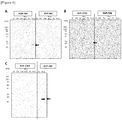
- each protein was confirmed by Western blot using an anti-His antibody ( Figure 5 ).
- each domain protein and a molecular weight marker were added in each amount of 5 ⁇ l/lane, so that they were separated by SDS-PAGE. Thereafter, gel was transferred on a membrane. After completion of the transcription, the membrane was immersed in 5% skim milk-TBST, and was then blocked at room temperature for 1 hour. The nanoantibodies prepared in 1 above were each diluted with TBST to a concentration of 10 ⁇ g/ml, and were then reacted on the membrane after completion of the transfer at room temperature for 1.5 hours. After completion of the reaction, the membrane was washed in TBST at room temperature for 5 minutes three times.
- newborn skin-derived human iPS cells (TkDN4-M; furnished from Dr. Masahiro OTSU, Division of Stem Cell Processing, Center for Stem Cell Biology and Regenerative Medicine, ISMUT, Tokyo University/Stem Cell Bank) were cultured in a medium supplemented with Activin A, Wnt3a and the like for 3 days, so that the iPS cells were allowed to differentiate into endoderm cells, which were further cultured in a medium supplemented with FGF4 and Wnt3a for 3 to 4 days, so that differentiation of the cells into the lower digestive tract was induced.
- TkDN4-M furnished from Dr. Masahiro OTSU, Division of Stem Cell Processing, Center for Stem Cell Biology and Regenerative Medicine, ISMUT, Tokyo University/Stem Cell Bank
- a spherical cell mass (spheroid) having a tubular structure was formed, it was subjected to a three-dimensional culture in a medium supplemented with R-spondin 1, EGF, etc. for 2 weeks to obtain organoids. Thereafter, the obtained organoids were further sub-cultured in a medium supplemented with Wnt3a, HGF, etc., so that the organoids were allowed to proliferate. The thus proliferating organoids were disintegrated, and stem cells were collected. Thereafter, the stem cells were seeded on Transwell (registered trademark) (24-well) and were then cultured for 10 days, so as to establish monolayered human intestinal epithelial cells.
- Transwell registered trademark
- induction of organoids and monolayered epithelial cells according to biopsy from living bodies can be carried out with reference to Ettayebi et al., please refer to Science 353: 1387-1393, 2016 , etc.
- induction of organoids from iPS cells can be carried out with reference to McCracken et al., Nat. protocol. 6: 1920 2011 , etc.
- VLP Virus-like particle
- nanoantibody molar ratio: 1 : 1, 1 : 0.1 and 1 : 0.01
- VLP 500 ng
- a positive control furnished from Dr. Kazuhiko KATAYAMA of Virus Department, National Institute of Infectious Diseases, (currently, a professor of Kitasato Institute for Life Sciences, Kitasato University)), which had previously been pre-incubated at room temperature for 1 hour, were each added in an amount of 100 ⁇ l/well to epithelial cells that had been monolayered on the Transwell.
- the thus obtained mixture was incubated at 37°C for 3 hours. After completion of washing, the reaction mixture was fixed with 4% PFA and was then subjected to a dialysis treatment with 0.5% Triton X-100 - PBS. Thereafter, the Transwell membrane was cut out from the back. The membrane was immersed in a 5% Goat Serum-NETG buffer (150 mM NaCl, 5 mM EDTA, 50 mM Tris-HCl, 0.05% Triton X-100, and 0.25% gelatin), so that it was blocked at room temperature for 1 hour.
- Goat Serum-NETG buffer 150 mM NaCl, 5 mM EDTA, 50 mM Tris-HCl, 0.05% Triton X-100, and 0.25% gelatin
- the resulting membrane was immersed in a primary antibody (anti-Zo-1 (Invitrogen); Zo-1 is a molecule existing in the tight junction of epithelial cells, and serves as a marker for intercellular adhesion formation) that had been 150 times diluted with an NETG buffer, and was reacted at 4°C overnight.
- a primary antibody anti-Zo-1 (Invitrogen); Zo-1 is a molecule existing in the tight junction of epithelial cells, and serves as a marker for intercellular adhesion formation
- the resulting membrane was washed, and was then immersed in a reaction solution prepared by diluting GII.6 (7K) VLP (100 times) labeled with HiLyte FluorTM 555 by employing HiLyte FluorTM 555 Labeling Kit-NH2 (DOJINDO LABORATORIES), DyLightTM488 conjugated anti-Mouse IgG (Jackson, 400 times) used as a secondary antibody, and DAPI (1000 times) with an NETG buffer, so that it was reacted at room temperature for 3 hours. Thereafter, the resulting membrane was washed with PBST and PBS, and was then enclosed. Then, using a confocal laser microscope, fluorescence was detected.
- Figure 7 shows the results obtained by examining the inhibitory ability of KVP-7F10 and KVP-7B8 towards the binding of VLP to epithelial cells.
- the fluorescence of VLP binding to epithelial cells was detected.
- the fluorescence of VLP was hardly detected (see “7K VLP” and “Merge” in Figure 7B and 7C ). Accordingly, it was demonstrated that KVP-7F10 and KVP-7B8 effectively inhibit the binding of GII.6 (7K) VLP to epithelial cells.
- Figure 8 shows the results obtained by examining the inhibitory ability of KVP-12B8 and KVP-7C6 towards the binding of VLP to epithelial cells.
- the fluorescence of VLP was detected in the cells indicated with the arrows in Figure 8A .
- the fluorescence of VLP was hardly detected ( Figure 8B ).
- KVP-7F10, KVP-7B8, KVP-12B8, and KVP-7C6 effectively inhibit the binding of GII.6 (7K) VLP to epithelial cells. It is to be noted that KVP-7G5 and KVP-7B4 ( Figure 9A ) that did not bind to GII.6 (7K) VLP did not inhibit the binding of the VLP to epithelial cells ( Figure 9B ).
- VLP In order to confirm incorporation of VLP into epithelial cells, GII.6 (7K) VLP (300ng) was added in an amount of 100 ⁇ l/well to epithelial cells that had been monolayered on the Transwell, and the obtained mixture was then incubated at 37°C for 3 hours. After completion of washing, the reaction mixture was fixed with 10% formalin and was then washed with 70% EtoH, and the membrane was cut out from the back. A frozen block embedded in O.C.T. Compound (Sakura Finetek Japan Co., Ltd.) was produced, and a frozen section with a size of 7 to 8 ⁇ m was then produced using a cryostat.
- the resulting membrane was washed, and was then immersed in a reaction solution prepared by diluting GII.6 (7K) VLP (100 times) labeled with HiLyte FluorTM 555 by employing HiLyte FluorTM 555 Labeling Kit-NH2 (DOJINDO LABORATORIES), DyLightTM488 conjugated anti-Mouse IgG (Jackson, 400 times) used as a secondary antibody, and DAPI (1000 times) with an NETG buffer, so that it was reacted at room temperature for 3 hours. Thereafter, the resulting membrane was washed with PBST and PBS, and was then enclosed. Then, using a confocal laser microscope, fluorescence was detected ( Figure 10 ).
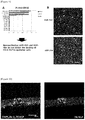
- VLP does not only bind to human epithelial cells, but it also invades into the cytoplasm thereof (the arrows shown in Figure 10 indicate the fluorescence of VLP).
- nanoantibody according to the embodiment of the present invention effectively inhibits invasion of norovirus VLP into monolayered human intestinal epithelial cells.
- Human norovirus Human norovirus (HuNoV) was furnished from Osaka Institute of Public Health (GII.3 (16-50 strain), GII.4 (17-53 strain), and GII.17 (16-421 strain)).
- monolayered intestinal epithelial cells derived from iPS cells were prepared from organoids that had been furnished from Dr. Shintaro SATO, Research Institute for Microbial Diseases, Osaka University. Specifically, cells were recovered from human iPS cell-derived organoids, which were maintained and sub-cultured according to the method of Takahashi et al. (Stem cell Reports 10: 314-328, 2018 ), and the recovered cells were then seeded on a 96-well cell culture plate in an amount of 2 x 10 4 cells/well.
- the cells were cultured according to the method of Takahashi et al. (EBioMedicine 23: 43-45, 2017 ) for 5 days, so as to prepare monolayered epithelial cells.
- the cells used for GII.3 infection were cultured under conditions with addition of 0.03% swine bile, whereas the cells used for GII.4 and GII.17 infection were cultured under conditions without addition of swine bile.
- HuNoV the viral genome copy number of which had already been calculated
- HuNoV the viral genome copy number of which had already been calculated
- the nanoantibody 5 ⁇ g/well
- KVP-7B8, KVP-7C6, KVP-7F10, and KVP-12B8 the nanoantibody
- the virus or a mixed solution of the virus and the nanoantibody, was added to iPS cell-derived monolayered intestinal epithelial cells, so that the cells were infected therewith at 37°C for 3 hours.
- the virus solutions were removed from all of the wells, and the wells were then washed twice.
- RNAs were extracted, and the viral copy number of each sample was then calculated according to real-time PCR based on the official analytical method (4x TaqMan Fast Virus 1-Step Master Mix/life technologies #4444434).
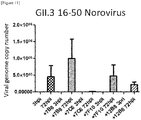
- Figure 11 shows the influence of the nanoantibodies according to the present invention on infection of human intestinal epithelial cells with GII.3 (16-50 strain).
- GII.3 16-50 strain
- KVP-7C6 and KVP-12B8 the viral copy number in the 72 hpi samples was significantly reduced. Accordingly, it was found that KVP-7C6 and KVP-12B8 do not only inhibit the binding of HuNoV to intestinal epithelial cells but also inhibit the infection.
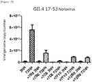
- Figure 12 shows the influence of the nanoantibodies according to the present invention on infection of human intestinal epithelial cells with GII.4 (17-53 strain).
- GII.4 was treated with KVP-7B8, KVP-7C6, KVP-7F10 and KVP-12B8, the viral copy number in the 72 hpi samples was significantly reduced. Accordingly, it was found that KVP-7B8, KVP-7C6 and KVP-12B8 do not only inhibit the binding of HuNoV to intestinal epithelial cells but also inhibit the infection.
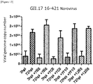
- Figure 13 shows the influence of the nanoantibodies according to the present invention on infection of human intestinal epithelial cells with GII.17 (16-421 strain). All of KVP-7B8, KVP-7C6, KVP-7F10 and KVP-12B8 did not exhibit effects on infection of the intestinal epithelial cells with GII.17.
- the antibody according to the present invention has the effect of effectively inhibiting norovirus infection. Accordingly, it can be expected that the present invention will be utilized in the medical field.
Landscapes
- Health & Medical Sciences (AREA)
- Life Sciences & Earth Sciences (AREA)
- Chemical & Material Sciences (AREA)
- Genetics & Genomics (AREA)
- Engineering & Computer Science (AREA)
- Organic Chemistry (AREA)
- Medicinal Chemistry (AREA)
- Molecular Biology (AREA)
- General Health & Medical Sciences (AREA)
- Bioinformatics & Cheminformatics (AREA)
- Biomedical Technology (AREA)
- Virology (AREA)
- Wood Science & Technology (AREA)
- Biotechnology (AREA)
- Zoology (AREA)
- Pharmacology & Pharmacy (AREA)
- Immunology (AREA)
- General Engineering & Computer Science (AREA)
- Biochemistry (AREA)
- Microbiology (AREA)
- Veterinary Medicine (AREA)
- Public Health (AREA)
- Animal Behavior & Ethology (AREA)
- Biophysics (AREA)
- Cell Biology (AREA)
- General Chemical & Material Sciences (AREA)
- Epidemiology (AREA)
- Mycology (AREA)
- Physics & Mathematics (AREA)
- Proteomics, Peptides & Aminoacids (AREA)
- Plant Pathology (AREA)
- Nuclear Medicine, Radiotherapy & Molecular Imaging (AREA)
- Chemical Kinetics & Catalysis (AREA)
- Oncology (AREA)
- Communicable Diseases (AREA)
- Gastroenterology & Hepatology (AREA)
- Polymers & Plastics (AREA)
- Food Science & Technology (AREA)
- Nutrition Science (AREA)
- Peptides Or Proteins (AREA)
Abstract
Description
- The present invention relates to a norovirus antibody. More specifically, the present invention relates to the variable domain of the heavy chain of a heavy-chain antibody against a norovirus.
- Norovirus is a pathogenic virus that invades into cells existing from the duodenum to the upper small intestine and causes nonbacterial acute gastroenteritis. Norovirus belongs to Caliciviridae lacking envelopes.
- The genome of norovirus is a single-stranded plus-sense RNA of approximately 7.7 kb. A viral protein called VPg covalently binds to the 5'-terminus of the genome, whereas the 3'-terminus thereof is polyadenylated. In the genome of norovirus, the coding regions of three proteins, namely, ORF1, VP1, and VP2 are present, and individual coding regions encode a non-structural protein, a
structural protein 1, and astructural protein 2, respectively. As a result of structural analyses, it has been elucidated that 180 VP1 molecules form a dimer, so as to construct a viral structure. Individual VP1 monomers are divided into two domains, namely, a shell domain (S domain) and a protruding domain (P domain), and the P domain is further divided into P1 and P2 subdomains. P2 recognizes blood group antigens functioning as a receptor and a host infectious factor upon infection. As a result of a mutation occurring in the P2 subdomain, a change is generated in the binding of P2 with a blood group antigen (Non Patent Literature 1). - Human noroviruses are classified into three gene groups (GI, GII and GIV) based on the genomic sequences, and reportedly, there are at least 25 types of genotypes. Human norovirus is a norovirus exhibiting high diversity. However, in the past few years, only a small number of strains, such as GII, in particular, GII genotype 4 (GII.4), have caused the epidemicity of the infection (Non Patent Literature 1).
- In the United States of America, approximately 2,000,000 people are infected with human norovirus infection every year, and among them, approximately 70,000 patients are hospitalized. In Japan, there are no accurate data, but it is assumed that approximately 10,000 to 20,000 hospitalized patients and more than approximately 7,000,000 or more infected people are generated every year. The symptoms of a majority of patients with human norovirus infection are ameliorated, but in the case of infant and aged patients, the symptoms become severe, and death cases are sometimes seen. Moreover, there are many cases where the outbreak of human norovirus infection takes place in facilities such as kindergartens and nursing homes. Under such circumstances, the number of outpatients and/or hospitalized patients has reached a considerable scale, and thus, it has posed a great burden on the social insurance system.
- As of now, an antiviral therapeutic agent, which is effective for human norovirus, has not been present, and the development of a vaccine has been merely promoted (Patent Literature 1). In view of the foregoing, in the medical field regarding infectious disease, it has been strongly desired to develop and practicalize drugs effective for the treatment and prevention of human norovirus.
- Patent Literature 1: JP Patent Publication (Kokai) No.
2015-171363 A -
- Non Patent Literature 1: Glass et al., N Engl J Med 361: 1776-1785, 2009
- Non Patent Literature 2: Hamers-Casterman et al., Nature 363: 446-448, 1993
- Under the aforementioned circumstances, it is an object of the present invention to provide an antibody that reacts against a human norovirus (HuNoV). More specifically, it is the object of the present invention to provide an antibody that inhibits infection of cells with HuNoV (i.e., invasion and/or replication of HuNoV).
- The present inventors have immunized llamas with VP1 of the HuNoV GII.4 strain as an antigen, and as a result, have succeeded in producing a nanoantibody that inhibits invasion of norovirus VLP (virus-like particle) into intestinal epithelial cells. Moreover, this antibody also inhibited infection of intestinal epithelial cells with a norovirus.
- In general, a human antibody is composed of a heavy chain and a light chain. However, in the case of camelids such as llamas, alpacas and camels, a single-stranded antibody only consisting of a heavy chain (i.e., a heavy-chain antibody) is present. Such a heavy-chain antibody can recognize a target antigen and can bind to the antigen, as in the case of an ordinary antibody consisting of a heavy chain and a light chain (Non Patent Literature 2). The variable domain of the heavy chain of a heavy-chain antibody (VHH) is a minimum unit having binding affinity for an antigen, and this variable domain fragment is referred to as a "nanoantibody." The nanoantibody has high heat resistance, digestion resistance and normal temperature resistance, and thus, it is possible to more easily prepare a large amount of the nanoantibody according to a genetic engineering method.
- Specifically, the present invention includes the following (1) to (15).
- (1) A nanoantibody that binds to a norovirus and inhibits infection of intestinal cells with the norovirus.
- (2) The nanoantibody according to the above (1), which is characterized in that the norovirus is HuNoV.
- (3) A nanoantibody that inhibits infection of intestinal cells with HuNoV GII.3 and/or HuNoV GII.4, which is characterized in that the amino acid sequences of complementarity-determining
regions 1 to 3 (CDR1, CDR2 and CDR3) are the following (A), (B), (C) or (D):- (A) CDR1 comprising the amino acid sequence as set forth in SEQ ID NO: 17, CDR2 comprising the amino acid sequence as set forth in SEQ ID NO: 18, and CDR3 comprising the amino acid sequence as set forth in SEQ ID NO: 19;
- (B) CDR1 comprising the amino acid sequence as set forth in SEQ ID NO: 20, CDR2 comprising the amino acid sequence as set forth in SEQ ID NO: 21, and CDR3 comprising the amino acid sequence as set forth in SEQ ID NO: 22;
- (C) CDR1 comprising the amino acid sequence as set forth in SEQ ID NO: 23, CDR2 comprising the amino acid sequence as set forth in SEQ ID NO: 24, and CDR3 comprising the amino acid sequence as set forth in SEQ ID NO: 25; and
- (D) CDR1 comprising the amino acid sequence as set forth in SEQ ID NO: 26, CDR2 comprising the amino acid sequence as set forth in SEQ ID NO: 27, and CDR3 comprising the amino acid sequence as set forth in SEQ ID NO: 28.
- (4) A nanoantibody comprising a polypeptide described in the following (a), (b), or (c), and inhibiting infection of intestinal cells with HuNoV GII.3 and/or HuNoV GII.4:
- (a) a polypeptide consisting of the amino acid sequence as set forth in SEQ ID NO: 1, SEQ ID NO: 5, SEQ ID NO: 9, or SEQ ID NO: 13;
- (b) a polypeptide consisting of an amino acid sequence comprising a substitution, deletion, insertion and/or addition of one or several amino acids with respect to the amino acid sequence as set forth in SEQ ID NO: 1, SEQ ID NO: 5, SEQ ID NO: 9, or SEQ ID NO: 13; and
- (c) a polypeptide consisting of an amino acid sequence having a sequence identity of 80% or more to the amino acid sequence as set forth in SEQ ID NO: 1, SEQ ID NO: 5, SEQ ID NO: 9, or SEQ ID NO: 13.
- (5) A heavy-chain antibody comprising the nanoantibody according to any one of the above (1) to (4).
- (6) A nanoantibody multimer, in which a plurality of the nanoantibodies according to any one of the above (1) to (4) are connected with one another.
- (7) A nanoantibody multimer, in which one or a plurality of the nanoantibodies according to any one of the above (1) to (4) are connected with one or a plurality of nanoantibodies each having antigen specificity that is different from the aforementioned nanoantibodies.
- (8) A nucleic acid encoding the nanoantibody according to any one of the above (1) to (4).
- (9) A nucleic acid as set forth in SEQ ID NO: 2, SEQ ID NO: 6, SEQ ID NO: 10, or SEQ ID NO: 14.
- (10) A vector comprising the nucleic acid according to the above (8) or (9).
- (11) A cell, into which the vector according to the above (10) is introduced.
- (12) A transgenic rice plant, into which the nucleic acid according to the above (8) or (9) is introduced and the nanoantibody is expressed in rice grains thereof.
- (13) A rice grain harvested from the transgenic rice plant according to the above (12).
- (14) A pharmaceutical composition comprising the nanoantibody, heavy-chain antibody or nanoantibody multimer according to any one of the above (1) to (7).
- (15) The pharmaceutical composition according to the above (14), which is characterized in that the disease as a therapeutic or preventive target is norovirus infection.
- According to the present invention, a nanoantibody that inhibits infection of intestinal epithelial cells with HuNoV is provided for the first time.
- A medicament or a pharmaceutical composition comprising the nanoantibody or the heavy-chain antibody according to the present invention exhibits the effect of treating and/or preventing infection of HuNoV and various symptoms associated with the infection.
-
- [
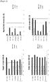
Figure 1] Figure 1 shows the results of screening for VHH specifically binding to various types of HuNoV GII VP1 (1st screening). The results of ELISA (absorbance at 450 nm) regarding the binding of VP1 derived from GII.4 (Sakai strain,Saga 1 strain, Narita104 strain) and GII.3 (U201 strain) with various types of VHH are shown. - [
Figure 2] Figure 2 shows the results of screening for VHH specifically binding to various types of HuNoV GII VP1 (1st screening). The results of ELISA (absorbance at 450 nm) regarding the binding of VP1 derived from GII.2 (MK04 strain), GII.6 (7K strain) and GII.17 (kawasaki308 strain) with various types of VHH are shown. - [
Figure 3] Figure 3 shows the results of screening for VHH specifically binding to various types of HuNoV GII VP1 (2nd screening). The results of ELISA (absorbance at 450 nm) regarding the binding of VP1 derived from GII.4 (Sakai strain,Saga 1 strain, Narita104 strain) and GII.3 (U201 strain) with various types of VHH are shown. - [
Figure 4] Figure 4 shows the results of screening for VHH specifically binding to various types of HuNoV GII VP1 (2nd screening). The results of ELISA (absorbance at 450 nm) regarding the binding of VP1 derived from GII.2 (MK04 strain), GII.6 (7K strain) and GII.17 (kawasaki308 strain) with various types of VHH are shown. - [
Figure 5] Figure 5 shows the results of confirmation of the expression of individual domains of HuNoV (GII.4 strain) VP1. The full-length P domain, P1-1 & P2 domains, P2 domain, and P1-2 domain of VP1, which were expressed in Escherichia coli, were electrophoresed (A), and they were then subjected to Western blotting with an anti-His tag antibody (B). - [
Figure 6] Figure 6 shows the results of the linear epitope mapping of the nanoantibodies (KVP-7F10, KVP-7C6 and KVP-7B8). Individual domains of VP1 shown inFigure 5 were subjected to Western blotting with KVP-7B8 (A), KVP-7F10 (B), KVP-7C6 (B), and KVP-12B8 (C). The arrow indicates a band of the VP1 P1-2 domain detected with the nanoantibodies. - [
Figure 7] Figure 7 shows the influence of the nanoantibodies (KVP-7F10 and KVP-7B8) on the binding of HuNoV VLP to intestinal epithelial cells. The results obtained by adding only GII.6 (7K) VLP (A), GII.6 (7K) VLP and KVP-7F10 (B), or GII.6 (7K) VLP and KVP-7B8 (C) to the intestinal epithelial cell layer are shown. The scale bar indicates 50 µm. DAPI: DAPI staining; Zo-1: staining with an anti-Zo-1 antibody; 7K VPL: GII.6 (7K) VLP labeled with HiLyte Fluor™ 555 pigment; and Merge: a view obtained by overlapping the fluorescence images of DAPI, Zo-1 and 7K VPL. - [
Figure 8] Figure 8 shows the influence of the nanoantibodies (KVP-12B8 and KVP-7C6) on the binding of HuNoV VLP to intestinal epithelial cells. The results obtained by adding only GII.6 (7K) VLP (A), GII.6 (7K) VLP and KVP-12B8 (B), or GII.6 (7K) VLP and KVP-7C6 (C) to the intestinal epithelial cell layer are shown. Each view shows the overlapping of a DAPI-stained image, an image stained with an anti-Zo-1 antibody, and a 7K VPL fluorescence image. The scale bar indicates 40 µm inFigure 8A and 8C , and the scale bar indicates 50 µm inFigure 8B . The arrow indicates a cell to which VLP has bound. - [
Figure 9] Figure 9 shows the influence of the nanoantibodies (KVP-7G5 and KVP-7B4) on the binding of HuNoV VLP to intestinal epithelial cells.Figure 9 shows KVP-7G5 or KVP-7B4 that does not bind to GII.6-derived VP1 (A); and the results obtained by adding KVP-7G5 or KVP-7B4 and GII.6 (7K) VLP to the intestinal epithelial cell layer (B).Figure 9B is a view obtained by overlapping a DAPI-stained image, an image stained with an anti-Zo-1 antibody, and a 7K VPL fluorescence image. The scale bar of KVP-7G5 indicates 50 µm, whereas the scale bar of KVP-7B4 indicates 30 µm. The arrow indicates a cell to which VLP has bound. - [
Figure 10] Figure 10 shows the results of confirmation of the invasion of HuNoV GII.6 (7K) VLP into intestinal epithelial cells. The results obtained by staining frozen sections of monolayered epithelial cells, to which HiLyte Fluor™ 555-labeled GII.6 (7K) VLP had been added, with DAPI and anti-Zo-1 antibody. The left indicates a view obtained by overlapping a DAPI-stained image, an image stained with an anti-Zo-1 antibody, and a 7K VPL fluorescence image; the right indicates a 7K VPL fluorescence image. The scale bar indicates 10 µm. The arrow indicates VLP, and the arrowhead indicates a nucleus. - [
Figure 11] Figure 11 shows the influence of the nanoantibodies (KVP-7B8, KVP-7C6, KVP-7F10, and KVP-12B8) on infection of intestinal epithelial cells with HuNoV GII.3 (16-50 strain). The virus was mixed with each nanoantibody, followed by pre-incubation, and the mixture was then added to the intestinal epithelial cell layer, so that the cells were infected with the virus. Three hours after viral infection (3 hpi) and 72 hours after viral infection (72 hpi), the viral copy number in the medium was measured. - [
Figure 12] Figure 12 shows the influence of the nanoantibodies (KVP-7B8, KVP-7C6, KVP-7F10, and KVP-12B8) on infection of intestinal epithelial cells with HuNoV GII.4 (17-53 strain). The virus was mixed with each nanoantibody, followed by pre-incubation, and the mixture was then added to intestinal epithelial cell layer, so that the cells were infected with the virus. Three hours after viral infection (3 hpi) and 72 hours after viral infection (72 hpi), the viral copy number in the medium was measured. - [
Figure 13] Figure 13 shows the influence of the nanoantibodies (KVP-7B8, KVP-7C6, KVP-7F10, and KVP-12B8) on infection of intestinal epithelial cells with HuNoV GII.17 (16-421 strain). The virus was mixed with each nanoantibody, followed by pre-incubation, and the mixture was then added to intestinal epithelial cell layer, so that the cells were infected with the virus. Three hours after viral infection (3 hpi) and 72 hours after viral infection (72 hpi), the viral copy number in the medium was measured. - A first embodiment of the present invention relates to a nanoantibody that binds to a norovirus and inhibits infection of intestinal epithelial cells (for example, small intestinal epithelial cells) with the norovirus (hereinafter also referred to as "the nanoantibody of the present invention"), or a heavy-chain antibody comprising the nanoantibody.
- In the embodiment of the present invention, the nanovirus is preferably HuNoV, and examples of the nanovirus may include, but are not limited to, viruses classified into the groups GI, GII and GIV, and preferably, viruses classified into the groups GII.3 and GII.4.
- VLP (virus-like particle) mentioned in the present description has a capsid shell consisting of a VP1 protein (i.e., a nanoantibody binding to the VP1 protein also binds to VLP) and also has the same outer shell structure as viruses. However, VLP does not have a gene therein. The VP1 protein means a
structural protein 1 that forms the structure of a norovirus. For more information, please refer to, for example, Glass et al., N Engl J Med 361: 1776-1785, 2009. - In the embodiment of the present invention, the nanoantibody of the present invention means a polypeptide that consists of the variable domain of the heavy chain of a heavy-chain antibody (VHH) and can inhibit infection of intestinal epithelial cells with a norovirus.
- The nanoantibody of the present invention is, for example, a polypeptide described in the following (a), (b), or (c):
- (a) a polypeptide consisting of the amino acid sequence as set forth in SEQ ID NO: 1, SEQ ID NO: 5, SEQ ID NO: 9, or SEQ ID NO: 13;
- (b) a polypeptide consisting of an amino acid sequence comprising a substitution, deletion, insertion and/or addition of one or several amino acids with respect to the amino acid sequence as set forth in SEQ ID NO: 1, SEQ ID NO: 5, SEQ ID NO: 9, or SEQ ID NO: 13, and having the activity of binding to HuNoV and inhibiting infection of intestinal cells with the HuNoV; and
- (c) a polypeptide consisting of an amino acid sequence having a sequence identity of 80% or more to the amino acid sequence as set forth in SEQ ID NO: 1, SEQ ID NO: 5, SEQ ID NO: 9, or SEQ ID NO: 13, and having the activity of binding to HuNoV and inhibiting infection of intestinal cells with the HuNoV.
- In the present description, in the case of using the phrase "an amino acid sequence comprising a substitution, deletion, insertion and/or addition of one or several amino acids," the number of substituted, deleted, inserted and/or added amino acids is not particularly limited. For example, approximately 1, 2, 3, 4, 5, 6, 7, 8, 9 or 10 amino acids are preferable. In addition, in the present description, in the case of using the phrase "an amino acid sequence having a sequence identity of 80% or more," the percentage of the sequence identity is not particularly limited, as long as the amino acid sequence has a sequence identity of 80% or more. The sequence identity may be, for example, 90% or more, 91% or more, 92% or more, 93% or more, 94% or more, 95% or more, 96% or more, 97% or more, 98% or more, or 99% or more.
- In the above (b) and (c), the site of SEQ ID NO: 1, SEQ ID NO: 5, SEQ ID NO: 9 or SEQ ID NO: 13, on which a substitution, deletion, insertion and/or addition of an amino acid(s) is performed, is desirably a sequence other than the CDR1, CDR2 and CDR3 of each sequence. The above-described substitution, deletion, insertion and/or addition of an amino acid(s) may be a mutation that is originally present in a nucleic acid encoding a protein, or may also be a mutation that is newly introduced by modifying the nucleic acid according to a method known in the present technical field. Such modification, for example, substitution of a specific amino acid residue(s) can be carried out using a commercially available kit (e.g., MutanTM-G (TaKaRa)), etc., by performing substitution of nucleotides according to a known method such as a Gapped Duplex method or a Kunkel method, or a method equivalent thereto.
- The polypeptide consisting of the amino acid sequence as set forth in SEQ ID NO: 1, SEQ ID NO: 5, SEQ ID NO: 9 or SEQ ID NO: 13 is the nanoantibody of the present invention. These sequences each have the following CDR1 to CDR3.
- (i) SEQ ID NO: 1 CDR1; INTM (SEQ ID NO: 17), CDR2; ISSGGGTNY (SEQ ID NO: 18), CDR3; NLHNFSPISPPRSY (SEQ ID NO: 19)
- (ii) SEQ ID NO: 5 CDR1; INGV (SEQ ID NO: 20), CDR2; ISRSGWTNY (SEQ ID NO: 21), CDR3; NLHSGLGNVKNY (SEQ ID NO: 22)
- (iii) SEQ ID NO: 9 CDR1; PNVM (SEQ ID NO: 23), CDR2; KTSGRLSNY (SEQ ID NO: 24), CDR3; WDSARSTEY (SEQ ID NO: 25)
- (iv) SEQ ID NO: 13 CDR1; DNAM (SEQ ID NO: 26), CDR2; ITNSNSTKY (SEQ ID NO: 27), CDR3; IERTGRTSIKWTY (SEQ ID NO: 28)
- The binding ability of the polypeptides described in the above (b) and (c) to VP1 (VLP) can be easily confirmed according to a Western blotting method or an ELISA method. Also, the presence or absence of the activity of inhibiting infection of intestinal epithelial cells (including cells induced from iPS cells and the like) with a norovirus can be easily confirmed by using an intestinal epithelial cell layer prepared as shown in Examples later.
- The nanoantibody of the present invention also includes a polypeptide, the amino acid sequences of complementarity-determining
regions 1 to 3 (CDR1, CDR2 and CDR3) of which are the following (A), (B), (C), or (D): - (A) CDR1 comprising the amino acid sequence as set forth in SEQ ID NO: 17, CDR2 comprising the amino acid sequence as set forth in SEQ ID NO: 18, and CDR3 comprising the amino acid sequence as set forth in SEQ ID NO: 19;
- (B) CDR1 comprising the amino acid sequence as set forth in SEQ ID NO: 20, CDR2 comprising the amino acid sequence as set forth in SEQ ID NO: 21, and CDR3 comprising the amino acid sequence as set forth in SEQ ID NO: 22;
- (C) CDR1 comprising the amino acid sequence as set forth in SEQ ID NO: 23, CDR2 comprising the amino acid sequence as set forth in SEQ ID NO: 24, and CDR3 comprising the amino acid sequence as set forth in SEQ ID NO: 25; and
- (D) CDR1 comprising the amino acid sequence as set forth in SEQ ID NO: 26, CDR2 comprising the amino acid sequence as set forth in SEQ ID NO: 27, and CDR3 comprising the amino acid sequence as set forth in SEQ ID NO: 28.
- Moreover, the embodiment of the present invention includes a heavy-chain antibody comprising the aforementioned "nanoantibody" according to the present invention as a variable domain (i.e., an antibody formed only with a heavy chain). The "heavy-chain antibody" of the present invention may be produced according to a genetic engineering method, or may also be generated by animals generating heavy-chain antibodies, such as camelids.
- Furthermore, "the nanoantibody of the present invention" includes not only VHH monomers, but also multimeric "nanoantibodies" ("nanoantibody multimers") prepared by binding a plurality of "the nanoantibodies of the present invention," which are identical to or different from one another, to one another, using a peptide linker or the like.
- Further, it may also be possible to produce a nanoantibody multimer, in which one or a plurality of "the nanoantibodies of the present invention" are combined with one or a plurality of other nanoantibodies having different antigen specificity, such as, for example, nanoantibodies reacting against rotavirus (e.g., Tokuhara et al., J Clin Invest. Doi: 10.1172/JCI70266), nanoantibodies binding to norovirus GI, or nanoantibodies reacting against bacterial toxin. The nanoantibody multimer can be produced with reference to, for example, the method described in Schmidt et al., Clin Vaccine Immuno. 23:774-784 2016.
- The nanoantibody of the present invention can be easily produced according to techniques known in the present technical field. Otherwise, the present nanoantibody may also be produced by combining a peptide solid-phase synthesis method with a Native Chemical Ligation (NCL) method.
- DNA consisting of a nucleic acid sequence encoding a polypeptide that is the nanoantibody of the present invention (e.g., the sequence as set forth in SEQ ID NO: 2, SEQ ID NO: 6, SEQ ID NO: 10, or SEQ ID NO: 14) is inserted into a suitable expression vector, and the polypeptide is expressed according to an ordinary method, and is then isolated and purified, thereby preparing the nanoantibody of the present invention.
- Various host cells, such as, for example, bacterial cells (e.g., Escherichia coli B strain, E. coli Kl2 strain, Corynebacterium ammoniagenes, C. glutamicum, Serratia liquefaciens, Streptomyces lividans, Pseudomonas putida, etc.); molds (e.g., Penicillium camembertii, Acremonium chrysogenum, etc.), animal cells, plant cells, baculoviruses/insect cells, or yeast cells (e.g., Saccharomyces cerevisiae, Pichia pastoris, etc.), are used, and the nanoantibody of the present invention can be expressed in these cells. As expression vectors for expressing nanoantibodies, vectors suitable for various types of host cells can be used. Examples of the expression vector that can be used herein may include: pBR322, pBR325, pUC118, pET, etc. (Escherichia coli hosts); pEGF-C, pEGF-N, etc. (animal cell hosts); pVL1392, pVL1393, etc. (insect cell hosts, baculovirus vectors); and pG-1, Yep13, pPICZ, etc. (yeast cell hosts). These expression vectors each have a replication origin, a selective marker, and a promoter, which are suitable for each vector. These expression vectors may also have an enhancer, a transcription termination sequence (terminator), a ribosome binding site, a polyadenylation signal, etc., as necessary. Further, in order to facilitate purification of the expressed polypeptide, a nucleotide sequence for fusing a FLAG tag, a His tag, an HA tag, a GST tag, etc. with the polypeptide to express it may be inserted into such an expression vector.
- Such an expression vector can be produced by a method known to a person skilled in the art, using a commercially available kit, as appropriate.
- When the expressed nanoantibody is extracted from cultured cell masses or cultured cells, the cell masses or the cultured cells are collected by a known method after completion of the culture, and the collected cell masses or cells are then suspended in a suitable buffer. Thereafter, the suspension is subjected to ultrasonic wave, lysozyme treatment and/or freezing-thawing, etc., so that the cell masses or cells are disintegrated. Thereafter, the resultant is subjected to centrifugation or filtration to obtain a soluble extract. An appropriate combination of known separation and/or purification methods is applied to the obtained extract, so as to obtain a polypeptide of interest. Examples of the known separation and/or purification methods that can be used herein may include: methods of utilizing solubility, such as salting-out or a solvent precipitation method; methods of mainly utilizing a difference in molecular weights, such as a dialysis method, an ultrafiltration method, a gel filtration method, or SDS-PAGE; methods of utilizing a difference in electric charges, such as ion exchange chromatography; methods of utilizing specific affinity, such as affinity chromatography (for example, methods, in which when a polypeptide is expressed together with a GST tag, a glutathione-bound carrier resin is used, when a polypeptide is expressed together with a His tag, a Ni-NTA resin or a Co-based resin is used, when a polypeptide is expressed together with a HA tag, an anti-HA antibody resin is used, and when a polypeptide is expressed together with a FLAG tag, an anti-FLAG antibody-bound resin or the like is used); methods of utilizing a difference in hydrophobicity, such as reverse phase high performance liquid chromatography; and methods of utilizing a difference in isoelectric points, such as an isoelectric focusing method.
- A second embodiment of the present invention relates to a medicament comprising the nanoantibody of the present invention or the heavy-chain antibody of the present invention. The medicament of the present invention may have an embodiment in which the nanoantibody or heavy-chain antibody of the present invention itself, which is used as an active ingredient, is directly administered. In general, however, the medicament of the present invention is desirably administered in the form of a pharmaceutical composition comprising one or two or more formulation additives, as well as the nanoantibody or the heavy-chain antibody used as an active ingredient. Moreover, as an active ingredient of the medicament or pharmaceutical composition of the present invention, one or more can be selected from the nanoantibodies or heavy-chain antibodies of the present invention, and they can be then used in combination. Furthermore, there may also be used a nanoantibody multimer, in which the nanoantibody of the present invention is combined with one or a plurality of other nanoantibodies having different antigen specificity, such as, for example, nanoantibodies reacting against rotavirus (e.g., Tokuhara et al., J Clin Invest. Doi: 10.1172/JCI70266), nanoantibodies binding to norovirus GI, or nanoantibodies reacting against bacterial toxin. Further, known other drugs may be combined and mixed into the pharmaceutical composition.
- The medicament or pharmaceutical composition according to the embodiment of the present invention is not particularly limited. Examples of the dosage form thereof may include a tablet, a capsule, a granule, a powder agent, a syrup agent, a suspending agent, a suppository, an ointment, a cream agent, a gelling agent, a patch, an inhalant, and an injection. These formulations are prepared according to ordinary methods. In the case of a liquid formulation, it may be dissolved or suspended in water or other suitable solvents upon use. In addition, a tablet or a granule may be coated according to publicly known methods. In the case of an injection, it is prepared by dissolving the compound of the present invention in water. Such an injection may also be prepared by dissolving the present compound in a normal saline or a glucose solution, as necessary. Otherwise, a buffer or a preservative may be added to the injection.
- A formulation for use in oral administration or parenteral administration is provided in the form of any given formulation. Examples of the formulation form may include: agents for oral administration, such as a granule, a fine granule, a powder agent, a hard capsule, a soft capsule, a syrup agent, an emulsion, a suspending agent, or a liquid agent; injections for intravenous administration, intramuscular administration, subcutaneous administration, etc.; and forms such as an infusion, a transdermal absorbent, a transmucosal absorbent, nasal drops, an inhalant, or a suppository. An injection, an infusion, or the like can also be used by being prepared in the dosage form of powders, such as a freeze-dried form, and then dissolving the powders in a suitable aqueous medium such as a normal saline, upon use.
- The types of formulation additives, the ratio of the formulation additives to the active ingredient, or the method for producing the medicament or pharmaceutical composition, which is applied to the medicament or pharmaceutical composition according to the embodiment of the present invention, can be selected, as appropriate, by a person skilled in the art, depending on the form. As formulation additives, inorganic or organic substances, or solid or liquid substances can be used. In general, such formulation additives can be mixed into the present medicament or pharmaceutical composition, in an amount of, for example, 0.1% by weight to 99.9% by weight, 1% by weight to 95.0% by weight, or 1% by weight to 90.0% by weight, based on the weight of the active ingredient. Specific examples of the formulation additives may include lactose, glucose, mannitol, dextrin, cyclodextrin, starch, sucrose, magnesium aluminometasilicate, synthetic aluminum silicate, carboxymethyl cellulose sodium, hydroxypropyl starch, carboxymethyl cellulose calcium, ion exchange resin, methyl cellulose, gelatin, gum Arabic, hydroxypropyl cellulose, hydroxypropylmethyl cellulose, polyvinyl pyrrolidone, polyvinyl alcohol, light anhydrous silicic acid, magnesium stearate, talc, tragacanth, bentonite, beegum, titanium oxide, sorbitan fatty acid ester, sodium lauryl sulfate, glycerin, fatty acid glycerin ester, purified lanolin, glycerogelatin, polysorbate, macrogol, vegetable oil, wax, liquid paraffin, white petrolatum, fluorocarbon, nonionic surfactant, propylene glycol, and water.
- In order to produce a solid formulation for oral administration, the active ingredient is mixed with excipient components such as, for example, lactose, starch, crystalline cellulose, calcium lactate, or anhydrous silicic acid to prepare a powder agent. Otherwise, as necessary, binders such as white sugar, hydroxypropyl cellulose or polyvinyl pyrrolidone, disintegrators such as carboxymethyl cellulose or carboxymethyl cellulose calcium, and the like are further added to the aforementioned mixture, and the thus obtained mixture is then subjected to wet or dry granulation, so as to prepare a granule. In order to produce a tablet, these powder agents and granules may be directly subjected to tablet making, or these powder agents and granules, together with a lubricant such as magnesium stearate or talc, may be subjected to tablet making. These granules or tablets can be processed into enteric-coated formulations by being coated with enteric-coating base agents such as hydroxypropylmethyl cellulose phthalate or a methacrylic acid-methyl methacrylate polymer, or can also be processed into sustained release formulations by being coated with ethyl cellulose, carnauba wax, hydrogenated oil, or the like. In addition, in order to produce a capsule agent, a powder agent or a granule is filled into a hard capsule, or the active ingredient is directly coated with gelatin, or is first dissolved in glycerin, polyethylene glycol, sesame oil, olive oil or the like, and is then coated with gelatin, so as to prepare a soft capsule.
- In order to produce an injection, the active ingredient is dissolved in distilled water for injection, as necessary, together with a pH adjuster such as hydrochloric acid, sodium hydroxide, lactose, lactic acid, sodium, sodium monohydrogen phosphate or sodium dihydrogen phosphate, and a tonicity agent such as sodium chloride or glucose, and thereafter, the obtained solution is subjected to aseptic filtration, and is then filled into an ampule. Otherwise, mannitol, dextrin, cyclodextrin, gelatin or the like is further added to the resulting solution, followed by vacuum freeze drying, so as to prepare an injection that is soluble at the time of use. Alternatively, lecithin,
polysorbate 80, polyoxyethylene hydrogenated castor oil, or the like is added to the active ingredient for emulsification in water, so as to prepare an emulsion for injection. - In order to produce a rectal administration agent, the active ingredient, together with a suppository base agent such as cacao butter, fatty acid tri-, di- and mono-glyceride, or polyethylene glycol, is humidified and is dissolved, and the resultant is then poured into a mold, followed by cooling. Otherwise, the active ingredient may be dissolved in polyethylene glycol, soybean oil, or the like, and may be then coated with a gelatin film.
- The applied dose and the number of doses of the medicament or pharmaceutical composition according to the embodiment of the present invention are not particularly limited. The applied dose and the number of doses can be appropriately selected according to the judgment of a doctor or a pharmacist, depending on conditions such as the purpose of preventing and/or treating deterioration and/or progression of a treatment target disease, the type of the disease, and the body weight and age of a patient.
- Generally, the dose applied for an adult per day by oral administration is approximately 0.01 to 1,000 mg (the weight of the active ingredient), and this dose can be administered once or divided over several administrations per day, or every several days. In the case of using the medicament or the pharmaceutical composition as an injection, the injection is desirably administered to an adult continuously or intermittently at a daily dose of 0.001 to 100 mg (the weight of the active ingredient).
- The medicament or pharmaceutical composition according to the embodiment of the present invention can be prepared as a sustained release formulation, such as an implant tablet and a delivery system encapsulated into a microcapsule, by using a carrier capable of preventing the prompt removal of the agent from the body. As such carriers, biodegradable and biocompatible polymers, such as ethylene vinyl acetate, polyanhydride, polyglycolic acid, collagen, polyorthoester and polylactic acid, can be used. These materials can be easily prepared by a person skilled in the art. Moreover, a liposome suspension can also be used as a pharmaceutically acceptable carrier. Liposome is prepared as a lipid composition comprising, but are not limited thereto, phosphatidylcholine, cholesterol and PEG-derived phosphatidylethanol (PEG-PE), by being passed through a filter with a suitable pore size, so that it can have a size suitable for the use thereof, and it is then purified by a reverse phase evaporation method.
- The medicament or pharmaceutical composition according to the embodiment of the present invention may be provided in the form of a kit, together with an instruction manual regarding an administration method and the like. The medicament or the pharmaceutical composition included in the kit is supplied with a vessel, in which the activity of the active ingredient is effectively sustained for a long period of time, the agent and the like are not absorbed on the inside thereof, and the vessel is produced from materials that do not degrade the components. For example, a sealed glass ampule may comprise a buffer or the like that has been enclosed in the presence of neutral and unreactive gas such as nitrogen gas.
- Moreover, an instruction manual may be included with the kit. The instruction manual of the present kit may be printed on a paper or the like, or may also be stored in an electromagnetically readable medium such as CD-ROM or DVD-ROM, and may be then supplied.
- A third embodiment of the present invention relates to a transgenic rice plant, in which the nanoantibody of the present invention is expressed in rice grains thereof, and the rice grains.
- The nanoantibody can be accumulated in rice grains, without losing the properties thereof. The method for producing such a transgenic rice plant, in which the nanoantibody is expressed in rice grains thereof, has already been known. For more information, please refer to, for example,
WO2015/011396 , Tokuhara et al., J Clin Invest. Doi: 10.1172/JCI70266, and the like (Japanese Patent No.5616449 - Furthermore, the embodiment of the present invention encompasses a method for treating or preventing norovirus infection or other infections and various symptoms associated with norovirus infection or other infections, wherein the method comprises administering a medicament or a pharmaceutical composition comprising the nanoantibody or heavy-chain antibody of the present invention to a patient and the like.
- Herein, the term "treatment" is used to mean prevention or alleviation of the progression and deterioration of the pathologic conditions of a mammal affected with a disease or the like, by which the progression and deterioration of the disease is prevented or alleviated.
- On the other hand, the term "prevention" is used to mean that the onset or development of a disease in a mammal that will be likely to be affected with the disease or the like is prevented in advance, and that the onset of various symptoms of the disease is thereby prevented in advance.
- The term "mammal" as a therapeutic target means any given animal classified into mammals, and thus, is not particularly limited. Examples of the mammal used herein may include: humans; pet animals such as dogs, cats and rabbits; and livestock animals such as bovines, pigs, sheep and horses. A particularly preferable "mammal" is a human.
- The disclosures of all publications cited in the present description are incorporated herein by reference in their entirety. In addition, throughout the present description, when singular terms such as "a," "an," and "the" are used, these terms include not only single items but also multiple items, unless otherwise clearly specified.
- Hereinafter, the present invention will be further described in the following examples. However, these examples are only illustrative examples of the embodiments of the present invention, and thus, are not intended to limit the scope of the present invention.
- Production of the nanoantibody of the present invention was outsourced to QVQ (Utrecht, Netherlands).
- Here, preparation of nanoantibodies will be summarized.
- Two llamas were immunized with a VP1 antigen solution (SEQ ID NO: 29) (300 µg/injection). The content of VP1 in the antigen solution was 10%, and thus, it means that the llama was immunized with approximately 30 µg of VP1 for a single immunization. Immunization was carried out on the 0th (initial immunization day), 14th, 28th and 35th days from the initial immunization day. On the 40th day from the initial immunization, the immunization was completed, and approximately 200 ml of a blood sample was then collected to prepare peripheral blood lymphocytes (PBL). For the measurement of antibody titer, the serums collected on 0th and 44th days of the immunization were used. Each well of an immunoplate was coated with 200 ng of VP1 in 50 µl of PBS (4°C, overnight). Thereafter, the plate was washed, and was then blocked with 4% Marvel (in PBS), and the serum was diluted stepwise with 2% Marvel (in PBS) and was then added to each well. Thereafter, each well was washed, and an antibody reacting against VP1 was detected by using a monoclonal antibody reacting against the llamas IgG.
- PBLs were prepared from blood collected from the llamas according to Ficoll gradient, and RNA was then prepared from the obtained PBLs according to a phenol-chloroform method. The obtained RNA was reversely transcribed into cDNA, using a reverse transcription enzyme kit (Invitrogen). A DNA fragment encoding IGH (antibody heavy chain) was amplified by using primers binding to leader sequence regions. As a result, a DNA fragment encoding VHH with approximately 700 bp was amplified. This approximately 700 bp DNA fragment was electrophoresed, and was cut out from agarose, followed by extraction. Using the obtained approximately 80 ng DNA fragment as a template, nested PCR was carried out. The amplified fragment was cleaved with BstEII and SfiI, and an approximately 350 bp fragment was then isolated and purified.
- The purified approximately 330 ng DNA fragment was inserted into the phagemid pUR8100, and was then introduced into Escherichia coli TG1 to produce a VHH phagemid library. Subsequently, from the VHH phagemid library, phages were recovered, so as to prepare a VHH phage library.
- From the prepared phage library, human VP1 was used as an antigen (i.e., it was immobilized on the wells of a microplate via an existing antibody, VP1 antibody), and two rounds of biopanning were performed according to an ELISA method, so that phage clones binding to the human VP1 were selected.
- From the selected phage clones, VHH DNA was amplified according to PCR, and Escherichia coli (E.coli TG1) were then transformed with pMEK222 (comprising a FLAG tag and a His tag), into which the amplified DNA had been inserted. Escherichia coli retaining an insert (VHH DNA fragment) were screened, and the sequence of VHH was determined. A VHH polypeptide expressed in Escherichia coli was purified by passing the cell extract through a Co resin.
- In order to confirm the specificity and cross activity of the obtained VHH, the VP1 of each of the genotypes belonging to the human norovirus GII (GII.2 (MK04 strain), GII.3 (U201 strain), GII.4 (Sakai strain,
Saga 1 strain, and Narita 104 strain), GII.6 (7K strain), and GII.17 (Kawasaki 308 strain)) was used as an antigen, and ELISA was carried out. - Each VP1 (100 ng/well) in a concentration of 1 µg/ml that had been diluted with PBS on the previous day was added to a microplate well, and it was then immobilized at 4°C overnight. After completion of the immobilization, the resultant was washed with PBST (250 µl/well) five times, and was then blocked with 1% BSA-PBST (200 µl/well) at room temperature for 1.5 hours. Thereafter, the resultant was washed with PBST (250 µl/well) three times, and each nanoantibody that had been diluted to 1 nM, 10 nM and 100 nM with a blocking buffer was added in an amount of 100 µl/well to the plate. The obtained mixture was incubated at room temperature for 2 hours. After completion of the incubation, the resultant was washed with PBST (250 µl/well) five times, and thereafter, HRP conjugated anti-FLAG (M2) (SIGMA) that had been 10,000 times diluted with PBST was added in an amount of 100 µl/well to the plate. The obtained mixture was incubated at room temperature for 1 hour. After completion of the incubation, the reaction mixture was washed with PBST (250 µl/well) five times, and a substrate solution (TMB Microwell Peroxidase Substrate System, KPL) was then added in an amount of 100 µl/well to the plate, followed by reacting at room temperature for 5 minutes. Thereafter, 2 N sulfuric acid was added in an amount of 50 µl/well to the plate, so that coloration was terminated. Thereafter, the absorbance at 450 nm was measured using a microplate reader (BIO-RAD).
- The aforementioned step was carried out twice.
- As a result of the first screening, KVP-7F10 (SEQ ID NO: 1), KVP-7B8 (SEQ ID NO: 5), and KVP-7C6 (SEQ ID NO: 9), which specifically bound to VP1 derived from GII.4 (Sakai strain,
Saga 1 strain and Narita 104 strain), GII.3 (U201 strain), GII.2(MK04 strain), GII.6 (7K strain), and GII.17 (Kawasaki 308 strain), were obtained (Figure 1 andFigure 2 ). Further, as a result of the second screening, KVP-12B8 (SEQ ID NO: 13) specifically binding to VP1 derived from GII.4 (Sakai strain,Saga 1 strain and Narita 104 strain), GII.3 (U201 strain), GII.2 (MK04 strain), GII.6 (7K strain), and GII.17 (Kawasaki 308 strain), was obtained (Figure 3 and4 ). It is to be noted that the nanoantibodies obtained herein had low binding ability to GI.4 (Chiba strain) and GI.6 (W18 strain). - CDR1 to CDR3 of the nanoantibodies consisting of the amino acid sequences as set forth in SEQ ID NO: 1, SEQ ID NO: 5, SEQ ID NO: 9 and SEQ ID NO: 13 were determined with reference to the CDR sequences of VHH of rotavirus (Stability of llama heavy-chain antibody fragments under
extreme conditions Chapter 4 "Decreasing trypsin susceptibility of single domain antibodies,: implications for structural stability:" / Edward Dolk - [S.I.] : [s.n.], 2004 - Universiteit Utrecht ISBN nr: 90-393-3643-1 (https://dspace.library.uu.nl/bitstream/handle/1874/251/inhoud.htm;jsessionid=AD E7F83BC64DB071D664C9CF587B01E5?sequence=16); Spinelli et al., Biochemistry 39: 1217-1222, 2000). - In order to examine the epitopes of the obtained nanoantibodies, individual domains of VP1 (SEQ ID NO: 29) of human norovirus (GII.4 strain), namely, a full-length P domain (amino acids at positions 224 to 540 in SEQ ID NO: 29), P1-1 and P2 (amino acids at positions 224 to 411 in SEQ ID NO: 29), P2 (amino acids at positions 271 to 411 in SEQ ID NO: 29), and P1-2 (amino acids at positions 412 to 540 in SEQ ID NO: 29) were allowed to express in Escherichia coli.
- Specifically, using the VP1 gene (SEQ ID NO: 30), the nucleotide sequence of each domain was amplified by PCR, so that an EcoRI cleaved sequence + GATG was added to the 5'-terminus and an XhoI cleaved sequence was added to the 3'-terminus, thereby producing a pET20b(+) expression vector (wherein 6xHis was added to the C-terminus).
- Escherichia coli retaining the expression vector of each domain was cloned, and Escherichia coli Rosetta2 pLysS (Novagen 71403) was then transformed according to a heat shock method. The thus transformed Escherichia coli were subjected to shaking culture in a SOC medium. The obtained culture was seeded on an LB-Amp agar medium, and was then cultured at 37°C. Thereafter, the colony was seeded on a liquid medium and was then cultured. The obtained culture solution was defined as a glycerol stock.
- Subsequently, the stock was cultured, and the expression of a protein was then confirmed by induction with 1 mM IPTG. The recovered cell mass was dissolved in a lysis buffer containing 6 M urea, and each domain protein was then extracted. The extracted domains were each purified with His-tag (metal chelate affinity chromatography) in a state denatured with 6 M urea. The obtained each protein was confirmed by Western blot using an anti-His antibody (
Figure 5 ). - Next, using the expressed each domain, the linear epitope mapping of each nanoantibody was carried out.
- The purified each domain protein and a molecular weight marker were added in each amount of 5 µl/lane, so that they were separated by SDS-PAGE. Thereafter, gel was transferred on a membrane. After completion of the transcription, the membrane was immersed in 5% skim milk-TBST, and was then blocked at room temperature for 1 hour. The nanoantibodies prepared in 1 above were each diluted with TBST to a concentration of 10 µg/ml, and were then reacted on the membrane after completion of the transfer at room temperature for 1.5 hours. After completion of the reaction, the membrane was washed in TBST at room temperature for 5 minutes three times. After completion of the washing, Monoclonal Anti-FLAG M2-HRP antibody that had been 3,000 times diluted with TBST was added to the membrane, and they were then reacted at room temperature for 1 hour. After completion of the reaction, the reaction mixture was washed in TBST at room temperature for 5 minutes three times. Subsequently, a substrate was added to the membrane, and was then reacted at room temperature for 5 minutes. After that, using Image Quant LAS 4000mini, chemiluminescence was measured.
- The nanoantibodies KVP-7F10 (
Figure 6B ) and KVP-7B8 (Figure 6A and 6C ) obtained by the first screening reacted with PI-2, but KVP-7C6 did not react with P1-2 (Figure 6B ). The nanoantibody KVP-12B8 obtained by the second screening did not react with P1-2, either (Figure 6C ). Under the same conditions as mentioned above, KPV-7B8 certainly reacted with P1-2 (Figure 6C ). It was considered that, at least, 7F10 and 7B8 would bind to a portion near here. However, even if they did not bind to this linear sequence, it could not be concluded that they did not exhibit binding ability to a region near here. Besides, Hansman et al. have reported that a mouse monoclonal antibody reacting against GII common binds to a region around P1-2 (J Virol. 2012 Apr;86(7): 3635-46. doi: 10.1128/JVI. 06868-11. Epub 2012Jan 25.). - In order to establish culturable human monolayered epithelial cells, newborn skin-derived human iPS cells (TkDN4-M; furnished from Dr. Masahiro OTSU, Division of Stem Cell Processing, Center for Stem Cell Biology and Regenerative Medicine, ISMUT, Tokyo University/Stem Cell Bank) were cultured in a medium supplemented with Activin A, Wnt3a and the like for 3 days, so that the iPS cells were allowed to differentiate into endoderm cells, which were further cultured in a medium supplemented with FGF4 and Wnt3a for 3 to 4 days, so that differentiation of the cells into the lower digestive tract was induced. When a spherical cell mass (spheroid) having a tubular structure was formed, it was subjected to a three-dimensional culture in a medium supplemented with R-
spondin 1, EGF, etc. for 2 weeks to obtain organoids. Thereafter, the obtained organoids were further sub-cultured in a medium supplemented with Wnt3a, HGF, etc., so that the organoids were allowed to proliferate. The thus proliferating organoids were disintegrated, and stem cells were collected. Thereafter, the stem cells were seeded on Transwell (registered trademark) (24-well) and were then cultured for 10 days, so as to establish monolayered human intestinal epithelial cells. - It is to be noted that induction of organoids and monolayered epithelial cells according to biopsy from living bodies can be carried out with reference to Ettayebi et al., please refer to Science 353: 1387-1393, 2016, etc. On the other hand, induction of organoids from iPS cells can be carried out with reference to McCracken et al., Nat. protocol. 6: 1920 2011, etc.
- In order to confirm inhibition of the binding of VLP to epithelial cells by nanoantibodies, a mixed solution of GII.6 (7K) VLP (Virus-like particle) and a nanoantibody (molar ratio: 1 : 1, 1 : 0.1 and 1 : 0.01), and only VLP (500 ng) used as a positive control (furnished from Dr. Kazuhiko KATAYAMA of Virus Department, National Institute of Infectious Diseases, (currently, a professor of Kitasato Institute for Life Sciences, Kitasato University)), which had previously been pre-incubated at room temperature for 1 hour, were each added in an amount of 100 µl/well to epithelial cells that had been monolayered on the Transwell. The thus obtained mixture was incubated at 37°C for 3 hours. After completion of washing, the reaction mixture was fixed with 4% PFA and was then subjected to a dialysis treatment with 0.5% Triton X-100 - PBS. Thereafter, the Transwell membrane was cut out from the back. The membrane was immersed in a 5% Goat Serum-NETG buffer (150 mM NaCl, 5 mM EDTA, 50 mM Tris-HCl, 0.05% Triton X-100, and 0.25% gelatin), so that it was blocked at room temperature for 1 hour. After that, the resulting membrane was immersed in a primary antibody (anti-Zo-1 (Invitrogen); Zo-1 is a molecule existing in the tight junction of epithelial cells, and serves as a marker for intercellular adhesion formation) that had been 150 times diluted with an NETG buffer, and was reacted at 4°C overnight. After completion of the reaction with the primary antibody, the resulting membrane was washed, and was then immersed in a reaction solution prepared by diluting GII.6 (7K) VLP (100 times) labeled with HiLyte FluorTM 555 by employing HiLyte Fluor™ 555 Labeling Kit-NH2 (DOJINDO LABORATORIES), DyLightTM488 conjugated anti-Mouse IgG (Jackson, 400 times) used as a secondary antibody, and DAPI (1000 times) with an NETG buffer, so that it was reacted at room temperature for 3 hours. Thereafter, the resulting membrane was washed with PBST and PBS, and was then enclosed. Then, using a confocal laser microscope, fluorescence was detected.
-
Figure 7 shows the results obtained by examining the inhibitory ability of KVP-7F10 and KVP-7B8 towards the binding of VLP to epithelial cells. As shown in "7K VLP" and "Merge" ofFigure 7A , in the case of only VLP as a control, the fluorescence of VLP binding to epithelial cells was detected. In contrast, in a case where KVP-7F10 and KVP-7B8 were added to GII.6 (7K) VLP at molar ratios of 1 : 1 and 1 : 0.1, the fluorescence of VLP was hardly detected (see "7K VLP" and "Merge" inFigure 7B and 7C ). Accordingly, it was demonstrated that KVP-7F10 and KVP-7B8 effectively inhibit the binding of GII.6 (7K) VLP to epithelial cells. -
Figure 8 shows the results obtained by examining the inhibitory ability of KVP-12B8 and KVP-7C6 towards the binding of VLP to epithelial cells. In the case of only VLP as a control, the fluorescence of VLP was detected in the cells indicated with the arrows inFigure 8A . In contrast, in a case where KVP-12B8 was added to GII.6 (7K) VLP at a molar ratio of 1 : 1, the fluorescence of VLP was hardly detected (Figure 8B ). In addition, in a case where KVP-7C6 was added to GII.6 (7K) VLP at molar ratios of 1 : 1 and 1 : 0.1, the fluorescence of VLP was slightly observed, but it is found that the binding of VLP to the cells was almost inhibited in comparison to the control (Figure 8C ). - From the aforementioned results, it was demonstrated that KVP-7F10, KVP-7B8, KVP-12B8, and KVP-7C6 effectively inhibit the binding of GII.6 (7K) VLP to epithelial cells. It is to be noted that KVP-7G5 and KVP-7B4 (
Figure 9A ) that did not bind to GII.6 (7K) VLP did not inhibit the binding of the VLP to epithelial cells (Figure 9B ). - In order to confirm incorporation of VLP into epithelial cells, GII.6 (7K) VLP (300ng) was added in an amount of 100 µl/well to epithelial cells that had been monolayered on the Transwell, and the obtained mixture was then incubated at 37°C for 3 hours. After completion of washing, the reaction mixture was fixed with 10% formalin and was then washed with 70% EtoH, and the membrane was cut out from the back. A frozen block embedded in O.C.T. Compound (Sakura Finetek Japan Co., Ltd.) was produced, and a frozen section with a size of 7 to 8 µm was then produced using a cryostat.
- 5% Goat Serum-NETG buffer (150 mM NaCl, 5 mM EDTA, 50 mM Tris-HCl, 0.05% Triton X-100, and 0.25% gelatin) was added to the frozen section, and the frozen section was then blocked at room temperature for 1 hour. Thereafter, a primary antibody (anti-Zo-1 (Invitrogen)) that had been 150 times diluted with an NETG buffer was added to the frozen section, and the obtained mixture was then reacted at 4°C overnight. After completion of the reaction with the primary antibody, the resulting membrane was washed, and was then immersed in a reaction solution prepared by diluting GII.6 (7K) VLP (100 times) labeled with HiLyte FluorTM 555 by employing HiLyte Fluor™ 555 Labeling Kit-NH2 (DOJINDO LABORATORIES), DyLightTM488 conjugated anti-Mouse IgG (Jackson, 400 times) used as a secondary antibody, and DAPI (1000 times) with an NETG buffer, so that it was reacted at room temperature for 3 hours. Thereafter, the resulting membrane was washed with PBST and PBS, and was then enclosed. Then, using a confocal laser microscope, fluorescence was detected (
Figure 10 ). - As shown in
Figure 10 , it could be confirmed that GII.6 (7K) VLP does not only bind to human epithelial cells, but it also invades into the cytoplasm thereof (the arrows shown inFigure 10 indicate the fluorescence of VLP). - From the results described in 3-2-1 and 3-2-2 above, it was confirmed that the nanoantibody according to the embodiment of the present invention effectively inhibits invasion of norovirus VLP into monolayered human intestinal epithelial cells.
- Human norovirus (HuNoV) was furnished from Osaka Institute of Public Health (GII.3 (16-50 strain), GII.4 (17-53 strain), and GII.17 (16-421 strain)).
- Moreover, monolayered intestinal epithelial cells derived from iPS cells (TkDN4-M strain; Takayama et al., J. Exp. Med., 207:2817-2830 2010) were prepared from organoids that had been furnished from Dr. Shintaro SATO, Research Institute for Microbial Diseases, Osaka University. Specifically, cells were recovered from human iPS cell-derived organoids, which were maintained and sub-cultured according to the method of Takahashi et al. (Stem cell Reports 10: 314-328, 2018), and the recovered cells were then seeded on a 96-well cell culture plate in an amount of 2 x 10⁴ cells/well. Then, the cells were cultured according to the method of Takahashi et al. (EBioMedicine 23: 43-45, 2017) for 5 days, so as to prepare monolayered epithelial cells. For 48 hours before infection with the virus, the cells used for GII.3 infection were cultured under conditions with addition of 0.03% swine bile, whereas the cells used for GII.4 and GII.17 infection were cultured under conditions without addition of swine bile.
- On the day of infection, HuNoV, the viral genome copy number of which had already been calculated, was mixed in an amount of 5 x 10⁶ copies/well with the nanoantibody (5 µg/well) (KVP-7B8, KVP-7C6, KVP-7F10, and KVP-12B8), and the thus obtained mixture was then pre-incubated at 37°C for 2 hours. After completion of the pre-incubation, only the virus, or a mixed solution of the virus and the nanoantibody, was added to iPS cell-derived monolayered intestinal epithelial cells, so that the cells were infected therewith at 37°C for 3 hours. Three hours after the infection, the virus solutions were removed from all of the wells, and the wells were then washed twice. Thereafter, 100 µl of a medium was added to each well, and the added medium was then recovered as a 3 hpi sample (i.e., the
sample 3 hours after the infection). After the recovery of the 3hpi sample, 100 µl of a new medium was added to the well, and it was continuously cultured until 72 hours after the infection. Seventy-two hours later, the supernatant was recovered from each well, and it was used as a 72 hpi sample (i.e., the sample 72 hours after the infection). - From the 3hpi and 72 hpi samples, RNAs were extracted, and the viral copy number of each sample was then calculated according to real-time PCR based on the official analytical method (4x TaqMan Fast Virus 1-Step Master Mix/life technologies #4444434).
-
Figure 11 shows the influence of the nanoantibodies according to the present invention on infection of human intestinal epithelial cells with GII.3 (16-50 strain). When GII.3 was treated with KVP-7C6 and KVP-12B8, the viral copy number in the 72 hpi samples was significantly reduced. Accordingly, it was found that KVP-7C6 and KVP-12B8 do not only inhibit the binding of HuNoV to intestinal epithelial cells but also inhibit the infection. -
Figure 12 shows the influence of the nanoantibodies according to the present invention on infection of human intestinal epithelial cells with GII.4 (17-53 strain). When GII.4 was treated with KVP-7B8, KVP-7C6, KVP-7F10 and KVP-12B8, the viral copy number in the 72 hpi samples was significantly reduced. Accordingly, it was found that KVP-7B8, KVP-7C6 and KVP-12B8 do not only inhibit the binding of HuNoV to intestinal epithelial cells but also inhibit the infection. -
Figure 13 shows the influence of the nanoantibodies according to the present invention on infection of human intestinal epithelial cells with GII.17 (16-421 strain). All of KVP-7B8, KVP-7C6, KVP-7F10 and KVP-12B8 did not exhibit effects on infection of the intestinal epithelial cells with GII.17. - The antibody according to the present invention has the effect of effectively inhibiting norovirus infection. Accordingly, it can be expected that the present invention will be utilized in the medical field.


Claims (15)
- A nanoantibody that binds to a norovirus and inhibits infection of intestinal cells with the norovirus.
- The nanoantibody according to claim 1, which is characterized in that the norovirus is a human norovirus (HuNoV).
- A nanoantibody that inhibits infection of intestinal cells with HuNoV GII.3 and/or HuNoV GII.4, which is characterized in that the amino acid sequences of complementarity-determining regions 1 to 3 (CDR1, CDR2 and CDR3) are the following (A), (B), (C) or (D):(A) CDR1 comprising the amino acid sequence as set forth in SEQ ID NO: 17, CDR2 comprising the amino acid sequence as set forth in SEQ ID NO: 18, and CDR3 comprising the amino acid sequence as set forth in SEQ ID NO: 19;(B) CDR1 comprising the amino acid sequence as set forth in SEQ ID NO: 20, CDR2 comprising the amino acid sequence as set forth in SEQ ID NO: 21, and CDR3 comprising the amino acid sequence as set forth in SEQ ID NO: 22;(C) CDR1 comprising the amino acid sequence as set forth in SEQ ID NO: 23, CDR2 comprising the amino acid sequence as set forth in SEQ ID NO: 24, and CDR3 comprising the amino acid sequence as set forth in SEQ ID NO: 25; and(D) CDR1 comprising the amino acid sequence as set forth in SEQ ID NO: 26, CDR2 comprising the amino acid sequence as set forth in SEQ ID NO: 27, and CDR3 comprising the amino acid sequence as set forth in SEQ ID NO: 28.
- A nanoantibody comprising a polypeptide described in the following (a), (b), or (c), and inhibiting infection of intestinal cells with HuNoV GII.3 and/or HuNoV GII.4:(a) a polypeptide consisting of the amino acid sequence as set forth in SEQ ID NO: 1, SEQ ID NO: 5, SEQ ID NO: 9, or SEQ ID NO: 13;(b) a polypeptide consisting of an amino acid sequence comprising a substitution, deletion, insertion and/or addition of one or several amino acids with respect to the amino acid sequence as set forth in SEQ ID NO: 1, SEQ ID NO: 5, SEQ ID NO: 9, or SEQ ID NO: 13; and(c) a polypeptide consisting of an amino acid sequence having a sequence identity of 80% or more to the amino acid sequence as set forth in SEQ ID NO: 1, SEQ ID NO: 5, SEQ ID NO: 9, or SEQ ID NO: 13.
- A heavy-chain antibody comprising the nanoantibody according to any one of claims 1 to 4.
- A nanoantibody multimer, in which a plurality of the nanoantibodies according to any one of claims 1 to 4 are connected with one another.
- A nanoantibody multimer, in which one or a plurality of the nanoantibodies according to any one of claims 1 to 4 are connected with one or a plurality of nanoantibodies each having antigen specificity that is different from the aforementioned nanoantibodies.
- A nucleic acid encoding the nanoantibody according to any one of claims 1 to 4.
- A nucleic acid as set forth in SEQ ID NO: 2, SEQ ID NO: 6, SEQ ID NO: 10, or SEQ ID NO: 14.
- A vector comprising the nucleic acid according to claim 8 or 9.
- A cell, into which the vector according to claim 10 is introduced.
- A transgenic rice plant, into which the nucleic acid according to claim 8 or 9 is introduced and the nanoantibody is expressed in rice grains thereof.
- A rice grain harvested from the transgenic rice plant according to claim 12.
- A pharmaceutical composition comprising the nanoantibody, heavy-chain antibody or nanoantibody multimer according to any one of claims 1 to 7.
- The pharmaceutical composition according to claim 14, which is characterized in that the disease as a therapeutic or preventive target is norovirus infection.
Applications Claiming Priority (2)
| Application Number | Priority Date | Filing Date | Title |
|---|---|---|---|
| JP2017070757 | 2017-03-31 | ||
| PCT/JP2018/013543 WO2018181866A1 (en) | 2017-03-31 | 2018-03-30 | Norovirus antibody |
Publications (2)
| Publication Number | Publication Date |
|---|---|
| EP3604540A1 true EP3604540A1 (en) | 2020-02-05 |
| EP3604540A4 EP3604540A4 (en) | 2020-12-23 |
Family
ID=63678244
Family Applications (1)
| Application Number | Title | Priority Date | Filing Date |
|---|---|---|---|
| EP18775369.4A Withdrawn EP3604540A4 (en) | 2017-03-31 | 2018-03-30 | NOROVIRUS ANTIBODIES |
Country Status (5)
| Country | Link |
|---|---|
| US (1) | US11530254B2 (en) |
| EP (1) | EP3604540A4 (en) |
| JP (1) | JP6877722B2 (en) |
| TW (1) | TWI743346B (en) |
| WO (1) | WO2018181866A1 (en) |
Families Citing this family (4)
| Publication number | Priority date | Publication date | Assignee | Title |
|---|---|---|---|---|
| EP3604540A4 (en) * | 2017-03-31 | 2020-12-23 | The University of Tokyo | NOROVIRUS ANTIBODIES |
| WO2020036135A1 (en) * | 2018-08-13 | 2020-02-20 | 国立大学法人東京大学 | Human norovirus-like particle and use thereof |
| US20220056111A1 (en) * | 2020-08-11 | 2022-02-24 | Yurogen Biosystems Llc | MONOCLONAL ANTIBODY AGAINST SPIKE S1 PROTEIN OF SARS-CoV-2 AND USE THEREOFOF |
| JP2023139384A (en) * | 2022-03-22 | 2023-10-04 | 株式会社HanaVax | Human norovirus GII. 2-specific antibody |
Family Cites Families (10)
| Publication number | Priority date | Publication date | Assignee | Title |
|---|---|---|---|---|
| JPS5482923A (en) | 1977-12-15 | 1979-07-02 | Fujitsu Ltd | Japanese character input of kana, kanji conversion type |
| WO2006040153A2 (en) * | 2004-10-13 | 2006-04-20 | Ablynx N.V. | Single domain camelide anti -amyloid beta antibodies and polypeptides comprising the same for the treatment and diagnosis of degenarative neural diseases such as alzheimer's disease |
| KR101793161B1 (en) | 2008-08-08 | 2017-11-03 | 다케다 백신즈 인코포레이티드 | Virus-like particles comprising composite capsid amino acid sequences for enhanced cross reactivity |
| WO2012011396A1 (en) * | 2010-07-20 | 2012-01-26 | 国立大学法人東京大学 | Transgenic rice plant capable of expressing nanoantibody |
| US9534041B2 (en) | 2013-02-12 | 2017-01-03 | The United States Of America, As Represented By The Secretary, Department Of Health And Human Services | Monoclonal antibodies that neutralize a norovirus |
| EP3583950A1 (en) * | 2013-05-09 | 2019-12-25 | The U.S.A. As Represented By The Secretary, Department Of Health And Human Services | Single-domain vhh antibodies directed to norovirus gi.1 and gii.4 and their use |
| FR3008978A1 (en) | 2013-07-23 | 2015-01-30 | Servier Lab | "NOVEL INDOLE AND PYRROLE DERIVATIVES, PROCESS FOR PREPARING THEM AND PHARMACEUTICAL COMPOSITIONS CONTAINING THEM" |
| US10233232B2 (en) * | 2014-10-14 | 2019-03-19 | Deutsches Krebsforschungszentrum | Norovirus antibodies |
| WO2017011394A1 (en) | 2015-07-10 | 2017-01-19 | Baylor College Of Medicine | Human monoclonal antibodies for human norovirus and epitope discovery |
| EP3604540A4 (en) * | 2017-03-31 | 2020-12-23 | The University of Tokyo | NOROVIRUS ANTIBODIES |
-
2018
- 2018-03-30 EP EP18775369.4A patent/EP3604540A4/en not_active Withdrawn
- 2018-03-30 US US16/498,824 patent/US11530254B2/en active Active
- 2018-03-30 TW TW107111095A patent/TWI743346B/en not_active IP Right Cessation
- 2018-03-30 WO PCT/JP2018/013543 patent/WO2018181866A1/en not_active Ceased
- 2018-03-30 JP JP2019510223A patent/JP6877722B2/en not_active Expired - Fee Related
Also Published As
| Publication number | Publication date |
|---|---|
| JP6877722B2 (en) | 2021-05-26 |
| JPWO2018181866A1 (en) | 2020-02-13 |
| US11530254B2 (en) | 2022-12-20 |
| TW201838650A (en) | 2018-11-01 |
| US20210087258A1 (en) | 2021-03-25 |
| TWI743346B (en) | 2021-10-21 |
| EP3604540A4 (en) | 2020-12-23 |
| WO2018181866A1 (en) | 2018-10-04 |
Similar Documents
| Publication | Publication Date | Title |
|---|---|---|
| CN112010965B (en) | Monoclonal antibody aiming at new coronavirus SARS-CoV-2 spinous process protein RBD region and application thereof | |
| Anand et al. | Phage Display Technique as a Tool for Diagnosis and Antibody Selection for Coronaviruses: Taruna et al. | |
| CN111647076B (en) | Neutralizing single domain antibody against novel coronavirus SARS-Cov-2 and its application | |
| US11530254B2 (en) | Norovirus antibody | |
| JP7747353B2 (en) | Anti-coronavirus fully human broad-spectrum neutralizing antibody 76E1 and its applications | |
| CN106188282A (en) | The preparation of anti-norovirus GI.1 type Mus resource monoclonal antibody and application | |
| CN110294802A (en) | A kind of monoclonal antibody 10G12 and its application | |
| RU2763001C1 (en) | Single-domain antibody and its modifications that specifically bind to rbds protein of sars-cov-2 virus, and method for their use for therapy and emergency prevention of diseases caused by sars- cov-2 virus | |
| TW200940091A (en) | Anti-human cytomegalovirus antibodies | |
| TW201026331A (en) | Anti-herpes simplex virus antibodies | |
| Zhu et al. | CRISPR engineering of bacteriophage T4 to design vaccines against SARS-CoV-2 and emerging pathogens | |
| WO2022011717A1 (en) | Nanobody against novel coronavirus, and use thereof | |
| CN114316040B (en) | A fully human monoclonal antibody against novel coronavirus and its use | |
| TWI235160B (en) | Anti-viral therapy | |
| Takahashi et al. | Production of monoclonal antibodies against the ORF3 protein of rat hepatitis E virus (HEV) and demonstration of the incorporation of the ORF3 protein into enveloped rat HEV particles | |
| WO2020036135A1 (en) | Human norovirus-like particle and use thereof | |
| Yan et al. | Development of mRNA–lipid nanoparticle intrabodies against rickettsial infection | |
| CN115594758B (en) | Nanobodies against SARS-CoV-2, their preparation methods and applications | |
| JP2002539771A (en) | Immunoglobulin receptor (pIgR) binding domains and methods of use therefor | |
| CN115594757B (en) | Neutralizing antibodies against the novel coronavirus, their preparation methods, formulations, and uses | |
| CN115947836A (en) | An alpaca-derived nanobody S102 and its application | |
| CN117069830A (en) | A kind of nanobody against novel coronavirus and its application | |
| CN113621598A (en) | Application of calpain-1 in resisting porcine epidemic diarrhea virus infection | |
| CN106188285B (en) | A kind of single domain antibody neutralizing xinjiang hemorrhagic fever virus | |
| CN119874894B (en) | Development and application of a nanobody 2A5 that broadly neutralizes respiratory syncytial virus |
Legal Events
| Date | Code | Title | Description |
|---|---|---|---|
| STAA | Information on the status of an ep patent application or granted ep patent |
Free format text: STATUS: THE INTERNATIONAL PUBLICATION HAS BEEN MADE |
|
| PUAI | Public reference made under article 153(3) epc to a published international application that has entered the european phase |
Free format text: ORIGINAL CODE: 0009012 |
|
| STAA | Information on the status of an ep patent application or granted ep patent |
Free format text: STATUS: REQUEST FOR EXAMINATION WAS MADE |
|
| 17P | Request for examination filed |
Effective date: 20191023 |
|
| AK | Designated contracting states |
Kind code of ref document: A1 Designated state(s): AL AT BE BG CH CY CZ DE DK EE ES FI FR GB GR HR HU IE IS IT LI LT LU LV MC MK MT NL NO PL PT RO RS SE SI SK SM TR |
|
| AX | Request for extension of the european patent |
Extension state: BA ME |
|
| DAV | Request for validation of the european patent (deleted) | ||
| DAX | Request for extension of the european patent (deleted) | ||
| A4 | Supplementary search report drawn up and despatched |
Effective date: 20201124 |
|
| RIC1 | Information provided on ipc code assigned before grant |
Ipc: C12N 15/13 20060101AFI20201118BHEP Ipc: A61K 39/395 20060101ALI20201118BHEP Ipc: A01H 5/10 20180101ALI20201118BHEP Ipc: C12N 5/10 20060101ALI20201118BHEP Ipc: A61P 31/14 20060101ALI20201118BHEP Ipc: A23L 7/10 20160101ALI20201118BHEP Ipc: C12N 1/19 20060101ALI20201118BHEP Ipc: C12N 5/071 20100101ALI20201118BHEP Ipc: C07K 16/10 20060101ALI20201118BHEP Ipc: C12N 1/21 20060101ALI20201118BHEP Ipc: C12N 1/15 20060101ALI20201118BHEP Ipc: A01H 6/46 20180101ALI20201118BHEP |
|
| RIC1 | Information provided on ipc code assigned before grant |
Ipc: A61P 31/14 20060101ALI20201127BHEP Ipc: A61K 39/395 20060101ALI20201127BHEP Ipc: A01H 6/46 20180101ALI20201127BHEP Ipc: C12N 5/10 20060101ALI20201127BHEP Ipc: C12N 1/15 20060101ALI20201127BHEP Ipc: C12N 5/071 20100101ALI20201127BHEP Ipc: A23L 7/10 20160101ALI20201127BHEP Ipc: C07K 16/10 20060101ALI20201127BHEP Ipc: C12N 1/21 20060101ALI20201127BHEP Ipc: A01H 5/10 20180101ALI20201127BHEP Ipc: C12N 15/13 20060101AFI20201127BHEP Ipc: C12N 1/19 20060101ALI20201127BHEP |
|
| STAA | Information on the status of an ep patent application or granted ep patent |
Free format text: STATUS: EXAMINATION IS IN PROGRESS |
|
| 17Q | First examination report despatched |
Effective date: 20230321 |
|
| RIC1 | Information provided on ipc code assigned before grant |
Ipc: C12N 5/071 20100101ALI20230717BHEP Ipc: C12N 5/10 20060101ALI20230717BHEP Ipc: C12N 1/21 20060101ALI20230717BHEP Ipc: C12N 1/19 20060101ALI20230717BHEP Ipc: C12N 1/15 20060101ALI20230717BHEP Ipc: C07K 16/10 20060101ALI20230717BHEP Ipc: A61P 31/14 20060101ALI20230717BHEP Ipc: A61K 39/395 20060101ALI20230717BHEP Ipc: C12N 15/82 20060101ALI20230717BHEP Ipc: A23L 7/10 20160101ALI20230717BHEP Ipc: A01H 6/46 20180101ALI20230717BHEP Ipc: A01H 5/10 20180101ALI20230717BHEP Ipc: C12N 15/13 20060101AFI20230717BHEP |
|
| GRAP | Despatch of communication of intention to grant a patent |
Free format text: ORIGINAL CODE: EPIDOSNIGR1 |
|
| STAA | Information on the status of an ep patent application or granted ep patent |
Free format text: STATUS: GRANT OF PATENT IS INTENDED |
|
| INTG | Intention to grant announced |
Effective date: 20231006 |
|
| STAA | Information on the status of an ep patent application or granted ep patent |
Free format text: STATUS: THE APPLICATION IS DEEMED TO BE WITHDRAWN |
|
| 18D | Application deemed to be withdrawn |
Effective date: 20240217 |