EP3587455A1 - Gm-csf and il-4 conjugates, compositions, and methods related thereto - Google Patents
Gm-csf and il-4 conjugates, compositions, and methods related thereto Download PDFInfo
- Publication number
- EP3587455A1 EP3587455A1 EP19185874.5A EP19185874A EP3587455A1 EP 3587455 A1 EP3587455 A1 EP 3587455A1 EP 19185874 A EP19185874 A EP 19185874A EP 3587455 A1 EP3587455 A1 EP 3587455A1
- Authority
- EP
- European Patent Office
- Prior art keywords
- cells
- gift4
- conjugate
- csf
- cell
- Prior art date
- Legal status (The legal status is an assumption and is not a legal conclusion. Google has not performed a legal analysis and makes no representation as to the accuracy of the status listed.)
- Withdrawn
Links
- 108010017213 Granulocyte-Macrophage Colony-Stimulating Factor Proteins 0.000 title claims abstract description 119
- 238000000034 method Methods 0.000 title claims description 68
- 102000004457 Granulocyte-Macrophage Colony-Stimulating Factor Human genes 0.000 title abstract 3
- 239000000203 mixture Substances 0.000 title description 36
- 239000013598 vector Substances 0.000 claims abstract description 122
- 108090000978 Interleukin-4 Proteins 0.000 claims abstract description 100
- 150000007523 nucleic acids Chemical class 0.000 claims abstract description 95
- 102000039446 nucleic acids Human genes 0.000 claims abstract description 88
- 108020004707 nucleic acids Proteins 0.000 claims abstract description 88
- 108090000765 processed proteins & peptides Proteins 0.000 claims abstract description 31
- 102000004196 processed proteins & peptides Human genes 0.000 claims abstract description 27
- 229920001184 polypeptide Polymers 0.000 claims abstract description 26
- 206010028980 Neoplasm Diseases 0.000 claims description 112
- 102100039620 Granulocyte-macrophage colony-stimulating factor Human genes 0.000 claims description 108
- 102000004388 Interleukin-4 Human genes 0.000 claims description 97
- 229940028885 interleukin-4 Drugs 0.000 claims description 86
- 239000000427 antigen Substances 0.000 claims description 81
- 108091007433 antigens Proteins 0.000 claims description 81
- 102000036639 antigens Human genes 0.000 claims description 81
- 201000011510 cancer Diseases 0.000 claims description 48
- 235000001014 amino acid Nutrition 0.000 claims description 28
- 150000001413 amino acids Chemical class 0.000 claims description 28
- 229940024606 amino acid Drugs 0.000 claims description 26
- 239000002671 adjuvant Substances 0.000 claims description 18
- 239000008194 pharmaceutical composition Substances 0.000 claims description 12
- 229960005486 vaccine Drugs 0.000 claims description 9
- DHMQDGOQFOQNFH-UHFFFAOYSA-N Glycine Chemical compound NCC(O)=O DHMQDGOQFOQNFH-UHFFFAOYSA-N 0.000 claims description 8
- 210000004976 peripheral blood cell Anatomy 0.000 claims description 8
- MTCFGRXMJLQNBG-UHFFFAOYSA-N Serine Natural products OCC(N)C(O)=O MTCFGRXMJLQNBG-UHFFFAOYSA-N 0.000 claims description 7
- 235000004400 serine Nutrition 0.000 claims description 7
- MTCFGRXMJLQNBG-REOHCLBHSA-N (2S)-2-Amino-3-hydroxypropansäure Chemical compound OC[C@H](N)C(O)=O MTCFGRXMJLQNBG-REOHCLBHSA-N 0.000 claims description 6
- 239000000546 pharmaceutical excipient Substances 0.000 claims description 6
- 239000004471 Glycine Substances 0.000 claims description 4
- FWMNVWWHGCHHJJ-SKKKGAJSSA-N 4-amino-1-[(2r)-6-amino-2-[[(2r)-2-[[(2r)-2-[[(2r)-2-amino-3-phenylpropanoyl]amino]-3-phenylpropanoyl]amino]-4-methylpentanoyl]amino]hexanoyl]piperidine-4-carboxylic acid Chemical compound C([C@H](C(=O)N[C@H](CC(C)C)C(=O)N[C@H](CCCCN)C(=O)N1CCC(N)(CC1)C(O)=O)NC(=O)[C@H](N)CC=1C=CC=CC=1)C1=CC=CC=C1 FWMNVWWHGCHHJJ-SKKKGAJSSA-N 0.000 claims description 3
- DCXYFEDJOCDNAF-REOHCLBHSA-N L-asparagine Chemical compound OC(=O)[C@@H](N)CC(N)=O DCXYFEDJOCDNAF-REOHCLBHSA-N 0.000 claims description 3
- AYFVYJQAPQTCCC-GBXIJSLDSA-N L-threonine Chemical compound C[C@@H](O)[C@H](N)C(O)=O AYFVYJQAPQTCCC-GBXIJSLDSA-N 0.000 claims description 3
- DCXYFEDJOCDNAF-UHFFFAOYSA-N Asparagine Natural products OC(=O)C(N)CC(N)=O DCXYFEDJOCDNAF-UHFFFAOYSA-N 0.000 claims description 2
- ONIBWKKTOPOVIA-BYPYZUCNSA-N L-Proline Chemical compound OC(=O)[C@@H]1CCCN1 ONIBWKKTOPOVIA-BYPYZUCNSA-N 0.000 claims description 2
- QNAYBMKLOCPYGJ-REOHCLBHSA-N L-alanine Chemical compound C[C@H](N)C(O)=O QNAYBMKLOCPYGJ-REOHCLBHSA-N 0.000 claims description 2
- ONIBWKKTOPOVIA-UHFFFAOYSA-N Proline Natural products OC(=O)C1CCCN1 ONIBWKKTOPOVIA-UHFFFAOYSA-N 0.000 claims description 2
- AYFVYJQAPQTCCC-UHFFFAOYSA-N Threonine Natural products CC(O)C(N)C(O)=O AYFVYJQAPQTCCC-UHFFFAOYSA-N 0.000 claims description 2
- 239000004473 Threonine Substances 0.000 claims description 2
- 235000004279 alanine Nutrition 0.000 claims description 2
- 235000009582 asparagine Nutrition 0.000 claims description 2
- 229960001230 asparagine Drugs 0.000 claims description 2
- 235000013930 proline Nutrition 0.000 claims description 2
- 235000008521 threonine Nutrition 0.000 claims description 2
- 108090000623 proteins and genes Proteins 0.000 abstract description 138
- 102000004169 proteins and genes Human genes 0.000 abstract description 90
- 230000014509 gene expression Effects 0.000 abstract description 48
- 230000003612 virological effect Effects 0.000 abstract description 47
- 239000002245 particle Substances 0.000 abstract description 42
- 208000015181 infectious disease Diseases 0.000 abstract description 27
- 230000002458 infectious effect Effects 0.000 abstract description 23
- 230000002708 enhancing effect Effects 0.000 abstract description 9
- 210000000987 immune system Anatomy 0.000 abstract description 8
- 210000004027 cell Anatomy 0.000 description 272
- 210000003719 b-lymphocyte Anatomy 0.000 description 147
- 241000699670 Mus sp. Species 0.000 description 111
- 235000018102 proteins Nutrition 0.000 description 84
- 108020001507 fusion proteins Proteins 0.000 description 54
- 102000037865 fusion proteins Human genes 0.000 description 54
- 201000001441 melanoma Diseases 0.000 description 47
- 210000001744 T-lymphocyte Anatomy 0.000 description 43
- 102000004127 Cytokines Human genes 0.000 description 39
- 108090000695 Cytokines Proteins 0.000 description 39
- 238000011282 treatment Methods 0.000 description 36
- 241000700605 Viruses Species 0.000 description 31
- -1 cancerous cell Substances 0.000 description 30
- 230000002950 deficient Effects 0.000 description 30
- 230000006870 function Effects 0.000 description 29
- 230000001105 regulatory effect Effects 0.000 description 29
- 208000032852 chronic lymphocytic leukemia Diseases 0.000 description 28
- 125000003275 alpha amino acid group Chemical group 0.000 description 24
- 239000000047 product Substances 0.000 description 21
- 208000010839 B-cell chronic lymphocytic leukemia Diseases 0.000 description 20
- 108010074328 Interferon-gamma Proteins 0.000 description 20
- 108700019146 Transgenes Proteins 0.000 description 20
- 239000003112 inhibitor Substances 0.000 description 20
- 102100037850 Interferon gamma Human genes 0.000 description 18
- 208000031422 Lymphocytic Chronic B-Cell Leukemia Diseases 0.000 description 18
- 125000003729 nucleotide group Chemical group 0.000 description 18
- 238000004519 manufacturing process Methods 0.000 description 17
- 239000002773 nucleotide Substances 0.000 description 17
- 241001529936 Murinae Species 0.000 description 16
- 230000005809 anti-tumor immunity Effects 0.000 description 16
- 239000003795 chemical substances by application Substances 0.000 description 16
- 229940027941 immunoglobulin g Drugs 0.000 description 16
- 239000013612 plasmid Substances 0.000 description 16
- 238000011746 C57BL/6J (JAX™ mouse strain) Methods 0.000 description 15
- 102100035703 Prostatic acid phosphatase Human genes 0.000 description 15
- 210000000612 antigen-presenting cell Anatomy 0.000 description 15
- 238000012217 deletion Methods 0.000 description 15
- 230000037430 deletion Effects 0.000 description 15
- 210000004443 dendritic cell Anatomy 0.000 description 15
- 230000028993 immune response Effects 0.000 description 15
- 238000001727 in vivo Methods 0.000 description 15
- 108010043671 prostatic acid phosphatase Proteins 0.000 description 15
- 210000000130 stem cell Anatomy 0.000 description 15
- 210000001185 bone marrow Anatomy 0.000 description 14
- 230000004614 tumor growth Effects 0.000 description 14
- 108010002350 Interleukin-2 Proteins 0.000 description 13
- 102000000588 Interleukin-2 Human genes 0.000 description 13
- 241000699666 Mus <mouse, genus> Species 0.000 description 13
- 108010058846 Ovalbumin Proteins 0.000 description 13
- 208000036142 Viral infection Diseases 0.000 description 13
- 230000000259 anti-tumor effect Effects 0.000 description 13
- 230000000694 effects Effects 0.000 description 13
- 238000002474 experimental method Methods 0.000 description 13
- 230000004048 modification Effects 0.000 description 13
- 238000012986 modification Methods 0.000 description 13
- 230000010076 replication Effects 0.000 description 13
- 210000004988 splenocyte Anatomy 0.000 description 13
- 241000701161 unidentified adenovirus Species 0.000 description 13
- 210000002798 bone marrow cell Anatomy 0.000 description 12
- 239000013604 expression vector Substances 0.000 description 12
- 238000000338 in vitro Methods 0.000 description 12
- 238000002156 mixing Methods 0.000 description 12
- 229940092253 ovalbumin Drugs 0.000 description 12
- 230000028327 secretion Effects 0.000 description 12
- 210000004881 tumor cell Anatomy 0.000 description 12
- 102100037877 Intercellular adhesion molecule 1 Human genes 0.000 description 11
- 230000004927 fusion Effects 0.000 description 11
- 230000035772 mutation Effects 0.000 description 11
- 230000035755 proliferation Effects 0.000 description 11
- 230000003248 secreting effect Effects 0.000 description 11
- 238000012360 testing method Methods 0.000 description 11
- 241000701806 Human papillomavirus Species 0.000 description 10
- 102000004889 Interleukin-6 Human genes 0.000 description 10
- 108090001005 Interleukin-6 Proteins 0.000 description 10
- 125000000539 amino acid group Chemical group 0.000 description 10
- 208000037265 diseases, disorders, signs and symptoms Diseases 0.000 description 10
- 239000003623 enhancer Substances 0.000 description 10
- 230000001404 mediated effect Effects 0.000 description 10
- 238000006366 phosphorylation reaction Methods 0.000 description 10
- 230000000638 stimulation Effects 0.000 description 10
- 230000009385 viral infection Effects 0.000 description 10
- AOJJSUZBOXZQNB-TZSSRYMLSA-N Doxorubicin Chemical compound O([C@H]1C[C@@](O)(CC=2C(O)=C3C(=O)C=4C=CC=C(C=4C(=O)C3=C(O)C=21)OC)C(=O)CO)[C@H]1C[C@H](N)[C@H](O)[C@H](C)O1 AOJJSUZBOXZQNB-TZSSRYMLSA-N 0.000 description 9
- 241001465754 Metazoa Species 0.000 description 9
- 108091028043 Nucleic acid sequence Proteins 0.000 description 9
- FAPWRFPIFSIZLT-UHFFFAOYSA-M Sodium chloride Chemical compound [Na+].[Cl-] FAPWRFPIFSIZLT-UHFFFAOYSA-M 0.000 description 9
- 238000013459 approach Methods 0.000 description 9
- 230000027455 binding Effects 0.000 description 9
- 230000000295 complement effect Effects 0.000 description 9
- 230000001419 dependent effect Effects 0.000 description 9
- 201000010099 disease Diseases 0.000 description 9
- 239000012634 fragment Substances 0.000 description 9
- 230000026731 phosphorylation Effects 0.000 description 9
- 210000000952 spleen Anatomy 0.000 description 9
- 230000003393 splenic effect Effects 0.000 description 9
- 230000001225 therapeutic effect Effects 0.000 description 9
- 238000012546 transfer Methods 0.000 description 9
- 101000599852 Homo sapiens Intercellular adhesion molecule 1 Proteins 0.000 description 8
- 101000914484 Homo sapiens T-lymphocyte activation antigen CD80 Proteins 0.000 description 8
- 108010065805 Interleukin-12 Proteins 0.000 description 8
- 102000013462 Interleukin-12 Human genes 0.000 description 8
- 108010072866 Prostate-Specific Antigen Proteins 0.000 description 8
- 102100038358 Prostate-specific antigen Human genes 0.000 description 8
- 102100027222 T-lymphocyte activation antigen CD80 Human genes 0.000 description 8
- 230000000692 anti-sense effect Effects 0.000 description 8
- 230000007969 cellular immunity Effects 0.000 description 8
- 238000001943 fluorescence-activated cell sorting Methods 0.000 description 8
- 210000000822 natural killer cell Anatomy 0.000 description 8
- 230000001177 retroviral effect Effects 0.000 description 8
- 239000000243 solution Substances 0.000 description 8
- 210000001519 tissue Anatomy 0.000 description 8
- 241000701022 Cytomegalovirus Species 0.000 description 7
- 241000588724 Escherichia coli Species 0.000 description 7
- 101000617830 Homo sapiens Sterol O-acyltransferase 1 Proteins 0.000 description 7
- 241000701044 Human gammaherpesvirus 4 Species 0.000 description 7
- 102100021993 Sterol O-acyltransferase 1 Human genes 0.000 description 7
- 101000697584 Streptomyces lavendulae Streptothricin acetyltransferase Proteins 0.000 description 7
- 108700005077 Viral Genes Proteins 0.000 description 7
- 230000001413 cellular effect Effects 0.000 description 7
- 239000012228 culture supernatant Substances 0.000 description 7
- 239000003814 drug Substances 0.000 description 7
- 238000005516 engineering process Methods 0.000 description 7
- 229930004094 glycosylphosphatidylinositol Natural products 0.000 description 7
- 230000036737 immune function Effects 0.000 description 7
- 230000006698 induction Effects 0.000 description 7
- 239000012528 membrane Substances 0.000 description 7
- 210000004379 membrane Anatomy 0.000 description 7
- 229920000642 polymer Polymers 0.000 description 7
- 230000029812 viral genome replication Effects 0.000 description 7
- 238000001262 western blot Methods 0.000 description 7
- 241000894006 Bacteria Species 0.000 description 6
- 108700012434 CCL3 Proteins 0.000 description 6
- 229940045513 CTLA4 antagonist Drugs 0.000 description 6
- 102000000013 Chemokine CCL3 Human genes 0.000 description 6
- 108091026890 Coding region Proteins 0.000 description 6
- CMSMOCZEIVJLDB-UHFFFAOYSA-N Cyclophosphamide Chemical compound ClCCN(CCCl)P1(=O)NCCCO1 CMSMOCZEIVJLDB-UHFFFAOYSA-N 0.000 description 6
- IAZDPXIOMUYVGZ-UHFFFAOYSA-N Dimethylsulphoxide Chemical compound CS(C)=O IAZDPXIOMUYVGZ-UHFFFAOYSA-N 0.000 description 6
- 108060003951 Immunoglobulin Proteins 0.000 description 6
- 108010029485 Protein Isoforms Proteins 0.000 description 6
- 102000001708 Protein Isoforms Human genes 0.000 description 6
- 102100040245 Tumor necrosis factor receptor superfamily member 5 Human genes 0.000 description 6
- 241000700618 Vaccinia virus Species 0.000 description 6
- 238000007792 addition Methods 0.000 description 6
- 239000003242 anti bacterial agent Substances 0.000 description 6
- 230000003127 anti-melanomic effect Effects 0.000 description 6
- 239000003443 antiviral agent Substances 0.000 description 6
- 238000003556 assay Methods 0.000 description 6
- 230000001580 bacterial effect Effects 0.000 description 6
- 239000002299 complementary DNA Substances 0.000 description 6
- 229960004397 cyclophosphamide Drugs 0.000 description 6
- WHBIGIKBNXZKFE-UHFFFAOYSA-N delavirdine Chemical compound CC(C)NC1=CC=CN=C1N1CCN(C(=O)C=2NC3=CC=C(NS(C)(=O)=O)C=C3C=2)CC1 WHBIGIKBNXZKFE-UHFFFAOYSA-N 0.000 description 6
- 229940079593 drug Drugs 0.000 description 6
- 239000012636 effector Substances 0.000 description 6
- 230000012010 growth Effects 0.000 description 6
- 102000018358 immunoglobulin Human genes 0.000 description 6
- 230000016784 immunoglobulin production Effects 0.000 description 6
- 238000009169 immunotherapy Methods 0.000 description 6
- 230000001965 increasing effect Effects 0.000 description 6
- 238000003780 insertion Methods 0.000 description 6
- 230000037431 insertion Effects 0.000 description 6
- GLVAUDGFNGKCSF-UHFFFAOYSA-N mercaptopurine Chemical compound S=C1NC=NC2=C1NC=N2 GLVAUDGFNGKCSF-UHFFFAOYSA-N 0.000 description 6
- NQDJXKOVJZTUJA-UHFFFAOYSA-N nevirapine Chemical compound C12=NC=CC=C2C(=O)NC=2C(C)=CC=NC=2N1C1CC1 NQDJXKOVJZTUJA-UHFFFAOYSA-N 0.000 description 6
- 238000006467 substitution reaction Methods 0.000 description 6
- 230000008685 targeting Effects 0.000 description 6
- 238000013518 transcription Methods 0.000 description 6
- 230000035897 transcription Effects 0.000 description 6
- 230000001960 triggered effect Effects 0.000 description 6
- OGWKCGZFUXNPDA-UHFFFAOYSA-N vincristine Natural products C1C(CC)(O)CC(CC2(C(=O)OC)C=3C(=CC4=C(C56C(C(C(OC(C)=O)C7(CC)C=CCN(C67)CC5)(O)C(=O)OC)N4C=O)C=3)OC)CN1CCC1=C2NC2=CC=CC=C12 OGWKCGZFUXNPDA-UHFFFAOYSA-N 0.000 description 6
- 229960004528 vincristine Drugs 0.000 description 6
- OGWKCGZFUXNPDA-XQKSVPLYSA-N vincristine Chemical compound C([N@]1C[C@@H](C[C@]2(C(=O)OC)C=3C(=CC4=C([C@]56[C@H]([C@@]([C@H](OC(C)=O)[C@]7(CC)C=CCN([C@H]67)CC5)(O)C(=O)OC)N4C=O)C=3)OC)C[C@@](C1)(O)CC)CC1=C2NC2=CC=CC=C12 OGWKCGZFUXNPDA-XQKSVPLYSA-N 0.000 description 6
- 239000013603 viral vector Substances 0.000 description 6
- 108091032973 (ribonucleotides)n+m Proteins 0.000 description 5
- 102100024222 B-lymphocyte antigen CD19 Human genes 0.000 description 5
- 206010006187 Breast cancer Diseases 0.000 description 5
- 208000026310 Breast neoplasm Diseases 0.000 description 5
- 101150013553 CD40 gene Proteins 0.000 description 5
- XQSPYNMVSIKCOC-NTSWFWBYSA-N Emtricitabine Chemical compound C1=C(F)C(N)=NC(=O)N1[C@H]1O[C@@H](CO)SC1 XQSPYNMVSIKCOC-NTSWFWBYSA-N 0.000 description 5
- 238000012413 Fluorescence activated cell sorting analysis Methods 0.000 description 5
- 101000980825 Homo sapiens B-lymphocyte antigen CD19 Proteins 0.000 description 5
- 102000000440 Melanoma-associated antigen Human genes 0.000 description 5
- 108050008953 Melanoma-associated antigen Proteins 0.000 description 5
- 208000000236 Prostatic Neoplasms Diseases 0.000 description 5
- 108010076504 Protein Sorting Signals Proteins 0.000 description 5
- 102000001712 STAT5 Transcription Factor Human genes 0.000 description 5
- 108010029477 STAT5 Transcription Factor Proteins 0.000 description 5
- 108020004440 Thymidine kinase Proteins 0.000 description 5
- 230000003044 adaptive effect Effects 0.000 description 5
- 238000004458 analytical method Methods 0.000 description 5
- 210000000481 breast Anatomy 0.000 description 5
- 150000001875 compounds Chemical class 0.000 description 5
- 210000001151 cytotoxic T lymphocyte Anatomy 0.000 description 5
- 238000000684 flow cytometry Methods 0.000 description 5
- 238000001415 gene therapy Methods 0.000 description 5
- 108020004999 messenger RNA Proteins 0.000 description 5
- 244000052769 pathogen Species 0.000 description 5
- 230000003389 potentiating effect Effects 0.000 description 5
- 238000002360 preparation method Methods 0.000 description 5
- 102000005962 receptors Human genes 0.000 description 5
- 108020003175 receptors Proteins 0.000 description 5
- 239000003419 rna directed dna polymerase inhibitor Substances 0.000 description 5
- 239000000126 substance Substances 0.000 description 5
- 238000002560 therapeutic procedure Methods 0.000 description 5
- 239000000439 tumor marker Substances 0.000 description 5
- HBOMLICNUCNMMY-XLPZGREQSA-N zidovudine Chemical compound O=C1NC(=O)C(C)=CN1[C@@H]1O[C@H](CO)[C@@H](N=[N+]=[N-])C1 HBOMLICNUCNMMY-XLPZGREQSA-N 0.000 description 5
- 208000023275 Autoimmune disease Diseases 0.000 description 4
- 208000035143 Bacterial infection Diseases 0.000 description 4
- 208000018240 Bone Marrow Failure disease Diseases 0.000 description 4
- 206010065553 Bone marrow failure Diseases 0.000 description 4
- 102000008203 CTLA-4 Antigen Human genes 0.000 description 4
- 108010021064 CTLA-4 Antigen Proteins 0.000 description 4
- 102000053602 DNA Human genes 0.000 description 4
- 108020004414 DNA Proteins 0.000 description 4
- BXZVVICBKDXVGW-NKWVEPMBSA-N Didanosine Chemical compound O1[C@H](CO)CC[C@@H]1N1C(NC=NC2=O)=C2N=C1 BXZVVICBKDXVGW-NKWVEPMBSA-N 0.000 description 4
- 102000001301 EGF receptor Human genes 0.000 description 4
- 108060006698 EGF receptor Proteins 0.000 description 4
- 241000233866 Fungi Species 0.000 description 4
- 102000003886 Glycoproteins Human genes 0.000 description 4
- 108090000288 Glycoproteins Proteins 0.000 description 4
- 101000746373 Homo sapiens Granulocyte-macrophage colony-stimulating factor Proteins 0.000 description 4
- 101000878605 Homo sapiens Low affinity immunoglobulin epsilon Fc receptor Proteins 0.000 description 4
- 241001135569 Human adenovirus 5 Species 0.000 description 4
- 241000701085 Human alphaherpesvirus 3 Species 0.000 description 4
- 241000725303 Human immunodeficiency virus Species 0.000 description 4
- 208000028622 Immune thrombocytopenia Diseases 0.000 description 4
- 102000003812 Interleukin-15 Human genes 0.000 description 4
- 108090000172 Interleukin-15 Proteins 0.000 description 4
- 102100026878 Interleukin-2 receptor subunit alpha Human genes 0.000 description 4
- 108010002586 Interleukin-7 Proteins 0.000 description 4
- 102100021592 Interleukin-7 Human genes 0.000 description 4
- 108091092195 Intron Proteins 0.000 description 4
- FBOZXECLQNJBKD-ZDUSSCGKSA-N L-methotrexate Chemical compound C=1N=C2N=C(N)N=C(N)C2=NC=1CN(C)C1=CC=C(C(=O)N[C@@H](CCC(O)=O)C(O)=O)C=C1 FBOZXECLQNJBKD-ZDUSSCGKSA-N 0.000 description 4
- 102100038007 Low affinity immunoglobulin epsilon Fc receptor Human genes 0.000 description 4
- 206010025323 Lymphomas Diseases 0.000 description 4
- NWIBSHFKIJFRCO-WUDYKRTCSA-N Mytomycin Chemical compound C1N2C(C(C(C)=C(N)C3=O)=O)=C3[C@@H](COC(N)=O)[C@@]2(OC)[C@@H]2[C@H]1N2 NWIBSHFKIJFRCO-WUDYKRTCSA-N 0.000 description 4
- 206010060862 Prostate cancer Diseases 0.000 description 4
- 108010044012 STAT1 Transcription Factor Proteins 0.000 description 4
- 108010017324 STAT3 Transcription Factor Proteins 0.000 description 4
- 108010011005 STAT6 Transcription Factor Proteins 0.000 description 4
- 240000004808 Saccharomyces cerevisiae Species 0.000 description 4
- 102100029904 Signal transducer and activator of transcription 1-alpha/beta Human genes 0.000 description 4
- 102100024040 Signal transducer and activator of transcription 3 Human genes 0.000 description 4
- 102100023980 Signal transducer and activator of transcription 6 Human genes 0.000 description 4
- NKANXQFJJICGDU-QPLCGJKRSA-N Tamoxifen Chemical compound C=1C=CC=CC=1C(/CC)=C(C=1C=CC(OCCN(C)C)=CC=1)/C1=CC=CC=C1 NKANXQFJJICGDU-QPLCGJKRSA-N 0.000 description 4
- 108010017842 Telomerase Proteins 0.000 description 4
- 102100032938 Telomerase reverse transcriptase Human genes 0.000 description 4
- 208000031981 Thrombocytopenic Idiopathic Purpura Diseases 0.000 description 4
- 102000005789 Vascular Endothelial Growth Factors Human genes 0.000 description 4
- 108010019530 Vascular Endothelial Growth Factors Proteins 0.000 description 4
- 229960004150 aciclovir Drugs 0.000 description 4
- MKUXAQIIEYXACX-UHFFFAOYSA-N aciclovir Chemical compound N1C(N)=NC(=O)C2=C1N(COCCO)C=N2 MKUXAQIIEYXACX-UHFFFAOYSA-N 0.000 description 4
- RJURFGZVJUQBHK-UHFFFAOYSA-N actinomycin D Natural products CC1OC(=O)C(C(C)C)N(C)C(=O)CN(C)C(=O)C2CCCN2C(=O)C(C(C)C)NC(=O)C1NC(=O)C1=C(N)C(=O)C(C)=C2OC(C(C)=CC=C3C(=O)NC4C(=O)NC(C(N5CCCC5C(=O)N(C)CC(=O)N(C)C(C(C)C)C(=O)OC4C)=O)C(C)C)=C3N=C21 RJURFGZVJUQBHK-UHFFFAOYSA-N 0.000 description 4
- 230000004913 activation Effects 0.000 description 4
- 239000013543 active substance Substances 0.000 description 4
- 208000022362 bacterial infectious disease Diseases 0.000 description 4
- 230000008901 benefit Effects 0.000 description 4
- 230000004071 biological effect Effects 0.000 description 4
- 230000015572 biosynthetic process Effects 0.000 description 4
- 238000002512 chemotherapy Methods 0.000 description 4
- HVYWMOMLDIMFJA-DPAQBDIFSA-N cholesterol Chemical compound C1C=C2C[C@@H](O)CC[C@]2(C)[C@@H]2[C@@H]1[C@@H]1CC[C@H]([C@H](C)CCCC(C)C)[C@@]1(C)CC2 HVYWMOMLDIMFJA-DPAQBDIFSA-N 0.000 description 4
- 238000011161 development Methods 0.000 description 4
- NOPFSRXAKWQILS-UHFFFAOYSA-N docosan-1-ol Chemical compound CCCCCCCCCCCCCCCCCCCCCCO NOPFSRXAKWQILS-UHFFFAOYSA-N 0.000 description 4
- 239000002158 endotoxin Substances 0.000 description 4
- 210000003527 eukaryotic cell Anatomy 0.000 description 4
- 102000046157 human CSF2 Human genes 0.000 description 4
- 238000002649 immunization Methods 0.000 description 4
- 230000003053 immunization Effects 0.000 description 4
- 208000032839 leukemia Diseases 0.000 description 4
- 210000000265 leukocyte Anatomy 0.000 description 4
- 150000002632 lipids Chemical class 0.000 description 4
- 229960001428 mercaptopurine Drugs 0.000 description 4
- 229960000485 methotrexate Drugs 0.000 description 4
- 238000012737 microarray-based gene expression Methods 0.000 description 4
- 210000001616 monocyte Anatomy 0.000 description 4
- 238000012243 multiplex automated genomic engineering Methods 0.000 description 4
- 238000002703 mutagenesis Methods 0.000 description 4
- 231100000350 mutagenesis Toxicity 0.000 description 4
- 210000000056 organ Anatomy 0.000 description 4
- ZJAOAACCNHFJAH-UHFFFAOYSA-N phosphonoformic acid Chemical compound OC(=O)P(O)(O)=O ZJAOAACCNHFJAH-UHFFFAOYSA-N 0.000 description 4
- 230000008488 polyadenylation Effects 0.000 description 4
- 229960004618 prednisone Drugs 0.000 description 4
- XOFYZVNMUHMLCC-ZPOLXVRWSA-N prednisone Chemical compound O=C1C=C[C@]2(C)[C@H]3C(=O)C[C@](C)([C@@](CC4)(O)C(=O)CO)[C@@H]4[C@@H]3CCC2=C1 XOFYZVNMUHMLCC-ZPOLXVRWSA-N 0.000 description 4
- 230000002265 prevention Effects 0.000 description 4
- 230000008569 process Effects 0.000 description 4
- 230000001681 protective effect Effects 0.000 description 4
- 210000002966 serum Anatomy 0.000 description 4
- 239000011780 sodium chloride Substances 0.000 description 4
- 238000010186 staining Methods 0.000 description 4
- 238000010561 standard procedure Methods 0.000 description 4
- 208000024891 symptom Diseases 0.000 description 4
- 230000002103 transcriptional effect Effects 0.000 description 4
- 230000032258 transport Effects 0.000 description 4
- 230000002476 tumorcidal effect Effects 0.000 description 4
- 229960002555 zidovudine Drugs 0.000 description 4
- 102100025573 1-alkyl-2-acetylglycerophosphocholine esterase Human genes 0.000 description 3
- WEVYAHXRMPXWCK-UHFFFAOYSA-N Acetonitrile Chemical compound CC#N WEVYAHXRMPXWCK-UHFFFAOYSA-N 0.000 description 3
- 108010024976 Asparaginase Proteins 0.000 description 3
- 241000283724 Bison bonasus Species 0.000 description 3
- 241000282465 Canis Species 0.000 description 3
- 102100025064 Cellular tumor antigen p53 Human genes 0.000 description 3
- 206010008342 Cervix carcinoma Diseases 0.000 description 3
- 108091033380 Coding strand Proteins 0.000 description 3
- 208000035473 Communicable disease Diseases 0.000 description 3
- 229940046168 CpG oligodeoxynucleotide Drugs 0.000 description 3
- 230000004543 DNA replication Effects 0.000 description 3
- 238000002965 ELISA Methods 0.000 description 3
- 238000008157 ELISA kit Methods 0.000 description 3
- XPOQHMRABVBWPR-UHFFFAOYSA-N Efavirenz Natural products O1C(=O)NC2=CC=C(Cl)C=C2C1(C(F)(F)F)C#CC1CC1 XPOQHMRABVBWPR-UHFFFAOYSA-N 0.000 description 3
- LFQSCWFLJHTTHZ-UHFFFAOYSA-N Ethanol Chemical compound CCO LFQSCWFLJHTTHZ-UHFFFAOYSA-N 0.000 description 3
- LYCAIKOWRPUZTN-UHFFFAOYSA-N Ethylene glycol Chemical compound OCCO LYCAIKOWRPUZTN-UHFFFAOYSA-N 0.000 description 3
- 208000000666 Fowlpox Diseases 0.000 description 3
- 241000700662 Fowlpox virus Species 0.000 description 3
- 102100041003 Glutamate carboxypeptidase 2 Human genes 0.000 description 3
- PEDCQBHIVMGVHV-UHFFFAOYSA-N Glycerine Chemical compound OCC(O)CO PEDCQBHIVMGVHV-UHFFFAOYSA-N 0.000 description 3
- 241000711549 Hepacivirus C Species 0.000 description 3
- 241000238631 Hexapoda Species 0.000 description 3
- 241000282412 Homo Species 0.000 description 3
- 101000721661 Homo sapiens Cellular tumor antigen p53 Proteins 0.000 description 3
- 101000892862 Homo sapiens Glutamate carboxypeptidase 2 Proteins 0.000 description 3
- 241000700588 Human alphaherpesvirus 1 Species 0.000 description 3
- 241000701074 Human alphaherpesvirus 2 Species 0.000 description 3
- XQFRJNBWHJMXHO-RRKCRQDMSA-N IDUR Chemical compound C1[C@H](O)[C@@H](CO)O[C@H]1N1C(=O)NC(=O)C(I)=C1 XQFRJNBWHJMXHO-RRKCRQDMSA-N 0.000 description 3
- 102100034349 Integrase Human genes 0.000 description 3
- 108010064593 Intercellular Adhesion Molecule-1 Proteins 0.000 description 3
- 108010047761 Interferon-alpha Proteins 0.000 description 3
- 102000006992 Interferon-alpha Human genes 0.000 description 3
- 102000018682 Interleukin Receptor Common gamma Subunit Human genes 0.000 description 3
- 108010066719 Interleukin Receptor Common gamma Subunit Proteins 0.000 description 3
- 102100030703 Interleukin-22 Human genes 0.000 description 3
- 102000015696 Interleukins Human genes 0.000 description 3
- 108010063738 Interleukins Proteins 0.000 description 3
- 208000008839 Kidney Neoplasms Diseases 0.000 description 3
- 206010058467 Lung neoplasm malignant Diseases 0.000 description 3
- 108010008707 Mucin-1 Proteins 0.000 description 3
- ZMXDDKWLCZADIW-UHFFFAOYSA-N N,N-Dimethylformamide Chemical compound CN(C)C=O ZMXDDKWLCZADIW-UHFFFAOYSA-N 0.000 description 3
- ZDZOTLJHXYCWBA-VCVYQWHSSA-N N-debenzoyl-N-(tert-butoxycarbonyl)-10-deacetyltaxol Chemical compound O([C@H]1[C@H]2[C@@](C([C@H](O)C3=C(C)[C@@H](OC(=O)[C@H](O)[C@@H](NC(=O)OC(C)(C)C)C=4C=CC=CC=4)C[C@]1(O)C3(C)C)=O)(C)[C@@H](O)C[C@H]1OC[C@]12OC(=O)C)C(=O)C1=CC=CC=C1 ZDZOTLJHXYCWBA-VCVYQWHSSA-N 0.000 description 3
- 108091034117 Oligonucleotide Proteins 0.000 description 3
- 206010033128 Ovarian cancer Diseases 0.000 description 3
- 206010061535 Ovarian neoplasm Diseases 0.000 description 3
- 206010061902 Pancreatic neoplasm Diseases 0.000 description 3
- 108091000080 Phosphotransferase Proteins 0.000 description 3
- 108010093965 Polymyxin B Proteins 0.000 description 3
- DNIAPMSPPWPWGF-UHFFFAOYSA-N Propylene glycol Chemical compound CC(O)CO DNIAPMSPPWPWGF-UHFFFAOYSA-N 0.000 description 3
- 102100036735 Prostate stem cell antigen Human genes 0.000 description 3
- 101710120463 Prostate stem cell antigen Proteins 0.000 description 3
- 102000004022 Protein-Tyrosine Kinases Human genes 0.000 description 3
- 108090000412 Protein-Tyrosine Kinases Proteins 0.000 description 3
- 239000012980 RPMI-1640 medium Substances 0.000 description 3
- 101000884281 Rattus norvegicus Signal transducer CD24 Proteins 0.000 description 3
- 206010038389 Renal cancer Diseases 0.000 description 3
- 241000700584 Simplexvirus Species 0.000 description 3
- XNKLLVCARDGLGL-JGVFFNPUSA-N Stavudine Chemical compound O=C1NC(=O)C(C)=CN1[C@H]1C=C[C@@H](CO)O1 XNKLLVCARDGLGL-JGVFFNPUSA-N 0.000 description 3
- 239000004098 Tetracycline Substances 0.000 description 3
- 102000006601 Thymidine Kinase Human genes 0.000 description 3
- 239000007983 Tris buffer Substances 0.000 description 3
- 208000006105 Uterine Cervical Neoplasms Diseases 0.000 description 3
- HDOVUKNUBWVHOX-QMMMGPOBSA-N Valacyclovir Chemical compound N1C(N)=NC(=O)C2=C1N(COCCOC(=O)[C@@H](N)C(C)C)C=N2 HDOVUKNUBWVHOX-QMMMGPOBSA-N 0.000 description 3
- 108010067390 Viral Proteins Proteins 0.000 description 3
- WREGKURFCTUGRC-POYBYMJQSA-N Zalcitabine Chemical compound O=C1N=C(N)C=CN1[C@@H]1O[C@H](CO)CC1 WREGKURFCTUGRC-POYBYMJQSA-N 0.000 description 3
- 229960004748 abacavir Drugs 0.000 description 3
- MCGSCOLBFJQGHM-SCZZXKLOSA-N abacavir Chemical compound C=12N=CN([C@H]3C=C[C@@H](CO)C3)C2=NC(N)=NC=1NC1CC1 MCGSCOLBFJQGHM-SCZZXKLOSA-N 0.000 description 3
- 238000010521 absorption reaction Methods 0.000 description 3
- 239000002253 acid Substances 0.000 description 3
- 230000003213 activating effect Effects 0.000 description 3
- 229960001997 adefovir Drugs 0.000 description 3
- WOZSCQDILHKSGG-UHFFFAOYSA-N adefovir depivoxil Chemical compound N1=CN=C2N(CCOCP(=O)(OCOC(=O)C(C)(C)C)OCOC(=O)C(C)(C)C)C=NC2=C1N WOZSCQDILHKSGG-UHFFFAOYSA-N 0.000 description 3
- 108700025316 aldesleukin Proteins 0.000 description 3
- 229940088710 antibiotic agent Drugs 0.000 description 3
- 230000010056 antibody-dependent cellular cytotoxicity Effects 0.000 description 3
- 239000002246 antineoplastic agent Substances 0.000 description 3
- 229940124522 antiretrovirals Drugs 0.000 description 3
- 238000009173 autologous immune enhancement therapy Methods 0.000 description 3
- 230000003115 biocidal effect Effects 0.000 description 3
- 230000033228 biological regulation Effects 0.000 description 3
- GXJABQQUPOEUTA-RDJZCZTQSA-N bortezomib Chemical compound C([C@@H](C(=O)N[C@@H](CC(C)C)B(O)O)NC(=O)C=1N=CC=NC=1)C1=CC=CC=C1 GXJABQQUPOEUTA-RDJZCZTQSA-N 0.000 description 3
- 239000002775 capsule Substances 0.000 description 3
- 230000010261 cell growth Effects 0.000 description 3
- 230000004663 cell proliferation Effects 0.000 description 3
- 201000010881 cervical cancer Diseases 0.000 description 3
- JCKYGMPEJWAADB-UHFFFAOYSA-N chlorambucil Chemical compound OC(=O)CCCC1=CC=C(N(CCCl)CCCl)C=C1 JCKYGMPEJWAADB-UHFFFAOYSA-N 0.000 description 3
- 238000004587 chromatography analysis Methods 0.000 description 3
- 238000003501 co-culture Methods 0.000 description 3
- 210000001072 colon Anatomy 0.000 description 3
- 208000029742 colonic neoplasm Diseases 0.000 description 3
- 238000002648 combination therapy Methods 0.000 description 3
- 230000001276 controlling effect Effects 0.000 description 3
- 238000007796 conventional method Methods 0.000 description 3
- 238000004132 cross linking Methods 0.000 description 3
- 238000012258 culturing Methods 0.000 description 3
- 229960005319 delavirdine Drugs 0.000 description 3
- 229960002656 didanosine Drugs 0.000 description 3
- 239000003085 diluting agent Substances 0.000 description 3
- 229960004679 doxorubicin Drugs 0.000 description 3
- 229960003804 efavirenz Drugs 0.000 description 3
- XPOQHMRABVBWPR-ZDUSSCGKSA-N efavirenz Chemical compound C([C@]1(C2=CC(Cl)=CC=C2NC(=O)O1)C(F)(F)F)#CC1CC1 XPOQHMRABVBWPR-ZDUSSCGKSA-N 0.000 description 3
- 229960000366 emtricitabine Drugs 0.000 description 3
- 229960000980 entecavir Drugs 0.000 description 3
- YXPVEXCTPGULBZ-WQYNNSOESA-N entecavir hydrate Chemical compound O.C1=NC=2C(=O)NC(N)=NC=2N1[C@H]1C[C@H](O)[C@@H](CO)C1=C YXPVEXCTPGULBZ-WQYNNSOESA-N 0.000 description 3
- AAKJLRGGTJKAMG-UHFFFAOYSA-N erlotinib Chemical compound C=12C=C(OCCOC)C(OCCOC)=CC2=NC=NC=1NC1=CC=CC(C#C)=C1 AAKJLRGGTJKAMG-UHFFFAOYSA-N 0.000 description 3
- 230000001747 exhibiting effect Effects 0.000 description 3
- 238000009472 formulation Methods 0.000 description 3
- 230000002068 genetic effect Effects 0.000 description 3
- 239000001963 growth medium Substances 0.000 description 3
- 238000009396 hybridization Methods 0.000 description 3
- 239000002955 immunomodulating agent Substances 0.000 description 3
- 238000011534 incubation Methods 0.000 description 3
- 230000001939 inductive effect Effects 0.000 description 3
- 238000001802 infusion Methods 0.000 description 3
- 230000005764 inhibitory process Effects 0.000 description 3
- 230000000977 initiatory effect Effects 0.000 description 3
- 238000002347 injection Methods 0.000 description 3
- 239000007924 injection Substances 0.000 description 3
- 108010074108 interleukin-21 Proteins 0.000 description 3
- 238000010253 intravenous injection Methods 0.000 description 3
- 201000010982 kidney cancer Diseases 0.000 description 3
- 210000003292 kidney cell Anatomy 0.000 description 3
- 229960001627 lamivudine Drugs 0.000 description 3
- JTEGQNOMFQHVDC-NKWVEPMBSA-N lamivudine Chemical compound O=C1N=C(N)C=CN1[C@H]1O[C@@H](CO)SC1 JTEGQNOMFQHVDC-NKWVEPMBSA-N 0.000 description 3
- GOTYRUGSSMKFNF-UHFFFAOYSA-N lenalidomide Chemical compound C1C=2C(N)=CC=CC=2C(=O)N1C1CCC(=O)NC1=O GOTYRUGSSMKFNF-UHFFFAOYSA-N 0.000 description 3
- 239000003446 ligand Substances 0.000 description 3
- 229920006008 lipopolysaccharide Polymers 0.000 description 3
- 239000002502 liposome Substances 0.000 description 3
- 210000004072 lung Anatomy 0.000 description 3
- 210000004698 lymphocyte Anatomy 0.000 description 3
- 210000004962 mammalian cell Anatomy 0.000 description 3
- 239000003550 marker Substances 0.000 description 3
- 239000002609 medium Substances 0.000 description 3
- 238000010369 molecular cloning Methods 0.000 description 3
- 238000010172 mouse model Methods 0.000 description 3
- 201000006417 multiple sclerosis Diseases 0.000 description 3
- 229960000689 nevirapine Drugs 0.000 description 3
- 244000045947 parasite Species 0.000 description 3
- 210000003819 peripheral blood mononuclear cell Anatomy 0.000 description 3
- 102000020233 phosphotransferase Human genes 0.000 description 3
- 210000004180 plasmocyte Anatomy 0.000 description 3
- 229920001223 polyethylene glycol Polymers 0.000 description 3
- 239000002243 precursor Substances 0.000 description 3
- 229960005205 prednisolone Drugs 0.000 description 3
- OIGNJSKKLXVSLS-VWUMJDOOSA-N prednisolone Chemical compound O=C1C=C[C@]2(C)[C@H]3[C@@H](O)C[C@](C)([C@@](CC4)(O)C(=O)CO)[C@@H]4[C@@H]3CCC2=C1 OIGNJSKKLXVSLS-VWUMJDOOSA-N 0.000 description 3
- 239000013615 primer Substances 0.000 description 3
- 239000002987 primer (paints) Substances 0.000 description 3
- 238000012545 processing Methods 0.000 description 3
- 230000002062 proliferating effect Effects 0.000 description 3
- 238000010188 recombinant method Methods 0.000 description 3
- 239000000523 sample Substances 0.000 description 3
- 230000011664 signaling Effects 0.000 description 3
- 230000000087 stabilizing effect Effects 0.000 description 3
- 229960001203 stavudine Drugs 0.000 description 3
- 230000004936 stimulating effect Effects 0.000 description 3
- 238000007920 subcutaneous administration Methods 0.000 description 3
- 230000004083 survival effect Effects 0.000 description 3
- 229960004556 tenofovir Drugs 0.000 description 3
- VCMJCVGFSROFHV-WZGZYPNHSA-N tenofovir disoproxil fumarate Chemical compound OC(=O)\C=C\C(O)=O.N1=CN=C2N(C[C@@H](C)OCP(=O)(OCOC(=O)OC(C)C)OCOC(=O)OC(C)C)C=NC2=C1N VCMJCVGFSROFHV-WZGZYPNHSA-N 0.000 description 3
- 235000019364 tetracycline Nutrition 0.000 description 3
- 150000003522 tetracyclines Chemical class 0.000 description 3
- 238000001890 transfection Methods 0.000 description 3
- IEDVJHCEMCRBQM-UHFFFAOYSA-N trimethoprim Chemical compound COC1=C(OC)C(OC)=CC(CC=2C(=NC(N)=NC=2)N)=C1 IEDVJHCEMCRBQM-UHFFFAOYSA-N 0.000 description 3
- 229960001082 trimethoprim Drugs 0.000 description 3
- LENZDBCJOHFCAS-UHFFFAOYSA-N tris Chemical compound OCC(N)(CO)CO LENZDBCJOHFCAS-UHFFFAOYSA-N 0.000 description 3
- 241001430294 unidentified retrovirus Species 0.000 description 3
- 210000000689 upper leg Anatomy 0.000 description 3
- XLYOFNOQVPJJNP-UHFFFAOYSA-N water Chemical compound O XLYOFNOQVPJJNP-UHFFFAOYSA-N 0.000 description 3
- 229960000523 zalcitabine Drugs 0.000 description 3
- YBJHBAHKTGYVGT-ZKWXMUAHSA-N (+)-Biotin Chemical compound N1C(=O)N[C@@H]2[C@H](CCCCC(=O)O)SC[C@@H]21 YBJHBAHKTGYVGT-ZKWXMUAHSA-N 0.000 description 2
- DIGQNXIGRZPYDK-WKSCXVIASA-N (2R)-6-amino-2-[[2-[[(2S)-2-[[2-[[(2R)-2-[[(2S)-2-[[(2R,3S)-2-[[2-[[(2S)-2-[[2-[[(2S)-2-[[(2S)-2-[[(2R)-2-[[(2S,3S)-2-[[(2R)-2-[[(2S)-2-[[(2S)-2-[[(2S)-2-[[2-[[(2S)-2-[[(2R)-2-[[2-[[2-[[2-[(2-amino-1-hydroxyethylidene)amino]-3-carboxy-1-hydroxypropylidene]amino]-1-hydroxy-3-sulfanylpropylidene]amino]-1-hydroxyethylidene]amino]-1-hydroxy-3-sulfanylpropylidene]amino]-1,3-dihydroxypropylidene]amino]-1-hydroxyethylidene]amino]-1-hydroxypropylidene]amino]-1,3-dihydroxypropylidene]amino]-1,3-dihydroxypropylidene]amino]-1-hydroxy-3-sulfanylpropylidene]amino]-1,3-dihydroxybutylidene]amino]-1-hydroxy-3-sulfanylpropylidene]amino]-1-hydroxypropylidene]amino]-1,3-dihydroxypropylidene]amino]-1-hydroxyethylidene]amino]-1,5-dihydroxy-5-iminopentylidene]amino]-1-hydroxy-3-sulfanylpropylidene]amino]-1,3-dihydroxybutylidene]amino]-1-hydroxy-3-sulfanylpropylidene]amino]-1,3-dihydroxypropylidene]amino]-1-hydroxyethylidene]amino]-1-hydroxy-3-sulfanylpropylidene]amino]-1-hydroxyethylidene]amino]hexanoic acid Chemical compound C[C@@H]([C@@H](C(=N[C@@H](CS)C(=N[C@@H](C)C(=N[C@@H](CO)C(=NCC(=N[C@@H](CCC(=N)O)C(=NC(CS)C(=N[C@H]([C@H](C)O)C(=N[C@H](CS)C(=N[C@H](CO)C(=NCC(=N[C@H](CS)C(=NCC(=N[C@H](CCCCN)C(=O)O)O)O)O)O)O)O)O)O)O)O)O)O)O)N=C([C@H](CS)N=C([C@H](CO)N=C([C@H](CO)N=C([C@H](C)N=C(CN=C([C@H](CO)N=C([C@H](CS)N=C(CN=C(C(CS)N=C(C(CC(=O)O)N=C(CN)O)O)O)O)O)O)O)O)O)O)O)O DIGQNXIGRZPYDK-WKSCXVIASA-N 0.000 description 2
- HKZAAJSTFUZYTO-LURJTMIESA-N (2s)-2-[[2-[[2-[[2-[(2-aminoacetyl)amino]acetyl]amino]acetyl]amino]acetyl]amino]-3-hydroxypropanoic acid Chemical compound NCC(=O)NCC(=O)NCC(=O)NCC(=O)N[C@@H](CO)C(O)=O HKZAAJSTFUZYTO-LURJTMIESA-N 0.000 description 2
- CNPVJJQCETWNEU-CYFREDJKSA-N (4,6-dimethyl-5-pyrimidinyl)-[4-[(3S)-4-[(1R)-2-methoxy-1-[4-(trifluoromethyl)phenyl]ethyl]-3-methyl-1-piperazinyl]-4-methyl-1-piperidinyl]methanone Chemical compound N([C@@H](COC)C=1C=CC(=CC=1)C(F)(F)F)([C@H](C1)C)CCN1C(CC1)(C)CCN1C(=O)C1=C(C)N=CN=C1C CNPVJJQCETWNEU-CYFREDJKSA-N 0.000 description 2
- FPVKHBSQESCIEP-UHFFFAOYSA-N (8S)-3-(2-deoxy-beta-D-erythro-pentofuranosyl)-3,6,7,8-tetrahydroimidazo[4,5-d][1,3]diazepin-8-ol Natural products C1C(O)C(CO)OC1N1C(NC=NCC2O)=C2N=C1 FPVKHBSQESCIEP-UHFFFAOYSA-N 0.000 description 2
- LKJPYSCBVHEWIU-KRWDZBQOSA-N (R)-bicalutamide Chemical compound C([C@@](O)(C)C(=O)NC=1C=C(C(C#N)=CC=1)C(F)(F)F)S(=O)(=O)C1=CC=C(F)C=C1 LKJPYSCBVHEWIU-KRWDZBQOSA-N 0.000 description 2
- UBCHPRBFMUDMNC-UHFFFAOYSA-N 1-(1-adamantyl)ethanamine Chemical compound C1C(C2)CC3CC2CC1(C(N)C)C3 UBCHPRBFMUDMNC-UHFFFAOYSA-N 0.000 description 2
- UEJJHQNACJXSKW-UHFFFAOYSA-N 2-(2,6-dioxopiperidin-3-yl)-1H-isoindole-1,3(2H)-dione Chemical compound O=C1C2=CC=CC=C2C(=O)N1C1CCC(=O)NC1=O UEJJHQNACJXSKW-UHFFFAOYSA-N 0.000 description 2
- NDMPLJNOPCLANR-UHFFFAOYSA-N 3,4-dihydroxy-15-(4-hydroxy-18-methoxycarbonyl-5,18-seco-ibogamin-18-yl)-16-methoxy-1-methyl-6,7-didehydro-aspidospermidine-3-carboxylic acid methyl ester Natural products C1C(CC)(O)CC(CC2(C(=O)OC)C=3C(=CC4=C(C56C(C(C(O)C7(CC)C=CCN(C67)CC5)(O)C(=O)OC)N4C)C=3)OC)CN1CCC1=C2NC2=CC=CC=C12 NDMPLJNOPCLANR-UHFFFAOYSA-N 0.000 description 2
- AOJJSUZBOXZQNB-VTZDEGQISA-N 4'-epidoxorubicin Chemical compound O([C@H]1C[C@@](O)(CC=2C(O)=C3C(=O)C=4C=CC=C(C=4C(=O)C3=C(O)C=21)OC)C(=O)CO)[C@H]1C[C@H](N)[C@@H](O)[C@H](C)O1 AOJJSUZBOXZQNB-VTZDEGQISA-N 0.000 description 2
- YLDCUKJMEKGGFI-QCSRICIXSA-N 4-acetamidobenzoic acid;9-[(2r,3r,4s,5r)-3,4-dihydroxy-5-(hydroxymethyl)oxolan-2-yl]-3h-purin-6-one;1-(dimethylamino)propan-2-ol Chemical compound CC(O)CN(C)C.CC(O)CN(C)C.CC(O)CN(C)C.CC(=O)NC1=CC=C(C(O)=O)C=C1.CC(=O)NC1=CC=C(C(O)=O)C=C1.CC(=O)NC1=CC=C(C(O)=O)C=C1.O[C@@H]1[C@H](O)[C@@H](CO)O[C@H]1N1C(NC=NC2=O)=C2N=C1 YLDCUKJMEKGGFI-QCSRICIXSA-N 0.000 description 2
- STQGQHZAVUOBTE-UHFFFAOYSA-N 7-Cyan-hept-2t-en-4,6-diinsaeure Natural products C1=2C(O)=C3C(=O)C=4C(OC)=CC=CC=4C(=O)C3=C(O)C=2CC(O)(C(C)=O)CC1OC1CC(N)C(O)C(C)O1 STQGQHZAVUOBTE-UHFFFAOYSA-N 0.000 description 2
- 208000024893 Acute lymphoblastic leukemia Diseases 0.000 description 2
- 206010000871 Acute monocytic leukaemia Diseases 0.000 description 2
- 208000031261 Acute myeloid leukaemia Diseases 0.000 description 2
- 102100036526 Anoctamin-7 Human genes 0.000 description 2
- BFYIZQONLCFLEV-DAELLWKTSA-N Aromasine Chemical compound O=C1C=C[C@]2(C)[C@H]3CC[C@](C)(C(CC4)=O)[C@@H]4[C@@H]3CC(=C)C2=C1 BFYIZQONLCFLEV-DAELLWKTSA-N 0.000 description 2
- AXRYRYVKAWYZBR-UHFFFAOYSA-N Atazanavir Natural products C=1C=C(C=2N=CC=CC=2)C=CC=1CN(NC(=O)C(NC(=O)OC)C(C)(C)C)CC(O)C(NC(=O)C(NC(=O)OC)C(C)(C)C)CC1=CC=CC=C1 AXRYRYVKAWYZBR-UHFFFAOYSA-N 0.000 description 2
- 108010019625 Atazanavir Sulfate Proteins 0.000 description 2
- 241000271566 Aves Species 0.000 description 2
- 102100038080 B-cell receptor CD22 Human genes 0.000 description 2
- 241000193830 Bacillus <bacterium> Species 0.000 description 2
- 108010006654 Bleomycin Proteins 0.000 description 2
- 241000283690 Bos taurus Species 0.000 description 2
- 108010037003 Buserelin Proteins 0.000 description 2
- QAGYKUNXZHXKMR-UHFFFAOYSA-N CPD000469186 Natural products CC1=C(O)C=CC=C1C(=O)NC(C(O)CN1C(CC2CCCCC2C1)C(=O)NC(C)(C)C)CSC1=CC=CC=C1 QAGYKUNXZHXKMR-UHFFFAOYSA-N 0.000 description 2
- KLWPJMFMVPTNCC-UHFFFAOYSA-N Camptothecin Natural products CCC1(O)C(=O)OCC2=C1C=C3C4Nc5ccccc5C=C4CN3C2=O KLWPJMFMVPTNCC-UHFFFAOYSA-N 0.000 description 2
- CURLTUGMZLYLDI-UHFFFAOYSA-N Carbon dioxide Chemical compound O=C=O CURLTUGMZLYLDI-UHFFFAOYSA-N 0.000 description 2
- 201000009030 Carcinoma Diseases 0.000 description 2
- 229930186147 Cephalosporin Natural products 0.000 description 2
- VWFCHDSQECPREK-LURJTMIESA-N Cidofovir Chemical compound NC=1C=CN(C[C@@H](CO)OCP(O)(O)=O)C(=O)N=1 VWFCHDSQECPREK-LURJTMIESA-N 0.000 description 2
- PTOAARAWEBMLNO-KVQBGUIXSA-N Cladribine Chemical compound C1=NC=2C(N)=NC(Cl)=NC=2N1[C@H]1C[C@H](O)[C@@H](CO)O1 PTOAARAWEBMLNO-KVQBGUIXSA-N 0.000 description 2
- 108700010070 Codon Usage Proteins 0.000 description 2
- 108010078777 Colistin Proteins 0.000 description 2
- 206010009944 Colon cancer Diseases 0.000 description 2
- UHDGCWIWMRVCDJ-CCXZUQQUSA-N Cytarabine Chemical compound O=C1N=C(N)C=CN1[C@H]1[C@@H](O)[C@H](O)[C@@H](CO)O1 UHDGCWIWMRVCDJ-CCXZUQQUSA-N 0.000 description 2
- 102000052510 DNA-Binding Proteins Human genes 0.000 description 2
- 101710096438 DNA-binding protein Proteins 0.000 description 2
- 108010092160 Dactinomycin Proteins 0.000 description 2
- MQJKPEGWNLWLTK-UHFFFAOYSA-N Dapsone Chemical compound C1=CC(N)=CC=C1S(=O)(=O)C1=CC=C(N)C=C1 MQJKPEGWNLWLTK-UHFFFAOYSA-N 0.000 description 2
- ZBNZXTGUTAYRHI-UHFFFAOYSA-N Dasatinib Chemical compound C=1C(N2CCN(CCO)CC2)=NC(C)=NC=1NC(S1)=NC=C1C(=O)NC1=C(C)C=CC=C1Cl ZBNZXTGUTAYRHI-UHFFFAOYSA-N 0.000 description 2
- WEAHRLBPCANXCN-UHFFFAOYSA-N Daunomycin Natural products CCC1(O)CC(OC2CC(N)C(O)C(C)O2)c3cc4C(=O)c5c(OC)cccc5C(=O)c4c(O)c3C1 WEAHRLBPCANXCN-UHFFFAOYSA-N 0.000 description 2
- 229920002307 Dextran Polymers 0.000 description 2
- RTZKZFJDLAIYFH-UHFFFAOYSA-N Diethyl ether Chemical compound CCOCC RTZKZFJDLAIYFH-UHFFFAOYSA-N 0.000 description 2
- ZQZFYGIXNQKOAV-OCEACIFDSA-N Droloxifene Chemical compound C=1C=CC=CC=1C(/CC)=C(C=1C=C(O)C=CC=1)\C1=CC=C(OCCN(C)C)C=C1 ZQZFYGIXNQKOAV-OCEACIFDSA-N 0.000 description 2
- 108010032976 Enfuvirtide Proteins 0.000 description 2
- 101800001466 Envelope glycoprotein E1 Proteins 0.000 description 2
- HTIJFSOGRVMCQR-UHFFFAOYSA-N Epirubicin Natural products COc1cccc2C(=O)c3c(O)c4CC(O)(CC(OC5CC(N)C(=O)C(C)O5)c4c(O)c3C(=O)c12)C(=O)CO HTIJFSOGRVMCQR-UHFFFAOYSA-N 0.000 description 2
- ULGZDMOVFRHVEP-RWJQBGPGSA-N Erythromycin Chemical compound O([C@@H]1[C@@H](C)C(=O)O[C@@H]([C@@]([C@H](O)[C@@H](C)C(=O)[C@H](C)C[C@@](C)(O)[C@H](O[C@H]2[C@@H]([C@H](C[C@@H](C)O2)N(C)C)O)[C@H]1C)(C)O)CC)[C@H]1C[C@@](C)(OC)[C@@H](O)[C@H](C)O1 ULGZDMOVFRHVEP-RWJQBGPGSA-N 0.000 description 2
- 108010087819 Fc receptors Proteins 0.000 description 2
- 102000009109 Fc receptors Human genes 0.000 description 2
- 241000282324 Felis Species 0.000 description 2
- GHASVSINZRGABV-UHFFFAOYSA-N Fluorouracil Chemical compound FC1=CNC(=O)NC1=O GHASVSINZRGABV-UHFFFAOYSA-N 0.000 description 2
- VWUXBMIQPBEWFH-WCCTWKNTSA-N Fulvestrant Chemical compound OC1=CC=C2[C@H]3CC[C@](C)([C@H](CC4)O)[C@@H]4[C@@H]3[C@H](CCCCCCCCCS(=O)CCCC(F)(F)C(F)(F)F)CC2=C1 VWUXBMIQPBEWFH-WCCTWKNTSA-N 0.000 description 2
- BLCLNMBMMGCOAS-URPVMXJPSA-N Goserelin Chemical compound C([C@@H](C(=O)N[C@H](COC(C)(C)C)C(=O)N[C@@H](CC(C)C)C(=O)N[C@@H](CCCN=C(N)N)C(=O)N1[C@@H](CCC1)C(=O)NNC(N)=O)NC(=O)[C@H](CO)NC(=O)[C@H](CC=1C2=CC=CC=C2NC=1)NC(=O)[C@H](CC=1NC=NC=1)NC(=O)[C@H]1NC(=O)CC1)C1=CC=C(O)C=C1 BLCLNMBMMGCOAS-URPVMXJPSA-N 0.000 description 2
- 108010069236 Goserelin Proteins 0.000 description 2
- 102100030385 Granzyme B Human genes 0.000 description 2
- 208000031886 HIV Infections Diseases 0.000 description 2
- 108091005904 Hemoglobin subunit beta Proteins 0.000 description 2
- 241000700721 Hepatitis B virus Species 0.000 description 2
- 241000724675 Hepatitis E virus Species 0.000 description 2
- 241000709721 Hepatovirus A Species 0.000 description 2
- 101000928370 Homo sapiens Anoctamin-7 Proteins 0.000 description 2
- 101000884305 Homo sapiens B-cell receptor CD22 Proteins 0.000 description 2
- 101001057504 Homo sapiens Interferon-stimulated gene 20 kDa protein Proteins 0.000 description 2
- 101001002657 Homo sapiens Interleukin-2 Proteins 0.000 description 2
- 101001055144 Homo sapiens Interleukin-2 receptor subunit alpha Proteins 0.000 description 2
- 101000917858 Homo sapiens Low affinity immunoglobulin gamma Fc region receptor III-A Proteins 0.000 description 2
- 101000917839 Homo sapiens Low affinity immunoglobulin gamma Fc region receptor III-B Proteins 0.000 description 2
- 101001008874 Homo sapiens Mast/stem cell growth factor receptor Kit Proteins 0.000 description 2
- 101000628547 Homo sapiens Metalloreductase STEAP1 Proteins 0.000 description 2
- 101001111338 Homo sapiens Neurofilament heavy polypeptide Proteins 0.000 description 2
- 241000598171 Human adenovirus sp. Species 0.000 description 2
- 241000713772 Human immunodeficiency virus 1 Species 0.000 description 2
- 208000022361 Human papillomavirus infectious disease Diseases 0.000 description 2
- VSNHCAURESNICA-UHFFFAOYSA-N Hydroxyurea Chemical compound NC(=O)NO VSNHCAURESNICA-UHFFFAOYSA-N 0.000 description 2
- XDXDZDZNSLXDNA-TZNDIEGXSA-N Idarubicin Chemical compound C1[C@H](N)[C@H](O)[C@H](C)O[C@H]1O[C@@H]1C2=C(O)C(C(=O)C3=CC=CC=C3C3=O)=C3C(O)=C2C[C@@](O)(C(C)=O)C1 XDXDZDZNSLXDNA-TZNDIEGXSA-N 0.000 description 2
- XDXDZDZNSLXDNA-UHFFFAOYSA-N Idarubicin Natural products C1C(N)C(O)C(C)OC1OC1C2=C(O)C(C(=O)C3=CC=CC=C3C3=O)=C3C(O)=C2CC(O)(C(C)=O)C1 XDXDZDZNSLXDNA-UHFFFAOYSA-N 0.000 description 2
- 229930010555 Inosine Natural products 0.000 description 2
- UGQMRVRMYYASKQ-KQYNXXCUSA-N Inosine Chemical compound O[C@@H]1[C@H](O)[C@@H](CO)O[C@H]1N1C2=NC=NC(O)=C2N=C1 UGQMRVRMYYASKQ-KQYNXXCUSA-N 0.000 description 2
- 108010014726 Interferon Type I Proteins 0.000 description 2
- 102000002227 Interferon Type I Human genes 0.000 description 2
- 102000008070 Interferon-gamma Human genes 0.000 description 2
- 108010002616 Interleukin-5 Proteins 0.000 description 2
- 108010002335 Interleukin-9 Proteins 0.000 description 2
- 102000000585 Interleukin-9 Human genes 0.000 description 2
- 241000701460 JC polyomavirus Species 0.000 description 2
- KJHKTHWMRKYKJE-SUGCFTRWSA-N Kaletra Chemical compound N1([C@@H](C(C)C)C(=O)N[C@H](C[C@H](O)[C@H](CC=2C=CC=CC=2)NC(=O)COC=2C(=CC=CC=2C)C)CC=2C=CC=CC=2)CCCNC1=O KJHKTHWMRKYKJE-SUGCFTRWSA-N 0.000 description 2
- 102100034872 Kallikrein-4 Human genes 0.000 description 2
- ODKSFYDXXFIFQN-BYPYZUCNSA-N L-arginine Chemical compound OC(=O)[C@@H](N)CCCN=C(N)N ODKSFYDXXFIFQN-BYPYZUCNSA-N 0.000 description 2
- 239000005517 L01XE01 - Imatinib Substances 0.000 description 2
- 239000005411 L01XE02 - Gefitinib Substances 0.000 description 2
- 239000005551 L01XE03 - Erlotinib Substances 0.000 description 2
- 108090001090 Lectins Proteins 0.000 description 2
- 102000004856 Lectins Human genes 0.000 description 2
- 108010000817 Leuprolide Proteins 0.000 description 2
- 241000186781 Listeria Species 0.000 description 2
- 102100029185 Low affinity immunoglobulin gamma Fc region receptor III-B Human genes 0.000 description 2
- 241000712899 Lymphocytic choriomeningitis mammarenavirus Species 0.000 description 2
- 108010010995 MART-1 Antigen Proteins 0.000 description 2
- 238000000134 MTT assay Methods 0.000 description 2
- 231100000002 MTT assay Toxicity 0.000 description 2
- 102100027754 Mast/stem cell growth factor receptor Kit Human genes 0.000 description 2
- 241000712079 Measles morbillivirus Species 0.000 description 2
- 108010052285 Membrane Proteins Proteins 0.000 description 2
- 102100026712 Metalloreductase STEAP1 Human genes 0.000 description 2
- 102000003792 Metallothionein Human genes 0.000 description 2
- 108090000157 Metallothionein Proteins 0.000 description 2
- 102000004668 Mitochondrial Form Creatine Kinase Human genes 0.000 description 2
- 108010003865 Mitochondrial Form Creatine Kinase Proteins 0.000 description 2
- 208000035489 Monocytic Acute Leukemia Diseases 0.000 description 2
- 102100034256 Mucin-1 Human genes 0.000 description 2
- 241000711386 Mumps virus Species 0.000 description 2
- 208000033776 Myeloid Acute Leukemia Diseases 0.000 description 2
- KJHOZAZQWVKILO-UHFFFAOYSA-N N-(diaminomethylidene)-4-morpholinecarboximidamide Chemical compound NC(N)=NC(=N)N1CCOCC1 KJHOZAZQWVKILO-UHFFFAOYSA-N 0.000 description 2
- 229930193140 Neomycin Natural products 0.000 description 2
- 102100024007 Neurofilament heavy polypeptide Human genes 0.000 description 2
- 229940122313 Nucleoside reverse transcriptase inhibitor Drugs 0.000 description 2
- 229930012538 Paclitaxel Natural products 0.000 description 2
- 208000030852 Parasitic disease Diseases 0.000 description 2
- JNTOCHDNEULJHD-UHFFFAOYSA-N Penciclovir Chemical compound N1C(N)=NC(=O)C2=C1N(CCC(CO)CO)C=N2 JNTOCHDNEULJHD-UHFFFAOYSA-N 0.000 description 2
- JGSARLDLIJGVTE-MBNYWOFBSA-N Penicillin G Chemical compound N([C@H]1[C@H]2SC([C@@H](N2C1=O)C(O)=O)(C)C)C(=O)CC1=CC=CC=C1 JGSARLDLIJGVTE-MBNYWOFBSA-N 0.000 description 2
- 102100035917 Peripheral myelin protein 22 Human genes 0.000 description 2
- 101710199257 Peripheral myelin protein 22 Proteins 0.000 description 2
- 102000012288 Phosphopyruvate Hydratase Human genes 0.000 description 2
- 108010022181 Phosphopyruvate Hydratase Proteins 0.000 description 2
- 241000224016 Plasmodium Species 0.000 description 2
- 241000223960 Plasmodium falciparum Species 0.000 description 2
- 239000004698 Polyethylene Substances 0.000 description 2
- 239000002202 Polyethylene glycol Substances 0.000 description 2
- 108010039918 Polylysine Proteins 0.000 description 2
- 102100033237 Pro-epidermal growth factor Human genes 0.000 description 2
- 108091008611 Protein Kinase B Proteins 0.000 description 2
- CZPWVGJYEJSRLH-UHFFFAOYSA-N Pyrimidine Chemical compound C1=CN=CN=C1 CZPWVGJYEJSRLH-UHFFFAOYSA-N 0.000 description 2
- 102100033810 RAC-alpha serine/threonine-protein kinase Human genes 0.000 description 2
- 108010092799 RNA-directed DNA polymerase Proteins 0.000 description 2
- 241000725643 Respiratory syncytial virus Species 0.000 description 2
- IWUCXVSUMQZMFG-AFCXAGJDSA-N Ribavirin Chemical compound N1=C(C(=O)N)N=CN1[C@H]1[C@H](O)[C@H](O)[C@@H](CO)O1 IWUCXVSUMQZMFG-AFCXAGJDSA-N 0.000 description 2
- NCDNCNXCDXHOMX-UHFFFAOYSA-N Ritonavir Natural products C=1C=CC=CC=1CC(NC(=O)OCC=1SC=NC=1)C(O)CC(CC=1C=CC=CC=1)NC(=O)C(C(C)C)NC(=O)N(C)CC1=CSC(C(C)C)=N1 NCDNCNXCDXHOMX-UHFFFAOYSA-N 0.000 description 2
- 241000702670 Rotavirus Species 0.000 description 2
- 241000714474 Rous sarcoma virus Species 0.000 description 2
- 206010039491 Sarcoma Diseases 0.000 description 2
- 108091081021 Sense strand Proteins 0.000 description 2
- 208000000453 Skin Neoplasms Diseases 0.000 description 2
- 206010041660 Splenomegaly Diseases 0.000 description 2
- 208000005718 Stomach Neoplasms Diseases 0.000 description 2
- 229930006000 Sucrose Natural products 0.000 description 2
- CZMRCDWAGMRECN-UGDNZRGBSA-N Sucrose Chemical compound O[C@H]1[C@H](O)[C@@H](CO)O[C@@]1(CO)O[C@@H]1[C@H](O)[C@@H](O)[C@H](O)[C@@H](CO)O1 CZMRCDWAGMRECN-UGDNZRGBSA-N 0.000 description 2
- 230000005867 T cell response Effects 0.000 description 2
- HJLSLZFTEKNLFI-UHFFFAOYSA-N Tinidazole Chemical compound CCS(=O)(=O)CCN1C(C)=NC=C1[N+]([O-])=O HJLSLZFTEKNLFI-UHFFFAOYSA-N 0.000 description 2
- SUJUHGSWHZTSEU-UHFFFAOYSA-N Tipranavir Natural products C1C(O)=C(C(CC)C=2C=C(NS(=O)(=O)C=3N=CC(=CC=3)C(F)(F)F)C=CC=2)C(=O)OC1(CCC)CCC1=CC=CC=C1 SUJUHGSWHZTSEU-UHFFFAOYSA-N 0.000 description 2
- IVTVGDXNLFLDRM-HNNXBMFYSA-N Tomudex Chemical compound C=1C=C2NC(C)=NC(=O)C2=CC=1CN(C)C1=CC=C(C(=O)N[C@@H](CCC(O)=O)C(O)=O)S1 IVTVGDXNLFLDRM-HNNXBMFYSA-N 0.000 description 2
- 101800001690 Transmembrane protein gp41 Proteins 0.000 description 2
- 206010046865 Vaccinia virus infection Diseases 0.000 description 2
- WPVFJKSGQUFQAP-GKAPJAKFSA-N Valcyte Chemical compound N1C(N)=NC(=O)C2=C1N(COC(CO)COC(=O)[C@@H](N)C(C)C)C=N2 WPVFJKSGQUFQAP-GKAPJAKFSA-N 0.000 description 2
- OIRDTQYFTABQOQ-UHTZMRCNSA-N Vidarabine Chemical compound C1=NC=2C(N)=NC=NC=2N1[C@@H]1O[C@H](CO)[C@@H](O)[C@@H]1O OIRDTQYFTABQOQ-UHTZMRCNSA-N 0.000 description 2
- JXLYSJRDGCGARV-WWYNWVTFSA-N Vinblastine Natural products O=C(O[C@H]1[C@](O)(C(=O)OC)[C@@H]2N(C)c3c(cc(c(OC)c3)[C@]3(C(=O)OC)c4[nH]c5c(c4CCN4C[C@](O)(CC)C[C@H](C3)C4)cccc5)[C@@]32[C@H]2[C@@]1(CC)C=CCN2CC3)C JXLYSJRDGCGARV-WWYNWVTFSA-N 0.000 description 2
- 108020000999 Viral RNA Proteins 0.000 description 2
- 241000710772 Yellow fever virus Species 0.000 description 2
- DLGSOJOOYHWROO-WQLSENKSSA-N [(z)-(1-methyl-2-oxoindol-3-ylidene)amino]thiourea Chemical compound C1=CC=C2N(C)C(=O)\C(=N/NC(N)=S)C2=C1 DLGSOJOOYHWROO-WQLSENKSSA-N 0.000 description 2
- GLWHPRRGGYLLRV-XLPZGREQSA-N [[(2s,3s,5r)-3-azido-5-(5-methyl-2,4-dioxopyrimidin-1-yl)oxolan-2-yl]methoxy-hydroxyphosphoryl] phosphono hydrogen phosphate Chemical compound O=C1NC(=O)C(C)=CN1[C@@H]1O[C@H](COP(O)(=O)OP(O)(=O)OP(O)(O)=O)[C@@H](N=[N+]=[N-])C1 GLWHPRRGGYLLRV-XLPZGREQSA-N 0.000 description 2
- RJURFGZVJUQBHK-IIXSONLDSA-N actinomycin D Chemical compound C[C@H]1OC(=O)[C@H](C(C)C)N(C)C(=O)CN(C)C(=O)[C@@H]2CCCN2C(=O)[C@@H](C(C)C)NC(=O)[C@H]1NC(=O)C1=C(N)C(=O)C(C)=C2OC(C(C)=CC=C3C(=O)N[C@@H]4C(=O)N[C@@H](C(N5CCC[C@H]5C(=O)N(C)CC(=O)N(C)[C@@H](C(C)C)C(=O)O[C@@H]4C)=O)C(C)C)=C3N=C21 RJURFGZVJUQBHK-IIXSONLDSA-N 0.000 description 2
- 230000009471 action Effects 0.000 description 2
- 239000012190 activator Substances 0.000 description 2
- 230000004721 adaptive immunity Effects 0.000 description 2
- 229940009456 adriamycin Drugs 0.000 description 2
- 230000002411 adverse Effects 0.000 description 2
- 229960003805 amantadine Drugs 0.000 description 2
- DKNWSYNQZKUICI-UHFFFAOYSA-N amantadine Chemical compound C1C(C2)CC3CC2CC1(N)C3 DKNWSYNQZKUICI-UHFFFAOYSA-N 0.000 description 2
- 229960001830 amprenavir Drugs 0.000 description 2
- YMARZQAQMVYCKC-OEMFJLHTSA-N amprenavir Chemical compound C([C@@H]([C@H](O)CN(CC(C)C)S(=O)(=O)C=1C=CC(N)=CC=1)NC(=O)O[C@@H]1COCC1)C1=CC=CC=C1 YMARZQAQMVYCKC-OEMFJLHTSA-N 0.000 description 2
- 229960001220 amsacrine Drugs 0.000 description 2
- XCPGHVQEEXUHNC-UHFFFAOYSA-N amsacrine Chemical compound COC1=CC(NS(C)(=O)=O)=CC=C1NC1=C(C=CC=C2)C2=NC2=CC=CC=C12 XCPGHVQEEXUHNC-UHFFFAOYSA-N 0.000 description 2
- OTBXOEAOVRKTNQ-UHFFFAOYSA-N anagrelide Chemical compound N1=C2NC(=O)CN2CC2=C(Cl)C(Cl)=CC=C21 OTBXOEAOVRKTNQ-UHFFFAOYSA-N 0.000 description 2
- 229960002932 anastrozole Drugs 0.000 description 2
- YBBLVLTVTVSKRW-UHFFFAOYSA-N anastrozole Chemical compound N#CC(C)(C)C1=CC(C(C)(C#N)C)=CC(CN2N=CN=C2)=C1 YBBLVLTVTVSKRW-UHFFFAOYSA-N 0.000 description 2
- 229940045799 anthracyclines and related substance Drugs 0.000 description 2
- 230000000340 anti-metabolite Effects 0.000 description 2
- 230000000798 anti-retroviral effect Effects 0.000 description 2
- 230000006023 anti-tumor response Effects 0.000 description 2
- 229940100197 antimetabolite Drugs 0.000 description 2
- 239000002256 antimetabolite Substances 0.000 description 2
- 229960003277 atazanavir Drugs 0.000 description 2
- AXRYRYVKAWYZBR-GASGPIRDSA-N atazanavir Chemical compound C([C@H](NC(=O)[C@@H](NC(=O)OC)C(C)(C)C)[C@@H](O)CN(CC=1C=CC(=CC=1)C=1N=CC=CC=1)NC(=O)[C@@H](NC(=O)OC)C(C)(C)C)C1=CC=CC=C1 AXRYRYVKAWYZBR-GASGPIRDSA-N 0.000 description 2
- 229940068561 atripla Drugs 0.000 description 2
- WZPBZJONDBGPKJ-VEHQQRBSSA-N aztreonam Chemical compound O=C1N(S([O-])(=O)=O)[C@@H](C)[C@@H]1NC(=O)C(=N/OC(C)(C)C(O)=O)\C1=CSC([NH3+])=N1 WZPBZJONDBGPKJ-VEHQQRBSSA-N 0.000 description 2
- 239000011324 bead Substances 0.000 description 2
- 229960000397 bevacizumab Drugs 0.000 description 2
- 229960000997 bicalutamide Drugs 0.000 description 2
- 229960001561 bleomycin Drugs 0.000 description 2
- OYVAGSVQBOHSSS-UAPAGMARSA-O bleomycin A2 Chemical compound N([C@H](C(=O)N[C@H](C)[C@@H](O)[C@H](C)C(=O)N[C@@H]([C@H](O)C)C(=O)NCCC=1SC=C(N=1)C=1SC=C(N=1)C(=O)NCCC[S+](C)C)[C@@H](O[C@H]1[C@H]([C@@H](O)[C@H](O)[C@H](CO)O1)O[C@@H]1[C@H]([C@@H](OC(N)=O)[C@H](O)[C@@H](CO)O1)O)C=1N=CNC=1)C(=O)C1=NC([C@H](CC(N)=O)NC[C@H](N)C(N)=O)=NC(N)=C1C OYVAGSVQBOHSSS-UAPAGMARSA-O 0.000 description 2
- 210000000601 blood cell Anatomy 0.000 description 2
- 229960000517 boceprevir Drugs 0.000 description 2
- LHHCSNFAOIFYRV-DOVBMPENSA-N boceprevir Chemical compound O=C([C@@H]1[C@@H]2[C@@H](C2(C)C)CN1C(=O)[C@@H](NC(=O)NC(C)(C)C)C(C)(C)C)NC(C(=O)C(N)=O)CC1CCC1 LHHCSNFAOIFYRV-DOVBMPENSA-N 0.000 description 2
- 229960001467 bortezomib Drugs 0.000 description 2
- 210000004556 brain Anatomy 0.000 description 2
- CUWODFFVMXJOKD-UVLQAERKSA-N buserelin Chemical compound CCNC(=O)[C@@H]1CCCN1C(=O)[C@H](CCCN=C(N)N)NC(=O)[C@H](CC(C)C)NC(=O)[C@@H](COC(C)(C)C)NC(=O)[C@@H](NC(=O)[C@H](CO)NC(=O)[C@H](CC=1C2=CC=CC=C2NC=1)NC(=O)[C@H](CC=1NC=NC=1)NC(=O)[C@H]1NC(=O)CC1)CC1=CC=C(O)C=C1 CUWODFFVMXJOKD-UVLQAERKSA-N 0.000 description 2
- 229960002719 buserelin Drugs 0.000 description 2
- VSJKWCGYPAHWDS-FQEVSTJZSA-N camptothecin Chemical compound C1=CC=C2C=C(CN3C4=CC5=C(C3=O)COC(=O)[C@]5(O)CC)C4=NC2=C1 VSJKWCGYPAHWDS-FQEVSTJZSA-N 0.000 description 2
- 229940127093 camptothecin Drugs 0.000 description 2
- 238000002619 cancer immunotherapy Methods 0.000 description 2
- OMZCMEYTWSXEPZ-UHFFFAOYSA-N canertinib Chemical compound C1=C(Cl)C(F)=CC=C1NC1=NC=NC2=CC(OCCCN3CCOCC3)=C(NC(=O)C=C)C=C12 OMZCMEYTWSXEPZ-UHFFFAOYSA-N 0.000 description 2
- 210000000234 capsid Anatomy 0.000 description 2
- 125000002091 cationic group Chemical group 0.000 description 2
- 229960004682 cefoperazone Drugs 0.000 description 2
- GCFBRXLSHGKWDP-XCGNWRKASA-N cefoperazone Chemical compound O=C1C(=O)N(CC)CCN1C(=O)N[C@H](C=1C=CC(O)=CC=1)C(=O)N[C@@H]1C(=O)N2C(C(O)=O)=C(CSC=3N(N=NN=3)C)CS[C@@H]21 GCFBRXLSHGKWDP-XCGNWRKASA-N 0.000 description 2
- 238000004113 cell culture Methods 0.000 description 2
- 230000022131 cell cycle Effects 0.000 description 2
- 229940124587 cephalosporin Drugs 0.000 description 2
- 150000001780 cephalosporins Chemical class 0.000 description 2
- 229960005395 cetuximab Drugs 0.000 description 2
- 229960004630 chlorambucil Drugs 0.000 description 2
- 229960005091 chloramphenicol Drugs 0.000 description 2
- WIIZWVCIJKGZOK-RKDXNWHRSA-N chloramphenicol Chemical compound ClC(Cl)C(=O)N[C@H](CO)[C@H](O)C1=CC=C([N+]([O-])=O)C=C1 WIIZWVCIJKGZOK-RKDXNWHRSA-N 0.000 description 2
- 235000012000 cholesterol Nutrition 0.000 description 2
- 229960000724 cidofovir Drugs 0.000 description 2
- DQLATGHUWYMOKM-UHFFFAOYSA-L cisplatin Chemical compound N[Pt](N)(Cl)Cl DQLATGHUWYMOKM-UHFFFAOYSA-L 0.000 description 2
- 229960004316 cisplatin Drugs 0.000 description 2
- 229960002436 cladribine Drugs 0.000 description 2
- 229960002227 clindamycin Drugs 0.000 description 2
- KDLRVYVGXIQJDK-AWPVFWJPSA-N clindamycin Chemical compound CN1C[C@H](CCC)C[C@H]1C(=O)N[C@H]([C@H](C)Cl)[C@@H]1[C@H](O)[C@H](O)[C@@H](O)[C@@H](SC)O1 KDLRVYVGXIQJDK-AWPVFWJPSA-N 0.000 description 2
- 238000010367 cloning Methods 0.000 description 2
- 229960003346 colistin Drugs 0.000 description 2
- 238000011284 combination treatment Methods 0.000 description 2
- 229940014461 combivir Drugs 0.000 description 2
- 230000021615 conjugation Effects 0.000 description 2
- 239000003246 corticosteroid Substances 0.000 description 2
- 210000004748 cultured cell Anatomy 0.000 description 2
- UWFYSQMTEOIJJG-FDTZYFLXSA-N cyproterone acetate Chemical compound C1=C(Cl)C2=CC(=O)[C@@H]3C[C@@H]3[C@]2(C)[C@@H]2[C@@H]1[C@@H]1CC[C@@](C(C)=O)(OC(=O)C)[C@@]1(C)CC2 UWFYSQMTEOIJJG-FDTZYFLXSA-N 0.000 description 2
- 230000009089 cytolysis Effects 0.000 description 2
- 239000000824 cytostatic agent Substances 0.000 description 2
- 231100000433 cytotoxic Toxicity 0.000 description 2
- 230000001472 cytotoxic effect Effects 0.000 description 2
- 229960000640 dactinomycin Drugs 0.000 description 2
- 229960005107 darunavir Drugs 0.000 description 2
- CJBJHOAVZSMMDJ-HEXNFIEUSA-N darunavir Chemical compound C([C@@H]([C@H](O)CN(CC(C)C)S(=O)(=O)C=1C=CC(N)=CC=1)NC(=O)O[C@@H]1[C@@H]2CCO[C@@H]2OC1)C1=CC=CC=C1 CJBJHOAVZSMMDJ-HEXNFIEUSA-N 0.000 description 2
- STQGQHZAVUOBTE-VGBVRHCVSA-N daunorubicin Chemical compound O([C@H]1C[C@@](O)(CC=2C(O)=C3C(=O)C=4C=CC=C(C=4C(=O)C3=C(O)C=21)OC)C(C)=O)[C@H]1C[C@H](N)[C@H](O)[C@H](C)O1 STQGQHZAVUOBTE-VGBVRHCVSA-N 0.000 description 2
- 230000007547 defect Effects 0.000 description 2
- CFCUWKMKBJTWLW-UHFFFAOYSA-N deoliosyl-3C-alpha-L-digitoxosyl-MTM Natural products CC=1C(O)=C2C(O)=C3C(=O)C(OC4OC(C)C(O)C(OC5OC(C)C(O)C(OC6OC(C)C(O)C(C)(O)C6)C5)C4)C(C(OC)C(=O)C(O)C(C)O)CC3=CC2=CC=1OC(OC(C)C1O)CC1OC1CC(O)C(O)C(C)O1 CFCUWKMKBJTWLW-UHFFFAOYSA-N 0.000 description 2
- 238000013461 design Methods 0.000 description 2
- 235000013681 dietary sucrose Nutrition 0.000 description 2
- 229940042399 direct acting antivirals protease inhibitors Drugs 0.000 description 2
- VSJKWCGYPAHWDS-UHFFFAOYSA-N dl-camptothecin Natural products C1=CC=C2C=C(CN3C4=CC5=C(C3=O)COC(=O)C5(O)CC)C4=NC2=C1 VSJKWCGYPAHWDS-UHFFFAOYSA-N 0.000 description 2
- 229960000735 docosanol Drugs 0.000 description 2
- 230000003828 downregulation Effects 0.000 description 2
- 229950004203 droloxifene Drugs 0.000 description 2
- 229960002030 edoxudine Drugs 0.000 description 2
- XACKNLSZYYIACO-DJLDLDEBSA-N edoxudine Chemical compound O=C1NC(=O)C(CC)=CN1[C@@H]1O[C@H](CO)[C@@H](O)C1 XACKNLSZYYIACO-DJLDLDEBSA-N 0.000 description 2
- 229960002062 enfuvirtide Drugs 0.000 description 2
- PEASPLKKXBYDKL-FXEVSJAOSA-N enfuvirtide Chemical compound C([C@@H](C(=O)N[C@@H](CO)C(=O)N[C@@H](CC(C)C)C(=O)N[C@@H]([C@@H](C)CC)C(=O)N[C@@H](CCC(O)=O)C(=O)N[C@@H](CCC(O)=O)C(=O)N[C@@H](CO)C(=O)N[C@@H](CCC(N)=O)C(=O)N[C@@H](CC(N)=O)C(=O)N[C@@H](CCC(N)=O)C(=O)N[C@@H](CCC(N)=O)C(=O)N[C@@H](CCC(O)=O)C(=O)N[C@@H](CCCCN)C(=O)N[C@@H](CC(N)=O)C(=O)N[C@@H](CCC(O)=O)C(=O)N[C@@H](CCC(N)=O)C(=O)N[C@@H](CCC(O)=O)C(=O)N[C@@H](CC(C)C)C(=O)N[C@@H](CC(C)C)C(=O)N[C@@H](CCC(O)=O)C(=O)N[C@@H](CC(C)C)C(=O)N[C@@H](CC(O)=O)C(=O)N[C@@H](CCCCN)C(=O)N[C@@H](CC=1C2=CC=CC=C2NC=1)C(=O)N[C@@H](C)C(=O)N[C@@H](CO)C(=O)N[C@@H](CC(C)C)C(=O)N[C@@H](CC=1C2=CC=CC=C2NC=1)C(=O)N[C@@H](CC(N)=O)C(=O)N[C@@H](CC=1C2=CC=CC=C2NC=1)C(=O)N[C@@H](CC=1C=CC=CC=1)C(N)=O)NC(=O)[C@@H](NC(=O)[C@H](CC(C)C)NC(=O)[C@H](CO)NC(=O)[C@@H](NC(=O)[C@H](CC=1C=CC(O)=CC=1)NC(C)=O)[C@@H](C)O)[C@@H](C)CC)C1=CN=CN1 PEASPLKKXBYDKL-FXEVSJAOSA-N 0.000 description 2
- YJGVMLPVUAXIQN-UHFFFAOYSA-N epipodophyllotoxin Natural products COC1=C(OC)C(OC)=CC(C2C3=CC=4OCOC=4C=C3C(O)C3C2C(OC3)=O)=C1 YJGVMLPVUAXIQN-UHFFFAOYSA-N 0.000 description 2
- 229960001904 epirubicin Drugs 0.000 description 2
- 229960001433 erlotinib Drugs 0.000 description 2
- VJJPUSNTGOMMGY-MRVIYFEKSA-N etoposide Chemical compound COC1=C(O)C(OC)=CC([C@@H]2C3=CC=4OCOC=4C=C3[C@@H](O[C@H]3[C@@H]([C@@H](O)[C@@H]4O[C@H](C)OC[C@H]4O3)O)[C@@H]3[C@@H]2C(OC3)=O)=C1 VJJPUSNTGOMMGY-MRVIYFEKSA-N 0.000 description 2
- 229960005420 etoposide Drugs 0.000 description 2
- 229960000255 exemestane Drugs 0.000 description 2
- 238000000605 extraction Methods 0.000 description 2
- 229960004396 famciclovir Drugs 0.000 description 2
- GGXKWVWZWMLJEH-UHFFFAOYSA-N famcyclovir Chemical compound N1=C(N)N=C2N(CCC(COC(=O)C)COC(C)=O)C=NC2=C1 GGXKWVWZWMLJEH-UHFFFAOYSA-N 0.000 description 2
- 239000000835 fiber Substances 0.000 description 2
- 229960004039 finasteride Drugs 0.000 description 2
- DBEPLOCGEIEOCV-WSBQPABSSA-N finasteride Chemical compound N([C@@H]1CC2)C(=O)C=C[C@]1(C)[C@@H]1[C@@H]2[C@@H]2CC[C@H](C(=O)NC(C)(C)C)[C@@]2(C)CC1 DBEPLOCGEIEOCV-WSBQPABSSA-N 0.000 description 2
- 229960000390 fludarabine Drugs 0.000 description 2
- GIUYCYHIANZCFB-FJFJXFQQSA-N fludarabine phosphate Chemical compound C1=NC=2C(N)=NC(F)=NC=2N1[C@@H]1O[C@H](COP(O)(O)=O)[C@@H](O)[C@@H]1O GIUYCYHIANZCFB-FJFJXFQQSA-N 0.000 description 2
- 229960002949 fluorouracil Drugs 0.000 description 2
- MKXKFYHWDHIYRV-UHFFFAOYSA-N flutamide Chemical compound CC(C)C(=O)NC1=CC=C([N+]([O-])=O)C(C(F)(F)F)=C1 MKXKFYHWDHIYRV-UHFFFAOYSA-N 0.000 description 2
- 229960002074 flutamide Drugs 0.000 description 2
- 229960001447 fomivirsen Drugs 0.000 description 2
- XCWFZHPEARLXJI-UHFFFAOYSA-N fomivirsen Chemical compound C1C(N2C3=C(C(NC(N)=N3)=O)N=C2)OC(CO)C1OP(O)(=S)OCC1OC(N(C)C(=O)\N=C(\N)C=C)CC1OP(O)(=S)OCC1OC(N2C3=C(C(NC(N)=N3)=O)N=C2)CC1OP(O)(=S)OCC1OC(N2C(NC(=O)C(C)=C2)=O)CC1OP(O)(=S)OCC1OC(N2C(NC(=O)C(C)=C2)=O)CC1OP(O)(=S)OCC1OC(N2C(NC(=O)C(C)=C2)=O)CC1OP(O)(=S)OCC1OC(N2C3=C(C(NC(N)=N3)=O)N=C2)CC1OP(O)(=S)OCC1OC(N2C(N=C(N)C=C2)=O)CC1OP(O)(=S)OCC(C(C1)OP(S)(=O)OCC2C(CC(O2)N2C(N=C(N)C=C2)=O)OP(O)(=S)OCC2C(CC(O2)N2C(NC(=O)C(C)=C2)=O)OP(O)(=S)OCC2C(CC(O2)N2C(NC(=O)C(C)=C2)=O)OP(O)(=S)OCC2C(CC(O2)N2C(N=C(N)C=C2)=O)OP(O)(=S)OCC2C(CC(O2)N2C(NC(=O)C(C)=C2)=O)OP(O)(=S)OCC2C(CC(O2)N2C(NC(=O)C(C)=C2)=O)OP(O)(=S)OCC2C(CC(O2)N2C(N=C(N)C=C2)=O)OP(O)(=S)OCC2C(CC(O2)N2C(NC(=O)C(C)=C2)=O)OP(O)(=S)OCC2C(CC(O2)N2C(NC(=O)C(C)=C2)=O)OP(O)(=S)OCC2C(CC(O2)N2C3=C(C(NC(N)=N3)=O)N=C2)OP(O)(=S)OCC2C(CC(O2)N2C(N=C(N)C=C2)=O)OP(O)(=S)OCC2C(CC(O2)N2C3=C(C(NC(N)=N3)=O)N=C2)O)OC1N1C=C(C)C(=O)NC1=O XCWFZHPEARLXJI-UHFFFAOYSA-N 0.000 description 2
- 229960003142 fosamprenavir Drugs 0.000 description 2
- MLBVMOWEQCZNCC-OEMFJLHTSA-N fosamprenavir Chemical compound C([C@@H]([C@H](OP(O)(O)=O)CN(CC(C)C)S(=O)(=O)C=1C=CC(N)=CC=1)NC(=O)O[C@@H]1COCC1)C1=CC=CC=C1 MLBVMOWEQCZNCC-OEMFJLHTSA-N 0.000 description 2
- 229960005102 foscarnet Drugs 0.000 description 2
- 229940112424 fosfonet Drugs 0.000 description 2
- 229960002258 fulvestrant Drugs 0.000 description 2
- 229960002963 ganciclovir Drugs 0.000 description 2
- IRSCQMHQWWYFCW-UHFFFAOYSA-N ganciclovir Chemical compound O=C1NC(N)=NC2=C1N=CN2COC(CO)CO IRSCQMHQWWYFCW-UHFFFAOYSA-N 0.000 description 2
- 206010017758 gastric cancer Diseases 0.000 description 2
- XGALLCVXEZPNRQ-UHFFFAOYSA-N gefitinib Chemical compound C=12C=C(OCCCN3CCOCC3)C(OC)=CC2=NC=NC=1NC1=CC=C(F)C(Cl)=C1 XGALLCVXEZPNRQ-UHFFFAOYSA-N 0.000 description 2
- 229960002584 gefitinib Drugs 0.000 description 2
- SDUQYLNIPVEERB-QPPQHZFASA-N gemcitabine Chemical compound O=C1N=C(N)C=CN1[C@H]1C(F)(F)[C@H](O)[C@@H](CO)O1 SDUQYLNIPVEERB-QPPQHZFASA-N 0.000 description 2
- 229960005277 gemcitabine Drugs 0.000 description 2
- 150000004676 glycans Chemical class 0.000 description 2
- 229960002913 goserelin Drugs 0.000 description 2
- 239000003102 growth factor Substances 0.000 description 2
- 208000014829 head and neck neoplasm Diseases 0.000 description 2
- 230000036541 health Effects 0.000 description 2
- 210000002443 helper t lymphocyte Anatomy 0.000 description 2
- 208000006454 hepatitis Diseases 0.000 description 2
- 231100000283 hepatitis Toxicity 0.000 description 2
- 208000002672 hepatitis B Diseases 0.000 description 2
- 239000005556 hormone Substances 0.000 description 2
- 229940088597 hormone Drugs 0.000 description 2
- 230000005745 host immune response Effects 0.000 description 2
- 229960001330 hydroxycarbamide Drugs 0.000 description 2
- 230000006951 hyperphosphorylation Effects 0.000 description 2
- 229960000374 ibacitabine Drugs 0.000 description 2
- WEVJJMPVVFNAHZ-RRKCRQDMSA-N ibacitabine Chemical compound C1=C(I)C(N)=NC(=O)N1[C@@H]1O[C@H](CO)[C@@H](O)C1 WEVJJMPVVFNAHZ-RRKCRQDMSA-N 0.000 description 2
- 229960000908 idarubicin Drugs 0.000 description 2
- 229960004716 idoxuridine Drugs 0.000 description 2
- 229960002411 imatinib Drugs 0.000 description 2
- KTUFNOKKBVMGRW-UHFFFAOYSA-N imatinib Chemical compound C1CN(C)CCN1CC1=CC=C(C(=O)NC=2C=C(NC=3N=C(C=CN=3)C=3C=NC=CC=3)C(C)=CC=2)C=C1 KTUFNOKKBVMGRW-UHFFFAOYSA-N 0.000 description 2
- 229960002751 imiquimod Drugs 0.000 description 2
- DOUYETYNHWVLEO-UHFFFAOYSA-N imiquimod Chemical compound C1=CC=CC2=C3N(CC(C)C)C=NC3=C(N)N=C21 DOUYETYNHWVLEO-UHFFFAOYSA-N 0.000 description 2
- 210000002865 immune cell Anatomy 0.000 description 2
- 230000036039 immunity Effects 0.000 description 2
- 230000002163 immunogen Effects 0.000 description 2
- 230000001976 improved effect Effects 0.000 description 2
- 230000000415 inactivating effect Effects 0.000 description 2
- 229960001936 indinavir Drugs 0.000 description 2
- CBVCZFGXHXORBI-PXQQMZJSSA-N indinavir Chemical compound C([C@H](N(CC1)C[C@@H](O)C[C@@H](CC=2C=CC=CC=2)C(=O)N[C@H]2C3=CC=CC=C3C[C@H]2O)C(=O)NC(C)(C)C)N1CC1=CC=CN=C1 CBVCZFGXHXORBI-PXQQMZJSSA-N 0.000 description 2
- 239000004615 ingredient Substances 0.000 description 2
- 230000002401 inhibitory effect Effects 0.000 description 2
- 230000015788 innate immune response Effects 0.000 description 2
- 229960003786 inosine Drugs 0.000 description 2
- 102000028416 insulin-like growth factor binding Human genes 0.000 description 2
- 108091022911 insulin-like growth factor binding Proteins 0.000 description 2
- 230000003993 interaction Effects 0.000 description 2
- 108010018844 interferon type III Proteins 0.000 description 2
- 229940028894 interferon type ii Drugs 0.000 description 2
- 230000003834 intracellular effect Effects 0.000 description 2
- 238000010212 intracellular staining Methods 0.000 description 2
- 238000007912 intraperitoneal administration Methods 0.000 description 2
- 238000001990 intravenous administration Methods 0.000 description 2
- 108010024383 kallikrein 4 Proteins 0.000 description 2
- 230000002147 killing effect Effects 0.000 description 2
- 239000002523 lectin Substances 0.000 description 2
- 229960004942 lenalidomide Drugs 0.000 description 2
- 229960003881 letrozole Drugs 0.000 description 2
- HPJKCIUCZWXJDR-UHFFFAOYSA-N letrozole Chemical compound C1=CC(C#N)=CC=C1C(N1N=CN=C1)C1=CC=C(C#N)C=C1 HPJKCIUCZWXJDR-UHFFFAOYSA-N 0.000 description 2
- GFIJNRVAKGFPGQ-LIJARHBVSA-N leuprolide Chemical compound CCNC(=O)[C@@H]1CCCN1C(=O)[C@H](CCCNC(N)=N)NC(=O)[C@H](CC(C)C)NC(=O)[C@@H](CC(C)C)NC(=O)[C@@H](NC(=O)[C@H](CO)NC(=O)[C@H](CC=1C2=CC=CC=C2NC=1)NC(=O)[C@H](CC=1N=CNC=1)NC(=O)[C@H]1NC(=O)CC1)CC1=CC=C(O)C=C1 GFIJNRVAKGFPGQ-LIJARHBVSA-N 0.000 description 2
- 229960004338 leuprorelin Drugs 0.000 description 2
- 239000007788 liquid Substances 0.000 description 2
- 210000004185 liver Anatomy 0.000 description 2
- 208000014018 liver neoplasm Diseases 0.000 description 2
- 229960004525 lopinavir Drugs 0.000 description 2
- CJPLEFFCVDQQFZ-UHFFFAOYSA-N loviride Chemical compound CC(=O)C1=CC=C(C)C=C1NC(C(N)=O)C1=C(Cl)C=CC=C1Cl CJPLEFFCVDQQFZ-UHFFFAOYSA-N 0.000 description 2
- 229950006243 loviride Drugs 0.000 description 2
- 201000005202 lung cancer Diseases 0.000 description 2
- 208000020816 lung neoplasm Diseases 0.000 description 2
- 208000015486 malignant pancreatic neoplasm Diseases 0.000 description 2
- 229960004710 maraviroc Drugs 0.000 description 2
- GSNHKUDZZFZSJB-QYOOZWMWSA-N maraviroc Chemical compound CC(C)C1=NN=C(C)N1[C@@H]1C[C@H](N2CC[C@H](NC(=O)C3CCC(F)(F)CC3)C=3C=CC=CC=3)CC[C@H]2C1 GSNHKUDZZFZSJB-QYOOZWMWSA-N 0.000 description 2
- OCSMOTCMPXTDND-OUAUKWLOSA-N marimastat Chemical compound CNC(=O)[C@H](C(C)(C)C)NC(=O)[C@H](CC(C)C)[C@H](O)C(=O)NO OCSMOTCMPXTDND-OUAUKWLOSA-N 0.000 description 2
- 229950008959 marimastat Drugs 0.000 description 2
- 239000000463 material Substances 0.000 description 2
- 230000007246 mechanism Effects 0.000 description 2
- RQZAXGRLVPAYTJ-GQFGMJRRSA-N megestrol acetate Chemical compound C1=C(C)C2=CC(=O)CC[C@]2(C)[C@@H]2[C@@H]1[C@@H]1CC[C@@](C(C)=O)(OC(=O)C)[C@@]1(C)CC2 RQZAXGRLVPAYTJ-GQFGMJRRSA-N 0.000 description 2
- 210000001806 memory b lymphocyte Anatomy 0.000 description 2
- 229960003152 metisazone Drugs 0.000 description 2
- 229960000282 metronidazole Drugs 0.000 description 2
- VAOCPAMSLUNLGC-UHFFFAOYSA-N metronidazole Chemical compound CC1=NC=C([N+]([O-])=O)N1CCO VAOCPAMSLUNLGC-UHFFFAOYSA-N 0.000 description 2
- 244000005700 microbiome Species 0.000 description 2
- CFCUWKMKBJTWLW-BKHRDMLASA-N mithramycin Chemical compound O([C@@H]1C[C@@H](O[C@H](C)[C@H]1O)OC=1C=C2C=C3C[C@H]([C@@H](C(=O)C3=C(O)C2=C(O)C=1C)O[C@@H]1O[C@H](C)[C@@H](O)[C@H](O[C@@H]2O[C@H](C)[C@H](O)[C@H](O[C@@H]3O[C@H](C)[C@@H](O)[C@@](C)(O)C3)C2)C1)[C@H](OC)C(=O)[C@@H](O)[C@@H](C)O)[C@H]1C[C@@H](O)[C@H](O)[C@@H](C)O1 CFCUWKMKBJTWLW-BKHRDMLASA-N 0.000 description 2
- 229960004857 mitomycin Drugs 0.000 description 2
- 238000009126 molecular therapy Methods 0.000 description 2
- 229960005389 moroxydine Drugs 0.000 description 2
- JORAUNFTUVJTNG-BSTBCYLQSA-N n-[(2s)-4-amino-1-[[(2s,3r)-1-[[(2s)-4-amino-1-oxo-1-[[(3s,6s,9s,12s,15r,18s,21s)-6,9,18-tris(2-aminoethyl)-3-[(1r)-1-hydroxyethyl]-12,15-bis(2-methylpropyl)-2,5,8,11,14,17,20-heptaoxo-1,4,7,10,13,16,19-heptazacyclotricos-21-yl]amino]butan-2-yl]amino]-3-h Chemical compound CC(C)CCCCC(=O)N[C@@H](CCN)C(=O)N[C@H]([C@@H](C)O)CN[C@@H](CCN)C(=O)N[C@H]1CCNC(=O)[C@H]([C@@H](C)O)NC(=O)[C@H](CCN)NC(=O)[C@H](CCN)NC(=O)[C@H](CC(C)C)NC(=O)[C@@H](CC(C)C)NC(=O)[C@H](CCN)NC1=O.CCC(C)CCCCC(=O)N[C@@H](CCN)C(=O)N[C@H]([C@@H](C)O)CN[C@@H](CCN)C(=O)N[C@H]1CCNC(=O)[C@H]([C@@H](C)O)NC(=O)[C@H](CCN)NC(=O)[C@H](CCN)NC(=O)[C@H](CC(C)C)NC(=O)[C@@H](CC(C)C)NC(=O)[C@H](CCN)NC1=O JORAUNFTUVJTNG-BSTBCYLQSA-N 0.000 description 2
- 210000000581 natural killer T-cell Anatomy 0.000 description 2
- 229960000884 nelfinavir Drugs 0.000 description 2
- QAGYKUNXZHXKMR-HKWSIXNMSA-N nelfinavir Chemical compound CC1=C(O)C=CC=C1C(=O)N[C@H]([C@H](O)CN1[C@@H](C[C@@H]2CCCC[C@@H]2C1)C(=O)NC(C)(C)C)CSC1=CC=CC=C1 QAGYKUNXZHXKMR-HKWSIXNMSA-N 0.000 description 2
- 229960004927 neomycin Drugs 0.000 description 2
- 210000002569 neuron Anatomy 0.000 description 2
- 229940101771 nexavir Drugs 0.000 description 2
- XWXYUMMDTVBTOU-UHFFFAOYSA-N nilutamide Chemical compound O=C1C(C)(C)NC(=O)N1C1=CC=C([N+]([O-])=O)C(C(F)(F)F)=C1 XWXYUMMDTVBTOU-UHFFFAOYSA-N 0.000 description 2
- 229960002653 nilutamide Drugs 0.000 description 2
- 229960000564 nitrofurantoin Drugs 0.000 description 2
- NXFQHRVNIOXGAQ-YCRREMRBSA-N nitrofurantoin Chemical compound O1C([N+](=O)[O-])=CC=C1\C=N\N1C(=O)NC(=O)C1 NXFQHRVNIOXGAQ-YCRREMRBSA-N 0.000 description 2
- 229940042402 non-nucleoside reverse transcriptase inhibitor Drugs 0.000 description 2
- 239000002726 nonnucleoside reverse transcriptase inhibitor Substances 0.000 description 2
- 235000015097 nutrients Nutrition 0.000 description 2
- 230000008520 organization Effects 0.000 description 2
- 229960003752 oseltamivir Drugs 0.000 description 2
- VSZGPKBBMSAYNT-RRFJBIMHSA-N oseltamivir Chemical compound CCOC(=O)C1=C[C@@H](OC(CC)CC)[C@H](NC(C)=O)[C@@H](N)C1 VSZGPKBBMSAYNT-RRFJBIMHSA-N 0.000 description 2
- 230000002611 ovarian Effects 0.000 description 2
- 238000004806 packaging method and process Methods 0.000 description 2
- 229960001592 paclitaxel Drugs 0.000 description 2
- 210000000496 pancreas Anatomy 0.000 description 2
- 201000002528 pancreatic cancer Diseases 0.000 description 2
- 208000008443 pancreatic carcinoma Diseases 0.000 description 2
- 230000001717 pathogenic effect Effects 0.000 description 2
- 230000001575 pathological effect Effects 0.000 description 2
- 230000037361 pathway Effects 0.000 description 2
- 108010092853 peginterferon alfa-2a Proteins 0.000 description 2
- 229960003930 peginterferon alfa-2a Drugs 0.000 description 2
- 229960001179 penciclovir Drugs 0.000 description 2
- 229960002340 pentostatin Drugs 0.000 description 2
- FPVKHBSQESCIEP-JQCXWYLXSA-N pentostatin Chemical compound C1[C@H](O)[C@@H](CO)O[C@H]1N1C(N=CNC[C@H]2O)=C2N=C1 FPVKHBSQESCIEP-JQCXWYLXSA-N 0.000 description 2
- 239000000137 peptide hydrolase inhibitor Substances 0.000 description 2
- 229960001084 peramivir Drugs 0.000 description 2
- XRQDFNLINLXZLB-CKIKVBCHSA-N peramivir Chemical compound CCC(CC)[C@H](NC(C)=O)[C@@H]1[C@H](O)[C@@H](C(O)=O)C[C@H]1NC(N)=N XRQDFNLINLXZLB-CKIKVBCHSA-N 0.000 description 2
- 230000003285 pharmacodynamic effect Effects 0.000 description 2
- XUYJLQHKOGNDPB-UHFFFAOYSA-N phosphonoacetic acid Chemical compound OC(=O)CP(O)(O)=O XUYJLQHKOGNDPB-UHFFFAOYSA-N 0.000 description 2
- 229960000471 pleconaril Drugs 0.000 description 2
- KQOXLKOJHVFTRN-UHFFFAOYSA-N pleconaril Chemical compound O1N=C(C)C=C1CCCOC1=C(C)C=C(C=2N=C(ON=2)C(F)(F)F)C=C1C KQOXLKOJHVFTRN-UHFFFAOYSA-N 0.000 description 2
- 229960003171 plicamycin Drugs 0.000 description 2
- 229960001237 podophyllotoxin Drugs 0.000 description 2
- YJGVMLPVUAXIQN-XVVDYKMHSA-N podophyllotoxin Chemical compound COC1=C(OC)C(OC)=CC([C@@H]2C3=CC=4OCOC=4C=C3[C@H](O)[C@@H]3[C@@H]2C(OC3)=O)=C1 YJGVMLPVUAXIQN-XVVDYKMHSA-N 0.000 description 2
- YVCVYCSAAZQOJI-UHFFFAOYSA-N podophyllotoxin Natural products COC1=C(O)C(OC)=CC(C2C3=CC=4OCOC=4C=C3C(O)C3C2C(OC3)=O)=C1 YVCVYCSAAZQOJI-UHFFFAOYSA-N 0.000 description 2
- 229920001481 poly(stearyl methacrylate) Polymers 0.000 description 2
- 229920000656 polylysine Polymers 0.000 description 2
- 229920000024 polymyxin B Polymers 0.000 description 2
- XDJYMJULXQKGMM-UHFFFAOYSA-N polymyxin E1 Natural products CCC(C)CCCCC(=O)NC(CCN)C(=O)NC(C(C)O)C(=O)NC(CCN)C(=O)NC1CCNC(=O)C(C(C)O)NC(=O)C(CCN)NC(=O)C(CCN)NC(=O)C(CC(C)C)NC(=O)C(CC(C)C)NC(=O)C(CCN)NC1=O XDJYMJULXQKGMM-UHFFFAOYSA-N 0.000 description 2
- KNIWPHSUTGNZST-UHFFFAOYSA-N polymyxin E2 Natural products CC(C)CCCCC(=O)NC(CCN)C(=O)NC(C(C)O)C(=O)NC(CCN)C(=O)NC1CCNC(=O)C(C(C)O)NC(=O)C(CCN)NC(=O)C(CCN)NC(=O)C(CC(C)C)NC(=O)C(CC(C)C)NC(=O)C(CCN)NC1=O KNIWPHSUTGNZST-UHFFFAOYSA-N 0.000 description 2
- 229960005266 polymyxin b Drugs 0.000 description 2
- 102000040430 polynucleotide Human genes 0.000 description 2
- 108091033319 polynucleotide Proteins 0.000 description 2
- 239000002157 polynucleotide Substances 0.000 description 2
- 235000010482 polyoxyethylene sorbitan monooleate Nutrition 0.000 description 2
- 229920001282 polysaccharide Polymers 0.000 description 2
- 239000005017 polysaccharide Substances 0.000 description 2
- 229920000053 polysorbate 80 Polymers 0.000 description 2
- 230000004481 post-translational protein modification Effects 0.000 description 2
- 239000000843 powder Substances 0.000 description 2
- 210000001236 prokaryotic cell Anatomy 0.000 description 2
- 229940087463 proleukin Drugs 0.000 description 2
- 210000002307 prostate Anatomy 0.000 description 2
- 229940034080 provenge Drugs 0.000 description 2
- 238000000746 purification Methods 0.000 description 2
- 238000011002 quantification Methods 0.000 description 2
- 238000001959 radiotherapy Methods 0.000 description 2
- 229960004622 raloxifene Drugs 0.000 description 2
- GZUITABIAKMVPG-UHFFFAOYSA-N raloxifene Chemical compound C1=CC(O)=CC=C1C1=C(C(=O)C=2C=CC(OCCN3CCCCC3)=CC=2)C2=CC=C(O)C=C2S1 GZUITABIAKMVPG-UHFFFAOYSA-N 0.000 description 2
- 229960004742 raltegravir Drugs 0.000 description 2
- CZFFBEXEKNGXKS-UHFFFAOYSA-N raltegravir Chemical compound O1C(C)=NN=C1C(=O)NC(C)(C)C1=NC(C(=O)NCC=2C=CC(F)=CC=2)=C(O)C(=O)N1C CZFFBEXEKNGXKS-UHFFFAOYSA-N 0.000 description 2
- 229960004432 raltitrexed Drugs 0.000 description 2
- 230000002829 reductive effect Effects 0.000 description 2
- 230000003362 replicative effect Effects 0.000 description 2
- 238000010839 reverse transcription Methods 0.000 description 2
- 229960000329 ribavirin Drugs 0.000 description 2
- HZCAHMRRMINHDJ-DBRKOABJSA-N ribavirin Natural products O[C@@H]1[C@H](O)[C@@H](CO)O[C@H]1N1N=CN=C1 HZCAHMRRMINHDJ-DBRKOABJSA-N 0.000 description 2
- 229960000888 rimantadine Drugs 0.000 description 2
- 229960000311 ritonavir Drugs 0.000 description 2
- NCDNCNXCDXHOMX-XGKFQTDJSA-N ritonavir Chemical compound N([C@@H](C(C)C)C(=O)N[C@H](C[C@H](O)[C@H](CC=1C=CC=CC=1)NC(=O)OCC=1SC=NC=1)CC=1C=CC=CC=1)C(=O)N(C)CC1=CSC(C(C)C)=N1 NCDNCNXCDXHOMX-XGKFQTDJSA-N 0.000 description 2
- 229930182490 saponin Natural products 0.000 description 2
- 150000007949 saponins Chemical class 0.000 description 2
- 235000017709 saponins Nutrition 0.000 description 2
- 229960001852 saquinavir Drugs 0.000 description 2
- QWAXKHKRTORLEM-UGJKXSETSA-N saquinavir Chemical compound C([C@@H]([C@H](O)CN1C[C@H]2CCCC[C@H]2C[C@H]1C(=O)NC(C)(C)C)NC(=O)[C@H](CC(N)=O)NC(=O)C=1N=C2C=CC=CC2=CC=1)C1=CC=CC=C1 QWAXKHKRTORLEM-UGJKXSETSA-N 0.000 description 2
- 239000013605 shuttle vector Substances 0.000 description 2
- 238000002741 site-directed mutagenesis Methods 0.000 description 2
- 201000000849 skin cancer Diseases 0.000 description 2
- 239000007787 solid Substances 0.000 description 2
- 239000008247 solid mixture Substances 0.000 description 2
- 229960000268 spectinomycin Drugs 0.000 description 2
- UNFWWIHTNXNPBV-WXKVUWSESA-N spectinomycin Chemical compound O([C@@H]1[C@@H](NC)[C@@H](O)[C@H]([C@@H]([C@H]1O1)O)NC)[C@]2(O)[C@H]1O[C@H](C)CC2=O UNFWWIHTNXNPBV-WXKVUWSESA-N 0.000 description 2
- 239000008223 sterile water Substances 0.000 description 2
- 201000011549 stomach cancer Diseases 0.000 description 2
- UCSJYZPVAKXKNQ-HZYVHMACSA-N streptomycin Chemical compound CN[C@H]1[C@H](O)[C@@H](O)[C@H](CO)O[C@H]1O[C@@H]1[C@](C=O)(O)[C@H](C)O[C@H]1O[C@@H]1[C@@H](NC(N)=N)[C@H](O)[C@@H](NC(N)=N)[C@H](O)[C@H]1O UCSJYZPVAKXKNQ-HZYVHMACSA-N 0.000 description 2
- 229960004793 sucrose Drugs 0.000 description 2
- 235000000346 sugar Nutrition 0.000 description 2
- 150000008163 sugars Chemical class 0.000 description 2
- 229960005404 sulfamethoxazole Drugs 0.000 description 2
- 229940124530 sulfonamide Drugs 0.000 description 2
- HHVIBTZHLRERCL-UHFFFAOYSA-N sulfonyldimethane Chemical compound CS(C)(=O)=O HHVIBTZHLRERCL-UHFFFAOYSA-N 0.000 description 2
- JLKIGFTWXXRPMT-UHFFFAOYSA-N sulphamethoxazole Chemical compound O1C(C)=CC(NS(=O)(=O)C=2C=CC(N)=CC=2)=N1 JLKIGFTWXXRPMT-UHFFFAOYSA-N 0.000 description 2
- 238000001356 surgical procedure Methods 0.000 description 2
- 230000009885 systemic effect Effects 0.000 description 2
- 229960001603 tamoxifen Drugs 0.000 description 2
- 229950006081 taribavirin Drugs 0.000 description 2
- NHKZSTHOYNWEEZ-AFCXAGJDSA-N taribavirin Chemical compound N1=C(C(=N)N)N=CN1[C@H]1[C@H](O)[C@H](O)[C@@H](CO)O1 NHKZSTHOYNWEEZ-AFCXAGJDSA-N 0.000 description 2
- RCINICONZNJXQF-MZXODVADSA-N taxol Chemical compound O([C@@H]1[C@@]2(C[C@@H](C(C)=C(C2(C)C)[C@H](C([C@]2(C)[C@@H](O)C[C@H]3OC[C@]3([C@H]21)OC(C)=O)=O)OC(=O)C)OC(=O)[C@H](O)[C@@H](NC(=O)C=1C=CC=CC=1)C=1C=CC=CC=1)O)C(=O)C1=CC=CC=C1 RCINICONZNJXQF-MZXODVADSA-N 0.000 description 2
- 229940063683 taxotere Drugs 0.000 description 2
- WFWLQNSHRPWKFK-ZCFIWIBFSA-N tegafur Chemical compound O=C1NC(=O)C(F)=CN1[C@@H]1OCCC1 WFWLQNSHRPWKFK-ZCFIWIBFSA-N 0.000 description 2
- 229960001674 tegafur Drugs 0.000 description 2
- NRUKOCRGYNPUPR-QBPJDGROSA-N teniposide Chemical compound COC1=C(O)C(OC)=CC([C@@H]2C3=CC=4OCOC=4C=C3[C@@H](O[C@H]3[C@@H]([C@@H](O)[C@@H]4O[C@@H](OC[C@H]4O3)C=3SC=CC=3)O)[C@@H]3[C@@H]2C(OC3)=O)=C1 NRUKOCRGYNPUPR-QBPJDGROSA-N 0.000 description 2
- 229960001278 teniposide Drugs 0.000 description 2
- SGOIRFVFHAKUTI-ZCFIWIBFSA-N tenofovir (anhydrous) Chemical compound N1=CN=C2N(C[C@@H](C)OCP(O)(O)=O)C=NC2=C1N SGOIRFVFHAKUTI-ZCFIWIBFSA-N 0.000 description 2
- 229960001355 tenofovir disoproxil Drugs 0.000 description 2
- JFVZFKDSXNQEJW-CQSZACIVSA-N tenofovir disoproxil Chemical compound N1=CN=C2N(C[C@@H](C)OCP(=O)(OCOC(=O)OC(C)C)OCOC(=O)OC(C)C)C=NC2=C1N JFVZFKDSXNQEJW-CQSZACIVSA-N 0.000 description 2
- 229960002180 tetracycline Drugs 0.000 description 2
- 229930101283 tetracycline Natural products 0.000 description 2
- 229960003433 thalidomide Drugs 0.000 description 2
- 229960005053 tinidazole Drugs 0.000 description 2
- 229960000838 tipranavir Drugs 0.000 description 2
- SUJUHGSWHZTSEU-FYBSXPHGSA-N tipranavir Chemical compound C([C@@]1(CCC)OC(=O)C([C@H](CC)C=2C=C(NS(=O)(=O)C=3N=CC(=CC=3)C(F)(F)F)C=CC=2)=C(O)C1)CC1=CC=CC=C1 SUJUHGSWHZTSEU-FYBSXPHGSA-N 0.000 description 2
- 229960000303 topotecan Drugs 0.000 description 2
- UCFGDBYHRUNTLO-QHCPKHFHSA-N topotecan Chemical compound C1=C(O)C(CN(C)C)=C2C=C(CN3C4=CC5=C(C3=O)COC(=O)[C@]5(O)CC)C4=NC2=C1 UCFGDBYHRUNTLO-QHCPKHFHSA-N 0.000 description 2
- XFCLJVABOIYOMF-QPLCGJKRSA-N toremifene Chemical compound C1=CC(OCCN(C)C)=CC=C1C(\C=1C=CC=CC=1)=C(\CCCl)C1=CC=CC=C1 XFCLJVABOIYOMF-QPLCGJKRSA-N 0.000 description 2
- 229960005026 toremifene Drugs 0.000 description 2
- 239000003053 toxin Substances 0.000 description 2
- 231100000765 toxin Toxicity 0.000 description 2
- 108700012359 toxins Proteins 0.000 description 2
- 238000013519 translation Methods 0.000 description 2
- 229960000575 trastuzumab Drugs 0.000 description 2
- 229960003962 trifluridine Drugs 0.000 description 2
- VSQQQLOSPVPRAZ-RRKCRQDMSA-N trifluridine Chemical compound C1[C@H](O)[C@@H](CO)O[C@H]1N1C(=O)NC(=O)C(C(F)(F)F)=C1 VSQQQLOSPVPRAZ-RRKCRQDMSA-N 0.000 description 2
- 229940111527 trizivir Drugs 0.000 description 2
- 229960000832 tromantadine Drugs 0.000 description 2
- UXQDWARBDDDTKG-UHFFFAOYSA-N tromantadine Chemical compound C1C(C2)CC3CC2CC1(NC(=O)COCCN(C)C)C3 UXQDWARBDDDTKG-UHFFFAOYSA-N 0.000 description 2
- 229940008349 truvada Drugs 0.000 description 2
- 229940121358 tyrosine kinase inhibitor Drugs 0.000 description 2
- 239000005483 tyrosine kinase inhibitor Substances 0.000 description 2
- 229960004626 umifenovir Drugs 0.000 description 2
- KCFYEAOKVJSACF-UHFFFAOYSA-N umifenovir Chemical compound CN1C2=CC(Br)=C(O)C(CN(C)C)=C2C(C(=O)OCC)=C1CSC1=CC=CC=C1 KCFYEAOKVJSACF-UHFFFAOYSA-N 0.000 description 2
- 241000701447 unidentified baculovirus Species 0.000 description 2
- 241001529453 unidentified herpesvirus Species 0.000 description 2
- 241000712461 unidentified influenza virus Species 0.000 description 2
- 241001515965 unidentified phage Species 0.000 description 2
- 208000007089 vaccinia Diseases 0.000 description 2
- 229940093257 valacyclovir Drugs 0.000 description 2
- 229960002149 valganciclovir Drugs 0.000 description 2
- 229950009860 vicriviroc Drugs 0.000 description 2
- 229960003636 vidarabine Drugs 0.000 description 2
- 229960003048 vinblastine Drugs 0.000 description 2
- JXLYSJRDGCGARV-XQKSVPLYSA-N vincaleukoblastine Chemical compound C([C@@H](C[C@]1(C(=O)OC)C=2C(=CC3=C([C@]45[C@H]([C@@]([C@H](OC(C)=O)[C@]6(CC)C=CCN([C@H]56)CC4)(O)C(=O)OC)N3C)C=2)OC)C[C@@](C2)(O)CC)N2CCC2=C1NC1=CC=CC=C21 JXLYSJRDGCGARV-XQKSVPLYSA-N 0.000 description 2
- 229960004355 vindesine Drugs 0.000 description 2
- UGGWPQSBPIFKDZ-KOTLKJBCSA-N vindesine Chemical compound C([C@@H](C[C@]1(C(=O)OC)C=2C(=CC3=C([C@]45[C@H]([C@@]([C@H](O)[C@]6(CC)C=CCN([C@H]56)CC4)(O)C(N)=O)N3C)C=2)OC)C[C@@](C2)(O)CC)N2CCC2=C1N=C1[C]2C=CC=C1 UGGWPQSBPIFKDZ-KOTLKJBCSA-N 0.000 description 2
- 229960002066 vinorelbine Drugs 0.000 description 2
- GBABOYUKABKIAF-GHYRFKGUSA-N vinorelbine Chemical compound C1N(CC=2C3=CC=CC=C3NC=22)CC(CC)=C[C@H]1C[C@]2(C(=O)OC)C1=CC([C@]23[C@H]([C@]([C@H](OC(C)=O)[C@]4(CC)C=CCN([C@H]34)CC2)(O)C(=O)OC)N2C)=C2C=C1OC GBABOYUKABKIAF-GHYRFKGUSA-N 0.000 description 2
- 239000000277 virosome Substances 0.000 description 2
- 230000004580 weight loss Effects 0.000 description 2
- 229940053819 winrho Drugs 0.000 description 2
- 229940051021 yellow-fever virus Drugs 0.000 description 2
- ARAIBEBZBOPLMB-UFGQHTETSA-N zanamivir Chemical compound CC(=O)N[C@@H]1[C@@H](N=C(N)N)C=C(C(O)=O)O[C@H]1[C@H](O)[C@H](O)CO ARAIBEBZBOPLMB-UFGQHTETSA-N 0.000 description 2
- HDTRYLNUVZCQOY-UHFFFAOYSA-N α-D-glucopyranosyl-α-D-glucopyranoside Natural products OC1C(O)C(O)C(CO)OC1OC1C(O)C(O)C(O)C(CO)O1 HDTRYLNUVZCQOY-UHFFFAOYSA-N 0.000 description 1
- XMAYWYJOQHXEEK-OZXSUGGESA-N (2R,4S)-ketoconazole Chemical compound C1CN(C(=O)C)CCN1C(C=C1)=CC=C1OC[C@@H]1O[C@@](CN2C=NC=C2)(C=2C(=CC(Cl)=CC=2)Cl)OC1 XMAYWYJOQHXEEK-OZXSUGGESA-N 0.000 description 1
- DQJCDTNMLBYVAY-ZXXIYAEKSA-N (2S,5R,10R,13R)-16-{[(2R,3S,4R,5R)-3-{[(2S,3R,4R,5S,6R)-3-acetamido-4,5-dihydroxy-6-(hydroxymethyl)oxan-2-yl]oxy}-5-(ethylamino)-6-hydroxy-2-(hydroxymethyl)oxan-4-yl]oxy}-5-(4-aminobutyl)-10-carbamoyl-2,13-dimethyl-4,7,12,15-tetraoxo-3,6,11,14-tetraazaheptadecan-1-oic acid Chemical compound NCCCC[C@H](C(=O)N[C@@H](C)C(O)=O)NC(=O)CC[C@H](C(N)=O)NC(=O)[C@@H](C)NC(=O)C(C)O[C@@H]1[C@@H](NCC)C(O)O[C@H](CO)[C@H]1O[C@H]1[C@H](NC(C)=O)[C@@H](O)[C@H](O)[C@@H](CO)O1 DQJCDTNMLBYVAY-ZXXIYAEKSA-N 0.000 description 1
- SSOORFWOBGFTHL-OTEJMHTDSA-N (4S)-5-[[(2S)-1-[[(2S)-1-[[(2S)-1-[[(2S)-1-[[(2S)-1-[[(2S)-1-[[(2S)-1-[[(2S)-6-amino-1-[[(2S)-1-[[(2S)-1-[[(2S)-1-[[(2S)-1-[[2-[(2S)-2-[[(2S)-1-[[(2S)-1-[[(2S)-1-[[(2S)-1-[[(2S)-1-[[(2S)-1-[[(2S)-6-amino-1-[[(2S)-1-[[(2S)-1-[[(2S,3S)-1-[[(2S)-1-[[(2S)-1-[[(2S)-6-amino-1-[[(2S)-1-[[(2S)-1-[[(2S)-1-[[(2S)-1-[[(2S)-1-[[(2S)-5-amino-1-[[(2S)-1-[[(2S)-1-[[(2S)-6-amino-1-[[(2S)-6-amino-1-[[(2S)-1-[[(2S)-1-[[(2S)-5-amino-1-[[(2S)-5-carbamimidamido-1-[[(2S)-5-carbamimidamido-1-[[(1S)-4-carbamimidamido-1-carboxybutyl]amino]-1-oxopentan-2-yl]amino]-1-oxopentan-2-yl]amino]-1,5-dioxopentan-2-yl]amino]-5-carbamimidamido-1-oxopentan-2-yl]amino]-5-carbamimidamido-1-oxopentan-2-yl]amino]-1-oxohexan-2-yl]amino]-1-oxohexan-2-yl]amino]-5-carbamimidamido-1-oxopentan-2-yl]amino]-4-methyl-1-oxopentan-2-yl]amino]-1,5-dioxopentan-2-yl]amino]-4-methyl-1-oxopentan-2-yl]amino]-3-hydroxy-1-oxopropan-2-yl]amino]-3-hydroxy-1-oxopropan-2-yl]amino]-3-hydroxy-1-oxopropan-2-yl]amino]-1-oxopropan-2-yl]amino]-1-oxohexan-2-yl]amino]-3-hydroxy-1-oxopropan-2-yl]amino]-1-oxo-3-phenylpropan-2-yl]amino]-3-methyl-1-oxopentan-2-yl]amino]-3-methyl-1-oxobutan-2-yl]amino]-5-carbamimidamido-1-oxopentan-2-yl]amino]-1-oxohexan-2-yl]amino]-3-methyl-1-oxobutan-2-yl]amino]-5-carbamimidamido-1-oxopentan-2-yl]amino]-3-methyl-1-oxobutan-2-yl]amino]-4-methyl-1-oxopentan-2-yl]amino]-1-oxopropan-2-yl]amino]-5-carbamimidamido-1-oxopentan-2-yl]carbamoyl]pyrrolidin-1-yl]-2-oxoethyl]amino]-3-(1H-indol-3-yl)-1-oxopropan-2-yl]amino]-4-methyl-1-oxopentan-2-yl]amino]-1-oxo-3-phenylpropan-2-yl]amino]-5-carbamimidamido-1-oxopentan-2-yl]amino]-1-oxohexan-2-yl]amino]-3-methyl-1-oxobutan-2-yl]amino]-5-carbamimidamido-1-oxopentan-2-yl]amino]-4-methyl-1-oxopentan-2-yl]amino]-1-oxo-3-phenylpropan-2-yl]amino]-3-(1H-imidazol-4-yl)-1-oxopropan-2-yl]amino]-3-methyl-1-oxobutan-2-yl]amino]-4-methyl-1-oxopentan-2-yl]amino]-4-[[(2S)-2-[[(2S)-2-[[(2S)-2,6-diaminohexanoyl]amino]-3-methylbutanoyl]amino]propanoyl]amino]-5-oxopentanoic acid Chemical compound CC[C@H](C)[C@H](NC(=O)[C@@H](NC(=O)[C@H](CCCNC(N)=N)NC(=O)[C@H](CCCCN)NC(=O)[C@@H](NC(=O)[C@H](CCCNC(N)=N)NC(=O)[C@@H](NC(=O)[C@H](CC(C)C)NC(=O)[C@H](C)NC(=O)[C@H](CCCNC(N)=N)NC(=O)[C@@H]1CCCN1C(=O)CNC(=O)[C@H](Cc1c[nH]c2ccccc12)NC(=O)[C@H](CC(C)C)NC(=O)[C@H](Cc1ccccc1)NC(=O)[C@H](CCCNC(N)=N)NC(=O)[C@H](CCCCN)NC(=O)[C@@H](NC(=O)[C@H](CCCNC(N)=N)NC(=O)[C@H](CC(C)C)NC(=O)[C@H](Cc1ccccc1)NC(=O)[C@H](Cc1c[nH]cn1)NC(=O)[C@@H](NC(=O)[C@H](CC(C)C)NC(=O)[C@H](CCC(O)=O)NC(=O)[C@H](C)NC(=O)[C@@H](NC(=O)[C@@H](N)CCCCN)C(C)C)C(C)C)C(C)C)C(C)C)C(C)C)C(C)C)C(=O)N[C@@H](Cc1ccccc1)C(=O)N[C@@H](CO)C(=O)N[C@@H](CCCCN)C(=O)N[C@@H](C)C(=O)N[C@@H](CO)C(=O)N[C@@H](CO)C(=O)N[C@@H](CO)C(=O)N[C@@H](CC(C)C)C(=O)N[C@@H](CCC(N)=O)C(=O)N[C@@H](CC(C)C)C(=O)N[C@@H](CCCNC(N)=N)C(=O)N[C@@H](CCCCN)C(=O)N[C@@H](CCCCN)C(=O)N[C@@H](CCCNC(N)=N)C(=O)N[C@@H](CCCNC(N)=N)C(=O)N[C@@H](CCC(N)=O)C(=O)N[C@@H](CCCNC(N)=N)C(=O)N[C@@H](CCCNC(N)=N)C(=O)N[C@@H](CCCNC(N)=N)C(O)=O SSOORFWOBGFTHL-OTEJMHTDSA-N 0.000 description 1
- XIYOPDCBBDCGOE-IWVLMIASSA-N (4s,4ar,5s,5ar,12ar)-4-(dimethylamino)-1,5,10,11,12a-pentahydroxy-6-methylidene-3,12-dioxo-4,4a,5,5a-tetrahydrotetracene-2-carboxamide Chemical compound C=C1C2=CC=CC(O)=C2C(O)=C2[C@@H]1[C@H](O)[C@H]1[C@H](N(C)C)C(=O)C(C(N)=O)=C(O)[C@@]1(O)C2=O XIYOPDCBBDCGOE-IWVLMIASSA-N 0.000 description 1
- RNIADBXQDMCFEN-IWVLMIASSA-N (4s,4ar,5s,5ar,12ar)-7-chloro-4-(dimethylamino)-1,5,10,11,12a-pentahydroxy-6-methylidene-3,12-dioxo-4,4a,5,5a-tetrahydrotetracene-2-carboxamide Chemical compound C=C1C2=C(Cl)C=CC(O)=C2C(O)=C2[C@@H]1[C@H](O)[C@H]1[C@H](N(C)C)C(=O)C(C(N)=O)=C(O)[C@@]1(O)C2=O RNIADBXQDMCFEN-IWVLMIASSA-N 0.000 description 1
- SGKRLCUYIXIAHR-AKNGSSGZSA-N (4s,4ar,5s,5ar,6r,12ar)-4-(dimethylamino)-1,5,10,11,12a-pentahydroxy-6-methyl-3,12-dioxo-4a,5,5a,6-tetrahydro-4h-tetracene-2-carboxamide Chemical compound C1=CC=C2[C@H](C)[C@@H]([C@H](O)[C@@H]3[C@](C(O)=C(C(N)=O)C(=O)[C@H]3N(C)C)(O)C3=O)C3=C(O)C2=C1O SGKRLCUYIXIAHR-AKNGSSGZSA-N 0.000 description 1
- FFTVPQUHLQBXQZ-KVUCHLLUSA-N (4s,4as,5ar,12ar)-4,7-bis(dimethylamino)-1,10,11,12a-tetrahydroxy-3,12-dioxo-4a,5,5a,6-tetrahydro-4h-tetracene-2-carboxamide Chemical compound C1C2=C(N(C)C)C=CC(O)=C2C(O)=C2[C@@H]1C[C@H]1[C@H](N(C)C)C(=O)C(C(N)=O)=C(O)[C@@]1(O)C2=O FFTVPQUHLQBXQZ-KVUCHLLUSA-N 0.000 description 1
- GUXHBMASAHGULD-SEYHBJAFSA-N (4s,4as,5as,6s,12ar)-7-chloro-4-(dimethylamino)-1,6,10,11,12a-pentahydroxy-3,12-dioxo-4a,5,5a,6-tetrahydro-4h-tetracene-2-carboxamide Chemical compound C1([C@H]2O)=C(Cl)C=CC(O)=C1C(O)=C1[C@@H]2C[C@H]2[C@H](N(C)C)C(=O)C(C(N)=O)=C(O)[C@@]2(O)C1=O GUXHBMASAHGULD-SEYHBJAFSA-N 0.000 description 1
- FMZXNVLFJHCSAF-DNVCBOLYSA-N (6R,7R)-3-[(4-carbamoyl-1-pyridin-1-iumyl)methyl]-8-oxo-7-[(1-oxo-2-thiophen-2-ylethyl)amino]-5-thia-1-azabicyclo[4.2.0]oct-2-ene-2-carboxylate Chemical compound C1=CC(C(=O)N)=CC=[N+]1CC1=C(C([O-])=O)N2C(=O)[C@@H](NC(=O)CC=3SC=CC=3)[C@H]2SC1 FMZXNVLFJHCSAF-DNVCBOLYSA-N 0.000 description 1
- XSPUSVIQHBDITA-KXDGEKGBSA-N (6r,7r)-7-[[(2e)-2-(2-amino-1,3-thiazol-4-yl)-2-methoxyiminoacetyl]amino]-3-[(5-methyltetrazol-2-yl)methyl]-8-oxo-5-thia-1-azabicyclo[4.2.0]oct-2-ene-2-carboxylic acid Chemical compound S([C@@H]1[C@@H](C(N1C=1C(O)=O)=O)NC(=O)/C(=N/OC)C=2N=C(N)SC=2)CC=1CN1N=NC(C)=N1 XSPUSVIQHBDITA-KXDGEKGBSA-N 0.000 description 1
- WDLWHQDACQUCJR-ZAMMOSSLSA-N (6r,7r)-7-[[(2r)-2-azaniumyl-2-(4-hydroxyphenyl)acetyl]amino]-8-oxo-3-[(e)-prop-1-enyl]-5-thia-1-azabicyclo[4.2.0]oct-2-ene-2-carboxylate Chemical compound C1([C@@H](N)C(=O)N[C@H]2[C@@H]3N(C2=O)C(=C(CS3)/C=C/C)C(O)=O)=CC=C(O)C=C1 WDLWHQDACQUCJR-ZAMMOSSLSA-N 0.000 description 1
- RXZBMPWDPOLZGW-XMRMVWPWSA-N (E)-roxithromycin Chemical compound O([C@@H]1[C@@H](C)C(=O)O[C@@H]([C@@]([C@H](O)[C@@H](C)C(=N/OCOCCOC)/[C@H](C)C[C@@](C)(O)[C@H](O[C@H]2[C@@H]([C@H](C[C@@H](C)O2)N(C)C)O)[C@H]1C)(C)O)CC)[C@H]1C[C@@](C)(OC)[C@@H](O)[C@H](C)O1 RXZBMPWDPOLZGW-XMRMVWPWSA-N 0.000 description 1
- WHTVZRBIWZFKQO-AWEZNQCLSA-N (S)-chloroquine Chemical compound ClC1=CC=C2C(N[C@@H](C)CCCN(CC)CC)=CC=NC2=C1 WHTVZRBIWZFKQO-AWEZNQCLSA-N 0.000 description 1
- XUBOMFCQGDBHNK-JTQLQIEISA-N (S)-gatifloxacin Chemical compound FC1=CC(C(C(C(O)=O)=CN2C3CC3)=O)=C2C(OC)=C1N1CCN[C@@H](C)C1 XUBOMFCQGDBHNK-JTQLQIEISA-N 0.000 description 1
- 102000040650 (ribonucleotides)n+m Human genes 0.000 description 1
- AVQQQNCBBIEMEU-UHFFFAOYSA-N 1,1,3,3-tetramethylurea Chemical compound CN(C)C(=O)N(C)C AVQQQNCBBIEMEU-UHFFFAOYSA-N 0.000 description 1
- VSNHCAURESNICA-NJFSPNSNSA-N 1-oxidanylurea Chemical compound N[14C](=O)NO VSNHCAURESNICA-NJFSPNSNSA-N 0.000 description 1
- BQCCJWMQESHLIT-UHFFFAOYSA-N 1-propylsulfinylpropane Chemical compound CCCS(=O)CCC BQCCJWMQESHLIT-UHFFFAOYSA-N 0.000 description 1
- IZXIZTKNFFYFOF-UHFFFAOYSA-N 2-Oxazolidone Chemical class O=C1NCCO1 IZXIZTKNFFYFOF-UHFFFAOYSA-N 0.000 description 1
- CXURGFRDGROIKG-UHFFFAOYSA-N 3,3-bis(chloromethyl)oxetane Chemical compound ClCC1(CCl)COC1 CXURGFRDGROIKG-UHFFFAOYSA-N 0.000 description 1
- NLJVXZFCYKWXLH-DXTIXLATSA-N 3-[(3r,6s,9s,12s,15s,17s,20s,22r,25s,28s)-20-(2-amino-2-oxoethyl)-9-(3-aminopropyl)-3,22,25-tribenzyl-15-[(4-hydroxyphenyl)methyl]-6-(2-methylpropyl)-2,5,8,11,14,18,21,24,27-nonaoxo-12-propan-2-yl-1,4,7,10,13,16,19,23,26-nonazabicyclo[26.3.0]hentriacontan Chemical compound C([C@H]1C(=O)N[C@H](C(=O)N[C@@H](CCCN)C(=O)N[C@H](C(N[C@H](CC=2C=CC=CC=2)C(=O)N2CCC[C@H]2C(=O)N[C@@H](CC=2C=CC=CC=2)C(=O)N[C@H](CC=2C=CC=CC=2)C(=O)[C@H](CC(N)=O)NC(=O)[C@H](CCC(O)=O)N1)=O)CC(C)C)C(C)C)C1=CC=C(O)C=C1 NLJVXZFCYKWXLH-DXTIXLATSA-N 0.000 description 1
- SGOOQMRIPALTEL-UHFFFAOYSA-N 4-hydroxy-N,1-dimethyl-2-oxo-N-phenyl-3-quinolinecarboxamide Chemical compound OC=1C2=CC=CC=C2N(C)C(=O)C=1C(=O)N(C)C1=CC=CC=C1 SGOOQMRIPALTEL-UHFFFAOYSA-N 0.000 description 1
- LGZKGOGODCLQHG-CYBMUJFWSA-N 5-[(2r)-2-hydroxy-2-(3,4,5-trimethoxyphenyl)ethyl]-2-methoxyphenol Chemical compound C1=C(O)C(OC)=CC=C1C[C@@H](O)C1=CC(OC)=C(OC)C(OC)=C1 LGZKGOGODCLQHG-CYBMUJFWSA-N 0.000 description 1
- GSDSWSVVBLHKDQ-UHFFFAOYSA-N 9-fluoro-3-methyl-10-(4-methylpiperazin-1-yl)-7-oxo-2,3-dihydro-7H-[1,4]oxazino[2,3,4-ij]quinoline-6-carboxylic acid Chemical compound FC1=CC(C(C(C(O)=O)=C2)=O)=C3N2C(C)COC3=C1N1CCN(C)CC1 GSDSWSVVBLHKDQ-UHFFFAOYSA-N 0.000 description 1
- 102000007469 Actins Human genes 0.000 description 1
- 108010085238 Actins Proteins 0.000 description 1
- 208000014697 Acute lymphocytic leukaemia Diseases 0.000 description 1
- 108010088751 Albumins Proteins 0.000 description 1
- 102000009027 Albumins Human genes 0.000 description 1
- APKFDSVGJQXUKY-KKGHZKTASA-N Amphotericin-B Natural products O[C@H]1[C@@H](N)[C@H](O)[C@@H](C)O[C@H]1O[C@H]1C=CC=CC=CC=CC=CC=CC=C[C@H](C)[C@@H](O)[C@@H](C)[C@H](C)OC(=O)C[C@H](O)C[C@H](O)CC[C@@H](O)[C@H](O)C[C@H](O)C[C@](O)(C[C@H](O)[C@H]2C(O)=O)O[C@H]2C1 APKFDSVGJQXUKY-KKGHZKTASA-N 0.000 description 1
- 239000004382 Amylase Substances 0.000 description 1
- 102000013142 Amylases Human genes 0.000 description 1
- 108010065511 Amylases Proteins 0.000 description 1
- 206010061424 Anal cancer Diseases 0.000 description 1
- 102000012936 Angiostatins Human genes 0.000 description 1
- 108010079709 Angiostatins Proteins 0.000 description 1
- 206010059313 Anogenital warts Diseases 0.000 description 1
- WZPBZJONDBGPKJ-UHFFFAOYSA-N Antibiotic SQ 26917 Natural products O=C1N(S(O)(=O)=O)C(C)C1NC(=O)C(=NOC(C)(C)C(O)=O)C1=CSC(N)=N1 WZPBZJONDBGPKJ-UHFFFAOYSA-N 0.000 description 1
- 208000007860 Anus Neoplasms Diseases 0.000 description 1
- 102000007592 Apolipoproteins Human genes 0.000 description 1
- 108010071619 Apolipoproteins Proteins 0.000 description 1
- 102100021569 Apoptosis regulator Bcl-2 Human genes 0.000 description 1
- 101100465060 Arabidopsis thaliana PRK4 gene Proteins 0.000 description 1
- 240000005528 Arctium lappa Species 0.000 description 1
- 241000228212 Aspergillus Species 0.000 description 1
- 206010003827 Autoimmune hepatitis Diseases 0.000 description 1
- 201000008162 B cell deficiency Diseases 0.000 description 1
- 102100035526 B melanoma antigen 1 Human genes 0.000 description 1
- 208000003950 B-cell lymphoma Diseases 0.000 description 1
- 102100022005 B-lymphocyte antigen CD20 Human genes 0.000 description 1
- 108010045634 B7 Antigens Proteins 0.000 description 1
- 102000005738 B7 Antigens Human genes 0.000 description 1
- 208000032791 BCR-ABL1 positive chronic myelogenous leukemia Diseases 0.000 description 1
- 241000193738 Bacillus anthracis Species 0.000 description 1
- 108010001478 Bacitracin Proteins 0.000 description 1
- 208000023328 Basedow disease Diseases 0.000 description 1
- 206010005003 Bladder cancer Diseases 0.000 description 1
- 206010005949 Bone cancer Diseases 0.000 description 1
- 208000018084 Bone neoplasm Diseases 0.000 description 1
- 241000588807 Bordetella Species 0.000 description 1
- 241000588779 Bordetella bronchiseptica Species 0.000 description 1
- 241000588780 Bordetella parapertussis Species 0.000 description 1
- 241000588832 Bordetella pertussis Species 0.000 description 1
- 241000589968 Borrelia Species 0.000 description 1
- 241000701822 Bovine papillomavirus Species 0.000 description 1
- 208000003174 Brain Neoplasms Diseases 0.000 description 1
- 208000011691 Burkitt lymphomas Diseases 0.000 description 1
- COVZYZSDYWQREU-UHFFFAOYSA-N Busulfan Chemical compound CS(=O)(=O)OCCCCOS(C)(=O)=O COVZYZSDYWQREU-UHFFFAOYSA-N 0.000 description 1
- 102100035875 C-C chemokine receptor type 5 Human genes 0.000 description 1
- 101710149870 C-C chemokine receptor type 5 Proteins 0.000 description 1
- 108010074051 C-Reactive Protein Proteins 0.000 description 1
- 102100032752 C-reactive protein Human genes 0.000 description 1
- 101150104494 CAV1 gene Proteins 0.000 description 1
- 102100027207 CD27 antigen Human genes 0.000 description 1
- 102100032937 CD40 ligand Human genes 0.000 description 1
- 108060001253 CD99 Proteins 0.000 description 1
- 102000024905 CD99 Human genes 0.000 description 1
- 101100478314 Caenorhabditis elegans sre-1 gene Proteins 0.000 description 1
- 240000001432 Calendula officinalis Species 0.000 description 1
- 235000005881 Calendula officinalis Nutrition 0.000 description 1
- 241001493160 California encephalitis virus Species 0.000 description 1
- 241000178270 Canarypox virus Species 0.000 description 1
- 241000222120 Candida <Saccharomycetales> Species 0.000 description 1
- 241000222122 Candida albicans Species 0.000 description 1
- 108010022366 Carcinoembryonic Antigen Proteins 0.000 description 1
- 102100025475 Carcinoembryonic antigen-related cell adhesion molecule 5 Human genes 0.000 description 1
- 101150047856 Cav2 gene Proteins 0.000 description 1
- UQLLWWBDSUHNEB-CZUORRHYSA-N Cefaprin Chemical compound N([C@H]1[C@@H]2N(C1=O)C(=C(CS2)COC(=O)C)C(O)=O)C(=O)CSC1=CC=NC=C1 UQLLWWBDSUHNEB-CZUORRHYSA-N 0.000 description 1
- QYQDKDWGWDOFFU-IUODEOHRSA-N Cefotiam Chemical compound CN(C)CCN1N=NN=C1SCC1=C(C(O)=O)N2C(=O)[C@@H](NC(=O)CC=3N=C(N)SC=3)[C@H]2SC1 QYQDKDWGWDOFFU-IUODEOHRSA-N 0.000 description 1
- GNWUOVJNSFPWDD-XMZRARIVSA-M Cefoxitin sodium Chemical compound [Na+].N([C@]1(OC)C(N2C(=C(COC(N)=O)CS[C@@H]21)C([O-])=O)=O)C(=O)CC1=CC=CS1 GNWUOVJNSFPWDD-XMZRARIVSA-M 0.000 description 1
- 206010057248 Cell death Diseases 0.000 description 1
- 241000282693 Cercopithecidae Species 0.000 description 1
- 102000019034 Chemokines Human genes 0.000 description 1
- 108010012236 Chemokines Proteins 0.000 description 1
- 240000006162 Chenopodium quinoa Species 0.000 description 1
- 235000015493 Chenopodium quinoa Nutrition 0.000 description 1
- 229920001661 Chitosan Polymers 0.000 description 1
- 241000606161 Chlamydia Species 0.000 description 1
- 241001647372 Chlamydia pneumoniae Species 0.000 description 1
- 241001647378 Chlamydia psittaci Species 0.000 description 1
- 241000606153 Chlamydia trachomatis Species 0.000 description 1
- 239000004099 Chlortetracycline Substances 0.000 description 1
- 108010049048 Cholera Toxin Proteins 0.000 description 1
- 102000009016 Cholera Toxin Human genes 0.000 description 1
- 108010066551 Cholestenone 5 alpha-Reductase Proteins 0.000 description 1
- 102000004410 Cholesterol 7-alpha-monooxygenases Human genes 0.000 description 1
- 108090000943 Cholesterol 7-alpha-monooxygenases Proteins 0.000 description 1
- 208000017667 Chronic Disease Diseases 0.000 description 1
- 208000010833 Chronic myeloid leukaemia Diseases 0.000 description 1
- 102100035371 Chymotrypsin-like elastase family member 1 Human genes 0.000 description 1
- 101710138848 Chymotrypsin-like elastase family member 1 Proteins 0.000 description 1
- 108020004638 Circular DNA Proteins 0.000 description 1
- 241000193163 Clostridioides difficile Species 0.000 description 1
- 241000193403 Clostridium Species 0.000 description 1
- 241000193155 Clostridium botulinum Species 0.000 description 1
- 241000193449 Clostridium tetani Species 0.000 description 1
- 102100022641 Coagulation factor IX Human genes 0.000 description 1
- 101710094648 Coat protein Proteins 0.000 description 1
- 206010009900 Colitis ulcerative Diseases 0.000 description 1
- 102000012422 Collagen Type I Human genes 0.000 description 1
- 108010022452 Collagen Type I Proteins 0.000 description 1
- HVXBOLULGPECHP-WAYWQWQTSA-N Combretastatin A4 Chemical compound C1=C(O)C(OC)=CC=C1\C=C/C1=CC(OC)=C(OC)C(OC)=C1 HVXBOLULGPECHP-WAYWQWQTSA-N 0.000 description 1
- 108020004635 Complementary DNA Proteins 0.000 description 1
- 208000000907 Condylomata Acuminata Diseases 0.000 description 1
- 208000027205 Congenital disease Diseases 0.000 description 1
- 241000711573 Coronaviridae Species 0.000 description 1
- 241000186216 Corynebacterium Species 0.000 description 1
- 241000186227 Corynebacterium diphtheriae Species 0.000 description 1
- 241000699800 Cricetinae Species 0.000 description 1
- 241000699802 Cricetulus griseus Species 0.000 description 1
- 229920000858 Cyclodextrin Polymers 0.000 description 1
- FBPFZTCFMRRESA-FSIIMWSLSA-N D-Glucitol Natural products OC[C@H](O)[C@H](O)[C@@H](O)[C@H](O)CO FBPFZTCFMRRESA-FSIIMWSLSA-N 0.000 description 1
- FBPFZTCFMRRESA-KVTDHHQDSA-N D-Mannitol Chemical compound OC[C@@H](O)[C@@H](O)[C@H](O)[C@H](O)CO FBPFZTCFMRRESA-KVTDHHQDSA-N 0.000 description 1
- FBPFZTCFMRRESA-JGWLITMVSA-N D-glucitol Chemical compound OC[C@H](O)[C@@H](O)[C@H](O)[C@H](O)CO FBPFZTCFMRRESA-JGWLITMVSA-N 0.000 description 1
- 239000003155 DNA primer Substances 0.000 description 1
- 102100031262 Deleted in malignant brain tumors 1 protein Human genes 0.000 description 1
- FMTDIUIBLCQGJB-UHFFFAOYSA-N Demethylchlortetracyclin Natural products C1C2C(O)C3=C(Cl)C=CC(O)=C3C(=O)C2=C(O)C2(O)C1C(N(C)C)C(O)=C(C(N)=O)C2=O FMTDIUIBLCQGJB-UHFFFAOYSA-N 0.000 description 1
- 208000001490 Dengue Diseases 0.000 description 1
- 206010012310 Dengue fever Diseases 0.000 description 1
- 241000725619 Dengue virus Species 0.000 description 1
- 102100036912 Desmin Human genes 0.000 description 1
- 108010044052 Desmin Proteins 0.000 description 1
- QRLVDLBMBULFAL-UHFFFAOYSA-N Digitonin Natural products CC1CCC2(OC1)OC3C(O)C4C5CCC6CC(OC7OC(CO)C(OC8OC(CO)C(O)C(OC9OCC(O)C(O)C9OC%10OC(CO)C(O)C(OC%11OC(CO)C(O)C(O)C%11O)C%10O)C8O)C(O)C7O)C(O)CC6(C)C5CCC4(C)C3C2C QRLVDLBMBULFAL-UHFFFAOYSA-N 0.000 description 1
- 108010053187 Diphtheria Toxin Proteins 0.000 description 1
- 102000016607 Diphtheria Toxin Human genes 0.000 description 1
- 206010061818 Disease progression Diseases 0.000 description 1
- 102100031480 Dual specificity mitogen-activated protein kinase kinase 1 Human genes 0.000 description 1
- 101710146526 Dual specificity mitogen-activated protein kinase kinase 1 Proteins 0.000 description 1
- 239000006144 Dulbecco’s modified Eagle's medium Substances 0.000 description 1
- 101150029662 E1 gene Proteins 0.000 description 1
- 101150082674 E2 gene Proteins 0.000 description 1
- 102100032045 E3 ubiquitin-protein ligase AMFR Human genes 0.000 description 1
- 101710094160 E3 ubiquitin-protein ligase AMFR Proteins 0.000 description 1
- 238000012286 ELISA Assay Methods 0.000 description 1
- 102100025137 Early activation antigen CD69 Human genes 0.000 description 1
- 241001115402 Ebolavirus Species 0.000 description 1
- UPEZCKBFRMILAV-JNEQICEOSA-N Ecdysone Natural products O=C1[C@H]2[C@@](C)([C@@H]3C([C@@]4(O)[C@@](C)([C@H]([C@H]([C@@H](O)CCC(O)(C)C)C)CC4)CC3)=C1)C[C@H](O)[C@H](O)C2 UPEZCKBFRMILAV-JNEQICEOSA-N 0.000 description 1
- 101710099240 Elastase-1 Proteins 0.000 description 1
- 238000011510 Elispot assay Methods 0.000 description 1
- 241000196324 Embryophyta Species 0.000 description 1
- 102100038132 Endogenous retrovirus group K member 6 Pro protein Human genes 0.000 description 1
- 206010014733 Endometrial cancer Diseases 0.000 description 1
- 206010014759 Endometrial neoplasm Diseases 0.000 description 1
- 241000194033 Enterococcus Species 0.000 description 1
- 241000194032 Enterococcus faecalis Species 0.000 description 1
- 241000194031 Enterococcus faecium Species 0.000 description 1
- 102000004190 Enzymes Human genes 0.000 description 1
- 108090000790 Enzymes Proteins 0.000 description 1
- 101800003838 Epidermal growth factor Proteins 0.000 description 1
- 102100033183 Epithelial membrane protein 1 Human genes 0.000 description 1
- 101710122228 Epstein-Barr nuclear antigen 2 Proteins 0.000 description 1
- 101710122231 Epstein-Barr nuclear antigen 3 Proteins 0.000 description 1
- 101710122233 Epstein-Barr nuclear antigen 4 Proteins 0.000 description 1
- 101710122229 Epstein-Barr nuclear antigen 6 Proteins 0.000 description 1
- 241000283074 Equus asinus Species 0.000 description 1
- 241000588722 Escherichia Species 0.000 description 1
- 101000759376 Escherichia phage Mu Tail sheath protein Proteins 0.000 description 1
- 241000701959 Escherichia virus Lambda Species 0.000 description 1
- 241000206602 Eukaryota Species 0.000 description 1
- 108010037362 Extracellular Matrix Proteins Proteins 0.000 description 1
- 102000010834 Extracellular Matrix Proteins Human genes 0.000 description 1
- 239000001116 FEMA 4028 Substances 0.000 description 1
- 108010076282 Factor IX Proteins 0.000 description 1
- 102000001690 Factor VIII Human genes 0.000 description 1
- 108010054218 Factor VIII Proteins 0.000 description 1
- 241000188330 Feline adenovirus Species 0.000 description 1
- 108090000386 Fibroblast Growth Factor 1 Proteins 0.000 description 1
- 102100031706 Fibroblast growth factor 1 Human genes 0.000 description 1
- 108010040721 Flagellin Proteins 0.000 description 1
- 241000710831 Flavivirus Species 0.000 description 1
- UIOFUWFRIANQPC-JKIFEVAISA-N Floxacillin Chemical compound N([C@@H]1C(N2[C@H](C(C)(C)S[C@@H]21)C(O)=O)=O)C(=O)C1=C(C)ON=C1C1=C(F)C=CC=C1Cl UIOFUWFRIANQPC-JKIFEVAISA-N 0.000 description 1
- 102100023328 G-protein coupled estrogen receptor 1 Human genes 0.000 description 1
- 101710113573 G-protein coupled estrogen receptor 1 Proteins 0.000 description 1
- 108700038812 GIFT15 Proteins 0.000 description 1
- 206010017993 Gastrointestinal neoplasms Diseases 0.000 description 1
- 108700004714 Gelonium multiflorum GEL Proteins 0.000 description 1
- 229930182566 Gentamicin Natural products 0.000 description 1
- CEAZRRDELHUEMR-URQXQFDESA-N Gentamicin Chemical compound O1[C@H](C(C)NC)CC[C@@H](N)[C@H]1O[C@H]1[C@H](O)[C@@H](O[C@@H]2[C@@H]([C@@H](NC)[C@@](C)(O)CO2)O)[C@H](N)C[C@@H]1N CEAZRRDELHUEMR-URQXQFDESA-N 0.000 description 1
- 101710103262 Glandular kallikrein Proteins 0.000 description 1
- WQZGKKKJIJFFOK-GASJEMHNSA-N Glucose Natural products OC[C@H]1OC(O)[C@H](O)[C@@H](O)[C@@H]1O WQZGKKKJIJFFOK-GASJEMHNSA-N 0.000 description 1
- 229930186217 Glycolipid Natural products 0.000 description 1
- 108010015899 Glycopeptides Proteins 0.000 description 1
- 102000002068 Glycopeptides Human genes 0.000 description 1
- 102100021186 Granulysin Human genes 0.000 description 1
- 108060005986 Granzyme Proteins 0.000 description 1
- 208000015023 Graves' disease Diseases 0.000 description 1
- AIJTTZAVMXIJGM-UHFFFAOYSA-N Grepafloxacin Chemical compound C1CNC(C)CN1C(C(=C1C)F)=CC2=C1C(=O)C(C(O)=O)=CN2C1CC1 AIJTTZAVMXIJGM-UHFFFAOYSA-N 0.000 description 1
- 108010009202 Growth Factor Receptors Proteins 0.000 description 1
- 102000009465 Growth Factor Receptors Human genes 0.000 description 1
- 241001316290 Gypsophila Species 0.000 description 1
- 241000197306 H1N1 subtype Species 0.000 description 1
- 208000037357 HIV infectious disease Diseases 0.000 description 1
- 206010066476 Haematological malignancy Diseases 0.000 description 1
- 229930195098 Hamycin Natural products 0.000 description 1
- 239000012981 Hank's balanced salt solution Substances 0.000 description 1
- 208000001204 Hashimoto Disease Diseases 0.000 description 1
- 208000030836 Hashimoto thyroiditis Diseases 0.000 description 1
- 241000589989 Helicobacter Species 0.000 description 1
- 101710154606 Hemagglutinin Proteins 0.000 description 1
- 208000002250 Hematologic Neoplasms Diseases 0.000 description 1
- 102100031573 Hematopoietic progenitor cell antigen CD34 Human genes 0.000 description 1
- 102100027685 Hemoglobin subunit alpha Human genes 0.000 description 1
- 108091005902 Hemoglobin subunit alpha Proteins 0.000 description 1
- 102100021519 Hemoglobin subunit beta Human genes 0.000 description 1
- 208000005176 Hepatitis C Diseases 0.000 description 1
- 108700039791 Hepatitis C virus nucleocapsid Proteins 0.000 description 1
- 208000005331 Hepatitis D Diseases 0.000 description 1
- 241000724709 Hepatitis delta virus Species 0.000 description 1
- 108090000100 Hepatocyte Growth Factor Proteins 0.000 description 1
- 102100021866 Hepatocyte growth factor Human genes 0.000 description 1
- 102000005548 Hexokinase Human genes 0.000 description 1
- 208000017604 Hodgkin disease Diseases 0.000 description 1
- 208000021519 Hodgkin lymphoma Diseases 0.000 description 1
- 208000010747 Hodgkins lymphoma Diseases 0.000 description 1
- 101000946384 Homo sapiens Alpha-lactalbumin Proteins 0.000 description 1
- 101000971171 Homo sapiens Apoptosis regulator Bcl-2 Proteins 0.000 description 1
- 101000874316 Homo sapiens B melanoma antigen 1 Proteins 0.000 description 1
- 101000897405 Homo sapiens B-lymphocyte antigen CD20 Proteins 0.000 description 1
- 101000942118 Homo sapiens C-reactive protein Proteins 0.000 description 1
- 101100273713 Homo sapiens CD2 gene Proteins 0.000 description 1
- 101000914511 Homo sapiens CD27 antigen Proteins 0.000 description 1
- 101000868215 Homo sapiens CD40 ligand Proteins 0.000 description 1
- 101000844721 Homo sapiens Deleted in malignant brain tumors 1 protein Proteins 0.000 description 1
- 101000934374 Homo sapiens Early activation antigen CD69 Proteins 0.000 description 1
- 101001040751 Homo sapiens Granulysin Proteins 0.000 description 1
- 101001009603 Homo sapiens Granzyme B Proteins 0.000 description 1
- 101000777663 Homo sapiens Hematopoietic progenitor cell antigen CD34 Proteins 0.000 description 1
- 101000899111 Homo sapiens Hemoglobin subunit beta Proteins 0.000 description 1
- 101000840558 Homo sapiens Hexokinase-4 Proteins 0.000 description 1
- 101001002709 Homo sapiens Interleukin-4 Proteins 0.000 description 1
- 101000620359 Homo sapiens Melanocyte protein PMEL Proteins 0.000 description 1
- 101000950687 Homo sapiens Mitogen-activated protein kinase 7 Proteins 0.000 description 1
- 101000623901 Homo sapiens Mucin-16 Proteins 0.000 description 1
- 101000874141 Homo sapiens Probable ATP-dependent RNA helicase DDX43 Proteins 0.000 description 1
- 101000904173 Homo sapiens Progonadoliberin-1 Proteins 0.000 description 1
- 101001130441 Homo sapiens Ras-related protein Rap-2a Proteins 0.000 description 1
- 101001012157 Homo sapiens Receptor tyrosine-protein kinase erbB-2 Proteins 0.000 description 1
- 101000738771 Homo sapiens Receptor-type tyrosine-protein phosphatase C Proteins 0.000 description 1
- 101001059454 Homo sapiens Serine/threonine-protein kinase MARK2 Proteins 0.000 description 1
- 101000766306 Homo sapiens Serotransferrin Proteins 0.000 description 1
- 101000652736 Homo sapiens Transgelin Proteins 0.000 description 1
- 108010084873 Human Immunodeficiency Virus nef Gene Products Proteins 0.000 description 1
- 102000003839 Human Proteins Human genes 0.000 description 1
- 108090000144 Human Proteins Proteins 0.000 description 1
- 241000713673 Human foamy virus Species 0.000 description 1
- 241001502974 Human gammaherpesvirus 8 Species 0.000 description 1
- 241000713340 Human immunodeficiency virus 2 Species 0.000 description 1
- 241000701828 Human papillomavirus type 11 Species 0.000 description 1
- 241000341655 Human papillomavirus type 16 Species 0.000 description 1
- 101000954493 Human papillomavirus type 16 Protein E6 Proteins 0.000 description 1
- 101000767631 Human papillomavirus type 16 Protein E7 Proteins 0.000 description 1
- 241000702617 Human parvovirus B19 Species 0.000 description 1
- 101100321817 Human parvovirus B19 (strain HV) 7.5K gene Proteins 0.000 description 1
- 241000829111 Human polyomavirus 1 Species 0.000 description 1
- 241000714192 Human spumaretrovirus Species 0.000 description 1
- 108010003272 Hyaluronate lyase Proteins 0.000 description 1
- 102000001974 Hyaluronidases Human genes 0.000 description 1
- 102000004286 Hydroxymethylglutaryl CoA Reductases Human genes 0.000 description 1
- 108090000895 Hydroxymethylglutaryl CoA Reductases Proteins 0.000 description 1
- 206010021143 Hypoxia Diseases 0.000 description 1
- 108010073807 IgG Receptors Proteins 0.000 description 1
- 102000009490 IgG Receptors Human genes 0.000 description 1
- 206010061598 Immunodeficiency Diseases 0.000 description 1
- 208000029462 Immunodeficiency disease Diseases 0.000 description 1
- 208000026350 Inborn Genetic disease Diseases 0.000 description 1
- 206010061218 Inflammation Diseases 0.000 description 1
- 241000712431 Influenza A virus Species 0.000 description 1
- 241000713196 Influenza B virus Species 0.000 description 1
- 241000713297 Influenza C virus Species 0.000 description 1
- 108090001061 Insulin Proteins 0.000 description 1
- 108010061833 Integrases Proteins 0.000 description 1
- 108091029795 Intergenic region Proteins 0.000 description 1
- 102000003810 Interleukin-18 Human genes 0.000 description 1
- 108090000171 Interleukin-18 Proteins 0.000 description 1
- 108010066979 Interleukin-27 Proteins 0.000 description 1
- 108010002386 Interleukin-3 Proteins 0.000 description 1
- 102100021596 Interleukin-31 Human genes 0.000 description 1
- 101710181613 Interleukin-31 Proteins 0.000 description 1
- 102000000743 Interleukin-5 Human genes 0.000 description 1
- JUZNIMUFDBIJCM-ANEDZVCMSA-N Invanz Chemical compound O=C([C@H]1NC[C@H](C1)SC=1[C@H](C)[C@@H]2[C@H](C(N2C=1C(O)=O)=O)[C@H](O)C)NC1=CC=CC(C(O)=O)=C1 JUZNIMUFDBIJCM-ANEDZVCMSA-N 0.000 description 1
- 241000710842 Japanese encephalitis virus Species 0.000 description 1
- 102000001399 Kallikrein Human genes 0.000 description 1
- 108060005987 Kallikrein Proteins 0.000 description 1
- 235000014852 L-arginine Nutrition 0.000 description 1
- 229930064664 L-arginine Natural products 0.000 description 1
- FFEARJCKVFRZRR-BYPYZUCNSA-N L-methionine Chemical compound CSCC[C@H](N)C(O)=O FFEARJCKVFRZRR-BYPYZUCNSA-N 0.000 description 1
- OUYCCCASQSFEME-QMMMGPOBSA-N L-tyrosine Chemical compound OC(=O)[C@@H](N)CC1=CC=C(O)C=C1 OUYCCCASQSFEME-QMMMGPOBSA-N 0.000 description 1
- 239000002067 L01XE06 - Dasatinib Substances 0.000 description 1
- GUBGYTABKSRVRQ-QKKXKWKRSA-N Lactose Natural products OC[C@H]1O[C@@H](O[C@H]2[C@H](O)[C@@H](O)C(O)O[C@@H]2CO)[C@H](O)[C@@H](O)[C@H]1O GUBGYTABKSRVRQ-QKKXKWKRSA-N 0.000 description 1
- 208000031671 Large B-Cell Diffuse Lymphoma Diseases 0.000 description 1
- 206010023825 Laryngeal cancer Diseases 0.000 description 1
- 241000589248 Legionella Species 0.000 description 1
- 241000589242 Legionella pneumophila Species 0.000 description 1
- 208000007764 Legionnaires' Disease Diseases 0.000 description 1
- GSDSWSVVBLHKDQ-JTQLQIEISA-N Levofloxacin Chemical compound C([C@@H](N1C2=C(C(C(C(O)=O)=C1)=O)C=C1F)C)OC2=C1N1CCN(C)CC1 GSDSWSVVBLHKDQ-JTQLQIEISA-N 0.000 description 1
- OJMMVQQUTAEWLP-UHFFFAOYSA-N Lincomycin Natural products CN1CC(CCC)CC1C(=O)NC(C(C)O)C1C(O)C(O)C(O)C(SC)O1 OJMMVQQUTAEWLP-UHFFFAOYSA-N 0.000 description 1
- 239000000232 Lipid Bilayer Substances 0.000 description 1
- 108010028921 Lipopeptides Proteins 0.000 description 1
- 241000186779 Listeria monocytogenes Species 0.000 description 1
- WHXSMMKQMYFTQS-UHFFFAOYSA-N Lithium Chemical compound [Li] WHXSMMKQMYFTQS-UHFFFAOYSA-N 0.000 description 1
- 229940124041 Luteinizing hormone releasing hormone (LHRH) antagonist Drugs 0.000 description 1
- 208000016604 Lyme disease Diseases 0.000 description 1
- 102000016200 MART-1 Antigen Human genes 0.000 description 1
- 101150040714 MB gene Proteins 0.000 description 1
- 229940124647 MEK inhibitor Drugs 0.000 description 1
- 102000043129 MHC class I family Human genes 0.000 description 1
- 108091054437 MHC class I family Proteins 0.000 description 1
- FYYHWMGAXLPEAU-UHFFFAOYSA-N Magnesium Chemical compound [Mg] FYYHWMGAXLPEAU-UHFFFAOYSA-N 0.000 description 1
- 229930195725 Mannitol Natural products 0.000 description 1
- 241001115401 Marburgvirus Species 0.000 description 1
- 102100022430 Melanocyte protein PMEL Human genes 0.000 description 1
- 102100028389 Melanoma antigen recognized by T-cells 1 Human genes 0.000 description 1
- 102000012750 Membrane Glycoproteins Human genes 0.000 description 1
- 108010090054 Membrane Glycoproteins Proteins 0.000 description 1
- 241000579048 Merkel cell polyomavirus Species 0.000 description 1
- 206010027476 Metastases Diseases 0.000 description 1
- 206010027480 Metastatic malignant melanoma Diseases 0.000 description 1
- 206010050513 Metastatic renal cell carcinoma Diseases 0.000 description 1
- 241000589343 Methylobacter luteus Species 0.000 description 1
- BYBLEWFAAKGYCD-UHFFFAOYSA-N Miconazole Chemical compound ClC1=CC(Cl)=CC=C1COC(C=1C(=CC(Cl)=CC=1)Cl)CN1C=NC=C1 BYBLEWFAAKGYCD-UHFFFAOYSA-N 0.000 description 1
- 108090000744 Mitogen-Activated Protein Kinase Kinases Proteins 0.000 description 1
- 102000004232 Mitogen-Activated Protein Kinase Kinases Human genes 0.000 description 1
- 102100037805 Mitogen-activated protein kinase 7 Human genes 0.000 description 1
- 241000700560 Molluscum contagiosum virus Species 0.000 description 1
- 241000713333 Mouse mammary tumor virus Species 0.000 description 1
- 102100023123 Mucin-16 Human genes 0.000 description 1
- 208000034578 Multiple myelomas Diseases 0.000 description 1
- 241000714177 Murine leukemia virus Species 0.000 description 1
- 101100346932 Mus musculus Muc1 gene Proteins 0.000 description 1
- 241000186367 Mycobacterium avium Species 0.000 description 1
- 241000187482 Mycobacterium avium subsp. paratuberculosis Species 0.000 description 1
- 241000186362 Mycobacterium leprae Species 0.000 description 1
- 241000187480 Mycobacterium smegmatis Species 0.000 description 1
- 208000033761 Myelogenous Chronic BCR-ABL Positive Leukemia Diseases 0.000 description 1
- 102100031789 Myeloid-derived growth factor Human genes 0.000 description 1
- FXHOOIRPVKKKFG-UHFFFAOYSA-N N,N-Dimethylacetamide Chemical compound CN(C)C(C)=O FXHOOIRPVKKKFG-UHFFFAOYSA-N 0.000 description 1
- SBKRTALNRRAOJP-BWSIXKJUSA-N N-[(2S)-4-amino-1-[[(2S,3R)-1-[[(2S)-4-amino-1-oxo-1-[[(3S,6S,9S,12S,15R,18R,21S)-6,9,18-tris(2-aminoethyl)-15-benzyl-3-[(1R)-1-hydroxyethyl]-12-(2-methylpropyl)-2,5,8,11,14,17,20-heptaoxo-1,4,7,10,13,16,19-heptazacyclotricos-21-yl]amino]butan-2-yl]amino]-3-hydroxy-1-oxobutan-2-yl]amino]-1-oxobutan-2-yl]-6-methylheptanamide (6S)-N-[(2S)-4-amino-1-[[(2S,3R)-1-[[(2S)-4-amino-1-oxo-1-[[(3S,6S,9S,12S,15R,18R,21S)-6,9,18-tris(2-aminoethyl)-15-benzyl-3-[(1R)-1-hydroxyethyl]-12-(2-methylpropyl)-2,5,8,11,14,17,20-heptaoxo-1,4,7,10,13,16,19-heptazacyclotricos-21-yl]amino]butan-2-yl]amino]-3-hydroxy-1-oxobutan-2-yl]amino]-1-oxobutan-2-yl]-6-methyloctanamide sulfuric acid Polymers OS(O)(=O)=O.CC(C)CCCCC(=O)N[C@@H](CCN)C(=O)N[C@@H]([C@@H](C)O)C(=O)N[C@@H](CCN)C(=O)N[C@H]1CCNC(=O)[C@@H](NC(=O)[C@H](CCN)NC(=O)[C@H](CCN)NC(=O)[C@H](CC(C)C)NC(=O)[C@@H](Cc2ccccc2)NC(=O)[C@@H](CCN)NC1=O)[C@@H](C)O.CC[C@H](C)CCCCC(=O)N[C@@H](CCN)C(=O)N[C@@H]([C@@H](C)O)C(=O)N[C@@H](CCN)C(=O)N[C@H]1CCNC(=O)[C@@H](NC(=O)[C@H](CCN)NC(=O)[C@H](CCN)NC(=O)[C@H](CC(C)C)NC(=O)[C@@H](Cc2ccccc2)NC(=O)[C@@H](CCN)NC1=O)[C@@H](C)O SBKRTALNRRAOJP-BWSIXKJUSA-N 0.000 description 1
- OVRNDRQMDRJTHS-FMDGEEDCSA-N N-acetyl-beta-D-glucosamine Chemical compound CC(=O)N[C@H]1[C@H](O)O[C@H](CO)[C@@H](O)[C@@H]1O OVRNDRQMDRJTHS-FMDGEEDCSA-N 0.000 description 1
- 238000005481 NMR spectroscopy Methods 0.000 description 1
- 241000588653 Neisseria Species 0.000 description 1
- 241000588652 Neisseria gonorrhoeae Species 0.000 description 1
- 241000588650 Neisseria meningitidis Species 0.000 description 1
- 108010006232 Neuraminidase Proteins 0.000 description 1
- 102000005348 Neuraminidase Human genes 0.000 description 1
- 102000008763 Neurofilament Proteins Human genes 0.000 description 1
- 108010088373 Neurofilament Proteins Proteins 0.000 description 1
- 208000015914 Non-Hodgkin lymphomas Diseases 0.000 description 1
- 241001263478 Norovirus Species 0.000 description 1
- 102000019040 Nuclear Antigens Human genes 0.000 description 1
- 108010051791 Nuclear Antigens Proteins 0.000 description 1
- 108010077850 Nuclear Localization Signals Proteins 0.000 description 1
- 101710163270 Nuclease Proteins 0.000 description 1
- HCUVEUVIUAJXRB-UHFFFAOYSA-N OC1=C(C=C(CNC(CCCC=2SC=CC=2)=O)C=C1)OC Chemical compound OC1=C(C=C(CNC(CCCC=2SC=CC=2)=O)C=C1)OC HCUVEUVIUAJXRB-UHFFFAOYSA-N 0.000 description 1
- 241000150452 Orthohantavirus Species 0.000 description 1
- 241000283973 Oryctolagus cuniculus Species 0.000 description 1
- 101000840556 Oryza sativa subsp. japonica Hexokinase-4, chloroplastic Proteins 0.000 description 1
- 101710093908 Outer capsid protein VP4 Proteins 0.000 description 1
- 101710135467 Outer capsid protein sigma-1 Proteins 0.000 description 1
- 241001503524 Ovine adenovirus Species 0.000 description 1
- 239000004100 Oxytetracycline Substances 0.000 description 1
- 238000012408 PCR amplification Methods 0.000 description 1
- 108060006580 PRAME Proteins 0.000 description 1
- 102000036673 PRAME Human genes 0.000 description 1
- 229940032310 PROSTVAC vaccine Drugs 0.000 description 1
- 102000016387 Pancreatic elastase Human genes 0.000 description 1
- 108010067372 Pancreatic elastase Proteins 0.000 description 1
- 108010074467 Pancreatitis-Associated Proteins Proteins 0.000 description 1
- 102000008080 Pancreatitis-Associated Proteins Human genes 0.000 description 1
- 206010033661 Pancytopenia Diseases 0.000 description 1
- 241001631646 Papillomaviridae Species 0.000 description 1
- 208000002606 Paramyxoviridae Infections Diseases 0.000 description 1
- 101150111072 Pars2 gene Proteins 0.000 description 1
- 229930182555 Penicillin Natural products 0.000 description 1
- 229930195708 Penicillin V Natural products 0.000 description 1
- 108091005804 Peptidases Proteins 0.000 description 1
- 206010035226 Plasma cell myeloma Diseases 0.000 description 1
- 101000983333 Plasmodium falciparum (isolate NF54) 25 kDa ookinete surface antigen Proteins 0.000 description 1
- 102100024616 Platelet endothelial cell adhesion molecule Human genes 0.000 description 1
- 108010038512 Platelet-Derived Growth Factor Proteins 0.000 description 1
- 102000010780 Platelet-Derived Growth Factor Human genes 0.000 description 1
- 241000233870 Pneumocystis Species 0.000 description 1
- 241000233872 Pneumocystis carinii Species 0.000 description 1
- RVGRUAULSDPKGF-UHFFFAOYSA-N Poloxamer Chemical compound C1CO1.CC1CO1 RVGRUAULSDPKGF-UHFFFAOYSA-N 0.000 description 1
- 229920002873 Polyethylenimine Polymers 0.000 description 1
- 239000004793 Polystyrene Substances 0.000 description 1
- 241000188845 Porcine adenovirus Species 0.000 description 1
- 208000006664 Precursor Cell Lymphoblastic Leukemia-Lymphoma Diseases 0.000 description 1
- 102100035724 Probable ATP-dependent RNA helicase DDX43 Human genes 0.000 description 1
- 102100024028 Progonadoliberin-1 Human genes 0.000 description 1
- 102000015428 Prostatic Secretory Proteins Human genes 0.000 description 1
- 108010064730 Prostatic Secretory Proteins Proteins 0.000 description 1
- 239000004365 Protease Substances 0.000 description 1
- 101710176177 Protein A56 Proteins 0.000 description 1
- 102100023097 Protein S100-A1 Human genes 0.000 description 1
- 241000589516 Pseudomonas Species 0.000 description 1
- 108700033844 Pseudomonas aeruginosa toxA Proteins 0.000 description 1
- 241000711798 Rabies lyssavirus Species 0.000 description 1
- KGZHFKDNSAEOJX-WIFQYKSHSA-N Ramoplanin Chemical compound C([C@H]1C(=O)N[C@H](CCCN)C(=O)N[C@H](C(=O)N[C@@H](C(=O)N[C@H](C(=O)NCC(=O)N[C@@H](CC(C)C)C(=O)N[C@H](C)C(=O)N[C@H](C(=O)O[C@@H]([C@@H](C(N[C@@H](C(=O)N[C@H](CCCN)C(=O)N[C@@H](C(=O)N[C@H](C(=O)N[C@@H](C(=O)N[C@H](C(=O)N1)[C@H](C)O)C=1C=CC(O)=CC=1)C=1C=CC(O)=CC=1)[C@@H](C)O)C=1C=CC(O)=CC=1)=O)NC(=O)[C@H](CC(N)=O)NC(=O)\C=C/C=C/CC(C)C)C(N)=O)C=1C=C(Cl)C(O)=CC=1)C=1C=CC(O)=CC=1)[C@@H](C)O)C=1C=CC(O[C@@H]2[C@H]([C@@H](O)[C@H](O)[C@@H](CO)O2)O[C@@H]2[C@H]([C@@H](O)[C@H](O)[C@@H](CO)O2)O)=CC=1)C1=CC=CC=C1 KGZHFKDNSAEOJX-WIFQYKSHSA-N 0.000 description 1
- 102100031420 Ras-related protein Rap-2a Human genes 0.000 description 1
- 102100030086 Receptor tyrosine-protein kinase erbB-2 Human genes 0.000 description 1
- 102100037422 Receptor-type tyrosine-protein phosphatase C Human genes 0.000 description 1
- 108020004511 Recombinant DNA Proteins 0.000 description 1
- 208000006265 Renal cell carcinoma Diseases 0.000 description 1
- 201000000582 Retinoblastoma Diseases 0.000 description 1
- 108091028664 Ribonucleotide Proteins 0.000 description 1
- 108010039491 Ricin Proteins 0.000 description 1
- 241000711897 Rinderpest morbillivirus Species 0.000 description 1
- 241000122129 Roseolovirus Species 0.000 description 1
- 241001137860 Rotavirus A Species 0.000 description 1
- 241001137861 Rotavirus B Species 0.000 description 1
- 241001506005 Rotavirus C Species 0.000 description 1
- 241000373026 Rotavirus D Species 0.000 description 1
- 241000710799 Rubella virus Species 0.000 description 1
- 241000315672 SARS coronavirus Species 0.000 description 1
- 241000235070 Saccharomyces Species 0.000 description 1
- 241000607142 Salmonella Species 0.000 description 1
- 241001138501 Salmonella enterica Species 0.000 description 1
- 241001354013 Salmonella enterica subsp. enterica serovar Enteritidis Species 0.000 description 1
- 241000531795 Salmonella enterica subsp. enterica serovar Paratyphi A Species 0.000 description 1
- 241000293871 Salmonella enterica subsp. enterica serovar Typhi Species 0.000 description 1
- 241000242680 Schistosoma mansoni Species 0.000 description 1
- 206010039710 Scleroderma Diseases 0.000 description 1
- 238000012300 Sequence Analysis Methods 0.000 description 1
- 102100028904 Serine/threonine-protein kinase MARK2 Human genes 0.000 description 1
- 108010079723 Shiga Toxin Proteins 0.000 description 1
- 241000607768 Shigella Species 0.000 description 1
- 241000607764 Shigella dysenteriae Species 0.000 description 1
- 241000607760 Shigella sonnei Species 0.000 description 1
- 241000580858 Simian-Human immunodeficiency virus Species 0.000 description 1
- 208000021386 Sjogren Syndrome Diseases 0.000 description 1
- 241000191940 Staphylococcus Species 0.000 description 1
- 241000191963 Staphylococcus epidermidis Species 0.000 description 1
- 229930182558 Sterol Natural products 0.000 description 1
- 108010090804 Streptavidin Proteins 0.000 description 1
- 101710172711 Structural protein Proteins 0.000 description 1
- 241000282898 Sus scrofa Species 0.000 description 1
- 101000996723 Sus scrofa Gonadotropin-releasing hormone receptor Proteins 0.000 description 1
- 229940100514 Syk tyrosine kinase inhibitor Drugs 0.000 description 1
- 230000024932 T cell mediated immunity Effects 0.000 description 1
- 230000006052 T cell proliferation Effects 0.000 description 1
- 108091008874 T cell receptors Proteins 0.000 description 1
- 102000016266 T-Cell Antigen Receptors Human genes 0.000 description 1
- 108010053950 Teicoplanin Proteins 0.000 description 1
- 208000024313 Testicular Neoplasms Diseases 0.000 description 1
- 206010057644 Testis cancer Diseases 0.000 description 1
- 210000000447 Th1 cell Anatomy 0.000 description 1
- WKDDRNSBRWANNC-UHFFFAOYSA-N Thienamycin Natural products C1C(SCCN)=C(C(O)=O)N2C(=O)C(C(O)C)C21 WKDDRNSBRWANNC-UHFFFAOYSA-N 0.000 description 1
- 108010022394 Threonine synthase Proteins 0.000 description 1
- 208000024770 Thyroid neoplasm Diseases 0.000 description 1
- 241000710771 Tick-borne encephalitis virus Species 0.000 description 1
- 102000057032 Tissue Kallikreins Human genes 0.000 description 1
- 241000223996 Toxoplasma Species 0.000 description 1
- 241000223997 Toxoplasma gondii Species 0.000 description 1
- 102000004357 Transferases Human genes 0.000 description 1
- 108090000992 Transferases Proteins 0.000 description 1
- 108010033576 Transferrin Receptors Proteins 0.000 description 1
- 102100026144 Transferrin receptor protein 1 Human genes 0.000 description 1
- 102100031013 Transgelin Human genes 0.000 description 1
- 101800000385 Transmembrane protein Proteins 0.000 description 1
- HDTRYLNUVZCQOY-WSWWMNSNSA-N Trehalose Natural products O[C@@H]1[C@@H](O)[C@@H](O)[C@@H](CO)O[C@@H]1O[C@@H]1[C@H](O)[C@@H](O)[C@@H](O)[C@@H](CO)O1 HDTRYLNUVZCQOY-WSWWMNSNSA-N 0.000 description 1
- 241000224526 Trichomonas Species 0.000 description 1
- 241000224527 Trichomonas vaginalis Species 0.000 description 1
- 108060008682 Tumor Necrosis Factor Proteins 0.000 description 1
- 208000035896 Twin-reversed arterial perfusion sequence Diseases 0.000 description 1
- 108010021006 Tyrothricin Proteins 0.000 description 1
- 201000006704 Ulcerative Colitis Diseases 0.000 description 1
- 208000007097 Urinary Bladder Neoplasms Diseases 0.000 description 1
- 108010042352 Urokinase Plasminogen Activator Receptors Proteins 0.000 description 1
- 102000004504 Urokinase Plasminogen Activator Receptors Human genes 0.000 description 1
- 108010059993 Vancomycin Proteins 0.000 description 1
- 108010073929 Vascular Endothelial Growth Factor A Proteins 0.000 description 1
- 241000251539 Vertebrata <Metazoa> Species 0.000 description 1
- 241000607598 Vibrio Species 0.000 description 1
- 241000607626 Vibrio cholerae Species 0.000 description 1
- 229940122803 Vinca alkaloid Drugs 0.000 description 1
- 239000005862 Whey Substances 0.000 description 1
- 102000007544 Whey Proteins Human genes 0.000 description 1
- 108010046377 Whey Proteins Proteins 0.000 description 1
- JLCPHMBAVCMARE-UHFFFAOYSA-N [3-[[3-[[3-[[3-[[3-[[3-[[3-[[3-[[3-[[3-[[3-[[5-(2-amino-6-oxo-1H-purin-9-yl)-3-[[3-[[3-[[3-[[3-[[3-[[5-(2-amino-6-oxo-1H-purin-9-yl)-3-[[5-(2-amino-6-oxo-1H-purin-9-yl)-3-hydroxyoxolan-2-yl]methoxy-hydroxyphosphoryl]oxyoxolan-2-yl]methoxy-hydroxyphosphoryl]oxy-5-(5-methyl-2,4-dioxopyrimidin-1-yl)oxolan-2-yl]methoxy-hydroxyphosphoryl]oxy-5-(6-aminopurin-9-yl)oxolan-2-yl]methoxy-hydroxyphosphoryl]oxy-5-(6-aminopurin-9-yl)oxolan-2-yl]methoxy-hydroxyphosphoryl]oxy-5-(6-aminopurin-9-yl)oxolan-2-yl]methoxy-hydroxyphosphoryl]oxy-5-(6-aminopurin-9-yl)oxolan-2-yl]methoxy-hydroxyphosphoryl]oxyoxolan-2-yl]methoxy-hydroxyphosphoryl]oxy-5-(5-methyl-2,4-dioxopyrimidin-1-yl)oxolan-2-yl]methoxy-hydroxyphosphoryl]oxy-5-(4-amino-2-oxopyrimidin-1-yl)oxolan-2-yl]methoxy-hydroxyphosphoryl]oxy-5-(5-methyl-2,4-dioxopyrimidin-1-yl)oxolan-2-yl]methoxy-hydroxyphosphoryl]oxy-5-(5-methyl-2,4-dioxopyrimidin-1-yl)oxolan-2-yl]methoxy-hydroxyphosphoryl]oxy-5-(6-aminopurin-9-yl)oxolan-2-yl]methoxy-hydroxyphosphoryl]oxy-5-(6-aminopurin-9-yl)oxolan-2-yl]methoxy-hydroxyphosphoryl]oxy-5-(4-amino-2-oxopyrimidin-1-yl)oxolan-2-yl]methoxy-hydroxyphosphoryl]oxy-5-(4-amino-2-oxopyrimidin-1-yl)oxolan-2-yl]methoxy-hydroxyphosphoryl]oxy-5-(4-amino-2-oxopyrimidin-1-yl)oxolan-2-yl]methoxy-hydroxyphosphoryl]oxy-5-(6-aminopurin-9-yl)oxolan-2-yl]methoxy-hydroxyphosphoryl]oxy-5-(4-amino-2-oxopyrimidin-1-yl)oxolan-2-yl]methyl [5-(6-aminopurin-9-yl)-2-(hydroxymethyl)oxolan-3-yl] hydrogen phosphate Polymers Cc1cn(C2CC(OP(O)(=O)OCC3OC(CC3OP(O)(=O)OCC3OC(CC3O)n3cnc4c3nc(N)[nH]c4=O)n3cnc4c3nc(N)[nH]c4=O)C(COP(O)(=O)OC3CC(OC3COP(O)(=O)OC3CC(OC3COP(O)(=O)OC3CC(OC3COP(O)(=O)OC3CC(OC3COP(O)(=O)OC3CC(OC3COP(O)(=O)OC3CC(OC3COP(O)(=O)OC3CC(OC3COP(O)(=O)OC3CC(OC3COP(O)(=O)OC3CC(OC3COP(O)(=O)OC3CC(OC3COP(O)(=O)OC3CC(OC3COP(O)(=O)OC3CC(OC3COP(O)(=O)OC3CC(OC3COP(O)(=O)OC3CC(OC3COP(O)(=O)OC3CC(OC3COP(O)(=O)OC3CC(OC3COP(O)(=O)OC3CC(OC3CO)n3cnc4c(N)ncnc34)n3ccc(N)nc3=O)n3cnc4c(N)ncnc34)n3ccc(N)nc3=O)n3ccc(N)nc3=O)n3ccc(N)nc3=O)n3cnc4c(N)ncnc34)n3cnc4c(N)ncnc34)n3cc(C)c(=O)[nH]c3=O)n3cc(C)c(=O)[nH]c3=O)n3ccc(N)nc3=O)n3cc(C)c(=O)[nH]c3=O)n3cnc4c3nc(N)[nH]c4=O)n3cnc4c(N)ncnc34)n3cnc4c(N)ncnc34)n3cnc4c(N)ncnc34)n3cnc4c(N)ncnc34)O2)c(=O)[nH]c1=O JLCPHMBAVCMARE-UHFFFAOYSA-N 0.000 description 1
- 238000002835 absorbance Methods 0.000 description 1
- 239000003070 absorption delaying agent Substances 0.000 description 1
- DPXJVFZANSGRMM-UHFFFAOYSA-N acetic acid;2,3,4,5,6-pentahydroxyhexanal;sodium Chemical compound [Na].CC(O)=O.OCC(O)C(O)C(O)C(O)C=O DPXJVFZANSGRMM-UHFFFAOYSA-N 0.000 description 1
- 230000002378 acidificating effect Effects 0.000 description 1
- 150000007513 acids Chemical class 0.000 description 1
- 208000037919 acquired disease Diseases 0.000 description 1
- 210000005006 adaptive immune system Anatomy 0.000 description 1
- 239000000654 additive Substances 0.000 description 1
- 230000000996 additive effect Effects 0.000 description 1
- 208000009956 adenocarcinoma Diseases 0.000 description 1
- 238000001042 affinity chromatography Methods 0.000 description 1
- 239000000556 agonist Substances 0.000 description 1
- 229940060238 agrylin Drugs 0.000 description 1
- 229960005310 aldesleukin Drugs 0.000 description 1
- 229960000548 alemtuzumab Drugs 0.000 description 1
- 229940100198 alkylating agent Drugs 0.000 description 1
- 239000002168 alkylating agent Substances 0.000 description 1
- 230000000735 allogeneic effect Effects 0.000 description 1
- 108010050122 alpha 1-Antitrypsin Proteins 0.000 description 1
- 102000015395 alpha 1-Antitrypsin Human genes 0.000 description 1
- 229940024142 alpha 1-antitrypsin Drugs 0.000 description 1
- HDTRYLNUVZCQOY-LIZSDCNHSA-N alpha,alpha-trehalose Chemical compound O[C@@H]1[C@@H](O)[C@H](O)[C@@H](CO)O[C@@H]1O[C@@H]1[C@H](O)[C@@H](O)[C@H](O)[C@@H](CO)O1 HDTRYLNUVZCQOY-LIZSDCNHSA-N 0.000 description 1
- UPEZCKBFRMILAV-UHFFFAOYSA-N alpha-Ecdysone Natural products C1C(O)C(O)CC2(C)C(CCC3(C(C(C(O)CCC(C)(C)O)C)CCC33O)C)C3=CC(=O)C21 UPEZCKBFRMILAV-UHFFFAOYSA-N 0.000 description 1
- 229940037003 alum Drugs 0.000 description 1
- 101150078331 ama-1 gene Proteins 0.000 description 1
- 150000001408 amides Chemical group 0.000 description 1
- 229960004821 amikacin Drugs 0.000 description 1
- LKCWBDHBTVXHDL-RMDFUYIESA-N amikacin Chemical compound O([C@@H]1[C@@H](N)C[C@H]([C@@H]([C@H]1O)O[C@@H]1[C@@H]([C@@H](N)[C@H](O)[C@@H](CO)O1)O)NC(=O)[C@@H](O)CCN)[C@H]1O[C@H](CN)[C@@H](O)[C@H](O)[C@H]1O LKCWBDHBTVXHDL-RMDFUYIESA-N 0.000 description 1
- 229940126575 aminoglycoside Drugs 0.000 description 1
- 238000012870 ammonium sulfate precipitation Methods 0.000 description 1
- 229960003022 amoxicillin Drugs 0.000 description 1
- LSQZJLSUYDQPKJ-NJBDSQKTSA-N amoxicillin Chemical compound C1([C@@H](N)C(=O)N[C@H]2[C@H]3SC([C@@H](N3C2=O)C(O)=O)(C)C)=CC=C(O)C=C1 LSQZJLSUYDQPKJ-NJBDSQKTSA-N 0.000 description 1
- APKFDSVGJQXUKY-INPOYWNPSA-N amphotericin B Chemical compound O[C@H]1[C@@H](N)[C@H](O)[C@@H](C)O[C@H]1O[C@H]1/C=C/C=C/C=C/C=C/C=C/C=C/C=C/[C@H](C)[C@@H](O)[C@@H](C)[C@H](C)OC(=O)C[C@H](O)C[C@H](O)CC[C@@H](O)[C@H](O)C[C@H](O)C[C@](O)(C[C@H](O)[C@H]2C(O)=O)O[C@H]2C1 APKFDSVGJQXUKY-INPOYWNPSA-N 0.000 description 1
- 229960003942 amphotericin b Drugs 0.000 description 1
- 229960000723 ampicillin Drugs 0.000 description 1
- AVKUERGKIZMTKX-NJBDSQKTSA-N ampicillin Chemical compound C1([C@@H](N)C(=O)N[C@H]2[C@H]3SC([C@@H](N3C2=O)C(O)=O)(C)C)=CC=CC=C1 AVKUERGKIZMTKX-NJBDSQKTSA-N 0.000 description 1
- 239000003708 ampul Substances 0.000 description 1
- 235000019418 amylase Nutrition 0.000 description 1
- 229960001694 anagrelide Drugs 0.000 description 1
- 208000007502 anemia Diseases 0.000 description 1
- 239000004037 angiogenesis inhibitor Substances 0.000 description 1
- 208000025009 anogenital human papillomavirus infection Diseases 0.000 description 1
- 201000004201 anogenital venereal wart Diseases 0.000 description 1
- 230000002280 anti-androgenic effect Effects 0.000 description 1
- 230000000844 anti-bacterial effect Effects 0.000 description 1
- 230000001093 anti-cancer Effects 0.000 description 1
- 230000003432 anti-folate effect Effects 0.000 description 1
- 230000001028 anti-proliverative effect Effects 0.000 description 1
- 230000002137 anti-vascular effect Effects 0.000 description 1
- 230000000840 anti-viral effect Effects 0.000 description 1
- 239000000051 antiandrogen Substances 0.000 description 1
- 229940030495 antiandrogen sex hormone and modulator of the genital system Drugs 0.000 description 1
- 230000005875 antibody response Effects 0.000 description 1
- 229940127074 antifolate Drugs 0.000 description 1
- 239000003429 antifungal agent Substances 0.000 description 1
- 229940121375 antifungal agent Drugs 0.000 description 1
- 230000030741 antigen processing and presentation Effects 0.000 description 1
- 230000000890 antigenic effect Effects 0.000 description 1
- 229940058936 antimalarials diaminopyrimidines Drugs 0.000 description 1
- 239000003080 antimitotic agent Substances 0.000 description 1
- 229940045719 antineoplastic alkylating agent nitrosoureas Drugs 0.000 description 1
- 229940041181 antineoplastic drug Drugs 0.000 description 1
- 239000003903 antiretrovirus agent Substances 0.000 description 1
- 229940027988 antiseptic and disinfectant nitrofuran derivative Drugs 0.000 description 1
- 201000011165 anus cancer Diseases 0.000 description 1
- RYMCFYKJDVMSIR-RNFRBKRXSA-N apricitabine Chemical compound O=C1N=C(N)C=CN1[C@@H]1S[C@H](CO)OC1 RYMCFYKJDVMSIR-RNFRBKRXSA-N 0.000 description 1
- 229950007936 apricitabine Drugs 0.000 description 1
- 239000003886 aromatase inhibitor Substances 0.000 description 1
- 229940046844 aromatase inhibitors Drugs 0.000 description 1
- 210000004507 artificial chromosome Anatomy 0.000 description 1
- 125000003118 aryl group Chemical group 0.000 description 1
- FZCSTZYAHCUGEM-UHFFFAOYSA-N aspergillomarasmine B Natural products OC(=O)CNC(C(O)=O)CNC(C(O)=O)CC(O)=O FZCSTZYAHCUGEM-UHFFFAOYSA-N 0.000 description 1
- QVGXLLKOCUKJST-UHFFFAOYSA-N atomic oxygen Chemical compound [O] QVGXLLKOCUKJST-UHFFFAOYSA-N 0.000 description 1
- 230000002238 attenuated effect Effects 0.000 description 1
- 230000003416 augmentation Effects 0.000 description 1
- 230000003190 augmentative effect Effects 0.000 description 1
- 230000001363 autoimmune Effects 0.000 description 1
- 201000003710 autoimmune thrombocytopenic purpura Diseases 0.000 description 1
- 230000010455 autoregulation Effects 0.000 description 1
- VSRXQHXAPYXROS-UHFFFAOYSA-N azanide;cyclobutane-1,1-dicarboxylic acid;platinum(2+) Chemical compound [NH2-].[NH2-].[Pt+2].OC(=O)C1(C(O)=O)CCC1 VSRXQHXAPYXROS-UHFFFAOYSA-N 0.000 description 1
- 229960004099 azithromycin Drugs 0.000 description 1
- MQTOSJVFKKJCRP-BICOPXKESA-N azithromycin Chemical compound O([C@@H]1[C@@H](C)C(=O)O[C@@H]([C@@]([C@H](O)[C@@H](C)N(C)C[C@H](C)C[C@@](C)(O)[C@H](O[C@H]2[C@@H]([C@H](C[C@@H](C)O2)N(C)C)O)[C@H]1C)(C)O)CC)[C@H]1C[C@@](C)(OC)[C@@H](O)[C@H](C)O1 MQTOSJVFKKJCRP-BICOPXKESA-N 0.000 description 1
- 229960003623 azlocillin Drugs 0.000 description 1
- JTWOMNBEOCYFNV-NFFDBFGFSA-N azlocillin Chemical compound N([C@@H](C(=O)N[C@H]1[C@H]2SC([C@@H](N2C1=O)C(O)=O)(C)C)C=1C=CC=CC=1)C(=O)N1CCNC1=O JTWOMNBEOCYFNV-NFFDBFGFSA-N 0.000 description 1
- 229960003644 aztreonam Drugs 0.000 description 1
- 210000002769 b effector cell Anatomy 0.000 description 1
- 229960003071 bacitracin Drugs 0.000 description 1
- 229930184125 bacitracin Natural products 0.000 description 1
- CLKOFPXJLQSYAH-ABRJDSQDSA-N bacitracin A Chemical compound C1SC([C@@H](N)[C@@H](C)CC)=N[C@@H]1C(=O)N[C@@H](CC(C)C)C(=O)N[C@H](CCC(O)=O)C(=O)N[C@@H]([C@@H](C)CC)C(=O)N[C@@H]1C(=O)N[C@H](CCCN)C(=O)N[C@@H]([C@@H](C)CC)C(=O)N[C@H](CC=2C=CC=CC=2)C(=O)N[C@@H](CC=2N=CNC=2)C(=O)N[C@H](CC(O)=O)C(=O)N[C@@H](CC(N)=O)C(=O)NCCCC1 CLKOFPXJLQSYAH-ABRJDSQDSA-N 0.000 description 1
- 244000052616 bacterial pathogen Species 0.000 description 1
- 230000003385 bacteriostatic effect Effects 0.000 description 1
- 239000003855 balanced salt solution Substances 0.000 description 1
- 230000004888 barrier function Effects 0.000 description 1
- 239000003782 beta lactam antibiotic agent Substances 0.000 description 1
- WQZGKKKJIJFFOK-VFUOTHLCSA-N beta-D-glucose Chemical compound OC[C@H]1O[C@@H](O)[C@H](O)[C@@H](O)[C@@H]1O WQZGKKKJIJFFOK-VFUOTHLCSA-N 0.000 description 1
- 229960004853 betadex Drugs 0.000 description 1
- 108010087173 bile salt-stimulated lipase Proteins 0.000 description 1
- 230000005540 biological transmission Effects 0.000 description 1
- 229960002685 biotin Drugs 0.000 description 1
- 235000020958 biotin Nutrition 0.000 description 1
- 239000011616 biotin Substances 0.000 description 1
- 230000000903 blocking effect Effects 0.000 description 1
- 210000004369 blood Anatomy 0.000 description 1
- 239000008280 blood Substances 0.000 description 1
- 238000004820 blood count Methods 0.000 description 1
- 230000037396 body weight Effects 0.000 description 1
- 238000010322 bone marrow transplantation Methods 0.000 description 1
- 239000000872 buffer Substances 0.000 description 1
- 239000007853 buffer solution Substances 0.000 description 1
- 229960002092 busulfan Drugs 0.000 description 1
- AIYUHDOJVYHVIT-UHFFFAOYSA-M caesium chloride Chemical compound [Cl-].[Cs+] AIYUHDOJVYHVIT-UHFFFAOYSA-M 0.000 description 1
- 238000004422 calculation algorithm Methods 0.000 description 1
- 238000004364 calculation method Methods 0.000 description 1
- 230000005907 cancer growth Effects 0.000 description 1
- 229950002826 canertinib Drugs 0.000 description 1
- 229940041011 carbapenems Drugs 0.000 description 1
- 229960003669 carbenicillin Drugs 0.000 description 1
- FPPNZSSZRUTDAP-UWFZAAFLSA-N carbenicillin Chemical compound N([C@H]1[C@H]2SC([C@@H](N2C1=O)C(O)=O)(C)C)C(=O)C(C(O)=O)C1=CC=CC=C1 FPPNZSSZRUTDAP-UWFZAAFLSA-N 0.000 description 1
- 239000001569 carbon dioxide Substances 0.000 description 1
- 229910002092 carbon dioxide Inorganic materials 0.000 description 1
- 229960004562 carboplatin Drugs 0.000 description 1
- 239000001768 carboxy methyl cellulose Substances 0.000 description 1
- 239000000969 carrier Substances 0.000 description 1
- 229960003972 cefacetrile Drugs 0.000 description 1
- RRYMAQUWDLIUPV-BXKDBHETSA-N cefacetrile Chemical compound S1CC(COC(=O)C)=C(C(O)=O)N2C(=O)[C@@H](NC(=O)CC#N)[C@@H]12 RRYMAQUWDLIUPV-BXKDBHETSA-N 0.000 description 1
- 229960005361 cefaclor Drugs 0.000 description 1
- QYIYFLOTGYLRGG-GPCCPHFNSA-N cefaclor Chemical compound C1([C@H](C(=O)N[C@@H]2C(N3C(=C(Cl)CS[C@@H]32)C(O)=O)=O)N)=CC=CC=C1 QYIYFLOTGYLRGG-GPCCPHFNSA-N 0.000 description 1
- 229960004841 cefadroxil Drugs 0.000 description 1
- NBFNMSULHIODTC-CYJZLJNKSA-N cefadroxil monohydrate Chemical compound O.C1([C@@H](N)C(=O)N[C@H]2[C@@H]3N(C2=O)C(=C(CS3)C)C(O)=O)=CC=C(O)C=C1 NBFNMSULHIODTC-CYJZLJNKSA-N 0.000 description 1
- FUBBGQLTSCSAON-PBFPGSCMSA-N cefaloglycin Chemical compound C1([C@@H](N)C(=O)N[C@H]2[C@@H]3N(C2=O)C(=C(CS3)COC(=O)C)C(O)=O)=CC=CC=C1 FUBBGQLTSCSAON-PBFPGSCMSA-N 0.000 description 1
- 229950004030 cefaloglycin Drugs 0.000 description 1
- 229950005258 cefalonium Drugs 0.000 description 1
- 229960003866 cefaloridine Drugs 0.000 description 1
- CZTQZXZIADLWOZ-CRAIPNDOSA-N cefaloridine Chemical compound O=C([C@@H](NC(=O)CC=1SC=CC=1)[C@H]1SC2)N1C(C(=O)[O-])=C2C[N+]1=CC=CC=C1 CZTQZXZIADLWOZ-CRAIPNDOSA-N 0.000 description 1
- 229960000603 cefalotin Drugs 0.000 description 1
- 229960004350 cefapirin Drugs 0.000 description 1
- 229960002420 cefatrizine Drugs 0.000 description 1
- UOCJDOLVGGIYIQ-PBFPGSCMSA-N cefatrizine Chemical compound S([C@@H]1[C@@H](C(N1C=1C(O)=O)=O)NC(=O)[C@H](N)C=2C=CC(O)=CC=2)CC=1CSC=1C=NNN=1 UOCJDOLVGGIYIQ-PBFPGSCMSA-N 0.000 description 1
- HGXLJRWXCXSEJO-GMSGAONNSA-N cefazaflur Chemical compound CN1N=NN=C1SCC1=C(C(O)=O)N2C(=O)[C@@H](NC(=O)CSC(F)(F)F)[C@H]2SC1 HGXLJRWXCXSEJO-GMSGAONNSA-N 0.000 description 1
- 229950004359 cefazaflur Drugs 0.000 description 1
- 229960005312 cefazedone Drugs 0.000 description 1
- VTLCNEGVSVJLDN-MLGOLLRUSA-N cefazedone Chemical compound S1C(C)=NN=C1SCC1=C(C(O)=O)N2C(=O)[C@@H](NC(=O)CN3C=C(Cl)C(=O)C(Cl)=C3)[C@H]2SC1 VTLCNEGVSVJLDN-MLGOLLRUSA-N 0.000 description 1
- 229960001139 cefazolin Drugs 0.000 description 1
- MLYYVTUWGNIJIB-BXKDBHETSA-N cefazolin Chemical compound S1C(C)=NN=C1SCC1=C(C(O)=O)N2C(=O)[C@@H](NC(=O)CN3N=NN=C3)[C@H]2SC1 MLYYVTUWGNIJIB-BXKDBHETSA-N 0.000 description 1
- 229960002966 cefcapene Drugs 0.000 description 1
- HJJRIJDTIPFROI-NVKITGPLSA-N cefcapene Chemical compound N([C@@H]1C(N2C(=C(COC(N)=O)CS[C@@H]21)C(O)=O)=O)C(=O)\C(=C/CC)C1=CSC(N)=N1 HJJRIJDTIPFROI-NVKITGPLSA-N 0.000 description 1
- HOGISBSFFHDTRM-GHXIOONMSA-N cefdaloxime Chemical compound N([C@H]1[C@@H]2N(C1=O)C(=C(CS2)COC)C(O)=O)C(=O)C(=N/O)\C1=CSC(N)=N1 HOGISBSFFHDTRM-GHXIOONMSA-N 0.000 description 1
- 229950006550 cefdaloxime Drugs 0.000 description 1
- 229960003719 cefdinir Drugs 0.000 description 1
- RTXOFQZKPXMALH-GHXIOONMSA-N cefdinir Chemical compound S1C(N)=NC(C(=N\O)\C(=O)N[C@@H]2C(N3C(=C(C=C)CS[C@@H]32)C(O)=O)=O)=C1 RTXOFQZKPXMALH-GHXIOONMSA-N 0.000 description 1
- 229960004069 cefditoren Drugs 0.000 description 1
- KMIPKYQIOVAHOP-YLGJWRNMSA-N cefditoren Chemical compound S([C@@H]1[C@@H](C(N1C=1C(O)=O)=O)NC(=O)\C(=N/OC)C=2N=C(N)SC=2)CC=1\C=C/C=1SC=NC=1C KMIPKYQIOVAHOP-YLGJWRNMSA-N 0.000 description 1
- 229960002100 cefepime Drugs 0.000 description 1
- HVFLCNVBZFFHBT-ZKDACBOMSA-N cefepime Chemical compound S([C@@H]1[C@@H](C(N1C=1C([O-])=O)=O)NC(=O)\C(=N/OC)C=2N=C(N)SC=2)CC=1C[N+]1(C)CCCC1 HVFLCNVBZFFHBT-ZKDACBOMSA-N 0.000 description 1
- 229960004041 cefetamet Drugs 0.000 description 1
- MQLRYUCJDNBWMV-GHXIOONMSA-N cefetamet Chemical compound N([C@@H]1C(N2C(=C(C)CS[C@@H]21)C(O)=O)=O)C(=O)\C(=N/OC)C1=CSC(N)=N1 MQLRYUCJDNBWMV-GHXIOONMSA-N 0.000 description 1
- 229960002129 cefixime Drugs 0.000 description 1
- OKBVVJOGVLARMR-QSWIMTSFSA-N cefixime Chemical compound S1C(N)=NC(C(=N\OCC(O)=O)\C(=O)N[C@@H]2C(N3C(=C(C=C)CS[C@@H]32)C(O)=O)=O)=C1 OKBVVJOGVLARMR-QSWIMTSFSA-N 0.000 description 1
- 229960003791 cefmenoxime Drugs 0.000 description 1
- HJJDBAOLQAWBMH-YCRCPZNHSA-N cefmenoxime Chemical compound S([C@@H]1[C@@H](C(N1C=1C(O)=O)=O)NC(=O)\C(=N/OC)C=2N=C(N)SC=2)CC=1CSC1=NN=NN1C HJJDBAOLQAWBMH-YCRCPZNHSA-N 0.000 description 1
- 229960003585 cefmetazole Drugs 0.000 description 1
- SNBUBQHDYVFSQF-HIFRSBDPSA-N cefmetazole Chemical compound S([C@@H]1[C@@](C(N1C=1C(O)=O)=O)(NC(=O)CSCC#N)OC)CC=1CSC1=NN=NN1C SNBUBQHDYVFSQF-HIFRSBDPSA-N 0.000 description 1
- 229960001958 cefodizime Drugs 0.000 description 1
- XDZKBRJLTGRPSS-BGZQYGJUSA-N cefodizime Chemical compound S([C@@H]1[C@@H](C(N1C=1C(O)=O)=O)NC(=O)\C(=N/OC)C=2N=C(N)SC=2)CC=1CSC1=NC(C)=C(CC(O)=O)S1 XDZKBRJLTGRPSS-BGZQYGJUSA-N 0.000 description 1
- 229960004489 cefonicid Drugs 0.000 description 1
- DYAIAHUQIPBDIP-AXAPSJFSSA-N cefonicid Chemical compound S([C@@H]1[C@@H](C(N1C=1C(O)=O)=O)NC(=O)[C@H](O)C=2C=CC=CC=2)CC=1CSC1=NN=NN1CS(O)(=O)=O DYAIAHUQIPBDIP-AXAPSJFSSA-N 0.000 description 1
- 229960004292 ceforanide Drugs 0.000 description 1
- SLAYUXIURFNXPG-CRAIPNDOSA-N ceforanide Chemical compound NCC1=CC=CC=C1CC(=O)N[C@@H]1C(=O)N2C(C(O)=O)=C(CSC=3N(N=NN=3)CC(O)=O)CS[C@@H]21 SLAYUXIURFNXPG-CRAIPNDOSA-N 0.000 description 1
- 229960004261 cefotaxime Drugs 0.000 description 1
- AZZMGZXNTDTSME-JUZDKLSSSA-M cefotaxime sodium Chemical compound [Na+].N([C@@H]1C(N2C(=C(COC(C)=O)CS[C@@H]21)C([O-])=O)=O)C(=O)\C(=N/OC)C1=CSC(N)=N1 AZZMGZXNTDTSME-JUZDKLSSSA-M 0.000 description 1
- 229960001242 cefotiam Drugs 0.000 description 1
- 229960002682 cefoxitin Drugs 0.000 description 1
- LNZMRLHZGOBKAN-KAWPREARSA-N cefpimizole Chemical compound N1=CNC(C(=O)N[C@@H](C(=O)N[C@@H]2C(N3C(=C(C[N+]=4C=CC(CCS(O)(=O)=O)=CC=4)CS[C@@H]32)C([O-])=O)=O)C=2C=CC=CC=2)=C1C(=O)O LNZMRLHZGOBKAN-KAWPREARSA-N 0.000 description 1
- 229950004036 cefpimizole Drugs 0.000 description 1
- 229960005446 cefpiramide Drugs 0.000 description 1
- PWAUCHMQEXVFJR-PMAPCBKXSA-N cefpiramide Chemical compound C1=NC(C)=CC(O)=C1C(=O)N[C@H](C=1C=CC(O)=CC=1)C(=O)N[C@@H]1C(=O)N2C(C(O)=O)=C(CSC=3N(N=NN=3)C)CS[C@@H]21 PWAUCHMQEXVFJR-PMAPCBKXSA-N 0.000 description 1
- 229960005090 cefpodoxime Drugs 0.000 description 1
- WYUSVOMTXWRGEK-HBWVYFAYSA-N cefpodoxime Chemical compound N([C@H]1[C@@H]2N(C1=O)C(=C(CS2)COC)C(O)=O)C(=O)C(=N/OC)\C1=CSC(N)=N1 WYUSVOMTXWRGEK-HBWVYFAYSA-N 0.000 description 1
- 229960002580 cefprozil Drugs 0.000 description 1
- 229960002588 cefradine Drugs 0.000 description 1
- 229960003844 cefroxadine Drugs 0.000 description 1
- RDMOROXKXONCAL-UEKVPHQBSA-N cefroxadine Chemical compound C1([C@@H](N)C(=O)N[C@H]2[C@@H]3N(C2=O)C(=C(CS3)OC)C(O)=O)=CCC=CC1 RDMOROXKXONCAL-UEKVPHQBSA-N 0.000 description 1
- 229960000484 ceftazidime Drugs 0.000 description 1
- ORFOPKXBNMVMKC-DWVKKRMSSA-N ceftazidime Chemical compound S([C@@H]1[C@@H](C(N1C=1C([O-])=O)=O)NC(=O)\C(=N/OC(C)(C)C(O)=O)C=2N=C(N)SC=2)CC=1C[N+]1=CC=CC=C1 ORFOPKXBNMVMKC-DWVKKRMSSA-N 0.000 description 1
- 229950000679 cefteram Drugs 0.000 description 1
- 229960004366 ceftezole Drugs 0.000 description 1
- DZMVCVMFETWNIU-LDYMZIIASA-N ceftezole Chemical compound O=C([C@@H](NC(=O)CN1N=NN=C1)[C@H]1SC2)N1C(C(=O)O)=C2CSC1=NN=CS1 DZMVCVMFETWNIU-LDYMZIIASA-N 0.000 description 1
- 229960004086 ceftibuten Drugs 0.000 description 1
- UNJFKXSSGBWRBZ-BJCIPQKHSA-N ceftibuten Chemical compound S1C(N)=NC(C(=C\CC(O)=O)\C(=O)N[C@@H]2C(N3C(=CCS[C@@H]32)C(O)=O)=O)=C1 UNJFKXSSGBWRBZ-BJCIPQKHSA-N 0.000 description 1
- 229960005229 ceftiofur Drugs 0.000 description 1
- ZBHXIWJRIFEVQY-IHMPYVIRSA-N ceftiofur Chemical compound S([C@@H]1[C@@H](C(N1C=1C(O)=O)=O)NC(=O)\C(=N/OC)C=2N=C(N)SC=2)CC=1CSC(=O)C1=CC=CO1 ZBHXIWJRIFEVQY-IHMPYVIRSA-N 0.000 description 1
- 229960001991 ceftizoxime Drugs 0.000 description 1
- NNULBSISHYWZJU-LLKWHZGFSA-N ceftizoxime Chemical compound N([C@@H]1C(N2C(=CCS[C@@H]21)C(O)=O)=O)C(=O)\C(=N/OC)C1=CSC(N)=N1 NNULBSISHYWZJU-LLKWHZGFSA-N 0.000 description 1
- 229960004755 ceftriaxone Drugs 0.000 description 1
- VAAUVRVFOQPIGI-SPQHTLEESA-N ceftriaxone Chemical compound S([C@@H]1[C@@H](C(N1C=1C(O)=O)=O)NC(=O)\C(=N/OC)C=2N=C(N)SC=2)CC=1CSC1=NC(=O)C(=O)NN1C VAAUVRVFOQPIGI-SPQHTLEESA-N 0.000 description 1
- 229960001668 cefuroxime Drugs 0.000 description 1
- JFPVXVDWJQMJEE-IZRZKJBUSA-N cefuroxime Chemical compound N([C@@H]1C(N2C(=C(COC(N)=O)CS[C@@H]21)C(O)=O)=O)C(=O)\C(=N/OC)C1=CC=CO1 JFPVXVDWJQMJEE-IZRZKJBUSA-N 0.000 description 1
- CXHKZHZLDMQGFF-ZSDSSEDPSA-N cefuzonam Chemical compound S([C@@H]1[C@@H](C(N1C=1C(O)=O)=O)NC(=O)\C(=N/OC)C=2N=C(N)SC=2)CC=1CSC1=CN=NS1 CXHKZHZLDMQGFF-ZSDSSEDPSA-N 0.000 description 1
- 229950000807 cefuzonam Drugs 0.000 description 1
- 230000003915 cell function Effects 0.000 description 1
- 230000004709 cell invasion Effects 0.000 description 1
- 230000022534 cell killing Effects 0.000 description 1
- 239000002771 cell marker Substances 0.000 description 1
- 238000001516 cell proliferation assay Methods 0.000 description 1
- 230000008614 cellular interaction Effects 0.000 description 1
- 230000005754 cellular signaling Effects 0.000 description 1
- 201000007455 central nervous system cancer Diseases 0.000 description 1
- 229940106164 cephalexin Drugs 0.000 description 1
- ZAIPMKNFIOOWCQ-UEKVPHQBSA-N cephalexin Chemical compound C1([C@@H](N)C(=O)N[C@H]2[C@@H]3N(C2=O)C(=C(CS3)C)C(O)=O)=CC=CC=C1 ZAIPMKNFIOOWCQ-UEKVPHQBSA-N 0.000 description 1
- VUFGUVLLDPOSBC-XRZFDKQNSA-M cephalothin sodium Chemical compound [Na+].N([C@H]1[C@@H]2N(C1=O)C(=C(CS2)COC(=O)C)C([O-])=O)C(=O)CC1=CC=CS1 VUFGUVLLDPOSBC-XRZFDKQNSA-M 0.000 description 1
- RDLPVSKMFDYCOR-UEKVPHQBSA-N cephradine Chemical compound C1([C@@H](N)C(=O)N[C@H]2[C@@H]3N(C2=O)C(=C(CS3)C)C(O)=O)=CCC=CC1 RDLPVSKMFDYCOR-UEKVPHQBSA-N 0.000 description 1
- DDTDNCYHLGRFBM-YZEKDTGTSA-N chembl2367892 Chemical compound CC(=O)N[C@H]1[C@@H](O)[C@H](O)[C@H](CO)O[C@H]1O[C@@H]([C@H]1C(N[C@@H](C2=CC(O)=CC(O[C@@H]3[C@H]([C@H](O)[C@H](O)[C@@H](CO)O3)O)=C2C=2C(O)=CC=C(C=2)[C@@H](NC(=O)[C@@H]2NC(=O)[C@@H]3C=4C=C(O)C=C(C=4)OC=4C(O)=CC=C(C=4)[C@@H](N)C(=O)N[C@H](CC=4C=C(Cl)C(O5)=CC=4)C(=O)N3)C(=O)N1)C(O)=O)=O)C(C=C1Cl)=CC=C1OC1=C(O[C@H]3[C@H]([C@@H](O)[C@H](O)[C@H](CO)O3)NC(C)=O)C5=CC2=C1 DDTDNCYHLGRFBM-YZEKDTGTSA-N 0.000 description 1
- 238000006243 chemical reaction Methods 0.000 description 1
- 230000000973 chemotherapeutic effect Effects 0.000 description 1
- 229940044683 chemotherapy drug Drugs 0.000 description 1
- 238000009104 chemotherapy regimen Methods 0.000 description 1
- 229960003677 chloroquine Drugs 0.000 description 1
- WHTVZRBIWZFKQO-UHFFFAOYSA-N chloroquine Natural products ClC1=CC=C2C(NC(C)CCCN(CC)CC)=CC=NC2=C1 WHTVZRBIWZFKQO-UHFFFAOYSA-N 0.000 description 1
- CYDMQBQPVICBEU-UHFFFAOYSA-N chlorotetracycline Natural products C1=CC(Cl)=C2C(O)(C)C3CC4C(N(C)C)C(O)=C(C(N)=O)C(=O)C4(O)C(O)=C3C(=O)C2=C1O CYDMQBQPVICBEU-UHFFFAOYSA-N 0.000 description 1
- 229960004475 chlortetracycline Drugs 0.000 description 1
- CYDMQBQPVICBEU-XRNKAMNCSA-N chlortetracycline Chemical compound C1=CC(Cl)=C2[C@](O)(C)[C@H]3C[C@H]4[C@H](N(C)C)C(O)=C(C(N)=O)C(=O)[C@@]4(O)C(O)=C3C(=O)C2=C1O CYDMQBQPVICBEU-XRNKAMNCSA-N 0.000 description 1
- 235000019365 chlortetracycline Nutrition 0.000 description 1
- 239000013611 chromosomal DNA Substances 0.000 description 1
- 210000000349 chromosome Anatomy 0.000 description 1
- 230000001684 chronic effect Effects 0.000 description 1
- VDUWPHTZYNWKRN-UHFFFAOYSA-N cinoxacin Chemical compound C1=C2N(CC)N=C(C(O)=O)C(=O)C2=CC2=C1OCO2 VDUWPHTZYNWKRN-UHFFFAOYSA-N 0.000 description 1
- 229960004621 cinoxacin Drugs 0.000 description 1
- 229960002626 clarithromycin Drugs 0.000 description 1
- AGOYDEPGAOXOCK-KCBOHYOISA-N clarithromycin Chemical compound O([C@@H]1[C@@H](C)C(=O)O[C@@H]([C@@]([C@H](O)[C@@H](C)C(=O)[C@H](C)C[C@](C)([C@H](O[C@H]2[C@@H]([C@H](C[C@@H](C)O2)N(C)C)O)[C@H]1C)OC)(C)O)CC)[C@H]1C[C@@](C)(OC)[C@@H](O)[C@H](C)O1 AGOYDEPGAOXOCK-KCBOHYOISA-N 0.000 description 1
- 229960004094 clomocycline Drugs 0.000 description 1
- BXVOHUQQUBSHLD-XCTBDMBQSA-N clomocycline Chemical compound C1=CC(Cl)=C2[C@](O)(C)[C@H]3C[C@H]4[C@H](N(C)C)C(=O)C(=C(/O)NCO)/C(=O)[C@@]4(O)C(=O)C3=C(O)C2=C1O BXVOHUQQUBSHLD-XCTBDMBQSA-N 0.000 description 1
- 238000013377 clone selection method Methods 0.000 description 1
- 229960004022 clotrimazole Drugs 0.000 description 1
- VNFPBHJOKIVQEB-UHFFFAOYSA-N clotrimazole Chemical compound ClC1=CC=CC=C1C(N1C=NC=C1)(C=1C=CC=CC=1)C1=CC=CC=C1 VNFPBHJOKIVQEB-UHFFFAOYSA-N 0.000 description 1
- 229960003326 cloxacillin Drugs 0.000 description 1
- LQOLIRLGBULYKD-JKIFEVAISA-N cloxacillin Chemical compound N([C@@H]1C(N2[C@H](C(C)(C)S[C@@H]21)C(O)=O)=O)C(=O)C1=C(C)ON=C1C1=CC=CC=C1Cl LQOLIRLGBULYKD-JKIFEVAISA-N 0.000 description 1
- 238000000576 coating method Methods 0.000 description 1
- 229940096422 collagen type i Drugs 0.000 description 1
- 238000009096 combination chemotherapy Methods 0.000 description 1
- 229940046044 combinations of antineoplastic agent Drugs 0.000 description 1
- LGZKGOGODCLQHG-UHFFFAOYSA-N combretastatin Natural products C1=C(O)C(OC)=CC=C1CC(O)C1=CC(OC)=C(OC)C(OC)=C1 LGZKGOGODCLQHG-UHFFFAOYSA-N 0.000 description 1
- 229960005537 combretastatin A-4 Drugs 0.000 description 1
- HVXBOLULGPECHP-UHFFFAOYSA-N combretastatin A4 Natural products C1=C(O)C(OC)=CC=C1C=CC1=CC(OC)=C(OC)C(OC)=C1 HVXBOLULGPECHP-UHFFFAOYSA-N 0.000 description 1
- 230000000052 comparative effect Effects 0.000 description 1
- 238000004590 computer program Methods 0.000 description 1
- 201000001130 congenital generalized lipodystrophy type 1 Diseases 0.000 description 1
- 238000009108 consolidation therapy Methods 0.000 description 1
- 229940028617 conventional vaccine Drugs 0.000 description 1
- 230000000139 costimulatory effect Effects 0.000 description 1
- 201000003740 cowpox Diseases 0.000 description 1
- 239000013078 crystal Substances 0.000 description 1
- 238000002425 crystallisation Methods 0.000 description 1
- 230000008025 crystallization Effects 0.000 description 1
- 238000009109 curative therapy Methods 0.000 description 1
- 125000004122 cyclic group Chemical group 0.000 description 1
- 229960003843 cyproterone Drugs 0.000 description 1
- 229960000978 cyproterone acetate Drugs 0.000 description 1
- 230000016396 cytokine production Effects 0.000 description 1
- 230000001086 cytosolic effect Effects 0.000 description 1
- 229940127089 cytotoxic agent Drugs 0.000 description 1
- 230000003013 cytotoxicity Effects 0.000 description 1
- 231100000135 cytotoxicity Toxicity 0.000 description 1
- 229960000860 dapsone Drugs 0.000 description 1
- 229960002448 dasatinib Drugs 0.000 description 1
- 230000007423 decrease Effects 0.000 description 1
- 230000001934 delay Effects 0.000 description 1
- 230000003111 delayed effect Effects 0.000 description 1
- 229960002398 demeclocycline Drugs 0.000 description 1
- 208000025729 dengue disease Diseases 0.000 description 1
- KXGVEGMKQFWNSR-LLQZFEROSA-N deoxycholic acid Chemical compound C([C@H]1CC2)[C@H](O)CC[C@]1(C)[C@@H]1[C@@H]2[C@@H]2CC[C@H]([C@@H](CCC(O)=O)C)[C@@]2(C)[C@@H](O)C1 KXGVEGMKQFWNSR-LLQZFEROSA-N 0.000 description 1
- 229960003964 deoxycholic acid Drugs 0.000 description 1
- 239000005547 deoxyribonucleotide Substances 0.000 description 1
- 125000002637 deoxyribonucleotide group Chemical group 0.000 description 1
- 201000001981 dermatomyositis Diseases 0.000 description 1
- 210000005045 desmin Anatomy 0.000 description 1
- 229960003957 dexamethasone Drugs 0.000 description 1
- UREBDLICKHMUKA-CXSFZGCWSA-N dexamethasone Chemical compound C1CC2=CC(=O)C=C[C@]2(C)[C@]2(F)[C@@H]1[C@@H]1C[C@@H](C)[C@@](C(=O)CO)(O)[C@@]1(C)C[C@@H]2O UREBDLICKHMUKA-CXSFZGCWSA-N 0.000 description 1
- 206010012601 diabetes mellitus Diseases 0.000 description 1
- 229960001585 dicloxacillin Drugs 0.000 description 1
- YFAGHNZHGGCZAX-JKIFEVAISA-N dicloxacillin Chemical compound N([C@@H]1C(N2[C@H](C(C)(C)S[C@@H]21)C(O)=O)=O)C(=O)C1=C(C)ON=C1C1=C(Cl)C=CC=C1Cl YFAGHNZHGGCZAX-JKIFEVAISA-N 0.000 description 1
- 235000014113 dietary fatty acids Nutrition 0.000 description 1
- CCAFPWNGIUBUSD-UHFFFAOYSA-N diethyl sulfoxide Chemical compound CCS(=O)CC CCAFPWNGIUBUSD-UHFFFAOYSA-N 0.000 description 1
- 230000004069 differentiation Effects 0.000 description 1
- 206010012818 diffuse large B-cell lymphoma Diseases 0.000 description 1
- UVYVLBIGDKGWPX-KUAJCENISA-N digitonin Chemical compound O([C@@H]1[C@@H]([C@]2(CC[C@@H]3[C@@]4(C)C[C@@H](O)[C@H](O[C@H]5[C@@H]([C@@H](O)[C@@H](O[C@H]6[C@@H]([C@@H](O[C@H]7[C@@H]([C@@H](O)[C@H](O)CO7)O)[C@H](O)[C@@H](CO)O6)O[C@H]6[C@@H]([C@@H](O[C@H]7[C@@H]([C@@H](O)[C@H](O)[C@@H](CO)O7)O)[C@@H](O)[C@@H](CO)O6)O)[C@@H](CO)O5)O)C[C@@H]4CC[C@H]3[C@@H]2[C@@H]1O)C)[C@@H]1C)[C@]11CC[C@@H](C)CO1 UVYVLBIGDKGWPX-KUAJCENISA-N 0.000 description 1
- UVYVLBIGDKGWPX-UHFFFAOYSA-N digitonine Natural products CC1C(C2(CCC3C4(C)CC(O)C(OC5C(C(O)C(OC6C(C(OC7C(C(O)C(O)CO7)O)C(O)C(CO)O6)OC6C(C(OC7C(C(O)C(O)C(CO)O7)O)C(O)C(CO)O6)O)C(CO)O5)O)CC4CCC3C2C2O)C)C2OC11CCC(C)CO1 UVYVLBIGDKGWPX-UHFFFAOYSA-N 0.000 description 1
- 102000004419 dihydrofolate reductase Human genes 0.000 description 1
- 229960004100 dirithromycin Drugs 0.000 description 1
- WLOHNSSYAXHWNR-NXPDYKKBSA-N dirithromycin Chemical compound O([C@@H]1[C@@H](C)C(=O)O[C@@H]([C@@]([C@H]2O[C@H](COCCOC)N[C@H]([C@@H]2C)[C@H](C)C[C@@](C)(O)[C@H](O[C@H]2[C@@H]([C@H](C[C@@H](C)O2)N(C)C)O)[C@H]1C)(C)O)CC)[C@H]1C[C@@](C)(OC)[C@@H](O)[C@H](C)O1 WLOHNSSYAXHWNR-NXPDYKKBSA-N 0.000 description 1
- 230000005750 disease progression Effects 0.000 description 1
- 208000035475 disorder Diseases 0.000 description 1
- 239000002612 dispersion medium Substances 0.000 description 1
- 238000004090 dissolution Methods 0.000 description 1
- 239000003534 dna topoisomerase inhibitor Substances 0.000 description 1
- 229960003668 docetaxel Drugs 0.000 description 1
- 230000007783 downstream signaling Effects 0.000 description 1
- 229960003722 doxycycline Drugs 0.000 description 1
- 201000004997 drug-induced lupus erythematosus Diseases 0.000 description 1
- UPEZCKBFRMILAV-JMZLNJERSA-N ecdysone Chemical compound C1[C@@H](O)[C@@H](O)C[C@]2(C)[C@@H](CC[C@@]3([C@@H]([C@@H]([C@H](O)CCC(C)(C)O)C)CC[C@]33O)C)C3=CC(=O)[C@@H]21 UPEZCKBFRMILAV-JMZLNJERSA-N 0.000 description 1
- 238000004520 electroporation Methods 0.000 description 1
- 239000000839 emulsion Substances 0.000 description 1
- 238000005538 encapsulation Methods 0.000 description 1
- 210000002889 endothelial cell Anatomy 0.000 description 1
- 229960002549 enoxacin Drugs 0.000 description 1
- IDYZIJYBMGIQMJ-UHFFFAOYSA-N enoxacin Chemical compound N1=C2N(CC)C=C(C(O)=O)C(=O)C2=CC(F)=C1N1CCNCC1 IDYZIJYBMGIQMJ-UHFFFAOYSA-N 0.000 description 1
- 230000000369 enteropathogenic effect Effects 0.000 description 1
- 231100000249 enterotoxic Toxicity 0.000 description 1
- 230000002242 enterotoxic effect Effects 0.000 description 1
- 229940088598 enzyme Drugs 0.000 description 1
- 238000003114 enzyme-linked immunosorbent spot assay Methods 0.000 description 1
- 229940116977 epidermal growth factor Drugs 0.000 description 1
- 210000002919 epithelial cell Anatomy 0.000 description 1
- 210000004955 epithelial membrane Anatomy 0.000 description 1
- 108010008594 epithelial membrane protein-1 Proteins 0.000 description 1
- 229960002770 ertapenem Drugs 0.000 description 1
- 229960003276 erythromycin Drugs 0.000 description 1
- 229930186222 escin Natural products 0.000 description 1
- 229940011399 escin Drugs 0.000 description 1
- 239000000262 estrogen Substances 0.000 description 1
- ZINJLDJMHCUBIP-UHFFFAOYSA-N ethametsulfuron-methyl Chemical compound CCOC1=NC(NC)=NC(NC(=O)NS(=O)(=O)C=2C(=CC=CC=2)C(=O)OC)=N1 ZINJLDJMHCUBIP-UHFFFAOYSA-N 0.000 description 1
- AEOCXXJPGCBFJA-UHFFFAOYSA-N ethionamide Chemical compound CCC1=CC(C(N)=S)=CC=N1 AEOCXXJPGCBFJA-UHFFFAOYSA-N 0.000 description 1
- 229960002001 ethionamide Drugs 0.000 description 1
- 229960002049 etravirine Drugs 0.000 description 1
- PYGWGZALEOIKDF-UHFFFAOYSA-N etravirine Chemical compound CC1=CC(C#N)=CC(C)=C1OC1=NC(NC=2C=CC(=CC=2)C#N)=NC(N)=C1Br PYGWGZALEOIKDF-UHFFFAOYSA-N 0.000 description 1
- 210000002744 extracellular matrix Anatomy 0.000 description 1
- 210000003414 extremity Anatomy 0.000 description 1
- 229960004222 factor ix Drugs 0.000 description 1
- 229960000301 factor viii Drugs 0.000 description 1
- 229930195729 fatty acid Natural products 0.000 description 1
- 239000000194 fatty acid Substances 0.000 description 1
- 150000004665 fatty acids Chemical class 0.000 description 1
- 238000000855 fermentation Methods 0.000 description 1
- 230000004151 fermentation Effects 0.000 description 1
- 210000002950 fibroblast Anatomy 0.000 description 1
- 229960004273 floxacillin Drugs 0.000 description 1
- 229960004884 fluconazole Drugs 0.000 description 1
- RFHAOTPXVQNOHP-UHFFFAOYSA-N fluconazole Chemical compound C1=NC=NN1CC(C=1C(=CC(F)=CC=1)F)(O)CN1C=NC=N1 RFHAOTPXVQNOHP-UHFFFAOYSA-N 0.000 description 1
- 239000012530 fluid Substances 0.000 description 1
- 150000005699 fluoropyrimidines Chemical class 0.000 description 1
- 239000004052 folic acid antagonist Substances 0.000 description 1
- 201000003444 follicular lymphoma Diseases 0.000 description 1
- 229960000308 fosfomycin Drugs 0.000 description 1
- YMDXZJFXQJVXBF-STHAYSLISA-N fosfomycin Chemical compound C[C@@H]1O[C@@H]1P(O)(O)=O YMDXZJFXQJVXBF-STHAYSLISA-N 0.000 description 1
- 229960003704 framycetin Drugs 0.000 description 1
- PGBHMTALBVVCIT-VCIWKGPPSA-N framycetin Chemical compound N[C@@H]1[C@@H](O)[C@H](O)[C@H](CN)O[C@@H]1O[C@H]1[C@@H](O)[C@H](O[C@H]2[C@@H]([C@@H](N)C[C@@H](N)[C@@H]2O)O[C@@H]2[C@@H]([C@@H](O)[C@H](O)[C@@H](CN)O2)N)O[C@@H]1CO PGBHMTALBVVCIT-VCIWKGPPSA-N 0.000 description 1
- 230000005714 functional activity Effects 0.000 description 1
- 229960001625 furazolidone Drugs 0.000 description 1
- PLHJDBGFXBMTGZ-WEVVVXLNSA-N furazolidone Chemical compound O1C([N+](=O)[O-])=CC=C1\C=N\N1C(=O)OCC1 PLHJDBGFXBMTGZ-WEVVVXLNSA-N 0.000 description 1
- 239000007789 gas Substances 0.000 description 1
- 230000002496 gastric effect Effects 0.000 description 1
- 229960003923 gatifloxacin Drugs 0.000 description 1
- 238000001502 gel electrophoresis Methods 0.000 description 1
- 229960003170 gemifloxacin Drugs 0.000 description 1
- ZRCVYEYHRGVLOC-HYARGMPZSA-N gemifloxacin Chemical compound C1C(CN)C(=N/OC)/CN1C(C(=C1)F)=NC2=C1C(=O)C(C(O)=O)=CN2C1CC1 ZRCVYEYHRGVLOC-HYARGMPZSA-N 0.000 description 1
- 238000003209 gene knockout Methods 0.000 description 1
- 238000012239 gene modification Methods 0.000 description 1
- 208000016361 genetic disease Diseases 0.000 description 1
- 238000010353 genetic engineering Methods 0.000 description 1
- 230000005017 genetic modification Effects 0.000 description 1
- 235000013617 genetically modified food Nutrition 0.000 description 1
- 229960002518 gentamicin Drugs 0.000 description 1
- 239000011521 glass Substances 0.000 description 1
- 208000005017 glioblastoma Diseases 0.000 description 1
- 239000008103 glucose Substances 0.000 description 1
- 230000013595 glycosylation Effects 0.000 description 1
- 238000006206 glycosylation reaction Methods 0.000 description 1
- 125000003630 glycyl group Chemical group [H]N([H])C([H])([H])C(*)=O 0.000 description 1
- XLXSAKCOAKORKW-UHFFFAOYSA-N gonadorelin Chemical compound C1CCC(C(=O)NCC(N)=O)N1C(=O)C(CCCN=C(N)N)NC(=O)C(CC(C)C)NC(=O)CNC(=O)C(NC(=O)C(CO)NC(=O)C(CC=1C2=CC=CC=C2NC=1)NC(=O)C(CC=1NC=NC=1)NC(=O)C1NC(=O)CC1)CC1=CC=C(O)C=C1 XLXSAKCOAKORKW-UHFFFAOYSA-N 0.000 description 1
- 239000008187 granular material Substances 0.000 description 1
- 229960000642 grepafloxacin Drugs 0.000 description 1
- 229950006942 hamycin Drugs 0.000 description 1
- 210000003128 head Anatomy 0.000 description 1
- 239000000185 hemagglutinin Substances 0.000 description 1
- 201000005787 hematologic cancer Diseases 0.000 description 1
- 208000024200 hematopoietic and lymphoid system neoplasm Diseases 0.000 description 1
- 210000003958 hematopoietic stem cell Anatomy 0.000 description 1
- 208000007475 hemolytic anemia Diseases 0.000 description 1
- 208000005252 hepatitis A Diseases 0.000 description 1
- 201000010284 hepatitis E Diseases 0.000 description 1
- 210000003494 hepatocyte Anatomy 0.000 description 1
- 229960003884 hetacillin Drugs 0.000 description 1
- DXVUYOAEDJXBPY-NFFDBFGFSA-N hetacillin Chemical compound C1([C@@H]2C(=O)N(C(N2)(C)C)[C@H]2[C@H]3SC([C@@H](N3C2=O)C(O)=O)(C)C)=CC=CC=C1 DXVUYOAEDJXBPY-NFFDBFGFSA-N 0.000 description 1
- 238000004128 high performance liquid chromatography Methods 0.000 description 1
- 229940010510 hizentra Drugs 0.000 description 1
- 238000002744 homologous recombination Methods 0.000 description 1
- 230000006801 homologous recombination Effects 0.000 description 1
- 238000001794 hormone therapy Methods 0.000 description 1
- 230000007236 host immunity Effects 0.000 description 1
- 102000055277 human IL2 Human genes 0.000 description 1
- 102000055229 human IL4 Human genes 0.000 description 1
- 210000005260 human cell Anatomy 0.000 description 1
- 208000033519 human immunodeficiency virus infectious disease Diseases 0.000 description 1
- 229940098197 human immunoglobulin g Drugs 0.000 description 1
- 230000028996 humoral immune response Effects 0.000 description 1
- 230000008348 humoral response Effects 0.000 description 1
- 229960002773 hyaluronidase Drugs 0.000 description 1
- 238000004191 hydrophobic interaction chromatography Methods 0.000 description 1
- 125000002887 hydroxy group Chemical group [H]O* 0.000 description 1
- 238000012872 hydroxylapatite chromatography Methods 0.000 description 1
- 230000003463 hyperproliferative effect Effects 0.000 description 1
- 230000007954 hypoxia Effects 0.000 description 1
- 229960003685 imatinib mesylate Drugs 0.000 description 1
- YLMAHDNUQAMNNX-UHFFFAOYSA-N imatinib methanesulfonate Chemical compound CS(O)(=O)=O.C1CN(C)CCN1CC1=CC=C(C(=O)NC=2C=C(NC=3N=C(C=CN=3)C=3C=NC=CC=3)C(C)=CC=2)C=C1 YLMAHDNUQAMNNX-UHFFFAOYSA-N 0.000 description 1
- 150000002460 imidazoles Chemical class 0.000 description 1
- 229960002182 imipenem Drugs 0.000 description 1
- ZSKVGTPCRGIANV-ZXFLCMHBSA-N imipenem Chemical compound C1C(SCC\N=C\N)=C(C(O)=O)N2C(=O)[C@H]([C@H](O)C)[C@H]21 ZSKVGTPCRGIANV-ZXFLCMHBSA-N 0.000 description 1
- 230000002519 immonomodulatory effect Effects 0.000 description 1
- 230000005965 immune activity Effects 0.000 description 1
- 230000005931 immune cell recruitment Effects 0.000 description 1
- 208000003669 immune deficiency disease Diseases 0.000 description 1
- 230000001900 immune effect Effects 0.000 description 1
- 229940124622 immune-modulator drug Drugs 0.000 description 1
- 230000006028 immune-suppresssive effect Effects 0.000 description 1
- 230000007813 immunodeficiency Effects 0.000 description 1
- 230000002998 immunogenetic effect Effects 0.000 description 1
- 230000005847 immunogenicity Effects 0.000 description 1
- 230000001024 immunotherapeutic effect Effects 0.000 description 1
- 239000007943 implant Substances 0.000 description 1
- 238000002513 implantation Methods 0.000 description 1
- 230000006872 improvement Effects 0.000 description 1
- 230000002779 inactivation Effects 0.000 description 1
- 230000001524 infective effect Effects 0.000 description 1
- 230000036512 infertility Effects 0.000 description 1
- 230000004054 inflammatory process Effects 0.000 description 1
- 230000028709 inflammatory response Effects 0.000 description 1
- 206010022000 influenza Diseases 0.000 description 1
- 229940124524 integrase inhibitor Drugs 0.000 description 1
- 239000002850 integrase inhibitor Substances 0.000 description 1
- 230000010354 integration Effects 0.000 description 1
- 102000006495 integrins Human genes 0.000 description 1
- 108010044426 integrins Proteins 0.000 description 1
- 229940079865 intestinal antiinfectives imidazole derivative Drugs 0.000 description 1
- 239000007928 intraperitoneal injection Substances 0.000 description 1
- 238000011835 investigation Methods 0.000 description 1
- 238000004255 ion exchange chromatography Methods 0.000 description 1
- 229960005386 ipilimumab Drugs 0.000 description 1
- 230000002427 irreversible effect Effects 0.000 description 1
- 238000002955 isolation Methods 0.000 description 1
- FZWBNHMXJMCXLU-BLAUPYHCSA-N isomaltotriose Chemical compound O[C@@H]1[C@@H](O)[C@H](O)[C@@H](CO)O[C@@H]1OC[C@@H]1[C@@H](O)[C@H](O)[C@@H](O)[C@@H](OC[C@@H](O)[C@@H](O)[C@H](O)[C@@H](O)C=O)O1 FZWBNHMXJMCXLU-BLAUPYHCSA-N 0.000 description 1
- 229960003350 isoniazid Drugs 0.000 description 1
- QRXWMOHMRWLFEY-UHFFFAOYSA-N isoniazide Chemical compound NNC(=O)C1=CC=NC=C1 QRXWMOHMRWLFEY-UHFFFAOYSA-N 0.000 description 1
- 238000005304 joining Methods 0.000 description 1
- 229960000318 kanamycin Drugs 0.000 description 1
- 229930027917 kanamycin Natural products 0.000 description 1
- SBUJHOSQTJFQJX-NOAMYHISSA-N kanamycin Chemical compound O[C@@H]1[C@@H](O)[C@H](O)[C@@H](CN)O[C@@H]1O[C@H]1[C@H](O)[C@@H](O[C@@H]2[C@@H]([C@@H](N)[C@H](O)[C@@H](CO)O2)O)[C@H](N)C[C@@H]1N SBUJHOSQTJFQJX-NOAMYHISSA-N 0.000 description 1
- 229930182823 kanamycin A Natural products 0.000 description 1
- 229960004125 ketoconazole Drugs 0.000 description 1
- 210000003734 kidney Anatomy 0.000 description 1
- 229940043355 kinase inhibitor Drugs 0.000 description 1
- 238000002372 labelling Methods 0.000 description 1
- 239000008101 lactose Substances 0.000 description 1
- 208000003849 large cell carcinoma Diseases 0.000 description 1
- 206010023841 laryngeal neoplasm Diseases 0.000 description 1
- 201000004962 larynx cancer Diseases 0.000 description 1
- 230000002045 lasting effect Effects 0.000 description 1
- 230000003902 lesion Effects 0.000 description 1
- 229960003376 levofloxacin Drugs 0.000 description 1
- 230000000670 limiting effect Effects 0.000 description 1
- 229960005287 lincomycin Drugs 0.000 description 1
- OJMMVQQUTAEWLP-KIDUDLJLSA-N lincomycin Chemical compound CN1C[C@H](CCC)C[C@H]1C(=O)N[C@H]([C@@H](C)O)[C@@H]1[C@H](O)[C@H](O)[C@@H](O)[C@@H](SC)O1 OJMMVQQUTAEWLP-KIDUDLJLSA-N 0.000 description 1
- GZQKNULLWNGMCW-PWQABINMSA-N lipid A (E. coli) Chemical compound O1[C@H](CO)[C@@H](OP(O)(O)=O)[C@H](OC(=O)C[C@@H](CCCCCCCCCCC)OC(=O)CCCCCCCCCCCCC)[C@@H](NC(=O)C[C@@H](CCCCCCCCCCC)OC(=O)CCCCCCCCCCC)[C@@H]1OC[C@@H]1[C@@H](O)[C@H](OC(=O)C[C@H](O)CCCCCCCCCCC)[C@@H](NC(=O)C[C@H](O)CCCCCCCCCCC)[C@@H](OP(O)(O)=O)O1 GZQKNULLWNGMCW-PWQABINMSA-N 0.000 description 1
- 238000001638 lipofection Methods 0.000 description 1
- 229910052744 lithium Inorganic materials 0.000 description 1
- 201000007270 liver cancer Diseases 0.000 description 1
- 244000144972 livestock Species 0.000 description 1
- 229960002422 lomefloxacin Drugs 0.000 description 1
- ZEKZLJVOYLTDKK-UHFFFAOYSA-N lomefloxacin Chemical compound FC1=C2N(CC)C=C(C(O)=O)C(=O)C2=CC(F)=C1N1CCNC(C)C1 ZEKZLJVOYLTDKK-UHFFFAOYSA-N 0.000 description 1
- 230000005923 long-lasting effect Effects 0.000 description 1
- 230000007774 longterm Effects 0.000 description 1
- 229960001977 loracarbef Drugs 0.000 description 1
- JAPHQRWPEGVNBT-UTUOFQBUSA-N loracarbef Chemical compound C1([C@H](C(=O)N[C@@H]2C(N3C(=C(Cl)CC[C@@H]32)C([O-])=O)=O)[NH3+])=CC=CC=C1 JAPHQRWPEGVNBT-UTUOFQBUSA-N 0.000 description 1
- 201000005296 lung carcinoma Diseases 0.000 description 1
- 206010025135 lupus erythematosus Diseases 0.000 description 1
- 229960004196 lymecycline Drugs 0.000 description 1
- AHEVKYYGXVEWNO-UEPZRUIBSA-N lymecycline Chemical compound C1=CC=C2[C@](O)(C)[C@H]3C[C@H]4[C@H](N(C)C)C(O)=C(C(=O)NCNCCCC[C@H](N)C(O)=O)C(=O)[C@@]4(O)C(O)=C3C(=O)C2=C1O AHEVKYYGXVEWNO-UEPZRUIBSA-N 0.000 description 1
- 230000002934 lysing effect Effects 0.000 description 1
- 239000003120 macrolide antibiotic agent Substances 0.000 description 1
- 210000002540 macrophage Anatomy 0.000 description 1
- 239000011777 magnesium Substances 0.000 description 1
- 229910052749 magnesium Inorganic materials 0.000 description 1
- 238000012423 maintenance Methods 0.000 description 1
- 201000004792 malaria Diseases 0.000 description 1
- 208000026037 malignant tumor of neck Diseases 0.000 description 1
- 210000005075 mammary gland Anatomy 0.000 description 1
- 238000007726 management method Methods 0.000 description 1
- 239000000594 mannitol Substances 0.000 description 1
- 235000010355 mannitol Nutrition 0.000 description 1
- 201000006512 mast cell neoplasm Diseases 0.000 description 1
- 208000006971 mastocytoma Diseases 0.000 description 1
- 239000011159 matrix material Substances 0.000 description 1
- 230000035800 maturation Effects 0.000 description 1
- 238000005259 measurement Methods 0.000 description 1
- 229960004961 mechlorethamine Drugs 0.000 description 1
- HAWPXGHAZFHHAD-UHFFFAOYSA-N mechlorethamine Chemical class ClCCN(C)CCCl HAWPXGHAZFHHAD-UHFFFAOYSA-N 0.000 description 1
- 229960000826 meclocycline Drugs 0.000 description 1
- 229960001786 megestrol Drugs 0.000 description 1
- 229960004296 megestrol acetate Drugs 0.000 description 1
- 229960001924 melphalan Drugs 0.000 description 1
- SGDBTWWWUNNDEQ-LBPRGKRZSA-N melphalan Chemical compound OC(=O)[C@@H](N)CC1=CC=C(N(CCCl)CCCl)C=C1 SGDBTWWWUNNDEQ-LBPRGKRZSA-N 0.000 description 1
- 229960002260 meropenem Drugs 0.000 description 1
- DMJNNHOOLUXYBV-PQTSNVLCSA-N meropenem Chemical compound C=1([C@H](C)[C@@H]2[C@H](C(N2C=1C(O)=O)=O)[C@H](O)C)S[C@@H]1CN[C@H](C(=O)N(C)C)C1 DMJNNHOOLUXYBV-PQTSNVLCSA-N 0.000 description 1
- 239000003475 metalloproteinase inhibitor Substances 0.000 description 1
- 230000009401 metastasis Effects 0.000 description 1
- 230000001394 metastastic effect Effects 0.000 description 1
- 208000021039 metastatic melanoma Diseases 0.000 description 1
- 206010061289 metastatic neoplasm Diseases 0.000 description 1
- 229940042016 methacycline Drugs 0.000 description 1
- 229930182817 methionine Natural products 0.000 description 1
- 229960000198 mezlocillin Drugs 0.000 description 1
- YPBATNHYBCGSSN-VWPFQQQWSA-N mezlocillin Chemical compound N([C@@H](C(=O)N[C@H]1[C@H]2SC([C@@H](N2C1=O)C(O)=O)(C)C)C=1C=CC=CC=1)C(=O)N1CCN(S(C)(=O)=O)C1=O YPBATNHYBCGSSN-VWPFQQQWSA-N 0.000 description 1
- 239000000693 micelle Substances 0.000 description 1
- 229960002509 miconazole Drugs 0.000 description 1
- 238000000520 microinjection Methods 0.000 description 1
- 239000012982 microporous membrane Substances 0.000 description 1
- 230000005012 migration Effects 0.000 description 1
- 238000013508 migration Methods 0.000 description 1
- 235000013336 milk Nutrition 0.000 description 1
- 239000008267 milk Substances 0.000 description 1
- 210000004080 milk Anatomy 0.000 description 1
- 239000002480 mineral oil Substances 0.000 description 1
- 235000010446 mineral oil Nutrition 0.000 description 1
- 229960004023 minocycline Drugs 0.000 description 1
- 238000001823 molecular biology technique Methods 0.000 description 1
- 238000012544 monitoring process Methods 0.000 description 1
- 208000005871 monkeypox Diseases 0.000 description 1
- 229940041009 monobactams Drugs 0.000 description 1
- 210000000214 mouth Anatomy 0.000 description 1
- 229960003702 moxifloxacin Drugs 0.000 description 1
- FABPRXSRWADJSP-MEDUHNTESA-N moxifloxacin Chemical compound COC1=C(N2C[C@H]3NCCC[C@H]3C2)C(F)=CC(C(C(C(O)=O)=C2)=O)=C1N2C1CC1 FABPRXSRWADJSP-MEDUHNTESA-N 0.000 description 1
- 230000016379 mucosal immune response Effects 0.000 description 1
- 210000003205 muscle Anatomy 0.000 description 1
- 210000000663 muscle cell Anatomy 0.000 description 1
- 206010028417 myasthenia gravis Diseases 0.000 description 1
- 210000004165 myocardium Anatomy 0.000 description 1
- 229950006780 n-acetylglucosamine Drugs 0.000 description 1
- OHDXDNUPVVYWOV-UHFFFAOYSA-N n-methyl-1-(2-naphthalen-1-ylsulfanylphenyl)methanamine Chemical compound CNCC1=CC=CC=C1SC1=CC=CC2=CC=CC=C12 OHDXDNUPVVYWOV-UHFFFAOYSA-N 0.000 description 1
- 210000004160 naive b lymphocyte Anatomy 0.000 description 1
- MHWLWQUZZRMNGJ-UHFFFAOYSA-N nalidixic acid Chemical class C1=C(C)N=C2N(CC)C=C(C(O)=O)C(=O)C2=C1 MHWLWQUZZRMNGJ-UHFFFAOYSA-N 0.000 description 1
- 210000001989 nasopharynx Anatomy 0.000 description 1
- 239000006199 nebulizer Substances 0.000 description 1
- 210000003739 neck Anatomy 0.000 description 1
- 229960000808 netilmicin Drugs 0.000 description 1
- ZBGPYVZLYBDXKO-HILBYHGXSA-N netilmycin Chemical compound O([C@@H]1[C@@H](N)C[C@H]([C@@H]([C@H]1O)O[C@@H]1[C@]([C@H](NC)[C@@H](O)CO1)(C)O)NCC)[C@H]1OC(CN)=CC[C@H]1N ZBGPYVZLYBDXKO-HILBYHGXSA-N 0.000 description 1
- 210000005044 neurofilament Anatomy 0.000 description 1
- 230000003472 neutralizing effect Effects 0.000 description 1
- IAIWVQXQOWNYOU-FPYGCLRLSA-N nitrofural Chemical class NC(=O)N\N=C\C1=CC=C([N+]([O-])=O)O1 IAIWVQXQOWNYOU-FPYGCLRLSA-N 0.000 description 1
- 150000004957 nitroimidazoles Chemical class 0.000 description 1
- 231100001222 nononcogenic Toxicity 0.000 description 1
- 231100000252 nontoxic Toxicity 0.000 description 1
- 230000003000 nontoxic effect Effects 0.000 description 1
- 229960001180 norfloxacin Drugs 0.000 description 1
- OGJPXUAPXNRGGI-UHFFFAOYSA-N norfloxacin Chemical compound C1=C2N(CC)C=C(C(O)=O)C(=O)C2=CC(F)=C1N1CCNCC1 OGJPXUAPXNRGGI-UHFFFAOYSA-N 0.000 description 1
- 239000002777 nucleoside Substances 0.000 description 1
- 229940042404 nucleoside and nucleotide reverse transcriptase inhibitor Drugs 0.000 description 1
- 150000003833 nucleoside derivatives Chemical class 0.000 description 1
- 210000004940 nucleus Anatomy 0.000 description 1
- 229960000988 nystatin Drugs 0.000 description 1
- VQOXZBDYSJBXMA-NQTDYLQESA-N nystatin A1 Chemical compound O[C@H]1[C@@H](N)[C@H](O)[C@@H](C)O[C@H]1O[C@H]1/C=C/C=C/C=C/C=C/CC/C=C/C=C/[C@H](C)[C@@H](O)[C@@H](C)[C@H](C)OC(=O)C[C@H](O)C[C@H](O)C[C@H](O)CC[C@@H](O)[C@H](O)C[C@](O)(C[C@H](O)[C@H]2C(O)=O)O[C@H]2C1 VQOXZBDYSJBXMA-NQTDYLQESA-N 0.000 description 1
- 229960001699 ofloxacin Drugs 0.000 description 1
- 230000006548 oncogenic transformation Effects 0.000 description 1
- 230000003571 opsonizing effect Effects 0.000 description 1
- PGZUMBJQJWIWGJ-ONAKXNSWSA-N oseltamivir phosphate Chemical compound OP(O)(O)=O.CCOC(=O)C1=C[C@@H](OC(CC)CC)[C@H](NC(C)=O)[C@@H](N)C1 PGZUMBJQJWIWGJ-ONAKXNSWSA-N 0.000 description 1
- 210000001672 ovary Anatomy 0.000 description 1
- 229960001019 oxacillin Drugs 0.000 description 1
- UWYHMGVUTGAWSP-JKIFEVAISA-N oxacillin Chemical compound N([C@@H]1C(N2[C@H](C(C)(C)S[C@@H]21)C(O)=O)=O)C(=O)C1=C(C)ON=C1C1=CC=CC=C1 UWYHMGVUTGAWSP-JKIFEVAISA-N 0.000 description 1
- 239000001301 oxygen Substances 0.000 description 1
- 229910052760 oxygen Inorganic materials 0.000 description 1
- 229960000625 oxytetracycline Drugs 0.000 description 1
- IWVCMVBTMGNXQD-PXOLEDIWSA-N oxytetracycline Chemical compound C1=CC=C2[C@](O)(C)[C@H]3[C@H](O)[C@H]4[C@H](N(C)C)C(O)=C(C(N)=O)C(=O)[C@@]4(O)C(O)=C3C(=O)C2=C1O IWVCMVBTMGNXQD-PXOLEDIWSA-N 0.000 description 1
- 235000019366 oxytetracycline Nutrition 0.000 description 1
- LSQZJLSUYDQPKJ-UHFFFAOYSA-N p-Hydroxyampicillin Natural products O=C1N2C(C(O)=O)C(C)(C)SC2C1NC(=O)C(N)C1=CC=C(O)C=C1 LSQZJLSUYDQPKJ-UHFFFAOYSA-N 0.000 description 1
- 238000012856 packing Methods 0.000 description 1
- FHFYDNQZQSQIAI-UHFFFAOYSA-N pefloxacin Chemical compound C1=C2N(CC)C=C(C(O)=O)C(=O)C2=CC(F)=C1N1CCN(C)CC1 FHFYDNQZQSQIAI-UHFFFAOYSA-N 0.000 description 1
- 229960004236 pefloxacin Drugs 0.000 description 1
- 239000008188 pellet Substances 0.000 description 1
- 230000035515 penetration Effects 0.000 description 1
- 229940056360 penicillin g Drugs 0.000 description 1
- 229940056367 penicillin v Drugs 0.000 description 1
- 150000002960 penicillins Chemical class 0.000 description 1
- 230000000737 periodic effect Effects 0.000 description 1
- 210000005259 peripheral blood Anatomy 0.000 description 1
- 239000011886 peripheral blood Substances 0.000 description 1
- 239000000825 pharmaceutical preparation Substances 0.000 description 1
- 229940127557 pharmaceutical product Drugs 0.000 description 1
- BPLBGHOLXOTWMN-MBNYWOFBSA-N phenoxymethylpenicillin Chemical compound N([C@H]1[C@H]2SC([C@@H](N2C1=O)C(O)=O)(C)C)C(=O)COC1=CC=CC=C1 BPLBGHOLXOTWMN-MBNYWOFBSA-N 0.000 description 1
- 239000008363 phosphate buffer Substances 0.000 description 1
- 229940080469 phosphocellulose Drugs 0.000 description 1
- 229930029653 phosphoenolpyruvate Natural products 0.000 description 1
- 150000003904 phospholipids Chemical class 0.000 description 1
- 239000003757 phosphotransferase inhibitor Substances 0.000 description 1
- 238000005222 photoaffinity labeling Methods 0.000 description 1
- 230000004962 physiological condition Effects 0.000 description 1
- 239000002504 physiological saline solution Substances 0.000 description 1
- 229960002292 piperacillin Drugs 0.000 description 1
- WCMIIGXFCMNQDS-IDYPWDAWSA-M piperacillin sodium Chemical compound [Na+].O=C1C(=O)N(CC)CCN1C(=O)N[C@H](C=1C=CC=CC=1)C(=O)N[C@@H]1C(=O)N2[C@@H](C([O-])=O)C(C)(C)S[C@@H]21 WCMIIGXFCMNQDS-IDYPWDAWSA-M 0.000 description 1
- 229940012957 plasmin Drugs 0.000 description 1
- 239000004033 plastic Substances 0.000 description 1
- 229920003023 plastic Polymers 0.000 description 1
- 201000000317 pneumocystosis Diseases 0.000 description 1
- 229920001992 poloxamer 407 Polymers 0.000 description 1
- 229940044476 poloxamer 407 Drugs 0.000 description 1
- 229920000747 poly(lactic acid) Polymers 0.000 description 1
- 229920002851 polycationic polymer Polymers 0.000 description 1
- 239000002459 polyene antibiotic agent Substances 0.000 description 1
- 229920001601 polyetherimide Polymers 0.000 description 1
- 238000006116 polymerization reaction Methods 0.000 description 1
- 208000005987 polymyositis Diseases 0.000 description 1
- 229960003548 polymyxin b sulfate Drugs 0.000 description 1
- 239000000244 polyoxyethylene sorbitan monooleate Substances 0.000 description 1
- 239000003910 polypeptide antibiotic agent Substances 0.000 description 1
- 229940068968 polysorbate 80 Drugs 0.000 description 1
- 229920002223 polystyrene Polymers 0.000 description 1
- 239000001267 polyvinylpyrrolidone Substances 0.000 description 1
- 229920000036 polyvinylpyrrolidone Polymers 0.000 description 1
- 235000013855 polyvinylpyrrolidone Nutrition 0.000 description 1
- 239000011148 porous material Substances 0.000 description 1
- 230000001855 preneoplastic effect Effects 0.000 description 1
- 230000003449 preventive effect Effects 0.000 description 1
- 229940117323 privigen Drugs 0.000 description 1
- 108010042121 probasin Proteins 0.000 description 1
- MFDFERRIHVXMIY-UHFFFAOYSA-N procaine Chemical compound CCN(CC)CCOC(=O)C1=CC=C(N)C=C1 MFDFERRIHVXMIY-UHFFFAOYSA-N 0.000 description 1
- 229960004919 procaine Drugs 0.000 description 1
- 239000000583 progesterone congener Substances 0.000 description 1
- 229940095055 progestogen systemic hormonal contraceptives Drugs 0.000 description 1
- 239000003380 propellant Substances 0.000 description 1
- 239000003528 protein farnesyltransferase inhibitor Substances 0.000 description 1
- XNSAINXGIQZQOO-SRVKXCTJSA-N protirelin Chemical compound NC(=O)[C@@H]1CCCN1C(=O)[C@@H](NC(=O)[C@H]1NC(=O)CC1)CC1=CN=CN1 XNSAINXGIQZQOO-SRVKXCTJSA-N 0.000 description 1
- 230000002685 pulmonary effect Effects 0.000 description 1
- 229960005206 pyrazinamide Drugs 0.000 description 1
- IPEHBUMCGVEMRF-UHFFFAOYSA-N pyrazinecarboxamide Chemical compound NC(=O)C1=CN=CC=N1 IPEHBUMCGVEMRF-UHFFFAOYSA-N 0.000 description 1
- 229960000611 pyrimethamine Drugs 0.000 description 1
- WKSAUQYGYAYLPV-UHFFFAOYSA-N pyrimethamine Chemical compound CCC1=NC(N)=NC(N)=C1C1=CC=C(Cl)C=C1 WKSAUQYGYAYLPV-UHFFFAOYSA-N 0.000 description 1
- MISVBCMQSJUHMH-UHFFFAOYSA-N pyrimidine-4,6-diamine Chemical class NC1=CC(N)=NC=N1 MISVBCMQSJUHMH-UHFFFAOYSA-N 0.000 description 1
- 150000007660 quinolones Chemical class 0.000 description 1
- 229950003551 ramoplanin Drugs 0.000 description 1
- 108010076689 ramoplanin Proteins 0.000 description 1
- 230000006798 recombination Effects 0.000 description 1
- 238000005215 recombination Methods 0.000 description 1
- 230000037425 regulation of transcription Effects 0.000 description 1
- 210000002707 regulatory b cell Anatomy 0.000 description 1
- 238000011160 research Methods 0.000 description 1
- 230000004044 response Effects 0.000 description 1
- 108091008146 restriction endonucleases Proteins 0.000 description 1
- 230000000717 retained effect Effects 0.000 description 1
- 230000000979 retarding effect Effects 0.000 description 1
- 238000004366 reverse phase liquid chromatography Methods 0.000 description 1
- 230000002441 reversible effect Effects 0.000 description 1
- 238000012552 review Methods 0.000 description 1
- 229940120975 revlimid Drugs 0.000 description 1
- 206010039073 rheumatoid arthritis Diseases 0.000 description 1
- 239000002336 ribonucleotide Substances 0.000 description 1
- 125000002652 ribonucleotide group Chemical group 0.000 description 1
- 229960004641 rituximab Drugs 0.000 description 1
- 229960005009 rolitetracycline Drugs 0.000 description 1
- HMEYVGGHISAPJR-IAHYZSEUSA-N rolitetracycline Chemical compound O=C([C@@]1(O)C(O)=C2[C@@H]([C@](C3=CC=CC(O)=C3C2=O)(C)O)C[C@H]1[C@@H](C=1O)N(C)C)C=1C(=O)NCN1CCCC1 HMEYVGGHISAPJR-IAHYZSEUSA-N 0.000 description 1
- 229960003522 roquinimex Drugs 0.000 description 1
- 229960005224 roxithromycin Drugs 0.000 description 1
- 235000002020 sage Nutrition 0.000 description 1
- 230000007781 signaling event Effects 0.000 description 1
- 229960000714 sipuleucel-t Drugs 0.000 description 1
- 238000005549 size reduction Methods 0.000 description 1
- 238000001542 size-exclusion chromatography Methods 0.000 description 1
- 210000002027 skeletal muscle Anatomy 0.000 description 1
- 210000003491 skin Anatomy 0.000 description 1
- 208000000649 small cell carcinoma Diseases 0.000 description 1
- 210000002460 smooth muscle Anatomy 0.000 description 1
- 235000019812 sodium carboxymethyl cellulose Nutrition 0.000 description 1
- 229920001027 sodium carboxymethylcellulose Polymers 0.000 description 1
- 239000001509 sodium citrate Substances 0.000 description 1
- NLJMYIDDQXHKNR-UHFFFAOYSA-K sodium citrate Chemical compound O.O.[Na+].[Na+].[Na+].[O-]C(=O)CC(O)(CC([O-])=O)C([O-])=O NLJMYIDDQXHKNR-UHFFFAOYSA-K 0.000 description 1
- OABYVIYXWMZFFJ-ZUHYDKSRSA-M sodium glycocholate Chemical compound [Na+].C([C@H]1C[C@H]2O)[C@H](O)CC[C@]1(C)[C@@H]1[C@@H]2[C@@H]2CC[C@H]([C@@H](CCC(=O)NCC([O-])=O)C)[C@@]2(C)[C@@H](O)C1 OABYVIYXWMZFFJ-ZUHYDKSRSA-M 0.000 description 1
- 239000002904 solvent Substances 0.000 description 1
- 238000000527 sonication Methods 0.000 description 1
- 239000000600 sorbitol Substances 0.000 description 1
- DZZWHBIBMUVIIW-DTORHVGOSA-N sparfloxacin Chemical compound C1[C@@H](C)N[C@@H](C)CN1C1=C(F)C(N)=C2C(=O)C(C(O)=O)=CN(C3CC3)C2=C1F DZZWHBIBMUVIIW-DTORHVGOSA-N 0.000 description 1
- 229960004954 sparfloxacin Drugs 0.000 description 1
- 239000007921 spray Substances 0.000 description 1
- 206010041823 squamous cell carcinoma Diseases 0.000 description 1
- 102000009076 src-Family Kinases Human genes 0.000 description 1
- 108010087686 src-Family Kinases Proteins 0.000 description 1
- 238000012409 standard PCR amplification Methods 0.000 description 1
- 230000001954 sterilising effect Effects 0.000 description 1
- 238000004659 sterilization and disinfection Methods 0.000 description 1
- 150000003431 steroids Chemical class 0.000 description 1
- 150000003432 sterols Chemical class 0.000 description 1
- 235000003702 sterols Nutrition 0.000 description 1
- 238000003860 storage Methods 0.000 description 1
- 229960005322 streptomycin Drugs 0.000 description 1
- 238000012916 structural analysis Methods 0.000 description 1
- 239000000758 substrate Substances 0.000 description 1
- 150000005846 sugar alcohols Polymers 0.000 description 1
- SEEPANYCNGTZFQ-UHFFFAOYSA-N sulfadiazine Chemical compound C1=CC(N)=CC=C1S(=O)(=O)NC1=NC=CC=N1 SEEPANYCNGTZFQ-UHFFFAOYSA-N 0.000 description 1
- 229960004306 sulfadiazine Drugs 0.000 description 1
- 229960005158 sulfamethizole Drugs 0.000 description 1
- VACCAVUAMIDAGB-UHFFFAOYSA-N sulfamethizole Chemical compound S1C(C)=NN=C1NS(=O)(=O)C1=CC=C(N)C=C1 VACCAVUAMIDAGB-UHFFFAOYSA-N 0.000 description 1
- FDDDEECHVMSUSB-UHFFFAOYSA-N sulfanilamide Chemical compound NC1=CC=C(S(N)(=O)=O)C=C1 FDDDEECHVMSUSB-UHFFFAOYSA-N 0.000 description 1
- 229960002211 sulfapyridine Drugs 0.000 description 1
- GECHUMIMRBOMGK-UHFFFAOYSA-N sulfapyridine Chemical compound C1=CC(N)=CC=C1S(=O)(=O)NC1=CC=CC=N1 GECHUMIMRBOMGK-UHFFFAOYSA-N 0.000 description 1
- HXJUTPCZVOIRIF-UHFFFAOYSA-N sulfolane Chemical compound O=S1(=O)CCCC1 HXJUTPCZVOIRIF-UHFFFAOYSA-N 0.000 description 1
- 150000003456 sulfonamides Chemical class 0.000 description 1
- 150000003457 sulfones Chemical class 0.000 description 1
- 239000013589 supplement Substances 0.000 description 1
- 239000000829 suppository Substances 0.000 description 1
- 230000001629 suppression Effects 0.000 description 1
- 239000004094 surface-active agent Substances 0.000 description 1
- 208000011580 syndromic disease Diseases 0.000 description 1
- 238000001308 synthesis method Methods 0.000 description 1
- 238000003786 synthesis reaction Methods 0.000 description 1
- 201000000596 systemic lupus erythematosus Diseases 0.000 description 1
- 229940061367 tamiflu Drugs 0.000 description 1
- 229960001608 teicoplanin Drugs 0.000 description 1
- LJVAJPDWBABPEJ-PNUFFHFMSA-N telithromycin Chemical compound O([C@@H]1[C@@H](C)C(=O)[C@@H](C)C(=O)O[C@@H]([C@]2(OC(=O)N(CCCCN3C=C(N=C3)C=3C=NC=CC=3)[C@@H]2[C@@H](C)C(=O)[C@H](C)C[C@@]1(C)OC)C)CC)[C@@H]1O[C@H](C)C[C@H](N(C)C)[C@H]1O LJVAJPDWBABPEJ-PNUFFHFMSA-N 0.000 description 1
- 229960003250 telithromycin Drugs 0.000 description 1
- IWVCMVBTMGNXQD-UHFFFAOYSA-N terramycin dehydrate Natural products C1=CC=C2C(O)(C)C3C(O)C4C(N(C)C)C(O)=C(C(N)=O)C(=O)C4(O)C(O)=C3C(=O)C2=C1O IWVCMVBTMGNXQD-UHFFFAOYSA-N 0.000 description 1
- 201000003120 testicular cancer Diseases 0.000 description 1
- 229940040944 tetracyclines Drugs 0.000 description 1
- 229940124597 therapeutic agent Drugs 0.000 description 1
- 206010043554 thrombocytopenia Diseases 0.000 description 1
- 201000002510 thyroid cancer Diseases 0.000 description 1
- 229960004089 tigecycline Drugs 0.000 description 1
- FPZLLRFZJZRHSY-HJYUBDRYSA-N tigecycline Chemical class C([C@H]1C2)C3=C(N(C)C)C=C(NC(=O)CNC(C)(C)C)C(O)=C3C(=O)C1=C(O)[C@@]1(O)[C@@H]2[C@H](N(C)C)C(O)=C(C(N)=O)C1=O FPZLLRFZJZRHSY-HJYUBDRYSA-N 0.000 description 1
- 229960000707 tobramycin Drugs 0.000 description 1
- NLVFBUXFDBBNBW-PBSUHMDJSA-N tobramycin Chemical compound N[C@@H]1C[C@H](O)[C@@H](CN)O[C@@H]1O[C@H]1[C@H](O)[C@@H](O[C@@H]2[C@@H]([C@@H](N)[C@H](O)[C@@H](CO)O2)O)[C@H](N)C[C@@H]1N NLVFBUXFDBBNBW-PBSUHMDJSA-N 0.000 description 1
- FUSNMLFNXJSCDI-UHFFFAOYSA-N tolnaftate Chemical compound C=1C=C2C=CC=CC2=CC=1OC(=S)N(C)C1=CC=CC(C)=C1 FUSNMLFNXJSCDI-UHFFFAOYSA-N 0.000 description 1
- 229940044693 topoisomerase inhibitor Drugs 0.000 description 1
- 230000005030 transcription termination Effects 0.000 description 1
- 238000010361 transduction Methods 0.000 description 1
- 230000026683 transduction Effects 0.000 description 1
- 230000010415 tropism Effects 0.000 description 1
- 229960000497 trovafloxacin Drugs 0.000 description 1
- WVPSKSLAZQPAKQ-CDMJZVDBSA-N trovafloxacin Chemical compound C([C@H]1[C@@H]([C@H]1C1)N)N1C(C(=CC=1C(=O)C(C(O)=O)=C2)F)=NC=1N2C1=CC=C(F)C=C1F WVPSKSLAZQPAKQ-CDMJZVDBSA-N 0.000 description 1
- 201000008827 tuberculosis Diseases 0.000 description 1
- 102000003390 tumor necrosis factor Human genes 0.000 description 1
- 230000005760 tumorsuppression Effects 0.000 description 1
- 229960003281 tyrothricin Drugs 0.000 description 1
- 238000005199 ultracentrifugation Methods 0.000 description 1
- 241000990167 unclassified Simian adenoviruses Species 0.000 description 1
- 201000005112 urinary bladder cancer Diseases 0.000 description 1
- VBEQCZHXXJYVRD-GACYYNSASA-N uroanthelone Chemical compound C([C@@H](C(=O)N[C@H](C(=O)N[C@@H](CS)C(=O)N[C@@H](CC(N)=O)C(=O)N[C@@H](CS)C(=O)N[C@H](C(=O)N[C@@H]([C@@H](C)CC)C(=O)NCC(=O)N[C@@H](CC=1C=CC(O)=CC=1)C(=O)N[C@@H](CO)C(=O)NCC(=O)N[C@@H](CC(O)=O)C(=O)N[C@@H](CCCNC(N)=N)C(=O)N[C@@H](CS)C(=O)N[C@@H](CCC(N)=O)C(=O)N[C@@H]([C@@H](C)O)C(=O)N[C@@H](CCCNC(N)=N)C(=O)N[C@@H](CC(O)=O)C(=O)N[C@@H](CC(C)C)C(=O)N[C@@H](CCCNC(N)=N)C(=O)N[C@@H](CC=1C2=CC=CC=C2NC=1)C(=O)N[C@@H](CC=1C2=CC=CC=C2NC=1)C(=O)N[C@@H](CCC(O)=O)C(=O)N[C@@H](CC(C)C)C(=O)N[C@@H](CCCNC(N)=N)C(O)=O)C(C)C)[C@@H](C)O)NC(=O)[C@H](CO)NC(=O)[C@H](CC(O)=O)NC(=O)[C@H](CC(C)C)NC(=O)[C@H](CO)NC(=O)[C@H](CCC(O)=O)NC(=O)[C@@H](NC(=O)[C@H](CC=1NC=NC=1)NC(=O)[C@H](CCSC)NC(=O)[C@H](CS)NC(=O)[C@@H](NC(=O)CNC(=O)CNC(=O)[C@H](CC(N)=O)NC(=O)[C@H](CC(C)C)NC(=O)[C@H](CS)NC(=O)[C@H](CC=1C=CC(O)=CC=1)NC(=O)CNC(=O)[C@H](CC(O)=O)NC(=O)[C@H](CC=1C=CC(O)=CC=1)NC(=O)[C@H](CO)NC(=O)[C@H](CO)NC(=O)[C@H]1N(CCC1)C(=O)[C@H](CS)NC(=O)CNC(=O)[C@H]1N(CCC1)C(=O)[C@H](CC=1C=CC(O)=CC=1)NC(=O)[C@H](CO)NC(=O)[C@@H](N)CC(N)=O)C(C)C)[C@@H](C)CC)C1=CC=C(O)C=C1 VBEQCZHXXJYVRD-GACYYNSASA-N 0.000 description 1
- 238000001291 vacuum drying Methods 0.000 description 1
- 238000009777 vacuum freeze-drying Methods 0.000 description 1
- 229940108442 valtrex Drugs 0.000 description 1
- 229960003165 vancomycin Drugs 0.000 description 1
- MYPYJXKWCTUITO-LYRMYLQWSA-N vancomycin Chemical compound O([C@@H]1[C@@H](O)[C@H](O)[C@@H](CO)O[C@H]1OC1=C2C=C3C=C1OC1=CC=C(C=C1Cl)[C@@H](O)[C@H](C(N[C@@H](CC(N)=O)C(=O)N[C@H]3C(=O)N[C@H]1C(=O)N[C@H](C(N[C@@H](C3=CC(O)=CC(O)=C3C=3C(O)=CC=C1C=3)C(O)=O)=O)[C@H](O)C1=CC=C(C(=C1)Cl)O2)=O)NC(=O)[C@@H](CC(C)C)NC)[C@H]1C[C@](C)(N)[C@H](O)[C@H](C)O1 MYPYJXKWCTUITO-LYRMYLQWSA-N 0.000 description 1
- MYPYJXKWCTUITO-UHFFFAOYSA-N vancomycin Natural products O1C(C(=C2)Cl)=CC=C2C(O)C(C(NC(C2=CC(O)=CC(O)=C2C=2C(O)=CC=C3C=2)C(O)=O)=O)NC(=O)C3NC(=O)C2NC(=O)C(CC(N)=O)NC(=O)C(NC(=O)C(CC(C)C)NC)C(O)C(C=C3Cl)=CC=C3OC3=CC2=CC1=C3OC1OC(CO)C(O)C(O)C1OC1CC(C)(N)C(O)C(C)O1 MYPYJXKWCTUITO-UHFFFAOYSA-N 0.000 description 1
- 230000002792 vascular Effects 0.000 description 1
- 239000003981 vehicle Substances 0.000 description 1
- 229940099039 velcade Drugs 0.000 description 1
- 210000003501 vero cell Anatomy 0.000 description 1
- 230000007442 viral DNA synthesis Effects 0.000 description 1
- 244000052613 viral pathogen Species 0.000 description 1
- 239000011782 vitamin Substances 0.000 description 1
- 235000013343 vitamin Nutrition 0.000 description 1
- 229940088594 vitamin Drugs 0.000 description 1
- 229930003231 vitamin Natural products 0.000 description 1
- 210000005253 yeast cell Anatomy 0.000 description 1
- 229960001028 zanamivir Drugs 0.000 description 1
- 239000002132 β-lactam antibiotic Substances 0.000 description 1
- 229940124586 β-lactam antibiotics Drugs 0.000 description 1
Images
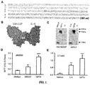
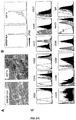

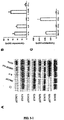


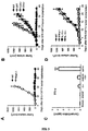


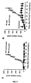
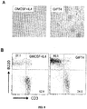


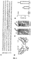

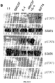


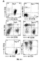

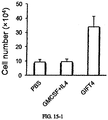
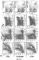
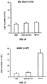


Classifications
-
- C—CHEMISTRY; METALLURGY
- C07—ORGANIC CHEMISTRY
- C07K—PEPTIDES
- C07K14/00—Peptides having more than 20 amino acids; Gastrins; Somatostatins; Melanotropins; Derivatives thereof
- C07K14/435—Peptides having more than 20 amino acids; Gastrins; Somatostatins; Melanotropins; Derivatives thereof from animals; from humans
- C07K14/52—Cytokines; Lymphokines; Interferons
- C07K14/54—Interleukins [IL]
- C07K14/5406—IL-4
-
- A—HUMAN NECESSITIES
- A61—MEDICAL OR VETERINARY SCIENCE; HYGIENE
- A61K—PREPARATIONS FOR MEDICAL, DENTAL OR TOILETRY PURPOSES
- A61K35/00—Medicinal preparations containing materials or reaction products thereof with undetermined constitution
- A61K35/12—Materials from mammals; Compositions comprising non-specified tissues or cells; Compositions comprising non-embryonic stem cells; Genetically modified cells
- A61K35/14—Blood; Artificial blood
-
- A—HUMAN NECESSITIES
- A61—MEDICAL OR VETERINARY SCIENCE; HYGIENE
- A61K—PREPARATIONS FOR MEDICAL, DENTAL OR TOILETRY PURPOSES
- A61K38/00—Medicinal preparations containing peptides
- A61K38/16—Peptides having more than 20 amino acids; Gastrins; Somatostatins; Melanotropins; Derivatives thereof
- A61K38/17—Peptides having more than 20 amino acids; Gastrins; Somatostatins; Melanotropins; Derivatives thereof from animals; from humans
- A61K38/19—Cytokines; Lymphokines; Interferons
- A61K38/193—Colony stimulating factors [CSF]
-
- A—HUMAN NECESSITIES
- A61—MEDICAL OR VETERINARY SCIENCE; HYGIENE
- A61K—PREPARATIONS FOR MEDICAL, DENTAL OR TOILETRY PURPOSES
- A61K38/00—Medicinal preparations containing peptides
- A61K38/16—Peptides having more than 20 amino acids; Gastrins; Somatostatins; Melanotropins; Derivatives thereof
- A61K38/17—Peptides having more than 20 amino acids; Gastrins; Somatostatins; Melanotropins; Derivatives thereof from animals; from humans
- A61K38/19—Cytokines; Lymphokines; Interferons
- A61K38/20—Interleukins [IL]
- A61K38/2026—IL-4
-
- A—HUMAN NECESSITIES
- A61—MEDICAL OR VETERINARY SCIENCE; HYGIENE
- A61K—PREPARATIONS FOR MEDICAL, DENTAL OR TOILETRY PURPOSES
- A61K39/00—Medicinal preparations containing antigens or antibodies
- A61K39/0005—Vertebrate antigens
- A61K39/0011—Cancer antigens
-
- A—HUMAN NECESSITIES
- A61—MEDICAL OR VETERINARY SCIENCE; HYGIENE
- A61K—PREPARATIONS FOR MEDICAL, DENTAL OR TOILETRY PURPOSES
- A61K39/00—Medicinal preparations containing antigens or antibodies
- A61K39/39—Medicinal preparations containing antigens or antibodies characterised by the immunostimulating additives, e.g. chemical adjuvants
-
- A—HUMAN NECESSITIES
- A61—MEDICAL OR VETERINARY SCIENCE; HYGIENE
- A61K—PREPARATIONS FOR MEDICAL, DENTAL OR TOILETRY PURPOSES
- A61K39/00—Medicinal preparations containing antigens or antibodies
- A61K39/46—Cellular immunotherapy
- A61K39/461—Cellular immunotherapy characterised by the cell type used
- A61K39/4611—T-cells, e.g. tumor infiltrating lymphocytes [TIL], lymphokine-activated killer cells [LAK] or regulatory T cells [Treg]
-
- A—HUMAN NECESSITIES
- A61—MEDICAL OR VETERINARY SCIENCE; HYGIENE
- A61K—PREPARATIONS FOR MEDICAL, DENTAL OR TOILETRY PURPOSES
- A61K39/00—Medicinal preparations containing antigens or antibodies
- A61K39/46—Cellular immunotherapy
- A61K39/461—Cellular immunotherapy characterised by the cell type used
- A61K39/4612—B-cells
-
- A—HUMAN NECESSITIES
- A61—MEDICAL OR VETERINARY SCIENCE; HYGIENE
- A61K—PREPARATIONS FOR MEDICAL, DENTAL OR TOILETRY PURPOSES
- A61K39/00—Medicinal preparations containing antigens or antibodies
- A61K39/46—Cellular immunotherapy
- A61K39/462—Cellular immunotherapy characterized by the effect or the function of the cells
- A61K39/4622—Antigen presenting cells
-
- A—HUMAN NECESSITIES
- A61—MEDICAL OR VETERINARY SCIENCE; HYGIENE
- A61K—PREPARATIONS FOR MEDICAL, DENTAL OR TOILETRY PURPOSES
- A61K39/00—Medicinal preparations containing antigens or antibodies
- A61K39/46—Cellular immunotherapy
- A61K39/464—Cellular immunotherapy characterised by the antigen targeted or presented
- A61K39/4643—Vertebrate antigens
- A61K39/4644—Cancer antigens
- A61K39/46449—Melanoma antigens
-
- A—HUMAN NECESSITIES
- A61—MEDICAL OR VETERINARY SCIENCE; HYGIENE
- A61K—PREPARATIONS FOR MEDICAL, DENTAL OR TOILETRY PURPOSES
- A61K47/00—Medicinal preparations characterised by the non-active ingredients used, e.g. carriers or inert additives; Targeting or modifying agents chemically bound to the active ingredient
- A61K47/30—Macromolecular organic or inorganic compounds, e.g. inorganic polyphosphates
- A61K47/42—Proteins; Polypeptides; Degradation products thereof; Derivatives thereof, e.g. albumin, gelatin or zein
-
- A—HUMAN NECESSITIES
- A61—MEDICAL OR VETERINARY SCIENCE; HYGIENE
- A61P—SPECIFIC THERAPEUTIC ACTIVITY OF CHEMICAL COMPOUNDS OR MEDICINAL PREPARATIONS
- A61P31/00—Antiinfectives, i.e. antibiotics, antiseptics, chemotherapeutics
- A61P31/12—Antivirals
-
- A—HUMAN NECESSITIES
- A61—MEDICAL OR VETERINARY SCIENCE; HYGIENE
- A61P—SPECIFIC THERAPEUTIC ACTIVITY OF CHEMICAL COMPOUNDS OR MEDICINAL PREPARATIONS
- A61P35/00—Antineoplastic agents
-
- A—HUMAN NECESSITIES
- A61—MEDICAL OR VETERINARY SCIENCE; HYGIENE
- A61P—SPECIFIC THERAPEUTIC ACTIVITY OF CHEMICAL COMPOUNDS OR MEDICINAL PREPARATIONS
- A61P37/00—Drugs for immunological or allergic disorders
- A61P37/02—Immunomodulators
- A61P37/04—Immunostimulants
-
- C—CHEMISTRY; METALLURGY
- C07—ORGANIC CHEMISTRY
- C07K—PEPTIDES
- C07K14/00—Peptides having more than 20 amino acids; Gastrins; Somatostatins; Melanotropins; Derivatives thereof
- C07K14/435—Peptides having more than 20 amino acids; Gastrins; Somatostatins; Melanotropins; Derivatives thereof from animals; from humans
- C07K14/52—Cytokines; Lymphokines; Interferons
- C07K14/53—Colony-stimulating factor [CSF]
- C07K14/535—Granulocyte CSF; Granulocyte-macrophage CSF
-
- C—CHEMISTRY; METALLURGY
- C12—BIOCHEMISTRY; BEER; SPIRITS; WINE; VINEGAR; MICROBIOLOGY; ENZYMOLOGY; MUTATION OR GENETIC ENGINEERING
- C12N—MICROORGANISMS OR ENZYMES; COMPOSITIONS THEREOF; PROPAGATING, PRESERVING, OR MAINTAINING MICROORGANISMS; MUTATION OR GENETIC ENGINEERING; CULTURE MEDIA
- C12N15/00—Mutation or genetic engineering; DNA or RNA concerning genetic engineering, vectors, e.g. plasmids, or their isolation, preparation or purification; Use of hosts therefor
- C12N15/09—Recombinant DNA-technology
- C12N15/10—Processes for the isolation, preparation or purification of DNA or RNA
- C12N15/102—Mutagenizing nucleic acids
- C12N15/1024—In vivo mutagenesis using high mutation rate "mutator" host strains by inserting genetic material, e.g. encoding an error prone polymerase, disrupting a gene for mismatch repair
-
- C—CHEMISTRY; METALLURGY
- C12—BIOCHEMISTRY; BEER; SPIRITS; WINE; VINEGAR; MICROBIOLOGY; ENZYMOLOGY; MUTATION OR GENETIC ENGINEERING
- C12N—MICROORGANISMS OR ENZYMES; COMPOSITIONS THEREOF; PROPAGATING, PRESERVING, OR MAINTAINING MICROORGANISMS; MUTATION OR GENETIC ENGINEERING; CULTURE MEDIA
- C12N15/00—Mutation or genetic engineering; DNA or RNA concerning genetic engineering, vectors, e.g. plasmids, or their isolation, preparation or purification; Use of hosts therefor
- C12N15/09—Recombinant DNA-technology
- C12N15/10—Processes for the isolation, preparation or purification of DNA or RNA
- C12N15/102—Mutagenizing nucleic acids
- C12N15/1027—Mutagenizing nucleic acids by DNA shuffling, e.g. RSR, STEP, RPR
-
- C—CHEMISTRY; METALLURGY
- C12—BIOCHEMISTRY; BEER; SPIRITS; WINE; VINEGAR; MICROBIOLOGY; ENZYMOLOGY; MUTATION OR GENETIC ENGINEERING
- C12N—MICROORGANISMS OR ENZYMES; COMPOSITIONS THEREOF; PROPAGATING, PRESERVING, OR MAINTAINING MICROORGANISMS; MUTATION OR GENETIC ENGINEERING; CULTURE MEDIA
- C12N15/00—Mutation or genetic engineering; DNA or RNA concerning genetic engineering, vectors, e.g. plasmids, or their isolation, preparation or purification; Use of hosts therefor
- C12N15/09—Recombinant DNA-technology
- C12N15/10—Processes for the isolation, preparation or purification of DNA or RNA
- C12N15/1034—Isolating an individual clone by screening libraries
- C12N15/1062—Isolating an individual clone by screening libraries mRNA-Display, e.g. polypeptide and encoding template are connected covalently
-
- C—CHEMISTRY; METALLURGY
- C12—BIOCHEMISTRY; BEER; SPIRITS; WINE; VINEGAR; MICROBIOLOGY; ENZYMOLOGY; MUTATION OR GENETIC ENGINEERING
- C12N—MICROORGANISMS OR ENZYMES; COMPOSITIONS THEREOF; PROPAGATING, PRESERVING, OR MAINTAINING MICROORGANISMS; MUTATION OR GENETIC ENGINEERING; CULTURE MEDIA
- C12N5/00—Undifferentiated human, animal or plant cells, e.g. cell lines; Tissues; Cultivation or maintenance thereof; Culture media therefor
- C12N5/06—Animal cells or tissues; Human cells or tissues
-
- A—HUMAN NECESSITIES
- A61—MEDICAL OR VETERINARY SCIENCE; HYGIENE
- A61K—PREPARATIONS FOR MEDICAL, DENTAL OR TOILETRY PURPOSES
- A61K39/00—Medicinal preparations containing antigens or antibodies
- A61K2039/51—Medicinal preparations containing antigens or antibodies comprising whole cells, viruses or DNA/RNA
- A61K2039/515—Animal cells
- A61K2039/5152—Tumor cells
-
- A—HUMAN NECESSITIES
- A61—MEDICAL OR VETERINARY SCIENCE; HYGIENE
- A61K—PREPARATIONS FOR MEDICAL, DENTAL OR TOILETRY PURPOSES
- A61K39/00—Medicinal preparations containing antigens or antibodies
- A61K2039/51—Medicinal preparations containing antigens or antibodies comprising whole cells, viruses or DNA/RNA
- A61K2039/515—Animal cells
- A61K2039/5156—Animal cells expressing foreign proteins
-
- A—HUMAN NECESSITIES
- A61—MEDICAL OR VETERINARY SCIENCE; HYGIENE
- A61K—PREPARATIONS FOR MEDICAL, DENTAL OR TOILETRY PURPOSES
- A61K39/00—Medicinal preparations containing antigens or antibodies
- A61K2039/555—Medicinal preparations containing antigens or antibodies characterised by a specific combination antigen/adjuvant
- A61K2039/55511—Organic adjuvants
- A61K2039/55522—Cytokines; Lymphokines; Interferons
- A61K2039/55527—Interleukins
-
- A—HUMAN NECESSITIES
- A61—MEDICAL OR VETERINARY SCIENCE; HYGIENE
- A61K—PREPARATIONS FOR MEDICAL, DENTAL OR TOILETRY PURPOSES
- A61K2239/00—Indexing codes associated with cellular immunotherapy of group A61K39/46
- A61K2239/31—Indexing codes associated with cellular immunotherapy of group A61K39/46 characterized by the route of administration
-
- A—HUMAN NECESSITIES
- A61—MEDICAL OR VETERINARY SCIENCE; HYGIENE
- A61K—PREPARATIONS FOR MEDICAL, DENTAL OR TOILETRY PURPOSES
- A61K2239/00—Indexing codes associated with cellular immunotherapy of group A61K39/46
- A61K2239/38—Indexing codes associated with cellular immunotherapy of group A61K39/46 characterised by the dose, timing or administration schedule
-
- A—HUMAN NECESSITIES
- A61—MEDICAL OR VETERINARY SCIENCE; HYGIENE
- A61K—PREPARATIONS FOR MEDICAL, DENTAL OR TOILETRY PURPOSES
- A61K2239/00—Indexing codes associated with cellular immunotherapy of group A61K39/46
- A61K2239/46—Indexing codes associated with cellular immunotherapy of group A61K39/46 characterised by the cancer treated
- A61K2239/48—Blood cells, e.g. leukemia or lymphoma
-
- A—HUMAN NECESSITIES
- A61—MEDICAL OR VETERINARY SCIENCE; HYGIENE
- A61K—PREPARATIONS FOR MEDICAL, DENTAL OR TOILETRY PURPOSES
- A61K2239/00—Indexing codes associated with cellular immunotherapy of group A61K39/46
- A61K2239/46—Indexing codes associated with cellular immunotherapy of group A61K39/46 characterised by the cancer treated
- A61K2239/57—Skin; melanoma
-
- A—HUMAN NECESSITIES
- A61—MEDICAL OR VETERINARY SCIENCE; HYGIENE
- A61K—PREPARATIONS FOR MEDICAL, DENTAL OR TOILETRY PURPOSES
- A61K38/00—Medicinal preparations containing peptides
-
- C—CHEMISTRY; METALLURGY
- C07—ORGANIC CHEMISTRY
- C07K—PEPTIDES
- C07K2319/00—Fusion polypeptide
Definitions
- Proleukin® (aldesleukin) contains a recombinant human Interleukin 2 (IL-2) thought to boost the immune system against cancer cells and is indicated for the treatment of adults with metastatic renal cell carcinoma (metastatic RCC). Severe adverse events generally accompany this therapy at the recommended dosages. Thus, there is a need to identify improved methods.
- GM-CSF cytokine granulocyte macrophage colony stimulating factor
- DC dendritic cells
- PAP prostatic acid phosphatase
- GM-CSF is thought to direct the target antigen to receptors on DC precursors, which then present PAP on their cell surface in a context sufficient to activate T cells for the cells that express PAP.
- PAP presenting DCs are administered to the subject to elicit an immune response retarding cancer growth.
- This strategy requires isolation and expansion of cells of the subject, and typically treatment does not entirely clear the subject of cancer or tumors. Thus, there is a need to identify improved methods.
- Interleukin 4 is a ⁇ -chain cytokine. It serves as a signal to activate and elicit antibody class switching by B lymphocytes and converts naive helper T lymphocytes to active T lymphocytes and then expand their population.
- U.S. Patent 6,838,081 reports enhancing the development of antigen presenting cells from precursor cells by administering a combination of IL-4 and GM-CSF. See also U.S. Patent Application 2004/0072299 and Hikino et al., ANTICANCER RESEARCH 24: 1609-1616 (2004 ).
- GIFT fusokines are the fused proteins derived from GM-CSF and common ⁇ chain interleukin fusion transgenes and may either suppress or enhance host immune response. Stagg et al., Molecular Therapy, 2004, 9, S133-S133 , disclose a GM-CSF/IL-2 Fusion (GIFT2). See also Penafuete et al., Cancer Res. 2009, 69(23):9020-8 ; Rafei et al., Nat Med.
- this disclosure relates to conjugates comprising a polypeptide of GM-CSF and a polypeptide IL-4.
- the GM-CSF and IL-4 are connected by a linker, e.g., polypeptide.
- the disclosure relates to isolated nucleic acids encoding these polypeptide conjugates, vectors comprising nucleic acid encoding polypeptide conjugates, and protein expression systems comprising these vectors such as infectious viral particles and host cells comprising such nucleic acids.
- the disclosure relates to pharmaceutical compositions comprising conjugates and vectors disclosed herein and a pharmaceutically acceptable excipient.
- the disclosure relates to vaccines comprising conjugates and vectors disclosed herein and an antigen and optionally an adjuvant.
- the antigen is contained in a live attenuated virus, killed virus, a virus-like particle, virosome, cancerous cell, lipid bilayer structure with a surface antigen, and the antigen is typically a viral protein or glycoprotein, bacteria, or bacterial antigen, or tumor associated antigen.
- the antigen is conjugated to a dendritic cell marker.
- the disclosure contemplates a method of mixing conjugates herein with B cells under conditions such that activated normal B cells can produce chemokine CCL3 or elevated levels of INF-gamma.
- the B-cells are chronic lymphoid leukemia B-cells.
- the mixing is in vitro or in vivo.
- in vitro activated B cells are administered to a subject, e.g., from which the B cells were originally obtained in an amount to effectively treat or prevent cancer.
- the disclosure relates to methods of treating or preventing a viral, bacterial, or parasitic infection comprising administering an effective amount of a pharmaceutical composition comprising a conjugate or vector disclosed herein optionally in combination with an antigen and optionally an adjuvant.
- the subject is at risk or, exhibiting symptoms of, or diagnosed with a viral infection, such as a chronic viral infection.
- the disclosure relates to methods of treating or preventing a viral infection comprising administering an effective amount of a vaccine comprising a conjugate disclosed herein to a subject in need thereof.
- influenza A virus including subtype H1N1, influenza B virus, influenza C virus, rotavirus A, rotavirus B, rotavirus C, rotavirus D, rotavirus E, SARS coronavirus, human adenovirus types (HAdV-1 to 55), human papillomavirus (HPV) Types 16, 18, 31, 33, 35, 39, 45, 51, 52, 56, 58, and 59, parvovirus B19, molluscum contagiosum virus, JC virus (JCV), BK virus, Merkel cell polyomavirus, coxsackie A virus, norovirus, Rubella virus, lymphocytic choriomeningitis virus (LCMV), yellow fever virus, measles virus, mumps virus, respiratory syncytial virus, rinderpest virus, California encephalitis virus, hantavirus, rabies virus, ebola virus, marburg virus, herpes simplex virus-1 (HSV
- the disclosure relates to administering a conjugate or vector disclosed herein in combination with another antiviral agent such as abacavir, acyclovir, acyclovir, adefovir, amantadine, amprenavir, ampligen, arbidol, atazanavir, atripla, boceprevir, cidofovir, combivir,darunavir, delavirdine, didanosine, docosanol, edoxudine, efavirenz, emtricitabine, enfuvirtide, entecavir, famciclovir, fomivirsen, fosamprenavir, foscarnet, fosfonet, ganciclovir, ibacitabine, imunovir, idoxuridine, imiquimod, indinavir, inosine, interferon type III, interferon type II, interferon type I, lamivudine,
- the disclosure relates to methods of treating or preventing cancer comprising administering a pharmaceutical composition comprising a conjugate or vector disclosed herein to a subject in need thereof.
- the disclosure relates to methods of treating or preventing cancer comprising administering autologous blood cells activated with a cancer antigen conjugated to GM-CSF in combination with a conjugate disclosed herein to a subject in need thereof.
- the disclosure relates to methods of activating peripheral blood cells comprising mixing peripheral blood cells with a conjugate disclosed herein comprising a tumor associated antigen/cancer marker under conditions such that increase expression of CD54 occurs.
- the disclosure relates to product produced by mixing peripheral blood cells and with a conjugate disclosed herein under conditions such that increase expression of CD54 occurs.
- the disclosure relates to methods of treating or preventing cancer comprising administering an effective amount of a product made by mixing peripheral blood cells with a conjugate disclosed herein to subject from whom the peripheral blood cells were obtained.
- the disclosure relates to methods of producing compositions comprising isolated bone marrow and/or bone marrow stem cells comprising administering conjugates and compositions disclosed herein, e.g., GIFT4, GIFT4 in combination with B cells, or isolated cells activated with GIFT4, optionally in combination with B cells, to a subject.
- the disclosure relates to the treatment or prevention of irradiation-caused bone marrow failure comprising administering conjugates and compositions disclosed herein to a subject in need thereof.
- the disclosure relates to methods of increasing the production of bone marrow and/or bone marrow stem cells comprising administering an effective amount of a conjugate disclosed herein to subject.
- the disclosure relates to a product made by the process of isolating bone marrow and/or bone marrow stem cells from a subject administered with a conjugate disclosed herein.
- the disclosure relates to methods of increasing the production of bone marrow stem cells comprising administering an effective amount of a conjugate disclosed herein in combination with B cells to subject that is B cell deficient.
- the subject is diagnosed with a B-cell immunodeficiency, defects of B-cell development/immunoglobulin production, excessive/uncontrolled B-cell proliferation, leukemia, chronic lymphocytic leukemia, lymphoma, follicular non-hodgkin's lymphoma, diffuse large B cell lymphoma, or lupus.
- the disclosure relates to a product made by the process of isolating bone marrow and/or bone marrow stem cells from a subject administered with a conjugate disclosed herein in combination with B cells.
- the disclosure relates to a composition
- a composition comprising isolated bone marrow cells and/or bone marrow stem cells and a conjugate disclosed herein, e.g., GM-CSF and IL-4 conjugate.
- the bone marrow cells and/or bone marrow stem cells are obtain from a subject that was previously administered the conjugate under conditions such that the bone marrow cells and/or bone marrow stem cells increase proliferation.
- the disclosure relates to methods of treating or preventing bone marrow failure comprising administering an effective amount of a product made by mixing bone marrow cells with a conjugate disclosed herein to subject.
- the bone marrow failure is caused by irradiation.
- the product made by mixing bone marrow cells with a conjugate disclosed herein is administered in combination with a conjugate disclosed herein to subject.
- the conjugate is GIFT4.
- the bone marrow cells were obtained from the subject.
- the disclosure relates to methods of treating or preventing cancer comprising administering an effective amount of a product made by mixing bone marrow cells with a conjugate disclosed herein to subject.
- the product made by mixing bone marrow cells with a conjugate disclosed herein is administered in combination with a conjugate disclosed herein to subject.
- the conjugate is GIFT4.
- the bone marrow cells were obtained from the subject.
- the disclosure relates to a method of treating or preventing cancer comprising by administering a pharmaceutical composition comprising conjugates or vector disclosed herein to a subject diagnosed with, exhibiting symptoms of, or at risk of cancer wherein the cancer is a hematological malignancy such as a leukemia or lymphoma, acute lymphoblastic leukemia (ALL), acute myelogenous leukemia (AML), chronic lymphocytic leukemia (CLL), small lymphocytic lymphoma (SLL), chronic myelogenous leukemia, acute monocytic leukemia (AMOL), Hodgkin's lymphomas, and non-Hodgkin's lymphomas such as Burkitt lymphoma, B-cell lymphoma and multiple myeloma.
- a hematological malignancy such as a leukemia or lymphoma, acute lymphoblastic leukemia (ALL), acute myelogenous leukemia (AML), chronic lymphocytic leukemia
- the conjugate or vector may be administered in combination with an anti-cancer agent such as gefitinib, erlotinib, docetaxel, cis-platin, 5-fluorouracil, gemcitabine, tegafur, raltitrexed, methotrexate, cytosine arabinoside, hydroxyurea, adriamycin, bleomycin, doxorubicin, daunomycin, epirubicin, idarubicin, mitomycin-C, dactinomycin and mithramycin, vincristine, vinblastine, vindesine, vinorelbine taxol, taxotere, etoposide, teniposide, amsacrine, topotecan, camptothecin, bortezomib, anagrelide, tamoxifen, toremifene, raloxifene, droloxifene,
- an anti-cancer agent such as
- the disclosure relates to gene therapies comprising administering vectors comprising nucleic acid encoding conjugates disclosed herein to a subject in need thereof.
- the nucleic acids are isolated and/or purified from their natural state or translated to a non-naturally occurring form such as cDNA.
- the disclosure contemplates incorporating conjugates disclosed herein into the surfaces of particles, e.g., cells, liposomes, micelles, vesicles, bilayer structures, virosomes, and virus-like particles.
- the conjugates may be linked to lipophilic moieties, e.g., fatty acids and GPI.
- the disclosure contemplates a GPI anchored conjugate comprising GPI, GM-CSF, IL-4, and optionally an antigen, adjuvant, or other polypeptide. It is contemplated that these particles may contain other surface polypeptides, antigens and co-stimulatory molecules such as B7-1, B7-2, ICAM-1, and/or IL-2. It is contemplated that these particles may be used in all the applications conjugates disclosed herein are mentioned.
- any of the conjugates disclosed herein may be further conjugated to an adjuvant, cytokine, co-stimulatory molecule, antigen, protein, or glycoprotein.
- the antigen is a viral protein or a cancer marker.
- the cancer marker is selected from PAP (prostatic acid phosphatase), prostate-specific antigen (PSA), (PSMA) prostate-specific membrane antigen, early prostate cancer antigen-2 (EPCA-2), AKAP-4 (A kinase [PRKA] anchor protein 4), NGEP (new gene expressed in prostate), PSCA (prostate stem cell antigen), STEAP (six-transmembrane epithelial antigen of the prostate), MUC 1 (mucin 1), HER-2, BCL-2, MAGE antigens such as CT7, MAGE-A3 and MAGE-A4, ERK5, G-protein coupled estrogen receptor 1, CA15-3, CA19-9, CA 72-4, CA-125, carcinoembryonic antigen, CD20, CD31, CD34, PTPRC (CD45), CD99, CD117, melanoma-associated antigen (TA-90), peripheral myelin protein 22 (PMP22), epithelial membrane proteins (EMP-1, -2, and -3),
- the viral antigen is selected from an influenza virus hemagglutinin and neuraminidase; cytomegalovirus glycoprotein gB, p28, p38, p50, p52, p65, and p150; Borrelia p41; HIV nef, integrase, gag, protease, tat, env, p31, p17, p24, p31, p55, p66, gp32, gp36, gp39, gp41, gp120, and gp160; SIV p55; HBV core, surface antigen, and australian antigen; HCV core nucleocapsid, NS3, NS4, and NS5; Dengue env and NS1; EBV early antigen, p18, p23, gp125, nuclear antigen (EBNA)-1, EBNA-2, EBNA-3A, EBNA-3B,
- EBNA
- the adjuvant or cytokine is selected from IL-2, IL-12, IL-15, IL-7, IL-18, IL-21, IL-27, IL-31, IFN-alpha, flagellin, unmethylated, CpG oligonucleotide, lipopolysaccharides, lipid A, and heat stable antigen (HSA).
- the disclosure contemplates administration of pharmaceutical products comprising conjugates disclosed herein by intravenous (IV), subcutaneous (SC), or intraperitoneal (IP) administration.
- IV intravenous
- SC subcutaneous
- IP intraperitoneal
- Common ⁇ chain interleukin cytokines include IL-2, IL-4, IL-7, IL-9, IL-15 and IL-21, and have important roles in the activation and differentiation of lymphocytes.
- IL-2 has strong immune modulatory properties on T cells, and had been approved by FDA as the first interleukin immunotherapeutic agent to use to treat patients with advanced kidney cancer and metastatic melanoma.
- FDA the first interleukin immunotherapeutic agent to use to treat patients with advanced kidney cancer and metastatic melanoma.
- IL-2 immunotherapy even combined with various chemotherapy drugs has not shown significant improvement of survival time in cancer patients.
- IL-2 has frequent, often serious and sometimes fatal side effects due to capillary leakage.
- GIFT2 shows anti-tumor activities via induction of tumor-killer NK cells.
- GIFT2 affects tumoricidal dendritic cells.
- GIFT15 derived from GM-CSF and IL-15
- GIFT4 has immune suppressive function to abrogate inflammatory response in multiple sclerosis.
- a fusokine derive from GM-CSF and IL-4 (GIFT4) is show to be a cancer immunotherapeutic agent.
- GIFT4 directly elicits adaptive B-cell immune response and consequent T cell immunity.
- GM-CSF-IL-4 Fusokine (GIFT4)
- Figure 1 provides for an embodiment of this disclosure comprising a GM-CSF sequence and a murine IL-4 sequence and figure 11 provides s human sequence.
- the disclosure contemplates a fusokine with a recombinant human form such as isoform 1 which is amino acids sequence MGLTSQLLPP LFFLLACAGN FVHGHKCDIT LQEIIKTLNS LTEQKTLCTE LTVTDIFAAS KNTTEKETFC RAATVLRQFY SHHEKDTRCL GATAQQFHRH KQLIRFLKRL DRNLWGLAGL NSCPVKEANQ STLENFLERL KTIMREKYSK CSS (SEQ ID NO: 3) or isoform 2 which is amino acid sequence MGLTSQLLPP LFFLLACAGN FVHGHKCDIT LQEIIKTLNS LTEQKNTTEK ETFCRAATVL RQFYSHHEKD TRCLGATAQQ FHRHKQLIRF LKRLDRNLWG LAGLNSCPVK
- the present disclosure encompasses fusion proteins involving full-length preprocessed forms, as well as mature processed forms, fragments thereof and variants of each or both of the GM-CSF and IL-4 entities with linker amino acids, including allelic as well as non-naturally occurring variants.
- allelic variants of the GM-CSF and IL-4 entities that may exist in the population, the skilled artisan will further appreciate that changes (i.e. one or more deletions, additions and/or substitutions of one or more amino acid) can be introduced by mutation using classic or recombinant techniques to effect random or targeted mutagenesis.
- a suitable variant in use in the present disclosure typically has an amino acid sequence having a high degree of homology with the amino acid sequence of the corresponding native cytokine.
- the amino acid sequence of the variant cytokine in use in the fusion protein of the disclosure is at least 70%, at least about 75%, at least about 80%, at least about 90%, typically at least about 95%, more typically at least about 97% and even more typically at least about 99% identical to the corresponding native sequence, e.g., SEQ ID NO: 3 or 4.
- such native sequence is of human GM-CSF and/or human IL-4.
- Percent identities between amino acid or nucleic acid sequences can be determined using standard methods known to those of skill in the art. For instance for determining the percentage of homology between two amino acid sequences, the sequences are aligned for optimal comparison purposes. The amino acid residues at corresponding amino acid positions are then compared. Gaps can be introduced in one or both amino acid sequence(s) for optimal alignment and non-homologous sequences can be disregarded for comparison purposes. When a position in the first sequence is occupied by the same amino acid residue as the corresponding position in the second sequence, then the sequences are identical at that position. The percent identity between the two sequences is a function of the number of identical positions shared by the sequences, taking into account the number of gaps which need to be introduced for optimal alignment and the length of each gap.
- GCGTM program available from Genetics Computer Group, Madison, Wis.
- DNAsisTM program available from Hitachi Software, San Bruno, Calif.
- MacVectorTM program available from the Eastman Kodak Company, New Haven, Conn.
- Suitable variants of GM-CSF and IL-4 entities for use in the present disclosure are biologically active and retain at least one of the activities described herein in connection with the corresponding polypeptide.
- the therapeutic effect e.g. anti-tumor activity, by-pass of tumor-induced immune energy
- a given function of the polypeptide(s) may be positively or negatively affected to some degree, e.g. with variants exhibiting reduced cytotoxicity or enhanced biological activity.
- Amino acids that are essential for a given function can be identified by methods known in the art, such as by site-directed mutagenesis.
- Amino acids that are critical for binding a partner/substrate e.g. a receptor
- the resulting variant can be tested for biological activity in assays such as those described above.
- one or more amino acid residues are conservatively substituted.
- a “conservative amino acid substitution” is one in which the amino acid residue in the native polypeptide is replaced with an amino acid residue having a similar side chain. Families of amino acid residues having similar side chains have been defined in the art. Typically, substitutions are regarded as conservative when the replacement, one for another, is among the aliphatic amino acids Ala, Val, Leu, and Ile; the hydroxyl residues Ser and Thr; the acidic residues Asp and Glu; the amide residues Asn and Gln; the basic residues Lys and Arg; or the aromatic residues Phe and Tyr.
- mutations can be introduced randomly along all or part of a cytokine coding sequence, such as by saturation mutagenesis, and the resultant mutant can be screened for its biological activity as described herein to identify mutants that retain at least therapeutic activity.
- the GM-CSF and IL-4 entities can be directly fused in the fusion protein of the disclosure, it is however typical to use a linker for joining GM-CSF and IL-4.
- the purpose of the linker is to allow the correct formation, folding and/or functioning of each of the GM-CSF and IL-4 entities. It should be sufficiently flexible and sufficiently long to achieve that purpose.
- the coding sequence of the linker may be chosen such that it encourages translational pausing and therefore independent folding of the GM-CSF and IL-4 entities.
- a person skilled in the art will be able to design suitable linkers in accordance with the disclosure.
- the present disclosure is, however, not limited by the form, size or number of linker sequences employed.
- linker sequence of choice may be inserted between GM-CSF and IL-4.
- the only requirement for the linker sequence is that it functionally does not adversely interfere with the folding and/or functioning of the individual entities of the fusion protein.
- a suitable linker is 1 to 5 or 5 to 50 amino acid long and may comprise amino acids such as glycine, serine, threonine, asparagine, alanine and proline (see for example Wiederrecht et al., 1988, Cell 54, 841 ; Dekker et al., 1993, Nature 362, 852 ; Sturm et al., 1988, Genes and Dev. 2, 1582 ; Aumailly et al., 1990 FEBS Lett.
- linkers consists of two or three or more (e.g. up to eight or more) copies of the sequence Gly-Gly-Gly-Gly-Ser (GGGGS) (SEQ ID NO:5). It will be evident that the disclosure is not limited to the use of these particular linkers.
- the disclosure further includes fusion proteins which comprise, or alternatively consist essentially of, or alternatively consist of an amino acid sequence which is at least 70%, 75%, 80%, 90%, 95%, 97%, 99% homologous or even better 100% homologous (identical) to all or part of any of the amino acid sequences recited in SEQ ID NO: 1-6.
- a protein in the context of the present disclosure, a protein "consists of' an amino acid sequence when the protein does not contain any amino acids but the recited amino acid sequence.
- a protein consists essentially of' an amino acid sequence when such an amino acid sequence is present together with only a few additional amino acid residues, typically from about 1 to about 50 or so additional residues.
- a protein “comprises” an amino acid sequence when the amino acid sequence is at least part of the final (i.e. mature) amino acid sequence of the protein.
- Such a protein can have a few up to several hundred additional amino acids residues.
- Such additional amino acid residues can be naturally associated with each or both entities contained in the fusion or heterologous amino acid/peptide sequences (heterologous with respect to the respective entities).
- the fusion proteins of the disclosure comprise a signal peptide at the NH 2 -terminus in order to promote secretion in the host cell or organism.
- the endogenous signal peptide i.e.
- a suitable heterologous (with respect to the cytokine in question) signal peptide sequence can be added to the cytokine entity present at the NH 2 terminus of the fusion or inserted in replacement of the endogenous one.
- the fusion proteins of the disclosure can comprise cytokine entities of any origin, i.e. any human or animal source (including canine, avian, bovine, murine, ovine, feline, porcine, etc).
- cytokine entities of any origin i.e. any human or animal source (including canine, avian, bovine, murine, ovine, feline, porcine, etc).
- chimeric fusion proteins are also encompassed by the disclosure (e.g. one cytokine entity of human origin and the other of an animal source), it is typical that each entity be of the same origin (e.g. both from humans).
- the fusion proteins of the present disclosure can be produced by standard techniques. Polypeptide and DNA sequences for each of the cytokines involved in the fusion protein of the present disclosure are published in the art, as are methods for obtaining expression thereof through recombinant or chemical synthetic techniques.
- a fusion-encoding DNA sequence can be synthesized by conventional techniques including automated DNA synthesizers. Then, the DNA sequence encoding the fusion protein may be constructed in a vector and operably linked to a regulatory region capable of controlling expression of the fusion protein in a host cell or organism. Techniques for cloning DNA sequences for instance in viral vectors or plasmids are known to those of skill in the art ( Sambrook et al, 2001, "Molecular Cloning. A Laboratory Manual", Laboratory Press, Cold Spring Harbor N.Y .). The fusion protein of the disclosure can be purified from cells that have been transformed to express it.
- nucleic acid and “polynucleotide” are used interchangeably and define a polymer of nucleotides of any length, either deoxyribonucleotide (DNA) molecules (e.g., cDNA or genomic DNA) and ribonucleotide (RNA) molecules (e.g., mRNA) and analogs of the DNA or RNA generated using nucleotide analogs (see U.S. Pat. No. 5,525,711 and U.S. Pat. No. 4,711,955 as examples of nucleotide analogs).
- DNA deoxyribonucleotide
- RNA ribonucleotide
- modifications to the nucleotide structure may be imparted before or after assembly of the polymer.
- the sequence of nucleotides may also be interrupted by non-nucleotide elements.
- the nucleic acid molecule may be further modified after polymerization, such as by conjugation with a labeling component.
- the nucleic acid, especially DNA can be double-stranded or single-stranded, but typically is double-stranded DNA. Single-stranded nucleic acids can be the coding strand (sense strand) or the non-coding strand (anti-sense strand).
- the nucleic acid molecules of the disclosure include, but are not limited to, the sequence encoding the fusion protein alone, but may comprise additional non-coding sequences, for example introns and non-coding 5' and 3' sequences that play a role in transcription, mRNA processing (including splicing and polyadenylation signals), ribosome binding and mRNA stability.
- the nucleic acid molecule of the disclosure can contain less than about 5 kb, 4 kb, 3 kb, 2 kb, 1 kb, 0.5 kb or 0.1 kb of nucleotide sequences which naturally flank (i.e. sequences located at the 5' and 3' ends) or are present within the genomic DNA encoding GM-CSF and 1L-4 entities.
- nucleic acid molecules which comprise, or alternatively consist essentially of, or alternatively consist of a nucleotide sequence encoding all or part of an amino acid sequence encoding a fusion protein which is at least about 70%, at least about 75%, at least about 80%, at least about 90%, at least about 95%, typically at least about 97%, more typically at least about 99% homologous or even more typically 100% homologous to any of the amino acid sequences shown in SEQ ID NO: 1-6.
- a nucleic acid molecule of the disclosure comprises a nucleic acid molecule which is a complement of all or part of a nucleotide sequence encoding the fusion protein shown in any of SEQ ID NO: 1-6.
- a nucleic acid molecule which is complementary to the nucleotide sequence of the present disclosure is one which is sufficiently complementary such that it can hybridize to the fusion-encoding nucleotide sequence under stringent conditions, thereby forming a stable duplex. Such stringent conditions are known to those skilled in the art.
- a typical, non-limiting example of stringent hybridization conditions are hybridization in 6 times sodium chloride/sodium citrate (SSC) at about 45 C, followed by one or more washes in 0.2 times SSC, 0.1% SDS at 50-65 C.
- SSC sodium chloride/sodium citrate
- the disclosure pertains to antisense nucleic acid to the nucleic acid molecules of the disclosure.
- the antisense nucleic acid can be complementary to an entire coding strand, or to only a portion thereof.
- the disclosure encompasses variants of the above-described nucleic acid molecules of the disclosure e.g., that encode variants of the fusion proteins that are described above.
- the variation(s) encompassed by the present disclosure can be created by introducing one or more nucleotide substitutions, additions and/or deletions into the nucleotide sequence by standard techniques, such as site-directed mutagenesis and PCR-mediated mutagenesis. Following mutagenesis, the variant nucleic acid molecule can be expressed recombinantly as described herein and the activity of the resulting protein can be determined using, for example, assays described herein.
- nucleic acid molecule of the disclosure can be altered to provide preferential codon usage for a specific host cell (for example E. coli; Wada et al., 1992, Nucleic Acids Res. 20, 2111-2118 ).
- the disclosure further encompasses nucleic acid molecules that differ due to the degeneracy of the genetic code and thus encode for example the same fusion protein as any of those shown in SEQ ID NO: 1-6.
- Another embodiment of the disclosure pertains to fragments of the nucleic acid molecule of the disclosure, e.g. restriction endonuclease and PCR-generated fragments. Such fragments can be used as probes, primers or fragments encoding an immunogenic portion of the fusion protein.
- the nucleic acid molecules of the present disclosure can be generated using the sequence information provided herein.
- the nucleic acid encoding each of the GM-CSF and IL-4 entities can be cloned or amplified using cDNA or, alternatively, genomic DNA, as a template and appropriate probes or oligonucleotide primers according to standard molecular biology techniques (e.g., as described in Sambrook, et al. "Molecular Cloning: A Laboratory Manual", Cold Spring Harbor Laboratory Press, Cold Spring Harbor, N.Y., 2001 ) or standard PCR amplification techniques based on sequence data accessible in the art (such as those provided above in connection with the fusion proteins of the disclosure or those provided in the Examples part).
- Fusing of the GM-CSF sequence to the IL-4 sequence may be accomplished as described in the Experimental below or by conventional techniques.
- the GM-CSF and IL-4 encoding sequences can be ligated together in-frame either directly or through a sequence encoding a peptide linker.
- the GM-CSF-encoding sequence can also be inserted directly into a vector which contains the IL-4 - encoding sequence, or vice versa.
- PCR amplification of the GM-CSF and IL-4 -encoding sequences can be carried out using primers which give rise to complementary overhangs which can subsequently be annealed and re-amplified to generate a fusion gene sequence.
- fusokine GIFT4 has the powerful capability to convert naive B-cells to tumoricidal effectors. Given its potential impact on host B cell immune functions, fusokine GIFT4 offers a strategy for immunotherapy against a wide variety of cancers, e.g., using a B cell-based cancer immunotherapy.
- the disclosure contemplates the fusokine GIFT4 (derive from GM-CSF and IL-4) use as a cancer immunotherapeutic agent. Functionally different from GM-CSF and IL-4 that induces monocytes to differentiate into dendritic cells in vitro, GIFT4 directly elicits adaptive B-cell immune response and consequent T cell immunity. See Gluckman et al., Cytokines, cellular & molecular therapy, 1997, 3:187-196 . GIFT4 fusokine is a potent activator to trigger robust B cell immunity against tumor.
- GIFT4-B cells secrete an array of innate cytokines such as IL-1 ⁇ , IL-6, IL-12, VEGF, GM-CSF and chemokine CCL3, but not IL-10 and IFN- ⁇ .
- the pro-Th1 cytokines IL-12, IL-1 ⁇ and IL-6 can promote IFN- ⁇ production by T cells and enhance host anti-tumor immunity.
- IL-4 stimulation only induces strong phosphorylation of STST6 in B cells, with little or weak phosphorylation of other STATs.
- GIFT4 protein shows gain-of-function to trigger hyper-phosphorylation of STAT1, STAT3, STAT5 and STAT6 upon ligation with B cells, the former two STATs are the major downstream signaling pathways for other IL-2 common ⁇ chain family members including IL-2, IL-7, IL-9, IL-15 and IL-21. Phosphorylation of STAT3 is related to the production of IL-1 ⁇ and IL-6. There is no report that STAT phosphorylation results in CCL3 secretion by normal B cells; however, BCR stimulation activates leukemic B cells to produce chemokine CCL3. See Miyauchi et al., Cancer science, 2011, 102:1236-1241 . Although GIFT4 protein possesses the functional activities of both GM-CSF and IL-4 components, how phosphorylation of STATs leads to the secretion of GM-CSF and CCL3 by GIFT4-B cells remains unclear.
- GIFT4-B cells have a unique phenotype expressing surface markers B220, CD19, CD40, CD80, CD86, and IgM, but not CD23. GIFT4-B cells secrete high amount of GM-CSF. Unlike IRA-B cells that secrete high amounts of GM-CSF (See Rauch et al., Science, 2012, 335:597-601 ), GIFT4-B cells are CD80 + CD86 + that are typical markers for antigen-presenting cells. The phenotype of GIFT4-B cells is also different from B1 cells and B2 cells, of which B1 cells are B220 low , and B2 cells are CD23 + .
- GIFT4-B cells Unlike IRA B cells, GIFT4-B cells also secrete IL-1 ⁇ , IL-5, IL-6, IL-12, VEFG and massive CCL3, but not IL-3 that is uniquely produced by IRA B cells. To our knowledge, it is the first report that activated normal B cells can produce chemokine CCL3. Therefore, GIFT4-B cells have distinct cytokine-secreting profile that can enhance IFN- ⁇ -mediated T cell antitumor immunity, but sharing some common surface markers with IRA-B cells. Strikingly, GIFT4-B cells have the plasticity to isotype switch from IgM to IgG upon BCR cross-linking; similar to IL-4 and anti-mouse IgM-treated B cells. Thus, GIFT4-B cells could function as both innate effectors and adaptive responders against a variety of cancers, and possible infectious pathogens.
- GIFT4 fusokine triggers previously un-described B cell-dependent anti-tumor immunity in a murine model of melanoma.
- B cells comprise heterogeneous subpopulations of B-lymphocytes with two sides of immune activities either enhancing or suppressing host immunity via B cell cytokine secretion, antibody production, and cellular interaction.
- B cells suppress host immune response against tumors by inhibition of T cell tumorcidial activities via IL-10 mediated pathway.
- new emerging evidences suggest that B cells play important protective roles in anti-tumor immunity.
- GTFT4 protein in vivo results in the expansion of a global of B cells including GM-CSF-secreting IRA-like B cells without expansion IL-10-producing regulatory B cells, since GIFT4-B cells do not secretes IL-10.
- GM-CSF has been shown to increase the proliferation of antigen-activated cytotoxic T cells; chemokine CCL3 is important for recruiting CCR5 + T cells in anti-tumor immunity, suggesting that GM-CSF and CCL3-producing GIFT4-B cells have potent efficacy to promote T cell immunity against tumors.
- VEGF-producing B cells facilitate dendritic cells migration in vivo.
- GIFT4-B cells Co-culture of GIFT4-B cells with T cells markedly increase IFN- ⁇ production by T cells further verifies that GIFT4-B cells actively participate in cellular immunity by augmenting the magnitude of IFN- ⁇ T cell response.
- B cells can act as real antigen-presenting cells to directly license cytotoxic T cells and establish long-lasting antitumor immunity strongly indicates that GIFT4-B cells function like dendritic cells to elicit T ⁇ cell immunity against tumors.
- Antigen-specific antibody production is an important protective arm of B effector cells against infectious pathogens and cancers.
- IL-4 has been shown to participate in generation and expansion of memory B cells upon antigen-BCR ligation; however IL-4 stimulation could not increase the number of plasma cells. See Choe et al., Journal of immunology, 1997, 159:3757-3766 .
- TLR ligands CpG oligodeoxynucleotides and LPS can activate memory B cells and differentiate into plasma cells in vitro, but lose the capability in vivo.
- GIFT4 protein is a potent stimulator for B cell anti-tumor humoral response.
- immunization of GIFT4-secreting B16F0 cells leads to tumor-specific antibody secretion in immunized mice suggests that GIFT4 is a powerful adjuvant for induction of anti-melanoma specific antibodies.
- the complete inhibition of melanoma tumor growth in B16F0-GIFT4 cell-immunized B6 mice indicates that GIFT4 elicits acquired anti-tumor B cell immunity.
- Fc-receptor substantially contributes to the action of cytotoxic antibodies against tumors through antibody-dependent cell-mediated cytotoxicity pathway (ADCC).
- ADCC antibody-dependent cell-mediated cytotoxicity pathway
- pharmaceutically acceptable excipient is intended to include any and all carriers, solvents, diluents, excipients, adjuvants, dispersion media, coatings, antibacterial and antifungal agents, and absorption delaying agents, and the like, compatible with pharmaceutical administration.
- the pharmaceutical composition of the disclosure comprises a carrier and/or diluent appropriate for its delivering by injection to a human or animal organism.
- a carrier and/or diluent appropriate for its delivering by injection to a human or animal organism.
- Such carrier and/or diluent is non-toxic at the dosage and concentration employed. It is selected from those usually employed to formulate compositions for parental administration in either unit dosage or multi-dose form or for direct infusion by continuous or periodic infusion. It is typically isotonic, hypotonic or weakly hypertonic and has a relatively low ionic strength, such as provided by sugars, polyalcohols and isotonic saline solutions. Representative examples include sterile water, physiological saline (e.g.
- the pH of the composition of the disclosure is suitably adjusted and buffered in order to be appropriate for use in humans or animals, typically at a physiological or slightly basic pH (between about pH 8 to about pH 9, with a special preference for pH 8.5).
- Suitable buffers include phosphate buffer (e.g. PBS), bicarbonate buffer and/or Tris buffer.
- a typical composition is formulated in 1M saccharose, 150 mM NaCl, 1 mM MgC12, 54 mg/l Tween 80, 10 mM Tris pH 8.5.
- Another typical composition is formulated in 10 mg/ml mannitol, 1 mg/ml HSA, 20 mM Tris, pH 7.2, and 150 mM NaCl.
- composition of the disclosure can be in various forms, e.g. in solid (e.g. powder, lyophilized form), or liquid (e.g. aqueous).
- solid compositions the typical methods of preparation are vacuum drying and freeze-drying which yields a powder of the active agent plus any additional desired ingredient from a previously sterile-filtered solution thereof.
- Such solutions can, if desired, be stored in a sterile ampoule ready for reconstitution by the addition of sterile water for ready injection.
- Nebulized or aerosolized formulations also form part of this disclosure.
- Methods of intranasal administration are well known in the art, including the administration of a droplet, spray, or dry powdered form of the composition into the nasopharynx of the individual to be treated from a pressured container or dispenser which contains a suitable propellant, e.g., a gas such as carbon dioxide, or a nebulizer (see for example WO 95/11664 ).
- Enteric formulations such as gastroresistant capsules and granules for oral administration, suppositories for rectal or vaginal administration also form part of this disclosure.
- the compositions can also include absorption enhancers which increase the pore size of the mucosal membrane.
- Such absorption enhancers include sodium deoxycholate, sodium glycocholate, dimethyl-beta-cyclodextrin, lauroyl-1-lysophosphatidylcholine and other substances having structural similarities to the phospholipid domains of the mucosal membrane.
- composition can also contain other pharmaceutically acceptable excipients for providing desirable pharmaceutical or pharmacodynamic properties, including for example modifying or maintaining the pH, osmolarity, viscosity, clarity, color, sterility, stability, rate of dissolution of the formulation, modifying or maintaining release or absorption into an the human or animal organism.
- excipients for providing desirable pharmaceutical or pharmacodynamic properties, including for example modifying or maintaining the pH, osmolarity, viscosity, clarity, color, sterility, stability, rate of dissolution of the formulation, modifying or maintaining release or absorption into an the human or animal organism.
- polymers such as polyethylene glycol may be used to obtain desirable properties of solubility, stability, half-life and other pharmaceutically advantageous properties ( Davis et al., 1978, Enzyme Eng. 4, 169-173 ; Burnham et al., 1994, Am. J. Hosp. Pharm. 51, 210-218 ).
- stabilizing components include polysorbate 80, L-arginine, polyvinylpyrrolidone, trehalose, and combinations thereof.
- Other stabilizing components especially suitable in plasmid-based compositions include hyaluronidase (which is thought to destabilize the extra cellular matrix of the host cells as described in WO 98/53853 ), chloroquine, protic compounds such as propylene glycol, polyethylene glycol, glycerol, ethanol, 1-methyl L-2-pyrrolidone or derivatives thereof, aprotic compounds such as dimethylsulfoxide (DMSO), diethylsulfoxide, di-n-propylsulfoxide, dimethylsulfone, sulfolane, dimethyl-formamide, dimethylacetamide, tetramethylurea, acetonitrile (see EP 890 362 ), nuclease inhibitors such as actin G ( WO 99/56784 ) and cationic salts such as magnesium (Mg
- the amount of cationic salt in the composition of the disclosure typically ranges from about 0.1 mM to about 100 mM, and still more typically from about 0.1 mM to about 10 mM.
- Viscosity enhancing agents include sodium carboxymethylcellulose, sorbitol, and dextran.
- the composition can also contain substances known in the art to promote penetration or transport across the blood barrier or membrane of a particular organ (e.g. antibody to transferrin receptor; Friden et al., 1993, Science 259, 373-377 ).
- a gel complex of poly-lysine and lactose ( Midoux et al., 1993, Nucleic Acid Res. 21, 871-878 ) or poloxamer 407 ( Pastore, 1994, Circulation 90, 1-517 ) can be used to facilitate administration in arterial cells.
- composition of the disclosure may also comprise one or more adjuvant(s) suitable for systemic or mucosal application in humans.
- useful adjuvants include without limitation alum, mineral oil emulsion such as Freunds complete and incomplete, lipopolysaccharide or a derivative thereof ( Ribi et al., 1986, Immunology and Immunopharmacology of Bacterial Endotoxins, Plenum Publ. Corp., NY, p407-419 ), saponins such as QS21 ( Sumino et al., 1998, J. Virol.
- composition of the disclosure may be formulated with conventional vaccine vehicles composed of chitosan or other polycationic polymers, polylactide and polylactide-co-glycolide particles, poly-N-acetyl glucosamine-based polymer matrix, particles composed of polysaccharides or chemically modified polysaccharides, and lipid-based particles, etc.
- the composition may also be formulated in the presence of cholesterol to form particulate structures such as liposomes.
- the composition may be administered to patients in an amount effective, especially to enhance an immune response in an animal or human organism.
- the term "effective amount” refers to an amount sufficient to realize a desired biological effect.
- an effective amount for enhancing an immune response could be that amount necessary to cause activation of the immune system, for instance resulting in the development of an anti-tumor response in a cancerous patient (e.g. size reduction or regression of the tumor into which the composition has been injected and/or distant tumors).
- the appropriate dosage may vary depending upon known factors such as the pharmacodynamic characteristics of the particular active agent, age, health, and weight of the host organism; the condition(s) to be treated, nature and extent of symptoms, kind of concurrent treatment, frequency of treatment, the need for prevention or therapy and/or the effect desired.
- the dosage will also be calculated dependent upon the particular route of administration selected. Further refinement of the calculations necessary to determine the appropriate dosage for treatment is routinely made by a practitioner, in the light of the relevant circumstances.
- the titer may be determined by conventional techniques.
- a composition based on vector plasmids may be formulated in the form of doses of between 1 ⁇ g to 100 mg, advantageously between 10 ⁇ g and 10 mg and typically between 100 ⁇ g and 1 mg.
- a composition based on proteins may be formulated in the form of doses of between 10 ng to 100 mg.
- a typical dose is from about 1 ⁇ g to about 10 mg of the therapeutic protein per kg body weight.
- the administration may take place in a single dose or a dose repeated one or several times after a certain time interval.
- the composition of the present disclosure is administered by injection using conventional syringes and needles, or devices designed for ballistic delivery of solid compositions ( WO 99/27961 ), or needleless pressure liquid jet device ( U.S. Pat. No. 4,596,556 ; U.S. Pat. No. 5,993,412 ).
- composition of the disclosure can be enclosed in ampoules, disposable syringes or multiple dose vials made of glass or plastic. In all cases, the composition must be sterile and should be fluid to the extent that easy syringability exists. It must be stable under the conditions of manufacture and storage and must be preserved against the contaminating action of microorganisms such as bacteria and fungi.
- Sterile injectable solutions can be prepared by incorporating the active agent (e.g., a fusion protein or infectious particles) in the required amount with one or a combination of ingredients enumerated above, followed by filtered sterilization.
- the pharmaceutical composition of the disclosure may be employed in methods for treating or preventing a variety of diseases and pathologic conditions, including genetic diseases, congenital diseases and acquired diseases such as infectious diseases (e.g. viral and/or bacterial infections), cancer, immune deficiency diseases, and autoimmune diseases. Accordingly, the present disclosure also encompasses the use of the fusion protein, vector, infectious viral particle, host cell or composition of the disclosure for the preparation of a drug intended for treating or preventing such diseases, and especially cancer or an infectious disease.
- infectious diseases e.g. viral and/or bacterial infections
- cancer immune deficiency diseases
- autoimmune diseases e.g., autoimmune diseases.
- the present disclosure also encompasses the use of the fusion protein, vector, infectious viral particle, host cell or composition of the disclosure for the preparation of a drug intended for treating or preventing such diseases, and especially cancer or an infectious disease.
- composition of the present disclosure is particularly intended for the preventive or curative treatment of disorders, conditions or diseases associated with cancer.
- cancer encompasses any cancerous conditions including diffuse or localized tumors, metastasis, cancerous polyps and preneoplastic lesions (e.g. dysplasics) as well as diseases which result from unwanted cell proliferation.
- preneoplastic lesions e.g. dysplasics
- a variety of tumors may be selected for treatment in accordance with the methods described herein. In general, solid tumors are typical.
- Cancers which are contemplated in the context of the disclosure include without limitation glioblastoma, sarcoma, melanomas, mastocytoma, carcinomas as well as breast cancer, prostate cancer, testicular cancer, ovarian cancer, endometrial cancer, cervical cancer (in particular, those induced by a papilloma virus), lung cancer (e.g. lung carcinomas including large cell, small cell, squamous and adeno-carcinomas), renal cancer, bladder cancer, liver cancer, colon cancer, anal cancer, pancreatic cancer, stomach cancer, gastrointestinal cancer, cancer of the oral cavity, larynx cancer, brain and CNS cancer, skin cancer (e.g.
- the composition is administered into or in close proximity to a solid tumor.
- AIET autologous immune enhancement therapy
- LAK lymphokine-activated killer
- NK natural killer
- CTLs cytotoxic T lymphocytes
- DCs dendritic cells
- the disclosure contemplates mixing harvested cells with conjugates of GM-CSF and IL-4 to activate them.
- the disclosure contemplates administering conjugates of GM-CSF and IL-4 when the cells are administered back to the subject.
- the disclosure contemplates the administration of sipuleucel-T (PROVENGE) in combination with a conjugate of GM-CSF and IL-4.
- PROVENGE consists of autologous peripheral blood mononuclear cells, including antigen presenting cells (APCs), that have been activated during a culture period with a recombinant human protein, PAP-GM-CSF, consisting of prostatic acid phosphatase (PAP), an antigen expressed in prostate cancer tissue, linked to GM-CSF.
- APCs antigen presenting cells
- PAP-GM-CSF consisting of prostatic acid phosphatase (PAP), an antigen expressed in prostate cancer tissue, linked to GM-CSF.
- the disclosure relates to a conjugate comprising PAP, GM-CSF, and IL-4, and uses in activating antigen presenting cells in peripheral blood mononuclear cells.
- the peripheral blood mononuclear cells of the subject may be obtained via a standard leukapheresis procedure prior to infusion.
- the recombinant antigen can bind to and be processed by antigen presenting cells (APCs).
- APCs antigen presenting cells
- the recombinant antigen is believed to direct the immune response to PAP.
- the infused product is believed to contain antigen presenting cells, dendritic cells, T cells, B cells, natural killer (NK) cells, and other cells.
- each dose contains more than 50 million autologous CD54 + cells activated with PAP-GM-CSF or PAP-GM-CSF-IL-4.
- the potency is typically evaluated by measuring the increased expression of the CD54 molecule, also known as ICAM-1, on the surface of APCs after culture with PAP-GM-CSF or PAP-CM-CSF-IL-4.
- CD54 is a cell surface molecule that plays a role in the immunologic interactions between APCs and T cells, and is considered a marker of immune cell activation.
- the disclosure contemplates methods for treating cancer comprising administering any GM-CSF and IL-4 conjugate disclosed herein as an immune adjuvant in combination with a vector that encodes a tumor associated antigen/cancer marker, such as PSA, PAP, and optionally encoding other co-stimulatory molecules selected from, B7-1, B7-2, ICAM-1, GM-CSF, leukocyte function-associated antigen-3 (LFA-3).
- a tumor associated antigen/cancer marker such as PSA, PAP
- LFA-3 leukocyte function-associated antigen-3
- Other embodiments contemplated for the treatment of cancer include administering an effective amount of a vector that encodes a GM-CSF and IL-4 conjugate disclosed herein and optionally further encodes a tumor associated antigen/cancer marker and optionally encodes other co-stimulatory molecules to a subject.
- PROSTVAC is a recombinant vector encoding costimulatory molecules, as well as PSA as a vaccine target. Plasmid DNA is incorporated into either vaccinia or fowlpox viruses by means of a packing cell line. Patients are treated with a vaccinia prime followed by a series of fowlpox-based boosts.
- the disclosure relates to methods of treating cancer comprising administering a GM-CSF and IL-4 conjugate in combination with an anti-CTLA-4 antibody.
- Anti-CTLA-4 antibody is contemplated to be administered in combination with any of the methods disclosed herein. It is believed that it binds to CTLA-4 surface glycoprotein on T-cell surface, minimizing immune autoregulation and potentially enhancing antitumor activity. Interactions between B7 molecules on antigen-presenting cells and CTLA-4 on tumor-specific T cells are inhibitory. Thus, CTLA-4 engagement negatively regulates the proliferation and function of such T cells. Under certain conditions, blocking CTLA-4 with a monoclonal antibody (ipilimumab or tremilimumab) restores T-cell function.
- a monoclonal antibody ipilimumab or tremilimumab
- inventions contemplated for the treatment of cancer include methods that utilize the extraction of cancer cells from a subject and incorporate glycosyl-phosphatidylinositol (GPI)-anchored co-stimulatory molecules such as B7-1 and B7-2 into tumor cell membranes optionally with a conjugate GM-CSF and IL-4 anchored GPI, and administering the modified cells to the subject in combination with a conjugate of GM-CSF and IL-7 to elicit an immune response.
- GPI glycosyl-phosphatidylinositol
- pathologic diseases and conditions are also contemplated in the context of the disclosure, especially infectious diseases associated with an infection by a pathogen such as fungi, bacteria, protozoa and viruses.
- infectious diseases associated with an infection by a pathogen such as fungi, bacteria, protozoa and viruses.
- viral pathogens include without limitation human immunodeficiency virus (e.g. HIV-1 or HIV-2), human herpes viruses (e.g. HSV1 or HSV2), cytomegalovirus, Rotavirus, Epstein Barr virus (EBV), hepatitis virus (e.g.
- hepatitis B virus hepatitis A virus, hepatitis C virus and hepatitis E virus
- varicella-zoster virus VZV
- paramyxoviruses coronaviruses
- respiratory syncytial virus parainfluenza virus
- measles virus mumps virus
- flaviviruses e.g. Yellow Fever Virus, Dengue Virus, Tick-borne encephalitis virus, Japanese Encephalitis Virus
- influenza virus and typically human papilloma viruses (e.g. HPV-6, 11, 16, 18, 31. 33).
- Representative examples of bacterial pathogens include Neisseria (e.g. N. gonorrhea and N.
- Bordetella e.g. B. pertussis, B. parapertussis and B. bronchiseptica
- Mycobacteria e.g. M. tuberculosis, M. bovis, M. leprae, M. avium, M. paratuberculosis, M. smegmatis
- Legionella e.g. L. pneumophila
- Escherichia e.g. enterotoxic E. coli, enterohemorragic E. coli, enteropathogenic E. coli
- Vibrio e.g. V. cholera
- Shigella e.g. S. sonnei, S. dysenteriae, S.
- Salmonella e.g. S. typhi, S. paratyphi, S. choleraesuis, S. enteritidis
- Listeria e.g. L. monocytogenes
- Helicobacter e.g. H. pylori
- Pseudomonas e.g. P. aeruginosa
- Staphylococcus e.g. S. aureus, S. epidermidis
- Enterococcus e.g. E. faecalis, E. faecium
- Clostridium e.g. C. tetani, C. botulinum, C. difficile
- Bacillus e.g. B.
- fungi include Candida (e.g. C. albicans) and Aspergillus.
- autoimmune diseases include, but are not limited to, multiple sclerosis (MS), scleroderma, rheumatoid arthritis, autoimmune hepatitis, diabetes mellitus, ulcerative colitis, Myasthenia gravis, systemic lupus erythematosus, Graves' disease, idiopathic thrombocytopenia purpura, hemolytic anemia, multiple myositis/dermatomyositis, Hashimoto's disease, autoimmune hypocytosis, Sjogren's syndrome, angitis syndrome and drug-induced autoimmune diseases (e.g., drug-induced lupus).
- MS multiple sclerosis
- scleroderma rheumatoid arthritis
- autoimmune hepatitis diabetes mellitus
- ulcerative colitis Myasthenia gravis
- Myasthenia gravis systemic lupus erythematosus
- Graves' disease idiopathic thrombocytopenia
- the fusion protein, nucleic acid molecule, vector, infectious particle, host cell and/or composition of the present disclosure can be used as an adjuvant to enhance the immune response of an animal or human organism to a particular antigen.
- This particular use of the present disclosure may be made in combination with one or more transgenes or transgene products as defined above, e.g. for purposes of immunotherapy.
- the active agent e.g. fusion protein, infectious particle or pharmaceutical composition of the disclosure
- a composition comprising in combination a transgene product (e.g.
- a viral antigen or a suicide gene product and a fusion protein as well as a composition comprising vector(s) or viral particles encoding a transgene product and a fusion protein.
- the transgene and the fusion-encoding nucleic acid sequences may be expressed from the same vector or from separate vectors which may have the same origin (e.g. adenoviral vectors) or a different origin (e.g. a MVA vector encoding the particular antigen and an adenoviral vector encoding the fusion protein).
- the fusion protein and the transgene product (or their respective encoding vectors) can be introduced into the host cell or organism either concomitantly or sequentially either via the mucosal and/or systemic route.
- cancer treatments disclosed herein can be applied as a sole therapy or can involve, conventional surgery or radiotherapy, hormonal therapy, or chemotherapy.
- Such chemotherapy can include one or more of the following categories of anti-tumour agents:
- the combination therapy also contemplates use of the disclosed pharmaceutical compositions with radiation therapy or surgery, as an alternative, or a supplement, to a second therapeutic or chemotherapeutic agent.
- a typical chronic lymphocytic leukemia (CLL) chemotherapeutic plan includes combination chemotherapy with chlorambucil or cyclophosphamide, plus a corticosteroid such as prednisone or prednisolone.
- a corticosteroid such as prednisone or prednisolone.
- the use of a corticosteroid has the additional benefit of suppressing some related autoimmune diseases, such as immunohemolytic anemia or immune-mediated thrombocytopenia.
- single-agent treatments with nucleoside drugs such as fludarabine, pentostatin, or cladribine may be successful.
- Patients may consider allogeneic or autologous bone marrow transplantation.
- the disclosure contemplates combination treatments using conjugates disclosed herein in combination with chloroambucil, cyclophosphamide, prednisone, prednisolone, fludarabine, pentostatin, and/or cladribine or combinations thereof.
- Treatment of acute lymphoblastic leukemia typically includes chemotherapy to bring about bone marrow remission.
- Typical regiments include prednisone, vincristine, and an anthracycline drug, L-asparaginase or cyclophosphamide.
- Other options include tprednisone, L-asparaginase, and vincristine.
- Consolidation therapy or intensification therapy to eliminate any remaining leukemia may include antimetabolite drugs such as methotrexate and 6-mercaptopurine (6-MP).
- the disclosure contemplates combination treatments using conjugates disclosed herein in combination with COP, CHOP, R-CHOP, imatinib, alemtuzumab, vincristine, L-asparaginase or cyclophosphamide, methotrexate and/or 6-mercaptopurine (6-MP).
- COP refers to a chemotherapy regimen used in the treatment of lymphoma of cyclophosphamide, vincristine, and prednisone or prednisolone and optionally hydroxydaunorubicin (CHOP) and optionally rituximab (R-CHOP).
- the disclosure relates to treating a viral infection by administering a GM-CSF and IL-4 conjugate in combination with a second antiviral agent.
- a GM-CSF and IL-4 conjugate is administered in combination with one or more of the following agents: abacavir, acyclovir, acyclovir, adefovir, amantadine, amprenavir, ampligen, arbidol, atazanavir, atripla, boceprevir, cidofovir, combivir,darunavir, delavirdine, didanosine, docosanol, edoxudine, efavirenz, emtricitabine, enfuvirtide, entecavir, famciclovir, fomivirsen, fosamprenavir, foscarnet, fosfonet, ganciclovir, ibacitabine, imunovir, idoxuridine
- Antiviral agents include, but are not limited to, protease inhibitors (PIs), integrase inhibitors, entry inhibitors (fusion inhibitors), maturation inhibitors, and reverse transcriptase inhibitors (anti-retrovirals). Combinations of antiviral agents create multiple obstacles to viral replication, i.e., to keep the number of offspring low and reduce the possibility of a superior mutation. If a mutation that conveys resistance to one of the agents being taken arises, the other agents continue to suppress reproduction of that mutation. For example, a single anti-retroviral agent has not been demonstrated to suppress an HIV infection for long. These agents are typically taken in combinations in order to have a lasting effect. As a result, the standard of care is to use combinations of anti-retrovirals.
- Retroviruses replicate using reverse transcription, i.e., the formation of DNA from an RNA template. Retroviruses often integrate the DNA produced by reverse transcription into the host genome. They are susceptible to antiviral drugs that inhibit the reverse transcriptase enzyme. In certain embodiments the disclosure relates to methods of treating viral infections by administering a GM-CSF and IL-4 conjugate, and a retroviral agent such as nucleoside and nucleotide reverse transcriptase inhibitors (NRTI) and/or a non-nucleoside reverse transcriptase inhibitors (NNRTI).
- NRTI nucleoside and nucleotide reverse transcriptase inhibitors
- NRTI non-nucleoside reverse transcriptase inhibitors
- nucleoside reverse transcriptase inhibitors examples include zidovudine, didanosine, zalcitabine, stavudine, lamivudine, abacavir, emtricitabine, entecavir, apricitabine.
- nucleotide reverse transcriptase inhibitors examples include tenofovir and adefovir.
- non-nucleoside reverse transcriptase inhibitors include efavirenz, nevirapine, delavirdine, and etravirine.
- the disclosure relates to methods of treating a viral infection by administering a GM-CSF and IL-4 conjugate optionally with an antigen in combination with an antiviral drug, e.g., 2',3'-dideoxyinosine and a cytostatic agent, e.g., hydroxyurea.
- an antiviral drug e.g., 2',3'-dideoxyinosine
- a cytostatic agent e.g., hydroxyurea.
- IgG Human immunoglobulin G antibodies are believed to have opsonizing and neutralizing effects against certain viruses. IgG is sometimes administered to a subject diagnosed with immune thrombocytopenic purpura (ITP) secondary to a viral infection since certain viruses such as, HIV and hepatitis, cause ITP.
- ITP immune thrombocytopenic purpura
- the disclosure relates to methods of treating or preventing viral infections comprising administering a GM-CSF and IL-4 conjugate in combination with an immunoglobulin to a subject.
- IgG is typically manufactured from large pools of human plasma that are screened to reduce the risk of undesired virus transmission. The Fc and Fab functions of the IgG molecule are usually retained. Therapeutic IgGs include Privigen, Hizentra, and WinRho.
- WinRho is an immunoglobulin (IgG) fraction containing antibodies to the Rho(D) antigen (D antigen).
- the antibodies have been shown to increase platelet counts in Rho(D) positive subjects with ITP.
- the mechanism is thought to be due to the formation of anti-Rho(D) (anti-D)-coated RBC complexes resulting in Fc receptor blockade, thus sparing antibody-coated platelets.
- the disclosure relates to treating a bacterial infection by administering a GM-CSF and IL-4 conjugate in combination with an antibiotic drug.
- the subject is co-administered with an antibiotic selected from the group comprising of Sulfonamides, Diaminopyrimidines, Quinolones, Beta-lactam antibiotics, Cephalosporins, Tetracyclines, Notribenzene derivatives, Aminoglycosides, Macrolide antibiotics, Polypeptide antibiotics, Nitrofuran derivatives, Nitroimidazoles, Nicotinin acid derivatives, Polyene antibiotics, Imidazole derivatives or Glycopeptide, Cyclic lipopeptides, Glycylcyclines and Oxazolidinones.
- an antibiotic selected from the group comprising of Sulfonamides, Diaminopyrimidines, Quinolones, Beta-lactam antibiotics, Cephalosporins, Tetracyclines, Notribenzene derivatives, Aminogly
- these antibiotics include but are not limited to Sulphadiazine, Sulfones - [Dapsone (DDS) and Paraaminosalicyclic (PAS)], Sulfanilamide, Sulfamethizole, Sulfamethoxazole, Sulfapyridine, Trimethoprim, Pyrimethamine, Nalidixic acids, Norfloxacin, Ciproflaxin, Cinoxacin, Enoxacin, Gatifloxacin, Gemifloxacin, Grepafloxacin, Levofloxacin, Lomefloxacin, Moxifloxacin, Ofloxacin, Pefloxacin, Sparfloxacin, Trovafloxacin, Penicillins (Amoxicillin, Ampicillin, Azlocillin, Carbenicillin, Cloxacillin, Dicloxacillin ,Flucloxacillin, Hetacillin, Oxacillin, Mezlocillin, Penicillin G, Penicillin
- vector refers to both expression and non-expression vectors and includes viral as well as non-viral vectors, including autonomous self-replicating circular plasmids. Where a recombinant microorganism or cell culture is described as hosting an "expression vector,” this includes both extra-chromosomal circular DNA and DNA that has been incorporated into the host chromosome(s).
- Typical vectors of the disclosure are expression vectors.
- An expression vector contains multiple genetic elements positionally and sequentially oriented, i.e., operatively linked with other necessary elements such that nucleic acid molecule in the vector encoding the fusion proteins of the disclosure can be transcribed, and when necessary, translated in the host cells.
- vectors can be used in the context of the present disclosure, whether of plasmid or viral origin, whether it is an integrating or non-integrating vector. Such vectors arc commercially available or described in the literature. Contemplated in the context of the disclosure are vectors for use in gene therapy (i.e. which are capable of delivering the nucleic acid molecule to a target cell) as well as expression vectors for use in recombinant techniques (i.e. which are capable for example of expressing the nucleic acid molecule of the disclosure in cultured host cells).
- the vectors of the disclosure can function in prokaryotic or eukaryotic cells or in both (shuttle vectors).
- Suitable vectors include without limitation vectors derived from bacterial plasmids, bacteriophages, yeast episomes, artificial chromosomes, such as BAC, PAC, YAC, or MAC, and vectors derived from viruses such as baculoviruses, papovaviruses (e.g. SV40), herpes viruses, adenoviruses, adenovirus-associated viruses (AAV), poxviruses, foamy viruses, and retroviruses.
- Vectors may also be derived from combinations of these sources such as those derived from plasmid and bacteriophage genetic elements, e.g. cosmids and phagemids.
- Viral vectors can be replication-competent, conditionally replicative or replication-defective. In the case in which viral replication is defective, replication will occur in host cells providing functions that complement the defects
- plasmids include but are not limited to those derived from pBR322 (Gibco BRL), pUC (Gibco BRL), pBluescript (Stratagene), p Poly ( Lathe et al., 1987, Gene 57, 193-201 ), pTrc ( Amann et al., 1988, Gene 69, 301-315 ) and pET 11d ( Studier et al., 1990, Gene Expression Technology: Methods in Enzymology 185, 60-89 ). It is well known that the four of the plasmid can affect the expression efficiency, and it is typical that a large fraction of the vector be in supercoiled form. Examples of vectors for expression in yeast (e.g. S.
- the vectors of the disclosure can also be derived from baculoviruses to be expressed in cultured insect cells (e.g. Sf 9 cells).
- the nucleic acid molecules described herein are expressed by using mammalian expression vectors.
- mammalian expression vectors include pREP4, pCEP4 (Invitrogene), pCI (Promega), pCDM8 ( Seed, 1987, Nature 329, 840 ) and pMT2PC ( Kaufman et al., 1987, EMBO J. 6, 187-195 ).
- the expression vectors listed herein are provided by way of example only of some well-known vectors available to those of ordinary skill in the art. The person of ordinary skill in the art would be aware of other vectors suitable for maintenance, propagation or expression of the nucleic acid molecules described herein.
- the vector of the present disclosure may also comprise a marker gene in order to select or to identify the transfected cells (e.g. by complementation of a cell auxotrophy or by antibiotic resistance), stabilizing elements (e.g. cer sequence; Summers and Sherrat, 1984, Cell 36, 1097-1103 ), integrative elements (e.g. LTR viral sequences and transposons) as well as elements providing a self-replicating function and enabling the vector to be stably maintained in cells, independently of the copy number of the vector in the cell.
- Markers include tetracycline or ampicillin-resistance genes for prokaryotic host cells and dihydrofolate reductase or neomycin resistance for eukaryotic host cells.
- the self-replicating function may be provided by using a viral origin of replication and providing one or more viral replication factors that are required for replication mediated by that particular viral origin ( WO 95/32299 ). Origins of replication and any replication factors may be obtained from a variety of viruses, including Epstein-Barr virus (EBV), human and bovine papilloma viruses and papovavirus BK.
- EBV Epstein-Barr virus
- human and bovine papilloma viruses and papovavirus BK.
- Typical vectors of the present disclosure are viral vectors and especially adenoviral vectors, which have a number of well-documented advantages as vectors for gene therapy.
- the adenoviral genome consists of a linear double-stranded DNA molecule of approximately 36 kb carrying more than about thirty genes necessary to complete the viral cycle.
- the early genes are divided into 4 regions (E1 to E4) that are essential for viral replication ( Pettersson and Roberts, 1986, In Cancer Cells (Vol 4): DNA Tumor Viruses, Botchan and Glodzicker Sharp Eds pp 37-47, Cold Spring Harbor Laboratory, Cold Spring Harbor, N.Y .; Halbert et al., 1985, J. Virol.
- the E1 gene products encode proteins responsible for the regulation of transcription of the viral genome.
- the E2 gene products are required for initiation and chain elongation in viral DNA synthesis.
- the proteins encoded by the E3 prevent cytolysis by cytotoxic T cells and tumor necrosis factor ( Wold and Gooding, 1991, Virology 184, 1-8 ).
- the proteins encoded by the E4 region are involved in DNA replication, late gene expression and splicing and host cell shut off ( Halbert et al., 1985, J. Virol. 56, 250-257 ).
- the late genes (L1 to L5) encode in their majority the structural proteins constituting the viral capsid. They overlap at least in part with the early transcription units and are transcribed from a unique promoter (MLP for Major Late Promoter).
- MLP Major Late Promoter
- the adenoviral genome carries at both extremities cis-acting 5' and 3' ITRs (Inverted Terminal Repeat) and the encapsidation region, both essential for DNA replication.
- the ITRs harbor origins of DNA replication whereas the encapsidation region is required for the packaging of adenoviral DNA into infectious particles.
- the adenoviral vectors for use in accordance with the present disclosure typically infects mammalian cells. It can be derived from any human or animal source, in particular canine (e.g. CAV-1 or CAV-2; Genbank ref CAV1GENOM and CAV77082 respectively), avian (Genbank ref AAVEDSDNA), bovine (such as BAV3; Seshidhar Reddy et al., 1998, J. Virol. 72, 1394-1402 ), murine (Genbank ref ADRMUSMAV1), ovine, feline, porcine or simian adenovirus or alternatively from a hybrid thereof. Any serotype can be employed from the adenovirus serotypes 1 through 51.
- canine e.g. CAV-1 or CAV-2; Genbank ref CAV1GENOM and CAV77082 respectively
- avian Genebank ref AAVEDSDNA
- bovine such as BAV3; Seshidhar Reddy et al., 1998, J. Virol.
- an adenovirus can be of subgroup A (e.g. serotypes 12, 18, and 31), subgroup B (e.g. serotypes 3, 7, 11, 14, 16, 21, 34, and 35), subgroup C (e.g. serotypes 1, 2, 5, and 6), subgroup D (e.g. serotypes 8, 9, 10, 13, 15, 17, 19, 20, 22-30, 32, 33, 36-39, and 42-47), subgroup E (serotype 4), subgroup F (serotypes 40 and 41), or any other adenoviral serotype.
- the human adenoviruses of the B or C sub-group are typical and especially adenoviruses 2 (Ad2), 5 (Ad5) and 35 (Ad35).
- adenoviral stocks that can be employed as a source of the cited adenovirus are currently available from the American Type Culture Collection (ATCC, Rockville, Md.), or from any other source. Moreover, such adenoviruses have been the subject of numerous publications describing their sequence, organization and biology, allowing the artisan to apply them. Adenoviral vectors, methods of producing adenoviral vectors, and methods of using adenoviral vectors are disclosed, for example in U.S. Pat. No. 6,133,028 , U.S. Pat. No. 6,040,174 , U.S. Pat. No. 6,110,735 , U.S. Pat. No.
- the adenoviral vector of the present disclosure is replication-competent.
- replication-competent refers to an adenoviral vector capable of replicating in a host cell in the absence of any trans-complementation. In the context of the present disclosure, this term also encompasses replication-selective or conditionally-replicative adenoviral vectors which are engineered to replicate better or selectively in cancer or hyperproliferative host cells. Examples of such replication-competent adenoviral vectors are well known in the art and readily available to those skill in the art (see, for example, Hernandez-Alcoceba et al., 2000, Human Gene Ther. 11, 2009-2024 ; Nemunaitis et al., 2001, Gene Ther. 8, 746-759 ; Alemany et al., 2000, Nature Biotechnology 18, 723-727 ).
- Replication-competent adenoviral vectors can be a wild-type adenovirus genome or can be derived therefrom by introducing modifications into the viral genome, e.g., for the purpose of generating a conditionally-replicative adenoviral vector.
- modification(s) include the deletion, insertion and/or mutation of one or more nucleotide(s) in the coding sequences and/or the regulatory sequences.
- Typical modifications are those that render said replication-competent adenoviral vector dependent on cellular activities specifically present in a tumor or cancerous cell.
- viral gene(s) that become dispensable in tumor cells can be completely or partially deleted or mutated.
- conditionally-replicative adenoviral vectors can be engineered by the complete deletion of the adenoviral MB gene encoding the 55 kDa protein or the complete deletion of the MB region to abrogate p53 binding (see for example U.S. Pat. No. 5,801,029 and U.S. Pat. No. 5,846,945 ). This prevents the virus from inactivating tumor suppression in normal cells, which means that the virus cannot replicate.
- the virus will replicate and lyse cells that have shut off p53 or Rb expression through oncogenic transformation.
- the complete deletion of the E1A region makes the adenoviral vector dependent on intrinsic or IL-6-induced E1A-like activities.
- an inactivating mutation may also be introduced in the E1A region to abrogate binding to the Rb.
- Rb defective mutation/deletion is typically introduced within the E1A CR1 and/or CR2 domain (see for example WO00/24408 ).
- native viral promoters controlling transcription of the viral genes can be replaced with tissue or tumor-specific promoters.
- regulation of the E1A and/or the E1B genes can be placed under the control of a tumor-specific promoter such as the PSA, the kallikrein, the probasin, the AFP, the a-fetoprotein or the telomerase reverse transcriptase (TERT) promoter (see for example U.S. Pat. No. 5,998,205 , WO 99/25860 , U.S. Pat. No. 5,698,443 and WO 00/46355 ) or a cell-cycle specific promoter such as E2F-1 promoter ( WO00/15820 and WO01/36650 ).
- a tumor-specific promoter such as the PSA, the kallikrein, the probasin, the AFP, the a-fetoprotein or the telomerase reverse transcriptase (TERT) promoter
- TERT telomerase reverse transcriptase
- E2F-1 promoter WO00/15820 and WO01/36650
- the adenoviral vector of the disclosure is replication-defective.
- Replication-defective adenoviral vectors are known in the art and can be defined as being deficient in one or more regions of the adenoviral genome that are essential to the viral replication (e.g., E1, E2 or E4 or combination thereof), and thus unable to propagate in the absence of trans-complementation (e.g., provided by either complementing cells or a helper virus).
- the replication-defective phenotype is obtained by introducing modifications in the viral genome to abrogate the function of one or more viral gene(s) essential to the viral replication.
- Typical replication-defective vectors are E1-deleted, and thus defective in E1 function.
- E1-deleted adenoviral vectors include those described in U.S. Pat. No. 6,063,622 ; U.S. Pat. No. 6,093,567 ; WO 94/28152 ; WO 98/55639 and EP 974 668 (the disclosures of all of these publications are hereby incorporated herein by reference).
- a typical E1 deletion covers approximately the nucleotides (nt) 459 to 3328 or 459 to 3510, by reference to the sequence of the human adenovirus type 5 (disclosed in the Genbank under the accession number M 73260 and in Chroboczek et al., 1992, Virol. 186, 280-285 ).
- the adenoviral backbone of the vector may comprise modifications (e.g. deletions, insertions or mutations) in additional viral region(s), to abolish the residual synthesis of the viral antigens and/or to improve long-term expression of the nucleic acid molecules in the transduced cells (see for example WO 94/28152 ; Lusky et al., 1998, J. Virol 72, 2022-2032 ; Yeh et al., 1997, FASEB J. 11, 615-623 ).
- an adenoviral vector defective for E2 function may be deleted of all or part of the E2 region (typically within the E2A or alternatively within the E2B or within both the E2A and the E2B regions) or comprises one or more mutations, such as the thermosensitive mutation of the DBP (DNA Binding Protein) encoding gene ( Ensinger et al., J. Virol. 10 (1972), 328-339 ).
- DBP DNA Binding Protein
- the adenoviral vector may also be deleted of all or part of the E4 region (see, for example, EP 974 668 and WO 00/12741 ).
- An exemplary E4 deletion covers approximately the nucleotides from position 32994 to position 34998, by reference to the sequence of the human adenovirus type 5.
- deletions within the non-essential E3 region e.g.
- Ad5 position 28597 to position 30469) may increase the cloning capacity, but it may be advantageous to retain the E3 sequences coding for gp19k, 14.7K and/or RID allowing to escape the host immune system ( Gooding et al., 1990, Critical Review of Immunology 10, 53-71 ) and inflammatory reactions ( EP 00 440 267.3 ).
- a minimal (or gutless) adenoviral vector which lacks all functional genes including early (E1, E2, E3 and E4) and late genes (LI, L2, L3, L4 and L5) with the exception of cis-acting sequences (see for example Kovesdi et al., 1997, Current Opinion in Biotechnology 8, 583-589 ; Yeh and Perricaudet, 1997, FASEB 11, 615-623 ; WO 94/12649 ; and WO 94/28152 ).
- the replication-deficient adenoviral vector may be readily engineered by one skilled in the art, taking into consideration the required minimum sequences, and is not limited to these exemplary embodiments.
- the nucleic acid molecule of the present disclosure can be inserted in any location of the adenoviral genome, with the exception of the cis-acting sequences. Typically, it is inserted in replacement of a deleted region (E1, E3 and/or E4), with a special preference for the deleted E1 region.
- the expression cassette may be positioned in sense or antisense orientation relative to the natural transcriptional direction of the region in question.
- Retroviruses are a class of integrative viruses which replicate using a virus-encoded reverse transcriptase, to replicate the viral RNA genome into double stranded DNA which is integrated into chromosomal DNA of the infected cells.
- the numerous vectors described in the literature may be used within the framework of the present disclosure and especially those derived from murine leukemia viruses, especially Moloney ( Gilboa et al., 1988, Adv. Exp. Med. Biol. 241, 29 ) or Friend's FB29 strains ( WO 95/01447 ).
- a retroviral vector is deleted of all or part of the viral genes gag, pol and env and retains 5' and 3' LTRs and an encapsidation sequence. These elements may be modified to increase expression level or stability of the retroviral vector. Such modifications include the replacement of the retroviral encapsidation sequence by one of a retrotransposon such as VL30 ( U.S. Pat. No. 5,747,323 ).
- the nucleic acid molecule of the disclosure can be inserted downstream of the encapsidation sequence, typically in opposite direction relative to the retroviral genome.
- a poxviral vector is also suitable in the context of the present disclosure.
- Poxviruses are a group of complex enveloped viruses that distinguish from the above-mentioned viruses by their large DNA genome and their cytoplasmic site of replication. The genome of several members of poxyiridae has been mapped and sequenced. It is a double-stranded DNA of approximately 200 kb coding for about 200 proteins of which approximately 100 are involved in virus assembly.
- a poxyiral vector may be obtained from any member of the poxyiridae, in particular canarypox, fowlpox and vaccinia virus, the latter being typical.
- Suitable vaccinia viruses include without limitation the Copenhagen strain ( Goebel et al., 1990, Virol. 179, 247-266 and 517-563 ; Johnson et al., 1993, Virol. 196, 381-401 ), the Wyeth strain and the modified Ankara (MVA) strain ( Antoine et al., 1998, Virol. 244, 365-396 ).
- the general conditions for constructing poxvirus comprising a nucleic acid molecule are well known in the art (see for example EP 83 286 ; EP 206 920 for Copenhagen vaccinia viruses and Mayr et al., 1975, Infection 3, 6-14 ; Sutter and Moss, 1992, Proc. Natl. Acad. Sci.
- the nucleic acid molecule of the present disclosure is typically inserted within the poxyiral genome in a non-essential locus, such as non-coding intergenic regions or any gene for which inactivation or deletion does not significantly impair viral growth and replication.
- Thymidine kinase gene is particularly appropriate for insertion in Copenhagen vaccinia viruses ( Hruby et al., 1983, Proc. Natl. Acad. Sci. USA 80, 3411-3415 ; Weir et al., 1983, J. Virol. 46, 530-537 ).
- insertion of the nucleic acid molecule can be performed in any of the excisions I to VII, and typically in excision H or III ( Meyer et al., 1991, J. Gen. Virol. 72, 1031-1038 ; Sutter et al., 1994, Vaccine 12, 1032-1040 ) or in D4R locus.
- the nucleic acid molecule is typically introduced into a non-coding intergenic region (see for example EP 314 569 and U.S. Pat. No. 5,180,675 ).
- poxyiral vectors can be readily generated from wild type poxviruses available in recognized collections such as ATCC (fowlpox ATCC VR-251, monkey pox ATCC VR-267, swine pox ATCC VR-363, canarypox ATCC VR-111, cowpox ATCC VR-302) or ICTV (Canberra, Australia) (Copenhagen virus code 58.1.1.0.001; GenBank accession number M35027).
- the vectors of the disclosure comprise the nucleic acid molecule of the disclosure in a form suitable for its expression in a host cell or organism, which means that the nucleic acid molecule is placed under the control of one or more regulatory sequences, selected on the basis of the vector type and/or host cell, which is operatively linked to the nucleic acid molecule to be expressed.
- regulatory sequence refers to any sequence that allows, contributes or modulates the functional regulation of the nucleic acid molecule, including replication, duplication, transcription, splicing, translation, stability and/or transport of the nucleic acid or one of its derivative (i.e. mRNA) into the host cell or organism.
- this term encompasses promoters, enhancers and other expression control elements (e.g., polyadenylation signals and elements that affect mRNA stability).
- "Operably linked” is intended to mean that the nucleic acid molecule of interest is linked to the regulatory sequence(s) in a manner which allows for expression of the nucleic acid molecule (e.g., in a host cell or organism). It will be appreciated by those skilled in the art that the design of the expression vector can depend on such factors as the choice of the host cell to be transformed, the level of expression of protein desired, etc.
- Regulatory sequences include promoters which direct constitutive expression of a nucleic acid molecule in many types of host cell and those which direct expression of the nucleotide sequence only in certain host cells (e.g., tissue-specific regulatory sequences) or in response to specific events or exogenous factors (e.g. by temperature, nutrient additive, hormone or other ligand).
- Suitable regulatory sequences useful in the context of the present disclosure include, but are not limited to, the left promoter from bacteriophage lambda, the lac, TRP, and TAC promoters from E. coli, the early and late promoters from SV40, the cytomegalovirus (CMV) immediate early promoter or enhancer ( Boshart et al., 1985, Cell 41, 521-530 ), the adenovirus early and late promoters, the phosphoglycero kinase (PGK) promoter ( Hitzeman et al., 1983, Science 219, 620-625 ; Adra et al., 1987, Gene 60, 65-74 ), the thymidine kinase (TK) promoter of herpes simplex virus (HSV)-1 and retroviral long-terminal repeats (e.g.
- Suitable promoters useful to drive expression of the nucleic acid molecule of the disclosure in a poxyiral vector include the 7.5K, H5R, TK, p28, p11 or K1L promoters of vaccinia virus.
- a synthetic promoter such as those described in Chakrabarti et al. (1997, Biotechniques 23, 1094-1097 ), Hammond et al. (1997, J. Virological Methods 66, 135-138 ) and Kumar and Boyle (1990, Virology 179, 151-158 ) as well as chimeric promoters between early and late poxyiral promoters.
- Inducible promoters are regulated by exogenously supplied compounds, and include, without limitation, the zinc-inducible metallothionein (MT) promoter ( Mc Ivor et al., 1987, Mol. Cell. Biol. 7, 838-848 ), the dexamethasone (Dex)-inducible mouse mammary tumor virus (MMTV) promoter, the T7 polymerase promoter system ( WO 98/10088 ), the ecdysone insect promoter ( No et al., 1996, Proc. Natl. Acad. Sci. USA 93, 3346-3351 ), the tetracycline-repressible promoter ( Gossen et al., 1992, Proc. Natl. Acad. Sci.
- MT zinc-inducible metallothionein
- Dex dexamethasone
- MMTV mouse mammary tumor virus
- T7 polymerase promoter system WO 98/10088
- liver-specific regulatory sequences include but are not limited to those of HMG-CoA reductase ( Luskey, 1987, Mol. Cell. Biol. 7, 1881-1893 ); sterol regulatory element 1 (SRE-1; Smith et al., 1990, J. Biol. Chem. 265, 2306-2310 ); albumin ( Pinkert et al., 1987, Genes Dev. 1, 268-277 ); phosphoenol pyruvate carboxy kinase (PEPCK) ( Eisenberger et al., 1992, Mol. Cell. Biol.
- CRP C-reactive protein
- CYP-7 human glucokinase
- CYP-7 CYP-7
- alpha-1 antitrypsin Ciliberto et al., 1985, Cell 41, 531-540
- IGFBP-1 insulin-like growth factor binding protein
- prostate-specific regulatory sequences include but are not limited to those of the prostatic acid phosphatase (PAP) ( Balms et al., 1994, Biochim. Biophys. Acta. 1217, 188-194 ); prostatic secretory protein 94 (PSP 94) ( Nolet et al., 1991, Biochim. Biophys.
- PAP prostatic acid phosphatase
- PSP 94 prostatic secretory protein 94
- pancreas-specific regulatory sequences include but are not limited to those of pancreatitis associated protein promoter ( Dusetti et al., 1993, J. Biol. Chem.
- neuron-specific regulatory sequences include but are not limited to neuron-specific enolase (NSE) ( Forss-Petter et al., 1990, Neuron 5, 187-197 ) and the neurofilament ( Byrne and Ruddle, 1989, Proc. Natl. Acad. Sci. USA 86, 5473-5477 ) gene promoters.
- exemplary regulatory sequences for expression in the brain include but are not limited to the neurofilament heavy chain (NF-H) promoter ( Schwartz et al., 1994, J. Biol. Chem. 269, 13444-13450 ).
- lymphoid-specific regulatory sequences include but are not limited to the human CGL1/granzyme B promoter ( Hanson et al., 1991, J. Biol. Chem. 266, 24433-24438 ); terminal deoxy transferase (TdT), lymphocyte specific tyrosine protein kinase (p561ck) promoters ( Lo et al., 1991, Mol. Cell. Biol. 11, 5229-5243 ); the human CD2 promoter/enhancer ( Lake et al., 1990, EMBO J.
- NKG5 human NK and T cell specific activation
- T cell receptor Winoto and Baltimore, 1989, EMBO J. 8, 729-733
- immunoglobulin Banerji et al., 1983, Cell 33, 729-740 ; Queen and Baltimore, 1983, Cell 33, 741-748
- Exemplary colon-specific regulatory sequences include but are not limited to pp 60c-src tyrosine kinase ( Talamonti et al., 1993, J. Clin.
- organ-specific neoantigens OSNs
- mw 40 kDa p40
- colon specific antigen-P promoter Sharkey et al., 1994, Cancer 73, 864-877 ) promoters.
- Exemplary regulatory sequences for expression in mammary gland and breast cells include but are not limited to the human alpha-lactalbumin ( Thean et al., 1990, British J. Cancer. 61, 773-775 ) and milk whey ( U.S. Pat. No. 4,873,316 ) promoters.
- Exemplary muscle-specific regulatory sequences include but are not limited to SM22 ( WO 98/15575 ; WO 97/35974 ), the desmin ( WO 96/26284 ), mitochondrial creatine kinase (MCK) promoters, and the chimeric promoter disclosed in EP 1310561 .
- Exemplary lung-specific regulatory sequences include but are not limited to the CFTR and surfactant promoters.
- Additional promoters suitable for use in this disclosure can be taken from genes that are preferentially expressed in proliferative tumor cells. Such genes can be identified for example by display and comparative genomic hybridization (see for example U.S. Pat. Nos. 5,759,776 and 5,776,683 ).
- Exemplary tumor specific promoters include but are not limited to the promoters of the MUC-1 gene overexpressed in breast and prostate cancers ( Chen et al., 1995, J. Clin. Invest. 96, 2775-2782 ), of the Carcinoma Embryonic Antigen (CEA)-encoding gene overexpressed in colon cancers ( Schrewe et al., 1990, Mol. Cell. Biol.
- the regulatory elements controlling the expression of the nucleic acid molecule of the disclosure may further comprise additional elements for proper initiation, regulation and/or termination of transcription and translation into the host cell or organism.
- additional elements include but are not limited to non-coding exon/intron sequences, transport sequences, secretion signal sequences, nuclear localization signal sequences, IRES, polyA transcription termination sequences, tripartite leader sequences, sequences involved in replication or integration.
- introns suitable in the context of the disclosure include those isolated from the genes encoding alpha or beta globin (i.e. the second intron of the rabbit beta globin gene; Green et al., 1988, Nucleic Acids Res.
- the vector of the disclosure can further comprise one or more transgenes (i.e. a gene of interest to be expressed together with the nucleic acid molecule of the disclosure in a host cell or organism).
- the expression of the transgene has a therapeutic or protective activity to the disease or illness condition for which the vector of the present disclosure is being given.
- Suitable transgenes include without limitation genes encoding (i) tumor proliferation inhibitors and/or (ii) at least one specific antigen against which an immune response is desired.
- the transgene product and the fusion protein act synergistically in the induction of immune responses or in providing a therapeutic (e.g. antitumoral) benefit. Accordingly, such combinations are not only suitable for immunoprophylaxis of diseases, but surprisingly also for immunotherapy of diseases such as viral, bacterial or parasitic infections, and also chronic disorders such as cancers.
- Tumor proliferation inhibitors act by directly inhibiting cell growth, or killing the tumor cells.
- Representative examples of tumor proliferation inhibitors include toxins and suicide genes.
- Representative examples of toxins include without limitation ricin ( Lamb et al., 1985, Eur. J. Biochem. 148, 265-270 ), diphtheria toxin ( Tweten et al., 1985, J. Biol. Chem. 260, 10392-10394 ), cholera toxin ( Mekalanos et al., 1983, Nature 306, 551-557 ; Sanchez and Holmgren, 1989, Proc. Natl. Acad. Sci. USA 86, 481-485 ), gelonin ( Stirpe et al., 1980, J. Biol. Chem.
- antiviral protein Barbieri et al., 1982, Biochem. J. 203, 55-59 ; Irvin et al., 1980, Arch. Biochem. Biophys. 200, 418-425 ), tritin, Shigella toxin ( Calderwood et al., 1987, Proc. Natl. Acad. Sci. USA 84, 4364-4368 ; Jackson et al., 1987, Microb. Path. 2, 147-153 ) and Pseudomonas exotoxin A ( Carroll and Collier, 1987, J. Biol. Chem. 262, 8707-8711 ).
- Specific antigens are typically those susceptible to confer an immune response, specific and/or nonspecific, antibody and/or cell-mediated, against a given pathogen (virus, bacterium, fungus or parasite) or against a non-self antigen (e.g. a tumor-associated antigen).
- the selected antigen comprises an epitope that binds to, and is presented onto the cell surface by MHC class I proteins.
- Representative examples of specific antigens include without limitation: antigen(s) of the Hepatitis B surface antigen are well known in the art and include, inter alia, those PreS1, Pars2 S antigens set forth described in European Patent applications EP 414 374 ; EP 304 578 , and EP 198 474 .
- Antigens of the Hepatitis C virus including any immunogenic antigen or fragment thereof selected from the group consisting of the Core (C), the envelope glycoprotein E1, E2, the non-structural polypeptide NS2, NS3, NS4 (NS4a and/or NS4b), NS5 (NS5a and/or NS5b) or any combination thereof (e.g. NS3 and NS4, NS3 and NS4 and NS5b) Antigen(s) of the HIV-1 virus, especially gp120 and gp160 (as described WO 87/06260 ).
- Contemplated HPV antigens are selected among the group consisting of E5, E6, E7, L1, and L2 either individually or in combination (see for example WO 94/00152 , WO 94/20137 , WO 93/02184 , WO 90/10459 , and WO 92/16636 ).
- membrane anchored forms of non-oncogenic variants of the early HPV-16 E6 and/or E7 antigens that are particularly suitable to achieve an anti-tumoral effect against an HPV-associated cancer.
- Antigens from parasites that cause malaria include RTS ( WO 93/10152 ), and TRAP ( WO 90/01496 ).
- RTS WO 93/10152
- TRAP WO 90/01496
- Other plasmodia antigens that are likely candidates are P. falciparum.
- tumour-associated antigens such as those associated with prostrate, breast, colorectal, lung, pancreatic, renal, liver, bladder, sarcoma or melanoma cancers.
- exemplary antigens include MAGE 1, 3 and MAGE 4 or other MAGE antigens ( WO 99/40188 ), PRAME, BAGE, Lü (also known as NY Eos 1) SAGE and HAGE ( WO 99/53061 ) or GAGE ( Robbins and Kawakami, 1996. Current Opinions in Immunol. 8, pps 628-636 ).
- Other suitable tumor-associated antigens include those known as prostase, including Prostate specific antigen (PSA), PAP, PSCA, PSMA.
- tumour-associated antigens include those associated with breast cancer, such as BRCA-1, BRCA-2 and MUC-1 (see for example WO 92/07000 ).
- the transgene in use in the present disclosure is placed under the control of appropriate regulatory elements to permit its expression in the selected host cell or organism in either a constitutive or inducible fashion.
- appropriate regulatory elements are within the reach of the skilled artisan. It is typically selected from the group consisting of constitutive, inducible, tumor-specific and tissue-specific promoters as described above in connection with the expression of the fusion protein of the present disclosure.
- the transgene is placed under control of the CMV promoter to ensure high level expression.
- the transgene in use in the present disclosure can be inserted in any location of the vector. According to one alternative, it is placed typically not in close proximity of the nucleic acid molecule of the disclosure. According to another alternative it can be placed in antisense orientation with respect to the nucleic acid molecule, in order to avoid transcriptional interference between the two expression cassettes.
- the transgene in an adenoviral genome, can be inserted in a different deleted region with respect to the nucleic acid molecule of the disclosure (E1, E3 and/or E4) or in the same deleted region as said nucleic acid molecule but in antisense orientation to one another.
- nucleic acid molecule of the disclosure into a vector backbone can proceed by any genetic engineering strategy appropriate in the art for any kind of vectors such as by methods described in Sambrook et al. (2001, Molecular Cloning--A Laboratory Manual, Cold Spring Harbor Laboratory ).
- a bacterial plasmid comprising the fusion-encoding nucleic acid molecule is engineered to replace an adenoviral gene required for replication or assembly (e.g. E1) with the substitute nucleic acid molecule.
- the plasmid is then used as a shuttle vector, and combined with a second plasmid containing the complementary portion of the adenovirus genome, permitting homologous recombination to occur by virtue of overlapping adenovirus sequences in the two plasmids.
- the recombination can be done directly in a suitable mammalian host (such as 293 as described in Graham and Prevect, 1991, Methods in Molecular Biology, Vol 7 " Gene Transfer and Expression Protocols"; Ed E. J. Murray, The Human Press Inc, Clinton, N.J .), or else in yeast YAC clones or E. coli (as described in WO 96/17070 ).
- the completed adenovirus genome is subsequently transfected into mammalian host cells for replication and viral encapsidation.
- the present disclosure also encompasses vectors of the disclosure or particles thereof that have been modified to allow preferential targeting of a particular target cell.
- a characteristic feature of targeted vectors/particles of the disclosure is the presence at their surface of a targeting moiety capable of recognizing and binding to a cellular and surface-exposed component.
- targeting moieties include without limitation chemical conjugates, lipids, glycolipids, hormones, sugars, polymers (e.g.
- adenoviruses can be carried out by genetic modification of the viral gene encoding the capsid polypeptide present on the surface of the virus (e.g. fiber, penton and/or pIX). Examples of such modifications are described in literature (for example in Wickam et al., 1997, J.
- the present disclosure relates to infectious viral particles comprising the above-described nucleic acid molecules or vectors of the present disclosure.
- the disclosure also relates to a process for producing an infectious viral particle, comprising the steps of: (a) introducing the viral vector of the disclosure into a suitable cell line, (b) culturing said cell line under suitable conditions so as to allow the production of said infectious viral particle, and (c) recovering the produced infectious viral particle from the culture of said cell line, and (d) optionally purifying said recovered infectious viral particle.
- the vector containing the nucleic acid molecule of the disclosure can be introduced into an appropriate cell line for propagation or expression using well-known techniques readily available to the person of ordinary skill in the art. These include, but are not limited to, microinjection of minute amounts of DNA into the nucleus of a cell ( Capechi et al., 1980, Cell 22, 479-488 ), CaPO.sub.4-mediated transfection ( Chen and Okayama, 1987, Mol. Cell Biol. 7, 2745-2752 ), DEAE-dextran-mediated transfection, electroporation ( Chu et al., 1987, Nucleic Acid Res. 15, 1311-1326 ), lipofection/liposome fusion ( Feigner et al., 1987, Proc. Natl. Acad.
- the infectious particles are usually produced in a complementation cell line or via the use of a helper virus, which supplies in trans the non-functional viral genes.
- suitable cell lines for complementing adenoviral vectors include the 293 cells ( Graham et al., 1997, J. Gen. Virol. 36, 59-72 ) as well as the PER-C6 cells ( Fallaux et al., 1998, Human Gene Ther. 9, 1909-1917 ) commonly used to complement the E1 function.
- Other cell lines have been engineered to complement doubly defective adenoviral vectors ( Yeh et al., 1996, J. Virol. 70, 559-565 ; Krougliak and Graham, 1995, Human Gene Ther.
- infectious viral particles may be recovered from the culture supernatant but also from the cells after lysis and optionally are further purified according to standard techniques (chromatography, ultracentrifugation in a cesium chloride gradient as described for example in WO 96/27677 , WO 98/00524 , WO 98/22588 , WO 98/26048 , WO 00/40702 , EP 1016700 and WO 00/50573 ).
- the disclosure also relates to host cells which comprise the nucleic acid molecules, vectors or infectious viral particles of the disclosure described herein.
- host cell should be understood broadly without any limitation concerning particular organization in tissue, organ, or isolated cells. Such cells may be of a unique type of cells or a group of different types of cells and encompass cultured cell lines, primary cells and proliferative cells.
- Host cells therefore include prokaryotic cells, lower eukaryotic cells such as yeast, and other eukaryotic cells such as insect cells, plant and higher eukaryotic cells, such as vertebrate cells and, with a special preference, mammalian (e.g. human or non-human) cells.
- mammalian cells include but are not limited to hematopoietic cells (totipotent, stem cells, leukocytes, lymphocytes, monocytes, macrophages, APC, dendritic cells, non-human cells and the like), pulmonary cells, tracheal cells, hepatic cells, epithelial cells, endothelial cells, muscle cells (e.g.
- Typical host cells include Escherichia coli, Bacillus, Listeria, Saccharomyces, BHK (baby hamster kidney) cells, MDCK cells (Madin-Darby canine kidney cell line), CRFK cells (Crandell feline kidney cell line), CV-1 cells (African monkey kidney cell line), COS (e.g., COS-7) cells, chinese hamster ovary (CHO) cells, mouse NIH/3T3 cells, HeLa cells and Vero cells.
- Host cells also encompass complementing cells capable of complementing at least one defective function of a replication-defective vector of the disclosure (e.g. adenoviral vector) such as those cited above.
- the host cell of the disclosure can contain more than one nucleic acid molecule, vector or infectious viral particle of the disclosure. Further it can additionally comprise a vector encoding a transgene, e.g. a transgene as described above. When more than one nucleic acid molecule, vector or infectious viral particle is introduced into a cell, the nucleic acid molecules, vectors or infectious viral particles can be introduced independently or co-introduced.
- the host cell of the disclosure can be further encapsulated.
- Cell encapsulation technology has been previously described ( Tresco et al., 1992, ASAJO J. 38, 17-23 ; Aebischer et al., 1996, Human Gene Ther. 7, 851-860 ).
- transfected or infected eukaryotic host cells are encapsulated with compounds which form a microporous membrane and said encapsulated cells can further be implanted in vivo.
- Capsules containing the cells of interest may be prepared employing hollow microporous membranes (e.g. Akzo Nobel Faser AG, Wuppertal, Germany; D eglon et al. 1996, Human Gene Ther. 7, 2135-2146 ) having a molecular weight cutoff appropriate to permit the free passage of proteins and nutrients between the capsule interior and exterior, while preventing the contact of transplanted cells with host cells.
- Still a further aspect of the present disclosure is a method for recombinantly producing the fusion protein, employing the vectors, infectious viral particles and/or host cells of the disclosure.
- the method for producing the fusion protein comprises introducing a vector or an infectious viral particle of the disclosure into a suitable host cell to produce a transfected or infected host cell, culturing in-vitro said transfected or infected host cell under conditions suitable for growth of the host cell, and thereafter recovering said fusion protein from said culture, and optionally, purifying said recovered fusion protein. It is expected that those skilled in the art are knowledgeable in the numerous expression systems available for expression of the fusion proteins of the disclosure in appropriate host cells.
- the host cell of the disclosure is typically produced by transfecting/infecting a host cell with one or more recombinant molecules, (e.g. a vector of the disclosure) comprising one or more nucleic acid molecules of the present disclosure.
- Recombinant DNA technologies can be used to improve expression of the nucleic acid molecule in the host cell by manipulating, for example, the number of copies of the nucleic acid molecule within a host cell, the efficiency with which the nucleic acid molecule is transcribed, the efficiency with which the resultant transcripts are translated, the efficiency of post-translational modifications and the use of appropriate selection.
- Recombinant techniques useful for increasing the expression of nucleic acid molecules of the present disclosure include, but are not limited to, the use of high-copy number vectors, addition of vector stability sequences, substitution or modification of one or more transcriptional regulatory sequences (e.g., promoters, operators, enhancers), substitution or modification of translational regulatory sequences (e.g., ribosome binding sites, Shine-Dalgamo sequences), modification of nucleic acid molecule of the present disclosure to correspond to the codon usage of the host cell, and deletion of sequences that destabilize transcripts.
- transcriptional regulatory sequences e.g., promoters, operators, enhancers
- translational regulatory sequences e.g., ribosome binding sites, Shine-Dalgamo sequences
- Host cells of the present disclosure can be cultured in conventional fermentation bioreactors, flasks, and petri plates. Culturing can be carried out at a temperature, pH and oxygen content appropriate for a given host cell. No attempts to describe in detail the various methods known for the expression of proteins in prokaryote and eukaryote cells will be made here.
- the vector is a plasmid carrying the fusion-encoding nucleic acid molecule in operative association with appropriate regulatory elements.
- Typical host cells in use in the method of the disclosure are mammalian cell lines, yeast cells and bacterial cells.
- the fusion protein is not secreted outside the producing cell or where it is not secreted completely, it can be recovered from the cell by standard disruption procedures, including freeze thaw, sonication, mechanical disruption, use of lysing agents and the like. If secreted, it can be recovered directly from the culture medium.
- the fusion protein can then be recovered and purified by well-known purification methods including ammonium sulfate precipitation, acid extraction, gel electrophoresis, reverse phase chromatography, size exclusion chromatography, ion exchange chromatography, affinity chromatography, phosphocellulose chromatography, hydrophobic-interaction chromatography, hydroxylapatite chromatography, lectin chromatography, or high performance liquid chromatography.
- fusion protein The conditions and technology used to purify a particular fusion protein of the disclosure will depend on the synthesis method and on factors such as net charge, molecular weight, hydrophobicity, hydrophilicity and will be apparent to those having skill in the art. It is also understood that depending upon the host cell used for the recombinant production of the fusion proteins described herein, the fusion proteins can have various glycosylation patterns, or may be non-glycosylated (e.g. when produced in bacteria). In addition, the fusion protein may include an initial methionine in some cases as a result of a host-mediated process.
- the fusion protein of the disclosure can be "purified” to the extent that it is substantially free of cellular material.
- the level of purification will be based on the intended use.
- the critical feature is that the preparation allows for the desired function of the fusion protein, even if in the presence of considerable amounts of other components.
- “substantially free of cellular material” includes preparations of the fusion protein having less than about 30% (by dry weight) other proteins (i.e., contaminating proteins), typically less than about 20% other proteins, more typically less than about 10% other proteins, or even more typically less than about 5% other proteins.
- culture medium represents less than about 20% of the volume of the protein preparation.
- conjugate refers to molecular entities joined by covalent bonds or other arrangement that provides substantially irreversible binding under physiological conditions.
- two proteins, isolated and/or purified polypeptide sequence may be conjugated together by a linker polymer, e.g., amino acid, polypeptide sequence, ethylene glycol polymer.
- linker polymer e.g., amino acid, polypeptide sequence, ethylene glycol polymer.
- Two proteins may be conjugated together by linking one protein to a ligand and linking the second protein to a receptor, e.g., streptavidin and biotin or an antibody and an epitope.
- the term "combination with” when used to describe administration with an additional treatment means that the agent may be administered prior to, together with, or after the additional treatment, or a combination thereof.
- subject refers to any animal, typically a human patient, livestock, or domestic pet.
- the terms “prevent” and “preventing” include the prevention of the recurrence, spread or onset. It is not intended that the present disclosure be limited to complete prevention. In some embodiments, the onset is delayed, or the severity is reduced.
- the terms “treat” and “treating” are not limited to the case where the subject (e.g. patient) is cured and the disease is eradicated. Rather, embodiments of the present disclosure also contemplate treatment that merely reduces symptoms, and/or delays disease progression.
- amino acid sequence refers to an amino acid sequence of a protein molecule.
- An “amino acid sequence” can be deduced from the nucleic acid sequence encoding the protein.
- terms such as “polypeptide” or “protein” are not meant to limit the amino acid sequence to the deduced amino acid sequence, but include non-naturally occurring amino acids, post-translational modifications of the deduced amino acid sequences, such as amino acid deletions, additions, and modifications such as glycolsylations and addition of lipid moieties.
- a nucleic acid sequence encoding refers to a nucleic acid sequence comprising the coding region of a gene or in other words the nucleic acid sequence which encodes a gene product.
- the coding region may be present in a cDNA, genomic DNA or RNA form.
- the oligonucleotide, polynucleotide, or nucleic acid may be single-stranded (i.e., the sense strand) or double-stranded.
- Suitable control elements such as enhancers/promoters, splice junctions, polyadenylation signals, etc.
- the coding region utilized in the expression vectors of the present disclosure may contain endogenous enhancers/promoters, splice junctions, intervening sequences, polyadenylation signals, etc. or a combination of both endogenous and exogenous control elements.
- nucleic acid molecule when made in reference to a nucleic acid molecule refers to a nucleic acid molecule which is comprised of segments of nucleic acid joined together by means of molecular biological techniques.
- recombinant when made in reference to a protein or a polypeptide refers to a protein molecule which is expressed using a recombinant nucleic acid molecule.
- virus-like particle refers to a particle comprising virion proteins but is substantially free of viral genetic material, e.g., viral RNA.
- Virus-like particles may contain viral proteins from different viruses. See e.g., Guo et al., Enhancement of mucosal immune responses by chimeric influenza HA/SHIV virus-like particles, Virology, 2003, 313(2):502-13 .
- Virus-like particles may contain lipid membranes and may be constructed to express a variety of antigens on their particle surface ether by expression in viral vectors use to create the particles or by mixing the virus-like particle with an antigen or other polypeptide conjugated to a glycosylphosphatidyl-inositol anchor. See e.g.
- the genes (cDNA) of murine TL-4 and GM-CSF were purchased from Invivogen (San-Diego, CA), and cloned into the bicistronic AP2 retrovector in a frame allowing the expression of both the chimeric trans genes and GIFT4 fusion proteins.
- One amino acid (Serine, S) serves as the bridge linker between the GM-CSF and IL-4 protein sequences.
- the GIFT4-encoding retroviral plasmid was introduced into the 293-GP2 packaging cells (Clontech, Mountain View, CA) following the manufacturer's instructions.
- the concentrated retroparticles encoding GIFT4 or GM-CSF or IL-4 genes were used to genetically modify 293T cells or B16F10 melanoma cells.
- 293T-GIFT4 cells or B16F0-GIFT4 cells were pooled together from positive single cell clone selections in the wells of a 96-well plate, confirmed by GIFT4 protein expression detected by ELISA.
- GIFT4 triggers B cell expansion.
- GIFT4 fusokine In order to test the immune stimulatory function of GIFT4 fusokine, murine GIFT4 cDNA from parental GM-CSF and IL-4 cDNAs was cloned into an AP2 retrovirus vector, then transfected into 293T cells.
- the translated GIFT4 protein sequence is consistent of a single polypeptide chain of 282 amino acids ( Figure 1A ) with a predicted 3D structure ( Figure 1B ).
- AP2-GIFT4 vector-transfected 293T cells stably express abundant GIFT4 protein with a about 50kDa of molecular weight ( Figure 1C ).
- GIFT4 fusion protein has strong bioactivities to induce proliferation of GM-CSF-responder JAWSII cells ( Figure 1D ) and IL-4-dependent CT.h4S cells ( Figure 1E ).
- GIFT4 To examine the immune function of GIFT4 protein, splenocytes were isolate from C57BL/6J (B6) mice, and stimulate the cells with GIFT4 protein in comparison with combined use of its parental molecules GM-CSF and IL-4 in vitro. GIFT4 triggers the expansion of splenocytes ( Figure 9A ), unexpectedly in the B-cell compartment ( Figure 9B ). Consistently, GIFT4 induce the proliferation of purified B cells ( Figure 2A-B ). To define the phenotype of GIFT4-treated splenic B cells (GIFT4-B cells), we profiled GIFT4-B cells with a panel of surface markers.
- GIFT4-B cells express B220, CD19, CD22, CD25, CD40, MHCI/II, IgM, CD80 and CD86 ( Figure 2C ); the latter two are the common markers for antigen-presenting cells.
- BCR cross-linking with anti-murine IgM further confirmed that GIFT4-B cells have the plasticity to switch the isotype of immunoglobulin expression on the cells from IgM to IgG ( Figure 2D ), with down-regulation of CD80 and CD86.
- GIFT4 triggers anti-tumor immunity.
- IL-4 is a ⁇ chain family member that induces the phosphorylation of STAT6.
- purified murine B cells were stimulated with GIFT4 protein, compared to the stimulation of individual or combined recombinant GM-SCF and IL-4.
- GIFT4 possesses gain-of-function on the phosphorylation of STAT1, STAT3, STAT5 and STAT6 ( Figure 3A ).
- GIFT4-B cells To check cytokine production by GIFT4-B cells, the culture supernatant of purified splenic B cell stimulated with GIFT4 protein was subject to cytokine luminex analyses.
- the prolife of secretome revealed that GIFT4-B cells produce IL-1 ⁇ , IL-6, IL-12 ( Figure 3B ), IL-5, VEGF, and massive amount of GM-CSF and the chemokine CCL3 ( Figure 3C ), with undetectable IL-10 and little IFN- ⁇ ( Figure 3B ), as well as other lower level of cytokines.
- Intracellular cytokine staining further confirmed the secretion of GM-CSF by GIFT4-B cells ( Figure 3D ), which is more than ten folds higher in comparison with the one in control treatment with combined use of recombinant GM-CSF and IL-4 ( Figure 3E ).
- GIFT4 protein was administrated into B5 mice by intravenous injection. After one week of GIFT4 treatment, the mice developed splenomegaly ( Figure 4A ); mice treated with combined GM-CSF and IL-4 showed normal size of spleens ( Figure 4A ) as untreated mice (not shown).
- GM-CSF is contemplated as a cytokine adjuvant for tumor vaccine.
- GIFT4-B cells secrete IL12, IL-6 and IL-1 ⁇ , which could enhance Th1 T cell response and the production of IFN- ⁇ that is an essential anti-tumor cytokines. Therefore, GIFT4 protein could elicit anti-tumor immunity in vivo.
- melanoma mouse model was established by subcutaneous implantation of B16F0 melanoma cells into B6 mice. Five days after tumor implant, there were visible tumors developed in the mice. Those mice were treated with GIFT4 protein or combined GM-CSF and IL-4, or PBS as untreated control. Two weeks later, the mice in control group or combined cytokine treatment developed massive melanoma tumors ( Figure 4C ); in contrast, GIFT4 treatment significantly suppressed tumor growth ( Figure 4C ).
- B16F0 melanoma cell line stably expressing GIFT4 protein B16F0-GIFT4 cells
- individual GM-CSF or IL-4 cytokine were generated.
- the tumor cells were injected subcutaneously into syngeneic B6 mice.
- mice implanted with wild type B16F0 cells or with mixed B16F0-GMCSF and B16F0-IL4 cells (B16F0-GMCSF+IL4) developed substantial melanoma tumors ( Figure 4D ); however, tumor growth was dramatically inhibited in mice implanted with B16F0-GIFT4 cells, indicating GIFT4 expression significantly suppressed melanoma tumor growth ( Figure 4D ).
- GIFT4-elicited anti-tumor immunity is B-cell dependent.
- B16F0-GIFT4 cells were implanted in Rag1 -/- mice that lack functional B cells and T cells. Melanoma tumor grew quickly in Rag1 -/- mice ( Figure 5A ). T cells play roles in anti-tumor immunity. Growth of B16F0-GIFT4 tumors in CD4 T cell or CD8 T cell deficient mice was observed ( Figure 5B ). To further test whether B cells play a pivotal role in GIFT4-triggered anti-tumor response, B16F0-GIFT4 cells were implanted into B cell deficient ⁇ MT mice.
- GIFT4-B cells secretes IL-12, IL-6 and IL-1 ⁇ which can enhance IFN- ⁇ production by T cells.
- T cells were co-cultured with purified B cells stimulated with GIFT4 protein, or individual GM-CSF or IL-4, or combined recombinant cytokines.
- Tumor-specific antibody is important for GIFT4-triggered anti-tumor immunity.
- B cell immunity includes cellular and humoral immune responses.
- OVA ovalbumin
- mice normal B6 mice were immunized with ovalbumin (OVA) in presence of GIFT4 protein or mixed GM-CSF and IL-4.
- Mice injected with control medium absent OVA served as the control ( Figure 6A ).
- ELISpot analyses of OVA-specific IgG-secreting B cells from the harvested spleens demonstrated that OVA with GIFT4 treatment significantly enhanced antigen-specific antibody production in vivo, compared with OVA plus GM-CSF and IL-4 ( Figure 6B ).
- OVA-specific IgG secreting B cells existed in the spleen when no antigen was administrated in the control mice.
- B6 or ⁇ MT B-cell deficient mice were immunized with B16F0-GIFT4 melanoma cells. One month later, serum was collected from the mice. Flow cytometry analyses of B16F0 cells treated with the serum from the immunized mice or naive B6 mice verified the presence of high title of anti-melanoma specific antibodies in immunized B6 mice ( Figure 6C-D ). B-cell deficient mice ⁇ MT mice and naive B6 mice have undetectable anti-B16F0 antibodies in the circulation.
- B16F0-GIFT4 melanoma cells were subcutaneously implanted into B6, Fc ⁇ R -/- or ⁇ MT mice.
- Fc ⁇ R -/- mice lack functional IgG.
- Monitoring tumor growth demonstrated that there was massive tumor growth in Fc ⁇ R -/- mice, as well as in ⁇ MT B-cell deficient mice, but not in the wild type mice ( Figure 6E ).
- immunized or unimmunized mice were challenged with B16F0 tumor cells.
- B16-GIFT4 cells completely prevented the mice from developing melanoma tumors ( Figure 7A ). In contrast, mice without immunization developed large tumors ( Figure 7A ). Adoptive transfer of immune cells is a promising approach for cancer cell immunotherapy.
- B16F0-GIFT4 cells were implanted into ⁇ MT mice. When the mice developed a visible size of melanoma tumors, splenic B cells purified from immunized mice were adoptively transferred into the mice.
- a human GM-CSF and IL-4 derived fusokine GIFT4 was generated to test its immune function on chronic lymphoid leukemia B-cells (CLL-B cells).
- CLL-B cells chronic lymphoid leukemia B-cells
- Figure 11 Human GIFT4 protein reprograms leukemic B cells into anti-CLL effectors and helpers.
- GIFT4 activated CLL B cells via exclusive hyper-phosphorylation of STAT5. Unlike the induced expansion of normal human B cells, GIFT4 did not trigger CLL B-cell proliferation.
- GIFT4-converted CLL B-cells up-regulated the expression of co-stimulatory molecules CD40, CD80 and CD86, behaved like antigen-presenting cells, and secreted IL-1 ⁇ , IL-6, ICAM1 and massive IL-2.
- Figure 13 GIFT4-CLL B cells further propelled the expansion of IFN- ⁇ -producing autologous cytotoxic NK and T cells.
- Co-culture GIFT4-treated CLL cells significantly increased the killing of primary autologous CLL cells ex vivo.
- Figure 14 Together, these data demonstrate that GIFT4 has potent anti-CLL immune function by reprograming leukemic B cells into anti-CLL helper cells. Fusokine GIFT4 protein and GIFT4-converted CLL-B cells could serve as novel immunotherapeutic for CLL treatment.
- GIFT4-secreting 293T cells or B16F0 melanoma cell line, or non-transfected cells were cultured in DMEM medium (Wisent Technologies, Rocklin, CA) supplemented with 10% FBS (Wisent Technologies) and 50 U/ml of Pen/Strep antibiotics (Wisent Technologies). Culture supernatant was collected and concentrated with sterile centrifugal filter units (Millipore Corporation, Billerica, MA) for ELISA assay and Western blot. The concentrated culture supernatant of 293T-GIFT4 cells was further used for in vitro and in vivo experiments.
- Splenocytes from C57BL/6J mice or B cells (10 5 cells/well) purified from splenocytes by negative selection with B-cell enrichment kit (StemCell, Montreal, Canada) were cultured in complete RPMI 1640 medium for 6 days in presence of 2ng/ml of GIFT4 protein or recombinant GM-CSF and IL-4 control proteins (R&D system, Minneapolis, USA).
- B cells were labeled with CFSE dye (Invitrogen, Eugene, OR) and cultured in complete RPMI 1640 medium for cell proliferation assay following the instruction from the company.
- GIFT4 protein expressed by transfused 293T or B16F0 melanoma tumor cells was performed with ELISA kits for murine GM-SCF or IL-4 (eBiosciences, San Diego, CA) following the instruction from the company.
- IFN- ⁇ production by T lymphocytes in vitro was determined with IFN- ⁇ ELISA kit from eBiosciences.
- Intact murine GIFT4 proteins were analyzed by Western blot with anti-mouse MG-SCF or anti-IL-4 specific antibodies (R&D systems).
- STAT phosphorylation activated by GIFT4 stimulation in B cells was profiled by Western blot with anti-pSTAT1, anti-pSTAT3, anti-pSTAT5, anti-pSTAT6, or anti-STAT antibodies (Cell Signaling, Boston, MA).
- IL-4-responsive CT.h4S cells Provided by the laboratory of Dr. William Paul in National Institutes of Health, USA
- GM-CSF-responsive JASWII cells were plated at a density of 5,000 cells per well in a 96-well plate, and cultured in complete RPMI 1640 medium supplemented with 2 ng/ml of recombinant IL-4 cytokines or 10 ng/ml of recombinant GM-CSF (PeproTech, Rocky Hill, NJ) respectively, or with GIFT4 protein (2 ng/ml for CT.h4S cells and 10 ng/ml for JASWII cells).
- GIFT4-treated splenocytes in vitro were stained with APC-conjugated anti-mouse B220 and PE-conjugated anti-mouse CD3 antibodies.
- B-cell and T-cell profiles were analyzed by cell flow cytometry (FACS) on a BD FACSCanto II flow cytometer.
- FACS cell flow cytometry
- Surface markers of GTFT4-treated B cells purified from splenocytes were profiled by flow cytometry with a panel of B cell surface markers (anti-B220, CD19, CD22, CD23, CD25, CD27, CD40, CD69, CD80, CD86, MHCI, MHCII, IgM, IgG) (BD, San Diego, CA).
- GM-CSF staining B cells were fixed and permeabilized with BD Cytofix/CytopermTM solution followed by GM-CSF antibody staining.
- murine GIFT4 protein (20ng) was intravenously injected into C57BL/6J mice at day 0, 2 and 4.
- Recombinant murine GM-CSF and IL-4 (20ng) served as protein control.
- Splenocytes were isolated from spleens of treated mice on day 6. Total B cells and T cells were profiled by cell flow cytometry with anti-B220 and anti-CD3 antibodies (BD), and the cell number per spleen was calculated.
- mice C57BL/6J mice were administrated by intraperitoneal injection with OVA protein (10 ⁇ g/mouse/time) supplemented with GIFT4 protein (20ng/mouse/time) or combined recombinant GM-CSF and IL-4 (20ng/mouse/time) on day 0 and 7.
- spleens were harvested, and B cells were purified from splenocytes by negative selection with B-cell enrichment kit (StemCell).
- B-cell ELISpot kit (Mabtech, Cincinnati, OH) following the instruction provided by the manufacturer.
- the culture supernatants of GIFT4-treated B cells were collected on day 5, and subject to luminex assay with murine 26-plex cytokine polystyrene bead kit (Affymetrix, Santa Clara, CA) performed in Human Immunology Monitoring Center of Stanford University, according to the manufacturer's instruction. Samples were read on a Luminex 200 instrument with a lower bound of 100 beads per sample per cytokine.
- B16F0 or GTFT4-producing B16F0 melanoma cells (1 06/mouse) were subcutaneously implanted into syngeneic C57BL/6J mice or Rag1 -/- , CD4 -/- , CD8 -/- , ⁇ MT (B-cell deficient) or Fc ⁇ R -/- (IgG function-deficient) mice.
- GIFT4-secreting B16F0 cells (10 6 cells/mouse) were subcutaneously immunized into C57BL/6J mice. After 30 days, wild type B16F0 melanoma cells (10 6 /mouse) were implanted into the immunized mice. Unimmunized mice served as controls.
- splenocytes or 5 millions of purified B cells isolated from immunized mice or control mice were adoptively transferred by intravenous injection into C57BL/6J mice with pre-established B16F0 melanoma.
- C57BL/6J mice with pre-established B16F0 tumors were administrated with three doses of 100ng/day/mouse of murine GIFT4 with 2 days interval.
- purified splenic B cells (10 ⁇ 10 6 cells/mouse) from immunized mice were adoptively transferred into B16F0 tumor pre-established ⁇ MT mice; mice without cell adoptive transfer served as control. Tumor growth was measured with a digital caliper. Mice used are female (6-8 weeks old) purchased from Jackson Laboratory (Bar Harbor, ME).
Abstract
Description
- This Application claims priority to
U.S. Provisional Application Number 61/717,129 filed October 23, 2012 - Cancer is thought to occur as a result of an immune system that is not properly removing uncontrolled proliferating cancer cells. Stimulating the immune system to recognize and eliminate cancerous cells has become a promising strategy for therapeutic treatments. Proleukin® (aldesleukin) contains a recombinant human Interleukin 2 (IL-2) thought to boost the immune system against cancer cells and is indicated for the treatment of adults with metastatic renal cell carcinoma (metastatic RCC). Severe adverse events generally accompany this therapy at the recommended dosages. Thus, there is a need to identify improved methods.
- The cytokine granulocyte macrophage colony stimulating factor (GM-CSF) enhances the adaptive immune system by enhancing antigen presentation and co-stimulation by dendritic cells (DC). Due to its immune stimulatory functions, GM-CSF has been used to augment host immune systems against cancer and to boost the white blood cell count for patients after chemotherapy. Provenge™ is a FDA-approved autologous cellular cancer immunotherapy treatment. Peripheral blood leukocytes of a subject are harvested via leukapheresis. These enriched monocytes are incubated with prostatic acid phosphatase (PAP) conjugated to granulocyte macrophage colony stimulating factor (PAP-GM-CSF). GM-CSF is thought to direct the target antigen to receptors on DC precursors, which then present PAP on their cell surface in a context sufficient to activate T cells for the cells that express PAP. Activated, PAP presenting DCs are administered to the subject to elicit an immune response retarding cancer growth. This strategy requires isolation and expansion of cells of the subject, and typically treatment does not entirely clear the subject of cancer or tumors. Thus, there is a need to identify improved methods.
- Interleukin 4 (IL-4) is a γ-chain cytokine. It serves as a signal to activate and elicit antibody class switching by B lymphocytes and converts naive helper T lymphocytes to active T lymphocytes and then expand their population.
U.S. Patent 6,838,081 reports enhancing the development of antigen presenting cells from precursor cells by administering a combination of IL-4 and GM-CSF. See alsoU.S. Patent Application 2004/0072299 and Hikino et al., ANTICANCER RESEARCH 24: 1609-1616 (2004). - GIFT fusokines are the fused proteins derived from GM-CSF and common γ chain interleukin fusion transgenes and may either suppress or enhance host immune response. Stagg et al., Molecular Therapy, 2004, 9, S133-S133, disclose a GM-CSF/IL-2 Fusion (GIFT2). See also Penafuete et al., Cancer Res. 2009, 69(23):9020-8; Rafei et al., Nat Med. 2009,15(9):1038-45; Williams and Park, Cancer, 1991, 67(10 Suppl):2705-7;
WO 2005/0053579 WO 2005/026820 ;WO 2008/0014612 U.S. Patent Numbers 7,323,549 ;7,217,421 ;6,617,135 ; and5,108,910 . - In certain embodiments, this disclosure relates to conjugates comprising a polypeptide of GM-CSF and a polypeptide IL-4. Typically the GM-CSF and IL-4 are connected by a linker, e.g., polypeptide. In certain embodiments, the disclosure relates to isolated nucleic acids encoding these polypeptide conjugates, vectors comprising nucleic acid encoding polypeptide conjugates, and protein expression systems comprising these vectors such as infectious viral particles and host cells comprising such nucleic acids.
- In certain embodiments, the disclosure relates to pharmaceutical compositions comprising conjugates and vectors disclosed herein and a pharmaceutically acceptable excipient. In certain embodiments, the disclosure relates to vaccines comprising conjugates and vectors disclosed herein and an antigen and optionally an adjuvant. Typically, the antigen is contained in a live attenuated virus, killed virus, a virus-like particle, virosome, cancerous cell, lipid bilayer structure with a surface antigen, and the antigen is typically a viral protein or glycoprotein, bacteria, or bacterial antigen, or tumor associated antigen. In certain embodiments, the antigen is conjugated to a dendritic cell marker.
- In certain embodiments, the disclosure contemplates a method of mixing conjugates herein with B cells under conditions such that activated normal B cells can produce chemokine CCL3 or elevated levels of INF-gamma. In certain embodiments, the B-cells are chronic lymphoid leukemia B-cells. In certain embodiments, the mixing is in vitro or in vivo. In certain embodiments, in vitro activated B cells are administered to a subject, e.g., from which the B cells were originally obtained in an amount to effectively treat or prevent cancer.
- In certain embodiments, the disclosure relates to methods of treating or preventing a viral, bacterial, or parasitic infection comprising administering an effective amount of a pharmaceutical composition comprising a conjugate or vector disclosed herein optionally in combination with an antigen and optionally an adjuvant. In certain embodiments, the subject is at risk or, exhibiting symptoms of, or diagnosed with a viral infection, such as a chronic viral infection.
- In certain embodiments, the disclosure relates to methods of treating or preventing a viral infection comprising administering an effective amount of a vaccine comprising a conjugate disclosed herein to a subject in need thereof.
- In certain embodiments, the subject is diagnosed with influenza A virus including subtype H1N1, influenza B virus, influenza C virus, rotavirus A, rotavirus B, rotavirus C, rotavirus D, rotavirus E, SARS coronavirus, human adenovirus types (HAdV-1 to 55), human papillomavirus (HPV)
Types - In certain embodiments, the disclosure relates to administering a conjugate or vector disclosed herein in combination with another antiviral agent such as abacavir, acyclovir, acyclovir, adefovir, amantadine, amprenavir, ampligen, arbidol, atazanavir, atripla, boceprevir, cidofovir, combivir,darunavir, delavirdine, didanosine, docosanol, edoxudine, efavirenz, emtricitabine, enfuvirtide, entecavir, famciclovir, fomivirsen, fosamprenavir, foscarnet, fosfonet, ganciclovir, ibacitabine, imunovir, idoxuridine, imiquimod, indinavir, inosine, interferon type III, interferon type II, interferon type I, lamivudine, lopinavir, loviride, maraviroc, moroxydine, methisazone, nelfinavir, nevirapine, nexavir, oseltamivir, peginterferon alfa-2a, penciclovir, peramivir, pleconaril, podophyllotoxin , raltegravir, ribavirin, rimantadine, ritonavir, pyramidine, saquinavir, stavudine, tenofovir, tenofovir disoproxil, tipranavir, trifluridine, trizivir, tromantadine, truvada, valaciclovir, valganciclovir, vicriviroc, vidarabine, viramidine zalcitabine, zanamivir, and/or zidovudine.
- In certain embodiments, the disclosure relates to methods of treating or preventing cancer comprising administering a pharmaceutical composition comprising a conjugate or vector disclosed herein to a subject in need thereof.
- In certain embodiments, the disclosure relates to methods of treating or preventing cancer comprising administering autologous blood cells activated with a cancer antigen conjugated to GM-CSF in combination with a conjugate disclosed herein to a subject in need thereof.
- In certain embodiments, the disclosure relates to methods of activating peripheral blood cells comprising mixing peripheral blood cells with a conjugate disclosed herein comprising a tumor associated antigen/cancer marker under conditions such that increase expression of CD54 occurs. In certain embodiments, the disclosure relates to product produced by mixing peripheral blood cells and with a conjugate disclosed herein under conditions such that increase expression of CD54 occurs. In certain embodiments, the disclosure relates to methods of treating or preventing cancer comprising administering an effective amount of a product made by mixing peripheral blood cells with a conjugate disclosed herein to subject from whom the peripheral blood cells were obtained.
- In certain embodiments, the disclosure relates to methods of producing compositions comprising isolated bone marrow and/or bone marrow stem cells comprising administering conjugates and compositions disclosed herein, e.g., GIFT4, GIFT4 in combination with B cells, or isolated cells activated with GIFT4, optionally in combination with B cells, to a subject. In certain embodiments, the disclosure relates to the treatment or prevention of irradiation-caused bone marrow failure comprising administering conjugates and compositions disclosed herein to a subject in need thereof.
- In certain embodiments, the disclosure relates to methods of increasing the production of bone marrow and/or bone marrow stem cells comprising administering an effective amount of a conjugate disclosed herein to subject. In certain embodiments, the disclosure relates to a product made by the process of isolating bone marrow and/or bone marrow stem cells from a subject administered with a conjugate disclosed herein.
- In certain embodiments, the disclosure relates to methods of increasing the production of bone marrow stem cells comprising administering an effective amount of a conjugate disclosed herein in combination with B cells to subject that is B cell deficient.
- In certain embodiments, the subject is diagnosed with a B-cell immunodeficiency, defects of B-cell development/immunoglobulin production, excessive/uncontrolled B-cell proliferation, leukemia, chronic lymphocytic leukemia, lymphoma, follicular non-hodgkin's lymphoma, diffuse large B cell lymphoma, or lupus.
- In certain embodiments, the disclosure relates to a product made by the process of isolating bone marrow and/or bone marrow stem cells from a subject administered with a conjugate disclosed herein in combination with B cells.
- In certain embodiments, the disclosure relates to a composition comprising isolated bone marrow cells and/or bone marrow stem cells and a conjugate disclosed herein, e.g., GM-CSF and IL-4 conjugate. In certain embodiments, the bone marrow cells and/or bone marrow stem cells are obtain from a subject that was previously administered the conjugate under conditions such that the bone marrow cells and/or bone marrow stem cells increase proliferation.
- In certain embodiments, the disclosure relates to methods of treating or preventing bone marrow failure comprising administering an effective amount of a product made by mixing bone marrow cells with a conjugate disclosed herein to subject. In certain embodiments, the bone marrow failure is caused by irradiation. In certain embodiments, the product made by mixing bone marrow cells with a conjugate disclosed herein is administered in combination with a conjugate disclosed herein to subject. In certain embodiments, the conjugate is GIFT4. In certain embodiments, the bone marrow cells were obtained from the subject.
- In certain embodiments, the disclosure relates to methods of treating or preventing cancer comprising administering an effective amount of a product made by mixing bone marrow cells with a conjugate disclosed herein to subject. In certain embodiments the product made by mixing bone marrow cells with a conjugate disclosed herein is administered in combination with a conjugate disclosed herein to subject. In certain embodiments, the conjugate is GIFT4. In certain embodiments, the bone marrow cells were obtained from the subject.
- In some embodiments, the disclosure relates to a method of treating or preventing cancer comprising by administering a pharmaceutical composition comprising conjugates or vector disclosed herein to a subject diagnosed with, exhibiting symptoms of, or at risk of cancer wherein the cancer is a hematological malignancy such as a leukemia or lymphoma, acute lymphoblastic leukemia (ALL), acute myelogenous leukemia (AML), chronic lymphocytic leukemia (CLL), small lymphocytic lymphoma (SLL), chronic myelogenous leukemia, acute monocytic leukemia (AMOL), Hodgkin's lymphomas, and non-Hodgkin's lymphomas such as Burkitt lymphoma, B-cell lymphoma and multiple myeloma. Other contemplated cancers include cervical, ovarian, colon, breast, gastric, lung, skin, ovarian, pancreatic, prostate, head, neck, and renal cancer.
- Within any of the cancer management methods disclosed herein, the conjugate or vector may be administered in combination with an anti-cancer agent such as gefitinib, erlotinib, docetaxel, cis-platin, 5-fluorouracil, gemcitabine, tegafur, raltitrexed, methotrexate, cytosine arabinoside, hydroxyurea, adriamycin, bleomycin, doxorubicin, daunomycin, epirubicin, idarubicin, mitomycin-C, dactinomycin and mithramycin, vincristine, vinblastine, vindesine, vinorelbine taxol, taxotere, etoposide, teniposide, amsacrine, topotecan, camptothecin, bortezomib, anagrelide, tamoxifen, toremifene, raloxifene, droloxifene, iodoxyfene, fulvestrant, bicalutamide, flutamide, nilutamide, cyproterone, goserelin, leuprorelin, buserelin, megestrol, anastrozole, letrozole, vorazole, exemestane, finasteride, marimastat, trastuzumab, cetuximab, dasatinib, imatinib, bevacizumab, combretastatin, thalidomide, and/or lenalidomide or combinations thereof. In certain embodiments, combination therapies with proleukin (recombinant human IL-2) and or interferon alpha are contemplated.
- In certain embodiments, the disclosure relates to gene therapies comprising administering vectors comprising nucleic acid encoding conjugates disclosed herein to a subject in need thereof. In certain embodiments, the nucleic acids are isolated and/or purified from their natural state or translated to a non-naturally occurring form such as cDNA.
- In certain embodiments, the disclosure contemplates incorporating conjugates disclosed herein into the surfaces of particles, e.g., cells, liposomes, micelles, vesicles, bilayer structures, virosomes, and virus-like particles. The conjugates may be linked to lipophilic moieties, e.g., fatty acids and GPI. In one example, the disclosure contemplates a GPI anchored conjugate comprising GPI, GM-CSF, IL-4, and optionally an antigen, adjuvant, or other polypeptide. It is contemplated that these particles may contain other surface polypeptides, antigens and co-stimulatory molecules such as B7-1, B7-2, ICAM-1, and/or IL-2. It is contemplated that these particles may be used in all the applications conjugates disclosed herein are mentioned.
- Within certain embodiments, any of the conjugates disclosed herein may be further conjugated to an adjuvant, cytokine, co-stimulatory molecule, antigen, protein, or glycoprotein. In certain embodiments, the antigen is a viral protein or a cancer marker.
- In certain embodiments, the cancer marker is selected from PAP (prostatic acid phosphatase), prostate-specific antigen (PSA), (PSMA) prostate-specific membrane antigen, early prostate cancer antigen-2 (EPCA-2), AKAP-4 (A kinase [PRKA] anchor protein 4), NGEP (new gene expressed in prostate), PSCA (prostate stem cell antigen), STEAP (six-transmembrane epithelial antigen of the prostate), MUC 1 (mucin 1), HER-2, BCL-2, MAGE antigens such as CT7, MAGE-A3 and MAGE-A4, ERK5, G-protein coupled estrogen receptor 1, CA15-3, CA19-9, CA 72-4, CA-125, carcinoembryonic antigen, CD20, CD31, CD34, PTPRC (CD45), CD99, CD117, melanoma-associated antigen (TA-90), peripheral myelin protein 22 (PMP22), epithelial membrane proteins (EMP-1, -2, and -3), HMB-45 antigen, MART-1 (Melan-A), S100A1, and S100B or fragments or mutated forms thereof.
- In certain embodiments, the viral antigen is selected from an influenza virus hemagglutinin and neuraminidase; cytomegalovirus glycoprotein gB, p28, p38, p50, p52, p65, and p150; Borrelia p41; HIV nef, integrase, gag, protease, tat, env, p31, p17, p24, p31, p55, p66, gp32, gp36, gp39, gp41, gp120, and gp160; SIV p55; HBV core, surface antigen, and australian antigen; HCV core nucleocapsid, NS3, NS4, and NS5; Dengue env and NS1; EBV early antigen, p18, p23, gp125, nuclear antigen (EBNA)-1, EBNA-2, EBNA-3A, EBNA-3B, EBNA-3C, EBNA-leader protein (EBNA-LP), latent membrane proteins (LMP)-1, LMP-2A and LMP-2B; and herpes simplex virus gD and gG or fragments or mutated forms thereof.
- In certain embodiments, the adjuvant or cytokine is selected from IL-2, IL-12, IL-15, IL-7, IL-18, IL-21, IL-27, IL-31, IFN-alpha, flagellin, unmethylated, CpG oligonucleotide, lipopolysaccharides, lipid A, and heat stable antigen (HSA).
- In certain embodiments, the disclosure contemplates administration of pharmaceutical products comprising conjugates disclosed herein by intravenous (IV), subcutaneous (SC), or intraperitoneal (IP) administration.
-
-
Figure 1 illustrates the GIFT4 protein. (A) Amino acid sequence of murine GIFT4 protein - GM-CSF amino acids (SEQ ID NO:1), the linker S, and IL-4 amino acids (SEQ ID NO: 2) (B) Predicted three-dimensional structure of GIFT4 protein. (C) Intact GIFT4 protein (50KDa) expressed by 293T cells, detected by Western blot with both anti-mouse GM-CSF and IL-4 antibodies. (D-E) GM-CSF-responsive JAWSII cells (D) or IL-4-responsive CT.h4S cells (E) were treated with GIFT4 or recombinant GM-CSF or IL-4 for 72 hours, medium served as control. Cell growth was analyzed by MTT assay. -
Figure 2 shows data on the phenotype of GIFT4-treated B cells. (A) Induction of B cell proliferation by GIFT4 stimulation. The cells were cultured for 4 days. Combined use of recombinant GM-CSF and IL-4 served as control. (B) B cells were labeled with CFSE dye. Cells division cycles were presented as individual peaks. (C) Surface markers of GIFT4-treated B cells (black color filled) compared to untreated B cells (gray color filled) or antibody isotype control (black line). (D) Down-regulation of co-stimulatory molecules CD80 and CD86, and immunoglobulin switch from IgM to IgG in B cells after BCR cross-linking with anti-mouse IgM antibodies in presence of GIFT4 (black color filled). Combined recombinant GM-CSF and IL-4 (gray color filled) served as control. Black line only is the antibody isotype control. -
Figure 3 shows data on the secretome of GIFT4-activated B cells. (A) Phosphorylation of STAT1, STAT3, STAT5 and STAT6 activated by GIFT4 stimulation (20 minutes), detected by Western blot. (B-C) Secretion of cytokines and chemokines by GIFT4-treated B cells; cytokine concentration was analyzed by luminex assay. (D-E) Induction of GM-CSF+ innate response activator (IRA, #42) B-cells profiled by FACS (D); percentage of GM-CSF-producing B cells was calculated (E). Data were represented from three independent experiments. -
Figure 4 shows data on the suppression of melanoma tumor growth in vivo by GIFT4. (A) Splenomegaly in C57BL/6J mice treated with GIFT4. Administration of recombinant GM-CSF and IL-4 served as control. (B) Splenocytes per spleen were isolated and counted. B cells and T cells were profiled by FACS analysis. Total splenic B cells or T cells were calculated. (C) B16F0 melanoma cells were subcutaneously implanted into C57BL/6J mice. Onday 5, the mice were treated with GIFT4 protein by intravenous injection; mice administrated with GM-CSF plus IL-4 or untreated mice served as control. (D) GIFT4-expressing B16F0 melanoma cells were subcutaneously implanted in B6 mice. Mixture of B16F0-GMCSF plus B16F0-IL-4 melanoma cells or wild type B16F0 cells served as control cells. Tumor size was measured. Five mice are in each group of treatment; data are represented from three independent experiments. -
Figure 5 shows data on the B-cell dependent tumorcidal activity elicited by GIFT4. (A) B16F0-GIFT4 melanoma cells were subcutaneously implanted into Rag1 knockout (A), B-cell deficient (µMT), CD4 or CD8 T cell-deficient (B), or wild type B6 mice. Rapid growth of melanoma tumors observed in absence of adaptive immunity. (C) T cells were co-cultured with B cells stimulated with GIFT4, individual or combined cytokines. IFN-γ production in the culture supernatant was measured with ELISA kit. (D) B16F0-GIFT4 tumor cells were subcutaneously injected into IFN-γ-/-, IL-10-/-, IL-12-/- or wild type mice. Five mice are in each group; data are represented from three independent experiments. -
Figure 6 shows data on the antigen-melanoma specific antibody production enhanced by GIFT4. (A) Time schedule of OVA administration in C57BL/6J mice. OVA were injected into mice supplemented with GIFT4 protein or combined recombinant GM-CSF and IL-4. Mice without cytokine treatment served as blank control. (B) Spleens were harvested from the mice, and B cells were purified from splenocytes. OVA-specific IgG-secreting cells per 50,000 B cells were determined by ELISpot assay. (C) C57BL/6J mice (gray color filled) or B-cell deficient mice (unfilled gray line) were immunized with B16F0-GIFT4 cells. PBS-treated mice served as controls (unfilled black line). Sera from the mice were used as primary antibody for FACS analysis with B16F0 melanoma cells followed by incubation of PE-conjugated anti-mouse IgG secondary antibodies. (D) Mean fluorescent intensity of B16F0 melanoma cells treated with serum from mice in each group. Five mice are in each group; data are represented from three independent experiments. (E) FcyR-/-, B-cell deficient µMT or wild type C57BL/6J mice were implanted with B16F0-GIFT4 melanoma cells. Tumor growth was monitored and measured. Five mice are in each group; data are represented from three independent experiments. -
Figure 7 shows data on the inhibition of melanoma tumor growth by adoptive-transferred B cells. (A) Immunized mice or unimmunized control C57BL/6J mice were challenged with B16F0 melanoma cells onday 30, and tumor growth was monitored and measured. (B) µMT mice implanted with B16F0-GIFT4 tumor cells were adaptively transferred with B cells isolated from immunized C57BL/6J mice. Mice without B cell transfer served as control. Five mice are in each group of treatment; data are represented from three independent experiments. -
Figure 8 shows data on the induction of splenic cell proliferation by GIFT4 stimulation. (A) Splenocytes isolated from C57BL/6J mice were treated with GIFT4 or combined recombinant GM-CSF and IL-4 for 5 days. Expansion of splenocytes aggregated as clusters. (B) Cells were collected and subjected to FACS with anti-mouse B220 and anti-CD3 antibodies, typical antibodies for murine B cells and T cells. -
Figure 9 shows data on GIFT4-triggered expansion of GM-CSF+ IRA-like cells in vivo. (A) Purified splenic B cells from mice treated with GIFT4 or combined use of GM-CSF and IL-4 were subject to intracellular staining of GM-CSF, followed by FACS. (B) Percentage of GM-CSF-producing B cells was calculated. Data were represented from two independent experiments. -
Figure 10 illustrates a proposed model of GIFT4 immune functions on CLL cells. GIFT4 has potent anti-CLL immune function by reprograming leukemic B cells into anti-CLL effectors and helpers, which drive the expansion of IFN-γ, NK, and T cells, as well as NKT cells. -
Figure 11 illustrates human GIFT4 protein (A) a polypeptide of GM-CSF. MWLQSLLLLGTV ACSISAPARS PSPSTQPWEHVNAI QEARRLLN LSRDTAAEMN ETVEVISE MFDLQEPTC LQTRLELYKQGL RGSLTKLKGPLTMMASH YKQHCPPETSCATQ TITFESF KENLKDFLLVIPFDCWEPVQE (SEQ ID NO: 6), an S linker and, human isoform 1 of IL-4 (SEQ ID NO: 3) with a 3D structure (B) of human GIFT4 protein. GIFT4 protein expressed by genetic-modified 293T-GIFT4 cells was detected by Western blot (C). GIFT4 has strong biological signaling activities of IL-4 signaling and induces proliferation of IL-4-responder CT.h4S cells (D). -
Figure 12 shows data on the activation of CLL-B cells by human GIFT4 protein. (A) Purified CLL B cells were stimulated with human GIFT4 protein or combined GM-CSF plus IL-4. (B) GIFT4 triggers hypcr-phosphorylation of STAT5 in CLL B cells, but not STAT1, 3 and 6. -
Figure 13 shows data on GIFT4-converted CLL B cells have unique surface markers and secretome. GIFT4 reprograms leukemic B cells into antigen-presenting cells, which are CD5+, CD19+, CD23+, CD40+, CD54+, CD80+, CD86+, MHC I/II+, but CD124low(A). GIFT4-treated CLL B cells secrete IL-1β, IL-6, ICAM1 and massive amounts of IL-2 (B). -
Figure 14 shows data indicating GIFT4-reprogramed CLL B cells prime autologous NK and T cell immune response. CD5+CD19+ B cells are the major component of CLL cells, CD3+ T cells and CD16+ NK cells are the minor populations (A, upper panels). GIFT4 treatment robustly propelled the expansion of NK and T cells, as well as CD3+CD16+ NKT cells (A, middle panels), which produce massive IFN-γ (A, low panels, and B). Co-culture of GIFT4-treated CLL cells with primary autologous CLL cells from patients led to the killing of primary pCLL cells in vitro (C). -
Figure 15 shows data indicating GIFT4 treatment increases bone marrow stem cells (BMSC) in B6 mice (Lin-SCA-1+CD117+). GIFT4 (20ng/day) or control cytokines were injected into B6 Mice for 6 Days. Bone marrow cells were isolated and subject to FACS analysis with lineage markers and stem cell markers. The number of BMSC per femur was calculated (A), based on the surface markers that are Lin-Sca-1+Ckit+ (B). Experiments were repeated three, 5 mice each group. -
Figure 16 shows data indicating B-cell deficiency abrogates the increase of BMSC in µMT mice. GIFT4 (20ng/day) or control cytokines was injected into µMT B cell-deficient mice for 6 Days. Bone marrow cells were isolated and subject to FACS analysis with lineage markers and stem cell markers (SeeFigure 15 ). The number of BMSC per femur was calculated. Experiments were repeated twice, 5 mice each group. -
Figure 17 shows data indicating adoptive transfer of B cells promotes BMSC expansion in µMT mice after GIFT4 treatment. Adoptive transfer of B cells (2×10⁷ cells/mouse) into µMT B cell-deficient mice combined with GIFT4 treatment for 6 days. Bone marrow cells were isolated and subject to FACS analysis with lineage markers and stem cell markers (SeeFigure 15 ). The number of BMSC per femur was calculated. Experiments were repeated twice, 5 mice each group. -
Figure 18 shows data indicating GIFT4 programmed BMSC have similar functions with the BMSC from naive mice. B6 mice were irradiated at 11 Gy (5.5 + 5.5 Gy, 3hr interval), then injected i.v. with BMSC purified from mGIFT4-treated or naive mice. Mice weight loss and survival were monitored. Experiments were repeated twice, 5 mice each group. -
Figure 19 shows data indicating delivery of GIFT4 plus B cells ameliorates irradiation-caused bone marrow failure. B6 mice were irradiated at 11 Gy (5.5 + 5.5 Gy, 3hr interval), then injected i.v. with mGIFT4 (20ng/mouse/day for 6days), or plus B cells (2x10⁶ cells/mouse). Mice without treatment served as controls. Mice weight loss and survival were monitored. Experiments were repeated twice, 5 mice each group. - Common γ chain interleukin cytokines include IL-2, IL-4, IL-7, IL-9, IL-15 and IL-21, and have important roles in the activation and differentiation of lymphocytes. IL-2 has strong immune modulatory properties on T cells, and had been approved by FDA as the first interleukin immunotherapeutic agent to use to treat patients with advanced kidney cancer and metastatic melanoma. Unfortunately, IL-2 immunotherapy even combined with various chemotherapy drugs has not shown significant improvement of survival time in cancer patients. In addition, IL-2 has frequent, often serious and sometimes fatal side effects due to capillary leakage.
- GIFT2 shows anti-tumor activities via induction of tumor-killer NK cells. GIFT2 affects tumoricidal dendritic cells. In contrast, GIFT15 (derived from GM-CSF and IL-15) has immune suppressive function to abrogate inflammatory response in multiple sclerosis. Here, a fusokine derive from GM-CSF and IL-4 (GIFT4) is show to be a cancer immunotherapeutic agent. Functionally different from GM-CSF and IL-4 that induces monocytes to differentiate into dendritic cells in vitro, GIFT4 directly elicits adaptive B-cell immune response and consequent T cell immunity.
-
Figure 1 provides for an embodiment of this disclosure comprising a GM-CSF sequence and a murine IL-4 sequence andfigure 11 provides s human sequence. In certain embodiments, the disclosure contemplates a fusokine with a recombinant human form such as isoform 1 which is amino acids sequence MGLTSQLLPP LFFLLACAGN FVHGHKCDIT LQEIIKTLNS LTEQKTLCTE LTVTDIFAAS KNTTEKETFC RAATVLRQFY SHHEKDTRCL GATAQQFHRH KQLIRFLKRL DRNLWGLAGL NSCPVKEANQ STLENFLERL KTIMREKYSK CSS (SEQ ID NO: 3) orisoform 2 which is amino acid sequence MGLTSQLLPP LFFLLACAGN FVHGHKCDIT LQEIIKTLNS LTEQKNTTEK ETFCRAATVL RQFYSHHEKD TRCLGATAQQ FHRHKQLIRF LKRLDRNLWG LAGLNSCPVK EANQSTLENFLERLKTIMRE KYSKCSS (SEQ ID NO: 4).Isoform 2 lacks an in-frame exon in the 5' region, compared to variant 1, resulting an isoform (2) that lacks an internal region, as compared to isoform 1. - The present disclosure encompasses fusion proteins involving full-length preprocessed forms, as well as mature processed forms, fragments thereof and variants of each or both of the GM-CSF and IL-4 entities with linker amino acids, including allelic as well as non-naturally occurring variants. In addition to naturally-occurring allelic variants of the GM-CSF and IL-4 entities that may exist in the population, the skilled artisan will further appreciate that changes (i.e. one or more deletions, additions and/or substitutions of one or more amino acid) can be introduced by mutation using classic or recombinant techniques to effect random or targeted mutagenesis. A suitable variant in use in the present disclosure typically has an amino acid sequence having a high degree of homology with the amino acid sequence of the corresponding native cytokine. In one embodiment, the amino acid sequence of the variant cytokine in use in the fusion protein of the disclosure is at least 70%, at least about 75%, at least about 80%, at least about 90%, typically at least about 95%, more typically at least about 97% and even more typically at least about 99% identical to the corresponding native sequence, e.g., SEQ ID NO: 3 or 4. In certain embodiments such native sequence is of human GM-CSF and/or human IL-4.
- Percent identities between amino acid or nucleic acid sequences can be determined using standard methods known to those of skill in the art. For instance for determining the percentage of homology between two amino acid sequences, the sequences are aligned for optimal comparison purposes. The amino acid residues at corresponding amino acid positions are then compared. Gaps can be introduced in one or both amino acid sequence(s) for optimal alignment and non-homologous sequences can be disregarded for comparison purposes. When a position in the first sequence is occupied by the same amino acid residue as the corresponding position in the second sequence, then the sequences are identical at that position. The percent identity between the two sequences is a function of the number of identical positions shared by the sequences, taking into account the number of gaps which need to be introduced for optimal alignment and the length of each gap. The comparison of sequences and determination of percent identity and similarity between two sequences can be accomplished using a mathematical algorithm (e.g. Computational Molecular Biology, 1988, Ed Lesk A M, Oxford University Press, New York; Biocomputing: Informatics and Genome Projects, 1993, Ed Smith D. W., Academic Press, New York; Computer Analysis of Sequence Data, 1994, Eds Griffin A. M. and Griffin H. G., Human Press, New Jersey; Sequence Analysis Primer, 1991, Eds Griskov M. and Devereux J., Stockton Press, New York). Moreover, various computer programs are available to determine percentage identities between amino acid sequences and between nucleic acid sequences, such as GCG™ program (available from Genetics Computer Group, Madison, Wis.), DNAsis™ program (available from Hitachi Software, San Bruno, Calif.) or the MacVector™ program (available from the Eastman Kodak Company, New Haven, Conn.).
- Suitable variants of GM-CSF and IL-4 entities for use in the present disclosure are biologically active and retain at least one of the activities described herein in connection with the corresponding polypeptide. Typically, the therapeutic effect (e.g. anti-tumor activity, by-pass of tumor-induced immune energy) is preserved, although a given function of the polypeptide(s) may be positively or negatively affected to some degree, e.g. with variants exhibiting reduced cytotoxicity or enhanced biological activity. Amino acids that are essential for a given function can be identified by methods known in the art, such as by site-directed mutagenesis. Amino acids that are critical for binding a partner/substrate (e.g. a receptor) can also be determined by structural analysis such as crystallization, nuclear magnetic resonance and/or photoaffinity labeling. The resulting variant can be tested for biological activity in assays such as those described above.
- For example, in one class of functional variants, one or more amino acid residues are conservatively substituted. A "conservative amino acid substitution" is one in which the amino acid residue in the native polypeptide is replaced with an amino acid residue having a similar side chain. Families of amino acid residues having similar side chains have been defined in the art. Typically, substitutions are regarded as conservative when the replacement, one for another, is among the aliphatic amino acids Ala, Val, Leu, and Ile; the hydroxyl residues Ser and Thr; the acidic residues Asp and Glu; the amide residues Asn and Gln; the basic residues Lys and Arg; or the aromatic residues Phe and Tyr. Alternatively, in another embodiment, mutations can be introduced randomly along all or part of a cytokine coding sequence, such as by saturation mutagenesis, and the resultant mutant can be screened for its biological activity as described herein to identify mutants that retain at least therapeutic activity.
- Although the GM-CSF and IL-4 entities can be directly fused in the fusion protein of the disclosure, it is however typical to use a linker for joining GM-CSF and IL-4. The purpose of the linker is to allow the correct formation, folding and/or functioning of each of the GM-CSF and IL-4 entities. It should be sufficiently flexible and sufficiently long to achieve that purpose. Typically, the coding sequence of the linker may be chosen such that it encourages translational pausing and therefore independent folding of the GM-CSF and IL-4 entities. A person skilled in the art will be able to design suitable linkers in accordance with the disclosure. The present disclosure is, however, not limited by the form, size or number of linker sequences employed. Multiple copies of the linker sequence of choice may be inserted between GM-CSF and IL-4. The only requirement for the linker sequence is that it functionally does not adversely interfere with the folding and/or functioning of the individual entities of the fusion protein. For example, a suitable linker is 1 to 5 or 5 to 50 amino acid long and may comprise amino acids such as glycine, serine, threonine, asparagine, alanine and proline (see for example Wiederrecht et al., 1988, Cell 54, 841; Dekker et al., 1993, Nature 362, 852; Sturm et al., 1988, Genes and Dev. 2, 1582; Aumailly et al., 1990 FEBS Lett. 262, 82). Repeats comprising serine and glycine residues are typical in the context of the disclosure. Specific examples of suitable linkers consists of two or three or more (e.g. up to eight or more) copies of the sequence Gly-Gly-Gly-Gly-Ser (GGGGS) (SEQ ID NO:5). It will be evident that the disclosure is not limited to the use of these particular linkers.
- The disclosure further includes fusion proteins which comprise, or alternatively consist essentially of, or alternatively consist of an amino acid sequence which is at least 70%, 75%, 80%, 90%, 95%, 97%, 99% homologous or even better 100% homologous (identical) to all or part of any of the amino acid sequences recited in SEQ ID NO: 1-6.
- In the context of the present disclosure, a protein "consists of' an amino acid sequence when the protein does not contain any amino acids but the recited amino acid sequence. A protein "consists essentially of' an amino acid sequence when such an amino acid sequence is present together with only a few additional amino acid residues, typically from about 1 to about 50 or so additional residues. A protein "comprises" an amino acid sequence when the amino acid sequence is at least part of the final (i.e. mature) amino acid sequence of the protein. Such a protein can have a few up to several hundred additional amino acids residues. Such additional amino acid residues can be naturally associated with each or both entities contained in the fusion or heterologous amino acid/peptide sequences (heterologous with respect to the respective entities). Such additional amino acid residues may play a role in processing of the fusion protein from a precursor to a mature form, may facilitate protein trafficking, prolong or shorten protein half-life or facilitate manipulation of the fusion protein for assay or production, among other things. Typically, the fusion proteins of the disclosure comprise a signal peptide at the NH2-terminus in order to promote secretion in the host cell or organism. For example, the endogenous signal peptide (i.e. naturally present in the cytokine present at the NH2 terminus of said fusion) can be used or alternatively a suitable heterologous (with respect to the cytokine in question) signal peptide sequence can be added to the cytokine entity present at the NH2 terminus of the fusion or inserted in replacement of the endogenous one.
- In the context of the disclosure, the fusion proteins of the disclosure can comprise cytokine entities of any origin, i.e. any human or animal source (including canine, avian, bovine, murine, ovine, feline, porcine, etc). Although "chimeric" fusion proteins are also encompassed by the disclosure (e.g. one cytokine entity of human origin and the other of an animal source), it is typical that each entity be of the same origin (e.g. both from humans).
- The fusion proteins of the present disclosure can be produced by standard techniques. Polypeptide and DNA sequences for each of the cytokines involved in the fusion protein of the present disclosure are published in the art, as are methods for obtaining expression thereof through recombinant or chemical synthetic techniques. In another embodiment, a fusion-encoding DNA sequence can be synthesized by conventional techniques including automated DNA synthesizers. Then, the DNA sequence encoding the fusion protein may be constructed in a vector and operably linked to a regulatory region capable of controlling expression of the fusion protein in a host cell or organism. Techniques for cloning DNA sequences for instance in viral vectors or plasmids are known to those of skill in the art (Sambrook et al, 2001, "Molecular Cloning. A Laboratory Manual", Laboratory Press, Cold Spring Harbor N.Y.). The fusion protein of the disclosure can be purified from cells that have been transformed to express it.
- The present disclosure also provides a nucleic acid molecule encoding the fusion protein of the disclosure. Within the context of the present disclosure, the term "nucleic acid" and "polynucleotide" are used interchangeably and define a polymer of nucleotides of any length, either deoxyribonucleotide (DNA) molecules (e.g., cDNA or genomic DNA) and ribonucleotide (RNA) molecules (e.g., mRNA) and analogs of the DNA or RNA generated using nucleotide analogs (see
U.S. Pat. No. 5,525,711 andU.S. Pat. No. 4,711,955 as examples of nucleotide analogs). If present, modifications to the nucleotide structure may be imparted before or after assembly of the polymer. The sequence of nucleotides may also be interrupted by non-nucleotide elements. The nucleic acid molecule may be further modified after polymerization, such as by conjugation with a labeling component. The nucleic acid, especially DNA, can be double-stranded or single-stranded, but typically is double-stranded DNA. Single-stranded nucleic acids can be the coding strand (sense strand) or the non-coding strand (anti-sense strand). - The nucleic acid molecules of the disclosure include, but are not limited to, the sequence encoding the fusion protein alone, but may comprise additional non-coding sequences, for example introns and non-coding 5' and 3' sequences that play a role in transcription, mRNA processing (including splicing and polyadenylation signals), ribosome binding and mRNA stability. For example, the nucleic acid molecule of the disclosure can contain less than about 5 kb, 4 kb, 3 kb, 2 kb, 1 kb, 0.5 kb or 0.1 kb of nucleotide sequences which naturally flank (i.e. sequences located at the 5' and 3' ends) or are present within the genomic DNA encoding GM-CSF and 1L-4 entities.
- According to a typical embodiment, the present disclosure provides nucleic acid molecules which comprise, or alternatively consist essentially of, or alternatively consist of a nucleotide sequence encoding all or part of an amino acid sequence encoding a fusion protein which is at least about 70%, at least about 75%, at least about 80%, at least about 90%, at least about 95%, typically at least about 97%, more typically at least about 99% homologous or even more typically 100% homologous to any of the amino acid sequences shown in SEQ ID NO: 1-6.
- In another embodiment, a nucleic acid molecule of the disclosure comprises a nucleic acid molecule which is a complement of all or part of a nucleotide sequence encoding the fusion protein shown in any of SEQ ID NO: 1-6. A nucleic acid molecule which is complementary to the nucleotide sequence of the present disclosure is one which is sufficiently complementary such that it can hybridize to the fusion-encoding nucleotide sequence under stringent conditions, thereby forming a stable duplex. Such stringent conditions are known to those skilled in the art. A typical, non-limiting example of stringent hybridization conditions are hybridization in 6 times sodium chloride/sodium citrate (SSC) at about 45 C, followed by one or more washes in 0.2 times SSC, 0.1% SDS at 50-65 C. In one embodiment, the disclosure pertains to antisense nucleic acid to the nucleic acid molecules of the disclosure. The antisense nucleic acid can be complementary to an entire coding strand, or to only a portion thereof.
- In still another embodiment, the disclosure encompasses variants of the above-described nucleic acid molecules of the disclosure e.g., that encode variants of the fusion proteins that are described above. The variation(s) encompassed by the present disclosure can be created by introducing one or more nucleotide substitutions, additions and/or deletions into the nucleotide sequence by standard techniques, such as site-directed mutagenesis and PCR-mediated mutagenesis. Following mutagenesis, the variant nucleic acid molecule can be expressed recombinantly as described herein and the activity of the resulting protein can be determined using, for example, assays described herein. Alternatively, the nucleic acid molecule of the disclosure can be altered to provide preferential codon usage for a specific host cell (for example E. coli; Wada et al., 1992, Nucleic Acids Res. 20, 2111-2118). The disclosure further encompasses nucleic acid molecules that differ due to the degeneracy of the genetic code and thus encode for example the same fusion protein as any of those shown in SEQ ID NO: 1-6.
- Another embodiment of the disclosure pertains to fragments of the nucleic acid molecule of the disclosure, e.g. restriction endonuclease and PCR-generated fragments. Such fragments can be used as probes, primers or fragments encoding an immunogenic portion of the fusion protein.
- The nucleic acid molecules of the present disclosure can be generated using the sequence information provided herein. The nucleic acid encoding each of the GM-CSF and IL-4 entities can be cloned or amplified using cDNA or, alternatively, genomic DNA, as a template and appropriate probes or oligonucleotide primers according to standard molecular biology techniques (e.g., as described in Sambrook, et al. "Molecular Cloning: A Laboratory Manual", Cold Spring Harbor Laboratory Press, Cold Spring Harbor, N.Y., 2001) or standard PCR amplification techniques based on sequence data accessible in the art (such as those provided above in connection with the fusion proteins of the disclosure or those provided in the Examples part). Fusing of the GM-CSF sequence to the IL-4 sequence may be accomplished as described in the Experimental below or by conventional techniques. For example, the GM-CSF and IL-4 encoding sequences can be ligated together in-frame either directly or through a sequence encoding a peptide linker. The GM-CSF-encoding sequence can also be inserted directly into a vector which contains the IL-4 - encoding sequence, or vice versa. Alternatively, PCR amplification of the GM-CSF and IL-4 -encoding sequences can be carried out using primers which give rise to complementary overhangs which can subsequently be annealed and re-amplified to generate a fusion gene sequence.
- Studies herein indicate that the GIFT4 fusokine has the powerful capability to convert naive B-cells to tumoricidal effectors. Given its potential impact on host B cell immune functions, fusokine GIFT4 offers a strategy for immunotherapy against a wide variety of cancers, e.g., using a B cell-based cancer immunotherapy.
- In certain embodiments the disclosure contemplates the fusokine GIFT4 (derive from GM-CSF and IL-4) use as a cancer immunotherapeutic agent. Functionally different from GM-CSF and IL-4 that induces monocytes to differentiate into dendritic cells in vitro, GIFT4 directly elicits adaptive B-cell immune response and consequent T cell immunity. See Gluckman et al., Cytokines, cellular & molecular therapy, 1997, 3:187-196. GIFT4 fusokine is a potent activator to trigger robust B cell immunity against tumor. Uniquely, GIFT4-B cells secrete an array of innate cytokines such as IL-1β, IL-6, IL-12, VEGF, GM-CSF and chemokine CCL3, but not IL-10 and IFN-γ. The pro-Th1 cytokines IL-12, IL-1β and IL-6 can promote IFN-γ production by T cells and enhance host anti-tumor immunity. As a parental molecule of GIFT4, IL-4 stimulation only induces strong phosphorylation of STST6 in B cells, with little or weak phosphorylation of other STATs. However, GIFT4 protein shows gain-of-function to trigger hyper-phosphorylation of STAT1, STAT3, STAT5 and STAT6 upon ligation with B cells, the former two STATs are the major downstream signaling pathways for other IL-2 common γ chain family members including IL-2, IL-7, IL-9, IL-15 and IL-21. Phosphorylation of STAT3 is related to the production of IL-1β and IL-6. There is no report that STAT phosphorylation results in CCL3 secretion by normal B cells; however, BCR stimulation activates leukemic B cells to produce chemokine CCL3. See Miyauchi et al., Cancer science, 2011, 102:1236-1241. Although GIFT4 protein possesses the functional activities of both GM-CSF and IL-4 components, how phosphorylation of STATs leads to the secretion of GM-CSF and CCL3 by GIFT4-B cells remains unclear.
- GIFT4-B cells have a unique phenotype expressing surface markers B220, CD19, CD40, CD80, CD86, and IgM, but not CD23. GIFT4-B cells secrete high amount of GM-CSF. Unlike IRA-B cells that secrete high amounts of GM-CSF (See Rauch et al., Science, 2012, 335:597-601), GIFT4-B cells are CD80+CD86+ that are typical markers for antigen-presenting cells. The phenotype of GIFT4-B cells is also different from B1 cells and B2 cells, of which B1 cells are B220low, and B2 cells are CD23+. Unlike IRA B cells, GIFT4-B cells also secrete IL-1β, IL-5, IL-6, IL-12, VEFG and massive CCL3, but not IL-3 that is uniquely produced by IRA B cells. To our knowledge, it is the first report that activated normal B cells can produce chemokine CCL3. Therefore, GIFT4-B cells have distinct cytokine-secreting profile that can enhance IFN-γ-mediated T cell antitumor immunity, but sharing some common surface markers with IRA-B cells. Strikingly, GIFT4-B cells have the plasticity to isotype switch from IgM to IgG upon BCR cross-linking; similar to IL-4 and anti-mouse IgM-treated B cells. Thus, GIFT4-B cells could function as both innate effectors and adaptive responders against a variety of cancers, and possible infectious pathogens.
- GIFT4 fusokine triggers previously un-described B cell-dependent anti-tumor immunity in a murine model of melanoma. B cells comprise heterogeneous subpopulations of B-lymphocytes with two sides of immune activities either enhancing or suppressing host immunity via B cell cytokine secretion, antibody production, and cellular interaction. Early investigation showed that B cells suppress host immune response against tumors by inhibition of T cell tumorcidial activities via IL-10 mediated pathway. However, new emerging evidences suggest that B cells play important protective roles in anti-tumor immunity. Studies disclosed herein demonstrated that GIFT4 induce B cell-initiated anti-melanoma immune response.
- Administration of GTFT4 protein in vivo results in the expansion of a global of B cells including GM-CSF-secreting IRA-like B cells without expansion IL-10-producing regulatory B cells, since GIFT4-B cells do not secretes IL-10. GM-CSF has been shown to increase the proliferation of antigen-activated cytotoxic T cells; chemokine CCL3 is important for recruiting CCR5+ T cells in anti-tumor immunity, suggesting that GM-CSF and CCL3-producing GIFT4-B cells have potent efficacy to promote T cell immunity against tumors. In addition, VEGF-producing B cells facilitate dendritic cells migration in vivo. Co-culture of GIFT4-B cells with T cells markedly increase IFN-γ production by T cells further verifies that GIFT4-B cells actively participate in cellular immunity by augmenting the magnitude of IFN-γ T cell response. The robust growth of B16F0 melanoma tumors in B-cell deficient mice that possess normal T cell function confirms that GIFT4-triggered anti-tumor immunity is B cell-dependent. The fact that B cells can act as real antigen-presenting cells to directly license cytotoxic T cells and establish long-lasting antitumor immunity strongly indicates that GIFT4-B cells function like dendritic cells to elicit T∼cell immunity against tumors.
- Antigen-specific antibody production is an important protective arm of B effector cells against infectious pathogens and cancers. IL-4 has been shown to participate in generation and expansion of memory B cells upon antigen-BCR ligation; however IL-4 stimulation could not increase the number of plasma cells. See Choe et al., Journal of immunology, 1997, 159:3757-3766. TLR ligands CpG oligodeoxynucleotides and LPS can activate memory B cells and differentiate into plasma cells in vitro, but lose the capability in vivo. Dramatic expansion of antigen-specific plasma cells and augmentation of antigen-specific antibody production in vivo by GIFT4 stimulation indicates that GIFT4 protein is a potent stimulator for B cell anti-tumor humoral response. In melanoma mouse model, immunization of GIFT4-secreting B16F0 cells leads to tumor-specific antibody secretion in immunized mice suggests that GIFT4 is a powerful adjuvant for induction of anti-melanoma specific antibodies. The complete inhibition of melanoma tumor growth in B16F0-GIFT4 cell-immunized B6 mice indicates that GIFT4 elicits acquired anti-tumor B cell immunity. Fc-receptor substantially contributes to the action of cytotoxic antibodies against tumors through antibody-dependent cell-mediated cytotoxicity pathway (ADCC). Data that mice lack of Fcγ receptor could not prevent GIFT4-secreting B16F0 tumor growth further highlights the participation of ADCC in GIFT4-triggered B-cell anti-melanoma immunity. In a pre-established melanoma model, adoptive transfer of tumor-primed B cells remarkably inhibited melanoma growth in B cell deficient mice further confirms the anti-melanoma effect of GIFT4 is dependent on the adoptive B-cell activity.
- As used herein the language "pharmaceutically acceptable excipient" is intended to include any and all carriers, solvents, diluents, excipients, adjuvants, dispersion media, coatings, antibacterial and antifungal agents, and absorption delaying agents, and the like, compatible with pharmaceutical administration.
- Suitably, the pharmaceutical composition of the disclosure comprises a carrier and/or diluent appropriate for its delivering by injection to a human or animal organism. Such carrier and/or diluent is non-toxic at the dosage and concentration employed. It is selected from those usually employed to formulate compositions for parental administration in either unit dosage or multi-dose form or for direct infusion by continuous or periodic infusion. It is typically isotonic, hypotonic or weakly hypertonic and has a relatively low ionic strength, such as provided by sugars, polyalcohols and isotonic saline solutions. Representative examples include sterile water, physiological saline (e.g. sodium chloride), bacteriostatic water, Ringer's solution, glucose or saccharose solutions, Hank's solution, and other aqueous physiologically balanced salt solutions (see for example the most current edition of Remington: The Science and Practice of Pharmacy, A. Gennaro, Lippincott, Williams & Wilkins). The pH of the composition of the disclosure is suitably adjusted and buffered in order to be appropriate for use in humans or animals, typically at a physiological or slightly basic pH (between about
pH 8 to about pH 9, with a special preference for pH 8.5). Suitable buffers include phosphate buffer (e.g. PBS), bicarbonate buffer and/or Tris buffer. A typical composition is formulated in 1M saccharose, 150 mM NaCl, 1 mM MgC12, 54 mg/l Tween - The composition of the disclosure can be in various forms, e.g. in solid (e.g. powder, lyophilized form), or liquid (e.g. aqueous). In the case of solid compositions, the typical methods of preparation are vacuum drying and freeze-drying which yields a powder of the active agent plus any additional desired ingredient from a previously sterile-filtered solution thereof. Such solutions can, if desired, be stored in a sterile ampoule ready for reconstitution by the addition of sterile water for ready injection.
- Nebulized or aerosolized formulations also form part of this disclosure. Methods of intranasal administration are well known in the art, including the administration of a droplet, spray, or dry powdered form of the composition into the nasopharynx of the individual to be treated from a pressured container or dispenser which contains a suitable propellant, e.g., a gas such as carbon dioxide, or a nebulizer (see for example
WO 95/11664 - The composition can also contain other pharmaceutically acceptable excipients for providing desirable pharmaceutical or pharmacodynamic properties, including for example modifying or maintaining the pH, osmolarity, viscosity, clarity, color, sterility, stability, rate of dissolution of the formulation, modifying or maintaining release or absorption into an the human or animal organism. For example, polymers such as polyethylene glycol may be used to obtain desirable properties of solubility, stability, half-life and other pharmaceutically advantageous properties (Davis et al., 1978, Enzyme Eng. 4, 169-173; Burnham et al., 1994, Am. J. Hosp. Pharm. 51, 210-218). Representative examples of stabilizing components include
polysorbate 80, L-arginine, polyvinylpyrrolidone, trehalose, and combinations thereof. Other stabilizing components especially suitable in plasmid-based compositions include hyaluronidase (which is thought to destabilize the extra cellular matrix of the host cells as described inWO 98/53853 EP 890 362 WO 99/56784 EP 998 945 WO 01/47563 - The composition of the disclosure may also comprise one or more adjuvant(s) suitable for systemic or mucosal application in humans. Representative examples of useful adjuvants include without limitation alum, mineral oil emulsion such as Freunds complete and incomplete, lipopolysaccharide or a derivative thereof (Ribi et al., 1986, Immunology and Immunopharmacology of Bacterial Endotoxins, Plenum Publ. Corp., NY, p407-419), saponins such as QS21 (Sumino et al., 1998, J. Virol. 72, 4931-4939;
WO 98/56415 - The composition may be administered to patients in an amount effective, especially to enhance an immune response in an animal or human organism. As used herein, the term "effective amount" refers to an amount sufficient to realize a desired biological effect. For example, an effective amount for enhancing an immune response could be that amount necessary to cause activation of the immune system, for instance resulting in the development of an anti-tumor response in a cancerous patient (e.g. size reduction or regression of the tumor into which the composition has been injected and/or distant tumors). The appropriate dosage may vary depending upon known factors such as the pharmacodynamic characteristics of the particular active agent, age, health, and weight of the host organism; the condition(s) to be treated, nature and extent of symptoms, kind of concurrent treatment, frequency of treatment, the need for prevention or therapy and/or the effect desired. The dosage will also be calculated dependent upon the particular route of administration selected. Further refinement of the calculations necessary to determine the appropriate dosage for treatment is routinely made by a practitioner, in the light of the relevant circumstances. The titer may be determined by conventional techniques. A composition based on vector plasmids may be formulated in the form of doses of between 1 µg to 100 mg, advantageously between 10 µg and 10 mg and typically between 100 µg and 1 mg. A composition based on proteins may be formulated in the form of doses of between 10 ng to 100 mg. A typical dose is from about 1 µg to about 10 mg of the therapeutic protein per kg body weight. The administration may take place in a single dose or a dose repeated one or several times after a certain time interval. In one typical embodiment, the composition of the present disclosure is administered by injection using conventional syringes and needles, or devices designed for ballistic delivery of solid compositions (
WO 99/27961 U.S. Pat. No. 4,596,556 ;U.S. Pat. No. 5,993,412 ). - The composition of the disclosure can be enclosed in ampoules, disposable syringes or multiple dose vials made of glass or plastic. In all cases, the composition must be sterile and should be fluid to the extent that easy syringability exists. It must be stable under the conditions of manufacture and storage and must be preserved against the contaminating action of microorganisms such as bacteria and fungi. Sterile injectable solutions can be prepared by incorporating the active agent (e.g., a fusion protein or infectious particles) in the required amount with one or a combination of ingredients enumerated above, followed by filtered sterilization.
- The pharmaceutical composition of the disclosure may be employed in methods for treating or preventing a variety of diseases and pathologic conditions, including genetic diseases, congenital diseases and acquired diseases such as infectious diseases (e.g. viral and/or bacterial infections), cancer, immune deficiency diseases, and autoimmune diseases. Accordingly, the present disclosure also encompasses the use of the fusion protein, vector, infectious viral particle, host cell or composition of the disclosure for the preparation of a drug intended for treating or preventing such diseases, and especially cancer or an infectious disease.
- The composition of the present disclosure is particularly intended for the preventive or curative treatment of disorders, conditions or diseases associated with cancer. The term "cancer" encompasses any cancerous conditions including diffuse or localized tumors, metastasis, cancerous polyps and preneoplastic lesions (e.g. dysplasics) as well as diseases which result from unwanted cell proliferation. A variety of tumors may be selected for treatment in accordance with the methods described herein. In general, solid tumors are typical. Cancers which are contemplated in the context of the disclosure include without limitation glioblastoma, sarcoma, melanomas, mastocytoma, carcinomas as well as breast cancer, prostate cancer, testicular cancer, ovarian cancer, endometrial cancer, cervical cancer (in particular, those induced by a papilloma virus), lung cancer (e.g. lung carcinomas including large cell, small cell, squamous and adeno-carcinomas), renal cancer, bladder cancer, liver cancer, colon cancer, anal cancer, pancreatic cancer, stomach cancer, gastrointestinal cancer, cancer of the oral cavity, larynx cancer, brain and CNS cancer, skin cancer (e.g. melanoma and non-melanoma), blood cancer (lymphomas, leukemia, especially if they have developed in solid mass), bone cancer, retinoblastoma and thyroid cancer. In one typical embodiment of the use of the disclosure, the composition is administered into or in close proximity to a solid tumor.
- In certain embodiments, the disclosure contemplates uses of conjugates disclosed herein in autologous immune enhancement therapy (AIET). AIET is a treatment method in which immune or cancer cells, e.g., lymphokine-activated killer (LAK) cells, natural killer (NK) cells, cytotoxic T lymphocytes (CTLs), dendritic cells (DCs), are taken out from the body of a subject which are cultured and processed to activate them until their resistance to cancer is strengthened and then the cells are put back in the body. The cells, antibodies, and organs of the immune system work to protect and defend the body against the tumor cells. In certain embodiments, the disclosure contemplates mixing harvested cells with conjugates of GM-CSF and IL-4 to activate them. In certain embodiments, the disclosure contemplates administering conjugates of GM-CSF and IL-4 when the cells are administered back to the subject.
- In certain embodiments, the disclosure contemplates the administration of sipuleucel-T (PROVENGE) in combination with a conjugate of GM-CSF and IL-4. PROVENGE consists of autologous peripheral blood mononuclear cells, including antigen presenting cells (APCs), that have been activated during a culture period with a recombinant human protein, PAP-GM-CSF, consisting of prostatic acid phosphatase (PAP), an antigen expressed in prostate cancer tissue, linked to GM-CSF. In certain embodiments, the disclosure relates to a conjugate comprising PAP, GM-CSF, and IL-4, and uses in activating antigen presenting cells in peripheral blood mononuclear cells. The peripheral blood mononuclear cells of the subject may be obtained via a standard leukapheresis procedure prior to infusion. During culture, the recombinant antigen can bind to and be processed by antigen presenting cells (APCs). The recombinant antigen is believed to direct the immune response to PAP. The infused product is believed to contain antigen presenting cells, dendritic cells, T cells, B cells, natural killer (NK) cells, and other cells. Typically each dose contains more than 50 million autologous CD54+ cells activated with PAP-GM-CSF or PAP-GM-CSF-IL-4. The potency is typically evaluated by measuring the increased expression of the CD54 molecule, also known as ICAM-1, on the surface of APCs after culture with PAP-GM-CSF or PAP-CM-CSF-IL-4. CD54 is a cell surface molecule that plays a role in the immunologic interactions between APCs and T cells, and is considered a marker of immune cell activation.
- In certain embodiments, the disclosure contemplates methods for treating cancer comprising administering any GM-CSF and IL-4 conjugate disclosed herein as an immune adjuvant in combination with a vector that encodes a tumor associated antigen/cancer marker, such as PSA, PAP, and optionally encoding other co-stimulatory molecules selected from, B7-1, B7-2, ICAM-1, GM-CSF, leukocyte function-associated antigen-3 (LFA-3). Other embodiments contemplated for the treatment of cancer include administering an effective amount of a vector that encodes a GM-CSF and IL-4 conjugate disclosed herein and optionally further encodes a tumor associated antigen/cancer marker and optionally encodes other co-stimulatory molecules to a subject. PROSTVAC is a recombinant vector encoding costimulatory molecules, as well as PSA as a vaccine target. Plasmid DNA is incorporated into either vaccinia or fowlpox viruses by means of a packing cell line. Patients are treated with a vaccinia prime followed by a series of fowlpox-based boosts.
- In certain embodiments, the disclosure relates to methods of treating cancer comprising administering a GM-CSF and IL-4 conjugate in combination with an anti-CTLA-4 antibody. Anti-CTLA-4 antibody is contemplated to be administered in combination with any of the methods disclosed herein. It is believed that it binds to CTLA-4 surface glycoprotein on T-cell surface, minimizing immune autoregulation and potentially enhancing antitumor activity. Interactions between B7 molecules on antigen-presenting cells and CTLA-4 on tumor-specific T cells are inhibitory. Thus, CTLA-4 engagement negatively regulates the proliferation and function of such T cells. Under certain conditions, blocking CTLA-4 with a monoclonal antibody (ipilimumab or tremilimumab) restores T-cell function.
- Other embodiments contemplated for the treatment of cancer include methods that utilize the extraction of cancer cells from a subject and incorporate glycosyl-phosphatidylinositol (GPI)-anchored co-stimulatory molecules such as B7-1 and B7-2 into tumor cell membranes optionally with a conjugate GM-CSF and IL-4 anchored GPI, and administering the modified cells to the subject in combination with a conjugate of GM-CSF and IL-7 to elicit an immune response. See e.g., McHugh et al., Cancer Res., 1999, 59(10):2433-7; Poloso et al., Mol Immunol., 2002, 38(11):803-16; and Nagarajan & Selvaraj, Cancer Res., 2002, 62(10):2869-74.
- Other pathologic diseases and conditions are also contemplated in the context of the disclosure, especially infectious diseases associated with an infection by a pathogen such as fungi, bacteria, protozoa and viruses. Representative examples of viral pathogens include without limitation human immunodeficiency virus (e.g. HIV-1 or HIV-2), human herpes viruses (e.g. HSV1 or HSV2), cytomegalovirus, Rotavirus, Epstein Barr virus (EBV), hepatitis virus (e.g. hepatitis B virus, hepatitis A virus, hepatitis C virus and hepatitis E virus), varicella-zoster virus (VZV), paramyxoviruses, coronaviruses; respiratory syncytial virus, parainfluenza virus, measles virus, mumps virus, flaviviruses (e.g. Yellow Fever Virus, Dengue Virus, Tick-borne encephalitis virus, Japanese Encephalitis Virus), influenza virus, and typically human papilloma viruses (e.g. HPV-6, 11, 16, 18, 31. 33). Representative examples of bacterial pathogens include Neisseria (e.g. N. gonorrhea and N. meningitidis); Bordetella (e.g. B. pertussis, B. parapertussis and B. bronchiseptica), Mycobacteria (e.g. M. tuberculosis, M. bovis, M. leprae, M. avium, M. paratuberculosis, M. smegmatis); Legionella (e.g. L. pneumophila); Escherichia (e.g. enterotoxic E. coli, enterohemorragic E. coli, enteropathogenic E. coli); Vibrio (e.g. V. cholera); Shigella (e.g. S. sonnei, S. dysenteriae, S. flexnerii); Salmonella (e.g. S. typhi, S. paratyphi, S. choleraesuis, S. enteritidis); Listeria (e.g. L. monocytogenes); Helicobacter (e.g. H. pylori); Pseudomonas (e.g. P. aeruginosa); Staphylococcus (e.g. S. aureus, S. epidermidis); Enterococcus (e.g. E. faecalis, E. faecium), Clostridium (e.g. C. tetani, C. botulinum, C. difficile); Bacillus (e.g. B. anthracis); Corynebacterium (e.g. C. diphtheriae), and Chlamydia (e.g. C. trachomatis, C. pneumoniae, C. psittaci). Representative examples of parasite pathogens include Plasmodium (e.g. P. falciparum), Toxoplasma (e.g. T. gondii) Leshmania (e.g. L. major), Pneumocystis (e.g. P. carinii), Trichomonas (e.g. T. vaginalis), Schisostoma (e.g. S. mansoni). Representative examples of fungi include Candida (e.g. C. albicans) and Aspergillus.
- Examples of autoimmune diseases include, but are not limited to, multiple sclerosis (MS), scleroderma, rheumatoid arthritis, autoimmune hepatitis, diabetes mellitus, ulcerative colitis, Myasthenia gravis, systemic lupus erythematosus, Graves' disease, idiopathic thrombocytopenia purpura, hemolytic anemia, multiple myositis/dermatomyositis, Hashimoto's disease, autoimmune hypocytosis, Sjogren's syndrome, angitis syndrome and drug-induced autoimmune diseases (e.g., drug-induced lupus).
- Moreover, as mentioned above, the fusion protein, nucleic acid molecule, vector, infectious particle, host cell and/or composition of the present disclosure can be used as an adjuvant to enhance the immune response of an animal or human organism to a particular antigen. This particular use of the present disclosure may be made in combination with one or more transgenes or transgene products as defined above, e.g. for purposes of immunotherapy. Typically, the active agent (e.g. fusion protein, infectious particle or pharmaceutical composition of the disclosure) is administered in combination with one or more transgenes or transgene products. Accordingly, there is typically also provided a composition comprising in combination a transgene product (e.g. a viral antigen or a suicide gene product) and a fusion protein as well as a composition comprising vector(s) or viral particles encoding a transgene product and a fusion protein. The transgene and the fusion-encoding nucleic acid sequences may be expressed from the same vector or from separate vectors which may have the same origin (e.g. adenoviral vectors) or a different origin (e.g. a MVA vector encoding the particular antigen and an adenoviral vector encoding the fusion protein). The fusion protein and the transgene product (or their respective encoding vectors) can be introduced into the host cell or organism either concomitantly or sequentially either via the mucosal and/or systemic route.
- The cancer treatments disclosed herein can be applied as a sole therapy or can involve, conventional surgery or radiotherapy, hormonal therapy, or chemotherapy. Such chemotherapy can include one or more of the following categories of anti-tumour agents:
- (i) antiproliferative/antineoplastic drugs and combinations thereof, as used in medical oncology, such as alkylating agents (for example cis-platin, carboplatin, cyclophosphamide, nitrogen mustard, melphalan, chlorambucil, busulfan and nitrosoureas); antimetabolites (for example antifolates such as fluoropyrimidines like 5-fluorouracil and gemcitabine, tegafur, raltitrexed, methotrexate, cytosine arabinoside and hydroxyurea); antitumour antibiotics (for example anthracyclines like adriamycin, bleomycin, doxorubicin, daunomycin, epirubicin, idarubicin, mitomycin-C, dactinomycin and mithramycin); antimitotic agents (for example vinca alkaloids like vincristine, vinblastine, vindesine and vinorelbine and taxoids like taxol and taxotere); and topoisomerase inhibitors (for example epipodophyllotoxins like etoposide and teniposide, amsacrine, topotecan and camptothecin); and proteosome inhibitors (for example bortezomib [Velcade®]); and the agent anegrilide [Agrylin®]; and the agent alpha- interferon
- (ii) cytostatic agents such as antioestrogens (for example tamoxifen, toremifene, raloxifene, droloxifene and iodoxyfene), oestrogen receptor down regulators (for example fulvestrant), antiandrogens (for example bicalutamide, flutamide, nilutamide and cyproterone acetate), LHRH antagonists or LHRH agonists (for example goserelin, leuprorelin and buserelin), progestogens (for example megestrol acetate), aromatase inhibitors (for example as anastrozole, letrozole, vorazole and exemestane) and inhibitors of 5α-reductase such as finasteride;
- (iii) agents which inhibit cancer cell invasion (for example metalloproteinase inhibitors like marimastat and inhibitors of urokinase plasminogen activator receptor function);
- (iv) inhibitors of growth factor function, for example such inhibitors include growth factor antibodies, growth factor receptor antibodies (for example the anti-Her2 antibody trastuzumab and the anti- epidermal growth factor receptor (EGFR) antibody, cetuximab), farnesyl transferase inhibitors, tyrosine kinase inhibitors and serine/threonine kinase inhibitors, for example inhibitors of the epidermal growth factor family for example EGFR family tyrosine kinase inhibitors such as: N-(3-chloro-4-fluorophenyl)-7-methoxy-6-(3-morpholinopropoxy)quinazolin-4-a mine (gefitinib), N-(3-ethynylphenyl)-6,7-bis(2-methoxyethoxy)quinazolin-4-amine (erlotinib), and 6-acrylamido-N-(3-chloro-4-fluorophenyl)-7-(3-morpholinopropoxy)quinazolin-4-amine (CI 1033), for example inhibitors of the platelet-derived growth factor family and for example inhibitors of the hepatocyte growth factor family, for example inhibitors of phosphotidylinositol 3-kinase (PI3K) and for example inhibitors of mitogen activated protein kinase kinase (MEK1/2) and for example inhibitors of protein kinase B (PKB/Akt), for example inhibitors of Src tyrosine kinase family and/or Abelson (AbI) tyrosine kinase family such as dasatinib (BMS-354825) and imatinib mesylate (Gleevec™); and any agents that modify STAT signalling;
- (v) antiangiogenic agents such as those which inhibit the effects of vascular endothelial growth factor, (for example the anti-vascular endothelial cell growth factor antibody bevacizumab [Avastin™]) and compounds that work by other mechanisms (for example linomide, inhibitors of integrin ocvβ3 function and angiostatin);
- (vi) vascular damaging agents such as Combretastatin A4;
- (vii) antisense therapies, for example those which are directed to the targets listed above, such as an anti-RAS antisense; and
- (viii) immunotherapy approaches, including for example ex-vivo and in-vivo approaches to increase the immunogenicity of subject tumour cells, such as transfection with cytokines such as
interleukin 2,interleukin 4 or granulocyte-macrophage colony stimulating factor, approaches to decrease T-cell energy, approaches using transfected immune cells such as cytokine-transfected dendritic cells, approaches using cytokine-transfected tumour cell lines and approaches using anti-idiotypic antibodies, and approaches using the immunomodulatory drugs thalidomide and lenalidomide [Revlimid®]. - The combination therapy also contemplates use of the disclosed pharmaceutical compositions with radiation therapy or surgery, as an alternative, or a supplement, to a second therapeutic or chemotherapeutic agent.
- A typical chronic lymphocytic leukemia (CLL) chemotherapeutic plan includes combination chemotherapy with chlorambucil or cyclophosphamide, plus a corticosteroid such as prednisone or prednisolone. The use of a corticosteroid has the additional benefit of suppressing some related autoimmune diseases, such as immunohemolytic anemia or immune-mediated thrombocytopenia. In resistant cases, single-agent treatments with nucleoside drugs such as fludarabine, pentostatin, or cladribine may be successful. Patients may consider allogeneic or autologous bone marrow transplantation. In certain embodiments, the disclosure contemplates combination treatments using conjugates disclosed herein in combination with chloroambucil, cyclophosphamide, prednisone, prednisolone, fludarabine, pentostatin, and/or cladribine or combinations thereof.
- Treatment of acute lymphoblastic leukemia typically includes chemotherapy to bring about bone marrow remission. Typical regiments include prednisone, vincristine, and an anthracycline drug, L-asparaginase or cyclophosphamide. Other options include tprednisone, L-asparaginase, and vincristine. Consolidation therapy or intensification therapy to eliminate any remaining leukemia may include antimetabolite drugs such as methotrexate and 6-mercaptopurine (6-MP). In certain embodiments, the disclosure contemplates combination treatments using conjugates disclosed herein in combination with COP, CHOP, R-CHOP, imatinib, alemtuzumab, vincristine, L-asparaginase or cyclophosphamide, methotrexate and/or 6-mercaptopurine (6-MP). COP refers to a chemotherapy regimen used in the treatment of lymphoma of cyclophosphamide, vincristine, and prednisone or prednisolone and optionally hydroxydaunorubicin (CHOP) and optionally rituximab (R-CHOP).
- In some embodiments, the disclosure relates to treating a viral infection by administering a GM-CSF and IL-4 conjugate in combination with a second antiviral agent. In further embodiments, a GM-CSF and IL-4 conjugate is administered in combination with one or more of the following agents: abacavir, acyclovir, acyclovir, adefovir, amantadine, amprenavir, ampligen, arbidol, atazanavir, atripla, boceprevir, cidofovir, combivir,darunavir, delavirdine, didanosine, docosanol, edoxudine, efavirenz, emtricitabine, enfuvirtide, entecavir, famciclovir, fomivirsen, fosamprenavir, foscarnet, fosfonet, ganciclovir, ibacitabine, imunovir, idoxuridine, imiquimod, indinavir, inosine, interferon type III, interferon type II, interferon type I, lamivudine, lopinavir, loviride, maraviroc, moroxydine, methisazone, nelfinavir, nevirapine, nexavir, oseltamivir (Tamiflu), peginterferon alfa-2a, penciclovir, peramivir, pleconaril, podophyllotoxin , raltegravir, ribavirin, rimantadine, ritonavir, pyramidine, saquinavir, stavudine, tenofovir, tenofovir disoproxil, tipranavir, trifluridine, trizivir, tromantadine, truvada, valaciclovir (Valtrex), valganciclovir, vicriviroc, vidarabine, viramidine zalcitabine, zanamivir (Relenza), and/or zidovudine (AZT).
- Antiviral agents include, but are not limited to, protease inhibitors (PIs), integrase inhibitors, entry inhibitors (fusion inhibitors), maturation inhibitors, and reverse transcriptase inhibitors (anti-retrovirals). Combinations of antiviral agents create multiple obstacles to viral replication, i.e., to keep the number of offspring low and reduce the possibility of a superior mutation. If a mutation that conveys resistance to one of the agents being taken arises, the other agents continue to suppress reproduction of that mutation. For example, a single anti-retroviral agent has not been demonstrated to suppress an HIV infection for long. These agents are typically taken in combinations in order to have a lasting effect. As a result, the standard of care is to use combinations of anti-retrovirals.
- Reverse transcribing viruses replicate using reverse transcription, i.e., the formation of DNA from an RNA template. Retroviruses often integrate the DNA produced by reverse transcription into the host genome. They are susceptible to antiviral drugs that inhibit the reverse transcriptase enzyme. In certain embodiments the disclosure relates to methods of treating viral infections by administering a GM-CSF and IL-4 conjugate, and a retroviral agent such as nucleoside and nucleotide reverse transcriptase inhibitors (NRTI) and/or a non-nucleoside reverse transcriptase inhibitors (NNRTI). Examples of nucleoside reverse transcriptase inhibitors include zidovudine, didanosine, zalcitabine, stavudine, lamivudine, abacavir, emtricitabine, entecavir, apricitabine. Examples of nucleotide reverse transcriptase inhibitors include tenofovir and adefovir. Examples of non-nucleoside reverse transcriptase inhibitors include efavirenz, nevirapine, delavirdine, and etravirine.
- In certain embodiments, the disclosure relates to methods of treating a viral infection by administering a GM-CSF and IL-4 conjugate optionally with an antigen in combination with an antiviral drug, e.g., 2',3'-dideoxyinosine and a cytostatic agent, e.g., hydroxyurea.
- Human immunoglobulin G (IgG) antibodies are believed to have opsonizing and neutralizing effects against certain viruses. IgG is sometimes administered to a subject diagnosed with immune thrombocytopenic purpura (ITP) secondary to a viral infection since certain viruses such as, HIV and hepatitis, cause ITP. In certain embodiments, the disclosure relates to methods of treating or preventing viral infections comprising administering a GM-CSF and IL-4 conjugate in combination with an immunoglobulin to a subject. IgG is typically manufactured from large pools of human plasma that are screened to reduce the risk of undesired virus transmission. The Fc and Fab functions of the IgG molecule are usually retained. Therapeutic IgGs include Privigen, Hizentra, and WinRho. WinRho is an immunoglobulin (IgG) fraction containing antibodies to the Rho(D) antigen (D antigen). The antibodies have been shown to increase platelet counts in Rho(D) positive subjects with ITP. The mechanism is thought to be due to the formation of anti-Rho(D) (anti-D)-coated RBC complexes resulting in Fc receptor blockade, thus sparing antibody-coated platelets.
- In some embodiments, the disclosure relates to treating a bacterial infection by administering a GM-CSF and IL-4 conjugate in combination with an antibiotic drug. In further embodiments, the subject is co-administered with an antibiotic selected from the group comprising of Sulfonamides, Diaminopyrimidines, Quinolones, Beta-lactam antibiotics, Cephalosporins, Tetracyclines, Notribenzene derivatives, Aminoglycosides, Macrolide antibiotics, Polypeptide antibiotics, Nitrofuran derivatives, Nitroimidazoles, Nicotinin acid derivatives, Polyene antibiotics, Imidazole derivatives or Glycopeptide, Cyclic lipopeptides, Glycylcyclines and Oxazolidinones. In further embodiments, these antibiotics include but are not limited to Sulphadiazine, Sulfones - [Dapsone (DDS) and Paraaminosalicyclic (PAS)], Sulfanilamide, Sulfamethizole, Sulfamethoxazole, Sulfapyridine, Trimethoprim, Pyrimethamine, Nalidixic acids, Norfloxacin, Ciproflaxin, Cinoxacin, Enoxacin, Gatifloxacin, Gemifloxacin, Grepafloxacin, Levofloxacin, Lomefloxacin, Moxifloxacin, Ofloxacin, Pefloxacin, Sparfloxacin, Trovafloxacin, Penicillins (Amoxicillin, Ampicillin, Azlocillin, Carbenicillin, Cloxacillin, Dicloxacillin ,Flucloxacillin, Hetacillin, Oxacillin, Mezlocillin, Penicillin G, Penicillin V, Piperacillin), Cephalosporins (Cefacetrile, Cefadroxil, Cefalexin, Cefaloglycin, Cefalonium, Cefaloridin, Cefalotin, Cefapirin, Cefatrizine, Cefazaflur, Cefazedone, Cefazolin, Cefradine, Cefroxadine, Ceftezole, Cefaclor, Cefonicid, Ceforanide, Cefprozil, Cefuroxime, Cefuzonam, Cefmetazole, Cefoteta, Cefoxitin, Cefcapene, Cefdaloxime, Cefdinir, Cefditoren, Cefetamet, Cefixime, Cefmenoxime, Cefodizime, Cefoperazone, Cefotaxime, Cefotiam, Cefpimizole, Cefpiramide, Cefpodoxime, Cefteram, Ceftibuten, Ceftiofur, Ceftiolen, Ceftizoxime, Ceftriaxone, Cefoperazone, Ceftazidime, Cefepime), Moxolactam, Carbapenems (Imipenem, Ertapenem, Meropenem), Monobactams (Aztreonam), Oxytetracycline, Chlortetracycline, Clomocycline, Demeclocycline,Tetracycline, Doxycycline, Lymecycline, Meclocycline, Methacycline, Minocycline, Rolitetracycline, Chloramphenicol, Amikacin, Gentamicin, Framycetin, Kanamycin, Neomicin, Neomycin, Netilmicin, Streptomycin, Tobramycin, Azithromycin, Clarithromycin, Dirithromycin, Erythromycin, Roxithromycin, Telithromycin, Polymyxin-B, Colistin, Bacitracin, Tyrothricin Notrifurantoin, Furazolidone, Metronidazole, Tinidazole, Isoniazid, Pyrazinamide, Ethionamide, Nystatin, Amphotericin-B, Hamycin, Miconazole, Clotrimazole, Ketoconazole, Fluconazole, Rifampacin, Lincomycin, Clindamycin, Spectinomycin,Chloramphenicol, Clindamycin, Colistin, Fosfomycin, Loracarbef, Metronidazole, Nitrofurantoin, Polymyxin B, Polymyxin B Sulfate, Procain, Spectinomycin, Tinidazole, Trimethoprim, Ramoplanin, Teicoplanin, Vancomycin, Trimethoprim, Sulfamethoxazole, and/or Nitrofurantoin.
- The term "vector" as used herein refers to both expression and non-expression vectors and includes viral as well as non-viral vectors, including autonomous self-replicating circular plasmids. Where a recombinant microorganism or cell culture is described as hosting an "expression vector," this includes both extra-chromosomal circular DNA and DNA that has been incorporated into the host chromosome(s). Typical vectors of the disclosure are expression vectors. An expression vector contains multiple genetic elements positionally and sequentially oriented, i.e., operatively linked with other necessary elements such that nucleic acid molecule in the vector encoding the fusion proteins of the disclosure can be transcribed, and when necessary, translated in the host cells.
- Any type of vector can be used in the context of the present disclosure, whether of plasmid or viral origin, whether it is an integrating or non-integrating vector. Such vectors arc commercially available or described in the literature. Contemplated in the context of the disclosure are vectors for use in gene therapy (i.e. which are capable of delivering the nucleic acid molecule to a target cell) as well as expression vectors for use in recombinant techniques (i.e. which are capable for example of expressing the nucleic acid molecule of the disclosure in cultured host cells).
- The vectors of the disclosure can function in prokaryotic or eukaryotic cells or in both (shuttle vectors). Suitable vectors include without limitation vectors derived from bacterial plasmids, bacteriophages, yeast episomes, artificial chromosomes, such as BAC, PAC, YAC, or MAC, and vectors derived from viruses such as baculoviruses, papovaviruses (e.g. SV40), herpes viruses, adenoviruses, adenovirus-associated viruses (AAV), poxviruses, foamy viruses, and retroviruses. Vectors may also be derived from combinations of these sources such as those derived from plasmid and bacteriophage genetic elements, e.g. cosmids and phagemids. Viral vectors can be replication-competent, conditionally replicative or replication-defective. In the case in which viral replication is defective, replication will occur in host cells providing functions that complement the defects.
- Examples of suitable plasmids include but are not limited to those derived from pBR322 (Gibco BRL), pUC (Gibco BRL), pBluescript (Stratagene), p Poly (Lathe et al., 1987, Gene 57, 193-201), pTrc (Amann et al., 1988, Gene 69, 301-315) and pET 11d (Studier et al., 1990, Gene Expression Technology: Methods in Enzymology 185, 60-89). It is well known that the four of the plasmid can affect the expression efficiency, and it is typical that a large fraction of the vector be in supercoiled form. Examples of vectors for expression in yeast (e.g. S. cerevisiae) include pYepSecl (Baldari et al., 1987, EMBO J. 6, 229-234), pMFa (Kujan et al., 1982, ), pJRY88 (Schultz et al., 1987, Gene 54, 113-123), and pYES2 (Invitrogen Corporation, San Diego, Calif.). The vectors of the disclosure can also be derived from baculoviruses to be expressed in cultured insect cells (e.g. Sf 9 cells).
- According to a typical embodiment of the disclosure, the nucleic acid molecules described herein are expressed by using mammalian expression vectors. Examples of mammalian expression vectors include pREP4, pCEP4 (Invitrogene), pCI (Promega), pCDM8 (Seed, 1987, Nature 329, 840) and pMT2PC (Kaufman et al., 1987, EMBO J. 6, 187-195). The expression vectors listed herein are provided by way of example only of some well-known vectors available to those of ordinary skill in the art. The person of ordinary skill in the art would be aware of other vectors suitable for maintenance, propagation or expression of the nucleic acid molecules described herein.
- Moreover, the vector of the present disclosure may also comprise a marker gene in order to select or to identify the transfected cells (e.g. by complementation of a cell auxotrophy or by antibiotic resistance), stabilizing elements (e.g. cer sequence; Summers and Sherrat, 1984, Cell 36, 1097-1103), integrative elements (e.g. LTR viral sequences and transposons) as well as elements providing a self-replicating function and enabling the vector to be stably maintained in cells, independently of the copy number of the vector in the cell. Markers include tetracycline or ampicillin-resistance genes for prokaryotic host cells and dihydrofolate reductase or neomycin resistance for eukaryotic host cells. However, any marker that provides selection for a phenotypic trait will be effective. The self-replicating function may be provided by using a viral origin of replication and providing one or more viral replication factors that are required for replication mediated by that particular viral origin (
WO 95/32299 - Typical vectors of the present disclosure are viral vectors and especially adenoviral vectors, which have a number of well-documented advantages as vectors for gene therapy. The adenoviral genome consists of a linear double-stranded DNA molecule of approximately 36 kb carrying more than about thirty genes necessary to complete the viral cycle. The early genes are divided into 4 regions (E1 to E4) that are essential for viral replication (Pettersson and Roberts, 1986, In Cancer Cells (Vol 4): DNA Tumor Viruses, Botchan and Glodzicker Sharp Eds pp 37-47, Cold Spring Harbor Laboratory, Cold Spring Harbor, N.Y.; Halbert et al., 1985, J. Virol. 56, 250-257) with the exception of the E3 region, which is believed dispensable for viral replication based on the observation that naturally-occurring mutants or hybrid viruses deleted within the E3 region still replicate like wild-type viruses in cultured cells (Kelly and Lewis, 1973, J. Virol. 12, 643-652). The E1 gene products encode proteins responsible for the regulation of transcription of the viral genome. The E2 gene products are required for initiation and chain elongation in viral DNA synthesis. The proteins encoded by the E3 prevent cytolysis by cytotoxic T cells and tumor necrosis factor (Wold and Gooding, 1991, Virology 184, 1-8). The proteins encoded by the E4 region are involved in DNA replication, late gene expression and splicing and host cell shut off (Halbert et al., 1985, J. Virol. 56, 250-257). The late genes (L1 to L5) encode in their majority the structural proteins constituting the viral capsid. They overlap at least in part with the early transcription units and are transcribed from a unique promoter (MLP for Major Late Promoter). In addition, the adenoviral genome carries at both extremities cis-acting 5' and 3' ITRs (Inverted Terminal Repeat) and the encapsidation region, both essential for DNA replication. The ITRs harbor origins of DNA replication whereas the encapsidation region is required for the packaging of adenoviral DNA into infectious particles.
- The adenoviral vectors for use in accordance with the present disclosure, typically infects mammalian cells. It can be derived from any human or animal source, in particular canine (e.g. CAV-1 or CAV-2; Genbank ref CAV1GENOM and CAV77082 respectively), avian (Genbank ref AAVEDSDNA), bovine (such as BAV3; Seshidhar Reddy et al., 1998, J. Virol. 72, 1394-1402), murine (Genbank ref ADRMUSMAV1), ovine, feline, porcine or simian adenovirus or alternatively from a hybrid thereof. Any serotype can be employed from the adenovirus serotypes 1 through 51. For instance, an adenovirus can be of subgroup A (e.g. serotypes 12, 18, and 31), subgroup B (e.g. serotypes 3, 7, 11, 14, 16, 21, 34, and 35), subgroup C (e.g. serotypes 1, 2, 5, and 6), subgroup D (e.g. serotypes 8, 9, 10, 13, 15, 17, 19, 20, 22-30, 32, 33, 36-39, and 42-47), subgroup E (serotype 4), subgroup F (serotypes 40 and 41), or any other adenoviral serotype. However, the human adenoviruses of the B or C sub-group are typical and especially adenoviruses 2 (Ad2), 5 (Ad5) and 35 (Ad35). Generally speaking, adenoviral stocks that can be employed as a source of the cited adenovirus are currently available from the American Type Culture Collection (ATCC, Rockville, Md.), or from any other source. Moreover, such adenoviruses have been the subject of numerous publications describing their sequence, organization and biology, allowing the artisan to apply them. Adenoviral vectors, methods of producing adenoviral vectors, and methods of using adenoviral vectors are disclosed, for example in
U.S. Pat. No. 6,133,028 ,U.S. Pat. No. 6,040,174 ,U.S. Pat. No. 6,110,735 ,U.S. Pat. No. 6,399,587 ,WO 00/50573 EP 1016711 for group C adenoviral vectors and for example inU.S. Pat. No. 6,492,169 andWO 02/40665 - In certain embodiments, the adenoviral vector of the present disclosure is replication-competent. The term "replication-competent" as used herein refers to an adenoviral vector capable of replicating in a host cell in the absence of any trans-complementation. In the context of the present disclosure, this term also encompasses replication-selective or conditionally-replicative adenoviral vectors which are engineered to replicate better or selectively in cancer or hyperproliferative host cells. Examples of such replication-competent adenoviral vectors are well known in the art and readily available to those skill in the art (see, for example, Hernandez-Alcoceba et al., 2000, Human Gene Ther. 11, 2009-2024; Nemunaitis et al., 2001, Gene Ther. 8, 746-759; Alemany et al., 2000, ).
- Replication-competent adenoviral vectors according to the disclosure can be a wild-type adenovirus genome or can be derived therefrom by introducing modifications into the viral genome, e.g., for the purpose of generating a conditionally-replicative adenoviral vector. Such modification(s) include the deletion, insertion and/or mutation of one or more nucleotide(s) in the coding sequences and/or the regulatory sequences. Typical modifications are those that render said replication-competent adenoviral vector dependent on cellular activities specifically present in a tumor or cancerous cell. In this regard, viral gene(s) that become dispensable in tumor cells, such as the genes responsible for activating the cell cycle through p53 or Rb binding, can be completely or partially deleted or mutated. By way of illustration, such conditionally-replicative adenoviral vectors can be engineered by the complete deletion of the adenoviral MB gene encoding the 55 kDa protein or the complete deletion of the MB region to abrogate p53 binding (see for example
U.S. Pat. No. 5,801,029 andU.S. Pat. No. 5,846,945 ). This prevents the virus from inactivating tumor suppression in normal cells, which means that the virus cannot replicate. However, the virus will replicate and lyse cells that have shut off p53 or Rb expression through oncogenic transformation. As another example, the complete deletion of the E1A region makes the adenoviral vector dependent on intrinsic or IL-6-induced E1A-like activities. Optionally, an inactivating mutation may also be introduced in the E1A region to abrogate binding to the Rb. Rb defective mutation/deletion is typically introduced within the E1A CR1 and/or CR2 domain (see for exampleWO00/24408 U.S. Pat. No. 5,998,205 ,WO 99/25860 U.S. Pat. No. 5,698,443 andWO 00/46355 WO00/15820 WO01/36650 - In certain embodiments, the adenoviral vector of the disclosure is replication-defective. Replication-defective adenoviral vectors are known in the art and can be defined as being deficient in one or more regions of the adenoviral genome that are essential to the viral replication (e.g., E1, E2 or E4 or combination thereof), and thus unable to propagate in the absence of trans-complementation (e.g., provided by either complementing cells or a helper virus). The replication-defective phenotype is obtained by introducing modifications in the viral genome to abrogate the function of one or more viral gene(s) essential to the viral replication. Typical replication-defective vectors are E1-deleted, and thus defective in E1 function. Such E1-deleted adenoviral vectors include those described in
U.S. Pat. No. 6,063,622 ;U.S. Pat. No. 6,093,567 ;WO 94/28152 WO 98/55639 EP 974 668 - Furthermore, the adenoviral backbone of the vector may comprise modifications (e.g. deletions, insertions or mutations) in additional viral region(s), to abolish the residual synthesis of the viral antigens and/or to improve long-term expression of the nucleic acid molecules in the transduced cells (see for example
WO 94/28152 EP 974 668 WO 00/12741 human adenovirus type 5. In addition, deletions within the non-essential E3 region (e.g. from Ad5 position 28597 to position 30469) may increase the cloning capacity, but it may be advantageous to retain the E3 sequences coding for gp19k, 14.7K and/or RID allowing to escape the host immune system (Gooding et al., 1990, Critical Review of ) and inflammatory reactions (EP 00 440 267.3 WO 94/12649 WO 94/28152 - The nucleic acid molecule of the present disclosure can be inserted in any location of the adenoviral genome, with the exception of the cis-acting sequences. Typically, it is inserted in replacement of a deleted region (E1, E3 and/or E4), with a special preference for the deleted E1 region. In addition, the expression cassette may be positioned in sense or antisense orientation relative to the natural transcriptional direction of the region in question.
- A retroviral vector is also suitable in the context of the present disclosure. Retroviruses are a class of integrative viruses which replicate using a virus-encoded reverse transcriptase, to replicate the viral RNA genome into double stranded DNA which is integrated into chromosomal DNA of the infected cells. The numerous vectors described in the literature may be used within the framework of the present disclosure and especially those derived from murine leukemia viruses, especially Moloney (Gilboa et al., 1988, Adv. Exp. Med. Biol. 241, 29) or Friend's FB29 strains (
WO 95/01447 U.S. Pat. No. 5,747,323 ). The nucleic acid molecule of the disclosure can be inserted downstream of the encapsidation sequence, typically in opposite direction relative to the retroviral genome. - A poxviral vector is also suitable in the context of the present disclosure. Poxviruses are a group of complex enveloped viruses that distinguish from the above-mentioned viruses by their large DNA genome and their cytoplasmic site of replication. The genome of several members of poxyiridae has been mapped and sequenced. It is a double-stranded DNA of approximately 200 kb coding for about 200 proteins of which approximately 100 are involved in virus assembly. In the context of the present disclosure, a poxyiral vector may be obtained from any member of the poxyiridae, in particular canarypox, fowlpox and vaccinia virus, the latter being typical. Suitable vaccinia viruses include without limitation the Copenhagen strain (Goebel et al., 1990, Virol. 179, 247-266 and 517-563; Johnson et al., 1993, Virol. 196, 381-401), the Wyeth strain and the modified Ankara (MVA) strain (Antoine et al., 1998, Virol. 244, 365-396). The general conditions for constructing poxvirus comprising a nucleic acid molecule are well known in the art (see for example
EP 83 286 EP 206 920 U.S. Pat. No. 6,440,422 for MVA viruses). The nucleic acid molecule of the present disclosure is typically inserted within the poxyiral genome in a non-essential locus, such as non-coding intergenic regions or any gene for which inactivation or deletion does not significantly impair viral growth and replication. Thymidine kinase gene is particularly appropriate for insertion in Copenhagen vaccinia viruses (Hruby et al., 1983, Proc. Natl. ; Weir et al., 1983, J. Virol. 46, 530-537). As far as MVA is concerned, insertion of the nucleic acid molecule can be performed in any of the excisions I to VII, and typically in excision H or III (Meyer et al., 1991, J. Gen. Virol. 72, 1031-1038; Sutter et al., 1994, ) or in D4R locus. For fowlpox virus, although insertion within the thymidine kinase gene may be considered, the nucleic acid molecule is typically introduced into a non-coding intergenic region (see for exampleEP 314 569 U.S. Pat. No. 5,180,675 ). One may also envisage insertion in an essential viral locus provided that the defective function be supplied in trans, via a helper virus or by expression in the producer cell line. Suitable poxyiral vectors can be readily generated from wild type poxviruses available in recognized collections such as ATCC (fowlpox ATCC VR-251, monkey pox ATCC VR-267, swine pox ATCC VR-363, canarypox ATCC VR-111, cowpox ATCC VR-302) or ICTV (Canberra, Australia) (Copenhagen virus code 58.1.1.0.001; GenBank accession number M35027). - In certain embodiments, the vectors of the disclosure comprise the nucleic acid molecule of the disclosure in a form suitable for its expression in a host cell or organism, which means that the nucleic acid molecule is placed under the control of one or more regulatory sequences, selected on the basis of the vector type and/or host cell, which is operatively linked to the nucleic acid molecule to be expressed. As used herein, the term "regulatory sequence" refers to any sequence that allows, contributes or modulates the functional regulation of the nucleic acid molecule, including replication, duplication, transcription, splicing, translation, stability and/or transport of the nucleic acid or one of its derivative (i.e. mRNA) into the host cell or organism. In the context of the disclosure, this term encompasses promoters, enhancers and other expression control elements (e.g., polyadenylation signals and elements that affect mRNA stability). "Operably linked" is intended to mean that the nucleic acid molecule of interest is linked to the regulatory sequence(s) in a manner which allows for expression of the nucleic acid molecule (e.g., in a host cell or organism). It will be appreciated by those skilled in the art that the design of the expression vector can depend on such factors as the choice of the host cell to be transformed, the level of expression of protein desired, etc.
- Regulatory sequences include promoters which direct constitutive expression of a nucleic acid molecule in many types of host cell and those which direct expression of the nucleotide sequence only in certain host cells (e.g., tissue-specific regulatory sequences) or in response to specific events or exogenous factors (e.g. by temperature, nutrient additive, hormone or other ligand).
- Suitable regulatory sequences useful in the context of the present disclosure include, but are not limited to, the left promoter from bacteriophage lambda, the lac, TRP, and TAC promoters from E. coli, the early and late promoters from SV40, the cytomegalovirus (CMV) immediate early promoter or enhancer (Boshart et al., 1985, Cell 41, 521-530), the adenovirus early and late promoters, the phosphoglycero kinase (PGK) promoter (Hitzeman et al., 1983, Science 219, 620-625; Adra et al., 1987, ), the thymidine kinase (TK) promoter of herpes simplex virus (HSV)-1 and retroviral long-terminal repeats (e.g. MoMuLV and Rous sarcoma virus (RSV) LTRs). Suitable promoters useful to drive expression of the nucleic acid molecule of the disclosure in a poxyiral vector include the 7.5K, H5R, TK, p28, p11 or K1L promoters of vaccinia virus. Alternatively, one may use a synthetic promoter such as those described in Chakrabarti et al. (1997, ), Hammond et al. (1997, J. Virological Methods 66, 135-138) and Kumar and Boyle (1990, Virology 179, 151-158) as well as chimeric promoters between early and late poxyiral promoters.
- Inducible promoters are regulated by exogenously supplied compounds, and include, without limitation, the zinc-inducible metallothionein (MT) promoter (Mc Ivor et al., 1987, Mol. Cell. Biol. 7, 838-848), the dexamethasone (Dex)-inducible mouse mammary tumor virus (MMTV) promoter, the T7 polymerase promoter system (
WO 98/10088 - The regulatory sequences in use in the context of the present disclosure can also be tissue-specific to drive expression of the nucleic acid molecule in the tissues where therapeutic benefit is desired. Exemplary liver-specific regulatory sequences include but are not limited to those of HMG-CoA reductase (Luskey, 1987, Mol. Cell. Biol. 7, 1881-1893); sterol regulatory element 1 (SRE-1; Smith et al., 1990, J. Biol. Chem. 265, 2306-2310); albumin (Pinkert et al., 1987, Genes Dev. 1, 268-277); phosphoenol pyruvate carboxy kinase (PEPCK) (Eisenberger et al., 1992, Mol. Cell. Biol. 12, 1396-1403); human C-reactive protein (CRP) (Li et al., 1990, J. Biol. Chem. 265, 4136-4142); human glucokinase (Tanizawa et al., 1992, Mol. Endocrinology. 6, 1070-1081); cholesterol 7-alpha hydroylase (CYP-7) (Lee et al., 1994, J. Biol. Chem. 269, 14681-14689); alpha-1 antitrypsin (Ciliberto et al., 1985, Cell 41, 531-540); insulin-like growth factor binding protein (IGFBP-1) (Babajko et al., 1993, Biochem Biophys. Res. Comm. 196, 480-486); human transferrin (Mendelzon et al., 1990, Nucl. Acids Res. 18, 5717-5721); collagen type I (Houglum et al., 1994, J. Clin. Invest. 94, 808-814) and FIX (
U.S. Pat. No. 5,814,716 ) genes. Exemplary prostate-specific regulatory sequences include but are not limited to those of the prostatic acid phosphatase (PAP) (Balms et al., 1994, Biochim. Biophys. Acta. 1217, 188-194); prostatic secretory protein 94 (PSP 94) (Nolet et al., 1991, Biochim. Biophys. Acta 1089, 247-249); prostate specific antigen complex (Kasper et al., 1993, J. Steroid Biochem. Mol. Biol. 47, 127-135); human glandular kallikrein (hgt-1) (Lilja et al., 1993, World J. Urology 11, 188-191) genes. Exemplary pancreas-specific regulatory sequences include but are not limited to those of pancreatitis associated protein promoter (Dusetti et al., 1993, J. Biol. Chem. 268, 14470-14475); elastase 1 transcriptional enhancer (Kruse et al., 1993, Genes and Development 7, 774-786); pancreas specific amylase and elastase enhancer/promoter (Wu et al., 1991, Mol. Cell. Biol. 11, 4423-4430; Keller et al., 1990, Genes & Dev. 4, 1316-1321); pancreatic cholesterol esterase gene promoter (Fontaine et al., 1991, ) and the insulin gene promoter (Edlund et al., 1985, Science 230, 912-916). Exemplary neuron-specific regulatory sequences include but are not limited to neuron-specific enolase (NSE) (Forss-Petter et al., 1990, ) and the neurofilament (Byrne and Ruddle, 1989, Proc. Natl. Acad. Sci. USA 86, 5473-5477) gene promoters. Exemplary regulatory sequences for expression in the brain include but are not limited to the neurofilament heavy chain (NF-H) promoter (Schwartz et al., 1994, J. Biol. Chem. 269, 13444-13450). Exemplary lymphoid-specific regulatory sequences include but are not limited to the human CGL1/granzyme B promoter (Hanson et al., 1991, J. Biol. Chem. 266, 24433-24438); terminal deoxy transferase (TdT), lymphocyte specific tyrosine protein kinase (p561ck) promoters (Lo et al., 1991, Mol. Cell. Biol. 11, 5229-5243); the human CD2 promoter/enhancer (Lake et al., 1990, EMBO J. 9, 3129-3136), the human NK and T cell specific activation (NKG5) (Houchins et al., 1993, Immunogenetics 37, 102-107), T cell receptor (Winoto and Baltimore, 1989, EMBO J. 8, 729-733) and immunoglobulin (Banerji et al., 1983, Cell 33, 729-740; Queen and Baltimore, 1983, Cell 33, 741-748) promoters. Exemplary colon-specific regulatory sequences include but are not limited to pp 60c-src tyrosine kinase (Talamonti et al., 1993, J. Clin. Invest 91, 53-60); organ-specific neoantigens (OSNs),mw 40 kDa (p40) (Ilantzis et al., 1993, Microbiol. Immunol. 37, 119-128); and colon specific antigen-P promoter (Sharkey et al., 1994, Cancer 73, 864-877) promoters. Exemplary regulatory sequences for expression in mammary gland and breast cells include but are not limited to the human alpha-lactalbumin (Thean et al., 1990, British J. Cancer. 61, 773-775) and milk whey (U.S. Pat. No. 4,873,316 ) promoters. Exemplary muscle-specific regulatory sequences include but are not limited to SM22 (WO 98/15575 WO 97/35974 WO 96/26284 EP 1310561 . Exemplary lung-specific regulatory sequences include but are not limited to the CFTR and surfactant promoters. - Additional promoters suitable for use in this disclosure can be taken from genes that are preferentially expressed in proliferative tumor cells. Such genes can be identified for example by display and comparative genomic hybridization (see for example
U.S. Pat. Nos. 5,759,776 and5,776,683 ). Exemplary tumor specific promoters include but are not limited to the promoters of the MUC-1 gene overexpressed in breast and prostate cancers (Chen et al., 1995, J. Clin. Invest. 96, 2775-2782), of the Carcinoma Embryonic Antigen (CEA)-encoding gene overexpressed in colon cancers (Schrewe et al., 1990, Mol. Cell. Biol. 10, 2738-2748), of the ERB-2 encoding gene overexpressed in breast and pancreas cancers (Harris et al., 1994, Gene Therapy 1, 170-175), of the alpha-foetoprotein gene overexpressed in liver cancers (Kanai et al., 1997, Cancer Res. 57, 461-465), of the telomerase reverse transcriptase (TERT) (WO99/27113 WO 02/053760 - Those skilled in the art will appreciate that the regulatory elements controlling the expression of the nucleic acid molecule of the disclosure may further comprise additional elements for proper initiation, regulation and/or termination of transcription and translation into the host cell or organism. Such additional elements include but are not limited to non-coding exon/intron sequences, transport sequences, secretion signal sequences, nuclear localization signal sequences, IRES, polyA transcription termination sequences, tripartite leader sequences, sequences involved in replication or integration. Illustrative examples of introns suitable in the context of the disclosure include those isolated from the genes encoding alpha or beta globin (i.e. the second intron of the rabbit beta globin gene; Green et al., 1988, Nucleic Acids Res. 16, 369; Karasuyama et al., 1988, Eur. J. Immunol. 18, 97-104), ovalbumin, apolipoprotein, immunoglobulin, factor IX, and factor VIII, the SV40 16S/19S intron (Okayma and Berg, 1983, Mol. Cell. Biol. 3, 280-289) as well as synthetic introns such as the intron present in the pCI vector (Promega Corp, pCI mammalian expression vector E1731) made of the human beta globin donor fused to the mouse immunoglobin. Where secretion of the fusion protein is desired, appropriate secretion signals are incorporated into the vector. The signal sequence can be endogenous to the fusion protein or heterologous to both entities involved in the fusion protein. The person of ordinary skill in the art would be aware of the numerous regulatory sequences that are useful in expression vectors.
- In addition, the vector of the disclosure can further comprise one or more transgenes (i.e. a gene of interest to be expressed together with the nucleic acid molecule of the disclosure in a host cell or organism). Desirably, the expression of the transgene has a therapeutic or protective activity to the disease or illness condition for which the vector of the present disclosure is being given. Suitable transgenes include without limitation genes encoding (i) tumor proliferation inhibitors and/or (ii) at least one specific antigen against which an immune response is desired. In a typical form of the present disclosure, the transgene product and the fusion protein act synergistically in the induction of immune responses or in providing a therapeutic (e.g. antitumoral) benefit. Accordingly, such combinations are not only suitable for immunoprophylaxis of diseases, but surprisingly also for immunotherapy of diseases such as viral, bacterial or parasitic infections, and also chronic disorders such as cancers.
- Tumor proliferation inhibitors act by directly inhibiting cell growth, or killing the tumor cells. Representative examples of tumor proliferation inhibitors include toxins and suicide genes. Representative examples of toxins include without limitation ricin (Lamb et al., 1985, Eur. J. Biochem. 148, 265-270), diphtheria toxin (Tweten et al., 1985, J. Biol. Chem. 260, 10392-10394), cholera toxin (Mekalanos et al., 1983, Nature 306, 551-557; Sanchez and Holmgren, 1989, Proc. Natl. Acad. Sci. USA 86, 481-485), gelonin (Stirpe et al., 1980, J. Biol. Chem. 255, 6947-6953), antiviral protein (Barbieri et al., 1982, Biochem. J. 203, 55-59; Irvin et al., 1980, Arch. Biochem. Biophys. 200, 418-425), tritin, Shigella toxin (Calderwood et al., 1987, Proc. Natl. Acad. Sci. USA 84, 4364-4368; Jackson et al., 1987, Microb. Path. 2, 147-153) and Pseudomonas exotoxin A (Carroll and Collier, 1987, J. Biol. Chem. 262, 8707-8711).
- Specific antigens are typically those susceptible to confer an immune response, specific and/or nonspecific, antibody and/or cell-mediated, against a given pathogen (virus, bacterium, fungus or parasite) or against a non-self antigen (e.g. a tumor-associated antigen). Typically, the selected antigen comprises an epitope that binds to, and is presented onto the cell surface by MHC class I proteins. Representative examples of specific antigens include without limitation: antigen(s) of the Hepatitis B surface antigen are well known in the art and include, inter alia, those PreS1, Pars2 S antigens set forth described in European Patent applications
EP 414 374 EP 304 578EP 198 474 WO 87/06260 HPV 6 or HPV 11 and others), and cervical cancer (HPV16, HPV18, HPV 31, HPV-33 and others). Contemplated HPV antigens are selected among the group consisting of E5, E6, E7, L1, and L2 either individually or in combination (see for exampleWO 94/00152 WO 94/20137 WO 93/02184 WO 90/10459 WO 92/16636 WO 99/03885 WO 93/10152 WO 90/01496 - Other suitable antigens include tumour-associated antigens such as those associated with prostrate, breast, colorectal, lung, pancreatic, renal, liver, bladder, sarcoma or melanoma cancers. Exemplary antigens include
MAGE 1, 3 andMAGE 4 or other MAGE antigens (WO 99/40188 WO 99/53061 WO 98/12302 WO 98/20117 WO 00/04149 WO 92/07000 - The transgene in use in the present disclosure is placed under the control of appropriate regulatory elements to permit its expression in the selected host cell or organism in either a constitutive or inducible fashion. The choice of such regulatory elements is within the reach of the skilled artisan. It is typically selected from the group consisting of constitutive, inducible, tumor-specific and tissue-specific promoters as described above in connection with the expression of the fusion protein of the present disclosure. In one example, the transgene is placed under control of the CMV promoter to ensure high level expression.
- The transgene in use in the present disclosure can be inserted in any location of the vector. According to one alternative, it is placed typically not in close proximity of the nucleic acid molecule of the disclosure. According to another alternative it can be placed in antisense orientation with respect to the nucleic acid molecule, in order to avoid transcriptional interference between the two expression cassettes. For example, in an adenoviral genome, the transgene can be inserted in a different deleted region with respect to the nucleic acid molecule of the disclosure (E1, E3 and/or E4) or in the same deleted region as said nucleic acid molecule but in antisense orientation to one another.
- Introducing the nucleic acid molecule of the disclosure into a vector backbone can proceed by any genetic engineering strategy appropriate in the art for any kind of vectors such as by methods described in Sambrook et al. (2001, Molecular Cloning--A Laboratory Manual, Cold Spring Harbor Laboratory). Typically, for the introduction of the nucleic acid molecule into an adenoviral vector, a bacterial plasmid comprising the fusion-encoding nucleic acid molecule is engineered to replace an adenoviral gene required for replication or assembly (e.g. E1) with the substitute nucleic acid molecule. The plasmid is then used as a shuttle vector, and combined with a second plasmid containing the complementary portion of the adenovirus genome, permitting homologous recombination to occur by virtue of overlapping adenovirus sequences in the two plasmids. The recombination can be done directly in a suitable mammalian host (such as 293 as described in Graham and Prevect, 1991, Methods in Molecular Biology, Vol 7 "Gene Transfer and Expression Protocols"; Ed E. J. Murray, The Human Press Inc, Clinton, N.J.), or else in yeast YAC clones or E. coli (as described in
WO 96/17070 - The present disclosure also encompasses vectors of the disclosure or particles thereof that have been modified to allow preferential targeting of a particular target cell. A characteristic feature of targeted vectors/particles of the disclosure (of both viral and non-viral origins, such as polymer- and lipid-complexed vectors) is the presence at their surface of a targeting moiety capable of recognizing and binding to a cellular and surface-exposed component. Such targeting moieties include without limitation chemical conjugates, lipids, glycolipids, hormones, sugars, polymers (e.g. PEG, polylysine, PEI and the like), peptides, polypeptides (for example JTS1 as described in
WO 94/40958 WO 94/10323 EP 02 360204 WO 02/96939 EP 1 146 125 . Other methods for cell specific targeting can be achieved by the chemical conjugation of targeting moieties at the surface of a viral particle. - In certain embodiments, the present disclosure relates to infectious viral particles comprising the above-described nucleic acid molecules or vectors of the present disclosure.
- The disclosure also relates to a process for producing an infectious viral particle, comprising the steps of: (a) introducing the viral vector of the disclosure into a suitable cell line, (b) culturing said cell line under suitable conditions so as to allow the production of said infectious viral particle, and (c) recovering the produced infectious viral particle from the culture of said cell line, and (d) optionally purifying said recovered infectious viral particle.
- The vector containing the nucleic acid molecule of the disclosure can be introduced into an appropriate cell line for propagation or expression using well-known techniques readily available to the person of ordinary skill in the art. These include, but are not limited to, microinjection of minute amounts of DNA into the nucleus of a cell (Capechi et al., 1980, Cell 22, 479-488), CaPO.sub.4-mediated transfection (Chen and Okayama, 1987, Mol. Cell Biol. 7, 2745-2752), DEAE-dextran-mediated transfection, electroporation (Chu et al., 1987, Nucleic Acid Res. 15, 1311-1326), lipofection/liposome fusion (Feigner et al., 1987, Proc. Natl. Acad. Sci. USA 84, 7413-7417), particle bombardment (Yang et al., 1990, Proc. Natl. Acad. Sci. USA 87, 9568-9572), gene guns, transduction, infection (e.g. with an infective viral particle), and other techniques such as those found in Sambrook, et al. (Molecular Cloning: A Laboratory Manual, Cold Spring Harbor Laboratory, Cold Spring Harbor Laboratory Press, Cold Spring Harbor, N.Y., 2001).
- When the vector of the disclosure is defective, the infectious particles are usually produced in a complementation cell line or via the use of a helper virus, which supplies in trans the non-functional viral genes. For example, suitable cell lines for complementing adenoviral vectors include the 293 cells (Graham et al., 1997, J. Gen. Virol. 36, 59-72) as well as the PER-C6 cells (Fallaux et al., 1998, Human Gene Ther. 9, 1909-1917) commonly used to complement the E1 function. Other cell lines have been engineered to complement doubly defective adenoviral vectors (Yeh et al., 1996, J. Virol. 70, 559-565; Krougliak and Graham, 1995, Human Gene Ther. 6, 1575-1586; Wang et al., 1995, Gene Ther. 2, 775-783; Lusky et al., 1998, J. Virol. 72, 2022-2033;
WO94/28152 WO97/04119 WO 96/27677 WO 98/00524 WO 98/22588 WO 98/26048 WO 00/40702 EP 1016700 andWO 00/50573 - The disclosure also relates to host cells which comprise the nucleic acid molecules, vectors or infectious viral particles of the disclosure described herein. For the purpose of the disclosure, the term "host cell" should be understood broadly without any limitation concerning particular organization in tissue, organ, or isolated cells. Such cells may be of a unique type of cells or a group of different types of cells and encompass cultured cell lines, primary cells and proliferative cells.
- Host cells therefore include prokaryotic cells, lower eukaryotic cells such as yeast, and other eukaryotic cells such as insect cells, plant and higher eukaryotic cells, such as vertebrate cells and, with a special preference, mammalian (e.g. human or non-human) cells. Suitable mammalian cells include but are not limited to hematopoietic cells (totipotent, stem cells, leukocytes, lymphocytes, monocytes, macrophages, APC, dendritic cells, non-human cells and the like), pulmonary cells, tracheal cells, hepatic cells, epithelial cells, endothelial cells, muscle cells (e.g. skeletal muscle, cardiac muscle or smooth muscle) or fibroblasts. Typical host cells include Escherichia coli, Bacillus, Listeria, Saccharomyces, BHK (baby hamster kidney) cells, MDCK cells (Madin-Darby canine kidney cell line), CRFK cells (Crandell feline kidney cell line), CV-1 cells (African monkey kidney cell line), COS (e.g., COS-7) cells, chinese hamster ovary (CHO) cells, mouse NIH/3T3 cells, HeLa cells and Vero cells. Host cells also encompass complementing cells capable of complementing at least one defective function of a replication-defective vector of the disclosure (e.g. adenoviral vector) such as those cited above.
- The host cell of the disclosure can contain more than one nucleic acid molecule, vector or infectious viral particle of the disclosure. Further it can additionally comprise a vector encoding a transgene, e.g. a transgene as described above. When more than one nucleic acid molecule, vector or infectious viral particle is introduced into a cell, the nucleic acid molecules, vectors or infectious viral particles can be introduced independently or co-introduced.
- Moreover, according to a specific embodiment, the host cell of the disclosure can be further encapsulated. Cell encapsulation technology has been previously described (Tresco et al., 1992, ASAJO J. 38, 17-23; Aebischer et al., 1996, Human Gene Ther. 7, 851-860). According to said specific embodiment, transfected or infected eukaryotic host cells are encapsulated with compounds which form a microporous membrane and said encapsulated cells can further be implanted in vivo. Capsules containing the cells of interest may be prepared employing hollow microporous membranes (e.g. Akzo Nobel Faser AG, Wuppertal, Germany; Deglon et al. 1996, Human Gene Ther. 7, 2135-2146) having a molecular weight cutoff appropriate to permit the free passage of proteins and nutrients between the capsule interior and exterior, while preventing the contact of transplanted cells with host cells.
- Still a further aspect of the present disclosure is a method for recombinantly producing the fusion protein, employing the vectors, infectious viral particles and/or host cells of the disclosure. The method for producing the fusion protein comprises introducing a vector or an infectious viral particle of the disclosure into a suitable host cell to produce a transfected or infected host cell, culturing in-vitro said transfected or infected host cell under conditions suitable for growth of the host cell, and thereafter recovering said fusion protein from said culture, and optionally, purifying said recovered fusion protein. It is expected that those skilled in the art are knowledgeable in the numerous expression systems available for expression of the fusion proteins of the disclosure in appropriate host cells.
- The host cell of the disclosure is typically produced by transfecting/infecting a host cell with one or more recombinant molecules, (e.g. a vector of the disclosure) comprising one or more nucleic acid molecules of the present disclosure. Recombinant DNA technologies can be used to improve expression of the nucleic acid molecule in the host cell by manipulating, for example, the number of copies of the nucleic acid molecule within a host cell, the efficiency with which the nucleic acid molecule is transcribed, the efficiency with which the resultant transcripts are translated, the efficiency of post-translational modifications and the use of appropriate selection. Recombinant techniques useful for increasing the expression of nucleic acid molecules of the present disclosure include, but are not limited to, the use of high-copy number vectors, addition of vector stability sequences, substitution or modification of one or more transcriptional regulatory sequences (e.g., promoters, operators, enhancers), substitution or modification of translational regulatory sequences (e.g., ribosome binding sites, Shine-Dalgamo sequences), modification of nucleic acid molecule of the present disclosure to correspond to the codon usage of the host cell, and deletion of sequences that destabilize transcripts.
- Host cells of the present disclosure can be cultured in conventional fermentation bioreactors, flasks, and petri plates. Culturing can be carried out at a temperature, pH and oxygen content appropriate for a given host cell. No attempts to describe in detail the various methods known for the expression of proteins in prokaryote and eukaryote cells will be made here. In one embodiment, the vector is a plasmid carrying the fusion-encoding nucleic acid molecule in operative association with appropriate regulatory elements. Typical host cells in use in the method of the disclosure are mammalian cell lines, yeast cells and bacterial cells.
- Where the fusion protein is not secreted outside the producing cell or where it is not secreted completely, it can be recovered from the cell by standard disruption procedures, including freeze thaw, sonication, mechanical disruption, use of lysing agents and the like. If secreted, it can be recovered directly from the culture medium. The fusion protein can then be recovered and purified by well-known purification methods including ammonium sulfate precipitation, acid extraction, gel electrophoresis, reverse phase chromatography, size exclusion chromatography, ion exchange chromatography, affinity chromatography, phosphocellulose chromatography, hydrophobic-interaction chromatography, hydroxylapatite chromatography, lectin chromatography, or high performance liquid chromatography. The conditions and technology used to purify a particular fusion protein of the disclosure will depend on the synthesis method and on factors such as net charge, molecular weight, hydrophobicity, hydrophilicity and will be apparent to those having skill in the art. It is also understood that depending upon the host cell used for the recombinant production of the fusion proteins described herein, the fusion proteins can have various glycosylation patterns, or may be non-glycosylated (e.g. when produced in bacteria). In addition, the fusion protein may include an initial methionine in some cases as a result of a host-mediated process.
- The fusion protein of the disclosure can be "purified" to the extent that it is substantially free of cellular material. The level of purification will be based on the intended use. The critical feature is that the preparation allows for the desired function of the fusion protein, even if in the presence of considerable amounts of other components. In some uses, "substantially free of cellular material" includes preparations of the fusion protein having less than about 30% (by dry weight) other proteins (i.e., contaminating proteins), typically less than about 20% other proteins, more typically less than about 10% other proteins, or even more typically less than about 5% other proteins. When the fusion protein is recombinantly produced, it can also be substantially free of culture medium, i.e., culture medium represents less than about 20% of the volume of the protein preparation.
- As used herein, the term "conjugate" refers to molecular entities joined by covalent bonds or other arrangement that provides substantially irreversible binding under physiological conditions. For example, two proteins, isolated and/or purified polypeptide sequence, may be conjugated together by a linker polymer, e.g., amino acid, polypeptide sequence, ethylene glycol polymer. Two proteins may be conjugated together by linking one protein to a ligand and linking the second protein to a receptor, e.g., streptavidin and biotin or an antibody and an epitope.
- As used herein, the term "combination with" when used to describe administration with an additional treatment means that the agent may be administered prior to, together with, or after the additional treatment, or a combination thereof.
- As used herein, "subject" refers to any animal, typically a human patient, livestock, or domestic pet.
- As used herein, the terms "prevent" and "preventing" include the prevention of the recurrence, spread or onset. It is not intended that the present disclosure be limited to complete prevention. In some embodiments, the onset is delayed, or the severity is reduced.
- As used herein, the terms "treat" and "treating" are not limited to the case where the subject (e.g. patient) is cured and the disease is eradicated. Rather, embodiments of the present disclosure also contemplate treatment that merely reduces symptoms, and/or delays disease progression.
- As used herein, "amino acid sequence" refers to an amino acid sequence of a protein molecule. An "amino acid sequence" can be deduced from the nucleic acid sequence encoding the protein. However, terms such as "polypeptide" or "protein" are not meant to limit the amino acid sequence to the deduced amino acid sequence, but include non-naturally occurring amino acids, post-translational modifications of the deduced amino acid sequences, such as amino acid deletions, additions, and modifications such as glycolsylations and addition of lipid moieties.
- The term "a nucleic acid sequence encoding" a specified polypeptide refers to a nucleic acid sequence comprising the coding region of a gene or in other words the nucleic acid sequence which encodes a gene product. The coding region may be present in a cDNA, genomic DNA or RNA form. When present in a DNA form, the oligonucleotide, polynucleotide, or nucleic acid may be single-stranded (i.e., the sense strand) or double-stranded. Suitable control elements such as enhancers/promoters, splice junctions, polyadenylation signals, etc. may be placed in close proximity to the coding region of the gene if needed to permit proper initiation of transcription and/or correct processing of the primary RNA transcript. Alternatively, the coding region utilized in the expression vectors of the present disclosure may contain endogenous enhancers/promoters, splice junctions, intervening sequences, polyadenylation signals, etc. or a combination of both endogenous and exogenous control elements.
- The term "recombinant" when made in reference to a nucleic acid molecule refers to a nucleic acid molecule which is comprised of segments of nucleic acid joined together by means of molecular biological techniques. The term "recombinant" when made in reference to a protein or a polypeptide refers to a protein molecule which is expressed using a recombinant nucleic acid molecule.
- A "virus-like particle" refers to a particle comprising virion proteins but is substantially free of viral genetic material, e.g., viral RNA. Virus-like particles may contain viral proteins from different viruses. See e.g., Guo et al., Enhancement of mucosal immune responses by chimeric influenza HA/SHIV virus-like particles, Virology, 2003, 313(2):502-13. Virus-like particles may contain lipid membranes and may be constructed to express a variety of antigens on their particle surface ether by expression in viral vectors use to create the particles or by mixing the virus-like particle with an antigen or other polypeptide conjugated to a glycosylphosphatidyl-inositol anchor. See e.g. Skountzou et al., J. Virol. 81(3):1083-94; Derdak et al., PNAS, 2006, 103(35) 13144-13149; Poloso et al., Molecular Immunology, 2001, 38:803-816.
- As used herein, the article "a" or "an" is intended to refer to one or more unless the context suggests otherwise.
- The genes (cDNA) of murine TL-4 and GM-CSF were purchased from Invivogen (San-Diego, CA), and cloned into the bicistronic AP2 retrovector in a frame allowing the expression of both the chimeric trans genes and GIFT4 fusion proteins. One amino acid (Serine, S) serves as the bridge linker between the GM-CSF and IL-4 protein sequences. To build the three-dimensional structure of murine GIFT4 protein, the crystal structures of human GM-CSF and IL-4 were used as the templates for homology modeling on the software PROSPECT v2 (Oak Ridge National Laboratory, Oak Ridge, TN). The GIFT4-encoding retroviral plasmid was introduced into the 293-GP2 packaging cells (Clontech, Mountain View, CA) following the manufacturer's instructions. The concentrated retroparticles encoding GIFT4 or GM-CSF or IL-4 genes were used to genetically modify 293T cells or B16F10 melanoma cells. 293T-GIFT4 cells or B16F0-GIFT4 cells were pooled together from positive single cell clone selections in the wells of a 96-well plate, confirmed by GIFT4 protein expression detected by ELISA.
- In order to test the immune stimulatory function of GIFT4 fusokine, murine GIFT4 cDNA from parental GM-CSF and IL-4 cDNAs was cloned into an AP2 retrovirus vector, then transfected into 293T cells. The translated GIFT4 protein sequence is consistent of a single polypeptide chain of 282 amino acids (
Figure 1A ) with a predicted 3D structure (Figure 1B ). AP2-GIFT4 vector-transfected 293T cells stably express abundant GIFT4 protein with a about 50kDa of molecular weight (Figure 1C ). GIFT4 fusion protein has strong bioactivities to induce proliferation of GM-CSF-responder JAWSII cells (Figure 1D ) and IL-4-dependent CT.h4S cells (Figure 1E ). - To examine the immune function of GIFT4 protein, splenocytes were isolate from C57BL/6J (B6) mice, and stimulate the cells with GIFT4 protein in comparison with combined use of its parental molecules GM-CSF and IL-4 in vitro. GIFT4 triggers the expansion of splenocytes (
Figure 9A ), unexpectedly in the B-cell compartment (Figure 9B ). Consistently, GIFT4 induce the proliferation of purified B cells (Figure 2A-B ). To define the phenotype of GIFT4-treated splenic B cells (GIFT4-B cells), we profiled GIFT4-B cells with a panel of surface markers. FACS analyses demonstrated that GIFT4-B cells express B220, CD19, CD22, CD25, CD40, MHCI/II, IgM, CD80 and CD86 (Figure 2C ); the latter two are the common markers for antigen-presenting cells. BCR cross-linking with anti-murine IgM further confirmed that GIFT4-B cells have the plasticity to switch the isotype of immunoglobulin expression on the cells from IgM to IgG (Figure 2D ), with down-regulation of CD80 and CD86. - Cytokine secretion by B cells plays important roles in both innate and adaptive immunities against infectious pathogens and tumors. IL-4 is a γ chain family member that induces the phosphorylation of STAT6. To test the capacity of GIFT4 on STAT signaling, purified murine B cells were stimulated with GIFT4 protein, compared to the stimulation of individual or combined recombinant GM-SCF and IL-4. GIFT4 possesses gain-of-function on the phosphorylation of STAT1, STAT3, STAT5 and STAT6 (
Figure 3A ). To check cytokine production by GIFT4-B cells, the culture supernatant of purified splenic B cell stimulated with GIFT4 protein was subject to cytokine luminex analyses. The prolife of secretome revealed that GIFT4-B cells produce IL-1β, IL-6, IL-12 (Figure 3B ), IL-5, VEGF, and massive amount of GM-CSF and the chemokine CCL3 (Figure 3C ), with undetectable IL-10 and little IFN-γ (Figure 3B ), as well as other lower level of cytokines. Intracellular cytokine staining further confirmed the secretion of GM-CSF by GIFT4-B cells (Figure 3D ), which is more than ten folds higher in comparison with the one in control treatment with combined use of recombinant GM-CSF and IL-4 (Figure 3E ). To test the effect of GIFT4 protein on GM-CSF-producing B cells in vivo, GIFT4 protein was administrated into B5 mice by intravenous injection. After one week of GIFT4 treatment, the mice developed splenomegaly (Figure 4A ); mice treated with combined GM-CSF and IL-4 showed normal size of spleens (Figure 4A ) as untreated mice (not shown). Profiling B220+ cells and CD3+ cells in the spleens from those mice by FACS demonstrated that there was robust expansion of splenic B cells in GIFT4-treated mice, compared with the mice treated with GM-CSF and IL-4 (Figure 4B ); there was also slight T cell proliferation in GIFT4-treated mice. Intracellular staining further confirmed the induction of GM-CSF-secreting splenic B cells by GIFT4 treatment. The percentage of GM-CSF+ B cells in GIFT4-treated mice is more than 30 folds higher than the one in mice treated with GM-CSF and IL-4 (Figure 10B ), the latter is similar to normal untreated mice. - GM-CSF is contemplated as a cytokine adjuvant for tumor vaccine. GIFT4-B cells secrete IL12, IL-6 and IL-1β, which could enhance Th1 T cell response and the production of IFN-γ that is an essential anti-tumor cytokines. Therefore, GIFT4 protein could elicit anti-tumor immunity in vivo. To test, melanoma mouse model was established by subcutaneous implantation of B16F0 melanoma cells into B6 mice. Five days after tumor implant, there were visible tumors developed in the mice. Those mice were treated with GIFT4 protein or combined GM-CSF and IL-4, or PBS as untreated control. Two weeks later, the mice in control group or combined cytokine treatment developed massive melanoma tumors (
Figure 4C ); in contrast, GIFT4 treatment significantly suppressed tumor growth (Figure 4C ). - To further test the anti-tumor function of GIFT4 protein, genetically modified B16F0 melanoma cell line stably expressing GIFT4 protein (B16F0-GIFT4 cells) or individual GM-CSF or IL-4 cytokine were generated. The tumor cells were injected subcutaneously into syngeneic B6 mice. Twenty days later, mice implanted with wild type B16F0 cells or with mixed B16F0-GMCSF and B16F0-IL4 cells (B16F0-GMCSF+IL4) developed substantial melanoma tumors (
Figure 4D ); however, tumor growth was dramatically inhibited in mice implanted with B16F0-GIFT4 cells, indicating GIFT4 expression significantly suppressed melanoma tumor growth (Figure 4D ). - Anti-tumor immunity consistent of two arms of innate and adaptive immune compartments. To check whether GIFT4 protein targets on the adaptive arm, B16F0-GIFT4 cells were implanted in Rag1-/- mice that lack functional B cells and T cells. Melanoma tumor grew quickly in Rag1-/- mice (
Figure 5A ). T cells play roles in anti-tumor immunity. Growth of B16F0-GIFT4 tumors in CD4 T cell or CD8 T cell deficient mice was observed (Figure 5B ). To further test whether B cells play a pivotal role in GIFT4-triggered anti-tumor response, B16F0-GIFT4 cells were implanted into B cell deficient µMT mice. Consistent with the immune function of GTFT4 protein on B cells in vitro and in vivo, µMT mice that exclusively lack functional B cells developed large size of melanoma tumors (Figure 5B ). GIFT4-B cells secretes IL-12, IL-6 and IL-1β which can enhance IFN-γ production by T cells. To test whether GIFT4-B cells could interact T cells and promote anti-tumor immunity the hypothesis, T cells were co-cultured with purified B cells stimulated with GIFT4 protein, or individual GM-CSF or IL-4, or combined recombinant cytokines. Quantification of IFN-γ secretion in the culture supernatant by ELISA showed that GIFT4 stimulation robustly increased IFN-γ production by T cells (Figure 5C ), while control treatments with individual recombinant cytokine GM-CSF, IL-4 or combined use had no significant effect on T cell IFN-γ production. Using gene knockout mice, it was confirmed that mice deficient with IL-12 or IFN-γ, but not IL-10, could not suppress melanoma tumor growth (Figure 5D ). - B cell immunity includes cellular and humoral immune responses. To test whether GIFT4 could boost B cell antibody response, normal B6 mice were immunized with ovalbumin (OVA) in presence of GIFT4 protein or mixed GM-CSF and IL-4. Mice injected with control medium absent OVA served as the control (
Figure 6A ). ELISpot analyses of OVA-specific IgG-secreting B cells from the harvested spleens demonstrated that OVA with GIFT4 treatment significantly enhanced antigen-specific antibody production in vivo, compared with OVA plus GM-CSF and IL-4 (Figure 6B ). There was undetectable OVA-specific IgG secreting B cells existed in the spleen when no antigen was administrated in the control mice. To examine the capability of GIFT4 protein as an adjuvant to boost anti-tumor specific antibodies, B6 or µMT B-cell deficient mice were immunized with B16F0-GIFT4 melanoma cells. One month later, serum was collected from the mice. Flow cytometry analyses of B16F0 cells treated with the serum from the immunized mice or naive B6 mice verified the presence of high title of anti-melanoma specific antibodies in immunized B6 mice (Figure 6C-D ). B-cell deficient mice µMT mice and naive B6 mice have undetectable anti-B16F0 antibodies in the circulation. To test whether the anti-melanoma antibodies involved in GIFT4-triggered B cell-mediated anti-tumor immunity, B16F0-GIFT4 melanoma cells were subcutaneously implanted into B6, FcγR-/- or µMT mice. FcγR-/- mice lack functional IgG. Monitoring tumor growth demonstrated that there was massive tumor growth in FcγR-/- mice, as well as in µMT B-cell deficient mice, but not in the wild type mice (Figure 6E ). To examine whether the immunization of B16F0-GIFT4 cells could elicit protective immunity against melanoma, immunized or unimmunized mice were challenged with B16F0 tumor cells. Immunization of B16-GIFT4 cells completely prevented the mice from developing melanoma tumors (Figure 7A ). In contrast, mice without immunization developed large tumors (Figure 7A ). Adoptive transfer of immune cells is a promising approach for cancer cell immunotherapy. To further investigate whether GIFT4-activated B cells from immunized mice could pass the active anti-tumor immunity into B cell deficient mice, B16F0-GIFT4 cells were implanted into µMT mice. When the mice developed a visible size of melanoma tumors, splenic B cells purified from immunized mice were adoptively transferred into the mice. Measurement of the tumor size showed that adoptive transfer of B16F0-primed B cells from immunized mice significantly inhibited melanoma tumor growth in B-cell deficient µMT mice (Figure 7B ). Mice without B-cell adoptive transfer developed large size of melanoma tumors. - A human GM-CSF and IL-4 derived fusokine GIFT4 was generated to test its immune function on chronic lymphoid leukemia B-cells (CLL-B cells). (
Figure 11 ) Human GIFT4 protein reprograms leukemic B cells into anti-CLL effectors and helpers. (Figure 10 ) GIFT4 activated CLL B cells via exclusive hyper-phosphorylation of STAT5. Unlike the induced expansion of normal human B cells, GIFT4 did not trigger CLL B-cell proliferation. (Figure 12 ) GIFT4-converted CLL B-cells up-regulated the expression of co-stimulatory molecules CD40, CD80 and CD86, behaved like antigen-presenting cells, and secreted IL-1β, IL-6, ICAM1 and massive IL-2. (Figure 13 ) GIFT4-CLL B cells further propelled the expansion of IFN-γ-producing autologous cytotoxic NK and T cells. Co-culture GIFT4-treated CLL cells significantly increased the killing of primary autologous CLL cells ex vivo. (Figure 14 ) Together, these data demonstrate that GIFT4 has potent anti-CLL immune function by reprograming leukemic B cells into anti-CLL helper cells. Fusokine GIFT4 protein and GIFT4-converted CLL-B cells could serve as novel immunotherapeutic for CLL treatment. - GIFT4-secreting 293T cells or B16F0 melanoma cell line, or non-transfected cells were cultured in DMEM medium (Wisent Technologies, Rocklin, CA) supplemented with 10% FBS (Wisent Technologies) and 50 U/ml of Pen/Strep antibiotics (Wisent Technologies). Culture supernatant was collected and concentrated with sterile centrifugal filter units (Millipore Corporation, Billerica, MA) for ELISA assay and Western blot. The concentrated culture supernatant of 293T-GIFT4 cells was further used for in vitro and in vivo experiments. Splenocytes from C57BL/6J mice or B cells (105 cells/well) purified from splenocytes by negative selection with B-cell enrichment kit (StemCell, Montreal, Canada) were cultured in complete RPMI 1640 medium for 6 days in presence of 2ng/ml of GIFT4 protein or recombinant GM-CSF and IL-4 control proteins (R&D system, Minneapolis, USA). Alternatively, B cells were labeled with CFSE dye (Invitrogen, Eugene, OR) and cultured in complete RPMI 1640 medium for cell proliferation assay following the instruction from the company.
- Quantification of GIFT4 protein expressed by transfused 293T or B16F0 melanoma tumor cells was performed with ELISA kits for murine GM-SCF or IL-4 (eBiosciences, San Diego, CA) following the instruction from the company. IFN-γ production by T lymphocytes in vitro was determined with IFN-γ ELISA kit from eBiosciences. Intact murine GIFT4 proteins were analyzed by Western blot with anti-mouse MG-SCF or anti-IL-4 specific antibodies (R&D systems). STAT phosphorylation activated by GIFT4 stimulation in B cells was profiled by Western blot with anti-pSTAT1, anti-pSTAT3, anti-pSTAT5, anti-pSTAT6, or anti-STAT antibodies (Cell Signaling, Boston, MA).
- For determining the bioactivity of IL-4 or GM-CSF fusion compartments of GIFT4 protein, IL-4-responsive CT.h4S cells (Provided by the laboratory of Dr. William Paul in National Institutes of Health, USA) and GM-CSF-responsive JASWII cells were plated at a density of 5,000 cells per well in a 96-well plate, and cultured in complete RPMI 1640 medium supplemented with 2 ng/ml of recombinant IL-4 cytokines or 10 ng/ml of recombinant GM-CSF (PeproTech, Rocky Hill, NJ) respectively, or with GIFT4 protein (2 ng/ml for CT.h4S cells and 10 ng/ml for JASWII cells).
- After 72-hour culture, 20µL of 3-(4,5- dimethylhiazol-2-yl)-2,5-diphenyltetrazolium bromide (MTT) (Sigma, Saint Louis, MO) solution was added for 4 hours of incubation at 37°C. Cell pellet was dissolved in 200µL of absolute DMSO (Quality Biological Inc., Solon, OH), and read at an absorbance of 570nm on a microplate spectrophotometer (BioTek Instruments Inc., Winooski, VT).
- GIFT4-treated splenocytes in vitro were stained with APC-conjugated anti-mouse B220 and PE-conjugated anti-mouse CD3 antibodies. B-cell and T-cell profiles were analyzed by cell flow cytometry (FACS) on a BD FACSCanto II flow cytometer. Surface markers of GTFT4-treated B cells purified from splenocytes were profiled by flow cytometry with a panel of B cell surface markers (anti-B220, CD19, CD22, CD23, CD25, CD27, CD40, CD69, CD80, CD86, MHCI, MHCII, IgM, IgG) (BD, San Diego, CA). For intracellular GM-CSF staining, B cells were fixed and permeabilized with BD Cytofix/Cytoperm™ solution followed by GM-CSF antibody staining. Alternatively, murine GIFT4 protein (20ng) was intravenously injected into C57BL/6J mice at
day day 6. Total B cells and T cells were profiled by cell flow cytometry with anti-B220 and anti-CD3 antibodies (BD), and the cell number per spleen was calculated. The production of specific antibodies against B16F0 melanoma cells in vivo was examined by FACS by incubation of serum from immunized or control mice with the melanoma cells, following with the staining of APC-conjugated donkey anti-mouse secondary antibodies (BD). FACS data were analyzed with FlowJo 9.1 software. - C57BL/6J mice were administrated by intraperitoneal injection with OVA protein (10µg/mouse/time) supplemented with GIFT4 protein (20ng/mouse/time) or combined recombinant GM-CSF and IL-4 (20ng/mouse/time) on
day 0 and 7. Mice without cytokine treatment served as blank control (n=5 in each group). Onday 14, spleens were harvested, and B cells were purified from splenocytes by negative selection with B-cell enrichment kit (StemCell). The number of OVA-specific IgG-secreting cells per 50,000 B cells was analyzed by B-cell ELISpot kit (Mabtech, Cincinnati, OH) following the instruction provided by the manufacturer. - The culture supernatants of GIFT4-treated B cells were collected on
day 5, and subject to luminex assay with murine 26-plex cytokine polystyrene bead kit (Affymetrix, Santa Clara, CA) performed in Human Immunology Monitoring Center of Stanford University, according to the manufacturer's instruction. Samples were read on aLuminex 200 instrument with a lower bound of 100 beads per sample per cytokine. - B16F0 or GTFT4-producing B16F0 melanoma cells (1 06/mouse) were subcutaneously implanted into syngeneic C57BL/6J mice or Rag1-/-, CD4-/-, CD8-/-, µMT (B-cell deficient) or FcγR-/- (IgG function-deficient) mice. Alternatively, GIFT4-secreting B16F0 cells (106 cells/mouse) were subcutaneously immunized into C57BL/6J mice. After 30 days, wild type B16F0 melanoma cells (106/mouse) were implanted into the immunized mice. Unimmunized mice served as controls. Additionally, 10 millions of splenocytes or 5 millions of purified B cells isolated from immunized mice or control mice were adoptively transferred by intravenous injection into C57BL/6J mice with pre-established B16F0 melanoma. For testing the anti-tumor function of GIFT4 protein, C57BL/6J mice with pre-established B16F0 tumors were administrated with three doses of 100ng/day/mouse of murine GIFT4 with 2 days interval. In addition, purified splenic B cells (10×106 cells/mouse) from immunized mice were adoptively transferred into B16F0 tumor pre-established µMT mice; mice without cell adoptive transfer served as control. Tumor growth was measured with a digital caliper. Mice used are female (6-8 weeks old) purchased from Jackson Laboratory (Bar Harbor, ME).
- Aspects and features of the present disclosure are set out in the following numbered clauses which contain the subject matter of the claims of the parent application as filed.
- 1. A conjugate comprising a GM-CSF polypeptide and an IL-4 polypeptide.
- 2. The conjugate of Clause 1, wherein GM-CSF and IL-4 polypeptides are connected by a linker.
- 3. The conjugate of
Clause 2, wherein the linker comprises an amino acid. - 4. The conjugate of Clause 1, further conjugated to one or more of the group selected from an adjuvant, a cytokine, a co-stimulatory molecule, an antigen, a protein, and a glycoprotein.
- 5. The conjugate of
Clause 4, wherein the antigen is a viral antigen or a tumor associated antigen. - 6. A pharmaceutical composition comprising a conjugate of any one of Clauses 1-5 and a pharmaceutically acceptable excipient.
- 7. A vaccine comprising a conjugate of any one of Clauses 1-5 and an antigen, optionally in combination with an adjuvant.
- 8. The vaccine of Clause 7, wherein the antigen is conjugated to the GM-CSF polypeptide.
- 9. A method of enhancing an immune response comprising administering a conjugate of any one of Clauses 1-5 to a subject in need thereof.
- 10. The method of Clause 9, further comprising administering an antigen to the subject.
- 11. A method of treating or preventing a viral infection comprising administering an effective amount of a pharmaceutical composition of
Clause 6 to a subject in need thereof, optionally in combination with an antigen and an adjuvant. - 12. The method of Clause 11, comprising administration of an antigen.
- 13. A method of treating or preventing a viral infection in a subject in need thereof comprising administering an effective amount of a vaccine of Clam 7 to the subject.
- 14. A method of treating or preventing cancer comprising administering autologous blood cells activated with a conjugate of any one of Clauses 1-5 to a subject in need thereof.
- 15. The method of
Clause 14 further comprising administration of an adjuvant. - 16. A product produced by mixing peripheral blood cells with a conjugate of any one of Clauses 1-5.
- 17. A method of treating or preventing cancer comprising administering an effective amount of a product of Clause 16 to subject.
- 18. The method of Clause 17 wherein the peripheral blood cells were obtained from the subject.
- 19. An isolated nucleic acid encoding a GM-CSF/IL-4 conjugate.
- 20. A recombinant vector comprising a nucleic acid of
Clause 19. - 21. A method of enhancing an immune response comprising administering a vector of
Clause 20 to a subject in need thereof. - 22. The method of Clause 21 further comprising administering a vector that encodes an antigen.
- 23. The method of Clause 22, wherein the antigen is a viral antigen or tumor associated antigen.


Claims (15)
- A conjugate comprising a granulocyte macrophage colony stimulating factor (GM-CSF) polypeptide and an interleukin 4 (IL-4) polypeptide, wherein said GM-CSF polypeptide and said IL-4 polypeptide are connected by a peptide linker consisting of amino acids selected from glycine, serine, threonine, asparagine, alanine and proline.
- The conjugate of Claim 1 wherein said GM-CSF polypeptide is located closer to the N-terminus of said conjugate relative to said IL-4 polypeptide.
- The conjugate of Claim 1 wherein said GM-CSF polypeptide comprises the amino acid sequence of SEQ ID NO: 5 and said IL-4 polypeptide comprises the amino acid sequence of SEQ ID NO: 2 and, wherein the sequences are connected by the peptide linker and wherein SEQ ID NO: 5 is located at the N-terminal.
- The conjugate of Claim 1 wherein said GM-CSF polypeptide and said IL-4 polypeptide are connected by a peptide linker consisting of amino acids selected from glycine and serine.
- The conjugate of Claim 4 wherein the linker comprises repeats of serine or repeats of glycine.
- The conjugate of Claim 1 wherein the linker is 1 to 5 amino acids.
- The conjugate of Claim 1 wherein the linker is 5 to 50 amino acids.
- The conjugate of Claim 1 wherein the linker comprises GGGGS (SEQ ID NO: 4).
- The conjugate of Claim 1 wherein the conjugate is at least 70% identical to an amino acid sequence comprising SEQ ID NO: 5 and SEQ ID NO: 2, wherein the sequences are connected by a serine linker and wherein SEQ ID NO: 5 is located at the N-terminal.
- A pharmaceutical composition comprising a conjugate of any one of Claims 1-9 and a pharmaceutically acceptable excipient.
- A vaccine comprising a conjugate of any one of Claims 1-9 and an antigen optionally in combination with an adjuvant.
- A product comprising peripheral blood cells and a conjugate of any one of Claims 1-9.
- An isolated nucleic acid encoding the conjugate of any one of Claims 1-9.
- A recombinant vector comprising the nucleic acid of Claim 13.
- The conjugate of any one of Claims 1-9 for use in a method of treating cancer, the method comprising the step of administering the conjugate to a subject in need thereof.
Applications Claiming Priority (3)
| Application Number | Priority Date | Filing Date | Title |
|---|---|---|---|
| US201261717129P | 2012-10-23 | 2012-10-23 | |
| EP13849808.4A EP2912069B1 (en) | 2012-10-23 | 2013-10-23 | Gm-csf and il-4 conjugates, compositions, and methods related thereto |
| PCT/US2013/066261 WO2014066443A1 (en) | 2012-10-23 | 2013-10-23 | Gm-csf and il-4 conjugates, compositions, and methods related thereto |
Related Parent Applications (1)
| Application Number | Title | Priority Date | Filing Date |
|---|---|---|---|
| EP13849808.4A Division EP2912069B1 (en) | 2012-10-23 | 2013-10-23 | Gm-csf and il-4 conjugates, compositions, and methods related thereto |
Publications (1)
| Publication Number | Publication Date |
|---|---|
| EP3587455A1 true EP3587455A1 (en) | 2020-01-01 |
Family
ID=50545195
Family Applications (2)
| Application Number | Title | Priority Date | Filing Date |
|---|---|---|---|
| EP19185874.5A Withdrawn EP3587455A1 (en) | 2012-10-23 | 2013-10-23 | Gm-csf and il-4 conjugates, compositions, and methods related thereto |
| EP13849808.4A Not-in-force EP2912069B1 (en) | 2012-10-23 | 2013-10-23 | Gm-csf and il-4 conjugates, compositions, and methods related thereto |
Family Applications After (1)
| Application Number | Title | Priority Date | Filing Date |
|---|---|---|---|
| EP13849808.4A Not-in-force EP2912069B1 (en) | 2012-10-23 | 2013-10-23 | Gm-csf and il-4 conjugates, compositions, and methods related thereto |
Country Status (5)
| Country | Link |
|---|---|
| US (4) | US9657076B2 (en) |
| EP (2) | EP3587455A1 (en) |
| JP (1) | JP6430949B2 (en) |
| CN (2) | CN104870477B (en) |
| WO (1) | WO2014066443A1 (en) |
Families Citing this family (11)
| Publication number | Priority date | Publication date | Assignee | Title |
|---|---|---|---|---|
| WO2013074489A1 (en) | 2011-11-14 | 2013-05-23 | Emory University | Conjugates of gm-csf and il-7, compositions and methods related thereto |
| WO2014066443A1 (en) | 2012-10-23 | 2014-05-01 | Emory University | Gm-csf and il-4 conjugates, compositions, and methods related thereto |
| US10072058B2 (en) | 2015-04-29 | 2018-09-11 | Emory University | Chimeric virus-like particles incorporating fusion GPI anchored GM-CSF and IL-4 conjugates |
| WO2017027866A1 (en) * | 2015-08-13 | 2017-02-16 | University Of Florida Research Foundation, Inc. | Aav6 vectors for immunotherapy |
| CN105647970A (en) * | 2016-02-22 | 2016-06-08 | 河北利同康生物科技有限公司 | Prostate-cancer-specific PAP (prostatic acid phosphatase)-GM-CSF (granulocyte-macrophage colony-stimulating factor)-IL-6 (interluekin-6) genetic recombinant fusion protein and preparation method thereof |
| US20210198689A1 (en) * | 2016-03-18 | 2021-07-01 | Nant Holdings Ip, Llc | Multimodal Vector for Dendritic Cell Infection |
| US11267859B2 (en) * | 2017-06-09 | 2022-03-08 | The Regents Of The University Of Colorado, A Body Corporate | GM-CSF mimetics and methods of making and using same |
| CN109735566B (en) * | 2018-12-13 | 2023-02-28 | 中国科学院广州生物医药与健康研究院 | Method for enhancing infection of target cell by adenovirus vector biological product |
| CA3137058A1 (en) | 2019-04-19 | 2020-10-22 | Synerkine Pharma B.V. | A fusion protein comprising il13 |
| WO2021188883A1 (en) * | 2020-03-20 | 2021-09-23 | The Regents Of The University Of Colorado, A Body Corporate | Methods and compositions for the treatment of viral disease using granulocyte-macrophage colony-stimulating factor (gm-csf) |
| WO2023072859A1 (en) * | 2021-10-25 | 2023-05-04 | Ellennbe Gmbh | Pharmaceutical composition and kit comprising an immunomodulatory substance for treating diseases |
Citations (89)
| Publication number | Priority date | Publication date | Assignee | Title |
|---|---|---|---|---|
| EP0083286A2 (en) | 1981-12-24 | 1983-07-06 | Health Research, Incorporated | Modified vaccinia virus and methods for making and using the same |
| US4596556A (en) | 1985-03-25 | 1986-06-24 | Bioject, Inc. | Hypodermic injection apparatus |
| EP0198474A1 (en) | 1985-04-15 | 1986-10-22 | SMITHKLINE BEECHAM BIOLOGICALS s.a. | Hepatitis B surface antigen formed by recombinant DNA techniques, vaccines, diagnostics, cell lines and methods of forming same |
| EP0206920A1 (en) | 1985-06-18 | 1986-12-30 | Transgene S.A. | Expression vector for gamma interferon in cells of mammals, process for its use, product obtained and pharmaceutical composition containing gamma interferon |
| WO1987006260A1 (en) | 1986-04-08 | 1987-10-22 | Transgene S.A. | Viral vector coding for a glycoprotein of the virus responsible for a.i.d.s., vaccine and antibody |
| US4711955A (en) | 1981-04-17 | 1987-12-08 | Yale University | Modified nucleotides and methods of preparing and using same |
| EP0304578A1 (en) | 1987-06-22 | 1989-03-01 | Medeva Holdings B.V. | Peptide comprising hepatitis B surface antigen |
| EP0314569A1 (en) | 1987-10-29 | 1989-05-03 | Transgene S.A. | Recombinant fowlpox virus, expression vectors for heterologous proteins, and vaccines for fowl derived from this virus |
| US4873316A (en) | 1987-06-23 | 1989-10-10 | Biogen, Inc. | Isolation of exogenous recombinant proteins from the milk of transgenic mammals |
| WO1990001496A1 (en) | 1988-08-12 | 1990-02-22 | 3I Research Exploitation Limited | Polypeptides and dna encoding same |
| WO1990010459A1 (en) | 1989-03-06 | 1990-09-20 | Transgene S.A. | Pharmaceutical composition useful in the prevention or treatment of papillomavirus-induced tumours |
| EP0414374A2 (en) | 1989-07-25 | 1991-02-27 | Smithkline Biologicals S.A. | Novel antigens and methods for their preparation |
| US5108910A (en) | 1989-08-22 | 1992-04-28 | Immunex Corporation | DNA sequences encoding fusion proteins comprising GM-CSF and IL-3 |
| WO1992007000A1 (en) | 1990-10-23 | 1992-04-30 | Transgene S.A. | Pharmaceutical composition for the treatment or prevention of a malignant tumor |
| WO1992016636A1 (en) | 1991-03-14 | 1992-10-01 | Cantab Pharmaceuticals Research Limited | Recombinant virus vectors encoding human papillomavirus proteins |
| WO1993002184A1 (en) | 1991-07-19 | 1993-02-04 | The University Of Queensland | Papilloma virus vaccine |
| WO1993010152A1 (en) | 1991-11-16 | 1993-05-27 | Smithkline Beecham Biologicals S.A. | HYBRID PROTEIN BETWEEN CS FROM PLASMODIUM AND HBsAG |
| WO1994000152A1 (en) | 1992-06-25 | 1994-01-06 | Georgetown University | Papillomavirus vaccines |
| WO1994010323A1 (en) | 1992-11-04 | 1994-05-11 | Imperial Cancer Research Technology Limited | Virus with modified binding moiety specific for the target cells |
| WO1994012649A2 (en) | 1992-12-03 | 1994-06-09 | Genzyme Corporation | Gene therapy for cystic fibrosis |
| WO1994020137A1 (en) | 1993-03-09 | 1994-09-15 | University Of Rochester | Production of human papillomavirus capsid protein and virus-like particles |
| WO1994028152A1 (en) | 1993-05-28 | 1994-12-08 | Transgene S.A. | Defective adenoviruses and corresponding complementation lines |
| WO1995001447A1 (en) | 1993-06-30 | 1995-01-12 | Cohen Haguenauer Odile | Retroviral vector for the transfer and expression of genes for therapeutical purposes in eucaryotic cells |
| WO1995011664A1 (en) | 1993-10-26 | 1995-05-04 | Transgene S.A. | Method for preparing a viral aerosol |
| WO1995032299A1 (en) | 1994-05-20 | 1995-11-30 | The Board Of Trustees Of The Leland Stanford Junior University | An autonomous replication system for mammalian cells |
| WO1996017070A1 (en) | 1994-12-01 | 1996-06-06 | Transgene S.A. | Method of preparing a viral vector by homologous intermolecular recombination |
| US5525711A (en) | 1994-05-18 | 1996-06-11 | The United States Of America As Represented By The Secretary Of The Department Of Health And Human Services | Pteridine nucleotide analogs as fluorescent DNA probes |
| WO1996026284A1 (en) | 1995-02-20 | 1996-08-29 | Institut Pasteur | Amplifier sequences derived from the desmin gene, vectors comprising same and uses thereof for producing proteins |
| WO1996027677A2 (en) | 1995-03-07 | 1996-09-12 | Canji, Inc. | Method of purification of recombinant viral vectors containing a therapeutic gene |
| WO1996040958A1 (en) | 1995-06-07 | 1996-12-19 | Baylor College Of Medicine | Nucleic acid transporters for delivery of nucleic acids into a cell |
| WO1997004119A1 (en) | 1995-07-24 | 1997-02-06 | Transgene S.A. | Viral vectors and line for gene therapy |
| WO1997035974A1 (en) | 1996-03-26 | 1997-10-02 | Institut Pasteur | Sequences ahead of gene sm22, vectors containing same, and therapeutical uses thereof, particularly for treating vascular diseases |
| US5698443A (en) | 1995-06-27 | 1997-12-16 | Calydon, Inc. | Tissue specific viral vectors |
| WO1998000524A1 (en) | 1996-07-01 | 1998-01-08 | Rhone-Poulenc Rorer S.A. | Method for producing recombinant adenovirus |
| WO1998010088A1 (en) | 1996-09-06 | 1998-03-12 | Trustees Of The University Of Pennsylvania | An inducible method for production of recombinant adeno-associated viruses utilizing t7 polymerase |
| WO1998012302A1 (en) | 1996-09-17 | 1998-03-26 | Millennium Pharmaceuticals, Inc. | Weight control pathway genes and uses therefor |
| WO1998015575A1 (en) | 1996-10-07 | 1998-04-16 | Arch Development Corporation | Promoter for smooth muscle cell expression |
| US5747323A (en) | 1992-12-31 | 1998-05-05 | Institut National De La Sante Et De La Recherche Medicale (Inserm) | Retroviral vectors comprising a VL30-derived psi region |
| WO1998020117A1 (en) | 1996-11-05 | 1998-05-14 | Incyte Pharmaceuticals, Inc. | Prostate-specific kallikrein |
| WO1998022588A2 (en) | 1996-11-20 | 1998-05-28 | Introgen Therapeutics, Inc. | An improved method for the production and purification of adenoviral vectors |
| US5759776A (en) | 1995-06-05 | 1998-06-02 | California Pacific Medical Center | Targets for breast cancer diagnosis and treatment |
| WO1998026048A1 (en) | 1996-12-13 | 1998-06-18 | Schering Corporation | Methods for purifying viruses |
| US5776683A (en) | 1996-07-11 | 1998-07-07 | California Pacific Medical Center | Methods for identifying genes amplified in cancer cells |
| US5801029A (en) | 1993-02-16 | 1998-09-01 | Onyx Pharmaceuticals, Inc. | Cytopathic viruses for therapy and prophylaxis of neoplasia |
| US5814716A (en) | 1989-08-09 | 1998-09-29 | Transgene S.A. | Cell lines from a transgenic mouse which express biologically active IX factor |
| WO1998053853A1 (en) | 1997-05-29 | 1998-12-03 | Transgene S.A. | Combined product associating a nucleic acid with a substance breaking up the extracellular matrix for gene therapy |
| WO1998055639A2 (en) | 1997-06-02 | 1998-12-10 | Transgene S.A. | Recombinant adenoviral vectors comprising a splicing sequence |
| WO1998056415A1 (en) | 1997-06-11 | 1998-12-17 | Aquila Biopharmaceuticals, Inc. | Purified saponins as oral adjuvants |
| EP0890362A1 (en) | 1997-07-01 | 1999-01-13 | Transgene S.A. | Compositions useful for transferring therapeutically active substances into a target cell, and their use in gene therapy |
| WO1999003885A1 (en) | 1997-07-18 | 1999-01-28 | Transgene S.A. | Antitumoral composition based on immunogenic polypeptide with modified cell location |
| WO1999025860A1 (en) | 1997-11-19 | 1999-05-27 | Novartis Ag | Vector for tissue-specific replication and gene expression |
| WO1999027113A1 (en) | 1997-11-26 | 1999-06-03 | Geron Corporation | Mouse telomerase reverse transcriptase |
| WO1999027961A1 (en) | 1997-12-02 | 1999-06-10 | Powderject Vaccines, Inc. | Transdermal delivery of particulate vaccine compositions |
| WO1999040188A2 (en) | 1998-02-05 | 1999-08-12 | Smithkline Beecham Biologicals S.A. | Tumor-associated antigen derivatives from the mage family, and nucleic acid sequences encoding them, used for the preparation of fusion proteins and of compositions for vaccinations |
| WO1999053061A2 (en) | 1998-04-15 | 1999-10-21 | Ludwig Institute For Cancer Research | Tumor associated nucleic acids and uses therefor |
| WO1999056784A2 (en) | 1998-05-06 | 1999-11-11 | Transgene S.A. | Inprovements in gene therapy using compositions comprising a polynucleotide and a nuclease inhibitor or interleukin-10 |
| US5993412A (en) | 1997-05-19 | 1999-11-30 | Bioject, Inc. | Injection apparatus |
| US5998205A (en) | 1994-11-28 | 1999-12-07 | Genetic Therapy, Inc. | Vectors for tissue-specific replication |
| EP0974668A1 (en) | 1998-07-07 | 2000-01-26 | Transgene S.A. | Use of adenoviral E4 reading frames to improve expression of a gene of interest |
| WO2000004149A2 (en) | 1998-07-14 | 2000-01-27 | Corixa Corporation | Compositions and methods for therapy and diagnosis of prostate cancer |
| WO2000012741A2 (en) | 1998-08-28 | 2000-03-09 | Transgene S.A. | Inducible expression system |
| WO2000015820A1 (en) | 1998-09-10 | 2000-03-23 | Calydon, Inc. | Adenovirus vectors containing cell status-specific response elements and methods of use thereof |
| WO2000024408A1 (en) | 1998-10-26 | 2000-05-04 | Onyx Pharmaceuticals, Inc. | Method for killing tumor and tumor associated endothelial cells using adenoviral mutants |
| EP0998945A1 (en) | 1998-09-30 | 2000-05-10 | Transgene S.A. | Use of magnesium (Mg2+) for the enhancement of gene delivery in gene therapy |
| US6063622A (en) | 1996-10-23 | 2000-05-16 | The Regents Of The University Of Michigan | Adenovirus vectors |
| EP1016700A1 (en) | 1998-05-21 | 2000-07-05 | Asahi Glass Company Ltd. | Water-and-oil repellant composition of aqueous dispersion type and process for producing the same |
| EP1016711A1 (en) | 1998-12-21 | 2000-07-05 | Transgene S.A. | Process for inactivating enveloped viruses in a viral preparation containing non-enveloped viruses |
| WO2000040702A1 (en) | 1998-12-31 | 2000-07-13 | Aventis Pharma S.A. | Method for separating viral particles |
| US6093567A (en) | 1990-03-05 | 2000-07-25 | Genzyme Corporation | Gene therapy for cystic fibrosis |
| WO2000046355A2 (en) | 1999-02-04 | 2000-08-10 | Geron Corporation | Telomerase reverse transcriptase transcriptional regulatory sequences |
| WO2000050573A1 (en) | 1999-02-22 | 2000-08-31 | Transgene S.A. | Method for obtaining a purified viral preparation |
| US6133028A (en) | 1993-05-28 | 2000-10-17 | Transgene S.A. | Defective adenoviruses and corresponding complementation lines |
| WO2001036650A2 (en) | 1999-11-15 | 2001-05-25 | Onyx Pharmaceuticals, Inc. | An oncolytic adenovirus |
| WO2001047563A1 (en) | 1999-12-28 | 2001-07-05 | Transgene S.A. | Use of lithium (li+) for the preparation of a composition for transfection of a polynucleotide into a cell and compositions useful in gene therapy |
| EP1146125A1 (en) | 2000-04-14 | 2001-10-17 | Transgene S.A. | Poxvirus with targeted infection specificity |
| WO2002040665A2 (en) | 2000-11-15 | 2002-05-23 | Crucell Holland B.V. | Complementing cell lines |
| WO2002053760A2 (en) | 2000-12-18 | 2002-07-11 | Geron Corporation | Chimeric cytolytic viruses for cancer treatment |
| US6440422B1 (en) | 1995-07-04 | 2002-08-27 | Gsf-Forschungszentrum Fur Umwelt Und Gesenudheit Gmbh | Recombinant MVA virus, and the use thereof |
| WO2002096939A2 (en) | 2001-05-30 | 2002-12-05 | Transgene S.A. | Adenovirus protein ix, its domains involved in capsid assembly, transcriptional activity and nuclear reorganization |
| EP1310561A1 (en) | 2001-11-09 | 2003-05-14 | Transgene S.A. | Chimeric promoters for controlling expression in skeletal muscle cells |
| US6617135B1 (en) | 1999-08-09 | 2003-09-09 | Emd Lexigen Research Center Corp. | Multiple cytokine protein complexes |
| US6838081B1 (en) | 1998-04-02 | 2005-01-04 | The Regents Of The University Of California | Methods for enhancing antigen-presenting cells and anti-tumor responses in a human patient |
| US20050053579A1 (en) * | 2001-10-23 | 2005-03-10 | Jacques Galipeau | Novel synthetic chimeric fusion transgene with immuno-therapeutic uses |
| WO2005026820A1 (en) | 2003-09-06 | 2005-03-24 | Donald Bennett Hilliard | Pellicle |
| WO2005053579A1 (en) | 2003-11-28 | 2005-06-16 | Gilles Voydeville | Postero-lateral intervertebral disc prosthesis |
| US7217421B1 (en) | 1998-11-03 | 2007-05-15 | Cell Genesys, Inc. | Cancer-associated antigens and methods of their identification and use |
| US7323549B2 (en) | 2003-12-30 | 2008-01-29 | Emd Lexigen Research Center Corp. | IL-7 fusion proteins |
| WO2008014612A1 (en) | 2006-08-02 | 2008-02-07 | Mcgill University | Fusion proteins and methods for modulation of immune response |
| EP2360204A1 (en) | 2008-11-21 | 2011-08-24 | DIC Corporation | Method for manufacturing polyarylene sulfide resin |
Family Cites Families (12)
| Publication number | Priority date | Publication date | Assignee | Title |
|---|---|---|---|---|
| US5635599A (en) * | 1994-04-08 | 1997-06-03 | The United States Of America As Represented By The Department Of Health And Human Services | Fusion proteins comprising circularly permuted ligands |
| US6225446B1 (en) * | 1995-12-06 | 2001-05-01 | Schering Corporation | Mutational variants of mammalian proteins |
| WO1999051257A1 (en) | 1998-04-02 | 1999-10-14 | The Regents Of The University Of California | Methods for enhancing antigen-presenting cells and anti-tumor responses in a human patient |
| CN1107604C (en) * | 1998-05-15 | 2003-05-07 | 李振东 | Technological improvement plan for anti-glare at meeting vehicle of rear lamp and side lamp background lighting |
| CN1258738A (en) * | 1998-12-31 | 2000-07-05 | 上海华晨生物技术研究所 | Recombined adenovirus expressing the stimulating factor of human granulocyte-macrophage colony and its preparation and use |
| GB0201679D0 (en) | 2002-01-25 | 2002-03-13 | Asterion Ltd | Polypeptide variants |
| WO2006097530A2 (en) * | 2005-03-18 | 2006-09-21 | Cytos Biotechnology Ag | Cat allergen fusion proteins and uses thereof |
| WO2008053573A1 (en) | 2006-10-30 | 2008-05-08 | National University Corporation Hokkaido University | Remedy for malignant neoplasm |
| CA2704232C (en) * | 2007-11-08 | 2017-05-02 | Dana-Farber Cancer Institute, Inc. | Stimulation of anti-tumor immunity using dendritic cell/tumor cell fusions and anti-cd3/cd28 |
| CA3017298C (en) * | 2009-05-15 | 2021-09-28 | Irx Therapeutics, Inc. | Compositions comprising primary cell-derived biologics for enhancing immune responses in patients |
| US20130216608A1 (en) * | 2010-09-09 | 2013-08-22 | Trifoilium Aps | Airway Administration of Angiogenesis Inhibitors |
| WO2014066443A1 (en) | 2012-10-23 | 2014-05-01 | Emory University | Gm-csf and il-4 conjugates, compositions, and methods related thereto |
-
2013
- 2013-10-23 WO PCT/US2013/066261 patent/WO2014066443A1/en active Application Filing
- 2013-10-23 JP JP2015538149A patent/JP6430949B2/en not_active Expired - Fee Related
- 2013-10-23 US US14/436,491 patent/US9657076B2/en active Active
- 2013-10-23 EP EP19185874.5A patent/EP3587455A1/en not_active Withdrawn
- 2013-10-23 EP EP13849808.4A patent/EP2912069B1/en not_active Not-in-force
- 2013-10-23 CN CN201380066094.6A patent/CN104870477B/en active Active
- 2013-10-23 CN CN201811051413.7A patent/CN109134640A/en active Pending
-
2017
- 2017-04-19 US US15/491,777 patent/US10093709B2/en active Active
-
2018
- 2018-10-02 US US16/149,372 patent/US11053292B2/en active Active
-
2021
- 2021-05-18 US US17/324,030 patent/US20210277083A1/en active Pending
Patent Citations (96)
| Publication number | Priority date | Publication date | Assignee | Title |
|---|---|---|---|---|
| US4711955A (en) | 1981-04-17 | 1987-12-08 | Yale University | Modified nucleotides and methods of preparing and using same |
| EP0083286A2 (en) | 1981-12-24 | 1983-07-06 | Health Research, Incorporated | Modified vaccinia virus and methods for making and using the same |
| US4596556A (en) | 1985-03-25 | 1986-06-24 | Bioject, Inc. | Hypodermic injection apparatus |
| EP0198474A1 (en) | 1985-04-15 | 1986-10-22 | SMITHKLINE BEECHAM BIOLOGICALS s.a. | Hepatitis B surface antigen formed by recombinant DNA techniques, vaccines, diagnostics, cell lines and methods of forming same |
| EP0206920A1 (en) | 1985-06-18 | 1986-12-30 | Transgene S.A. | Expression vector for gamma interferon in cells of mammals, process for its use, product obtained and pharmaceutical composition containing gamma interferon |
| WO1987006260A1 (en) | 1986-04-08 | 1987-10-22 | Transgene S.A. | Viral vector coding for a glycoprotein of the virus responsible for a.i.d.s., vaccine and antibody |
| EP0304578A1 (en) | 1987-06-22 | 1989-03-01 | Medeva Holdings B.V. | Peptide comprising hepatitis B surface antigen |
| US4873316A (en) | 1987-06-23 | 1989-10-10 | Biogen, Inc. | Isolation of exogenous recombinant proteins from the milk of transgenic mammals |
| EP0314569A1 (en) | 1987-10-29 | 1989-05-03 | Transgene S.A. | Recombinant fowlpox virus, expression vectors for heterologous proteins, and vaccines for fowl derived from this virus |
| US5180675A (en) | 1987-10-29 | 1993-01-19 | Transgene, S. A. | Recombinant fowlpox virus |
| WO1990001496A1 (en) | 1988-08-12 | 1990-02-22 | 3I Research Exploitation Limited | Polypeptides and dna encoding same |
| WO1990010459A1 (en) | 1989-03-06 | 1990-09-20 | Transgene S.A. | Pharmaceutical composition useful in the prevention or treatment of papillomavirus-induced tumours |
| EP0414374A2 (en) | 1989-07-25 | 1991-02-27 | Smithkline Biologicals S.A. | Novel antigens and methods for their preparation |
| US5814716A (en) | 1989-08-09 | 1998-09-29 | Transgene S.A. | Cell lines from a transgenic mouse which express biologically active IX factor |
| US5108910A (en) | 1989-08-22 | 1992-04-28 | Immunex Corporation | DNA sequences encoding fusion proteins comprising GM-CSF and IL-3 |
| US6093567A (en) | 1990-03-05 | 2000-07-25 | Genzyme Corporation | Gene therapy for cystic fibrosis |
| WO1992007000A1 (en) | 1990-10-23 | 1992-04-30 | Transgene S.A. | Pharmaceutical composition for the treatment or prevention of a malignant tumor |
| WO1992016636A1 (en) | 1991-03-14 | 1992-10-01 | Cantab Pharmaceuticals Research Limited | Recombinant virus vectors encoding human papillomavirus proteins |
| WO1993002184A1 (en) | 1991-07-19 | 1993-02-04 | The University Of Queensland | Papilloma virus vaccine |
| WO1993010152A1 (en) | 1991-11-16 | 1993-05-27 | Smithkline Beecham Biologicals S.A. | HYBRID PROTEIN BETWEEN CS FROM PLASMODIUM AND HBsAG |
| WO1994000152A1 (en) | 1992-06-25 | 1994-01-06 | Georgetown University | Papillomavirus vaccines |
| WO1994010323A1 (en) | 1992-11-04 | 1994-05-11 | Imperial Cancer Research Technology Limited | Virus with modified binding moiety specific for the target cells |
| WO1994012649A2 (en) | 1992-12-03 | 1994-06-09 | Genzyme Corporation | Gene therapy for cystic fibrosis |
| US5747323A (en) | 1992-12-31 | 1998-05-05 | Institut National De La Sante Et De La Recherche Medicale (Inserm) | Retroviral vectors comprising a VL30-derived psi region |
| US5801029A (en) | 1993-02-16 | 1998-09-01 | Onyx Pharmaceuticals, Inc. | Cytopathic viruses for therapy and prophylaxis of neoplasia |
| US5846945A (en) | 1993-02-16 | 1998-12-08 | Onyx Pharmaceuticals, Inc. | Cytopathic viruses for therapy and prophylaxis of neoplasia |
| WO1994020137A1 (en) | 1993-03-09 | 1994-09-15 | University Of Rochester | Production of human papillomavirus capsid protein and virus-like particles |
| US6133028A (en) | 1993-05-28 | 2000-10-17 | Transgene S.A. | Defective adenoviruses and corresponding complementation lines |
| WO1994028152A1 (en) | 1993-05-28 | 1994-12-08 | Transgene S.A. | Defective adenoviruses and corresponding complementation lines |
| US6040174A (en) | 1993-05-28 | 2000-03-21 | Transgene S.A. | Defective adenoviruses and corresponding complementation lines |
| WO1995001447A1 (en) | 1993-06-30 | 1995-01-12 | Cohen Haguenauer Odile | Retroviral vector for the transfer and expression of genes for therapeutical purposes in eucaryotic cells |
| WO1995011664A1 (en) | 1993-10-26 | 1995-05-04 | Transgene S.A. | Method for preparing a viral aerosol |
| US5525711A (en) | 1994-05-18 | 1996-06-11 | The United States Of America As Represented By The Secretary Of The Department Of Health And Human Services | Pteridine nucleotide analogs as fluorescent DNA probes |
| WO1995032299A1 (en) | 1994-05-20 | 1995-11-30 | The Board Of Trustees Of The Leland Stanford Junior University | An autonomous replication system for mammalian cells |
| US5998205A (en) | 1994-11-28 | 1999-12-07 | Genetic Therapy, Inc. | Vectors for tissue-specific replication |
| US6110735A (en) | 1994-12-01 | 2000-08-29 | Transgene, S.A. | Method for the preparation of a viral vector by intermolecular homologous recombination |
| WO1996017070A1 (en) | 1994-12-01 | 1996-06-06 | Transgene S.A. | Method of preparing a viral vector by homologous intermolecular recombination |
| WO1996026284A1 (en) | 1995-02-20 | 1996-08-29 | Institut Pasteur | Amplifier sequences derived from the desmin gene, vectors comprising same and uses thereof for producing proteins |
| WO1996027677A2 (en) | 1995-03-07 | 1996-09-12 | Canji, Inc. | Method of purification of recombinant viral vectors containing a therapeutic gene |
| US5759776A (en) | 1995-06-05 | 1998-06-02 | California Pacific Medical Center | Targets for breast cancer diagnosis and treatment |
| WO1996040958A1 (en) | 1995-06-07 | 1996-12-19 | Baylor College Of Medicine | Nucleic acid transporters for delivery of nucleic acids into a cell |
| US5698443A (en) | 1995-06-27 | 1997-12-16 | Calydon, Inc. | Tissue specific viral vectors |
| US6440422B1 (en) | 1995-07-04 | 2002-08-27 | Gsf-Forschungszentrum Fur Umwelt Und Gesenudheit Gmbh | Recombinant MVA virus, and the use thereof |
| WO1997004119A1 (en) | 1995-07-24 | 1997-02-06 | Transgene S.A. | Viral vectors and line for gene therapy |
| WO1997035974A1 (en) | 1996-03-26 | 1997-10-02 | Institut Pasteur | Sequences ahead of gene sm22, vectors containing same, and therapeutical uses thereof, particularly for treating vascular diseases |
| WO1998000524A1 (en) | 1996-07-01 | 1998-01-08 | Rhone-Poulenc Rorer S.A. | Method for producing recombinant adenovirus |
| US5776683A (en) | 1996-07-11 | 1998-07-07 | California Pacific Medical Center | Methods for identifying genes amplified in cancer cells |
| WO1998010088A1 (en) | 1996-09-06 | 1998-03-12 | Trustees Of The University Of Pennsylvania | An inducible method for production of recombinant adeno-associated viruses utilizing t7 polymerase |
| WO1998012302A1 (en) | 1996-09-17 | 1998-03-26 | Millennium Pharmaceuticals, Inc. | Weight control pathway genes and uses therefor |
| WO1998015575A1 (en) | 1996-10-07 | 1998-04-16 | Arch Development Corporation | Promoter for smooth muscle cell expression |
| US6063622A (en) | 1996-10-23 | 2000-05-16 | The Regents Of The University Of Michigan | Adenovirus vectors |
| WO1998020117A1 (en) | 1996-11-05 | 1998-05-14 | Incyte Pharmaceuticals, Inc. | Prostate-specific kallikrein |
| WO1998022588A2 (en) | 1996-11-20 | 1998-05-28 | Introgen Therapeutics, Inc. | An improved method for the production and purification of adenoviral vectors |
| WO1998026048A1 (en) | 1996-12-13 | 1998-06-18 | Schering Corporation | Methods for purifying viruses |
| US5993412A (en) | 1997-05-19 | 1999-11-30 | Bioject, Inc. | Injection apparatus |
| WO1998053853A1 (en) | 1997-05-29 | 1998-12-03 | Transgene S.A. | Combined product associating a nucleic acid with a substance breaking up the extracellular matrix for gene therapy |
| US6399587B1 (en) | 1997-06-02 | 2002-06-04 | Transgene S.A. | Recombinant adenoviral vectors comprising a splicing sequence |
| WO1998055639A2 (en) | 1997-06-02 | 1998-12-10 | Transgene S.A. | Recombinant adenoviral vectors comprising a splicing sequence |
| WO1998056415A1 (en) | 1997-06-11 | 1998-12-17 | Aquila Biopharmaceuticals, Inc. | Purified saponins as oral adjuvants |
| EP0890362A1 (en) | 1997-07-01 | 1999-01-13 | Transgene S.A. | Compositions useful for transferring therapeutically active substances into a target cell, and their use in gene therapy |
| WO1999003885A1 (en) | 1997-07-18 | 1999-01-28 | Transgene S.A. | Antitumoral composition based on immunogenic polypeptide with modified cell location |
| WO1999025860A1 (en) | 1997-11-19 | 1999-05-27 | Novartis Ag | Vector for tissue-specific replication and gene expression |
| WO1999027113A1 (en) | 1997-11-26 | 1999-06-03 | Geron Corporation | Mouse telomerase reverse transcriptase |
| WO1999027961A1 (en) | 1997-12-02 | 1999-06-10 | Powderject Vaccines, Inc. | Transdermal delivery of particulate vaccine compositions |
| WO1999040188A2 (en) | 1998-02-05 | 1999-08-12 | Smithkline Beecham Biologicals S.A. | Tumor-associated antigen derivatives from the mage family, and nucleic acid sequences encoding them, used for the preparation of fusion proteins and of compositions for vaccinations |
| US6838081B1 (en) | 1998-04-02 | 2005-01-04 | The Regents Of The University Of California | Methods for enhancing antigen-presenting cells and anti-tumor responses in a human patient |
| WO1999053061A2 (en) | 1998-04-15 | 1999-10-21 | Ludwig Institute For Cancer Research | Tumor associated nucleic acids and uses therefor |
| WO1999056784A2 (en) | 1998-05-06 | 1999-11-11 | Transgene S.A. | Inprovements in gene therapy using compositions comprising a polynucleotide and a nuclease inhibitor or interleukin-10 |
| EP1016700A1 (en) | 1998-05-21 | 2000-07-05 | Asahi Glass Company Ltd. | Water-and-oil repellant composition of aqueous dispersion type and process for producing the same |
| EP0974668A1 (en) | 1998-07-07 | 2000-01-26 | Transgene S.A. | Use of adenoviral E4 reading frames to improve expression of a gene of interest |
| WO2000004149A2 (en) | 1998-07-14 | 2000-01-27 | Corixa Corporation | Compositions and methods for therapy and diagnosis of prostate cancer |
| WO2000012741A2 (en) | 1998-08-28 | 2000-03-09 | Transgene S.A. | Inducible expression system |
| WO2000015820A1 (en) | 1998-09-10 | 2000-03-23 | Calydon, Inc. | Adenovirus vectors containing cell status-specific response elements and methods of use thereof |
| EP0998945A1 (en) | 1998-09-30 | 2000-05-10 | Transgene S.A. | Use of magnesium (Mg2+) for the enhancement of gene delivery in gene therapy |
| WO2000024408A1 (en) | 1998-10-26 | 2000-05-04 | Onyx Pharmaceuticals, Inc. | Method for killing tumor and tumor associated endothelial cells using adenoviral mutants |
| US7217421B1 (en) | 1998-11-03 | 2007-05-15 | Cell Genesys, Inc. | Cancer-associated antigens and methods of their identification and use |
| EP1016711A1 (en) | 1998-12-21 | 2000-07-05 | Transgene S.A. | Process for inactivating enveloped viruses in a viral preparation containing non-enveloped viruses |
| WO2000040702A1 (en) | 1998-12-31 | 2000-07-13 | Aventis Pharma S.A. | Method for separating viral particles |
| WO2000046355A2 (en) | 1999-02-04 | 2000-08-10 | Geron Corporation | Telomerase reverse transcriptase transcriptional regulatory sequences |
| WO2000050573A1 (en) | 1999-02-22 | 2000-08-31 | Transgene S.A. | Method for obtaining a purified viral preparation |
| US6492169B1 (en) | 1999-05-18 | 2002-12-10 | Crucell Holland, B.V. | Complementing cell lines |
| US20040072299A1 (en) | 1999-08-09 | 2004-04-15 | Gillies Stephen D. | Multiple cytokine protein complexes |
| US6617135B1 (en) | 1999-08-09 | 2003-09-09 | Emd Lexigen Research Center Corp. | Multiple cytokine protein complexes |
| WO2001036650A2 (en) | 1999-11-15 | 2001-05-25 | Onyx Pharmaceuticals, Inc. | An oncolytic adenovirus |
| WO2001047563A1 (en) | 1999-12-28 | 2001-07-05 | Transgene S.A. | Use of lithium (li+) for the preparation of a composition for transfection of a polynucleotide into a cell and compositions useful in gene therapy |
| EP1146125A1 (en) | 2000-04-14 | 2001-10-17 | Transgene S.A. | Poxvirus with targeted infection specificity |
| WO2002040665A2 (en) | 2000-11-15 | 2002-05-23 | Crucell Holland B.V. | Complementing cell lines |
| WO2002053760A2 (en) | 2000-12-18 | 2002-07-11 | Geron Corporation | Chimeric cytolytic viruses for cancer treatment |
| WO2002096939A2 (en) | 2001-05-30 | 2002-12-05 | Transgene S.A. | Adenovirus protein ix, its domains involved in capsid assembly, transcriptional activity and nuclear reorganization |
| US20050053579A1 (en) * | 2001-10-23 | 2005-03-10 | Jacques Galipeau | Novel synthetic chimeric fusion transgene with immuno-therapeutic uses |
| EP1310561A1 (en) | 2001-11-09 | 2003-05-14 | Transgene S.A. | Chimeric promoters for controlling expression in skeletal muscle cells |
| WO2005026820A1 (en) | 2003-09-06 | 2005-03-24 | Donald Bennett Hilliard | Pellicle |
| WO2005053579A1 (en) | 2003-11-28 | 2005-06-16 | Gilles Voydeville | Postero-lateral intervertebral disc prosthesis |
| US7323549B2 (en) | 2003-12-30 | 2008-01-29 | Emd Lexigen Research Center Corp. | IL-7 fusion proteins |
| WO2008014612A1 (en) | 2006-08-02 | 2008-02-07 | Mcgill University | Fusion proteins and methods for modulation of immune response |
| EP2360204A1 (en) | 2008-11-21 | 2011-08-24 | DIC Corporation | Method for manufacturing polyarylene sulfide resin |
Non-Patent Citations (137)
| Title |
|---|
| "Genbank", Database accession no. ADRMUSMAVl |
| "GenBank", Database accession no. M35027 |
| AEBISCHER ET AL., HUMAN GENE THER, vol. 7, 1996, pages 851 - 860 |
| ALEMANY ET AL., NATURE BIOTECHNOLOGY, vol. 18, 2000, pages 723 - 727 |
| AMANN ET AL., GENE, vol. 69, 1988, pages 301 - 315 |
| AMBERG ET AL., VIROL, vol. 227, 1997, pages 239 - 244 |
| ANTOINE ET AL., VIROL, vol. 244, 1998, pages 365 - 396 |
| AUMAILLY ET AL., FEBS LETT, vol. 262, 1990, pages 82 |
| BABAJKO ET AL., BIOCHEM BIOPHYS. RES. COMM., vol. 196, 1993, pages 480 - 486 |
| BALMS ET AL., BIOCHIM. BIOPHYS. ACTA, vol. 1217, 1994, pages 188 - 194 |
| BARBIERI ET AL., BIOCHEM. J., vol. 203, 1982, pages 55 - 59 |
| BURNHAM ET AL., AM. J. HOSP. PHARM., vol. 51, 1994, pages 210 - 218 |
| CALDERWOOD ET AL., PROC. NATL. ACAD. SCI. USA, vol. 84, 1987, pages 7413 - 7417 |
| CAPECHI ET AL., CELL, vol. 22, 1980, pages 479 - 488 |
| CARROLLCOLLIER, J. BIOL. CHEM., vol. 262, 1987, pages 8707 - 8711 |
| CHAKRABARTI ET AL., BIOTECHNIQUES, vol. 23, 1997, pages 1094 - 1097 |
| CHEN ET AL., J. CLIN. INVEST., vol. 96, 1995, pages 2775 - 2782 |
| CHENOKAYAMA, MOL. CELL BIOL., vol. 7, 1987, pages 2745 - 2752 |
| CHOE ET AL., JOURNAL OF IMMUNOLOGY, vol. 159, 1997, pages 3757 - 3766 |
| CHROBOCZEK ET AL., VIROL, vol. 186, 1992, pages 280 - 285 |
| CHU ET AL., NUCLEIC ACID RES., vol. 15, 1987, pages 1311 - 1326 |
| CILIBERTO ET AL., CELL, vol. 41, 1985, pages 531 - 540 |
| DAVIS ET AL., ENZYME ENG, vol. 4, 1978, pages 169 - 173 |
| DEGLON ET AL.: "Human Gene Ther", vol. 7, 1996, AKZO NOBEL FASER AG, pages: 2135 - 2146 |
| DEKKER ET AL., NATURE, vol. 362, 1993, pages 852 |
| DENG JIUSHENG ET AL: "A GM-CSF and IL-4 Fusion Cytokine Triggers Conversion of B-Cells to Tumoricidal Effectors", vol. 120, no. 21, 1 December 2012 (2012-12-01), pages 1 - 4, XP008179794, ISSN: 0006-4971, Retrieved from the Internet <URL:http://www.bloodjournal.org/content/120/21/1048> * |
| DENG JIUSHENG ET AL: "GM-CSF and IL-4 Derived Fusion Cytokine Reprograms Leukemic B-Cells to Anti-CLL Effectors", vol. 120, no. 21, 1 December 2012 (2012-12-01), pages 1 - 4, XP008179795, ISSN: 0006-4971, Retrieved from the Internet <URL:http://www.bloodjournal.org/content/120/21/1048?sso-checked=true> * |
| DERDAK ET AL., PNAS, vol. 103, no. 35, 2006, pages 13144 - 13149 |
| DUSETTI ET AL., J. BIOL. CHEM., vol. 268, 1993, pages 14470 - 14475 |
| EDLUND ET AL., SCIENCE, vol. 230, 1985, pages 912 - 916 |
| EISENBERGER ET AL., MOL. CELL. BIOL., vol. 12, 1992, pages 1396 - 1403 |
| ENSINGER ET AL., J. VIROL., vol. 10, 1972, pages 328 - 339 |
| FALLAUX ET AL., HUMAN GENE THER, vol. 9, 1998, pages 1909 - 1917 |
| FERGUSON ET AL., PROC. NATL. ACAD. SCI. USA., vol. 96, 1999, pages 3114 - 3119 |
| FONTAINE ET AL., BIOCHEMISTRY, vol. 30, 1991, pages 7008 - 7014 |
| FORSS-PETTER ET AL., NEURON, vol. 5, 1990, pages 187 - 197 |
| FRIDEN ET AL., SCIENCE, vol. 259, 1993, pages 373 - 377 |
| GILBOA ET AL., ADV. EXP. MED. BIOL., vol. 241, 1988, pages 29 |
| GLUCKMAN ET AL., CYTOKINES, CELLULAR & MOLECULAR THERAPY, vol. 3, 1997, pages 187 - 196 |
| GOEBEL ET AL., VIROL, vol. 179, 1990, pages 247 - 266,517-563 |
| GOODING ET AL., CRITICAL REVIEW OF IMMUNOLOGY, vol. 10, 1990, pages 53 - 71 |
| GOSSEN ET AL., PROC. NATL. ACAD. SCI. USA, vol. 89, 1992, pages 10847 - 10851 |
| GRAHAM ET AL., J. GEN. VIROL., vol. 36, 1997, pages 59 - 72 |
| GRAHAMPREVECT: "Methods in Molecular Biology", vol. 7, 1991, THE HUMAN PRESS INC, article "Gene Transfer and Expression Protocols" |
| GREEN ET AL., NUCLEIC ACIDS RES., vol. 16, 1988, pages 369 |
| GUO ET AL.: "Enhancement of mucosal immune responses by chimeric influenza HA/SHIV virus-like particles", VIROLOGY, vol. 313, no. 2, 2003, pages 502 - 13, XP004452429, doi:10.1016/S0042-6822(03)00372-6 |
| HALBERT ET AL., J. VIROL., vol. 56, 1985, pages 250 - 257 |
| HAMMOND ET AL., J. VIROLOGICAL METHODS, vol. 66, 1997, pages 135 - 138 |
| HANSON ET AL., J. BIOL. CHEM., vol. 266, 1991, pages 24433 - 24438 |
| HARRIS ET AL., GENE THERAPY, vol. 1, 1994, pages 170 - 175 |
| HERNANDEZ-ALCOCEBA ET AL., HUMAN GENE THER, vol. 11, 2000, pages 2009 - 2024 |
| HIKINO ET AL., ANTICANCER RESEARCH, vol. 24, 2004, pages 1609 - 1616 |
| HITZEMAN ET AL., SCIENCE, vol. 219, 1983, pages 620 - 625 |
| HORIKAWA ET AL., CANCER RES., vol. 59, no. 10, 1999, pages 2433 - 7 |
| HOUCHINS ET AL., IMMUNOGENETICS, vol. 37, 1993, pages 102 - 107 |
| HOUGLUM ET AL., J. CLIN. INVEST., vol. 94, 1994, pages 808 - 814 |
| HRUBY ET AL., PROC. NATL. ACAD. SCI. USA, vol. 80, 1983, pages 3411 - 3415 |
| ILANTZIS ET AL., MICROBIOL. IMMUNOL., vol. 37, 1993, pages 119 - 128 |
| IRVIN ET AL., ARCH. BIOCHEM. BIOPHYS., vol. 200, 1980, pages 418 - 425 |
| JACKSON ET AL., MICROB. PATH., vol. 2, 1987, pages 147 - 153 |
| JOHNSON ET AL., VIROL, vol. 196, 1993, pages 381 - 401 |
| KANAI ET AL., CANCER RES., vol. 57, 1997, pages 461 - 465 |
| KARASUYAMA ET AL., EUR. J. IMMUNOL., vol. 18, 1988, pages 97 - 104 |
| KASPER ET AL., J. STEROID BIOCHEM. MOL. BIOL, vol. 47, 1993, pages 127 - 135 |
| KAUFMAN ET AL., EMBO J., vol. 6, 1987, pages 187 - 195 |
| KELLER ET AL., GENES & DEV, vol. 4, 1990, pages 1316 - 1321 |
| KELLYLEWIS, J. VIROL., vol. 12, 1973, pages 643 - 652 |
| KIM ET AL., J. VIROL., vol. 69, 1995, pages 2565 - 2573 |
| KOVESDI ET AL., CURRENT OPINION IN BIOTECHNOLOGY, vol. 8, 1997, pages 583 - 589 |
| KROUGLIAKGRAHAM, HUMAN GENE THER, vol. 6, 1995, pages 1575 - 1586 |
| KRUSE ET AL., GENES AND DEVELOPMENT, vol. 7, 1993, pages 774 - 786 |
| KUJAN ET AL., CELL, vol. 30, 1982, pages 933 - 943 |
| KUMARBOYLE, VIROLOGY, vol. 179, 1990, pages 151 - 158 |
| LAKE ET AL., EMBO J., vol. 9, 1990, pages 3129 - 3136 |
| LAMB ET AL., EUR. J. BIOCHEM., vol. 148, 1985, pages 265 - 270 |
| LILJA ET AL., WORLD J. UROLOGY, vol. 11, 1993, pages 188 - 191 |
| LO ET AL., MOL. CELL. BIOL., vol. 11, 1991, pages 5229 - 5243 |
| LUSKY ET AL., J. VIROL, vol. 72, 1998, pages 2022 - 2032 |
| MAGARI ET AL., J. CLIN. INVEST., vol. 100, 1997, pages 2865 - 2872 |
| MAYR ET AL., INFECTION, vol. 3, 1975, pages 6 - 14 |
| MC IVOR ET AL., MOL. CELL. BIOL., vol. 7, 1987, pages 1881 - 1893 |
| MEKALANOS ET AL., NATURE, vol. 306, 1983, pages 551 - 557 |
| MENDELZON ET AL., NUCL. ACIDS RES., vol. 18, 1990, pages 5717 - 5721 |
| MEYER ET AL., J. GEN. VIROL., vol. 72, 1991, pages 1031 - 1038 |
| MICHAEL ET AL., GENE THERAPY, vol. 2, 1995, pages 660 - 668 |
| MIDOUX ET AL., NUCLEIC ACID RES., vol. 21, 1993, pages 871 - 878 |
| MIYAUCHI ET AL., CANCER SCIENCE, vol. 102, 2011, pages 1236 - 1241 |
| NAGARAJANSELVARAJ, CANCER RES., vol. 62, no. 10, 2002, pages 2869 - 74 |
| NEMUNAITIS ET AL., GENE THER, vol. 8, 2001, pages 746 - 759 |
| NO ET AL., PROC. NATL. ACAD. SCI. USA, vol. 93, 1996, pages 3346 - 3351 |
| NOLET ET AL., BIOCHIM. BIOPHYS. ACTA, vol. 1089, 1991, pages 247 - 249 |
| OKAYMABERG, MOL. CELL. BIOL., vol. 3, 1983, pages 280 - 289 |
| P. WILLIAMS ET AL: "GMCSF-Interleukin fusion cytokines induce novel immune effectors that can serve as biopharmaceuticals for treatment of autoimmunity and cancer", JOURNAL OF INTERNAL MEDICINE, vol. 269, no. 1, 16 December 2010 (2010-12-16), pages 74 - 84, XP055142064, ISSN: 0954-6820, DOI: 10.1111/j.1365-2796.2010.02314.x * |
| PASTORE, CIRCULATION, vol. 90, 1994, pages 1 - 517 |
| PENAFUETE ET AL., CANCER RES., vol. 69, no. 23, 2009, pages 9020 - 8 |
| PETTERSSONROBERTS: "Immunology and Immunopharmacology of Bacterial Endotoxins", vol. 4, 1986, COLD SPRING HARBOR LABORATORY, pages: 407 - 419 |
| PINKERT ET AL., GENES DEV, vol. 1, 1987, pages 268 - 277 |
| POLOSO ET AL., MOL IMMUNOL., vol. 38, no. 11, 2002, pages 803 - 16 |
| POLOSO ET AL., MOLECULAR IMMUNOLOGY, vol. 38, 2001, pages 803 - 816 |
| QUEENBALTIMORE, CELL, vol. 33, 1983, pages 741 - 748 |
| RAFEI ET AL., NAT MED, vol. 15, no. 9, 2009, pages 1038 - 45 |
| RAUCH ET AL., SCIENCE, vol. 335, 2012, pages 597 - 601 |
| ROBBINSKAWAKAMI, CURRENT OPINIONS IN IMMUNOL, vol. 8, 1996, pages 628 - 636 |
| SANCHEZHOLMGREN, PROC. NATL. ACAD. SCI. USA, vol. 86, 1989, pages 5473 - 5477 |
| SCHREWE ET AL., MOL. CELL. BIOL., vol. 10, 1990, pages 2738 - 2748 |
| SCHULTZ ET AL., GENE, vol. 60, 1987, pages 113 - 123 |
| SCHWARTZ ET AL., J. BIOL. CHEM., vol. 269, 1994, pages 13444 - 13450 |
| SEED, NATURE, vol. 329, 1987, pages 840 |
| SESHIDHAR REDDY ET AL., J. VIROL., vol. 72, 1998, pages 2022 - 2033 |
| SHARKEY ET AL., CANCER, vol. 73, 1994, pages 864 - 877 |
| SKOUNTZOU ET AL., J. VIROL, vol. 81, no. 3, pages 1083 - 94 |
| SMITH ET AL., J. BIOL. CHEM., vol. 265, 1990, pages 4136 - 4142 |
| STAGG ET AL., MOLECULAR THERAPY, vol. 9, 2004, pages S133 - S133 |
| STIRPE ET AL., J. BIOL. CHEM., vol. 255, 1980, pages 6947 - 6953 |
| STUDIER ET AL., GENE EXPRESSION TECHNOLOGY: METHODS IN ENZYMOLOGY, vol. 185, 1990, pages 60 - 89 |
| STURM ET AL., GENES AND DEV, vol. 2, 1988, pages 1582 |
| SUMMERSSHERRAT, CELL, vol. 36, 1984, pages 1097 - 1103 |
| SUTTER ET AL., VACCINE, vol. 12, 1994, pages 1032 - 1040 |
| TALAMONTI ET AL., J. CLIN. INVEST, vol. 91, 1993, pages 53 - 60 |
| TANIZAWA ET AL., MOL. ENDOCRINOLOGY., vol. 6, 1992, pages 1070 - 1081 |
| THEAN ET AL., BRITISH J. CANCER., vol. 61, 1990, pages 773 - 775 |
| TRESCO ET AL., ASAJO J, vol. 38, 1992, pages 17 - 23 |
| TWETEN ET AL., J. BIOL. CHEM., vol. 260, 1985, pages 10392 - 10394 |
| WADA ET AL., NUCLEIC ACIDS RES., vol. 20, 1992, pages 2111 - 2118 |
| WANG ET AL., GENE THER, vol. 2, 1995, pages 775 - 783 |
| WANG ET AL., GENE THER, vol. 4, 1997, pages 432 - 441 |
| WANG ET AL., NAT. BIOTECH., vol. 15, 1997, pages 239 - 243 |
| WEIR ET AL., J. VIROL., vol. 46, 1983, pages 530 - 537 |
| WICKAM ET AL., J. VIRAL., vol. 71, 1997, pages 8221 - 8229 |
| WIEDERRECHT ET AL., CELL, vol. 54, 1988, pages 841 |
| WILLIAMSPARK, CANCER, vol. 67, no. 10, 1991, pages 2705 - 7 |
| WINOTOBALTIMORE, EMBO J., vol. 8, 1989, pages 729 - 733 |
| WOLDGOODING, VIROLOGY, vol. 184, 1991, pages 1 - 8 |
| YANG ET AL., PROC. NATL. ACAD. SCI. USA, vol. 87, 1990, pages 9568 - 9572 |
| YEH ET AL., FASEB J, vol. 11, 1997, pages 615 - 623 |
| YEH ET AL., J. VIROL., vol. 70, 1996, pages 559 - 565 |
| YEHPERRICAUDET, FASEB, vol. 11, 1997, pages 615 - 623 |
Also Published As
| Publication number | Publication date |
|---|---|
| CN104870477B (en) | 2018-10-09 |
| CN109134640A (en) | 2019-01-04 |
| JP2015533376A (en) | 2015-11-24 |
| US10093709B2 (en) | 2018-10-09 |
| EP2912069A4 (en) | 2016-05-18 |
| WO2014066443A1 (en) | 2014-05-01 |
| EP2912069B1 (en) | 2019-07-31 |
| JP6430949B2 (en) | 2018-11-28 |
| US20210277083A1 (en) | 2021-09-09 |
| US9657076B2 (en) | 2017-05-23 |
| US11053292B2 (en) | 2021-07-06 |
| US20190016772A1 (en) | 2019-01-17 |
| US20160046687A1 (en) | 2016-02-18 |
| CN104870477A (en) | 2015-08-26 |
| EP2912069A1 (en) | 2015-09-02 |
| US20170226173A1 (en) | 2017-08-10 |
Similar Documents
| Publication | Publication Date | Title |
|---|---|---|
| US20210277083A1 (en) | GM-CSF and IL-4 Conjugates, Compositions, and Methods Related Thereto | |
| US20230053010A1 (en) | Methods of Treating Glioblastoma | |
| US7947288B2 (en) | Viral particles encoding multifunctional cytokines | |
| CN110372796B (en) | Chimeric antigen receptor targeting BCMA and preparation method and application thereof | |
| EP2550298B1 (en) | Compositions and methods for self-adjuvanting vaccines against microbes and tumors | |
| US20100074869A1 (en) | Multifunctional Cytokines | |
| US10072058B2 (en) | Chimeric virus-like particles incorporating fusion GPI anchored GM-CSF and IL-4 conjugates | |
| TW201938793A (en) | A novel vaccine adjuvant | |
| US20140348781A1 (en) | Conjugates of gm-csf and il-9, compositions and methods related thereto | |
| CN115103681B (en) | Recombinant viral vector, immune composition containing recombinant viral vector and application of recombinant viral vector | |
| WO2015164107A1 (en) | Compositions of gm-csf and interleukin fusions for immune modulation and uses related thereto | |
| CN116648457A (en) | Membrane fusion proteins and their use in immune cells |
Legal Events
| Date | Code | Title | Description |
|---|---|---|---|
| PUAI | Public reference made under article 153(3) epc to a published international application that has entered the european phase |
Free format text: ORIGINAL CODE: 0009012 |
|
| STAA | Information on the status of an ep patent application or granted ep patent |
Free format text: STATUS: THE APPLICATION HAS BEEN PUBLISHED |
|
| AC | Divisional application: reference to earlier application |
Ref document number: 2912069 Country of ref document: EP Kind code of ref document: P |
|
| AK | Designated contracting states |
Kind code of ref document: A1 Designated state(s): AL AT BE BG CH CY CZ DE DK EE ES FI FR GB GR HR HU IE IS IT LI LT LU LV MC MK MT NL NO PL PT RO RS SE SI SK SM TR |
|
| STAA | Information on the status of an ep patent application or granted ep patent |
Free format text: STATUS: REQUEST FOR EXAMINATION WAS MADE |
|
| 17P | Request for examination filed |
Effective date: 20200701 |
|
| RBV | Designated contracting states (corrected) |
Designated state(s): AL AT BE BG CH CY CZ DE DK EE ES FI FR GB GR HR HU IE IS IT LI LT LU LV MC MK MT NL NO PL PT RO RS SE SI SK SM TR |
|
| STAA | Information on the status of an ep patent application or granted ep patent |
Free format text: STATUS: THE APPLICATION IS DEEMED TO BE WITHDRAWN |
|
| 18D | Application deemed to be withdrawn |
Effective date: 20220503 |