EP3586872A1 - Pharmaceutical composition, antigen-binding molecules, treatment method, and screening method - Google Patents
Pharmaceutical composition, antigen-binding molecules, treatment method, and screening method Download PDFInfo
- Publication number
- EP3586872A1 EP3586872A1 EP18756600.5A EP18756600A EP3586872A1 EP 3586872 A1 EP3586872 A1 EP 3586872A1 EP 18756600 A EP18756600 A EP 18756600A EP 3586872 A1 EP3586872 A1 EP 3586872A1
- Authority
- EP
- European Patent Office
- Prior art keywords
- antigen
- binding molecule
- binding
- antibody
- molecule
- Prior art date
- Legal status (The legal status is an assumption and is not a legal conclusion. Google has not performed a legal analysis and makes no representation as to the accuracy of the status listed.)
- Pending
Links
Images




Classifications
-
- C—CHEMISTRY; METALLURGY
- C07—ORGANIC CHEMISTRY
- C07K—PEPTIDES
- C07K16/00—Immunoglobulins [IG], e.g. monoclonal or polyclonal antibodies
- C07K16/18—Immunoglobulins [IG], e.g. monoclonal or polyclonal antibodies against material from animals or humans
- C07K16/28—Immunoglobulins [IG], e.g. monoclonal or polyclonal antibodies against material from animals or humans against receptors, cell surface antigens or cell surface determinants
- C07K16/2803—Immunoglobulins [IG], e.g. monoclonal or polyclonal antibodies against material from animals or humans against receptors, cell surface antigens or cell surface determinants against the immunoglobulin superfamily
-
- A—HUMAN NECESSITIES
- A61—MEDICAL OR VETERINARY SCIENCE; HYGIENE
- A61P—SPECIFIC THERAPEUTIC ACTIVITY OF CHEMICAL COMPOUNDS OR MEDICINAL PREPARATIONS
- A61P31/00—Antiinfectives, i.e. antibiotics, antiseptics, chemotherapeutics
- A61P31/04—Antibacterial agents
-
- A—HUMAN NECESSITIES
- A61—MEDICAL OR VETERINARY SCIENCE; HYGIENE
- A61P—SPECIFIC THERAPEUTIC ACTIVITY OF CHEMICAL COMPOUNDS OR MEDICINAL PREPARATIONS
- A61P31/00—Antiinfectives, i.e. antibiotics, antiseptics, chemotherapeutics
- A61P31/12—Antivirals
-
- A—HUMAN NECESSITIES
- A61—MEDICAL OR VETERINARY SCIENCE; HYGIENE
- A61P—SPECIFIC THERAPEUTIC ACTIVITY OF CHEMICAL COMPOUNDS OR MEDICINAL PREPARATIONS
- A61P35/00—Antineoplastic agents
- A61P35/02—Antineoplastic agents specific for leukemia
-
- G—PHYSICS
- G01—MEASURING; TESTING
- G01N—INVESTIGATING OR ANALYSING MATERIALS BY DETERMINING THEIR CHEMICAL OR PHYSICAL PROPERTIES
- G01N33/00—Investigating or analysing materials by specific methods not covered by groups G01N1/00 - G01N31/00
- G01N33/48—Biological material, e.g. blood, urine; Haemocytometers
- G01N33/50—Chemical analysis of biological material, e.g. blood, urine; Testing involving biospecific ligand binding methods; Immunological testing
- G01N33/68—Chemical analysis of biological material, e.g. blood, urine; Testing involving biospecific ligand binding methods; Immunological testing involving proteins, peptides or amino acids
- G01N33/6854—Immunoglobulins
-
- A—HUMAN NECESSITIES
- A61—MEDICAL OR VETERINARY SCIENCE; HYGIENE
- A61K—PREPARATIONS FOR MEDICAL, DENTAL OR TOILETRY PURPOSES
- A61K39/00—Medicinal preparations containing antigens or antibodies
- A61K2039/505—Medicinal preparations containing antigens or antibodies comprising antibodies
-
- C—CHEMISTRY; METALLURGY
- C07—ORGANIC CHEMISTRY
- C07K—PEPTIDES
- C07K2317/00—Immunoglobulins specific features
- C07K2317/30—Immunoglobulins specific features characterized by aspects of specificity or valency
- C07K2317/31—Immunoglobulins specific features characterized by aspects of specificity or valency multispecific
-
- C—CHEMISTRY; METALLURGY
- C07—ORGANIC CHEMISTRY
- C07K—PEPTIDES
- C07K2317/00—Immunoglobulins specific features
- C07K2317/50—Immunoglobulins specific features characterized by immunoglobulin fragments
- C07K2317/52—Constant or Fc region; Isotype
- C07K2317/524—CH2 domain
-
- C—CHEMISTRY; METALLURGY
- C07—ORGANIC CHEMISTRY
- C07K—PEPTIDES
- C07K2317/00—Immunoglobulins specific features
- C07K2317/50—Immunoglobulins specific features characterized by immunoglobulin fragments
- C07K2317/52—Constant or Fc region; Isotype
- C07K2317/526—CH3 domain
-
- C—CHEMISTRY; METALLURGY
- C07—ORGANIC CHEMISTRY
- C07K—PEPTIDES
- C07K2317/00—Immunoglobulins specific features
- C07K2317/50—Immunoglobulins specific features characterized by immunoglobulin fragments
- C07K2317/52—Constant or Fc region; Isotype
- C07K2317/53—Hinge
-
- C—CHEMISTRY; METALLURGY
- C07—ORGANIC CHEMISTRY
- C07K—PEPTIDES
- C07K2317/00—Immunoglobulins specific features
- C07K2317/70—Immunoglobulins specific features characterized by effect upon binding to a cell or to an antigen
- C07K2317/72—Increased effector function due to an Fc-modification
-
- C—CHEMISTRY; METALLURGY
- C07—ORGANIC CHEMISTRY
- C07K—PEPTIDES
- C07K2317/00—Immunoglobulins specific features
- C07K2317/70—Immunoglobulins specific features characterized by effect upon binding to a cell or to an antigen
- C07K2317/73—Inducing cell death, e.g. apoptosis, necrosis or inhibition of cell proliferation
- C07K2317/732—Antibody-dependent cellular cytotoxicity [ADCC]
-
- C—CHEMISTRY; METALLURGY
- C07—ORGANIC CHEMISTRY
- C07K—PEPTIDES
- C07K2317/00—Immunoglobulins specific features
- C07K2317/90—Immunoglobulins specific features characterized by (pharmaco)kinetic aspects or by stability of the immunoglobulin
-
- C—CHEMISTRY; METALLURGY
- C07—ORGANIC CHEMISTRY
- C07K—PEPTIDES
- C07K2317/00—Immunoglobulins specific features
- C07K2317/90—Immunoglobulins specific features characterized by (pharmaco)kinetic aspects or by stability of the immunoglobulin
- C07K2317/92—Affinity (KD), association rate (Ka), dissociation rate (Kd) or EC50 value
Definitions
- the present invention relates to pharmaceutical compositions, antigen-binding molecules, therapeutic methods, and screening methods.
- Antibodies are attracting attention as pharmaceuticals because of their high stability in plasma and few side effects. Of these, many IgG-type antibody drugs are on the market, and many antibody drugs are currently being developed (Non-patent Document 1 and Non-patent Document 2). Therapeutic antibodies are required to have functions such as blocking of interactions between specific molecules by binding to targets, and removal of target cells by antibody-dependent cellular cytotoxicity (hereinafter also referred to as ADCC) activity and complement-dependent cytotoxicity (hereinafter also referred to as CDC) activity, which are effector functions of antibody.
- ADCC antibody-dependent cellular cytotoxicity
- CDC complement-dependent cytotoxicity
- IgGs Native antibodies and immunoglobulins usually consist of two identical light (L) chains and two identical heavy (H) chains.
- the L chains and the H chains are linked by disulfide bonds, and these L chain-H chain complexes also mutually form disulfide bonds between the H chains to form a homodimer of the complexes with a molecular weight of about 150,000 Daltons.
- the L chains consist of an L chain variable region and an L chain constant region (hereinafter also referred to as CL), and the H chains are composed of an H chain variable region and an H chain constant region consisting of CH1, CH2, CH3, and a hinge region.
- the CH1 and CH2 of the H chain are separated by the hinge region, which is involved in the disulfide bonds between the H chains.
- IgGs having such a structure have two antigen-binding sites (bivalent antibodies).
- Non-patent Documents 3, 4, and 5 examples have been reported where heterodimer formation was promoted by introducing a mutation at the CH3 interface (Non-patent Documents 3, 4, and 5), and where a monovalent antibody was produced by substituting a cysteine residue in the hinge region with another amino acid and introducing a modification that inhibits CH3 dimer formation (Non-patent Document 6).
- the Fc region also contributes to prolongation of retention in blood by interaction with neonatal Fc receptor (hereinafter also referred to as FcRn) (Non-patent Document 7), and is involved in expression of effector functions such as Fc receptor-mediated ADCC, CDC, and antibody-dependent cell-mediated phagocytosis (ADCP).
- effector functions such as Fc receptor-mediated ADCC, CDC, and antibody-dependent cell-mediated phagocytosis (ADCP).
- ADCP antibody-dependent cell-mediated phagocytosis
- modification techniques for improving effector functions have also been reported (Non-patent Document 8) and utilized for enhancing the drug efficacy of antibody drugs.
- Antibody molecules bind to antigens expressed on cancer cells, and exert cytotoxic activity against cancer cells by ADCC or such. It is known that such cytotoxic activity by ADCC or such depends on the number of antigens expressed on cells targeted by the therapeutic antibody (Non-patent Document 9), and therefore, from the viewpoint of the effect of a therapeutic antibody, it is preferable that the target antigen is expressed in high amounts. However, even if the expression level of the antigen is high, if the antigen is expressed in normal tissues, side effects are a serious problem since cytotoxic activities such as ADCC will be exerted on normal cells. Therefore, it is preferable that the antigen targeted by a therapeutic antibody as a cancer therapeutic agent is specifically expressed in cancer cells.
- Non-patent Document 10 an antibody molecule against the EpCAM antigen, known as a cancer antigen, was considered to be a promising cancer therapeutic agent, it is known that the EpCAM antigen is also expressed in the pancreas, and in fact, administration of an anti-EpCAM antibody has been reported to cause pancreatitis as a side effect in clinical trials due to cytotoxic activity against the pancreas.
- Non-patent Document 11 second-generation improved antibody molecules which exhibit potent cytotoxic activity by enhancement of ADCC activity by removing fucose of N-type sugar chains in the Fc region of native human IgG1
- Non-patent Document 12 second-generation improved antibody molecules which exhibit potent cytotoxic activity by enhancement of ADCC activity by removing fucose of N-type sugar chains in the Fc region of native human IgG1
- Non-patent Document 12 second-generation improved antibody molecules
- ADCs antibody drug conjugates which are conjugates of antibodies with drugs having potent cytotoxic activity
- Non-patent Document 14 modified antibody molecules that exhibit more potent cytotoxic activity such as low molecular weight antibodies that exert cytotoxic activity on cancer cells by recruiting T cells to cancer cells
- Such antibody molecules that exert more potent cytotoxic activity are able to exert cytotoxic activity against cancer cells having not much expression of antigens, but on the other hand, they also exert cytotoxic activity against normal tissues with less antigen expression.
- an anti-CD3 and anti-EGFR bispecific antibody EGFR-BiTE can exhibit potent cytotoxic activity and anti-tumor effect against cancer cells by recruiting T cells to cancer cells.
- administration of EGFR-BiTE to cynomolgus monkeys has also been found to cause serious side effects since EGFR is also expressed in normal tissues (Non-patent Document 15).
- bivatuzumab mertansine which is an ADC in which mertansine is conjugated to an antibody against CD44v6, which is highly expressed in cancer cells, has been found to have serious skin toxicity and hepatotoxicity in clinical practice as CD44v6 is also expressed in normal tissues (Non-patent Document 16).
- Non-patent Document 17 Ipilimumab, which enhances tumor immunity by inhibiting CTLA4, which contributes to immunosuppression in cancer, prolongs overall survival against metastatic melanoma.
- Ipilimumab systemically inhibits CTLA4, while tumor immunity is enhanced, the issue is that it shows severe side-effects like autoimmune disease due to systemic activation of immunity (Non-patent Document 18).
- Non-patent Document 19 Non-patent Document 19
- Non-patent Documents 20, 21, 22, and 23 As a technique for imparting high selectivity, a strategy to aim at two different targets at the same time has been reported (Non-patent Documents 20, 21, 22, and 23).
- Non-patent Documents 24 and 25 Studies of bispecific antibodies using CD4 and CD70 double-positive cells and HER2 and EGFR double-positive cells have shown improved cytotoxic activity by about ten times as compared to when using single positive cells.
- Patent Documents 1 to 6 various techniques relating to methods for producing bispecific antibodies have been developed in recent years.
- the present inventors created a combination of antigen-binding molecules that exist separately as antibody-half molecules without interaction when present in plasma in a non-antigen-bound state, but form a bispecific antibody on target cell surface, which led to the completion of the present invention.
- Examples of embodiments of the present invention are the following [1] to [15].
- the present invention provides combinations of antigen-binding molecules with reduced side effects.
- polypeptide encompasses all peptides with a plurality of amino acids linked by peptide bonds.
- polypeptides are sometimes referred to as “peptides” or “proteins.”
- antigen-binding region means a compound having an activity of binding to an antigen.
- the antigen-binding region may be peptidic or non-peptidic.
- CH1 means a single polypeptide chain of CH1 of an antibody.
- CH1 is a region represented by amino acid residues at positions 118 to 215 of an H chain in the EU numbering system, and herein encompasses the wild-type and also variants produced by introducing amino acid residue substitutions, additions, or deletions into the wild-type.
- CH2 means a single polypeptide chain of CH2 of an antibody.
- CH2 is a region represented by amino acid residues at positions 231 to 340 of an H chain in the EU numbering system, and herein encompasses the wild-type and also variants produced by introducing amino acid residue substitutions, additions, or deletions into the wild-type.
- CH3 means a single polypeptide chain of CH3 of an antibody. Specifically, CH3 is a region represented by amino acid residues from position 341 to the C-terminus of an H chain in the EU numbering system, and herein encompasses the wild-type and also variants produced by introducing amino acid residue substitutions, additions, or deletions into the wild-type.
- CL means a single polypeptide chain of CL of an antibody.
- CL is a region represented by amino acid residues from position 108 to the C-terminus of an L chain in the EU numbering system, and herein encompasses the wild-type and also variants produced by introducing amino acid residue substitutions, additions or deletions into the wild-type.
- antibody-half molecule means a single molecule when the binding between H chains in an antibody is dissociated, and is sometimes generally called a monovalent antibody.
- Examples of an antibody-half molecule in the case where the antibody is IgG include a complex composed of one H chain and one L chain.
- Antibody-half molecules include molecules consisting of one H chain which are produced by dissociating the inter-H chain bonds of so-called heavy chain antibodies (also called VHHs (VH originating from heavy-chain antibody)), which are antibodies consisting of two H chains found in camelid antibodies and such.
- the antibody-half molecules include those derived from chimeric antibodies or humanized antibodies.
- the antibody-half molecules include those derived from various isotypes such as IgG, IgM, IgA, IgD, and IgE.
- the antibody-half molecules are preferably those derived from IgG. There are IgG1, IgG2, IgG3, and IgG4 in IgG.
- the antibody-half molecules may be derived from any of these subtypes.
- the antibody-half molecules are preferably derived from IgG1 or IgG2 from the viewpoint of easily exerting effector function.
- a “hinge region” as used herein is a region located between CH1 and CH2 in an antibody.
- the hinge region is a region represented by amino acid residues at positions 216 to 230 in the EU numbering system, and herein encompasses the wild-type and also variants produced by introducing amino acid residue substitutions, additions, or deletions into the wild-type.
- the "hinge region portion in an antibody-half molecule” means a hinge region portion in one H chain, and it means a portion consisting of a single chain polypeptide.
- a "constant region” is a region including CH1, CH2, CH3, CL, and a hinge region in an antibody.
- a "constant region portion in an antibody-half molecule” means a constant region portion in an antibody-half molecule.
- Fc region herein is used to define a C-terminal region of an immunoglobulin H chain that contains at least a portion of the constant region.
- the term includes native sequence Fc regions and variant Fc regions.
- a human IgG H chain Fc region extends from Cys226, or from Pro230, to the carboxyl-terminus of the H chain.
- the C-terminal lysine (Lys447) or glycine-lysine (residues 446-447) of the Fc region may or may not be present.
- EU numbering system also called the EU index, as described in Kabat et al., Sequences of Proteins of Immunological Interest, 5th Ed. Public Health Service, National Institutes of Health, Bethesda, MD, 1991 .
- Antibody effector functions refer to those biological activities attributable to the Fc region of an antibody, which vary with the antibody isotype. Examples of antibody effector functions include: Clq binding and complement dependent cytotoxicity (CDC); Fc receptor binding; antibody-dependent cell-mediated cytotoxicity (ADCC); phagocytosis; down regulation of cell surface receptors (e.g. B cell receptor); and B cell activation.
- Fc receptor refers to a receptor that binds to the Fc region of an antibody.
- an FcR is a native human FcR.
- an FcR is one which binds an IgG antibody (a gamma receptor) and includes receptors of the Fc gamma RI, Fc gamma RII, and Fc gamma RIII subclasses, including allelic variants and alternatively spliced forms of those receptors.
- Fc gamma RII receptors include Fc gamma RIIA (an “activating receptor”) and Fc gamma RIIB (an “inhibiting receptor”), which have similar amino acid sequences that differ primarily in the cytoplasmic domains thereof.
- Activating receptor Fc gamma RIIA contains an immunoreceptor tyrosine-based activation motif (ITAM) in its cytoplasmic domain.
- Inhibiting receptor Fc gamma RIIB contains an immunoreceptor tyrosine-based inhibition motif (ITIM) in its cytoplasmic domain.
- ITAM immunoreceptor tyrosine-based activation motif
- ITIM immunoreceptor tyrosine-based inhibition motif
- FcRs are reviewed, for example, in Ravetch and Kinet, Annu. Rev. Immunol 9:457-92 (1991 ); Capel et al., Immunomethods 4:25-34 (1994 ); and de Haas et al., J. Lab. Clin. Med. 126:330-41 (1995 ).
- Other FcRs including those to be identified in the future, are encompassed by the term "FcR" herein.
- Covalent bond herein includes all those generally known. “Covalent bonds” includes, for example, disulfide bonds and carbon-carbon bonds.
- cytotoxic agent refers to a substance that inhibits or prevents a cellular function and/or causes cell death or destruction.
- Cytotoxic agents include, but are not limited to, radioactive isotopes (e.g., 211 At, 131 I, 125 I, 90 Y, 186 Re, 188 Re, 153 Sm, 212 Bi, 32 P, 212 Pb and radioactive isotopes of Lu); chemotherapeutic agents or drugs (e.g., methotrexate, adriamycin, vinca alkaloids (vincristine, vinblastine, etoposide), doxorubicin, melphalan, mitomycin C, chlorambucil, daunorubicin or other intercalating agents); growth inhibitory agents; enzymes and fragments thereof such as nucleolytic enzymes; antibiotics; toxins such as small molecule toxins or enzymatically active toxins of bacterial, fungal, plant or animal origin, including fragments and
- the present invention provides pharmaceutical compositions comprising either or both of a first antigen-binding molecule and a second antigen-binding molecule.
- the first antigen-binding molecule has a first antigen-binding region and a first polypeptide.
- the first antigen-binding region is a region that binds to a first antigen.
- the first antigen-binding region comprises the variable region portion of an antibody-half molecule or a first antigen-binding fragment thereof.
- the first antigen-binding fragment means a fragment of the variable region portion of an antibody-half molecule that retains the ability to bind to the first antigen.
- the first antigen includes, for example, a protein expressed on a target cell.
- the protein is preferably an antigen expressed on abnormal cells causing a target disease.
- the antigen expressed on abnormal cells is preferably a membrane protein.
- the membrane protein is preferably an extracellular region thereof.
- the first antigen may be the same as or different from a second antigen described later on.
- the first antigen and the second antigen are different.
- the target specificity of the combination of the first antigen-binding molecule and the second antigen-binding molecule to abnormal cells is improved.
- either or both of the first antigen and the second antigen are expressed on abnormal cells but not on normal cells, more preferably, both the first antigen and the second antigen are expressed on abnormal cells but not on normal cells.
- antigens are not particularly limited, and any type of antigen can be used.
- antigens include receptors or their fragments, cancer antigens, MHC antigens, and differentiation antigens and the like, but are not particularly limited thereto.
- the receptors include receptors belonging to the hematopoietic factor receptor family, cytokine receptor family, tyrosine kinase-type receptor family, serine/threonine kinase-type receptor family, TNF receptor family, G protein-coupled receptor family, GPI-anchored receptor family, tyrosine phosphatase-type receptor family, adhesion factor family, hormone receptor family, and such. Reports on the receptors belonging to these receptor families and their characteristics can be found in various sources of documents, for example, in Cooke BA., King RJB., van der Molen HJ. ed.
- Examples of specific receptors belonging to the above-mentioned receptor families include human or mouse erythropoietin (EPO) receptor, human or mouse granulocyte-colony stimulating factor (G-CSF) receptor, human or mouse thrombopoietin (TPO) receptor, human or mouse insulin receptor, human or mouse Flt-3 ligand receptor, human or mouse platelet-derived growth factor (PDGF) receptor, human or mouse interferon (IFN)- ⁇ or - ⁇ receptor, human or mouse leptin receptor, human or mouse growth hormone (GH) receptor, human or mouse interleukin (IL)-10 receptor, human or mouse insulin-like growth factor (IGF)-I receptor, human or mouse leukemia inhibitory factor (LIF) receptor, and human or mouse ciliary neurotrophic factor (CNTF) receptor (hEPOR: Simon, S.
- EPO erythropoietin
- G-CSF granulocyte-colony stimulating factor
- TPO thrombopoietin
- hInsR Ullrich, A. et al. (1985) Nature 313, 756-761 ; hFlt-3: Small, D. et al. (1994) Proc. Natl. Acad. Sci. USA. 91, 459-463 ; hPDGFR: Gronwald, RGK. et al. (1988) Proc. Natl. Acad. Sci. USA. 85, 3435-3439 ; hIFN ⁇ / ⁇ R: Uze, G. et al. (1990) Cell 60, 225-234 ; and Novick, D. et al. (1994) Cell 77, 391-400 ).
- Cancer antigens are antigens that are expressed as cells become malignant, and are also called tumor-specific antigens. Furthermore, abnormal sugar chains that appear on cell surfaces and protein molecules when the cells become cancerous are also cancer antigens and are specifically called as carcinoma associated carbohydrate antigen. Examples of cancer antigens include CA19-9, CA15-3, and sialyl SSEA-1 (SLX).
- MHC antigens can be classified broadly into MHC class I antigens and MHC class II antigens: MHC class I antigens include HLA-A, -B, -C, -E, -F, -G, and -H; and MHC class II antigens include HLA-DR, -DQ, and -DP.
- Differentiation antigens include CD1, CD2, CD3, CD4, CD5, CD6, CD7, CD8, CD10, CD11a, CD11b, CD11c, CD13, CD14, CD15s, CD16, CD18, CD19, CD20, CD21, CD23, CD25, CD28, CD29, CD30, CD32, CD33, CD34, CD35, CD38, CD40, CD41a, CD41b, CD42a, CD42b, CD43, CD44, CD45, CD45RO, CD48, CD49a, CD49b, CD49c, CD49d, CD49e, CD49f, CD51, CD54, CD55, CD56, CD57, CD58, CD61, CD62E, CD62L, CD62P, CD64, CD69, CD71, CD73, CD95, CD102, CD106, CD122, CD126, and CDw130.
- the first polypeptide comprises either or both of a first CH2 and a first CH3.
- the first polypeptide preferably comprises a first CH3.
- the first polypeptide may further comprise a hinge region portion in an antibody-half molecule.
- the first polypeptide may comprise an Fc region portion in an antibody-half molecule.
- the first polypeptide may further comprise CH1 in an antibody-half molecule. In this embodiment, the first polypeptide may further comprise CL in an antibody-half molecule. In this embodiment, the first polypeptide may comprise a constant region portion in an antibody-half molecule. The constant region portion includes the Fc region portion.
- modifications that improve or decrease their inherent effector function may be further added to the Fc region portion or the constant region portion.
- modifications include modifications that enhance or decrease binding affinity to FcyR, FcRn, or C1q, but are not limited thereto.
- the first antigen-binding molecule may have a compound other than the above-mentioned first antigen-binding region and first polypeptide.
- a compound other than the first antigen-binding region and the first polypeptide includes, for example, peptidic or non-peptidic linkers, and other compounds. Examples of other compounds include peptidic or non-peptidic cytotoxic agents.
- the second antigen-binding molecule has a second antigen-binding region and a second polypeptide.
- the second antigen-binding region is a region that binds to a second antigen.
- the second antigen-binding region comprises a variable region portion of an antibody-half molecule or a second antigen-binding fragment thereof.
- the second antigen-binding fragment means a fragment of the variable region portion of an antibody-half molecule that retains its ability to bind to the second antigen.
- the second antigen includes, for example, a protein expressed on a target cell.
- the protein is preferably an antigen expressed on abnormal cells causing a target disease.
- the antigen expressed on abnormal cells is preferably a membrane protein.
- the membrane protein is preferably an extracellular region thereof.
- the second antigen may be the same as or different from the above-mentioned first antigen.
- the first antigen and the second antigen are different.
- the target specificity of the combination of the first antigen-binding molecule and the second antigen-binding molecule to abnormal cells is improved.
- either or both of the first antigen and the second antigen are expressed on abnormal cells but not on normal cells, more preferably, both the first antigen and the second antigen are expressed on abnormal cells but not on normal cells.
- the second polypeptide comprises either or both of a second CH2 and a second CH3.
- the second polypeptide preferably comprises a second CH3.
- the second polypeptide may further comprise a hinge region portion in an antibody-half molecule.
- the second polypeptide may comprise an Fc region portion in an antibody-half molecule.
- the second polypeptide may further comprise CH1 in an antibody-half molecule. In this embodiment, the second polypeptide may further comprise CL in an antibody-half molecule. In this embodiment, the second polypeptide may comprise a constant region portion in an antibody-half molecule. The constant region portion includes the Fc region portion.
- modifications that improve or decrease their inherent effector function may be further added to the Fc region portion or the constant region portion.
- modifications include modifications that enhance or decrease binding affinity to FcyR, FcRn, or C1q, but are not limited thereto.
- the second antigen-binding molecule may have a compound other than the above-mentioned second antigen-binding region and second polypeptide.
- a compound other than the second antigen-binding region and the second polypeptide includes, for example, peptidic or non-peptidic linkers, and other compounds. Examples of other compounds include peptidic or non-peptidic cytotoxic agents.
- the first antigen-binding molecule and the second antigen-binding molecule are not linked by a covalent bond.
- the first antigen-binding molecule and the second antigen-binding molecule may interact unless linked by a covalent bond. Examples of such interactions that do not depend on covalent bonds include hydrogen bonding and intermolecular bonding.
- the amount of such interaction is preferably small. The smaller the amount, the more the side effects are reduced.
- the molar ratio of the amount of binding between the first antigen-binding molecule and the second antigen-binding molecule measured by surface plasmon resonance can be used as an indicator of the interaction.
- the affinity between the two antigen-binding molecules is measured in surface plasmon resonance using a sensor chip on which 50 pg of the first antigen-binding molecule is immobilized per 1 mm 2 and a measurement solution containing 2.5 mg/mL of the second antigen-binding molecule
- the binding amount of the second antigen-binding molecule to the first antigen-binding molecule is within the range of 1:0.1 to 1:0.9 in terms of molar ratio.
- the molar ratio may be 1:0.9 or less, preferably 1:0.8 or less, more preferably 1:0.7 or less, still more preferably 1:0.65 or less, most preferably 1:0.5 or less.
- the lower the upper limit value the less heterodimer is formed under conditions in which cells expressing the first antigen and the second antigen are not present, and side effects are further reduced.
- the molar ratio may be 1:0.1 or more, preferably 1:0.14 or more, more preferably 1:0.17 or more, still more preferably 1: 0.2 or more, and most preferably 1: 0.23 or more.
- the higher the lower limit value the more easily heterodimers of the first antigen-binding molecule and the second antigen-binding molecule are formed on the surface of cells expressing the first antigen and the second antigen, and the effector function becomes higher.
- Examples of a surface plasmon resonance apparatus include Biacore (registered trademark) T200 (GE Healthcare).
- Examples of a measurement solution used for surface plasmon resonance measurement include HBS-EP+10X (GE Healthcare). Since HBS-EP+10X is a measurement solution concentrated 10 times, it is diluted to 1/10th when used.
- the specific composition of the measurement solution at the time of use is 0.01 M HEPES, 0.15 M NaCl, 3 mM EDTA, 0.05% (v/v) Surfactant P20, pH 7.4.
- the preferred temperature of the measurement solution at the time of measurement is 25°C.
- the first antigen-binding molecule and the second antigen-binding molecule are more likely to form heterodimers than homodimers when mixed in solution.
- the "homodimer” in this embodiment means a dimer formed by a noncovalent interaction between a first antigen-binding molecule and a first antigen-binding molecule or a dimer formed by a noncovalent interaction between a second antigen-binding molecule and a second antigen-binding molecule.
- the "heterodimer” is a dimer formed by a noncovalent interaction between a first antigen-binding molecule and a second antigen-binding molecule. Examples of the interactions include hydrogen bonding and intermolecular bonding.
- the first antigen-binding molecule and the second antigen-binding molecule are preferably less likely to interact in solution.
- interaction since the interaction between the first antigen-binding molecule and the second antigen-binding molecule is in a state of equilibrium, interaction may occur when the concentration of the first antigen-binding molecule and the second antigen-binding molecule in solution is increased more than the concentration in the pharmaceutical composition suitable for administration to a subject.
- concentration of the first antigen-binding molecule and the second antigen-binding molecule the greater the amount of the interacting first antigen-binding molecule and second antigen-binding molecule.
- a specific embodiment of the first antigen-binding molecule and the second antigen-binding molecule which are more likely to form heterodimers than homodimers when mixed in solution includes an embodiment wherein the first polypeptide comprises a first CH3 and the second polypeptide comprises a second CH3, and wherein at least one modification of (i) to (iii) below is included as a modification to make the first CH3 and the second CH3 easier to form the heterodimer than the homodimers when mixed in solution:
- Examples of the modification of (i) include those disclosed in WO 2006/106905 , WO 2009/089004 , WO 2010/129304 , and WO 2014/084607 .
- An example of a specific method is: modifying at least one combination from among the combinations of positions 356 and 439, positions 357 and 370, and position 399 and 409 of the EU numbering system in the amino acid sequence of the heavy chain constant region of the polypeptide having the first antigen-binding activity, to amino acids having the same charge; and modifying at least one combination from among the combinations of positions 356 and 439, positions 357 and 370, and position 399 and 409 of the EU numbering system in the heavy chain constant region of the polypeptide having the second antigen-binding activity or not having antigen-binding activity, to amino acids having a charge opposite to that of the polypeptide having the first antigen-binding activity.
- either one of the polypeptides is introduced with a mutation that substitutes Glu at position 356 in the EU numbering system with Lys, and the other polypeptide is introduced with a mutation that substitutes Lys at position 439 in the EU numbering system with Glu.
- Examples of the modification of (ii) include those disclosed in WO 96/027011 and Margaret Merchant et al., Nature Biotechnology 1998, 16, 677-681 .
- Examples of specific methods are: the combination of introducing T366Y to one CH3 and Y407A to the other CH3; or the combination of introducing T366W to one CH3 and Y407A to the other CH3, or the combination of introducing F405A to one CH3 and T394W to the other CH3, or the combination of introducing Y407T to one CH3 and T366Y to the other CH3, or the combination of introducing T366Y/F405A to one CH3 and T394W/Y407T to the other CH3, or the combination of introducing T366W/F405W to one CH3 and T394S/Y407A to the other CH3, or the combination of introducing F405W/Y407A to one CH3 and T366W/T394S to the other CH3, or the combination
- the modification of (iii) is a technique of using strand-exchange engineered domain CH3s, in which a part of one H chain CH3 of an antibody is modified to a sequence derived from IgA corresponding to that part, and the complementary part of the other H chain CH3 is introduced with an IgA-derived sequence corresponding to that part, to efficiently induce the interaction of polypeptides having different sequences by complementary interaction of the CH3s ( Protein Engineering Design & Selection, 23 ; 195-202, 2010 ).
- This known technique can also be used to make it easier to form a heterodimer efficiently.
- Examples of the modification of (iii) include the modification technique disclosed in WO 2007/110205 .
- modifications added to the hinge region portion may be applied.
- modifications include, for example, the modification technique disclosed in WO 2011/143545 .
- Either or both of the first CH3 and the second CH3 preferably has a substitution of at least one of the amino acid residues at positions 357, 397, and 409 in the EU numbering system to another amino acid residue.
- the amount of heterodimer formation under conditions in which cells expressing the first antigen and the second antigen are present is greater than that under conditions in which the cells are not present.
- Conditions in which said cells are not present encompass conditions in which cells expressing the first antigen and the second antigen are not present but there are cells expressing the first antigen without expressing the second antigen or cells expressing the second antigen without expressing the first antigen. That is, "the amount of heterodimer formation under conditions in which cells expressing the first antigen and the second antigen are present is greater than that under conditions in which the cells are not present" encompasses a case in which the amount of heterodimer formation on the surface of cells expressing the first antigen and the second antigen is greater than the amount of heterodimer formation on the surface of cells expressing the first antigen without expressing the second antigen or cells expressing the second antigen without expressing the first antigen, when the pharmaceutical composition is administered to a living body.
- the binding activity of the heterodimer to FcyR is higher than the binding activity of the monomer of the first antigen-binding molecule or the monomer of the second antigen-binding molecule to Fc ⁇ R, or, when the homodimers are formed, than the binding activity of the homodimers to Fc ⁇ R.
- the pharmaceutical composition when administered to a subject having cells expressing the first antigen and the second antigen, the heterodimer formed by the first antigen-binding molecule and the second antigen-binding molecule that have reached the cell surface leads to greater activation of Fc ⁇ R than the monomers simply bound to the cell surface or the homodimers formed on the cell surface, and exerts an effector function. This further reduces side effects.
- the Fc ⁇ R includes, for example, rodent and primate Fc ⁇ Rs and may be any one of these Fc ⁇ Rs.
- the Fc ⁇ R is preferably a rodent and a primate Fc ⁇ R.
- Rodents are preferably mice and rats.
- Primates are preferably cynomolgus monkeys and humans.
- the Fc ⁇ R includes human Fc ⁇ Rs, and rodent and nonhuman primate homologs that are structurally homologous and functionally similar to human Fc ⁇ Rs.
- Fc ⁇ R in this embodiment, includes human Fc ⁇ RI, human Fc ⁇ RII, and human Fc ⁇ RIII, and rodent and non-human primate homologs thereof.
- Fc ⁇ R is preferably a human Fc ⁇ RII or human Fc ⁇ RIII, or a rodent and nonhuman primate homolog thereof, more preferably, human Fc ⁇ RIII or a rodent and nonhuman primate homolog thereof.
- the human Fc ⁇ R is preferably human Fc ⁇ RII or human Fc ⁇ RIII, more preferably human Fc ⁇ RIII.
- Human Fc ⁇ RII in this embodiment is further divided into human Fc ⁇ RIIA, human Fc ⁇ RIIB, and human Fc ⁇ RIIC.
- human Fc ⁇ RII is preferably human Fc ⁇ RIIB.
- Human Fc ⁇ RIII is further divided into human Fc ⁇ RIIIA and human Fc ⁇ RIIIB.
- human Fc ⁇ RIII is preferably human Fc ⁇ RIIIA.
- the effector function under conditions in which cells expressing the first antigen and the second antigen are present is higher than that under conditions in which cells expressing the first antigen without expressing the second antigen or cells expressing the second antigen without expressing the first antigen are present. This further reduces side effects.
- the effector function is preferably ADCC and CDC, more preferably ADCC.
- the hinge region portion in either or both of the first polypeptide and the second polypeptide has a substitution of a cysteine residue with another amino acid residue at either or both of positions 226 and 229 in the EU numbering system.
- the hinge region portion in both the first polypeptide and the second polypeptide has a substitution of a cysteine residue with another amino acid residue at either or both of positions 226 and 229 in the EU numbering system, or the hinge region portion in either or both of the first polypeptide and the second polypeptide has a substitution of a cysteine residue with another amino acid residue at both positions 226 and 229 in the EU numbering system. More preferably, the hinge region portion in both the first polypeptide and the second polypeptide has a substitution of a cysteine residue with another amino acid at both positions 226 and 229 in the EU numbering system. This substitution can suppress disulfide bonds between H chains, making the first antigen-binding molecule and the second antigen-binding molecule more likely to be not linked by covalent bonds.
- the substitution of a cysteine residue with another amino acid residue at either or both of positions 226 and 229 in the EU numbering system is combined with the above mentioned substitution of at least one of amino acid residues at positions 357, 397 and 409 in the EU numbering system with another amino acid residue in either or both the first CH3 and the second CH3.
- the first antigen-binding molecule and the second antigen-binding molecule are not covalently linked, and further, when mixed in solution, they are more likely to form the heterodimer than the homodimers.
- compositions may contain other components besides the first antigen-binding molecule and the second antigen-binding molecule.
- Examples of other components include pharmaceutically acceptable carriers.
- compositions can be formulated by methods known to those skilled in the art.
- such pharmaceutical compositions can be used parenterally, as injections which are sterile solutions or suspensions including an antibody along with water or another pharmaceutically acceptable liquid.
- such compositions may be formulated as unit doses that meet the requirements for the preparation of pharmaceuticals by appropriately combining the antibody with pharmaceutically acceptable carriers or media, specifically with sterile water, physiological saline, a vegetable oil, emulsifier, suspension, detergent, stabilizer, flavoring agent, excipient, vehicle, preservative, binder, or such.
- the amount of active ingredient is adjusted such that the dose falls within an appropriately pre-determined range.
- Sterile compositions for injection can be formulated using vehicles such as distilled water for injection, according to standard protocols for formulation.
- Aqueous solutions for injection include, for example, physiological saline and isotonic solutions containing dextrose or other adjuvants (for example, D-sorbitol, D-mannose, D-mannitol, and sodium chloride).
- dextrose or other adjuvants for example, D-sorbitol, D-mannose, D-mannitol, and sodium chloride.
- solubilizers for example, alcohols (ethanol and such), polyalcohols (propylene glycol, polyethylene glycol, and such), non-ionic detergents (polysorbate 80TM, HCO-50, and such), may be used in combination.
- Oils include sesame and soybean oils.
- Benzyl benzoate and/or benzyl alcohol can be used in combination as solubilizers.
- Buffers for example, phosphate buffer and sodium acetate buffer
- soothing agents for example, procaine hydrochloride
- stabilizers for example, benzyl alcohol and phenol
- antioxidants can also be combined.
- Prepared injectables are generally filled into appropriate ampules.
- both the first antigen and the second antigen are preferably cancer antigens
- the pharmaceutical composition contains a cytotoxic agent as another component.
- the cytotoxic agent includes immunity checkpoint inhibitors in addition to those exemplified above.
- compositions are preferably administered parenterally.
- the compositions may be injections, transnasal compositions, transpulmonary compositions or transdermal compositions.
- such compositions can be administered systemically or locally by intravenous injection, intramuscular injection, intraperitoneal injection, subcutaneous injection, or such.
- the first antigen-binding molecule and the second antigen-binding molecule may be formulated into the same agent or may be formulated into separate agents.
- the pharmaceutical compositions contain a cytotoxic agent as another component
- the cytotoxic agent may be formulated into the same agent as, or into an agent separate from, the first antigen-binding molecule or the second antigen-binding molecule.
- the timing of administration can be determined for each contained component.
- the target disease of the pharmaceutical compositions is not particularly limited, but is preferably a disease caused by a pathogenic cell expressing the first antigen and the second antigen. That is, the disease is a disease in which it is desirable that the first antigen-binding molecule and the second antigen-binding molecule form a heterodimer on the cell surface and exert effector function.
- Specific target diseases include, for example, cell proliferative diseases, hyperimmune diseases, and infectious diseases.
- Cell proliferative diseases include tumors.
- hyperimmune diseases include autoimmune diseases.
- Infectious diseases include bacterial and viral infections.
- the first antigen-binding molecule and the second antigen-binding molecule are produced by a general method for obtaining a protein.
- Antigen-binding molecules are generally obtained by expressing them in host cells using nucleic acids encoding them.
- the antigen-binding molecule expressed in the host cell is usually recovered from the host cell and purified.
- the first antigen-binding molecule and the second antigen-binding molecule may be obtained by co-expression in a host cell or may be obtained by expression in separate host cells. A specific production method is described below.
- the nucleic acids are ordinarily carried by (inserted into) suitable vectors and then introduced into host cells. These vectors are not particularly limited so long as the inserted nucleic acid is stably maintained.
- the cloning vector is preferably a pBluescript vector (Stratagene) and such, but various commercially available vectors may be used.
- Expression vectors are particularly useful as vectors for producing the antigen-binding molecules. Expression vectors are not particularly limited so long as they can express polypeptides in test tubes, E. coli, cultured cells, or individual organisms. For example, preferred vectors include pBEST vector (Promega) for expression in test tubes, pET vector (Invitrogen) for E.
- coli pME18S-FL3 vector (GenBank Accession No. AB009864) for cultured cells
- pME18S vector Mol. Cell Biol. 8:466-472(1998 )
- Insertion of a DNA of the present invention into vectors can be performed by standard methods such as ligase reactions using restriction enzyme sites ( Current protocols in Molecular Biology edit. Ausubel et al. (1987) Publish. John Wiley & Sons. Section 11.4-11.11 ).
- the above-mentioned host cells are not particularly limited, and various host cells can be used, depending on the purpose.
- Cells used for expressing the antigen-binding molecules include bacterial cells (for example, Streptococcus, Staphylococcus, E. coli, Streptomyces, and Bacillus subtilis ), fungal cells (for example, yeast and Aspergillus ), insect cells (for example, Drosophila S2 and Spodoptera SF9), animal cells (for example, CHO, COS, HeLa, C127, 3T3, BHK, HEK293, Bowes melanoma cell), and plant cells.
- bacterial cells for example, Streptococcus, Staphylococcus, E. coli, Streptomyces, and Bacillus subtilis
- fungal cells for example, yeast and Aspergillus
- insect cells for example, Drosophila S2 and Spodoptera SF9
- animal cells for example, CHO, COS, HeLa,
- Vectors can be introduced into host cells using known methods, such as the calcium phosphate precipitation method, electroporation method ( Current protocols in Molecular Biology edit. Ausubel et al. (1987) Publish. John Wiley & Sons. Section 9.1-9.9 ), lipofectamine method (GIBCOBRL), and microinjection method.
- suitable secretion signals can be incorporated into the antigen-binding molecules of interest. These signals may be intrinsic or foreign to the antigen-binding molecules of interest.
- the antigen-binding molecules of the present invention When the antigen-binding molecules of the present invention are secreted into the culture media, the antigen-binding molecules produced by the above-mentioned method can be harvested by collecting the media. When the antigen-binding molecules of the present invention are produced inside cells, first, the cells are lysed, and then these antigen-binding molecules are collected.
- the antigen-binding molecules of the present invention can be collected and purified from recombinant cell cultures by using known methods, including ammonium sulfate or ethanol precipitation, acidic extraction, anion or cation exchange chromatography, phosphocellulose chromatography, hydrophobic interaction chromatography, affinity chromatography, hydroxyapatite chromatography, and lectin chromatography.
- the antigen-binding molecules when the first antigen-binding molecule and the second antigen-binding molecule are co-expressed in a host cell, one only has to collect the fractions that contain the first antigen-binding molecule and the second antigen-binding molecule from the medium or cell lysate by various kinds of chromatographies.
- the antigen-binding molecules may be purified after mixing the medium or cell lysate, or the first antigen-binding molecule and the second antigen-binding molecule may be separately purified from the medium or cell lysate and then mixed.
- the first antigen-binding molecule and the second antigen-binding molecule are preferably expressed in separate host cells.
- the first antigen-binding molecule and the second antigen-binding molecule are expressed in separate cells and purified to produce pharmaceutical compositions separately containing the first antigen-binding molecule and the second antigen-binding molecule.
- the first antigen-binding molecule includes, for example, a first antigen-binding molecule having a first antigen-binding region that binds to a first antigen, and a first polypeptide that includes either or both of a first CH2 and a first CH3, wherein said first antigen-binding molecule, when mixed in solution with a second antigen-binding molecule having a second antigen binding region that binds to a second antigen, and a second polypeptide comprising either or both of a second CH2 and a second CH3, is more likely to form a heterodimer with the second antigen binding molecule than a homodimer between the first antigen binding molecules, and in the heterodimer, the first antigen-binding molecule and the second antigen-binding molecule are not linked by a covalent bond.
- first antigen-binding region, the first polypeptide, and parts other than these are the same as those described in "1.
- First antigen-binding molecule The second antigen-binding region, the second polypeptide, and parts other than these are the same as those described in "2.
- Second antigen-binding molecule The relationship between the first antigen-binding molecule and the second antigen-binding molecule is the same as that described in "3. Relationship between first antigen-binding molecule and second antigen-binding molecule”.
- the first antigen-binding molecule in this embodiment can be prepared as a first pharmaceutical composition containing it, but not containing the second antigen-binding molecule.
- a second pharmaceutical composition containing the second antigen-binding molecule is prepared separately.
- the second pharmaceutical composition may or may not contain the first antigen-binding molecule.
- the second pharmaceutical composition may or may not be produced within the same establishment.
- the first pharmaceutical composition and the second pharmaceutical composition are used in combination for a subject.
- the first pharmaceutical composition is the same as in the above-mentioned "1. First antigen-binding molecule”, "2. Second antigen-binding molecule”, “3. Relationship between first antigen-binding molecule and second antigen-binding molecule”, “4. Other components”, “5. Dosage form”, “6. Target diseases” and “7. Production Method”, with the exception that it does not contain the second antigen-binding molecule.
- the second antigen-binding molecule includes, for example, a second antigen-binding molecule having a second antigen-binding region that binds to a second antigen, and a second polypeptide that comprise either or both of a second CH2 and a second CH3, wherein said second antigen-binding molecule, when mixed in solution with a first antigen binding molecule having a first antigen binding region that binds to a first antigen, and a first polypeptide comprising either or both of a first CH2 and a first CH3, is more likely to form a heterodimer with the first antigen-binding molecule than a homodimer between the second antigen-binding molecules, and in the heterodimer, the second antigen-binding molecule and the first antigen-binding molecule are not linked by a covalent bond.
- first antigen-binding region, the first polypeptide, and parts other than these are the same as those described in "1.
- First antigen-binding molecule The second antigen-binding region, the second polypeptide, and parts other than these are the same as those described in "2.
- Second antigen-binding molecule The relationship between the first antigen-binding molecule and the second antigen-binding molecule is the same as that described in "3. Relationship between first antigen-binding molecule and second antigen-binding molecule”.
- the second antigen-binding molecule in this embodiment can be prepared as a third pharmaceutical composition containing it, but not containing the first antigen-binding molecule.
- a fourth pharmaceutical composition containing the first antigen-binding molecule is prepared separately.
- the fourth pharmaceutical composition may or may not contain the first antigen-binding molecule.
- the third pharmaceutical composition and the fourth pharmaceutical composition are used in combination.
- the third pharmaceutical composition is the same as in the above-mentioned "1. First antigen-binding molecule”, "2. Second antigen-binding molecule”, “3. Relationship between first antigen-binding molecule and second antigen-binding molecule”, “4. Other components”, “5. Dosage form”, “6. Target diseases” and “7. Production Method”, with the exception that it does not contain the first antigen-binding molecule.
- compositions includes, for example, a pharmaceutical composition which contains a first antigen-binding molecule having a first antigen-binding region that binds to a first antigen and a first polypeptide comprising a first CH3, and a second antigen-binding molecule having a second antigen-binding region that binds to a second antigen and a second polypeptide comprising a second CH3, and wherein said first antigen-binding molecule and said second antigen-binding molecule are not linked by a covalent bond, and wherein said first CH3 and said second CH3 have at least one modification of (iv) to (vi) below:
- the first antigen-binding molecule and the second antigen-binding molecule are the same as in the above-described "1. First antigen-binding molecule” and “2. Second antigen-binding molecule”. Details of the pharmaceutical composition of this embodiment are the same as in the above-described “4. Other components”, “5. Dosage form”, “6. Target diseases” and “7. Production method”.
- Examples of the modifications of (iv) include those disclosed in WO 2006/106905 , WO 2009/089004 , WO 2010/129304 , and WO 2014/084607 .
- An example of a specific method is: modifying at least one combination from among the combinations of positions 356 and 439, positions 357 and 370, and position 399 and 409 according to the EU numbering system in the amino acid sequence of the heavy chain constant region of the polypeptide having the first antigen-binding activity, to amino acids having the same charge; and modifying at least one combination from among the combinations of positions 356 and 439, positions 357 and 370, and positions 399 and 409 according to the EU numbering system in the heavy chain constant region of the polypeptide having the second antigen-binding activity or not having antigen-binding activity, to amino acids having a charge opposite to that of the polypeptide having the first antigen-binding activity.
- either one of the polypeptides is introduced with a mutation that substitutes Glu at position 356 in the EU numbering system with Lys, and the other polypeptide is introduced with a mutation that substitutes Lys at position 439 in the EU numbering system with Glu.
- Examples of the modification of (v) include those disclosed in WO 96/027011 and Margaret Merchant et al., Nature Biotechnology 1998, 16, 677-681 .
- Examples of specific methods are: the combination of introducing T366Y to one CH3 and Y407A to the other CH3; or the combination of introducing T366W to one CH3 and Y407A to the other CH3, or the combination of introducing F405A to one CH3 and T394W to the other CH3, or the combination of introducing Y407T to one CH3 and T366Y to the other CH3, or the combination of introducing T366Y/F405A to one CH3 and T394W/Y407T to the other CH3, or the combination of introducing T366W/F405W to one CH3 and T394S/Y407A to the other CH3, or the combination of introducing F405W/Y407A to one CH3 and T366W/T394S to the other CH3, or the combination of
- the modification of (vi) is a technique of using strand-exchange engineered domain CH3s, in which a part of one H chain CH3 of an antibody is modified to a sequence derived from IgA corresponding to that part, and the complementary part of the other H chain CH3 is introduced with an IgA-derived sequence corresponding to that part, to efficiently induce the interaction of polypeptides having different sequences by complementary interaction of the CH3s ( Protein Engineering Design & Selection, 23; 195-202, 2010 ).
- This known technique can also be used to make it easier to form a heterodimer efficiently.
- Examples of the modification of (vi) include the modification technique disclosed in WO 2007/110205 .
- modifications added to the hinge region portion may be applied.
- modifications include, for example, the modification technique disclosed in WO 2011/143545 .
- Either or both of the first CH3 and the second CH3 preferably has a substitution of at least one of the amino acid residues at positions 357, 397, and 409 in the EU numbering system with another amino acid residue.
- the present invention provides a method of treatment comprising simultaneous or sequential administration of a first antigen-binding molecule and a second antigen-binding molecule to a subject.
- Subjects suitable for the method of treatment have a disease caused by a pathogenic cell expressing the first antigen and the second antigen.
- the first antigen-binding molecule is the same as that in the above-mentioned "1.
- Other embodiments of antigen-binding molecules and pharmaceutical compositions are possible.
- the second antigen-binding molecule is the same as that in the above-mentioned "2.
- Pharmaceutical compositions or "2.
- Other embodiments of antigen-binding molecules and pharmaceutical composition are possible.
- the first antigen-binding molecule and the second antigen-binding molecule are not linked by a covalent bond before and after administration, but exert effector function by forming the heterodimer of the first antigen-binding molecule and the second antigen-binding molecule on the surface of the pathogenic cell.
- Simultaneous administration encompasses administration of a pharmaceutical composition containing a first antigen-binding molecule and a second antigen-binding molecule, as well as simultaneous administration of a first pharmaceutical composition containing a first antigen-binding molecule but not a second antigen-binding molecule and a third pharmaceutical composition containing a second antigen-binding molecule but not a first antigen-binding molecule.
- the first pharmaceutical composition containing a first antigen-binding molecule but not a second antigen-binding molecule, and the third pharmaceutical composition containing a second antigen-binding molecule but not a first antigen-binding molecule are administered with an interval between each other.
- the administration interval of the first pharmaceutical composition and the second pharmaceutical composition is set within such a range that after administration, the first antigen-binding molecule and the second antigen-binding molecule form a heterodimer on the surface of the pathogenic cell, to exert effector function.
- “Simultaneous or sequential administration” encompasses a combination of simultaneous administration and sequential administration.
- the administration methods can be appropriately selected in consideration of a patient's age and symptoms.
- the dose of a pharmaceutical composition composed of an antibody or a polynucleotide encoding an antibody may be, for example, from 0.0001 to 1000 mg/kg for each administration. Alternatively, the dose may be, for example, from 0.001 to 100,000 mg per patient. However, the doses are not limited to the ranges described above.
- the doses and administration methods vary depending on a patient's weight, age, symptoms, and such. Those skilled in the art can select appropriate doses and administration methods in consideration of the factors described above.
- the present invention provides a method of screening for selecting a combination of a first antigen-binding molecule and a second antigen-binding molecule.
- the screening method is a method of selecting a combination of a first antigen-binding molecule and a second antigen-binding molecule.
- the combination is selected from a group of variants of the first antigen-binding molecule and a group of variants of the second antigen-binding molecule.
- the group of variants of the first antigen-binding molecule is a group of antigen-binding molecule variants that have a first antigen-binding region which binds to a first antigen and a first polypeptide comprising either or both of a first CH2 and a first CH3.
- the group of variants of the twenty-first antigen-binding molecule is a group of antigen-binding molecule variants that have a second antigen-binding region which binds to a second antigen and a second polypeptide comprising either or both of a second CH2 and a second CH3.
- the combination of the first antigen-binding molecule and the second antigen-binding molecule is selected from these groups as a combination which satisfies all of the following (a) to (c):
- (a) and (b) for example, a method of fractionation based on molecular size is adopted. Specific methods include size exclusion chromatography.
- the selection method for (c) is carried out, for example, depending on whether or not the molar ratio calculated from the result using surface plasmon resonance is within the above range.
- the antibody-half molecules were purified from the obtained culture supernatant by a method known to those skilled in the art using rProtein A Sepharose (registered trademark) Fast Flow (GE Healthcare). Purified antibody-half molecule concentration was calculated by measuring the absorbance at 280 nm using a spectrophotometer and calculating the antibody-half molecule concentration from the obtained value using the extinction coefficient calculated by the PACE method ( Protein Science 1995; 4: 2411-2423 ).
- the molecular weight of the obtained antibody-half molecules was analyzed by a method known to those skilled in the art using HPLC, Agilent 1260 Infinity (registered trademark) (Agilent Technologies), and using a G3000SW XL column (TOSOH).
- the protein concentration of the antibody-half molecules was 0.25 mg/mL, and 80 ⁇ L was injected.
- the extracellular domain of Fc ⁇ R was prepared by the following method. First, the gene of the extracellular domain of Fc ⁇ R was synthesized by a method known to those skilled in the art. At this time, as polymorphisms are known for Fc ⁇ RIIIa, polymorphic sites were prepared referring to J. Clin. Invest., 1997, 100 (5): 1059-1070 .
- the obtained gene fragment was inserted into an animal cell expression vector to prepare an expression vector.
- the prepared expression vector was transiently introduced into FreeStyle293 cells derived from human embryonic kidney cancer cells (Invitrogen) to express the protein of interest. After culturing, the obtained culture supernatant was recovered, and passed through a 0.22 ⁇ m filter to obtain the culture supernatant. Basically, the obtained culture supernatant was purified in the following 4 steps.
- the first step was cation exchange column chromatography (SP Sepharose (registered trademark) FF)
- the second step was affinity column chromatography to His tag (HisTrap HP)
- the third step was gel filtration column chromatography (Superdex (registered trademark) 200)
- the fourth step was aseptic filtration.
- the absorbance at 280 nm was measured for the purified protein using a spectrophotometer and the concentration of the purified protein was calculated from the obtained value by using the extinction coefficient calculated by PACE or the like ( Protein Science 1995; 4: 2411-2423 ).
- the binding activity of antibody-half molecules to Fc ⁇ R was evaluated mainly based on the binding activity to Fc ⁇ R and the dissociation constant for Fc ⁇ R as indicators.
- the dissociation constant of each antibody-half molecule for Fc ⁇ R was calculated by performing kinetic analysis of the measurement result of Biacore (registered trademark). Specifically, the sensorgram obtained by the measurement was subjected to global fitting with a 1:1 Langmuir binding model using Biacore (registered trademark) Evaluation Software to calculate the association rate constant ka (L/mol/s) and the dissociation rate constant kd (1/s), and the dissociation constant KD (mol/L) was calculated from those values.
- ADCC activity of each test antibody-half molecule was measured as follows using Fc ⁇ RIIIa-V158 Jurkat cells (hereinafter referred to as Jurkat cells) as effector cells.
- Jurkat cells were recovered from a flask and washed once with RPMI 1640 medium (Gibco) containing 4% FBS (hereinafter referred to as "assay buffer"), and then the cells were suspended in the assay buffer at a cell density of 3x10 6 cells/mL. The cell suspension was used as a Jurkat cell solution for subsequent experiments.
- RPMI 1640 medium Gibco
- FBS 4% FBS
- SK-pca60, SKE-4B2, or EREG_SK-pca60_#2 which is SK-Hep-1 forced to express human glypican 3, human epiregulin, or both human glypican 3 and human epiregulin, respectively, was detached from dishes, washed once with the assay buffer, and then suspended in the assay buffer to a cell density of 1 x 10 6 cells/mL. The cell suspensions were used as target cell solutions for subsequent experiments.
- ADCC activity was evaluated by fold change of luciferase luminescence.
- 25 ⁇ l (2.5 ⁇ 10 4 cells/well) of the target cells prepared in (2) was added to each well of a 96-well flat bottom white plate.
- 25 ⁇ l of antibody-half molecule solutions adjusted to each concentration (0.00003, 0.0003, 0.003, 0.03, 0.3, 3, and 30 ⁇ g/mL) was added into each well.
- 25 ⁇ l (7.5 ⁇ 10 4 cells/well) of each of the Jurkat cell solution prepared in (1) was left to stand at 37°C for 24 hours in a 5% carbon dioxide incubator.
- the luminescent reagent prepared in (3) was thawed, 75 ⁇ l was added to each well, and the mixture was allowed to stand at room temperature for 10 minutes.
- the luciferase luminescence of 150 ⁇ l culture supernatant in each well of the plate was measured using a luminometer.
- A represents the average value of luciferase luminescence of 150 ⁇ l of culture supernatant in each well.
- B represents the average value of luciferase luminescence of 150 ⁇ l of the culture supernatant when 25 ⁇ l of the assay buffer was added instead of the antibody-half molecule solutions in the experiment of (3).
- the test was carried out in triplicates, and the average value of ADCC activity (fold change) in the above test was calculated, which reflects the ADCC activity of each test antibody-half molecule.
- the interaction between the obtained antibody-half molecules was analyzed using Biacore (registered trademark) T200.
- Biotin CAPture Kit, Series S (GE Healthcare) was used for the measurement, and HBS-EP+10X (GE Healthcare) diluted 1/10th was used as a running buffer.
- the measurement temperature was 25°C.
- Series S Sensor Chip CAP GE Healthcare
- These chips were allowed to capture approximately 50 RU of the antibody-half molecule A of interest and then interact with the antibody-half molecule B of interest diluted with the running buffer at 2.5 mg/mL for 180 seconds at a flow rate of 30 ⁇ L/min, and measurement was carried out.
- the molar binding ratio was calculated based on the following Formula 2.
- the antibody-half molecule A captured on the chip was washed as described in the instructions attached to the kit, and the chip was regenerated and reused.
- Molar binding ratio binding amount of antibody-half molecule B bound as analyte RU , maximum value / molecular weight of antibody-half molecule B / binding amount of captured antibody ⁇ half molecule A RU / molecular weight of antibody-half molecule A
- Plasma samples were added to a MULTI-ARRAY 96-well plate (MSD) on which Human IgG-heavy and L chain antibody (Bethyl) was immobilized, and reacted at room temperature. Subsequently, after reacting with Anti-Human IgG (Jackson Immuno Research), SULFO-TAG streptavidin (MSD) was added and reacted. Measurement was carried out with SECTOR S 600 (MSD).
- MSD electrochemiluminescence immunoassay
- Blood was collected from the dorsal metatarsal vein of a C57BL/6NCr Slc mouse (male, 7 weeks of age, Japan SLC Inc.) using a hemacrit capillary (Terumo Corporation). 15 ⁇ L each of the collected blood was added to 2 mL of ACK Lyncing Buffer (Gibco), incubated for 5 minutes at room temperature in the dark, and centrifuged at 500 x g for 5 minutes to remove the supernatant. This was repeated.
- a buffer FACS buffer
- FcR blocking reagent (Miltenyi Biotec) was diluted 10-fold with the FACS buffer, and then the cells were suspended at 20 ⁇ L/tube. After incubation for 10 minutes at room temperature, 0.5 ⁇ L of BUV395 anti-CD45R/B220 (BD), 0.5 ⁇ L of PE-CF594 anti-IgM (BD), and 0.4 ⁇ L of Zombie NIR Fixable Viability Kit were added and incubated at 4°C for 20 minutes. Thereafter, 2 mL of FACS buffer was added, followed by centrifugation at 500 x g for 5 minutes, and the supernatant was removed. The cells were resuspended in 400 ⁇ L of FACS buffer and analyzed with BD FACS LSR Fortessa X-20 (BD).
- VH1-CH1 as an expression vector corresponding to the antibody-half molecule H chain
- VL1-CL1 as an expression vector corresponding to the antibody-half molecule L chain
- VH1-CH1/VL1-CL1 the antibody-half molecule obtained by purification after expression
- VH1-CH1 as one of the expression vectors corresponding to the antibody-half molecule H chains and VH2-CH2 for the other antibody-half molecule H chain
- VL1-CL1 as one of the expression vectors corresponding to the antibody-half molecule L chains and VL2-CL2 for the other antibody-half molecule L chain
- VH1-CH1/VL1-CL1 is used as antibody-half molecule A and VH2-CH2/VL2-CL2 is used as antibody-half molecule B
- VH1-CH1/VL1-CL1+VH2-CH2/VL2-CL2 is sometimes denoted as A1, B1, or A1+B1 using only the abbreviation of the constant region.
- Amino acid modifications are denoted like D356K.
- the first alphabet (corresponding to D of D356K) means the alphabet of the amino acid residue before modification when indicated by the one letter code
- the number following it (corresponding to 356 of D356K) means the EU number of the modification position
- the last alphabet (corresponding to K of D356K) means the alphabet of the amino acid residue after modification when indicated by the one letter code.
- substitutions D356K and K439E were introduced into antibody-half molecules A and B, respectively.
- the modifications described in WO 2015/046467 were used. Modifications were introduced into both antibody-half molecules A and B at E357, V397 and K409, which are amino acid residues on CH3 interface.
- EGLVH SEQ ID NO: 15
- wtIgG4C4 SEQ ID NO: 16
- Lu et al. Shan L, Colazet M, Rosenthal KL, Yu XQ, Bee JS, Ferguson A, Damschroder MM, Wu H, Dall'Acqua WF, Tsui P, Oganesyan V in Generation and Characterization of an IgG 4 Monomeric Fc Platform. PLoS One 2016 Aug 1; 11 (8 )
- Lu et al. Shan L, Colazet M, Rosenthal KL, Yu XQ, Bee JS, Ferguson A, Damschroder MM, Wu H, Dall'Acqua WF, Tsui P, Oganesyan V in Generation and Characterization of an IgG 4 Monomeric Fc Platform.
- PLoS One 2016 Aug 1; 11 (8 ) was used as the H chain constant region portion.
- EGLVL (SEQ ID NO: 17), which is the L chain variable region portion of the anti-human epiregulin antibody described in WO 2013/100120 , was used as the L chain variable region, and the sequence of k0, which is a wild-type ⁇ chain, was used as the L chain constant region.
- the assay was carried out according to the method described in Test Example 2. In this case, MRAH was used for each of the H chain variable regions, and MRAL-kO was used for all of the L chains.
- GHO-Kn125P17/GL0-k0//EGLVH-H1076N17/EGLVL-k0 was used as the existing bispecific antibody.
- the L234Y, L235Q, G236W, S239M, H268D, D270E, and S298A substitutions and the D270E, K326D, A330M, and K334E substitutions, as described in WO2013/002362 were introduced into the CH2 of Kn125P17 (SEQ ID NO: 20) and H1076N17 (SEQ ID NO: 21), respectively, so that the ADCC activity would be enhanced upon heterodimerization.
- the D356K and V397Y substitutions and the V397Y and K439E substitutions were introduced into Kn125P17 and H1076N17, respectively.
- the sequence of EEM which is a wild-type IgG1 allotype, was used.
- the thus prepared GH0-Kn125P17/GL0-k0 and EGLVH-H1076N17/EGLVL-k0 were mixed by a method known to those skilled in the art using the difference in charge in the constant regions ( Proc. Natl. Acad. Sci., 110, 5145 -5150, 2013 ) to prepare the bispecific antibody of interest.
- Assay was done by addition of mixed antibody-half molecules A1 and B1, A2 and B2, A3 and B3, A4 and B4, and A5 and B5.
- ADCC activity was low against SK-pca60 and SKE-4B2, but a strong ADCC activity was shown against EREG_SK-pca60_#2.
- A2+B2 and A3+B3 the ADCC activity was low against SKE-4B2, but a strong ADCC activity was shown against SK-pca60 and EREG_SK-pca60_#2.
- IC17H SEQ ID NO: 24
- IC17L SEQ ID NO: 25
- k0 was used as the L chain constant region
- antibody-half molecules DA303v2 and DB220v2 form a dimer and show cytotoxic activity only when they bind to antigens in mice in vivo.
- the cell line HB-305 was purchased from ATCC and the antibody gene was cloned by a method known to those skilled in the art.
- a gene was prepared for an antibody-half molecule H chain having the H chain variable region of the anti-mouse CD19 antibody and having DA303v2 (SEQ ID NO: 27) as the antibody-half molecule H chain constant region portion, which is a constant region portion prepared by introducing the modifications L234Y/L235Q/G236W/S239M/H268D/D270E/S298A for enhancing ADCC and the modifications D356K/V397Y/K409D for promoting antibody-half molecule formation and heterodimerization into the CH2 and CH3, respectively, of G1d (SEQ ID NO: 26), which is wild-type human IgG1 from which C-terminal Gly and Lys are removed.
- a gene was prepared for an antibody-half molecule H chain having the H chain variable region of the anti-mouse CD19 antibody and having DB220v2 (SEQ ID NO: 28) as the antibody H chain constant region portion, which is a constant region portion prepared by introducing the modifications D270E/K326D/A330M/K334E for enhancing ADCC and the modifications V397Y/K409D/K439E for promoting antibody-half molecule-formation and heterodimerization into the CH2 and CH3, respectively, of Gld.
- genes were prepared for an antibody H chain having the H chain variable region of the anti-mouse CD19 antibody and having Kn125 (SEQ ID NO: 29) as the antibody H chain constant region, which was prepared by introducing the ADCC-enhancing modifications L234Y/L235Q/G236W/S239M/H268D/D270E/S298A and the heterodimerization modifications Y349C/T366W into the CH2 and CH3 of Gld, respectively, and for an antibody H chain having the H chain variable region of the anti-mouse CD19 antibody and having H1076 (SEQ ID NO: 30) as the antibody H chain constant region, which was prepared by introducing the ADCC-enhancing modifications D270E/K326D/A330M/K334E and the heterodimerization modifications D356C/T366S/L368A/Y407V into the CH2 and CH3
- knobs-into-holes technique ( Margaret Merchant et al., Nature Biotechnology 1998, 16, 677-681 ) has been introduced into Kn125 and H1076, and therefore co-expression of these two H chain genes with an gene for the L chain of the anti-mouse CD19 antibody results in a heterodimerized whole antibody being produced.
- genes were also prepared for an antibody H chain having the H chain variable region of the anti-mouse CD19 antibody and having F760 (SEQ ID NO: 31) as the H chain constant region, which is a constant region prepared by introducing the modifications for attenuating the binding to mouse Fc ⁇ R described in WO 2013/047748 (L235R/S239K) into Gld; and for an antibody H chain having the H chain variable region of the anti-mouse CD19 antibody and having the mouse IgG2a constant region with enhanced ADCC activity described in WO 2015/174439 (mFa55: SEQ ID NO: 32).
- Example 6-1 Of the antibody-half molecules prepared in Example 6-1, 1D3-mFa55, 1D3-Kn125/H1076, 1D3-DA303v2, 1D3-DB220v2, and a mixture of equal amounts of 1D3-DA303v2 and 1D3-DB220v2 were assayed for binding to mouse Fc ⁇ RI, II, III, and IV.
- anti-mouse CD19 antibody-half molecule 1D3-Gld was obtained according to the method of Test Example 1 by preparing a gene for an antibody-half molecule H chain having the H chain variable region portion of the anti-mouse CD19 antibody and having the H chain constant region Gld obtained by removing C-terminal Gly and Lys from the sequence of wild-type human IgG1, and a gene for an antibody L chain having the wild-type human kappa constant region k0 and the L chain variable region of the anti-mouse CD19 antibody.
- Mouse Fc ⁇ RI, II, III, and IV were prepared by the method described in WO 2014/030750 .
- the antibody-half molecule captured on the sensor chip was washed off and the sensor chip was regenerated by reaction with 25 mM NaOH and with 10 mM Glycine-HCl (pH 1.5), and the sensor chip was reused.
- the kinetic analysis for calculating the KD value for each antibody-half molecule or its dimer for Fc ⁇ R was carried out according to the following method. First, the antibody-half molecule of interest was captured onto the above sensor chip and mFc ⁇ R diluted with the running buffer was allowed to interact.
- the measurement results were globally fitted with a 1:1 Langmuir binding model using Biacore (registered trademark) Evaluation Software to calculate the association rate constant ka (L/mol/s) and the dissociation rate constant kd (1/s). From these values, the dissociation constant KD (mol/L) was calculated.
- the KD for that interaction was calculated using the following 1:1 binding model formula described in Biacore (registered trademark) T100 Software Handbook BR1006-48 Edition AE.
- KD C ⁇ R max / R eq ⁇ RI ⁇ C
- R max was calculated in accordance with the following method.
- an R max value was obtained by global fitting with the above-mentioned 1:1 Langmuir binding model. This R max value was divided by the amount of the reference antibody captured to the sensor chip and multiplied by the captured amount of the modified antibody to be evaluated.
- R max the value of R max obtained by performing global fitting with a 1:1 Langmuir binding model on the sensorgram resulting from analyzing the interaction of Gld with each Fc ⁇ R was divided by the captured amount of Gld, and multiplied by the captured amount of each antibody to obtain an R max value.
- KD values of each antibody for mouse Fc ⁇ Rs are shown in Table 5. Values filled with gray in the table were calculated using the above Formula 4, because it was judged that the binding of Fc ⁇ R to the antibody-half molecule or dimer thereof was so weak that it could not be correctly analyzed by kinetic analysis.
- the value of "KD fold for mouse Fc ⁇ Rs" is the value obtained by dividing the KD of Gld for each Fc ⁇ R by the KD of each antibody, and indicates how much the affinity of each antibody for each FcyR was enhanced or weakened as compared to Gld. N.D. indicates that no binding was observed on the sensorgram.
- mFa55 which is an Fc ⁇ R binding-enhanced mouse IgG2a antibody, showed a binding enhancement of 44.7 folds to Fc ⁇ RI, 3.4 folds to Fc ⁇ RII, 11.7 folds to Fc ⁇ RIII, and 57.5 folds to Fc ⁇ RIV as compared to Gld.
- Kn125/H1076, a heterodimerized ADCC-enhanced antibody of human IgG1 type showed a weakened binding by 0.4 folds to Fc ⁇ RI, and a binding enhancement of 5.7 folds to Fc ⁇ RII, 13.2 folds to Fc ⁇ RIII, and 253.3 folds to Fc ⁇ RIV, as compared to Gld.
- the antibody-half molecule DA303v2 did not bind to any mouse Fc ⁇ R.
- the binding of the antibody-half molecule DB220v2 to any mouse Fc ⁇ R was also weakened, and its affinity was 0.05 folds to Fc ⁇ RI, 0.03 folds to Fc ⁇ RII, and 0.04 folds to Fc ⁇ RIII, as compared to Gld. It did not bind to Fc ⁇ RIV.
- 1D3-DA303v2, 1D3-DB220v2, and 1D3-DA303v2/DB220v2 disappeared from blood much faster than the whole antibody 1D3-Kn125/H1076, and decreased to about 1/100th of the concentration immediately after administration on day 3 after administration ( Fig. 3 ).
- 1D3-DA303v2 and 1D3-DB220v2 it was shown that there was no significant difference in the change in their plasma concentrations between when each was administered alone and when both were administered in combination. This suggests that even when administered in combination, the antibody-half molecules are present alone without heterodimerization and that they are heterodimerized only when bound to the antigen by their variable regions.
- the cytotoxic activity was verified by intravenously administering to mice the anti-mouse CD19 antibody-half molecules (described in Table 4) purified and prepared by the method described in Example 6-1.
- the dose in the in vivo B cell depletion test was set, from the disappearance concentration profiles of the whole antibody and antibody-half molecules shown in Example 6-3, such that the trough concentrations would become comparable with each other in each specimen.
- the antibody-half molecules 1D3-DA303v2 and 1D3-DB220v2 did not show cytotoxic activity when each was administered alone, they showed significant cytotoxic activity as compared to KLH-F760nN17 when they were mixed in equal amounts and administered ( Fig. 4 ).
- the B cell depletion activity obtained here is considered to be the result of ADCC activity exerted by the respective antibody-half molecules which formed a heterodimer only after binding to the antigen by their variable regions.
- the concentrations of the purified antibody-half molecules were calculated by measuring the absorbance at 280 nm using a spectrophotometer and calculating the antibody-half molecule concentrations from the obtained values using the extinction coefficient calculated by the PACE method ( Protein Science 1995; 4: 2411-2423 ).
- the molecular weight of the obtained antibody-half molecules was analyzed by a method known to those skilled in the art using ACQUITY UPLC H-Class (registered trademark) (Waters), an HPLC, and using SuperSW3000 (TOSOH) as column.
- the antibody-half molecule concentration was 0.10 mg/mL, and 10 ⁇ L was injected.
- the molecular weight of the prepared antibody-half molecules A and B was evaluated by size exclusion chromatography.
- the antibody against the human interleukin 6 receptor disclosed in WO 2009/125825 was used as the whole antibody control (peak W in Fig. 5-la ).
- MRAH SEQ ID NO: 11
- SEQ ID NO: 12 the sequence of the wild-type IgG1
- MRAL SEQ ID NO: 13
- k0 SEQ ID NO: 14
- EGLVH SEQ ID NO: 15
- wtIgG4C4 SEQ ID NO: 16
- Lu et al. Shan L, Colazet M, Rosenthal KL, Yu XQ, Bee JS, Ferguson A, Damschroder MM, Wu H, Dall'Acqua WF, Tsui P, Oganesyan V in Generation and Characterization of an IgG 4 Monomeric Fc Platform. PLoS One. 2016 Aug 1; 11 (8 ) was used as the H chain constant region portion.
- EGLVL (SEQ ID NO: 17), which is the L chain variable region portion of the anti-human epiregulin antibody described in WO 2013/100120 , was used as the L chain variable region, and the sequence of k0, which is a wild-type ⁇ chain, was used as the L chain constant region.
- the assay was carried out according to the method described in Test Example 11. In this case, MRAH was used for each of the H chain variable regions, and MRAL-kO was used for all of the L chains.
- the H chain variable region portion was GH0 (SEQ ID NO: 18), which is of the anti-GPC3 antibody described in WO 2009/041062
- the L chain variable region portion was GL0 (SEQ ID NO: 19), which is of the anti-GPC3 antibody described in WO 2009/041062
- the L chain constant region portion was k0.
- EGLVH was used
- EGLVL was used as the L chain variable region
- k0 was used as the L chain constant region.
- GH0-Kn125P17/GL0-k0//EGLVH-H1076N17/EGLVL-k0 was used as the existing bispecific antibody.
- the substitutions L234Y, L235Q, G236W, S239M, H268D, D270E, and S298A and the substitutions D270E, K326D, A330M, and K334E, as described in WO2013/002362 were introduced into the CH2 of Kn125P17 (SEQ ID NO: 20) and H1076N17 (SEQ ID NO: 21), respectively, so that the ADCC activity would be enhanced upon heterodimerization.
- the substitutions D356K and V397Y described in WO 2015/046467 were introduced into Kn125P17, and the substitutions V397Y and K439E were introduced into H1076N17, respectively.
- the sequence at positions 356 to 358 the sequence of EEM, which is a wild-type IgG1 allotype, was used.
- the thus prepared GH0-Kn125P17/GL0-k0 and EGLVH-H1076N17/EGLVL-k0 were mixed by a method known to those skilled in the art using the difference in charge in the constant region ( Proc. Natl. Acad. Sci., 110, 5145 -5150, 2013 ) to prepare the bispecific antibody of interest.
- Assay was done by mixing and adding antibody-half molecules A1 and B1, A6 and B6, and A7 and B7. The assay results are shown in Fig 6 . As in the case of A1+B1, A6+B6 and A7+B7 showed low ADCC activity against SK-pca60 and SKE-4B2, but strong ADCC activity against EREG_SK-pca60_#2. When the existing bispecific antibody was used, strong ADCC activity was exhibited against all SK-pca60, SKE-4B2, and EREG_SK-pca60_#2 cells. Based on the above, it was revealed that the antibody-half molecules A6 and A7, and B6 and B7 prepared this time had more selective cytotoxic activity against the double-positive cell over both cells than the existing bispecific antibody.
- IC17H SEQ ID NO: 24
- IC17L SEQ ID NO: 25
- k0 was used as L chain constant region
Landscapes
- Health & Medical Sciences (AREA)
- Life Sciences & Earth Sciences (AREA)
- Chemical & Material Sciences (AREA)
- Immunology (AREA)
- Organic Chemistry (AREA)
- General Health & Medical Sciences (AREA)
- Medicinal Chemistry (AREA)
- Molecular Biology (AREA)
- Engineering & Computer Science (AREA)
- Proteomics, Peptides & Aminoacids (AREA)
- Hematology (AREA)
- Biochemistry (AREA)
- Chemical Kinetics & Catalysis (AREA)
- Oncology (AREA)
- Veterinary Medicine (AREA)
- Public Health (AREA)
- Animal Behavior & Ethology (AREA)
- Pharmacology & Pharmacy (AREA)
- Nuclear Medicine, Radiotherapy & Molecular Imaging (AREA)
- General Chemical & Material Sciences (AREA)
- Urology & Nephrology (AREA)
- Biomedical Technology (AREA)
- Biophysics (AREA)
- Genetics & Genomics (AREA)
- Communicable Diseases (AREA)
- Food Science & Technology (AREA)
- Biotechnology (AREA)
- Pathology (AREA)
- General Physics & Mathematics (AREA)
- Cell Biology (AREA)
- Microbiology (AREA)
- Analytical Chemistry (AREA)
- Physics & Mathematics (AREA)
- Virology (AREA)
- Peptides Or Proteins (AREA)
- Medicines Containing Antibodies Or Antigens For Use As Internal Diagnostic Agents (AREA)
- Investigating Or Analysing Biological Materials (AREA)
Abstract
Description
- The present invention relates to pharmaceutical compositions, antigen-binding molecules, therapeutic methods, and screening methods.
- Antibodies are attracting attention as pharmaceuticals because of their high stability in plasma and few side effects. Of these, many IgG-type antibody drugs are on the market, and many antibody drugs are currently being developed (Non-patent
Document 1 and Non-patent Document 2). Therapeutic antibodies are required to have functions such as blocking of interactions between specific molecules by binding to targets, and removal of target cells by antibody-dependent cellular cytotoxicity (hereinafter also referred to as ADCC) activity and complement-dependent cytotoxicity (hereinafter also referred to as CDC) activity, which are effector functions of antibody. - Native antibodies and immunoglobulins (hereinafter also referred to as IgGs) usually consist of two identical light (L) chains and two identical heavy (H) chains. The L chains and the H chains are linked by disulfide bonds, and these L chain-H chain complexes also mutually form disulfide bonds between the H chains to form a homodimer of the complexes with a molecular weight of about 150,000 Daltons. The L chains consist of an L chain variable region and an L chain constant region (hereinafter also referred to as CL), and the H chains are composed of an H chain variable region and an H chain constant region consisting of CH1, CH2, CH3, and a hinge region. The CH1 and CH2 of the H chain are separated by the hinge region, which is involved in the disulfide bonds between the H chains. IgGs having such a structure have two antigen-binding sites (bivalent antibodies).
- The Fc region structurally contributes to dimer formation via the hinge region and the CH3 interface. For this reason, examples have been reported where heterodimer formation was promoted by introducing a mutation at the CH3 interface (
Non-patent Documents - In terms of function, the Fc region also contributes to prolongation of retention in blood by interaction with neonatal Fc receptor (hereinafter also referred to as FcRn) (Non-patent Document 7), and is involved in expression of effector functions such as Fc receptor-mediated ADCC, CDC, and antibody-dependent cell-mediated phagocytosis (ADCP). In recent years, modification techniques for improving effector functions have also been reported (Non-patent Document 8) and utilized for enhancing the drug efficacy of antibody drugs.
- Antibody molecules bind to antigens expressed on cancer cells, and exert cytotoxic activity against cancer cells by ADCC or such. It is known that such cytotoxic activity by ADCC or such depends on the number of antigens expressed on cells targeted by the therapeutic antibody (Non-patent Document 9), and therefore, from the viewpoint of the effect of a therapeutic antibody, it is preferable that the target antigen is expressed in high amounts. However, even if the expression level of the antigen is high, if the antigen is expressed in normal tissues, side effects are a serious problem since cytotoxic activities such as ADCC will be exerted on normal cells. Therefore, it is preferable that the antigen targeted by a therapeutic antibody as a cancer therapeutic agent is specifically expressed in cancer cells. For example, although an antibody molecule against the EpCAM antigen, known as a cancer antigen, was considered to be a promising cancer therapeutic agent, it is known that the EpCAM antigen is also expressed in the pancreas, and in fact, administration of an anti-EpCAM antibody has been reported to cause pancreatitis as a side effect in clinical trials due to cytotoxic activity against the pancreas (Non-patent Document 10).
- Encouraged by the success of antibody drugs that exert cytotoxic activity by ADCC activity, second-generation improved antibody molecules have been reported which exhibit potent cytotoxic activity by enhancement of ADCC activity by removing fucose of N-type sugar chains in the Fc region of native human IgG1 (Non-patent Document 11), or by enhancement of ADCC activity by enhancing the binding to FcγRIIIa through amino acid substitution of the Fc region of wild-type human IgG1 (Non-patent Document 12) and the like. As antibody drugs that exert cytotoxic activity on cancer cells by mechanisms other than the above-mentioned NK cell-mediated ADCC activity, antibody drug conjugates (ADCs) which are conjugates of antibodies with drugs having potent cytotoxic activity (Non-patent Document 13), and modified antibody molecules that exhibit more potent cytotoxic activity such as low molecular weight antibodies that exert cytotoxic activity on cancer cells by recruiting T cells to cancer cells (Non-patent Document 14) have been reported.
- Such antibody molecules that exert more potent cytotoxic activity are able to exert cytotoxic activity against cancer cells having not much expression of antigens, but on the other hand, they also exert cytotoxic activity against normal tissues with less antigen expression. Actually, as compared to Cetuximab, which is native human IgG1 against the EGFR antigen, an anti-CD3 and anti-EGFR bispecific antibody EGFR-BiTE can exhibit potent cytotoxic activity and anti-tumor effect against cancer cells by recruiting T cells to cancer cells. On the other hand, administration of EGFR-BiTE to cynomolgus monkeys has also been found to cause serious side effects since EGFR is also expressed in normal tissues (Non-patent Document 15). Furthermore, bivatuzumab mertansine, which is an ADC in which mertansine is conjugated to an antibody against CD44v6, which is highly expressed in cancer cells, has been found to have serious skin toxicity and hepatotoxicity in clinical practice as CD44v6 is also expressed in normal tissues (Non-patent Document 16).
- Therefore, when using an antibody capable of exerting potent cytotoxic activity even against cancer cells with low antigen expression, it is necessary for the target antigen to be expressed in a highly cancer-specific manner. However, the number of cancer antigens expressed extremely specifically in cancer is thought to be limited, as is seen by the fact that HER2, which is the target antigen of Herceptin, and EGFR, which is the target antigen of Cetuximab, are also expressed in normal tissues. Therefore, while it is possible to enhance cytotoxic activity against cancer, side effects due to cytotoxic effects on normal tissues can be problematic.
- Recently, it has been shown that Ipilimumab, which enhances tumor immunity by inhibiting CTLA4, which contributes to immunosuppression in cancer, prolongs overall survival against metastatic melanoma (Non-patent Document 17). However, since Ipilimumab systemically inhibits CTLA4, while tumor immunity is enhanced, the issue is that it shows severe side-effects like autoimmune disease due to systemic activation of immunity (Non-patent Document 18).
- Various technologies have been developed as technologies applicable to second-generation antibody drugs, and techniques and such have been reported for improving effector functions, antigen-binding ability, pharmacokinetics, or stability or reducing immunogenicity risk. (Non-patent Document 19). However, there have been not many reports of a technology for resolving the side effects described above, that would enable antibody drugs to act on target tissue with high specificity.
- As a technique for imparting high selectivity, a strategy to aim at two different targets at the same time has been reported (Non-patent
Documents - Studies of bispecific antibodies using CD4 and CD70 double-positive cells and HER2 and EGFR double-positive cells have shown improved cytotoxic activity by about ten times as compared to when using single positive cells (Non-patent
Documents 24 and 25). - Amid such circumstances, various techniques relating to methods for producing bispecific antibodies have been developed in recent years (
Patent Documents 1 to 6). -
- [Patent Document 1]
WO96/027011 - [Patent Document 2]
WO2006/106905 - [Patent Document 3]
WO2009/089004 - [Patent Document 4]
WO2010/129304 - [Patent Document 5]
WO2011/143545 ] - [Patent Document 6]
WO2014/084607 -
- [Non-patent Document 1] Monoclonal antibody successes in the clinic. Janice M Reichert, Clark J Rosensweig, Laura B Faden & Matthew C Dewitz, Nat. Biotechnol. (2005) 23, 1073 - 1078
- [Non-patent Document 2] The therapeutic antibodies market to 2008. Pavlou AK, Belsey MJ., Eur. J. Pharm. Biopharm. (2005) 59 (3), 389-396
- [Non-patent Document 3] 'Knobs-into-hole' engineering of antibody CH3 domains for heavy chain heterodimerization. Ridgway JB, Presta LG, Carter P, Protein Eng. (1996) 9 (7), 617-621
- [Non-patent Document 4] SEEDbodies: fusion based on strand-exchange engineered domain (SEED) CH3 heterodimers in an Fc analogue platform for asymmetric binders or immunofusions and bispecific antibodies. Davis JH, Aperlo C, Li Y, Kurosawa E, Lan Y, Lo KM, Huston JS, Protein Eng. Des. Sel. (2010) 23 (4) 195-202
- [Non-patent Document 5] A bispecific antibody to factor IXa and X restores factor VIII hemostatic activity in an hemophilia A model. Kitazawa T, Igawa T, Sampei Z, Muto A, Kojima T, Soeda T, Yoshihashi K, Okuyama-Nishida Y, Saito H, Tshunoda H, Suzuki T, Adachi H, Miyazaki T, Ishii S, Kamata-Sakurai M, Iida T, Harada A, Esaki K, Funaki M, Moriyama C, Tanaka E, Kikuchi Y, Wakabayashi T, Wada M, Goto M, Toyoda T, Ueyama A, Suzuki S, Haraya K, Tachibana T, Kawabe Y, Shima M, Yoshioka A, Hattori K, Nat. Med.(2012) 18 (10) 1570-1574
- [Non-patent Document 6] Monovalent IgG4 molecules Immunoglobulin Fc mutations that result in a monomeric structure. Wilkinson IC, Fowler SB, Machiesky L, Miller K, Adib M, Her C, Borrok MJ, Tsui P, Burrell M, Corkill DJ, Witt S, Lowe DC, Webster CI, mAbs (2013) 5 (3) 406-417
- [Non-patent Document 7] Multiple roles for the major histocompatibility complex class I-related receptor FcRn. Ghetie V, Ward S, Annu. Rev. Immunol. (2000) 18, 739-766
- [Non-patent Document 8] Fc engineering to improve the function of therapeutic antibodies. Mimoto F, Kuramochi T, Katada H, Igawa T, Hattori K, Curr. Pharm. Biotech. (2012) 7 1444-1450
- [Non-patent Document 9] Differential responses of human tumor cell lines to anti-pl 85HER2 monoclonal antibodies. Lewis GD, Figari I, Fendly B, Wong WL, Carter P, Gorman C, Shepard HM, Cancer Immunol. Immunother. (1993) 37, 255-263
- [Non-patent Document 10] ING-1, a monoclonal antibody targeting Ep-CAM in patients with advanced adenocarcinomas. de Bono JS, Tolcher AW, Forero A, Vanhove GF, Takimoto C, Bauer RJ, Hammond LA, Patnaik A, White ML, Shen S, Khazaeli MB, Rowinsky EK, LoBuglio AF, Clin. Cancer Res. (2004) 10 (22), 7555-7565
- [Non-patent Document 11] Non-fucosylated therapeutic antibodies as next-generation therapeutic antibodies. Satoh M, Iida S, Shitara K., Expert Opin. Biol. Ther. (2006) 6 (11), 1161-1173
- [Non-patent Document 12] Optimizing engagement of the immune system by anti-tumor antibodies: an engineer's perspective. Desjarlais JR, Lazar GA, Zhukovsky EA, Chu SY., Drug Discov. Today (2007) 12 (21-22), 898-910
- [Non-patent Document 13] Antibody-drug conjugates: targeted drug delivery for cancer. Alley SC, Okeley NM, Senter PD., Curr. Opin. Chem. Biol. (2010) 14 (4), 529-537
- [Non-patent Document 14] BiTE: Teaching antibodies to engage T-cells for cancer therapy. Baeuerle PA, Kufer P, Bargou R., Curr. Opin. Mol. Ther. (2009) 11 (1), 22-30
- [Non-patent Document 15] T cell-engaging BiTE antibodies specific for EGFR potently eliminate KRAS- and BRAF-mutated colorectal cancer cells. Lutterbuese R, Raum T, Kischel R, Hoffmann P, Mangold S, Rattel B, Friedrich M, Thomas O, Lorenczewski G, Rau D, Schaller E, Herrmann I, Wolf A, Urbig T, Baeuerle PA, Kufer P., Proc. Natl. Acad. Sci. U.S.A. (2010) 107 (28), 12605-12610
- [Non-patent Document 16] Phase I trial with the CD44v6-targeting immunoconjugate bivatuzumab mertansine in head and neck squamous cell carcinoma. Riechelmann H, Sauter A, Golze W, Hanft G, Schroen C, Hoermann K, Erhardt T, Gronau S., Oral Oncol. (2008) 44 (9), 823-829
- [Non-patent Document 17] Ipilimumab in the treatment of melanoma. Trinh VA, Hwu WJ. Expert Opin. Biol. Ther., (2012) 12 (6), 773-782
- [Non-patent Document 18] IPILIMUMAB - A NOVEL IMMUNOMODULATING THERAPY CAUSING AUTOIMMUNE HYPOPHYSITIS: A CASE REPORT AND REVIEW. Juszczak A, Gupta A, Karavitaki N, Middleton MR, Grossman A., Eur. J. Endocrinol. (2012) 167 (1), 1-5
- [Non-patent Document 19] Antibody engineering for the development of therapeutic antibodies. Kim SJ, Park Y, Hong HJ, Mol. Cells. (2005) 20 (1), 17-29
- [Non-patent Document 20] Dual targeting strategies with bispecific antibodies. Kontermann RE. MAbs (2012) 4 (2), 182-97
- [Non-patent Document 21] Smarter drugs: a focus on pan-specific monoclonal antibodies. Fagete S, Fischer N, BioDrugs (2011) 25 (6), 357-364
- [Non-patent Document 22] "NextGen" Biologics: Bispecific Antibodies and Emerging Clinical Results. Thakur A, Lum LG, Expert Opin. Biol. Ther. 2016 16 (5), 675-688.
- [Non-patent Document 23] Bispecific antibodies and their applications. Fan G, Wang Z, Hao M, Li J. J Hematol Oncol. (2015) 8:130.
- [Non-patent Document 24] A two-in-one antibody against HER3 and EGFR has superior inhibitory activity compared with monospecific antibodies. Schaefer G, Haber L, Crocker LM, Shia S, Shao L, Dowbenko D, Totpal K, Wong A, Lee CV, Stawicki S, Clark R, Fields C, Lewis Phillips GD, Prell RA, Danilenko DM, Franke Y, Stephan JP, Hwang J, Wu Y, Bostrom J, Sliwkowski MX, Fuh G, Eigenbrot C. Cancer Cell. (2011) 20 (4), 472-486.
- [Non-patent Document 25] Improving target cell specificity using a novel monovalent bispecific IgG design. Mazor Y, Oganesyan V, Yang C, Hansen A, Wang J, Liu H, Sachsenmeier K, Carlson M, Gadre DV, Borrok MJ, Yu XQ, Dall'Acqua W, Wu H, Chowdhury PS. MAbs. (2015) 7 (2), 377-389.
- Under the circumstances where the utility of bispecific antibodies is increasingly anticipated as described above, antibodies whose effector function is more highly specific for cells involved in disease than normal cells, and as a result, have highly reduced side effects are desired.
- The present inventors created a combination of antigen-binding molecules that exist separately as antibody-half molecules without interaction when present in plasma in a non-antigen-bound state, but form a bispecific antibody on target cell surface, which led to the completion of the present invention. Examples of embodiments of the present invention are the following [1] to [15].
- [1] a pharmaceutical composition, comprising
- a first antigen-binding molecule having a first antigen-binding region that binds to a first antigen and a first polypeptide comprising either or both of a first CH2 and a first CH3, and
- a second antigen-binding molecule having a second antigen-binding region that binds to a second antigen and a second polypeptide comprising either or both of a second CH2 and a second CH3,
- wherein said first antigen-binding molecule and said second antigen-binding molecule are not linked by a covalent bond and are more likely to form a heterodimer than a homodimer when mixed in solution;
- [2] the composition according to [1], wherein, when a sensor chip on which 50 pg per mm2 of the first antigen-binding molecule is immobilized and a measurement solution containing 2.5 mg/mL of the second antigen-binding molecule are used in surface plasmon resonance to measure affinity between the two antigen-binding molecules, the binding amount of the second antigen-binding molecule to the first antigen-binding molecule is within the range of 1:0.1 to 1:0.9 in terms of molar ratio;
- [3] the composition according to [1] or [2], wherein the amount of the heterodimer formed under conditions in which cells expressing the first antigen and the second antigen are present is higher than that under conditions in which the cells are not present;
- [4] the composition according to any one of [1] to [3], wherein the binding activity of the heterodimer to FcγR when said heterodimer is formed is higher than the binding activity of a monomer of the first antigen-binding molecule or a monomer of the second antigen-binding molecule to FcγR, or the binding activity of the homodimer to FcγR when said homodimer is formed;
- [5] the composition according to any one of [1] to [4], wherein the first polypeptide comprises the first CH3 and the second polypeptide comprises the second CH3, and wherein the first CH3 and the second CH3 have at least one modification of (i) to (iii) below to make the first antigen-binding molecule and the second binding-molecule more likely to form the heterodimer than the homodimer when mixed in solution:
- (i) a modification where either of the first CH3 and the second CH3 has a positively-charged region and the other has a negatively-charged region, and when the heterodimer is formed, the positively-charged region interacts with the negatively-charged region,
- (ii) a modification where either of the first CH3 and the second CH3 has a convex portion and the other has a concave portion, and when the heterodimer is formed, the convex portion fits into and interacts with the concave portion, and
- (iii) a modification where the first CH3 and the second CH3 are modified IgG CH3, a part of which is replaced with a part of IgA CH3, and when said heterodimer is formed, the replaced part of IgA CH3 in said first CH3 interacts with the replaced part of IgA CH3 in said second CH3;
- [6] the composition according to any one of [1] to [5], wherein either or both of said first CH3 and said second CH3 further have a substitution of at least one of the amino acid residues at positions 357, 397, and 409 in the EU numbering system with another amino acid residue;
- [7] the composition according to any one of [1] to [6], wherein either or both of said first polypeptide and said second polypeptide further comprise a hinge region portion in an antibody-half molecule;
- [8] the composition of [7], wherein said hinge region portion in either or both of said first polypeptide and said second polypeptide has a modification of the cysteine residues at either or both of positions 226 and 229 in the EU numbering system to another amino acid residue;
- [9] the composition according to any one of [1] to [8], wherein said first polypeptide and said second polypeptide each comprises a constant region portion in an antibody-half molecule;
- [10] the composition according to any one of [1] to [9], wherein the effector function under conditions in which cells expressing said first antigen and said second antigen are present is higher than that under conditions in which cells expressing said first antigen but not the second antigen or cells expressing said second antigen but not the first antigen are present;
- [11] a pharmaceutical composition, comprising
- a first antigen-binding molecule having a first antigen-binding region that binds to a first antigen and a first polypeptide comprising a first CH3, and
- a second antigen-binding molecule having a second antigen-binding region that binds to a second antigen and a second polypeptide comprising a second CH3,
- wherein said first antigen-binding molecule and said second antigen-binding molecule are not linked by a covalent bond, and
- wherein said first CH3 and said second CH3 have at least one modification of (iv) to (vi) below:
- (iv) a modification where either of said first CH3 and said second CH3 has a positively-charged region and the other has a negatively-charged region, and when the heterodimer is formed, the positively-charged region interacts with the negatively-charged region,
- (v) a modification where either of said first CH3 and said second CH3 has a convex portion and the other has a concave portion, and when the heterodimer is formed, the convex portion fits into and interacts with the concave portion, and
- (vi) a modification where said first CH3 and said second CH3 are modified IgG CH3 a part of which is replaced with a part of IgA CH3, and when said heterodimer is formed, the replaced part of IgA CH3 in said first CH3 interacts with the replaced part of IgA CH3 in said second CH3;
- [12] a first antigen-binding molecule comprising a first antigen-binding region that binds to a first antigen and a first polypeptide comprising either or both of a first CH2 and a first CH3,
wherein, when mixed in solution with a second antigen-binding molecule having a second antigen-binding region that binds to a second antigen and a second polypeptide comprising either or both of a second CH2 and a second CH3, the first antigen-binding molecule is more likely to form a heterodimer with the second antigen-binding molecule than a homodimer of the first antigen-binding molecule, and wherein in said heterodimer, said first antigen-binding molecule and said second antigen-binding molecule are not linked by a covalent bond; - [13] a second antigen-binding molecule comprising a second antigen-binding region that binds to a second antigen and a second polypeptide comprising either or both of a second CH2 and a second CH3,
wherein, when mixed in solution with a first antigen-binding molecule having a first antigen-binding region that binds to a first antigen and a first polypeptide comprising either or both of a first CH2 and a first CH3, the second antigen-binding molecule is more likely to form a heterodimer with the first antigen-binding molecule than a homodimer of the second antigen-binding molecule, and wherein in said heterodimer, said first antigen-binding molecule and said second antigen-binding molecule are not linked by a covalent bond; - [14] a method for treating a disease caused by a pathogenic cell in a subject, wherein
- a first antigen-binding molecule having a first antigen-binding region that binds to a first antigen and a first polypeptide comprising either or both of a first CH2 and a first CH3, and
- a second antigen-binding molecule having a second antigen-binding region that binds to a second antigen and a second polypeptide comprising either or both of a second CH2 and a second CH3
- [15] a method of selecting a combination of a first antigen-binding molecule and a second antigen-binding molecule from a group of variants of a first antigen-binding molecule having a first antigen-binding region that binds to a first antigen and a first polypeptide comprising either or both of a first CH2 and a first CH3, and a group of variants of a second antigen-binding molecule having a second antigen-binding region that binds to a second antigen and a second polypeptide comprising either or both of a second CH2 and a second CH3,
wherein the combination is one where,- (a) the first antigen-binding molecule and the second antigen-binding molecule are not linked by a covalent bond,
- (b) the first antigen-binding molecule and the second antigen-binding molecule are more likely to form a heterodimer between the first antigen-binding molecule and the second antigen-binding molecule than a homodimer of the first antigen-binding molecule or a homodimer of the second antigen-binding molecule, and
- (c) when a sensor chip on which 50 pg per mm2 of the first antigen-binding molecule is immobilized and a measurement solution containing 2.5 mg/mL of the second antigen-binding molecule are used in surface plasmon resonance to measure affinity between the two antigen-binding molecules, the binding amount of the second antigen-binding molecule to the first antigen-binding molecule is within the range of 1:0.1 to 1:0.9 in terms of molar ratio.
- The present invention provides combinations of antigen-binding molecules with reduced side effects.
-
-
Fig. 1-1 is a set of graphs showing the result of confirming whether or not antibody-half molecules form dimers (whole antibodies) by size exclusion chromatography. W and H represent the elution positions of the whole antibodies and the antibody-half molecules, respectively, and E represents the elution position of the molecular state presumed to be in equilibrium between the whole antibodies and the antibody-half molecules. -
Fig. 1-2 is a continuation ofFig. 1-1 . -
Fig. 1-3 is a continuation ofFig. 1-2 . -
Fig. 2 shows the result of ADCC reporter assay by which ADCC activity by a combination of antibody-half molecules was examined in the presence of cells expressing two different antigens (EREG_SK-pca60_#2) or in the presence of cells expressing one antigen (SK-pca60 or SKE-4B2). -
Fig. 3 shows changes in blood concentration in normal mice of antibody-half molecules or a whole antibody having a variable region for mouse CD19. -
Fig. 4 is a graph showing FACS quantification of the proportion of B cells in blood of normal mice to which antibody-half molecules or a whole antibody having a variable region for mouse CD19 was administered. -
Fig. 5-1 is a set of graphs showing the result of confirming whether or not antibody-half molecules form dimers (whole antibodies) by size exclusion chromatography. W and H represent the elution positions of the whole antibodies and the antibody-half molecules, respectively. -
Fig. 5-2 is a continuation ofFig. 5-1 . -
Fig. 6 shows the result of ADCC reporter assay by which ADCC activity by a combination of antibody-half molecules was examined in the presence of cells expressing two different antigens (EREG_SK-pca60_#2) or in the presence of cells expressing one antigen (SK-pca60 or SKE-4B2). - Herein, the term "polypeptide" encompasses all peptides with a plurality of amino acids linked by peptide bonds. Herein, polypeptides are sometimes referred to as "peptides" or "proteins."
- Herein, the term "antigen-binding region" means a compound having an activity of binding to an antigen. The antigen-binding region may be peptidic or non-peptidic.
- Herein, "CH1" means a single polypeptide chain of CH1 of an antibody. Specifically, CH1 is a region represented by amino acid residues at positions 118 to 215 of an H chain in the EU numbering system, and herein encompasses the wild-type and also variants produced by introducing amino acid residue substitutions, additions, or deletions into the wild-type.
- Herein, "CH2" means a single polypeptide chain of CH2 of an antibody. Specifically, CH2 is a region represented by amino acid residues at positions 231 to 340 of an H chain in the EU numbering system, and herein encompasses the wild-type and also variants produced by introducing amino acid residue substitutions, additions, or deletions into the wild-type.
- Herein, "CH3" means a single polypeptide chain of CH3 of an antibody. Specifically, CH3 is a region represented by amino acid residues from position 341 to the C-terminus of an H chain in the EU numbering system, and herein encompasses the wild-type and also variants produced by introducing amino acid residue substitutions, additions, or deletions into the wild-type.
- Herein, "CL" means a single polypeptide chain of CL of an antibody. Specifically, CL is a region represented by amino acid residues from position 108 to the C-terminus of an L chain in the EU numbering system, and herein encompasses the wild-type and also variants produced by introducing amino acid residue substitutions, additions or deletions into the wild-type.
- Herein, "antibody-half molecule" means a single molecule when the binding between H chains in an antibody is dissociated, and is sometimes generally called a monovalent antibody. Examples of an antibody-half molecule in the case where the antibody is IgG include a complex composed of one H chain and one L chain. Antibody-half molecules include molecules consisting of one H chain which are produced by dissociating the inter-H chain bonds of so-called heavy chain antibodies (also called VHHs (VH originating from heavy-chain antibody)), which are antibodies consisting of two H chains found in camelid antibodies and such.
- In one embodiment, the antibody-half molecules include those derived from chimeric antibodies or humanized antibodies.
- In one embodiment, the antibody-half molecules include those derived from various isotypes such as IgG, IgM, IgA, IgD, and IgE. The antibody-half molecules are preferably those derived from IgG. There are IgG1, IgG2, IgG3, and IgG4 in IgG. The antibody-half molecules may be derived from any of these subtypes. The antibody-half molecules are preferably derived from IgG1 or IgG2 from the viewpoint of easily exerting effector function.
- A "hinge region" as used herein is a region located between CH1 and CH2 in an antibody. Specifically, the hinge region is a region represented by amino acid residues at positions 216 to 230 in the EU numbering system, and herein encompasses the wild-type and also variants produced by introducing amino acid residue substitutions, additions, or deletions into the wild-type. Herein, the "hinge region portion in an antibody-half molecule" means a hinge region portion in one H chain, and it means a portion consisting of a single chain polypeptide.
- Herein, a "constant region" is a region including CH1, CH2, CH3, CL, and a hinge region in an antibody. Herein, a "constant region portion in an antibody-half molecule" means a constant region portion in an antibody-half molecule.
- The term "Fc region" herein is used to define a C-terminal region of an immunoglobulin H chain that contains at least a portion of the constant region. The term includes native sequence Fc regions and variant Fc regions. In one embodiment, a human IgG H chain Fc region extends from Cys226, or from Pro230, to the carboxyl-terminus of the H chain. However, the C-terminal lysine (Lys447) or glycine-lysine (residues 446-447) of the Fc region may or may not be present. Unless otherwise specified herein, numbering of amino acid residues in the Fc region or constant region is according to the EU numbering system, also called the EU index, as described in Kabat et al., Sequences of Proteins of Immunological Interest, 5th Ed. Public Health Service, National Institutes of Health, Bethesda, MD, 1991.
- "Effector functions" refer to those biological activities attributable to the Fc region of an antibody, which vary with the antibody isotype. Examples of antibody effector functions include: Clq binding and complement dependent cytotoxicity (CDC); Fc receptor binding; antibody-dependent cell-mediated cytotoxicity (ADCC); phagocytosis; down regulation of cell surface receptors (e.g. B cell receptor); and B cell activation.
- The term "Fc receptor" or "FcR" refers to a receptor that binds to the Fc region of an antibody. In some embodiments, an FcR is a native human FcR. In some embodiments, an FcR is one which binds an IgG antibody (a gamma receptor) and includes receptors of the Fc gamma RI, Fc gamma RII, and Fc gamma RIII subclasses, including allelic variants and alternatively spliced forms of those receptors. Fc gamma RII receptors include Fc gamma RIIA (an "activating receptor") and Fc gamma RIIB (an "inhibiting receptor"), which have similar amino acid sequences that differ primarily in the cytoplasmic domains thereof. Activating receptor Fc gamma RIIA contains an immunoreceptor tyrosine-based activation motif (ITAM) in its cytoplasmic domain. Inhibiting receptor Fc gamma RIIB contains an immunoreceptor tyrosine-based inhibition motif (ITIM) in its cytoplasmic domain. (see, e.g., Daeron, Annu. Rev. Immunol. 15:203-234 (1997)). FcRs are reviewed, for example, in Ravetch and Kinet, Annu. Rev. Immunol 9:457-92 (1991); Capel et al., Immunomethods 4:25-34 (1994); and de Haas et al., J. Lab. Clin. Med. 126:330-41 (1995). Other FcRs, including those to be identified in the future, are encompassed by the term "FcR" herein.
- The term "covalent bond" herein includes all those generally known. "Covalent bonds" includes, for example, disulfide bonds and carbon-carbon bonds.
- The term "cytotoxic agent" as used herein refers to a substance that inhibits or prevents a cellular function and/or causes cell death or destruction. Cytotoxic agents include, but are not limited to, radioactive isotopes (e.g., 211At, 131I, 125I, 90Y, 186Re, 188Re, 153Sm, 212Bi, 32P, 212Pb and radioactive isotopes of Lu); chemotherapeutic agents or drugs (e.g., methotrexate, adriamycin, vinca alkaloids (vincristine, vinblastine, etoposide), doxorubicin, melphalan, mitomycin C, chlorambucil, daunorubicin or other intercalating agents); growth inhibitory agents; enzymes and fragments thereof such as nucleolytic enzymes; antibiotics; toxins such as small molecule toxins or enzymatically active toxins of bacterial, fungal, plant or animal origin, including fragments and/or variants thereof; and the various antitumor or anticancer agents disclosed below.
- In one aspect, the present invention provides pharmaceutical compositions comprising either or both of a first antigen-binding molecule and a second antigen-binding molecule.
- The first antigen-binding molecule has a first antigen-binding region and a first polypeptide.
- The first antigen-binding region is a region that binds to a first antigen. Preferably, the first antigen-binding region comprises the variable region portion of an antibody-half molecule or a first antigen-binding fragment thereof. The first antigen-binding fragment means a fragment of the variable region portion of an antibody-half molecule that retains the ability to bind to the first antigen.
- The first antigen includes, for example, a protein expressed on a target cell. The protein is preferably an antigen expressed on abnormal cells causing a target disease. The antigen expressed on abnormal cells is preferably a membrane protein. The membrane protein is preferably an extracellular region thereof.
- The first antigen may be the same as or different from a second antigen described later on. Preferably, the first antigen and the second antigen are different. As a result of the difference between the first antigen and the second antigen, the target specificity of the combination of the first antigen-binding molecule and the second antigen-binding molecule to abnormal cells is improved.
- Preferably, either or both of the first antigen and the second antigen are expressed on abnormal cells but not on normal cells, more preferably, both the first antigen and the second antigen are expressed on abnormal cells but not on normal cells.
- The type of antigens is not particularly limited, and any type of antigen can be used. Examples of antigens include receptors or their fragments, cancer antigens, MHC antigens, and differentiation antigens and the like, but are not particularly limited thereto.
- Examples of the receptors include receptors belonging to the hematopoietic factor receptor family, cytokine receptor family, tyrosine kinase-type receptor family, serine/threonine kinase-type receptor family, TNF receptor family, G protein-coupled receptor family, GPI-anchored receptor family, tyrosine phosphatase-type receptor family, adhesion factor family, hormone receptor family, and such. Reports on the receptors belonging to these receptor families and their characteristics can be found in various sources of documents, for example, in Cooke BA., King RJB., van der Molen HJ. ed. New Comprehensive Biochemistry Vol.18B "Hormones and their Actions Part II" pp.1-46 (1988) Elsevier Science Publishers BV., New York, USA; Patthy L. (1990) Cell, 61: 13-14; Ullrich A., et al. (1990) Cell, 61: 203-212; Massagul J. (1992) Cell, 69: 1067-1070; Miyajima A., et al. (1992) Annu. Rev. Immunol., 10: 295-331; Taga T. and Kishimoto T. (1992) FASEB J., 7: 3387-3396; Fantl WI., et al. (1993) Annu. Rev. Biochem., 62: 453-481; Smith CA., et al. (1994) Cell, 76: 959-962; Flower DR. (1999) Biochim. Biophys. Acta, 1422: 207-234; Miyasaka M. ed. Cell Technology, Handbook Series "Handbook for adhesion factors" (1994) Shujunsha, Tokyo, Japan; and such. Examples of specific receptors belonging to the above-mentioned receptor families include human or mouse erythropoietin (EPO) receptor, human or mouse granulocyte-colony stimulating factor (G-CSF) receptor, human or mouse thrombopoietin (TPO) receptor, human or mouse insulin receptor, human or mouse Flt-3 ligand receptor, human or mouse platelet-derived growth factor (PDGF) receptor, human or mouse interferon (IFN)-α or -β receptor, human or mouse leptin receptor, human or mouse growth hormone (GH) receptor, human or mouse interleukin (IL)-10 receptor, human or mouse insulin-like growth factor (IGF)-I receptor, human or mouse leukemia inhibitory factor (LIF) receptor, and human or mouse ciliary neurotrophic factor (CNTF) receptor (hEPOR: Simon, S. et al. (1990) Blood 76, 31-35; mEPOR: D'Andrea, AD. et al. (1989) Cell 57, 277-285; hG-CSFR: Fukunaga, R. et al. (1990) Proc. Natl. Acad. Sci. USA. 87, 8702-8706; mG-CSFR: Fukunaga, R. et al. (1990) Cell 61, 341-350; hTPOR: Vigon, I. et al. (1992) 89, 5640-5644.; mTPOR: Skoda, RC. et al. (1993) 12, 2645-2653; hInsR: Ullrich, A. et al. (1985) Nature 313, 756-761; hFlt-3: Small, D. et al. (1994) Proc. Natl. Acad. Sci. USA. 91, 459-463; hPDGFR: Gronwald, RGK. et al. (1988) Proc. Natl. Acad. Sci. USA. 85, 3435-3439; hIFN α/β R: Uze, G. et al. (1990) ; and Novick, D. et al. (1994) Cell 77, 391-400).
- Cancer antigens are antigens that are expressed as cells become malignant, and are also called tumor-specific antigens. Furthermore, abnormal sugar chains that appear on cell surfaces and protein molecules when the cells become cancerous are also cancer antigens and are specifically called as carcinoma associated carbohydrate antigen. Examples of cancer antigens include CA19-9, CA15-3, and sialyl SSEA-1 (SLX).
- MHC antigens can be classified broadly into MHC class I antigens and MHC class II antigens: MHC class I antigens include HLA-A, -B, -C, -E, -F, -G, and -H; and MHC class II antigens include HLA-DR, -DQ, and -DP.
- Differentiation antigens include CD1, CD2, CD3, CD4, CD5, CD6, CD7, CD8, CD10, CD11a, CD11b, CD11c, CD13, CD14, CD15s, CD16, CD18, CD19, CD20, CD21, CD23, CD25, CD28, CD29, CD30, CD32, CD33, CD34, CD35, CD38, CD40, CD41a, CD41b, CD42a, CD42b, CD43, CD44, CD45, CD45RO, CD48, CD49a, CD49b, CD49c, CD49d, CD49e, CD49f, CD51, CD54, CD55, CD56, CD57, CD58, CD61, CD62E, CD62L, CD62P, CD64, CD69, CD71, CD73, CD95, CD102, CD106, CD122, CD126, and CDw130.
- The first polypeptide comprises either or both of a first CH2 and a first CH3. The first polypeptide preferably comprises a first CH3.
- In one embodiment, the first polypeptide may further comprise a hinge region portion in an antibody-half molecule. In this embodiment, the first polypeptide may comprise an Fc region portion in an antibody-half molecule.
- In another embodiment, the first polypeptide may further comprise CH1 in an antibody-half molecule. In this embodiment, the first polypeptide may further comprise CL in an antibody-half molecule. In this embodiment, the first polypeptide may comprise a constant region portion in an antibody-half molecule. The constant region portion includes the Fc region portion.
- When the first polypeptide includes an Fc region portion or a constant region portion in an antibody-half molecule, modifications that improve or decrease their inherent effector function may be further added to the Fc region portion or the constant region portion. Specifically, such modifications include modifications that enhance or decrease binding affinity to FcyR, FcRn, or C1q, but are not limited thereto.
- The first antigen-binding molecule may have a compound other than the above-mentioned first antigen-binding region and first polypeptide. "A compound other than the first antigen-binding region and the first polypeptide" includes, for example, peptidic or non-peptidic linkers, and other compounds. Examples of other compounds include peptidic or non-peptidic cytotoxic agents.
- The second antigen-binding molecule has a second antigen-binding region and a second polypeptide.
- The second antigen-binding region is a region that binds to a second antigen. Preferably, the second antigen-binding region comprises a variable region portion of an antibody-half molecule or a second antigen-binding fragment thereof. The second antigen-binding fragment means a fragment of the variable region portion of an antibody-half molecule that retains its ability to bind to the second antigen.
- The second antigen includes, for example, a protein expressed on a target cell. The protein is preferably an antigen expressed on abnormal cells causing a target disease. The antigen expressed on abnormal cells is preferably a membrane protein. The membrane protein is preferably an extracellular region thereof.
- The second antigen may be the same as or different from the above-mentioned first antigen. Preferably, the first antigen and the second antigen are different. As a result of the difference between the first antigen and the second antigen, the target specificity of the combination of the first antigen-binding molecule and the second antigen-binding molecule to abnormal cells is improved.
- Preferably, either or both of the first antigen and the second antigen are expressed on abnormal cells but not on normal cells, more preferably, both the first antigen and the second antigen are expressed on abnormal cells but not on normal cells.
- The second polypeptide comprises either or both of a second CH2 and a second CH3. The second polypeptide preferably comprises a second CH3.
- In one embodiment, the second polypeptide may further comprise a hinge region portion in an antibody-half molecule. In this embodiment, the second polypeptide may comprise an Fc region portion in an antibody-half molecule.
- In another embodiment, the second polypeptide may further comprise CH1 in an antibody-half molecule. In this embodiment, the second polypeptide may further comprise CL in an antibody-half molecule. In this embodiment, the second polypeptide may comprise a constant region portion in an antibody-half molecule. The constant region portion includes the Fc region portion.
- When the second polypeptide includes an Fc region portion or a constant region portion in an antibody-half molecule, modifications that improve or decrease their inherent effector function may be further added to the Fc region portion or the constant region portion. Specifically, such modifications include modifications that enhance or decrease binding affinity to FcyR, FcRn, or C1q, but are not limited thereto.
- The second antigen-binding molecule may have a compound other than the above-mentioned second antigen-binding region and second polypeptide. "A compound other than the second antigen-binding region and the second polypeptide" includes, for example, peptidic or non-peptidic linkers, and other compounds. Examples of other compounds include peptidic or non-peptidic cytotoxic agents.
- The first antigen-binding molecule and the second antigen-binding molecule are not linked by a covalent bond. In the pharmaceutical composition, the first antigen-binding molecule and the second antigen-binding molecule may interact unless linked by a covalent bond. Examples of such interactions that do not depend on covalent bonds include hydrogen bonding and intermolecular bonding. The amount of such interaction is preferably small. The smaller the amount, the more the side effects are reduced.
- In one embodiment, the molar ratio of the amount of binding between the first antigen-binding molecule and the second antigen-binding molecule measured by surface plasmon resonance can be used as an indicator of the interaction. For example, when the affinity between the two antigen-binding molecules is measured in surface plasmon resonance using a sensor chip on which 50 pg of the first antigen-binding molecule is immobilized per 1 mm2 and a measurement solution containing 2.5 mg/mL of the second antigen-binding molecule, the binding amount of the second antigen-binding molecule to the first antigen-binding molecule is within the range of 1:0.1 to 1:0.9 in terms of molar ratio.
- As the upper limit value of the binding amount of the second antigen-binding molecule, the molar ratio may be 1:0.9 or less, preferably 1:0.8 or less, more preferably 1:0.7 or less, still more preferably 1:0.65 or less, most preferably 1:0.5 or less. The lower the upper limit value, the less heterodimer is formed under conditions in which cells expressing the first antigen and the second antigen are not present, and side effects are further reduced.
- On the other hand, as the lower limit value of the binding amount of the second antigen-binding molecule, the molar ratio may be 1:0.1 or more, preferably 1:0.14 or more, more preferably 1:0.17 or more, still more preferably 1: 0.2 or more, and most preferably 1: 0.23 or more. The higher the lower limit value, the more easily heterodimers of the first antigen-binding molecule and the second antigen-binding molecule are formed on the surface of cells expressing the first antigen and the second antigen, and the effector function becomes higher.
- Examples of a surface plasmon resonance apparatus include Biacore (registered trademark) T200 (GE Healthcare).
- Examples of a measurement solution used for surface plasmon resonance measurement include HBS-EP+10X (GE Healthcare). Since HBS-EP+10X is a measurement solution concentrated 10 times, it is diluted to 1/10th when used. The specific composition of the measurement solution at the time of use is 0.01 M HEPES, 0.15 M NaCl, 3 mM EDTA, 0.05% (v/v) Surfactant P20, pH 7.4. The preferred temperature of the measurement solution at the time of measurement is 25°C.
- In one embodiment, the first antigen-binding molecule and the second antigen-binding molecule are more likely to form heterodimers than homodimers when mixed in solution. The "homodimer" in this embodiment means a dimer formed by a noncovalent interaction between a first antigen-binding molecule and a first antigen-binding molecule or a dimer formed by a noncovalent interaction between a second antigen-binding molecule and a second antigen-binding molecule. Moreover, the "heterodimer" is a dimer formed by a noncovalent interaction between a first antigen-binding molecule and a second antigen-binding molecule. Examples of the interactions include hydrogen bonding and intermolecular bonding.
- From the viewpoint of reducing side effects, the first antigen-binding molecule and the second antigen-binding molecule are preferably less likely to interact in solution. However, since the interaction between the first antigen-binding molecule and the second antigen-binding molecule is in a state of equilibrium, interaction may occur when the concentration of the first antigen-binding molecule and the second antigen-binding molecule in solution is increased more than the concentration in the pharmaceutical composition suitable for administration to a subject. In that case, the higher the concentration of the first antigen-binding molecule and the second antigen-binding molecule, the greater the amount of the interacting first antigen-binding molecule and second antigen-binding molecule.
- A specific embodiment of the first antigen-binding molecule and the second antigen-binding molecule which are more likely to form heterodimers than homodimers when mixed in solution includes an embodiment wherein the first polypeptide comprises a first CH3 and the second polypeptide comprises a second CH3, and wherein at least one modification of (i) to (iii) below is included as a modification to make the first CH3 and the second CH3 easier to form the heterodimer than the homodimers when mixed in solution:
- (i) a modification where either one of the first CH3 and the second CH3 has a positively-charged region and the other has a negatively-charged region, and when the heterodimer is formed, the positively-charged region interacts with the negatively-charged region,
- (ii) a modification where either one of the first CH3 and the second CH3 has a convex portion and the other has a concave portion, and when the heterodimer is formed, the convex portion fits into and interacts with the concave portion, and
- (iii) a modification where the first CH3 and the second CH3 are modified IgG CH3, a part of which is replaced with a part of IgA CH3, and when said heterodimer is formed, the replaced part of IgA CH3 in said first CH3 interacts with the replaced part of IgA CH3 in said second CH3.
- Examples of the modification of (i) include those disclosed in
WO 2006/106905 ,WO 2009/089004 ,WO 2010/129304 , andWO 2014/084607 . An example of a specific method is: modifying at least one combination from among the combinations of positions 356 and 439, positions 357 and 370, and position 399 and 409 of the EU numbering system in the amino acid sequence of the heavy chain constant region of the polypeptide having the first antigen-binding activity, to amino acids having the same charge; and modifying at least one combination from among the combinations of positions 356 and 439, positions 357 and 370, and position 399 and 409 of the EU numbering system in the heavy chain constant region of the polypeptide having the second antigen-binding activity or not having antigen-binding activity, to amino acids having a charge opposite to that of the polypeptide having the first antigen-binding activity. More specifically, for example, in the amino acid sequences of the heavy chain constant regions of the polypeptide having the first antigen-binding activity and the polypeptide having the second antigen-binding activity, either one of the polypeptides is introduced with a mutation that substitutes Glu at position 356 in the EU numbering system with Lys, and the other polypeptide is introduced with a mutation that substitutes Lys at position 439 in the EU numbering system with Glu. - Examples of the modification of (ii) include those disclosed in
WO 96/027011 WO 2012/058768 . - The modification of (iii) is a technique of using strand-exchange engineered domain CH3s, in which a part of one H chain CH3 of an antibody is modified to a sequence derived from IgA corresponding to that part, and the complementary part of the other H chain CH3 is introduced with an IgA-derived sequence corresponding to that part, to efficiently induce the interaction of polypeptides having different sequences by complementary interaction of the CH3s (Protein Engineering Design & Selection, 23 ; 195-202, 2010). This known technique can also be used to make it easier to form a heterodimer efficiently. Examples of the modification of (iii) include the modification technique disclosed in
WO 2007/110205 . - As another specific embodiment of the first antigen-binding molecule and the second antigen-binding molecule which are more likely to form heterodimers than homodimers when mixed in solution, modifications added to the hinge region portion may be applied. Such modifications include, for example, the modification technique disclosed in
WO 2011/143545 . - Either or both of the first CH3 and the second CH3 preferably has a substitution of at least one of the amino acid residues at positions 357, 397, and 409 in the EU numbering system to another amino acid residue. By adding such a modification, it becomes possible to set the binding amount of the second antigen-binding molecule to the first antigen-binding molecule easily within the range of the above-mentioned molar ratio when affinity between the two antigen-binding molecules is measured using a sensor chip on which 50 pg of the first antigen-binding molecule is immobilized per 1 mm2 and a measurement solution containing 2.5 mg/mL of the second antigen-binding molecule in the surface plasmon resonance described above.
- In one embodiment, it is preferred that the amount of heterodimer formation under conditions in which cells expressing the first antigen and the second antigen are present is greater than that under conditions in which the cells are not present.
- Conditions in which said cells are not present encompass conditions in which cells expressing the first antigen and the second antigen are not present but there are cells expressing the first antigen without expressing the second antigen or cells expressing the second antigen without expressing the first antigen. That is, "the amount of heterodimer formation under conditions in which cells expressing the first antigen and the second antigen are present is greater than that under conditions in which the cells are not present" encompasses a case in which the amount of heterodimer formation on the surface of cells expressing the first antigen and the second antigen is greater than the amount of heterodimer formation on the surface of cells expressing the first antigen without expressing the second antigen or cells expressing the second antigen without expressing the first antigen, when the pharmaceutical composition is administered to a living body.
- In one embodiment, when the heterodimer is formed on the surface of cells expressing the first antigen and the second antigen, the binding activity of the heterodimer to FcyR is higher than the binding activity of the monomer of the first antigen-binding molecule or the monomer of the second antigen-binding molecule to FcγR, or, when the homodimers are formed, than the binding activity of the homodimers to FcγR. This means, for example, that when the pharmaceutical composition is administered to a subject having cells expressing the first antigen and the second antigen, the heterodimer formed by the first antigen-binding molecule and the second antigen-binding molecule that have reached the cell surface leads to greater activation of FcγR than the monomers simply bound to the cell surface or the homodimers formed on the cell surface, and exerts an effector function. This further reduces side effects.
- In this embodiment, the FcγR includes, for example, rodent and primate FcγRs and may be any one of these FcγRs. In this embodiment, the FcγR is preferably a rodent and a primate FcγR. Rodents are preferably mice and rats. Primates are preferably cynomolgus monkeys and humans. In this embodiment, the FcγR includes human FcγRs, and rodent and nonhuman primate homologs that are structurally homologous and functionally similar to human FcγRs.
- Subclasses of FcγR in this embodiment include human FcγRI, human FcγRII, and human FcγRIII, and rodent and non-human primate homologs thereof. Among these, FcγR is preferably a human FcγRII or human FcγRIII, or a rodent and nonhuman primate homolog thereof, more preferably, human FcγRIII or a rodent and nonhuman primate homolog thereof. The human FcγR is preferably human FcγRII or human FcγRIII, more preferably human FcγRIII.
- Human FcγRII in this embodiment is further divided into human FcγRIIA, human FcγRIIB, and human FcγRIIC. Among these, human FcγRII is preferably human FcγRIIB. Human FcγRIII is further divided into human FcγRIIIA and human FcγRIIIB. Among these, human FcγRIII is preferably human FcγRIIIA.
- In one embodiment, the effector function under conditions in which cells expressing the first antigen and the second antigen are present is higher than that under conditions in which cells expressing the first antigen without expressing the second antigen or cells expressing the second antigen without expressing the first antigen are present. This further reduces side effects.
- The effector function is preferably ADCC and CDC, more preferably ADCC.
- In one embodiment, when either or both of the first polypeptide and the second polypeptide comprise a hinge region portion in the antibody-half molecule, the hinge region portion in either or both of the first polypeptide and the second polypeptide has a substitution of a cysteine residue with another amino acid residue at either or both of positions 226 and 229 in the EU numbering system. In this embodiment, preferably, the hinge region portion in both the first polypeptide and the second polypeptide has a substitution of a cysteine residue with another amino acid residue at either or both of positions 226 and 229 in the EU numbering system, or the hinge region portion in either or both of the first polypeptide and the second polypeptide has a substitution of a cysteine residue with another amino acid residue at both positions 226 and 229 in the EU numbering system. More preferably, the hinge region portion in both the first polypeptide and the second polypeptide has a substitution of a cysteine residue with another amino acid at both positions 226 and 229 in the EU numbering system. This substitution can suppress disulfide bonds between H chains, making the first antigen-binding molecule and the second antigen-binding molecule more likely to be not linked by covalent bonds.
- In this embodiment, it is more preferable that the substitution of a cysteine residue with another amino acid residue at either or both of positions 226 and 229 in the EU numbering system is combined with the above mentioned substitution of at least one of amino acid residues at positions 357, 397 and 409 in the EU numbering system with another amino acid residue in either or both the first CH3 and the second CH3. As a result of this combination of modifications, the first antigen-binding molecule and the second antigen-binding molecule are not covalently linked, and further, when mixed in solution, they are more likely to form the heterodimer than the homodimers.
- The pharmaceutical compositions may contain other components besides the first antigen-binding molecule and the second antigen-binding molecule.
- Examples of other components include pharmaceutically acceptable carriers.
- The pharmaceutical compositions can be formulated by methods known to those skilled in the art. For example, such pharmaceutical compositions can be used parenterally, as injections which are sterile solutions or suspensions including an antibody along with water or another pharmaceutically acceptable liquid. For example, such compositions may be formulated as unit doses that meet the requirements for the preparation of pharmaceuticals by appropriately combining the antibody with pharmaceutically acceptable carriers or media, specifically with sterile water, physiological saline, a vegetable oil, emulsifier, suspension, detergent, stabilizer, flavoring agent, excipient, vehicle, preservative, binder, or such. In such preparations, the amount of active ingredient is adjusted such that the dose falls within an appropriately pre-determined range.
- Sterile compositions for injection can be formulated using vehicles such as distilled water for injection, according to standard protocols for formulation.
- Aqueous solutions for injection include, for example, physiological saline and isotonic solutions containing dextrose or other adjuvants (for example, D-sorbitol, D-mannose, D-mannitol, and sodium chloride). Appropriate solubilizers, for example, alcohols (ethanol and such), polyalcohols (propylene glycol, polyethylene glycol, and such), non-ionic detergents (polysorbate 80™, HCO-50, and such), may be used in combination.
- Oils include sesame and soybean oils. Benzyl benzoate and/or benzyl alcohol can be used in combination as solubilizers. Buffers (for example, phosphate buffer and sodium acetate buffer), soothing agents (for example, procaine hydrochloride), stabilizers (for example, benzyl alcohol and phenol), and/or antioxidants can also be combined. Prepared injectables are generally filled into appropriate ampules.
- In one embodiment, when the target disease of the pharmaceutical composition is a malignant tumor among cell proliferative diseases, both the first antigen and the second antigen are preferably cancer antigens, and the pharmaceutical composition contains a cytotoxic agent as another component. The cytotoxic agent includes immunity checkpoint inhibitors in addition to those exemplified above.
- The pharmaceutical compositions are preferably administered parenterally. For example, the compositions may be injections, transnasal compositions, transpulmonary compositions or transdermal compositions. For example, such compositions can be administered systemically or locally by intravenous injection, intramuscular injection, intraperitoneal injection, subcutaneous injection, or such.
- The first antigen-binding molecule and the second antigen-binding molecule may be formulated into the same agent or may be formulated into separate agents. When the pharmaceutical compositions contain a cytotoxic agent as another component, the cytotoxic agent may be formulated into the same agent as, or into an agent separate from, the first antigen-binding molecule or the second antigen-binding molecule. When formulated into separate agents, the timing of administration can be determined for each contained component.
- The target disease of the pharmaceutical compositions is not particularly limited, but is preferably a disease caused by a pathogenic cell expressing the first antigen and the second antigen. That is, the disease is a disease in which it is desirable that the first antigen-binding molecule and the second antigen-binding molecule form a heterodimer on the cell surface and exert effector function.
- Specific target diseases include, for example, cell proliferative diseases, hyperimmune diseases, and infectious diseases. Cell proliferative diseases include tumors. Examples of the hyperimmune diseases include autoimmune diseases. Infectious diseases include bacterial and viral infections.
- The first antigen-binding molecule and the second antigen-binding molecule are produced by a general method for obtaining a protein. Antigen-binding molecules are generally obtained by expressing them in host cells using nucleic acids encoding them. The antigen-binding molecule expressed in the host cell is usually recovered from the host cell and purified. The first antigen-binding molecule and the second antigen-binding molecule may be obtained by co-expression in a host cell or may be obtained by expression in separate host cells. A specific production method is described below.
- The nucleic acids are ordinarily carried by (inserted into) suitable vectors and then introduced into host cells. These vectors are not particularly limited so long as the inserted nucleic acid is stably maintained. For example, when using E. coli as the host, the cloning vector is preferably a pBluescript vector (Stratagene) and such, but various commercially available vectors may be used. Expression vectors are particularly useful as vectors for producing the antigen-binding molecules. Expression vectors are not particularly limited so long as they can express polypeptides in test tubes, E. coli, cultured cells, or individual organisms. For example, preferred vectors include pBEST vector (Promega) for expression in test tubes, pET vector (Invitrogen) for E. coli, pME18S-FL3 vector (GenBank Accession No. AB009864) for cultured cells, and pME18S vector (Mol. Cell Biol. 8:466-472(1998)) for individual organisms. Insertion of a DNA of the present invention into vectors can be performed by standard methods such as ligase reactions using restriction enzyme sites (Current protocols in Molecular Biology edit. Ausubel et al. (1987) Publish. John Wiley & Sons. Section 11.4-11.11).
- The above-mentioned host cells are not particularly limited, and various host cells can be used, depending on the purpose. Cells used for expressing the antigen-binding molecules include bacterial cells (for example, Streptococcus, Staphylococcus, E. coli, Streptomyces, and Bacillus subtilis), fungal cells (for example, yeast and Aspergillus), insect cells (for example, Drosophila S2 and Spodoptera SF9), animal cells (for example, CHO, COS, HeLa, C127, 3T3, BHK, HEK293, Bowes melanoma cell), and plant cells. Vectors can be introduced into host cells using known methods, such as the calcium phosphate precipitation method, electroporation method (Current protocols in Molecular Biology edit. Ausubel et al. (1987) Publish. John Wiley & Sons. Section 9.1-9.9), lipofectamine method (GIBCOBRL), and microinjection method.
- For secreting host cell-expressed antigen-binding molecules into the lumen of the endoplasmic reticulum, periplasmic space, or extracellular environment, suitable secretion signals can be incorporated into the antigen-binding molecules of interest. These signals may be intrinsic or foreign to the antigen-binding molecules of interest.
- When the antigen-binding molecules of the present invention are secreted into the culture media, the antigen-binding molecules produced by the above-mentioned method can be harvested by collecting the media. When the antigen-binding molecules of the present invention are produced inside cells, first, the cells are lysed, and then these antigen-binding molecules are collected.
- The antigen-binding molecules of the present invention can be collected and purified from recombinant cell cultures by using known methods, including ammonium sulfate or ethanol precipitation, acidic extraction, anion or cation exchange chromatography, phosphocellulose chromatography, hydrophobic interaction chromatography, affinity chromatography, hydroxyapatite chromatography, and lectin chromatography.
- In the preparation of bispecific antibodies in which the H chains are linked by covalent bonds such as disulfide bonds, purification steps are often required to eliminate antigen-binding molecules in which undesired H chain and L chain are combined. On the other hand, in the production of the first antigen-binding molecule and the second antigen-binding molecule of the present invention, since the first antigen-binding molecule and the second antigen-binding molecule are not covalently bound to each other, such purification steps can be omitted. For example, when the first antigen-binding molecule and the second antigen-binding molecule are co-expressed in a host cell, one only has to collect the fractions that contain the first antigen-binding molecule and the second antigen-binding molecule from the medium or cell lysate by various kinds of chromatographies. When the first antigen-binding molecule and the second antigen-binding molecule are expressed in separate host cells, the antigen-binding molecules may be purified after mixing the medium or cell lysate, or the first antigen-binding molecule and the second antigen-binding molecule may be separately purified from the medium or cell lysate and then mixed. From the viewpoint of reducing the labor of the purification step to eliminate antigen-binding molecules in which undesired H chain and L chain are combined and reducing wastage of the antigen-binding molecules eliminated in this step, the first antigen-binding molecule and the second antigen-binding molecule are preferably expressed in separate host cells.
- When mixing and preparing a first antigen-binding molecule and a second antigen-binding molecule just prior to administering a pharmaceutical composition to a subject, or when sequentially administering the later-described first pharmaceutical composition that contains a first antigen-binding molecule but does not contain a second antigen-binding molecule, and a third pharmaceutical composition that contains a second antigen-binding molecule but does not contain a first antigen-binding molecule, preferably, the first antigen-binding molecule and the second antigen-binding molecule are expressed in separate cells and purified to produce pharmaceutical compositions separately containing the first antigen-binding molecule and the second antigen-binding molecule.
- Another specific embodiment of the first antigen-binding molecule includes, for example, a first antigen-binding molecule having a first antigen-binding region that binds to a first antigen, and a first polypeptide that includes either or both of a first CH2 and a first CH3, wherein said first antigen-binding molecule, when mixed in solution with a second antigen-binding molecule having a second antigen binding region that binds to a second antigen, and a second polypeptide comprising either or both of a second CH2 and a second CH3, is more likely to form a heterodimer with the second antigen binding molecule than a homodimer between the first antigen binding molecules, and in the heterodimer, the first antigen-binding molecule and the second antigen-binding molecule are not linked by a covalent bond.
- In this embodiment, the first antigen-binding region, the first polypeptide, and parts other than these are the same as those described in "1. First antigen-binding molecule". The second antigen-binding region, the second polypeptide, and parts other than these are the same as those described in "2. Second antigen-binding molecule". The relationship between the first antigen-binding molecule and the second antigen-binding molecule is the same as that described in "3. Relationship between first antigen-binding molecule and second antigen-binding molecule".
- The first antigen-binding molecule in this embodiment can be prepared as a first pharmaceutical composition containing it, but not containing the second antigen-binding molecule. In this case, a second pharmaceutical composition containing the second antigen-binding molecule is prepared separately. The second pharmaceutical composition may or may not contain the first antigen-binding molecule. The second pharmaceutical composition may or may not be produced within the same establishment. The first pharmaceutical composition and the second pharmaceutical composition are used in combination for a subject.
- In this embodiment, the first pharmaceutical composition is the same as in the above-mentioned "1. First antigen-binding molecule", "2. Second antigen-binding molecule", "3. Relationship between first antigen-binding molecule and second antigen-binding molecule", "4. Other components", "5. Dosage form", "6. Target diseases" and "7. Production Method", with the exception that it does not contain the second antigen-binding molecule.
- Another specific embodiment of the second antigen-binding molecule includes, for example, a second antigen-binding molecule having a second antigen-binding region that binds to a second antigen, and a second polypeptide that comprise either or both of a second CH2 and a second CH3, wherein said second antigen-binding molecule, when mixed in solution with a first antigen binding molecule having a first antigen binding region that binds to a first antigen, and a first polypeptide comprising either or both of a first CH2 and a first CH3, is more likely to form a heterodimer with the first antigen-binding molecule than a homodimer between the second antigen-binding molecules, and in the heterodimer, the second antigen-binding molecule and the first antigen-binding molecule are not linked by a covalent bond.
- In this embodiment, the first antigen-binding region, the first polypeptide, and parts other than these are the same as those described in "1. First antigen-binding molecule". The second antigen-binding region, the second polypeptide, and parts other than these are the same as those described in "2. Second antigen-binding molecule". The relationship between the first antigen-binding molecule and the second antigen-binding molecule is the same as that described in "3. Relationship between first antigen-binding molecule and second antigen-binding molecule".
- The second antigen-binding molecule in this embodiment can be prepared as a third pharmaceutical composition containing it, but not containing the first antigen-binding molecule. In this case, a fourth pharmaceutical composition containing the first antigen-binding molecule is prepared separately. The fourth pharmaceutical composition may or may not contain the first antigen-binding molecule. The third pharmaceutical composition and the fourth pharmaceutical composition are used in combination.
- In this embodiment, the third pharmaceutical composition is the same as in the above-mentioned "1. First antigen-binding molecule", "2. Second antigen-binding molecule", "3. Relationship between first antigen-binding molecule and second antigen-binding molecule", "4. Other components", "5. Dosage form", "6. Target diseases" and "7. Production Method", with the exception that it does not contain the first antigen-binding molecule.
- Another specific embodiment of the pharmaceutical compositions includes, for example, a pharmaceutical composition which contains a first antigen-binding molecule having a first antigen-binding region that binds to a first antigen and a first polypeptide comprising a first CH3, and a second antigen-binding molecule having a second antigen-binding region that binds to a second antigen and a second polypeptide comprising a second CH3, and wherein said first antigen-binding molecule and said second antigen-binding molecule are not linked by a covalent bond, and wherein said first CH3 and said second CH3 have at least one modification of (iv) to (vi) below:
- (iv) a modification where either one of the first CH3 and the second CH3 has a positively-charged region, and the other has a negatively-charged region, and when the heterodimer is formed, the positively-charged region interacts with the negatively-charged region,
- (v) a modification where either one of the first CH3 and the second CH3 has a convex portion and the other has a concave portion, and when the heterodimer is formed, the convex portion fits into and interacts with the concave portion, and
- (vi) a modification where the first CH3 and the second CH3 are modified IgG CH3 a part of which is replaced with a part of IgA CH3, and when said heterodimer is formed, the replaced part of IgA CH3 in said first CH3 interacts with the replaced part of IgA CH3 in said second CH3.
- In this embodiment, the first antigen-binding molecule and the second antigen-binding molecule are the same as in the above-described "1. First antigen-binding molecule" and "2. Second antigen-binding molecule". Details of the pharmaceutical composition of this embodiment are the same as in the above-described "4. Other components", "5. Dosage form", "6. Target diseases" and "7. Production method".
- Examples of the modifications of (iv) include those disclosed in
WO 2006/106905 ,WO 2009/089004 ,WO 2010/129304 , andWO 2014/084607 . An example of a specific method is: modifying at least one combination from among the combinations of positions 356 and 439, positions 357 and 370, and position 399 and 409 according to the EU numbering system in the amino acid sequence of the heavy chain constant region of the polypeptide having the first antigen-binding activity, to amino acids having the same charge; and modifying at least one combination from among the combinations of positions 356 and 439, positions 357 and 370, and positions 399 and 409 according to the EU numbering system in the heavy chain constant region of the polypeptide having the second antigen-binding activity or not having antigen-binding activity, to amino acids having a charge opposite to that of the polypeptide having the first antigen-binding activity. More specifically, for example, in the amino acid sequences of the heavy chain constant regions of the polypeptide having the first antigen-binding activity and the polypeptide having the second antigen-binding activity, either one of the polypeptides is introduced with a mutation that substitutes Glu at position 356 in the EU numbering system with Lys, and the other polypeptide is introduced with a mutation that substitutes Lys at position 439 in the EU numbering system with Glu. - Examples of the modification of (v) include those disclosed in
WO 96/027011 WO 2012/058768 . - The modification of (vi) is a technique of using strand-exchange engineered domain CH3s, in which a part of one H chain CH3 of an antibody is modified to a sequence derived from IgA corresponding to that part, and the complementary part of the other H chain CH3 is introduced with an IgA-derived sequence corresponding to that part, to efficiently induce the interaction of polypeptides having different sequences by complementary interaction of the CH3s (Protein Engineering Design & Selection, 23; 195-202, 2010). This known technique can also be used to make it easier to form a heterodimer efficiently. Examples of the modification of (vi) include the modification technique disclosed in
WO 2007/110205 . - As another specific embodiment of the first antigen-binding molecule and the second antigen-binding molecule which are more likely to form heterodimers than homodimers when mixed in solution, modifications added to the hinge region portion may be applied. Such modifications include, for example, the modification technique disclosed in
WO 2011/143545 . - Either or both of the first CH3 and the second CH3 preferably has a substitution of at least one of the amino acid residues at positions 357, 397, and 409 in the EU numbering system with another amino acid residue. By adding such a modification, it becomes possible to set the binding amount of the second antigen-binding molecule to the first antigen-binding molecule easily within the range of the above mentioned- molar ratio when affinity between the two antigen-binding molecules is measured using a sensor chip on which 50 pg of the first antigen-binding molecule is immobilized per 1 mm2 and a measurement solution containing 2.5 mg/mL of the second antigen-binding molecule in the surface plasmon resonance described above.
- In one aspect, the present invention provides a method of treatment comprising simultaneous or sequential administration of a first antigen-binding molecule and a second antigen-binding molecule to a subject. Subjects suitable for the method of treatment have a disease caused by a pathogenic cell expressing the first antigen and the second antigen.
- The first antigen-binding molecule is the same as that in the above-mentioned "1. First antigen-binding molecule" of "B. Pharmaceutical compositions", or "1. Another embodiment of the first antigen-binding molecule" in "C. Other embodiments of antigen-binding molecules and pharmaceutical compositions".
- The second antigen-binding molecule is the same as that in the above-mentioned "2. Second antigen-binding molecule" of "B. Pharmaceutical compositions", or "2. Another embodiment of the second antigen-binding molecule" of "C. Other embodiments of antigen-binding molecules and pharmaceutical composition".
- The first antigen-binding molecule and the second antigen-binding molecule are not linked by a covalent bond before and after administration, but exert effector function by forming the heterodimer of the first antigen-binding molecule and the second antigen-binding molecule on the surface of the pathogenic cell.
- Simultaneous administration encompasses administration of a pharmaceutical composition containing a first antigen-binding molecule and a second antigen-binding molecule, as well as simultaneous administration of a first pharmaceutical composition containing a first antigen-binding molecule but not a second antigen-binding molecule and a third pharmaceutical composition containing a second antigen-binding molecule but not a first antigen-binding molecule.
- In sequential administration, the first pharmaceutical composition containing a first antigen-binding molecule but not a second antigen-binding molecule, and the third pharmaceutical composition containing a second antigen-binding molecule but not a first antigen-binding molecule are administered with an interval between each other. The administration interval of the first pharmaceutical composition and the second pharmaceutical composition is set within such a range that after administration, the first antigen-binding molecule and the second antigen-binding molecule form a heterodimer on the surface of the pathogenic cell, to exert effector function.
- "Simultaneous or sequential administration" encompasses a combination of simultaneous administration and sequential administration.
- The administration methods can be appropriately selected in consideration of a patient's age and symptoms. The dose of a pharmaceutical composition composed of an antibody or a polynucleotide encoding an antibody may be, for example, from 0.0001 to 1000 mg/kg for each administration. Alternatively, the dose may be, for example, from 0.001 to 100,000 mg per patient. However, the doses are not limited to the ranges described above. The doses and administration methods vary depending on a patient's weight, age, symptoms, and such. Those skilled in the art can select appropriate doses and administration methods in consideration of the factors described above.
- In one aspect, the present invention provides a method of screening for selecting a combination of a first antigen-binding molecule and a second antigen-binding molecule. The screening method is a method of selecting a combination of a first antigen-binding molecule and a second antigen-binding molecule.
- The combination is selected from a group of variants of the first antigen-binding molecule and a group of variants of the second antigen-binding molecule.
- The group of variants of the first antigen-binding molecule is a group of antigen-binding molecule variants that have a first antigen-binding region which binds to a first antigen and a first polypeptide comprising either or both of a first CH2 and a first CH3.
- The group of variants of the twenty-first antigen-binding molecule is a group of antigen-binding molecule variants that have a second antigen-binding region which binds to a second antigen and a second polypeptide comprising either or both of a second CH2 and a second CH3.
- The combination of the first antigen-binding molecule and the second antigen-binding molecule is selected from these groups as a combination which satisfies all of the following (a) to (c):
- (a) the first antigen-binding molecule and the second antigen-binding molecule are not linked by a covalent bond,
- (b) the first antigen-binding molecule and the second antigen-binding molecule are more likely to form heterodimers between the first antigen-binding molecule and the second antigen-binding molecule than homodimers of the first antigen-binding molecules or homodimers of the second antigen-binding molecules, and
- (c) a sensor chip on which 50 pg per mm2 of the first antigen-binding molecule is immobilized and a measurement solution containing 2.5 mg/mL of the second antigen-binding molecule are used in surface plasmon resonance to measure affinity between the two antigen-binding molecules, the binding amount of the second antigen-binding molecule to the first antigen-binding molecule is within the range of 1:0.1 to 1:0.9 in terms of molar ratio.
- As a selection method for (a) and (b), for example, a method of fractionation based on molecular size is adopted. Specific methods include size exclusion chromatography.
- The selection method for (c) is carried out, for example, depending on whether or not the molar ratio calculated from the result using surface plasmon resonance is within the above range.
- Introduction of amino acid substitution was carried out by a method known to those skilled in the art using QuikChange Site-Directed Mutagenesis Kit (Stratagene), PCR or In-fusion Advantage PCR cloning kit (TAKARA) and the like, to construct expression vectors. The nucleotide sequences of the obtained expression vectors were determined by a method known to those skilled in the art. The prepared plasmids were transiently introduced into human embryonic kidney cancer cell-derived HEK293H strain (Invitrogen) or FreeStyle293 cells (Invitrogen) to express antibody-half molecules. The antibody-half molecules were purified from the obtained culture supernatant by a method known to those skilled in the art using rProtein A Sepharose (registered trademark) Fast Flow (GE Healthcare). Purified antibody-half molecule concentration was calculated by measuring the absorbance at 280 nm using a spectrophotometer and calculating the antibody-half molecule concentration from the obtained value using the extinction coefficient calculated by the PACE method (Protein Science 1995; 4: 2411-2423).
- The molecular weight of the obtained antibody-half molecules was analyzed by a method known to those skilled in the art using HPLC, Agilent 1260 Infinity (registered trademark) (Agilent Technologies), and using a G3000SWXL column (TOSOH). The protein concentration of the antibody-half molecules was 0.25 mg/mL, and 80 µL was injected.
- The extracellular domain of FcγR was prepared by the following method. First, the gene of the extracellular domain of FcγR was synthesized by a method known to those skilled in the art. At this time, as polymorphisms are known for FcγRIIIa, polymorphic sites were prepared referring to J. Clin. Invest., 1997, 100 (5): 1059-1070.
- The obtained gene fragment was inserted into an animal cell expression vector to prepare an expression vector. The prepared expression vector was transiently introduced into FreeStyle293 cells derived from human embryonic kidney cancer cells (Invitrogen) to express the protein of interest. After culturing, the obtained culture supernatant was recovered, and passed through a 0.22 µm filter to obtain the culture supernatant. Basically, the obtained culture supernatant was purified in the following 4 steps. The first step was cation exchange column chromatography (SP Sepharose (registered trademark) FF), the second step was affinity column chromatography to His tag (HisTrap HP), the third step was gel filtration column chromatography (Superdex (registered trademark) 200), and the fourth step was aseptic filtration. The absorbance at 280 nm was measured for the purified protein using a spectrophotometer and the concentration of the purified protein was calculated from the obtained value by using the extinction coefficient calculated by PACE or the like (Protein Science 1995; 4: 2411-2423).
- Interaction analysis of antibody-half molecules of interest and FcyR was performed using Biacore (registered trademark) T200. Biotin CAPture Kit, Series S (GE Healthcare) was used for the measurement, and HBS-EP+10X (GE Healthcare) diluted 1/10th was used as a running buffer. The measurement temperature was 25°C. For the sensor chip, Series S Sensor Chip CAP (GE Healthcare) onto which previously-biotinylated antigen peptides were allowed to interact and thereby immobilized was used. These chips were allowed to capture the antibody-half molecules of interest and then interact with FcγR diluted with the running buffer. The antigens and antibody-half molecules captured on the chip were washed as described in the instructions attached to the kit, and the chips were regenerated and reused.
- The binding activity of antibody-half molecules to FcγR was evaluated mainly based on the binding activity to FcγR and the dissociation constant for FcγR as indicators.
- The dissociation constant of each antibody-half molecule for FcγR was calculated by performing kinetic analysis of the measurement result of Biacore (registered trademark). Specifically, the sensorgram obtained by the measurement was subjected to global fitting with a 1:1 Langmuir binding model using Biacore (registered trademark) Evaluation Software to calculate the association rate constant ka (L/mol/s) and the dissociation rate constant kd (1/s), and the dissociation constant KD (mol/L) was calculated from those values.
- ADCC activity of each test antibody-half molecule was measured as follows using FcγRIIIa-V158 Jurkat cells (hereinafter referred to as Jurkat cells) as effector cells.
- Jurkat cells were recovered from a flask and washed once with RPMI 1640 medium (Gibco) containing 4% FBS (hereinafter referred to as "assay buffer"), and then the cells were suspended in the assay buffer at a cell density of 3x10⁶ cells/mL. The cell suspension was used as a Jurkat cell solution for subsequent experiments.
- SK-pca60, SKE-4B2, or EREG_SK-
pca60_# 2, which is SK-Hep-1 forced to expresshuman glypican 3, human epiregulin, or bothhuman glypican 3 and human epiregulin, respectively, was detached from dishes, washed once with the assay buffer, and then suspended in the assay buffer to a cell density of 1 x 10⁶ cells/mL. The cell suspensions were used as target cell solutions for subsequent experiments. - 100 mL of Bio-Glo Luciferase Assay buffer (Promega) was added to a Bio-Glo Luciferase Assay Substrate (Promega) bottle and mixed by inverting. This bottle was shielded from light and frozen at -20°C. This luminescent reagent was used for subsequent experiments.
- ADCC activity was evaluated by fold change of luciferase luminescence. First, 25 µl (2.5×10⁴ cells/well) of the target cells prepared in (2) was added to each well of a 96-well flat bottom white plate. Next, 25 µl of antibody-half molecule solutions adjusted to each concentration (0.00003, 0.0003, 0.003, 0.03, 0.3, 3, and 30 µg/mL) was added into each well. To each well was added 25 µl (7.5×10⁴ cells/well) of each of the Jurkat cell solution prepared in (1), and the plate was left to stand at 37°C for 24 hours in a 5% carbon dioxide incubator. The luminescent reagent prepared in (3) was thawed, 75 µl was added to each well, and the mixture was allowed to stand at room temperature for 10 minutes. The luciferase luminescence of 150 µl culture supernatant in each well of the plate was measured using a luminometer. ADCC activity was determined based on the following
Formula 1.
- In the
above Formula 1, A represents the average value of luciferase luminescence of 150 µl of culture supernatant in each well. B represents the average value of luciferase luminescence of 150 µl of the culture supernatant when 25 µl of the assay buffer was added instead of the antibody-half molecule solutions in the experiment of (3). The test was carried out in triplicates, and the average value of ADCC activity (fold change) in the above test was calculated, which reflects the ADCC activity of each test antibody-half molecule. - The interaction between the obtained antibody-half molecules was analyzed using Biacore (registered trademark) T200. Biotin CAPture Kit, Series S (GE Healthcare) was used for the measurement, and HBS-EP+10X (GE Healthcare) diluted 1/10th was used as a running buffer. The measurement temperature was 25°C. For the sensor chip, Series S Sensor Chip CAP (GE Healthcare) onto which previously-biotinylated antigen peptides were allowed to interact and thereby immobilized was used. These chips were allowed to capture approximately 50 RU of the antibody-half molecule A of interest and then interact with the antibody-half molecule B of interest diluted with the running buffer at 2.5 mg/mL for 180 seconds at a flow rate of 30 µL/min, and measurement was carried out. The molar binding ratio was calculated based on the following
Formula 2. The antibody-half molecule A captured on the chip was washed as described in the instructions attached to the kit, and the chip was regenerated and reused.
- Measurement was done by electrochemiluminescence immunoassay (ECLIA). Plasma samples were added to a MULTI-ARRAY 96-well plate (MSD) on which Human IgG-heavy and L chain antibody (Bethyl) was immobilized, and reacted at room temperature. Subsequently, after reacting with Anti-Human IgG (Jackson Immuno Research), SULFO-TAG streptavidin (MSD) was added and reacted. Measurement was carried out with SECTOR S 600 (MSD).
- Measurement was done by LC-MS. After mixing Ab-Capcher Mag (ProteNova) and the plasma sample, a mixed solution containing lysozyme, DTT, and urea was added and reacted. Furthermore, after reacting iodoacetamide solution (Sigma Aldrich), trypsin solution was added. Thereafter, the supernatant was recovered, and trifluoroacetic acid (Wako) was added to it.
This was injected into Acquity UPLC (Waters), and measured with Xevo TQ-S (Waters). - Blood was collected from the dorsal metatarsal vein of a C57BL/6NCr Slc mouse (male, 7 weeks of age, Japan SLC Inc.) using a hemacrit capillary (Terumo Corporation). 15 µL each of the collected blood was added to 2 mL of ACK Lyncing Buffer (Gibco), incubated for 5 minutes at room temperature in the dark, and centrifuged at 500 x g for 5 minutes to remove the supernatant. This was repeated. A buffer (FACS buffer) was prepared by adding 75 mL of BSA stock solution (Miltenyi Biotec) to 1450 mL of MACS buffer (Miltenyi Biotec). FcR blocking reagent (Miltenyi Biotec) was diluted 10-fold with the FACS buffer, and then the cells were suspended at 20 µL/tube. After incubation for 10 minutes at room temperature, 0.5 µL of BUV395 anti-CD45R/B220 (BD), 0.5 µL of PE-CF594 anti-IgM (BD), and 0.4 µL of Zombie NIR Fixable Viability Kit were added and incubated at 4°C for 20 minutes. Thereafter, 2 mL of FACS buffer was added, followed by centrifugation at 500 x g for 5 minutes, and the supernatant was removed. The cells were resuspended in 400 µL of FACS buffer and analyzed with BD FACS LSR Fortessa X-20 (BD).
- Analysis was performed using Flowjo ver 7.6 (Tomy Digital Biology). After gating viable cells, the CD45R/B220+IgM+ fraction was defined as B cell fraction which to be analyzed. The percentage of B cells in viable cells was calculated for each sample.
- Specific amino acid substitution procedures for preparing antibody-half molecules having double-positive cell-selective cytotoxic activity are shown below. Abbreviations and names, and CH3 modifications of prepared and evaluated antibody-half molecules, and SEQ ID NOs of the antibody-half molecules are shown in Table 1. When the name of the antibody-half molecule H chain variable region portion is VH, the sequence corresponding to the H chain of the antibody-half molecule having CH in the constant region portion is called VH-CH, and when the name of the antibody-half molecule L chain variable region portion is VL, the sequence corresponding to the L chain of the antibody-half molecule having CL in the constant region portion is called VL-CL. For example, in the case of a homodimer antibody which is expressed by using VH1-CH1 as an expression vector corresponding to the antibody-half molecule H chain and VL1-CL1 as an expression vector corresponding to the antibody-half molecule L chain, the antibody-half molecule obtained by purification after expression is denoted as VH1-CH1/VL1-CL1. When a heterodimerized antibody having four chains is expressed by using VH1-CH1 as one of the expression vectors corresponding to the antibody-half molecule H chains and VH2-CH2 for the other antibody-half molecule H chain, and using VL1-CL1 as one of the expression vectors corresponding to the antibody-half molecule L chains and VL2-CL2 for the other antibody-half molecule L chain, it is denoted as VH1-CH1/VL1-CL1//VH2-CH2/VL2-CL2. In addition, in the case of mixing the respective antibody-half molecules in equal amounts, when VH1-CH1/VL1-CL1 is used as antibody-half molecule A and VH2-CH2/VL2-CL2 is used as antibody-half molecule B, it is denoted as VH1-CH1/VL1-CL1+VH2-CH2/VL2-CL2. For the sake of simplicity, it is sometimes denoted as A1, B1, or A1+B1 using only the abbreviation of the constant region. Amino acid modifications are denoted like D356K. The first alphabet (corresponding to D of D356K) means the alphabet of the amino acid residue before modification when indicated by the one letter code, the number following it (corresponding to 356 of D356K) means the EU number of the modification position, and the last alphabet (corresponding to K of D356K) means the alphabet of the amino acid residue after modification when indicated by the one letter code. As a template to be modified, a sequence in which the terminal GK was deleted with respect to the wild-type IgG1 was used.
- First, the modifications described in
WO 2013/002362 were used so that ADCC activity would be enhanced upon heterodimerization. The L234Y, L235Q, G236W, S239M, H268D, D270E, and S298A substitutions and the D270E, K326D, A330M and K334E substitutions were introduced into the CH2 of antibody-half molecule A and antibody-half molecule B, respectively. In order not to form a disulfide bond between the hinge regions of the antibody, C226S and C229S were introduced into both antibody-half molecules A and B. In addition to these mutations, the modifications described inWO 2006/106905 were used as modifications to suppress the formation of homodimers and promote the formation of heterodimers. Specifically, substitutions D356K and K439E were introduced into antibody-half molecules A and B, respectively. However, since the interaction at the CH3 interface is strong, it is not possible to obtain antibody-half molecules only by these mutations. Therefore, in order to weaken the interaction at the CH3 interface, the modifications described inWO 2015/046467 were used. Modifications were introduced into both antibody-half molecules A and B at E357, V397 and K409, which are amino acid residues on CH3 interface. By combining these ADCC-enhancing modifications, hinge region modifications, heterodimerization modifications, and CH3 interface destabilization modifications, the antibody-half molecule A and antibody-half molecule B of interest were obtained. That is, homodimerization of antibody-half molecules A and antibody-half molecules B does not occur in blood or on cells on which each antigen is singly expressed, but the two antibody-half molecules heterodimerize with each other and become able to bind to FcγRIIIa only when they are simultaneously bound to cells on which the two antigens are present.[Table 1] Abbreviation Name CH3 Modification SEQ ID NO A1 MRAH-DA303v2 D356K/V397Y/ K409D 1 A2 MRAH-DA402v2 D356K/ V397Y/K409H 2 A3 MRAH-DA405v2 D356K/ V397Y/K409T 3 A4 MRAH-DA424v2 D356K/E357N/V397Y/ K409D 4 A5 MRAH-DA430v2 D356K/E357L/V397Y/ K409D 5 B1 MRAH-DB220v2 V397Y/K409D/K439E 6 B2 MRAH-DB402v2 V397Y/K409H/K439E 7 B3 MRAH-DB405v2 V397Y/ K409T/K439E 8 B4 MRAH-DB424v2 E357N/V397Y/K409D/ K439E 9 B5 MRAH-DB430v2 E357L/V397Y/K409D/ K439E 10 - Evaluation of the molecular weight of the prepared antibody-half molecules A and B was carried out by size exclusion chromatography. At this time, for the whole antibody control (peak W in
Fig. 1a ), the antibody against the human interleukin 6 receptor disclosed inWO 2009/125825 was used. MRAH (SEQ ID NO: 11) was used as the H chain variable region and the sequence of wild-type IgG1 (SEQ ID NO: 12) was used as the H chain constant region. MRAL (SEQ ID NO: 13) was used as the L chain variable region, and the k0 sequence (SEQ ID NO: 14), which is a wild-type κ chain was used as the L chain constant region. For the control of the half molecule state of antibody (peak H inFig. 1b ), EGLVH (SEQ ID NO: 15), which is the H chain variable region portion of the anti-human epiregulin antibody described inWO 2013/100120 , was used as the H chain variable region, and wtIgG4C4 (SEQ ID NO: 16), which has the modification described by Lu et al. (Shan L, Colazet M, Rosenthal KL, Yu XQ, Bee JS, Ferguson A, Damschroder MM, Wu H, Dall'Acqua WF, Tsui P, Oganesyan V in Generation and Characterization of an )), was used as the H chain constant region portion. EGLVL (SEQ ID NO: 17), which is the L chain variable region portion of the anti-human epiregulin antibody described inWO 2013/100120 , was used as the L chain variable region, and the sequence of k0, which is a wild-type κ chain, was used as the L chain constant region. The assay was carried out according to the method described in Test Example 2. In this case, MRAH was used for each of the H chain variable regions, and MRAL-kO was used for all of the L chains. - As a result of the assay, as shown in
Fig. 1 , in the case of antibody-half molecule A1, A3, A4, or A5 alone, a mixture of the antibody-half molecule (peak H) and the whole antibody (peak W) was obtained (Figs. 1c and 1e to 1g ). For A2, the antibody-half molecule (peak W) and a molecule (peak E) presumed to be in the state of equilibrium between antibody-half molecule and whole antibody were obtained (Fig. Id). Antibody-half molecule B1, B2, B3, B4, or B5 alone existed as the antibody-half molecule (peak H) (Figs. 1h to 1l ). Moreover, even when antibody-half molecules A1 and B1 (Fig. 1m ), A2 and B2 (Fig. In), A3 and B3 (Fig. 1o ), A4 and B4 (Fig. 1p ), A5 and B5 (Fig. 1q ) were mixed, whole antibody formation (peak W) was not increased as compared to the case of antibody-half molecule A1, A2, A3, A4, or A5 alone or antibody-half molecule B1, B2, B3, B4, or B5 alone. When A1-B1 (Fig. 1m ) and A3-B3 (Fig. 1o ) were mixed, formation of molecules presumed to be in the state of equilibrium between antibody-half molecule and whole antibody (peak E) was observed. - Next, according to the method described in Test Example 3, it was assayed whether these antibody-half molecules are in the state of antibody-half molecules in solution and do not show the ability to bind to FcγRIIIa, but heterodimerize by being locally-concentrated as a result of antigen-binding, and the binding to FcγRIIIa is restored. At this time, the assay was performed using MRAH for the H chain variable region portion, MRAL for the L chain variable region portion, and k0 for the L chain constant region portion. For the value of wild-type IgG1, the value reported by Mimoto et al. was used (Mimoto F, Igawa T, Kuramochi T, Katada H, Kadono S, Kamikawa T, Shida-Kawazoe M, Hattori K. (2013) Novel asymmetrically engineered antibody Fc variant with superior FcγR binding affinity and specificity compared with afucosylated Fc variant MAbs 5(2), 229-236).
- As a result of the assay, as shown in Table 2, A1 to A5, the series of antibody-half molecules A, and B1 to B5, the series of antibody-half molecules B, did not show binding ability to FcγRIIIa when each was present alone, but showed binding ability to FcγRIIIa only when antibody-half molecules A1 and B1, A2 and B2, A3 and B3, A4 and B4, or A5 and B5 were mixed.
[Table 2] Constant region KD (M) Wild- type IgG1 3. 1E-07 A1 n. d. A2 n. d. A3 n. d. A4 n. d. A5 n. d. B1 n. d. B2 n. d. B3 n. d. B4 n. d. B5 n. d. A1 + B1 6. 1E-09 A2 + B2 9. 5E-10 A3 + B3 1. 2E-09 A4 + B4 1. 0E-08 A5 + B5 5. 3E-08 * n.d. stands for "not detected", meaning that measurement was not possible because the value was below the lower limit of measurement. - According to the method described in Test Example 4, it was assayed whether antibody-half molecules with these constant regions have superior double-positive cell-selective cytotoxic activity relative to an existing bispecific antibody. As antigen-expressing cells, SK-pca60 and SKE-4B2 described in
WO 2016/182064 andWO 2014/208482 , respectively, which were obtained by forcing SK-Hep-1 to expressglypican 3 and epiregulin, respectively, were used. As aglypican 3/epiregulin double-positive cell, EREG_SK-pca60_# 2 obtained by forcing SK-pca60 to express epiregulin was used. At this time, for the series of antibody-half molecules A used in Example 3, those in which the H chain variable region portion is GH0 (SEQ ID NO: 18), which is of an anti-GPC3 antibody described inWO 2009/041062 , the L chain variable region portion is GL0 (SEQ ID NO: 19), which is of an anti-GPC3 antibody described inWO 2009/041062 , and the L chain constant region portion was k0, were used. For the antibody-half molecule B, EGLVH was used, EGLVL was used as the L chain variable region, and k0 was used as the L chain constant region. In addition,
GHO-Kn125P17/GL0-k0//EGLVH-H1076N17/EGLVL-k0 (GH0/EGLVH-BiAb) was used as the existing bispecific antibody. For the constant regions of the bispecific antibody, the L234Y, L235Q, G236W, S239M, H268D, D270E, and S298A substitutions and the D270E, K326D, A330M, and K334E substitutions, as described inWO2013/002362 , were introduced into the CH2 of Kn125P17 (SEQ ID NO: 20) and H1076N17 (SEQ ID NO: 21), respectively, so that the ADCC activity would be enhanced upon heterodimerization. In addition, in order to prepare the bispecific antibody utilizing the difference in electrical charge, the D356K and V397Y substitutions and the V397Y and K439E substitutions, as described inWO 2015/046467 , were introduced into Kn125P17 and H1076N17, respectively. For the sequence at positions 356 to 358, the sequence of EEM, which is a wild-type IgG1 allotype, was used. The thus prepared GH0-Kn125P17/GL0-k0 and EGLVH-H1076N17/EGLVL-k0 were mixed by a method known to those skilled in the art using the difference in charge in the constant regions (Proc. Natl. Acad. Sci., 110, 5145 -5150, 2013) to prepare the bispecific antibody of interest. - Assay was done by addition of mixed antibody-half molecules A1 and B1, A2 and B2, A3 and B3, A4 and B4, and A5 and B5. In the cases of A1+B1, A4+B4, and A5+B5, ADCC activity was low against SK-pca60 and SKE-4B2, but a strong ADCC activity was shown against EREG_SK-
pca60_# 2. In the cases of A2+B2 and A3+B3, the ADCC activity was low against SKE-4B2, but a strong ADCC activity was shown against SK-pca60 and EREG_SK-pca60_# 2. When the existing bispecific antibody was used, a strong ADCC activity was shown against all the cells of SK-pca60, SKE-4B2, and EREG_SK-pca60_# 2. Based on the above, it was revealed that the antibody-half molecules A1, A4, and A5, and B1, B4, and B5 prepared this time had more selective cytotoxic activity against the double-positive cell over both cells than the existing bispecific antibody, and it was also revealed that the antibody-half molecules A2 and A3, and B2 and B3 had more selective cytotoxic activity against the double-positive cell over one type of cell than the existing bispecific antibody. - The interaction between these antibody-half molecules A1 and B1, A2 and B2, A3 and B3, A4 and B4, or A5 and B5 was measured according to the method described in Test Example 5 using Biacore (registered trademark). At this time, for the antibody-half molecules to be captured, PF1H (SEQ ID NO: 22), which is the H chain variable region portion of the anti-human IL6 receptor antibody described in
WO 2009/041621 , was used as the H chain variable region portion, PF1L (SEQ ID NO: 23), which is the L chain variable region portion of the anti-human IL6 receptor antibody described inWO 2009/041621 , was used as the L chain variable region portion, and k0 was used as the L chain constant region portion. In addition, for the antibody-half molecules used as analytes, IC17H (SEQ ID NO: 24), which is the anti-KLH antibody described inWO 2015/174439 , was used as the H chain variable region, IC17L (SEQ ID NO: 25), which is the anti-KLH antibody described inWO 2015/174439 , was used as the L chain variable region, and k0 was used as the L chain constant region. - As a result, as shown in Table 3, when approximately 50 RU of the antibody-half molecule A1-A5 was captured, it was demonstrated that double-positive cell-selective ADCC activity was exerted when the molar binding ratio of the antibody-half molecules B1-B5 was 0.18 to 0.63.
[Table 3] B1/A1 B2/A2 B3/A3 B4/A4 B5/A5 0.38 0.63 0.54 0.25 0.27 - Using antibody-half molecules with the variable region portions of anti-mouse CD 19 antibody, it was evaluated whether antibody-half molecules DA303v2 and DB220v2 form a dimer and show cytotoxic activity only when they bind to antigens in mice in vivo.
- For the anti-mouse CD19 antibody (Clone: 1D3), the cell line HB-305 was purchased from ATCC and the antibody gene was cloned by a method known to those skilled in the art. A gene was prepared for an antibody-half molecule H chain having the H chain variable region of the anti-mouse CD19 antibody and having DA303v2 (SEQ ID NO: 27) as the antibody-half molecule H chain constant region portion, which is a constant region portion prepared by introducing the modifications L234Y/L235Q/G236W/S239M/H268D/D270E/S298A for enhancing ADCC and the modifications D356K/V397Y/K409D for promoting antibody-half molecule formation and heterodimerization into the CH2 and CH3, respectively, of G1d (SEQ ID NO: 26), which is wild-type human IgG1 from which C-terminal Gly and Lys are removed. Likewise, a gene was prepared for an antibody-half molecule H chain having the H chain variable region of the anti-mouse CD19 antibody and having DB220v2 (SEQ ID NO: 28) as the antibody H chain constant region portion, which is a constant region portion prepared by introducing the modifications D270E/K326D/A330M/K334E for enhancing ADCC and the modifications V397Y/K409D/K439E for promoting antibody-half molecule-formation and heterodimerization into the CH2 and CH3, respectively, of Gld. In addition, in order to prepare a whole antibody having the same ADCC-enhancing modifications as those of these antibody-half molecules, genes were prepared for an antibody H chain having the H chain variable region of the anti-mouse CD19 antibody and having Kn125 (SEQ ID NO: 29) as the antibody H chain constant region, which was prepared by introducing the ADCC-enhancing modifications L234Y/L235Q/G236W/S239M/H268D/D270E/S298A and the heterodimerization modifications Y349C/T366W into the CH2 and CH3 of Gld, respectively, and for an antibody H chain having the H chain variable region of the anti-mouse CD19 antibody and having H1076 (SEQ ID NO: 30) as the antibody H chain constant region, which was prepared by introducing the ADCC-enhancing modifications D270E/K326D/A330M/K334E and the heterodimerization modifications D356C/T366S/L368A/Y407V into the CH2 and CH3 of Gld, respectively. The knobs-into-holes technique (Margaret Merchant et al., ) has been introduced into Kn125 and H1076, and therefore co-expression of these two H chain genes with an gene for the L chain of the anti-mouse CD19 antibody results in a heterodimerized whole antibody being produced. Similarly, genes were also prepared for an antibody H chain having the H chain variable region of the anti-mouse CD19 antibody and having F760 (SEQ ID NO: 31) as the H chain constant region, which is a constant region prepared by introducing the modifications for attenuating the binding to mouse FcγR described in
WO 2013/047748 (L235R/S239K) into Gld; and for an antibody H chain having the H chain variable region of the anti-mouse CD19 antibody and having the mouse IgG2a constant region with enhanced ADCC activity described inWO 2015/174439 (mFa55: SEQ ID NO: 32). As an antibody not binding to mouse CD19, a gene for an antibody H chain having IC17H (SEQ ID NO: 24), which is the H chain variable region of the anti-KLH antibody described inWO 2015/174439 , and having F760nN17 (SEQ ID NO: 33), which is a constant region with attenuated binding to FcγR, was prepared, respectively. As antibody L chains, genes for an antibody L chain having a wild-type human kappa constant region (k0: SEQ ID NO: 14) and the L chain variable region of the anti-mouse CD19 antibody, and for an antibody L chain having a wild-type mouse kappa constant region (mk0: SEQ ID NO: 34) and the L chain variable region of the anti-mouse CD19 antibody, were prepared. Moreover, a gene for an antibody L chain having the anti-KLH antibody L chain variable region IC17L (SEQ ID NO: 25) and a wild-type human kappa constant region (k0) was prepared. The respective genes thus prepared were expressed in the combinations shown in Table 4, and the antibodies of interest were obtained by the method described in Test Example 1.[Table 4] Composition of each domain in in vivo B cell depletion Name of Antibody-half molecules H chain variable region portion H chain constant region portion 1 H cahin constant region portion 2 L chain variable region portion L chain constant region portion 1D3-mFa55 anti-mouse CD19 mFa55 anti-mouse CD19 mk0 1D3-Kn125/HI076 anti-mouse CD19 Kn125 HI076 anti-mouse CD19 k0 1 D3-DA303v2 anti-mouse CD19 DA303v2 anti-mouse CD19 k0 1 D3-DB220v2 anti-mouse CD19 DB220v2 anti-mouse CD19 k0 1 D3-F760 anti-mouse CD19 F760 anti-mouse CD19 k0 KLH-F760nN17 anti-KLH F760nN17 anti-KLH k0 - Of the antibody-half molecules prepared in Example 6-1, 1D3-mFa55, 1D3-Kn125/H1076, 1D3-DA303v2, 1D3-DB220v2, and a mixture of equal amounts of 1D3-DA303v2 and 1D3-DB220v2 were assayed for binding to mouse FcγRI, II, III, and IV. Further, as a reference, anti-mouse CD19 antibody-half molecule 1D3-Gld was obtained according to the method of Test Example 1 by preparing a gene for an antibody-half molecule H chain having the H chain variable region portion of the anti-mouse CD19 antibody and having the H chain constant region Gld obtained by removing C-terminal Gly and Lys from the sequence of wild-type human IgG1, and a gene for an antibody L chain having the wild-type human kappa constant region k0 and the L chain variable region of the anti-mouse CD19 antibody. Mouse FcγRI, II, III, and IV were prepared by the method described in
WO 2014/030750 . Interaction analysis of each antibody-half molecule or dimer thereof with FcγR was performed using Biacore (registered trademark) T200 (GE Healthcare). The used running buffer was HBS-EP+10X (GE Healthcare) diluted to 1/10th, and assay temperature was 25°C. The chip used was Series S Sensor Chip CM5 (GE Healthcare) on which Protein A/G (PIERCE) was immobilized by the amine coupling method. Assay was performed by capturing a antibody-half molecule of interest on this sensor chip and allowing the molecule to interact with FcγR diluted with the running buffer. The antibody-half molecule captured on the sensor chip was washed off and the sensor chip was regenerated by reaction with 25 mM NaOH and with 10 mM Glycine-HCl (pH 1.5), and the sensor chip was reused. The kinetic analysis for calculating the KD value for each antibody-half molecule or its dimer for FcγR was carried out according to the following method. First, the antibody-half molecule of interest was captured onto the above sensor chip and mFcγR diluted with the running buffer was allowed to interact. For the obtained sensorgram, the measurement results were globally fitted with a 1:1 Langmuir binding model using Biacore (registered trademark) Evaluation Software to calculate the association rate constant ka (L/mol/s) and the dissociation rate constant kd (1/s). From these values, the dissociation constant KD (mol/L) was calculated. In addition, when it was judged that the interaction between the antibody-half molecule or its dimer and FcγR was so weak that it could not be correctly analyzed by the above kinetic analysis, the KD for that interaction was calculated using the following 1:1 binding model formula described in Biacore (registered trademark) T100 Software Handbook BR1006-48 Edition AE. - The behavior of molecules interacting in the 1:1 binding model on Biacore (registered trademark) can be represented by the following
Formula 3.
- Req: a plot of steady state binding level against analyte concentration
- C: concentration
- RI: bulk refractive index contribution in the sample
- Rmax: analyte binding capacity of the surface
- When this Formula is transformed, KD can be expressed as the following
Formula 4.
- It is possible to calculate KD by substituting the values of Rmax, RI, and C into this Formula. The values of RI and C can be obtained from the sensorgram resulting from the measurement and the measurement conditions. Rmax was calculated in accordance with the following method. For the reference antibody showing sufficiently strong interaction that was evaluated simultaneously in that measurement round, an Rmax value was obtained by global fitting with the above-mentioned 1:1 Langmuir binding model. This Rmax value was divided by the amount of the reference antibody captured to the sensor chip and multiplied by the captured amount of the modified antibody to be evaluated. The resulting value was used as Rmax Under the present measurement conditions, RI = 0, C = 500 nM (FcγRI, FcγRIV) or 4000 nM (FcγRII, FcγRIII). For Rmax, the value of Rmax obtained by performing global fitting with a 1:1 Langmuir binding model on the sensorgram resulting from analyzing the interaction of Gld with each FcγR was divided by the captured amount of Gld, and multiplied by the captured amount of each antibody to obtain an Rmax value. This calculation is based on the assumption that in whichever antibody-half molecule or dimer thereof, as compared to Gld, the maximum amount of each FcγR that can bind is unchanged, and the Rmax at the time of measurement is in proportion to the amount of the antibody bound on the chip at that time of measurement. Req was the amount of binding of each FcγR observed at the time of measurement to the antibody-half molecule or dimer thereof on the sensor chip.
- The thus obtained KD values of each antibody for mouse FcγRs are shown in Table 5. Values filled with gray in the table were calculated using the
above Formula 4, because it was judged that the binding of FcγR to the antibody-half molecule or dimer thereof was so weak that it could not be correctly analyzed by kinetic analysis. The value of "KD fold for mouse FcγRs" is the value obtained by dividing the KD of Gld for each FcγR by the KD of each antibody, and indicates how much the affinity of each antibody for each FcyR was enhanced or weakened as compared to Gld. N.D. indicates that no binding was observed on the sensorgram.
- From the results in Table 5, mFa55, which is an FcγR binding-enhanced mouse IgG2a antibody, showed a binding enhancement of 44.7 folds to FcγRI, 3.4 folds to FcγRII, 11.7 folds to FcγRIII, and 57.5 folds to FcγRIV as compared to Gld. Kn125/H1076, a heterodimerized ADCC-enhanced antibody of human IgG1 type, showed a weakened binding by 0.4 folds to FcγRI, and a binding enhancement of 5.7 folds to FcγRII, 13.2 folds to FcγRIII, and 253.3 folds to FcγRIV, as compared to Gld. The antibody-half molecule DA303v2 did not bind to any mouse FcγR. The binding of the antibody-half molecule DB220v2 to any mouse FcγR was also weakened, and its affinity was 0.05 folds to FcγRI, 0.03 folds to FcγRII, and 0.04 folds to FcγRIII, as compared to Gld. It did not bind to FcγRIV. When measuring 1D3-DA303v2/1D3-DB220v2 prepared by mixing equal amounts of the two antibody-half molecules DA303v2 and DB220v2, although the binding was weakened by 0.03 folds to FcγRI, 0.08 folds to FcγRII, and 0.4 folds to FcγRIII, the binding was enhanced by 11.5 folds to FcγRIV, as compared to Gld. Based on the above results, although either antibody-half molecule alone has almost no FcγR-binding activity, the two antibody-half molecules are expected to show effector activity due to the enhanced binding to FcγRIV when they are mixed and form a heterodimer.
- Pharmacokinetics in murine blood of 1D3-Kn125/H1076, 1D3-DA303v2, 1D3-DB220v2, and 1D3-DA303v2/DB220v2 prepared by the method described in Example 6-1 was evaluated. The administration was carried out from the mouse tail vein at 10 mg/kg, and blood was taken over time from the jugular vein. Plasma concentration of antibody-half molecule was determined by electrochemiluminescence immunoassay (ECLIA: Test Example 6) for 1D3-Kn125/H1076, 1D3-DA303v2 and 1D3-DB220v2, and by LC-MS (Test Example 7) for 1D3-DA303v2/DB220v2, respectively. As a result, 1D3-DA303v2, 1D3-DB220v2, and 1D3-DA303v2/DB220v2 disappeared from blood much faster than the whole antibody 1D3-Kn125/H1076, and decreased to about 1/100th of the concentration immediately after administration on
day 3 after administration (Fig. 3 ). In addition, for 1D3-DA303v2 and 1D3-DB220v2, it was shown that there was no significant difference in the change in their plasma concentrations between when each was administered alone and when both were administered in combination. This suggests that even when administered in combination, the antibody-half molecules are present alone without heterodimerization and that they are heterodimerized only when bound to the antigen by their variable regions. - The cytotoxic activity was verified by intravenously administering to mice the anti-mouse CD19 antibody-half molecules (described in Table 4) purified and prepared by the method described in Example 6-1. The dose in the in vivo B cell depletion test was set, from the disappearance concentration profiles of the whole antibody and antibody-half molecules shown in Example 6-3, such that the trough concentrations would become comparable with each other in each specimen. Specifically, each antibody or antibody-half molecule was intravenously administered to C57BL/6NCr S1c mice under the administration conditions shown in Table 6 in order to maintain necessary blood concentrations (n=3 in each group). On the third day after the administration, blood was sampled from the dorsal metatarsal vein, B cells in blood were stained by the method described in Test Example 8, and B cells in viable cells were detected with FACS. The percentage of B cells in blood was calculated by the method described in Test Example 9. As a result, as compared to KLH-F760nN17, 1D3-F760 showed no cytotoxic activity, and 1D3-mFa55 and 1D3-Kn125/H1076, which were the positive controls, showed significant cytotoxic activity. Under these conditions, although the antibody-half molecules 1D3-DA303v2 and 1D3-DB220v2 did not show cytotoxic activity when each was administered alone, they showed significant cytotoxic activity as compared to KLH-F760nN17 when they were mixed in equal amounts and administered (
Fig. 4 ). As shown in Example 6-3, even if the antibody-half molecules are administered in combination, it is thought that they exist as antibody-half molecules unless their variable regions bind to the antigen. Therefore, the B cell depletion activity obtained here is considered to be the result of ADCC activity exerted by the respective antibody-half molecules which formed a heterodimer only after binding to the antigen by their variable regions.[Table 6] Anti-CD 19 antibody administration group settings Group No Antibody Name Administration concentration [mg/kg] Administration Frequency n 1 KLH- F760nN17 10 1 shot 3 2 1D3- F760 10 1 shot 3 3 1D3- mFa55 10 1 shot 3 4 1D3-Kn125/ H1076 10 1 shot 3 5 1D3-DA303v2 70. 3 QD × 3shot 3 6 1D3-DB220v2 70. 3 QD × 3shot 3 7 1D3-DA303v2/DB220v2 70. 3/70. 3 QD × 3shot 3 - Introduction of amino acid substitutions was carried out by a method known to those skilled in the art using QuikChange Site-Directed Mutagenesis Kit (Stratagene), PCR or In-fusion Advantage PCR cloning kit (TAKARA) and the like to construct expression vectors. The nucleotide sequences of the obtained expression vectors were determined by a method known to those skilled in the art. The prepared plasmids were transiently introduced into Expi293 cells (Invitrogen) to express antibody-half molecules. Antibody-half molecules were purified from the obtained culture supernatants by a method known to those skilled in the art using MonoSpin ProA 96-well plate type (registered trademark) (GL Science). The concentrations of the purified antibody-half molecules were calculated by measuring the absorbance at 280 nm using a spectrophotometer and calculating the antibody-half molecule concentrations from the obtained values using the extinction coefficient calculated by the PACE method (Protein Science 1995; 4: 2411-2423).
- The molecular weight of the obtained antibody-half molecules was analyzed by a method known to those skilled in the art using ACQUITY UPLC H-Class (registered trademark) (Waters), an HPLC, and using SuperSW3000 (TOSOH) as column. The antibody-half molecule concentration was 0.10 mg/mL, and 10 µL was injected.
- To verify whether homodimer formation can be suppressed without using the heterodimerization modifications used in Example 1, heterodimerization modifications were not used, or the modifications reported by Ha et al. were used (Ha JH, Kim JE, Kim YS (2016) Immunoglobulin Fc Heterodimer Platform Technology: From Design to Applications in Therapeutic Antibodies and Proteins. Front Immunol. 2016 Oct 6; 7:394). Specifically, half molecule antibodies A and B with ADCC-enhancing modifications, hinge region modifications, and CH3 interface destabilization modifications only were used. In addition, the substitutions Y349T and T394F and the substitutions S364H and F405A were introduced into antibody-half molecule A and antibody-half molecule B, respectively. Abbreviations and names of the antibody-half molecules prepared and evaluated, CH3 modifications, and SEQ ID NOs of antibody-half molecules are shown in Table 7.
[Table 7] Abbreviation Name CH3 modifications SEQ ID NO A6 MRAH-DA303v2.n.n V397Y/ K409D 35 A7 MRAH-DA303v2.n.HATF.B V397Y/K409D/Y349T/T394F 36 B6 MRAH-DB220v2.n.n V397Y/K409D 37 B7 MRAH-DB220v2.n.HATF.A V397Y/K409D/S364H/ F405A 38 - The molecular weight of the prepared antibody-half molecules A and B was evaluated by size exclusion chromatography. At this time, the antibody against the human interleukin 6 receptor disclosed in
WO 2009/125825 was used as the whole antibody control (peak W inFig. 5-la ). MRAH (SEQ ID NO: 11) was used as the H chain variable region and the sequence of the wild-type IgG1 (SEQ ID NO: 12) was used as the H chain constant region. MRAL (SEQ ID NO: 13) was used as the L chain variable region, and the sequence of k0 (SEQ ID NO: 14), which is a wild-type κ chain, was used as the L chain constant region. As the antibody-half molecule state control (peak H inFig. 5b ), EGLVH (SEQ ID NO: 15), which is the H chain variable region portion of the anti-human epiregulin antibody described inWO 2013/100120 , was used as the H chain variable region, and wtIgG4C4 (SEQ ID NO: 16) having the modification described by Lu et al. (Shan L, Colazet M, Rosenthal KL, Yu XQ, Bee JS, Ferguson A, Damschroder MM, Wu H, Dall'Acqua WF, Tsui P, Oganesyan V in Generation and Characterization of an )) was used as the H chain constant region portion. EGLVL (SEQ ID NO: 17), which is the L chain variable region portion of the anti-human epiregulin antibody described inWO 2013/100120 , was used as the L chain variable region, and the sequence of k0, which is a wild-type κ chain, was used as the L chain constant region. The assay was carried out according to the method described in Test Example 11. In this case, MRAH was used for each of the H chain variable regions, and MRAL-kO was used for all of the L chains. - As a result of the assay, as shown in
Fig. 5 , in the case of antibody-half molecule A1 or A6 alone, a mixture of the antibody-half molecule (peak H peak) and the whole antibody (peak W) was obtained (Figs. 5c and 5d ). Antibody-half molecule A7, B1, B6, or B7 alone existed as an antibody-half molecule (peak H) (Figs. 5e to 5h ). In addition, even when antibody-half molecules A1 and B1 (Fig. 5i ), A6 and B6 (Fig. 5j ), and A7 and B7 (Fig. 5k ) were mixed, formation of the whole antibody (peak W) was not increased as compared to the case of antibody-half molecule A1, A6 or A7 alone or antibody-half molecule B1, B6, or B7 alone, respectively. - It was assayed according to the method described in Test Example 4 whether antibody-half molecules with these constant regions have superior double-positive cell-selective cytotoxic activity relative to an existing bispecific antibody. As antigen-expressing cells, SK-pca60 and SKE-4B2 described in
WO 2016/182064 andWO 2014/208482 , respectively, which were obtained by forcing SK-Hep1 to expressglypican 3 and epiregulin, respectively, were used. As aglypican 3/epiregulin double-positive cell, EREG_SK-pca60_# 2 obtained by forcing SK-pca60 to express epiregulin was used. At this time, for the series of antibody-half molecules A, the H chain variable region portion was GH0 (SEQ ID NO: 18), which is of the anti-GPC3 antibody described inWO 2009/041062 , the L chain variable region portion was GL0 (SEQ ID NO: 19), which is of the anti-GPC3 antibody described inWO 2009/041062 , and the L chain constant region portion was k0. For the antibody-half molecule B, EGLVH was used, EGLVL was used as the L chain variable region, and k0 was used as the L chain constant region. In addition, GH0-Kn125P17/GL0-k0//EGLVH-H1076N17/EGLVL-k0 (GH0/EGLVH-BiAb) was used as the existing bispecific antibody. For the constant regions of the bispecific antibody, the substitutions L234Y, L235Q, G236W, S239M, H268D, D270E, and S298A and the substitutions D270E, K326D, A330M, and K334E, as described inWO2013/002362 , were introduced into the CH2 of Kn125P17 (SEQ ID NO: 20) and H1076N17 (SEQ ID NO: 21), respectively, so that the ADCC activity would be enhanced upon heterodimerization. In addition, in order to prepare the bispecific antibody utilizing the difference in charge, the substitutions D356K and V397Y described inWO 2015/046467 were introduced into Kn125P17, and the substitutions V397Y and K439E were introduced into H1076N17, respectively. For the sequence at positions 356 to 358, the sequence of EEM, which is a wild-type IgG1 allotype, was used. The thus prepared GH0-Kn125P17/GL0-k0 and EGLVH-H1076N17/EGLVL-k0 were mixed by a method known to those skilled in the art using the difference in charge in the constant region (Proc. Natl. Acad. Sci., 110, 5145 -5150, 2013) to prepare the bispecific antibody of interest. - Assay was done by mixing and adding antibody-half molecules A1 and B1, A6 and B6, and A7 and B7. The assay results are shown in
Fig 6 . As in the case of A1+B1, A6+B6 and A7+B7 showed low ADCC activity against SK-pca60 and SKE-4B2, but strong ADCC activity against EREG_SK-pca60_# 2. When the existing bispecific antibody was used, strong ADCC activity was exhibited against all SK-pca60, SKE-4B2, and EREG_SK-pca60_# 2 cells. Based on the above, it was revealed that the antibody-half molecules A6 and A7, and B6 and B7 prepared this time had more selective cytotoxic activity against the double-positive cell over both cells than the existing bispecific antibody. - The interaction between these antibody-half molecules A1 and B1, A6 and B6, or A7 and B7 was measured according to the method described in Test Example 5 using Biacore (registered trademark). At this time, for the antibody-half molecule to be captured, PF1H (SEQ ID NO: 22), which is the H chain variable region portion of the anti-human IL6 receptor antibody described in
WO 2009/041621 , was used as the H chain variable region portion, PF1L (SEQ ID NO: 23), which is the L chain variable region portion of the anti-human IL6 receptor antibody described inWO 2009/041621 , was used as the L chain variable region portion, and k0 was used as the L chain constant region portion. In addition, for the antibody-half molecules used as analytes, IC17H (SEQ ID NO: 24), which is the anti-KLH antibody described inWO 2015/174439 , was used as the H chain variable region, IC17L (SEQ ID NO: 25), which is the anti-KLH antibody described inWO 2015/174439 , was used as the L chain variable region, and k0 was used as L chain constant region. - As a result, as shown in Table 8, it was demonstrated that when approximately 50 RU of antibody-half molecule A6 or A7 was captured, the molar binding ratio of antibody-half molecule B6 or B7 became within the range of 0.18 to 0.63, respectively, and double-positive cell-selective ADCC activity was also exerted. The dilution of antibody-half molecule B with the running buffer was 5.7 to 9.0 times in this Example, while it was 18.6 to 28.0 times in Example 5.
[Table 8] B1/A1 B6/A6 B7/A7 0.54 0.42 0.32 


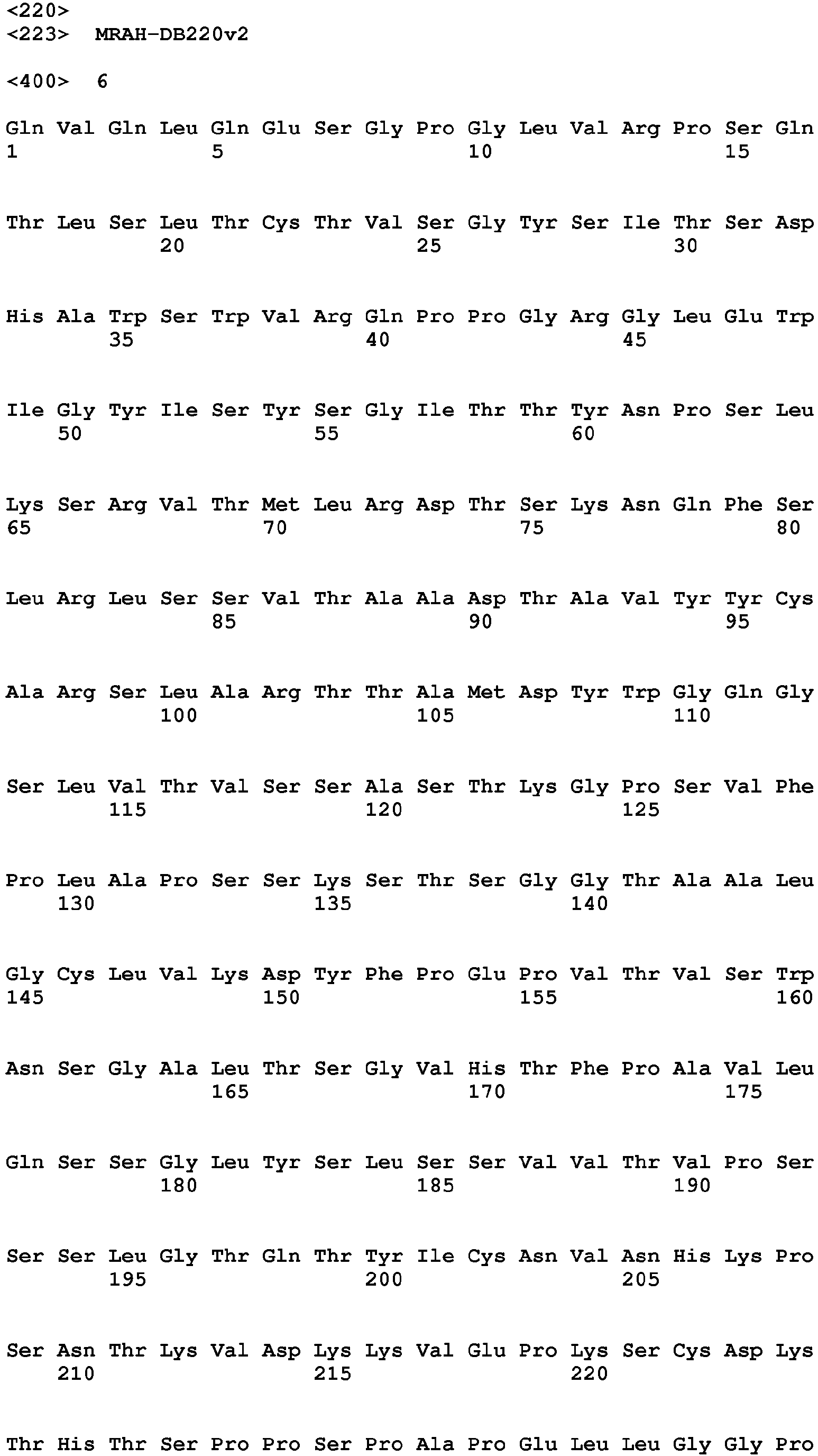


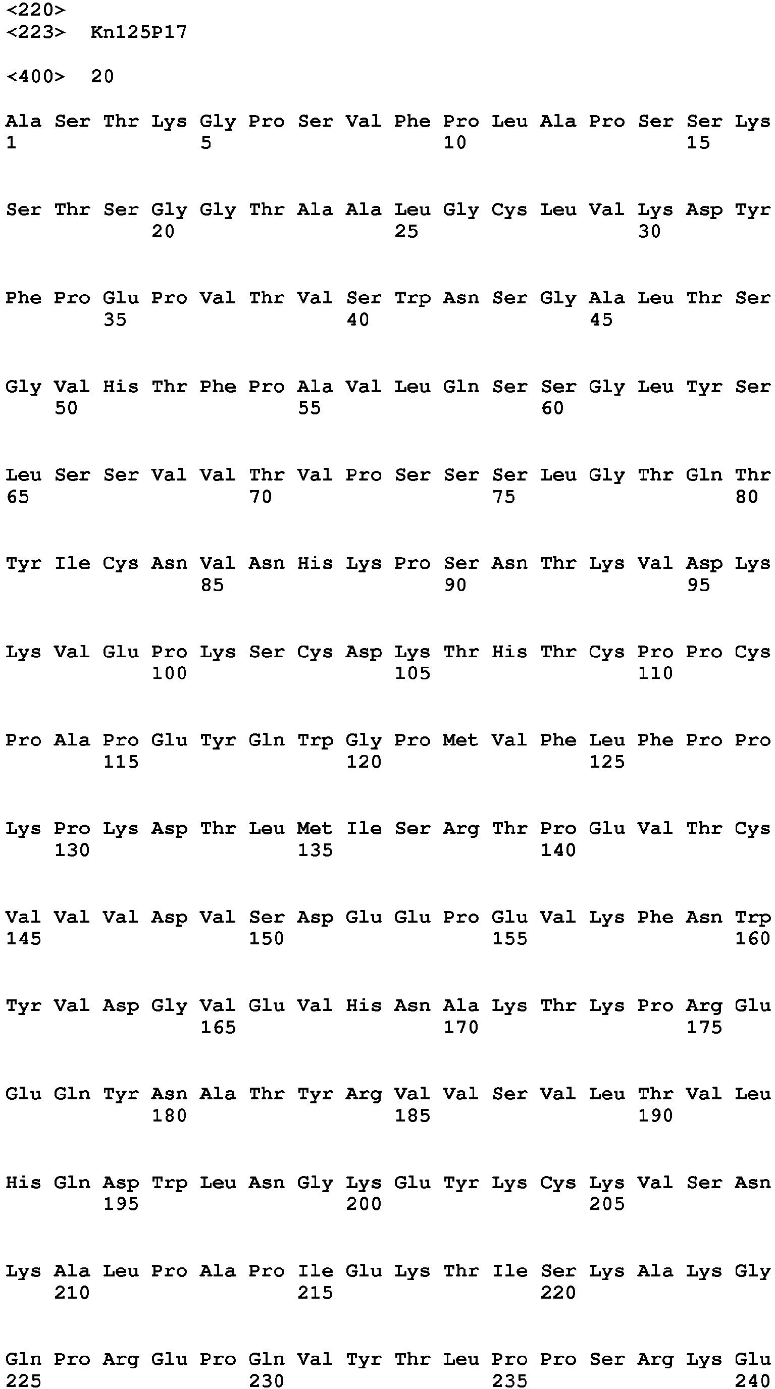


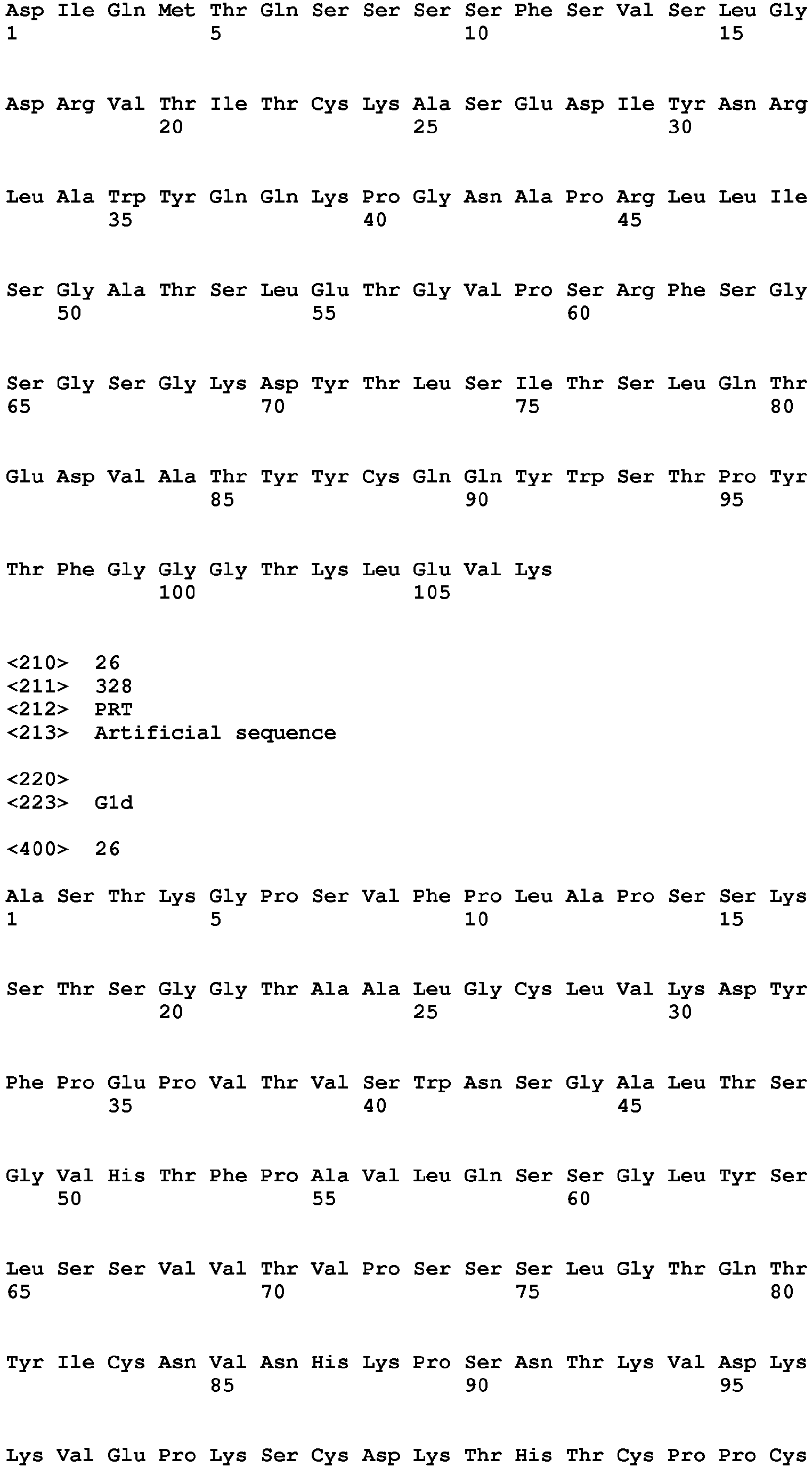


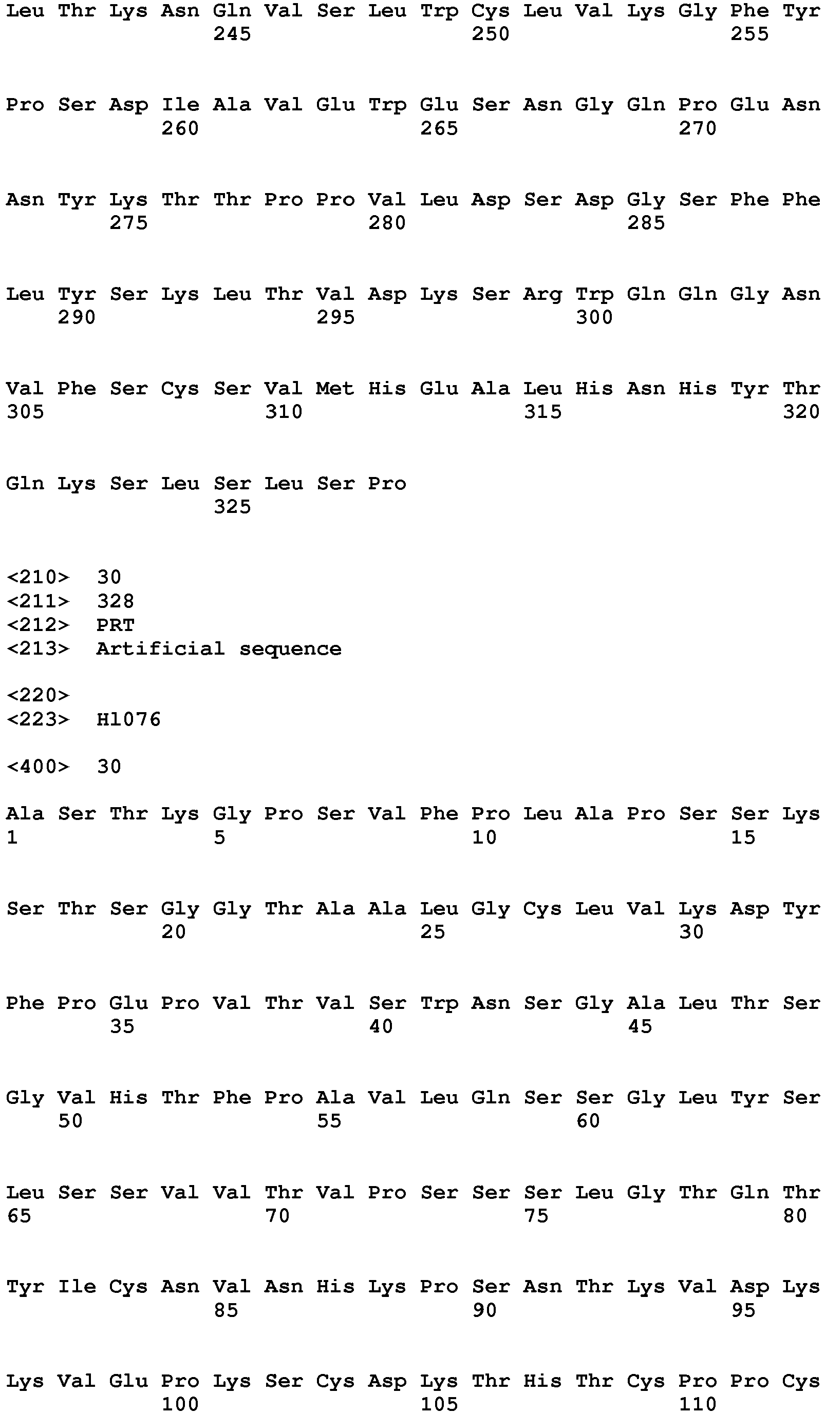


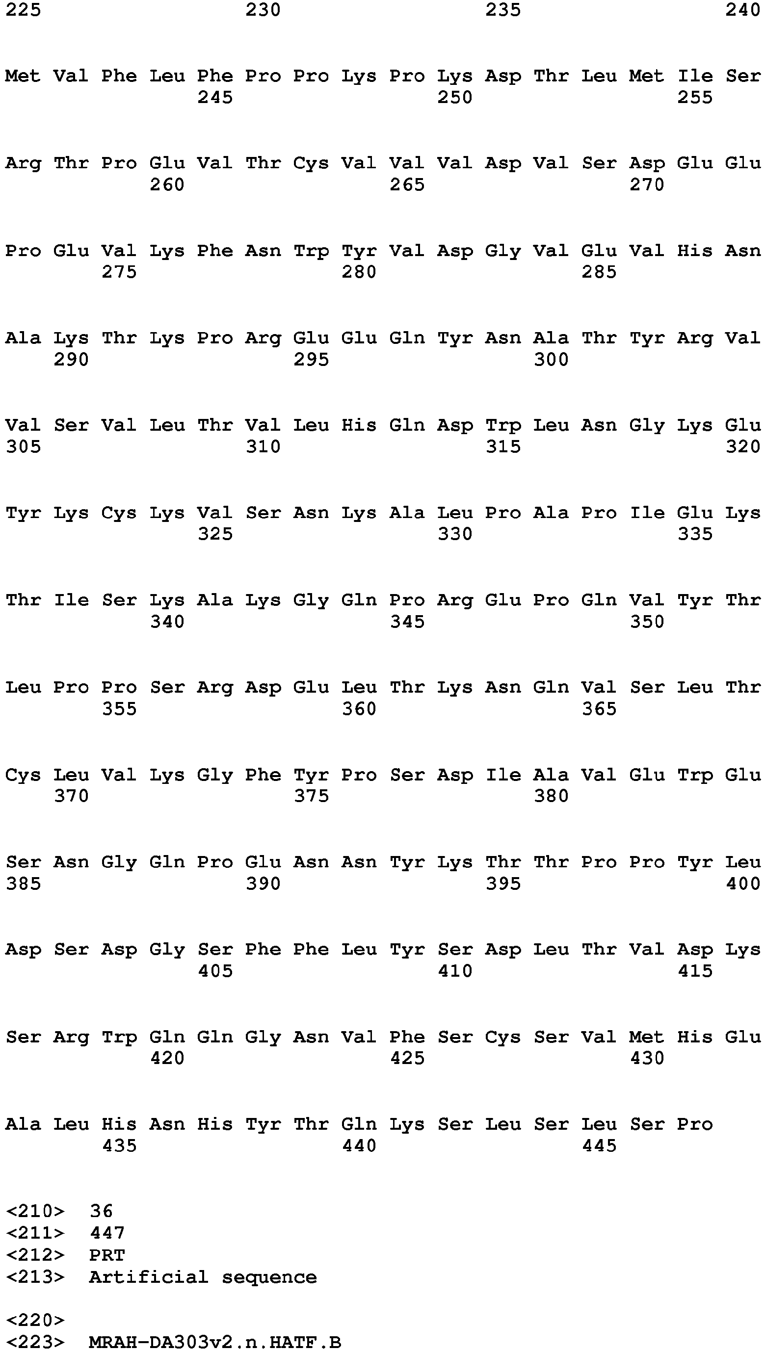


Claims (15)
- A pharmaceutical composition, comprising
a first antigen-binding molecule having a first antigen-binding region that binds to a first antigen and a first polypeptide comprising either or both of a first CH2 and a first CH3, and
a second antigen-binding molecule having a second antigen-binding region that binds to a second antigen and a second polypeptide comprising either or both of a second CH2 and a second CH3,
wherein said first antigen-binding molecule and said second antigen-binding molecule are not linked by a covalent bond and are more likely to form a heterodimer than a homodimer when mixed in solution. - The composition according to claim 1, wherein, when a sensor chip on which 50 pg per mm2 of the first antigen-binding molecule is immobilized and a measurement solution containing 2.5 mg/mL of the second antigen-binding molecule are used in surface plasmon resonance to measure affinity between the two antigen-binding molecules, the binding amount of the second antigen-binding molecule to the first antigen-binding molecule is within the range of 1:0.1 to 1:0.9 in terms of molar ratio.
- The composition according to claim 1 or 2, wherein the amount of the heterodimer formed under conditions in which cells expressing the first antigen and the second antigen are present is higher than that under conditions in which the cells are not present.
- The composition according to any one of claims 1 to 3, wherein the binding activity of the heterodimer to FcyR when said heterodimer is formed is higher than the binding activity of a monomer of the first antigen-binding molecule or a monomer of the second antigen-binding molecule to FcyR, or the binding activity of the homodimer to FcyR when said homodimer is formed.
- The composition according to any one of claims 1 to 4, wherein the first polypeptide comprises the first CH3 and the second polypeptide comprises the second CH3, and wherein the first CH3 and the second CH3 have at least one modification of (i) to (iii) below to make the first antigen-binding molecule and the second binding-molecule more likely to form the heterodimer than the homodimer when mixed in solution:(i) a modification where either of the first CH3 and the second CH3 has a positively-charged region and the other has a negatively-charged region, and when the heterodimer is formed, the positively-charged region interacts with the negatively-charged region,(ii) a modification where either of the first CH3 and the second CH3 has a convex portion and the other has a concave portion, and when the heterodimer is formed, the convex portion fits into and interacts with the concave portion, and(iii) a modification where the first CH3 and the second CH3 are modified IgG CH3, a part of which is replaced with a part of IgA CH3, and when said heterodimer is formed, the replaced part of IgA CH3 in said first CH3 interacts with the replaced part of IgA CH3 in said second CH3.
- The composition according to any one of claims 1 to 5, wherein either or both of said first CH3 and said second CH3 further have a substitution of at least one of the amino acid residues at positions 357, 397, and 409 in the EU numbering system with another amino acid residue.
- The composition according to any one of claims 1 to 6, wherein either or both of said first polypeptide and said second polypeptide further comprise a hinge region portion in an antibody-half molecule.
- The composition of claim 7, wherein said hinge region portion in either or both of said first polypeptide and said second polypeptide has a modification of the cysteine residues at either or both of positions 226 and 229 in the EU numbering system to another amino acid residue.
- The composition according to any one of claims 1 to 8, wherein said first polypeptide and said second polypeptide each comprises a constant region portion in an antibody-half molecule.
- The composition according to any one of claims 1 to 9, wherein the effector function under conditions in which cells expressing said first antigen and said second antigen are present is higher than that under conditions in which cells expressing said first antigen but not the second antigen or cells expressing said second antigen but not the first antigen are present.
- A pharmaceutical composition, comprising
a first antigen-binding molecule having a first antigen-binding region that binds to a first antigen and a first polypeptide comprising a first CH3, and
a second antigen-binding molecule having a second antigen-binding region that binds to a second antigen and a second polypeptide comprising a second CH3,
wherein said first antigen-binding molecule and said second antigen-binding molecule are not linked by a covalent bond, and
wherein said first CH3 and said second CH3 have at least one modification of (iv) to (vi) below:(iv) a modification where either of said first CH3 and said second CH3 has a positively-charged region and the other has a negatively-charged region, and when the heterodimer is formed, the positively-charged region interacts with the negatively-charged region,(v) a modification where either of said first CH3 and said second CH3 has a convex portion and the other has a concave portion, and when the heterodimer is formed, the convex portion fits into and interacts with the concave portion, and(vi) a modification where said first CH3 and said second CH3 are modified IgG CH3 a part of which is replaced with a part of IgA CH3, and when said heterodimer is formed, the replaced part of IgA CH3 in said first CH3 interacts with the replaced part of IgA CH3 in said second CH3. - A first antigen-binding molecule comprising a first antigen-binding region that binds to a first antigen and a first polypeptide comprising either or both of a first CH2 and a first CH3, wherein, when mixed in solution with a second antigen-binding molecule having a second antigen-binding region that binds to a second antigen and a second polypeptide comprising either or both of a second CH2 and a second CH3, the first antigen-binding molecule is more likely to form a heterodimer with the second antigen-binding molecule than a homodimer of the first antigen-binding molecule, and wherein in said heterodimer, said first antigen-binding molecule and said second antigen-binding molecule are not linked by a covalent bond.
- A second antigen-binding molecule comprising a second antigen-binding region that binds to a second antigen and a second polypeptide comprising either or both of a second CH2 and a second CH3,
wherein, when mixed in solution with a first antigen-binding molecule having a first antigen-binding region that binds to a first antigen and a first polypeptide comprising either or both of a first CH2 and a first CH3, the second antigen-binding molecule is more likely to form a heterodimer with the first antigen-binding molecule than a homodimer of the second antigen-binding molecule, and wherein in said heterodimer, said first antigen-binding molecule and said second antigen-binding molecule are not linked by a covalent bond. - A method for treating a disease caused by a pathogenic cell in a subject, wherein
a first antigen-binding molecule having a first antigen-binding region that binds to a first antigen and a first polypeptide comprising either or both of a first CH2 and a first CH3, and
a second antigen-binding molecule having a second antigen-binding region that binds to a second antigen and a second polypeptide comprising either or both of a second CH2 and a second CH3
are administered simultaneously or sequentially to a subject having a pathogenic cell that expresses the first antigen and the second antigen, and wherein said first antigen-binding molecule and said second antigen-binding molecule are not linked by a covalent bond before and after administration and form a heterodimer on the surface of said pathogenic cell to exert an effector function. - A method of selecting a combination of a first antigen-binding molecule and a second antigen-binding molecule from a group of variants of a first antigen-binding molecule having a first antigen-binding region that binds to a first antigen and a first polypeptide comprising either or both of a first CH2 and a first CH3, and a group of variants of a second antigen-binding molecule having a second antigen-binding region that binds to a second antigen and a second polypeptide comprising either or both of a second CH2 and a second CH3,
wherein the combination is one where,(a) the first antigen-binding molecule and the second antigen-binding molecule are not linked by a covalent bond,(b) the first antigen-binding molecule and the second antigen-binding molecule are more likely to form a heterodimer between the first antigen-binding molecule and the second antigen-binding molecule than a homodimer of the first antigen-binding molecule or a homodimer of the second antigen-binding molecule, and(c) when a sensor chip on which 50 pg per mm2 of the first antigen-binding molecule is immobilized and a measurement solution containing 2.5 mg/mL of the second antigen-binding molecule are used in surface plasmon resonance to measure affinity between the two antigen-binding molecules, the binding amount of the second antigen-binding molecule to the first antigen-binding molecule is within the range of 1:0.1 to 1:0.9 in terms of molar ratio.
Applications Claiming Priority (2)
| Application Number | Priority Date | Filing Date | Title |
|---|---|---|---|
| JP2017033564 | 2017-02-24 | ||
| PCT/JP2018/006626 WO2018155611A1 (en) | 2017-02-24 | 2018-02-23 | Pharmaceutical composition, antigen-binding molecules, treatment method, and screening method |
Publications (2)
| Publication Number | Publication Date |
|---|---|
| EP3586872A1 true EP3586872A1 (en) | 2020-01-01 |
| EP3586872A4 EP3586872A4 (en) | 2020-12-30 |
Family
ID=63253875
Family Applications (1)
| Application Number | Title | Priority Date | Filing Date |
|---|---|---|---|
| EP18756600.5A Pending EP3586872A4 (en) | 2017-02-24 | 2018-02-23 | PHARMACEUTICAL COMPOSITION, ANTIG-BINDING MOLECULES, TREATMENT METHODS AND SCREENING METHODS |
Country Status (5)
| Country | Link |
|---|---|
| US (2) | US12054544B2 (en) |
| EP (1) | EP3586872A4 (en) |
| JP (4) | JP7556687B2 (en) |
| TW (1) | TW201834688A (en) |
| WO (1) | WO2018155611A1 (en) |
Cited By (2)
| Publication number | Priority date | Publication date | Assignee | Title |
|---|---|---|---|---|
| US12371511B2 (en) | 2011-06-30 | 2025-07-29 | Chugai Seiyaku Kabushiki Kaisha | Heterodimerized polypeptide |
| US12415857B2 (en) | 2021-06-25 | 2025-09-16 | Chugai Seiyaku Kabushiki Kaisha | Anti-CTLA-4 antibody and use thereof |
Families Citing this family (7)
| Publication number | Priority date | Publication date | Assignee | Title |
|---|---|---|---|---|
| JP7556687B2 (en) | 2017-02-24 | 2024-09-26 | 中外製薬株式会社 | Pharmaceutical compositions, antigen-binding molecules, methods of treatment, and screening methods |
| WO2019077092A1 (en) | 2017-10-20 | 2019-04-25 | F. Hoffmann-La Roche Ag | Method for generating multispecific antibodies from monospecific antibodies |
| IL274071B2 (en) | 2017-10-30 | 2025-10-01 | Hoffmann La Roche | Method for in vivo generation of multispecific antibodies from monospecific antibodies |
| JP7523349B2 (en) * | 2018-08-29 | 2024-07-26 | 中外製薬株式会社 | Antibody half molecules and methods for inhibiting homodimer formation of antibody half molecules |
| TW202104260A (en) * | 2019-04-05 | 2021-02-01 | 美商西建公司 | Engineering of an antibody for tumor-selective binding of cd47 |
| TWI884247B (en) | 2020-04-01 | 2025-05-21 | 日商協和麒麟股份有限公司 | Antibody Combinations |
| IL308633B2 (en) * | 2021-06-25 | 2025-12-01 | Chugai Pharmaceutical Co Ltd | Use of anti-ctla-4 antibodies |
Family Cites Families (54)
| Publication number | Priority date | Publication date | Assignee | Title |
|---|---|---|---|---|
| US5731168A (en) | 1995-03-01 | 1998-03-24 | Genentech, Inc. | Method for making heteromultimeric polypeptides |
| US7355008B2 (en) | 2003-01-09 | 2008-04-08 | Macrogenics, Inc. | Identification and engineering of antibodies with variant Fc regions and methods of using same |
| WO2005062916A2 (en) * | 2003-12-22 | 2005-07-14 | Centocor, Inc. | Methods for generating multimeric molecules |
| KR101374454B1 (en) * | 2005-03-31 | 2014-03-17 | 추가이 세이야쿠 가부시키가이샤 | Methods for producing polypeptides by regulating polypeptide association |
| US8236308B2 (en) | 2005-10-11 | 2012-08-07 | Micromet Ag | Composition comprising cross-species-specific antibodies and uses thereof |
| KR101866623B1 (en) | 2005-11-28 | 2018-07-04 | 젠맵 에이/에스 | Recombinant monovalent antibodies and methods for production thereof |
| DK1999154T3 (en) | 2006-03-24 | 2012-12-03 | Merck Patent Gmbh | MANUFACTURED HETERODIMERED PROTEIN DOMAINS |
| EP2626372B1 (en) * | 2007-03-29 | 2018-03-21 | Genmab A/S | Bispecific antibodies and methods for production thereof |
| WO2008119565A2 (en) | 2007-04-03 | 2008-10-09 | Micromet Ag | Cross-species-specific binding domain |
| EP2190861A4 (en) | 2007-08-22 | 2011-03-30 | Univ California | ACTIVATED BINDING POLYPEPTIDES AND METHODS OF IDENTIFICATION AND USE THEREOF |
| RU2505603C2 (en) | 2007-09-26 | 2014-01-27 | Чугаи Сейяку Кабусики Кайся | Antibody against il-6 receptor |
| KR20150126724A (en) | 2007-09-28 | 2015-11-12 | 추가이 세이야쿠 가부시키가이샤 | Anti-glypican-3 antibody having improved kinetics in plasma |
| EP3663318A1 (en) * | 2008-01-07 | 2020-06-10 | Amgen Inc. | Method for making antibody fc-heterodimeric molecules using electrostatic steering effects |
| TW201920257A (en) | 2008-04-11 | 2019-06-01 | 日商中外製藥股份有限公司 | Antigen-binding molecule capable of binding two or more antigen molecules repeatedly |
| US8093018B2 (en) | 2008-05-20 | 2012-01-10 | Otsuka Pharmaceutical Co., Ltd. | Antibody identifying an antigen-bound antibody and an antigen-unbound antibody, and method for preparing the same |
| US9067986B2 (en) * | 2009-04-27 | 2015-06-30 | Oncomed Pharmaceuticals, Inc. | Method for making heteromultimeric molecules |
| EP2504360B1 (en) | 2009-11-23 | 2018-08-15 | Amgen Inc. | Monomeric antibody fc |
| WO2011143545A1 (en) | 2010-05-14 | 2011-11-17 | Rinat Neuroscience Corporation | Heterodimeric proteins and methods for producing and purifying them |
| JP2013537416A (en) | 2010-08-13 | 2013-10-03 | メディミューン リミテッド | Monomer polypeptide containing mutant Fc region and method of use |
| CN103429620B (en) | 2010-11-05 | 2018-03-06 | 酵活有限公司 | Antibody design for stable heterodimerization with mutations in the Fc domain |
| US20120201746A1 (en) * | 2010-12-22 | 2012-08-09 | Abbott Laboratories | Half immunoglobulin binding proteins and uses thereof |
| US10689447B2 (en) * | 2011-02-04 | 2020-06-23 | Genentech, Inc. | Fc variants and methods for their production |
| WO2012125850A1 (en) | 2011-03-16 | 2012-09-20 | Amgen Inc. | Fc variants |
| WO2013002362A1 (en) * | 2011-06-30 | 2013-01-03 | 中外製薬株式会社 | Heterodimerized polypeptide |
| JP6322411B2 (en) | 2011-09-30 | 2018-05-09 | 中外製薬株式会社 | Antigen-binding molecules that promote the disappearance of antigens with multiple physiological activities |
| DK2766392T3 (en) | 2011-10-10 | 2019-10-07 | Xencor Inc | PROCEDURE FOR CLEANING ANTIBODIES |
| SI2766397T1 (en) * | 2011-10-11 | 2018-09-28 | F. Hoffmann-La Roche Ag | Improved assembly of bispecific antibodies |
| TWI593705B (en) | 2011-12-28 | 2017-08-01 | Chugai Pharmaceutical Co Ltd | Humanized anti-epiregulin antibody and cancer therapeutic agent containing the antibody as an active ingredient |
| MX359411B (en) | 2012-01-13 | 2018-09-27 | Univ Wuerzburg J Maximilians | BIPARTITA FUNCTIONAL COMPLEMENTATION Induced by ANTIGENS, DUAL. |
| WO2013157953A1 (en) * | 2012-04-20 | 2013-10-24 | Merus B.V. | Methods and means for the production of ig-like molecules |
| WO2013180200A1 (en) | 2012-05-30 | 2013-12-05 | 中外製薬株式会社 | Target-tissue-specific antigen-binding molecule |
| JP6774164B2 (en) | 2012-08-24 | 2020-10-21 | 中外製薬株式会社 | Mouse FcγRII specific Fc antibody |
| UY35148A (en) * | 2012-11-21 | 2014-05-30 | Amgen Inc | HETERODIMERIC IMMUNOGLOBULINS |
| EA032681B1 (en) * | 2012-11-27 | 2019-07-31 | Аджоу Юниверсити Индастри-Академик Кооперейшн Фаундейшн | HETERODIMER Fc OF IMMUNOGLOBULIN COMPRISING A CH3 DOMAIN VARIANT FOR FORMING AN ANTIBODY OR A FUSION PROTEIN WITH A HETERODIMERIC Fc AT HIGH EFFICIENCY, METHOD FOR PREPARING AND USING SAME |
| ES2664095T3 (en) | 2012-12-07 | 2018-04-18 | Pfizer Inc. | Monomeric antibody fragments designed by genetic engineering |
| US20160159919A1 (en) | 2013-06-24 | 2016-06-09 | Chugai Seiyaku Kabushiki Kaisha | Therapeutic Agent Comprising Humanized Anti-Epiregulin Antibody as Active Ingredient for Non-Small-Cell Lung Carcinoma Excluding Adenocarcinoma |
| BR112016000666A2 (en) * | 2013-07-12 | 2017-10-03 | Zymeworks Inc | BISPECIFIC CD3 AND CD19 ANTIGEN BINDING CONSTRUCTS |
| CA2925256C (en) * | 2013-09-27 | 2023-08-15 | Chugai Seiyaku Kabushiki Kaisha | Method for producing polypeptide heteromultimer |
| CA2929044A1 (en) | 2013-11-11 | 2015-05-14 | Chugai Seiyaku Kabushiki Kaisha | Antigen-binding molecule containing modified antibody variable region |
| TWI702316B (en) | 2013-12-04 | 2020-08-21 | 日商中外製藥股份有限公司 | Antigen-binding molecules and their database that change antigen-binding ability in response to compound concentration |
| KR20170017916A (en) | 2014-05-13 | 2017-02-15 | 추가이 세이야쿠 가부시키가이샤 | T cell-redirected antigen-binding molecule for cells having immunosuppression function |
| EP2982693A1 (en) | 2014-08-07 | 2016-02-10 | Affimed Therapeutics AG | CD3 binding domain |
| FI3223848T3 (en) | 2014-11-27 | 2025-02-28 | Zymeworks Bc Inc | METHODS USING BISPECIFIC ANTIGEN-BINDING STRUCTURES THAT TARGET HER2 |
| JP6724023B2 (en) | 2015-02-09 | 2020-07-15 | リサーチ ディベロップメント ファウンデーション | Engineered immunoglobulin FC polypeptides showing improved complement activation |
| JP7082484B2 (en) * | 2015-04-01 | 2022-06-08 | 中外製薬株式会社 | Method for Producing Polypeptide Heterogeneous Multimer |
| JP6841754B2 (en) | 2015-05-13 | 2021-03-10 | 中外製薬株式会社 | Method for identifying multiple antigen-binding molecular fusions, pharmaceutical compositions, linear epitopes, and methods for producing multiple antigen-binding molecular fusions |
| FR3038517B1 (en) | 2015-07-06 | 2020-02-28 | Laboratoire Francais Du Fractionnement Et Des Biotechnologies | USE OF MODIFIED FC FRAGMENTS IN IMMUNOTHERAPY |
| TWI812873B (en) | 2015-11-30 | 2023-08-21 | 美商輝瑞股份有限公司 | Antibodies and antibody fragments for site-specific conjugation |
| EP3431102A4 (en) | 2016-03-14 | 2019-09-25 | Chugai Seiyaku Kabushiki Kaisha | THERAPEUTIC MEDICINE INDUCING CELLULAR INJURY FOR USE IN THE TREATMENT OF CANCER |
| CA3025162A1 (en) * | 2016-05-26 | 2017-11-30 | Qilu Puget Sound Biotherapeutics Corporation | Mixtures of antibodies |
| JP7556687B2 (en) | 2017-02-24 | 2024-09-26 | 中外製薬株式会社 | Pharmaceutical compositions, antigen-binding molecules, methods of treatment, and screening methods |
| WO2019160007A1 (en) | 2018-02-14 | 2019-08-22 | 中外製薬株式会社 | Antigen-binding molecule and combination |
| US12435137B2 (en) | 2018-08-03 | 2025-10-07 | Chugai Seiyaku Kabushiki Kaisha | Antigen-binding molecule containing two antigen-binding domains that are linked to each other |
| JP7523349B2 (en) | 2018-08-29 | 2024-07-26 | 中外製薬株式会社 | Antibody half molecules and methods for inhibiting homodimer formation of antibody half molecules |
-
2018
- 2018-02-23 JP JP2019501825A patent/JP7556687B2/en active Active
- 2018-02-23 EP EP18756600.5A patent/EP3586872A4/en active Pending
- 2018-02-23 US US16/488,115 patent/US12054544B2/en active Active
- 2018-02-23 WO PCT/JP2018/006626 patent/WO2018155611A1/en not_active Ceased
- 2018-02-23 TW TW107106137A patent/TW201834688A/en unknown
-
2022
- 2022-06-30 JP JP2022105369A patent/JP2022130617A/en active Pending
-
2024
- 2024-02-29 JP JP2024029274A patent/JP7798938B2/en active Active
- 2024-06-27 US US18/756,956 patent/US20240417464A1/en active Pending
-
2025
- 2025-12-25 JP JP2025281528A patent/JP2026040660A/en active Pending
Cited By (3)
| Publication number | Priority date | Publication date | Assignee | Title |
|---|---|---|---|---|
| US12371511B2 (en) | 2011-06-30 | 2025-07-29 | Chugai Seiyaku Kabushiki Kaisha | Heterodimerized polypeptide |
| US12415857B2 (en) | 2021-06-25 | 2025-09-16 | Chugai Seiyaku Kabushiki Kaisha | Anti-CTLA-4 antibody and use thereof |
| US12448451B2 (en) | 2021-06-25 | 2025-10-21 | Chugai Seiyaku Kabushiki Kaisha | Anti-CTLA-4 antibody and use thereof |
Also Published As
| Publication number | Publication date |
|---|---|
| EP3586872A4 (en) | 2020-12-30 |
| WO2018155611A1 (en) | 2018-08-30 |
| JP7556687B2 (en) | 2024-09-26 |
| US12054544B2 (en) | 2024-08-06 |
| JP2024052890A (en) | 2024-04-12 |
| JP2026040660A (en) | 2026-03-09 |
| US20240417464A1 (en) | 2024-12-19 |
| JP7798938B2 (en) | 2026-01-14 |
| TW201834688A (en) | 2018-10-01 |
| JP2022130617A (en) | 2022-09-06 |
| JPWO2018155611A1 (en) | 2019-12-12 |
| US20190382484A1 (en) | 2019-12-19 |
Similar Documents
| Publication | Publication Date | Title |
|---|---|---|
| US20240417464A1 (en) | Compositions comprising antigen-binding molecules | |
| US20250057919A1 (en) | Il-15/il-15ra heterodimeric fc fusion proteins and uses thereof | |
| US12448423B2 (en) | Targeted heterodimeric Fc fusion proteins containing NKG2D antigen binding domains | |
| US11524991B2 (en) | PD-1 targeted heterodimeric fusion proteins containing IL-15/IL-15Ra Fc-fusion proteins and PD-1 antigen binding domains and uses thereof | |
| EP2543730B1 (en) | Antibody constant region variant | |
| JP6956083B2 (en) | Heterodimer antibody that binds CD3 and CD38 | |
| US20200140512A1 (en) | Pd-1 targeted il-15/il-15ralpha fc fusion proteins and uses in combination therapies thereof | |
| TWI807362B (en) | cell damage-inducing therapeutics | |
| KR20230052293A (en) | Anti-CD28 composition | |
| TW202325741A (en) | Dll3-targeting multispecific antigen-binding molecules and uses thereof | |
| US20230331809A1 (en) | Fusion proteins comprising a ligand-receptor pair and a biologically functional protein | |
| JP2021520829A (en) | TIM-3 targeted heterodimer fusion protein containing IL-15 / IL-15RA Fc fusion protein and TIM-3 antigen binding domain | |
| JP2021521779A (en) | A heterodimer fusion protein targeting LAG-3, including the IL-15 / IL-15RA Fc fusion protein and the LAG-3 antigen binding domain. | |
| CN110214148A (en) | Bispecific heterodimer fusion protein containing IL-15/IL-15R α Fc fusion protein and PD-1 antibody fragment | |
| JP2019527047A (en) | Heterodimeric antibody binding to somatostatin receptor 2 | |
| KR20230162013A (en) | Multispecific protein containing NKP46-binding site, cancer antigen binding site fused to cytokines for NK cell engagement | |
| JP2024133723A (en) | Antibody half molecules and methods for inhibiting homodimer formation of antibody half molecules | |
| US20260103514A1 (en) | Antibodies that specifically bind cldn6 or cd3 and bispecific antibodies that specifically bind cldn6 and cd3 | |
| WO2026078029A1 (en) | Splitted interleukin-2 prodrugs and uses thereof | |
| CN116547306A (en) | anti-CD 28 and/or anti-B7H 3 compositions | |
| CN117377689A (en) | Heterodimeric antibodies that bind CD3 and GPC3 |
Legal Events
| Date | Code | Title | Description |
|---|---|---|---|
| STAA | Information on the status of an ep patent application or granted ep patent |
Free format text: STATUS: THE INTERNATIONAL PUBLICATION HAS BEEN MADE |
|
| PUAI | Public reference made under article 153(3) epc to a published international application that has entered the european phase |
Free format text: ORIGINAL CODE: 0009012 |
|
| STAA | Information on the status of an ep patent application or granted ep patent |
Free format text: STATUS: REQUEST FOR EXAMINATION WAS MADE |
|
| 17P | Request for examination filed |
Effective date: 20190924 |
|
| AK | Designated contracting states |
Kind code of ref document: A1 Designated state(s): AL AT BE BG CH CY CZ DE DK EE ES FI FR GB GR HR HU IE IS IT LI LT LU LV MC MK MT NL NO PL PT RO RS SE SI SK SM TR |
|
| AX | Request for extension of the european patent |
Extension state: BA ME |
|
| DAV | Request for validation of the european patent (deleted) | ||
| DAX | Request for extension of the european patent (deleted) | ||
| A4 | Supplementary search report drawn up and despatched |
Effective date: 20201126 |
|
| RIC1 | Information provided on ipc code assigned before grant |
Ipc: A61K 39/395 20060101AFI20201120BHEP Ipc: A61P 35/00 20060101ALI20201120BHEP Ipc: A61P 37/02 20060101ALI20201120BHEP Ipc: A61P 35/02 20060101ALI20201120BHEP Ipc: C07K 16/28 20060101ALI20201120BHEP |
|
| STAA | Information on the status of an ep patent application or granted ep patent |
Free format text: STATUS: EXAMINATION IS IN PROGRESS |
|
| 17Q | First examination report despatched |
Effective date: 20230519 |