EP3575813B1 - Quantitative mapping of a magnetic resonance imaging parameter by data-driven signal-model learning - Google Patents
Quantitative mapping of a magnetic resonance imaging parameter by data-driven signal-model learning Download PDFInfo
- Publication number
- EP3575813B1 EP3575813B1 EP18175213.0A EP18175213A EP3575813B1 EP 3575813 B1 EP3575813 B1 EP 3575813B1 EP 18175213 A EP18175213 A EP 18175213A EP 3575813 B1 EP3575813 B1 EP 3575813B1
- Authority
- EP
- European Patent Office
- Prior art keywords
- mri
- data
- parameter
- determining
- value
- Prior art date
- Legal status (The legal status is an assumption and is not a legal conclusion. Google has not performed a legal analysis and makes no representation as to the accuracy of the status listed.)
- Active
Links
Images

Classifications
-
- G—PHYSICS
- G16—INFORMATION AND COMMUNICATION TECHNOLOGY [ICT] SPECIALLY ADAPTED FOR SPECIFIC APPLICATION FIELDS
- G16H—HEALTHCARE INFORMATICS, i.e. INFORMATION AND COMMUNICATION TECHNOLOGY [ICT] SPECIALLY ADAPTED FOR THE HANDLING OR PROCESSING OF MEDICAL OR HEALTHCARE DATA
- G16H50/00—ICT specially adapted for medical diagnosis, medical simulation or medical data mining; ICT specially adapted for detecting, monitoring or modelling epidemics or pandemics
- G16H50/20—ICT specially adapted for medical diagnosis, medical simulation or medical data mining; ICT specially adapted for detecting, monitoring or modelling epidemics or pandemics for computer-aided diagnosis, e.g. based on medical expert systems
-
- G—PHYSICS
- G01—MEASURING; TESTING
- G01R—MEASURING ELECTRIC VARIABLES; MEASURING MAGNETIC VARIABLES
- G01R33/00—Arrangements or instruments for measuring magnetic variables
- G01R33/20—Arrangements or instruments for measuring magnetic variables involving magnetic resonance
- G01R33/44—Arrangements or instruments for measuring magnetic variables involving magnetic resonance using nuclear magnetic resonance [NMR]
- G01R33/48—NMR imaging systems
- G01R33/50—NMR imaging systems based on the determination of relaxation times, e.g. T1 measurement by IR sequences; T2 measurement by multiple-echo sequences
-
- G—PHYSICS
- G01—MEASURING; TESTING
- G01R—MEASURING ELECTRIC VARIABLES; MEASURING MAGNETIC VARIABLES
- G01R33/00—Arrangements or instruments for measuring magnetic variables
- G01R33/20—Arrangements or instruments for measuring magnetic variables involving magnetic resonance
- G01R33/44—Arrangements or instruments for measuring magnetic variables involving magnetic resonance using nuclear magnetic resonance [NMR]
- G01R33/48—NMR imaging systems
- G01R33/54—Signal processing systems, e.g. using pulse sequences ; Generation or control of pulse sequences; Operator console
- G01R33/56—Image enhancement or correction, e.g. subtraction or averaging techniques, e.g. improvement of signal-to-noise ratio and resolution
- G01R33/5608—Data processing and visualization specially adapted for MR, e.g. for feature analysis and pattern recognition on the basis of measured MR data, segmentation of measured MR data, edge contour detection on the basis of measured MR data, for enhancing measured MR data in terms of signal-to-noise ratio by means of noise filtering or apodization, for enhancing measured MR data in terms of resolution by means for deblurring, windowing, zero filling, or generation of gray-scaled images, colour-coded images or images displaying vectors instead of pixels
-
- G—PHYSICS
- G06—COMPUTING OR CALCULATING; COUNTING
- G06F—ELECTRIC DIGITAL DATA PROCESSING
- G06F16/00—Information retrieval; Database structures therefor; File system structures therefor
- G06F16/20—Information retrieval; Database structures therefor; File system structures therefor of structured data, e.g. relational data
- G06F16/28—Databases characterised by their database models, e.g. relational or object models
- G06F16/284—Relational databases
- G06F16/288—Entity relationship models
-
- G—PHYSICS
- G06—COMPUTING OR CALCULATING; COUNTING
- G06N—COMPUTING ARRANGEMENTS BASED ON SPECIFIC COMPUTATIONAL MODELS
- G06N20/00—Machine learning
-
- G—PHYSICS
- G06—COMPUTING OR CALCULATING; COUNTING
- G06N—COMPUTING ARRANGEMENTS BASED ON SPECIFIC COMPUTATIONAL MODELS
- G06N3/00—Computing arrangements based on biological models
- G06N3/02—Neural networks
- G06N3/04—Architecture, e.g. interconnection topology
- G06N3/0499—Feedforward networks
-
- G—PHYSICS
- G06—COMPUTING OR CALCULATING; COUNTING
- G06N—COMPUTING ARRANGEMENTS BASED ON SPECIFIC COMPUTATIONAL MODELS
- G06N3/00—Computing arrangements based on biological models
- G06N3/02—Neural networks
- G06N3/08—Learning methods
- G06N3/09—Supervised learning
-
- G—PHYSICS
- G06—COMPUTING OR CALCULATING; COUNTING
- G06F—ELECTRIC DIGITAL DATA PROCESSING
- G06F16/00—Information retrieval; Database structures therefor; File system structures therefor
- G06F16/20—Information retrieval; Database structures therefor; File system structures therefor of structured data, e.g. relational data
- G06F16/22—Indexing; Data structures therefor; Storage structures
-
- G—PHYSICS
- G06—COMPUTING OR CALCULATING; COUNTING
- G06N—COMPUTING ARRANGEMENTS BASED ON SPECIFIC COMPUTATIONAL MODELS
- G06N3/00—Computing arrangements based on biological models
- G06N3/02—Neural networks
- G06N3/08—Learning methods
Definitions
- the present disclosure is directed, in general, to imaging techniques for imaging biological tissues, and more specifically to quantitative imaging in Magnetic Resonance Imaging (MRI).
- MRI Magnetic Resonance Imaging
- the contrast apparent in images acquired through classical MRI is the result of a combination of different physical parameters of underlying tissue(s), the particular MRI acquisition technique and its parameters.
- An approach for acquiring MR-based information on biological tissue is to directly measure one or more of its underlying physical properties, e.g. the tissue-specific T1 and T2 relaxation constants or the proton density PD.
- Those quantitative techniques are usually referred to as "parametric mapping” or “quantitative imaging” methods.
- the resulting image contrasts become more independent from the employed hardware, the applied imaging technique and the particular imaging parameters, because they directly probe the properties of the tissue.
- This facilitates comparability and thus clinical diagnosis and may enable building up a database of normal parametric values to which a newly scanned patient dataset can be compared. In other words, it provides the means to move from relative contrast information depending on many different factors towards an absolute measure of one or more separate physical properties.
- images encoding the quantitative parameters of interest are typically sampled multiple times, e.g. at different echo times using a multi-echo spin-echo (MESE) sequence to estimate the transverse relaxation time T2.
- MESE multi-echo spin-echo
- a signal model describing the relation between image intensities and relevant tissue properties is fitted onto these series of images (in the case of the MESE sequence that could e.g. be a simple mono-exponential T2-signal decay model), yielding a quantitative map.
- the chosen signal model is an approximation of reality by nature and may omit important components that drive the signal behavior beyond the quantitative parameter of interest due to the complex nature of the tissue microstructure (e.g. magnetization transfer effects) or system imperfections (e.g.
- a more detailed signal model uses a more complex analytical model known as "generating function" and incorporates one of the sources of error - the stimulated echoes - into the model (see for instance Lukzen et al., J. Magn. Reson. 2009, 196(2): 164-169 ), or Sumpf et al., IEEE Trans. Med. Imaging 2014, 33(12): 2213-2222 ).
- the generating function describes the relation between image intensities and relevant tissue properties better, it is more difficult to fit to the image series since the model has more independent variables, i.e. more mathematical degrees of freedom.
- additional regularization terms are often required to allow fitting of this ill-posed problem, which is usually tuned by yet more variables/regularization weights.
- An objective of the present invention is to propose an efficient and simple method and system for the quantitative mapping of biological tissue.
- the present invention proposes to use a machine learning method and system for determining parameters characterizing biological tissues as disclosed in the objects of the independent claims 1 and 9.
- the present invention proposes notably to learn a relation between
- the present invention concerns also an MRI system, as defined in independent claim 9, said system being configured for implementing the previously described machine learning method, said system comprising at least:
- Various disclosed embodiments include machine learning methods and corresponding systems and computer readable mediums for determining, for instance automatically determining, a value for a parameter from the relationship between a set of data acquired according to a second technique and a reference value acquired according to a first technique, wherein said relationship is obtained by training a machine learning algorithm on a learning dataset formed by said set of data and its corresponding reference value.
- FIGURES 1 and 2 discussed below, and the various embodiments used to describe the principles of the present disclosure in this patent document are by way of illustration only and should not be construed in any way to limit the scope of the disclosure. Those skilled in the art will understand that the principles of the present disclosure may be implemented in any suitably arranged device. The numerous innovative teachings of the present application will be described with reference to exemplary non-limiting embodiments.
- Figure 1 discloses in more details a machine learning method 100 for preferentially automatically determining a value (in particular a quantitative value) for a parameter, wherein determined values might be represented in a quantitative map, which enables characterizing a biological tissue of a patient.
- a value in particular a quantitative value
- FIG. 1 discloses in more details a machine learning method 100 for preferentially automatically determining a value (in particular a quantitative value) for a parameter, wherein determined values might be represented in a quantitative map, which enables characterizing a biological tissue of a patient.
- the concept of the invention described in Figure 1 is then applied to a specific, non-limiting, case of MRI, wherein an example of quantification of T2 using a learnt signal model for a MESE sequence 210 with a single-echo spin-echo (SE) sequence 220 as gold-standard reference is taken.
- the first technique according to the invention uses a SE sequence 220 for quantifying T2 according to classical process and the second technique uses a MESE sequence 210 for quantifying T2 using a machine learning technique wherein the quantified T2 values obtained from the first technique are used as target output for the machine learning technique.
- reference values for a parameter are determined by the system according to the invention from a group of objects, for instance a group of subjects.
- Said parameter might be a physical parameter or a biological parameter.
- Said reference values also called gold-standard reference values, are values, preferentially quantitative values, of said parameter that are determined by the system according to a first technique.
- Said first technique is typically a time consuming technique that cannot be applied as such during clinical diagnosis. According to the present invention, said first technique is only used to create a learning dataset.
- the first technique is typically a known technique wherein the first set of data corresponds to a first signal intensity (or signal data) acquired by the system according to the invention for each object, the system being further configured for determining said value of the parameter for each object by fitting the signal intensity with a signal model. Each determined value for the parameter is then used as a reference value within the machine learning technique.
- the first signal intensity might be a SE signal intensity
- step 101 may comprise acquiring SE signal intensity (or data) for each object of the group of objects and determining quantitative gold-standard T2 values (T2 being thus the parameter whose values have to be determined) for each object from a single-slice, resolution- and orientation-matched single-echo spin-echo sequence.
- the gold-standard T2 values 222 are preferentially computed using a classical signal fitting 221 onto the spin-echo data.
- the system creates a learning dataset by associating for each object a second set of signal intensities (which corresponds in that case to said second set of data) and the T2 reference value determined from the first signal intensity using the first technique.
- a given gold-standard T2 reference value might be associated with a measured MESE signal evolution (i.e. signal decay) in a database.
- normalized signal decay 214 and gold-standard T2 value in the brain 224 preferentially obtained after application 223 of a brain mask 230 determined for each object, are stored in a database in order to create said learning dataset.
- the method according to the invention may comprise acquiring MESE signal intensities, for instance MESE images, from said group of objects.
- intracranial volume might be first segmented 211 to restrict the learning solely to brain tissue, wherein the object corresponding brain mask 230 is applied to the MESE data for this purpose.
- All MESE signal decays 212 are preferably normalized 213 by dividing the signal intensities by the L2-norm of the decay.
- the normalization of the signal decays, or more generally of data used as input within the machine learning algorithm, could be performed differently (different norm or different scale) depending on the used technique.
- the optional application of the brain mask 230 as previously described advantageously improves the final result.
- the system uses a machine learning technique for determining the value of the parameter from the second set of signal intensities, wherein said machine learning technique comprises using a machine learning algorithm for determining said value, and training said machine learning algorithm on the learning dataset wherein for each object the second signal intensity is used as input and the associated reference value as output target.
- the machine learning algorithm might be an artificial neuronal network 240 that is trained using the signal intensities of each echo as input and the gold-standard T2 value as target output.
- said artificial neuronal network 240 may comprise two layers with each 8 neurons and a root-mean-squared error to the gold-standard T2 as cost function.
- the training is preferentially performed with data from all available objects, i.e. with whole data of the learning dataset.
- the system determines a relationship between the signal intensity and the value of the parameter obtained for each object from the training of the machine learning algorithm on the learning dataset.
- the machine learning algorithm is typically used to learn the relationship between the decay and the T2 value.
- the system uses said relationship to determine the value of the parameter from a measured set of signal intensities for a new object. For instance and as illustrated in Fig. 2 , in order to reconstruct a new dataset (e.g. coming from the new object), every decay is fed to the previously trained artificial neuronal network to estimate T2 in each voxel of the new dataset, resulting in the desired quantitative value for the parameter based on the data driven signal model.
- the present invention proposes therefore to replace a fixed signal model with a learnt relationship between "true" values (i.e. based on the gold standard reference values) and the respective quantitative measurements can help eliminating many of the difficulties related to finding an appropriate model for a given problem.
- the data-driven approach proposed here is able to learn potential dirt effects in the acquired signal intensity, which are typically hard to model or even, cannot be modelled at all according to prior art technique.
- Another advantage is that no a priori knowledge has to be imposed regarding the assumed interaction of the tissue microstructure and the underlying MR physics.
- the application of a trained neural network is typically very fast, i.e. once the machine learning algorithm, like the artificial neuronal network 240 of Figure 2 , is trained, parametric maps can be obtained very quickly in contrast to typically slow fitting procedures.
- the present invention is not restricted to the specific embodiment of Figure 2 , but might be without effort generalizable to other quantification or determination of parameters since only a set of datasets acquired through the so-called second technique and gold-standard reference data acquired through the so-called first technique are required.
- the first technique and the second technique according to the reference might be the same, or are two different techniques.
- a different type of input data than a MESE dataset using a different sequence could be used as input for the machine learning algorithm.
- the invention could also be used for magnetic resonance fingerprinting (see Ma et al., Nature 2013, 495(7440): 187-192 ), where the fingerprint is the input and the multiparametric values are outputs of the artificial neuronal network.
- the machine learning technique described in the present invention and sometimes also referred to as artificial intelligence or data science tool may also use different approaches like a linear regression, a non-linear regression or convolutional neuronal networks.
Landscapes
- Engineering & Computer Science (AREA)
- Physics & Mathematics (AREA)
- Theoretical Computer Science (AREA)
- Health & Medical Sciences (AREA)
- General Physics & Mathematics (AREA)
- Data Mining & Analysis (AREA)
- Databases & Information Systems (AREA)
- Software Systems (AREA)
- Biomedical Technology (AREA)
- General Engineering & Computer Science (AREA)
- Artificial Intelligence (AREA)
- General Health & Medical Sciences (AREA)
- Computing Systems (AREA)
- Evolutionary Computation (AREA)
- Medical Informatics (AREA)
- Mathematical Physics (AREA)
- Computational Linguistics (AREA)
- Biophysics (AREA)
- Public Health (AREA)
- Molecular Biology (AREA)
- Life Sciences & Earth Sciences (AREA)
- Computer Vision & Pattern Recognition (AREA)
- High Energy & Nuclear Physics (AREA)
- Condensed Matter Physics & Semiconductors (AREA)
- Pathology (AREA)
- Epidemiology (AREA)
- Primary Health Care (AREA)
- Signal Processing (AREA)
- Radiology & Medical Imaging (AREA)
- Nuclear Medicine, Radiotherapy & Molecular Imaging (AREA)
- Magnetic Resonance Imaging Apparatus (AREA)
Description
- The present disclosure is directed, in general, to imaging techniques for imaging biological tissues, and more specifically to quantitative imaging in Magnetic Resonance Imaging (MRI).
- The contrast apparent in images acquired through classical MRI is the result of a combination of different physical parameters of underlying tissue(s), the particular MRI acquisition technique and its parameters.
- An approach for acquiring MR-based information on biological tissue is to directly measure one or more of its underlying physical properties, e.g. the tissue-specific T1 and T2 relaxation constants or the proton density PD. Those quantitative techniques are usually referred to as "parametric mapping" or "quantitative imaging" methods. Using this approach, the resulting image contrasts become more independent from the employed hardware, the applied imaging technique and the particular imaging parameters, because they directly probe the properties of the tissue. This facilitates comparability and thus clinical diagnosis and may enable building up a database of normal parametric values to which a newly scanned patient dataset can be compared. In other words, it provides the means to move from relative contrast information depending on many different factors towards an absolute measure of one or more separate physical properties.
- In quantitative MRI, images encoding the quantitative parameters of interest are typically sampled multiple times, e.g. at different echo times using a multi-echo spin-echo (MESE) sequence to estimate the transverse relaxation time T2. Subsequently, a signal model describing the relation between image intensities and relevant tissue properties is fitted onto these series of images (in the case of the MESE sequence that could e.g. be a simple mono-exponential T2-signal decay model), yielding a quantitative map. The chosen signal model is an approximation of reality by nature and may omit important components that drive the signal behavior beyond the quantitative parameter of interest due to the complex nature of the tissue microstructure (e.g. magnetization transfer effects) or system imperfections (e.g. field inhomogeneity), among others. Taking MESE-based T2 mapping as an example, one often consciously ignores sources of model failure resulting from non-ideal pulse profiles inducing stimulated echoes that cannot be described with a simple mono-exponential decay. As a result of this omission, T2 values are systematically overestimated in comparison to a gold-standard sequence (e.g. single-echo spin-echo sequence). There is noteworthy a trade-off between the complexity of the employed signal model and the precision of the obtained quantitative maps as well as the robustness of the signal model fitting.
- In the past years, more complex signal models have been developed to describe the behavior of the magnetization more accurately. In the example introduced above, performing T2 mapping with a MESE MRI sequence, a more detailed signal model uses a more complex analytical model known as "generating function" and incorporates one of the sources of error - the stimulated echoes - into the model (see for instance Lukzen et al., J. Magn. Reson. 2009, 196(2): 164-169), or Sumpf et al., IEEE Trans. Med. Imaging 2014, 33(12): 2213-2222). Although the generating function describes the relation between image intensities and relevant tissue properties better, it is more difficult to fit to the image series since the model has more independent variables, i.e. more mathematical degrees of freedom. Thus, additional regularization terms are often required to allow fitting of this ill-posed problem, which is usually tuned by yet more variables/regularization weights.
- Alternatively, the problem was addressed by replacing the analytical signal model with simulations and using dictionary fitting (e.g. Bloch or Extended-Phase-Graph simulations - see for instance Ben-Eliezer et al, Magn. Reson. Med. 2015, 73(2): 809-817, or Ma et al., Nature 2013, 495(7440): 187-192). However, similar to the more complex analytical solutions, the simulations have a large number of independent variables, leading to a significantly increased numerical complexity which requires high computing powers or might not even be solvable at all with today's hardware. To address these limitations, some variables are often set to a fixed value introducing assumptions into the model which might not be in line with reality.
- Both analytical solutions and simulations are based on the current understanding of MR physics and tissue microstructure, where the complex physical interactions of the latter are not fully understood in every detail today. Consequently, it might today not even be possible to correctly fit the above-mentioned parameters correctly. Noteworthy, to validate a given model, the quantitative values obtained with this model are compared to gold-standard values, i.e. reference values. These gold-standard values can be obtained from MRI measurements using acquisition schemes that are typically very long and hence not applicable in clinical or even clinical research practice.
- More recently, deep learning has been proposed for parameter mapping reconstruction in MRI and for reducing data processing within the MRI field (see for instance the papers of Congbo et al. (Magn. Reson. Med. 2018: 80, 2202-2214), or Golkov et al. (IEEE Trans. Med. Imag. 2016: 35, 1344-1351), or Bagher-Ebadian et al. (IEEE-IJCNN, 2009: 3, 236-240) or Hoppe et al. (GMDS: Visions and Bridges, 2017, 202-206).
- An objective of the present invention is to propose an efficient and simple method and system for the quantitative mapping of biological tissue.
- For achieving said objective, the present invention proposes to use a machine learning method and system for determining parameters characterizing biological tissues as disclosed in the objects of the independent claims 1 and 9.
- Other advantages of the invention are presented in the dependent claims.
- The present invention proposes notably to learn a relation between
- a signal intensity measured by a magnetic resonance imaging (MRI) system or signal data acquired by said system for determining an MRI parameter (or a quantitative value) or measuring an MRI parameter characterizing a biological tissue or organ, and
- a gold-standard value of said parameter, using machine learning tools and techniques rather than a predefined physical model as currently used in prior art techniques. To this end, gold-standard data is used to train the machine learning method and system according to the invention. Advantageously, the method and system according to the invention are model-free method and system, and enable purely signal-driven modelling and subsequent generation of parametric maps for the parameter, potentially incorporating unwanted "dirt effects" in the learnt model in a way not possible with a predefined signal prior art model. In particular, the present invention concerns a machine learning method for measuring or determining a magnetic resonance imaging (MRI) parameter which might be characterized by a quantitative value and enables a characterization of a biological tissue, as defined in independent claim 1, the method comprising:
- determining reference values for said parameter from a group of objects, wherein each reference value is the value of said parameter determined from a first set of data (e.g. a first set signal intensities) measured by an MRI system for at least one of the objects according to a first technique, said first technique typically determining for each object said value of the parameter from fitting a signal model to the first set of data (e.g. said first signal intensities). According to the present invention, said object can be any material or matter that can be characterized by the parameter under investigation, for instance living matter, a patient, or a subject, said object encompassing also phantom, histology or animal data, and can be therefore also a data object from which said reference values might be determined;
- creating a learning dataset by associating for each object a second set of data (e.g. a second set of signal intensities or signal data) enabling the determination of said parameter and the reference value previously determined from the first set of data using the first technique, wherein the second set of data is acquired by the system according to a second technique for determining values of the parameter;
- using a machine learning algorithm, in particular implemented by the system, trained on the learning dataset for determining a value of said parameter, wherein the second set of data determined or measured for each of the objects is used as input in the machine learning algorithm and its associated reference value is used as output target for said machine learning algorithm;
- determining a relationship between the second set of data and a value of the parameter obtained for each object by training the machine learning algorithm on the learning dataset obtained from the group of objects;
- using, for a new object, said relationship to determine a value for the new object parameter from another set of data obtained by means of the second technique, e.g. another set of signal intensities measured or determined by means of the second technique, wherein the first technique comprises a single-echo spin-echo (SE) sequence and said parameter is a T2 relaxation time.
- The present invention concerns also an MRI system, as defined in independent claim 9, said system being configured for implementing the previously described machine learning method, said system comprising at least:
- a processing unit capable of determining reference values for said parameter from the group of objects, wherein each reference value is the value of said parameter determined by the processing unit according to a first technique from a first set of data determined by the system for each of the objects, wherein said first technique typically determines the reference value from fitting the first set of data with a signal model;
- a database for storing a learning dataset comprising and associating for each object a second set of data and the reference value determined from said first set of data using the first technique, wherein the second set of data (e.g. second set of signal intensities) is acquired by the system according to a second technique for determining values of the parameter;
- for using a machine learning technique trained on the learning dataset for determining a value for the parameter from the second set of data for each of the objects, wherein the second set of data obtained for each of the objects is used as input in a machine learning algorithm and its associated reference value is used as output target for the machine learning algorithm;
- for determining a relationship between the second set of data and values of the parameter obtained for each object by training the machine learning algorithm on the learning dataset obtained for the group of objects;
- using, for a new object, said relationship to determine a value of the new object parameter from another set of data obtained by the system through said second technique. For instance, the system might be configured for measuring another set of signal intensities according to said second technique and using it as new input in the machine learning algorithm for calculating a value for the said parameter for the new object, wherein the first technique comprises a single-echo spin-echo (SE) sequence and said parameter is a T2 relaxation time.
- Various disclosed embodiments include machine learning methods and corresponding systems and computer readable mediums for determining, for instance automatically determining, a value for a parameter from the relationship between a set of data acquired according to a second technique and a reference value acquired according to a first technique, wherein said relationship is obtained by training a machine learning algorithm on a learning dataset formed by said set of data and its corresponding reference value.
- The foregoing has outlined rather broadly the features and technical advantages of the present disclosure so that those skilled in the art may better understand the detailed description that follows. Additional features and advantages of the disclosure will be described hereinafter that form the object of the claims. Those skilled in the art will appreciate that they may readily use the concept and the specific embodiment disclosed as a basis for modifying or designing other structures for carrying out the same purposes of the present disclosure. Those skilled in the art will also realize that such equivalent constructions do not depart from the scope of the disclosure in its broadest form.
- For a more complete understanding of the present disclosure, and the advantages thereof, reference is now made to the following descriptions taken in conjunction with the accompanying drawings, wherein like numbers designate like objects, and in which:
-
Figure 1 illustrates a flowchart of a machine learning method according to the invention; -
Figure 2 illustrates another flowchart of the method according to the invention applied to MRI. -
FIGURES 1 and2 , discussed below, and the various embodiments used to describe the principles of the present disclosure in this patent document are by way of illustration only and should not be construed in any way to limit the scope of the disclosure. Those skilled in the art will understand that the principles of the present disclosure may be implemented in any suitably arranged device. The numerous innovative teachings of the present application will be described with reference to exemplary non-limiting embodiments. -
Figure 1 discloses in more details amachine learning method 100 for preferentially automatically determining a value (in particular a quantitative value) for a parameter, wherein determined values might be represented in a quantitative map, which enables characterizing a biological tissue of a patient. By characterizing, it has to be understood providing quantitative values regarding one or several biological tissue parameters, wherein said quantitative values may help a physician to determine whether the patient comprises potentially pathologic tissue. - According to a preferred embodiment, the concept of the invention described in
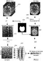
Figure 1 is then applied to a specific, non-limiting, case of MRI, wherein an example of quantification of T2 using a learnt signal model for aMESE sequence 210 with a single-echo spin-echo (SE)sequence 220 as gold-standard reference is taken. In other words, in the preferred embodiment ofFigure 2 , the first technique according to the invention uses aSE sequence 220 for quantifying T2 according to classical process and the second technique uses aMESE sequence 210 for quantifying T2 using a machine learning technique wherein the quantified T2 values obtained from the first technique are used as target output for the machine learning technique. - At
step 101, reference values for a parameter are determined by the system according to the invention from a group of objects, for instance a group of subjects. Said parameter might be a physical parameter or a biological parameter. Said reference values, also called gold-standard reference values, are values, preferentially quantitative values, of said parameter that are determined by the system according to a first technique. Said first technique is typically a time consuming technique that cannot be applied as such during clinical diagnosis. According to the present invention, said first technique is only used to create a learning dataset. The first technique is typically a known technique wherein the first set of data corresponds to a first signal intensity (or signal data) acquired by the system according to the invention for each object, the system being further configured for determining said value of the parameter for each object by fitting the signal intensity with a signal model. Each determined value for the parameter is then used as a reference value within the machine learning technique. - In the particular case of MRI described in
Fig. 2 , the first signal intensity might be a SE signal intensity, and step 101 may comprise acquiring SE signal intensity (or data) for each object of the group of objects and determining quantitative gold-standard T2 values (T2 being thus the parameter whose values have to be determined) for each object from a single-slice, resolution- and orientation-matched single-echo spin-echo sequence. The gold-standard T2 values 222 are preferentially computed using a classical signal fitting 221 onto the spin-echo data. - At
step 102, the system according to the invention creates a learning dataset by associating for each object a second set of signal intensities (which corresponds in that case to said second set of data) and the T2 reference value determined from the first signal intensity using the first technique. For instance, in the case illustrated byFigure 2 , a given gold-standard T2 reference value might be associated with a measured MESE signal evolution (i.e. signal decay) in a database. In particular, for each object, normalizedsignal decay 214 and gold-standard T2 value in thebrain 224, preferentially obtained afterapplication 223 of abrain mask 230 determined for each object, are stored in a database in order to create said learning dataset. In other words, the method according to the invention may comprise acquiring MESE signal intensities, for instance MESE images, from said group of objects. Preferentially, intracranial volume might be first segmented 211 to restrict the learning solely to brain tissue, wherein the object correspondingbrain mask 230 is applied to the MESE data for this purpose. All MESE signal decays 212 are preferably normalized 213 by dividing the signal intensities by the L2-norm of the decay. The normalization of the signal decays, or more generally of data used as input within the machine learning algorithm, could be performed differently (different norm or different scale) depending on the used technique. The optional application of thebrain mask 230 as previously described advantageously improves the final result. - At
step 103, the system according to the invention uses a machine learning technique for determining the value of the parameter from the second set of signal intensities, wherein said machine learning technique comprises using a machine learning algorithm for determining said value, and training said machine learning algorithm on the learning dataset wherein for each object the second signal intensity is used as input and the associated reference value as output target. For instance, the machine learning algorithm might be an artificialneuronal network 240 that is trained using the signal intensities of each echo as input and the gold-standard T2 value as target output. Preferentially, said artificialneuronal network 240 may comprise two layers with each 8 neurons and a root-mean-squared error to the gold-standard T2 as cost function. The training is preferentially performed with data from all available objects, i.e. with whole data of the learning dataset. - At
step 104, the system determines a relationship between the signal intensity and the value of the parameter obtained for each object from the training of the machine learning algorithm on the learning dataset. In the example ofFigure 2 , the machine learning algorithm is typically used to learn the relationship between the decay and the T2 value. - At
step 105, the system uses said relationship to determine the value of the parameter from a measured set of signal intensities for a new object. For instance and as illustrated inFig. 2 , in order to reconstruct a new dataset (e.g. coming from the new object), every decay is fed to the previously trained artificial neuronal network to estimate T2 in each voxel of the new dataset, resulting in the desired quantitative value for the parameter based on the data driven signal model. - Advantageously, the present invention proposes therefore to replace a fixed signal model with a learnt relationship between "true" values (i.e. based on the gold standard reference values) and the respective quantitative measurements can help eliminating many of the difficulties related to finding an appropriate model for a given problem. Additionally, the data-driven approach proposed here is able to learn potential dirt effects in the acquired signal intensity, which are typically hard to model or even, cannot be modelled at all according to prior art technique. Another advantage is that no a priori knowledge has to be imposed regarding the assumed interaction of the tissue microstructure and the underlying MR physics.
- Advantageously, the application of a trained neural network is typically very fast, i.e. once the machine learning algorithm, like the artificial
neuronal network 240 ofFigure 2 , is trained, parametric maps can be obtained very quickly in contrast to typically slow fitting procedures. - As already mentioned above, the present invention is not restricted to the specific embodiment of
Figure 2 , but might be without effort generalizable to other quantification or determination of parameters since only a set of datasets acquired through the so-called second technique and gold-standard reference data acquired through the so-called first technique are required. In particular, the first technique and the second technique according to the reference might be the same, or are two different techniques. Regarding the specific embodiment illustrated byFigure 2 , a different type of input data than a MESE dataset using a different sequence could be used as input for the machine learning algorithm. For example, the invention could also be used for magnetic resonance fingerprinting (see Ma et al., Nature 2013, 495(7440): 187-192), where the fingerprint is the input and the multiparametric values are outputs of the artificial neuronal network. - The machine learning technique described in the present invention and sometimes also referred to as artificial intelligence or data science tool may also use different approaches like a linear regression, a non-linear regression or convolutional neuronal networks.
Claims (9)
- A machine learning method for determining a value for a magnetic resonance imaging (MRI) parameter, said value enabling a characterization of a biological tissue, the method comprising:- determining (101) reference values for said MRI parameter from a group of objects, wherein a first MRI technique is used by an MRI system for determining for each object said reference value from a first set of data;- creating (102) a learning dataset by associating for each object of the group of objects a second set of data and the reference value previously determined from the first set of data, wherein the second set of data is acquired by the MRI system according to a second MRI technique for determining values of the MRI parameter and is configured for enabling a determination of said MRI parameter;- using (103) a machine learning algorithm, in particular implemented by the MRI system, trained on the learning dataset for determining a value of said MRI parameter, wherein the second set of data obtained for each of the objects is used as input in the machine learning algorithm and its associated reference value is used as output target for said machine learning algorithm;- determining (104) a relationship between the second set of data and a value of the MRI parameter obtained for each object by training the machine learning algorithm on the learning dataset;- using (105) said relationship for determining from another data set a value for the MRI parameter of a new object, wherein said another dataset has been obtained by the MRI system by means of the second MRI technique, wherein said MRI parameter is a T2 relaxation time, the method being characterized in that the first MRI technique comprises a single-echo spin-echo (SE) sequence.
- The method of claim 1, wherein the first MRI technique comprises fitting the first data set by means of a signal model.
- The method of any of the previous claims, wherein the second MRI technique comprises a multi-echo spin-echo (MESE) sequence, such that the second dataset comprises MESE signal decays (212).
- The method according to claim 3, wherein a brain mask is applied to the MESE signal decays (212) and to the first dataset.
- The method according to claim 3 or 4, wherein the MESE signal decays are normalized.
- The method according to one of the claims 1-5, wherein the machine learning algorithm is an artificial neuronal network.
- The method according to claim 1 or 2, wherein the second MRI technique comprises magnetic resonance fingerprinting.
- The method according to one of the claims 1-7, wherein said object is a subject, a patient, phantom, histology or animal data.
- Magnetic resonance imaging (MRI) system configured for determining a value for a magnetic resonance imaging (MRI) parameter, the MRI system being configured to acquire a first set of data from each object of a group of objects, the MRI system comprising:- a processing unit configured for determining reference values for said MRI parameter for each object of said group of objects by a first MRI technique from the first set of data acquired by the MRI system; the MRI system being further configured to acquire a second set of data from each object of the group of objects according to a second MRI technique; the MRI system further comprising- a database storing a learning dataset comprising and associating for each object of said group of objects said second set of data and the previously determined reference value, wherein the second set of data is acquired by the MRI system according to the second MRI technique for determining values of the MRI parameter; the processing unit being further configured- for using a machine learning algorithm trained on the learning dataset for determining a value for the MRI parameter from the second set of data for each of the objects, wherein the second set of data obtained for each of the objects is used as input in the machine learning algorithm and its associated reference value is used as output target for the machine learning algorithm;- for determining a relationship between the second set of data and a value of the MRI parameter obtained for each object by training the machine learning algorithm on the learning dataset; and- for using (105) said relationship for determining from another data set a value for the MRI parameter of a new object, wherein said another dataset has been acquired by the MRI system by means of the second MRI technique, wherein said MRI parameter is a T2 relaxation time, the MRI system being characterized in that it is configured for using a single-echo spin-echo (SE) sequence as the first MRI technique.
Priority Applications (2)
| Application Number | Priority Date | Filing Date | Title |
|---|---|---|---|
| EP18175213.0A EP3575813B1 (en) | 2018-05-30 | 2018-05-30 | Quantitative mapping of a magnetic resonance imaging parameter by data-driven signal-model learning |
| US16/426,486 US11587675B2 (en) | 2018-05-30 | 2019-05-30 | Quantitative mapping by data-driven signal-model learning |
Applications Claiming Priority (1)
| Application Number | Priority Date | Filing Date | Title |
|---|---|---|---|
| EP18175213.0A EP3575813B1 (en) | 2018-05-30 | 2018-05-30 | Quantitative mapping of a magnetic resonance imaging parameter by data-driven signal-model learning |
Publications (2)
| Publication Number | Publication Date |
|---|---|
| EP3575813A1 EP3575813A1 (en) | 2019-12-04 |
| EP3575813B1 true EP3575813B1 (en) | 2022-06-29 |
Family
ID=62492522
Family Applications (1)
| Application Number | Title | Priority Date | Filing Date |
|---|---|---|---|
| EP18175213.0A Active EP3575813B1 (en) | 2018-05-30 | 2018-05-30 | Quantitative mapping of a magnetic resonance imaging parameter by data-driven signal-model learning |
Country Status (2)
| Country | Link |
|---|---|
| US (1) | US11587675B2 (en) |
| EP (1) | EP3575813B1 (en) |
Families Citing this family (4)
| Publication number | Priority date | Publication date | Assignee | Title |
|---|---|---|---|---|
| EP3575813B1 (en) * | 2018-05-30 | 2022-06-29 | Siemens Healthcare GmbH | Quantitative mapping of a magnetic resonance imaging parameter by data-driven signal-model learning |
| DE102018125908A1 (en) * | 2018-10-18 | 2020-04-23 | Endress+Hauser Conducta Gmbh+Co. Kg | Method for determining a process variable with a classifier for selecting a measuring method |
| CN113391250B (en) * | 2021-07-09 | 2022-11-29 | 清华大学 | Tissue attribute multi-parameter quantitative test system and method thereof |
| CN116068473A (en) * | 2021-10-29 | 2023-05-05 | 通用电气精准医疗有限责任公司 | Method and magnetic resonance imaging system for generating magnetic resonance images |
Family Cites Families (35)
| Publication number | Priority date | Publication date | Assignee | Title |
|---|---|---|---|---|
| US7444308B2 (en) * | 2001-06-15 | 2008-10-28 | Health Discovery Corporation | Data mining platform for bioinformatics and other knowledge discovery |
| CN1249620C (en) * | 2000-06-19 | 2006-04-05 | 科雷洛吉克系统公司 | classification heuristics |
| US8463718B2 (en) * | 2000-08-07 | 2013-06-11 | Health Discovery Corporation | Support vector machine-based method for analysis of spectral data |
| EP1393196A4 (en) * | 2001-05-07 | 2007-02-28 | Health Discovery Corp | Kernels and methods for selecting kernels for use in learning machines |
| US20090132443A1 (en) * | 2007-11-16 | 2009-05-21 | Odilo Mueller | Methods and Devices for Analyzing Lipoproteins |
| US8386401B2 (en) * | 2008-09-10 | 2013-02-26 | Digital Infuzion, Inc. | Machine learning methods and systems for identifying patterns in data using a plurality of learning machines wherein the learning machine that optimizes a performance function is selected |
| WO2010126867A1 (en) * | 2009-04-27 | 2010-11-04 | Cincinnati Children's Hospital Medical Center | Computer implemented system and method for assessing a neuropsychiatric condition of a human subject |
| US20140156573A1 (en) * | 2011-07-27 | 2014-06-05 | The Research Foundation Of State University Of New York | Methods for generating predictive models for epithelial ovarian cancer and methods for identifying eoc |
| US9326684B2 (en) * | 2011-11-08 | 2016-05-03 | Covidien Lp | Magnetic enhancement in determination of physiological blood parameters |
| CA2877426C (en) * | 2012-06-21 | 2024-05-21 | Philip Morris Products S.A. | Systems and methods relating to network-based biomarker signatures |
| HK1222243A1 (en) * | 2012-12-13 | 2017-06-23 | 麦太宝根有限公司 | Identification of a person having risk for developing type 2 diabetes |
| US10156567B2 (en) * | 2012-12-17 | 2018-12-18 | General Electric Company | In-vitro magnetic resonance detection of a target substance without separating bound magnetic nanoparticles from unbound magnetic nanoparticles |
| US10646595B2 (en) * | 2013-02-12 | 2020-05-12 | The Governing Council Of The University Of Toronto | Porphyrin compounds and their use as MRI contrast agents |
| US9466024B2 (en) * | 2013-03-15 | 2016-10-11 | Northrop Grumman Systems Corporation | Learning health systems and methods |
| JP6371554B2 (en) * | 2014-03-28 | 2018-08-08 | 株式会社日立製作所 | Magnetic resonance imaging system |
| US10761171B2 (en) * | 2015-06-22 | 2020-09-01 | Case Western Reserve University | Systems and methods for free-breathing three-dimensional magnetic resonance fingerprinting |
| US10869939B2 (en) * | 2015-08-03 | 2020-12-22 | Ramot At Tel-Aviv University Ltd. | Delivery system in micellar form having modular spectral response based on enzyme-responsive amphiphilic PEG-dendron hybrid polymers |
| US10548505B2 (en) * | 2016-03-15 | 2020-02-04 | Siemens Healthcare Gmbh | System and method for real-time MRI-guided object navigation |
| JP6703264B2 (en) * | 2016-06-22 | 2020-06-03 | 富士通株式会社 | Machine learning management program, machine learning management method, and machine learning management device |
| US20180231626A1 (en) * | 2017-02-10 | 2018-08-16 | Case Western Reserve University | Systems and methods for magnetic resonance fingerprinting for quantitative breast imaging |
| US10846616B1 (en) * | 2017-04-28 | 2020-11-24 | Iqvia Inc. | System and method for enhanced characterization of structured data for machine learning |
| US11234657B2 (en) * | 2017-05-01 | 2022-02-01 | Rhode Island Hospital | Non-invasive measurement to predict post-surgery anterior cruciate ligament success |
| US10346974B2 (en) * | 2017-05-18 | 2019-07-09 | Toshiba Medical Systems Corporation | Apparatus and method for medical image processing |
| US11972355B2 (en) * | 2017-07-18 | 2024-04-30 | iQGateway LLC | Method and system for generating best performing data models for datasets in a computing environment |
| JP6898561B2 (en) * | 2017-09-08 | 2021-07-07 | 富士通株式会社 | Machine learning programs, machine learning methods, and machine learning equipment |
| JP6898562B2 (en) * | 2017-09-08 | 2021-07-07 | 富士通株式会社 | Machine learning programs, machine learning methods, and machine learning equipment |
| US11423325B2 (en) * | 2017-10-25 | 2022-08-23 | International Business Machines Corporation | Regression for metric dataset |
| US20200256856A1 (en) * | 2017-10-26 | 2020-08-13 | Essenlix Corporation | System and methods of image-based assay using crof and machine learning |
| WO2019113428A1 (en) * | 2017-12-08 | 2019-06-13 | Rensselaer Polytechnic Institute | A synergized pulsing-imaging network (spin) |
| US11426073B2 (en) * | 2018-04-27 | 2022-08-30 | Washington University | Imaging nerve function and pathologies using diffusion basis spectrum imaging |
| WO2019229528A2 (en) * | 2018-05-30 | 2019-12-05 | Alexander Meyer | Using machine learning to predict health conditions |
| EP3575813B1 (en) * | 2018-05-30 | 2022-06-29 | Siemens Healthcare GmbH | Quantitative mapping of a magnetic resonance imaging parameter by data-driven signal-model learning |
| US20200334524A1 (en) * | 2019-04-17 | 2020-10-22 | Here Global B.V. | Edge learning |
| JP7561822B2 (en) * | 2019-07-10 | 2024-10-04 | ベクトン・ディキンソン・アンド・カンパニー | Reconfigurable integrated circuits for coordinating cell sorting |
| US20220076157A1 (en) * | 2020-09-04 | 2022-03-10 | Aperio Global, LLC | Data analysis system using artificial intelligence |
-
2018
- 2018-05-30 EP EP18175213.0A patent/EP3575813B1/en active Active
-
2019
- 2019-05-30 US US16/426,486 patent/US11587675B2/en active Active
Also Published As
| Publication number | Publication date |
|---|---|
| EP3575813A1 (en) | 2019-12-04 |
| US11587675B2 (en) | 2023-02-21 |
| US20190371465A1 (en) | 2019-12-05 |
Similar Documents
| Publication | Publication Date | Title |
|---|---|---|
| Buonincontri et al. | Multi-site repeatability and reproducibility of MR fingerprinting of the healthy brain at 1.5 and 3.0 T | |
| Shams et al. | A comparison of in vivo MRI based cortical myelin mapping using T1w/T2w and R1 mapping at 3T | |
| Esses et al. | Automated image quality evaluation of T2‐weighted liver MRI utilizing deep learning architecture | |
| Rokem et al. | Evaluating the accuracy of diffusion MRI models in white matter | |
| Ben‐Eliezer et al. | Rapid and accurate T2 mapping from multi–spin‐echo data using Bloch‐simulation‐based reconstruction | |
| Alexander | An introduction to computational diffusion MRI: the diffusion tensor and beyond | |
| EP4276756A2 (en) | Tumor tissue characterization using multi-parametric magnetic resonance imaging | |
| EP3617733A1 (en) | Method and apparatus for processing magnetic resonance data using machine learning | |
| Denis de Senneville et al. | RegQCNET: Deep quality control for image-to-template brain MRI affine registration | |
| US11965946B2 (en) | Machine learning based processing of magnetic resonance data, including an uncertainty quantification | |
| US11587675B2 (en) | Quantitative mapping by data-driven signal-model learning | |
| Torop et al. | Deep learning using a biophysical model for robust and accelerated reconstruction of quantitative, artifact‐free and denoised images | |
| Shamir et al. | Tutorial: a guide to diffusion MRI and structural connectomics | |
| Jallais et al. | Introducing µGUIDE for quantitative imaging via generalized uncertainty-driven inference using deep learning | |
| Trotier et al. | The compressed sensing MP2RAGE as a surrogate to the MPRAGE for neuroimaging at 3 T | |
| Perrone et al. | D-BRAIN: anatomically accurate simulated diffusion MRI brain data | |
| Piredda et al. | Submillimeter T1 atlas for subject‐specific abnormality detection at 7T | |
| CN117689623A (en) | Adaptive diffusion magnetic resonance microstructure model parameter estimation method based on external gradient | |
| Lim et al. | Fitting a directional microstructure model to diffusion-relaxation MRI data with self-supervised machine learning | |
| Economou et al. | Myelin water fraction in relation to fractional anisotropy and reading in 10-year-old children | |
| DE102010032825A1 (en) | Method and apparatus for magnetic resonance imaging | |
| CN116888489A (en) | Methods for analyzing medical images | |
| Fang et al. | Deep learning for fast and spatially-constrained tissue quantification from highly-undersampled data in magnetic resonance fingerprinting (MRF) | |
| CN114127574A (en) | Validation of quantitative magnetic resonance imaging protocols | |
| Mazzoni et al. | New developments in MRI: System characterization, technical advances and radiotherapy applications |
Legal Events
| Date | Code | Title | Description |
|---|---|---|---|
| PUAI | Public reference made under article 153(3) epc to a published international application that has entered the european phase |
Free format text: ORIGINAL CODE: 0009012 |
|
| STAA | Information on the status of an ep patent application or granted ep patent |
Free format text: STATUS: THE APPLICATION HAS BEEN PUBLISHED |
|
| AK | Designated contracting states |
Kind code of ref document: A1 Designated state(s): AL AT BE BG CH CY CZ DE DK EE ES FI FR GB GR HR HU IE IS IT LI LT LU LV MC MK MT NL NO PL PT RO RS SE SI SK SM TR |
|
| AX | Request for extension of the european patent |
Extension state: BA ME |
|
| STAA | Information on the status of an ep patent application or granted ep patent |
Free format text: STATUS: REQUEST FOR EXAMINATION WAS MADE |
|
| 17P | Request for examination filed |
Effective date: 20200519 |
|
| RBV | Designated contracting states (corrected) |
Designated state(s): AL AT BE BG CH CY CZ DE DK EE ES FI FR GB GR HR HU IE IS IT LI LT LU LV MC MK MT NL NO PL PT RO RS SE SI SK SM TR |
|
| GRAP | Despatch of communication of intention to grant a patent |
Free format text: ORIGINAL CODE: EPIDOSNIGR1 |
|
| STAA | Information on the status of an ep patent application or granted ep patent |
Free format text: STATUS: GRANT OF PATENT IS INTENDED |
|
| RIC1 | Information provided on ipc code assigned before grant |
Ipc: G06N 3/08 20060101ALI20220131BHEP Ipc: G01R 33/56 20060101ALI20220131BHEP Ipc: G01R 33/50 20060101AFI20220131BHEP |
|
| INTG | Intention to grant announced |
Effective date: 20220303 |
|
| GRAS | Grant fee paid |
Free format text: ORIGINAL CODE: EPIDOSNIGR3 |
|
| GRAA | (expected) grant |
Free format text: ORIGINAL CODE: 0009210 |
|
| STAA | Information on the status of an ep patent application or granted ep patent |
Free format text: STATUS: THE PATENT HAS BEEN GRANTED |
|
| AK | Designated contracting states |
Kind code of ref document: B1 Designated state(s): AL AT BE BG CH CY CZ DE DK EE ES FI FR GB GR HR HU IE IS IT LI LT LU LV MC MK MT NL NO PL PT RO RS SE SI SK SM TR |
|
| REG | Reference to a national code |
Ref country code: CH Ref legal event code: EP |
|
| REG | Reference to a national code |
Ref country code: AT Ref legal event code: REF Ref document number: 1501694 Country of ref document: AT Kind code of ref document: T Effective date: 20220715 |
|
| REG | Reference to a national code |
Ref country code: IE Ref legal event code: FG4D |
|
| REG | Reference to a national code |
Ref country code: DE Ref legal event code: R096 Ref document number: 602018037224 Country of ref document: DE |
|
| REG | Reference to a national code |
Ref country code: LT Ref legal event code: MG9D |
|
| PG25 | Lapsed in a contracting state [announced via postgrant information from national office to epo] |
Ref country code: SE Free format text: LAPSE BECAUSE OF FAILURE TO SUBMIT A TRANSLATION OF THE DESCRIPTION OR TO PAY THE FEE WITHIN THE PRESCRIBED TIME-LIMIT Effective date: 20220629 Ref country code: NO Free format text: LAPSE BECAUSE OF FAILURE TO SUBMIT A TRANSLATION OF THE DESCRIPTION OR TO PAY THE FEE WITHIN THE PRESCRIBED TIME-LIMIT Effective date: 20220929 Ref country code: LT Free format text: LAPSE BECAUSE OF FAILURE TO SUBMIT A TRANSLATION OF THE DESCRIPTION OR TO PAY THE FEE WITHIN THE PRESCRIBED TIME-LIMIT Effective date: 20220629 Ref country code: HR Free format text: LAPSE BECAUSE OF FAILURE TO SUBMIT A TRANSLATION OF THE DESCRIPTION OR TO PAY THE FEE WITHIN THE PRESCRIBED TIME-LIMIT Effective date: 20220629 Ref country code: GR Free format text: LAPSE BECAUSE OF FAILURE TO SUBMIT A TRANSLATION OF THE DESCRIPTION OR TO PAY THE FEE WITHIN THE PRESCRIBED TIME-LIMIT Effective date: 20220930 Ref country code: FI Free format text: LAPSE BECAUSE OF FAILURE TO SUBMIT A TRANSLATION OF THE DESCRIPTION OR TO PAY THE FEE WITHIN THE PRESCRIBED TIME-LIMIT Effective date: 20220629 Ref country code: BG Free format text: LAPSE BECAUSE OF FAILURE TO SUBMIT A TRANSLATION OF THE DESCRIPTION OR TO PAY THE FEE WITHIN THE PRESCRIBED TIME-LIMIT Effective date: 20220929 |
|
| REG | Reference to a national code |
Ref country code: NL Ref legal event code: MP Effective date: 20220629 |
|
| REG | Reference to a national code |
Ref country code: AT Ref legal event code: MK05 Ref document number: 1501694 Country of ref document: AT Kind code of ref document: T Effective date: 20220629 |
|
| PG25 | Lapsed in a contracting state [announced via postgrant information from national office to epo] |
Ref country code: RS Free format text: LAPSE BECAUSE OF FAILURE TO SUBMIT A TRANSLATION OF THE DESCRIPTION OR TO PAY THE FEE WITHIN THE PRESCRIBED TIME-LIMIT Effective date: 20220629 Ref country code: LV Free format text: LAPSE BECAUSE OF FAILURE TO SUBMIT A TRANSLATION OF THE DESCRIPTION OR TO PAY THE FEE WITHIN THE PRESCRIBED TIME-LIMIT Effective date: 20220629 |
|
| PG25 | Lapsed in a contracting state [announced via postgrant information from national office to epo] |
Ref country code: NL Free format text: LAPSE BECAUSE OF FAILURE TO SUBMIT A TRANSLATION OF THE DESCRIPTION OR TO PAY THE FEE WITHIN THE PRESCRIBED TIME-LIMIT Effective date: 20220629 |
|
| PG25 | Lapsed in a contracting state [announced via postgrant information from national office to epo] |
Ref country code: SM Free format text: LAPSE BECAUSE OF FAILURE TO SUBMIT A TRANSLATION OF THE DESCRIPTION OR TO PAY THE FEE WITHIN THE PRESCRIBED TIME-LIMIT Effective date: 20220629 Ref country code: SK Free format text: LAPSE BECAUSE OF FAILURE TO SUBMIT A TRANSLATION OF THE DESCRIPTION OR TO PAY THE FEE WITHIN THE PRESCRIBED TIME-LIMIT Effective date: 20220629 Ref country code: RO Free format text: LAPSE BECAUSE OF FAILURE TO SUBMIT A TRANSLATION OF THE DESCRIPTION OR TO PAY THE FEE WITHIN THE PRESCRIBED TIME-LIMIT Effective date: 20220629 Ref country code: PT Free format text: LAPSE BECAUSE OF FAILURE TO SUBMIT A TRANSLATION OF THE DESCRIPTION OR TO PAY THE FEE WITHIN THE PRESCRIBED TIME-LIMIT Effective date: 20221031 Ref country code: ES Free format text: LAPSE BECAUSE OF FAILURE TO SUBMIT A TRANSLATION OF THE DESCRIPTION OR TO PAY THE FEE WITHIN THE PRESCRIBED TIME-LIMIT Effective date: 20220629 Ref country code: EE Free format text: LAPSE BECAUSE OF FAILURE TO SUBMIT A TRANSLATION OF THE DESCRIPTION OR TO PAY THE FEE WITHIN THE PRESCRIBED TIME-LIMIT Effective date: 20220629 Ref country code: AT Free format text: LAPSE BECAUSE OF FAILURE TO SUBMIT A TRANSLATION OF THE DESCRIPTION OR TO PAY THE FEE WITHIN THE PRESCRIBED TIME-LIMIT Effective date: 20220629 |
|
| PG25 | Lapsed in a contracting state [announced via postgrant information from national office to epo] |
Ref country code: PL Free format text: LAPSE BECAUSE OF FAILURE TO SUBMIT A TRANSLATION OF THE DESCRIPTION OR TO PAY THE FEE WITHIN THE PRESCRIBED TIME-LIMIT Effective date: 20220629 Ref country code: IS Free format text: LAPSE BECAUSE OF FAILURE TO SUBMIT A TRANSLATION OF THE DESCRIPTION OR TO PAY THE FEE WITHIN THE PRESCRIBED TIME-LIMIT Effective date: 20221029 |
|
| REG | Reference to a national code |
Ref country code: DE Ref legal event code: R097 Ref document number: 602018037224 Country of ref document: DE |
|
| PG25 | Lapsed in a contracting state [announced via postgrant information from national office to epo] |
Ref country code: AL Free format text: LAPSE BECAUSE OF FAILURE TO SUBMIT A TRANSLATION OF THE DESCRIPTION OR TO PAY THE FEE WITHIN THE PRESCRIBED TIME-LIMIT Effective date: 20220629 |
|
| PG25 | Lapsed in a contracting state [announced via postgrant information from national office to epo] |
Ref country code: DK Free format text: LAPSE BECAUSE OF FAILURE TO SUBMIT A TRANSLATION OF THE DESCRIPTION OR TO PAY THE FEE WITHIN THE PRESCRIBED TIME-LIMIT Effective date: 20220629 Ref country code: CZ Free format text: LAPSE BECAUSE OF FAILURE TO SUBMIT A TRANSLATION OF THE DESCRIPTION OR TO PAY THE FEE WITHIN THE PRESCRIBED TIME-LIMIT Effective date: 20220629 |
|
| PLBE | No opposition filed within time limit |
Free format text: ORIGINAL CODE: 0009261 |
|
| STAA | Information on the status of an ep patent application or granted ep patent |
Free format text: STATUS: NO OPPOSITION FILED WITHIN TIME LIMIT |
|
| 26N | No opposition filed |
Effective date: 20230330 |
|
| PG25 | Lapsed in a contracting state [announced via postgrant information from national office to epo] |
Ref country code: SI Free format text: LAPSE BECAUSE OF FAILURE TO SUBMIT A TRANSLATION OF THE DESCRIPTION OR TO PAY THE FEE WITHIN THE PRESCRIBED TIME-LIMIT Effective date: 20220629 |
|
| REG | Reference to a national code |
Ref country code: CH Ref legal event code: PL |
|
| PG25 | Lapsed in a contracting state [announced via postgrant information from national office to epo] |
Ref country code: MC Free format text: LAPSE BECAUSE OF FAILURE TO SUBMIT A TRANSLATION OF THE DESCRIPTION OR TO PAY THE FEE WITHIN THE PRESCRIBED TIME-LIMIT Effective date: 20220629 |
|
| REG | Reference to a national code |
Ref country code: BE Ref legal event code: MM Effective date: 20230531 |
|
| PG25 | Lapsed in a contracting state [announced via postgrant information from national office to epo] |
Ref country code: MC Free format text: LAPSE BECAUSE OF FAILURE TO SUBMIT A TRANSLATION OF THE DESCRIPTION OR TO PAY THE FEE WITHIN THE PRESCRIBED TIME-LIMIT Effective date: 20220629 Ref country code: LU Free format text: LAPSE BECAUSE OF NON-PAYMENT OF DUE FEES Effective date: 20230530 Ref country code: LI Free format text: LAPSE BECAUSE OF NON-PAYMENT OF DUE FEES Effective date: 20230531 Ref country code: IT Free format text: LAPSE BECAUSE OF FAILURE TO SUBMIT A TRANSLATION OF THE DESCRIPTION OR TO PAY THE FEE WITHIN THE PRESCRIBED TIME-LIMIT Effective date: 20220629 Ref country code: CH Free format text: LAPSE BECAUSE OF NON-PAYMENT OF DUE FEES Effective date: 20230531 |
|
| REG | Reference to a national code |
Ref country code: DE Ref legal event code: R081 Ref document number: 602018037224 Country of ref document: DE Owner name: SIEMENS HEALTHINEERS AG, DE Free format text: FORMER OWNER: SIEMENS HEALTHCARE GMBH, MUENCHEN, DE Ref country code: IE Ref legal event code: MM4A |
|
| PG25 | Lapsed in a contracting state [announced via postgrant information from national office to epo] |
Ref country code: IE Free format text: LAPSE BECAUSE OF NON-PAYMENT OF DUE FEES Effective date: 20230530 |
|
| PG25 | Lapsed in a contracting state [announced via postgrant information from national office to epo] |
Ref country code: IE Free format text: LAPSE BECAUSE OF NON-PAYMENT OF DUE FEES Effective date: 20230530 |
|
| PG25 | Lapsed in a contracting state [announced via postgrant information from national office to epo] |
Ref country code: BE Free format text: LAPSE BECAUSE OF NON-PAYMENT OF DUE FEES Effective date: 20230531 |
|
| PG25 | Lapsed in a contracting state [announced via postgrant information from national office to epo] |
Ref country code: BG Free format text: LAPSE BECAUSE OF FAILURE TO SUBMIT A TRANSLATION OF THE DESCRIPTION OR TO PAY THE FEE WITHIN THE PRESCRIBED TIME-LIMIT Effective date: 20220629 |
|
| PG25 | Lapsed in a contracting state [announced via postgrant information from national office to epo] |
Ref country code: BG Free format text: LAPSE BECAUSE OF FAILURE TO SUBMIT A TRANSLATION OF THE DESCRIPTION OR TO PAY THE FEE WITHIN THE PRESCRIBED TIME-LIMIT Effective date: 20220629 |
|
| PGFP | Annual fee paid to national office [announced via postgrant information from national office to epo] |
Ref country code: GB Payment date: 20250610 Year of fee payment: 8 |
|
| PGFP | Annual fee paid to national office [announced via postgrant information from national office to epo] |
Ref country code: FR Payment date: 20250516 Year of fee payment: 8 |
|
| PG25 | Lapsed in a contracting state [announced via postgrant information from national office to epo] |
Ref country code: CY Free format text: LAPSE BECAUSE OF FAILURE TO SUBMIT A TRANSLATION OF THE DESCRIPTION OR TO PAY THE FEE WITHIN THE PRESCRIBED TIME-LIMIT; INVALID AB INITIO Effective date: 20180530 |
|
| PG25 | Lapsed in a contracting state [announced via postgrant information from national office to epo] |
Ref country code: HU Free format text: LAPSE BECAUSE OF FAILURE TO SUBMIT A TRANSLATION OF THE DESCRIPTION OR TO PAY THE FEE WITHIN THE PRESCRIBED TIME-LIMIT; INVALID AB INITIO Effective date: 20180530 |
|
| PGFP | Annual fee paid to national office [announced via postgrant information from national office to epo] |
Ref country code: DE Payment date: 20250718 Year of fee payment: 8 |
|
| PG25 | Lapsed in a contracting state [announced via postgrant information from national office to epo] |
Ref country code: TR Free format text: LAPSE BECAUSE OF FAILURE TO SUBMIT A TRANSLATION OF THE DESCRIPTION OR TO PAY THE FEE WITHIN THE PRESCRIBED TIME-LIMIT Effective date: 20220629 |