EP3569150B1 - Systeme und verfahren zur vorhersage von koronarplaqueanfälligkeit aus patientenspezifischen anatomischen bilddaten - Google Patents
Systeme und verfahren zur vorhersage von koronarplaqueanfälligkeit aus patientenspezifischen anatomischen bilddaten Download PDFInfo
- Publication number
- EP3569150B1 EP3569150B1 EP19183048.8A EP19183048A EP3569150B1 EP 3569150 B1 EP3569150 B1 EP 3569150B1 EP 19183048 A EP19183048 A EP 19183048A EP 3569150 B1 EP3569150 B1 EP 3569150B1
- Authority
- EP
- European Patent Office
- Prior art keywords
- plaque
- patient
- image data
- coronary
- specific
- Prior art date
- Legal status (The legal status is an assumption and is not a legal conclusion. Google has not performed a legal analysis and makes no representation as to the accuracy of the status listed.)
- Active
Links
Images


Classifications
-
- A—HUMAN NECESSITIES
- A61—MEDICAL OR VETERINARY SCIENCE; HYGIENE
- A61B—DIAGNOSIS; SURGERY; IDENTIFICATION
- A61B34/00—Computer-aided surgery; Manipulators or robots specially adapted for use in surgery
- A61B34/10—Computer-aided planning, simulation or modelling of surgical operations
-
- A—HUMAN NECESSITIES
- A61—MEDICAL OR VETERINARY SCIENCE; HYGIENE
- A61B—DIAGNOSIS; SURGERY; IDENTIFICATION
- A61B5/00—Measuring for diagnostic purposes; Identification of persons
- A61B5/0059—Measuring for diagnostic purposes; Identification of persons using light, e.g. diagnosis by transillumination, diascopy, fluorescence
- A61B5/0062—Arrangements for scanning
- A61B5/0066—Optical coherence imaging
-
- A—HUMAN NECESSITIES
- A61—MEDICAL OR VETERINARY SCIENCE; HYGIENE
- A61B—DIAGNOSIS; SURGERY; IDENTIFICATION
- A61B5/00—Measuring for diagnostic purposes; Identification of persons
- A61B5/02—Detecting, measuring or recording for evaluating the cardiovascular system, e.g. pulse, heart rate, blood pressure or blood flow
- A61B5/02007—Evaluating blood vessel condition, e.g. elasticity, compliance
-
- A—HUMAN NECESSITIES
- A61—MEDICAL OR VETERINARY SCIENCE; HYGIENE
- A61B—DIAGNOSIS; SURGERY; IDENTIFICATION
- A61B5/00—Measuring for diagnostic purposes; Identification of persons
- A61B5/02—Detecting, measuring or recording for evaluating the cardiovascular system, e.g. pulse, heart rate, blood pressure or blood flow
- A61B5/026—Measuring blood flow
-
- A—HUMAN NECESSITIES
- A61—MEDICAL OR VETERINARY SCIENCE; HYGIENE
- A61B—DIAGNOSIS; SURGERY; IDENTIFICATION
- A61B5/00—Measuring for diagnostic purposes; Identification of persons
- A61B5/05—Detecting, measuring or recording for diagnosis by means of electric currents or magnetic fields; Measuring using microwaves or radio waves
- A61B5/055—Detecting, measuring or recording for diagnosis by means of electric currents or magnetic fields; Measuring using microwaves or radio waves involving electronic [EMR] or nuclear [NMR] magnetic resonance, e.g. magnetic resonance imaging
-
- A—HUMAN NECESSITIES
- A61—MEDICAL OR VETERINARY SCIENCE; HYGIENE
- A61B—DIAGNOSIS; SURGERY; IDENTIFICATION
- A61B5/00—Measuring for diagnostic purposes; Identification of persons
- A61B5/72—Signal processing specially adapted for physiological signals or for diagnostic purposes
- A61B5/7271—Specific aspects of physiological measurement analysis
- A61B5/7275—Determining trends in physiological measurement data; Predicting development of a medical condition based on physiological measurements, e.g. determining a risk factor
-
- A—HUMAN NECESSITIES
- A61—MEDICAL OR VETERINARY SCIENCE; HYGIENE
- A61B—DIAGNOSIS; SURGERY; IDENTIFICATION
- A61B6/00—Apparatus or devices for radiation diagnosis; Apparatus or devices for radiation diagnosis combined with radiation therapy equipment
- A61B6/02—Arrangements for diagnosis sequentially in different planes; Stereoscopic radiation diagnosis
- A61B6/03—Computed tomography [CT]
- A61B6/032—Transmission computed tomography [CT]
-
- A—HUMAN NECESSITIES
- A61—MEDICAL OR VETERINARY SCIENCE; HYGIENE
- A61B—DIAGNOSIS; SURGERY; IDENTIFICATION
- A61B6/00—Apparatus or devices for radiation diagnosis; Apparatus or devices for radiation diagnosis combined with radiation therapy equipment
- A61B6/50—Apparatus or devices for radiation diagnosis; Apparatus or devices for radiation diagnosis combined with radiation therapy equipment specially adapted for specific body parts; specially adapted for specific clinical applications
- A61B6/503—Apparatus or devices for radiation diagnosis; Apparatus or devices for radiation diagnosis combined with radiation therapy equipment specially adapted for specific body parts; specially adapted for specific clinical applications for diagnosis of the heart
-
- A—HUMAN NECESSITIES
- A61—MEDICAL OR VETERINARY SCIENCE; HYGIENE
- A61B—DIAGNOSIS; SURGERY; IDENTIFICATION
- A61B6/00—Apparatus or devices for radiation diagnosis; Apparatus or devices for radiation diagnosis combined with radiation therapy equipment
- A61B6/50—Apparatus or devices for radiation diagnosis; Apparatus or devices for radiation diagnosis combined with radiation therapy equipment specially adapted for specific body parts; specially adapted for specific clinical applications
- A61B6/504—Apparatus or devices for radiation diagnosis; Apparatus or devices for radiation diagnosis combined with radiation therapy equipment specially adapted for specific body parts; specially adapted for specific clinical applications for diagnosis of blood vessels, e.g. by angiography
-
- A—HUMAN NECESSITIES
- A61—MEDICAL OR VETERINARY SCIENCE; HYGIENE
- A61B—DIAGNOSIS; SURGERY; IDENTIFICATION
- A61B6/00—Apparatus or devices for radiation diagnosis; Apparatus or devices for radiation diagnosis combined with radiation therapy equipment
- A61B6/52—Devices using data or image processing specially adapted for radiation diagnosis
- A61B6/5211—Devices using data or image processing specially adapted for radiation diagnosis involving processing of medical diagnostic data
- A61B6/5217—Devices using data or image processing specially adapted for radiation diagnosis involving processing of medical diagnostic data extracting a diagnostic or physiological parameter from medical diagnostic data
-
- A—HUMAN NECESSITIES
- A61—MEDICAL OR VETERINARY SCIENCE; HYGIENE
- A61B—DIAGNOSIS; SURGERY; IDENTIFICATION
- A61B8/00—Diagnosis using ultrasonic, sonic or infrasonic waves
- A61B8/12—Diagnosis using ultrasonic, sonic or infrasonic waves in body cavities or body tracts, e.g. by using catheters
-
- G—PHYSICS
- G06—COMPUTING OR CALCULATING; COUNTING
- G06T—IMAGE DATA PROCESSING OR GENERATION, IN GENERAL
- G06T7/00—Image analysis
- G06T7/0002—Inspection of images, e.g. flaw detection
- G06T7/0012—Biomedical image inspection
-
- G—PHYSICS
- G16—INFORMATION AND COMMUNICATION TECHNOLOGY [ICT] SPECIALLY ADAPTED FOR SPECIFIC APPLICATION FIELDS
- G16H—HEALTHCARE INFORMATICS, i.e. INFORMATION AND COMMUNICATION TECHNOLOGY [ICT] SPECIALLY ADAPTED FOR THE HANDLING OR PROCESSING OF MEDICAL OR HEALTHCARE DATA
- G16H50/00—ICT specially adapted for medical diagnosis, medical simulation or medical data mining; ICT specially adapted for detecting, monitoring or modelling epidemics or pandemics
- G16H50/30—ICT specially adapted for medical diagnosis, medical simulation or medical data mining; ICT specially adapted for detecting, monitoring or modelling epidemics or pandemics for calculating health indices; for individual health risk assessment
-
- G—PHYSICS
- G16—INFORMATION AND COMMUNICATION TECHNOLOGY [ICT] SPECIALLY ADAPTED FOR SPECIFIC APPLICATION FIELDS
- G16H—HEALTHCARE INFORMATICS, i.e. INFORMATION AND COMMUNICATION TECHNOLOGY [ICT] SPECIALLY ADAPTED FOR THE HANDLING OR PROCESSING OF MEDICAL OR HEALTHCARE DATA
- G16H50/00—ICT specially adapted for medical diagnosis, medical simulation or medical data mining; ICT specially adapted for detecting, monitoring or modelling epidemics or pandemics
- G16H50/50—ICT specially adapted for medical diagnosis, medical simulation or medical data mining; ICT specially adapted for detecting, monitoring or modelling epidemics or pandemics for simulation or modelling of medical disorders
-
- G—PHYSICS
- G06—COMPUTING OR CALCULATING; COUNTING
- G06T—IMAGE DATA PROCESSING OR GENERATION, IN GENERAL
- G06T2207/00—Indexing scheme for image analysis or image enhancement
- G06T2207/10—Image acquisition modality
- G06T2207/10072—Tomographic images
- G06T2207/10104—Positron emission tomography [PET]
-
- G—PHYSICS
- G06—COMPUTING OR CALCULATING; COUNTING
- G06T—IMAGE DATA PROCESSING OR GENERATION, IN GENERAL
- G06T2207/00—Indexing scheme for image analysis or image enhancement
- G06T2207/10—Image acquisition modality
- G06T2207/10072—Tomographic images
- G06T2207/10108—Single photon emission computed tomography [SPECT]
-
- G—PHYSICS
- G06—COMPUTING OR CALCULATING; COUNTING
- G06T—IMAGE DATA PROCESSING OR GENERATION, IN GENERAL
- G06T2207/00—Indexing scheme for image analysis or image enhancement
- G06T2207/30—Subject of image; Context of image processing
- G06T2207/30004—Biomedical image processing
- G06T2207/30101—Blood vessel; Artery; Vein; Vascular
- G06T2207/30104—Vascular flow; Blood flow; Perfusion
-
- Y—GENERAL TAGGING OF NEW TECHNOLOGICAL DEVELOPMENTS; GENERAL TAGGING OF CROSS-SECTIONAL TECHNOLOGIES SPANNING OVER SEVERAL SECTIONS OF THE IPC; TECHNICAL SUBJECTS COVERED BY FORMER USPC CROSS-REFERENCE ART COLLECTIONS [XRACs] AND DIGESTS
- Y02—TECHNOLOGIES OR APPLICATIONS FOR MITIGATION OR ADAPTATION AGAINST CLIMATE CHANGE
- Y02A—TECHNOLOGIES FOR ADAPTATION TO CLIMATE CHANGE
- Y02A90/00—Technologies having an indirect contribution to adaptation to climate change
- Y02A90/10—Information and communication technologies [ICT] supporting adaptation to climate change, e.g. for weather forecasting or climate simulation
Definitions
- Various embodiments of the present disclosure relate generally to medical imaging and related methods. More specifically, particular embodiments of the present disclosure relate to systems and methods for predicting coronary plaque vulnerability from patient-specific anatomic image data.
- Coronary artery disease may produce coronary lesions in the blood vessels providing blood to the heart, such as a stenosis (abnormal narrowing of a blood vessel). As a result, blood flow to the heart may be restricted.
- a patient suffering from coronary artery disease may experience chest pain, referred to as chronic stable angina during physical exertion or unstable angina when the patient is at rest. A more severe manifestation of disease may lead to myocardial infarction, or heart attack.
- Noninvasive tests may include electrocardiograms, biomarker evaluation from blood tests, treadmill tests, echocardiography, single positron emission computed tomography (SPECT), and positron emission tomography (PET).
- Anatomic data may be obtained noninvasively using coronary computed tomographic angiography (CCTA).
- CCTA may be used for imaging of patients with chest pain and involves using computed tomography (CT) technology to image the heart and the coronary arteries following an intravenous infusion of a contrast agent.
- CT computed tomography
- APCs adverse plaque characteristics
- IVUS intravascular ultrasound
- OCT optical coherence tomography
- CCTA coronary computed tomography data
- the Applicant's earlier application US 2012/041318 describes various techniques in which medical image data is processed to model the patient's anatomy and extract clinically-relevant information.
- a method for determining whether a plaque is vulnerable to rupture involves computation of stress on the plaque due to hemodynamic forces and cardiac-motion induced strain and calculating a plaque vulnerability index based on total stress, stress frequency, stress direction and/or plaque strength/properties.
- a myocardial volume risk index may be calculated based on the plaque vulnerability index combined with 3D hemodynamic simulation.
- the present disclosure is directed to a new approach for providing prognosis of adverse cardiac events and for guiding medical therapy based on patient-specific geometry and blood flow characteristics.
- the present disclosure is described with respect to coronary artery disease, the same system is applicable to creating a patient-specific prediction of rupture risks in other vascular systems beyond the coronary arteries, such as the carotid artery.
- the present disclosure is directed to using patients' cardiac imaging to derive a patient-specific geometric model of the coronary vessels. Coronary flow simulations with respect to patient physiological information and estimated boundary conditions may then be performed on the model to extract hemodynamic characteristics.
- the hemodynamic characteristics may be used to predict cardiac events, including plaque rupture and/or myocardial infarction.
- the present disclosure may use physics-based simulation of blood flow to predict those cardiac events.
- the present disclosure includes the use of machine learning or rule-based methods to achieve the predictions.
- the machine-learning and rule-based methods may incorporate various risk factors, including patient demographics, biomarkers, and/or coronary geometry, as well as the results of patient-specific biophysical simulations (e.g., hemodynamic characteristics).
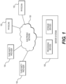
- Physicians 102 and/or third party providers 104 may transmit the cardiac/vascular images and/or patient-specific information to server systems 106 over the electronic network 100.
- Server systems 106 may include storage devices for storing images and data received from physicians 102 and/or third party providers 104.
- Server systems 106 may also include processing devices for processing images and data stored in the storage devices.
- method 200 may include obtaining patient-specific information (e.g., CT scan images, phenotype information, etc.) (step 202), constructing a patient-specific geometric model (step 204), performing flow dynamics and structural mechanics simulations on geometrical features and image features of the model, and extracting hemodynamic and mechanical characteristics (step 206). Based on the extracted characteristics and features, the server systems 106 may then perform step 208 to predict and/or report adverse plaque characteristics (APCs) (step 210). Further detail of one embodiment of step 210 is provided in FIG. 3 , in which metrics for computing APCs are determined, and metric values are found for specific patients, in order to determine APCs associated with the patients.
- APIs adverse plaque characteristics
- step 202 may comprise obtaining the model, such as by constructing a patient-specific model of coronary geometry, for instance, by modeling a patient's coronary vasculature, including one or more lumens, plaque, and/or lumen walls (step 204).
- a patient-specific model of coronary geometry for instance, by modeling a patient's coronary vasculature, including one or more lumens, plaque, and/or lumen walls (step 204).
- the patient-specific model may be constructed or rendered based on images, such as CT scans associated with a patient.
- the model may be derived by performing a cardiac CT in the end of a diastole phase of the cardiac cycle, for instance, using Black-Blood Magnetic Resonance Imaging.
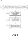
- FIG. 3 is a block diagram of an exemplary method 300 for reporting adverse plaque characteristics (APCs) from a patient-specific model.
- the method of FIG. 3 may be performed by server systems 106, based on information, images, and/or data received from physicians 102 and/or third party providers 104 over electronic network 100.
- method 300 may be performed using a patient-specific model of a patient's coronary vasculature.
- the patient-specific model may include the geometry for, at least, the patient's coronary artery tree, including the lumen, plaque, and/or lumen walls (i.e., external elastic membrane (EEM) of the coronary arteries).
- EEM external elastic membrane
- the model may be segmented manually or automatically to identify voxels belonging to the lumen and lumen wall.
- step 306 may calculate APCs. Each metric may alone constitute an APC, or the metrics may be combined in a form indicative of collective APCs. Step 306 may optionally involve calculating other risk factors.
- method 300 may include step 308 of saving the results of computed APCs scores and/or other risk factors with images as a digital representation (e.g., the memory or digital storage (e.g., hard drive, network drive) of a computational device such as a computer, laptop, DSP, server, etc.) and making them available to a physician, for instance.
- step 308 may include actively reporting APCs and/or other risk factors to physicians.
- step 308 may simply prompt or signal to a user that computed APC scores and risk factors are available for viewing and/or verification.
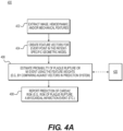
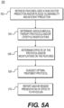
- FIG. 4A is a block diagram of an exemplary method 400 for predicting cardiac risk or risk-related features based on patient-specific models.
- the method of FIG. 4A may be performed by server systems 106, based on information, images, and data received from physicians 102 and/or third party providers 104 over electronic network 100.
- Method 400 may be performed on a patient-specific model including one or more modeled lumens, plaque, lumen walls, left and right myocardium, etc.
- the model may describe a patient's ascending aorta, coronary artery tree, myocardium, valves, and chambers. Then, segmenting may help identify voxels belonging to the aorta and the lumen of the coronary arteries.
- method 400 may include constructing the model from the patient image(s) prior to assessing the model for cardiac risk. Furthermore, method 400 may include collecting information, including patient demographics (e.g., age, gender, weight, blood pressure, etc.) and/or biomarkers (e.g., blood markers, DNA sequencing, etc.). This patient information may further inform construction of the patient-specific model.
- patient demographics e.g., age, gender, weight, blood pressure, etc.
- biomarkers e.g., blood markers, DNA sequencing, etc.
- Hemodynamic features may be extracted, for instance, by performing computational flow dynamic analysis for various physiologic conditions (e.g., rest, exercise, hyperemia, etc.) and/or computing hemodynamic characteristics associated with lesions (e.g., max/mean/cyclic wall shear stress, traction, turbulent kinetic energy, etc.).
- Extracting biomechanical features of vessel wall(s) and plaque may include defining biomechanical properties of vessel wall and plaques based on geometrical and image features (e.g., vessel wall density and elastic properties using linear or nonlinear elasticity model; plaque density and elastic properties using linear or nonlinear elasticity model; and/or ultimate strength of plaque).
- method 400 may include performing computational solid dynamic analysis for various physiologic conditions under steady and/or pulsatile flow (e.g., for rest, exercise, hyperemia, etc.). Method 400 may also include computing tissue stress and strain characteristics in lesions (e.g., max/mean/cyclic stress, ultimate stress, turbulent kinetic energy, etc.) and/or generating a Goodman diagram to identify plaque rupture risk based on mean and alternating stresses.
- step 404 may include creating a feature vector for every point in the patient-specific geometric model, comprising a numerical description of the geometry, biophysical hemodynamic, and wall and plaque biomechanical characteristic at that point, as well as estimates of physiological or phenotypic parameters of the patient. Alternately or in addition, step 404 may include determining every location in the patient-specific geometric model for which plaque vulnerability may be identified, wherein a feature vector is created only for such locations.
- method 400 may include step 408 where the estimates are reported to physicians, for instance, in the form of cardiac risk.
- the cardiac risk discussed including risk of plaque rupture, possibility of an MI event, etc. are merely exemplary instances of cardiac risk.
- Method 400 may be applied to predicting and reporting any measurement of cardiac risk.
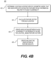
- FIG. 4B is a block diagram of an exemplary method 420 for creating and training a prediction system to predict cardiac risk.
- the prediction system trained via method 420 may permit the estimates of cardiac risk for method 400.
- the method of FIG. 4B may be performed by server systems 106, based on information, images, and data received from physicians 102 and/or third party providers 104 over electronic network 100.
- method 420 may include obtaining patient-specific models of coronary geometry based on an image of a patient (e.g., CTA). More specifically though, method 420 may involve collecting one or more models in order to create or determine models for comparison to patient-specific models undergoing analysis.
- the models may be derived from models associated with individuals, meaning patients other than the patient associated with the patient-specific model undergoing analysis. Aggregating models from a collection of individuals may provide indicators or patterns associated with MI occurrences and/or plaque vulnerability.
- Method 420 may depict the process of a machine-learning algorithm that continually updates and revises its understanding of indications of plaque vulnerability.
- method 400 may derive from presence of an MI event associated with a lesion, if there exists a sufficiently large number of MI event patients. If the number of MI events is limited, a surrogate plaque vulnerability model can be used in place of the actual MI events.
- the surrogate plaque vulnerability model can be utilized from vulnerable features characterized by invasive imaging such as optical coherence tomography (OCT), near infrared spectroscopy (NIRS) and virtual histology intravascular ultrasound (VH-IVUS).
- OCT optical coherence tomography
- NIRS near infrared spectroscopy
- VH-IVUS virtual histology intravascular ultrasound
- the general risk factors of coronary artery disease may include: smoking, diabetes, hypertension, lipid level (e.g., low density lipoprotein (LDL) cholesterol (LDL-C) levels), dietary habits, family history, physical activity, sexual activity, weight (abdominal obesity), cholesterol, and/or stress state (e.g., depression, anxiety, or distress).
- lipid level e.g., low density lipoprotein (LDL) cholesterol (LDL-C) levels
- dietary habits e.g., low density lipoprotein (LDL) cholesterol (LDL-C) levels
- family history e.g., physical activity, sexual activity, weight (abdominal obesity), cholesterol, and/or stress state (e.g., depression, anxiety, or distress).
- stress state e.g., depression, anxiety, or distress
- the biomarkers may include: complement reactive protein (CRP), fibrinogen, WBC (White blood cell) count, matrix metalloproteinase (e.g., MMP-9, MMP-3 polymorphism), IL-6, IL-18, and TCT-D (Cytokines), circulating soluble CD40 Ligand (sCD40L), and/or Vascular Calcification Markers (e.g., Osteopontin).
- CRP complement reactive protein
- fibrinogen e.g., fibrinogen
- WBC White blood cell
- matrix metalloproteinase e.g., MMP-9, MMP-3 polymorphism
- IL-6 IL-6
- IL-18 IL-18
- TCT-D Cytokines
- sCD40L circulating soluble CD40 Ligand
- Vascular Calcification Markers e.g., Osteopontin
- Acquire image features from CT, including: plaque burden (thickness, area, volume), SYNTAX score, napkin ring, and/or necrotic core
- Simulation condition e.g., rest, exercise (Low/Medium/High grade by changing degree of cardiac output), hyperemia, etc.).
- Acquire one or more estimates of vessel wall and plaque biomechanical characteristic from computational solid dynamics analysis.
- the estimates in this embodiment may include: simulation condition (pulsatile or steady flow) (rest, exercise (Low/Medium/High grade by changing degree of cardiac output), and/or hyperemia; biomechanical material properties of vessel wall and plaque derived from literature data and/or image characteristics (e.g., linear elastic, nonlinear elastic, viscoelastic constitutive models, density, compressible or incompressible material behavior, and/or ultimate strength of material; and biomechanical stress and strain (e.g., max or mean cyclic wall and plaque stress, max or mean cyclic wall and plaque strain, and/or alternating stress and strain).
- Acquire location(s) of plaque at culprit lesion being targeted for prediction of vulnerability.
- the location of plaque can be determined by use of CT and other imaging modalities including intravascular ultrasound, or optical coherence tomography.
- CAD General risk factors of CAD, such as: smoking status, diabetes, hypertension, lipid level (e.g., low density lipoprotein (LDL) cholesterol (LDL-C) levels), dietary habits, family history, physical activity, sexual activity, weight (abdominal obesity), cholesterol, and/or stress state (e.g., depression, anxiety or distress)
- LDL low density lipoprotein
- LDL-C low density lipoprotein cholesterol
- stress state e.g., depression, anxiety or distress
- Characteristics of surface of coronary geometry at lesion e.g., based on 3-D surface curvature of geometry (Gaussian, maximum, minimum, mean), e.g., based on characteristics of coronary centerline (topology) at lesion:
- Characteristics of coronary deformation (possibly involving multi-phase CCTA (e.g., diastole and systole)): distensibility of coronary artery over cardiac cycle; bifurcation angle change over cardiac cycle; and/or curvature change over cardiac cycle
- Characteristics of existing plaque location of plaque along centerline (distance to closest upstream bifurcation point, and/or bifurcation angle of coronary branches if plaque is located at the bifurcation), adverse plaque characteristics (presence of positive remodeling, presence of low attenuation plaque, and/or presence of spotty calcification), plaque burden (thickness, area, and/or volume), presence of Napkin ring, intensity of plaque, type of plaque (calcified, non-calcified), distance from the plaque location to ostium (LM or RCA), and/or distance from the plaque location to the nearest downstream/upstream bifurcation.
- pulsatile flow simulation may be performed by using a lumped parameter coronary vascular model for downstream vasculatures, inflow boundary condition with coupling a lumped parameter heart model and a closed loop model to describe the intramyocardial pressure variation resulting from the interactions between the heart and arterial system during cardiac cycle.
- plaque mean ⁇ max and alternating stress and strain, and/or ultimate stress and strength
- step 426 may include associating the feature vector with available models of plaque vulnerability at the same location.
- models may include surrogate vulnerable feature models.
- the following surrogate vulnerable features can be available at the time when cardiac images were acquired by invasive imaging such as OCT, NIRS, or VH-IVUS:
- step 426 the associations created between feature vectors and models may permit recognition of trends, similarities, and/or groupings of various factors that may indicate plaque vulnerability or likelihood or presence of MI events at specific points.
- step 426 may include quantifying the associations as feature weights, such that relationships between various factors that play into cardiac risk can be returned as predictions.
- the prediction system may assign or combine feature vectors with weights.
- Part of the training aspect of the prediction system may include continually adjusting feature weights for better accuracy in predictions.
- step 426 may include training a machine-learning algorithm (e.g. a linear SVM) to learn the associations and/or feature weights in order to predict plaque vulnerability or presence of MI event at points on a model.
- a machine-learning algorithm e.g. a linear SVM
- results (e.g. feature weights) of the machine learning algorithm-based prediction system may be continually saved as a digital representation (e.g., the memory or digital storage (e.g., hard drive, network drive) of a computational device such as a computer, laptop, DSP, server, etc.).
- Step 428 may include continually updating feature weights as more patient-specific models are collected and feature vectors constructed. Step 428, therefore, permits a prediction system that continually incorporates features input from acquired patient-specific models.
- an exemplary method may include acquiring a digital representation (e.g., the memory or digital storage (e.g., hard drive, network drive) of a patient-specific model of the geometry for the patient's ascending aorta, coronary artery tree, myocardium, valves, and chambers.
- This geometry may be represented as a list of points in space (possibly with a list of neighbors for each point) in which the space can be mapped to spatial units between points (e.g., millimeters).
- This model may be derived by performing a cardiac CT imaging of the patient in the end diastole phase of the cardiac cycle.
- This image then may be segmented manually or automatically to identify voxels belonging to the aorta and the lumen of the coronary arteries. Once the voxels are identified, the geometric model can be derived (e.g., using marching cubes). The process for generating the patient-specific model of the geometry may be the same as in the training mode. A list of physiological and phenotypic parameters of the patient may be obtained during training mode.
- the exemplary method may include using the saved results of the machine-learning algorithm produced in the training mode (e.g., feature weights) to produce estimates of the probability of the plaque rupture or MI event at lesions in the patient-specific geometric model. These estimates may be produced using the same machine learning technique used in the training mode.
- the exemplary method may include saving the predicted probability of the plaque vulnerability (rupture) for lesions or MI event to a digital representation (e.g., the memory or digital storage (e.g., hard drive, network drive) of a computational device such as a computer, laptop, DSP, server, etc.), and communicating the patient-specific risk factors to a health care provider.
- a digital representation e.g., the memory or digital storage (e.g., hard drive, network drive) of a computational device such as a computer, laptop, DSP, server, etc.
- beta-blocker e.g., metoprolol, bisoprolol, atenolol
- the algorithm may update the following features: reduce blood pressure by 10% and heart rate by 10% and/or update boundary conditions for coronary blood flow simulation and extract new hemodynamics and wall and plaque biomechanical features.
Landscapes
- Health & Medical Sciences (AREA)
- Life Sciences & Earth Sciences (AREA)
- Engineering & Computer Science (AREA)
- Medical Informatics (AREA)
- Public Health (AREA)
- General Health & Medical Sciences (AREA)
- Biomedical Technology (AREA)
- Pathology (AREA)
- Physics & Mathematics (AREA)
- Surgery (AREA)
- Veterinary Medicine (AREA)
- Heart & Thoracic Surgery (AREA)
- Molecular Biology (AREA)
- Animal Behavior & Ethology (AREA)
- Biophysics (AREA)
- Nuclear Medicine, Radiotherapy & Molecular Imaging (AREA)
- Radiology & Medical Imaging (AREA)
- High Energy & Nuclear Physics (AREA)
- Physiology (AREA)
- Optics & Photonics (AREA)
- Cardiology (AREA)
- Computer Vision & Pattern Recognition (AREA)
- Theoretical Computer Science (AREA)
- Vascular Medicine (AREA)
- Epidemiology (AREA)
- Primary Health Care (AREA)
- Data Mining & Analysis (AREA)
- Databases & Information Systems (AREA)
- Dentistry (AREA)
- Oral & Maxillofacial Surgery (AREA)
- Pulmonology (AREA)
- Psychiatry (AREA)
- Signal Processing (AREA)
- Artificial Intelligence (AREA)
- Hematology (AREA)
- Quality & Reliability (AREA)
- General Physics & Mathematics (AREA)
- Robotics (AREA)
- Apparatus For Radiation Diagnosis (AREA)
- Measuring And Recording Apparatus For Diagnosis (AREA)
Claims (10)
- Computerimplementiertes Verfahren (200, 400) zum Melden von Koronarplaqueanfälligkeit aus patientenspezifischen anatomischen Bilddaten, das Verfahren umfassend:Erfassen anatomischer Bilddaten von mindestens einem Teil des Gefäßsystems des Patienten;Aufbauen eines patientenspezifischen geometrischen Modells aus den anatomischen Bilddaten;Durchführen von Strömungsdynamik- und Strukturmechaniksimulationen an geometrischen Merkmalen und an Bildmerkmalen des Modells;Extrahieren (402) von geometrischen, bildlichen, hämodynamischen und/oder biomechanischen Merkmalen aus dem patientenspezifischen geometrischen Modell;Erzeugen (404) von Merkmalsvektoren für Punkte in dem patientenspezifischen geometrischen Modell, der Merkmalsvektor für einen Punkt umfassend eine numerische Beschreibung der Geometrie, der biophysikalischen hämodynamischen und der biomechanischen Wand- und Plaque-Charakteristik an diesem Punkt sowie Schätzungen der physiologischen oder phänotypischen Parameter des Patienten;Verwenden von Merkmalsgewichtungen zum Erzeugen (406) von Schätzungen einer Wahrscheinlichkeit einer Plaqueruptur oder Wahrscheinlichkeit eines Myokardinfarktereignisses; undMelden (408) der geschätzten Wahrscheinlichkeit einer Plaqueruptur oder eines Myokardinfarktereignisses.
- Verfahren nach Anspruch 1, und umfassend:Berechnen und Melden von ungünstigen Plaque-Charakteristiken, wobei ein Berechnen der ungünstigen Plaque-Charakteristik ein Bestimmen einer oder mehrerer Metriken beinhaltet, die mit der ungünstigen Plaque-Charakteristik assoziiert sind; undDurchführen, unter Verwendung eines Prozessors, von Berechnungen für die eine oder die mehreren Metriken, die mit der ungünstigen Plaque-Charakteristik assoziiert sind.
- Verfahren nach Anspruch 2, das ferner Folgendes beinhaltet:
Bestimmen eines oder mehrerer Schwellenwerte, die mit der einen oder den mehreren Metriken assoziiert sind, wobei das Melden der berechneten ungünstigen Plaque-Charakteristik auf dem einen oder den mehreren Schwellenwerten im Verhältnis zu den mit der einen oder den mehreren Metriken assoziierten Berechnungen basiert. - Verfahren nach Anspruch 2, wobei die eine oder die mehreren Metriken eines oder mehrere von Folgenden beinhalten:
Vorhandensein von positiver Remodellierung, Vorhandensein von Plaque mit geringer Attenuierung und Vorhandensein von fleckiger Verkalkung in Plaque. - Verfahren nach Anspruch 4, das ferner Folgendes beinhaltet:Identifizieren eines erkrankten Segments basierend auf den anatomischen Bilddaten, wobei das erkrankte Segment mit mindestens einem Grad von Stenose, einem Vorhandensein von Plaque oder einer Kombination davon assoziiert ist;Bestimmen einer Querschnittsfläche, die mit dem erkrankten Segment assoziiert ist; undDurchführen, unter Verwendung eines Prozessors, von Berechnungen zum Bestimmen eines positiven Remodellierung-Index basierend auf der Querschnittsfläche.
- Verfahren nach Anspruch 4, das ferner Folgendes beinhaltet:Identifizieren eines erkrankten Segments basierend auf den anatomischen Bilddaten, wobei das erkrankte Segment mit nicht verkalkter Plaque assoziiert ist;Bestimmen einer mit der nicht verkalkten Plaque assoziierten Intensität; undDurchführen, unter Verwendung eines Prozessors, von Berechnungen zum Bestimmen eines Volumens, das mit der nicht verkalkten Plaque assoziiert ist.
- Verfahren nach Anspruch 4, das ferner Folgendes beinhaltet:Identifizieren eines erkrankten Segments basierend auf den anatomischen Bilddaten, wobei das erkrankte Segment mit verkalkter Plaque assoziiert ist; undDurchführen, unter Verwendung eines Prozessors, von Berechnungen zum Bestimmen eines oder mehrerer Durchmesser, die mit der verkalkten Plaque assoziiert sind.
- Verfahren nach Anspruch 2, das ferner Folgendes beinhaltet: Bestimmeneines oder mehrerer kardialer Risikofaktoren; undSpeichern des einen oder der mehreren kardialen Risikofaktoren, der berechneten ungünstigen Plaque-Charakteristik oder einer Kombination davon mit den anatomischen Bilddaten, wobei ein oder mehrere kardiale Risikofaktoren, die berechnete ungünstige Plaque-Charakteristik oder eine Kombination abrufbar sind.
- System zum Melden von Koronarplaqueanfälligkeit aus patientenspezifischen anatomischen Bilddaten, das System umfassend:eine Datenspeichervorrichtung, die Anweisungen zum Vorhersagen von Koronarplaqueanfälligkeit aus patientenspezifischen anatomischen Bilddaten speichert; undeinen Prozessor, der konfiguriert ist, um die Anweisungen auszuführen, um ein Verfahren nach einem der Ansprüche 1 bis 8 durchzuführen.
- Nicht-transitorisches computerlesbares Medium zur Verwendung auf einem Computersystem, das computerausführbare Programmieranweisungen zum Durchführen eines Verfahrens zum Melden von Koronarplaqueanfälligkeit aus patientenspezifischen anatomischen Bilddaten nach einem der Ansprüche 1 bis 8 enthält.
Priority Applications (1)
| Application Number | Priority Date | Filing Date | Title |
|---|---|---|---|
| EP24178561.7A EP4404141A3 (de) | 2013-12-18 | 2014-12-17 | Systeme und verfahren zur planung einer behandlung von herz-kreislauf-erkrankungen eines patienten |
Applications Claiming Priority (4)
| Application Number | Priority Date | Filing Date | Title |
|---|---|---|---|
| US201361917639P | 2013-12-18 | 2013-12-18 | |
| US14/254,481 US9220418B2 (en) | 2013-12-18 | 2014-04-16 | Systems and methods for predicting coronary plaque vulnerability from patient-specific anatomic image data |
| PCT/US2014/070760 WO2015095282A1 (en) | 2013-12-18 | 2014-12-17 | Systems and methods for predicting coronary plaque vulnerability from patient-specific anatomic image data |
| EP14830432.2A EP3082602B1 (de) | 2013-12-18 | 2014-12-17 | Systeme und verfahren zur vorhersage von koronarplaqueanfälligkeit aus patientenspezifischen anatomischen bilddaten |
Related Parent Applications (2)
| Application Number | Title | Priority Date | Filing Date |
|---|---|---|---|
| EP14830432.2A Division EP3082602B1 (de) | 2013-12-18 | 2014-12-17 | Systeme und verfahren zur vorhersage von koronarplaqueanfälligkeit aus patientenspezifischen anatomischen bilddaten |
| EP14830432.2A Division-Into EP3082602B1 (de) | 2013-12-18 | 2014-12-17 | Systeme und verfahren zur vorhersage von koronarplaqueanfälligkeit aus patientenspezifischen anatomischen bilddaten |
Related Child Applications (1)
| Application Number | Title | Priority Date | Filing Date |
|---|---|---|---|
| EP24178561.7A Division EP4404141A3 (de) | 2013-12-18 | 2014-12-17 | Systeme und verfahren zur planung einer behandlung von herz-kreislauf-erkrankungen eines patienten |
Publications (3)
| Publication Number | Publication Date |
|---|---|
| EP3569150A2 EP3569150A2 (de) | 2019-11-20 |
| EP3569150A3 EP3569150A3 (de) | 2020-02-12 |
| EP3569150B1 true EP3569150B1 (de) | 2024-07-03 |
Family
ID=53366976
Family Applications (3)
| Application Number | Title | Priority Date | Filing Date |
|---|---|---|---|
| EP19183048.8A Active EP3569150B1 (de) | 2013-12-18 | 2014-12-17 | Systeme und verfahren zur vorhersage von koronarplaqueanfälligkeit aus patientenspezifischen anatomischen bilddaten |
| EP24178561.7A Pending EP4404141A3 (de) | 2013-12-18 | 2014-12-17 | Systeme und verfahren zur planung einer behandlung von herz-kreislauf-erkrankungen eines patienten |
| EP14830432.2A Active EP3082602B1 (de) | 2013-12-18 | 2014-12-17 | Systeme und verfahren zur vorhersage von koronarplaqueanfälligkeit aus patientenspezifischen anatomischen bilddaten |
Family Applications After (2)
| Application Number | Title | Priority Date | Filing Date |
|---|---|---|---|
| EP24178561.7A Pending EP4404141A3 (de) | 2013-12-18 | 2014-12-17 | Systeme und verfahren zur planung einer behandlung von herz-kreislauf-erkrankungen eines patienten |
| EP14830432.2A Active EP3082602B1 (de) | 2013-12-18 | 2014-12-17 | Systeme und verfahren zur vorhersage von koronarplaqueanfälligkeit aus patientenspezifischen anatomischen bilddaten |
Country Status (8)
| Country | Link |
|---|---|
| US (9) | US9220418B2 (de) |
| EP (3) | EP3569150B1 (de) |
| JP (1) | JP6203410B2 (de) |
| KR (1) | KR101737286B1 (de) |
| CN (2) | CN106061387B (de) |
| AU (1) | AU2014364889B2 (de) |
| CA (2) | CA3164247A1 (de) |
| WO (1) | WO2015095282A1 (de) |
Cited By (2)
| Publication number | Priority date | Publication date | Assignee | Title |
|---|---|---|---|---|
| US12144669B2 (en) | 2022-03-10 | 2024-11-19 | Cleerly, Inc. | Systems, devices, and methods for non-invasive image-based plaque analysis and risk determination |
| US12324695B2 (en) | 2020-01-07 | 2025-06-10 | Cleerly, Inc. | Systems, methods, and devices for medical image analysis, diagnosis, risk stratification, decision making and/or disease tracking |
Families Citing this family (102)
| Publication number | Priority date | Publication date | Assignee | Title |
|---|---|---|---|---|
| US10210956B2 (en) | 2012-10-24 | 2019-02-19 | Cathworks Ltd. | Diagnostically useful results in real time |
| EP2943902B1 (de) | 2012-10-24 | 2020-03-11 | CathWorks Ltd. | Automatisiertes messsystem und verfahren zur bewertung einer koronararterienerkrankung |
| US9135381B2 (en) | 2013-05-10 | 2015-09-15 | Stenomics, Inc. | Modeling and simulation system for optimizing prosthetic heart valve treatment |
| US9092743B2 (en) | 2013-10-23 | 2015-07-28 | Stenomics, Inc. | Machine learning system for assessing heart valves and surrounding cardiovascular tracts |
| US11568982B1 (en) | 2014-02-17 | 2023-01-31 | Health at Scale Corporation | System to improve the logistics of clinical care by selectively matching patients to providers |
| US9349178B1 (en) * | 2014-11-24 | 2016-05-24 | Siemens Aktiengesellschaft | Synthetic data-driven hemodynamic determination in medical imaging |
| US10478130B2 (en) | 2015-02-13 | 2019-11-19 | Siemens Healthcare Gmbh | Plaque vulnerability assessment in medical imaging |
| US10716513B2 (en) * | 2015-04-17 | 2020-07-21 | Heartflow, Inc. | Systems and methods for cardiovascular blood flow and musculoskeletal modeling for predicting device failure or clinical events |
| US9785748B2 (en) | 2015-07-14 | 2017-10-10 | Heartflow, Inc. | Systems and methods for estimating hemodynamic forces acting on plaque and monitoring patient risk |
| US12008751B2 (en) | 2015-08-14 | 2024-06-11 | Elucid Bioimaging Inc. | Quantitative imaging for detecting histopathologically defined plaque fissure non-invasively |
| US12026868B2 (en) | 2015-08-14 | 2024-07-02 | Elucid Bioimaging Inc. | Quantitative imaging for detecting histopathologically defined plaque erosion non-invasively |
| US11094058B2 (en) | 2015-08-14 | 2021-08-17 | Elucid Bioimaging Inc. | Systems and method for computer-aided phenotyping (CAP) using radiologic images |
| US10176408B2 (en) | 2015-08-14 | 2019-01-08 | Elucid Bioimaging Inc. | Systems and methods for analyzing pathologies utilizing quantitative imaging |
| US11113812B2 (en) | 2015-08-14 | 2021-09-07 | Elucid Bioimaging Inc. | Quantitative imaging for detecting vulnerable plaque |
| US11071501B2 (en) | 2015-08-14 | 2021-07-27 | Elucid Bioiwaging Inc. | Quantitative imaging for determining time to adverse event (TTE) |
| US11087459B2 (en) | 2015-08-14 | 2021-08-10 | Elucid Bioimaging Inc. | Quantitative imaging for fractional flow reserve (FFR) |
| US11676359B2 (en) | 2015-08-14 | 2023-06-13 | Elucid Bioimaging Inc. | Non-invasive quantitative imaging biomarkers of atherosclerotic plaque biology |
| WO2017093337A1 (en) * | 2015-12-02 | 2017-06-08 | Siemens Healthcare Gmbh | Personalized assessment of patients with acute coronary syndrome |
| JP6651402B2 (ja) * | 2016-04-12 | 2020-02-19 | キヤノンメディカルシステムズ株式会社 | 医用画像処理装置、医用画像診断装置およびプログラム |
| US10971271B2 (en) | 2016-04-12 | 2021-04-06 | Siemens Healthcare Gmbh | Method and system for personalized blood flow modeling based on wearable sensor networks |
| EP3457930B1 (de) | 2016-05-16 | 2023-11-15 | Cathworks Ltd. | System zur beurteilung von blutgefässen |
| EP4241694A3 (de) | 2016-05-16 | 2023-12-20 | Cathworks Ltd. | Auswahl von blutgefässpfaden aus bildern |
| CN106372654B (zh) * | 2016-08-29 | 2020-02-14 | 南京景三医疗科技有限公司 | 一种头颈动脉粥样斑块图像的力学分析方法 |
| EP3332710B1 (de) * | 2016-12-12 | 2020-09-16 | Siemens Healthcare GmbH | Charakterisierung von plaque |
| FR3062498B1 (fr) * | 2017-02-02 | 2019-06-07 | Casis - Cardiac Simulation & Imaging Software | Systeme et procede pour l'evaluation de risques vasculaires |
| KR101902883B1 (ko) | 2017-02-22 | 2018-10-01 | 연세대학교 산학협력단 | 컴퓨터 단층촬영 영상에서 플라크를 분석하기 위한 방법 및 장치 |
| US11263275B1 (en) * | 2017-04-03 | 2022-03-01 | Massachusetts Mutual Life Insurance Company | Systems, devices, and methods for parallelized data structure processing |
| EP3404667B1 (de) * | 2017-05-19 | 2024-02-28 | Siemens Healthineers AG | Lernbasierte verfahren zur personalisierten beurteilung, langzeitvorhersage und verwaltung von atherosklerose |
| IT201700059572A1 (it) * | 2017-05-31 | 2018-12-01 | Fond Ri Med | Metodo e sistema per la valutazione del rischio di un aneurisma dell’aorta toracica ascendente |
| US11589924B2 (en) * | 2017-08-01 | 2023-02-28 | Siemens Healthcare Gmbh | Non-invasive assessment and therapy guidance for coronary artery disease in diffuse and tandem lesions |
| JP7232245B2 (ja) * | 2017-08-30 | 2023-03-02 | コーニンクレッカ フィリップス エヌ ヴェ | モデル及びイメージングデータに基づく冠動脈健康状態予測 |
| WO2019071249A1 (en) * | 2017-10-06 | 2019-04-11 | Emory University | METHODS AND SYSTEMS FOR DETERMINING HEMODYNAMIC INFORMATION FOR ONE OR MORE ARTERIAL SEGMENTS |
| US11605447B2 (en) * | 2017-10-27 | 2023-03-14 | Siemens Healthcare Gmbh | Intelligent agents for patient management |
| US11871995B2 (en) | 2017-12-18 | 2024-01-16 | Hemolens Diagnostics Sp. Z O.O. | Patient-specific modeling of hemodynamic parameters in coronary arteries |
| KR102212499B1 (ko) | 2018-01-03 | 2021-02-04 | 주식회사 메디웨일 | Ivus 영상 분석방법 |
| EP3576097A1 (de) | 2018-05-30 | 2019-12-04 | Koninklijke Philips N.V. | Auflösung und lenkung von entscheidungsfokussen in der auf maschinellem lernen basierenden gefässbildgebung |
| EP3762937A1 (de) | 2018-03-08 | 2021-01-13 | Koninklijke Philips N.V. | Auflösung und lenkung von entscheidungsfokussen in der auf maschinellem lernen basierenden gefässbildgebung |
| WO2019173830A1 (en) | 2018-03-09 | 2019-09-12 | Emory University | Methods and systems for determining coronary hemodynamic characteristic(s) that is predictive of myocardial infarction |
| WO2019209753A1 (en) * | 2018-04-22 | 2019-10-31 | Viome, Inc. | Systems and methods for inferring scores for health metrics |
| CN108665449B (zh) * | 2018-04-28 | 2022-11-15 | 杭州脉流科技有限公司 | 预测血流矢量路径上的血流特征的深度学习模型及装置 |
| CN108742547B (zh) * | 2018-06-20 | 2021-01-08 | 博动医学影像科技(上海)有限公司 | 基于吸烟史信息获取压力差的方法及装置 |
| WO2020076874A1 (en) | 2018-10-08 | 2020-04-16 | Viome, Inc. | Methods for and compositions for determining food item recommendations |
| US11127138B2 (en) * | 2018-11-20 | 2021-09-21 | Siemens Healthcare Gmbh | Automatic detection and quantification of the aorta from medical images |
| US10813612B2 (en) | 2019-01-25 | 2020-10-27 | Cleerly, Inc. | Systems and method of characterizing high risk plaques |
| US12272262B2 (en) | 2019-02-12 | 2025-04-08 | Viome Life Sciences, Inc. | Personalizing food recommendations to reduce glycemic response |
| CN109907732B (zh) * | 2019-04-09 | 2022-12-02 | 广州新脉科技有限公司 | 一种颅内动脉瘤破裂风险的评估方法及系统 |
| US11707325B2 (en) * | 2019-05-17 | 2023-07-25 | Heartflow, Inc. | System and methods for estimation of blood flow using response surface and reduced order modeling |
| CN110223781B (zh) * | 2019-06-03 | 2021-06-04 | 中国医科大学附属第一医院 | 一种多维度斑块破裂风险预警系统 |
| WO2021026125A1 (en) | 2019-08-05 | 2021-02-11 | Elucid Bioimaging Inc. | Combined assessment of morphological and perivascular disease markers |
| WO2021030995A1 (zh) * | 2019-08-16 | 2021-02-25 | 未艾医疗技术(深圳)有限公司 | 基于vrds ai下腔静脉影像的分析方法及产品 |
| US11631500B2 (en) * | 2019-08-20 | 2023-04-18 | Siemens Healthcare Gmbh | Patient specific risk prediction of cardiac events from image-derived cardiac function features |
| JP7596092B2 (ja) * | 2019-08-30 | 2024-12-09 | キヤノン株式会社 | 情報処理装置、情報処理方法、情報処理システム及びプログラム |
| EP4023143A4 (de) * | 2019-08-30 | 2023-08-30 | Canon Kabushiki Kaisha | Informationsverarbeitungsvorrichtung, informationsverarbeitungsverfahren, informationsverarbeitungssystem und programm |
| US11350888B2 (en) * | 2019-09-03 | 2022-06-07 | Siemens Healthcare Gmbh | Risk prediction for sudden cardiac death from image derived cardiac motion and structure features |
| GB201914089D0 (en) * | 2019-09-30 | 2019-11-13 | King S College London | Apparatus and method for determining a biological characteristic |
| WO2021097393A1 (en) * | 2019-11-15 | 2021-05-20 | Geisinger Clinic | Systems and methods for machine learning approaches to management of healthcare populations |
| KR102360697B1 (ko) * | 2019-12-05 | 2022-02-10 | 연세대학교 산학협력단 | 비접촉식 생체신호 측정 시스템 |
| KR102327662B1 (ko) * | 2019-12-10 | 2021-11-17 | 한양대학교 에리카산학협력단 | 동맥류 파열 예측 장치 및 방법 |
| US20220392065A1 (en) | 2020-01-07 | 2022-12-08 | Cleerly, Inc. | Systems, methods, and devices for medical image analysis, diagnosis, risk stratification, decision making and/or disease tracking |
| US11969280B2 (en) | 2020-01-07 | 2024-04-30 | Cleerly, Inc. | Systems, methods, and devices for medical image analysis, diagnosis, risk stratification, decision making and/or disease tracking |
| CN111312374B (zh) * | 2020-01-21 | 2024-03-22 | 上海联影智能医疗科技有限公司 | 医学图像处理方法、装置、存储介质及计算机设备 |
| EP3866176A1 (de) * | 2020-02-17 | 2021-08-18 | Siemens Healthcare GmbH | Maschinenbasierte risikovorhersage für peri-prozedurale myokardinfarkte oder komplikationen aus medizinischen daten |
| WO2021193007A1 (ja) * | 2020-03-27 | 2021-09-30 | テルモ株式会社 | プログラム、情報処理方法、情報処理装置及びモデル生成方法 |
| JP7667134B2 (ja) * | 2020-03-27 | 2025-04-22 | テルモ株式会社 | プログラム、情報処理方法、情報処理装置及びモデル生成方法 |
| JP7644092B2 (ja) * | 2020-03-30 | 2025-03-11 | テルモ株式会社 | プログラム、情報処理方法、学習モデルの生成方法、学習モデルの再学習方法、および、情報処理システム |
| WO2021199961A1 (ja) * | 2020-03-30 | 2021-10-07 | テルモ株式会社 | コンピュータプログラム、情報処理方法及び情報処理装置 |
| CN111477348A (zh) * | 2020-04-09 | 2020-07-31 | 北京中腾佰脉医疗科技有限责任公司 | 基于移动互联网络的新型冠状病毒肺炎疾病防控系统 |
| US11610679B1 (en) * | 2020-04-20 | 2023-03-21 | Health at Scale Corporation | Prediction and prevention of medical events using machine-learning algorithms |
| WO2021230687A1 (ko) | 2020-05-13 | 2021-11-18 | 주식회사 루닛 | 의학 데이터로부터 바이오마커와 관련된 의학적 예측을 생성하는 방법 및 시스템 |
| US12094582B1 (en) | 2020-08-11 | 2024-09-17 | Health at Scale Corporation | Intelligent healthcare data fabric system |
| US12080428B1 (en) | 2020-09-10 | 2024-09-03 | Health at Scale Corporation | Machine intelligence-based prioritization of non-emergent procedures and visits |
| CN112294260B (zh) * | 2020-10-10 | 2022-04-05 | 浙江大学 | 一种磁兼容的光学脑功能成像方法与装置 |
| TWI768624B (zh) * | 2020-12-28 | 2022-06-21 | 財團法人國家衛生研究院 | 預測冠狀動脈的阻塞的電子裝置和方法 |
| WO2022150631A1 (en) * | 2021-01-08 | 2022-07-14 | Hdl Therapeutics, Inc | Systems and methods for reducing low attenuation plaque and/or plaque burden in patients |
| EP4284237A1 (de) * | 2021-01-27 | 2023-12-06 | Octogone Medical | System zur vorhersage von gefässplaqueruptur oder -ablösung, die zu einem schlaganfall führen und/oder zur vorhersage von gefässthrombose fähig sind |
| FR3119089B1 (fr) * | 2021-01-27 | 2024-05-24 | Octogone Medical | Système de prédiction de rupture ou de décollement de plaque vasculaire pouvant entraîner un accident vasculaire cérébral |
| CA3217882A1 (en) * | 2021-05-11 | 2022-11-17 | Ohio State Innovation Foundation | Systems and methods for modeling risk of transcatheter valve deployment |
| CN113393427B (zh) * | 2021-05-28 | 2023-04-25 | 上海联影医疗科技股份有限公司 | 斑块分析方法、装置、计算机设备和存储介质 |
| US11869186B2 (en) * | 2021-06-10 | 2024-01-09 | Elucid Bioimaging Inc. | Non-invasive determination of likely response to combination therapies for cardiovascular disease |
| US11887734B2 (en) | 2021-06-10 | 2024-01-30 | Elucid Bioimaging Inc. | Systems and methods for clinical decision support for lipid-lowering therapies for cardiovascular disease |
| US11887701B2 (en) | 2021-06-10 | 2024-01-30 | Elucid Bioimaging Inc. | Non-invasive determination of likely response to anti-inflammatory therapies for cardiovascular disease |
| US11887713B2 (en) | 2021-06-10 | 2024-01-30 | Elucid Bioimaging Inc. | Non-invasive determination of likely response to anti-diabetic therapies for cardiovascular disease |
| CN113420480B (zh) * | 2021-06-23 | 2022-05-10 | 武汉工程大学 | 一种动脉斑块破裂评估方法、装置及存储介质 |
| CN113538365B (zh) * | 2021-07-13 | 2025-05-30 | 深圳市中科微光医疗器械技术有限公司 | 一种计算腔内oct图像的ipa的方法和装置 |
| CN117769746A (zh) * | 2021-07-23 | 2024-03-26 | 纳维尔医疗有限公司 | 用于检测微钙化活性的系统和方法 |
| US12315076B1 (en) | 2021-09-22 | 2025-05-27 | Cathworks Ltd. | Four-dimensional motion analysis of a patient's coronary arteries and myocardial wall |
| CN113962948A (zh) * | 2021-10-13 | 2022-01-21 | 上海联影医疗科技股份有限公司 | 斑块稳定性检测方法、装置、计算机设备和可读存储介质 |
| CN114387464B (zh) * | 2021-12-01 | 2024-11-08 | 杭州脉流科技有限公司 | 基于ivus影像的易损斑块识别方法、计算机设备、可读存储介质和程序产品 |
| CN116211347A (zh) * | 2021-12-02 | 2023-06-06 | 深圳迈瑞生物医疗电子股份有限公司 | 一种用于血管分析的方法、超声成像设备和介质 |
| WO2023152688A1 (en) | 2022-02-10 | 2023-08-17 | Cathworks Ltd. | System and method for machine-learning based sensor analysis and vascular tree segmentation |
| US20250217981A1 (en) | 2022-03-10 | 2025-07-03 | Cleerly, Inc. | Systems, methods, and devices for image-based plaque analysis and risk determination |
| US12406365B2 (en) | 2022-03-10 | 2025-09-02 | Cleerly, Inc. | Systems, devices, and methods for non-invasive image-based plaque analysis and risk determination |
| US12440180B2 (en) | 2022-03-10 | 2025-10-14 | Cleerly, Inc. | Systems, devices, and methods for non-invasive image-based plaque analysis and risk determination |
| EP4591319A1 (de) * | 2022-09-20 | 2025-07-30 | Koninklijke Philips N.V. | Auswahlverfahren für den schutz vor zerebralen embolien für tavi-verfahren |
| CN116228731B (zh) * | 2023-03-16 | 2025-11-25 | 西北大学 | 一种多对比学习冠状动脉高危斑块检测方法、系统及终端 |
| US12446965B2 (en) | 2023-08-09 | 2025-10-21 | Cathworks Ltd. | Enhanced user interface and crosstalk analysis for vascular index measurement |
| CN117455878A (zh) * | 2023-11-08 | 2024-01-26 | 中国医学科学院北京协和医院 | 一种基于ccta图像的冠状动脉易损斑块识别方法及系统 |
| WO2025131924A1 (en) * | 2023-12-18 | 2025-06-26 | Koninklijke Philips N.V. | System and method of determining preferred treatment of pulmonary embolism using thrombolytic agent |
| CN117831699B (zh) * | 2023-12-27 | 2024-06-18 | 江苏瑞康成医疗科技有限公司 | 一种用于心脏影像检查的结构化报告系统 |
| TWI871181B (zh) * | 2024-02-07 | 2025-01-21 | 長庚醫療財團法人林口長庚紀念醫院 | 運用人工智慧分析主動脈電腦斷層影像的方法、系統及電腦可讀取記錄媒體 |
| US20250266163A1 (en) * | 2024-02-20 | 2025-08-21 | Siemens Healthineers Ag | Vulnerable plaque assessment and outcome prediction in coronary artery disease |
| CN120318175B (zh) * | 2025-03-31 | 2025-09-05 | 民航总医院 | 一种冠状动脉易损斑块检测分析系统 |
Family Cites Families (19)
| Publication number | Priority date | Publication date | Assignee | Title |
|---|---|---|---|---|
| AU2001268217A1 (en) * | 2000-06-06 | 2001-12-17 | The Research Foundation Of State University Of New York | Computer aided visualization, fusion and treatment planning |
| US7657299B2 (en) * | 2003-08-21 | 2010-02-02 | Ischem Corporation | Automated methods and systems for vascular plaque detection and analysis |
| WO2006062958A2 (en) | 2004-12-10 | 2006-06-15 | Worcester Polytechnic Institute | Image-based computational mechanical analysis and indexing for cardiovascular diseases |
| US20100278405A1 (en) | 2005-11-11 | 2010-11-04 | Kakadiaris Ioannis A | Scoring Method for Imaging-Based Detection of Vulnerable Patients |
| US20070232883A1 (en) * | 2006-02-15 | 2007-10-04 | Ilegbusi Olusegun J | Systems and methods for determining plaque vulnerability to rupture |
| US7752233B2 (en) | 2006-03-29 | 2010-07-06 | Massachusetts Institute Of Technology | Techniques for clustering a set of objects |
| US7940970B2 (en) * | 2006-10-25 | 2011-05-10 | Rcadia Medical Imaging, Ltd | Method and system for automatic quality control used in computerized analysis of CT angiography |
| US7983459B2 (en) * | 2006-10-25 | 2011-07-19 | Rcadia Medical Imaging Ltd. | Creating a blood vessel tree from imaging data |
| EP2219520B1 (de) | 2007-10-31 | 2012-12-12 | Cabra Technology A/S | Verfahren zur berechnung von drücken in einem flüssigkeitsstrom durch einen röhrenförmigen abschnitt, insbesondere ein blutgefäss mit atherosklerotischem plaque |
| FR2938957B1 (fr) * | 2008-11-21 | 2011-01-21 | Univ Joseph Fourier Grenoble I | Procede de traitement d'image pour l'estimation d'un risque de rupture de plaque d'atherome |
| US8554490B2 (en) | 2009-02-25 | 2013-10-08 | Worcester Polytechnic Institute | Automatic vascular model generation based on fluid-structure interactions (FSI) |
| CN101799864B (zh) * | 2010-01-15 | 2012-05-09 | 北京工业大学 | 一种基于血管内超声波图像的动脉斑块类型自动识别方法 |
| JP4981938B2 (ja) * | 2010-03-08 | 2012-07-25 | 富士フイルム株式会社 | 診断支援装置、冠動脈解析プログラムおよび冠動脈解析方法 |
| US20110257545A1 (en) | 2010-04-20 | 2011-10-20 | Suri Jasjit S | Imaging based symptomatic classification and cardiovascular stroke risk score estimation |
| US8157742B2 (en) | 2010-08-12 | 2012-04-17 | Heartflow, Inc. | Method and system for patient-specific modeling of blood flow |
| US8315812B2 (en) | 2010-08-12 | 2012-11-20 | Heartflow, Inc. | Method and system for patient-specific modeling of blood flow |
| US10186056B2 (en) | 2011-03-21 | 2019-01-22 | General Electric Company | System and method for estimating vascular flow using CT imaging |
| US10162932B2 (en) * | 2011-11-10 | 2018-12-25 | Siemens Healthcare Gmbh | Method and system for multi-scale anatomical and functional modeling of coronary circulation |
| CN103247071B (zh) * | 2013-03-29 | 2015-11-11 | 哈尔滨工业大学深圳研究生院 | 一种构建三维血管模型方法及设备 |
-
2014
- 2014-04-16 US US14/254,481 patent/US9220418B2/en active Active
- 2014-04-16 US US14/254,521 patent/US9155512B2/en active Active
- 2014-04-16 US US14/254,451 patent/US20150164451A1/en not_active Abandoned
- 2014-11-18 US US14/546,100 patent/US9220419B2/en active Active
- 2014-12-17 EP EP19183048.8A patent/EP3569150B1/de active Active
- 2014-12-17 EP EP24178561.7A patent/EP4404141A3/de active Pending
- 2014-12-17 WO PCT/US2014/070760 patent/WO2015095282A1/en not_active Ceased
- 2014-12-17 KR KR1020167016367A patent/KR101737286B1/ko active Active
- 2014-12-17 CN CN201480069668.XA patent/CN106061387B/zh active Active
- 2014-12-17 JP JP2016540003A patent/JP6203410B2/ja active Active
- 2014-12-17 CA CA3164247A patent/CA3164247A1/en active Pending
- 2014-12-17 AU AU2014364889A patent/AU2014364889B2/en active Active
- 2014-12-17 CA CA2933879A patent/CA2933879C/en active Active
- 2014-12-17 EP EP14830432.2A patent/EP3082602B1/de active Active
- 2014-12-17 CN CN201811609814.XA patent/CN110074756B/zh active Active
-
2015
- 2015-10-13 US US14/881,989 patent/US9770303B2/en active Active
-
2017
- 2017-08-18 US US15/680,950 patent/US10939960B2/en active Active
-
2021
- 2021-02-02 US US17/164,885 patent/US11678937B2/en active Active
-
2023
- 2023-05-09 US US18/314,396 patent/US12035976B2/en active Active
-
2024
- 2024-06-03 US US18/731,873 patent/US20240315777A1/en active Pending
Cited By (4)
| Publication number | Priority date | Publication date | Assignee | Title |
|---|---|---|---|---|
| US12324695B2 (en) | 2020-01-07 | 2025-06-10 | Cleerly, Inc. | Systems, methods, and devices for medical image analysis, diagnosis, risk stratification, decision making and/or disease tracking |
| US12396695B2 (en) | 2020-01-07 | 2025-08-26 | Cleerly, Inc. | Systems, methods, and devices for medical image analysis, diagnosis, risk stratification, decision making and/or disease tracking |
| US12144669B2 (en) | 2022-03-10 | 2024-11-19 | Cleerly, Inc. | Systems, devices, and methods for non-invasive image-based plaque analysis and risk determination |
| US12324696B2 (en) | 2022-03-10 | 2025-06-10 | Cleerly, Inc. | Systems, devices, and methods for non-invasive image-based plaque analysis and risk determination |
Also Published As
Similar Documents
| Publication | Publication Date | Title |
|---|---|---|
| US12035976B2 (en) | Systems and methods for predicting coronary plaque vulnerability from patient specific anatomic image data | |
| US11861831B2 (en) | Systems and methods for processing electronic images to predict lesions |
Legal Events
| Date | Code | Title | Description |
|---|---|---|---|
| PUAI | Public reference made under article 153(3) epc to a published international application that has entered the european phase |
Free format text: ORIGINAL CODE: 0009012 |
|
| STAA | Information on the status of an ep patent application or granted ep patent |
Free format text: STATUS: REQUEST FOR EXAMINATION WAS MADE |
|
| 17P | Request for examination filed |
Effective date: 20190627 |
|
| AC | Divisional application: reference to earlier application |
Ref document number: 3082602 Country of ref document: EP Kind code of ref document: P |
|
| AK | Designated contracting states |
Kind code of ref document: A2 Designated state(s): AL AT BE BG CH CY CZ DE DK EE ES FI FR GB GR HR HU IE IS IT LI LT LU LV MC MK MT NL NO PL PT RO RS SE SI SK SM TR |
|
| PUAL | Search report despatched |
Free format text: ORIGINAL CODE: 0009013 |
|
| AK | Designated contracting states |
Kind code of ref document: A3 Designated state(s): AL AT BE BG CH CY CZ DE DK EE ES FI FR GB GR HR HU IE IS IT LI LT LU LV MC MK MT NL NO PL PT RO RS SE SI SK SM TR |
|
| RIC1 | Information provided on ipc code assigned before grant |
Ipc: G06T 7/00 20170101ALI20200109BHEP Ipc: G06T 7/60 20170101ALI20200109BHEP Ipc: A61B 6/00 20060101AFI20200109BHEP |
|
| STAA | Information on the status of an ep patent application or granted ep patent |
Free format text: STATUS: EXAMINATION IS IN PROGRESS |
|
| 17Q | First examination report despatched |
Effective date: 20220822 |
|
| GRAP | Despatch of communication of intention to grant a patent |
Free format text: ORIGINAL CODE: EPIDOSNIGR1 |
|
| STAA | Information on the status of an ep patent application or granted ep patent |
Free format text: STATUS: GRANT OF PATENT IS INTENDED |
|
| INTG | Intention to grant announced |
Effective date: 20240216 |
|
| GRAS | Grant fee paid |
Free format text: ORIGINAL CODE: EPIDOSNIGR3 |
|
| GRAA | (expected) grant |
Free format text: ORIGINAL CODE: 0009210 |
|
| STAA | Information on the status of an ep patent application or granted ep patent |
Free format text: STATUS: THE PATENT HAS BEEN GRANTED |
|
| P01 | Opt-out of the competence of the unified patent court (upc) registered |
Effective date: 20240520 |
|
| AC | Divisional application: reference to earlier application |
Ref document number: 3082602 Country of ref document: EP Kind code of ref document: P |
|
| AK | Designated contracting states |
Kind code of ref document: B1 Designated state(s): AL AT BE BG CH CY CZ DE DK EE ES FI FR GB GR HR HU IE IS IT LI LT LU LV MC MK MT NL NO PL PT RO RS SE SI SK SM TR |
|
| REG | Reference to a national code |
Ref country code: CH Ref legal event code: EP |
|
| REG | Reference to a national code |
Ref country code: DE Ref legal event code: R096 Ref document number: 602014090455 Country of ref document: DE |
|
| REG | Reference to a national code |
Ref country code: LT Ref legal event code: MG9D |
|
| REG | Reference to a national code |
Ref country code: NL Ref legal event code: MP Effective date: 20240703 |
|
| PG25 | Lapsed in a contracting state [announced via postgrant information from national office to epo] |
Ref country code: PT Free format text: LAPSE BECAUSE OF FAILURE TO SUBMIT A TRANSLATION OF THE DESCRIPTION OR TO PAY THE FEE WITHIN THE PRESCRIBED TIME-LIMIT Effective date: 20241104 |
|
| REG | Reference to a national code |
Ref country code: AT Ref legal event code: MK05 Ref document number: 1698955 Country of ref document: AT Kind code of ref document: T Effective date: 20240703 |
|
| PG25 | Lapsed in a contracting state [announced via postgrant information from national office to epo] |
Ref country code: NL Free format text: LAPSE BECAUSE OF FAILURE TO SUBMIT A TRANSLATION OF THE DESCRIPTION OR TO PAY THE FEE WITHIN THE PRESCRIBED TIME-LIMIT Effective date: 20240703 |
|
| PG25 | Lapsed in a contracting state [announced via postgrant information from national office to epo] |
Ref country code: PT Free format text: LAPSE BECAUSE OF FAILURE TO SUBMIT A TRANSLATION OF THE DESCRIPTION OR TO PAY THE FEE WITHIN THE PRESCRIBED TIME-LIMIT Effective date: 20241104 Ref country code: NL Free format text: LAPSE BECAUSE OF FAILURE TO SUBMIT A TRANSLATION OF THE DESCRIPTION OR TO PAY THE FEE WITHIN THE PRESCRIBED TIME-LIMIT Effective date: 20240703 |
|
| PGFP | Annual fee paid to national office [announced via postgrant information from national office to epo] |
Ref country code: DE Payment date: 20241210 Year of fee payment: 11 |
|
| PG25 | Lapsed in a contracting state [announced via postgrant information from national office to epo] |
Ref country code: NO Free format text: LAPSE BECAUSE OF FAILURE TO SUBMIT A TRANSLATION OF THE DESCRIPTION OR TO PAY THE FEE WITHIN THE PRESCRIBED TIME-LIMIT Effective date: 20241003 |
|
| PG25 | Lapsed in a contracting state [announced via postgrant information from national office to epo] |
Ref country code: FI Free format text: LAPSE BECAUSE OF FAILURE TO SUBMIT A TRANSLATION OF THE DESCRIPTION OR TO PAY THE FEE WITHIN THE PRESCRIBED TIME-LIMIT Effective date: 20240703 Ref country code: GR Free format text: LAPSE BECAUSE OF FAILURE TO SUBMIT A TRANSLATION OF THE DESCRIPTION OR TO PAY THE FEE WITHIN THE PRESCRIBED TIME-LIMIT Effective date: 20241004 Ref country code: PL Free format text: LAPSE BECAUSE OF FAILURE TO SUBMIT A TRANSLATION OF THE DESCRIPTION OR TO PAY THE FEE WITHIN THE PRESCRIBED TIME-LIMIT Effective date: 20240703 |
|
| PGFP | Annual fee paid to national office [announced via postgrant information from national office to epo] |
Ref country code: GB Payment date: 20241226 Year of fee payment: 11 |
|
| PG25 | Lapsed in a contracting state [announced via postgrant information from national office to epo] |
Ref country code: BG Free format text: LAPSE BECAUSE OF FAILURE TO SUBMIT A TRANSLATION OF THE DESCRIPTION OR TO PAY THE FEE WITHIN THE PRESCRIBED TIME-LIMIT Effective date: 20240703 |
|
| PG25 | Lapsed in a contracting state [announced via postgrant information from national office to epo] |
Ref country code: LV Free format text: LAPSE BECAUSE OF FAILURE TO SUBMIT A TRANSLATION OF THE DESCRIPTION OR TO PAY THE FEE WITHIN THE PRESCRIBED TIME-LIMIT Effective date: 20240703 |
|
| PG25 | Lapsed in a contracting state [announced via postgrant information from national office to epo] |
Ref country code: AT Free format text: LAPSE BECAUSE OF FAILURE TO SUBMIT A TRANSLATION OF THE DESCRIPTION OR TO PAY THE FEE WITHIN THE PRESCRIBED TIME-LIMIT Effective date: 20240703 Ref country code: IS Free format text: LAPSE BECAUSE OF FAILURE TO SUBMIT A TRANSLATION OF THE DESCRIPTION OR TO PAY THE FEE WITHIN THE PRESCRIBED TIME-LIMIT Effective date: 20241103 |
|
| PG25 | Lapsed in a contracting state [announced via postgrant information from national office to epo] |
Ref country code: HR Free format text: LAPSE BECAUSE OF FAILURE TO SUBMIT A TRANSLATION OF THE DESCRIPTION OR TO PAY THE FEE WITHIN THE PRESCRIBED TIME-LIMIT Effective date: 20240703 Ref country code: CZ Free format text: LAPSE BECAUSE OF FAILURE TO SUBMIT A TRANSLATION OF THE DESCRIPTION OR TO PAY THE FEE WITHIN THE PRESCRIBED TIME-LIMIT Effective date: 20240703 |
|
| PG25 | Lapsed in a contracting state [announced via postgrant information from national office to epo] |
Ref country code: RS Free format text: LAPSE BECAUSE OF FAILURE TO SUBMIT A TRANSLATION OF THE DESCRIPTION OR TO PAY THE FEE WITHIN THE PRESCRIBED TIME-LIMIT Effective date: 20241003 Ref country code: ES Free format text: LAPSE BECAUSE OF FAILURE TO SUBMIT A TRANSLATION OF THE DESCRIPTION OR TO PAY THE FEE WITHIN THE PRESCRIBED TIME-LIMIT Effective date: 20240703 |
|
| PG25 | Lapsed in a contracting state [announced via postgrant information from national office to epo] |
Ref country code: RS Free format text: LAPSE BECAUSE OF FAILURE TO SUBMIT A TRANSLATION OF THE DESCRIPTION OR TO PAY THE FEE WITHIN THE PRESCRIBED TIME-LIMIT Effective date: 20241003 Ref country code: PL Free format text: LAPSE BECAUSE OF FAILURE TO SUBMIT A TRANSLATION OF THE DESCRIPTION OR TO PAY THE FEE WITHIN THE PRESCRIBED TIME-LIMIT Effective date: 20240703 Ref country code: NO Free format text: LAPSE BECAUSE OF FAILURE TO SUBMIT A TRANSLATION OF THE DESCRIPTION OR TO PAY THE FEE WITHIN THE PRESCRIBED TIME-LIMIT Effective date: 20241003 Ref country code: LV Free format text: LAPSE BECAUSE OF FAILURE TO SUBMIT A TRANSLATION OF THE DESCRIPTION OR TO PAY THE FEE WITHIN THE PRESCRIBED TIME-LIMIT Effective date: 20240703 Ref country code: IS Free format text: LAPSE BECAUSE OF FAILURE TO SUBMIT A TRANSLATION OF THE DESCRIPTION OR TO PAY THE FEE WITHIN THE PRESCRIBED TIME-LIMIT Effective date: 20241103 Ref country code: HR Free format text: LAPSE BECAUSE OF FAILURE TO SUBMIT A TRANSLATION OF THE DESCRIPTION OR TO PAY THE FEE WITHIN THE PRESCRIBED TIME-LIMIT Effective date: 20240703 Ref country code: GR Free format text: LAPSE BECAUSE OF FAILURE TO SUBMIT A TRANSLATION OF THE DESCRIPTION OR TO PAY THE FEE WITHIN THE PRESCRIBED TIME-LIMIT Effective date: 20241004 Ref country code: FI Free format text: LAPSE BECAUSE OF FAILURE TO SUBMIT A TRANSLATION OF THE DESCRIPTION OR TO PAY THE FEE WITHIN THE PRESCRIBED TIME-LIMIT Effective date: 20240703 Ref country code: ES Free format text: LAPSE BECAUSE OF FAILURE TO SUBMIT A TRANSLATION OF THE DESCRIPTION OR TO PAY THE FEE WITHIN THE PRESCRIBED TIME-LIMIT Effective date: 20240703 Ref country code: CZ Free format text: LAPSE BECAUSE OF FAILURE TO SUBMIT A TRANSLATION OF THE DESCRIPTION OR TO PAY THE FEE WITHIN THE PRESCRIBED TIME-LIMIT Effective date: 20240703 Ref country code: BG Free format text: LAPSE BECAUSE OF FAILURE TO SUBMIT A TRANSLATION OF THE DESCRIPTION OR TO PAY THE FEE WITHIN THE PRESCRIBED TIME-LIMIT Effective date: 20240703 Ref country code: AT Free format text: LAPSE BECAUSE OF FAILURE TO SUBMIT A TRANSLATION OF THE DESCRIPTION OR TO PAY THE FEE WITHIN THE PRESCRIBED TIME-LIMIT Effective date: 20240703 |
|
| REG | Reference to a national code |
Ref country code: DE Ref legal event code: R097 Ref document number: 602014090455 Country of ref document: DE |
|
| PG25 | Lapsed in a contracting state [announced via postgrant information from national office to epo] |
Ref country code: RO Free format text: LAPSE BECAUSE OF FAILURE TO SUBMIT A TRANSLATION OF THE DESCRIPTION OR TO PAY THE FEE WITHIN THE PRESCRIBED TIME-LIMIT Effective date: 20240703 Ref country code: DK Free format text: LAPSE BECAUSE OF FAILURE TO SUBMIT A TRANSLATION OF THE DESCRIPTION OR TO PAY THE FEE WITHIN THE PRESCRIBED TIME-LIMIT Effective date: 20240703 Ref country code: SM Free format text: LAPSE BECAUSE OF FAILURE TO SUBMIT A TRANSLATION OF THE DESCRIPTION OR TO PAY THE FEE WITHIN THE PRESCRIBED TIME-LIMIT Effective date: 20240703 |
|
| PG25 | Lapsed in a contracting state [announced via postgrant information from national office to epo] |
Ref country code: EE Free format text: LAPSE BECAUSE OF FAILURE TO SUBMIT A TRANSLATION OF THE DESCRIPTION OR TO PAY THE FEE WITHIN THE PRESCRIBED TIME-LIMIT Effective date: 20240703 |
|
| PG25 | Lapsed in a contracting state [announced via postgrant information from national office to epo] |
Ref country code: SK Free format text: LAPSE BECAUSE OF FAILURE TO SUBMIT A TRANSLATION OF THE DESCRIPTION OR TO PAY THE FEE WITHIN THE PRESCRIBED TIME-LIMIT Effective date: 20240703 Ref country code: IT Free format text: LAPSE BECAUSE OF FAILURE TO SUBMIT A TRANSLATION OF THE DESCRIPTION OR TO PAY THE FEE WITHIN THE PRESCRIBED TIME-LIMIT Effective date: 20240703 |
|
| PLBE | No opposition filed within time limit |
Free format text: ORIGINAL CODE: 0009261 |
|
| STAA | Information on the status of an ep patent application or granted ep patent |
Free format text: STATUS: NO OPPOSITION FILED WITHIN TIME LIMIT |
|
| 26N | No opposition filed |
Effective date: 20250404 |
|
| PG25 | Lapsed in a contracting state [announced via postgrant information from national office to epo] |
Ref country code: MC Free format text: LAPSE BECAUSE OF FAILURE TO SUBMIT A TRANSLATION OF THE DESCRIPTION OR TO PAY THE FEE WITHIN THE PRESCRIBED TIME-LIMIT Effective date: 20240703 |
|
| REG | Reference to a national code |
Ref country code: CH Ref legal event code: PL |
|
| PG25 | Lapsed in a contracting state [announced via postgrant information from national office to epo] |
Ref country code: LU Free format text: LAPSE BECAUSE OF NON-PAYMENT OF DUE FEES Effective date: 20241217 |
|
| PG25 | Lapsed in a contracting state [announced via postgrant information from national office to epo] |
Ref country code: SE Free format text: LAPSE BECAUSE OF FAILURE TO SUBMIT A TRANSLATION OF THE DESCRIPTION OR TO PAY THE FEE WITHIN THE PRESCRIBED TIME-LIMIT Effective date: 20240703 |
|
| REG | Reference to a national code |
Ref country code: BE Ref legal event code: MM Effective date: 20241231 |
|
| PG25 | Lapsed in a contracting state [announced via postgrant information from national office to epo] |
Ref country code: BE Free format text: LAPSE BECAUSE OF NON-PAYMENT OF DUE FEES Effective date: 20241231 |
|
| PG25 | Lapsed in a contracting state [announced via postgrant information from national office to epo] |
Ref country code: FR Free format text: LAPSE BECAUSE OF NON-PAYMENT OF DUE FEES Effective date: 20241231 |
|
| PG25 | Lapsed in a contracting state [announced via postgrant information from national office to epo] |
Ref country code: CH Free format text: LAPSE BECAUSE OF NON-PAYMENT OF DUE FEES Effective date: 20241231 |
|
| PG25 | Lapsed in a contracting state [announced via postgrant information from national office to epo] |
Ref country code: IE Free format text: LAPSE BECAUSE OF NON-PAYMENT OF DUE FEES Effective date: 20241217 |