EP3559248B2 - In vitro glycoengineering of antibodies - Google Patents
In vitro glycoengineering of antibodies Download PDFInfo
- Publication number
- EP3559248B2 EP3559248B2 EP17829955.8A EP17829955A EP3559248B2 EP 3559248 B2 EP3559248 B2 EP 3559248B2 EP 17829955 A EP17829955 A EP 17829955A EP 3559248 B2 EP3559248 B2 EP 3559248B2
- Authority
- EP
- European Patent Office
- Prior art keywords
- antibody
- glycosylation
- ligand
- light chain
- bound
- Prior art date
- Legal status (The legal status is an assumption and is not a legal conclusion. Google has not performed a legal analysis and makes no representation as to the accuracy of the status listed.)
- Active
Links
Classifications
-
- C—CHEMISTRY; METALLURGY
- C12—BIOCHEMISTRY; BEER; SPIRITS; WINE; VINEGAR; MICROBIOLOGY; ENZYMOLOGY; MUTATION OR GENETIC ENGINEERING
- C12P—FERMENTATION OR ENZYME-USING PROCESSES TO SYNTHESISE A DESIRED CHEMICAL COMPOUND OR COMPOSITION OR TO SEPARATE OPTICAL ISOMERS FROM A RACEMIC MIXTURE
- C12P21/00—Preparation of peptides or proteins
-
- C—CHEMISTRY; METALLURGY
- C07—ORGANIC CHEMISTRY
- C07K—PEPTIDES
- C07K1/00—General methods for the preparation of peptides, i.e. processes for the organic chemical preparation of peptides or proteins of any length
- C07K1/14—Extraction; Separation; Purification
- C07K1/16—Extraction; Separation; Purification by chromatography
- C07K1/22—Affinity chromatography or related techniques based upon selective absorption processes
-
- C—CHEMISTRY; METALLURGY
- C07—ORGANIC CHEMISTRY
- C07K—PEPTIDES
- C07K16/00—Immunoglobulins [IG], e.g. monoclonal or polyclonal antibodies
-
- C—CHEMISTRY; METALLURGY
- C12—BIOCHEMISTRY; BEER; SPIRITS; WINE; VINEGAR; MICROBIOLOGY; ENZYMOLOGY; MUTATION OR GENETIC ENGINEERING
- C12P—FERMENTATION OR ENZYME-USING PROCESSES TO SYNTHESISE A DESIRED CHEMICAL COMPOUND OR COMPOSITION OR TO SEPARATE OPTICAL ISOMERS FROM A RACEMIC MIXTURE
- C12P21/00—Preparation of peptides or proteins
- C12P21/005—Glycopeptides, glycoproteins
-
- C—CHEMISTRY; METALLURGY
- C07—ORGANIC CHEMISTRY
- C07K—PEPTIDES
- C07K2317/00—Immunoglobulins specific features
- C07K2317/20—Immunoglobulins specific features characterized by taxonomic origin
- C07K2317/21—Immunoglobulins specific features characterized by taxonomic origin from primates, e.g. man
-
- C—CHEMISTRY; METALLURGY
- C07—ORGANIC CHEMISTRY
- C07K—PEPTIDES
- C07K2317/00—Immunoglobulins specific features
- C07K2317/20—Immunoglobulins specific features characterized by taxonomic origin
- C07K2317/24—Immunoglobulins specific features characterized by taxonomic origin containing regions, domains or residues from different species, e.g. chimeric, humanized or veneered
-
- C—CHEMISTRY; METALLURGY
- C07—ORGANIC CHEMISTRY
- C07K—PEPTIDES
- C07K2317/00—Immunoglobulins specific features
- C07K2317/40—Immunoglobulins specific features characterized by post-translational modification
- C07K2317/41—Glycosylation, sialylation, or fucosylation
-
- C—CHEMISTRY; METALLURGY
- C07—ORGANIC CHEMISTRY
- C07K—PEPTIDES
- C07K2317/00—Immunoglobulins specific features
- C07K2317/50—Immunoglobulins specific features characterized by immunoglobulin fragments
- C07K2317/52—Constant or Fc region; Isotype
-
- C—CHEMISTRY; METALLURGY
- C07—ORGANIC CHEMISTRY
- C07K—PEPTIDES
- C07K2317/00—Immunoglobulins specific features
- C07K2317/50—Immunoglobulins specific features characterized by immunoglobulin fragments
- C07K2317/55—Fab or Fab'
Definitions
- the current invention is in the field of antibody engineering.
- a method for the in vitro glycoengineering of the glycosylation in the Fc-region of an antibody is reported.
- IgGs are the most abundant antibody isotypes, with IgG1 antibodies being the subclass exhibiting the most significant degree and array of effector functions. IgG1 antibodies are the most commonly used antibodies in immunotherapy, where ADCC and CDC are often deemed important.
- the CH2 domain as well as the IgG hinge region plays a major role in Fc mediated antibody effector functions.
- Each CH2 domain comprises a conserved glycosylation site at an asparagine residue located at about position 297 (numbering according to EU index of Kabat), at which a glycan moiety is covalently bound ( Wright, A. and Morrison, S.L., TIBTECH 15 (1997) 26-32 ).
- the glycans are buried between the CH2 domains, influencing the tertiary structure of the IgG molecule.
- the glycans of the Fc-region of antibodies predominantly are highly heterogeneous complex biantennary structures. While further non-conserved glycosylation sites may be present within the Fab region of an antibody, the influence of antibody glycosylation on its effector functions has been attributed to Fc-region glycosylation.
- the N-linked glycans present in the Fc-region of an antibody are known to be essential for the antibody to mediate effector functions such as ADCC ( Lively, M.R. et al. Glycobiol. 8 (1995) 813-822 ; Jefferis R. et al. Immunol Rev. 163 (1998) 59-76 ). It has been shown that the composition of the N-linked glycan affects the structure of the Fc-region of the IgG molecule and thereby alters antibody effector functions such as Fc-receptor binding, ADCC activity and CDC activity ( Presta, L., Curr. Opin. Struct. Biol. 13 (2003) 519-525 ).
- antibodies expressed in recombinant expression systems e.g. by expression in prokaryotic or eukaryotic host cells
- the N-linked glycan structure varies between individual antibody molecules. Therefore, antibodies produced in recombinant expression systems can be considered a "population of antibodies" (a term that is further used herein), with antibodies being identical in their amino acid sequence but exhibiting heterogeneity with respect to the N-linked glycan pattern of their Fc-region.
- the composition of the Fc-region glycans is known to vary between different host cell species used for expression of recombinant antibodies.
- Two commonly used host cell lines for the recombinant expression of antibodies are Chinese hamster ovary cells (CHO cells) and mouse myeloma cells (e.g. sp2/0, P3X63Ag8.653, NSO).
- CHO cells express recombinant antibodies, which are substantially devoid of terminal sialic acid residues, while a major fraction of the glycan patterns are fucosylated.
- mouse myeloma cells give rise to antibody populations with up to 50 % (relative frequency) of sialic acid residues but with less of fucose residues.
- Afucosylation of recombinantly expressed IgG may be achieved by expressing antibodies in genetically engineered host cells, e.g. Lecl3 CHO cells deficient in protein fucosylation or knockout cell lines, such as CHO cells with a knockout of the alpha-1,6-fucosyltransferase (FUT8) gene.
- FUT8 alpha-1,6-fucosyltransferase
- antibodies generated by current expression systems e.g. CHO cells
- exhibit a heterogeneous glycan pattern leading to variations in the distribution of the distinct glycan species within different batches of generated antibodies. Therefore, there is still a need for tailoring effector functions of recombinant IgG antibodies, especially for the provision of means for improving ADCC mediated by therapeutic antibodies.
- a method for producing an immunoglobulin or immunoglobulin fragment with defined glycostructure comprising the steps of providing an affinity chromatography column eluate containing the immunoglobulin or immunoglobulin fragment, incubating the affinity chromatography column eluate with (a1,3)galactosidase of plant origin, e.g. from green coffee beans (EC 3.2.1.22), applying the incubated affinity chromatography column eluate to a protein A chromatography material and recovering the immunoglobulin or immunoglobulin fragment from the protein A chromatography material and thereby producing an immunoglobulin or immunoglobulin fragment with defined glycostructure is reported.
- a1,3galactosidase of plant origin e.g. from green coffee beans (EC 3.2.1.22)
- an enzymatic method for restructuring an affinity ligand bound heterogeneous glycoform antibody sample to a substantially homogenous single desired glycoform antibody sample for therapeutic uses and kits for performing the methods.
- a method for enzymatically altering the Fc region of an affinity ligand bound antibody from a heterogeneous glycoform to a substantially homogenous single glycoform comprises: contacting the affinity ligand bound heterogeneous glycoform antibody with a reaction buffer designed for a particular glycoform modification for a time sufficient and under conditions to modify the glycoform of the Fc region to a substantially homogeneous single form; optionally adding one or more nucleotide sugars and/or cofactors; and releasing the substantially homogeneous single glycoform antibody sample from said affinity ligand.
- the invention also encompasses biopharmaceuticals comprising single glycoform mAbs and polyclonal antibodies enzymatically produced for the treatment of cancers and immune disorders as well as compositions comprising the single glycoform antibodies as a biopharmaceutic
- Hayes, et al. reported about the glycosylation and Fc receptors (Curr. Top. Microbiol. Immunol. 382 (2014) 165-199 ).
- Higel et al. reported about the N-glycosylation heterogeneity and the influence on structure, function and pharmacokinetics of monoclonal antibodies and Fc fusion proteins (Eur. J. Pharm. Biopharm. 100 (2016) 94-100 .
- a monoclonal antibody is bound to an antibody light chain affinity ligand that is specific for the kappa or the lambda constant light chain, for enzymatic modification and subsequently released as a monoclonal antibody preparation with modified glycostructure.
- This modification can be at an N-glycosylation site in the Fab fragment or in the Fc-region. It was surprisingly found that a kappa or lambda light chain specific affinity ligand bound antibody can be effectively enzymatically modified as if the antibody would be in solution.
- the method as reported herein can be easily integrated into any antibody purification process thereby providing a novel, efficient and cost-effective process of in vitro antibody glycan modification.
- the method as reported herein is useful for the modification of any monoclonal antibody without the need of modifications to the preceding up-stream production process steps.
- the method as reported herein can be integrated as a single in vitro modification and purification step into an existing process. Inherently no significant changes to existing antibody producing cell lines are required as the glycostructure modification is provided by the method as reported herein during down-stream processing.
- the enzymes used for the modification of the glycosylation of the antibody can be removed from the antibody preparation resulting in an improved preparation.
- Reported herein is a method for the enzymatic preparation/production of an antibody with a modified (substantially homogeneous) glycosylation at an N-glycosylation site wherein the antibody is bound to an antibody light chain affinity ligand that is specific for the kappa or the lambda constant light chain during the enzymatic modification.
- One aspect of the invention is a method for producing an antibody comprising the following steps:
- the method is for the enzymatic modification of the glycosylation of an N-glycosylation site of an antibody (to a substantially homogeneous glycosylation) wherein the antibody is bound to an antibody light chain affinity ligand that is specific for the kappa or the lambda constant light chain during the enzymatic modification.
- the antibodies as used in the methods as reported herein can be any antibody or antibody fragment, including Fab fragments, single chain antibodies, multispecific antibodies and antibody fusions, so long as it contains an N-glycosylation site.
- the N-glycosylation site is in the Fab or in the Fc-region.
- the antibody is selected from the group of antibodies consisting of an antibody Fab fragment, a full length antibody, a bivalent monospecific antibody, a bispecific antibody, a bivalent bispecific antibody, a trivalent bispecific antibody, a tetravalent bispecific antibody, a trivalent trispecific antibody, and a tetravalent tetraspecific antibody.
- the antibody is a bivalent monospecific antibody.
- the antibody is a bivalent or trivalent or tetravalent bispecific antibody.
- the antibody is a chimeric or humanized or human antibody.
- the antibody is a polyclonal antibody preparation.
- the antibody is a monoclonal antibody.
- the antibody (preparation) is an antibody (preparation) of the human IgG class. In one embodiment the antibody is an antibody of the human IgG1 or IgG4 subclass.
- the defined glycosylation is a glycosylation selected from the group consisting of G2 glycoform, G0 glycoform, M3 glycoform, S2 glycoform, A2B glycoform, A2BG2 glycoform and S1 glycoform.
- the defined glycosylation is a glycosylation selected from the group consisting of galactose as the terminal sugar, GlcNAc as the terminal sugar, mannose as the terminal sugar and sialic acid as the terminal sugar.
- the antibody is a recombinantly produced antibody.
- Another aspect of the invention is a method for the recombinant production of an antibody with defined glycosylation at an N-glycosylation site, comprising the steps of
- the first glycosylation modifying enzyme is a galactosyltransferase.
- the first glycosylation modifying enzyme is a galactosyltransferase and the second glycosylation modifying enzyme is a sialyltransferase.
- the galactosyltransferase is ⁇ 4GalT1.
- the sialyltransferase is ST6.
- sialyltransferase is ST6Gal1 or ST6Gal2.
- the (first) buffered solution comprises UDP-Gal.
- the (second) buffered solution comprises CMP-NANA.
- the incubation is at room temperature (20 - 25 °C, preferably about 22 °C).
- the incubation is at 25 °C.
- the incubation is at 37 °C.
- the incubation is for 7 to 48 hours.
- the solution comprises a chromatographically purified antibody
- the (first) glycosylation modifying enzyme is GalT1
- the incubation with the (first) glycosylation modifying enzyme is for 24 hours at 20-27 °C or 37 °C.
- the incubation is at room temperature (about 22 °C).
- the solution comprises a chromatographically purified antibody
- the (second) glycosylation modifying enzyme is ST6
- the incubation with the (second) glycosylation modifying enzyme is for 24 hours at 20-27°C or 37 °C.
- the incubation is at room temperature (about 22 °C).
- the solution is a buffered, cell-free cultivation supernatant comprising the antibody
- the first glycosylation modifying enzyme is GalT1
- the second glycosylation modifying enzyme is ST6, which is added 6 to 24 hours, preferably 24 hours, after the first glycosylation modifying enzyme
- the total incubation time is 24 hours to 48 hours, preferably 30 hours, at 20-27°C or 37 °C.
- the incubation is at room temperature (about 22 °C).
- One aspect of the invention is a method for producing an antibody or fragment thereof comprising the following steps in the following order:
- the N-glycosylation site is in the Fab or in the Fc-region.
- the amino acid positions of all constant regions and domains of the heavy and light chain are numbered according to the Kabat numbering system described in Kabat, et al., Sequences of Proteins of Immunological Interest, 5th ed., Public Health Service, National Institutes of Health, Bethesda, MD (1991 ) and is referred to as "numbering according to Kabat” herein.
- the Kabat numbering system see pages 647-660 of Kabat, et al., Sequences of Proteins of Immunological Interest, 5th ed., Public Health Service, National Institutes of Health, Bethesda, MD (1991 ) is used for the light chain constant domain CL of kappa and lambda isotype.
- Kabat EU index numbering system (see pages 661-723) is used for the constant heavy chain domains (CH1, Hinge, CH2 and CH3, which is herein further clarified by referring to "numbering according to Kabat EU index” in this case).
- nucleic acid is characterized by its nucleic acid sequence consisting of individual nucleotides and likewise by the amino acid sequence of a polypeptide encoded thereby.
- the term "about” denotes a range of +/- 20 % of the thereafter following numerical value. In one embodiment the term about denotes a range of +/- 10 % of the thereafter following numerical value. In one embodiment the term about denotes a range of +/-5 % of the thereafter following numerical value.
- antibody herein is used in the broadest sense and encompasses various antibody structures, including but not limited to monoclonal antibodies, polyclonal antibodies, multispecific antibodies (e.g., bispecific antibodies), and antibody fragments so long as they exhibit the desired antigen-binding activity.
- ADCC antibody-dependent cellular cytotoxicity
- the labeled cells are incubated with effector cells and the supernatant is analyzed for released Cr-51. Controls include the incubation of the target endothelial cells with effector cells but without the antibody.
- the capacity of the antibody to induce the initial steps mediating ADCC is investigated by measuring their binding to Fc ⁇ receptors expressing cells, such as cells, recombinantly expressing Fc ⁇ RI and/or Fc ⁇ RIIA or NK cells (expressing essentially Fc ⁇ RIIIA). In one preferred embodiment binding to Fc ⁇ R on NK cells is measured.
- antibody fragment refers to a molecule other than an intact antibody that comprises a portion of an intact antibody that binds the antigen to which the intact antibody binds.
- antibody fragments include but are not limited to Fv, Fab, Fab', Fab'-SH, F(ab') 2 ; diabodies; linear antibodies; single-chain antibody molecules (e.g. scFv); and multispecific antibodies formed from antibody fragments.
- chimeric antibody refers to an antibody in which a portion of the heavy and/or light chain is derived from a particular source or species, while the remainder of the heavy and/or light chain is derived from a different source or species.
- the "class" of an antibody refers to the type of constant domain or constant region possessed by its heavy chain.
- the heavy chain constant domains that correspond to the different classes of immunoglobulins are called ⁇ , ⁇ , ⁇ , ⁇ , and m, respectively.
- complement-dependent cytotoxicity refers to lysis of cells induced by the antibody as reported herein in the presence of complement.
- CDC is measured in one embodiment by the treatment of CD19 expressing human endothelial cells with an antibody as reported herein in the presence of complement.
- the cells are in one embodiment labeled with calcein.
- CDC is found if the antibody induces lysis of 20 % or more of the target cells at a concentration of 30 mg/ml.
- Binding to the complement factor C1q can be measured in an ELISA. In such an assay in principle an ELISA plate is coated with concentration ranges of the antibody, to which purified human C1q or human serum is added.
- C1q binding is detected by an antibody directed against C1q followed by a peroxidase-labeled conjugate. Detection of binding (maximal binding Bmax) is measured as optical density at 405 nm (OD405) for peroxidase substrate ABTS ® (2,2'-azino-di-[3-ethylbenzthiazoline-6-sulfonate]).
- Antibody effector functions refer to those biological activities attributable to the Fc-region of an antibody, which vary with the antibody class. Examples of antibody effector functions include: C1q binding and complement dependent cytotoxicity (CDC); Fc receptor binding; antibody-dependent cell-mediated cytotoxicity (ADCC); phagocytosis; down regulation of cell surface receptors (e.g. B cell receptor); and B cell activation.
- Fc receptor binding dependent effector functions can be mediated by the interaction of the Fc-region of an antibody with Fc receptors (FcRs), which are specialized cell surface receptors on hematopoietic cells.
- Fc receptors belong to the immunoglobulin superfamily, and have been shown to mediate both the removal of antibody-coated pathogens by phagocytosis of immune complexes, and the lysis of erythrocytes and various other cellular targets (e.g. tumor cells) coated with the corresponding antibody, via antibody dependent cell mediated cytotoxicity (ADCC) (see e.g. Van de Winkel, J.G. and Anderson, C.L., J. Leukoc. Biol. 49 (1991) 511-524 ).
- ADCC antibody dependent cell mediated cytotoxicity
- FcRs are defined by their specificity for immunoglobulin isotypes: Fc receptors for IgG antibodies are referred to as Fc ⁇ R. Fc receptor binding is described e.g. in Ravetch, J.V. and Kinet, J.P., Annu. Rev. Immunol. 9 (1991) 457-492 ; Capel, P.J., et al., Immunomethods 4 (1994) 25-34 ; de Haas, M., et al., J. Lab. Clin. Med. 126 (1995) 330-341 ; and Gessner, J.E., et al., Ann. Hematol. 76 (1998) 231-248 .
- Fc ⁇ R cross-linking of receptors for the Fc-region of IgG antibodies
- IgG4 shows reduced FcR binding
- antibodies of other IgG subclasses show strong binding.
- Pro238, Asp265, Asp270, Asn297 (loss of Fc carbohydrate), Pro329 and 234, 235, 236 and 237 Ile253, Ser254, Lys288 , Thr307, Gln311, Asn434, and His435 are residues which provide if altered also reduce FcR binding ( Shields, R.L., et al. J. Biol. Chem. 276 (2001) 6591-6604 ; Lund, J., et al., FASEB J. 9 (1995) 115-119 ; Morgan, A., et al., Immunology 86 (1995) 319-324 ; and EP 0 307 434 ).
- the antibodies as reported herein comprise as Fc-region, in one embodiment an Fc-region derived from human origin.
- the Fc-region comprises all parts of the human constant region.
- the Fc-region of an antibody is directly involved in complement activation, C1q binding, C3 activation and Fc receptor binding. While the influence of an antibody on the complement system is dependent on certain conditions, binding to C1q is caused by defined binding sites in the Fc-region. Such binding sites are known in the state of the art and described e.g. by Lukas, T.J., et al., J. Immunol. 127 (1981) 2555-2560 ; Brunhouse, R., and Cebra, J.J., Mol. Immunol.
- binding sites are e.g.
- Antibodies of subclass IgG1, IgG2 and IgG3 usually show complement activation, C1q binding and C3 activation, whereas IgG4 do not activate the complement system, do not bind C1q and do not activate C3.
- An "Fc-region of an antibody" is a term well known to the skilled artisan and defined on the basis of papain cleavage of antibodies. In one embodiment the Fc-region is a human Fc-region.
- wild-type Fc-region denotes an amino acid sequence identical to the amino acid sequence of an Fc-region found in nature.
- Wild-type human Fc-regions include a native human IgG1 Fc-region (non-A and A allotypes), native human IgG2 Fc-region, native human IgG3 Fc-region, and native human IgG4 Fc-region as well as naturally occurring variants thereof.
- Wild-type Fc-regions are denoted in SEQ ID NO: 01 (IgG1, caucasian allotype), SEQ ID NO: 02 (IgG1, afroamerican allotype), SEQ ID NO: 03 (IgG2), SEQ ID NO: 04 (IgG3) and SEQ ID NO: 05 (IgG4).
- Variant (human) Fc-regions are defined by the amino acid mutations that are contained.
- P329G denotes a variant Fc-region with the mutation of proline to glycine at amino acid position 329 relative to the parent (wild-type) Fc-region (numbering according to EU index of Kabat).
- the identity of the wild-type amino acid may be unspecified, in which case the aforementioned variant is referred to as 329G.
- a polypeptide chain of a wild-type human Fc-region of the IgG1 subclass has the following amino acid sequence starting with a cysteine residue at position 227 and ending with a glycine residue at position 446:
- a polypeptide chain of a variant human Fc-region of the IgG1 subclass with the mutations L234A and L235A has the following amino acid sequence:
- a polypeptide chain of a variant human Fc-region of the IgG1 subclass with the mutations L234A, L235A, T366S, L368A and Y407V has the following amino acid sequence:
- a polypeptide chain of a variant human Fc-region of the IgG1 subclass with the mutations L234A, L235A and T366W has the following amino acid sequence:
- a polypeptide chain of a variant human Fc-region of the IgG1 subclass with the mutations L234A, L235A and P329G has the following amino acid sequence:
- a polypeptide chain of a variant human Fc-region of the IgG1 subclass with the mutations L234A, L235A, P329G, T366S, L368A and Y407V has the following amino acid sequence:
- a polypeptide chain of a variant human Fc-region of the IgG1 subclass with the mutations L234A, L235A, P329G and T366W has the following amino acid sequence:
- a polypeptide chain of a variant human Fc-region of the IgG1 subclass with the mutations L234A, L235A, P329G, Y349C, T366S, L368A and Y407V has the following amino acid sequence:
- a polypeptide chain of a variant human Fc-region of the IgG1 subclass with the mutations L234A, L235A, P329G, S354C and T366W has the following amino acid sequence:
- a polypeptide chain of a variant human Fc-region of the IgG1 subclass with the mutations L234A, L235A, P329G, S354C, T366S, L368A and Y407V has the following amino acid sequence:
- a polypeptide chain of a variant human Fc-region of the IgG1 subclass with the mutations L234A, L235A, P329G, Y349C and T366W has the following amino acid sequence:
- a polypeptide chain of a variant human Fc-region of the IgG1 subclass with the mutations I253A, H310A and H435A has the following amino acid sequence:
- a polypeptide chain of a variant human Fc-region of the IgG1 subclass with the mutations H310A, H433A and Y436A has the following amino acid sequence:
- a polypeptide chain of a variant human Fc-region of the IgG1 subclass with the mutations M252Y, S254T and T256E has the following amino acid sequence:
- a polypeptide chain of a wild-type human Fc-region of the IgG4 subclass has the following amino acid sequence:
- a polypeptide chain of a variant human Fc-region of the IgG4 subclass with the mutations S228P and L235E has the following amino acid sequence:
- a polypeptide chain of a variant human Fc-region of the IgG4 subclass with the mutations S228P, L235E and P329G has the following amino acid sequence:
- a polypeptide chain of a variant human Fc-region of the IgG4 subclass with the mutations S228P, L235E, P329G, T366S, L368A and Y407V has the following amino acid sequence:
- a polypeptide chain of a variant human Fc-region of the IgG4 subclass with the mutations S228P, L235E, P329G and T366W has the following amino acid sequence:
- full length antibody “intact antibody,” and “whole antibody” are used herein interchangeably to refer to an antibody having a structure substantially similar to a native antibody structure or having heavy chains that contain an Fc-region as defined herein.
- glycocan denotes a polysaccharide, or oligosaccharide. Glycan is also used herein to refer to the carbohydrate portion of a glycoconjugate, such as a glycoprotein, glycolipid, glycopeptide, glycoproteome, peptidoglycan, lipopolysaccharide or a proteoglycan. Glycans usually consist solely of ⁇ -glycosidic linkages between monosaccharides. Glycans can be homo- or heteropolymers of monosaccharide residues, and can be linear or branched.
- glycosyltransferase denotes an enzyme capable of transferring the monosaccharide moiety from a nucleotide sugar to an acceptor molecule such as a sugar molecule in an oligosaccharide.
- examples of such glycosyltransferase include, but not limited to galactosyltransferase and sialyltransferase.
- hinge region denotes the part of an antibody heavy chain polypeptide that joins in a wild-type antibody heavy chain the CH1 domain and the CH2 domain, e. g. from about position 216 to about position 230 according to the EU number system of Kabat, or from about position 226 to about position 230 according to the EU number system of Kabat.
- the hinge regions of other IgG subclasses can be determined by aligning with the hinge-region cysteine residues of the IgG1 subclass sequence.
- the hinge region is normally a dimeric molecule consisting of two polypeptides with identical amino acid sequence.
- the hinge region generally comprises about 25 amino acid residues and is flexible allowing the associated target binding sites to move independently.
- the hinge region can be subdivided into three domains: the upper, the middle, and the lower hinge domain (see e.g. Roux, et al., J. Immunol. 161 (1998) 4083 ).
- a “humanized” antibody refers to a chimeric antibody comprising amino acid residues from non-human HVRs and amino acid residues from human FRs.
- a humanized antibody will comprise substantially all of at least one, and typically two, variable domains, in which all or substantially all of the HVRs (e.g., CDRs) correspond to those of a non-human antibody, and all or substantially all of the FRs correspond to those of a human antibody.
- a humanized antibody optionally may comprise at least a portion of an antibody constant region derived from a human antibody.
- a "humanized form" of an antibody, e.g., a non-human antibody refers to an antibody that has undergone humanization.
- hypervariable region refers to each of the regions of an antibody variable domain comprising the amino acid residue stretches which are hypervariable in sequence ("complementarity determining regions” or “CDRs") and/or form structurally defined loops ("hypervariable loops"), and/or contain the antigen-contacting residues ("antigen contacts").
- CDRs complementarity determining regions
- hypervariable loops form structurally defined loops
- antigen contacts Generally, antibodies comprise six HVRs; three in the VH (HI, H2, H3), and three in the VL (L1, L2, L3).
- HVRs include
- HVR residues and other residues in the variable domain are numbered herein according to Kabat et al., supra.
- an “isolated” antibody is one, which has been separated from a component of its natural environment.
- an antibody is purified to greater than 95% or 99% purity as determined by, for example, electrophoretic (e.g., SDS-PAGE, isoelectric focusing (IEF), capillary electrophoresis) or chromatographic (e.g., ion exchange or reverse phase HPLC).
- electrophoretic e.g., SDS-PAGE, isoelectric focusing (IEF), capillary electrophoresis
- chromatographic e.g., ion exchange or reverse phase HPLC
- nucleic acid refers to a nucleic acid molecule that has been separated from a component of its natural environment.
- An isolated nucleic acid includes a nucleic acid molecule contained in cells that ordinarily contain the nucleic acid molecule, but the nucleic acid molecule is present extrachromosomally or at a chromosomal location that is different from its natural chromosomal location.
- light chain denotes the shorter polypeptide chains of native IgG antibodies.
- the light chain of an antibody may be assigned to one of two types, called kappa ( ⁇ ) and lambda ( ⁇ ), based on the amino acid sequence of its constant domain, see SEQ ID NO: 27 for a human kappa light chain constant domain and SEQ ID NO: 28 for a human lambda light chain constant domain.
- monoclonal antibody refers to an antibody obtained from a population of substantially homogeneous antibodies, i.e., the individual antibodies comprising the population are identical and/or bind the same epitope, except for possible variant antibodies, e.g., containing naturally occurring mutations or arising during production of a monoclonal antibody preparation, such variants generally being present in minor amounts.
- polyclonal antibody preparations typically include different antibodies directed against different determinants (epitopes)
- each monoclonal antibody of a monoclonal antibody preparation is directed against a single determinant on an antigen.
- the modifier "monoclonal” indicates the character of the antibody as being obtained from a substantially homogeneous population of antibodies, and is not to be construed as requiring production of the antibody by any particular method.
- the monoclonal antibodies to be used in accordance with the present invention may be made by a variety of techniques, including but not limited to the hybridoma method, recombinant DNA methods, phage-display methods, and methods utilizing transgenic animals containing all or part of the human immunoglobulin loci, such methods and other exemplary methods for making monoclonal antibodies being described herein.
- “Native antibodies” refer to naturally occurring immunoglobulin molecules with varying structures.
- native IgG antibodies are heterotetrameric glycoproteins of about 150,000 daltons, composed of two identical light chains and two identical heavy chains that are disulfide-bonded. From N- to C-terminus, each heavy chain has a variable region (VH), also called a variable heavy domain or a heavy chain variable domain, followed by three constant domains (CHI, CH2, and CH3), whereby between the first and the second constant domain a hinge region is located.
- VH variable region
- CHI, CH2, and CH3 constant domains
- each light chain has a variable region (VL), also called a variable light domain or a light chain variable domain, followed by a constant light (CL) domain.
- VH variable region
- VL variable region
- CL constant light domain
- the light chain of an antibody may be assigned to one of two types, called kappa ( ⁇ ) and lambda ( ⁇ ), based on the amino acid sequence of its constant domain.
- N-linked oligosaccharide denotes oligosaccharides that are linked to the peptide backbone at an asparagine amino acid residue, by way of an asparagine-N-acetyl glucosamine linkage. N-linked oligosaccharides are also called “N-glycans.” All N-linked oligo saccharides have a common pentasaccharide core of Man3GlcNAc2. They differ in the presence of, and in the number of branches (also called antennae) of peripheral sugars such as N-acetyl glucosamine, galactose, N-acetyl galactosamine, fucose and sialic acid. Optionally, this structure may also contain a core fucose molecule and/or a xylose molecule.
- O-linked oligosaccharide denotes oligosaccharides that are linked to the peptide backbone at a threonine or serine amino acid residue.
- sialic acid denotes any member of a family of nine-carbon carboxylated sugars.
- the most common member of the sialic acid family is N-acetyl-neuraminic acid (2-keto-5-acetamido-3,5-dideoxy-D-glycero-D-galactononulopyranos-1-onic acid (often abbreviated as Neu5Ac, NeuAc, or NANA).
- a second member of the family is N-glycolyl neuraminic acid (Neu5Gc or NeuGc), in which the N-acetyl group of NeuAc is hydroxylated.
- a third sialic acid family member is 2-keto-3-deoxy-nonulosonic acid (KDN) ( Nadano et al. (1986) J. Biol. Chem. 261: 11550-11557 ; Kanamori et al., J. Biol. Chem. 265: 21811-21819 (1990 )). Also included are 9-substituted sialic acids such as a 9-O--C1-C6 acyl-NeuSAc like 9-O-lactyl-Neu5Ac or 9-O-acetyl-NeuSAc, 9-deoxy-9-fluoro-Neu5Ac and 9-azido-9-deoxyNeu5Ac.
- KDN 2-keto-3-deoxy-nonulosonic acid
- 9-substituted sialic acids such as a 9-O--C1-C6 acyl-NeuSAc like 9-O-lactyl-Neu5Ac or 9-O-acety
- sialic acid family see, e.g., Varki, Glycobiol. 2 (1992) 25-40 ; Sialic Acids: Chemistry, Metabolism and Function, R. Schauer, Ed. (Springer-Verlag, New York (1992 )).
- the synthesis and use of sialic acid compounds in a sialylation procedure is reported in WO 92/16640 , the disclosure of which is incorporated herein in its entirety.
- the term “substantially” denotes that the respective product (antibody) has a single glycosylation state, whether or not this state includes glycosylation at a single site or multiple sites.
- the antibody is substantially pure when it constitutes at least 60%, by weight, of the antibody in the preparation.
- the antibody in the preparation is at least about 75%, in certain embodiments at least about 80%, in certain embodiments at about 85%, in certain embodiments at least about 90%, in certain embodiments at least about 95%, 96%, 97%, 98% and most preferably at least about 99%, by weight, of the desired antibody.
- glycosylation state denotes a specific or desired glycosylation pattern of an antibody.
- a “glycoform” is an antibody comprising a particular glycosylation state.
- Such glycosylation patterns include, for example, attaching one or more sugars at position N-297 of the Fc-region of an antibody (numbering according to Kabat), wherein said sugars are produced naturally, recombinantly, synthetically, or semi-synthetically.
- the glycosylation pattern can be determined by many methods known in the art. For example, methods of analyzing carbohydrates on proteins have been reported in US 2006/0057638 and US 2006/0127950 (the disclosures of which are hereby incorporated by reference in their entirety).
- variable region refers to the domain of an antibody heavy or light chain that is involved in binding the antibody to antigen.
- the variable domains of the heavy chain and light chain (VH and VL, respectively) of a native antibody generally have similar structures, with each domain comprising four conserved framework regions (FRs) and three hypervariable regions (HVRs).
- FRs conserved framework regions
- HVRs hypervariable regions
- antibodies that bind a particular antigen may be isolated using a VH or VL domain from an antibody that binds the antigen to screen a library of complementary VL or VH domains, respectively. See, e.g., Portolano, S. et al., J. Immunol. 150 (1993) 880-887 ; Clackson, T. et al., Nature 352 (1991) 624-628 ).
- N-glycosylation site denotes the amino acid residue within an N-glycosylation site consensus sequence to which a glycan is or can be attached.
- N-linked glycans are attached to the amid nitrogen atom of an asparagine amino acid (Asn, N) side chain.
- the N-glycosylation site consensus sequence is Asn-X-Ser/Thr, wherein X can be any amino acid residue except proline.
- N-linked glycosylation denotes the result of the attachment of a sugar molecule oligosaccharide (denotes as glycan) to e.g. the amide nitrogen atom of asparagine.
- Human antibodies are mainly glycosylated at the asparagine residue at about position 297 (Asn297) of the heavy chain CH2 domain or in the Fab region with a more or less fucosylated biantennary complex oligosaccharide (antibody amino acid residue numbering according to Kabat, supra).
- the biantennary glycostructure can be terminated by up to two consecutive galactose (Gal) residues in each arm.
- the arms are denoted (1,6) and (1,3) according to the glycoside bond to the central mannose residue.
- the glycostructure denoted as G0 comprises no galactose residue.
- the glycostructure denoted as G1 contains one or more galactose residues in one arm.
- the glycostructure denoted as G2 contains one or more galactose residues in each arm ( Raju, T.S., Bioprocess Int. 1 (2003) 44-53 ).
- Human constant heavy chain regions are reported in detail by Kabat, supra, and by Brueggemann, M., et al., J. Exp. Med. 166 (1987) 1351-1361 ; Love, T.W., et al., Methods Enzymol. 178 (1989) 515-527 .
- CHO type glycosylation of antibody Fc-regions is e.g. described by Routier, F.H., Glycoconjugate J. 14 (1997) 201-207 .
- antibody denotes and encompasses the various forms of antibodies such as human antibodies, humanized antibodies, chimeric antibodies, or T-cell antigen depleted antibodies (see e.g. WO 98/33523 , WO 98/52976 , and WO 00/34317 ).
- the antibody in the methods according to the invention is a human or humanized antibody. Genetic engineering of antibodies is e.g. described in Morrison, S.L., et al., Proc. Natl. Acad. Sci.
- An antibody in general comprises two so called full length light chain polypeptides (light chain) and two so called full length heavy chain polypeptides (heavy chain).
- Each of the full length heavy and light chain polypeptides contains a variable domain (variable region) (generally the amino terminal portion of the full length polypeptide chain) comprising binding regions, which interact with an antigen.
- Each of the full length heavy and light chain polypeptides comprises a constant region (generally the carboxyl terminal portion).
- the constant region of the full length heavy chain mediates the binding of the antibody i) to cells bearing a Fc gamma receptor (FcyR), such as phagocytic cells, or ii) to cells bearing the neonatal Fc receptor (FcRn) also known as Brambell receptor.
- FcyR Fc gamma receptor
- FcRn neonatal Fc receptor
- variable domain of a full length antibody's light or heavy chain in turn comprises different segments, i.e. four framework regions (FR) and three hypervariable regions (CDR).
- a "full length antibody heavy chain” is a polypeptide consisting in N-terminal to C-terminal direction of an antibody heavy chain variable domain (VH), an antibody constant domain 1 (CH1), an antibody hinge region, an antibody constant domain 2 (CH2), an antibody constant domain 3 (CH3), and optionally an antibody constant domain 4 (CH4) in case of an antibody of the subclass IgE.
- a “full length antibody light chain” is a polypeptide consisting in N-terminal to C-terminal direction of an antibody light chain variable domain (VL), and an antibody light chain constant domain (CL).
- VL antibody light chain variable domain
- CL antibody light chain constant domain
- glycostructure denotes a single, defined N- or O-linked oligosaccharide at a specified amino acid residue.
- antibody with a G1 glycostructure denotes an antibody comprising at the asparagine amino acid residue at about amino acid position 297 according to the Kabat numbering scheme or in the FAB region a biantennary oligosaccharide comprising only one terminal galactose residue at the non-reducing ends of the oligosaccharide.
- oligosaccharide as used within this application denotes a polymeric saccharide comprising two or more covalently linked monosaccharide units.
- the individual sugar residues are listed from the non-reducing end to the reducing end of the oligosaccharide molecule.
- the longest sugar chain is chosen as basic chain for the notation.
- the reducing end of an N- or O-linked oligosaccharide is the monosaccharide residue, which is directly bound to the amino acid of the amino acid backbone of the antibody, whereas the end of an N- or O-linked oligosaccharide, which is located at the opposite terminus as the reducing end of the basic chain, is termed non-reducing end.
- oligosaccharides are described herein with the name or abbreviation for the non-reducing saccharide (i.e., Gal), followed by the configuration of the glycosidic bond ( ⁇ or ⁇ ), the ring bond (1 or 2), the ring position of the reducing saccharide involved in the bond (2, 3, 4, 6 or 8), and then the name or abbreviation of the reducing saccharide (i.e., GlcNAc).
- Each saccharide is preferably a pyranose.
- defined glycostructure denotes within this application a glycostructure in which the monosaccharide residue at the non-reducing ends of the glycostructure is of a specific kind.
- defined glycostructure denotes within this application a glycostructure in which the monosaccharide residue at the non-reducing end of glycostructures are defined and of a specific kind.
- affinity chromatography denotes a chromatography method which employs an "affinity chromatography material".
- affinity chromatography antibodies are separated based on their biological activity or chemical structure depending on the formation of electrostatic interactions, hydrophobic bonds, and/or hydrogen bonds to the chromatographical functional groups of the chromatography material.
- a competitor ligand can be added or the chromatography conditions, such as pH value, polarity or ionic strength of the buffer, can be changed.
- affinity chromatography materials are metal chelating chromatography materials such as Ni(II)-NTA or Cu(II)-NTA, or antibody affinity chromatography materials such as chromatography materials comprising thereto covalently linked protein A or protein G, or enzyme binding affinity chromatography materials such as chromatography materials comprising thereto covalently bound enzyme substrate analogues, enzyme cofactors, or enzyme inhibitors as chromatographical functional group, or lectin binding chromatography materials such as chromatography materials comprising thereto covalently linked polysaccharides, cell surface receptors, glycoproteins, or intact cells as chromatographical functional group.
- the antibody light chain affinity ligand uses a light chain constant domain specific capture reagent, which is specific for the kappa or the lambda constant light chain, depending on whether a kappa or a lambda light chain is contained in the antibody.
- light chain constant domain specific capture reagents are e.g. KappaSelect TM and LambdaFabSelect TM (available from GE Healthcare/BAC), which are based on a highly rigid agarose base matrix that allows high flow rates and low back pressure at large scale. These materials contain a ligand that binds to the constant region of the kappa or the lambda light chain, respectively (antibodies or fragments thereof lacking the constant region of the light chain will not bind).
- Both are therefore capable of binding other target molecules containing the constant region of the light chain, for example, IgG, IgA and IgM.
- the ligands are attached to the matrix via a long hydrophilic spacer arm to make them easily available for binding to the target molecule. They are based on a single-chain antibody fragment that is screened for either human Ig kappa or lambda.
- light chain denotes the shorter polypeptide chains of native IgG antibodies.
- the light chain of an antibody may be assigned to one of two types, called kappa ( ⁇ ) and lambda ( ⁇ ), based on the amino acid sequence of its constant domain, see SEQ ID NO: 27 for a human kappa light chain constant domain and SEQ ID NO: 28 for a human lambda light chain constant domain.
- applying to denotes a partial step of a purification method in which a solution containing a substance of interest is brought in contact with a stationary phase.
- the solution containing the substance of interest to be purified passes through the stationary phase providing for an interaction between the stationary phase and the substances in solution.
- some substances of the solution are bound to the stationary phase and therewith are removed from the solution. Other substances remain in solution. The substances remaining in solution can be found in the flow-through.
- the "flow-through” denotes the solution obtained after the passage of the chromatographic device, which may either be the applied solution containing the substance of interest or the buffer, which is used to flush the column or to cause elution of one or more substances bound to the stationary phase.
- the substance of interest can be recovered from the solution after the purification step by methods familiar to a person of skill in the art, such as e.g. precipitation, salting out, ultrafiltration, diafiltration, lyophilization, affinity chromatography, or solvent volume reduction to obtain the substance in substantially homogeneous form.
- an antibody or antibody fragment whose glycostructure can be modified in the methods as reported herein can be produced by recombinant means.
- Methods for recombinant production are widely known in the state of the art and comprise protein expression in eukaryotic cells with subsequent isolation of the antibody or antibody fragment and purification to a pharmaceutically acceptable purity.
- a hybridoma cell or a eukaryotic cell in which one or more nucleic acids encoding the antibody or antibody fragment have been introduced, is used.
- the eukaryotic cells is selected from CHO cells, NS0 cells, SP2/0 cells, HEK 293 cells, COS cells, PER.C6 cells, BHK cells, rabbit cells, or sheep cells.
- the eukaryotic cell is selected from CHO cells, HEK cells, or rabbit cells. After expression the antibody or antibody fragment is recovered from the cells (from the supernatant or from the cells after lysis).
- General methods for recombinant production of antibodies are well-known in the state of the art and reported, for example, in the review articles of Makrides, S.C., Protein Expr. Purif. 17 (1999) 183-202 ; Geisse, S., et al., Protein Expr. Purif. 8 (1996) 271-282 ; Kaufman, R.J., Mol. Biotechnol. 16 (2000) 151-160 ; Werner, R.G., Drug Res. 48 (1998) 870-880 .
- Purification of antibodies can be performed in order to eliminate cellular components or other contaminants, e.g. other cellular nucleic acids or proteins, by standard techniques, including alkaline/SDS treatment, CsCl banding, column chromatography, agarose gel electrophoresis, and others well known in the art (see e.g. Ausubel, F.M, et al. (eds.), Current Protocols in Molecular Biology, John Wiley & Sons, Inc., New York (2005 )).
- Different methods are well established and widespread used for protein purification, such as affinity chromatography with microbial proteins (e.g. protein A or protein G affinity chromatography), ion exchange chromatography (e.g.
- cation exchange (carboxymethyl resins), anion exchange (amino ethyl resins) and mixed-mode exchange), thiophilic adsorption (e.g. with beta-mercaptoethanol and other SH ligands), hydrophobic interaction or aromatic adsorption chromatography (e.g. with phenyl-sepharose, aza-arenophilic resins, or m-aminophenylboronic acid), metal chelate affinity chromatography (e.g.
- Ni(II)- and Cu(II)-affinity material size exclusion chromatography
- electrophoretical methods such as gel electrophoresis, capillary electrophoresis
- affinity chromatography with microbial proteins e.g. Vijayalakshmi, M.A., Appl. Biochem. Biotech. 75 (1998) 93-102 ).
- General chromatographic methods and their use are known to a person skilled in the art. See for example, Heftmann, E.
- chromatography steps For the purification of antibodies or antibody fragments, which have been produced e.g. by cell cultivation methods, generally a combination of different chromatography steps can be employed. Normally a (protein A) affinity chromatography is followed by one or two additional separation steps. In one embodiment the additional chromatography steps are a cation and an anion exchange chromatography step or vice versa.
- the final purification step is a so called “polishing step" for the removal of trace impurities and contaminants like aggregated immunoglobulins, residual HCP (host cell protein), DNA (host cell nucleic acid), viruses, or endotoxins. In one embodiment the final purification step is an anion exchange chromatography in flow-through mode.
- glycostructure of a recombinantly produced antibody or antibody fragment will be determined by the employed cell line and the employed cultivation conditions. With conventional downstream processing techniques selective removal of specific glycostructures is not possible.
- recombinantly produced monoclonal antibodies are generally comprising at their glycosylation sites a heterogeneous mixture of glycoforms.
- This glycosylation profile is influenced by different factors during the recombinant production, such as the enzyme activities present in the host cell as well as in the cultivation medium, and the cultivation conditions.
- the method according to the invention provides an antibody with defined glycosylation at an N-glycosylation site, e.g. at an N-glycosylation site in the Fab region or in the Fc-region, i.e. containing essentially a single glycoform attached to the glycosylation site, e.g. at Asn297 in the Fc-region, by enzymatically modifying the glycan at the N-glycosylation site following harvesting the antibody from a culture.
- the method according to the invention has the advantage that it can be easily incorporated into standard operating procedures used in antibody purification from culture supernatant. Because the antibody is bound to the antibody light chain affinity ligand that is specific for the kappa or the lambda constant light chain, which in turn is immobilized on a solid phase, amongst other things the amount of the enzymes employed for the modification can be reduced compared to the amount that would be required if the modification would be performed in solution; additionally the entire modification can be achieved in a single step.
- antibody with defined glycosylation or "antibody with defined glycostructure” denotes a population of antibody molecules wherein a limited number of different glycans are attached to a (predetermined) N-glycosylation site, e.g. in the Fc-region at Asn297 (numbering according to EU index of Kabat).
- one of the glycans account for 50 % or more of the G0F, G1F and G2F glycoforms or for 30 % or more of the G0F, G1F, G2F, G1S1F, G2S1F and G2S2F glycoforms.
- substantially denotes that 40 % or more, in one embodiment 50 % or more, of the compounds has the same glycosylation, i.e. comprises the same glycan at the N-glycosylation site, e.g. at Asn297 (numbering according to Kabat) in the Fc-region.
- antibodies irrespective of type and size, can be modified to comprise a defined glycoform.
- the glycosylation of an N-glycosylation site e.g. in the Fc-region
- the glycosylation of an N-glycosylation site can be tailor-made, e.g. for the intended therapeutic applications of the antibody.
- galactosylation of the Fc-region of the antibody is useful for the treatment of cancers.
- sialylation of the Fc-region of an antibody to a defined glycoform is useful in the treatment of autoimmune disorders.
- de-galactosylation may be desired and/or de-sialylation of the Fc-region.
- an antibody with a G2 glycoform can be produced from a heterogeneous population of monoclonal antibodies using the method as reported herein.
- the same method can be used to convert non-fucosylated heterogeneous antibodies, which can be produced by glyco-engineering methods, to homogeneous G2-glycoforms.
- the batch to batch variability of galactosylation of antibodies can also be addressed by modulating the galactosylation to a desired level using the method as reported herein.
- the method according to the invention comprises the steps of applying a solution comprising an antibody with glycosylation at an N-glycosylation site, e.g. in the Fc-region, to an antibody light chain affinity ligand that is specific for the kappa or the lambda constant light chain and is immobilized to a solid phase/support.
- the support comprises a column that is washed with wash buffer and then with a reaction buffer solution that is suitable for a corresponding desired enzymatic on column glycostructure modification.
- the reaction buffer can be further optimized with the addition of selected secondary enzyme(s), optionally cofactor(s) and optionally nucleotide sugar(s).
- the column is then incubated, either at room temperature or at an elevated temperature of about 37 °C.
- the column is thereafter washed with the wash buffer and the modified monoclonal antibody with a defined glycoform is eluted from the solid support using an elution buffer.
- the eluted antibody may then be neutralized using a neutralization buffer
- the nucleotide sugars for use in the reaction buffer are selected from the group consisting of UDP-Glc, UDP-Gal, UDP-GalNAc, UDP-GlcNAc, UDP-GlcUA, UDP-Xyl, GDP-Man, GDP-Fuc, CMP-NeuSAc, CMP-NeuSGc and combinations thereof. Concentrations used in the reaction buffer are in the range of about 0.5 mM to about 5 mM, in embodiments from about 1 mM to about 1.5 mM.
- the cofactor for use in the reaction buffer may be selected from the group consisting of Mn 2+ , Ca 2+ , Mg 2+ , Na + , K + , ⁇ -Lactalbumin and combinations thereof. Concentrations of cofactor for use in the reaction buffer may be in the range of about 2 mM to about 10 mM.
- the antibody light chain affinity ligand that is specific for the kappa or the lambda constant light chain is immobilized on a solid phase that is retained in the column during the purification and modification process.
- the solid phase includes but is not limited to agarose, sepharose, polyacrylic, polystyrene and other synthetic polymers, which provide negligible non-specific adsorption of non-target proteins and enzymes of modification.
- the affinity ligand is covalently bound to the solid phase by, for example any of a variety of chemistries, such as N-hydroxysuccinimide (NHS) esters, epoxide, aldehyde, or cyanogen bromide, to a solid phase.
- NHS N-hydroxysuccinimide
- epoxide epoxide
- aldehyde aldehyde
- cyanogen bromide cyanogen bromide
- the wash buffer assures that a high affinity between antibody and affinity ligand during the washing steps is maintained.
- phosphate buffered saline solution (PBS) with pH of about 7.2 can be used as wash buffer, however it is understood by one of skill in the art that the pH may vary to some degree.
- PBS phosphate buffered saline solution
- the wash and reaction buffers assure that high affinity between antibody and affinity ligand is maintained and, at the same time, the activity of the respective enzyme(s) is maintained.
- the wash and reaction buffers are used at temperatures of about 25°C to about 40°C, and any temperature therein between. Temperatures of about 37°C are often used.
- the optimum pH range for high affinity of antibodies to the light chain affinity ligand is about 6.0 to about 8.0.
- the buffers overlap with optimum pH ranges of the affinity ligands that can be used in the method as reported herein. These include but are not limited to TRIS buffer, BIS-TRIS buffer, MES buffer, BES buffer, MOPS buffer and HEPES buffer.
- Washing conditions for the affinity column minimizes non-specific binding and, thus, affect enzyme reaction and, thus, antibody modification. Wash conditions are such that they will not break the bind between the antibody light chain affinity ligand and the target monoclonal antibody.
- Enzymes suitable for use in the methods according to the invention can be selected depending on the modification from the group consisting of mannosyl-glucosamine transferases (MGAT1, MGAT2 and MGAT3); galactosyltransferases ( ⁇ 4GalT1, ⁇ 4GalT2, ⁇ 4GalT3, ⁇ 4GalT4, ⁇ 4GalT5, ⁇ 4GalT6, ( ⁇ 4GalT7), sialyltransferases (ST6Gal1, ST6Gal2); mannosidases ( ⁇ mannosidase-I, ⁇ mannosidase-II, ⁇ (1-2) mannosidase, ⁇ (1-6) mannosidase, ⁇ (1-2,3) mannosidase, ⁇ (1-2,3,6) mannosidase); hexosaminidases ( ⁇ -N-acetyl hexosaminidase, ⁇ -N-acetyl glucosaminidase,
- the method according to the invention can be used to remove or add the terminal sialic acid from galactose for the generation of an antibody with homogeneous G2 glycostructure, e.g. in the Fc-region. Therefore, for example, a non-specific neuraminidase enzyme can be utilized which removes the sialic acid from any linkage or a specific sialidase that add the respective sialic acid. This enzyme can be used in combination with a galactosyltransferase to concomitantly effect galactosylation and removal or addition of sialic acid.
- an antibody with a defined G2 glycoform e.g. in the Fc-region
- an antibody with a glycosylation e.g. in the Fc-region, comprising at least the glycoforms G0, G1, G2, G1S1 and G2S2.
- the modification of the glycosylation of an antibody according to the method according to the invention can be performed using a sequential incubation with the individual enzymes, or a semi-sequential incubation, wherein the first enzyme is added and the second enzyme is added after a certain period of time while the first enzyme is not removed, or a simultaneous incubation with both enzyme being present together. Any of these protocols results in an improved modification compared to the modification completely in solution reaction or to the modification with the antibody immobilized on protein A.
- the method according to the invention is exemplified in the following by providing an antibody with defined galactosylation and sialylation in the Fc-region by use of corresponding transferase enzymes.
- a purified humanized antibody of the IgG1 subclass was applied to protein A affinity chromatography material and an antibody light chain affinity ligand chromatography material specific for the kappa constant light chain (Kappa select from GE Healthcare).
- the bound antibody was incubated on-column with a buffered solution comprising a galactosyltransferase (GalT1) and UDP-GAL.
- GalT1 galactosyltransferase
- UDP-GAL UDP-GAL
- a purified humanized antibody of the IgG1 subclass with a homogeneous glycosylation in the Fc-region was applied to protein A affinity chromatography material and an antibody light chain affinity ligand chromatography material specific for the kappa constant light chain (Kappa select from GE Healthcare).
- the bound antibody was incubated on-column with a buffered solution comprising a sialyltransferase (ST6) and CMP-NANA.
- ST6 sialyltransferase
- CMP-NANA CMP-NANA
- a human antibody of the IgG4 subclass was applied to protein A affinity chromatography material and an antibody light chain affinity ligand chromatography material specific for the kappa constant light chain (Kappa select from GE Healthcare).
- the bound antibody was incubated on-column with a buffered solution comprising a galactosyltransferase (GaIT1) and UDP-GAL.
- GaIT1 galactosyltransferase
- UDP-GAL UDP-GAL
- a human antibody of the IgG4 subclass with a homogeneous glycosylation in the Fc-region was applied to protein A affinity chromatography material and an antibody light chain affinity ligand chromatography material specific for the kappa constant light chain (Kappa select from GE Healthcare).
- the bound antibody was incubated on-column with a buffered solution comprising a sialyltransferase (ST6) and CMP-NANA.
- ST6 sialyltransferase
- CMP-NANA CMP-NANA
- a humanized antibody of the IgG1 subclass with an additional glycosylation site in the Fab was applied to protein A affinity chromatography material and an antibody light chain affinity ligand chromatography material specific for the kappa constant light chain (Kappa select from GE Healthcare).
- the bound antibody was incubated on-column with a buffered solution comprising a sialyltransferase (ST6) and CMP-NANA.
- ST6 sialyltransferase
- CMP-NANA CMP-NANA
- an antibody modified in the method according to the invention is a chimeric antibody.
- chimeric antibodies are described, e.g., in US 4,816,567 ; and Morrison, S.L. et al., Proc. Natl. Acad. Sci. USA 81 (1984) 6851-6855 ).
- a chimeric antibody comprises a non-human variable region (e.g., a variable region derived from a mouse, rat, hamster, rabbit, or non-human primate, such as a monkey) and a human constant region.
- a chimeric antibody is a "class switched" antibody in which the class or subclass has been changed from that of the parent antibody. Chimeric antibodies include antigen-binding fragments thereof as long as these bind to the antibody light chain affinity ligand used in the method as reported herein.
- a chimeric antibody is a humanized antibody.
- a non-human antibody is humanized to reduce immunogenicity to humans, while retaining the specificity and affinity of the parental non-human antibody.
- a humanized antibody comprises one or more variable domains in which HVRs, e.g., CDRs, (or portions thereof) are derived from a non-human antibody, and FRs (or portions thereof) are derived from human antibody sequences.
- HVRs e.g., CDRs, (or portions thereof) are derived from a non-human antibody
- FRs or portions thereof
- a humanized antibody optionally will also comprise at least a portion of a human constant region.
- some FR residues in a humanized antibody are substituted with corresponding residues from a non-human antibody (e.g., the antibody from which the HVR residues are derived), e.g., to restore or improve antibody specificity or affinity.
- a non-human antibody e.g., the antibody from which the HVR residues are derived
- an antibody modified in the method according to the invention is a human antibod
- Antibodies are not part of the invention.
- Human antibodies can be produced using various techniques known in the art. Human antibodies are described generally in van Dijk, M.A. and van de Winkel, J.G., Curr. Opin. Pharmacol. 5 (2001) 368-374 and Lonberg, N., Curr. Opin. Immunol. 20 (2008) 450-459 .
- Human antibodies may be prepared by administering an immunogen to a transgenic animal that has been modified to produce intact human antibodies or intact antibodies with human variable regions in response to antigenic challenge.
- Such animals typically contain all or a portion of the human immunoglobulin loci, which replace the endogenous immunoglobulin loci, or which are present extrachromosomally or integrated randomly into the animal's chromosomes. In such transgenic mice, the endogenous immunoglobulin loci have generally been inactivated.
- Human antibodies can also be made by hybridoma-based methods. Human myeloma and mouse-human heteromyeloma cell lines for the production of human monoclonal antibodies have been described (see, e.g., Kozbor, D., J. Immunol. 133 (1984) 3001-3005 ; Brodeur, B.R. et al., Monoclonal Antibody Production Techniques and Applications, Marcel Dekker, Inc., New York (1987), pp. 51-63 ; and Boerner, P. et al., J. Immunol. 147 (1991) 86-95 ). Human antibodies generated via human B-cell hybridoma technology are also described in Li, J. et al., Proc. Natl.
- Human antibodies may also be generated by isolating Fv clone variable domain sequences selected from human-derived phage display libraries. Such variable domain sequences may then be combined with a desired human constant domain. Techniques for selecting human antibodies from antibody libraries are described below.
- Antibodies modified in the method according to the invention may be isolated by screening combinatorial libraries for antibodies with the desired activity or activities. For example, a variety of methods are known in the art for generating phage display libraries and screening such libraries for antibodies possessing the desired binding characteristics. Such methods are reviewed, e.g., in Hoogenboom, H.R. et al., Methods in Molecular Biology 178 (2001) 1-37 and further described, e.g., in the McCafferty, J. et al., Nature 348 (1990) 552-554 ; Clackson, T. et al., Nature 352 (1991) 624-628 ; Marks, J.D. et al., J. Mol. Biol.
- repertoires of VH and VL genes are separately cloned by polymerase chain reaction (PCR) and recombined randomly in phage libraries, which can then be screened for antigen-binding phage as described in Winter, G. et al., Ann. Rev. Immunol. 12 (1994) 433-455 .
- Phage typically display antibody fragments, either as single-chain Fv (scFv) fragments or as Fab fragments.
- scFv single-chain Fv
- Libraries from immunized sources provide high-affinity antibodies to the immunogen without the requirement of constructing hybridomas.
- naive repertoire can be cloned (e.g., from human) to provide a single source of antibodies to a wide range of non-self and also self-antigens without any immunization as described by Griffiths, A.D. et al., EMBO J. 12 (1993) 725-734 .
- naive libraries can also be made synthetically by cloning non-rearranged V-gene segments from stem cells, and using PCR primers containing random sequence to encode the highly variable CDR3 regions and to accomplish rearrangement in vitro, as described by Hoogenboom, H.R. and Winter, G., J. Mol. Biol. 227 (1992) 381-388 .
- Patent publications describing human antibody phage libraries include, for example: US 5,750,373 , and US 2005/0079574 , US 2005/0119455 , US 2005/0266000 , US 2007/0117126 , US 2007/0160598 , US 2007/0237764 , US 2007/0292936 , and US 2009/0002360 .
- Antibodies or antibody fragments isolated from human antibody libraries are considered human antibodies or human antibody fragments herein.
- an antibody modified in the method according to the invention is a multispecific antibody, e.g. a bispecific antibody.
- Multispecific antibodies are monoclonal antibodies that have binding specificities for at least two different sites.
- Bispecific antibodies can be prepared as full length antibodies or antibody fragments. Fragments of multispecific (bispecific) antibodies are encompassed as long as these bind to the antibody light chain affinity ligand as used in the methods as reported herein.
- Techniques for making multispecific antibodies include, but are not limited to, recombinant co-expression of two immunoglobulin heavy chain-light chain pairs having different specificities (see Milstein, C. and Cuello, A.C., Nature 305 (1983) 537-540 , WO 93/08829 , and Traunecker, A. et al., EMBO J. 10 (1991) 3655-3659 ), and "knob-in-hole” engineering (see, e.g., US 5,731,168 ).
- Multi-specific antibodies may also be made by engineering electrostatic steering effects for making antibody Fc-heterodimeric molecules ( WO 2009/089004 ); cross-linking two or more antibodies or fragments (see, e.g., US 4,676,980 , and Brennan, M. et al., Science 229 (1985) 81-83 ); using leucine zippers to produce bi-specific antibodies (see, e.g., Kostelny, S.A. et al., J. Immunol. 148 (1992) 1547-1553 ; using "diabody” technology for making bispecific antibody fragments (see, e.g., Holliger, P. et al., Proc. Natl. Acad. Sci.
- the antibody or fragment modified in the method as reported herein also includes a "Dual Acting Fab” or “DAF” (see, US 2008/0069820 , for example).
- the antibody or fragment herein also includes multispecific antibodies described in WO 2009/080251 , WO 2009/080252 , WO 2009/080253 , WO 2009/080254 , WO 2010/112193 , WO 2010/115589 , WO 2010/136172 , WO 2010/145792 , and WO 2010/145793 .
- Antibodies may be produced using recombinant methods and compositions, e.g., as described in US 4,816,567 . For these methods one or more isolated nucleic acid(s) encoding an antibody are provided.
- nucleic acids In case of a native antibody or native antibody fragment two nucleic acids are required, one for the light chain or a fragment thereof and one for the heavy chain or a fragment thereof.
- Such nucleic acid(s) encode an amino acid sequence comprising the VL and/or an amino acid sequence comprising the VH of the antibody (e.g., the light and/or heavy chain(s) of the antibody).
- These nucleic acids can be on the same expression vector or on different expression vectors.
- one of the heterodimeric heavy chain comprises to so-called “knobs mutations” (T366W and optionally one of S354C or Y349C) and the other comprises the so-called “hole mutations” (T366S, L368A and Y407V and optionally Y349C or S354C) (see, e.g., Carter, P.
- nucleic acid(s) encode an amino acid sequence comprising the first VL and/or an amino acid sequence comprising the first VH including the first heteromonomeric Fc-region and/or an amino acid sequence comprising the second VL and/or an amino acid sequence comprising the second VH including the second heteromonomeric Fc-region of the antibody (e.g., the first and/or second light and/or the first and/or second heavy chains of the antibody).
- nucleic acids can be on the same expression vector or on different expression vectors, normally these nucleic acids are located on two or three expression vectors, i.e. one vector can comprise more than one of these nucleic acids. Examples of these bispecific antibodies are CrossMabs and T-cell bispecific antibodies (see, e.g. Schaefer, W. et al, Proc. Natl. Acad. Sci. USA, 108 (2011) 11187-1191 ).
- Isolated nucleic acids encoding an antibody as used in the methods as reported herein are provided.
- One or more vectors comprising such nucleic acid(s) are provided.
- a host cell comprising such nucleic acid(s) is provided.
- a host cell comprises (e.g., has been transformed with):
- the host cell is eukaryotic, e.g. a Chinese Hamster Ovary (CHO) cell or lymphoid cell (e.g., Y0, NS0, Sp20 cell).
- a method of making an antibody comprises culturing a host cell comprising nucleic acids encoding the antibody, as provided above, under conditions suitable for expression of the antibody, optionally recovering the antibody from the host cell (or host cell culture medium), and modifying the glycosylation of the antibody with a method according to the invention.
- nucleic acids encoding an antibody are isolated and inserted into one or more vectors for further cloning and/or expression in a host cell.
- Such nucleic acids may be readily isolated and sequenced using conventional procedures (e.g., by using oligonucleotide probes that are capable of binding specifically to genes encoding the heavy and light chains of the antibody) or produced by recombinant methods or obtained by chemical synthesis.
- Suitable host cells for cloning or expression of antibody-encoding vectors include prokaryotic or eukaryotic cells described herein.
- antibodies may be produced in bacteria, in particular when glycosylation and Fc effector function are not needed.
- For expression of antibody fragments and polypeptides in bacteria see, e.g., US 5,648,237 , US 5,789,199 , and US 5,840,523 . (See also Charlton, K.A., In: Methods in Molecular Biology, Vol. 248, Lo, B.K.C. (ed.), Humana Press, Totowa, NJ (2003), pp. 245-254 , describing expression of antibody fragments in E. coli.)
- the antibody may be isolated from the bacterial cell paste in a soluble fraction and can be further purified.
- eukaryotic microbes such as filamentous fungi or yeast are suitable cloning or expression hosts for antibody-encoding vectors, including fungi and yeast strains whose glycosylation pathways have been "humanized,” resulting in the production of an antibody with a partially or fully human glycosylation pattern. See Gerngross, T.U., Nat. Biotech. 22 (2004) 1409-1414 ; and Li, H. et al., Nat. Biotech. 24 (2006) 210-215 .
- Suitable host cells for the expression of glycosylated antibody are also derived from multicellular organisms (invertebrates and vertebrates). Examples of invertebrate cells include plant and insect cells. Numerous baculoviral strains have been identified which may be used in conjunction with insect cells, particularly for transfection of Spodoptera frugiperda cells.
- Plant cell cultures can also be utilized as hosts. See, e.g., US 5,959,177 , US 6,040,498 , US 6,420,548 , US 7,125,978 , and US 6,417,429 (describing PLANTIBODIES TM technology for producing antibodies in transgenic plants).
- Vertebrate cells may also be used as hosts.
- mammalian cell lines that are adapted to grow in suspension may be useful.
- Other examples of useful mammalian host cell lines are monkey kidney CV1 line transformed by SV40 (COS-7); human embryonic kidney line (293 or 293 cells as described, e.g., in Graham, F.L. et al., J. Gen Virol. 36 (1977) 59-74 ); baby hamster kidney cells (BHK); mouse sertoli cells (TM4 cells as described, e.g., in Mather, J.P., Biol. Reprod.
- monkey kidney cells (CV1); African green monkey kidney cells (VERO-76); human cervical carcinoma cells (HELA); canine kidney cells (MDCK; buffalo rat liver cells (BRL 3A); human lung cells (W138); human liver cells (Hep G2); mouse mammary tumor (MMT 060562); TRI cells, as described, e.g., in Mather, J.P. et al., Annals N.Y. Acad. Sci. 383 (1982) 44-68 ; MRC 5 cells; and FS4 cells.
- Other useful mammalian host cell lines include Chinese hamster ovary (CHO) cells, including DHFR- CHO cells ( Urlaub, G. et al., Proc. Natl.
- myeloma cell lines such as Y0, NS0 and Sp2/0.
- myeloma cell lines suitable for antibody production see, e.g., Yazaki, P. and Wu, A.M., Methods in Molecular Biology, Vol. 248, Lo, B.K.C. (ed.), Humana Press, Totowa, NJ (2004), pp. 255-268 .
- compositions of an antibody modified with any of the methods as reported herein are prepared by mixing such antibody having the desired degree of purity with one or more optional pharmaceutically acceptable carriers ( Remington's Pharmaceutical Sciences, 16th edition, Osol, A. (ed.) (1980 )), in the form of lyophilized formulations or aqueous solutions.
- Pharmaceutically acceptable carriers are generally nontoxic to recipients at the dosages and concentrations employed, and include, but are not limited to: buffers such as phosphate, citrate, and other organic acids; antioxidants including ascorbic acid and methionine; preservatives (such as octadecyl dimethylbenzyl ammonium chloride; hexamethonium chloride; benzalkonium chloride; benzethonium chloride; phenol, butyl or benzyl alcohol; alkyl parabens such as methyl or propyl paraben; catechol; resorcinol; cyclohexanol; 3-pentanol; and m-cresol); low molecular weight (less than about 10 residues) polypeptides; proteins, such as serum albumin, gelatin, or immunoglobulins; hydrophilic polymers such as poly(vinylpyrrolidone); amino acids such as glycine, glutamine, asparagine, histidine, arg
- sHASEGP soluble neutral-active hyaluronidase glycoproteins
- rhuPH20 HYLENEX ® , Baxter International, Inc.
- Certain exemplary sHASEGPs and methods of use, including rhuPH20, are described in US 2005/0260186 and US 2006/0104968 .
- a sHASEGP can be combined with one or more additional glycosaminoglycanases such as chondroitinases.
- Exemplary lyophilized antibody formulations are described in US 6,267,958 .
- Aqueous antibody formulations include those described in US6,171,586 and WO 2006/044908 , the latter formulations including a histidine-acetate buffer.
- the formulation herein may also contain more than one active ingredients as necessary for the particular indication being treated, preferably those with complementary activities that do not adversely affect each other.
- active ingredients are suitably present in combination in amounts that are effective for the purpose intended.
- Active ingredients may be entrapped in microcapsules prepared, for example, by coacervation techniques or by interfacial polymerization, for example, hydroxymethylcellulose or gelatin-microcapsules and poly-(methyl methacrylate) microcapsules, respectively, in colloidal drug delivery systems (for example, liposomes, albumin microspheres, microemulsions, nano-particles and nanocapsules) or in macroemulsions.
- colloidal drug delivery systems for example, liposomes, albumin microspheres, microemulsions, nano-particles and nanocapsules
- Sustained-release preparations may be prepared. Suitable examples of sustained-release preparations include semi-permeable matrices of solid hydrophobic polymers containing the antibody, which matrices are in the form of shaped articles, e.g. films, or microcapsules.
- the formulations to be used for in vivo administration are generally sterile. Sterility may be readily accomplished, e.g., by filtration through sterile filtration membranes.
- the medicament is for treatment of a disease.
- compositions comprising any of the antibodies modified with any of the methods as reported herein, e.g., for use in any of the above therapeutic methods.
- Antibodies obtained with a method of the invention can be used either alone or in combination with other agents in a therapy.
- an antibody of the invention may be co-administered with at least one additional therapeutic agent.
- An antibody modified with any of the methods as reported herein (and any additional therapeutic agent) can be administered by any suitable means, including parenteral, intrapulmonary, and intranasal, and, if desired for local treatment, intralesional administration.
- Parenteral infusions include intramuscular, intravenous, intraarterial, intraperitoneal, or subcutaneous administration. Dosing can be by any suitable route, e.g. by injections, such as intravenous or subcutaneous injections, depending in part on whether the administration is brief or chronic.
- Various dosing schedules including but not limited to single or multiple administrations over various time-points, bolus administration, and pulse infusion are contemplated herein.
- Antibodies modified with any of the methods as reported herein would be formulated, dosed, and administered in a fashion consistent with good medical practice. Factors for consideration in this context include the particular disorder being treated, the particular mammal being treated, the clinical condition of the individual patient, the cause of the disorder, the site of delivery of the agent, the method of administration, the scheduling of administration, and other factors known to medical practitioners.
- the antibody need not be, but is optionally formulated with one or more agents currently used to prevent or treat the disorder in question. The effective amount of such other agents depends on the amount of antibody present in the formulation, the type of disorder or treatment, and other factors discussed above. These are generally used in the same dosages and with administration routes as described herein, or about from 1 to 99% of the dosages described herein, or in any dosage and by any route that is empirically/clinically determined to be appropriate.
- an antibody modified with any of the methods as reported herein will depend on the type of disease to be treated, the type of antibody, the severity and course of the disease, whether the antibody is administered for preventive or therapeutic purposes, previous therapy, the patient's clinical history and response to the antibody, and the discretion of the attending physician.
- the antibody is suitably administered to the patient at one time or over a series of treatments.
- about 1 mg/kg to 15 mg/kg (e.g. 0.5 mg/kg - 10 mg/kg) of antibody can be an initial candidate dosage for administration to the patient, whether, for example, by one or more separate administrations, or by continuous infusion.
- One typical daily dosage might range from about 1 mg/kg to 100 mg/kg or more, depending on the factors mentioned above. For repeated administrations over several days or longer, depending on the condition, the treatment would generally be sustained until a desired suppression of disease symptoms occurs.
- One exemplary dosage of the antibody would be in the range from about 0.05 mg/kg to about 10 mg/kg.
- one or more doses of about 0.5 mg/kg, 2.0 mg/kg, 4.0 mg/kg or 10 mg/kg (or any combination thereof) may be administered to the patient.
- Such doses may be administered intermittently, e.g. every week or every three weeks (e.g. such that the patient receives from about two to about twenty, or e.g. about six doses of the antibody).
- An initial higher loading dose, followed by one or more lower doses may be administered.
- other dosage regimens may be useful. The progress of this therapy is easily monitored by conventional techniques and assays.
- GalT reaction solution (5 mM MnC12, 10 mM UDP-Gal, 100 mM MES, 0.05 mg/ml GalT, pH 6.5):
- ST6 reaction solution (0.1 mM ZnC12, 200 nM AP, 50 mM MES, 1.7 mg/mlCMP-NANA, 0.7 mg/ml ST6, pH 6.5):
Landscapes
- Chemical & Material Sciences (AREA)
- Organic Chemistry (AREA)
- Life Sciences & Earth Sciences (AREA)
- Health & Medical Sciences (AREA)
- Molecular Biology (AREA)
- Genetics & Genomics (AREA)
- General Health & Medical Sciences (AREA)
- Biochemistry (AREA)
- Zoology (AREA)
- Engineering & Computer Science (AREA)
- Wood Science & Technology (AREA)
- Biophysics (AREA)
- Medicinal Chemistry (AREA)
- Proteomics, Peptides & Aminoacids (AREA)
- Biotechnology (AREA)
- General Chemical & Material Sciences (AREA)
- Chemical Kinetics & Catalysis (AREA)
- Microbiology (AREA)
- General Engineering & Computer Science (AREA)
- Bioinformatics & Cheminformatics (AREA)
- Immunology (AREA)
- Analytical Chemistry (AREA)
- Peptides Or Proteins (AREA)
- Preparation Of Compounds By Using Micro-Organisms (AREA)
- Medicines Containing Antibodies Or Antigens For Use As Internal Diagnostic Agents (AREA)
Description
- The current invention is in the field of antibody engineering. In more detail herein is reported a method for the in vitro glycoengineering of the glycosylation in the Fc-region of an antibody.
- IgGs are the most abundant antibody isotypes, with IgG1 antibodies being the subclass exhibiting the most significant degree and array of effector functions. IgG1 antibodies are the most commonly used antibodies in immunotherapy, where ADCC and CDC are often deemed important. Within the structure of the antibody, the CH2 domain as well as the IgG hinge region plays a major role in Fc mediated antibody effector functions. Each CH2 domain comprises a conserved glycosylation site at an asparagine residue located at about position 297 (numbering according to EU index of Kabat), at which a glycan moiety is covalently bound (Wright, A. and Morrison, S.L., TIBTECH 15 (1997) 26-32). In the mature IgG molecule, the glycans are buried between the CH2 domains, influencing the tertiary structure of the IgG molecule.
- The glycans of the Fc-region of antibodies predominantly are highly heterogeneous complex biantennary structures. While further non-conserved glycosylation sites may be present within the Fab region of an antibody, the influence of antibody glycosylation on its effector functions has been attributed to Fc-region glycosylation.
- The N-linked glycans present in the Fc-region of an antibody are known to be essential for the antibody to mediate effector functions such as ADCC (Lively, M.R. et al. Glycobiol. 8 (1995) 813-822; Jefferis R. et al. Immunol Rev. 163 (1998) 59-76). It has been shown that the composition of the N-linked glycan affects the structure of the Fc-region of the IgG molecule and thereby alters antibody effector functions such as Fc-receptor binding, ADCC activity and CDC activity (Presta, L., Curr. Opin. Struct. Biol. 13 (2003) 519-525).
- Within IgG antibodies expressed in recombinant expression systems, e.g. by expression in prokaryotic or eukaryotic host cells, the N-linked glycan structure varies between individual antibody molecules. Therefore, antibodies produced in recombinant expression systems can be considered a "population of antibodies" (a term that is further used herein), with antibodies being identical in their amino acid sequence but exhibiting heterogeneity with respect to the N-linked glycan pattern of their Fc-region.
- The composition of the Fc-region glycans is known to vary between different host cell species used for expression of recombinant antibodies. Two commonly used host cell lines for the recombinant expression of antibodies are Chinese hamster ovary cells (CHO cells) and mouse myeloma cells (e.g. sp2/0, P3X63Ag8.653, NSO). CHO cells express recombinant antibodies, which are substantially devoid of terminal sialic acid residues, while a major fraction of the glycan patterns are fucosylated. In contrast, mouse myeloma cells give rise to antibody populations with up to 50 % (relative frequency) of sialic acid residues but with less of fucose residues.
- It is known that some of the terminal residues of the glycan structure influence the IgG effector functions. The presence of a terminal fucose residue is known to contribute to reduced FcgammaRllla binding and to reduced ADCC. Hence, antibodies lacking terminal fucose residues ("afucosylated" antibodies) are associated with an increase of ADCC mediated by the antibody population. While the influence of afucosylation on improvement of ADCC mediation is been widely accepted within the art, the role of Fc-region galactosylation in ADCC mediation is controversially reported. Several studies indicate that galactosylation has no effect on ADCC (Boyd, P.N., et al. Mol Immunol. 32 (1995) 1311-1318; Hodoniczky, J., et al. Biotechnol. Prog. 21 (2005) 1644-1652; Raju, T.S., Curr. Opin. Immunol. 20 (2008) 471-478); whereas other studies do report that galactosylation of IgG increases FcgammaRllla binding (Houde, D., et al., Mol. Cell. Proteom. 9 (2010) 1716-1728; Kumpel, B.M., et al., Hum. Antibod. Hybridom. 6 (1995) 82-88; Thomann, M., et al., Mol. Immunol. 73 (2016) 69-75).
- Currently, engineering of IgG molecules in order to improve ADCC mediated by the antibodies focuses on adjusting the fucosylation of IgG molecules. Afucosylation of recombinantly expressed IgG may be achieved by expressing antibodies in genetically engineered host cells, e.g. Lecl3 CHO cells deficient in protein fucosylation or knockout cell lines, such as CHO cells with a knockout of the alpha-1,6-fucosyltransferase (FUT8) gene.
- However, antibodies generated by current expression systems, e.g. CHO cells, exhibit a heterogeneous glycan pattern, leading to variations in the distribution of the distinct glycan species within different batches of generated antibodies. Therefore, there is still a need for tailoring effector functions of recombinant IgG antibodies, especially for the provision of means for improving ADCC mediated by therapeutic antibodies.
- In
WO 2011/012297 a method for producing an immunoglobulin or immunoglobulin fragment with defined glycostructure comprising the steps of providing an affinity chromatography column eluate containing the immunoglobulin or immunoglobulin fragment, incubating the affinity chromatography column eluate with (a1,3)galactosidase of plant origin, e.g. from green coffee beans (EC 3.2.1.22), applying the incubated affinity chromatography column eluate to a protein A chromatography material and recovering the immunoglobulin or immunoglobulin fragment from the protein A chromatography material and thereby producing an immunoglobulin or immunoglobulin fragment with defined glycostructure is reported. - In
WO 2015/123754 an enzymatic method is provided for restructuring an affinity ligand bound heterogeneous glycoform antibody sample to a substantially homogenous single desired glycoform antibody sample for therapeutic uses and kits for performing the methods. A method for enzymatically altering the Fc region of an affinity ligand bound antibody from a heterogeneous glycoform to a substantially homogenous single glycoform comprises: contacting the affinity ligand bound heterogeneous glycoform antibody with a reaction buffer designed for a particular glycoform modification for a time sufficient and under conditions to modify the glycoform of the Fc region to a substantially homogeneous single form; optionally adding one or more nucleotide sugars and/or cofactors; and releasing the substantially homogeneous single glycoform antibody sample from said affinity ligand. The invention also encompasses biopharmaceuticals comprising single glycoform mAbs and polyclonal antibodies enzymatically produced for the treatment of cancers and immune disorders as well as compositions comprising the single glycoform antibodies as a biopharmaceutical. - In
WO 2016/037947 galactoengineered recombinant antibodies of IgG1 isotype, methods for the production of said antibodies and uses thereof are reported. - Hayes, et al., reported about the glycosylation and Fc receptors (Curr. Top. Microbiol. Immunol. 382 (2014) 165-199).
- Higel et al. reported about the N-glycosylation heterogeneity and the influence on structure, function and pharmacokinetics of monoclonal antibodies and Fc fusion proteins (Eur. J. Pharm. Biopharm. 100 (2016) 94-100.
- Lin et al. reported about a common glycan structure on immunoglobulin G for enhancement of effector functions (Proc. Natl. Acad. Sci. USA 112 (2015) 10611-10616.
- Brinkmann, U., et al. reported about the making of bispecific antibodies (MABS 9 (2017) 182-212.
- Herein is reported a method for the in vitro glycoengineering of antibodies, in one embodiment of recombinantly produced monoclonal antibodies, wherein the modification of the glycosylation is performed while the antibody is bound to a solid phase comprising an antibody light chain affinity ligand that is specific for the kappa or the lambda constant light chain. The method as reported herein is, amongst other things, an improved method for the modification of immobilized antibodies.
- In the method as reported herein a monoclonal antibody is bound to an antibody light chain affinity ligand that is specific for the kappa or the lambda constant light chain, for enzymatic modification and subsequently released as a monoclonal antibody preparation with modified glycostructure. This modification can be at an N-glycosylation site in the Fab fragment or in the Fc-region. It was surprisingly found that a kappa or lambda light chain specific affinity ligand bound antibody can be effectively enzymatically modified as if the antibody would be in solution. The method as reported herein can be easily integrated into any antibody purification process thereby providing a novel, efficient and cost-effective process of in vitro antibody glycan modification.
- The method as reported herein is useful for the modification of any monoclonal antibody without the need of modifications to the preceding up-stream production process steps. The method as reported herein can be integrated as a single in vitro modification and purification step into an existing process. Inherently no significant changes to existing antibody producing cell lines are required as the glycostructure modification is provided by the method as reported herein during down-stream processing.
- With the method as reported herein the enzymes used for the modification of the glycosylation of the antibody can be removed from the antibody preparation resulting in an improved preparation.
- Reported herein is a method for the enzymatic preparation/production of an antibody with a modified (substantially homogeneous) glycosylation at an N-glycosylation site wherein the antibody is bound to an antibody light chain affinity ligand that is specific for the kappa or the lambda constant light chain during the enzymatic modification.
- One aspect of the invention is a method for producing an antibody comprising the following steps:
- forming an antibody-antibody light chain affinity ligand complex, wherein the antibody light chain affinity ligand is specific for the kappa or the lambda constant light chain and is immobilized on a solid phase, by applying a solution comprising the antibody to the immobilized antibody light chain affinity ligand,
- incubating the complex formed in the previous step with one or more enzymes to modify the glycosylation of the antibody,
- In one embodiment the method is for the enzymatic modification of the glycosylation of an N-glycosylation site of an antibody (to a substantially homogeneous glycosylation) wherein the antibody is bound to an antibody light chain affinity ligand that is specific for the kappa or the lambda constant light chain during the enzymatic modification.
- In one embodiment of all aspects the method comprises the following step:
- incubating the antibody light chain affinity ligand-bound monoclonal antibody with a glycosylation at an N-glycosylation site with one or more enzymes for a time sufficient and under conditions suitable to modify the glycosylation of the N-glycosylation site to a defined (substantially homogeneous) form (homogeneous glycosylation), wherein the antibody light chain affinity ligand is specific for the kappa or the lambda constant light chain.
- In one embodiment of all aspects the method comprises after the incubation step the step of
- releasing the antibody with a defined (substantially homogeneous) glycosylation at the N-glycosylation site from the antibody light chain affinity ligand, wherein the antibody light chain affinity ligand is specific for the kappa or the lambda constant light chain.
- In one embodiment of all aspects the method comprises the following steps in the following order
- applying a (buffered) solution comprising the antibody with glycosylation at an N-glycosylation site to an antibody light chain affinity ligand bound to a solid phase (antibody light chain affinity ligand chromatography material) whereby the antibody is bound to the ligand (resulting in a ligand-bound antibody),
- optionally washing the solid phase with a buffered solution,
- enzymatically modifying the glycosylation at the N-glycosylation site of the antibody by either
- applying a first (buffered) solution comprising a first glycosylation modifying enzyme (and a first activated sugar residue) for a time sufficient and under conditions suitable for the enzymatic modification to the ligand-bound antibody, optionally washing the modified ligand-bound antibody, applying a second (buffered) solution comprising a second glycosylation modifying enzyme (and a second activated sugar) for a time sufficient and under conditions suitable for the enzymatic modification to the modified ligand-bound antibody, optionally washing the two-times modified ligand-bound antibody,
or - applying a first (buffered) solution comprising a first glycosylation modifying enzyme (and a first activated sugar) for a time sufficient and under conditions suitable for at least a partial enzymatic modification to the ligand-bound antibody, applying after a defined period of time a second (buffered) solution comprising a second glycosylation modifying enzyme (and a second activated sugar) for a time sufficient and under conditions suitable for the enzymatic modification to the modified ligand-bound antibody, optionally washing the two-times modified ligand-bound antibody,
or - applying a (buffered) solution comprising a first and a second glycosylation modifying enzyme (and a first and second activated sugar) for a time sufficient and under conditions suitable for the enzymatic modification of the ligand-bound antibody, optionally washing the modified ligand-bound antibody,
- releasing the antibody with a defined glycosylation at the N-glycosylation site from the antibody light chain affinity ligand,
- The antibodies as used in the methods as reported herein can be any antibody or antibody fragment, including Fab fragments, single chain antibodies, multispecific antibodies and antibody fusions, so long as it contains an N-glycosylation site.
- In one embodiment of all aspects the N-glycosylation site is in the Fab or in the Fc-region.
- Thus, in one embodiment of all aspects as reported herein the antibody is selected from the group of antibodies consisting of an antibody Fab fragment, a full length antibody, a bivalent monospecific antibody, a bispecific antibody, a bivalent bispecific antibody, a trivalent bispecific antibody, a tetravalent bispecific antibody, a trivalent trispecific antibody, and a tetravalent tetraspecific antibody.
- In one embodiment the antibody is a bivalent monospecific antibody.
- In one embodiment the antibody is a bivalent or trivalent or tetravalent bispecific antibody.
- In one embodiment the antibody is a chimeric or humanized or human antibody.
- In one embodiment the antibody is a polyclonal antibody preparation.
- In one embodiment the antibody is a monoclonal antibody.
- In one embodiment of all aspects as reported herein the antibody (preparation) is an antibody (preparation) of the human IgG class. In one embodiment the antibody is an antibody of the human IgG1 or IgG4 subclass.
- In one embodiment of all aspects as reported herein the defined glycosylation is a glycosylation selected from the group consisting of G2 glycoform, G0 glycoform, M3 glycoform, S2 glycoform, A2B glycoform, A2BG2 glycoform and S1 glycoform.
- In one embodiment of all aspects as reported herein the defined glycosylation is a glycosylation selected from the group consisting of galactose as the terminal sugar, GlcNAc as the terminal sugar, mannose as the terminal sugar and sialic acid as the terminal sugar.
- In one embodiment the antibody is a recombinantly produced antibody.
- Another aspect of the invention is a method for the recombinant production of an antibody with defined glycosylation at an N-glycosylation site, comprising the steps of
- a) recombinantly producing an antibody (of IgG1 isotype) in a (mammalian or CHO) cell, which comprises nucleic acids encoding the antibody, to obtain an antibody with glycosylation at an N-glycosylation site,
- b) isolating (recovering and optionally purifying) the antibody with heterogeneous glycosylation at the N-glycosylation site, and forming an antibody-antibody light chain affinity ligand complex, wherein the antibody light chain affinity ligand is specific for the kappa or the lambda constant light chain and is immobilized on a solid phase, by applying a solution comprising the antibody to the immobilized antibody light chain affinity ligand,
- c) enzymatically modifying the antibody with glycosylation at the N-glycosylation site in the complex formed in the previous step with galactosyltransferase and/or a sialyl transferase to obtain an antibody with defined glycosylation at the N-glycosylation site, which comprises a relative amount of at least 70 % of bigalactosylated antibodies (G2F glycoform) (wherein 100 % corresponds to the amount of G0F, G1F and G2F glycoforms) at the N-glycosylation site, and subsequent separation of the modified antibody from the enzyme(s),
- d) optionally purifying the modified antibody by one or more chromatographic steps, and thereby producing an antibody with defined glycosylation at the N-glycosylation site.
- In one embodiment of all aspects as reported herein the first glycosylation modifying enzyme is a galactosyltransferase.
- In one embodiment of all aspects as reported herein the first glycosylation modifying enzyme is a galactosyltransferase and the second glycosylation modifying enzyme is a sialyltransferase.
- In one embodiment the galactosyltransferase is β4GalT1.
- In one embodiment the sialyltransferase is ST6.
- In one embodiment the sialyltransferase is ST6Gal1 or ST6Gal2.
- In one embodiment the (first) buffered solution comprises UDP-Gal.
- In one embodiment the (second) buffered solution comprises CMP-NANA.
- In one embodiment the incubation is at room temperature (20 - 25 °C, preferably about 22 °C).
- In one embodiment the incubation is at 25 °C.
- In one embodiment the incubation is at 37 °C.
- In one embodiment the incubation is for 7 to 48 hours.
- In one embodiment of all aspects as reported herein the solution comprises a chromatographically purified antibody, the (first) glycosylation modifying enzyme is GalT1, and the incubation with the (first) glycosylation modifying enzyme is for 24 hours at 20-27 °C or 37 °C. In one embodiment the incubation is at room temperature (about 22 °C).
- In one embodiment the solution comprises a chromatographically purified antibody, the (second) glycosylation modifying enzyme is ST6, and the incubation with the (second) glycosylation modifying enzyme is for 24 hours at 20-27°C or 37 °C. In one embodiment the incubation is at room temperature (about 22 °C).
- In one embodiment the solution is a buffered, cell-free cultivation supernatant comprising the antibody, the first glycosylation modifying enzyme is GalT1, the second glycosylation modifying enzyme is ST6, which is added 6 to 24 hours, preferably 24 hours, after the first glycosylation modifying enzyme, the total incubation time is 24 hours to 48 hours, preferably 30 hours, at 20-27°C or 37 °C. In one embodiment the incubation is at room temperature (about 22 °C).
- One aspect of the invention is a method for producing an antibody or fragment thereof comprising the following steps in the following order:
- providing a cell comprising a nucleic acid encoding the antibody or a fragment thereof comprising at least an antibody light chain,
- cultivating the cell under conditions suitable for the expression of the antibody or fragment thereof with glycosylation at an N-glycosylation site (the fragment comprises at least on light chain that can specifically be bound by the antibody light chain affinity chromatography material used in one of the next steps),
- recovering the antibody or fragment thereof from the cell or the cultivation medium,
- applying a solution comprising antibody or fragment thereof to an antibody light chain affinity chromatography column under conditions suitable for binding of the antibody or fragment thereof to the affinity chromatography material,
- modifying the glycosylation of the antibody or fragment thereof at the N-glycosylation site with a method as reported herein, and
- recovering the modified antibody or fragment thereof with a defined glycosylation at the N-glycosylation site from the affinity chromatography material,
- and thereby producing an antibody,
- wherein the antibody light chain affinity chromatography material has an antibody light chain affinity ligand that is specific for the kappa or the lambda constant light chain.
- In one embodiment comprises the method the following step as final step:
- purifying the modified antibody or fragment thereof with one to three chromatography steps.
- In one embodiment of all aspects the N-glycosylation site is in the Fab or in the Fc-region.
- As used herein, the amino acid positions of all constant regions and domains of the heavy and light chain are numbered according to the Kabat numbering system described in Kabat, et al., Sequences of Proteins of Immunological Interest, 5th ed., Public Health Service, National Institutes of Health, Bethesda, MD (1991) and is referred to as "numbering according to Kabat" herein. Specifically, the Kabat numbering system (see pages 647-660) of Kabat, et al., Sequences of Proteins of Immunological Interest, 5th ed., Public Health Service, National Institutes of Health, Bethesda, MD (1991) is used for the light chain constant domain CL of kappa and lambda isotype. Specifically the Kabat EU index numbering system (see pages 661-723) is used for the constant heavy chain domains (CH1, Hinge, CH2 and CH3, which is herein further clarified by referring to "numbering according to Kabat EU index" in this case).
- It must be noted that as used herein and in the appended claims, the singular forms "a", "an", and "the" include plural reference unless the context clearly dictates otherwise. Thus, for example, reference to "a cell" includes a plurality of such cells and equivalents thereof known to those skilled in the art, and so forth. As well, the terms "a" (or "an"), "one or more" and "at least one" can be used interchangeably herein. It is also to be noted that the terms "comprising", "including", and "having" can be used interchangeably.
- To a person skilled in the art procedures and methods are well known to convert an amino acid sequence, e.g. of a polypeptide, into a corresponding nucleic acid sequence encoding this amino acid sequence. Therefore, a nucleic acid is characterized by its nucleic acid sequence consisting of individual nucleotides and likewise by the amino acid sequence of a polypeptide encoded thereby.
- The term "about" denotes a range of +/- 20 % of the thereafter following numerical value. In one embodiment the term about denotes a range of +/- 10 % of the thereafter following numerical value. In one embodiment the term about denotes a range of +/-5 % of the thereafter following numerical value.
- The term "antibody" herein is used in the broadest sense and encompasses various antibody structures, including but not limited to monoclonal antibodies, polyclonal antibodies, multispecific antibodies (e.g., bispecific antibodies), and antibody fragments so long as they exhibit the desired antigen-binding activity.
- The term "antibody-dependent cellular cytotoxicity (ADCC)" is a function mediated by Fc receptor binding and refers to lysis of target cells by an antibody as reported herein in the presence of effector cells. ADCC is measured in one embodiment by the treatment of a preparation of CD19 expressing erythroid cells (e.g. K562 cells expressing recombinant human CD19) with an antibody as reported herein in the presence of effector cells such as freshly isolated PBMC (peripheral blood mononuclear cells) or purified effector cells from buffy coats, like monocytes or NK (natural killer) cells. Target cells are labeled with Cr-51 and subsequently incubated with the antibody. The labeled cells are incubated with effector cells and the supernatant is analyzed for released Cr-51. Controls include the incubation of the target endothelial cells with effector cells but without the antibody. The capacity of the antibody to induce the initial steps mediating ADCC is investigated by measuring their binding to Fcγ receptors expressing cells, such as cells, recombinantly expressing FcγRI and/or FcγRIIA or NK cells (expressing essentially FcγRIIIA). In one preferred embodiment binding to FcγR on NK cells is measured.
- An "antibody fragment" refers to a molecule other than an intact antibody that comprises a portion of an intact antibody that binds the antigen to which the intact antibody binds. Examples of antibody fragments include but are not limited to Fv, Fab, Fab', Fab'-SH, F(ab')2; diabodies; linear antibodies; single-chain antibody molecules (e.g. scFv); and multispecific antibodies formed from antibody fragments.
- The term "chimeric" antibody refers to an antibody in which a portion of the heavy and/or light chain is derived from a particular source or species, while the remainder of the heavy and/or light chain is derived from a different source or species.
- The "class" of an antibody refers to the type of constant domain or constant region possessed by its heavy chain. There are five major classes of antibodies: IgA, IgD, IgE, IgG, and IgM, and several of these may be further divided into subclasses (isotypes), e.g., IgG1, IgG2, IgG3, IgG4, IgAl, and IgA2. The heavy chain constant domains that correspond to the different classes of immunoglobulins are called α, δ, ε, γ, and m, respectively.
- The term "complement-dependent cytotoxicity (CDC)" refers to lysis of cells induced by the antibody as reported herein in the presence of complement. CDC is measured in one embodiment by the treatment of CD19 expressing human endothelial cells with an antibody as reported herein in the presence of complement. The cells are in one embodiment labeled with calcein. CDC is found if the antibody induces lysis of 20 % or more of the target cells at a concentration of 30 mg/ml. Binding to the complement factor C1q can be measured in an ELISA. In such an assay in principle an ELISA plate is coated with concentration ranges of the antibody, to which purified human C1q or human serum is added. C1q binding is detected by an antibody directed against C1q followed by a peroxidase-labeled conjugate. Detection of binding (maximal binding Bmax) is measured as optical density at 405 nm (OD405) for peroxidase substrate ABTS® (2,2'-azino-di-[3-ethylbenzthiazoline-6-sulfonate]).
- "Effector functions" refer to those biological activities attributable to the Fc-region of an antibody, which vary with the antibody class. Examples of antibody effector functions include: C1q binding and complement dependent cytotoxicity (CDC); Fc receptor binding; antibody-dependent cell-mediated cytotoxicity (ADCC); phagocytosis; down regulation of cell surface receptors (e.g. B cell receptor); and B cell activation.
- Fc receptor binding dependent effector functions can be mediated by the interaction of the Fc-region of an antibody with Fc receptors (FcRs), which are specialized cell surface receptors on hematopoietic cells. Fc receptors belong to the immunoglobulin superfamily, and have been shown to mediate both the removal of antibody-coated pathogens by phagocytosis of immune complexes, and the lysis of erythrocytes and various other cellular targets (e.g. tumor cells) coated with the corresponding antibody, via antibody dependent cell mediated cytotoxicity (ADCC) (see e.g. Van de Winkel, J.G. and Anderson, C.L., J. Leukoc. Biol. 49 (1991) 511-524). FcRs are defined by their specificity for immunoglobulin isotypes: Fc receptors for IgG antibodies are referred to as FcγR. Fc receptor binding is described e.g. in Ravetch, J.V. and Kinet, J.P., Annu. Rev. Immunol. 9 (1991) 457-492; Capel, P.J., et al., Immunomethods 4 (1994) 25-34; de Haas, M., et al., J. Lab. Clin. Med. 126 (1995) 330-341; and Gessner, J.E., et al., Ann. Hematol. 76 (1998) 231-248.
- Cross-linking of receptors for the Fc-region of IgG antibodies (FcγR) triggers a wide variety of effector functions including phagocytosis, antibody-dependent cellular cytotoxicity, and release of inflammatory mediators, as well as immune complex clearance and regulation of antibody production. In humans, three classes of FcγR have been characterized, which are:
- FcγRI (CD64) binds monomeric IgG with high affinity and is expressed on macrophages, monocytes, neutrophils and eosinophils. Modification in the Fc-region IgG at least at one of the amino acid residues E233-G236, P238, D265, N297, A327 and P329 (numbering according to EU index of Kabat) reduce binding to FcγRI. IgG2 residues at positions 233-236, substituted into IgG1 and IgG4, reduced binding to FcγRI by 103-fold and eliminated the human monocyte response to antibody-sensitized red blood cells (Armour, K.L., et al., Eur. J. Immunol. 29 (1999) 2613-2624).
- FcγRII (CD32) binds complexed IgG with medium to low affinity and is widely expressed. This receptor can be divided into two sub-types, FcγRIIA and FcyRIIB. FcγRIIA is found on many cells involved in killing (e.g. macrophages, monocytes, neutrophils) and seems able to activate the killing process. FcyRIIB seems to play a role in inhibitory processes and is found on B cells, macrophages and on mast cells and eosinophils. On B-cells it seems to function to suppress further immunoglobulin production and isotype switching to, for example, the IgE class. On macrophages, FcγRIIB acts to inhibit phagocytosis as mediated through FcγRIIA. On eosinophils and mast cells the B-form may help to suppress activation of these cells through IgE binding to its separate receptor. Reduced binding for FcγRIIA is found e.g. for antibodies comprising an IgG Fc-region with mutations at least at one of the amino acid residues E233-G236, P238, D265, N297, A327, P329, D270, Q295, A327, R292, and K414 (numbering according to EU index of Kabat).
- FcγRIII (CD16) binds IgG with medium to low affinity and exists as two types. FcγRIIIA is found on NK cells, macrophages, eosinophils and some monocytes and T cells and mediates ADCC. FcyRIIIB is highly expressed on neutrophils. Reduced binding to FcγRIIIA is found e.g. for antibodies comprising an IgG Fc-region with mutation at least at one of the amino acid residues E233-G236, P238, D265, N297, A327, P329, D270, Q295, A327, S239, E269, E293, Y296, V303, A327, K338 and D376 (numbering according to EU index of Kabat).
- Mapping of the binding sites on human IgG1 for Fc receptors, the above mentioned mutation sites and methods for measuring binding to FcγRI and FcγRIIA are described in Shields, R.L., et al. J. Biol. Chem. 276 (2001) 6591-6604.
- The term "Fc receptor" as used herein refers to activation receptors characterized by the presence of a cytoplasmatic ITAM sequence associated with the receptor (see e.g. Ravetch, J.V. and Bolland, S., Annu. Rev. Immunol. 19 (2001) 275-290). Such receptors are FcγRI, FcγRIIA and FcγRIIIA. The term "no binding of FcyR" denotes that at an antibody concentration of 10 mg/ml the binding of an antibody as reported herein to NK cells is 10 % or less of the binding found for anti-OX40L antibody LC.001 as reported in
WO 2006/029879 . - While IgG4 shows reduced FcR binding, antibodies of other IgG subclasses show strong binding. However Pro238, Asp265, Asp270, Asn297 (loss of Fc carbohydrate), Pro329 and 234, 235, 236 and 237 Ile253, Ser254, Lys288 , Thr307, Gln311, Asn434, and His435 are residues which provide if altered also reduce FcR binding (Shields, R.L., et al. J. Biol. Chem. 276 (2001) 6591-6604; Lund, J., et al., FASEB J. 9 (1995) 115-119; Morgan, A., et al., Immunology 86 (1995) 319-324; and
EP 0 307 434 ). - The term "Fc-region" herein is used to define a C-terminal region of an immunoglobulin heavy chain that contains at least a portion of the constant region. The term includes native sequence Fc-regions and variant Fc-regions. In one embodiment, a human IgG heavy chain Fc-region extends from Cys226, or from Pro230, or from Ala 231 to the carboxyl-terminus of the heavy chain. However, the C-terminal lysine (Lys447) of the Fc-region may or may not be present.
- The antibodies as reported herein comprise as Fc-region, in one embodiment an Fc-region derived from human origin. In one embodiment the Fc-region comprises all parts of the human constant region. The Fc-region of an antibody is directly involved in complement activation, C1q binding, C3 activation and Fc receptor binding. While the influence of an antibody on the complement system is dependent on certain conditions, binding to C1q is caused by defined binding sites in the Fc-region. Such binding sites are known in the state of the art and described e.g. by Lukas, T.J., et al., J. Immunol. 127 (1981) 2555-2560; Brunhouse, R., and Cebra, J.J., Mol. Immunol. 16 (1979) 907-917; Burton, D.R., et al., Nature 288 (1980) 338-344; Thommesen, J.E., et al., Mol. Immunol. 37 (2000) 995-1004; Idusogie, E.E., et al., J. Immunol. 164 (2000) 4178-4184; Hezareh, M., et al., J. Virol. 75 (2001) 12161-12168; Morgan, A., et al., Immunology 86 (1995) 319-324; and
EP 0 307 434 . Such binding sites are e.g. L234, L235, D270, N297, E318, K320, K322, P331 and P329 (numbering according to EU index of Kabat). Antibodies of subclass IgG1, IgG2 and IgG3 usually show complement activation, C1q binding and C3 activation, whereas IgG4 do not activate the complement system, do not bind C1q and do not activate C3. An "Fc-region of an antibody" is a term well known to the skilled artisan and defined on the basis of papain cleavage of antibodies. In one embodiment the Fc-region is a human Fc-region. In one embodiment the Fc-region is of the human IgG4 subclass comprising the mutations S228P and/or L235E and/or P329G (numbering according to EU index of Kabat). In one embodiment the Fc-region is of the human IgG1 subclass comprising the mutations L234A and L235A and optionally P329G (numbering according to EU index of Kabat). - The term "wild-type Fc-region" denotes an amino acid sequence identical to the amino acid sequence of an Fc-region found in nature. Wild-type human Fc-regions include a native human IgG1 Fc-region (non-A and A allotypes), native human IgG2 Fc-region, native human IgG3 Fc-region, and native human IgG4 Fc-region as well as naturally occurring variants thereof. Wild-type Fc-regions are denoted in SEQ ID NO: 01 (IgG1, caucasian allotype), SEQ ID NO: 02 (IgG1, afroamerican allotype), SEQ ID NO: 03 (IgG2), SEQ ID NO: 04 (IgG3) and SEQ ID NO: 05 (IgG4).
- Variant (human) Fc-regions are defined by the amino acid mutations that are contained. Thus, for example, the term P329G denotes a variant Fc-region with the mutation of proline to glycine at amino acid position 329 relative to the parent (wild-type) Fc-region (numbering according to EU index of Kabat). The identity of the wild-type amino acid may be unspecified, in which case the aforementioned variant is referred to as 329G.
- A polypeptide chain of a wild-type human Fc-region of the IgG1 subclass has the following amino acid sequence starting with a cysteine residue at position 227 and ending with a glycine residue at position 446:

- A polypeptide chain of a variant human Fc-region of the IgG1 subclass with the mutations T366S, L368A and Y407V has the following amino acid sequence:
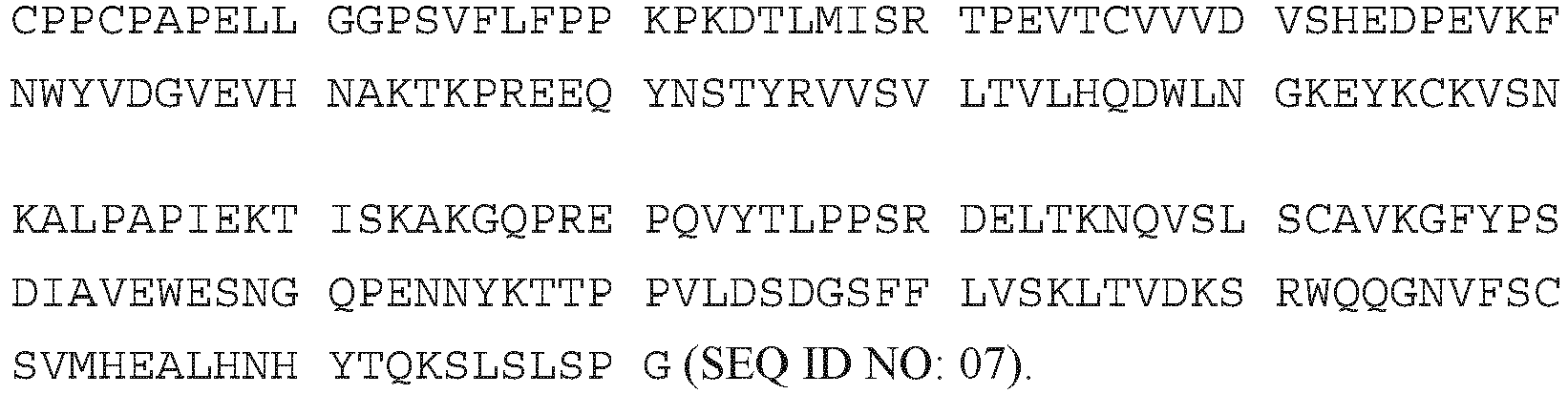
- A polypeptide chain of a variant human Fc-region of the IgG1 subclass with the mutation T366W has the following amino acid sequence:

- A polypeptide chain of a variant human Fc-region of the IgG1 subclass with the mutations L234A and L235A has the following amino acid sequence:
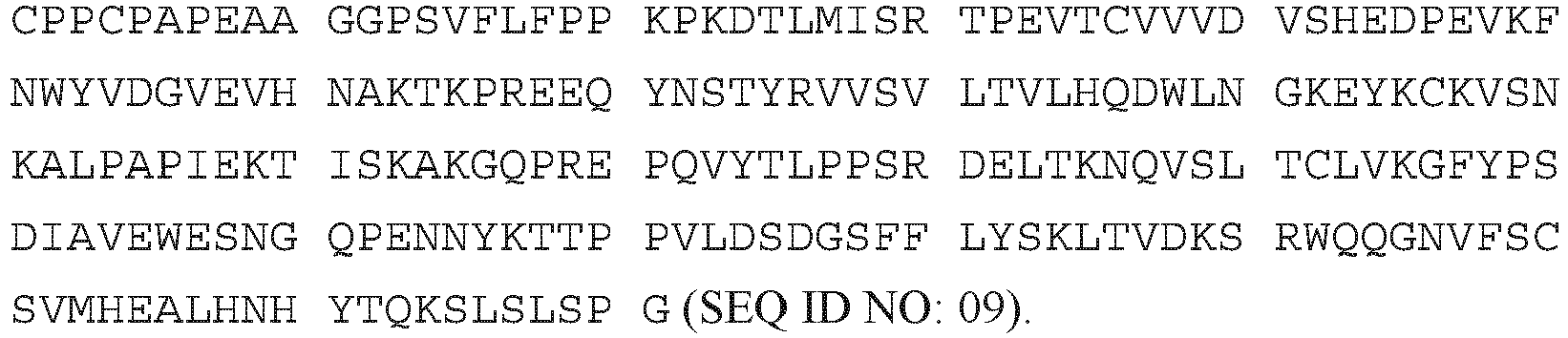
- A polypeptide chain of a variant human Fc-region of the IgG1 subclass with the mutations L234A, L235A, T366S, L368A and Y407V has the following amino acid sequence:

- A polypeptide chain of a variant human Fc-region of the IgG1 subclass with the mutations L234A, L235A and T366W has the following amino acid sequence:

- A polypeptide chain of a variant human Fc-region of the IgG1 subclass with the mutations L234A, L235A and P329G has the following amino acid sequence:

- A polypeptide chain of a variant human Fc-region of the IgG1 subclass with the mutations L234A, L235A, P329G, T366S, L368A and Y407V has the following amino acid sequence:
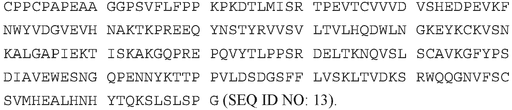
- A polypeptide chain of a variant human Fc-region of the IgG1 subclass with the mutations L234A, L235A, P329G and T366W has the following amino acid sequence:

- A polypeptide chain of a variant human Fc-region of the IgG1 subclass with the mutations L234A, L235A, P329G, Y349C, T366S, L368A and Y407V has the following amino acid sequence:

- A polypeptide chain of a variant human Fc-region of the IgG1 subclass with the mutations L234A, L235A, P329G, S354C and T366W has the following amino acid sequence:

- A polypeptide chain of a variant human Fc-region of the IgG1 subclass with the mutations L234A, L235A, P329G, S354C, T366S, L368A and Y407V has the following amino acid sequence:
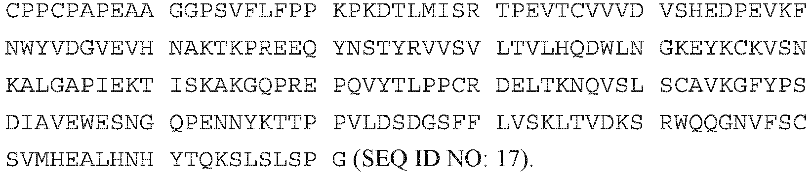
- A polypeptide chain of a variant human Fc-region of the IgG1 subclass with the mutations L234A, L235A, P329G, Y349C and T366W has the following amino acid sequence:

- A polypeptide chain of a variant human Fc-region of the IgG1 subclass with the mutations I253A, H310A and H435A has the following amino acid sequence:

- A polypeptide chain of a variant human Fc-region of the IgG1 subclass with the mutations H310A, H433A and Y436A has the following amino acid sequence:
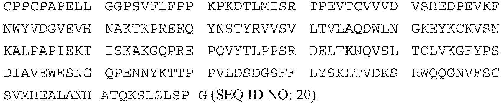
- A polypeptide chain of a variant human Fc-region of the IgG1 subclass with the mutations M252Y, S254T and T256E has the following amino acid sequence:

- A polypeptide chain of a wild-type human Fc-region of the IgG4 subclass has the following amino acid sequence:

- A polypeptide chain of a variant human Fc-region of the IgG4 subclass with the mutations S228P and L235E has the following amino acid sequence:

- A polypeptide chain of a variant human Fc-region of the IgG4 subclass with the mutations S228P, L235E and P329G has the following amino acid sequence:

- A polypeptide chain of a variant human Fc-region of the IgG4 subclass with the mutations S228P, L235E, P329G, T366S, L368A and Y407V has the following amino acid sequence:

- A polypeptide chain of a variant human Fc-region of the IgG4 subclass with the mutations S228P, L235E, P329G and T366W has the following amino acid sequence:

- The terms "full length antibody", "intact antibody," and "whole antibody" are used herein interchangeably to refer to an antibody having a structure substantially similar to a native antibody structure or having heavy chains that contain an Fc-region as defined herein.
- The term "glycan" denotes a polysaccharide, or oligosaccharide. Glycan is also used herein to refer to the carbohydrate portion of a glycoconjugate, such as a glycoprotein, glycolipid, glycopeptide, glycoproteome, peptidoglycan, lipopolysaccharide or a proteoglycan. Glycans usually consist solely of β-glycosidic linkages between monosaccharides. Glycans can be homo- or heteropolymers of monosaccharide residues, and can be linear or branched.
- The term "glycosyltransferase" denotes an enzyme capable of transferring the monosaccharide moiety from a nucleotide sugar to an acceptor molecule such as a sugar molecule in an oligosaccharide. Examples of such glycosyltransferase include, but not limited to galactosyltransferase and sialyltransferase.
- The term "hinge region" denotes the part of an antibody heavy chain polypeptide that joins in a wild-type antibody heavy chain the CH1 domain and the CH2 domain, e. g. from about position 216 to about position 230 according to the EU number system of Kabat, or from about position 226 to about position 230 according to the EU number system of Kabat. The hinge regions of other IgG subclasses can be determined by aligning with the hinge-region cysteine residues of the IgG1 subclass sequence.
- The hinge region is normally a dimeric molecule consisting of two polypeptides with identical amino acid sequence. The hinge region generally comprises about 25 amino acid residues and is flexible allowing the associated target binding sites to move independently. The hinge region can be subdivided into three domains: the upper, the middle, and the lower hinge domain (see e.g. Roux, et al., J. Immunol. 161 (1998) 4083).
- A "humanized" antibody refers to a chimeric antibody comprising amino acid residues from non-human HVRs and amino acid residues from human FRs. In certain embodiments, a humanized antibody will comprise substantially all of at least one, and typically two, variable domains, in which all or substantially all of the HVRs (e.g., CDRs) correspond to those of a non-human antibody, and all or substantially all of the FRs correspond to those of a human antibody. A humanized antibody optionally may comprise at least a portion of an antibody constant region derived from a human antibody. A "humanized form" of an antibody, e.g., a non-human antibody, refers to an antibody that has undergone humanization.
- The term "hypervariable region" or "HVR", as used herein, refers to each of the regions of an antibody variable domain comprising the amino acid residue stretches which are hypervariable in sequence ("complementarity determining regions" or "CDRs") and/or form structurally defined loops ("hypervariable loops"), and/or contain the antigen-contacting residues ("antigen contacts"). Generally, antibodies comprise six HVRs; three in the VH (HI, H2, H3), and three in the VL (L1, L2, L3).
- HVRs include
- (a) hypervariable loops occurring at amino acid residues 26-32 (L1), 50-52 (L2), 91-96 (L3), 26-32 (H1), 53-55 (H2), and 96-101 (H3) (Chothia, C. and Lesk, A.M., J. Mol. Biol. 196 (1987) 901-917);
- (b) CDRs occurring at amino acid residues 24-34 (L1), 50-56 (L2), 89-97 (L3), 31-35b (H1), 50-65 (H2), and 95-102 (H3) (Kabat, E.A. et al., Sequences of Proteins of Immunological Interest, 5th ed. Public Health Service, National Institutes of Health, Bethesda, MD (1991), NIH Publication 91-3242.);
- (c) antigen contacts occurring at amino acid residues 27c-36 (L1), 46-55 (L2), 89-96 (L3), 30-35b (H1), 47-58 (H2), and 93-101 (H3) (MacCallum et al. J. Mol. Biol. 262: 732-745 (1996)); and
- (d) combinations of (a), (b), and/or (c), including amino acid residues 46-56 (L2), 47-56 (L2), 48-56 (L2), 49-56 (L2), 26-35 (H1), 26-35b (H1), 49-65 (H2), 93-102 (H3), and 94-102 (H3).
- Unless otherwise indicated, HVR residues and other residues in the variable domain (e.g., FR residues) are numbered herein according to Kabat et al., supra.
- An "isolated" antibody is one, which has been separated from a component of its natural environment. In some embodiments, an antibody is purified to greater than 95% or 99% purity as determined by, for example, electrophoretic (e.g., SDS-PAGE, isoelectric focusing (IEF), capillary electrophoresis) or chromatographic (e.g., ion exchange or reverse phase HPLC). For review of methods for assessment of antibody purity, see, e.g., Flatman, S. et al., J. Chromatogr. B 848 (2007) 79-87.
- An "isolated" nucleic acid refers to a nucleic acid molecule that has been separated from a component of its natural environment. An isolated nucleic acid includes a nucleic acid molecule contained in cells that ordinarily contain the nucleic acid molecule, but the nucleic acid molecule is present extrachromosomally or at a chromosomal location that is different from its natural chromosomal location.
- The term "light chain" denotes the shorter polypeptide chains of native IgG antibodies. The light chain of an antibody may be assigned to one of two types, called kappa (κ) and lambda (λ), based on the amino acid sequence of its constant domain, see SEQ ID NO: 27 for a human kappa light chain constant domain and SEQ ID NO: 28 for a human lambda light chain constant domain.
- The term "monoclonal antibody" as used herein refers to an antibody obtained from a population of substantially homogeneous antibodies, i.e., the individual antibodies comprising the population are identical and/or bind the same epitope, except for possible variant antibodies, e.g., containing naturally occurring mutations or arising during production of a monoclonal antibody preparation, such variants generally being present in minor amounts. In contrast to polyclonal antibody preparations, which typically include different antibodies directed against different determinants (epitopes), each monoclonal antibody of a monoclonal antibody preparation is directed against a single determinant on an antigen. Thus, the modifier "monoclonal" indicates the character of the antibody as being obtained from a substantially homogeneous population of antibodies, and is not to be construed as requiring production of the antibody by any particular method. For example, the monoclonal antibodies to be used in accordance with the present invention may be made by a variety of techniques, including but not limited to the hybridoma method, recombinant DNA methods, phage-display methods, and methods utilizing transgenic animals containing all or part of the human immunoglobulin loci, such methods and other exemplary methods for making monoclonal antibodies being described herein.
- "Native antibodies" refer to naturally occurring immunoglobulin molecules with varying structures. For example, native IgG antibodies are heterotetrameric glycoproteins of about 150,000 daltons, composed of two identical light chains and two identical heavy chains that are disulfide-bonded. From N- to C-terminus, each heavy chain has a variable region (VH), also called a variable heavy domain or a heavy chain variable domain, followed by three constant domains (CHI, CH2, and CH3), whereby between the first and the second constant domain a hinge region is located. Similarly, from N- to C-terminus, each light chain has a variable region (VL), also called a variable light domain or a light chain variable domain, followed by a constant light (CL) domain. The light chain of an antibody may be assigned to one of two types, called kappa (κ) and lambda (λ), based on the amino acid sequence of its constant domain.
- The term "N-linked oligosaccharide" denotes oligosaccharides that are linked to the peptide backbone at an asparagine amino acid residue, by way of an asparagine-N-acetyl glucosamine linkage. N-linked oligosaccharides are also called "N-glycans." All N-linked oligo saccharides have a common pentasaccharide core of Man3GlcNAc2. They differ in the presence of, and in the number of branches (also called antennae) of peripheral sugars such as N-acetyl glucosamine, galactose, N-acetyl galactosamine, fucose and sialic acid. Optionally, this structure may also contain a core fucose molecule and/or a xylose molecule.
- The term "O-linked oligosaccharide" denotes oligosaccharides that are linked to the peptide backbone at a threonine or serine amino acid residue.
- The term "sialic acid" denotes any member of a family of nine-carbon carboxylated sugars. The most common member of the sialic acid family is N-acetyl-neuraminic acid (2-keto-5-acetamido-3,5-dideoxy-D-glycero-D-galactononulopyranos-1-onic acid (often abbreviated as Neu5Ac, NeuAc, or NANA). A second member of the family is N-glycolyl neuraminic acid (Neu5Gc or NeuGc), in which the N-acetyl group of NeuAc is hydroxylated. A third sialic acid family member is 2-keto-3-deoxy-nonulosonic acid (KDN) (Nadano et al. (1986) J. Biol. Chem. 261: 11550-11557; Kanamori et al., J. Biol. Chem. 265: 21811-21819 (1990)). Also included are 9-substituted sialic acids such as a 9-O--C1-C6 acyl-NeuSAc like 9-O-lactyl-Neu5Ac or 9-O-acetyl-NeuSAc, 9-deoxy-9-fluoro-Neu5Ac and 9-azido-9-deoxyNeu5Ac. For review of the sialic acid family, see, e.g., Varki, Glycobiol. 2 (1992) 25-40; Sialic Acids: Chemistry, Metabolism and Function, R. Schauer, Ed. (Springer-Verlag, New York (1992)). The synthesis and use of sialic acid compounds in a sialylation procedure is reported in
WO 92/16640 - With respect to antibodies, the term "substantially" denotes that the respective product (antibody) has a single glycosylation state, whether or not this state includes glycosylation at a single site or multiple sites. Typically, the antibody is substantially pure when it constitutes at least 60%, by weight, of the antibody in the preparation. For example, the antibody in the preparation is at least about 75%, in certain embodiments at least about 80%, in certain embodiments at about 85%, in certain embodiments at least about 90%, in certain embodiments at least about 95%, 96%, 97%, 98% and most preferably at least about 99%, by weight, of the desired antibody.
- The term "glycosylation state" denotes a specific or desired glycosylation pattern of an antibody. A "glycoform" is an antibody comprising a particular glycosylation state. Such glycosylation patterns include, for example, attaching one or more sugars at position N-297 of the Fc-region of an antibody (numbering according to Kabat), wherein said sugars are produced naturally, recombinantly, synthetically, or semi-synthetically. The glycosylation pattern can be determined by many methods known in the art. For example, methods of analyzing carbohydrates on proteins have been reported in
US 2006/0057638 andUS 2006/0127950 (the disclosures of which are hereby incorporated by reference in their entirety). - The term "variable region" or "variable domain" refers to the domain of an antibody heavy or light chain that is involved in binding the antibody to antigen. The variable domains of the heavy chain and light chain (VH and VL, respectively) of a native antibody generally have similar structures, with each domain comprising four conserved framework regions (FRs) and three hypervariable regions (HVRs). (See, e.g., Kindt, T.J. et al. Kuby Immunology, 6th ed., W.H. Freeman and Co., N.Y. (2007), page 91) A single VH or VL domain may be sufficient to confer antigen-binding specificity. Furthermore, antibodies that bind a particular antigen may be isolated using a VH or VL domain from an antibody that binds the antigen to screen a library of complementary VL or VH domains, respectively. See, e.g., Portolano, S. et al., J. Immunol. 150 (1993) 880-887; Clackson, T. et al., Nature 352 (1991) 624-628).
- The term "N-glycosylation site" denotes the amino acid residue within an N-glycosylation site consensus sequence to which a glycan is or can be attached. Generally N-linked glycans are attached to the amid nitrogen atom of an asparagine amino acid (Asn, N) side chain. The N-glycosylation site consensus sequence is Asn-X-Ser/Thr, wherein X can be any amino acid residue except proline. The term "N-linked glycosylation" denotes the result of the attachment of a sugar molecule oligosaccharide (denotes as glycan) to e.g. the amide nitrogen atom of asparagine.
- Human antibodies are mainly glycosylated at the asparagine residue at about position 297 (Asn297) of the heavy chain CH2 domain or in the Fab region with a more or less fucosylated biantennary complex oligosaccharide (antibody amino acid residue numbering according to Kabat, supra). The biantennary glycostructure can be terminated by up to two consecutive galactose (Gal) residues in each arm. The arms are denoted (1,6) and (1,3) according to the glycoside bond to the central mannose residue. The glycostructure denoted as G0 comprises no galactose residue. The glycostructure denoted as G1 contains one or more galactose residues in one arm. The glycostructure denoted as G2 contains one or more galactose residues in each arm (Raju, T.S., Bioprocess Int. 1 (2003) 44-53). Human constant heavy chain regions are reported in detail by Kabat, supra, and by Brueggemann, M., et al., J. Exp. Med. 166 (1987) 1351-1361; Love, T.W., et al., Methods Enzymol. 178 (1989) 515-527. CHO type glycosylation of antibody Fc-regions is e.g. described by Routier, F.H., Glycoconjugate J. 14 (1997) 201-207.
- The term "antibody" denotes and encompasses the various forms of antibodies such as human antibodies, humanized antibodies, chimeric antibodies, or T-cell antigen depleted antibodies (see e.g.
WO 98/33523 WO 98/52976 WO 00/34317 US 5,202,238 andUS 5,204,244 ; Riechmann, L., et al., Nature 332 (1988) 323-327; Neuberger, M.S., et al., Nature 314 (1985) 268-270; Lonberg, N., Nat. Biotechnol. 23 (2005) 1117-1125. - An antibody in general comprises two so called full length light chain polypeptides (light chain) and two so called full length heavy chain polypeptides (heavy chain). Each of the full length heavy and light chain polypeptides contains a variable domain (variable region) (generally the amino terminal portion of the full length polypeptide chain) comprising binding regions, which interact with an antigen. Each of the full length heavy and light chain polypeptides comprises a constant region (generally the carboxyl terminal portion). The constant region of the full length heavy chain mediates the binding of the antibody i) to cells bearing a Fc gamma receptor (FcyR), such as phagocytic cells, or ii) to cells bearing the neonatal Fc receptor (FcRn) also known as Brambell receptor. It also mediates the binding to some factors including factors of the classical complement system such as component (C1q). The variable domain of a full length antibody's light or heavy chain in turn comprises different segments, i.e. four framework regions (FR) and three hypervariable regions (CDR). A "full length antibody heavy chain" is a polypeptide consisting in N-terminal to C-terminal direction of an antibody heavy chain variable domain (VH), an antibody constant domain 1 (CH1), an antibody hinge region, an antibody constant domain 2 (CH2), an antibody constant domain 3 (CH3), and optionally an antibody constant domain 4 (CH4) in case of an antibody of the subclass IgE. A "full length antibody light chain" is a polypeptide consisting in N-terminal to C-terminal direction of an antibody light chain variable domain (VL), and an antibody light chain constant domain (CL). The full length antibody chains a linked together via inter-polypeptide disulfide bonds between the CL-domain and the CH1 domain and between the hinge regions of the full length antibody heavy chains.
- It has been reported in recent years that the glycosylation pattern of antibodies, i.e. the saccharide composition and multitude of attached glycostructures, has a strong influence on the biological properties (see e.g . Jefferis, R., Biotechnol. Prog. 21 (2005) 11-16). Antibodies produced by mammalian cells contain 2-3 % by mass oligosaccharides (Taniguchi, T., et al., Biochem. 24 (1985) 5551-5557). This is equivalent e.g. in an antibody of class G (IgG) to 2.3 oligosaccharide residues in an IgG of mouse origin (Mizuochi, T., et al., Arch. Biochem. Biophys. 257 (1987) 387-394) and to 2.8 oligosaccharide residues in an IgG of human origin (Parekh, R.B., et al., Nature 316 (1985) 452-457), whereof generally two are located in the Fc-region at Asn297 and the remaining in the variable region (Saba, J.A., et al., Anal. Biochem. 305 (2002) 16-31).
- The term "glycostructure" as used within this application denotes a single, defined N- or O-linked oligosaccharide at a specified amino acid residue. Thus, the term "antibody with a G1 glycostructure" denotes an antibody comprising at the asparagine amino acid residue at about amino acid position 297 according to the Kabat numbering scheme or in the FAB region a biantennary oligosaccharide comprising only one terminal galactose residue at the non-reducing ends of the oligosaccharide. The term "oligosaccharide" as used within this application denotes a polymeric saccharide comprising two or more covalently linked monosaccharide units.
- For the notation of the different N- or O-linked oligosaccharides in the current invention the individual sugar residues are listed from the non-reducing end to the reducing end of the oligosaccharide molecule. The longest sugar chain is chosen as basic chain for the notation. The reducing end of an N- or O-linked oligosaccharide is the monosaccharide residue, which is directly bound to the amino acid of the amino acid backbone of the antibody, whereas the end of an N- or O-linked oligosaccharide, which is located at the opposite terminus as the reducing end of the basic chain, is termed non-reducing end.
- All oligosaccharides are described herein with the name or abbreviation for the non-reducing saccharide (i.e., Gal), followed by the configuration of the glycosidic bond (α or β), the ring bond (1 or 2), the ring position of the reducing saccharide involved in the bond (2, 3, 4, 6 or 8), and then the name or abbreviation of the reducing saccharide (i.e., GlcNAc). Each saccharide is preferably a pyranose. For a review of standard glycobiology nomenclatures see, Essentials of Glycobiology Varki et al. eds., 1999, CSHL Press.
- The term "defined glycostructure" denotes within this application a glycostructure in which the monosaccharide residue at the non-reducing ends of the glycostructure is of a specific kind. The term "defined glycostructure" denotes within this application a glycostructure in which the monosaccharide residue at the non-reducing end of glycostructures are defined and of a specific kind.
- The term "affinity chromatography" as used within this application denotes a chromatography method which employs an "affinity chromatography material". In an affinity chromatography antibodies are separated based on their biological activity or chemical structure depending on the formation of electrostatic interactions, hydrophobic bonds, and/or hydrogen bonds to the chromatographical functional groups of the chromatography material. To recover the specifically bound antibody from the affinity chromatography material either a competitor ligand can be added or the chromatography conditions, such as pH value, polarity or ionic strength of the buffer, can be changed. Exemplary "affinity chromatography materials" are metal chelating chromatography materials such as Ni(II)-NTA or Cu(II)-NTA, or antibody affinity chromatography materials such as chromatography materials comprising thereto covalently linked protein A or protein G, or enzyme binding affinity chromatography materials such as chromatography materials comprising thereto covalently bound enzyme substrate analogues, enzyme cofactors, or enzyme inhibitors as chromatographical functional group, or lectin binding chromatography materials such as chromatography materials comprising thereto covalently linked polysaccharides, cell surface receptors, glycoproteins, or intact cells as chromatographical functional group.
- The antibody light chain affinity ligand uses a light chain constant domain specific capture reagent, which is specific for the kappa or the lambda constant light chain, depending on whether a kappa or a lambda light chain is contained in the antibody. Examples of such light chain constant domain specific capture reagents are e.g. KappaSelect™ and LambdaFabSelect™ (available from GE Healthcare/BAC), which are based on a highly rigid agarose base matrix that allows high flow rates and low back pressure at large scale. These materials contain a ligand that binds to the constant region of the kappa or the lambda light chain, respectively (antibodies or fragments thereof lacking the constant region of the light chain will not bind). Both are therefore capable of binding other target molecules containing the constant region of the light chain, for example, IgG, IgA and IgM. The ligands are attached to the matrix via a long hydrophilic spacer arm to make them easily available for binding to the target molecule. They are based on a single-chain antibody fragment that is screened for either human Ig kappa or lambda.
- The term "light chain" denotes the shorter polypeptide chains of native IgG antibodies. The light chain of an antibody may be assigned to one of two types, called kappa (κ) and lambda (λ), based on the amino acid sequence of its constant domain, see SEQ ID NO: 27 for a human kappa light chain constant domain and SEQ ID NO: 28 for a human lambda light chain constant domain.
- The term "applying to" and grammatical equivalents thereof as used within this application denotes a partial step of a purification method in which a solution containing a substance of interest is brought in contact with a stationary phase. The solution containing the substance of interest to be purified passes through the stationary phase providing for an interaction between the stationary phase and the substances in solution. Depending on the conditions, such as e.g. pH, conductivity, salt concentration, temperature, and/or flow rate, some substances of the solution are bound to the stationary phase and therewith are removed from the solution. Other substances remain in solution. The substances remaining in solution can be found in the flow-through. The "flow-through" denotes the solution obtained after the passage of the chromatographic device, which may either be the applied solution containing the substance of interest or the buffer, which is used to flush the column or to cause elution of one or more substances bound to the stationary phase. The substance of interest can be recovered from the solution after the purification step by methods familiar to a person of skill in the art, such as e.g. precipitation, salting out, ultrafiltration, diafiltration, lyophilization, affinity chromatography, or solvent volume reduction to obtain the substance in substantially homogeneous form.
- An antibody or antibody fragment whose glycostructure can be modified in the methods as reported herein can be produced by recombinant means. Methods for recombinant production are widely known in the state of the art and comprise protein expression in eukaryotic cells with subsequent isolation of the antibody or antibody fragment and purification to a pharmaceutically acceptable purity. For the expression of the antibody or antibody fragment either a hybridoma cell or a eukaryotic cell, in which one or more nucleic acids encoding the antibody or antibody fragment have been introduced, is used. In one embodiment the eukaryotic cells is selected from CHO cells, NS0 cells, SP2/0 cells, HEK 293 cells, COS cells, PER.C6 cells, BHK cells, rabbit cells, or sheep cells. In another embodiment the eukaryotic cell is selected from CHO cells, HEK cells, or rabbit cells. After expression the antibody or antibody fragment is recovered from the cells (from the supernatant or from the cells after lysis). General methods for recombinant production of antibodies are well-known in the state of the art and reported, for example, in the review articles of Makrides, S.C., Protein Expr. Purif. 17 (1999) 183-202; Geisse, S., et al., Protein Expr. Purif. 8 (1996) 271-282; Kaufman, R.J., Mol. Biotechnol. 16 (2000) 151-160; Werner, R.G., Drug Res. 48 (1998) 870-880.
- Purification of antibodies can be performed in order to eliminate cellular components or other contaminants, e.g. other cellular nucleic acids or proteins, by standard techniques, including alkaline/SDS treatment, CsCl banding, column chromatography, agarose gel electrophoresis, and others well known in the art (see e.g. Ausubel, F.M, et al. (eds.), Current Protocols in Molecular Biology, John Wiley & Sons, Inc., New York (2005)). Different methods are well established and widespread used for protein purification, such as affinity chromatography with microbial proteins (e.g. protein A or protein G affinity chromatography), ion exchange chromatography (e.g. cation exchange (carboxymethyl resins), anion exchange (amino ethyl resins) and mixed-mode exchange), thiophilic adsorption (e.g. with beta-mercaptoethanol and other SH ligands), hydrophobic interaction or aromatic adsorption chromatography (e.g. with phenyl-sepharose, aza-arenophilic resins, or m-aminophenylboronic acid), metal chelate affinity chromatography (e.g. with Ni(II)- and Cu(II)-affinity material), size exclusion chromatography, and electrophoretical methods (such as gel electrophoresis, capillary electrophoresis), as well as combinations thereof, such as affinity chromatography with microbial proteins, cation exchange chromatography and anion exchange chromatography (see e.g. Vijayalakshmi, M.A., Appl. Biochem. Biotech. 75 (1998) 93-102). General chromatographic methods and their use are known to a person skilled in the art. See for example, Heftmann, E. (ed.), Chromatography, 5th edition, Part A: Fundamentals and Techniques, Elsevier Science Publishing Company, New York (1992); Deyl, Z. (ed.), Advanced Chromatographic and Electromigration Methods in Biosciences, Elsevier Science BV, Amsterdam, The Netherlands (1998); Poole, C. F., and Poole, S. K., Chromatography Today, Elsevier Science Publishing Company, New York (1991); Scopes, Protein Purification: Principles and Practice (1982); Sambrook, J., et al. (eds.), Molecular Cloning: A Laboratory Manual, Second Edition, Cold Spring Harbor Laboratory Press, Cold Spring Harbor, N.Y. (1989); or Ausubel, F.M., et al. (eds.), Current Protocols in Molecular Biology, John Wiley & Sons, Inc., New York (2005).
- For the purification of antibodies or antibody fragments, which have been produced e.g. by cell cultivation methods, generally a combination of different chromatography steps can be employed. Normally a (protein A) affinity chromatography is followed by one or two additional separation steps. In one embodiment the additional chromatography steps are a cation and an anion exchange chromatography step or vice versa. The final purification step is a so called "polishing step" for the removal of trace impurities and contaminants like aggregated immunoglobulins, residual HCP (host cell protein), DNA (host cell nucleic acid), viruses, or endotoxins. In one embodiment the final purification step is an anion exchange chromatography in flow-through mode.
- The glycostructure of a recombinantly produced antibody or antibody fragment will be determined by the employed cell line and the employed cultivation conditions. With conventional downstream processing techniques selective removal of specific glycostructures is not possible.
- In more detail, recombinantly produced monoclonal antibodies are generally comprising at their glycosylation sites a heterogeneous mixture of glycoforms. This glycosylation profile is influenced by different factors during the recombinant production, such as the enzyme activities present in the host cell as well as in the cultivation medium, and the cultivation conditions.
- There is a need to produce an antibody with a prevalent or even pre-determined glycosylation, such as e.g. amongst other things the therapeutic effect.
- The method according to the invention provides an antibody with defined glycosylation at an N-glycosylation site, e.g. at an N-glycosylation site in the Fab region or in the Fc-region, i.e. containing essentially a single glycoform attached to the glycosylation site, e.g. at Asn297 in the Fc-region, by enzymatically modifying the glycan at the N-glycosylation site following harvesting the antibody from a culture. As the antibody is tightly bound to the antibody light chain affinity ligand that is specific for the kappa or the lambda constant light chain, its glycosylation can be modified in a desired manner, and as such, the method according to the invention has the advantage that it can be easily incorporated into standard operating procedures used in antibody purification from culture supernatant. Because the antibody is bound to the antibody light chain affinity ligand that is specific for the kappa or the lambda constant light chain, which in turn is immobilized on a solid phase, amongst other things the amount of the enzymes employed for the modification can be reduced compared to the amount that would be required if the modification would be performed in solution; additionally the entire modification can be achieved in a single step.
- The term "antibody with defined glycosylation" or "antibody with defined glycostructure" denotes a population of antibody molecules wherein a limited number of different glycans are attached to a (predetermined) N-glycosylation site, e.g. in the Fc-region at Asn297 (numbering according to EU index of Kabat). In one embodiment one of the glycans account for 50 % or more of the G0F, G1F and G2F glycoforms or for 30 % or more of the G0F, G1F, G2F, G1S1F, G2S1F and G2S2F glycoforms.
- The term "substantially" as used herein denotes that 40 % or more, in one embodiment 50 % or more, of the compounds has the same glycosylation, i.e. comprises the same glycan at the N-glycosylation site, e.g. at Asn297 (numbering according to Kabat) in the Fc-region.
- With the method according to the invention antibodies, irrespective of type and size, can be modified to comprise a defined glycoform. More specifically, the glycosylation of an N-glycosylation site, e.g. in the Fc-region, can be tailor-made, e.g. for the intended therapeutic applications of the antibody. For example, galactosylation of the Fc-region of the antibody is useful for the treatment of cancers. Further for example, sialylation of the Fc-region of an antibody to a defined glycoform is useful in the treatment of autoimmune disorders. For different applications de-galactosylation may be desired and/or de-sialylation of the Fc-region. Still in other embodiments production of hybrid structures having a core of GlcNAc and mannose residues may be effected such as N-acetyl glucosamine, GlcNAc; or mannose-N-acetyl glucosamine-N-acetyl glucosamine, Man-GlcNAc-GlcNAc. Any of the foregoing may be produced using the method as reported herein, as any antibody and any glycostructure of said antibody can be modified stepwise by repeating in a series the method according to the invention with different glycosylation enzymes in order to produce a desired defined glycoform antibody.
- For example, an antibody with a G2 glycoform can be produced from a heterogeneous population of monoclonal antibodies using the method as reported herein. The same method can be used to convert non-fucosylated heterogeneous antibodies, which can be produced by glyco-engineering methods, to homogeneous G2-glycoforms. In addition, the batch to batch variability of galactosylation of antibodies can also be addressed by modulating the galactosylation to a desired level using the method as reported herein.
- Briefly, the method according to the invention comprises the steps of applying a solution comprising an antibody with glycosylation at an N-glycosylation site, e.g. in the Fc-region, to an antibody light chain affinity ligand that is specific for the kappa or the lambda constant light chain and is immobilized to a solid phase/support. The support comprises a column that is washed with wash buffer and then with a reaction buffer solution that is suitable for a corresponding desired enzymatic on column glycostructure modification. The reaction buffer can be further optimized with the addition of selected secondary enzyme(s), optionally cofactor(s) and optionally nucleotide sugar(s). The column is then incubated, either at room temperature or at an elevated temperature of about 37 °C. The column is thereafter washed with the wash buffer and the modified monoclonal antibody with a defined glycoform is eluted from the solid support using an elution buffer. The eluted antibody may then be neutralized using a neutralization buffer.
- The nucleotide sugars for use in the reaction buffer are selected from the group consisting of UDP-Glc, UDP-Gal, UDP-GalNAc, UDP-GlcNAc, UDP-GlcUA, UDP-Xyl, GDP-Man, GDP-Fuc, CMP-NeuSAc, CMP-NeuSGc and combinations thereof. Concentrations used in the reaction buffer are in the range of about 0.5 mM to about 5 mM, in embodiments from about 1 mM to about 1.5 mM. The cofactor for use in the reaction buffer may be selected from the group consisting of Mn2+, Ca2+, Mg2+, Na+, K+, α-Lactalbumin and combinations thereof. Concentrations of cofactor for use in the reaction buffer may be in the range of about 2 mM to about 10 mM.
- The antibody light chain affinity ligand that is specific for the kappa or the lambda constant light chain is immobilized on a solid phase that is retained in the column during the purification and modification process. The solid phase includes but is not limited to agarose, sepharose, polyacrylic, polystyrene and other synthetic polymers, which provide negligible non-specific adsorption of non-target proteins and enzymes of modification. The affinity ligand is covalently bound to the solid phase by, for example any of a variety of chemistries, such as N-hydroxysuccinimide (NHS) esters, epoxide, aldehyde, or cyanogen bromide, to a solid phase. Such conjugation chemistries are well-known in the art, as exemplified in Hermanson, G. T., Bioconjugate Techniques.
- The wash buffer assures that a high affinity between antibody and affinity ligand during the washing steps is maintained. For example, phosphate buffered saline solution (PBS) with pH of about 7.2 can be used as wash buffer, however it is understood by one of skill in the art that the pH may vary to some degree. The wash and reaction buffers assure that high affinity between antibody and affinity ligand is maintained and, at the same time, the activity of the respective enzyme(s) is maintained. The wash and reaction buffers are used at temperatures of about 25°C to about 40°C, and any temperature therein between. Temperatures of about 37°C are often used. The optimum pH range for high affinity of antibodies to the light chain affinity ligand is about 6.0 to about 8.0. Within this range of pH, the buffers overlap with optimum pH ranges of the affinity ligands that can be used in the method as reported herein. These include but are not limited to TRIS buffer, BIS-TRIS buffer, MES buffer, BES buffer, MOPS buffer and HEPES buffer.
- Washing conditions for the affinity column minimizes non-specific binding and, thus, affect enzyme reaction and, thus, antibody modification. Wash conditions are such that they will not break the bind between the antibody light chain affinity ligand and the target monoclonal antibody.
- Enzymes suitable for use in the methods according to the invention can be selected depending on the modification from the group consisting of mannosyl-glucosamine transferases (MGAT1, MGAT2 and MGAT3); galactosyltransferases (β4GalT1, β4GalT2, β4GalT3, β4GalT4, β4GalT5, β4GalT6, (β4GalT7), sialyltransferases (ST6Gal1, ST6Gal2); mannosidases (α mannosidase-I, α mannosidase-II, α(1-2) mannosidase, α(1-6) mannosidase, α(1-2,3) mannosidase, α(1-2,3,6) mannosidase); hexosaminidases (β-N-acetyl hexosaminidase, β-N-acetyl glucosaminidase, α-N-acetyl glucosaminidase); galactosidases (β-galactosidase, β(1-4) galactosidase, α(1-3,6) galactosidase); sialidases (α(2-3,6,8) sialidase, α(2-3) sialidase), fucosidases (α-L-fucosidase, α(1-6) fucosidase, α(1-2) fucosidase, α(1-3,4) fucosidase, α(1-2,3,4) fucosidase) and any combinations thereof.
- The method according to the invention can be used to remove or add the terminal sialic acid from galactose for the generation of an antibody with homogeneous G2 glycostructure, e.g. in the Fc-region. Therefore, for example, a non-specific neuraminidase enzyme can be utilized which removes the sialic acid from any linkage or a specific sialidase that add the respective sialic acid. This enzyme can be used in combination with a galactosyltransferase to concomitantly effect galactosylation and removal or addition of sialic acid. Thereby an antibody with a defined G2 glycoform, e.g. in the Fc-region, can be obtained from an antibody with a glycosylation, e.g. in the Fc-region, comprising at least the glycoforms G0, G1, G2, G1S1 and G2S2.
- The modification of the glycosylation of an antibody according to the method according to the invention can be performed using a sequential incubation with the individual enzymes, or a semi-sequential incubation, wherein the first enzyme is added and the second enzyme is added after a certain period of time while the first enzyme is not removed, or a simultaneous incubation with both enzyme being present together. Any of these protocols results in an improved modification compared to the modification completely in solution reaction or to the modification with the antibody immobilized on protein A.
- The method according to the invention is exemplified in the following by providing an antibody with defined galactosylation and sialylation in the Fc-region by use of corresponding transferase enzymes.
- A purified humanized antibody of the IgG1 subclass was applied to protein A affinity chromatography material and an antibody light chain affinity ligand chromatography material specific for the kappa constant light chain (Kappa select from GE Healthcare). The bound antibody was incubated on-column with a buffered solution comprising a galactosyltransferase (GalT1) and UDP-GAL. The results are presented in the following table. It can be seen that a higher amount of galactosylation is achieved when the antibody is bound to a column comprising the antibody light chain affinity ligand.
enzymatic modification of Fc-region N-glycosylation performed on an antibody Fc-region affinity ligand chromatography material (protein A) enzymatic modification of Fc-region N-glycosylation performed on an antibody light chain affinity ligand chromatography material (Kappa select) time [h] G0F [%] G1F [%] G2F [%] G0F [%] G1F [%] G2F [%] 0 50 35 15 50 35 15 2 33 50 17 19 58 23 7 25 50 25 5 49 46 24 17 42 41 0 22 78 G0F = complex N-glycan with two terminal N-acetyl glucosamine residues and fucose G1F = complex N-glycan with one terminal N-acetyl glucosamine residue and one terminal galactose residue and fucose G2F = complex N-glycan with two terminal galactose residues and fucose - A purified humanized antibody of the IgG1 subclass with a homogeneous glycosylation in the Fc-region (homogeneous G2F glycoform) was applied to protein A affinity chromatography material and an antibody light chain affinity ligand chromatography material specific for the kappa constant light chain (Kappa select from GE Healthcare). The bound antibody was incubated on-column with a buffered solution comprising a sialyltransferase (ST6) and CMP-NANA. The results are presented in the following table. It can be seen that a higher amount of sialylation is achieved when the antibody is bound to a column comprising the antibody light chain affinity ligand.
37°C enzymatic modification of Fc-region N-glycosylation performed on an antibody Fc-region affinity ligand chromatography material (protein A) enzymatic modification of Fc-region N-glycosylation performed on an antibody light chain affinity ligand chromatography material (Kappa select) time [h] G2F [%] G2S1F [%] G2S2F [%] G2F [%] G2S1F [%] G2S2F [%] 0 100 0 0 100 0 0 2 17 66 17 0 74 26 7 11 59 30 0 44 56 24 10 58 32 0 45 55 48 12 58 30 RT enzymatic modification of Fc-region N-glycosylation performed on an antibody Fc-region affinity ligand chromatography material (protein A) enzymatic modification of Fc-region N-glycosylation performed on an antibody light chain affinity ligand chromatography material (Kappa select) time [h] G2F [%] G2S1F [%] G2S2F [%] G2F [%] G2S1F [%] G2S2F [%] 0 100 0 0 100 0 0 24 - - - 0 38 62 48 15 54 31 - - - - The presence or absence of alkaline phosphatase did not change the yield on the protein A column (19 % G2F, 56 % G2S1F, 25 % G2S2F). In solution the following result can be obtained:
37°C in solutiontime [h] G2F [%] G2S1F [%] G2S2F [%] 0 100 0 0 48 0 40-30 60-70 G2F = complex N-glycan with two terminal galactose residues and fucose G2S1F = complex N-glycan with two terminal galactose residues one being sialidated and fucose G2S2F = complex N-glycan with two terminal galactose residues both being sialidated and fucose time [h] G0F [%] GIF [%] G2F [%] G1S1F [%] G2S1F [%] G2S2F [%] 0 50 38 12 0 0 0 6 26 47 27 0 0 0 8 25 36 9 9 14 6 24 26 31 7 15 13 9 48 26 32 7 14 13 9 - A human antibody of the IgG4 subclass was applied to protein A affinity chromatography material and an antibody light chain affinity ligand chromatography material specific for the kappa constant light chain (Kappa select from GE Healthcare). The bound antibody was incubated on-column with a buffered solution comprising a galactosyltransferase (GaIT1) and UDP-GAL. The results are presented in the following table. It can be seen that a higher amount of galactosylation is achieved when the antibody is bound to a column comprising the antibody light chain affinity ligand.
enzymatic modification of Fc-region N-glycosylation performed on an antibody Fc-region affinity ligand chromatography material (protein A) enzymatic modification of Fc-region N-glycosylation performed on an antibody light chain affinity ligand chromatography material (Kappa select) time [h] G0F [%] G1F [%] G2F [%] G0F [%] G1F [%] G2F [%] 0 91 9 0 91 9 0 2 49 36 15 13 49 38 7 26 33 41 0 14 86 24 13 22 65 0 0 100 G0F = complex N-glycan with two terminal N-acetyl glucosamine residues and fucose G1F = complex N-glycan with one terminal N-acetyl glucosamine residue and one terminal galactose residue and fucose G2F = complex N-glycan with two terminal galactose residues and fucose - A human antibody of the IgG4 subclass with a homogeneous glycosylation in the Fc-region (homogeneous G2F glycoform) was applied to protein A affinity chromatography material and an antibody light chain affinity ligand chromatography material specific for the kappa constant light chain (Kappa select from GE Healthcare). The bound antibody was incubated on-column with a buffered solution comprising a sialyltransferase (ST6) and CMP-NANA. The results are presented in the following table. It can be seen that a higher amount of sialylation is achieved when the antibody is bound to a column comprising the antibody light chain affinity ligand.
enzymatic modification of Fc-region N-glycosylation performed on an antibody Fc-region affinity ligand chromatography material (protein A) enzymatic modification of Fc-region N-glycosylation performed on an antibody light chain affinity ligand chromatography material (Kappa select) time [h] G2F [%] G2S1F [%] G2S2F [%] G2F [%] G2S1F [%] G2S2F [%] 0 100 0 D 100 a 0 7 n.d. n.d. n.d. 0 6 94 24 0 22 78 0 0 100 n.d. = not determined - A humanized antibody of the IgG1 subclass with an additional glycosylation site in the Fab was applied to protein A affinity chromatography material and an antibody light chain affinity ligand chromatography material specific for the kappa constant light chain (Kappa select from GE Healthcare). The bound antibody was incubated on-column with a buffered solution comprising a sialyltransferase (ST6) and CMP-NANA. The results are presented in the following table. In this example the glycosylation of an N-glycosylation site in the Fab was modified. It can be seen that an improved reaction kinetic is achieved when the antibody is bound to a column comprising the antibody light chain affinity ligand.
enzymatic modification of Fab N-glycosylation performed on an antibody Fc-region affinity ligand chromatography material (protein A) enzymatic modification of Fab-region N-glycosylation performed on an antibody light chain affinity ligand chromatography material (Kappa select) time [h] G2 [%] G2S1 [%] G2S2 [%] G2 [%] G2S1 [%] G2S2 [%] 0 0 52 48 0 51 49 2 0 20 80 0 8 92 7 0 5 95 0 6 94 24 0 5 95 0 8 92 G2 = complex N-glycan with two terminal galactose residues
G2S1 = complex N-glycan with two terminal galactose residues one being sialidated
G2S2 = complex N-glycan with two terminal galactose residues both being sialidated - Cell-free cultivation supernatant comprising a humanized antibody of the IgG1 subclass was applied to protein A affinity chromatography material and an antibody light chain affinity ligand chromatography material specific for the kappa constant light chain (Kappa select from GE Healthcare). The bound antibody was incubated on-column sequentially first with a buffered solution comprising a galactosyltransferase (GalT1) and UDP-GAL, and second with a buffered solution comprising a sialyltransferase (ST6) and CMP-NANA. The results are presented in the following table. The sialyltransferase was added after 6 hours incubation time.
protein A -
time [h] G0F [%] G1F [%] G2F [%] G1S1 F[%] G2S1F [%] G2S2F [%] 0 50 38 12 0 0 0 6 26 47 27 0 0 0 8 25 36 9 9 14 6 24 26 31 7 15 13 9 48 26 32 7 14 13 9 -
time [h] G0F [%] G1F [%] G2F [%] G1S1 F[%] G2S1F [%] G2S2F [%] 0 50 38 12 0 0 0 6 3 42 55 0 0 0 8 3 32 8 10 39 8 24 0 24 6 19 30 21 48 0 24 6 19 30 21 - The same experiment was repeated with purified bulk material.
-
time [h] G0F [%] G1F [%] G2F [%] G1S1F [%] G2S1F [%] G2S2F [%] 0 52 40 8 0 0 0 6 26 48 26 0 0 0 8 25 38 8 10 14 5 24 27 34 7 8 14 10 48 25 33 6 14 13 9 -
time [h] G0F [%] G1F [%] G2F [%] G1S1F [%] G2S1F [%] G2S2F [%] 0 52 40 8 0 0 0 6 4 46 50 0 0 0 8 4 36 6 10 36 8 24 0 28 4 20 29 19 48 0 28 3 21 29 19 - Improved kappa select method with addition of sialyltransferase after 24 hours
time [h] G0F [%] G1F [%] G2F [%] G1S1F [%] G2S1F [%] G2S2F [%] 0 52 40 8 0 0 0 24 0 22 78 0 0 0 30 0 12 0 6 53 29 - In certain embodiments, an antibody modified in the method according to the invention is a chimeric antibody.
- Certain chimeric antibodies are described, e.g., in
US 4,816,567 ; and Morrison, S.L. et al., Proc. Natl. Acad. Sci. USA 81 (1984) 6851-6855). In one example, a chimeric antibody comprises a non-human variable region (e.g., a variable region derived from a mouse, rat, hamster, rabbit, or non-human primate, such as a monkey) and a human constant region. In a further example, a chimeric antibody is a "class switched" antibody in which the class or subclass has been changed from that of the parent antibody. Chimeric antibodies include antigen-binding fragments thereof as long as these bind to the antibody light chain affinity ligand used in the method as reported herein. - In certain embodiments, a chimeric antibody is a humanized antibody. Typically, a non-human antibody is humanized to reduce immunogenicity to humans, while retaining the specificity and affinity of the parental non-human antibody. Generally, a humanized antibody comprises one or more variable domains in which HVRs, e.g., CDRs, (or portions thereof) are derived from a non-human antibody, and FRs (or portions thereof) are derived from human antibody sequences. A humanized antibody optionally will also comprise at least a portion of a human constant region. In some embodiments, some FR residues in a humanized antibody are substituted with corresponding residues from a non-human antibody (e.g., the antibody from which the HVR residues are derived), e.g., to restore or improve antibody specificity or affinity.
- Humanized antibodies and methods of making them are reviewed, e.g., in Almagro, J.C. and Fransson, J., Front. Biosci. 13 (2008) 1619-1633, and are further described, e.g., in Riechmann, I. et al., Nature 332 (1988) 323-329; Queen, C. et al., Proc. Natl. Acad. Sci. USA 86 (1989) 10029-10033;
US 5, 821,337 ,US 7,527,791 ,US 6,982,321 , andUS 7,087,409 ; Kashmiri, S.V. et al., Methods 36 (2005) 25-34 (describing specificity determining region (SDR) grafting); Padlan, E.A., Mol. Immunol. 28 (1991) 489-498 (describing "resurfacing"); Dall'Acqua, W.F. et al., Methods 36 (2005) 43-60 (describing "FR shuffling"); and Osbourn, J. et al., Methods 36 (2005) 61-68 and Klimka, A. et al., Br. J. Cancer 83 (2000) 252-260 (describing the "guided selection" approach to FR shuffling). - Human framework regions that may be used for humanization include but are not limited to: framework regions selected using the "best-fit" method (see, e.g., Sims, M.J. et al., J. Immunol. 151 (1993) 2296-2308; framework regions derived from the consensus sequence of human antibodies of a particular subgroup of light or heavy chain variable regions (see, e.g., Carter, P. et al., Proc. Natl. Acad. Sci. USA 89 (1992) 4285-4289; and Presta, L.G. et al., J. Immunol. 151 (1993) 2623-2632); human mature (somatically mutated) framework regions or human germline framework regions (see, e.g., Almagro, J.C. and Fransson, J., Front. Biosci. 13 (2008) 1619-1633); and framework regions derived from screening FR libraries (see, e.g., Baca, M. et al., J. Biol. Chem. 272 (1997) 10678-10684 and Rosok, M.J. et al., J. Biol. Chem. 271 (19969 22611-22618).
- In certain embodiments, an antibody modified in the method according to the invention is a human antibod) Antibodies are not part of the invention. Human antibodies can be produced using various techniques known in the art. Human antibodies are described generally in van Dijk, M.A. and van de Winkel, J.G., Curr. Opin. Pharmacol. 5 (2001) 368-374 and Lonberg, N., Curr. Opin. Immunol. 20 (2008) 450-459.
- Human antibodies may be prepared by administering an immunogen to a transgenic animal that has been modified to produce intact human antibodies or intact antibodies with human variable regions in response to antigenic challenge. Such animals typically contain all or a portion of the human immunoglobulin loci, which replace the endogenous immunoglobulin loci, or which are present extrachromosomally or integrated randomly into the animal's chromosomes. In such transgenic mice, the endogenous immunoglobulin loci have generally been inactivated. For review of methods for obtaining human antibodies from transgenic animals, see Lonberg, N., Nat. Biotech. 23 (2005) 1117-1125. See also, e.g.,
US 6,075,181 andUS 6,150,584 describing XENOMOUSETM technology;US 5,770,429 describing HUMAB® technology;US 7,041 ,870 describing K-M MOUSE® technology,US 2007/0061900 , describing VELOCIMOUSE® technology, andWO 2007/131676 describing an immunoreconstituted mouse). Human variable regions from intact antibodies generated by such animals may be further modified, e.g., by combining with a different human constant region. - Human antibodies can also be made by hybridoma-based methods. Human myeloma and mouse-human heteromyeloma cell lines for the production of human monoclonal antibodies have been described (see, e.g., Kozbor, D., J. Immunol. 133 (1984) 3001-3005; Brodeur, B.R. et al., Monoclonal Antibody Production Techniques and Applications, Marcel Dekker, Inc., New York (1987), pp. 51-63; and Boerner, P. et al., J. Immunol. 147 (1991) 86-95). Human antibodies generated via human B-cell hybridoma technology are also described in Li, J. et al., Proc. Natl. Acad. Sci. USA 103 (2006) 3557-3562. Additional methods include those described, for example, in
US 7,189,826 (describing production of monoclonal human IgM antibodies from hybridoma cell lines) and Ni, J., Xiandai Mianyixue 26 (2006) 265-268 (describing human-human hybridomas). Human hybridoma technology (Trioma technology) is also described in Vollmers, H.P. and Brandlein, S., Histology and Histopathology 20 (2005) 927-937 and Vollmers, H.P. and Brandlein, S., Methods and Findings in Experimental and Clinical Pharmacology 27 (2005) 185-191. - Human antibodies may also be generated by isolating Fv clone variable domain sequences selected from human-derived phage display libraries. Such variable domain sequences may then be combined with a desired human constant domain. Techniques for selecting human antibodies from antibody libraries are described below.
- Antibodies modified in the method according to the invention may be isolated by screening combinatorial libraries for antibodies with the desired activity or activities. For example, a variety of methods are known in the art for generating phage display libraries and screening such libraries for antibodies possessing the desired binding characteristics. Such methods are reviewed, e.g., in Hoogenboom, H.R. et al., Methods in Molecular Biology 178 (2001) 1-37 and further described, e.g., in the McCafferty, J. et al., Nature 348 (1990) 552-554; Clackson, T. et al., Nature 352 (1991) 624-628; Marks, J.D. et al., J. Mol. Biol. 222 (1992) 581-597; Marks, J.D. and Bradbury, A., Methods in Molecular Biology 248 (2003) 161-175; Sidhu, S.S. et al., J. Mol. Biol. 338 (2004) 299-310; Lee, C.V. et al., J. Mol. Biol. 340 (2004) 1073-1093; Fellouse, F.A., Proc. Natl. Acad. Sci. USA 101 (2004) 12467-12472; and Lee, C.V. et al., J. Immunol. Methods 284 (2004) 119-132.
- In certain phage display methods, repertoires of VH and VL genes are separately cloned by polymerase chain reaction (PCR) and recombined randomly in phage libraries, which can then be screened for antigen-binding phage as described in Winter, G. et al., Ann. Rev. Immunol. 12 (1994) 433-455. Phage typically display antibody fragments, either as single-chain Fv (scFv) fragments or as Fab fragments. Libraries from immunized sources provide high-affinity antibodies to the immunogen without the requirement of constructing hybridomas. Alternatively, the naive repertoire can be cloned (e.g., from human) to provide a single source of antibodies to a wide range of non-self and also self-antigens without any immunization as described by Griffiths, A.D. et al., EMBO J. 12 (1993) 725-734. Finally, naive libraries can also be made synthetically by cloning non-rearranged V-gene segments from stem cells, and using PCR primers containing random sequence to encode the highly variable CDR3 regions and to accomplish rearrangement in vitro, as described by Hoogenboom, H.R. and Winter, G., J. Mol. Biol. 227 (1992) 381-388. Patent publications describing human antibody phage libraries include, for example:
US 5,750,373 , andUS 2005/0079574 ,US 2005/0119455 ,US 2005/0266000 ,US 2007/0117126 ,US 2007/0160598 ,US 2007/0237764 ,US 2007/0292936 , andUS 2009/0002360 . - Antibodies or antibody fragments isolated from human antibody libraries are considered human antibodies or human antibody fragments herein.
- In certain embodiments, an antibody modified in the method according to the invention is a multispecific antibody, e.g. a bispecific antibody. Multispecific antibodies are monoclonal antibodies that have binding specificities for at least two different sites. Bispecific antibodies can be prepared as full length antibodies or antibody fragments. Fragments of multispecific (bispecific) antibodies are encompassed as long as these bind to the antibody light chain affinity ligand as used in the methods as reported herein.
- Techniques for making multispecific antibodies include, but are not limited to, recombinant co-expression of two immunoglobulin heavy chain-light chain pairs having different specificities (see Milstein, C. and Cuello, A.C., Nature 305 (1983) 537-540,
WO 93/08829 US 5,731,168 ). Multi-specific antibodies may also be made by engineering electrostatic steering effects for making antibody Fc-heterodimeric molecules (WO 2009/089004 ); cross-linking two or more antibodies or fragments (see, e.g.,US 4,676,980 , and Brennan, M. et al., Science 229 (1985) 81-83); using leucine zippers to produce bi-specific antibodies (see, e.g., Kostelny, S.A. et al., J. Immunol. 148 (1992) 1547-1553; using "diabody" technology for making bispecific antibody fragments (see, e.g., Holliger, P. et al., Proc. Natl. Acad. Sci. USA 90 (1993) 6444-6448); and using single-chain Fv (sFv) dimers (see, e.g. Gruber, M et al., J. Immunol. 152 (1994) 5368-5374); and preparing trispecific antibodies as described, e.g., in Tutt, A. et al., J. Immunol. 147 (1991) 60-69). - Engineered antibodies with three or more functional antigen binding sites, including "Octopus antibodies," are also included herein (see, e.g.
US 2006/0025576 ). - The antibody or fragment modified in the method as reported herein also includes a "Dual Acting Fab" or "DAF" (see,
US 2008/0069820 , for example). - The antibody or fragment herein also includes multispecific antibodies described in
WO 2009/080251 ,WO 2009/080252 ,WO 2009/080253 ,WO 2009/080254 ,WO 2010/112193 ,WO 2010/115589 ,WO 2010/136172 ,WO 2010/145792 , andWO 2010/145793 . - Antibodies may be produced using recombinant methods and compositions, e.g., as described in
US 4,816,567 . For these methods one or more isolated nucleic acid(s) encoding an antibody are provided. - In case of a native antibody or native antibody fragment two nucleic acids are required, one for the light chain or a fragment thereof and one for the heavy chain or a fragment thereof. Such nucleic acid(s) encode an amino acid sequence comprising the VL and/or an amino acid sequence comprising the VH of the antibody (e.g., the light and/or heavy chain(s) of the antibody). These nucleic acids can be on the same expression vector or on different expression vectors.
- In case of a bispecific antibody with heterodimeric heavy chains four nucleic acids are required, one for the first light chain, one for the second light chain comprising the first heteromonomeric Fc-region polypeptide, one for the second light chain, and one for the second heavy chain comprising the second heteromonomeric Fc-region polypeptide. For example, one of the heterodimeric heavy chain comprises to so-called "knobs mutations" (T366W and optionally one of S354C or Y349C) and the other comprises the so-called "hole mutations" (T366S, L368A and Y407V and optionally Y349C or S354C) (see, e.g., Carter, P. et al., Immunotechnol. 2 (1996) 73). Such nucleic acid(s) encode an amino acid sequence comprising the first VL and/or an amino acid sequence comprising the first VH including the first heteromonomeric Fc-region and/or an amino acid sequence comprising the second VL and/or an amino acid sequence comprising the second VH including the second heteromonomeric Fc-region of the antibody (e.g., the first and/or second light and/or the first and/or second heavy chains of the antibody). These nucleic acids can be on the same expression vector or on different expression vectors, normally these nucleic acids are located on two or three expression vectors, i.e. one vector can comprise more than one of these nucleic acids. Examples of these bispecific antibodies are CrossMabs and T-cell bispecific antibodies (see, e.g. Schaefer, W. et al, Proc. Natl. Acad. Sci. USA, 108 (2011) 11187-1191).
- Isolated nucleic acids encoding an antibody as used in the methods as reported herein are provided.
- One or more vectors (e.g., expression vectors) comprising such nucleic acid(s) are provided.
- In a further embodiment, a host cell comprising such nucleic acid(s) is provided.
- In one such embodiment, a host cell comprises (e.g., has been transformed with):
- in case of a native antibody or native antibody fragment:
- (1) a vector comprising a nucleic acid that encodes an amino acid sequence comprising the VL of the antibody and an amino acid sequence comprising the VH of the antibody, or
- (2) a first vector comprising a nucleic acid that encodes an amino acid sequence comprising the VL of the antibody and a second vector comprising a nucleic acid that encodes an amino acid sequence comprising the VH of the antibody.
- in case of a bispecific antibody with heterodimeric heavy chains:
- (1) a first vector comprising a first pair of nucleic acids that encode amino acid sequences one of them comprising the first VL and the other comprising the first VH of the antibody and a second vector comprising a second pair of nucleic acids that encode amino acid sequences one of them comprising the second VL and the other comprising the second VH of the antibody, or
- (2) a first vector comprising a first nucleic acid that encode an amino acid sequence comprising one of the variable domains (preferably a light chain variable domain), a second vector comprising a pair of nucleic acids that encode amino acid sequences one of them comprising a light chain variable domain and the other comprising the first heavy chain variable domain, and a third vector comprising a pair of nucleic acids that encode amino acid sequences one of them comprising the respective other light chain variable domain as in the second vector and the other comprising the second heavy chain variable domain, or
- (3) a first vector comprising a nucleic acid that encodes an amino acid sequence comprising the first VL of the antibody, a second vector comprising a nucleic acid that encodes an amino acid sequence comprising the first VH of the antibody, a third vector comprising a nucleic acid that encodes an amino acid sequence comprising the second VL of the antibody, and a fourth vector comprising a nucleic acid that encodes an amino acid sequence comprising the second VH of the antibody.
- In one embodiment, the host cell is eukaryotic, e.g. a Chinese Hamster Ovary (CHO) cell or lymphoid cell (e.g., Y0, NS0, Sp20 cell). In one embodiment, a method of making an antibody is provided, wherein the method comprises culturing a host cell comprising nucleic acids encoding the antibody, as provided above, under conditions suitable for expression of the antibody, optionally recovering the antibody from the host cell (or host cell culture medium), and modifying the glycosylation of the antibody with a method according to the invention.
- For recombinant production of an antibody, nucleic acids encoding an antibody, e.g., as described above, are isolated and inserted into one or more vectors for further cloning and/or expression in a host cell. Such nucleic acids may be readily isolated and sequenced using conventional procedures (e.g., by using oligonucleotide probes that are capable of binding specifically to genes encoding the heavy and light chains of the antibody) or produced by recombinant methods or obtained by chemical synthesis.
- Suitable host cells for cloning or expression of antibody-encoding vectors include prokaryotic or eukaryotic cells described herein. For example, antibodies may be produced in bacteria, in particular when glycosylation and Fc effector function are not needed. For expression of antibody fragments and polypeptides in bacteria, see, e.g.,
US 5,648,237 ,US 5,789,199 , andUS 5,840,523 . (See also Charlton, K.A., In: Methods in Molecular Biology, Vol. 248, Lo, B.K.C. (ed.), Humana Press, Totowa, NJ (2003), pp. 245-254, describing expression of antibody fragments in E. coli.) After expression, the antibody may be isolated from the bacterial cell paste in a soluble fraction and can be further purified. - In addition to prokaryotes, eukaryotic microbes such as filamentous fungi or yeast are suitable cloning or expression hosts for antibody-encoding vectors, including fungi and yeast strains whose glycosylation pathways have been "humanized," resulting in the production of an antibody with a partially or fully human glycosylation pattern. See Gerngross, T.U., Nat. Biotech. 22 (2004) 1409-1414; and Li, H. et al., Nat. Biotech. 24 (2006) 210-215.
- Suitable host cells for the expression of glycosylated antibody are also derived from multicellular organisms (invertebrates and vertebrates). Examples of invertebrate cells include plant and insect cells. Numerous baculoviral strains have been identified which may be used in conjunction with insect cells, particularly for transfection of Spodoptera frugiperda cells.
- Plant cell cultures can also be utilized as hosts. See, e.g.,
US 5,959,177 ,US 6,040,498 ,US 6,420,548 ,US 7,125,978 , andUS 6,417,429 (describing PLANTIBODIES™ technology for producing antibodies in transgenic plants). - Vertebrate cells may also be used as hosts. For example, mammalian cell lines that are adapted to grow in suspension may be useful. Other examples of useful mammalian host cell lines are monkey kidney CV1 line transformed by SV40 (COS-7); human embryonic kidney line (293 or 293 cells as described, e.g., in Graham, F.L. et al., J. Gen Virol. 36 (1977) 59-74); baby hamster kidney cells (BHK); mouse sertoli cells (TM4 cells as described, e.g., in Mather, J.P., Biol. Reprod. 23 (1980) 243-252); monkey kidney cells (CV1); African green monkey kidney cells (VERO-76); human cervical carcinoma cells (HELA); canine kidney cells (MDCK; buffalo rat liver cells (BRL 3A); human lung cells (W138); human liver cells (Hep G2); mouse mammary tumor (MMT 060562); TRI cells, as described, e.g., in Mather, J.P. et al., Annals N.Y. Acad. Sci. 383 (1982) 44-68; MRC 5 cells; and FS4 cells. Other useful mammalian host cell lines include Chinese hamster ovary (CHO) cells, including DHFR- CHO cells (Urlaub, G. et al., Proc. Natl. Acad. Sci. USA 77 (1980) 4216-4220); and myeloma cell lines such as Y0, NS0 and Sp2/0. For a review of certain mammalian host cell lines suitable for antibody production, see, e.g., Yazaki, P. and Wu, A.M., Methods in Molecular Biology, Vol. 248, Lo, B.K.C. (ed.), Humana Press, Totowa, NJ (2004), pp. 255-268.
- Pharmaceutical formulations of an antibody modified with any of the methods as reported herein are prepared by mixing such antibody having the desired degree of purity with one or more optional pharmaceutically acceptable carriers (Remington's Pharmaceutical Sciences, 16th edition, Osol, A. (ed.) (1980)), in the form of lyophilized formulations or aqueous solutions. Pharmaceutically acceptable carriers are generally nontoxic to recipients at the dosages and concentrations employed, and include, but are not limited to: buffers such as phosphate, citrate, and other organic acids; antioxidants including ascorbic acid and methionine; preservatives (such as octadecyl dimethylbenzyl ammonium chloride; hexamethonium chloride; benzalkonium chloride; benzethonium chloride; phenol, butyl or benzyl alcohol; alkyl parabens such as methyl or propyl paraben; catechol; resorcinol; cyclohexanol; 3-pentanol; and m-cresol); low molecular weight (less than about 10 residues) polypeptides; proteins, such as serum albumin, gelatin, or immunoglobulins; hydrophilic polymers such as poly(vinylpyrrolidone); amino acids such as glycine, glutamine, asparagine, histidine, arginine, or lysine; monosaccharides, disaccharides, and other carbohydrates including glucose, mannose, or dextrins; chelating agents such as EDTA; sugars such as sucrose, mannitol, trehalose or sorbitol; salt-forming counter-ions such as sodium; metal complexes (e.g. Zn-protein complexes); and/or non-ionic surfactants such as polyethylene glycol (PEG). Exemplary pharmaceutically acceptable carriers herein further include interstitial drug dispersion agents such as soluble neutral-active hyaluronidase glycoproteins (sHASEGP), for example, human soluble PH-20 hyaluronidase glycoproteins, such as rhuPH20 (HYLENEX®, Baxter International, Inc.). Certain exemplary sHASEGPs and methods of use, including rhuPH20, are described in
US 2005/0260186 andUS 2006/0104968 . A sHASEGP can be combined with one or more additional glycosaminoglycanases such as chondroitinases. - Exemplary lyophilized antibody formulations are described in
US 6,267,958 . Aqueous antibody formulations include those described inUS6,171,586 andWO 2006/044908 , the latter formulations including a histidine-acetate buffer. - The formulation herein may also contain more than one active ingredients as necessary for the particular indication being treated, preferably those with complementary activities that do not adversely affect each other. Such active ingredients are suitably present in combination in amounts that are effective for the purpose intended.
- Active ingredients may be entrapped in microcapsules prepared, for example, by coacervation techniques or by interfacial polymerization, for example, hydroxymethylcellulose or gelatin-microcapsules and poly-(methyl methacrylate) microcapsules, respectively, in colloidal drug delivery systems (for example, liposomes, albumin microspheres, microemulsions, nano-particles and nanocapsules) or in macroemulsions. Such techniques are disclosed in Remington's Pharmaceutical Sciences, 16th edition, Osol, A. (ed.) (1980).
- Sustained-release preparations may be prepared. Suitable examples of sustained-release preparations include semi-permeable matrices of solid hydrophobic polymers containing the antibody, which matrices are in the form of shaped articles, e.g. films, or microcapsules.
- The formulations to be used for in vivo administration are generally sterile. Sterility may be readily accomplished, e.g., by filtration through sterile filtration membranes.
- Any of the antibodies modified with any of the methods as reported herein may be used in therapeutic methods.
- An antibody modified with any of the methods as reported herein for use as a medicament is provided.
- Herein is provided the use of an antibody modified with any of the methods as reported herein in the manufacture or preparation of a medicament. In one embodiment, the medicament is for treatment of a disease.
- Herein are provided pharmaceutical formulations comprising any of the antibodies modified with any of the methods as reported herein, e.g., for use in any of the above therapeutic methods.
- Antibodies obtained with a method of the invention can be used either alone or in combination with other agents in a therapy. For instance, an antibody of the invention may be co-administered with at least one additional therapeutic agent.
- An antibody modified with any of the methods as reported herein (and any additional therapeutic agent) can be administered by any suitable means, including parenteral, intrapulmonary, and intranasal, and, if desired for local treatment, intralesional administration. Parenteral infusions include intramuscular, intravenous, intraarterial, intraperitoneal, or subcutaneous administration. Dosing can be by any suitable route, e.g. by injections, such as intravenous or subcutaneous injections, depending in part on whether the administration is brief or chronic. Various dosing schedules including but not limited to single or multiple administrations over various time-points, bolus administration, and pulse infusion are contemplated herein.
- Antibodies modified with any of the methods as reported herein would be formulated, dosed, and administered in a fashion consistent with good medical practice. Factors for consideration in this context include the particular disorder being treated, the particular mammal being treated, the clinical condition of the individual patient, the cause of the disorder, the site of delivery of the agent, the method of administration, the scheduling of administration, and other factors known to medical practitioners. The antibody need not be, but is optionally formulated with one or more agents currently used to prevent or treat the disorder in question. The effective amount of such other agents depends on the amount of antibody present in the formulation, the type of disorder or treatment, and other factors discussed above. These are generally used in the same dosages and with administration routes as described herein, or about from 1 to 99% of the dosages described herein, or in any dosage and by any route that is empirically/clinically determined to be appropriate.
- For the prevention or treatment of disease, the appropriate dosage of an antibody modified with any of the methods as reported herein (when used alone or in combination with one or more other additional therapeutic agents) will depend on the type of disease to be treated, the type of antibody, the severity and course of the disease, whether the antibody is administered for preventive or therapeutic purposes, previous therapy, the patient's clinical history and response to the antibody, and the discretion of the attending physician. The antibody is suitably administered to the patient at one time or over a series of treatments. Depending on the type and severity of the disease, about 1 mg/kg to 15 mg/kg (e.g. 0.5 mg/kg - 10 mg/kg) of antibody can be an initial candidate dosage for administration to the patient, whether, for example, by one or more separate administrations, or by continuous infusion. One typical daily dosage might range from about 1 mg/kg to 100 mg/kg or more, depending on the factors mentioned above. For repeated administrations over several days or longer, depending on the condition, the treatment would generally be sustained until a desired suppression of disease symptoms occurs. One exemplary dosage of the antibody would be in the range from about 0.05 mg/kg to about 10 mg/kg. Thus, one or more doses of about 0.5 mg/kg, 2.0 mg/kg, 4.0 mg/kg or 10 mg/kg (or any combination thereof) may be administered to the patient. Such doses may be administered intermittently, e.g. every week or every three weeks (e.g. such that the patient receives from about two to about twenty, or e.g. about six doses of the antibody). An initial higher loading dose, followed by one or more lower doses may be administered. However, other dosage regimens may be useful. The progress of this therapy is easily monitored by conventional techniques and assays.
- The following examples are provided to aid the understanding of the present invention, the true scope of which is set forth in the appended claims.
- GalT reaction solution (5 mM MnC12, 10 mM UDP-Gal, 100 mM MES, 0.05 mg/ml GalT, pH 6.5):
- 153 mg UDP-Gal (MW=610.27 g/mol)
- 32 mg MnC12 (MW=125.84 g/mol)
- 460 mL GalT (c=5.43 mg/mL; 10 mg/2 mg antibody --> 10 mg in 300 mL = 0.033 mg/ml)
- in 100 mM MES buffer pH 6.5
- ST6 reaction solution (0.1 mM ZnC12, 200 nM AP, 50 mM MES, 1.7 mg/mlCMP-NANA, 0.7 mg/ml ST6, pH 6.5):
- 50 mL ZnCl (100 mM solution: 13.6 mg in 1 mL 50 mM MES)
- 28 mL alkaline phosphatase (AP) (c = 20 mg/mL, MW = 56,000g/mol)
- 167 mg CMP-NANA (1000 mg/2 mg antibody --> 1000 mg pro 300 mL = 3.34 mg/mL)
- 6 mL ST6 (c=5.45 mg/mL, target: 200 mg in 300 mL (2 mg AK) = 0.67 mg/mL)
- in 50 mM MES buffer pH 6.5
-
- Regeneration buffer 1 (0.1 M phosphoric acid)
- Regeneration buffer 2 (3 M Guanidine-HCl)
- Equilibration buffer (25 mM Tris, 25 mM NaCl, 5mM EDTA, pH7.1)
- Wash buffer 1 (100 mM MES, pH 6.5): 21.3 mg MES in 1000 mL H2O, pH 6.5 (adjusted with 50 % (w/v) NaOH)
- Wash buffer 2 (1 M Tris, pH 7.2)
- Wash buffer 3 (50 mM MES, pH 6.5): Wash buffer 100 mM MES 1:1 with distilled H2O
- Elution buffer Kappa select (0.1 M glycine, pH 2.7): 750 mg glycine in 100 mL H2O, pH 2.7 (adjusted with 25 % (w/v) HCl)
- Elution buffer protein A (25 mM Na-citrate, pH 2.8)
-
- regenerate, equilibrate and wash protein A respectively Kappa select columns by applying 2 column volumes regeneration buffer 1, 10 column volumes equilibration buffer and 4 column volumes wash buffer 1
- apply 2 mg of IgG (bulk material) onto the column
- wash with 10 column volumes wash buffer 1
- apply 2 mL galactosylation reaction solution (with 0.033 mg/ml GalT), let 0.8 mL flow through
- incubate respectively at 25 °C (2, 7 or 24 h)
- wash with 8 column volumes wash buffer 1
- elute with the respective elution buffer (2 column volumes for Protein A; 8 column volumes for kappa select) and use 1 M Tris buffer (pH 9.0) for pH adjustment
-
- regenerate, equilibrate and wash protein A column by applying 2 column volumes regeneration buffer 1, 10 column volumes equilibration buffer and 10 column volumes wash buffer 3
- apply 2 mg of IgG (bulk material) onto the column
- apply 2 mL sialylation reaction solution (3.3 mg/ml CMP-NANA, +/-AP), let 0.8 mL flow through
- incubate respectively at 37°C (2, 7, 24 or 48 h) and 25 °C (48 h)
- wash with 4 column volumes wash buffer 3
- elute with 2 column volumes of elution buffer protein A (sodium citrate) and use 1 M Tris buffer (pH 9.0) for pH adjustment
-
- regenerate, equilibrate and wash Kappa select column by applying 2 column volumes equilibration buffer, 3 column volumes regeneration buffer 2, 4 column volumes equilibration buffer and 2 column volumes wash buffer 3
- apply 2 mg of IgG (bulk material) onto the column
- wash with 3 column volumes wash buffer 3
- apply 2 mL sialylation reaction solution (3.3 mg/ml CMP-NANA, +/-AP), let 0.8 mL flow through
- incubate respectively at 37 °C (2, 7, and 24 h) and at 25 °C (24 h)
- wash with 3 column volumes wash buffer 3
- elute with 8 column volumes of elution buffer Kappa select and use 1 M Tris buffer (pH 9.0) for pH adjustment
-
- regenerate and equilibrate Protein A respectively Kappa Select columns by applying 2 column volumes regeneration buffer 1 and 10 column volumes equilibration buffer
- apply 1 mg of IgG (in supernatant) onto the column
- wash with 10 column volumes equilibration buffer, then 2 column volumes wash buffer 2 and 6 column volumes wash buffer 1
- apply 2 mL galactosylation reaction solution, let 0.8 mL flow through
- incubate at 25°C for about 6 to 24 h (to allow for sufficient galactosylation)
- wash with 8 column volumes wash buffer 1, 10 column volumes equilibration buffer, 2 column volumes wash buffer 2 and 6 column volumes wash buffer 3
- apply 2 mL sialylation reaction solution, let 0.8 mL flow through
- incubate (e.g. 25°C respectively for 2, 7 or 24 h or even longer)
- wash with 8 column volumes wash buffer 1
- elute with the respective elution buffer (2 column volumes for Protein A; 8 column volumes for Kappa select) and use 1 M Tris buffer (pH 9.0) for pH adjustment
-
- regenerate, equilibrate and wash protein A respectively Kappa Select columns by applying 2 column volumes regeneration buffer 1, 10 column volumes equilibration buffer and 4 column volumes wash buffer 1
- apply 1 mg of IgG (bulk material) onto the column
- wash with 10 column volumes wash buffer 1
- apply 2 mL galactosylation reaction solution, let 0.8 mL flow through
- incubate at 25°C for about 6 to 24 h (to allow for sufficient galactosylation)
- wash with 8 column volumes wash buffer 1, 10 column volumes equilibration buffer, 2 column volumes wash buffer 2 and 6 column volumes wash buffer 3
- apply 2 mL sialylation reaction solution, let 0.8 mL flow through
- incubate (e.g. 25°C respectively for 2, 7 or 24 h or even longer)
- wash with 8 column volumes wash buffer 1
- elute with the respective elution buffer (2 column volumes for protein A; 8 column volumes for Kappa select) and use 1 M Tris buffer (pH 9.0) for pH adjustment.
-
- <110> F. Hoffmann-La Roche AG
- <120> In vitro glycoengineering of antibodies
- <130> P34037
- <150>
EP16205587.5
<151> 2016-12-21 - <150>
EP17157002.1
<151> 2017-02-20 - <160> 28
- <170> PatentIn version 3.5
- <210> 1
<211> 330
<212> PRT
<213> Homo sapiens - <400> 1


- <210> 2
<211> 330
<212> PRT
<213> Homo sapiens - <400> 2


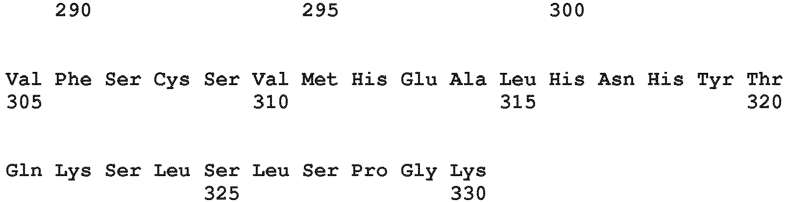
- <210> 3
<211> 326
<212> PRT
<213> Homo sapiens - <400> 3


- <210> 4
<211> 377
<212> PRT
<213> Homo sapiens - <400> 4
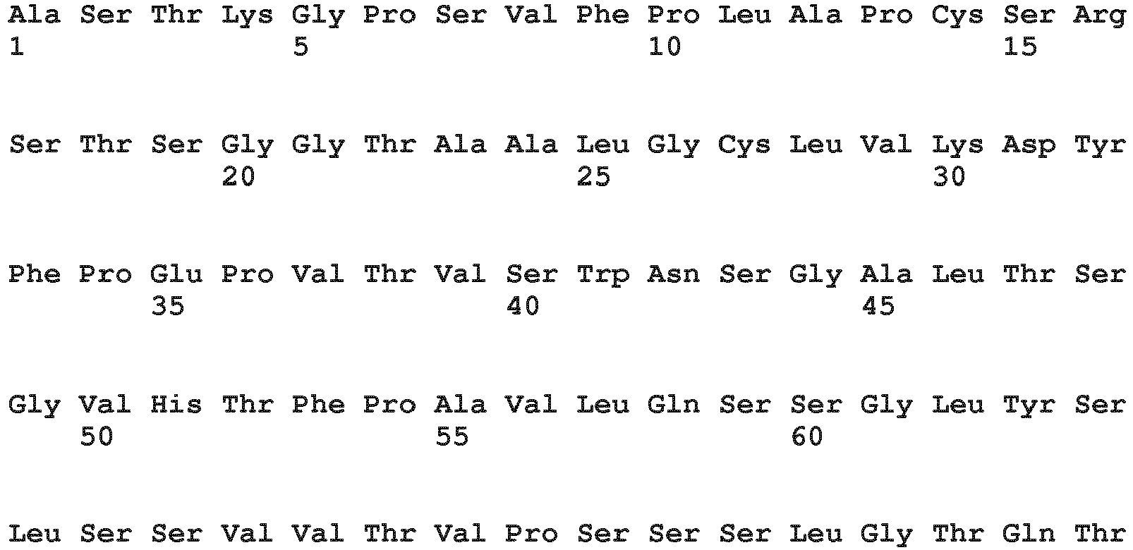
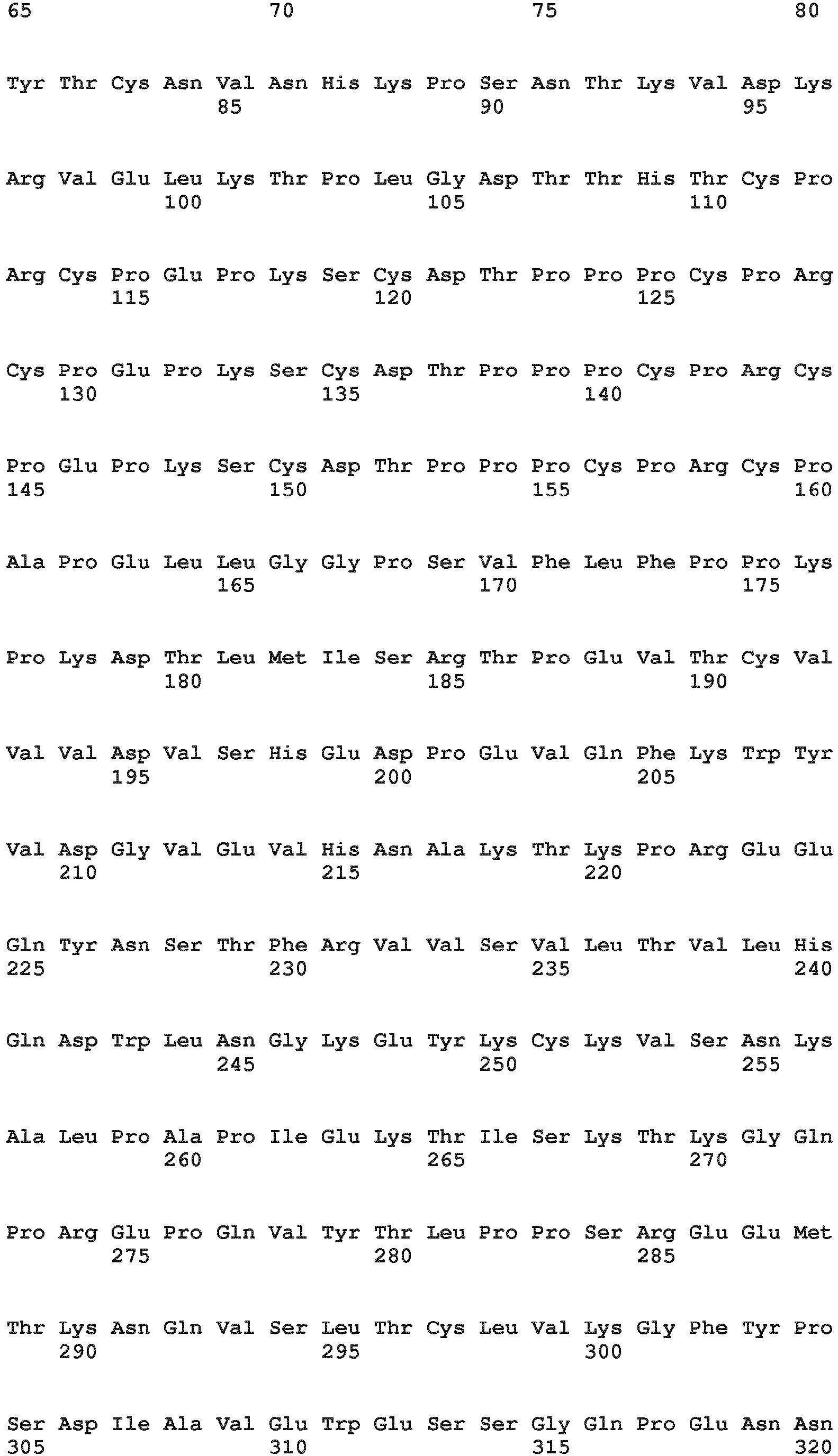

- <210> 5
<211> 327
<212> PRT
<213> Homo sapiens - <400> 5

- <210> 6
<211> 221
<212> PRT
<213> Homo sapiens - <220>
<221> MISC_FEATURE
<222> (131)..(131)
<223> X=E or D - <220>
<221> MISC_FEATURE
<222> (133)..(133)
<223> X=M or L - <400> 6

- <210> 7
<211> 221
<212> PRT
<213> Artificial Sequence - <220>
<223> variant human Fc-region of the IgG1 isotype with a hole mutation - <400> 7

- <210> 8
<211> 221
<212> PRT
<213> Artificial Sequence - <220>
<223> variant human Fc-region of the IgG1 isotype with a knob mutation - <400> 8

- <210> 9
<211> 221
<212> PRT
<213> Artificial Sequence - <220>
<223> variant human Fc-region of the IgG1 isotype with the mutations L234A, L235A - <400> 9

- <210> 10
<211> 221
<212> PRT
<213> Artificial Sequence - <220>
<223> variant human Fc-region of the IgG1 isotype with a L234A, L235A and hole mutation - <400> 10

- <210> 11
<211> 221
<212> PRT
<213> Artificial Sequence - <220>
<223> variant human Fc-region of the IgG1 isotype with a L234A, L235A and knob mutation - <400> 11

- <210> 12
<211> 226
<212> PRT
<213> Artificial Sequence - <220>
<223> variant human Fc-region of the IgG1 isotype with a L234A, L235A and P329G mutation - <400> 12


- <210> 13
<211> 221
<212> PRT
<213> Artificial Sequence - <220>
<223> variant human Fc-region of the IgG1 isotype with a L234A, L235A, P329G and hole mutation - <400> 13


- <210> 14
<211> 221
<212> PRT
<213> Artificial Sequence - <220>
<223> variant human Fc-region of the IgG1 isotype with a L234A, L235A, P329G and knob mutation - <400> 14

- <210> 15
<211> 221
<212> PRT
<213> Artificial Sequence - <220>
<223> variant human Fc-region of the IgG1 subclass with the mutations L234A, L235A, P329G, Y349C, T366S, L368A and Y407V - <400> 15


- <210> 16
<211> 221
<212> PRT
<213> Artificial Sequence - <220>
<223> variant human Fc-region of the IgG1 subclass with a L234A, L235A, P329G and S354C, T366W mutation - <400> 16

- <210> 17
<211> 221
<212> PRT
<213> Artificial Sequence - <220>
<223> variant human Fc-region of the IgG1 subclass with a L234A, L235A, P329G and S354C, T366S, L368A and Y407V mutation - <400> 17


- <210> 18
<211> 221
<212> PRT
<213> Artificial Sequence - <220>
<223> variant human Fc-region of the IgG1 subclass with a L234A, L235A, P329G and Y349C, T366W mutation - <400> 18


- <210> 19
<211> 221
<212> PRT
<213> Artificial Sequence - <220>
<223> variant human Fc-region of the IgG1 subclass with the mutations I253A, H310A and H435A - <400> 19


- <210> 20
<211> 221
<212> PRT
<213> Artificial Sequence - <220>
<223> variant human Fc-region of the IgG1 subclass with the mutations H310A, H433A and Y436A - <400> 20

- <210> 21
<211> 221
<212> PRT
<213> Artificial Sequence - <220>
<223> variant human Fc-region of the IgG1 subclass with the mutations M252Y, S254T and T256E - <400> 21


- <210> 22
<211> 228
<212> PRT
<213> Homo sapiens - <400> 22


- <210> 23
<211> 228
<212> PRT
<213> Artificial Sequence - <220>
<223> variant human Fc-region of the IgG4 isotype with a S228P and L235E mutation - <400> 23


- <210> 24
<211> 228
<212> PRT
<213> Artificial Sequence - <220>
<223> variant human Fc-region of the IgG4 isotype with a S228P, L235E and P329G mutation - <400> 24

- <210> 25
<211> 228
<212> PRT
<213> Artificial Sequence - <220>
<223> variant human Fc-region of the IgG4 isotype with a S228P and L235E mutation - <400> 25


- <210> 26
<211> 228
<212> PRT
<213> Artificial Sequence - <220>
<223> variant human Fc-region of the IgG4 isotype with a S228P, L235E and P329G mutation - <400> 26

- <210> 27
<211> 107
<212> PRT
<213> Homo sapiens - <400> 27

- <210> 28
<211> 105
<212> PRT
<213> Homo sapiens - <400> 28

Claims (17)
- A method for producing an antibody comprising the following steps:- forming an antibody-antibody light chain affinity ligand complex, wherein the antibody light chain affinity ligand is specific for the kappa or the lambda constant light chain and is immobilized on a solid phase, by applying a solution comprising the antibody to the immobilized antibody light chain affinity ligand,- incubating the complex formed in the previous step with one or more enzymes to modify the glycosylation of the antibody,and thereby producing the antibody.
- The method according to claim 1 for producing a glycosylation modified antibody comprising the following steps:- forming an antibody-antibody light chain affinity ligand complex, wherein the antibody light chain affinity ligand is specific for the kappa or the lambda constant light chain and is immobilized on a solid phase, by applying a solution comprising the antibody to the immobilized antibody light chain affinity ligand,- incubating the complex formed in the previous step with one or more enzymes and one or more activated sugar residues to modify the glycosylation of the antibody,and thereby producing the glycosylation modified antibody.
- The method according to any one of claims 1 to 2, comprising after the incubation step the step of- recovering the modified antibody from the antibody light chain affinity ligand.
- The method according to any one of claims 1 to 3, comprising the following steps in the following order- applying a solution comprising the antibody with glycosylation at an N-glycosylation site to an antibody light chain affinity ligand bound to a solid phase, whereby the antibody is bound by the ligand,- optionally washing the solid phase with a buffered solution,- enzymatically modifying the glycosylation at the N-glycosylation site of the antibody by either- applying a first solution comprising a first glycosylation modifying enzyme for a time sufficient and under conditions suitable for the enzymatic modification of the ligand-bound antibody, optionally washing the modified ligand-bound antibody, applying a second solution comprising a second glycosylation modifying enzyme for a time sufficient and under conditions suitable for the enzymatic modification of the modified ligand-bound antibody, optionally washing the two-times modified ligand-bound antibody,
or- applying a first solution comprising a first glycosylation modifying enzyme for a time sufficient and under conditions suitable for at least a partial enzymatic modification of the ligand-bound antibody, applying after a defined period of time a second solution comprising a second glycosylation modifying enzyme for a time sufficient and under conditions suitable for the enzymatic modification of the modified ligand-bound antibody, optionally washing the two-times modified ligand-bound antibody,
or- applying a solution comprising a first and a second glycosylation modifying enzyme for a time sufficient and under conditions suitable for the enzymatic modification of the ligand-bound antibody, optionally washing the modified ligand-bound antibody,- releasing the antibody from the antibody light chain affinity ligand, and thereby producing an antibody. - The method according to any one of claims 1 to 3, comprising the following steps in the following order- applying a buffered solution comprising the antibody with glycosylation at an N-glycosylation site to an antibody light chain affinity ligand bound to a solid phase, whereby the antibody is bound by the ligand resulting in a ligand-bound antibody,- optionally washing the solid phase with a buffered solution without desorbing the antibody,- enzymatically modifying the glycosylation at the N-glycosylation site of the antibody by either- applying a first buffered solution comprising a first glycosylation modifying enzyme and a first activated sugar residue for a time sufficient and under conditions suitable for the enzymatic modification of the ligand-bound antibody, optionally washing the modified ligand-bound antibody, applying a second buffered solution comprising a second glycosylation modifying enzyme and a second activated sugar for a time sufficient and under conditions suitable for the enzymatic modification of the modified ligand-bound antibody, optionally washing the two-times modified ligand-bound antibody,
or- applying a first buffered solution comprising a first glycosylation modifying enzyme and a first activated sugar for a time sufficient and under conditions suitable for at least a partial enzymatic modification of the ligand-bound antibody, applying after a defined period of time a second buffered solution comprising a second glycosylation modifying enzyme and a second activated sugar for a time sufficient and under conditions suitable for the enzymatic modification of the modified ligand-bound antibody, optionally washing the two-times modified ligand-bound antibody,
or- applying a buffered solution comprising a first and a second glycosylation modifying enzyme and a first and second activated sugar for a time sufficient and under conditions suitable for the enzymatic modification of the ligand-bound antibody, optionally washing the modified ligand-bound antibody,- releasing the antibody from the antibody light chain affinity ligand, and thereby producing a glycosylation modified antibody. - The method according to any one of claims 1 to 5, wherein antibody is selected from the group of antibodies consisting of a full length antibody, a bivalent monospecific antibody, a bispecific antibody, a bivalent bispecific antibody, a trivalent bispecific antibody, a tetravalent bispecific antibody, a trivalent trispecific antibody, an antibody Fab fragment, and a tetravalent tetraspecific antibody.
- The method according to any one of claims 1 to 6, wherein the antibody is a bivalent or trivalent or tetravalent bispecific antibody or a Fab fragment.
- The method according to any one of claims 1 to 7, wherein the antibody is a chimeric or humanized or human antibody.
- The method according to any one of claims 4 to 8, wherein the first glycosylation modifying enzyme is a galactosyltransferase and the second glycosylation modifying enzyme is a sialyltransferase.
- The method according to any one of claims 1 to 9, wherein the solution comprises a chromatographically purified antibody, the (first) glycosylation modifying enzyme is GaIT1, and the incubation with the (first) glycosylation modifying enzyme is for 24 hours at 37°C or room temperature.
- The method according to any one of claims 1 to 10, wherein the solution comprises a chromatographically purified antibody, the first glycosylation modifying enzyme is GaIT1, and the incubation with the first glycosylation modifying enzyme is for 24 hours at 37°C or room temperature.
- The method according to any one of claims 1 to 11, wherein the solution comprises a chromatographically purified antibody, the (second) glycosylation modifying enzyme is ST6, and the incubation with the (second) glycosylation modifying enzyme is for 24 hours at 37 °C or room temperature.
- The method according to any one of claims 1 to 12, wherein the solution comprises a chromatographically purified antibody, the second glycosylation modifying enzyme is ST6, and the incubation with the second glycosylation modifying enzyme is for 24 hours at 37 °C or room temperature.
- The method according to any one of claims 1 to 13, wherein the solution is a buffered, cell-free cultivation supernatant comprising the antibody, the first glycosylation modifying enzyme is GaIT1, the second glycosylation modifying enzyme is ST6, which is added 6 to 24 hours after the first glycosylation modifying enzyme, the total incubation time is 24 hours to 48 hours at 37 °C or room temperature.
- The method according to any one of claims 1 to 14 comprising the steps ofa) recombinantly producing an antibody or a Fab fragment in a cell, which comprises nucleic acids encoding the antibody or Fab fragment, to obtain an antibody or Fab fragment with glycosylation at an N-glycosylation site,b) isolating the antibody or Fab fragment produced in step a) with heterogeneous glycosylation at the N-glycosylation site,c) enzymatically modifying the antibody or Fab fragment with heterogeneous glycosylation at an N-glycosylation site with a galactosyltransferase and a sialyl transferase to obtain a modified antibody, and subsequently separating the modified antibody or Fab fragment from the enzyme(s),d) optionally purifying the modified antibody or Fab fragment by one or more chromatography steps,wherein the produced antibody or Fab fragment comprises a relative amount of at least 70 % of bi-galactosylated antibody or Fab fragment (wherein 100 % corresponds to the amount of G0, G1 and G2 glycoforms) at the N-glycosylation site.
- The method according to any one of claims 1 to 15 comprising the steps ofa) recombinantly producing an antibody of IgG1 isotype or a Fab fragment in a mammalian or CHO cell, which comprises nucleic acids encoding the antibody or Fab fragment, to obtain an antibody or Fab fragment with glycosylation at an N-glycosylation site,b) recovering and purifying the antibody or Fab fragment produced in step a) with heterogeneous glycosylation at the N-glycosylation site,c) enzymatically modifying the antibody or Fab fragment with heterogeneous glycosylation at an N-glycosylation site with a galactosyltransferase and a sialyl transferase to obtain a modified antibody, and subsequently separating the modified antibody or Fab fragment from the enzyme(s),d) optionally purifying the modified antibody or Fab fragment by one or more chromatography steps,wherein the produced antibody or Fab fragment comprises a relative amount of at least 70 % of bi-galactosylated antibody or Fab fragment (wherein 100 % corresponds to the amount of G0, G1 and G2 glycoforms) at the N-glycosylation site.
- The method according to any one of claims 1 to 16, wherein the N-glycosylation site is a Fab region N-glycosylation site or the Fc-region N-glycosylation site at asparagine residue 297 (numbering according to Kabat).
Applications Claiming Priority (3)
| Application Number | Priority Date | Filing Date | Title |
|---|---|---|---|
| EP16205587 | 2016-12-21 | ||
| EP17157002 | 2017-02-20 | ||
| PCT/EP2017/083429 WO2018114877A1 (en) | 2016-12-21 | 2017-12-19 | In vitro glycoengineering of antibodies |
Publications (3)
| Publication Number | Publication Date |
|---|---|
| EP3559248A1 EP3559248A1 (en) | 2019-10-30 |
| EP3559248B1 EP3559248B1 (en) | 2021-11-17 |
| EP3559248B2 true EP3559248B2 (en) | 2024-11-20 |
Family
ID=60997429
Family Applications (1)
| Application Number | Title | Priority Date | Filing Date |
|---|---|---|---|
| EP17829955.8A Active EP3559248B2 (en) | 2016-12-21 | 2017-12-19 | In vitro glycoengineering of antibodies |
Country Status (11)
| Country | Link |
|---|---|
| US (2) | US20200165320A1 (en) |
| EP (1) | EP3559248B2 (en) |
| JP (1) | JP6850351B2 (en) |
| KR (1) | KR102317884B1 (en) |
| CN (2) | CN110088291A (en) |
| AU (1) | AU2017384276B9 (en) |
| CA (1) | CA3044920C (en) |
| IL (1) | IL267351A (en) |
| MX (1) | MX2019006266A (en) |
| TW (1) | TWI780097B (en) |
| WO (1) | WO2018114877A1 (en) |
Families Citing this family (9)
| Publication number | Priority date | Publication date | Assignee | Title |
|---|---|---|---|---|
| CN117887794A (en) * | 2016-12-21 | 2024-04-16 | 豪夫迈·罗氏有限公司 | Methods for in vitro glycoengineering of antibodies |
| JP6931058B2 (en) | 2016-12-21 | 2021-09-01 | エフ.ホフマン−ラ ロシュ アーゲーF. Hoffmann−La Roche Aktiengesellschaft | Reuse of enzymes in in vitro glycan engineering of antibodies |
| KR102317884B1 (en) | 2016-12-21 | 2021-10-26 | 에프. 호프만-라 로슈 아게 | In vitro glycoengineering of antibodies |
| US20200017600A1 (en) | 2018-07-13 | 2020-01-16 | Genmab A/S | Variants of cd38 antibody and uses thereof |
| US20240254252A1 (en) | 2018-07-13 | 2024-08-01 | Genmab A/S | Trogocytosis-mediated therapy using cd38 antibodies |
| US20230272105A1 (en) | 2020-01-16 | 2023-08-31 | Genmab A/S | Formulations of cd38 antibodies and uses thereof |
| IL311141A (en) | 2021-09-06 | 2024-04-01 | Genmab As | Antibodies capable of binding to cd27, variants thereof and uses thereof |
| TW202509071A (en) | 2023-05-12 | 2025-03-01 | 丹麥商珍美寶股份有限公司 | Antibodies capable of binding to ox40, variants thereof and uses thereof |
| WO2025049881A1 (en) * | 2023-09-01 | 2025-03-06 | Catalent Pharma Solutions, Llc | Dnase co-expression in host cells |
Citations (1)
| Publication number | Priority date | Publication date | Assignee | Title |
|---|---|---|---|---|
| WO2012037407A1 (en) † | 2010-09-17 | 2012-03-22 | Prozyme, Inc. | Isolation and deglycosylation of glycoproteins |
Family Cites Families (67)
| Publication number | Priority date | Publication date | Assignee | Title |
|---|---|---|---|---|
| US4816567A (en) | 1983-04-08 | 1989-03-28 | Genentech, Inc. | Recombinant immunoglobin preparations |
| US4676980A (en) | 1985-09-23 | 1987-06-30 | The United States Of America As Represented By The Secretary Of The Department Of Health And Human Services | Target specific cross-linked heteroantibodies |
| US6548640B1 (en) | 1986-03-27 | 2003-04-15 | Btg International Limited | Altered antibodies |
| JP3101690B2 (en) | 1987-03-18 | 2000-10-23 | エス・ビィ・2・インコーポレイテッド | Modifications of or for denatured antibodies |
| US5202238A (en) | 1987-10-27 | 1993-04-13 | Oncogen | Production of chimeric antibodies by homologous recombination |
| US5204244A (en) | 1987-10-27 | 1993-04-20 | Oncogen | Production of chimeric antibodies by homologous recombination |
| US5959177A (en) | 1989-10-27 | 1999-09-28 | The Scripps Research Institute | Transgenic plants expressing assembled secretory antibodies |
| US6150584A (en) | 1990-01-12 | 2000-11-21 | Abgenix, Inc. | Human antibodies derived from immunized xenomice |
| US6075181A (en) | 1990-01-12 | 2000-06-13 | Abgenix, Inc. | Human antibodies derived from immunized xenomice |
| US5770429A (en) | 1990-08-29 | 1998-06-23 | Genpharm International, Inc. | Transgenic non-human animals capable of producing heterologous antibodies |
| CA2095633C (en) | 1990-12-03 | 2003-02-04 | Lisa J. Garrard | Enrichment method for variant proteins with altered binding properties |
| EP0576592B1 (en) | 1991-03-18 | 2000-05-31 | The Scripps Research Institute | Oligosaccharide enzyme substrates and inhibitors: methods and compositions |
| EP1400536A1 (en) | 1991-06-14 | 2004-03-24 | Genentech Inc. | Method for making humanized antibodies |
| US7018809B1 (en) | 1991-09-19 | 2006-03-28 | Genentech, Inc. | Expression of functional antibody fragments |
| WO1993008829A1 (en) | 1991-11-04 | 1993-05-13 | The Regents Of The University Of California | Compositions that mediate killing of hiv-infected cells |
| US5789199A (en) | 1994-11-03 | 1998-08-04 | Genentech, Inc. | Process for bacterial production of polypeptides |
| US5840523A (en) | 1995-03-01 | 1998-11-24 | Genetech, Inc. | Methods and compositions for secretion of heterologous polypeptides |
| US5731168A (en) | 1995-03-01 | 1998-03-24 | Genentech, Inc. | Method for making heteromultimeric polypeptides |
| US6267958B1 (en) | 1995-07-27 | 2001-07-31 | Genentech, Inc. | Protein formulation |
| AU5874798A (en) | 1997-01-31 | 1998-08-25 | Biovation Limited | Vaccination methods and molecules |
| EP0983303B1 (en) | 1997-05-21 | 2006-03-08 | Biovation Limited | Method for the production of non-immunogenic proteins |
| US6171586B1 (en) | 1997-06-13 | 2001-01-09 | Genentech, Inc. | Antibody formulation |
| US6040498A (en) | 1998-08-11 | 2000-03-21 | North Caroline State University | Genetically engineered duckweed |
| US6610833B1 (en) | 1997-11-24 | 2003-08-26 | The Institute For Human Genetics And Biochemistry | Monoclonal human natural antibodies |
| DK1034298T3 (en) | 1997-12-05 | 2012-01-30 | Scripps Research Inst | Humanization of murine antibody |
| AU776910B2 (en) | 1998-12-08 | 2004-09-23 | Merck Patent Gesellschaft Mit Beschrankter Haftung | Modifying protein immunogenicity |
| ES2248127T3 (en) | 1999-10-04 | 2006-03-16 | Medicago Inc. | METHOD FOR REGULATING THE TRANSCRIPTION OF FOREIGN GENES IN THE PRESENCE OF NIGTROGEN. |
| US7125978B1 (en) | 1999-10-04 | 2006-10-24 | Medicago Inc. | Promoter for regulating expression of foreign genes |
| EP1240319A1 (en) | 1999-12-15 | 2002-09-18 | Genentech, Inc. | Shotgun scanning, a combinatorial method for mapping functional protein epitopes |
| JP2003531588A (en) | 2000-04-11 | 2003-10-28 | ジェネンテック・インコーポレーテッド | Multivalent antibodies and their uses |
| US6596541B2 (en) | 2000-10-31 | 2003-07-22 | Regeneron Pharmaceuticals, Inc. | Methods of modifying eukaryotic cells |
| CA2430013C (en) | 2000-11-30 | 2011-11-22 | Medarex, Inc. | Transgenic transchromosomal rodents for making human antibodies |
| CA2488441C (en) | 2002-06-03 | 2015-01-27 | Genentech, Inc. | Synthetic antibody phage libraries |
| WO2004065416A2 (en) | 2003-01-16 | 2004-08-05 | Genentech, Inc. | Synthetic antibody phage libraries |
| US20060104968A1 (en) | 2003-03-05 | 2006-05-18 | Halozyme, Inc. | Soluble glycosaminoglycanases and methods of preparing and using soluble glycosaminogly ycanases |
| US7871607B2 (en) | 2003-03-05 | 2011-01-18 | Halozyme, Inc. | Soluble glycosaminoglycanases and methods of preparing and using soluble glycosaminoglycanases |
| EP1460425A1 (en) * | 2003-03-17 | 2004-09-22 | Boehringer Mannheim Gmbh | Deglycosylated enzymes for conjugates |
| EP1740615B1 (en) | 2004-03-31 | 2014-11-05 | Genentech, Inc. | Humanized anti-tgf-beta antibodies |
| US7785903B2 (en) | 2004-04-09 | 2010-08-31 | Genentech, Inc. | Variable domain library and uses |
| WO2005111627A2 (en) | 2004-04-15 | 2005-11-24 | Massachusetts Institute Of Technology | Methods and products related to the improved analysis of carbohydrates |
| US20060127950A1 (en) | 2004-04-15 | 2006-06-15 | Massachusetts Institute Of Technology | Methods and products related to the improved analysis of carbohydrates |
| TWI380996B (en) | 2004-09-17 | 2013-01-01 | Hoffmann La Roche | Anti-ox40l antibodies |
| JO3000B1 (en) | 2004-10-20 | 2016-09-05 | Genentech Inc | Antibody Formulations. |
| ES2577292T3 (en) | 2005-11-07 | 2016-07-14 | Genentech, Inc. | Binding polypeptides with diversified VH / VL hypervariable sequences and consensus |
| EP1973951A2 (en) | 2005-12-02 | 2008-10-01 | Genentech, Inc. | Binding polypeptides with restricted diversity sequences |
| TW200812616A (en) | 2006-05-09 | 2008-03-16 | Genentech Inc | Binding polypeptides with optimized scaffolds |
| US20070266448A1 (en) | 2006-05-11 | 2007-11-15 | Alexander Lifke | Method for the production of antibodies |
| WO2008027236A2 (en) | 2006-08-30 | 2008-03-06 | Genentech, Inc. | Multispecific antibodies |
| CN100592373C (en) | 2007-05-25 | 2010-02-24 | 群康科技(深圳)有限公司 | Liquid crystal display panel driving device and driving method thereof |
| AU2008291358A1 (en) * | 2007-08-31 | 2009-03-05 | F. Hoffmann-La Roche Ag | Glycosylation profile analysis |
| US8227577B2 (en) | 2007-12-21 | 2012-07-24 | Hoffman-La Roche Inc. | Bivalent, bispecific antibodies |
| US8242247B2 (en) | 2007-12-21 | 2012-08-14 | Hoffmann-La Roche Inc. | Bivalent, bispecific antibodies |
| US20090162359A1 (en) | 2007-12-21 | 2009-06-25 | Christian Klein | Bivalent, bispecific antibodies |
| US9266967B2 (en) | 2007-12-21 | 2016-02-23 | Hoffmann-La Roche, Inc. | Bivalent, bispecific antibodies |
| JP6157046B2 (en) | 2008-01-07 | 2017-07-05 | アムジェン インコーポレイテッド | Method for generating antibody Fc heterodimer molecules using electrostatic steering effect |
| CA2756244A1 (en) | 2009-04-02 | 2010-10-07 | Roche Glycart Ag | Multispecific antibodies comprising full length antibodies and single chain fab fragments |
| SG175077A1 (en) | 2009-04-07 | 2011-11-28 | Roche Glycart Ag | Trivalent, bispecific antibodies |
| PE20120540A1 (en) | 2009-05-27 | 2012-05-09 | Hoffmann La Roche | THREE-SPECIFIC OR TETRA-SPECIFIC ANTIBODIES |
| US9676845B2 (en) | 2009-06-16 | 2017-06-13 | Hoffmann-La Roche, Inc. | Bispecific antigen binding proteins |
| US8703132B2 (en) | 2009-06-18 | 2014-04-22 | Hoffmann-La Roche, Inc. | Bispecific, tetravalent antigen binding proteins |
| SG177764A1 (en) * | 2009-07-30 | 2012-03-29 | Hoffmann La Roche | Enzymatic antibody processing |
| US10087236B2 (en) | 2009-12-02 | 2018-10-02 | Academia Sinica | Methods for modifying human antibodies by glycan engineering |
| PT2768857T (en) * | 2011-10-19 | 2020-01-27 | Novimmune Sa | Methods of purifying antibodies |
| IN2014DN06806A (en) * | 2012-02-10 | 2015-05-22 | Univ Maryland | |
| EP3108004B1 (en) * | 2014-02-18 | 2019-02-20 | The University of Manitoba | Methods to produce single glycoform antibodies |
| CN114181312A (en) * | 2014-09-10 | 2022-03-15 | 豪夫迈·罗氏有限公司 | Galactose engineered immunoglobulin 1 antibodies |
| KR102317884B1 (en) | 2016-12-21 | 2021-10-26 | 에프. 호프만-라 로슈 아게 | In vitro glycoengineering of antibodies |
-
2017
- 2017-12-19 KR KR1020197017701A patent/KR102317884B1/en active Active
- 2017-12-19 MX MX2019006266A patent/MX2019006266A/en unknown
- 2017-12-19 CA CA3044920A patent/CA3044920C/en active Active
- 2017-12-19 CN CN201780079057.7A patent/CN110088291A/en active Pending
- 2017-12-19 AU AU2017384276A patent/AU2017384276B9/en active Active
- 2017-12-19 CN CN202411870260.4A patent/CN119685431A/en active Pending
- 2017-12-19 WO PCT/EP2017/083429 patent/WO2018114877A1/en not_active Ceased
- 2017-12-19 EP EP17829955.8A patent/EP3559248B2/en active Active
- 2017-12-19 JP JP2019533340A patent/JP6850351B2/en active Active
- 2017-12-20 TW TW106144931A patent/TWI780097B/en active
-
2019
- 2019-06-13 IL IL267351A patent/IL267351A/en unknown
- 2019-06-20 US US16/447,093 patent/US20200165320A1/en not_active Abandoned
-
2023
- 2023-05-16 US US18/318,487 patent/US20240101643A1/en active Pending
Patent Citations (1)
| Publication number | Priority date | Publication date | Assignee | Title |
|---|---|---|---|---|
| WO2012037407A1 (en) † | 2010-09-17 | 2012-03-22 | Prozyme, Inc. | Isolation and deglycosylation of glycoproteins |
Non-Patent Citations (8)
| Title |
|---|
| "Life technologies", 1 January 2013, article ANONYMOUS: "Protein Handbook 2013", pages: 1 - 62 † |
| ÅKERSTRÖM B, BJÖRCK L: "Protein L: An Immunoglobulin Light Chain-binding Bacterial Protein", JOURNAL OF BIOLOGICAL CHEMISTRY, AMERICAN SOCIETY FOR BIOCHEMISTRY AND MOLECULAR BIOLOGY, US, vol. 264, no. 33, 1 November 1989 (1989-11-01), US , pages 19740 - 19746, ISSN: 0021-9258, DOI: 10.1016/S0021-9258(19)47174-3 † |
| BARB ADAM W., MENG LU, GAO ZHONGWEI, JOHNSON ROY W., MOREMEN KELLEY W., PRESTEGARD JAMES H.: "NMR Characterization of Immunoglobulin G Fc Glycan Motion on Enzymatic Sialylation", BIOCHEMISTRY, vol. 51, no. 22, 5 June 2012 (2012-06-05), pages 4618 - 4626, ISSN: 0006-2960, DOI: 10.1021/bi300319q † |
| BOLT S., ET AL.: "THE GENERATION OF A HUMANIZED, NON-MITOGENIC CD3 MONOCLONAL ANTIBODY WHICH RETAINS IN VIRTO IMMUNUSUPPRESSIVE PROPERTIES.", EUROPEAN JOURNAL OF IMMUNOLOGY, WILEY-VCH, HOBOKEN, USA, vol. 23., no. 02., 1 February 1993 (1993-02-01), Hoboken, USA, pages 403 - 410., ISSN: 0014-2980, DOI: 10.1002/eji.1830230216 † |
| CASTILHO, ALEXANDRA: "Glyco-engineering : methods and protocols", 1 January 2015, NEW YORK, NY [U.A.] : HUMANA PR. , US , ISBN: 978-1-4939-2759-3, article CASTILHO ALEXANDRA: "Chapter 20", pages: 287 - 305 † |
| FRANK MARTIN; WALKER ROSS C.; LANZILOTTA WILLIAM N.; PRESTEGARD JAMES H.; BARB ADAM W.: "Immunoglobulin G1 Fc Domain Motions: Implications for Fc Engineering", JOURNAL OF MOLECULAR BIOLOGY, ACADEMIC PRESS, UNITED KINGDOM, vol. 426, no. 8, 9 February 2014 (2014-02-09), United Kingdom , pages 1799 - 1811, ISSN: 0022-2836, DOI: 10.1016/j.jmb.2014.01.011 † |
| MALIK ET AL: "In vitro glycoengineering - suitability for BioPharma manufacturing", ROCHE, APPLICATION NOTE, 1 April 2016 (2016-04-01), pages 1 - 8, Retrieved from the Internet <URL:http://custombiotech.roche.com/content/dam/internet/dia/custombiotech/custombiotech_com/en_GB/pdf/CustomBiotech_Glycoengineering_ApplicationNote.pdf> [retrieved on 20180302] † |
| MALIK SEBASTIAN, GRUNERT INGRID, VON ROMAN MATTHIAS FREIHERR, WALCH HEIKO, DAMS THOMAS, THOMANN MARCO, FALKENSTEIN ROBERTO: "Implementation of in vitro glycoengineering of monoclonal antibodies into downstream processing of industrial production", GLYCOBIOLOGY, OXFORD UNIVERSITY PRESS, US, vol. 32, no. 2, 19 March 2022 (2022-03-19), US , pages 123 - 135, ISSN: 0959-6658, DOI: 10.1093/glycob/cwab109 † |
Also Published As
| Publication number | Publication date |
|---|---|
| CA3044920A1 (en) | 2018-06-28 |
| EP3559248A1 (en) | 2019-10-30 |
| JP6850351B2 (en) | 2021-03-31 |
| MX2019006266A (en) | 2019-08-21 |
| AU2017384276B2 (en) | 2020-07-02 |
| BR112019010485A2 (en) | 2019-09-10 |
| TWI780097B (en) | 2022-10-11 |
| TW201835333A (en) | 2018-10-01 |
| JP2020501577A (en) | 2020-01-23 |
| IL267351A (en) | 2019-08-29 |
| CN110088291A (en) | 2019-08-02 |
| KR20190082942A (en) | 2019-07-10 |
| US20240101643A1 (en) | 2024-03-28 |
| EP3559248B1 (en) | 2021-11-17 |
| CA3044920C (en) | 2022-06-28 |
| WO2018114877A1 (en) | 2018-06-28 |
| US20200165320A1 (en) | 2020-05-28 |
| KR102317884B1 (en) | 2021-10-26 |
| AU2017384276B9 (en) | 2020-11-26 |
| CN119685431A (en) | 2025-03-25 |
| AU2017384276A1 (en) | 2019-05-30 |
Similar Documents
| Publication | Publication Date | Title |
|---|---|---|
| EP3559248B2 (en) | In vitro glycoengineering of antibodies | |
| US12269867B2 (en) | Re-use of enzymes in in vitro glycoengineering of antibodies | |
| US11767342B2 (en) | Method for in vitro glycoengineering of antibodies | |
| HK40123043A (en) | Methods for in vitro glycoengineering of antibodies | |
| HK40106545A (en) | Method for in vitro glycoengineering of antibodies | |
| HK40011892B (en) | Re-use of enzymes in in vitro glycoengineering of antibodies |
Legal Events
| Date | Code | Title | Description |
|---|---|---|---|
| STAA | Information on the status of an ep patent application or granted ep patent |
Free format text: STATUS: UNKNOWN |
|
| STAA | Information on the status of an ep patent application or granted ep patent |
Free format text: STATUS: THE INTERNATIONAL PUBLICATION HAS BEEN MADE |
|
| PUAI | Public reference made under article 153(3) epc to a published international application that has entered the european phase |
Free format text: ORIGINAL CODE: 0009012 |
|
| STAA | Information on the status of an ep patent application or granted ep patent |
Free format text: STATUS: REQUEST FOR EXAMINATION WAS MADE |
|
| 17P | Request for examination filed |
Effective date: 20190722 |
|
| AK | Designated contracting states |
Kind code of ref document: A1 Designated state(s): AL AT BE BG CH CY CZ DE DK EE ES FI FR GB GR HR HU IE IS IT LI LT LU LV MC MK MT NL NO PL PT RO RS SE SI SK SM TR |
|
| AX | Request for extension of the european patent |
Extension state: BA ME |
|
| DAV | Request for validation of the european patent (deleted) | ||
| DAX | Request for extension of the european patent (deleted) | ||
| STAA | Information on the status of an ep patent application or granted ep patent |
Free format text: STATUS: EXAMINATION IS IN PROGRESS |
|
| 17Q | First examination report despatched |
Effective date: 20201124 |
|
| GRAP | Despatch of communication of intention to grant a patent |
Free format text: ORIGINAL CODE: EPIDOSNIGR1 |
|
| STAA | Information on the status of an ep patent application or granted ep patent |
Free format text: STATUS: GRANT OF PATENT IS INTENDED |
|
| INTG | Intention to grant announced |
Effective date: 20210428 |
|
| GRAS | Grant fee paid |
Free format text: ORIGINAL CODE: EPIDOSNIGR3 |
|
| GRAA | (expected) grant |
Free format text: ORIGINAL CODE: 0009210 |
|
| STAA | Information on the status of an ep patent application or granted ep patent |
Free format text: STATUS: THE PATENT HAS BEEN GRANTED |
|
| AK | Designated contracting states |
Kind code of ref document: B1 Designated state(s): AL AT BE BG CH CY CZ DE DK EE ES FI FR GB GR HR HU IE IS IT LI LT LU LV MC MK MT NL NO PL PT RO RS SE SI SK SM TR |
|
| REG | Reference to a national code |
Ref country code: GB Ref legal event code: FG4D |
|
| REG | Reference to a national code |
Ref country code: DE Ref legal event code: R096 Ref document number: 602017049556 Country of ref document: DE |
|
| REG | Reference to a national code |
Ref country code: IE Ref legal event code: FG4D |
|
| REG | Reference to a national code |
Ref country code: AT Ref legal event code: REF Ref document number: 1448087 Country of ref document: AT Kind code of ref document: T Effective date: 20211215 |
|
| REG | Reference to a national code |
Ref country code: LT Ref legal event code: MG9D |
|
| REG | Reference to a national code |
Ref country code: NL Ref legal event code: MP Effective date: 20211117 |
|
| REG | Reference to a national code |
Ref country code: AT Ref legal event code: MK05 Ref document number: 1448087 Country of ref document: AT Kind code of ref document: T Effective date: 20211117 |
|
| PG25 | Lapsed in a contracting state [announced via postgrant information from national office to epo] |
Ref country code: RS Free format text: LAPSE BECAUSE OF FAILURE TO SUBMIT A TRANSLATION OF THE DESCRIPTION OR TO PAY THE FEE WITHIN THE PRESCRIBED TIME-LIMIT Effective date: 20211117 Ref country code: LT Free format text: LAPSE BECAUSE OF FAILURE TO SUBMIT A TRANSLATION OF THE DESCRIPTION OR TO PAY THE FEE WITHIN THE PRESCRIBED TIME-LIMIT Effective date: 20211117 Ref country code: FI Free format text: LAPSE BECAUSE OF FAILURE TO SUBMIT A TRANSLATION OF THE DESCRIPTION OR TO PAY THE FEE WITHIN THE PRESCRIBED TIME-LIMIT Effective date: 20211117 Ref country code: BG Free format text: LAPSE BECAUSE OF FAILURE TO SUBMIT A TRANSLATION OF THE DESCRIPTION OR TO PAY THE FEE WITHIN THE PRESCRIBED TIME-LIMIT Effective date: 20220217 Ref country code: AT Free format text: LAPSE BECAUSE OF FAILURE TO SUBMIT A TRANSLATION OF THE DESCRIPTION OR TO PAY THE FEE WITHIN THE PRESCRIBED TIME-LIMIT Effective date: 20211117 |
|
| PG25 | Lapsed in a contracting state [announced via postgrant information from national office to epo] |
Ref country code: IS Free format text: LAPSE BECAUSE OF FAILURE TO SUBMIT A TRANSLATION OF THE DESCRIPTION OR TO PAY THE FEE WITHIN THE PRESCRIBED TIME-LIMIT Effective date: 20220317 Ref country code: SE Free format text: LAPSE BECAUSE OF FAILURE TO SUBMIT A TRANSLATION OF THE DESCRIPTION OR TO PAY THE FEE WITHIN THE PRESCRIBED TIME-LIMIT Effective date: 20211117 Ref country code: PT Free format text: LAPSE BECAUSE OF FAILURE TO SUBMIT A TRANSLATION OF THE DESCRIPTION OR TO PAY THE FEE WITHIN THE PRESCRIBED TIME-LIMIT Effective date: 20220317 Ref country code: PL Free format text: LAPSE BECAUSE OF FAILURE TO SUBMIT A TRANSLATION OF THE DESCRIPTION OR TO PAY THE FEE WITHIN THE PRESCRIBED TIME-LIMIT Effective date: 20211117 Ref country code: NO Free format text: LAPSE BECAUSE OF FAILURE TO SUBMIT A TRANSLATION OF THE DESCRIPTION OR TO PAY THE FEE WITHIN THE PRESCRIBED TIME-LIMIT Effective date: 20220217 Ref country code: NL Free format text: LAPSE BECAUSE OF FAILURE TO SUBMIT A TRANSLATION OF THE DESCRIPTION OR TO PAY THE FEE WITHIN THE PRESCRIBED TIME-LIMIT Effective date: 20211117 Ref country code: LV Free format text: LAPSE BECAUSE OF FAILURE TO SUBMIT A TRANSLATION OF THE DESCRIPTION OR TO PAY THE FEE WITHIN THE PRESCRIBED TIME-LIMIT Effective date: 20211117 Ref country code: HR Free format text: LAPSE BECAUSE OF FAILURE TO SUBMIT A TRANSLATION OF THE DESCRIPTION OR TO PAY THE FEE WITHIN THE PRESCRIBED TIME-LIMIT Effective date: 20211117 Ref country code: GR Free format text: LAPSE BECAUSE OF FAILURE TO SUBMIT A TRANSLATION OF THE DESCRIPTION OR TO PAY THE FEE WITHIN THE PRESCRIBED TIME-LIMIT Effective date: 20220218 Ref country code: ES Free format text: LAPSE BECAUSE OF FAILURE TO SUBMIT A TRANSLATION OF THE DESCRIPTION OR TO PAY THE FEE WITHIN THE PRESCRIBED TIME-LIMIT Effective date: 20211117 |
|
| PG25 | Lapsed in a contracting state [announced via postgrant information from national office to epo] |
Ref country code: SM Free format text: LAPSE BECAUSE OF FAILURE TO SUBMIT A TRANSLATION OF THE DESCRIPTION OR TO PAY THE FEE WITHIN THE PRESCRIBED TIME-LIMIT Effective date: 20211117 Ref country code: SK Free format text: LAPSE BECAUSE OF FAILURE TO SUBMIT A TRANSLATION OF THE DESCRIPTION OR TO PAY THE FEE WITHIN THE PRESCRIBED TIME-LIMIT Effective date: 20211117 Ref country code: RO Free format text: LAPSE BECAUSE OF FAILURE TO SUBMIT A TRANSLATION OF THE DESCRIPTION OR TO PAY THE FEE WITHIN THE PRESCRIBED TIME-LIMIT Effective date: 20211117 Ref country code: EE Free format text: LAPSE BECAUSE OF FAILURE TO SUBMIT A TRANSLATION OF THE DESCRIPTION OR TO PAY THE FEE WITHIN THE PRESCRIBED TIME-LIMIT Effective date: 20211117 Ref country code: DK Free format text: LAPSE BECAUSE OF FAILURE TO SUBMIT A TRANSLATION OF THE DESCRIPTION OR TO PAY THE FEE WITHIN THE PRESCRIBED TIME-LIMIT Effective date: 20211117 Ref country code: CZ Free format text: LAPSE BECAUSE OF FAILURE TO SUBMIT A TRANSLATION OF THE DESCRIPTION OR TO PAY THE FEE WITHIN THE PRESCRIBED TIME-LIMIT Effective date: 20211117 |
|
| REG | Reference to a national code |
Ref country code: DE Ref legal event code: R026 Ref document number: 602017049556 Country of ref document: DE |
|
| PLBI | Opposition filed |
Free format text: ORIGINAL CODE: 0009260 |
|
| PLAB | Opposition data, opponent's data or that of the opponent's representative modified |
Free format text: ORIGINAL CODE: 0009299OPPO |
|
| PLAX | Notice of opposition and request to file observation + time limit sent |
Free format text: ORIGINAL CODE: EPIDOSNOBS2 |
|
| PG25 | Lapsed in a contracting state [announced via postgrant information from national office to epo] |
Ref country code: MC Free format text: LAPSE BECAUSE OF FAILURE TO SUBMIT A TRANSLATION OF THE DESCRIPTION OR TO PAY THE FEE WITHIN THE PRESCRIBED TIME-LIMIT Effective date: 20211117 |
|
| 26 | Opposition filed |
Opponent name: MAIWALD PATENT- UND RECHTSANWALTSGESELLSCHAFT MBH Effective date: 20220817 |
|
| REG | Reference to a national code |
Ref country code: BE Ref legal event code: MM Effective date: 20211231 |
|
| R26 | Opposition filed (corrected) |
Opponent name: MAIWALD PATENT- UND RECHTSANWALTSGESELLSCHAFT MBH Effective date: 20220817 |
|
| PG25 | Lapsed in a contracting state [announced via postgrant information from national office to epo] |
Ref country code: LU Free format text: LAPSE BECAUSE OF NON-PAYMENT OF DUE FEES Effective date: 20211219 Ref country code: IE Free format text: LAPSE BECAUSE OF NON-PAYMENT OF DUE FEES Effective date: 20211219 Ref country code: AL Free format text: LAPSE BECAUSE OF FAILURE TO SUBMIT A TRANSLATION OF THE DESCRIPTION OR TO PAY THE FEE WITHIN THE PRESCRIBED TIME-LIMIT Effective date: 20211117 |
|
| PG25 | Lapsed in a contracting state [announced via postgrant information from national office to epo] |
Ref country code: SI Free format text: LAPSE BECAUSE OF FAILURE TO SUBMIT A TRANSLATION OF THE DESCRIPTION OR TO PAY THE FEE WITHIN THE PRESCRIBED TIME-LIMIT Effective date: 20211117 Ref country code: BE Free format text: LAPSE BECAUSE OF NON-PAYMENT OF DUE FEES Effective date: 20211231 |
|
| PLAB | Opposition data, opponent's data or that of the opponent's representative modified |
Free format text: ORIGINAL CODE: 0009299OPPO |
|
| PLBB | Reply of patent proprietor to notice(s) of opposition received |
Free format text: ORIGINAL CODE: EPIDOSNOBS3 |
|
| R26 | Opposition filed (corrected) |
Opponent name: MAIWALD GMBH Effective date: 20220817 |
|
| PG25 | Lapsed in a contracting state [announced via postgrant information from national office to epo] |
Ref country code: IT Free format text: LAPSE BECAUSE OF FAILURE TO SUBMIT A TRANSLATION OF THE DESCRIPTION OR TO PAY THE FEE WITHIN THE PRESCRIBED TIME-LIMIT Effective date: 20211117 |
|
| PG25 | Lapsed in a contracting state [announced via postgrant information from national office to epo] |
Ref country code: CY Free format text: LAPSE BECAUSE OF FAILURE TO SUBMIT A TRANSLATION OF THE DESCRIPTION OR TO PAY THE FEE WITHIN THE PRESCRIBED TIME-LIMIT Effective date: 20211117 |
|
| PG25 | Lapsed in a contracting state [announced via postgrant information from national office to epo] |
Ref country code: HU Free format text: LAPSE BECAUSE OF FAILURE TO SUBMIT A TRANSLATION OF THE DESCRIPTION OR TO PAY THE FEE WITHIN THE PRESCRIBED TIME-LIMIT; INVALID AB INITIO Effective date: 20171219 |
|
| REG | Reference to a national code |
Ref country code: DE Ref legal event code: R082 Ref document number: 602017049556 Country of ref document: DE Representative=s name: KRAUS & LEDERER PARTGMBB, DE |
|
| PG25 | Lapsed in a contracting state [announced via postgrant information from national office to epo] |
Ref country code: MK Free format text: LAPSE BECAUSE OF FAILURE TO SUBMIT A TRANSLATION OF THE DESCRIPTION OR TO PAY THE FEE WITHIN THE PRESCRIBED TIME-LIMIT Effective date: 20211117 |
|
| APBP | Date of receipt of notice of appeal recorded |
Free format text: ORIGINAL CODE: EPIDOSNNOA2O |
|
| APAH | Appeal reference modified |
Free format text: ORIGINAL CODE: EPIDOSCREFNO |
|
| PG25 | Lapsed in a contracting state [announced via postgrant information from national office to epo] |
Ref country code: TR Free format text: LAPSE BECAUSE OF FAILURE TO SUBMIT A TRANSLATION OF THE DESCRIPTION OR TO PAY THE FEE WITHIN THE PRESCRIBED TIME-LIMIT Effective date: 20211117 |
|
| APBU | Appeal procedure closed |
Free format text: ORIGINAL CODE: EPIDOSNNOA9O |
|
| PG25 | Lapsed in a contracting state [announced via postgrant information from national office to epo] |
Ref country code: MT Free format text: LAPSE BECAUSE OF FAILURE TO SUBMIT A TRANSLATION OF THE DESCRIPTION OR TO PAY THE FEE WITHIN THE PRESCRIBED TIME-LIMIT Effective date: 20211117 |
|
| PUAH | Patent maintained in amended form |
Free format text: ORIGINAL CODE: 0009272 |
|
| STAA | Information on the status of an ep patent application or granted ep patent |
Free format text: STATUS: PATENT MAINTAINED AS AMENDED |
|
| 27A | Patent maintained in amended form |
Effective date: 20241120 |
|
| AK | Designated contracting states |
Kind code of ref document: B2 Designated state(s): AL AT BE BG CH CY CZ DE DK EE ES FI FR GB GR HR HU IE IS IT LI LT LU LV MC MK MT NL NO PL PT RO RS SE SI SK SM TR |
|
| REG | Reference to a national code |
Ref country code: DE Ref legal event code: R102 Ref document number: 602017049556 Country of ref document: DE |
|
| PGFP | Annual fee paid to national office [announced via postgrant information from national office to epo] |
Ref country code: DE Payment date: 20241121 Year of fee payment: 8 |
|
| PGFP | Annual fee paid to national office [announced via postgrant information from national office to epo] |
Ref country code: GB Payment date: 20241122 Year of fee payment: 8 |
|
| PGFP | Annual fee paid to national office [announced via postgrant information from national office to epo] |
Ref country code: FR Payment date: 20241121 Year of fee payment: 8 |
|
| PGFP | Annual fee paid to national office [announced via postgrant information from national office to epo] |
Ref country code: CH Payment date: 20250101 Year of fee payment: 8 |