EP3544645B1 - Procédés et dispositifs de production de fibres trabéculaires - Google Patents
Procédés et dispositifs de production de fibres trabéculaires Download PDFInfo
- Publication number
- EP3544645B1 EP3544645B1 EP17787687.7A EP17787687A EP3544645B1 EP 3544645 B1 EP3544645 B1 EP 3544645B1 EP 17787687 A EP17787687 A EP 17787687A EP 3544645 B1 EP3544645 B1 EP 3544645B1
- Authority
- EP
- European Patent Office
- Prior art keywords
- substrate
- heart
- trabecular
- tissue anchor
- electrode
- Prior art date
- Legal status (The legal status is an assumption and is not a legal conclusion. Google has not performed a legal analysis and makes no representation as to the accuracy of the status listed.)
- Active
Links
- 239000000835 fiber Substances 0.000 title claims description 84
- 238000000034 method Methods 0.000 title description 4
- 239000000758 substrate Substances 0.000 claims description 95
- 210000002216 heart Anatomy 0.000 claims description 63
- 210000001174 endocardium Anatomy 0.000 claims description 34
- 102000008186 Collagen Human genes 0.000 claims description 6
- 108010035532 Collagen Proteins 0.000 claims description 6
- 229920001436 collagen Polymers 0.000 claims description 6
- 229920000642 polymer Polymers 0.000 claims description 6
- 229940079593 drug Drugs 0.000 claims description 5
- 239000003814 drug Substances 0.000 claims description 5
- 230000000947 anti-immunosuppressive effect Effects 0.000 claims description 4
- 230000003110 anti-inflammatory effect Effects 0.000 claims description 4
- 239000003124 biologic agent Substances 0.000 claims description 4
- 239000008177 pharmaceutical agent Substances 0.000 claims description 4
- 102000008946 Fibrinogen Human genes 0.000 claims description 3
- 108010049003 Fibrinogen Proteins 0.000 claims description 3
- 229940012952 fibrinogen Drugs 0.000 claims description 3
- UHKPXKGJFOKCGG-UHFFFAOYSA-N 2-methylprop-1-ene;styrene Chemical compound CC(C)=C.C=CC1=CC=CC=C1.C=CC1=CC=CC=C1 UHKPXKGJFOKCGG-UHFFFAOYSA-N 0.000 claims description 2
- 102000009123 Fibrin Human genes 0.000 claims description 2
- 108010073385 Fibrin Proteins 0.000 claims description 2
- BWGVNKXGVNDBDI-UHFFFAOYSA-N Fibrin monomer Chemical compound CNC(=O)CNC(=O)CN BWGVNKXGVNDBDI-UHFFFAOYSA-N 0.000 claims description 2
- 102000016359 Fibronectins Human genes 0.000 claims description 2
- 108010067306 Fibronectins Proteins 0.000 claims description 2
- 229920002678 cellulose Polymers 0.000 claims description 2
- 239000001913 cellulose Substances 0.000 claims description 2
- 229920000295 expanded polytetrafluoroethylene Polymers 0.000 claims description 2
- 229950003499 fibrin Drugs 0.000 claims description 2
- 229920002674 hyaluronan Polymers 0.000 claims description 2
- KIUKXJAPPMFGSW-MNSSHETKSA-N hyaluronan Chemical compound CC(=O)N[C@H]1[C@H](O)O[C@H](CO)[C@@H](O)C1O[C@H]1[C@H](O)[C@@H](O)[C@H](O[C@H]2[C@@H](C(O[C@H]3[C@@H]([C@@H](O)[C@H](O)[C@H](O3)C(O)=O)O)[C@H](O)[C@@H](CO)O2)NC(C)=O)[C@@H](C(O)=O)O1 KIUKXJAPPMFGSW-MNSSHETKSA-N 0.000 claims description 2
- 229940099552 hyaluronan Drugs 0.000 claims description 2
- 239000000463 material Substances 0.000 claims description 2
- 229920000728 polyester Polymers 0.000 claims description 2
- 229920002635 polyurethane Polymers 0.000 claims description 2
- 239000004814 polyurethane Substances 0.000 claims description 2
- 229920005573 silicon-containing polymer Polymers 0.000 claims description 2
- 229920006132 styrene block copolymer Polymers 0.000 claims description 2
- 210000001519 tissue Anatomy 0.000 description 48
- 230000002861 ventricular Effects 0.000 description 30
- 230000008439 repair process Effects 0.000 description 13
- 210000004165 myocardium Anatomy 0.000 description 12
- 210000005241 right ventricle Anatomy 0.000 description 12
- 210000004413 cardiac myocyte Anatomy 0.000 description 10
- 210000004369 blood Anatomy 0.000 description 9
- 239000008280 blood Substances 0.000 description 9
- 150000003431 steroids Chemical class 0.000 description 9
- 210000005245 right atrium Anatomy 0.000 description 8
- 210000000591 tricuspid valve Anatomy 0.000 description 8
- 239000004944 Liquid Silicone Rubber Substances 0.000 description 6
- 230000000747 cardiac effect Effects 0.000 description 6
- 239000011159 matrix material Substances 0.000 description 6
- 229920002379 silicone rubber Polymers 0.000 description 6
- 238000012800 visualization Methods 0.000 description 6
- 210000004027 cell Anatomy 0.000 description 5
- 230000004936 stimulating effect Effects 0.000 description 5
- 102000010834 Extracellular Matrix Proteins Human genes 0.000 description 4
- 108010037362 Extracellular Matrix Proteins Proteins 0.000 description 4
- 206010061216 Infarction Diseases 0.000 description 4
- 210000002744 extracellular matrix Anatomy 0.000 description 4
- 210000003111 iliac vein Anatomy 0.000 description 4
- 238000002513 implantation Methods 0.000 description 4
- 230000007574 infarction Effects 0.000 description 4
- BASFCYQUMIYNBI-UHFFFAOYSA-N platinum Chemical compound [Pt] BASFCYQUMIYNBI-UHFFFAOYSA-N 0.000 description 4
- 210000000130 stem cell Anatomy 0.000 description 4
- 210000003462 vein Anatomy 0.000 description 4
- 102000016942 Elastin Human genes 0.000 description 3
- 108010014258 Elastin Proteins 0.000 description 3
- 206010067171 Regurgitation Diseases 0.000 description 3
- 229920000249 biocompatible polymer Polymers 0.000 description 3
- 210000003129 brachiocephalic vein Anatomy 0.000 description 3
- 229920002549 elastin Polymers 0.000 description 3
- -1 for example Substances 0.000 description 3
- 210000005240 left ventricle Anatomy 0.000 description 3
- 239000000126 substance Substances 0.000 description 3
- 238000011282 treatment Methods 0.000 description 3
- 210000001631 vena cava inferior Anatomy 0.000 description 3
- RTAQQCXQSZGOHL-UHFFFAOYSA-N Titanium Chemical compound [Ti] RTAQQCXQSZGOHL-UHFFFAOYSA-N 0.000 description 2
- 210000004204 blood vessel Anatomy 0.000 description 2
- 229960003657 dexamethasone acetate Drugs 0.000 description 2
- HTXDPTMKBJXEOW-UHFFFAOYSA-N dioxoiridium Chemical compound O=[Ir]=O HTXDPTMKBJXEOW-UHFFFAOYSA-N 0.000 description 2
- 210000002064 heart cell Anatomy 0.000 description 2
- 210000003709 heart valve Anatomy 0.000 description 2
- JYGXADMDTFJGBT-VWUMJDOOSA-N hydrocortisone Chemical compound O=C1CC[C@]2(C)[C@H]3[C@@H](O)C[C@](C)([C@@](CC4)(O)C(=O)CO)[C@@H]4[C@@H]3CCC2=C1 JYGXADMDTFJGBT-VWUMJDOOSA-N 0.000 description 2
- 230000001771 impaired effect Effects 0.000 description 2
- 229910052741 iridium Inorganic materials 0.000 description 2
- GKOZUEZYRPOHIO-UHFFFAOYSA-N iridium atom Chemical compound [Ir] GKOZUEZYRPOHIO-UHFFFAOYSA-N 0.000 description 2
- 229910000457 iridium oxide Inorganic materials 0.000 description 2
- HLXZNVUGXRDIFK-UHFFFAOYSA-N nickel titanium Chemical compound [Ti].[Ti].[Ti].[Ti].[Ti].[Ti].[Ti].[Ti].[Ti].[Ti].[Ti].[Ni].[Ni].[Ni].[Ni].[Ni].[Ni].[Ni].[Ni].[Ni].[Ni].[Ni].[Ni].[Ni].[Ni] HLXZNVUGXRDIFK-UHFFFAOYSA-N 0.000 description 2
- 229910001000 nickel titanium Inorganic materials 0.000 description 2
- 229910052697 platinum Inorganic materials 0.000 description 2
- 230000008569 process Effects 0.000 description 2
- 231100000241 scar Toxicity 0.000 description 2
- 229910001220 stainless steel Inorganic materials 0.000 description 2
- 239000010935 stainless steel Substances 0.000 description 2
- 210000001321 subclavian vein Anatomy 0.000 description 2
- 229910052719 titanium Inorganic materials 0.000 description 2
- 239000010936 titanium Substances 0.000 description 2
- 230000002792 vascular Effects 0.000 description 2
- 210000002620 vena cava superior Anatomy 0.000 description 2
- 206010056370 Congestive cardiomyopathy Diseases 0.000 description 1
- 201000010046 Dilated cardiomyopathy Diseases 0.000 description 1
- 206010016654 Fibrosis Diseases 0.000 description 1
- FAPWRFPIFSIZLT-UHFFFAOYSA-M Sodium chloride Chemical compound [Na+].[Cl-] FAPWRFPIFSIZLT-UHFFFAOYSA-M 0.000 description 1
- 210000001744 T-lymphocyte Anatomy 0.000 description 1
- FPVRUILUEYSIMD-RPRRAYFGSA-N [(8s,9r,10s,11s,13s,14s,16r,17r)-9-fluoro-11-hydroxy-17-(2-hydroxyacetyl)-10,13,16-trimethyl-3-oxo-6,7,8,11,12,14,15,16-octahydrocyclopenta[a]phenanthren-17-yl] acetate Chemical compound C1CC2=CC(=O)C=C[C@]2(C)[C@]2(F)[C@@H]1[C@@H]1C[C@@H](C)[C@@](C(=O)CO)(OC(C)=O)[C@@]1(C)C[C@@H]2O FPVRUILUEYSIMD-RPRRAYFGSA-N 0.000 description 1
- 239000000560 biocompatible material Substances 0.000 description 1
- 238000001574 biopsy Methods 0.000 description 1
- 230000017531 blood circulation Effects 0.000 description 1
- 210000001185 bone marrow Anatomy 0.000 description 1
- 210000005242 cardiac chamber Anatomy 0.000 description 1
- 210000003698 chordae tendineae Anatomy 0.000 description 1
- 239000004020 conductor Substances 0.000 description 1
- 210000004351 coronary vessel Anatomy 0.000 description 1
- 230000001934 delay Effects 0.000 description 1
- 230000010339 dilation Effects 0.000 description 1
- 201000010099 disease Diseases 0.000 description 1
- 208000037265 diseases, disorders, signs and symptoms Diseases 0.000 description 1
- 210000002889 endothelial cell Anatomy 0.000 description 1
- 210000001105 femoral artery Anatomy 0.000 description 1
- 210000003191 femoral vein Anatomy 0.000 description 1
- 230000004761 fibrosis Effects 0.000 description 1
- 239000003862 glucocorticoid Substances 0.000 description 1
- 208000019622 heart disease Diseases 0.000 description 1
- 210000005003 heart tissue Anatomy 0.000 description 1
- 230000002962 histologic effect Effects 0.000 description 1
- 229960000890 hydrocortisone Drugs 0.000 description 1
- 238000003384 imaging method Methods 0.000 description 1
- 239000007943 implant Substances 0.000 description 1
- 230000006872 improvement Effects 0.000 description 1
- 208000028867 ischemia Diseases 0.000 description 1
- 210000005246 left atrium Anatomy 0.000 description 1
- 210000000265 leukocyte Anatomy 0.000 description 1
- 210000002540 macrophage Anatomy 0.000 description 1
- 230000028161 membrane depolarization Effects 0.000 description 1
- 230000002107 myocardial effect Effects 0.000 description 1
- 208000010125 myocardial infarction Diseases 0.000 description 1
- 230000000144 pharmacologic effect Effects 0.000 description 1
- 238000005086 pumping Methods 0.000 description 1
- 230000008263 repair mechanism Effects 0.000 description 1
- 239000000523 sample Substances 0.000 description 1
- 239000011780 sodium chloride Substances 0.000 description 1
- 239000007787 solid Substances 0.000 description 1
- 238000007920 subcutaneous administration Methods 0.000 description 1
- 238000001356 surgical procedure Methods 0.000 description 1
Images


Classifications
-
- A—HUMAN NECESSITIES
- A61—MEDICAL OR VETERINARY SCIENCE; HYGIENE
- A61N—ELECTROTHERAPY; MAGNETOTHERAPY; RADIATION THERAPY; ULTRASOUND THERAPY
- A61N1/00—Electrotherapy; Circuits therefor
- A61N1/02—Details
- A61N1/04—Electrodes
- A61N1/05—Electrodes for implantation or insertion into the body, e.g. heart electrode
- A61N1/056—Transvascular endocardial electrode systems
- A61N1/057—Anchoring means; Means for fixing the head inside the heart
- A61N1/0573—Anchoring means; Means for fixing the head inside the heart chacterised by means penetrating the heart tissue, e.g. helix needle or hook
-
- A—HUMAN NECESSITIES
- A61—MEDICAL OR VETERINARY SCIENCE; HYGIENE
- A61L—METHODS OR APPARATUS FOR STERILISING MATERIALS OR OBJECTS IN GENERAL; DISINFECTION, STERILISATION OR DEODORISATION OF AIR; CHEMICAL ASPECTS OF BANDAGES, DRESSINGS, ABSORBENT PADS OR SURGICAL ARTICLES; MATERIALS FOR BANDAGES, DRESSINGS, ABSORBENT PADS OR SURGICAL ARTICLES
- A61L27/00—Materials for grafts or prostheses or for coating grafts or prostheses
- A61L27/50—Materials characterised by their function or physical properties, e.g. injectable or lubricating compositions, shape-memory materials, surface modified materials
- A61L27/54—Biologically active materials, e.g. therapeutic substances
-
- A—HUMAN NECESSITIES
- A61—MEDICAL OR VETERINARY SCIENCE; HYGIENE
- A61N—ELECTROTHERAPY; MAGNETOTHERAPY; RADIATION THERAPY; ULTRASOUND THERAPY
- A61N1/00—Electrotherapy; Circuits therefor
- A61N1/02—Details
- A61N1/04—Electrodes
- A61N1/05—Electrodes for implantation or insertion into the body, e.g. heart electrode
- A61N1/056—Transvascular endocardial electrode systems
-
- A—HUMAN NECESSITIES
- A61—MEDICAL OR VETERINARY SCIENCE; HYGIENE
- A61N—ELECTROTHERAPY; MAGNETOTHERAPY; RADIATION THERAPY; ULTRASOUND THERAPY
- A61N1/00—Electrotherapy; Circuits therefor
- A61N1/18—Applying electric currents by contact electrodes
- A61N1/32—Applying electric currents by contact electrodes alternating or intermittent currents
- A61N1/326—Applying electric currents by contact electrodes alternating or intermittent currents for promoting growth of cells, e.g. bone cells
-
- C—CHEMISTRY; METALLURGY
- C12—BIOCHEMISTRY; BEER; SPIRITS; WINE; VINEGAR; MICROBIOLOGY; ENZYMOLOGY; MUTATION OR GENETIC ENGINEERING
- C12N—MICROORGANISMS OR ENZYMES; COMPOSITIONS THEREOF; PROPAGATING, PRESERVING, OR MAINTAINING MICROORGANISMS; MUTATION OR GENETIC ENGINEERING; CULTURE MEDIA
- C12N5/00—Undifferentiated human, animal or plant cells, e.g. cell lines; Tissues; Cultivation or maintenance thereof; Culture media therefor
- C12N5/06—Animal cells or tissues; Human cells or tissues
- C12N5/0602—Vertebrate cells
- C12N5/0652—Cells of skeletal and connective tissues; Mesenchyme
- C12N5/0657—Cardiomyocytes; Heart cells
-
- A—HUMAN NECESSITIES
- A61—MEDICAL OR VETERINARY SCIENCE; HYGIENE
- A61L—METHODS OR APPARATUS FOR STERILISING MATERIALS OR OBJECTS IN GENERAL; DISINFECTION, STERILISATION OR DEODORISATION OF AIR; CHEMICAL ASPECTS OF BANDAGES, DRESSINGS, ABSORBENT PADS OR SURGICAL ARTICLES; MATERIALS FOR BANDAGES, DRESSINGS, ABSORBENT PADS OR SURGICAL ARTICLES
- A61L2300/00—Biologically active materials used in bandages, wound dressings, absorbent pads or medical devices
- A61L2300/40—Biologically active materials used in bandages, wound dressings, absorbent pads or medical devices characterised by a specific therapeutic activity or mode of action
- A61L2300/41—Anti-inflammatory agents, e.g. NSAIDs
-
- A—HUMAN NECESSITIES
- A61—MEDICAL OR VETERINARY SCIENCE; HYGIENE
- A61L—METHODS OR APPARATUS FOR STERILISING MATERIALS OR OBJECTS IN GENERAL; DISINFECTION, STERILISATION OR DEODORISATION OF AIR; CHEMICAL ASPECTS OF BANDAGES, DRESSINGS, ABSORBENT PADS OR SURGICAL ARTICLES; MATERIALS FOR BANDAGES, DRESSINGS, ABSORBENT PADS OR SURGICAL ARTICLES
- A61L2300/00—Biologically active materials used in bandages, wound dressings, absorbent pads or medical devices
- A61L2300/40—Biologically active materials used in bandages, wound dressings, absorbent pads or medical devices characterised by a specific therapeutic activity or mode of action
- A61L2300/42—Anti-thrombotic agents, anticoagulants, anti-platelet agents
-
- A—HUMAN NECESSITIES
- A61—MEDICAL OR VETERINARY SCIENCE; HYGIENE
- A61L—METHODS OR APPARATUS FOR STERILISING MATERIALS OR OBJECTS IN GENERAL; DISINFECTION, STERILISATION OR DEODORISATION OF AIR; CHEMICAL ASPECTS OF BANDAGES, DRESSINGS, ABSORBENT PADS OR SURGICAL ARTICLES; MATERIALS FOR BANDAGES, DRESSINGS, ABSORBENT PADS OR SURGICAL ARTICLES
- A61L2430/00—Materials or treatment for tissue regeneration
- A61L2430/20—Materials or treatment for tissue regeneration for reconstruction of the heart, e.g. heart valves
-
- A—HUMAN NECESSITIES
- A61—MEDICAL OR VETERINARY SCIENCE; HYGIENE
- A61L—METHODS OR APPARATUS FOR STERILISING MATERIALS OR OBJECTS IN GENERAL; DISINFECTION, STERILISATION OR DEODORISATION OF AIR; CHEMICAL ASPECTS OF BANDAGES, DRESSINGS, ABSORBENT PADS OR SURGICAL ARTICLES; MATERIALS FOR BANDAGES, DRESSINGS, ABSORBENT PADS OR SURGICAL ARTICLES
- A61L2430/00—Materials or treatment for tissue regeneration
- A61L2430/30—Materials or treatment for tissue regeneration for muscle reconstruction
-
- A—HUMAN NECESSITIES
- A61—MEDICAL OR VETERINARY SCIENCE; HYGIENE
- A61N—ELECTROTHERAPY; MAGNETOTHERAPY; RADIATION THERAPY; ULTRASOUND THERAPY
- A61N1/00—Electrotherapy; Circuits therefor
- A61N1/02—Details
- A61N1/04—Electrodes
- A61N1/05—Electrodes for implantation or insertion into the body, e.g. heart electrode
- A61N1/056—Transvascular endocardial electrode systems
- A61N1/0565—Electrode heads
- A61N1/0568—Electrode heads with drug delivery
-
- A—HUMAN NECESSITIES
- A61—MEDICAL OR VETERINARY SCIENCE; HYGIENE
- A61N—ELECTROTHERAPY; MAGNETOTHERAPY; RADIATION THERAPY; ULTRASOUND THERAPY
- A61N1/00—Electrotherapy; Circuits therefor
- A61N1/18—Applying electric currents by contact electrodes
- A61N1/20—Applying electric currents by contact electrodes continuous direct currents
- A61N1/205—Applying electric currents by contact electrodes continuous direct currents for promoting a biological process
Definitions
- the present invention relates to medical devices for producing de novo trabecular fibers. More specifically, the invention relates to devices producing trabecular fibers to repair a heart in a minimally-invasive manner.
- a weakened or damaged heart resulting in impaired cardiac output.
- some people may suffer from dilated cardiomyopathy resulting in a thinning, weakened ventricular heart wall.
- the weakened heart wall may not be able to pump blood efficiently.
- Other people may have gaps from a misalignment in the heart valve leaflets resulting in regurgitation. Regurgitation of the heart valve may also result in less efficient pumping of blood.
- Still other people may suffer damage to the ventricular heart wall from ischemia or a myocardial infarction.
- the damaged heart wall usually heals by fibrosis, with nonfunctional connective scar tissue replacing the lost cardiac cells (conductile or contractile).
- the loss of heart cells and replacement by scar tissue is a suboptimal repair. This typically results in a condition where the heart may not be able to pump blood efficiently. Any of these diseases may result in impaired cardiac output.
- Invasive treatments may include, for example, implanting an artificial heart pump, or coronary artery bypass grafting (CABG) requiring open-chest surgery.
- CABG coronary artery bypass grafting
- the treatments often require pharmacologic intervention with cardiac drugs.
- WO 2005/039691 A1 discloses a myocardial lead attachment system for securing a distal end of a lead within a myocardium of a patient's heart.
- the system includes an anchor, a tether coupled to the anchor, and a delivery instrument for receiving and advancing the anchor through a tract in the heart from a proximal entrance site to a distal exit site in such a manner that the tether extends proximally from the anchor through the tract.
- the present invention provides a device (40, 72, 82, 92, 110) for producing a trabecular fiber (58) within a ventricle (14, 42) of a heart (12), the device (40, 72, 82, 92, 110) comprising: a substrate (52, 74, 84, 94) formed of a non-rigid material; a first tissue anchor (50, 96) connected to the substrate (52, 74, 84, 94); and a second tissue anchor (50, 98) connected to the substrate (52, 74, 84, 94) opposite the first tissue anchor (50, 96); and wherein said device (40, 72, 82, 92, 110) is configured to be contained entirely within the ventricle (14, 42) and wherein the first tissue anchor and the second tissue anchor are configured to be secured to an endocardium to anchor the device within the heart such that the substrate is spaced apart from the endocardium.
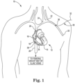
- FIG. 1 is a schematic view illustrating the implantation of a device for producing at least one trabecular fiber within a heart to help repair the heart, in accordance with embodiments of the present invention.
- the at least one trabecular fiber includes new contractile or conductile heart muscle tissue, or cardiomyocyte cells.
- the trabecular fiber produced as described herein includes new cardiomyocyte cells grown de novo within the heart chamber, and not pre-existing heart tissue simply rearranged.
- the device and attached at least one trabecular fiber may repair the heart by connecting to various structures encircling a ventricle, such as a ventricle wall, a septum, and a valve, to strengthen and/or reshape the heart.
- a ventricle such as a ventricle wall, a septum, and a valve
- the de novo trabecular fiber may serve as a moderator band in those without this anatomical feature to limit sudden dilation due to fluctuating venous return, aid in synchronizing right ventricle free wall depolarization to treat conduction delays, and/or provide collateral blood flow.
- FIG. 1 is a schematic view illustrating the implantation of a device for producing at least one trabecular fiber within a heart.
- FIG. 1 illustrates a patient 10 including a heart 12.
- the heart 12 includes a right ventricle 14, a right atrium 16, and a tricuspid valve 18 separating the right atrium 16 from the right ventricle 14.
- veins directing blood to the heart 12 including a left auxiliary vein 20, which flows into a left subclavian vein 22, which flows into a left brachiocephalic vein 24.
- the left brachiocephalic vein 24 flows into a superior vena cava 26, which supplies blood to the right atrium 16 from the upper part of the body.
- An inferior vena cava 28 receives blood from a femoral vein (not shown) by way of an external iliac vein (not shown) and a common iliac vein (not shown). The inferior vena cava 28 also supplies blood to the right atrium 16.
- FIG. 1 shows a catheter 30 having a proximal end 32 and a distal end 34.
- the catheter 30 may enter the left auxiliary vein 20 percutaneously through a vascular entry site 36.
- the distal end 34 may be maneuvered through a left auxiliary vein 20, the left subclavian vein 22, the left brachiocephalic vein 24, the superior vena cava 26, and into the heart 12 at the right atrium 16.
- the catheter 30 may percutaneously enter the femoral artery.
- the distal end 34 may be maneuvered through the external iliac vein, the common iliac vein, the inferior vena cava 28, and into the heart 12 at the right atrium 16.
- the distal end 34 may be maneuvered from the right atrium 16, through the tricuspid valve 18, and into the right ventricle 14.
- the catheter 30 may include at least one lumen (not shown) extending from the proximal end 32 to the distal end 34 through which instruments (not shown) may be used to implant a device into an endocardium 38 lining the walls of the right ventricle 14, such as a device 40 for producing at least one trabecular fiber within the heart 12 as described below in reference to FIG. 2 .
- implantation of device 40 is done in a minimally-invasive manner.
- FIG. 2 is an enlarged schematic view of the heart 12 of FIG. 1 .
- the heart 12 further includes left ventricle 42, left atrium 44, ventricular wall 46, and septum 48 between the right ventricle 14 and the left ventricle 42.
- FIG. 2 illustrates the device 40, according to some embodiments, implanted into the endocardium 38 of the ventricular wall 46.
- the device 40 includes a tissue anchor 50 and a substrate 52.
- the tissue anchor 50 can include a linking section 54 and a fixation device 56.
- the linking section 54 connects the fixation device 56 to the substrate 52, connecting the tissue anchor 50 to the substrate 52.
- the substrate 52 is a flexible, non-rigid ribbon or sheet made at least in part from a biocompatible polymer, for example, a polyurethane polymer, a polyester polymer, silicone polymer, a styrene-isobutylene-styrene block copolymer, or an expanded polytetrafluoroethylene polymer.
- a biocompatible polymer for example, a polyurethane polymer, a polyester polymer, silicone polymer, a styrene-isobutylene-styrene block copolymer, or an expanded polytetrafluoroethylene polymer.
- the substrate 52 may be made at least in part of an organic substance, for example, collagen, hyaluronan, cellulose, fibrin, fibrinogen, or fibronectin.
- the substrate 52 may be a solid or may be an electro-spun mesh of a biocompatible polymer and/or an organic substance.
- the linking section 54 of the tissue anchor 50 can be made of any of the above mentioned biocompatible polymers or organic substances.
- the fixation device 56 may be a passive fixation device, such as tines as illustrated in FIG. 2 , or an active fixation device, such as a hook or helical configuration to bore into a heart wall, such as the ventricular wall 46 or the septum 48.
- the fixation device 56 of the tissue anchor 50 is secured into the endocardium 38 and the ventricular wall 46 to anchor the device 40 within the right ventricle 14 of the heart 12 such that the substrate 52 is spaced apart from endocardium 38.
- the device 40 is maintained in the heart 12, at least one de novo trabecular fiber 58 forms between the endocardium 38 and the substrate 52.
- a plurality of trabecular fibers 58 are formed.
- the trabecular fibers 58 extending between the substrate 52 and ventricular wall 46, and the trabecular fibers 58 extending between the substrate 52 the septum 48 serve to connect the ventricular wall 46 to the septum with the new contractile or conductile heart muscle tissue. So disposed, the substrate 52 and the trabecular fibers 58 with their contractile or conductile heart muscle tissue can serve to strengthen the ventricular wall to repair damage due to thinning of the ventricular wall 46 or to an infarction in the ventricular wall 46.
- FIG. 3 is a schematic cross-sectional view of the trabecular fiber 58.
- the trabecular fiber 58 includes a blood vessel 60, a plurality of cardiomyocyte cells 62, an extracellular matrix layer 64, an elastin layer 66, an outer collagen layer 68, and an endothelial cell layer 70.
- the cardiomyocyte cells 62 can be disposed at a core of the trabecular fiber 58 and can be generally oriented with their long-axis (not shown) parallel to the length of the trabecular fiber 58 ( FIG. 2 ).
- the cardiomyocyte cells 62 can be embedded in an extracellular matrix formed by the extracellular matrix layer 64, and may be nourished by the blood vessel 60 coursing in parallel to the cardiomyocyte cells 62 and/or by blood within the right ventricle 14 ( FIG. 2 ).
- the outer collagen layer 68 can be an external layer of the trabecular fiber 58.
- the elastin layer 66 can be disposed between the extracellular matrix layer 64 and the outer collagen layer 68.
- the overall organizational histologic architecture of the trabecular fiber 58 is that of a tube (the cardiomyocyte cells 62), within a tube (the elastin layer 66), within a tube (the outer collagen layer 68).
- the presence of the cardiomyocyte cells 62 at the core of the trabecular fiber 58 distinguishes the trabecular fiber 58 from structures of somewhat similar appearance, such as chordae tendineae of the tricuspid valve 18 ( FIG. 1 ).
- chordae tendineae of the tricuspid valve 18 FIG. 1
- the substrate 52 creates a stimulus for the development of a provisional matrix connected to the substrate 52, the provisional matrix composed of fibrinogen, red cells, platelets, white cells and, in some embodiments, stem or progenitor cells. It is believed that the white cells may be leukocytes, such as macrophages and/or T cells.
- stem or progenitor cells may be bone marrow derived or resident cells within the endocardium 38 of the right ventricle 14. It is believed that the provisional matrix facilitates the growth and propagation of cells to create an immature de novo trabecular fiber which matures into the contractile/conductile functioning trabecular fiber 58.
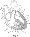
- FIG. 4 is an enlarged schematic view of the heart 12 of FIG. 1 illustrating another embodiment.
- FIG. 4 illustrates a device 72, according to some embodiments, implanted into the endocardium 38 of the ventricular wall 46.
- the device 72 includes a tissue anchor 50, a substrate 74, a power supply 76, and pulse generator 78.
- the substrate 74 can be substantially similar to substrate 40 described above, except that it includes at least one electrode 80 (four shown in FIG. 4 ) disposed on the substrate 74.
- the power supply 76 can include a battery.
- the pulse generator 78 is electrically connected to the power supply 76 and to the at least one electrode 80 so that together, the power supply 76 and the pulse generator 78 generate a plurality of voltage pulses at the at least one electrode 80.
- the pulse generator 78 can control characteristics of the plurality of voltage pulses, for example, the frequency of the pulses, the length of the pulses, and the amplitude of the pulses. Such pulse generators are well known in the art.
- the at least one electrode 80 can be made of any conductive, biocompatible material, for example, titanium, platinum, stainless steel, nitinol, iridium, or iridium oxide. In some embodiments, the at least one electrode 80 is disposed on a surface of the substrate 74.
- the tissue anchor 50 can include a linking section 54 and a fixation device 56.
- the linking section 54 connects the fixation device 56 to the substrate 74, connecting the tissue anchor 50 to the substrate 74.
- the power supply 76 and the pulse generator 78 are disposed at an end of the substrate 74.
- the power supply 76 and the pulse generator 78 can be disposed anywhere along the substrate 74 or the anchor 50.
- the fixation device 56 of the tissue anchor 50 is secured into the endocardium 38 to anchor the device 72 within the heart 12 such that the substrate 74 is spaced apart from endocardium 38.
- Voltage pulses can be generated from the at least one electrode 80 to produce an electrical potential between the substrate 74 and the endocardium 38.
- at least one trabecular fiber 58 forms between the endocardium 38 and the substrate 74.
- a plurality of trabecular fibers 58 are formed.
- FIG. 4 a plurality of trabecular fibers 58 are formed.
- the trabecular fibers 58 extending between the substrate 74 and ventricular wall 46, and the trabecular fibers 58 extending between the substrate 74 the septum 48 serve to connect the ventricular wall 46 to the septum with the new contractile/conductile heart muscle tissue. So disposed, the substrate 74 and the trabecular fibers 58 with their contractile heart muscle tissue can serve to repair damage due to thinning of the ventricular wall 46 or to an infarction in the ventricular wall 46, thus improving cardiac output.
- the voltage pulses at the at least one electrode 80 may further stimulate the growth of the trabecular fibers 58.
- the trabecular fibers 58 are shown attached to the substrate 74 at the electrodes 80. However, in other embodiments, the trabecular fibers 58 may attach elsewhere on the substrate 74.
- FIG. 5 is an enlarged schematic view of the heart 12 of FIG. 1 illustrating another embodiment.
- FIG. 5 illustrates a device 82, according to some embodiments, implanted into the endocardium 38 of the ventricular wall 46.
- the device 82 includes a tissue anchor 50, and a substrate 84.
- the tissue anchor 50 can include a linking section 54 and a fixation device 56.
- the linking section 54 connects the fixation device 56 to an end of the substrate 84, connecting the tissue anchor 50 to the substrate 84.
- the substrate 84 is a flexible, non-rigid, helically wound conductive coil made at least in part from a biocompatible conductor, for example, titanium, platinum, stainless steel, nitinol, iridium, or iridium oxide.
- the coil turns can be closely spaced such that adjacent turns are in physical contact for improved MRI compatibility. In other embodiments the coil turns can be more widely spaced to provide larger gaps between adjacent turns to enhance adhesion of the trabecular fibers to the substrate 84.
- an end of the substrate 84 can be electrically and physically connected to an electrical lead 86 extending from the device 82, through the tricuspid valve 18, the right atrium 16 and out of the heart 12 to a pulse generator 88 and a power supply 90 external to the heart 12.
- the pulse generator 88 and the power supply 90 can be disposed in a subcutaneous pocket (not shown) adjacent to the vascular entry site 36 ( FIG. 1 ).
- the tissue anchor 50 can include a linking section 54 and a fixation device 56.
- the linking section 54 connects the fixation device 56 to the substrate 44, connecting the tissue anchor 50 to the substrate 44.
- the power supply 90 can include a battery.
- the pulse generator 88 is electrically connected to the power supply 90 and to the substrate 84 so that together, the power supply 90 and the pulse generator 88 generate a plurality of voltage pulses at the substrate 84, which functions as an electrode.
- the pulse generator 88 can control characteristics of the plurality of voltage pulses, for example, the frequency of the pulses, the length of the pulses, and the amplitude of the pulses. Such pulse generators are well known in the art.
- the fixation device 56 of the tissue anchor 50 is secured to the endocardium 38 to anchor the device 82 within the heart 12 such that the substrate 84 is spaced apart from endocardium 38.
- Voltage pulses can be generated at the substrate 84 to produce an electrical potential between the substrate 84 and the endocardium 38.
- at least one trabecular fiber 58 forms between the endocardium 38 and the substrate 84.
- a plurality of trabecular fibers 58 are formed.
- FIG. 5 a plurality of trabecular fibers 58 are formed.
- the trabecular fibers 58 extending between the substrate 84 and ventricular wall 46 and the anchor 50 extending between the substrate 84 and another part of the ventricular wall 46 serve to connect the different portions of the ventricular wall 46 to each other with the new contractile/conductile heart muscle tissue of the trabecular fibers 58. So disposed, the substrate 84 and the trabecular fibers 58 with their contractile heart muscle tissue can serve to strengthen the ventricular wall to repair damage due to thinning of the ventricular wall 46 or to an infarction in the ventricular wall 46. Not wishing to be bound by any theory, it is believed that the voltage pulses at the substrate 84 may further stimulate the growth of the trabecular fibers 58.
- FIG. 6 is an enlarged schematic view of the heart 12 of FIG. 1 illustrating the present invention.
- FIG. 6 illustrates a device 92, according to some embodiments, implanted into the endocardium 38 of the septum 48.
- the device 92 includes a substrate 94, a first tissue anchor 96, and a second tissue anchor 98.
- the first tissue anchor 96 and the second tissue anchor 98 can each be substantially similar to the tissue anchor 50 described above, including a linking section 54 and a fixation device 56.
- the first tissue anchor 96 and the second tissue anchor 98 are connected to opposite ends of the substrate 94.
- the substrate 94 can be substantially similar to substrate 52 described above in reference to FIG.
- the substrate 94 can be substantially similar to substrate 74, and the device 92 can further include a power supply 76 and pulse generator 78 as described above in reference to FIG. 4 . In still other embodiments, the substrate 94 can be substantially similar to substrate 84 described above in reference to FIG. 5 .
- the first tissue anchor 96 and the second tissue anchor 98 are secured the endocardium 38 of the septum 48 to anchor the device 92 within the heart 12 such that the substrate 92 is spaced apart from the endocardium 38.
- the device 92 is maintained in the heart 12
- at least one trabecular fiber 58 forms between the endocardium 38 and the substrate 94.
- the trabecular fibers 58 are formed between the substrate 94 and the endocardium 38 on the tricuspid valve 18, the ventricular wall 46, and the septum 48.
- the trabecular fibers 58 extending between the substrate 94 and septum 48 and between the substrate 94 and the tricuspid valve 18 serve to connect a portion of the tricuspid valve 18 to the septum 48 with the new contractile heart muscle tissue of the trabecular fibers 58. So disposed, the substrate 94 and the trabecular fibers 58 with their contractile heart muscle tissue can alter the shape of the heart 12 to repair damage due a misalignment in the tricuspid valve 18 resulting in regurgitation by reshaping the heart 12. However, the trabecular fiber 58 extending between the substrate 94 and the ventricular wall 46 may not be necessary and may, in some instances, interfere with the repair effected by the substrate 94 and the other trabecular fibers 58.
- FIGS. 7A and 7B are enlarged schematic views of a portion of the heart 12 of FIG. 6 illustrating the removal of the unnecessary trabecular fiber 58 in accordance with embodiments.
- FIGS. 7A and 7B show a tool 100 for imaging and extracting the trabecular fibers 58.
- the tool 100 can include a catheter 102, a visualization device 104, and a forceps device 106.
- the forceps device 106 can include a pair of jaws 108.
- the pair of jaws 108 can be used to cut and/or grasp tissue, such as the trabecular fiber 58.
- the catheter 102 can include a plurality of lumens (not shown) extending the length of the catheter 102 for accommodating the visualization device 104 and the forceps device 106.
- the catheter 102 can be maneuvered into the right ventricle 14 as described above for catheter 30 in reference to FIG. 1 .
- the catheter 102 can be, for example, a SpyGlass ® Catheter from Boston Scientific Corporation, Natick, Massachusetts.
- the visualization device 104 can be a fiber-optic based device, for example, a SpyGlass ® Direct Visualization Probe from Boston Scientific Corporation, Natick, Massachusetts.
- the visualization device 104 can include a solid-state camera, a transparent balloon (not shown) extending around the camera, and a source of saline (not shown) for inflating the transparent balloon to enhance direct visualization by displacing blood proximate to the trabecular fiber 58.
- the forceps device 106 can be, for example, a SpyBite ® Biopsy Forceps from Boston Scientific Corporation, Natick, Massachusetts.
- the tool 100 can further include a separate light source (not shown).
- removing the trabecular fiber 58 can include cutting away the trabecular fiber 58 from the endocardium 38 by operation of the pair of jaws 108 of the forceps device 106.
- the trabecular fiber 58 can be cut from the substrate 94 by operation of the pair of jaws 108 of the forceps device 106 ( FIG. 6 ).
- FIG. 7B once the trabecular fiber 58 is cut away from both the endocardium 38 and the substrate 94, the pair of jaws 108 can grasp the trabecular fiber 58.
- the forceps device 106 can be withdrawn through the lumen in the catheter 102 to remove the entire trabecular fiber 58 from the heart 12.
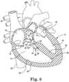
- FIG. 8 is a schematic view of the heart 12 of FIG. 6 after removal of the unnecessary trabecular fiber 58.
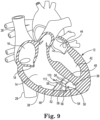
- FIG. 9 is an enlarged schematic view of the heart 12 of FIG. 1 illustrating yet another embodiment.
- FIG. 9 illustrates a device 110, not according to the claimed invention, implanted into the endocardium 38 of the ventricular wall 46.
- the device 110 includes a tissue anchor 50, a substrate 52, and at least one drug eluting collar 112.
- the tissue anchor 50 and the substrate 52 are as described above in reference to FIG. 2 .
- the collar 112 is disposed around the middle of the substrate 52. However, in other embodiments, the collar 112 can be disposed anywhere along the substrate 52.
- the drug eluting collar 112 can include an anti-inflammatory or immunosuppressive biologic or pharmaceutical agent, such as a steroid.
- the steroid can be, for example, a glucocorticoid such as dexamethasone acetate or hydrocortisone.
- the steroid can be disposed within a liquid silicone rubber (LSR) matrix such that the steroid can elute from the LSR matrix.
- LSR liquid silicone rubber
- the collar 112 may be formed by mixing the steroid into the LSR before the LSR cures. In some embodiments, the steroid may elute from the LSR matrix over an extended period of time.
- the collar 112 may include a steroid dose of as little as 0.20 milligrams (mg), 0.30 mg, 0.40 mg, or 0.50 mg, or as great as 0.70 mg, 0.80 mg, 0.90 mg or 1.0 mg, or any amount between any of the preceding values.
- the collar 112 may include a steroid dose ranging from 0.20 mg to 1.0 mg, from 0.30 mg to 0.90 mg, from 0.40 mg to 0.80 mg, or from 0.50 mg to 0.70 mg.
- the collar 112 may include as steroid dose of about 0.60 mg.
- the tissue anchor 50 can include a linking section 54 and a fixation device 56.
- the linking section 54 connects the fixation device 56 to the substrate 52, connecting the tissue anchor 50 to the substrate 52.
- the fixation device 56 of the tissue anchor 50 is secured into the endocardium 38 to anchor the device 110 within the heart 12 such that the substrate 522 is spaced apart from endocardium 38.
- An anti-inflammatory or immunosuppressive biologic or pharmaceutical agent such as the steroid dexamethasone acetate, can elute from the collar 112.
- at least one trabecular fiber 58 forms between the endocardium 38 and the substrate 52.
- a plurality of trabecular fibers 58 are formed.
- FIG. 9 a plurality of trabecular fibers 58 are formed.
- the trabecular fibers 58 extending between the substrate 52 and ventricular wall 46, and the trabecular fibers 58 extending between the substrate 52 the septum 48 serve to connect the ventricular wall 46 to the septum with the new contractile/conductile heart muscle tissue. So disposed, the substrate 52 and the trabecular fibers 58 with their contractile/conductile heart muscle tissue can serve to repair damage due to thinning of the ventricular wall 46 or to an infarction in the ventricular wall 46, thus improving cardiac output.
- the anti-inflammatory or immunosuppressive biologic or pharmaceutical agent eluting from the collar 112 may further stimulate the growth of the trabecular fibers 58.
- the device for producing at least one trabecular fiber within a heart to repair the heart is configured to be contained entirely within the ventricle. That is, the device is sized and shaped to be contained entirely within the ventricle to enhance the minimally invasive nature of the device.
- the device for producing at least one trabecular fiber within a heart to repair the heart is implanted in the right ventricle.
- the device may be implanted additionally or alternatively in the left ventricle.
Claims (8)
- Dispositif (40, 72, 82, 92, 110) pour la production d'une fibre trabéculaire (58) à l'intérieur d'un ventricule (14, 42) d'un coeur (12), le dispositif (40, 72, 82, 92, 110) comprenant :un substrat (52, 74, 84, 94) formé d'un matériau non rigide ;un premier ancrage de tissu (50, 96) relié au substrat (52, 74, 84, 94) ; etun second ancrage de tissu (50, 98) relié au substrat (52, 74, 84, 94) à l'opposé du premier ancrage de tissu (50, 96) ; etdans lequel le dispositif (40, 72, 82, 92, 110) est conçu pour être contenu entièrement à l'intérieur du ventricule (14, 42) etdans lequel le premier ancrage de tissu et le second ancrage de tissu sont conçus pour être fixés solidement à un endocarde afin d'ancrer le dispositif à l'intérieur du coeur de telle sorte que le substrat soit espacé de l'endocarde.
- Dispositif (40, 72, 82, 92, 110) selon la revendication 1, dans lequel le substrat (52, 74, 84, 94) comprend un ruban de fibres électrofilées.
- Dispositif (40, 72, 82, 92, 110) selon l'une quelconque des revendications 1 et 2, dans lequel le substrat (52, 74, 84, 94) est formé d'au moins un parmi un polymère de polyuréthane, un polymère de polyester, un polymère de silicone, un copolymère séquencé de styrène-isobutylène-styrène, un polymère de polytétrafluoroéthylène expansé, du collagène, du hyaluronane, de la cellulose, de la fibrine, du fibrinogène et de la fibronectine.
- Dispositif (40, 72, 82, 92, 110) selon l'une quelconque des revendications 1 à 3, dans lequel le substrat (52, 74, 84, 94) comprend au moins une électrode (80) disposée sur le substrat (52, 74, 84, 94).
- Dispositif (40, 72, 82, 92, 110) selon la revendication 1, dans lequel le substrat (52, 74, 84, 94) comprend au moins une bobine conductrice enroulée hélicoïdalement formant au moins une électrode (80).
- Dispositif (40, 72, 82, 92, 110) selon l'une ou l'autre des revendications 4 et 5, dans lequel le dispositif (40, 72, 82, 92, 110) est conçu pour être connecté à un générateur d'impulsions (78, 88) et à une alimentation électrique (76, 90) externe au coeur (12) par un fil électrique (86), le fil électrique (86) connectant le générateur d'impulsions (78, 88) et l'alimentation électrique (76, 90) à l'au moins une électrode (80) pour générer une pluralité d'impulsions de tension au niveau de l'au moins une électrode (80).
- Dispositif (40, 72, 82, 92, 110) selon l'une ou l'autre des revendications 4 et 5, dans lequel le dispositif (40, 72, 82, 92, 110) comprend en outre un générateur d'impulsions (78, 88) et une alimentation électrique (76, 90), le générateur d'impulsions (78, 88) et l'alimentation électrique (76, 90) étant connectés électriquement à l'au moins une électrode (80) pour générer une pluralité d'impulsions de tension à l'au moins une électrode (80).
- Dispositif (40, 72, 82, 92, 110) selon l'une quelconque des revendications 1 à 7, comprenant en outre au moins un collier d'élution de médicament (112) disposé autour du substrat (52, 74, 84, 94), le collier d'élution de médicament (112) comprenant un agent biologique ou pharmaceutique anti-inflammatoire ou immunosuppresseur.
Applications Claiming Priority (2)
| Application Number | Priority Date | Filing Date | Title |
|---|---|---|---|
| US201662426995P | 2016-11-28 | 2016-11-28 | |
| PCT/US2017/052909 WO2018097884A1 (fr) | 2016-11-28 | 2017-09-22 | Procédés et dispositifs de production de fibres trabéculaires |
Publications (2)
| Publication Number | Publication Date |
|---|---|
| EP3544645A1 EP3544645A1 (fr) | 2019-10-02 |
| EP3544645B1 true EP3544645B1 (fr) | 2023-03-15 |
Family
ID=60153422
Family Applications (1)
| Application Number | Title | Priority Date | Filing Date |
|---|---|---|---|
| EP17787687.7A Active EP3544645B1 (fr) | 2016-11-28 | 2017-09-22 | Procédés et dispositifs de production de fibres trabéculaires |
Country Status (6)
| Country | Link |
|---|---|
| US (1) | US10773077B2 (fr) |
| EP (1) | EP3544645B1 (fr) |
| JP (1) | JP6936858B2 (fr) |
| CN (1) | CN110022910B (fr) |
| AU (1) | AU2017366432B2 (fr) |
| WO (1) | WO2018097884A1 (fr) |
Families Citing this family (1)
| Publication number | Priority date | Publication date | Assignee | Title |
|---|---|---|---|---|
| EP3337892A2 (fr) | 2015-08-18 | 2018-06-27 | Boston Scientific Scimed Inc. | Procédés de production de cellules cardiomyocytaires |
Family Cites Families (13)
| Publication number | Priority date | Publication date | Assignee | Title |
|---|---|---|---|---|
| US5632716A (en) | 1996-06-20 | 1997-05-27 | Telectronics Pacing Systems, Inc. | Apparatus and method of muscle training in dynamic cardiomyoplasty |
| WO2003064637A1 (fr) * | 2001-11-06 | 2003-08-07 | Medtronic, Inc. | Methode et systeme permettant de reparer un infarctus du myocarde |
| WO2001028455A1 (fr) * | 1999-10-21 | 2001-04-26 | Myocor, Inc. | Procedes et dispositifs permettant d'ameliorer la fonction cardiaque |
| US7400931B2 (en) * | 2002-09-18 | 2008-07-15 | Cardiac Pacemakers, Inc. | Devices and methods to stimulate therapeutic angiogenesis for ischemia and heart failure |
| ATE418357T1 (de) * | 2003-10-24 | 2009-01-15 | Cardiac Pacemakers Inc | Myokardiales leitungsbefestigungssystem |
| US7410497B2 (en) * | 2004-12-14 | 2008-08-12 | Boston Scientific Scimed, Inc. | Stimulation of cell growth at implant surfaces |
| US8221744B2 (en) | 2007-09-19 | 2012-07-17 | Abbott Cardiovascular Systems Inc. | Cytocompatible alginate gels |
| EP2265166B1 (fr) * | 2008-03-25 | 2020-08-05 | EBR Systems, Inc. | Connexion d électrode temporaire pour des systèmes de stimulation sans fil |
| WO2011041571A2 (fr) | 2009-10-01 | 2011-04-07 | Kardium Inc. | Dispositif médical, kit et procédé de construction de tissu ou d'orifice corporel, par exemple de valvule mitrale |
| US20130231727A1 (en) * | 2012-03-05 | 2013-09-05 | Pacesetter, Inc. | Lead with bioabsorbable metallic fixation structure |
| US20150088155A1 (en) * | 2013-09-23 | 2015-03-26 | Cardiac Pacemakers, Inc. | Mechanical configurations for a multi-site leadless pacemaker |
| EP3137163B1 (fr) * | 2014-04-29 | 2019-02-20 | Cardiac Pacemakers, Inc. | Stimulateur cardiaque sans fil à caractéristiques permettant son extraction |
| EP3337892A2 (fr) * | 2015-08-18 | 2018-06-27 | Boston Scientific Scimed Inc. | Procédés de production de cellules cardiomyocytaires |
-
2017
- 2017-09-22 WO PCT/US2017/052909 patent/WO2018097884A1/fr active Application Filing
- 2017-09-22 CN CN201780073667.6A patent/CN110022910B/zh active Active
- 2017-09-22 EP EP17787687.7A patent/EP3544645B1/fr active Active
- 2017-09-22 JP JP2019528738A patent/JP6936858B2/ja active Active
- 2017-09-22 US US15/712,542 patent/US10773077B2/en active Active
- 2017-09-22 AU AU2017366432A patent/AU2017366432B2/en active Active
Also Published As
| Publication number | Publication date |
|---|---|
| US20180147409A1 (en) | 2018-05-31 |
| WO2018097884A1 (fr) | 2018-05-31 |
| CN110022910A (zh) | 2019-07-16 |
| US10773077B2 (en) | 2020-09-15 |
| CN110022910B (zh) | 2022-03-29 |
| EP3544645A1 (fr) | 2019-10-02 |
| JP2020513265A (ja) | 2020-05-14 |
| AU2017366432A1 (en) | 2019-05-23 |
| AU2017366432B2 (en) | 2021-04-29 |
| JP6936858B2 (ja) | 2021-09-22 |
Similar Documents
| Publication | Publication Date | Title |
|---|---|---|
| US10537731B2 (en) | Transvenous mediastinum access for the placement of cardiac pacing and defibrillation electrodes | |
| US20210106807A1 (en) | Treating congestive heart failure | |
| US10646720B2 (en) | Parasternal placement of an active medical device using the internal thoracic vasculature | |
| US10980570B2 (en) | Implantation of an active medical device using the internal thoracic vasculature | |
| US10850067B2 (en) | Implantation of an active medical device using the intercostal vein | |
| US7797059B1 (en) | System and method for lead implantation in a pericardial space | |
| US7840266B2 (en) | Integrated lead for applying cardiac resynchronization therapy and neural stimulation therapy | |
| US20180133463A1 (en) | Electrode for sensing, pacing, and defibrillation deployable in the mediastinal space | |
| EP1863564B1 (fr) | Therapie combinee de stimulation neurale et de resynchronisation cardiaque | |
| US20180169425A1 (en) | Lead with integrated electrodes | |
| US20180325480A1 (en) | Implantation of an active medical device using the internal thoracic vasculature | |
| US20210370057A1 (en) | Methods for producing cardiomyocyte cells | |
| EP3544645B1 (fr) | Procédés et dispositifs de production de fibres trabéculaires | |
| US20100137927A1 (en) | Multifunctional cardiac pacemaker system | |
| Nakatani et al. | Unroofed coronary sinus | |
| Beghetti et al. | Thoraco-omphalagus twins: heart to heart |
Legal Events
| Date | Code | Title | Description |
|---|---|---|---|
| STAA | Information on the status of an ep patent application or granted ep patent |
Free format text: STATUS: UNKNOWN |
|
| STAA | Information on the status of an ep patent application or granted ep patent |
Free format text: STATUS: THE INTERNATIONAL PUBLICATION HAS BEEN MADE |
|
| PUAI | Public reference made under article 153(3) epc to a published international application that has entered the european phase |
Free format text: ORIGINAL CODE: 0009012 |
|
| STAA | Information on the status of an ep patent application or granted ep patent |
Free format text: STATUS: REQUEST FOR EXAMINATION WAS MADE |
|
| 17P | Request for examination filed |
Effective date: 20190627 |
|
| AK | Designated contracting states |
Kind code of ref document: A1 Designated state(s): AL AT BE BG CH CY CZ DE DK EE ES FI FR GB GR HR HU IE IS IT LI LT LU LV MC MK MT NL NO PL PT RO RS SE SI SK SM TR |
|
| AX | Request for extension of the european patent |
Extension state: BA ME |
|
| DAV | Request for validation of the european patent (deleted) | ||
| DAX | Request for extension of the european patent (deleted) | ||
| STAA | Information on the status of an ep patent application or granted ep patent |
Free format text: STATUS: EXAMINATION IS IN PROGRESS |
|
| 17Q | First examination report despatched |
Effective date: 20201008 |
|
| STAA | Information on the status of an ep patent application or granted ep patent |
Free format text: STATUS: EXAMINATION IS IN PROGRESS |
|
| GRAP | Despatch of communication of intention to grant a patent |
Free format text: ORIGINAL CODE: EPIDOSNIGR1 |
|
| STAA | Information on the status of an ep patent application or granted ep patent |
Free format text: STATUS: GRANT OF PATENT IS INTENDED |
|
| INTG | Intention to grant announced |
Effective date: 20220923 |
|
| GRAS | Grant fee paid |
Free format text: ORIGINAL CODE: EPIDOSNIGR3 |
|
| GRAA | (expected) grant |
Free format text: ORIGINAL CODE: 0009210 |
|
| STAA | Information on the status of an ep patent application or granted ep patent |
Free format text: STATUS: THE PATENT HAS BEEN GRANTED |
|
| AK | Designated contracting states |
Kind code of ref document: B1 Designated state(s): AL AT BE BG CH CY CZ DE DK EE ES FI FR GB GR HR HU IE IS IT LI LT LU LV MC MK MT NL NO PL PT RO RS SE SI SK SM TR |
|
| REG | Reference to a national code |
Ref country code: CH Ref legal event code: EP Ref country code: GB Ref legal event code: FG4D |
|
| REG | Reference to a national code |
Ref country code: DE Ref legal event code: R096 Ref document number: 602017066847 Country of ref document: DE |
|
| REG | Reference to a national code |
Ref country code: IE Ref legal event code: FG4D |
|
| REG | Reference to a national code |
Ref country code: AT Ref legal event code: REF Ref document number: 1553620 Country of ref document: AT Kind code of ref document: T Effective date: 20230415 |
|
| REG | Reference to a national code |
Ref country code: NL Ref legal event code: FP |
|
| REG | Reference to a national code |
Ref country code: LT Ref legal event code: MG9D |
|
| PG25 | Lapsed in a contracting state [announced via postgrant information from national office to epo] |
Ref country code: RS Free format text: LAPSE BECAUSE OF FAILURE TO SUBMIT A TRANSLATION OF THE DESCRIPTION OR TO PAY THE FEE WITHIN THE PRESCRIBED TIME-LIMIT Effective date: 20230315 Ref country code: NO Free format text: LAPSE BECAUSE OF FAILURE TO SUBMIT A TRANSLATION OF THE DESCRIPTION OR TO PAY THE FEE WITHIN THE PRESCRIBED TIME-LIMIT Effective date: 20230615 Ref country code: LV Free format text: LAPSE BECAUSE OF FAILURE TO SUBMIT A TRANSLATION OF THE DESCRIPTION OR TO PAY THE FEE WITHIN THE PRESCRIBED TIME-LIMIT Effective date: 20230315 Ref country code: LT Free format text: LAPSE BECAUSE OF FAILURE TO SUBMIT A TRANSLATION OF THE DESCRIPTION OR TO PAY THE FEE WITHIN THE PRESCRIBED TIME-LIMIT Effective date: 20230315 Ref country code: HR Free format text: LAPSE BECAUSE OF FAILURE TO SUBMIT A TRANSLATION OF THE DESCRIPTION OR TO PAY THE FEE WITHIN THE PRESCRIBED TIME-LIMIT Effective date: 20230315 |
|
| REG | Reference to a national code |
Ref country code: AT Ref legal event code: MK05 Ref document number: 1553620 Country of ref document: AT Kind code of ref document: T Effective date: 20230315 |
|
| PG25 | Lapsed in a contracting state [announced via postgrant information from national office to epo] |
Ref country code: SE Free format text: LAPSE BECAUSE OF FAILURE TO SUBMIT A TRANSLATION OF THE DESCRIPTION OR TO PAY THE FEE WITHIN THE PRESCRIBED TIME-LIMIT Effective date: 20230315 Ref country code: GR Free format text: LAPSE BECAUSE OF FAILURE TO SUBMIT A TRANSLATION OF THE DESCRIPTION OR TO PAY THE FEE WITHIN THE PRESCRIBED TIME-LIMIT Effective date: 20230616 Ref country code: FI Free format text: LAPSE BECAUSE OF FAILURE TO SUBMIT A TRANSLATION OF THE DESCRIPTION OR TO PAY THE FEE WITHIN THE PRESCRIBED TIME-LIMIT Effective date: 20230315 |
|
| PGFP | Annual fee paid to national office [announced via postgrant information from national office to epo] |
Ref country code: NL Payment date: 20230822 Year of fee payment: 7 |
|
| PG25 | Lapsed in a contracting state [announced via postgrant information from national office to epo] |
Ref country code: SM Free format text: LAPSE BECAUSE OF FAILURE TO SUBMIT A TRANSLATION OF THE DESCRIPTION OR TO PAY THE FEE WITHIN THE PRESCRIBED TIME-LIMIT Effective date: 20230315 Ref country code: RO Free format text: LAPSE BECAUSE OF FAILURE TO SUBMIT A TRANSLATION OF THE DESCRIPTION OR TO PAY THE FEE WITHIN THE PRESCRIBED TIME-LIMIT Effective date: 20230315 Ref country code: PT Free format text: LAPSE BECAUSE OF FAILURE TO SUBMIT A TRANSLATION OF THE DESCRIPTION OR TO PAY THE FEE WITHIN THE PRESCRIBED TIME-LIMIT Effective date: 20230717 Ref country code: ES Free format text: LAPSE BECAUSE OF FAILURE TO SUBMIT A TRANSLATION OF THE DESCRIPTION OR TO PAY THE FEE WITHIN THE PRESCRIBED TIME-LIMIT Effective date: 20230315 Ref country code: EE Free format text: LAPSE BECAUSE OF FAILURE TO SUBMIT A TRANSLATION OF THE DESCRIPTION OR TO PAY THE FEE WITHIN THE PRESCRIBED TIME-LIMIT Effective date: 20230315 Ref country code: CZ Free format text: LAPSE BECAUSE OF FAILURE TO SUBMIT A TRANSLATION OF THE DESCRIPTION OR TO PAY THE FEE WITHIN THE PRESCRIBED TIME-LIMIT Effective date: 20230315 Ref country code: AT Free format text: LAPSE BECAUSE OF FAILURE TO SUBMIT A TRANSLATION OF THE DESCRIPTION OR TO PAY THE FEE WITHIN THE PRESCRIBED TIME-LIMIT Effective date: 20230315 |
|
| PGFP | Annual fee paid to national office [announced via postgrant information from national office to epo] |
Ref country code: IE Payment date: 20230823 Year of fee payment: 7 |
|
| PG25 | Lapsed in a contracting state [announced via postgrant information from national office to epo] |
Ref country code: SK Free format text: LAPSE BECAUSE OF FAILURE TO SUBMIT A TRANSLATION OF THE DESCRIPTION OR TO PAY THE FEE WITHIN THE PRESCRIBED TIME-LIMIT Effective date: 20230315 Ref country code: PL Free format text: LAPSE BECAUSE OF FAILURE TO SUBMIT A TRANSLATION OF THE DESCRIPTION OR TO PAY THE FEE WITHIN THE PRESCRIBED TIME-LIMIT Effective date: 20230315 Ref country code: IS Free format text: LAPSE BECAUSE OF FAILURE TO SUBMIT A TRANSLATION OF THE DESCRIPTION OR TO PAY THE FEE WITHIN THE PRESCRIBED TIME-LIMIT Effective date: 20230715 |
|
| PGFP | Annual fee paid to national office [announced via postgrant information from national office to epo] |
Ref country code: DE Payment date: 20230822 Year of fee payment: 7 |
|
| REG | Reference to a national code |
Ref country code: DE Ref legal event code: R097 Ref document number: 602017066847 Country of ref document: DE |
|
| PLBE | No opposition filed within time limit |
Free format text: ORIGINAL CODE: 0009261 |
|
| STAA | Information on the status of an ep patent application or granted ep patent |
Free format text: STATUS: NO OPPOSITION FILED WITHIN TIME LIMIT |
|
| PG25 | Lapsed in a contracting state [announced via postgrant information from national office to epo] |
Ref country code: SI Free format text: LAPSE BECAUSE OF FAILURE TO SUBMIT A TRANSLATION OF THE DESCRIPTION OR TO PAY THE FEE WITHIN THE PRESCRIBED TIME-LIMIT Effective date: 20230315 Ref country code: DK Free format text: LAPSE BECAUSE OF FAILURE TO SUBMIT A TRANSLATION OF THE DESCRIPTION OR TO PAY THE FEE WITHIN THE PRESCRIBED TIME-LIMIT Effective date: 20230315 |
|
| 26N | No opposition filed |
Effective date: 20231218 |