EP3544485B1 - Quantitative retinalbildgebung - Google Patents
Quantitative retinalbildgebung Download PDFInfo
- Publication number
- EP3544485B1 EP3544485B1 EP17817262.3A EP17817262A EP3544485B1 EP 3544485 B1 EP3544485 B1 EP 3544485B1 EP 17817262 A EP17817262 A EP 17817262A EP 3544485 B1 EP3544485 B1 EP 3544485B1
- Authority
- EP
- European Patent Office
- Prior art keywords
- illuminated
- retina
- patterns
- tissue
- optical
- Prior art date
- Legal status (The legal status is an assumption and is not a legal conclusion. Google has not performed a legal analysis and makes no representation as to the accuracy of the status listed.)
- Active
Links
Images

Classifications
-
- A—HUMAN NECESSITIES
- A61—MEDICAL OR VETERINARY SCIENCE; HYGIENE
- A61B—DIAGNOSIS; SURGERY; IDENTIFICATION
- A61B3/00—Apparatus for testing the eyes; Instruments for examining the eyes
- A61B3/10—Objective types, i.e. instruments for examining the eyes independent of the patients' perceptions or reactions
- A61B3/12—Objective types, i.e. instruments for examining the eyes independent of the patients' perceptions or reactions for looking at the eye fundus, e.g. ophthalmoscopes
-
- A—HUMAN NECESSITIES
- A61—MEDICAL OR VETERINARY SCIENCE; HYGIENE
- A61B—DIAGNOSIS; SURGERY; IDENTIFICATION
- A61B3/00—Apparatus for testing the eyes; Instruments for examining the eyes
- A61B3/10—Objective types, i.e. instruments for examining the eyes independent of the patients' perceptions or reactions
- A61B3/14—Arrangements specially adapted for eye photography
Definitions
- the invention relates to a method of obtaining an image of the human retina for the purpose of analysing tissue structure, function and biochemistry.
- fundus imaging is a technique for obtaining high resolution images of the interior of the eye, in particular, the human retina. This can be done for many diagnostic reasons, e.g. to identify glaucoma, diabetic retinopathy, and age related macula degeneration. Fundus imaging can be performed with high resolution fundus cameras in multispectral images.
- Another technique is scanning laser ophthalmoscopy.
- the retina is scanned by a point or line source, that is imaged on a photodiode or line camera. Since the only light that is received is directly related to the imaged point itself, diffraction and scattering is minimal and the resolution of the images is high.
- This technique can be applied for many different wavelengths, and can be combined with fluorescence measurements.
- Another technique is optical coherence tomography, wherein a reflected beam is optically combined with an incoming scanning point source beam, and scattering behaviour can be made visible. 2D high resolution cross sections can be produced, inter alia, to visualize structures in the retina, such as cysts, ruptures, or determining the retinal nerve fibre layer thickness.
- Angiography is another application area of optical coherence tomography. Multiple images can be used to distinguish differences attributed to blood flow. In this way blood vessels can be analysed.
- the eye is primarily imaged for studying the function of the eye itself, the prior art has also mentioned studies, e.g. wherein the vessel diameter is correlated to e.g. a risk of a myocardial infarct in patients. Also fluorescence markers can be used to label individual substances. In relation to this studies were performed to image beta amyloid aggregates in the retina. These aggregates are associated with Alzheimer's disease, and it is contemplated that the optical nerve system is physically closely linked to the brain.
- SFDI spatial frequency domain imaging
- US2007/0091265 A1 concerns a system (and method) to detect changes in reflectance of near infrared light from the retina of human subjects in response to visual activation of the retina by a pattern stimulus.
- the spatial frequency, temporal frequency and intensity of the stimulus used, along with the stimulus wavelength, can be chosen to provide maximum stimulation of retinal ganglion cells while being completely within the safety limits of light exposure to the eye.
- US20160157723 shows quantification of the absorption properties of easily accessible turbid media using spatial frequencies usually employed for SFDI, which are not appropriate for imaging the absorption properties of the retina.
- the invention has as an object to provide an imaging technique that does not suffer from the afore described problems and that is able to provide an enhanced, quantitative image of the retina.
- a light source is provided and a repetitive pattern is projected on the retina by said light source having illuminated and non-illuminated portions with a spatial frequency larger than 0.33 ⁇ tr .
- the periodic signal is shifted in phase and the phase-shifted images are combined to form a demodulated image, for which a tissue optical transport model is used to determine the tissue optical properties.
- a high spatial frequency of the phase shifted images induces an additional sensitivity to the angular scattering distribution related to sub-diffuse light transport.
- phase shifted images By projecting the phase shifted images with spatial frequencies in a sub-diffuse region, such high spatial frequencies are utilized as a wide-field equivalent of spatially resolved diffuse reflectance measurements at short source-detector separations.
- This sub diffuse technique has the following advantages for obtaining an optical characteristic of the retina.
- the limited penetration depth of the sub-diffuse light that is required due to the small thickness of the retina, enables to accurately recover optical property maps of the retina without any distorting effects from the underlying choroid.
- the spatial frequencies employed (>0.33 ⁇ tr ) are useful in the limited-area geometry of the retina, which has a typical retinal image sizes of about 15 mm in diameter.
- ⁇ tr is the transport coefficient, which is the sum of an absorption coefficient ⁇ a and a reduced scattering coefficient ⁇ ' s measured in mm -1 .
- implementation of the projection technique in the retina is particularly advantageous since specular reflections that are problematic for current application areas are not present in the retina due to a combination of annular illumination (or other optical solutions) used in fundus camera's to reduce the corneal reflections, and the relatively small specular reflectance of the retina due to the small vitreous-retina refractive index mismatch.
- US2016/0157723 A1 a structured light imaging method is described to measure sub-diffuse scattering parameters, being reduced scattering coefficient and backscatter likelihood. It is the objective of the current invention to measure sub-diffuse scattering, absorption and fluorescence parameters in the thin top-layer of a 2-layer medium, i.e. the retina on top of the choroid.
- absorption and fluorescence of a material can be measured using SFDI, it is suggested to do so with low spatial frequency modulated patterns which is not appropriate for separating retinal absorption and fluorescence from choroid absorption and fluorescence.
- tissue optical properties have been shown to have a strong influence on the amount of fluorescence signal that is collected under wide-field optical imaging. Unless these differences are appropriately corrected for, this can lead to misinterpretation of fluorescence molecular imaging signatures. This can be overcome by measuring the tissue optical properties and using this knowledge e.g. to correct the raw fluorescence to recover the intrinsic, undistorted fluorescence from naturally present fluorophores such as lipofuscin or from labeled fluorophores such as curcumin or targeted fluorescent tracers such as Bevacizumab-IRDye800CW.
- the retinal image is spectrally analysed.
- Spectral analysis of the sub-diffuse scattering and absorption properties is particularly advantageous for accurate determination of molecular concentrations of absorbing and fluorescent molecules through spectral unmixing, as is known from the prior art.
- a scattering coefficient ⁇ s is measured as average number of scattering events per unit distance [mm -1 ].
- a phase function p( ⁇ ) is measured as an angular scattering distribution of tissue.
- Figure 1 shows an example of spatially varying patterns that can be projected onto the retina.
- a sinusoid pattern can be provided at a certain pitch, e.g. 2 mm, for a first spectral wavelength, e.g. in the 450-600 nm range.
- the spectral spreading can be very small, e.g. less than 1 nm, or can be larger, eg. 50 nm.
- another pitch can be used, e.g. of 1.7 mm.
- overlapping patterns of different colors can be projected simultaneously at different pitches, which can be deconvoluted by applying a Fourier transform as is known from the prior art.
- simultaneous projection of such repetitive patterns is possible, that can be detected independently. In this way, increasing the spectral resolution will not go to the detriment of the camera's framerate.
- the patterns are spatially varying, and can be temporally varying as well, for instance, for a scanning sinusoid pattern that is moved over the retina.
- a scanning point source, or scanning patterns of various dimensions in the form of circles lines or ellipses can be provided.
- Figure 2 shows a configuration of a narrow banded n-color source, e.g. RGB source.
- a high frequency spatial light modulator 2 is arranged, that is able to project n-numbered patterns, e.g. a blue pattern, red pattern and green pattern, as depicted in Figure 1 .
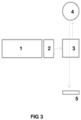
- Figure 3 shows a configuration of the broad band light source 1, that is capable of projecting various patterns, via a spatial light modulator 2 and a dichroic mirror 3, to the eye 4.
- Such patterns can be provided by projecting said repetitive pattern by a scanning light beam from said light source derived from a DMD or SLM and using a photodiode or camera 5 as an optical detector.
- the detector 5 is capable of registering light from the illuminated pattern on the retina, as well as light from neighbouring parts. This light contains information on the tissue through which it travelled.
- the detector may be multispectral, divided over several channels. In this way simultaneous spectral detection of several wavelengths is feasible.
- the application of a multispectral camera that images a large number of spectral bands at the expense of a low spatial resolution, by e.g. creating a mosaic pattern of pixel-based color filters on the imaging chip, is appropriate for sd-SFDI since the spatial resolution achieved by sd-SFDI is fundamentally limited by the tissue light transport properties anyway, allowing to use a large number of imaging wavelengths at the expense of a low spatial pixel resolution on the imaging chip.
- the camera can also be a monochrome camera, providing an instantaneous multispectral image, without necessarily projecting multiple colors sequentially.
- an optical response of for example biomarkers in the retina can be influenced.
- the invention is not limited to the projection of patterned images, but can be used with other techniques as well, such as Raman scattering, 2 photon fluorescence lifetime measurement.
- a photodiode array instead of a (pinhole) camera a photodiode array can be used to provide a detection similar to scanning laser ophthalmoscopy (SLO).
- SLO scanning laser ophthalmoscopy
- Annular projection in combination with a polarizing filter may prevent specular reflection in the air- lens surface or in the pupil. Variations are a full illumination of the pupil, a halve illumination, ring illumination or point illumination. These variations will alter the optical transfer path through the retina to the camera.
- the pattern is constant in the orthogonal y direction.
- M AC (x, fx) represents the amplitude envelope of the reflected photon density standing wave at frequency f x .
- M AC (x, f x ) curves can be sampled in parallel at each y pixel row using a 2-D camera, allowing spatial sampling of millions of reflectance values simultaneously.
- This differencing approach is convenient, as it automatically removes features common to all three images, including the average image noise and digitization offset.
- a measurement M AC (x, f x ) is the product of the source intensity, I 0 ; the modulation transfer function (MTF) of the illumination and imaging optical system, MTF system ; and the true turbid system MTF, R d (x i ,f x ):
- M AC x i f x I 0 ⁇ MTF system x i f x ⁇ R d x i f x
- multiple spatial frequencies will be used and multiple wavelengths will be used in order to obtain sufficient information to fit the data R d (x i ,f x ) to a reflectance model that describes the dependence of the signal R d (x i ,f x ) on the tissue optical properties ⁇ ' s , y, and ⁇ a .
- the reflectance model dependence on scattering properties ⁇ ' s and y is based on the idea that R d exhibits a y -specific proportionality vs.
- C PF and p 1 -p 3 are fitted coefficients used to capture the dynamics in attenuation of demodulated reflectance observed in response to changes in ⁇ s ', f x -1 , y and ⁇ a .
- Model coefficients can be estimated by minimizing the difference between simulated and model estimated values of demodulated reflectance.
- Other expressions than Eq. (2,3) may also be investigated to describe the effect of attenuation due to absorbing molecules on the collected signals.
Landscapes
- Life Sciences & Earth Sciences (AREA)
- Health & Medical Sciences (AREA)
- Medical Informatics (AREA)
- Biophysics (AREA)
- Ophthalmology & Optometry (AREA)
- Engineering & Computer Science (AREA)
- Biomedical Technology (AREA)
- Heart & Thoracic Surgery (AREA)
- Physics & Mathematics (AREA)
- Molecular Biology (AREA)
- Surgery (AREA)
- Animal Behavior & Ethology (AREA)
- General Health & Medical Sciences (AREA)
- Public Health (AREA)
- Veterinary Medicine (AREA)
- Investigating, Analyzing Materials By Fluorescence Or Luminescence (AREA)
- Investigating Or Analysing Materials By Optical Means (AREA)
Claims (10)
- Verfahren zum Erhalten eines Bildes der Retinalstruktur eines Auges, umfassend:- Bereitstellen einer Lichtquelle;- Projizieren einer Vielzahl von phasenverschobenen, sich wiederholenden Mustern auf die Retina durch die Lichtquelle, wobei jedes Muster beleuchtete und nicht beleuchtete Abschnitte mit einer räumlichen Frequenz von mehr als 0,33µtr [mm-1] aufweist;- Messen einer optischen Reaktion für jedes Muster für die beleuchteten und nicht beleuchteten Abschnitte der Retina; und- Ableiten eines Absorptionswerts der Retina durch Kombinieren der optischen Reaktionen aus der Vielzahl phasenverschobener sich wiederholender Muster beleuchteter und nicht beleuchteter Bereiche.
- Verfahren nach Anspruch 1, wobei die sich wiederholenden Muster gleichzeitig für eine Vielzahl von Spektralbändern bereitgestellt werden, von denen jedes seine eigene Ortsfrequenz hat.
- Verfahren nach Anspruch 1, wobei für die beleuchteten und nicht beleuchteten Abschnitte ein optisches Gewebetransportmodell verwendet wird, um den Absorptionswert des Retinagewebes, einen Streuungswert des Retinagewebes oder einen Fluoreszenzwert des Retinagewebes zu bestimmen.
- Verfahren nach Anspruch 1, wobei ein Fluoreszenzpegel gemessen wird und ein tatsächlicher Fluoreszenzpegel für beleuchtete und nicht beleuchtete Bereiche abgeleitet wird.
- Verfahren nach Anspruch 1, wobei das Bild durch Projizieren der sich wiederholenden Muster durch einen Abtastlichtstrahl von der Lichtquelle, der von einem DMD oder SLM abgeleitet wird, und unter Verwendung einer Photodiode oder Kamera als optischer Detektor erhalten wird.
- Verfahren nach Anspruch 1, wobei die Muster sinusförmige Muster, Linienmuster, Punktmuster oder konzentrische Muster sind.
- Verfahren nach einem der vorstehenden Ansprüche, wobei ein Streukoeffizient µs als durchschnittliche Anzahl von Streuereignissen pro Längeneinheit [mm] gemessen wird.
- Verfahren nach einem der vorstehenden Ansprüche, wobei eine Phasenfunktion als eine winkelabhängige Streuverteilung von Gewebe gemessen wird.
- Verfahren nach einem der vorstehenden Ansprüche, wobei das Retinalbild spektral auf Chromophore analysiert wird, einschließlich eines beliebigen von sauerstoffhaltigem oder sauerstofffreiem Hämoglobin, Melanin, Bilirubin, Beta-Carotin, Lipiden und Wasser.
- System zum Abbilden der Retinalstruktur eines Auges, umfassend:- eine Lichtquelle (1) und einen Projektor (2), wobei der Projektor angeordnet ist, um das Licht von der Lichtquelle in einer Vielzahl von phasenverschobenen, sich wiederholenden Mustern auf die Retina zu projizieren, die beleuchtete und nicht beleuchtete Abschnitte mit einer räumlichen Frequenz von mehr als 0,33µt [mm-1] aufweisen;- ein optischer Detektor (5), der zum Erfassen einer optischen Reaktion von den projizierten sich wiederholenden Mustern auf der Retina für beleuchtete und nicht beleuchtete Bereiche angeordnet ist;- eine Verarbeitungsvorrichtung, die angeordnet ist, um einen Absorptionswert des Retinagewebes durch Kombinieren von optischen Reaktionen, die durch den optischen Detektor gemessen werden, aus der Vielzahl von phasenverschobenen sich wiederholenden Mustern von beleuchteten und nicht beleuchteten Bereichen, die auf die Retina projiziert werden, abzuleiten.
Applications Claiming Priority (2)
| Application Number | Priority Date | Filing Date | Title |
|---|---|---|---|
| EP16200795.9A EP3326514A1 (de) | 2016-11-25 | 2016-11-25 | Quantitative retinalbildgebung |
| PCT/NL2017/050772 WO2018097723A1 (en) | 2016-11-25 | 2017-11-24 | Quantitative retinal imaging |
Publications (2)
| Publication Number | Publication Date |
|---|---|
| EP3544485A1 EP3544485A1 (de) | 2019-10-02 |
| EP3544485B1 true EP3544485B1 (de) | 2025-03-05 |
Family
ID=57406134
Family Applications (2)
| Application Number | Title | Priority Date | Filing Date |
|---|---|---|---|
| EP16200795.9A Withdrawn EP3326514A1 (de) | 2016-11-25 | 2016-11-25 | Quantitative retinalbildgebung |
| EP17817262.3A Active EP3544485B1 (de) | 2016-11-25 | 2017-11-24 | Quantitative retinalbildgebung |
Family Applications Before (1)
| Application Number | Title | Priority Date | Filing Date |
|---|---|---|---|
| EP16200795.9A Withdrawn EP3326514A1 (de) | 2016-11-25 | 2016-11-25 | Quantitative retinalbildgebung |
Country Status (3)
| Country | Link |
|---|---|
| US (1) | US11666212B2 (de) |
| EP (2) | EP3326514A1 (de) |
| WO (1) | WO2018097723A1 (de) |
Families Citing this family (2)
| Publication number | Priority date | Publication date | Assignee | Title |
|---|---|---|---|---|
| EP4215904A1 (de) | 2022-01-25 | 2023-07-26 | Nederlandse Organisatie voor toegepast-natuurwetenschappelijk Onderzoek TNO | Raumfrequenzdomäne-abbildungssystem und -verfahren |
| US12262947B1 (en) * | 2024-09-30 | 2025-04-01 | The Florida International University Board Of Trustees | Systems and methods for retinal imaging |
Family Cites Families (17)
| Publication number | Priority date | Publication date | Assignee | Title |
|---|---|---|---|---|
| US5467104A (en) | 1992-10-22 | 1995-11-14 | Board Of Regents Of The University Of Washington | Virtual retinal display |
| CA2284299A1 (en) | 1999-09-28 | 2001-03-28 | Brian Moore | Method and apparatus for a high resolution eye scanner |
| US20070091265A1 (en) * | 2002-01-18 | 2007-04-26 | Kardon Randy H | System and method for optical imaging of human retinal function |
| NL1024082C1 (nl) | 2003-08-11 | 2005-02-14 | Michiel Herman Mensink | Confocale multi-kleuren scanning laser ophthalmoscoop. |
| US20090270717A1 (en) * | 2008-04-25 | 2009-10-29 | Welch Allyn, Inc. | Apparatus and method for diagnosis of optically identifiable ophthalmic conditions |
| US20060069552A1 (en) * | 2004-08-20 | 2006-03-30 | Spitler Mark T | Apparatus and method for displaying signals of a detector |
| JP2010085621A (ja) | 2008-09-30 | 2010-04-15 | Brother Ind Ltd | 画像表示装置 |
| JP5426026B2 (ja) * | 2009-07-28 | 2014-02-26 | エフ・ホフマン−ラ・ロシュ・アクチェンゲゼルシャフト | 非侵襲性インビボ光学イメージング方法 |
| GB201000973D0 (en) * | 2010-01-21 | 2010-03-10 | Glynn Chris | Non-evasive spectrophotometer and related method |
| US8781184B2 (en) * | 2010-02-10 | 2014-07-15 | Tokitae Llc | Systems, devices, and methods for detection of malaria |
| EP2755547A4 (de) | 2011-09-16 | 2015-04-01 | Annidis Corp | System und verfahren zur beurteilung der netzhautfunktion |
| CN103969745B (zh) | 2013-01-30 | 2016-12-28 | 福州高意通讯有限公司 | 一种基于dlp的带宽可调平顶型光学滤波器 |
| WO2014151877A1 (en) | 2013-03-14 | 2014-09-25 | The Board Of Trustees Of The Leland Stanford Junior University | Holographic head-mounted display |
| WO2015103566A2 (en) * | 2014-01-06 | 2015-07-09 | The Regents Of The University Of California | Spatial frequency domain imaging using custom patterns |
| WO2015120020A1 (en) * | 2014-02-04 | 2015-08-13 | The Trustees Of Dartmouth College | Apparatus and methods for structured light scatteroscopy |
| US10463256B2 (en) | 2014-02-04 | 2019-11-05 | The Trustees Of Dartmouth College | Structured-light imaging systems and methods for determining sub-diffuse scattering parameters |
| CN105300928B (zh) * | 2015-10-15 | 2018-08-24 | 温州医科大学 | 一种大面积获取组织光学参数及微观结构的光反射成像技术 |
-
2016
- 2016-11-25 EP EP16200795.9A patent/EP3326514A1/de not_active Withdrawn
-
2017
- 2017-11-24 WO PCT/NL2017/050772 patent/WO2018097723A1/en not_active Ceased
- 2017-11-24 EP EP17817262.3A patent/EP3544485B1/de active Active
- 2017-11-24 US US16/463,253 patent/US11666212B2/en active Active
Non-Patent Citations (1)
| Title |
|---|
| MCCLATCHY DAVID M. ET AL: "Wide-field quantitative imaging of tissue microstructure using sub-diffuse spatial frequency domain imaging", OPTICA, vol. 3, no. 6, 20 June 2016 (2016-06-20), US, pages 613, XP055908855, ISSN: 2334-2536, Retrieved from the Internet <URL:http://dx.doi.org/10.1364/OPTICA.3.000613> DOI: 10.1364/OPTICA.3.000613 * |
Also Published As
| Publication number | Publication date |
|---|---|
| EP3544485A1 (de) | 2019-10-02 |
| US11666212B2 (en) | 2023-06-06 |
| WO2018097723A1 (en) | 2018-05-31 |
| EP3326514A1 (de) | 2018-05-30 |
| US20190290125A1 (en) | 2019-09-26 |
Similar Documents
| Publication | Publication Date | Title |
|---|---|---|
| US11751767B2 (en) | Structured-light imaging systems and methods for determining sub-diffuse scattering parameters | |
| CN102883658B (zh) | 用于使用结构化照明经由单元件检测来分析浑浊介质的方法和设备 | |
| Jonnal et al. | The cellular origins of the outer retinal bands in optical coherence tomography images | |
| US11779246B2 (en) | Hemoglobin measurement from a single vessel | |
| US20180012359A1 (en) | Systems and Methods for Automated Image Classification and Segmentation | |
| US20120120368A1 (en) | Fundus analyzing appartus and fundus analyzing method | |
| KR102521793B1 (ko) | 광학 흡수 및 산란 매체 내 표면 형광 물체의 깊이를 결정하고 물체의 형광 농도를 결정하기 위한 장치 및 방법 | |
| US10768165B2 (en) | Systems and methods for measuring water and lipid content in tissue samples | |
| US20190269333A1 (en) | Dual mode biophotonic imaging systems and their applications for detection of epithelial dysplasia in vivo | |
| US20240306908A1 (en) | A method to detect retinal amyloidosis and tauopathy using snap hyperspectral imaging and/or snap hyperspectral optical coherence tomography | |
| US11666212B2 (en) | Quantitative retinal imaging | |
| WO2021099127A1 (en) | Device, apparatus and method for imaging an object | |
| US20020193948A1 (en) | Arrangement and method for determining the two-dimensional distribution of fundus pigments, particularly of the macular pigment xanthophyll | |
| US12433482B2 (en) | Multi-modal diagnostic device and database service for ophthalmology and neurology | |
| US12629018B2 (en) | Using the dynamic forward scattering signal for optical coherence tomography based flow quantification | |
| JP7846210B2 (ja) | 光干渉断層撮影に基づく流量定量化のための動的前方散乱信号の使用 | |
| US6709109B1 (en) | Differential spectroscopic imaging of the human retina | |
| Miller | Development of Visible-Light Optical Coherence Tomography Fibergraphy | |
| Soetikno | Advances in Optical Coherence Tomography for Retinal Oximetry and Angiography | |
| Yang | Optical and structural property mapping of soft tissues using spatial frequency domain imaging | |
| Dubois | Technological extensions of full-field optical coherence microscopy for multicontrast imaging | |
| Shu | Developing Functional Optical Imaging Modalities to Study Retinal Diseases | |
| Reble | Raman spectroscopy in biological tissue: influence of absorption and scattering and detection of cervical precancer | |
| Folio et al. | Technologies for analyzing the optic disc, RNFL, and macula | |
| Kolste | Localization of subsurface fluorescent lesions using surface spectral measurements |
Legal Events
| Date | Code | Title | Description |
|---|---|---|---|
| STAA | Information on the status of an ep patent application or granted ep patent |
Free format text: STATUS: UNKNOWN |
|
| STAA | Information on the status of an ep patent application or granted ep patent |
Free format text: STATUS: THE INTERNATIONAL PUBLICATION HAS BEEN MADE |
|
| PUAI | Public reference made under article 153(3) epc to a published international application that has entered the european phase |
Free format text: ORIGINAL CODE: 0009012 |
|
| STAA | Information on the status of an ep patent application or granted ep patent |
Free format text: STATUS: REQUEST FOR EXAMINATION WAS MADE |
|
| 17P | Request for examination filed |
Effective date: 20190603 |
|
| AK | Designated contracting states |
Kind code of ref document: A1 Designated state(s): AL AT BE BG CH CY CZ DE DK EE ES FI FR GB GR HR HU IE IS IT LI LT LU LV MC MK MT NL NO PL PT RO RS SE SI SK SM TR |
|
| AX | Request for extension of the european patent |
Extension state: BA ME |
|
| DAV | Request for validation of the european patent (deleted) | ||
| DAX | Request for extension of the european patent (deleted) | ||
| STAA | Information on the status of an ep patent application or granted ep patent |
Free format text: STATUS: EXAMINATION IS IN PROGRESS |
|
| 17Q | First examination report despatched |
Effective date: 20220414 |
|
| P01 | Opt-out of the competence of the unified patent court (upc) registered |
Effective date: 20230522 |
|
| GRAP | Despatch of communication of intention to grant a patent |
Free format text: ORIGINAL CODE: EPIDOSNIGR1 |
|
| STAA | Information on the status of an ep patent application or granted ep patent |
Free format text: STATUS: GRANT OF PATENT IS INTENDED |
|
| INTG | Intention to grant announced |
Effective date: 20240923 |
|
| RIN1 | Information on inventor provided before grant (corrected) |
Inventor name: ODERWALD, MICHIEL PETER Inventor name: WIERINGA, FOKKO PIETER Inventor name: AMELINK, ARJEN |
|
| GRAS | Grant fee paid |
Free format text: ORIGINAL CODE: EPIDOSNIGR3 |
|
| GRAA | (expected) grant |
Free format text: ORIGINAL CODE: 0009210 |
|
| STAA | Information on the status of an ep patent application or granted ep patent |
Free format text: STATUS: THE PATENT HAS BEEN GRANTED |
|
| AK | Designated contracting states |
Kind code of ref document: B1 Designated state(s): AL AT BE BG CH CY CZ DE DK EE ES FI FR GB GR HR HU IE IS IT LI LT LU LV MC MK MT NL NO PL PT RO RS SE SI SK SM TR |
|
| REG | Reference to a national code |
Ref country code: GB Ref legal event code: FG4D |
|
| REG | Reference to a national code |
Ref country code: CH Ref legal event code: EP |
|
| REG | Reference to a national code |
Ref country code: IE Ref legal event code: FG4D |
|
| REG | Reference to a national code |
Ref country code: DE Ref legal event code: R096 Ref document number: 602017088187 Country of ref document: DE |
|
| REG | Reference to a national code |
Ref country code: NL Ref legal event code: FP |
|
| PG25 | Lapsed in a contracting state [announced via postgrant information from national office to epo] |
Ref country code: RS Free format text: LAPSE BECAUSE OF FAILURE TO SUBMIT A TRANSLATION OF THE DESCRIPTION OR TO PAY THE FEE WITHIN THE PRESCRIBED TIME-LIMIT Effective date: 20250605 |
|
| PG25 | Lapsed in a contracting state [announced via postgrant information from national office to epo] |
Ref country code: FI Free format text: LAPSE BECAUSE OF FAILURE TO SUBMIT A TRANSLATION OF THE DESCRIPTION OR TO PAY THE FEE WITHIN THE PRESCRIBED TIME-LIMIT Effective date: 20250305 |
|
| PG25 | Lapsed in a contracting state [announced via postgrant information from national office to epo] |
Ref country code: ES Free format text: LAPSE BECAUSE OF FAILURE TO SUBMIT A TRANSLATION OF THE DESCRIPTION OR TO PAY THE FEE WITHIN THE PRESCRIBED TIME-LIMIT Effective date: 20250305 |
|
| REG | Reference to a national code |
Ref country code: LT Ref legal event code: MG9D |
|
| PG25 | Lapsed in a contracting state [announced via postgrant information from national office to epo] |
Ref country code: NO Free format text: LAPSE BECAUSE OF FAILURE TO SUBMIT A TRANSLATION OF THE DESCRIPTION OR TO PAY THE FEE WITHIN THE PRESCRIBED TIME-LIMIT Effective date: 20250605 |
|
| PG25 | Lapsed in a contracting state [announced via postgrant information from national office to epo] |
Ref country code: HR Free format text: LAPSE BECAUSE OF FAILURE TO SUBMIT A TRANSLATION OF THE DESCRIPTION OR TO PAY THE FEE WITHIN THE PRESCRIBED TIME-LIMIT Effective date: 20250305 |
|
| PG25 | Lapsed in a contracting state [announced via postgrant information from national office to epo] |
Ref country code: LV Free format text: LAPSE BECAUSE OF FAILURE TO SUBMIT A TRANSLATION OF THE DESCRIPTION OR TO PAY THE FEE WITHIN THE PRESCRIBED TIME-LIMIT Effective date: 20250305 |
|
| PG25 | Lapsed in a contracting state [announced via postgrant information from national office to epo] |
Ref country code: BG Free format text: LAPSE BECAUSE OF FAILURE TO SUBMIT A TRANSLATION OF THE DESCRIPTION OR TO PAY THE FEE WITHIN THE PRESCRIBED TIME-LIMIT Effective date: 20250305 Ref country code: GR Free format text: LAPSE BECAUSE OF FAILURE TO SUBMIT A TRANSLATION OF THE DESCRIPTION OR TO PAY THE FEE WITHIN THE PRESCRIBED TIME-LIMIT Effective date: 20250606 |
|
| REG | Reference to a national code |
Ref country code: AT Ref legal event code: MK05 Ref document number: 1772110 Country of ref document: AT Kind code of ref document: T Effective date: 20250305 |
|
| PG25 | Lapsed in a contracting state [announced via postgrant information from national office to epo] |
Ref country code: SE Free format text: LAPSE BECAUSE OF FAILURE TO SUBMIT A TRANSLATION OF THE DESCRIPTION OR TO PAY THE FEE WITHIN THE PRESCRIBED TIME-LIMIT Effective date: 20250305 |
|
| PG25 | Lapsed in a contracting state [announced via postgrant information from national office to epo] |
Ref country code: SM Free format text: LAPSE BECAUSE OF FAILURE TO SUBMIT A TRANSLATION OF THE DESCRIPTION OR TO PAY THE FEE WITHIN THE PRESCRIBED TIME-LIMIT Effective date: 20250305 |
|
| PG25 | Lapsed in a contracting state [announced via postgrant information from national office to epo] |
Ref country code: PT Free format text: LAPSE BECAUSE OF FAILURE TO SUBMIT A TRANSLATION OF THE DESCRIPTION OR TO PAY THE FEE WITHIN THE PRESCRIBED TIME-LIMIT Effective date: 20250707 |
|
| PG25 | Lapsed in a contracting state [announced via postgrant information from national office to epo] |
Ref country code: IT Free format text: LAPSE BECAUSE OF FAILURE TO SUBMIT A TRANSLATION OF THE DESCRIPTION OR TO PAY THE FEE WITHIN THE PRESCRIBED TIME-LIMIT Effective date: 20250305 Ref country code: PL Free format text: LAPSE BECAUSE OF FAILURE TO SUBMIT A TRANSLATION OF THE DESCRIPTION OR TO PAY THE FEE WITHIN THE PRESCRIBED TIME-LIMIT Effective date: 20250305 |
|
| PG25 | Lapsed in a contracting state [announced via postgrant information from national office to epo] |
Ref country code: AT Free format text: LAPSE BECAUSE OF FAILURE TO SUBMIT A TRANSLATION OF THE DESCRIPTION OR TO PAY THE FEE WITHIN THE PRESCRIBED TIME-LIMIT Effective date: 20250305 |
|
| PG25 | Lapsed in a contracting state [announced via postgrant information from national office to epo] |
Ref country code: CZ Free format text: LAPSE BECAUSE OF FAILURE TO SUBMIT A TRANSLATION OF THE DESCRIPTION OR TO PAY THE FEE WITHIN THE PRESCRIBED TIME-LIMIT Effective date: 20250305 Ref country code: EE Free format text: LAPSE BECAUSE OF FAILURE TO SUBMIT A TRANSLATION OF THE DESCRIPTION OR TO PAY THE FEE WITHIN THE PRESCRIBED TIME-LIMIT Effective date: 20250305 |
|
| PG25 | Lapsed in a contracting state [announced via postgrant information from national office to epo] |
Ref country code: RO Free format text: LAPSE BECAUSE OF FAILURE TO SUBMIT A TRANSLATION OF THE DESCRIPTION OR TO PAY THE FEE WITHIN THE PRESCRIBED TIME-LIMIT Effective date: 20250305 |
|
| PG25 | Lapsed in a contracting state [announced via postgrant information from national office to epo] |
Ref country code: SK Free format text: LAPSE BECAUSE OF FAILURE TO SUBMIT A TRANSLATION OF THE DESCRIPTION OR TO PAY THE FEE WITHIN THE PRESCRIBED TIME-LIMIT Effective date: 20250305 |
|
| PG25 | Lapsed in a contracting state [announced via postgrant information from national office to epo] |
Ref country code: IS Free format text: LAPSE BECAUSE OF FAILURE TO SUBMIT A TRANSLATION OF THE DESCRIPTION OR TO PAY THE FEE WITHIN THE PRESCRIBED TIME-LIMIT Effective date: 20250705 |
|
| REG | Reference to a national code |
Ref country code: DE Ref legal event code: R097 Ref document number: 602017088187 Country of ref document: DE |
|
| PGFP | Annual fee paid to national office [announced via postgrant information from national office to epo] |
Ref country code: NL Payment date: 20251119 Year of fee payment: 9 |
|
| PGFP | Annual fee paid to national office [announced via postgrant information from national office to epo] |
Ref country code: DE Payment date: 20251119 Year of fee payment: 9 |
|
| PGFP | Annual fee paid to national office [announced via postgrant information from national office to epo] |
Ref country code: GB Payment date: 20251121 Year of fee payment: 9 |
|
| PLBE | No opposition filed within time limit |
Free format text: ORIGINAL CODE: 0009261 |
|
| STAA | Information on the status of an ep patent application or granted ep patent |
Free format text: STATUS: NO OPPOSITION FILED WITHIN TIME LIMIT |
|
| PG25 | Lapsed in a contracting state [announced via postgrant information from national office to epo] |
Ref country code: DK Free format text: LAPSE BECAUSE OF FAILURE TO SUBMIT A TRANSLATION OF THE DESCRIPTION OR TO PAY THE FEE WITHIN THE PRESCRIBED TIME-LIMIT Effective date: 20250305 |
|
| REG | Reference to a national code |
Ref country code: CH Ref legal event code: L10 Free format text: ST27 STATUS EVENT CODE: U-0-0-L10-L00 (AS PROVIDED BY THE NATIONAL OFFICE) Effective date: 20260114 |
|
| PGFP | Annual fee paid to national office [announced via postgrant information from national office to epo] |
Ref country code: FR Payment date: 20251126 Year of fee payment: 9 |
|
| 26N | No opposition filed |
Effective date: 20251208 |