EP3527123B1 - Bildverarbeitungsverfahren und -vorrichtung mit elastischer abbildung von gefässplexusstrukturen - Google Patents
Bildverarbeitungsverfahren und -vorrichtung mit elastischer abbildung von gefässplexusstrukturen Download PDFInfo
- Publication number
- EP3527123B1 EP3527123B1 EP18156906.2A EP18156906A EP3527123B1 EP 3527123 B1 EP3527123 B1 EP 3527123B1 EP 18156906 A EP18156906 A EP 18156906A EP 3527123 B1 EP3527123 B1 EP 3527123B1
- Authority
- EP
- European Patent Office
- Prior art keywords
- image data
- interoperative
- operative
- vascular plexus
- data
- Prior art date
- Legal status (The legal status is an assumption and is not a legal conclusion. Google has not performed a legal analysis and makes no representation as to the accuracy of the status listed.)
- Active
Links
- 230000002792 vascular Effects 0.000 title claims description 42
- 238000003672 processing method Methods 0.000 title claims description 10
- 238000013507 mapping Methods 0.000 title description 5
- 230000003287 optical effect Effects 0.000 claims description 15
- 238000001356 surgical procedure Methods 0.000 claims description 15
- 230000017531 blood circulation Effects 0.000 claims description 12
- 238000001228 spectrum Methods 0.000 claims description 12
- 238000002189 fluorescence spectrum Methods 0.000 claims description 3
- 238000002329 infrared spectrum Methods 0.000 claims description 3
- 238000001429 visible spectrum Methods 0.000 claims description 2
- 239000003550 marker Substances 0.000 claims 1
- 238000000034 method Methods 0.000 description 24
- 210000001519 tissue Anatomy 0.000 description 20
- 238000005286 illumination Methods 0.000 description 15
- 239000008280 blood Substances 0.000 description 8
- 210000004369 blood Anatomy 0.000 description 8
- 210000004872 soft tissue Anatomy 0.000 description 8
- 210000004556 brain Anatomy 0.000 description 4
- 238000002675 image-guided surgery Methods 0.000 description 4
- 238000003384 imaging method Methods 0.000 description 3
- 238000000985 reflectance spectrum Methods 0.000 description 3
- OYPRJOBELJOOCE-UHFFFAOYSA-N Calcium Chemical compound [Ca] OYPRJOBELJOOCE-UHFFFAOYSA-N 0.000 description 2
- 206010028980 Neoplasm Diseases 0.000 description 2
- 238000002583 angiography Methods 0.000 description 2
- 230000000903 blocking effect Effects 0.000 description 2
- 210000004204 blood vessel Anatomy 0.000 description 2
- 229910052791 calcium Inorganic materials 0.000 description 2
- 239000011575 calcium Substances 0.000 description 2
- 230000001427 coherent effect Effects 0.000 description 2
- 230000001054 cortical effect Effects 0.000 description 2
- MOFVSTNWEDAEEK-UHFFFAOYSA-M indocyanine green Chemical compound [Na+].[O-]S(=O)(=O)CCCCN1C2=CC=C3C=CC=CC3=C2C(C)(C)C1=CC=CC=CC=CC1=[N+](CCCCS([O-])(=O)=O)C2=CC=C(C=CC=C3)C3=C2C1(C)C MOFVSTNWEDAEEK-UHFFFAOYSA-M 0.000 description 2
- 229960004657 indocyanine green Drugs 0.000 description 2
- 230000003834 intracellular effect Effects 0.000 description 2
- 238000002595 magnetic resonance imaging Methods 0.000 description 2
- 238000012634 optical imaging Methods 0.000 description 2
- 230000011218 segmentation Effects 0.000 description 2
- 238000004458 analytical method Methods 0.000 description 1
- 210000001367 artery Anatomy 0.000 description 1
- 230000000712 assembly Effects 0.000 description 1
- 238000000429 assembly Methods 0.000 description 1
- 230000005540 biological transmission Effects 0.000 description 1
- 210000005013 brain tissue Anatomy 0.000 description 1
- 239000003086 colorant Substances 0.000 description 1
- 239000000470 constituent Substances 0.000 description 1
- 230000004069 differentiation Effects 0.000 description 1
- 230000000694 effects Effects 0.000 description 1
- 238000000799 fluorescence microscopy Methods 0.000 description 1
- 230000006870 function Effects 0.000 description 1
- 210000002751 lymph Anatomy 0.000 description 1
- 210000003205 muscle Anatomy 0.000 description 1
- 210000005036 nerve Anatomy 0.000 description 1
- 238000006213 oxygenation reaction Methods 0.000 description 1
- 238000003909 pattern recognition Methods 0.000 description 1
- 238000007781 pre-processing Methods 0.000 description 1
- 238000000513 principal component analysis Methods 0.000 description 1
- 230000003595 spectral effect Effects 0.000 description 1
- 230000002123 temporal effect Effects 0.000 description 1
- 210000003462 vein Anatomy 0.000 description 1
- 210000001835 viscera Anatomy 0.000 description 1
- 230000000007 visual effect Effects 0.000 description 1
Images
Classifications
-
- A—HUMAN NECESSITIES
- A61—MEDICAL OR VETERINARY SCIENCE; HYGIENE
- A61B—DIAGNOSIS; SURGERY; IDENTIFICATION
- A61B5/00—Measuring for diagnostic purposes; Identification of persons
- A61B5/0033—Features or image-related aspects of imaging apparatus classified in A61B5/00, e.g. for MRI, optical tomography or impedance tomography apparatus; arrangements of imaging apparatus in a room
- A61B5/0037—Performing a preliminary scan, e.g. a prescan for identifying a region of interest
-
- A—HUMAN NECESSITIES
- A61—MEDICAL OR VETERINARY SCIENCE; HYGIENE
- A61B—DIAGNOSIS; SURGERY; IDENTIFICATION
- A61B1/00—Instruments for performing medical examinations of the interior of cavities or tubes of the body by visual or photographical inspection, e.g. endoscopes; Illuminating arrangements therefor
- A61B1/04—Instruments for performing medical examinations of the interior of cavities or tubes of the body by visual or photographical inspection, e.g. endoscopes; Illuminating arrangements therefor combined with photographic or television appliances
- A61B1/043—Instruments for performing medical examinations of the interior of cavities or tubes of the body by visual or photographical inspection, e.g. endoscopes; Illuminating arrangements therefor combined with photographic or television appliances for fluorescence imaging
-
- A—HUMAN NECESSITIES
- A61—MEDICAL OR VETERINARY SCIENCE; HYGIENE
- A61B—DIAGNOSIS; SURGERY; IDENTIFICATION
- A61B34/00—Computer-aided surgery; Manipulators or robots specially adapted for use in surgery
- A61B34/20—Surgical navigation systems; Devices for tracking or guiding surgical instruments, e.g. for frameless stereotaxis
-
- A—HUMAN NECESSITIES
- A61—MEDICAL OR VETERINARY SCIENCE; HYGIENE
- A61B—DIAGNOSIS; SURGERY; IDENTIFICATION
- A61B5/00—Measuring for diagnostic purposes; Identification of persons
- A61B5/0059—Measuring for diagnostic purposes; Identification of persons using light, e.g. diagnosis by transillumination, diascopy, fluorescence
- A61B5/0071—Measuring for diagnostic purposes; Identification of persons using light, e.g. diagnosis by transillumination, diascopy, fluorescence by measuring fluorescence emission
-
- A—HUMAN NECESSITIES
- A61—MEDICAL OR VETERINARY SCIENCE; HYGIENE
- A61B—DIAGNOSIS; SURGERY; IDENTIFICATION
- A61B5/00—Measuring for diagnostic purposes; Identification of persons
- A61B5/02—Detecting, measuring or recording pulse, heart rate, blood pressure or blood flow; Combined pulse/heart-rate/blood pressure determination; Evaluating a cardiovascular condition not otherwise provided for, e.g. using combinations of techniques provided for in this group with electrocardiography or electroauscultation; Heart catheters for measuring blood pressure
- A61B5/026—Measuring blood flow
- A61B5/0261—Measuring blood flow using optical means, e.g. infrared light
-
- A—HUMAN NECESSITIES
- A61—MEDICAL OR VETERINARY SCIENCE; HYGIENE
- A61B—DIAGNOSIS; SURGERY; IDENTIFICATION
- A61B5/00—Measuring for diagnostic purposes; Identification of persons
- A61B5/74—Details of notification to user or communication with user or patient ; user input means
- A61B5/742—Details of notification to user or communication with user or patient ; user input means using visual displays
- A61B5/7425—Displaying combinations of multiple images regardless of image source, e.g. displaying a reference anatomical image with a live image
-
- A—HUMAN NECESSITIES
- A61—MEDICAL OR VETERINARY SCIENCE; HYGIENE
- A61B—DIAGNOSIS; SURGERY; IDENTIFICATION
- A61B90/00—Instruments, implements or accessories specially adapted for surgery or diagnosis and not covered by any of the groups A61B1/00 - A61B50/00, e.g. for luxation treatment or for protecting wound edges
- A61B90/20—Surgical microscopes characterised by non-optical aspects
-
- A—HUMAN NECESSITIES
- A61—MEDICAL OR VETERINARY SCIENCE; HYGIENE
- A61B—DIAGNOSIS; SURGERY; IDENTIFICATION
- A61B90/00—Instruments, implements or accessories specially adapted for surgery or diagnosis and not covered by any of the groups A61B1/00 - A61B50/00, e.g. for luxation treatment or for protecting wound edges
- A61B90/36—Image-producing devices or illumination devices not otherwise provided for
- A61B90/361—Image-producing devices, e.g. surgical cameras
-
- A—HUMAN NECESSITIES
- A61—MEDICAL OR VETERINARY SCIENCE; HYGIENE
- A61B—DIAGNOSIS; SURGERY; IDENTIFICATION
- A61B90/00—Instruments, implements or accessories specially adapted for surgery or diagnosis and not covered by any of the groups A61B1/00 - A61B50/00, e.g. for luxation treatment or for protecting wound edges
- A61B90/36—Image-producing devices or illumination devices not otherwise provided for
- A61B90/37—Surgical systems with images on a monitor during operation
-
- G—PHYSICS
- G06—COMPUTING; CALCULATING OR COUNTING
- G06F—ELECTRIC DIGITAL DATA PROCESSING
- G06F18/00—Pattern recognition
- G06F18/20—Analysing
- G06F18/22—Matching criteria, e.g. proximity measures
-
- G—PHYSICS
- G06—COMPUTING; CALCULATING OR COUNTING
- G06T—IMAGE DATA PROCESSING OR GENERATION, IN GENERAL
- G06T3/00—Geometric image transformations in the plane of the image
- G06T3/14—Transformations for image registration, e.g. adjusting or mapping for alignment of images
- G06T3/153—Transformations for image registration, e.g. adjusting or mapping for alignment of images using elastic snapping
-
- G—PHYSICS
- G06—COMPUTING; CALCULATING OR COUNTING
- G06T—IMAGE DATA PROCESSING OR GENERATION, IN GENERAL
- G06T7/00—Image analysis
- G06T7/30—Determination of transform parameters for the alignment of images, i.e. image registration
-
- G—PHYSICS
- G16—INFORMATION AND COMMUNICATION TECHNOLOGY [ICT] SPECIALLY ADAPTED FOR SPECIFIC APPLICATION FIELDS
- G16H—HEALTHCARE INFORMATICS, i.e. INFORMATION AND COMMUNICATION TECHNOLOGY [ICT] SPECIALLY ADAPTED FOR THE HANDLING OR PROCESSING OF MEDICAL OR HEALTHCARE DATA
- G16H30/00—ICT specially adapted for the handling or processing of medical images
-
- A—HUMAN NECESSITIES
- A61—MEDICAL OR VETERINARY SCIENCE; HYGIENE
- A61B—DIAGNOSIS; SURGERY; IDENTIFICATION
- A61B34/00—Computer-aided surgery; Manipulators or robots specially adapted for use in surgery
- A61B34/20—Surgical navigation systems; Devices for tracking or guiding surgical instruments, e.g. for frameless stereotaxis
- A61B2034/2046—Tracking techniques
- A61B2034/2065—Tracking using image or pattern recognition
-
- A—HUMAN NECESSITIES
- A61—MEDICAL OR VETERINARY SCIENCE; HYGIENE
- A61B—DIAGNOSIS; SURGERY; IDENTIFICATION
- A61B90/00—Instruments, implements or accessories specially adapted for surgery or diagnosis and not covered by any of the groups A61B1/00 - A61B50/00, e.g. for luxation treatment or for protecting wound edges
- A61B90/36—Image-producing devices or illumination devices not otherwise provided for
- A61B2090/364—Correlation of different images or relation of image positions in respect to the body
- A61B2090/365—Correlation of different images or relation of image positions in respect to the body augmented reality, i.e. correlating a live optical image with another image
-
- A—HUMAN NECESSITIES
- A61—MEDICAL OR VETERINARY SCIENCE; HYGIENE
- A61B—DIAGNOSIS; SURGERY; IDENTIFICATION
- A61B90/00—Instruments, implements or accessories specially adapted for surgery or diagnosis and not covered by any of the groups A61B1/00 - A61B50/00, e.g. for luxation treatment or for protecting wound edges
- A61B90/36—Image-producing devices or illumination devices not otherwise provided for
- A61B2090/364—Correlation of different images or relation of image positions in respect to the body
- A61B2090/367—Correlation of different images or relation of image positions in respect to the body creating a 3D dataset from 2D images using position information
-
- A—HUMAN NECESSITIES
- A61—MEDICAL OR VETERINARY SCIENCE; HYGIENE
- A61B—DIAGNOSIS; SURGERY; IDENTIFICATION
- A61B90/00—Instruments, implements or accessories specially adapted for surgery or diagnosis and not covered by any of the groups A61B1/00 - A61B50/00, e.g. for luxation treatment or for protecting wound edges
- A61B90/36—Image-producing devices or illumination devices not otherwise provided for
- A61B90/37—Surgical systems with images on a monitor during operation
- A61B2090/373—Surgical systems with images on a monitor during operation using light, e.g. by using optical scanners
-
- A—HUMAN NECESSITIES
- A61—MEDICAL OR VETERINARY SCIENCE; HYGIENE
- A61B—DIAGNOSIS; SURGERY; IDENTIFICATION
- A61B90/00—Instruments, implements or accessories specially adapted for surgery or diagnosis and not covered by any of the groups A61B1/00 - A61B50/00, e.g. for luxation treatment or for protecting wound edges
- A61B90/39—Markers, e.g. radio-opaque or breast lesions markers
- A61B2090/3937—Visible markers
- A61B2090/3941—Photoluminescent markers
-
- A—HUMAN NECESSITIES
- A61—MEDICAL OR VETERINARY SCIENCE; HYGIENE
- A61B—DIAGNOSIS; SURGERY; IDENTIFICATION
- A61B90/00—Instruments, implements or accessories specially adapted for surgery or diagnosis and not covered by any of the groups A61B1/00 - A61B50/00, e.g. for luxation treatment or for protecting wound edges
- A61B90/39—Markers, e.g. radio-opaque or breast lesions markers
- A61B2090/397—Markers, e.g. radio-opaque or breast lesions markers electromagnetic other than visible, e.g. microwave
- A61B2090/3975—Markers, e.g. radio-opaque or breast lesions markers electromagnetic other than visible, e.g. microwave active
- A61B2090/3979—Markers, e.g. radio-opaque or breast lesions markers electromagnetic other than visible, e.g. microwave active infrared
-
- A—HUMAN NECESSITIES
- A61—MEDICAL OR VETERINARY SCIENCE; HYGIENE
- A61B—DIAGNOSIS; SURGERY; IDENTIFICATION
- A61B2505/00—Evaluating, monitoring or diagnosing in the context of a particular type of medical care
- A61B2505/05—Surgical care
-
- G—PHYSICS
- G06—COMPUTING; CALCULATING OR COUNTING
- G06T—IMAGE DATA PROCESSING OR GENERATION, IN GENERAL
- G06T2207/00—Indexing scheme for image analysis or image enhancement
- G06T2207/10—Image acquisition modality
- G06T2207/10048—Infrared image
-
- G—PHYSICS
- G06—COMPUTING; CALCULATING OR COUNTING
- G06T—IMAGE DATA PROCESSING OR GENERATION, IN GENERAL
- G06T2207/00—Indexing scheme for image analysis or image enhancement
- G06T2207/10—Image acquisition modality
- G06T2207/10056—Microscopic image
-
- G—PHYSICS
- G06—COMPUTING; CALCULATING OR COUNTING
- G06T—IMAGE DATA PROCESSING OR GENERATION, IN GENERAL
- G06T2207/00—Indexing scheme for image analysis or image enhancement
- G06T2207/10—Image acquisition modality
- G06T2207/10056—Microscopic image
- G06T2207/10061—Microscopic image from scanning electron microscope
-
- G—PHYSICS
- G06—COMPUTING; CALCULATING OR COUNTING
- G06T—IMAGE DATA PROCESSING OR GENERATION, IN GENERAL
- G06T2207/00—Indexing scheme for image analysis or image enhancement
- G06T2207/10—Image acquisition modality
- G06T2207/10068—Endoscopic image
-
- G—PHYSICS
- G06—COMPUTING; CALCULATING OR COUNTING
- G06T—IMAGE DATA PROCESSING OR GENERATION, IN GENERAL
- G06T2207/00—Indexing scheme for image analysis or image enhancement
- G06T2207/20—Special algorithmic details
- G06T2207/20212—Image combination
- G06T2207/20221—Image fusion; Image merging
-
- G—PHYSICS
- G06—COMPUTING; CALCULATING OR COUNTING
- G06T—IMAGE DATA PROCESSING OR GENERATION, IN GENERAL
- G06T2207/00—Indexing scheme for image analysis or image enhancement
- G06T2207/30—Subject of image; Context of image processing
- G06T2207/30004—Biomedical image processing
- G06T2207/30101—Blood vessel; Artery; Vein; Vascular
- G06T2207/30104—Vascular flow; Blood flow; Perfusion
-
- G—PHYSICS
- G06—COMPUTING; CALCULATING OR COUNTING
- G06V—IMAGE OR VIDEO RECOGNITION OR UNDERSTANDING
- G06V2201/00—Indexing scheme relating to image or video recognition or understanding
- G06V2201/03—Recognition of patterns in medical or anatomical images
Definitions
- the invention relates to an image processing method and a medical observation device for displaying soft tissue images, in particular in real time during surgery.
- Image-guided surgery is nowadays commonly used for certain kind of surgical operations, such as brain surgery.
- Image-guided surgery uses stored pre-operative three-dimensional information in the form of image data about the operation area.
- Such pre-operative three-dimensional image data may, for example, have been obtained using magnetic resonance imaging.
- the pre-operative information is visually aligned with the actual optical view of the tissue to be operated on.
- tissue structures such as tumors or vascular plexus structures, may be visualized that are otherwise invisible under the visible tissue surface.
- Use of the pre-operative three-dimensional information helps the surgeon to find and reach a certain area of the tissue, to avoid sensitive tissue such as nerves, arteries and veins, and/or to remove certain tissue effectively, such as a tumor.
- a method and apparatus for quantitative and depth resolved fluorescence and reflective imaging for surgical guidance is known from WO 2015/023990 A1 .
- a patient's brain is analyzed by a diagnostic tool such as MRI, PMR or CT in order to provide a three-dimensional model of the tissue.
- a surgeon may identify structures in the tissue and mark them during pre-operative planning.
- fluorophore concentrations in the tissue are imaged using depth resolved fluorescence microscopy.
- stereo infrared cameras or sensors for detecting optical or electromagnetic markers fixed on the patient's body are typically used.
- this object is solved by an image processing method for displaying soft tissue images as defined in claim 1.
- the medical observation device for the observation of soft tissue images is configured as defined in claim 6.
- the method and device according to the invention allow the continuous performance of image-guided surgery in soft tissue even if the tissue deforms and moves within the body without the need to manually realign the pre-operative image data to the interoperative image data.
- the structure which is used for elastically matching the pre-operative three-dimensional image data is a structure which is part of the soft tissue and thus deforms and moves together with the soft tissue.
- the pre-operative three-dimensional image data are thus mapped continuously to what the surgeon actually sees. In fact, the visual information that is available to the surgeon is itself used to align the pre-operative three-dimensional image data.
- the image processing method and the medical observation device according to the invention may be improved by adding one or more of the following features.
- Each of the following features can be added independently of the remaining features.
- Each of the following features has its own advantageous technical effect. Further, the following features can all be added equally to both the method and the device.
- modules described above can be implemented in software, hardware or a combination of both software and hardware. Further, the differences between the particular modules are primarily functional. Different modules may thus be comprised of a single or a plurality of electric components and/or a single logical unit, such as a single sub-routine.
- the output image data may be displayed on a display assembly, which may be part of the medical observation device.
- the step of elastic matching may include a technique as is described in Gee, J. C.; Reivich, M.; and Bajcsy, R.: "Elastically Deforming a Three-Dimensional Atlas to Match Anatomical Brain Images” (1993). IRCS Technical Reports Series. 192 .
- the step of identifying a vascular plexus structure may use the method as described in Suri, J.S.; Laxminarayan, S. [eds]: "Angiography and Plaque Imaging: Advanced Segmentation Techniques", CRC Press, 2003, pp.
- the matching module may in particular be configured to execute a matching routine described in any of these references.
- the identification of the at least one vascular plexus structure may be done preferably exclusively with interoperative fluorescent-light image data.
- fluorescent-light image data may be obtained by injecting a fluorophore, such as indocyanine green, into the tissue.
- the camera assembly may comprise an optical filter assembly, such as a band-pass filter assembly, of which the pass band is restricted to the fluorescence spectrum of the fluorophore. As the fluorophore is transported by the blood, the vascular plexus structure may be more easily defined in the fluorescent-light image.
- the medical observation device may have an illumination assembly, having an illumination spectrum which comprises light in wavelengths that trigger fluorescence of the fluorophore. The illumination spectrum may be restricted to these fluorescence-triggering wavelengths.
- the interoperative image data may, in another embodiment, contain both white-light image data and fluorescent-light image data.
- the white-light image data may be used to present output image data to the surgeon that represent what he would see with his own eyes.
- the illumination spectrum may also comprise wavelengths of the visible spectrum, in particular white light.
- the medical observation device may be one of a microscope and an endoscope.
- the interoperative image data may be two-dimensional, three-dimensional or multidimensional data.
- Three-dimensional interoperative image data may, for example, be acquired by a microscope using z-stacking or a stereoscopic setup, or a SCAPE or SPIM microscope.
- the interoperative image data may be recorded simultaneously in more than three different wave bands using, for example, more than one camera, a multi-spectral camera and/or a hyper-spectral camera.
- the elastic mapping may include or consist of the step of elastically matching the at least one identified vascular plexus structure in the pre-operative three-dimensional image data to the corresponding at least one identified vascular plexus structure in the interoperative image data.
- the mapping used for the at least one vascular plexus structure may be used for the rest of the pre-operative three-dimensional image data.
- the accuracy and reliability of the mapping of the pre-operative three-dimensional image data to the interoperative image data depends on how accurately the at least one vascular plexus structure may be recognised.
- pattern recognition may be performed on the fluorescent-light interoperative image data, using regions in which the fluorophore is located and which therefore have high fluorescent-light intensity.
- the method in another embodiment may comprise the step of identifying at least one arterial vascular plexus structure within the interoperative image data, and/or at least one venous vascular plexus structure within the interoperative image data.
- identifying at least one arterial vascular plexus structure within the interoperative image data and/or at least one venous vascular plexus structure within the interoperative image data.
- European patent application EP 17 174 047.5 may be used (published as EP 3 410 394 A1 and prior art under Article 54(3) EPC).
- the at least one vascular plexus structure may be identified using interoperative image data which have been acquired in at least one of a plurality of separate wavelengths e.g. by a multispectral and/or hyperspectral camera.
- the multi- and/or hyperspectral interoperative image data may be unmixed and/or processed to show the distribution of at least one of arterial or venous blood, or the respective blood vessels.
- a method and apparatus for identifying the at least one vascular plexus structure is described e.g. in Matthew B. Bouchard, Brenda R. Chen, Sean A. Burgess, and Elizabeth M. C. Hillman, "Ultra-fast multispectral optical imaging of cortical oxygenation, blood flow, and intracellular calcium dynamics," Opt. Express 17, 15670-15678 (2009 ).
- Another measure for facilitating the identification of the at least one vascular plexus structure is to use at least one optical cross-polarizing filter assembly for reducing specular reflections. This method is described in European patent application EP 16 204 933.2 , published as EP 3 336 597 A1 and prior art under Article 54(3) EPC.
- a further step towards a more reliable identification of the at least one vascular plexus structure is to compute, at at least one location in the interoperative image data, the blood flow direction using the fluorescent-light image data.
- the blood flow direction can be computed at a given location by determining at least one of the temporal and derivative and the spatial derivative, as is described in European patent application EP 17 210 909.2 , published as EP 3 505 059 A1 and prior art under Article 54(3) EPC.
- a location of the interoperative image data may respond to a single pixel or a coherent array of pixels. Additionally or alternatively, a principal component analysis may be used to determine blood flow direction as description in the above-mentioned European patent application EP 17 174 047.5 .
- the mapping of the pre-operative three-dimensional image data to the interoperative image data may be facilitated if in addition to the mere image data, additional information about the position of the interoperative image data, and thus the vascular plexus structure within the tissue, is available.
- a position sensor may be provided for generating position data representative of the position of a field of view of the camera assembly.
- the position data may comprise at least one of focal length, field depth and distance setting of the camera assembly.
- the position data may comprise at least one of incremental position data and absolute position data. Incremental position data may be used to indicate the change of the position of the field of view from one frame of interoperative image data to a subsequent frame of interoperative image data.
- Absolute position data may be used for each frame of the interoperative image data to indicate absolute position with reference to a constant reference. Changes of the position data between subsequent frames of interoperative image data may then be obtained by computing differences of the respective absolute position data.
- the positioning data may, in one embodiment, be used in at least one of identifying the at least one vascular plexus structure in the three-dimensional data, elastically matching the pre-operative three-dimensional image data to the interoperative image data, and displaying the output image data.
- the invention is also directed to a non-transitory computer-readable medium storing a programme causing a computer to execute the method in any of the above-described embodiments.
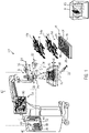
- Fig. 1 shows an exemplary embodiment of the method and device according to the invention.
- the configuration and function of an optical observation device 1 for observing live tissue, in particular during surgery, is explained.
- the medical observation device is shown to be a microscope 2 just for the purposes of explanation.
- the medical observation device 1 may also be an endoscope (not shown).
- the medical observation device 1 comprises a memory assembly 4, in which pre-operative three-dimensional image data 6 are stored.
- the memory assembly 4 may comprise standard computer memory.
- the medical observation device 1 further comprises a camera assembly 8, which has a field of view 10.
- soft biological tissue 12 such as brain tissue, muscle tissue, lymph tissue or tissue of an internal organ or of other soft body parts, may be arranged in the field of view 10.
- the camera assembly 8 acquires interoperative image data 14, which may be structured as a single input frame 16 or a time series 18 of input frames 16.
- the interoperative image data 14 may comprise or consist of pixels 20.
- the interoperative image data 14 may be two-dimensional, i.e. representing a plane in the field of view 10, three-dimensional, i.e. representing a volume in the field of view 10, or multidimensional image data which may e.g. comprises three-dimensional data in the field of view 10 at different spectral wavelengths.
- the camera assembly 8 may comprise at least one of an RGB camera, a multi-spectral camera and a hyper-spectral camera.
- the interoperative image data comprise or consist of fluorescent-light image data.
- fluorescent-light image data may be obtained when a fluorophore 22, such as indocyanine green, is injected into the tissue 12 and illuminated at wavelengths which trigger the fluorescence.
- the camera assembly 8 may comprise one or more filter assemblies 24, which are only schematically shown in Fig. 1 .
- the filter assembly 24 may comprise a filter arrangement 26 for blocking specular reflections. Examples of such a filter arrangement are described in the above-mentioned European patent application EP 16 204 933.2 .
- the filter assembly 24 may also comprise a band-pass filter arrangement 28 for restricting the light in the interoperative image data 14 to the fluorescence wavelengths of the at least one fluorophore 22.
- a band-pass filter arrangement is shown in European patent application EP 17 179 019.8 , published as EP 3 422 072 A1 and prior art under Article 54(3) EPC.
- the medical observation device 1 may further include an illumination assembly 32 for generating illumination light 34 having an illumination spectrum.
- the illumination spectrum may be restricted to or include wavelengths that trigger fluorescence of the at least one fluorophore 22.
- the illumination light 34 may further comprise or be restricted to wavelengths that match the reflectance spectrum of arterial blood.
- the illumination light 34 may be restricted to or comprise wavelengths that are matched to the reflectance spectrum of venous blood. Restricting the illumination spectrum of the illumination light 34 to a single or to preferably separate wavelengths reduced cross-talk between the various frequency bands. This facilitates an automatic analysis of the interoperative image data 14. Subsequent input frames 16 may have been acquired at different illumination spectra.
- the interoperative image data 14 contain information preferably in at least one of the visible-light spectra, e.g. in at least one of the reflective spectrum of arterial blood and venous blood, and the near-infrared spectrum, e.g. in the fluorescence wavelengths of the at least one fluorophore 22.
- the medical observation device 1 further includes an image processor assembly 40, which only by way of example is shown as an integrated circuit in Fig. 1 .
- the image processor 40 and its constituents may be software-implemented, hardware-implemented or be implemented as a combination of hardware and software.
- the memory assembly 4 may be part of the image processor 40.
- the image processor assembly 40 may comprise several modules which may be differentiated functionally and/or structurally.
- the image processor assembly 40 is connected to the camera assembly 8 by a data connection 42, which may be wired and/or wireless.
- An input interface 44 of the image processor assembly 40 may be adapted to acquire interoperative image data 14 from at least one camera assembly 8 and/or a storage where the interoperative image data 14 are stored or buffered, e.g. after pre-processing, such as memory assembly 4.
- the image processor assembly 40 comprises a pattern-matching module 46 for identifying at least one vascular plexus structure 48 in the interoperative image data 14 and for identifying the at least one identified vascular plexus structure 48 in the pre-operative three-dimensional image data 6.
- the at least one vascular plexus structure 48 may be identified e.g. in interoperative image data 14a which are restricted to the fluorescence spectrum of the fluorophore 22.
- the at least one vascular plexus structure 48 may be identified in interoperative image data 14b which have been recorded in the visible-light spectrum and may in particular be restricted to at least one of the reflectance spectrum of arterial blood and venous blood.
- Algorithms for identifying a vascular plexus structure 48 in the interoperative image data 14 and then identifying this structure in the pre-operative three-dimensional image data 6 are, for example, given in Rouchdy, Y.; Cohen, L.: “A Geodesic Voting Method of Tubular Tree and Centrelines", DOI: 10.1109/ISBI.2011.5872566, pp. 979-983 , and Suri, J.S.; Laxminarayan, S. (eds): "Angiography and Plaque Imaging: Advanced Segmentation Techniques", CRC Press, pp. 501-518 . Further, a method of identifying vascular plexus structures by using a bolus of at least one fluorophore is described in the above-mentioned EP 17 174 047.5 .
- the image processor assembly 40 further comprises an elastic-matching module 50 for elastically matching the pre-operative three-dimensional image data 6 to the interoperative image data 14 based on the at least one identified vascular plexus structure 48.
- an algorithm for performing such elastic matching is described in IRCS Technical Reports Series, 192, "Elastically Deforming a Three-Dimensional Atlas to Match Anatomical Brain Images " as given above.
- the pre-operative three-dimensional image data 6 are shifted, rotated and/or distorted so that the identified vascular plexus structure 48 in both data coincides geometrically.
- the step of elastic matching may also define a section through the pre-operative three-dimensional image data 6 which results in the field of view 10 represented in the interoperative image data 14.
- the blood vessel structure 48 and its type may be determined using the apparatus and method described in Opt. Express 17, 15670-15678, "Ultra-fast Multispectral Optical Imaging of Cortical Oxigenation, Blood Flow, and Intracellular Calcium Dynamics ", as given above.
- the image processor assembly 40 further comprises an image-forming module 52 for combining the elastically matched pre-operative three-dimensional image data 6 or a section thereof with the interoperative image data 14 into output image data 54.
- the image processor assembly may further comprise an output interface 55 for outputting the output image data 54.
- the output interface 55 may comprise at least one standard connector, such as an HDMI, DVI, RGB or any other suitable type of connector, and/or a wireless connection, including the matching data transmission protocol.
- the medical observation device 1 may comprise a display assembly 56, which may include stereoscopic display, for example an eyepiece of a microscope or endoscope and/or a monitor.
- the display assembly 56 may be connected to the output interface 55 by wire or wireless.
- the computational burden for locating and/or identifying the identified vascular plexus structure 48 of the interoperative image data 14 in the pre-operative three-dimensional image data 6 may be high. This burden can be reduced if position information is provided as to where the field of view 10, the vascular plexus structure 48, and/or the interoperative image data 14 are located within the soft biological tissue 12. This information is useful if, for example, an elastic-matching process has already been successfully carried out for one input frame 16 of a time series 18 and then has to be repeated for subsequent input frame 16 of the time series 18 later during surgery.
- the medical observation device 1 may comprise at least one position sensor 58 for acquiring and/or providing positioning data 60 representative of at least one of focal length, field depth and distance setting of an optical lens system 62 of the medical observation device 1 and size, distance from the optical lens system 62 and the image processor assembly 40, such as a dimension of the field of view 10.
- the positioning data 60 may be input into at least one of the pattern-matching modules 46, the elastic-matching module 50 and the image-forming module 52 and be used in identifying the at least one identified vascular plexus structure 48 in the pre-operative three-dimensional image data 6, the elastic matching of the pre-operative three-dimensional image data 6 to the interoperative image data 14 and for forming the output image data 54 from the elastically matched pre-operative three-dimensional image data 6 and the interoperative image data 14.
- the image processor assembly 40 may be adapted to compute the blood flow direction 64 at a location 66 in the at least one identified vascular plexus structure 48 as is described in the above-mentioned European patent application EP 17 210 909.2 .
- a location may be a pixel 20 or a preferably coherent array of pixels in the interoperative image data 14.
- the tissue type of the identified vascular plexus structure 48 i.e. a differentiation between venous, arterial and capillary tissue and/or vessels may be carried out using the method and system described in the above-mentioned EP 17 174 047.5 .
- the system and method rely solely on information that is automatically acquired when the interoperative image data 14 are recorded.
- the system and method may further comprise positioning data 60 that result from the settings of the medical observation device 1 for acquiring the interoperative image data 14.
Landscapes
- Health & Medical Sciences (AREA)
- Life Sciences & Earth Sciences (AREA)
- Engineering & Computer Science (AREA)
- Surgery (AREA)
- Public Health (AREA)
- Medical Informatics (AREA)
- General Health & Medical Sciences (AREA)
- Animal Behavior & Ethology (AREA)
- Biomedical Technology (AREA)
- Heart & Thoracic Surgery (AREA)
- Molecular Biology (AREA)
- Veterinary Medicine (AREA)
- Physics & Mathematics (AREA)
- Pathology (AREA)
- Nuclear Medicine, Radiotherapy & Molecular Imaging (AREA)
- Biophysics (AREA)
- Radiology & Medical Imaging (AREA)
- Theoretical Computer Science (AREA)
- General Physics & Mathematics (AREA)
- Oral & Maxillofacial Surgery (AREA)
- Computer Vision & Pattern Recognition (AREA)
- Data Mining & Analysis (AREA)
- Physiology (AREA)
- Optics & Photonics (AREA)
- Gynecology & Obstetrics (AREA)
- Hematology (AREA)
- Cardiology (AREA)
- Bioinformatics & Cheminformatics (AREA)
- Primary Health Care (AREA)
- Epidemiology (AREA)
- Artificial Intelligence (AREA)
- Robotics (AREA)
- Bioinformatics & Computational Biology (AREA)
- Evolutionary Biology (AREA)
- Evolutionary Computation (AREA)
- General Engineering & Computer Science (AREA)
- Investigating, Analyzing Materials By Fluorescence Or Luminescence (AREA)
- Endoscopes (AREA)
- Measuring And Recording Apparatus For Diagnosis (AREA)
Claims (11)
- Bildverarbeitungsverfahren zum Anzeigen von Ausgabebilddaten (54) eines weichen biologischen Gewebes (12) insbesondere in Echtzeit während eines chirurgischen Eingriffs, das die folgenden Schritte umfasst:- Vorsehen von präoperativen dreidimensionalen Bilddaten (6) des weichen biologischen Gewebes (12),- Erhalten von intraoperativen Bilddaten (14) des weichen biologischen Gewebes (12) im sichtbaren Lichtspektrum und/oder Nahinfrarotspektrum, wobei die intraoperativen Bilddaten (14) Fluoreszenzlicht-Bilddaten, die unter Verwendung eines Fluoreszenzlichts von einem Fluorophor (22) erhalten werden, umfassen,- Berechnen einer Blutflussrichtung (64) an wenigstens einer Position in den intraoperativen Bilddaten (14) unter Verwendung der Fluoreszenzlicht-Bilddaten,- automatisches Identifizieren wenigstens einer Gefäßplexusstruktur (48) in den intraoperativen Bilddaten (14) unter Verwendung der intraoperativen Bilddaten (14) und der berechneten Blutflussrichtung (64),- Identifizieren der wenigstens einen identifizierten Gefäßplexusstruktur (48) in den präoperativen dreidimensionalen Bilddaten (6),- elastisches Abgleichen der präoperativen dreidimensionalen Bilddaten (6) mit den intraoperativen Bilddaten (14) basierend auf der wenigstens einen identifizierten Gefäßplexusstruktur (48),- Erstellen der Ausgabebilddaten (54) aus den elastisch abgeglichen präoperativen dreidimensionalen Bilddaten (6) und den intraoperativen Bilddaten (14).
- Bildverarbeitungsverfahren nach Anspruch 1, wobei die identifizierte Gefäßplexusstruktur (48) in den intraoperativen Bilddaten (14) unter Verwendung von Licht im sichtbaren Spektrum aufgezeichnet wurde.
- Bildverarbeitungsverfahren nach Anspruch 1 oder 2, wobei die Ausgabebilddaten (54) angezeigt werden.
- Bildverarbeitungsverfahren nach einem der Ansprüche 1 bis 3, wobei wenigstens eine optische Filteranordnung (26) zum Reduzieren von Spiegelreflexionen für das Erhalten der intraoperativen Bilddaten (14) verwendet wird.
- Bildverarbeitungsverfahren nach einem der Ansprüche 1 bis 4, wobei Positionierungsdaten (60) erhalten werden, wobei die Positionierungsdaten (60) die Brennweite eines optischen Linsensystems (62), das für das Erhalten der intraoperativen Bilddaten (14) verwendet wird, die Feldtiefe des optischen Linsensystems (62), die Distanzeinstellung des optischen Linsensystems (62) und/oder die Größe, Dimension und Ausrichtung eines Sichtfelds (10) des optischen Linsensystems (62) zum Zeitpunkt des Erhaltens der intraoperativen Bilddaten (14) wiedergeben und wobei die Positionierungsdaten (60) beim Identifizieren der Gefäßplexusstruktur (48) in den präoperativen dreidimensionalen Bilddaten (6), beim elastischen Abgleichen der präoperativen dreidimensionalen Bilddaten (6) mit den intraoperativen Bilddaten (14) und/oder beim Anzeigen der Ausgabebilddaten (54) verwendet werden.
- Medizinisches Beobachtungsgerät (1) für das Erzeugen von Ausgabebilddaten (54) eines weichen biologischen Gewebes (12) insbesondere während eines chirurgischen Eingriffs wie etwa ein Mikroskop (2) oder Endoskop, wobei das Gerät umfasst:- eine Speicheranordnung (4), die präoperative dreidimensionale Bilddaten (6) des weichen biologischen Gewebes (12) enthält,- eine Kameraanordnung (8), die konfiguriert ist zum Erhalten von intraoperativen Bilddaten (14) des weichen biologischen Gewebes (12) im sichtbaren Lichtspektrum und/oder Nahinfrarotspektrum, wobei die intraoperativen Bilddaten (14) Fluoreszenzlicht-Bilddaten, die unter Verwendung eines Fluoreszenzlichts von einem Fluorophor (22) erhalten werden, umfassen,- eine Bildprozessoranordnung (40), die konfiguriert ist zum Berechnen einer Blutflussrichtung (64) an wenigstens einer Position in den intraoperativen Bilddaten (14) unter Verwendung der Fluoreszenzlicht-Bilddaten, wobei die Bildprozessoranordnung (40) umfasst:- ein Musterabgleichmodul (46), das konfiguriert ist zum Identifizieren wenigstens einer Gefäßplexusstruktur (48) in den intraoperativen Bilddaten (14) unter Verwendung der intraoperativen Bilddaten (14) und der berechneten Blutflussrichtung (64) und zum Identifizieren der wenigstens einen identifizierten Gefäßplexusstruktur (48) in den präoperativen dreidimensionalen Bilddaten (6),- ein Abgleichmodul (50), das konfiguriert ist zum elastischen Abgleichen der präoperativen dreidimensionalen Bilddaten (6) mit den intraoperativen Bilddaten (14) basierend auf der wenigstens einen identifizierten Gefäßplexusstruktur (48), und- ein Bilderstellungsmodul (52), das konfiguriert ist zum Kombinieren wenigstens eines Teils der elastisch mit den intraoperativen Bilddaten (14) abgeglichenen präoperativen dreidimensionalen Bilddaten (6) mit den Ausgabebilddaten (54), und- eine Ausgabeschnittstelle, die konfiguriert ist zum Ausgeben der Ausgabebilddaten (54),wobei das Bilderstellungsmodul (52) ausgebildet ist zum Kombinieren der identifizierten Gefäßplexusstruktur (48) in den Ausgabebilddaten (54) mit in der Zeit variierenden Markerdaten, die die Blutflussrichtung (64) wiedergeben.
- Medizinisches Beobachtungsgerät (1) nach Anspruch 6, das weiterhin eine Displayanordnung (56) für das Anzeigen der Ausgabebilddaten (54) umfasst.
- Medizinisches Beobachtungsgerät (1) nach Anspruch 6 oder 7, wobei die Kameraanordnung (8) wenigstens eine Filteranordnung (26) umfasst, die konfiguriert ist zum Reduzieren von Spiegelreflexionen, wobei die Filteranordnung (26) wenigstens ein Paar von Kreuzpolarisatoren umfasst.
- Medizinisches Beobachtungsgerät (1) nach einem der Ansprüche 6 bis 8, wobei die Kameraanordnung (8) wenigstens eine Filteranordnung (28), die ein mit dem Fluoreszenzspektrum des wenigstens einen Fluorophors (22) abgeglichenes Durchlassband aufweist, umfasst.
- Medizinisches Beobachtungsgerät (1) nach einem der Ansprüche 6 bis 9, wobei die Kameraanordnung (8) ein optisches Linsensystem (62) und wenigstens einen Positionssensor (58) für das Erhalten von Positionierungsdaten (60) umfasst, wobei die Positionierungsdaten (60) die Brennweite, die Feldtiefe und die Distanzeinstellung des optischen Linsensystems (62) und/oder die Größe, Dimension und Ausrichtung des Sichtfelds (10) wiedergeben.
- Nicht-transitorisches, computerlesbares Medium mit einem darauf gespeicherten Programm, das das medizinische Beobachtungsgerät gemäß einem der Ansprüche 6 bis 10 zum Ausführen des Bildverarbeitungsverfahrens gemäß einem der Ansprüche 1 bis 5 veranlasst.
Priority Applications (4)
| Application Number | Priority Date | Filing Date | Title |
|---|---|---|---|
| EP18156906.2A EP3527123B1 (de) | 2018-02-15 | 2018-02-15 | Bildverarbeitungsverfahren und -vorrichtung mit elastischer abbildung von gefässplexusstrukturen |
| US16/269,968 US20190247142A1 (en) | 2018-02-15 | 2019-02-07 | Image processing method and apparatus using elastic mapping of vascular plexus structures |
| CN201910112740.7A CN110164528B (zh) | 2018-02-15 | 2019-02-13 | 利用血管丛结构的弹性映射的图像处理方法和装置 |
| JP2019024250A JP6972049B2 (ja) | 2018-02-15 | 2019-02-14 | 脈管叢構造の弾性マッピングを用いた画像処理方法および画像処理装置 |
Applications Claiming Priority (1)
| Application Number | Priority Date | Filing Date | Title |
|---|---|---|---|
| EP18156906.2A EP3527123B1 (de) | 2018-02-15 | 2018-02-15 | Bildverarbeitungsverfahren und -vorrichtung mit elastischer abbildung von gefässplexusstrukturen |
Publications (2)
| Publication Number | Publication Date |
|---|---|
| EP3527123A1 EP3527123A1 (de) | 2019-08-21 |
| EP3527123B1 true EP3527123B1 (de) | 2022-08-31 |
Family
ID=61521289
Family Applications (1)
| Application Number | Title | Priority Date | Filing Date |
|---|---|---|---|
| EP18156906.2A Active EP3527123B1 (de) | 2018-02-15 | 2018-02-15 | Bildverarbeitungsverfahren und -vorrichtung mit elastischer abbildung von gefässplexusstrukturen |
Country Status (4)
| Country | Link |
|---|---|
| US (1) | US20190247142A1 (de) |
| EP (1) | EP3527123B1 (de) |
| JP (1) | JP6972049B2 (de) |
| CN (1) | CN110164528B (de) |
Families Citing this family (6)
| Publication number | Priority date | Publication date | Assignee | Title |
|---|---|---|---|---|
| EP3540494B1 (de) | 2018-03-16 | 2022-11-23 | Leica Instruments (Singapore) Pte. Ltd. | Chirurgisches mikroskop und mikroskopisches verfahren mit erweiterter realität |
| JP6988001B2 (ja) * | 2018-08-30 | 2022-01-05 | オリンパス株式会社 | 記録装置、画像観察装置、観察システム、観察システムの制御方法、及び観察システムの作動プログラム |
| CN111292410B (zh) * | 2020-01-19 | 2022-04-12 | 华中科技大学同济医学院附属协和医院 | 一种静脉显影照相装置及其三维全景模型的生成方法 |
| EP3991685A1 (de) * | 2020-11-03 | 2022-05-04 | Leica Instruments (Singapore) Pte. Ltd. | Chirurgisches mikroskopsystem |
| US20240122671A1 (en) * | 2021-04-28 | 2024-04-18 | Smith & Nephew, Inc. | Computerized Systems for Arthroscopic Applications Using Real-Time Blood-Flow Detection |
| WO2023052534A1 (en) * | 2021-09-30 | 2023-04-06 | Leica Instruments (Singapore) Pte. Ltd. | Image processing for surgical applications |
Family Cites Families (22)
| Publication number | Priority date | Publication date | Assignee | Title |
|---|---|---|---|---|
| JP3267625B2 (ja) * | 1995-10-23 | 2002-03-18 | サイトメトリクス インコーポレイテッド | 反射画像分析の方法および装置 |
| DE10163813A1 (de) * | 2001-12-22 | 2003-07-03 | Philips Intellectual Property | Verfahren zur Darstellung von unterschiedlichen Bildern eines Untersuchungsobjektes |
| JP4147033B2 (ja) * | 2002-01-18 | 2008-09-10 | オリンパス株式会社 | 内視鏡装置 |
| DE60316123T2 (de) * | 2002-03-20 | 2008-05-29 | Novadaq Technologies Inc., Mississauga | System und verfahren zur visualisierung des flüssigkeitsflusses durch gefässe |
| US20060173358A1 (en) * | 2005-01-11 | 2006-08-03 | Olympus Corporation | Fluorescence observation endoscope apparatus and fluorescence observation method |
| EP1969564A2 (de) * | 2005-12-20 | 2008-09-17 | University of Maryland, Baltimore | Verfahren und vorrichtung zur beschleunigten elastischen registration mehrerer abtastungen interner eigenschaften eines körpers |
| JP5214876B2 (ja) * | 2006-12-15 | 2013-06-19 | 株式会社東芝 | 3次元画像処理装置及び医用画像診断装置 |
| WO2010046838A1 (en) * | 2008-10-23 | 2010-04-29 | Koninklijke Philips Electronics N.V. | Cardiac- and/or respiratory-gated image acquisition system and method for virtual anatomy enriched real-time 2d imaging in interventional radiofrequency ablation or pacemaker placement procedures |
| US8423117B2 (en) * | 2009-06-22 | 2013-04-16 | General Electric Company | System and method to process an acquired image of a subject anatomy to differentiate a portion of subject anatomy to protect relative to a portion to receive treatment |
| JP2011104199A (ja) * | 2009-11-19 | 2011-06-02 | Fujifilm Corp | 内視鏡装置 |
| US8761474B2 (en) * | 2011-07-25 | 2014-06-24 | Siemens Aktiengesellschaft | Method for vascular flow pattern analysis |
| DE102011120937B4 (de) * | 2011-12-14 | 2019-03-14 | Carl Zeiss Meditec Ag | Anordnung und Verfahren zur Registrierung von Gewebeverschiebungen |
| US11510600B2 (en) * | 2012-01-04 | 2022-11-29 | The Trustees Of Dartmouth College | Method and apparatus for quantitative and depth resolved hyperspectral fluorescence and reflectance imaging for surgical guidance |
| WO2015023990A1 (en) * | 2013-08-15 | 2015-02-19 | The Trustees Of Dartmouth College | Method and apparatus for quantitative and depth resolved hyperspectral fluorescence and reflectance imaging for surgical guidance |
| MY167083A (en) * | 2013-12-06 | 2018-08-10 | Mimos Berhad | An apparatus and method for volumetric and speed measurement of blood 'train' in visible vessels |
| WO2015101948A2 (en) * | 2014-01-06 | 2015-07-09 | Body Vision Medical Ltd. | Surgical devices and methods of use thereof |
| JP2017536870A (ja) * | 2014-10-20 | 2017-12-14 | ボディ・ビジョン・メディカル・リミテッドBody Vision Medical Ltd. | 外科的デバイスおよび外科的デバイスの使用方法 |
| US11844576B2 (en) * | 2015-01-22 | 2023-12-19 | Koninklijke Philips N.V. | Endograft visualization with optical shape sensing |
| JP2016177327A (ja) * | 2015-03-18 | 2016-10-06 | 伸彦 井戸 | 動的計画法を適用する有向無閉路グラフの構成と格子点のコストとに特徴を持つ、2つの要素列の対応付け方法 |
| EP3616596A3 (de) * | 2016-05-23 | 2020-03-18 | Leica Instruments (Singapore) Pte. Ltd. | Bildprozessor, medizinische beobachtungsvorrichtung wie ein mikroskop oder ein endoskop und verfahren mit pseudo-farbbilddaten |
| WO2018002347A1 (en) * | 2016-06-30 | 2018-01-04 | Koninklijke Philips N.V. | Registering tomographic imaging and endoscopic imaging |
| JP6883222B2 (ja) * | 2016-07-12 | 2021-06-09 | ソニーグループ株式会社 | 画像処理装置、画像処理方法、プログラム及び手術用ナビゲーションシステム |
-
2018
- 2018-02-15 EP EP18156906.2A patent/EP3527123B1/de active Active
-
2019
- 2019-02-07 US US16/269,968 patent/US20190247142A1/en not_active Abandoned
- 2019-02-13 CN CN201910112740.7A patent/CN110164528B/zh active Active
- 2019-02-14 JP JP2019024250A patent/JP6972049B2/ja active Active
Also Published As
| Publication number | Publication date |
|---|---|
| JP6972049B2 (ja) | 2021-11-24 |
| CN110164528A (zh) | 2019-08-23 |
| CN110164528B (zh) | 2023-12-29 |
| EP3527123A1 (de) | 2019-08-21 |
| JP2019141578A (ja) | 2019-08-29 |
| US20190247142A1 (en) | 2019-08-15 |
Similar Documents
| Publication | Publication Date | Title |
|---|---|---|
| EP3527123B1 (de) | Bildverarbeitungsverfahren und -vorrichtung mit elastischer abbildung von gefässplexusstrukturen | |
| US11857317B2 (en) | Method and apparatus for quantitative and depth resolved hyperspectral fluorescence and reflectance imaging for surgical guidance | |
| EP3540494B1 (de) | Chirurgisches mikroskop und mikroskopisches verfahren mit erweiterter realität | |
| US20230320649A1 (en) | Surgical navigation with stereovision and associated methods | |
| EP3232975B1 (de) | Harnleitererkennung mit wellenbandselektiver abbildung | |
| US20160086380A1 (en) | Hyperspectral imager | |
| CA2940297C (en) | Methods and systems for intraoperatively confirming location of tissue structures | |
| EP3116376B1 (de) | Verfahren und systeme zur interoperativen bestätigung der stelle von gewebestrukturen | |
| US20160015471A1 (en) | Context aware surgical systems | |
| WO2015023990A1 (en) | Method and apparatus for quantitative and depth resolved hyperspectral fluorescence and reflectance imaging for surgical guidance | |
| US20220125280A1 (en) | Apparatuses and methods involving multi-modal imaging of a sample | |
| WO2015187620A1 (en) | Surgical navigation with stereovision and associated methods | |
| CN114300095A (zh) | 图像处理设备、图像处理方法、装置、设备及存储介质 | |
| US11690558B2 (en) | Surgical navigation with stereovision and associated methods | |
| KR20220098578A (ko) | 조직 식별 및 생체 확인 장치 및 이를 이용한 조직 식별 및 생체 확인 방법 | |
| WO2022233961A1 (en) | Augmented reality headset and probe for medical imaging | |
| Tran et al. | Nerve detection and visualization using hyperspectral imaging for surgical guidance | |
| WO2018055061A1 (en) | Hyperspectral tissue imaging |
Legal Events
| Date | Code | Title | Description |
|---|---|---|---|
| PUAI | Public reference made under article 153(3) epc to a published international application that has entered the european phase |
Free format text: ORIGINAL CODE: 0009012 |
|
| STAA | Information on the status of an ep patent application or granted ep patent |
Free format text: STATUS: THE APPLICATION HAS BEEN PUBLISHED |
|
| AK | Designated contracting states |
Kind code of ref document: A1 Designated state(s): AL AT BE BG CH CY CZ DE DK EE ES FI FR GB GR HR HU IE IS IT LI LT LU LV MC MK MT NL NO PL PT RO RS SE SI SK SM TR |
|
| AX | Request for extension of the european patent |
Extension state: BA ME |
|
| STAA | Information on the status of an ep patent application or granted ep patent |
Free format text: STATUS: REQUEST FOR EXAMINATION WAS MADE |
|
| 17P | Request for examination filed |
Effective date: 20200221 |
|
| RBV | Designated contracting states (corrected) |
Designated state(s): AL AT BE BG CH CY CZ DE DK EE ES FI FR GB GR HR HU IE IS IT LI LT LU LV MC MK MT NL NO PL PT RO RS SE SI SK SM TR |
|
| STAA | Information on the status of an ep patent application or granted ep patent |
Free format text: STATUS: EXAMINATION IS IN PROGRESS |
|
| 17Q | First examination report despatched |
Effective date: 20210202 |
|
| STAA | Information on the status of an ep patent application or granted ep patent |
Free format text: STATUS: EXAMINATION IS IN PROGRESS |
|
| GRAP | Despatch of communication of intention to grant a patent |
Free format text: ORIGINAL CODE: EPIDOSNIGR1 |
|
| STAA | Information on the status of an ep patent application or granted ep patent |
Free format text: STATUS: GRANT OF PATENT IS INTENDED |
|
| INTG | Intention to grant announced |
Effective date: 20220314 |
|
| GRAS | Grant fee paid |
Free format text: ORIGINAL CODE: EPIDOSNIGR3 |
|
| GRAA | (expected) grant |
Free format text: ORIGINAL CODE: 0009210 |
|
| STAA | Information on the status of an ep patent application or granted ep patent |
Free format text: STATUS: THE PATENT HAS BEEN GRANTED |
|
| AK | Designated contracting states |
Kind code of ref document: B1 Designated state(s): AL AT BE BG CH CY CZ DE DK EE ES FI FR GB GR HR HU IE IS IT LI LT LU LV MC MK MT NL NO PL PT RO RS SE SI SK SM TR |
|
| REG | Reference to a national code |
Ref country code: CH Ref legal event code: EP Ref country code: GB Ref legal event code: FG4D |
|
| REG | Reference to a national code |
Ref country code: AT Ref legal event code: REF Ref document number: 1514686 Country of ref document: AT Kind code of ref document: T Effective date: 20220915 |
|
| REG | Reference to a national code |
Ref country code: DE Ref legal event code: R096 Ref document number: 602018039896 Country of ref document: DE |
|
| REG | Reference to a national code |
Ref country code: IE Ref legal event code: FG4D |
|
| REG | Reference to a national code |
Ref country code: LT Ref legal event code: MG9D |
|
| REG | Reference to a national code |
Ref country code: NL Ref legal event code: MP Effective date: 20220831 |
|
| PG25 | Lapsed in a contracting state [announced via postgrant information from national office to epo] |
Ref country code: SE Free format text: LAPSE BECAUSE OF FAILURE TO SUBMIT A TRANSLATION OF THE DESCRIPTION OR TO PAY THE FEE WITHIN THE PRESCRIBED TIME-LIMIT Effective date: 20220831 Ref country code: RS Free format text: LAPSE BECAUSE OF FAILURE TO SUBMIT A TRANSLATION OF THE DESCRIPTION OR TO PAY THE FEE WITHIN THE PRESCRIBED TIME-LIMIT Effective date: 20220831 Ref country code: NO Free format text: LAPSE BECAUSE OF FAILURE TO SUBMIT A TRANSLATION OF THE DESCRIPTION OR TO PAY THE FEE WITHIN THE PRESCRIBED TIME-LIMIT Effective date: 20221130 Ref country code: LV Free format text: LAPSE BECAUSE OF FAILURE TO SUBMIT A TRANSLATION OF THE DESCRIPTION OR TO PAY THE FEE WITHIN THE PRESCRIBED TIME-LIMIT Effective date: 20220831 Ref country code: LT Free format text: LAPSE BECAUSE OF FAILURE TO SUBMIT A TRANSLATION OF THE DESCRIPTION OR TO PAY THE FEE WITHIN THE PRESCRIBED TIME-LIMIT Effective date: 20220831 Ref country code: FI Free format text: LAPSE BECAUSE OF FAILURE TO SUBMIT A TRANSLATION OF THE DESCRIPTION OR TO PAY THE FEE WITHIN THE PRESCRIBED TIME-LIMIT Effective date: 20220831 |
|
| REG | Reference to a national code |
Ref country code: AT Ref legal event code: MK05 Ref document number: 1514686 Country of ref document: AT Kind code of ref document: T Effective date: 20220831 |
|
| PG25 | Lapsed in a contracting state [announced via postgrant information from national office to epo] |
Ref country code: PL Free format text: LAPSE BECAUSE OF FAILURE TO SUBMIT A TRANSLATION OF THE DESCRIPTION OR TO PAY THE FEE WITHIN THE PRESCRIBED TIME-LIMIT Effective date: 20220831 Ref country code: IS Free format text: LAPSE BECAUSE OF FAILURE TO SUBMIT A TRANSLATION OF THE DESCRIPTION OR TO PAY THE FEE WITHIN THE PRESCRIBED TIME-LIMIT Effective date: 20221231 Ref country code: HR Free format text: LAPSE BECAUSE OF FAILURE TO SUBMIT A TRANSLATION OF THE DESCRIPTION OR TO PAY THE FEE WITHIN THE PRESCRIBED TIME-LIMIT Effective date: 20220831 Ref country code: GR Free format text: LAPSE BECAUSE OF FAILURE TO SUBMIT A TRANSLATION OF THE DESCRIPTION OR TO PAY THE FEE WITHIN THE PRESCRIBED TIME-LIMIT Effective date: 20221201 |
|
| PG25 | Lapsed in a contracting state [announced via postgrant information from national office to epo] |
Ref country code: SM Free format text: LAPSE BECAUSE OF FAILURE TO SUBMIT A TRANSLATION OF THE DESCRIPTION OR TO PAY THE FEE WITHIN THE PRESCRIBED TIME-LIMIT Effective date: 20220831 Ref country code: RO Free format text: LAPSE BECAUSE OF FAILURE TO SUBMIT A TRANSLATION OF THE DESCRIPTION OR TO PAY THE FEE WITHIN THE PRESCRIBED TIME-LIMIT Effective date: 20220831 Ref country code: PT Free format text: LAPSE BECAUSE OF FAILURE TO SUBMIT A TRANSLATION OF THE DESCRIPTION OR TO PAY THE FEE WITHIN THE PRESCRIBED TIME-LIMIT Effective date: 20230102 Ref country code: ES Free format text: LAPSE BECAUSE OF FAILURE TO SUBMIT A TRANSLATION OF THE DESCRIPTION OR TO PAY THE FEE WITHIN THE PRESCRIBED TIME-LIMIT Effective date: 20220831 Ref country code: DK Free format text: LAPSE BECAUSE OF FAILURE TO SUBMIT A TRANSLATION OF THE DESCRIPTION OR TO PAY THE FEE WITHIN THE PRESCRIBED TIME-LIMIT Effective date: 20220831 Ref country code: CZ Free format text: LAPSE BECAUSE OF FAILURE TO SUBMIT A TRANSLATION OF THE DESCRIPTION OR TO PAY THE FEE WITHIN THE PRESCRIBED TIME-LIMIT Effective date: 20220831 Ref country code: AT Free format text: LAPSE BECAUSE OF FAILURE TO SUBMIT A TRANSLATION OF THE DESCRIPTION OR TO PAY THE FEE WITHIN THE PRESCRIBED TIME-LIMIT Effective date: 20220831 |
|
| PGFP | Annual fee paid to national office [announced via postgrant information from national office to epo] |
Ref country code: FR Payment date: 20230223 Year of fee payment: 6 |
|
| PG25 | Lapsed in a contracting state [announced via postgrant information from national office to epo] |
Ref country code: SK Free format text: LAPSE BECAUSE OF FAILURE TO SUBMIT A TRANSLATION OF THE DESCRIPTION OR TO PAY THE FEE WITHIN THE PRESCRIBED TIME-LIMIT Effective date: 20220831 Ref country code: EE Free format text: LAPSE BECAUSE OF FAILURE TO SUBMIT A TRANSLATION OF THE DESCRIPTION OR TO PAY THE FEE WITHIN THE PRESCRIBED TIME-LIMIT Effective date: 20220831 |
|
| REG | Reference to a national code |
Ref country code: DE Ref legal event code: R097 Ref document number: 602018039896 Country of ref document: DE |
|
| P01 | Opt-out of the competence of the unified patent court (upc) registered |
Effective date: 20230414 |
|
| PG25 | Lapsed in a contracting state [announced via postgrant information from national office to epo] |
Ref country code: NL Free format text: LAPSE BECAUSE OF FAILURE TO SUBMIT A TRANSLATION OF THE DESCRIPTION OR TO PAY THE FEE WITHIN THE PRESCRIBED TIME-LIMIT Effective date: 20220831 Ref country code: AL Free format text: LAPSE BECAUSE OF FAILURE TO SUBMIT A TRANSLATION OF THE DESCRIPTION OR TO PAY THE FEE WITHIN THE PRESCRIBED TIME-LIMIT Effective date: 20220831 |
|
| PLBE | No opposition filed within time limit |
Free format text: ORIGINAL CODE: 0009261 |
|
| STAA | Information on the status of an ep patent application or granted ep patent |
Free format text: STATUS: NO OPPOSITION FILED WITHIN TIME LIMIT |
|
| 26N | No opposition filed |
Effective date: 20230601 |
|
| PG25 | Lapsed in a contracting state [announced via postgrant information from national office to epo] |
Ref country code: SI Free format text: LAPSE BECAUSE OF FAILURE TO SUBMIT A TRANSLATION OF THE DESCRIPTION OR TO PAY THE FEE WITHIN THE PRESCRIBED TIME-LIMIT Effective date: 20220831 |
|
| PG25 | Lapsed in a contracting state [announced via postgrant information from national office to epo] |
Ref country code: MC Free format text: LAPSE BECAUSE OF FAILURE TO SUBMIT A TRANSLATION OF THE DESCRIPTION OR TO PAY THE FEE WITHIN THE PRESCRIBED TIME-LIMIT Effective date: 20220831 |
|
| REG | Reference to a national code |
Ref country code: CH Ref legal event code: PL |
|
| REG | Reference to a national code |
Ref country code: BE Ref legal event code: MM Effective date: 20230228 |
|
| PG25 | Lapsed in a contracting state [announced via postgrant information from national office to epo] |
Ref country code: LU Free format text: LAPSE BECAUSE OF NON-PAYMENT OF DUE FEES Effective date: 20230215 Ref country code: LI Free format text: LAPSE BECAUSE OF NON-PAYMENT OF DUE FEES Effective date: 20230228 Ref country code: CH Free format text: LAPSE BECAUSE OF NON-PAYMENT OF DUE FEES Effective date: 20230228 |
|
| REG | Reference to a national code |
Ref country code: IE Ref legal event code: MM4A |
|
| PG25 | Lapsed in a contracting state [announced via postgrant information from national office to epo] |
Ref country code: IE Free format text: LAPSE BECAUSE OF NON-PAYMENT OF DUE FEES Effective date: 20230215 |
|
| PG25 | Lapsed in a contracting state [announced via postgrant information from national office to epo] |
Ref country code: BE Free format text: LAPSE BECAUSE OF NON-PAYMENT OF DUE FEES Effective date: 20230228 |
|
| PGFP | Annual fee paid to national office [announced via postgrant information from national office to epo] |
Ref country code: DE Payment date: 20240228 Year of fee payment: 7 Ref country code: GB Payment date: 20240220 Year of fee payment: 7 |