EP3521417A1 - Method for producing cell tissue, and porous film - Google Patents
Method for producing cell tissue, and porous film Download PDFInfo
- Publication number
- EP3521417A1 EP3521417A1 EP17855801.1A EP17855801A EP3521417A1 EP 3521417 A1 EP3521417 A1 EP 3521417A1 EP 17855801 A EP17855801 A EP 17855801A EP 3521417 A1 EP3521417 A1 EP 3521417A1
- Authority
- EP
- European Patent Office
- Prior art keywords
- cells
- opening
- pores
- porous film
- culture
- Prior art date
- Legal status (The legal status is an assumption and is not a legal conclusion. Google has not performed a legal analysis and makes no representation as to the accuracy of the status listed.)
- Pending
Links
- 238000004519 manufacturing process Methods 0.000 title claims abstract description 52
- 239000011148 porous material Substances 0.000 claims abstract description 238
- 238000012258 culturing Methods 0.000 claims abstract description 54
- 210000004027 cell Anatomy 0.000 claims description 292
- 210000002901 mesenchymal stem cell Anatomy 0.000 claims description 51
- 238000010899 nucleation Methods 0.000 claims description 37
- 238000005119 centrifugation Methods 0.000 claims description 29
- 210000003556 vascular endothelial cell Anatomy 0.000 claims description 25
- 210000005073 lymphatic endothelial cell Anatomy 0.000 claims description 16
- 210000004738 parenchymal cell Anatomy 0.000 claims description 16
- 210000002950 fibroblast Anatomy 0.000 claims description 6
- 239000002609 medium Substances 0.000 description 84
- 210000001519 tissue Anatomy 0.000 description 69
- 238000003501 co-culture Methods 0.000 description 32
- 238000012744 immunostaining Methods 0.000 description 30
- 102100024616 Platelet endothelial cell adhesion molecule Human genes 0.000 description 29
- 239000010410 layer Substances 0.000 description 29
- XLYOFNOQVPJJNP-UHFFFAOYSA-N water Substances O XLYOFNOQVPJJNP-UHFFFAOYSA-N 0.000 description 29
- 239000011248 coating agent Substances 0.000 description 28
- 238000000576 coating method Methods 0.000 description 28
- 238000002474 experimental method Methods 0.000 description 20
- 239000000463 material Substances 0.000 description 15
- 210000004271 bone marrow stromal cell Anatomy 0.000 description 14
- 150000001875 compounds Chemical class 0.000 description 13
- 230000004069 differentiation Effects 0.000 description 13
- 239000002904 solvent Substances 0.000 description 13
- 229920000747 poly(lactic acid) Polymers 0.000 description 12
- 239000004626 polylactic acid Substances 0.000 description 12
- 239000007788 liquid Substances 0.000 description 11
- 230000015572 biosynthetic process Effects 0.000 description 10
- -1 polymethacrylamide Polymers 0.000 description 10
- 238000002073 fluorescence micrograph Methods 0.000 description 9
- 229920001600 hydrophobic polymer Polymers 0.000 description 9
- 238000001000 micrograph Methods 0.000 description 9
- 230000035755 proliferation Effects 0.000 description 8
- 238000012360 testing method Methods 0.000 description 8
- 101100352374 Oryza sativa subsp. japonica PLA3 gene Proteins 0.000 description 7
- 239000000203 mixture Substances 0.000 description 7
- 210000001778 pluripotent stem cell Anatomy 0.000 description 7
- 229920000642 polymer Polymers 0.000 description 7
- 239000000243 solution Substances 0.000 description 7
- 238000004113 cell culture Methods 0.000 description 6
- 230000001939 inductive effect Effects 0.000 description 6
- 210000000056 organ Anatomy 0.000 description 6
- 229920002799 BoPET Polymers 0.000 description 5
- 239000006144 Dulbecco’s modified Eagle's medium Substances 0.000 description 5
- 230000002209 hydrophobic effect Effects 0.000 description 5
- HEDRZPFGACZZDS-UHFFFAOYSA-N Chloroform Chemical compound ClC(Cl)Cl HEDRZPFGACZZDS-UHFFFAOYSA-N 0.000 description 4
- LFQSCWFLJHTTHZ-UHFFFAOYSA-N Ethanol Chemical compound CCO LFQSCWFLJHTTHZ-UHFFFAOYSA-N 0.000 description 4
- 241001465754 Metazoa Species 0.000 description 4
- 210000001789 adipocyte Anatomy 0.000 description 4
- 238000004458 analytical method Methods 0.000 description 4
- 238000005520 cutting process Methods 0.000 description 4
- 210000002889 endothelial cell Anatomy 0.000 description 4
- 238000001704 evaporation Methods 0.000 description 4
- 239000012091 fetal bovine serum Substances 0.000 description 4
- 230000005484 gravity Effects 0.000 description 4
- 238000002156 mixing Methods 0.000 description 4
- 210000001082 somatic cell Anatomy 0.000 description 4
- 210000000130 stem cell Anatomy 0.000 description 4
- VUDQSRFCCHQIIU-UHFFFAOYSA-N 1-(3,5-dichloro-2,6-dihydroxy-4-methoxyphenyl)hexan-1-one Chemical compound CCCCCC(=O)C1=C(O)C(Cl)=C(OC)C(Cl)=C1O VUDQSRFCCHQIIU-UHFFFAOYSA-N 0.000 description 3
- 102000007469 Actins Human genes 0.000 description 3
- 108010085238 Actins Proteins 0.000 description 3
- YMWUJEATGCHHMB-UHFFFAOYSA-N Dichloromethane Chemical compound ClCCl YMWUJEATGCHHMB-UHFFFAOYSA-N 0.000 description 3
- 101000942967 Homo sapiens Leukemia inhibitory factor Proteins 0.000 description 3
- 102100032352 Leukemia inhibitory factor Human genes 0.000 description 3
- IMNFDUFMRHMDMM-UHFFFAOYSA-N N-Heptane Chemical compound CCCCCCC IMNFDUFMRHMDMM-UHFFFAOYSA-N 0.000 description 3
- 239000004793 Polystyrene Substances 0.000 description 3
- 230000009815 adipogenic differentiation Effects 0.000 description 3
- 210000000988 bone and bone Anatomy 0.000 description 3
- 229960001701 chloroform Drugs 0.000 description 3
- 239000004205 dimethyl polysiloxane Substances 0.000 description 3
- 235000013870 dimethyl polysiloxane Nutrition 0.000 description 3
- 210000002919 epithelial cell Anatomy 0.000 description 3
- 230000008020 evaporation Effects 0.000 description 3
- 230000006698 induction Effects 0.000 description 3
- 210000004185 liver Anatomy 0.000 description 3
- 238000013508 migration Methods 0.000 description 3
- 230000005012 migration Effects 0.000 description 3
- 238000009343 monoculture Methods 0.000 description 3
- 239000003960 organic solvent Substances 0.000 description 3
- 230000009818 osteogenic differentiation Effects 0.000 description 3
- 230000000149 penetrating effect Effects 0.000 description 3
- 229920000435 poly(dimethylsiloxane) Polymers 0.000 description 3
- 229920003229 poly(methyl methacrylate) Polymers 0.000 description 3
- 229920000139 polyethylene terephthalate Polymers 0.000 description 3
- 239000005020 polyethylene terephthalate Substances 0.000 description 3
- 239000004926 polymethyl methacrylate Substances 0.000 description 3
- 229920002223 polystyrene Polymers 0.000 description 3
- 239000000047 product Substances 0.000 description 3
- 230000002062 proliferating effect Effects 0.000 description 3
- 239000002356 single layer Substances 0.000 description 3
- 210000003491 skin Anatomy 0.000 description 3
- 239000000126 substance Substances 0.000 description 3
- CIWBSHSKHKDKBQ-JLAZNSOCSA-N Ascorbic acid Chemical compound OC[C@H](O)[C@H]1OC(=O)C(O)=C1O CIWBSHSKHKDKBQ-JLAZNSOCSA-N 0.000 description 2
- 102000003974 Fibroblast growth factor 2 Human genes 0.000 description 2
- 108090000379 Fibroblast growth factor 2 Proteins 0.000 description 2
- OFBQJSOFQDEBGM-UHFFFAOYSA-N Pentane Chemical compound CCCCC OFBQJSOFQDEBGM-UHFFFAOYSA-N 0.000 description 2
- FAPWRFPIFSIZLT-UHFFFAOYSA-M Sodium chloride Chemical compound [Na+].[Cl-] FAPWRFPIFSIZLT-UHFFFAOYSA-M 0.000 description 2
- 102000004887 Transforming Growth Factor beta Human genes 0.000 description 2
- 108090001012 Transforming Growth Factor beta Proteins 0.000 description 2
- 108010073929 Vascular Endothelial Growth Factor A Proteins 0.000 description 2
- 102000005789 Vascular Endothelial Growth Factors Human genes 0.000 description 2
- 108010019530 Vascular Endothelial Growth Factors Proteins 0.000 description 2
- 239000002390 adhesive tape Substances 0.000 description 2
- 210000004204 blood vessel Anatomy 0.000 description 2
- 210000001185 bone marrow Anatomy 0.000 description 2
- 210000000845 cartilage Anatomy 0.000 description 2
- 230000004956 cell adhesive effect Effects 0.000 description 2
- 230000010261 cell growth Effects 0.000 description 2
- 239000006285 cell suspension Substances 0.000 description 2
- 210000001612 chondrocyte Anatomy 0.000 description 2
- 238000009833 condensation Methods 0.000 description 2
- 230000005494 condensation Effects 0.000 description 2
- 230000002354 daily effect Effects 0.000 description 2
- 230000002500 effect on skin Effects 0.000 description 2
- 239000007850 fluorescent dye Substances 0.000 description 2
- 239000011521 glass Substances 0.000 description 2
- 239000003102 growth factor Substances 0.000 description 2
- 239000004615 ingredient Substances 0.000 description 2
- NOESYZHRGYRDHS-UHFFFAOYSA-N insulin Chemical compound N1C(=O)C(NC(=O)C(CCC(N)=O)NC(=O)C(CCC(O)=O)NC(=O)C(C(C)C)NC(=O)C(NC(=O)CN)C(C)CC)CSSCC(C(NC(CO)C(=O)NC(CC(C)C)C(=O)NC(CC=2C=CC(O)=CC=2)C(=O)NC(CCC(N)=O)C(=O)NC(CC(C)C)C(=O)NC(CCC(O)=O)C(=O)NC(CC(N)=O)C(=O)NC(CC=2C=CC(O)=CC=2)C(=O)NC(CSSCC(NC(=O)C(C(C)C)NC(=O)C(CC(C)C)NC(=O)C(CC=2C=CC(O)=CC=2)NC(=O)C(CC(C)C)NC(=O)C(C)NC(=O)C(CCC(O)=O)NC(=O)C(C(C)C)NC(=O)C(CC(C)C)NC(=O)C(CC=2NC=NC=2)NC(=O)C(CO)NC(=O)CNC2=O)C(=O)NCC(=O)NC(CCC(O)=O)C(=O)NC(CCCNC(N)=N)C(=O)NCC(=O)NC(CC=3C=CC=CC=3)C(=O)NC(CC=3C=CC=CC=3)C(=O)NC(CC=3C=CC(O)=CC=3)C(=O)NC(C(C)O)C(=O)N3C(CCC3)C(=O)NC(CCCCN)C(=O)NC(C)C(O)=O)C(=O)NC(CC(N)=O)C(O)=O)=O)NC(=O)C(C(C)CC)NC(=O)C(CO)NC(=O)C(C(C)O)NC(=O)C1CSSCC2NC(=O)C(CC(C)C)NC(=O)C(NC(=O)C(CCC(N)=O)NC(=O)C(CC(N)=O)NC(=O)C(NC(=O)C(N)CC=1C=CC=CC=1)C(C)C)CC1=CN=CN1 NOESYZHRGYRDHS-UHFFFAOYSA-N 0.000 description 2
- 239000002184 metal Substances 0.000 description 2
- 229910052751 metal Inorganic materials 0.000 description 2
- 210000004088 microvessel Anatomy 0.000 description 2
- 210000000107 myocyte Anatomy 0.000 description 2
- VLKZOEOYAKHREP-UHFFFAOYSA-N n-Hexane Chemical compound CCCCCC VLKZOEOYAKHREP-UHFFFAOYSA-N 0.000 description 2
- CXQXSVUQTKDNFP-UHFFFAOYSA-N octamethyltrisiloxane Chemical compound C[Si](C)(C)O[Si](C)(C)O[Si](C)(C)C CXQXSVUQTKDNFP-UHFFFAOYSA-N 0.000 description 2
- TVMXDCGIABBOFY-UHFFFAOYSA-N octane Chemical compound CCCCCCCC TVMXDCGIABBOFY-UHFFFAOYSA-N 0.000 description 2
- 210000000963 osteoblast Anatomy 0.000 description 2
- 210000000496 pancreas Anatomy 0.000 description 2
- 238000004987 plasma desorption mass spectroscopy Methods 0.000 description 2
- 229920000070 poly-3-hydroxybutyrate Polymers 0.000 description 2
- 229920002401 polyacrylamide Polymers 0.000 description 2
- 229920000058 polyacrylate Polymers 0.000 description 2
- 229920001610 polycaprolactone Polymers 0.000 description 2
- 239000004632 polycaprolactone Substances 0.000 description 2
- 229920000515 polycarbonate Polymers 0.000 description 2
- 239000004417 polycarbonate Substances 0.000 description 2
- 229920000728 polyester Polymers 0.000 description 2
- 230000002685 pulmonary effect Effects 0.000 description 2
- 238000003753 real-time PCR Methods 0.000 description 2
- 239000011347 resin Substances 0.000 description 2
- 229920005989 resin Polymers 0.000 description 2
- 238000001878 scanning electron micrograph Methods 0.000 description 2
- 210000002966 serum Anatomy 0.000 description 2
- 230000001954 sterilising effect Effects 0.000 description 2
- 238000004659 sterilization and disinfection Methods 0.000 description 2
- UCSJYZPVAKXKNQ-HZYVHMACSA-N streptomycin Chemical compound CN[C@H]1[C@H](O)[C@@H](O)[C@H](CO)O[C@H]1O[C@@H]1[C@](C=O)(O)[C@H](C)O[C@H]1O[C@@H]1[C@@H](NC(N)=N)[C@H](O)[C@@H](NC(N)=N)[C@H](O)[C@H]1O UCSJYZPVAKXKNQ-HZYVHMACSA-N 0.000 description 2
- 239000000758 substrate Substances 0.000 description 2
- 230000001502 supplementing effect Effects 0.000 description 2
- ZRKFYGHZFMAOKI-QMGMOQQFSA-N tgfbeta Chemical compound C([C@H](NC(=O)[C@H](C(C)C)NC(=O)CNC(=O)[C@H](CCC(O)=O)NC(=O)[C@H](CCCNC(N)=N)NC(=O)[C@H](CC(N)=O)NC(=O)[C@H](CC(C)C)NC(=O)[C@H]([C@@H](C)O)NC(=O)[C@H](CCC(O)=O)NC(=O)[C@H]([C@@H](C)O)NC(=O)[C@H](CC(C)C)NC(=O)CNC(=O)[C@H](C)NC(=O)[C@H](CO)NC(=O)[C@H](CCC(N)=O)NC(=O)[C@@H](NC(=O)[C@H](C)NC(=O)[C@H](C)NC(=O)[C@@H](NC(=O)[C@H](CC(C)C)NC(=O)[C@@H](N)CCSC)C(C)C)[C@@H](C)CC)C(=O)N[C@@H]([C@@H](C)O)C(=O)N[C@@H](C(C)C)C(=O)N[C@@H](CC=1C=CC=CC=1)C(=O)N[C@@H](C)C(=O)N1[C@@H](CCC1)C(=O)N[C@@H]([C@@H](C)O)C(=O)N[C@@H](CC(N)=O)C(=O)N[C@@H](CCC(O)=O)C(=O)N[C@@H](C)C(=O)N[C@@H](CC=1C=CC=CC=1)C(=O)N[C@@H](CCCNC(N)=N)C(=O)N[C@@H](C)C(=O)N[C@@H](CC(C)C)C(=O)N1[C@@H](CCC1)C(=O)N1[C@@H](CCC1)C(=O)N[C@@H](CCCNC(N)=N)C(=O)N[C@@H](CCC(O)=O)C(=O)N[C@@H](CCCNC(N)=N)C(=O)N[C@@H](CO)C(=O)N[C@@H](CCCNC(N)=N)C(=O)N[C@@H](CC(C)C)C(=O)N[C@@H](CC(C)C)C(O)=O)C1=CC=C(O)C=C1 ZRKFYGHZFMAOKI-QMGMOQQFSA-N 0.000 description 2
- 238000002054 transplantation Methods 0.000 description 2
- 150000003722 vitamin derivatives Chemical class 0.000 description 2
- DGVVWUTYPXICAM-UHFFFAOYSA-N β‐Mercaptoethanol Chemical compound OCCS DGVVWUTYPXICAM-UHFFFAOYSA-N 0.000 description 2
- ODIGIKRIUKFKHP-UHFFFAOYSA-N (n-propan-2-yloxycarbonylanilino) acetate Chemical compound CC(C)OC(=O)N(OC(C)=O)C1=CC=CC=C1 ODIGIKRIUKFKHP-UHFFFAOYSA-N 0.000 description 1
- 239000000592 Artificial Cell Substances 0.000 description 1
- 210000002237 B-cell of pancreatic islet Anatomy 0.000 description 1
- 108091003079 Bovine Serum Albumin Proteins 0.000 description 1
- 229920008347 Cellulose acetate propionate Polymers 0.000 description 1
- 229920002284 Cellulose triacetate Polymers 0.000 description 1
- 102000015775 Core Binding Factor Alpha 1 Subunit Human genes 0.000 description 1
- 108010024682 Core Binding Factor Alpha 1 Subunit Proteins 0.000 description 1
- XDTMQSROBMDMFD-UHFFFAOYSA-N Cyclohexane Chemical compound C1CCCCC1 XDTMQSROBMDMFD-UHFFFAOYSA-N 0.000 description 1
- 108090000695 Cytokines Proteins 0.000 description 1
- 102000004127 Cytokines Human genes 0.000 description 1
- 239000006145 Eagle's minimal essential medium Substances 0.000 description 1
- 201000009051 Embryonal Carcinoma Diseases 0.000 description 1
- WQZGKKKJIJFFOK-GASJEMHNSA-N Glucose Natural products OC[C@H]1OC(O)[C@H](O)[C@@H](O)[C@@H]1O WQZGKKKJIJFFOK-GASJEMHNSA-N 0.000 description 1
- 102000004877 Insulin Human genes 0.000 description 1
- 108090001061 Insulin Proteins 0.000 description 1
- 239000004677 Nylon Substances 0.000 description 1
- 102000000536 PPAR gamma Human genes 0.000 description 1
- 108010016731 PPAR gamma Proteins 0.000 description 1
- 239000002033 PVDF binder Substances 0.000 description 1
- 229930182555 Penicillin Natural products 0.000 description 1
- JGSARLDLIJGVTE-MBNYWOFBSA-N Penicillin G Chemical compound N([C@H]1[C@H]2SC([C@@H](N2C1=O)C(O)=O)(C)C)C(=O)CC1=CC=CC=C1 JGSARLDLIJGVTE-MBNYWOFBSA-N 0.000 description 1
- 239000004952 Polyamide Substances 0.000 description 1
- 239000004962 Polyamide-imide Substances 0.000 description 1
- 239000005062 Polybutadiene Substances 0.000 description 1
- 239000004695 Polyether sulfone Substances 0.000 description 1
- 239000002202 Polyethylene glycol Substances 0.000 description 1
- 239000004642 Polyimide Substances 0.000 description 1
- 229920002396 Polyurea Polymers 0.000 description 1
- 229920001328 Polyvinylidene chloride Polymers 0.000 description 1
- 102000004338 Transferrin Human genes 0.000 description 1
- 108090000901 Transferrin Proteins 0.000 description 1
- NNLVGZFZQQXQNW-ADJNRHBOSA-N [(2r,3r,4s,5r,6s)-4,5-diacetyloxy-3-[(2s,3r,4s,5r,6r)-3,4,5-triacetyloxy-6-(acetyloxymethyl)oxan-2-yl]oxy-6-[(2r,3r,4s,5r,6s)-4,5,6-triacetyloxy-2-(acetyloxymethyl)oxan-3-yl]oxyoxan-2-yl]methyl acetate Chemical compound O([C@@H]1O[C@@H]([C@H]([C@H](OC(C)=O)[C@H]1OC(C)=O)O[C@H]1[C@@H]([C@@H](OC(C)=O)[C@H](OC(C)=O)[C@@H](COC(C)=O)O1)OC(C)=O)COC(=O)C)[C@@H]1[C@@H](COC(C)=O)O[C@@H](OC(C)=O)[C@H](OC(C)=O)[C@H]1OC(C)=O NNLVGZFZQQXQNW-ADJNRHBOSA-N 0.000 description 1
- 238000010521 absorption reaction Methods 0.000 description 1
- 239000002253 acid Substances 0.000 description 1
- XECAHXYUAAWDEL-UHFFFAOYSA-N acrylonitrile butadiene styrene Chemical compound C=CC=C.C=CC#N.C=CC1=CC=CC=C1 XECAHXYUAAWDEL-UHFFFAOYSA-N 0.000 description 1
- 239000004676 acrylonitrile butadiene styrene Substances 0.000 description 1
- 229920000122 acrylonitrile butadiene styrene Polymers 0.000 description 1
- 239000000853 adhesive Substances 0.000 description 1
- 230000001070 adhesive effect Effects 0.000 description 1
- 125000001931 aliphatic group Chemical group 0.000 description 1
- SHGAZHPCJJPHSC-YCNIQYBTSA-N all-trans-retinoic acid Chemical compound OC(=O)\C=C(/C)\C=C\C=C(/C)\C=C\C1=C(C)CCCC1(C)C SHGAZHPCJJPHSC-YCNIQYBTSA-N 0.000 description 1
- 239000000956 alloy Substances 0.000 description 1
- 229910045601 alloy Inorganic materials 0.000 description 1
- 210000002383 alveolar type I cell Anatomy 0.000 description 1
- 210000002588 alveolar type II cell Anatomy 0.000 description 1
- 150000001413 amino acids Chemical class 0.000 description 1
- 239000003242 anti bacterial agent Substances 0.000 description 1
- 229940088710 antibiotic agent Drugs 0.000 description 1
- 230000000890 antigenic effect Effects 0.000 description 1
- 239000003963 antioxidant agent Substances 0.000 description 1
- 235000006708 antioxidants Nutrition 0.000 description 1
- 210000000709 aorta Anatomy 0.000 description 1
- 239000007864 aqueous solution Substances 0.000 description 1
- 210000001367 artery Anatomy 0.000 description 1
- 239000002473 artificial blood Substances 0.000 description 1
- 229960005070 ascorbic acid Drugs 0.000 description 1
- 235000010323 ascorbic acid Nutrition 0.000 description 1
- 239000011668 ascorbic acid Substances 0.000 description 1
- 239000012298 atmosphere Substances 0.000 description 1
- 239000007640 basal medium Substances 0.000 description 1
- 210000000013 bile duct Anatomy 0.000 description 1
- 229920001400 block copolymer Polymers 0.000 description 1
- 125000003178 carboxy group Chemical group [H]OC(*)=O 0.000 description 1
- 230000012292 cell migration Effects 0.000 description 1
- 239000002458 cell surface marker Substances 0.000 description 1
- 229920002678 cellulose Polymers 0.000 description 1
- 239000001913 cellulose Substances 0.000 description 1
- 229920006217 cellulose acetate butyrate Polymers 0.000 description 1
- 239000000919 ceramic Substances 0.000 description 1
- 239000002131 composite material Substances 0.000 description 1
- 239000000470 constituent Substances 0.000 description 1
- 229920001577 copolymer Polymers 0.000 description 1
- 210000004351 coronary vessel Anatomy 0.000 description 1
- 238000000354 decomposition reaction Methods 0.000 description 1
- 230000007547 defect Effects 0.000 description 1
- 230000003111 delayed effect Effects 0.000 description 1
- 239000000539 dimer Substances 0.000 description 1
- BVTBRVFYZUCAKH-UHFFFAOYSA-L disodium selenite Chemical compound [Na+].[Na+].[O-][Se]([O-])=O BVTBRVFYZUCAKH-UHFFFAOYSA-L 0.000 description 1
- 239000006185 dispersion Substances 0.000 description 1
- VHJLVAABSRFDPM-QWWZWVQMSA-N dithiothreitol Chemical compound SC[C@@H](O)[C@H](O)CS VHJLVAABSRFDPM-QWWZWVQMSA-N 0.000 description 1
- 125000003438 dodecyl group Chemical group [H]C([H])([H])C([H])([H])C([H])([H])C([H])([H])C([H])([H])C([H])([H])C([H])([H])C([H])([H])C([H])([H])C([H])([H])C([H])([H])C([H])([H])* 0.000 description 1
- 210000001671 embryonic stem cell Anatomy 0.000 description 1
- 230000002708 enhancing effect Effects 0.000 description 1
- 238000005530 etching Methods 0.000 description 1
- 230000003203 everyday effect Effects 0.000 description 1
- 210000004602 germ cell Anatomy 0.000 description 1
- 210000001654 germ layer Anatomy 0.000 description 1
- 230000000762 glandular Effects 0.000 description 1
- 239000008103 glucose Substances 0.000 description 1
- 230000012010 growth Effects 0.000 description 1
- 229910052736 halogen Inorganic materials 0.000 description 1
- 150000002367 halogens Chemical class 0.000 description 1
- 210000002216 heart Anatomy 0.000 description 1
- 210000003494 hepatocyte Anatomy 0.000 description 1
- 229920001519 homopolymer Polymers 0.000 description 1
- 229940088597 hormone Drugs 0.000 description 1
- 239000005556 hormone Substances 0.000 description 1
- 150000002430 hydrocarbons Chemical class 0.000 description 1
- 125000001165 hydrophobic group Chemical group 0.000 description 1
- 238000001727 in vivo Methods 0.000 description 1
- 238000010348 incorporation Methods 0.000 description 1
- 210000004263 induced pluripotent stem cell Anatomy 0.000 description 1
- 208000015181 infectious disease Diseases 0.000 description 1
- 230000002458 infectious effect Effects 0.000 description 1
- 230000002401 inhibitory effect Effects 0.000 description 1
- 229940125396 insulin Drugs 0.000 description 1
- 210000003734 kidney Anatomy 0.000 description 1
- 238000010030 laminating Methods 0.000 description 1
- 238000003475 lamination Methods 0.000 description 1
- 150000002605 large molecules Chemical class 0.000 description 1
- 201000007270 liver cancer Diseases 0.000 description 1
- 208000014018 liver neoplasm Diseases 0.000 description 1
- 210000004072 lung Anatomy 0.000 description 1
- 230000001926 lymphatic effect Effects 0.000 description 1
- 210000004962 mammalian cell Anatomy 0.000 description 1
- 210000005075 mammary gland Anatomy 0.000 description 1
- 210000004379 membrane Anatomy 0.000 description 1
- 239000012528 membrane Substances 0.000 description 1
- 238000002493 microarray Methods 0.000 description 1
- 239000007758 minimum essential medium Substances 0.000 description 1
- 239000012046 mixed solvent Substances 0.000 description 1
- 238000000465 moulding Methods 0.000 description 1
- 210000003205 muscle Anatomy 0.000 description 1
- 229940028444 muse Drugs 0.000 description 1
- 235000015097 nutrients Nutrition 0.000 description 1
- 229920001778 nylon Polymers 0.000 description 1
- 230000003287 optical effect Effects 0.000 description 1
- 210000004409 osteocyte Anatomy 0.000 description 1
- 210000005009 osteogenic cell Anatomy 0.000 description 1
- 210000001672 ovary Anatomy 0.000 description 1
- 210000001711 oxyntic cell Anatomy 0.000 description 1
- 210000002571 pancreatic alpha cell Anatomy 0.000 description 1
- 229940049954 penicillin Drugs 0.000 description 1
- 150000003904 phospholipids Chemical class 0.000 description 1
- 229920003227 poly(N-vinyl carbazole) Polymers 0.000 description 1
- 229920003207 poly(ethylene-2,6-naphthalate) Polymers 0.000 description 1
- 229920002492 poly(sulfone) Polymers 0.000 description 1
- 229920002647 polyamide Polymers 0.000 description 1
- 229920002312 polyamide-imide Polymers 0.000 description 1
- 229920002857 polybutadiene Polymers 0.000 description 1
- 229920002961 polybutylene succinate Polymers 0.000 description 1
- 239000004631 polybutylene succinate Substances 0.000 description 1
- 229920006393 polyether sulfone Polymers 0.000 description 1
- 229920001223 polyethylene glycol Polymers 0.000 description 1
- 239000011112 polyethylene naphthalate Substances 0.000 description 1
- 229920001721 polyimide Polymers 0.000 description 1
- 229920002959 polymer blend Polymers 0.000 description 1
- 238000003752 polymerase chain reaction Methods 0.000 description 1
- 229920000193 polymethacrylate Polymers 0.000 description 1
- 229920001451 polypropylene glycol Polymers 0.000 description 1
- 229920001296 polysiloxane Polymers 0.000 description 1
- 229920001343 polytetrafluoroethylene Polymers 0.000 description 1
- 239000004810 polytetrafluoroethylene Substances 0.000 description 1
- 229920002635 polyurethane Polymers 0.000 description 1
- 239000004814 polyurethane Substances 0.000 description 1
- 229920002689 polyvinyl acetate Polymers 0.000 description 1
- 239000011118 polyvinyl acetate Substances 0.000 description 1
- 239000004800 polyvinyl chloride Substances 0.000 description 1
- 229920000915 polyvinyl chloride Polymers 0.000 description 1
- 229920001289 polyvinyl ether Polymers 0.000 description 1
- 239000005033 polyvinylidene chloride Substances 0.000 description 1
- 229920002981 polyvinylidene fluoride Polymers 0.000 description 1
- 239000002244 precipitate Substances 0.000 description 1
- 102000004169 proteins and genes Human genes 0.000 description 1
- 108090000623 proteins and genes Proteins 0.000 description 1
- 210000001147 pulmonary artery Anatomy 0.000 description 1
- 238000011160 research Methods 0.000 description 1
- 229930002330 retinoic acid Natural products 0.000 description 1
- 150000003839 salts Chemical class 0.000 description 1
- 238000005488 sandblasting Methods 0.000 description 1
- 239000011780 sodium chloride Substances 0.000 description 1
- 229960001471 sodium selenite Drugs 0.000 description 1
- 235000015921 sodium selenite Nutrition 0.000 description 1
- 239000011781 sodium selenite Substances 0.000 description 1
- 210000001988 somatic stem cell Anatomy 0.000 description 1
- 210000002325 somatostatin-secreting cell Anatomy 0.000 description 1
- 210000000952 spleen Anatomy 0.000 description 1
- 239000008223 sterile water Substances 0.000 description 1
- 229960005322 streptomycin Drugs 0.000 description 1
- 239000004094 surface-active agent Substances 0.000 description 1
- 229920003002 synthetic resin Polymers 0.000 description 1
- 239000000057 synthetic resin Substances 0.000 description 1
- 210000001550 testis Anatomy 0.000 description 1
- 210000001541 thymus gland Anatomy 0.000 description 1
- 239000012581 transferrin Substances 0.000 description 1
- 229960001727 tretinoin Drugs 0.000 description 1
- 239000013638 trimer Substances 0.000 description 1
- 210000003954 umbilical cord Anatomy 0.000 description 1
- 210000003606 umbilical vein Anatomy 0.000 description 1
- VBEQCZHXXJYVRD-GACYYNSASA-N uroanthelone Chemical compound C([C@@H](C(=O)N[C@H](C(=O)N[C@@H](CS)C(=O)N[C@@H](CC(N)=O)C(=O)N[C@@H](CS)C(=O)N[C@H](C(=O)N[C@@H]([C@@H](C)CC)C(=O)NCC(=O)N[C@@H](CC=1C=CC(O)=CC=1)C(=O)N[C@@H](CO)C(=O)NCC(=O)N[C@@H](CC(O)=O)C(=O)N[C@@H](CCCNC(N)=N)C(=O)N[C@@H](CS)C(=O)N[C@@H](CCC(N)=O)C(=O)N[C@@H]([C@@H](C)O)C(=O)N[C@@H](CCCNC(N)=N)C(=O)N[C@@H](CC(O)=O)C(=O)N[C@@H](CC(C)C)C(=O)N[C@@H](CCCNC(N)=N)C(=O)N[C@@H](CC=1C2=CC=CC=C2NC=1)C(=O)N[C@@H](CC=1C2=CC=CC=C2NC=1)C(=O)N[C@@H](CCC(O)=O)C(=O)N[C@@H](CC(C)C)C(=O)N[C@@H](CCCNC(N)=N)C(O)=O)C(C)C)[C@@H](C)O)NC(=O)[C@H](CO)NC(=O)[C@H](CC(O)=O)NC(=O)[C@H](CC(C)C)NC(=O)[C@H](CO)NC(=O)[C@H](CCC(O)=O)NC(=O)[C@@H](NC(=O)[C@H](CC=1NC=NC=1)NC(=O)[C@H](CCSC)NC(=O)[C@H](CS)NC(=O)[C@@H](NC(=O)CNC(=O)CNC(=O)[C@H](CC(N)=O)NC(=O)[C@H](CC(C)C)NC(=O)[C@H](CS)NC(=O)[C@H](CC=1C=CC(O)=CC=1)NC(=O)CNC(=O)[C@H](CC(O)=O)NC(=O)[C@H](CC=1C=CC(O)=CC=1)NC(=O)[C@H](CO)NC(=O)[C@H](CO)NC(=O)[C@H]1N(CCC1)C(=O)[C@H](CS)NC(=O)CNC(=O)[C@H]1N(CCC1)C(=O)[C@H](CC=1C=CC(O)=CC=1)NC(=O)[C@H](CO)NC(=O)[C@@H](N)CC(N)=O)C(C)C)[C@@H](C)CC)C1=CC=C(O)C=C1 VBEQCZHXXJYVRD-GACYYNSASA-N 0.000 description 1
- 230000002792 vascular Effects 0.000 description 1
- 210000004509 vascular smooth muscle cell Anatomy 0.000 description 1
- 210000003462 vein Anatomy 0.000 description 1
- 229930003231 vitamin Natural products 0.000 description 1
- 235000013343 vitamin Nutrition 0.000 description 1
- 239000011782 vitamin Substances 0.000 description 1
- 229940088594 vitamin Drugs 0.000 description 1
- 229920003176 water-insoluble polymer Polymers 0.000 description 1
Images










Classifications
-
- C—CHEMISTRY; METALLURGY
- C12—BIOCHEMISTRY; BEER; SPIRITS; WINE; VINEGAR; MICROBIOLOGY; ENZYMOLOGY; MUTATION OR GENETIC ENGINEERING
- C12M—APPARATUS FOR ENZYMOLOGY OR MICROBIOLOGY; APPARATUS FOR CULTURING MICROORGANISMS FOR PRODUCING BIOMASS, FOR GROWING CELLS OR FOR OBTAINING FERMENTATION OR METABOLIC PRODUCTS, i.e. BIOREACTORS OR FERMENTERS
- C12M21/00—Bioreactors or fermenters specially adapted for specific uses
- C12M21/08—Bioreactors or fermenters specially adapted for specific uses for producing artificial tissue or for ex-vivo cultivation of tissue
-
- C—CHEMISTRY; METALLURGY
- C12—BIOCHEMISTRY; BEER; SPIRITS; WINE; VINEGAR; MICROBIOLOGY; ENZYMOLOGY; MUTATION OR GENETIC ENGINEERING
- C12N—MICROORGANISMS OR ENZYMES; COMPOSITIONS THEREOF; PROPAGATING, PRESERVING, OR MAINTAINING MICROORGANISMS; MUTATION OR GENETIC ENGINEERING; CULTURE MEDIA
- C12N5/00—Undifferentiated human, animal or plant cells, e.g. cell lines; Tissues; Cultivation or maintenance thereof; Culture media therefor
- C12N5/0068—General culture methods using substrates
-
- A—HUMAN NECESSITIES
- A61—MEDICAL OR VETERINARY SCIENCE; HYGIENE
- A61L—METHODS OR APPARATUS FOR STERILISING MATERIALS OR OBJECTS IN GENERAL; DISINFECTION, STERILISATION OR DEODORISATION OF AIR; CHEMICAL ASPECTS OF BANDAGES, DRESSINGS, ABSORBENT PADS OR SURGICAL ARTICLES; MATERIALS FOR BANDAGES, DRESSINGS, ABSORBENT PADS OR SURGICAL ARTICLES
- A61L27/00—Materials for grafts or prostheses or for coating grafts or prostheses
- A61L27/50—Materials characterised by their function or physical properties, e.g. injectable or lubricating compositions, shape-memory materials, surface modified materials
- A61L27/56—Porous materials, e.g. foams or sponges
-
- C—CHEMISTRY; METALLURGY
- C08—ORGANIC MACROMOLECULAR COMPOUNDS; THEIR PREPARATION OR CHEMICAL WORKING-UP; COMPOSITIONS BASED THEREON
- C08J—WORKING-UP; GENERAL PROCESSES OF COMPOUNDING; AFTER-TREATMENT NOT COVERED BY SUBCLASSES C08B, C08C, C08F, C08G or C08H
- C08J9/00—Working-up of macromolecular substances to porous or cellular articles or materials; After-treatment thereof
- C08J9/28—Working-up of macromolecular substances to porous or cellular articles or materials; After-treatment thereof by elimination of a liquid phase from a macromolecular composition or article, e.g. drying of coagulum
-
- C—CHEMISTRY; METALLURGY
- C12—BIOCHEMISTRY; BEER; SPIRITS; WINE; VINEGAR; MICROBIOLOGY; ENZYMOLOGY; MUTATION OR GENETIC ENGINEERING
- C12M—APPARATUS FOR ENZYMOLOGY OR MICROBIOLOGY; APPARATUS FOR CULTURING MICROORGANISMS FOR PRODUCING BIOMASS, FOR GROWING CELLS OR FOR OBTAINING FERMENTATION OR METABOLIC PRODUCTS, i.e. BIOREACTORS OR FERMENTERS
- C12M25/00—Means for supporting, enclosing or fixing the microorganisms, e.g. immunocoatings
-
- C—CHEMISTRY; METALLURGY
- C12—BIOCHEMISTRY; BEER; SPIRITS; WINE; VINEGAR; MICROBIOLOGY; ENZYMOLOGY; MUTATION OR GENETIC ENGINEERING
- C12M—APPARATUS FOR ENZYMOLOGY OR MICROBIOLOGY; APPARATUS FOR CULTURING MICROORGANISMS FOR PRODUCING BIOMASS, FOR GROWING CELLS OR FOR OBTAINING FERMENTATION OR METABOLIC PRODUCTS, i.e. BIOREACTORS OR FERMENTERS
- C12M25/00—Means for supporting, enclosing or fixing the microorganisms, e.g. immunocoatings
- C12M25/02—Membranes; Filters
-
- C—CHEMISTRY; METALLURGY
- C12—BIOCHEMISTRY; BEER; SPIRITS; WINE; VINEGAR; MICROBIOLOGY; ENZYMOLOGY; MUTATION OR GENETIC ENGINEERING
- C12M—APPARATUS FOR ENZYMOLOGY OR MICROBIOLOGY; APPARATUS FOR CULTURING MICROORGANISMS FOR PRODUCING BIOMASS, FOR GROWING CELLS OR FOR OBTAINING FERMENTATION OR METABOLIC PRODUCTS, i.e. BIOREACTORS OR FERMENTERS
- C12M25/00—Means for supporting, enclosing or fixing the microorganisms, e.g. immunocoatings
- C12M25/14—Scaffolds; Matrices
-
- C—CHEMISTRY; METALLURGY
- C12—BIOCHEMISTRY; BEER; SPIRITS; WINE; VINEGAR; MICROBIOLOGY; ENZYMOLOGY; MUTATION OR GENETIC ENGINEERING
- C12M—APPARATUS FOR ENZYMOLOGY OR MICROBIOLOGY; APPARATUS FOR CULTURING MICROORGANISMS FOR PRODUCING BIOMASS, FOR GROWING CELLS OR FOR OBTAINING FERMENTATION OR METABOLIC PRODUCTS, i.e. BIOREACTORS OR FERMENTERS
- C12M35/00—Means for application of stress for stimulating the growth of microorganisms or the generation of fermentation or metabolic products; Means for electroporation or cell fusion
- C12M35/08—Chemical, biochemical or biological means, e.g. plasma jet, co-culture
-
- C—CHEMISTRY; METALLURGY
- C12—BIOCHEMISTRY; BEER; SPIRITS; WINE; VINEGAR; MICROBIOLOGY; ENZYMOLOGY; MUTATION OR GENETIC ENGINEERING
- C12N—MICROORGANISMS OR ENZYMES; COMPOSITIONS THEREOF; PROPAGATING, PRESERVING, OR MAINTAINING MICROORGANISMS; MUTATION OR GENETIC ENGINEERING; CULTURE MEDIA
- C12N5/00—Undifferentiated human, animal or plant cells, e.g. cell lines; Tissues; Cultivation or maintenance thereof; Culture media therefor
- C12N5/06—Animal cells or tissues; Human cells or tissues
- C12N5/0602—Vertebrate cells
-
- C—CHEMISTRY; METALLURGY
- C12—BIOCHEMISTRY; BEER; SPIRITS; WINE; VINEGAR; MICROBIOLOGY; ENZYMOLOGY; MUTATION OR GENETIC ENGINEERING
- C12N—MICROORGANISMS OR ENZYMES; COMPOSITIONS THEREOF; PROPAGATING, PRESERVING, OR MAINTAINING MICROORGANISMS; MUTATION OR GENETIC ENGINEERING; CULTURE MEDIA
- C12N5/00—Undifferentiated human, animal or plant cells, e.g. cell lines; Tissues; Cultivation or maintenance thereof; Culture media therefor
- C12N5/06—Animal cells or tissues; Human cells or tissues
- C12N5/0602—Vertebrate cells
- C12N5/0634—Cells from the blood or the immune system
- C12N5/0651—Lymph nodes
-
- C—CHEMISTRY; METALLURGY
- C12—BIOCHEMISTRY; BEER; SPIRITS; WINE; VINEGAR; MICROBIOLOGY; ENZYMOLOGY; MUTATION OR GENETIC ENGINEERING
- C12N—MICROORGANISMS OR ENZYMES; COMPOSITIONS THEREOF; PROPAGATING, PRESERVING, OR MAINTAINING MICROORGANISMS; MUTATION OR GENETIC ENGINEERING; CULTURE MEDIA
- C12N5/00—Undifferentiated human, animal or plant cells, e.g. cell lines; Tissues; Cultivation or maintenance thereof; Culture media therefor
- C12N5/06—Animal cells or tissues; Human cells or tissues
- C12N5/0602—Vertebrate cells
- C12N5/0652—Cells of skeletal and connective tissues; Mesenchyme
- C12N5/0656—Adult fibroblasts
-
- C—CHEMISTRY; METALLURGY
- C12—BIOCHEMISTRY; BEER; SPIRITS; WINE; VINEGAR; MICROBIOLOGY; ENZYMOLOGY; MUTATION OR GENETIC ENGINEERING
- C12N—MICROORGANISMS OR ENZYMES; COMPOSITIONS THEREOF; PROPAGATING, PRESERVING, OR MAINTAINING MICROORGANISMS; MUTATION OR GENETIC ENGINEERING; CULTURE MEDIA
- C12N5/00—Undifferentiated human, animal or plant cells, e.g. cell lines; Tissues; Cultivation or maintenance thereof; Culture media therefor
- C12N5/06—Animal cells or tissues; Human cells or tissues
- C12N5/0602—Vertebrate cells
- C12N5/0652—Cells of skeletal and connective tissues; Mesenchyme
- C12N5/0662—Stem cells
- C12N5/0663—Bone marrow mesenchymal stem cells (BM-MSC)
-
- C—CHEMISTRY; METALLURGY
- C12—BIOCHEMISTRY; BEER; SPIRITS; WINE; VINEGAR; MICROBIOLOGY; ENZYMOLOGY; MUTATION OR GENETIC ENGINEERING
- C12N—MICROORGANISMS OR ENZYMES; COMPOSITIONS THEREOF; PROPAGATING, PRESERVING, OR MAINTAINING MICROORGANISMS; MUTATION OR GENETIC ENGINEERING; CULTURE MEDIA
- C12N5/00—Undifferentiated human, animal or plant cells, e.g. cell lines; Tissues; Cultivation or maintenance thereof; Culture media therefor
- C12N5/06—Animal cells or tissues; Human cells or tissues
- C12N5/0602—Vertebrate cells
- C12N5/069—Vascular Endothelial cells
-
- C—CHEMISTRY; METALLURGY
- C12—BIOCHEMISTRY; BEER; SPIRITS; WINE; VINEGAR; MICROBIOLOGY; ENZYMOLOGY; MUTATION OR GENETIC ENGINEERING
- C12N—MICROORGANISMS OR ENZYMES; COMPOSITIONS THEREOF; PROPAGATING, PRESERVING, OR MAINTAINING MICROORGANISMS; MUTATION OR GENETIC ENGINEERING; CULTURE MEDIA
- C12N2502/00—Coculture with; Conditioned medium produced by
- C12N2502/13—Coculture with; Conditioned medium produced by connective tissue cells; generic mesenchyme cells, e.g. so-called "embryonic fibroblasts"
- C12N2502/1352—Mesenchymal stem cells
-
- C—CHEMISTRY; METALLURGY
- C12—BIOCHEMISTRY; BEER; SPIRITS; WINE; VINEGAR; MICROBIOLOGY; ENZYMOLOGY; MUTATION OR GENETIC ENGINEERING
- C12N—MICROORGANISMS OR ENZYMES; COMPOSITIONS THEREOF; PROPAGATING, PRESERVING, OR MAINTAINING MICROORGANISMS; MUTATION OR GENETIC ENGINEERING; CULTURE MEDIA
- C12N2502/00—Coculture with; Conditioned medium produced by
- C12N2502/14—Coculture with; Conditioned medium produced by hepatocytes
-
- C—CHEMISTRY; METALLURGY
- C12—BIOCHEMISTRY; BEER; SPIRITS; WINE; VINEGAR; MICROBIOLOGY; ENZYMOLOGY; MUTATION OR GENETIC ENGINEERING
- C12N—MICROORGANISMS OR ENZYMES; COMPOSITIONS THEREOF; PROPAGATING, PRESERVING, OR MAINTAINING MICROORGANISMS; MUTATION OR GENETIC ENGINEERING; CULTURE MEDIA
- C12N2502/00—Coculture with; Conditioned medium produced by
- C12N2502/28—Vascular endothelial cells
-
- C—CHEMISTRY; METALLURGY
- C12—BIOCHEMISTRY; BEER; SPIRITS; WINE; VINEGAR; MICROBIOLOGY; ENZYMOLOGY; MUTATION OR GENETIC ENGINEERING
- C12N—MICROORGANISMS OR ENZYMES; COMPOSITIONS THEREOF; PROPAGATING, PRESERVING, OR MAINTAINING MICROORGANISMS; MUTATION OR GENETIC ENGINEERING; CULTURE MEDIA
- C12N2506/00—Differentiation of animal cells from one lineage to another; Differentiation of pluripotent cells
- C12N2506/13—Differentiation of animal cells from one lineage to another; Differentiation of pluripotent cells from connective tissue cells, from mesenchymal cells
- C12N2506/1307—Differentiation of animal cells from one lineage to another; Differentiation of pluripotent cells from connective tissue cells, from mesenchymal cells from adult fibroblasts
-
- C—CHEMISTRY; METALLURGY
- C12—BIOCHEMISTRY; BEER; SPIRITS; WINE; VINEGAR; MICROBIOLOGY; ENZYMOLOGY; MUTATION OR GENETIC ENGINEERING
- C12N—MICROORGANISMS OR ENZYMES; COMPOSITIONS THEREOF; PROPAGATING, PRESERVING, OR MAINTAINING MICROORGANISMS; MUTATION OR GENETIC ENGINEERING; CULTURE MEDIA
- C12N2506/00—Differentiation of animal cells from one lineage to another; Differentiation of pluripotent cells
- C12N2506/13—Differentiation of animal cells from one lineage to another; Differentiation of pluripotent cells from connective tissue cells, from mesenchymal cells
- C12N2506/1346—Differentiation of animal cells from one lineage to another; Differentiation of pluripotent cells from connective tissue cells, from mesenchymal cells from mesenchymal stem cells
-
- C—CHEMISTRY; METALLURGY
- C12—BIOCHEMISTRY; BEER; SPIRITS; WINE; VINEGAR; MICROBIOLOGY; ENZYMOLOGY; MUTATION OR GENETIC ENGINEERING
- C12N—MICROORGANISMS OR ENZYMES; COMPOSITIONS THEREOF; PROPAGATING, PRESERVING, OR MAINTAINING MICROORGANISMS; MUTATION OR GENETIC ENGINEERING; CULTURE MEDIA
- C12N2533/00—Supports or coatings for cell culture, characterised by material
- C12N2533/30—Synthetic polymers
-
- C—CHEMISTRY; METALLURGY
- C12—BIOCHEMISTRY; BEER; SPIRITS; WINE; VINEGAR; MICROBIOLOGY; ENZYMOLOGY; MUTATION OR GENETIC ENGINEERING
- C12N—MICROORGANISMS OR ENZYMES; COMPOSITIONS THEREOF; PROPAGATING, PRESERVING, OR MAINTAINING MICROORGANISMS; MUTATION OR GENETIC ENGINEERING; CULTURE MEDIA
- C12N2535/00—Supports or coatings for cell culture characterised by topography
Definitions
- the present disclosure relates to a method for producing a cell tissue and a porous film to be used for the production of a cell tissue.
- a filter for enriching cells, a scaffold for growing cells, and a graft material for engraftment of cells and transplantation thereof into a living body, which are used for the purpose of transplanting cells or cell tissues into a living body, are known.
- JP2012-006010A discloses a composite porous membrane that separates a target cell by size.
- WO2006/093207A discloses a culture substrate in which pores are arranged in a honeycomb shape on the cell culture surface.
- JP2005-110709A discloses a bone prosthetic material in which through-pores and communicating pores are combined, as a bone prosthetic material that engrafts bone marrow mesenchymal stem cells and induces differentiation thereof into osteoblasts and then transplants the osteoblasts into a bone defect portion.
- JP2004-216119A discloses a porous support having a structure in which pores having long shapes in the thickness direction are arranged in parallel in the plane direction and the pores are communicated through small pores, as a scaffold for engraftment and proliferation of cells.
- JP2008-199962A discloses a spheroid forming substrate in which a plurality of cell adhesive regions and cell non-adhesive regions surrounding the cell adhesive regions are arranged on the surface of a porous film having a three dimensional network structure.
- JP2001-523483A discloses a porous polymer scaffold including interconnected pores, as a scaffold for engraftment and proliferation of osteogenic cells.
- JP2008-307180A discloses a scaffold including a lamination of struts arranged in a longitudinal and lateral lattice shape, as a graft material for injecting stem cells into the inside of a structure and transplanting the same into a living body.
- JP4437227B discloses an artificial blood vessel including a tubular porous body made of a water-insoluble polymer, vascular endothelial cells covering the inner surface of the tubular porous body, and vascular smooth muscle cells covering the outer surface of the tubular porous body.
- a cell tissue with a relatively simple structure such as skin or cartilage is artificially created and is used in the field of medical transplantation.
- practical application of an artificial cell tissue substituting an organ in which various cells form a complicated structure, such as liver or pancreas is expected, but for this purpose, it is necessary to prepare a cell tissue having organ-specific cells functionally arranged and comprising a vascular network and a lymphatic network.
- a test cell tissue replacing an animal experiment or a clinical test a cell tissue having a structure resembling an organ in a living body is awaited.
- An object of the present disclosure is to provide a production method for producing a cell tissue comprising a micro-order network structure, and a porous film to be used for the production of a cell tissue comprising a micro-order network structure.
- a production method for producing a cell tissue comprising a micro-order network structure and a porous film to be used for the production of a cell tissue comprising a micro-order network structure.
- the numerical range indicated by using “to” in the present disclosure indicates a range including numerical values described before and after “to” as a lower limit value and an upper limit value, respectively.
- step in the present disclosure includes not only an independent step but also a step in which the intended purpose of this step can be achieved even in the case where the step cannot be clearly distinguished from other steps.
- the amount means a total amount of the plurality of substances present in the composition unless otherwise specified.
- the variation coefficient is expressed in terms of percentage.
- the variation coefficient is a value obtained by dividing the standard deviation by a mean value for a certain population and is an index showing the degree of variation of the population.
- the method for producing a cell tissue of the present embodiment is a method for producing a cell tissue ex vivo and includes a culturing step of culturing cells inside opening pores and communicating pores of a porous film having a plurality of the opening pores provided on a surface thereof and the communicating pores communicating mutually adjacent opening pores with one another.
- a porous film to be used for the production of a cell tissue will be described.
- the porous film of the present embodiment functions as a scaffold for cells to engraft and form tissues.
- the porous film of the present embodiment has a plurality of opening pores provided on the surface thereof and communicating pores communicating the adjacent opening pores with one another.
- the wall surfaces of the opening pores and the communicating pores provided in the porous film serve as scaffolds for cells to engraft.
- Cells seeded on the porous film of the present embodiment engraft within the opening pores and the communicating pores to form a cell tissue comprising a micro-order network structure.
- the term “plane direction” means a main plane direction of the porous film
- the term “thickness direction” means a thickness direction of the porous film.
- major axis means a maximum length among any two-point distances on the contour, but in the case where the direction is specified, it means a maximum length among any two-point distances in that direction.
- center of the opening means the center of gravity in the case where the opening is taken as a two-dimensional figure in the plane direction.
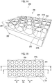
- Fig. 1A is a perspective view showing the structure of the porous film
- Fig. 1B is a plan view seen from the opening surface side in Fig. 1A
- Fig. 1C is a cross-sectional view taken along a line c-c in Fig. 1B
- Fig. 1D is a cross-sectional view taken along a line d-d in Fig. 1B
- Figs. 1A to 1D show a porous film 200 which is an example of the present embodiment.
- the porous film 200 is a laminate comprising a porous layer 204 in which a plurality of opening pores 210 are arranged in the plane direction and a support 202 for supporting the porous layer 204.
- the porous film 200 may not be provided with the support 202 and may be a monolayer structure of only the porous layer 204.
- the opening pores 210 are arranged over an entire area in a plane direction. However, in the case where there is a region where the cells cannot come into contact with the porous layer 204, the opening pores 210 may not be arranged in the region.
- An opening surface 206 of the porous film 200 is the surface on the side where the plurality of opening pores 210 are opened and is the surface on the side where the cells are seeded.
- the openings of opening pores 210 adjacent to one another are spaced apart by flat portions 208 extending among opening portions 210a.
- a liquid medium is supplied to the inside of the opening pores 210 and the communicating pores 212 through the openings of the opening pores 210 in the opening surface 206.
- Each of the opening pores 210 is a bottomed pore not penetrating the porous layer 204.
- each of the opening pores 210 may be a bottomed pore penetrating the porous layer 204 and having the surface of the support 202 as a bottom surface.
- each of the opening pores 210 may be a bottomed pore that does not penetrate the porous layer 204 or may be a through-pore penetrating the porous layer 204.
- each of the opening pores 210 for example, a spherical shape obtained by cutting a part of a sphere, a barrel shape, a cylindrical shape, or a prismatic shape can be mentioned.
- a shape of the opening of each opening pore 210 for example, a circle, an ellipse, or a polygon can be mentioned.
- the plurality of opening pores 210 are regularly arranged, specifically, they are arranged in a honeycomb shape. As another example, the plurality of opening pores 210 may be arranged in a lattice shape or a face-centered lattice shape.
- the arrangement of the plurality of opening pores 210 may be random, but the plurality of opening pores 210 are preferably regularly arranged from the viewpoint of making the density of the opening pores 210 uniform in the plane direction and therefore enhancing the homogeneity of the tissue formed by the cells in the porous film 200. Regular arrangement may be discontinuous or misaligned, but it is preferred that the arrangement is repeated continuously without gaps in all directions.
- the honeycomb-like arrangement refers to an arrangement in which a parallelepiped shape (preferably a regular hexagon) or a shape close thereto is used as a unit and the center of gravity of the opening is located at the apexes and the intersection of diagonal lines of these figures.
- the lattice-like arrangement refers to an arrangement in which a parallelogram shape (needless to say, including square, rectangle, and rhomboid, preferably square) or a shape close thereto is used as a unit and the center of gravity of the opening is located at the apexes of these figures.
- the face-centered lattice-like arrangement refers to an arrangement in which a parallelogram shape (needless to say, including square, rectangle, and rhomboid, preferably square) or a shape close thereto is used as a unit and the center of gravity of the opening is located at the apexes and the intersection of diagonal lines of these figures.
- a parallelogram shape needleless to say, including square, rectangle, and rhomboid, preferably square
- the center of gravity of the opening is located at the apexes and the intersection of diagonal lines of these figures.
- the mutually adjacent opening pores 210 communicate with one another through the communicating pores 212 inside the porous layer 204. It is preferred that one opening pore 210 communicates with all adjacent opening pores 210. In the porous film 200 in which the plurality of opening pores 210 are arranged in a honeycomb shape, it is preferred that one opening pore 210 communicates with six adjacent opening pores 210, that is, it is preferred that one opening pore 210 preferably has six communicating pores 212.
- the mutually adjacent opening pores 210 are arranged such that a part of each wall surface is continuous, and the communicating pore 212 is a pore surrounded by a connecting portion 210b between the opening pores 210.
- the adjacent opening pores 210 are independent from one another, and the communicating pore 212 which is a cylindrical gap may connect the adjacent opening pores 210 to one another.
- the plurality of opening pores 210 have such a size that the cells seeded on the porous film 200 can enter and engraft.
- the major axis of the cells seeded on the porous film 200 is, for example, 10 ⁇ m to 50 ⁇ m.
- the opening diameter Da of the opening pore 210 is preferably 50% or more, more preferably 80% or more, and still more preferably 120% or more, with respect to the major axis of the cell seeded on the porous film 200.
- the opening diameter Da is 50% or more (more preferably 80% or more, and still more preferably 120% or more)
- the cells seeded on the porous film 200 can enter the inside of the opening pore 210.
- the opening diameter Da of the opening pore 210 is preferably 5 ⁇ m or more, more preferably 8 ⁇ m or more, and still more preferably 12 ⁇ m or more.
- the opening diameter Da of the opening pore 210 is preferably 90 ⁇ m or less, more preferably 70 ⁇ m or less, and still more preferably 50 ⁇ m or less, depending on the relationship with the preferable arrangement and size of the opening pore 210.
- the opening diameter Da is the major axis of the opening of the opening pore 210, and the range of the opening diameter Da is confirmed by measuring the opening diameters of 10 or more selected openings.
- the variation coefficient of the opening diameter Da of the opening pore 210 is preferably 20% or less, and is preferably as small as possible. The smaller the variation coefficient of the opening diameter Da, the higher the homogeneity of the tissue formed by the cells in the porous film 200.
- the ratio of the total area of the openings of the opening pores 210 to the total area of the opening surface 206 is preferably 20% to 90%, more preferably 30% to 80%, and still more preferably 40% to 70%.
- the major axis Db in the plane direction of the opening pore 210 is preferably in the range of 10 ⁇ m to 100 ⁇ m, more preferably in the range of 20 ⁇ m to 80 ⁇ m, and still more preferably in the range of 30 ⁇ m to 60 ⁇ m.
- the major axis Db is 10 ⁇ m or more (more preferably 20 ⁇ m or more, and still more preferably 30 ⁇ m or more)
- a space for the proliferation of proliferating cells is secured and a space in which a plurality of types of co-cultured cells coexist is secured.
- the major axis Db is 100 ⁇ m or less (more preferably 80 ⁇ m or less, and still more preferably 60 ⁇ m or less), it is possible to form a micro-order network structure.
- the major axis Db is a major axis in the plane direction of the contour of the opening pore 210 appearing on the cut surface obtained by cutting the opening pore 210 on the major axis of the opening (that is, on the opening diameter Da) and in the thickness direction.
- a center-to-center distance PI between the mutually adjacent openings is regarded as the major axis Db.
- the range of the major axis Db is confirmed by measuring the center-to-center distance PI in total of 10 or more in three directions whose angles are shifted by approximately 60 degrees (including 60 degrees).
- the range of the major axis Db is confirmed according to the regularity of the arrangement of the opening pores 210 in accordance with the above.
- a depth Dc of the opening pore 210 is preferably in the range of 10 ⁇ m to 100 ⁇ m, more preferably in the range of 20 ⁇ m to 80 ⁇ m, and still more preferably in the range of 30 ⁇ m to 60 ⁇ m.
- the depth Dc of the opening pore 210 is 10 ⁇ m or more (more preferably 20 ⁇ m or more, and still more preferably 30 ⁇ m or more)
- a space for the proliferation of proliferating cells is secured and a space in which a plurality of types of co-cultured cells coexist is secured.
- the depth Dc of the opening pore 210 is 100 ⁇ m or less (more preferably 80 ⁇ m or less, and still more preferably 60 ⁇ m or less), it is possible to form a micro-order network structure.
- the depth Dc is a distance from the opening surface 206 to the deepest portion of the opening pore 210 in each of the opening pores 210.
- the distance from the opening surface 206 to the deepest portion of the opening pore 210 in the contour of the opening pore 210 appearing on the cut surface in the thickness direction on the line connecting the centers of the openings is defined as the depth Dc.
- the range of the depth Dc is confirmed by forming three cut surfaces in which the angle of the cutting line is shifted by approximately 60 degrees (including 60 degrees), and measuring the distance from the opening surface 206 to the deepest portion of the opening pore 210 in total of 10 or more.
- a pore diameter Dd of the communicating pore 212 is preferably in the range of 50% to 500% of the major axis of the cell seeded on the porous film 200.
- the pore diameter Dd of the communicating pore 212 is 50% or more of the major axis of the seeded cell, the cells can move among the mutually adjacent opening pores 210.
- the pore diameter Dd of the communicating pore 212 is more preferably 80% or more and still more preferably 120% or more of the major axis of the seeded cell.
- the pore diameter Dd of the communicating pore 212 is 500% or less of the major axis of the seeded cell
- the number of cells engrafting into the communicating pore 212 becomes appropriate.
- the pore diameter Dd of the communicating pore 212 is more preferably 400% or less and more preferably 300% or less of the major axis of the seeded cell.
- the pore diameter Dd of the communicating pore 212 is preferably in the range of 5 ⁇ m to 50 ⁇ m, more preferably in the range of 8 ⁇ m to 45 ⁇ m, and still more preferably in the range of 12 ⁇ m to 40 ⁇ m.
- the pore diameter Dd is a major axis in the thickness direction in the contour of the communicating pore 212 appearing on the cut surface in the thickness direction on the line connecting the centers of the openings.
- the range of the pore diameter Dd is confirmed by forming three cut surfaces in which the angle of the cutting line is shifted by approximately 60 degrees (including 60 degrees), and measuring the pore diameter Dd in total of 10 or more.
- the range of the pore diameter Dd is confirmed by forming cut surfaces according to the regularity of the arrangement of the opening pores 210 in accordance with the above.
- the variation coefficient of the pore diameter Dd of the communicating pore 212 is preferably 30% or less and more preferably 20% or less, and is preferably as small as possible.
- the communicating pores 212 are provided at substantially the same depth (including the same depth) over an entire area in a plane direction. Thereby, the homogeneity of the tissue formed by the cells in the porous film 200 is secured. Roughly the same guideline is that the variation coefficient of the distance from the opening surface 206 to the deepest portion of the communicating pore 212 on the cut surface for measuring the pore diameter Dd is 10% or less.
- a width W of the flat portion 208 is preferably narrower than the major axis of the cell. This reduces the proportion of cells cultured on the flat portion 208 and increases the proportion of cells cultured inside the opening pores 210 and the communicating pores 212.
- the width W is a length of the flat portion 208 which is measured between the centers of the openings.
- the material of the porous film 200 is selected from the viewpoint of cell adhesiveness, affinity with a transplant site in a living body, difficulty in decomposition or absorption in a living body, and the like.
- a hydrophobic polymer soluble in a hydrophobic organic solvent is preferable from the viewpoint of producing the porous film 200 by a production method which will be described later.
- the hydrophobic organic solvent is a liquid having a solubility in water at 25°C of 10 (g/100 g water) or less.
- hydrophobic polymer examples include polymers such as polystyrene, polyacrylate, polymethacrylate, polyacrylamide, polymethacrylamide, polyvinyl chloride, polyvinylidene chloride, polyvinylidene fluoride, polyhexafluoropropane, polyvinyl ether, polyvinyl carbazole, polyvinyl acetate, polytetrafluoroethylene, polyester (for example, polyethylene terephthalate, polyethylene naphthalate, polyethylene succinate, polybutylene succinate, polylactic acid, or poly-3-hydroxybutyrate), polylactone (for example, polycaprolactone), polyamide or polyimide (for example, nylon or polyamide acid), polyurethane, polyurea, polybutadiene, polycarbonate, polyaromatics, polysulfone, polyethersulfone, polysiloxane derivatives, and cellulose acylate (for example, triacetyl cellulose, cellulose acetate propy
- These polymers may be homopolymers, copolymers, polymer blends, or polymer alloys, if necessary, from the viewpoints of solubility in solvents, optical properties, electrical properties, film hardness, elasticity, and the like. These polymers may be used alone or in admixture of two or more thereof. From the viewpoint of bioabsorbability, polylactic acid, polycaprolactone, poly-3-hydroxybutyrate, and the like are preferable.
- the material of the support 202 may be the same as or different from that of the porous layer 204.
- Examples of the material of the support 202 include synthetic resin, glass, and metal.
- the material of the support 202 is preferably a material different from that of the porous layer 204.
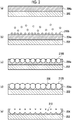
- the porous film 200 is produced by, for example, a production method in which the following steps (a) to (e) are carried out in order.
- Fig. 2 is a schematic view showing a cross section of the porous film 200 in steps (a) to (e).
- a coating liquid prepared by dissolving a hydrophobic polymer constituting the porous layer 204 in a solvent is prepared and the coating liquid is applied onto the support 202 to form a coating film 204a on the support 202.
- the coating liquid is prepared by mixing a hydrophobic polymer, a solvent, and an amphiphilic compound.
- the solvent is preferably a mixed solvent of a good solvent for a hydrophobic polymer and a hydrophobic liquid, or a hydrophobic organic solvent which is a good solvent for a hydrophobic polymer.
- examples of the latter include halogen-based solvents such as trichloromethane, dichloromethane, and chloroform, in which these solvents may be used in admixture with hydrocarbon compounds such as n-hexane, cyclohexane, n-pentane, n-octane, and n-heptane which are hydrophobic liquids.
- the amphiphilic compound is a compound having both a hydrophilic group and a hydrophobic group. In the case where the amphiphilic compound is blended in a coating liquid, it is easy to form water droplets on the surface of the coating film. Further, by controlling the dispersion state of the hydrophobic polymer with respect to the solvent with the amphiphilic compound, it is possible to more easily control the growth of water droplets.
- the amphiphilic compound include many sorts of commercially available surfactants, oligomers such as dimers and trimers, and high molecular weight compounds such as polymers.
- amphiphilic compound examples include compounds having a polyacrylate skeleton as a main chain and having a long chain aliphatic group (for example, a dodecyl group) as a lipophilic side chain and a carboxyl group as a hydrophilic side chain; polyethylene glycol/polypropylene glycol block copolymers; and phospholipids.
- the concentration of the hydrophobic polymer is preferably 0.1% by mass to 10% by mass, and the concentration of the amphiphilic compound is preferably 0.01% by mass to 1% by mass.
- Humidified air is allowed to flow over the entire surface of the coating film 204a to form water droplets 210S on the coating film 204a due to dew condensation.
- Water droplets 210S are grown by continuing the supply of humidified air. Each of the water droplets 210S grows to substantially the same size (including the same size). With the evaporation of the solvent contained in the coating film 204a, the water droplets 210S are arranged in a honeycomb shape by the transverse capillary force and enter the coating film 204a. In parallel with this, as the solvent contained in the coating film 204a evaporates, the hydrophobic polymer precipitates around the water droplets 210S.
- the supply of the humidified air is continued to grow the water droplets 210S until the water droplets 210S are brought into close contact with one another with amphiphilic compound films 220 interposed therebetween in the coating film 204a.
- the water droplets 210S are evaporated so that the portion where the water droplets 210S enter the coating film 204a is left as the opening pore 210, and in parallel, the amphiphilic compound films 220 that separate the closely adjacent water droplets 210S are destroyed. This results in the formation of the porous layer 204 in which a plurality of opening pores 210 are arranged in a honeycomb shape and connected inside.
- a laminate of the porous layer 204 and the support 202 is obtained.
- the laminate of the porous layer 204 and the support 202 may be used as the porous film 200; the porous layer 204 may be peeled from the support 202, and then the monolayer structure of the porous layer 204 may be used as the porous film 200; or the porous layer 204 may be peeled from the support 202 and attached to another support to form the porous film 200.
- a charge treatment by plasma discharge or the like may be carried out on the surface of the porous film 200 on which the cells are seeded.
- Fig. 3 is a micrograph of an example of a porous film produced through the steps (a) to (e) taken from the side of an opening surface using a scanning electron microscope.
- the opening diameter Da, the major axis Db, and the depth Dc of the opening pore 210 and the pore diameter Dd of the communicating pore 212 can be controlled by adjusting the size of the water droplets 210S.
- the size of the water droplets 210S can be adjusted by the film thickness of the coating film 204a, the amount of water vapor contained in the humidified air, and the timing at which the water droplets 210S are evaporated.
- the above-mentioned production method it is possible to produce a porous film in which a plurality of opening pores are arranged with high uniformity in a honeycomb shape without using a mold or a mask.
- the details of the above-mentioned production method are described in, for example, JP2007-291367A , JP2009-256624A , JP2011-074140A , and JP2011-202100A .
- the porous film 200 can also be produced by etching, sandblasting, press molding, or the like.
- the method for producing a cell tissue of the present embodiment is a production method for producing a cell tissue ex vivo and includes a culturing step of culturing the cells inside the opening pores and the communicating pores of the porous film of the present disclosure.
- the opening pores and the communicating pores of the porous film are collectively referred to as "pores".
- a cell tissue comprising a micro-order network structure is formed since the pores of the porous film of the present disclosure act as a trajectory in which cells are arranged in a micro-order network shape.
- the cell tissue comprising a micro-order network structure is useful as a graft material in vivo and as a test cell tissue replacing an animal experiment or a clinical test.
- the cell tissue may be used per porous film or may be used by removing the flat portion around the opening of the porous film and taking it out of the pore of the porous film.
- the cell tissue may also be used in the form of a laminate of a plurality of cell tissues.
- the culturing step of the present embodiment includes the following embodiments (1) to (4).
- Embodiment (1) a culturing step of culturing cells capable of serving as a feeder alone.
- cells capable of serving as a feeder may be of one type or two or more types.
- a cell tissue comprising a network structure of feeder cells.
- the cell tissue produced according to the embodiment (1) is used, for example, as a scaffold in the case where cell culture is carried out ex vivo, or as a scaffold which is transplanted into a living body to regenerate tissues at a transplant site.
- Examples of cells capable of serving as a feeder include mesenchymal stem cells and fibroblasts.
- the mesenchymal stem cells are somatic stem cells that can differentiate into myocytes, adipocytes, chondrocytes, or the like, and are also known to serve as a feeder. It is also possible to differentiate mesenchymal stem cells into somatic cells (for example, myocytes, adipocytes, or chondrocytes) by adding a differentiation-inducing factor for mesenchymal stem cells to a medium in the culturing step.
- a muscle tissue, adipose tissue, or cartilage tissue comprising a micro-order network structure is provided.
- Embodiment (2) a culturing step of co-culturing cells capable of serving as a feeder and at least one of vascular endothelial cells or lymphatic endothelial cells.
- a cell tissue comprising a network structure of vascular endothelial cells or lymphatic endothelial cells is provided.
- the cell tissue produced according to the embodiment (2) is used, for example, as a scaffold in the case where culture of somatic cells is carried out ex vivo, or as a scaffold which is transplanted into a living body to regenerate tissues at a transplant site.
- vascular endothelial cells examples include vascular endothelial cells derived from umbilical cord vein, umbilical cord artery, aorta, coronary artery, pulmonary artery, pulmonary microvessel, dermal microvessel, and the like, and vascular endothelial cells differentiated from pluripotent stem cells.
- lymphatic endothelial cells examples include lymphatic endothelial cells derived from dermal micro-lymphatic vessels, pulmonary micro-lymphatic vessels, and the like, and lymphatic endothelial cells differentiated from pluripotent stem cells. It is possible to control proliferation, migration, and tube formation of vascular endothelial cells or lymphatic endothelial cells by changing the composition of the medium in the culturing step.
- Embodiment (3) a culturing step of co-culturing cells capable of serving as a feeder and cells forming a parenchymal organ.
- a cell tissue comprising a micro-order network structure.
- the cell tissue produced according to the embodiment (3) is used, for example, for supplementing the function of a tissue or an organ by being transplanted into a living body.
- the cell tissue produced according to the embodiment (3) is used as a test cell tissue replacing an animal experiment or a clinical test.
- parenchymal organ examples include liver, pancreas, kidney, spleen, heart, lung, mammary gland tissue, adipose tissue, ovary, testis, and thymus
- examples of the cells forming a parenchymal organ include parenchymal cells forming those organs, epithelial cells, glandular cells, adipocytes, and stem cells or progenitor cells of these cells.
- the cells forming a parenchymal organ may be used alone or in combination of two or more thereof.
- Examples of combinations of two or more types of cells forming a parenchymal organ include hepatocytes and bile duct epithelial cells; pancreatic ⁇ cells, pancreatic ⁇ cells, and pancreatic ⁇ cells; type I alveolar epithelial cells and type II alveolar epithelial cells; and mammary alveolar cells and mammary ductal epithelial cells.
- the cells forming a parenchymal organ also include pluripotent stem cells.
- Pluripotent stem cells are undifferentiated cells having "self-renewal ability" capable of growing while maintaining an undifferentiated state and "pluripotency” capable of differentiating into all three germ layer series.
- Examples of pluripotent stem cells include embryonic stem cells (ES cells), induced pluripotent stem cells (iPS cells), embryonic germ cells (EG cells), embryonal carcinoma cells (EC cells), multipotent adult progenitor cells (MAP cells), an adult pluripotent stem cells (APS cells), and Muse cells (multi-lineage differentiating stress enduring cells).
- a differentiation-inducing factor which induces differentiation into somatic cells of interest is added to a medium to differentiate pluripotent stem cells into somatic cells.
- Embodiment (4) a culturing step of co-culturing cells capable of serving as a feeder, cells forming a parenchymal organ, and at least one of vascular endothelial cells or lymphatic endothelial cells.
- a cell tissue comprising a network structure of vascular endothelial cells or lymphatic endothelial cells is provided on the surface or inside of the cell tissue of the parenchymal organ.
- the cell tissue produced according to the embodiment (4) is used, for example, for supplementing the function of a tissue or an organ by being transplanted into a living body.
- the cell tissue produced according to the embodiment (4) is used as a test cell tissue replacing an animal experiment or a clinical test.
- the culturing step of the embodiment (4) may be a culturing step in which three types of cells are seeded at the same time and the three types of cells are simultaneously co-cultured or may be a culturing step in which three types of cells are seeded stepwise and the three types of cells are co-cultured stepwise.
- cells capable of serving as a feeder and at least one of vascular endothelial cells or lymphatic endothelial cells are seeded at the same time and cultured for several days to form a network structure of vascular endothelial cells or lymphatic endothelial cells, cells forming a parenchymal organ are then seeded, and culture is continued.
- the culturing step is carried out, for example, by placing the porous film in a culture vessel with the side on which a plurality of opening pores are opened facing upward, seeding a cell suspension on the porous film, and culturing the cells under the culture conditions adapted to the seeded cell type.
- the culture vessel on which the porous film is placed may be any culture vessel known for cell culture.
- the material of the culture vessel include a resin (for example, polystyrene, polycarbonate, polyester, or acrylonitrile-butadiene-styrene resin), glass, ceramics, and metal.
- the shape of the culture vessel include a multi-well plate, a Petri dish, a microarray plate, a tube, a flask, a roller bottle, and a bag.
- the present embodiment preferably includes, prior to the culturing step, a centrifugation step of seeding the cells on the surface of the porous film on the side where the plurality of opening pores are opened, and then applying a centrifugal force in the direction from the surface seeded with the cells to the opposite surface to move the cells to the inside of the plurality of opening pores. Since the cells seeded on the porous film migrate into the opening pores of the porous film by the centrifugation step, the proportion of the cells cultured in the pores increases.
- the centrifugation step it is preferable to seed a cell suspension in which the cells are suspended in a relatively small amount of medium and carry out the centrifugation step. Then, the medium is added into the culture vessel after the centrifugation step.
- the medium used in the present embodiment is selected from known media used for culturing mammalian cells according to the type of cells to be cultured.
- a specific example of the medium may be, for example, a medium optimized for the cell type by adding a cell growth factor to a basic medium such as Dulbecco's Modified Eagle's Medium (DMEM), DMEM: F-12 (Dulbecco's Modified Eagle Medium: Nutrient Mixture F-12), Eagle's minimal essential medium (EMEM), Minimum Essential Medium Alpha (MEM ⁇ ), or Basal Medium Eagle (BME).
- DMEM Dulbecco's Modified Eagle's Medium
- F-12 Dulbecco's Modified Eagle Medium: Nutrient Mixture F-12
- EMEM Eagle's minimal essential medium
- MEM ⁇ Minimum Essential Medium Alpha
- Basal Medium Eagle Basal Medium Eagle
- the medium used in the present embodiment may be a medium in which a plurality of media are mixed, depending on the type of cells to be co-cultured.
- cell growth factors such as fibroblast growth factor-2 (FGF-2), transforming growth factor- ⁇ (TGF- ⁇ ), epidermal growth factor (EGF), and vascular endothelial growth factor (VEGF); vitamin or vitamin derivatives such as ascorbic acid and retinoic acid; sugar sources such as glucose; amino acids; inorganic salts such as sodium selenite and sodium chloride; proteins such as transferrin; hormones such as insulin; differentiation-inhibiting factor; differentiation-inducing factor; antioxidants such as 2-mercaptoethanol and dithiothreitol; and antibiotics such as penicillin and streptomycin, may be added to the medium.
- FGF-2 fibroblast growth factor-2
- TGF- ⁇ transforming growth factor- ⁇
- EGF epidermal growth factor
- VEGF vascular endothelial growth factor
- vitamin or vitamin derivatives such as ascorbic acid and retinoic acid
- sugar sources such as glucose
- amino acids such as sodium selenite and sodium chloride
- proteins such as transfer
- the ingredients may be supplemented to the medium during cell culture in order to keep the concentration thereof within the desired range throughout the culture period. From the viewpoint of suppressing incorporation of antigenic substances, infectious sources, and the like into cell tissues, it is preferred that the medium does not contain serum and serum substitutes.
- the medium used for the series of culturing steps may not have the same composition. Culturing may be continued while replacing the medium with a medium having a different composition depending on the cell type to be co-cultured.
- the mixing ratio of first cells:second cells is, for example, 1:0.5 to 1:5 in terms of number of first cells:number of second cells, and preferably 1:1 to 1:3 in terms of number of first cells:number of second cells.
- the mixing ratio of those three types of cells is, for example, 1:1 to 1:10 in terms of (number of first cells+number of second cells):number of third cells, and preferably 1:2 to 1:5 in terms of (number of first cells+number of second cells):number of third cells.
- the mixing ratio of first cells:second cells is preferably within the above range.
- the seeding density of the cells with respect to the porous film is, for example, 1 ⁇ 10 3 cells/cm 2 to 1 ⁇ 10 6 cells/cm 2 , and preferably 1 ⁇ 10 4 cells/cm 2 to 1 ⁇ 10 6 cells/cm 2 . In the case where a plurality of types of cells are co-cultured, it is preferable to set the total seeding density of the cells within the above range.
- General conditions may be applied to the culture conditions in the present embodiment.
- culture in an incubator at 37°C and 5% (v/v) CO 2 concentration is applied.
- the culture period is not particularly limited, and culturing is preferably carried out for a period of time during which the network structure of the cells is sufficiently developed.
- PET means polyethylene terephthalate
- PMMA means polymethyl methacrylate
- PDMS means polydimethylsiloxane
- SEM means a scanning electron microscope
- FBS means fetal bovine serum.
- pores the opening pores and the communicating pores of the porous film are collectively referred to as "pores".
- a coating liquid 5 parts by mass of polylactic acid, 94.5 parts by mass of trichloromethane, and 0.5 parts by mass of a polyacrylamide-based amphiphilic polymer as an amphiphilic compound were mixed to prepare a coating liquid.
- the coating liquid was applied on a PET film (film thickness: 188 ⁇ m) to form a coating film on the PET film.
- Humidified air was allowed to flow over the entire surface of the coating film, and water droplets were formed on the coating film due to dew condensation. While continuing to supply the humidified air to grow the water droplets, the water droplets were made to enter the coating film.
- Polylactic acid was precipitated around the water droplets by evaporation of the solvent contained in the coating film while growing water droplets until the water droplets were closely in contact with one another with the amphiphilic compound film interposed therebetween in the coating film. Then, the supply of the humidified air was stopped, and the temperature of the atmosphere was raised to evaporate the water droplets so that the portions where the water droplets have entered the coating film were left as opening pores, whereby a porous film comprising a porous layer formed of polylactic acid having a plurality of opening pores arranged in a honeycomb shape and having a communicating structure and a PET film layer was obtained.
- the porous film was cut into a circle having a diameter of 23 mm, and a ring (outer diameter: 22 mm, inner diameter: 16 mm, height: 8 mm, made of PMMA, PDMS coated on the side) was attached to the outer periphery thereof using a double-sided adhesive tape (3M product number 1510, adhesive tape for skin) to prepare a ring-attached porous film.
- the ring-attached porous film was immersed in a 70% (v/v) ethanol aqueous solution for a few minutes for the purpose of sterilization and then washed with sterile water to remove ethanol.
- Fig. 4 shows an example of a porous film, a ring, and a culture device.
- the ring-attached porous film was placed on a dish for tissue culture so that the PET film layer side of the porous film faced downward to obtain a culture device.
- the culture device was irradiated with ultraviolet rays for 2 hours.
- the dimensions of the structure of the porous film of each culture device are shown in Table 1.
- Fig. 5A is an SEM image of the cross section and the surface of the porous film comprised in PLA10
- Fig. 5B is a SEM image of the cross section and the surface of the porous film comprised in PLA40.
- openings of adjacent opening pores were spaced apart by flat portions, and parts of individual wall surfaces were continuous and communicated with one another among the adjacent opening pores.
- the opening pores arranged in the porous layer formed of polylactic acid did not penetrate the porous layer.
- a device (referred to as "Flat”) comprising a laminate obtained by laminating an opening pore-free flat layer formed of polylactic acid on a PET film in place of a porous film, and a device (referred to as "TCP") with a ring attached to the culture surface of a dish for tissue culture (Tissue culture polystyrene: TCPS, diameter: 35 mm, trade name: Falcon Cell Culture Dish 353001, manufactured by Corning Incorporated) were prepared.
- the bottom surface inside the ring is the culture surface for culturing the cells, and the area of the bottom surface surrounded by the ring is 2 cm 2 .
- MSCs were cultured alone under the following culture conditions using TCP, Flat, PLA3, PLA10, and PLA30 as culture devices.
- the culture was started using the MSC medium.
- the medium On Day 1 of culture, the medium was exchanged with a differentiation-inducing medium in which the osteogenic differentiation-inducing medium and the adipogenic differentiation-inducing medium were mixed in a ratio of 1:1.
- the differentiation-inducing medium was exchanged every 3 days, and the cells were cultured for a total of 15 days.
- the expression of osteogenic differentiation markers Runx2 and Osx, and adipogenic differentiation markers C/EBP ⁇ and PPAR ⁇ was analyzed by real-time polymerase chain reaction (PCR).
- fluorescent immunostaining of actin was carried out on Day 14 of induction of differentiation.
- Fig. 6 shows a fluorescent immunostaining image of actin on Day 14 of induction of differentiation.
- HUVECs were cultured alone, or MSCs and HUVECs were co-cultured.
- the total of the 10 regions was calculated and the calculated value was taken as the total length of the network formed by HUVECs.
- the areas of the fluorescent regions scattered throughout the binarized image were individually measured and the areas were summed up.
- the total of the 10 regions was calculated and the calculated value was taken as the total area of the network formed by HUVECs.
- Figs. 7A to 7D The analysis results of confocal laser microscope images and networks are shown in Figs. 7A to 7D and Table 2.
- "Mono-” means monoculture
- "Co-” means co-culture.
- an image attached with "cfg” is a culture example subjected to centrifugation
- an image without "cfg” is a culture example subjected to no centrifugation.
- MSCs and HUVECs were co-cultured, or NHSF46 and HUVECs were co-cultured.
- Fluorescent immunostaining of CD31 was carried out on Days 3 and 7 of culture and observation was carried out using a confocal laser microscope. The analysis results of the microscope image and the network are shown in Figs. 8A to 8D and Table 3.
- MSCs and HUVECs were co-cultured under the following culture conditions using TCP, Flat, PLA3, PLA10, PLA20, and PLA40 as culture devices.
- Fluorescent immunostaining of CD31 was carried out on Days 3 and 7 of culture and observation was carried out using a confocal laser microscope. The analysis results of the microscope image and the network are shown in Figs. 9A to 9E and Table 4.
- MSCs, HUVECs, and HepG2 were co-cultured under the following culture conditions using TCP and PLA40 as culture devices.
- Figs. 10A to 10B show microscope images.
- MSCs, HUVECs, and HepG2 were co-cultured under the following culture conditions using TCP and PLA40 as culture devices.
- Fluorescent immunostaining of CD31 was carried out on Day 3 after seeding HepG2, and the network formed by the cells was analyzed from the fluorescence image of CD31 and the fluorescence image of HepG2.
- Figs. 11A to 11B show microscope images.
- Fig. 11C and Table 5 show the total length and total area of the network obtained by analyzing the fluorescence image of CD31.
- JP 2016-188627 filed on September 27, 2016 is hereby incorporated by reference in its entirety.
Landscapes
- Health & Medical Sciences (AREA)
- Engineering & Computer Science (AREA)
- Life Sciences & Earth Sciences (AREA)
- Chemical & Material Sciences (AREA)
- Biomedical Technology (AREA)
- Organic Chemistry (AREA)
- Bioinformatics & Cheminformatics (AREA)
- Wood Science & Technology (AREA)
- Zoology (AREA)
- Genetics & Genomics (AREA)
- Biotechnology (AREA)
- General Health & Medical Sciences (AREA)
- Biochemistry (AREA)
- General Engineering & Computer Science (AREA)
- Microbiology (AREA)
- Cell Biology (AREA)
- Immunology (AREA)
- Sustainable Development (AREA)
- Rheumatology (AREA)
- Developmental Biology & Embryology (AREA)
- Molecular Biology (AREA)
- Medicinal Chemistry (AREA)
- Hematology (AREA)
- Vascular Medicine (AREA)
- Polymers & Plastics (AREA)
- Chemical Kinetics & Catalysis (AREA)
- Materials Engineering (AREA)
- General Chemical & Material Sciences (AREA)
- Physiology (AREA)
- Transplantation (AREA)
- Epidemiology (AREA)
- Animal Behavior & Ethology (AREA)
- Public Health (AREA)
- Veterinary Medicine (AREA)
- Oral & Maxillofacial Surgery (AREA)
- Dermatology (AREA)
- Dispersion Chemistry (AREA)
- Micro-Organisms Or Cultivation Processes Thereof (AREA)
- Apparatus Associated With Microorganisms And Enzymes (AREA)
- Manufacture Of Porous Articles, And Recovery And Treatment Of Waste Products (AREA)
Abstract
Description
- The present disclosure relates to a method for producing a cell tissue and a porous film to be used for the production of a cell tissue.
- A filter for enriching cells, a scaffold for growing cells, and a graft material for engraftment of cells and transplantation thereof into a living body, which are used for the purpose of transplanting cells or cell tissues into a living body, are known.
- For example,
JP2012-006010A WO2006/093207A discloses a culture substrate in which pores are arranged in a honeycomb shape on the cell culture surface. For example,JP2005-110709A JP2004-216119A JP2008-199962A JP2001-523483A JP2008-307180A JP4437227B - Currently, a cell tissue with a relatively simple structure such as skin or cartilage is artificially created and is used in the field of medical transplantation. On the other hand, practical application of an artificial cell tissue substituting an organ in which various cells form a complicated structure, such as liver or pancreas, is expected, but for this purpose, it is necessary to prepare a cell tissue having organ-specific cells functionally arranged and comprising a vascular network and a lymphatic network. In addition, as a test cell tissue replacing an animal experiment or a clinical test, a cell tissue having a structure resembling an organ in a living body is awaited.
- The present disclosure was made under the foregoing circumstances.
- An object of the present disclosure is to provide a production method for producing a cell tissue comprising a micro-order network structure, and a porous film to be used for the production of a cell tissue comprising a micro-order network structure.
- Specific means for achieving the foregoing object includes the following aspects.
- [1] A method for producing a cell tissue, comprising a culturing step of culturing cells capable of serving as a feeder inside opening pores and communicating pores of a porous film having a plurality of the opening pores provided on a surface thereof and the communicating pores communicating mutually adjacent opening pores with one another.
- [2] The method for producing a cell tissue according to [1], in which the culturing step is a culturing step of co-culturing the cells capable of serving as a feeder and at least one of vascular endothelial cells or lymphatic endothelial cells inside the opening pores and the communicating pores.
- [3] The method for producing a cell tissue according to [1], in which the culturing step is a culturing step of co-culturing the cells capable of serving as a feeder and cells forming a parenchymal organ inside the opening pores and the communicating pores.
- [4] The method for producing a cell tissue according to [1], in which the culturing step is a culturing step of co-culturing the cells capable of serving as a feeder, cells forming a parenchymal organ, and at least one of vascular endothelial cells or lymphatic endothelial cells inside the opening pores and the communicating pores.
- [5] The method for producing a cell tissue according to any one of [1] to [4], in which the cells capable of serving as a feeder are at least one of mesenchymal stem cells or fibroblasts.
- [6] The method for producing a cell tissue according to any one of [1] to [5], in which the plurality of opening pores are arranged in a honeycomb shape on the surface of the porous film.
- [7] The method for producing a cell tissue according to any one of [1] to [6], in which the communicating pores are provided at substantially the same depth over an entire area of the porous film in a plane direction.
- [8] The method for producing a cell tissue according to any one of [1] to [7], in which a pore diameter of the communicating pore is in a range of 50% to 500% of a major axis of the cell seeded on the porous film.
- [9] The method for producing a cell tissue according to any one of [1] to [8], in which a pore diameter of the communicating pore is in a range of 5 µm to 50 µm.
- [10] The method for producing a cell tissue according to any one of [1] to [9], in which a variation coefficient of a pore diameter of the communicating pore is 30% or less.
- [11] The method for producing a cell tissue according to any one of [1] to [10], in which a major axis of the opening pore in a plane direction of the porous film is in a range of 10 µm to 100 µm.
- [12] The method for producing a cell tissue according to any one of [1] to [11], in which a depth of the opening pore is in a range of 10 µm to 100 µm.
- [13] The method for producing a cell tissue according to any one of [1] to [12], in which an opening diameter of the opening pore is in a range of 5 µm to 90 µm.
- [14] The method for producing a cell tissue according to any one of [1] to [13], in which a variation coefficient of an opening diameter of the opening pore is 20% or less.
- [15] The method for producing a cell tissue according to any one of [1] to [14], further comprising, prior to the culturing step, a centrifugation step of seeding the cells on a surface of the porous film on a side where the plurality of opening pores are opened, and then applying a centrifugal force in a direction from a surface seeded with the cells to an opposite surface to move the cells to the inside of the plurality of opening pores.
- [16] A porous film comprising a plurality of opening pores provided on a surface thereof and communicating pores communicating mutually adjacent opening pores with one another, in which a major axis of the opening pore in a plane direction of the porous film is in a range of 20 µm to 100 µm.
- [17] A porous film comprising a plurality of opening pores provided on a surface thereof and communicating pores communicating mutually adjacent opening pores with one another, in which a depth of the opening pore is in a range of 20 µm to 100 µm.
- [18] The porous film according to [16] or [17], in which an opening diameter of the opening pore is in a range of 5 µm to 90 µm.
- [19] The porous film according to any one of [16] to [18], in which a pore diameter of the communicating pore is in a range of 5 µm to 50 µm.
- [20] The porous film according to any one of [16] to [19], in which the plurality of opening pores are arranged in a honeycomb shape on the surface of the porous film.
- According to the present disclosure, there are provided a production method for producing a cell tissue comprising a micro-order network structure, and a porous film to be used for the production of a cell tissue comprising a micro-order network structure.
-
-
Fig. 1A is a perspective view showing a structure of a porous film. -
Fig. 1B is a plan view seen from the opening surface side inFig. 1A . -
Fig. 1C is a cross-sectional view taken along a line c-c inFig. 1B . -
Fig. 1D is a cross-sectional view taken along a line d-d inFig. 1B . -
Fig. 2 is a schematic view showing an example of a method for producing a porous film. -
Fig. 3 is a scanning electron microscope image of the porous film. -
Fig. 4 shows components of a culture device and an example of the culture device. -
Fig. 5A is a scanning electron microscope image of a cross section and a surface of the porous film. -
Fig. 5B is a scanning electron microscope image of the cross section and the surface of the porous film. -
Fig. 6 is a fluorescent immunostaining image of actin in cells differentiated from mesenchymal stem cells. -
Fig. 7A is a fluorescent immunostaining image (low magnification) of CD31 in cells onDay 3 of culture in which vascular endothelial cells are cultured alone or mesenchymal stem cells and vascular endothelial cells are co-cultured. -
Fig. 7B is a fluorescent immunostaining image (high magnification) of CD31 in cells onDay 3 of culture in the same manner. -
Fig. 7C is a fluorescent immunostaining image (high magnification) of CD31 in cells onDay 7 of culture in the same manner. -
Fig. 7D is a graph showing network total length and total area in co-culture of mesenchymal stem cells and vascular endothelial cells. -
Fig. 8A is a fluorescent immunostaining image of CD31 in cells onDay 3 of culture in which mesenchymal stem cells or fibroblasts and vascular endothelial cells are co-cultured. -
Fig. 8B is a fluorescent immunostaining image of CD31 in cells onDay 7 of culture in the same manner. -
Fig. 8C is a graph showing the network total length and total area onDay 3 of culture. -
Fig. 8D is a graph showing the network total length and total area onDay 7 of culture. -
Fig. 9A is a fluorescent immunostaining image (low magnification) of CD31 in cells onDay 3 of culture in which mesenchymal stem cells and vascular endothelial cells are co-cultured using porous films having different opening pore sizes. -
Fig. 9B is a fluorescent immunostaining image (high magnification) of CD31 in cells onDay 3 of culture in the same manner. -
Fig. 9C is a fluorescent immunostaining image (low magnification) of CD31 in cells onDay 7 of culture in the same manner. -
Fig. 9D is a fluorescent immunostaining image (high magnification) of CD31 in cells onDay 7 of culture in the same manner. -
Fig. 9E is a graph showing the network total length and total area onDays -
Fig. 10A is a fluorescence image onDay 3 of culture in which three types of cells are co-cultured at the same time. -
Fig. 10B is a fluorescent immunostaining image of CD31 in cells onDay 3 of culture in the same manner. -
Fig. 11A is a fluorescence image onDay 3 of co-culture in which three types of cells are co-cultured stepwise. -
Fig. 11B is a fluorescent immunostaining image of CD31 in cells onDay 3 of co-culture in the same manner. -
Fig. 11C is a graph showing the network total length and total area in each of simultaneous co-culture and stepwise co-culture onDay 3 of co-culture. - Hereinafter, embodiments of the present disclosure will be described. These descriptions and examples are illustrative of embodiments and do not limit the scope of the invention.
- The numerical range indicated by using "to" in the present disclosure indicates a range including numerical values described before and after "to" as a lower limit value and an upper limit value, respectively.
- The term "step" in the present disclosure includes not only an independent step but also a step in which the intended purpose of this step can be achieved even in the case where the step cannot be clearly distinguished from other steps.
- In the present disclosure, upon referring to an amount of each component in a composition, in the case where there are a plurality of substances corresponding to each component in the composition, the amount means a total amount of the plurality of substances present in the composition unless otherwise specified.
- In the present disclosure, the variation coefficient is expressed in terms of percentage. The variation coefficient is a value obtained by dividing the standard deviation by a mean value for a certain population and is an index showing the degree of variation of the population.
- The method for producing a cell tissue of the present embodiment is a method for producing a cell tissue ex vivo and includes a culturing step of culturing cells inside opening pores and communicating pores of a porous film having a plurality of the opening pores provided on a surface thereof and the communicating pores communicating mutually adjacent opening pores with one another. First, a porous film to be used for the production of a cell tissue will be described. The porous film of the present embodiment functions as a scaffold for cells to engraft and form tissues.
- The porous film of the present embodiment has a plurality of opening pores provided on the surface thereof and communicating pores communicating the adjacent opening pores with one another. The wall surfaces of the opening pores and the communicating pores provided in the porous film serve as scaffolds for cells to engraft. Cells seeded on the porous film of the present embodiment engraft within the opening pores and the communicating pores to form a cell tissue comprising a micro-order network structure.
- Hereinafter, an example of the porous film will be described with reference to the drawings. In the drawings, the same or equivalent constituent elements and parts are given the same reference numerals. In the following description, the term "plane direction" means a main plane direction of the porous film, and the term "thickness direction" means a thickness direction of the porous film. In the following description, "major axis" means a maximum length among any two-point distances on the contour, but in the case where the direction is specified, it means a maximum length among any two-point distances in that direction. In the following description, the term "center of the opening" means the center of gravity in the case where the opening is taken as a two-dimensional figure in the plane direction.
-
Fig. 1A is a perspective view showing the structure of the porous film,Fig. 1B is a plan view seen from the opening surface side inFig. 1A ,Fig. 1C is a cross-sectional view taken along a line c-c inFig. 1B , andFig. 1D is a cross-sectional view taken along a line d-d inFig. 1B .Figs. 1A to 1D show aporous film 200 which is an example of the present embodiment. - The
porous film 200 is a laminate comprising aporous layer 204 in which a plurality of openingpores 210 are arranged in the plane direction and asupport 202 for supporting theporous layer 204. As another example, theporous film 200 may not be provided with thesupport 202 and may be a monolayer structure of only theporous layer 204. - In the
porous layer 204 of theporous film 200, the opening pores 210 are arranged over an entire area in a plane direction. However, in the case where there is a region where the cells cannot come into contact with theporous layer 204, the opening pores 210 may not be arranged in the region. - An
opening surface 206 of theporous film 200 is the surface on the side where the plurality of openingpores 210 are opened and is the surface on the side where the cells are seeded. In theopening surface 206, the openings of openingpores 210 adjacent to one another are spaced apart byflat portions 208 extending among openingportions 210a. A liquid medium is supplied to the inside of the opening pores 210 and the communicatingpores 212 through the openings of the opening pores 210 in theopening surface 206. - Each of the opening pores 210 is a bottomed pore not penetrating the
porous layer 204. As another example, each of the opening pores 210 may be a bottomed pore penetrating theporous layer 204 and having the surface of thesupport 202 as a bottom surface. In the example where theporous film 200 is a monolayer structure of theporous layer 204, each of the opening pores 210 may be a bottomed pore that does not penetrate theporous layer 204 or may be a through-pore penetrating theporous layer 204. - As the shape of each of the opening pores 210, for example, a spherical shape obtained by cutting a part of a sphere, a barrel shape, a cylindrical shape, or a prismatic shape can be mentioned. As the shape of the opening of each
opening pore 210, for example, a circle, an ellipse, or a polygon can be mentioned. - The plurality of opening
pores 210 are regularly arranged, specifically, they are arranged in a honeycomb shape. As another example, the plurality of openingpores 210 may be arranged in a lattice shape or a face-centered lattice shape. The arrangement of the plurality of openingpores 210 may be random, but the plurality of openingpores 210 are preferably regularly arranged from the viewpoint of making the density of the opening pores 210 uniform in the plane direction and therefore enhancing the homogeneity of the tissue formed by the cells in theporous film 200. Regular arrangement may be discontinuous or misaligned, but it is preferred that the arrangement is repeated continuously without gaps in all directions. The honeycomb-like arrangement refers to an arrangement in which a parallelepiped shape (preferably a regular hexagon) or a shape close thereto is used as a unit and the center of gravity of the opening is located at the apexes and the intersection of diagonal lines of these figures. The lattice-like arrangement refers to an arrangement in which a parallelogram shape (needless to say, including square, rectangle, and rhomboid, preferably square) or a shape close thereto is used as a unit and the center of gravity of the opening is located at the apexes of these figures. The face-centered lattice-like arrangement refers to an arrangement in which a parallelogram shape (needless to say, including square, rectangle, and rhomboid, preferably square) or a shape close thereto is used as a unit and the center of gravity of the opening is located at the apexes and the intersection of diagonal lines of these figures. As a measure of being arranged regularly, regarding the area of the parallelepiped shape or the parallelogram shape which is the unit of arrangement, its variation coefficient is 10% or less. - In the
porous film 200, the mutually adjacent opening pores 210 communicate with one another through the communicatingpores 212 inside theporous layer 204. It is preferred that oneopening pore 210 communicates with all adjacent opening pores 210. In theporous film 200 in which the plurality of openingpores 210 are arranged in a honeycomb shape, it is preferred that oneopening pore 210 communicates with six adjacent opening pores 210, that is, it is preferred that oneopening pore 210 preferably has six communicatingpores 212. - In the
porous film 200, the mutually adjacent opening pores 210 are arranged such that a part of each wall surface is continuous, and the communicatingpore 212 is a pore surrounded by a connectingportion 210b between the opening pores 210. As another example, the adjacent opening pores 210 are independent from one another, and the communicatingpore 212 which is a cylindrical gap may connect the adjacent opening pores 210 to one another. - Hereinafter, the dimensions of the structure of the
porous film 200 will be described. - The plurality of opening
pores 210 have such a size that the cells seeded on theporous film 200 can enter and engraft. The major axis of the cells seeded on theporous film 200 is, for example, 10 µm to 50 µm. - The opening diameter Da of the
opening pore 210 is preferably 50% or more, more preferably 80% or more, and still more preferably 120% or more, with respect to the major axis of the cell seeded on theporous film 200. In the case where the opening diameter Da is 50% or more (more preferably 80% or more, and still more preferably 120% or more), the cells seeded on theporous film 200 can enter the inside of theopening pore 210. Specifically, the opening diameter Da of theopening pore 210 is preferably 5 µm or more, more preferably 8 µm or more, and still more preferably 12 µm or more. The opening diameter Da of theopening pore 210 is preferably 90 µm or less, more preferably 70 µm or less, and still more preferably 50 µm or less, depending on the relationship with the preferable arrangement and size of theopening pore 210. The opening diameter Da is the major axis of the opening of theopening pore 210, and the range of the opening diameter Da is confirmed by measuring the opening diameters of 10 or more selected openings. - The variation coefficient of the opening diameter Da of the
opening pore 210 is preferably 20% or less, and is preferably as small as possible. The smaller the variation coefficient of the opening diameter Da, the higher the homogeneity of the tissue formed by the cells in theporous film 200. - In the cell culture region of the
porous film 200, the ratio of the total area of the openings of the opening pores 210 to the total area of theopening surface 206 is preferably 20% to 90%, more preferably 30% to 80%, and still more preferably 40% to 70%. - The major axis Db in the plane direction of the
opening pore 210 is preferably in the range of 10 µm to 100 µm, more preferably in the range of 20 µm to 80 µm, and still more preferably in the range of 30 µm to 60 µm. In the case where the major axis Db is 10 µm or more (more preferably 20 µm or more, and still more preferably 30 µm or more), a space for the proliferation of proliferating cells is secured and a space in which a plurality of types of co-cultured cells coexist is secured. In the case where the major axis Db is 100 µm or less (more preferably 80 µm or less, and still more preferably 60 µm or less), it is possible to form a micro-order network structure. The major axis Db is a major axis in the plane direction of the contour of theopening pore 210 appearing on the cut surface obtained by cutting theopening pore 210 on the major axis of the opening (that is, on the opening diameter Da) and in the thickness direction. In theporous film 200 in which a plurality of openingpores 210 having an isotropic shape and substantially the same size (including the same size) in the plane direction are arranged in a honeycomb shape having a regular hexagonal shape or a shape close thereto as a unit, and the mutually adjacent opening pores 210 are arranged such that part of each wall surface is continuous, a center-to-center distance PI between the mutually adjacent openings is regarded as the major axis Db. In this case, the range of the major axis Db is confirmed by measuring the center-to-center distance PI in total of 10 or more in three directions whose angles are shifted by approximately 60 degrees (including 60 degrees). Regarding porous films having an arrangement other than the above-mentioned arrangement, the range of the major axis Db is confirmed according to the regularity of the arrangement of the opening pores 210 in accordance with the above. - A depth Dc of the
opening pore 210 is preferably in the range of 10 µm to 100 µm, more preferably in the range of 20 µm to 80 µm, and still more preferably in the range of 30 µm to 60 µm. In the case where the depth Dc of theopening pore 210 is 10 µm or more (more preferably 20 µm or more, and still more preferably 30 µm or more), a space for the proliferation of proliferating cells is secured and a space in which a plurality of types of co-cultured cells coexist is secured. In the case where the depth Dc of theopening pore 210 is 100 µm or less (more preferably 80 µm or less, and still more preferably 60 µm or less), it is possible to form a micro-order network structure. The depth Dc is a distance from the openingsurface 206 to the deepest portion of theopening pore 210 in each of the opening pores 210. In theporous film 200 in which the plurality of openingpores 210 are arranged in a honeycomb shape having a regular hexagonal shape or a shape close thereto as a unit, the distance from the openingsurface 206 to the deepest portion of theopening pore 210 in the contour of theopening pore 210 appearing on the cut surface in the thickness direction on the line connecting the centers of the openings is defined as the depth Dc. In this case, the range of the depth Dc is confirmed by forming three cut surfaces in which the angle of the cutting line is shifted by approximately 60 degrees (including 60 degrees), and measuring the distance from the openingsurface 206 to the deepest portion of theopening pore 210 in total of 10 or more. - A pore diameter Dd of the communicating
pore 212 is preferably in the range of 50% to 500% of the major axis of the cell seeded on theporous film 200. In the case where the pore diameter Dd of the communicatingpore 212 is 50% or more of the major axis of the seeded cell, the cells can move among the mutually adjacent opening pores 210. From this point of view, the pore diameter Dd of the communicatingpore 212 is more preferably 80% or more and still more preferably 120% or more of the major axis of the seeded cell. On the other hand, in the case where the pore diameter Dd of the communicatingpore 212 is 500% or less of the major axis of the seeded cell, the number of cells engrafting into the communicatingpore 212 becomes appropriate. From this point of view, the pore diameter Dd of the communicatingpore 212 is more preferably 400% or less and more preferably 300% or less of the major axis of the seeded cell. - In view of the above, the pore diameter Dd of the communicating
pore 212 is preferably in the range of 5 µm to 50 µm, more preferably in the range of 8 µm to 45 µm, and still more preferably in the range of 12 µm to 40 µm. The pore diameter Dd is a major axis in the thickness direction in the contour of the communicatingpore 212 appearing on the cut surface in the thickness direction on the line connecting the centers of the openings. In theporous film 200 in which the plurality of openingpores 210 are arranged in a honeycomb shape having a regular hexagonal shape or a shape close thereto as a unit, the range of the pore diameter Dd is confirmed by forming three cut surfaces in which the angle of the cutting line is shifted by approximately 60 degrees (including 60 degrees), and measuring the pore diameter Dd in total of 10 or more. Regarding porous films having an arrangement other than the above-mentioned arrangement, the range of the pore diameter Dd is confirmed by forming cut surfaces according to the regularity of the arrangement of the opening pores 210 in accordance with the above. - The variation coefficient of the pore diameter Dd of the communicating
pore 212 is preferably 30% or less and more preferably 20% or less, and is preferably as small as possible. The smaller the variation coefficient of the pore diameter Dd, the higher the homogeneity of the tissue formed by the cells in theporous film 200. - It is preferred that the communicating
pores 212 are provided at substantially the same depth (including the same depth) over an entire area in a plane direction. Thereby, the homogeneity of the tissue formed by the cells in theporous film 200 is secured. Roughly the same guideline is that the variation coefficient of the distance from the openingsurface 206 to the deepest portion of the communicatingpore 212 on the cut surface for measuring the pore diameter Dd is 10% or less. - A width W of the
flat portion 208 is preferably narrower than the major axis of the cell. This reduces the proportion of cells cultured on theflat portion 208 and increases the proportion of cells cultured inside the opening pores 210 and the communicating pores 212. The width W is a length of theflat portion 208 which is measured between the centers of the openings. - Hereinafter, the material of the
porous film 200 will be described. The material of theporous film 200 is selected from the viewpoint of cell adhesiveness, affinity with a transplant site in a living body, difficulty in decomposition or absorption in a living body, and the like. - As the material of the
porous layer 204, a hydrophobic polymer soluble in a hydrophobic organic solvent is preferable from the viewpoint of producing theporous film 200 by a production method which will be described later. The hydrophobic organic solvent is a liquid having a solubility in water at 25°C of 10 (g/100 g water) or less. - Examples of the hydrophobic polymer include polymers such as polystyrene, polyacrylate, polymethacrylate, polyacrylamide, polymethacrylamide, polyvinyl chloride, polyvinylidene chloride, polyvinylidene fluoride, polyhexafluoropropane, polyvinyl ether, polyvinyl carbazole, polyvinyl acetate, polytetrafluoroethylene, polyester (for example, polyethylene terephthalate, polyethylene naphthalate, polyethylene succinate, polybutylene succinate, polylactic acid, or poly-3-hydroxybutyrate), polylactone (for example, polycaprolactone), polyamide or polyimide (for example, nylon or polyamide acid), polyurethane, polyurea, polybutadiene, polycarbonate, polyaromatics, polysulfone, polyethersulfone, polysiloxane derivatives, and cellulose acylate (for example, triacetyl cellulose, cellulose acetate propionate, or cellulose acetate butyrate). These polymers may be homopolymers, copolymers, polymer blends, or polymer alloys, if necessary, from the viewpoints of solubility in solvents, optical properties, electrical properties, film hardness, elasticity, and the like. These polymers may be used alone or in admixture of two or more thereof. From the viewpoint of bioabsorbability, polylactic acid, polycaprolactone, poly-3-hydroxybutyrate, and the like are preferable.
- The material of the
support 202 may be the same as or different from that of theporous layer 204. Examples of the material of thesupport 202 include synthetic resin, glass, and metal. In the case of being used for the production of a cell tissue or in the case of peeling theporous layer 204 from thesupport 202 after the production of the cell tissue, the material of thesupport 202 is preferably a material different from that of theporous layer 204. - The
porous film 200 is produced by, for example, a production method in which the following steps (a) to (e) are carried out in order.Fig. 2 is a schematic view showing a cross section of theporous film 200 in steps (a) to (e). - A coating liquid prepared by dissolving a hydrophobic polymer constituting the
porous layer 204 in a solvent is prepared and the coating liquid is applied onto thesupport 202 to form acoating film 204a on thesupport 202. - The coating liquid is prepared by mixing a hydrophobic polymer, a solvent, and an amphiphilic compound. The solvent is preferably a mixed solvent of a good solvent for a hydrophobic polymer and a hydrophobic liquid, or a hydrophobic organic solvent which is a good solvent for a hydrophobic polymer. Examples of the latter include halogen-based solvents such as trichloromethane, dichloromethane, and chloroform, in which these solvents may be used in admixture with hydrocarbon compounds such as n-hexane, cyclohexane, n-pentane, n-octane, and n-heptane which are hydrophobic liquids.
- The amphiphilic compound is a compound having both a hydrophilic group and a hydrophobic group. In the case where the amphiphilic compound is blended in a coating liquid, it is easy to form water droplets on the surface of the coating film. Further, by controlling the dispersion state of the hydrophobic polymer with respect to the solvent with the amphiphilic compound, it is possible to more easily control the growth of water droplets. Examples of the amphiphilic compound include many sorts of commercially available surfactants, oligomers such as dimers and trimers, and high molecular weight compounds such as polymers. Specific examples of the amphiphilic compound include compounds having a polyacrylate skeleton as a main chain and having a long chain aliphatic group (for example, a dodecyl group) as a lipophilic side chain and a carboxyl group as a hydrophilic side chain; polyethylene glycol/polypropylene glycol block copolymers; and phospholipids.
- In the coating liquid, the concentration of the hydrophobic polymer is preferably 0.1% by mass to 10% by mass, and the concentration of the amphiphilic compound is preferably 0.01% by mass to 1% by mass.
- Humidified air is allowed to flow over the entire surface of the
coating film 204a to formwater droplets 210S on thecoating film 204a due to dew condensation. At this time, at least one of dew point Td of the humidified air or surface temperature Ts of thecoating film 204a is controlled so that the difference ΔT (=Td-Ts) between the dew point Td of the humidified air flowing in the vicinity of thecoating film 204a and the surface temperature Ts of thecoating film 204a satisfies Expression (1).
-
Water droplets 210S are grown by continuing the supply of humidified air. Each of thewater droplets 210S grows to substantially the same size (including the same size). With the evaporation of the solvent contained in thecoating film 204a, thewater droplets 210S are arranged in a honeycomb shape by the transverse capillary force and enter thecoating film 204a. In parallel with this, as the solvent contained in thecoating film 204a evaporates, the hydrophobic polymer precipitates around thewater droplets 210S. - The supply of the humidified air is continued to grow the
water droplets 210S until thewater droplets 210S are brought into close contact with one another withamphiphilic compound films 220 interposed therebetween in thecoating film 204a. - Simultaneously with or after evaporation of the solvent, the
water droplets 210S are evaporated so that the portion where thewater droplets 210S enter thecoating film 204a is left as theopening pore 210, and in parallel, theamphiphilic compound films 220 that separate the closelyadjacent water droplets 210S are destroyed. This results in the formation of theporous layer 204 in which a plurality of openingpores 210 are arranged in a honeycomb shape and connected inside. - Through the above steps (a) to (e), a laminate of the
porous layer 204 and thesupport 202 is obtained. The laminate of theporous layer 204 and thesupport 202 may be used as theporous film 200; theporous layer 204 may be peeled from thesupport 202, and then the monolayer structure of theporous layer 204 may be used as theporous film 200; or theporous layer 204 may be peeled from thesupport 202 and attached to another support to form theporous film 200. A charge treatment by plasma discharge or the like may be carried out on the surface of theporous film 200 on which the cells are seeded.Fig. 3 is a micrograph of an example of a porous film produced through the steps (a) to (e) taken from the side of an opening surface using a scanning electron microscope. - The opening diameter Da, the major axis Db, and the depth Dc of the
opening pore 210 and the pore diameter Dd of the communicatingpore 212 can be controlled by adjusting the size of thewater droplets 210S. The size of thewater droplets 210S can be adjusted by the film thickness of thecoating film 204a, the amount of water vapor contained in the humidified air, and the timing at which thewater droplets 210S are evaporated. - According to the above-mentioned production method, it is possible to produce a porous film in which a plurality of opening pores are arranged with high uniformity in a honeycomb shape without using a mold or a mask. The details of the above-mentioned production method are described in, for example,
JP2007-291367A JP2009-256624A JP2011-074140A JP2011-202100A porous film 200 can also be produced by etching, sandblasting, press molding, or the like. - The method for producing a cell tissue of the present embodiment is a production method for producing a cell tissue ex vivo and includes a culturing step of culturing the cells inside the opening pores and the communicating pores of the porous film of the present disclosure. Hereinafter, the opening pores and the communicating pores of the porous film are collectively referred to as "pores".
- According to the present embodiment, a cell tissue comprising a micro-order network structure is formed since the pores of the porous film of the present disclosure act as a trajectory in which cells are arranged in a micro-order network shape. The cell tissue comprising a micro-order network structure is useful as a graft material in vivo and as a test cell tissue replacing an animal experiment or a clinical test. The cell tissue may be used per porous film or may be used by removing the flat portion around the opening of the porous film and taking it out of the pore of the porous film. The cell tissue may also be used in the form of a laminate of a plurality of cell tissues.
- The culturing step of the present embodiment includes the following embodiments (1) to (4).
- Embodiment (1): a culturing step of culturing cells capable of serving as a feeder alone. However, cells capable of serving as a feeder may be of one type or two or more types.
- According to the embodiment (1), a cell tissue comprising a network structure of feeder cells is provided. The cell tissue produced according to the embodiment (1) is used, for example, as a scaffold in the case where cell culture is carried out ex vivo, or as a scaffold which is transplanted into a living body to regenerate tissues at a transplant site.
- Examples of cells capable of serving as a feeder include mesenchymal stem cells and fibroblasts. The mesenchymal stem cells (MSCs) are somatic stem cells that can differentiate into myocytes, adipocytes, chondrocytes, or the like, and are also known to serve as a feeder. It is also possible to differentiate mesenchymal stem cells into somatic cells (for example, myocytes, adipocytes, or chondrocytes) by adding a differentiation-inducing factor for mesenchymal stem cells to a medium in the culturing step. In this case, for example, a muscle tissue, adipose tissue, or cartilage tissue comprising a micro-order network structure is provided.
- Embodiment (2): a culturing step of co-culturing cells capable of serving as a feeder and at least one of vascular endothelial cells or lymphatic endothelial cells.
- According to the embodiment (2), a cell tissue comprising a network structure of vascular endothelial cells or lymphatic endothelial cells is provided. The cell tissue produced according to the embodiment (2) is used, for example, as a scaffold in the case where culture of somatic cells is carried out ex vivo, or as a scaffold which is transplanted into a living body to regenerate tissues at a transplant site.
- Examples of vascular endothelial cells include vascular endothelial cells derived from umbilical cord vein, umbilical cord artery, aorta, coronary artery, pulmonary artery, pulmonary microvessel, dermal microvessel, and the like, and vascular endothelial cells differentiated from pluripotent stem cells. Examples of lymphatic endothelial cells include lymphatic endothelial cells derived from dermal micro-lymphatic vessels, pulmonary micro-lymphatic vessels, and the like, and lymphatic endothelial cells differentiated from pluripotent stem cells. It is possible to control proliferation, migration, and tube formation of vascular endothelial cells or lymphatic endothelial cells by changing the composition of the medium in the culturing step.
- Embodiment (3): a culturing step of co-culturing cells capable of serving as a feeder and cells forming a parenchymal organ.
- According to the embodiment (3), a cell tissue comprising a micro-order network structure is provided. The cell tissue produced according to the embodiment (3) is used, for example, for supplementing the function of a tissue or an organ by being transplanted into a living body. In addition, the cell tissue produced according to the embodiment (3) is used as a test cell tissue replacing an animal experiment or a clinical test.
- Examples of the parenchymal organ include liver, pancreas, kidney, spleen, heart, lung, mammary gland tissue, adipose tissue, ovary, testis, and thymus, and examples of the cells forming a parenchymal organ include parenchymal cells forming those organs, epithelial cells, glandular cells, adipocytes, and stem cells or progenitor cells of these cells. The cells forming a parenchymal organ may be used alone or in combination of two or more thereof. Examples of combinations of two or more types of cells forming a parenchymal organ include hepatocytes and bile duct epithelial cells; pancreatic α cells, pancreatic β cells, and pancreatic δ cells; type I alveolar epithelial cells and type II alveolar epithelial cells; and mammary alveolar cells and mammary ductal epithelial cells.
- The cells forming a parenchymal organ also include pluripotent stem cells. Pluripotent stem cells are undifferentiated cells having "self-renewal ability" capable of growing while maintaining an undifferentiated state and "pluripotency" capable of differentiating into all three germ layer series. Examples of pluripotent stem cells include embryonic stem cells (ES cells), induced pluripotent stem cells (iPS cells), embryonic germ cells (EG cells), embryonal carcinoma cells (EC cells), multipotent adult progenitor cells (MAP cells), an adult pluripotent stem cells (APS cells), and Muse cells (multi-lineage differentiating stress enduring cells). In the culturing step, a differentiation-inducing factor which induces differentiation into somatic cells of interest is added to a medium to differentiate pluripotent stem cells into somatic cells.
- Embodiment (4): a culturing step of co-culturing cells capable of serving as a feeder, cells forming a parenchymal organ, and at least one of vascular endothelial cells or lymphatic endothelial cells.
- According to the embodiment (4), a cell tissue comprising a network structure of vascular endothelial cells or lymphatic endothelial cells is provided on the surface or inside of the cell tissue of the parenchymal organ. The cell tissue produced according to the embodiment (4) is used, for example, for supplementing the function of a tissue or an organ by being transplanted into a living body. In addition, the cell tissue produced according to the embodiment (4) is used as a test cell tissue replacing an animal experiment or a clinical test.
- The culturing step of the embodiment (4) may be a culturing step in which three types of cells are seeded at the same time and the three types of cells are simultaneously co-cultured or may be a culturing step in which three types of cells are seeded stepwise and the three types of cells are co-cultured stepwise. As the latter, for example, cells capable of serving as a feeder and at least one of vascular endothelial cells or lymphatic endothelial cells are seeded at the same time and cultured for several days to form a network structure of vascular endothelial cells or lymphatic endothelial cells, cells forming a parenchymal organ are then seeded, and culture is continued.
- The culturing step is carried out, for example, by placing the porous film in a culture vessel with the side on which a plurality of opening pores are opened facing upward, seeding a cell suspension on the porous film, and culturing the cells under the culture conditions adapted to the seeded cell type. The culture vessel on which the porous film is placed may be any culture vessel known for cell culture. Examples of the material of the culture vessel include a resin (for example, polystyrene, polycarbonate, polyester, or acrylonitrile-butadiene-styrene resin), glass, ceramics, and metal. Examples of the shape of the culture vessel include a multi-well plate, a Petri dish, a microarray plate, a tube, a flask, a roller bottle, and a bag.
- The present embodiment preferably includes, prior to the culturing step, a centrifugation step of seeding the cells on the surface of the porous film on the side where the plurality of opening pores are opened, and then applying a centrifugal force in the direction from the surface seeded with the cells to the opposite surface to move the cells to the inside of the plurality of opening pores. Since the cells seeded on the porous film migrate into the opening pores of the porous film by the centrifugation step, the proportion of the cells cultured in the pores increases.
- In order to facilitate the centrifugation step in the case where the centrifugation step is included in the present embodiment, it is preferable to seed a cell suspension in which the cells are suspended in a relatively small amount of medium and carry out the centrifugation step. Then, the medium is added into the culture vessel after the centrifugation step.
- The medium used in the present embodiment is selected from known media used for culturing mammalian cells according to the type of cells to be cultured. A specific example of the medium may be, for example, a medium optimized for the cell type by adding a cell growth factor to a basic medium such as Dulbecco's Modified Eagle's Medium (DMEM), DMEM: F-12 (Dulbecco's Modified Eagle Medium: Nutrient Mixture F-12), Eagle's minimal essential medium (EMEM), Minimum Essential Medium Alpha (MEMα), or Basal Medium Eagle (BME). Such a medium is commercially available. The medium used in the present embodiment may be a medium in which a plurality of media are mixed, depending on the type of cells to be co-cultured. The pH of the medium is, for example, pH 7.0 to 8.0 and preferably pH 7.3 to 7.4.
- Various ingredients commonly added, for example, cell growth factors such as fibroblast growth factor-2 (FGF-2), transforming growth factor-β (TGF-β), epidermal growth factor (EGF), and vascular endothelial growth factor (VEGF); vitamin or vitamin derivatives such as ascorbic acid and retinoic acid; sugar sources such as glucose; amino acids; inorganic salts such as sodium selenite and sodium chloride; proteins such as transferrin; hormones such as insulin; differentiation-inhibiting factor; differentiation-inducing factor; antioxidants such as 2-mercaptoethanol and dithiothreitol; and antibiotics such as penicillin and streptomycin, may be added to the medium. The ingredients may be supplemented to the medium during cell culture in order to keep the concentration thereof within the desired range throughout the culture period. From the viewpoint of suppressing incorporation of antigenic substances, infectious sources, and the like into cell tissues, it is preferred that the medium does not contain serum and serum substitutes.
- In the culturing step in the present embodiment, it is preferable to exchange the medium as appropriate. The media used for the series of culturing steps may not have the same composition. Culturing may be continued while replacing the medium with a medium having a different composition depending on the cell type to be co-cultured.
- In the case where cells capable of serving as a feeder (first cells) and vascular endothelial cells or lymphatic endothelial cells (second cells) are co-cultured, or in the case where cells capable of serving as a feeder (first cells) and cells forming a parenchymal organ (second cells) are co-cultured, the mixing ratio of first cells:second cells is, for example, 1:0.5 to 1:5 in terms of number of first cells:number of second cells, and preferably 1:1 to 1:3 in terms of number of first cells:number of second cells.
- In the case where cells capable of serving as a feeder (first cells), vascular endothelial cells or lymphatic endothelial cells (second cells), and cells forming a parenchymal organ (third cells) are co-cultured, the mixing ratio of those three types of cells is, for example, 1:1 to 1:10 in terms of (number of first cells+number of second cells):number of third cells, and preferably 1:2 to 1:5 in terms of (number of first cells+number of second cells):number of third cells. The mixing ratio of first cells:second cells is preferably within the above range.
- The seeding density of the cells with respect to the porous film is, for example, 1×10³ cells/cm2 to 1×10⁶ cells/cm2, and preferably 1×10⁴ cells/cm2 to 1×10⁶ cells/cm2. In the case where a plurality of types of cells are co-cultured, it is preferable to set the total seeding density of the cells within the above range.
- General conditions may be applied to the culture conditions in the present embodiment. For example, culture in an incubator at 37°C and 5% (v/v) CO2 concentration is applied. The culture period is not particularly limited, and culturing is preferably carried out for a period of time during which the network structure of the cells is sufficiently developed.
- Hereinafter, embodiments of the invention will be described in detail with reference to Examples. The embodiments of the invention should not be interpreted restrictively by the following Examples.
- Hereinafter, "PET" means polyethylene terephthalate, "PMMA" means polymethyl methacrylate, "PDMS" means polydimethylsiloxane, "SEM" means a scanning electron microscope, and "FBS" means fetal bovine serum.
- Hereinafter, the opening pores and the communicating pores of the porous film are collectively referred to as "pores".
- 5 parts by mass of polylactic acid, 94.5 parts by mass of trichloromethane, and 0.5 parts by mass of a polyacrylamide-based amphiphilic polymer as an amphiphilic compound were mixed to prepare a coating liquid. The coating liquid was applied on a PET film (film thickness: 188 µm) to form a coating film on the PET film. Humidified air was allowed to flow over the entire surface of the coating film, and water droplets were formed on the coating film due to dew condensation. While continuing to supply the humidified air to grow the water droplets, the water droplets were made to enter the coating film. Polylactic acid was precipitated around the water droplets by evaporation of the solvent contained in the coating film while growing water droplets until the water droplets were closely in contact with one another with the amphiphilic compound film interposed therebetween in the coating film. Then, the supply of the humidified air was stopped, and the temperature of the atmosphere was raised to evaporate the water droplets so that the portions where the water droplets have entered the coating film were left as opening pores, whereby a porous film comprising a porous layer formed of polylactic acid having a plurality of opening pores arranged in a honeycomb shape and having a communicating structure and a PET film layer was obtained. By adjusting the film thickness of the coating film, the amount of water vapor contained in the humidified air, and the timing of evaporating the water droplets, the dimensions of the opening pores and the communicating pores in the porous layer were adjusted to obtain each of porous films shown in Table 1.
- The porous film was cut into a circle having a diameter of 23 mm, and a ring (outer diameter: 22 mm, inner diameter: 16 mm, height: 8 mm, made of PMMA, PDMS coated on the side) was attached to the outer periphery thereof using a double-sided adhesive tape (3M product number 1510, adhesive tape for skin) to prepare a ring-attached porous film. The ring-attached porous film was immersed in a 70% (v/v) ethanol aqueous solution for a few minutes for the purpose of sterilization and then washed with sterile water to remove ethanol.
Fig. 4 shows an example of a porous film, a ring, and a culture device. - The ring-attached porous film was placed on a dish for tissue culture so that the PET film layer side of the porous film faced downward to obtain a culture device. For the purpose of sterilization, the culture device was irradiated with ultraviolet rays for 2 hours. The dimensions of the structure of the porous film of each culture device are shown in Table 1.
[Table 1] Designation of culture device Porous film Material Arrangement of opening pores Opening diameter Variation coefficient of opening diameter Center-to-center distance of openings Depth of opening pore Pore diameter of communicating pore Variation coefficient of pore diameter of communicating pore PLA40 Polylactic acid Honeycomb shape 36 to 44 µm 9% 40 to 50 µm 36 to 48 µm 30 to 40 µm 12% PLA30 Polylactic acid Honeycomb shape 27 to 33 µm 7% 30 to 39 µm 27 to 36 µm 22 to 30 µm 9% PLA20 Polylactic acid Honeycomb shape 18 to 22 µm 6% 20 to 26 µm 18 to 24 µm 14 to 20 µm 9% PLA10 Polylactic acid Honeycomb shape 9 to 11 µm 4% 10 to 14 µm 10 to 12 µm 7 to 10 µm 7% PLA3 Polylactic acid Honeycomb shape 2 to 4 µm 15% 2.2 to 5 µm 2 to 4.5 µm 1.5 to 3.5 µm 18% * All of the above have an arrangement in a honeycomb shape having a shape close to a regular hexagon as a unit. -
Fig. 5A is an SEM image of the cross section and the surface of the porous film comprised in PLA10, andFig. 5B is a SEM image of the cross section and the surface of the porous film comprised in PLA40. In all of the porous films shown in Table 1, openings of adjacent opening pores were spaced apart by flat portions, and parts of individual wall surfaces were continuous and communicated with one another among the adjacent opening pores. In all of the porous films shown in Table 1, the opening pores arranged in the porous layer formed of polylactic acid did not penetrate the porous layer. - As control culture devices, a device (referred to as "Flat") comprising a laminate obtained by laminating an opening pore-free flat layer formed of polylactic acid on a PET film in place of a porous film, and a device (referred to as "TCP") with a ring attached to the culture surface of a dish for tissue culture (Tissue culture polystyrene: TCPS, diameter: 35 mm, trade name: Falcon Cell Culture Dish 353001, manufactured by Corning Incorporated) were prepared.
- In these culture devices, the bottom surface inside the ring is the culture surface for culturing the cells, and the area of the bottom surface surrounded by the ring is 2 cm2.
-
- Mesenchymal stem cells: adult human bone marrow-derived normal cells, purchased from Lonza Japan Ltd. Hereinafter referred to as "MSCs".
- Fibroblasts: human skin-derived normal cells NHSF46, distributed from the RIKEN Bioresource Center (3-1-1 Koyadai, Tsukuba, Ibaraki, Japan).
- Vascular endothelial cells: human neonatal umbilical cord vein-derived normal cells, purchased from Lonza Japan Ltd. Hereinafter referred to as "HUVECs".
- Liver-derived proliferating cells: human liver cancer-derived cells HepG2, distributed from the RIKEN BioResource Center (3-1-1 Koyadai, Tsukuba, Ibaraki, Japan).
-
- MSC medium: human mesenchymal stem cell medium kit MSCGM BulletKit, available from Lonza Japan Ltd.
- NHSF 46 medium: MEMα+10% FBS
- HUVEC medium: human endothelial cell medium kit EGM-2 BulletKit, available from Lonza Japan Ltd.
- HepG2 medium: DMEM+10% FBS
- MSCs were cultured alone under the following culture conditions using TCP, Flat, PLA3, PLA10, and PLA30 as culture devices.
- Seeding density: 1×10⁵ cells/disk (5×10⁴ cells/cm2)
- Centrifugation after seeding: culture devices comprising a porous film were subjected to centrifugation with a rotation radius of 120 mm, a rotation speed of 1100 rpm, and a rotation time of 3 minutes.
- Osteogenic differentiation-inducing medium: hMSC differentiation BulletKit-osteogenic (product number: PT-3002, available from Lonza Japan Ltd.)
- Adipogenic differentiation-inducing medium: hMSC differentiation BulletKit-adipogenic (product number: PT-3004, available from Lonza Japan Ltd.)
- Culture solution volume: 1.0 ml/disk
- Incubator: 37°C, 5% CO2
- The culture was started using the MSC medium. On Day 1 of culture, the medium was exchanged with a differentiation-inducing medium in which the osteogenic differentiation-inducing medium and the adipogenic differentiation-inducing medium were mixed in a ratio of 1:1. The differentiation-inducing medium was exchanged every 3 days, and the cells were cultured for a total of 15 days. On
Day 14 of induction of differentiation, the expression of osteogenic differentiation markers Runx2 and Osx, and adipogenic differentiation markers C/EBPα and PPARγ was analyzed by real-time polymerase chain reaction (PCR). In addition, fluorescent immunostaining of actin was carried out onDay 14 of induction of differentiation.Fig. 6 shows a fluorescent immunostaining image of actin onDay 14 of induction of differentiation. - From the present experiment, the following has become clear.
- Cell morphology changed depending on the size of the opening pore of the porous film. In PLA10 and PLA30, the cells showed an elongated morphology within the pores of the porous film. In TCP, Flat, and PLA3, the cells showed an extended morphology on the surface of the porous film.
- The proliferation of cells in PLA3 and PLA10 was comparable to that of Flat, but the proliferation of cells in PLA30 was lower than that of Flat. This suggested that the proliferation of cells was inhibited in the case where the cells entered the pores of the porous film.
- The results of real-time PCR analysis showed that differentiation into osteocytes and differentiation into adipocytes occurred in any culture device.
- Under the following culture conditions using TCP and PLA40 as culture devices, HUVECs were cultured alone, or MSCs and HUVECs were co-cultured.
- Monoculture: HUVECs seeding density of 1×10⁵ cells/disk (5×10⁴ cells/cm2). Culture in a mixed medium of MSC medium and HUVEC medium (MSC medium:HUVEC medium = 1:2 by volume ratio).
- Co-culture: MSCs and HUVECs were mixed and then seeded. MSCs seeding density of 0.5×10⁵ cells/disk, HUVECs seeding density of 1×10⁵ cells/disk, and total seeding density of 1.5×10⁵ cells/disk (7.5×10⁴ cells/cm2). Culture in a mixed medium of MSC medium and HUVEC medium (MSC medium:HUVEC medium = 1:2 by volume ratio).
- Centrifugation after seeding: half of the culture devices comprising a porous film were subjected to centrifugation with a rotation radius of 120 mm, a rotation speed of 1100 rpm, and a rotation time of 3 minutes.
- Culture solution volume: 1.0 ml/disk
- Incubator: 37°C, 5% CO2
- Culture period: cultured for 7 days. The medium was exchanged every 2 days.
- Fluorescent immunostaining of HUVEC-specific cell surface marker CD31 was carried out on
Days - The analysis results of confocal laser microscope images and networks are shown in
Figs. 7A to 7D and Table 2. InFigs. 7A to 7C , "Mono-" means monoculture and "Co-" means co-culture. InFigs. 7A to 7D , an image attached with "cfg" is a culture example subjected to centrifugation, and an image without "cfg" is a culture example subjected to no centrifugation. -
Fig. 7A : Fluorescent immunostaining image (low magnification) of CD31 onDay 3 of culture. -
Fig. 7B : Fluorescent immunostaining image (high magnification) of CD31 onDay 3 of culture. -
Fig. 7C : Fluorescent immunostaining image (high magnification) of CD31 onDay 7 of culture. -
Fig. 7D and Table 2: Total length and total area of the network in co-culture. - From the present experiment, the following has become clear.
- The formation of capillary network-like network was difficult with only HUVECs which are cells forming the intima of blood vessels, but a capillary network-like network was formed by co-culture of MSCs and HUVECs. It was thought that cells supporting endothelial cells are required for the endothelial cells to form luminal structures, and MSCs secrete cytokines acting on endothelial cells and are capable of differentiation into parietal cells, so it was suggested that MSCs effectively acts on capillary network-like network formation of HUVECs.
- The capillary network-like network formation progressed with time, and a more developed network was formed on
Day 7 of culture as compared withDay 3 of culture. - In PLA40, many cells were present in the pores of the porous film. A developed capillary network-like network was formed in PLA40 as compared with TCP.
- In PLA40 subjected to centrifugation, the capillary network-like network developed longer than in PLA40 subjected to no centrifugation. It was suggested that the centrifugation leads to an increase in the proportion of cells migrating into the pores of the porous film.
- In the culture after the usual seeding operation, the cell density tended to be higher around the ring of the culture device, but in the culture after being subjected to centrifugation, relatively uniform network formation was observed in the entire culture surface. It was suggested that migration of the cells around the ring was suppressed due to migration of the seeded cells into the pores of the porous film by the centrifugation.
- Under the following culture conditions using TCP, Flat, and PLA40 as culture devices, MSCs and HUVECs were co-cultured, or NHSF46 and HUVECs were co-cultured.
- Co-culture of MSCs and HUVECs: MSCs and HUVECs were mixed and then seeded. MSCs seeding density of 0.5×10⁵ cells/disk, HUVECs seeding density of 1×10⁵ cells/disk, and total seeding density of 1.5×10⁵ cells/disk (7.5×10⁴ cells/cm2). Culture in a mixed medium of MSC medium and HUVEC medium (MSC medium:HUVEC medium = 1:2 by volume ratio).
- Co-culture of NHSF46 and HUVECs: NHSF46 and HUVECs were mixed and then seeded. NHSF46 seeding density of 0.5×10⁵ cells/disk, HUVECs seeding density of 1×10⁵ cells/disk, and total seeding density of 1.5×10⁵ cells/disk (7.5×10⁴ cells/cm2). Culture in a mixed medium of NHSF46 medium and HUVEC medium (NHSF46 medium:HUVEC medium = 1:2 by volume ratio).
- Centrifugation after seeding: culture devices comprising a porous film were subjected to centrifugation with a rotation radius of 120 mm, a rotation speed of 1100 rpm, and a rotation time of 3 minutes.
- Culture solution volume: 1.0 ml/disk
- Incubator: 37°C, 5% CO2
- Culture period: cultured for 7 days. The medium was exchanged daily.
- Fluorescent immunostaining of CD31 was carried out on
Days Figs. 8A to 8D and Table 3. -
Fig. 8A : Fluorescent immunostaining image (high magnification) of CD31 onDay 3 of culture. -
Fig. 8B : Fluorescent immunostaining image (high magnification) of CD31 onDay 7 of culture -
Fig. 8C and Table 3: Total length and total area of the network onDay 3 of culture. -
Fig. 8D and Table 3: Total length and total area of the network onDay 7 of culture. - From the present experiment, the following has become clear.
- Co-culture of NHSF46 and HUVECs formed a capillary network-like network similar to the co-culture of MSCs and HUVECs.
- The capillary network-like network developed with time.
- A more developed network was formed in PLA40 as compared with TCP and Flat.
- MSCs and HUVECs were co-cultured under the following culture conditions using TCP, Flat, PLA3, PLA10, PLA20, and PLA40 as culture devices.
- Seeding: MSCs and HUVECs were mixed and then seeded.
- Seeding density: MSCs: 0.5×10⁵ cells/disk, HUVECs: 1×10⁵ cells/disk, and total: 1.5×10⁵ cells/disk (7.5×10⁴ cells/cm2).
- Medium: a mixed medium of MSC medium and HUVEC medium, in which MSC medium:HUVEC medium = 1:2 by volume ratio.
- Centrifugation after seeding: culture devices comprising a porous film were subjected to centrifugation with a rotation radius of 120 mm, a rotation speed of 1100 rpm, and a rotation time of 3 minutes.
- Culture solution volume: 1.0 ml/disk
- Incubator: 37°C, 5% CO2
- Culture period: cultured for 7 days. The medium was exchanged every 2 days.
- Fluorescent immunostaining of CD31 was carried out on
Days Figs. 9A to 9E and Table 4. -
Fig. 9A : Fluorescent immunostaining image (low magnification) of CD31 onDay 3 of culture. -
Fig. 9B : Fluorescent immunostaining image (high magnification) of CD31 onDay 3 of culture. -
Fig. 9C : Fluorescent immunostaining image (low magnification) of CD31 onDay 7 of culture. -
Fig. 9D : Fluorescent immunostaining image (high magnification) of CD31 onDay 7 of culture. -
Fig. 9E and Table 4: Total length and total area of the network. - From the present experiment, the following has become clear.
- In PLA3 and PLA10, the cells showed an extended morphology on the surface of the porous film and no formation of a network.
- In PLA20, the cells showed an extended morphology on the surface of the porous film and a form in which a network was formed within the pores of the porous film.
- In PLA40, a capillary network-like network was formed.
- As described above, the cell morphology changed depending on the size of the opening pores of the porous film. Since the major axis of the cell used in the present experiment was about 20 µm, it was suggested that the cells were likely to form a network within the pores of the porous film, in the case where the size of the opening pore of the porous film was equal to or larger than the cell size.
- MSCs, HUVECs, and HepG2 were co-cultured under the following culture conditions using TCP and PLA40 as culture devices.
- Seeding: MSCs, HUVECs, and HepG2 were mixed and then seeded. HepG2 was previously labeled with a fluorescent dye CellTracker Red.
- Seeding density: MSCs: 0.5×10⁵ cells/disk, HUVECs: 1×10⁵ cells/disk, and HepG2: 5×10⁵ cells/disk.
- Medium: a mixed medium of MSC medium, HUVEC medium, and HepG2 medium, in which MSC medium:HUVEC medium:HepG2 medium = 1:2:3 by volume ratio.
- Centrifugation after seeding: culture devices comprising a porous film were subjected to centrifugation with a rotation radius of 120 mm, a rotation speed of 1100 rpm, and a rotation time of 3 minutes.
- Culture solution volume: 1.0 ml/disk
- Incubator: 37°C, 5% CO2
- Culture period: cultured for 3 days. The medium was exchanged daily.
- Fluorescent immunostaining of CD31 was carried out on
Day 3 of culture and observation was carried out using a confocal laser microscope to analyze the network formed by the cells.Figs. 10A to 10B show microscope images. -
Fig. 10A : Fluorescence image onDay 3 of culture (superimposed image of green fluorescence and red fluorescence). -
Fig. 10B : Fluorescent immunostaining image of CD31 onDay 3 of culture. - From the present experiment, the following has become clear.
- In PLA40, the cells formed a network structure, but the connection state between the networks was poor.
- The co-culture of three types of MSCs, HUVECs, and HepG2 had a higher seeding density as compared to the co-culture of two types of MSCs and HUVECs, and therefore the cell migration was reduced due to a high density of cells filled in the pores of the porous film, suggesting that network formation has been slowed or delayed. It was speculated that network formation could be achieved by optimizing the seeding density or prolonging the culture time.
- MSCs, HUVECs, and HepG2 were co-cultured under the following culture conditions using TCP and PLA40 as culture devices.
- Seeding: MSCs and HUVECs were mixed, seeded, cultured for 3 days, and then HepG2 was seeded. HepG2 was previously labeled with a fluorescent dye CellTracker Red.
- Seeding density: MSCs: 0.5×10⁵ cells/disk, HUVECs: 1×10⁵ cells/disk, and
HepG2 5×10⁵ cells/disk. - Centrifugation after seeding: culture devices comprising a porous film were subjected to centrifugation with a rotation radius of 120 mm, a rotation speed of 1100 rpm, and a rotation time of 3 minutes.
- Culture solution volume: 1.0 ml/disk
- Incubator: 37°C, 5% CO2
- Culture period: The co-culture of two types of cells was carried out for 3 days, and the co-culture of three types of cells was carried out for 3 days.
- Medium: For 3 days from the start of culture, the cells were cultured in a mixed medium of MSC medium and HUVEC medium (1:2 by volume ratio), and medium exchange was carried out on Day 1. After seeding HepG2, the medium was exchanged to a mixed medium of MSC medium, HUVEC medium, and HepG2 medium (1:2:3 by volume ratio). The cells were cultured while exchanging the medium every day.
- Fluorescent immunostaining of CD31 was carried out on
Day 3 after seeding HepG2, and the network formed by the cells was analyzed from the fluorescence image of CD31 and the fluorescence image of HepG2.Figs. 11A to 11B show microscope images.Fig. 11C and Table 5 show the total length and total area of the network obtained by analyzing the fluorescence image of CD31. -
Fig. 11A : Fluorescence image onDay 3 of culture after seeding HepG2 (superimposed image of green fluorescence and red fluorescence). -
Fig. 11B : Fluorescent immunostaining image of CD31 onDay 3 of culture after seeding HepG2. -
Fig. 11C and Table 5: Total length and total area of the network in each of simultaneous co-culture (Culture Experiment 5) and stepwise co-culture (Culture Experiment 6) onDay 3 of co-culture of three types of cells. - From the present experiment, the following has become clear.
- A narrow and long network was formed in PLA40 as compared with TCP.
- Stepwise co-culture had an increased network linkage as compared with simultaneous co-culture. It was suggested that seeding of HepG2 following co-culture of MSCs and HUVECs to pre-form a network is effective for the formation of a higher order network structure.
- The disclosure of
JP 2016-188627 filed on September 27, 2016 - All documents, patent applications, and technical standards described in the present specification are incorporated herein by reference to the same extent as the case where each individual document, patent application, and technical standard were specifically and individually indicated to be incorporated by reference.
| HUVECs/MSCs | Total length of network [×103 µm] | Total area of network [×105 µm2] | ||
| | Day | 3 of culture | 5.6±0.8 | 4.7±1.3 |
| | 7.2±2.0 | 3.3±1.5 | ||
| PLA40 (without centrifugation) | | 10.9±4.1 | 2.4±1.1 | |
| | 16.3±3.9 | 2.7±0.5 | ||
| PLA40 (with centrifugation) | | 18.4±2.6 | 3.4±0.4 | |
| | 18.0±4.6 | 3.0±0.7 | ||
| | Total length of network [×103 µm] | Total area of network [×105 µm2] | |
| TCP | HUVECs/MSCs | 6.0±0.7 | 3.9±1.5 |
| HUVECs/NHSF46 | 8.5±1.3 | 5.0±2.7 | |
| Flat | HUVECs/MSCs | 6.6±1.8 | 4.8±1.6 |
| HUVECs/NHSF46 | 9.0±3.4 | 7.9±2.0 | |
| PLA40 | HUVECs/MSCs | 13.6±2.5 | 4.2±0.6 |
| HUVECs/NHSF46 | 14.7±2.3 | 4.6±0.4 | |
| | Total length of network [×103 µm] | Total area of network [×105 µm]2 | |
| TCP | HUVECs/MSCs | 6.6±3.0 | 3.9±1.6 |
| HUVECs/NHSF46 | 9.8±4.3 | 7.6±1.7 | |
| Flat | HUVECs/MSCs | 14.6±6.2 | 6.7±2.8 |
| HUVECs/NHSF46 | 11.0±4.7 | 5.1±1.7 | |
| PLA40 | HUVECs/MSCs | 19.4±3.1 | 4.1±0.6 |
| HUVECs/NHSF46 | 13.1±4.9 | 3.1±1.1 | |
| Total length of network [×103 µm] | Total area of network [×105 µm2] | |||
| | Day | 3 of culture | 3.9±0.9 | 3.6±1.1 |
| | 4.5±1.6 | 5.6±3.0 | ||
| | Day | 3 of culture | 4.8±1.2 | 5.3±1.3 |
| | Not measurable due to peeling | Not measurable due to peeling | ||
| | Day | 3 of culture | 6.2±1.6 | 6.1±1.0 |
| | 9.0±5.0 | 9.9±2.1 | ||
| | Day | 3 of culture | 4.9±2.2 | 5.8±1.7 |
| | 10.6±5.2 | 9.7±3.4 | ||
| | Day | 3 of culture | 8.6±1.5 | 2.6±0.6 |
| | 11.8±3.0 | 6.0±2.4 | ||
| | Day | 3 of culture | 6.4±1.0 | 2.7±0.5 |
| | 12.2±2.9 | 3.7±0.7 | ||
| HepG2/HUVECs/MSCs | Total length of network [× 103 µm] | Total area of network [×105 µm2] | |
| TCP | Simultaneous co-culture | 4.0±1.0 | 0.9±0.2 |
| Stepwise co-culture | 5.1±0.8 | 1.9±0.5 | |
| PLA40 | Simultaneous co-culture | 8.6±2.1 | 2.1±0.5 |
| Stepwise co-culture | 15.6±1.6 | 4.3±0.7 | |
Claims (20)
- A method for producing a cell tissue, comprising:
a culturing step of culturing cells capable of serving as a feeder inside opening pores and communicating pores of a porous film having a plurality of the opening pores provided on a surface thereof and the communicating pores communicating mutually adjacent opening pores with one another. - The method for producing a cell tissue according to claim 1, wherein the culturing step is a culturing step of co-culturing the cells capable of serving as a feeder and at least one of vascular endothelial cells or lymphatic endothelial cells inside the opening pores and the communicating pores.
- The method for producing a cell tissue according to claim 1, wherein the culturing step is a culturing step of co-culturing the cells capable of serving as a feeder and cells forming a parenchymal organ inside the opening pores and the communicating pores.
- The method for producing a cell tissue according to claim 1, wherein the culturing step is a culturing step of co-culturing the cells capable of serving as a feeder, cells forming a parenchymal organ, and at least one of vascular endothelial cells or lymphatic endothelial cells inside the opening pores and the communicating pores.
- The method for producing a cell tissue according to any one of claims 1 to 4, wherein the cells capable of serving as a feeder are at least one of mesenchymal stem cells or fibroblasts.
- The method for producing a cell tissue according to any one of claims 1 to 5, wherein the plurality of opening pores are arranged in a honeycomb shape on the surface of the porous film.
- The method for producing a cell tissue according to any one of claims 1 to 6, wherein the communicating pores are provided at substantially the same depth over an entire area of the porous film in a plane direction.
- The method for producing a cell tissue according to any one of claims 1 to 7, wherein a pore diameter of the communicating pore is in a range of 50% to 500% of a major axis of the cell seeded on the porous film.
- The method for producing a cell tissue according to any one of claims 1 to 8, wherein a pore diameter of the communicating pore is in a range of 5 µm to 50 µm.
- The method for producing a cell tissue according to any one of claims 1 to 9, wherein a variation coefficient of a pore diameter of the communicating pore is 30% or less.
- The method for producing a cell tissue according to any one of claims 1 to 10, wherein a major axis of the opening pore in a plane direction of the porous film is in a range of 10 µm to 100 µm.
- The method for producing a cell tissue according to any one of claims 1 to 11, wherein a depth of the opening pore is in a range of 10 µm to 100 µm.
- The method for producing a cell tissue according to any one of claims 1 to 12, wherein an opening diameter of the opening pore is in a range of 5 µm to 90 µm.
- The method for producing a cell tissue according to any one of claims 1 to 13, wherein a variation coefficient of an opening diameter of the opening pore is 20% or less.
- The method for producing a cell tissue according to any one of claims 1 to 14, further comprising:
prior to the culturing step, a centrifugation step of seeding the cells on a surface of the porous film on a side where the plurality of opening pores are opened, and then applying a centrifugal force in a direction from a surface seeded with the cells to an opposite surface to move the cells to the inside of the plurality of opening pores. - A porous film comprising:a plurality of opening pores provided on a surface thereof; andcommunicating pores communicating mutually adjacent opening pores with one another,wherein a major axis of the opening pore in a plane direction of the porous film is in a range of 20 µm to 100 µm.
- A porous film comprising:a plurality of opening pores provided on a surface thereof; andcommunicating pores communicating mutually adjacent opening pores with one another,wherein a depth of the opening pore is in a range of 20 µm to 100 µm.
- The porous film according to claim 16 or 17, wherein an opening diameter of the opening pore is in a range of 5 µm to 90 µm.
- The porous film according to any one of claims 16 to 18, wherein a pore diameter of the communicating pore is in a range of 5 µm to 50 µm.
- The porous film according to any one of claims 16 to 19, wherein the plurality of opening pores are arranged in a honeycomb shape on the surface of the porous film.
Applications Claiming Priority (2)
| Application Number | Priority Date | Filing Date | Title |
|---|---|---|---|
| JP2016188627 | 2016-09-27 | ||
| PCT/JP2017/033605 WO2018061846A1 (en) | 2016-09-27 | 2017-09-15 | Method for producing cell tissue, and porous film |
Publications (2)
| Publication Number | Publication Date |
|---|---|
| EP3521417A1 true EP3521417A1 (en) | 2019-08-07 |
| EP3521417A4 EP3521417A4 (en) | 2019-10-16 |
Family
ID=61760337
Family Applications (1)
| Application Number | Title | Priority Date | Filing Date |
|---|---|---|---|
| EP17855801.1A Pending EP3521417A4 (en) | 2016-09-27 | 2017-09-15 | PROCESS FOR PRODUCING CELLULAR TISSUE, AND POROUS FILM |
Country Status (6)
| Country | Link |
|---|---|
| US (1) | US11633523B2 (en) |
| EP (1) | EP3521417A4 (en) |
| JP (1) | JP6690001B2 (en) |
| KR (1) | KR102282805B1 (en) |
| CN (1) | CN109790515B (en) |
| WO (1) | WO2018061846A1 (en) |
Families Citing this family (6)
| Publication number | Priority date | Publication date | Assignee | Title |
|---|---|---|---|---|
| WO2020116254A1 (en) * | 2018-12-06 | 2020-06-11 | 富士フイルム株式会社 | Cell culture device |
| JP2020115754A (en) * | 2019-01-18 | 2020-08-06 | 富士フイルム株式会社 | Blood vessel formation promoter screening method, and preparation |
| US11492581B2 (en) * | 2019-04-17 | 2022-11-08 | Academia Sinica | Microwell device and method of manufacturing the same |
| EP3971280A4 (en) * | 2019-06-27 | 2022-07-27 | FUJIFILM Corporation | METHOD FOR PRODUCING A CULTURE OF HEPATOCYTES, POROUS FILM CONTAINING CELLS AND METHOD FOR IMPROVING OR MAINTAINING THE FUNCTION OF A HEPATOCYTE OR HEPATIC PROGENITOR CELL |
| KR20240006088A (en) | 2019-08-23 | 2024-01-12 | 양쯔 메모리 테크놀로지스 씨오., 엘티디. | Vertical memory devices |
| CN115433679B (en) * | 2022-06-17 | 2023-06-09 | 成都诺医德医学检验实验室有限公司 | Organ-like culture device, manufacturing method and application |
Family Cites Families (23)
| Publication number | Priority date | Publication date | Assignee | Title |
|---|---|---|---|---|
| JPH06284883A (en) * | 1992-07-10 | 1994-10-11 | Sanyo Chem Ind Ltd | Hepatocyte cell culture |
| CA2221195A1 (en) | 1997-11-14 | 1999-05-14 | Chantal E. Holy | Biodegradable polymer matrix |
| US6479064B1 (en) * | 1999-12-29 | 2002-11-12 | Children's Medical Center Corporation | Culturing different cell populations on a decellularized natural biostructure for organ reconstruction |
| JP2002306155A (en) * | 2001-03-27 | 2002-10-22 | Becton Dickinson & Co | Method and apparatus for culturing cells |
| JP4353510B2 (en) | 2002-09-09 | 2009-10-28 | 株式会社カネカ | Tissue regeneration support and method for producing the same |
| WO2005014149A1 (en) | 2003-08-07 | 2005-02-17 | Asahi Kasei Kabushiki Kaisha | Composite porous membrane and process for producing the same |
| JP2005110709A (en) | 2003-10-02 | 2005-04-28 | Olympus Corp | Biotissue filler |
| WO2006093207A1 (en) | 2005-03-02 | 2006-09-08 | National University Corporation Hokkaido University | Base material for regulating the differentiation/proliferation of cells |
| JP4437227B2 (en) | 2005-09-28 | 2010-03-24 | 国立大学法人北海道大学 | Artificial blood vessel |
| US20090041825A1 (en) * | 2006-02-10 | 2009-02-12 | Kotov Nicholas A | Cell culture well-plates having inverted colloidal crystal scaffolds |
| US20100273667A1 (en) * | 2006-02-10 | 2010-10-28 | The Regents Of The University Of Michigan | Cell culture well-plates having inverted colloidal crystal scaffolds |
| EP1999200B1 (en) | 2006-03-28 | 2013-07-03 | FUJIFILM Corporation | Production method of porous film |
| JP5583312B2 (en) | 2007-02-20 | 2014-09-03 | 富士フイルム株式会社 | Tissue formation substrate, tissue formation kit, tissue formation method using the same, and three-dimensional tissue formed by the tissue formation method |
| JP2008307180A (en) | 2007-06-13 | 2008-12-25 | Kobe Univ | Laminated structure 3D scaffold and its fabrication method |
| JP2009213421A (en) * | 2008-03-11 | 2009-09-24 | Fujifilm Corp | Method and apparatus for culturing cell |
| JP5422230B2 (en) | 2008-03-17 | 2014-02-19 | 富士フイルム株式会社 | Method and apparatus for producing porous film |
| JP2009254271A (en) * | 2008-04-16 | 2009-11-05 | Asahi Kasei Corp | Method for induction of cardiomyocyte |
| JP2009273444A (en) * | 2008-05-19 | 2009-11-26 | Univ Of Tsukuba | Cell-culturing supporter, method for producing the same and cell-culturing method using the supporter |
| JP2011074140A (en) | 2009-09-29 | 2011-04-14 | Fujifilm Corp | Porous film and manufacturing method for the same |
| JP5405374B2 (en) | 2010-03-26 | 2014-02-05 | 富士フイルム株式会社 | Manufacturing method of honeycomb structure film |
| WO2014197622A2 (en) * | 2013-06-04 | 2014-12-11 | The Regents Of The University Of California | Liver-mimetic device and method for simulation of hepatic function using such device |
| WO2015046216A1 (en) * | 2013-09-25 | 2015-04-02 | 富士フイルム株式会社 | Method for producing biocompatible macromolecular porous body, biocompatible macromolecular porous body, biocompatible macromolecular block and cell structure |
| JP2016188627A (en) | 2015-03-30 | 2016-11-04 | いすゞ自動車株式会社 | Egr device of engine |
-
2017
- 2017-09-15 CN CN201780057813.6A patent/CN109790515B/en active Active
- 2017-09-15 KR KR1020197007676A patent/KR102282805B1/en active Active
- 2017-09-15 WO PCT/JP2017/033605 patent/WO2018061846A1/en not_active Ceased
- 2017-09-15 JP JP2018542409A patent/JP6690001B2/en active Active
- 2017-09-15 EP EP17855801.1A patent/EP3521417A4/en active Pending
-
2019
- 2019-03-07 US US16/294,931 patent/US11633523B2/en active Active
Also Published As
| Publication number | Publication date |
|---|---|
| CN109790515B (en) | 2023-05-05 |
| US11633523B2 (en) | 2023-04-25 |
| JPWO2018061846A1 (en) | 2018-12-27 |
| CN109790515A (en) | 2019-05-21 |
| KR102282805B1 (en) | 2021-07-27 |
| JP6690001B2 (en) | 2020-04-28 |
| EP3521417A4 (en) | 2019-10-16 |
| US20190201585A1 (en) | 2019-07-04 |
| WO2018061846A1 (en) | 2018-04-05 |
| KR20190039283A (en) | 2019-04-10 |
Similar Documents
| Publication | Publication Date | Title |
|---|---|---|
| US11633523B2 (en) | Method for producing cell tissue, and porous film | |
| Gionet-Gonzales et al. | Engineering principles for guiding spheroid function in the regeneration of bone, cartilage, and skin | |
| Zhang et al. | The effects of pore architecture in silk fibroin scaffolds on the growth and differentiation of mesenchymal stem cells expressing BMP7 | |
| US9217129B2 (en) | Oscillating cell culture bioreactor | |
| CN102481389B (en) | Three-dimensional nanostructured composite scaffold and its preparation method | |
| JP6434014B2 (en) | Method for producing spherical chondrocyte therapeutic agent | |
| Iyer et al. | Biofabrication enables efficient interrogation and optimization of sequential culture of endothelial cells, fibroblasts and cardiomyocytes for formation of vascular cords in cardiac tissue engineering | |
| CN107075443B (en) | Three-dimensional cell culture system and cell culture method using the same | |
| DE10326750A1 (en) | Making a cell preparation from an unfractionated cell mixture, useful e.g. for preparing cell or progenitor cell cultures, comprises damaging cells having a particular degree of maturation or differentiation | |
| Lech et al. | Biomimetic microenvironmental preconditioning enhance neuroprotective properties of human mesenchymal stem cells derived from Wharton's Jelly (WJ-MSCs) | |
| Zhong et al. | A self-forming bone membrane generated by periosteum-derived stem cell spheroids enhances the repair of bone defects | |
| Vitacolonna et al. | Effect of dynamic seeding methods on the distribution of fibroblasts within human acellular dermis | |
| KR102796480B1 (en) | Method for manufacturing artificial skin tissue using three-dimensional bioprinting and artificial skin tissue manufactured by thereof | |
| US20230087578A1 (en) | Device and methods for engineering 3d complex tissues | |
| EP4277979A1 (en) | Methods of producing cell spheroids and tool therefore | |
| KR20220164111A (en) | Method for differentiation to stem cell derived hyaline cartilage spheroid and uses thereof | |
| EP3971280A1 (en) | Method for producing hepatocyte culture, cell-containing porous film, and method for improving or maintaining function of hepatocyte or hepatic progenitor cell | |
| JP5816073B2 (en) | Method for producing tissue regeneration material and tissue regeneration material | |
| Korovina et al. | Maintenance of multipotent mesynchymal stem cells of farm animals in cryogels based on naturally-derived polymers | |
| SARIGİL | TISSUE ENGINEERING APPLICATIONS FOR ADIPOGENESIS | |
| JP4977854B2 (en) | Composite material for tissue formation and method for producing the same | |
| Xu | Stem Cell Instruction via Appropriate Geometry and Local Curvature of Microstructured Polymeric Substrates | |
| Honda et al. | Microenvironment of apatite-fiber scaffold affects cell proliferation and resulting cell differentiation | |
| van Rijswijk et al. | novel microwell array for 3D culture of valvular interstitial cells. IN PREPARATION | |
| Killat et al. | Three-Dimensional Bovine Collagen-Elastin Matrix (Matriderm®) and Application for Full Thickness Wound Coverage in Vivo |
Legal Events
| Date | Code | Title | Description |
|---|---|---|---|
| STAA | Information on the status of an ep patent application or granted ep patent |
Free format text: STATUS: THE INTERNATIONAL PUBLICATION HAS BEEN MADE |
|
| PUAI | Public reference made under article 153(3) epc to a published international application that has entered the european phase |
Free format text: ORIGINAL CODE: 0009012 |
|
| STAA | Information on the status of an ep patent application or granted ep patent |
Free format text: STATUS: REQUEST FOR EXAMINATION WAS MADE |
|
| 17P | Request for examination filed |
Effective date: 20190315 |
|
| AK | Designated contracting states |
Kind code of ref document: A1 Designated state(s): AL AT BE BG CH CY CZ DE DK EE ES FI FR GB GR HR HU IE IS IT LI LT LU LV MC MK MT NL NO PL PT RO RS SE SI SK SM TR |
|
| AX | Request for extension of the european patent |
Extension state: BA ME |
|
| A4 | Supplementary search report drawn up and despatched |
Effective date: 20190917 |
|
| RIC1 | Information provided on ipc code assigned before grant |
Ipc: C12M 1/00 20060101ALI20190911BHEP Ipc: C08J 9/28 20060101ALI20190911BHEP Ipc: C12N 5/077 20100101ALI20190911BHEP Ipc: C12N 5/0775 20100101ALI20190911BHEP Ipc: C12M 3/00 20060101ALI20190911BHEP Ipc: C12N 5/00 20060101ALI20190911BHEP Ipc: C12N 5/071 20100101AFI20190911BHEP |
|
| DAV | Request for validation of the european patent (deleted) | ||
| DAX | Request for extension of the european patent (deleted) | ||
| RAP3 | Party data changed (applicant data changed or rights of an application transferred) |
Owner name: FUJIFILM CORPORATION |
|
| STAA | Information on the status of an ep patent application or granted ep patent |
Free format text: STATUS: EXAMINATION IS IN PROGRESS |
|
| 17Q | First examination report despatched |
Effective date: 20220909 |