EP3519558B1 - Verwendung von technisiertem nierengewebe in assays - Google Patents
Verwendung von technisiertem nierengewebe in assays Download PDFInfo
- Publication number
- EP3519558B1 EP3519558B1 EP17857412.5A EP17857412A EP3519558B1 EP 3519558 B1 EP3519558 B1 EP 3519558B1 EP 17857412 A EP17857412 A EP 17857412A EP 3519558 B1 EP3519558 B1 EP 3519558B1
- Authority
- EP
- European Patent Office
- Prior art keywords
- renal
- tissue
- cells
- layer
- epithelial
- Prior art date
- Legal status (The legal status is an assumption and is not a legal conclusion. Google has not performed a legal analysis and makes no representation as to the accuracy of the status listed.)
- Active
Links
Images









Classifications
-
- G—PHYSICS
- G01—MEASURING; TESTING
- G01N—INVESTIGATING OR ANALYSING MATERIALS BY DETERMINING THEIR CHEMICAL OR PHYSICAL PROPERTIES
- G01N33/00—Investigating or analysing materials by specific methods not covered by groups G01N1/00 - G01N31/00
- G01N33/48—Biological material, e.g. blood, urine; Haemocytometers
- G01N33/50—Chemical analysis of biological material, e.g. blood, urine; Testing involving biospecific ligand binding methods; Immunological testing
- G01N33/5005—Chemical analysis of biological material, e.g. blood, urine; Testing involving biospecific ligand binding methods; Immunological testing involving human or animal cells
- G01N33/5008—Chemical analysis of biological material, e.g. blood, urine; Testing involving biospecific ligand binding methods; Immunological testing involving human or animal cells for testing or evaluating the effect of chemical or biological compounds, e.g. drugs, cosmetics
- G01N33/5082—Supracellular entities, e.g. tissue, organisms
- G01N33/5088—Supracellular entities, e.g. tissue, organisms of vertebrates
-
- G—PHYSICS
- G01—MEASURING; TESTING
- G01N—INVESTIGATING OR ANALYSING MATERIALS BY DETERMINING THEIR CHEMICAL OR PHYSICAL PROPERTIES
- G01N33/00—Investigating or analysing materials by specific methods not covered by groups G01N1/00 - G01N31/00
- G01N33/48—Biological material, e.g. blood, urine; Haemocytometers
- G01N33/50—Chemical analysis of biological material, e.g. blood, urine; Testing involving biospecific ligand binding methods; Immunological testing
- G01N33/5005—Chemical analysis of biological material, e.g. blood, urine; Testing involving biospecific ligand binding methods; Immunological testing involving human or animal cells
- G01N33/5008—Chemical analysis of biological material, e.g. blood, urine; Testing involving biospecific ligand binding methods; Immunological testing involving human or animal cells for testing or evaluating the effect of chemical or biological compounds, e.g. drugs, cosmetics
- G01N33/5044—Chemical analysis of biological material, e.g. blood, urine; Testing involving biospecific ligand binding methods; Immunological testing involving human or animal cells for testing or evaluating the effect of chemical or biological compounds, e.g. drugs, cosmetics involving specific cell types
-
- C—CHEMISTRY; METALLURGY
- C12—BIOCHEMISTRY; BEER; SPIRITS; WINE; VINEGAR; MICROBIOLOGY; ENZYMOLOGY; MUTATION OR GENETIC ENGINEERING
- C12N—MICROORGANISMS OR ENZYMES; COMPOSITIONS THEREOF; PROPAGATING, PRESERVING, OR MAINTAINING MICROORGANISMS; MUTATION OR GENETIC ENGINEERING; CULTURE MEDIA
- C12N5/00—Undifferentiated human, animal or plant cells, e.g. cell lines; Tissues; Cultivation or maintenance thereof; Culture media therefor
- C12N5/0062—General methods for three-dimensional culture
-
- C—CHEMISTRY; METALLURGY
- C12—BIOCHEMISTRY; BEER; SPIRITS; WINE; VINEGAR; MICROBIOLOGY; ENZYMOLOGY; MUTATION OR GENETIC ENGINEERING
- C12N—MICROORGANISMS OR ENZYMES; COMPOSITIONS THEREOF; PROPAGATING, PRESERVING, OR MAINTAINING MICROORGANISMS; MUTATION OR GENETIC ENGINEERING; CULTURE MEDIA
- C12N5/00—Undifferentiated human, animal or plant cells, e.g. cell lines; Tissues; Cultivation or maintenance thereof; Culture media therefor
- C12N5/06—Animal cells or tissues; Human cells or tissues
- C12N5/0602—Vertebrate cells
- C12N5/0684—Cells of the urinary tract or kidneys
- C12N5/0686—Kidney cells
-
- C—CHEMISTRY; METALLURGY
- C12—BIOCHEMISTRY; BEER; SPIRITS; WINE; VINEGAR; MICROBIOLOGY; ENZYMOLOGY; MUTATION OR GENETIC ENGINEERING
- C12N—MICROORGANISMS OR ENZYMES; COMPOSITIONS THEREOF; PROPAGATING, PRESERVING, OR MAINTAINING MICROORGANISMS; MUTATION OR GENETIC ENGINEERING; CULTURE MEDIA
- C12N5/00—Undifferentiated human, animal or plant cells, e.g. cell lines; Tissues; Cultivation or maintenance thereof; Culture media therefor
- C12N5/06—Animal cells or tissues; Human cells or tissues
- C12N5/0697—Artificial constructs associating cells of different lineages, e.g. tissue equivalents
-
- G—PHYSICS
- G01—MEASURING; TESTING
- G01N—INVESTIGATING OR ANALYSING MATERIALS BY DETERMINING THEIR CHEMICAL OR PHYSICAL PROPERTIES
- G01N33/00—Investigating or analysing materials by specific methods not covered by groups G01N1/00 - G01N31/00
- G01N33/48—Biological material, e.g. blood, urine; Haemocytometers
- G01N33/50—Chemical analysis of biological material, e.g. blood, urine; Testing involving biospecific ligand binding methods; Immunological testing
- G01N33/53—Immunoassay; Biospecific binding assay; Materials therefor
- G01N33/573—Immunoassay; Biospecific binding assay; Materials therefor for enzymes or isoenzymes
-
- C—CHEMISTRY; METALLURGY
- C12—BIOCHEMISTRY; BEER; SPIRITS; WINE; VINEGAR; MICROBIOLOGY; ENZYMOLOGY; MUTATION OR GENETIC ENGINEERING
- C12N—MICROORGANISMS OR ENZYMES; COMPOSITIONS THEREOF; PROPAGATING, PRESERVING, OR MAINTAINING MICROORGANISMS; MUTATION OR GENETIC ENGINEERING; CULTURE MEDIA
- C12N2503/00—Use of cells in diagnostics
- C12N2503/04—Screening or testing on artificial tissues
-
- G—PHYSICS
- G01—MEASURING; TESTING
- G01N—INVESTIGATING OR ANALYSING MATERIALS BY DETERMINING THEIR CHEMICAL OR PHYSICAL PROPERTIES
- G01N2800/00—Detection or diagnosis of diseases
- G01N2800/34—Genitourinary disorders
- G01N2800/347—Renal failures; Glomerular diseases; Tubulointerstitial diseases, e.g. nephritic syndrome, glomerulonephritis; Renovascular diseases, e.g. renal artery occlusion, nephropathy
Definitions
- the invention is in the field of renal tubule models and their use in assays.
- methods of assessing the ability of a candidate therapeutic agent to reverse, reduce, or prevent renal injury by a potential toxic agent using a three-dimensional, engineered, bioprinted, biological renal tubule model are also disclosed.
- methods of assessing the effect of an agent on renal function the method comprising contacting the agent with a three-dimensional, engineered, bioprinted, biological renal tubule model.
- models of a renal disorder In one embodiment, disclosed are models of renal fibrosis, comprising a three-dimensional, engineered, bioprinted, biological renal tubule model.
- methods of making the model of a renal disorder comprising contacting a three-dimensional, engineered, bioprinted, biological renal tubule model with an agent that is capable of inducing interstitial fibrotic tissue formation.
- the kidneys play a central role in the metabolism and elimination of a variety of drugs, with the proximal tubule (PT) being exposed to high concentrations of reactive hydrophilic metabolites at both the luminal surface following filtration of plasma at the glomerulus, as well as the basolateral surface following absorption from the peritubular capillaries.
- PT proximal tubule
- RPTEC renal proximal tubule epithelial cells
- the RPTEC exist in close connection with the renal interstitium, defined as the space between the cortical tubules comprising cells, extracellular matrix, proteoglycans, glycoproteins, and interstitial fluid (Lemley and Kriz, 1991).
- the cell types found in the cortical interstitium include fibroblast-like cells and immune cells, which are interspersed with the microvasculature of peritubular capillaries (Brenner, 2008). These supporting cell types may play a key role in maintaining the continued function of RPTEC, as co-culture of primary RPTEC with endothelial cells results in a robust paracrine signaling network that improves RPTEC proliferation and differentiation (Tasnim and Zink, 2012).
- placing primary RPTEC together with supporting interstitial cells in a more native, three-dimensional (3D) architecture may aid in maintaining their function over time, as well as allowing for assessment of additional types of kidney injury that are difficult to model using epithelial cells alone, such as fibrosis (Subramanian et al., 2010).
- tissue engineering is to use living cells and biomaterials to generate 3D tissues that recapitulate key aspects of the architecture and function of a native tissue or organ.
- the cells within these structures can respond to soluble and mechanical cues by establishing cell-cell and cell-matrix interactions that mimic some aspects of native tissue (Griffith et al., 2014).
- 3D configurations such as spheroids or collagen gels
- One such means for fabricating these 3D structures is bioprinting.
- bioinks composed of cellular material are extruded in reproducible, geometrically-defined patterns created by the investigator (Ozbolat and Hospodiuk, 2016).
- the bioink is composed of self-assembling multicellular aggregates that adhere to one another following deposition, leading to formation of complex, patterned tissues (Jakab et al., 2008; Jakab et al., 2010).
- Combining the use of self-assembling multicellular aggregates with computer-controlled bioprinting allows the creation of highly reproducible, scaffold-free tissues that form and mature in the absence of exogenous extracellular matrix that can interfere with direct cell-cell contacts (Norotte et al., 2009).
- WO 2016/057571 in the name of Organovo Inc, describes methods for determining whether a candidate therapeutic agent causes renal injury by contacting a renal tubule model with with the candidate therapeutic agent and assessing the renal toxicity of the therapeutic agent.
- Bioprinting technology was leveraged to design and create layered tissue models of the human PT that incorporate key interstitial cell types supporting RPTEC to facilitate both cell-cell interactions and paracrine signaling between renal fibroblasts, endothelial cells, and epithelial cells.
- the resulting tissues supported epithelial morphology and function for at least 30 days in culture, and were effectively used to model the role of the organic cation transporter OCT2 in nephrotoxic responses to cisplatin using a combination of biochemical, transcriptional, and histological endpoints.
- OCT2 organic cation transporter
- the engineered tissues described herein represent a model of the tubulointerstitial interface in which human renal interstitial tissue is supporting human renal proximal tubule epithelial cells to facilitate their optimal morphology and function. Creation of a three-dimensional tubulointerstitial interface facilitates the correct localization of drug transporters and receptors required for metabolism in order to accurately study how small molecules, chemicals, contaminants, or biologics affect the renal proximal tubule. This represents a more physiologically relevant alternative to two-dimensional monolayers of human or canine kidney epithelial cells and serves as an adjunct to, or in some cases, replacement of animal studies in which species difference in renal functions hamper interpretation of results.
- the engineered tissues described herein provide an opportunity to accurately study how compounds affect the renal proximal tubule as well as modeling pathogenic processes that involve tubular transport, cell-cell interactions, and the development of renal disorders such as may occur in chronic renal disease, polycystic kidney disease, or type II diabetes.
- the engineered tissues described herein provide an opportunity to accurately study how compounds affect the renal proximal tubule as well as modeling pathogenic processes that involve tubular transport, cell-cell interactions, and the development of renal disorders such as may occur in chronic renal disease, polycystic kidney disease, or type II diabetes.
- the engineered tissues described herein provide an opportunity to accurately study how compounds affect the renal proximal tubule as well as modeling pathogenic processes that involve tubular transport, cell-cell interactions, and the development of tubulointerstitial fibrosis such as may occur in chronic renal disease, polycystic kidney disease, or type II diabetes.
- a candidate therapeutic agent to reverse, reduce, or prevent renal injury by a potential toxic agent
- the method comprising: contacting the potential toxic agent with a three-dimensional, engineered, bioprinted, biological renal tubule model comprising: (i) a layer of renal interstitial tissue, the renal interstitial tissue comprising renal fibroblasts and endothelial cells; and (ii) a layer of renal epithelial tissue, the renal epithelial tissue comprising renal tubular epithelial cells, to form the three-dimensional, engineered, biological renal tubule model; provided that the interstitial tissue comprises an interstitial bio-ink comprising renal fibroblasts and endothelial cells, and the epithelial tissue comprises an epithelial bio-ink comprising renal tubular epithelial cells; contacting the renal tubule model with the candidate therapeutic agent; determining the viability or functionality of the renal tubular epithelial cells; and assessing the ability of the candidate therapeutic agent to reverse, reduce, or prevent
- the three-dimensional, engineered, bioprinted, biological renal tubule model comprises a layer of renal interstitial tissue, the renal interstitial tissue comprising renal fibroblasts and endothelial cells; and a layer of renal epithelial tissue, the renal epithelial tissue comprising renal tubular epithelial cells; provided that the interstitial tissue comprises an interstitial bio-ink, the epithelial tissue comprises an epithelial bio-ink, and form a three-dimensional, engineered, biological renal tubule model.
- the renal tubular epithelial cells are polarized.
- the layer of renal interstitial tissue possesses an apical and basolateral surface.
- the renal tubule model further comprises a layer of basement membrane between the renal interstitial tissue layer and the renal epithelial tissue layer.
- the layer of renal epithelial tissue is in continuous contact with the layer of basement membrane, and the layer of basement membrane is in continuous contact with the layer of renal interstitial tissue.
- the layer of renal epithelial tissue is substantially a monolayer.
- the renal tubular epithelial cells are the only cells present in the layer of renal epithelial tissue.
- the fibroblasts and endothelial cells are the only cells present in the layer of renal interstitial tissue.
- the layer of renal interstitial tissue further comprises interstitial fibrotic tissue.
- the fibroblasts and endothelial cells are present in the layer of renal interstitial tissue at a ratio of about 50:50 fibroblasts to endothelial cells.
- the layer of renal interstitial tissue or layer of renal epithelial tissue is between 70%-100% living cells by volume.
- the layer of renal interstitial tissue further comprises interstitial fibrotic tissue.
- the renal tubule model further comprises a biocompatible membrane.
- the renal tubular model is of uniform thickness. In some embodiments, the renal tubular model is at least 2 cell layers thick.
- the fibroblasts and endothelial cells are present in a ratio at which the renal tubule model is planar six days post-printing.
- a plurality of the renal tubule models are configured to form an array.
- the array is present in the wells of a microtiter plate.
- the potential toxic agent is a toxin, a therapeutic agent, an antimicrobial agent, a metal, or an environmental agent.
- the potential toxic agent is an antiviral, an analgesic agent, an antidepressant agent, a diuretic agent, or a proton pump inhibitor.
- the potential toxic agent is a cytokine, a chemokine, a small molecule drug, a large molecule drug, a protein or a peptide.
- the potential toxic agent is a chemotherapeutic agent which is an aromatase inhibitor; an anti-estrogen; an anti-androgen; a gonadorelin agonist; a topoisomerase I inhibitor; a topoisomerase II inhibitor; a microtubule active agent; an alkylating agent; a retinoid, a carontenoid, or a tocopherol; a cyclooxygenase inhibitor; an MMP inhibitor; an mTOR inhibitor; an antimetabolite; a platin compound; a methionine aminopeptidase inhibitor; a bisphosphonate; an antiproliferative antibody; a heparanase inhibitor; an inhibitor of Ras oncogenic isoforms; a telomerase inhibitor; a proteasome inhibitor; a compound used in the treatment of hematologic malignancies; a Flt-3 inhibitor; an Hsp90 inhibitor; a kinesin spindle protein inhibitor;
- the potential toxic agent is a chemotherapeutic agent which is daunorubicin, adriamycin, Ara-C, VP-16, teniposide, mitoxantrone, idarubicin, cisplatin, carboplatinum, PKC412, 6-mercaptopurine (6-MP), fludarabine phosphate, octreotide, SOM230, FTY720, 6-thioguanine, cladribine, 6-mercaptopurine, pentostatin, hydroxyurea, 2-hydroxy-lH-isoindole-l,3-dione derivatives, l-(4-chloroanilino)-4-(4-pyridylmethyl)phthalazine, 1 -(4-chloroanilino)-4-(4-pyridylmethyl)phthalazine succinate, angiostatin, endostatin, anthranilic acid amides, ZD4190
- the potential toxic agent is acetaminophen, lithium, acyclovir, amphotericin B, and aminoglycoside, a beta lactams, foscavir, ganciclovir, pentamidine, a quinolone, a sulfonamide, vancomycin, rifampin, adefovir, indinavir, didofovir, tenofovir, methotrexate, lansoprazole, omeprazole, pantopraxole, allopurinol, phenytoin, ifosfamide, gentamycin, or zoledronate.
- the potential toxic agent is radiation.
- the viability or functionality of the renal tubular epithelial cells is determined by measuring an indicator of metabolic activity.
- the indicator of metabolic activity is resazurin reduction or tetrazolium salt reduction in the renal tubule mode compared to a control.
- the viability or functionality of the renal tubular epithelial cells is determined by measuring lactate dehydrogenase (LDH) activity, gamma glutamyl-transferase (GGT) activity, protease activity, ATP utilization, glucose uptake activity, sodium-glucose co-transporter-2 (SGLT2) activity or RNA expression compared to a control.
- LDH lactate dehydrogenase
- GTT gamma glutamyl-transferase
- protease activity ATP utilization
- glucose uptake activity sodium-glucose co-transporter-2 (SGLT2) activity or RNA expression compared to a control.
- SGLT2 sodium-glucose co-transporter-2
- the viability or functionality of the renal tubular epithelial cells is determined by measuring a renal transport molecule activity in the model compared to a control.
- the transport molecule activity is excretion and/or uptake of at least one macromolecule.
- the macromolecule is albumin.
- the viability or functionality of the renal tubular epithelial cells is determined by identifying regeneration of the renal tubular epithelial cells compared to a control.
- the viability or functionality of the renal tubular epithelial cells is determined by measuring the trans-epithelial electrical resistance or the passive permeability of the renal tubule model compared to a control.
- the viability or functionality of the renal tubular epithelial cells is determined by measuring changes in vitamin D production, changes in angiotensin conversion, alterations to ion exchange, alterations to pH, alterations to acid/base balance, alterations to renal tubule barrier function, or alterations to the intrarenal renin/angiotensin system (RAS), alterations in physiology, alterations in pathology, alterations to transport of molecules, alterations to sodium-glucose cotransporter-2 (SGLT2) activity, amounts of interstitial fibrotic tissue, or regeneration of the renal tubule model compared to a control.
- RAS intrarenal renin/angiotensin system
- the viability or functionality of the renal tubular epithelial cells is determined by measuring amounts of interstitial fibrotic tissue compared to a control.
- the viability or functionality of the renal tubular epithelial cells is measured over time.
- the renal tubule model is contacted first with the potential toxic agent and then with the candidate therapeutic agent. In other embodiments, the renal tubule model is contacted first with the candidate therapeutic agent and then with the potential toxic agent.
- the renal tubule model has been cultured in a cell culture medium prior to being contacted with the candidate therapeutic agent and the potential toxic agent.
- the renal tubule model has been cultured for 3 or more days in the cell culture medium.
- Also provided are methods of assessing the effect of an agent on renal function comprising contacting the agent with a three-dimensional, engineered, bioprinted, biological renal tubule model; and measuring the effect of the agent on the viability or functionality of the renal tubular epithelial cells.
- the three-dimensional, engineered, bioprinted, biological renal tubule model comprises a layer of renal interstitial tissue, the renal interstitial tissue comprising renal fibroblasts and endothelial cells; and a layer of renal epithelial tissue, the renal epithelial tissue comprising renal tubular epithelial cells, to form the three-dimensional, engineered, biological renal tubule model; provided that the interstitial tissue comprises an interstitial bio-ink, the epithelial tissue comprises an epithelial bio-ink, and form a three-dimensional, engineered, biological renal tubule model.
- the fibroblasts and endothelial cells are present in a ratio of fibroblasts to endothelial cells at which the renal tubule model is planar six days post-printing.
- the renal tubular epithelial cells are polarized.
- the layer of renal interstitial tissue possesses an apical and basolateral surface.
- the model further comprises a layer of basement membrane between the renal interstitial tissue layer and the renal epithelial tissue layer.
- the layer of renal epithelial tissue is in continuous contact with the layer of basement membrane and wherein the layer of basement membrane is in continuous contact with the layer of renal interstitial tissue.
- the layer of renal epithelial tissue is substantially a monolayer.
- renal tubular epithelial cells are the only cells present in the layer of renal epithelial tissue.
- the fibroblasts and endothelial cells are the only cells present in the layer of renal interstitial tissue.
- the layer of renal interstitial tissue further comprises interstitial fibrotic tissue.
- the fibroblasts and endothelial cells are present in the layer of renal interstitial tissue at a ratio of about 50:50 fibroblasts to endothelial cells.
- any of the layer of renal interstitial tissue or layer of renal epithelial tissue is between 70%-100% living cells by volume.
- the renal tubule model further comprises a biocompatible membrane.
- the renal tubular model is of uniform thickness. In other embodiments, the renal tubule model is 2 or more cell layers thick.
- a plurality of the renal tubule models are configured to form an array.
- the array is present in the wells of a microtiter plate.
- the agent is a toxin, a therapeutic agent, an antimicrobial agent, a metal, or an environmental agent.
- the agent is an antiviral, an analgesic agent, an antidepressant agent, a diuretic agent, or a proton pump inhibitor.
- the agent is a cytokine, a chemokine, a small molecule drug, a large molecule drug, a protein or a peptide.
- the agent is a chemotherapeutic agent which is an aromatase inhibitor; an anti-estrogen; an anti-androgen; a gonadorelin agonist; a topoisomerase I inhibitor; a topoisomerase II inhibitor; a microtubule active agent; an alkylating agent; a retinoid, a carontenoid, or a tocopherol; a cyclooxygenase inhibitor; an MMP inhibitor; an mTOR inhibitor; an antimetabolite; a platin compound; a methionine aminopeptidase inhibitor; a bisphosphonate; an antiproliferative antibody; a heparanase inhibitor; an inhibitor of Ras oncogenic isoforms; a telomerase inhibitor; a proteasome inhibitor; a compound used in the treatment of hematologic malignancies; a Flt-3 inhibitor; an Hsp90 inhibitor; a kinesin spindle protein inhibitor; a ME
- the agent is a chemotherapeutic agent which is daunorubicin, adriamycin, Ara-C, VP-16, teniposide, mitoxantrone, idarubicin, cisplatin, carboplatinum, PKC412, 6-mercaptopurine (6-MP), fludarabine phosphate, octreotide, SOM230, FTY720, 6-thioguanine, cladribine, 6-mercaptopurine, pentostatin, hydroxyurea, 2-hydroxy-lH-isoindole-l,3-dione derivatives, l-(4-chloroanilino)-4-(4-pyridylmethyl)phthalazine, 1 -(4-chloroanilino)-4-(4-pyridylmethyl)phthalazine succinate, angiostatin, endostatin, anthranilic acid amides, ZD4190, Z
- the agent is acetaminophen, lithium, acyclovir, amphotericin B, an aminoglycoside, a beta lactam, foscavir, ganciclovir, pentamidine, a quinolone, a sulfonamide, vancomycin, rifampin, adefovir, indinavir, didofovir, tenofovir, methotrexate, lansoprazole, omeprazole, pantopraxole, allopurinol, phenytoin, ifosfamide, gentamycin, or zoledronate.
- the agent is radiation.
- the viability or functionality of the renal tubular epithelial cells is determined by measuring an indicator of metabolic activity.
- the indicator of metabolic activity is resazurin reduction or tetrazolium salt reduction in the renal tubule mode compared to a control.
- the viability or functionality of the renal tubular epithelial cells is determined by measuring lactate dehydrogenase (LDH) activity, gamma glutamyl-transferase (GGT) activity, protease activity, ATP utilization, SGLT2 activity, glucose uptake activity or RNA expression compared to a control.
- LDH lactate dehydrogenase
- GTT gamma glutamyl-transferase
- protease activity ATP utilization
- SGLT2 activity glucose uptake activity or RNA expression compared to a control.
- the viability or functionality of the renal tubular epithelial cells is determined by measuring a renal transport molecule activity in the model compared to a control.
- the transport molecule activity is excretion and/or uptake of at least one macromolecule.
- the macromolecule is albumin.
- the viability or functionality of the renal tubular epithelial cells is determined by identifying regeneration of the renal tubular epithelial cells compared to a control.
- the viability or functionality of the renal tubular epithelial cells is determined by measuring the trans-epithelial electrical resistance or the passive permeability of the renal tubule model compared to a control.
- the viability or functionality of the renal tubular epithelial cells is determined by measuring changes in vitamin D production, changes in angiotensin conversion, alterations to ion exchange, alterations to pH, alterations to acid/base balance, alterations to renal tubule barrier function, alterations to the intrarenal renin/angiotensin system (RAS), alterations in physiology, alterations in pathology, alterations to transport of molecules, alterations to sodium-glucose cotransporter-2 (SGLT2) activity, amounts of interstitial fibrotic tissue, or regeneration of the renal tubule model compared to a control.
- RAS intrarenal renin/angiotensin system
- the viability or functionality of the renal tubular epithelial cells is measured over time.
- the renal tubule model has been cultured in a cell culture medium prior to being contacted with the agent. In some embodiments, the renal tubule model has been cultured for 3 or more days in the cell culture medium.
- the agent is removed, and the renal tubule model is assessed to determine whether the absence of the agent results in improved viability or functionality of the renal tubular epithelial cells.
- models of renal fibrosis comprising a three-dimensional, engineered, bioprinted, biological renal tubule model.
- the three-dimensional, engineered, bioprinted, biological renal tubule model comprises a layer of renal interstitial tissue, the renal interstitial tissue comprising renal fibroblasts, endothelial cells and fibrotic tissue; and a layer of renal epithelial tissue, the renal epithelial tissue comprising renal tubular epithelial cells, to form the three-dimensional, engineered, biological renal tubule model; provided that the interstitial tissue comprises an interstitial bio-ink, the epithelial tissue comprises an epithelial bio-ink, and form a three-dimensional, engineered, biological renal tubule model.
- the model displays contraction, curling, expansion of the tissue, or another fibrosis phenotype when fibrosis is present in the model.
- the renal tubular epithelial cells are polarized.
- the layer of renal interstitial tissue possesses an apical and basolateral surface.
- the model further comprises a layer of basement membrane between the renal interstitial tissue layer and the renal epithelial tissue layer.
- the layer of renal epithelial tissue is in continuous contact with the layer of basement membrane, and the layer of basement membrane is in continuous contact with the layer of renal interstitial tissue.
- the layer of renal epithelial tissue is substantially a monolayer.
- renal tubular epithelial cells are the only cells present in the layer of renal epithelial tissue.
- the fibroblasts and endothelial cells are the only cells present in the layer of renal interstitial tissue.
- the fibroblasts and endothelial cells are present in the layer of renal interstitial tissue at a ratio of about 50:50 fibroblasts to endothelial cells.
- the layer of renal interstitial tissue or layer of renal epithelial tissue is between 70%-100% living cells by volume.
- the renal tubule model further comprises a biocompatible membrane.
- the renal tubular model displays deformation of the planar tissue structure and excess extracellular matrix deposition.
- the renal tubular model is 2 or more cell layers thick.
- a plurality of the renal tubule models are configured to form an array.
- the array is present in the wells of a microtiter plate.
- the fibroblasts and endothelial cells are present at a ratio at which the renal tubule model is planar six days post printing.
- Also provided are methods of making the model of renal fibrosis comprising contacting a three-dimensional, engineered, bioprinted, biological renal tubule model with an agent that is capable of inducing interstitial fibrotic tissue formation, wherein the renal tubule model comprises a layer of renal interstitial tissue, the renal interstitial tissue comprising renal fibroblasts and endothelial cells; and a layer of renal epithelial tissue, the renal epithelial tissue comprising renal tubular epithelial cells, to form the three-dimensional, engineered, biological renal tubule model; provided that the interstitial tissue comprises an interstitial bio-ink, the epithelial tissue comprises an epithelial bio-ink, and form a three-dimensional, engineered, biological renal tubule model.
- the fibroblasts and endothelial cells are present in a ratio of fibroblasts to endothelial cells at which the renal tubule model is planar six days post printing.
- the agent that is capable of inducing interstitial fibrotic tissue deposition is cyclosporine A, aristolochoic acid, tacrolimus, TGFbeta, cisplatin, acyclovir, allopurinol, beta lactam antibiotics, indinavir, lansoprazole, omeprazole, pantoprazole, phenytoin, ranitidine, or vancomycin.
- a model of a renal disorder comprising a three-dimensional, engineered, bioprinted, biological tubule model comprising:
- the phenotype includes at least one of contraction, curling, expansion , necrosis, apoptosis, tubular regeneration, compensatory proliferation, epithelial-mesenchymal transition, inflammation, ischemia, ischemia/reperfusion, reactive oxygen species, changes in the mitochondria, changes to cell morphology, changes to nuclear morphology, hyperproliferation, alterations in gene expression, secretion of biomarkers, epigenetic modifications, crystal deposition, cyst formation, a change to a cellular function, angiogenesis, hypoxia, extraceullar matrix deposition, or death of surrounding tissue.
- the renal tubular epithelial cells are polarized.
- the layer of renal interstitial tissue possesses an apical and basolateral surface.
- the model further comprises a layer of basement membrane between the renal interstitial tissue layer and the renal epithelial tissue layer.
- the layer of renal epithelial tissue is in continuous contact with the layer of basement membrane and wherein the layer of basement membrane is in continuous contact with the layer of renal interstitial tissue.
- the layer of renal epithelial tissue is substantially a monolayer.
- renal tubular epithelial cells are the only cells present in the layer of renal epithelial tissue.
- the fibroblasts and endothelial cells are the only cells present in the layer of renal interstitial tissue.
- the fibroblasts and endothelial cells are present in the layer of renal interstitial tissue at a ratio of about 50:50 fibroblasts to endothelial cells.
- the layer of renal interstitial tissue or layer of renal epithelial tissue is between 70%-100% living cells by volume.
- the renal tubule model further comprises a biocompatible membrane.
- the renal tubular model displays deformation of the planar tissue structure and excess extracellular matrix deposition.
- the renal tubular model is at least 2 cell layers thick.
- a plurality of the renal tubule models are configured to form an array.
- the array is present in the wells of a microtiter plate.
- the fibroblasts and endothelial cells are present at a ratio at which the renal tubule model is planar six days post printing.
- the renal disorder is associated with a congenital abnormality, diabetes, an immune complex disease, vascular sclerosis, renal ablation, renal fibrosis, hypertension, arterionephrosclerosis, lupus nephritis, vascular disease, inflammation, hemolytic-uremic syndrome, obstructive nephropathy, dyslipoproteinemia, recurrent dehydration, reflux nephropathy, radiation nephropathy, atheroembolic renal disease, scleroderma, sickle cell anemia, retention of lipids, infection, ischemia, ischemia/reperfusion, a transport deficiency, crystal deposition, a genetic disorder, a chronic system disorder, renal cancer, or a combination thereof.
- the phenotype is induced by contacting the renal tubule model with a treatment, compound, or infectious agent that gives rise to the phenotype.
- the phenotype is the presence of a tumor, a tumor fragment, a tumor cell, or an immortalized cell in the the renal tubule model.
- the fibroblasts, endothelial cells, epithelial cells, or combinations thereof of the renal tubule model are primary cells obtained from a diseased donor.
- the treatment, compound, or infectious agent that gives rise to the phenotype is a toxin, a potential toxic agent, an antimicrobial agent, a metal, or an environmental agent.
- the potential toxic agent is an anti-infective, an analgesic agent, an antidepressant agent, a diuretic agent, or a proton pump inhibitor.
- the potential toxic agent is a cytokine, a chemokine, a small molecule drug, a large molecule drug, a protein, or a peptide.
- the potential toxic agent is a chemotherapeutic agent which is an aromatase inhibitor; an anti-estrogen; an anti-androgen; a gonadorelin agonist; a topoisomerase I inhibitor; a topoisomerase II inhibitor; a microtubule active agent; an alkylating agent; a retinoid, a carontenoid, or a tocopherol; a cyclooxygenase inhibitor; an MMP inhibitor; an mTOR inhibitor; an antimetabolite; a platin compound; a methionine aminopeptidase inhibitor; a bisphosphonate; an antiproliferative antibody; a heparanase inhibitor; an inhibitor of Ras oncogenic isoforms; a telomerase inhibitor; a proteasome inhibitor; a compound used in the treatment of hematologic malignancies; a Flt-3 inhibitor; an Hsp90 inhibitor; a kinesin spindle protein inhibitor;
- the potential toxic agent is a chemotherapeutic agent which is daunorubicin, adriamycin, Ara-C, VP-16, teniposide, mitoxantrone, idarubicin, cisplatin, carboplatinum, PKC412, 6-mercaptopurine (6-MP), fludarabine phosphate, octreotide, SOM230, FTY720, 6-thioguanine, cladribine, 6-mercaptopurine, pentostatin, hydroxyurea, 2-hydroxy-lH-isoindole-1,3-dione derivatives, l-(4-chloroanilino)-4-(4-pyridylmethyl)phthalazine, 1 -(4-chloroanilino)-4-(4-pyridylmethyl)phthalazine succinate, angiostatin, endostatin, anthranilic acid amides, ZD4190,
- the potential toxic agent is acetaminophen, lithium, acyclovir, amphotericin B, an aminoglycoside, a beta lactam, foscavir, ganciclovir, pentamidine, a quinolone, a sulfonamide, vancomycin, rifampin, adefovir, indinavir, didofovir, tenofovir, methotrexate, lansoprazole, omeprazole, pantopraxole, allopurinol, phenytoin, ifosfamide, gentamycin, or zoledronate.
- the potential toxic agent is radiation.
- the renal disorder is acute renal disorder, chronic renal disorder, or renal cancer.
- the method of assessing a candidate therapeutic further comprises:
- the phenotype is the presence of a tumor, a tumor fragment, a tumor cell, or an immortalized cell in the the renal tubule model.
- the fibroblasts, endothelial cells, epithelial cells, or combinations thereof of the renal tubule model are primary cells obtained from a diseased donor.
- the candidate therapeutic agent is an antiviral, an analgesic agent, an antidepressant agent, a diuretic agent, or a proton pump inhibitor.
- the candidate therapeutic agent is a cytokine, a chemokine, a small molecule drug, a large molecule drug, a protein, or a peptide.
- the candidate therapeutic agent is a chemotherapeutic agent which is an aromatase inhibitor; an anti-estrogen; an anti-androgen; a gonadorelin agonist; a topoisomerase I inhibitor; a topoisomerase II inhibitor; a microtubule active agent; an alkylating agent; a retinoid, a carontenoid, or a tocopherol; a cyclooxygenase inhibitor; an MMP inhibitor; an mTOR inhibitor; an antimetabolite; a platin compound; a methionine aminopeptidase inhibitor; a bisphosphonate; an antiproliferative antibody; a heparanase inhibitor; an inhibitor of Ras oncogenic isoforms; a telomerase inhibitor; a proteasome inhibitor; a compound used in the treatment of hematologic malignancies; a Flt-3 inhibitor; an Hsp90 inhibitor; a kinesin spindle protein inhibitor;
- the candidate therapeutic agent is a chemotherapeutic agent which is daunorubicin, adriamycin, Ara-C, VP-16, teniposide, mitoxantrone, idarubicin, cisplatin, carboplatinum, PKC412, 6-mercaptopurine (6-MP), fludarabine phosphate, octreotide, SOM230, FTY720, 6-thioguanine, cladribine, 6-mercaptopurine, pentostatin, hydroxyurea, 2-hydroxy-lH-isoindole-l,3-dione derivatives, 1-(4-chloroanilino)-4-(4-pyridylmethyl)phthalazine, 1-(4-chloroanilino)-4-(4-pyridylmethyl)phthalazine succinate, angiostatin, endostatin, anthranilic acid amides, ZD4190, ZD64
- the candidate therapeutic agent is acetaminophen, lithium, acyclovir, amphotericin B, an aminoglycoside, a beta lactam, foscavir, ganciclovir, pentamidine, a quinolone, a sulfonamide, vancomycin, rifampin, adefovir, indinavir, didofovir, tenofovir, methotrexate, lansoprazole, omeprazole, pantopraxole, allopurinol, phenytoin, ifosfamide, gentamycin, or zoledronate.
- the candidate therapeutic agent is radiation.
- the viability or functionality of the renal tissue cells is determined by measuring an indicator of metabolic activity.
- the indicator of metabolic activity is resazurin reduction or tetrazolium salt reduction in the renal tubule mode compared to a control.
- the viability or functionality of the renal tissue cells is determined by measuring lactate dehydrogenase (LDH) activity, gamma glutamyl-transferase (GGT) activity, protease activity, ATP utilization, SGLT2 activity, glucose uptake activity or RNA expression compared to a control.
- LDH lactate dehydrogenase
- GTT gamma glutamyl-transferase
- protease activity ATP utilization
- SGLT2 activity glucose uptake activity or RNA expression compared to a control.
- the viability or functionality of the renal tissue cells is determined by measuring a renal transport molecule activity in the model compared to a control.
- the transport molecule activity is excretion and/or uptake of at least one macromolecule.
- the macromolecule is a protein.
- the viability or functionality of the renal tissue cells is determined by identifying regeneration of the renal tubular epithelial cells compared to a control.
- the viability or functionality of the renal tissue cells is determined by measuring the trans-epithelial electrical resistance or the passive permeability of the renal tubule model compared to a control.
- the viability or functionality of the renal tissue cells is determined by measuring changes in vitamin D production, changes in angiotensin conversion, alterations to ion exchange, alterations to pH, alterations to acid/base balance, alterations to renal tubule barrier function, alterations to the intrarenal renin/angiotensin system (RAS), alterations in physiology, alterations in pathology, alterations to transport of molecules, alterations to sodium-glucose cotransporter-2 (SGLT2) activity, amounts of interstitial fibrotic tissue, or regeneration of the renal tubule model compared to a control.
- RAS intrarenal renin/angiotensin system
- SGLT2 sodium-glucose cotransporter-2
- the viability or functionality of the renal tissue cells is determining by measuring changes in cytoplasmic proline-rich tyrosine kinase-2 (Pyk2) expression, thiazide-sensitive cotransporter (TSC) expression, epidermal growth factor (EGF) expression, transforming growth factor-alpha (TGF- ⁇ ) expression, stem cell factor (SCF) expression, transforming growth factor-beta (TGF- ⁇ ) expression, connective growth tissue factor (CTGF) expression, complement factor B expression, toll-like receptor 2 (TLR2) expression, toll-like receptor 4 (TLR4) expression, interleukin-6 (IL-6) expression, Class II major histocompatibility complex (MHC) expression, intercellular adhesion moleculare-1 (ICAM-1) expression, monocyte chemoattractant protein-1 (MCP-1) expression, or plasminogen activator inhibitor-1 (PAI-1) compared to a control.
- Pr2 cytoplasmic proline-rich tyrosine kinase-2
- the viability or functionality of the renal tissue cells is determined by measuring induction of an apoptotic pathway, changes in cellular or nuclear morphology, changes in the number or morphology of mitochondria, changes in mitochondrial function, secretion of chemokines, secretion of cytokines, changes in the amount or pattern of deposition of extracellular matrix, deposition of protein crystals or salt crystals, tubular regeneration, epithelial-mesenchymal transition, inflammation, ischemia, ischemia/reperfusion, hyperproliferation, alterations in gene expression, secretion of biomarkers, or epigenetic modifications.
- the viability or functionality of the renal tissue cells is measured over time.
- the renal tubule model has been cultured in a cell culture medium prior to being contacted with the candidate therapeutic agent.
- the renal tubule model has been cultured for at least 3 days in the cell culture medium.
- the diseased donor has a congenital abnormality, diabetes, an immune complex disease, vascular sclerosis, renal ablation, renal fibrosis, hypertension, arterionephrosclerosis, lupus nephritis, vascular disease, inflammation, hemolytic-uremic syndrome, obstructive nephropathy, dyslipoproteinemia, recurrent dehydration, reflux nephropathy, radiation nephropathy, atheroembolic renal disease, scleroderma, sickle cell anemia, retention of lipids, infection, ischemia, a transport deficiency, crystal deposition, a genetic disorder, a chronic system disorder, renal cancer,
- the method further comprises measuring a recovery of the renal tissue cells over time to determine a minimum timing between doses that provide efficacy.
- the present invention also provides a method of making a renal tubule disorder model, the method comprising forming a three-dimensional, engineered, biological renal tubule disorder model by contacting a first layer of renal tissue with a second layer of renal tissue, provided that the first layer of renal tissue comprises renal interstitial tissue, the second layer of renal tissue comprises epithelial tissue, and at least one renal tissue layer comprises a bio-ink having diseased cells.
- the diseased cells of the bio-ink comprise genetically modified cells specific to a disease.
- the genetically modified cells are genetically modified stem cells.
- the genetically modified cells are genetically modified fibroblast cells, endothelial cells, epithelial cells, or any combination thereof.
- the genetically modified cells include a polycystic mutation in a transporter, a retrovirus, a CRISPR, a viral transduction, a chemical mutagenesis, or any combination thereof.
- the diseased cells of the bio-ink comprise cells isolated from a donor with a specific disease.
- the donor has a genetic dysfunction corresponding to the specific disease.
- the cells isolated from the donor are induced pluripotent stem cells.
- At least a second renal tissue layer comprises a bio-ink having diseased cells.
- the renal interstitial tissue comprises renal fibroblasts and endothelial cells
- the renal epithelial tissue comprises renal tubular epithelial cells
- the present invention also provides a method of making a renal tubule disorder model, the method comprising conditioning a three-dimensional, engineered, bioprinted, biological renal tubule model to produce a phenotype characteristic of a desired renal tubule disorder, wherein the renal tubule model comprises: a layer of renal interstitial tissue, the renal interstitial tissue comprising renal fibroblasts and endothelial cells; and a layer of renal epithelial tissue, the renal epithelial tissue comprising renal tubular epithelial cells, to form the three-dimensional, engineered, biological renal tubule model; provided that the interstitial tissue comprises an interstitial bio-ink, the epithelial tissue comprises an epithelial bio-ink, and form a three-dimensional, engineered, biological renal tubule model.
- the conditioning step is genetically modifying the cells to produce the phenotype characteristic of the desired renal tubule disorder.
- the cells are genetically modified by a polycystic mutation in a transporter, a retovirus, a CRISPR, a viral transduction, a chemical mutagenesis, or any combination thereof.
- the conditioning step includes contacting the three-dimensional, engineered, biological renal tubule model with an agent capable of inducing the phenotype characteristic of the desired renal tubule disorder.
- the agent is a toxicant.
- the toxicant includes one or more of the following: anti-infective, antibiotics, antibacterials, antifungals, antivirals, acetaminophen, lithium, acyclovir, amphotericin B, an aminoglycoside, a beta lactam, foscavir, ganciclovir, pentamidine, a quinolone, a sulfonamide, vancomycin, rifampin, adefovir, indinavir, didofovir, tenofovir, methotrexate, lansoprazole, omeprazole, pantopraxole, allopurinol, phenytoin, ifosfamide, gentamicin, zoledronate, or any combination thereof.
- the agent is a glucose
- the agent is a microorganism.
- the microorganism is a bacteria, a virus, a fungi, a protozoa, or a helminth.
- the agent is an inflammation stimulator.
- the conditioning step is reducing oxygen to the three-dimensional, engineered, biological renal tubule model.
- the conditioning step is applying radiation to the three-dimensional, engineered, biological renal tubule model.
- the conditioning step is caused by a diabetic condition.
- the diabetic condition is type 2 diabetes.
- the conditioning step is applying a high concentration of glucose, high blood pressure, or any combination thereof.
- the conditioning step is causing a renal lesion.
- the conditioning step is applying a carcinogen.
- the conditioning step is causing inflammation.
- the conditioning step is reducing blood supply to the three-dimensional, engineered, biological renal tubule model.
- the conditioning step is applying changes to a mitochondria.
- the conditioning step is changing cell morphology.
- the conditioning step is a hyperproliferation.
- the conditioning step is an epigenetic modification.
- the conditioning step is depositing crystals.
- the crystals is one or more of the following: cholesterol crystals, cholesterol monosodium urate, calcium oxalate, calcium phosphate hydroxyapatite, 2,8-dihydroxyadenine, uromodulin, myoglobin-uromodulin, indinavir, acyclovir, a polymyxin (e.g., polysporin, neosporin, polymyxin B, or polymyxin E), sulfadiazine, cysteine, uric acid, or magnesium ammonium phosphate.
- a polymyxin e.g., polysporin, neosporin, polymyxin B, or polymyxin E
- sulfadiazine cysteine
- cysteine uric acid
- magnesium ammonium phosphate magnesium ammonium phosphate.
- the conditioning step is an accumulation of proteins, salts, or other precipitous matter.
- the desired renal tubule disorder is an acute renal disorder.
- the desired renal tubule disorder is a chronic renal disorder.
- the desired renal tubule disorder is a renal cancer.
- the acute renal disorder arises from a chronic renal disorder.
- array means a scientific tool including an association of multiple elements spatially arranged to allow a plurality of tests to be performed on a sample, one or more tests to be performed on a plurality of samples, or both.
- a plurality of the renal tubule models are configured to form an array.
- the arrays are adapted for, or compatible with, screening methods and devices, including those associated with medium-or high-throughput screening.
- an array allows a plurality of tests to be performed simultaneously.
- an array allows a plurality of samples to be tested simultaneously.
- the arrays are cellular microarrays.
- a cellular microarray is a laboratory tool that allows for the multiplex interrogation of living cells on the surface of a solid support.
- the arrays are tissue microarrays.
- tissue microarrays include a plurality of separate tissues or tissue samples assembled in an array to allow the performance of multiple biochemical, metabolic, molecular, or histological analyses (Murphy et al ., 2013).
- the array is present in the wells of a microtiter plate.
- test means a procedure for testing or measuring the presence or activity of a substance (e.g., a chemical, molecule, biochemical, protein, hormone, or drug, etc.) in an organic or biologic sample (e.g., cell aggregate, tissue, organ, organism, etc.).
- a substance e.g., a chemical, molecule, biochemical, protein, hormone, or drug, etc.
- organic or biologic sample e.g., cell aggregate, tissue, organ, organism, etc.
- basement membrane means an extracellular matrix which may comprise collagen IV, laminin-entactin/nidogen complexes, and proteoglycans (Paulsson, 1992).
- biocompatible membrane means a membrane that is not toxic to tissue.
- bio-ink means a liquid, semi-solid, or solid composition for use in bioprinting.
- bio-ink comprises cell solutions, cell aggregates, cell-comprising gels, multicellular bodies, or tissues.
- the bio-ink additionally comprises non-cellular materials that provide specific biomechanical properties that enable bioprinting.
- the bio-ink comprises an extrusion compound.
- the extrusion compound is engineered to be removed after the bioprinting process. In other embodiments, at least some portion of the extrusion compound remains entrained with the cells post-printing and is not removed.
- An interstitial bio-ink comprises at least one cell of interstitial origin such as a fibroblast, mesenchymal cell, or pluripotent cells induced to have interstitial characteristics.
- An epithelial bio-ink comprises at least one epithelial cell type including cells of the proximal tubule.
- bioprinting means utilizing three-dimensional, precise deposition of cells (e.g., cell solutions, cell-containing gels, cell suspensions, cell concentrations, multicellular aggregates, multicellular bodies, etc.) via methodology that is compatible with an automated or semi-automated, computer-aided, three-dimensional prototyping device (e.g., a bioprinter).
- Suitable bioprinters include the Novogen Bioprinter ® from Organovo, Inc. (San Diego, CA) and those described in U.S. Pat. No. 9,149,952 and U.S. Publ Appl. Nos. 2015/0093932, 2015/0004273, and 2015/0037445.
- fibrotic tissue refers to renal interstitial tissue that has undergone fibrosis (Farris and Colvin, 2012). Fibrosis may include both quantitative and qualitative changes to the renal interstitium and may involve multiple extracellular components as well as various cell types, including, but not limited to, fibroblasts, fibrocytes, lymphocytes, monocytes, macrophages, dendritic cells, mast cells, endothelial cells, and tubular epithelial cells (Zeisberg and Kalluri, 2015; Farris and Colvin, 2012).
- each layer of renal tubule described herein comprises multiple cells in the X, Y, and Z axes.
- polarized means spatially asymmetric (Bryant and Mostov, 2008).
- tissue scaffold refers to synthetic scaffolds such as polymer scaffolds and porous hydrogels, non-synthetic scaffolds such as pre-formed extracellular matrix layers, dead cell layers, and decellularized tissues, and any other type of pre-formed scaffold that is integral to the physical structure of the engineered tissue and not able to be removed from the tissue without damage/destruction of said tissue.
- decellularized tissue scaffolds include decellularized native tissues or decellularized cellular material generated by cultured cells in any manner; for example, cell layers that are allowed to die or are decellularized, leaving behind the extracellular matrix (ECM) they produced while living.
- ECM extracellular matrix
- a "subject” is an organism of any mammalian species including but not limited to humans, primates, apes, monkey, dogs, cats, mice, rats, rabbits, pigs, horses and others.
- a subject can be any mammalian species alive or dead.
- Subject includes recently deceased subjects or biopsy samples taken from a living subject.
- therapeutic substance means any molecule, biologic, compound or composition that is approved to treat a disease, under investigation to treat a disease, or that elicits a biological response such as changes in DNA, RNA, peptide, polypeptide or protein.
- tissue means an aggregate of cells.
- viability means that at least 50% of the cells are alive. In other embodiments, viable cells are at least 60%, 70%, 80%, 90%, 95%, 97% or more of cells in a bio-ink or tissue layer as determined by at least one test of viability. Tests for viability are known in the art, and include the alamarBlue TM Assay performed according to the manufacturer's protocol (Thermo Fisher, Carlsbad, CA).
- the cells within the tissues are organized spatially to recapitulate the laminar architecture of the tubule-interstitial tissue interface; a polarized tubular epithelium is present on top of a layer of renal interstitial tissue that includes an endothelial cell-based microvascular network. Specialized cells, such as EPO-producing cells, are optionally included within the peritubular spaces.
- the epithelium possesses or generates brush borders.
- the engineered renal tissues described herein comprise two major parts: 1) an interstitial layer composed of adult renal fibroblasts and human umbilical vein endothelial cells (HUVEC); and 2) a polarized epithelial monolayer composed of either normal human renal proximal tubule epithelial cells (RPTEC), Madin-Darby canine kidney cells (MDCK), rat primary RPTEC cells, and/or immortalized RPTEC cells, wherein immortalization is optionally achieved through genetic manipulation of hTERT to form hTERT-immortalized RPTEC cells.
- RPTEC normal human renal proximal tubule epithelial cells
- MDCK Madin-Darby canine kidney cells
- rat primary RPTEC cells rat primary RPTEC cells
- immortalized RPTEC cells wherein immortalization is optionally achieved through genetic manipulation of hTERT to form hTERT-immortalized RPTEC cells.
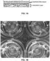
- the cells are deposited using the Novogen MMX Bioprinter in such a way that the epithelial layer is apical to the interstitial layer (see Fig. 1A ). Structures are created by spatially-controlled deposition of cells mixed with a thermo-responsive hydrogel that degrades over time (Novogel ® 2.0) combined with deposition of aerosolized cellular materials by compressed gas propulsion (inkjet spray). In this embodiment, the two layers together model the wall of a renal distal tubule. This configuration is critical for modeling in vivo tissues and predicting native tissue responses. Response of the epithelial layer is predictive of native tissue response to drugs, chemicals, or biological agents, and may provide information relative to toxicity or efficacy.
- the interstitial layer is critical for proper functioning of the epithelium and serves as a model for native tissue fibrosis, in particular renal tubulointerstitial fibrosis.
- an interstitial layer is bioprinted, using continuous deposition techniques.
- an epithelial layer is bioprinted, using ink-jet deposition techniques onto the interstitial layer.
- a substantially contiguous layer of epithelium is consistent with in vivo tissues and is critical to replicate a physiologically relevant architecture.
- Ink-jet deposition techniques provide the ability to deposit one or more thin layers of epithelial cells onto the potentially irregular surface of the interstitial layer.
- ink-jet deposition of the epithelial layer is optionally performed immediately after bioprinting of the interstitial layer or after the interstitial layer has been allowed to mature.
- the cells are bioprinted.
- the bioprinted cells are cohered to form the engineered renal tubule models.
- the engineered renal tubule models are free or substantially free of preformed scaffold at the time of fabrication or the time of use.
- bioprinting allows fabrication of tissues that mimic the appropriate cellularity of native tissue.
- the three-dimensional, engineered renal tubule models described herein are distinguished from tissues fabricated by prior technologies by virtue of the fact that they are three-dimensional, free of pre-formed scaffolds, consist essentially of cells, and/or have a high cell density (e.g., greater than 30% cellular, greater than 40% cellular, greater than 50% cellular, greater than 60% cellular, greater than 70% cellular, greater than 80% cellular, greater than 90% cellular, or greater than 95% cellular).
- a high cell density e.g., greater than 30% cellular, greater than 40% cellular, greater than 50% cellular, greater than 60% cellular, greater than 70% cellular, greater than 80% cellular, greater than 90% cellular, or greater than 95% cellular.
- the three-dimensional, engineered renal tubule models described herein are distinguished from native (e.g., non-engineered) tissues by virtue of the fact that they are non-innervated (e.g., substantially free of nervous tissue), substantially free of mature vasculature, and/or substantially free of blood components.
- the three-dimensional, engineered renal tubule models are free of plasma, red blood cells, platelets, and the like and/or endogenously-generated plasma, red blood cells, platelets, and the like.
- the engineered renal tubule model lacks immune cells such as T cell, B cells, macrophages, dendritic cells, basophils, mast cells or eosinophils.
- the model is not tubular in shape like a naturally occurring renal proximal tubule, but is planar or sheet-like, this advantageously allows for in vitro assays and analysis.
- the fibroblasts are not of renal origin.
- the endothelial cells are not of renal origin.
- the epithelial cells are not of human origin.
- the engineered renal tubule model lacks undifferentiated cells.
- the engineered renal tubule model lacks undifferentiated renal cells.
- the three-dimensional, engineered renal tubule models described herein are distinguished from native renal tubule tissues in that they are flat or substantially planar.
- the three-dimensional, engineered renal tubule models described herein possess functional improvements over native renal tubule tissues; one example is high viability after a sustained amount of time in culture up to at least 7, 10 or 27 days in culture.
- the cells used in the renal tubule model are transformed or immortalized.
- the cells used in the renal tubule model are transgenic and contain protein fusions with fluorescent proteins, like EGFP, GFP, RFP, YFP, or CFP.
- the cells used in the renal tubule model are transgenic and contain reporter constructs with fluorescent proteins; like EGFP, GFP, RFP, YFP, GFP; or luminescent proteins like firefly or renilla luciferase.
- any of the cells contain a deletion or insertion of 1, 2, 3, 4, 5, 6, 7, 8, 9, 10 genes or more.
- the 3D renal tubule models are chimeras, wherein at least one cell is form a different mammalian species than any other cell of the 3D renal tubule model.
- the 3D renal tubule models are chimeras, wherein at least one cell is form a different human donor than any other cell of the 3D renal tubule model.
- the engineered tissues, arrays, and methods described herein include a plurality of cell types.
- the renal tubule models comprise a layer of interstitial tissue comprising mammalian fibroblasts and mammalian endothelial cells.
- suitable endothelial cells are derived from human umbilical vein (HUVEC), human primary, human kidney, or from directed differentiation of induced pluripotent stem cells (iPS) or human embryonic stem cells (hES).
- the fibroblasts are renal interstitial fibroblasts.
- suitable renal interstitial fibroblasts are derived from primary cells isolated from human kidney.
- the fibroblasts are dermal or vascular in origin.
- the interstitial tissue comprises tumor cells or cancer cells.
- the layer of interstitial tissue is substantially a monolayer.
- the layer of interstitial tissue comprises a monolayer over 95% of its surface area.
- the layer of interstitial tissue comprises a monolayer over 90% of its surface area.
- the layer of interstitial tissue comprises a monolayer over 80% of its surface area.
- the layer of interstitial tissue is greater than 1 cell thick. In some embodiments, the layer of interstitial tissue is greater than 2 cells thick.
- the layer of interstitial tissue is greater than 3 cells thick. In some embodiments, the layer of interstitial tissue is greater than 4 cells thick. In some embodiments, the layer of interstitial tissue is greater than 5 cells thick. In some embodiments, the layer of interstitial tissue is greater than 10 cells thick. In some embodiments, the layer of interstitial tissue is greater than 20 cells thick. In some embodiments, the layer of interstitial tissue is greater than 50 cells thick. In some embodiments, the layer of interstitial tissue is greater than 100 cells thick. In some embodiments, the layer of interstitial tissue is 2-100 cells thick. In some embodiments, the layer of interstitial tissue is greater than 20 ⁇ m thick. In some embodiments, the layer of interstitial tissue is greater than 30 um thick.
- the layer of interstitial tissue is greater than 40 ⁇ m thick. In some embodiments, the layer of interstitial tissue is greater than 50 ⁇ m thick. In some embodiments, the layer of interstitial tissue is greater than 100 ⁇ m thick. In some embodiments, the layer of interstitial tissue is greater than 200 ⁇ m thick. In some embodiments, the layer of interstitial tissue is greater than 500 ⁇ m thick. In some embodiments, the layer of interstitial tissue is greater than 600 ⁇ m thick. In some embodiments, the layer of interstitial tissue is greater than 1000 ⁇ m thick. In some embodiments, the layer of interstitial tissue is 20 ⁇ m -1000 ⁇ m thick.
- the layer of interstitial tissue is less than 20 ⁇ m thick. In some embodiments, the layer of interstitial tissue is less than 30 ⁇ m thick. In some embodiments, the layer of interstitial tissue is less than 40 ⁇ m thick. In some embodiments, the layer of interstitial tissue is less than 50 ⁇ m thick. In some embodiments, the layer of interstitial tissue is less than 100 ⁇ m thick. In some embodiments, the layer of interstitial tissue is less than 200 ⁇ m thick. In some embodiments, the layer of interstitial tissue is less than 500 ⁇ m thick. In some embodiments, the layer of interstitial tissue is less than 600 ⁇ m thick. In some embodiments, the layer of interstitial tissue is less than 1000 ⁇ m thick.
- the renal tubule models comprise a layer of epithelial tissue comprising mammalian epithelial cells.
- the epithelial cells are renal tubular epithelial cells (e.g., proximal tubule epithelial cells).
- suitable renal tubular epithelial cells are primary isolates or cells derived from the directed differentiation of stem cells (induced pluripotent stem cell (iPS)-derived and/or human embryonic stem cell (hES)-derived).
- iPS induced pluripotent stem cell
- hES human embryonic stem cell
- the renal tubular epithelial cells are Madin-Darby canine kidney (MDCK) cells.
- the renal tubular epithelial cells are immortalized human cells.
- the renal tubular epithelial cells are immortalized cells such as hTERT-RPTEC cells, HK-2 cells, LLC-PK1 cells, or OK cells.
- the epithelial cells are derived from a non-human mammal such as, for example, rat, mouse, pig, or primate.
- the layer of epithelial tissue consists essentially of renal tubule epithelial cells.
- the layer of epithelial tissue consists essentially of primary renal tubule epithelial cells.
- the layer of epithelial tissue consists essentially of renal proximal tubule epithelial cells.
- the layer of epithelial tissue consists essentially of primary renal proximal tubule epithelial cells.
- the layer of renal epithelial tissue is substantially a monolayer.
- renal tubular epithelial cells are the only cells present in the layer of renal epithelial tissue.
- the layer of epithelial tissue comprises tumor cells.
- the layer of epithelial tissue comprises renal cell carcinoma cells.
- the layer of epithelial tissue comprises a monolayer over 95% of its surface area. In some embodiments, the layer of epithelial tissue comprises a monolayer over 90% of its surface area.
- the layer of epithelial tissue comprises a monolayer over 80% of its surface area. In some embodiments, the layer of epithelial tissue is greater than 1 cell thick. In some embodiments, the layer of epithelial tissue is greater than 2 cells thick. In some embodiments, the layer of epithelial tissue is greater than 3 cells thick. In some embodiments, the layer of epithelial tissue is greater than 4 cells thick. In some embodiments, the layer of epithelial tissue is greater than 5 cells thick. In some embodiments, the layer of epithelial tissue is greater than 10 cells thick. In some embodiments, the layer of epithelial tissue is greater than 20 cells thick. In some embodiments, the layer of epithelial tissue is greater than 50 cells thick.
- the layer of epithelial tissue is greater than 100 cells thick. In some embodiments, the layer of epithelial tissue is 2-100 cells thick. In some embodiments, the layer of epithelial tissue is greater than 20 ⁇ m thick. In some embodiments, the layer of epithelial tissue is greater than 30 ⁇ m thick. In some embodiments, the layer of epithelial tissue is greater than 40 ⁇ m thick. In some embodiments, the layer of epithelial tissue is greater than 50 ⁇ m thick. In some embodiments, the layer of epithelial tissue is greater than 100 ⁇ m thick. In some embodiments, the layer of epithelial tissue is greater than 200 ⁇ m thick.
- the layer of epithelial tissue is greater than 500 ⁇ m thick. In some embodiments, the layer of interstitial tissue is greater than 600 ⁇ m thick. In some embodiments, the layer of epithelial tissue is greater than 1000 ⁇ m thick. In some embodiments, the layer of epithelial tissue is 20-1000 ⁇ m thick. In some embodiments, the layer of epithelial tissue is less than 1000 ⁇ m thick. In some embodiments, the layer of interstitial tissue is less than 600 ⁇ m thick. In some embodiments, the layer of epithelial tissue is less than 500 ⁇ m thick. In some embodiments, the layer of epithelial tissue is less than 200 ⁇ m thick.
- the layer of epithelial tissue is less than 100 ⁇ m thick. In some embodiments, the layer of epithelial tissue is less than 50 ⁇ m thick. In some embodiments, the layer of epithelial tissue is less than 40 ⁇ m thick. In some embodiments, the layer of epithelial tissue is less than 30 ⁇ m thick. In some embodiments, the layer of epithelial tissue is less than 20 ⁇ m thick.
- the renal tubule models comprise other cell types (e.g., EPO-producing cells, immune cells, etc.).
- the immune cells are T cells.
- the immune cells are B cells.
- the immune cells are NK cells.
- the immune cells are dendritic cells.
- the immune cells are macrophage cells.
- the epithelial layer comprises, consists of, or consists essentially of proximal tubule epithelial cells.
- the fibroblasts and endothelial cells are the only cells present in the layer of renal interstitial tissue.
- the layer of renal interstitial tissue further comprises fibrotic tissue.
- the interstitial layer comprises, consists of, or consists essentially of fibroblasts and endothelial cells in specific ratios.
- Suitable proportions of fibroblasts include, by way of non-limiting examples, about 5, 10, 15, 20, 25, 30, 35, 40, 45, 50, 55, 60, 65, 70, 75, 80, 85, 90, and 95% fibroblasts, including increments therein.
- Suitable proportions of endothelial cells include, by way of non-limiting examples, about 5, 10, 15, 20, 25, 30, 35, 40, 45, 50, 55, 60, 65, 70, 75, 80, 85, 90, and 95% endothelial cells, including increments therein.
- the interstitial layer comprises, consists essentially of, or consists of a specified ratio of fibroblast to endothelial cells.
- the ratio of fibroblast to endothelial cells is at least 5:95, 10:90, 15:85, 20:80, 25:75, 30:70, 35:65, 40:60, 45:65, 50:50, 55:45, 60:40, 65:35, 70:30, 75:25, 80:20, 85:15, 90:10 or 95:5, including increments therein. In certain embodiments, the ratio of fibroblast to endothelial cells is 5:95 to 95:5.
- the ratio of fibroblast to endothelial cells is no more than 5:95, 10:90, 15:85, 20:80, 25:75, 30:70, 35:65, 40:60, 45:65, 50:50, 55:45, 60:40, 65:35, 70:30, 75:25, 80:20, 85:15, 90:10 or 95:5, including increments therein.
- the ratio of fibroblast to endothelial cells is about 50:50. In certain embodiments, the ratio of fibroblast to endothelial cells is from about 60:40 to about 40:60.
- Bio-inks are suitably prepared for continuous deposition bioprinting techniques with concentrations of cells including, by way of non-limiting examples, about 1, 2, 3, 4, 5, 6, 7, 8, 9, 10, 20, 30, 40, 50, 60, 70, 80, 90, 100, 110, 120, 130, 140, 150, 160, 170, 180, 190, 200, 225, 250, 275, 300, or more, million cells per milliliter of bio-ink.
- bio-ink prepared for continuous deposition bioprinting comprises about 100-200 million cells/mL.
- Bio-inks are suitably prepared for ink-jet deposition bioprinting techniques with concentrations of cells including, by way of non-limiting examples, about 0.25, 0.5, 1, 2, 3, 5, 10, 15 or more, million cells per milliliter of bio-ink.
- bio-ink prepared for ink-jet deposition bioprinting comprises about 1-5 million cells/mL.
- bio-ink prepared for ink-jet deposition bioprinting comprises about 1-4 million cells/mL.
- bio-ink prepared for ink-jet deposition bioprinting comprises about 1-3 million cells/mL.
- bio-ink prepared for ink-jet deposition bioprinting comprises about 1-2 million cells/mL.
- the renal interstitial bio-ink comprises between 50 million and 1 billion cells per milliliter. In certain embodiments, the renal interstitial bio-ink comprises between 50 million and 900 million cells per milliliter. In certain embodiments, the renal interstitial bio-ink comprises between 50 million and 800 million cells per milliliter. In certain embodiments, the renal interstitial bio-ink comprises between 50 million and 700 million cells per milliliter. In certain embodiments, the renal interstitial bio-ink comprises between 50 million and 600 million cells per milliliter. In certain embodiments, the renal interstitial bio-ink comprises between 50 million and 500 million cells per milliliter. In certain embodiments, the renal interstitial bio-ink comprises between 50 million and 400 million cells per milliliter.
- the renal interstitial bio-ink comprises between 50 million and 300 million cells per milliliter. In certain embodiments, the renal interstitial bio-ink comprises between 50 million and 200 million cells per milliliter. In certain embodiments, the renal interstitial bio-ink comprises between 75 million and 600 million cells per milliliter. In certain embodiments, the renal interstitial bio-ink comprises between 100 million and 600 million cells per milliliter. In certain embodiments, the renal interstitial bio-ink comprises between 100 million and 500 million cells per milliliter. In certain embodiments, the renal interstitial bio-ink comprises between 100 million and 400 million cells per milliliter. In certain embodiments, the renal interstitial bio-ink comprises between 100 million and 300 million cells per milliliter. In certain embodiments, the renal interstitial bio-ink comprises between 100 million and 200 million cells per milliliter. In certain embodiments, the renal interstitial bio-ink comprises between 100 million and 150 million cells per milliliter.
- the renal epithelial bio-ink comprises between 0.25 million and 5 million cells per milliliter. In certain embodiments, the renal epithelial bio-ink comprises between 0.25 million and 4 million cells per milliliter. In certain embodiments, the renal epithelial bio-ink comprises between 0.25 million and 3 million cells per milliliter. In certain embodiments, the renal epithelial bio-ink comprises between 0.25 million and 2 million cells per milliliter. In certain embodiments, the renal epithelial bio-ink comprises between 0.25 million and 1 million cells per milliliter. In certain embodiments, the renal epithelial bio-ink comprises between 0.5 million and 5 million cells per milliliter.
- the renal epithelial bio-ink comprises between 0.5 million and 4 million cells per milliliter. In certain embodiments, the renal epithelial bio-ink comprises between 0.5 million and 3 million cells per milliliter. In certain embodiments, the renal epithelial bio-ink comprises between 0.5 million and 2 million cells per milliliter. In certain embodiments, the renal epithelial bio-ink comprises between 0.5 million and 1 million cells per milliliter. In certain embodiments, the renal epithelial bio-ink comprises between 1 million and 5 million cells per milliliter. In certain embodiments, the renal epithelial bio-ink comprises between 1 million and 4 million cells per milliliter. In certain embodiments, the renal epithelial bio-ink comprises between 1 million and 3 million cells per milliliter. In certain embodiments, the renal epithelial bio-ink comprises between 1 million and 2 million cells per milliliter.
- the density of the epithelial bio-ink is less than the density of the interstitial bio-ink.
- the ratio of the density of the interstitial bio-ink to the density of the epithelial bio-ink is about 300:1; about 275:1; about 250:1; about 225:1; about 200:1; about 175:1; about 150:1, about 125:1; about 100:1, about 75:1 or about 50:1. In certain embodiments, the ratio of the density of the interstitial bio-ink to the density of the epithelial bio-ink ranges from about 300: 1 to about 50:1.
- the ratio of the density of the interstitial bio-ink to the density of the epithelial bio-ink ranges from about 250:1 to about 75:1. In certain embodiments, the ratio of the density of the interstitial bio-ink to the density of the epithelial bio-ink ranges from about 200:1 to about 75:1. In certain embodiments, the ratio of the density of the interstitial bio-ink to the density of the epithelial bio-ink ranges from about 150:1 to about 75:1. In certain embodiments, the ratio of the density of the interstitial bio-ink to the density of the epithelial bio-ink ranges from about 125:1 to about 75:1.
- the bio-ink is a viscous liquid. In certain embodiments, the bio-ink is a semi-solid. In certain embodiments, the bio-ink is a solid. In certain embodiments, the viscosity of the bio-ink is greater than 100 centipoise. In certain embodiments, the viscosity of the bio-ink is greater than 200 centipoise. In certain embodiments, the viscosity of the bio-ink is greater than 500 centipoise. In certain embodiments, the viscosity of the bio-ink is greater than 1,000 centipoise. In certain embodiments, the viscosity of the bio-ink is greater than 2,000 centipoise.
- the viscosity of the bio-ink is greater than 5,000 centipoise. In certain embodiments, the viscosity of the bio-ink is greater than 10,000 centipoise. In certain embodiments, the viscosity of the bio-ink is greater than 20,000 centipoise. In certain embodiments, the viscosity of the bio-ink is greater than 50,000 centipoise. In certain embodiments, the viscosity of the bio-ink is greater than 100,000 centipoise. In certain embodiments, the viscosity of the bio-ink is less than 100 centipoise. In certain embodiments, the viscosity of the bio-ink is less than 200 centipoise.
- the viscosity of the bio-ink is less than 500 centipoise. In certain embodiments, the viscosity of the bio-ink is less than 1,000 centipoise. In certain embodiments, the viscosity of the bio-ink is less than 2,000 centipoise. In certain embodiments, the viscosity of the bio-ink is less than 5,000 centipoise. In certain embodiments, the viscosity of the bio-ink is less than 10,000 centipoise. In certain embodiments, the viscosity of the bio-ink is less than 20,000 centipoise. In certain embodiments, the viscosity of the bio-ink is less than 50,000 centipoise. In certain embodiments, the viscosity of the bio-ink is less than 100,000 centipoise. In certain embodiments, the viscosity of the bio-ink is 100-100,000 centipoise.
- the renal tubule models of the present disclosure can be architecturally arranged in many configurations.
- the epithelial tissue and interstitial tissue layers are separate architecturally distinct layers that are in direct contact or separated by 1, 2, 3, 4, 5, 6, 7, 8, 9, 10, 15, 20 ⁇ m or more, including increments therein.
- the separation is due to the secretion and deposition of extracellular matrix between the two layers, which for the purposes of this disclosure is considered contact.
- normal physiological tissue cells and cell layers are polarized to have an apical (lumen facing) surface and a basolateral surface, which faces other cells or tissue matrix.
- the basolateral surface refers to a surface that faces another cell, an extracellular matrix or the surface of a biocompatible membrane or culture vessel.
- the apical surface refers to a surface that faces away from the surface of a biocompatible membrane or culture vessel.
- the renal tubular epithelial cells are polarized.
- the layer of renal interstitial tissue possesses an apical and basolateral surface.
- the renal tubule model further comprises a biocompatible membrane.
- the basolateral surface of the interstitial tissue layer is the surface attached to a biocompatible membrane or culture vessel; and the apical surface of the interstitial tissue layer is the surface not attached to a biocompatible membrane or culture vessel.
- the epithelial tissue layer is deposited onto and forms a layer on the apical surface of the interstitial tissue layer, thus forming two architecturally distinct layers.
- the epithelial tissue and interstitial tissue layers are in continuous contact. In certain embodiments, between 99%-100% of the epithelial tissue layer is in continuous contact with the interstitial tissue layer.
- between 95%-100% of the epithelial tissue layer is in continuous contact with the interstitial tissue layer. In certain embodiments, between 90%-100% of the epithelial tissue layer is in continuous contact with the interstitial tissue layer. In certain embodiments, 50-99% of the epithelial tissue layer is in continuous contact with the interstitial tissue layer. In certain embodiments, between 80%-100% of the epithelial tissue layer is in continuous contact with the interstitial tissue layer. In certain embodiments, between 70%-100% of the epithelial tissue layer is in continuous contact with the interstitial tissue layer. In certain embodiments, between 60% -100% of the epithelial tissue layer is in continuous contact with the interstitial tissue layer.
- between 50%-100% of the epithelial tissue layer is in continuous contact with the interstitial tissue layer. In certain embodiments, less than 99% of the epithelial tissue layer is in continuous contact with the interstitial tissue layer. In certain embodiments, less than 98% of the epithelial tissue layer is in continuous contact with the interstitial tissue layer. In certain embodiments, less than 97% of the epithelial tissue layer is in continuous contact with the interstitial tissue layer. In certain embodiments, less than 95% of the epithelial tissue layer is in continuous contact with the interstitial tissue layer. In certain embodiments, less than 90% of the epithelial tissue layer is in continuous contact with the interstitial tissue layer.
- the epithelial tissue layer covers between 80%-100% of the apical surface of the interstitial tissue layer. In certain embodiments, the epithelial tissue layer completely covers the apical surface of the interstitial tissue layer. In certain embodiments, the epithelial tissue layer covers between 99%-100% of the apical surface of the interstitial tissue layer. In certain embodiments, the epithelial tissue layer covers between 95%-100% of the apical surface of the interstitial tissue layer. In certain embodiments, the epithelial tissue layer covers between 90%-100% of the apical surface of the interstitial tissue layer. In certain embodiments, the epithelial tissue layer covers between 80%-100% of the apical surface of the interstitial tissue layer.
- the epithelial tissue layer covers between 70%-100% of the apical surface of the interstitial tissue layer. In certain embodiments, the epithelial tissue layer covers between 60%-100% of the apical surface of the interstitial tissue layer. In certain embodiments, the epithelial tissue layer covers between 50%-100% of the apical surface of the interstitial tissue layer. In certain embodiments, the epithelial tissue layer covers less than 99% of the apical surface of the interstitial tissue layer. In certain embodiments, the epithelial tissue layer covers less than 98% of the apical surface of the interstitial tissue layer. In certain embodiments, the epithelial tissue layer covers less than 97% of the apical surface of the interstitial tissue layer.
- the epithelial tissue layer covers less than 95% of the apical surface of the interstitial tissue layer. In certain embodiments, the epithelial tissue layer covers less than 90% of the apical surface of the interstitial tissue layer. In certain embodiments, the epithelial tissue layer covers less than 80% of the apical surface of the interstitial tissue layer. In certain embodiments, the epithelial tissue layer covers less than 70% of the apical surface of the interstitial tissue layer. In certain embodiments, the epithelial tissue layer covers 50-99% of the apical surface of the interstitial tissue layer.
- the epithelial tissue layer consists of cells that form tight junctions.
- substantially all cells in the epithelial tissue layer form a tight junction with at least one neighboring cell.
- between 99%-100% of cells in the epithelial tissue layer form a tight junction with at least one other cell.
- between 95%-100% of cells in the epithelial tissue layer form a tight junction with at least one other cell.
- between 90%-100% of cells in the epithelial tissue layer form a tight junction with at least one other cell. In certain embodiments, between 80%-100% of cells in the epithelial tissue layer form a tight junction with at least one other cell. In certain embodiments, between 70%-100% of cells in the epithelial tissue layer form a tight junction with at least one other cell. In certain embodiments, between 60%-100% of cells in the epithelial tissue layer form a tight junction with at least one other cell. In certain embodiments, between 50%-100% of cells in the epithelial tissue layer form a tight junction with at least one other cell. In certain embodiments, 50-99% of cells in the epithelial tissue layer form a tight junction with at least one other cell.
- the density of the interstitial cell layer is greater than 1 ⁇ 10 6 cells per mL. In certain embodiments, the density of the interstitial cell layer is at least 5 ⁇ 10 6 cells per mL. In certain embodiments, the density of the interstitial cell layer is at least 10 ⁇ 10 6 cells per mL. In certain embodiments, the density of the interstitial cell layer is at least 20 ⁇ 10 6 cells per mL. In certain embodiments, the density of the interstitial cell layer is at least 50 ⁇ 10 6 cells per mL. In certain embodiments, the density of the interstitial cell layer is at least 100 ⁇ 10 6 cells per mL.
- the density of the interstitial cell layer is at least 200 ⁇ 10 6 cells per mL. In certain embodiments, the density of the interstitial cell layer is at least 500 ⁇ 10 6 cells per mL. In certain embodiments, the density of the interstitial cell layer is between about 100 ⁇ 10 6 cells per mL and about 900 ⁇ 10 6 cells per mL. In certain embodiments, the density of the interstitial cell layer is between about 100 ⁇ 10 6 cells per mL and about 700 ⁇ 10 6 cells per mL. In certain embodiments, the density of the interstitial cell layer is between about 100 ⁇ 10 6 cells per mL and about 600 ⁇ 10 6 cells per mL.
- the density of the interstitial cell layer is between about 100 ⁇ 10 6 cells per mL and about 500 ⁇ 10 6 cells per mL. In certain embodiments, the density of the interstitial cell layer is between about 100 ⁇ 10 6 cells per mL and about 300 ⁇ 10 6 cells per mL. In certain embodiments, the density of the interstitial cell layer is between about 100 ⁇ 10 6 cells per mL and about 200 ⁇ 10 6 cells per mL. In certain embodiments, the layer of renal interstitial tissue or layer of renal epithelial tissue is between 70%-100% living cells by volume. In certain embodiments, the viability of the interstitial tissue layer is greater than 99% living cells by volume.
- the viability of the interstitial tissue layer is greater than 95% living cells by volume. In certain embodiments, the viability of the interstitial tissue layer is greater than 90% living cells by volume. In certain embodiments, the viability of the interstitial tissue layer is greater than 80% living cells by volume. In certain embodiments, the viability of the interstitial tissue layer is greater than 70% living cells by volume. In certain embodiments, the viability of the interstitial tissue layer is greater than 60% living cells by volume. In certain embodiments, the viability of the interstitial tissue layer is greater than 50% living cells by volume. In certain embodiments, the viability of the interstitial tissue layer is 50-99% living cells by volume.
- this viability is maintained for at least 8, 12, 24, 48, 72, 96, or more hours post printing. In certain embodiments, this viability is maintained for at least 1, 2, 3, 4, 5, 6, 7, 8, 9, 10, 11, 12, 13, 14, 21, or more days post printing.
- the density of the epithelial cell layer is at least 1 ⁇ 10 5 cells per mL. In certain embodiments, the density of the epithelial cell layer is at least 2 ⁇ 10 5 cells per mL. In certain embodiments, the density of the epithelial cell layer is at least 5 ⁇ 10 5 cells per mL. In certain embodiments, the density of the epithelial cell layer is at least 1 ⁇ 10 6 cells per mL.
- the density of the epithelial cell layer is at least 5 ⁇ 10 6 cells per mL. In certain embodiments, the density of the epithelial cell layer is at least 10 ⁇ 10 6 cells per mL. In certain embodiments, the density of the epithelial cell layer is at least 20 ⁇ 10 6 cells per mL. In certain embodiments, the density of the epithelial cell layer is at least 50 ⁇ 10 6 cells per mL. In certain embodiments, the density of the epithelial cell layer is at least 100 ⁇ 10 6 cells per mL. In certain embodiments, the density of the epithelial cell layer is at least 200 ⁇ 10 6 cells per mL.
- the density of the epithelial cell layer is at least 500 ⁇ 10 6 cells per mL. In certain embodiments, the density of the epithelial cell layer is less than 1 ⁇ 10 5 cells per mL. In certain embodiments, the density of the epithelial cell layer is less than 2 ⁇ 10 5 cells per mL. In certain embodiments, the density of the epithelial cell layer is less than 5 ⁇ 10 5 cells per mL. In certain embodiments, the density of the epithelial cell layer is less than 1 ⁇ 10 6 6 cells per mL. In certain embodiments, the density of the epithelial cell layer is less than 5 ⁇ 10 6 cells per mL.
- the density of the epithelial cell layer is less than 10 ⁇ 10 6 cells per mL. In certain embodiments, the density of the epithelial cell layer is 10 ⁇ 10 6 cells per mL. In certain embodiments, the viability of the epithelial tissue layer is greater than 99% living cells by volume. In certain embodiments, the viability of the epithelial tissue layer is greater than 95% living cells by volume. In certain embodiments, the viability of the epithelial tissue layer is greater than 90% living cells by volume. In certain embodiments, the viability of the epithelial tissue layer is greater than 80% living cells by volume. In certain embodiments, the viability of the epithelial tissue layer is greater than 70% living cells by volume.
- the viability of the epithelial tissue layer is greater than 60% living cells by volume. In certain embodiments, the viability of the epithelial tissue layer is greater than 50% living cells by volume. In certain embodiments, the viability of the epithelial tissue layer is 50-99% living cells by volume. In certain embodiments, this viability is maintained for at least 8, 12, 24, 48, 72, or 96 hours post-printing. In certain embodiments, this viability is maintained for at least 1, 2, 3, 4, 5, 6, 7, 8, 9, 10, 11, 12, 13, or 14 days post-printing.
- the thickness of the renal tubule model is substantially uniform. In certain embodiments, between 99%-100% of the renal tubule model is within 10% plus or minus of the overall mean thickness of the renal tubule model. In certain embodiments, between 95%-100% of the renal tubule model is within 10% plus or minus of the overall mean thickness of the renal tubule model. In certain embodiments, between 90%-100% of the renal tubule model is within 10% plus or minus of the overall mean thickness of the renal tubule model.
- between 80%-100% of the renal tubule model is within 10% plus or minus of the overall mean thickness of the renal tubule model. In certain embodiments, between 70%-100% of the renal tubule model is within 10% plus or minus of the overall mean thickness of the renal tubule model. In certain embodiments, between 99%-100% of the renal tubule model is within 20% plus or minus of the overall mean thickness of the renal tubule model. In certain embodiments, between 95%-100% of the renal tubule model is within 20% plus or minus of the overall mean thickness of the renal tubule model. In certain embodiments, between 90%-100% of the renal tubule model is within 20% plus or minus of the overall mean thickness of the renal tubule model.
- between 80%-100% of the renal tubule model is within 20% plus or minus of the overall mean thickness of the renal tubule model. In certain embodiments, between 70%-100% of the renal tubule model is within 20% plus or minus of the overall mean thickness of the renal tubule model. After treatment with a potential toxic agent, the renal tubule model may become less uniform.
- extrusion compounds include, but are not limited to gels, hydrogels, peptide hydrogels, amino acid-based gels, surfactant polyols (e.g., Pluronic F-127 or PF-127), thermo-responsive polymers, hyaluronates, alginates, extracellular matrix components (and derivatives thereof), collagens, gelatin, other biocompatible natural or synthetic polymers, nanofibers, and self-assembling nanofibers.
- the extrusion compound contains a synthetic polymer.
- the extrusion compound contains a non-synthetic polymer that is not normally associated with mammalian tissues.
- extrusion compounds are removed after bioprinting by physical, chemical, or enzymatic means.
- the bio-inks of the present disclosure contain 1% or more extrusion compound by weight.
- the renal tubule models of the present disclosure contain more than 1% extrusion compound by weight.
- the bio-inks of the present disclosure contain less than 5% extrusion compound by weight.
- the bio-inks of the present disclosure contain between 0%-2% extrusion compound by weight.
- the bio-inks of the present disclosure contain less than 1% extrusion compound by weight.
- the renal tubule models of the present disclosure contain between 0%-5% extrusion compound by weight. In some embodiments, the renal tubule models of the present disclosure contain less than 2% extrusion compound by weight. In some embodiments, the renal tubule models of the present disclosure contain less than 1% extrusion compound by weight.
- the epithelial bio-ink is free from hydrogel. In some embodiments, the epithelial bio-ink is free from extrusion compound. In some embodiments, the epithelial bio-ink is free from synthetic polymers that are used as excipient or extrusion compounds. In some embodiments, the renal tubule model is free from synthetic polymers that are used as excipient or extrusion compounds. In some embodiments, the epithelial cell layer is free from synthetic polymers that are used as excipient or extrusion compounds. In some embodiments, the interstitial cell layer is free from synthetic polymers that are used as excipient or extrusion compounds.
- the interstitial tissue layer is printed onto a biocompatible surface.
- the biocompatible surface is a membrane with a pore size of 0.4 ⁇ m to 10 ⁇ m. In certain embodiments, the biocompatible surface has a pore size of about 1 ⁇ m. In certain embodiments, the biocompatible surface is coated with a composition to improve cell adherence or viability.
- the renal tubule modules are printed into 6-well, 12-well, 24-well, 48-well, 96-well, or 384-well plates. In certain embodiments, the renal tubule modules are printed into tissue culture plates with diameters of 60, 100 or 150 mm or more. In certain embodiments, the renal tubule modules are printed into tissue culture flasks or onto microfluidic chips. In certain embodiments, the renal tubule models are printed into/onto Transwell inserts.
- the product of a three-dimensional, engineered, biological renal tubule model is produced by the process of bioprinting.
- at least one constituent of the product of a three-dimensional, engineered, biological renal tubule model is produced by the process of bioprinting.
- the process of fabricating a three-dimensional, engineered, biological renal tubule model comprises: preparing a renal interstitial bio-ink, the interstitial bio-ink comprising a plurality of interstitial cell types, the interstitial cell types comprising renal fibroblasts and endothelial cells; preparing a renal epithelial bio-ink, the epithelial bio-ink comprising renal tubular epithelial cells; depositing the renal interstitial bio-ink and the renal epithelial bio-ink such that the renal epithelial bio-ink forms a layer on at least one surface of the layer of renal interstitial bio-ink; and maturing the deposited bio-ink in a cell culture media to allow the cells to cohere to form the three-dimensional, engineered, biological renal tubule model.
- the renal interstitial tissue bio-ink forms a renal interstitial tissue layer with an apical and basolateral surface.
- the renal epithelial bio-ink is deposited in contact with the apical surface of the renal interstitial tissue layer.
- the renal epithelial bio-ink consists essentially of renal tubular epithelial cells.
- the renal epithelial bio-ink consists essentially of primary renal tubular epithelial cells.
- the primary renal tubular epithelial cells are isolated from a subject with a disease that affects kidney function.
- the primary renal tubular epithelial cells are isolated from a subject with polycystic kidney disease.
- the primary renal tubular epithelial cells are isolated from a subject with diabetes mellitus type II.
- the renal epithelial bio-ink comprises renal cell carcinoma cells.
- the renal epithelial bio-ink is deposited in a monolayer.
- the renal interstitial tissue bio-ink is deposited in a monolayer.
- the layer of renal epithelial tissue is deposited in continuous contact with the layer of renal interstitial tissue.
- the renal epithelial bio-ink forms a layer that covers between 50%-100% of the apical surface of the layer of renal interstitial tissue.
- the renal epithelial bio-ink forms a layer that covers between 70%-100% of the apical surface of the layer of renal interstitial tissue. In certain embodiments, the renal epithelial bio-ink forms a layer that covers between 90%-100% of the apical surface of the layer of renal interstitial tissue. In certain embodiments, the renal epithelial bio-ink forms a layer that covers 50-90% the apical surface of the layer of renal interstitial tissue. In certain embodiments, at least 50% of renal epithelial cells of the renal epithelial layer form tight junctions with other renal epithelial cells.
- the renal tubule model is between 50 and 500 ⁇ m thick. In certain embodiments, the renal tubule model is about 100 ⁇ m thick. In certain embodiments, the renal epithelial bio-ink further comprises an extrusion compound.
- the fibroblasts and endothelial cells are present in the renal interstitial bio-ink at a ratio of about 95:5 to about 5:95 fibroblasts to endothelial cells. In certain embodiments, the fibroblasts and endothelial cells are present in the renal interstitial bio-ink at a ratio of about 75:25 to about 25:75 fibroblasts to endothelial cells. In certain embodiments, the fibroblasts and endothelial cells are present in the renal interstitial bio-ink at a ratio of about 60:40 to about 40:60 fibroblasts to endothelial cells.
- the fibroblasts and endothelial cells are present in the renal interstitial bio-ink at a ratio of about 50:50 fibroblasts to endothelial cells.
- the renal interstitial bio-ink further comprises secretory cells.
- the renal interstitial bio-ink further comprises immune cells.
- the renal interstitial bio-ink further comprises an extrusion compound.
- the renal interstitial bio-ink comprises glomerular cells.
- the model is fabricated substantially free of pre-formed scaffold.
- the renal fibroblasts, endothelial cells, and renal tubular epithelial cells are mammalian cells.
- either of the renal interstitial bio-ink or renal epithelial bio-ink forms a planar layer after deposition.
- the renal tubule model is of a uniform thickness.
- the renal interstitial bio-ink is deposited onto a biocompatible membrane.
- the renal interstitial bio-ink is deposited onto a biocompatible membrane with a pore size greater than 0.4 ⁇ m.
- the renal interstitial bio-ink is deposited onto a biocompatible membrane with a pore size of about 1 um.
- the three-dimensional, engineered, biological renal tubule models are deposited to form an array.
- the three-dimensional, engineered, biological renal tubule models are deposited to form an array configured to allow between about 20 ⁇ m and about 100 ⁇ m of space between each renal tubule model.
- the renal interstitial bio-ink is between 30%-100% living cells by volume.
- the renal interstitial bio-ink is between 70%-100% living cells by volume.
- the renal interstitial bio-ink is between 90%-100% living cells by volume.
- the renal interstitial bio-ink is deposited by extrusion bioprinting.
- the renal epithelial bio-ink is deposited by ink-jet bioprinting.
- the renal interstitial bio-ink is not deposited by ink-jet bioprinting.
- any layer of the renal tubule model is viable in in vitro culture in culture after 3 days. In certain embodiments, any layer of the renal tubule model is viable in in vitro culture after 10 days.
- the 3D renal tubule models disclosed herein are produced by an additive manufacturing process.
- the additive manufacturing process for 3D tubule models herein allows customized fabrication of 3D renal tubule models for in vitro purposes. This is significant in that the tissues are fabricated due to a user specified design.
- the 3D renal tubule models contain only the cells that the user specifies.
- the 3D renal tubule models contain only the cell types that the user specifies.
- the 3D renal tubule models contain only the number of cells or concentration of cells that the user specifies.
- the 3D renal tubule models contain cells that have been treated with a small molecule, therapeutic molecule, or therapeutic substance before or during fabrication.
- the 3D renal tubule models contain biocompatible or tissue culture plastics, biocompatible synthetic polymers, cross linkable gels, reversibly cross-linked gels and other non-cellular constituents.
- the renal tubule models of the present disclosure are matured for a certain amount of time after bioprinting.
- the models are matured for 1-24 hours before use, for example, at least 1, 2, 3, 4, 5, 6, 7, 8, 9, 10, 11, 12, 16, 18, 24 hours or more before use.
- the models are matured for 1-30 days before use, for example, at least 1, 2, 3, 4, 5, 6, 7, 8, 9, 10, 11, 12, 13, 14, 15, 16, 17, 18, 19, 20, 21, 22, 23, 24, 25, 26, 27, 28, 29, 30 days or more before use.
- shipment or transfer of the tissues is a use.
- the interstitial layer of the renal tubule model of the present disclosure is matured for a certain amount of time after bioprinting before addition of the epithelial layer.
- the interstitial layer is matured for 1-24 hours before use, for example, at least 1, 2, 3, 4, 5, 6, 7, 8, 9, 10, 11, 12, 16, 18, 24 hours or more before use.
- the interstitial layer is matured for 1-30 days before use, for example, at least 1, 2, 3, 4, 5, 6, 7, 8, 9, 10, 11, 12, 13, 14, 15, 16, 17, 18, 19, 20, 21, 22, 23, 24, 25, 26, 27, 28, 29, 30 days or more before use.
- shipment or transfer of the tissues is a use.
- the epithelial layer is bioprinted onto the interstitial layer within 1-24 hours after bioprinting of the interstitial layer, for example, 1, 2, 3, 4, 5, 6, 7, 8, 9, 10, 11, 12, 16, 18, 24 hours after bioprinting of the interstitial layer.
- shipment or transfer of the tissues is a use.
- the epithelial layer is bioprinted onto the interstitial layer within 1-30 days after bioprinting of the interstitial layer, for example 1, 2, 3, 4, 5, 6, 7, 8, 9, 10, 11, 12, 13, 14, 15, 16, 17, 18, 19, 20, 21, 22, 23, 24, 25, 26, 27, 28, 29, 30 days after bioprinting of the interstitial layer.
- the renal tubule models described herein can be utilized for multiple applications.
- the tissue barrier can be utilized for toxicology and ADME applications.
- functional features of the renal tubule models include establishment of a barrier and demonstrating permeability/absorption (as evidenced by TEER and Lucifer yellow permeability). These features allow for permeability kinetics (Papp) and influx/efflux (ab, ba) studies.
- the investigation of active transport and metabolism via key transporters and metabolic enzymes respectively can be performed via well-based assays or through detection of substrates and their metabolites by mass spectrometry. These same techniques can be used to assess the mechanism of active transport and metabolism of various drugs and applied compounds.
- the renal tubules may be used to predict whether there is active transport of compounds via renal transporters similar to native tissue, predict the ability of compounds to disrupt the renal barrier and/or induce renal inflammation, and/or predict the efficacy of compounds to modulate inflammation.
- the renal tubule models disclosed herein comprising immune cells are used in the modeling of inflammation and inflammatory diseases, as well as the impact of immune modulation on cancer.
- the immune cells are myeloid or lymphoid cells.
- the disease models are compared side by side to normal tissue models, e.g., renal tubule models lacking immune cells, comprising immune cells but not stimulated to activate the immune cells (quiescent), or lacking immune cells and stimulated with cytokines to mimic an immune response.
- the renal tubule models are useful for evaluation of inflammation and immune responses. Tissue constructs comprising immune cells may also be used to study acute responses.
- Renal tubule models comprising immune cells may also be used to model injury and recovery including acute, subchronic, or chronic dosing of candidate pharmaceutical compounds or therapies.
- the renal tubule models comprising immune cells are used to evaluate wound healing and fibrosis.
- the tissue constructs comprising immune cells may be used to model microbial/microbiome interactions. The 3D nature of the renal tubule models allow for enhanced observation of pathogen invasiveness and translocation.
- the renal tubule models are treated subsequently with candidate pharmaceutical agents or treatments to reverse or control the inflammatory effects. Inflammatory signals that may be detected include the release of cytokines (e.g.
- IL-8 TNF- ⁇ , IL-4, IL-19, IL-13, IL-17, and/or IFN-gamma
- antimicrobial peptides e.g. beta defensin, lysozymes, and/or sIgA
- endocrine products such as somatostatin
- activation of inflammatory pathways e.g.
- JAK/STAT, and/or NFkB evaluation of a barrier disruption in response to inflammation (histology, TEER, Lucifer yellow, Ussing chamber, and/or other well-based assays), measuring proliferation, cytotoxicity, tissue damage, or apoptosis (Caspase 8 or Tunel) or autophagy or re-epithelialization of wounded area, and expression of key markers and receptors upregulated in response to stimulation.
- the renal tubule models may be used to demonstrate the kinetics and magnitude of onset as well as recovery from perturbation.
- the renal tubule models are used in a model of a renal disorder.
- the renal tubule models are used in a model of a renal disorder, wherein the renal tubule models comprise:
- the renal tubule models may be used in a method of assessing the ability of a therapeutic agent to reverse, reduce, induce, or prevent a renal disorder.
- the renal tubules and arrays disclosed herein are for use in in vitro assays.
- an "assay” is a procedure for testing or measuring the presence or activity of a substance (e.g., a chemical, molecule, biochemical, drug, etc.) in an organic or biologic sample (e.g., cell aggregate, tissue, organ, organism, etc.).
- assays include qualitative assays and quantitative assays.
- a quantitative assay measures the amount of a substance such as a chemical or biomolecule in a sample.
- the renal tubules and arrays are for use in, by way of non-limiting example, image-based assays, measurement of secreted proteins, expression of markers, and production of proteins or mRNAs.
- the renal tubules and arrays are for use in assays to detect or measure one or more of: molecular binding (including radioligand binding), molecular uptake, activity (e.g., enzymatic activity and receptor activity, etc.), gene expression, protein expression, protein modifications (non-limiting examples include: phosphorylation, ubiquitination, acetylation, glycosylation, lipidation, etc.), receptor agonism, receptor antagonism, cell signaling, apoptosis, chemosensitivity, transfection, cell migration, chemotaxis, cell viability, cell proliferation, safety, efficacy, metabolism, toxicity, infectivity, and abuse liability.
- the renal tubules are for toxicology, pharmaceutical or toxicity testing.
- the renal tubules and arrays are for use in immunoassays.
- Immunoassays include, for example, flow cytometry, high throughput or low throughput image analysis, immunoprecipitation, radio-immunoassay (RIA), enzyme-linked immunosorbent assays (ELISA), western blot, homogenous assays, such as AlphaLISA TM and related technologies that rely on time resolved fluorescence or fluorescence resonance energy transfer (FRET).
- immunoassays are competitive immunoassays or noncompetitive immunoassays.
- a competitive immunoassay for example, the antigen in a sample competes with labeled antigen to bind with antibodies and the amount of labeled antigen bound to the antibody site is then measured.
- a noncompetitive immunoassay also referred to as a "sandwich assay”
- antigen in a sample is bound to an antibody site; subsequently, labeled antibody is bound to the antigen and the amount of labeled antibody on the site is then measured.
- the renal tubules and arrays are for use in ELISA.
- an ELISA is a biochemical technique used to detect the presence of an antibody or an antigen in a sample.
- ELISA for example, at least one antibody with specificity for a particular antigen is utilized.
- a sample with an unknown amount of antigen is immobilized on a solid support (e.g., a polystyrene microtiter plate) either non-specifically (via adsorption to the surface) or specifically (via capture by another antibody specific to the same antigen, in a "sandwich" ELISA).
- the detection antibody is added, forming a complex with the antigen.
- the detection antibody is, for example, covalently linked to an enzyme, or is itself detected by a secondary antibody that is linked to an enzyme through bioconjugation.
- an array, microarray, or chip of cells, multicellular aggregates, or tissues is used for drug screening or drug discovery.
- an array, microarray, or chip of tissues is used as part of a kit for drug screening or drug discovery.
- each renal tubule exists within a well of a biocompatible multi-well container, wherein the container is compatible with one or more automated drug screening procedures and/or devices.
- automated drug screening procedures and/or devices include any suitable procedure or device that is computer or robot-assisted.
- arrays for drug screening assays or drug discovery assays are used to research or develop drugs potentially useful in any therapeutic area.
- suitable therapeutic areas include, by way of non-limiting examples, infectious disease, hematology, oncology, pediatrics, cardiology, central nervous system disease, neurology, gastroenterology, hepatology, urology, infertility, ophthalmology, nephrology, orthopedics, pain control, psychiatry, pulmonology, vaccines, wound healing, physiology, pharmacology, dermatology, gene therapy, toxicology, toxicity, and immunology.
- the renal tubules and arrays are for use in cell-based screening.
- the cell-based screening is for one or more infectious diseases such as viral, fungal, bacterial or parasitic infection.
- the cell-based screening is for kidney cancer, including renal cell carcinoma, juxtaglomerular cell tumor (reninoma), angiomyolipoma, renal oncocytoma, Bellini duct carcinoma, clear-cell sarcoma of the kidney, mesoblastic nephroma, Wilms' tumor, mixed epithelial stromal tumor, and transitional cell carcinoma of the renal pelvis.
- the cell-based screening is for nephritis, including, glomerulonephritis, interstitial nephritis or tubulo-interstitial nephritis, pyelonephritis, lupus nephritis and athletic nephritis.
- the cell-based screening is for hypertension.
- the cell-based screening is for diabetes mellitus, type I, type II and MODY.
- the cell-based screening is for a nephropathy, including IgA nephropathy, analgesic nephropathy, or onconephropathy.
- the cell-based screening is for polycystic kidney disease or Xanthine oxidase deficiency.
- the renal tubules and arrays are for use in the study of cancer initiation, progression, or metastasis.
- the renal tubules and arrays are for use in the study of the interaction of other cell types, such as cancer cells, pathogen-bearing cells, pathogenic cells, immune cells, blood-derived cells, or stem/progenitor cells.
- the constructs or arrays thereof are for use in assessing the performance of biologics, including antibodies, mammalian cells, bacteria, biologically-active proteins, hormones, etc.
- the renal tubules or arrays thereof are useful in the study of cell-cell and cell-tissue interactions between the mammalian renal tubules comprising the construct and one or more additional cell types, including but not limited to pathogen-bearing cells, living pathogenic cells, cancer cells, immune cells, blood cells, stem/progenitor cells, or genetically-manipulated cells.
- the array comprises renal tubules and additional tissue constructs.
- the renal tubule construct is in direct contact with an additional tissue construct on one or more surfaces.
- the renal tubule is connected to one or more additional tissues constructs or cells via a fluid path or common fluid reservoir.
- the liquid media that contacts the engineered renal tubule construct contains living mammalian cells such as immune cells, blood-derived cells, or tumor-derived cells.
- the liquid media that contacts the renal tubule contains bacteria, fungi, viruses, parasites, or other pathogens.
- assessing the ability of a candidate therapeutic agent to reverse, reduce or prevent renal injury by a potential toxic agent comprising: contacting the potential toxic agent with a three-dimensional, engineered, bioprinted, biological renal tubule model; contacting the renal tubule model with the candidate therapeutic agent; determining the viability or functionality of the renal tubular epithelial cells; and assessing the ability of the candidate therapeutic agent to reverse, reduce or prevent renal injury by the potential toxic agent based on the determined viability or functionality of the renal tubular epithelial cells compared to a control renal tubule model that has not been contacted with the candidate therapeutic agent.
- the three-dimensional, engineered, bioprinted, biological renal tubule model comprises a layer of renal interstitial tissue and a layer of renal epithelial tissue.
- the renal interstitial tissue comprises renal fibroblasts and endothelial cells
- the renal epithelial tissue comprises renal tubular epithelial cells to form the three-dimensional, engineered, biological renal tubule model; provided that the interstitial tissue comprises an interstitial bio-ink, the epithelial tissue comprises an epithelial bio-ink, and form a three-dimensional, engineered, biological renal tubule model.
- the fibroblasts and endothelial cells are present in a ratio of fibroblasts to endothelial cells at which the renal tubule model is planar six days post-printing. In some embodiments, the fibroblasts and endothelial cells are present in the layer of renal interstitial tissue at a ratio of about 50:50 fibroblasts to endothelial cells.
- the model further comprises a layer of basement membrane between the renal interstitial tissue layer and the renal epithelial tissue layer.
- the layer of renal epithelial tissue is in continuous contact with the layer of basement membrane, and the layer of basement membrane is in continuous contact with the layer of renal interstitial tissue.
- the renal tubular model is at least 3 cell layers thick. In some embodiments, the renal tubular model is 2 or more cell layers thick. In some embodiments, the mean thickness of the renal tubule model is at least 50 ⁇ m. In some embodiments, the mean thickness of the renal tubule model is at least 100 ⁇ m. In some embodiments, the mean thickness of the renal tubule model is at least 200 ⁇ m. In some embodiments, the mean thickness of the renal tubule model is at least 300 ⁇ m. In some embodiments, the mean thickness of the renal tubule model is at least 400 ⁇ m. In some embodiments, the mean thickness of the renal tubule model is at least 500 ⁇ m.
- the mean thickness of the renal tubule model is at least 600 ⁇ m. In some embodiments, the mean thickness of the renal tubule model is at least 700 ⁇ m. In some embodiments, the mean thickness of the renal tubule model is at least 800 ⁇ m. In some embodiments, the mean thickness of the renal tubule model is at least 900 ⁇ m. In some embodiments, the mean thickness of the renal tubule model is at least 1000 ⁇ m. In some embodiments, the mean thickness of the renal tubule model is between 50 ⁇ m and 1000 ⁇ m. In some embodiments, the mean thickness of the renal tubule model is between 75 ⁇ m and 1000 ⁇ m.
- the mean thickness of the renal tubule model is between 100 ⁇ m and 1000 ⁇ m. In some embodiments, the mean thickness of the renal tubule model is between 200 ⁇ m and 1000 ⁇ m. In some embodiments, the mean thickness of the renal tubule model is between 500 ⁇ m and 1000 ⁇ m. In some embodiments, the mean thickness of the renal tubule model is between 50 ⁇ m and 500 ⁇ m. In some embodiments, the mean thickness of the renal tubule model is between 50 ⁇ m and 300 ⁇ m. In some embodiments, the mean thickness of the renal tubule model is between 50 ⁇ m and 200 ⁇ m. In some embodiments, the mean thickness of the renal tubule model is between 50 ⁇ m and 150 ⁇ m. In some embodiments, the mean thickness of the renal tubule model is between 50 ⁇ m and 125 ⁇ m. In some embodiments, the mean thickness of the renal tubule model is between 75 ⁇ m and 100 ⁇ m.
- the surface area of the renal tubule model is between 0.01 cm 2 and 0.1 cm 2 . In some embodiments, the surface area of the renal tubule model is at least 0.01 cm 2 . In some embodiments, the surface area of the renal tubule model is at least 0.02 cm 2 . In some embodiments, the surface area of the renal tubule model is at least 0.03 cm 2 . In some embodiments, the surface area of the renal tubule model is at least 0.04 cm 2 . In some embodiments, the surface area of the renal tubule model is at least 0.05 cm 2 . In some embodiments, the surface area of the renal tubule model is at least 0.06 cm 2 .
- the surface area of the renal tubule model is at least 0.07 cm 2 . In some embodiments, the surface area of the renal tubule model is at least 0.08 cm 2 . In some embodiments, the surface area of the renal tubule model is at least 0.09 cm 2 . In some embodiments, the surface area of the renal tubule model is at least 0.10 cm 2 . In some embodiments, the surface area of the renal tubule model is at least 0.11 cm 2 . In some embodiments, the surface area of the renal tubule model is at least 0.12 cm 2 . In some embodiments, the surface area of the renal tubule model is less than 0.5 cm 2 . In some embodiments, the surface area of the renal tubule model is less than 0.4 cm 2 .
- the surface area of the renal tubule model is less than 0.3 cm 2 . In some embodiments, the surface area of the renal tubule model is less than 0.2 cm 2 . In some embodiments, the surface area of the renal tubule model is less than 0.1 cm 2 .
- the potential toxic agent is anything that may have an affect on the structure or function of renal tissue.
- the potential toxic agent is a toxin, a therapeutic agent, an antimicrobial agent, a metal, or an environmental agent.
- the potential toxic agent is an antiviral, an analgesic agent, an antidepressant agent, a diuretic agent, or a proton pump inhibitor.
- the potential toxic agent is a cytokine, a chemokine, a small molecule drug, a large molecule drug, a protein or a peptide.
- the potential toxic agent is a chemotherapeutic agent which is an aromatase inhibitor; an anti-estrogen; an anti-androgen; a gonadorelin agonist; a topoisomerase I inhibitor; a topoisomerase II inhibitor; a microtubule active agent; an alkylating agent; a retinoid, a carontenoid, or a tocopherol; a cyclooxygenase inhibitor; an MMP inhibitor; an mTOR inhibitor; an antimetabolite; a platin compound; a methionine aminopeptidase inhibitor; a bisphosphonate; an antiproliferative antibody; a heparanase inhibitor; an inhibitor of Ras oncogenic isoforms; a telomerase inhibitor; a proteasome inhibitor; a compound used in the treatment of hematologic malignancies; a Flt-3 inhibitor; an Hsp90 inhibitor; a kinesin spindle protein inhibitor;
- the potential toxic agent is a chemotherapeutic agent which is daunorubicin, adriamycin, Ara-C, VP-16, teniposide, mitoxantrone, idarubicin, cisplatin, carboplatinum, PKC412, 6-mercaptopurine (6-MP), fludarabine phosphate, octreotide, SOM230, FTY720, 6-thioguanine, cladribine, 6-mercaptopurine, pentostatin, hydroxyurea, 2-hydroxy-1H-isoindole-1,3-dione derivatives, 1-(4-chloroanilino)-4-(4-pyridylmethyl)phthalazine, 1-(4-chloroanilino)-4-(4-pyridylmethyl)phthalazine succinate, angiostatin, endostatin, anthranilic acid amides, ZD4190, ZD6474,
- the potential toxic agent is acetaminophen, lithium, acyclovir, amphotericin B, and aminoglycoside, a beta lactams, foscavir, ganciclovir, pentamidine, a quinolone, a sulfonamide, vancomycin, rifampin, adefovir, indinavir, didofovir, tenofovir, methotrexate, lansoprazole, omeprazole, pantopraxole, allopurinol, phenytoin, ifosfamide, gentamycin, or zoledronate.
- the potential toxic agent is radiation.
- radiation may include X-rays, gamma rays, UV, and others.
- radiation is used alone or in combination with another toxic agent or agents.
- the radiation may include photon radiotherapy, particle beam radiation therapy, other types of radiotherapies, and combinations thereof.
- the toxic agent is dissolved in a biocompatible solvent.
- the potential toxic agent may be dissolved with a polar, aprotic organic solvent such as dimethyl sulfoxide (DMSO) or dimethyl formamide (DMF) and then diluted with a aqueous solution such as 9 g/L sodium chloride (saline), in distilled water, aqueous Tween, culture media, or another biocompatible solvent.
- a polar, aprotic organic solvent such as dimethyl sulfoxide (DMSO) or dimethyl formamide (DMF)
- a aqueous solution such as 9 g/L sodium chloride (saline)
- the viability or functionality of the renal tubular epithelial cells is determined by measuring an indicator of metabolic activity.
- metabolic activity may be measured by alamarBlue TM Assay (Thermo Fisher, Carslbad, CA), lactate dehydrogenase (LDH) activity assay, or another assay.
- the indicator of metabolic activity is resazurin reduction or tetrazolium salt reduction in the renal tubule mode compared to a control.
- resazurin reduction is measured using the alamar blue assay (Rampersad, 2012).
- the tetrazolium salts include 3-(4,5-dimethyethiazol-2-yl)-2, 5-diphenyltetrazolium bromide (MTT); sodium 3'- [1-phenylamino)-carbonyl]-3,4-tetrazolium]-bis (4-methoxy-6-nitrobenzene) sulfonic acid hydrate (XTT); 4-[3-(4-iodophenyl)-2-(4-nitrophenyl)-2H-5-tetrazolio]-1,3-benzene disulfonate, water-soluble tetrazolium salt (WST-1); and others (Rampersad, 2012).
- MTT 5-diphenyltetrazolium bromide
- XTT 4-[3-(4-iodophenyl)-2-(4-nitrophenyl)-2H-5-tetrazolio]-1,3-benzene disulfonate, water-soluble tetrazol
- the viability or functionality of the renal tubular epithelial cells is determined by measuring lactate dehydrogenase (LDH) activity (see Example 2), gamma glutamyl-transferase (GGT) activity (see Example 2), protease activity, ATP utilization, glucose uptake activity (see Example 7), sodium-glucose co-transporter-2 (SGLT2) activity (see Example 12), or RNA expression (see Example 6) compared to a control.
- protease activity is measured by measuring caspase activity using synthetic peptide substrates (Kumar, 2004).
- intracellular ATP is measured using an ATP assay kit (Weng, 2015).
- the viability or functionality of the renal tubular epithelial cells is determined by measuring a renal transport molecule activity in the model compared to a control.
- the transport molecule activity is excretion and/or uptake of at least one macromolecule.
- the macromolecule is albumin.
- albumin uptake is measured using fluorescence microscopy and cell lysate fluorescence (Ferrell, 2012).
- the viability or functionality of the renal tubular epithelial cells is determined by identifying regeneration of the renal tubular epithelial cells compared to a control. In one embodiment, regeneration is identified by visually inspecting the renal tubular epithelial cells and identifying an increase in the number of viable cells.
- the viability or functionality of the renal tubular epithelial cells is determined by measuring the trans-epithelial electrical resistance (see Example 2) or the passive permeability (see Example 2) of the renal tubule model compared to a control.
- the viability or functionality of the renal tubular epithelial cells is determined by measuring changes in vitamin D production, changes in angiotensin conversion (see Example 3), alterations to ion exchange, alterations to pH, alterations to acid/base balance, alterations to renal tubule barrier function (see Example 10), or alterations to the intrarenal renin/angiotensin system (RAS) (see Example 11), alterations in physiology, alterations in pathology (see Example 5), alterations to transport of molecules (see Example 12), alterations to sodium-glucose cotransporter-2 (SGLT2) activity (see Example 12), amounts of interstitial fibrotic tissue, or regeneration of the renal tubule model compared to a control.
- RAS intrarenal renin/angiotensin system
- the viability or functionality of the renal tubular epithelial cells is determined by measuring amounts of interstitial fibrotic tissue compared to a control.
- interstitial fibrotic tissue is measured using trichrome-PAS fibrosis measurement, collagen III immunohistochemistry, Sirius Red staining, or another type of assay (Farris et al., 2011).
- the viability or functionality of the renal tubular epithelial cells is measured over time.
- the renal tubule model is contacted first with the potential toxic agent and then with the candidate therapeutic agent. In other embodiments, the renal tubule model is contacted first with the candidate therapeutic agent and then with the potential toxic agent. In some embodiments, the renal tubule model has been cultured in a cell culture medium prior to being contacted with the candidate therapeutic agent and the potential toxic agent. In some embodiments, the renal tubule model has been cultured for at least 3 days in the cell culture medium.
- Also provided are methods of assessing the effect of an agent on renal function the method comprising contacting the agent with a three-dimensional, engineered, bioprinted, biological renal tubule model and measuring the effect of the agent on renal function the viability or functionality of the renal tubular epithelial cells.
- the three-dimensional, engineered, bioprinted, biological renal tubule model comprises a layer of renal interstitial tissue, the renal interstitial tissue comprising renal fibroblasts and endothelial cells; and a layer of renal epithelial tissue, the renal epithelial tissue comprising renal tubular epithelial cells; provided that the interstitial tissue comprises an interstitial bio-ink, the epithelial tissue comprises an epithelial bio-ink, and form a three-dimensional, engineered, biological renal tubule model.
- the fibroblasts and endothelial cells are present in a ratio of fibroblasts to endothelial cells at which the renal tubule model is planar six days post-printing.
- models of a renal disorder comprising a three-dimensional, engineered, bioprinted, biological renal tubule model.
- the three-dimensional, engineered, bioprinted, biological renal tubule models comprise a layer of renal interstitial tissue, the renal interstitial tissue comprising renal fibroblasts and/or endothelial cells; and a layer of renal epithelial tissue, the renal epithelial tissue comprising renal tubular epithelial cells; provided that the interstitial tissue comprises an interstitial bio-ink, the epithelial tissue comprises an epithelial bio-ink, and form a three-dimensional, engineered, biological renal tubule model.
- the renal disorder is associated with retention of lipids within the renal model. Lipid accumulation can be induced in the model by incorporation of adipocytes.
- the renal disorder is associated with congenital abnormality, diabetes, an immune complex disease, vascular sclerosis, renal fibrosis, hypertension, arterionephrosclerosis, lupus nephritis, vascular disease, inflammation, hemolytic-uremic syndrome, obstructive nephropathy, dyslipoproteinemia, recurrent dehydration, reflux nephropathy, radiation nephropathy, atheroembolic renal disease, scleroderma, sickle cell anemia, retention of lipids, toxicant exposure, infection, ischemia, ischemia/reperfusion, a transport deficiency, a cystic disease, a crystallopathy, or a combination thereof.
- the renal disorder may arise following an environmental exposure. In other embodiments, the renal disorder may arise as a result of a genetic or epigenetic modification. In some embodiments, the renal disorder may arise following a defect in cellular localization or activity of a transporter, enzyme, or other protein.
- the renal disorder is an acute renal disorder.
- the acute renal disorder is acute tubular necrosis.
- Acute tubular necrosis involves the death of tubular epithelial cells that form the renal tubules of the kidneys.
- Acute tubular necrosis is a form of acute kidney injury that may be life-threatening.
- the acute renal disorder is acute interstitial nephritis.
- Acute interstitial nephritis is a renal lesion that causes a decline in renal function and is characterized by the infiltration and localization of inflammatory cells in the kidney interstitium.
- Acute interstitial nephritis is a form of acute kidney injury.
- the acute renal disorder is an acute kidney injury.
- Acute kidney injury is also called acute renal failure or acute kidney failure.
- Acute kidney injury is an abrupt or rapid decline in renal filtration. Acute kidney injury occurs when the kidneys suddenly become unable to filter waste products from the blood and can result in the accumulation of dangerous levels of waste.
- the acute kidney injury is caused by toxicant exposure, diabetes, infection, inflammation, ischemia, or ischemia/reperfusion.
- the acute kidney injury is caused by toxicant exposure.
- the toxicant is an anti-infective.
- Anti-infectives include antibiotics, antibacterials, antifungals, and antivirals.
- the toxicant is an antibiotic, an antibacterial, an antifungal, or an antiviral.
- the toxicant is acetaminophen, lithium, acyclovir, amphotericin B, an aminoglycoside, a beta lactam, foscavir, ganciclovir, pentamidine, a quinolone, a sulfonamide, vancomycin, rifampin, adefovir, indinavir, didofovir, tenofovir, methotrexate, lansoprazole, omeprazole, pantopraxole, allopurinol, phenytoin, ifosfamide, gentamicin, or zoledronate.
- the acute kidney injury is caused by diabetes.
- the diabetes is type 2 diabetes.
- the diabetes is type 1 diabetes.
- the diabetes is caused by exposure to high blood glucose and/or high blood pressure.
- the proximal tubule injury resulting from diabetes is secondary to a decline in glomerular integrity and function, which leads to elevated glucose, proteins such as albumin, or other blood components in the filtrate.
- the kidney injury resulting from diabetes is caused in part by exposure of the tubule to elevated levels of glucose, protein, and other blood components in the filtrate.
- the acute kidney injury is caused by diabetic ketoacidosis.
- Diabetic ketoacidosis is a life-threatening complication of diabetes (usually type 1 diabetes) characterized by hyperglycemia, hyperketonemia, and metabolic acidosis.
- DKA is usually triggered by insulin deficiency and hyperglycemia combined with significant physiologic stress, such as acute infection, myocardial infarction, stroke, pancreatitis, or trauma. It can also be triggered by corticosteroids, thiazide diuretics, and sympathomimetics.
- tubular injury is further compromised by loss or compromise of microvasculature and resulting hypoxia.
- the acute kidney injury is caused by infection.
- the infection is caused by a microorganism or microbe.
- the microorganism that causes infection is a bacteria, a virus, a fungi, a protozoa, or a helminth.
- the acute kidney injury is caused by inflammation.
- the inflammation is caused by a pattern recognition receptor.
- Pattern recognition receptors include toll-like receptors (TLRs), retinoic acid-inducible gene (RIG)-I-like receptors, NOD-like receptors, and C-type lectin receptors.
- TLRs toll-like receptors
- RIG retinoic acid-inducible gene
- NOD-like receptors NOD-like receptors
- C-type lectin receptors C-type lectin receptors.
- the inflammation is caused by a TLR, a RIG-I-like receptor, a NOD-like receptor, or a C-type lectin receptor.
- the acute kidney injury is caused by ischemia. Ischemia is an inadequate blood supply to an organ or part of the body. In some embodiments, the acute kidney injury is caused by reperfusion. Reperfusion is injury to the kidney caused when blood supply returns to the kidney after a period of ischemia.
- the acute kidney injury is a secondary condition to another disorder.
- the acute kidney injury is caused by acute interstitial nephritis, a cystic disease, a nephropathy, a crystallopathy/nephrolithiasis, an infectious disease, exposure to a toxicant, renal cancer, or a potential toxic agent.
- the acute kidney injury is caused by nephritis which arises from lupus, pyelonephritis, or onconephritis.
- the acute kidney injury is caused by a genetic disorder such as polycystic disease.
- the acute kidney injury is caused by a nephropathy such as diabetic nephropathy. In some embodiments, the acute kidney injury is caused by an infectious disease. In some embodiments, the acute kidney injury is caused by exposure to a toxicant. In some embodiments, the acute kidney injury is caused by renal cancer. In some embodiments, the acute kidney injury is caused by a potential toxic agent.
- the result of acute kidney injury is necrosis, apoptosis, nephritis, tubular regeneration, compensatory proliferation, epithelial-mesenchymal transition (EMT), inflammation, ischemia, reactive oxygen species, changes in the mitochondria, changes to cell morphology, changes to nuclear morphology, hyperproliferation, alterations in gene expression, secretion of biomarkers, or epigenetic modifications.
- EMT epithelial-mesenchymal transition
- inflammation changes in the mitochondria
- changes to cell morphology changes to nuclear morphology
- hyperproliferation alterations in gene expression, secretion of biomarkers, or epigenetic modifications.
- the result of acute kidney injury is necrosis.
- acute kidney injury caused by exposure to toxicants results in necrosis.
- the result of acute kidney injury is apoptosis.
- Apoptosis is the death of cells that occurs as a normal and controlled part of an organism's growth or development.
- acute kidney injury is caused by exposure to toxicants resulting in apoptosis.
- the result of acute kidney injury is tubular regeneration.
- renal epithelial cells undergo morphological changes, migrate, and proliferate to replace lost cells, finally resulting in physiological and functional recovery of the renal epithelium.
- Molecules such as vimentin, Pax-2, and neural cell adhesion molecule can be re-expressed in renal epithelial cells during recovery from acute kidney injury (Tang et al., 2015).
- the result of acute kidney injury is compensatory proliferation of existing tubular cells, which proliferate to repopulate the tubule when cells are lost due to damage and cell death.
- EMT is a process by which epithelial cells lose their cell polarity and cell-cell adhesion, and gain migratory and invasive properties to become mesenchymal stem cells, which can either remain mesenchymal or differentiate back to epithelial cells. EMT can occur in wound healing, in tissue fibrosis, and in the initiation of metastasis for cancer progression.
- the result of acute kidney injury is inflammation.
- acute kidney injury can be caused by inflammation.
- the inflammation that causes or results from acute kidney injury is caused by the activation of pattern recognition receptors.
- the inflammation that causes or results from acute kidney injury is caused by cytokines.
- the inflammation that causes or results from acute kidney injury is caused by chemokines.
- the result of acute kidney injury is ischemia.
- activation of hypoxia-inducible transcription factor (HIF) protects against ischemia.
- HIF has been identified as an important mechanism of cellular adaptation to low oxygen (hypoxia).
- the result of acute kidney injury is a change in the mitochondria.
- Changes in the mitochondria caused by acute kidney injury include changes in mitochondrial glutathione levels, changes in reactive oxygen species, and changes in mitochondrial morphology.
- the result of acute kidney injury is mitochondrial impairment. Mitochondrial impairment includes loss of mitochondrial membrane potential, reduction in mitochondrial biogenesis, and a drop in ATP production (Granata et al., 2015).
- the result of acute kidney injury is a change to cell morphology.
- Changes in cell morphology caused by acute kidney injury include changes in the amount of cytoplasm and changes in the shape of the cell.
- the result of proximal tubule cell injury is evidenced by a change in nuclear morphology.
- the cellular injury presents as the accumulation of glycogen.
- glycogen accumulates in the nucleus, such that the nuclei appear clear and vacuolated in standard histological stains such as H&E and Periodic Acid Shiff.
- cellular injury is evidenced by accumulation of glycogen in the cytoplasm.
- the result of acute kidney injury is hyperproliferation.
- Hyperproliferation is an abnormally high rate of proliferation by cells by rapid division.
- the result of acute kidney injury is an alteration in gene expression.
- the alteration of gene expression may lead to downstream changes in protein expression and/or function.
- the result of acute kidney injury is a secretion of biomarkers.
- Biomarkers of acute kidney injury can be components of serum or urine or can be imaging studies.
- the biomarkers of acute kidney injury include N-acetyl- ⁇ -glucosamide, ⁇ 2 -microglobulin, ⁇ 1 -microglobulin, retinol binding protein, cystatin-C, microalbumin, kidney injury molecule-1, clusterin, neutrophil gelatinase-associated lipocalin, interleukin-18, cysteine-rich protein, osteopontin, fatty acid-binding protein, sodium/hydrogen exchanger isoform, or fetuin-A (Vaidya et al., 2008).
- the result of acute kidney injury is an epigenetic modification.
- Epigenetics refers to the modulation of gene expression via post-translational modification of protein complexes which are associated with DNA but do not change the DNA sequence such as acetylation, methylation, phosphorylation, ubiquitinylation, sumoylation, carbonylation, glycosylation, and expression of microRNA (Tang et al., 2015).
- the epigenetic modification may lead to downstream changes in protein expression and/or function.
- the renal disorder is a chronic renal disorder.
- Chronic renal disorder is also called chronic kidney disease or chronic kidney failure.
- the chronic renal disorder is chronic kidney injury.
- Chronic kidney injury is the progressive deterioration of renal function.
- tubular flow is required to develop a relevant phenotype characteristic of a chronic renal disorder.
- chronic renal disorder is caused by the same mechanisms as acute renal disorder but with exposure over a longer period of time.
- the chronic kidney injury is caused by toxicant exposure, diabetes, infection, inflammation, ischemia, crystal deposition, a genetic disorder, a cystic disease, a chronic system disorder, or a transport deficiency.
- the chronic kidney injury is caused by toxicant exposure.
- the toxicant is an anti-infective.
- Anti-infectives include antibiotics, antibacterials, antifungals, and antiviral.
- the toxicant is an antibiotic, an antibacterial, an antifungal, or an antiviral.
- the toxicant is acetaminophen, lithium, acyclovir, amphotericin B, an aminoglycoside, a beta lactam, foscavir, ganciclovir, pentamidine, a quinolone, a sulfonamide, vancomycin, rifampin, adefovir, indinavir, didofovir, tenofovir, methotrexate, lansoprazole, omeprazole, pantopraxole, allopurinol, phenytoin, ifosfamide, gentamicin, or zoledronate.
- the chronic kidney injury is secondary to diabetes.
- the diabetes is type 2 diabetes.
- the diabetes is type 1 diabetes.
- the chronic kidney disease is secondary to hypertension and vascular/glomerular injury.
- the proximal tubule injury is secondary to a decline in glomerular integrity and function, which leads to elevated glucose, proteins such as albumin, or other blood components in the filtrate.
- the kidney injury resulting from diabetes is caused in part by exposure of the tubule to elevated levels of glucose, protein, and other blood components in the filtrate.
- the acute kidney injury is caused by DKA.
- tubular injury is further compromised by loss or compromise of microvasculature and resulting hypoxia.
- the chronic kidney injury is caused by infection.
- the infection is caused by a microorganism or microbe.
- the microorganism that causes infection is a bacteria, a virus, a fungi, a protozoa, or a helminth.
- the chronic kidney injury is caused by inflammation.
- the inflammation is caused by a pattern recognition receptor.
- Pattern recognition receptors include toll-like receptors (TLRs), retinoic acid-inducible gene (RIG)-I-like receptors, NOD-like receptors, and C-type lectin receptors.
- TLRs toll-like receptors
- RIG retinoic acid-inducible gene
- NOD-like receptors NOD-like receptors
- C-type lectin receptors C-type lectin receptors.
- the inflammation is caused by a TLR, a RIG-I-like receptor, a NOD-like receptor, or a C-type lectin receptor.
- the inflammation that causes chronic kidney injury is caused by cytokines.
- the inflammation that causes chronic kidney injury is caused by chemokines.
- the chronic kidney injury is caused by persistent ischemia.
- the chronic kidney injury is caused by crystal deposition.
- Crystal deposition in the kidney can result from different mechanisms including: (1) Crystal embolism, mostly caused by cholesterol crystals originating from atherosclerotic lesions of the aorta. These crystals can obstruct smaller arteries and arterioles and lead to ishemic kidney injury. (2) Intratubular cast formation leading to obstruction of distal tubules. (3) Diffuse crystallization with intratubular plugs and intratubular and intrastitial crystals like in oxalate nephropathy or cystinosis. (4) Pelvic stone formation at the papilla, which consist of calcium phosphate precipitates in the interstitium at the thin loop of Henle.
- the crystal or particle is cholesterol monosodium urate, calcium oxalate, calcium phosphate hydroxyapatite, 2,8-dihydroxyadenine, uromodulin, myoglobin-uromodulin, indinavir, acyclovir, a polymyxin (e.g., polysporin, neosporin, polymyxin B, or polymyxin E), sulfadiazine, cysteine, uric acid, or magnesium ammonium phosphate.
- a polymyxin e.g., polysporin, neosporin, polymyxin B, or polymyxin E
- sulfadiazine cysteine, uric acid
- magnesium ammonium phosphate e.g., magnesium ammonium phosphate.
- the chronic kidney injury is caused by a genetic disorder.
- the genetic disorder is a cystic kidney disease, Alport's syndrome, Bartter's syndrome, cystinosis, cystinuria, hyperoxaluria, congenital nephrotic syndrome, nail-patella syndrome, primary immune glomerulonephritis, reflux nephropathy, or haemolytic uraemic syndrome.
- the genetic disorder is a cystic kidney disease such as autosomal dominant polycystic kidney disease, autosomal recessive polycystic kidney disease, juvenile nephronophthiasis, adult nephronophthisis, medullary sponge kidney, a cystic kidney disease associated with a multiple malformation syndrome (e.g., tuberous sclerosis, Lowe's syndrome, or Von Hippel-Lindau disease).
- a cystic kidney disease such as autosomal dominant polycystic kidney disease, autosomal recessive polycystic kidney disease, juvenile nephronophthiasis, adult nephronophthisis, medullary sponge kidney, a cystic kidney disease associated with a multiple malformation syndrome (e.g., tuberous sclerosis, Lowe's syndrome, or Von Hippel-Lindau disease).
- the chronic kidney injury is caused by a chronic system disorder such as diabetes, an autoimmune disease (e.g., systemic lupus erythematosus or Goodpasture's syndrome), or gout.
- a chronic system disorder such as diabetes, an autoimmune disease (e.g., systemic lupus erythematosus or Goodpasture's syndrome), or gout.
- the chronic kidney injury is caused by a transport deficiency. In some embodiments, the chronic kidney injury is caused by a glucose transport deficiency.
- the chronic kidney injury is caused by an accumulation of proteins, salts, or other precipitous matter.
- the result of chronic kidney injury is necrosis, apoptosis, nephritis, tubular regeneration, EMT, inflammation, ischemia, changes in the mitochondria, changes to cell morphology, changes to nuclear morphology, hyperproliferation, alternations in gene expression, secretion of biomarkers, epigenetic modifications, or crystal deposition.
- the result of chronic kidney injury is necrosis.
- chronic kidney injury caused by exposure to toxicants results in necrosis.
- the result of chronic kidney injury is apoptosis.
- chronic kidney injury caused by exposure to toxicants results in apoptosis.
- the result of chronic kidney injury is tubular regeneration.
- the result of chronic kidney injury is compensatory proliferation.
- the result of chronic kidney injury is EMT.
- the result of chronic kidney injury is inflammation.
- chronic kidney injury can be caused by inflammation.
- the inflammation that results from chronic kidney injury is caused by the activation of pattern recognition receptors.
- the inflammation that results from chronic kidney injury is caused by cytokines.
- the inflammation that results from chronic kidney injury is caused by chemokines.
- the result of chronic kidney injury is ischemia.
- activation of hypoxia-inducible transcription factor (HIF) protects against ischemia.
- the result of chronic kidney injury is a change in the mitochondria.
- Changes in the mitochondria caused by chronic kidney injury include changes in mitochondrial glutathione levels, changes in reactive oxygen species, and changes in mitochondrial morphology.
- the result of chronic kidney injury is mitochondrial impairment. Mitochondrial impairment includes loss of mitochondrial membrane potential, reduction in mitochondrial biogenesis, and a drop of ATP production (Granata et al., 2015).
- the result of chronic kidney injury is a change to cell morphology.
- Changes in cell morphology caused by chronic kidney injury include changes in the amount of cytoplasm, changes in the shape of the cell and cyst formation.
- the result of proximal tubule cell injury is evidenced by a change in nuclear morphology.
- the cellular injury presents as the accumulation of glycogen.
- glycogen accumulates in the nucleus, such that the nuclei appear clear and vacuolated in standard histological stains such as H&E and Periodic Acid Shiff.
- cellular injury is evidenced by accumulation of glycogen in the cytoplasm.
- the result of chronic kidney injury is hyperproliferation.
- the result of chronic kidney injury is an alteration in gene expression.
- the alteration of gene expression may lead to downstream changes in protein expression and/or function.
- the result of chronic kidney injury is a secretion of biomarkers.
- Biomarkers of chronic kidney injury can be components of serum or urine or can be imaging studies.
- the biomarkers of chronic kidney injury include N-acetyl- ⁇ -glucosamide, ⁇ 2 -microglubulin, ⁇ 1 -microglobulin, retinol binding protein, cystatin-C, microalbumin, kidney injury molecule-1, clusterin, neutrophil gelatinase-associated lipocalin, interleukin-18, cysteine-rich protein, osteopontin, fatty acid-binding protein, sodium/hydrogen exchanger isoform, or fetuin-A (Vaidya et al., 2008).
- the result of chronic kidney injury is an epigenetic modification.
- Epigenetic modification include modifications to the nucleotides or DNA backbone by acetylation, methylation, phosphorylation, ubiquitinylation, sumoylation, carbonylation, glycosylation, or expression of microRNA.
- the epigenetic modification may lead to downstream changes in protein expression and/or function.
- the result of chronic kidney injury is the presence of a crystal or a particle.
- the crystal or particle is cholesterol monosodium urate, calcium oxalate, calcium phosphate hydroxyapatite, 2,8-dihydroxyadenine, uromodulin, myoglobin-uromodulin, indinavir, acyclovir, a polymyxin (e.g., polysporin, neosporin, polymyxin B, or polymyxin E), sulfadiazine, cysteine, uric acid, or magnesium ammonium phosphate.
- the renal disorder is a renal cancer.
- the renal cancer is renal cell carcinoma, transitional cell carcinoma, Wilms' tumor, or renal sarcoma.
- the renal cancer is a renal cell carcinoma such as clear cell renal cell carcinoma, papillary renal cell carcinoma, chromophobe renal cell carcinoma, collecting duct renal cell carcinoma, multilocular cystic renal cell carcinoma, medullary carcinoma, mucinous tubular and spindle cell carcinoma, or neuroblastoma-associated renal cell carcinoma.
- the renal cancer is caused by protein modifications, gene mutations, gene translocations, chemical exposure, genetic dysfunction, or an epigenetic modification.
- the renal cancer is caused by protein modifications.
- the protein modification is phosphorylation of a protein or truncation of a protein.
- the renal cancer is caused by gene mutations.
- the gene mutation turns on an oncogene or turns off a tumor suppressor gene.
- the gene mutation is an inherited gene mutation such as a mutation in the VHL gene, a mutation in the FH gene, a mutation in the FLCN gene, a mutation in the SDHB gene, a mutation in the SDHD gene, or a mutation in the MET oncogene.
- the gene mutation is an acquired gene mutation such as a mutation in the tumor suppressor gene and/or oncogene caused by cancer-causing chemicals.
- the gene mutation is an acquired gene mutation caused by a mutation to the VHL gene.
- the alteration of gene expression may lead to downstream changes in protein expression and/or function.
- the renal cancer is caused by gene translocations.
- Gene translocation can lead to fusion between two different genes that results in a protein with altered function (i.e., BCR-ABL gene fusion).
- the renal cancer is caused by exposure to a chemical that leads to renal cancer.
- the renal cancer is caused by a genetic dysfunction.
- the genetic dysfunction causes a mutation in the nucleotide.
- the mutation may lead to downstream changes in protein expression and/or function.
- the renal cancer is caused by an epigenetic modification.
- Epigenetic modification includes modifications to the nucleotides or DNA backbone by acetylation, methylation, phosphorylation, ubiquitinylation, sumoylation, carbonylation, glycosylation, or expression of microRNA.
- the epigenetic modification may lead to downstream changes in protein expression and/or function.
- the result of renal cancer is hyperproliferation, angiogenesis, hypoxia, or death of surrounding tissue.
- the result of renal cancer is hyperproliferation.
- the result of renal cancer is angiogenesis.
- tumor hypoxia is the situation where tumor cells have been deprived of oxygen. As a tumor grows, it rapidly outgrows its blood supply, leaving portions of the tumor with regions where the oxygen concentration is significantly lower than in healthy tissues.
- the result of renal cancer is the death of surrounding tissue.
- a renal disorder as described herein is produced in a renal tubule model as described herein by contacting the model with a molecule such as a toxicant, or a high level of glucose to produce a renal tubule phenotype that is characetistic of the renal disorder.
- a molecule such as a toxicant, or a high level of glucose
- a renal disorder is produced in the renal tubule model by genetically modified cells in the model.
- the genetically modified cells are modified before the model is formed.
- the genetically modified cells are modified after the model is formed.
- the genetic modification is a polycystic mutation in a transporter.
- the genetic modification is made by using a retrovirus, CRISPR, viral transduction, or chemical mutagenesis.
- the genetic modification is made in a stem cell, which is then used to fabricate the renal tubule disorder model.
- the bio-ink includes a stem cell having the genetic modification, with the bio-ink being used to fabricate the renal tubule disorder model.
- the renal disorder is produced in the renal tubule model by using cells from diseased donors and using them as cell inputs.
- cells can be isolated from donors with a specific disease and used to fabricate the renal tubule disorder model.
- the bio-ink includes cells isolatd from donors with a specific disease, with the bio-ink being used to fabricate the renal tubule disorder model.
- induced pluripotent stem cells can be taken from adults with a genetic dysfunction and used to fabricate the renal tubule disorder model.
- the bio-ink includes induced pluripotent stem cells taken from adults with a genetic dysfunction, with the bio-ink being used to fabric the renal tubule disorder model.
- the fibroblasts, endothelial cells, and/or epithelial cells can be genetically modified prior to incorporation into the tissue or after tissue formation to induce the disease phenotype.
- the bio-ink includes genetically modified fibroblasts, endothelial cells, epithelial cells, or other kidney cells, with the bio-ink being used to fabricate the renal tubule disorder model.
- the viability or functionality of the renal tubule model is determined by measuring the induction of an apoptotic pathway. In some embodiments, the viability or functionality of the renal tubule model is determined by caspase activation. In some embodiments, caspase activity is measured using synthetic peptide substrates (Kumar, 2004). In other embodiments, the viability or functionality of the renal tubule model is determined by measuring the hallmarks of apoptosis such as chromatin condensation, nuclear fragmentation, or mitochondrial release or cytochrome c.
- the viability or functionality of the renal tubule model is determined by measuring changes in cellular or nuclear morphology.
- Methods of measuring changes in cellular or nuclear morphology include examination by histology or microscopy.
- the viability or functionality of the renal tubule model is determined by measuring changes in the number or morphology of mitochondria.
- the number and morphology of mitochondria can be measured using histology or microscopy.
- the viability or functionality of the renal tubule model is determined by measuring the downstream function of mitochondria.
- the downstream function of mitochondria can be measured using a commercially available kit to measure mitochondria respiration.
- the viability or functionality of the renal tubule model is determined by measuring the secretion of a cytokine or a chemokine.
- the secretion of cytokines and chemokines can be measured by histology, ELISA, mass spectroscopy, a clinical chemical analyzer, or an immunoassay analyzer.
- the viability or functionality of the renal tubule model is determined by measuring the amount and/or pattern of deposition of the extracellular matrix. In some embodiments, the amount and/or pattern of deposition of the extracellular matrix can be measured by histology or an immunoassay.
- the viability or functionality of the renal tubule model is determined by measuring the deposition of a protein crystal or a salt crystals within the tissue.
- the deposition of a protein crystal or a salt crystal within the tissue can be measured using histology or microscopy.
- the viability or functionality of the renal tubule model is determined by measuring tubular regeneration or compensatory proliferation.
- tubular regeneration or compensatory proliferation can be measured using histology.
- the histological stain is for proliferating cell nuclear antigen (PCNA) or Ki-67.
- the viability or functionality of the renal tubule model is determined by measuring epithelial-mesenchymal transition (EMT).
- EMT epithelial-mesenchymal transition
- EMT can be measured using histology or microscopy.
- the viability or functionality of the renal tubule model is determined by measuring inflammation.
- inflammation can be measured using histology, ELISA, mass spectroscopy, a clinical chemical analyzer, an immunoassay analyzer, or by gene expression using a molecular diagnostic analyzer.
- the viability or functionality of the renal tubule model is determined by measuring ischemia.
- ischemia can be measured by looking for the evidence of hypoxia inductable factors. Evicence of hypoxia inductable factors can be measured using histology or by gene expression using a molecular diagnostic analyzer.
- the viability or functionality of the renal tubule model is determined by measuring hyperproliferation.
- hyperproliferation can be measured using histology or microscopy.
- the viability or functionality of the renal tubule model is determined by measuring alterations in gene expression.
- alterations in gene expression can be measured using a molecular diagnostic analyzer (e.g., a microarray, RNA sequencing, or a qPCR).
- the viability or functionality of the renal tubule model is determined by alterations in protein expression and/or post-translational modification.
- levels of protein expression and post-translational modification of proteins can be measured using histology, microscopy, flow cytometry, Western blot, ELISA, an immunoassay, or a molecular diagnostic analyzer.
- the viability or functionality of the renal tubule model is determined by measuring secretion of biomarkers.
- secretion of biomarkers can be measured using ELISA, a protein activity assay, an immunoassay, or a molecular diagnostic analyzer.
- secretion of nuclear biomarkers such as micro-RNAS can be measured using a molecular diagnostic analyzer (e.g., a microarray, RNA sequencing, or a qPCR).
- secretion of other chemical biomarkers can be measured using mass spectroscopy or a clinical chemical analyzer.
- the viability or functionality of the renal tubule model is determined by measuring epigenetic changes.
- epigenetic changes can be measured using a molecular diagnostic analyzer (e.g., a DNA methylation kit).
- the viability or functionality of the renal tissue cells is determined by measuring changes in expression and/or concentration of cytoplasmic proline-rich tyrosine kinase-2 (Pyk2) expression, thiazide-sensitive cotransporter (TSC) expression, epidermal growth factor (EGF) expression, transforming growth factor-alpha (TGF- ⁇ ) expression, stem cell factor (SCF) expression, transforming growth factor-beta (TGF- ⁇ ) expression, connective growth tissue factor (CTGF) expression, complement factor B expression, toll-like receptor 2 (TLR2) expression, toll-like receptor 4 (TLR4) expression, interleukin-6 (IL-6) expression, Class II major histocompatibility complex (MHC) expression, intercellular adhesion moleculare-1 (ICAM-1) expression, monocyte chemoattractant protein-1 (MCP-1) expression, or plasminogen activator inhibitor-1 (PAI-1) expression compared to a control. Changes in expression and/or concentration of these factors may be any suitable cell factor
- Cytoplasmic proline-rich tyrosine kinase-2 (Pyk2) has been found to be abundantly expressed in tubular epithelial cells were it is activated by several stimuli including agonists for G protein-coupled receptors, intracellular calcium concentration, inflammatory cytokines, stress signals, and integrin-mediated cell adhesion (Sonomura et al., 2012). It is believed that Pyk2 may be an important initiating factor in renal fibrosis.
- Thiazide-sensitive cotransporter has been shown to be localized to the distal convoluted tubule in the kidney by in situ hybridization studies, in reverse transcription, and in polymerase chain reaction with microdissected nephron segments (Taniyama et al., 2001). Mutations that may lead to loss of function in the human TSC gene have been shown to cause Gitelman's syndrome, which is characterized by dehydration, hypokalemic metabolic alkalosis, hypomagnesemia, and hypocalciuria.
- ErbB signaling has been found to be involved in renal electrolyte homeostatis and maintenance of kidney integrity (Melenhorst et al., 2008).
- the ErbB receptor family belongs to subclass I of the receptor tyrosine kinase superfamily and incorporates epidermal growth factor (EGF) receptors (EGFR, HER1, and ErbB 1), HER2/neu (ErbB2), HER3 (ErbB3), and HER4 (ErbB4).
- EGF epidermal growth factor
- HER2/neu HER2/neu
- ErbB3 HER3
- HER4 HER4
- TGF- ⁇ Transforming growth factor-alpha
- the cytokine stem cell factor has been shown to protect the tubular epithelium against apoptosis (Stokman, G., 2010). Survival of the tubular epithelium is important to successfully regenerate renal tissue following renal ischemia.
- TGF- ⁇ 1 Transforming growth factor-beta 1
- PPAR- ⁇ peroxisome proliferator-activated receptor- ⁇
- Connective tissue growth factor has been found to act as a downstream mediator for the profibrotic effects of TGF- ⁇ 1 in the remant kidney and may be a target for antifibrotic drugs designed to treat TGF- ⁇ 1 dependent interstitial fibrosis (Okada et al., 2005). It has also been found that after treatment with the glucocorticoid dexamethasone, renal tubular epithelial cells from patients with minimal change nephritic syndrome produced CTGF (Okada et al., 2006).
- complement factor B has been shown to increase in human proximal tubular cells and mouse tubular epithelial cells after stimulation with toll-like receptor 4 (lipopolysaccharide) or toll-like receptor 3 (polyinosinic-olycytidylic acid) (Li et al., 2016).
- TNF- ⁇ tumor necrotic factor alpha
- triptolide Exposure of renal tubular epithelial cells to tumor necrotic factor alpha (TNF- ⁇ ) and triptolide followed by examination of expression of B7-H1 and B7-DC by flow cytometric analysis, showed that B7-H1 but not B7-DC constitutively expresses on renal tubular epithelial cells (Chen et al., 2006). And, B7-H1 was shown to be profoundly upregulated by the stimulation of TNF- ⁇ and downregulated by triptolide.
- TLRs toll-like receptors
- Interleukin-6 (IL-6) expression in renal tubular epithelial cells has been found to be inhibited by administration of the immunosuppressant drug mycophenolic acid (Baer et al., 2004). IL-6 has been implicated in the development of tubular injury in various forms of immune-mediated renal diseases.
- MHC major histocompatibility complex
- IFN- ⁇ interferon-gamma
- liposaccharide Bosset ⁇
- Intercellular adhesion molecular-1 (ICAM-1) expression was found to be upregulated in renal tubular epithelial cells by the cytokines interferon- ⁇ , TNF- ⁇ , and IL-1 (Ishikura et al., 1991).
- Monocyte chemoattractant protein-1 (MCP-1), a chemokine with potent chemotactic activity for monocytes/macrophages and T lymphocytes, has been found to be upregulated in proximal renal tubular cells challenged with protein overload (Zoja, et al., 2003). And, the chemokine fractalkine was also found to be overexpressed upon albumin stimulation of proximal renal tubular cells.
- PAI-1 plasminogen activator inhibitor-1
- models of renal fibrosis comprising a three-dimensional, engineered, bioprinted, biological renal tubule model.
- the three-dimensional, engineered, bioprinted, biological renal tubule models comprise a layer of renal interstitial tissue, the renal interstitial tissue comprising renal fibroblasts, endothelial cells and/or fibrotic tissue; and a layer of renal epithelial tissue, the renal epithelial tissue comprising renal tubular epithelial cells; provided that the interstitial tissue comprises an interstitial bio-ink, the epithelial tissue comprises an epithelial bio-ink, and form a three-dimensional, engineered, biological renal tubule model.
- the model of renal fibrosis displays contraction, curling, expansion of the tissue, or another fibrosis phenotype when fibrosis is present in the model.
- the model further comprises a layer of basement membrane between the renal interstitial tissue layer and the renal epithelial tissue layer.
- the layer of renal epithelial tissue is in continuous contact with the layer of basement membrane, and the layer of basement membrane is in continuous contact with the layer of renal interstitial tissue.
- the fibroblasts and endothelial cells are present in the layer of renal interstitial tissue at a ratio of about 50:50 fibroblasts to endothelial cells.
- the renal tubular model displays deformation of the planar tissue structure and excess extracellular matrix deposition.
- the fibroblasts and endothelial cells are present in a ratio at which the renal tubule model is planar six days post-printing.
- Also provided are methods of making the model of renal fibrosis comprising contacting a three-dimensional, engineered, bioprinted, biological renal tubule model with an agent that is capable of inducing interstitial fibrotic tissue formation, wherein the renal tubule model comprises: a layer of renal interstitial tissue, the renal interstitial tissue comprising renal fibroblasts and endothelial cells; and a layer of renal epithelial tissue, the renal epithelial tissue comprising renal tubular epithelial cells, to form the three-dimensional, engineered, biological renal tubule model; provided that the interstitial tissue comprises an interstitial bio-ink, the epithelial tissue comprises an epithelial bio-ink, and form a three-dimensional, engineered, biological renal tubule model.
- the agent that is capable of inducing interstitial fibrotic tissue deposition is cyclosporine A, aristolochoic acid, tacrolimus, TGF- ⁇ , cisplatin, acyclovir, allopurinol, beta lactam antibiotics, indinavir, lansoprazole, omeprazole, pantoprazole, phenytoin, ranitidine, or vancomycin.
- the disclosure herein includes business methods.
- the speed and scalability of the techniques and methods disclosed herein are utilized to design, build, and operate industrial and/or commercial facilities for production of renal tubule models for use in cell-based tools for research and development, such as in vitro assays.
- the renal tubule models and arrays thereof are produced, stored, distributed, marketed, advertised, and sold as, for example, cellular arrays (e.g., microarrays or chips), tissue arrays (e.g., microarrays or chips), and kits for biological assays and high-throughput drug screening.
- the engineered renal tubule models and arrays thereof are produced and utilized to conduct biological assays and/or drug screening as a service.
- the ideal engineered renal tissues are fully human and multicellular, comprising renal tubular epithelial cells, renal interstitial fibroblasts, and endothelial cells. Moreover, ideal engineered renal tissues demonstrate specific functions including, but not limited to, CYP1A2, CYP2C9, and CYP3A4 activity, albumin transport, and vitamin D hydroxylation, ⁇ -glutamyl-transferase activity. Also, the ideal engineered renal tissues are characterized by tight junctions, cadherin, polarity of transporters, and CD31 expression and are validated by specific assays including albumin transport, CYP450 activity, histology, and viability.
- the renal tubule models of the present disclosure display increased specific functions compared to 2D co-culture or tissue explants that have been maintained in culture longer than 1, 2, 3, 4, 5, 6, 7, 8, 9, 10, or more days. In some embodiments, the renal tubule models of the present disclosure display 2-fold increased specific functions compared to 2D co-culture or tissue explants that have been maintained in culture longer than 1, 2, 3, 4, 5, 6, 7, 8, 9, 10, or more days. In some embodiments, the renal tubule models of the present disclosure display 5-fold or more increased specific functions compared to 2D co-culture or tissue explants that have been maintained in culture longer than 1, 2, 3, 4, 5, 6, 7, 8, 9, 10, or more days.
- the renal tubule models of the present disclosure display 2-fold or more increased specific functions compared to 2D co-culture or tissue explants that have been maintained in culture longer than 21 or more days. In some embodiments, the renal tubule models of the present disclosure display 5-fold or more increased specific functions compared to 2D co-culture or tissue explants that have been maintained in culture longer than 27 or more days. In some embodiments, the renal tubule models of the present disclosure display 2-fold or more increased specific functions compared to 2D co-culture or tissue explants that have been maintained in culture longer than 27 or more days.
- the renal tubule models of the present disclosure display 5-fold or more increased specific functions compared to 2D co-culture or tissue explants that have been maintained in culture longer than 21 or more days.
- the specific function is ⁇ -glutamyl-transferase activity.
- the specific function is vitamin D hydroxylation.
- the engineered tissues described herein possess key architectural and functional attributes associated with in vivo human renal tissue, including histologic features and renal tubule-specific functions, including but not limited to:
- Example 1 A bioprinted three-dimensional renal tubule cell model
- Human umbilical vein endothelial cells were purchased from BD Biosciences (Franklin Lakes, NJ) and cultured in EGM-2 media with EBM-2 supplements without gentamycin or amphotericin B (Lonza, Basel, Switzerland).
- Adult renal fibroblasts were purchased from DV Biologics (Yorba Linda, CA) and grown in Fibroblast Cellutions Medium with Fibroblast Cellutions supplement (DV Biologics, Yorba Linda, CA).
- Primary human RPTEC were purchased from four different commercial vendors (Lonza, Sciencell (Carlsbad, CA), Zen-Bio (Research Triangle Park, NC), Lifeline Cell Technology (Frederick, MD) and cultured according to the manufacturer's instructions.
- RPTEC cells were isolated as previously described (Vesey et al., 2009). In brief, upon receipt, kidneys were aseptically unpacked and cleaned to remove any remaining fat pads, ureters, blood vessels or other tissue. Sections of cortical tissue were minced, digested with collagenase, and the collected cells were enriched for epithelium by centrifugation across an iodixanol gradient (Sigma-Aldrich, St. Louis, MO). RPTECs were cultured in GBG TM Epithelial Media (Samsara Sciences, San Diego, CA).
- Example 2 A bioprinted three-dimensional renal tubule cell model
- 3D PT NovoView TM Tissues were fabricated as described (Nguyen et al., 2016). Briefly, cultured renal fibroblasts and HUVEC were combined in a 50:50 ratio and resuspended in NovoGel ® Bio-Ink, and then bioprinted onto 0.4 ⁇ m Transwell clear polyester membrane inserts in a 24-well plate (Corning Costar, Corning, NY) using a NovoGen Bioprinter ® Instrument (Organovo Inc., San Diego, CA) with previously established protocols (Forgacs et al., 2012; Murphy et al., 2015; Nguyen et al., 2016).
- NovoView TM Tissues were cultured in NovoView TM Kidney Media (Organovo, San Diego, CA).
- primary RPTEC cells were added to the tissues in a suspension of 1.25 ⁇ 10 6 cells/ml in RPTEC media. Tissues were then maintained for up to 30 days in NovoView TM Kidney Media (Organovo, San Diego, CA), with media exchanges every other day.
- tissues were dosed daily to the apical and basolateral compartments beginning at day 14 of culture.
- culture media was supplemented with a final concentration of 2.5% FBS v/v to the apical and basolateral compartments of the Transwell inserts.
- Lactate dehydrogenase (LDH) activity assay was performed according to the manufacturer's protocol (Abcam, Cambridge, MA). Conditioned media was collected from 3D PT tissues and further diluted in fresh media to ensure that the LDH activity of the sample was within the linear range of the assay. Samples were measured on a microplate reader (BMG Labtech, Cary, NC). LDH activity was determined by standard curve integration of absorbance normalized for volume and duration using GraphPad Prism software (GraphPad, San Diego, CA). Data shown represent the fold change in LDH activity relative to vehicle control for each day of sampling.
- GGT activity was measured according to the manufacturer's protocol (Sigma Aldrich, St. Louis, MO). Tissues were washed twice with DPBS and lysed in GGT assay buffer in a Precellys lysis tube (Precellys, Rockville, MD). Lysate was assessed for GGT activity by comparison to a standard curve integration of absorbance normalized for volume and duration of incubation period at 37°C using GraphPad Prism software (GraphPad, San Diego, CA). Data shown represent the average GGT activity in mIU/ml for analysis of GGT function over time, or percent relative to vehicle for toxicity studies.
- tissue were washed with DPBS three times and equilibrated to assay buffer (DPBS with 10 mM HEPES pH 7.4) for 10 min at 37°C. Tissues were then dosed with 250 ⁇ M Lucifer yellow (Thermo Fisher, Carlsbad, CA) to the apical compartment and fresh assay buffer in the basolateral (receiver) compartment. Following incubation for 1 h at 37°C, samples were taken from both the apical and basolateral compartments.
- assay buffer DPBS with 10 mM HEPES pH 7.4
- Example 4 ELISA Assay for Angiotensin-Converting Enzyme (ACE) and Angiotensin II
- ACE protein levels in both tissue lysates and conditioned media were detected by ELISA using the manufacturer's instructions (Abcam, Cambridge, MA). Plates were read at 450 nM (BMG Labtech, Cary, NC) within 30 minutes of addition of the stop solution. Concentrations of the test samples were determined by comparison to the standard curve using GraphPad Prism software (GraphPad, San Diego, CA).
- 3D PT tissues were treated for 24 h with 5 ng/ml human angiotensin I (Abcam, Cambridge, MA) and angiotensin II was then detected using a competitive ELISA kit from Sigma per the manufacturer's instructions (Sigma-Aldrich, St. Louis, MO). Plates were read at 450 nM within 30 minutes of addition of the stop solution (BMG Labtech, Cary, NC). The concentration of angiotensin II in the test samples was determined by comparison to the standard curve using GraphPad Prism software (GraphPad, San Diego, CA).
- Example 5 A three-dimensional renal tubule model bioprinted with different ratios of renal fibroblasts to endothelial cells
- Renal tubule models were bioprinted using bio-inks comprising renal fibroblasts and HUVEC cells at ratios of 90: 10; 75:25; and 50:50 (fibroblast to endothelial cells).
- 3D PT were fixed overnight in 2% paraformaldehyde (Electron Microscopy Sciences, Hatfield, PA). Tissues were oriented for transverse sectioning by pre-embedding in HistoGel (Thermo Fisher, Carlsbad, CA) and were then dehydrated and infiltrated with paraffin by automated processing on a TissueTek VIP tissue processing system (Sakura Finetek USA, Torrance, CA). Tissues were sectioned at 5 ⁇ M on a Jung Histocut microtome (Leica Biosystems, Buffalo Grove, IL).
- Hematoxylin and eosin (H&E) or Gomori's trichrome (TCM) stains were generated using a Leica Autostainer XL (Leica Biosystems, Buffalo Grove, IL) according to manufacturer's instructions. Immunohistochemistry was performed as previously described (King et al., 2013) using the primary antibodies in Table 1. Following overnight incubation with primary antibodies at 4 °C, sections were stained with AlexaFluor-conjugated secondary antibodies (Thermo Fisher, Carlsbad, CA) at 1:200 dilution. For P-gp and SGLT2 detection, tyramide signal amplification was performed according to the manufacturer's instructions (Thermo Fisher, Carlsbad, CA).
- RNA extraction from 3D PT tissues was performed using the Zymo Direct-zol RNA kit according to the manufacturer's instructions (Zymo Research, Irvine, CA). RNA was quantified by spectrophotometry using a NanoDrop 2000 (Thermo Fisher, Carlsbad, CA) and converted to cDNA using SuperScript III First-Strand Synthesis SuperMix according to the manufacturer's instructions (Thermo Fisher, Carlsbad, CA). Amplification reactions were performed with 200 ng of cDNA using TaqMan Gene Expression Array Cards (Thermo Fisher, Carlsbad, CA) with GAPDH amplification as an endogenous housekeeping control gene. TaqMan probe/primer sets are described in Table 2.
- RQ Relative quantitation
- Glucose uptake in 3D PT tissues was detected and quantified according to the manufacturer's protocol (Abcam, Cambridge, MA). Insulin-Transferrin-Selenium (Gibco, Carlsbad, CA) was used to stimulate glucose uptake and canagliflozin (Santa Cruz Biotech, Dallas, TX) was used to inhibit SGLT2 function. Tissues were starved overnight in DPBS/HEPES, pH 7.4 prior to assay. Tissues were then pretreated with 1X insulin or 500 ⁇ M canagliflozin for 20 minutes, followed by addition of 1 mM 2-deoxyglucose.
- Insulin-Transferrin-Selenium Gibco, Carlsbad, CA
- canagliflozin Santa Cruz Biotech, Dallas, TX
- Tissues were washed extensively with PBS and lysed in extraction buffer in Precellys lysis tubes (Precellys, Rockville, MD). 2-deoxyglucose uptake was measured at OD412 nm on a microplate reader (BMG Labtech, Cary, NC) and results were graphed as fold change relative to control using GraphPad Prism Software (GraphPad, San Diego).
- 3D PT tissues were washed with DPBS three times and equilibrated to assay buffer (DPBS supplemented with 10 mM HEPES, pH 7.4) for 10 min at 37°C. Both apical and basolateral sides of tissues were then pre-incubated for 20 min at 37°C in assay buffer in the presence or absence of 5 ⁇ M zosuquidar (Sigma Aldrich, St. Louis, MO). Following pre-treatment, tissues were dosed on the basolateral side with 1 ⁇ M rhodamine 123 (Molecular Probes, Eugene, OR) with or without 5 ⁇ M zosuquidar for 2h at 37°C.
- the Organovo NovoGen Bioprinter system was used to create a model of the PT tubulointerstitial interface (NovoView tissues).
- tissues were designed with a basal multicellular interstitial layer composed of primary human renal fibroblasts and HUVEC, and an apical monolayer of polarized primary human RPTEC supported by a basement membrane.
- Use of the bioprinter allowed reproducible generation of spatially-defined tissues created on standard multi-well Transwell inserts ( Fig. 1B ).
- PT tissues were analyzed for tissue organization, cell morphology, and retention of endothelial and epithelial markers.
- a hematoxylin and eosin (H&E) stain of the 3D tissues showed an interstitial layer with low cell density composed of spindle-shaped fibroblasts and areas of HUVEC undergoing remodeling to form endothelial cell-lined networks ( Fig. 2A ).
- a monolayer of RPTEC cells was observed immediately above the interstitium, with columnar morphology and basally oriented nuclei.
- the RPTEC cells in the 3D PT model expressed cytokeratin 18 uniformly across the monolayer ( Fig. 2E ) with E-cadherin localized laterally between adjacent cells ( Fig. 2E ). Polarized distribution of Na+/K+ ATPase to the basolateral membrane of RPTEC was also observed ( Fig. 2F ).
- tissue metabolic activity and longevity were assessed for the ability to reduce resazurin over 4 weeks in culture in an alamarBlue assay ( FIG. 3A ). Following an initial decrease in metabolic activity from day 3 to day 7, the metabolic activity of the 3D PT tissues continued to increase between day 10 and day 30.
- 3D PT tissues or tissues lacking epithelium (interstitium only) were assessed for GGT activity as a function of time in culture. The 3D PT tissues exhibited an increase in GGT activity from 5 mIU/ml at day 7 to 30 mIU/ml at day 30 ( Fig. 3B ).
- trans-epithelial electrical resistance (TEER) measurements were performed using an Ussing chamber after 21 days in culture.
- Table 3 shows the average area-corrected resistance values for 3D PT tissues, which averaged 18.1 Q*cm2.
- Passive permeability (Papp) was also measured in 3D PT tissues by addition of Lucifer yellow to the apical or basolateral compartment of the Transwell and detection of the fluorophore in the opposite compartment as a function of time.
- 3D PT tissues exhibited an average Papp value of 2.97 ⁇ 10 -6 cm/s indicating that the 3D PT tissues exhibited a more permeable barrier than observed for epithelial monolayers with Lucifer yellow (Tran et al., 2004).
- ACE protein was detected in both conditioned media and tissue lysates, with higher detection in the tissue lysates ( Fig. 4A ). This may correlate with the observed expression of ACE in the brush border of the PT (Kobori et al., 2007).
- 3D PT tissues were dosed with 5 ng/ml human angiotensin I for 24 h and assessed for the ability to convert angiotensin I to angiotensin II. Following stimulation, angiotensin II was detected in 3D PT tissues at 0.4 pg/ml ( Fig . 4B ). Thus the 3D PT tissues exhibited physiologically relevant features of the in vivo PT, including development of barrier functions and conversion of angiotensin I to angiotensin II.
- a key feature of the PT that relates to its susceptibility to nephrotoxicity is the expression and function of renal transporters, which take up or efflux compounds from the capillaries surrounding the PT or the glomerular filtrate in the lumen of the tubule.
- Primary human RPTEC dedifferentiate rapidly when cultured in 2D, exhibiting varying levels of renal transporters and a range of cellular morphologies depending on the time and method of culture ( Figs. 11A-11E and Wieser et al., 2008; Vesey et al., 2009).
- Figs. 11A-11E and Wieser et al., 2008; Vesey et al., 2009 We hypothesized that culturing low-passage primary human RPTEC on a relevant renal interstitium would preserve transporter expression and function.
- SGLT2 glucose uptake transporter SGLT2 and the xenobiotic efflux transporter P-gp were selected for functional analysis.
- SGLT2 protein expression was detected primarily at the apical surface of RPTEC on 3D PT tissues (3D tissues were stained with antibodies against SGLT2 (green)). This pattern matches what is seen in vivo in the human PT (Brenner, 2008).
- tissues were kept in either normal tissue maintenance media or starved of glucose for 24 h, followed by stimulation of glucose uptake by insulin in the presence or absence of the SGLT2 transport inhibitor canagliflozin ( Fig.
- Cisplatin is a chemotherapeutic agent with multiple mechanisms of action that lead to nephrotoxicity, including generation of reactive oxygen species and formation of toxic glutathione conjugates following concentration of the molecule in RPTEC by renal uptake transporters including OCT2 (Hanigan and Devaraj an, 2003; Yonezawa et al., 2005).
- OCT2 renal uptake transporters
- cisplatin has been reported to lead to tubulointerstitial fibrosis (Guinee et al., 1993).
- To assess whether the 3D PT tissues could manifest cisplatin toxicity tissues were exposed daily to cisplatin followed by measurement of overall viability, levels of GGT activity, release of LDH, and histological analysis.
- Tissues treated with cisplatin exhibited a significant decrease in alamarBlue metabolism at doses as low as 1 ⁇ M, with an LD 50 value of 5.72 ⁇ M and complete loss of viability at 10 ⁇ M ( Fig. 7A ).
- a similar pattern was observed for GGT activity in response to cisplatin, with 5 ⁇ M cisplatin causing a nearly 50% reduction in GGT activity, indicating a significant effect on the RPTEC of the 3D PT model ( Fig. 7B ).
- tissue were treated with cisplatin in the presence of cimetidine, an OCT2 inhibitor.
- cimetidine an OCT2 inhibitor.
- FIG. 8A no loss of viability was observed in tissues treated with cimetidine alone compared to the vehicle control.
- Tissues treated with 5 ⁇ M cisplatin exhibited a nearly 50% decrease in viability ( Fig. 7A and Fig. 8A ), and tissues treated with a combination of cisplatin and cimetidine exhibited viability levels indistinguishable from vehicle or cimetidine-only controls.
- Figs. 9A-9D Histological analysis by H&E staining confirmed the loss of epithelial viability in response to cisplatin ( FIGs. 9A-9D ).
- Vehicle or cimetidine-only tissues exhibited healthy, columnar RPTEC with round nuclei ( Fig. 9A and B ), while tissues treated with 5 ⁇ M cisplatin exhibited a more squamous morphology and loss of nuclei ( Fig. 9C ).
- Tissues treated with cisplatin plus cimetidine exhibited a substantial improvement in epithelial morphology versus cisplatin alone, with partial restoration of nuclear localization and columnar RPTEC ( Fig. 9D ).
- ExVive TM Human Kidney Tissue (Organovo, San Diego, CA) is a fully human 3D bioprinted tissue comprised of an apical layer of polarized primary renal proximal tubule epithelial cells (RPTECs) supported by a collagen IV-rich tubulointerstitial interface of primary renal fibroblasts and endothelial cells.
- RPTECs primary renal proximal tubule epithelial cells
- Figs. 12B and 12D The tissues were then formalin fixed, embedded, sectioned, and stained with hemotoxylin and eosin (H&E) to look for changes in cellular and nuclear morphology.
- H&E hemotoxylin and eosin
- Figs. 12B and 12D high glucose treatment leads to the generation of glycogenated nuclei in the epithelial cells layer (as shown by the arrows).
- the glycogenated nuclei are characterized by nuclei with a "hollowed out" appearance due to the storage of glycogen, with condensation of chromatin around the nuclear membrane and the presence of a prominent nucleolus (see insert in Fig. 12D ).
- the presence of glycogenated nuclei has been observed in diabetic patient and rodent models.
- Example 15 shows that an isolated, 3D printed proximal tubule disorder model can be successfully induced to exhibit the desired phenotype without the proximal tubule's usual support system inducing this phenotype.
- the usual support system of the proximal tubules would include, by way of example, the glomeruli, the Bowman's capsule, and the surrounding perfusion system for the proximal tubules.
- high concentrations of glucose may accumulate in the glomeruli, which may gradually contribute to the dysfunction of the glomeruli. This gradual dysfunction of the glomeruli eventually results in a leakage of excess glucose into the proximal tubules.
- the proximal tubules may begin to develop an acute or chronic diabetic disorder displaying the presence of a glycogenated nuclei.
- Example 15 also displays the presence of glycogenated nuclei, which shows that an isolated, 3D bioprinted proximal tubule disorder model can be successfully induced to exhibit the desired disorder phenotype without the proximal tubule's usual support system inducing this phenotype.
- the tissues were then formalin fixed, embedded, sectioned, and then stained to look for calcium oxalate deposits.
- Calcium oxalate is a known component of kidney stones.
- Figs. 13B and 13C exposure of the kidney tissues to a nephrotoxic agent produces treatment-dependent deposits (as shown by the arrows).
- the ExVive TM Human Kidney Tissue (Organovo, San Diego, CA) is a fully human three-dimensional (3D), bioprinted tissue comprised of an apical layer of polarized primary renal proximal tubule epithelial cells (RPTECs) supported by a collagen IV-rich tubulointerstitial interface of primary renal fibroblasts and endothelial cells.
- RPTECs primary renal proximal tubule epithelial cells
- the ExVive TM Human Kidney Tissue was treated with Transforming Growth Factor ⁇ (TGF ⁇ or TGFbeta), a key player of the fibrotic response, for purposes of evaluating tubulointerstitial fibrosis.
- TGF ⁇ or TGFbeta Transforming Growth Factor ⁇
- a healthy ExVive TM Human Kidney Tissue was treated with TGF ⁇ , a key player of the fibrotic response.
- each healthy ExVive TM Human Kidney Tissue was dosed daily for seven days with vehicle control, 0.37 ng/ml TGF ⁇ , 1.1ng/ml TGF ⁇ , 3.3ng/ml TGF ⁇ , or 10ng/ml TGF ⁇ , respectively.
- TGF ⁇ tumor necrosis factor ⁇
- Fig. 14A shows the analysis of Resazurin conversion as measure of overall tissue metabolic activity and cell health.
- Fig. 14B shows the analysis of gamma glutamyl transfer (GGT) activity as a measure of epithelial cell function. Statistically significant reduction in RPTEC function was detected with higher doses of TGF ⁇ . Data shown is the average of 3 tissue samples per condition and is represented as the fold change relative to the vehicle control. *p ⁇ 0.05; ***p ⁇ 0.001 for each condition compared to vehicle control.
- Fig. 15 shows gene expression analysis by semiquantitative RTPCR showed induction of the fibrotic markers collagen I (COL1A1), connective tissue growth factor (CTGF), fibroblast-activating protein (FAP), or platelet-derived growth factor receptor beta (PDGFRB). Data shown is the average of 3 tissue samples per condition. *p ⁇ 0.0001 for each condition compared to vehicle control.
- Fig. 16A shows representative Gomori's Trichrome stains for ECM deposition. As shown in Fig. 16A , increased TGF ⁇ induced an increase in the extracellular matrix deposition.
- Fig. 16B shows quantification of Sirius red-stained collagen in tissue sections. Data represents the average of 4 technical replicates per tissue, 3 tissues per condition and is represented as the fold change relative to the vehicle control following normalization to total protein content as measured by Fast Green staining. *p ⁇ 0.05 for each condition compared to vehicle control.
- Example 17 show that renal interstitial fibrosis can be induced in the ExVive TM Human Kidney Tissue by treating with TGF ⁇ . Following treatment of ExVive TM Human Kidney Tissue with TGF ⁇ for 7 days, an increase in the expression of fibrosis-related genes ( Fig. 15 ), extensive ECM deposition in the interstitium ( Fig. 16A ), and loss of epithelial cell function at the highest dose ( Fig. 14B ) were shown.
- Cisplatin was used to induce nephrotoxicity. As previously described in Figs. 7- 10 , cisplatin can induce nephrotoxicity. Each healthy ExVive TM Human Kidney Tissue was dosed daily with 5, 10, or 25 ⁇ M cisplatin, respectively. As described in this present disclosure, the ExVive TM Human Kidney Tissue (Organovo, San Diego, CA) is a fully human three-dimensional (3D), bioprinted tissue comprised of an apical layer of polarized primary renal proximal tubule epithelial cells (RPTECs) supported by a collagen IV-rich tubulointerstitial interface of primary renal fibroblasts and endothelial cells.
- RPTECs polarized primary renal proximal tubule epithelial cells
- Cytokeratin 18 is a surface marker produced in epithelial cells. Secreted cleavage products from CK18 differ based on the mechanism of cell death and can be measured from spent media samples with solid-phase sandwich enzyme ELISA (Diapharma, West Chester, OH).
- the M30 CK18 fragment is produced by enzymatic caspase activity stemming from apoptosis, while the M65 CK18 fragment is released from dead cells (apoptotic and necrotic).
- This ELISA assay provides the ability to distinguish the magnitude of an epithelial specific injury within in tissue comprised of multiple cell types. In tandem, M30 and M65 helps to determine mechanism of epithelial cell death.
- Figs. 18A-I show human renal cortex samples (KT1 and KT2), ExVive TM Human Kidney Tissue (3D-1 and 3D-2), and plated 2D RPTEC cells (2D RPTEC lot 1105) were analyzed for transporter expression by LC-MS/MS.
- the ExVive TM Human Kidney Tissue (Organovo, San Diego, CA) is a fully human three-dimensional (3D), bioprinted tissue comprised of an apical layer of polarized primary renal proximal tubule epithelial cells (RPTECs) supported by a collagen IV-rich tubulointerstitial interface of primary renal fibroblasts and endothelial cells.
- RPTECs polarized primary renal proximal tubule epithelial cells
- Figs. 18A-I describe protein expression of transporters, wheras Fig. 11 describes gene expression of transporters.
- the goal of this study was to use 3D bioprinting to build and characterize a model in which a renal interstitium supported the continued growth and maintenance of healthy epithelia.
- the renal fibroblasts and endothelial cells provided a robust source of endogenously-produced extracellular matrix, which enabled tissue formation without the use of exogenous scaffolding as well as supported the formation of open networks of endothelial cells in the interstitial layer and a collagen-rich basement membrane underlying the epithelium.
- the endothelial networks form open spaces in the interstitium that may allow better access of media and nutrients to the entirety of the tissue.
- the combination of the renal fibroblasts with the endothelial cells does enable a cellular density more pronounced of the in vivo tissue, which contains fibroblast-like cells immediately adjacent to the epithelium (Lemley and Kriz, 1991).
- 3D PT tissues were evaluated for their ability to recapitulate physiologically-relevant aspects of the in vivo proximal tubule, including reconstitution of the intrarenal RAS and barrier functions.
- the human PT expresses ACE at the apical surface of RPTEC in order to convert angiotensin I to angiotensin II (Schulz et al., 1988; Ichihara et al., 2004), which then plays a critical role in regulating sodium transport to influence hypertension through feedback onto the renal microvasculature and glomerulus (Kobori et al., 2007).
- the 3D PT model was able to demonstrate angiotensin II conversion in response to angiotensin I stimulation ( Fig. 4B ).
- Monolayer cultures of renal epithelial such as LLC-PK1 and MDCK cells have been shown to exhibit higher TEER values of 100-200 ⁇ ⁇ cm2, while in vivo tubules exhibit values between 6.6 and 11.6 ⁇ ⁇ cm2 (Boulpaep and Seely, 1971; Liang et al., 1999).
- 3D PT tissues exhibited TEER values of 18.1 ⁇ ⁇ cm2 (Table 3), which more closely matches values measured in vivo for PT barrier formation.
- Monolayer epithelial cultures with tight barrier function and high TEER values exhibit a Papp of 0.5-1 ⁇ 10 -6 cm/s for Lucifer yellow (Tran et al., 2004).
- the average Papp value for Lucifer yellow in 3D PT tissues was 2.97 ⁇ 10 -6 cm/s, indicating transcellular or paracellular transport through the tissues and confirming the leaky barrier function noted by TEER measurements (Table 3).
- One possible cause for this is the presence of the extracellular matrix-rich interstitium underlying the RPTEC, which may support the formation of a leaky barrier through formation of a physiologically-relevant basement membrane structure.
- Primary human RPTEC provide the advantage of expressing a variety of transporters known to play a role in drug-induced kidney injury; however, these cells can be cultured for a limited time ( ⁇ 14 days) before undergoing senescence or epithelial to mesenchymal transition and concomitant loss of renal transporter expression and function ( Figs. 11A-11E ).
- culturing these cells in a 3D context on an interstitial layer enabled retention of epithelial cell viability and function for at least 30 days in culture while retaining gene expression of many renal transporters such as cubilin and megalin, MATE1 and MATE2K, OCT2, BCRP, and P-gp (Tables 1 and 5).
- polarized distribution and functional activity of P-gp and SGLT2 were confirmed in 3D PT tissues by transport of a glucose analog and R123, respectively ( Figs. 5A-B and Figs. 6A-6E ).
- the continued expression and function of renal transporters in the 3D PT tissues allows the possibility of performing chronic dosing studies to assess human nephrotoxicity coupled with detailed analysis of molecular mechanisms of action.
- a human 3D multi-cellular renal tissue composed of distinct epithelial and interstitial cell compartments provides a unique test platform for evaluating new drug entities for potential nephrotoxicity, allowing for the assessment of biochemical, transcriptional, and histological endpoints across multiple cell types and anatomical locations ex vivo.
- 3D PT tissues were exposed to the classical nephrotoxin cisplatin. 3D PT tissues exhibited an LD50 value of 5.72 ⁇ M ( Fig.
- tubulointerstitial interface in the model allows for exploration of complex, multifactorial disease processes like fibrosis, as well as assessing the capacity of the RPTEC to repopulate and regenerate during or after drug-induced injury.
- the system may enable the parallel investigation of biomarkers that may be useful in noninvasively detecting early kidney injury.
- ACE angiotensin-converting enzyme
- AGT angiotensinogen
- AGTR angiotensin receptor type I
- AKI acute kidney injury
- DPBS Dulbecco's phosphate buffered saline
- H&E hematoxylin and eosin
- HUVEC human umbilical vein endothelial cell
- LDH lactate dehydrogenase
- OCT organic cation transporter
- Papp passive permeability
- PCNA proliferating cell nuclear antigen
- PT proximal tubule
- R123 rhodamine 123
- RAS renin-angiotensin system
- RFU relative fluorescence units
- RPTEC renal proximal tubule epithelial cell
- TEER trans-epithelial electrical resistance.
Landscapes
- Health & Medical Sciences (AREA)
- Engineering & Computer Science (AREA)
- Life Sciences & Earth Sciences (AREA)
- Biomedical Technology (AREA)
- Chemical & Material Sciences (AREA)
- Biotechnology (AREA)
- Bioinformatics & Cheminformatics (AREA)
- Immunology (AREA)
- Cell Biology (AREA)
- Zoology (AREA)
- Urology & Nephrology (AREA)
- Organic Chemistry (AREA)
- Wood Science & Technology (AREA)
- Genetics & Genomics (AREA)
- Microbiology (AREA)
- General Health & Medical Sciences (AREA)
- Biochemistry (AREA)
- Hematology (AREA)
- Molecular Biology (AREA)
- General Engineering & Computer Science (AREA)
- Medicinal Chemistry (AREA)
- General Physics & Mathematics (AREA)
- Pathology (AREA)
- Analytical Chemistry (AREA)
- Physics & Mathematics (AREA)
- Food Science & Technology (AREA)
- Tropical Medicine & Parasitology (AREA)
- Toxicology (AREA)
- Measuring Or Testing Involving Enzymes Or Micro-Organisms (AREA)
- Micro-Organisms Or Cultivation Processes Thereof (AREA)
- Medicines That Contain Protein Lipid Enzymes And Other Medicines (AREA)
- Investigating Or Analysing Biological Materials (AREA)
Claims (14)
- Verfahren zum Beurteilen der Fähigkeit eines therapeutischen Wirkstoffkandidaten, eine Nierenschädigung durch einen potenziell toxischen Wirkstoff umzukehren, zu reduzieren oder zu verhindern, wobei das Verfahren Folgendes umfasst:(a) Inkontaktbringen des potenziell toxischen Wirkstoffs mit einem dreidimensionalen, technisierten, biogedruckten, biologischen Nierentubulimodell, umfassend:(i) eine Schicht aus interstitiellem Nierengewebe, wobei das interstitielle Nierengewebe Nierenfibroblasten und Endothelzellen umfasst; und(ii) eine Schicht aus Nierenepithelgewebe, wobei das Nierenepithelgewebe Nierentubulusepithelzellen umfasst, um das dreidimensionale, technisierte, biologische Nierentubulimodell zu bilden;vorausgesetzt, dass das interstitielle Gewebe eine interstitielle Biotinte umfasst, die Nierenfibroblasten und Endothelzellen umfasst, und das Epithelgewebe eine Epithelbiotinte umfasst, die Nierentubulusepithelzellen umfasst;(b) Inkontaktbringen des Nierentubulimodells mit dem therapeutischen Wirkstoffkandidaten;(c) Bestimmen der Lebensfähigkeit oder Funktionalität der Nierentubulusepithelzellen; und(d) Beurteilen der Fähigkeit des therapeutischen Wirkstoffkandidaten, eine Nierenschädigung durch den potenziell toxischen Wirkstoff umzukehren, zu reduzieren oder zu verhindern, auf Grundlage der bestimmten Lebensfähigkeit oder Funktionalität der Nierentubulusepithelzellen im Vergleich zu einem Kontroll-Nierentubulimodell, das nicht mit dem therapeutischen Wirkstoffkandidaten in Kontakt gebracht wurde.
- Verfahren nach Anspruch 1, wobei die Nierentubulusepithelzellen polarisiert sind.
- Verfahren nach Anspruch 1 oder 2, wobei die Schicht aus interstitiellem Nierengewebe eine apikale und basolaterale Oberfläche besitzt.
- Verfahren nach einem der Ansprüche 1-3, wobei das Nierentubulimodell ferner eine Schicht aus Basalmembran zwischen der interstitiellen Gewebeschicht der Niere und der Epithelgewebeschicht der Niere umfasst.
- Verfahren nach Anspruch 4, wobei die Schicht aus Nierenepithelgewebe in ständigem Kontakt mit der Schicht aus Basalmembran steht und wobei die Schicht aus Basalmembran in ständigem Kontakt mit der Schicht aus interstitiellem Nierengewebe steht.
- Verfahren nach einem der Ansprüche 1-5, wobei die Nierentubulusepithelzellen die einzigen in der Schicht aus Nierenepithelgewebe vorhandenen Zellen sind und/oder die Fibroblasten und Endothelzellen die einzigen in der Schicht aus interstitiellem Nierengewebe vorhandenen Zellen sind.
- Verfahren nach einem der Ansprüche 1-6, wobei die Schicht aus interstitiellem Nierengewebe ferner interstitielles fibrotisches Gewebe umfasst.
- Verfahren nach einem der Ansprüche 1-7, wobei die Fibroblasten und Endothelzellen in der Schicht aus interstitiellem Nierengewebe in einem Verhältnis von etwa 50:50 Fibroblasten zu Endothelzellen vorhanden sind.
- Verfahren nach einem der Ansprüche 1-8, wobei das Nierentubulimodell ferner eine biokompatible Membran umfasst und/oder eine gleichmäßige Dicke aufweist.
- Verfahren nach einem der Ansprüche 1-9, wobei eine Vielzahl von Nierentubulimodellen dazu konfiguriert ist, ein Array zu bilden.
- Verfahren nach einem der Ansprüche 1-10, wobei die Lebensfähigkeit oder Funktionalität der Nierentubulusepithelzellen durch Messen eines Indikators der Stoffwechselaktivität bestimmt wird.
- Verfahren nach Anspruch 11, wobei der Indikator der Stoffwechselaktivität eine Resazurinreduzierung oder Tetrazoliumsalzreduzierung in dem Nierentubulimodus im Vergleich zu einer Kontrolle ist.
- Verfahren nach einem der Ansprüche 1-12, wobei die Lebensfähigkeit oder Funktionalität der Nierentubulusepithelzellen im Zeitverlauf gemessen wird.
- Verfahren nach einem der Ansprüche 1-13, bei dem es sich um ein Verfahren zum Umkehren oder Reduzieren von Schäden durch den potenziell toxischen Wirkstoff handelt, und das Nierentubulimodell wird:(a) zuerst mit dem potenziell toxischen Wirkstoff und dann mit dem therapeutischen Wirkstoffkandidaten in Kontakt gebracht oder(b) zuerst mit dem therapeutischen Wirkstoffkandidaten und dann mit dem potenziell toxischen Wirkstoff in Kontakt gebracht.
Applications Claiming Priority (3)
| Application Number | Priority Date | Filing Date | Title |
|---|---|---|---|
| US201662400894P | 2016-09-28 | 2016-09-28 | |
| US201762453367P | 2017-02-01 | 2017-02-01 | |
| PCT/US2017/053997 WO2018064323A1 (en) | 2016-09-28 | 2017-09-28 | Use of engineered renal tissues in assays |
Publications (3)
| Publication Number | Publication Date |
|---|---|
| EP3519558A1 EP3519558A1 (de) | 2019-08-07 |
| EP3519558A4 EP3519558A4 (de) | 2020-08-05 |
| EP3519558B1 true EP3519558B1 (de) | 2023-08-09 |
Family
ID=61760966
Family Applications (1)
| Application Number | Title | Priority Date | Filing Date |
|---|---|---|---|
| EP17857412.5A Active EP3519558B1 (de) | 2016-09-28 | 2017-09-28 | Verwendung von technisiertem nierengewebe in assays |
Country Status (6)
| Country | Link |
|---|---|
| US (2) | US11268950B2 (de) |
| EP (1) | EP3519558B1 (de) |
| JP (1) | JP2019537729A (de) |
| AU (2) | AU2017335841A1 (de) |
| DK (1) | DK3519558T3 (de) |
| WO (1) | WO2018064323A1 (de) |
Families Citing this family (12)
| Publication number | Priority date | Publication date | Assignee | Title |
|---|---|---|---|---|
| CN107002033B (zh) | 2014-10-06 | 2021-08-10 | 奥加诺沃公司 | 工程化肾组织、其阵列及其制备方法 |
| WO2016123163A2 (en) | 2015-01-27 | 2016-08-04 | Kardiatonos, Inc. | Biomarkers of vascular disease |
| WO2018064323A1 (en) | 2016-09-28 | 2018-04-05 | Organovo, Inc. | Use of engineered renal tissues in assays |
| ES2999513T3 (en) * | 2018-05-04 | 2025-02-26 | Fibrobiologics Inc | Fibroblast delivery of tumor inhibitory agents |
| DE102018111751A1 (de) * | 2018-05-16 | 2019-11-21 | Cellbricks Gmbh | Verfahren zur Herstellung eines Zellkultureinsatzes mit mindestens einer Membran |
| WO2020170240A1 (en) * | 2019-02-21 | 2020-08-27 | Yissum Research Development Company Of The Hebrew University Of Jerusalem Ltd. | Method for reduction drug-induced nephrotoxicity |
| CN109771648A (zh) * | 2019-03-11 | 2019-05-21 | 上海市东方医院(同济大学附属东方医院) | 去泛素化酶usp11的抑制剂在制备预防或治疗尿酸性肾病药物中的用途 |
| GB2599815A (en) * | 2019-05-30 | 2022-04-13 | Discoverybiomed Inc | Human diseased and normal kidney epithelial cell cultures and uses thereof |
| EP4017962A4 (de) * | 2019-08-23 | 2023-06-14 | Murdoch Childrens Research Institute | Biologisch gedrucktes nierengewebe |
| WO2022004718A1 (ja) * | 2020-06-29 | 2022-01-06 | 公益財団法人動物臨床医学研究所 | 尿路結石症の予防又は治療薬のスクリーニング方法及びそのための樹立細胞株並びに尿路結石症の予防又は治療薬 |
| CN112014501B (zh) * | 2020-08-25 | 2021-09-10 | 北京师范大学 | 一种类固醇雌激素在小麦体内迁移过程的研究方法 |
| WO2023230508A2 (en) * | 2022-05-26 | 2023-11-30 | University Of Washington | Systems and methods for characterization of polycystic kidney disease |
Family Cites Families (52)
| Publication number | Priority date | Publication date | Assignee | Title |
|---|---|---|---|---|
| US6517872B1 (en) | 1995-06-12 | 2003-02-11 | Yeda Research And Development Co., Ltd. | FGFR3 as a marker for mesenchymal skeletal progenitor cells |
| WO1998009582A1 (en) | 1996-09-05 | 1998-03-12 | Children's Medical Center Corporation | Prosthetic kidney and its use for treating kidney disease |
| US6417185B1 (en) | 1998-06-19 | 2002-07-09 | Chiron Corporation | Inhibitors of glycogen synthase kinase 3 |
| US7045519B2 (en) | 1998-06-19 | 2006-05-16 | Chiron Corporation | Inhibitors of glycogen synthase kinase 3 |
| US7005252B1 (en) | 2000-03-09 | 2006-02-28 | Wisconsin Alumni Research Foundation | Serum free cultivation of primate embryonic stem cells |
| US7439064B2 (en) | 2000-03-09 | 2008-10-21 | Wicell Research Institute, Inc. | Cultivation of human embryonic stem cells in the absence of feeder cells or without conditioned medium |
| US7326570B2 (en) | 2000-06-16 | 2008-02-05 | The Regents Of The University Of California | Induction of tubular morphogenesis using pleiotrophin |
| US6477783B1 (en) | 2000-09-19 | 2002-11-12 | The United States Of America As Represented By The Secretary Of Transportation | Gyratory compactor angle measurement device |
| MXPA04005010A (es) | 2001-11-26 | 2005-04-08 | Advanced Cell Tech Inc | METODO PARA HACER Y USAR NUCLEOS CELULARES SOMáTICOS METODOS PARA HACER Y USAR NUCLEOS CELULARES SOMáTICOS HUMANOS REPROGRAMADOS Y CELULAS DEL TALLO HUMANAS AUTOLOGAS E ISOGENICAS. |
| EP1660644A1 (de) | 2003-08-29 | 2006-05-31 | Regents Of The University Of Minnesota | Aus der niere stammende stammzellen und methoden zu deren isolierung, differenzierung und verwendung |
| WO2005055706A1 (ja) | 2003-12-12 | 2005-06-23 | Japan Science And Technology Agency | メグシン/RAGE/iNOS発現疾患モデル動物及び該動物を用いた化合物の評価方法 |
| WO2005075636A1 (en) | 2004-02-09 | 2005-08-18 | The University Of Queensland | Molecular markers associated with metanephric development and renal progenitors |
| US8241905B2 (en) | 2004-02-24 | 2012-08-14 | The Curators Of The University Of Missouri | Self-assembling cell aggregates and methods of making engineered tissue using the same |
| US8597947B2 (en) | 2004-12-29 | 2013-12-03 | Hadasit Medical Research Services & Development Limited | Undifferentiated stem cell culture systems |
| GB0615327D0 (en) | 2006-03-30 | 2006-09-13 | Univ Edinburgh | Culture medium containing kinase inhibitors and uses thereof |
| EP1999249B8 (de) | 2006-03-30 | 2012-02-15 | The University Court Of The University of Edinburgh | Kulturmedium mit kinasehemmern und verwendung |
| WO2008018450A1 (en) | 2006-08-08 | 2008-02-14 | National University Corporation Nagoya University | Cell preparation containing multipotential stem cells originating in fat tissue |
| EP2457996B1 (de) | 2007-01-03 | 2016-08-31 | NeoStem Oncology, LLC | Stammzellenwachstumsmittel und Verfahren zur Herstellung und Verwendung dafür |
| EP2798956A1 (de) | 2007-04-23 | 2014-11-05 | Stowers Institute for Medical Research | Verfahren und Zusammensetzungen für die Selbsterneuerung von haematopoetischen Stammzellen |
| US9080145B2 (en) | 2007-07-01 | 2015-07-14 | Lifescan Corporation | Single pluripotent stem cell culture |
| CA2726990C (en) | 2008-06-04 | 2020-09-08 | Cellular Dynamics International, Inc. | Methods for the production of ips cells using extra-chromosomal vectors |
| US8278105B2 (en) | 2008-09-09 | 2012-10-02 | University Of Southern California | Induction, propagation and isolation of liver progenitor cells |
| JP6093110B2 (ja) | 2008-12-17 | 2017-03-08 | ザ スクリプス リサーチ インスティテュート | 幹細胞の作製と維持 |
| US20120207744A1 (en) | 2009-03-19 | 2012-08-16 | Mendlein John D | Reprogramming compositions and methods of using the same |
| US10894944B2 (en) | 2009-04-10 | 2021-01-19 | Monash University | Cell culture media |
| WO2011021558A1 (ja) | 2009-08-17 | 2011-02-24 | 株式会社オーガンテクノロジーズ | バイオ人工臓器作製方法 |
| CN102242146B (zh) | 2010-05-10 | 2015-11-25 | 高丽大学校产学协力团 | 组合物和用其产生诱导全能干细胞的方法 |
| US20130059325A1 (en) | 2010-05-13 | 2013-03-07 | Tel HaShomer Medical Research Infratructure and Services Ltd. | Isolated populations of adult renal cells and methods of isolating and using same |
| US9121011B2 (en) | 2010-07-21 | 2015-09-01 | Kyoto University | Method for inducing differentiation of human pluripotent stem cell into intermediate mesoderm cell |
| US20130122589A1 (en) | 2010-07-26 | 2013-05-16 | The University Of Manchester | Targeted differentiation of stem cells |
| EP2629975B1 (de) | 2010-10-21 | 2022-03-09 | Organovo, Inc. | Vorrichtungen zur herstellung eines gewebes |
| WO2012168167A1 (en) | 2011-06-09 | 2012-12-13 | F. Hoffmann-La Roche Ag | Method for differentiation of pluripotent stem cells into vascular bed cells |
| RU2623303C2 (ru) | 2011-09-12 | 2017-06-23 | Органово, Инк. | Платформа для инженерии имплантируемых тканей и органов и способы создания (биофабрикации) этих тканей и органов |
| CN108619572A (zh) * | 2011-09-12 | 2018-10-09 | 奥加诺沃公司 | 用于体外研究用途的工程化组织、其阵列及其制备方法 |
| US8951798B2 (en) | 2011-10-13 | 2015-02-10 | Wisconsin Alumni Research Foundation | Generation of cardiomyocytes from human pluripotent stem cells |
| ES2689265T3 (es) * | 2011-11-04 | 2018-11-12 | Inregen | Cribado de fármacos y ensayos de potencia |
| JP6143268B2 (ja) | 2011-12-19 | 2017-06-07 | 国立大学法人京都大学 | ヒト多能性幹細胞から中間中胚葉細胞への分化誘導方法 |
| KR20130119683A (ko) | 2012-04-24 | 2013-11-01 | 사회복지법인 삼성생명공익재단 | 줄기세포 배양 배지 및 이를 이용한 줄기세포의 배양방법 |
| KR101523040B1 (ko) | 2012-06-26 | 2015-05-26 | 라정찬 | 고농도의 줄기세포 제조방법 |
| US8962322B2 (en) | 2012-12-03 | 2015-02-24 | City Of Hope | Enhancers of induced pluripotent stem cell reprogramming |
| US20150037883A1 (en) | 2013-03-03 | 2015-02-05 | Royan Institute | Method for derivation and long-term establishment of ground state pluripotent embryonic stem cells |
| GB201307310D0 (en) | 2013-04-23 | 2013-05-29 | Ucl Business Plc | Treatment |
| CA2915085C (en) | 2013-06-11 | 2021-04-27 | Kyoto University | Method for producing renal progenitor cells and drug comprising the same |
| AU2014280843B2 (en) | 2013-06-14 | 2017-04-20 | The University Of Queensland | Renal progenitor cells |
| KR20160036619A (ko) | 2013-07-31 | 2016-04-04 | 오가노보, 인크. | 조직을 제작하기 위한 자동화 장치, 시스템 및 방법 |
| CN103555660B (zh) | 2013-09-09 | 2016-03-30 | 依科赛生物科技(太仓)有限公司 | 一种胚胎干细胞无血清培养基及其应用 |
| CN104518333A (zh) | 2013-09-30 | 2015-04-15 | 鸿富锦精密电子(天津)有限公司 | 连接器组合及其固定件 |
| EP3065791B1 (de) | 2013-11-05 | 2023-04-12 | President and Fellows of Harvard College | Verfahren zum drucken eines gewebekonstrukts mit eingebetteter vaskulatur |
| CN107002033B (zh) | 2014-10-06 | 2021-08-10 | 奥加诺沃公司 | 工程化肾组织、其阵列及其制备方法 |
| AU2014277667B2 (en) | 2014-12-15 | 2022-07-14 | The University Of Queensland | Differentiation of pluripotent stem cells to form renal organoids |
| US20180258404A1 (en) * | 2015-09-17 | 2018-09-13 | The Brigham And Women's Hospital, Inc. | Methods of generating nephrons from human pluripotent stem cells |
| WO2018064323A1 (en) | 2016-09-28 | 2018-04-05 | Organovo, Inc. | Use of engineered renal tissues in assays |
-
2017
- 2017-09-28 WO PCT/US2017/053997 patent/WO2018064323A1/en not_active Ceased
- 2017-09-28 EP EP17857412.5A patent/EP3519558B1/de active Active
- 2017-09-28 AU AU2017335841A patent/AU2017335841A1/en not_active Abandoned
- 2017-09-28 JP JP2019538105A patent/JP2019537729A/ja not_active Withdrawn
- 2017-09-28 US US16/334,937 patent/US11268950B2/en active Active
- 2017-09-28 DK DK17857412.5T patent/DK3519558T3/da active
-
2022
- 2022-01-26 US US17/584,932 patent/US11982668B2/en active Active
-
2024
- 2024-02-26 AU AU2024201266A patent/AU2024201266A1/en not_active Abandoned
Also Published As
| Publication number | Publication date |
|---|---|
| JP2019537729A (ja) | 2019-12-26 |
| AU2017335841A1 (en) | 2019-04-04 |
| EP3519558A4 (de) | 2020-08-05 |
| AU2024201266A1 (en) | 2024-03-14 |
| US20200018747A1 (en) | 2020-01-16 |
| WO2018064323A1 (en) | 2018-04-05 |
| US11268950B2 (en) | 2022-03-08 |
| DK3519558T3 (da) | 2023-11-13 |
| US11982668B2 (en) | 2024-05-14 |
| EP3519558A1 (de) | 2019-08-07 |
| US20220229045A1 (en) | 2022-07-21 |
Similar Documents
| Publication | Publication Date | Title |
|---|---|---|
| US11982668B2 (en) | Use of engineered renal tissues in assays | |
| King et al. | 3D proximal tubule tissues recapitulate key aspects of renal physiology to enable nephrotoxicity testing | |
| Astashkina et al. | Comparing predictive drug nephrotoxicity biomarkers in kidney 3-D primary organoid culture and immortalized cell lines | |
| EP2773955B1 (de) | Wirkstoffprüfung und potenztests | |
| EP3204488B1 (de) | Gezüchtetes nierengewebe, anordnungen daraus und verfahren zur herstellung davon | |
| Norona et al. | Editor’s highlight: modeling compound-induced fibrogenesis in vitro using three-dimensional bioprinted human liver tissues | |
| Nayak et al. | Tissue mimetic 3D scaffold for breast tumor-derived organoid culture toward personalized chemotherapy | |
| West et al. | Identification of a common Wnt-associated genetic signature across multiple cell types in pulmonary arterial hypertension | |
| US9835616B2 (en) | In vitro method for assessing cytokine storm responses | |
| Ma et al. | Efficient proximal tubule-on-chip model from hiPSC-derived kidney organoids for functional analysis of renal transporters | |
| JP2019537452A (ja) | 非治癒性皮膚創傷の同定方法および皮膚創傷の治癒のモニター方法 | |
| Gijzen et al. | An immunocompetent human kidney on-a-chip model to study renal inflammation and immune-mediated injury | |
| Addario et al. | Kidney fibrosis in vitro and in vivo models: path toward physiologically relevant humanized models | |
| Zhou et al. | Distribution and expression of fibroblast-specific protein chemokine CCL21 and chemokine receptor CCR7 in renal allografts | |
| Hoffman et al. | Aberrant Transitional Alveolar Epithelial Cells Promote Pathogenic Activation of Lung Fibroblasts in Preclinical Fibrosis Modeling | |
| Ikeda et al. | Cellular physiology of rat cardiac myocytes in cardiac fibrosis: in vitro simulation using the cardiac myocyte/cardiac non-myocyte co-culture system | |
| Cosker et al. | Microglial signalling pathway deficits associated with the R47H TREM2 variant linked to AD indicate inability to activate inflammasome | |
| Tasinato | Development of an in vitro 3D model of the proximal tubule interstitial interface as a high throughput renal fibrosis assay platform | |
| Liu et al. | Using Mouse Kidney Organoid Model for Nephrotoxic Drug Screening | |
| HK40002970A (en) | Drug screening and potency assays | |
| Vormann | Development of a kidney-on-a-chip model for compound screening and transport | |
| CN106461676A (zh) | 基于核因子κB易位预测化合物的毒性的方法 | |
| DONOGHUE et al. | PROINFLAMMATORY AND PROFIBROTIC PROFILES OF HUMAN RENAL PROXIMAL EPITHELIAL CELLS IN RESPONSE TO DRUGS AND FLUIDIC SHEAR STRESS |
Legal Events
| Date | Code | Title | Description |
|---|---|---|---|
| STAA | Information on the status of an ep patent application or granted ep patent |
Free format text: STATUS: THE INTERNATIONAL PUBLICATION HAS BEEN MADE |
|
| PUAI | Public reference made under article 153(3) epc to a published international application that has entered the european phase |
Free format text: ORIGINAL CODE: 0009012 |
|
| STAA | Information on the status of an ep patent application or granted ep patent |
Free format text: STATUS: REQUEST FOR EXAMINATION WAS MADE |
|
| 17P | Request for examination filed |
Effective date: 20190314 |
|
| AK | Designated contracting states |
Kind code of ref document: A1 Designated state(s): AL AT BE BG CH CY CZ DE DK EE ES FI FR GB GR HR HU IE IS IT LI LT LU LV MC MK MT NL NO PL PT RO RS SE SI SK SM TR |
|
| AX | Request for extension of the european patent |
Extension state: BA ME |
|
| DAV | Request for validation of the european patent (deleted) | ||
| DAX | Request for extension of the european patent (deleted) | ||
| REG | Reference to a national code |
Ref country code: HK Ref legal event code: DE Ref document number: 40012806 Country of ref document: HK |
|
| A4 | Supplementary search report drawn up and despatched |
Effective date: 20200703 |
|
| RIC1 | Information provided on ipc code assigned before grant |
Ipc: G01N 33/50 20060101ALI20200629BHEP Ipc: C12N 5/07 20100101AFI20200629BHEP Ipc: C12N 5/09 20100101ALI20200629BHEP |
|
| STAA | Information on the status of an ep patent application or granted ep patent |
Free format text: STATUS: EXAMINATION IS IN PROGRESS |
|
| 17Q | First examination report despatched |
Effective date: 20221128 |
|
| REG | Reference to a national code |
Ref country code: DE Ref legal event code: R079 Free format text: PREVIOUS MAIN CLASS: C12N0005070000 Ipc: G01N0033500000 Ref country code: DE Ref legal event code: R079 Ref document number: 602017072605 Country of ref document: DE Free format text: PREVIOUS MAIN CLASS: C12N0005070000 Ipc: G01N0033500000 |
|
| GRAP | Despatch of communication of intention to grant a patent |
Free format text: ORIGINAL CODE: EPIDOSNIGR1 |
|
| STAA | Information on the status of an ep patent application or granted ep patent |
Free format text: STATUS: GRANT OF PATENT IS INTENDED |
|
| INTG | Intention to grant announced |
Effective date: 20230303 |
|
| RIC1 | Information provided on ipc code assigned before grant |
Ipc: C12N 5/00 20060101ALI20230217BHEP Ipc: C12N 5/071 20100101ALI20230217BHEP Ipc: C12N 5/09 20100101ALI20230217BHEP Ipc: G01N 33/50 20060101AFI20230217BHEP |
|
| GRAS | Grant fee paid |
Free format text: ORIGINAL CODE: EPIDOSNIGR3 |
|
| GRAA | (expected) grant |
Free format text: ORIGINAL CODE: 0009210 |
|
| STAA | Information on the status of an ep patent application or granted ep patent |
Free format text: STATUS: THE PATENT HAS BEEN GRANTED |
|
| P01 | Opt-out of the competence of the unified patent court (upc) registered |
Effective date: 20230620 |
|
| AK | Designated contracting states |
Kind code of ref document: B1 Designated state(s): AL AT BE BG CH CY CZ DE DK EE ES FI FR GB GR HR HU IE IS IT LI LT LU LV MC MK MT NL NO PL PT RO RS SE SI SK SM TR |
|
| REG | Reference to a national code |
Ref country code: GB Ref legal event code: FG4D |
|
| REG | Reference to a national code |
Ref country code: CH Ref legal event code: EP |
|
| REG | Reference to a national code |
Ref country code: IE Ref legal event code: FG4D |
|
| REG | Reference to a national code |
Ref country code: DE Ref legal event code: R096 Ref document number: 602017072605 Country of ref document: DE |
|
| REG | Reference to a national code |
Ref country code: DK Ref legal event code: T3 Effective date: 20231106 |
|
| REG | Reference to a national code |
Ref country code: SE Ref legal event code: TRGR |
|
| REG | Reference to a national code |
Ref country code: NL Ref legal event code: FP |
|
| REG | Reference to a national code |
Ref country code: LT Ref legal event code: MG9D |
|
| REG | Reference to a national code |
Ref country code: AT Ref legal event code: MK05 Ref document number: 1598112 Country of ref document: AT Kind code of ref document: T Effective date: 20230809 |
|
| PG25 | Lapsed in a contracting state [announced via postgrant information from national office to epo] |
Ref country code: GR Free format text: LAPSE BECAUSE OF FAILURE TO SUBMIT A TRANSLATION OF THE DESCRIPTION OR TO PAY THE FEE WITHIN THE PRESCRIBED TIME-LIMIT Effective date: 20231110 |
|
| PG25 | Lapsed in a contracting state [announced via postgrant information from national office to epo] |
Ref country code: IS Free format text: LAPSE BECAUSE OF FAILURE TO SUBMIT A TRANSLATION OF THE DESCRIPTION OR TO PAY THE FEE WITHIN THE PRESCRIBED TIME-LIMIT Effective date: 20231209 |
|
| PG25 | Lapsed in a contracting state [announced via postgrant information from national office to epo] |
Ref country code: RS Free format text: LAPSE BECAUSE OF FAILURE TO SUBMIT A TRANSLATION OF THE DESCRIPTION OR TO PAY THE FEE WITHIN THE PRESCRIBED TIME-LIMIT Effective date: 20230809 Ref country code: PT Free format text: LAPSE BECAUSE OF FAILURE TO SUBMIT A TRANSLATION OF THE DESCRIPTION OR TO PAY THE FEE WITHIN THE PRESCRIBED TIME-LIMIT Effective date: 20231211 Ref country code: NO Free format text: LAPSE BECAUSE OF FAILURE TO SUBMIT A TRANSLATION OF THE DESCRIPTION OR TO PAY THE FEE WITHIN THE PRESCRIBED TIME-LIMIT Effective date: 20231109 Ref country code: LV Free format text: LAPSE BECAUSE OF FAILURE TO SUBMIT A TRANSLATION OF THE DESCRIPTION OR TO PAY THE FEE WITHIN THE PRESCRIBED TIME-LIMIT Effective date: 20230809 Ref country code: LT Free format text: LAPSE BECAUSE OF FAILURE TO SUBMIT A TRANSLATION OF THE DESCRIPTION OR TO PAY THE FEE WITHIN THE PRESCRIBED TIME-LIMIT Effective date: 20230809 Ref country code: IS Free format text: LAPSE BECAUSE OF FAILURE TO SUBMIT A TRANSLATION OF THE DESCRIPTION OR TO PAY THE FEE WITHIN THE PRESCRIBED TIME-LIMIT Effective date: 20231209 Ref country code: HR Free format text: LAPSE BECAUSE OF FAILURE TO SUBMIT A TRANSLATION OF THE DESCRIPTION OR TO PAY THE FEE WITHIN THE PRESCRIBED TIME-LIMIT Effective date: 20230809 Ref country code: GR Free format text: LAPSE BECAUSE OF FAILURE TO SUBMIT A TRANSLATION OF THE DESCRIPTION OR TO PAY THE FEE WITHIN THE PRESCRIBED TIME-LIMIT Effective date: 20231110 Ref country code: FI Free format text: LAPSE BECAUSE OF FAILURE TO SUBMIT A TRANSLATION OF THE DESCRIPTION OR TO PAY THE FEE WITHIN THE PRESCRIBED TIME-LIMIT Effective date: 20230809 Ref country code: AT Free format text: LAPSE BECAUSE OF FAILURE TO SUBMIT A TRANSLATION OF THE DESCRIPTION OR TO PAY THE FEE WITHIN THE PRESCRIBED TIME-LIMIT Effective date: 20230809 |
|
| PG25 | Lapsed in a contracting state [announced via postgrant information from national office to epo] |
Ref country code: PL Free format text: LAPSE BECAUSE OF FAILURE TO SUBMIT A TRANSLATION OF THE DESCRIPTION OR TO PAY THE FEE WITHIN THE PRESCRIBED TIME-LIMIT Effective date: 20230809 |
|
| PG25 | Lapsed in a contracting state [announced via postgrant information from national office to epo] |
Ref country code: ES Free format text: LAPSE BECAUSE OF FAILURE TO SUBMIT A TRANSLATION OF THE DESCRIPTION OR TO PAY THE FEE WITHIN THE PRESCRIBED TIME-LIMIT Effective date: 20230809 |
|
| PG25 | Lapsed in a contracting state [announced via postgrant information from national office to epo] |
Ref country code: SM Free format text: LAPSE BECAUSE OF FAILURE TO SUBMIT A TRANSLATION OF THE DESCRIPTION OR TO PAY THE FEE WITHIN THE PRESCRIBED TIME-LIMIT Effective date: 20230809 Ref country code: RO Free format text: LAPSE BECAUSE OF FAILURE TO SUBMIT A TRANSLATION OF THE DESCRIPTION OR TO PAY THE FEE WITHIN THE PRESCRIBED TIME-LIMIT Effective date: 20230809 Ref country code: ES Free format text: LAPSE BECAUSE OF FAILURE TO SUBMIT A TRANSLATION OF THE DESCRIPTION OR TO PAY THE FEE WITHIN THE PRESCRIBED TIME-LIMIT Effective date: 20230809 Ref country code: EE Free format text: LAPSE BECAUSE OF FAILURE TO SUBMIT A TRANSLATION OF THE DESCRIPTION OR TO PAY THE FEE WITHIN THE PRESCRIBED TIME-LIMIT Effective date: 20230809 Ref country code: CZ Free format text: LAPSE BECAUSE OF FAILURE TO SUBMIT A TRANSLATION OF THE DESCRIPTION OR TO PAY THE FEE WITHIN THE PRESCRIBED TIME-LIMIT Effective date: 20230809 Ref country code: SK Free format text: LAPSE BECAUSE OF FAILURE TO SUBMIT A TRANSLATION OF THE DESCRIPTION OR TO PAY THE FEE WITHIN THE PRESCRIBED TIME-LIMIT Effective date: 20230809 |
|
| REG | Reference to a national code |
Ref country code: DE Ref legal event code: R097 Ref document number: 602017072605 Country of ref document: DE |
|
| PG25 | Lapsed in a contracting state [announced via postgrant information from national office to epo] |
Ref country code: LU Free format text: LAPSE BECAUSE OF NON-PAYMENT OF DUE FEES Effective date: 20230928 |
|
| REG | Reference to a national code |
Ref country code: BE Ref legal event code: MM Effective date: 20230930 |
|
| PG25 | Lapsed in a contracting state [announced via postgrant information from national office to epo] |
Ref country code: LU Free format text: LAPSE BECAUSE OF NON-PAYMENT OF DUE FEES Effective date: 20230928 Ref country code: IT Free format text: LAPSE BECAUSE OF FAILURE TO SUBMIT A TRANSLATION OF THE DESCRIPTION OR TO PAY THE FEE WITHIN THE PRESCRIBED TIME-LIMIT Effective date: 20230809 Ref country code: MC Free format text: LAPSE BECAUSE OF FAILURE TO SUBMIT A TRANSLATION OF THE DESCRIPTION OR TO PAY THE FEE WITHIN THE PRESCRIBED TIME-LIMIT Effective date: 20230809 |
|
| PLBE | No opposition filed within time limit |
Free format text: ORIGINAL CODE: 0009261 |
|
| STAA | Information on the status of an ep patent application or granted ep patent |
Free format text: STATUS: NO OPPOSITION FILED WITHIN TIME LIMIT |
|
| 26N | No opposition filed |
Effective date: 20240513 |
|
| PG25 | Lapsed in a contracting state [announced via postgrant information from national office to epo] |
Ref country code: SI Free format text: LAPSE BECAUSE OF FAILURE TO SUBMIT A TRANSLATION OF THE DESCRIPTION OR TO PAY THE FEE WITHIN THE PRESCRIBED TIME-LIMIT Effective date: 20230809 |
|
| PG25 | Lapsed in a contracting state [announced via postgrant information from national office to epo] |
Ref country code: BE Free format text: LAPSE BECAUSE OF NON-PAYMENT OF DUE FEES Effective date: 20230930 |
|
| PG25 | Lapsed in a contracting state [announced via postgrant information from national office to epo] |
Ref country code: BG Free format text: LAPSE BECAUSE OF FAILURE TO SUBMIT A TRANSLATION OF THE DESCRIPTION OR TO PAY THE FEE WITHIN THE PRESCRIBED TIME-LIMIT Effective date: 20230809 |
|
| PG25 | Lapsed in a contracting state [announced via postgrant information from national office to epo] |
Ref country code: BG Free format text: LAPSE BECAUSE OF FAILURE TO SUBMIT A TRANSLATION OF THE DESCRIPTION OR TO PAY THE FEE WITHIN THE PRESCRIBED TIME-LIMIT Effective date: 20230809 |
|
| PG25 | Lapsed in a contracting state [announced via postgrant information from national office to epo] |
Ref country code: CY Free format text: LAPSE BECAUSE OF FAILURE TO SUBMIT A TRANSLATION OF THE DESCRIPTION OR TO PAY THE FEE WITHIN THE PRESCRIBED TIME-LIMIT; INVALID AB INITIO Effective date: 20170928 |
|
| PG25 | Lapsed in a contracting state [announced via postgrant information from national office to epo] |
Ref country code: HU Free format text: LAPSE BECAUSE OF FAILURE TO SUBMIT A TRANSLATION OF THE DESCRIPTION OR TO PAY THE FEE WITHIN THE PRESCRIBED TIME-LIMIT; INVALID AB INITIO Effective date: 20170928 |
|
| PGFP | Annual fee paid to national office [announced via postgrant information from national office to epo] |
Ref country code: NL Payment date: 20250704 Year of fee payment: 9 |
|
| REG | Reference to a national code |
Ref country code: CH Ref legal event code: U11 Free format text: ST27 STATUS EVENT CODE: U-0-0-U10-U11 (AS PROVIDED BY THE NATIONAL OFFICE) Effective date: 20251001 |
|
| PGFP | Annual fee paid to national office [announced via postgrant information from national office to epo] |
Ref country code: DK Payment date: 20250911 Year of fee payment: 9 Ref country code: DE Payment date: 20250702 Year of fee payment: 9 |
|
| PGFP | Annual fee paid to national office [announced via postgrant information from national office to epo] |
Ref country code: GB Payment date: 20250703 Year of fee payment: 9 |
|
| PGFP | Annual fee paid to national office [announced via postgrant information from national office to epo] |
Ref country code: FR Payment date: 20250808 Year of fee payment: 9 |
|
| PGFP | Annual fee paid to national office [announced via postgrant information from national office to epo] |
Ref country code: SE Payment date: 20250702 Year of fee payment: 9 |
|
| PGFP | Annual fee paid to national office [announced via postgrant information from national office to epo] |
Ref country code: IE Payment date: 20250702 Year of fee payment: 9 |
|
| PG25 | Lapsed in a contracting state [announced via postgrant information from national office to epo] |
Ref country code: TR Free format text: LAPSE BECAUSE OF FAILURE TO SUBMIT A TRANSLATION OF THE DESCRIPTION OR TO PAY THE FEE WITHIN THE PRESCRIBED TIME-LIMIT Effective date: 20230809 |
|
| PGFP | Annual fee paid to national office [announced via postgrant information from national office to epo] |
Ref country code: CH Payment date: 20251001 Year of fee payment: 9 |
|
| REG | Reference to a national code |
Ref country code: HK Ref legal event code: WD Ref document number: 40012806 Country of ref document: HK |