EP3518751B1 - Apparatus for determining a functional index for stenosis assessment - Google Patents
Apparatus for determining a functional index for stenosis assessment Download PDFInfo
- Publication number
- EP3518751B1 EP3518751B1 EP17780700.5A EP17780700A EP3518751B1 EP 3518751 B1 EP3518751 B1 EP 3518751B1 EP 17780700 A EP17780700 A EP 17780700A EP 3518751 B1 EP3518751 B1 EP 3518751B1
- Authority
- EP
- European Patent Office
- Prior art keywords
- vessel
- processing unit
- image data
- functional index
- determining
- Prior art date
- Legal status (The legal status is an assumption and is not a legal conclusion. Google has not performed a legal analysis and makes no representation as to the accuracy of the status listed.)
- Active
Links
Images

Classifications
-
- A—HUMAN NECESSITIES
- A61—MEDICAL OR VETERINARY SCIENCE; HYGIENE
- A61B—DIAGNOSIS; SURGERY; IDENTIFICATION
- A61B5/00—Measuring for diagnostic purposes; Identification of persons
- A61B5/02—Detecting, measuring or recording for evaluating the cardiovascular system, e.g. pulse, heart rate, blood pressure or blood flow
- A61B5/02007—Evaluating blood vessel condition, e.g. elasticity, compliance
-
- A—HUMAN NECESSITIES
- A61—MEDICAL OR VETERINARY SCIENCE; HYGIENE
- A61B—DIAGNOSIS; SURGERY; IDENTIFICATION
- A61B5/00—Measuring for diagnostic purposes; Identification of persons
- A61B5/0059—Measuring for diagnostic purposes; Identification of persons using light, e.g. diagnosis by transillumination, diascopy, fluorescence
- A61B5/0062—Arrangements for scanning
-
- A—HUMAN NECESSITIES
- A61—MEDICAL OR VETERINARY SCIENCE; HYGIENE
- A61B—DIAGNOSIS; SURGERY; IDENTIFICATION
- A61B5/00—Measuring for diagnostic purposes; Identification of persons
- A61B5/02—Detecting, measuring or recording for evaluating the cardiovascular system, e.g. pulse, heart rate, blood pressure or blood flow
- A61B5/021—Measuring pressure in heart or blood vessels
-
- A—HUMAN NECESSITIES
- A61—MEDICAL OR VETERINARY SCIENCE; HYGIENE
- A61B—DIAGNOSIS; SURGERY; IDENTIFICATION
- A61B5/00—Measuring for diagnostic purposes; Identification of persons
- A61B5/02—Detecting, measuring or recording for evaluating the cardiovascular system, e.g. pulse, heart rate, blood pressure or blood flow
- A61B5/026—Measuring blood flow
-
- A—HUMAN NECESSITIES
- A61—MEDICAL OR VETERINARY SCIENCE; HYGIENE
- A61B—DIAGNOSIS; SURGERY; IDENTIFICATION
- A61B5/00—Measuring for diagnostic purposes; Identification of persons
- A61B5/103—Measuring devices for testing the shape, pattern, colour, size or movement of the body or parts thereof, for diagnostic purposes
- A61B5/107—Measuring physical dimensions, e.g. size of the entire body or parts thereof
- A61B5/1075—Measuring physical dimensions, e.g. size of the entire body or parts thereof for measuring dimensions by non-invasive methods, e.g. for determining thickness of tissue layer
-
- A—HUMAN NECESSITIES
- A61—MEDICAL OR VETERINARY SCIENCE; HYGIENE
- A61B—DIAGNOSIS; SURGERY; IDENTIFICATION
- A61B6/00—Apparatus or devices for radiation diagnosis; Apparatus or devices for radiation diagnosis combined with radiation therapy equipment
- A61B6/48—Diagnostic techniques
- A61B6/481—Diagnostic techniques involving the use of contrast agents
-
- A—HUMAN NECESSITIES
- A61—MEDICAL OR VETERINARY SCIENCE; HYGIENE
- A61B—DIAGNOSIS; SURGERY; IDENTIFICATION
- A61B6/00—Apparatus or devices for radiation diagnosis; Apparatus or devices for radiation diagnosis combined with radiation therapy equipment
- A61B6/50—Apparatus or devices for radiation diagnosis; Apparatus or devices for radiation diagnosis combined with radiation therapy equipment specially adapted for specific body parts; specially adapted for specific clinical applications
- A61B6/504—Apparatus or devices for radiation diagnosis; Apparatus or devices for radiation diagnosis combined with radiation therapy equipment specially adapted for specific body parts; specially adapted for specific clinical applications for diagnosis of blood vessels, e.g. by angiography
-
- A—HUMAN NECESSITIES
- A61—MEDICAL OR VETERINARY SCIENCE; HYGIENE
- A61B—DIAGNOSIS; SURGERY; IDENTIFICATION
- A61B6/00—Apparatus or devices for radiation diagnosis; Apparatus or devices for radiation diagnosis combined with radiation therapy equipment
- A61B6/52—Devices using data or image processing specially adapted for radiation diagnosis
- A61B6/5211—Devices using data or image processing specially adapted for radiation diagnosis involving processing of medical diagnostic data
- A61B6/5217—Devices using data or image processing specially adapted for radiation diagnosis involving processing of medical diagnostic data extracting a diagnostic or physiological parameter from medical diagnostic data
-
- G—PHYSICS
- G01—MEASURING; TESTING
- G01B—MEASURING LENGTH, THICKNESS OR SIMILAR LINEAR DIMENSIONS; MEASURING ANGLES; MEASURING AREAS; MEASURING IRREGULARITIES OF SURFACES OR CONTOURS
- G01B15/00—Measuring arrangements characterised by the use of electromagnetic waves or particle radiation, e.g. by the use of microwaves, X-rays, gamma rays or electrons
-
- G—PHYSICS
- G06—COMPUTING OR CALCULATING; COUNTING
- G06T—IMAGE DATA PROCESSING OR GENERATION, IN GENERAL
- G06T7/00—Image analysis
- G06T7/60—Analysis of geometric attributes
- G06T7/62—Analysis of geometric attributes of area, perimeter, diameter or volume
-
- G—PHYSICS
- G16—INFORMATION AND COMMUNICATION TECHNOLOGY [ICT] SPECIALLY ADAPTED FOR SPECIFIC APPLICATION FIELDS
- G16H—HEALTHCARE INFORMATICS, i.e. INFORMATION AND COMMUNICATION TECHNOLOGY [ICT] SPECIALLY ADAPTED FOR THE HANDLING OR PROCESSING OF MEDICAL OR HEALTHCARE DATA
- G16H50/00—ICT specially adapted for medical diagnosis, medical simulation or medical data mining; ICT specially adapted for detecting, monitoring or modelling epidemics or pandemics
- G16H50/30—ICT specially adapted for medical diagnosis, medical simulation or medical data mining; ICT specially adapted for detecting, monitoring or modelling epidemics or pandemics for calculating health indices; for individual health risk assessment
-
- A—HUMAN NECESSITIES
- A61—MEDICAL OR VETERINARY SCIENCE; HYGIENE
- A61B—DIAGNOSIS; SURGERY; IDENTIFICATION
- A61B5/00—Measuring for diagnostic purposes; Identification of persons
- A61B5/103—Measuring devices for testing the shape, pattern, colour, size or movement of the body or parts thereof, for diagnostic purposes
- A61B5/107—Measuring physical dimensions, e.g. size of the entire body or parts thereof
- A61B5/1076—Measuring physical dimensions, e.g. size of the entire body or parts thereof for measuring dimensions inside body cavities, e.g. using catheters
-
- A—HUMAN NECESSITIES
- A61—MEDICAL OR VETERINARY SCIENCE; HYGIENE
- A61B—DIAGNOSIS; SURGERY; IDENTIFICATION
- A61B5/00—Measuring for diagnostic purposes; Identification of persons
- A61B5/103—Measuring devices for testing the shape, pattern, colour, size or movement of the body or parts thereof, for diagnostic purposes
- A61B5/107—Measuring physical dimensions, e.g. size of the entire body or parts thereof
- A61B5/1079—Measuring physical dimensions, e.g. size of the entire body or parts thereof using optical or photographic means
-
- G—PHYSICS
- G06—COMPUTING OR CALCULATING; COUNTING
- G06T—IMAGE DATA PROCESSING OR GENERATION, IN GENERAL
- G06T2207/00—Indexing scheme for image analysis or image enhancement
- G06T2207/10—Image acquisition modality
- G06T2207/10116—X-ray image
-
- G—PHYSICS
- G06—COMPUTING OR CALCULATING; COUNTING
- G06T—IMAGE DATA PROCESSING OR GENERATION, IN GENERAL
- G06T2207/00—Indexing scheme for image analysis or image enhancement
- G06T2207/30—Subject of image; Context of image processing
- G06T2207/30004—Biomedical image processing
- G06T2207/30101—Blood vessel; Artery; Vein; Vascular
- G06T2207/30104—Vascular flow; Blood flow; Perfusion
-
- Y—GENERAL TAGGING OF NEW TECHNOLOGICAL DEVELOPMENTS; GENERAL TAGGING OF CROSS-SECTIONAL TECHNOLOGIES SPANNING OVER SEVERAL SECTIONS OF THE IPC; TECHNICAL SUBJECTS COVERED BY FORMER USPC CROSS-REFERENCE ART COLLECTIONS [XRACs] AND DIGESTS
- Y02—TECHNOLOGIES OR APPLICATIONS FOR MITIGATION OR ADAPTATION AGAINST CLIMATE CHANGE
- Y02A—TECHNOLOGIES FOR ADAPTATION TO CLIMATE CHANGE
- Y02A90/00—Technologies having an indirect contribution to adaptation to climate change
- Y02A90/10—Information and communication technologies [ICT] supporting adaptation to climate change, e.g. for weather forecasting or climate simulation
Definitions
- the present invention relates to an apparatus for determining a functional index for stenosis assessment of a vessel, a method for determining a functional index for stenosis assessment of a vessel, a related computer program product and a computer-readable medium having stored said computer program product.
- Cardiovascular diseases are a leading cause of death in the industrialized world.
- the predominant form of cardiovascular disease results from the chronic build-up of a fatty material ("plaque") in the inner tissue layer of the arteries supplying the heart, brain, kidneys and lower extremities.
- plaque a fatty material
- Such build-up of plaque in the arteries may lead to a narrowing of the lumen of the vessel, which narrowing is referred to as a stenosis.
- Progressive coronary artery disease such as vessel stenosis restricts blood flow to the heart. Assessing a blood flow through a vessel may require various examination procedures, be it invasive or non-invasive ones. Due to the lack of accurate information provided by current non-invasive tests, many patients require invasive catheter procedures to assess a blood flow through a vessel.
- fractional flow reserve may be important indicators to assist in determining optimal treatment for a patient with arterial disease.
- FFR fractional flow reserve
- Some of the conventional assessments of the fractional flow reserve use invasive catheterization to directly measure blood flow characteristics, such as pressure and flow velocities.
- these invasive measurement techniques present a risk to the patient and may result in significant costs.
- the FFR is an index of the functional severity of a stenosis that is calculated from pressure measurements, preferably made during arteriography.
- the FFR may be defined as the distal blood pressure (behind a stenosis or downstream of it when viewed in flow direction of the blood) relative to the proximal pressure (behind the stenosis or upstream of it when viewed in flow direction of the blood) under hyperemic conditions (i.e. the ratio between the pressure after a lesion and the normal pressure).
- the fractional flow reserve expresses the maximum flow down a vessel, in particular in the presence of a stenosis compared to the maximal flow in the hypothetical absence of the stenosis.
- the fractional flow reserve is a normalized value in the range between 0 and 1, wherein the fractional flow reserve of 0.5 indicates that a given stenosis causes a 50% drop in blood pressure and thus restricts the maximum blood flow capacity in the vessel significantly.
- the Instantaneous Free-Wave Ratio may be used as an indicator for the remaining flow maximum flow capacity.
- the amount of effort required for stenosis assessment may be very high and may require a lot of information, in particular for non-invasive stenosis assessment.
- WO 2014/072861 A2 describes determination of a fractional flow reserve based on certain extracted features.
- the features are extracted from a volumetric representation of a region of interest of a patient's body.
- boundary conditions for determining an FFR via simulation are determined and these boundary conditions can be used to classify the unknown FFR.
- an apparatus for determining a functional index for stenosis assessment of a vessel comprises an input interface and a processing unit.
- the input interface is configured to obtain image data representing a two-dimensional representation of a vessel.
- the processing unit is configured to determine a course of the vessel and a width of the vessel along its course in the image data and is further configured to determine the functional index for stenosis assessment of the vessel based on the width of the vessel in the image data.
- the input interface may receive image data in any data format, wherein the image data correspond to a two-dimensional representation of a region of interest of a patient's body.
- the image data may be captured by various possible image capturing devices as long as the image data are a two-dimensional representation of a region of interest containing at least one vessel, in particular at least one blood vessel.
- the input interface is adapted to receive two-dimensional (2D) image data.
- the input interface may be an optical capturing unit which is adapted to scan an image.
- the processing unit may be adapted to process the scanned image data and to identify a blood vessel as well as to determine the functional index for stenosis assessment based on the width of the blood vessel.
- the width may be, in particular, an internal width of the vessel such that the width identifies the space or diameter available for the blood flow.
- the input interface may be adapted to receive data representing a two-dimensional image, which data are provided by an external unit.
- the image data may be provided by an X-ray imaging system, wherein the vessel or vessels in a region of interest are projected onto a two-dimensional image plane while the blood flowing through the vessel(s) contain a contrast agent such that the internal diameter of the vessel is represented in the image.
- the 2D image data is acquired using X-ray angiography.
- angiography images of the coronary arteries of the patient may be acquired.
- the functional index for stenosis assessment may be, for example, a fractional flow reserve indicator.
- iFR fractional flow reserve indicator
- a method for determining a functional index for stenosis assessment as defined in claim 12 is provided.
- the two-dimensional image data may be a projection of a spatial object, e.g., a body having at least one vessel, onto a two-dimensional surface resulting in a two-dimensional image of the at least one vessel.
- a spatial object e.g., a body having at least one vessel
- Quantitative assessment of stenosis in the human arteries is highly desirable for interventional decision making.
- Current concepts typically include purely geometric assessment of stenosis diameter, length, cross-sectional area and derived quantities. These can be determined from a single 2D angiographic projection (2D QCA, quantitative coronary angiography) or from multiple 2D projections or rotational sequences (3D QCA).
- virtual Fractional Flow Reserve aims to predict measured fractional flow reserve (FFR).
- Virtual FFR can be calculated by applying computational fluid dynamics (CFD) to a 3D vessel geometry model from CT image volumes or 3D angiographic datasets.
- CFD computational fluid dynamics
- the invention is exemplarily described as being used in the context of the apparatus for determining a functional index for stenosis assessment.
- the invention can also be used in the context of the method for determining a functional index for stenosis assessment.
- all the following examples and/or explanations may also be intended as being implemented by the method of the invention.
- Features relating to the apparatus are to be understood as to similarly apply to the method, as well, and vice versa.
- Fig. 1 schematically illustrates an imaging system 10, for example an X-ray system, for generating image data of a region of interest of an object 5, for example of a part of the human body.
- the imaging system 10 may emit X-ray radiation towards and through the object such that the composition of the object is projected onto the projection surface 20.
- a two-dimensional image is generated which can be used as input data for the apparatus described herein.
- image data generated by other means or by other processes may be used as input data for the apparatus as long as the image data are a two-dimensional representation of a spatial object like the human body.
- Fig. 2 schematically shows an image 30 containing a projection of a section of a vessel 6 in a two-dimensional projection.
- the section of the vessel 6 does not have a constant diameter but the diameter changes.
- the diameter W1 at the left side is greater than the diameter W2 at the right side of the image 30.
- the functional index for stenosis assessment for the shown section of vessel 6 is determined as follows:
- the determined pressure drop for example over a stenosis in a coronary vessel segment, is an equivalent of the functional index for stenosis assessment.
- Fig. 3 shows an apparatus for determining a functional index for stenosis assessment.
- the apparatus comprises an interface 40 for receiving image data representing a two-dimensional image, a processing unit 50, and an output unit 60. Even though the output unit 60 is shown in Fig. 3 , this is not a necessarily required component of the apparatus.
- the processing unit may determine the functional index for stenosis assessment and may store this value in a memory unit or storage unit such that an external output unit may access said memory unit or storage unit to read out the functional index for stenosis assessment.
- the interface 40 is configured for receiving image data 30.
- the image data may be provided as pictures or projections of body regions or as digital data transmitted to the interface 40 via a suitable data transmission protocol either via a data transmission network or by accessing kind of memory unit which stores the image data.
- the output unit may be a device for optically indicating the functional index for stenosis assessment.
- the output unit may be a monitor or any other kind of display.
- the input interface is configured to obtain image data representing a two-dimensional representation of a vessel and the processing unit is configured to determine a course of the vessel and a width of the vessel along its course in the image data and is further configured to determine the functional index for stenosis assessment of the vessel based on the width of the vessel in the image data.
- the input interface may receive image data in any format.
- the input interface is adapted to receive two-dimensional image data, in particular. Especially, only a single two-dimensional representation (a projection, for example) of a vessel is used for determining the functional index for stenosis assessment, for example a fractional flow reserve.
- the two-dimensional image data of the vessel represents a two-dimensional image of a blood vessel of a human.
- the two-dimensional image is a projection of the vessel.
- the width of the vessel corresponds to a diameter of the vessel in an image plane or projection plane.
- coronary lesion hemodynamic significance may be estimated based on fractional flow reserve, for example.
- the processing unit is configured to apply a densitometry method to the image data as to compensate for foreshortening effects in the image data when determining the width of the vessel.
- Densitometry is used for quantitative measurement of optical density.
- it may be determined if the distance of the vessel from the image plane or projection plane varies along the course of the vessel and this may be additionally considered when determining the width of the vessel. This may be done as to not adulterate the determined width value of the vessel as a result of the varying distance from the image plane or projection plane. In other words, the measured width may be corrected based on the results of the densitometry method.
- the densitometry may be used to compensate for foreshortening in the projection/two-dimensional image and the width may be determined based on both the diameter in the projection and the result of the densitometry measurement by accumulating the results of these approaches.
- the processing unit is configured to apply a scaling factor to the width of the vessel in the image and to determine the functional index for stenosis assessment based on the width multiplied by the scaling factor.
- a non-magnified size of the vessel may be determined and the functional index for stenosis assessment is determined based on this corrected vessel diameter.
- the processing unit is configured to receive the distance of the projected vessel from a projection plane of the projected image data and to determine the scaling factor based on said distance.
- the magnification of the vessel width in the image or projection is dependent on the distance of the vessel from the image or projection surface, respectively. This distance may be considered when determining the scaling factor to get a more accurate result.
- the processing unit is configured to segment the vessel along its course such that there are multiple vessel segments or partitions and to apply a specific scaling factor to the width of each one of the multiple vessel segments or partitions.
- the width along the course of the vessel in the projection or image may not be true to scale. Therefore, it may be necessary to apply different scaling factors to the width of the segments of the vessel in order to determine the non-magnified width.
- the processing unit is configured to segment the vessel such that one segment of the vessel has substantially the same distance from the projection surface.
- the segmentation may be done such that the distance of the vessel within one segment is within a corridor of a predetermined width around the medium distance of the segment, for example within 5% or 10% around the medium distance.
- the processing unit is configured to detect a reference element within the image data and to determine the scaling factor based on a known size of the reference element and the size of the reference element in the image data.
- the scaling factor of the vessel may be determined based on the known dimensions of the reference element in a more accurate manner.
- the processing unit is configured to determine a functional index for stenosis assessment based on the width of the vessel in the image data, wherein the functional index is one of: a pressure drop along a centreline of the vessel, a virtual fractional flow reserve, a curve of the pressure drop as a function of the blood flow through the vessel, a fluid-dynamic resistance value, a blood velocity profile, a blood velocity distribution.
- any parameter or any functional index that can be derived from hemodynamic parameters may be used individually or in combination with any one or multiple of the abovementioned indicators.
- the vessel is an artery of a human body.
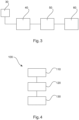
- Fig. 4 schematically illustrates a flow chart 100 of a method for determining a functional index for stenosis assessment.
- the method comprises the following steps:
- a first step 110 also referred to as step a
- image data corresponding to two-dimensional image data of a vessel are obtained.
- a course of the vessel and a width of the vessel along its course in the image data is determined.
- a functional index for stenosis assessment of the vessel is determined based on the width of the vessel in the image data.
- the step of obtaining image data comprises obtaining a projection of the vessel, wherein the width of the vessel is determined based on the projection.
- the method further comprises the steps of: determining a length of the vessel in the image; determining a scaling factor based on a densitometry approach; applying the scaling factor to the determined width of the vessel.
- a computer program element is provided, which, when being executed by a processing unit is adapted to carry out the method described above.
- a computer readable medium having stored thereon a program element is provided, which, when being executed by a processing unit is adapted to carry out the method described above.
- the approach described above relating to the apparatus and method for determining a functional index for stenosis assessment may be summed up as follows and it is proposed here to approximate this by one or more of the following: Apply 2D QCA methods to obtain a 2D segmentation and a vessel centerline of the culprit vessel segments.
- the scaling factor due to magnification can be estimated from either of the known system geometry and an estimate of the position of the heart inside the X-ray system, the known size of the catheter at the ostium of the contrary artery tree, or a phantom/reference element placed on the chest wall of the patient.
- a geometric model including a centerline and the local vessel radius r i for each centerline point are determined. This may include branching points.
- densitometric information is used to estimate the vessel diameter in the through-plane direction. This may be based on cardiac digital subtraction angiography (DSA). Furthermore, this may utilize a time series images over a complete cardiac cycle to improve the robustness of the densitometric measurement. Densitometric evaluation might be limited to a segment of the vessel, e.g. between two bifurcations.
- DSA cardiac digital subtraction angiography
- the projection angle is chosen such that the apparent stenosis diameter is minimized.
- This choice may be done manually by a human operator of an imaging system by selecting one of multiple acquired projections, or an automatic suggestion is made based on prior-knowledge using a reference database of projection images. This approach may result in a systematic underestimation of the cross-sectional area, and may lead to improved reproducibility as compared to an unguided approach.
- the projection angle is chosen (manually or automated) such that least vessel foreshortening occurs. This may be combined with the diameter minimization.
- a functional index for stenosis assessment is calculated.
- This functional index can, for instance, be either of: the pressure drop along the centerline of the vessel, virtual FFR, e.g. based on CFD simulations, a curve of the pressure drop as a function of the blood flow through the stenosis, or a quantity derived thereof, a fluid-dynamic resistance value, simulated average blood velocity profile or velocity distribution, or any other functional index that can be derived from hemodynamic parameters (e.g. CFR, iFR, etc.).
- the functional index for stenosis assessment is derived from a 2D projection image.
- a single angiographic 2D projection image with segmentation and pressure drop profile is used for functional assessment, as it can be obtained from fluid dynamics simulations.
- the index may be independently derived from a plurality of 2D images. That is, a functional index being derived "independently” involves deriving separate indices for individual images in a series, for instance carrying out the vessel segmentation, modeling and fluid dynamics simulation for each image of the series.
- Different images in the series are for example acquired in different cardiac states.
- different projection angles may be used providing different viewing angles on the vasculature.
- an improved assessment can be based on one or more of the functional indices derived from different images. For example, in this case, it may be helpful to merge the individual numbers into a combined index (e.g. the mean value).
- a variation between the functional indices from different images is determined. For example, such variation can be compared to a predetermined maximal variation (Vmax), based on which an acceptance criterion may be generated, in order to have feedback on the accuracy of the results. If the simulated values from multiple images are within expected or predetermined limits of variation, the acceptance criterion may indicate that the simulated results may be accepted with higher confidence than for a single frame evaluation.
- Vmax a predetermined maximal variation
- the expected maximal variation (Vmax) may be predetermined taking into account the nature of the different images of the sequence. For example, for multiple images taken at the same projection angle but in different heart phases, Vmax may be lower than for multiple images taken at different projection angles.
- the processing unit may be configured to calculate a quality score for one or more images being used as a basis for calculating the functional index for stenosis assessment.
- quality score may be based on one or more of the following quality parameters:
- the quality score may be visualized together with the angiographic X-ray image, the calculated functional index and, optionally, the determined segmentation of the vessel segment of interest.
- the quality score may be calculated for each image together with the functional index. For example, the quality score may then be used as a weighting factor in determining a combined functional index, whereby a functional index derived from a lower quality image is given less weight in the combined index as a functional index derived from a higher quality image.
- the approach described herein is applicable for functional assessment of stenosis in all major arteries of the human body (coronaries, iliac, femoral, brachial, hepatic, carotids).
- the functional index may be compared to one or more geometric measures for example obtained by means of QCA.
- Automatic QCA measurements may be carried out along the centerline of the vessel or vessel section of interest.
- an FFR value may be calculated for the same vessel or vessel section.
- Both the QCA and FFR values can be normalized and overlaid on top of the angiographic X-ray image that was used as a basis for the calculations.
- portions of the vessel where a discrepancy between the normalized QCA and FFR values exceeds a predetermined threshold may be determined and visualized on the image, for instance by means of highlighting or color coding.
- the normalization may be based on standard decision thresholds, e.g. a threshold for a decision whether a stent is to be placed or not. In that case, a QCA value of 0,5 may correspond to an FFR value of 0,8.
Landscapes
- Health & Medical Sciences (AREA)
- Life Sciences & Earth Sciences (AREA)
- Engineering & Computer Science (AREA)
- Physics & Mathematics (AREA)
- Medical Informatics (AREA)
- Public Health (AREA)
- General Health & Medical Sciences (AREA)
- Biomedical Technology (AREA)
- Pathology (AREA)
- Surgery (AREA)
- Heart & Thoracic Surgery (AREA)
- Molecular Biology (AREA)
- Biophysics (AREA)
- Animal Behavior & Ethology (AREA)
- Veterinary Medicine (AREA)
- Physiology (AREA)
- Cardiology (AREA)
- Vascular Medicine (AREA)
- Nuclear Medicine, Radiotherapy & Molecular Imaging (AREA)
- Radiology & Medical Imaging (AREA)
- Computer Vision & Pattern Recognition (AREA)
- Oral & Maxillofacial Surgery (AREA)
- Dentistry (AREA)
- High Energy & Nuclear Physics (AREA)
- Optics & Photonics (AREA)
- General Physics & Mathematics (AREA)
- Hematology (AREA)
- Geometry (AREA)
- Theoretical Computer Science (AREA)
- Electromagnetism (AREA)
- Data Mining & Analysis (AREA)
- Databases & Information Systems (AREA)
- Epidemiology (AREA)
- Primary Health Care (AREA)
- Apparatus For Radiation Diagnosis (AREA)
- Image Analysis (AREA)
Applications Claiming Priority (2)
| Application Number | Priority Date | Filing Date | Title |
|---|---|---|---|
| EP16191781 | 2016-09-30 | ||
| PCT/EP2017/075021 WO2018060529A1 (en) | 2016-09-30 | 2017-10-02 | Apparatus for determining a functional index for stenosis assessment |
Publications (2)
| Publication Number | Publication Date |
|---|---|
| EP3518751A1 EP3518751A1 (en) | 2019-08-07 |
| EP3518751B1 true EP3518751B1 (en) | 2025-01-29 |
Family
ID=57103837
Family Applications (1)
| Application Number | Title | Priority Date | Filing Date |
|---|---|---|---|
| EP17780700.5A Active EP3518751B1 (en) | 2016-09-30 | 2017-10-02 | Apparatus for determining a functional index for stenosis assessment |
Country Status (5)
| Country | Link |
|---|---|
| US (4) | US11179043B2 (enExample) |
| EP (1) | EP3518751B1 (enExample) |
| JP (1) | JP7109429B2 (enExample) |
| CN (1) | CN109843161B (enExample) |
| WO (1) | WO2018060529A1 (enExample) |
Families Citing this family (18)
| Publication number | Priority date | Publication date | Assignee | Title |
|---|---|---|---|---|
| US10210956B2 (en) | 2012-10-24 | 2019-02-19 | Cathworks Ltd. | Diagnostically useful results in real time |
| EP4241694A3 (en) | 2016-05-16 | 2023-12-20 | Cathworks Ltd. | Selection of vascular paths from images |
| JP7036742B2 (ja) | 2016-05-16 | 2022-03-15 | キャスワークス リミテッド | 血管評価システム |
| WO2019238754A1 (en) * | 2018-06-15 | 2019-12-19 | Pie Medical Imaging Bv | Method and apparatus for quantitative hemodynamic flow analysis |
| EP3871233A1 (en) | 2018-10-26 | 2021-09-01 | Koninklijke Philips N.V. | Determination of a treatment response index |
| EP3686898A1 (en) * | 2019-01-24 | 2020-07-29 | Koninklijke Philips N.V. | Determination of a treatment response index |
| JP7532402B2 (ja) | 2019-04-01 | 2024-08-13 | キャスワークス リミテッド | 血管造影画像選択のための方法および装置 |
| EP4033964B1 (en) | 2019-09-23 | 2025-04-09 | Cathworks Ltd. | Methods, apparatus, and system for synchronization between a three-dimensional vascular model and an imaging device |
| EP3828817B1 (en) * | 2019-11-28 | 2023-08-23 | Siemens Healthcare GmbH | Computer-implemented method for evaluating a ct data set regarding perivascular tissue, evaluation device, computer program and electronically readable storage medium |
| CN112472112B (zh) * | 2020-11-25 | 2024-02-27 | 苏州润迈德医疗科技有限公司 | 调节血管狭窄区间的方法、系统及存储介质 |
| US12315076B1 (en) | 2021-09-22 | 2025-05-27 | Cathworks Ltd. | Four-dimensional motion analysis of a patient's coronary arteries and myocardial wall |
| KR20240148399A (ko) | 2022-02-10 | 2024-10-11 | 캐스웍스 엘티디. | 기계 학습 기반 센서 분석 및 혈관 트리 분할을 위한 시스템 및 방법 |
| EP4311493A1 (en) | 2022-07-27 | 2024-01-31 | Koninklijke Philips N.V. | Spectral x-ray projection data |
| CN117670782A (zh) * | 2022-08-30 | 2024-03-08 | 武汉联影智融医疗科技有限公司 | 血管医学图像处理方法及其装置、医学图像处理设备 |
| CN121942048A (zh) | 2023-08-09 | 2026-04-28 | 凯思沃克斯有限公司 | 针对血管指数测量的增强用户界面和串扰分析 |
| IL326432A (en) | 2023-08-09 | 2026-04-01 | Cathworks Ltd | Coronary artery assessment after PCI |
| US12512196B2 (en) | 2024-06-12 | 2025-12-30 | Cathworks Ltd. | Systems and methods for secure sharing of cardiac assessments using QR codes |
| CN120078440B (zh) * | 2025-03-13 | 2025-12-23 | 苏州仲如悦科技有限责任公司 | 一种基于听诊声学信号的动静脉血管通路狭窄检测系统 |
Citations (2)
| Publication number | Priority date | Publication date | Assignee | Title |
|---|---|---|---|---|
| US20150051888A1 (en) * | 2012-03-15 | 2015-02-19 | Siemens Corporation | Framework for personalization of coronary flow computations during rest and hyperemia |
| EP3062248A1 (en) * | 2015-02-27 | 2016-08-31 | Pie Medical Imaging BV | Method and apparatus for quantitative flow analysis |
Family Cites Families (19)
| Publication number | Priority date | Publication date | Assignee | Title |
|---|---|---|---|---|
| US5150292A (en) * | 1989-10-27 | 1992-09-22 | Arch Development Corporation | Method and system for determination of instantaneous and average blood flow rates from digital angiograms |
| US7970187B2 (en) | 2005-09-06 | 2011-06-28 | Pie Medical Imaging B.V. | Method, apparatus and computer program for contour detection of vessels using x-ray densitometry |
| AU2010298333B2 (en) | 2009-09-23 | 2014-04-17 | Lightlab Imaging, Inc. | Lumen morphology and vascular resistance measurements data collection systems, apparatus and methods |
| EP2667816A2 (en) | 2011-01-28 | 2013-12-04 | Koninklijke Philips N.V. | Optical shape sensing fiber for tip and shape characterization of medical instruments |
| US9247918B2 (en) | 2012-07-09 | 2016-02-02 | Siemens Aktiengesellschaft | Computation of hemodynamic quantities from angiographic data |
| US20140236011A1 (en) * | 2012-08-31 | 2014-08-21 | General Electric Company | Methods and systems for simultaneous interventional imaging and functional measurements |
| US9858387B2 (en) | 2013-01-15 | 2018-01-02 | CathWorks, LTD. | Vascular flow assessment |
| US9814433B2 (en) * | 2012-10-24 | 2017-11-14 | Cathworks Ltd. | Creating a vascular tree model |
| US10210956B2 (en) * | 2012-10-24 | 2019-02-19 | Cathworks Ltd. | Diagnostically useful results in real time |
| US10595807B2 (en) * | 2012-10-24 | 2020-03-24 | Cathworks Ltd | Calculating a fractional flow reserve |
| US9943233B2 (en) * | 2012-10-24 | 2018-04-17 | Cathworks Ltd. | Automated measurement system and method for coronary artery disease scoring |
| WO2014072861A2 (en) | 2012-11-06 | 2014-05-15 | Koninklijke Philips N.V. | Fractional flow reserve (ffr) index |
| EP3061015A2 (en) | 2013-10-24 | 2016-08-31 | Cathworks Ltd. | Vascular characteristic determination with correspondence modeling of a vascular tree |
| US10111633B2 (en) * | 2013-12-04 | 2018-10-30 | Koninklijke Philips N.V. | Local FFR estimation and visualisation for improved functional stenosis analysis |
| NL2012459B1 (en) | 2014-03-18 | 2016-01-08 | Medis Ass B V | Method and device for determining deviation in pressure in a blood vessel. |
| DE102014210591B4 (de) | 2014-06-04 | 2022-09-22 | Siemens Healthcare Gmbh | Fluiddynamische Analyse eines Gefäßbaums mittels Angiographie |
| CN106659400B (zh) * | 2014-06-30 | 2021-01-05 | 皇家飞利浦有限公司 | 用于确定血流储备分数值的装置 |
| CN107580470B (zh) | 2015-01-15 | 2021-06-22 | 皇家飞利浦有限公司 | 瞬时流量储备-计算机断层摄影 |
| JP6818492B2 (ja) * | 2015-10-05 | 2021-01-20 | キヤノンメディカルシステムズ株式会社 | 画像処理装置、画像処理方法、及びプログラム |
-
2017
- 2017-10-02 JP JP2019516643A patent/JP7109429B2/ja active Active
- 2017-10-02 US US16/338,290 patent/US11179043B2/en active Active
- 2017-10-02 EP EP17780700.5A patent/EP3518751B1/en active Active
- 2017-10-02 WO PCT/EP2017/075021 patent/WO2018060529A1/en not_active Ceased
- 2017-10-02 CN CN201780060812.7A patent/CN109843161B/zh active Active
-
2021
- 2021-11-22 US US17/531,899 patent/US11690518B2/en active Active
-
2023
- 2023-07-03 US US18/217,713 patent/US12042249B2/en active Active
-
2024
- 2024-07-23 US US18/780,933 patent/US20240374148A1/en active Pending
Patent Citations (2)
| Publication number | Priority date | Publication date | Assignee | Title |
|---|---|---|---|---|
| US20150051888A1 (en) * | 2012-03-15 | 2015-02-19 | Siemens Corporation | Framework for personalization of coronary flow computations during rest and hyperemia |
| EP3062248A1 (en) * | 2015-02-27 | 2016-08-31 | Pie Medical Imaging BV | Method and apparatus for quantitative flow analysis |
Also Published As
| Publication number | Publication date |
|---|---|
| CN109843161A (zh) | 2019-06-04 |
| US20200029830A1 (en) | 2020-01-30 |
| US12042249B2 (en) | 2024-07-23 |
| US20240374148A1 (en) | 2024-11-14 |
| EP3518751A1 (en) | 2019-08-07 |
| US11690518B2 (en) | 2023-07-04 |
| JP7109429B2 (ja) | 2022-07-29 |
| WO2018060529A1 (en) | 2018-04-05 |
| CN109843161B (zh) | 2022-07-12 |
| US20220079455A1 (en) | 2022-03-17 |
| JP2019534740A (ja) | 2019-12-05 |
| US20230355107A1 (en) | 2023-11-09 |
| US11179043B2 (en) | 2021-11-23 |
Similar Documents
| Publication | Publication Date | Title |
|---|---|---|
| US12042249B2 (en) | Apparatus for determining a functional index for stenosis assessment | |
| US12193793B2 (en) | Method and apparatus for quantitative hemodynamic flow analysis | |
| EP4247263B1 (en) | Method and system for calculating myocardial infarction likelihood based on lesion wall shear stress descriptors | |
| EP3713482B1 (en) | Apparatus, computer program and computer-readable medium for assessing a coronary vasculature | |
| EP3140757B1 (en) | Method and system for non-invasive functional assessment of coronary artery stenosis using flow computations in models based on diseased patients and hypothetical normal anatomical models | |
| EP3685389B1 (en) | Estimating flow to vessel bifurcations for simulated hemodynamics | |
| US20230252628A1 (en) | Estimating flow to vessel bifurcations for simulated hemodynamics | |
| WO2014091339A1 (en) | Method of determining the blood flow through coronary arteries | |
| EP3512416B1 (en) | Apparatus and method for determining a fractional flow reserve | |
| EP4548838A1 (en) | Comuter-implemented method and system for calculating a pressure drop along vessels |
Legal Events
| Date | Code | Title | Description |
|---|---|---|---|
| STAA | Information on the status of an ep patent application or granted ep patent |
Free format text: STATUS: UNKNOWN |
|
| STAA | Information on the status of an ep patent application or granted ep patent |
Free format text: STATUS: THE INTERNATIONAL PUBLICATION HAS BEEN MADE |
|
| PUAI | Public reference made under article 153(3) epc to a published international application that has entered the european phase |
Free format text: ORIGINAL CODE: 0009012 |
|
| STAA | Information on the status of an ep patent application or granted ep patent |
Free format text: STATUS: REQUEST FOR EXAMINATION WAS MADE |
|
| 17P | Request for examination filed |
Effective date: 20190430 |
|
| AK | Designated contracting states |
Kind code of ref document: A1 Designated state(s): AL AT BE BG CH CY CZ DE DK EE ES FI FR GB GR HR HU IE IS IT LI LT LU LV MC MK MT NL NO PL PT RO RS SE SI SK SM TR |
|
| AX | Request for extension of the european patent |
Extension state: BA ME |
|
| DAV | Request for validation of the european patent (deleted) | ||
| DAX | Request for extension of the european patent (deleted) | ||
| RAP1 | Party data changed (applicant data changed or rights of an application transferred) |
Owner name: KONINKLIJKE PHILIPS N.V. |
|
| STAA | Information on the status of an ep patent application or granted ep patent |
Free format text: STATUS: EXAMINATION IS IN PROGRESS |
|
| 17Q | First examination report despatched |
Effective date: 20220607 |
|
| GRAP | Despatch of communication of intention to grant a patent |
Free format text: ORIGINAL CODE: EPIDOSNIGR1 |
|
| STAA | Information on the status of an ep patent application or granted ep patent |
Free format text: STATUS: GRANT OF PATENT IS INTENDED |
|
| RIC1 | Information provided on ipc code assigned before grant |
Ipc: G16H 50/30 20180101ALI20240812BHEP Ipc: G06T 7/62 20170101ALI20240812BHEP Ipc: G01B 15/00 20060101ALI20240812BHEP Ipc: A61B 6/50 20240101ALI20240812BHEP Ipc: G01N 21/59 20060101ALI20240812BHEP Ipc: A61B 5/021 20060101ALI20240812BHEP Ipc: A61B 5/026 20060101ALI20240812BHEP Ipc: A61B 5/00 20060101ALI20240812BHEP Ipc: A61B 6/00 20060101ALI20240812BHEP Ipc: A61B 5/107 20060101ALI20240812BHEP Ipc: A61B 5/02 20060101AFI20240812BHEP |
|
| INTG | Intention to grant announced |
Effective date: 20240827 |
|
| GRAS | Grant fee paid |
Free format text: ORIGINAL CODE: EPIDOSNIGR3 |
|
| GRAA | (expected) grant |
Free format text: ORIGINAL CODE: 0009210 |
|
| STAA | Information on the status of an ep patent application or granted ep patent |
Free format text: STATUS: THE PATENT HAS BEEN GRANTED |
|
| AK | Designated contracting states |
Kind code of ref document: B1 Designated state(s): AL AT BE BG CH CY CZ DE DK EE ES FI FR GB GR HR HU IE IS IT LI LT LU LV MC MK MT NL NO PL PT RO RS SE SI SK SM TR |
|
| REG | Reference to a national code |
Ref country code: GB Ref legal event code: FG4D |
|
| REG | Reference to a national code |
Ref country code: CH Ref legal event code: EP |
|
| REG | Reference to a national code |
Ref country code: DE Ref legal event code: R096 Ref document number: 602017087554 Country of ref document: DE |
|
| REG | Reference to a national code |
Ref country code: IE Ref legal event code: FG4D |
|
| REG | Reference to a national code |
Ref country code: NL Ref legal event code: MP Effective date: 20250129 |
|
| PG25 | Lapsed in a contracting state [announced via postgrant information from national office to epo] |
Ref country code: NL Free format text: LAPSE BECAUSE OF FAILURE TO SUBMIT A TRANSLATION OF THE DESCRIPTION OR TO PAY THE FEE WITHIN THE PRESCRIBED TIME-LIMIT Effective date: 20250129 |
|
| PG25 | Lapsed in a contracting state [announced via postgrant information from national office to epo] |
Ref country code: RS Free format text: LAPSE BECAUSE OF FAILURE TO SUBMIT A TRANSLATION OF THE DESCRIPTION OR TO PAY THE FEE WITHIN THE PRESCRIBED TIME-LIMIT Effective date: 20250429 |
|
| PG25 | Lapsed in a contracting state [announced via postgrant information from national office to epo] |
Ref country code: FI Free format text: LAPSE BECAUSE OF FAILURE TO SUBMIT A TRANSLATION OF THE DESCRIPTION OR TO PAY THE FEE WITHIN THE PRESCRIBED TIME-LIMIT Effective date: 20250129 |
|
| PG25 | Lapsed in a contracting state [announced via postgrant information from national office to epo] |
Ref country code: PL Free format text: LAPSE BECAUSE OF FAILURE TO SUBMIT A TRANSLATION OF THE DESCRIPTION OR TO PAY THE FEE WITHIN THE PRESCRIBED TIME-LIMIT Effective date: 20250129 |
|
| PG25 | Lapsed in a contracting state [announced via postgrant information from national office to epo] |
Ref country code: ES Free format text: LAPSE BECAUSE OF FAILURE TO SUBMIT A TRANSLATION OF THE DESCRIPTION OR TO PAY THE FEE WITHIN THE PRESCRIBED TIME-LIMIT Effective date: 20250129 |
|
| REG | Reference to a national code |
Ref country code: LT Ref legal event code: MG9D |
|
| PG25 | Lapsed in a contracting state [announced via postgrant information from national office to epo] |
Ref country code: IS Free format text: LAPSE BECAUSE OF FAILURE TO SUBMIT A TRANSLATION OF THE DESCRIPTION OR TO PAY THE FEE WITHIN THE PRESCRIBED TIME-LIMIT Effective date: 20250529 Ref country code: NO Free format text: LAPSE BECAUSE OF FAILURE TO SUBMIT A TRANSLATION OF THE DESCRIPTION OR TO PAY THE FEE WITHIN THE PRESCRIBED TIME-LIMIT Effective date: 20250429 |
|
| REG | Reference to a national code |
Ref country code: AT Ref legal event code: MK05 Ref document number: 1762621 Country of ref document: AT Kind code of ref document: T Effective date: 20250129 |
|
| PG25 | Lapsed in a contracting state [announced via postgrant information from national office to epo] |
Ref country code: HR Free format text: LAPSE BECAUSE OF FAILURE TO SUBMIT A TRANSLATION OF THE DESCRIPTION OR TO PAY THE FEE WITHIN THE PRESCRIBED TIME-LIMIT Effective date: 20250129 |
|
| PG25 | Lapsed in a contracting state [announced via postgrant information from national office to epo] |
Ref country code: LV Free format text: LAPSE BECAUSE OF FAILURE TO SUBMIT A TRANSLATION OF THE DESCRIPTION OR TO PAY THE FEE WITHIN THE PRESCRIBED TIME-LIMIT Effective date: 20250129 Ref country code: PT Free format text: LAPSE BECAUSE OF FAILURE TO SUBMIT A TRANSLATION OF THE DESCRIPTION OR TO PAY THE FEE WITHIN THE PRESCRIBED TIME-LIMIT Effective date: 20250529 |
|
| PG25 | Lapsed in a contracting state [announced via postgrant information from national office to epo] |
Ref country code: BG Free format text: LAPSE BECAUSE OF FAILURE TO SUBMIT A TRANSLATION OF THE DESCRIPTION OR TO PAY THE FEE WITHIN THE PRESCRIBED TIME-LIMIT Effective date: 20250129 Ref country code: GR Free format text: LAPSE BECAUSE OF FAILURE TO SUBMIT A TRANSLATION OF THE DESCRIPTION OR TO PAY THE FEE WITHIN THE PRESCRIBED TIME-LIMIT Effective date: 20250430 |
|
| PG25 | Lapsed in a contracting state [announced via postgrant information from national office to epo] |
Ref country code: AT Free format text: LAPSE BECAUSE OF FAILURE TO SUBMIT A TRANSLATION OF THE DESCRIPTION OR TO PAY THE FEE WITHIN THE PRESCRIBED TIME-LIMIT Effective date: 20250129 |
|
| PG25 | Lapsed in a contracting state [announced via postgrant information from national office to epo] |
Ref country code: SE Free format text: LAPSE BECAUSE OF FAILURE TO SUBMIT A TRANSLATION OF THE DESCRIPTION OR TO PAY THE FEE WITHIN THE PRESCRIBED TIME-LIMIT Effective date: 20250129 |
|
| PG25 | Lapsed in a contracting state [announced via postgrant information from national office to epo] |
Ref country code: SM Free format text: LAPSE BECAUSE OF FAILURE TO SUBMIT A TRANSLATION OF THE DESCRIPTION OR TO PAY THE FEE WITHIN THE PRESCRIBED TIME-LIMIT Effective date: 20250129 |
|
| PG25 | Lapsed in a contracting state [announced via postgrant information from national office to epo] |
Ref country code: DK Free format text: LAPSE BECAUSE OF FAILURE TO SUBMIT A TRANSLATION OF THE DESCRIPTION OR TO PAY THE FEE WITHIN THE PRESCRIBED TIME-LIMIT Effective date: 20250129 |
|
| PG25 | Lapsed in a contracting state [announced via postgrant information from national office to epo] |
Ref country code: IT Free format text: LAPSE BECAUSE OF FAILURE TO SUBMIT A TRANSLATION OF THE DESCRIPTION OR TO PAY THE FEE WITHIN THE PRESCRIBED TIME-LIMIT Effective date: 20250129 |
|
| PG25 | Lapsed in a contracting state [announced via postgrant information from national office to epo] |
Ref country code: EE Free format text: LAPSE BECAUSE OF FAILURE TO SUBMIT A TRANSLATION OF THE DESCRIPTION OR TO PAY THE FEE WITHIN THE PRESCRIBED TIME-LIMIT Effective date: 20250129 Ref country code: CZ Free format text: LAPSE BECAUSE OF FAILURE TO SUBMIT A TRANSLATION OF THE DESCRIPTION OR TO PAY THE FEE WITHIN THE PRESCRIBED TIME-LIMIT Effective date: 20250129 |
|
| PG25 | Lapsed in a contracting state [announced via postgrant information from national office to epo] |
Ref country code: RO Free format text: LAPSE BECAUSE OF FAILURE TO SUBMIT A TRANSLATION OF THE DESCRIPTION OR TO PAY THE FEE WITHIN THE PRESCRIBED TIME-LIMIT Effective date: 20250129 |
|
| PG25 | Lapsed in a contracting state [announced via postgrant information from national office to epo] |
Ref country code: SK Free format text: LAPSE BECAUSE OF FAILURE TO SUBMIT A TRANSLATION OF THE DESCRIPTION OR TO PAY THE FEE WITHIN THE PRESCRIBED TIME-LIMIT Effective date: 20250129 |
|
| REG | Reference to a national code |
Ref country code: DE Ref legal event code: R097 Ref document number: 602017087554 Country of ref document: DE |
|
| PLBE | No opposition filed within time limit |
Free format text: ORIGINAL CODE: 0009261 |
|
| STAA | Information on the status of an ep patent application or granted ep patent |
Free format text: STATUS: NO OPPOSITION FILED WITHIN TIME LIMIT |
|
| 26N | No opposition filed |
Effective date: 20251030 |
|
| PGFP | Annual fee paid to national office [announced via postgrant information from national office to epo] |
Ref country code: DE Payment date: 20251028 Year of fee payment: 9 |
|
| PGFP | Annual fee paid to national office [announced via postgrant information from national office to epo] |
Ref country code: GB Payment date: 20251023 Year of fee payment: 9 |
|
| PGFP | Annual fee paid to national office [announced via postgrant information from national office to epo] |
Ref country code: FR Payment date: 20251027 Year of fee payment: 9 |