EP3511413B1 - Lentiviral vectors for regulated expression of a chimeric antigen receptor molecule - Google Patents
Lentiviral vectors for regulated expression of a chimeric antigen receptor molecule Download PDFInfo
- Publication number
- EP3511413B1 EP3511413B1 EP19159129.6A EP19159129A EP3511413B1 EP 3511413 B1 EP3511413 B1 EP 3511413B1 EP 19159129 A EP19159129 A EP 19159129A EP 3511413 B1 EP3511413 B1 EP 3511413B1
- Authority
- EP
- European Patent Office
- Prior art keywords
- seq
- cell
- cells
- car
- chimeric antigen
- Prior art date
- Legal status (The legal status is an assumption and is not a legal conclusion. Google has not performed a legal analysis and makes no representation as to the accuracy of the status listed.)
- Active
Links
- 239000013598 vector Substances 0.000 title claims description 253
- 108010019670 Chimeric Antigen Receptors Proteins 0.000 title claims description 183
- 230000014509 gene expression Effects 0.000 title claims description 106
- 230000001105 regulatory effect Effects 0.000 title description 16
- 210000004027 cell Anatomy 0.000 claims description 222
- 210000001744 T-lymphocyte Anatomy 0.000 claims description 130
- YBJHBAHKTGYVGT-ZKWXMUAHSA-N (+)-Biotin Chemical compound N1C(=O)N[C@@H]2[C@H](CCCCC(=O)O)SC[C@@H]21 YBJHBAHKTGYVGT-ZKWXMUAHSA-N 0.000 claims description 101
- 150000007523 nucleic acids Chemical class 0.000 claims description 95
- 230000027455 binding Effects 0.000 claims description 84
- 239000002245 particle Substances 0.000 claims description 73
- 241000282414 Homo sapiens Species 0.000 claims description 72
- 102000039446 nucleic acids Human genes 0.000 claims description 71
- 108020004707 nucleic acids Proteins 0.000 claims description 71
- 150000001413 amino acids Chemical class 0.000 claims description 63
- 238000000034 method Methods 0.000 claims description 55
- 229960002685 biotin Drugs 0.000 claims description 51
- 235000020958 biotin Nutrition 0.000 claims description 51
- 239000011616 biotin Substances 0.000 claims description 51
- 239000012634 fragment Substances 0.000 claims description 50
- 230000004913 activation Effects 0.000 claims description 23
- 230000003213 activating effect Effects 0.000 claims description 21
- 230000028993 immune response Effects 0.000 claims description 21
- 206010028980 Neoplasm Diseases 0.000 claims description 15
- 108010018381 streptavidin-binding peptide Proteins 0.000 claims description 12
- 108010081355 beta 2-Microglobulin Proteins 0.000 claims description 9
- 102000015736 beta 2-Microglobulin Human genes 0.000 claims description 9
- 238000011282 treatment Methods 0.000 claims description 9
- 108090000848 Ubiquitin Proteins 0.000 claims description 8
- 238000000338 in vitro Methods 0.000 claims description 8
- 102000044159 Ubiquitin Human genes 0.000 claims description 7
- 210000000987 immune system Anatomy 0.000 claims description 6
- 230000001939 inductive effect Effects 0.000 claims description 6
- 210000000822 natural killer cell Anatomy 0.000 claims description 6
- 238000003306 harvesting Methods 0.000 claims description 5
- 230000002463 transducing effect Effects 0.000 claims description 5
- 208000023275 Autoimmune disease Diseases 0.000 claims description 2
- 210000005260 human cell Anatomy 0.000 claims description 2
- 210000004962 mammalian cell Anatomy 0.000 claims description 2
- 210000004986 primary T-cell Anatomy 0.000 claims description 2
- 125000003275 alpha amino acid group Chemical group 0.000 claims 2
- 108090000623 proteins and genes Proteins 0.000 description 100
- 108020004414 DNA Proteins 0.000 description 72
- 102000004169 proteins and genes Human genes 0.000 description 67
- 108090000765 processed proteins & peptides Proteins 0.000 description 66
- 235000018102 proteins Nutrition 0.000 description 65
- 102000004196 processed proteins & peptides Human genes 0.000 description 57
- 229920001184 polypeptide Polymers 0.000 description 52
- 235000001014 amino acid Nutrition 0.000 description 41
- 229940024606 amino acid Drugs 0.000 description 41
- 108010090804 Streptavidin Proteins 0.000 description 29
- 238000010361 transduction Methods 0.000 description 29
- 108091054437 MHC class I family Proteins 0.000 description 27
- 239000013612 plasmid Substances 0.000 description 27
- 102000043129 MHC class I family Human genes 0.000 description 25
- 108091054438 MHC class II family Proteins 0.000 description 23
- 108091028043 Nucleic acid sequence Proteins 0.000 description 23
- 238000011534 incubation Methods 0.000 description 23
- 125000003729 nucleotide group Chemical group 0.000 description 23
- 239000002773 nucleotide Substances 0.000 description 22
- 239000000427 antigen Substances 0.000 description 21
- 108091007433 antigens Proteins 0.000 description 21
- 102000036639 antigens Human genes 0.000 description 21
- 102000043131 MHC class II family Human genes 0.000 description 20
- 210000002472 endoplasmic reticulum Anatomy 0.000 description 20
- 239000003623 enhancer Substances 0.000 description 20
- 210000004443 dendritic cell Anatomy 0.000 description 19
- 238000003752 polymerase chain reaction Methods 0.000 description 19
- 239000002609 medium Substances 0.000 description 18
- 230000026683 transduction Effects 0.000 description 18
- 239000005090 green fluorescent protein Substances 0.000 description 16
- 239000000203 mixture Substances 0.000 description 15
- 238000004806 packaging method and process Methods 0.000 description 15
- 210000003819 peripheral blood mononuclear cell Anatomy 0.000 description 15
- 108700018351 Major Histocompatibility Complex Proteins 0.000 description 14
- 230000001413 cellular effect Effects 0.000 description 14
- 102000040430 polynucleotide Human genes 0.000 description 14
- 108091033319 polynucleotide Proteins 0.000 description 14
- 239000002157 polynucleotide Substances 0.000 description 14
- 230000003612 virological effect Effects 0.000 description 14
- 108091032973 (ribonucleotides)n+m Proteins 0.000 description 13
- 108090000144 Human Proteins Proteins 0.000 description 13
- 102000003839 Human Proteins Human genes 0.000 description 13
- 230000020382 suppression by virus of host antigen processing and presentation of peptide antigen via MHC class I Effects 0.000 description 13
- 102000014914 Carrier Proteins Human genes 0.000 description 12
- 108091008324 binding proteins Proteins 0.000 description 12
- 239000000523 sample Substances 0.000 description 12
- 229960005486 vaccine Drugs 0.000 description 12
- 241000124008 Mammalia Species 0.000 description 11
- 238000005516 engineering process Methods 0.000 description 11
- 238000002955 isolation Methods 0.000 description 11
- 238000004519 manufacturing process Methods 0.000 description 11
- 230000001177 retroviral effect Effects 0.000 description 11
- 102100034349 Integrase Human genes 0.000 description 10
- 241000713666 Lentivirus Species 0.000 description 10
- 108700019146 Transgenes Proteins 0.000 description 10
- 210000000612 antigen-presenting cell Anatomy 0.000 description 10
- 238000000926 separation method Methods 0.000 description 10
- 241000700605 Viruses Species 0.000 description 9
- 230000000694 effects Effects 0.000 description 9
- 230000035772 mutation Effects 0.000 description 9
- 239000000047 product Substances 0.000 description 9
- OZFAFGSSMRRTDW-UHFFFAOYSA-N (2,4-dichlorophenyl) benzenesulfonate Chemical compound ClC1=CC(Cl)=CC=C1OS(=O)(=O)C1=CC=CC=C1 OZFAFGSSMRRTDW-UHFFFAOYSA-N 0.000 description 8
- 102100024222 B-lymphocyte antigen CD19 Human genes 0.000 description 8
- 241000283707 Capra Species 0.000 description 8
- 239000012591 Dulbecco’s Phosphate Buffered Saline Substances 0.000 description 8
- 108010043121 Green Fluorescent Proteins Proteins 0.000 description 8
- 102000004144 Green Fluorescent Proteins Human genes 0.000 description 8
- 101000980825 Homo sapiens B-lymphocyte antigen CD19 Proteins 0.000 description 8
- 101000946843 Homo sapiens T-cell surface glycoprotein CD8 alpha chain Proteins 0.000 description 8
- 102100034922 T-cell surface glycoprotein CD8 alpha chain Human genes 0.000 description 8
- 108020001507 fusion proteins Proteins 0.000 description 8
- 102000037865 fusion proteins Human genes 0.000 description 8
- 230000010354 integration Effects 0.000 description 8
- 230000014759 maintenance of location Effects 0.000 description 8
- 239000012146 running buffer Substances 0.000 description 8
- 230000035899 viability Effects 0.000 description 8
- 108700028146 Genetic Enhancer Elements Proteins 0.000 description 7
- DHMQDGOQFOQNFH-UHFFFAOYSA-N Glycine Chemical compound NCC(O)=O DHMQDGOQFOQNFH-UHFFFAOYSA-N 0.000 description 7
- 108010061833 Integrases Proteins 0.000 description 7
- 241001465754 Metazoa Species 0.000 description 7
- 241000699666 Mus <mouse, genus> Species 0.000 description 7
- 241000711975 Vesicular stomatitis virus Species 0.000 description 7
- 230000001086 cytosolic effect Effects 0.000 description 7
- 230000003834 intracellular effect Effects 0.000 description 7
- 210000004698 lymphocyte Anatomy 0.000 description 7
- 230000004048 modification Effects 0.000 description 7
- 238000012986 modification Methods 0.000 description 7
- 230000001566 pro-viral effect Effects 0.000 description 7
- 230000010076 replication Effects 0.000 description 7
- 238000013518 transcription Methods 0.000 description 7
- 230000035897 transcription Effects 0.000 description 7
- 241001529936 Murinae Species 0.000 description 6
- 241000699670 Mus sp. Species 0.000 description 6
- 108091034117 Oligonucleotide Proteins 0.000 description 6
- 230000006044 T cell activation Effects 0.000 description 6
- 238000003556 assay Methods 0.000 description 6
- 201000011510 cancer Diseases 0.000 description 6
- 238000012217 deletion Methods 0.000 description 6
- 230000037430 deletion Effects 0.000 description 6
- 238000011156 evaluation Methods 0.000 description 6
- 238000000684 flow cytometry Methods 0.000 description 6
- 238000001943 fluorescence-activated cell sorting Methods 0.000 description 6
- 239000001963 growth medium Substances 0.000 description 6
- 230000002163 immunogen Effects 0.000 description 6
- 238000002743 insertional mutagenesis Methods 0.000 description 6
- 230000003993 interaction Effects 0.000 description 6
- 230000001225 therapeutic effect Effects 0.000 description 6
- 238000012546 transfer Methods 0.000 description 6
- 101150104383 ALOX5AP gene Proteins 0.000 description 5
- 101710125370 C-type lectin domain family 6 member A Proteins 0.000 description 5
- 102000053602 DNA Human genes 0.000 description 5
- 229920001917 Ficoll Polymers 0.000 description 5
- 101000914514 Homo sapiens T-cell-specific surface glycoprotein CD28 Proteins 0.000 description 5
- 102000008394 Immunoglobulin Fragments Human genes 0.000 description 5
- 101100236114 Mus musculus Lrrfip1 gene Proteins 0.000 description 5
- 108010004729 Phycoerythrin Proteins 0.000 description 5
- 102100027213 T-cell-specific surface glycoprotein CD28 Human genes 0.000 description 5
- 230000015572 biosynthetic process Effects 0.000 description 5
- 230000010261 cell growth Effects 0.000 description 5
- 239000000470 constituent Substances 0.000 description 5
- 230000006870 function Effects 0.000 description 5
- 238000009169 immunotherapy Methods 0.000 description 5
- 238000001727 in vivo Methods 0.000 description 5
- 230000006698 induction Effects 0.000 description 5
- 238000002347 injection Methods 0.000 description 5
- 239000007924 injection Substances 0.000 description 5
- -1 nef Proteins 0.000 description 5
- 210000001519 tissue Anatomy 0.000 description 5
- 238000002255 vaccination Methods 0.000 description 5
- 241000713704 Bovine immunodeficiency virus Species 0.000 description 4
- 241000713756 Caprine arthritis encephalitis virus Species 0.000 description 4
- 241000713800 Feline immunodeficiency virus Species 0.000 description 4
- 102100028970 HLA class I histocompatibility antigen, alpha chain E Human genes 0.000 description 4
- 102100028966 HLA class I histocompatibility antigen, alpha chain F Human genes 0.000 description 4
- 108010074032 HLA-A2 Antigen Proteins 0.000 description 4
- 102000025850 HLA-A2 Antigen Human genes 0.000 description 4
- 108010091938 HLA-B7 Antigen Proteins 0.000 description 4
- 108010016121 HLA-C*05 antigen Proteins 0.000 description 4
- 241000282412 Homo Species 0.000 description 4
- 101000986085 Homo sapiens HLA class I histocompatibility antigen, alpha chain E Proteins 0.000 description 4
- 101000986080 Homo sapiens HLA class I histocompatibility antigen, alpha chain F Proteins 0.000 description 4
- 241000725303 Human immunodeficiency virus Species 0.000 description 4
- 241000713772 Human immunodeficiency virus 1 Species 0.000 description 4
- 108091054729 IRF family Proteins 0.000 description 4
- 108010021625 Immunoglobulin Fragments Proteins 0.000 description 4
- 102000016854 Interferon Regulatory Factors Human genes 0.000 description 4
- 108010002350 Interleukin-2 Proteins 0.000 description 4
- 108010076504 Protein Sorting Signals Proteins 0.000 description 4
- 108020004511 Recombinant DNA Proteins 0.000 description 4
- 241000713311 Simian immunodeficiency virus Species 0.000 description 4
- 108010003723 Single-Domain Antibodies Proteins 0.000 description 4
- JLCPHMBAVCMARE-UHFFFAOYSA-N [3-[[3-[[3-[[3-[[3-[[3-[[3-[[3-[[3-[[3-[[3-[[5-(2-amino-6-oxo-1H-purin-9-yl)-3-[[3-[[3-[[3-[[3-[[3-[[5-(2-amino-6-oxo-1H-purin-9-yl)-3-[[5-(2-amino-6-oxo-1H-purin-9-yl)-3-hydroxyoxolan-2-yl]methoxy-hydroxyphosphoryl]oxyoxolan-2-yl]methoxy-hydroxyphosphoryl]oxy-5-(5-methyl-2,4-dioxopyrimidin-1-yl)oxolan-2-yl]methoxy-hydroxyphosphoryl]oxy-5-(6-aminopurin-9-yl)oxolan-2-yl]methoxy-hydroxyphosphoryl]oxy-5-(6-aminopurin-9-yl)oxolan-2-yl]methoxy-hydroxyphosphoryl]oxy-5-(6-aminopurin-9-yl)oxolan-2-yl]methoxy-hydroxyphosphoryl]oxy-5-(6-aminopurin-9-yl)oxolan-2-yl]methoxy-hydroxyphosphoryl]oxyoxolan-2-yl]methoxy-hydroxyphosphoryl]oxy-5-(5-methyl-2,4-dioxopyrimidin-1-yl)oxolan-2-yl]methoxy-hydroxyphosphoryl]oxy-5-(4-amino-2-oxopyrimidin-1-yl)oxolan-2-yl]methoxy-hydroxyphosphoryl]oxy-5-(5-methyl-2,4-dioxopyrimidin-1-yl)oxolan-2-yl]methoxy-hydroxyphosphoryl]oxy-5-(5-methyl-2,4-dioxopyrimidin-1-yl)oxolan-2-yl]methoxy-hydroxyphosphoryl]oxy-5-(6-aminopurin-9-yl)oxolan-2-yl]methoxy-hydroxyphosphoryl]oxy-5-(6-aminopurin-9-yl)oxolan-2-yl]methoxy-hydroxyphosphoryl]oxy-5-(4-amino-2-oxopyrimidin-1-yl)oxolan-2-yl]methoxy-hydroxyphosphoryl]oxy-5-(4-amino-2-oxopyrimidin-1-yl)oxolan-2-yl]methoxy-hydroxyphosphoryl]oxy-5-(4-amino-2-oxopyrimidin-1-yl)oxolan-2-yl]methoxy-hydroxyphosphoryl]oxy-5-(6-aminopurin-9-yl)oxolan-2-yl]methoxy-hydroxyphosphoryl]oxy-5-(4-amino-2-oxopyrimidin-1-yl)oxolan-2-yl]methyl [5-(6-aminopurin-9-yl)-2-(hydroxymethyl)oxolan-3-yl] hydrogen phosphate Polymers Cc1cn(C2CC(OP(O)(=O)OCC3OC(CC3OP(O)(=O)OCC3OC(CC3O)n3cnc4c3nc(N)[nH]c4=O)n3cnc4c3nc(N)[nH]c4=O)C(COP(O)(=O)OC3CC(OC3COP(O)(=O)OC3CC(OC3COP(O)(=O)OC3CC(OC3COP(O)(=O)OC3CC(OC3COP(O)(=O)OC3CC(OC3COP(O)(=O)OC3CC(OC3COP(O)(=O)OC3CC(OC3COP(O)(=O)OC3CC(OC3COP(O)(=O)OC3CC(OC3COP(O)(=O)OC3CC(OC3COP(O)(=O)OC3CC(OC3COP(O)(=O)OC3CC(OC3COP(O)(=O)OC3CC(OC3COP(O)(=O)OC3CC(OC3COP(O)(=O)OC3CC(OC3COP(O)(=O)OC3CC(OC3COP(O)(=O)OC3CC(OC3CO)n3cnc4c(N)ncnc34)n3ccc(N)nc3=O)n3cnc4c(N)ncnc34)n3ccc(N)nc3=O)n3ccc(N)nc3=O)n3ccc(N)nc3=O)n3cnc4c(N)ncnc34)n3cnc4c(N)ncnc34)n3cc(C)c(=O)[nH]c3=O)n3cc(C)c(=O)[nH]c3=O)n3ccc(N)nc3=O)n3cc(C)c(=O)[nH]c3=O)n3cnc4c3nc(N)[nH]c4=O)n3cnc4c(N)ncnc34)n3cnc4c(N)ncnc34)n3cnc4c(N)ncnc34)n3cnc4c(N)ncnc34)O2)c(=O)[nH]c1=O JLCPHMBAVCMARE-UHFFFAOYSA-N 0.000 description 4
- 230000000890 antigenic effect Effects 0.000 description 4
- 239000011324 bead Substances 0.000 description 4
- 230000006399 behavior Effects 0.000 description 4
- 230000008901 benefit Effects 0.000 description 4
- 210000004369 blood Anatomy 0.000 description 4
- 239000008280 blood Substances 0.000 description 4
- 210000000170 cell membrane Anatomy 0.000 description 4
- 239000003795 chemical substances by application Substances 0.000 description 4
- 238000010276 construction Methods 0.000 description 4
- 238000011161 development Methods 0.000 description 4
- 230000004927 fusion Effects 0.000 description 4
- 238000001415 gene therapy Methods 0.000 description 4
- 230000002068 genetic effect Effects 0.000 description 4
- 210000004408 hybridoma Anatomy 0.000 description 4
- 238000012744 immunostaining Methods 0.000 description 4
- 208000032839 leukemia Diseases 0.000 description 4
- 239000002105 nanoparticle Substances 0.000 description 4
- 238000003908 quality control method Methods 0.000 description 4
- 239000000126 substance Substances 0.000 description 4
- 238000003146 transient transfection Methods 0.000 description 4
- 230000032258 transport Effects 0.000 description 4
- 241001430294 unidentified retrovirus Species 0.000 description 4
- GNENVASJJIUNER-UHFFFAOYSA-N 2,4,6-tricyclohexyloxy-1,3,5,2,4,6-trioxatriborinane Chemical compound C1CCCCC1OB1OB(OC2CCCCC2)OB(OC2CCCCC2)O1 GNENVASJJIUNER-UHFFFAOYSA-N 0.000 description 3
- 102100040839 C-type lectin domain family 6 member A Human genes 0.000 description 3
- 108010029697 CD40 Ligand Proteins 0.000 description 3
- 102100032937 CD40 ligand Human genes 0.000 description 3
- 101710091045 Envelope protein Proteins 0.000 description 3
- 241000283073 Equus caballus Species 0.000 description 3
- 101150066002 GFP gene Proteins 0.000 description 3
- 239000004471 Glycine Substances 0.000 description 3
- 101100382122 Homo sapiens CIITA gene Proteins 0.000 description 3
- 101000623857 Homo sapiens Serine/threonine-protein kinase mTOR Proteins 0.000 description 3
- 101000716102 Homo sapiens T-cell surface glycoprotein CD4 Proteins 0.000 description 3
- ZDXPYRJPNDTMRX-VKHMYHEASA-N L-glutamine Chemical compound OC(=O)[C@@H](N)CCC(N)=O ZDXPYRJPNDTMRX-VKHMYHEASA-N 0.000 description 3
- 102100026371 MHC class II transactivator Human genes 0.000 description 3
- 108700002010 MHC class II transactivator Proteins 0.000 description 3
- 206010035226 Plasma cell myeloma Diseases 0.000 description 3
- 101710188315 Protein X Proteins 0.000 description 3
- 241000700159 Rattus Species 0.000 description 3
- 102100023085 Serine/threonine-protein kinase mTOR Human genes 0.000 description 3
- 102100036011 T-cell surface glycoprotein CD4 Human genes 0.000 description 3
- AYFVYJQAPQTCCC-UHFFFAOYSA-N Threonine Natural products CC(O)C(N)C(O)=O AYFVYJQAPQTCCC-UHFFFAOYSA-N 0.000 description 3
- 239000004473 Threonine Substances 0.000 description 3
- 239000002671 adjuvant Substances 0.000 description 3
- 230000001745 anti-biotin effect Effects 0.000 description 3
- 230000030741 antigen processing and presentation Effects 0.000 description 3
- 239000012298 atmosphere Substances 0.000 description 3
- 238000005119 centrifugation Methods 0.000 description 3
- 238000006243 chemical reaction Methods 0.000 description 3
- 230000008030 elimination Effects 0.000 description 3
- 238000003379 elimination reaction Methods 0.000 description 3
- 238000002474 experimental method Methods 0.000 description 3
- 239000013604 expression vector Substances 0.000 description 3
- 238000010353 genetic engineering Methods 0.000 description 3
- 210000002865 immune cell Anatomy 0.000 description 3
- 230000008105 immune reaction Effects 0.000 description 3
- 230000001976 improved effect Effects 0.000 description 3
- 208000015181 infectious disease Diseases 0.000 description 3
- 238000003780 insertion Methods 0.000 description 3
- 230000037431 insertion Effects 0.000 description 3
- 210000003292 kidney cell Anatomy 0.000 description 3
- 229940126601 medicinal product Drugs 0.000 description 3
- 239000012528 membrane Substances 0.000 description 3
- 108020004999 messenger RNA Proteins 0.000 description 3
- 244000005700 microbiome Species 0.000 description 3
- 230000003278 mimic effect Effects 0.000 description 3
- 201000000050 myeloid neoplasm Diseases 0.000 description 3
- 239000008188 pellet Substances 0.000 description 3
- 239000013600 plasmid vector Substances 0.000 description 3
- 108010089520 pol Gene Products Proteins 0.000 description 3
- 108700004029 pol Genes Proteins 0.000 description 3
- 102000005962 receptors Human genes 0.000 description 3
- 108020003175 receptors Proteins 0.000 description 3
- 230000009467 reduction Effects 0.000 description 3
- 230000000717 retained effect Effects 0.000 description 3
- 238000010839 reverse transcription Methods 0.000 description 3
- 230000002441 reversible effect Effects 0.000 description 3
- 208000002491 severe combined immunodeficiency Diseases 0.000 description 3
- 210000004989 spleen cell Anatomy 0.000 description 3
- 239000006228 supernatant Substances 0.000 description 3
- 238000012360 testing method Methods 0.000 description 3
- 238000001890 transfection Methods 0.000 description 3
- 238000013519 translation Methods 0.000 description 3
- QCUPYFTWJOZAOB-HWKANZROSA-N (e)-n-carbamoyl-2-ethylbut-2-enamide Chemical compound CC\C(=C/C)C(=O)NC(N)=O QCUPYFTWJOZAOB-HWKANZROSA-N 0.000 description 2
- 102000005869 Activating Transcription Factors Human genes 0.000 description 2
- 108010005254 Activating Transcription Factors Proteins 0.000 description 2
- 101100256368 Arabidopsis thaliana SBP3 gene Proteins 0.000 description 2
- 239000004475 Arginine Substances 0.000 description 2
- 241000894006 Bacteria Species 0.000 description 2
- 108020000946 Bacterial DNA Proteins 0.000 description 2
- 241000283690 Bos taurus Species 0.000 description 2
- 102100027207 CD27 antigen Human genes 0.000 description 2
- 241000282472 Canis lupus familiaris Species 0.000 description 2
- 102000005636 Cyclic AMP Response Element-Binding Protein Human genes 0.000 description 2
- 108010045171 Cyclic AMP Response Element-Binding Protein Proteins 0.000 description 2
- 238000002965 ELISA Methods 0.000 description 2
- 241000196324 Embryophyta Species 0.000 description 2
- 108010087819 Fc receptors Proteins 0.000 description 2
- 102000009109 Fc receptors Human genes 0.000 description 2
- 241000724791 Filamentous phage Species 0.000 description 2
- WHUUTDBJXJRKMK-UHFFFAOYSA-N Glutamic acid Natural products OC(=O)C(N)CCC(O)=O WHUUTDBJXJRKMK-UHFFFAOYSA-N 0.000 description 2
- 102000003886 Glycoproteins Human genes 0.000 description 2
- 108090000288 Glycoproteins Proteins 0.000 description 2
- 102100029360 Hematopoietic cell signal transducer Human genes 0.000 description 2
- 102000008949 Histocompatibility Antigens Class I Human genes 0.000 description 2
- 108010088652 Histocompatibility Antigens Class I Proteins 0.000 description 2
- 101000914511 Homo sapiens CD27 antigen Proteins 0.000 description 2
- 101000990188 Homo sapiens Hematopoietic cell signal transducer Proteins 0.000 description 2
- 101000692225 Homo sapiens Selenocysteine insertion sequence-binding protein 2 Proteins 0.000 description 2
- 208000006142 Infectious Encephalitis Diseases 0.000 description 2
- DCXYFEDJOCDNAF-REOHCLBHSA-N L-asparagine Chemical compound OC(=O)[C@@H](N)CC(N)=O DCXYFEDJOCDNAF-REOHCLBHSA-N 0.000 description 2
- KDXKERNSBIXSRK-UHFFFAOYSA-N Lysine Natural products NCCCCC(N)C(O)=O KDXKERNSBIXSRK-UHFFFAOYSA-N 0.000 description 2
- TWRXJAOTZQYOKJ-UHFFFAOYSA-L Magnesium chloride Chemical compound [Mg+2].[Cl-].[Cl-] TWRXJAOTZQYOKJ-UHFFFAOYSA-L 0.000 description 2
- 108010057466 NF-kappa B Proteins 0.000 description 2
- 102000003945 NF-kappa B Human genes 0.000 description 2
- 108700026244 Open Reading Frames Proteins 0.000 description 2
- 241000283973 Oryctolagus cuniculus Species 0.000 description 2
- 241001494479 Pecora Species 0.000 description 2
- 239000002202 Polyethylene glycol Substances 0.000 description 2
- 241000288906 Primates Species 0.000 description 2
- 102000052575 Proto-Oncogene Human genes 0.000 description 2
- 108700020978 Proto-Oncogene Proteins 0.000 description 2
- 230000004570 RNA-binding Effects 0.000 description 2
- 238000011529 RT qPCR Methods 0.000 description 2
- 102100026077 Selenocysteine insertion sequence-binding protein 2 Human genes 0.000 description 2
- 108010008038 Synthetic Vaccines Proteins 0.000 description 2
- 208000012827 T-B+ severe combined immunodeficiency due to gamma chain deficiency Diseases 0.000 description 2
- 102000040945 Transcription factor Human genes 0.000 description 2
- 108091023040 Transcription factor Proteins 0.000 description 2
- 108091061763 Triple-stranded DNA Proteins 0.000 description 2
- 102100022153 Tumor necrosis factor receptor superfamily member 4 Human genes 0.000 description 2
- 101710165473 Tumor necrosis factor receptor superfamily member 4 Proteins 0.000 description 2
- 102100028298 Vacuolar protein sorting-associated protein VTA1 homolog Human genes 0.000 description 2
- 101710111280 Vacuolar protein sorting-associated protein VTA1 homolog Proteins 0.000 description 2
- 201000007146 X-linked severe combined immunodeficiency Diseases 0.000 description 2
- DZBUGLKDJFMEHC-UHFFFAOYSA-N acridine Chemical compound C1=CC=CC2=CC3=CC=CC=C3N=C21 DZBUGLKDJFMEHC-UHFFFAOYSA-N 0.000 description 2
- 230000003321 amplification Effects 0.000 description 2
- 238000004458 analytical method Methods 0.000 description 2
- 238000010171 animal model Methods 0.000 description 2
- ODKSFYDXXFIFQN-UHFFFAOYSA-N arginine Natural products OC(=O)C(N)CCCNC(N)=N ODKSFYDXXFIFQN-UHFFFAOYSA-N 0.000 description 2
- 210000004436 artificial bacterial chromosome Anatomy 0.000 description 2
- 210000001106 artificial yeast chromosome Anatomy 0.000 description 2
- 230000033228 biological regulation Effects 0.000 description 2
- 229960001714 calcium phosphate Drugs 0.000 description 2
- 239000001506 calcium phosphate Substances 0.000 description 2
- 229910000389 calcium phosphate Inorganic materials 0.000 description 2
- 235000011010 calcium phosphates Nutrition 0.000 description 2
- 238000004113 cell culture Methods 0.000 description 2
- 239000002299 complementary DNA Substances 0.000 description 2
- 238000007796 conventional method Methods 0.000 description 2
- 210000004748 cultured cell Anatomy 0.000 description 2
- 210000001151 cytotoxic T lymphocyte Anatomy 0.000 description 2
- 238000013461 design Methods 0.000 description 2
- 201000010099 disease Diseases 0.000 description 2
- 208000037265 diseases, disorders, signs and symptoms Diseases 0.000 description 2
- 108700004025 env Genes Proteins 0.000 description 2
- 239000012091 fetal bovine serum Substances 0.000 description 2
- 108700004026 gag Genes Proteins 0.000 description 2
- 230000003053 immunization Effects 0.000 description 2
- 230000002998 immunogenetic effect Effects 0.000 description 2
- 230000005847 immunogenicity Effects 0.000 description 2
- 230000006872 improvement Effects 0.000 description 2
- 239000000411 inducer Substances 0.000 description 2
- 230000000670 limiting effect Effects 0.000 description 2
- 230000036210 malignancy Effects 0.000 description 2
- 230000001404 mediated effect Effects 0.000 description 2
- 230000005012 migration Effects 0.000 description 2
- 238000013508 migration Methods 0.000 description 2
- 238000009126 molecular therapy Methods 0.000 description 2
- 108010028606 nuclear factor Y Proteins 0.000 description 2
- 230000012223 nuclear import Effects 0.000 description 2
- 238000003199 nucleic acid amplification method Methods 0.000 description 2
- 210000004940 nucleus Anatomy 0.000 description 2
- 230000002688 persistence Effects 0.000 description 2
- 230000002085 persistent effect Effects 0.000 description 2
- 229920001223 polyethylene glycol Polymers 0.000 description 2
- 229920000642 polymer Polymers 0.000 description 2
- 230000001124 posttranscriptional effect Effects 0.000 description 2
- 238000002360 preparation method Methods 0.000 description 2
- 230000008569 process Effects 0.000 description 2
- 238000012545 processing Methods 0.000 description 2
- ZCCUUQDIBDJBTK-UHFFFAOYSA-N psoralen Chemical compound C1=C2OC(=O)C=CC2=CC2=C1OC=C2 ZCCUUQDIBDJBTK-UHFFFAOYSA-N 0.000 description 2
- 238000011002 quantification Methods 0.000 description 2
- ZAHRKKWIAAJSAO-UHFFFAOYSA-N rapamycin Natural products COCC(O)C(=C/C(C)C(=O)CC(OC(=O)C1CCCCN1C(=O)C(=O)C2(O)OC(CC(OC)C(=CC=CC=CC(C)CC(C)C(=O)C)C)CCC2C)C(C)CC3CCC(O)C(C3)OC)C ZAHRKKWIAAJSAO-UHFFFAOYSA-N 0.000 description 2
- 230000009257 reactivity Effects 0.000 description 2
- 229940124551 recombinant vaccine Drugs 0.000 description 2
- 230000002829 reductive effect Effects 0.000 description 2
- 108091008146 restriction endonucleases Proteins 0.000 description 2
- 210000002966 serum Anatomy 0.000 description 2
- QFJCIRLUMZQUOT-HPLJOQBZSA-N sirolimus Chemical compound C1C[C@@H](O)[C@H](OC)C[C@@H]1C[C@@H](C)[C@H]1OC(=O)[C@@H]2CCCCN2C(=O)C(=O)[C@](O)(O2)[C@H](C)CC[C@H]2C[C@H](OC)/C(C)=C/C=C/C=C/[C@@H](C)C[C@@H](C)C(=O)[C@H](OC)[C@H](O)/C(C)=C/[C@@H](C)C(=O)C1 QFJCIRLUMZQUOT-HPLJOQBZSA-N 0.000 description 2
- 229960002930 sirolimus Drugs 0.000 description 2
- DAEPDZWVDSPTHF-UHFFFAOYSA-M sodium pyruvate Chemical compound [Na+].CC(=O)C([O-])=O DAEPDZWVDSPTHF-UHFFFAOYSA-M 0.000 description 2
- 239000007787 solid Substances 0.000 description 2
- 241000894007 species Species 0.000 description 2
- 238000010186 staining Methods 0.000 description 2
- 239000000758 substrate Substances 0.000 description 2
- 238000004448 titration Methods 0.000 description 2
- 230000001052 transient effect Effects 0.000 description 2
- QORWJWZARLRLPR-UHFFFAOYSA-H tricalcium bis(phosphate) Chemical compound [Ca+2].[Ca+2].[Ca+2].[O-]P([O-])([O-])=O.[O-]P([O-])([O-])=O QORWJWZARLRLPR-UHFFFAOYSA-H 0.000 description 2
- 238000011144 upstream manufacturing Methods 0.000 description 2
- 238000005406 washing Methods 0.000 description 2
- 108010015889 zeta receptor Proteins 0.000 description 2
- YURDCJXYOLERLO-LCYFTJDESA-N (2E)-5-methyl-2-phenylhex-2-enal Chemical compound CC(C)C\C=C(\C=O)C1=CC=CC=C1 YURDCJXYOLERLO-LCYFTJDESA-N 0.000 description 1
- JBFQOLHAGBKPTP-NZATWWQASA-N (2s)-2-[[(2s)-4-carboxy-2-[[3-carboxy-2-[[(2s)-2,6-diaminohexanoyl]amino]propanoyl]amino]butanoyl]amino]-4-methylpentanoic acid Chemical compound CC(C)C[C@@H](C(O)=O)NC(=O)[C@H](CCC(O)=O)NC(=O)C(CC(O)=O)NC(=O)[C@@H](N)CCCCN JBFQOLHAGBKPTP-NZATWWQASA-N 0.000 description 1
- 102000040650 (ribonucleotides)n+m Human genes 0.000 description 1
- VXGRJERITKFWPL-UHFFFAOYSA-N 4',5'-Dihydropsoralen Natural products C1=C2OC(=O)C=CC2=CC2=C1OCC2 VXGRJERITKFWPL-UHFFFAOYSA-N 0.000 description 1
- 108010085238 Actins Proteins 0.000 description 1
- 244000303258 Annona diversifolia Species 0.000 description 1
- 235000002198 Annona diversifolia Nutrition 0.000 description 1
- DCXYFEDJOCDNAF-UHFFFAOYSA-N Asparagine Natural products OC(=O)C(N)CC(N)=O DCXYFEDJOCDNAF-UHFFFAOYSA-N 0.000 description 1
- 208000031504 Asymptomatic Infections Diseases 0.000 description 1
- 108091003079 Bovine Serum Albumin Proteins 0.000 description 1
- 238000011740 C57BL/6 mouse Methods 0.000 description 1
- 108050005493 CD3 protein, epsilon/gamma/delta subunit Proteins 0.000 description 1
- 210000001266 CD8-positive T-lymphocyte Anatomy 0.000 description 1
- QCMYYKRYFNMIEC-UHFFFAOYSA-N COP(O)=O Chemical class COP(O)=O QCMYYKRYFNMIEC-UHFFFAOYSA-N 0.000 description 1
- UXVMQQNJUSDDNG-UHFFFAOYSA-L Calcium chloride Chemical compound [Cl-].[Cl-].[Ca+2] UXVMQQNJUSDDNG-UHFFFAOYSA-L 0.000 description 1
- 208000005623 Carcinogenesis Diseases 0.000 description 1
- 108091026890 Coding region Proteins 0.000 description 1
- 108020004705 Codon Proteins 0.000 description 1
- 206010010144 Completed suicide Diseases 0.000 description 1
- 108090000695 Cytokines Proteins 0.000 description 1
- 102000004127 Cytokines Human genes 0.000 description 1
- 102100020986 DNA-binding protein RFX5 Human genes 0.000 description 1
- 102100021044 DNA-binding protein RFXANK Human genes 0.000 description 1
- 102000016911 Deoxyribonucleases Human genes 0.000 description 1
- 108010053770 Deoxyribonucleases Proteins 0.000 description 1
- 239000006144 Dulbecco’s modified Eagle's medium Substances 0.000 description 1
- 101150029707 ERBB2 gene Proteins 0.000 description 1
- 241000283086 Equidae Species 0.000 description 1
- 241000289695 Eutheria Species 0.000 description 1
- 241000282326 Felis catus Species 0.000 description 1
- 101710177291 Gag polyprotein Proteins 0.000 description 1
- 241000287828 Gallus gallus Species 0.000 description 1
- 108700039691 Genetic Promoter Regions Proteins 0.000 description 1
- 102100041032 Golgin subfamily A member 5 Human genes 0.000 description 1
- 101710176014 Golgin subfamily A member 5 Proteins 0.000 description 1
- 101710071593 Golgin-84 Proteins 0.000 description 1
- 208000031886 HIV Infections Diseases 0.000 description 1
- 208000002250 Hematologic Neoplasms Diseases 0.000 description 1
- 101000937544 Homo sapiens Beta-2-microglobulin Proteins 0.000 description 1
- 101100273713 Homo sapiens CD2 gene Proteins 0.000 description 1
- 101001075432 Homo sapiens DNA-binding protein RFX5 Proteins 0.000 description 1
- 101001057504 Homo sapiens Interferon-stimulated gene 20 kDa protein Proteins 0.000 description 1
- 101001055144 Homo sapiens Interleukin-2 receptor subunit alpha Proteins 0.000 description 1
- 101000917858 Homo sapiens Low affinity immunoglobulin gamma Fc region receptor III-A Proteins 0.000 description 1
- 101000917839 Homo sapiens Low affinity immunoglobulin gamma Fc region receptor III-B Proteins 0.000 description 1
- 101000934338 Homo sapiens Myeloid cell surface antigen CD33 Proteins 0.000 description 1
- 101100194605 Homo sapiens RFXANK gene Proteins 0.000 description 1
- 101001075466 Homo sapiens Regulatory factor X-associated protein Proteins 0.000 description 1
- 101001115218 Homo sapiens Ubiquitin-40S ribosomal protein S27a Proteins 0.000 description 1
- 241000713340 Human immunodeficiency virus 2 Species 0.000 description 1
- 208000029462 Immunodeficiency disease Diseases 0.000 description 1
- 108060003951 Immunoglobulin Proteins 0.000 description 1
- 108010054477 Immunoglobulin Fab Fragments Proteins 0.000 description 1
- 102000001706 Immunoglobulin Fab Fragments Human genes 0.000 description 1
- 102100034353 Integrase Human genes 0.000 description 1
- 102000014150 Interferons Human genes 0.000 description 1
- 108010050904 Interferons Proteins 0.000 description 1
- 102100026878 Interleukin-2 receptor subunit alpha Human genes 0.000 description 1
- 101150008942 J gene Proteins 0.000 description 1
- QNAYBMKLOCPYGJ-REOHCLBHSA-N L-alanine Chemical compound C[C@H](N)C(O)=O QNAYBMKLOCPYGJ-REOHCLBHSA-N 0.000 description 1
- CKLJMWTZIZZHCS-REOHCLBHSA-N L-aspartic acid Chemical compound OC(=O)[C@@H](N)CC(O)=O CKLJMWTZIZZHCS-REOHCLBHSA-N 0.000 description 1
- 229930182816 L-glutamine Natural products 0.000 description 1
- AGPKZVBTJJNPAG-WHFBIAKZSA-N L-isoleucine Chemical compound CC[C@H](C)[C@H](N)C(O)=O AGPKZVBTJJNPAG-WHFBIAKZSA-N 0.000 description 1
- ROHFNLRQFUQHCH-YFKPBYRVSA-N L-leucine Chemical compound CC(C)C[C@H](N)C(O)=O ROHFNLRQFUQHCH-YFKPBYRVSA-N 0.000 description 1
- FFEARJCKVFRZRR-BYPYZUCNSA-N L-methionine Chemical compound CSCC[C@H](N)C(O)=O FFEARJCKVFRZRR-BYPYZUCNSA-N 0.000 description 1
- COLNVLDHVKWLRT-QMMMGPOBSA-N L-phenylalanine Chemical compound OC(=O)[C@@H](N)CC1=CC=CC=C1 COLNVLDHVKWLRT-QMMMGPOBSA-N 0.000 description 1
- QIVBCDIJIAJPQS-VIFPVBQESA-N L-tryptophane Chemical compound C1=CC=C2C(C[C@H](N)C(O)=O)=CNC2=C1 QIVBCDIJIAJPQS-VIFPVBQESA-N 0.000 description 1
- OUYCCCASQSFEME-QMMMGPOBSA-N L-tyrosine Chemical compound OC(=O)[C@@H](N)CC1=CC=C(O)C=C1 OUYCCCASQSFEME-QMMMGPOBSA-N 0.000 description 1
- KZSNJWFQEVHDMF-BYPYZUCNSA-N L-valine Chemical compound CC(C)[C@H](N)C(O)=O KZSNJWFQEVHDMF-BYPYZUCNSA-N 0.000 description 1
- 101710192602 Latent membrane protein 1 Proteins 0.000 description 1
- ROHFNLRQFUQHCH-UHFFFAOYSA-N Leucine Natural products CC(C)CC(N)C(O)=O ROHFNLRQFUQHCH-UHFFFAOYSA-N 0.000 description 1
- 102100029185 Low affinity immunoglobulin gamma Fc region receptor III-B Human genes 0.000 description 1
- 206010025323 Lymphomas Diseases 0.000 description 1
- 239000004472 Lysine Substances 0.000 description 1
- 108700005089 MHC Class I Genes Proteins 0.000 description 1
- 101710125418 Major capsid protein Proteins 0.000 description 1
- 241000283923 Marmota monax Species 0.000 description 1
- 102000029749 Microtubule Human genes 0.000 description 1
- 108091022875 Microtubule Proteins 0.000 description 1
- 241000713869 Moloney murine leukemia virus Species 0.000 description 1
- 241000714177 Murine leukemia virus Species 0.000 description 1
- 101100226902 Mus musculus Fcrlb gene Proteins 0.000 description 1
- 102100025243 Myeloid cell surface antigen CD33 Human genes 0.000 description 1
- BKAYIFDRRZZKNF-VIFPVBQESA-N N-acetylcarnosine Chemical compound CC(=O)NCCC(=O)N[C@H](C(O)=O)CC1=CN=CN1 BKAYIFDRRZZKNF-VIFPVBQESA-N 0.000 description 1
- 108700019961 Neoplasm Genes Proteins 0.000 description 1
- 102000048850 Neoplasm Genes Human genes 0.000 description 1
- 101100101420 Neurospora crassa (strain ATCC 24698 / 74-OR23-1A / CBS 708.71 / DSM 1257 / FGSC 987) ubi gene Proteins 0.000 description 1
- 108010038807 Oligopeptides Proteins 0.000 description 1
- 102000015636 Oligopeptides Human genes 0.000 description 1
- 238000012408 PCR amplification Methods 0.000 description 1
- 229910019142 PO4 Inorganic materials 0.000 description 1
- 108010076039 Polyproteins Proteins 0.000 description 1
- ONIBWKKTOPOVIA-UHFFFAOYSA-N Proline Natural products OC(=O)C1CCCN1 ONIBWKKTOPOVIA-UHFFFAOYSA-N 0.000 description 1
- 108010029485 Protein Isoforms Proteins 0.000 description 1
- 102000001708 Protein Isoforms Human genes 0.000 description 1
- 244000078856 Prunus padus Species 0.000 description 1
- 108010030933 Regulatory Factor X1 Proteins 0.000 description 1
- 102000005945 Regulatory Factor X1 Human genes 0.000 description 1
- 102100021043 Regulatory factor X-associated protein Human genes 0.000 description 1
- 108091027981 Response element Proteins 0.000 description 1
- 241000712907 Retroviridae Species 0.000 description 1
- 241000283984 Rodentia Species 0.000 description 1
- MTCFGRXMJLQNBG-UHFFFAOYSA-N Serine Natural products OCC(N)C(O)=O MTCFGRXMJLQNBG-UHFFFAOYSA-N 0.000 description 1
- 102000004094 Stromal Interaction Molecule 1 Human genes 0.000 description 1
- 108090000532 Stromal Interaction Molecule 1 Proteins 0.000 description 1
- 108091008874 T cell receptors Proteins 0.000 description 1
- 102000016266 T-Cell Antigen Receptors Human genes 0.000 description 1
- 108010065917 TOR Serine-Threonine Kinases Proteins 0.000 description 1
- 102000013530 TOR Serine-Threonine Kinases Human genes 0.000 description 1
- RYYWUUFWQRZTIU-UHFFFAOYSA-N Thiophosphoric acid Chemical class OP(O)(S)=O RYYWUUFWQRZTIU-UHFFFAOYSA-N 0.000 description 1
- 108700009124 Transcription Initiation Site Proteins 0.000 description 1
- QIVBCDIJIAJPQS-UHFFFAOYSA-N Tryptophan Natural products C1=CC=C2C(CC(N)C(O)=O)=CNC2=C1 QIVBCDIJIAJPQS-UHFFFAOYSA-N 0.000 description 1
- 102100023341 Ubiquitin-40S ribosomal protein S27a Human genes 0.000 description 1
- KZSNJWFQEVHDMF-UHFFFAOYSA-N Valine Natural products CC(C)C(N)C(O)=O KZSNJWFQEVHDMF-UHFFFAOYSA-N 0.000 description 1
- 108020005202 Viral DNA Proteins 0.000 description 1
- 108010003533 Viral Envelope Proteins Proteins 0.000 description 1
- 108010067390 Viral Proteins Proteins 0.000 description 1
- 239000002253 acid Substances 0.000 description 1
- 230000002411 adverse Effects 0.000 description 1
- 235000004279 alanine Nutrition 0.000 description 1
- 239000002168 alkylating agent Substances 0.000 description 1
- 239000005557 antagonist Substances 0.000 description 1
- 230000000692 anti-sense effect Effects 0.000 description 1
- 125000000637 arginyl group Chemical group N[C@@H](CCCNC(N)=N)C(=O)* 0.000 description 1
- 235000009582 asparagine Nutrition 0.000 description 1
- 229960001230 asparagine Drugs 0.000 description 1
- 235000003704 aspartic acid Nutrition 0.000 description 1
- 230000002238 attenuated effect Effects 0.000 description 1
- 230000001580 bacterial effect Effects 0.000 description 1
- 238000002869 basic local alignment search tool Methods 0.000 description 1
- OQFSQFPPLPISGP-UHFFFAOYSA-N beta-carboxyaspartic acid Natural products OC(=O)C(N)C(C(O)=O)C(O)=O OQFSQFPPLPISGP-UHFFFAOYSA-N 0.000 description 1
- 230000003115 biocidal effect Effects 0.000 description 1
- 230000008827 biological function Effects 0.000 description 1
- 230000009141 biological interaction Effects 0.000 description 1
- FMWLUWPQPKEARP-UHFFFAOYSA-N bromodichloromethane Chemical compound ClC(Cl)Br FMWLUWPQPKEARP-UHFFFAOYSA-N 0.000 description 1
- 210000004899 c-terminal region Anatomy 0.000 description 1
- 239000001110 calcium chloride Substances 0.000 description 1
- 229910001628 calcium chloride Inorganic materials 0.000 description 1
- 230000036952 cancer formation Effects 0.000 description 1
- 150000004657 carbamic acid derivatives Chemical class 0.000 description 1
- 231100000504 carcinogenesis Toxicity 0.000 description 1
- 239000006143 cell culture medium Substances 0.000 description 1
- 238000012512 characterization method Methods 0.000 description 1
- 239000002738 chelating agent Substances 0.000 description 1
- 239000003153 chemical reaction reagent Substances 0.000 description 1
- 238000002512 chemotherapy Methods 0.000 description 1
- 235000013330 chicken meat Nutrition 0.000 description 1
- 210000000349 chromosome Anatomy 0.000 description 1
- 238000010367 cloning Methods 0.000 description 1
- 238000004590 computer program Methods 0.000 description 1
- 108091036078 conserved sequence Proteins 0.000 description 1
- 238000010924 continuous production Methods 0.000 description 1
- 239000013601 cosmid vector Substances 0.000 description 1
- 108091008034 costimulatory receptors Proteins 0.000 description 1
- 238000004132 cross linking Methods 0.000 description 1
- 238000012258 culturing Methods 0.000 description 1
- 235000018417 cysteine Nutrition 0.000 description 1
- XUJNEKJLAYXESH-UHFFFAOYSA-N cysteine Natural products SCC(N)C(O)=O XUJNEKJLAYXESH-UHFFFAOYSA-N 0.000 description 1
- 230000007123 defense Effects 0.000 description 1
- 230000001419 dependent effect Effects 0.000 description 1
- 238000009795 derivation Methods 0.000 description 1
- 238000002050 diffraction method Methods 0.000 description 1
- 238000010790 dilution Methods 0.000 description 1
- 239000012895 dilution Substances 0.000 description 1
- 239000012153 distilled water Substances 0.000 description 1
- NAGJZTKCGNOGPW-UHFFFAOYSA-N dithiophosphoric acid Chemical class OP(O)(S)=S NAGJZTKCGNOGPW-UHFFFAOYSA-N 0.000 description 1
- 108010078428 env Gene Products Proteins 0.000 description 1
- 230000007613 environmental effect Effects 0.000 description 1
- 230000002255 enzymatic effect Effects 0.000 description 1
- 238000001476 gene delivery Methods 0.000 description 1
- 231100000025 genetic toxicology Toxicity 0.000 description 1
- 230000001738 genotoxic effect Effects 0.000 description 1
- 235000013922 glutamic acid Nutrition 0.000 description 1
- 239000004220 glutamic acid Substances 0.000 description 1
- ZDXPYRJPNDTMRX-UHFFFAOYSA-N glutamine Natural products OC(=O)C(N)CCC(N)=O ZDXPYRJPNDTMRX-UHFFFAOYSA-N 0.000 description 1
- 230000012010 growth Effects 0.000 description 1
- HNDVDQJCIGZPNO-UHFFFAOYSA-N histidine Natural products OC(=O)C(N)CC1=CN=CN1 HNDVDQJCIGZPNO-UHFFFAOYSA-N 0.000 description 1
- 238000002744 homologous recombination Methods 0.000 description 1
- 230000006801 homologous recombination Effects 0.000 description 1
- 102000047279 human B2M Human genes 0.000 description 1
- 229910052739 hydrogen Inorganic materials 0.000 description 1
- 239000001257 hydrogen Substances 0.000 description 1
- 230000036039 immunity Effects 0.000 description 1
- 238000002649 immunization Methods 0.000 description 1
- 238000000760 immunoelectrophoresis Methods 0.000 description 1
- 238000010166 immunofluorescence Methods 0.000 description 1
- 102000018358 immunoglobulin Human genes 0.000 description 1
- 230000008676 import Effects 0.000 description 1
- 238000010874 in vitro model Methods 0.000 description 1
- 229940031551 inactivated vaccine Drugs 0.000 description 1
- 230000000977 initiatory effect Effects 0.000 description 1
- 238000011081 inoculation Methods 0.000 description 1
- 229940079322 interferon Drugs 0.000 description 1
- 230000004068 intracellular signaling Effects 0.000 description 1
- 238000007918 intramuscular administration Methods 0.000 description 1
- 230000002601 intratumoral effect Effects 0.000 description 1
- 238000001990 intravenous administration Methods 0.000 description 1
- 108010028930 invariant chain Proteins 0.000 description 1
- 238000011835 investigation Methods 0.000 description 1
- AGPKZVBTJJNPAG-UHFFFAOYSA-N isoleucine Natural products CCC(C)C(N)C(O)=O AGPKZVBTJJNPAG-UHFFFAOYSA-N 0.000 description 1
- 229960000310 isoleucine Drugs 0.000 description 1
- 239000008101 lactose Substances 0.000 description 1
- 239000003446 ligand Substances 0.000 description 1
- 244000144972 livestock Species 0.000 description 1
- 210000001165 lymph node Anatomy 0.000 description 1
- 108010089256 lysyl-aspartyl-glutamyl-leucine Proteins 0.000 description 1
- 229910001629 magnesium chloride Inorganic materials 0.000 description 1
- 239000000463 material Substances 0.000 description 1
- 239000011159 matrix material Substances 0.000 description 1
- 230000007246 mechanism Effects 0.000 description 1
- 210000003071 memory t lymphocyte Anatomy 0.000 description 1
- MYWUZJCMWCOHBA-VIFPVBQESA-N methamphetamine Chemical compound CN[C@@H](C)CC1=CC=CC=C1 MYWUZJCMWCOHBA-VIFPVBQESA-N 0.000 description 1
- 229930182817 methionine Natural products 0.000 description 1
- 230000011987 methylation Effects 0.000 description 1
- 238000007069 methylation reaction Methods 0.000 description 1
- 230000000813 microbial effect Effects 0.000 description 1
- 210000004688 microtubule Anatomy 0.000 description 1
- 230000009456 molecular mechanism Effects 0.000 description 1
- 230000003472 neutralizing effect Effects 0.000 description 1
- 230000009871 nonspecific binding Effects 0.000 description 1
- 238000010606 normalization Methods 0.000 description 1
- 210000004492 nuclear pore Anatomy 0.000 description 1
- 231100000590 oncogenic Toxicity 0.000 description 1
- 230000002246 oncogenic effect Effects 0.000 description 1
- 230000008520 organization Effects 0.000 description 1
- 238000004091 panning Methods 0.000 description 1
- 230000001717 pathogenic effect Effects 0.000 description 1
- 230000007918 pathogenicity Effects 0.000 description 1
- 230000007170 pathology Effects 0.000 description 1
- 230000037361 pathway Effects 0.000 description 1
- 210000005259 peripheral blood Anatomy 0.000 description 1
- 239000011886 peripheral blood Substances 0.000 description 1
- COLNVLDHVKWLRT-UHFFFAOYSA-N phenylalanine Natural products OC(=O)C(N)CC1=CC=CC=C1 COLNVLDHVKWLRT-UHFFFAOYSA-N 0.000 description 1
- NBIIXXVUZAFLBC-UHFFFAOYSA-K phosphate Chemical compound [O-]P([O-])([O-])=O NBIIXXVUZAFLBC-UHFFFAOYSA-K 0.000 description 1
- 239000010452 phosphate Substances 0.000 description 1
- 150000008298 phosphoramidates Chemical class 0.000 description 1
- 101150088264 pol gene Proteins 0.000 description 1
- 230000008488 polyadenylation Effects 0.000 description 1
- 239000013641 positive control Substances 0.000 description 1
- 230000023603 positive regulation of transcription initiation, DNA-dependent Effects 0.000 description 1
- 239000002244 precipitate Substances 0.000 description 1
- 238000001556 precipitation Methods 0.000 description 1
- 229940021993 prophylactic vaccine Drugs 0.000 description 1
- 230000012846 protein folding Effects 0.000 description 1
- 238000001742 protein purification Methods 0.000 description 1
- 238000000746 purification Methods 0.000 description 1
- 238000003127 radioimmunoassay Methods 0.000 description 1
- 238000003156 radioimmunoprecipitation Methods 0.000 description 1
- 238000003259 recombinant expression Methods 0.000 description 1
- 230000022532 regulation of transcription, DNA-dependent Effects 0.000 description 1
- 238000009256 replacement therapy Methods 0.000 description 1
- 230000003362 replicative effect Effects 0.000 description 1
- 230000004044 response Effects 0.000 description 1
- 108700004030 rev Genes Proteins 0.000 description 1
- 210000003705 ribosome Anatomy 0.000 description 1
- 125000000548 ribosyl group Chemical group C1([C@H](O)[C@H](O)[C@H](O1)CO)* 0.000 description 1
- 238000012216 screening Methods 0.000 description 1
- 230000009291 secondary effect Effects 0.000 description 1
- 230000028327 secretion Effects 0.000 description 1
- 210000002027 skeletal muscle Anatomy 0.000 description 1
- 210000002363 skeletal muscle cell Anatomy 0.000 description 1
- 229940054269 sodium pyruvate Drugs 0.000 description 1
- 230000009870 specific binding Effects 0.000 description 1
- 238000011895 specific detection Methods 0.000 description 1
- 230000010473 stable expression Effects 0.000 description 1
- 210000000130 stem cell Anatomy 0.000 description 1
- 230000000638 stimulation Effects 0.000 description 1
- 229960005322 streptomycin Drugs 0.000 description 1
- 238000006467 substitution reaction Methods 0.000 description 1
- 230000004083 survival effect Effects 0.000 description 1
- 230000008961 swelling Effects 0.000 description 1
- 208000011580 syndromic disease Diseases 0.000 description 1
- 230000002194 synthesizing effect Effects 0.000 description 1
- 108700004027 tat Genes Proteins 0.000 description 1
- 238000002560 therapeutic procedure Methods 0.000 description 1
- 229940021747 therapeutic vaccine Drugs 0.000 description 1
- 231100000331 toxic Toxicity 0.000 description 1
- 230000002588 toxic effect Effects 0.000 description 1
- 230000005026 transcription initiation Effects 0.000 description 1
- 230000005030 transcription termination Effects 0.000 description 1
- 108091006106 transcriptional activators Proteins 0.000 description 1
- 230000002103 transcriptional effect Effects 0.000 description 1
- 230000009261 transgenic effect Effects 0.000 description 1
- 230000010474 transient expression Effects 0.000 description 1
- 231100000588 tumorigenic Toxicity 0.000 description 1
- 230000000381 tumorigenic effect Effects 0.000 description 1
- OUYCCCASQSFEME-UHFFFAOYSA-N tyrosine Natural products OC(=O)C(N)CC1=CC=C(O)C=C1 OUYCCCASQSFEME-UHFFFAOYSA-N 0.000 description 1
- 241000701161 unidentified adenovirus Species 0.000 description 1
- 239000004474 valine Substances 0.000 description 1
- 230000029812 viral genome replication Effects 0.000 description 1
- 239000013603 viral vector Substances 0.000 description 1
- XLYOFNOQVPJJNP-UHFFFAOYSA-N water Chemical compound O XLYOFNOQVPJJNP-UHFFFAOYSA-N 0.000 description 1
Images


Classifications
-
- C—CHEMISTRY; METALLURGY
- C07—ORGANIC CHEMISTRY
- C07K—PEPTIDES
- C07K14/00—Peptides having more than 20 amino acids; Gastrins; Somatostatins; Melanotropins; Derivatives thereof
- C07K14/435—Peptides having more than 20 amino acids; Gastrins; Somatostatins; Melanotropins; Derivatives thereof from animals; from humans
- C07K14/705—Receptors; Cell surface antigens; Cell surface determinants
- C07K14/70503—Immunoglobulin superfamily
-
- A—HUMAN NECESSITIES
- A61—MEDICAL OR VETERINARY SCIENCE; HYGIENE
- A61K—PREPARATIONS FOR MEDICAL, DENTAL OR TOILETRY PURPOSES
- A61K35/00—Medicinal preparations containing materials or reaction products thereof with undetermined constitution
- A61K35/12—Materials from mammals; Compositions comprising non-specified tissues or cells; Compositions comprising non-embryonic stem cells; Genetically modified cells
- A61K35/14—Blood; Artificial blood
- A61K35/17—Lymphocytes; B-cells; T-cells; Natural killer cells; Interferon-activated or cytokine-activated lymphocytes
-
- A—HUMAN NECESSITIES
- A61—MEDICAL OR VETERINARY SCIENCE; HYGIENE
- A61K—PREPARATIONS FOR MEDICAL, DENTAL OR TOILETRY PURPOSES
- A61K39/00—Medicinal preparations containing antigens or antibodies
- A61K39/46—Cellular immunotherapy
- A61K39/461—Cellular immunotherapy characterised by the cell type used
- A61K39/4611—T-cells, e.g. tumor infiltrating lymphocytes [TIL], lymphokine-activated killer cells [LAK] or regulatory T cells [Treg]
-
- A—HUMAN NECESSITIES
- A61—MEDICAL OR VETERINARY SCIENCE; HYGIENE
- A61K—PREPARATIONS FOR MEDICAL, DENTAL OR TOILETRY PURPOSES
- A61K39/00—Medicinal preparations containing antigens or antibodies
- A61K39/46—Cellular immunotherapy
- A61K39/463—Cellular immunotherapy characterised by recombinant expression
- A61K39/4631—Chimeric Antigen Receptors [CAR]
-
- A—HUMAN NECESSITIES
- A61—MEDICAL OR VETERINARY SCIENCE; HYGIENE
- A61K—PREPARATIONS FOR MEDICAL, DENTAL OR TOILETRY PURPOSES
- A61K39/00—Medicinal preparations containing antigens or antibodies
- A61K39/46—Cellular immunotherapy
- A61K39/464—Cellular immunotherapy characterised by the antigen targeted or presented
- A61K39/4643—Vertebrate antigens
- A61K39/4644—Cancer antigens
- A61K39/464402—Receptors, cell surface antigens or cell surface determinants
- A61K39/464403—Receptors for growth factors
- A61K39/464406—Her-2/neu/ErbB2, Her-3/ErbB3 or Her 4/ ErbB4
-
- A—HUMAN NECESSITIES
- A61—MEDICAL OR VETERINARY SCIENCE; HYGIENE
- A61K—PREPARATIONS FOR MEDICAL, DENTAL OR TOILETRY PURPOSES
- A61K39/00—Medicinal preparations containing antigens or antibodies
- A61K39/46—Cellular immunotherapy
- A61K39/464—Cellular immunotherapy characterised by the antigen targeted or presented
- A61K39/4643—Vertebrate antigens
- A61K39/4644—Cancer antigens
- A61K39/464402—Receptors, cell surface antigens or cell surface determinants
- A61K39/464411—Immunoglobulin superfamily
- A61K39/464412—CD19 or B4
-
- A—HUMAN NECESSITIES
- A61—MEDICAL OR VETERINARY SCIENCE; HYGIENE
- A61P—SPECIFIC THERAPEUTIC ACTIVITY OF CHEMICAL COMPOUNDS OR MEDICINAL PREPARATIONS
- A61P35/00—Antineoplastic agents
-
- A—HUMAN NECESSITIES
- A61—MEDICAL OR VETERINARY SCIENCE; HYGIENE
- A61P—SPECIFIC THERAPEUTIC ACTIVITY OF CHEMICAL COMPOUNDS OR MEDICINAL PREPARATIONS
- A61P37/00—Drugs for immunological or allergic disorders
- A61P37/02—Immunomodulators
- A61P37/04—Immunostimulants
-
- C—CHEMISTRY; METALLURGY
- C07—ORGANIC CHEMISTRY
- C07K—PEPTIDES
- C07K16/00—Immunoglobulins [IGs], e.g. monoclonal or polyclonal antibodies
-
- C—CHEMISTRY; METALLURGY
- C12—BIOCHEMISTRY; BEER; SPIRITS; WINE; VINEGAR; MICROBIOLOGY; ENZYMOLOGY; MUTATION OR GENETIC ENGINEERING
- C12N—MICROORGANISMS OR ENZYMES; COMPOSITIONS THEREOF; PROPAGATING, PRESERVING, OR MAINTAINING MICROORGANISMS; MUTATION OR GENETIC ENGINEERING; CULTURE MEDIA
- C12N5/00—Undifferentiated human, animal or plant cells, e.g. cell lines; Tissues; Cultivation or maintenance thereof; Culture media therefor
- C12N5/06—Animal cells or tissues; Human cells or tissues
- C12N5/0602—Vertebrate cells
- C12N5/0634—Cells from the blood or the immune system
- C12N5/0636—T lymphocytes
-
- C—CHEMISTRY; METALLURGY
- C12—BIOCHEMISTRY; BEER; SPIRITS; WINE; VINEGAR; MICROBIOLOGY; ENZYMOLOGY; MUTATION OR GENETIC ENGINEERING
- C12N—MICROORGANISMS OR ENZYMES; COMPOSITIONS THEREOF; PROPAGATING, PRESERVING, OR MAINTAINING MICROORGANISMS; MUTATION OR GENETIC ENGINEERING; CULTURE MEDIA
- C12N5/00—Undifferentiated human, animal or plant cells, e.g. cell lines; Tissues; Cultivation or maintenance thereof; Culture media therefor
- C12N5/06—Animal cells or tissues; Human cells or tissues
- C12N5/0602—Vertebrate cells
- C12N5/0634—Cells from the blood or the immune system
- C12N5/0646—Natural killers cells [NK], NKT cells
-
- A—HUMAN NECESSITIES
- A61—MEDICAL OR VETERINARY SCIENCE; HYGIENE
- A61K—PREPARATIONS FOR MEDICAL, DENTAL OR TOILETRY PURPOSES
- A61K2239/00—Indexing codes associated with cellular immunotherapy of group A61K39/46
- A61K2239/10—Indexing codes associated with cellular immunotherapy of group A61K39/46 characterized by the structure of the chimeric antigen receptor [CAR]
- A61K2239/23—On/off switch
-
- A—HUMAN NECESSITIES
- A61—MEDICAL OR VETERINARY SCIENCE; HYGIENE
- A61K—PREPARATIONS FOR MEDICAL, DENTAL OR TOILETRY PURPOSES
- A61K38/00—Medicinal preparations containing peptides
-
- C—CHEMISTRY; METALLURGY
- C07—ORGANIC CHEMISTRY
- C07K—PEPTIDES
- C07K2317/00—Immunoglobulins specific features
- C07K2317/60—Immunoglobulins specific features characterized by non-natural combinations of immunoglobulin fragments
- C07K2317/62—Immunoglobulins specific features characterized by non-natural combinations of immunoglobulin fragments comprising only variable region components
- C07K2317/622—Single chain antibody (scFv)
-
- C—CHEMISTRY; METALLURGY
- C07—ORGANIC CHEMISTRY
- C07K—PEPTIDES
- C07K2319/00—Fusion polypeptide
-
- C—CHEMISTRY; METALLURGY
- C07—ORGANIC CHEMISTRY
- C07K—PEPTIDES
- C07K2319/00—Fusion polypeptide
- C07K2319/01—Fusion polypeptide containing a localisation/targetting motif
- C07K2319/03—Fusion polypeptide containing a localisation/targetting motif containing a transmembrane segment
-
- C—CHEMISTRY; METALLURGY
- C07—ORGANIC CHEMISTRY
- C07K—PEPTIDES
- C07K2319/00—Fusion polypeptide
- C07K2319/20—Fusion polypeptide containing a tag with affinity for a non-protein ligand
- C07K2319/22—Fusion polypeptide containing a tag with affinity for a non-protein ligand containing a Strep-tag
-
- C—CHEMISTRY; METALLURGY
- C12—BIOCHEMISTRY; BEER; SPIRITS; WINE; VINEGAR; MICROBIOLOGY; ENZYMOLOGY; MUTATION OR GENETIC ENGINEERING
- C12N—MICROORGANISMS OR ENZYMES; COMPOSITIONS THEREOF; PROPAGATING, PRESERVING, OR MAINTAINING MICROORGANISMS; MUTATION OR GENETIC ENGINEERING; CULTURE MEDIA
- C12N2510/00—Genetically modified cells
-
- C—CHEMISTRY; METALLURGY
- C12—BIOCHEMISTRY; BEER; SPIRITS; WINE; VINEGAR; MICROBIOLOGY; ENZYMOLOGY; MUTATION OR GENETIC ENGINEERING
- C12N—MICROORGANISMS OR ENZYMES; COMPOSITIONS THEREOF; PROPAGATING, PRESERVING, OR MAINTAINING MICROORGANISMS; MUTATION OR GENETIC ENGINEERING; CULTURE MEDIA
- C12N2799/00—Uses of viruses
- C12N2799/02—Uses of viruses as vector
- C12N2799/021—Uses of viruses as vector for the expression of a heterologous nucleic acid
- C12N2799/027—Uses of viruses as vector for the expression of a heterologous nucleic acid where the vector is derived from a retrovirus
Definitions
- the present invention is in the field of recombinant vaccine technology and relates to improvements of lentiviral vectors, which can be used as therapeutic and prophylactic vaccines.
- the vectors provide improved induction of immune responses over other vectors.
- Recombinant vaccines have been developed with the progress of recombinant DNA technology, allowing the modification of viral genomes to produce modified viruses. In this manner, it has been possible to introduce genetic sequences into non-pathogenic viruses, so that they encode immunogenic proteins to be expressed in target cells upon infection or transduction, in order to develop a specific immune response in their host.
- Such vaccines constitute a major advance in vaccine technology ( Kutzler et al., Nat Rev Genet, 9(10): 776-788, 2008 ). In particular, they have the advantage over traditional vaccines of avoiding live (attenuated) virus and eliminating risks associated with the manufacture of inactivated vaccines.
- retroviral vectors Gene delivery using modified retroviruses was introduced in the early 1980s by Mann et al. (Cell, 33(1):153-9, 1983 ).
- the most commonly used oncogenic retroviral vectors are based on the Moloney murine leukemia virus (MLV). They have a simple genome from which the polyproteins Gag, Pol and Env are produced and are required in trans for viral replication ( Breckpot et al., 2007, Gene Ther, 14(11):847-62 ; He et al. 2007, Expert Rev vaccines, 6(6):913-24 ).
- LTRs long terminal repeats
- IR or att sites the inverted repeats required for integration
- packaging sequence ⁇ the packaging sequence ⁇
- transport RNA-binding site primer binding site, PBS
- some additional sequences involved in reverse transcription the repeat R within the LTRs, and the polypurine tracts, PPT, necessary for plus strand initiation.
- the gag , pol , and env genes are generally entirely deleted and replaced with an expression cassette.
- Retroviral vectors deriving from lentivirus genomes have emerged as promising tools for both gene therapy and immunotherapy purposes, because they exhibit several advantages over other viral systems.
- lentiviral vectors themselves are not toxic and, unlike other retroviruses, lentiviruses are capable of transducing non-dividing cells, in particular dendritic cells ( He et al. 2007, Expert Rev vaccines, 6(6):913-24 ), allowing antigen presentation through the endogenous pathway.
- Lentiviruses are linked by similarities in genetic composition, molecular mechanisms of replication and biological interactions with their hosts. They are best known as agents of slow disease syndromes that begin insidiously after prolonged periods of subclinical infection and progress slowly; thus, they are referred to as the "slow" viruses ( Narayan et al., 1989, J Gen Virol, 70(7):1617-39 ). They have the same basic organization as all retroviruses but are more complex due to the presence of accessory genes (e.g., vif , vpr , vpu , nef , tat , and rev ), which play key roles in lentiviral replication in vivo.
- accessory genes e.g., vif , vpr , vpu , nef , tat , and rev
- Lentiviruses represent a genus of slow viruses of the Retroviridae family, which includes the human immunodeficiency viruses (HIV), the simian immunodeficiency virus (SIV), the equine infectious encephalitis virus (EIAV), the caprine arthritis encephalitis virus (CAEV), the bovine immunodeficiency virus (BIV), and the feline immunodeficiency virus (FIV).
- Lentiviruses can persist indefinitely in their hosts and replicate continuously at variable rates during the course of the lifelong infection. Persistent replication of the viruses in their hosts depends on their ability to circumvent host defenses.
- the design of recombinant integrating lentiviral vectors is based on the separation of the cis - and trans -acting sequences of the lentivirus. Efficient transduction in non-dividing cells requires the presence of two cis-acting sequences in the lentiviral genome, the central polypurine tract (cPPT) and the central termination sequence (CTS).
- cPPT central polypurine tract
- CTS central termination sequence
- Dendritic cells are of primary importance for antigen presentation because they constitute the main class of antigen presenting cells (APCs) whose primary function is to present antigens and initiate an immune response.
- APCs antigen presenting cells
- antigenic proteins must be processed by cells into peptides that are displayed on the cell surface by major histocompatibility complex proteins (MHCs).
- MHCs major histocompatibility complex proteins
- Circulating APCs present the peptide-MHC complexes to T cells in the draining lymph nodes, where they interact with T cell receptors, and, in conjunction with co-stimulatory signals, activate the T cells.
- lentiviral vectors have been engineered for the last 10 years for gene transfer and immunotherapy applications.
- the vectors routinely contain strong constitutive promoters containing enhancers, such as the CMV promoter. Michelini et al., Vaccine 27(34):4622-29 (2009 ); Karwacz et al., J. Virol. 83(7):30943103 (2009 ); Negri et al., Molecular Therapy 15(9):1716-23 (2007 ); and Buffa et al., J. General Virology 87:1625-1634 (2006 ).
- enhancers such as the CMV promoter.
- Lentiviral vectors have been improved in their safety by removal of the LTR U3 sequence, resulting in " self-inactivating" vectors that are entirely devoid of viral promoter and enhancer sequences originally present within the LTRs.
- the lentiviral particles which contain lentiviral vectors, can be produced by recombinant technology upon transient transfection of cells, for example HEK 293T human cultured cells, by different DNA plasmids:
- the lentiviral particle vectors may also be continuously produced by cells by stably inserting the packaging genes, the proviral coding DNA, and the envelope gene into the cellular genome. This allows the continuous production of lentiviral particle vectors by the cells without the need for transient transfection.
- a combination of these procedures can be used, with some of the DNAs/plasmids integrated into the cellular genome and others provided by transient transfection.
- Non-integrating lentiviral vectors have been designed to mitigate the risks of potential oncogenesis linked to insertional mutagenesis events, particularly for vaccination purposes.
- Examples of non-integrating lentiviral vectors are provided in Coutant et al., PLOS ONE 7(11):e48644 (2102), Karwacz et al., J. Virol. 83(7):3094-3103 (2009 ), Negri et al., Molecular Therapy 15(9):1716-1723 (2007 ); Hu et al., Vaccine 28:6675-6683 (2010 ). Consequently, it has been reported that a non-integrating lentiviral vector system can mitigate the potential risk of insertional mutagenesis as compared to an integrating system. Hu et al., Vaccine 28:6675-6683 (2010 ).
- Enhancers are cis-acting sequences, which can act as transcriptional activators at a distance. They have been widely employed in viral derived vectors because they appear to be the most efficient for obtaining transgene strong expression in a variety of cell types, in particular DCs ( Chinnasamy et al., 2000, Hum Gene Ther 11(13):1901-9 ; Rouas et al., 2008, Cancer Gene Ther 9(9):715-24 ; Kimura et al., 2007, Mol Ther 15(7):1390-9 ; Gruh et al., 2008, J Gene Med 10(1) 21-32 ).
- transcriptional enhancer sequences should be deleted from the lentiviral vector constructs to abolish the risk of insertional mutagenesis by enhancer proximity effect.
- This enhancer proximity effect is by far the most frequent mechanism of insertional mutagenesis and is the only effect described in human or animal cases of tumorigenic events after gene transfer.
- MHC class II promoter was found not to provide sufficient transgene expression in DCs, when administered intravenously.
- lentiviral vectors including MHC class II promoters did not provoke an immune reaction in immunocompetent C57BL/6 mice, in contrast to the immune responses observed with CMV promoters/enhancers.
- integration and persistent transgene expression were observed after injection in mice, the lentiviral vectors transcribed through MHC class II promoters failed to stimulate an antigen-specific CD8+ cytotoxic T-lymphocyte response, even after vaccination boost.
- the authors of these studies therefore concluded that the use of MHC class II promoters was of interest only for applications where persistence of expression is sought as in gene replacement therapy, but not in the context of immunotherapy.
- MHC class II promoters are expressed poorly in most cell types.
- the MHC class II promoter is not an adequate promoter for lentiviral vectors for induction of an immune response against an antigen via IV injection.
- the dectin-2 promoter is expressed poorly in most cell types and appears to contain an enhancer.
- the dectin-2 promoter is not a good promoter for lentiviral vectors for safety reasons.
- lentiviral vectors provide effective expression of the transgene that elicits a desired specific immune response. This requires that the expression is at a high level in APCs, such as dendritic cells.
- the cells transduced by the lentiviral vectors are eliminated by the immune response to provide a higher degree of safety. That is, the immune response generated against the transgene can elicit an immune response in the host sufficient to eliminate the cells that are transduced by the lentiviral vectors.
- the elimination of transduced cells eliminates the persistence of the lentiviral vector in the host, and possible secondary effects of the vector. In order for the transduced cells to be eliminated, expression is required in non-dendritic cells at a level that allows elimination by the immune response. Thus, appropriate expression of an antigen is desirable.
- the promoter should maximize immune stimulation through the key cells (i.e., dendritic cells) involved in the activation of naive and memory T cells, and should minimize the risk of insertional mutagenesis and genotoxicity in stem cells, leading to malignancies.
- the promoter should have sufficiently high activity in dendritic and other cells, but not contain an enhancer.
- viral promoters such as the CMV promoter, are not ideal because of the presence of strong enhancers.
- Chimeric antigen receptors are recombinant receptors for antigens. Sadelain et al., Cancer Discov. 2013 April; 3(4): 388-398 . They are recombinant receptors that typically target native cell surface antigens. Id. CARs engage molecules that do not require peptide processing or HLA expression to be recognized. Id. CARs have been shown to have T cell-proliferating and activating potential on their own, including chimeric molecules between CD3- ⁇ or Fc receptor ⁇ and CD8, CD4, CD25 or CD16.
- the antigen-binding parts of the CARs generally are either scFv's derived from antibodies, Fab's selected from libraries, or natural receptor ligands. Han EQ et al.; J Hematol Oncol, 6:47, 2013 .
- CARs have been generated against many different cell surface molecules, including CD19, HER2, GD2, PSMA, and many are in clinical trials. Davila et al., Oncolmmunology 1:9, 1577-1583; 2012 . The most promising clinical outcomes of CARs have been in patients treated with autologous CAR-modified T cells targeting CD19. Maus MV et al.; 123(17): 2625-35, 2014.
- nucleic acid molecules and vectors encoding CARs and methods of making the nucleic acid molecules and vectors and claims an isolated cell as defined in the claims comprising a nucleic acid molecule, a nucleic acid vector or a lentiviral vector encoding a chimeric antigen receptor as well as an in vitro method for expressing a chimeric antigen receptor in a cell as defined in the claims.
- a chimeric antigen receptor comprising a binding domain; a transmembrane domain; a hook-binding domain, preferably comprising a streptavidin-binding peptide; and an activation domain comprising a T cell activating fragment of at least 100 amino acids of SEQ ID NO:3; SEQ ID NO:4; SEQ ID NO:5; SEQ ID NO:6, SEQ ID NO:7, or SEQ ID NO:8, SEQ ID NO:33, or SEQ ID NO:34.
- a lentiviral vector expressing a chimeric antigen receptor comprising a binding domain; a transmembrane domain; a hook-binding domain comprising a streptavidin-binding peptide; and an activation domain comprising a T cell activating fragment of at least 100 amino acids of SEQ ID NO:3; SEQ ID NO:4; SEQ ID NO:5; SEQ ID NO:6, SEQ ID NO:7, or SEQ ID NO:8, SEQ ID NO:33, or SEQ ID NO:34.
- nucleic acid vector encoding a chimeric antigen receptor comprising a binding domain; a transmembrane domain; a hook-binding domain, preferably comprising a streptavidin-binding peptide; and an activation domain comprising a T cell activating fragment of at least 100 amino acids of SEQ ID NO:3; SEQ ID NO:4; SEQ ID NO:5; SEQ ID NO:6, SEQ ID NO:7, or SEQ ID NO:8, SEQ ID NO:33, or SEQ ID NO:34.
- a lentiviral vector particle encoding a chimeric antigen receptor comprising a binding domain; a transmembrane domain; a hook-binding domain, preferably comprising a streptavidin-binding peptide; and an activation domain comprising a T cell activating fragment of at least 100 amino acids of SEQ ID NO:3; SEQ ID NO:4; SEQ ID NO:5; SEQ ID NO:6, SEQ ID NO:7, or SEQ ID NO:8, SEQ ID NO:33, or SEQ ID NO:34.
- a first object of the present invention relates to an isolated cell comprising a nucleic acid molecule, a nucleic acid vector or a lentiviral vector encoding a chimeric antigen receptor, the said chimeric antigen receptor comprising:
- Another object of the present invention relates to an isolated cell comprising a vector or a lentiviral vector particle expressing a chimeric antigen receptor, the said chimeric antigen receptor comprising:
- the chimeric antigen receptor or the lentiviral vector or the nucleic acid vector or the lentiviral vector particle comprises a single-chain Fv antibody or a single-domain antibodies (i.e., nanobody).
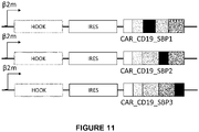
- the vector encodes a hook comprising the amino acid sequence of SEQ ID NO:31 or SEQ ID NO:32.
- the vector comprises a ⁇ 2-microglobulin, Ubi, EF1 ⁇ , MHCI, or MHCII promoter. In one embodiment, the vector is a DNA.
- the lentiviral vector particle comprises a vesicular stomatitis virus glycoprotein. In one embodiment, the lentiviral vector particle comprises HIV-1 subtype D Gag and Pol proteins.
- the text describes but does not claim per se the use of the chimeric antigen receptor or the lentiviral vector or the nucleic acid vector or the lentiviral vector particle for inducing an immune response in a human.
- the text describes but does not claim per se a method for inducing an immune response in a human comprising administering the lentiviral vector particle or cells transduced by the lentiviral vector particle encoding a chimeric antigen receptor to a human and, optionally, subsequently administering biotin to the human.
- Another object of the invention relates to an in vitro method for expressing a chimeric antigen receptor in a cell, preferably in a T cell, comprising:
- the chimeric antigen receptor or the lentiviral vector or the nucleic acid vector comprises the nucleotide sequence of any of SEQ ID NO:45, SEQ ID NO:47, or SEQ ID NO:49. In some embodiments, the chimeric antigen receptor comprises the amino acid sequence of any of SEQ ID NO:46, SEQ ID NO:48, or SEQ ID NO:50.
- the lentiviral vector or the nucleic acid vector further encodes the amino acid sequence of SEQ ID NO:42 or comprises the nucleic acid sequence of SEQ ID NO:43 and/or SEQ ID NO:44.
- CARs chimeric antigen receptors
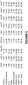
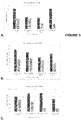
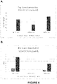
- a kidney cell line and a dendritic cell line were transduced with lentiviral vectors expressing green fluorescent protein (GFP) from various promoters. It was found that the human EF1 ⁇ promoter was the strongest promoter in the dendritic cell line BDCM ( Fig. 1 ). This promoter was also very strong in the kidney cell line 293T. Cell-specific expression of various promoters was analyzed using the data sets at biogps.org ( Fig.2 ).
- the Hook-streptavidin sequence was cloned into a pseudotyping vector encoding a codon-optimized VSV-G protein as a fusion with VSV-G.
- This vector was evaluated by cotransfection with a lentivector encoding GFP and a dose escalation of hook-streptavidin encoding vector. No reduction in titers due to retention of the VSV protein in the ER was seen ( Figure 4 ). Immunofluorescence studies showed a lack of retention of the VSV protein in the ER. This contrasts with the results seen for the VSV protein alone. Nature Methods 9(5): 493-500 (2102). Thus, it was unclear whether CAR expression might be regulated with the Hook-Steptavidin system.
- CAR-CD19 or CAR-CD19 with a streptavidin binding protein (SBP) at various positions between the intracellular domains was evaluated to check whether the presence of the SBP sequence was modifying the CAR expression.
- CAR-CD19+ with the SBP at various positions were cloned into lentiviral vectors under the control of the ⁇ 2m promoter.
- Human T-cells were transduced with the lentiviral vectors and the expression of the CD19 was evaluated at the surface of the cells.
- the CAR-CD19-SBP are expressed at the surface of the T-cells, with a slight reduction of the MFI compared to the CAR-CD19 ( Figure 5 ).
- Hook-streptavidin could retain the CAR-CD19-SBP in the endoplasmic reticulum of the T-cells and the addition of biotin in the media induced the release of the CAR-CD19 and its expression at the surface of the cells ( Figure 6 ).
- the text describes, but does not claim per se, lentiviral vectors encoding a CAR, and their use for the induction of immune responses in a host, especially a human.
- in vitro refers to events that occur in an artificial environment, e.g., in a test tube or reaction vessel, in cell culture, in a Petri dish, etc., rather than within an organism (e.g., animal, plant, or microbe).
- in vivo refers to events that occur within an organism (e.g., animal, plant, or microbe).
- isolated refers to a substance or entity that has been (1) separated from at least some of the components with which it was associated when initially produced (whether in nature or in an experimental setting), and (2) produced, prepared, and/or manufactured by the hand of man. Isolated substances and/or entities may be separated from at least about 10%, about 20%, about 30%, about 40%, about 50%, about 60%, about 70%, about 80%, about 90%, or more of the other components with which they were initially associated. In some embodiments, isolated agents are more than about 80%, about 85%, about 90%, about 91%, about 92%, about 93%, about 94%, about 95%, about 96%, about 97%, about 98%, about 99%, or more than about 99% pure. As used herein, a substance is "pure” if it is substantially free of other components.
- isolated products of the text including isolated nucleic acids, proteins, polypeptides, and antibodies are not products of nature (i.e., "non-naturally occurring”). Rather, the “isolated” nucleic acids, proteins, polypeptides, and antibodies are “man-made” products.
- the “isolated” products can be “markedly different” or “significantly different” from products of nature.
- the isolated nucleic acids may be purified, recombinant, synthetic, labeled, and/or attached to a solid substrate. Such nucleic acids can be markedly different or significantly different than nucleic acids that occur in nature.
- the "isolated" proteins, polypeptides, and antibodies may be purified, recombinant, synthetic, labeled, and/or attached to a solid substrate.
- Such proteins, polypeptides, and antibodies can be markedly different or significantly different from proteins, polypeptides, and antibodies that occur in nature.
- peptide refers to a short polypeptide, e.g., one that typically contains less than about 50 amino acids and more typically less than about 30 amino acids.
- the term as used herein encompasses analogs and mimetics that mimic structural and thus biological function.
- polypeptide encompasses both naturally-occurring and non-naturally occurring proteins, and fragments, mutants, derivatives and analogs thereof.
- a polypeptide may be monomeric or polymeric. Further, a polypeptide may comprise a number of different domains each of which has one or more distinct activities. For the avoidance of doubt, a "polypeptide" may be any length greater two amino acids.
- isolated protein or "isolated polypeptide” is a protein or polypeptide that by virtue of its origin or source of derivation (1) is not associated with naturally associated components that accompany it in its native state, (2) exists in a purity not found in nature, where purity can be adjudged with respect to the presence of other cellular material (e.g., is free of other proteins from the same species) (3) is expressed by a cell from a different species, or (4) does not occur in nature (e.g., it is a fragment of a polypeptide found in nature or it includes amino acid analogs or derivatives not found in nature or linkages other than standard peptide bonds).
- polypeptide that is chemically synthesized or synthesized in a cellular system different from the cell from which it naturally originates will be “isolated” from its naturally associated components.
- a polypeptide or protein may also be rendered substantially free of naturally associated components by isolation, using protein purification techniques well known in the art.
- isolated does not necessarily require that the protein, polypeptide, peptide or oligopeptide so described has been physically removed from a cell in which it was synthesized.
- the protein or polypeptide can be purified.
- the purified protein or polypeptide is more than 50%, 75%, 85%, 90%, 95%, 97%, 98%, or 99% pure.
- a purified protein that is more than 50% (etc.) pure means a purified protein sample containing less than 50% (etc.) other proteins.
- a sample of a protein comprising can be 99% pure if it contains less than 1% contaminating host cell proteins.
- fusion protein refers to a polypeptide comprising a polypeptide or fragment coupled to heterologous amino acid sequences. Fusion proteins are useful because they can be constructed to contain two or more desired functional elements that can be from two or more different proteins.
- a fusion protein comprises at least 10 contiguous amino acids from a polypeptide of interest, or at least 20 or 30 amino acids, or at least 40, 50 or 60 amino acids, or at least 75, 100 or 125 amino acids.
- the heterologous polypeptide included within the fusion protein is usually at least 6 amino acids in length, or at least 8 amino acids in length, or at least 15, 20, or 25 amino acids in length.
- Fusions that include larger polypeptides, such as an IgG Fc region, and even entire proteins, such as the green fluorescent protein (“GFP") chromophore-containing proteins, have particular utility. Fusion proteins can be produced recombinantly by constructing a nucleic acid sequence which encodes the polypeptide or a fragment thereof in frame with a nucleic acid sequence encoding a different protein or peptide and then expressing the fusion protein. Alternatively, a fusion protein can be produced chemically by crosslinking the polypeptide or a fragment thereof to another protein.
- GFP green fluorescent protein
- recombinant may refer to a biomolecule, e.g., a gene or protein, or to an organism.
- the term “recombinant” may be used in reference to cloned DNA isolates, chemically synthesized polynucleotides, or polynucleotides that are biologically synthesized by heterologous systems, as well as proteins or polypeptides and/or RNAs encoded by such nucleic acids.
- a “recombinant” nucleic acid is a nucleic acid linked to a nucleotide or polynucleotide to which it is not linked in nature.
- a “recombinant” protein or polypeptide may be (1) a protein or polypeptide linked to an amino acid or polypeptide to which it is not linked in nature; and/or (2) a protein or polypeptide made by transcription and/or translation of a recombinant nucleic acid.
- a protein synthesized by a microorganism is recombinant, for example, if it is synthesized from an mRNA synthesized from a recombinant nucleic acid present in the cell.
- a “recombinant” cell is a cell comprising a "recombinant” biomolecule.
- a T cell that comprises a "recombinant” nucleic acid is a "recombinant” cell.
- nucleic acid refers to a polymeric form of nucleotides of at least 10 bases in length.
- the term includes DNA molecules (e.g., cDNA or genomic or synthetic DNA) and RNA molecules (e.g., mRNA or synthetic RNA), as well as analogs of DNA or RNA containing non-natural nucleotide analogs, non-native internucleoside bonds, or both.
- the nucleic acid can be in any topological conformation.
- the nucleic acid can be single-stranded, double-stranded, triple-stranded, quadruplexed, partially double-stranded, branched, hairpinned, circular, or in a padlocked conformation.
- the nucleic acid also referred to as polynucleotides
- Such modifications include, for example, labels, methylation, substitution of one or more of the naturally occurring nucleotides with an analog, internucleotide modifications such as uncharged linkages (e.g., methyl phosphonates, phosphotriesters, phosphoramidates, carbamates, etc.), charged linkages (e.g., phosphorothioates, phosphorodithioates, etc.), pendent moieties (e.g., polypeptides), intercalators (e.g., acridine, psoralen, etc.), chelators, alkylators, and modified linkages (e.g., alpha anomeric nucleic acids, etc.) Also included are synthetic molecules that mimic polynucleotides in their ability to bind to a designated sequence via hydrogen bonding and other chemical interactions.
- internucleotide modifications such as uncharged linkages (e.g., methyl phosphonates, phosphotriesters, phosphoramidates, carb
- Such molecules are known in the art and include, for example, those in which peptide linkages substitute for phosphate linkages in the backbone of the molecule.
- Other modifications can include, for example, analogs in which the ribose ring contains a bridging moiety or other structure such as the modifications found in "locked" nucleic acids.
- RNA, DNA or a mixed polymer is one created outside of a cell, for example one synthesized chemically.
- nucleic acid fragment refers to a nucleic acid sequence that has a deletion, e.g., a 5'-terminal or 3'-terminal deletion compared to a full-length reference nucleotide sequence.
- the nucleic acid fragment is a contiguous sequence in which the nucleotide sequence of the fragment is identical to the corresponding positions in the naturally-occurring sequence.
- fragments are at least 10, 15, 20, or 25 nucleotides long, or at least 20, 30, 40, 50, 60, 70, 80, 90, 100, 110, 120, 130, 140, or 150 nucleotides long.
- a fragment of a nucleic acid sequence is a fragment of an open reading frame sequence.
- such a fragment encodes a polypeptide fragment (as defined herein) of the protein encoded by the open reading frame nucleotide sequence.
- the nucleic acid can be purified.
- the purified nucleic acid is more than 50%, 75%, 85%, 90%, 95%, 97%, 98%, or 99% pure.
- a purified nucleic acid that is at least 50% pure means a purified nucleic acid sample containing less than 50% other nucleic acids.
- a sample of a plasmid can be at least 99% pure if it contains less than 1% contaminating bacterial DNA.
- an endogenous nucleic acid sequence in the genome of an organism is deemed "recombinant” herein if a heterologous sequence is placed adjacent to the endogenous nucleic acid sequence.
- a heterologous sequence is a sequence that is not naturally adjacent to the endogenous nucleic acid sequence, whether or not the heterologous sequence is itself endogenous (originating from the same host cell or progeny thereof) or exogenous (originating from a different host cell or progeny thereof).
- a promoter sequence can be substituted (e.g., by homologous recombination) for the native promoter of a gene in the genome of a host cell, such that this gene has an altered expression pattern.
- This gene would now become “recombinant” because it is separated from at least some of the sequences that naturally flank it.
- a nucleic acid is also considered “recombinant” if it contains any modifications that do not naturally occur to the corresponding nucleic acid in a genome.
- an endogenous coding sequence is considered “recombinant” if it contains an insertion, deletion or a point mutation introduced artificially, e.g., by human intervention.
- a "recombinant nucleic acid” also includes a nucleic acid integrated into a host cell chromosome at a heterologous site and a nucleic acid construct present as an episome.
- the phrase "degenerate variant" of a reference nucleic acid sequence encompasses nucleic acid sequences that can be translated, according to the standard genetic code, to provide an amino acid sequence identical to that translated from the reference nucleic acid sequence.
- the term "degenerate oligonucleotide” or “degenerate primer” is used to signify an oligonucleotide capable of hybridizing with target nucleic acid sequences that are not necessarily identical in sequence but that are homologous to one another within one or more particular segments.
- sequence identity refers to the residues in the two sequences which are the same when aligned for maximum correspondence.
- the length of sequence identity comparison may be over a stretch of at least about nine nucleotides, usually at least about 20 nucleotides, more usually at least about 24 nucleotides, typically at least about 28 nucleotides, more typically at least about 32, and even more typically at least about 36 or more nucleotides.
- polynucleotide sequences can be compared using FASTA, Gap or Bestfit, which are programs in Wisconsin Package Version 10.0, Genetics Computer Group (GCG), Madison, Wis.
- FASTA provides alignments and percent sequence identity of the regions of the best overlap between the query and search sequences. Pearson, Methods Enzymol. 183:63-98 (1990 ).
- percent sequence identity between nucleic acid sequences can be determined using FASTA with its default parameters (a word size of 6 and the NOPAM factor for the scoring matrix) or using Gap with its default parameters as provided in GCG Version 6.1.
- sequences can be compared using the computer program, BLAST ( Altschul et al., J. Mol. Biol.
- an "expression control sequence” refers to polynucleotide sequences which affect the expression of coding sequences to which they are operatively linked. Expression control sequences are sequences which control the transcription, posttranscriptional events and translation of nucleic acid sequences. Expression control sequences include appropriate transcription initiation, termination, promoter and enhancer sequences; efficient RNA processing signals such as splicing and polyadenylation signals; sequences that stabilize cytoplasmic mRNA; sequences that enhance translation efficiency (e.g., ribosome binding sites); sequences that enhance protein stability; and when desired, sequences that enhance protein secretion.
- control sequences differs depending upon the host organism; in prokaryotes, such control sequences generally include promoter, ribosomal binding site, and transcription termination sequence.
- control sequences is intended to encompass, at a minimum, any component whose presence is essential for expression, and can also encompass an additional component whose presence is advantageous, for example, leader sequences and fusion partner sequences.
- operatively linked or “operably linked” expression control sequences refers to a linkage in which the expression control sequence is contiguous with the gene of interest to control the gene of interest, as well as expression control sequences that act in trans or at a distance to control the gene of interest.
- a "vector” is intended to refer to a nucleic acid molecule capable of transporting another nucleic acid to which it has been linked.
- a "plasmid” which generally refers to a circular double stranded DNA loop into which additional DNA segments may be ligated, but also includes linear double-stranded molecules such as those resulting from amplification by the polymerase chain reaction (PCR) or from treatment of a circular plasmid with a restriction enzyme.
- Other vectors include cosmids, bacterial artificial chromosomes (BAC) and yeast artificial chromosomes (YAC).
- BAC bacterial artificial chromosome
- YAC yeast artificial chromosome
- Another type of vector is a viral vector, wherein additional DNA segments may be ligated into the viral genome (discussed in more detail below).
- vectors are capable of autonomous replication in a host cell into which they are introduced (e.g., vectors having an origin of replication which functions in the host cell). Other vectors can be integrated into the genome of a host cell upon introduction into the host cell, and are thereby replicated along with the host genome. Moreover, certain vectors are capable of directing the expression of genes to which they are operatively linked. Such vectors are referred to herein as “recombinant expression vectors" (or simply "expression vectors").
- the integrating cosmid vector pYUB412 is an example of a "vector”.
- recombinant host cell (or simply “recombinant cell” or “host cell”), as used herein, is intended to refer to a cell into which a recombinant nucleic acid such as a recombinant vector has been introduced.
- the word "cell” is replaced by a name specifying a type of cell.
- a “recombinant microorganism” is a recombinant host cell that is a microorganism host cell. It should be understood that such terms are intended to refer not only to the particular subject cell but to the progeny of such a cell.
- a recombinant host cell may be an isolated cell or cell line grown in culture or may be a cell which resides in a living tissue or organism.
- the term "mammal” refers to any member of the taxonomic class mammalia, including placental mammals and marsupial mammals.
- “mammal” includes humans, primates, livestock, and laboratory mammals.
- Exemplary mammals include a rodent, a mouse, a rat, a rabbit, a dog, a cat, a sheep, a horse, a goat, a llama, cattle, a primate, a pig, and any other mammal.
- the mammal is at least one of a transgenic mammal, a genetically-engineered mammal, and a cloned mammal.
- CAR chimeric antigen receptor
- binding domain binding domain
- hinge and transmembrane domain transmembrane domain
- hook-binding domain an intracellular signaling domain
- activation domain an intracellular signaling domain
- a "hook-binding domain” is a domain that reversibly binds directly or indirectly to a "hook” protein inside of the cell, which binding prevents the CAR from exiting the endoplasmic reticulum (ER) or Golgi under appropriate conditions.
- the hook-binding domain comprises a streptavidin-binding peptide (SBP), which can bind to a hook protein that bears the core streptavidin.
- SBP streptavidin-binding peptide
- Biotin causes the release of the CAR containing the hook-binding domain from the hook by out-competing the SBP. The CAR can then move to the cell membrane.
- the hook-binding domain comprises the amino acid sequence: MDEKTTGWRGGHVVEGLAGELEQLRARLEHHPQGQREP (SEQ ID NO:1) or is encoded by the nucleic acid sequence:
- Shorter SBP fragments, deleted at their N-terminus and C-terminus may be used with identical efficacy. See Barrette-Ng, I.H., S.C. Wu, W.M. Tjia, S.L. Wong, and K.K. Ng. 2013,
- the structure of the SBP-Tag-streptavidin complex reveals a novel helical scaffold bridging binding pockets on separate subunits, Acta crystallographica. Section D, Biological crystallography 69:879-887 .
- FKBPF-K506 binding domain 12 can be used with FKBP-rapamycin associated protein (FRAP) as the hook.
- FRAP FKBP-rapamycin associated protein
- the interaction occurs only in the presence of rapamycin or analogues thereof that are able to mediate the interaction between FKBP12 and FRAP and can be, in particular, selected from the group consisting of FKI012, FK-CsA, and rapamycin.
- the hook-binding domain is located in the intracytoplasmic region of the CAR. In other embodiments, the hook-binding domain is located in other positions, i.e., in all the junctions between the different intracytoplamsic elements (e.g., between the transmembrane region and the first co-stimulation element) or between the different co-stimulation elements.
- antibody is meant to include polyclonal antibodies, monoclonal antibodies, fragments thereof, such as F(ab')2 and Fab fragments, single-chain variable fragments (scFvs), single-domain antibody fragments (VHHs or Nanobodies, preferably camelid), and bivalent and trivalent antibody fragments (diabodies and triabodies).
- the antibody is a single-chain Fv antibody or a nanobody.
- the antibody is monospecific. In one embodiment, the antibody is multispecific for 2, 3, or 4 polypeptides. Preferably, the antibody is bispecific.
- Antibodies can be synthetic, monoclonal, or polyclonal and can be made by techniques well known in the art. Such antibodies specifically bind to human proteins via the antigen-binding sites of the antibody (as opposed to non-specific binding). Human proteins, polypeptide fragments, and peptides can be employed as immunogens in producing antibodies immunoreactive therewith. The human proteins, polypeptides, and peptides contain antigenic determinants or epitopes that elicit the formation of antibodies.
- Linear epitopes are composed of a single section of amino acids of the polypeptide, while conformational or discontinuous epitopes are composed of amino acids sections from different regions of the polypeptide chain that are brought into close proximity upon protein folding ( C. A. Janeway, Jr. and P. Travers, Immuno Biology 3:9 (Garland Publishing Inc., 2nd ed. 1996 )). Because folded proteins have complex surfaces, the number of epitopes available is quite numerous; however, due to the conformation of the protein and steric hindrance, the number of antibodies that actually bind to the epitopes is less than the number of available epitopes ( C. A. Janeway, Jr. and P. Travers, Immuno Biology 2:14 (Garland Publishing Inc., 2nd ed. 1996 )). Epitopes can be identified by any of the methods known in the art.
- one aspect of the present invention relates to the antigenic epitopes of human proteins.
- Such epitopes are useful for raising antibodies, in particular monoclonal antibodies, as described in detail below.
- Antibodies are defined to be specifically binding if they bind human proteins or polypeptides with a Ka of greater than or equal to about 10 7 M -1 . Affinities of binding partners or antibodies can be readily determined using conventional techniques, for example those described by Scatchard et al., Ann. N.Y. Acad. Sci., 51:660 (1949 ).
- Polyclonal antibodies can be readily generated from a variety of sources, for example, horses, cows, goats, sheep, dogs, chickens, rabbits, mice, or rats, using procedures that are well known in the art.
- a purified human protein or polypeptide that is appropriately conjugated is administered to the host animal typically through parenteral injection.
- the immunogenicity can be enhanced through the use of an adjuvant, for example, Freund's complete or incomplete adjuvant.
- small samples of serum are collected and tested for reactivity to human proteins or polypeptides.
- Examples of various assays useful for such determination include those described in Antibodies: A Laboratory Manual, Harlow and Lane (eds.), Cold Spring Harbor Laboratory Press, 1988 ; as well as procedures, such as countercurrent immuno-electrophoresis (CIEP), radioimmunoassay, radioimmunoprecipitation, enzyme-linked immunosorbent assays (ELISA), dot blot assays, and sandwich assays. See U.S. Pat. Nos. 4,376,110 and 4,486,530 .
- Monoclonal antibodies can be readily prepared using well known procedures. See, for example, the procedures described in U.S. Pat. Nos. RE 32,011 , 4,902,614 , 4,543,439 , and 4,411,993 ; Monoclonal Antibodies, Hybridomas: A New Dimension in Biological Analyses, Plenum Press, Kennett, McKeam, and Bechtol (eds.), 1980 .
- the host animals such as mice
- Mouse sera are then assayed by conventional dot blot technique or antibody capture (ABC) to determine which animal is best to fuse.
- ABSC antibody capture
- mice are given an intravenous boost of the human protein or polypeptide.
- Mice are later sacrificed and spleen cells fused with commercially available myeloma cells, such as Ag8.653 (ATCC), following established protocols.
- ATCC Ag8.653
- the monoclonal antibodies described herein but not claimed per se can be produced using alternative techniques, such as those described by Alting-Mees et al., "Monoclonal Antibody Expression Libraries: A Rapid Alternative to Hybridomas", Strategies in Molecular Biology 3:1-9 (1990 ).
- binding partners can be constructed using recombinant DNA techniques to incorporate the variable regions of a gene that encodes a specific binding antibody. Such a technique is described in Larrick et al., Biotechnology, 7:394 (1989 ).
- the monoclonal antibodies described herein but not claimed per se include chimeric antibodies, e.g., humanized versions of murine monoclonal antibodies.
- Such humanized antibodies can be prepared by known techniques, and offer the advantage of reduced immunogenicity when the antibodies are administered to humans.
- a humanized monoclonal antibody comprises the variable region of a murine antibody (or just the antigen binding site thereof) and a constant region derived from a human antibody.
- a humanized antibody fragment can comprise the antigen binding site of a murine monoclonal antibody and a variable region fragment (lacking the antigen-binding site) derived from a human antibody.
- Procedures for the production of chimeric and further engineered monoclonal antibodies include those described in Riechmann et al. (Nature 332:323, 1988 ), Liu et al. (PNAS 84:3439, 1987 ), Larrick et al. (Bio/Technology 7:934, 1989 ), and Winter and Harris (TIPS 14:139, May, 1993 ). Procedures to generate antibodies transgenically can be found in GB 2,272,440 , U.S. Pat. Nos. 5,569,825 and 5,545,806 .
- Antibodies produced by genetic engineering methods such as chimeric and humanized monoclonal antibodies, comprising both human and non-human portions, which can be made using standard recombinant DNA techniques, can be used.
- Such chimeric and humanized monoclonal antibodies can be produced by genetic engineering using standard DNA techniques known in the art, for example using methods described in Robinson et al. International Publication No. WO 87/02671; Akira, et al. European Patent Application 0184187; Taniguchi, M. , European Patent Application 0171496; Morrison et al. European Patent Application 0173494; Neuberger et al. PCT International Publication No. WO 86/01533; Cabilly et al. U.S. Pat. No.
- An immunoglobulin library can be expressed by a population of display packages, preferably derived from filamentous phage, to form an antibody display library.
- Examples of methods and reagents particularly amenable for use in generating antibody display library can be found in, for example, Ladner et al. U.S. Pat. No. 5,223,409 ; Kang et al. PCT publication WO 92/18619 ; Dower et al. PCT publication WO 91/17271 ; Winter et al. PCT publication WO 92/20791 ; Markland et al. PCT publication WO 92/15679 ; Breitling et al.
- the antibody library is screened to identify and isolate packages that express an antibody that binds a human protein or polypeptide.
- the primary screening of the library involves panning with an immobilized human protein or polypeptide and display packages expressing antibodies that bind immobilized human protein or polypeptide are selected.
- antibodies In connection with synthetic and semi-synthetic antibodies, such terms are intended to cover but are not limited to antibody fragments, isotype switched antibodies, humanized antibodies (e.g., mouse-human, human-mouse), hybrids, antibodies having plural specificities, and fully synthetic antibody-like molecules.
- the text describes but does not claim per se a CAR comprising an activation domain comprising CD3- ⁇ or Fc receptor ⁇ amino acid sequences. Sadelain et al., Cancer Discov. 2013 April; 3(4): 388-398 . The text further described but does not claim per se a CAR comprising an activation domain comprising a CD3- ⁇ chain and the cytoplasmic domain of a costimulatory receptor such as CD28, 4-1BB (CD137), DAP10, OX40 (CD134), ICOS, CD27, or CD40L. Id.
- the CAR comprises a fragment of at least 50, 60, 70, 80, 90,100, 110, 120, 150, or 200 amino acids of at least one of the following amino acid sequences having T-cell activating activity.
- CD40L (CD154):
- CAR comprises a fragment of at least 100, 110, 120, 150, or 200 contiguous amino acids of SEQ ID NO:3, SEQ ID NO:4, SEQ ID NO:5, SEQ ID NO:7, SEQ ID NO:8, SEQ ID NO:33, or SEQ ID NO:34 or a fragment of at least 90 contiguous amino acids of SEQ ID NO:6.
- the CAR comprises a transmembrane (TM) domain, preferably a fragment of at least 20, 30, 40, 50, 60, 70, 80, 90,100, 110, 120, 150, or 200 amino acids, most preferably of at least one of CD28, CD3z, CD8, CD4, FcRy, TM regions.
- TM transmembrane
- the CAR can be purified.
- the purified CAR is more than 50%, 75%, 85%, 90%, 95%, 97%, 98%, or 99% pure.
- a purified CAR that is more than 50% (etc.) pure means a purified CAR sample containing less than 50% (etc.) other proteins.
- a sample of a recombinant CAR purified from a host cell can be 99% pure if it contains less than 1% contaminating host cell proteins.
- the CAR encodes the amino acid sequence of any of SEQ ID NO:46, SEQ ID NO:48, or SEQ ID NO:50.
- Preferred nucleic acids are of at least 50, 60, 70, 80, 90,100, 110, 120, 150, 200, 300, 400, 500, or 600 nucleotides.
- the nucleic acid can be purified.
- the purified nucleic acid is more than 50%, 75%, 85%, 90%, 95%, 97%, 98%, or 99% pure.
- a purified nucleic acid that is more than 50% pure means a purified nucleic acid sample containing less than 50% other nucleic acids.
- a sample of a plasmid purified from a host bacteria can be 99% pure if it contains less than 1% contaminating bacterial DNA.
- nucleic acids include the following: CAR_CD19 2 Nd generation: 1518bp
- nucleic acids include those comprising or encoding any of the constituents of the CAR depicted in Figure 9 , including signal sequence, CD19 ScFv, CD8 hinge,transmembrane domain, 4-1BB, and/or CD3z sequences.
- Other preferred nucleic acids include those comprising or encoding any of the constituents of the CARs of SEQ ID NOs:45-50.
- the nucleic acid comprises a hook operably-linked to a promoter, preferably UBC or ⁇ 2M, and a CAR comprising a hook-binding protein, operably-linked to an IRES.
- the hook is a streptavidin protein, preferably core Streptavidin, and the hook-binding protein is a streptavidin-binding protein.
- Vectors comprise a nucleic acid sequence or encode an amino acid sequence of at least one of the SEQ ID NOs detailed herein.
- the retention can take place in the lumen of the ER or at its cytoplasmic face, depending on the topology of the protein and the orientation of tagging with the interaction domains. Boncompain et al., Current Protocols in Cell Biology 15.19.1-15.19.16, December 2012 .
- the hook for the ER comprises a mutant of stromal interaction molecule 1 (STIM1-NN; a type I protein) that localizes in the ER but that cannot bind microtubules, an isoform of the human invariant chain of the major histocompatibility complex (li; a type II protein) that has an N-terminal arginine-based motif; or a C-terminal ER retention signal (Lys-Asp-Glu-Leu; KDEL). Boncompain et al., Nat. Methods 9:493-498, 2012 .
- the hook can be fused to a core Streptavidin in their luminal or cytoplasmic domain depending on the hook-binding protein. Id. at Fig. 1 .
- the hook for the Golgi can be Golgin-84 to be used as a cytoplasmic Golgi hook. Id.
- the hook comprises a Streptavidin protein sequence, most preferably core Streptavidin.
- a Streptavidin protein sequence most preferably core Streptavidin.
- the hook comprises one of the following Streptavidin protein sequences:
- the vector comprises a hook operably-linked to a promoter, preferably UBC or ⁇ 2M, and a CAR comprising a hook-binding protein, operably-linked to an IRES.
- the hook is a streptavidin protein, preferably core Streptavidin, and the hook-binding protein is a streptavidin-binding protein.
- Preferred vectors are lentivectors comprising any of the following nucleotide or amino acid sequences:
- the vector can be an expression vector.
- the vector can be a plasmid vector.
- the vector is a lentiviral vector.
- a "lentiviral vector” means a non-replicating vector for the transduction of a host cell with a transgene comprising cis-acting lentiviral RNA or DNA sequences, and requiring lentiviral proteins (e.g., Gag, Pol, and/or Env) that are provided in trans.
- the lentiviral vector lacks expression of functional Gag, Pol, and Env proteins.
- the lentiviral vector may be present in the form of an RNA or DNA molecule, depending on the stage of production or development of said retroviral vectors.
- the lentiviral vector can be in the form of a recombinant DNA molecule, such as a plasmid.
- the lentiviral vector can be in the form of a lentiviral vector particle, such as an RNA molecule(s) within a complex of lentiviral and other proteins.
- lentiviral particle vectors which correspond to modified or recombinant lentivirus particles, comprise a genome which is composed of two copies of single-stranded RNA. These RNA sequences can be obtained by transcription from a double-stranded DNA sequence inserted into a host cell genome (proviral vector DNA) or can be obtained from the transient expression of plasmid DNA (plasmid vector DNA) in a transformed host cell.
- Lentiviral vectors derive from lentiviruses, in particular human immunodeficiency virus (HIV-1 or HIV-2), simian immunodeficiency virus (SIV), equine infectious encephalitis virus (EIAV), caprine arthritis encephalitis virus (CAEV), bovine immunodeficiency virus (BIV) and feline immunodeficiency virus (FIV), which are modified to remove genetic determinants involved in pathogenicity and introduce new determinants useful for obtaining therapeutic effects.
- HSV-1 or HIV-2 human immunodeficiency virus
- SIV simian immunodeficiency virus
- EIAV equine infectious encephalitis virus
- CAEV caprine arthritis encephalitis virus
- BIV bovine immunodeficiency virus
- FV feline immunodeficiency virus
- trans-acting sequences e.g., gag , pol , tat, rev, and env genes
- the trans-acting sequences can be deleted and replaced by an expression cassette encoding a transgene.
- Efficient integration and replication in non-dividing cells generally requires the presence of two cis-acting sequences at the center of the lentiviral genome, the central polypurine tract (cPPT) and the central termination sequence (CTS). These lead to the formation of a triple-stranded DNA structure called the central DNA "flap", which acts as a signal for uncoating of the pre-integration complex at the nuclear pore and efficient importation of the expression cassette into the nucleus of non-dividing cells, such as dendritic cells.
- cPPT central polypurine tract
- CTS central termination sequence
- LTRs long terminal repeats
- Vectors may be obtained by mutating the LTR sequences, for instance, in domain U3 of said LTR ( ⁇ U3) ( Miyoshi H et al, 1998, J Virol. 72(10):8150-7 ; Zufferey et al., 1998, J Virol 72(12):9873-80 ).
- the packaging sequence ⁇ (psi) can also be incorporated to help the encapsidation of the polynucleotide sequence into the vector particles ( Kessler et al., 2007, Leukemia, 21(9):1859-74 ; Paschen et al., 2004, Cancer Immunol Immunother 12(6):196-203 ).
- lentiviral vector polynucleotide sequence can also be advantageously included in the lentiviral vector polynucleotide sequence, to obtain a more stable expression of the transgene in vivo.
- PBS primer binding site
- WPRE Woodchuck PostTranscriptional Regulatory Element
- the lentiviral vector comprises at least one cPPT/CTS sequence, one ⁇ sequence, one (preferably 2) LTR sequence, and an expression cassette including a transgene under the transcriptional control of a ⁇ 2m or class I MHC promoter.
- the promoter sequence comprises a polynucleotide sequence that shares more than 90%, preferably more than 95%, more preferably more than 99% identity with the promoter sequence of SEQ ID NO:9, SEQ ID NO:10, SEQ ID NO:11, SEQ ID NO:12, SEQ ID NO:13, SEQ ID NO:14, SEQ ID NO:38, or SEQ ID NO:41.
- lentiviral vector particles comprising a lentiviral vector that comprises a dendritic cell-specific promoter directing expression of a microbial or tumor antigen, wherein the lentiviral vector particles exhibit higher expression of the antigen in BDCM cells than in HEK 293 T cells.
- the invention encompasses the insertion of an MHC Class I (MHCI), Ubi, EF1 ⁇ , or ⁇ 2 microglobulin promoter ( ⁇ 2m) promoter into a lentiviral vector.
- MHC Class I (MHCI) promoter or " ⁇ 2 microglobulin promoter” or “MHC Class II (MHCII)” or "human ubiquitin promoter” includes a naturally occurring or synthetic MHC Class I promoter or ⁇ 2 microglobulin promoter or MHC Class II promoter or human ubiquitin promoter.
- MHC Class I promoter does not include a ⁇ 2m promoter.
- the promoter can be a naturally occurring promoter.
- naturally occurring promoters are the human ⁇ 2m, HLA-A2, HLA-B7, HLA-Cw5, HLA-E, HLA-F, HLA-DR ⁇ , and ubiquitin gene promoters.
- the promoters can contain further regulatory elements, such as one or more Sp1 and ETs binding sites.
- the MHC class I promoter contains 2 Sp1 binding sites and 1 Ets binding site.
- Ap1 and/or Ap2 sites are further contained in the promoter.
- enhancer A containing binding sites for nuclear transcription factor ⁇ B (NF- ⁇ B)
- NF- ⁇ B nuclear transcription factor ⁇ B
- a lentiviral vector particle (or lentiviral particle vector) comprises a lentiviral vector in association with viral proteins.
- the vector is preferably an integrating vector.
- Such a method allows producing a recombinant vector particle, comprising the following steps of:
- the host cell is further modified to be able to produce viral particle in a culture medium in a continuous manner, without the entire cells swelling or dying.
- a culture medium in a continuous manner, without the entire cells swelling or dying.
- One may refer to Strang et al., 2005, J Virol 79(3):1165-71 ); Relander et al., 2005, Mol Ther 11(3):452-9 ; Stewart et al., 2009, Gene Ther, 16(6):805-14 ; and Stuart et al., 2011, Hum gene Ther , with respect to such techniques for producing viral particles.
- the method can further comprise harvesting or isolating the CAR.
- the lentiviral vector or lentiviral particle vector preferably comprises a promoter as defined above.
- the present text further describes but does not claim per se to the use of the lentiviral vectors as defined above, especially in the form of lentiviral vector particles, for the preparation of therapeutic compositions or vaccines which are capable of inducing or contributing to the occurrence or improvement of an immunological reaction with the CAR encoded by the vectors.
- Preferred modes of administration include reinfusion of the modified T cells, preferably intravenously or intra-articular administration, most preferably intra-tumoral administration.
- the text described but does not claim per se a method for inducing an immune response in a human comprising administering lentiviral vector particles comprising a functional integrase protein and a lentiviral vector to T or NK cells and administering the modified cells to a human; wherein the integrating lentiviral vector comprises a promoter directing expression of a CAR; and generating immunological reaction with the CAR.
- the immunogenic compositions preferably contain cPPT and CTS sequences in the vector and vector particles to induce or to stimulate the nuclear import of the vector genome in the target cells.
- the vector comprises a CAR and regulatory signals of retrotranscription, expression and encapsidation of retroviral or retroviral-like origin.
- immunogenic compositions previously described are that they can be used to redirect the specificity and function of T lymphocytes and other immune cells against multiple antigens against which the CAR in the vector or vector particles are directed.
- PCR amplification of the proviral region of the pTRIP ⁇ U3-CMV-GFP(15) was performed using direct (5'-CTTACTAGTTGGAAGGGCTAATTCACTCCCAAC-3'; SEQ ID NO:15) and reverse (5'-CATTCTAGAACTGCTAGAGATTTTCCACACTG-3'; SEQ ID NO:16) oligonucleotides encompassing respectively the Spel and Xbal restriction sites.
- the resulting fragment was digested and cloned between the Spel and Xbal sites of the pVAX-1 plasmid (Invitrogen, Lifetech) from which the Mlul site have been deleted.
- the resulting plasmid was named pFLAP-CMV-GFP.
- the packaging plasmid pTHV-GP-N was constructed by amplifying the HIV-1 NDK genome by PCR (using the following oligonucleotides with 5'-atgcatgcgtcgacctcgagttaatcctcatcctgtctacttgccac-3' (SEQ ID NO:24) and 5'-gcatgcatcggccggggcggcgactgGTgagagGCCACCatgggtgcgagagcgtcagtattaag-3' (SEQ ID NO:25)).
- the resulting fragment has been digested by Eagl and Sall restriction enzymes and inserted in the p8.74 packaging plasmid (15) from which the Eag1-Sall fragment had been previously removed.
- Pseudotyping plasmids were generated by synthesizing the codon optimized genes corresponding to the vesicular stomatitis virus Indiana (GenBank #CAX62728.1), New Jersey GenBank #CAX62729.1) and Cocal (GenBank # CAX62731.1) strains. Those genes were then digested with EcoR1 and BamH1 and cloned between the corresponding restriction sites of the pVAX1 plasmid (Invitrogen, Lifetech).
- Vectors can be produced by transient calcium-phosphate transfection of HEK 293T as previously described (25).
- HEK 293T human embryonic kidney cell line, ATCC CRL-11268, (Graham et al. 1977) cells were maintained in Dubelcco's modified Eagle's medium (DMEM/ High modified, Hyclone) supplemented with 10% fetal bovine serum (FBS, PAA), 1% L-Glutamine (Eurobio), 1% Penicillin-Streptomycin (Gibco by Life technologies) and 1% Sodium Pyruvate (Gibco by Life technologies).). The cell line was kept in an incubator with humidified atmosphere of 5% CO 2 at 37°C.
- the medium was replaced 24h after transfection by 7mL of harvest medium without serum and the viral supernatant was harvested after an additional 24h, and clarified by centrifugation 5min. at 2500rpm.
- the harvest clarified bulk (210mL) is then treated 30min. with DNase (Roche) in the presence of MgCl 2 (Sigma Aldrich) to avoid residual transfection DNA, and ultraconcentrated by centrifugation 1h at 22000rpm, 4°C.
- Each vector pellets are resuspended in 70 ⁇ l PBS-Lactose (40mg/L), pooled, 30 ⁇ L aliquoted and stored at -70°C ⁇ 10°C
- HEK 293T cells are seeded in 6-well plates (BD Falcon) in culture medium and incubated for 4 h at 37°C, 5% CO2 in moist atmosphere. Cells are transduced with 3 successive dilutions of lentiviral vector. 72h post-incubation, cells are harvested and transduced HEK 293T cell pellets are produced. Total genomic DNA from transduced cell-pellets is extracted using a method based on QIAGEN QIAamp DNA mini kit handbook. Proviral quantification is performed using Taqman qPCR.
- Both "second generation” CAR (2 intra cytoplasmic activating domains) and “third generation” CAR (3 intra cytoplasmic activating domains) constructs will be generated containing a hook-binding domain (SBP). Initially, the binding domains evaluated will be anti-CD19, anti-PDL-1, anti-PD1, anti-hedgehog, anti-CD123, and anti-CD123/CD33.
- SBP hook-binding domain
- CAR expression was assessed by flow cytometry 48 or 72h lentiviral particle addition, by staining the murine CD19-binding domain with a biotinylated goat anti-mouse IgG followed by streptavidin conjugated to phycoerythrin. T cell subpopulations were characterized by CD3, CD4 and CD8 staining. This allowed specific detection of the CAR on the surface of T cells. The whole process was performed with the TexMACS medium allowing survival and expansion of T cells.
- T cells were put in culture at a concentration of 2,5x10⁶ cells/ml in TexMACS medium (Miltenyi) at 37°C / 5%CO2 and activated 3 days by the T cell activation/expansion kit (anti-CD2/-CD3/-CD28 nanoparticles prepared according the manufacturer's instructions) from Miltenyi at a bead:T cell ratio of 1:2. After activation, T cells were harvested, counted and put in culture in TexMACS medium in 24 well-plates at 37°C / 5% CO2. Transduction was performed by adding directly into wells lentiviral vectors at different MOI.
- T cells were put in culture at a concentration of 2,5 ⁇ 10 6 cells/ml in TexMACS medium (Miltenyi) at 37°C / 5%CO 2 and activated 3 days by the T cell activation/expansion kit (anti-CD2/-CD3/-CD28 nanoparticles prepared according the manufacturer's instructions) from Miltenyi at a bead:T cell ratio of 1:2.
- transduced T cells were harvested, washed extensively with DPBS 1X and immunostaining of molecules of interest was performed in 96 well-plates.
- T cells were first incubated with a viability dye (Fixable Viability Dye, eBiosciences) conjugated to eFluor 780 and incubated 30 minutes at 4°C. The incubation was performed in azide-free and protein-free DPBS1X. Cells were then washed with DPBS 1X and then incubated with a biotinylated Goat anti-mouse IgG (Fab')2 (Jackson ImmunoResearch) for 30 minutes a 4°C.
- Fab' biotinylated Goat anti-mouse IgG
- PBMC Peripheral Blood Mononuclear Cells
- Ficoll Lymphocyte Separation Medium, Eurobio
- T cells were then purified from PBMC by magnetic isolation using the Pan T cell isolation kit (Miltenyi). T cells were separated according to the manufacturer's instructions.
- PBMC Peripheral Blood Mononuclear Cells
- Ficoll Lymphocyte Separation Medium, Eurobio
- T cells were then purified from PBMC by magnetic isolation using the Pan T cell isolation kit (Miltenyi). T cells were separated according to the manufacturer's instructions.
Description
- The present invention is in the field of recombinant vaccine technology and relates to improvements of lentiviral vectors, which can be used as therapeutic and prophylactic vaccines. The vectors provide improved induction of immune responses over other vectors.
- Recombinant vaccines have been developed with the progress of recombinant DNA technology, allowing the modification of viral genomes to produce modified viruses. In this manner, it has been possible to introduce genetic sequences into non-pathogenic viruses, so that they encode immunogenic proteins to be expressed in target cells upon infection or transduction, in order to develop a specific immune response in their host.
- Such vaccines constitute a major advance in vaccine technology (Kutzler et al., Nat Rev Genet, 9(10): 776-788, 2008). In particular, they have the advantage over traditional vaccines of avoiding live (attenuated) virus and eliminating risks associated with the manufacture of inactivated vaccines.
- Gene delivery using modified retroviruses (retroviral vectors) was introduced in the early 1980s by Mann et al. (Cell, 33(1):153-9, 1983). The most commonly used oncogenic retroviral vectors are based on the Moloney murine leukemia virus (MLV). They have a simple genome from which the polyproteins Gag, Pol and Env are produced and are required in trans for viral replication (Breckpot et al., 2007, Gene Ther, 14(11):847-62; He et al. 2007, Expert Rev vaccines, 6(6):913-24). Sequences generally required in cis are the long terminal repeats (LTRs) and its vicinity: the inverted repeats (IR or att sites) required for integration, the packaging sequence Ψ, the transport RNA-binding site (primer binding site, PBS), and some additional sequences involved in reverse transcription (the repeat R within the LTRs, and the polypurine tracts, PPT, necessary for plus strand initiation). To generate replication-defective retroviral vectors, the gag, pol, and env genes are generally entirely deleted and replaced with an expression cassette.
- Retroviral vectors deriving from lentivirus genomes (i.e. lentiviral vectors) have emerged as promising tools for both gene therapy and immunotherapy purposes, because they exhibit several advantages over other viral systems. In particular, lentiviral vectors themselves are not toxic and, unlike other retroviruses, lentiviruses are capable of transducing non-dividing cells, in particular dendritic cells (He et al. 2007, Expert Rev vaccines, 6(6):913-24), allowing antigen presentation through the endogenous pathway.
- Lentiviruses are linked by similarities in genetic composition, molecular mechanisms of replication and biological interactions with their hosts. They are best known as agents of slow disease syndromes that begin insidiously after prolonged periods of subclinical infection and progress slowly; thus, they are referred to as the "slow" viruses (Narayan et al., 1989, J Gen Virol, 70(7):1617-39). They have the same basic organization as all retroviruses but are more complex due to the presence of accessory genes (e.g., vif, vpr, vpu, nef, tat, and rev), which play key roles in lentiviral replication in vivo.
- Lentiviruses represent a genus of slow viruses of the Retroviridae family, which includes the human immunodeficiency viruses (HIV), the simian immunodeficiency virus (SIV), the equine infectious encephalitis virus (EIAV), the caprine arthritis encephalitis virus (CAEV), the bovine immunodeficiency virus (BIV), and the feline immunodeficiency virus (FIV). Lentiviruses can persist indefinitely in their hosts and replicate continuously at variable rates during the course of the lifelong infection. Persistent replication of the viruses in their hosts depends on their ability to circumvent host defenses.
- The design of recombinant integrating lentiviral vectors is based on the separation of the cis- and trans-acting sequences of the lentivirus. Efficient transduction in non-dividing cells requires the presence of two cis-acting sequences in the lentiviral genome, the central polypurine tract (cPPT) and the central termination sequence (CTS). These lead to the formation of a triple-stranded DNA structure called the central DNA "flap", which maximizes the efficiency of gene import into the nuclei of non-dividing cells, including dendritic cells (DCs) (Zennou et al., 2000, Cell, 101(2) 173-85; Arhel et al., 2007, EMBO J, 26(12):3025-37).
- Dendritic cells are of primary importance for antigen presentation because they constitute the main class of antigen presenting cells (APCs) whose primary function is to present antigens and initiate an immune response.
- To generate an immune response, antigenic proteins must be processed by cells into peptides that are displayed on the cell surface by major histocompatibility complex proteins (MHCs). Circulating APCs present the peptide-MHC complexes to T cells in the draining lymph nodes, where they interact with T cell receptors, and, in conjunction with co-stimulatory signals, activate the T cells.
- A variety of studies have shown that inoculation with lentiviral vectors leads to antigen presentation by DCs and strong activation of antigen specific cytotoxic T lymphocytes (CTLs; CD8+T cells). Therefore, lentiviral vectors have been engineered for the last 10 years for gene transfer and immunotherapy applications.
- The vectors routinely contain strong constitutive promoters containing enhancers, such as the CMV promoter. Michelini et al., Vaccine 27(34):4622-29 (2009); Karwacz et al., J. Virol. 83(7):30943103 (2009); Negri et al., Molecular Therapy 15(9):1716-23 (2007); and Buffa et al., J. General Virology 87:1625-1634 (2006).
- Lentiviral vectors have been improved in their safety by removal of the LTR U3 sequence, resulting in "self-inactivating" vectors that are entirely devoid of viral promoter and enhancer sequences originally present within the LTRs.
- The lentiviral particles, which contain lentiviral vectors, can be produced by recombinant technology upon transient transfection of cells, for
example HEK 293T human cultured cells, by different DNA plasmids: - (i) a packaging plasmid, which expresses at least the Gag, Pol Rev, Tat and, in some cases, structural and enzymatic proteins necessary for the packaging of the transfer construct;
- (ii) a proviral transfer plasmid, containing an expression cassette and HIV cis-acting factors necessary for packaging, reverse transcription, and integration; and
- (iii) an envelope-encoding plasmid, in most cases the glycoprotein of vesicular stomatitis virus (VSV.G), a protein that allows the formation of mixed particles (pseudotypes) that can target a wide variety of cells, especially major histocompatibility (MHC) antigen-presenting cells (APCs), including DCs.
- This procedure allows obtaining transient production of lentiviral particle vectors by the transfected cells. However, the lentiviral particle vectors may also be continuously produced by cells by stably inserting the packaging genes, the proviral coding DNA, and the envelope gene into the cellular genome. This allows the continuous production of lentiviral particle vectors by the cells without the need for transient transfection. Of course, a combination of these procedures can be used, with some of the DNAs/plasmids integrated into the cellular genome and others provided by transient transfection.
- Non-integrating lentiviral vectors have been designed to mitigate the risks of potential oncogenesis linked to insertional mutagenesis events, particularly for vaccination purposes. Examples of non-integrating lentiviral vectors are provided in Coutant et al., PLOS ONE 7(11):e48644 (2102), Karwacz et al., J. Virol. 83(7):3094-3103 (2009), Negri et al., Molecular Therapy 15(9):1716-1723 (2007); Hu et al., Vaccine 28:6675-6683 (2010). Consequently, it has been reported that a non-integrating lentiviral vector system can mitigate the potential risk of insertional mutagenesis as compared to an integrating system. Hu et al., Vaccine 28:6675-6683 (2010).
- In addition, deletion in the U3 region of the 3' LTR of the viral promoter and enhancer sequences in self-inactivating lentiviral vectors limits the likelihood of endogenous promoter activation. These concerns with safety directly address the experiences gained from the SCID-X1 gene therapy trial carried out in 1998-1999, performed with Moloney virus-based retroviral vectors on children suffering from a rare form of X-linked (SCID-X1 gene) severe immunodeficiency disease (Cavazzana-Calvo et al., 2000, Science., 288(5466):669-72). During this trial, four of nine children developed leukemia as a result of the integration of the Moloney-derived retroviral vector at close proximity to the human LM02 proto-oncogene (Hacein-Bey-Abina et al., 2008, J.Clin.Invest., 118(9):3132-3142). It was demonstrated that malignancy was the consequence of the proximity of the viral U3 promoter/enhancer to the LM02 proto-oncogene. As a result, safety is a major concern for the administration of lentivectors to humans.
- Enhancers are cis-acting sequences, which can act as transcriptional activators at a distance. They have been widely employed in viral derived vectors because they appear to be the most efficient for obtaining transgene strong expression in a variety of cell types, in particular DCs (Chinnasamy et al., 2000, Hum Gene Ther 11(13):1901-9; Rouas et al., 2008, Cancer Gene Ther 9(9):715-24; Kimura et al., 2007, Mol Ther 15(7):1390-9; Gruh et al., 2008, J Gene Med 10(1) 21-32). However, given the safety issue of insertional mutagenesis, such transcriptional enhancer sequences should be deleted from the lentiviral vector constructs to abolish the risk of insertional mutagenesis by enhancer proximity effect. This enhancer proximity effect is by far the most frequent mechanism of insertional mutagenesis and is the only effect described in human or animal cases of tumorigenic events after gene transfer.
- Thus, there is a need to develop retroviral, particularly lentiviral vectors, which do not include viral enhancers, but still allow sufficient expression of transgenes encoding immunogenic peptides, if possible, as much expression as that observed when using the CMV promoter.
- Recent studies have reported on the replacement of viral promoters by DC-specific promoters deriving from major histocompatibility complex class II genes (MHC class II) (Kimura et al., 2007, Mol Ther 15(7):1390-9) and dectin-2 genes (Lopes et al., 2008, J Virol 82(1):86-95). The dectin-2 gene promoter used in Lopes et al. contains a putative enhancer and an adenoviral conserved sequence (inverted terminal repeats in adenovirus promoter) (Bonkabara et al., 2001, J. Immunology, 167:6893-6900). The MHC class II gene promoter used by Kimura et al. does not contain any known enhancer.
- Yet, without an enhancer, the MHC class II promoter was found not to provide sufficient transgene expression in DCs, when administered intravenously. In particular, lentiviral vectors including MHC class II promoters did not provoke an immune reaction in immunocompetent C57BL/6 mice, in contrast to the immune responses observed with CMV promoters/enhancers. Although integration and persistent transgene expression were observed after injection in mice, the lentiviral vectors transcribed through MHC class II promoters failed to stimulate an antigen-specific CD8+ cytotoxic T-lymphocyte response, even after vaccination boost. The authors of these studies therefore concluded that the use of MHC class II promoters was of interest only for applications where persistence of expression is sought as in gene replacement therapy, but not in the context of immunotherapy. Of note, MHC class II promoters are expressed poorly in most cell types.
- Thus, the MHC class II promoter is not an adequate promoter for lentiviral vectors for induction of an immune response against an antigen via IV injection. Moreover, the dectin-2 promoter is expressed poorly in most cell types and appears to contain an enhancer. Thus, the dectin-2 promoter is not a good promoter for lentiviral vectors for safety reasons.
- Preferably, in immunotherapy, lentiviral vectors provide effective expression of the transgene that elicits a desired specific immune response. This requires that the expression is at a high level in APCs, such as dendritic cells.
- It is also preferable that the cells transduced by the lentiviral vectors are eliminated by the immune response to provide a higher degree of safety. That is, the immune response generated against the transgene can elicit an immune response in the host sufficient to eliminate the cells that are transduced by the lentiviral vectors. The elimination of transduced cells eliminates the persistence of the lentiviral vector in the host, and possible secondary effects of the vector. In order for the transduced cells to be eliminated, expression is required in non-dendritic cells at a level that allows elimination by the immune response. Thus, appropriate expression of an antigen is desirable.
- At the same time, the promoter should maximize immune stimulation through the key cells (i.e., dendritic cells) involved in the activation of naive and memory T cells, and should minimize the risk of insertional mutagenesis and genotoxicity in stem cells, leading to malignancies. Thus, the promoter should have sufficiently high activity in dendritic and other cells, but not contain an enhancer. Based on these criteria, viral promoters, such as the CMV promoter, are not ideal because of the presence of strong enhancers. These criteria are summarized as follows:
- high expression in antigen presenting cells, including dendritic cells, to induce maximal immune responses;
- expression in other transduced cell types sufficient for elimination by the induced immune response; and
- lack of an enhancer element to avoid insertional effects.
- Chimeric antigen receptors (CARs) are recombinant receptors for antigens. Sadelain et al., Cancer Discov. 2013 April; 3(4): 388-398. They are recombinant receptors that typically target native cell surface antigens. Id. CARs engage molecules that do not require peptide processing or HLA expression to be recognized. Id. CARs have been shown to have T cell-proliferating and activating potential on their own, including chimeric molecules between CD3-ζ or Fc receptor γ and CD8, CD4, CD25 or CD16. The antigen-binding parts of the CARs generally are either scFv's derived from antibodies, Fab's selected from libraries, or natural receptor ligands. Han EQ et al.; J Hematol Oncol, 6:47, 2013.
- CARs have been generated against many different cell surface molecules, including CD19, HER2, GD2, PSMA, and many are in clinical trials. Davila et al., Oncolmmunology 1:9, 1577-1583; 2012. The most promising clinical outcomes of CARs have been in patients treated with autologous CAR-modified T cells targeting CD19. Maus MV et al.; 123(17): 2625-35, 2014.
- The in vivo elimination of T cells expressing CARs has been investigated. Human T cells have been genetically modified with a lentiviral vector to express a CD20-CAR containing a suicide gene relying on inducible activation of
caspase 9. Budde et al., PLoS ONE 8(12): e82742 (2013). This lentivector used the human EF1α promoter to obtain expression in T cells. Activation of the suicide fuse resulted in efficient removal of transduced T cells both in vitro and in vivo. Id. - It would be better to be able to quickly turn off the expression of the CAR at the surface of the cells to prevent adverse events and be able reactivate the system after it has been turned off. Thus, a need exists in the art for improved vectors and methods for treatment of humans. The present invention fulfills these needs in the art.
- The present text describes, but does not claim per se, nucleic acid molecules and vectors encoding CARs and methods of making the nucleic acid molecules and vectors and claims an isolated cell as defined in the claims comprising a nucleic acid molecule, a nucleic acid vector or a lentiviral vector encoding a chimeric antigen receptor as well as an in vitro method for expressing a chimeric antigen receptor in a cell as defined in the claims. The text describes, but does not claim per se, a chimeric antigen receptor comprising a binding domain; a transmembrane domain; a hook-binding domain, preferably comprising a streptavidin-binding peptide; and an activation domain comprising a T cell activating fragment of at least 100 amino acids of SEQ ID NO:3; SEQ ID NO:4; SEQ ID NO:5; SEQ ID NO:6, SEQ ID NO:7, or SEQ ID NO:8, SEQ ID NO:33, or SEQ ID NO:34.
- The text also describes, but does not claim per se, a lentiviral vector expressing a chimeric antigen receptor comprising a binding domain; a transmembrane domain; a hook-binding domain comprising a streptavidin-binding peptide; and an activation domain comprising a T cell activating fragment of at least 100 amino acids of SEQ ID NO:3; SEQ ID NO:4; SEQ ID NO:5; SEQ ID NO:6, SEQ ID NO:7, or SEQ ID NO:8, SEQ ID NO:33, or SEQ ID NO:34.
- The text further describes, but does not claim per se, a nucleic acid vector encoding a chimeric antigen receptor comprising a binding domain; a transmembrane domain; a hook-binding domain, preferably comprising a streptavidin-binding peptide; and an activation domain comprising a T cell activating fragment of at least 100 amino acids of SEQ ID NO:3; SEQ ID NO:4; SEQ ID NO:5; SEQ ID NO:6, SEQ ID NO:7, or SEQ ID NO:8, SEQ ID NO:33, or SEQ ID NO:34.
- The text also describes, but does not claim per se, a lentiviral vector particle encoding a chimeric antigen receptor comprising a binding domain; a transmembrane domain; a hook-binding domain, preferably comprising a streptavidin-binding peptide; and an activation domain comprising a T cell activating fragment of at least 100 amino acids of SEQ ID NO:3; SEQ ID NO:4; SEQ ID NO:5; SEQ ID NO:6, SEQ ID NO:7, or SEQ ID NO:8, SEQ ID NO:33, or SEQ ID NO:34.
- More particularly, a first object of the present invention relates to an isolated cell comprising a nucleic acid molecule, a nucleic acid vector or a lentiviral vector encoding a chimeric antigen receptor, the said chimeric antigen receptor comprising:
- a binding domain;
- a transmembrane domain;
- a hook-binding domain comprising a streptavidin-binding peptide; and
- a T cell activating fragment of at least 100 contiguous amino acids of SEQ ID NO:3; SEQ ID NO:4; SEQ ID NO:5; SEQ ID NO:7, or SEQ ID NO:8, SEQ ID NO:33, or SEQ ID NO:34, or
- a T cell activating fragment of at least 90 contiguous amino acids of SEQ ID NO:6, the hook-binding domain being located in the intracytoplasmic region of the chimeric antigen receptor,
- Another object of the present invention relates to an isolated cell comprising a vector or a lentiviral vector particle expressing a chimeric antigen receptor, the said chimeric antigen receptor comprising:
- a binding domain;
- a transmembrane domain;
- a hook-binding domain comprising a streptavidin-binding peptide; and
- an activation domain comprising:
- a T cell activating fragment of at least 100 contiguous amino acids of SEQ ID NO:3; SEQ ID NO:4; SEQ ID NO:5; SEQ ID NO:7, or SEQ ID NO:8, SEQ ID NO:33, or SEQ ID NO:34, or
- a T cell activating fragment of at least 90 contiguous amino acids of SEQ ID NO:6, the hook-binding domain being located in the intracytoplasmic region of the chimeric antigen receptor
- In one embodiment, the chimeric antigen receptor or the lentiviral vector or the nucleic acid vector or the lentiviral vector particle comprises a single-chain Fv antibody or a single-domain antibodies (i.e., nanobody).
- In one embodiment, the vector encodes a hook comprising the amino acid sequence of SEQ ID NO:31 or SEQ ID NO:32.
- In one embodiment, the vector comprises a β2-microglobulin, Ubi, EF1α, MHCI, or MHCII promoter. In one embodiment, the vector is a DNA.
- In one embodiment, the lentiviral vector particle comprises a vesicular stomatitis virus glycoprotein. In one embodiment, the lentiviral vector particle comprises HIV-1 subtype D Gag and Pol proteins.
- The text describes but does not claim per se the use of the chimeric antigen receptor or the lentiviral vector or the nucleic acid vector or the lentiviral vector particle for inducing an immune response in a human.
- The text describes but does not claim per se a method for inducing an immune response in a human comprising administering the lentiviral vector particle or cells transduced by the lentiviral vector particle encoding a chimeric antigen receptor to a human and, optionally, subsequently administering biotin to the human.
- Another object of the invention relates to an in vitro method for expressing a chimeric antigen receptor in a cell, preferably in a T cell, comprising:
- expressing a nucleic acid molecule, a nucleic acid vector or a lentiviral vector encoding the chimeric antigen receptor, or a vector expressing the chimeric antigen receptor in the cell, or
- transducing the cell with a lentiviral vector particle expressing the chimeric antigen receptor,
- a binding domain;
- a transmembrane domain;
- a hook-binding domain comprising a streptavidin-binding peptide; and
- an activation domain comprising
- a T cell activating fragment of at least 100 contiguous amino acids of SEQ ID NO:3; SEQ ID NO:4; SEQ ID NO:5; SEQ ID NO:7, or SEQ ID NO:8, SEQ ID NO:33, or SEQ ID NO:34, or
- a T cell activating fragment of at least 90 contiguous amino acids of SEQ ID NO:6, the hook-binding domain being located in the intracytoplasmic region of the chimeric antigen receptor.
- In some embodiments, the chimeric antigen receptor or the lentiviral vector or the nucleic acid vector comprises the nucleotide sequence of any of SEQ ID NO:45, SEQ ID NO:47, or SEQ ID NO:49. In some embodiments, the chimeric antigen receptor comprises the amino acid sequence of any of SEQ ID NO:46, SEQ ID NO:48, or SEQ ID NO:50.
- In some embodiments, the lentiviral vector or the nucleic acid vector further encodes the amino acid sequence of SEQ ID NO:42 or comprises the nucleic acid sequence of SEQ ID NO:43 and/or SEQ ID NO:44.
-
-
Figure 1 depicts GFP expression inHEK 293T and BDCM cells from lentivectors with the indicated promoters. -
Figure 2 depicts cell-specific expression of various promoters analyzed using the data sets at biogps.org. The expression levels were normalized to expression in skeletal muscle (=1.0). -
Figure 3A-C depict expression of a CAR in T cells from various lentivectors with human ubiquitin, MHC class I, MHC class II, and β2 microglobulin (β2m) promoters. -
Figure 4 depicts titers with a pseudotyping vector encoding a codon-optimized VSV-G protein fused to Hook-streptavidin with a dose escalation of hook-streptavidin encoding vector and +/- biotin. -
Figure 5A-B depict structure and expression of CAR-RUSH. A) Scheme of the three CAR-RUSH constructions designed. CAR-RUSH are second generation CARs displaying 4-1BB and CD3zeta intracellular domains. The peculiarity of CAR-RUSH resides in the presence of a SBP (Streptavidin Binding Protein) region allowing the interaction with a HOOK protein coupled to streptavidin and, consequently, the retention in the endoplasmic reticulum. The difference between SBP1, SBP2 and SBP3 consists in the position of the SBP region, which has been placed between the transmembrane domain and 4-1BB, or between 4-1BB and CD3zeta, or at the C-terminus, respectively. B) MFI showing median fluorescence intensity of CAR-positive T lymphocytes analysed by FACS. Cells were transduced at a MOI of 10 and analysed atday 3 post-transduction. MFI values reported in the histogram are normalized by substracting the mfi from untransduced cells, corresponding to the background. All CAR-positive T cells showed comparable MFI, with second generation CAR-CD19 (positive control) displaying MFI three-fold higher than the other constructs tested. All three CAR-RUSH (SBP1, SBP2 and SBP3) proteins showed very similar MFI and were therefore expressed at similar levels at the cellular surface. -
Figure 6A-B depict expression and behavior of CAR-RUSH constructs following co-transduction with a lentivector encoding a HOOK-streptavidin, and biotin treatment. Percentage (A) and MFI showing median fluorescence intensity (B) of CAR-positive T lymphocytes analysed by FACS. Cells were co-transduced with a lentivector encoding a HOOK-streptavidin (MOI 5) and a lentivector encoding a CAR-SBP (MOI 5), corresponding to a final total MOI of 10. Biotin was added at the time of co-transduction and cells were analysed atday 3 post-transduction. The percentage and the MFI values reported in the histograms were obtained by subtracting the value from untransduced cells, corresponding to the background,. CAR-RUSH constructs were expressed at low percentages specifically following biotin addition into the medium, suggesting that in a co-transduction context, the CAR-RUSH was efficiently retained by the HOOK protein and it was able to be presented at the cellular membrane only upon biotin-mediated induction. -
Figure 7A-B depict expression and behavior of CAR-RUSH bicistronic constructs. Percentage and MFI showing median fluorescence intensity of CAR-positive T lymphocytes analysed by FACS. Cells were transduced at a MOI of 10 (A) or 30 (B) and analysed atday 3 post-transduction. Biotin was added at the time of transduction. The percentage and the MFI values reported in the histograms were obtained by substracting values from untransduced cells, corresponding to the background. SBP1-CD19 and SBP3-CD19 CAR-RUSH constructs showed expression at the cellular surface only upon addition of biotin in the culture medium, suggesting that CAR-RUSH were specifically retained by the HOOK-streptavidin protein in absence of biotin and that, following biotin delivery, the RUSH regulation system induced their trafficking towards and their expression on the plasma membrane. SBP1-CD19 and SBP3-CD19 CAR-RUSH constructs showed expression at the cellular surface upon addition of biotin in the culture medium. At high MOI (MOI=30) and even in absence of biotin, SBP1-CD19 showed slight expression at the cellular surface. In view of these results, CAR-RUSH were considered as largely retained by the HOOK streptavidin protein in the absence of biotin. Moreover, as expected, the biotin delivery induced their trafficking towards, and their expression on the plasma membrane. -
Figure 8A-B depict CAR-RUSH system switch evaluation (OFF/ON). Purified T cells were transduced atday 0 with lentivectors encoding the CAR-RUSH system and biotin was added or not at the same time. Atday 3, T cells were analyzed by flow cytometry for the expression of CARs at the T cell surface. Percentage (A) and MFI showing median fluorescence intensity (B) of CAR-positive T lymphocytes from one representative donor analyzed by FACS. CAR-RUSH constructs were transduced at a MOI of 10 or 20, biotin was added at the same time and T cells were analysed for CAR expression at the cellular surface atday 3 post-transduction. The rationale behind this experiment was to study the kinetics of induction of CAR-RUSH constructs upon biotin delivery and the efficiency of CAR-RUSH retention in absence of the biotin inducer. As shown in the top panel, a low percentage of CAR-positive T cells was detected and, depending on the CAR-RUSH construct, the effect was dependent or independent on biotin addition. Depending on the CAR-RUSH construct analyzed, a low percentage of CAR-positive T cells was detected. The ability of biotin to induce CAR-RUSH expression at the cellular surface was detected for CAR-SBP1 and CAR-SBP2. CAR-SBP3 showed a slight CAR expression in both the presence and absence of biotin. -
Figure 9A-D depict CAR-RUSH system switch evaluation (OFF/ON/OFF). Purified T cells were transduced atday 0 with lentivectors encoding the CAR-RUSH system and biotin was added or not at the same time. At day 3 a set of wells was analysed by flow cytometry for the expression of CARs in both the presence and absence of biotin. Other sets of wells were washed and put back in culture with or without biotin, in order to monitor the ability of CAR-positive cells to switch the expression of CAR off. T cells were then analysed for CAR expression at the T cell surface atday 7. Percentage (A) and MFI showing median fluorescence intensity (B) of CAR-positive T lymphocytes analysed by FACS and derived from healthy donor 32. CAR-RUSH constructs were transduced at a MOI of 10 and analysed for CAR expression at the cellular surface atday 7 post-transduction following the re-addition or not of biotin atday 3. As shown in A), a low percentage of CAR-positive T cells was detected. Biotin induced CAR-RUSH expression at the extracellular surface for both CAR-SBP2 and CAR-SBP3 and, in particular for CAR-SBP3, CAR expression was also re-induced following washing and biotin re-addition atday 3. Percentage (C) and MFI showing median fluorescence intensity (D) of CAR-positive T lymphocytes analysed by FACS and derived from healthy donor 31. CAR-RUSH constructs were transduced at a MOI of 20 and analysed for CAR expression at the cellular surface atday 7 post-transduction following the re-addition or not of biotin atday 3. All three CAR-RUSH tested showed a low percentage of CAR-positive cells. In addition, at exception of CAR-SBP1 which showed CAR expression both in the presence and absence of biotin, CAR-RUSH were exclusively expressed in the conditions in which biotin was added atday 0 and/or atday 7. -
Figure 10A-C depicts the amino acid and nucleotide sequences of CAR-CD19 2nd generation and its constituents. A) nucleotide sequence (SEQ ID NO:39), B) amino acid sequence (SEQ ID NO:51), and C) key indicating signal sequence, CD19 ScFv, CD8 hinge, transmembrane domain, 4-1BB, and CD3z sequences. -
Figure 11 depicts the structure of the bicistronic HOOK streptavidin-IRES-CAR-SBP-CD19 2nd generation constructs. - Various lentivector constructs encoding chimeric antigen receptors (CARs) were generated for the expression of CARs in T cells. It was unknown what promoter might be most suitable for expression in human T cells. To compare promoters, a kidney cell line and a dendritic cell line were transduced with lentiviral vectors expressing green fluorescent protein (GFP) from various promoters. It was found that the human EF1α promoter was the strongest promoter in the dendritic cell line BDCM (
Fig. 1 ). This promoter was also very strong in thekidney cell line 293T. Cell-specific expression of various promoters was analyzed using the data sets at biogps.org (Fig.2 ). From this data set, expression of the EF1α promoter in T cells is similar to expression in BDCA+ cells. In fact, the human EF1α promoter was used previously to express a CAR in activated human T cells. Budde et al., PLoS ONE 8(12): e82742 (2013. - Based on differences in expression in different cell types, it was not evident which promoters might be used successfully for expression of CARs in human T cells. Human ubiquitin MHC I, MHC II, and β2 microglobulin (β2m) promoters were assessed for their suitability for expression of CARs in human T cells. Unexpectedly, it was found that all of these promoters worked in human T cells (
Fig. 3A-C ). - In order to find the best conditions to generate CAR T-Cells, several parameters that could affect T cells transduction and CAR expression (e.g. MOI, incubation time, promoters, T cell activation and purification strategies) were tested.
- Following subtraction of background (% of CAR+ cells in untransduced (UTD) control) CAR+ cells were detected at 24hrs (UBC vector at MOI= 10 and 30), at 96hrs (β2M vector at MOI=3 and UBC vector MOI=3 and 10) and at 8 days (β2M vector at MOI=30 and UBC vector at every MOI tested). At 96hrs and at 8 days, a high percentage (about 30%) of CAR-CD19 positive cells in comparison to untransduced lymphocytes was found. At
day 7, a high percentage (about 70%) of CAR-CD19 positive cells in comparison to untransduced lymphocytes was found when the viable CD3+ population was analyzed. - Strong expression of CAR-CD19 in several donors (up to 70% of CD3 positive lymphocytes) was found. CAR expression was found even at MOI=3; however, a high percentage of CAR-positive cells was achieved at late time points (
day 7 in this experiment). Interestingly, the CAR expression between donors was variable. For CAR-CD19, expression was restricted to the CD3 positive population, as CD3 negative cells failed to express CAR-CD19. - The Hook-streptavidin sequence was cloned into a pseudotyping vector encoding a codon-optimized VSV-G protein as a fusion with VSV-G. This vector was evaluated by cotransfection with a lentivector encoding GFP and a dose escalation of hook-streptavidin encoding vector. No reduction in titers due to retention of the VSV protein in the ER was seen (
Figure 4 ). Immunofluorescence studies showed a lack of retention of the VSV protein in the ER. This contrasts with the results seen for the VSV protein alone. Nature Methods 9(5): 493-500 (2102). Thus, it was unclear whether CAR expression might be regulated with the Hook-Steptavidin system. - The expression at the surface of T-cells of CAR-CD19 or CAR-CD19 with a streptavidin binding protein (SBP) at various positions between the intracellular domains was evaluated to check whether the presence of the SBP sequence was modifying the CAR expression. CAR-CD19+ with the SBP at various positions were cloned into lentiviral vectors under the control of the β2m promoter.
- Human T-cells were transduced with the lentiviral vectors and the expression of the CD19 was evaluated at the surface of the cells. The CAR-CD19-SBP are expressed at the surface of the T-cells, with a slight reduction of the MFI compared to the CAR-CD19 (
Figure 5 ). - CAR-CD19 lentivectors with the SBP at various positions between the intracellular domains were evaluated in presence of the Hook-Streptavidin. Human T-cells were co-transduced with a lentivector expressing the CAR-CD19-SBP and with a lentivector expressing the Hook-streptavidin and the percentage of transduced cells and MFI were evaluated before or after addition of biotin. It was found that the Hook-streptavidin and the CAR-CD19-SBP can be co-expressed in human T-cells. The presence of the Hook-streptavidin could retain the CAR-CD19-SBP in the endoplasmic reticulum of the T-cells and the addition of biotin in the media induced the release of the CAR-CD19 and its expression at the surface of the cells (
Figure 6 ). - Several lentivectors were then constructed containing both the Hook and the CAR containing the SBP at various locations (
Figure 11 ). The constructs contained the β2m promoter linked to the Hook streptavidin, followed by an IRES linked to the CAR-CD19 with the SBP at 3 different locations. - Human T-cells from various donors were transduced with the lentiviral vectors and the expression of the CAR-CD19-SBP was evaluated at the surface of the cells in the presence and absence of biotin. The Hook-IRES-CAR-CD19-SBP was expressed in human T-cells. The expression of the Hook-streptavidin could retain the CAR-CD19-SBP in the endoplasmic reticulum of the T-cells, regardless of its position. Addition of biotin in the media induced the release of the CAR-CD19 and its expression at the surface of the cells, regardless of its position (
Figure 7 ). - The text describes, but does not claim per se, lentiviral vectors encoding a CAR, and their use for the induction of immune responses in a host, especially a human.
- It must be noted that, as used in the specification and the appended claims, the singular forms "a," "an" and "the" include plural referents unless the context clearly dictates otherwise.
- The term "comprising" as used herein is synonymous with "including" or "containing", and is inclusive or open-ended and does not exclude additional, unrecited members, elements or method steps.
- The full name of amino acids is used interchangeably with the standard three letter and one letter abbreviations for each in this disclosure. For the avoidance of doubt, those are: Alanine (Ala, A), Arginine (Arg, R), Asparagine (Asn, N), Aspartic acid (Asp, D), Cysteine (Cys, C), Glutamic Acid (Glu, E), Glutamine (Gln, Q), Glycine (Gly, G), Histidine (His, H), Isoleucine (Ile, I), Leucine (Leu, L), Lysine (Lys, K), Methionine (Met, M), Phenylalanine (Phe, F), Proline (Pro, P), Serine (Ser, S), Threonine (Thr, T), Tryptophan (Trp, W), Tyrosine (Tyr, Y), Valine (Val, V).
- As used herein, the term "in vitro" refers to events that occur in an artificial environment, e.g., in a test tube or reaction vessel, in cell culture, in a Petri dish, etc., rather than within an organism (e.g., animal, plant, or microbe).
- As used herein, the term "in vivo" refers to events that occur within an organism (e.g., animal, plant, or microbe).
- As used herein, the term "isolated" refers to a substance or entity that has been (1) separated from at least some of the components with which it was associated when initially produced (whether in nature or in an experimental setting), and (2) produced, prepared, and/or manufactured by the hand of man. Isolated substances and/or entities may be separated from at least about 10%, about 20%, about 30%, about 40%, about 50%, about 60%, about 70%, about 80%, about 90%, or more of the other components with which they were initially associated. In some embodiments, isolated agents are more than about 80%, about 85%, about 90%, about 91%, about 92%, about 93%, about 94%, about 95%, about 96%, about 97%, about 98%, about 99%, or more than about 99% pure. As used herein, a substance is "pure" if it is substantially free of other components.
- The "isolated" products of the text, including isolated nucleic acids, proteins, polypeptides, and antibodies are not products of nature (i.e., "non-naturally occurring"). Rather, the "isolated" nucleic acids, proteins, polypeptides, and antibodies are "man-made" products. The "isolated" products can be "markedly different" or "significantly different" from products of nature. By way of non-limiting example, the isolated nucleic acids may be purified, recombinant, synthetic, labeled, and/or attached to a solid substrate. Such nucleic acids can be markedly different or significantly different than nucleic acids that occur in nature. By way of further non-limiting example, the "isolated" proteins, polypeptides, and antibodies may be purified, recombinant, synthetic, labeled, and/or attached to a solid substrate. Such proteins, polypeptides, and antibodies can be markedly different or significantly different from proteins, polypeptides, and antibodies that occur in nature.
- The term "peptide" as used herein refers to a short polypeptide, e.g., one that typically contains less than about 50 amino acids and more typically less than about 30 amino acids. The term as used herein encompasses analogs and mimetics that mimic structural and thus biological function.
- The term "polypeptide" encompasses both naturally-occurring and non-naturally occurring proteins, and fragments, mutants, derivatives and analogs thereof. A polypeptide may be monomeric or polymeric. Further, a polypeptide may comprise a number of different domains each of which has one or more distinct activities. For the avoidance of doubt, a "polypeptide" may be any length greater two amino acids.
- The term "isolated protein" or "isolated polypeptide" is a protein or polypeptide that by virtue of its origin or source of derivation (1) is not associated with naturally associated components that accompany it in its native state, (2) exists in a purity not found in nature, where purity can be adjudged with respect to the presence of other cellular material (e.g., is free of other proteins from the same species) (3) is expressed by a cell from a different species, or (4) does not occur in nature (e.g., it is a fragment of a polypeptide found in nature or it includes amino acid analogs or derivatives not found in nature or linkages other than standard peptide bonds). Thus, a polypeptide that is chemically synthesized or synthesized in a cellular system different from the cell from which it naturally originates will be "isolated" from its naturally associated components. A polypeptide or protein may also be rendered substantially free of naturally associated components by isolation, using protein purification techniques well known in the art. As thus defined, "isolated" does not necessarily require that the protein, polypeptide, peptide or oligopeptide so described has been physically removed from a cell in which it was synthesized.
- The protein or polypeptide can be purified. Preferably, the purified protein or polypeptide is more than 50%, 75%, 85%, 90%, 95%, 97%, 98%, or 99% pure. Within the context of this text, a purified protein that is more than 50% (etc.) pure means a purified protein sample containing less than 50% (etc.) other proteins. For example, a sample of a protein comprising can be 99% pure if it contains less than 1% contaminating host cell proteins.
- The term "polypeptide fragment" as used herein refers to a polypeptide that has a deletion, e.g., an amino-terminal and/or carboxy-terminal deletion compared to a full-length polypeptide, such as a naturally occurring protein. In an embodiment, the polypeptide fragment is a contiguous sequence in which the amino acid sequence of the fragment is identical to the corresponding positions in the naturally-occurring sequence. Fragments typically are at least 5, 6, 7, 8, 9 or 10 amino acids long, or at least 12, 14, 16 or 18 amino acids long, or at least 20 amino acids long, or at least 25, 30, 35, 40 or 45, amino acids, or at least 50 or 60 amino acids long, or at least 70 amino acids long, or at least 100 amino acids long.
- The term "fusion protein" refers to a polypeptide comprising a polypeptide or fragment coupled to heterologous amino acid sequences. Fusion proteins are useful because they can be constructed to contain two or more desired functional elements that can be from two or more different proteins. A fusion protein comprises at least 10 contiguous amino acids from a polypeptide of interest, or at least 20 or 30 amino acids, or at least 40, 50 or 60 amino acids, or at least 75, 100 or 125 amino acids. The heterologous polypeptide included within the fusion protein is usually at least 6 amino acids in length, or at least 8 amino acids in length, or at least 15, 20, or 25 amino acids in length. Fusions that include larger polypeptides, such as an IgG Fc region, and even entire proteins, such as the green fluorescent protein ("GFP") chromophore-containing proteins, have particular utility. Fusion proteins can be produced recombinantly by constructing a nucleic acid sequence which encodes the polypeptide or a fragment thereof in frame with a nucleic acid sequence encoding a different protein or peptide and then expressing the fusion protein. Alternatively, a fusion protein can be produced chemically by crosslinking the polypeptide or a fragment thereof to another protein.
- As used herein, "recombinant" may refer to a biomolecule, e.g., a gene or protein, or to an organism. The term "recombinant" may be used in reference to cloned DNA isolates, chemically synthesized polynucleotides, or polynucleotides that are biologically synthesized by heterologous systems, as well as proteins or polypeptides and/or RNAs encoded by such nucleic acids. A "recombinant" nucleic acid is a nucleic acid linked to a nucleotide or polynucleotide to which it is not linked in nature. A "recombinant" protein or polypeptide may be (1) a protein or polypeptide linked to an amino acid or polypeptide to which it is not linked in nature; and/or (2) a protein or polypeptide made by transcription and/or translation of a recombinant nucleic acid. Thus, a protein synthesized by a microorganism is recombinant, for example, if it is synthesized from an mRNA synthesized from a recombinant nucleic acid present in the cell. A "recombinant" cell is a cell comprising a "recombinant" biomolecule. For example, a T cell that comprises a "recombinant" nucleic acid is a "recombinant" cell.
- The term "polynucleotide", "nucleic acid molecule", "nucleic acid", or "nucleic acid sequence" refers to a polymeric form of nucleotides of at least 10 bases in length. The term includes DNA molecules (e.g., cDNA or genomic or synthetic DNA) and RNA molecules (e.g., mRNA or synthetic RNA), as well as analogs of DNA or RNA containing non-natural nucleotide analogs, non-native internucleoside bonds, or both. The nucleic acid can be in any topological conformation. For instance, the nucleic acid can be single-stranded, double-stranded, triple-stranded, quadruplexed, partially double-stranded, branched, hairpinned, circular, or in a padlocked conformation. The nucleic acid (also referred to as polynucleotides) may include both sense and antisense strands of RNA, cDNA, genomic DNA, and synthetic forms and mixed polymers of the above. They may be modified chemically or biochemically or may contain non-natural or derivatized nucleotide bases, as will be readily appreciated by those of skill in the art. Such modifications include, for example, labels, methylation, substitution of one or more of the naturally occurring nucleotides with an analog, internucleotide modifications such as uncharged linkages (e.g., methyl phosphonates, phosphotriesters, phosphoramidates, carbamates, etc.), charged linkages (e.g., phosphorothioates, phosphorodithioates, etc.), pendent moieties (e.g., polypeptides), intercalators (e.g., acridine, psoralen, etc.), chelators, alkylators, and modified linkages (e.g., alpha anomeric nucleic acids, etc.) Also included are synthetic molecules that mimic polynucleotides in their ability to bind to a designated sequence via hydrogen bonding and other chemical interactions. Such molecules are known in the art and include, for example, those in which peptide linkages substitute for phosphate linkages in the backbone of the molecule. Other modifications can include, for example, analogs in which the ribose ring contains a bridging moiety or other structure such as the modifications found in "locked" nucleic acids.
- A "synthetic" RNA, DNA or a mixed polymer is one created outside of a cell, for example one synthesized chemically.
- The term "nucleic acid fragment" as used herein refers to a nucleic acid sequence that has a deletion, e.g., a 5'-terminal or 3'-terminal deletion compared to a full-length reference nucleotide sequence. In an embodiment, the nucleic acid fragment is a contiguous sequence in which the nucleotide sequence of the fragment is identical to the corresponding positions in the naturally-occurring sequence. In some embodiments, fragments are at least 10, 15, 20, or 25 nucleotides long, or at least 20, 30, 40, 50, 60, 70, 80, 90, 100, 110, 120, 130, 140, or 150 nucleotides long. In some embodiments a fragment of a nucleic acid sequence is a fragment of an open reading frame sequence. In some embodiments such a fragment encodes a polypeptide fragment (as defined herein) of the protein encoded by the open reading frame nucleotide sequence.
- The nucleic acid can be purified. Preferably, the purified nucleic acid is more than 50%, 75%, 85%, 90%, 95%, 97%, 98%, or 99% pure. Within the context of this text, a purified nucleic acid that is at least 50% pure means a purified nucleic acid sample containing less than 50% other nucleic acids. For example, a sample of a plasmid can be at least 99% pure if it contains less than 1% contaminating bacterial DNA.
- As used herein, an endogenous nucleic acid sequence in the genome of an organism (or the encoded protein product of that sequence) is deemed "recombinant" herein if a heterologous sequence is placed adjacent to the endogenous nucleic acid sequence. In this context, a heterologous sequence is a sequence that is not naturally adjacent to the endogenous nucleic acid sequence, whether or not the heterologous sequence is itself endogenous (originating from the same host cell or progeny thereof) or exogenous (originating from a different host cell or progeny thereof). By way of example, a promoter sequence can be substituted (e.g., by homologous recombination) for the native promoter of a gene in the genome of a host cell, such that this gene has an altered expression pattern. This gene would now become "recombinant" because it is separated from at least some of the sequences that naturally flank it.
- A nucleic acid is also considered "recombinant" if it contains any modifications that do not naturally occur to the corresponding nucleic acid in a genome. For instance, an endogenous coding sequence is considered "recombinant" if it contains an insertion, deletion or a point mutation introduced artificially, e.g., by human intervention. A "recombinant nucleic acid" also includes a nucleic acid integrated into a host cell chromosome at a heterologous site and a nucleic acid construct present as an episome.
- As used herein, the phrase "degenerate variant" of a reference nucleic acid sequence encompasses nucleic acid sequences that can be translated, according to the standard genetic code, to provide an amino acid sequence identical to that translated from the reference nucleic acid sequence. The term "degenerate oligonucleotide" or "degenerate primer" is used to signify an oligonucleotide capable of hybridizing with target nucleic acid sequences that are not necessarily identical in sequence but that are homologous to one another within one or more particular segments.
- The term "percent sequence identity" or "identical" in the context of nucleic acid sequences refers to the residues in the two sequences which are the same when aligned for maximum correspondence. The length of sequence identity comparison may be over a stretch of at least about nine nucleotides, usually at least about 20 nucleotides, more usually at least about 24 nucleotides, typically at least about 28 nucleotides, more typically at least about 32, and even more typically at least about 36 or more nucleotides. There are a number of different algorithms known in the art which can be used to measure nucleotide sequence identity. For instance, polynucleotide sequences can be compared using FASTA, Gap or Bestfit, which are programs in Wisconsin Package Version 10.0, Genetics Computer Group (GCG), Madison, Wis. FASTA provides alignments and percent sequence identity of the regions of the best overlap between the query and search sequences. Pearson, Methods Enzymol. 183:63-98 (1990). For instance, percent sequence identity between nucleic acid sequences can be determined using FASTA with its default parameters (a word size of 6 and the NOPAM factor for the scoring matrix) or using Gap with its default parameters as provided in GCG Version 6.1. Alternatively, sequences can be compared using the computer program, BLAST (Altschul et al., J. Mol. Biol. 215:403-410 (1990); Gish and States, Nature Genet. 3:266-272 (1993); Madden et al., Meth. Enzymol. 266:131-141 (1996); Altschul et al., Nucleic Acids Res. 25:3389-3402 (1997); Zhang and Madden, Genome Res. 7:649-656 (1997)), especially blastp or tblastn (Altschul et al., Nucleic Acids Res. 25:3389-3402 (1997)).
- As used herein, an "expression control sequence" refers to polynucleotide sequences which affect the expression of coding sequences to which they are operatively linked. Expression control sequences are sequences which control the transcription, posttranscriptional events and translation of nucleic acid sequences. Expression control sequences include appropriate transcription initiation, termination, promoter and enhancer sequences; efficient RNA processing signals such as splicing and polyadenylation signals; sequences that stabilize cytoplasmic mRNA; sequences that enhance translation efficiency (e.g., ribosome binding sites); sequences that enhance protein stability; and when desired, sequences that enhance protein secretion. The nature of such control sequences differs depending upon the host organism; in prokaryotes, such control sequences generally include promoter, ribosomal binding site, and transcription termination sequence. The term "control sequences" is intended to encompass, at a minimum, any component whose presence is essential for expression, and can also encompass an additional component whose presence is advantageous, for example, leader sequences and fusion partner sequences.
- As used herein, "operatively linked" or "operably linked" expression control sequences refers to a linkage in which the expression control sequence is contiguous with the gene of interest to control the gene of interest, as well as expression control sequences that act in trans or at a distance to control the gene of interest.
- As used herein, a "vector" is intended to refer to a nucleic acid molecule capable of transporting another nucleic acid to which it has been linked. One type of vector is a "plasmid," which generally refers to a circular double stranded DNA loop into which additional DNA segments may be ligated, but also includes linear double-stranded molecules such as those resulting from amplification by the polymerase chain reaction (PCR) or from treatment of a circular plasmid with a restriction enzyme. Other vectors include cosmids, bacterial artificial chromosomes (BAC) and yeast artificial chromosomes (YAC). Another type of vector is a viral vector, wherein additional DNA segments may be ligated into the viral genome (discussed in more detail below). Certain vectors are capable of autonomous replication in a host cell into which they are introduced (e.g., vectors having an origin of replication which functions in the host cell). Other vectors can be integrated into the genome of a host cell upon introduction into the host cell, and are thereby replicated along with the host genome. Moreover, certain vectors are capable of directing the expression of genes to which they are operatively linked. Such vectors are referred to herein as "recombinant expression vectors" (or simply "expression vectors"). The integrating cosmid vector pYUB412 is an example of a "vector".
- The term "recombinant host cell" (or simply "recombinant cell" or "host cell"), as used herein, is intended to refer to a cell into which a recombinant nucleic acid such as a recombinant vector has been introduced. In some instances the word "cell" is replaced by a name specifying a type of cell. For example, a "recombinant microorganism" is a recombinant host cell that is a microorganism host cell. It should be understood that such terms are intended to refer not only to the particular subject cell but to the progeny of such a cell. Because certain modifications may occur in succeeding generations due to either mutation or environmental influences, such progeny may not, in fact, be identical to the parent cell, but are still included within the scope of the term "recombinant host cell," "recombinant cell," and "host cell", as used herein. A recombinant host cell may be an isolated cell or cell line grown in culture or may be a cell which resides in a living tissue or organism.
- As used herein, the term "mammal" refers to any member of the taxonomic class mammalia, including placental mammals and marsupial mammals. Thus, "mammal" includes humans, primates, livestock, and laboratory mammals. Exemplary mammals include a rodent, a mouse, a rat, a rabbit, a dog, a cat, a sheep, a horse, a goat, a llama, cattle, a primate, a pig, and any other mammal. In some embodiments, the mammal is at least one of a transgenic mammal, a genetically-engineered mammal, and a cloned mammal.
- The text describes, but does not claim per se, a chimeric antigen receptor (CAR) comprising an extracellular antigen-binding domain (binding domain), a hinge and transmembrane domain (transmembrane domain); a hook-binding domain; and an intracellular signaling domain (activation domain). The CAR can contain one, two, three, or more of each of these domains.
- The text describes but does not claim per se individually all possible combinations of the specific polypeptides and fragments thereof recited herein.
- The text describes but does not claim per se CARs comprising a hook-binding domain. A "hook-binding domain" is a domain that reversibly binds directly or indirectly to a "hook" protein inside of the cell, which binding prevents the CAR from exiting the endoplasmic reticulum (ER) or Golgi under appropriate conditions.
- In one embodiment, the hook-binding domain comprises a streptavidin-binding peptide (SBP), which can bind to a hook protein that bears the core streptavidin. Biotin causes the release of the CAR containing the hook-binding domain from the hook by out-competing the SBP. The CAR can then move to the cell membrane.
- Preferably, a system referred to as RUSH (retention using selective hooks) system can be employed, Boncompain et al., Nat. Methods 9:493-498, 2012.
- Preferably, the hook-binding domain comprises the amino acid sequence: MDEKTTGWRGGHVVEGLAGELEQLRARLEHHPQGQREP (SEQ ID NO:1) or is encoded by the nucleic acid sequence:

- Shorter SBP fragments, deleted at their N-terminus and C-terminus may be used with identical efficacy. See Barrette-Ng, I.H., S.C. Wu, W.M. Tjia, S.L. Wong, and K.K. Ng. 2013, The structure of the SBP-Tag-streptavidin complex reveals a novel helical scaffold bridging binding pockets on separate subunits, Acta crystallographica. Section D, Biological crystallography 69:879-887.
- One embodiment of a hook-binding domain is set forth in
WO2010/142785 . The FKBPF-K506 binding domain 12 (FKBPI2) can be used with FKBP-rapamycin associated protein (FRAP) as the hook. In this embodiment, the interaction occurs only in the presence of rapamycin or analogues thereof that are able to mediate the interaction between FKBP12 and FRAP and can be, in particular, selected from the group consisting of FKI012, FK-CsA, and rapamycin. - The hook-binding domain is located in the intracytoplasmic region of the CAR. In other embodiments, the hook-binding domain is located in other positions, i.e., in all the junctions between the different intracytoplamsic elements (e.g., between the transmembrane region and the first co-stimulation element) or between the different co-stimulation elements.
- It is described but not claimed per se CARs containing a binding domain that comprises an antibody that binds specifically to a human polypeptide. The term "antibody" is meant to include polyclonal antibodies, monoclonal antibodies, fragments thereof, such as F(ab')2 and Fab fragments, single-chain variable fragments (scFvs), single-domain antibody fragments (VHHs or Nanobodies, preferably camelid), and bivalent and trivalent antibody fragments (diabodies and triabodies).
- Preferably, the antibody is a single-chain Fv antibody or a nanobody.
- In one embodiment, the antibody is monospecific. In one embodiment, the antibody is multispecific for 2, 3, or 4 polypeptides. Preferably, the antibody is bispecific.
- Antibodies can be synthetic, monoclonal, or polyclonal and can be made by techniques well known in the art. Such antibodies specifically bind to human proteins via the antigen-binding sites of the antibody (as opposed to non-specific binding). Human proteins, polypeptide fragments, and peptides can be employed as immunogens in producing antibodies immunoreactive therewith. The human proteins, polypeptides, and peptides contain antigenic determinants or epitopes that elicit the formation of antibodies.
- These antigenic determinants or epitopes can be either linear or conformational (discontinuous). Linear epitopes are composed of a single section of amino acids of the polypeptide, while conformational or discontinuous epitopes are composed of amino acids sections from different regions of the polypeptide chain that are brought into close proximity upon protein folding (C. A. Janeway, Jr. and P. Travers, Immuno Biology 3:9 (Garland Publishing Inc., 2nd ed. 1996)). Because folded proteins have complex surfaces, the number of epitopes available is quite numerous; however, due to the conformation of the protein and steric hindrance, the number of antibodies that actually bind to the epitopes is less than the number of available epitopes (C. A. Janeway, Jr. and P. Travers, Immuno Biology 2:14 (Garland Publishing Inc., 2nd ed. 1996)). Epitopes can be identified by any of the methods known in the art.
- Thus, one aspect of the present invention relates to the antigenic epitopes of human proteins. Such epitopes are useful for raising antibodies, in particular monoclonal antibodies, as described in detail below.
- Antibodies are defined to be specifically binding if they bind human proteins or polypeptides with a Ka of greater than or equal to about 107 M-1. Affinities of binding partners or antibodies can be readily determined using conventional techniques, for example those described by Scatchard et al., Ann. N.Y. Acad. Sci., 51:660 (1949).
- Polyclonal antibodies can be readily generated from a variety of sources, for example, horses, cows, goats, sheep, dogs, chickens, rabbits, mice, or rats, using procedures that are well known in the art. In general, a purified human protein or polypeptide that is appropriately conjugated is administered to the host animal typically through parenteral injection. The immunogenicity can be enhanced through the use of an adjuvant, for example, Freund's complete or incomplete adjuvant. Following booster immunizations, small samples of serum are collected and tested for reactivity to human proteins or polypeptides. Examples of various assays useful for such determination include those described in Antibodies: A Laboratory Manual, Harlow and Lane (eds.), Cold Spring Harbor Laboratory Press, 1988; as well as procedures, such as countercurrent immuno-electrophoresis (CIEP), radioimmunoassay, radioimmunoprecipitation, enzyme-linked immunosorbent assays (ELISA), dot blot assays, and sandwich assays. See
U.S. Pat. Nos. 4,376,110 and4,486,530 . - Monoclonal antibodies can be readily prepared using well known procedures. See, for example, the procedures described in
U.S. Pat. Nos. RE 32,011 ,4,902,614 ,4,543,439 , and4,411,993 ; Monoclonal Antibodies, Hybridomas: A New Dimension in Biological Analyses, Plenum Press, Kennett, McKeam, and Bechtol (eds.), 1980. - For example, the host animals, such as mice, can be injected intraperitoneally at least once and preferably at least twice at about 3 week intervals with isolated and purified human proteins or conjugated human polypeptides, for example a peptide comprising or consisting of the specific amino acids set forth above. Mouse sera are then assayed by conventional dot blot technique or antibody capture (ABC) to determine which animal is best to fuse. Approximately two to three weeks later, the mice are given an intravenous boost of the human protein or polypeptide. Mice are later sacrificed and spleen cells fused with commercially available myeloma cells, such as Ag8.653 (ATCC), following established protocols. Briefly, the myeloma cells are washed several times in media and fused to mouse spleen cells at a ratio of about three spleen cells to one myeloma cell. The fusing agent can be any suitable agent used in the art, for example, polyethylene glycol (PEG). Fusion is plated out into plates containing media that allows for the selective growth of the fused cells. The fused cells can then be allowed to grow for approximately eight days. Supernatants from resultant hybridomas are collected and added to a plate that is first coated with goat anti-mouse Ig. Following washes, a label, such as a labeled human protein or polypeptide, is added to each well followed by incubation. Positive wells can be subsequently detected. Positive clones can be grown in bulk culture and supernatants are subsequently purified over a Protein A column (Pharmacia).
- The monoclonal antibodies described herein but not claimed per se can be produced using alternative techniques, such as those described by Alting-Mees et al., "Monoclonal Antibody Expression Libraries: A Rapid Alternative to Hybridomas", Strategies in Molecular Biology 3:1-9 (1990). Similarly, binding partners can be constructed using recombinant DNA techniques to incorporate the variable regions of a gene that encodes a specific binding antibody. Such a technique is described in Larrick et al., Biotechnology, 7:394 (1989).
- Antigen-binding fragments of such antibodies, which can be produced by conventional techniques, are also encompassed by the present invention. Examples of such fragments include, but are not limited to, Fab and F(ab')2 fragments. Antibody fragments and derivatives produced by genetic engineering techniques are also provided.
- The monoclonal antibodies described herein but not claimed per se include chimeric antibodies, e.g., humanized versions of murine monoclonal antibodies. Such humanized antibodies can be prepared by known techniques, and offer the advantage of reduced immunogenicity when the antibodies are administered to humans. In one embodiment, a humanized monoclonal antibody comprises the variable region of a murine antibody (or just the antigen binding site thereof) and a constant region derived from a human antibody. Alternatively, a humanized antibody fragment can comprise the antigen binding site of a murine monoclonal antibody and a variable region fragment (lacking the antigen-binding site) derived from a human antibody. Procedures for the production of chimeric and further engineered monoclonal antibodies include those described in Riechmann et al. (Nature 332:323, 1988), Liu et al. (PNAS 84:3439, 1987), Larrick et al. (Bio/Technology 7:934, 1989), and Winter and Harris (TIPS 14:139, May, 1993). Procedures to generate antibodies transgenically can be found in
GB 2,272,440 U.S. Pat. Nos. 5,569,825 and5,545,806 . - Antibodies produced by genetic engineering methods, such as chimeric and humanized monoclonal antibodies, comprising both human and non-human portions, which can be made using standard recombinant DNA techniques, can be used. Such chimeric and humanized monoclonal antibodies can be produced by genetic engineering using standard DNA techniques known in the art, for example using methods described in Robinson et al. International Publication No.
WO 87/02671; Akira, et al. European Patent Application 0184187; Taniguchi, M. ,European Patent Application 0171496; Morrison et al. European Patent Application 0173494; Neuberger et al. PCT International Publication No. WO 86/01533; Cabilly et al. U.S. Pat. No. 4,816,567; Cabilly et al. European Patent Application 0125023; Better et al. , Science 240:1041 1043, 1988; Liu et al., PNAS 84:3439 3443, 1987; Liu et al., J. Immunol. 139:3521 3526, 1987; Sun et al. PNAS 84:214 218, 1987; Nishimura et al., Canc. Res. 47:999 1005, 1987; Wood et al., Nature 314:446 449, 1985; and Shaw et al., J. Natl. Cancer Inst. 80:1553 1559, 1988); Morrison, S. L., Science 229:1202 1207, 1985; Oi et al., BioTechniques 4:214, 1986; WinterU.S. Pat. No. 5,225,539 ; Jones et al., Nature 321:552 525, 1986; Verhoeyan et al., Science 239:1534, 1988; and Beidler et al., J. Immunol. 141:4053 4060, 1988. - An immunoglobulin library can be expressed by a population of display packages, preferably derived from filamentous phage, to form an antibody display library. Examples of methods and reagents particularly amenable for use in generating antibody display library can be found in, for example,
Ladner et al. U.S. Pat. No. 5,223,409 ;Kang et al. PCT publication WO 92/18619 Dower et al. PCT publication WO 91/17271 Winter et al. PCT publication WO 92/20791 Markland et al. PCT publication WO 92/15679 Breitling et al. PCT publication WO 93/01288 McCafferty et al. PCT publication WO 92/01047 Garrard et al. PCT publication WO 92/09690 Ladner et al. PCT publication WO 90/02809 - In connection with synthetic and semi-synthetic antibodies, such terms are intended to cover but are not limited to antibody fragments, isotype switched antibodies, humanized antibodies (e.g., mouse-human, human-mouse), hybrids, antibodies having plural specificities, and fully synthetic antibody-like molecules.
- The text describes but does not claim per se a CAR comprising an activation domain comprising CD3-ζ or Fc receptor γ amino acid sequences. Sadelain et al., Cancer Discov. 2013 April; 3(4): 388-398. The text further described but does not claim per se a CAR comprising an activation domain comprising a CD3-ζ chain and the cytoplasmic domain of a costimulatory receptor such as CD28, 4-1BB (CD137), DAP10, OX40 (CD134), ICOS, CD27, or CD40L. Id.
- Preferably, the CAR comprises a fragment of at least 50, 60, 70, 80, 90,100, 110, 120, 150, or 200 amino acids of at least one of the following amino acid sequences having T-cell activating activity.
- CD3-ζ:

- CD28:

- 4-1BB (CD137):

- DAP10:

- OX40 (CD134):

- ICOS:

- CD27:

- CD40L (CD154):

- CAR comprises a fragment of at least 100, 110, 120, 150, or 200 contiguous amino acids of SEQ ID NO:3, SEQ ID NO:4, SEQ ID NO:5, SEQ ID NO:7, SEQ ID NO:8, SEQ ID NO:33, or SEQ ID NO:34 or a fragment of at least 90 contiguous amino acids of SEQ ID NO:6.
- The CAR comprises a transmembrane (TM) domain, preferably a fragment of at least 20, 30, 40, 50, 60, 70, 80, 90,100, 110, 120, 150, or 200 amino acids, most preferably of at least one of CD28, CD3z, CD8, CD4, FcRy, TM regions.
- The CAR can be purified. Preferably, the purified CAR is more than 50%, 75%, 85%, 90%, 95%, 97%, 98%, or 99% pure. Within the context of this text, a purified CAR that is more than 50% (etc.) pure means a purified CAR sample containing less than 50% (etc.) other proteins. For example, a sample of a recombinant CAR purified from a host cell can be 99% pure if it contains less than 1% contaminating host cell proteins.
- In a preferred embodiment, the CAR encodes the amino acid sequence of any of SEQ ID NO:46, SEQ ID NO:48, or SEQ ID NO:50.
- Particularly preferred CARs include those encoding any of the constituents of the CAR depicted in
Figure 11 , including signal sequence, CD19 ScFv, CD8 hinge, transmembrane domain, 4-1BB, and/or CD3z sequences. Other preferred CARs include those encoding any of the constituents of the CARs of SEQ ID NOs:44-50. Particularly preferred CARs have the CD19 ScFv domain replaced by another binding region. - The text describes but does not claim per se nucleic acids encoding a CAR. The nucleic acid can be single-stranded or double-stranded. The nucleic acid can be an RNA or DNA molecule. Preferred nucleic acids encode an amino acid sequence of at least one of the SEQ ID NOs detailed herein. The text describes but does not claim per se an isolated nucleic acid inserted into a vector.
- In one embodiment, the nucleic acid sequence comprises the nucleic acid sequence of SEQ ID NO:2. In one embodiment the nucleic acid sequence comprises one or more of the following nucleic acid sequences:
- HOOK: (CORE + HA TAG + OTHER SEQUENCE)


- CORE STREPTAVIDIN:

- Preferred nucleic acids are of at least 50, 60, 70, 80, 90,100, 110, 120, 150, 200, 300, 400, 500, or 600 nucleotides.
- The nucleic acid can be purified. Preferably, the purified nucleic acid is more than 50%, 75%, 85%, 90%, 95%, 97%, 98%, or 99% pure. Within the context of this text, a purified nucleic acid that is more than 50% pure means a purified nucleic acid sample containing less than 50% other nucleic acids. For example, a sample of a plasmid purified from a host bacteria can be 99% pure if it contains less than 1% contaminating bacterial DNA.
- Particularly preferred nucleic acids include the following:
CAR_CD19 2Nd generation: 1518bp

-
CAR_CD19 3rd generation: 1641bp

- Particularly preferred nucleic acids include those comprising or encoding any of the constituents of the CAR depicted in
Figure 9 , including signal sequence, CD19 ScFv, CD8 hinge,transmembrane domain, 4-1BB, and/or CD3z sequences. Other preferred nucleic acids include those comprising or encoding any of the constituents of the CARs of SEQ ID NOs:45-50. - In some embodiments, the nucleic acid comprises a hook operably-linked to a promoter, preferably UBC or β2M, and a CAR comprising a hook-binding protein, operably-linked to an IRES. In a preferred embodiment, the hook is a streptavidin protein, preferably core Streptavidin, and the hook-binding protein is a streptavidin-binding protein.
- The text describes but does not claim per se vectors encoding a CAR and a hook-binding domain. Vectors comprise a nucleic acid sequence or encode an amino acid sequence of at least one of the SEQ ID NOs detailed herein.
- The vector can further encode a "hook." A "hook" is a protein that prevents a CAR containing a hook-binding domain from exiting the endoplasmic reticulum (ER) or Golgi by reversibly binding, directly or indirectly, the hook-binding domain within the CAR.
- The retention can take place in the lumen of the ER or at its cytoplasmic face, depending on the topology of the protein and the orientation of tagging with the interaction domains. Boncompain et al., Current Protocols in Cell Biology 15.19.1-15.19.16, December 2012.
- In some embodiments, the hook for the ER comprises a mutant of stromal interaction molecule 1 (STIM1-NN; a type I protein) that localizes in the ER but that cannot bind microtubules, an isoform of the human invariant chain of the major histocompatibility complex (li; a type II protein) that has an N-terminal arginine-based motif; or a C-terminal ER retention signal (Lys-Asp-Glu-Leu; KDEL). Boncompain et al., Nat. Methods 9:493-498, 2012. The hook can be fused to a core Streptavidin in their luminal or cytoplasmic domain depending on the hook-binding protein. Id. at
Fig. 1 . - In an alternative embodiment, the hook for the Golgi can be Golgin-84 to be used as a cytoplasmic Golgi hook. Id.
- Preferably, the hook comprises a Streptavidin protein sequence, most preferably core Streptavidin.
U.S. Patent 5,672,691 . - Preferably, the hook comprises one of the following Streptavidin protein sequences:


- Preferably, the hook is encoded by the following nucleotide sequence:


- In some embodiments, the glycine at aa 49 of SEQ ID NO:31 or SEQ ID NO:32 is replaced with a bulkier residue (e.g., threonine) to reduces the biotin binding affinity without affecting the SBP binding affinity. Wu et al., PLoS ONE 8(7): e69530 (2013). Another mutation can also be introduced to further favor SBP binding over biotin (mutation S27A).
- In some embodiments, the vector comprises a hook operably-linked to a promoter, preferably UBC or β2M, and a CAR comprising a hook-binding protein, operably-linked to an IRES.
- A preferred IRES nucleotide sequence is:

- In a preferred embodiment, the hook is a streptavidin protein, preferably core Streptavidin, and the hook-binding protein is a streptavidin-binding protein. Preferred vectors are lentivectors comprising any of the following nucleotide or amino acid sequences:
- CAR-CD19 2nd generation-SBP1 (nt):


- CAR-CD19 2nd generation-SBP1 (aa):

- CAR-CD19 2nd generation-SBP2(nt):


- CAR-CD19 2nd generation-SBP2(aa):

- CAR-CD19 2nd generation-SBP3(nt):


- CAR-CD19 2nd generation-SBP3(aa):


- The vector can be an expression vector. The vector can be a plasmid vector. Preferably, the vector is a lentiviral vector.
- Within the context of this invention, a "lentiviral vector" means a non-replicating vector for the transduction of a host cell with a transgene comprising cis-acting lentiviral RNA or DNA sequences, and requiring lentiviral proteins (e.g., Gag, Pol, and/or Env) that are provided in trans. The lentiviral vector lacks expression of functional Gag, Pol, and Env proteins. The lentiviral vector may be present in the form of an RNA or DNA molecule, depending on the stage of production or development of said retroviral vectors.
- The lentiviral vector can be in the form of a recombinant DNA molecule, such as a plasmid. The lentiviral vector can be in the form of a lentiviral vector particle, such as an RNA molecule(s) within a complex of lentiviral and other proteins. Typically, lentiviral particle vectors, which correspond to modified or recombinant lentivirus particles, comprise a genome which is composed of two copies of single-stranded RNA. These RNA sequences can be obtained by transcription from a double-stranded DNA sequence inserted into a host cell genome (proviral vector DNA) or can be obtained from the transient expression of plasmid DNA (plasmid vector DNA) in a transformed host cell.
- Preferably the lentiviral vector particles have the capacity for integration. As such, they contain a functional integrase protein. Non-integrating vector particles have one or more mutations that eliminate most or all of the integrating capacity of the lentiviral vector particles. For, example, a non-integrating vector particle can contain mutation(s) in the integrase encoded by the lentiviral pol gene that cause a reduction in integrating capacity. In contrast, an integrating vector particle comprises a functional integrase protein that does not contain any mutations that eliminate most or all of the integrating capacity of the lentiviral vector particles.
- Lentiviral vectors derive from lentiviruses, in particular human immunodeficiency virus (HIV-1 or HIV-2), simian immunodeficiency virus (SIV), equine infectious encephalitis virus (EIAV), caprine arthritis encephalitis virus (CAEV), bovine immunodeficiency virus (BIV) and feline immunodeficiency virus (FIV), which are modified to remove genetic determinants involved in pathogenicity and introduce new determinants useful for obtaining therapeutic effects.
- Such vectors are based on the separation of the cis- and trans-acting sequences. In order to generate replication-defective vectors, the trans-acting sequences (e.g., gag, pol, tat, rev, and env genes) can be deleted and replaced by an expression cassette encoding a transgene.
- Efficient integration and replication in non-dividing cells generally requires the presence of two cis-acting sequences at the center of the lentiviral genome, the central polypurine tract (cPPT) and the central termination sequence (CTS). These lead to the formation of a triple-stranded DNA structure called the central DNA "flap", which acts as a signal for uncoating of the pre-integration complex at the nuclear pore and efficient importation of the expression cassette into the nucleus of non-dividing cells, such as dendritic cells.
-
- Further sequences are usually present in cis, such as the long terminal repeats (LTRs) that are involved in integration of the vector proviral DNA sequence into a host cell genome. Vectors may be obtained by mutating the LTR sequences, for instance, in domain U3 of said LTR (ΔU3) (Miyoshi H et al, 1998, J Virol. 72(10):8150-7; Zufferey et al., 1998, J Virol 72(12):9873-80).
- Preferably, the vector does not contain an enhancer. In one embodiment, the text described but does not claim per se a lentiviral vector comprising LTR sequences, preferably with a mutated U3 region (ΔU3) removing promoter and enhancer sequences in the 3' LTR.
- The packaging sequence Ψ (psi) can also be incorporated to help the encapsidation of the polynucleotide sequence into the vector particles (Kessler et al., 2007, Leukemia, 21(9):1859-74; Paschen et al., 2004, Cancer Immunol Immunother 12(6):196-203).
- In one embodiment, the text described but does not claim per se a lentiviral vector comprising a lentiviral packaging sequence Ψ (psi).
- Further additional functional sequences, such as a transport RNA-binding site or primer binding site (PBS) or a Woodchuck PostTranscriptional Regulatory Element (WPRE), can also be advantageously included in the lentiviral vector polynucleotide sequence, to obtain a more stable expression of the transgene in vivo.
- In one embodiment, the text described but does not claim per se a lentiviral vector comprising a PBS. In one embodiment, the text described but does not claim per se a lentiviral vector comprising a WPRE and/or an IRES.
- Thus, in a preferred embodiment, the lentiviral vector comprises at least one cPPT/CTS sequence, one Ψ sequence, one (preferably 2) LTR sequence, and an expression cassette including a transgene under the transcriptional control of a β2m or class I MHC promoter.
- The invention encompasses the use of promoters to drive high expression of CARs from lentivectors in T cells, preferably human T cells. Preferred promoters are human ubiquitin, MHC class I, MHC class II, and β2 microglobulin (β2m) promoters.
- In various embodiments, the promoter drives high expression in antigen presenting cells, including dendritic cells. Preferably, the promoter lacks an enhancer element to avoid insertional effects.
- Most preferably, the promoter is not a CMV promoter/enhancer. Preferably, the promoter is not a dectin-2 or MHCII promoter.
- The sequences of various mammalian (human) MHC class I promoters are shown below:
- HLA-A2 (MHC I):

- HLA-B7 (MHC I):

- HLA-Cw5 (MHC I):

- HLA-E (MHC I):

- HLA-F (MHC I):

- A sequence of the human β2-microglobulin promoter is shown below:


- A sequence of the human ubiquitin (Ubi) promoter is shown below:

- A sequence of the human HLA-DRα promoter is shown below:

- In various embodiments, the lentiviral vector comprises a β2m, Ubi, MHCII, or MHC class I promoter. Preferably, the MHC class I promoter is an HLA-A2 promoter, an HLA-B7 promoter, an HLA-Cw5 promoter, an HLA-F, or an HLA-E promoter. In various embodiments, the promoter sequence comprises a polynucleotide sequence that shares more than 90%, preferably more than 95%, more preferably more than 99% identity with the promoter sequence of SEQ ID NO:9, SEQ ID NO:10, SEQ ID NO:11, SEQ ID NO:12, SEQ ID NO:13, SEQ ID NO:14, SEQ ID NO:38, or SEQ ID NO:41.
- In some embodiments, the expression of the promoter in BDCA+ dendritic cells is at least 10, 12, 15, 20, 25, 30, 35, 40, 50, or 60 times the expression of that promoter in skeletal muscle cells.
- The text described but does not claim per se lentiviral vector particles comprising a lentiviral vector that comprises a dendritic cell-specific promoter directing expression of a microbial or tumor antigen, wherein the lentiviral vector particles exhibit higher expression of the antigen in BDCM cells than in
HEK 293 T cells. - The text described but does not claim per se lentiviral vectors containing a promoter that does not contain an enhancer.
- The invention encompasses the insertion of an MHC Class I (MHCI), Ubi, EF1α, or β2 microglobulin promoter (β2m) promoter into a lentiviral vector. As used herein, an "MHC Class I (MHCI) promoter" or "β2 microglobulin promoter" or "MHC Class II (MHCII)" or "human ubiquitin promoter" includes a naturally occurring or synthetic MHC Class I promoter or β2 microglobulin promoter or MHC Class II promoter or human ubiquitin promoter. The term "MHC Class I promoter" does not include a β2m promoter.
- In one embodiment, the lentiviral vector particles comprising the promoter exhibit higher expression in BDCM cells than in
HEK 293 T cells. - The promoter can be a naturally occurring promoter. Examples of naturally occurring promoters are the human β2m, HLA-A2, HLA-B7, HLA-Cw5, HLA-E, HLA-F, HLA-DRα, and ubiquitin gene promoters.
- These naturally occurring MHCI promoters are generally cloned or reproduced from the promoter region of a gene encoding the MHC class I protein, or referred to as putatively encoding such proteins in genome databases (ex: NCBI polynucleotide database http://www.ncbi.nlm.nih.gov/guide/dna-rna). Both β2m and class I MHC proteins enter the Major Histocompatibility Complex (MHC).
- The proteins encoded by these genes are found in almost all cell types. MHCI proteins are generally present at the surface of the membrane of leucocytes, where they are associated with the β2-microglobulin (β2m). The role of these associated proteins is to present peptides from endogenous sources to CD8+ T cells. They thus play a central role to the generation of the antigen-specific immune response. Because MHC class I proteins have been widely studied and described for many years, their genes are well characterized and detectable using sequence comparison tools, such as the BLAST method (Altschul, S.F. et al. (1990). Basic local alignment search tool. J. Mol. Biol. 215(3):403-410).
- MHC class I promoters share the ability to be strongly activated in antigen presenting cells, including dendritic cells, as well as, to lower intensity, in the majority of the other human body tissues.
- The promoters can contain further regulatory elements, such as one or more Sp1 and ETs binding sites. In a preferred embodiment, the MHC class I promoter contains 2 Sp1 binding sites and 1 Ets binding site. In other embodiments, Ap1 and/or Ap2 sites are further contained in the promoter.
- Preferred promoters are naturally occurring human β2m, HLA-A2, HLA-B7, HLA-Cw5, HLA-E and HLA-F promoters.
- Promoters can also be synthetic. Synthetic promoters include promoters that are synthesized using molecular biological techniques to assemble the individual components of a promoter or that are derived from naturally occurring promoters using molecular biological techniques.
- In various embodiments, the synthetic promoter comprises a polynucleotide sequence that shares more than 90%, preferably more than 95%, more preferably more than 99% identity, or 100% with the promoter sequence of a β2m, Ubi, MHC class II, or MHC class I gene promoter (e.g., SEQ ID NOs: 9-14, 38, or 41).
- The transcription of MHC class genes are usually mediated by two major regulatory elements: Interferon stimulated response element (ISRE) and the SXY module (encompassing the W/S, X1X2/Site α and Y/enhancer B regulatory elements). See Van den Elsen, Immunogenetics (1998) 48:208-211.
- These regulatory promoter elements are localized in a region extending approximately from nucleotides -220 to -95 upstream of the transcription initiation site. They mediate tissue-specific and cytokine-induced transcription of MHC class I genes.
- The ISRE of MHC class I gene promoters generally contains binding sites for interferon regulatory factor (IRF) family members. It is thus a property of MHC class I promoters to bind to interferon regulatory factor (IRF) family members. This may be verified, for example, by gel shift assays.
- Another regulatory element, the enhancer A (containing binding sites for nuclear transcription factor κB (NF-κB)) is present in most cases. It is thus a property of MHC class I promoters to bind to nuclear transcription factor κB (NF-κB). This may be verified, for example, by gel shift assays.
- In addition to ISRE, MHC class I promoters generally share another set of conserved upstream sequence motifs, consisting of three regulatory elements: the S or W box, the X1/CREX2 boxes or site α, and the Y box or enhancer B, which together are termed the SXY module. This SXY module is generally cooperatively bound by a multiprotein complex containing regulatory factor X (RFX; consisting of RFX5, RFXB/ANK and RFXAP), cAMP response element binding protein (CREB)/activating transcription factor (ATF), and nuclear factor Y (NFY), which acts as an enhanceosome driving transactivation of these genes. It is thus a property of MHC class I promoters to bind to these factors. This may be verified, for example, by gel shift assays.
- In contrast, MHC class II promoters do not display enhancer A nor ISRE elements (Van den Elsen, P.J. et al, 1998, Immunogenetics. 48:208-221). Furthermore, RFX and CIITA in MHC class II gene regulation have been found of crucial importance as illustrated by studies with cell lines established from patients with the bare lymphocyte syndrome (BLS), a severe combined immunodeficiency due to mutations in one of the RFX subunits or CIITA (DeSandro, A. et al., 1999, Am J Hum Genet, 65:279-286). Also, lack of either CIITA or one of the RFX subunits affects the functioning and assembly of the MHC enhanceosome, respectively, leading to a lack of MHC class II and reduced levels of MHC class I transcription (Van den Elsen, P.J. et al. 2004, Current Opinion in Immunology, 16:67-75).
- In one embodiment, the text described but does not claim per se a method comprising inserting a promoter, particularly a β2m, Ubi, MHC class II,or MHC class I promoter, into a lentiviral vector to direct expression of a CAR. The method can further comprise inserting any of the other nucleic acid elements mentioned herein, such as a DNA flap sequence.
- The invention encompasses cells, particularly cells of the immune system, comprising vectors and lentiviral vector particles encoding a CAR. Preferably, the cells are T cells, including Tαβ and Tγδ cells, or NK cells.
- In one embodiment, the cell contains the vector integrated into the cellular genome. In one embodiment, the cell contains the vector transiently expressing the CAR. In one embodiment, the cell produces lentiviral vector particles encoding the CAR.
- In various embodiments, the invention encompasses a cell line, a population of cells, or a cell culture comprising vectors and lentiviral vector particles encoding the CAR.
- The present text described but does not claim per se a method for producing a lentiviral vector particle. A lentiviral vector particle (or lentiviral particle vector) comprises a lentiviral vector in association with viral proteins. The vector is preferably an integrating vector.
- In one embodiment, the lentiviral vector particles encode a CAR.
- In one embodiment, the lentiviral vector particle comprises HIV-1 Gag and Pol proteins. Preferably, the lentiviral vector particle comprises subtype D, especially HIV-1NDK, Gag and Pol proteins.
- According to one embodiment of this method, the lentivector particles are obtained in a host cell transformed with a DNA plasmid.
- Such a DNA plasmid can comprise:
- bacterial origin of replication (ex: pUC ori);
- antibiotic resistance gene (ex: KanR) for selection; and more particularly:
- a lentiviral vector comprising at least one nucleic acid encoding a CAR transcriptionally linked to a β2m, Ubi, MHC class II, or MHC class I promoter.
- Such a method allows producing a recombinant vector particle, comprising the following steps of:
- i) transfecting a suitable host cell with a lentiviral vector;
- ii) transfecting said host cell with a packaging plasmid vector, containing viral DNA sequences encoding at least structural and polymerase (+ integrase) activities of a retrovirus (preferably lentivirus); Such packaging plasmids are described in the art (Dull et al., 1998, J Virol, 72(11):8463-71; Zufferey et al., 1998, J Virol 72(12):9873-80).
- iii) culturing said transfected host cell in order to obtain expression and packaging of said lentiviral vector into lentiviral vector particles; and
- iv) harvesting the lentiviral vector particles resulting from the expression and packaging of step iii) in said cultured host cells.
- For different reasons, it may be helpful to pseudotype the obtained retroviral particles, i.e. to add or replace specific particle envelope proteins. For instance, this may be advantageous to have different envelope proteins in order to distinguish the recombinant particle from natural particles or from other recombinant particles. In matter of vaccination strategy, pseudotyped particle vectors are more likely to escape the immune system, when this latter already developed immunity against lentiviruses. This is particularly helpful when successive injections of similar particle vectors are required for immunizing a patient against a disease.
- In order to pseudotype the retroviral particles, the host cell can be further transfected with one or several envelope DNA plasmid(s) encoding viral envelope protein(s), preferably a VSV-G envelope protein.
- An appropriate host cell is preferably a human cultured cell line as, for example, a HEK cell line.
- Alternatively, the method for producing the vector particle is carried out in a host cell, which genome has been stably transformed with one or more of the following components: a lentiviral vector DNA sequence, the packaging genes, and the envelope gene. Such a DNA sequence may be regarded as being similar to a proviral vector, comprising an additional promoter to allow the transcription of the vector sequence and improve the particle production rate.
- In a preferred embodiment, the host cell is further modified to be able to produce viral particle in a culture medium in a continuous manner, without the entire cells swelling or dying. One may refer to Strang et al., 2005, J Virol 79(3):1165-71); Relander et al., 2005, Mol Ther 11(3):452-9; Stewart et al., 2009, Gene Ther, 16(6):805-14; and Stuart et al., 2011, Hum gene Ther, with respect to such techniques for producing viral particles.
- The present invention encompasses a host cell transformed with a lentiviral particle vector.
- The lentiviral particle vectors can comprise the following elements, as previously defined:
- cPPT/CTS polynucleotide sequence; and
- a nucleic acid encoding a CAR under control of a β2m, Ubi, or MHCl promoter, and optionally one of the additional elements described above.
- Preferably, the lentivector particles are in a dose of 106, 2 × 10⁶, 5× 10⁶, 107, 2 × 10⁷, 5 × 10⁷, 108, 2 × 10⁸, 5 × 10⁸, or 109 TU.
- The present invention encompasses methods for expressing a CAR in a cell, preferably in T cells, and preferably in expanded T cells. The method comprises transducing a cell with a lentiviral vector or lentiviral particle vector under conditions that allow the expression of the CAR, and preferably expanding the T cells.
- The cells are preferably mammalian cells, particularly human cells. Particularly preferred are human non-dividing cells.
- Preferably, the cells are primary T cells or NK cells.
- The method can further comprise harvesting or isolating the CAR.
- The lentiviral vector or lentiviral particle vector preferably comprises a promoter as defined above.
- In one embodiment, the method comprises treating the cells with biotin to release the CAR from the hook. Preferably, the cells are treated with biotin at an initial concentration of, at least, 0.2, 0.4, 0.8. 1.6, 2.5, 5, 10, 20, 40, or 80 µM.
- In one embodiment, the invention encompasses a method for expressing a CAR comprising inserting a β2m, Ubi, or MHCl promoter into a lentiviral vector such that it directs the expression of a nucleic acid encoding a CAR and transducing a cell, preferably a T or NK cell, with the vector containing the promoter, and optionally, treating the cell with biotin at an initial concentration of, at least, 0.2, 0.4, 0.8. 1.6, 2.5, 5, 10, 20, 40, or 80 µM.
- The present text further describes but does not claim per se to the use of the lentiviral vectors as defined above, especially in the form of lentiviral vector particles, for the preparation of therapeutic compositions or vaccines which are capable of inducing or contributing to the occurrence or improvement of an immunological reaction with the CAR encoded by the vectors.
- The text described but does not claim per se methods of administration of a lentiviral vector (or "lentivector") to a human. Preferably, the lentivector particle is an integrating lentivector particle, comprising a functional integrase protein.
- Preferred modes of administration include reinfusion of the modified T cells, preferably intravenously or intra-articular administration, most preferably intra-tumoral administration.
- In one embodiment, the text described but does not claim per se a method for inducing an immune response in a human comprising administering lentiviral vector particles comprising a functional integrase protein and a lentiviral vector to T or NK cells and administering the modified cells to a human; wherein the integrating lentiviral vector comprises a promoter directing expression of a CAR; and generating immunological reaction with the CAR.
- The text described also describes but does not claim per se the use of the above-described elements in treatment protocols against tumors and cancers and especially could be used in protocols for immunotherapy or vaccination therapy against cancers and tumors.
- The text further described but does not claim per se an immunogenic composition comprising a lentiviral vector as previously defined.
- The immunogenic compositions preferably contain cPPT and CTS sequences in the vector and vector particles to induce or to stimulate the nuclear import of the vector genome in the target cells.
- During reverse transcription, cPPT and CTS sequences induce the formation of a three stranded DNA structure referred as DNA triplex, which stimulates the nuclear import of DNA vector sequence. Preferably, the vector comprises a CAR and regulatory signals of retrotranscription, expression and encapsidation of retroviral or retroviral-like origin.
- The lentiviral vectors defined previously have the ability to redirect the specificity and function of T lymphocytes and/or other immune cells. They can rapidly generate T cells targeted to a specific tumor antigen or an antigen relevant in other pathologies like auto-immune diseases.
- The lentiviral vectors described above can be used in methods of treatment and methods of inducing an immune response comprising administering the lentiviral vector to a cell, preferably a T or NK cell, administering the cell to a host, and generating a specific immune response that redirects the specificity and function of T lymphocytes and/or other immune cells.
- A particular advantage of the immunogenic compositions previously described is that they can be used to redirect the specificity and function of T lymphocytes and other immune cells against multiple antigens against which the CAR in the vector or vector particles are directed.
- As a result, the text described but does not claim per se a composition that could be used in therapeutic vaccination protocols.
- In particular, it can be used in combination with adjuvants, other immunogenic compositions, chemotherapy, or any other therapeutic treatment.
- The text described but does not claim per se a composition for administration to a human comprising lentiviral vector particles comprising a functional integrase protein and a lentiviral vector; wherein the DNA of the lentiviral vector comprises a promoter directing expression of an amino acid comprising or consisting of a CAR.
- The text described but does not claim per se administering, preferably via intramuscular administration, a lentiviral vector, or cells transduced by the lentiviral vector, encoding a chimeric antigen receptor comprising a binding domain; a transmembrane domain; a hook-binding domain, preferably comprising a streptavidin-binding peptide; and an activation domain comprising a T cell activating fragment of at least 100 amino acids of SEQ ID NO:3; SEQ ID NO:4; SEQ ID NO:5; SEQ ID NO:6, SEQ ID NO:7, or SEQ ID NO:8, SEQ ID NO:33, or SEQ ID NO:34 to a human. Preferably, the lentiviral vector further comprises a hook, preferably comprising a streptavidin protein, most preferably comprising the amino acid sequence of SEQ ID NO:32, SEQ ID NO:33, or a mutant thereof having a mutation of the Glycine at amino acid 49, preferably to threonine. Preferably, the hook-binding domain comprises the amino acid sequence of SEQ ID NO:1 or is encoded by the nucleic acid sequence of SEQ ID NO:2.
- The method can further comprise administering biotin to the human to release the CAR from the ER or Golgi. Preferably, the biotin is administered at an initial concentration of at least, 0.2, 0.4, 0.8. 1.6, 3.2, 5, 10, 20, 40, or 80 µM.
- PCR amplification of the proviral region of the pTRIPΔU3-CMV-GFP(15) was performed using direct (5'-CTTACTAGTTGGAAGGGCTAATTCACTCCCAAC-3'; SEQ ID NO:15) and reverse (5'-CATTCTAGAACTGCTAGAGATTTTCCACACTG-3'; SEQ ID NO:16) oligonucleotides encompassing respectively the Spel and Xbal restriction sites. The resulting fragment was digested and cloned between the Spel and Xbal sites of the pVAX-1 plasmid (Invitrogen, Lifetech) from which the Mlul site have been deleted. The resulting plasmid was named pFLAP-CMV-GFP. The SV40 sequence was amplified by PCR from the pTRIPΔU3-CMV-GFP plasmid (using the 5'-TACCCCGGGCCATGGCCTCCAAAAAAGCCTCCTCACTACTTC-3' (SEQ ID NO:17) and 5'-ACTCCCGGGTAATTTTTTTTATTTATGCAGAGGCCGAGGCCGCC-3' (SEQ ID NO:18) oligonucleotides), and cloned into the Pml1 site of the pFLAP-CMV-GFP, the resulting plasmid being then named pFLAP-CMV-GFP-SV. The CMV promoter was amplified with direct (5'-TACACGCGTGGAGTTCCGCGTTACATAACTTACGG-3'; SEQ ID NO:19) and reverse (5'-CGTGGATCCGATCGCGGTGTCTTCTATGGAGGTCAAAAC-3'; SEQ ID NO:20) oligonucleotides encompassing the Mlul and BamHI sites, respectively. The resulting fragment was cloned back between the Mlul and BamHI sites of the pFlap-CMV-GFP-SV allowing the easy replacement of the promoters inside the lentiviral vectors. The promoter was then amplified by PCR from
HEK 293T cells DNA with 5'-GCCGGCGCGCCGAGAAACCCTGCAGGGAATTCCC-3' (SEQ ID NO:21) and 5'-CGTGGATCCGATCGCTCGGCCCGAATGCTGTCAGCTTCAGG-3' (SEQ ID NO:22) for the β2m promoter and cloned between the Mlul and BamH1 sites of pFLAP-CMV-GFP-SV to create pFlap-β2m-SV. The amplified β2m promoter sequence is the following : GAGAAACCCTGCAGGGAATTCCCCAGCTGTAGTTATAAACAGAAGTTCTCCTTCTG CTAGGTAGCATTCAAAGATCTTAATCTTCTGGGTTTCCGTTTTCTCGAATGAAAAAT GCAGGTCCGAGCAGTTAACTGGCGGGGGCACCATTAGCAAGTCACTTAGCATCTC TGGGGCCAGTCTGCAAAGCGAGGGGGCAGCCTTAATGTGCCTCCAGCCTGAAGT CCTAGAATGAGCGCCCGGTGTCCCAAGCTGGGGCGCGCACCCCAGATCGGAGGG CGCCGATGTACAGACAGCAAACTCACCCAGTCTAGTGCATGCCTTCTTAAACATCA CGAGACTCTAAGAAAAGGAAACTGAAAACGGGAAAGTCCCTCTCTCTAACCTGGCA CTGCGTCGCTGGCTTGGAGACAGGTGACGGTCCCTGCGGGCCTTGTCCTGATTG GCTGGGCACGCGTTTAATATAAGTGGAGGCGTCGCGCTGGCGGGCATTCCTGAA GCTGACAGCATTCGGGCCGAG (SEQ ID NO:23). The CAR can be synthetized and cloned between the BamHI and Xhol sites of the pFlap-β2m-SV, in place of the GFP gene. - For example, pFlap-β2m-GFP-SV can be digested by BamHI and Xhol, and a DNA linker containing a Multiple Cloning Site (MCS, carrying Sall, Sacll, Ndel, Ascl and Nhel restriction sites) can be cloned between those sites, in place of the GFP gene to allow insertion of a nucleic acid sequence encoding the CAR.
- The packaging plasmid pTHV-GP-N was constructed by amplifying the HIV-1 NDK genome by PCR (using the following oligonucleotides with 5'-atgcatgcgtcgacctcgagttaatcctcatcctgtctacttgccac-3' (SEQ ID NO:24) and 5'-gcatgcatcggccggggcggcgactgGTgagagGCCACCatgggtgcgagagcgtcagtattaag-3' (SEQ ID NO:25)). The resulting fragment has been digested by Eagl and Sall restriction enzymes and inserted in the p8.74 packaging plasmid (15) from which the Eag1-Sall fragment had been previously removed.
- Pseudotyping plasmids were generated by synthesizing the codon optimized genes corresponding to the vesicular stomatitis virus Indiana (GenBank #CAX62728.1), New Jersey GenBank #CAX62729.1) and Cocal (GenBank # CAX62731.1) strains. Those genes were then digested with EcoR1 and BamH1 and cloned between the corresponding restriction sites of the pVAX1 plasmid (Invitrogen, Lifetech).
- The plasmids can produced using Nucleobond Xtra Maxi EF column according to manufacturer's instructions (Macherey Nagel).
- R&D productions: Vectors can be produced by transient calcium-phosphate transfection of
HEK 293T as previously described (25).HEK 293T (human embryonic kidney cell line, ATCC CRL-11268, (Graham et al. 1977)) cells were maintained in Dubelcco's modified Eagle's medium (DMEM/ High modified, Hyclone) supplemented with 10% fetal bovine serum (FBS, PAA), 1% L-Glutamine (Eurobio), 1% Penicillin-Streptomycin (Gibco by Life technologies) and 1% Sodium Pyruvate (Gibco by Life technologies).). The cell line was kept in an incubator with humidified atmosphere of 5% CO2 at 37°C. The lentiviral vectors were produced by transient transfection ofHEK 293T cells using a standard calcium phosphate precipitation protocol.HEK 293T cells were seeded at 7×10⁶ cells in 10cm2 Tissue Culture Dish (BD Falcon) in 10mL of complete culture medium and maintained 24h in an incubator with humidified atmosphere of 5% CO2 at 37°C to adhere. For each vector produced, one tissue culture dish is transfected as following: the lentiviral backbone plasmid pFlap-promoter-CAR_CD19 (10µg), the pThV-Env1 encoding envelope plasmid (2µg), and the pThV-GP packaging plasmid (10µg) were mixed with 353µL of sterile distilled water (Gibco by Life Technologies) and 125µL of CaCl2 (Fluka). The DNA mix is then added drop to drop to 500µL of 37°C prewarmed HBS 2X pH=7.3 and the 1mL of precipitate obtained was added to the culture medium of the cells. The transfected cells were then incubated at 37°C, 5% CO2. The medium was replaced 24h after transfection by 7mL of harvest medium without serum and the viral supernatant was harvested after an additional 24h, and clarified by centrifugation 5min. at 2500rpm. The harvest clarified bulk (210mL) is then treated 30min. with DNase (Roche) in the presence of MgCl2 (Sigma Aldrich) to avoid residual transfection DNA, and ultraconcentrated by centrifugation 1h at 22000rpm, 4°C. Each vector pellets are resuspended in 70µl PBS-Lactose (40mg/L), pooled, 30µL aliquoted and stored at -70°C±10°C - For product characterization and pharmaceutical release, quality tests can be performed according to regulatory texts on vaccines: the quality control required for vaccines as per the European Pharmacopeia (section 6.16), the "guideline on quality, non-clinical and clinical aspects of live recombinant viral vectored vaccines" (EMA/CHMP/141697/2009), the "guideline on development and manufacture of lentiviral vectors" (CHMP/BWP/2458/03); regulatory text on gene therapy medicinal products: the quality controls required for gene transfer medicinal products for human use as per the European Pharmacopeia (section 5.14), the quality controls specific to gene therapy products as defined in the "note for guidance on the quality, preclinical and clinical aspects of gene transfer medicinal products" (CHMP/BWP/3088/99); regulatory texts on biotechnological products (ICH Q5A to ICH Q5E); regulatory texts on specifications (ICH Q6A and ICH Q6B) and the quality controls required for parenteral preparations as per the European Pharmacopeia (section 7.0).
- qPCR reactions:
HEK 293T cells are seeded in 6-well plates (BD Falcon) in culture medium and incubated for 4 h at 37°C, 5% CO2 in moist atmosphere. Cells are transduced with 3 successive dilutions of lentiviral vector. 72h post-incubation, cells are harvested and transducedHEK 293T cell pellets are produced. Total genomic DNA from transduced cell-pellets is extracted using a method based on QIAGEN QIAamp DNA mini kit handbook. Proviral quantification is performed using Taqman qPCR. The amplification is performed with the Master Mix (Fermentas Thermo Scientific), the Flap A (CCCAAGAACCCAAGGAACA; SEQ ID NO:26) and Flap S (AGACAA GATAGAGGAAGAGCAAAAC; SEQ ID NO:27) primers and LENTI TM probe (6FAM-AACCATTAGGAGTAGCACCCACCAAGG-BBQ; SEQ ID NO:52). Normalization is performed with the quantification of the actin gene (same Mix, Actine A - CGGTGAGGATCTTCATGAGGTAGT- (SEQ ID NO:28), Actine S - AACACCCCAGCCATGTACGT-(SEQ ID NO:29) primers and HUMURA ACT TM probe - 6FAM-CCAGCCAGGTCCAGACGCAGGA-BBQ-(SEQ ID NO:30). Both reactions are achieved on MasterCycler Ep Realplex S (Eppendorf, 2 min at 50°C, 10 min at 95°C and 40 cycles of 15 seconds at 95°C and 1 min at 63°C). The analysis is performed on MasterCycler Ep Realplex Software. - Lentiviral vectors were generated encoding CARs. The CAR_CD19 2nd and 3rd generation sequences (SEQ ID NO:39 and SEQ ID NO:40) were purchased from GeneArt (Lifetech), and cloned replacing the GFP gene between BamHI and Xhol restrictions sites of the pFlap-ΔU3-β2m-GFP, pFlap-ΔU3-HLA-A2-GFP, pFlap-ΔU3-HLA-DRα-GFP or pFlap-ΔU3-UBC-GFP, depending of the required promoter.
- CARs were generated as a fusion protein with SBP at 3 different positions. The lentiviral vectors were further modified to contain a hook for the ER fused to a core Streptavidin protein. The promoter was an β2m promoter.
- These lentiviral vectors will be used in in vitro and animal models of cancer. Biotin will be administered at 40µM (with a titration of higher and lower concentrations) initial concentration to test for release from the ER. First, migration of the CAR to the surface of the cell (GFP in place of the binding domain or between the signal sequence and the binding domain) will be analyzed in vitro. Next, the same will be performed in animal models (mice and rats) by injection of CAR-T cells and evaluation of the migration at the surface of cells (GFP) from the animal.
- Both "second generation" CAR (2 intra cytoplasmic activating domains) and "third generation" CAR (3 intra cytoplasmic activating domains) constructs will be generated containing a hook-binding domain (SBP). Initially, the binding domains evaluated will be anti-CD19, anti-PDL-1, anti-PD1, anti-hedgehog, anti-CD123, and anti-CD123/CD33.
- CARMIN 1.0: development of lentiviral vectors coding for CAR of the second (containing the CD3_ and the 4-1BB cosignaling domains) and third generations (containing the CD3_, the CD28 and the 4-1BB domains) directed against CD19 (for CD19+ leukemias and lymphomas), LMP-1 and -2 (for EBV-induced leukemias). The lentiviral vectors allow optimal expression of CAR in T cells and the impact on the efficacy of CAR-T cells is under investigation. Hematological malignancies can be used as a benchmark.
- CARMIN 2.0: development of a switchable on/off system which is based on a W protein anchored to the membrane of the endoplasmic reticulum (ER) through a hook, and its binding partner Y introduced into the CAR structure. The interaction between the X-hook (e.g., Streptavidin) and the Y (e.g., Streptavidin-Binding Protein)-CAR allows the CAR retention inside the ER. The addition of a Z protein (e.g., Biotin) displaces the equilibrium of binding of X towards Z instead of Y, thus leading to the release of the CAR from the ER and its expression to the cytoplasmic membrane. The release of the CAR will stop with Z exhaustion (or antagonist) and remaining cells can be easily reactivated through reintroduction of the Z inducer.
- Hook and CAR are vectorized in one lentivector and can be used in clinics (b2m-HOOK-IRES-CAR). Evaluations can be performed in vitro both on immortalized cells (HEK293T, Jurkat, HeLa) and on primary cells (T-cells). This system will increase safety of CAR-T cells. A switchable CD19-CAR system can be evaluated in vitro for expression and in vivo for efficacy.
- Most of the scFv used to date are of murine origin. Neutralizing antibodies against these murine scFv can limit the efficacy of CAR. As an alternative, we will develop camelid nanobodies to be used as binding domains since they are highly homologous to the human VH domain of antibodies and they display high antigen binding capacities. The proof-of-concept will be made with a second generation CAR containing a nanobody directed against Her2 as a binding domain. These technological platforms allows flexibility and reactivity in the CAR design, production and evaluation, thus leading to the generation of optimal CAR-T cells. This differentiated inductible and reversible (ON/OFF) CAR T-cell technology is aimed to be delivered at the patient's bedside (automated process).
- Peripheral blood mononuclear cells (PBMC) were purified from peripheral blood by gradient density centrifugation on Ficoll. After PBMC washing, CD3+ T cells were purified by negative magnetic selection (ie unwanted cells were magnetically labelled while T cells were left untouched) using the Pan T Cell Isolation Kit (Miltenyi). This step is required to obtain a highly purified T cell population. According to the yield obtained after this step of isolation, 107 to 108 T cells were cultured at 2,5×10⁶/ml in an optimized serum-free cell culture medium developed for the cultivation and expansion of human T cells (TexMACS medium, Miltenyi). These T cells are activated by the T cell Activation/Expansion kit from Miltenyi. The kit consists of anti-biotin MACSiBead Particles and biotinylated antibodies against human CD2, CD3 and CD28. Anti-biotin MACSiBead Particles loaded with biotinylated antibodies are used to mimic antigen-presenting cells and to activate T cells. An optimal activation of T cells is accomplished by using one loaded anti-biotin MACSiBead Particle per two cells. T cells are activated for 3 days. Transduction of activated T cells was then performed at a MOI of 4 which means that 1 T cell is incubated with 4 transduction units of lentiviral particles. CAR expression was assessed by
flow cytometry 48 or 72h lentiviral particle addition, by staining the murine CD19-binding domain with a biotinylated goat anti-mouse IgG followed by streptavidin conjugated to phycoerythrin. T cell subpopulations were characterized by CD3, CD4 and CD8 staining. This allowed specific detection of the CAR on the surface of T cells. The whole process was performed with the TexMACS medium allowing survival and expansion of T cells. - Qualified blood was obtained from the Etablissement Français du Sang (Rungis, France). Peripheral Blood Mononuclear Cells (PBMC) were purified by Ficoll (Lymphocyte Separation Medium, Eurobio) gradient density separation. T cells were then purified from PBMC by magnetic isolation using the Pan T cell isolation kit (Miltenyi). T cells were separated according to the manufacturer's instructions. T cells were put in culture at a concentration of 2,5x10⁶ cells/ml in TexMACS medium (Miltenyi) at 37°C / 5%CO2 and activated 3 days by the T cell activation/expansion kit (anti-CD2/-CD3/-CD28 nanoparticles prepared according the manufacturer's instructions) from Miltenyi at a bead:T cell ratio of 1:2. After activation, T cells were harvested, counted and put in culture in TexMACS medium in 24 well-plates at 37°C / 5% CO2. Transduction was performed by adding directly into wells lentiviral vectors at different MOI. The different lentiviral vectors tested were: (i) the 2nd generation anti-CD 19 CAR containing the 4-1 BB and the CD3zeta intracellular domains; (ii) the same vector containing the streptavidin binding protein at three different positions (CAR-SBP1, CAR-SBP2, CAR-SBP3).
- The volume of vector to add to each well according to the MOI was calculated as follows: volume to be added (µl) = (MOI x number of cells (in millions) / concentration of vector (transduction unit/ml)) x 1000.
- 200 000 cells were transduced at a MOI of 10 with a vector titer at 3.109 TU/ml -> volume of vector to be added (µl) = (10 × 0.2×106/3×109) x 1000 = 0.51µl. Human recombinant IL-2 (Miltenyi) was added the day of the transduction at 50 IU/ml. At
day 3, transduced T cells were harvested, washed extensively with DPBS 1X and immunostaining of molecules of interest was performed in 96 well-plates. T cells were first incubated with a viability dye (Fixable Viability Dye, eBiosciences) conjugated to eFluor 780 and incubated 30 minutes at 4°C. The incubation was performed in azide-free and protein-free DPBS1X. Cells were then washed with DPBS 1X and then incubated with a biotinylated Goat anti-mouse IgG (Fab')2 (Jackson ImmunoResearch) for 30 minutes a 4°C. After incubation, cells were washed in autoMACS running buffer (Miltenyi) and the third incubation was performed with a mix of streptavidin conjugated to phycoerythrin (Jackson ImmunoResearch) and mouse anti-human CD3 conjugated to PE-Cy7 (BD Biosciences). Incubation was done for 30 minutes at 4°C. After this third incubation, cells were washed in autoMACS running buffer and fixed in CellFIX (BD Biosciences) before acquisition on a MACSQuant analyzer (Miltenyi). Flow cytometry data were analyzed using the FlowJo software.. - Qualified blood was obtained from the Etablissement Français du Sang (Rungis, France). Peripheral Blood Mononuclear Cells (PBMC) were purified by Ficoll (Lymphocyte Separation Medium, Eurobio) gradient density separation. T cells were then purified from PBMC by magnetic isolation using the Pan T cell isolation kit (Miltenyi). T cells were separated according to the manufacturer's instructions. T cells were put in culture at a concentration of 2,5×10⁶ cells/ml in TexMACS medium (Miltenyi) at 37°C / 5%CO2 and activated 3 days by the T cell activation/expansion kit (anti-CD2/-CD3/-CD28 nanoparticles prepared according the manufacturer's instructions) from Miltenyi at a bead:T cell ratio of 1:2.
- After activation, T cells were harvested, counted and put in culture in TexMACS medium in 24 well-plates at 37°C / 5% CO2. Co-transduction was performed by adding directly into wells lentiviral vectors at different MOI. The two lentiviral vectors tested were: (i) a hook-streptavidin vector and (ii) the 2nd generation anti-CD19 CAR containing the 4-1BB and the CD3zeta intracellular domains with the streptavidin binding protein at three different positions (CAR-SBP1, CAR-SBP2 and CAR-SBP3).
- The volume of each vector to add to each well according to the MOI was calculated as follows: volume to be added (µl) = (MOI x number of cells (in millions) / concentration of vector (transduction unit/ml)) × 1000
- Human recombinant IL-2 (50 IU/ml; Miltenyi) and biotin (40 µM; Sigma-Aldrich) were added the day of the transduction.
- At
day 3, transduced T cells were harvested, washed extensively with DPBS 1X and immunostaining of molecules of interest was performed in 96 well-plates. T cells were first incubated with a viability dye (Fixable Viability Dye, eBiosciences) conjugated to eFluor 780 and incubated 30 minutes at 4°C. The incubation was performed in azide-free and protein-free DPBS1X. Cells were then washed with DPBS 1X and then incubated with a biotinylated Goat anti-mouse IgG (Fab')2 (Jackson ImmunoResearch) for 30 minutes a 4°C. After incubation, cells were washed in autoMACS running buffer (Miltenyi) and the third incubation was performed with a mix of streptavidin conjugated to phycoerythrin (Jackson ImmunoResearch) and mouse anti-human CD3 conjugated to PE-Cy7 (BD Biosciences). Incubation was done for 30 minutes at 4°C. After this third incubation, cells were washed in autoMACS running buffer and fixed in CellFIX (BD Biosciences) before acquisition on a MACSQuant analyzer (Miltenyi). Flow cytometry data were analyzed using the FlowJo software. - Qualified blood was obtained from the Etablissement Français du Sang (Rungis, France). Peripheral Blood Mononuclear Cells (PBMC) were purified by Ficoll (Lymphocyte Separation Medium, Eurobio) gradient density separation. T cells were then purified from PBMC by magnetic isolation using the Pan T cell isolation kit (Miltenyi). T cells were separated according to the manufacturer's instructions. T cells were put in culture at a concentration of 2,5x10⁶ cells/ml in TexMACS medium (Miltenyi) at 37°C / 5%CO2 and activated 3 days by the T cell activation/expansion kit (anti-CD2/-CD3/-CD28 nanoparticles prepared according the manufacturer's instructions) from Miltenyi at a bead:T cell ratio of 1:2.
- After activation, T cells were harvested, counted and put in culture in TexMACS medium in 24 well-plates at 37°C / 5% CO2. Transduction was performed by adding directly into wells lentiviral vectors at different MOI. The different lentiviral vectors tested were ; (i) the "classical" 2nd generation anti-CD19 CAR; (ii) three constructions containing the hook-streptavidin, an IRES and the 2nd generation anti-CD19 CAR containing the 4-1BB and the CD3zeta intracellular domains with the streptavidin binding protein at three different positions (hook-IRES-CAR-SBP1, hook-IRES-CAR-SBP2, hook-IRES-CAR-SBP3).
- The volume of vector to add to each well according to the MOI was calculated as follows: volume to be added (µl) = (MOI x number of cells (in millions) / concentration of vector (transduction unit/ml)) x 1000.
- The different MOI tested were 10, 20 or 30 depending on the experiment.
- Human recombinant IL-2 (50 IU/ml; Miltenyi) and biotin (40 µM; Sigma-Aldrich) were added the day of the transduction.
- At
days - Qualified blood was obtained from the Etablissement Français du Sang (Rungis, France). Peripheral Blood Mononuclear Cells (PBMC) were purified by Ficoll (Lymphocyte Separation Medium, Eurobio) gradient density separation. T cells were then purified from PBMC by magnetic isolation using the Pan T cell isolation kit (Miltenyi). T cells were separated according to the manufacturer's instructions. T cells were put in culture at a concentration of 2,5×10⁶ cells/ml in TexMACS medium (Miltenyi) at 37°C / 5%CO2 and activated 3 days by the T cell activation/expansion kit (anti-CD2/-CD3/-CD28 nanoparticles prepared according the manufacturer's instructions) from Miltenyi at a bead:T cell ratio of 1:2.
- After activation, T cells were harvested, counted and put in culture in TexMACS medium in 24 well-plates at 37°C / 5% CO2. Transduction was performed by adding directly into wells lentiviral vectors at different MOI. The different lentiviral vectors tested were ; (i) the "classical" 2nd generation anti-CD19 CAR; (ii) three constructions containing the hook-streptavidin, an IRES and the 2nd generation anti-CD19 CAR containing the 4-1BB and the CD3zeta intracellular domains with the streptavidin binding protein at three different positions (hook-IRES-CAR-SBP1, hook-IRES-CAR-SBP2, hook-IRES-CAR-SBP3).
- The volume of vector to add to each well according to the MOI was calculated as follows: volume to be added (µl) = (MOI x number of cells (in millions) / concentration of vector (transduction unit/ml)) × 1000
- The different MOI tested were 10 and 20.
- Human recombinant IL-2 (50 IU/ml; Miltenyi) and biotin (40 µM; Sigma-Aldrich) were added the day of the transduction.
- At
day 3, cells were washed and biotin was added or not at 40µM to re-induce CAR-SBP expression. - At
day 7, transduced T cells were harvested, washed extensively with DPBS 1X and immunostaining of molecules of interest was performed in 96 well-plates. T cells were first incubated with a viability dye (Fixable Viability Dye, eBiosciences) conjugated to eFluor 780 and incubated 30 minutes at 4°C. The incubation was performed in azide-free and protein-free DPBS1X. Cells were then washed with DPBS 1X and then incubated with a biotinylated Goat anti-mouse IgG (Fab')2 (Jackson ImmunoResearch) for 30 minutes at 4°C. After incubation, cells were washed in autoMACS running buffer (Miltenyi) and the third incubation was performed with a mix of streptavidin conjugated to phycoerythrin (Jackson ImmunoResearch) and mouse anti-human CD3 conjugated to PE-Cy7 (BD Biosciences). Incubation was done for 30 minutes at 4°C. After this third incubation, cells were washed in autoMACS running buffer and fixed in CellFIX (BD Biosciences) before acquisition on a MACSQuant analyzer (Miltenyi). -
- <110> THERAVECTYS Institut Curie Centre National de la Recherche Scientifique Institut National de la Santé et de la Recherche Médicale Université Pierre et Marie Curie
- <120> LENTIVIRAL VECTORS FOR REGULATED EXPRESSION OF A CHIMERIC ANTIGEN
RECEPTOR MOLECULE - <130> PR83183
- <150>
EP15306036
<151> 2015-06-29 - <150>
US62/155,811
<151> 2015-05-01 - <150>
US62/028,889
<151> 2014-07-25 - <160> 52
- <170> BiSSAP 1.3.6
- <210> 1
<211> 38
<212> PRT
<213> Artificial Sequence - <220>
<223> hook-binding domain - <400> 1

- <210> 2
<211> 114
<212> DNA
<213> Artificial Sequence - <220>
<223> hook-binding domain - <400> 2

- <210> 3
<211> 163
<212> PRT
<213> Homo sapiens - <400> 3

- <210> 4
<211> 220
<212> PRT
<213> Homo sapiens - <400> 4

- <210> 5
<211> 255
<212> PRT
<213> Homo sapiens - <400> 5

- <210> 6
<211> 93
<212> PRT
<213> Homo sapiens - <400> 6

- <210> 7
<211> 277
<212> PRT
<213> Homo sapiens - <400> 7

- <210> 8
<211> 199
<212> PRT
<213> Homo sapiens - <400> 8


- <210> 9
<211> 220
<212> DNA
<213> Homo sapiens - <400> 9

- <210> 10
<211> 197
<212> DNA
<213> Homo sapiens - <400> 10

- <210> 11
<211> 216
<212> DNA
<213> Homo sapiens - <400> 11

- <210> 12
<211> 205
<212> DNA
<213> Homo sapiens - <400> 12

- <210> 13
<211> 252
<212> DNA
<213> Homo sapiens - <400> 13

- <210> 14
<211> 192
<212> DNA
<213> Homo sapiens - <400> 14

- <210> 15
<211> 33
<212> DNA
<213> Artificial Sequence - <220>
<223> PCR primer - <400> 15
cttactagtt ggaagggcta attcactccc aac 33 - <210> 16
<211> 32
<212> DNA
<213> Artificial Sequence - <220>
<223> PCR primer - <400> 16
cattctagaa ctgctagaga ttttccacac tg 32 - <210> 17
<211> 42
<212> DNA
<213> Artificial Sequence - <220>
<223> PCR primer - <400> 17
taccccgggc catggcctcc aaaaaagcct cctcactact tc 42 - <210> 18
<211> 44
<212> DNA
<213> Artificial Sequence - <220>
<223> PCR primer - <400> 18
actcccgggt aatttttttt atttatgcag aggccgaggc cgcc 44 - <210> 19
<211> 35
<212> DNA
<213> Artificial Sequence - <220>
<223> PCR primer - <400> 19
tacacgcgtg gagttccgcg ttacataact tacgg 35 - <210> 20
<211> 39
<212> DNA
<213> Artificial Sequence - <220>
<223> PCR primer - <400> 20
cgtggatccg atcgcggtgt cttctatgga ggtcaaaac 39 - <210> 21
<211> 34
<212> DNA
<213> Artificial Sequence - <220>
<223> PCR primer - <400> 21
gccggcgcgc cgagaaaccc tgcagggaat tccc 34 - <210> 22
<211> 41
<212> DNA
<213> Artificial Sequence - <220>
<223> PCR primer - <400> 22
cgtggatccg atcgctcggc ccgaatgctg tcagcttcag g 41 - <210> 23
<211> 517
<212> DNA
<213> Artificial Sequence - <220>
<223> Promoter - <400> 23

- <210> 24
<211> 47
<212> DNA
<213> Artificial Sequence - <220>
<223> PCR primer - <400> 24
atgcatgcgt cgacctcgag ttaatcctca tcctgtctac ttgccac 47 - <210> 25
<211> 65
<212> DNA
<213> Artificial Sequence - <220>
<223> PCR primer - <400> 25

- <210> 26
<211> 19
<212> DNA
<213> Artificial Sequence - <220>
<223> PCR primer - <400> 26
cccaagaacc caaggaaca 19 - <210> 27
<211> 25
<212> DNA
<213> Artificial Sequence - <220>
<223> PCR primer - <400> 27
agacaagata gaggaagagc aaaac 25 - <210> 28
<211> 24
<212> DNA
<213> Artificial Sequence - <220>
<223> PCR primer - <400> 28
cggtgaggat cttcatgagg tagt 24 - <210> 29
<211> 20
<212> DNA
<213> Artificial Sequence - <220>
<223> PCR primer - <400> 29
aacaccccag ccatgtacgt 20 - <210> 30
<211> 22
<212> DNA
<213> Artificial Sequence - <220>
<223> Probe - <400> 30
ccagccaggt ccagacgcag ga 22 - <210> 31
<211> 160
<212> PRT
<213> Artificial Sequence - <220>
<223> Hook peptide - <400> 31

- <210> 32
<211> 160
<212> PRT
<213> Artificial Sequence - <220>
<223> Hook peptide - <400> 32


- <210> 33
<211> 260
<212> PRT
<213> Homo sapiens - <400> 33

- <210> 34
<211> 261
<212> PRT
<213> Homo sapiens - <400> 34

- <210> 35
<211> 1224
<212> DNA
<213> Artificial Sequence - <220>
<223> Hook sequence - <400> 35


- <210> 36
<211> 477
<212> DNA
<213> Artificial Sequence - <220>
<223> Hook sequence - <400> 36

- <210> 37
<211> 27
<212> DNA
<213> Artificial Sequence - <220>
<223> HA TAG - <400> 37
tatccgtacg acgtaccaga ctacgca 27 - <210> 38
<211> 1216
<212> DNA
<213> Homo sapiens - <400> 38

- <210> 39
<211> 1518
<212> DNA
<213> Artificial Sequence - <220>
<223> CAR - <400> 39

- <210> 40
<211> 1641
<212> DNA
<213> Artificial Sequence - <220>
<223> CAR - <400> 40

- <210> 41
<211> 300
<212> DNA
<213> Homo sapiens - <400> 41

- <210> 42
<211> 407
<212> PRT
<213> Artificial Sequence - <220>
<223> Hook peptide - <400> 42


- <210> 43
<211> 1224
<212> DNA
<213> Artificial Sequence - <220>
<223> Hook sequence - <400> 43


- <210> 44
<211> 574
<212> DNA
<213> Artificial Sequence - <220>
<223> IRES - <400> 44

- <210> 45
<211> 1638
<212> DNA
<213> Artificial Sequence - <220>
<223> CAR - <400> 45


- <210> 46
<211> 546
<212> PRT
<213> Artificial Sequence - <220>
<223> CAR - <400> 46


- <210> 47
<211> 1638
<212> DNA
<213> Artificial Sequence - <220>
<223> CAR - <400> 47

- <210> 48
<211> 546
<212> PRT
<213> Artificial Sequence - <220>
<223> CAR - <400> 48


- <210> 49
<211> 1638
<212> DNA
<213> Artificial Sequence - <220>
<223> CAR - <400> 49


- <210> 50
<211> 546
<212> PRT
<213> Artificial Sequence - <220>
<223> CAR - <400> 50


- <210> 51
<211> 506
<212> PRT
<213> Artificial Sequence - <220>
<223> CAR - <400> 51


- <210> 52
<211> 27
<212> DNA
<213> Artificial Sequence - <220>
<223> Probe - <400> 52
aaccattagg agtagcaccc accaagg 27
said chimeric antigen receptor comprising:
Claims (16)
- An isolated cell comprising a nucleic acid molecule, a nucleic acid vector or a lentiviral vector encoding a chimeric antigen receptor, the said chimeric antigen receptor comprising:- a binding domain;- a transmembrane domain;- a hook-binding domain comprising a streptavidin-binding peptide; and an activation domain comprising:the hook-binding domain being located in the intracytoplasmic region of the chimeric antigen receptor,- a T cell activating fragment of at least 100 contiguous amino acids of SEQ ID NO:3; SEQ ID NO:4; SEQ ID NO:5; SEQ ID NO:7, or SEQ ID NO:8, SEQ ID NO:33, or SEQ ID NO:34, or- a T cell activating fragment of at least 90 contiguous amino acids of SEQ ID NO:6,
the isolated cell being a cell of the immune system. - An isolated cell comprising a vector or a lentiviral vector particle expressing a chimeric antigen receptor, the said chimeric antigen receptor comprising:- a binding domain;- a transmembrane domain;- a hook-binding domain comprising a streptavidin-binding peptide; and- an activation domain comprising:the hook-binding domain being located in the intracytoplasmic region of the chimeric antigen receptor- a T cell activating fragment of at least 100 contiguous amino acids of SEQ ID NO:3; SEQ ID NO:4; SEQ ID NO:5; SEQ ID NO:7, or SEQ ID NO:8, SEQ ID NO:33, or SEQ ID NO:34, or- a T cell activating fragment of at least 90 contiguous amino acids of SEQ ID NO:6,
the isolated cell being a cell of the immune system. - The isolated cell according to claim 1 or 2, wherein the said isolated cell is(i) a T cell, in particular a primary T cell, or(ii) a NK cell.
- The isolated cell according to any one of claims 1 to 3, wherein the isolated cell is a mammalian cell, particularly a human cell.
- The isolated cell according to any one of claims 1 to 4, wherein the binding domain comprises a single-chain Fv antibody or a nanobody.
- The isolated cell according to any one of claims 1 to 5, wherein the hook comprises the amino acid sequence of SEQ ID NO:31 or SEQ ID NO:32.
- The isolated cell according to any one of claims 1 to 6, wherein the vector, the lentiviral vector or the lentiviral vector particle comprises a β2-microglobulin, ubiquitin, MHCI, or MHCII promoter.
- The isolated cell according to any one of claims 1 to 7, wherein the chimeric antigen receptor comprises the amino acid sequence of any of SEQ ID NO:46, SEQ ID NO:48, or SEQ ID NO:50.
- Isolated cell according to any one of claims 1 to 8, for its use in inducing an immune response in a human.
- Isolated cell according to any one of claims 1 to 8, for its use in the treatment of cancers and tumors.
- Isolated cell according to any one of claims 1 to 8, for its use in the treatment of auto-immune diseases.
- The isolated cell for use according to any one of claims 9, 10 and 11, wherein the isolated cell is intramuscularly, intravenously, intra-articularly or intratumoraly administered to a human.
- The isolated cell for use according to claim 12, wherein biotin is administered to the human subsequently to the administration of the cell.
- An in vitro method for expressing a chimeric antigen receptor in a cell, preferably in a T cell, comprising:expressing a nucleic acid molecule, a nucleic acid vector or a lentiviral vector encoding the chimeric antigen receptor, or a vector expressing the chimeric antigen receptor in the cell, ortransducing the cell with a lentiviral vector particle expressing the chimericantigen receptor,under conditions that allow the expression of the chimeric antigen receptor;
said chimeric antigen receptor comprising:- a binding domain;- a transmembrane domain;- a hook-binding domain comprising a streptavidin-binding peptide; and- an activation domain comprisingthe hook-binding domain being located in the intracytoplasmic region of the chimeric antigen receptor.- a T cell activating fragment of at least 100 contiguous amino acids of SEQ ID NO:3; SEQ ID NO:4; SEQ ID NO:5; SEQ ID NO:7, or SEQ ID NO:8, SEQ ID NO:33, or SEQ ID NO:34, or- a T cell activating fragment of at least 90 contiguous amino acids of SEQ ID NO:6, - The method according to claim 14, wherein it further comprises harvesting or isolating the chimeric antigen receptor.
- The method according to claim 14 or 15, wherein the cells are treated with biotin, in particular at an initial concentration of at least 0.2, 0.4, 0.8, 1.6, 2.5, 5, 10, 20, 40 or 80 µM.
Applications Claiming Priority (5)
| Application Number | Priority Date | Filing Date | Title |
|---|---|---|---|
| US201462028889P | 2014-07-25 | 2014-07-25 | |
| US201562155811P | 2015-05-01 | 2015-05-01 | |
| EP15306036 | 2015-06-29 | ||
| PCT/EP2015/067090 WO2016012623A1 (en) | 2014-07-25 | 2015-07-24 | Lentiviral vectors for regulated expression of a chimeric antigen receptor molecule |
| EP15742027.4A EP3172228B1 (en) | 2014-07-25 | 2015-07-24 | Lentiviral vectors for regulated expression of a chimeric antigen receptor molecule |
Related Parent Applications (1)
| Application Number | Title | Priority Date | Filing Date |
|---|---|---|---|
| EP15742027.4A Division EP3172228B1 (en) | 2014-07-25 | 2015-07-24 | Lentiviral vectors for regulated expression of a chimeric antigen receptor molecule |
Publications (2)
| Publication Number | Publication Date |
|---|---|
| EP3511413A1 EP3511413A1 (en) | 2019-07-17 |
| EP3511413B1 true EP3511413B1 (en) | 2022-09-07 |
Family
ID=53514131
Family Applications (2)
| Application Number | Title | Priority Date | Filing Date |
|---|---|---|---|
| EP19159129.6A Active EP3511413B1 (en) | 2014-07-25 | 2015-07-24 | Lentiviral vectors for regulated expression of a chimeric antigen receptor molecule |
| EP15742027.4A Active EP3172228B1 (en) | 2014-07-25 | 2015-07-24 | Lentiviral vectors for regulated expression of a chimeric antigen receptor molecule |
Family Applications After (1)
| Application Number | Title | Priority Date | Filing Date |
|---|---|---|---|
| EP15742027.4A Active EP3172228B1 (en) | 2014-07-25 | 2015-07-24 | Lentiviral vectors for regulated expression of a chimeric antigen receptor molecule |
Country Status (7)
| Country | Link |
|---|---|
| US (2) | US10752668B2 (en) |
| EP (2) | EP3511413B1 (en) |
| JP (3) | JP6940406B2 (en) |
| CN (2) | CN113846062A (en) |
| CA (1) | CA2956385A1 (en) |
| HK (1) | HK1248720A1 (en) |
| WO (1) | WO2016012623A1 (en) |
Families Citing this family (20)
| Publication number | Priority date | Publication date | Assignee | Title |
|---|---|---|---|---|
| WO2017068419A2 (en) * | 2015-10-22 | 2017-04-27 | Juno Therapeutics Gmbh | Methods, kits, agents and apparatuses for transduction |
| US11446398B2 (en) | 2016-04-11 | 2022-09-20 | Obsidian Therapeutics, Inc. | Regulated biocircuit systems |
| CN108276493B (en) | 2016-12-30 | 2023-11-14 | 南京传奇生物科技有限公司 | Chimeric antigen receptor and application thereof |
| WO2018197949A1 (en) | 2017-04-27 | 2018-11-01 | Juno Therapeutics Gmbh | Oligomeric particle reagents and methods of use thereof |
| EP3697809A1 (en) | 2017-10-20 | 2020-08-26 | Institut Curie | Dap10/12 based cars adapted for rush |
| EP3697811A1 (en) | 2017-10-20 | 2020-08-26 | Institut Curie | Hook fusion protein for regulating the cellular trafficking of a target protein |
| CN111542545A (en) | 2017-11-03 | 2020-08-14 | 索伦托治疗有限公司 | CD38 directed chimeric antigen receptor constructs |
| EP3740199A2 (en) | 2018-01-17 | 2020-11-25 | aratinga. bio TNP | Polymer-encapsulated viral vectors for genetic therapy |
| EP3549597A1 (en) * | 2018-04-06 | 2019-10-09 | Heinrich-Heine-Universität Düsseldorf | Synthetic signalling constructs and its use |
| WO2019199689A1 (en) * | 2018-04-09 | 2019-10-17 | The Trustees Of The University Of Pennsylvania | Methods and compositions comprising a viral vector for expression of a transgene and an effector |
| US20210147871A1 (en) * | 2018-04-12 | 2021-05-20 | Umoja Biopharma, Inc. | Viral vectors and packaging cell lines |
| KR20220034807A (en) * | 2019-07-16 | 2022-03-18 | 인쎄름 (엥스띠뛰 나씨오날 드 라 쌍떼 에 드 라 흐쉐르슈 메디깔) | Antibodies having specificity for CD38 and uses thereof |
| US11058725B2 (en) | 2019-09-10 | 2021-07-13 | Obsidian Therapeutics, Inc. | CA2 compositions and methods for tunable regulation |
| GB201914611D0 (en) * | 2019-10-09 | 2019-11-20 | Autolus Ltd | Engineered immune cell |
| KR20220150353A (en) | 2020-03-05 | 2022-11-10 | 네오티엑스 테라퓨틱스 엘티디. | Methods and compositions for treating cancer using immune cells |
| JP2023554542A (en) | 2020-11-13 | 2023-12-27 | ホーニング バイオサイエンシズ | Means and methods for controlling intracellular transport of secreted or cell membrane-anchored proteins of interest |
| IT202100011576A1 (en) * | 2021-05-06 | 2022-11-06 | Univ Degli Studi Padova | NEW PROMOTER SEQUENCE FOR GENE THERAPY |
| WO2023084094A1 (en) | 2021-11-15 | 2023-05-19 | Theravectys | Lentiviral vectors for expression of human papillomavirus (hpv) antigens and its implementation in the treatment of hpv induced cancers |
| WO2023230471A2 (en) * | 2022-05-23 | 2023-11-30 | The General Hospital Corporation | Methods and compositions for immune cell crispr screens |
| WO2024023135A1 (en) | 2022-07-27 | 2024-02-01 | Theravectys | Lentiviral vectors for expression of human papillomavirus (hpv) antigens and its implementation in the treatment of hpv induced cancers |
Family Cites Families (44)
| Publication number | Priority date | Publication date | Assignee | Title |
|---|---|---|---|---|
| US4486530A (en) | 1980-08-04 | 1984-12-04 | Hybritech Incorporated | Immunometric assays using monoclonal antibodies |
| US4376110A (en) | 1980-08-04 | 1983-03-08 | Hybritech, Incorporated | Immunometric assays using monoclonal antibodies |
| US4411993A (en) | 1981-04-29 | 1983-10-25 | Steven Gillis | Hybridoma antibody which inhibits interleukin 2 activity |
| USRE32011E (en) | 1981-12-14 | 1985-10-22 | Scripps Clinic And Research Foundation | Ultrapurification of factor VIII using monoclonal antibodies |
| US4543439A (en) | 1982-12-13 | 1985-09-24 | Massachusetts Institute Of Technology | Production and use of monoclonal antibodies to phosphotyrosine-containing proteins |
| US4816567A (en) | 1983-04-08 | 1989-03-28 | Genentech, Inc. | Recombinant immunoglobin preparations |
| JPS6147500A (en) | 1984-08-15 | 1986-03-07 | Res Dev Corp Of Japan | Chimera monoclonal antibody and its preparation |
| EP0173494A3 (en) | 1984-08-27 | 1987-11-25 | The Board Of Trustees Of The Leland Stanford Junior University | Chimeric receptors by dna splicing and expression |
| GB8422238D0 (en) | 1984-09-03 | 1984-10-10 | Neuberger M S | Chimeric proteins |
| US4902614A (en) | 1984-12-03 | 1990-02-20 | Teijin Limited | Monoclonal antibody to human protein C |
| JPS61134325A (en) | 1984-12-04 | 1986-06-21 | Teijin Ltd | Expression of hybrid antibody gene |
| EP0247091B1 (en) | 1985-11-01 | 1993-09-29 | Xoma Corporation | Modular assembly of antibody genes, antibodies prepared thereby and use |
| US5225539A (en) | 1986-03-27 | 1993-07-06 | Medical Research Council | Recombinant altered antibodies and methods of making altered antibodies |
| US5223409A (en) | 1988-09-02 | 1993-06-29 | Protein Engineering Corp. | Directed evolution of novel binding proteins |
| JP3771253B2 (en) | 1988-09-02 | 2006-04-26 | ダイアックス コープ. | Generation and selection of novel binding proteins |
| US5427908A (en) | 1990-05-01 | 1995-06-27 | Affymax Technologies N.V. | Recombinant library screening methods |
| ES2139598T3 (en) | 1990-07-10 | 2000-02-16 | Medical Res Council | PROCEDURES FOR THE PRODUCTION OF SPECIFIC UNION COUPLE MEMBERS. |
| GB9015198D0 (en) | 1990-07-10 | 1990-08-29 | Brien Caroline J O | Binding substance |
| US5545806A (en) | 1990-08-29 | 1996-08-13 | Genpharm International, Inc. | Ransgenic non-human animals for producing heterologous antibodies |
| ES2108048T3 (en) | 1990-08-29 | 1997-12-16 | Genpharm Int | PRODUCTION AND USE OF LOWER TRANSGENIC ANIMALS CAPABLE OF PRODUCING HETEROLOGICAL ANTIBODIES. |
| DE69129154T2 (en) | 1990-12-03 | 1998-08-20 | Genentech Inc | METHOD FOR ENRICHING PROTEIN VARIANTS WITH CHANGED BINDING PROPERTIES |
| EP1820858B1 (en) | 1991-03-01 | 2009-08-12 | Dyax Corporation | Chimeric protein comprising micro-protein having two or more disulfide bonds and embodiments thereof |
| AU662148B2 (en) | 1991-04-10 | 1995-08-24 | Scripps Research Institute, The | Heterodimeric receptor libraries using phagemids |
| DE4122599C2 (en) | 1991-07-08 | 1993-11-11 | Deutsches Krebsforsch | Phagemid for screening antibodies |
| DE4135543A1 (en) | 1991-10-28 | 1993-04-29 | Boehringer Mannheim Gmbh | RECOMBINANT CORE STREPTAVIDINE |
| CA2344995A1 (en) * | 1998-10-05 | 2000-04-13 | Ludwig Institute For Cancer Research | Cancer associated antigens and uses therefor |
| PT1092779E (en) | 1999-10-11 | 2010-01-19 | Pasteur Institut | Lentiviral vectors for the preparation of immunotherapeutical compositions |
| US6841359B2 (en) * | 2000-10-31 | 2005-01-11 | The General Hospital Corporation | Streptavidin-binding peptides and uses thereof |
| US7754208B2 (en) * | 2001-01-17 | 2010-07-13 | Trubion Pharmaceuticals, Inc. | Binding domain-immunoglobulin fusion proteins |
| JP2005507236A (en) * | 2001-04-03 | 2005-03-17 | キュラジェン コーポレイション | Novel antibodies that bind to antigenic polypeptides, nucleic acids encoding antigens, and methods of use |
| AU2002307625A1 (en) * | 2002-03-08 | 2003-09-22 | Cueros Industrializados Del Bajio | Method of incorporating heat-stable particles into leather |
| US20050032173A1 (en) * | 2003-08-05 | 2005-02-10 | Mauricio Rojas | Fusion proteins with a membrane translocating sequence and methods of using same to inhibit an immune response |
| US20130266551A1 (en) * | 2003-11-05 | 2013-10-10 | St. Jude Children's Research Hospital, Inc. | Chimeric receptors with 4-1bb stimulatory signaling domain |
| PT2005185E (en) * | 2006-03-22 | 2011-01-27 | Viral Logic Systems Technology Corp | Methods for identifying polypeptide targets |
| EP2097378B1 (en) | 2006-11-03 | 2012-09-12 | GlaxoSmithKline LLC | Novel seh inhibitors and their use |
| EP2147683A1 (en) * | 2008-07-25 | 2010-01-27 | DKFZ Deutsches Krebsforschungszentrum | Construct and method for the internalization of cargo molecules into a cell |
| US20120149101A1 (en) * | 2009-06-11 | 2012-06-14 | Institut Curie | Methods and kits for regulating intracellular trafficking of a target protein |
| EP3693017A1 (en) * | 2010-12-14 | 2020-08-12 | University of Maryland, Baltimore | Universal anti-tag chimeric antigen receptor-expressing t cells and methods of treating cancer |
| WO2012140627A1 (en) * | 2011-04-15 | 2012-10-18 | Compugen Ltd. | Polypeptides and polynucleotides, and uses thereof for treatment of immune related disorders and cancer |
| US20130071414A1 (en) * | 2011-04-27 | 2013-03-21 | Gianpietro Dotti | Engineered cd19-specific t lymphocytes that coexpress il-15 and an inducible caspase-9 based suicide gene for the treatment of b-cell malignancies |
| KR20140135715A (en) * | 2012-02-22 | 2014-11-26 | 더 트러스티스 오브 더 유니버시티 오브 펜실바니아 | Use of icos-based cars to enhance antitumor activity and car persistence |
| CA3177394A1 (en) * | 2012-08-20 | 2014-02-27 | Fred Hutchinson Cancer Center | Method and compositions for cellular immunotherapy |
| AU2013329186B2 (en) | 2012-10-10 | 2019-02-14 | Sangamo Therapeutics, Inc. | T cell modifying compounds and uses thereof |
| RU2729463C2 (en) * | 2013-12-20 | 2020-08-06 | Фред Хатчинсон Кансэр Рисёч Сентер | Labeled chimeric effector molecules and receptors thereof |
-
2015
- 2015-07-24 CN CN202110939899.3A patent/CN113846062A/en active Pending
- 2015-07-24 WO PCT/EP2015/067090 patent/WO2016012623A1/en active Application Filing
- 2015-07-24 EP EP19159129.6A patent/EP3511413B1/en active Active
- 2015-07-24 CA CA2956385A patent/CA2956385A1/en active Pending
- 2015-07-24 CN CN201580051938.9A patent/CN107531800B/en active Active
- 2015-07-24 JP JP2017524110A patent/JP6940406B2/en active Active
- 2015-07-24 US US15/327,517 patent/US10752668B2/en active Active
- 2015-07-24 EP EP15742027.4A patent/EP3172228B1/en active Active
-
2018
- 2018-06-26 HK HK18108178.7A patent/HK1248720A1/en unknown
-
2020
- 2020-07-17 US US16/931,986 patent/US20210009653A1/en active Pending
- 2020-09-17 JP JP2020156063A patent/JP7341110B2/en active Active
-
2023
- 2023-05-10 JP JP2023078059A patent/JP2023113656A/en not_active Withdrawn
Also Published As
| Publication number | Publication date |
|---|---|
| JP7341110B2 (en) | 2023-09-08 |
| HK1248720A1 (en) | 2018-10-19 |
| CN107531800B (en) | 2021-08-10 |
| WO2016012623A1 (en) | 2016-01-28 |
| EP3172228A1 (en) | 2017-05-31 |
| EP3511413A1 (en) | 2019-07-17 |
| CN113846062A (en) | 2021-12-28 |
| US20210009653A1 (en) | 2021-01-14 |
| CN107531800A (en) | 2018-01-02 |
| JP2021019590A (en) | 2021-02-18 |
| EP3172228B1 (en) | 2019-02-27 |
| JP6940406B2 (en) | 2021-09-29 |
| US10752668B2 (en) | 2020-08-25 |
| CA2956385A1 (en) | 2016-01-28 |
| JP2017528158A (en) | 2017-09-28 |
| JP2023113656A (en) | 2023-08-16 |
| US20170166623A1 (en) | 2017-06-15 |
Similar Documents
| Publication | Publication Date | Title |
|---|---|---|
| US20210009653A1 (en) | Lentiviral vectors for regulated expression of a chimeric antigen receptor molecule | |
| US11649284B2 (en) | Cancer gene therapy targeting CD47 | |
| CN110526983B (en) | Improved anti-CD 19 CAR-T cell | |
| KR20180021137A (en) | Chimeric antigen receptor (CAR), compositions and methods for their use | |
| CN113604491A (en) | Compositions and methods for chimeric autoantibody receptor T cells | |
| US20240101616A1 (en) | Dap10/12 based cars adapted for rush | |
| JP7404331B2 (en) | Nucleic acid molecules encoding membrane-bound IL-10 and genetically reprogrammed Tregs expressing the same | |
| CN113913379A (en) | T lymphocyte and application thereof | |
| US20240018197A1 (en) | Hook fusion protein for regulating the cellular trafficking of a target protein | |
| KR20230151513A (en) | Uses of CD8 Targeting Viral Vectors | |
| KR20190131061A (en) | CS1 Targeted Chimeric Antigen Receptor-Modified T Cells for the Treatment of AL Amyloidosis | |
| KR20230006819A (en) | Targeted Lipid Particles and Compositions and Uses Thereof | |
| CN110526970A (en) | Target single-chain antibody, the Chimeric antigen receptor T cell and its preparation method and application of CD133 | |
| KR20230002681A (en) | Integration of large adenovirus payloads | |
| EP4149965A2 (en) | A modified epidermal growth factor receptor and its use in tracking cells | |
| CN111378623B (en) | Targeting anti-tumor T cell and preparation method and application thereof | |
| RU2774895C2 (en) | T-cells modified with chimeric antigen receptor targeted to cs1 for treatment of amyloidosis al | |
| WO2023288267A1 (en) | Engineered t cell receptors fused to binding domains from antibodies | |
| Ward | Novel fusion protein-expressing lentiviral vectors ameliorate collagen induced arthritis |
Legal Events
| Date | Code | Title | Description |
|---|---|---|---|
| PUAI | Public reference made under article 153(3) epc to a published international application that has entered the european phase |
Free format text: ORIGINAL CODE: 0009012 |
|
| STAA | Information on the status of an ep patent application or granted ep patent |
Free format text: STATUS: THE APPLICATION HAS BEEN PUBLISHED |
|
| AC | Divisional application: reference to earlier application |
Ref document number: 3172228 Country of ref document: EP Kind code of ref document: P |
|
| AK | Designated contracting states |
Kind code of ref document: A1 Designated state(s): AL AT BE BG CH CY CZ DE DK EE ES FI FR GB GR HR HU IE IS IT LI LT LU LV MC MK MT NL NO PL PT RO RS SE SI SK SM TR |
|
| STAA | Information on the status of an ep patent application or granted ep patent |
Free format text: STATUS: REQUEST FOR EXAMINATION WAS MADE |
|
| 17P | Request for examination filed |
Effective date: 20191206 |
|
| RBV | Designated contracting states (corrected) |
Designated state(s): AL AT BE BG CH CY CZ DE DK EE ES FI FR GB GR HR HU IE IS IT LI LT LU LV MC MK MT NL NO PL PT RO RS SE SI SK SM TR |
|
| RAP1 | Party data changed (applicant data changed or rights of an application transferred) |
Owner name: INSTITUT CURIE Owner name: SORBONNE UNIVERSITE Owner name: CENTRE NATIONAL DE LA RECHERCHE SCIENTIFIQUE Owner name: INSTITUT NATIONAL DE LA SANTE ET DE LA RECHERCHE MEDICALE (INSERM) Owner name: THERAVECTYS |
|
| STAA | Information on the status of an ep patent application or granted ep patent |
Free format text: STATUS: EXAMINATION IS IN PROGRESS |
|
| STAA | Information on the status of an ep patent application or granted ep patent |
Free format text: STATUS: EXAMINATION IS IN PROGRESS |
|
| 17Q | First examination report despatched |
Effective date: 20201202 |
|
| GRAP | Despatch of communication of intention to grant a patent |
Free format text: ORIGINAL CODE: EPIDOSNIGR1 |
|
| STAA | Information on the status of an ep patent application or granted ep patent |
Free format text: STATUS: GRANT OF PATENT IS INTENDED |
|
| INTG | Intention to grant announced |
Effective date: 20220225 |
|
| GRAS | Grant fee paid |
Free format text: ORIGINAL CODE: EPIDOSNIGR3 |
|
| GRAA | (expected) grant |
Free format text: ORIGINAL CODE: 0009210 |
|
| STAA | Information on the status of an ep patent application or granted ep patent |
Free format text: STATUS: THE PATENT HAS BEEN GRANTED |
|
| AC | Divisional application: reference to earlier application |
Ref document number: 3172228 Country of ref document: EP Kind code of ref document: P |
|
| AK | Designated contracting states |
Kind code of ref document: B1 Designated state(s): AL AT BE BG CH CY CZ DE DK EE ES FI FR GB GR HR HU IE IS IT LI LT LU LV MC MK MT NL NO PL PT RO RS SE SI SK SM TR |
|
| REG | Reference to a national code |
Ref country code: GB Ref legal event code: FG4D |
|
| REG | Reference to a national code |
Ref country code: CH Ref legal event code: EP Ref country code: AT Ref legal event code: REF Ref document number: 1517064 Country of ref document: AT Kind code of ref document: T Effective date: 20220915 |
|
| REG | Reference to a national code |
Ref country code: DE Ref legal event code: R096 Ref document number: 602015080761 Country of ref document: DE |
|
| REG | Reference to a national code |
Ref country code: IE Ref legal event code: FG4D |
|
| REG | Reference to a national code |
Ref country code: LT Ref legal event code: MG9D |
|
| REG | Reference to a national code |
Ref country code: NL Ref legal event code: MP Effective date: 20220907 |
|
| PG25 | Lapsed in a contracting state [announced via postgrant information from national office to epo] |
Ref country code: SE Free format text: LAPSE BECAUSE OF FAILURE TO SUBMIT A TRANSLATION OF THE DESCRIPTION OR TO PAY THE FEE WITHIN THE PRESCRIBED TIME-LIMIT Effective date: 20220907 Ref country code: RS Free format text: LAPSE BECAUSE OF FAILURE TO SUBMIT A TRANSLATION OF THE DESCRIPTION OR TO PAY THE FEE WITHIN THE PRESCRIBED TIME-LIMIT Effective date: 20220907 Ref country code: NO Free format text: LAPSE BECAUSE OF FAILURE TO SUBMIT A TRANSLATION OF THE DESCRIPTION OR TO PAY THE FEE WITHIN THE PRESCRIBED TIME-LIMIT Effective date: 20221207 Ref country code: LV Free format text: LAPSE BECAUSE OF FAILURE TO SUBMIT A TRANSLATION OF THE DESCRIPTION OR TO PAY THE FEE WITHIN THE PRESCRIBED TIME-LIMIT Effective date: 20220907 Ref country code: LT Free format text: LAPSE BECAUSE OF FAILURE TO SUBMIT A TRANSLATION OF THE DESCRIPTION OR TO PAY THE FEE WITHIN THE PRESCRIBED TIME-LIMIT Effective date: 20220907 Ref country code: FI Free format text: LAPSE BECAUSE OF FAILURE TO SUBMIT A TRANSLATION OF THE DESCRIPTION OR TO PAY THE FEE WITHIN THE PRESCRIBED TIME-LIMIT Effective date: 20220907 |
|
| REG | Reference to a national code |
Ref country code: AT Ref legal event code: MK05 Ref document number: 1517064 Country of ref document: AT Kind code of ref document: T Effective date: 20220907 |
|
| PG25 | Lapsed in a contracting state [announced via postgrant information from national office to epo] |
Ref country code: HR Free format text: LAPSE BECAUSE OF FAILURE TO SUBMIT A TRANSLATION OF THE DESCRIPTION OR TO PAY THE FEE WITHIN THE PRESCRIBED TIME-LIMIT Effective date: 20220907 Ref country code: GR Free format text: LAPSE BECAUSE OF FAILURE TO SUBMIT A TRANSLATION OF THE DESCRIPTION OR TO PAY THE FEE WITHIN THE PRESCRIBED TIME-LIMIT Effective date: 20221208 |
|
| PG25 | Lapsed in a contracting state [announced via postgrant information from national office to epo] |
Ref country code: SM Free format text: LAPSE BECAUSE OF FAILURE TO SUBMIT A TRANSLATION OF THE DESCRIPTION OR TO PAY THE FEE WITHIN THE PRESCRIBED TIME-LIMIT Effective date: 20220907 Ref country code: RO Free format text: LAPSE BECAUSE OF FAILURE TO SUBMIT A TRANSLATION OF THE DESCRIPTION OR TO PAY THE FEE WITHIN THE PRESCRIBED TIME-LIMIT Effective date: 20220907 Ref country code: PT Free format text: LAPSE BECAUSE OF FAILURE TO SUBMIT A TRANSLATION OF THE DESCRIPTION OR TO PAY THE FEE WITHIN THE PRESCRIBED TIME-LIMIT Effective date: 20230109 Ref country code: ES Free format text: LAPSE BECAUSE OF FAILURE TO SUBMIT A TRANSLATION OF THE DESCRIPTION OR TO PAY THE FEE WITHIN THE PRESCRIBED TIME-LIMIT Effective date: 20220907 Ref country code: CZ Free format text: LAPSE BECAUSE OF FAILURE TO SUBMIT A TRANSLATION OF THE DESCRIPTION OR TO PAY THE FEE WITHIN THE PRESCRIBED TIME-LIMIT Effective date: 20220907 Ref country code: AT Free format text: LAPSE BECAUSE OF FAILURE TO SUBMIT A TRANSLATION OF THE DESCRIPTION OR TO PAY THE FEE WITHIN THE PRESCRIBED TIME-LIMIT Effective date: 20220907 |
|
| PG25 | Lapsed in a contracting state [announced via postgrant information from national office to epo] |
Ref country code: SK Free format text: LAPSE BECAUSE OF FAILURE TO SUBMIT A TRANSLATION OF THE DESCRIPTION OR TO PAY THE FEE WITHIN THE PRESCRIBED TIME-LIMIT Effective date: 20220907 Ref country code: PL Free format text: LAPSE BECAUSE OF FAILURE TO SUBMIT A TRANSLATION OF THE DESCRIPTION OR TO PAY THE FEE WITHIN THE PRESCRIBED TIME-LIMIT Effective date: 20220907 Ref country code: IS Free format text: LAPSE BECAUSE OF FAILURE TO SUBMIT A TRANSLATION OF THE DESCRIPTION OR TO PAY THE FEE WITHIN THE PRESCRIBED TIME-LIMIT Effective date: 20230107 Ref country code: EE Free format text: LAPSE BECAUSE OF FAILURE TO SUBMIT A TRANSLATION OF THE DESCRIPTION OR TO PAY THE FEE WITHIN THE PRESCRIBED TIME-LIMIT Effective date: 20220907 |
|
| REG | Reference to a national code |
Ref country code: DE Ref legal event code: R097 Ref document number: 602015080761 Country of ref document: DE |
|
| PG25 | Lapsed in a contracting state [announced via postgrant information from national office to epo] |
Ref country code: NL Free format text: LAPSE BECAUSE OF FAILURE TO SUBMIT A TRANSLATION OF THE DESCRIPTION OR TO PAY THE FEE WITHIN THE PRESCRIBED TIME-LIMIT Effective date: 20220907 Ref country code: AL Free format text: LAPSE BECAUSE OF FAILURE TO SUBMIT A TRANSLATION OF THE DESCRIPTION OR TO PAY THE FEE WITHIN THE PRESCRIBED TIME-LIMIT Effective date: 20220907 |
|
| PLBE | No opposition filed within time limit |
Free format text: ORIGINAL CODE: 0009261 |
|
| STAA | Information on the status of an ep patent application or granted ep patent |
Free format text: STATUS: NO OPPOSITION FILED WITHIN TIME LIMIT |
|
| PG25 | Lapsed in a contracting state [announced via postgrant information from national office to epo] |
Ref country code: DK Free format text: LAPSE BECAUSE OF FAILURE TO SUBMIT A TRANSLATION OF THE DESCRIPTION OR TO PAY THE FEE WITHIN THE PRESCRIBED TIME-LIMIT Effective date: 20220907 |
|
| PGFP | Annual fee paid to national office [announced via postgrant information from national office to epo] |
Ref country code: FR Payment date: 20230530 Year of fee payment: 9 |
|
| 26N | No opposition filed |
Effective date: 20230608 |
|
| PG25 | Lapsed in a contracting state [announced via postgrant information from national office to epo] |
Ref country code: SI Free format text: LAPSE BECAUSE OF FAILURE TO SUBMIT A TRANSLATION OF THE DESCRIPTION OR TO PAY THE FEE WITHIN THE PRESCRIBED TIME-LIMIT Effective date: 20220907 |
|
| PGFP | Annual fee paid to national office [announced via postgrant information from national office to epo] |
Ref country code: GB Payment date: 20230620 Year of fee payment: 9 |
|
| PGFP | Annual fee paid to national office [announced via postgrant information from national office to epo] |
Ref country code: DE Payment date: 20230620 Year of fee payment: 9 |
|
| PG25 | Lapsed in a contracting state [announced via postgrant information from national office to epo] |
Ref country code: MC Free format text: LAPSE BECAUSE OF FAILURE TO SUBMIT A TRANSLATION OF THE DESCRIPTION OR TO PAY THE FEE WITHIN THE PRESCRIBED TIME-LIMIT Effective date: 20220907 |
|
| PG25 | Lapsed in a contracting state [announced via postgrant information from national office to epo] |
Ref country code: MC Free format text: LAPSE BECAUSE OF FAILURE TO SUBMIT A TRANSLATION OF THE DESCRIPTION OR TO PAY THE FEE WITHIN THE PRESCRIBED TIME-LIMIT Effective date: 20220907 |
|
| REG | Reference to a national code |
Ref country code: CH Ref legal event code: PL |
|
| PG25 | Lapsed in a contracting state [announced via postgrant information from national office to epo] |
Ref country code: LU Free format text: LAPSE BECAUSE OF NON-PAYMENT OF DUE FEES Effective date: 20230724 |
|
| PG25 | Lapsed in a contracting state [announced via postgrant information from national office to epo] |
Ref country code: LU Free format text: LAPSE BECAUSE OF NON-PAYMENT OF DUE FEES Effective date: 20230724 |