EP3511005A1 - Composés destinés à être utilisés dans le traitement du syndrome de la progéria d'hutchinson-gilford - Google Patents
Composés destinés à être utilisés dans le traitement du syndrome de la progéria d'hutchinson-gilford Download PDFInfo
- Publication number
- EP3511005A1 EP3511005A1 EP18151570.1A EP18151570A EP3511005A1 EP 3511005 A1 EP3511005 A1 EP 3511005A1 EP 18151570 A EP18151570 A EP 18151570A EP 3511005 A1 EP3511005 A1 EP 3511005A1
- Authority
- EP
- European Patent Office
- Prior art keywords
- hgps
- fibroblasts
- baricitinib
- jak
- progerin
- Prior art date
- Legal status (The legal status is an assumption and is not a legal conclusion. Google has not performed a legal analysis and makes no representation as to the accuracy of the status listed.)
- Withdrawn
Links
Images


Classifications
-
- A—HUMAN NECESSITIES
- A61—MEDICAL OR VETERINARY SCIENCE; HYGIENE
- A61K—PREPARATIONS FOR MEDICAL, DENTAL OR TOILETRY PURPOSES
- A61K31/00—Medicinal preparations containing organic active ingredients
- A61K31/33—Heterocyclic compounds
- A61K31/395—Heterocyclic compounds having nitrogen as a ring hetero atom, e.g. guanethidine or rifamycins
- A61K31/495—Heterocyclic compounds having nitrogen as a ring hetero atom, e.g. guanethidine or rifamycins having six-membered rings with two or more nitrogen atoms as the only ring heteroatoms, e.g. piperazine or tetrazines
- A61K31/505—Pyrimidines; Hydrogenated pyrimidines, e.g. trimethoprim
- A61K31/519—Pyrimidines; Hydrogenated pyrimidines, e.g. trimethoprim ortho- or peri-condensed with heterocyclic rings
-
- A—HUMAN NECESSITIES
- A61—MEDICAL OR VETERINARY SCIENCE; HYGIENE
- A61P—SPECIFIC THERAPEUTIC ACTIVITY OF CHEMICAL COMPOUNDS OR MEDICINAL PREPARATIONS
- A61P43/00—Drugs for specific purposes, not provided for in groups A61P1/00-A61P41/00
Definitions
- the present invention is concerned with compounds, and pharmaceutical compositions comprising the compound, for use in treating symptoms of Hutchinson-Gilford progeria syndrome (HGPS) in a subject by inhibiting the JAK-STAT signaling pathway.
- HGPS Hutchinson-Gilford progeria syndrome
- Hutchinson-Gilford progeria syndrome is a rare pediatric genetic disorder which was originally described in the medical literature in 1886 (J. Hutchinson) and 1897 (H. Gilford). Affected children are normal at birth and grow normally until about the end of the first year. At approximately nine to 24 months of age, affected children begin to experience profound growth delays, resulting in short stature and low weight. In addition, by the second year of life, further symptoms of HGPS will manifest.
- alopecia which is loss of the scalp hair, eyebrows, and eyelashes
- vascular disease such as generalized atherosclerosis and/or widespread arteriosclerosis
- lipodystrophy which is loss of subcutaneous adipose tissue, arthritis, and/or other abnormalities.
- Progeria is caused by a mutation of the gene LMNA, or lamin A, a protein that is a key component of the nucleoskeleton located beneath the membrane surrounding the cell's nucleus.
- the abnormal lamin A protein produced in HGPS is called progerin. It is believed that the defective lamin A protein makes the nucleus unstable. That cellular instability appears to lead to the process of premature aging in progeria.
- progerin accumulation induces changes in the composition of the HGPS nuclear proteome, including alterations to several components of the protein degradation pathways. These changes result in impaired proteasome and autophagy activity in HGPS cells.
- FTIs block the farnesylation of prelamin A in HGPS cells.
- farnesylated prelamin A does not accumulate in the nuclear rim and the percentage of cells with misshapen nuclei is reduced.
- FTI-treated HGPS mice models exhibited improved body weight, grip strength and bone integrity, and a higher survival percentage at 20 weeks of age, suggesting that FTIs may have beneficial effects in humans with progeria.
- the disease phenotypes were not completely eliminated by the FTI treatment.
- clinical trials of FTI have identified anemia, thrombocytopenia, myelosuppression, and neutropenia as most common side effects caused by the administration of FTIs
- Statins are inhibitors of the cholesterol biosynthetic pathway and are widely used to reduce hypercholesterolemia and associated disorders, such as atherosclerosis. These compounds also inhibit the synthesis of isoprenoid precursors such as HMG-CoA reductase, which is involved in the protein prenylation pathway, thus reducing lamin A maturation.
- Bisphosphonates which are commonly used as therapeutic agents against disorders with increased bone resorption, reduce the synthesis of geranyl-geranyl groups and farnesyl groups by inhibiting farnesylpyrophosphate synthase.
- statins and bisphosphonates have potential in the treatment of progeria syndromes or some of their symptoms, such as vascular or bone defects.
- combined administration of these two types of compounds has already been tested in HGPS mice models, showing that they efficiently inhibit both farnesylation and geranylgeranlyation of both progerin and prelamin A.
- Another approach attempted in the prior art is to enhance proteasome and autophagy activity in the cells comprising accumulated progerin by increasing the expression of proteasome system components and of several heats hock proteins and co-chaperone by application of SFN, a molecule found in cruciferous vegetables that has the ability to reduce oxidative stress and promote proteostasis.
- SFN enhances progerin clearance by autophagy and reverses some of the phenotypic changes that are the hallmarks of HGPS.
- Another drug that significantly reduces progerin levels in HGPS cells via autophagy is rapamycin, an inhibitor of the mTOR pathway.
- rapamycin By inducing progerin clearance through autophagic mechanisms, rapamycin abolishes characteristic nuclear defects, markedly prolongs cellular life span, and decreases the formation of insoluble progerin aggregates. Despite all the beneficial effects, it has been shown that regular administration of rapamycin produces a number of side effects in humans, such as myelosuppression, hyperlipidemia and problems related to over-immunosuppression.
- the present invention is concerned with a compound for use in treating Hutchinson-Gilford progeria syndrome (HGPS) in a subject suffering from HGPS.
- HGPS Hutchinson-Gilford progeria syndrome
- the compound is an inhibitor of the JAK-STAT signaling pathway. It has been surprisingly found that selective and reversible inhibition of this pathway leads to an amelioration of the main symptoms of HGPS, which are vascular disease, arthritis, lipodystrophy, and alopecia.
- the present invention is also concerned with a pharmaceutical composition
- a pharmaceutical composition comprising a compound capable of selectively and reversely inhibiting the JAK-STAT pathway in a subject and a pharmaceutically acceptable carrier for use in treating Hutchinson-Gilford progeria syndrome in a subject.
- the compound for use in treating Hutchinson-Gilford progeria syndrome (HGPS) in patients suffering from HGPS selectively and reversely inhibits JAK1 and/or JAK2.
- the compound for use in treating Hutchinson-Gilford progeria syndrome (HGPS) in patients suffering from HGPS exhibits an IC 50 for JAK1 of about 6 nM or less, and wherein the compound exhibits an IC 50 for JAK2 of about 6 nM or less.
- the compound for use in treating Hutchinson-Gilford progeria syndrome (HGPS) in patients suffering from HGPS is baricitinib having the following structure: or a pharmaceutically acceptable salt thereof.
- the present invention is therefore concerned with a compound for use in treating Hutchinson-Gilford progeria syndrome in a subject, wherein the compound selectively and reversely inhibits the JAK-STAT pathway in the subject.
- HGPS patients The main cause of death in HGPS patients is accelerated cardiovascular disease with wide spread atherosclerosis that leads to myocardial infarction or stroke.
- children with HGPS have normal blood pressure and do not have any cardiovascular problems.
- murmurs the sound of blood flowing
- HGPS patients gradually develop shortness of breath with easy fatigability and their pulse rates and blood pressure rise.
- HGPS patients also show signs of impaired coronary function, enlarged heart, and earlier silent infarctions.
- the children exhibit angina pectoris and extreme dyspnoea.
- Progerin the mutant lamin A expressed in HGPS cells, is widely present in the arterial walls and atherosclerotic plaques of HGPS patients, being also detected in the coronary arteries of non-HGPS aging individuals, increasing with advancing age. Possibly, this accumulation of progerin in vascular cells causes nuclear defects, premature senescence, and increased susceptibility to mechanical strain that triggers some combination of cell death and inflammatory response, resulting in atherosclerosis.
- HGPS patients constantly exhibit some form of osteolysis that can be found at the distal phalanges, clavicles, mandible, viscerocranium and neurocranium.
- osteolysis usually starts between the ages of 1 and 2 years in the index and little fingers, gradually extending after, with both toes and fingers being affected.
- the characteristic narrow shoulders of HGPS patients are originated by the increasing osteolysis of the clavicles and upper ribs.
- Osteolysis of the mandible is marked, causing the chin to become smaller after 1 or 2 years of age.
- osteolysis causes the facial bones to become smaller and thinner with age. The consequent decrease in size of the maxilla and mandible leads to crowded teeth.
- RA severe juvenile rheumatoid arthritis
- Lipodystrophy is a heterogeneous disorder characterized by selective loss of body fat and numerous metabolic complications that indicate the importance of adipose tissue as an active endocrine organ.
- lipodystrophy can start as early as 6 months, but may not become visible until 3 or 4 years of age. Firstly this condition becomes noticeable in the limbs, followed by the neurocranium, thorax and face. The loss of body fat and the consequent thinning of the skin make the blood vessels more visible. In the early stages of HGPS, a characteristic visible vein across the nasal bridge is one of the earliest symptoms. Other consequences of body fat loss are the appearance of prominent eyes and wrinkling and thinning of the facial skin.
- HGPS HGPS
- eyelashes and eyebrows also disappear; body hair in the chest, axillae, pubis or limbs is sparse or completely absent; and hair as part of secondary sexual characteristics is rare. Besides that, the remaining hair becomes lighter in color.
- Hair follicle growth occurs in a four phase cycle: a long growing phase (anagen), a short transitional phase (catagen), a short resting phase (telogen), and, at the end of the resting phase, the hair falls out (exogen) and a new hair starts to grow in the follicle, beginning the cycle again.
- alopecia is a disorder of hair follicle cycling. What triggers this condition is unknown in HGPS and remains to be established.
- HGPS patients occurs in a similar manner as in patients with alopecia areata, where infiltration of lymphocytes into the peribular space of anagen stage hair follicles and into intrafollicular locations causes an inflammation that is specific for anagen hairs, this inflammation might be occurring in HGPS as well, and might therefore be responsible for the disruption of the growing phase that causes abnormal loss of anagen hairs.
- the JAK-STAT signaling pathway is the main signaling mechanism for an extensive range of cytokines and growth factors, and thus it is used to transduce a multitude of signals for the development and homeostasis in humans.
- This pathway transmits information received from extracellular polypeptide signals through transmembrane receptors, directly to target gene promoters in the nucleus, providing a mechanism for transcriptional regulation without second messengers.
- JAK activation stimulates cell proliferation, differentiation, migration and apoptosis. These processes are critical to hematopoiesis, immune development, mammary gland development and lactation, adipogenesis, sexually dimorphic growth, and other processes.
- the JAK family of kinases includes JAK1, JAK2, JAK3 and TYK2.
- STATs comprise a family of seven structurally and functionally related latent transcription factors that reside in the cytoplasm until activated: STAT1, STAT2, STAT3, STAT4, STAT5a, STAT5b and STAT6.
- Signaling through the JAK-STAT pathway is initiated when a cytokine binds to its corresponding cell-surface receptor. This results in conformational changes in the cytoplasmic portion of the receptor and the subsequent activation of JAKs, which are constitutively associated with it.
- JAKs mediate phosphorylation at specific receptor tyrosine residues that serve as docking sites for STATs.
- STATs Once recruited to the receptor, STATs become phosphorylated by JAKs on a single tyrosine residue, resulting in their activation. Activated STATs dissociate from the receptor, dimerize, translocate to the nucleus and modulate the expression of target genes.
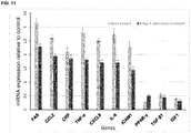
- this potential involvement was confirmed by assaying gene expression alterations in a HGPS mouse and human fibroblast compared to control fibroblasts.
- PPAR- ⁇ and IGF1 are downregulated in HGPS, and that CCL2, CXCL8, ICAM1, CRP, TRAF1, IL-18, IL-6, TNF- ⁇ and FAS are overexpressed in HGPS. It was also found that although TGF- ⁇ 1 and C3 expression changes in HGPS, it is visible that among the downregulated genes in HGPS, TGF- ⁇ 1 has the lowest fold decrease, and among the overexpressed genes in HGPS, C3 has the lowest fold increase.
- TGF- ⁇ 1 is downregulated in vascular disease and arthritis but overexpressed in lipodystrophy and alopecia
- C3 is overexpressed in all conditions except lipodystrophy in which it is downregulated.
- STAT3 is the STAT that regulates the highest amount of genes simultaneously involved in the pathogenesis of the four main phenotypes that characterize HGPS and so has the highest level of overexpression in HGPS, immediately followed by STAT1; STAT5a and STAT5b regulate a lower amount of genes and, accordingly, have significantly lower levels of overexpression in comparison to STAT1 and STAT3; STAT6 only regulates one gene and thus is only slightly overexpressed in HGPS fibroblasts; STAT2 and STAT4 do not regulate any of the genes and so their expression does not change between HGPS and control fibroblasts.
- the next step in the project was to try to reverse these gene expression alterations that occur in HGPS to determine if it results in the amelioration of some of the typical phenotypic traits that characterize this condition.
- a JAK-STAT inhibitor was used to try to reduce the expression of STAT1, STAT3, and normalize STAT5a, STAT5b, and possibly correct the expression levels of the 11 JAK-STAT regulated genes that are involved simultaneously in the pathogenesis of the four main phenotypes that characterize HGPS.
- the one that was selected was baricitinib, a drug that potently inhibits JAK1 and JAK2, but has a lower potency against TYK2 and JAK3.
- JAK-STAT signaling pathway As the JAK-STAT signaling pathway is involved in many biological processes, it is very important to control any sort of manipulation of this pathway to avoid undesired side effects. Therefore it was necessary to assay the cytotoxicity of the compound used to treat HGPS cells.
- a suitable compound selectively and reversely inhibits the JAK-STAT signaling pathway. In this way manipulation of the pathway can be controlled and reversed if necessary, for example by choosing the correct dosage of a compound with a suitable clearance.
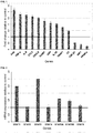
- a cytotoxicity assay was performed with baricitinib to determine the suitable concentration that maintains efficacy and does not lead to cytotoxicity (see Example 5 ).
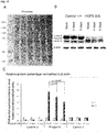
- the results are shown in Figure 3 . Based on these results an upper concentration of 1 ⁇ M baricitinib was chosen for further assays for HGPS cells. For mouse fibroblasts obtained from a progerin mouse model, an upper concentration of 3 ⁇ M baricitinib was chosen for further assays (see Figure 14 ).
- the next step was to determine if the inhibition of the JAK-STAT pathway by baricitinib exerts any beneficial effects in HGPS fibroblasts and, if so, which of the tested concentrations and what treatment duration is the most beneficial to the cells.
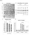
- Five functional assays were performed on cells treated for 4-days and 9-days either mock-treated or baricitinib-treated (at the concentrations of 0.25 ⁇ M, 0.5 ⁇ M and 1 ⁇ M): growth rate (cumulative population doubling assay), autophagy and proteasome activities, the ROS and ATP levels (see Examples 6 - 10, and Figures 4 - 8 ; for data obtained with mouse fibroblasts obtained from a progerin mouse model, see Figures 15 and 16 )
- baricitinib is a promising drug to be used in the therapy of HGPS patients since it can revert some of the characteristic phenotypic traits that impair the condition of HGPS cells.
- Baricitinib is however only one example for a compound suitable for use in treating HGPS. Any compound exhibiting similar characteristics as baricitinib can be used for treating HGPS. For example any compound that selectively and reversely inhibits JAK1 with an IC 50 of about 6 nM, and JAK2 with an IC 50 of about 6 nM is suitable for treating HGPS.
- IC 50 is the half maximal inhibitory concentration and is a measure of the effectiveness of a substance in inhibiting a specific biological or biochemical function. This quantitative measure indicates how much of a particular drug or other substance (e.g. a JAK-STAT inhibitor) is needed to inhibit a given biological process (such as the processes at the root of the main symptoms in HGPS) by half. Values of IC 50 can be obtained by the person of skill in the art with assays established in the art and can be compared with the values exhibited by baricitinib in the same type of assay.
- a particular drug or other substance e.g. a JAK-STAT inhibitor
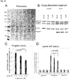
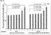
- JAK-STAT inhibition resulted in a reduction of levels of progerin by an average of 15% after 4 days treatment with 1 ⁇ M baricitinib as determined with anti-progerin and anti-lamin A/C antibodies (see Figure 9 A and D ). Furthermore, progerin levels were decreased by 32 to 33% by 9 days treatment with 1 ⁇ M baricitinib (see Figure 10 ). It was therefore shown that long-term baricitinib treatment significantly increased the clearance of progerin in HGPS cells while lamin A and lamin C levels remained constant. In mouse fibroblasts obtained from a progerin mouse model, it was shown that the progerin levels were decreased by 35.6 % by 9 days treatment with 3 ⁇ M baricitinib (see Figure 17 ).
- baricitinib treatment ameliorates proteostasis (autophagy and proteasome activities) and consequently increase progerin clearance in HGPS cells.
- This reduction in progerin content by an average of 33% within a period of 9 days is higher than the reduction observed after treatment with sulforaphane and rapamycin, which reduced progerin by approximately 20 to 25% in average in a period of 9 days.
- baricitinib treatment ameliorates the ratios of lamin A/C in HGPS and therefore might significantly improve other cellular functions that are altered by the build-up of progerin in HGPS nuclear compartment.
- Data obtained in mouse fibroblasts from a progerin mouse model confirm these results (wherein the baricitinib concentration used was 3 ⁇ M).
- JAK-STAT pathway inhibitor could be used as a drug to ameliorate the condition of HGPS patients.
- Treatment of HGPS fibroblasts with JAK-STAT pathway inhibitor induced amelioration of autophagy, proteasome and mitochondrial activity that are known to malfunction in HGPS cells.
- These positive effects consequently reduce the levels of progerin, the mutated form of lamin A whose accumulation in HGPS nucleus is the main originator of the phenotypic traits that characterize HGPS.
- treating refers to the medical management of a patient with the intent to cure, ameliorate, stabilize, or prevent a disease, pathological condition, or disorder.
- This term includes active treatment, that is, treatment directed specifically toward the improvement of a disease, pathological condition, or disorder.
- palliative treatment that is, treatment designed for the relief of symptoms rather than the curing of the disease, pathological condition, or disorder
- preventative treatment that is, treatment directed to minimizing or partially or completely inhibiting the development of the associated disease, pathological condition, or disorder
- supportive treatment that is, treatment employed to supplement another specific therapy directed toward the improvement of the associated disease, pathological condition, or disorder.
- treatment includes treating of HGPS by ameliorating the main symptoms of HGPS, which are vascular disease, arthritis, lipodystrophy, and alopecia.
- Example 1 Resources used to find the list of genes regulated by the JAK-STAT signaling pathway
- TRRUST is a manually curated reference database of human transcriptional regulatory interactions (Han et al., 2015); TRED is an integrated repository for both cis- and trans- regulatory elements in mammals (Jiang et al., 2007); ENCODE is a database in which putative human transcription factors interactions with DNA are named based on ChIP-seq data (2012); and Neph2012 is a regulatory network in which transcription factor targets were discovered by DNase foot-printing and transcription factor recognition sequences (Neph et al., 2012). After obtaining the complete list of genes regulated by the JAK-STAT signaling pathway using these databases, the unintentional repetition data was removed manually (some genes are mentioned more than once in the list because they have different aliases in different databases). In this search 269 genes were found, out of which 119 are regulated by STAT1, 10 by STAT2, 157 by STAT3, 14 by STAT4, 32 by STAT5a, 27 by STAT5b and 43 by STAT6.
- STAT1 and STAT3 are the STATs that regulate the highest number of genes, suggesting that these two are the most important activators of transcription in the JAK-STAT pathway in humans.
- the genes regulated by STAT2 and STAT4 are represented in in Table 2 and Table 3, respectively.
- the STATs that simultaneously regulate these genes with STAT2 and STAT4 are also represented in the table.
- Table 2 - STAT2 regulated genes and other STATs that regulate the genes simultaneously with STAT2 ( ⁇ - regulated; ⁇ - not regulated).
- HGPS vascular disease, arthritis, lipodystrophy and alopecia.
- This software was developed by the inventors and it is based on scientific literature present in PubMed. Besides this list of genes, all genes present in the HGNC (Human Gene Nomenclature Committee) official list were also screened.
- the first step on this search was the retrieval of all papers in which these four conditions were mentioned individually or together.
- the PubMed database an NCBI database that comprises more than 27 million citations for biomedical literature from MEDLINE, life science journals and online books
- Two of the software's functions are used to import either the text file or the XML file of each saved paper, constructing the starting point for all subsequent processing. Papers with publication types that were not related to research or reviews (for example bibliographies, comments, or editorials) were filtered out to eliminate false-positive findings.
- HGPS Primary dermal fibroblasts from subjects with HGPS were obtained from the Progeria Research Foundation. Two different fibroblasts derived from HGPS patients carrying the LMNA mutation G608G were used: HGADFN003 (male, 2 years old) and HGADFN127 (female, 3 years and 9 months old). Control dermal fibroblasts were obtained from the Coriell Institute for Medical Research (Camden, NJ). The following cell lines were used: GMO1651C and GMO3349C. All cells were maintained at -80 °C in 90% fetal bovine serum (FBS) and 10% dimethyl sulfoxide (DMSO). The Institutional Review Board of the Technische (2015) München (TUM) approved the use of human cells established from skin biopsies from patients with HGPS and unaffected individuals in this study.
- FBS fetal bovine serum
- DMSO dimethyl sulfoxide
- Control and HGPS fibroblasts were cultured at 37 °C and 5% CO 2 in Dulbecco's Modified Eagle Media (DMEM) (Gibco by life technologies) containing 15% FBS (Gibco by life technologies), 1% L-glutamine (Gibco by life technologies), 1% penicillin/streptomycin (Gibco by life technologies), and 2 ml of gentamycin (Gibco by life technologies). Mock-treated and baricitinib-treated fibroblasts were cultured in parallel.
- DMEM Dulbecco's Modified Eagle Media
- Baricitinib (Selleckchem) was added to the media at a concentration of 0.125 ⁇ M, 0.25 ⁇ M, 0.5 ⁇ M, 1 ⁇ M, 2 ⁇ M or 4 ⁇ M. Three times a week the cells were re-fed with new culture media.
- Fibroblasts cease proliferating once they become confluent (when they completely cover the surface of the cell culture vessel) and some die if left in this state for too long. In order to keep the cells healthy and actively growing, they were periodically subcultured at approximately 80% confluency.
- the first step of this process is the removal of the culture media, washing with PBS (Sigma), and the addition of 1 ml of a 0.1 % sterile trypsin solution (Gibco by life technologies) to dissociate adherent cells from the vessel. After trypsinization, the cells are diluted with 8 ml of new culture media, counted with a CASY® Cell Counter (Roche), subdivided into new vessels and incubated in the same conditions.
- RT-PCR reactions for the genes PPAR- ⁇ , IGF-1, TFG- ⁇ 1, C3, TRAF1, IL-18, CRP, ICAM1, CXCL8, CCL2, IL-6, TNF- ⁇ , FAS, STAT1, STAT2, STAT3, STAT4, STAT5a, STAT5b and STAT6 were performed to compare their expression level between control and HGPS fibroblasts.
- RT-PCR reactions were also performed to compare their expression level between mock-treated and 9-day 1 ⁇ M baricitinib-treated HGPS fibroblasts.
- the cell lysate was then homogenized by being pipetted to into a QIAshredder spin column (a biopolymer-shredding system in a microcentrifuge spin-column format) and centrifuged at 13.000 rpm for 2 min.
- QIAshredder spin column a biopolymer-shredding system in a microcentrifuge spin-column format
- 1 volume (350 ⁇ l) of 70% ethanol (VWR Chemicals) was added to the homogenized lysate, that was then transferred to an RNeasy spin column and centrifuged for 15 s at >10.000 rpm.
- Buffer RW1 700 ⁇ l of Buffer RW1 were added to the spin column and centrifuged for 15 s at >10.000 rpm in order to wash its membrane, removing biomolecules such as carbohydrates, proteins and fatty acids that are non-specifically bound to the silica membrane (RNA molecules larger than 200 bases remain bound to the column).
- 500 ⁇ l of buffer RPE were added to the spin column and centrifuged for 15 s at >10.000 rpm to remove traces of salts which were still in the column due to buffers used earlier in the protocol. This step was then repeated, but with a centrifugation lasting for 2 min, to ensure that all ethanol was removed.
- RNA obtained at the end of the procedure was determined using a NanoDrop® ND-1000 Spectrophotometer.
- cDNA was synthesized from the extracted RNA samples using an Omniscript Reverse Transcription Kit (Qiagen).
- a fresh master mix containing all the components required for first-strand synthesis except the template RNA was prepared on ice and thoroughly mixed: 2 ⁇ l of 10x Buffer RT, 2 ⁇ l of dNTP Mix (5 mM each dNTP), 2 ⁇ l of 10 ⁇ M Oligo-dT primer, 1 ⁇ l of 10 units/ ⁇ l RNase inhibitor and 1 ⁇ l of Omniscript Reverse Transcriptase.
- 2000 ng of template RNA were added to the mix and RNase-free water was added until a total volume of 20 ⁇ l was reached.
- the mix was incubated for 60 min at 37 °C.
- Table 4 Nucleotide sequence, molecular weight (MW), GC content and melting temperature (MT) of the forward (fw) and reverse (rv) primers used in the PCR reaction for the genes PPAR- ⁇ , CCL2, TFG- ⁇ 1, CXCL8, ICAM1, CRP, C3, TRAF1, IL-18, IGF1, IL-6, TNF- ⁇ , and FAS, STAT1, STAT2, STAT3, STAT4, STAT5A, STAT5B, STAT6, JAK1, JAK2, JAK3, TYK2.
- the objective of these qPCR reactions is the amplification and quantification of the mRNA molecules that are transcribed from each gene.
- the primers' sequences correspond to exon-exon junctions to assure that no genomic DNA is amplified.
- Other criterion used in the primers' design were: %GC between 40% and 60% to assure that there is no formation of primer dimers; primer melting temperature between 55 °C and 62 °C because higher temperatures may result in less binding efficiency and lower temperatures may result in less specificity; fw and reverse primers of each gene with similar primer melting temperatures; primer length between 18 and 22 bp for good specificity and binding abilities; and PCR products no longer than 300 bp, since the DNA polymerase cannot amplify DNA fragments longer than this.
- the exons' sequence of each gene was consulted on, and the primer melting temperature of each primer was consulted on.
- the first step of this phase of the procedure was the preparation of a master mix for each RT-PCR reaction containing all the components needed for extension except the template cDNA: 5 ⁇ l of SsoAdvanced universal SYBR® Green supermix (2x), and fw and reverse primers at a final concentration of 250-500 nM each. After mixing all these components, 100 ng - 100 fg of template cDNA were added, and finally nuclease-free water was also added until 10 ⁇ l of reaction volume was reached. The qPCR was then carried out using an Mx3000P Real-Time PCR Detection System (Stratagene) following the PCR program summarized in Table 5. Three experiments were performed for each assay and all samples were run in triplicate.
- Relative quantification was performed by determining the RT-PCR signal of the experimental RNA samples in relation to the signal of GAPDH, a housekeeping gene that is used as the internal normalization control gene.
- the 2( ⁇ C T ) method was used to calculate the relative changes in gene expression between HGPS fibroblasts and control fibroblasts, and also between HGPS 9-day 1 ⁇ M baricitinib-treated fibroblasts and HGPS mock-treated fibroblasts. In this method a certain expression level threshold value is established and what is quantified is the number of RT-PCR cycles that it takes for gene expression level to reach the threshold value (C T ).
- equations 1, 2, 3 and 4 are applied for the calculation of the normalized target gene expression.
- equations 1, 2, 3 and 4 are analogous.
- the assay was also performed in control fibroblasts that were grown for 24 h and 48 h without any exposure to baricitinib or DMSO (mock).
- This assay measures changes in membrane integrity that occur as a result of cell death. It uses a cyanine dye that is excluded from viable cells but preferentially stains the dead cells' DNA. When the dye binds DNA, its fluorescent properties are enhanced.
- control fibroblasts were harvested in the above mentioned conditions, and after trypsinization the number of cells was counted with a CASY® Cell Counter (Roche). Then, the fibroblasts from each condition were seeded in triplicate in a 96 well plate with 32.000 cells/well in 100 ⁇ l of culture media with the appropriate drug concentration, and the plate was incubated at 37 °C for 8 h. After centrifuging the 96 wells plate at 1200 rpm for 5 min, the culture media was aspirated from each well and 100 ⁇ l of CellToxTMGreen Dye Substrate (1:500 in assay buffer) was added.
- the plate was incubated at room temperature in the dark for 15 min, and the fluorescence was measured in a FLUOstar Omega Microplate Reader (Omega BMG Labtech) using an excitation wavelength of 485 nm and an emission wavelength of 520 nm. This measurement corresponds to the number of dead cells that are present in each well.
- 4 ⁇ l of lysis solution was added to each well, the plate was incubated at 37 °C for 30 min, and the fluorescence level was measured again using the same wavelengths. This last measurement corresponds to the total number of cells present in each well. By dividing the first measurement by the last one, it is possible to obtain the percentage of cell death. Lastly the results were adjusted by considering that the mock treatment corresponds to 0% of cell death.
- a further baricitinib cytotoxicity assay was done comparing wild-type mouse fibroblasts and mouse fibroblasts from a progerin mouse model subjected to differing concentrations of The progerin mouse is homozygous for LMNA G609G/G609G (referred to as ⁇ / ⁇ G609G/G609G mouse).
- ⁇ / ⁇ G609G/G609G mouse LMNA G609G/G609G
- a cumulative population doubling assay was performed in 4 day and 9 day mock-treated and baricitinib-treated (at the concentrations of 0.25 ⁇ M, 0.5 ⁇ M, and 1 ⁇ M) control and HGPS fibroblasts (see Figure 4 ).
- a further assay was carried out comparing proliferation of wild-type mouse fibroblasts and mouse fibroblasts from a progerin mouse model treated with 3 ⁇ M of baricitinib (see Figure 15 ). These results indicate that 3 ⁇ M of baricitinib ameliorate cell growth of control and progerin mouse cells. Cells from progerin (or ⁇ / ⁇ ) mouse exhibit growth retardation compared to control cells. Baricitinib increases and ameliorate the proliferation potency of the mouse progerin fibroblasts.
- Autophagic vacuoles in 4-day and 9-day mock-treated and baricitinib-treated (at the concentrations of 0.25 ⁇ M, 0.5 ⁇ M, and 1 ⁇ M) control and HGPS fibroblasts were quantified using an Autophagy/Cytotoxicity Dual Staining Kit (Cayman Chemical).
- This assay uses monodansylcadaverine (MDC), a fluorescent compound that is incorporated into multilamellar bodies by both an ion trapping mechanism and the interaction with membrane lipids, as a probe for the detection of autophagic vacuoles in cells.
- MDC monodansylcadaverine
- control and HGPS fibroblasts were harvested in the above mentioned conditions and, following trypsinization, the number of cells was counted with a CASY® Cell Counter (Roche). Afterwards, fibroblasts from each condition were seeded in triplicate in a 96 well plate with 32.000 cells/well in 100 ⁇ l of culture media with the appropriate drug concentration, and the plate was incubated at 37 °C for 8 h. After centrifuging the 96 well plate at 1200 rpm for 5 min, the culture media was aspirated from each well and 100 ⁇ l of staining solution was added. The staining solution was prepared by diluting Cell-Based Monodansylcadaverine 1:1000 in Cell Based Assay Buffer.
- the assay buffer was prepared by dissolving a Cell-Based Assay Buffer Tablet in 100 ml of distilled water. The plate was then incubated at 37 °C for 10 min and centrifuged for 5 min at 1200 rpm. Next, the supernatant was removed, 100 ⁇ l of Cell-Based Assay Buffer was added to each well and the plate was again centrifuged in the same conditions. Lastly, after removing the supernatant, 100 ⁇ l of Cell-Based Assay Buffer was added to each well and the autophagic vacuole staining intensity was immediately detected in a FLUOstar Omega Microplate Reader (Omega BMG Labtech) using an excitation wavelength of 355 nm and an emission wavelength of 520 nm. Considering that the autophagy level in mock-treated control fibroblasts is 100%, the level of autophagy relative to mock-treated control fibroblasts for each condition was determined (see Figure 5 ).
- Cell-Based Tamoxifen 100 mM
- a known inducer of autophagy was used as a positive control. This compound was initially added to a control culture diluted 1:50.000 into the culture media. Cell Based Assay Buffer was used as a negative blank control.
- a further assay was carried out showing autophagy levels in wild-type mouse fibroblasts and mouse fibroblasts from a progerin mouse model (see Figure 16 ).
- the results indicate that 3 ⁇ M baricitinib increases the levels of autophagy in both control and progerin fibroblasts. Importantly, the levels are increased by an average of 39.9% in progerin mouse fibroblast relative to the mock-treated control cells
- 20S proteasome core particle in 4-day and 9-day mock-treated and baricitinib-treated (at the concentrations of 0.25 ⁇ M, 0.5 ⁇ M, and 1 ⁇ M) control and HGPS fibroblasts was quantified using a 20S Proteasome Assay Kit (Cayman Chemical). This assay uses a specific 20S substrate (SUC-LLVY-AMC) which, upon cleavage by the active enzyme, generates a fluorescent product that can be detected.
- SUC-LLVY-AMC specific 20S substrate
- control and HGPS fibroblasts were harvested in the above mentioned conditions and, following trypsinization, the number of cells was counted with a CASY® Cell Counter (Roche).
- the fibroblasts from each condition were seeded in triplicate in a 96 well plate with 100.000 cells/well in 100 ⁇ l of culture media with the appropriate drug concentration, and the plate was incubated at 37 °C for 8 h. After centrifuging the 96 well plate at 1200 rpm for 5 min, the culture media was aspirated and 200 ⁇ l of 20S Proteasome Assay Buffer was added to each well.
- the plate was again centrifuged in the same conditions, the supernatant was aspirated, and 100 ⁇ l of 20S Proteasome Lysis Buffer was added to each well. Then, the plate was incubated with gentle shaking for 30 min at room temperature to allow cell lysis and it was centrifuged at 1300 rpm for 7 min. Following centrifugation, 90 ⁇ l of the supernatant from each well was transferred to a new empty well together with 10 ⁇ l of substrate solution.
- the substrate solution was prepared by adding 25 ⁇ l of 20S Proteasome Substrate (SUC-LLVY-AMC) to 1 ml of 20S Proteasome Assay Buffer.
- the plate was incubated at 37 °C for 1 h and after that time the fluorescence intensity was read in FLUOstar Omega Microplate Reader (Omega BMG Labtech) using an excitation wavelength of 360 nm and an emission wavelength of 480 nm. Considering that the proteasomal activity in mock-treated control fibroblasts is 100%, the proteasomal activity relative to mock-treated control fibroblasts was calculated for each condition.
- a Jurkat cell lysate supernatant which contains a high level of 20S activity was used as positive control (20S Proteasome Positive Control) and 20S Proteasome Assay Buffer was used as negative blank control.
- Intracellular ROS in 4-day and 9-day mock-treated and baricitinib-treated (at the concentrations of 0.25 ⁇ M, 0.5 ⁇ M, and 1 ⁇ M) control and HGPS fibroblasts was measured using a DCFDA Cellular ROS Detection Assay Kit (Abcam).
- This assay uses 2',7'-dichlorofluorescein diacetate (DCFDA), a cell permeant reagent that after entering the cell is deacetylated by cellular esterases to a non-fluorescent compound that is later oxidized by ROS into 2',7'-dichlorofluorescein (DCF).
- DCF is highly fluorescent and can be detected by fluorescence spectroscopy.
- control and HGPS fibroblasts were harvested in the above mentioned conditions and, following trypsinization, the number of cells was counted with a CASY® Cell Counter (Roche).
- the fibroblasts from each condition were seeded in triplicate in a 96 well plate with 32.000 cells/well in 100 ⁇ l of culture media with the appropriate drug concentration, and the plate was incubated at 37 °C for 8 h. After the incubation, the culture media was aspirated, 100 ⁇ l of 1 x Buffer were added to each well, and the plate was centrifuged at 1200 rpm for 5 min.
- the supernatant was aspirated, 100 ⁇ l of DCFDA solution was added to each well and the plate was incubated at 37 °C for 45 min in the dark.
- the DCFDA solution was prepared by adding 12.5 ⁇ l of 20 mM DCFDA solution to 10 ml of 1x Buffer.
- the plate was centrifuged under the same conditions, the supernatant was aspirated, and 100 ⁇ l of 1x Buffer was added to each well.
- fluorescence was measured immediately in a FLUOstar Omega Microplate Reader (Omega BMG Labtech) using an excitation wavelength of 485 nm and an emission wavelength of 535 nm. Considering that the ROS level in mock-treated control fibroblasts is 100%, the ROS levels relative to mock-treated control fibroblasts were calculated for each condition.
- TBHP tert-butyl hydrogen peroxide
- the intracellular ATP content of 4-day and 9-day mock-treated and baricitinib-treated (at the concentrations of 0.25 ⁇ M, 0.5 ⁇ M, and 1 ⁇ M) control and HGPS fibroblasts was measured by using a CellTiter-Glo® Luminescent Cell Viability Assay (Promega).
- This assay relies in the properties of a proprietary thermostable luciferase which generates a stable luminescent signal that is proportional to the amount of ATP in the cells by catalyzing the mono-oxygenation of luciferin in the presence of Mg 2+ , ATP and molecular oxygen.
- control and HGPS fibroblasts were harvested in the above mentioned conditions and the number of cells was counted with a CASY® Cell Counter (Roche). Then, the fibroblasts from each condition were seeded in triplicate in a 96 well plate with 32.000 cells/well in 100 ⁇ l of culture media with the appropriate drug concentration, and the plate was incubated at 37 °C for 8 h. After equilibrating the 96 well plate at room temperature for 30 min and centrifuging it at 1200 rpm for 5 min, the culture media was aspirated and 100 ⁇ l of CellTiter-Glo® Reagent was added to each well.
- This reagent is obtained by transferring 10 ml of CellTiter-Glo® Buffer into an amber bottle containing CellTiter-Glo® Substrate and then mixing it gently by vortexing. The plate was then mixed for 2 min in an orbital shaker to induce cell lysis and following that, it was incubated at room temperature for 10 min to stabilize the luminescent signal. Finally, luminescence was recorded in a FLUOstar Omega Microplate Reader (Omega BMG Labtech). Considering that the ATP level in mock-treated control fibroblasts is 100%, the ATP levels relative to mock-treated control fibroblasts were calculated for each condition.
- HGPS fibroblasts An immunocytochemistry assay with an anti-progerin antibody was performed in control and HGPS fibroblasts to substantiate that HGPS cells are characterized by an accumulation of progerin in the nucleus. Both control and HGPS fibroblasts were directly grown in coverslips. After one day of growing, the fibroblasts were washed with PBS (Sigma) for three times and fixed with 4% PFA (Sigma-Aldrich) at room temperature for 15 min to preserve cellular morphology and cellular structures in their native conformation. Then, the fibroblasts were washed again for three times with PBS (Sigma) and permeabilized by 1% Triton X-100 (PanReac AppliChem) for 3 min at room temperature.
- PBS Sigma
- PFA Sigma-Aldrich
- fibroblasts were again washed for three times with PBS (Sigma) and blocked with 15% FBS/PBST blocking buffer at room temperature for 30 min to minimize unspecific binding of the primary antibody inside the cell.
- the blocking buffer contains 15% FBS (Gibco by life technologies) and 85% PBST, while PBST is constituted by 98.5% PBS (Sigma) and 1.5% Tween 20 (Sigma).
- the fibroblasts were incubated in anti-progerin S9 primary antibody (McClintock et al ., 2007), 1 ⁇ g/ml diluted in blocking buffer, for 24h at room temperature.
- fibroblasts were washed three times with PBST for 5 min to remove the primary antibody that did not bind to any protein inside the cell and incubated in Alexa Fluor® 555 donkey anti-rabbit IgG (H+L) secondary antibody (Thermo Fisher Scientific), 1/800 diluted in blocking buffer, for 1h at room temperature. After this, fibroblasts were washed for three times with PBST for 5 min to remove the secondary antibody that did not bind to the primary antibody inside the cell and, afterwards, washed for three times with PBS (Sigma) for 5 min. Finally, the samples were counterstained with the DNA dye DAPI in Vectashield mounting medium (Vector Inc.) and observed using an Axioplan fluorescence microscope (Carl Zeiss).
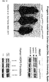
- a quantitative Western Blot analysis using a Santa Cruz rabbit anti-lamin A/C (H-1 10) antibody was performed in control and HGSP fibroblasts that were mock-treated or baricitinib-treated (at the concentrations of 0.25 ⁇ M, 0.5 ⁇ M or 1 ⁇ M) for a period of 4 and 9 days to determine if baricitinib can reduce progerin levels in HGPS fibroblasts (see Figures 9 , 10 , and 13 ).
- a similar quantitative Western Blot analysis was carried out in wild-type mouse fibroblasts compared to fibroblasts obtained from the progerin mouse model (see Figure 17 ).
- control and HGPS fibroblasts were harvested in the above mentioned conditions, and after trypsinization the number of cells was counted with a CASY® Cell Counter (Roche). Subsequently, the cell suspension was centrifuged for 5 min at 1200 rpm, the supernatant was aspirated, and the cells were washed with 4.5 ml PBS (Sigma). These steps were performed thrice in order to assure that all culture media is removed. Then, the cells were transferred to Eppendorf tubes with 1 ml PBS (Sigma) and the tubes were centrifuged for 5 min at 1200 rpm. After fully aspirating PBS, the cell pellets were obtained.
- the first step was the addition of 100 ⁇ l per 10 7 cells of Laemmli Buffer-inhibitor mix to each Eppendorf tube to lyse the fibroblasts and denature the proteins of the lysate.
- This solution contains 60 ⁇ l of ⁇ -mercaptoethanol (Biorad) to reduce the intra and inter-molecular disulfide bonds, 12.5 ⁇ l of protease inhibitor cocktail (Biorad) and 7.5 ⁇ l of 200 mM PMSF (Biorad) to avoid protein degradation, and 0.95 ml of Laemmli Sample buffer (BioRad), a gel electrophoresis loading buffer that contains the SDS detergent that denatures the proteins and gives them an overall negative charge. Afterwards, the solution in each Eppendorf was vortexed for 2 min and heated at 99 °C for 3 minutes. These two steps were repeated for three times to assure that all the cells were lysed.
- Biorad ⁇ -mercaptoethanol
- the total protein concentration of each gel electrophoresis loading sample was determined by using a series of dilutions of known concentrations of bovine serum albumin (BSA) to obtain a standard curve.
- BSA bovine serum albumin
- the content of an Albumin Standard Ampule that contains BSA at 2 mg/ml was diluted in Laemmli Buffer-inhibitor mix into the concentrations of 2000 ⁇ g/ml, 1500 ⁇ g/ml, 1000 ⁇ g/ml, 750 ⁇ g/ml, 500 ⁇ g/ml, 250 ⁇ g/ml, 125 ⁇ g/ml, 25 ⁇ g/ml and 0 ⁇ g/ml to obtain the BSA standard dilutions samples that were used to acquire the standard curve.
- each gel electrophoresis loading sample is diluted in 8 ⁇ l of Laemmli Sample buffer (BioRad). Then, 2 ⁇ l of each BSA standard dilution sample and 2 ⁇ l of each diluted electrophoresis loading sample were blotted for three times on the nitrocellulose blotting membrane. After being washed with ddH 2 O, the membrane was incubated with ponceau S (Sigma-Aldrich) for 5 min in order to stain the proteins that are present in the membrane, and finally washed again with ddH 2 O to remove the dye that did not bind to any protein.
- ponceau S Sigma-Aldrich
- a SDS-gel electrophoresis to separate the proteins in each sample based on their molecular weight was performed in a 4-20% Mini-Protean® TGXTM Precast Protein Gel (BioRad), using 1x running buffer, at 200 V until the 37 kD lane reached the bottom of the gel.
- This running buffer was prepared as 10x running buffer that contains 1.92 M glycine (Carl Roth), 248 mM tris-base (Sigma) and 1% SDS (Sigma), and then diluted in ddH 2 O to 1 x.
- each sample was vortexed for three times during 30 s and heated at 99 °C for 3 minutes to completely denature the proteins that are present in each sample. 5 ⁇ l of Precision plus protein standard (Biorad) were added to one of the wells to be used as a marker. After finishing the SDS-gel electrophoresis, the gel was removed from the cassette, washed with ddH 2 O and equilibrated for 10 min in transfer buffer.
- the transfer buffer contains 20 mM tris (Sigma), 150 mM glycine (Carl Roth), 20% methanol (PanReac AppliChem) and 0.1% SDS (Sigma).
- the proteins that were separated in the gel were then transferred to a nitrocellulose membrane using a Trans-Blot® TurboTM Transfer System (Biorad) at 25 V for 20 min, and using the same transfer buffer.
- the fiber pads, filter papers and nitrocellulose membrane that were used in the electroblotting were previously equilibrated overnight in pre-cooled transfer buffer at 4 °C. Even before that, the nitrocellulose membrane had to be equilibrated in ddH 2 O for 10 min.
- the nitrocellulose membrane was washed with ddH 2 O and incubated in Ponceau S (Sigma-Aldrich) for 5 min to verify the effectiveness of the protein transfer from the gel to the membrane.
- the membrane was again washed with ddH 2 O and a picture of it was taken using a ChemiDocTM MP Imaging System (Biorad). Ponceau S staining technique is reversible to allow further immunological detection. Accordingly, after this procedure the blotting membrane was washed with PBST and destained in PBS (Sigma). PBST contains 98.5% PBS (Sigma) and 1.5% Tween 20 (Sigma).
- the membrane Before proceeding to the immunodetection, the membrane had to be blocked to avoid unspecific binding of the antibody used in the immunodetection.
- the nitrocellulose membrane was incubated for 4 h in milk buffer.
- the milk buffer contains 1.2% nonfat dry milk vitamin A+D fortified (Nestlé) and 98.8% PBST.
- PBST contains 98.5% PBS (Sigma) and 1.5% Tween 20 (Sigma).
- the membrane was incubated in rabbit anti-lamin A/C H-1 10 antibody (Santa Cruz Biotechnology, 1/10.000 in PBST) for 12 h at 4 °C.
- the membrane was washed with PBST for three times during 5 min and subsequently incubated in Peroxidase-conjugated AffiniPure goat anti-rabbit IgG (H+L) (Abcam, 1/5000 in PBST) for 50 min at room temperature.
- the membrane was again washed with PBST for three times during 5 min and then washed with PBS (Sigma) for three times during 5 min to remove the secondary antibody that did not bind to any primary antibody.
- the proteins were visualized using ECL1 and ECL2 (Biorad) at a dilution of 1:1 in a ChemiDocTM MP Imaging System (Biorad). Protein signals were quantified by normalizing to ⁇ -tubulin using the software Image Lab (Biorad).
Landscapes
- Health & Medical Sciences (AREA)
- Veterinary Medicine (AREA)
- Chemical & Material Sciences (AREA)
- Medicinal Chemistry (AREA)
- Pharmacology & Pharmacy (AREA)
- Life Sciences & Earth Sciences (AREA)
- Animal Behavior & Ethology (AREA)
- General Health & Medical Sciences (AREA)
- Public Health (AREA)
- Epidemiology (AREA)
- Engineering & Computer Science (AREA)
- Bioinformatics & Cheminformatics (AREA)
- Chemical Kinetics & Catalysis (AREA)
- General Chemical & Material Sciences (AREA)
- Nuclear Medicine, Radiotherapy & Molecular Imaging (AREA)
- Organic Chemistry (AREA)
- Pharmaceuticals Containing Other Organic And Inorganic Compounds (AREA)
Priority Applications (1)
| Application Number | Priority Date | Filing Date | Title |
|---|---|---|---|
| EP18151570.1A EP3511005A1 (fr) | 2018-01-15 | 2018-01-15 | Composés destinés à être utilisés dans le traitement du syndrome de la progéria d'hutchinson-gilford |
Applications Claiming Priority (1)
| Application Number | Priority Date | Filing Date | Title |
|---|---|---|---|
| EP18151570.1A EP3511005A1 (fr) | 2018-01-15 | 2018-01-15 | Composés destinés à être utilisés dans le traitement du syndrome de la progéria d'hutchinson-gilford |
Publications (1)
| Publication Number | Publication Date |
|---|---|
| EP3511005A1 true EP3511005A1 (fr) | 2019-07-17 |
Family
ID=60972108
Family Applications (1)
| Application Number | Title | Priority Date | Filing Date |
|---|---|---|---|
| EP18151570.1A Withdrawn EP3511005A1 (fr) | 2018-01-15 | 2018-01-15 | Composés destinés à être utilisés dans le traitement du syndrome de la progéria d'hutchinson-gilford |
Country Status (1)
| Country | Link |
|---|---|
| EP (1) | EP3511005A1 (fr) |
Cited By (1)
| Publication number | Priority date | Publication date | Assignee | Title |
|---|---|---|---|---|
| WO2021167478A1 (fr) * | 2020-02-19 | 2021-08-26 | Ibmc - Instituto De Biologia Molecular E Celular | 4-pyrrolidin-1-yl-5-p-tolyl-thiéno[2,3-d] pyrimidine destinée à être utilisée dans le traitement de maladies associées au vieillissement et au vieillissement prématuré par la stabilité chromosomique restaurée et l'inhibition de la sénescence cellulaire |
-
2018
- 2018-01-15 EP EP18151570.1A patent/EP3511005A1/fr not_active Withdrawn
Non-Patent Citations (29)
| Title |
|---|
| ALTEN, R.; NITSCHMANN, S.: "Efficacy and safety of baricitinib in Japanese patients with rheumatoid arthritis: Subgroup analyses of four multinational phase 3 randomized trials", INTERNIST (BERL, vol. 58, 2017, pages 1341 - 1344 |
| ANNE-LAURE EGESIPE ET AL: "Metformin decreases progerin expression and alleviates pathological defects of Hutchinson-Gilford progeria syndrome cells", NPJ AGING AND MECHANISMS OF DISEASE, vol. 2, no. 1, 10 November 2016 (2016-11-10), XP055484935, DOI: 10.1038/npjamd.2016.26 * |
| CARFAGNA, M.; CANNADY, E.; RYAN, T.; HERMAN, J.; TRUEX, L.; NARWANI, K.; SULLIVAN, J.: "Carcinogenicity assessment of baricitinib in Tg.rasH2 mice and Sprague-Dawley (Crl:CD) rats", REGULATORY IN TOXICOLOGY AND PHARMACOLOGY, 2017 |
| FLEISCHMANN, R.; ALAM, J.; ARORA, V.; BRADLEY, J.; SCHLICHTING, D. E.; MURAM, D.; SMOLEN, J. S.: "Safety and efficacy of baricitinib in elderly patients with rheumatoid arthritis", RMD OPEN, vol. 3, 2017, pages e000546 |
| GENOVESE, M. C. ET AL.: "Baricitinib in Patients with Refractory Rheumatoid Arthritis", NEW ENGLAND JOURNAL OF MEDECIN, vol. 374, 2016, pages 1243 - 1252 |
| GONCALO PINTO DE SOUSA ALMEIDA HENRIQUES: "The connection between the JAK-STAT signaling pathway and Hutchinson-Gilford progeria syndrome", 25 September 2017 (2017-09-25), XP055484427, Retrieved from the Internet <URL:https://fenix.tecnico.ulisboa.pt/cursos/mebiol/dissertacao/1972678479053419> [retrieved on 20180614] * |
| JABBARI, A. ET AL.: "Reversal of Alopecia Areata Following Treatment With the JAK1/2 Inhibitor Baricitinib", EBIOMEDICINE, vol. 2, 2015, pages 351 - 355, XP055442084, DOI: doi:10.1016/j.ebiom.2015.02.015 |
| KARONITSCH, T. ET AL.: "Targeted inhibition of Janus kinases abates interfon gamma-induced invasive behaviour of fibroblast-like synoviocytes", RHEUMATOLOG, 2017 |
| KEYSTONE, E. C. ET AL.: "Patient-reported outcomes from a phase 3 study of baricitinib versus placebo or adalimumab in rheumatoid arthritis: secondary analyses from the RA-BEAM study", ANNUAL RHREUMATOLOGY DISEASE, vol. 76, 2017, pages 1853 - 1861 |
| KEYSTONE, E. C. ET AL.: "Safety and efficacy of baricitinib at 24 weeks in patients with rheumatoid arthritis who have had an inadequate response to methotrexate", ANNUAL RHRUMATOLOGY DISEASE, vol. 74, 2015, pages 333 - 340 |
| KEYSTONE, E. C.; GENOVESE, M. C.; SCHLICHTING, D. E.; DE LA TORRE, I.; BEATTIE, S. D.; ROONEY, T. P.; TAYLOR, P. C.: "Safety and Efficacy of Baricitinib Through 128 Weeks in an Open-label, Longterm Extension Study in Patients with Rheumatoid Arthritis", JOURNAL OF RHEUMATOLOGY, vol. 15, 2017 |
| KUBO, S.; NAKAYAMADA, S.; TANAKA, Y.: "Baricitinib for the treatment of rheumatoid arthritis", EXPERT REVIEW IN CLINICAL IMMUNOLOGY, vol. 12, 2016, pages 911 - 919 |
| LEE, Y. H.; BAE, S. C.: "Comparative efficacy and safety of baricitinib 2 mg and 4 mg in patients with active rheumatoid arthritis : A Bayesian network meta-analysis of randomized controlled trials", Z RHEUMATOLOGY, 2017 |
| MARKHAM, A.: "Baricitinib: First Global Approval", DRUGS, vol. 77, 2017, pages 697 - 704 |
| MURAKAMI, K. ET AL.: "A Jak1/2 inhibitor, baricitinib, inhibits osteoclastogenesis by suppressing RANKL expression in osteoblasts in vitro", PLOS ONE, vol. 12, 2017, pages e0181126 |
| NAKAYAMADA, S.; KUBO, S.; IWATA, S.; TANAKA, Y.: "Recent Progress in JAK Inhibitors for the Treatment of Rheumatoid Arthritis", BIODRUG, vol. 30, 2016, pages 407 - 419 |
| NORMAN, P., EXPERT OPINION INVESTIGATIVE DRUGS, vol. 23, 2014, pages 1067 - 1077 |
| OSORIO ET AL., SCI TRANSL. MED., 2001 |
| OSORIO F. G. ET AL.: "Splicing-Directed Therapy in a New Mouse Model of Human Accelerated Aging", SCI TRANSL MED., vol. 3, no. 106, 26 October 2011 (2011-10-26), pages 106ra107, XP008160111, DOI: doi:10.1126/scitranslmed.3002847 |
| PINTO DE SOUSA HENRIQUES: "Announcements . Mestrado Integrado em Engenharia Biológica: Prova pública de Mestrado de Gonçalo Pinto de Sousa Almeida Henriques", 5 September 2017 (2017-09-05), XP055484422, Retrieved from the Internet <URL:https://fenix.tecnico.ulisboa.pt/cursos/mebiol/anuncios?p=11> [retrieved on 20180614] * |
| S BLONDEL ET AL: "Drug screening on Hutchinson Gilford progeria pluripotent stem cells reveals aminopyrimidines as new modulators of farnesylation", CELL DEATH & DISEASE, vol. 7, no. 2, 1 February 2016 (2016-02-01), pages e2105 - e2105, XP055484717, DOI: 10.1038/cddis.2015.374 * |
| SHI, J. G.; CHEN, X.; LEE, F.; EMM, T.; SCHERLE, P. A.; LO, Y.; PUNWANI, N.; WILLIAMS, W. V.; YELESWARAM, S.: "The pharmacokinetics, pharmacodynamics, and safety of baricitinib, an oral JAK 1/2 inhibitor, in healthy volunteers", JOURNAL OF CLINICAL PHARMACOLOGY, vol. 54, 2014, pages 1354 - 1361 |
| SOTO-GAMEZ ABEL ET AL: "Therapeutic interventions for aging: the case of cellular senescence", DRUG DISCOVERY TODAY, ELSEVIER, AMSTERDAM, NL, vol. 22, no. 5, 19 January 2017 (2017-01-19), pages 786 - 795, XP085018036, ISSN: 1359-6446, DOI: 10.1016/J.DRUDIS.2017.01.004 * |
| TANAKA, Y.; EMOTO, K.; CAI, Z.; AOKI, T.; SCHLICHTING, D.; ROONEY, T.; MACIAS, W.: "Efficacy and Safety of Baricitinib in Japanese Patients with Active Rheumatoid Arthritis Receiving Background Methotrexate Therapy: A 12-week, Double-blind, Randomized Placebo-controlled Study", JOURNAL OF RHEUMATOLOGY, vol. 43, 2016, pages 504 - 514 |
| TAYLOR, P. C. ET AL.: "Baricitinib versus Placebo or Adalimumab in Rheumatoid Arthritis", NEW ENGLAND JOURNAL OF MEDECIN, vol. 376, 2017, pages 652 - 662 |
| XIN-SHENG DENG ET AL: "Metformin targets Stat3 to inhibit cell growth and induce apoptosis in triple-negative breast cancers", CELL CYCLE, vol. 11, no. 2, 15 January 2012 (2012-01-15), US, pages 367 - 376, XP055484937, ISSN: 1538-4101, DOI: 10.4161/cc.11.2.18813 * |
| XU MING ET AL: "Perspective: Targeting the JAK/STAT pathway to fight age-related dysfunction", PHARMACOLOGICAL RESEARCH, ACADEMIC PRESS, LONDON, GB, vol. 111, 27 May 2016 (2016-05-27), pages 152 - 154, XP029725313, ISSN: 1043-6618, DOI: 10.1016/J.PHRS.2016.05.015 * |
| YAMAOKA KUNIHIRO ED - TAWFIK DAN S ET AL: "Janus kinase inhibitors for rheumatoid arthritis", CURRENT OPINION IN CHEMICAL BIOLOGY, CURRENT BIOLOGY LTD, LONDON, GB, vol. 32, 17 March 2016 (2016-03-17), pages 29 - 33, XP029598624, ISSN: 1367-5931, DOI: 10.1016/J.CBPA.2016.03.006 * |
| YAMAOKA, K.: "Janus kinase inhibitors for rheumatoid arthritis", CURRENT OPINION IN CHEMISTRY AND BIOLOGY, vol. 32, 2016, pages 29 - 33, XP029598624, DOI: doi:10.1016/j.cbpa.2016.03.006 |
Cited By (1)
| Publication number | Priority date | Publication date | Assignee | Title |
|---|---|---|---|---|
| WO2021167478A1 (fr) * | 2020-02-19 | 2021-08-26 | Ibmc - Instituto De Biologia Molecular E Celular | 4-pyrrolidin-1-yl-5-p-tolyl-thiéno[2,3-d] pyrimidine destinée à être utilisée dans le traitement de maladies associées au vieillissement et au vieillissement prématuré par la stabilité chromosomique restaurée et l'inhibition de la sénescence cellulaire |
Similar Documents
| Publication | Publication Date | Title |
|---|---|---|
| Lee et al. | Pathways regulated by glucocorticoids in omental and subcutaneous human adipose tissues: a microarray study | |
| Oñate et al. | Stem cells isolated from adipose tissue of obese patients show changes in their transcriptomic profile that indicate loss in stemcellness and increased commitment to an adipocyte-like phenotype | |
| WO2017047769A1 (fr) | Inhibiteur d'activation visant le récepteur toll-like 7 ou le récepteur toll-like 9 | |
| US6887853B2 (en) | Use of geldanamycin and related compounds for treatment of fibrogenic disorders | |
| JP5500989B2 (ja) | 総コレステロールおよびldlコレステロール値を低下させる大豆ペプチドを用いる生成物および方法 | |
| Li et al. | Telmisartan suppresses cardiac hypertrophy by inhibiting cardiomyocyte apoptosis via the NFAT/ANP/BNP signaling pathway | |
| EP2841067A2 (fr) | Plate-forme de criblage de médicaments pour le syndrome de rett | |
| CN105229023A (zh) | 源自线粒体的肽mots3调节代谢和细胞存活 | |
| TW202139980A (zh) | 以大麻二酚及依維莫司(everolimus)治療結節性硬化症之方法 | |
| WO2007103114A2 (fr) | Inhibition de notch dans le traitement ou la prévention d'athérosclérose | |
| Wiederholt et al. | Calcium pantothenate modulates gene expression in proliferating human dermal fibroblasts | |
| JP2009541213A (ja) | 高脂血症を改善するための組成物および方法 | |
| Raad et al. | Thyroid hydrogen peroxide production is enhanced by the Th2 cytokines, IL-4 and IL-13, through increased expression of the dual oxidase 2and its maturation factorDUOXA2 | |
| Levitt et al. | Ethanol‐impaired myogenic differentiation is associated with decreased myoblast glycolytic function | |
| EP3503898A1 (fr) | Compositions et méthodes de traitement du cancer de la prostate | |
| Wu et al. | Palmitic acid aggravates inflammation of pancreatic acinar cells by enhancing unfolded protein response induced CCAAT-enhancer-binding protein β–CCAAT-enhancer-binding protein α activation | |
| US20220117206A1 (en) | Mouse Model of Alcohol-induced Liver Cancer | |
| Tazawa et al. | Transient receptor potential ankyrin 1 is up-regulated in response to lipopolysaccharide via P38/mitogen-activated protein kinase in dental pulp cells and promotes mineralization | |
| EP3511005A1 (fr) | Composés destinés à être utilisés dans le traitement du syndrome de la progéria d'hutchinson-gilford | |
| US20230084515A1 (en) | Therapeutic methods and compositions for treating cancer using braf and/or mek inhibitor combination therapy | |
| Arcaro et al. | Involvement of cAMP/EPAC/Akt signaling in the antiproteolytic effects of pentoxifylline on skeletal muscles of diabetic rats | |
| US20220347186A1 (en) | Methods and compositions for treating sickle cell disease and thalassemia | |
| KR20210151854A (ko) | 산화 스트레스와 관련된 질환의 치료에 사용하기 위한 니타족사니드 및 티아졸리드 | |
| AU2012308097A1 (en) | Treatment of bone diseases | |
| Morari et al. | The role of proliferator-activated receptor γ coactivator–1α in the fatty-acid–dependent transcriptional control of interleukin-10 in hepatic cells of rodents |
Legal Events
| Date | Code | Title | Description |
|---|---|---|---|
| PUAI | Public reference made under article 153(3) epc to a published international application that has entered the european phase |
Free format text: ORIGINAL CODE: 0009012 |
|
| AK | Designated contracting states |
Kind code of ref document: A1 Designated state(s): AL AT BE BG CH CY CZ DE DK EE ES FI FR GB GR HR HU IE IS IT LI LT LU LV MC MK MT NL NO PL PT RO RS SE SI SK SM TR |
|
| AX | Request for extension of the european patent |
Extension state: BA ME |
|
| STAA | Information on the status of an ep patent application or granted ep patent |
Free format text: STATUS: THE APPLICATION IS DEEMED TO BE WITHDRAWN |
|
| 18D | Application deemed to be withdrawn |
Effective date: 20200118 |