EP3504532B1 - Diagnostic methods and device - Google Patents
Diagnostic methods and device Download PDFInfo
- Publication number
- EP3504532B1 EP3504532B1 EP17842455.2A EP17842455A EP3504532B1 EP 3504532 B1 EP3504532 B1 EP 3504532B1 EP 17842455 A EP17842455 A EP 17842455A EP 3504532 B1 EP3504532 B1 EP 3504532B1
- Authority
- EP
- European Patent Office
- Prior art keywords
- ded
- tear
- tear sample
- dry eye
- physical parameter
- Prior art date
- Legal status (The legal status is an assumption and is not a legal conclusion. Google has not performed a legal analysis and makes no representation as to the accuracy of the status listed.)
- Active
Links
Images


Classifications
-
- G—PHYSICS
- G01—MEASURING; TESTING
- G01N—INVESTIGATING OR ANALYSING MATERIALS BY DETERMINING THEIR CHEMICAL OR PHYSICAL PROPERTIES
- G01N11/00—Investigating flow properties of materials, e.g. viscosity, plasticity; Analysing materials by determining flow properties
- G01N11/02—Investigating flow properties of materials, e.g. viscosity, plasticity; Analysing materials by determining flow properties by measuring flow of the material
-
- G—PHYSICS
- G01—MEASURING; TESTING
- G01N—INVESTIGATING OR ANALYSING MATERIALS BY DETERMINING THEIR CHEMICAL OR PHYSICAL PROPERTIES
- G01N11/00—Investigating flow properties of materials, e.g. viscosity, plasticity; Analysing materials by determining flow properties
-
- A—HUMAN NECESSITIES
- A61—MEDICAL OR VETERINARY SCIENCE; HYGIENE
- A61B—DIAGNOSIS; SURGERY; IDENTIFICATION
- A61B5/00—Measuring for diagnostic purposes; Identification of persons
- A61B5/0048—Detecting, measuring or recording by applying mechanical forces or stimuli
- A61B5/0051—Detecting, measuring or recording by applying mechanical forces or stimuli by applying vibrations
-
- A—HUMAN NECESSITIES
- A61—MEDICAL OR VETERINARY SCIENCE; HYGIENE
- A61B—DIAGNOSIS; SURGERY; IDENTIFICATION
- A61B8/00—Diagnosis using ultrasonic, sonic or infrasonic waves
-
- G—PHYSICS
- G01—MEASURING; TESTING
- G01N—INVESTIGATING OR ANALYSING MATERIALS BY DETERMINING THEIR CHEMICAL OR PHYSICAL PROPERTIES
- G01N13/00—Investigating surface or boundary effects, e.g. wetting power; Investigating diffusion effects; Analysing materials by determining surface, boundary, or diffusion effects
- G01N13/02—Investigating surface tension of liquids
-
- G—PHYSICS
- G01—MEASURING; TESTING
- G01N—INVESTIGATING OR ANALYSING MATERIALS BY DETERMINING THEIR CHEMICAL OR PHYSICAL PROPERTIES
- G01N29/00—Investigating or analysing materials by the use of ultrasonic, sonic or infrasonic waves; Visualisation of the interior of objects by transmitting ultrasonic or sonic waves through the object
- G01N29/02—Analysing fluids
- G01N29/022—Fluid sensors based on microsensors, e.g. quartz crystal-microbalance [QCM], surface acoustic wave [SAW] devices, tuning forks, cantilevers, flexural plate wave [FPW] devices
-
- G—PHYSICS
- G01—MEASURING; TESTING
- G01N—INVESTIGATING OR ANALYSING MATERIALS BY DETERMINING THEIR CHEMICAL OR PHYSICAL PROPERTIES
- G01N29/00—Investigating or analysing materials by the use of ultrasonic, sonic or infrasonic waves; Visualisation of the interior of objects by transmitting ultrasonic or sonic waves through the object
- G01N29/02—Analysing fluids
- G01N29/036—Analysing fluids by measuring frequency or resonance of acoustic waves
-
- G—PHYSICS
- G01—MEASURING; TESTING
- G01N—INVESTIGATING OR ANALYSING MATERIALS BY DETERMINING THEIR CHEMICAL OR PHYSICAL PROPERTIES
- G01N29/00—Investigating or analysing materials by the use of ultrasonic, sonic or infrasonic waves; Visualisation of the interior of objects by transmitting ultrasonic or sonic waves through the object
- G01N29/14—Investigating or analysing materials by the use of ultrasonic, sonic or infrasonic waves; Visualisation of the interior of objects by transmitting ultrasonic or sonic waves through the object using acoustic emission techniques
-
- G—PHYSICS
- G01—MEASURING; TESTING
- G01N—INVESTIGATING OR ANALYSING MATERIALS BY DETERMINING THEIR CHEMICAL OR PHYSICAL PROPERTIES
- G01N29/00—Investigating or analysing materials by the use of ultrasonic, sonic or infrasonic waves; Visualisation of the interior of objects by transmitting ultrasonic or sonic waves through the object
- G01N29/22—Details, e.g. general constructional or apparatus details
- G01N29/24—Probes
- G01N29/2418—Probes using optoacoustic interaction with the material, e.g. laser radiation, photoacoustics
-
- G—PHYSICS
- G01—MEASURING; TESTING
- G01N—INVESTIGATING OR ANALYSING MATERIALS BY DETERMINING THEIR CHEMICAL OR PHYSICAL PROPERTIES
- G01N29/00—Investigating or analysing materials by the use of ultrasonic, sonic or infrasonic waves; Visualisation of the interior of objects by transmitting ultrasonic or sonic waves through the object
- G01N29/44—Processing the detected response signal, e.g. electronic circuits specially adapted therefor
- G01N29/4409—Processing the detected response signal, e.g. electronic circuits specially adapted therefor by comparison
- G01N29/4436—Processing the detected response signal, e.g. electronic circuits specially adapted therefor by comparison with a reference signal
-
- G—PHYSICS
- G01—MEASURING; TESTING
- G01N—INVESTIGATING OR ANALYSING MATERIALS BY DETERMINING THEIR CHEMICAL OR PHYSICAL PROPERTIES
- G01N33/00—Investigating or analysing materials by specific methods not covered by groups G01N1/00 - G01N31/00
- G01N33/48—Biological material, e.g. blood, urine; Haemocytometers
- G01N33/483—Physical analysis of biological material
- G01N33/487—Physical analysis of biological material of liquid biological material
-
- G—PHYSICS
- G01—MEASURING; TESTING
- G01N—INVESTIGATING OR ANALYSING MATERIALS BY DETERMINING THEIR CHEMICAL OR PHYSICAL PROPERTIES
- G01N11/00—Investigating flow properties of materials, e.g. viscosity, plasticity; Analysing materials by determining flow properties
- G01N2011/006—Determining flow properties indirectly by measuring other parameters of the system
- G01N2011/0073—Determining flow properties indirectly by measuring other parameters of the system acoustic properties
-
- G—PHYSICS
- G01—MEASURING; TESTING
- G01N—INVESTIGATING OR ANALYSING MATERIALS BY DETERMINING THEIR CHEMICAL OR PHYSICAL PROPERTIES
- G01N2291/00—Indexing codes associated with group G01N29/00
- G01N2291/02—Indexing codes associated with the analysed material
- G01N2291/028—Material parameters
- G01N2291/02827—Elastic parameters, strength or force
-
- G—PHYSICS
- G01—MEASURING; TESTING
- G01N—INVESTIGATING OR ANALYSING MATERIALS BY DETERMINING THEIR CHEMICAL OR PHYSICAL PROPERTIES
- G01N2291/00—Indexing codes associated with group G01N29/00
- G01N2291/04—Wave modes and trajectories
- G01N2291/042—Wave modes
- G01N2291/0423—Surface waves, e.g. Rayleigh waves, Love waves
Definitions
- the present invention relates to a method and device for evaluating Dry Eye Disease (DED) in a human or animal subject. It relates more particularly but not exclusively to methods for diagnosing and monitoring DED, assessing the severity of DED and identifying clinical sub-types of DED, and a device customised for performing the diagnostic methods.
- DED Dry Eye Disease
- DED Dry Eye Disease
- DED is a multifactorial condition that involves perturbation(s) to the lacrimal functional unit (LFU) consisting of the lacrimal gland and its accessory glands, ocular surface components (i.e., cornea and conjunctiva), meibomian glands, eyelids and their associated sensory and motor nerves.
- LFU lacrimal functional unit
- ocular surface components i.e., cornea and conjunctiva
- meibomian glands i.e., cornea and conjunctiva
- eyelids i.e., cornea and conjunctiva
- Tear hyperosmolarity being a consequence of reduced lacrimal secretion and/or excessive tear evaporation, is an established feature of DED. While tear osmolarity measures were historically confined to the laboratory setting, clinical implementation has more recently been facilitated with microchip technology using the TearLab TM system. However, measurement reliability with the TearLab TM system is influenced by a range of factors, such as ambient temperature and operator error. Furthermore, the cost of the system is substantial and employs relatively expensive, single-use consumables thus limiting use in clinical practice.
- Tear film performance is traditionally assessed clinically using 'tear break-up time' (TBUT), involving the instillation of sodium fluorescein (NaFI) into the eye and observation of the tear film using a slit lamp biomicroscope through cobalt blue illumination.
- TBUT is recorded as the time, in seconds, between a full blink and the first appearance of a break (or dark patch) in the tears.
- the technique also has poor reproducibility since it is influenced by a variety of factors such as pH and drop size of NaFI instilled, illumination technique and the clinician's expertise.
- Intrinsic factors such as the patient's blinking characteristics (e.g. quality and completeness), may also contribute to variability in tear stability measures.
- DED can be sub-categorised as aqueous deficient and/or evaporative in aetiology.
- the former primarily involves reduced lacrimal gland secretion, whereas the latter is considered predominately due to abnormalities in the lipid-secreting meibomian glands.
- Clinicians face a further clinical challenge when attempting to ascertain the relative contribution of these subtypes, in order to appropriately guide dry eye management.
- some clinical tests exist to assist with differentiating the sub-type of DED such as the Schirmer test for aqueous deficiency and meibomian gland expression for evaporative dry eye, these procedures are generally invasive and their overall resolution is limited.
- DED diagnosis is clinically complex due to significantly variability in the self-reported diagnostic approaches for DED among clinicians in multiple demographics. Such inconsistency and inaccuracy in current approaches provides a need for improved diagnostic modalities for DED.
- ADMiER-ing thin but complex SMART NANO-MICRO MATERIALS AND DEVICES; vol. 8204 (1), 2011, pages 1-7
- ADMiER Acoustics Driven Microfluidic Extensional Rheometer
- McDonnell et al. (“The acoustically-driven microfluidic extensional rheometer: development, validation, and application to complex low-viscosity fluids", PhD Thesis, Monash University, 2016, pages 1-191 ) discloses the use of ADMiER to study complex low-viscosity fluids.
- WO 2011/160168 A1 discloses a capillary break-up extensional rheometry (CaBER) instrument and a method of use thereof, and is in particular directed to a CaBER instrument utilizing surface acoustic waves (SAW).
- CaBER capillary break-up extensional rheometry
- the present invention provides a method for evaluating Dry Eye Disease (DED) in a human or animal subject as defined in claim 1.
- DED Dry Eye Disease
- the present invention provides a novel approach for assessing tear film integrity for clinical diagnosis and monitoring of DED, using acoustically-driven microfluidic extensional rheometry (ADMiER) to quantify the viscoelastic properties of human or animal tears.
- DED can be evaluated based on calculation of a single physical parameter to capture the subject's tear film status.
- the calculation of the single physical parameter is simple, rapid and objective through use of the ADMiER instrument which reliably determines thread thinning dynamics of the tear sample. Further physical parameters may be used to provide additional information about the DED diagnosis.
- Determining thread thinning dynamics includes the steps of obtaining thread thinning data of the tear sample using the ADMiER instrument and analysing the thread thinning data to determine the thread thinning dynamics.
- the thread thinning data may be obtained by forming a fluid capillary bridge of the tear sample using the ADMiER instrument and measuring changes in radius along the fluid capillary bridge during thread thinning.
- the radius of the fluid capillary bridge is measured at a neck, which is defined as the location where the thread first pinches and ruptures under capillary stresses.
- the ADMiER instrument is provided with opposing plates between which a fluid capillary bridge can be formed, and an acoustic wave actuator having a working surface located on one of the plates.
- a fluid capillary bridge of the tear sample is produced between the plates by applying the tear sample to the working surface of the acoustic wave actuator and energising the acoustic wave actuator.
- the acoustic wave actuator may be configured to use surface acoustic waves (SAW), bulk waves, surface reflected bulk waves (SRBW), or combinations thereof.
- the thread thinning dynamics may be monitored using a detector and illuminator for capturing images of the fluid capillary bridge during thread thinning.
- the detector is a high-speed camera with microscopic lens attachment and the illuminator is facilitated with an LED, however various detection mechanisms may be used to monitor the change in the thread dimension, including, but not limited to, laser micrometers, line scan cameras, photodetectors or portable or mobile phone cameras with magnification lenses.
- the neck radius may be extracted from the images using standard image-analysis techniques.
- the ADMiER instrument may include a sensor for measuring the changes in radius along the fluid capillary bridge during thread thinning.
- the sensor is a line scan camera that functions as an optical micrometer for compact and robust measurement of the tear film capillary thinning.
- the method further includes the step of collecting a basal tear sample from an eye of the subject.
- the basal tear sample may be non-invasively collected from the subject's eye, such as with the use of a microcapillary tube.
- the collected tear sample may be dispensed into a sampling cartridge, which is loaded into a device housing the ADMiER instrument.
- the device may allow automation of one or more of the method steps for fast processing of the thread thinning dynamics and evaluation of DED.
- the tear sample may have a volume in a range of 1 nL to 10 mL, and preferably a volume in a range of 1 to 2 ⁇ L.
- the method may also include the step of identifying one or more reference values for evaluating DED.
- the one or more reference values may be identified from data obtained from a population of individuals. The data may be located in a database which is accessible by the clinician.
- the step of evaluating DED may include the steps of comparing the at least one physical parameter value with the one or more reference values and evaluating DED based on the comparison of the at least one physical parameter value with the one or more reference values.
- evaluating DED includes one or more of the following steps: diagnosing the presence of DED; assessing the severity of DED; and identifying a clinical sub-type of DED.
- the one or more reference values identified may include at least one threshold value or range of reference values indicative of one or more of the following: the presence of DED; the severity of DED; and a clinical sub-type of DED.
- the presence of DED may be diagnosed when the at least one physical parameter value is less than a threshold value or within a range of reference values indicative of the presence of DED.
- the step of assessing the severity of DED may include the step of classifying the severity as borderline DED or definitive DED.
- the severity may be classified as borderline DED or definitive DED when the at least one physical parameter value is less than a respective threshold value or within a respective range of reference values indicative of borderline DED or definitive DED.
- the step of assessing the severity of DED may further include classifying a clinical severity of definitive DED as one of mild, moderate or severe definitive DED.
- the clinical severity of definitive DED may be classified based at least in part on the at least one physical parameter value and a further physical parameter value of the tear sample.
- the step of identifying a clinical sub-type of DED may include the step of classifying a clinical sub-type of DED as one or both of aqueous deficient DED and evaporative DED.
- the step of identifying a clinical sub-type of DED may further include the step of classifying a predominant clinical sub-type of DED as one of predominant aqueous deficient DED or predominant evaporative DED.
- the clinical sub-type may be classified as one or both of aqueous deficient DED and evaporative DED and one of predominant aqueous deficient DED or predominant evaporative DED when the at least one physical parameter value is less than a respective threshold value or within a respective range of reference values indicative of one or both of aqueous deficient DED and evaporative DED and one of predominant aqueous deficient DED or predominant evaporative DED.
- the method further includes providing one or more outcomes of the evaluation of DED.
- the method may further include the step of providing one or more outcomes of a previous evaluation of DED for the subject.
- the one or more outcomes may include one or more of the following: the presence of DED; borderline DED or definitive DED; mild, moderate or severe definitive DED; aqueous deficient DED and/or evaporative DED; and predominant aqueous deficient DED or predominant evaporative DED.
- the method may further include the step of monitoring DED by comparing the one or more outcomes of the evaluation of DED with the previous evaluation of DED and observing changes in the one or more outcomes. For example, the changes observed may be deviations or trends in the one or more outcomes.
- the at least one physical parameter value is the extensional viscosity.
- the at least one physical parameter value manifests as an apparent viscosity based on the extensional measurement obtained using the ADMiER instrument.
- the range of reference values indicative of the presence of DED may include the range of about 0.0001 Pa.s to about 0.025 Pa.s, preferably about 0.0031 Pa.s to about 0.0151 Pa.s, and more preferably about 0.0059 Pa.s to about 0.0115 Pa.s.
- the range of reference values indicative of definitive DED may include the range of about 0.0031 Pa.s to about 0.0151 Pa.s, and preferably, about 0.0059 Pa.s to about 0.0079 Pa.s.
- the range of reference values indicative of borderline DED may include the range of about 0.0002 Pa.s to about 0.03 Pa.s, preferably about 0.00455 Pa.s to about 0.0259 Pa.s and more preferably about 0.0079 Pa.s to about 0.0115 Pa.s.
- the threshold value indicative of the presence of DED may include about 0.0115 Pa.s, preferably, about 0.00873 Pa.s, and more preferably about 0.0093 Pa.s.
- the threshold value indicative of definitive DED may include about 0.0093 Pa.s, and preferably, about 0.0079 Pa.s.
- Definitive DED may be assessed when the measured tear extensional viscosity is less than the threshold values of about 0.0079 Pa.s or preferably about 0.0104 Pa.s otherwise, the severity of DED is assessed as borderline DED.
- the range of reference values indicative of predominantly aqueous deficient DED may include the range of about 0.00307 Pa.s to about 0.0105 Pa.s.
- the range of reference values indicative of predominantly evaporative DED may include the range of about 0.00455 Pa.s to about 0.0151 Pa.s.
- predominantly aqueous deficient DED may be classified when the measured tear extensional viscosity is less than threshold value of about 0.00651 Pa.s, otherwise evaporative DED is classified.
- the threshold value may vary depending on the sensitivity and specificity.
- the threshold values and range of references values indicative of predominantly aqueous deficient DED and predominantly evaporative DED may also be indicative of aqueous deficient DED and evaporative DED, respectively, for classification of the clinical sub-types of one or both of aqueous deficient DED and evaporative DED.
- the present invention provides a device for evaluating Dry Eye Disease (DED) in a human or animal subject as defined in claim 9.
- DED Dry Eye Disease
- the processing device is configured to determine thread thinning dynamics by receiving thread thinning data of the tear sample obtained using the ADMiER instrument and analysing the thread thinning data to determine the thread thinning dynamics.
- the thread thinning data may include changes in radius along a fluid capillary bridge of the tear sample during thread thinning. Preferably, the changes in radius were measured at a neck of the fluid capillary bridge, which is defined as the location where the thread first pinches and ruptures under capillary stresses.
- the fluid capillary bridge is formed using the ADMiER instrument.
- the ADMiER instrument includes opposing plates between which a fluid capillary bridge can be formed, and an acoustic wave actuator having a working surface located on one of the plates.
- a fluid capillary bridge of the tear sample is produced between the plates when the tear sample is applied to the working surface of the acoustic wave actuator and the acoustic wave actuator is energised.
- the acoustic wave actuator may be configured to use surface acoustic waves (SAW), bulk waves, surface reflected bulk waves (SRBW) or combinations thereof.
- the ADMiER instrument may also include a sensor for measuring the changes in radius along the fluid capillary bridge during thread thinning to provide the thread thinning data.
- the device may include a memory device in communication with the processing device for storing the thread thinning data.
- the ADMiER instrument may be in communication with the memory device for storage of the thread thinning data measured by the sensor.
- the memory device may also be in communication with the processing device so that the stored thread thinning data may be retrieved by the processing device for use in determining the thread thinning dynamics.
- the device includes a housing configured to receive a sampling cartridge storing the tear sample.
- the sampling cartridge may be housed separately from other components to preserve tear sample integrity.
- the device may include an interface that separates the sampling cartridge from the ADMiER instrument and processing device.
- the device may be configured to dispense the tear sample from the sampling cartridge and apply it to the working surface of the acoustic wave actuator of the ADMiER instrument.
- the device may be configured to clean surfaces of the ADMiER instrument on receiving a new sampling cartridge.
- the sampling cartridge is preferably disposable and for single-use.
- the tear sample volume may have a volume in a range of 1 nL to 10 mL, and preferably a volume in a range of 1 to 2 ⁇ L.
- the processing device may be configured to receive one or more reference values for evaluating DED.
- the one or more reference values may be received from a memory device in communication with the processing device.
- the one or more reference values may have been identified using data obtained from a population of individuals.
- the processing device may also be configured to evaluate DED by comparing the at least one physical parameter value with the one or more reference values and evaluating DED based on the comparison of the at least one physical parameter value with the one or more reference values.
- the processing device is configured to evaluate DED by one or more of the following: diagnosing the presence of DED; assessing the severity of DED; and identifying a clinical sub-type of DED.
- the one or more reference values identified may include at least one threshold value or range of reference values indicative of one or more of the following: the presence of DED; the severity of DED; and a clinical sub-type of DED.
- the processing device may be configured to evaluate DED by performing the steps of the method as described above for diagnosing the presence of DED, assessing the severity of DED and identifying a clinical sub-type of DED.
- the processing device may be configured to diagnose the presence of DED when the at least one physical parameter value is less than a threshold value or within a range of reference values indicative of the presence of DED.
- the processing device may also be configured to assess the severity of DED by classifying the severity as borderline DED or definitive DED.
- the processing device may be configured to classify the severity as borderline DED or definitive DED when the at least one physical parameter value is less than a respective threshold value or within a respective range of reference values indicative of borderline DED or definitive DED.
- the processing device may be configured to assess the severity of DED by classifying a clinical severity of definitive DED as one of mild DED, moderate DED or severe DED.
- the clinical severity of definitive DED may be classified based at least in part on the at least one physical parameter value and a further physical parameter value of the tear sample.
- the processing device may be configured to receive the further physical parameter value of the tear sample from a memory device in communication with the processing device.
- the processing device may be configured to identify a clinical sub-type of DED by classifying a clinical sub-type of DED as one or both of aqueous deficient DED and evaporative DED.
- the processing device may also be configured to identify a clinical sub-type of DED by further classifying a predominant clinical sub-type of DED as one of predominant aqueous deficient DED or predominant evaporative DED.
- the processing device may classify the clinical sub-type as one or both of aqueous deficient DED and evaporative DED and one of predominant aqueous deficient DED or predominant evaporative DED when the at least one physical parameter value is less than a respective threshold value or within a respective range of reference values indicative of one or both of aqueous deficient DED and evaporative DED and one of predominant aqueous deficient DED or predominant evaporative DED.
- the processing device may also be configured to provide one or more outcomes of the evaluation of DED.
- the processing device may also be configured to provide one or more outcomes of a previous evaluation of DED for the subject.
- the one or more outcomes may include one or more of the following: the presence of DED; borderline DED or definitive DED; mild, moderate or severe definitive DED; aqueous deficient DED and/or evaporative DED; predominant aqueous deficient DED or predominant evaporative DED.
- the device may include a user interface configured to receive one or more outcomes of the previous evaluation of DED for the subject.
- the processing device may be configured to monitor DED in the subject by comparing the one or more outcomes of the evaluation of DED with the previous evaluation of DED and displaying changes in the one or more outcomes on a display device.
- the changes may be deviations or trends in the one or more outcomes. This allows a clinician to identify changes in the subject's tear film status based on the deviations or trends in the at least one physical parameter value and outcomes of the evaluation of DED for long-term treatment and monitoring of DED.
- the physical parameter is the extensional viscosity.
- the physical parameter value manifests as an apparent viscosity based on the extensional measurement obtained using the ADMiER instrument.
- the range of reference values indicative of the presence of DED may include the range of about 0.0001 Pa.s to about 0.025 Pa.s, preferably about 0.0031 Pa.s to about 0.0151 Pa.s, and more preferably about 0.0059 Pa.s to about 0.0115 Pa.s.
- the range of reference values indicative of definitive DED may include the range of about 0.0031 Pa.s to about 0.0151 Pa.s, and preferably, about 0.0059 Pa.s to about 0.0079 Pa.s.
- the range of reference values indicative of borderline DED may include the range of about 0.0002 Pa.s to about 0.03 Pa.s, preferably about 0.00455 Pa.s to about 0.0259 Pa.s and more preferably about 0.0079 Pa.s to about 0.0115 Pa.s.
- the threshold value indicative of the presence of DED may include about 0.0115 Pa.s, preferably, about 0.00873 Pa.s, and more preferably about 0.0093 Pa.s.
- the threshold value indicative of definitive DED may include about 0.0093 Pa.s, and preferably, about 0.0079 Pa.s.
- Definitive DED may be assessed when the measured tear extensional viscosity is less than the threshold values of about 0.0079 Pa.s or preferably about 0.0104 Pa.s otherwise, the severity of DED is assessed as borderline DED.
- the range of reference values indicative of predominantly aqueous deficient DED may include the range of about 0.00307 Pa.s to about 0.0105 Pa.s.
- the range of reference values indicative of predominantly evaporative DED may include the range of about 0.00455 Pa.s to about 0.0151 Pa.s.
- predominantly aqueous deficient DED may be classified when the measured tear extensional viscosity is less than threshold value of about 0.00651 Pa.s, otherwise evaporative DED is classified.
- the threshold value may vary depending on the sensitivity and specificity.
- the threshold values and range of references values indicative of predominantly aqueous deficient DED and predominantly evaporative DED may also be indicative of aqueous deficient DED and evaporative DED, respectively, for classification of the clinical sub-types of one or both of aqueous deficient DED and evaporative DED.
- the present invention provides a method for evaluating Dry Eye Disease (DED) in a human or animal subject, the method including the steps of: identifying one or more reference values for evaluating DED; identifying at least one physical parameter value of a tear sample of the subject, wherein the at least one physical parameter value has been calculated based at least in part on determined thread thinning dynamics of the tear sample; and evaluating DED based on a comparison of the identified at least one physical parameter value with the one or more reference values.
- DED Dry Eye Disease
- evaluating DED includes one or more of the following steps: diagnosing the presence of DED; assessing the severity of DED; and identifying a clinical sub-type of DED.
- the one or more reference values identified may include at least one threshold value or range of reference values indicative of one or more of the following: the presence of DED; the severity of DED; and a clinical sub-type of DED.
- the step of evaluating DED may include the steps of the method as described above for diagnosing the presence of DED, assessing the severity of DED and identifying a clinical sub-type of DED.
- the presence of DED may be diagnosed when the at least one physical parameter value is less than a threshold value or within a range of reference values indicative of the presence of DED.
- the step of assessing the severity of DED may include the step of classifying the severity as borderline DED or definitive DED.
- the severity may be classified as borderline DED or definitive DED when the at least one physical parameter value is less than a respective threshold value or within a respective range of reference values indicative of borderline DED or definitive DED.
- the step of assessing the severity of DED may further include classifying a clinical severity of definitive DED as one of mild, moderate or severe definitive DED.
- the clinical severity of definitive DED may be classified based at least in part on the at least one physical parameter value and a further physical parameter value of the tear sample.
- the step of identifying a clinical sub-type of DED may include the step of classifying a clinical sub-type of DED as one or both of aqueous deficient DED and evaporative DED.
- the step of identifying a clinical sub-type of DED may further include the step of classifying a predominant clinical sub-type of DED as one of predominant aqueous deficient DED or predominant evaporative DED.
- the clinical sub-type may be classified as one or both of aqueous deficient DED and evaporative DED and one of predominant aqueous deficient DED or predominant evaporative DED when the at least one physical parameter value is less than a respective threshold value or within a respective range of reference values indicative of one or both of aqueous deficient DED and evaporative DED and one of predominant aqueous deficient DED or predominant evaporative DED.
- the method further includes providing one or more outcomes of the evaluation of DED.
- the method may further include the step of providing one or more outcomes of a previous evaluation of DED for the subject.
- the one or more outcomes may include one or more of the following: the presence of DED; borderline DED or definitive DED; mild, moderate or severe definitive DED; aqueous deficient DED and/or evaporative DED; and predominant aqueous deficient DED or predominant evaporative DED.
- the method may further include the step of monitoring DED by comparing the one or more outcomes of the evaluation of DED with the previous evaluation of DED and observing changes in the one or more outcomes. For example, the changes observed may be deviations or trends in the one or more outcomes.
- the physical parameter is the extensional viscosity.
- the physical parameter value manifests as an apparent viscosity based on the extensional measurement obtained using the ADMiER instrument.
- the range of reference values indicative of the presence of DED may include the range of about 0.0001 Pa.s to about 0.025 Pa.s, preferably about 0.0031 Pa.s to about 0.0151 Pa.s, and more preferably about 0.0059 Pa.s to about 0.0115 Pa.s.
- the range of reference values indicative of definitive DED may include the range of about 0.0031 Pa.s to about 0.0151 Pa.s, and preferably, about 0.0059 Pa.s to about 0.0079 Pa.s.
- the range of reference values indicative of borderline DED may include the range of about 0.0002 Pa.s to about 0.03 Pa.s, preferably about 0.00455 Pa.s to about 0.0259 Pa.s and more preferably about 0.0079 Pa.s to about 0.0115 Pa.s.
- the threshold value indicative of the presence of DED may include about 0.0115 Pa.s, preferably, about 0.00873 Pa.s, and more preferably about 0.0093 Pa.s.
- the threshold value indicative of definitive DED may include about 0.0093 Pa.s, and preferably, about 0.0079 Pa.s.
- Definitive DED may be assessed when the measured tear extensional viscosity is less than the threshold values of about 0.0079 Pa.s or preferably about 0.0104 Pa.s otherwise, the severity of DED is assessed as borderline DED.
- the range of reference values indicative of predominantly aqueous deficient DED may include the range of about 0.00307 Pa.s to about 0.0105 Pa.s.
- the range of reference values indicative of predominantly evaporative DED may include the range of about 0.00455 Pa.s to about 0.0151 Pa.s.
- predominantly aqueous deficient DED may be classified when the measured tear extensional viscosity is less than threshold value of about 0.00651 Pa.s, otherwise evaporative DED is classified.
- the threshold value may vary depending on the sensitivity and specificity.
- the threshold values and range of references values indicative of predominantly aqueous deficient DED and predominantly evaporative DED may also be indicative of aqueous deficient DED and evaporative DED, respectively, for classification of the clinical sub-types of one or both of aqueous deficient DED and evaporative DED.
- inventive methods and device have utility in evaluating DED in human or animal subjects, particularly in one or more of diagnosing the presence of DED, assessing the severity as definitive DED or borderline DED and classifying a predominant clinical sub-type of DED as aqueous deficient DED or evaporative DED. Since DED is a highly prevalent condition, it is useful to provide accurate and reliable diagnostic methods and devices for evaluating DED.
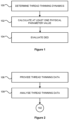
- Figure 1 illustrates a flow chart showing steps in a method for evaluating DED in a human or animal subject according to a preferred embodiment of the invention.
- the method includes at step 100 determining thread thinning dynamics of a tear sample 228 of the subject using an acoustically-driven microfluidic extensional rheometry (ADMiER) instrument 200 (see also Figure 5 ).
- At least one physical parameter value of the tear sample 228 is calculated at step 102 based at least in part on the thread thinning dynamics determined at step 100.
- the method includes evaluating DED at step 104 based at least in part on the calculated at least one physical parameter value of the tear sample 228.
- Figure 2 illustrates a flow chart showing further steps in the method shown in Figure 1 .
- Determining thread thinning dynamics includes the step 106 of obtaining thread thinning data of the tear sample 228 using the ADMiER instrument 200.
- the method may also include further steps relating to obtaining thread thinning data as illustrated in the flow chart of Figure 3 .
- the method includes at step 110 forming a fluid capillary bridge 224 of the tear sample 228 using the ADMiER instrument 200 (see also Figure 5 ) and at step 112 measuring changes in radius along the fluid capillary bridge 224 during thread thinning to obtain the thread thinning data.
- the thread thinning data is then analysed at step 108 in Figure 2 to determine thread thinning dynamics of the tear sample 228.
- Figure 4 illustrates a flow chart showing further steps in the method of shown in Figure 3 relating to forming the fluid capillary bridge 224.
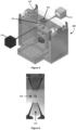
- the method includes at step 114 providing the ADMiER instrument 200 with opposing plates 202, 204 between which a fluid capillary bridge 224 can be formed, and an acoustic wave actuator 206 having a working surface located on one of the plates 202 (see also Figure 5 ).
- the method includes at step 116 applying the tear sample 228 to the working surface of the acoustic wave actuator 206 and at step 118 energising the acoustic wave actuator 206 to produce a fluid capillary bridge 224 of the tear sample 228 between the plates 202, 204.
- the ADMiER instrument 200 includes an upper plate 202 having the acoustic wave actuator 206 and a lower plate 204.
- the acoustic wave actuator 206 may be configured to use surface acoustic waves (SAW), bulk waves, surface reflected bulk waves (SRBW), or combinations thereof.
- the acoustic wave actuator 206 is configured to generate a short ultrasonic pulse in the form of a surface acoustic wave (SAW).
- the acoustic wave actuator 206 may include a piezoelectric chip for generating SAWs by applying a pulsed AC signal to interdigital (IDT) electrodes 222 on the chip at a frequency commensurate with the resonant frequency of the IDT as shown in Figure 6 .
- IDT interdigital
- the pulsed signal parameter, IDT design and the thickness of the chip allow for the generation of a bulk, surface or a combined surface or bulk acoustic wave at the same frequency or at a higher harmonic frequency, such as 1 MHz to 100 GHz, that is launched on or through the chip with an amplitude that ranges from between 1 picometre (pm) to 100 nanometres (nm), although the amplitude to generate the fluid capillary bridge from a microliter drop is typically around 10 nm.
- the SAW generated has an amplitude of approximately 10 nm.
- the working surface of the acoustic wave actuator 206 having the piezoelectric chip may be located on an underside of the upper plate 202.
- the tear sample 228 is applied to the working surface in the form of a sessile droplet 228.
- the droplet may have a diameter of approximately 1 mm.
- the acoustic wave actuator 206 then generates a SAW burst 226 to jet the sessile droplet 228 toward the lower plate 204. This results in formation of a fluid capillary bridge 224 between the upper and lower plates 202, 204.
- the acoustic wave actuator 206 having the piezoelectric chip may be located on a topside of the lower plate 204 (not shown).
- the sessile droplet 228 is jetted from the topside of the lower plate 204 to the upper plate 204 against gravity to form the fluid capillary bridge 224.
- the fluid capillary bridge 224 subsequently thins under capillary stresses and ruptures below a critical thread thickness due to the fluid's surface tension and resisted by its viscosity.
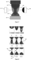
- Figure 8 shows a series of milli-second time lapse photos illustrating formation of the fluid capillary bridge 224 and thinning under capillary stresses.
- the sessile droplet 228 is applied to the working surface on the upper plate 202 initially and jetted toward the lower plate 204 from 1.5 to 6.0 ms.
- the fluid capillary bridge 224 is formed at 7.5 ms and then thins under capillary stresses from 9.0 to 10.5 ms. Finally, the fluid capillary bridge 224 ruptures below a critical thread thickness at 12.0 ms.
- the capillary thinning of the fluid capillary bridge 224 formed using the ADMiER instrument 200 follows established physics in its macroscopic counterpart - the Capillary Breakup Elongational Rheometer (CaBER).
- the CaBER is the subject of United States Patent Application No. 13/805,711 .
- the thread thinning dynamics may be monitored using a detector 208 and illuminator 210 for capturing images of the fluid capillary bridge 224 during thread thinning as shown in Figure 8 .
- the detector 208 may include a high-speed camera (e.g. Photron SA5) with a microscopic lens attachment (e.g. Infinity K2/SC) and the illuminator 210 may include an LED.
- the radius along the fluid capillary bridge 224 may be measured during thread thinning by extracting the radius in each image frame using standard image-analysis techniques.
- the ADMiER instrument 200 includes a sensor 310 (see also Figure 15 ) for measuring changes in radius along the fluid capillary bridge 224 during thread thinning.
- the sensor may be a line scan camera that functions as an optical micrometer to obtain thread thinning data of the changes in radius. It is preferable that the radius of the fluid capillary bridge 224 is measured at a neck 230 as shown in Figure 8 from 9.0 to 10.5 ms.
- the neck 230 is defined as the location where the thread of the fluid capillary bridge 224 first pinches and ruptures under capillary stresses.
- the thread thinning dynamics are governed by the filament and liquid properties, particularly its extensional viscosity and surface tension. This facilitates characterisation of complex physiological fluids, such as tear film samples, through viscoelastic properties derived using the ADMiER instrument 200. Such characterisation of physiological fluids however poses a considerable challenge for conventional extensional rheometers, particularly because of the difficulty in consistently generating capillary bridges for low viscosity fluids. Further, standard rheological analyses require large (i.e. millilitre) sample volumes, which are not feasible for tear samples.
- the ADMiER instrument's 200 ability to form capillary bridges from microlitre samples of fluids with viscosities as low as that of water provide a unique platform for testing the viscoelastic properties of tear samples.
- Such small sample sizes and fast (e.g. less than 1 second) processing times are not only advantageous for diagnosis but also render evaporative and gravitational effects that typically confound the measurements negligible.
- FIG 9 is a flow chart illustrating further steps in the method of Figures 1 to 4 relating to collecting a tear sample according to an embodiment of the invention.
- the method includes at step 120 collecting a basal tear sample 228 from the subject's eye.
- the basal tear sample 228 may be non-invasively collected from the subject's eye, such as with the use of a microcapillary tube.
- the method also includes the step 122 of dispensing the collected tear sample 228 into a sampling cartridge 318 (see also Figure 15 ).
- the sampling cartridge 318 may be loaded into a device 300 housing the ADMiER instrument 200 (see also Figure 15 ).
- the device 300 may allow automation of one or more of the method steps for fast processing of the thread thinning dynamics and evaluation of DED.
- the tear sample 228 that is collected may have a volume in a range of 1 nL to 10 mL, and preferably a volume in a range of 1 to 2 ⁇ L.
- the method may also include further steps relating to evaluating DED as illustrated in the flow chart of Figure 10 .
- step 126 one or more reference values are identified for evaluating DED.
- the reference values may be identified from data obtained from a population of individuals.
- the method may also include at step 128 comparing at least one physical parameter value calculated at step 102 with the identified reference values from step 126. DED is then evaluated at step 130 based on the comparison of the calculated at least one physical parameter value with the identified reference values.
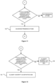
- Figures 11 to 13 are flow charts illustrating further steps in the method of Figure 10 relating to diagnosing the presence of DED at step 132, classifying the severity of DED as definitive DED or borderline DED at steps 134, 136, and classifying a predominant clinical sub-type of DED as aqueous deficient DED or evaporative DED at steps 138, 140.
- Each of the diagnostic methods involves a comparison of the physical parameter values with a respective threshold value or range of reference values indicative of the particular evaluation of DED.
- the identified reference values may include at least one threshold value or range of reference values indicative of one or more of the following: the presence of DED, the severity of DED and a clinical sub-type of DED.
- Figure 11 shows that the presence of DED is diagnosed at step 132 when the at least one physical parameter value is less than a threshold value or within a range of reference values indicative of the presence of DED. Otherwise, there is no presence of DED in the subject, i.e. the subject has a healthy tear film.
- the severity and predominant clinical sub-type may be classified by the steps of the flow charts in Figures 12 and 13 .
- Figure 12 shows that assessing the severity of DED includes classifying the severity as definitive DED at step 134 or borderline DED at step 136. The severity is classified as definitive DED at step 134 when the at least one physical parameter value is less than a threshold value or within a range of reference values indicative of definitive DED. Otherwise, the severity is classified as borderline DED at step 136.
- the method may include identifying a clinical sub-type of DED by classifying a clinical sub-type of DED as one or both of aqueous deficient DED and evaporative DED. Accordingly, the subject may exhibit the following types of DED: (i) aqueous deficient DED only; (ii) evaporative DED only; or both aqueous deficient and evaporative DED.
- the present invention may enable the presence of each of these clinical sub-types of DED to be identified.
- the clinical sub-type is classified as one or both of aqueous deficient DED and evaporative DED when the at least one physical parameter value is less than a threshold value or within a range of reference values indicative of aqueous deficient DED and/or evaporative DED.
- Figure 13 shows that identifying a clinical sub-type of DED may include classifying a predominant clinical sub-type as predominant aqueous deficient DED at step 138 or predominant evaporative DED at step 140.
- the clinical sub-type is classified as predominant aqueous deficient DED at step 138 when the at least one physical parameter value is less than a threshold value or within a range of reference values indicative of predominant aqueous deficient DED. Otherwise, the clinical sub-type is classified as predominant evaporative DED at step 140.
- assessing the severity of DED further includes classifying a clinical severity of definitive DED as one of mild, moderate or severe definitive DED.
- the clinical severity of definitive DED may be classified based at least in part on the at least one physical parameter value and a further physical parameter value of the tear sample.
- the method further includes providing one or more outcomes of the evaluation of DED.
- the method may further include the step of providing one or more outcomes of a previous evaluation of DED for the subject.
- the one or more outcomes may include one or more of the following: the presence of DED; borderline DED or definitive DED; mild, moderate or severe definitive DED; aqueous deficient DED and/or evaporative DED; and predominant aqueous deficient DED or predominant evaporative DED.
- the method may further include the step of monitoring DED by comparing the one or more outcomes of the evaluation of DED with the previous evaluation of DED and observing changes in the one or more outcomes. For example, the changes observed may be deviations or trends in the one or more outcomes.
- embodiments of the invention advantageously provide for evaluation of DED in the subject and the ability to differentiate between healthy subjects, and those subjects with borderline DED or definitive DED. Further, for those subjects evaluated to have definitive DED, embodiments of the invention also advantageously provide for evaluation of the severity of definitive DED, as one of mild, moderate and severe definitive DED. The ability to stratify based upon disease severity for DED is desirable for evaluating and monitoring DED.
- Figure 14 illustrates a flow chart showing steps in another method for evaluating DED in a human or animal subject according to a preferred embodiment of the invention.
- the method includes at step 126 identifying one or more reference values for evaluating DED.
- the method also includes at step 142 identifying at least one physical parameter value of a tear sample 228 of the subject.
- the at least one physical parameter value has been calculated based at least in part on determined thread thinning dynamics of the tear sample 228.
- the effect of the at least one physical parameter value is calculated as an apparent viscosity based at least in part on determined extensional measurement of the thread thinning dynamics of the tear sample 228.
- the method also includes at steps 128 and 130 evaluating DED based on a comparison of the identified at least one physical parameter value with the one or more reference values.
- the method includes diagnosing the presence of DED at step 132, classifying the severity of DED as definitive DED or borderline DED at steps 134, 136, and identifying a clinical sub-type of DED by classifying a predominant clinical sub-type of DED as predominant aqueous deficient DED or predominant evaporative DED at steps 138, 140 by performing the steps shown in Figures 11 to 13 .
- the identified one or more reference values may include at least one threshold value or range of reference values indicative of one or more of the following: the presence of DED, the severity of DED and a clinical sub-type of DED.
- the thread thinning dynamics may be determined using the ADMiER instrument 200 according to any one of the steps of the method as described herein and particularly with reference to Figures 2 to 4 .
- a computer program product stores instructions for controlling a processing device 302 to evaluate DED in a human or animal subject (see also Figure 15 ).
- the processing device 302 may include a processor or a controller as would be appreciated by a person skilled in the art.
- the instructions may cause the processing device 302 to determine thread thinning dynamics of a tear sample 228 of the subject using an ADMiER instrument 200 and calculate at least one physical parameter value of the tear sample 228 based at least in part on the determined thread thinning dynamics.
- the instructions may also cause the processing device to evaluate DED based at least in part on the calculated at least one physical parameter value of the tear sample 228.
- the instructions may cause the processing device 302 to receive thread thinning data of the tear sample 228 obtained using the ADMiER instrument 200 and analyse the thread thinning data to determine the thread thinning dynamics.
- the thread thinning data may include changes in radius along a fluid capillary bridge 224 of the tear sample 228 during thread thinning.
- the fluid capillary bridge 224 may have been formed using the ADMiER instrument 200 as described herein.
- the instructions may cause the processing device 302 to determine the thread thinning dynamics using the ADMiER instrument 200 according to any one of the steps of the methods as described herein and particularly with reference to Figures 2 to 4 .
- the thread thinning data is received from a memory device 304 in communication with the processing device 302 (see also Figure 15 ).
- the instructions may also cause the processing device 302 to receive one or more reference values for evaluating DED.
- the reference values may be received from the memory device 304 and may have been identified using data obtained from a population of individuals.
- the instructions may further cause the processing device 302 to evaluate DED by comparing the at least one physical parameter value with the one or more reference values and evaluating DED based on the comparison.
- the instructions may also cause the processing device 302 to evaluate DED by diagnosing the presence of DED at step 132, classifying the severity of DED as definitive DED or borderline DED at steps 134, 136, and identifying a clinical sub-type of DED by classifying a predominant clinical sub-type of DED as predominant aqueous deficient DED or predominant evaporative DED at steps 138, 140 by performing the steps of the method shown in Figures 11 to 13 .
- the instructions may also cause the processing device 302 to identify a clinical sub-type of DED by classifying a clinical sub-type of DED as one or both of aqueous deficient DED and evaporative DED (not shown). Accordingly, the identified one or more reference values may include at least one threshold value or range of reference values indicative of one or more of the following: the presence of DED, the severity of DED and a clinical sub-type of DED.
- the instructions cause the processing device to generate a display signal for causing a display device 316 to provide one or more outcomes of the evaluation of DED (see also Figure 15 ).
- the one or more outcomes may include one or more of the following: the presence of DED; borderline or definitive DED; aqueous deficient DED and/or evaporative DED; and predominant aqueous deficient DED or predominant evaporative DED.
- the instructions may also cause the processing device 302 to receive one or more outcomes of a previous evaluation of DED, such as from the memory device 304.
- the instructions cause the processing device 302 to compare the one or more outcomes of the evaluation of DED with the previous evaluation of DED and display trends or deviations in the one or more outcomes on the display device 316.
- another computer program product stores instructions for controlling a processing device 302 to evaluate DED in a human or animal subject (see also Figure 15 ).
- the instructions may cause the processing device 302 to receive one or more reference values for evaluating DED, identify at least one physical parameter value of a tear sample 228 of the subject and evaluate DED based on a comparison of the identified at least one physical parameter value with the one or more reference values.
- the at least one physical parameter value may have been calculated based at least in part on determined thread thinning dynamics of the tear sample 228.
- the effect of the at least one physical parameter value is calculated as an apparent viscosity based at least in part on determined extensional measurement of the thread thinning dynamics of the tear sample 228.
- the instructions may cause the processing device 302 to evaluate DED by diagnosing the presence of DED at step 132, classifying the severity of DED as definitive DED or borderline DED at steps 134,136, and identifying a clinical sub-type of DED by classifying a predominant clinical sub-type of DED as predominant aqueous deficient DED or predominant evaporative DED at steps 138, 140 by performing the steps of the method shown in Figures 11 to 13 .
- the instructions may also cause the processing device 302 to identify a clinical sub-type of DED by classifying a clinical sub-type of DED as one or both of aqueous deficient DED and evaporative DED (not shown). Accordingly, the identified one or more reference values may include at least one threshold value or range of reference values indicative of one or more of the following: the presence of DED, the severity of DED and a clinical sub-type of DED.
- FIG. 15 is a schematic illustration showing components of a device 300 for evaluating DED in a human or animal subject according to a preferred embodiment of the invention.
- the device 300 includes an ADMiER instrument 200 and a processing device 302.
- the processing device 302 is configured to: determine thread thinning dynamics of a tear sample 228 of the subject using the ADMiER instrument 200; calculate at least one physical parameter value of the tear sample 228 based at least in part on the determined thread thinning dynamics; and evaluate DED based at least in part on the calculated at least one physical parameter value of the tear sample 228.
- the processing device 302 includes an analysis module 306 for determining thread thinning dynamics.
- the analysis module 306 determines thread thinning dynamics by receiving thread thinning data of the tear sample 228 obtained using the ADMiER instrument 200 and analysing the thread thinning data to determine the thread thinning dynamics.
- the thread thinning data may include changes in radius along a fluid capillary bridge 224 of the tear sample 228 during thread thinning.
- the fluid capillary bridge 224 may have been formed using the ADMiER instrument 200 as described herein and particularly with reference to Figures 2 to 8 .
- the device 300 may also include a memory device 304 in communication with the processing device 302 for storing the thread thinning data.
- the analysis module 306 is also configured to calculate the at least one physical parameter value of the tear sample 228 based at least in part on the determined thread thinning dynamics.
- the effect of the at least one physical parameter value is calculated as an apparent viscosity based at least in part on determined extensional measurement of the thread thinning dynamics of the tear sample 228. The calculation may be performed using established methods as described in United States Patent Application No. 13/805,711 .
- the processing device 302 may also include an evaluation module 308 for evaluating DED.
- the evaluation module 308 may be configured to receive one or more reference values for evaluating DED, such as from the memory device 304.
- the one or more reference values may have been identified using data obtained from a population of individuals.
- the evaluation module 308 may also be configured to evaluate DED by comparing the at least one physical parameter value with the one or more reference values and evaluating DED based on the comparison.
- the evaluation module 308 may be configured to evaluate DED by one or more of the following: diagnosing the presence of DED; assessing the severity of DED and identifying a clinical sub-type of DED.
- the evaluation module 308 is configured to perform steps of one or more of the methods shown in Figures 11 to 13 , such as diagnosing the presence of DED at step 132, classifying the severity of DED as definitive DED or borderline DED at steps 134, 136, and identifying a clinical sub-type of DED by classifying a predominant clinical sub-type of DED as aqueous deficient DED or evaporative DED at steps 138, 140.
- the evaluation module 308 may also be configured to identify a clinical sub-type of DED by classifying a clinical sub-type of DED as one or both of aqueous deficient DED and evaporative DED (not shown). Accordingly, the identified one or more reference values may include at least one threshold value or range of reference values indicative of one or more of the following: the presence of DED, the severity of DED and a clinical sub-type of DED.
- the processing device 302 is configured to display on a display device 316 one or more outcomes of the evaluation of DED.
- the display device 316 may be included in the device 300 as shown in Figure 15 .
- the one or more outcomes may include one or more of the following: the presence of DED; borderline or definitive DED; aqueous deficient DED and/or evaporative DED; and predominant aqueous deficient DED or predominant evaporative DED.
- the device 300 may also include a user interface 314 as shown in Figure 15 .
- the user interface 314 may be configured to receive one or more outcomes of a previous evaluation of DED, such as from the memory device 304.
- the evaluation module 308 may be configured to compare the one or more outcomes of the evaluation with the previous evaluation of DED and display trends or deviations in the one or more outcomes on the display device 316.
- the device 300 may include a housing configured to receive a sampling cartridge 318 storing the tear sample 228 (not shown).
- the device 300 may house the ADMiER instrument 200, processing device 302, user interface 314 with display device 316 along with the sampling cartridge 318 as an integrated assembly.
- the sampling cartridge 318 is housed separately from other components in the device 300 to preserve tear sample integrity.
- the device 300 may also include a transducer 320 and driver 322 for detecting loading and unloading of the sampling cartridge 318 from the device housing as shown in Figure 15 .
- the device 300 may be configured to dispense the tear sample 228 from the sampling cartridge 318 and apply it to the working surface of the acoustic wave actuator 206 of the ADMiER instrument 200. Additionally, the device 300 may be configured to clean surfaces of the ADMiER instrument on receiving a new sampling cartridge 318. The device 300 may clean one or more of the working surface of the acoustic wave actuator 206, the plate 202 of the ADMiER instrument 200 and the plate 204 of the ADMiER instrument 200. Preferably, the device 300 cleans the working surface and plates 202, 204 of the ADMiER instrument 200. In some embodiments, the sampling cartridge may be disposable and for single use.
- the device 300 may include a power supply 324 connected to mains power and a battery 326 connected to the power supply 324.
- the battery 326 may be rechargeable for portable use of the device 300, such as in hospitals or clinics, or in pathology testing facilities.
- the at least one physical parameter value in the methods, computer product programs and device as described herein is the extensional viscosity.
- the at least one physical parameter value manifests as an apparent extensional viscosity based on the extensional measurement obtained using the ADMiER instrument.
- the possible correlation between apparent viscosity measures and clinical diagnosis of DED is discussed below in Examples 1 and 2.
- inventive methods, computer product programs and device provide novel diagnostic modalities for reliably and accurately evaluating DED in a human or animal subject.
- the novel diagnostic methods performed are simple, rapid and objective, and allow measurement of a single physical parameter to capture the subject's tear film status and evaluate DED. Further physical parameters may be used to provide additional information about the DED diagnosis.
- the inventive methods, computer product programs and device involve the use of an ADMiER instrument to quantify the viscoelastic properties of a human or animal tear sample.
- the ADMiER instrument is able to consistently form capillary bridges for low viscosity fluids from microlitre tear samples in order to provide objective and robust measurement of tear film capillary thinning.
- no or minimal consumables are required which greatly reduce the testing cost in contrast to prior art diagnostic modalities.
- tear film status was graded on the basis of tear osmolarity findings into one of three categories: 'healthy' ( ⁇ 308 mOsmol/L), 'borderline' (308-315 mOsmol/L) or 'dry' ( ⁇ 316 mOsmol/L).
- Basal tear samples (-2 ⁇ l per eye) were non-invasively collected using a glass microcapillary tube and immediately analysed using the ADMiER instrument, to derive tear viscoelasticity measures.
- Tear stability (seconds) Non-invasive tear break-up-time (NITBUT) measured 3 times per eye using the Placido-disc based Tear Film Surface Quality (TFSQ) index with the Medmont E300 corneal topographer.
- Tear viscoelasticity (Pa.s) Index test A 2 ⁇ l basal tear sample non-invasively collected from the lateral canthus using a glass microcapillary tube and immediately analysed using the ADMiER platform to derive tear apparent viscosity measures. Tear flow rate monitored to exclude potential tear dilution effects caused by reflex tearing. Samples with a flow rate of 1-5 ⁇ l/min were considered consistent with basal tears.
- Slit lamp Examination A slit lamp examination, using 10x and 16x magnification, undertaken to assess corneal integrity (in particular to examine for the presence of active inflammation or structural abnormalities).
- the iris and anterior chamber examined using the SUN criteria for inflammation.
- the extent of any anterior blepharitis graded using the Efron scale.
- Total ocular surface staining (/15.0) Ocular surface staining quantified as the sum of the corneal NaFI staining score /5.0 (graded under 16x magnification with Cobalt blue illumination and a yellow barrier filter, one minute after NaFl instillation) and the nasal+temporal conjunctival lissamine green (LG) staining score /10.0 (graded under 16x magnification with diffuse white illumination, 3 mins after LG instillation). Each ocular surface region graded in 0.1 increments using the Oxford scale.
- Meibomian gland evaluation Meibomian gland integrity, to assess for evaporative dry eye, evaluated by grading the quality of meibum, level of gland expressibility and volume of gland secretion, using the Bron/Foulks scoring system. Tear production The Schirmer test used to assess aqueous-deficient dry eye using a standard clinical protocol. The extent of strip wetting (in mm) is measured after 5 mins, following topical anaesthesia.
- Table 2 found in the figures shows the results from the series of standard dry eye diagnostic tests performed according to the protocols outlined in Table 1.
- the factor 'n' refers to the number of eyes included in the analyses and the data is shown as mean ⁇ SEM with * symbol representing p ⁇ 0.05.
- Table 2 shows significant differences between clinical parameters were evident between 'healthy' and 'dry' eyes (see Table 2), the latter showing relatively higher tear osmolarity, reduced tear stability (NITBUT and NaFI TBUT), higher levels of ocular surface staining and reduced tear production (p ⁇ 0.05 for all comparisons).
- the study provides representative ranges and threshold values of apparent tear viscosity that may be used for diagnosing the presence and assessing the severity of DED.
- the range of reference values indicative of the presence of DED may include the range of 0.0059 Pa.s to 0.0115 Pa.s.
- the range of reference values indicative of definitive DED may include the range of 0.0059 Pa.s to 0.0079 Pa.s.
- the range of reference values indicative of borderline DED may include the range of 0.0079 Pa.s to 0.0115 Pa.s.
- DED may also be diagnosed when the measured tear apparent viscosity is less than threshold values of 0.0115 Pa.s and more preferably, 0.00873 Pa.s.
- definitive DED ('dry' tears) may be assessed when the measured tear apparent viscosity is less than the threshold value of 0.0079 Pa.s, otherwise the severity of DED is assessed as borderline DED.
- Tear hyperosmolarity is considered to be the preferred indicator of DED severity although it cannot differentiate between different DED sub-types.
- Figure 18 is a chart providing filament break-up data from the study of 22 eyes that suggests potential differences in decay of filament thinning between clinically-defined aqueous-deficient and evaporative DED.
- Definite aqueous-deficient DED was defined by the presence of significant tear hyperosmolarity ( ⁇ 316 mOsmol/L), in association with a Schirmer strip test measurement of 5 mm or less in 5 mins.
- Definite evaporative DED was defined by the presence of significant tear hyperosmolarity ( ⁇ 316 mOsmol/L), in association with a Schirmer strip test measurement of at least 10 mm in 5 mins and evidence of meibomian gland dysfunction (considered as at least Grade 2 findings in one or more of: quality of meibum, level of gland expressibility and volume of gland secretion using the Bron/Foulks scoring system).
- Figure 18 indicates that shorter decays occur in less viscous unhealthy tears, suggesting the absence of constituents otherwise seen in healthy samples.
- the inventors have also quantified significant differences in the viscoelastic properties of tear supplemental products marketed specifically for these sub-types (data not shown). Together, the data supports the hypothesis that tear viscoelasticity assays may be useful in assessing the severity of DED and/or for differentiating between its clinical sub-types.
- a cross-sectional study was performed to assess diagnostic test accuracy in using tear extensional viscosity measurements obtained using ADMiER to diagnose DED (presence of DED or no presence of DED), assess the severity of DED (definitive or borderline DED) and classify DED into its predominant clinical sub-types (aqueous deficient or evaporative DED).
- the study involved a primary eye care population of 78 adults (156 eyes) with a mean ⁇ standard deviation (SD) age of 32 ⁇ 12 years (range of 18-77 years) and gender of 69% female.
- SD standard deviation
- Table 3 found in the figures shows summary results from a series of standard dry eye diagnostic tests for DED performed based on the protocols outlined in Table 1 and tear extensional viscosity measurements made using ADMiER.
- a clinical dry eye severity score (0.0 to 4.0) was objectively derived using standardised dry eye questionnaires (Ocular Surface Disease Index (OSDI) and Standardized Patient Evaluation of Eye Dryness (SPEED)) and clinical signs of DED (as determined by a compound score relating to the combination of findings from a comprehensive slit lamp examination, assessment of ocular redness, sodium fluorescein tear break-up time, corneal sodium fluorescein staining, conjunctival lissamine green staining, meibomian gland evaluation and Schirmer test score).
- OSDI Opte Index
- SPEED Standardized Patient Evaluation of Eye Dryness
- the score (from 0.0 to 4.0) was based upon the DEWS (2007) Diagnostic Methodology sub-committee classification. Clinically significant DED was referenced by a clinical dry eye score of 1.0 or greater. The factor 'n' refers to the number of eyes in the analyses and the data is shown as mean ⁇ SD. Similar to Example 1, statistically significant differences between clinical parameters were evident between 'healthy' and 'dry' eyes, the latter showing higher clinical dry eye severity score, reduced tear stability (TBUT) and reduced tear production (Schirmer test).
- the representative ranges of tear extensional viscosity values for the categories of tear film status were as follows: 'healthy' tears 0.00699 Pa.s to 0.0275 Pa.s; 'borderline' tears 0.00455 Pa.s to 0.029 Pa.s; and 'dry' tears 0.00307 Pa.s to 0.0151 Pa.s.
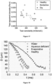
- Figure 20 is a chart showing results of a relationship between tear extensional viscosity and clinical dry eye severity score for this study.
- r - 0.46, p ⁇ 0.0001
- r - 0.46, p ⁇ 0.0001
- FIG 21 is a chart showing Receiver Operator Characteristic (ROC) curve analysis for this study. The analysis revealed that a cut-off criterion of 0.0093 Pa.s yields 83% sensitivity and 75% specificity for diagnosing the presence of DED ('dry' tears versus 'healthy' and 'borderline' tears combined). The Area Under Curve (AUC) was 0.84, 95% Confidence Limit (CI) of 0.78 to 0.91 (p ⁇ 0.0001).
- Figure 22 shows the chart of Figure 20 indicating the exemplary tear extensional viscosity value for diagnosing DED. The positive predictive value for DED was 81% and the negative predictive value for DED was 78%.
- ROC Receiver Operator Characteristic
- the study provides representative ranges and threshold values of extensional viscosity that may be used for diagnosing the presence and assessing the severity of DED.
- the range of reference values indicative of the presence of DED may include the range of about 0.0031 Pa.s to about 0.0151 Pa.s, and more specifically in the range of about 0.0059 Pa.s to about 0.0115 Pa.s.
- the range of reference values indicative of definitive DED may include the range of about 0.0031 Pa.s to about 0.0151 Pa.s, and more specifically in the range of about 0.0059 Pa.s to about 0.0079 Pa.s.
- the range of reference values indicative of borderline DED may include the range of about 0.00455 Pa.s to 0.0259 Pa.s and more specifically in the range of about 0.0079 Pa.s to about 0.0115 Pa.s. DED may also be diagnosed when the measured tear extensional viscosity is less than threshold value of about 0.0093 Pa.s. Furthermore, definitive DED ('dry' tears) may be assessed when the measured tear extensional viscosity is less than the threshold value of about 0.0093 Pa.s, otherwise the tears are assessed as healthy or borderline.
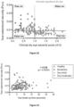
- Figure 23 is a chart showing results of a relationship between tear extensional viscosity and sodium fluorescein tear break-up time (TBUT) for this study.
- Figure 24 is a chart showing results of tear extensional viscosity measurements from this study for classifying the predominant clinical sub-type of DED as aqueous deficient DED or evaporative DED.
- Figure 25 is a chart showing results of a relationship between tear extensional viscosity and the Schirmer test score to differentiate between clinical sub-types of DED.
- Schirmer test score values of 5 mm/5min or less show a statistically significant difference in extensional viscosity for definite aqueous deficient DED and evaporative DED.
- definitive evaporative DED is associated with higher tear extensional viscosity values than definite aqueous deficient DED and Schirmer test scores of more than 5 mm/5min.
- the study provides representative ranges and threshold values of extensional viscosity that may be used for classifying sub-types of DED.
- the range of reference values indicative of predominantly aqueous deficient DED may include the range of 0.00307 Pa.s to 0.0105 Pa.s.
- the range of reference values indicative of predominantly evaporative DED may include the range of 0.00455 Pa.s to 0.0151 Pa.s.
- aqueous deficient DED may be classified when the measured tear extensional viscosity is less than the threshold value of about 0.00651 Pa.s, otherwise the clinical sub-type is classified as evaporative DED.
Landscapes
- Health & Medical Sciences (AREA)
- Life Sciences & Earth Sciences (AREA)
- Physics & Mathematics (AREA)
- General Health & Medical Sciences (AREA)
- Pathology (AREA)
- Chemical & Material Sciences (AREA)
- Immunology (AREA)
- Analytical Chemistry (AREA)
- General Physics & Mathematics (AREA)
- Biochemistry (AREA)
- Engineering & Computer Science (AREA)
- Biomedical Technology (AREA)
- Acoustics & Sound (AREA)
- Biophysics (AREA)
- Molecular Biology (AREA)
- Animal Behavior & Ethology (AREA)
- Public Health (AREA)
- Veterinary Medicine (AREA)
- Surgery (AREA)
- Medical Informatics (AREA)
- Heart & Thoracic Surgery (AREA)
- Urology & Nephrology (AREA)
- Medicinal Chemistry (AREA)
- Food Science & Technology (AREA)
- Hematology (AREA)
- Radiology & Medical Imaging (AREA)
- Optics & Photonics (AREA)
- Nuclear Medicine, Radiotherapy & Molecular Imaging (AREA)
- Signal Processing (AREA)
- Eye Examination Apparatus (AREA)
- Investigating Or Analysing Biological Materials (AREA)
Applications Claiming Priority (2)
| Application Number | Priority Date | Filing Date | Title |
|---|---|---|---|
| AU2016903362A AU2016903362A0 (en) | 2016-08-24 | Diagnostic methods and device | |
| PCT/AU2017/050900 WO2018035569A1 (en) | 2016-08-24 | 2017-08-24 | Diagnostic methods and device |
Publications (3)
| Publication Number | Publication Date |
|---|---|
| EP3504532A1 EP3504532A1 (en) | 2019-07-03 |
| EP3504532A4 EP3504532A4 (en) | 2020-04-22 |
| EP3504532B1 true EP3504532B1 (en) | 2024-08-21 |
Family
ID=61245972
Family Applications (1)
| Application Number | Title | Priority Date | Filing Date |
|---|---|---|---|
| EP17842455.2A Active EP3504532B1 (en) | 2016-08-24 | 2017-08-24 | Diagnostic methods and device |
Country Status (7)
| Country | Link |
|---|---|
| US (2) | US11397145B2 (enExample) |
| EP (1) | EP3504532B1 (enExample) |
| JP (1) | JP7058013B2 (enExample) |
| AU (1) | AU2017316997B2 (enExample) |
| CA (1) | CA3034608A1 (enExample) |
| ES (1) | ES2992526T3 (enExample) |
| WO (1) | WO2018035569A1 (enExample) |
Families Citing this family (2)
| Publication number | Priority date | Publication date | Assignee | Title |
|---|---|---|---|---|
| KR102293429B1 (ko) * | 2019-12-26 | 2021-08-26 | 고려대학교 산학협력단 | 안구건조증 유형 구별을 위한 진단 방법 및 진단 장치 |
| DE102020110729A1 (de) | 2020-04-20 | 2021-10-21 | Martin Gmbh | Verfahren und Vorrichtung zur optischen Bestimmung von Tropfenvolumina |
Family Cites Families (9)
| Publication number | Priority date | Publication date | Assignee | Title |
|---|---|---|---|---|
| WO2007126391A1 (en) | 2006-04-28 | 2007-11-08 | Singapore Health Services Pte Ltd | Investigation of mucosa dryness conditions |
| JP5511949B2 (ja) | 2009-06-25 | 2014-06-04 | ザ ユニバーシティ オブ ノース カロライナ アット チャペル ヒル | 体液レオロジーを評価するための、作動され表面に付着したポストを使用するための方法及びシステム |
| TWI547522B (zh) * | 2009-07-07 | 2016-09-01 | 愛爾康研究有限公司 | 環氧乙烷環氧丁烷嵌段共聚物組成物 |
| JP5572407B2 (ja) | 2010-01-29 | 2014-08-13 | 京都府公立大学法人 | 眼科装置及び画像分類プログラム |
| PE20130038A1 (es) * | 2010-03-10 | 2013-01-28 | Incyte Corp | Derivados de piperidin-4-il azetidina como inhibidores de jak1 |
| US20130233059A1 (en) * | 2010-06-22 | 2013-09-12 | Royal Melbourne Institute Of Technology | Rheometry instrument utilizing surface acoustic waves |
| US9230062B2 (en) * | 2012-11-06 | 2016-01-05 | 20/20 Vision Center, Llc | Systems and methods for enabling customers to obtain vision and eye health examinations |
| ES2650981B2 (es) * | 2016-07-20 | 2018-05-04 | Ocupharm Diagnostics, Sl | Preparación y uso de un extracto de Artemia salina para tratar la superficie ocular |
| CN113244291A (zh) * | 2016-08-19 | 2021-08-13 | 阿克里维斯塔有限责任公司 | 诊断和治疗干眼综合症的方法以及用于治疗人眼的组合物 |
-
2017
- 2017-08-24 JP JP2019510809A patent/JP7058013B2/ja active Active
- 2017-08-24 AU AU2017316997A patent/AU2017316997B2/en active Active
- 2017-08-24 WO PCT/AU2017/050900 patent/WO2018035569A1/en not_active Ceased
- 2017-08-24 CA CA3034608A patent/CA3034608A1/en active Pending
- 2017-08-24 US US16/327,063 patent/US11397145B2/en active Active
- 2017-08-24 EP EP17842455.2A patent/EP3504532B1/en active Active
- 2017-08-24 ES ES17842455T patent/ES2992526T3/es active Active
-
2022
- 2022-06-07 US US17/834,542 patent/US12140516B2/en active Active
Also Published As
| Publication number | Publication date |
|---|---|
| JP7058013B2 (ja) | 2022-04-21 |
| US12140516B2 (en) | 2024-11-12 |
| CA3034608A1 (en) | 2018-03-01 |
| EP3504532A1 (en) | 2019-07-03 |
| US20200158611A1 (en) | 2020-05-21 |
| AU2017316997B2 (en) | 2022-06-30 |
| EP3504532A4 (en) | 2020-04-22 |
| AU2017316997A1 (en) | 2019-03-14 |
| US11397145B2 (en) | 2022-07-26 |
| WO2018035569A1 (en) | 2018-03-01 |
| US20220299415A1 (en) | 2022-09-22 |
| JP2019525196A (ja) | 2019-09-05 |
| ES2992526T3 (es) | 2024-12-13 |
Similar Documents
| Publication | Publication Date | Title |
|---|---|---|
| Downie | Automated tear film surface quality breakup time as a novel clinical marker for tear hyperosmolarity in dry eye disease | |
| Gumus et al. | Noninvasive assessment of tear stability with the tear stability analysis system in tear dysfunction patients | |
| Vellara et al. | Biomechanical properties of the keratoconic cornea: a review | |
| Tomlinson et al. | Comparison of human tear film osmolarity measured by electrical impedance and freezing point depression techniques | |
| Brown et al. | Corneal structure and biomechanics: impact on the diagnosis and management of glaucoma | |
| Fuller et al. | Noninvasive tear breakup times and ocular surface disease | |
| Wei et al. | Assessment of lower tear meniscus | |
| US12140516B2 (en) | Diagnostic methods and device | |
| Kanellopoulos et al. | In pursuit of objective dry eye screening clinical techniques | |
| Manni et al. | Intraocular pressure and central corneal thickness | |
| Cardigos et al. | Tear meniscus and corneal sub-basal nerve plexus assessment in primary Sjögren syndrome and Sicca syndrome patients | |
| García et al. | Basal values, intra-day and inter-day variations in tear film osmolarity and tear fluorescein clearance | |
| Nichols et al. | Clinical assessments of dry eye disease: tear film and ocular surface health | |
| Rinert et al. | Accuracy of a new noninvasive automatic ocular surface analyzer for the diagnosis of dry eye disease—Two-gate design using healthy controls | |
| Akkaya et al. | Comparison of the corneal biomechanical properties, optic nerve head topographic parameters, and retinal nerve fiber layer thickness measurements in diabetic and non-diabetic primary open-angle glaucoma | |
| Edris et al. | Evaluation of corneal biomechanical parameters in psoriasis patients: A controlled study | |
| Kashiwagi et al. | Comparison between newly developed scanning peripheral anterior chamber depth analyzer and conventional methods of evaluating anterior chamber configuration | |
| Eroglu et al. | Is optical coherence tomography an effective device for evaluation of tear film meniscus in patients with acne rosacea? | |
| KR102200958B1 (ko) | 비침습적 결막 미소순환 측정 장치 | |
| Kelly et al. | Ocular manifestations of Sjögren’s syndrome | |
| Potenza et al. | How well does non-invasive tear break-up time reflect patient-reported ocular discomfort? Insights from a clinical setting | |
| Bhargava et al. | Can conjunctival impression cytology be the first line diagnostic test for evaluation of dry eye syndrome | |
| Sicks | ELEVATE YOUR DRY EYE PRACTICE: Let's break down every diagnostic test tier by tier to help you decide which ones are best for your patients. | |
| Omoti et al. | Current concepts in the diagnosis of primary open angle glaucoma | |
| Mondal et al. | RECENT TRENDS FOR DRY EYE DIAGNOSTIC PROCEDURE–A REVIEW |
Legal Events
| Date | Code | Title | Description |
|---|---|---|---|
| STAA | Information on the status of an ep patent application or granted ep patent |
Free format text: STATUS: THE INTERNATIONAL PUBLICATION HAS BEEN MADE |
|
| PUAI | Public reference made under article 153(3) epc to a published international application that has entered the european phase |
Free format text: ORIGINAL CODE: 0009012 |
|
| STAA | Information on the status of an ep patent application or granted ep patent |
Free format text: STATUS: REQUEST FOR EXAMINATION WAS MADE |
|
| 17P | Request for examination filed |
Effective date: 20190308 |
|
| AK | Designated contracting states |
Kind code of ref document: A1 Designated state(s): AL AT BE BG CH CY CZ DE DK EE ES FI FR GB GR HR HU IE IS IT LI LT LU LV MC MK MT NL NO PL PT RO RS SE SI SK SM TR |
|
| AX | Request for extension of the european patent |
Extension state: BA ME |
|
| DAV | Request for validation of the european patent (deleted) | ||
| DAX | Request for extension of the european patent (deleted) | ||
| REG | Reference to a national code |
Ref country code: DE Ref legal event code: R079 Free format text: PREVIOUS MAIN CLASS: G01N0011020000 Ipc: G01N0011000000 Ref document number: 602017084308 Country of ref document: DE |
|
| A4 | Supplementary search report drawn up and despatched |
Effective date: 20200324 |
|
| RIC1 | Information provided on ipc code assigned before grant |
Ipc: G01N 29/02 20060101ALI20200318BHEP Ipc: G01N 11/00 20060101AFI20200318BHEP |
|
| STAA | Information on the status of an ep patent application or granted ep patent |
Free format text: STATUS: EXAMINATION IS IN PROGRESS |
|
| 17Q | First examination report despatched |
Effective date: 20210810 |
|
| P01 | Opt-out of the competence of the unified patent court (upc) registered |
Effective date: 20230711 |
|
| GRAP | Despatch of communication of intention to grant a patent |
Free format text: ORIGINAL CODE: EPIDOSNIGR1 |
|
| STAA | Information on the status of an ep patent application or granted ep patent |
Free format text: STATUS: GRANT OF PATENT IS INTENDED |
|
| INTG | Intention to grant announced |
Effective date: 20240314 |
|
| GRAS | Grant fee paid |
Free format text: ORIGINAL CODE: EPIDOSNIGR3 |
|
| GRAA | (expected) grant |
Free format text: ORIGINAL CODE: 0009210 |
|
| STAA | Information on the status of an ep patent application or granted ep patent |
Free format text: STATUS: THE PATENT HAS BEEN GRANTED |
|
| AK | Designated contracting states |
Kind code of ref document: B1 Designated state(s): AL AT BE BG CH CY CZ DE DK EE ES FI FR GB GR HR HU IE IS IT LI LT LU LV MC MK MT NL NO PL PT RO RS SE SI SK SM TR |
|
| REG | Reference to a national code |
Ref country code: GB Ref legal event code: FG4D |
|
| REG | Reference to a national code |
Ref country code: CH Ref legal event code: EP |
|
| REG | Reference to a national code |
Ref country code: IE Ref legal event code: FG4D |
|
| REG | Reference to a national code |
Ref country code: DE Ref legal event code: R096 Ref document number: 602017084308 Country of ref document: DE |
|
| REG | Reference to a national code |
Ref country code: LT Ref legal event code: MG9D |
|
| REG | Reference to a national code |
Ref country code: ES Ref legal event code: FG2A Ref document number: 2992526 Country of ref document: ES Kind code of ref document: T3 Effective date: 20241213 |
|
| REG | Reference to a national code |
Ref country code: NL Ref legal event code: MP Effective date: 20240821 |
|
| PG25 | Lapsed in a contracting state [announced via postgrant information from national office to epo] |
Ref country code: NO Free format text: LAPSE BECAUSE OF FAILURE TO SUBMIT A TRANSLATION OF THE DESCRIPTION OR TO PAY THE FEE WITHIN THE PRESCRIBED TIME-LIMIT Effective date: 20241121 |
|
| REG | Reference to a national code |
Ref country code: AT Ref legal event code: MK05 Ref document number: 1715927 Country of ref document: AT Kind code of ref document: T Effective date: 20240821 |
|
| PG25 | Lapsed in a contracting state [announced via postgrant information from national office to epo] |
Ref country code: NL Free format text: LAPSE BECAUSE OF FAILURE TO SUBMIT A TRANSLATION OF THE DESCRIPTION OR TO PAY THE FEE WITHIN THE PRESCRIBED TIME-LIMIT Effective date: 20240821 Ref country code: PT Free format text: LAPSE BECAUSE OF FAILURE TO SUBMIT A TRANSLATION OF THE DESCRIPTION OR TO PAY THE FEE WITHIN THE PRESCRIBED TIME-LIMIT Effective date: 20241223 Ref country code: GR Free format text: LAPSE BECAUSE OF FAILURE TO SUBMIT A TRANSLATION OF THE DESCRIPTION OR TO PAY THE FEE WITHIN THE PRESCRIBED TIME-LIMIT Effective date: 20241122 Ref country code: PL Free format text: LAPSE BECAUSE OF FAILURE TO SUBMIT A TRANSLATION OF THE DESCRIPTION OR TO PAY THE FEE WITHIN THE PRESCRIBED TIME-LIMIT Effective date: 20240821 Ref country code: FI Free format text: LAPSE BECAUSE OF FAILURE TO SUBMIT A TRANSLATION OF THE DESCRIPTION OR TO PAY THE FEE WITHIN THE PRESCRIBED TIME-LIMIT Effective date: 20240821 |
|
| PG25 | Lapsed in a contracting state [announced via postgrant information from national office to epo] |
Ref country code: BG Free format text: LAPSE BECAUSE OF FAILURE TO SUBMIT A TRANSLATION OF THE DESCRIPTION OR TO PAY THE FEE WITHIN THE PRESCRIBED TIME-LIMIT Effective date: 20240821 |
|
| PG25 | Lapsed in a contracting state [announced via postgrant information from national office to epo] |
Ref country code: LV Free format text: LAPSE BECAUSE OF FAILURE TO SUBMIT A TRANSLATION OF THE DESCRIPTION OR TO PAY THE FEE WITHIN THE PRESCRIBED TIME-LIMIT Effective date: 20240821 |
|
| PG25 | Lapsed in a contracting state [announced via postgrant information from national office to epo] |
Ref country code: AT Free format text: LAPSE BECAUSE OF FAILURE TO SUBMIT A TRANSLATION OF THE DESCRIPTION OR TO PAY THE FEE WITHIN THE PRESCRIBED TIME-LIMIT Effective date: 20240821 Ref country code: IS Free format text: LAPSE BECAUSE OF FAILURE TO SUBMIT A TRANSLATION OF THE DESCRIPTION OR TO PAY THE FEE WITHIN THE PRESCRIBED TIME-LIMIT Effective date: 20241221 |
|
| PG25 | Lapsed in a contracting state [announced via postgrant information from national office to epo] |
Ref country code: HR Free format text: LAPSE BECAUSE OF FAILURE TO SUBMIT A TRANSLATION OF THE DESCRIPTION OR TO PAY THE FEE WITHIN THE PRESCRIBED TIME-LIMIT Effective date: 20240821 |
|
| PG25 | Lapsed in a contracting state [announced via postgrant information from national office to epo] |
Ref country code: RS Free format text: LAPSE BECAUSE OF FAILURE TO SUBMIT A TRANSLATION OF THE DESCRIPTION OR TO PAY THE FEE WITHIN THE PRESCRIBED TIME-LIMIT Effective date: 20241121 |
|
| PG25 | Lapsed in a contracting state [announced via postgrant information from national office to epo] |
Ref country code: RS Free format text: LAPSE BECAUSE OF FAILURE TO SUBMIT A TRANSLATION OF THE DESCRIPTION OR TO PAY THE FEE WITHIN THE PRESCRIBED TIME-LIMIT Effective date: 20241121 Ref country code: PT Free format text: LAPSE BECAUSE OF FAILURE TO SUBMIT A TRANSLATION OF THE DESCRIPTION OR TO PAY THE FEE WITHIN THE PRESCRIBED TIME-LIMIT Effective date: 20241223 Ref country code: PL Free format text: LAPSE BECAUSE OF FAILURE TO SUBMIT A TRANSLATION OF THE DESCRIPTION OR TO PAY THE FEE WITHIN THE PRESCRIBED TIME-LIMIT Effective date: 20240821 Ref country code: NO Free format text: LAPSE BECAUSE OF FAILURE TO SUBMIT A TRANSLATION OF THE DESCRIPTION OR TO PAY THE FEE WITHIN THE PRESCRIBED TIME-LIMIT Effective date: 20241121 Ref country code: NL Free format text: LAPSE BECAUSE OF FAILURE TO SUBMIT A TRANSLATION OF THE DESCRIPTION OR TO PAY THE FEE WITHIN THE PRESCRIBED TIME-LIMIT Effective date: 20240821 Ref country code: LV Free format text: LAPSE BECAUSE OF FAILURE TO SUBMIT A TRANSLATION OF THE DESCRIPTION OR TO PAY THE FEE WITHIN THE PRESCRIBED TIME-LIMIT Effective date: 20240821 Ref country code: IS Free format text: LAPSE BECAUSE OF FAILURE TO SUBMIT A TRANSLATION OF THE DESCRIPTION OR TO PAY THE FEE WITHIN THE PRESCRIBED TIME-LIMIT Effective date: 20241221 Ref country code: HR Free format text: LAPSE BECAUSE OF FAILURE TO SUBMIT A TRANSLATION OF THE DESCRIPTION OR TO PAY THE FEE WITHIN THE PRESCRIBED TIME-LIMIT Effective date: 20240821 Ref country code: GR Free format text: LAPSE BECAUSE OF FAILURE TO SUBMIT A TRANSLATION OF THE DESCRIPTION OR TO PAY THE FEE WITHIN THE PRESCRIBED TIME-LIMIT Effective date: 20241122 Ref country code: FI Free format text: LAPSE BECAUSE OF FAILURE TO SUBMIT A TRANSLATION OF THE DESCRIPTION OR TO PAY THE FEE WITHIN THE PRESCRIBED TIME-LIMIT Effective date: 20240821 Ref country code: BG Free format text: LAPSE BECAUSE OF FAILURE TO SUBMIT A TRANSLATION OF THE DESCRIPTION OR TO PAY THE FEE WITHIN THE PRESCRIBED TIME-LIMIT Effective date: 20240821 Ref country code: AT Free format text: LAPSE BECAUSE OF FAILURE TO SUBMIT A TRANSLATION OF THE DESCRIPTION OR TO PAY THE FEE WITHIN THE PRESCRIBED TIME-LIMIT Effective date: 20240821 |
|
| PG25 | Lapsed in a contracting state [announced via postgrant information from national office to epo] |
Ref country code: SM Free format text: LAPSE BECAUSE OF FAILURE TO SUBMIT A TRANSLATION OF THE DESCRIPTION OR TO PAY THE FEE WITHIN THE PRESCRIBED TIME-LIMIT Effective date: 20240821 Ref country code: RO Free format text: LAPSE BECAUSE OF FAILURE TO SUBMIT A TRANSLATION OF THE DESCRIPTION OR TO PAY THE FEE WITHIN THE PRESCRIBED TIME-LIMIT Effective date: 20240821 Ref country code: DK Free format text: LAPSE BECAUSE OF FAILURE TO SUBMIT A TRANSLATION OF THE DESCRIPTION OR TO PAY THE FEE WITHIN THE PRESCRIBED TIME-LIMIT Effective date: 20240821 |
|
| PG25 | Lapsed in a contracting state [announced via postgrant information from national office to epo] |
Ref country code: LU Free format text: LAPSE BECAUSE OF NON-PAYMENT OF DUE FEES Effective date: 20240824 |
|
| PG25 | Lapsed in a contracting state [announced via postgrant information from national office to epo] |
Ref country code: EE Free format text: LAPSE BECAUSE OF FAILURE TO SUBMIT A TRANSLATION OF THE DESCRIPTION OR TO PAY THE FEE WITHIN THE PRESCRIBED TIME-LIMIT Effective date: 20240821 |
|
| PG25 | Lapsed in a contracting state [announced via postgrant information from national office to epo] |
Ref country code: CZ Free format text: LAPSE BECAUSE OF FAILURE TO SUBMIT A TRANSLATION OF THE DESCRIPTION OR TO PAY THE FEE WITHIN THE PRESCRIBED TIME-LIMIT Effective date: 20240821 |
|
| PG25 | Lapsed in a contracting state [announced via postgrant information from national office to epo] |
Ref country code: SK Free format text: LAPSE BECAUSE OF FAILURE TO SUBMIT A TRANSLATION OF THE DESCRIPTION OR TO PAY THE FEE WITHIN THE PRESCRIBED TIME-LIMIT Effective date: 20240821 |
|
| REG | Reference to a national code |
Ref country code: DE Ref legal event code: R097 Ref document number: 602017084308 Country of ref document: DE |
|
| REG | Reference to a national code |
Ref country code: BE Ref legal event code: MM Effective date: 20240831 |
|
| PLBE | No opposition filed within time limit |
Free format text: ORIGINAL CODE: 0009261 |
|
| STAA | Information on the status of an ep patent application or granted ep patent |
Free format text: STATUS: NO OPPOSITION FILED WITHIN TIME LIMIT |
|
| PG25 | Lapsed in a contracting state [announced via postgrant information from national office to epo] |
Ref country code: MC Free format text: LAPSE BECAUSE OF FAILURE TO SUBMIT A TRANSLATION OF THE DESCRIPTION OR TO PAY THE FEE WITHIN THE PRESCRIBED TIME-LIMIT Effective date: 20240821 |
|
| PG25 | Lapsed in a contracting state [announced via postgrant information from national office to epo] |
Ref country code: BE Free format text: LAPSE BECAUSE OF NON-PAYMENT OF DUE FEES Effective date: 20240831 |
|
| PG25 | Lapsed in a contracting state [announced via postgrant information from national office to epo] |
Ref country code: IE Free format text: LAPSE BECAUSE OF NON-PAYMENT OF DUE FEES Effective date: 20240824 |
|
| 26N | No opposition filed |
Effective date: 20250522 |
|
| PG25 | Lapsed in a contracting state [announced via postgrant information from national office to epo] |
Ref country code: SE Free format text: LAPSE BECAUSE OF FAILURE TO SUBMIT A TRANSLATION OF THE DESCRIPTION OR TO PAY THE FEE WITHIN THE PRESCRIBED TIME-LIMIT Effective date: 20240821 |
|
| PGFP | Annual fee paid to national office [announced via postgrant information from national office to epo] |
Ref country code: ES Payment date: 20250901 Year of fee payment: 9 |
|
| PGFP | Annual fee paid to national office [announced via postgrant information from national office to epo] |
Ref country code: DE Payment date: 20250828 Year of fee payment: 9 |
|
| PGFP | Annual fee paid to national office [announced via postgrant information from national office to epo] |
Ref country code: IT Payment date: 20250827 Year of fee payment: 9 |
|
| PGFP | Annual fee paid to national office [announced via postgrant information from national office to epo] |
Ref country code: GB Payment date: 20250810 Year of fee payment: 9 |
|
| PGFP | Annual fee paid to national office [announced via postgrant information from national office to epo] |
Ref country code: FR Payment date: 20250827 Year of fee payment: 9 |
|
| PGFP | Annual fee paid to national office [announced via postgrant information from national office to epo] |
Ref country code: CH Payment date: 20250901 Year of fee payment: 9 |