EP3500964B1 - System und verfahren zur klassifizierung von biologischen partikeln - Google Patents
System und verfahren zur klassifizierung von biologischen partikeln Download PDFInfo
- Publication number
- EP3500964B1 EP3500964B1 EP17761164.7A EP17761164A EP3500964B1 EP 3500964 B1 EP3500964 B1 EP 3500964B1 EP 17761164 A EP17761164 A EP 17761164A EP 3500964 B1 EP3500964 B1 EP 3500964B1
- Authority
- EP
- European Patent Office
- Prior art keywords
- level
- features
- classification
- image
- particle
- Prior art date
- Legal status (The legal status is an assumption and is not a legal conclusion. Google has not performed a legal analysis and makes no representation as to the accuracy of the status listed.)
- Active
Links
Images


Classifications
-
- G—PHYSICS
- G06—COMPUTING OR CALCULATING; COUNTING
- G06T—IMAGE DATA PROCESSING OR GENERATION, IN GENERAL
- G06T7/00—Image analysis
- G06T7/0002—Inspection of images, e.g. flaw detection
- G06T7/0004—Industrial image inspection
- G06T7/001—Industrial image inspection using an image reference approach
-
- G—PHYSICS
- G06—COMPUTING OR CALCULATING; COUNTING
- G06T—IMAGE DATA PROCESSING OR GENERATION, IN GENERAL
- G06T7/00—Image analysis
- G06T7/0002—Inspection of images, e.g. flaw detection
- G06T7/0012—Biomedical image inspection
-
- G—PHYSICS
- G06—COMPUTING OR CALCULATING; COUNTING
- G06T—IMAGE DATA PROCESSING OR GENERATION, IN GENERAL
- G06T7/00—Image analysis
- G06T7/30—Determination of transform parameters for the alignment of images, i.e. image registration
- G06T7/33—Determination of transform parameters for the alignment of images, i.e. image registration using feature-based methods
-
- G—PHYSICS
- G06—COMPUTING OR CALCULATING; COUNTING
- G06T—IMAGE DATA PROCESSING OR GENERATION, IN GENERAL
- G06T7/00—Image analysis
- G06T7/40—Analysis of texture
- G06T7/41—Analysis of texture based on statistical description of texture
-
- G—PHYSICS
- G16—INFORMATION AND COMMUNICATION TECHNOLOGY [ICT] SPECIALLY ADAPTED FOR SPECIFIC APPLICATION FIELDS
- G16B—BIOINFORMATICS, i.e. INFORMATION AND COMMUNICATION TECHNOLOGY [ICT] SPECIALLY ADAPTED FOR GENETIC OR PROTEIN-RELATED DATA PROCESSING IN COMPUTATIONAL MOLECULAR BIOLOGY
- G16B40/00—ICT specially adapted for biostatistics; ICT specially adapted for bioinformatics-related machine learning or data mining, e.g. knowledge discovery or pattern finding
- G16B40/10—Signal processing, e.g. from mass spectrometry [MS] or from PCR
-
- G—PHYSICS
- G16—INFORMATION AND COMMUNICATION TECHNOLOGY [ICT] SPECIALLY ADAPTED FOR SPECIFIC APPLICATION FIELDS
- G16B—BIOINFORMATICS, i.e. INFORMATION AND COMMUNICATION TECHNOLOGY [ICT] SPECIALLY ADAPTED FOR GENETIC OR PROTEIN-RELATED DATA PROCESSING IN COMPUTATIONAL MOLECULAR BIOLOGY
- G16B5/00—ICT specially adapted for modelling or simulations in systems biology, e.g. gene-regulatory networks, protein interaction networks or metabolic networks
-
- G—PHYSICS
- G16—INFORMATION AND COMMUNICATION TECHNOLOGY [ICT] SPECIALLY ADAPTED FOR SPECIFIC APPLICATION FIELDS
- G16B—BIOINFORMATICS, i.e. INFORMATION AND COMMUNICATION TECHNOLOGY [ICT] SPECIALLY ADAPTED FOR GENETIC OR PROTEIN-RELATED DATA PROCESSING IN COMPUTATIONAL MOLECULAR BIOLOGY
- G16B50/00—ICT programming tools or database systems specially adapted for bioinformatics
- G16B50/30—Data warehousing; Computing architectures
-
- G—PHYSICS
- G16—INFORMATION AND COMMUNICATION TECHNOLOGY [ICT] SPECIALLY ADAPTED FOR SPECIFIC APPLICATION FIELDS
- G16H—HEALTHCARE INFORMATICS, i.e. INFORMATION AND COMMUNICATION TECHNOLOGY [ICT] SPECIALLY ADAPTED FOR THE HANDLING OR PROCESSING OF MEDICAL OR HEALTHCARE DATA
- G16H10/00—ICT specially adapted for the handling or processing of patient-related medical or healthcare data
- G16H10/40—ICT specially adapted for the handling or processing of patient-related medical or healthcare data for data related to laboratory analysis, e.g. patient specimen analysis
-
- G—PHYSICS
- G16—INFORMATION AND COMMUNICATION TECHNOLOGY [ICT] SPECIALLY ADAPTED FOR SPECIFIC APPLICATION FIELDS
- G16H—HEALTHCARE INFORMATICS, i.e. INFORMATION AND COMMUNICATION TECHNOLOGY [ICT] SPECIALLY ADAPTED FOR THE HANDLING OR PROCESSING OF MEDICAL OR HEALTHCARE DATA
- G16H30/00—ICT specially adapted for the handling or processing of medical images
- G16H30/40—ICT specially adapted for the handling or processing of medical images for processing medical images, e.g. editing
-
- G—PHYSICS
- G06—COMPUTING OR CALCULATING; COUNTING
- G06T—IMAGE DATA PROCESSING OR GENERATION, IN GENERAL
- G06T2207/00—Indexing scheme for image analysis or image enhancement
- G06T2207/10—Image acquisition modality
- G06T2207/10056—Microscopic image
-
- G—PHYSICS
- G06—COMPUTING OR CALCULATING; COUNTING
- G06T—IMAGE DATA PROCESSING OR GENERATION, IN GENERAL
- G06T2207/00—Indexing scheme for image analysis or image enhancement
- G06T2207/30—Subject of image; Context of image processing
- G06T2207/30004—Biomedical image processing
- G06T2207/30024—Cell structures in vitro; Tissue sections in vitro
Definitions
- the identification and enumeration of biological particles, including cells and particles is useful in a host of research and clinical applications, including the detection of hematological conditions.
- Automated biological particle recognition is a task that requires complex operations to be executed in a time sensitive manner, oftentimes on hardware with limited computational resources. It is therefore important that each phase in the system be efficient.
- Automated biological particle recognition, particularly for blood cells has conventionally been done using techniques which require heavy preprocessing. This results in a necessary compromise between computational efficiency and descriptive power. Furthermore, analysis and troubleshooting of conventional systems can be cumbersome if not impossible due to the large number of factors required for such complex operations.
- US 7,236623 B2 discloses a method of analyte recognition for a urinalysis system. It however does not disclose the concept of concentric circular or ring shaped masks as presented in the present invention.
- This disclosure relates to a system containing an automated image-based feature extraction and classification architecture which is suitable for real-time classification of biological particles, including cells and other particles, in a biological sample.
- This system may be used as a medical diagnostic tool and may enhance the identification and quantification of cells and/or particles.
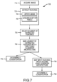
- the disclosed image-based classification system includes four major steps: image acquisition, feature extraction, feature selection, and the determination of a cell or particle's classification using a cascade classifier architecture.
- images of the cells or particles may first be collected or acquired. Using these images, the system may then extract particular numerical or categorical values or characteristics known as "features" from the individual images.
- the system may then use hierarchical or cascaded classification architecture in analysis of the extracted features.
- the cascade classifier architecture used in the determination step may include a two-level analysis. If the outcome of the first level analysis is inconclusive, the second level analysis may be performed on the selected ones of the extracted features of the biological sample (e.g. a blood sample).
- a select set of the extracted features of the biological sample may be compared to a select set of features extracted from cells or particles with known characteristics.
- comparison (“first level model”) provides an accurate identification of the cell or particle.
- classification of the cell or particle requires a further step (a "second level model”) to classify the cell or the particle.
- This step may include the use of numerical or categorical values from the first level model in combination with a second level model.
- the blood particle feature selection and image classifier architecture systems and methods discussed herein can provide various benefits and advantages when compared to other traditional approaches.
- embodiments of the present invention provide systems and methods where feature extraction computational complexity can be kept to a minimum value.
- complex and costly feature computation can be postponed until a cell event reaches a particular classifier within the architecture that requires the specific feature.
- Feature extraction can be a costly stage of any automated classification system.
- the architecture systems and methods disclosed herein introduce a simple yet powerful approach to balance complexity and performance.
- the cascade architecture of the classifier system can be modular, scalable, and simple to post-analyze. The output of the system can be easily traced back to individual classifiers.
- the method of extracting includes clustering the first set of pixels into a group.
- the method of extracting includes creating a color palette from the clustered group of pixels.
- the method of extracting includes determining a label for the image based in part on the color palette.
- the method of extracting includes normalizing the image to a mask size.
- the method of extracting includes normalizing the first mask to a unit magnitude.
- the method of extracting includes using a chosen color space, including red-green-blue (RGB) hue-saturation-value (HSV), hue-saturation-lightness (HSL), or hue-saturation-brightness (HSB).
- RGB red-green-blue
- HSV hue-saturation-value
- HSL hue-saturation-lightness
- HB hue-saturation-brightness
- the selected subset of the extracted features comprises training features, validation features, or testing features. In some instances, the subset of the extracted features is mapped into a cascade classifier architecture.
- the first level model is a machine learning model.
- a method of determining a classification of a particle in a biological sample including a second level model.
- the second level model includes receiving the probability value at the second level model, creating a sorted list of values according to a classification performance in relation to a cell or particle category, combining the probability value and the sorted list to create a second level probability value, and using the probability value determined at the first level model and the probability value determined at the second level model to determine the cell or particle classification.
- the mapping includes using a first level model to compare the subset of the extracted features to a previously stored data set, identifying a preliminary classification based on the comparison of the subset of the extracted features to the previously stored data set, calculating a probability value that the preliminary classification is correct using the first level model, and determining the classification based on the preliminary classification when the probability value is at or above a threshold value.
- the computer readable storage medium includes code executable by the processor for implementing any of the methods disclosed herein.
- the system uses a digital microscope camera.
- Embodiments of the present invention also encompass a non-transitory computer-readable storage medium including program instructions executable by one or more processors that, when executed, cause the one or more processors to perform operations, the operations including any of the methods disclosed herein.
- individual embodiments may be described as a process which is depicted as a flowchart, a flow diagram, a data flow diagram, a structure diagram, or a block diagram. Although a flowchart may describe the operations as a sequential process, many of the operations may be performed in parallel or concurrently. In addition, the order of the operations may be re-arranged.
- a process is terminated when its operations are completed, but could have additional steps not included in a figure.
- a process may correspond to a method, a function, a procedure, a subroutine, a subprogram, etc. When a process corresponds to a function, its termination may correspond to a return of the function to the calling function or the main function.
- This disclosure relates to a system containing an automated image-based feature extraction and classification architecture which is suitable for real-time classification of cells and/or particles in a biological sample.
- Automated particle classification systems may be used to analyze biological samples to determine the composition and/or number of one or more types of cells and/or particles contained in the samples. These systems commonly include hematology analyzers and flow cytometers.
- the analysis of the cellular populations in peripheral blood includes the ability to detect and enumerate the five major subtypes of white blood cells (WBC), which include neutrophils, lymphocytes, monocytes, eosinophils and basophils.
- WBC white blood cells
- RBC main red blood cells
- cellular populations have differing shapes and functions, and the number and presence of these populations in a sample may differ according to pathological conditions, cell maturity and other factors.
- Cell classification systems may differentiate cells of various types by collecting and analyzing signals produced when the cells pass through a small aperture or measurement region that is monitored by one or more instruments.
- Advantageous aspects of an automated cell classification system include the capability to identify a plurality of types of cells, based on their architecture, and also to identify artifacts resulting from the cellular processing or image acquisition process (e.g. images that depict old or damaged cells and images that are out of focus).
- Blood cell analysis is one of the most commonly performed medical tests for providing an overview of a patient's health status.
- a blood sample can be drawn from a patient's body and stored in a test tube containing an anticoagulant to prevent clotting.
- a whole blood sample normally comprises three major classes of blood cells including red blood cells (erythrocytes), white blood cells (leukocytes) and platelets (thrombocytes). Each class can be further divided into subclasses of members. For example, five major types or subclasses of white blood cells (WBCs) have different shapes and functions.
- White blood cells may include neutrophils, lymphocytes, monocytes, eosinophils, and basophils.
- Red blood cell subclasses may include reticulocytes and nucleated red blood cells.
- the particle is selected from at least one of neutrophil, lymphocyte, monocyte, eosinophil, basophil, platelet, reticulocyte, nucleated red blood cell (RBC), blast, promyelocyte, myelocyte, metamyelocyte, red blood cell (RBC), platelet, cell, bacteria, particulate matter, cell clump, or cellular fragment or component.
- neutrophil neutrophil, lymphocyte, monocyte, eosinophil, basophil, platelet, reticulocyte, nucleated red blood cell (RBC), blast, promyelocyte, myelocyte, metamyelocyte, red blood cell (RBC), platelet, cell, bacteria, particulate matter, cell clump, or cellular fragment or component.
- particles can include all measurable and detectable (e.g ., by image and/or other measurable parameters) components in biological fluids.
- the particles are of any material, any shape and any size.
- particles can comprise cells. Examples of particles include but are not limited to cells, including blood cells, fetal cells, epithelials, stem cells, tumor cells, or bacteria, parasites, or fragments of any of the foregoing or other fragments in a biological fluid.
- Exemplary casts can include acellular pigment casts, unclassified cast (e.g. granular casts).
- Exemplary acellular casts can include, for example, waxy casts, broad casts, fatty casts, and crystal casts.
- Exemplary cellular casts can include, for example, RBC casts, WBC casts, and cellular casts.
- Exemplary crystals can include, for example, calcium oxalate, triple phosphate, calcium phosphate, uric acid, calcium carbonate, leucine, cystine, tyrosine, and amorphous crystals.
- Exemplary non-squamous epithelial cells can include, for example, renal epithelials and transitional epithelials.
- Exemplary yeast can include, for example, budding yeast and yeast with pseudohyphae.
- Exemplary urinary sediment particle can also include RBC clumps, fat, oval fat bodies, and trichomonas.
- the assignment of cell and/or particle images into different classes or categories may be a complex computational task. While some analysis and comparisons can be done through an automated system, not all images of cells and/or particles are sufficiently clear or are similar enough to images of cells and/or particles with known characteristics and/or properties for automation to work properly or effectively.
- the extracted features may have different degrees of computational complexity. In many cases, cells and/or particles may be classified using a low number or complexity of extracted features, for example by using color-based features. Typically, color-based features are a fast computational task, whereas texture and shape features are a slow computational task and could impose a constraint for real-time classification.
- a second computer may provide the program instructions 145 to the first computer for execution.
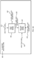
- FIG. 1A depicts a single computing device 110 with a single processor 120
- the system 100 may include any number of computing devices 110 and any number of processors 120.
- multiple computing devices 110 or multiple processors 120 may be distributed over a wired or wireless network (e.g., a Wide Area Network, Local Area Network, or the Internet).
- the multiple computing devices 110 or multiple processors 120 may perform any of the steps of the present disclosure individually or in coordination with one another.
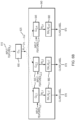
- FIG. 1B illustrates an exemplary cascade classifier architecture 185.
- the cascade classifier architecture may contain two level models and may be capable of performing two-level analysis.
- a first level model 187 may provide an accurate identification of the cell or particle or the cascade classifier architecture 185 may further use a second level model 189 to provide an accurate identification of the cell or particle.
- the second level model 189 may include the use of numerical or categorical values from the first level model 187 in combination with the second level model 189.
- the second level model 189 may be used.
- the images for analysis in the cascade classifier architecture 185 may come from the storage 180 or directly from the analyzer 115 may be input to the first level model 187. If necessary, the same may be input to the second level model.
- the output of the first level model 187 may be input to the second level model 189.
- FIG. 2 illustrates sample blood cell images 200-270 that may be used in systems and methods disclosed herein.
- n refers to a data set of blood particle images P illustrated in the first row 205 with corresponding target label T identifying each particle to a corresponding category.
- m refers to different blood particle categories C (i.e., NRBC, Lymphocytes, RBC, Neutrophil, etc.) illustrated in the second row 215 to which image P i may be assigned to, where 1 ⁇ i ⁇ n .
- the letter "k” is used to refer to a single category belonging to the set of "m" available categories.
- Feature extraction is intended to reduce the amount of data needed to analyze and discriminate among a set of categories. This process may interpret or summarize a large amount of information as a value that may later be used to make a determination.
- Extracted features may include numerical values that correlate with a particular characteristic of the data. For example, in an image, instead of using all colors as input to a cascade classifier architecture, a mean and standard deviation along all colors may be extracted as a feature. In image processing, feature extraction may have varying degrees of computational complexity. Highly complex extraction procedures may involve segmentation of the image to isolate a region of interest and prevent lengthy computational operations in that area while still extracting meaningful information. Simple extraction procedures may involve shape features, including but not limited to area, perimeter, and circularity. Extraction procedures may involve gradient profiles to detect edges. Extraction profiles may involve color intensity, histogram color mean, mode, standard deviation, or color thresholding. Extraction procedures may mean histogram differences or ratios between channel red and channel green, between channel red and channel blue, and/or between channel green and channel blue.
- the color space quantization approach may create a set of R binary masks composed of individual concentric rings.
- FIG. 3 shows an example of R 360.
- the concentric ring masks 300-350 When the concentric ring masks 300-350 are used, the resulting image plane projection of the cells will generally be circular in shape.
- the ring masks 300-350 may be isotropic and thus feature signatures derived using the rings may be rotation invariant.
- scale invariance may be inherent when the distance to the cell and/or particle imaged is fixed.
- the centroid of each ring mask 100-150 may be dynamic and determined by features in the image such as intensity or entropy. In the simplest case, the location may be fixed and defined as the center of the image 100. This enables translation invariance of the feature signature.
- the width and number r of ring masks 300-350 in R 360 may be heuristically chosen based on final classification performance. Each ring mask 300-350 may be used to filter pixels from the original cell image.
- the masks in a set 360 may be applied in any order. Once an order of masks is chosen for a set 360, the same predetermined order may be used and applied for image analysis.
- the masks in a set 360 do not necessarily reveal adjacent areas of the image, but may reveal adjacent areas of the image.
- the cell image may be first normalized to the size of the ring mask 300-350.
- Pixels falling into each ring mask 300-350 may then be extracted and analyzed in a chosen color space (i.e., red-green-blue (“RGB”) hue-saturation-value (“HSV”), hue-saturation-lightness (“HSL”), etc.).
- RGB red-green-blue
- HSV hue-saturation-value
- HSL hue-saturation-lightness
- the RGB color space may be chosen as the analysis space of the masked cell image.
- the process to extract the color palette given a set of ring masks 300-350 begins with the normalization of all cell images P i to the size of the ring masks R l to enable the application of the masks to the cell images.
- P C j may denote the set of all normalized cell images P i with corresponding target label T i in the training set equal to blood cell category C j , 1 ⁇ j ⁇ m .
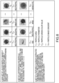
- FIG. 4 shows an example of the clustering process for a given ring mask in the HSV color space, where the X 400 in the chart represents the center of the clusters and each color is associated to pixels belonging to a cell category, the s-axis 410 represents the Saturation component, the h-axis 420 represents the Hue component, and the v-axis 430 represents the Value or brightness.
- the pixels belonging to a given category may further be identified by using other visual cues, such as color.
- Each cell category may be represented by a different number of images in the training set (e.g. reference images). To reduce the bias towards a particular cell type with a greater representation in the training set, the color samples used for clustering may be sampled from an equal number of training set images across the cell categories.
- the different cell categories (identified by the different colors) occupy different location in the color space from the other cell categories, and furthermore each cell category has a different distribution within the chart.
- the color quantization may be accelerated by storing the mapping between the color space and generated palette using look up tables or other indexing methods (e.g. k-dimensional trees).
- look up tables or other indexing methods e.g. k-dimensional trees.
- the ring masks 300-350 may yield histograms with different sample counts.
- Using such histograms for classification may introduce a bias towards the ring masks 300-350 with a larger diameter because the sample counts for those ring masks 300-350 may be greater.
- each histogram vector may be normalized to unit magnitude.
- L2-norm scheme is selected for histogram vector normalization.
- h be the non-normalized histogram vector
- the normalization of images to a common mask size may discard the relative size information between the images.
- the image width may be appended to the feature vector as a final feature to preserve the cell or particle size information.
- the features extracted may be augmented with additional morphological features such as gradient, entropy, etc., to complement the extracted information available in the color space.
- Feature selection which is also known as subset selection or variable selection, is a method used in machine learning to select a subset of features from all of the features available in a dataset. It is utilized in machine learning prior to applying a learning algorithm because it is computationally infeasible to use all available features in a dataset. Feature selection also may minimize problems of estimation and over fitting when a dataset contains limited data samples containing a large number of features. For example, a cell image may contain thousands of features, which may not all be good for analysis. The selection of particular features may depend on the specifications of the system. The selection of particular features may depend on the speed requirements for extraction within a particular system. Extracted features may include features used for training, validation, and/or testing (i.e. "training features, validation features, or testing features").
- a classifier architecture is a set of rules governing the transitions between classifier states.
- a classifier may include a cascade of evaluation or processing stages, such as a first level model and a second level model.
- the first level model may generate an opinion in the form of a level of confidence between zero and one. If the level of confidence is at or above a certain threshold, a decision may be made regarding the identity of the imaged particle or cell. In an exemplary embodiment, if the level of confidence is below a certain threshold, the information may be sent to the second level model.
- the second level model may use a more complex level of features than the first level model, including in some instances a random forest of decision trees, in combination with the level of confidence from the first level model, to make a decision regarding the identity of the imaged particle or cell.
- An exemplary cascade classifier architecture 650 including a first level model 600 and a second level model 640 is illustrated in FIG. 6A .
- a subset of the features extracted may be selected using appropriate separability measures and a final data set may be constructed.
- the data may be consolidated in a table where each row corresponds to a particular cell (and therefore to a cell image) and the Features columns correspond to the unique features associated with the corresponding cell category, as shown in exemplary TABLE 1.
- the Category column serves the purpose of defining the "true" label or class for each cell.
- the data set may be separated into 3 subsets: training set, validation set and testing set.
- the training set may include training features from images that have been classified and coded by one or more human experts. This coding (called human reference coding) may be utilized to train the classifier (as a training dataset) and/or may be used as a validation dataset.
- human reference coding may be utilized to train the classifier (as a training dataset) and/or may be used as a validation dataset.
- the training and validation features or data may be used to evaluate the performance of the CL first level model 600.
- the testing set may include images of uncharacterized cells and/or particles.
- the first level (L1) model 600 of an architecture may be composed of a classifier model CL.
- the classifier CL may be any machine learning model capable of mapping a set of input features 610 to a known class label as defined by the training data set. Examples of machine learning models suitable for this architecture may be Random Forest, multiclass Support Vector Machines (SVMs), Feedforward Neural Networks (FNNs), etc.
- a Random Forest machine learning model may be selected to map the input feature vector into one of the blood cell category C j , 1 ⁇ j ⁇ m defined in the training data set.
- a Random Forest may be an ensemble classifier comprised of a multitude of decision trees that may each be trained on a different portion of the training set.
- the final classification decision of the Random Forest may be the mode of the classification decisions of the individual trees.
- the advantage of a random forest over a single decision tree classifier is that a Random Forest is less prone to over fitting on the training set because a Random Forest classification decision is an aggregate response of multiple independently trained decision trees.
- the trees of the Random Forest machine may be trained using 80% of the data.
- the Random Forest includes 64 trees.
- ⁇ j is a real number.
- Large values of ⁇ j indicate belongingness to a particular cell class. In this context, the higher the score ⁇ j the more likely the input feature 610 belongs to the cell category j and the less uncertainty there is about that assessment.
- a preliminary category label for the input feature 610 may initially be given by the category corresponding to the maximum value ⁇ j in M 625.
- both the training and validation data may be used to evaluate the performance of the CL first level model 600.
- a predicted class label L i with corresponding M scores may be obtained for each input cell image P i . This information may then be used as input to the design process of the second level model 640.
- the set of M i scores 625 may be analyzed to establish the probability of correct cell preliminary classification by the first level model 600.
- the probability of correct preliminary classification may be estimated by using the level one predicted class label L i , the human expert target label T i and the level one M i scores 625.
- the expectation is that M i scores 625 with a maximum ⁇ j close to 0.5 will be associated with a low probability of correct preliminary classification value in level one.
- ⁇ Pos i A minimum value for ⁇ Pos i may be adopted to avoid biasing of results at low ⁇ Pos i values.
- TABLE 2 provides an example of the calculation of Pr ⁇ C j , C k ⁇ for two cell categories (i.e., Neutrophils and Eosinophils) on a given training and validation data set. Assuming a subset of all positive Neutrophil and Eosinophil subset of data is available with corresponding predicted class label L i , target label T i and M scores for that subset, matrices of ⁇ TP os i and ⁇ Pos i may be constructed to find Pr ⁇ Neutrophil,Eosinophil ⁇ .
- the probability matrices Pr ⁇ C j , C k ⁇ exemplified in TABLE 2 have at least two main purposes.
- the matrices may be used to establish an overall measurement of uncertainty for pairs of categories ⁇ C j , C k ⁇ .
- cell images in the training and validation data set associated to low or high probability values in Pr ⁇ C j , C k ⁇ may be selected as candidates for training the second level component 640 of the cascade classifier architecture.
- To establish an overall measurement of uncertainty it is possible to calculate the sum of all probability values on each Pr ⁇ C j , C k ⁇ . Pairs of categories having larger sum values will have less uncertainty associated to their discrimination.
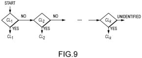
- one embodiment of the disclosed method allows a ranking of each category C j in terms of its separability from the rest of the categories. This sorted list is used to define the architecture scaffold of the classifier. Categories with highest level of separability (i.e., easier to classify from the rest) will be the assessed first by the classifier architecture.
- the discriminatory coefficients D s,j and complexity indexes O s are stored for further classifier feature assignment.
- each classifier Cl j is unique because it provides a balance between complexity and performance. This approach has not been seen on other alternatives approaches.
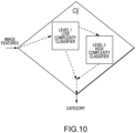
- FIG. 10 depicts a proposed internal structure for classifier Cl j according to embodiments of the present invention.
- the classifier Cl j is composed of two classifiers.
- the first one known as Level 1 classifier is commonly a simple linear classifier model that uses a reduced number of features (usually three or less to allow easy visualization of the feature space).
- the feature selection for this Level 1 classifier is given by a weighted combination of the D s,j feature discriminatory coefficient obtained above and the level of computational complexity O s associated to each feature. Uncorrelated features with high discriminatory coefficient and low computational complexity are ideal candidates for Level 1.
- Machine learning classifier models such as Support Vector Machines, Perceptron or other simple models can be used to automatically train the Level 1 classifier.
- the Level 2 classifier is a more complex classifier model. Commonly, based on a non-linear model with a complex structure to handle non-obvious relationships among the features.
- the feature selection for this Level 2 classifier is also given by a weighted combination of the D s,j feature discriminatory coefficients obtained above and the level of computational complexity O s associated to each feature.
- Features with high discriminatory coefficient and high computational complexity are ideal candidates for Level 2.
- the number of features commonly goes above three thus visualization of the feature space is no longer possible.
- Machine learning classifier models such as Multilayer Feedforward Neural Networks, Bootstrap, or any other complex models can be used to automatically estimate the model parameters.
- the Level 1 classifier handles the vast majority of input images, thus lowering computational time and simplifying analysis of the classification flow.
- the Level 2 classifier engages only when the Level 1 classifier decision is uncertain and not trustable. The following approach is proposed to assess the uncertainty of the Level 1 classifier and control the contribution of Level 1 and Level 2 classifier to the final outcome of Cl j .
- Level 1 classifier in charge of identifying category Cl 9 as shown in FIG. 11 .
- the Level 1 classifier model output ⁇ Level 1 is defined as the linear combination of features F 1 and F 2 with model coefficients ⁇ , ⁇ and the bias term ⁇ .
- a larger number of model coefficients will be present as the number of input features increases.
- the model coefficients can be obtained by a machine learning algorithm such as linear discriminant analysis, support vector machine or any other suitable approach.
- a linear combination value equal to zero corresponds to the exact location of the boundary separating Cl 9 from the rest of the blood particles categories (i.e., "other").
- a small change in features F 1 and F 2 values can change the classifier decision one way or another around this area.
- the uncertainty region can be defined by setting offsets H Level 1 1 and H Level 1 2 around the boundary.
- the offsets could have the same value.
- the actual values of offsets can be defined by setting an arbitrarily performance metric for the Level 1 classifier. For example, it might be desired to have a high degree of specificity in the non-uncertain regions, which means that if a blood particle is detected outside the uncertainty region there is a high confidence that the blood particle will be correctly classified.
- the Level 2 classifier will only be called and its corresponding input features (commonly different from Level 1 features) computed when a particular combination of features (in the example F 1 and F 2 ) for a blood particle fall inside the Level 1 uncertainty region.
- This unique design allow the overall classification system to remain simple and fast for easy to classify blood particles but flexible enough to handle more complex scenarios when needed.
- the same approach to create the Level 2 uncertainty region can be applied to the Level 2 classifier to define its offsets.
- a couple of transition functions ⁇ Level 1 ( ⁇ Level 1 ) and ⁇ Level 2 ( ⁇ Level 2 ) where -1 ⁇ ⁇ Level 1 ( ⁇ Level 1 ) ⁇ 1 and -1 ⁇ ⁇ Level 2 ( ⁇ Level 2 ) ⁇ 1 can be defined as depicted in FIG. 12 .
- This set of functions allow a smooth transition between Level 1 and Level 2 classifier outputs by linearly combining their response according to each classifier's uncertainty value.
- function ⁇ Level 2 commonly have a non-linear behavior in the uncertainty region due to the complex nature of the Level 2 classifier.
- Cl j Classifier Response ⁇ Level 1 ⁇ Level 1 + ⁇ Level 2 ⁇ Level 2
Landscapes
- Engineering & Computer Science (AREA)
- Health & Medical Sciences (AREA)
- Physics & Mathematics (AREA)
- Medical Informatics (AREA)
- General Health & Medical Sciences (AREA)
- Theoretical Computer Science (AREA)
- Life Sciences & Earth Sciences (AREA)
- Computer Vision & Pattern Recognition (AREA)
- Epidemiology (AREA)
- Public Health (AREA)
- General Physics & Mathematics (AREA)
- Primary Health Care (AREA)
- Nuclear Medicine, Radiotherapy & Molecular Imaging (AREA)
- Radiology & Medical Imaging (AREA)
- Bioinformatics & Cheminformatics (AREA)
- Spectroscopy & Molecular Physics (AREA)
- Quality & Reliability (AREA)
- Biophysics (AREA)
- Bioinformatics & Computational Biology (AREA)
- Biotechnology (AREA)
- Evolutionary Biology (AREA)
- Molecular Biology (AREA)
- Bioethics (AREA)
- Databases & Information Systems (AREA)
- Software Systems (AREA)
- Signal Processing (AREA)
- Artificial Intelligence (AREA)
- Evolutionary Computation (AREA)
- Data Mining & Analysis (AREA)
- Physiology (AREA)
- Probability & Statistics with Applications (AREA)
- Investigating Or Analysing Biological Materials (AREA)
Claims (15)
- Verfahren zum Bestimmen einer Klassifizierung eines Partikels in einer biologischen Probe, umfassend:Erfassen eines Bildes des Partikels;Empfangen des Bildes des Partikels an einem Prozessorsystem;Ausführen, unter Verwendung des Prozessorsystems, von computerausführbarem Code, der auf einem nichtflüchtigen computerlesbaren Medium gespeichert ist, wobei der computerausführbare Code Anweisungen umfasst, die, wenn sie auf dem Prozessorsystem ausgeführt werden, das Prozessorsystem zu Folgendem veranlassen:
Durchführen einer Extraktionsroutine, die das Extrahieren einer Vielzahl von Merkmalen aus dem Bild basierend auf Inhalt und Stelle von Pixeln des Bildes umfasst, wobei das Extrahieren umfasst:Definieren des Mittelpunkts des Bildes;Normalisieren des Bildes auf eine Größe einer ersten Maske;Anlegen der ersten Maske auf den Mittelpunkt des normalisierten Bildes, wobei die erste Maske kreisförmig ist;Erfassen eines ersten Satzes von Pixeln aus dem Bild basierend auf Anlegen der ersten Maske;Anlegen einer zweiten Maske auf das Bild, wobei die zweite Maske ringförmig ist und zur ersten Maske konzentrisch ist, und das Anlegen der zweiten Maske von den ersten Pixeln unterschiedliche Pixel aufdeckt, wobei die unterschiedlichen Pixel konzentrisch außerhalb des Mittelpunkts des Bildes sind;Erfassen eines zweiten Satzes von Pixeln aus dem Bild basierend auf Anlegen der zweiten Maske; undBestimmen der Vielzahl von Merkmalen aus dem ersten und dem zweiten Satz von Pixeln;Auswählen einer Teilmenge der extrahierten Merkmale;Durchführen einer Abbildungsroutine, die das Abbilden der Teilmenge der extrahierten Merkmale in eine Klassifizierungsarchitektur umfasst, wobei das Abbilden umfasst:Verwenden eines Modells einer ersten Stufe, um die Teilmenge der extrahierten Merkmale mit einem zuvor gespeicherten Datensatz zu vergleichen;Identifizieren einer vorläufigen Klassifizierung basierend auf dem Vergleich der Teilmenge der extrahierten Merkmale mit dem zuvor gespeicherten Datensatz;Berechnen eines Wahrscheinlichkeitswerts, dass die vorläufige Klassifizierung korrekt ist, unter Verwendung des Modells der ersten Stufe; undBestimmen der Klassifizierung basierend auf der vorläufigen Klassifizierung, wenn der Wahrscheinlichkeitswert bei oder über einem Schwellenwert liegt. - Verfahren nach Anspruch 1, wobei das Extrahieren das Clustern des ersten Satzes von Pixeln in eine Gruppe umfasst, oderwobei das Extrahieren das Clustern des ersten Satzes von Pixeln in eine Gruppe und das Erzeugen einer Farbpalette aus der geclusterten Gruppe von Pixeln umfasst, oderwobei das Extrahieren das Clustern des ersten Satzes von Pixeln in eine Gruppe und das Erzeugen einer Farbpalette aus der geclusterten Gruppe von Pixeln und das Bestimmen einer Beschriftung für das Bild teilweise auf der Farbpalette basierend umfasst.
- Verfahren nach einem der vorstehenden Ansprüche, wobei das Extrahieren das Normalisieren der ersten Maske auf eine Einheitsgröße und/oder
das Verwenden eines ausgewählten Farbraums umfasst, der rot-grün-blau (RGB) Farbwertsättigungswert (HSV), relative Farbwertsättigungshelligkeit (HSL) oder absolute Farbwertsättigungshelligkeit (HSB) einschließt. - Verfahren nach einem der vorstehenden Ansprüche, wobei die Teilmenge der extrahierten Merkmale Trainingsmerkmale, Validierungsmerkmale oder Prüfmerkmale umfasst.
- Verfahren nach einem der vorstehenden Ansprüche, wobei die Teilmenge der extrahierten Merkmale in eine Kaskaden-Klassifizierungsarchitektur abgebildet wird.
- Verfahren nach einem der vorstehenden Ansprüche, wobei das Modell der ersten Stufe ein Maschinenlernmodell ist.
- Verfahren nach einem der vorstehenden Ansprüche, wobei das Abbilden weiter umfasst:
Verwenden eines Modells einer zweiten Stufe zur Bestimmung der Partikelklassifizierung, wenn der Wahrscheinlichkeitswert unter dem Schwellenwert liegt. - Verfahren nach Anspruch 7, wobei das Modell der zweiten Stufe ein Maschinenlernmodell ist.
- Verfahren nach Anspruch 7 oder 8, wobei Verwenden des Modells der zweiten Stufe weiter Folgendes umfasst:Empfangen eines Wahrscheinlichkeitswerts am Modell der zweiten Stufe;Erzeugen einer sortierten Liste von Werten gemäß einer Klassifizierungsleistung in Bezug auf eine Partikelkategorie;Kombinieren des Wahrscheinlichkeitswerts und der sortierten Liste, um einen Wahrscheinlichkeitswert einer zweiten Stufe zu erzeugen;Verwenden des ersten Wahrscheinlichkeitswerts und des zweiten Wahrscheinlichkeitswerts zum Bestimmen der Partikelklassifizierung.
- Verfahren nach einem der vorstehenden Ansprüche, wobei das Partikel ein Mitglied umfasst, das aus der Gruppe ausgewählt ist, die aus einem Neutrophil, einem Lymphozyten, einem Monozyten, einem Eosinophil, einen Basophil, einer unreifen weißen Blutzelle, einem Retikulozyten, einer kernhaltigen roten Blutzelle, einem Erythrozyten, einer Epithelzelle, einem Bakterium, einer Hefe oder einem Parasiten besteht.
- Verfahren nach Anspruch 1, wobei das Verfahren eine digitale Mikroskopkamera verwendet.
- System zum Bestimmen einer Klassifizierung eines Partikels in einer biologischen Probe, wobei das System umfasst:
einen Prozessor und ein computerlesbares Speichermedium, das mit dem Prozessor gekoppelt ist, wobei das computerlesbare Speichermedium Code umfasst, der von dem Prozessor zum Implementieren des Verfahrens nach Anspruch 1 ausführbar ist. - System nach Anspruch 12, wobei das System eine digitale Mikroskopkamera verwendet.
- System nach Anspruch 12 oder 13, wobei das computerlesbare Speichermedium Code umfasst, der von dem Prozessor zum Implementieren des Verfahrens nach einem der Ansprüche 1 bis 11 ausführbar ist.
- Nichttransitorisches computerlesbares Speichermedium, das von einem oder mehreren Prozessoren ausführbare Programmanweisungen einschließt, die, wenn sie ausgeführt werden, den einen oder die mehreren Prozessoren veranlassen, Vorgänge durchzuführen, wobei die Vorgänge das Verfahren nach einem der Ansprüche 1 bis 10 umfassen.
Applications Claiming Priority (2)
| Application Number | Priority Date | Filing Date | Title |
|---|---|---|---|
| US201662377851P | 2016-08-22 | 2016-08-22 | |
| PCT/US2017/047993 WO2018039216A1 (en) | 2016-08-22 | 2017-08-22 | System and method of classification of biological particles |
Publications (2)
| Publication Number | Publication Date |
|---|---|
| EP3500964A1 EP3500964A1 (de) | 2019-06-26 |
| EP3500964B1 true EP3500964B1 (de) | 2025-07-09 |
Family
ID=59746362
Family Applications (1)
| Application Number | Title | Priority Date | Filing Date |
|---|---|---|---|
| EP17761164.7A Active EP3500964B1 (de) | 2016-08-22 | 2017-08-22 | System und verfahren zur klassifizierung von biologischen partikeln |
Country Status (7)
| Country | Link |
|---|---|
| US (3) | US11403751B2 (de) |
| EP (1) | EP3500964B1 (de) |
| JP (2) | JP7104691B2 (de) |
| KR (1) | KR102469620B1 (de) |
| CN (1) | CN109952614B (de) |
| BR (1) | BR112019003144A2 (de) |
| WO (1) | WO2018039216A1 (de) |
Families Citing this family (45)
| Publication number | Priority date | Publication date | Assignee | Title |
|---|---|---|---|---|
| US9279750B2 (en) | 2013-03-15 | 2016-03-08 | Iris International, Inc. | Method and composition for staining and sample processing |
| KR102469620B1 (ko) | 2016-08-22 | 2022-11-21 | 아이리스 인터내셔널 인크. | 생물학적 입자의 분류 시스템 및 방법 |
| WO2018091486A1 (en) * | 2016-11-16 | 2018-05-24 | Ventana Medical Systems, Inc. | Convolutional neural networks for locating objects of interest in images of biological samples |
| CN109117689B (zh) * | 2017-06-22 | 2020-01-07 | 京东方科技集团股份有限公司 | 行人检测方法和装置 |
| EP4292520A3 (de) | 2017-10-16 | 2024-07-10 | Massachusetts Institute of Technology | Systeme, vorrichtungen und verfahren für nichtinvasive hämatologische messungen |
| WO2020004101A1 (ja) | 2018-06-27 | 2020-01-02 | 株式会社Cybo | 表示制御装置、表示制御方法及び表示制御プログラム |
| SE542402C2 (en) * | 2018-07-02 | 2020-04-21 | Cellavision Ab | Method and apparatus for training a neural network classifier to classify an image depicting one or more objects of a biological sample |
| US12039796B2 (en) * | 2019-02-01 | 2024-07-16 | Sartorius Bioanalytical Instruments, Inc. | Method for classifying cells |
| JP7352365B2 (ja) * | 2019-03-22 | 2023-09-28 | シスメックス株式会社 | 細胞の分析方法、深層学習アルゴリズムの訓練方法、細胞分析装置、深層学習アルゴリズムの訓練装置、細胞の分析プログラム及び深層学習アルゴリズムの訓練プログラム |
| EP3956649A4 (de) * | 2019-04-19 | 2023-01-04 | Becton, Dickinson and Company | Unterabtastung von durchflusszytometrischen ereignisdaten |
| US10891550B2 (en) * | 2019-05-16 | 2021-01-12 | PAIGE.AI, Inc. | Systems and methods for processing images to classify the processed images for digital pathology |
| CN112241748B (zh) * | 2019-07-16 | 2024-06-14 | 广州汽车集团股份有限公司 | 一种基于多源信息熵差异性的数据降维方法及装置 |
| EP4529839A3 (de) | 2019-07-24 | 2025-04-16 | Massachusetts Institute of Technology | Fingereinsätze für eine nagelfaltbildgebungsvorrichtung |
| KR102201433B1 (ko) * | 2019-10-10 | 2021-01-13 | 연세대학교 산학협력단 | 기계학습을 이용한 바이오 에어로졸 모니터링 장치 및 그 방법 |
| CN110796668B (zh) * | 2019-10-28 | 2022-04-01 | 闽江学院 | 一种基于稀疏限制的白细胞细胞核分割方法 |
| WO2021100606A1 (ja) * | 2019-11-19 | 2021-05-27 | 昭和電工株式会社 | 機械学習装置、方法、プログラム、およびシステム |
| US20210158189A1 (en) * | 2019-11-26 | 2021-05-27 | Sony Corporation | Electronic device and a method for controlling a configuration parameter for an electronic device |
| US11625626B2 (en) * | 2020-01-31 | 2023-04-11 | Rsa Security Llc | Performance improvement recommendations for machine learning models |
| EP4157088A4 (de) * | 2020-05-28 | 2024-04-24 | Leuko Labs, Inc. | Verfahren zum nachweis von weissen blutzellen und/oder weissen blutzellensubtypen aus nichtinvasiven kapillarvideos |
| US12209948B2 (en) * | 2020-06-26 | 2025-01-28 | Cytek Biosciences, Inc. | Methods of forming multi-color fluorescence-based flow cytometry panel |
| CN114065826A (zh) * | 2020-07-28 | 2022-02-18 | 紫东信息科技(苏州)有限公司 | 图像分类模型的构建方法、分类方法、装置及电子设备 |
| US12008825B2 (en) | 2020-11-19 | 2024-06-11 | Sony Group Corporation | Classification workflow for flexible image based particle sorting |
| CN112419262A (zh) * | 2020-11-19 | 2021-02-26 | 上海联影智能医疗科技有限公司 | 骨髓涂片显微图像分析方法、装置、电子设备及存储介质 |
| CN114663330B (zh) * | 2020-12-03 | 2025-05-06 | 富泰华工业(深圳)有限公司 | 干细胞密度确定方法、装置、电子设备及存储介质 |
| CN114723652B (zh) * | 2021-01-04 | 2024-11-15 | 富泰华工业(深圳)有限公司 | 细胞密度确定方法、装置、电子设备及存储介质 |
| CN114765575B (zh) * | 2021-01-04 | 2024-06-11 | 中国移动通信有限公司研究院 | 一种网络故障原因预测方法、装置及电子设备 |
| CN112834518A (zh) * | 2021-01-06 | 2021-05-25 | 优刻得科技股份有限公司 | 颗粒缺陷检测方法、系统、设备和介质 |
| US11953419B2 (en) | 2021-02-23 | 2024-04-09 | Industry-Academic Cooperation Foundation, Yonsei University | Apparatus for monitoring bioaerosols using machine learning and method thereof |
| KR102326602B1 (ko) * | 2021-03-24 | 2021-11-16 | 주식회사 에니텍 | 자료동화 및 하이브리드 모델을 적용한 상세 공간 해상도 오염지도 산출 방법 및 정보 제공 방법 |
| US12555251B2 (en) * | 2021-05-17 | 2026-02-17 | Nec Corporation | Inspection system for decision making support |
| CN113435252B (zh) * | 2021-05-27 | 2023-09-29 | 广西壮族自治区烟草公司百色市公司 | 一种基于遥感的烟草病虫害监测预警方法和系统 |
| CN113986765A (zh) * | 2021-11-26 | 2022-01-28 | 中国银行股份有限公司 | Sql语句性能检测方法及装置 |
| KR102750600B1 (ko) | 2021-11-29 | 2025-01-09 | 한국생산기술연구원 | 딥러닝 기반 미세 액적 분사 상태 모니터링 장치 및 방법 |
| US20250037487A1 (en) * | 2021-12-06 | 2025-01-30 | Nec Corporation | Classification apparatus, classification method, and storage medium |
| US20230196539A1 (en) * | 2021-12-18 | 2023-06-22 | Imageprovision Technology Private Limited | Artificial intelligence based method for detection and analysis of image quality and particles viewed through a microscope |
| WO2023172763A1 (en) | 2022-03-11 | 2023-09-14 | Beckman Coulter, Inc. | Controls and their use in analyzers |
| US20230334826A1 (en) * | 2022-04-13 | 2023-10-19 | Arkray, Inc. | Image processing device, data management device, image processing system, image processing method, and non-transitory storage medium |
| CN119630955A (zh) | 2022-08-05 | 2025-03-14 | 贝克曼库尔特有限公司 | 利用成像对未成熟细胞类型的识别 |
| JP2026502063A (ja) | 2022-12-05 | 2026-01-21 | ベックマン コールター, インコーポレイテッド | マルチチャネル血液学フローシステム |
| WO2024237342A1 (en) * | 2023-05-17 | 2024-11-21 | Arkray, Inc. | Material component classification |
| WO2025014621A1 (en) | 2023-07-12 | 2025-01-16 | Beckman Coulter, Inc. | Biological sample analysis techniques |
| KR102820910B1 (ko) * | 2023-07-20 | 2025-06-16 | 광주과학기술원 | 인공지능 모델을 이용한 광학 현미경 이미지 개선 방법 및 이를 이용한 입자 정량화 방법 |
| CN116934742B (zh) * | 2023-09-13 | 2024-02-27 | 中山大学肿瘤防治中心(中山大学附属肿瘤医院、中山大学肿瘤研究所) | 一种淋巴结构图像识别方法及系统 |
| WO2025144829A1 (en) | 2023-12-27 | 2025-07-03 | Qbeckman Coulter, Inc. | Systems and methods of for integrating stained and unstained sample processing |
| CN119992312B (zh) * | 2024-12-20 | 2025-11-25 | 江苏省气候中心 | 基于小样本图像识别叶用类农作物成熟度的方法及系统 |
Family Cites Families (13)
| Publication number | Priority date | Publication date | Assignee | Title |
|---|---|---|---|---|
| US4612614A (en) * | 1980-09-12 | 1986-09-16 | International Remote Imaging Systems, Inc. | Method of analyzing particles in a fluid sample |
| US6026174A (en) | 1992-10-14 | 2000-02-15 | Accumed International, Inc. | System and method for automatically detecting malignant cells and cells having malignancy-associated changes |
| AU2001257299A1 (en) * | 2000-04-24 | 2001-11-07 | International Remote Imaging Systems Inc. | Multi-neural net imaging apparatus and method |
| US7236623B2 (en) * | 2000-04-24 | 2007-06-26 | International Remote Imaging Systems, Inc. | Analyte recognition for urinalysis diagnostic system |
| US20050251347A1 (en) * | 2004-05-05 | 2005-11-10 | Pietro Perona | Automatic visual recognition of biological particles |
| JP4719619B2 (ja) | 2006-05-19 | 2011-07-06 | ポーラ化成工業株式会社 | 角層細胞の自動鑑別装置 |
| CN102663406A (zh) * | 2012-04-12 | 2012-09-12 | 中国海洋大学 | 一种基于显微图像的角毛藻和非角毛藻自动分类方法 |
| CN105408746A (zh) * | 2013-02-28 | 2016-03-16 | 普罗吉涅股份有限公司 | 基于图像的人胚胎细胞分类的装置、方法和系统 |
| US8879813B1 (en) * | 2013-10-22 | 2014-11-04 | Eyenuk, Inc. | Systems and methods for automated interest region detection in retinal images |
| JP6197659B2 (ja) | 2014-01-20 | 2017-09-20 | 富士ゼロックス株式会社 | 検出制御装置、プログラム及び検出システム |
| CN103745210B (zh) * | 2014-01-28 | 2018-02-06 | 爱威科技股份有限公司 | 一种白细胞分类方法及装置 |
| JP6496814B2 (ja) * | 2015-04-08 | 2019-04-10 | オリンパス株式会社 | 細胞追跡修正方法、細胞追跡修正装置及び細胞追跡修正プログラム |
| KR102469620B1 (ko) | 2016-08-22 | 2022-11-21 | 아이리스 인터내셔널 인크. | 생물학적 입자의 분류 시스템 및 방법 |
-
2017
- 2017-08-22 KR KR1020197005157A patent/KR102469620B1/ko active Active
- 2017-08-22 EP EP17761164.7A patent/EP3500964B1/de active Active
- 2017-08-22 US US16/324,795 patent/US11403751B2/en active Active
- 2017-08-22 BR BR112019003144-8A patent/BR112019003144A2/pt not_active Application Discontinuation
- 2017-08-22 CN CN201780051805.0A patent/CN109952614B/zh active Active
- 2017-08-22 WO PCT/US2017/047993 patent/WO2018039216A1/en not_active Ceased
- 2017-08-22 JP JP2019510405A patent/JP7104691B2/ja active Active
-
2022
- 2022-06-27 US US17/850,234 patent/US11900598B2/en active Active
- 2022-07-08 JP JP2022110246A patent/JP7465914B2/ja active Active
-
2023
- 2023-12-18 US US18/544,074 patent/US12586190B2/en active Active
Also Published As
| Publication number | Publication date |
|---|---|
| EP3500964A1 (de) | 2019-06-26 |
| CN109952614B (zh) | 2023-11-10 |
| JP7465914B2 (ja) | 2024-04-11 |
| BR112019003144A2 (pt) | 2019-05-21 |
| WO2018039216A1 (en) | 2018-03-01 |
| US20240212149A1 (en) | 2024-06-27 |
| JP2019529882A (ja) | 2019-10-17 |
| KR20190043135A (ko) | 2019-04-25 |
| JP7104691B2 (ja) | 2022-07-21 |
| CN109952614A (zh) | 2019-06-28 |
| JP2022137166A (ja) | 2022-09-21 |
| US20190228527A1 (en) | 2019-07-25 |
| US20220335609A1 (en) | 2022-10-20 |
| US12586190B2 (en) | 2026-03-24 |
| US11403751B2 (en) | 2022-08-02 |
| US11900598B2 (en) | 2024-02-13 |
| KR102469620B1 (ko) | 2022-11-21 |
Similar Documents
| Publication | Publication Date | Title |
|---|---|---|
| US12586190B2 (en) | System and method of classification of biological particles | |
| Sabino et al. | A texture approach to leukocyte recognition | |
| Savkare et al. | Automatic detection of malaria parasites for estimating parasitemia | |
| CN104200114B (zh) | 流式细胞仪数据快速分析方法 | |
| CN106248559A (zh) | 一种基于深度学习的白细胞五分类方法 | |
| Dong et al. | A self-adaptive approach for white blood cell classification towards point-of-care testing | |
| Alreza et al. | Design a new algorithm to count white blood cells for classification leukemic blood image using machine vision system | |
| Madhukar et al. | New decision support tool for acute lymphoblastic leukemia classification | |
| Meimban et al. | Blood cells counting using python OpenCV | |
| Jambhekar | Red blood cells classification using image processing | |
| Nithya et al. | Detection of Anaemia using Image Processing Techniques from microscopy blood smear images | |
| Sapna et al. | Techniques for segmentation and classification of leukocytes in blood smear images-a review | |
| Sutabri et al. | White blood cell classification using smote-svm method with hybrid feature extraction and image segmentation using gaussian mixture model | |
| Dhar et al. | Morphological abnormalities classification of red blood cells using fusion method on imbalance datasets | |
| CN118953914A (zh) | 一种基于图像识别的垃圾分类系统及方法 | |
| HK40010506A (en) | System and method of classification of biological particles | |
| CN115270874B (zh) | 一种基于密度估计的流式细胞分类和计数的方法和系统 | |
| CN120954685B (zh) | 基于机器学习的免疫标志分析系统 | |
| Yousif et al. | Computer-aided classification of images containing white blood cells | |
| CN113723448B (zh) | 图像中对象分类和计数方法、装置、电子设备及介质 | |
| Hussein et al. | Classification of Anemia Images Based on Principal Component Analysis (PCA) | |
| APARNA et al. | DETECTION OF ACUTE LYMPHOBLASTIC LEUKEMIA USING NOVEL BONE MARROW IMAGE SEGMENTATION | |
| Kiu et al. | Geometric Feature Extraction for Identification and Classification of Overlapping Cells for Leukaemia. Biomedinformatics 2022, 2, 234–243 | |
| Velasco-Molina et al. | Monocyte and Lymphocyte Classifier from Blood Smear Digital Images Using K-Means Clustering | |
| Gavhale et al. | Medicinal plant identification using image processing technique. |
Legal Events
| Date | Code | Title | Description |
|---|---|---|---|
| STAA | Information on the status of an ep patent application or granted ep patent |
Free format text: STATUS: UNKNOWN |
|
| STAA | Information on the status of an ep patent application or granted ep patent |
Free format text: STATUS: THE INTERNATIONAL PUBLICATION HAS BEEN MADE |
|
| PUAI | Public reference made under article 153(3) epc to a published international application that has entered the european phase |
Free format text: ORIGINAL CODE: 0009012 |
|
| STAA | Information on the status of an ep patent application or granted ep patent |
Free format text: STATUS: REQUEST FOR EXAMINATION WAS MADE |
|
| 17P | Request for examination filed |
Effective date: 20190321 |
|
| AK | Designated contracting states |
Kind code of ref document: A1 Designated state(s): AL AT BE BG CH CY CZ DE DK EE ES FI FR GB GR HR HU IE IS IT LI LT LU LV MC MK MT NL NO PL PT RO RS SE SI SK SM TR |
|
| AX | Request for extension of the european patent |
Extension state: BA ME |
|
| DAV | Request for validation of the european patent (deleted) | ||
| DAX | Request for extension of the european patent (deleted) | ||
| STAA | Information on the status of an ep patent application or granted ep patent |
Free format text: STATUS: EXAMINATION IS IN PROGRESS |
|
| 17Q | First examination report despatched |
Effective date: 20210604 |
|
| P01 | Opt-out of the competence of the unified patent court (upc) registered |
Effective date: 20230529 |
|
| REG | Reference to a national code |
Ref country code: DE Ref country code: DE Ref legal event code: R079 Ref document number: 602017090457 Country of ref document: DE Free format text: PREVIOUS MAIN CLASS: G06F0019000000 Ipc: G16H0030400000 |
|
| GRAP | Despatch of communication of intention to grant a patent |
Free format text: ORIGINAL CODE: EPIDOSNIGR1 |
|
| STAA | Information on the status of an ep patent application or granted ep patent |
Free format text: STATUS: GRANT OF PATENT IS INTENDED |
|
| RIC1 | Information provided on ipc code assigned before grant |
Ipc: G06T 7/00 20170101ALI20250203BHEP Ipc: G16H 10/40 20180101ALI20250203BHEP Ipc: G16H 30/40 20180101AFI20250203BHEP |
|
| INTG | Intention to grant announced |
Effective date: 20250219 |
|
| GRAS | Grant fee paid |
Free format text: ORIGINAL CODE: EPIDOSNIGR3 |
|
| GRAA | (expected) grant |
Free format text: ORIGINAL CODE: 0009210 |
|
| STAA | Information on the status of an ep patent application or granted ep patent |
Free format text: STATUS: THE PATENT HAS BEEN GRANTED |
|
| AK | Designated contracting states |
Kind code of ref document: B1 Designated state(s): AL AT BE BG CH CY CZ DE DK EE ES FI FR GB GR HR HU IE IS IT LI LT LU LV MC MK MT NL NO PL PT RO RS SE SI SK SM TR |
|
| REG | Reference to a national code |
Ref country code: GB Ref legal event code: FG4D |
|
| REG | Reference to a national code |
Ref country code: CH Ref legal event code: EP |
|
| REG | Reference to a national code |
Ref country code: IE Ref legal event code: FG4D |
|
| REG | Reference to a national code |
Ref country code: DE Ref legal event code: R096 Ref document number: 602017090457 Country of ref document: DE |
|
| PGFP | Annual fee paid to national office [announced via postgrant information from national office to epo] |
Ref country code: DE Payment date: 20250827 Year of fee payment: 9 |
|
| PGFP | Annual fee paid to national office [announced via postgrant information from national office to epo] |
Ref country code: GB Payment date: 20250826 Year of fee payment: 9 |
|
| PGFP | Annual fee paid to national office [announced via postgrant information from national office to epo] |
Ref country code: FR Payment date: 20250825 Year of fee payment: 9 |
|
| REG | Reference to a national code |
Ref country code: NL Ref legal event code: MP Effective date: 20250709 |
|
| PG25 | Lapsed in a contracting state [announced via postgrant information from national office to epo] |
Ref country code: PT Free format text: LAPSE BECAUSE OF FAILURE TO SUBMIT A TRANSLATION OF THE DESCRIPTION OR TO PAY THE FEE WITHIN THE PRESCRIBED TIME-LIMIT Effective date: 20251110 |
|
| PG25 | Lapsed in a contracting state [announced via postgrant information from national office to epo] |
Ref country code: NL Free format text: LAPSE BECAUSE OF FAILURE TO SUBMIT A TRANSLATION OF THE DESCRIPTION OR TO PAY THE FEE WITHIN THE PRESCRIBED TIME-LIMIT Effective date: 20250709 |
|
| REG | Reference to a national code |
Ref country code: AT Ref legal event code: MK05 Ref document number: 1812679 Country of ref document: AT Kind code of ref document: T Effective date: 20250709 |
|
| PG25 | Lapsed in a contracting state [announced via postgrant information from national office to epo] |
Ref country code: IS Free format text: LAPSE BECAUSE OF FAILURE TO SUBMIT A TRANSLATION OF THE DESCRIPTION OR TO PAY THE FEE WITHIN THE PRESCRIBED TIME-LIMIT Effective date: 20251109 |
|
| PG25 | Lapsed in a contracting state [announced via postgrant information from national office to epo] |
Ref country code: NO Free format text: LAPSE BECAUSE OF FAILURE TO SUBMIT A TRANSLATION OF THE DESCRIPTION OR TO PAY THE FEE WITHIN THE PRESCRIBED TIME-LIMIT Effective date: 20251009 |
|
| REG | Reference to a national code |
Ref country code: LT Ref legal event code: MG9D |
|
| PG25 | Lapsed in a contracting state [announced via postgrant information from national office to epo] |
Ref country code: AT Free format text: LAPSE BECAUSE OF FAILURE TO SUBMIT A TRANSLATION OF THE DESCRIPTION OR TO PAY THE FEE WITHIN THE PRESCRIBED TIME-LIMIT Effective date: 20250709 |
|
| PG25 | Lapsed in a contracting state [announced via postgrant information from national office to epo] |
Ref country code: FI Free format text: LAPSE BECAUSE OF FAILURE TO SUBMIT A TRANSLATION OF THE DESCRIPTION OR TO PAY THE FEE WITHIN THE PRESCRIBED TIME-LIMIT Effective date: 20250709 |
|
| PG25 | Lapsed in a contracting state [announced via postgrant information from national office to epo] |
Ref country code: HR Free format text: LAPSE BECAUSE OF FAILURE TO SUBMIT A TRANSLATION OF THE DESCRIPTION OR TO PAY THE FEE WITHIN THE PRESCRIBED TIME-LIMIT Effective date: 20250709 |
|
| PG25 | Lapsed in a contracting state [announced via postgrant information from national office to epo] |
Ref country code: GR Free format text: LAPSE BECAUSE OF FAILURE TO SUBMIT A TRANSLATION OF THE DESCRIPTION OR TO PAY THE FEE WITHIN THE PRESCRIBED TIME-LIMIT Effective date: 20251010 |
|
| PG25 | Lapsed in a contracting state [announced via postgrant information from national office to epo] |
Ref country code: SE Free format text: LAPSE BECAUSE OF FAILURE TO SUBMIT A TRANSLATION OF THE DESCRIPTION OR TO PAY THE FEE WITHIN THE PRESCRIBED TIME-LIMIT Effective date: 20250709 |
|
| PG25 | Lapsed in a contracting state [announced via postgrant information from national office to epo] |
Ref country code: LV Free format text: LAPSE BECAUSE OF FAILURE TO SUBMIT A TRANSLATION OF THE DESCRIPTION OR TO PAY THE FEE WITHIN THE PRESCRIBED TIME-LIMIT Effective date: 20250709 |
|
| PG25 | Lapsed in a contracting state [announced via postgrant information from national office to epo] |
Ref country code: BG Free format text: LAPSE BECAUSE OF FAILURE TO SUBMIT A TRANSLATION OF THE DESCRIPTION OR TO PAY THE FEE WITHIN THE PRESCRIBED TIME-LIMIT Effective date: 20250709 Ref country code: PL Free format text: LAPSE BECAUSE OF FAILURE TO SUBMIT A TRANSLATION OF THE DESCRIPTION OR TO PAY THE FEE WITHIN THE PRESCRIBED TIME-LIMIT Effective date: 20250709 |
|
| PG25 | Lapsed in a contracting state [announced via postgrant information from national office to epo] |
Ref country code: RS Free format text: LAPSE BECAUSE OF FAILURE TO SUBMIT A TRANSLATION OF THE DESCRIPTION OR TO PAY THE FEE WITHIN THE PRESCRIBED TIME-LIMIT Effective date: 20251009 |
|
| PG25 | Lapsed in a contracting state [announced via postgrant information from national office to epo] |
Ref country code: ES Free format text: LAPSE BECAUSE OF FAILURE TO SUBMIT A TRANSLATION OF THE DESCRIPTION OR TO PAY THE FEE WITHIN THE PRESCRIBED TIME-LIMIT Effective date: 20250709 |
|
| PG25 | Lapsed in a contracting state [announced via postgrant information from national office to epo] |
Ref country code: RO Free format text: LAPSE BECAUSE OF FAILURE TO SUBMIT A TRANSLATION OF THE DESCRIPTION OR TO PAY THE FEE WITHIN THE PRESCRIBED TIME-LIMIT Effective date: 20250709 |
|
| REG | Reference to a national code |
Ref country code: CH Ref legal event code: H13 Free format text: ST27 STATUS EVENT CODE: U-0-0-H10-H13 (AS PROVIDED BY THE NATIONAL OFFICE) Effective date: 20260324 |
|
| PG25 | Lapsed in a contracting state [announced via postgrant information from national office to epo] |
Ref country code: SM Free format text: LAPSE BECAUSE OF FAILURE TO SUBMIT A TRANSLATION OF THE DESCRIPTION OR TO PAY THE FEE WITHIN THE PRESCRIBED TIME-LIMIT Effective date: 20250709 |
|
| PG25 | Lapsed in a contracting state [announced via postgrant information from national office to epo] |
Ref country code: DK Free format text: LAPSE BECAUSE OF FAILURE TO SUBMIT A TRANSLATION OF THE DESCRIPTION OR TO PAY THE FEE WITHIN THE PRESCRIBED TIME-LIMIT Effective date: 20250709 |
|
| PG25 | Lapsed in a contracting state [announced via postgrant information from national office to epo] |
Ref country code: IT Free format text: LAPSE BECAUSE OF FAILURE TO SUBMIT A TRANSLATION OF THE DESCRIPTION OR TO PAY THE FEE WITHIN THE PRESCRIBED TIME-LIMIT Effective date: 20250709 Ref country code: LU Free format text: LAPSE BECAUSE OF NON-PAYMENT OF DUE FEES Effective date: 20250822 |