EP3484463B1 - Selective estrogen-receptor modulators (serms) confer protection against photoreceptor degeneration - Google Patents
Selective estrogen-receptor modulators (serms) confer protection against photoreceptor degeneration Download PDFInfo
- Publication number
- EP3484463B1 EP3484463B1 EP17757972.9A EP17757972A EP3484463B1 EP 3484463 B1 EP3484463 B1 EP 3484463B1 EP 17757972 A EP17757972 A EP 17757972A EP 3484463 B1 EP3484463 B1 EP 3484463B1
- Authority
- EP
- European Patent Office
- Prior art keywords
- tamoxifen
- serm
- retinal
- subject
- photoreceptor
- Prior art date
- Legal status (The legal status is an assumption and is not a legal conclusion. Google has not performed a legal analysis and makes no representation as to the accuracy of the status listed.)
- Active
Links
- 239000000333 selective estrogen receptor modulator Substances 0.000 title claims description 116
- 229940095743 selective estrogen receptor modulator Drugs 0.000 title claims description 110
- 108091008695 photoreceptors Proteins 0.000 title claims description 108
- 230000007850 degeneration Effects 0.000 title description 40
- 230000004224 protection Effects 0.000 title description 14
- NKANXQFJJICGDU-QPLCGJKRSA-N Tamoxifen Chemical compound C=1C=CC=CC=1C(/CC)=C(C=1C=CC(OCCN(C)C)=CC=1)/C1=CC=CC=C1 NKANXQFJJICGDU-QPLCGJKRSA-N 0.000 claims description 474
- 229960001603 tamoxifen Drugs 0.000 claims description 236
- 230000002207 retinal effect Effects 0.000 claims description 83
- 150000003839 salts Chemical class 0.000 claims description 62
- 238000000034 method Methods 0.000 claims description 53
- 208000007014 Retinitis pigmentosa Diseases 0.000 claims description 40
- GZUITABIAKMVPG-UHFFFAOYSA-N raloxifene Chemical compound C1=CC(O)=CC=C1C1=C(C(=O)C=2C=CC(OCCN3CCCCC3)=CC=2)C2=CC=C(O)C=C2S1 GZUITABIAKMVPG-UHFFFAOYSA-N 0.000 claims description 39
- 229960004622 raloxifene Drugs 0.000 claims description 38
- 201000007737 Retinal degeneration Diseases 0.000 claims description 34
- 230000004258 retinal degeneration Effects 0.000 claims description 31
- 102000004127 Cytokines Human genes 0.000 claims description 26
- 108090000695 Cytokines Proteins 0.000 claims description 26
- 206010028980 Neoplasm Diseases 0.000 claims description 26
- 208000002780 macular degeneration Diseases 0.000 claims description 25
- MCGDSOGUHLTADD-UHFFFAOYSA-N arzoxifene Chemical compound C1=CC(OC)=CC=C1C1=C(OC=2C=CC(OCCN3CCCCC3)=CC=2)C2=CC=C(O)C=C2S1 MCGDSOGUHLTADD-UHFFFAOYSA-N 0.000 claims description 22
- UCJGJABZCDBEDK-UHFFFAOYSA-N bazedoxifene Chemical compound C=1C=C(OCCN2CCCCCC2)C=CC=1CN1C2=CC=C(O)C=C2C(C)=C1C1=CC=C(O)C=C1 UCJGJABZCDBEDK-UHFFFAOYSA-N 0.000 claims description 22
- 229950005529 arzoxifene Drugs 0.000 claims description 21
- 229960000817 bazedoxifene Drugs 0.000 claims description 21
- 230000014509 gene expression Effects 0.000 claims description 19
- 208000026310 Breast neoplasm Diseases 0.000 claims description 18
- 206010006187 Breast cancer Diseases 0.000 claims description 17
- 201000011510 cancer Diseases 0.000 claims description 14
- 230000006724 microglial activation Effects 0.000 claims description 13
- 230000001154 acute effect Effects 0.000 claims description 11
- 230000000770 proinflammatory effect Effects 0.000 claims description 11
- 230000004438 eyesight Effects 0.000 claims description 10
- TXUZVZSFRXZGTL-QPLCGJKRSA-N afimoxifene Chemical compound C=1C=CC=CC=1C(/CC)=C(C=1C=CC(OCCN(C)C)=CC=1)/C1=CC=C(O)C=C1 TXUZVZSFRXZGTL-QPLCGJKRSA-N 0.000 claims description 7
- 206010012689 Diabetic retinopathy Diseases 0.000 claims description 6
- 229950003105 afimoxifene Drugs 0.000 claims description 6
- 238000002571 electroretinography Methods 0.000 claims description 4
- 230000004382 visual function Effects 0.000 claims description 4
- 125000000217 alkyl group Chemical group 0.000 claims description 2
- 239000011737 fluorine Substances 0.000 claims description 2
- 229910052731 fluorine Inorganic materials 0.000 claims description 2
- 125000001153 fluoro group Chemical group F* 0.000 claims description 2
- PNDPGZBMCMUPRI-UHFFFAOYSA-N iodine Chemical group II PNDPGZBMCMUPRI-UHFFFAOYSA-N 0.000 claims description 2
- 229940060037 fluorine Drugs 0.000 claims 1
- 241001465754 Metazoa Species 0.000 description 99
- 210000004027 cell Anatomy 0.000 description 75
- 239000011604 retinal Substances 0.000 description 66
- 210000001525 retina Anatomy 0.000 description 63
- 235000020945 retinal Nutrition 0.000 description 63
- 210000000274 microglia Anatomy 0.000 description 51
- 230000006378 damage Effects 0.000 description 44
- 230000000694 effects Effects 0.000 description 42
- 210000001508 eye Anatomy 0.000 description 42
- 208000014674 injury Diseases 0.000 description 42
- 208000027418 Wounds and injury Diseases 0.000 description 41
- 238000011282 treatment Methods 0.000 description 34
- 239000000203 mixture Substances 0.000 description 32
- 241000699670 Mus sp. Species 0.000 description 29
- 238000012014 optical coherence tomography Methods 0.000 description 29
- 230000004044 response Effects 0.000 description 28
- 201000010099 disease Diseases 0.000 description 26
- 208000037265 diseases, disorders, signs and symptoms Diseases 0.000 description 26
- 239000003814 drug Substances 0.000 description 26
- 230000000007 visual effect Effects 0.000 description 25
- -1 desmethylarzoxifene Chemical compound 0.000 description 23
- 239000007943 implant Substances 0.000 description 23
- 239000002158 endotoxin Substances 0.000 description 21
- 229920006008 lipopolysaccharide Polymers 0.000 description 21
- 206010064930 age-related macular degeneration Diseases 0.000 description 20
- 239000003795 chemical substances by application Substances 0.000 description 19
- 230000000670 limiting effect Effects 0.000 description 19
- 230000006870 function Effects 0.000 description 17
- 238000001727 in vivo Methods 0.000 description 17
- 210000001519 tissue Anatomy 0.000 description 17
- 206010046851 Uveitis Diseases 0.000 description 16
- 229940079593 drug Drugs 0.000 description 16
- 238000009472 formulation Methods 0.000 description 16
- 238000003384 imaging method Methods 0.000 description 16
- 238000012360 testing method Methods 0.000 description 16
- 238000000540 analysis of variance Methods 0.000 description 14
- 230000002025 microglial effect Effects 0.000 description 14
- 108090000623 proteins and genes Proteins 0.000 description 14
- 208000017442 Retinal disease Diseases 0.000 description 13
- 206010038848 Retinal detachment Diseases 0.000 description 12
- 230000004913 activation Effects 0.000 description 12
- 230000002757 inflammatory effect Effects 0.000 description 12
- 230000002829 reductive effect Effects 0.000 description 12
- 230000004264 retinal detachment Effects 0.000 description 12
- 210000003583 retinal pigment epithelium Anatomy 0.000 description 12
- 201000004569 Blindness Diseases 0.000 description 11
- 201000003533 Leber congenital amaurosis Diseases 0.000 description 11
- 230000007423 decrease Effects 0.000 description 11
- 102000015694 estrogen receptors Human genes 0.000 description 11
- 108010038795 estrogen receptors Proteins 0.000 description 11
- 230000008595 infiltration Effects 0.000 description 11
- 238000001764 infiltration Methods 0.000 description 11
- 238000010172 mouse model Methods 0.000 description 11
- LFQSCWFLJHTTHZ-UHFFFAOYSA-N Ethanol Chemical compound CCO LFQSCWFLJHTTHZ-UHFFFAOYSA-N 0.000 description 10
- 239000007924 injection Substances 0.000 description 10
- 238000002347 injection Methods 0.000 description 10
- 230000001404 mediated effect Effects 0.000 description 10
- 238000002203 pretreatment Methods 0.000 description 10
- PEDCQBHIVMGVHV-UHFFFAOYSA-N Glycerine Chemical compound OCC(O)CO PEDCQBHIVMGVHV-UHFFFAOYSA-N 0.000 description 9
- 206010029350 Neurotoxicity Diseases 0.000 description 9
- 206010038923 Retinopathy Diseases 0.000 description 9
- 206010044221 Toxic encephalopathy Diseases 0.000 description 9
- 238000004458 analytical method Methods 0.000 description 9
- 150000001875 compounds Chemical class 0.000 description 9
- 238000011156 evaluation Methods 0.000 description 9
- 231100000228 neurotoxicity Toxicity 0.000 description 9
- 230000007135 neurotoxicity Effects 0.000 description 9
- 239000008194 pharmaceutical composition Substances 0.000 description 9
- 239000000049 pigment Substances 0.000 description 9
- 102000004169 proteins and genes Human genes 0.000 description 9
- 208000024891 symptom Diseases 0.000 description 9
- 230000001225 therapeutic effect Effects 0.000 description 9
- 206010061218 Inflammation Diseases 0.000 description 8
- 238000010171 animal model Methods 0.000 description 8
- 230000001833 anti-estrogenic effect Effects 0.000 description 8
- 230000006907 apoptotic process Effects 0.000 description 8
- 230000003247 decreasing effect Effects 0.000 description 8
- 230000004054 inflammatory process Effects 0.000 description 8
- 238000005259 measurement Methods 0.000 description 8
- 230000004112 neuroprotection Effects 0.000 description 8
- 210000001328 optic nerve Anatomy 0.000 description 8
- 230000026269 optomotor response Effects 0.000 description 8
- 238000011160 research Methods 0.000 description 8
- 239000002253 acid Substances 0.000 description 7
- 239000005557 antagonist Substances 0.000 description 7
- 210000000481 breast Anatomy 0.000 description 7
- 239000003636 conditioned culture medium Substances 0.000 description 7
- 230000016396 cytokine production Effects 0.000 description 7
- 230000001419 dependent effect Effects 0.000 description 7
- 238000009826 distribution Methods 0.000 description 7
- 239000000546 pharmaceutical excipient Substances 0.000 description 7
- 229920000642 polymer Polymers 0.000 description 7
- 229940124597 therapeutic agent Drugs 0.000 description 7
- 230000001988 toxicity Effects 0.000 description 7
- 231100000419 toxicity Toxicity 0.000 description 7
- 101150053137 AIF1 gene Proteins 0.000 description 6
- 201000009030 Carcinoma Diseases 0.000 description 6
- 206010022941 Iridocyclitis Diseases 0.000 description 6
- 239000000556 agonist Substances 0.000 description 6
- 201000004612 anterior uveitis Diseases 0.000 description 6
- 239000000328 estrogen antagonist Substances 0.000 description 6
- 230000001076 estrogenic effect Effects 0.000 description 6
- 239000003889 eye drop Substances 0.000 description 6
- 238000000338 in vitro Methods 0.000 description 6
- 230000001965 increasing effect Effects 0.000 description 6
- 239000002502 liposome Substances 0.000 description 6
- 239000011159 matrix material Substances 0.000 description 6
- 230000000324 neuroprotective effect Effects 0.000 description 6
- 210000000608 photoreceptor cell Anatomy 0.000 description 6
- 230000000750 progressive effect Effects 0.000 description 6
- 238000011002 quantification Methods 0.000 description 6
- 239000007787 solid Substances 0.000 description 6
- 239000000126 substance Substances 0.000 description 6
- 230000000699 topical effect Effects 0.000 description 6
- 230000004304 visual acuity Effects 0.000 description 6
- 230000004393 visual impairment Effects 0.000 description 6
- XLYOFNOQVPJJNP-UHFFFAOYSA-N water Substances O XLYOFNOQVPJJNP-UHFFFAOYSA-N 0.000 description 6
- 241000124008 Mammalia Species 0.000 description 5
- 241000699666 Mus <mouse, genus> Species 0.000 description 5
- JGSARLDLIJGVTE-MBNYWOFBSA-N Penicillin G Chemical compound N([C@H]1[C@H]2SC([C@@H](N2C1=O)C(O)=O)(C)C)C(=O)CC1=CC=CC=C1 JGSARLDLIJGVTE-MBNYWOFBSA-N 0.000 description 5
- 210000001642 activated microglia Anatomy 0.000 description 5
- 239000002775 capsule Substances 0.000 description 5
- 210000003161 choroid Anatomy 0.000 description 5
- 230000004456 color vision Effects 0.000 description 5
- 235000020940 control diet Nutrition 0.000 description 5
- 235000005911 diet Nutrition 0.000 description 5
- 230000037213 diet Effects 0.000 description 5
- 238000002474 experimental method Methods 0.000 description 5
- 239000012530 fluid Substances 0.000 description 5
- 230000003211 malignant effect Effects 0.000 description 5
- 239000000463 material Substances 0.000 description 5
- 238000011201 multiple comparisons test Methods 0.000 description 5
- 210000002569 neuron Anatomy 0.000 description 5
- 230000008447 perception Effects 0.000 description 5
- 230000001629 suppression Effects 0.000 description 5
- 230000009885 systemic effect Effects 0.000 description 5
- 239000003981 vehicle Substances 0.000 description 5
- 230000035899 viability Effects 0.000 description 5
- 206010003694 Atrophy Diseases 0.000 description 4
- 208000002691 Choroiditis Diseases 0.000 description 4
- 102100023328 G-protein coupled estrogen receptor 1 Human genes 0.000 description 4
- 101000829902 Homo sapiens G-protein coupled estrogen receptor 1 Proteins 0.000 description 4
- 101000934372 Homo sapiens Macrosialin Proteins 0.000 description 4
- 102100025136 Macrosialin Human genes 0.000 description 4
- 208000035719 Maculopathy Diseases 0.000 description 4
- 208000003971 Posterior uveitis Diseases 0.000 description 4
- 206010048955 Retinal toxicity Diseases 0.000 description 4
- 229940088710 antibiotic agent Drugs 0.000 description 4
- 238000003556 assay Methods 0.000 description 4
- 230000037444 atrophy Effects 0.000 description 4
- 239000000969 carrier Substances 0.000 description 4
- 238000006243 chemical reaction Methods 0.000 description 4
- 230000002596 correlated effect Effects 0.000 description 4
- 230000004300 dark adaptation Effects 0.000 description 4
- LOKCTEFSRHRXRJ-UHFFFAOYSA-I dipotassium trisodium dihydrogen phosphate hydrogen phosphate dichloride Chemical compound P(=O)(O)(O)[O-].[K+].P(=O)(O)([O-])[O-].[Na+].[Na+].[Cl-].[K+].[Cl-].[Na+] LOKCTEFSRHRXRJ-UHFFFAOYSA-I 0.000 description 4
- 231100000673 dose–response relationship Toxicity 0.000 description 4
- 230000004064 dysfunction Effects 0.000 description 4
- 229940011871 estrogen Drugs 0.000 description 4
- 239000000262 estrogen Substances 0.000 description 4
- 230000002068 genetic effect Effects 0.000 description 4
- 210000003128 head Anatomy 0.000 description 4
- JYGXADMDTFJGBT-VWUMJDOOSA-N hydrocortisone Chemical compound O=C1CC[C@]2(C)[C@H]3[C@@H](O)C[C@](C)([C@@](CC4)(O)C(=O)CO)[C@@H]4[C@@H]3CCC2=C1 JYGXADMDTFJGBT-VWUMJDOOSA-N 0.000 description 4
- 238000002513 implantation Methods 0.000 description 4
- 230000001939 inductive effect Effects 0.000 description 4
- 230000005764 inhibitory process Effects 0.000 description 4
- 239000007788 liquid Substances 0.000 description 4
- HQKMJHAJHXVSDF-UHFFFAOYSA-L magnesium stearate Chemical compound [Mg+2].CCCCCCCCCCCCCCCCCC([O-])=O.CCCCCCCCCCCCCCCCCC([O-])=O HQKMJHAJHXVSDF-UHFFFAOYSA-L 0.000 description 4
- 230000007246 mechanism Effects 0.000 description 4
- 231100000252 nontoxic Toxicity 0.000 description 4
- 230000003000 nontoxic effect Effects 0.000 description 4
- 239000002953 phosphate buffered saline Substances 0.000 description 4
- 238000004321 preservation Methods 0.000 description 4
- 239000003755 preservative agent Substances 0.000 description 4
- 102000003998 progesterone receptors Human genes 0.000 description 4
- 108090000468 progesterone receptors Proteins 0.000 description 4
- 238000011084 recovery Methods 0.000 description 4
- 210000000880 retinal rod photoreceptor cell Anatomy 0.000 description 4
- 231100000385 retinal toxicity Toxicity 0.000 description 4
- 230000004083 survival effect Effects 0.000 description 4
- 239000003826 tablet Substances 0.000 description 4
- 230000002123 temporal effect Effects 0.000 description 4
- WYWHKKSPHMUBEB-UHFFFAOYSA-N tioguanine Chemical compound N1C(N)=NC(=S)C2=C1N=CN2 WYWHKKSPHMUBEB-UHFFFAOYSA-N 0.000 description 4
- 230000003827 upregulation Effects 0.000 description 4
- 108091003079 Bovine Serum Albumin Proteins 0.000 description 3
- 206010011715 Cyclitis Diseases 0.000 description 3
- IAZDPXIOMUYVGZ-UHFFFAOYSA-N Dimethylsulphoxide Chemical compound CS(C)=O IAZDPXIOMUYVGZ-UHFFFAOYSA-N 0.000 description 3
- 239000006144 Dulbecco’s modified Eagle's medium Substances 0.000 description 3
- 102000001301 EGF receptor Human genes 0.000 description 3
- 108060006698 EGF receptor Proteins 0.000 description 3
- VHSVKVWHYFBIFJ-HKZYLEAXSA-N G-1 Chemical compound C1=C(Br)C([C@@H]2NC3=CC=C(C=C3[C@@H]3C=CC[C@@H]32)C(=O)C)=CC2=C1OCO2 VHSVKVWHYFBIFJ-HKZYLEAXSA-N 0.000 description 3
- WQZGKKKJIJFFOK-GASJEMHNSA-N Glucose Natural products OC[C@H]1OC(O)[C@H](O)[C@@H](O)[C@@H]1O WQZGKKKJIJFFOK-GASJEMHNSA-N 0.000 description 3
- 241000282412 Homo Species 0.000 description 3
- VEXZGXHMUGYJMC-UHFFFAOYSA-N Hydrochloric acid Chemical compound Cl VEXZGXHMUGYJMC-UHFFFAOYSA-N 0.000 description 3
- 206010022557 Intermediate uveitis Diseases 0.000 description 3
- 102100025756 Keratin, type II cytoskeletal 5 Human genes 0.000 description 3
- 108010070553 Keratin-5 Proteins 0.000 description 3
- 238000012313 Kruskal-Wallis test Methods 0.000 description 3
- 238000000134 MTT assay Methods 0.000 description 3
- 231100000002 MTT assay Toxicity 0.000 description 3
- 206010025421 Macule Diseases 0.000 description 3
- OKKJLVBELUTLKV-UHFFFAOYSA-N Methanol Chemical compound OC OKKJLVBELUTLKV-UHFFFAOYSA-N 0.000 description 3
- MVTQIFVKRXBCHS-SMMNFGSLSA-N N-[(3S,6S,12R,15S,16R,19S,22S)-3-benzyl-12-ethyl-4,16-dimethyl-2,5,11,14,18,21,24-heptaoxo-19-phenyl-17-oxa-1,4,10,13,20-pentazatricyclo[20.4.0.06,10]hexacosan-15-yl]-3-hydroxypyridine-2-carboxamide (10R,11R,12E,17E,19E,21S)-21-hydroxy-11,19-dimethyl-10-propan-2-yl-9,26-dioxa-3,15,28-triazatricyclo[23.2.1.03,7]octacosa-1(27),6,12,17,19,25(28)-hexaene-2,8,14,23-tetrone Chemical compound CC(C)[C@H]1OC(=O)C2=CCCN2C(=O)c2coc(CC(=O)C[C@H](O)\C=C(/C)\C=C\CNC(=O)\C=C\[C@H]1C)n2.CC[C@H]1NC(=O)[C@@H](NC(=O)c2ncccc2O)[C@@H](C)OC(=O)[C@@H](NC(=O)[C@@H]2CC(=O)CCN2C(=O)[C@H](Cc2ccccc2)N(C)C(=O)[C@@H]2CCCN2C1=O)c1ccccc1 MVTQIFVKRXBCHS-SMMNFGSLSA-N 0.000 description 3
- 208000036110 Neuroinflammatory disease Diseases 0.000 description 3
- 206010034972 Photosensitivity reaction Diseases 0.000 description 3
- XBDQKXXYIPTUBI-UHFFFAOYSA-N Propionic acid Chemical class CCC(O)=O XBDQKXXYIPTUBI-UHFFFAOYSA-N 0.000 description 3
- DNIAPMSPPWPWGF-UHFFFAOYSA-N Propylene glycol Chemical compound CC(O)CO DNIAPMSPPWPWGF-UHFFFAOYSA-N 0.000 description 3
- 206010038910 Retinitis Diseases 0.000 description 3
- VMHLLURERBWHNL-UHFFFAOYSA-M Sodium acetate Chemical compound [Na+].CC([O-])=O VMHLLURERBWHNL-UHFFFAOYSA-M 0.000 description 3
- FAPWRFPIFSIZLT-UHFFFAOYSA-M Sodium chloride Chemical compound [Na+].[Cl-] FAPWRFPIFSIZLT-UHFFFAOYSA-M 0.000 description 3
- 230000002159 abnormal effect Effects 0.000 description 3
- 230000009471 action Effects 0.000 description 3
- 230000032683 aging Effects 0.000 description 3
- 150000001413 amino acids Chemical class 0.000 description 3
- 210000002159 anterior chamber Anatomy 0.000 description 3
- 239000003242 anti bacterial agent Substances 0.000 description 3
- 229940046836 anti-estrogen Drugs 0.000 description 3
- 230000008901 benefit Effects 0.000 description 3
- WQZGKKKJIJFFOK-VFUOTHLCSA-N beta-D-glucose Chemical compound OC[C@H]1O[C@@H](O)[C@H](O)[C@@H](O)[C@@H]1O WQZGKKKJIJFFOK-VFUOTHLCSA-N 0.000 description 3
- 210000004556 brain Anatomy 0.000 description 3
- 201000008275 breast carcinoma Diseases 0.000 description 3
- 239000000872 buffer Substances 0.000 description 3
- 239000006172 buffering agent Substances 0.000 description 3
- 238000004113 cell culture Methods 0.000 description 3
- 230000010261 cell growth Effects 0.000 description 3
- 230000003833 cell viability Effects 0.000 description 3
- 230000001413 cellular effect Effects 0.000 description 3
- 230000001684 chronic effect Effects 0.000 description 3
- 210000004240 ciliary body Anatomy 0.000 description 3
- 210000004087 cornea Anatomy 0.000 description 3
- 238000011161 development Methods 0.000 description 3
- 230000018109 developmental process Effects 0.000 description 3
- 230000010339 dilation Effects 0.000 description 3
- 239000002552 dosage form Substances 0.000 description 3
- 239000006196 drop Substances 0.000 description 3
- 239000003937 drug carrier Substances 0.000 description 3
- 239000003995 emulsifying agent Substances 0.000 description 3
- 229940012356 eye drops Drugs 0.000 description 3
- 239000012091 fetal bovine serum Substances 0.000 description 3
- 230000004886 head movement Effects 0.000 description 3
- 230000006872 improvement Effects 0.000 description 3
- 201000004614 iritis Diseases 0.000 description 3
- 238000002372 labelling Methods 0.000 description 3
- 208000032839 leukemia Diseases 0.000 description 3
- 238000004519 manufacturing process Methods 0.000 description 3
- 230000004060 metabolic process Effects 0.000 description 3
- 230000035772 mutation Effects 0.000 description 3
- 230000003959 neuroinflammation Effects 0.000 description 3
- 230000003287 optical effect Effects 0.000 description 3
- 239000006179 pH buffering agent Substances 0.000 description 3
- 230000000242 pagocytic effect Effects 0.000 description 3
- 230000001575 pathological effect Effects 0.000 description 3
- 229940056360 penicillin g Drugs 0.000 description 3
- 208000007578 phototoxic dermatitis Diseases 0.000 description 3
- 231100000018 phototoxicity Toxicity 0.000 description 3
- NLKNQRATVPKPDG-UHFFFAOYSA-M potassium iodide Chemical compound [K+].[I-] NLKNQRATVPKPDG-UHFFFAOYSA-M 0.000 description 3
- 230000003389 potentiating effect Effects 0.000 description 3
- 230000002265 prevention Effects 0.000 description 3
- 239000000047 product Substances 0.000 description 3
- 230000001681 protective effect Effects 0.000 description 3
- 210000001747 pupil Anatomy 0.000 description 3
- 230000009467 reduction Effects 0.000 description 3
- 210000000964 retinal cone photoreceptor cell Anatomy 0.000 description 3
- 239000001632 sodium acetate Substances 0.000 description 3
- 235000017281 sodium acetate Nutrition 0.000 description 3
- 230000000638 stimulation Effects 0.000 description 3
- 238000001356 surgical procedure Methods 0.000 description 3
- 238000013268 sustained release Methods 0.000 description 3
- 239000012730 sustained-release form Substances 0.000 description 3
- 210000000225 synapse Anatomy 0.000 description 3
- 230000003442 weekly effect Effects 0.000 description 3
- 238000009736 wetting Methods 0.000 description 3
- 239000000080 wetting agent Substances 0.000 description 3
- ZHIKHAVOCHJPNC-SQAHNGQVSA-N (2r,3s,4r,5r,6r)-5-amino-2-(aminomethyl)-6-[(1r,2r,3s,4r,6s)-4,6-diamino-2,3-dihydroxycyclohexyl]oxyoxane-3,4-diol;undec-10-enoic acid Chemical compound OC(=O)CCCCCCCCC=C.N[C@@H]1[C@@H](O)[C@H](O)[C@@H](CN)O[C@@H]1O[C@H]1[C@H](O)[C@@H](O)[C@H](N)C[C@@H]1N ZHIKHAVOCHJPNC-SQAHNGQVSA-N 0.000 description 2
- AGNGYMCLFWQVGX-AGFFZDDWSA-N (e)-1-[(2s)-2-amino-2-carboxyethoxy]-2-diazonioethenolate Chemical compound OC(=O)[C@@H](N)CO\C([O-])=C\[N+]#N AGNGYMCLFWQVGX-AGFFZDDWSA-N 0.000 description 2
- KILNVBDSWZSGLL-KXQOOQHDSA-N 1,2-dihexadecanoyl-sn-glycero-3-phosphocholine Chemical compound CCCCCCCCCCCCCCCC(=O)OC[C@H](COP([O-])(=O)OCC[N+](C)(C)C)OC(=O)CCCCCCCCCCCCCCC KILNVBDSWZSGLL-KXQOOQHDSA-N 0.000 description 2
- WXTMDXOMEHJXQO-UHFFFAOYSA-N 2,5-dihydroxybenzoic acid Chemical compound OC(=O)C1=CC(O)=CC=C1O WXTMDXOMEHJXQO-UHFFFAOYSA-N 0.000 description 2
- LVYLCBNXHHHPSB-UHFFFAOYSA-N 2-hydroxyethyl salicylate Chemical compound OCCOC(=O)C1=CC=CC=C1O LVYLCBNXHHHPSB-UHFFFAOYSA-N 0.000 description 2
- WRMNZCZEMHIOCP-UHFFFAOYSA-N 2-phenylethanol Chemical compound OCCC1=CC=CC=C1 WRMNZCZEMHIOCP-UHFFFAOYSA-N 0.000 description 2
- DODQJNMQWMSYGS-QPLCGJKRSA-N 4-[(z)-1-[4-[2-(dimethylamino)ethoxy]phenyl]-1-phenylbut-1-en-2-yl]phenol Chemical compound C=1C=C(O)C=CC=1C(/CC)=C(C=1C=CC(OCCN(C)C)=CC=1)/C1=CC=CC=C1 DODQJNMQWMSYGS-QPLCGJKRSA-N 0.000 description 2
- HVBSAKJJOYLTQU-UHFFFAOYSA-N 4-aminobenzenesulfonic acid Chemical compound NC1=CC=C(S(O)(=O)=O)C=C1 HVBSAKJJOYLTQU-UHFFFAOYSA-N 0.000 description 2
- XZIIFPSPUDAGJM-UHFFFAOYSA-N 6-chloro-2-n,2-n-diethylpyrimidine-2,4-diamine Chemical compound CCN(CC)C1=NC(N)=CC(Cl)=N1 XZIIFPSPUDAGJM-UHFFFAOYSA-N 0.000 description 2
- HDZZVAMISRMYHH-UHFFFAOYSA-N 9beta-Ribofuranosyl-7-deazaadenin Natural products C1=CC=2C(N)=NC=NC=2N1C1OC(CO)C(O)C1O HDZZVAMISRMYHH-UHFFFAOYSA-N 0.000 description 2
- 206010000830 Acute leukaemia Diseases 0.000 description 2
- 208000031261 Acute myeloid leukaemia Diseases 0.000 description 2
- GUBGYTABKSRVRQ-XLOQQCSPSA-N Alpha-Lactose Chemical compound O[C@@H]1[C@@H](O)[C@@H](O)[C@@H](CO)O[C@H]1O[C@@H]1[C@@H](CO)O[C@H](O)[C@H](O)[C@H]1O GUBGYTABKSRVRQ-XLOQQCSPSA-N 0.000 description 2
- CIWBSHSKHKDKBQ-JLAZNSOCSA-N Ascorbic acid Chemical compound OC[C@H](O)[C@H]1OC(=O)C(O)=C1O CIWBSHSKHKDKBQ-JLAZNSOCSA-N 0.000 description 2
- 108010001478 Bacitracin Proteins 0.000 description 2
- 229930186147 Cephalosporin Natural products 0.000 description 2
- PMATZTZNYRCHOR-CGLBZJNRSA-N Cyclosporin A Chemical compound CC[C@@H]1NC(=O)[C@H]([C@H](O)[C@H](C)C\C=C\C)N(C)C(=O)[C@H](C(C)C)N(C)C(=O)[C@H](CC(C)C)N(C)C(=O)[C@H](CC(C)C)N(C)C(=O)[C@@H](C)NC(=O)[C@H](C)NC(=O)[C@H](CC(C)C)N(C)C(=O)[C@H](C(C)C)NC(=O)[C@H](CC(C)C)N(C)C(=O)CN(C)C1=O PMATZTZNYRCHOR-CGLBZJNRSA-N 0.000 description 2
- 108010036949 Cyclosporine Proteins 0.000 description 2
- FBPFZTCFMRRESA-KVTDHHQDSA-N D-Mannitol Chemical compound OC[C@@H](O)[C@@H](O)[C@H](O)[C@H](O)CO FBPFZTCFMRRESA-KVTDHHQDSA-N 0.000 description 2
- 108010092160 Dactinomycin Proteins 0.000 description 2
- MQJKPEGWNLWLTK-UHFFFAOYSA-N Dapsone Chemical compound C1=CC(N)=CC=C1S(=O)(=O)C1=CC=C(N)C=C1 MQJKPEGWNLWLTK-UHFFFAOYSA-N 0.000 description 2
- AOJJSUZBOXZQNB-TZSSRYMLSA-N Doxorubicin Chemical compound O([C@H]1C[C@@](O)(CC=2C(O)=C3C(=O)C=4C=CC=C(C=4C(=O)C3=C(O)C=21)OC)C(=O)CO)[C@H]1C[C@H](N)[C@H](O)[C@H](C)O1 AOJJSUZBOXZQNB-TZSSRYMLSA-N 0.000 description 2
- 241000283074 Equus asinus Species 0.000 description 2
- ULGZDMOVFRHVEP-RWJQBGPGSA-N Erythromycin Chemical compound O([C@@H]1[C@@H](C)C(=O)O[C@@H]([C@@]([C@H](O)[C@@H](C)C(=O)[C@H](C)C[C@@](C)(O)[C@H](O[C@H]2[C@@H]([C@H](C[C@@H](C)O2)N(C)C)O)[C@H]1C)(C)O)CC)[C@H]1C[C@@](C)(OC)[C@@H](O)[C@H](C)O1 ULGZDMOVFRHVEP-RWJQBGPGSA-N 0.000 description 2
- 208000003098 Ganglion Cysts Diseases 0.000 description 2
- 206010064571 Gene mutation Diseases 0.000 description 2
- DHMQDGOQFOQNFH-UHFFFAOYSA-N Glycine Chemical compound NCC(O)=O DHMQDGOQFOQNFH-UHFFFAOYSA-N 0.000 description 2
- FWKQNCXZGNBPFD-UHFFFAOYSA-N Guaiazulene Chemical compound CC(C)C1=CC=C(C)C2=CC=C(C)C2=C1 FWKQNCXZGNBPFD-UHFFFAOYSA-N 0.000 description 2
- CPELXLSAUQHCOX-UHFFFAOYSA-N Hydrogen bromide Chemical compound Br CPELXLSAUQHCOX-UHFFFAOYSA-N 0.000 description 2
- GUBGYTABKSRVRQ-QKKXKWKRSA-N Lactose Natural products OC[C@H]1O[C@@H](O[C@H]2[C@H](O)[C@@H](O)C(O)O[C@@H]2CO)[C@H](O)[C@@H](O)[C@H]1O GUBGYTABKSRVRQ-QKKXKWKRSA-N 0.000 description 2
- 206010025323 Lymphomas Diseases 0.000 description 2
- 229930195725 Mannitol Natural products 0.000 description 2
- 241000699660 Mus musculus Species 0.000 description 2
- 201000003793 Myelodysplastic syndrome Diseases 0.000 description 2
- 208000033776 Myeloid Acute Leukemia Diseases 0.000 description 2
- 208000001140 Night Blindness Diseases 0.000 description 2
- 208000015914 Non-Hodgkin lymphomas Diseases 0.000 description 2
- 102000010175 Opsin Human genes 0.000 description 2
- 108050001704 Opsin Proteins 0.000 description 2
- MITFXPHMIHQXPI-UHFFFAOYSA-N Oraflex Chemical compound N=1C2=CC(C(C(O)=O)C)=CC=C2OC=1C1=CC=C(Cl)C=C1 MITFXPHMIHQXPI-UHFFFAOYSA-N 0.000 description 2
- 206010061902 Pancreatic neoplasm Diseases 0.000 description 2
- 229930040373 Paraformaldehyde Natural products 0.000 description 2
- 208000004788 Pars Planitis Diseases 0.000 description 2
- 206010057249 Phagocytosis Diseases 0.000 description 2
- 206010034960 Photophobia Diseases 0.000 description 2
- WCUXLLCKKVVCTQ-UHFFFAOYSA-M Potassium chloride Chemical compound [Cl-].[K+] WCUXLLCKKVVCTQ-UHFFFAOYSA-M 0.000 description 2
- 241000700159 Rattus Species 0.000 description 2
- 201000001949 Retinal Vasculitis Diseases 0.000 description 2
- 108090000820 Rhodopsin Proteins 0.000 description 2
- 206010039491 Sarcoma Diseases 0.000 description 2
- 206010039729 Scotoma Diseases 0.000 description 2
- CDBYLPFSWZWCQE-UHFFFAOYSA-L Sodium Carbonate Chemical compound [Na+].[Na+].[O-]C([O-])=O CDBYLPFSWZWCQE-UHFFFAOYSA-L 0.000 description 2
- UIIMBOGNXHQVGW-UHFFFAOYSA-M Sodium bicarbonate Chemical compound [Na+].OC([O-])=O UIIMBOGNXHQVGW-UHFFFAOYSA-M 0.000 description 2
- 229920002472 Starch Polymers 0.000 description 2
- 208000005400 Synovial Cyst Diseases 0.000 description 2
- 239000004098 Tetracycline Substances 0.000 description 2
- 229920004890 Triton X-100 Polymers 0.000 description 2
- 239000013504 Triton X-100 Substances 0.000 description 2
- 241000251539 Vertebrata <Metazoa> Species 0.000 description 2
- 238000009825 accumulation Methods 0.000 description 2
- RJURFGZVJUQBHK-UHFFFAOYSA-N actinomycin D Natural products CC1OC(=O)C(C(C)C)N(C)C(=O)CN(C)C(=O)C2CCCN2C(=O)C(C(C)C)NC(=O)C1NC(=O)C1=C(N)C(=O)C(C)=C2OC(C(C)=CC=C3C(=O)NC4C(=O)NC(C(N5CCCC5C(=O)N(C)CC(=O)N(C)C(C(C)C)C(=O)OC4C)=O)C(C)C)=C3N=C21 RJURFGZVJUQBHK-UHFFFAOYSA-N 0.000 description 2
- 230000008484 agonism Effects 0.000 description 2
- 230000000844 anti-bacterial effect Effects 0.000 description 2
- 230000001640 apoptogenic effect Effects 0.000 description 2
- 238000013459 approach Methods 0.000 description 2
- 229950011321 azaserine Drugs 0.000 description 2
- 239000003855 balanced salt solution Substances 0.000 description 2
- 210000002469 basement membrane Anatomy 0.000 description 2
- 230000006399 behavior Effects 0.000 description 2
- CNBGNNVCVSKAQZ-UHFFFAOYSA-N benzydamine Chemical compound C12=CC=CC=C2C(OCCCN(C)C)=NN1CC1=CC=CC=C1 CNBGNNVCVSKAQZ-UHFFFAOYSA-N 0.000 description 2
- 230000031018 biological processes and functions Effects 0.000 description 2
- 230000015572 biosynthetic process Effects 0.000 description 2
- 239000007844 bleaching agent Substances 0.000 description 2
- 230000000903 blocking effect Effects 0.000 description 2
- 230000037182 bone density Effects 0.000 description 2
- 201000003714 breast lobular carcinoma Diseases 0.000 description 2
- 210000003986 cell retinal photoreceptor Anatomy 0.000 description 2
- 230000007248 cellular mechanism Effects 0.000 description 2
- 210000003169 central nervous system Anatomy 0.000 description 2
- 238000005119 centrifugation Methods 0.000 description 2
- 229940124587 cephalosporin Drugs 0.000 description 2
- 150000001780 cephalosporins Chemical class 0.000 description 2
- 230000003727 cerebral blood flow Effects 0.000 description 2
- 230000008859 change Effects 0.000 description 2
- OSASVXMJTNOKOY-UHFFFAOYSA-N chlorobutanol Chemical compound CC(C)(O)C(Cl)(Cl)Cl OSASVXMJTNOKOY-UHFFFAOYSA-N 0.000 description 2
- 229960001265 ciclosporin Drugs 0.000 description 2
- MYSWGUAQZAJSOK-UHFFFAOYSA-N ciprofloxacin Chemical compound C12=CC(N3CCNCC3)=C(F)C=C2C(=O)C(C(=O)O)=CN1C1CC1 MYSWGUAQZAJSOK-UHFFFAOYSA-N 0.000 description 2
- 230000000875 corresponding effect Effects 0.000 description 2
- 125000004122 cyclic group Chemical group 0.000 description 2
- 229930182912 cyclosporin Natural products 0.000 description 2
- 230000034994 death Effects 0.000 description 2
- 230000004452 decreased vision Effects 0.000 description 2
- 230000003412 degenerative effect Effects 0.000 description 2
- 239000008121 dextrose Substances 0.000 description 2
- 206010012601 diabetes mellitus Diseases 0.000 description 2
- 230000029087 digestion Effects 0.000 description 2
- 238000012377 drug delivery Methods 0.000 description 2
- 238000005516 engineering process Methods 0.000 description 2
- 208000030533 eye disease Diseases 0.000 description 2
- 230000036541 health Effects 0.000 description 2
- 206010073071 hepatocellular carcinoma Diseases 0.000 description 2
- 229960000890 hydrocortisone Drugs 0.000 description 2
- 238000003364 immunohistochemistry Methods 0.000 description 2
- CGIGDMFJXJATDK-UHFFFAOYSA-N indomethacin Chemical compound CC1=C(CC(O)=O)C2=CC(OC)=CC=C2N1C(=O)C1=CC=C(Cl)C=C1 CGIGDMFJXJATDK-UHFFFAOYSA-N 0.000 description 2
- 230000001524 infective effect Effects 0.000 description 2
- 230000028709 inflammatory response Effects 0.000 description 2
- 230000003993 interaction Effects 0.000 description 2
- 238000007912 intraperitoneal administration Methods 0.000 description 2
- 238000001990 intravenous administration Methods 0.000 description 2
- 230000002427 irreversible effect Effects 0.000 description 2
- 239000008101 lactose Substances 0.000 description 2
- 230000007774 longterm Effects 0.000 description 2
- 235000019359 magnesium stearate Nutrition 0.000 description 2
- 230000036210 malignancy Effects 0.000 description 2
- 208000015486 malignant pancreatic neoplasm Diseases 0.000 description 2
- 239000000594 mannitol Substances 0.000 description 2
- 235000010355 mannitol Nutrition 0.000 description 2
- 239000002609 medium Substances 0.000 description 2
- 208000023356 medullary thyroid gland carcinoma Diseases 0.000 description 2
- GLVAUDGFNGKCSF-UHFFFAOYSA-N mercaptopurine Chemical compound S=C1NC=NC2=C1NC=N2 GLVAUDGFNGKCSF-UHFFFAOYSA-N 0.000 description 2
- LXCFILQKKLGQFO-UHFFFAOYSA-N methylparaben Chemical compound COC(=O)C1=CC=C(O)C=C1 LXCFILQKKLGQFO-UHFFFAOYSA-N 0.000 description 2
- 230000001613 neoplastic effect Effects 0.000 description 2
- 230000001537 neural effect Effects 0.000 description 2
- 208000015122 neurodegenerative disease Diseases 0.000 description 2
- 229960002136 nifuratel Drugs 0.000 description 2
- SRQKTCXJCCHINN-NYYWCZLTSA-N nifuratel Chemical compound O=C1OC(CSC)CN1\N=C\C1=CC=C([N+]([O-])=O)O1 SRQKTCXJCCHINN-NYYWCZLTSA-N 0.000 description 2
- 239000000041 non-steroidal anti-inflammatory agent Substances 0.000 description 2
- 229940021182 non-steroidal anti-inflammatory drug Drugs 0.000 description 2
- 210000003733 optic disk Anatomy 0.000 description 2
- 210000000056 organ Anatomy 0.000 description 2
- 201000002528 pancreatic cancer Diseases 0.000 description 2
- 208000008443 pancreatic carcinoma Diseases 0.000 description 2
- 229920002866 paraformaldehyde Polymers 0.000 description 2
- 239000004031 partial agonist Substances 0.000 description 2
- 229960003187 penimepicycline Drugs 0.000 description 2
- MEGKRPMNPGTIIG-VNYBMUHKSA-N penimepicycline Chemical compound N([C@H]1[C@H]2SC([C@@H](N2C1=O)C(O)=O)(C)C)C(=O)COC1=CC=CC=C1.O=C([C@@]1(O)C(O)=C2[C@@H]([C@](C3=CC=CC(O)=C3C2=O)(C)O)C[C@H]1[C@@H](C=1O)N(C)C)C=1C(=O)NCN1CCN(CCO)CC1 MEGKRPMNPGTIIG-VNYBMUHKSA-N 0.000 description 2
- 230000008782 phagocytosis Effects 0.000 description 2
- 239000008177 pharmaceutical agent Substances 0.000 description 2
- 125000001997 phenyl group Chemical group [H]C1=C([H])C([H])=C(*)C([H])=C1[H] 0.000 description 2
- ZQBAKBUEJOMQEX-UHFFFAOYSA-N phenyl salicylate Chemical compound OC1=CC=CC=C1C(=O)OC1=CC=CC=C1 ZQBAKBUEJOMQEX-UHFFFAOYSA-N 0.000 description 2
- WTJKGGKOPKCXLL-RRHRGVEJSA-N phosphatidylcholine Chemical compound CCCCCCCCCCCCCCCC(=O)OC[C@H](COP([O-])(=O)OCC[N+](C)(C)C)OC(=O)CCCCCCCC=CCCCCCCCC WTJKGGKOPKCXLL-RRHRGVEJSA-N 0.000 description 2
- 239000002504 physiological saline solution Substances 0.000 description 2
- 239000006187 pill Substances 0.000 description 2
- 239000000843 powder Substances 0.000 description 2
- 229960005205 prednisolone Drugs 0.000 description 2
- OIGNJSKKLXVSLS-VWUMJDOOSA-N prednisolone Chemical compound O=C1C=C[C@]2(C)[C@H]3[C@@H](O)C[C@](C)([C@@](CC4)(O)C(=O)CO)[C@@H]4[C@@H]3CCC2=C1 OIGNJSKKLXVSLS-VWUMJDOOSA-N 0.000 description 2
- 238000002360 preparation method Methods 0.000 description 2
- 210000000063 presynaptic terminal Anatomy 0.000 description 2
- 230000008569 process Effects 0.000 description 2
- 230000000069 prophylactic effect Effects 0.000 description 2
- 230000001179 pupillary effect Effects 0.000 description 2
- RXWNCPJZOCPEPQ-NVWDDTSBSA-N puromycin Chemical compound C1=CC(OC)=CC=C1C[C@H](N)C(=O)N[C@H]1[C@@H](O)[C@H](N2C3=NC=NC(=C3N=C2)N(C)C)O[C@@H]1CO RXWNCPJZOCPEPQ-NVWDDTSBSA-N 0.000 description 2
- QJBZDBLBQWFTPZ-UHFFFAOYSA-N pyrrolnitrin Chemical compound [O-][N+](=O)C1=C(Cl)C=CC=C1C1=CNC=C1Cl QJBZDBLBQWFTPZ-UHFFFAOYSA-N 0.000 description 2
- 230000006798 recombination Effects 0.000 description 2
- 210000000844 retinal pigment epithelial cell Anatomy 0.000 description 2
- 238000012552 review Methods 0.000 description 2
- 210000004358 rod cell outer segment Anatomy 0.000 description 2
- WVYADZUPLLSGPU-UHFFFAOYSA-N salsalate Chemical compound OC(=O)C1=CC=CC=C1OC(=O)C1=CC=CC=C1O WVYADZUPLLSGPU-UHFFFAOYSA-N 0.000 description 2
- 201000000306 sarcoidosis Diseases 0.000 description 2
- 210000003786 sclera Anatomy 0.000 description 2
- 230000011218 segmentation Effects 0.000 description 2
- 230000035945 sensitivity Effects 0.000 description 2
- 238000000926 separation method Methods 0.000 description 2
- 239000011780 sodium chloride Substances 0.000 description 2
- 239000008247 solid mixture Substances 0.000 description 2
- 239000000243 solution Substances 0.000 description 2
- 229940035044 sorbitan monolaurate Drugs 0.000 description 2
- 239000007921 spray Substances 0.000 description 2
- 239000008107 starch Substances 0.000 description 2
- 235000019698 starch Nutrition 0.000 description 2
- 238000007619 statistical method Methods 0.000 description 2
- 210000000130 stem cell Anatomy 0.000 description 2
- UCSJYZPVAKXKNQ-HZYVHMACSA-N streptomycin Chemical compound CN[C@H]1[C@H](O)[C@@H](O)[C@H](CO)O[C@H]1O[C@@H]1[C@](C=O)(O)[C@H](C)O[C@H]1O[C@@H]1[C@@H](NC(N)=N)[C@H](O)[C@@H](NC(N)=N)[C@H](O)[C@H]1O UCSJYZPVAKXKNQ-HZYVHMACSA-N 0.000 description 2
- PVYJZLYGTZKPJE-UHFFFAOYSA-N streptonigrin Chemical compound C=1C=C2C(=O)C(OC)=C(N)C(=O)C2=NC=1C(C=1N)=NC(C(O)=O)=C(C)C=1C1=CC=C(OC)C(OC)=C1O PVYJZLYGTZKPJE-UHFFFAOYSA-N 0.000 description 2
- 229940124530 sulfonamide Drugs 0.000 description 2
- 235000019364 tetracycline Nutrition 0.000 description 2
- 150000003522 tetracyclines Chemical class 0.000 description 2
- 238000002560 therapeutic procedure Methods 0.000 description 2
- 238000011830 transgenic mouse model Methods 0.000 description 2
- URAYPUMNDPQOKB-UHFFFAOYSA-N triacetin Chemical compound CC(=O)OCC(OC(C)=O)COC(C)=O URAYPUMNDPQOKB-UHFFFAOYSA-N 0.000 description 2
- NOYPYLRCIDNJJB-UHFFFAOYSA-N trimetrexate Chemical compound COC1=C(OC)C(OC)=CC(NCC=2C(=C3C(N)=NC(N)=NC3=CC=2)C)=C1 NOYPYLRCIDNJJB-UHFFFAOYSA-N 0.000 description 2
- 238000001665 trituration Methods 0.000 description 2
- HDZZVAMISRMYHH-LITAXDCLSA-N tubercidin Chemical compound C1=CC=2C(N)=NC=NC=2N1[C@@H]1O[C@@H](CO)[C@H](O)[C@H]1O HDZZVAMISRMYHH-LITAXDCLSA-N 0.000 description 2
- 238000013042 tunel staining Methods 0.000 description 2
- GSXRBRIWJGAPDU-BBVRJQLQSA-N tyrocidine A Chemical compound C([C@H]1C(=O)N[C@H](C(=O)N[C@@H](CCCN)C(=O)N[C@H](C(N[C@H](CC=2C=CC=CC=2)C(=O)N2CCC[C@H]2C(=O)N[C@@H](CC=2C=CC=CC=2)C(=O)N[C@H](CC=2C=CC=CC=2)C(=O)N[C@@H](CC(N)=O)C(=O)N[C@@H](CCC(N)=O)C(=O)N1)=O)CC(C)C)C(C)C)C1=CC=C(O)C=C1 GSXRBRIWJGAPDU-BBVRJQLQSA-N 0.000 description 2
- 238000011870 unpaired t-test Methods 0.000 description 2
- 239000013603 viral vector Substances 0.000 description 2
- FBTUMDXHSRTGRV-ALTNURHMSA-N zorubicin Chemical compound O([C@H]1C[C@@](O)(CC=2C(O)=C3C(=O)C=4C=CC=C(C=4C(=O)C3=C(O)C=21)OC)C(\C)=N\NC(=O)C=1C=CC=CC=1)[C@H]1C[C@H](N)[C@H](O)[C@H](C)O1 FBTUMDXHSRTGRV-ALTNURHMSA-N 0.000 description 2
- AYIRNRDRBQJXIF-NXEZZACHSA-N (-)-Florfenicol Chemical compound CS(=O)(=O)C1=CC=C([C@@H](O)[C@@H](CF)NC(=O)C(Cl)Cl)C=C1 AYIRNRDRBQJXIF-NXEZZACHSA-N 0.000 description 1
- RGZSQWQPBWRIAQ-CABCVRRESA-N (-)-alpha-Bisabolol Chemical compound CC(C)=CCC[C@](C)(O)[C@H]1CCC(C)=CC1 RGZSQWQPBWRIAQ-CABCVRRESA-N 0.000 description 1
- YKSVGLFNJPQDJE-YDMQLZBCSA-N (19E,21E,23E,25E,27E,29E,31E)-33-[(2R,3S,4R,5S,6R)-4-amino-3,5-dihydroxy-6-methyloxan-2-yl]oxy-17-[7-(4-aminophenyl)-5-hydroxy-4-methyl-7-oxoheptan-2-yl]-1,3,5,7,37-pentahydroxy-18-methyl-9,13,15-trioxo-16,39-dioxabicyclo[33.3.1]nonatriaconta-19,21,23,25,27,29,31-heptaene-36-carboxylic acid Chemical compound CC(CC(C)C1OC(=O)CC(=O)CCCC(=O)CC(O)CC(O)CC(O)CC2(O)CC(O)C(C(CC(O[C@@H]3O[C@H](C)[C@@H](O)[C@@H](N)[C@@H]3O)\C=C\C=C\C=C\C=C\C=C\C=C\C=C\C1C)O2)C(O)=O)C(O)CC(=O)C1=CC=C(N)C=C1 YKSVGLFNJPQDJE-YDMQLZBCSA-N 0.000 description 1
- MNULEGDCPYONBU-DJRUDOHVSA-N (1s,4r,5z,5'r,6'r,7e,10s,11r,12s,14r,15s,18r,19r,20s,21e,26r,27s)-4-ethyl-11,12,15,19-tetrahydroxy-6'-(2-hydroxypropyl)-5',10,12,14,16,18,20,26,29-nonamethylspiro[24,28-dioxabicyclo[23.3.1]nonacosa-5,7,21-triene-27,2'-oxane]-13,17,23-trione Polymers O([C@H]1CC[C@H](\C=C/C=C/C[C@H](C)[C@@H](O)[C@](C)(O)C(=O)[C@H](C)[C@@H](O)C(C)C(=O)[C@H](C)[C@H](O)[C@@H](C)/C=C/C(=O)OC([C@H]2C)C1C)CC)[C@]12CC[C@@H](C)[C@@H](CC(C)O)O1 MNULEGDCPYONBU-DJRUDOHVSA-N 0.000 description 1
- XMAYWYJOQHXEEK-OZXSUGGESA-N (2R,4S)-ketoconazole Chemical compound C1CN(C(=O)C)CCN1C(C=C1)=CC=C1OC[C@@H]1O[C@@](CN2C=NC=C2)(C=2C(=CC(Cl)=CC=2)Cl)OC1 XMAYWYJOQHXEEK-OZXSUGGESA-N 0.000 description 1
- BLSQLHNBWJLIBQ-OZXSUGGESA-N (2R,4S)-terconazole Chemical compound C1CN(C(C)C)CCN1C(C=C1)=CC=C1OC[C@@H]1O[C@@](CN2N=CN=C2)(C=2C(=CC(Cl)=CC=2)Cl)OC1 BLSQLHNBWJLIBQ-OZXSUGGESA-N 0.000 description 1
- MQHLMHIZUIDKOO-OKZBNKHCSA-N (2R,6S)-2,6-dimethyl-4-[(2S)-2-methyl-3-[4-(2-methylbutan-2-yl)phenyl]propyl]morpholine Chemical compound C1=CC(C(C)(C)CC)=CC=C1C[C@H](C)CN1C[C@@H](C)O[C@@H](C)C1 MQHLMHIZUIDKOO-OKZBNKHCSA-N 0.000 description 1
- RJMIEHBSYVWVIN-LLVKDONJSA-N (2r)-2-[4-(3-oxo-1h-isoindol-2-yl)phenyl]propanoic acid Chemical compound C1=CC([C@H](C(O)=O)C)=CC=C1N1C(=O)C2=CC=CC=C2C1 RJMIEHBSYVWVIN-LLVKDONJSA-N 0.000 description 1
- XEQLFNPSYWZPOW-NUOYRARPSA-N (2r)-4-amino-n-[(1r,2s,3r,4r,5s)-5-amino-4-[(2r,3r,4r,5s,6r)-3-amino-6-(aminomethyl)-4,5-dihydroxyoxan-2-yl]oxy-3-[(2r,3r,4s,5r)-3,4-dihydroxy-5-(hydroxymethyl)oxolan-2-yl]oxy-2-hydroxycyclohexyl]-2-hydroxybutanamide Chemical compound O([C@@H]1[C@@H](N)C[C@H]([C@@H]([C@H]1O[C@@H]1[C@@H]([C@H](O)[C@@H](CO)O1)O)O)NC(=O)[C@H](O)CCN)[C@H]1O[C@H](CN)[C@@H](O)[C@H](O)[C@H]1N XEQLFNPSYWZPOW-NUOYRARPSA-N 0.000 description 1
- XBNDESPXQUOOBQ-LSMLZNGOSA-N (2r,3s)-4-[[(2s)-1-[[2-[[(2s)-1-[[2-[[(2r,3s)-1-[[(2s)-1-[(2s)-2-[[(1s)-1-[(3s,9ar)-1,4-dioxo-3,6,7,8,9,9a-hexahydro-2h-pyrido[1,2-a]pyrazin-3-yl]ethyl]carbamoyl]pyrrolidin-1-yl]-3-methyl-1-oxobutan-2-yl]amino]-3-amino-1-oxobutan-2-yl]amino]-2-oxoethyl]am Chemical compound CCC(C)CCCCC\C=C\CC(=O)N[C@@H](CC(O)=O)C(=O)N[C@@H]([C@@H](C)C(O)=O)C(=O)N[C@@H](CC(O)=O)C(=O)NCC(=O)N[C@@H](CC(O)=O)C(=O)NCC(=O)N[C@H]([C@H](C)N)C(=O)N[C@@H](C(C)C)C(=O)N1CCC[C@H]1C(=O)N[C@@H](C)[C@H]1C(=O)N2CCCC[C@@H]2C(=O)N1 XBNDESPXQUOOBQ-LSMLZNGOSA-N 0.000 description 1
- RDJGLLICXDHJDY-NSHDSACASA-N (2s)-2-(3-phenoxyphenyl)propanoic acid Chemical compound OC(=O)[C@@H](C)C1=CC=CC(OC=2C=CC=CC=2)=C1 RDJGLLICXDHJDY-NSHDSACASA-N 0.000 description 1
- GUHPRPJDBZHYCJ-SECBINFHSA-N (2s)-2-(5-benzoylthiophen-2-yl)propanoic acid Chemical compound S1C([C@H](C(O)=O)C)=CC=C1C(=O)C1=CC=CC=C1 GUHPRPJDBZHYCJ-SECBINFHSA-N 0.000 description 1
- MDKGKXOCJGEUJW-VIFPVBQESA-N (2s)-2-[4-(thiophene-2-carbonyl)phenyl]propanoic acid Chemical compound C1=CC([C@@H](C(O)=O)C)=CC=C1C(=O)C1=CC=CS1 MDKGKXOCJGEUJW-VIFPVBQESA-N 0.000 description 1
- FLWWDYNPWOSLEO-HQVZTVAUSA-N (2s)-2-[[4-[1-(2-amino-4-oxo-1h-pteridin-6-yl)ethyl-methylamino]benzoyl]amino]pentanedioic acid Chemical compound C=1N=C2NC(N)=NC(=O)C2=NC=1C(C)N(C)C1=CC=C(C(=O)N[C@@H](CCC(O)=O)C(O)=O)C=C1 FLWWDYNPWOSLEO-HQVZTVAUSA-N 0.000 description 1
- BAPRUDZDYCKSOQ-RITPCOANSA-N (2s,4r)-1-acetyl-4-hydroxypyrrolidine-2-carboxylic acid Chemical compound CC(=O)N1C[C@H](O)C[C@H]1C(O)=O BAPRUDZDYCKSOQ-RITPCOANSA-N 0.000 description 1
- QCFJTUGHFMTKSB-LQDWTQKMSA-N (2s,5r,6r)-3,3-dimethyl-7-oxo-6-[(2-phenylacetyl)amino]-4-thia-1-azabicyclo[3.2.0]heptane-2-carboxylic acid;diphenylmethanamine Chemical compound C=1C=CC=CC=1C(N)C1=CC=CC=C1.N([C@H]1[C@H]2SC([C@@H](N2C1=O)C(O)=O)(C)C)C(=O)CC1=CC=CC=C1 QCFJTUGHFMTKSB-LQDWTQKMSA-N 0.000 description 1
- JETQIUPBHQNHNZ-NJBDSQKTSA-N (2s,5r,6r)-3,3-dimethyl-7-oxo-6-[[(2r)-2-phenyl-2-sulfoacetyl]amino]-4-thia-1-azabicyclo[3.2.0]heptane-2-carboxylic acid Chemical compound C1([C@H](C(=O)N[C@H]2[C@H]3SC([C@@H](N3C2=O)C(O)=O)(C)C)S(O)(=O)=O)=CC=CC=C1 JETQIUPBHQNHNZ-NJBDSQKTSA-N 0.000 description 1
- NLFFJIIRAGZISV-LKMNLCDCSA-N (3S)-3,6-diamino-N-[(3S,6Z,9S,12S,15S)-3-[(4R,6S)-2-amino-6-hydroxy-1,4,5,6-tetrahydropyrimidin-4-yl]-6-[(carbamoylamino)methylidene]-9,12-bis(hydroxymethyl)-2,5,8,11,14-pentaoxo-1,4,7,10,13-pentazacyclohexadec-15-yl]hexanamide (3R,4R)-3,6-diamino-N-[(3S,6Z,9S,12S,15S)-3-[(4R,6S)-2-amino-6-hydroxy-1,4,5,6-tetrahydropyrimidin-4-yl]-6-[(carbamoylamino)methylidene]-9,12-bis(hydroxymethyl)-2,5,8,11,14-pentaoxo-1,4,7,10,13-pentazacyclohexadec-15-yl]-4-hydroxyhexanamide (3R,4R)-3,6-diamino-N-[(3S,6Z,9S,12S,15S)-3-[(4R)-2-amino-1,4,5,6-tetrahydropyrimidin-4-yl]-6-[(carbamoylamino)methylidene]-9,12-bis(hydroxymethyl)-2,5,8,11,14-pentaoxo-1,4,7,10,13-pentazacyclohexadec-15-yl]-4-hydroxyhexanamide Chemical compound NCCC[C@H](N)CC(=O)N[C@H]1CNC(=O)[C@@H](NC(=O)\C(NC(=O)[C@H](CO)NC(=O)[C@H](CO)NC1=O)=C\NC(N)=O)[C@H]1C[C@H](O)N=C(N)N1.NCC[C@@H](O)[C@H](N)CC(=O)N[C@H]1CNC(=O)[C@@H](NC(=O)\C(NC(=O)[C@H](CO)NC(=O)[C@H](CO)NC1=O)=C\NC(N)=O)[C@H]1CCN=C(N)N1.NCC[C@@H](O)[C@H](N)CC(=O)N[C@H]1CNC(=O)[C@@H](NC(=O)\C(NC(=O)[C@H](CO)NC(=O)[C@H](CO)NC1=O)=C\NC(N)=O)[C@H]1C[C@H](O)N=C(N)N1 NLFFJIIRAGZISV-LKMNLCDCSA-N 0.000 description 1
- HPWIIERXAFODPP-GHBBWTPBSA-N (3r,4r)-3,6-diamino-n-[(3s,6z,9s,12s,15s)-3-[(6r)-2-amino-1,4,5,6-tetrahydropyrimidin-6-yl]-6-[(carbamoylamino)methylidene]-9,12-bis(hydroxymethyl)-2,5,8,11,14-pentaoxo-1,4,7,10,13-pentazacyclohexadec-15-yl]-4-hydroxyhexanamide Chemical compound N1C(=O)\C(=C\NC(N)=O)NC(=O)[C@H](CO)NC(=O)[C@H](CO)NC(=O)[C@@H](NC(=O)C[C@@H](N)[C@H](O)CCN)CNC(=O)[C@@H]1[C@@H]1NC(=N)NCC1 HPWIIERXAFODPP-GHBBWTPBSA-N 0.000 description 1
- ZXBDZLHAHGPXIG-VTXLJDRKSA-N (3r,4s,5s,6r,7r,9r,11r,12r,13s,14r)-6-[(2s,3r,4s,6r)-4-(dimethylamino)-3-hydroxy-6-methyloxan-2-yl]oxy-14-ethyl-7,12,13-trihydroxy-4-[(2r,4r,5s,6s)-5-hydroxy-4-methoxy-4,6-dimethyloxan-2-yl]oxy-3,5,7,9,11,13-hexamethyl-oxacyclotetradecane-2,10-dione;(2r,3 Chemical compound OC[C@@H](O)[C@@H](O)[C@H](O)[C@@H](O)[C@@H](O)C(O)=O.O([C@@H]1[C@@H](C)C(=O)O[C@@H]([C@@]([C@H](O)[C@@H](C)C(=O)[C@H](C)C[C@@](C)(O)[C@H](O[C@H]2[C@@H]([C@H](C[C@@H](C)O2)N(C)C)O)[C@H]1C)(C)O)CC)[C@H]1C[C@@](C)(OC)[C@@H](O)[C@H](C)O1 ZXBDZLHAHGPXIG-VTXLJDRKSA-N 0.000 description 1
- NNRXCKZMQLFUPL-WBMZRJHASA-N (3r,4s,5s,6r,7r,9r,11r,12r,13s,14r)-6-[(2s,3r,4s,6r)-4-(dimethylamino)-3-hydroxy-6-methyloxan-2-yl]oxy-14-ethyl-7,12,13-trihydroxy-4-[(2r,4r,5s,6s)-5-hydroxy-4-methoxy-4,6-dimethyloxan-2-yl]oxy-3,5,7,9,11,13-hexamethyl-oxacyclotetradecane-2,10-dione;(2r,3 Chemical compound OC(=O)[C@H](O)[C@@H](O)[C@@H]([C@H](O)CO)O[C@@H]1O[C@H](CO)[C@H](O)[C@H](O)[C@H]1O.O([C@@H]1[C@@H](C)C(=O)O[C@@H]([C@@]([C@H](O)[C@@H](C)C(=O)[C@H](C)C[C@@](C)(O)[C@H](O[C@H]2[C@@H]([C@H](C[C@@H](C)O2)N(C)C)O)[C@H]1C)(C)O)CC)[C@H]1C[C@@](C)(OC)[C@@H](O)[C@H](C)O1 NNRXCKZMQLFUPL-WBMZRJHASA-N 0.000 description 1
- VCOPTHOUUNAYKQ-WBTCAYNUSA-N (3s)-3,6-diamino-n-[[(2s,5s,8e,11s,15s)-15-amino-11-[(6r)-2-amino-1,4,5,6-tetrahydropyrimidin-6-yl]-8-[(carbamoylamino)methylidene]-2-(hydroxymethyl)-3,6,9,12,16-pentaoxo-1,4,7,10,13-pentazacyclohexadec-5-yl]methyl]hexanamide;(3s)-3,6-diamino-n-[[(2s,5s,8 Chemical compound N1C(=O)\C(=C/NC(N)=O)NC(=O)[C@H](CNC(=O)C[C@@H](N)CCCN)NC(=O)[C@H](C)NC(=O)[C@@H](N)CNC(=O)[C@@H]1[C@@H]1NC(N)=NCC1.N1C(=O)\C(=C/NC(N)=O)NC(=O)[C@H](CNC(=O)C[C@@H](N)CCCN)NC(=O)[C@H](CO)NC(=O)[C@@H](N)CNC(=O)[C@@H]1[C@@H]1NC(N)=NCC1 VCOPTHOUUNAYKQ-WBTCAYNUSA-N 0.000 description 1
- XIYOPDCBBDCGOE-IWVLMIASSA-N (4s,4ar,5s,5ar,12ar)-4-(dimethylamino)-1,5,10,11,12a-pentahydroxy-6-methylidene-3,12-dioxo-4,4a,5,5a-tetrahydrotetracene-2-carboxamide Chemical compound C=C1C2=CC=CC(O)=C2C(O)=C2[C@@H]1[C@H](O)[C@H]1[C@H](N(C)C)C(=O)C(C(N)=O)=C(O)[C@@]1(O)C2=O XIYOPDCBBDCGOE-IWVLMIASSA-N 0.000 description 1
- RNIADBXQDMCFEN-IWVLMIASSA-N (4s,4ar,5s,5ar,12ar)-7-chloro-4-(dimethylamino)-1,5,10,11,12a-pentahydroxy-6-methylidene-3,12-dioxo-4,4a,5,5a-tetrahydrotetracene-2-carboxamide Chemical compound C=C1C2=C(Cl)C=CC(O)=C2C(O)=C2[C@@H]1[C@H](O)[C@H]1[C@H](N(C)C)C(=O)C(C(N)=O)=C(O)[C@@]1(O)C2=O RNIADBXQDMCFEN-IWVLMIASSA-N 0.000 description 1
- SGKRLCUYIXIAHR-AKNGSSGZSA-N (4s,4ar,5s,5ar,6r,12ar)-4-(dimethylamino)-1,5,10,11,12a-pentahydroxy-6-methyl-3,12-dioxo-4a,5,5a,6-tetrahydro-4h-tetracene-2-carboxamide Chemical compound C1=CC=C2[C@H](C)[C@@H]([C@H](O)[C@@H]3[C@](C(O)=C(C(N)=O)C(=O)[C@H]3N(C)C)(O)C3=O)C3=C(O)C2=C1O SGKRLCUYIXIAHR-AKNGSSGZSA-N 0.000 description 1
- FFTVPQUHLQBXQZ-KVUCHLLUSA-N (4s,4as,5ar,12ar)-4,7-bis(dimethylamino)-1,10,11,12a-tetrahydroxy-3,12-dioxo-4a,5,5a,6-tetrahydro-4h-tetracene-2-carboxamide Chemical compound C1C2=C(N(C)C)C=CC(O)=C2C(O)=C2[C@@H]1C[C@H]1[C@H](N(C)C)C(=O)C(C(N)=O)=C(O)[C@@]1(O)C2=O FFTVPQUHLQBXQZ-KVUCHLLUSA-N 0.000 description 1
- MTCQOMXDZUULRV-ADOAZJKMSA-N (4s,4as,5ar,12ar)-4-(dimethylamino)-1,10,11,12a-tetrahydroxy-3,12-dioxo-4a,5,5a,6-tetrahydro-4h-tetracene-2-carboxamide Chemical compound C1C2=CC=CC(O)=C2C(O)=C2[C@@H]1C[C@H]1[C@H](N(C)C)C(=O)C(C(N)=O)=C(O)[C@@]1(O)C2=O MTCQOMXDZUULRV-ADOAZJKMSA-N 0.000 description 1
- GUXHBMASAHGULD-SEYHBJAFSA-N (4s,4as,5as,6s,12ar)-7-chloro-4-(dimethylamino)-1,6,10,11,12a-pentahydroxy-3,12-dioxo-4a,5,5a,6-tetrahydro-4h-tetracene-2-carboxamide Chemical compound C1([C@H]2O)=C(Cl)C=CC(O)=C1C(O)=C1[C@@H]2C[C@H]2[C@H](N(C)C)C(=O)C(C(N)=O)=C(O)[C@@]2(O)C1=O GUXHBMASAHGULD-SEYHBJAFSA-N 0.000 description 1
- FAMUIRDLAWWMCQ-AQFAATAFSA-N (4s,4as,5as,6s,12ar)-n-[[4-[n-(diaminomethylidene)carbamimidoyl]piperazin-1-yl]methyl]-4-(dimethylamino)-1,6,10,11,12a-pentahydroxy-6-methyl-3,12-dioxo-4,4a,5,5a-tetrahydrotetracene-2-carboxamide Chemical compound OC([C@@]1(O)C(=O)C=2[C@@H]([C@](C3=CC=CC(O)=C3C=2O)(C)O)C[C@H]1[C@@H](C1=O)N(C)C)=C1C(=O)NCN1CCN(C(=N)N=C(N)N)CC1 FAMUIRDLAWWMCQ-AQFAATAFSA-N 0.000 description 1
- WTSKMKRYHATLLL-UHFFFAOYSA-N (6-benzoyloxy-3-cyanopyridin-2-yl) 3-[3-(ethoxymethyl)-5-fluoro-2,6-dioxopyrimidine-1-carbonyl]benzoate Chemical compound O=C1N(COCC)C=C(F)C(=O)N1C(=O)C1=CC=CC(C(=O)OC=2C(=CC=C(OC(=O)C=3C=CC=CC=3)N=2)C#N)=C1 WTSKMKRYHATLLL-UHFFFAOYSA-N 0.000 description 1
- XSPUSVIQHBDITA-KXDGEKGBSA-N (6r,7r)-7-[[(2e)-2-(2-amino-1,3-thiazol-4-yl)-2-methoxyiminoacetyl]amino]-3-[(5-methyltetrazol-2-yl)methyl]-8-oxo-5-thia-1-azabicyclo[4.2.0]oct-2-ene-2-carboxylic acid Chemical compound S([C@@H]1[C@@H](C(N1C=1C(O)=O)=O)NC(=O)/C(=N/OC)C=2N=C(N)SC=2)CC=1CN1N=NC(C)=N1 XSPUSVIQHBDITA-KXDGEKGBSA-N 0.000 description 1
- WDLWHQDACQUCJR-ZAMMOSSLSA-N (6r,7r)-7-[[(2r)-2-azaniumyl-2-(4-hydroxyphenyl)acetyl]amino]-8-oxo-3-[(e)-prop-1-enyl]-5-thia-1-azabicyclo[4.2.0]oct-2-ene-2-carboxylate Chemical compound C1([C@@H](N)C(=O)N[C@H]2[C@@H]3N(C2=O)C(=C(CS3)/C=C/C)C(O)=O)=CC=C(O)C=C1 WDLWHQDACQUCJR-ZAMMOSSLSA-N 0.000 description 1
- MMRINLZOZVAPDZ-LSGRDSQZSA-N (6r,7r)-7-[[(2z)-2-(2-amino-1,3-thiazol-4-yl)-2-methoxyiminoacetyl]amino]-3-[(1-methylpyrrolidin-1-ium-1-yl)methyl]-8-oxo-5-thia-1-azabicyclo[4.2.0]oct-2-ene-2-carboxylic acid;chloride Chemical compound Cl.S([C@@H]1[C@@H](C(N1C=1C([O-])=O)=O)NC(=O)\C(=N/OC)C=2N=C(N)SC=2)CC=1C[N+]1(C)CCCC1 MMRINLZOZVAPDZ-LSGRDSQZSA-N 0.000 description 1
- MINDHVHHQZYEEK-UHFFFAOYSA-N (E)-(2S,3R,4R,5S)-5-[(2S,3S,4S,5S)-2,3-epoxy-5-hydroxy-4-methylhexyl]tetrahydro-3,4-dihydroxy-(beta)-methyl-2H-pyran-2-crotonic acid ester with 9-hydroxynonanoic acid Natural products CC(O)C(C)C1OC1CC1C(O)C(O)C(CC(C)=CC(=O)OCCCCCCCCC(O)=O)OC1 MINDHVHHQZYEEK-UHFFFAOYSA-N 0.000 description 1
- RXZBMPWDPOLZGW-XMRMVWPWSA-N (E)-roxithromycin Chemical compound O([C@@H]1[C@@H](C)C(=O)O[C@@H]([C@@]([C@H](O)[C@@H](C)C(=N/OCOCCOC)/[C@H](C)C[C@@](C)(O)[C@H](O[C@H]2[C@@H]([C@H](C[C@@H](C)O2)N(C)C)O)[C@H]1C)(C)O)CC)[C@H]1C[C@@](C)(OC)[C@@H](O)[C@H](C)O1 RXZBMPWDPOLZGW-XMRMVWPWSA-N 0.000 description 1
- MPIPASJGOJYODL-SFHVURJKSA-N (R)-isoconazole Chemical compound ClC1=CC(Cl)=CC=C1[C@@H](OCC=1C(=CC=CC=1Cl)Cl)CN1C=NC=C1 MPIPASJGOJYODL-SFHVURJKSA-N 0.000 description 1
- QKDHBVNJCZBTMR-LLVKDONJSA-N (R)-temafloxacin Chemical compound C1CN[C@H](C)CN1C(C(=C1)F)=CC2=C1C(=O)C(C(O)=O)=CN2C1=CC=C(F)C=C1F QKDHBVNJCZBTMR-LLVKDONJSA-N 0.000 description 1
- MXOAEAUPQDYUQM-QMMMGPOBSA-N (S)-chlorphenesin Chemical compound OC[C@H](O)COC1=CC=C(Cl)C=C1 MXOAEAUPQDYUQM-QMMMGPOBSA-N 0.000 description 1
- DYLIWHYUXAJDOJ-OWOJBTEDSA-N (e)-4-(6-aminopurin-9-yl)but-2-en-1-ol Chemical compound NC1=NC=NC2=C1N=CN2C\C=C\CO DYLIWHYUXAJDOJ-OWOJBTEDSA-N 0.000 description 1
- JCIIKRHCWVHVFF-UHFFFAOYSA-N 1,2,4-thiadiazol-5-amine;hydrochloride Chemical compound Cl.NC1=NC=NS1 JCIIKRHCWVHVFF-UHFFFAOYSA-N 0.000 description 1
- UHKJKVIZTFFFSB-UHFFFAOYSA-N 1,2-diphenylbutan-1-one Chemical compound C=1C=CC=CC=1C(CC)C(=O)C1=CC=CC=C1 UHKJKVIZTFFFSB-UHFFFAOYSA-N 0.000 description 1
- DTOUUUZOYKYHEP-UHFFFAOYSA-N 1,3-bis(2-ethylhexyl)-5-methyl-1,3-diazinan-5-amine Chemical compound CCCCC(CC)CN1CN(CC(CC)CCCC)CC(C)(N)C1 DTOUUUZOYKYHEP-UHFFFAOYSA-N 0.000 description 1
- XOZLRRYPUKAKMU-UHFFFAOYSA-N 1,5-dimethyl-2-phenyl-4-(propan-2-ylamino)-3-pyrazolone Chemical compound O=C1C(NC(C)C)=C(C)N(C)N1C1=CC=CC=C1 XOZLRRYPUKAKMU-UHFFFAOYSA-N 0.000 description 1
- YOUWVLSTAZVPHM-UHFFFAOYSA-N 1-(4-hydroxyphenyl)-2-phenylbutan-1-one Chemical compound C=1C=CC=CC=1C(CC)C(=O)C1=CC=C(O)C=C1 YOUWVLSTAZVPHM-UHFFFAOYSA-N 0.000 description 1
- AFNXATANNDIXLG-SFHVURJKSA-N 1-[(2r)-2-[(4-chlorophenyl)methylsulfanyl]-2-(2,4-dichlorophenyl)ethyl]imidazole Chemical compound C1=CC(Cl)=CC=C1CS[C@H](C=1C(=CC(Cl)=CC=1)Cl)CN1C=NC=C1 AFNXATANNDIXLG-SFHVURJKSA-N 0.000 description 1
- FMBVHKPWDJQLNO-UHFFFAOYSA-N 1-[(3-fluorophenyl)methyl]-5-nitroindazole Chemical compound N1=CC2=CC([N+](=O)[O-])=CC=C2N1CC1=CC=CC(F)=C1 FMBVHKPWDJQLNO-UHFFFAOYSA-N 0.000 description 1
- ZCJYUTQZBAIHBS-UHFFFAOYSA-N 1-[2-(2,4-dichlorophenyl)-2-{[4-(phenylsulfanyl)benzyl]oxy}ethyl]imidazole Chemical compound ClC1=CC(Cl)=CC=C1C(OCC=1C=CC(SC=2C=CC=CC=2)=CC=1)CN1C=NC=C1 ZCJYUTQZBAIHBS-UHFFFAOYSA-N 0.000 description 1
- PZBPKYOVPCNPJY-UHFFFAOYSA-N 1-[2-(allyloxy)-2-(2,4-dichlorophenyl)ethyl]imidazole Chemical compound ClC1=CC(Cl)=CC=C1C(OCC=C)CN1C=NC=C1 PZBPKYOVPCNPJY-UHFFFAOYSA-N 0.000 description 1
- OBBFYFBJWYPOQQ-UHFFFAOYSA-N 1-[4-[2-(dimethylamino)ethoxy]phenyl]-2-phenylbutan-1-one Chemical compound C=1C=CC=CC=1C(CC)C(=O)C1=CC=C(OCCN(C)C)C=C1 OBBFYFBJWYPOQQ-UHFFFAOYSA-N 0.000 description 1
- OCAPBUJLXMYKEJ-UHFFFAOYSA-N 1-[biphenyl-4-yl(phenyl)methyl]imidazole Chemical compound C1=NC=CN1C(C=1C=CC(=CC=1)C=1C=CC=CC=1)C1=CC=CC=C1 OCAPBUJLXMYKEJ-UHFFFAOYSA-N 0.000 description 1
- LEZWWPYKPKIXLL-UHFFFAOYSA-N 1-{2-(4-chlorobenzyloxy)-2-(2,4-dichlorophenyl)ethyl}imidazole Chemical compound C1=CC(Cl)=CC=C1COC(C=1C(=CC(Cl)=CC=1)Cl)CN1C=NC=C1 LEZWWPYKPKIXLL-UHFFFAOYSA-N 0.000 description 1
- QXHHHPZILQDDPS-UHFFFAOYSA-N 1-{2-[(2-chloro-3-thienyl)methoxy]-2-(2,4-dichlorophenyl)ethyl}imidazole Chemical compound S1C=CC(COC(CN2C=NC=C2)C=2C(=CC(Cl)=CC=2)Cl)=C1Cl QXHHHPZILQDDPS-UHFFFAOYSA-N 0.000 description 1
- JLGKQTAYUIMGRK-UHFFFAOYSA-N 1-{2-[(7-chloro-1-benzothiophen-3-yl)methoxy]-2-(2,4-dichlorophenyl)ethyl}imidazole Chemical compound ClC1=CC(Cl)=CC=C1C(OCC=1C2=CC=CC(Cl)=C2SC=1)CN1C=NC=C1 JLGKQTAYUIMGRK-UHFFFAOYSA-N 0.000 description 1
- FRPZMMHWLSIFAZ-UHFFFAOYSA-N 10-undecenoic acid Chemical compound OC(=O)CCCCCCCCC=C FRPZMMHWLSIFAZ-UHFFFAOYSA-N 0.000 description 1
- NCYCYZXNIZJOKI-IOUUIBBYSA-N 11-cis-retinal Chemical compound O=C/C=C(\C)/C=C\C=C(/C)\C=C\C1=C(C)CCCC1(C)C NCYCYZXNIZJOKI-IOUUIBBYSA-N 0.000 description 1
- FUFLCEKSBBHCMO-UHFFFAOYSA-N 11-dehydrocorticosterone Natural products O=C1CCC2(C)C3C(=O)CC(C)(C(CC4)C(=O)CO)C4C3CCC2=C1 FUFLCEKSBBHCMO-UHFFFAOYSA-N 0.000 description 1
- FPIPGXGPPPQFEQ-UHFFFAOYSA-N 13-cis retinol Natural products OCC=C(C)C=CC=C(C)C=CC1=C(C)CCCC1(C)C FPIPGXGPPPQFEQ-UHFFFAOYSA-N 0.000 description 1
- MPDGHEJMBKOTSU-YKLVYJNSSA-N 18beta-glycyrrhetic acid Chemical compound C([C@H]1C2=CC(=O)[C@H]34)[C@@](C)(C(O)=O)CC[C@]1(C)CC[C@@]2(C)[C@]4(C)CC[C@@H]1[C@]3(C)CC[C@H](O)C1(C)C MPDGHEJMBKOTSU-YKLVYJNSSA-N 0.000 description 1
- VFYFMNCKPJDAPV-UHFFFAOYSA-N 2,2'-(5-oxo-1,3-dioxolan-4,4-diyl)diessigs Chemical compound C1N(C2)CN3CN1CN2C3.OC(=O)CC1(CC(O)=O)OCOC1=O VFYFMNCKPJDAPV-UHFFFAOYSA-N 0.000 description 1
- WVPAABNYMHNFJG-QDVBXLKVSA-N 2,2-dimethylpropanoyloxymethyl (6r,7r)-7-[[(z)-2-(2-amino-1,3-thiazol-4-yl)pent-2-enoyl]amino]-3-(carbamoyloxymethyl)-8-oxo-5-thia-1-azabicyclo[4.2.0]oct-2-ene-2-carboxylate Chemical compound N([C@@H]1C(N2C(=C(COC(N)=O)CS[C@@H]21)C(=O)OCOC(=O)C(C)(C)C)=O)C(=O)\C(=C/CC)C1=CSC(N)=N1 WVPAABNYMHNFJG-QDVBXLKVSA-N 0.000 description 1
- MSJBLPVXRJMJSY-UHFFFAOYSA-N 2,6-bis(2-ethylhexyl)-7a-methyl-1,3,5,7-tetrahydroimidazo[1,5-c]imidazole Chemical compound C1N(CC(CC)CCCC)CC2(C)CN(CC(CC)CCCC)CN21 MSJBLPVXRJMJSY-UHFFFAOYSA-N 0.000 description 1
- BFUUJUGQJUTPAF-UHFFFAOYSA-N 2-(3-amino-4-propoxybenzoyl)oxyethyl-diethylazanium;chloride Chemical compound [Cl-].CCCOC1=CC=C(C(=O)OCC[NH+](CC)CC)C=C1N BFUUJUGQJUTPAF-UHFFFAOYSA-N 0.000 description 1
- WLEXCTJQABXUGS-UHFFFAOYSA-N 2-(4-hydroxyphenyl)-3-[4-(2-piperidin-1-ylethoxy)phenoxy]-1-benzothiophen-6-ol Chemical compound C1=CC(O)=CC=C1C1=C(OC=2C=CC(OCCN3CCCCC3)=CC=2)C2=CC=C(O)C=C2S1 WLEXCTJQABXUGS-UHFFFAOYSA-N 0.000 description 1
- TUMWSYRTKGBCAG-UHFFFAOYSA-N 2-(5-benzyl-6-sulfanylidene-1,3,5-thiadiazinan-3-yl)acetic acid Chemical compound C1N(CC(=O)O)CSC(=S)N1CC1=CC=CC=C1 TUMWSYRTKGBCAG-UHFFFAOYSA-N 0.000 description 1
- HQVZOORKDNCGCK-UHFFFAOYSA-N 2-[(2,4-dichlorophenyl)methyl]-4-(2,4,4-trimethylpentan-2-yl)phenol Chemical compound CC(C)(C)CC(C)(C)C1=CC=C(O)C(CC=2C(=CC(Cl)=CC=2)Cl)=C1 HQVZOORKDNCGCK-UHFFFAOYSA-N 0.000 description 1
- VHVPQPYKVGDNFY-DFMJLFEVSA-N 2-[(2r)-butan-2-yl]-4-[4-[4-[4-[[(2r,4s)-2-(2,4-dichlorophenyl)-2-(1,2,4-triazol-1-ylmethyl)-1,3-dioxolan-4-yl]methoxy]phenyl]piperazin-1-yl]phenyl]-1,2,4-triazol-3-one Chemical compound O=C1N([C@H](C)CC)N=CN1C1=CC=C(N2CCN(CC2)C=2C=CC(OC[C@@H]3O[C@](CN4N=CN=C4)(OC3)C=3C(=CC(Cl)=CC=3)Cl)=CC=2)C=C1 VHVPQPYKVGDNFY-DFMJLFEVSA-N 0.000 description 1
- XWRCFDRXQPRCCO-FLQNVMKHSA-N 2-[(2s,5r,6r)-3,3-dimethyl-7-oxo-6-[(2-phenylacetyl)amino]-4-thia-1-azabicyclo[3.2.0]heptane-2-carbonyl]oxyethyl-diethylazanium;iodide Chemical compound I.N([C@H]1[C@@H]2N(C1=O)[C@H](C(S2)(C)C)C(=O)OCCN(CC)CC)C(=O)CC1=CC=CC=C1 XWRCFDRXQPRCCO-FLQNVMKHSA-N 0.000 description 1
- ACTOXUHEUCPTEW-BWHGAVFKSA-N 2-[(4r,5s,6s,7r,9r,10r,11e,13e,16r)-6-[(2s,3r,4r,5s,6r)-5-[(2s,4r,5s,6s)-4,5-dihydroxy-4,6-dimethyloxan-2-yl]oxy-4-(dimethylamino)-3-hydroxy-6-methyloxan-2-yl]oxy-10-[(2s,5s,6r)-5-(dimethylamino)-6-methyloxan-2-yl]oxy-4-hydroxy-5-methoxy-9,16-dimethyl-2-o Chemical compound O([C@H]1/C=C/C=C/C[C@@H](C)OC(=O)C[C@@H](O)[C@@H]([C@H]([C@@H](CC=O)C[C@H]1C)O[C@H]1[C@@H]([C@H]([C@H](O[C@@H]2O[C@@H](C)[C@H](O)[C@](C)(O)C2)[C@@H](C)O1)N(C)C)O)OC)[C@@H]1CC[C@H](N(C)C)[C@@H](C)O1 ACTOXUHEUCPTEW-BWHGAVFKSA-N 0.000 description 1
- XLVXAUNDHWERBM-IVGWJTKZSA-N 2-[1-(4-chlorobenzoyl)-5-methoxy-2-methylindol-3-yl]-n-[(2r,3r,4s,5r)-3,4,5,6-tetrahydroxy-1-oxohexan-2-yl]acetamide Chemical compound CC1=C(CC(=O)N[C@@H](C=O)[C@@H](O)[C@H](O)[C@H](O)CO)C2=CC(OC)=CC=C2N1C(=O)C1=CC=C(Cl)C=C1 XLVXAUNDHWERBM-IVGWJTKZSA-N 0.000 description 1
- APBSKHYXXKHJFK-UHFFFAOYSA-N 2-[2-(4-chlorophenyl)-1,3-thiazol-4-yl]acetic acid Chemical compound OC(=O)CC1=CSC(C=2C=CC(Cl)=CC=2)=N1 APBSKHYXXKHJFK-UHFFFAOYSA-N 0.000 description 1
- ANMLJLFWUCQGKZ-UHFFFAOYSA-N 2-[3-(trifluoromethyl)anilino]-3-pyridinecarboxylic acid (3-oxo-1H-isobenzofuran-1-yl) ester Chemical compound FC(F)(F)C1=CC=CC(NC=2C(=CC=CN=2)C(=O)OC2C3=CC=CC=C3C(=O)O2)=C1 ANMLJLFWUCQGKZ-UHFFFAOYSA-N 0.000 description 1
- XILVEPYQJIOVNB-UHFFFAOYSA-N 2-[3-(trifluoromethyl)anilino]benzoic acid 2-(2-hydroxyethoxy)ethyl ester Chemical compound OCCOCCOC(=O)C1=CC=CC=C1NC1=CC=CC(C(F)(F)F)=C1 XILVEPYQJIOVNB-UHFFFAOYSA-N 0.000 description 1
- YAMFWQIVVMITPG-UHFFFAOYSA-N 2-[4-(4-chlorophenyl)-1-(4-fluorophenyl)pyrazol-3-yl]acetic acid Chemical compound OC(=O)CC1=NN(C=2C=CC(F)=CC=2)C=C1C1=CC=C(Cl)C=C1 YAMFWQIVVMITPG-UHFFFAOYSA-N 0.000 description 1
- JIEKMACRVQTPRC-UHFFFAOYSA-N 2-[4-(4-chlorophenyl)-2-phenyl-5-thiazolyl]acetic acid Chemical compound OC(=O)CC=1SC(C=2C=CC=CC=2)=NC=1C1=CC=C(Cl)C=C1 JIEKMACRVQTPRC-UHFFFAOYSA-N 0.000 description 1
- IQPPOXSMSDPZKU-JQIJEIRASA-N 2-[4-[(3e)-3-hydroxyiminocyclohexyl]phenyl]propanoic acid Chemical compound C1=CC(C(C(O)=O)C)=CC=C1C1CC(=N/O)/CCC1 IQPPOXSMSDPZKU-JQIJEIRASA-N 0.000 description 1
- YYPATXTUOFGPQZ-OCEACIFDSA-N 2-[4-[(e)-1-(3-fluorophenyl)-2-phenylbut-1-enyl]phenoxy]-n,n-dimethylethanamine Chemical compound C=1C=CC=CC=1C(/CC)=C(C=1C=C(F)C=CC=1)\C1=CC=C(OCCN(C)C)C=C1 YYPATXTUOFGPQZ-OCEACIFDSA-N 0.000 description 1
- NYWSLZMTZNODJM-MCGDBQAWSA-N 2-[5-[(4e,20e)-35-butyl-19-[(2s,3s,4s,5r)-3,4-dihydroxy-5-(hydroxymethyl)oxolan-2-yl]oxy-10,12,14,16,18,22,26,30,34-nonahydroxy-3,5,21,33-tetramethyl-36-oxo-1-oxacyclohexatriaconta-4,20-dien-2-yl]-4-hydroxyhexyl]guanidine Chemical compound OC1CC(O)CC(O)CC(O)CC(O)CCCC\C(C)=C\C(C)C(C(C)C(O)CCCN=C(N)N)OC(=O)C(CCCC)C(O)C(C)CCC(O)CCCC(O)CCCC(O)\C(C)=C\C1O[C@@H]1[C@@H](O)[C@H](O)[C@@H](CO)O1 NYWSLZMTZNODJM-MCGDBQAWSA-N 0.000 description 1
- JJBCTCGUOQYZHK-UHFFFAOYSA-N 2-acetyloxybenzoate;(5-amino-1-carboxypentyl)azanium Chemical compound OC(=O)C(N)CCCC[NH3+].CC(=O)OC1=CC=CC=C1C([O-])=O JJBCTCGUOQYZHK-UHFFFAOYSA-N 0.000 description 1
- QCXJFISCRQIYID-IAEPZHFASA-N 2-amino-1-n-[(3s,6s,7r,10s,16s)-3-[(2s)-butan-2-yl]-7,11,14-trimethyl-2,5,9,12,15-pentaoxo-10-propan-2-yl-8-oxa-1,4,11,14-tetrazabicyclo[14.3.0]nonadecan-6-yl]-4,6-dimethyl-3-oxo-9-n-[(3s,6s,7r,10s,16s)-7,11,14-trimethyl-2,5,9,12,15-pentaoxo-3,10-di(propa Chemical compound C[C@H]1OC(=O)[C@H](C(C)C)N(C)C(=O)CN(C)C(=O)[C@@H]2CCCN2C(=O)[C@H](C(C)C)NC(=O)[C@H]1NC(=O)C1=C(N=C2C(C(=O)N[C@@H]3C(=O)N[C@H](C(N4CCC[C@H]4C(=O)N(C)CC(=O)N(C)[C@@H](C(C)C)C(=O)O[C@@H]3C)=O)[C@@H](C)CC)=C(N)C(=O)C(C)=C2O2)C2=C(C)C=C1 QCXJFISCRQIYID-IAEPZHFASA-N 0.000 description 1
- HUADITLKOCMHSB-AVQIMAJZSA-N 2-butan-2-yl-4-[4-[4-[4-[[(2s,4r)-2-(2,4-difluorophenyl)-2-(1,2,4-triazol-1-ylmethyl)-1,3-dioxolan-4-yl]methoxy]phenyl]piperazin-1-yl]phenyl]-1,2,4-triazol-3-one Chemical compound O=C1N(C(C)CC)N=CN1C1=CC=C(N2CCN(CC2)C=2C=CC(OC[C@H]3O[C@@](CN4N=CN=C4)(OC3)C=3C(=CC(F)=CC=3)F)=CC=2)C=C1 HUADITLKOCMHSB-AVQIMAJZSA-N 0.000 description 1
- XCHHJFVNQPPLJK-UHFFFAOYSA-N 2-carboxyphenolate;1h-imidazol-1-ium Chemical compound C1=CNC=N1.OC(=O)C1=CC=CC=C1O XCHHJFVNQPPLJK-UHFFFAOYSA-N 0.000 description 1
- MECVOSKQBMPUFG-UHFFFAOYSA-N 2-carboxyphenolate;morpholin-4-ium Chemical compound C1COCCN1.OC(=O)C1=CC=CC=C1O MECVOSKQBMPUFG-UHFFFAOYSA-N 0.000 description 1
- FUBFWTUFPGFHOJ-UHFFFAOYSA-N 2-nitrofuran Chemical class [O-][N+](=O)C1=CC=CO1 FUBFWTUFPGFHOJ-UHFFFAOYSA-N 0.000 description 1
- FWBHETKCLVMNFS-UHFFFAOYSA-N 4',6-Diamino-2-phenylindol Chemical compound C1=CC(C(=N)N)=CC=C1C1=CC2=CC=C(C(N)=N)C=C2N1 FWBHETKCLVMNFS-UHFFFAOYSA-N 0.000 description 1
- AOJJSUZBOXZQNB-VTZDEGQISA-N 4'-epidoxorubicin Chemical compound O([C@H]1C[C@@](O)(CC=2C(O)=C3C(=O)C=4C=CC=C(C=4C(=O)C3=C(O)C=21)OC)C(=O)CO)[C@H]1C[C@H](N)[C@@H](O)[C@H](C)O1 AOJJSUZBOXZQNB-VTZDEGQISA-N 0.000 description 1
- WOVTUUKKGNHVFZ-UHFFFAOYSA-N 4-(fluoren-9-ylidenemethyl)benzenecarboximidamide Chemical compound C1=CC(C(=N)N)=CC=C1C=C1C2=CC=CC=C2C2=CC=CC=C21 WOVTUUKKGNHVFZ-UHFFFAOYSA-N 0.000 description 1
- SAVXTCZPVGDUCR-UHFFFAOYSA-N 4-[4-(4-aminophenyl)sulfonylanilino]-4-oxobutanoic acid;2-(2-hydroxyethylamino)ethanol Chemical compound OCCNCCO.C1=CC(N)=CC=C1S(=O)(=O)C1=CC=C(NC(=O)CCC(O)=O)C=C1 SAVXTCZPVGDUCR-UHFFFAOYSA-N 0.000 description 1
- HLCZHPINLSRYNY-UHFFFAOYSA-N 4-[4-(aminomethyl)phenyl]sulfonylaniline Chemical compound C1=CC(CN)=CC=C1S(=O)(=O)C1=CC=C(N)C=C1 HLCZHPINLSRYNY-UHFFFAOYSA-N 0.000 description 1
- SVYBEBLNQGDRHF-UHFFFAOYSA-N 4-amino-N-(5-ethyl-1,3,4-thiadiazol-2-yl)benzenesulfonamide Chemical compound S1C(CC)=NN=C1NS(=O)(=O)C1=CC=C(N)C=C1 SVYBEBLNQGDRHF-UHFFFAOYSA-N 0.000 description 1
- YEAICDDXRUOCKJ-UHFFFAOYSA-N 4-amino-n-pyrazin-2-ylbenzenesulfonamide Chemical compound C1=CC(N)=CC=C1S(=O)(=O)NC1=CN=CC=N1 YEAICDDXRUOCKJ-UHFFFAOYSA-N 0.000 description 1
- KNKRHSVKIORZQB-UHFFFAOYSA-N 4-bromo-2-(hydroxymethyl)phenol Chemical compound OCC1=CC(Br)=CC=C1O KNKRHSVKIORZQB-UHFFFAOYSA-N 0.000 description 1
- IMKNHLPRDSWAHW-UHFFFAOYSA-N 4-butyl-1,2-diphenylpyrazolidine-3,5-dione;4,5-dihydro-1,3-thiazol-2-amine Chemical compound NC1=NCCS1.O=C1C(CCCC)C(=O)N(C=2C=CC=CC=2)N1C1=CC=CC=C1 IMKNHLPRDSWAHW-UHFFFAOYSA-N 0.000 description 1
- YVQVOQKFMFRVGR-VGOFMYFVSA-N 5-(morpholin-4-ylmethyl)-3-[(e)-(5-nitrofuran-2-yl)methylideneamino]-1,3-oxazolidin-2-one Chemical compound O1C([N+](=O)[O-])=CC=C1\C=N\N1C(=O)OC(CN2CCOCC2)C1 YVQVOQKFMFRVGR-VGOFMYFVSA-N 0.000 description 1
- NMUSYJAQQFHJEW-KVTDHHQDSA-N 5-azacytidine Chemical compound O=C1N=C(N)N=CN1[C@H]1[C@H](O)[C@H](O)[C@@H](CO)O1 NMUSYJAQQFHJEW-KVTDHHQDSA-N 0.000 description 1
- DVEQCIBLXRSYPH-UHFFFAOYSA-N 5-butyl-1-cyclohexylbarbituric acid Chemical compound O=C1C(CCCC)C(=O)NC(=O)N1C1CCCCC1 DVEQCIBLXRSYPH-UHFFFAOYSA-N 0.000 description 1
- VJXSSYDSOJBUAV-UHFFFAOYSA-N 6-(2,5-dimethoxy-benzyl)-5-methyl-pyrido[2,3-d]pyrimidine-2,4-diamine Chemical compound COC1=CC=C(OC)C(CC=2C(=C3C(N)=NC(N)=NC3=NC=2)C)=C1 VJXSSYDSOJBUAV-UHFFFAOYSA-N 0.000 description 1
- WYXSYVWAUAUWLD-SHUUEZRQSA-N 6-azauridine Chemical compound O[C@@H]1[C@H](O)[C@@H](CO)O[C@H]1N1C(=O)NC(=O)C=N1 WYXSYVWAUAUWLD-SHUUEZRQSA-N 0.000 description 1
- 229960005538 6-diazo-5-oxo-L-norleucine Drugs 0.000 description 1
- YCWQAMGASJSUIP-YFKPBYRVSA-N 6-diazo-5-oxo-L-norleucine Chemical compound OC(=O)[C@@H](N)CCC(=O)C=[N+]=[N-] YCWQAMGASJSUIP-YFKPBYRVSA-N 0.000 description 1
- MYYIMZRZXIQBGI-HVIRSNARSA-N 6alpha-Fluoroprednisolone Chemical compound O=C1C=C[C@]2(C)[C@H]3[C@@H](O)C[C@](C)([C@@](CC4)(O)C(=O)CO)[C@@H]4[C@@H]3C[C@H](F)C2=C1 MYYIMZRZXIQBGI-HVIRSNARSA-N 0.000 description 1
- WUWFMDMBOJLQIV-UHFFFAOYSA-N 7-(3-aminopyrrolidin-1-yl)-1-(2,4-difluorophenyl)-6-fluoro-4-oxo-1,4-dihydro-1,8-naphthyridine-3-carboxylic acid Chemical compound C1C(N)CCN1C(C(=C1)F)=NC2=C1C(=O)C(C(O)=O)=CN2C1=CC=C(F)C=C1F WUWFMDMBOJLQIV-UHFFFAOYSA-N 0.000 description 1
- STQGQHZAVUOBTE-UHFFFAOYSA-N 7-Cyan-hept-2t-en-4,6-diinsaeure Natural products C1=2C(O)=C3C(=O)C=4C(OC)=CC=CC=4C(=O)C3=C(O)C=2CC(O)(C(C)=O)CC1OC1CC(N)C(O)C(C)O1 STQGQHZAVUOBTE-UHFFFAOYSA-N 0.000 description 1
- GSDSWSVVBLHKDQ-UHFFFAOYSA-N 9-fluoro-3-methyl-10-(4-methylpiperazin-1-yl)-7-oxo-2,3-dihydro-7H-[1,4]oxazino[2,3,4-ij]quinoline-6-carboxylic acid Chemical compound FC1=CC(C(C(C(O)=O)=C2)=O)=C3N2C(C)COC3=C1N1CCN(C)CC1 GSDSWSVVBLHKDQ-UHFFFAOYSA-N 0.000 description 1
- DPSPPJIUMHPXMA-UHFFFAOYSA-N 9-fluoro-5-methyl-1-oxo-6,7-dihydro-1H,5H-pyrido[3,2,1-ij]quinoline-2-carboxylic acid Chemical compound C1CC(C)N2C=C(C(O)=O)C(=O)C3=C2C1=CC(F)=C3 DPSPPJIUMHPXMA-UHFFFAOYSA-N 0.000 description 1
- 230000035502 ADME Effects 0.000 description 1
- 208000004142 Acute Retinal Necrosis Syndrome Diseases 0.000 description 1
- 208000024893 Acute lymphoblastic leukemia Diseases 0.000 description 1
- 208000014697 Acute lymphocytic leukaemia Diseases 0.000 description 1
- 239000012103 Alexa Fluor 488 Substances 0.000 description 1
- APKFDSVGJQXUKY-KKGHZKTASA-N Amphotericin-B Natural products O[C@H]1[C@@H](N)[C@H](O)[C@@H](C)O[C@H]1O[C@H]1C=CC=CC=CC=CC=CC=CC=C[C@H](C)[C@@H](O)[C@@H](C)[C@H](C)OC(=O)C[C@H](O)C[C@H](O)CC[C@@H](O)[C@H](O)C[C@H](O)C[C@](O)(C[C@H](O)[C@H]2C(O)=O)O[C@H]2C1 APKFDSVGJQXUKY-KKGHZKTASA-N 0.000 description 1
- 206010002556 Ankylosing Spondylitis Diseases 0.000 description 1
- WZPBZJONDBGPKJ-UHFFFAOYSA-N Antibiotic SQ 26917 Natural products O=C1N(S(O)(=O)=O)C(C)C1NC(=O)C(=NOC(C)(C)C(O)=O)C1=CSC(N)=N1 WZPBZJONDBGPKJ-UHFFFAOYSA-N 0.000 description 1
- BSYNRYMUTXBXSQ-UHFFFAOYSA-N Aspirin Chemical compound CC(=O)OC1=CC=CC=C1C(O)=O BSYNRYMUTXBXSQ-UHFFFAOYSA-N 0.000 description 1
- BHELIUBJHYAEDK-OAIUPTLZSA-N Aspoxicillin Chemical compound C1([C@H](C(=O)N[C@@H]2C(N3[C@H](C(C)(C)S[C@@H]32)C(O)=O)=O)NC(=O)[C@H](N)CC(=O)NC)=CC=C(O)C=C1 BHELIUBJHYAEDK-OAIUPTLZSA-N 0.000 description 1
- 206010003571 Astrocytoma Diseases 0.000 description 1
- 208000023275 Autoimmune disease Diseases 0.000 description 1
- 241000271566 Aves Species 0.000 description 1
- 208000010839 B-cell chronic lymphocytic leukemia Diseases 0.000 description 1
- 238000009020 BCA Protein Assay Kit Methods 0.000 description 1
- 208000032791 BCR-ABL1 positive chronic myelogenous leukemia Diseases 0.000 description 1
- 206010004146 Basal cell carcinoma Diseases 0.000 description 1
- 208000020941 Benign concentric annular macular dystrophy Diseases 0.000 description 1
- 206010004593 Bile duct cancer Diseases 0.000 description 1
- MNIPYSSQXLZQLJ-UHFFFAOYSA-N Biofenac Chemical compound OC(=O)COC(=O)CC1=CC=CC=C1NC1=C(Cl)C=CC=C1Cl MNIPYSSQXLZQLJ-UHFFFAOYSA-N 0.000 description 1
- 206010005003 Bladder cancer Diseases 0.000 description 1
- 108010006654 Bleomycin Proteins 0.000 description 1
- 241000283690 Bos taurus Species 0.000 description 1
- 206010055113 Breast cancer metastatic Diseases 0.000 description 1
- VOVIALXJUBGFJZ-KWVAZRHASA-N Budesonide Chemical compound C1CC2=CC(=O)C=C[C@]2(C)[C@@H]2[C@@H]1[C@@H]1C[C@H]3OC(CCC)O[C@@]3(C(=O)CO)[C@@]1(C)C[C@@H]2O VOVIALXJUBGFJZ-KWVAZRHASA-N 0.000 description 1
- 229930183180 Butirosin Natural products 0.000 description 1
- 238000011746 C57BL/6J (JAX™ mouse strain) Methods 0.000 description 1
- KHFUQWURHSKTPO-LBYUQGKWSA-N CC(C)c1cc(\N=N\c2ccc(cc2)S(=O)(=O)c2ccc(cc2)\N=N\c2cc(C(C)C)c(O)cc2C)c(C)cc1O Chemical compound CC(C)c1cc(\N=N\c2ccc(cc2)S(=O)(=O)c2ccc(cc2)\N=N\c2cc(C(C)C)c(O)cc2C)c(C)cc1O KHFUQWURHSKTPO-LBYUQGKWSA-N 0.000 description 1
- MUAOHYJGHYFDSA-YZMLMZOASA-N CCCCC1C\C=C\C=C\C=C\C=C\[C@@H](C[C@@H]2O[C@@](O)(C[C@H](O)[C@H]2C(O)=O)C[C@@H](O)C[C@H]2O[C@@H]2\C=C\C(=O)O1)O[C@@H]1O[C@H](C)[C@@H](O)[C@H](N)[C@@H]1O Chemical compound CCCCC1C\C=C\C=C\C=C\C=C\[C@@H](C[C@@H]2O[C@@](O)(C[C@H](O)[C@H]2C(O)=O)C[C@@H](O)C[C@H]2O[C@@H]2\C=C\C(=O)O1)O[C@@H]1O[C@H](C)[C@@H](O)[C@H](N)[C@@H]1O MUAOHYJGHYFDSA-YZMLMZOASA-N 0.000 description 1
- 102100032912 CD44 antigen Human genes 0.000 description 1
- BCZXFFBUYPCTSJ-UHFFFAOYSA-L Calcium propionate Chemical compound [Ca+2].CCC([O-])=O.CCC([O-])=O BCZXFFBUYPCTSJ-UHFFFAOYSA-L 0.000 description 1
- 241000222122 Candida albicans Species 0.000 description 1
- 206010007134 Candida infections Diseases 0.000 description 1
- 241000282472 Canis lupus familiaris Species 0.000 description 1
- 108010065839 Capreomycin Proteins 0.000 description 1
- 229930188120 Carbomycin Natural products 0.000 description 1
- AOCCBINRVIKJHY-UHFFFAOYSA-N Carmofur Chemical compound CCCCCCNC(=O)N1C=C(F)C(=O)NC1=O AOCCBINRVIKJHY-UHFFFAOYSA-N 0.000 description 1
- GXCRUTWHNMMJEK-WYUVZMMLSA-M Cefacetrile sodium Chemical compound [Na+].S1CC(COC(=O)C)=C(C([O-])=O)N2C(=O)[C@@H](NC(=O)CC#N)[C@@H]12 GXCRUTWHNMMJEK-WYUVZMMLSA-M 0.000 description 1
- QYQDKDWGWDOFFU-IUODEOHRSA-N Cefotiam Chemical compound CN(C)CCN1N=NN=C1SCC1=C(C(O)=O)N2C(=O)[C@@H](NC(=O)CC=3N=C(N)SC=3)[C@H]2SC1 QYQDKDWGWDOFFU-IUODEOHRSA-N 0.000 description 1
- GNWUOVJNSFPWDD-XMZRARIVSA-M Cefoxitin sodium Chemical compound [Na+].N([C@]1(OC)C(N2C(=C(COC(N)=O)CS[C@@H]21)C([O-])=O)=O)C(=O)CC1=CC=CS1 GNWUOVJNSFPWDD-XMZRARIVSA-M 0.000 description 1
- 206010008342 Cervix carcinoma Diseases 0.000 description 1
- 102000019034 Chemokines Human genes 0.000 description 1
- 108010012236 Chemokines Proteins 0.000 description 1
- 239000004099 Chlortetracycline Substances 0.000 description 1
- 208000005243 Chondrosarcoma Diseases 0.000 description 1
- 208000006332 Choriocarcinoma Diseases 0.000 description 1
- 208000024304 Choroidal Effusions Diseases 0.000 description 1
- 208000033810 Choroidal dystrophy Diseases 0.000 description 1
- 208000010833 Chronic myeloid leukaemia Diseases 0.000 description 1
- KRKNYBCHXYNGOX-UHFFFAOYSA-K Citrate Chemical compound [O-]C(=O)CC(O)(CC([O-])=O)C([O-])=O KRKNYBCHXYNGOX-UHFFFAOYSA-K 0.000 description 1
- PTOAARAWEBMLNO-KVQBGUIXSA-N Cladribine Chemical compound C1=NC=2C(N)=NC(Cl)=NC=2N1[C@H]1C[C@H](O)[C@@H](CO)O1 PTOAARAWEBMLNO-KVQBGUIXSA-N 0.000 description 1
- OIRAEJWYWSAQNG-UHFFFAOYSA-N Clidanac Chemical compound ClC=1C=C2C(C(=O)O)CCC2=CC=1C1CCCCC1 OIRAEJWYWSAQNG-UHFFFAOYSA-N 0.000 description 1
- ARPLCFGLEYFDCN-CDACMRRYSA-N Clocortolone acetate Chemical compound C1([C@@H](F)C2)=CC(=O)C=C[C@]1(C)[C@]1(Cl)[C@@H]2[C@@H]2C[C@@H](C)[C@H](C(=O)COC(C)=O)[C@@]2(C)C[C@@H]1O ARPLCFGLEYFDCN-CDACMRRYSA-N 0.000 description 1
- 108010078777 Colistin Proteins 0.000 description 1
- 206010009944 Colon cancer Diseases 0.000 description 1
- 208000036493 Contralateral breast cancer Diseases 0.000 description 1
- OMFXVFTZEKFJBZ-UHFFFAOYSA-N Corticosterone Natural products O=C1CCC2(C)C3C(O)CC(C)(C(CC4)C(=O)CO)C4C3CCC2=C1 OMFXVFTZEKFJBZ-UHFFFAOYSA-N 0.000 description 1
- MFYSYFVPBJMHGN-ZPOLXVRWSA-N Cortisone Chemical compound O=C1CC[C@]2(C)[C@H]3C(=O)C[C@](C)([C@@](CC4)(O)C(=O)CO)[C@@H]4[C@@H]3CCC2=C1 MFYSYFVPBJMHGN-ZPOLXVRWSA-N 0.000 description 1
- MFYSYFVPBJMHGN-UHFFFAOYSA-N Cortisone Natural products O=C1CCC2(C)C3C(=O)CC(C)(C(CC4)(O)C(=O)CO)C4C3CCC2=C1 MFYSYFVPBJMHGN-UHFFFAOYSA-N 0.000 description 1
- 108010051219 Cre recombinase Proteins 0.000 description 1
- 241000938605 Crocodylia Species 0.000 description 1
- WHPAGCJNPTUGGD-UHFFFAOYSA-N Croconazole Chemical compound ClC1=CC=CC(COC=2C(=CC=CC=2)C(=C)N2C=NC=C2)=C1 WHPAGCJNPTUGGD-UHFFFAOYSA-N 0.000 description 1
- UHDGCWIWMRVCDJ-CCXZUQQUSA-N Cytarabine Chemical compound O=C1N=C(N)C=CN1[C@H]1[C@@H](O)[C@H](O)[C@@H](CO)O1 UHDGCWIWMRVCDJ-CCXZUQQUSA-N 0.000 description 1
- 206010048843 Cytomegalovirus chorioretinitis Diseases 0.000 description 1
- DYDCUQKUCUHJBH-UWTATZPHSA-N D-Cycloserine Chemical compound N[C@@H]1CONC1=O DYDCUQKUCUHJBH-UWTATZPHSA-N 0.000 description 1
- DYDCUQKUCUHJBH-UHFFFAOYSA-N D-Cycloserine Natural products NC1CONC1=O DYDCUQKUCUHJBH-UHFFFAOYSA-N 0.000 description 1
- FBPFZTCFMRRESA-FSIIMWSLSA-N D-Glucitol Natural products OC[C@H](O)[C@H](O)[C@@H](O)[C@H](O)CO FBPFZTCFMRRESA-FSIIMWSLSA-N 0.000 description 1
- RGHNJXZEOKUKBD-SQOUGZDYSA-N D-gluconic acid Chemical class OC[C@@H](O)[C@@H](O)[C@H](O)[C@@H](O)C(O)=O RGHNJXZEOKUKBD-SQOUGZDYSA-N 0.000 description 1
- 238000012270 DNA recombination Methods 0.000 description 1
- FMTDIUIBLCQGJB-UHFFFAOYSA-N Demethylchlortetracyclin Natural products C1C2C(O)C3=C(Cl)C=CC(O)=C3C(=O)C2=C(O)C2(O)C1C(N(C)C)C(O)=C(C(N)=O)C2=O FMTDIUIBLCQGJB-UHFFFAOYSA-N 0.000 description 1
- FZNXAQMQVKBXDR-UHFFFAOYSA-N Diamthazole dihydrochloride Chemical compound Cl.Cl.CCN(CC)CCOC1=CC=C2N=C(N(C)C)SC2=C1 FZNXAQMQVKBXDR-UHFFFAOYSA-N 0.000 description 1
- QFVAWNPSRQWSDU-UHFFFAOYSA-N Dibenzthion Chemical compound C1N(CC=2C=CC=CC=2)C(=S)SCN1CC1=CC=CC=C1 QFVAWNPSRQWSDU-UHFFFAOYSA-N 0.000 description 1
- WYQPLTPSGFELIB-JTQPXKBDSA-N Difluprednate Chemical compound C1([C@@H](F)C2)=CC(=O)C=C[C@]1(C)[C@]1(F)[C@@H]2[C@@H]2CC[C@@](C(=O)COC(C)=O)(OC(=O)CCC)[C@@]2(C)C[C@@H]1O WYQPLTPSGFELIB-JTQPXKBDSA-N 0.000 description 1
- IIUZTXTZRGLYTI-UHFFFAOYSA-N Dihydrogriseofulvin Natural products COC1CC(=O)CC(C)C11C(=O)C(C(OC)=CC(OC)=C2Cl)=C2O1 IIUZTXTZRGLYTI-UHFFFAOYSA-N 0.000 description 1
- ASXBYYWOLISCLQ-UHFFFAOYSA-N Dihydrostreptomycin Natural products O1C(CO)C(O)C(O)C(NC)C1OC1C(CO)(O)C(C)OC1OC1C(N=C(N)N)C(O)C(N=C(N)N)C(O)C1O ASXBYYWOLISCLQ-UHFFFAOYSA-N 0.000 description 1
- JWCSIUVGFCSJCK-CAVRMKNVSA-N Disodium Moxalactam Chemical compound N([C@]1(OC)C(N2C(=C(CSC=3N(N=NN=3)C)CO[C@@H]21)C(O)=O)=O)C(=O)C(C(O)=O)C1=CC=C(O)C=C1 JWCSIUVGFCSJCK-CAVRMKNVSA-N 0.000 description 1
- 208000006402 Ductal Carcinoma Diseases 0.000 description 1
- 208000004483 Dyspareunia Diseases 0.000 description 1
- 101150029707 ERBB2 gene Proteins 0.000 description 1
- URJQOOISAKEBKW-UHFFFAOYSA-N Emorfazone Chemical compound C1=NN(C)C(=O)C(OCC)=C1N1CCOCC1 URJQOOISAKEBKW-UHFFFAOYSA-N 0.000 description 1
- 206010014759 Endometrial neoplasm Diseases 0.000 description 1
- SAMRUMKYXPVKPA-VFKOLLTISA-N Enocitabine Chemical compound O=C1N=C(NC(=O)CCCCCCCCCCCCCCCCCCCCC)C=CN1[C@H]1[C@@H](O)[C@H](O)[C@@H](CO)O1 SAMRUMKYXPVKPA-VFKOLLTISA-N 0.000 description 1
- 108010038532 Enviomycin Proteins 0.000 description 1
- 206010014967 Ependymoma Diseases 0.000 description 1
- RHAXSHUQNIEUEY-UHFFFAOYSA-N Epirizole Chemical compound COC1=CC(C)=NN1C1=NC(C)=CC(OC)=N1 RHAXSHUQNIEUEY-UHFFFAOYSA-N 0.000 description 1
- HTIJFSOGRVMCQR-UHFFFAOYSA-N Epirubicin Natural products COc1cccc2C(=O)c3c(O)c4CC(O)(CC(OC5CC(N)C(=O)C(C)O5)c4c(O)c3C(=O)c12)C(=O)CO HTIJFSOGRVMCQR-UHFFFAOYSA-N 0.000 description 1
- 241000283086 Equidae Species 0.000 description 1
- 206010015150 Erythema Diseases 0.000 description 1
- 208000031637 Erythroblastic Acute Leukemia Diseases 0.000 description 1
- 208000036566 Erythroleukaemia Diseases 0.000 description 1
- YAVZHCFFUATPRK-YZPBMOCRSA-N Erythromycin stearate Chemical compound CCCCCCCCCCCCCCCCCC(O)=O.O([C@@H]1[C@@H](C)C(=O)O[C@@H]([C@@]([C@H](O)[C@@H](C)C(=O)[C@H](C)C[C@@](C)(O)[C@H](O[C@H]2[C@@H]([C@H](C[C@@H](C)O2)N(C)C)O)[C@H]1C)(C)O)CC)[C@H]1C[C@@](C)(OC)[C@@H](O)[C@H](C)O1 YAVZHCFFUATPRK-YZPBMOCRSA-N 0.000 description 1
- 229940127406 Estrogen Receptor Agonists Drugs 0.000 description 1
- 229940102550 Estrogen receptor antagonist Drugs 0.000 description 1
- KDYQVUUCWUPJGE-UHFFFAOYSA-N Ethamoxytriphetol Chemical compound C1=CC(OCCN(CC)CC)=CC=C1C(O)(C=1C=CC=CC=1)CC1=CC=C(OC)C=C1 KDYQVUUCWUPJGE-UHFFFAOYSA-N 0.000 description 1
- 208000006168 Ewing Sarcoma Diseases 0.000 description 1
- 241000282326 Felis catus Species 0.000 description 1
- RBBWCVQDXDFISW-UHFFFAOYSA-N Feprazone Chemical compound O=C1C(CC=C(C)C)C(=O)N(C=2C=CC=CC=2)N1C1=CC=CC=C1 RBBWCVQDXDFISW-UHFFFAOYSA-N 0.000 description 1
- 102000009123 Fibrin Human genes 0.000 description 1
- 108010073385 Fibrin Proteins 0.000 description 1
- BWGVNKXGVNDBDI-UHFFFAOYSA-N Fibrin monomer Chemical compound CNC(=O)CNC(=O)CN BWGVNKXGVNDBDI-UHFFFAOYSA-N 0.000 description 1
- 201000008808 Fibrosarcoma Diseases 0.000 description 1
- 229930183931 Filipin Natural products 0.000 description 1
- UIOFUWFRIANQPC-JKIFEVAISA-N Floxacillin Chemical compound N([C@@H]1C(N2[C@H](C(C)(C)S[C@@H]21)C(O)=O)=O)C(=O)C1=C(C)ON=C1C1=C(F)C=CC=C1Cl UIOFUWFRIANQPC-JKIFEVAISA-N 0.000 description 1
- WJOHZNCJWYWUJD-IUGZLZTKSA-N Fluocinonide Chemical compound C1([C@@H](F)C2)=CC(=O)C=C[C@]1(C)[C@]1(F)[C@@H]2[C@@H]2C[C@H]3OC(C)(C)O[C@@]3(C(=O)COC(=O)C)[C@@]2(C)C[C@@H]1O WJOHZNCJWYWUJD-IUGZLZTKSA-N 0.000 description 1
- GHASVSINZRGABV-UHFFFAOYSA-N Fluorouracil Chemical compound FC1=CNC(=O)NC1=O GHASVSINZRGABV-UHFFFAOYSA-N 0.000 description 1
- POPFMWWJOGLOIF-XWCQMRHXSA-N Flurandrenolide Chemical compound C1([C@@H](F)C2)=CC(=O)CC[C@]1(C)[C@@H]1[C@@H]2[C@@H]2C[C@H]3OC(C)(C)O[C@@]3(C(=O)CO)[C@@]2(C)C[C@@H]1O POPFMWWJOGLOIF-XWCQMRHXSA-N 0.000 description 1
- 229930195503 Fortimicin Natural products 0.000 description 1
- AGJUUQSLGVCRQA-SWOUQTJZSA-N Fungichromin Chemical compound CCCCC[C@@H](O)[C@@H]1[C@@H](O)C[C@@H](O)C[C@@H](O)C[C@@H](O)C[C@@H](O)C[C@@H](O)[C@@H](O)[C@H](O)\C(C)=C\C=C\C=C\C=C\C=C\[C@H](O)[C@@H](C)OC1=O AGJUUQSLGVCRQA-SWOUQTJZSA-N 0.000 description 1
- MZHMKNKHHJVDLK-UHFFFAOYSA-N Fungichromin Natural products CCCCCC(O)C1C(O)CC(O)CC(O)CC(O)CC(O)CC(O)C(O)C(O)C(=CC=CC=CC=CC=CC(C)C(C)OC1=O)C MZHMKNKHHJVDLK-UHFFFAOYSA-N 0.000 description 1
- 241000287828 Gallus gallus Species 0.000 description 1
- 229930182566 Gentamicin Natural products 0.000 description 1
- CEAZRRDELHUEMR-URQXQFDESA-N Gentamicin Chemical compound O1[C@H](C(C)NC)CC[C@@H](N)[C@H]1O[C@H]1[C@H](O)[C@@H](O[C@@H]2[C@@H]([C@@H](NC)[C@@](C)(O)CO2)O)[C@H](N)C[C@@H]1N CEAZRRDELHUEMR-URQXQFDESA-N 0.000 description 1
- DNYGXMICFMACRA-XHEDQWPISA-N Gentamicin C2b Chemical compound O1[C@H](CNC)CC[C@@H](N)[C@H]1O[C@H]1[C@H](O)[C@@H](O[C@@H]2[C@@H]([C@@H](NC)[C@@](C)(O)CO2)O)[C@H](N)C[C@@H]1N DNYGXMICFMACRA-XHEDQWPISA-N 0.000 description 1
- 208000008069 Geographic Atrophy Diseases 0.000 description 1
- 208000010412 Glaucoma Diseases 0.000 description 1
- 208000032612 Glial tumor Diseases 0.000 description 1
- 206010018338 Glioma Diseases 0.000 description 1
- 239000004471 Glycine Substances 0.000 description 1
- MPDGHEJMBKOTSU-UHFFFAOYSA-N Glycyrrhetinsaeure Natural products C12C(=O)C=C3C4CC(C)(C(O)=O)CCC4(C)CCC3(C)C1(C)CCC1C2(C)CCC(O)C1(C)C MPDGHEJMBKOTSU-UHFFFAOYSA-N 0.000 description 1
- 108010026389 Gramicidin Proteins 0.000 description 1
- AIJTTZAVMXIJGM-UHFFFAOYSA-N Grepafloxacin Chemical compound C1CNC(C)CN1C(C(=C1C)F)=CC2=C1C(=O)C(C(O)=O)=CN2C1CC1 AIJTTZAVMXIJGM-UHFFFAOYSA-N 0.000 description 1
- UXWOXTQWVMFRSE-UHFFFAOYSA-N Griseoviridin Natural products O=C1OC(C)CC=C(C(NCC=CC=CC(O)CC(O)C2)=O)SCC1NC(=O)C1=COC2=N1 UXWOXTQWVMFRSE-UHFFFAOYSA-N 0.000 description 1
- MUQNGPZZQDCDFT-JNQJZLCISA-N Halcinonide Chemical compound C1CC2=CC(=O)CC[C@]2(C)[C@]2(F)[C@@H]1[C@@H]1C[C@H]3OC(C)(C)O[C@@]3(C(=O)CCl)[C@@]1(C)C[C@@H]2O MUQNGPZZQDCDFT-JNQJZLCISA-N 0.000 description 1
- YCISZOVUHXIOFY-HKXOFBAYSA-N Halopredone acetate Chemical compound C1([C@H](F)C2)=CC(=O)C(Br)=C[C@]1(C)[C@]1(F)[C@@H]2[C@@H]2CC[C@](OC(C)=O)(C(=O)COC(=O)C)[C@@]2(C)C[C@@H]1O YCISZOVUHXIOFY-HKXOFBAYSA-N 0.000 description 1
- 229930195098 Hamycin Natural products 0.000 description 1
- 201000002563 Histoplasmosis Diseases 0.000 description 1
- 208000017604 Hodgkin disease Diseases 0.000 description 1
- 208000010747 Hodgkins lymphoma Diseases 0.000 description 1
- 101000868273 Homo sapiens CD44 antigen Proteins 0.000 description 1
- 101001012157 Homo sapiens Receptor tyrosine-protein kinase erbB-2 Proteins 0.000 description 1
- HEFNNWSXXWATRW-UHFFFAOYSA-N Ibuprofen Chemical compound CC(C)CC1=CC=C(C(C)C(O)=O)C=C1 HEFNNWSXXWATRW-UHFFFAOYSA-N 0.000 description 1
- XDXDZDZNSLXDNA-UHFFFAOYSA-N Idarubicin Natural products C1C(N)C(O)C(C)OC1OC1C2=C(O)C(C(=O)C3=CC=CC=C3C3=O)=C3C(O)=C2CC(O)(C(C)=O)C1 XDXDZDZNSLXDNA-UHFFFAOYSA-N 0.000 description 1
- XDXDZDZNSLXDNA-TZNDIEGXSA-N Idarubicin Chemical compound C1[C@H](N)[C@H](O)[C@H](C)O[C@H]1O[C@@H]1C2=C(O)C(C(=O)C3=CC=CC=C3C3=O)=C3C(O)=C2C[C@@](O)(C(C)=O)C1 XDXDZDZNSLXDNA-TZNDIEGXSA-N 0.000 description 1
- 239000005795 Imazalil Substances 0.000 description 1
- 208000032578 Inherited retinal disease Diseases 0.000 description 1
- 208000005045 Interdigitating dendritic cell sarcoma Diseases 0.000 description 1
- 102000003996 Interferon-beta Human genes 0.000 description 1
- 108090000467 Interferon-beta Proteins 0.000 description 1
- 102000010789 Interleukin-2 Receptors Human genes 0.000 description 1
- 108010038453 Interleukin-2 Receptors Proteins 0.000 description 1
- 208000037396 Intraductal Noninfiltrating Carcinoma Diseases 0.000 description 1
- 206010073094 Intraductal proliferative breast lesion Diseases 0.000 description 1
- 208000032984 Intraoperative Complications Diseases 0.000 description 1
- MIFYHUACUWQUKT-UHFFFAOYSA-N Isopenicillin N Natural products OC(=O)C1C(C)(C)SC2C(NC(=O)CCCC(N)C(O)=O)C(=O)N21 MIFYHUACUWQUKT-UHFFFAOYSA-N 0.000 description 1
- UETNIIAIRMUTSM-UHFFFAOYSA-N Jacareubin Natural products CC1(C)OC2=CC3Oc4c(O)c(O)ccc4C(=O)C3C(=C2C=C1)O UETNIIAIRMUTSM-UHFFFAOYSA-N 0.000 description 1
- 201000002287 Keratoconus Diseases 0.000 description 1
- YQEZLKZALYSWHR-UHFFFAOYSA-N Ketamine Chemical compound C=1C=CC=C(Cl)C=1C1(NC)CCCCC1=O YQEZLKZALYSWHR-UHFFFAOYSA-N 0.000 description 1
- FBOZXECLQNJBKD-ZDUSSCGKSA-N L-methotrexate Chemical compound C=1N=C2N=C(N)N=C(N)C2=NC=1CN(C)C1=CC=C(C(=O)N[C@@H](CCC(O)=O)C(O)=O)C=C1 FBOZXECLQNJBKD-ZDUSSCGKSA-N 0.000 description 1
- XAGMUUZPGZWTRP-ZETCQYMHSA-N LSM-5745 Chemical compound C([C@@H](N1C2=C(C(C(C(O)=O)=C1)=O)C=C1F)C)OC2=C1C1(N)CC1 XAGMUUZPGZWTRP-ZETCQYMHSA-N 0.000 description 1
- ZRTQSJFIDWNVJW-WYMLVPIESA-N Lanoconazole Chemical compound ClC1=CC=CC=C1C(CS\1)SC/1=C(\C#N)N1C=NC=C1 ZRTQSJFIDWNVJW-WYMLVPIESA-N 0.000 description 1
- 208000018142 Leiomyosarcoma Diseases 0.000 description 1
- 206010024305 Leukaemia monocytic Diseases 0.000 description 1
- OJMMVQQUTAEWLP-UHFFFAOYSA-N Lincomycin Natural products CN1CC(CCC)CC1C(=O)NC(C(C)O)C1C(O)C(O)C(O)C(SC)O1 OJMMVQQUTAEWLP-UHFFFAOYSA-N 0.000 description 1
- 208000000265 Lobular Carcinoma Diseases 0.000 description 1
- MUAOHYJGHYFDSA-UHFFFAOYSA-N Lucensomycin Natural products C1C(C(C(O)C2)C(O)=O)OC2(O)CC(O)CC2OC2C=CC(=O)OC(CCCC)CC=CC=CC=CC=CC1OC1OC(C)C(O)C(N)C1O MUAOHYJGHYFDSA-UHFFFAOYSA-N 0.000 description 1
- 208000031422 Lymphocytic Chronic B-Cell Leukemia Diseases 0.000 description 1
- 206010025412 Macular dystrophy congenital Diseases 0.000 description 1
- TYMRLRRVMHJFTF-UHFFFAOYSA-N Mafenide Chemical compound NCC1=CC=C(S(N)(=O)=O)C=C1 TYMRLRRVMHJFTF-UHFFFAOYSA-N 0.000 description 1
- 206010064912 Malignant transformation Diseases 0.000 description 1
- SBDNJUWAMKYJOX-UHFFFAOYSA-N Meclofenamic Acid Chemical compound CC1=CC=C(Cl)C(NC=2C(=CC=CC=2)C(O)=O)=C1Cl SBDNJUWAMKYJOX-UHFFFAOYSA-N 0.000 description 1
- GZENKSODFLBBHQ-ILSZZQPISA-N Medrysone Chemical compound C([C@@]12C)CC(=O)C=C1[C@@H](C)C[C@@H]1[C@@H]2[C@@H](O)C[C@]2(C)[C@@H](C(C)=O)CC[C@H]21 GZENKSODFLBBHQ-ILSZZQPISA-N 0.000 description 1
- 208000007054 Medullary Carcinoma Diseases 0.000 description 1
- 208000037196 Medullary thyroid carcinoma Diseases 0.000 description 1
- 208000000172 Medulloblastoma Diseases 0.000 description 1
- 206010027406 Mesothelioma Diseases 0.000 description 1
- 206010027476 Metastases Diseases 0.000 description 1
- ROAIXOJGRFKICW-UHFFFAOYSA-N Methenamine hippurate Chemical compound C1N(C2)CN3CN1CN2C3.OC(=O)CNC(=O)C1=CC=CC=C1 ROAIXOJGRFKICW-UHFFFAOYSA-N 0.000 description 1
- FQISKWAFAHGMGT-SGJOWKDISA-M Methylprednisolone sodium succinate Chemical compound [Na+].C([C@@]12C)=CC(=O)C=C1[C@@H](C)C[C@@H]1[C@@H]2[C@@H](O)C[C@]2(C)[C@@](O)(C(=O)COC(=O)CCC([O-])=O)CC[C@H]21 FQISKWAFAHGMGT-SGJOWKDISA-M 0.000 description 1
- BYBLEWFAAKGYCD-UHFFFAOYSA-N Miconazole Chemical compound ClC1=CC(Cl)=CC=C1COC(C=1C(=CC(Cl)=CC=1)Cl)CN1C=NC=C1 BYBLEWFAAKGYCD-UHFFFAOYSA-N 0.000 description 1
- 108010046774 Mikamycin Proteins 0.000 description 1
- 229930192051 Mikamycin Natural products 0.000 description 1
- 206010027646 Miosis Diseases 0.000 description 1
- 229930192392 Mitomycin Natural products 0.000 description 1
- HRHKSTOGXBBQCB-UHFFFAOYSA-N Mitomycin E Natural products O=C1C(N)=C(C)C(=O)C2=C1C(COC(N)=O)C1(OC)C3N(C)C3CN12 HRHKSTOGXBBQCB-UHFFFAOYSA-N 0.000 description 1
- DJEIHHYCDCTAAH-UHFFFAOYSA-N Mofezolac (TN) Chemical compound C1=CC(OC)=CC=C1C1=NOC(CC(O)=O)=C1C1=CC=C(OC)C=C1 DJEIHHYCDCTAAH-UHFFFAOYSA-N 0.000 description 1
- 208000034578 Multiple myelomas Diseases 0.000 description 1
- 241001529936 Murinae Species 0.000 description 1
- 208000033761 Myelogenous Chronic BCR-ABL Positive Leukemia Diseases 0.000 description 1
- GWBPFRGXNGPPMF-UHFFFAOYSA-N N-[4-[(4-nitrophenyl)sulfamoyl]phenyl]acetamide Chemical compound C1=CC(NC(=O)C)=CC=C1S(=O)(=O)NC1=CC=C([N+]([O-])=O)C=C1 GWBPFRGXNGPPMF-UHFFFAOYSA-N 0.000 description 1
- SKVLYVHULOWXTD-UHFFFAOYSA-N N-succinylsulfathiazole Chemical compound C1=CC(NC(=O)CCC(=O)O)=CC=C1S(=O)(=O)NC1=NC=CS1 SKVLYVHULOWXTD-UHFFFAOYSA-N 0.000 description 1
- BLXXJMDCKKHMKV-UHFFFAOYSA-N Nabumetone Chemical compound C1=C(CCC(C)=O)C=CC2=CC(OC)=CC=C21 BLXXJMDCKKHMKV-UHFFFAOYSA-N 0.000 description 1
- CMWTZPSULFXXJA-UHFFFAOYSA-N Naproxen Natural products C1=C(C(C)C(O)=O)C=CC2=CC(OC)=CC=C21 CMWTZPSULFXXJA-UHFFFAOYSA-N 0.000 description 1
- DDUHZTYCFQRHIY-UHFFFAOYSA-N Negwer: 6874 Natural products COC1=CC(=O)CC(C)C11C(=O)C(C(OC)=CC(OC)=C2Cl)=C2O1 DDUHZTYCFQRHIY-UHFFFAOYSA-N 0.000 description 1
- 229930193140 Neomycin Natural products 0.000 description 1
- 206010029113 Neovascularisation Diseases 0.000 description 1
- 206010029260 Neuroblastoma Diseases 0.000 description 1
- JZFPYUNJRRFVQU-UHFFFAOYSA-N Niflumic acid Chemical compound OC(=O)C1=CC=CN=C1NC1=CC=CC(C(F)(F)F)=C1 JZFPYUNJRRFVQU-UHFFFAOYSA-N 0.000 description 1
- VSVAVMVWTLLTCH-BJMVGYQFSA-N Nifuradene Chemical compound O1C([N+](=O)[O-])=CC=C1\C=N\N1C(=O)NCC1 VSVAVMVWTLLTCH-BJMVGYQFSA-N 0.000 description 1
- KGTDRFCXGRULNK-UHFFFAOYSA-N Nogalamycin Natural products COC1C(OC)(C)C(OC)C(C)OC1OC1C2=C(O)C(C(=O)C3=C(O)C=C4C5(C)OC(C(C(C5O)N(C)C)O)OC4=C3C3=O)=C3C=C2C(C(=O)OC)C(C)(O)C1 KGTDRFCXGRULNK-UHFFFAOYSA-N 0.000 description 1
- CANCCLAKQQHLNK-LSDHHAIUSA-N O-[[(1R,8S)-4-tricyclo[6.2.1.02,7]undeca-2(7),3,5-trienyl]] N-methyl-N-(3-methylphenyl)carbamothioate Chemical compound CN(C(=S)Oc1ccc2[C@H]3CC[C@H](C3)c2c1)c1cccc(C)c1 CANCCLAKQQHLNK-LSDHHAIUSA-N 0.000 description 1
- 208000022873 Ocular disease Diseases 0.000 description 1
- 206010061137 Ocular toxicity Diseases 0.000 description 1
- 206010030113 Oedema Diseases 0.000 description 1
- 206010067572 Oestrogenic effect Diseases 0.000 description 1
- 239000004104 Oleandomycin Substances 0.000 description 1
- RZPAKFUAFGMUPI-UHFFFAOYSA-N Oleandomycin Natural products O1C(C)C(O)C(OC)CC1OC1C(C)C(=O)OC(C)C(C)C(O)C(C)C(=O)C2(OC2)CC(C)C(OC2C(C(CC(C)O2)N(C)C)O)C1C RZPAKFUAFGMUPI-UHFFFAOYSA-N 0.000 description 1
- 201000010133 Oligodendroglioma Diseases 0.000 description 1
- 241000283973 Oryctolagus cuniculus Species 0.000 description 1
- 206010033128 Ovarian cancer Diseases 0.000 description 1
- 206010061535 Ovarian neoplasm Diseases 0.000 description 1
- KYGZCKSPAKDVKC-UHFFFAOYSA-N Oxolinic acid Chemical compound C1=C2N(CC)C=C(C(O)=O)C(=O)C2=CC2=C1OCO2 KYGZCKSPAKDVKC-UHFFFAOYSA-N 0.000 description 1
- 239000004100 Oxytetracycline Substances 0.000 description 1
- 229910019142 PO4 Inorganic materials 0.000 description 1
- 208000002193 Pain Diseases 0.000 description 1
- TYMABNNERDVXID-DLYFRVTGSA-N Panipenem Chemical compound C([C@@H]1[C@H](C(N1C=1C(O)=O)=O)[C@H](O)C)C=1S[C@H]1CCN(C(C)=N)C1 TYMABNNERDVXID-DLYFRVTGSA-N 0.000 description 1
- 108090000526 Papain Proteins 0.000 description 1
- 206010033701 Papillary thyroid cancer Diseases 0.000 description 1
- 206010033712 Papilloedema Diseases 0.000 description 1
- HYRKAAMZBDSJFJ-LFDBJOOHSA-N Paramethasone acetate Chemical compound C1([C@@H](F)C2)=CC(=O)C=C[C@]1(C)[C@@H]1[C@@H]2[C@@H]2C[C@@H](C)[C@@](C(=O)COC(C)=O)(O)[C@@]2(C)C[C@@H]1O HYRKAAMZBDSJFJ-LFDBJOOHSA-N 0.000 description 1
- UOZODPSAJZTQNH-UHFFFAOYSA-N Paromomycin II Natural products NC1C(O)C(O)C(CN)OC1OC1C(O)C(OC2C(C(N)CC(N)C2O)OC2C(C(O)C(O)C(CO)O2)N)OC1CO UOZODPSAJZTQNH-UHFFFAOYSA-N 0.000 description 1
- 241001494479 Pecora Species 0.000 description 1
- 208000028361 Penetrating Head injury Diseases 0.000 description 1
- 229930182555 Penicillin Natural products 0.000 description 1
- 239000004186 Penicillin G benzathine Substances 0.000 description 1
- 239000004105 Penicillin G potassium Substances 0.000 description 1
- 239000004185 Penicillin G procaine Substances 0.000 description 1
- 229930195708 Penicillin V Natural products 0.000 description 1
- AGJUUQSLGVCRQA-UHFFFAOYSA-N Pentamycin Natural products CCCCCC(O)C1C(O)CC(O)CC(O)CC(O)CC(O)CC(O)C(O)C(O)C(C)=CC=CC=CC=CC=CC(O)C(C)OC1=O AGJUUQSLGVCRQA-UHFFFAOYSA-N 0.000 description 1
- 108010057150 Peplomycin Proteins 0.000 description 1
- 108010003081 Peripherins Proteins 0.000 description 1
- 208000007641 Pinealoma Diseases 0.000 description 1
- KMSKQZKKOZQFFG-HSUXVGOQSA-N Pirarubicin Chemical compound O([C@H]1[C@@H](N)C[C@@H](O[C@H]1C)O[C@H]1C[C@@](O)(CC=2C(O)=C3C(=O)C=4C=CC=C(C=4C(=O)C3=C(O)C=21)OC)C(=O)CO)[C@H]1CCCCO1 KMSKQZKKOZQFFG-HSUXVGOQSA-N 0.000 description 1
- 206010035226 Plasma cell myeloma Diseases 0.000 description 1
- 229920002732 Polyanhydride Polymers 0.000 description 1
- 239000002202 Polyethylene glycol Substances 0.000 description 1
- 108010040201 Polymyxins Proteins 0.000 description 1
- 239000004372 Polyvinyl alcohol Substances 0.000 description 1
- TVQZAMVBTVNYLA-UHFFFAOYSA-N Pranoprofen Chemical compound C1=CC=C2CC3=CC(C(C(O)=O)C)=CC=C3OC2=N1 TVQZAMVBTVNYLA-UHFFFAOYSA-N 0.000 description 1
- 208000006664 Precursor Cell Lymphoblastic Leukemia-Lymphoma Diseases 0.000 description 1
- 108010079780 Pristinamycin Proteins 0.000 description 1
- RLNUPSVMIYRZSM-UHFFFAOYSA-N Pristinamycin Natural products CC1OC(=O)C(C=2C=CC=CC=2)NC(=O)C2CC(=O)CCN2C(=O)C(CC=2C=CC(=CC=2)N(C)C)CCN(C)C(=O)C2CCCN2C(=O)C(CC)NC(=O)C1NC(=O)C1=NC=CC=C1O RLNUPSVMIYRZSM-UHFFFAOYSA-N 0.000 description 1
- 206010060862 Prostate cancer Diseases 0.000 description 1
- 208000000236 Prostatic Neoplasms Diseases 0.000 description 1
- 239000004365 Protease Substances 0.000 description 1
- 229940079156 Proteasome inhibitor Drugs 0.000 description 1
- VSQMKHNDXWGCDB-UHFFFAOYSA-N Protizinic acid Chemical compound OC(=O)C(C)C1=CC=C2SC3=CC(OC)=CC=C3N(C)C2=C1 VSQMKHNDXWGCDB-UHFFFAOYSA-N 0.000 description 1
- 102100030086 Receptor tyrosine-protein kinase erbB-2 Human genes 0.000 description 1
- 208000006265 Renal cell carcinoma Diseases 0.000 description 1
- 208000037111 Retinal Hemorrhage Diseases 0.000 description 1
- 201000007527 Retinal artery occlusion Diseases 0.000 description 1
- 208000032430 Retinal dystrophy Diseases 0.000 description 1
- 206010057430 Retinal injury Diseases 0.000 description 1
- NCYCYZXNIZJOKI-OVSJKPMPSA-N Retinaldehyde Chemical compound O=C\C=C(/C)\C=C\C=C(/C)\C=C\C1=C(C)CCCC1(C)C NCYCYZXNIZJOKI-OVSJKPMPSA-N 0.000 description 1
- 201000000582 Retinoblastoma Diseases 0.000 description 1
- 102000004330 Rhodopsin Human genes 0.000 description 1
- URWAJWIAIPFPJE-UHFFFAOYSA-N Rickamicin Natural products O1CC(O)(C)C(NC)C(O)C1OC1C(O)C(OC2C(CC=C(CN)O2)N)C(N)CC1N URWAJWIAIPFPJE-UHFFFAOYSA-N 0.000 description 1
- 108010081391 Ristocetin Proteins 0.000 description 1
- 241000283984 Rodentia Species 0.000 description 1
- VYWWNRMSAPEJLS-MDWYKHENSA-N Rokitamycin Chemical compound C1[C@](OC(=O)CC)(C)[C@@H](OC(=O)CCC)[C@H](C)O[C@H]1O[C@H]1[C@H](N(C)C)[C@@H](O)[C@H](O[C@@H]2[C@H]([C@H](O)CC(=O)O[C@H](C)C/C=C/C=C/[C@H](O)[C@H](C)C[C@@H]2CC=O)OC)O[C@@H]1C VYWWNRMSAPEJLS-MDWYKHENSA-N 0.000 description 1
- NJCJBUHJQLFDSW-UHFFFAOYSA-N Rufloxacin Chemical compound C1CN(C)CCN1C1=C(F)C=C2C(=O)C(C(O)=O)=CN3CCSC1=C32 NJCJBUHJQLFDSW-UHFFFAOYSA-N 0.000 description 1
- MEFKEPWMEQBLKI-AIRLBKTGSA-N S-adenosyl-L-methioninate Chemical compound O[C@@H]1[C@H](O)[C@@H](C[S+](CC[C@H](N)C([O-])=O)C)O[C@H]1N1C2=NC=NC(N)=C2N=C1 MEFKEPWMEQBLKI-AIRLBKTGSA-N 0.000 description 1
- 201000010208 Seminoma Diseases 0.000 description 1
- UGGAILYEBCSZIV-ITJSPEIASA-N Siccanin Chemical compound C1CCC(C)(C)[C@@H]2CC[C@]3(C)OC4=CC(C)=CC(O)=C4[C@H]4[C@@H]3[C@@]21CO4 UGGAILYEBCSZIV-ITJSPEIASA-N 0.000 description 1
- UGGAILYEBCSZIV-UHFFFAOYSA-N Siccanin Natural products C1CCC(C)(C)C2CCC3(C)OC4=CC(C)=CC(O)=C4C4C3C21CO4 UGGAILYEBCSZIV-UHFFFAOYSA-N 0.000 description 1
- 229930192786 Sisomicin Natural products 0.000 description 1
- DWAQJAXMDSEUJJ-UHFFFAOYSA-M Sodium bisulfite Chemical compound [Na+].OS([O-])=O DWAQJAXMDSEUJJ-UHFFFAOYSA-M 0.000 description 1
- 239000004187 Spiramycin Substances 0.000 description 1
- 208000027073 Stargardt disease Diseases 0.000 description 1
- 238000000692 Student's t-test Methods 0.000 description 1
- QKLSCPPJEVXONT-UHFFFAOYSA-N Sulfametomidine Chemical compound CC1=NC(OC)=CC(NS(=O)(=O)C=2C=CC(N)=CC=2)=N1 QKLSCPPJEVXONT-UHFFFAOYSA-N 0.000 description 1
- ZLYHTQKHXIVMLY-UHFFFAOYSA-N Sulfatolamide Chemical compound NCC1=CC=C(S(N)(=O)=O)C=C1.NC(=S)NS(=O)(=O)C1=CC=C(N)C=C1 ZLYHTQKHXIVMLY-UHFFFAOYSA-N 0.000 description 1
- NHUHCSRWZMLRLA-UHFFFAOYSA-N Sulfisoxazole Chemical compound CC1=NOC(NS(=O)(=O)C=2C=CC(N)=CC=2)=C1C NHUHCSRWZMLRLA-UHFFFAOYSA-N 0.000 description 1
- PJSFRIWCGOHTNF-UHFFFAOYSA-N Sulphormetoxin Chemical compound COC1=NC=NC(NS(=O)(=O)C=2C=CC(N)=CC=2)=C1OC PJSFRIWCGOHTNF-UHFFFAOYSA-N 0.000 description 1
- 102000019197 Superoxide Dismutase Human genes 0.000 description 1
- 108010012715 Superoxide dismutase Proteins 0.000 description 1
- 206010042742 Sympathetic ophthalmia Diseases 0.000 description 1
- QJJXYPPXXYFBGM-LFZNUXCKSA-N Tacrolimus Chemical compound C1C[C@@H](O)[C@H](OC)C[C@@H]1\C=C(/C)[C@@H]1[C@H](C)[C@@H](O)CC(=O)[C@H](CC=C)/C=C(C)/C[C@H](C)C[C@H](OC)[C@H]([C@H](C[C@H]2C)OC)O[C@@]2(O)C(=O)C(=O)N2CCCC[C@H]2C(=O)O1 QJJXYPPXXYFBGM-LFZNUXCKSA-N 0.000 description 1
- 108010053950 Teicoplanin Proteins 0.000 description 1
- 208000024313 Testicular Neoplasms Diseases 0.000 description 1
- WKDDRNSBRWANNC-UHFFFAOYSA-N Thienamycin Natural products C1C(SCCN)=C(C(O)=O)N2C(=O)C(C(O)C)C21 WKDDRNSBRWANNC-UHFFFAOYSA-N 0.000 description 1
- NSFFHOGKXHRQEW-UHFFFAOYSA-N Thiostrepton B Natural products N1C(=O)C(C)NC(=O)C(=C)NC(=O)C(C)NC(=O)C(C(C)CC)NC(C(C2=N3)O)C=CC2=C(C(C)O)C=C3C(=O)OC(C)C(C=2SC=C(N=2)C2N=3)NC(=O)C(N=4)=CSC=4C(C(C)(O)C(C)O)NC(=O)C(N=4)CSC=4C(=CC)NC(=O)C(C(C)O)NC(=O)C(N=4)=CSC=4C21CCC=3C1=NC(C(=O)NC(=C)C(=O)NC(=C)C(N)=O)=CS1 NSFFHOGKXHRQEW-UHFFFAOYSA-N 0.000 description 1
- IVTVGDXNLFLDRM-HNNXBMFYSA-N Tomudex Chemical compound C=1C=C2NC(C)=NC(=O)C2=CC=1CN(C)C1=CC=C(C(=O)N[C@@H](CCC(O)=O)C(O)=O)S1 IVTVGDXNLFLDRM-HNNXBMFYSA-N 0.000 description 1
- 206010044245 Toxic optic neuropathy Diseases 0.000 description 1
- 206010044269 Toxocariasis Diseases 0.000 description 1
- 241000223996 Toxoplasma Species 0.000 description 1
- TZIZWYVVGLXXFV-FLRHRWPCSA-N Triamcinolone hexacetonide Chemical compound C1CC2=CC(=O)C=C[C@]2(C)[C@]2(F)[C@@H]1[C@@H]1C[C@H]3OC(C)(C)O[C@@]3(C(=O)COC(=O)CC(C)(C)C)[C@@]1(C)C[C@@H]2O TZIZWYVVGLXXFV-FLRHRWPCSA-N 0.000 description 1
- 208000003721 Triple Negative Breast Neoplasms Diseases 0.000 description 1
- BGDKAVGWHJFAGW-UHFFFAOYSA-N Tropicamide Chemical compound C=1C=CC=CC=1C(CO)C(=O)N(CC)CC1=CC=NC=C1 BGDKAVGWHJFAGW-UHFFFAOYSA-N 0.000 description 1
- 229930185860 Tuberactinomycin Natural products 0.000 description 1
- 102000002027 Tuberin Human genes 0.000 description 1
- 108050009309 Tuberin Proteins 0.000 description 1
- 206010045178 Tunnel vision Diseases 0.000 description 1
- 108010076164 Tyrocidine Proteins 0.000 description 1
- 108010021006 Tyrothricin Proteins 0.000 description 1
- VGQOVCHZGQWAOI-UHFFFAOYSA-N UNPD55612 Natural products N1C(O)C2CC(C=CC(N)=O)=CN2C(=O)C2=CC=C(C)C(O)=C12 VGQOVCHZGQWAOI-UHFFFAOYSA-N 0.000 description 1
- 208000006105 Uterine Cervical Neoplasms Diseases 0.000 description 1
- 208000001445 Uveomeningoencephalitic Syndrome Diseases 0.000 description 1
- 108010059993 Vancomycin Proteins 0.000 description 1
- ZYPGADGCNXOUJP-CXVPHVKISA-N Variotin Chemical compound CCCC[C@@H](O)\C=C(/C)\C=C\C=C\C(=O)N1CCCC1=O ZYPGADGCNXOUJP-CXVPHVKISA-N 0.000 description 1
- 208000014070 Vestibular schwannoma Diseases 0.000 description 1
- 108010015940 Viomycin Proteins 0.000 description 1
- OZKXLOZHHUHGNV-UHFFFAOYSA-N Viomycin Natural products NCCCC(N)CC(=O)NC1CNC(=O)C(=CNC(=O)N)NC(=O)C(CO)NC(=O)C(CO)NC(=O)C(NC1=O)C2CC(O)NC(=N)N2 OZKXLOZHHUHGNV-UHFFFAOYSA-N 0.000 description 1
- 108010080702 Virginiamycin Proteins 0.000 description 1
- 239000004188 Virginiamycin Substances 0.000 description 1
- FPIPGXGPPPQFEQ-BOOMUCAASA-N Vitamin A Natural products OC/C=C(/C)\C=C\C=C(\C)/C=C/C1=C(C)CCCC1(C)C FPIPGXGPPPQFEQ-BOOMUCAASA-N 0.000 description 1
- 208000034699 Vitreous floaters Diseases 0.000 description 1
- 208000034698 Vitreous haemorrhage Diseases 0.000 description 1
- 208000034705 Vogt-Koyanagi-Harada syndrome Diseases 0.000 description 1
- 208000033559 Waldenström macroglobulinemia Diseases 0.000 description 1
- 208000000208 Wet Macular Degeneration Diseases 0.000 description 1
- 208000008383 Wilms tumor Diseases 0.000 description 1
- MUXFZBHBYYYLTH-UHFFFAOYSA-N Zaltoprofen Chemical compound O=C1CC2=CC(C(C(O)=O)C)=CC=C2SC2=CC=CC=C21 MUXFZBHBYYYLTH-UHFFFAOYSA-N 0.000 description 1
- ZYVSOIYQKUDENJ-ASUJBHBQSA-N [(2R,3R,4R,6R)-6-[[(6S,7S)-6-[(2S,4R,5R,6R)-4-[(2R,4R,5R,6R)-4-[(2S,4S,5S,6S)-5-acetyloxy-4-hydroxy-4,6-dimethyloxan-2-yl]oxy-5-hydroxy-6-methyloxan-2-yl]oxy-5-hydroxy-6-methyloxan-2-yl]oxy-7-[(3S,4R)-3,4-dihydroxy-1-methoxy-2-oxopentyl]-4,10-dihydroxy-3-methyl-5-oxo-7,8-dihydro-6H-anthracen-2-yl]oxy]-4-[(2R,4R,5R,6R)-4-hydroxy-5-methoxy-6-methyloxan-2-yl]oxy-2-methyloxan-3-yl] acetate Chemical class COC([C@@H]1Cc2cc3cc(O[C@@H]4C[C@@H](O[C@@H]5C[C@@H](O)[C@@H](OC)[C@@H](C)O5)[C@H](OC(C)=O)[C@@H](C)O4)c(C)c(O)c3c(O)c2C(=O)[C@H]1O[C@H]1C[C@@H](O[C@@H]2C[C@@H](O[C@H]3C[C@](C)(O)[C@@H](OC(C)=O)[C@H](C)O3)[C@H](O)[C@@H](C)O2)[C@H](O)[C@@H](C)O1)C(=O)[C@@H](O)[C@@H](C)O ZYVSOIYQKUDENJ-ASUJBHBQSA-N 0.000 description 1
- FQVHOULQCKDUCY-OGHXVOSASA-N [(2s,3s,4r,6s)-6-[(2r,3s,4r,5r,6s)-6-[[(1s,3r,7r,8s,9s,10r,12r,14e,16s)-7-acetyloxy-8-methoxy-3,12-dimethyl-5,13-dioxo-10-(2-oxoethyl)-4,17-dioxabicyclo[14.1.0]heptadec-14-en-9-yl]oxy]-4-(dimethylamino)-5-hydroxy-2-methyloxan-3-yl]oxy-4-hydroxy-2,4-dimeth Chemical compound O([C@@H]1[C@@H](C)O[C@H]([C@@H]([C@H]1N(C)C)O)O[C@H]1[C@@H](CC=O)C[C@@H](C)C(=O)/C=C/[C@@H]2O[C@H]2C[C@@H](C)OC(=O)C[C@H]([C@@H]1OC)OC(C)=O)[C@H]1C[C@@](C)(O)[C@@H](OC(=O)CC(C)C)[C@H](C)O1 FQVHOULQCKDUCY-OGHXVOSASA-N 0.000 description 1
- RACDDTQBAFEERP-PLTZVPCUSA-N [2-[(6s,8s,9s,10r,13s,14s,17r)-6-chloro-17-hydroxy-10,13-dimethyl-3,11-dioxo-6,7,8,9,12,14,15,16-octahydrocyclopenta[a]phenanthren-17-yl]-2-oxoethyl] acetate Chemical compound C1([C@@H](Cl)C2)=CC(=O)C=C[C@]1(C)[C@@H]1[C@@H]2[C@@H]2CC[C@@](C(=O)COC(=O)C)(O)[C@@]2(C)CC1=O RACDDTQBAFEERP-PLTZVPCUSA-N 0.000 description 1
- 230000005856 abnormality Effects 0.000 description 1
- 239000011358 absorbing material Substances 0.000 description 1
- 238000010521 absorption reaction Methods 0.000 description 1
- 230000001133 acceleration Effects 0.000 description 1
- 229960004420 aceclofenac Drugs 0.000 description 1
- 229950009438 acedapsone Drugs 0.000 description 1
- FKKUMFTYSTZUJG-UHFFFAOYSA-N acediasulfone Chemical compound C1=CC(N)=CC=C1S(=O)(=O)C1=CC=C(NCC(O)=O)C=C1 FKKUMFTYSTZUJG-UHFFFAOYSA-N 0.000 description 1
- 229950010964 acediasulfone Drugs 0.000 description 1
- 229960004892 acemetacin Drugs 0.000 description 1
- FSQKKOOTNAMONP-UHFFFAOYSA-N acemetacin Chemical compound CC1=C(CC(=O)OCC(O)=O)C2=CC(OC)=CC=C2N1C(=O)C1=CC=C(Cl)C=C1 FSQKKOOTNAMONP-UHFFFAOYSA-N 0.000 description 1
- TWIIVLKQFJBFPW-UHFFFAOYSA-N acetaminosalol Chemical compound C1=CC(NC(=O)C)=CC=C1OC(=O)C1=CC=CC=C1O TWIIVLKQFJBFPW-UHFFFAOYSA-N 0.000 description 1
- 229950007008 acetaminosalol Drugs 0.000 description 1
- 150000001242 acetic acid derivatives Chemical class 0.000 description 1
- 229960001138 acetylsalicylic acid Drugs 0.000 description 1
- WDSCBUNMANHPFH-UHFFFAOYSA-N acexamic acid Chemical compound CC(=O)NCCCCCC(O)=O WDSCBUNMANHPFH-UHFFFAOYSA-N 0.000 description 1
- 150000007513 acids Chemical class 0.000 description 1
- 229930188522 aclacinomycin Natural products 0.000 description 1
- LJZPVWKMAYDYAS-QKKPTTNWSA-N aclacinomycin T Chemical class O([C@H]1C[C@]([C@@H](C2=CC=3C(=O)C4=CC=CC(O)=C4C(=O)C=3C(O)=C21)C(=O)OC)(O)CC)[C@H]1C[C@H](N(C)C)[C@H](O)[C@H](C)O1 LJZPVWKMAYDYAS-QKKPTTNWSA-N 0.000 description 1
- 208000004064 acoustic neuroma Diseases 0.000 description 1
- 208000017733 acquired polycythemia vera Diseases 0.000 description 1
- YZODJQFXMFEJRM-UHFFFAOYSA-N acrisorcin Chemical compound CCCCCCC1=CC=C(O)C=C1O.C1=CC=C2C(N)=C(C=CC=C3)C3=NC2=C1 YZODJQFXMFEJRM-UHFFFAOYSA-N 0.000 description 1
- 229960004124 acrisorcin Drugs 0.000 description 1
- 229930183665 actinomycin Natural products 0.000 description 1
- RJURFGZVJUQBHK-IIXSONLDSA-N actinomycin D Chemical compound C[C@H]1OC(=O)[C@H](C(C)C)N(C)C(=O)CN(C)C(=O)[C@@H]2CCCN2C(=O)[C@@H](C(C)C)NC(=O)[C@H]1NC(=O)C1=C(N)C(=O)C(C)=C2OC(C(C)=CC=C3C(=O)N[C@@H]4C(=O)N[C@@H](C(N5CCC[C@H]5C(=O)N(C)CC(=O)N(C)[C@@H](C(C)C)C(=O)O[C@@H]4C)=O)C(C)C)=C3N=C21 RJURFGZVJUQBHK-IIXSONLDSA-N 0.000 description 1
- 208000021841 acute erythroid leukemia Diseases 0.000 description 1
- 230000003044 adaptive effect Effects 0.000 description 1
- 229960001570 ademetionine Drugs 0.000 description 1
- 208000009956 adenocarcinoma Diseases 0.000 description 1
- 230000001464 adherent effect Effects 0.000 description 1
- 229950008644 adicillin Drugs 0.000 description 1
- 230000002411 adverse Effects 0.000 description 1
- 229960005142 alclofenac Drugs 0.000 description 1
- ARHWPKZXBHOEEE-UHFFFAOYSA-N alclofenac Chemical compound OC(=O)CC1=CC=C(OCC=C)C(Cl)=C1 ARHWPKZXBHOEEE-UHFFFAOYSA-N 0.000 description 1
- 229960000552 alclometasone Drugs 0.000 description 1
- FJXOGVLKCZQRDN-PHCHRAKRSA-N alclometasone Chemical compound C([C@H]1Cl)C2=CC(=O)C=C[C@]2(C)[C@@H]2[C@@H]1[C@@H]1C[C@@H](C)[C@@](C(=O)CO)(O)[C@@]1(C)C[C@@H]2O FJXOGVLKCZQRDN-PHCHRAKRSA-N 0.000 description 1
- 229950006704 aldesulfone Drugs 0.000 description 1
- NEDPPCHNEOMTJV-UHFFFAOYSA-N aldesulfone Chemical compound C1=CC(NCS(=O)O)=CC=C1S(=O)(=O)C1=CC=C(NCS(O)=O)C=C1 NEDPPCHNEOMTJV-UHFFFAOYSA-N 0.000 description 1
- 229960001900 algestone Drugs 0.000 description 1
- CXDWHYOBSJTRJU-SRWWVFQWSA-N algestone Chemical compound C1CC2=CC(=O)CC[C@]2(C)[C@@H]2[C@@H]1[C@@H]1C[C@@H](O)[C@@](C(=O)C)(O)[C@@]1(C)CC2 CXDWHYOBSJTRJU-SRWWVFQWSA-N 0.000 description 1
- FPIPGXGPPPQFEQ-OVSJKPMPSA-N all-trans-retinol Chemical compound OC\C=C(/C)\C=C\C=C(/C)\C=C\C1=C(C)CCCC1(C)C FPIPGXGPPPQFEQ-OVSJKPMPSA-N 0.000 description 1
- 229950008560 almecillin Drugs 0.000 description 1
- 229960004663 alminoprofen Drugs 0.000 description 1
- FPHLBGOJWPEVME-UHFFFAOYSA-N alminoprofen Chemical compound OC(=O)C(C)C1=CC=C(NCC(C)=C)C=C1 FPHLBGOJWPEVME-UHFFFAOYSA-N 0.000 description 1
- 230000004075 alteration Effects 0.000 description 1
- 229960003099 amcinonide Drugs 0.000 description 1
- ILKJAFIWWBXGDU-MOGDOJJUSA-N amcinonide Chemical compound O([C@@]1([C@H](O2)C[C@@H]3[C@@]1(C[C@H](O)[C@]1(F)[C@@]4(C)C=CC(=O)C=C4CC[C@H]13)C)C(=O)COC(=O)C)C12CCCC1 ILKJAFIWWBXGDU-MOGDOJJUSA-N 0.000 description 1
- 229940024554 amdinocillin Drugs 0.000 description 1
- SOYCMDCMZDHQFP-UHFFFAOYSA-N amfenac Chemical compound NC1=C(CC(O)=O)C=CC=C1C(=O)C1=CC=CC=C1 SOYCMDCMZDHQFP-UHFFFAOYSA-N 0.000 description 1
- 229950008930 amfenac Drugs 0.000 description 1
- 229960004821 amikacin Drugs 0.000 description 1
- LKCWBDHBTVXHDL-RMDFUYIESA-N amikacin Chemical compound O([C@@H]1[C@@H](N)C[C@H]([C@@H]([C@H]1O)O[C@@H]1[C@@H]([C@@H](N)[C@H](O)[C@@H](CO)O1)O)NC(=O)[C@@H](O)CCN)[C@H]1O[C@H](CN)[C@@H](O)[C@H](O)[C@H]1O LKCWBDHBTVXHDL-RMDFUYIESA-N 0.000 description 1
- ISRODTBNJUAWEJ-UHFFFAOYSA-N amixetrine Chemical compound C=1C=CC=CC=1C(OCCC(C)C)CN1CCCC1 ISRODTBNJUAWEJ-UHFFFAOYSA-N 0.000 description 1
- 229950001993 amixetrine Drugs 0.000 description 1
- 229960003204 amorolfine Drugs 0.000 description 1
- 229960003022 amoxicillin Drugs 0.000 description 1
- LSQZJLSUYDQPKJ-NJBDSQKTSA-N amoxicillin Chemical compound C1([C@@H](N)C(=O)N[C@H]2[C@H]3SC([C@@H](N3C2=O)C(O)=O)(C)C)=CC=C(O)C=C1 LSQZJLSUYDQPKJ-NJBDSQKTSA-N 0.000 description 1
- 229940072174 amphenicols Drugs 0.000 description 1
- 108010079465 amphomycin Proteins 0.000 description 1
- APKFDSVGJQXUKY-INPOYWNPSA-N amphotericin B Chemical compound O[C@H]1[C@@H](N)[C@H](O)[C@@H](C)O[C@H]1O[C@H]1/C=C/C=C/C=C/C=C/C=C/C=C/C=C/[C@H](C)[C@@H](O)[C@@H](C)[C@H](C)OC(=O)C[C@H](O)C[C@H](O)CC[C@@H](O)[C@H](O)C[C@H](O)C[C@](O)(C[C@H](O)[C@H]2C(O)=O)O[C@H]2C1 APKFDSVGJQXUKY-INPOYWNPSA-N 0.000 description 1
- 229960003942 amphotericin b Drugs 0.000 description 1
- AVKUERGKIZMTKX-NJBDSQKTSA-N ampicillin Chemical compound C1([C@@H](N)C(=O)N[C@H]2[C@H]3SC([C@@H](N3C2=O)C(O)=O)(C)C)=CC=CC=C1 AVKUERGKIZMTKX-NJBDSQKTSA-N 0.000 description 1
- 229960000723 ampicillin Drugs 0.000 description 1
- 229950011249 ampiroxicam Drugs 0.000 description 1
- LSNWBKACGXCGAJ-UHFFFAOYSA-N ampiroxicam Chemical compound CN1S(=O)(=O)C2=CC=CC=C2C(OC(C)OC(=O)OCC)=C1C(=O)NC1=CC=CC=N1 LSNWBKACGXCGAJ-UHFFFAOYSA-N 0.000 description 1
- 229950003227 amtolmetin guacil Drugs 0.000 description 1
- CWJNMKKMGIAGDK-UHFFFAOYSA-N amtolmetin guacil Chemical compound COC1=CC=CC=C1OC(=O)CNC(=O)CC(N1C)=CC=C1C(=O)C1=CC=C(C)C=C1 CWJNMKKMGIAGDK-UHFFFAOYSA-N 0.000 description 1
- 229940051880 analgesics and antipyretics pyrazolones Drugs 0.000 description 1
- BBDAGFIXKZCXAH-CCXZUQQUSA-N ancitabine Chemical compound N=C1C=CN2[C@@H]3O[C@H](CO)[C@@H](O)[C@@H]3OC2=N1 BBDAGFIXKZCXAH-CCXZUQQUSA-N 0.000 description 1
- 229950000242 ancitabine Drugs 0.000 description 1
- 125000000129 anionic group Chemical group 0.000 description 1
- VGQOVCHZGQWAOI-HYUHUPJXSA-N anthramycin Chemical compound N1[C@@H](O)[C@@H]2CC(\C=C\C(N)=O)=CN2C(=O)C2=CC=C(C)C(O)=C12 VGQOVCHZGQWAOI-HYUHUPJXSA-N 0.000 description 1
- 230000003466 anti-cipated effect Effects 0.000 description 1
- 230000000843 anti-fungal effect Effects 0.000 description 1
- 230000000340 anti-metabolite Effects 0.000 description 1
- 229940121375 antifungal agent Drugs 0.000 description 1
- 239000000427 antigen Substances 0.000 description 1
- 229940100197 antimetabolite Drugs 0.000 description 1
- 239000002256 antimetabolite Substances 0.000 description 1
- 229940045687 antimetabolites folic acid analogs Drugs 0.000 description 1
- 239000004599 antimicrobial Substances 0.000 description 1
- 229940034982 antineoplastic agent Drugs 0.000 description 1
- 239000002246 antineoplastic agent Substances 0.000 description 1
- XMQVYNAURODYCQ-SLFBBCNNSA-N apalcillin Chemical compound C1([C@@H](NC(=O)C=2C(=C3N=CC=CC3=NC=2)O)C(=O)N[C@H]2[C@H]3SC([C@@H](N3C2=O)C(O)=O)(C)C)=CC=CC=C1 XMQVYNAURODYCQ-SLFBBCNNSA-N 0.000 description 1
- 229950001979 apalcillin Drugs 0.000 description 1
- 229950008405 apicycline Drugs 0.000 description 1
- HRWVXKVRSNICJQ-GMJIGYHYSA-N apicycline Chemical compound O=C([C@@]1(O)C(O)=C2[C@@H]([C@](C3=CC=CC(O)=C3C2=O)(C)O)C[C@H]1[C@@H](C=1O)N(C)C)C=1C(=O)NC(C(O)=O)N1CCN(CCO)CC1 HRWVXKVRSNICJQ-GMJIGYHYSA-N 0.000 description 1
- 229950006334 apramycin Drugs 0.000 description 1
- XZNUGFQTQHRASN-XQENGBIVSA-N apramycin Chemical compound O([C@H]1O[C@@H]2[C@H](O)[C@@H]([C@H](O[C@H]2C[C@H]1N)O[C@@H]1[C@@H]([C@@H](O)[C@H](N)[C@@H](CO)O1)O)NC)[C@@H]1[C@@H](N)C[C@@H](N)[C@H](O)[C@H]1O XZNUGFQTQHRASN-XQENGBIVSA-N 0.000 description 1
- 210000001742 aqueous humor Anatomy 0.000 description 1
- 229960005397 arbekacin Drugs 0.000 description 1
- MKKYBZZTJQGVCD-XTCKQBCOSA-N arbekacin Chemical compound O([C@@H]1[C@@H](N)C[C@H]([C@@H]([C@H]1O)O[C@@H]1[C@@H]([C@@H](N)[C@H](O)[C@@H](CO)O1)O)NC(=O)[C@@H](O)CCN)[C@H]1O[C@H](CN)CC[C@H]1N MKKYBZZTJQGVCD-XTCKQBCOSA-N 0.000 description 1
- MDJRZSNPHZEMJH-MTMZYOSNSA-N artisone acetate Chemical compound C1C=C2C[C@@H](O)CC[C@]2(C)[C@@H]2[C@@H]1[C@@H]1CC[C@H](C(=O)COC(=O)C)[C@@]1(C)CC2 MDJRZSNPHZEMJH-MTMZYOSNSA-N 0.000 description 1
- 235000010323 ascorbic acid Nutrition 0.000 description 1
- 229960005070 ascorbic acid Drugs 0.000 description 1
- 239000011668 ascorbic acid Substances 0.000 description 1
- 229960000202 aspoxicillin Drugs 0.000 description 1
- 238000003149 assay kit Methods 0.000 description 1
- TYNZVWXDLOJTIM-QQFWICJTSA-N astromycin sulfate Chemical compound OS(O)(=O)=O.OS(O)(=O)=O.O[C@@H]1[C@H](N(C)C(=O)CN)[C@@H](OC)[C@@H](O)[C@H](N)[C@H]1O[C@@H]1[C@H](N)CC[C@@H]([C@H](C)N)O1 TYNZVWXDLOJTIM-QQFWICJTSA-N 0.000 description 1
- 239000012298 atmosphere Substances 0.000 description 1
- 229960002756 azacitidine Drugs 0.000 description 1
- 229960001671 azapropazone Drugs 0.000 description 1
- WOIIIUDZSOLAIW-NSHDSACASA-N azapropazone Chemical compound C1=C(C)C=C2N3C(=O)[C@H](CC=C)C(=O)N3C(N(C)C)=NC2=C1 WOIIIUDZSOLAIW-NSHDSACASA-N 0.000 description 1
- 229960002278 azidamfenicol Drugs 0.000 description 1
- SGRUZFCHLOFYHZ-MWLCHTKSSA-N azidamfenicol Chemical compound [N-]=[N+]=NCC(=O)N[C@H](CO)[C@H](O)C1=CC=C([N+]([O-])=O)C=C1 SGRUZFCHLOFYHZ-MWLCHTKSSA-N 0.000 description 1
- 229960004328 azidocillin Drugs 0.000 description 1
- ODFHGIPNGIAMDK-NJBDSQKTSA-N azidocillin Chemical compound C1([C@@H](N=[N+]=[N-])C(=O)N[C@H]2[C@H]3SC([C@@H](N3C2=O)C(O)=O)(C)C)=CC=CC=C1 ODFHGIPNGIAMDK-NJBDSQKTSA-N 0.000 description 1
- 229960004099 azithromycin Drugs 0.000 description 1
- MQTOSJVFKKJCRP-BICOPXKESA-N azithromycin Chemical compound O([C@@H]1[C@@H](C)C(=O)O[C@@H]([C@@]([C@H](O)[C@@H](C)N(C)C[C@H](C)C[C@@](C)(O)[C@H](O[C@H]2[C@@H]([C@H](C[C@@H](C)O2)N(C)C)O)[C@H]1C)(C)O)CC)[C@H]1C[C@@](C)(OC)[C@@H](O)[C@H](C)O1 MQTOSJVFKKJCRP-BICOPXKESA-N 0.000 description 1
- 229960003623 azlocillin Drugs 0.000 description 1
- JTWOMNBEOCYFNV-NFFDBFGFSA-N azlocillin Chemical compound N([C@@H](C(=O)N[C@H]1[C@H]2SC([C@@H](N2C1=O)C(O)=O)(C)C)C=1C=CC=CC=1)C(=O)N1CCNC1=O JTWOMNBEOCYFNV-NFFDBFGFSA-N 0.000 description 1
- WZPBZJONDBGPKJ-VEHQQRBSSA-N aztreonam Chemical compound O=C1N(S([O-])(=O)=O)[C@@H](C)[C@@H]1NC(=O)C(=N/OC(C)(C)C(O)=O)\C1=CSC([NH3+])=N1 WZPBZJONDBGPKJ-VEHQQRBSSA-N 0.000 description 1
- 229960003644 aztreonam Drugs 0.000 description 1
- 229960002699 bacampicillin Drugs 0.000 description 1
- PFOLLRNADZZWEX-FFGRCDKISA-N bacampicillin Chemical compound C1([C@@H](N)C(=O)N[C@H]2[C@@H]3N(C2=O)[C@H](C(S3)(C)C)C(=O)OC(C)OC(=O)OCC)=CC=CC=C1 PFOLLRNADZZWEX-FFGRCDKISA-N 0.000 description 1
- 229960003071 bacitracin Drugs 0.000 description 1
- 229930184125 bacitracin Natural products 0.000 description 1
- CLKOFPXJLQSYAH-ABRJDSQDSA-N bacitracin A Chemical compound C1SC([C@@H](N)[C@@H](C)CC)=N[C@@H]1C(=O)N[C@@H](CC(C)C)C(=O)N[C@H](CCC(O)=O)C(=O)N[C@@H]([C@@H](C)CC)C(=O)N[C@@H]1C(=O)N[C@H](CCCN)C(=O)N[C@@H]([C@@H](C)CC)C(=O)N[C@H](CC=2C=CC=CC=2)C(=O)N[C@@H](CC=2N=CNC=2)C(=O)N[C@H](CC(O)=O)C(=O)N[C@@H](CC(N)=O)C(=O)NCCCC1 CLKOFPXJLQSYAH-ABRJDSQDSA-N 0.000 description 1
- PERZMHJGZKHNGU-JGYWJTCASA-N bambermycin Chemical class O([C@H]1[C@H](NC(C)=O)[C@@H](O)[C@@H]([C@H](O1)CO[C@H]1[C@@H]([C@@H](O)[C@H](O)[C@@H](CO)O1)O)O[C@@H]1O[C@@H]([C@H]([C@H](O)[C@H]1NC(C)=O)O[C@H]1[C@@H]([C@@H](O)[C@@H](O)[C@H](O1)C(=O)NC=1C(CCC=1O)=O)O)C)[C@H]1[C@@H](OP(O)(=O)OC[C@@H](OC\C=C(/C)CC\C=C\C(C)(C)CCC(=C)C\C=C(/C)CCC=C(C)C)C(O)=O)O[C@H](C(O)=O)[C@@](C)(O)[C@@H]1OC(N)=O PERZMHJGZKHNGU-JGYWJTCASA-N 0.000 description 1
- 229940100627 bambermycins Drugs 0.000 description 1
- 230000004888 barrier function Effects 0.000 description 1
- 210000000270 basal cell Anatomy 0.000 description 1
- 239000011324 bead Substances 0.000 description 1
- 229940092705 beclomethasone Drugs 0.000 description 1
- NBMKJKDGKREAPL-DVTGEIKXSA-N beclomethasone Chemical compound C1CC2=CC(=O)C=C[C@]2(C)[C@]2(Cl)[C@@H]1[C@@H]1C[C@H](C)[C@@](C(=O)CO)(O)[C@@]1(C)C[C@@H]2O NBMKJKDGKREAPL-DVTGEIKXSA-N 0.000 description 1
- 229960005149 bendazac Drugs 0.000 description 1
- BYFMCKSPFYVMOU-UHFFFAOYSA-N bendazac Chemical compound C12=CC=CC=C2C(OCC(=O)O)=NN1CC1=CC=CC=C1 BYFMCKSPFYVMOU-UHFFFAOYSA-N 0.000 description 1
- FEJKLNWAOXSSNR-UHFFFAOYSA-N benorilate Chemical compound C1=CC(NC(=O)C)=CC=C1OC(=O)C1=CC=CC=C1OC(C)=O FEJKLNWAOXSSNR-UHFFFAOYSA-N 0.000 description 1
- 229960004277 benorilate Drugs 0.000 description 1
- 229960005430 benoxaprofen Drugs 0.000 description 1
- 229960000686 benzalkonium chloride Drugs 0.000 description 1
- 150000001558 benzoic acid derivatives Chemical class 0.000 description 1
- 229950007647 benzpiperylone Drugs 0.000 description 1
- KMGARVOVYXNAOF-UHFFFAOYSA-N benzpiperylone Chemical compound C1CN(C)CCC1N1C(=O)C(CC=2C=CC=CC=2)=C(C=2C=CC=CC=2)N1 KMGARVOVYXNAOF-UHFFFAOYSA-N 0.000 description 1
- 229960000333 benzydamine Drugs 0.000 description 1
- CADWTSSKOVRVJC-UHFFFAOYSA-N benzyl(dimethyl)azanium;chloride Chemical compound [Cl-].C[NH+](C)CC1=CC=CC=C1 CADWTSSKOVRVJC-UHFFFAOYSA-N 0.000 description 1
- JAQPGQYDZJZOIN-LQDWTQKMSA-N benzylpenicillin benethamine Chemical compound C=1C=CC=CC=1C[NH2+]CCC1=CC=CC=C1.N([C@H]1[C@H]2SC([C@@H](N2C1=O)C([O-])=O)(C)C)C(=O)CC1=CC=CC=C1 JAQPGQYDZJZOIN-LQDWTQKMSA-N 0.000 description 1
- BVGLIYRKPOITBQ-ANPZCEIESA-N benzylpenicillin benzathine Chemical compound C=1C=CC=CC=1C[NH2+]CC[NH2+]CC1=CC=CC=C1.N([C@H]1[C@H]2SC([C@@H](N2C1=O)C([O-])=O)(C)C)C(=O)CC1=CC=CC=C1.N([C@H]1[C@H]2SC([C@@H](N2C1=O)C([O-])=O)(C)C)C(=O)CC1=CC=CC=C1 BVGLIYRKPOITBQ-ANPZCEIESA-N 0.000 description 1
- WHRVRSCEWKLAHX-LQDWTQKMSA-N benzylpenicillin procaine Chemical compound [H+].CCN(CC)CCOC(=O)C1=CC=C(N)C=C1.N([C@H]1[C@H]2SC([C@@H](N2C1=O)C([O-])=O)(C)C)C(=O)CC1=CC=CC=C1 WHRVRSCEWKLAHX-LQDWTQKMSA-N 0.000 description 1
- 229950005348 benzylsulfamide Drugs 0.000 description 1
- HLIBJQGJVDHCNB-UHFFFAOYSA-N benzylsulfamide Chemical compound C1=CC(S(=O)(=O)N)=CC=C1NCC1=CC=CC=C1 HLIBJQGJVDHCNB-UHFFFAOYSA-N 0.000 description 1
- REHLODZXMGOGQP-UHFFFAOYSA-N bermoprofen Chemical compound C1C(=O)C2=CC(C(C(O)=O)C)=CC=C2OC2=CC=C(C)C=C21 REHLODZXMGOGQP-UHFFFAOYSA-N 0.000 description 1
- 229950007517 bermoprofen Drugs 0.000 description 1
- 229960002537 betamethasone Drugs 0.000 description 1
- UREBDLICKHMUKA-DVTGEIKXSA-N betamethasone Chemical compound C1CC2=CC(=O)C=C[C@]2(C)[C@]2(F)[C@@H]1[C@@H]1C[C@H](C)[C@@](C(=O)CO)(O)[C@@]1(C)C[C@@H]2O UREBDLICKHMUKA-DVTGEIKXSA-N 0.000 description 1
- 229960003169 biapenem Drugs 0.000 description 1
- MRMBZHPJVKCOMA-YJFSRANCSA-N biapenem Chemical compound C1N2C=NC=[N+]2CC1SC([C@@H]1C)=C(C([O-])=O)N2[C@H]1[C@@H]([C@H](O)C)C2=O MRMBZHPJVKCOMA-YJFSRANCSA-N 0.000 description 1
- 229960002206 bifonazole Drugs 0.000 description 1
- 230000002146 bilateral effect Effects 0.000 description 1
- 201000007180 bile duct carcinoma Diseases 0.000 description 1
- 229920013641 bioerodible polymer Polymers 0.000 description 1
- QRZAKQDHEVVFRX-UHFFFAOYSA-N biphenyl-4-ylacetic acid Chemical compound C1=CC(CC(=O)O)=CC=C1C1=CC=CC=C1 QRZAKQDHEVVFRX-UHFFFAOYSA-N 0.000 description 1
- 206010072959 birdshot chorioretinopathy Diseases 0.000 description 1
- 201000001531 bladder carcinoma Diseases 0.000 description 1
- 238000004061 bleaching Methods 0.000 description 1
- OYVAGSVQBOHSSS-UAPAGMARSA-O bleomycin A2 Chemical class N([C@H](C(=O)N[C@H](C)[C@@H](O)[C@H](C)C(=O)N[C@@H]([C@H](O)C)C(=O)NCCC=1SC=C(N=1)C=1SC=C(N=1)C(=O)NCCC[S+](C)C)[C@@H](O[C@H]1[C@H]([C@@H](O)[C@H](O)[C@H](CO)O1)O[C@@H]1[C@H]([C@@H](OC(N)=O)[C@H](O)[C@@H](CO)O1)O)C=1N=CNC=1)C(=O)C1=NC([C@H](CC(N)=O)NC[C@H](N)C(N)=O)=NC(N)=C1C OYVAGSVQBOHSSS-UAPAGMARSA-O 0.000 description 1
- 210000004369 blood Anatomy 0.000 description 1
- 239000008280 blood Substances 0.000 description 1
- 230000037396 body weight Effects 0.000 description 1
- 210000000988 bone and bone Anatomy 0.000 description 1
- 229910021538 borax Inorganic materials 0.000 description 1
- 208000014581 breast ductal adenocarcinoma Diseases 0.000 description 1
- 201000010983 breast ductal carcinoma Diseases 0.000 description 1
- 210000000069 breast epithelial cell Anatomy 0.000 description 1
- 229960000252 brodimoprim Drugs 0.000 description 1
- BFCRRLMMHNLSCP-UHFFFAOYSA-N brodimoprim Chemical compound COC1=C(Br)C(OC)=CC(CC=2C(=NC(N)=NC=2)N)=C1 BFCRRLMMHNLSCP-UHFFFAOYSA-N 0.000 description 1
- 229960003655 bromfenac Drugs 0.000 description 1
- ZBPLOVFIXSTCRZ-UHFFFAOYSA-N bromfenac Chemical compound NC1=C(CC(O)=O)C=CC=C1C(=O)C1=CC=C(Br)C=C1 ZBPLOVFIXSTCRZ-UHFFFAOYSA-N 0.000 description 1
- 229960000712 bromochlorosalicylanilide Drugs 0.000 description 1
- QBSGXIBYUQJHMJ-UHFFFAOYSA-N bromochlorosalicylanilide Chemical compound OC1=CC=C(Br)C=C1C(=O)NC1=CC=C(Cl)C=C1 QBSGXIBYUQJHMJ-UHFFFAOYSA-N 0.000 description 1
- 208000003362 bronchogenic carcinoma Diseases 0.000 description 1
- ZGJHIFYEQJEUKA-UHFFFAOYSA-N buclosamide Chemical compound CCCCNC(=O)C1=CC=C(Cl)C=C1O ZGJHIFYEQJEUKA-UHFFFAOYSA-N 0.000 description 1
- 229950000430 buclosamide Drugs 0.000 description 1
- IJTPQQVCKPZIMV-UHFFFAOYSA-N bucloxic acid Chemical compound ClC1=CC(C(=O)CCC(=O)O)=CC=C1C1CCCCC1 IJTPQQVCKPZIMV-UHFFFAOYSA-N 0.000 description 1
- 229950005608 bucloxic acid Drugs 0.000 description 1
- 229950003872 bucolome Drugs 0.000 description 1
- 229960004436 budesonide Drugs 0.000 description 1
- 229960000962 bufexamac Drugs 0.000 description 1
- MXJWRABVEGLYDG-UHFFFAOYSA-N bufexamac Chemical compound CCCCOC1=CC=C(CC(=O)NO)C=C1 MXJWRABVEGLYDG-UHFFFAOYSA-N 0.000 description 1
- 229960003354 bumadizone Drugs 0.000 description 1
- FLWFHHFTIRLFPV-UHFFFAOYSA-N bumadizone Chemical compound C=1C=CC=CC=1N(C(=O)C(C(O)=O)CCCC)NC1=CC=CC=C1 FLWFHHFTIRLFPV-UHFFFAOYSA-N 0.000 description 1
- 229960002962 butenafine Drugs 0.000 description 1
- ABJKWBDEJIDSJZ-UHFFFAOYSA-N butenafine Chemical compound C=1C=CC2=CC=CC=C2C=1CN(C)CC1=CC=C(C(C)(C)C)C=C1 ABJKWBDEJIDSJZ-UHFFFAOYSA-N 0.000 description 1
- 229960002973 butibufen Drugs 0.000 description 1
- UULSXYSSHHRCQK-UHFFFAOYSA-N butibufen Chemical compound CCC(C(O)=O)C1=CC=C(CC(C)C)C=C1 UULSXYSSHHRCQK-UHFFFAOYSA-N 0.000 description 1
- 229950004527 butirosin Drugs 0.000 description 1
- 229960005074 butoconazole Drugs 0.000 description 1
- SWLMUYACZKCSHZ-UHFFFAOYSA-N butoconazole Chemical compound C1=CC(Cl)=CC=C1CCC(SC=1C(=CC=CC=1Cl)Cl)CN1C=NC=C1 SWLMUYACZKCSHZ-UHFFFAOYSA-N 0.000 description 1
- 239000006227 byproduct Substances 0.000 description 1
- 108700002839 cactinomycin Proteins 0.000 description 1
- 229950009908 cactinomycin Drugs 0.000 description 1
- 235000010331 calcium propionate Nutrition 0.000 description 1
- 239000004330 calcium propionate Substances 0.000 description 1
- PEWXRXAGXPYMIB-ANPZCEIESA-L calcium;(2s,5r,6r)-3,3-dimethyl-7-oxo-6-[(2-phenylacetyl)amino]-4-thia-1-azabicyclo[3.2.0]heptane-2-carboxylate Chemical compound [Ca+2].N([C@H]1[C@H]2SC([C@@H](N2C1=O)C([O-])=O)(C)C)C(=O)CC1=CC=CC=C1.N([C@H]1[C@H]2SC([C@@H](N2C1=O)C([O-])=O)(C)C)C(=O)CC1=CC=CC=C1 PEWXRXAGXPYMIB-ANPZCEIESA-L 0.000 description 1
- 229960004348 candicidin Drugs 0.000 description 1
- 201000003984 candidiasis Diseases 0.000 description 1
- 229960004602 capreomycin Drugs 0.000 description 1
- KHAVLLBUVKBTBG-UHFFFAOYSA-N caproleic acid Natural products OC(=O)CCCCCCCC=C KHAVLLBUVKBTBG-UHFFFAOYSA-N 0.000 description 1
- 229940041011 carbapenems Drugs 0.000 description 1
- 229960003669 carbenicillin Drugs 0.000 description 1
- FPPNZSSZRUTDAP-UWFZAAFLSA-N carbenicillin Chemical compound N([C@H]1[C@H]2SC([C@@H](N2C1=O)C(O)=O)(C)C)C(=O)C(C(O)=O)C1=CC=CC=C1 FPPNZSSZRUTDAP-UWFZAAFLSA-N 0.000 description 1
- 150000001720 carbohydrates Chemical class 0.000 description 1
- 235000014633 carbohydrates Nutrition 0.000 description 1
- 229950005779 carbomycin Drugs 0.000 description 1
- 229960000717 carindacillin Drugs 0.000 description 1
- JIRBAUWICKGBFE-MNRDOXJOSA-N carindacillin Chemical compound N([C@H]1[C@H]2SC([C@@H](N2C1=O)C(O)=O)(C)C)C(=O)C(C(=O)OC=1C=C2CCCC2=CC=1)C1=CC=CC=C1 JIRBAUWICKGBFE-MNRDOXJOSA-N 0.000 description 1
- XREUEWVEMYWFFA-CSKJXFQVSA-N carminomycin Chemical compound C1[C@H](N)[C@H](O)[C@H](C)O[C@H]1O[C@@H]1C2=C(O)C(C(=O)C3=C(O)C=CC=C3C3=O)=C3C(O)=C2C[C@@](O)(C(C)=O)C1 XREUEWVEMYWFFA-CSKJXFQVSA-N 0.000 description 1
- XREUEWVEMYWFFA-UHFFFAOYSA-N carminomycin I Natural products C1C(N)C(O)C(C)OC1OC1C2=C(O)C(C(=O)C3=C(O)C=CC=C3C3=O)=C3C(O)=C2CC(O)(C(C)=O)C1 XREUEWVEMYWFFA-UHFFFAOYSA-N 0.000 description 1
- 229960003261 carmofur Drugs 0.000 description 1
- 229960003184 carprofen Drugs 0.000 description 1
- IVUMCTKHWDRRMH-UHFFFAOYSA-N carprofen Chemical compound C1=CC(Cl)=C[C]2C3=CC=C(C(C(O)=O)C)C=C3N=C21 IVUMCTKHWDRRMH-UHFFFAOYSA-N 0.000 description 1
- 229950001725 carubicin Drugs 0.000 description 1
- UIMOJFJSJSIGLV-JNHMLNOCSA-N carumonam Chemical compound O=C1N(S(O)(=O)=O)[C@H](COC(=O)N)[C@@H]1NC(=O)C(=N/OCC(O)=O)\C1=CSC(N)=N1 UIMOJFJSJSIGLV-JNHMLNOCSA-N 0.000 description 1
- 229960000662 carumonam Drugs 0.000 description 1
- 108010047060 carzinophilin Proteins 0.000 description 1
- 125000002091 cationic group Chemical group 0.000 description 1
- 229960005361 cefaclor Drugs 0.000 description 1
- QYIYFLOTGYLRGG-GPCCPHFNSA-N cefaclor Chemical compound C1([C@H](C(=O)N[C@@H]2C(N3C(=C(Cl)CS[C@@H]32)C(O)=O)=O)N)=CC=CC=C1 QYIYFLOTGYLRGG-GPCCPHFNSA-N 0.000 description 1
- 229960004841 cefadroxil Drugs 0.000 description 1
- NBFNMSULHIODTC-CYJZLJNKSA-N cefadroxil monohydrate Chemical compound O.C1([C@@H](N)C(=O)N[C@H]2[C@@H]3N(C2=O)C(=C(CS3)C)C(O)=O)=CC=C(O)C=C1 NBFNMSULHIODTC-CYJZLJNKSA-N 0.000 description 1
- DIGADQKVPFDJSI-SPYBWZPUSA-N cefalexin pivoxil Chemical compound C1([C@@H](N)C(=O)N[C@H]2[C@@H]3N(C2=O)C(=C(CS3)C)C(=O)OCOC(=O)C(C)(C)C)=CC=CC=C1 DIGADQKVPFDJSI-SPYBWZPUSA-N 0.000 description 1
- FUBBGQLTSCSAON-PBFPGSCMSA-N cefaloglycin Chemical compound C1([C@@H](N)C(=O)N[C@H]2[C@@H]3N(C2=O)C(=C(CS3)COC(=O)C)C(O)=O)=CC=CC=C1 FUBBGQLTSCSAON-PBFPGSCMSA-N 0.000 description 1
- 229950004030 cefaloglycin Drugs 0.000 description 1
- 229960003866 cefaloridine Drugs 0.000 description 1
- CZTQZXZIADLWOZ-CRAIPNDOSA-N cefaloridine Chemical compound O=C([C@@H](NC(=O)CC=1SC=CC=1)[C@H]1SC2)N1C(C(=O)[O-])=C2C[N+]1=CC=CC=C1 CZTQZXZIADLWOZ-CRAIPNDOSA-N 0.000 description 1
- 229960000603 cefalotin Drugs 0.000 description 1
- 229960003012 cefamandole Drugs 0.000 description 1
- OLVCFLKTBJRLHI-AXAPSJFSSA-N cefamandole Chemical compound CN1N=NN=C1SCC1=C(C(O)=O)N2C(=O)[C@@H](NC(=O)[C@H](O)C=3C=CC=CC=3)[C@H]2SC1 OLVCFLKTBJRLHI-AXAPSJFSSA-N 0.000 description 1
- 229960002420 cefatrizine Drugs 0.000 description 1
- UOCJDOLVGGIYIQ-PBFPGSCMSA-N cefatrizine Chemical compound S([C@@H]1[C@@H](C(N1C=1C(O)=O)=O)NC(=O)[C@H](N)C=2C=CC(O)=CC=2)CC=1CSC=1C=NNN=1 UOCJDOLVGGIYIQ-PBFPGSCMSA-N 0.000 description 1
- 229960005312 cefazedone Drugs 0.000 description 1
- VTLCNEGVSVJLDN-MLGOLLRUSA-N cefazedone Chemical compound S1C(C)=NN=C1SCC1=C(C(O)=O)N2C(=O)[C@@H](NC(=O)CN3C=C(Cl)C(=O)C(Cl)=C3)[C@H]2SC1 VTLCNEGVSVJLDN-MLGOLLRUSA-N 0.000 description 1
- 229960001139 cefazolin Drugs 0.000 description 1
- MLYYVTUWGNIJIB-BXKDBHETSA-N cefazolin Chemical compound S1C(C)=NN=C1SCC1=C(C(O)=O)N2C(=O)[C@@H](NC(=O)CN3N=NN=C3)[C@H]2SC1 MLYYVTUWGNIJIB-BXKDBHETSA-N 0.000 description 1
- 229960001817 cefbuperazone Drugs 0.000 description 1
- SMSRCGPDNDCXFR-CYWZMYCQSA-N cefbuperazone Chemical compound O=C1C(=O)N(CC)CCN1C(=O)N[C@H]([C@H](C)O)C(=O)N[C@]1(OC)C(=O)N2C(C(O)=O)=C(CSC=3N(N=NN=3)C)CS[C@@H]21 SMSRCGPDNDCXFR-CYWZMYCQSA-N 0.000 description 1
- 229950004627 cefcapene pivoxil Drugs 0.000 description 1
- JUVHVMCKLDZLGN-TVNFHGJBSA-N cefclidin Chemical compound N([C@@H]1C(N2C(=C(C[N+]34CCC(CC3)(CC4)C(N)=O)CS[C@@H]21)C([O-])=O)=O)C(=O)\C(=N/OC)C1=NSC(N)=N1 JUVHVMCKLDZLGN-TVNFHGJBSA-N 0.000 description 1
- 229950011467 cefclidin Drugs 0.000 description 1
- 229960003719 cefdinir Drugs 0.000 description 1
- RTXOFQZKPXMALH-GHXIOONMSA-N cefdinir Chemical compound S1C(N)=NC(C(=N\O)\C(=O)N[C@@H]2C(N3C(=C(C=C)CS[C@@H]32)C(O)=O)=O)=C1 RTXOFQZKPXMALH-GHXIOONMSA-N 0.000 description 1
- 229960004069 cefditoren Drugs 0.000 description 1
- KMIPKYQIOVAHOP-YLGJWRNMSA-N cefditoren Chemical compound S([C@@H]1[C@@H](C(N1C=1C(O)=O)=O)NC(=O)\C(=N/OC)C=2N=C(N)SC=2)CC=1\C=C/C=1SC=NC=1C KMIPKYQIOVAHOP-YLGJWRNMSA-N 0.000 description 1
- 229960002100 cefepime Drugs 0.000 description 1
- 229960004041 cefetamet Drugs 0.000 description 1
- MQLRYUCJDNBWMV-GHXIOONMSA-N cefetamet Chemical compound N([C@@H]1C(N2C(=C(C)CS[C@@H]21)C(O)=O)=O)C(=O)\C(=N/OC)C1=CSC(N)=N1 MQLRYUCJDNBWMV-GHXIOONMSA-N 0.000 description 1
- 229960002129 cefixime Drugs 0.000 description 1
- OKBVVJOGVLARMR-QSWIMTSFSA-N cefixime Chemical compound S1C(N)=NC(C(=N\OCC(O)=O)\C(=O)N[C@@H]2C(N3C(=C(C=C)CS[C@@H]32)C(O)=O)=O)=C1 OKBVVJOGVLARMR-QSWIMTSFSA-N 0.000 description 1
- 229960003791 cefmenoxime Drugs 0.000 description 1
- HJJDBAOLQAWBMH-YCRCPZNHSA-N cefmenoxime Chemical compound S([C@@H]1[C@@H](C(N1C=1C(O)=O)=O)NC(=O)\C(=N/OC)C=2N=C(N)SC=2)CC=1CSC1=NN=NN1C HJJDBAOLQAWBMH-YCRCPZNHSA-N 0.000 description 1
- 229960003585 cefmetazole Drugs 0.000 description 1
- SNBUBQHDYVFSQF-HIFRSBDPSA-N cefmetazole Chemical compound S([C@@H]1[C@@](C(N1C=1C(O)=O)=O)(NC(=O)CSCC#N)OC)CC=1CSC1=NN=NN1C SNBUBQHDYVFSQF-HIFRSBDPSA-N 0.000 description 1
- 229960001958 cefodizime Drugs 0.000 description 1
- XDZKBRJLTGRPSS-BGZQYGJUSA-N cefodizime Chemical compound S([C@@H]1[C@@H](C(N1C=1C(O)=O)=O)NC(=O)\C(=N/OC)C=2N=C(N)SC=2)CC=1CSC1=NC(C)=C(CC(O)=O)S1 XDZKBRJLTGRPSS-BGZQYGJUSA-N 0.000 description 1
- 229960004489 cefonicid Drugs 0.000 description 1
- DYAIAHUQIPBDIP-AXAPSJFSSA-N cefonicid Chemical compound S([C@@H]1[C@@H](C(N1C=1C(O)=O)=O)NC(=O)[C@H](O)C=2C=CC=CC=2)CC=1CSC1=NN=NN1CS(O)(=O)=O DYAIAHUQIPBDIP-AXAPSJFSSA-N 0.000 description 1
- 229960004682 cefoperazone Drugs 0.000 description 1
- GCFBRXLSHGKWDP-XCGNWRKASA-N cefoperazone Chemical compound O=C1C(=O)N(CC)CCN1C(=O)N[C@H](C=1C=CC(O)=CC=1)C(=O)N[C@@H]1C(=O)N2C(C(O)=O)=C(CSC=3N(N=NN=3)C)CS[C@@H]21 GCFBRXLSHGKWDP-XCGNWRKASA-N 0.000 description 1
- 229960004292 ceforanide Drugs 0.000 description 1
- SLAYUXIURFNXPG-CRAIPNDOSA-N ceforanide Chemical compound NCC1=CC=CC=C1CC(=O)N[C@@H]1C(=O)N2C(C(O)=O)=C(CSC=3N(N=NN=3)CC(O)=O)CS[C@@H]21 SLAYUXIURFNXPG-CRAIPNDOSA-N 0.000 description 1
- 229960004261 cefotaxime Drugs 0.000 description 1
- AZZMGZXNTDTSME-JUZDKLSSSA-M cefotaxime sodium Chemical compound [Na+].N([C@@H]1C(N2C(=C(COC(C)=O)CS[C@@H]21)C([O-])=O)=O)C(=O)\C(=N/OC)C1=CSC(N)=N1 AZZMGZXNTDTSME-JUZDKLSSSA-M 0.000 description 1
- 229960005495 cefotetan Drugs 0.000 description 1
- SRZNHPXWXCNNDU-RHBCBLIFSA-N cefotetan Chemical compound N([C@]1(OC)C(N2C(=C(CSC=3N(N=NN=3)C)CS[C@@H]21)C(O)=O)=O)C(=O)C1SC(=C(C(N)=O)C(O)=O)S1 SRZNHPXWXCNNDU-RHBCBLIFSA-N 0.000 description 1
- 229960001242 cefotiam Drugs 0.000 description 1
- 229960002682 cefoxitin Drugs 0.000 description 1
- 229960002642 cefozopran Drugs 0.000 description 1
- QDUIJCOKQCCXQY-WHJQOFBOSA-N cefozopran Chemical compound N([C@@H]1C(N2C(=C(CN3C4=CC=CN=[N+]4C=C3)CS[C@@H]21)C([O-])=O)=O)C(=O)\C(=N/OC)C1=NSC(N)=N1 QDUIJCOKQCCXQY-WHJQOFBOSA-N 0.000 description 1
- LNZMRLHZGOBKAN-KAWPREARSA-N cefpimizole Chemical compound N1=CNC(C(=O)N[C@@H](C(=O)N[C@@H]2C(N3C(=C(C[N+]=4C=CC(CCS(O)(=O)=O)=CC=4)CS[C@@H]32)C([O-])=O)=O)C=2C=CC=CC=2)=C1C(=O)O LNZMRLHZGOBKAN-KAWPREARSA-N 0.000 description 1
- 229950004036 cefpimizole Drugs 0.000 description 1
- 229960005446 cefpiramide Drugs 0.000 description 1
- PWAUCHMQEXVFJR-PMAPCBKXSA-N cefpiramide Chemical compound C1=NC(C)=CC(O)=C1C(=O)N[C@H](C=1C=CC(O)=CC=1)C(=O)N[C@@H]1C(=O)N2C(C(O)=O)=C(CSC=3N(N=NN=3)C)CS[C@@H]21 PWAUCHMQEXVFJR-PMAPCBKXSA-N 0.000 description 1
- 229960000466 cefpirome Drugs 0.000 description 1
- DKOQGJHPHLTOJR-WHRDSVKCSA-N cefpirome Chemical compound N([C@@H]1C(N2C(=C(C[N+]=3C=4CCCC=4C=CC=3)CS[C@@H]21)C([O-])=O)=O)C(=O)\C(=N/OC)C1=CSC(N)=N1 DKOQGJHPHLTOJR-WHRDSVKCSA-N 0.000 description 1
- 229960004797 cefpodoxime proxetil Drugs 0.000 description 1
- LTINZAODLRIQIX-FBXRGJNPSA-N cefpodoxime proxetil Chemical compound N([C@H]1[C@@H]2N(C1=O)C(=C(CS2)COC)C(=O)OC(C)OC(=O)OC(C)C)C(=O)C(=N/OC)\C1=CSC(N)=N1 LTINZAODLRIQIX-FBXRGJNPSA-N 0.000 description 1
- 229960002580 cefprozil Drugs 0.000 description 1
- 229960002588 cefradine Drugs 0.000 description 1
- RDMOROXKXONCAL-UEKVPHQBSA-N cefroxadine Chemical compound C1([C@@H](N)C(=O)N[C@H]2[C@@H]3N(C2=O)C(=C(CS3)OC)C(O)=O)=CCC=CC1 RDMOROXKXONCAL-UEKVPHQBSA-N 0.000 description 1
- 229960003844 cefroxadine Drugs 0.000 description 1
- 229960003202 cefsulodin Drugs 0.000 description 1
- SYLKGLMBLAAGSC-QLVMHMETSA-N cefsulodin Chemical compound C1=CC(C(=O)N)=CC=[N+]1CC1=C(C([O-])=O)N2C(=O)[C@@H](NC(=O)[C@@H](C=3C=CC=CC=3)S(O)(=O)=O)[C@H]2SC1 SYLKGLMBLAAGSC-QLVMHMETSA-N 0.000 description 1
- 229960000484 ceftazidime Drugs 0.000 description 1
- ORFOPKXBNMVMKC-DWVKKRMSSA-N ceftazidime Chemical compound S([C@@H]1[C@@H](C(N1C=1C([O-])=O)=O)NC(=O)\C(=N/OC(C)(C)C(O)=O)C=2N=C(N)SC=2)CC=1C[N+]1=CC=CC=C1 ORFOPKXBNMVMKC-DWVKKRMSSA-N 0.000 description 1
- 229950000679 cefteram Drugs 0.000 description 1
- 229960004366 ceftezole Drugs 0.000 description 1
- DZMVCVMFETWNIU-LDYMZIIASA-N ceftezole Chemical compound O=C([C@@H](NC(=O)CN1N=NN=C1)[C@H]1SC2)N1C(C(=O)O)=C2CSC1=NN=CS1 DZMVCVMFETWNIU-LDYMZIIASA-N 0.000 description 1
- 229960004086 ceftibuten Drugs 0.000 description 1
- UNJFKXSSGBWRBZ-BJCIPQKHSA-N ceftibuten Chemical compound S1C(N)=NC(C(=C\CC(O)=O)\C(=O)N[C@@H]2C(N3C(=CCS[C@@H]32)C(O)=O)=O)=C1 UNJFKXSSGBWRBZ-BJCIPQKHSA-N 0.000 description 1
- 229960001991 ceftizoxime Drugs 0.000 description 1
- NNULBSISHYWZJU-LLKWHZGFSA-N ceftizoxime Chemical compound N([C@@H]1C(N2C(=CCS[C@@H]21)C(O)=O)=O)C(=O)\C(=N/OC)C1=CSC(N)=N1 NNULBSISHYWZJU-LLKWHZGFSA-N 0.000 description 1
- 229960004755 ceftriaxone Drugs 0.000 description 1
- VAAUVRVFOQPIGI-SPQHTLEESA-N ceftriaxone Chemical compound S([C@@H]1[C@@H](C(N1C=1C(O)=O)=O)NC(=O)\C(=N/OC)C=2N=C(N)SC=2)CC=1CSC1=NC(=O)C(=O)NN1C VAAUVRVFOQPIGI-SPQHTLEESA-N 0.000 description 1
- 229960001668 cefuroxime Drugs 0.000 description 1
- JFPVXVDWJQMJEE-IZRZKJBUSA-N cefuroxime Chemical compound N([C@@H]1C(N2C(=C(COC(N)=O)CS[C@@H]21)C(O)=O)=O)C(=O)\C(=N/OC)C1=CC=CO1 JFPVXVDWJQMJEE-IZRZKJBUSA-N 0.000 description 1
- CXHKZHZLDMQGFF-ZSDSSEDPSA-N cefuzonam Chemical compound S([C@@H]1[C@@H](C(N1C=1C(O)=O)=O)NC(=O)\C(=N/OC)C=2N=C(N)SC=2)CC=1CSC1=CN=NS1 CXHKZHZLDMQGFF-ZSDSSEDPSA-N 0.000 description 1
- 229950000807 cefuzonam Drugs 0.000 description 1
- 230000005779 cell damage Effects 0.000 description 1
- 230000032823 cell division Effects 0.000 description 1
- 230000003915 cell function Effects 0.000 description 1
- 208000037887 cell injury Diseases 0.000 description 1
- 230000006727 cell loss Effects 0.000 description 1
- 239000013592 cell lysate Substances 0.000 description 1
- 210000003855 cell nucleus Anatomy 0.000 description 1
- 230000004663 cell proliferation Effects 0.000 description 1
- 238000002737 cell proliferation kit Methods 0.000 description 1
- 238000003570 cell viability assay Methods 0.000 description 1
- 230000010001 cellular homeostasis Effects 0.000 description 1
- 208000025997 central nervous system neoplasm Diseases 0.000 description 1
- 229940106164 cephalexin Drugs 0.000 description 1
- ZAIPMKNFIOOWCQ-UEKVPHQBSA-N cephalexin Chemical compound C1([C@@H](N)C(=O)N[C@H]2[C@@H]3N(C2=O)C(=C(CS3)C)C(O)=O)=CC=CC=C1 ZAIPMKNFIOOWCQ-UEKVPHQBSA-N 0.000 description 1
- VUFGUVLLDPOSBC-XRZFDKQNSA-M cephalothin sodium Chemical compound [Na+].N([C@H]1[C@@H]2N(C1=O)C(=C(CS2)COC(=O)C)C([O-])=O)C(=O)CC1=CC=CS1 VUFGUVLLDPOSBC-XRZFDKQNSA-M 0.000 description 1
- 229940009063 cephapirin sodium Drugs 0.000 description 1
- VGEOUKPOQQEQSX-OALZAMAHSA-M cephapirin sodium Chemical compound [Na+].N([C@H]1[C@@H]2N(C1=O)C(=C(CS2)COC(=O)C)C([O-])=O)C(=O)CSC1=CC=NC=C1 VGEOUKPOQQEQSX-OALZAMAHSA-M 0.000 description 1
- RDLPVSKMFDYCOR-UEKVPHQBSA-N cephradine Chemical compound C1([C@@H](N)C(=O)N[C@H]2[C@@H]3N(C2=O)C(=C(CS3)C)C(O)=O)=CCC=CC1 RDLPVSKMFDYCOR-UEKVPHQBSA-N 0.000 description 1
- 201000010881 cervical cancer Diseases 0.000 description 1
- BGTFCAQCKWKTRL-YDEUACAXSA-N chembl1095986 Chemical compound C1[C@@H](N)[C@@H](O)[C@H](C)O[C@H]1O[C@@H]([C@H]1C(N[C@H](C2=CC(O)=CC(O[C@@H]3[C@H]([C@@H](O)[C@H](O)[C@@H](CO)O3)O)=C2C=2C(O)=CC=C(C=2)[C@@H](NC(=O)[C@@H]2NC(=O)[C@@H]3C=4C=C(C(=C(O)C=4)C)OC=4C(O)=CC=C(C=4)[C@@H](N)C(=O)N[C@@H](C(=O)N3)[C@H](O)C=3C=CC(O4)=CC=3)C(=O)N1)C(O)=O)=O)C(C=C1)=CC=C1OC1=C(O[C@@H]3[C@H]([C@H](O)[C@@H](O)[C@H](CO[C@@H]5[C@H]([C@@H](O)[C@H](O)[C@@H](C)O5)O)O3)O[C@@H]3[C@H]([C@@H](O)[C@H](O)[C@@H](CO)O3)O[C@@H]3[C@H]([C@H](O)[C@@H](CO)O3)O)C4=CC2=C1 BGTFCAQCKWKTRL-YDEUACAXSA-N 0.000 description 1
- NPGNOVNWUSPMDP-UTEPHESZSA-N chembl1650818 Chemical compound N(/[C@H]1[C@@H]2N(C1=O)[C@H](C(S2)(C)C)C(=O)OCOC(=O)C(C)(C)C)=C\N1CCCCCC1 NPGNOVNWUSPMDP-UTEPHESZSA-N 0.000 description 1
- DDTDNCYHLGRFBM-YZEKDTGTSA-N chembl2367892 Chemical compound CC(=O)N[C@H]1[C@@H](O)[C@H](O)[C@H](CO)O[C@H]1O[C@@H]([C@H]1C(N[C@@H](C2=CC(O)=CC(O[C@@H]3[C@H]([C@H](O)[C@H](O)[C@@H](CO)O3)O)=C2C=2C(O)=CC=C(C=2)[C@@H](NC(=O)[C@@H]2NC(=O)[C@@H]3C=4C=C(O)C=C(C=4)OC=4C(O)=CC=C(C=4)[C@@H](N)C(=O)N[C@H](CC=4C=C(Cl)C(O5)=CC=4)C(=O)N3)C(=O)N1)C(O)=O)=O)C(C=C1Cl)=CC=C1OC1=C(O[C@H]3[C@H]([C@@H](O)[C@H](O)[C@H](CO)O3)NC(C)=O)C5=CC2=C1 DDTDNCYHLGRFBM-YZEKDTGTSA-N 0.000 description 1
- BWWVAEOLVKTZFQ-ISVUSNJMSA-N chembl530 Chemical compound N(/[C@H]1[C@H]2SC([C@@H](N2C1=O)C(O)=O)(C)C)=C\N1CCCCCC1 BWWVAEOLVKTZFQ-ISVUSNJMSA-N 0.000 description 1
- 235000013330 chicken meat Nutrition 0.000 description 1
- VDQQXEISLMTGAB-UHFFFAOYSA-N chloramine T Chemical compound [Na+].CC1=CC=C(S(=O)(=O)[N-]Cl)C=C1 VDQQXEISLMTGAB-UHFFFAOYSA-N 0.000 description 1
- 229960005091 chloramphenicol Drugs 0.000 description 1
- WIIZWVCIJKGZOK-RKDXNWHRSA-N chloramphenicol Chemical compound ClC(Cl)C(=O)N[C@H](CO)[C@H](O)C1=CC=C([N+]([O-])=O)C=C1 WIIZWVCIJKGZOK-RKDXNWHRSA-N 0.000 description 1
- 229960004926 chlorobutanol Drugs 0.000 description 1
- 229950006229 chloroprednisone Drugs 0.000 description 1
- CYDMQBQPVICBEU-UHFFFAOYSA-N chlorotetracycline Natural products C1=CC(Cl)=C2C(O)(C)C3CC4C(N(C)C)C(O)=C(C(N)=O)C(=O)C4(O)C(O)=C3C(=O)C2=C1O CYDMQBQPVICBEU-UHFFFAOYSA-N 0.000 description 1
- BFPSDSIWYFKGBC-UHFFFAOYSA-N chlorotrianisene Chemical compound C1=CC(OC)=CC=C1C(Cl)=C(C=1C=CC(OC)=CC=1)C1=CC=C(OC)C=C1 BFPSDSIWYFKGBC-UHFFFAOYSA-N 0.000 description 1
- 229960002559 chlorotrianisene Drugs 0.000 description 1
- 229960003993 chlorphenesin Drugs 0.000 description 1
- 229960004475 chlortetracycline Drugs 0.000 description 1
- 235000019365 chlortetracycline Nutrition 0.000 description 1
- CYDMQBQPVICBEU-XRNKAMNCSA-N chlortetracycline Chemical compound C1=CC(Cl)=C2[C@](O)(C)[C@H]3C[C@H]4[C@H](N(C)C)C(O)=C(C(N)=O)C(=O)[C@@]4(O)C(O)=C3C(=O)C2=C1O CYDMQBQPVICBEU-XRNKAMNCSA-N 0.000 description 1
- HVYWMOMLDIMFJA-DPAQBDIFSA-N cholesterol Chemical compound C1C=C2C[C@@H](O)CC[C@]2(C)[C@@H]2[C@@H]1[C@@H]1CC[C@H]([C@H](C)CCCC(C)C)[C@@]1(C)CC2 HVYWMOMLDIMFJA-DPAQBDIFSA-N 0.000 description 1
- 201000004709 chorioretinitis Diseases 0.000 description 1
- 208000003571 choroideremia Diseases 0.000 description 1
- 238000004587 chromatography analysis Methods 0.000 description 1
- 208000024207 chronic leukemia Diseases 0.000 description 1
- 208000032852 chronic lymphocytic leukemia Diseases 0.000 description 1
- 229960003749 ciclopirox Drugs 0.000 description 1
- SCKYRAXSEDYPSA-UHFFFAOYSA-N ciclopirox Chemical compound ON1C(=O)C=C(C)C=C1C1CCCCC1 SCKYRAXSEDYPSA-UHFFFAOYSA-N 0.000 description 1
- 210000004081 cilia Anatomy 0.000 description 1
- 229950011171 cinmetacin Drugs 0.000 description 1
- NKPPORKKCMYYTO-DHZHZOJOSA-N cinmetacin Chemical compound CC1=C(CC(O)=O)C2=CC(OC)=CC=C2N1C(=O)\C=C\C1=CC=CC=C1 NKPPORKKCMYYTO-DHZHZOJOSA-N 0.000 description 1
- 229960004621 cinoxacin Drugs 0.000 description 1
- VDUWPHTZYNWKRN-UHFFFAOYSA-N cinoxacin Chemical compound C1=C2N(CC)N=C(C(O)=O)C(=O)C2=CC2=C1OCO2 VDUWPHTZYNWKRN-UHFFFAOYSA-N 0.000 description 1
- 229960003405 ciprofloxacin Drugs 0.000 description 1
- 229960002436 cladribine Drugs 0.000 description 1
- 229960002626 clarithromycin Drugs 0.000 description 1
- AGOYDEPGAOXOCK-KCBOHYOISA-N clarithromycin Chemical compound O([C@@H]1[C@@H](C)C(=O)O[C@@H]([C@@]([C@H](O)[C@@H](C)C(=O)[C@H](C)C[C@](C)([C@H](O[C@H]2[C@@H]([C@H](C[C@@H](C)O2)N(C)C)O)[C@H]1C)OC)(C)O)CC)[C@H]1C[C@@](C)(OC)[C@@H](O)[C@H](C)O1 AGOYDEPGAOXOCK-KCBOHYOISA-N 0.000 description 1
- 229950010886 clidanac Drugs 0.000 description 1
- 229950001320 clinafloxacin Drugs 0.000 description 1
- QGPKADBNRMWEQR-UHFFFAOYSA-N clinafloxacin Chemical compound C1C(N)CCN1C1=C(F)C=C2C(=O)C(C(O)=O)=CN(C3CC3)C2=C1Cl QGPKADBNRMWEQR-UHFFFAOYSA-N 0.000 description 1
- 229960002227 clindamycin Drugs 0.000 description 1
- KDLRVYVGXIQJDK-AWPVFWJPSA-N clindamycin Chemical compound CN1C[C@H](CCC)C[C@H]1C(=O)N[C@H]([C@H](C)Cl)[C@@H]1[C@H](O)[C@H](O)[C@@H](O)[C@@H](SC)O1 KDLRVYVGXIQJDK-AWPVFWJPSA-N 0.000 description 1
- 229960002842 clobetasol Drugs 0.000 description 1
- CBGUOGMQLZIXBE-XGQKBEPLSA-N clobetasol propionate Chemical compound C1CC2=CC(=O)C=C[C@]2(C)[C@]2(F)[C@@H]1[C@@H]1C[C@H](C)[C@@](C(=O)CCl)(OC(=O)CC)[C@@]1(C)C[C@@H]2O CBGUOGMQLZIXBE-XGQKBEPLSA-N 0.000 description 1
- 229960001146 clobetasone Drugs 0.000 description 1
- XXIFVOHLGBURIG-OZCCCYNHSA-N clobetasone Chemical compound C1CC2=CC(=O)C=C[C@]2(C)[C@]2(F)[C@@H]1[C@@H]1C[C@H](C)[C@@](C(=O)CCl)(O)[C@@]1(C)CC2=O XXIFVOHLGBURIG-OZCCCYNHSA-N 0.000 description 1
- 229960004299 clocortolone Drugs 0.000 description 1
- 229960004208 clodantoin Drugs 0.000 description 1
- VOGJJBHRUDVEFM-UHFFFAOYSA-N clodantoin Chemical compound CCCCC(CC)C1NC(=O)N(SC(Cl)(Cl)Cl)C1=O VOGJJBHRUDVEFM-UHFFFAOYSA-N 0.000 description 1
- 229960003769 clofoctol Drugs 0.000 description 1
- 229960001351 clometocillin Drugs 0.000 description 1
- JKXQBIZCQJLVOS-GSNLGQFWSA-N clometocillin Chemical compound N([C@@H]1C(N2[C@H](C(C)(C)S[C@@H]21)C(O)=O)=O)C(=O)C(OC)C1=CC=C(Cl)C(Cl)=C1 JKXQBIZCQJLVOS-GSNLGQFWSA-N 0.000 description 1
- GKIRPKYJQBWNGO-OCEACIFDSA-N clomifene Chemical compound C1=CC(OCCN(CC)CC)=CC=C1C(\C=1C=CC=CC=1)=C(\Cl)C1=CC=CC=C1 GKIRPKYJQBWNGO-OCEACIFDSA-N 0.000 description 1
- 229960003608 clomifene Drugs 0.000 description 1
- 229960004094 clomocycline Drugs 0.000 description 1
- BXVOHUQQUBSHLD-XCTBDMBQSA-N clomocycline Chemical compound C1=CC(Cl)=C2[C@](O)(C)[C@H]3C[C@H]4[C@H](N(C)C)C(=O)C(=C(/O)NCO)/C(=O)[C@@]4(O)C(=O)C3=C(O)C2=C1O BXVOHUQQUBSHLD-XCTBDMBQSA-N 0.000 description 1
- 229950009185 clopirac Drugs 0.000 description 1
- SJCRQMUYEQHNTC-UHFFFAOYSA-N clopirac Chemical compound CC1=CC(CC(O)=O)=C(C)N1C1=CC=C(Cl)C=C1 SJCRQMUYEQHNTC-UHFFFAOYSA-N 0.000 description 1
- 229960002219 cloprednol Drugs 0.000 description 1
- YTJIBEDMAQUYSZ-FDNPDPBUSA-N cloprednol Chemical compound O=C1C=C[C@]2(C)[C@H]3[C@@H](O)C[C@](C)([C@@](CC4)(O)C(=O)CO)[C@@H]4[C@@H]3C=C(Cl)C2=C1 YTJIBEDMAQUYSZ-FDNPDPBUSA-N 0.000 description 1
- 229960004022 clotrimazole Drugs 0.000 description 1
- VNFPBHJOKIVQEB-UHFFFAOYSA-N clotrimazole Chemical compound ClC1=CC=CC=C1C(N1C=NC=C1)(C=1C=CC=CC=1)C1=CC=CC=C1 VNFPBHJOKIVQEB-UHFFFAOYSA-N 0.000 description 1
- 229960003326 cloxacillin Drugs 0.000 description 1
- LQOLIRLGBULYKD-JKIFEVAISA-N cloxacillin Chemical compound N([C@@H]1C(N2[C@H](C(C)(C)S[C@@H]21)C(O)=O)=O)C(=O)C1=C(C)ON=C1C1=CC=CC=C1Cl LQOLIRLGBULYKD-JKIFEVAISA-N 0.000 description 1
- CTQMJYWDVABFRZ-UHFFFAOYSA-N cloxiquine Chemical compound C1=CN=C2C(O)=CC=C(Cl)C2=C1 CTQMJYWDVABFRZ-UHFFFAOYSA-N 0.000 description 1
- 229950003660 cloxiquine Drugs 0.000 description 1
- 229960003346 colistin Drugs 0.000 description 1
- 239000003086 colorant Substances 0.000 description 1
- 238000004891 communication Methods 0.000 description 1
- 230000002860 competitive effect Effects 0.000 description 1
- 230000001010 compromised effect Effects 0.000 description 1
- 238000010226 confocal imaging Methods 0.000 description 1
- 238000004624 confocal microscopy Methods 0.000 description 1
- 210000000795 conjunctiva Anatomy 0.000 description 1
- 238000013270 controlled release Methods 0.000 description 1
- 201000001891 corneal deposit Diseases 0.000 description 1
- 238000012937 correction Methods 0.000 description 1
- OMFXVFTZEKFJBZ-HJTSIMOOSA-N corticosterone Chemical compound O=C1CC[C@]2(C)[C@H]3[C@@H](O)C[C@](C)([C@H](CC4)C(=O)CO)[C@@H]4[C@@H]3CCC2=C1 OMFXVFTZEKFJBZ-HJTSIMOOSA-N 0.000 description 1
- 229960004544 cortisone Drugs 0.000 description 1
- 229960003840 cortivazol Drugs 0.000 description 1
- RKHQGWMMUURILY-UHRZLXHJSA-N cortivazol Chemical compound C([C@H]1[C@@H]2C[C@H]([C@]([C@@]2(C)C[C@H](O)[C@@H]1[C@@]1(C)C2)(O)C(=O)COC(C)=O)C)=C(C)C1=CC1=C2C=NN1C1=CC=CC=C1 RKHQGWMMUURILY-UHRZLXHJSA-N 0.000 description 1
- 239000006071 cream Substances 0.000 description 1
- 239000013078 crystal Substances 0.000 description 1
- 238000002425 crystallisation Methods 0.000 description 1
- 230000008025 crystallization Effects 0.000 description 1
- 239000012228 culture supernatant Substances 0.000 description 1
- 210000004748 cultured cell Anatomy 0.000 description 1
- 229960004244 cyclacillin Drugs 0.000 description 1
- HGBLNBBNRORJKI-WCABBAIRSA-N cyclacillin Chemical compound N([C@H]1[C@H]2SC([C@@H](N2C1=O)C(O)=O)(C)C)C(=O)C1(N)CCCCC1 HGBLNBBNRORJKI-WCABBAIRSA-N 0.000 description 1
- 229960003077 cycloserine Drugs 0.000 description 1
- 229960000684 cytarabine Drugs 0.000 description 1
- 208000001763 cytomegalovirus retinitis Diseases 0.000 description 1
- 229960000640 dactinomycin Drugs 0.000 description 1
- 229960000860 dapsone Drugs 0.000 description 1
- 238000007405 data analysis Methods 0.000 description 1
- 229960000975 daunorubicin Drugs 0.000 description 1
- STQGQHZAVUOBTE-VGBVRHCVSA-N daunorubicin Chemical compound O([C@H]1C[C@@](O)(CC=2C(O)=C3C(=O)C=4C=CC=C(C=4C(=O)C3=C(O)C=21)OC)C(C)=O)[C@H]1C[C@H](N)[C@H](O)[C@H](C)O1 STQGQHZAVUOBTE-VGBVRHCVSA-N 0.000 description 1
- 230000007812 deficiency Effects 0.000 description 1
- 230000006735 deficit Effects 0.000 description 1
- 229960001145 deflazacort Drugs 0.000 description 1
- FBHSPRKOSMHSIF-GRMWVWQJSA-N deflazacort Chemical compound C1CC2=CC(=O)C=C[C@]2(C)[C@@H]2[C@@H]1[C@@H]1C[C@H]3OC(C)=N[C@@]3(C(=O)COC(=O)C)[C@@]1(C)C[C@@H]2O FBHSPRKOSMHSIF-GRMWVWQJSA-N 0.000 description 1
- 230000004340 degenerative myopia Effects 0.000 description 1
- 238000006297 dehydration reaction Methods 0.000 description 1
- 229960002398 demeclocycline Drugs 0.000 description 1
- 229960003662 desonide Drugs 0.000 description 1
- WBGKWQHBNHJJPZ-LECWWXJVSA-N desonide Chemical compound C1CC2=CC(=O)C=C[C@]2(C)[C@@H]2[C@@H]1[C@@H]1C[C@H]3OC(C)(C)O[C@@]3(C(=O)CO)[C@@]1(C)C[C@@H]2O WBGKWQHBNHJJPZ-LECWWXJVSA-N 0.000 description 1
- 229960002593 desoximetasone Drugs 0.000 description 1
- VWVSBHGCDBMOOT-IIEHVVJPSA-N desoximetasone Chemical compound C1CC2=CC(=O)C=C[C@]2(C)[C@]2(F)[C@@H]1[C@@H]1C[C@@H](C)[C@H](C(=O)CO)[C@@]1(C)C[C@@H]2O VWVSBHGCDBMOOT-IIEHVVJPSA-N 0.000 description 1
- 238000001514 detection method Methods 0.000 description 1
- 239000003599 detergent Substances 0.000 description 1
- 230000006866 deterioration Effects 0.000 description 1
- 230000001627 detrimental effect Effects 0.000 description 1
- 229960003957 dexamethasone Drugs 0.000 description 1
- UREBDLICKHMUKA-CXSFZGCWSA-N dexamethasone Chemical compound C1CC2=CC(=O)C=C[C@]2(C)[C@]2(F)[C@@H]1[C@@H]1C[C@@H](C)[C@@](C(=O)CO)(O)[C@@]1(C)C[C@@H]2O UREBDLICKHMUKA-CXSFZGCWSA-N 0.000 description 1
- 238000003745 diagnosis Methods 0.000 description 1
- 229950002043 diathymosulfone Drugs 0.000 description 1
- 229960003807 dibekacin Drugs 0.000 description 1
- JJCQSGDBDPYCEO-XVZSLQNASA-N dibekacin Chemical compound O1[C@H](CN)CC[C@@H](N)[C@H]1O[C@H]1[C@H](O)[C@@H](O[C@@H]2[C@@H]([C@@H](N)[C@H](O)[C@@H](CO)O2)O)[C@H](N)C[C@@H]1N JJCQSGDBDPYCEO-XVZSLQNASA-N 0.000 description 1
- 229960001193 diclofenac sodium Drugs 0.000 description 1
- YFAGHNZHGGCZAX-JKIFEVAISA-N dicloxacillin Chemical compound N([C@@H]1C(N2[C@H](C(C)(C)S[C@@H]21)C(O)=O)=O)C(=O)C1=C(C)ON=C1C1=C(Cl)C=CC=C1Cl YFAGHNZHGGCZAX-JKIFEVAISA-N 0.000 description 1
- 229960001585 dicloxacillin Drugs 0.000 description 1
- PCXMKBOWWVXEDT-UHFFFAOYSA-N difenamizole Chemical compound CN(C)C(C)C(=O)NC1=CC(C=2C=CC=CC=2)=NN1C1=CC=CC=C1 PCXMKBOWWVXEDT-UHFFFAOYSA-N 0.000 description 1
- 229950000061 difenamizole Drugs 0.000 description 1
- 229960001536 difenpiramide Drugs 0.000 description 1
- PWHROYKAGRUWDQ-UHFFFAOYSA-N difenpiramide Chemical compound C=1C=CC=NC=1NC(=O)CC(C=C1)=CC=C1C1=CC=CC=C1 PWHROYKAGRUWDQ-UHFFFAOYSA-N 0.000 description 1
- 230000004069 differentiation Effects 0.000 description 1
- 229960004154 diflorasone Drugs 0.000 description 1
- BOBLHFUVNSFZPJ-JOYXJVLSSA-N diflorasone diacetate Chemical compound C1([C@@H](F)C2)=CC(=O)C=C[C@]1(C)[C@]1(F)[C@@H]2[C@@H]2C[C@H](C)[C@@](C(=O)COC(C)=O)(OC(C)=O)[C@@]2(C)C[C@@H]1O BOBLHFUVNSFZPJ-JOYXJVLSSA-N 0.000 description 1
- 229950001733 difloxacin Drugs 0.000 description 1
- NOCJXYPHIIZEHN-UHFFFAOYSA-N difloxacin Chemical compound C1CN(C)CCN1C(C(=C1)F)=CC2=C1C(=O)C(C(O)=O)=CN2C1=CC=C(F)C=C1 NOCJXYPHIIZEHN-UHFFFAOYSA-N 0.000 description 1
- 229960004091 diflucortolone Drugs 0.000 description 1
- OGPWIDANBSLJPC-RFPWEZLHSA-N diflucortolone Chemical compound C1([C@@H](F)C2)=CC(=O)C=C[C@]1(C)[C@]1(F)[C@@H]2[C@@H]2C[C@@H](C)[C@H](C(=O)CO)[C@@]2(C)C[C@@H]1O OGPWIDANBSLJPC-RFPWEZLHSA-N 0.000 description 1
- HUPFGZXOMWLGNK-UHFFFAOYSA-N diflunisal Chemical compound C1=C(O)C(C(=O)O)=CC(C=2C(=CC(F)=CC=2)F)=C1 HUPFGZXOMWLGNK-UHFFFAOYSA-N 0.000 description 1
- 229960000616 diflunisal Drugs 0.000 description 1
- 229960004875 difluprednate Drugs 0.000 description 1
- 229960002222 dihydrostreptomycin Drugs 0.000 description 1
- ASXBYYWOLISCLQ-HZYVHMACSA-N dihydrostreptomycin Chemical compound CN[C@H]1[C@H](O)[C@@H](O)[C@H](CO)O[C@H]1O[C@@H]1[C@](CO)(O)[C@H](C)O[C@H]1O[C@@H]1[C@@H](NC(N)=N)[C@H](O)[C@@H](NC(N)=N)[C@H](O)[C@H]1O ASXBYYWOLISCLQ-HZYVHMACSA-N 0.000 description 1
- 229960004462 dimazole Drugs 0.000 description 1
- 230000003467 diminishing effect Effects 0.000 description 1
- 229960004100 dirithromycin Drugs 0.000 description 1
- WLOHNSSYAXHWNR-NXPDYKKBSA-N dirithromycin Chemical compound O([C@@H]1[C@@H](C)C(=O)O[C@@H]([C@@]([C@H]2O[C@H](COCCOC)N[C@H]([C@@H]2C)[C@H](C)C[C@@](C)(O)[C@H](O[C@H]2[C@@H]([C@H](C[C@@H](C)O2)N(C)C)O)[C@H]1C)(C)O)CC)[C@H]1C[C@@](C)(OC)[C@@H](O)[C@H](C)O1 WLOHNSSYAXHWNR-NXPDYKKBSA-N 0.000 description 1
- VJUSDOIKMJHJPN-UHFFFAOYSA-N diseptal B Chemical compound C1=CC(S(=O)(=O)NC)=CC=C1NS(=O)(=O)C1=CC=C(N)C=C1 VJUSDOIKMJHJPN-UHFFFAOYSA-N 0.000 description 1
- RGLKLHBCKRBXLJ-UHFFFAOYSA-L disodium;1-phenyl-3-(4-sulfamoylanilino)propane-1,3-disulfonate Chemical compound [Na+].[Na+].C1=CC(S(=O)(=O)N)=CC=C1NC(S([O-])(=O)=O)CC(S([O-])(=O)=O)C1=CC=CC=C1 RGLKLHBCKRBXLJ-UHFFFAOYSA-L 0.000 description 1
- 238000004090 dissolution Methods 0.000 description 1
- 229960005067 ditazole Drugs 0.000 description 1
- UUCMDZWCRNZCOY-UHFFFAOYSA-N ditazole Chemical compound O1C(N(CCO)CCO)=NC(C=2C=CC=CC=2)=C1C1=CC=CC=C1 UUCMDZWCRNZCOY-UHFFFAOYSA-N 0.000 description 1
- ZWAOHEXOSAUJHY-ZIYNGMLESA-N doxifluridine Chemical compound O[C@@H]1[C@H](O)[C@@H](C)O[C@H]1N1C(=O)NC(=O)C(F)=C1 ZWAOHEXOSAUJHY-ZIYNGMLESA-N 0.000 description 1
- 229950005454 doxifluridine Drugs 0.000 description 1
- 229960004679 doxorubicin Drugs 0.000 description 1
- 229960003722 doxycycline Drugs 0.000 description 1
- 229960001850 droxicam Drugs 0.000 description 1
- OEHFRZLKGRKFAS-UHFFFAOYSA-N droxicam Chemical compound C12=CC=CC=C2S(=O)(=O)N(C)C(C2=O)=C1OC(=O)N2C1=CC=CC=N1 OEHFRZLKGRKFAS-UHFFFAOYSA-N 0.000 description 1
- 208000028715 ductal breast carcinoma in situ Diseases 0.000 description 1
- 201000007273 ductal carcinoma in situ Diseases 0.000 description 1
- 229960003913 econazole Drugs 0.000 description 1
- 229950006700 edatrexate Drugs 0.000 description 1
- FSIRXIHZBIXHKT-MHTVFEQDSA-N edatrexate Chemical compound C=1N=C2N=C(N)N=C(N)C2=NC=1CC(CC)C1=CC=C(C(=O)N[C@@H](CCC(O)=O)C(O)=O)C=C1 FSIRXIHZBIXHKT-MHTVFEQDSA-N 0.000 description 1
- 239000003792 electrolyte Substances 0.000 description 1
- 229950005450 emitefur Drugs 0.000 description 1
- 229950010243 emorfazone Drugs 0.000 description 1
- 238000005538 encapsulation Methods 0.000 description 1
- 210000004696 endometrium Anatomy 0.000 description 1
- 210000000871 endothelium corneal Anatomy 0.000 description 1
- 229950010996 enfenamic acid Drugs 0.000 description 1
- HLNLBEFKHHCAMV-UHFFFAOYSA-N enfenamic acid Chemical compound OC(=O)C1=CC=CC=C1NCCC1=CC=CC=C1 HLNLBEFKHHCAMV-UHFFFAOYSA-N 0.000 description 1
- 229960002125 enilconazole Drugs 0.000 description 1
- MIZMDSVSLSIMSC-OGLSAIDSSA-N enniatin Chemical compound CC(C)C1OC(=O)[C@H](C(C)C)N(C)C(=O)C(C(C)C)OC(=O)[C@H](C(C)C)N(C)C(=O)C(C(C)C)OC(=O)[C@H](C(C)C)N(C)C1=O MIZMDSVSLSIMSC-OGLSAIDSSA-N 0.000 description 1
- 229950011487 enocitabine Drugs 0.000 description 1
- 229960002549 enoxacin Drugs 0.000 description 1
- IDYZIJYBMGIQMJ-UHFFFAOYSA-N enoxacin Chemical compound N1=C2N(CC)C=C(C(O)=O)C(=O)C2=CC(F)=C1N1CCNCC1 IDYZIJYBMGIQMJ-UHFFFAOYSA-N 0.000 description 1
- 229960003720 enoxolone Drugs 0.000 description 1
- 229950000219 enviomycin Drugs 0.000 description 1
- 229960002457 epicillin Drugs 0.000 description 1
- RPBAFSBGYDKNRG-NJBDSQKTSA-N epicillin Chemical compound C1([C@@H](N)C(=O)N[C@H]2[C@H]3SC([C@@H](N3C2=O)C(O)=O)(C)C)=CCC=CC1 RPBAFSBGYDKNRG-NJBDSQKTSA-N 0.000 description 1
- 229950003801 epirizole Drugs 0.000 description 1
- 229960001904 epirubicin Drugs 0.000 description 1
- 210000000981 epithelium Anatomy 0.000 description 1
- 229960003276 erythromycin Drugs 0.000 description 1
- TYQXKHPOXXXCTP-CSLYCKPJSA-N erythromycin A 2'-propanoate Chemical compound O([C@@H]1[C@@H](C)C(=O)O[C@@H]([C@@]([C@H](O)[C@@H](C)C(=O)[C@H](C)C[C@@](C)(O)[C@H](O[C@H]2[C@@H]([C@H](C[C@@H](C)O2)N(C)C)OC(=O)CC)[C@H]1C)(C)O)CC)[C@H]1C[C@@](C)(OC)[C@@H](O)[C@H](C)O1 TYQXKHPOXXXCTP-CSLYCKPJSA-N 0.000 description 1
- 229950007610 erythromycin acistrate Drugs 0.000 description 1
- CVBHEIRZLPKMSH-SNWVVRALSA-N erythromycin acistrate Chemical compound O([C@@H]1[C@@H](C)C(=O)O[C@@H]([C@@]([C@H](O)[C@@H](C)C(=O)[C@H](C)C[C@@](C)(O)[C@H](O[C@H]2[C@@H]([C@H](C[C@@H](C)O2)N(C)C)OC(C)=O)[C@H]1C)(C)O)CC)[C@H]1C[C@@](C)(OC)[C@@H](O)[C@H](C)O1 CVBHEIRZLPKMSH-SNWVVRALSA-N 0.000 description 1
- 229960003203 erythromycin estolate Drugs 0.000 description 1
- AWMFUEJKWXESNL-JZBHMOKNSA-N erythromycin estolate Chemical compound CCCCCCCCCCCCOS(O)(=O)=O.O([C@@H]1[C@@H](C)C(=O)O[C@@H]([C@@]([C@H](O)[C@@H](C)C(=O)[C@H](C)C[C@@](C)(O)[C@H](O[C@H]2[C@@H]([C@H](C[C@@H](C)O2)N(C)C)OC(=O)CC)[C@H]1C)(C)O)CC)[C@H]1C[C@@](C)(OC)[C@@H](O)[C@H](C)O1 AWMFUEJKWXESNL-JZBHMOKNSA-N 0.000 description 1
- 229960004213 erythromycin lactobionate Drugs 0.000 description 1
- 229950001028 erythromycin propionate Drugs 0.000 description 1
- 229960004142 erythromycin stearate Drugs 0.000 description 1
- 239000002834 estrogen receptor modulator Substances 0.000 description 1
- PXBFSRVXEKCBFP-UHFFFAOYSA-N etersalate Chemical compound C1=CC(NC(=O)C)=CC=C1OCCOC(=O)C1=CC=CC=C1OC(C)=O PXBFSRVXEKCBFP-UHFFFAOYSA-N 0.000 description 1
- 229950006159 etersalate Drugs 0.000 description 1
- FRQSLQPWXFAJFO-UHFFFAOYSA-N ethoxymethyl 2-(2,6-dichloro-3-methylanilino)benzoate Chemical compound CCOCOC(=O)C1=CC=CC=C1NC1=C(Cl)C=CC(C)=C1Cl FRQSLQPWXFAJFO-UHFFFAOYSA-N 0.000 description 1
- 229960005293 etodolac Drugs 0.000 description 1
- XFBVBWWRPKNWHW-UHFFFAOYSA-N etodolac Chemical compound C1COC(CC)(CC(O)=O)C2=N[C]3C(CC)=CC=CC3=C21 XFBVBWWRPKNWHW-UHFFFAOYSA-N 0.000 description 1
- 229960001493 etofenamate Drugs 0.000 description 1
- CKSJXOVLXUMMFF-UHFFFAOYSA-N exalamide Chemical compound CCCCCCOC1=CC=CC=C1C(N)=O CKSJXOVLXUMMFF-UHFFFAOYSA-N 0.000 description 1
- 229950010333 exalamide Drugs 0.000 description 1
- 230000005284 excitation Effects 0.000 description 1
- 230000029142 excretion Effects 0.000 description 1
- 238000013265 extended release Methods 0.000 description 1
- 229960000192 felbinac Drugs 0.000 description 1
- VZPPEUOYDWPUKO-MQWDNKACSA-N fenbenicillin Chemical compound N([C@H]1[C@H]2SC([C@@H](N2C1=O)C(O)=O)(C)C)C(=O)C(C=1C=CC=CC=1)OC1=CC=CC=C1 VZPPEUOYDWPUKO-MQWDNKACSA-N 0.000 description 1
- 229950002965 fenbenicillin Drugs 0.000 description 1
- 229960001395 fenbufen Drugs 0.000 description 1
- ZPAKPRAICRBAOD-UHFFFAOYSA-N fenbufen Chemical compound C1=CC(C(=O)CCC(=O)O)=CC=C1C1=CC=CC=C1 ZPAKPRAICRBAOD-UHFFFAOYSA-N 0.000 description 1
- 229950011481 fenclozic acid Drugs 0.000 description 1
- HAWWPSYXSLJRBO-UHFFFAOYSA-N fendosal Chemical compound C1=C(O)C(C(=O)O)=CC(N2C(=CC=3C4=CC=CC=C4CCC=32)C=2C=CC=CC=2)=C1 HAWWPSYXSLJRBO-UHFFFAOYSA-N 0.000 description 1
- 229950005416 fendosal Drugs 0.000 description 1
- 229960001419 fenoprofen Drugs 0.000 description 1
- 229960002679 fentiazac Drugs 0.000 description 1
- 229960001274 fenticonazole Drugs 0.000 description 1
- PVOOBRUZWPQOER-UHFFFAOYSA-N fepradinol Chemical compound OCC(C)(C)NCC(O)C1=CC=CC=C1 PVOOBRUZWPQOER-UHFFFAOYSA-N 0.000 description 1
- 229950008205 fepradinol Drugs 0.000 description 1
- 229960000489 feprazone Drugs 0.000 description 1
- 229950003499 fibrin Drugs 0.000 description 1
- 229950000152 filipin Drugs 0.000 description 1
- IMQSIXYSKPIGPD-NKYUYKLDSA-N filipin Chemical compound CCCCC[C@H](O)[C@@H]1[C@@H](O)C[C@@H](O)C[C@@H](O)C[C@@H](O)C[C@@H](O)C[C@@H](O)C[C@H](O)\C(C)=C\C=C\C=C\C=C\C=C\[C@H](O)[C@@H](C)OC1=O IMQSIXYSKPIGPD-NKYUYKLDSA-N 0.000 description 1
- IMQSIXYSKPIGPD-UHFFFAOYSA-N filipin III Natural products CCCCCC(O)C1C(O)CC(O)CC(O)CC(O)CC(O)CC(O)CC(O)C(C)=CC=CC=CC=CC=CC(O)C(C)OC1=O IMQSIXYSKPIGPD-UHFFFAOYSA-N 0.000 description 1
- 235000019374 flavomycin Nutrition 0.000 description 1
- 229960003306 fleroxacin Drugs 0.000 description 1
- XBJBPGROQZJDOJ-UHFFFAOYSA-N fleroxacin Chemical compound C1CN(C)CCN1C1=C(F)C=C2C(=O)C(C(O)=O)=CN(CCF)C2=C1F XBJBPGROQZJDOJ-UHFFFAOYSA-N 0.000 description 1
- 238000007667 floating Methods 0.000 description 1
- 229960002878 flomoxef Drugs 0.000 description 1
- UHRBTBZOWWGKMK-DOMZBBRYSA-N flomoxef Chemical compound O([C@@H]1[C@@](C(N1C=1C(O)=O)=O)(NC(=O)CSC(F)F)OC)CC=1CSC1=NN=NN1CCO UHRBTBZOWWGKMK-DOMZBBRYSA-N 0.000 description 1
- 229960003760 florfenicol Drugs 0.000 description 1
- 229960004273 floxacillin Drugs 0.000 description 1
- 229960000961 floxuridine Drugs 0.000 description 1
- ODKNJVUHOIMIIZ-RRKCRQDMSA-N floxuridine Chemical compound C1[C@H](O)[C@@H](CO)O[C@H]1N1C(=O)NC(=O)C(F)=C1 ODKNJVUHOIMIIZ-RRKCRQDMSA-N 0.000 description 1
- 229950002335 fluazacort Drugs 0.000 description 1
- BYZCJOHDXLROEC-RBWIMXSLSA-N fluazacort Chemical compound C1CC2=CC(=O)C=C[C@]2(C)[C@]2(F)[C@@H]1[C@@H]1C[C@H]3OC(C)=N[C@@]3(C(=O)COC(=O)C)[C@@]1(C)C[C@@H]2O BYZCJOHDXLROEC-RBWIMXSLSA-N 0.000 description 1
- NJNWEGFJCGYWQT-VSXGLTOVSA-N fluclorolone acetonide Chemical compound C1([C@@H](F)C2)=CC(=O)C=C[C@]1(C)[C@]1(Cl)[C@@H]2[C@@H]2C[C@H]3OC(C)(C)O[C@@]3(C(=O)CO)[C@@]2(C)C[C@@H]1Cl NJNWEGFJCGYWQT-VSXGLTOVSA-N 0.000 description 1
- 229940094766 flucloronide Drugs 0.000 description 1
- 229960004884 fluconazole Drugs 0.000 description 1
- RFHAOTPXVQNOHP-UHFFFAOYSA-N fluconazole Chemical compound C1=NC=NN1CC(C=1C(=CC(F)=CC=1)F)(O)CN1C=NC=N1 RFHAOTPXVQNOHP-UHFFFAOYSA-N 0.000 description 1
- 229960004413 flucytosine Drugs 0.000 description 1
- XRECTZIEBJDKEO-UHFFFAOYSA-N flucytosine Chemical compound NC1=NC(=O)NC=C1F XRECTZIEBJDKEO-UHFFFAOYSA-N 0.000 description 1
- 229960000390 fludarabine Drugs 0.000 description 1
- GIUYCYHIANZCFB-FJFJXFQQSA-N fludarabine phosphate Chemical compound C1=NC=2C(N)=NC(F)=NC=2N1[C@@H]1O[C@H](COP(O)(O)=O)[C@@H](O)[C@@H]1O GIUYCYHIANZCFB-FJFJXFQQSA-N 0.000 description 1
- 229960004511 fludroxycortide Drugs 0.000 description 1
- 229960004369 flufenamic acid Drugs 0.000 description 1
- LPEPZBJOKDYZAD-UHFFFAOYSA-N flufenamic acid Chemical compound OC(=O)C1=CC=CC=C1NC1=CC=CC(C(F)(F)F)=C1 LPEPZBJOKDYZAD-UHFFFAOYSA-N 0.000 description 1
- 229960000702 flumequine Drugs 0.000 description 1
- 229960003469 flumetasone Drugs 0.000 description 1
- WXURHACBFYSXBI-GQKYHHCASA-N flumethasone Chemical compound C1([C@@H](F)C2)=CC(=O)C=C[C@]1(C)[C@]1(F)[C@@H]2[C@@H]2C[C@@H](C)[C@@](C(=O)CO)(O)[C@@]2(C)C[C@@H]1O WXURHACBFYSXBI-GQKYHHCASA-N 0.000 description 1
- 229960000676 flunisolide Drugs 0.000 description 1
- 229960001321 flunoxaprofen Drugs 0.000 description 1
- ARPYQKTVRGFPIS-VIFPVBQESA-N flunoxaprofen Chemical compound N=1C2=CC([C@@H](C(O)=O)C)=CC=C2OC=1C1=CC=C(F)C=C1 ARPYQKTVRGFPIS-VIFPVBQESA-N 0.000 description 1
- 229960001347 fluocinolone acetonide Drugs 0.000 description 1
- FEBLZLNTKCEFIT-VSXGLTOVSA-N fluocinolone acetonide Chemical compound C1([C@@H](F)C2)=CC(=O)C=C[C@]1(C)[C@]1(F)[C@@H]2[C@@H]2C[C@H]3OC(C)(C)O[C@@]3(C(=O)CO)[C@@]2(C)C[C@@H]1O FEBLZLNTKCEFIT-VSXGLTOVSA-N 0.000 description 1
- 229960000785 fluocinonide Drugs 0.000 description 1
- XWTIDFOGTCVGQB-FHIVUSPVSA-N fluocortin butyl Chemical group C1([C@@H](F)C2)=CC(=O)C=C[C@]1(C)[C@@H]1[C@@H]2[C@@H]2C[C@@H](C)[C@H](C(=O)C(=O)OCCCC)[C@@]2(C)C[C@@H]1O XWTIDFOGTCVGQB-FHIVUSPVSA-N 0.000 description 1
- 229950008509 fluocortin butyl Drugs 0.000 description 1
- 229960003973 fluocortolone Drugs 0.000 description 1
- GAKMQHDJQHZUTJ-ULHLPKEOSA-N fluocortolone Chemical compound C1([C@@H](F)C2)=CC(=O)C=C[C@]1(C)[C@@H]1[C@@H]2[C@@H]2C[C@@H](C)[C@H](C(=O)CO)[C@@]2(C)C[C@@H]1O GAKMQHDJQHZUTJ-ULHLPKEOSA-N 0.000 description 1
- 229960001048 fluorometholone Drugs 0.000 description 1
- FAOZLTXFLGPHNG-KNAQIMQKSA-N fluorometholone Chemical compound C([C@@]12C)=CC(=O)C=C1[C@@H](C)C[C@@H]1[C@]2(F)[C@@H](O)C[C@]2(C)[C@@](O)(C(C)=O)CC[C@H]21 FAOZLTXFLGPHNG-KNAQIMQKSA-N 0.000 description 1
- 229960002949 fluorouracil Drugs 0.000 description 1
- 229960003590 fluperolone Drugs 0.000 description 1
- HHPZZKDXAFJLOH-QZIXMDIESA-N fluperolone Chemical compound C1CC2=CC(=O)C=C[C@]2(C)[C@]2(F)[C@@H]1[C@@H]1CC[C@@](C(=O)[C@@H](OC(C)=O)C)(O)[C@@]1(C)C[C@@H]2O HHPZZKDXAFJLOH-QZIXMDIESA-N 0.000 description 1
- 229960002650 fluprednidene acetate Drugs 0.000 description 1
- DEFOZIFYUBUHHU-IYQKUMFPSA-N fluprednidene acetate Chemical compound C1CC2=CC(=O)C=C[C@]2(C)[C@]2(F)[C@@H]1[C@@H]1CC(=C)[C@@](C(=O)COC(=O)C)(O)[C@@]1(C)C[C@@H]2O DEFOZIFYUBUHHU-IYQKUMFPSA-N 0.000 description 1
- 229960000618 fluprednisolone Drugs 0.000 description 1
- 229960002390 flurbiprofen Drugs 0.000 description 1
- SYTBZMRGLBWNTM-UHFFFAOYSA-N flurbiprofen Chemical compound FC1=CC(C(C(O)=O)C)=CC=C1C1=CC=CC=C1 SYTBZMRGLBWNTM-UHFFFAOYSA-N 0.000 description 1
- 229960000289 fluticasone propionate Drugs 0.000 description 1
- WMWTYOKRWGGJOA-CENSZEJFSA-N fluticasone propionate Chemical compound C1([C@@H](F)C2)=CC(=O)C=C[C@]1(C)[C@]1(F)[C@@H]2[C@@H]2C[C@@H](C)[C@@](C(=O)SCF)(OC(=O)CC)[C@@]2(C)C[C@@H]1O WMWTYOKRWGGJOA-CENSZEJFSA-N 0.000 description 1
- 229960000690 flutrimazole Drugs 0.000 description 1
- QHMWCHQXCUNUAK-UHFFFAOYSA-N flutrimazole Chemical compound C1=CC(F)=CC=C1C(N1C=NC=C1)(C=1C(=CC=CC=1)F)C1=CC=CC=C1 QHMWCHQXCUNUAK-UHFFFAOYSA-N 0.000 description 1
- 150000002224 folic acids Chemical class 0.000 description 1
- 235000003599 food sweetener Nutrition 0.000 description 1
- 229960000671 formocortal Drugs 0.000 description 1
- QNXUUBBKHBYRFW-QWAPGEGQSA-N formocortal Chemical compound C1C(C=O)=C2C=C(OCCCl)CC[C@]2(C)[C@]2(F)[C@@H]1[C@@H]1C[C@H]3OC(C)(C)O[C@@]3(C(=O)COC(=O)C)[C@@]1(C)C[C@@H]2O QNXUUBBKHBYRFW-QWAPGEGQSA-N 0.000 description 1
- 210000000873 fovea centralis Anatomy 0.000 description 1
- 239000012737 fresh medium Substances 0.000 description 1
- 201000006321 fundus dystrophy Diseases 0.000 description 1
- 229950000337 furaltadone Drugs 0.000 description 1
- 229950008849 furazolium chloride Drugs 0.000 description 1
- 108010092764 fusafungin Proteins 0.000 description 1
- 229960003847 fusafungine Drugs 0.000 description 1
- 229960005277 gemcitabine Drugs 0.000 description 1
- SDUQYLNIPVEERB-QPPQHZFASA-N gemcitabine Chemical compound O=C1N=C(N)C=CN1[C@H]1C(F)(F)[C@H](O)[C@@H](CO)O1 SDUQYLNIPVEERB-QPPQHZFASA-N 0.000 description 1
- 238000001415 gene therapy Methods 0.000 description 1
- 210000000320 geniculate body Anatomy 0.000 description 1
- 229960002518 gentamicin Drugs 0.000 description 1
- DNYGXMICFMACRA-UHFFFAOYSA-N gentamicin C1A Natural products O1C(CNC)CCC(N)C1OC1C(O)C(OC2C(C(NC)C(C)(O)CO2)O)C(N)CC1N DNYGXMICFMACRA-UHFFFAOYSA-N 0.000 description 1
- 229960005219 gentisic acid Drugs 0.000 description 1
- 210000004907 gland Anatomy 0.000 description 1
- 230000000762 glandular Effects 0.000 description 1
- 229960004410 glucametacin Drugs 0.000 description 1
- 229950009858 glucosulfone Drugs 0.000 description 1
- SQQCWHCJRWYRLB-AGNGBHFPSA-N glucosulfone Chemical compound C1=CC(NC([C@H](O)[C@@H](O)[C@H](O)[C@H](O)CO)S(O)(=O)=O)=CC=C1S(=O)(=O)C1=CC=C(NC([C@H](O)[C@@H](O)[C@H](O)[C@H](O)CO)S(O)(=O)=O)C=C1 SQQCWHCJRWYRLB-AGNGBHFPSA-N 0.000 description 1
- 239000001087 glyceryl triacetate Substances 0.000 description 1
- 235000013773 glyceryl triacetate Nutrition 0.000 description 1
- 229960002389 glycol salicylate Drugs 0.000 description 1
- PCHJSUWPFVWCPO-UHFFFAOYSA-N gold Chemical compound [Au] PCHJSUWPFVWCPO-UHFFFAOYSA-N 0.000 description 1
- 229960004905 gramicidin Drugs 0.000 description 1
- IUAYMJGZBVDSGL-XNNAEKOYSA-N gramicidin S Chemical compound C([C@@H]1C(=O)N2CCC[C@H]2C(=O)N[C@H](C(=O)N[C@@H](CCCN)C(=O)N[C@H](C(N[C@H](CC=2C=CC=CC=2)C(=O)N2CCC[C@H]2C(=O)N[C@H](C(=O)N[C@@H](CCCN)C(=O)N[C@@H](CC(C)C)C(=O)N1)C(C)C)=O)CC(C)C)C(C)C)C1=CC=CC=C1 IUAYMJGZBVDSGL-XNNAEKOYSA-N 0.000 description 1
- 229950009774 gramicidin s Drugs 0.000 description 1
- ZWCXYZRRTRDGQE-SORVKSEFSA-N gramicidina Chemical compound C1=CC=C2C(C[C@H](NC(=O)[C@@H](CC(C)C)NC(=O)[C@H](CC=3C4=CC=CC=C4NC=3)NC(=O)[C@@H](CC(C)C)NC(=O)[C@H](CC=3C4=CC=CC=C4NC=3)NC(=O)[C@@H](CC(C)C)NC(=O)[C@H](CC=3C4=CC=CC=C4NC=3)NC(=O)[C@H](C(C)C)NC(=O)[C@H](C(C)C)NC(=O)[C@@H](C(C)C)NC(=O)[C@H](C)NC(=O)[C@H](NC(=O)[C@H](C)NC(=O)CNC(=O)[C@@H](NC=O)C(C)C)CC(C)C)C(=O)NCCO)=CNC2=C1 ZWCXYZRRTRDGQE-SORVKSEFSA-N 0.000 description 1
- 239000008187 granular material Substances 0.000 description 1
- 229960000642 grepafloxacin Drugs 0.000 description 1
- 229960002867 griseofulvin Drugs 0.000 description 1
- DDUHZTYCFQRHIY-RBHXEPJQSA-N griseofulvin Chemical compound COC1=CC(=O)C[C@@H](C)[C@@]11C(=O)C(C(OC)=CC(OC)=C2Cl)=C2O1 DDUHZTYCFQRHIY-RBHXEPJQSA-N 0.000 description 1
- 244000144993 groups of animals Species 0.000 description 1
- 239000001963 growth medium Substances 0.000 description 1
- 229960002350 guaiazulen Drugs 0.000 description 1
- 229950007488 guamecycline Drugs 0.000 description 1
- 229940093915 gynecological organic acid Drugs 0.000 description 1
- 229960003372 hachimycin Drugs 0.000 description 1
- IDWJWYPAJJDASX-GKXBZDASSA-N hachimycin Chemical compound O1C(=O)CC(=O)CC(O)CC(O)CCCC(O)CC(O)CC(O2)(O)CC(O)C(C(O)=O)C2CC(OC2C(C(N)C(O)C(C)O2)O)\C=C/C=C\C=C\C=C/C=C\C=C\C=C\C(C)C1C(C)CCC(O)CC(=O)C1=CC=C(N)C=C1 IDWJWYPAJJDASX-GKXBZDASSA-N 0.000 description 1
- 201000009277 hairy cell leukemia Diseases 0.000 description 1
- 229960002383 halcinonide Drugs 0.000 description 1
- TXOKWXJQVFUUDD-UHFFFAOYSA-N haletazole Chemical compound C1=CC(OCCN(CC)CC)=CC=C1C1=NC2=CC(Cl)=CC=C2S1 TXOKWXJQVFUUDD-UHFFFAOYSA-N 0.000 description 1
- 229950005233 haletazole Drugs 0.000 description 1
- 229960002475 halometasone Drugs 0.000 description 1
- GGXMRPUKBWXVHE-MIHLVHIWSA-N halometasone Chemical compound C1([C@@H](F)C2)=CC(=O)C(Cl)=C[C@]1(C)[C@]1(F)[C@@H]2[C@@H]2C[C@@H](C)[C@@](C(=O)CO)(O)[C@@]2(C)C[C@@H]1O GGXMRPUKBWXVHE-MIHLVHIWSA-N 0.000 description 1
- 229950004611 halopredone acetate Drugs 0.000 description 1
- 229950006942 hamycin Drugs 0.000 description 1
- 208000025750 heavy chain disease Diseases 0.000 description 1
- 201000002222 hemangioblastoma Diseases 0.000 description 1
- 201000005787 hematologic cancer Diseases 0.000 description 1
- 208000024200 hematopoietic and lymphoid system neoplasm Diseases 0.000 description 1
- 231100000844 hepatocellular carcinoma Toxicity 0.000 description 1
- 229960003884 hetacillin Drugs 0.000 description 1
- DXVUYOAEDJXBPY-NFFDBFGFSA-N hetacillin Chemical compound C1([C@@H]2C(=O)N(C(N2)(C)C)[C@H]2[C@H]3SC([C@@H](N3C2=O)C(O)=O)(C)C)=CC=CC=C1 DXVUYOAEDJXBPY-NFFDBFGFSA-N 0.000 description 1
- 235000010299 hexamethylene tetramine Nutrition 0.000 description 1
- VKYKSIONXSXAKP-UHFFFAOYSA-N hexamethylenetetramine Chemical compound C1N(C2)CN3CN1CN2C3 VKYKSIONXSXAKP-UHFFFAOYSA-N 0.000 description 1
- UXNFIJPHRQEWRQ-UHFFFAOYSA-N hexamethylenetetramine mandelate salt Chemical compound C1N(C2)CN3CN1CN2C3.OC(=O)C(O)C1=CC=CC=C1 UXNFIJPHRQEWRQ-UHFFFAOYSA-N 0.000 description 1
- 229950004575 hexedine Drugs 0.000 description 1
- 229960004867 hexetidine Drugs 0.000 description 1
- 229940088597 hormone Drugs 0.000 description 1
- 239000005556 hormone Substances 0.000 description 1
- 150000003840 hydrochlorides Chemical class 0.000 description 1
- FWFVLWGEFDIZMJ-FOMYWIRZSA-N hydrocortamate Chemical compound C1CC2=CC(=O)CC[C@]2(C)[C@@H]2[C@@H]1[C@@H]1CC[C@@](C(=O)COC(=O)CN(CC)CC)(O)[C@@]1(C)C[C@@H]2O FWFVLWGEFDIZMJ-FOMYWIRZSA-N 0.000 description 1
- 229950000208 hydrocortamate Drugs 0.000 description 1
- FMPJXUZSXKJUQI-UHFFFAOYSA-N hydron;3-(5-nitrofuran-2-yl)-5,6-dihydroimidazo[2,1-b][1,3]thiazole;chloride Chemical compound Cl.O1C([N+](=O)[O-])=CC=C1C1=CSC2=NCCN12 FMPJXUZSXKJUQI-UHFFFAOYSA-N 0.000 description 1
- 230000033444 hydroxylation Effects 0.000 description 1
- 238000005805 hydroxylation reaction Methods 0.000 description 1
- 230000002102 hyperpolarization Effects 0.000 description 1
- 229950009183 ibufenac Drugs 0.000 description 1
- CYWFCPPBTWOZSF-UHFFFAOYSA-N ibufenac Chemical compound CC(C)CC1=CC=C(CC(O)=O)C=C1 CYWFCPPBTWOZSF-UHFFFAOYSA-N 0.000 description 1
- 229960001680 ibuprofen Drugs 0.000 description 1
- 229960002595 ibuproxam Drugs 0.000 description 1
- BYPIURIATSUHDW-UHFFFAOYSA-N ibuproxam Chemical compound CC(C)CC1=CC=C(C(C)C(=O)NO)C=C1 BYPIURIATSUHDW-UHFFFAOYSA-N 0.000 description 1
- 229960000908 idarubicin Drugs 0.000 description 1
- 208000019482 idiopathic posterior uveitis Diseases 0.000 description 1
- 238000005286 illumination Methods 0.000 description 1
- 229960004769 imidazole salicylate Drugs 0.000 description 1
- 150000002460 imidazoles Chemical class 0.000 description 1
- 229960002182 imipenem Drugs 0.000 description 1
- ZSKVGTPCRGIANV-ZXFLCMHBSA-N imipenem Chemical compound C1C(SCC\N=C\N)=C(C(O)=O)N2C(=O)[C@H]([C@H](O)C)[C@H]21 ZSKVGTPCRGIANV-ZXFLCMHBSA-N 0.000 description 1
- 230000028993 immune response Effects 0.000 description 1
- 230000036039 immunity Effects 0.000 description 1
- 238000002991 immunohistochemical analysis Methods 0.000 description 1
- 239000003018 immunosuppressive agent Substances 0.000 description 1
- 229940125721 immunosuppressive agent Drugs 0.000 description 1
- 230000004377 improving vision Effects 0.000 description 1
- 238000000099 in vitro assay Methods 0.000 description 1
- 238000005462 in vivo assay Methods 0.000 description 1
- 238000011065 in-situ storage Methods 0.000 description 1
- 229960000905 indomethacin Drugs 0.000 description 1
- 229960004187 indoprofen Drugs 0.000 description 1
- 239000012678 infectious agent Substances 0.000 description 1
- 208000015181 infectious disease Diseases 0.000 description 1
- 230000002458 infectious effect Effects 0.000 description 1
- 210000004969 inflammatory cell Anatomy 0.000 description 1
- 208000027866 inflammatory disease Diseases 0.000 description 1
- 238000001802 infusion Methods 0.000 description 1
- 208000021005 inheritance pattern Diseases 0.000 description 1
- 208000017532 inherited retinal dystrophy Diseases 0.000 description 1
- 230000002401 inhibitory effect Effects 0.000 description 1
- 229910052500 inorganic mineral Inorganic materials 0.000 description 1
- 238000007918 intramuscular administration Methods 0.000 description 1
- 230000009545 invasion Effects 0.000 description 1
- 206010073095 invasive ductal breast carcinoma Diseases 0.000 description 1
- 206010073096 invasive lobular breast carcinoma Diseases 0.000 description 1
- 108010016980 iodopsin Proteins 0.000 description 1
- 229960000798 isepamicin Drugs 0.000 description 1
- UDIIBEDMEYAVNG-ZKFPOVNWSA-N isepamicin Chemical compound O1C[C@@](O)(C)[C@H](NC)[C@@H](O)[C@H]1O[C@@H]1[C@@H](O)[C@H](O[C@@H]2[C@@H]([C@@H](O)[C@H](O)[C@@H](CN)O2)O)[C@@H](N)C[C@H]1NC(=O)[C@@H](O)CN UDIIBEDMEYAVNG-ZKFPOVNWSA-N 0.000 description 1
- 229960004849 isoconazole Drugs 0.000 description 1
- 229950004425 isofezolac Drugs 0.000 description 1
- LZRDDINFIHUVCX-UHFFFAOYSA-N isofezolac Chemical compound OC(=O)CC1=C(C=2C=CC=CC=2)C(C=2C=CC=CC=2)=NN1C1=CC=CC=C1 LZRDDINFIHUVCX-UHFFFAOYSA-N 0.000 description 1
- 229950000248 isonixin Drugs 0.000 description 1
- WJDDCFNFNAHLAF-UHFFFAOYSA-N isonixin Chemical compound CC1=CC=CC(C)=C1NC(=O)C1=CC=CNC1=O WJDDCFNFNAHLAF-UHFFFAOYSA-N 0.000 description 1
- 229950011455 isoxepac Drugs 0.000 description 1
- QFGMXJOBTNZHEL-UHFFFAOYSA-N isoxepac Chemical compound O1CC2=CC=CC=C2C(=O)C2=CC(CC(=O)O)=CC=C21 QFGMXJOBTNZHEL-UHFFFAOYSA-N 0.000 description 1
- 229950002252 isoxicam Drugs 0.000 description 1
- YYUAYBYLJSNDCX-UHFFFAOYSA-N isoxicam Chemical compound OC=1C2=CC=CC=C2S(=O)(=O)N(C)C=1C(=O)NC=1C=C(C)ON=1 YYUAYBYLJSNDCX-UHFFFAOYSA-N 0.000 description 1
- 229960004130 itraconazole Drugs 0.000 description 1
- 229960004144 josamycin Drugs 0.000 description 1
- XJSFLOJWULLJQS-NGVXBBESSA-N josamycin Chemical compound CO[C@H]1[C@H](OC(C)=O)CC(=O)O[C@H](C)C\C=C\C=C\[C@H](O)[C@H](C)C[C@H](CC=O)[C@@H]1O[C@H]1[C@H](O)[C@@H](N(C)C)[C@H](O[C@@H]2O[C@@H](C)[C@H](OC(=O)CC(C)C)[C@](C)(O)C2)[C@@H](C)O1 XJSFLOJWULLJQS-NGVXBBESSA-N 0.000 description 1
- 229960000318 kanamycin Drugs 0.000 description 1
- 229930027917 kanamycin Natural products 0.000 description 1
- SBUJHOSQTJFQJX-NOAMYHISSA-N kanamycin Chemical compound O[C@@H]1[C@@H](O)[C@H](O)[C@@H](CN)O[C@@H]1O[C@H]1[C@H](O)[C@@H](O[C@@H]2[C@@H]([C@@H](N)[C@H](O)[C@@H](CO)O2)O)[C@H](N)C[C@@H]1N SBUJHOSQTJFQJX-NOAMYHISSA-N 0.000 description 1
- 229930182823 kanamycin A Natural products 0.000 description 1
- 229960003299 ketamine Drugs 0.000 description 1
- 229960004125 ketoconazole Drugs 0.000 description 1
- DKYWVDODHFEZIM-UHFFFAOYSA-N ketoprofen Chemical compound OC(=O)C(C)C1=CC=CC(C(=O)C=2C=CC=CC=2)=C1 DKYWVDODHFEZIM-UHFFFAOYSA-N 0.000 description 1
- 229960000991 ketoprofen Drugs 0.000 description 1
- 229960004752 ketorolac Drugs 0.000 description 1
- OZWKMVRBQXNZKK-UHFFFAOYSA-N ketorolac Chemical compound OC(=O)C1CCN2C1=CC=C2C(=O)C1=CC=CC=C1 OZWKMVRBQXNZKK-UHFFFAOYSA-N 0.000 description 1
- 230000002147 killing effect Effects 0.000 description 1
- 238000003475 lamination Methods 0.000 description 1
- 229950010163 lanoconazole Drugs 0.000 description 1
- 229960000433 latamoxef Drugs 0.000 description 1
- ZKUKMWMSYCIYRD-ZXFNITATSA-N lenampicillin Chemical compound O1C(=O)OC(COC(=O)[C@H]2C(S[C@H]3N2C([C@H]3NC(=O)[C@H](N)C=2C=CC=CC=2)=O)(C)C)=C1C ZKUKMWMSYCIYRD-ZXFNITATSA-N 0.000 description 1
- 229950005831 lenampicillin Drugs 0.000 description 1
- 230000003902 lesion Effects 0.000 description 1
- 230000031700 light absorption Effects 0.000 description 1
- 229960005287 lincomycin Drugs 0.000 description 1
- OJMMVQQUTAEWLP-KIDUDLJLSA-N lincomycin Chemical compound CN1C[C@H](CCC)C[C@H]1C(=O)N[C@H]([C@@H](C)O)[C@@H]1[C@H](O)[C@H](O)[C@@H](O)[C@@H](SC)O1 OJMMVQQUTAEWLP-KIDUDLJLSA-N 0.000 description 1
- 229940041028 lincosamides Drugs 0.000 description 1
- 150000002632 lipids Chemical class 0.000 description 1
- 206010024627 liposarcoma Diseases 0.000 description 1
- 239000012669 liquid formulation Substances 0.000 description 1
- 230000033001 locomotion Effects 0.000 description 1
- ZHNUMLOCJMCLIT-UHFFFAOYSA-N loflucarban Chemical compound C1=CC(F)=CC=C1NC(=S)NC1=CC(Cl)=CC(Cl)=C1 ZHNUMLOCJMCLIT-UHFFFAOYSA-N 0.000 description 1
- 229950002384 loflucarban Drugs 0.000 description 1
- 229960002422 lomefloxacin Drugs 0.000 description 1
- ZEKZLJVOYLTDKK-UHFFFAOYSA-N lomefloxacin Chemical compound FC1=C2N(CC)C=C(C(O)=O)C(=O)C2=CC(F)=C1N1CCNC(C)C1 ZEKZLJVOYLTDKK-UHFFFAOYSA-N 0.000 description 1
- 229960003768 lonazolac Drugs 0.000 description 1
- XVUQHFRQHBLHQD-UHFFFAOYSA-N lonazolac Chemical compound OC(=O)CC1=CN(C=2C=CC=CC=2)N=C1C1=CC=C(Cl)C=C1 XVUQHFRQHBLHQD-UHFFFAOYSA-N 0.000 description 1
- 229960001977 loracarbef Drugs 0.000 description 1
- JAPHQRWPEGVNBT-UTUOFQBUSA-N loracarbef Chemical compound C1([C@H](C(=O)N[C@@H]2C(N3C(=C(Cl)CC[C@@H]32)C([O-])=O)=O)[NH3+])=CC=CC=C1 JAPHQRWPEGVNBT-UTUOFQBUSA-N 0.000 description 1
- 229960002202 lornoxicam Drugs 0.000 description 1
- OXROWJKCGCOJDO-JLHYYAGUSA-N lornoxicam Chemical compound O=C1C=2SC(Cl)=CC=2S(=O)(=O)N(C)\C1=C(\O)NC1=CC=CC=N1 OXROWJKCGCOJDO-JLHYYAGUSA-N 0.000 description 1
- DMKSVUSAATWOCU-HROMYWEYSA-N loteprednol etabonate Chemical compound C1CC2=CC(=O)C=C[C@]2(C)[C@@H]2[C@@H]1[C@@H]1CC[C@@](C(=O)OCCl)(OC(=O)OCC)[C@@]1(C)C[C@@H]2O DMKSVUSAATWOCU-HROMYWEYSA-N 0.000 description 1
- 229960003744 loteprednol etabonate Drugs 0.000 description 1
- 229960002373 loxoprofen Drugs 0.000 description 1
- BAZQYVYVKYOAGO-UHFFFAOYSA-M loxoprofen sodium hydrate Chemical compound O.O.[Na+].C1=CC(C(C([O-])=O)C)=CC=C1CC1C(=O)CCC1 BAZQYVYVKYOAGO-UHFFFAOYSA-M 0.000 description 1
- 229950005519 lucimycin Drugs 0.000 description 1
- 208000020816 lung neoplasm Diseases 0.000 description 1
- 206010025135 lupus erythematosus Diseases 0.000 description 1
- 229960004196 lymecycline Drugs 0.000 description 1
- AHEVKYYGXVEWNO-UEPZRUIBSA-N lymecycline Chemical compound C1=CC=C2[C@](O)(C)[C@H]3C[C@H]4[C@H](N(C)C)C(O)=C(C(=O)NCNCCCC[C@H](N)C(O)=O)C(=O)[C@@]4(O)C(O)=C3C(=O)C2=C1O AHEVKYYGXVEWNO-UEPZRUIBSA-N 0.000 description 1
- 210000002751 lymph Anatomy 0.000 description 1
- 210000001165 lymph node Anatomy 0.000 description 1
- 239000003120 macrolide antibiotic agent Substances 0.000 description 1
- 229940041033 macrolides Drugs 0.000 description 1
- 229960003640 mafenide Drugs 0.000 description 1
- JNWNAQZSBRFGPC-UHFFFAOYSA-M magnesium;2-(phenoxy)oxane;bromide Chemical compound [Mg+2].[Br-].O1CCCCC1OC1=CC=[C-]C=C1 JNWNAQZSBRFGPC-UHFFFAOYSA-M 0.000 description 1
- 238000012423 maintenance Methods 0.000 description 1
- 230000036212 malign transformation Effects 0.000 description 1
- 150000002690 malonic acid derivatives Chemical class 0.000 description 1
- 239000003550 marker Substances 0.000 description 1
- CZBOZZDZNVIXFC-VRRJBYJJSA-N mazipredone Chemical compound C1CN(C)CCN1CC(=O)[C@]1(O)[C@@]2(C)C[C@H](O)[C@@H]3[C@@]4(C)C=CC(=O)C=C4CC[C@H]3[C@@H]2CC1 CZBOZZDZNVIXFC-VRRJBYJJSA-N 0.000 description 1
- 229950002555 mazipredone Drugs 0.000 description 1
- 229960000826 meclocycline Drugs 0.000 description 1
- 229960003803 meclofenamic acid Drugs 0.000 description 1
- 229960001011 medrysone Drugs 0.000 description 1
- 229960003464 mefenamic acid Drugs 0.000 description 1
- HYYBABOKPJLUIN-UHFFFAOYSA-N mefenamic acid Chemical compound CC1=CC=CC(NC=2C(=CC=CC=2)C(O)=O)=C1C HYYBABOKPJLUIN-UHFFFAOYSA-N 0.000 description 1
- 201000001441 melanoma Diseases 0.000 description 1
- 206010027191 meningioma Diseases 0.000 description 1
- 229950002676 menogaril Drugs 0.000 description 1
- LWYJUZBXGAFFLP-OCNCTQISSA-N menogaril Chemical compound O1[C@@]2(C)[C@H](O)[C@@H](N(C)C)[C@H](O)[C@@H]1OC1=C3C(=O)C(C=C4C[C@@](C)(O)C[C@H](C4=C4O)OC)=C4C(=O)C3=C(O)C=C12 LWYJUZBXGAFFLP-OCNCTQISSA-N 0.000 description 1
- 230000003340 mental effect Effects 0.000 description 1
- 229960000667 mepartricin Drugs 0.000 description 1
- 229960001810 meprednisone Drugs 0.000 description 1
- PIDANAQULIKBQS-RNUIGHNZSA-N meprednisone Chemical compound C1CC2=CC(=O)C=C[C@]2(C)[C@@H]2[C@@H]1[C@@H]1C[C@H](C)[C@@](C(=O)CO)(O)[C@@]1(C)CC2=O PIDANAQULIKBQS-RNUIGHNZSA-N 0.000 description 1
- 229960001428 mercaptopurine Drugs 0.000 description 1
- 229960002260 meropenem Drugs 0.000 description 1
- DMJNNHOOLUXYBV-PQTSNVLCSA-N meropenem Chemical compound C=1([C@H](C)[C@@H]2[C@H](C(N2C=1C(O)=O)=O)[C@H](O)C)S[C@@H]1CN[C@H](C(=O)N(C)C)C1 DMJNNHOOLUXYBV-PQTSNVLCSA-N 0.000 description 1
- KBOPZPXVLCULAV-UHFFFAOYSA-N mesalamine Chemical compound NC1=CC=C(O)C(C(O)=O)=C1 KBOPZPXVLCULAV-UHFFFAOYSA-N 0.000 description 1
- 229960004963 mesalazine Drugs 0.000 description 1
- 230000002503 metabolic effect Effects 0.000 description 1
- 239000002207 metabolite Substances 0.000 description 1
- 229960003806 metampicillin Drugs 0.000 description 1
- FZECHKJQHUVANE-MCYUEQNJSA-N metampicillin Chemical compound C1([C@@H](N=C)C(=O)N[C@H]2[C@H]3SC([C@@H](N3C2=O)C(O)=O)(C)C)=CC=CC=C1 FZECHKJQHUVANE-MCYUEQNJSA-N 0.000 description 1
- 230000009401 metastasis Effects 0.000 description 1
- 206010061289 metastatic neoplasm Diseases 0.000 description 1
- 229940042016 methacycline Drugs 0.000 description 1
- 229960004011 methenamine Drugs 0.000 description 1
- 229960003900 methenamine hippurate Drugs 0.000 description 1
- 229960002786 methenamine mandelate Drugs 0.000 description 1
- 229960003412 methenamine sulfosalicylate Drugs 0.000 description 1
- 229940019826 methicillin sodium Drugs 0.000 description 1
- MGFZNWDWOKASQZ-UMLIZJHQSA-M methicillin sodium Chemical compound [Na+].COC1=CC=CC(OC)=C1C(=O)N[C@@H]1C(=O)N2[C@@H](C([O-])=O)C(C)(C)S[C@@H]21 MGFZNWDWOKASQZ-UMLIZJHQSA-M 0.000 description 1
- 229960000485 methotrexate Drugs 0.000 description 1
- ALPPGSBMHVCELA-WHUUVLPESA-N methyl (19E,21E,23E,25E,27E,29E,31E)-33-[(2R,3S,4S,5S,6R)-4-amino-3,5-dihydroxy-6-methyloxan-2-yl]oxy-17-[7-(4-aminophenyl)-5-hydroxy-7-oxoheptan-2-yl]-1,3,5,7,9,13,37-heptahydroxy-18-methyl-11,15-dioxo-16,39-dioxabicyclo[33.3.1]nonatriaconta-19,21,23,25,27,29,31-heptaene-36-carboxylate methyl (19E,21E,23E,25E,27E,29E,31E)-33-[(2R,3S,4S,5S,6R)-4-amino-3,5-dihydroxy-6-methyloxan-2-yl]oxy-1,3,5,7,9,13,37-heptahydroxy-17-[5-hydroxy-7-[4-(methylamino)phenyl]-7-oxoheptan-2-yl]-18-methyl-11,15-dioxo-16,39-dioxabicyclo[33.3.1]nonatriaconta-19,21,23,25,27,29,31-heptaene-36-carboxylate Chemical compound CC1\C=C\C=C\C=C\C=C\C=C\C=C\C=C\C(O[C@H]2[C@H]([C@@H](N)[C@H](O)[C@@H](C)O2)O)CC(O2)C(C(=O)OC)C(O)CC2(O)CC(O)CC(O)CC(O)CC(O)CC(=O)CC(O)CC(=O)OC1C(C)CCC(O)CC(=O)C1=CC=C(N)C=C1.C1=CC(NC)=CC=C1C(=O)CC(O)CCC(C)C1C(C)/C=C/C=C/C=C/C=C/C=C/C=C/C=C/C(O[C@H]2[C@H]([C@@H](N)[C@H](O)[C@@H](C)O2)O)CC(O2)C(C(=O)OC)C(O)CC2(O)CC(O)CC(O)CC(O)CC(O)CC(=O)CC(O)CC(=O)O1 ALPPGSBMHVCELA-WHUUVLPESA-N 0.000 description 1
- 235000010270 methyl p-hydroxybenzoate Nutrition 0.000 description 1
- 239000004292 methyl p-hydroxybenzoate Substances 0.000 description 1
- HRHKSTOGXBBQCB-VFWICMBZSA-N methylmitomycin Chemical compound O=C1C(N)=C(C)C(=O)C2=C1[C@@H](COC(N)=O)[C@@]1(OC)[C@H]3N(C)[C@H]3CN12 HRHKSTOGXBBQCB-VFWICMBZSA-N 0.000 description 1
- 229960002216 methylparaben Drugs 0.000 description 1
- 229960004584 methylprednisolone Drugs 0.000 description 1
- 229950005798 metiazinic acid Drugs 0.000 description 1
- LMINNBXUMGNKMM-UHFFFAOYSA-N metiazinic acid Chemical compound C1=C(CC(O)=O)C=C2N(C)C3=CC=CC=C3SC2=C1 LMINNBXUMGNKMM-UHFFFAOYSA-N 0.000 description 1
- 229960000198 mezlocillin Drugs 0.000 description 1
- YPBATNHYBCGSSN-VWPFQQQWSA-N mezlocillin Chemical compound N([C@@H](C(=O)N[C@H]1[C@H]2SC([C@@H](N2C1=O)C(O)=O)(C)C)C=1C=CC=CC=1)C(=O)N1CCN(S(C)(=O)=O)C1=O YPBATNHYBCGSSN-VWPFQQQWSA-N 0.000 description 1
- 229960002509 miconazole Drugs 0.000 description 1
- HPNSFSBZBAHARI-UHFFFAOYSA-N micophenolic acid Natural products OC1=C(CC=C(C)CCC(O)=O)C(OC)=C(C)C2=C1C(=O)OC2 HPNSFSBZBAHARI-UHFFFAOYSA-N 0.000 description 1
- 229960004744 micronomicin Drugs 0.000 description 1
- 229950007764 mikamycin Drugs 0.000 description 1
- 229950007835 miloxacin Drugs 0.000 description 1
- ABQYZRZVRIPTPI-UHFFFAOYSA-N miloxacin Chemical compound C1=C2N(OC)C=C(C(O)=O)C(=O)C2=CC2=C1OCO2 ABQYZRZVRIPTPI-UHFFFAOYSA-N 0.000 description 1
- 235000010755 mineral Nutrition 0.000 description 1
- 239000011707 mineral Substances 0.000 description 1
- 229960004023 minocycline Drugs 0.000 description 1
- 229960000931 miocamycin Drugs 0.000 description 1
- GQNZGCARKRHPOH-RQIKCTSVSA-N miocamycin Chemical compound C1[C@](OC(C)=O)(C)[C@@H](OC(=O)CC)[C@H](C)O[C@H]1O[C@H]1[C@H](N(C)C)[C@@H](O)[C@H](O[C@@H]2[C@H]([C@H](OC(=O)CC)CC(=O)O[C@H](C)C/C=C/C=C/[C@H](OC(C)=O)[C@H](C)C[C@@H]2CC=O)OC)O[C@@H]1C GQNZGCARKRHPOH-RQIKCTSVSA-N 0.000 description 1
- CFCUWKMKBJTWLW-BKHRDMLASA-N mithramycin Chemical compound O([C@@H]1C[C@@H](O[C@H](C)[C@H]1O)OC=1C=C2C=C3C[C@H]([C@@H](C(=O)C3=C(O)C2=C(O)C=1C)O[C@@H]1O[C@H](C)[C@@H](O)[C@H](O[C@@H]2O[C@H](C)[C@H](O)[C@H](O[C@@H]3O[C@H](C)[C@@H](O)[C@@](C)(O)C3)C2)C1)[C@H](OC)C(=O)[C@@H](O)[C@@H](C)O)[C@H]1C[C@@H](O)[C@H](O)[C@@H](C)O1 CFCUWKMKBJTWLW-BKHRDMLASA-N 0.000 description 1
- 230000009526 moderate injury Effects 0.000 description 1
- 239000003607 modifier Substances 0.000 description 1
- 229960005285 mofebutazone Drugs 0.000 description 1
- REOJLIXKJWXUGB-UHFFFAOYSA-N mofebutazone Chemical compound O=C1C(CCCC)C(=O)NN1C1=CC=CC=C1 REOJLIXKJWXUGB-UHFFFAOYSA-N 0.000 description 1
- 229960000429 mofezolac Drugs 0.000 description 1
- 229960002744 mometasone furoate Drugs 0.000 description 1
- WOFMFGQZHJDGCX-ZULDAHANSA-N mometasone furoate Chemical compound O([C@]1([C@@]2(C)C[C@H](O)[C@]3(Cl)[C@@]4(C)C=CC(=O)C=C4CC[C@H]3[C@@H]2C[C@H]1C)C(=O)CCl)C(=O)C1=CC=CO1 WOFMFGQZHJDGCX-ZULDAHANSA-N 0.000 description 1
- 229940041009 monobactams Drugs 0.000 description 1
- 201000006894 monocytic leukemia Diseases 0.000 description 1
- 229960004610 morazone Drugs 0.000 description 1
- OOGNFQMTGRZRAB-UHFFFAOYSA-N morazone Chemical compound CC1C(C=2C=CC=CC=2)OCCN1CC(C1=O)=C(C)N(C)N1C1=CC=CC=C1 OOGNFQMTGRZRAB-UHFFFAOYSA-N 0.000 description 1
- 229960002186 morpholine salicylate Drugs 0.000 description 1
- 230000000877 morphologic effect Effects 0.000 description 1
- 229960003128 mupirocin Drugs 0.000 description 1
- 229930187697 mupirocin Natural products 0.000 description 1
- DDHVILIIHBIMQU-YJGQQKNPSA-L mupirocin calcium hydrate Chemical compound O.O.[Ca+2].C[C@H](O)[C@H](C)[C@@H]1O[C@H]1C[C@@H]1[C@@H](O)[C@@H](O)[C@H](C\C(C)=C\C(=O)OCCCCCCCCC([O-])=O)OC1.C[C@H](O)[C@H](C)[C@@H]1O[C@H]1C[C@@H]1[C@@H](O)[C@@H](O)[C@H](C\C(C)=C\C(=O)OCCCCCCCCC([O-])=O)OC1 DDHVILIIHBIMQU-YJGQQKNPSA-L 0.000 description 1
- 229960000951 mycophenolic acid Drugs 0.000 description 1
- HPNSFSBZBAHARI-RUDMXATFSA-N mycophenolic acid Chemical compound OC1=C(C\C=C(/C)CCC(O)=O)C(OC)=C(C)C2=C1C(=O)OC2 HPNSFSBZBAHARI-RUDMXATFSA-N 0.000 description 1
- 208000001611 myxosarcoma Diseases 0.000 description 1
- BIPGVSDEAYGJOG-YQUITFMISA-N n,n'-bis[[(1r,4as,10ar)-1,4a-dimethyl-7-propan-2-yl-2,3,4,9,10,10a-hexahydrophenanthren-1-yl]methyl]ethane-1,2-diamine;(2s,5r,6r)-3,3-dimethyl-7-oxo-6-[(2-phenylacetyl)amino]-4-thia-1-azabicyclo[3.2.0]heptane-2-carboxylic acid Chemical compound N([C@H]1[C@H]2SC([C@@H](N2C1=O)C(O)=O)(C)C)C(=O)CC1=CC=CC=C1.N([C@H]1[C@H]2SC([C@@H](N2C1=O)C(O)=O)(C)C)C(=O)CC1=CC=CC=C1.C([C@@H]12)CC3=CC(C(C)C)=CC=C3[C@@]2(C)CCC[C@@]1(C)CNCCNC[C@@]1(C)[C@@H]2CCC3=CC(C(C)C)=CC=C3[C@@]2(C)CCC1 BIPGVSDEAYGJOG-YQUITFMISA-N 0.000 description 1
- ARGDYOIRHYLIMT-UHFFFAOYSA-N n,n-dichloro-4-methylbenzenesulfonamide Chemical compound CC1=CC=C(S(=O)(=O)N(Cl)Cl)C=C1 ARGDYOIRHYLIMT-UHFFFAOYSA-N 0.000 description 1
- DAZXVJBJRMWXJP-UHFFFAOYSA-N n,n-dimethylethylamine Chemical compound CCN(C)C DAZXVJBJRMWXJP-UHFFFAOYSA-N 0.000 description 1
- FBFBRAFXKGRRHI-UHFFFAOYSA-N n-(4-aminophenyl)sulfonyl-4-propan-2-yloxybenzamide Chemical compound C1=CC(OC(C)C)=CC=C1C(=O)NS(=O)(=O)C1=CC=C(N)C=C1 FBFBRAFXKGRRHI-UHFFFAOYSA-N 0.000 description 1
- GWVCIJWBGGVDJJ-UHFFFAOYSA-N n-(4-aminophenyl)sulfonyl-n-(3-methoxypyrazin-2-yl)acetamide Chemical compound COC1=NC=CN=C1N(C(C)=O)S(=O)(=O)C1=CC=C(N)C=C1 GWVCIJWBGGVDJJ-UHFFFAOYSA-N 0.000 description 1
- JORAUNFTUVJTNG-BSTBCYLQSA-N n-[(2s)-4-amino-1-[[(2s,3r)-1-[[(2s)-4-amino-1-oxo-1-[[(3s,6s,9s,12s,15r,18s,21s)-6,9,18-tris(2-aminoethyl)-3-[(1r)-1-hydroxyethyl]-12,15-bis(2-methylpropyl)-2,5,8,11,14,17,20-heptaoxo-1,4,7,10,13,16,19-heptazacyclotricos-21-yl]amino]butan-2-yl]amino]-3-h Chemical compound CC(C)CCCCC(=O)N[C@@H](CCN)C(=O)N[C@H]([C@@H](C)O)CN[C@@H](CCN)C(=O)N[C@H]1CCNC(=O)[C@H]([C@@H](C)O)NC(=O)[C@H](CCN)NC(=O)[C@H](CCN)NC(=O)[C@H](CC(C)C)NC(=O)[C@@H](CC(C)C)NC(=O)[C@H](CCN)NC1=O.CCC(C)CCCCC(=O)N[C@@H](CCN)C(=O)N[C@H]([C@@H](C)O)CN[C@@H](CCN)C(=O)N[C@H]1CCNC(=O)[C@H]([C@@H](C)O)NC(=O)[C@H](CCN)NC(=O)[C@H](CCN)NC(=O)[C@H](CC(C)C)NC(=O)[C@@H](CC(C)C)NC(=O)[C@H](CCN)NC1=O JORAUNFTUVJTNG-BSTBCYLQSA-N 0.000 description 1
- 229960004270 nabumetone Drugs 0.000 description 1
- 229960003808 nadifloxacin Drugs 0.000 description 1
- JYJTVFIEFKZWCJ-UHFFFAOYSA-N nadifloxacin Chemical compound FC1=CC(C(C(C(O)=O)=C2)=O)=C3N2C(C)CCC3=C1N1CCC(O)CC1 JYJTVFIEFKZWCJ-UHFFFAOYSA-N 0.000 description 1
- 229960001775 nafcillin sodium Drugs 0.000 description 1
- OCXSDHJRMYFTMA-KMFBOIRUSA-M nafcillin sodium monohydrate Chemical compound O.[Na+].C1=CC=CC2=C(C(=O)N[C@@H]3C(N4[C@H](C(C)(C)S[C@@H]43)C([O-])=O)=O)C(OCC)=CC=C21 OCXSDHJRMYFTMA-KMFBOIRUSA-M 0.000 description 1
- 229960004313 naftifine Drugs 0.000 description 1
- OZGNYLLQHRPOBR-DHZHZOJOSA-N naftifine Chemical compound C=1C=CC2=CC=CC=C2C=1CN(C)C\C=C\C1=CC=CC=C1 OZGNYLLQHRPOBR-DHZHZOJOSA-N 0.000 description 1
- 229960000210 nalidixic acid Drugs 0.000 description 1
- MHWLWQUZZRMNGJ-UHFFFAOYSA-N nalidixic acid Chemical compound C1=C(C)N=C2N(CC)C=C(C(O)=O)C(=O)C2=C1 MHWLWQUZZRMNGJ-UHFFFAOYSA-N 0.000 description 1
- CVRCFLFEGNKMEC-UHFFFAOYSA-N naphthalen-1-yl 2-hydroxybenzoate Chemical compound OC1=CC=CC=C1C(=O)OC1=CC=CC2=CC=CC=C12 CVRCFLFEGNKMEC-UHFFFAOYSA-N 0.000 description 1
- 229960002009 naproxen Drugs 0.000 description 1
- CMWTZPSULFXXJA-VIFPVBQESA-N naproxen Chemical compound C1=C([C@H](C)C(O)=O)C=CC2=CC(OC)=CC=C21 CMWTZPSULFXXJA-VIFPVBQESA-N 0.000 description 1
- 229960003255 natamycin Drugs 0.000 description 1
- 235000010298 natamycin Nutrition 0.000 description 1
- 239000004311 natamycin Substances 0.000 description 1
- NCXMLFZGDNKEPB-FFPOYIOWSA-N natamycin Chemical compound O[C@H]1[C@@H](N)[C@H](O)[C@@H](C)O[C@H]1O[C@H]1/C=C/C=C/C=C/C=C/C[C@@H](C)OC(=O)/C=C/[C@H]2O[C@@H]2C[C@H](O)C[C@](O)(C[C@H](O)[C@H]2C(O)=O)O[C@H]2C1 NCXMLFZGDNKEPB-FFPOYIOWSA-N 0.000 description 1
- 230000031990 negative regulation of inflammatory response Effects 0.000 description 1
- QZGIWPZCWHMVQL-UIYAJPBUSA-N neocarzinostatin chromophore Chemical compound O1[C@H](C)[C@H](O)[C@H](O)[C@@H](NC)[C@H]1O[C@@H]1C/2=C/C#C[C@H]3O[C@@]3([C@@H]3OC(=O)OC3)C#CC\2=C[C@H]1OC(=O)C1=C(O)C=CC2=C(C)C=C(OC)C=C12 QZGIWPZCWHMVQL-UIYAJPBUSA-N 0.000 description 1
- 229960004927 neomycin Drugs 0.000 description 1
- 208000025189 neoplasm of testis Diseases 0.000 description 1
- 229960000808 netilmicin Drugs 0.000 description 1
- ZBGPYVZLYBDXKO-HILBYHGXSA-N netilmycin Chemical compound O([C@@H]1[C@@H](N)C[C@H]([C@@H]([C@H]1O)O[C@@H]1[C@]([C@H](NC)[C@@H](O)CO1)(C)O)NCC)[C@H]1OC(CN)=CC[C@H]1N ZBGPYVZLYBDXKO-HILBYHGXSA-N 0.000 description 1
- 210000005157 neural retina Anatomy 0.000 description 1
- 230000004770 neurodegeneration Effects 0.000 description 1
- 210000004498 neuroglial cell Anatomy 0.000 description 1
- 230000002314 neuroinflammatory effect Effects 0.000 description 1
- 238000010984 neurological examination Methods 0.000 description 1
- 230000002887 neurotoxic effect Effects 0.000 description 1
- 239000002858 neurotransmitter agent Substances 0.000 description 1
- 229960000916 niflumic acid Drugs 0.000 description 1
- 229950001913 nifuradene Drugs 0.000 description 1
- 229950000495 nifurfoline Drugs 0.000 description 1
- WCEJYDIWHURKJX-VGOFMYFVSA-N nifurfoline Chemical compound O1C([N+](=O)[O-])=CC=C1\C=N\N1C(=O)N(CN2CCOCC2)C(=O)C1 WCEJYDIWHURKJX-VGOFMYFVSA-N 0.000 description 1
- 229950009146 nifurpirinol Drugs 0.000 description 1
- JQKHJQJVKRFMCO-SNAWJCMRSA-N nifurpirinol Chemical compound OCC1=CC=CC(\C=C\C=2OC(=CC=2)[N+]([O-])=O)=N1 JQKHJQJVKRFMCO-SNAWJCMRSA-N 0.000 description 1
- 229950005949 nifurprazine Drugs 0.000 description 1
- XDWLRMBWIWKFNC-HNQUOIGGSA-N nifurprazine Chemical compound N1=NC(N)=CC=C1\C=C\C1=CC=C([N+]([O-])=O)O1 XDWLRMBWIWKFNC-HNQUOIGGSA-N 0.000 description 1
- 229960002592 nifurtoinol Drugs 0.000 description 1
- UIDWQGRXEVDFCA-XCVCLJGOSA-N nifurtoinol Chemical compound O=C1N(CO)C(=O)CN1\N=C\C1=CC=C([N+]([O-])=O)O1 UIDWQGRXEVDFCA-XCVCLJGOSA-N 0.000 description 1
- 229960000965 nimesulide Drugs 0.000 description 1
- HYWYRSMBCFDLJT-UHFFFAOYSA-N nimesulide Chemical compound CS(=O)(=O)NC1=CC=C([N+]([O-])=O)C=C1OC1=CC=CC=C1 HYWYRSMBCFDLJT-UHFFFAOYSA-N 0.000 description 1
- 229960000564 nitrofurantoin Drugs 0.000 description 1
- NXFQHRVNIOXGAQ-YCRREMRBSA-N nitrofurantoin Chemical compound O1C([N+](=O)[O-])=CC=C1\C=N\N1C(=O)NC(=O)C1 NXFQHRVNIOXGAQ-YCRREMRBSA-N 0.000 description 1
- 229960005131 nitroxoline Drugs 0.000 description 1
- RJIWZDNTCBHXAL-UHFFFAOYSA-N nitroxoline Chemical compound C1=CN=C2C(O)=CC=C([N+]([O-])=O)C2=C1 RJIWZDNTCBHXAL-UHFFFAOYSA-N 0.000 description 1
- 229950009266 nogalamycin Drugs 0.000 description 1
- KGTDRFCXGRULNK-JYOBTZKQSA-N nogalamycin Chemical compound CO[C@@H]1[C@@](OC)(C)[C@@H](OC)[C@H](C)O[C@H]1O[C@@H]1C2=C(O)C(C(=O)C3=C(O)C=C4[C@@]5(C)O[C@H]([C@H]([C@@H]([C@H]5O)N(C)C)O)OC4=C3C3=O)=C3C=C2[C@@H](C(=O)OC)[C@@](C)(O)C1 KGTDRFCXGRULNK-JYOBTZKQSA-N 0.000 description 1
- 229940085033 nolvadex Drugs 0.000 description 1
- 229960001180 norfloxacin Drugs 0.000 description 1
- OGJPXUAPXNRGGI-UHFFFAOYSA-N norfloxacin Chemical compound C1=C2N(CC)C=C(C(O)=O)C(=O)C2=CC(F)=C1N1CCNCC1 OGJPXUAPXNRGGI-UHFFFAOYSA-N 0.000 description 1
- 238000001422 normality test Methods 0.000 description 1
- 210000004940 nucleus Anatomy 0.000 description 1
- 235000016709 nutrition Nutrition 0.000 description 1
- 206010029864 nystagmus Diseases 0.000 description 1
- 229960000988 nystatin Drugs 0.000 description 1
- VQOXZBDYSJBXMA-NQTDYLQESA-N nystatin A1 Chemical compound O[C@H]1[C@@H](N)[C@H](O)[C@@H](C)O[C@H]1O[C@H]1/C=C/C=C/C=C/C=C/CC/C=C/C=C/[C@H](C)[C@@H](O)[C@@H](C)[C@H](C)OC(=O)C[C@H](O)C[C@H](O)C[C@H](O)CC[C@@H](O)[C@H](O)C[C@](O)(C[C@H](O)[C@H]2C(O)=O)O[C@H]2C1 VQOXZBDYSJBXMA-NQTDYLQESA-N 0.000 description 1
- 210000000869 occipital lobe Anatomy 0.000 description 1
- 231100000990 ocular phototoxicity Toxicity 0.000 description 1
- 231100000327 ocular toxicity Toxicity 0.000 description 1
- 229960001699 ofloxacin Drugs 0.000 description 1
- 239000002674 ointment Substances 0.000 description 1
- 229960002351 oleandomycin Drugs 0.000 description 1
- 235000019367 oleandomycin Nutrition 0.000 description 1
- RZPAKFUAFGMUPI-KGIGTXTPSA-N oleandomycin Chemical compound O1[C@@H](C)[C@H](O)[C@@H](OC)C[C@@H]1O[C@@H]1[C@@H](C)C(=O)O[C@H](C)[C@H](C)[C@H](O)[C@@H](C)C(=O)[C@]2(OC2)C[C@H](C)[C@H](O[C@H]2[C@@H]([C@H](C[C@@H](C)O2)N(C)C)O)[C@H]1C RZPAKFUAFGMUPI-KGIGTXTPSA-N 0.000 description 1
- 229930191479 oligomycin Natural products 0.000 description 1
- QQBDLJCYGRGAKP-FOCLMDBBSA-N olsalazine Chemical compound C1=C(O)C(C(=O)O)=CC(\N=N\C=2C=C(C(O)=CC=2)C(O)=O)=C1 QQBDLJCYGRGAKP-FOCLMDBBSA-N 0.000 description 1
- 229960004110 olsalazine Drugs 0.000 description 1
- 229960004031 omoconazole Drugs 0.000 description 1
- JMFOSJNGKJCTMJ-ZHZULCJRSA-N omoconazole Chemical compound C1=CN=CN1C(/C)=C(C=1C(=CC(Cl)=CC=1)Cl)\OCCOC1=CC=C(Cl)C=C1 JMFOSJNGKJCTMJ-ZHZULCJRSA-N 0.000 description 1
- 238000001543 one-way ANOVA Methods 0.000 description 1
- 238000002577 ophthalmoscopy Methods 0.000 description 1
- 210000003463 organelle Anatomy 0.000 description 1
- 150000007524 organic acids Chemical class 0.000 description 1
- 235000005985 organic acids Nutrition 0.000 description 1
- 239000003960 organic solvent Substances 0.000 description 1
- 230000003534 oscillatory effect Effects 0.000 description 1
- 201000008968 osteosarcoma Diseases 0.000 description 1
- 229960005113 oxaceprol Drugs 0.000 description 1
- UWYHMGVUTGAWSP-JKIFEVAISA-N oxacillin Chemical compound N([C@@H]1C(N2[C@H](C(C)(C)S[C@@H]21)C(O)=O)=O)C(=O)C1=C(C)ON=C1C1=CC=CC=C1 UWYHMGVUTGAWSP-JKIFEVAISA-N 0.000 description 1
- 229960001019 oxacillin Drugs 0.000 description 1
- AJRNYCDWNITGHF-UHFFFAOYSA-N oxametacin Chemical compound CC1=C(CC(=O)NO)C2=CC(OC)=CC=C2N1C(=O)C1=CC=C(Cl)C=C1 AJRNYCDWNITGHF-UHFFFAOYSA-N 0.000 description 1
- 229960002739 oxaprozin Drugs 0.000 description 1
- OFPXSFXSNFPTHF-UHFFFAOYSA-N oxaprozin Chemical compound O1C(CCC(=O)O)=NC(C=2C=CC=CC=2)=C1C1=CC=CC=C1 OFPXSFXSNFPTHF-UHFFFAOYSA-N 0.000 description 1
- 229960002894 oxiconazole nitrate Drugs 0.000 description 1
- WVNOAGNOIPTWPT-NDUABGMUSA-N oxiconazole nitrate Chemical compound O[N+]([O-])=O.ClC1=CC(Cl)=CC=C1CO\N=C(C=1C(=CC(Cl)=CC=1)Cl)/CN1C=NC=C1 WVNOAGNOIPTWPT-NDUABGMUSA-N 0.000 description 1
- 229960000321 oxolinic acid Drugs 0.000 description 1
- 229960000649 oxyphenbutazone Drugs 0.000 description 1
- HFHZKZSRXITVMK-UHFFFAOYSA-N oxyphenbutazone Chemical compound O=C1C(CCCC)C(=O)N(C=2C=CC=CC=2)N1C1=CC=C(O)C=C1 HFHZKZSRXITVMK-UHFFFAOYSA-N 0.000 description 1
- 229960000625 oxytetracycline Drugs 0.000 description 1
- 235000019366 oxytetracycline Nutrition 0.000 description 1
- IWVCMVBTMGNXQD-PXOLEDIWSA-N oxytetracycline Chemical compound C1=CC=C2[C@](O)(C)[C@H]3[C@H](O)[C@H]4[C@H](N(C)C)C(O)=C(C(N)=O)C(=O)[C@@]4(O)C(O)=C3C(=O)C2=C1O IWVCMVBTMGNXQD-PXOLEDIWSA-N 0.000 description 1
- LSQZJLSUYDQPKJ-UHFFFAOYSA-N p-Hydroxyampicillin Natural products O=C1N2C(C(O)=O)C(C)(C)SC2C1NC(=O)C(N)C1=CC=C(O)C=C1 LSQZJLSUYDQPKJ-UHFFFAOYSA-N 0.000 description 1
- 230000036407 pain Effects 0.000 description 1
- 229950011346 panipenem Drugs 0.000 description 1
- 201000007407 panuveitis Diseases 0.000 description 1
- 235000019834 papain Nutrition 0.000 description 1
- 229940055729 papain Drugs 0.000 description 1
- 208000004019 papillary adenocarcinoma Diseases 0.000 description 1
- 201000010198 papillary carcinoma Diseases 0.000 description 1
- 229960002858 paramethasone Drugs 0.000 description 1
- 229960001914 paromomycin Drugs 0.000 description 1
- UOZODPSAJZTQNH-LSWIJEOBSA-N paromomycin Chemical compound N[C@@H]1[C@@H](O)[C@H](O)[C@H](CN)O[C@@H]1O[C@H]1[C@@H](O)[C@H](O[C@H]2[C@@H]([C@@H](N)C[C@@H](N)[C@@H]2O)O[C@@H]2[C@@H]([C@@H](O)[C@H](O)[C@@H](CO)O2)N)O[C@@H]1CO UOZODPSAJZTQNH-LSWIJEOBSA-N 0.000 description 1
- DXHYQIJBUNRPJT-UHFFFAOYSA-N parsalmide Chemical compound CCCCNC(=O)C1=CC(N)=CC=C1OCC#C DXHYQIJBUNRPJT-UHFFFAOYSA-N 0.000 description 1
- 229950001060 parsalmide Drugs 0.000 description 1
- 230000036961 partial effect Effects 0.000 description 1
- 230000007170 pathology Effects 0.000 description 1
- 230000037361 pathway Effects 0.000 description 1
- 229960002625 pazufloxacin Drugs 0.000 description 1
- 229960000599 pecilocin Drugs 0.000 description 1
- 229960004236 pefloxacin Drugs 0.000 description 1
- FHFYDNQZQSQIAI-UHFFFAOYSA-N pefloxacin Chemical compound C1=C2N(CC)C=C(C(O)=O)C(=O)C2=CC(F)=C1N1CCN(C)CC1 FHFYDNQZQSQIAI-UHFFFAOYSA-N 0.000 description 1
- 229960000596 penamecillin Drugs 0.000 description 1
- NLOOMWLTUVBWAW-HLLBOEOZSA-N penamecillin Chemical compound N([C@H]1[C@@H]2N(C1=O)[C@H](C(S2)(C)C)C(=O)OCOC(=O)C)C(=O)CC1=CC=CC=C1 NLOOMWLTUVBWAW-HLLBOEOZSA-N 0.000 description 1
- 230000009520 penetrating brain damage Effects 0.000 description 1
- 235000019371 penicillin G benzathine Nutrition 0.000 description 1
- 235000019368 penicillin G potassium Nutrition 0.000 description 1
- 235000019370 penicillin G procaine Nutrition 0.000 description 1
- MIFYHUACUWQUKT-GPUHXXMPSA-N penicillin N Chemical compound OC(=O)[C@H]1C(C)(C)S[C@@H]2[C@H](NC(=O)CCC[C@@H](N)C(O)=O)C(=O)N21 MIFYHUACUWQUKT-GPUHXXMPSA-N 0.000 description 1
- QULKGELYPOJSLP-WCABBAIRSA-N penicillin O Chemical compound OC(=O)[C@H]1C(C)(C)S[C@@H]2[C@H](NC(=O)CSCC=C)C(=O)N21 QULKGELYPOJSLP-WCABBAIRSA-N 0.000 description 1
- 229940056365 penicillin g benzathine Drugs 0.000 description 1
- 229940056362 penicillin g procaine Drugs 0.000 description 1
- 229940056367 penicillin v Drugs 0.000 description 1
- 229940024772 penicillin v benzathine Drugs 0.000 description 1
- 150000002960 penicillins Chemical class 0.000 description 1
- 229960000339 pentamycin Drugs 0.000 description 1
- 229950003180 peplomycin Drugs 0.000 description 1
- QIMGFXOHTOXMQP-GFAGFCTOSA-N peplomycin Chemical compound N([C@H](C(=O)N[C@H](C)[C@@H](O)[C@H](C)C(=O)N[C@@H]([C@H](O)C)C(=O)NCCC=1SC=C(N=1)C=1SC=C(N=1)C(=O)NCCCN[C@@H](C)C=1C=CC=CC=1)[C@@H](O[C@H]1[C@H]([C@@H](O)[C@H](O)[C@H](CO)O1)O[C@@H]1[C@H]([C@@H](OC(N)=O)[C@H](O)[C@@H](CO)O1)O)C=1NC=NC=1)C(=O)C1=NC([C@H](CC(N)=O)NC[C@H](N)C(N)=O)=NC(N)=C1C QIMGFXOHTOXMQP-GFAGFCTOSA-N 0.000 description 1
- 239000000137 peptide hydrolase inhibitor Substances 0.000 description 1
- 230000000737 periodic effect Effects 0.000 description 1
- 230000002093 peripheral effect Effects 0.000 description 1
- 230000005043 peripheral vision Effects 0.000 description 1
- XKFIQZCHJUUSBA-UHFFFAOYSA-N perisoxal Chemical compound C1=C(C=2C=CC=CC=2)ON=C1C(O)CN1CCCCC1 XKFIQZCHJUUSBA-UHFFFAOYSA-N 0.000 description 1
- 229950005491 perisoxal Drugs 0.000 description 1
- ORMNNUPLFAPCFD-DVLYDCSHSA-M phenethicillin potassium Chemical compound [K+].N([C@@H]1C(N2[C@H](C(C)(C)S[C@@H]21)C([O-])=O)=O)C(=O)C(C)OC1=CC=CC=C1 ORMNNUPLFAPCFD-DVLYDCSHSA-M 0.000 description 1
- 229960004894 pheneticillin Drugs 0.000 description 1
- BPLBGHOLXOTWMN-MBNYWOFBSA-N phenoxymethylpenicillin Chemical compound N([C@H]1[C@H]2SC([C@@H](N2C1=O)C(O)=O)(C)C)C(=O)COC1=CC=CC=C1 BPLBGHOLXOTWMN-MBNYWOFBSA-N 0.000 description 1
- BBTOYUUSUQNIIY-ANPZCEIESA-N phenoxymethylpenicillin benzathine Chemical compound C=1C=CC=CC=1C[NH2+]CC[NH2+]CC1=CC=CC=C1.N([C@H]1[C@H]2SC([C@@H](N2C1=O)C([O-])=O)(C)C)C(=O)COC1=CC=CC=C1.N([C@H]1[C@H]2SC([C@@H](N2C1=O)C([O-])=O)(C)C)C(=O)COC1=CC=CC=C1 BBTOYUUSUQNIIY-ANPZCEIESA-N 0.000 description 1
- IJXFBPWHGGIUAV-YQUITFMISA-N phenoxymethylpenicillin hydrabamine Chemical compound N([C@H]1[C@H]2SC([C@@H](N2C1=O)C([O-])=O)(C)C)C(=O)COC1=CC=CC=C1.N([C@H]1[C@H]2SC([C@@H](N2C1=O)C([O-])=O)(C)C)C(=O)COC1=CC=CC=C1.C([C@@H]12)CC3=CC(C(C)C)=CC=C3[C@@]2(C)CCC[C@@]1(C)C[NH2+]CC[NH2+]C[C@@]1(C)[C@@H]2CCC3=CC(C(C)C)=CC=C3[C@@]2(C)CCC1 IJXFBPWHGGIUAV-YQUITFMISA-N 0.000 description 1
- PSBAIJVSCTZDDB-UHFFFAOYSA-N phenyl acetylsalicylate Chemical compound CC(=O)OC1=CC=CC=C1C(=O)OC1=CC=CC=C1 PSBAIJVSCTZDDB-UHFFFAOYSA-N 0.000 description 1
- 229950009058 phenyl acetylsalicylate Drugs 0.000 description 1
- 229960000969 phenyl salicylate Drugs 0.000 description 1
- 229960002895 phenylbutazone Drugs 0.000 description 1
- VYMDGNCVAMGZFE-UHFFFAOYSA-N phenylbutazonum Chemical compound O=C1C(CCCC)C(=O)N(C=2C=CC=CC=2)N1C1=CC=CC=C1 VYMDGNCVAMGZFE-UHFFFAOYSA-N 0.000 description 1
- SONNWYBIRXJNDC-VIFPVBQESA-N phenylephrine Chemical compound CNC[C@H](O)C1=CC=CC(O)=C1 SONNWYBIRXJNDC-VIFPVBQESA-N 0.000 description 1
- 229960001802 phenylephrine Drugs 0.000 description 1
- 229940067107 phenylethyl alcohol Drugs 0.000 description 1
- 229940096826 phenylmercuric acetate Drugs 0.000 description 1
- PDTFCHSETJBPTR-UHFFFAOYSA-N phenylmercuric nitrate Chemical compound [O-][N+](=O)O[Hg]C1=CC=CC=C1 PDTFCHSETJBPTR-UHFFFAOYSA-N 0.000 description 1
- 208000028591 pheochromocytoma Diseases 0.000 description 1
- 235000021317 phosphate Nutrition 0.000 description 1
- 150000003013 phosphoric acid derivatives Chemical class 0.000 description 1
- 231100000760 phototoxic Toxicity 0.000 description 1
- SNWQKAWITMVCQW-UHFFFAOYSA-N phthalylsulfacetamide Chemical compound C1=CC(S(=O)(=O)NC(=O)C)=CC=C1NC(=O)C1=CC=CC=C1C(O)=O SNWQKAWITMVCQW-UHFFFAOYSA-N 0.000 description 1
- 229960000837 phthalylsulfacetamide Drugs 0.000 description 1
- 229960001106 phthalylsulfathiazole Drugs 0.000 description 1
- PBMSWVPMRUJMPE-UHFFFAOYSA-N phthalylsulfathiazole Chemical compound OC(=O)C1=CC=CC=C1C(=O)NC1=CC=C(S(=O)(=O)\N=C\2SC=CN/2)C=C1 PBMSWVPMRUJMPE-UHFFFAOYSA-N 0.000 description 1
- 230000035479 physiological effects, processes and functions Effects 0.000 description 1
- 208000024724 pineal body neoplasm Diseases 0.000 description 1
- 201000004123 pineal gland cancer Diseases 0.000 description 1
- 229950001465 pipacycline Drugs 0.000 description 1
- XATZHCXBMKRRDO-REHNUXHNSA-N pipacycline Chemical compound O=C([C@@]1(O)C(O)=C2[C@@H]([C@](C3=CC=CC(O)=C3C2=O)(C)O)C[C@H]1[C@@H](C=1O)N(C)C)C=1C(=O)NCN1CCN(CCO)CC1 XATZHCXBMKRRDO-REHNUXHNSA-N 0.000 description 1
- 229950004769 pipebuzone Drugs 0.000 description 1
- XGNKHIPCARGLGS-UHFFFAOYSA-N pipebuzone Chemical compound O=C1N(C=2C=CC=CC=2)N(C=2C=CC=CC=2)C(=O)C1(CCCC)CN1CCN(C)CC1 XGNKHIPCARGLGS-UHFFFAOYSA-N 0.000 description 1
- 229960001732 pipemidic acid Drugs 0.000 description 1
- JOHZPMXAZQZXHR-UHFFFAOYSA-N pipemidic acid Chemical compound N1=C2N(CC)C=C(C(O)=O)C(=O)C2=CN=C1N1CCNCC1 JOHZPMXAZQZXHR-UHFFFAOYSA-N 0.000 description 1
- 229960002292 piperacillin Drugs 0.000 description 1
- WCMIIGXFCMNQDS-IDYPWDAWSA-M piperacillin sodium Chemical compound [Na+].O=C1C(=O)N(CC)CCN1C(=O)N[C@H](C=1C=CC=CC=1)C(=O)N[C@@H]1C(=O)N2[C@@H](C([O-])=O)C(C)(C)S[C@@H]21 WCMIIGXFCMNQDS-IDYPWDAWSA-M 0.000 description 1
- 229960001221 pirarubicin Drugs 0.000 description 1
- 229950007914 pirazolac Drugs 0.000 description 1
- 229950001030 piritrexim Drugs 0.000 description 1
- 229960004444 piromidic acid Drugs 0.000 description 1
- RCIMBBZXSXFZBV-UHFFFAOYSA-N piromidic acid Chemical compound N1=C2N(CC)C=C(C(O)=O)C(=O)C2=CN=C1N1CCCC1 RCIMBBZXSXFZBV-UHFFFAOYSA-N 0.000 description 1
- 229960002702 piroxicam Drugs 0.000 description 1
- QYSPLQLAKJAUJT-UHFFFAOYSA-N piroxicam Chemical compound OC=1C2=CC=CC=C2S(=O)(=O)N(C)C=1C(=O)NC1=CC=CC=N1 QYSPLQLAKJAUJT-UHFFFAOYSA-N 0.000 description 1
- 229960000851 pirprofen Drugs 0.000 description 1
- PIDSZXPFGCURGN-UHFFFAOYSA-N pirprofen Chemical compound ClC1=CC(C(C(O)=O)C)=CC=C1N1CC=CC1 PIDSZXPFGCURGN-UHFFFAOYSA-N 0.000 description 1
- ZEMIJUDPLILVNQ-ZXFNITATSA-N pivampicillin Chemical compound C1([C@@H](N)C(=O)N[C@H]2[C@@H]3N(C2=O)[C@H](C(S3)(C)C)C(=O)OCOC(=O)C(C)(C)C)=CC=CC=C1 ZEMIJUDPLILVNQ-ZXFNITATSA-N 0.000 description 1
- 229960003342 pivampicillin Drugs 0.000 description 1
- 229960004212 pivmecillinam Drugs 0.000 description 1
- 229960003171 plicamycin Drugs 0.000 description 1
- 229920000747 poly(lactic acid) Polymers 0.000 description 1
- 229920001610 polycaprolactone Polymers 0.000 description 1
- 208000037244 polycythemia vera Diseases 0.000 description 1
- 150000004291 polyenes Chemical class 0.000 description 1
- 229920001223 polyethylene glycol Polymers 0.000 description 1
- XDJYMJULXQKGMM-UHFFFAOYSA-N polymyxin E1 Natural products CCC(C)CCCCC(=O)NC(CCN)C(=O)NC(C(C)O)C(=O)NC(CCN)C(=O)NC1CCNC(=O)C(C(C)O)NC(=O)C(CCN)NC(=O)C(CCN)NC(=O)C(CC(C)C)NC(=O)C(CC(C)C)NC(=O)C(CCN)NC1=O XDJYMJULXQKGMM-UHFFFAOYSA-N 0.000 description 1
- KNIWPHSUTGNZST-UHFFFAOYSA-N polymyxin E2 Natural products CC(C)CCCCC(=O)NC(CCN)C(=O)NC(C(C)O)C(=O)NC(CCN)C(=O)NC1CCNC(=O)C(C(C)O)NC(=O)C(CCN)NC(=O)C(CCN)NC(=O)C(CC(C)C)NC(=O)C(CC(C)C)NC(=O)C(CCN)NC1=O KNIWPHSUTGNZST-UHFFFAOYSA-N 0.000 description 1
- 229920001184 polypeptide Polymers 0.000 description 1
- 229920000136 polysorbate Polymers 0.000 description 1
- 229920002451 polyvinyl alcohol Polymers 0.000 description 1
- 229940068984 polyvinyl alcohol Drugs 0.000 description 1
- 238000011176 pooling Methods 0.000 description 1
- 229950004406 porfiromycin Drugs 0.000 description 1
- 208000001685 postmenopausal osteoporosis Diseases 0.000 description 1
- 239000001103 potassium chloride Substances 0.000 description 1
- 235000011164 potassium chloride Nutrition 0.000 description 1
- 229960004839 potassium iodide Drugs 0.000 description 1
- 229960003101 pranoprofen Drugs 0.000 description 1
- 238000011533 pre-incubation Methods 0.000 description 1
- 229960002794 prednicarbate Drugs 0.000 description 1
- FNPXMHRZILFCKX-KAJVQRHHSA-N prednicarbate Chemical compound C1CC2=CC(=O)C=C[C@]2(C)[C@@H]2[C@@H]1[C@@H]1CC[C@@](C(=O)COC(=O)CC)(OC(=O)OCC)[C@@]1(C)C[C@@H]2O FNPXMHRZILFCKX-KAJVQRHHSA-N 0.000 description 1
- JDOZJEUDSLGTLU-VWUMJDOOSA-N prednisolone phosphate Chemical compound O=C1C=C[C@]2(C)[C@H]3[C@@H](O)C[C@](C)([C@@](CC4)(O)C(=O)COP(O)(O)=O)[C@@H]4[C@@H]3CCC2=C1 JDOZJEUDSLGTLU-VWUMJDOOSA-N 0.000 description 1
- 229960002943 prednisolone sodium phosphate Drugs 0.000 description 1
- 229960004618 prednisone Drugs 0.000 description 1
- XOFYZVNMUHMLCC-ZPOLXVRWSA-N prednisone Chemical compound O=C1C=C[C@]2(C)[C@H]3C(=O)C[C@](C)([C@@](CC4)(O)C(=O)CO)[C@@H]4[C@@H]3CCC2=C1 XOFYZVNMUHMLCC-ZPOLXVRWSA-N 0.000 description 1
- 229950000696 prednival Drugs 0.000 description 1
- BOFKYYWJAOZDPB-FZNHGJLXSA-N prednival Chemical compound C1CC2=CC(=O)C=C[C@]2(C)[C@@H]2[C@@H]1[C@@H]1CC[C@@](C(=O)CO)(OC(=O)CCCC)[C@@]1(C)C[C@@H]2O BOFKYYWJAOZDPB-FZNHGJLXSA-N 0.000 description 1
- 229960001917 prednylidene Drugs 0.000 description 1
- WSVOMANDJDYYEY-CWNVBEKCSA-N prednylidene Chemical group O=C1C=C[C@]2(C)[C@H]3[C@@H](O)C[C@](C)([C@@](C(=C)C4)(O)C(=O)CO)[C@@H]4[C@@H]3CCC2=C1 WSVOMANDJDYYEY-CWNVBEKCSA-N 0.000 description 1
- 238000003825 pressing Methods 0.000 description 1
- 229950010664 primycin Drugs 0.000 description 1
- NYWSLZMTZNODJM-SDUQVVOESA-N primycin Natural products CCCC[C@H]1[C@H](O)[C@H](C)CC[C@@H](O)CCC[C@@H](O)CCC[C@@H](O)C(=C[C@H](O[C@H]2O[C@H](CO)[C@@H](O)[C@@H]2O)[C@@H](O)C[C@H](O)C[C@@H](O)C[C@H](O)C[C@H](O)CCCCC(=C[C@@H](C)[C@@H](OC1=O)[C@H](C)[C@H](O)CCCNC(=N)N)C)C NYWSLZMTZNODJM-SDUQVVOESA-N 0.000 description 1
- 229960003961 pristinamycin Drugs 0.000 description 1
- DAIKHDNSXMZDCU-OUDXUNEISA-N pristinamycin-IIA Natural products CC(C)[C@H]1OC(=O)C2=CCCN2C(=O)c3coc(CC(=O)C[C@H](O)C=C(C)C=CCNC(=O)C=C[C@@H]1C)n3 DAIKHDNSXMZDCU-OUDXUNEISA-N 0.000 description 1
- JOOMGSFOCRDAHL-XKCHLWDXSA-N pristinamycin-IIB Natural products CC(C)[C@@H]1OC(=O)[C@H]2CCCN2C(=O)c3coc(CC(=O)C[C@@H](O)C=C(C)C=CCNC(=O)C=C[C@H]1C)n3 JOOMGSFOCRDAHL-XKCHLWDXSA-N 0.000 description 1
- 230000007112 pro inflammatory response Effects 0.000 description 1
- 230000037457 pro-inflammatory mechanism Effects 0.000 description 1
- 102000004196 processed proteins & peptides Human genes 0.000 description 1
- 108090000765 processed proteins & peptides Proteins 0.000 description 1
- 229960000825 proglumetacin Drugs 0.000 description 1
- PTXGHCGBYMQQIG-UHFFFAOYSA-N proglumetacin Chemical compound C=1C=CC=CC=1C(=O)NC(C(=O)N(CCC)CCC)CCC(=O)OCCCN(CC1)CCN1CCOC(=O)CC(C1=CC(OC)=CC=C11)=C(C)N1C(=O)C1=CC=C(Cl)C=C1 PTXGHCGBYMQQIG-UHFFFAOYSA-N 0.000 description 1
- 230000002035 prolonged effect Effects 0.000 description 1
- 229960001371 proparacaine hydrochloride Drugs 0.000 description 1
- HOCWPKXKMNXINF-XQERAMJGSA-N propicillin Chemical compound N([C@@H]1C(N2[C@H](C(C)(C)S[C@@H]21)C(O)=O)=O)C(=O)C(CC)OC1=CC=CC=C1 HOCWPKXKMNXINF-XQERAMJGSA-N 0.000 description 1
- 229960003672 propicillin Drugs 0.000 description 1
- 235000019260 propionic acid Nutrition 0.000 description 1
- 229940095574 propionic acid Drugs 0.000 description 1
- 229960002189 propyphenazone Drugs 0.000 description 1
- PXWLVJLKJGVOKE-UHFFFAOYSA-N propyphenazone Chemical compound O=C1C(C(C)C)=C(C)N(C)N1C1=CC=CC=C1 PXWLVJLKJGVOKE-UHFFFAOYSA-N 0.000 description 1
- 229960002466 proquazone Drugs 0.000 description 1
- JTIGKVIOEQASGT-UHFFFAOYSA-N proquazone Chemical compound N=1C(=O)N(C(C)C)C2=CC(C)=CC=C2C=1C1=CC=CC=C1 JTIGKVIOEQASGT-UHFFFAOYSA-N 0.000 description 1
- 239000003207 proteasome inhibitor Substances 0.000 description 1
- 239000003531 protein hydrolysate Substances 0.000 description 1
- 229950001856 protizinic acid Drugs 0.000 description 1
- WOLQREOUPKZMEX-UHFFFAOYSA-N pteroyltriglutamic acid Chemical compound C=1N=C2NC(N)=NC(=O)C2=NC=1CNC1=CC=C(C(=O)NC(CCC(=O)NC(CCC(=O)NC(CCC(O)=O)C(O)=O)C(O)=O)C(O)=O)C=C1 WOLQREOUPKZMEX-UHFFFAOYSA-N 0.000 description 1
- 230000004439 pupillary reactions Effects 0.000 description 1
- 150000003212 purines Chemical class 0.000 description 1
- 229950010131 puromycin Drugs 0.000 description 1
- JEXVQSWXXUJEMA-UHFFFAOYSA-N pyrazol-3-one Chemical class O=C1C=CN=N1 JEXVQSWXXUJEMA-UHFFFAOYSA-N 0.000 description 1
- 150000003217 pyrazoles Chemical class 0.000 description 1
- FGVVTMRZYROCTH-UHFFFAOYSA-N pyridine-2-thiol N-oxide Chemical compound [O-][N+]1=CC=CC=C1S FGVVTMRZYROCTH-UHFFFAOYSA-N 0.000 description 1
- YAAWASYJIRZXSZ-UHFFFAOYSA-N pyrimidine-2,4-diamine Chemical class NC1=CC=NC(N)=N1 YAAWASYJIRZXSZ-UHFFFAOYSA-N 0.000 description 1
- 150000003230 pyrimidines Chemical class 0.000 description 1
- 229960002026 pyrithione Drugs 0.000 description 1
- 229960002132 pyrrolnitrin Drugs 0.000 description 1
- MIXMJCQRHVAJIO-TZHJZOAOSA-N qk4dys664x Chemical compound O.C1([C@@H](F)C2)=CC(=O)C=C[C@]1(C)[C@@H]1[C@@H]2[C@@H]2C[C@H]3OC(C)(C)O[C@@]3(C(=O)CO)[C@@]2(C)C[C@@H]1O.C1([C@@H](F)C2)=CC(=O)C=C[C@]1(C)[C@@H]1[C@@H]2[C@@H]2C[C@H]3OC(C)(C)O[C@@]3(C(=O)CO)[C@@]2(C)C[C@@H]1O MIXMJCQRHVAJIO-TZHJZOAOSA-N 0.000 description 1
- GPMSLJIYNWBYEL-TYNCELHUSA-N quinacillin Chemical compound C1=CC=C2N=C(C(O)=O)C(C(=O)N[C@H]3[C@H]4SC([C@@H](N4C3=O)C(O)=O)(C)C)=NC2=C1 GPMSLJIYNWBYEL-TYNCELHUSA-N 0.000 description 1
- 229950009721 quinacillin Drugs 0.000 description 1
- IUVKMZGDUIUOCP-BTNSXGMBSA-N quinbolone Chemical compound O([C@H]1CC[C@H]2[C@H]3[C@@H]([C@]4(C=CC(=O)C=C4CC3)C)CC[C@@]21C)C1=CCCC1 IUVKMZGDUIUOCP-BTNSXGMBSA-N 0.000 description 1
- 150000007660 quinolones Chemical class 0.000 description 1
- 230000005855 radiation Effects 0.000 description 1
- 229960004432 raltitrexed Drugs 0.000 description 1
- 229950000385 ramifenazone Drugs 0.000 description 1
- 229940044551 receptor antagonist Drugs 0.000 description 1
- 239000002464 receptor antagonist Substances 0.000 description 1
- 238000005215 recombination Methods 0.000 description 1
- 230000011514 reflex Effects 0.000 description 1
- 208000029922 reticulum cell sarcoma Diseases 0.000 description 1
- 210000001927 retinal artery Anatomy 0.000 description 1
- 230000004254 retinal expression Effects 0.000 description 1
- 230000004243 retinal function Effects 0.000 description 1
- 230000004245 retinal neuroinflammation Effects 0.000 description 1
- 210000001116 retinal neuron Anatomy 0.000 description 1
- 230000004286 retinal pathology Effects 0.000 description 1
- 210000001957 retinal vein Anatomy 0.000 description 1
- 208000004644 retinal vein occlusion Diseases 0.000 description 1
- 201000009410 rhabdomyosarcoma Diseases 0.000 description 1
- 206010039073 rheumatoid arthritis Diseases 0.000 description 1
- 229960003485 ribostamycin Drugs 0.000 description 1
- 229930190553 ribostamycin Natural products 0.000 description 1
- NSKGQURZWSPSBC-NLZFXWNVSA-N ribostamycin Chemical compound N[C@H]1[C@H](O)[C@@H](O)[C@H](CN)O[C@@H]1O[C@@H]1[C@@H](O[C@H]2[C@@H]([C@@H](O)[C@H](CO)O2)O)[C@H](O)[C@@H](N)C[C@H]1N NSKGQURZWSPSBC-NLZFXWNVSA-N 0.000 description 1
- NSKGQURZWSPSBC-UHFFFAOYSA-N ribostamycin A Natural products NC1C(O)C(O)C(CN)OC1OC1C(OC2C(C(O)C(CO)O2)O)C(O)C(N)CC1N NSKGQURZWSPSBC-UHFFFAOYSA-N 0.000 description 1
- 229950003104 rifamide Drugs 0.000 description 1
- 229960001225 rifampicin Drugs 0.000 description 1
- JQXXHWHPUNPDRT-WLSIYKJHSA-N rifampicin Chemical compound O([C@](C1=O)(C)O/C=C/[C@@H]([C@H]([C@@H](OC(C)=O)[C@H](C)[C@H](O)[C@H](C)[C@@H](O)[C@@H](C)\C=C\C=C(C)/C(=O)NC=2C(O)=C3C([O-])=C4C)C)OC)C4=C1C3=C(O)C=2\C=N\N1CC[NH+](C)CC1 JQXXHWHPUNPDRT-WLSIYKJHSA-N 0.000 description 1
- HJYYPODYNSCCOU-ODRIEIDWSA-N rifamycin SV Chemical compound OC1=C(C(O)=C2C)C3=C(O)C=C1NC(=O)\C(C)=C/C=C/[C@H](C)[C@H](O)[C@@H](C)[C@@H](O)[C@@H](C)[C@H](OC(C)=O)[C@H](C)[C@@H](OC)\C=C\O[C@@]1(C)OC2=C3C1=O HJYYPODYNSCCOU-ODRIEIDWSA-N 0.000 description 1
- VFYNXKZVOUXHDX-VDPUEHCXSA-N rifamycin b diethylamide Chemical compound CC1=C(O)C(C=2O)=C3C(OCC(=O)N(CC)CC)=CC=2NC(=O)\C(C)=C/C=C/[C@H](C)[C@H](O)[C@@H](C)[C@@H](O)[C@@H](C)[C@H](OC(C)=O)[C@H](C)[C@@H](OC)\C=C\O[C@@]2(C)OC1=C3C2=O VFYNXKZVOUXHDX-VDPUEHCXSA-N 0.000 description 1
- 229940109171 rifamycin sv Drugs 0.000 description 1
- 229960002599 rifapentine Drugs 0.000 description 1
- WDZCUPBHRAEYDL-GZAUEHORSA-N rifapentine Chemical compound O([C@](C1=O)(C)O/C=C/[C@@H]([C@H]([C@@H](OC(C)=O)[C@H](C)[C@H](O)[C@H](C)[C@@H](O)[C@@H](C)\C=C\C=C(C)/C(=O)NC=2C(O)=C3C(O)=C4C)C)OC)C4=C1C3=C(O)C=2\C=N\N(CC1)CCN1C1CCCC1 WDZCUPBHRAEYDL-GZAUEHORSA-N 0.000 description 1
- 229960003040 rifaximin Drugs 0.000 description 1
- NZCRJKRKKOLAOJ-XRCRFVBUSA-N rifaximin Chemical compound OC1=C(C(O)=C2C)C3=C4N=C5C=C(C)C=CN5C4=C1NC(=O)\C(C)=C/C=C/[C@H](C)[C@H](O)[C@@H](C)[C@@H](O)[C@@H](C)[C@H](OC(C)=O)[C@H](C)[C@@H](OC)\C=C\O[C@@]1(C)OC2=C3C1=O NZCRJKRKKOLAOJ-XRCRFVBUSA-N 0.000 description 1
- 229960001487 rimexolone Drugs 0.000 description 1
- QTTRZHGPGKRAFB-OOKHYKNYSA-N rimexolone Chemical compound C1CC2=CC(=O)C=C[C@]2(C)[C@@H]2[C@@H]1[C@@H]1C[C@@H](C)[C@@](C(=O)CC)(C)[C@@]1(C)C[C@@H]2O QTTRZHGPGKRAFB-OOKHYKNYSA-N 0.000 description 1
- 229950004257 ristocetin Drugs 0.000 description 1
- IKQNRQOUOZJHTR-UWBRJAPDSA-N ritipenem Chemical compound S1C(COC(N)=O)=C(C(O)=O)N2C(=O)[C@H]([C@H](O)C)[C@H]21 IKQNRQOUOZJHTR-UWBRJAPDSA-N 0.000 description 1
- 229950004286 ritipenem Drugs 0.000 description 1
- 238000011808 rodent model Methods 0.000 description 1
- 229960001170 rokitamycin Drugs 0.000 description 1
- 229960005009 rolitetracycline Drugs 0.000 description 1
- HMEYVGGHISAPJR-IAHYZSEUSA-N rolitetracycline Chemical compound O=C([C@@]1(O)C(O)=C2[C@@H]([C@](C3=CC=CC(O)=C3C2=O)(C)O)C[C@H]1[C@@H](C=1O)N(C)C)C=1C(=O)NCN1CCCC1 HMEYVGGHISAPJR-IAHYZSEUSA-N 0.000 description 1
- IUPCWCLVECYZRV-JZMZINANSA-N rosaramicin Chemical compound O([C@@H]1[C@@H](C)[C@H](O)CC(=O)O[C@@H]([C@H]([C@@H]2O[C@@]2(C)/C=C/C(=O)[C@H](C)C[C@@H]1CC=O)C)CC)[C@@H]1O[C@H](C)C[C@H](N(C)C)[C@H]1O IUPCWCLVECYZRV-JZMZINANSA-N 0.000 description 1
- 229950001447 rosaramicin Drugs 0.000 description 1
- 229960003889 rosoxacin Drugs 0.000 description 1
- XBPZXDSZHPDXQU-UHFFFAOYSA-N rosoxacin Chemical compound C1=C2N(CC)C=C(C(O)=O)C(=O)C2=CC=C1C1=CC=NC=C1 XBPZXDSZHPDXQU-UHFFFAOYSA-N 0.000 description 1
- 229960005224 roxithromycin Drugs 0.000 description 1
- 229960004062 rufloxacin Drugs 0.000 description 1
- JZWFDVDETGFGFC-UHFFFAOYSA-N salacetamide Chemical compound CC(=O)NC(=O)C1=CC=CC=C1O JZWFDVDETGFGFC-UHFFFAOYSA-N 0.000 description 1
- 229950009280 salacetamide Drugs 0.000 description 1
- RLISWLLILOTWGG-UHFFFAOYSA-N salamidacetic acid Chemical compound NC(=O)C1=CC=CC=C1OCC(O)=O RLISWLLILOTWGG-UHFFFAOYSA-N 0.000 description 1
- 229950000417 salamidacetic acid Drugs 0.000 description 1
- DFPJEJKMDZFZHC-GHVJWSGMSA-N salazosulfadimidine Chemical compound CC1=CC(C)=NC(NS(=O)(=O)C2=CC=C(C=C2)\N=N\C2=CC(C(O)=O)=C(O)C=C2)=N1 DFPJEJKMDZFZHC-GHVJWSGMSA-N 0.000 description 1
- 229950009916 salazosulfadimidine Drugs 0.000 description 1
- WKEDVNSFRWHDNR-UHFFFAOYSA-N salicylanilide Chemical compound OC1=CC=CC=C1C(=O)NC1=CC=CC=C1 WKEDVNSFRWHDNR-UHFFFAOYSA-N 0.000 description 1
- 229950000975 salicylanilide Drugs 0.000 description 1
- 229940058287 salicylic acid derivative anticestodals Drugs 0.000 description 1
- 150000003872 salicylic acid derivatives Chemical class 0.000 description 1
- MOODSJOROWROTO-UHFFFAOYSA-N salicylsulfuric acid Chemical compound OC(=O)C1=CC=CC=C1OS(O)(=O)=O MOODSJOROWROTO-UHFFFAOYSA-N 0.000 description 1
- 229950001102 salicylsulfuric acid Drugs 0.000 description 1
- 229960000953 salsalate Drugs 0.000 description 1
- 229950000614 sancycline Drugs 0.000 description 1
- 229950005137 saperconazole Drugs 0.000 description 1
- 201000008407 sebaceous adenocarcinoma Diseases 0.000 description 1
- 230000003248 secreting effect Effects 0.000 description 1
- 230000028327 secretion Effects 0.000 description 1
- 229960005429 sertaconazole Drugs 0.000 description 1
- 210000002966 serum Anatomy 0.000 description 1
- 230000009528 severe injury Effects 0.000 description 1
- 208000037974 severe injury Diseases 0.000 description 1
- 229950008379 siccanin Drugs 0.000 description 1
- 230000019491 signal transduction Effects 0.000 description 1
- 230000011664 signaling Effects 0.000 description 1
- 229960005456 sisomicin Drugs 0.000 description 1
- URWAJWIAIPFPJE-YFMIWBNJSA-N sisomycin Chemical compound O1C[C@@](O)(C)[C@H](NC)[C@@H](O)[C@H]1O[C@@H]1[C@@H](O)[C@H](O[C@@H]2[C@@H](CC=C(CN)O2)N)[C@@H](N)C[C@H]1N URWAJWIAIPFPJE-YFMIWBNJSA-N 0.000 description 1
- 150000003384 small molecules Chemical class 0.000 description 1
- 235000017557 sodium bicarbonate Nutrition 0.000 description 1
- 229910000030 sodium bicarbonate Inorganic materials 0.000 description 1
- WBHQBSYUUJJSRZ-UHFFFAOYSA-M sodium bisulfate Chemical compound [Na+].OS([O-])(=O)=O WBHQBSYUUJJSRZ-UHFFFAOYSA-M 0.000 description 1
- 229910000342 sodium bisulfate Inorganic materials 0.000 description 1
- 229940100996 sodium bisulfate Drugs 0.000 description 1
- 229940001607 sodium bisulfite Drugs 0.000 description 1
- 229910000029 sodium carbonate Inorganic materials 0.000 description 1
- 235000017550 sodium carbonate Nutrition 0.000 description 1
- 235000010267 sodium hydrogen sulphite Nutrition 0.000 description 1
- 239000001488 sodium phosphate Substances 0.000 description 1
- 229910000162 sodium phosphate Inorganic materials 0.000 description 1
- 235000011008 sodium phosphates Nutrition 0.000 description 1
- JXKPEJDQGNYQSM-UHFFFAOYSA-M sodium propionate Chemical compound [Na+].CCC([O-])=O JXKPEJDQGNYQSM-UHFFFAOYSA-M 0.000 description 1
- 235000010334 sodium propionate Nutrition 0.000 description 1
- 239000004324 sodium propionate Substances 0.000 description 1
- 229960003212 sodium propionate Drugs 0.000 description 1
- 235000010339 sodium tetraborate Nutrition 0.000 description 1
- AKHNMLFCWUSKQB-UHFFFAOYSA-L sodium thiosulfate Chemical compound [Na+].[Na+].[O-]S([O-])(=O)=S AKHNMLFCWUSKQB-UHFFFAOYSA-L 0.000 description 1
- 229940001474 sodium thiosulfate Drugs 0.000 description 1
- 235000019345 sodium thiosulphate Nutrition 0.000 description 1
- JGMJQSFLQWGYMQ-UHFFFAOYSA-M sodium;2,6-dichloro-n-phenylaniline;acetate Chemical compound [Na+].CC([O-])=O.ClC1=CC=CC(Cl)=C1NC1=CC=CC=C1 JGMJQSFLQWGYMQ-UHFFFAOYSA-M 0.000 description 1
- 229960000260 solasulfone Drugs 0.000 description 1
- WAGUNVVOQBKLDL-UHFFFAOYSA-J solasulfone Chemical compound [Na+].[Na+].[Na+].[Na+].C=1C=C(S(=O)(=O)C=2C=CC(NC(CC(C=3C=CC=CC=3)S([O-])(=O)=O)S([O-])(=O)=O)=CC=2)C=CC=1NC(S(=O)(=O)[O-])CC(S([O-])(=O)=O)C1=CC=CC=C1 WAGUNVVOQBKLDL-UHFFFAOYSA-J 0.000 description 1
- 239000012453 solvate Substances 0.000 description 1
- 239000002904 solvent Substances 0.000 description 1
- 238000000527 sonication Methods 0.000 description 1
- 239000000600 sorbitol Substances 0.000 description 1
- 229960004954 sparfloxacin Drugs 0.000 description 1
- DZZWHBIBMUVIIW-DTORHVGOSA-N sparfloxacin Chemical compound C1[C@@H](C)N[C@@H](C)CN1C1=C(F)C(N)=C2C(=O)C(C(O)=O)=CN(C3CC3)C2=C1F DZZWHBIBMUVIIW-DTORHVGOSA-N 0.000 description 1
- 241000894007 species Species 0.000 description 1
- 229960000268 spectinomycin Drugs 0.000 description 1
- UNFWWIHTNXNPBV-WXKVUWSESA-N spectinomycin Chemical compound O([C@@H]1[C@@H](NC)[C@@H](O)[C@H]([C@@H]([C@H]1O1)O)NC)[C@]2(O)[C@H]1O[C@H](C)CC2=O UNFWWIHTNXNPBV-WXKVUWSESA-N 0.000 description 1
- 208000020431 spinal cord injury Diseases 0.000 description 1
- 229960001294 spiramycin Drugs 0.000 description 1
- 235000019372 spiramycin Nutrition 0.000 description 1
- 229930191512 spiramycin Natural products 0.000 description 1
- 206010041823 squamous cell carcinoma Diseases 0.000 description 1
- 238000010186 staining Methods 0.000 description 1
- 235000000891 standard diet Nutrition 0.000 description 1
- 238000010561 standard procedure Methods 0.000 description 1
- WNIFXKPDILJURQ-UHFFFAOYSA-N stearyl glycyrrhizinate Natural products C1CC(O)C(C)(C)C2CCC3(C)C4(C)CCC5(C)CCC(C(=O)OCCCCCCCCCCCCCCCCCC)(C)CC5C4=CC(=O)C3C21C WNIFXKPDILJURQ-UHFFFAOYSA-N 0.000 description 1
- 239000002294 steroidal antiinflammatory agent Substances 0.000 description 1
- 150000003431 steroids Chemical class 0.000 description 1
- 229960005322 streptomycin Drugs 0.000 description 1
- 229960001052 streptozocin Drugs 0.000 description 1
- ZSJLQEPLLKMAKR-GKHCUFPYSA-N streptozocin Chemical compound O=NN(C)C(=O)N[C@H]1[C@@H](O)O[C@H](CO)[C@@H](O)[C@@H]1O ZSJLQEPLLKMAKR-GKHCUFPYSA-N 0.000 description 1
- 238000007920 subcutaneous administration Methods 0.000 description 1
- 229960005379 succinylsulfathiazole Drugs 0.000 description 1
- 229950008210 succisulfone Drugs 0.000 description 1
- 229960004932 sulbenicillin Drugs 0.000 description 1
- 229960002999 sulbentine Drugs 0.000 description 1
- 229960002607 sulconazole Drugs 0.000 description 1
- 229960004730 sulfabenzamide Drugs 0.000 description 1
- PBCZLFBEBARBBI-UHFFFAOYSA-N sulfabenzamide Chemical compound C1=CC(N)=CC=C1S(=O)(=O)NC(=O)C1=CC=CC=C1 PBCZLFBEBARBBI-UHFFFAOYSA-N 0.000 description 1
- 229950010053 sulfacarbamide Drugs 0.000 description 1
- WVAKABMNNSMCDK-UHFFFAOYSA-N sulfacarbamide Chemical compound NC(=O)NS(=O)(=O)C1=CC=C(N)C=C1 WVAKABMNNSMCDK-UHFFFAOYSA-N 0.000 description 1
- SKIVFJLNDNKQPD-UHFFFAOYSA-N sulfacetamide Chemical compound CC(=O)NS(=O)(=O)C1=CC=C(N)C=C1 SKIVFJLNDNKQPD-UHFFFAOYSA-N 0.000 description 1
- 229960002673 sulfacetamide Drugs 0.000 description 1
- XOXHILFPRYWFOD-UHFFFAOYSA-N sulfachloropyridazine Chemical compound C1=CC(N)=CC=C1S(=O)(=O)NC1=CC=C(Cl)N=N1 XOXHILFPRYWFOD-UHFFFAOYSA-N 0.000 description 1
- 229950008831 sulfachlorpyridazine Drugs 0.000 description 1
- ZELCNSAUMHNSSU-ISLYRVAYSA-N sulfachrysoidine Chemical compound OC(=O)c1cc(N)cc(N)c1\N=N\c1ccc(S(N)(=O)=O)cc1 ZELCNSAUMHNSSU-ISLYRVAYSA-N 0.000 description 1
- 229960001343 sulfachrysoidine Drugs 0.000 description 1
- 229960002076 sulfacytine Drugs 0.000 description 1
- SIBQAECNSSQUOD-UHFFFAOYSA-N sulfacytine Chemical compound O=C1N(CC)C=CC(NS(=O)(=O)C=2C=CC(N)=CC=2)=N1 SIBQAECNSSQUOD-UHFFFAOYSA-N 0.000 description 1
- 229950009341 sulfadiasulfone Drugs 0.000 description 1
- RAMPGXSXWLFXFU-UHFFFAOYSA-N sulfadiasulfone Chemical compound CC(=O)NS(=O)(=O)C1=CC(N)=CC=C1S(=O)(=O)C1=CC=C(N)C=C1 RAMPGXSXWLFXFU-UHFFFAOYSA-N 0.000 description 1
- SEEPANYCNGTZFQ-UHFFFAOYSA-N sulfadiazine Chemical compound C1=CC(N)=CC=C1S(=O)(=O)NC1=NC=CC=N1 SEEPANYCNGTZFQ-UHFFFAOYSA-N 0.000 description 1
- 229960004306 sulfadiazine Drugs 0.000 description 1
- 229960002953 sulfadicramide Drugs 0.000 description 1
- XRVJPLDTMUSSDE-UHFFFAOYSA-N sulfadicramide Chemical compound CC(C)=CC(=O)NS(=O)(=O)C1=CC=C(N)C=C1 XRVJPLDTMUSSDE-UHFFFAOYSA-N 0.000 description 1
- ZZORFUFYDOWNEF-UHFFFAOYSA-N sulfadimethoxine Chemical compound COC1=NC(OC)=CC(NS(=O)(=O)C=2C=CC(N)=CC=2)=N1 ZZORFUFYDOWNEF-UHFFFAOYSA-N 0.000 description 1
- 229960000973 sulfadimethoxine Drugs 0.000 description 1
- 229960002135 sulfadimidine Drugs 0.000 description 1
- 229960004673 sulfadoxine Drugs 0.000 description 1
- 229960003288 sulfaethidole Drugs 0.000 description 1
- 229960000654 sulfafurazole Drugs 0.000 description 1
- 229960004257 sulfaguanidine Drugs 0.000 description 1
- BRBKOPJOKNSWSG-UHFFFAOYSA-N sulfaguanidine Chemical compound NC(=N)NS(=O)(=O)C1=CC=C(N)C=C1 BRBKOPJOKNSWSG-UHFFFAOYSA-N 0.000 description 1
- IJZUQDQOAFUFJY-UHFFFAOYSA-N sulfaguanole Chemical compound O1C(C)=C(C)N=C1N\C(N)=N\S(=O)(=O)C1=CC=C(N)C=C1 IJZUQDQOAFUFJY-UHFFFAOYSA-N 0.000 description 1
- 229950008582 sulfaguanole Drugs 0.000 description 1
- 229960000468 sulfalene Drugs 0.000 description 1
- UPCBSVILVWKHIG-UHFFFAOYSA-N sulfaloxic acid Chemical compound C1=CC(S(=O)(=O)NC(=O)NCO)=CC=C1NC(=O)C1=CC=CC=C1C(O)=O UPCBSVILVWKHIG-UHFFFAOYSA-N 0.000 description 1
- 229950001027 sulfaloxic acid Drugs 0.000 description 1
- QPPBRPIAZZHUNT-UHFFFAOYSA-N sulfamerazine Chemical compound CC1=CC=NC(NS(=O)(=O)C=2C=CC(N)=CC=2)=N1 QPPBRPIAZZHUNT-UHFFFAOYSA-N 0.000 description 1
- 229960002597 sulfamerazine Drugs 0.000 description 1
- ASWVTGNCAZCNNR-UHFFFAOYSA-N sulfamethazine Chemical compound CC1=CC(C)=NC(NS(=O)(=O)C=2C=CC(N)=CC=2)=N1 ASWVTGNCAZCNNR-UHFFFAOYSA-N 0.000 description 1
- 229960005158 sulfamethizole Drugs 0.000 description 1
- VACCAVUAMIDAGB-UHFFFAOYSA-N sulfamethizole Chemical compound S1C(C)=NN=C1NS(=O)(=O)C1=CC=C(N)C=C1 VACCAVUAMIDAGB-UHFFFAOYSA-N 0.000 description 1
- KXRZBTAEDBELFD-UHFFFAOYSA-N sulfamethopyrazine Chemical compound COC1=NC=CN=C1NS(=O)(=O)C1=CC=C(N)C=C1 KXRZBTAEDBELFD-UHFFFAOYSA-N 0.000 description 1
- 229960005404 sulfamethoxazole Drugs 0.000 description 1
- GPTONYMQFTZPKC-UHFFFAOYSA-N sulfamethoxydiazine Chemical compound N1=CC(OC)=CN=C1NS(=O)(=O)C1=CC=C(N)C=C1 GPTONYMQFTZPKC-UHFFFAOYSA-N 0.000 description 1
- VLYWMPOKSSWJAL-UHFFFAOYSA-N sulfamethoxypyridazine Chemical compound N1=NC(OC)=CC=C1NS(=O)(=O)C1=CC=C(N)C=C1 VLYWMPOKSSWJAL-UHFFFAOYSA-N 0.000 description 1
- 229960004936 sulfamethoxypyridazine Drugs 0.000 description 1
- 229960001873 sulfametomidine Drugs 0.000 description 1
- 229960002229 sulfametoxydiazine Drugs 0.000 description 1
- 229960001969 sulfametrole Drugs 0.000 description 1
- IZOYMGQQVNAMHS-UHFFFAOYSA-N sulfametrole Chemical compound COC1=NSN=C1NS(=O)(=O)C1=CC=C(N)C=C1 IZOYMGQQVNAMHS-UHFFFAOYSA-N 0.000 description 1
- 229960001363 sulfamoxole Drugs 0.000 description 1
- CYFLXLSBHQBMFT-UHFFFAOYSA-N sulfamoxole Chemical compound O1C(C)=C(C)N=C1NS(=O)(=O)C1=CC=C(N)C=C1 CYFLXLSBHQBMFT-UHFFFAOYSA-N 0.000 description 1
- FDDDEECHVMSUSB-UHFFFAOYSA-N sulfanilamide Chemical compound NC1=CC=C(S(N)(=O)=O)C=C1 FDDDEECHVMSUSB-UHFFFAOYSA-N 0.000 description 1
- 229950000244 sulfanilic acid Drugs 0.000 description 1
- 229950004215 sulfanitran Drugs 0.000 description 1
- 229960000277 sulfaperin Drugs 0.000 description 1
- DZQVFHSCSRACSX-UHFFFAOYSA-N sulfaperin Chemical compound N1=CC(C)=CN=C1NS(=O)(=O)C1=CC=C(N)C=C1 DZQVFHSCSRACSX-UHFFFAOYSA-N 0.000 description 1
- 229960004818 sulfaphenazole Drugs 0.000 description 1
- QWCJHSGMANYXCW-UHFFFAOYSA-N sulfaphenazole Chemical compound C1=CC(N)=CC=C1S(=O)(=O)NC1=CC=NN1C1=CC=CC=C1 QWCJHSGMANYXCW-UHFFFAOYSA-N 0.000 description 1
- 229950001296 sulfaproxyline Drugs 0.000 description 1
- 229960002211 sulfapyridine Drugs 0.000 description 1
- GECHUMIMRBOMGK-UHFFFAOYSA-N sulfapyridine Chemical compound C1=CC(N)=CC=C1S(=O)(=O)NC1=CC=CC=N1 GECHUMIMRBOMGK-UHFFFAOYSA-N 0.000 description 1
- NCEXYHBECQHGNR-QZQOTICOSA-N sulfasalazine Chemical compound C1=C(O)C(C(=O)O)=CC(\N=N\C=2C=CC(=CC=2)S(=O)(=O)NC=2N=CC=CC=2)=C1 NCEXYHBECQHGNR-QZQOTICOSA-N 0.000 description 1
- 229960001940 sulfasalazine Drugs 0.000 description 1
- NCEXYHBECQHGNR-UHFFFAOYSA-N sulfasalazine Natural products C1=C(O)C(C(=O)O)=CC(N=NC=2C=CC(=CC=2)S(=O)(=O)NC=2N=CC=CC=2)=C1 NCEXYHBECQHGNR-UHFFFAOYSA-N 0.000 description 1
- 229950001997 sulfasomizole Drugs 0.000 description 1
- JVYKJZPZFIUYAB-UHFFFAOYSA-N sulfasomizole Chemical compound S1N=C(C)C=C1NS(=O)(=O)C1=CC=C(N)C=C1 JVYKJZPZFIUYAB-UHFFFAOYSA-N 0.000 description 1
- 229950003748 sulfasymazine Drugs 0.000 description 1
- ZQMQGBHQZZQTJE-UHFFFAOYSA-N sulfasymazine Chemical compound CCC1=NC(CC)=NC(NS(=O)(=O)C=2C=CC(N)=CC=2)=N1 ZQMQGBHQZZQTJE-UHFFFAOYSA-N 0.000 description 1
- 229960001544 sulfathiazole Drugs 0.000 description 1
- JNMRHUJNCSQMMB-UHFFFAOYSA-N sulfathiazole Chemical compound C1=CC(N)=CC=C1S(=O)(=O)NC1=NC=CS1 JNMRHUJNCSQMMB-UHFFFAOYSA-N 0.000 description 1
- 229960004052 sulfathiourea Drugs 0.000 description 1
- UEMLYRZWLVXWRU-UHFFFAOYSA-N sulfathiourea Chemical compound NC(=S)NS(=O)(=O)C1=CC=C(N)C=C1 UEMLYRZWLVXWRU-UHFFFAOYSA-N 0.000 description 1
- 229960003356 sulfatolamide Drugs 0.000 description 1
- 229960001975 sulfisomidine Drugs 0.000 description 1
- YZMCKZRAOLZXAZ-UHFFFAOYSA-N sulfisomidine Chemical compound CC1=NC(C)=CC(NS(=O)(=O)C=2C=CC(N)=CC=2)=N1 YZMCKZRAOLZXAZ-UHFFFAOYSA-N 0.000 description 1
- 150000003456 sulfonamides Chemical class 0.000 description 1
- 150000003457 sulfones Chemical class 0.000 description 1
- 150000003467 sulfuric acid derivatives Chemical class 0.000 description 1
- 229960000894 sulindac Drugs 0.000 description 1
- MLKXDPUZXIRXEP-MFOYZWKCSA-N sulindac Chemical compound CC1=C(CC(O)=O)C2=CC(F)=CC=C2\C1=C/C1=CC=C(S(C)=O)C=C1 MLKXDPUZXIRXEP-MFOYZWKCSA-N 0.000 description 1
- JLKIGFTWXXRPMT-UHFFFAOYSA-N sulphamethoxazole Chemical compound O1C(C)=CC(NS(=O)(=O)C=2C=CC(N)=CC=2)=N1 JLKIGFTWXXRPMT-UHFFFAOYSA-N 0.000 description 1
- OPYGFNJSCUDTBT-PMLPCWDUSA-N sultamicillin Chemical compound C1([C@@H](N)C(=O)N[C@H]2[C@H]3SC([C@@H](N3C2=O)C(=O)OCOC(=O)[C@H]2C(S(=O)(=O)[C@H]3N2C(C3)=O)(C)C)(C)C)=CC=CC=C1 OPYGFNJSCUDTBT-PMLPCWDUSA-N 0.000 description 1
- 229960001326 sultamicillin Drugs 0.000 description 1
- 239000013589 supplement Substances 0.000 description 1
- 229960004492 suprofen Drugs 0.000 description 1
- 239000000725 suspension Substances 0.000 description 1
- 230000002459 sustained effect Effects 0.000 description 1
- 229960003755 suxibuzone Drugs 0.000 description 1
- ONWXNHPOAGOMTG-UHFFFAOYSA-N suxibuzone Chemical compound O=C1C(CCCC)(COC(=O)CCC(O)=O)C(=O)N(C=2C=CC=CC=2)N1C1=CC=CC=C1 ONWXNHPOAGOMTG-UHFFFAOYSA-N 0.000 description 1
- 201000010965 sweat gland carcinoma Diseases 0.000 description 1
- 239000003765 sweetening agent Substances 0.000 description 1
- 206010042863 synovial sarcoma Diseases 0.000 description 1
- 238000003786 synthesis reaction Methods 0.000 description 1
- 239000006188 syrup Substances 0.000 description 1
- 235000020357 syrup Nutrition 0.000 description 1
- 238000012353 t test Methods 0.000 description 1
- SOROUYSPFADXSN-SUWVAFIASA-N talampicillin Chemical compound C1([C@@H](N)C(=O)N[C@H]2[C@H]3SC([C@@H](N3C2=O)C(=O)OC2C3=CC=CC=C3C(=O)O2)(C)C)=CC=CC=C1 SOROUYSPFADXSN-SUWVAFIASA-N 0.000 description 1
- 229960002780 talampicillin Drugs 0.000 description 1
- 229960005262 talniflumate Drugs 0.000 description 1
- 238000002626 targeted therapy Methods 0.000 description 1
- AJKIRUJIDFJUKJ-UHFFFAOYSA-N taurolidine Chemical compound C1NS(=O)(=O)CCN1CN1CNS(=O)(=O)CC1 AJKIRUJIDFJUKJ-UHFFFAOYSA-N 0.000 description 1
- 229960004267 taurolidine Drugs 0.000 description 1
- 229960001608 teicoplanin Drugs 0.000 description 1
- 229960004576 temafloxacin Drugs 0.000 description 1
- BVCKFLJARNKCSS-DWPRYXJFSA-N temocillin Chemical compound N([C@]1(OC)C(N2[C@H](C(C)(C)S[C@@H]21)C(O)=O)=O)C(=O)C(C(O)=O)C=1C=CSC=1 BVCKFLJARNKCSS-DWPRYXJFSA-N 0.000 description 1
- 229960001114 temocillin Drugs 0.000 description 1
- 229960003676 tenidap Drugs 0.000 description 1
- LXIKEPCNDFVJKC-QXMHVHEDSA-N tenidap Chemical compound C12=CC(Cl)=CC=C2N(C(=O)N)C(=O)\C1=C(/O)C1=CC=CS1 LXIKEPCNDFVJKC-QXMHVHEDSA-N 0.000 description 1
- 229960004480 tenonitrozole Drugs 0.000 description 1
- ZLOXYEZYWCTXHU-UHFFFAOYSA-N tenonitrozole Chemical compound S1C([N+](=O)[O-])=CN=C1NC(=O)C1=CC=CS1 ZLOXYEZYWCTXHU-UHFFFAOYSA-N 0.000 description 1
- 229960002871 tenoxicam Drugs 0.000 description 1
- WZWYJBNHTWCXIM-UHFFFAOYSA-N tenoxicam Chemical compound O=C1C=2SC=CC=2S(=O)(=O)N(C)C1=C(O)NC1=CC=CC=N1 WZWYJBNHTWCXIM-UHFFFAOYSA-N 0.000 description 1
- 229960002722 terbinafine Drugs 0.000 description 1
- DOMXUEMWDBAQBQ-WEVVVXLNSA-N terbinafine Chemical compound C1=CC=C2C(CN(C\C=C\C#CC(C)(C)C)C)=CC=CC2=C1 DOMXUEMWDBAQBQ-WEVVVXLNSA-N 0.000 description 1
- 229960000580 terconazole Drugs 0.000 description 1
- 238000012349 terminal deoxynucleotidyl transferase dUTP nick-end labeling Methods 0.000 description 1
- 229950002207 terofenamate Drugs 0.000 description 1
- IWVCMVBTMGNXQD-UHFFFAOYSA-N terramycin dehydrate Natural products C1=CC=C2C(O)(C)C3C(O)C4C(N(C)C)C(O)=C(C(N)=O)C(=O)C4(O)C(O)=C3C(=O)C2=C1O IWVCMVBTMGNXQD-UHFFFAOYSA-N 0.000 description 1
- 201000003120 testicular cancer Diseases 0.000 description 1
- 229960002180 tetracycline Drugs 0.000 description 1
- 229930101283 tetracycline Natural products 0.000 description 1
- 229940040944 tetracyclines Drugs 0.000 description 1
- WSWJIZXMAUYHOE-UHFFFAOYSA-N tetroxoprim Chemical compound C1=C(OC)C(OCCOC)=C(OC)C=C1CC1=CN=C(N)N=C1N WSWJIZXMAUYHOE-UHFFFAOYSA-N 0.000 description 1
- 229960004809 tetroxoprim Drugs 0.000 description 1
- 229940126585 therapeutic drug Drugs 0.000 description 1
- 229960003053 thiamphenicol Drugs 0.000 description 1
- OTVAEFIXJLOWRX-NXEZZACHSA-N thiamphenicol Chemical compound CS(=O)(=O)C1=CC=C([C@@H](O)[C@@H](CO)NC(=O)C(Cl)Cl)C=C1 OTVAEFIXJLOWRX-NXEZZACHSA-N 0.000 description 1
- KVEZIRCKNOTGKY-UHFFFAOYSA-N thiazosulfone Chemical compound S1C(N)=NC=C1S(=O)(=O)C1=CC=C(N)C=C1 KVEZIRCKNOTGKY-UHFFFAOYSA-N 0.000 description 1
- RTKIYNMVFMVABJ-UHFFFAOYSA-L thimerosal Chemical compound [Na+].CC[Hg]SC1=CC=CC=C1C([O-])=O RTKIYNMVFMVABJ-UHFFFAOYSA-L 0.000 description 1
- 229940033663 thimerosal Drugs 0.000 description 1
- 150000003558 thiocarbamic acid derivatives Chemical class 0.000 description 1
- 229940063214 thiostrepton Drugs 0.000 description 1
- 229930188070 thiostrepton Natural products 0.000 description 1
- NSFFHOGKXHRQEW-AIHSUZKVSA-N thiostrepton Chemical compound C([C@]12C=3SC=C(N=3)C(=O)N[C@H](C(=O)NC(/C=3SC[C@@H](N=3)C(=O)N[C@H](C=3SC=C(N=3)C(=O)N[C@H](C=3SC=C(N=3)[C@H]1N=1)[C@@H](C)OC(=O)C3=CC(=C4C=C[C@H]([C@@H](C4=N3)O)N[C@H](C(N[C@@H](C)C(=O)NC(=C)C(=O)N[C@@H](C)C(=O)N2)=O)[C@@H](C)CC)[C@H](C)O)[C@](C)(O)[C@@H](C)O)=C\C)[C@@H](C)O)CC=1C1=NC(C(=O)NC(=C)C(=O)NC(=C)C(N)=O)=CS1 NSFFHOGKXHRQEW-AIHSUZKVSA-N 0.000 description 1
- NSFFHOGKXHRQEW-OFMUQYBVSA-N thiostrepton A Natural products CC[C@H](C)[C@@H]1N[C@@H]2C=Cc3c(cc(nc3[C@H]2O)C(=O)O[C@H](C)[C@@H]4NC(=O)c5csc(n5)[C@@H](NC(=O)[C@H]6CSC(=N6)C(=CC)NC(=O)[C@@H](NC(=O)c7csc(n7)[C@]8(CCC(=N[C@@H]8c9csc4n9)c%10nc(cs%10)C(=O)NC(=C)C(=O)NC(=C)C(=O)N)NC(=O)[C@H](C)NC(=O)C(=C)NC(=O)[C@H](C)NC1=O)[C@@H](C)O)[C@](C)(O)[C@@H](C)O)[C@H](C)O NSFFHOGKXHRQEW-OFMUQYBVSA-N 0.000 description 1
- 208000013818 thyroid gland medullary carcinoma Diseases 0.000 description 1
- 208000030045 thyroid gland papillary carcinoma Diseases 0.000 description 1
- YFTWHEBLORWGNI-UHFFFAOYSA-N tiamiprine Chemical compound CN1C=NC([N+]([O-])=O)=C1SC1=NC(N)=NC2=C1NC=N2 YFTWHEBLORWGNI-UHFFFAOYSA-N 0.000 description 1
- 229950011457 tiamiprine Drugs 0.000 description 1
- 229960001312 tiaprofenic acid Drugs 0.000 description 1
- 229950010302 tiaramide Drugs 0.000 description 1
- HTJXMOGUGMSZOG-UHFFFAOYSA-N tiaramide Chemical compound C1CN(CCO)CCN1C(=O)CN1C(=O)SC2=CC=C(Cl)C=C21 HTJXMOGUGMSZOG-UHFFFAOYSA-N 0.000 description 1
- OHKOGUYZJXTSFX-KZFFXBSXSA-N ticarcillin Chemical compound C=1([C@@H](C(O)=O)C(=O)N[C@H]2[C@H]3SC([C@@H](N3C2=O)C(O)=O)(C)C)C=CSC=1 OHKOGUYZJXTSFX-KZFFXBSXSA-N 0.000 description 1
- 229960004659 ticarcillin Drugs 0.000 description 1
- VAMSVIZLXJOLHZ-QWFSEIHXSA-N tigemonam Chemical compound O=C1N(OS(O)(=O)=O)C(C)(C)[C@@H]1NC(=O)C(=N/OCC(O)=O)\C1=CSC(N)=N1 VAMSVIZLXJOLHZ-QWFSEIHXSA-N 0.000 description 1
- 229950010206 tigemonam Drugs 0.000 description 1
- 229950010298 tinoridine Drugs 0.000 description 1
- PFENFDGYVLAFBR-UHFFFAOYSA-N tinoridine Chemical compound C1CC=2C(C(=O)OCC)=C(N)SC=2CN1CC1=CC=CC=C1 PFENFDGYVLAFBR-UHFFFAOYSA-N 0.000 description 1
- 229960004214 tioconazole Drugs 0.000 description 1
- 229960003087 tioguanine Drugs 0.000 description 1
- 229960004631 tixocortol Drugs 0.000 description 1
- YWDBSCORAARPPF-VWUMJDOOSA-N tixocortol Chemical compound O=C1CC[C@]2(C)[C@H]3[C@@H](O)C[C@](C)([C@@](CC4)(O)C(=O)CS)[C@@H]4[C@@H]3CCC2=C1 YWDBSCORAARPPF-VWUMJDOOSA-N 0.000 description 1
- 229960000707 tobramycin Drugs 0.000 description 1
- NLVFBUXFDBBNBW-PBSUHMDJSA-N tobramycin Chemical compound N[C@@H]1C[C@H](O)[C@@H](CN)O[C@@H]1O[C@H]1[C@H](O)[C@@H](O[C@@H]2[C@@H]([C@@H](N)[C@H](O)[C@@H](CO)O2)O)[C@H](N)C[C@@H]1N NLVFBUXFDBBNBW-PBSUHMDJSA-N 0.000 description 1
- 229960003916 tolciclate Drugs 0.000 description 1
- 229960002905 tolfenamic acid Drugs 0.000 description 1
- YEZNLOUZAIOMLT-UHFFFAOYSA-N tolfenamic acid Chemical compound CC1=C(Cl)C=CC=C1NC1=CC=CC=C1C(O)=O YEZNLOUZAIOMLT-UHFFFAOYSA-N 0.000 description 1
- 229950007633 tolindate Drugs 0.000 description 1
- ANJNOJFLVNXCHT-UHFFFAOYSA-N tolindate Chemical compound C=1C=C2CCCC2=CC=1OC(=S)N(C)C1=CC=CC(C)=C1 ANJNOJFLVNXCHT-UHFFFAOYSA-N 0.000 description 1
- 229960001017 tolmetin Drugs 0.000 description 1
- UPSPUYADGBWSHF-UHFFFAOYSA-N tolmetin Chemical compound C1=CC(C)=CC=C1C(=O)C1=CC=C(CC(O)=O)N1C UPSPUYADGBWSHF-UHFFFAOYSA-N 0.000 description 1
- 229960004880 tolnaftate Drugs 0.000 description 1
- FUSNMLFNXJSCDI-UHFFFAOYSA-N tolnaftate Chemical compound C=1C=C2C=CC=CC2=CC=1OC(=S)N(C)C1=CC=CC(C)=C1 FUSNMLFNXJSCDI-UHFFFAOYSA-N 0.000 description 1
- 238000002691 topical anesthesia Methods 0.000 description 1
- 229950008187 tosufloxacin Drugs 0.000 description 1
- 229960001479 tosylchloramide sodium Drugs 0.000 description 1
- 230000032258 transport Effects 0.000 description 1
- 230000008736 traumatic injury Effects 0.000 description 1
- 229960002622 triacetin Drugs 0.000 description 1
- 229960005294 triamcinolone Drugs 0.000 description 1
- GFNANZIMVAIWHM-OBYCQNJPSA-N triamcinolone Chemical compound O=C1C=C[C@]2(C)[C@@]3(F)[C@@H](O)C[C@](C)([C@@]([C@H](O)C4)(O)C(=O)CO)[C@@H]4[C@@H]3CCC2=C1 GFNANZIMVAIWHM-OBYCQNJPSA-N 0.000 description 1
- 229960002117 triamcinolone acetonide Drugs 0.000 description 1
- YNDXUCZADRHECN-JNQJZLCISA-N triamcinolone acetonide Chemical compound C1CC2=CC(=O)C=C[C@]2(C)[C@]2(F)[C@@H]1[C@@H]1C[C@H]3OC(C)(C)O[C@@]3(C(=O)CO)[C@@]1(C)C[C@@H]2O YNDXUCZADRHECN-JNQJZLCISA-N 0.000 description 1
- 229950006782 triamcinolone benetonide Drugs 0.000 description 1
- GUYPYYARYIIWJZ-CYEPYHPTSA-N triamcinolone benetonide Chemical compound O=C([C@]12[C@H](OC(C)(C)O1)C[C@@H]1[C@@]2(C[C@H](O)[C@]2(F)[C@@]3(C)C=CC(=O)C=C3CC[C@H]21)C)COC(=O)C(C)CNC(=O)C1=CC=CC=C1 GUYPYYARYIIWJZ-CYEPYHPTSA-N 0.000 description 1
- 229960004221 triamcinolone hexacetonide Drugs 0.000 description 1
- 150000003852 triazoles Chemical class 0.000 description 1
- 229960001082 trimethoprim Drugs 0.000 description 1
- IEDVJHCEMCRBQM-UHFFFAOYSA-N trimethoprim Chemical compound COC1=C(OC)C(OC)=CC(CC=2C(=NC(N)=NC=2)N)=C1 IEDVJHCEMCRBQM-UHFFFAOYSA-N 0.000 description 1
- 229960001099 trimetrexate Drugs 0.000 description 1
- FYZXEMANQYHCFX-UHFFFAOYSA-K tripotassium;2-[2-[bis(carboxylatomethyl)amino]ethyl-(carboxymethyl)amino]acetate Chemical compound [K+].[K+].[K+].OC(=O)CN(CC([O-])=O)CCN(CC([O-])=O)CC([O-])=O FYZXEMANQYHCFX-UHFFFAOYSA-K 0.000 description 1
- BSVBQGMMJUBVOD-UHFFFAOYSA-N trisodium borate Chemical compound [Na+].[Na+].[Na+].[O-]B([O-])[O-] BSVBQGMMJUBVOD-UHFFFAOYSA-N 0.000 description 1
- RYFMWSXOAZQYPI-UHFFFAOYSA-K trisodium phosphate Chemical compound [Na+].[Na+].[Na+].[O-]P([O-])([O-])=O RYFMWSXOAZQYPI-UHFFFAOYSA-K 0.000 description 1
- 229960005041 troleandomycin Drugs 0.000 description 1
- LQCLVBQBTUVCEQ-QTFUVMRISA-N troleandomycin Chemical compound O1[C@@H](C)[C@H](OC(C)=O)[C@@H](OC)C[C@@H]1O[C@@H]1[C@@H](C)C(=O)O[C@H](C)[C@H](C)[C@H](OC(C)=O)[C@@H](C)C(=O)[C@@]2(OC2)C[C@H](C)[C@H](O[C@H]2[C@@H]([C@H](C[C@@H](C)O2)N(C)C)OC(C)=O)[C@H]1C LQCLVBQBTUVCEQ-QTFUVMRISA-N 0.000 description 1
- 229950002470 tropesin Drugs 0.000 description 1
- UCCJWNPWWPJKGL-UHFFFAOYSA-N tropesin Chemical compound CC1=C(CC(=O)OCC(C(O)=O)C=2C=CC=CC=2)C2=CC(OC)=CC=C2N1C(=O)C1=CC=C(Cl)C=C1 UCCJWNPWWPJKGL-UHFFFAOYSA-N 0.000 description 1
- 229960004791 tropicamide Drugs 0.000 description 1
- 229950000976 trospectomycin Drugs 0.000 description 1
- KHAUBYTYGDOYRU-IRXASZMISA-N trospectomycin Chemical compound CN[C@H]([C@H]1O2)[C@@H](O)[C@@H](NC)[C@H](O)[C@H]1O[C@H]1[C@]2(O)C(=O)C[C@@H](CCCC)O1 KHAUBYTYGDOYRU-IRXASZMISA-N 0.000 description 1
- 229960000497 trovafloxacin Drugs 0.000 description 1
- WVPSKSLAZQPAKQ-CDMJZVDBSA-N trovafloxacin Chemical compound C([C@H]1[C@@H]([C@H]1C1)N)N1C(C(=CC=1C(=O)C(C(O)=O)=C2)F)=NC=1N2C1=CC=C(F)C=C1F WVPSKSLAZQPAKQ-CDMJZVDBSA-N 0.000 description 1
- 108700030422 tuberactinomycin Proteins 0.000 description 1
- SZCZSKMCTGEJKI-UHFFFAOYSA-N tuberin Natural products COC1=CC=C(C=CNC=O)C=C1 SZCZSKMCTGEJKI-UHFFFAOYSA-N 0.000 description 1
- 229960003281 tyrothricin Drugs 0.000 description 1
- 229950008396 ulobetasol propionate Drugs 0.000 description 1
- BDSYKGHYMJNPAB-LICBFIPMSA-N ulobetasol propionate Chemical compound C1([C@@H](F)C2)=CC(=O)C=C[C@]1(C)[C@]1(F)[C@@H]2[C@@H]2C[C@H](C)[C@@](C(=O)CCl)(OC(=O)CC)[C@@]2(C)C[C@@H]1O BDSYKGHYMJNPAB-LICBFIPMSA-N 0.000 description 1
- 229960002703 undecylenic acid Drugs 0.000 description 1
- 208000010570 urinary bladder carcinoma Diseases 0.000 description 1
- AFOTXNKRSJVXBS-UHFFFAOYSA-N uropurat Chemical compound C1N(C2)CN3CN1CN2C3.OC(=O)C1=CC(S(O)(=O)=O)=CC=C1O AFOTXNKRSJVXBS-UHFFFAOYSA-N 0.000 description 1
- 210000004291 uterus Anatomy 0.000 description 1
- 210000001215 vagina Anatomy 0.000 description 1
- 229960003165 vancomycin Drugs 0.000 description 1
- MYPYJXKWCTUITO-LYRMYLQWSA-N vancomycin Chemical compound O([C@@H]1[C@@H](O)[C@H](O)[C@@H](CO)O[C@H]1OC1=C2C=C3C=C1OC1=CC=C(C=C1Cl)[C@@H](O)[C@H](C(N[C@@H](CC(N)=O)C(=O)N[C@H]3C(=O)N[C@H]1C(=O)N[C@H](C(N[C@@H](C3=CC(O)=CC(O)=C3C=3C(O)=CC=C1C=3)C(O)=O)=O)[C@H](O)C1=CC=C(C(=C1)Cl)O2)=O)NC(=O)[C@@H](CC(C)C)NC)[C@H]1C[C@](C)(N)[C@H](O)[C@H](C)O1 MYPYJXKWCTUITO-LYRMYLQWSA-N 0.000 description 1
- MYPYJXKWCTUITO-UHFFFAOYSA-N vancomycin Natural products O1C(C(=C2)Cl)=CC=C2C(O)C(C(NC(C2=CC(O)=CC(O)=C2C=2C(O)=CC=C3C=2)C(O)=O)=O)NC(=O)C3NC(=O)C2NC(=O)C(CC(N)=O)NC(=O)C(NC(=O)C(CC(C)C)NC)C(O)C(C=C3Cl)=CC=C3OC3=CC2=CC1=C3OC1OC(CO)C(O)C(O)C1OC1CC(C)(N)C(O)C(C)O1 MYPYJXKWCTUITO-UHFFFAOYSA-N 0.000 description 1
- 230000002792 vascular Effects 0.000 description 1
- 229950001272 viomycin Drugs 0.000 description 1
- GXFAIFRPOKBQRV-GHXCTMGLSA-N viomycin Chemical compound N1C(=O)\C(=C\NC(N)=O)NC(=O)[C@H](CO)NC(=O)[C@H](CO)NC(=O)[C@@H](NC(=O)C[C@@H](N)CCCN)CNC(=O)[C@@H]1[C@@H]1NC(=N)N[C@@H](O)C1 GXFAIFRPOKBQRV-GHXCTMGLSA-N 0.000 description 1
- 229960003842 virginiamycin Drugs 0.000 description 1
- 235000019373 virginiamycin Nutrition 0.000 description 1
- YEIGUXGHHKAURB-VAMGGRTRSA-N viridin Chemical compound O=C1C2=C3CCC(=O)C3=CC=C2[C@@]2(C)[C@H](O)[C@H](OC)C(=O)C3=COC1=C23 YEIGUXGHHKAURB-VAMGGRTRSA-N 0.000 description 1
- 108010086097 viridin Proteins 0.000 description 1
- YEIGUXGHHKAURB-UHFFFAOYSA-N viridine Natural products O=C1C2=C3CCC(=O)C3=CC=C2C2(C)C(O)C(OC)C(=O)C3=COC1=C23 YEIGUXGHHKAURB-UHFFFAOYSA-N 0.000 description 1
- 230000003945 visual behavior Effects 0.000 description 1
- 210000000857 visual cortex Anatomy 0.000 description 1
- 230000008403 visual deficit Effects 0.000 description 1
- 235000019155 vitamin A Nutrition 0.000 description 1
- 239000011719 vitamin A Substances 0.000 description 1
- 229940045997 vitamin a Drugs 0.000 description 1
- 201000007790 vitelliform macular dystrophy Diseases 0.000 description 1
- 210000004127 vitreous body Anatomy 0.000 description 1
- 238000005406 washing Methods 0.000 description 1
- 230000036642 wellbeing Effects 0.000 description 1
- 229950005298 xenbucin Drugs 0.000 description 1
- IYEPZNKOJZOGJG-UHFFFAOYSA-N xenbucin Chemical compound C1=CC(C(C(O)=O)CC)=CC=C1C1=CC=CC=C1 IYEPZNKOJZOGJG-UHFFFAOYSA-N 0.000 description 1
- HLDCSYXMVXILQC-UHFFFAOYSA-N xenysalate Chemical compound CCN(CC)CCOC(=O)C1=CC=CC(C=2C=CC=CC=2)=C1O HLDCSYXMVXILQC-UHFFFAOYSA-N 0.000 description 1
- 229960003434 xenysalate Drugs 0.000 description 1
- 229960001643 xibornol Drugs 0.000 description 1
- RNRHMQWZFJXKLZ-XUWXXGDYSA-N xibornol Chemical compound C1=C(C)C(C)=CC(O)=C1[C@H]1[C@](C2(C)C)(C)CC[C@@H]2C1 RNRHMQWZFJXKLZ-XUWXXGDYSA-N 0.000 description 1
- 229950000707 ximoprofen Drugs 0.000 description 1
- BPICBUSOMSTKRF-UHFFFAOYSA-N xylazine Chemical compound CC1=CC=CC(C)=C1NC1=NCCCS1 BPICBUSOMSTKRF-UHFFFAOYSA-N 0.000 description 1
- 229960001600 xylazine Drugs 0.000 description 1
- LFAXYIHYMGEIHW-UHFFFAOYSA-N xyloylsulfamine Chemical compound C1=C(C)C(C)=CC=C1C(=O)NS(=O)(=O)C1=CC=C(N)C=C1 LFAXYIHYMGEIHW-UHFFFAOYSA-N 0.000 description 1
- 229950004227 zaltoprofen Drugs 0.000 description 1
- 229960005332 zileuton Drugs 0.000 description 1
- MWLSOWXNZPKENC-SSDOTTSWSA-N zileuton Chemical compound C1=CC=C2SC([C@H](N(O)C(N)=O)C)=CC2=C1 MWLSOWXNZPKENC-SSDOTTSWSA-N 0.000 description 1
- UCRLQOPRDMGYOA-DFTDUNEMSA-L zinc;(4r)-4-[[(2s)-2-[[(4r)-2-[(1s,2s)-1-amino-2-methylbutyl]-4,5-dihydro-1,3-thiazole-4-carbonyl]amino]-4-methylpentanoyl]amino]-5-[[(2s,3s)-1-[[(3s,6r,9s,12r,15s,18r,21s)-3-(2-amino-2-oxoethyl)-18-(3-aminopropyl)-12-benzyl-15-[(2s)-butan-2-yl]-6-(carbox Chemical compound [Zn+2].C1SC([C@@H](N)[C@@H](C)CC)=N[C@@H]1C(=O)N[C@@H](CC(C)C)C(=O)N[C@H](CCC([O-])=O)C(=O)N[C@@H]([C@@H](C)CC)C(=O)N[C@@H]1C(=O)N[C@H](CCCN)C(=O)N[C@@H]([C@@H](C)CC)C(=O)N[C@H](CC=2C=CC=CC=2)C(=O)N[C@@H](CC=2NC=NC=2)C(=O)N[C@H](CC([O-])=O)C(=O)N[C@@H](CC(N)=O)C(=O)NCCCC1 UCRLQOPRDMGYOA-DFTDUNEMSA-L 0.000 description 1
- XDWXRAYGALQIFG-UHFFFAOYSA-L zinc;propanoate Chemical compound [Zn+2].CCC([O-])=O.CCC([O-])=O XDWXRAYGALQIFG-UHFFFAOYSA-L 0.000 description 1
- 229950009268 zinostatin Drugs 0.000 description 1
- 229960003414 zomepirac Drugs 0.000 description 1
- ZXVNMYWKKDOREA-UHFFFAOYSA-N zomepirac Chemical compound C1=C(CC(O)=O)N(C)C(C(=O)C=2C=CC(Cl)=CC=2)=C1C ZXVNMYWKKDOREA-UHFFFAOYSA-N 0.000 description 1
- 229960000641 zorubicin Drugs 0.000 description 1
- 150000003952 β-lactams Chemical class 0.000 description 1
Images
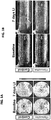
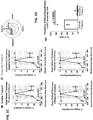

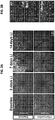

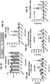

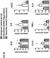

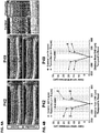
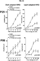
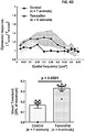
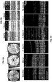
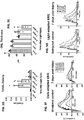


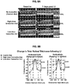

Classifications
-
- A—HUMAN NECESSITIES
- A61—MEDICAL OR VETERINARY SCIENCE; HYGIENE
- A61K—PREPARATIONS FOR MEDICAL, DENTAL OR TOILETRY PURPOSES
- A61K31/00—Medicinal preparations containing organic active ingredients
- A61K31/13—Amines
- A61K31/135—Amines having aromatic rings, e.g. ketamine, nortriptyline
- A61K31/138—Aryloxyalkylamines, e.g. propranolol, tamoxifen, phenoxybenzamine
-
- A—HUMAN NECESSITIES
- A61—MEDICAL OR VETERINARY SCIENCE; HYGIENE
- A61B—DIAGNOSIS; SURGERY; IDENTIFICATION
- A61B5/00—Measuring for diagnostic purposes; Identification of persons
- A61B5/24—Detecting, measuring or recording bioelectric or biomagnetic signals of the body or parts thereof
- A61B5/316—Modalities, i.e. specific diagnostic methods
- A61B5/398—Electrooculography [EOG], e.g. detecting nystagmus; Electroretinography [ERG]
-
- A—HUMAN NECESSITIES
- A61—MEDICAL OR VETERINARY SCIENCE; HYGIENE
- A61B—DIAGNOSIS; SURGERY; IDENTIFICATION
- A61B5/00—Measuring for diagnostic purposes; Identification of persons
- A61B5/48—Other medical applications
- A61B5/4848—Monitoring or testing the effects of treatment, e.g. of medication
-
- A—HUMAN NECESSITIES
- A61—MEDICAL OR VETERINARY SCIENCE; HYGIENE
- A61K—PREPARATIONS FOR MEDICAL, DENTAL OR TOILETRY PURPOSES
- A61K31/00—Medicinal preparations containing organic active ingredients
- A61K31/33—Heterocyclic compounds
- A61K31/395—Heterocyclic compounds having nitrogen as a ring hetero atom, e.g. guanethidine or rifamycins
- A61K31/435—Heterocyclic compounds having nitrogen as a ring hetero atom, e.g. guanethidine or rifamycins having six-membered rings with one nitrogen as the only ring hetero atom
- A61K31/44—Non condensed pyridines; Hydrogenated derivatives thereof
- A61K31/445—Non condensed piperidines, e.g. piperocaine
- A61K31/4523—Non condensed piperidines, e.g. piperocaine containing further heterocyclic ring systems
- A61K31/4535—Non condensed piperidines, e.g. piperocaine containing further heterocyclic ring systems containing a heterocyclic ring having sulfur as a ring hetero atom, e.g. pizotifen
-
- A—HUMAN NECESSITIES
- A61—MEDICAL OR VETERINARY SCIENCE; HYGIENE
- A61K—PREPARATIONS FOR MEDICAL, DENTAL OR TOILETRY PURPOSES
- A61K31/00—Medicinal preparations containing organic active ingredients
- A61K31/33—Heterocyclic compounds
- A61K31/395—Heterocyclic compounds having nitrogen as a ring hetero atom, e.g. guanethidine or rifamycins
- A61K31/55—Heterocyclic compounds having nitrogen as a ring hetero atom, e.g. guanethidine or rifamycins having seven-membered rings, e.g. azelastine, pentylenetetrazole
-
- A—HUMAN NECESSITIES
- A61—MEDICAL OR VETERINARY SCIENCE; HYGIENE
- A61P—SPECIFIC THERAPEUTIC ACTIVITY OF CHEMICAL COMPOUNDS OR MEDICINAL PREPARATIONS
- A61P27/00—Drugs for disorders of the senses
- A61P27/02—Ophthalmic agents
Definitions
- This relates to the field of treatment and/or prevention of retinal degeneration using SERMS, such as tamoxifen or a salt or derivative thereof, or raloxifene or a salt or derivative thereof.
- SERMS such as tamoxifen or a salt or derivative thereof, or raloxifene or a salt or derivative thereof.
- Photoreceptor degeneration occurs in a variety of retinal conditions ranging from atrophic age-related macular degeneration (AMD) and diabetic maculopathy, where death of macular photoreceptors results in central vision loss, to retinitis pigmentosa (RP), in which widespread photoreceptor degeneration across the entire retina leads to total blindness.
- AMD age-related macular degeneration
- RP retinitis pigmentosa
- tamoxifen a drug previously associated with retina toxicity, confers significant structural and functional protection to photoreceptors in both acute and genetic models of photoreceptor degeneration.
- tamoxifen, and other SERMs with the same activities can be used therapeutically, such as for the neuroprotection of endangered photoreceptors.
- methods are disclosed for treating or preventing retinal degeneration in a subject.
- the subject can have retinitis pigmentosa, atrophic macular degeneration, or any other retinal disease in which neuroinflammation drives neuronal loss.
- a method for treating and/or preventing retinal degeneration in a subject includes administering to the subject a therapeutically effective amount of a selective estrogen receptor modulator (SERM) to treat the retinal degeneration in the subject.
- SERM selective estrogen receptor modulator
- the SERM is one or more of tamoxifen, afimoxifene, raloxifene, apeledoxifene, arzoxifene, desmethylarzoxifene, or a salt or derivative thereof.
- the subject does not have cancer, such as breast cancer.
- the subject is human.
- SERM is administered orally.
- the SERM is tamoxifen or a salt or derivate thereof.
- tamoxifen, salt or derivative thereof is administered orally, such as at a dose of 40 mg/kg to 80 mg per kg daily.
- tamoxifen previously was clinically associated with a low frequency occurrence of adverse retinal changes, it is disclosed herein that actually exerts broad neuroprotective effects in patients with ongoing photoreceptor degeneration. It was demonstrated that oral administration of SERMS, such as tamoxifen and raloxifene, can be used to treat or prevent retinal degeneration at a variety of dosages.
- SERMS such as tamoxifen and raloxifene
- Methods for treating or preventing retinal degeneration is a subject are disclosed herein. These methods include administering to the subject a therapeutically effective amount of a selective estrogen receptor modulator (SERM) to treat the retinal degeneration in the subject, wherein the SERM is one or more of tamoxifen, afimoxifene, raloxifene, apeledoxifene, arzoxifene, desmethylarzoxifene, or a salt or derivative thereof.
- SERM selective estrogen receptor modulator
- the SERM is a) tamoxifen or a salt or derivative thereof; or b) raloxifene or a salt or derivative thereof.
- the subject is human.
- the subject has retinitis pigmentosa, acute retinal degeneration, atrophic macular degeneration, or diabetic retinopathy.
- the SERM is administered orally.
- the subject does not have cancer, such as breast cancer.
- the SERM is tamoxifen, or a salt or derivative thereof, the subject is human, and the tamoxifen is administered at a dose of about 0.8 mg/kg to about 6.5 mg/kg daily( ⁇ 10mg/kg/day, approximately equivalent to a 0.81mg/kg/day dose in an adult human).
- the SERM is tamoxifen, or a salt or derivative thereof, the subject is human, and the tamoxifen is administered orally at a dose of about 3.2 mg/kg to about 6.5 mg per kg daily.
- the SERM is administered for a minimum of three months.
- the SERM defers photoreceptor loss, reduces photoreceptor function decrement, reduces visual function loss, suppresses retinal microglial activation and/or suppresses pro-inflammatory cytokine expression.
- the methods include evaluating the vision of the subject. In one specific non-limiting examples, the methods include performing electroretinography on the subject.
- Age-related macular degeneration A disease that is a major cause of blindness in the United States and other industrialized nations. ( Evans J, Wormald R., British Journal Ophthalmology 80:9-14, 1996 ; Klein R, Klein B E K, Linton K L P, Ophthalmology 99:933-943, 1992 ; Vingerling J R, Ophthalmology 102:205-210, 1995 ).
- drusen are extracellular deposits of proteins, lipids, and cellular debris, ( Hageman G S, Mullins R F, Mol Vis 5:28, 1999 ), that are located beneath the retinal pigment epithelium (RPE).
- RPE retinal pigment epithelium
- the RPE provides nutritional, metabolic, and phagocytic functions for the overlying photoreceptors.
- Significant vision loss results from dysfunction or death of photoreceptors in the macula in association with late stages of AMD (geographic atrophy of the retinal pigment epithelial cells and subretinal neovascularization).
- Animal Living multi-cellular vertebrate organisms, a category that includes, for example, mammals and birds.
- mammal includes both human and non-human mammals.
- subject includes both human and veterinary subjects.
- Breast cancer A neoplastic condition of breast tissue that can be benign or malignant.
- the most common type of breast cancer is ductal carcinoma.
- Ductal carcinoma in situ is a non-invasive neoplastic condition of the ducts.
- Lobular carcinoma is not an invasive disease but is an indicator that a carcinoma may develop.
- Infiltrating (malignant) carcinoma of the breast can be divided into stages (I, IIA, IIB, IIIA, IIIB, and IV).
- carcinomas lose the typical histology and architecture of normal breast glands. Generally, carcinoma cells overgrow the normal cells and lose their ability to differentiate into glandular like structures. The degree of loss of differentiation in general is related to the aggressiveness of the tumor. For example, "in situ" carcinoma by definition retains the basement membrane intact, whereas as it progresses to "invasive", the tumor shows breakout of basement membranes. Thus one would not expect to see, within breast carcinomas, staining of a discrete layer of basal cells as seen in normal breast tissue.
- Breast cancers can be divided into groups based on their expression profiles. Basal-type carcinomas usually are negative for expression of estrogen receptor (ER) and negative for expression of HER2 (erbB2) and progesterone receptor (PR), and thus are referred to as “triple-negative breast cancers" or "TNBC.” This type of breast cancer is also denoted ER - /HER2 - /PR - and represents about 15-20 % of all breast cancer, and generally cannot be treated using Her2 targeted or estrogen targeted therapies. It is believed that the aggressive nature of this cancer is correlated with an enrichment for cancer stem cells (CSC) with a CD44 + CD24 -/lo phenotype.
- CSC cancer stem cells
- basal carcinomas are negative for expression of progesterone receptor (PR), positive for expression of epidermal growth factor receptor (EGFR), and positive for expression of cytokeratin 5 (CK5).
- PR progesterone receptor
- EGFR epidermal growth factor receptor
- CK5 cytokeratin 5
- Cells grown under controlled condition A primary cell culture is a culture of cells, tissues or organs taken directly from an organism and before the first subculture. Cells are expanded in culture when they are placed in a growth medium under conditions that facilitate cell growth and/or division, resulting in a larger population of the cells. When cells are expanded in culture, the rate of cell proliferation is typically measured by the amount of time required for the cells to double in number, otherwise known as the doubling time.
- Leber congenital amaurosis A rare inherited eye disease that appears at birth or in the first few months of life and primarily affects the retina. The presentation can vary because is it associated with multiple genes. However, it is characterized by characterized by nystagmus, photophobia, sluggish or absent pupillary response, and severe vision loss or blindness.
- the pupils which usually expand and contract in response to the amount of light entering the eye, do not react normally to light. Instead, they expand and contract more slowly than normal, or they may not respond to light at all. Additionally, the clear front covering of the eye (the cornea) may be cone-shaped and abnormally thin, a condition known as keratoconus.
- Franceschetti's oculo-digital sign is characteristic of Leber congenital amaurosis. This sign consists of poking, pressing, and rubbing the eyes with a knuckle or finger.
- compositions and formulations suitable for pharmaceutical delivery of the SERMS herein disclosed are conventional. Remington's Pharmaceutical Sciences, by E. W. Martin, Mack Publishing Co., Easton, PA, 15th Edition (1975 ), describes compositions and formulations suitable for pharmaceutical delivery of the SERMS herein disclosed.
- parenteral formulations usually comprise injectable fluids that include pharmaceutically and physiologically acceptable fluids such as water, physiological saline, balanced salt solutions, aqueous dextrose, glycerol or the like as a vehicle.
- pharmaceutically and physiologically acceptable fluids such as water, physiological saline, balanced salt solutions, aqueous dextrose, glycerol or the like as a vehicle.
- physiologically acceptable fluids such as water, physiological saline, balanced salt solutions, aqueous dextrose, glycerol or the like
- solid compositions e.g., powder, pill, tablet, or capsule forms
- conventional non-toxic solid carriers can include, for example, pharmaceutical grades of mannitol, lactose, starch or magnesium stearate.
- compositions to be administered can contain minor amounts of non-toxic auxiliary substances, such as wetting or emulsifying agents, preservatives, and pH buffering agents and the like, for example sodium acetate or sorbitan monolaurate.
- non-toxic auxiliary substances such as wetting or emulsifying agents, preservatives, and pH buffering agents and the like, for example sodium acetate or sorbitan monolaurate.
- Pharmaceutical agent A chemical compound or composition capable of inducing a desired therapeutic or prophylactic effect when properly administered to a subject or a cell.
- “Incubating” includes a sufficient amount of time for a drug to interact with a cell.
- “Contacting” includes incubating a drug in solid or in liquid form with a cell.
- Photic Retinopathy Damage to the retina, such as the macula, from prolonged exposure to solar radiation or other bright light, e.g. lasers or arc welders.
- the term includes solar, laser, and welder's retinopathy.
- photic retinopathy is caused by intense artificial light or sunlight.
- the light can be ultraviolet light (UV-B, 295-320 nm; UV-A, 320-400 nm) or visible light (400-700 nm).
- Phototoxic damage can occur in retinal pigment epithelial cells, the choroid, and the rod outer segments.
- Photic retinopathy results in reduced visual acuity in the long-term, and central or paracentral scotoma. Fundus changes are usually (but not always) bilateral Phototoxicity: Damage to cells, such as damage to cells of the retina, induced by light.
- Preventing or treating a disease refers to inhibiting the full development of a disease, for example in a person who is known to have a predisposition to a disease such as age related macular degeneration.
- An example of a person with a known predisposition is someone with a history of a disease in the family, or who has been exposed to factors that predispose the subject to a condition.
- Treatment refers to a therapeutic intervention that ameliorates a sign or symptom of a disease or pathological condition after it has begun to develop.
- Retina The light (photon) sensitive portion of the eye, that contains the photoreceptors (cones and rods) for light. Rods and cones perform light perception through the use of light sensitive pigments.
- the light sensitive pigments are made of protein called opsin and a chromophore called retinene, which the variant is of vitamin A.
- the rods contain rhodopsin while the cones contains iodopsin.
- Rods and cones transmit signals through successive neurons that trigger a neural discharge in the output cells of the retina and the ganglion cells.
- the visual signals are conveyed by the optic nerve to the lateral geniculate bodies from where the visual signal is passed to the visual cortex (occipital lobe) and registered as a visual stimulus.
- Rods are photoreceptor cells in the retina of the eye that can function in less intense light than the other type of visual photoreceptor, cone cells. Rods are concentrated at the outer edges of the retina and are used in peripheral vision. Rods are a little longer and leaner than cones but have the same structural basis.
- the opsin or pigment is on the outer side, lying on the retinal pigment epithelium, completing the cell's homeostasis. This epithelium end contains many stacked disks. Rods have a high area for visual pigment and thus substantial efficiency of light absorption.
- rod cells have a synaptic terminal, an inner segment, and an outer segment.
- the synaptic terminal forms a synapse with another neuron, for example a bipolar cell.
- the inner and outer segments are connected by a cilium, which lines the distal segment.
- the inner segment contains organelles and the cell's nucleus, while the rod outer segment, which is pointed toward the back of the eye, contains the light-absorbing materials.
- Activation of photopigments by light sends a signal by hyperpolarizing the rod cell, leading to the rod cell not sending its neurotransmitter, which leads to the bipolar cell then releasing its transmitter at the bipolar-ganglion synapse and exciting the synapse.
- Cone cells or “cones,” are responsible for color vision and function best in relatively bright light.
- Cone cells are densely packed in the fovea centralis, a 0.3 mm diameter rod-free area with very thin, densely packed cones which quickly reduce in number towards the periphery of the retina.
- Cones are less sensitive to light than the rod cells in the retina (which support vision at low light levels), but allow the perception of color. They are also able to perceive finer detail and more rapid changes in images, because their response times to stimuli are faster than those of rods.
- cones are normally one of the three types, each with different pigment, namely: S-cones, M-cones and L-cones.
- Each cone is therefore sensitive to visible wavelengths of light that correspond to short-wavelength, medium-wavelength and long-wavelength light.
- the three types have peak wavelengths near 420-440 nm, 534-545 nm and 564-580 nm, respectively, depending on the individual.
- Retinal Pigment Epithelium The pigmented layer of hexagonal cells, present in vivo in mammals, just outside of the neurosensory retinal that is attached to the underlying choroid. These cells are densely packed with pigment granules, and shield the retinal from incoming light.
- the retinal pigment epithelium also serves as the limiting transport factor that maintains the retinal environment by supplying small molecules such as amino acid, ascorbic acid and D-glucose while remaining a tight barrier to choroidal blood borne substances.
- Retinitis pigmentosa An inherited, degenerative eye disease that causes severe vision impairment due to the progressive degeneration of the rod photoreceptor cells in the retina. This form of retinal dystrophy manifests initial symptoms independent of age.
- the initial retinal degenerative symptoms of Retinitis pigmentosa are characterized by decreased night vision (nyctalopia) and the loss of the mid-peripheral visual field.
- the rod photoreceptor cells which are responsible for low-light vision and are orientated in the retinal periphery, are the retinal processes affected first during non-syndromic forms of this disease. Visual decline progresses relatively quickly to the far peripheral field, eventually extending into the central visual field as tunnel vision increases.
- SERM Selection Estrogen Receptor Modulator
- a characteristic that distinguishes these substances from estrogen receptor agonists and antagonists is that their action is different in various tissues, thereby granting the possibility to selectively inhibit or stimulate estrogen-like action in various tissues.
- SERMs are competitive partial agonists of the ER. Different tissues have different degrees of sensitivity to and activity of endogenous estrogens, so SERMs produce estrogenic or antiestrogenic effects depending on the specific tissue in question as well as the percentage of intrinsic activity (IA) of the SERM.
- SERMs like clomifene and tamoxifen are more in the middle in their IA and their balance of estrogenic and antiestrogenic actions in comparison.
- Raloxifene is a SERM that is more antiestrogenic than tamoxifen; both are estrogenic in bone, but raloxifene is antiestrogenic in the uterus while tamoxifen is estrogenic.
- Subject Human and non-human animals, including all vertebrates, such as mammals and non-mammals, such as non-human primates, mice, rabbits, sheep, dogs, cats, horses, cows, chickens, amphibians, and reptiles.
- the subject is a human.
- Therapeutic agent Used in a generic sense, it includes treating agents, prophylactic agents, and replacement agents.
- a SERM is one form of a therapeutic agent.
- Therapeutically effective amount A quantity of an agent sufficient to achieve a desired effect in a subject being treated. For instance, this can be the amount of a SERM to treat or prevent retinal degeneration in a subject, or a dose sufficient to prevent advancement, or to treat retinal degeneration. In one example, the amount is sufficient to prevent advancement, or to cause regression of the disease. In another example, the amount inhibits progression of retinal degeneration.
- a therapeutically effective amount of a SERM, such as tamoxifen or a salt or derivative thereof, used to achieve a specific desired effect on a biological process, such as to reduce or inhibit degeneration of the retina can be different than the dose of same compound that results in an effect on a different biological process, such as killing cancer cells.
- a therapeutically effective amount of a SERM can be administered systemically or locally (see below).
- an effective amount of a SERM can be administered in a single dose, or in several doses, for example daily, during a course of treatment.
- the effective amount of the SERM will be dependent on the preparation applied, the subject being treated, the severity and type of the affliction, and the manner of administration of the compound.
- Treating, Treatment, and Therapy Any success or indicia of success in the attenuation or amelioration of an injury, pathology or condition, including any objective or subjective parameter such as abatement, remission, diminishing of symptoms or making the condition more tolerable to the patient, slowing in the rate of degeneration or decline, making the final point of degeneration less debilitating, improving a subject's physical or mental well-being, or improving vision.
- the treatment may be assessed by objective or subjective parameters; including the results of a physical examination, neurological examination, or psychiatric evaluations.
- Tumor and Cancer A tumor is an abnormal growth of cells, which can be benign or malignant. Cancer is a malignant tumor, which is characterized by abnormal or uncontrolled cell growth. Other features often associated with malignancy include metastasis, interference with the normal functioning of neighboring cells, release of cytokines or other secretory products at abnormal levels and suppression or aggravation of inflammatory or immunological response, invasion of surrounding or distant tissues or organs, such as lymph nodes, etc. "Metastatic disease” refers to cancer cells that have left the original tumor site and migrate to other parts of the body for example via the bloodstream or lymph system.
- the amount of a tumor in an individual is the "tumor burden" which can be measured as the number, volume, or weight of the tumor.
- a tumor that does not metastasize is referred to as "benign.”
- a tumor that invades the surrounding tissue and/or can metastasize is referred to as "malignant.”
- hematological tumors include leukemias, including acute leukemias (such as 11q23-positive acute leukemia, acute lymphocytic leukemia, acute myelocytic leukemia, acute myelogenous leukemia and myeloblastic, promyelocytic, myelomonocytic, monocytic and erythroleukemia), chronic leukemias (such as chronic myelocytic (granulocytic) leukemia, chronic myelogenous leukemia, and chronic lymphocytic leukemia), polycythemia vera, lymphoma, Hodgkin's disease, non-Hodgkin's lympho
- solid tumors such as sarcomas and carcinomas
- solid tumors include fibrosarcoma, myxosarcoma, liposarcoma, chondrosarcoma, osteogenic sarcoma, and other sarcomas, synovioma, mesothelioma, Ewing's tumor, leiomyosarcoma, rhabdomyosarcoma, colon carcinoma, lymphoid malignancy, pancreatic cancer, breast cancer (including basal breast carcinoma, ductal carcinoma and lobular breast carcinoma), lung cancers, ovarian cancer, prostate cancer, hepatocellular carcinoma, squamous cell carcinoma, basal cell carcinoma, adenocarcinoma, sweat gland carcinoma, medullary thyroid carcinoma, papillary thyroid carcinoma, pheochromocytomas sebaceous gland carcinoma, papillary carcinoma, papillary adenocarcinomas, medullary carcinoma, bronchogenic carcinoma, renal cell carcinoma, hepatoma,
- Uveitus An intraocular inflammatory disease that includes crizotitis, cyclitis, panuveits, posterior uveitis and anterior uveitis.
- Iritis is inflammation of the iris.
- Cyclitis is inflammation of the ciliary body.
- Panuveitis refers to inflammation of the entire uveal (vascular) layer of the eye.
- Intermediate uveitis also called peripheral uveitis, is centered in the area immediately behind the iris and lens in the region of the ciliary body and pars plana, and is also termed "cyclitis” and "pars planitis.”
- Posterior uveitis generally refers to chorioretinitis (inflammation of the choroid and retina). Posterior uveitis can give rise to diverse symptoms but most commonly causes floaters and decreased vision similar to intermediate uveitis. Signs include cells in the vitreous humor, white or yellow-white lesions in the retina and/or underlying choroid, exudative retinal detachments, retinal vasculitis, and optic nerve edema.
- Anterior uveitis refers to iridocyclitis (inflammation of the iris and the ciliary body) and/or ulceris. Anterior uveitis tends to be the most symptomatic, typically presenting with pain, redness, photophobia, and decreased vision. Signs of anterior uveitis include pupillary miosis and injections of the conjunctiva adjacent to the cornea, so-called perilimbal flush. Biomicroscopic, or slit lamp, findings include cells and flare in the aqueous humor as well as keratic precipitates, which are clumps of cells and proteinaceous material adherent to the corneal endothelium. "Diffuse" uveitis implies inflammation involving all parts of the eye, including anterior, intermediate, and posterior structures.
- Acute uveitis is a form of uveitis in which signs and symptoms occur suddenly and last for up to about six weeks.
- Chronic uveitis is a form in which onset is gradual and lasts longer than about six weeks.
- the inflammatory products i.e., cells, fibrin, excess proteins
- the inflammatory products are commonly found in the fluid spaces of the eye, i.e., anterior chamber, posterior chamber and vitreous space as well as infiltrating the tissue imminently involved in the inflammatory response.
- the subject can have uveitis.
- Uveitis may occur following surgical or traumatic injury to the eye; as a component of an autoimmune disorder (such as rheumatoid arthritis, Bechet's disease, ankylosing spondylitis, sarcoidosis), as an isolated immune mediated ocular disorder (such as pars planitis or iridocyclitis), as a disease unassociated with known etiologies, and following certain systemic diseases which cause antibody-antigen complexes to be deposited in the uveal tissues.
- an autoimmune disorder such as rheumatoid arthritis, Bechet's disease, ankylosing spondylitis, sarcoidosis
- an isolated immune mediated ocular disorder such as pars planitis or iridocyclitis
- Uveitis includes ocular inflammation associated with Bechet's disease, sarcoidosis, Vogt-Koyanagi-Harada syndrome, birdshot chorioretinopathy and sympathetic ophthalmia. Thus, non-infectious uveitis occurs in the absence of an infectious agent.
- infective agents can also cause uveitis.
- an appropriate antimicrobial drug can be given to cure the disease.
- the etiology of uveitis remains elusive in the majority of cases.
- Tamoxifen is the trans isomer of 1-(p-beta-dimethyaminoetho- xyphenyl)-1,2-diphenylbut-1-ene, which is disclosed in U.S. Pat. No. 4,536,516 .
- An alternative name is (Z)-2-[p-(1,2-diphenylbut-1-enyl)pheno-xy]ethyldimethylamine.
- the structure of tamoxifen is shown below.
- Tamoxifen, and pharmaceutically acceptable salts and solvates thereof are known to be useful in the treatment of hormone-dependent tumors, such as for the treatment of breast cancer in women, see U.S. Patent Application Publication No. 2003/0158160 . Reviews of its clinical usage are available, for example by Furr and Jordan in "Pharmacology and Therapeutics", 1984, Volume 25, pages 127-205 .
- Pharmaceutically acceptable salts of tamoxifen are known.
- a suitable pharmaceutically acceptable acid-addition salt is, for example, the hydrochloride, hydrobromide, citrate or D-gluconate salt. Tamoxifen is marketed under the trade-name NOLVADEX®.
- Tamoxifen or a salt (also called a “pharmaceutically acceptable salt") or derivative thereof, is an "anti-estrogen," a selective estrogen-receptor modulator.
- Anti-estrogens such as tamoxifen have been shown to dramatically reduce the risk of breast cancer ( Powles, T. J., Nat Rev Cancer, 2:787-794, 2002 ) and of breast cancer recurrence ( Jordan, V. C., Nat Rev Drug Discov, 2:205-213, 2003 ).
- Tamoxifen is a partial agonist and exhibits both species and tissue specificity for inducing either an agonist or antagonist response.
- tamoxifen exhibits partial agonism, e.g., producing antagonist effects in the breast, but agonist effects in the vagina and endometrium.
- Long-term tamoxifen use has been associated with a reduced incidence of contralateral breast cancer (antagonist), a reduced incidence of primary breast cancer in high-risk women (antagonist), maintenance of bone density (agonist), and increased risk of endometrial carcinomas (agonist).
- tamoxifen At doses used to treat breast cancer, tamoxifen has been associated with retinopathy.
- the present disclosure is related to the use of tamoxifen, and salts and derivatives thereof, for the treatment or prevention of retinal degeneration.
- U.S. Patent No. 5,219,549 discloses tamoxifen derivatives wherein the alkyl chain of the molecule is substituted with fluorine or iodine, such as fluorotamoxifen.
- the compound 4-hydroxy tamoxifen (afimoxifene), or 1-[4-(2-N-dimethylaminoethoxy)-phenyl]-1-(4-hydroxyphenyl)-2-phenylbut-1-(Z)-ene constitutes an active metabolite of the well characterized anti-estrogen compound, tamoxifen. Both cis and trans isomers exist, either of which, alone or in combination, are useful.
- Raloxifene [6-hydroxy-2-(4-hydroxyphenyl)- benzothiophen-3-yl]- [4-[2-(1-piperidyl)ethoxy]phenyl] -methanone
- the structure of raloxifene is shown below:
- Bazedoxifene (1- ⁇ 4-[2-(Azepan-1-yl)ethoxy]benzyl ⁇ -2-(4-hydroxyphenyl)-3-methyl-1 H- indol-5-ol) is a third generation SERM developed by Pfizer; the structure is shown below:
- Bazedoxifene has been approved for use in postmenopausal osteoporosis, and has been used for treatment of dyspareunia, breast cncer, and pancreatic cancer. Bazedoxifene is also of use in the methods disclosed herein.
- Arzoxifene (2-(4-Methoxyphenyl)-3-[4-(2-piperidin-1-ylethoxy)phenoxy]-1-benzothiophen-6-ol) is a SERM that is a potent estrogen antagonist in mammary and uterine tissue while acting as an estrogen agonist to maintain bone density and lower serum cholesterol.
- Arzoxifene is also of use in the methods disclosed herein. The structure of arzoxifene is shown below:
- Desmethylarzoxifene (2-(4-hydroxyphenyl)-3-[4-(2-piperidin-1-ylethoxy)phenoxy]-1-benzothiophen-6-ol) is also a SERM and is of use in the methods disclosed herein. Desmethylarzoxifene blocks estrogen-induced malignant transformation of human breast epithelial cell lines ( Kastrati et al., PLOS One 6(11): e27876, 2011 ). The structure of desmethylarzoxifene is shown below:
- the methods include administering a therapeutically effective amount of raloxifene, apeledoxifene, arzoxifene, desmethylarzoxifene, tamoxifen and/or a pharmaceutically acceptable salt or derivative thereof.
- compositions are thus provided for both local use (for example, topical or within an ocular transplant), as well as for systemic use.
- the subject can be any subject, such as a mammalian subject. Therefore, the disclosure includes within its scope pharmaceutical compositions comprising a SERM, raloxifene, apeledoxifene, arzoxifene, desmethylarzoxifene, tamoxifen and/or a pharmaceutically acceptable salt or derivative thereof, formulated for use in human or veterinary medicine.
- the SERM such as SERM, raloxifene, raloxifene, arcoxifene, desmethylarzoxifene, tamoxifen and/or a pharmaceutically acceptable salt or derivative thereof, is formulated for oral administration.
- compositions that include a SERM such as raloxifene, apeledoxifene, arzoxifene, desmethylarzoxifene, tamoxifen and/or a pharmaceutically acceptable salt or derivative thereof, can be formulated with an appropriate solid or liquid carrier, depending upon the particular mode of administration chosen.
- the SERM such as raloxifene, apeledoxifene, arzoxifene, desmethylarzoxifene, tamoxifen and/or a pharmaceutically acceptable salt or derivative thereof
- flavorings, colors and sweeteners can be added.
- Oral formulations may be liquid (e.g., syrups, solutions, or suspensions), or solid (e.g., powders, pills, tablets, or capsules).
- solid compositions conventional non-toxic solid carriers can include pharmaceutical grades of mannitol, lactose, starch, or magnesium stearate. Actual methods of preparing such dosage forms are known, or will be apparent, to those of ordinary skill in the art.
- parenteral formulations usually comprise injectable fluids that are pharmaceutically and physiologically acceptable fluid vehicles such as water, physiological saline, other balanced salt solutions, aqueous dextrose, glycerol or the like.
- the pharmaceutical composition to be administered may also contain minor amounts of non-toxic auxiliary substances, such as wetting or emulsifying agents, preservatives, and pH buffering agents and the like, for example sodium acetate or sorbitan monolaurate.
- the pharmaceutical compositions can contain the SERM and a pharmaceutically acceptable excipient.
- excipients include any pharmaceutical agent that does not itself induce the production of antibodies harmful to the individual receiving the composition, and which may be administered without undue toxicity.
- Pharmaceutically acceptable excipients include, but are not limited to, liquids such as water, saline, glycerol and ethanol.
- Pharmaceutically acceptable salts can be included therein, for example, mineral acid salts such as hydrochlorides, hydrobromides, phosphates, sulfates, and the like; and the salts of organic acids such as acetates, propionates, malonates, benzoates, and the like.
- auxiliary substances such as wetting or emulsifying agents, pH buffering substances, and the like, may be present in such vehicles.
- auxiliary substances such as wetting or emulsifying agents, pH buffering substances, and the like.
- a protein excipient such as BSA, if present, will can be present at a concentration of between 1.0 weight (wt.) % to about 20 wt. %, such as 10 wt. %. If an amino acid such as glycine is used in the formulations, it can be present at a concentration of about 1 wt. % to about 5 wt. %.
- a carbohydrate, such as sorbitol, if present, can be present at a concentration of about 0.1 wt % to about 10 wt. %, such as between about 0.5 wt.
- polyethylene glycol it can generally be present on the order of about 2 wt. % to about 40 wt. %, such as about 10 wt. % top about 25 wt. %.
- propylene glycol it will typically be present at a concentration of about 2 wt. % to about 60 wt. %, such as about 5 wt. % to about 30 wt. %.
- a detergent such as a sorbitan ester (TWEEN®) is present, it can be present at a concentration of about 0.05 wt. % to about 5 wt. %, such as between about 0.1 wt. % and about 1 wt %, see U.S. Published Patent Application No. 2012/0219528 .
- the dosage form of the pharmaceutical composition will be determined by the mode of administration chosen. For instance, topical and oral formulations can be employed.
- compositions that include a SERM can be formulated in unit dosage form, suitable for individual administration of precise dosages.
- the amount of active compound(s) administered will be dependent on the subject being treated, the severity of the affliction, and the manner of administration, and can be left to the judgment of the prescribing clinician.
- the formulation to be administered will contain a quantity of the active component(s) in amounts effective to achieve the desired effect in the subject being treated, such as treatment or prevention of retinal degeneration. The effect can also be on microglia, or a particular ocular motor response.
- the SERM can be formulated with additional therapeutic agents.
- the SERM is formulated for delivery to the eye.
- Topical preparations can include eye drops, ointments, sprays and the like. Eye drops or sprays can be provided in unit dose dispensers (such as eye drop bottles that dispense a metered unit dose that contains the SERM, such as raloxifene, apeledoxifene, arzoxifene, desmethylarzoxifene, tamoxifen and/or a pharmaceutically acceptable salt or derivative thereof, either alone or in combination with other therapeutic agents).
- unit dose dispensers such as eye drop bottles that dispense a metered unit dose that contains the SERM, such as raloxifene, thaledoxifene, arzoxifene, desmethylarzoxifene, tamoxifen and/or a pharmaceutically acceptable salt or derivative thereof, either alone or in combination with other therapeutic agents).
- the SERM can be included in an inert matrix for either topical application or injection into the eye, such as for intra-vitreal administration.
- liposomes may be prepared from dipalmitoyl phosphatidylcholine (DPPC), such as egg phosphatidylcholine (PC).
- DPPC dipalmitoyl phosphatidylcholine
- PC egg phosphatidylcholine
- Liposomes, including cationic and anionic liposomes can be made using standard procedures as known to one skilled in the art.
- Liposomes including a SERM can be applied topically, either in the form of drops or as an aqueous based cream, or can be injected intraocularly. In a formulation for topical application, the SERM is slowly released over time as the liposome capsule degrades due to wear and tear from the eye surface.
- the liposome capsule degrades due to cellular digestion. Both of these formulations provide advantages of a slow release drug delivery system, allowing the subject to be exposed to a substantially constant concentration of the SERM over time.
- the SERM can be dissolved in an organic solvent such as DMSO or alcohol as previously described and contain a polyanhydride, poly(glycolic) acid, poly(lactic) acid, or polycaprolactone polymer.
- the SERM such as raloxifene, apeledoxifene, arzoxifene, desmethylarzoxifene, tamoxifen and/or a pharmaceutically acceptable salt or derivative thereof, can be included in a delivery system that can be implanted or injected at various sites in and around the eye, depending on the size, shape and formulation of the implant or injection.
- the SERM can be used alone.
- at least one additional agent such as at least one agent that is described above, can be included along with the SERM in the delivery system, such as in an implant. The delivery system is then introduced into the eye.
- Suitable sites include but are not limited to the anterior chamber, anterior segment, posterior chamber, posterior segment, vitreous cavity, suprachoroidal space, subconjunctiva, episcleral, intracorneal, epicorneal and sclera.
- the SERM delivery system is placed in the anterior chamber of the eye. In another example, the SERM delivery system is placed in the vitreous cavity.
- the implants can be inserted into the eye by a variety of methods, including placement by forceps or by trocar following making an incision in the sclera (for example, a 2-3 mm incision) or other suitable site.
- the implant can be placed by trocar without making a separate incision, but instead by forming a hole directly into the eye with the trocar.
- the method of placement can influence the release kinetics. For example, implanting the device into the vitreous or the posterior chamber with a trocar may result in placement of the device deeper within the vitreous than placement by forceps, which may result in the implant being closer to the edge of the vitreous.
- the location of the implanted device may influence the concentration gradients of SERM surrounding the device, and thus influence the release rates (for example, a device placed closer to the edge of the vitreous may result in a slower release rate, see U.S. Patent No. 5,869,079 and U.S. Patent No. 6,699,493 ).
- the SERM is delivered to the eye for a sufficient time period to achieve the desired effect.
- the SERM is delivered for at least about 2 days, such as for about five days, seven days, ten days, 14 or 21 days.
- the immunosuppressive agent is delivered for at least about one week, at least about two weeks, at least about three weeks, at least about four weeks, at least about five weeks, at least about six weeks, at least about seven weeks, at least about eight weeks, at least about nine weeks, at least about 10 weeks, and at least about 12 weeks.
- extended periods of delivery are achieved with the use of an implant.
- the duration of use of a SERM can be the medical history of the patient and other contributing factors (such as use of other agents, etc.).
- the SERM can be administered for up to six months, or one year, two years, three years, or longer.
- an implant is utilized for extended release. More than one implant can also be utilized. For example, implants can be sequentially implanted into the vitreous in order to maintain concentrations for even long periods. In one embodiment, more than one implant can be sequentially implanted into the eye in order to maintain therapeutic drug concentrations for longer periods.
- an implant is formulated with the SERM associated with a bioerodible polymer matrix.
- the SERM is homogeneously distributed through the polymeric matrix, such that it is distributed evenly enough that no detrimental fluctuations in rate of release occur because of uneven distribution of the agent in the polymer matrix.
- the selection of the polymeric composition to be employed varies with the desired release kinetics, the location of the implant, patient tolerance, and the nature of the implant procedure.
- the polymer can be included as at least about 10 weight percent of the implant. In one example, the polymer is included as at least about 20 weight percent of the implant. In another embodiment, the implant comprises more than one polymer. These factors are described in detail in U.S. Patent No. 6,699,493 .
- Characteristics of the polymers generally include biodegradability at the site of implantation, compatibility with the agent of interest, ease of encapsulation, and water insolubility, amongst others. Generally, the polymeric matrix is not fully degraded until the drug load has been released.
- the chemical composition of suitable polymers is known in the art (for example, see U.S. Patent No. 6,699,493 ).
- the SERM such as raloxifene, adoxifene, arzoxifene, desmethylarzoxifene, tamoxifen and/or a pharmaceutically acceptable salt or derivative thereof
- the SERM can be formulated in an implantable form with other carriers and solvents.
- buffering agents and preservatives can be employed.
- Water soluble preservatives include sodium bisulfite, sodium bisulfate, sodium thiosulfate, benzalkonium chloride, chlorobutanol, thimerosal, phenylmercuric acetate, phenylmercuric nitrate, methylparaben, polyvinyl alcohol and phenylethyl alcohol.
- Suitable water soluble buffering agents that may be employed are sodium carbonate, sodium borate, sodium phosphate, sodium acetate, sodium bicarbonate. These agents can be present in amounts sufficient to maintain a pH of the system of between about 2 to about 9 such as about 4 to about 8, or at about 6 to about 7. In one example, the pH of the system is maintained at about 7. As such, the buffering agent can be as much as 5% on a weight-to-weight basis of the total composition. Electrolytes such as sodium chloride and potassium chloride may also be included in the formulation.
- the proportions of SERM, polymer, and any other modifiers may be empirically determined by formulating several implants with varying proportions.
- a USP approved method for dissolution or release test can be used to measure the rate of release ( USP 23; NF 18 (1995) pp. 1790-1798 ).
- the implant sizes and shape can also be varied for use in particular regions of the eye (see U.S. Patent No. 5,869,079 ).
- the SERM can be formulated with additional therapeutic agents.
- exemplary agents include cyclosporine, FK506, steroids such as hydrocortisone, antibodies (such as anti-CD4 or antibodies that specifically bind the IL-2 receptor), cytokines (such as beta-interferon), or non-steroidal anti-inflammatory agents.
- Additional agents include antibacterial antibiotics, such as minoglycosides (for example, amikacin, apramycin, arbekacin, bambermycins, butirosin, dibekacin, dihydrostreptomycin, fortimicin(s), gentamicin, isepamicin, kanamycin, micronomicin, neomycin, neomycin undecylenate, netilmicin, paromomycin, ribostamycin, sisomicin, spectinomycin, streptomycin, tobramycin, trospectomycin), amphenicols (for example, azidamfenicol, chloramphenicol, florfenicol, thiamphenicol), ansamycins (for example, rifamide, rifampin, rifamycin sv, rifapentine, rifaximin), ⁇ -lactams (for example, carbacephems (e.g.,
- Agents of use also include synthetic antibacterials, such as 2,4-Diaminopyrimidines (for example, brodimoprim, tetroxoprim, trimethoprim), nitrofurans (for example, furaltadone, furazolium chloride, nifuradene, nifuratel, nifurfoline, nifurpirinol, nifurprazine, nifurtoinol, nitrofurantoin), quinolones and analogs (for example, cinoxacin, ciprofloxacin, clinafloxacin, difloxacin, enoxacin, fleroxacin, flumequine, grepafloxacin, lomefloxacin, miloxacin, nadifloxacin, nalidixic acid, norfloxacin, ofloxacin, oxolinic acid, pazufloxacin, pefloxacin, pipemidic acid,
- antifungal antibiotics such as polyenes (for example, amphotericin B, candicidin, beostatin, filipin, fungichromin, hachimycin, hamycin, lucensomycin, mepartricin, natamycin, nystatin, pecilocin, perimycin), others (for example, azaserine, griseofulvin, oligomycins, neomycin undecylenate, pyrrolnitrin, siccanin, tubercidin, viridin) allylamines (for example, butenafine, naftifine, terbinafine), imidazoles (for example, bifonazole, butoconazole, chlordantoin, chlormiidazole, cloconazole, clotrimazole, econazole, enilconazole, fenticonazole, flutrimazole, isoconazole, ketoconazoles (for example
- Antineoplastic agents can also be of use including (1) antibiotics and analogs (for example, aclacinomycins, actinomycin, anthramycin, azaserine, bleomycins, cactinomycin, carubicin, carzinophilin, chromomycins, dactinomycin, daunorubicin, 6-diazo-5-oxo-L-norleucine, doxorubicin, epirubicin, idarubicin, menogaril, mitomycins, mycophenolic acid, nogalamycin, olivomycines, peplomycin, pirarubicin, plicamycin, porfiromycin, puromycin, streptonigrin, streptozocin, tubercidin, zinostatin, zorubicin), (2) antimetabolites such as folic acid analogs (for example, denopterin, edatrexate, methotrexate, piritrexim, pter
- Steroidal anti-inflammatory agents can also be included such as 21-acetoxypregnenolone, alclometasone, algestone, amcinonide, beclomethasone, betamethasone, budesonide, chloroprednisone, clobetasol, clobetasone, clocortolone, cloprednol, corticosterone, cortisone, cortivazol, cyclosporine, deflazacort, desonide, desoximetasone, dexamethasone, diflorasone, diflucortolone, difluprednate, enoxolone, fluazacort, flucloronide, flumethasone, flunisolide, fluocinolone acetonide, fluocinonide, fluocortin butyl, fluocortolone, fluorometholone, fluperolone acetate, fluprednidene acetate, flupre
- non-steroidal anti-inflammatory agents can be used. These include aminoarylcarboxylic acid derivatives (for example, enfenamic acid, etofenamate, flufenamic acid, isonixin, meclofenamic acid, mefenamic acid, niflumic acid, talniflumate, terofenamate, tolfenamic acid), arylacetic acid derivatives (for example, aceclofenac, acemetacin, alclofenac, amfenac, amtolmetin guacil, bromfenac, bufexamac, cinmetacin, clopirac, diclofenac sodium, etodolac, felbinac, fenclozic acid, fentiazac, glucametacin, ibufenac, indomethacin, isofezolac, isoxepac, lonazolac, metiazinic acid, mof
- the methods can include selecting a subject with retinal degeneration, or a subject that is of risk for retinal degeneration.
- the therapeutically effective amount of the SERM such as, but not limited to, one or more of tamoxifen, afimoxifene, raloxifene, apeledoxifene, arzoxifene, desmethylarzoxifene, or a salt or derivative thereof, is sufficient to treat, inhibit and/or prevent retinal degeneration.
- the subject has ongoing photoreceptor degeneration.
- methods for treating retinitis pigmentosa, age related macular degeneration or Leber Congenital amaurosis (LCA) in a subject. These methods includes selecting a subject with retinitis pigmentosa, age related macular degeneration, or LCA, and administering to the subject an effective amount of a SERM, such as one or more of tamoxifen, afimoxifene, raloxifene, apeledoxifene, arzoxifene, desmethylarzoxifene, or a salt or derivative thereof, to treat and/or prevent the retinal degeneration in the subject.
- the method can include selecting a subject with the retinal degeneration, such as retinitis pigmentosa, age-related macular degeneration or LCA.
- the subject does not have a tumor, such as a cancer. In some non-limiting examples, the subject does not have breast cancer.
- the subject can be male or female. Human and veterinary subjects can be treated using the disclosed methods.
- the subject can have ocular phototoxicity, specifically photic retinopathy, also called photic maculopathy.
- the photic retinopathy is caused by exposure to the sun.
- the photic retinopathy is caused by exposure to the artificial light.
- the subject can be at risk for photic retinopathy, also known as photic maculopathy.
- the subject can be undergoing laser procedures to the eye, or can be a welder.
- the method can include selecting a subject at risk for retinal degeneration, such as, but not limited to, retinitis pigmentosa, age-related macular degeneration or LCA.
- a subject at risk for retinal degeneration such as, but not limited to, retinitis pigmentosa, age-related macular degeneration or LCA.
- subject can have diabetic retinopathy.
- the methods can included selecting a subject with diabetic retinopathy, or a subject at risk for diabetic retinopathy, such as a diabetic subject.
- the subject has a disease with photoreceptor degeneration features. These include atrophic and exudative age-related macular degeneration, diabetic retinopathy, pathologic myopic degeneration, photic injury to the retina.
- the disease can arise from a gene mutation, such as retinitis pigmentosa, Stargardt's disease, LCA, or Best's disease.
- the subject has a disease in which activated microglia contribute to increased neuroinflammation in the retina. These include, but are not limited to, posterior uveitis, a response to intraocular surgery and retinal laser procedures, a response to viral vectors for gene therapy, and a response to cell-based (e.g., stem-cell) therapy.
- the subject has a disease in which neuronal (i.e., non-photoreceptor) degeneration occurs.
- neuronal i.e., non-photoreceptor
- these include, but are not limited to, glaucoma, retinal artery occlusions, and retinal vein occlusions.
- the subject has posterior uveitis.
- subjects can be treated that are affected with toxoplasma retinochroiditis, retinal vasculitis, idiopathic posterior uveitis, ocular histoplasmosis, toxocariasis, cytomegalovirus retinitis, idiopathic retinitis, serpinous choroidopathy, acute multifocal placoid, pigment eptiheliopathy, acute retinal necrosis, bird shot choroidopathy, uveitis associated with a leukemia or a lymphoma, reticulum cell sarcoma, ocular candidiasis, tuberculous uveitis, and lupus retinitis.
- Diagnosis can utilize tests which examine the fundus of the eye and/or evaluate the visual field. These include electroretinogram, fluorangiography, and visual examination.
- the fundus of the eye examination aims to evaluate the condition of the retina and to evaluate for the presence of the characteristic pigment spots on the retinal surface. Examination of the visual field makes possible to evaluate the sensitivity of the various parts of the retina to light stimuli.
- An electroretinogram (ERG) can be used, which records the electrical activity of the retina in response to particular light stimuli and allows distinct valuations of the functionality of the two different types of photoreceptors (i.e., cone cells and rod cells).
- the presently disclosed methods can be used to treat any type of retinitis pigmentosa.
- the retinitis pigmentosa is caused by mutations in the rhodopsin gene, the peripherin gene, and/or other genes expressed in the rod.
- the retinitis pigmentosa can be the result of a genetic condition inherited in an autosomal dominant, autosomal recessive or X-linked manner.
- the X-linked retinitis pigmentosa can be recessive, affecting males, or dominant, so that it affects males and females.
- the retinitis pigmentosa can be associated with rod-cone retinal degenerations present with central macular pigmentary changes (bull's eye maculopathy).
- the retinitis pigmentosa can be choroideremia, which is an X-linked recessive retinal degenerative disease.
- RP retinitis pigmentosa
- the presently disclosed methods can be used to prevent or treat age-related macular degeneration (AMD).
- AMD age-related macular degeneration
- the subject has atrophic AMD (also called “dry” AMD), wherein the subject has symptomatic central vision loss due to retinal atrophy.
- the subject has wet AMD.
- the disclosed methods are of use to treat a subject with LCA.
- the SERM such as raloxifene, apeledoxifene, arzoxifene, desmethylarzoxifene, tamoxifen and/or a pharmaceutically acceptable salt or derivative thereof, is administered systemically.
- Systemic modes of administration include oral and parenteral routes.
- Parenteral routes include, by way of example, intravenous, intrarterial, intramuscular, intradermal, subcutaneous, intranasal and intraperitoneal routes.
- Components administered systemically may be modified or formulated to target the components to the eye (see above).
- the SERM such as raloxifene,bazedoxifene, arzoxifene, desmethylarzoxifene, tamoxifen and/or a pharmaceutically acceptable salt or derivative thereof, is administered orally.
- the SERM such as raloxifene, apeledoxifene, arzoxifene, desmethylarzoxifene, tamoxifen and/or a pharmaceutically acceptable salt or derivative thereof, can be administered repeatedly.
- the SERM is administered for 10, 15, 20, 25, or 30 days.
- SERM is administered for at least 1, 2, 3, 4, 5, 6, 7, 8, 9, 10, 11 or 12 months.
- the SERM can be administered for up to six months, or one year, two years, three years, or longer.
- the SERM can be administered daily, every daily other day, every three days, or weekly for the specified time period.
- a sustained release formulation such as a SERM-releasing drug depot or sustained release implant or device, can also be used.
- a suitable oral formulation of a SERM is for example, a tablet or capsule, preferably a tablet containing, for example, about 10, 20, 30 or 40 mg/kg, of therapeutic agent.
- the SERM can be administered at a dose in the range of about 20 mg/kg to about 160 mg per day, such as about 20 mg/kg to about 80 mg/kg, for example, about 20 mg/kg to about 40 mg/kg, either as a single dose or as divided doses. In a specific non-limiting example, the dose is administered daily.
- tamoxifen, or a salt or derivative thereof is administered orally at a dose of about 10 mg/kg to about 80 mg/kg. In other embodiments, tamoxifen, or a salt or derivative thereof, is administered orally at a dose of about 40 mg/kg to about 80 mg/kg. In some examples, tamoxifen, or a salt or derivative thereof is administered orally at a dose of about 40, 45, 50, 55, 60, 65, 70, 75 or 80 mg/kg. In specific non-limiting examples, this dose is administered daily.
- tamoxifen, or a salt or derivative thereof is administered orally at a dose of about 0.8 mg/kg to about 6.5 mg/kg daily ( ⁇ 10mg/kg/day, approximately equivalent to a 0.81 mg/kg/day dose in an adult human).
- the tamoxifen is administered orally at a dose of about 3.2 mg/kg to about 6.5 mg per kg daily.
- Suitable doses include, but are not limited to, about 0.8 mg/kg, 0.9 mg/kg, 1 mg/kg, 1.1 mg/kg, 1.2 mg/kg, 1.3 mg/kg.
- tamoxifen, or a salt or derivative thereof, or raloxifene, or a salt or derivative thereof is administered for 10, 15, 20, 25, or 30 days.
- tamoxifen, or a salt or derivative thereof, or raloxifene, or a salt or derivative thereof is administered for at least 1, 2, 3, 4, 5, 6, 7, 8, 9, 10, 11 or 12 months.
- tamoxifen, or a salt or derivative thereof, or raloxifene, or a salt or derivative thereof can be administered for up to six months, or one year, two years, three years, or longer.
- tamoxifen, or a salt or derivative thereof, or raloxifene, or a salt or derivative thereof can be administered daily, every daily other day, every three days, or weekly for the specified time period. In some examples, tamoxifen, or salt or derivative thereof, or raloxifene, or a salt or derivative thereof, is administered daily.
- tamoxifen, or a salt or derivative thereof is administered orally at a dose of about 40 mg/kg to about 80 mg per kg daily for a minimum of 1, 2 3, 4, 5, 6, 7, 8, 9, 10, 11 or 12 months.
- tamoxifen, or a salt or derivative thereof is administered orally at a dose of about 0.8 mg/kg to about 6.5 mg/kg daily for a minimum of 1, 2 3, 4, 5, 6, 7, 8, 9, 10, 11 or 12 months.
- the tamoxifen, or a salt or derivative thereof can be administered orally for up to six months, or one year, two years, three years, or longer.
- the tamoxifen, or a salt or derivative thereof can be administered orally, and daily, every daily other day, every three days, or weekly for the specified time period.
- the tamoxifen, or salt or derivative thereof is administered daily and orally.
- tamoxifen, or a salt or derivative thereof is administered orally at a dose of about 40 mg/kg to about 80 mg per kg daily for at least 3 months, 4, 5, 6, 7, 8, 9, 10, 11 or 12 months.
- tamoxifen, or a salt or derivative thereof is administered orally at a dose of about 0.8 mg/kg to about 6.5 mg/kg daily for at least 3 months, 4, 5, 6, 7, 8, 9, 10, 11 or 12 months.
- Dosage treatment may be a single dose schedule or a multiple dose schedule to ultimately deliver the amount specified above.
- the doses can be intermittent.
- the subject may be administered as many doses as appropriate.
- the subject is administered the SERM prior to the onset of a condition.
- the SERM induces a downregulation of inflammatory responses and decreases microglia activation in the retina, and provides protection to photoreceptors and retinal neurons.
- Pre-treatment administration of the SERM can provide protection to impending or possible events of retinal neuroinflammation and/or photoreceptor injury.
- intraocular surgery or ocular procedures involving direct light illumination of the retina laser or surgical procedures to the retina in which thermal or mechanical damage may be sustained
- implantation of retinal devices, intraocular drug-release devices, or intraocular implants implantation of retinal cells, intraocular delivery of viral vectors, high-risk progression to pathological events in the retina, e.g., impending retinal artery/vein occlusion, vitreous or retinal hemorrhage.
- Individual doses are typically not less than an amount required to produce a measurable effect on the subject, and may be determined based on the pharmacokinetics and pharmacology for absorption, distribution, metabolism, and excretion ("ADME") of the subject composition or its byproducts, and thus based on the disposition of the composition within the subject. This includes consideration of the route of administration as well as dosage amount, which can be adjusted for local and systemic (for example, oral) applications.
- Effective amounts of dose and/or dose regimen can readily be determined empirically from preclinical assays, from safety and escalation and dose range trials, individual clinician-patient relationships, as well as in vitro and in vivo assays. Generally, these assays will evaluate retinal degeneration, or expression of a biological component (cytokine, specific inflammatory cell, microglia, etc.) that affects retinal degeneration.
- a biological component cytokine, specific inflammatory cell, microglia, etc.
- Local modes of administration include, by way of example, intraocular, intraorbital, subconjunctival, sub-Tenon's, subretinal or transscleral routes.
- significantly smaller amounts of the components may exert an effect when administered locally (for example, intravitreally) compared to when administered systemically (for example, intravenously).
- the SERM such as raloxifene, apeledoxifene, arzoxifene, desmethylarzoxifene, tamoxifen and/or a pharmaceutically acceptable salt or derivative thereof, are delivered subretinally, e.g., by subretinal injection.
- Subretinal injections may be made directly into the macular, e.g., submacular injection.
- exemplary methods include intraocular injection (e.g., retrobulbar, subretinal, submacular, intravitreal and intrachoridal), iontophoresis, eye drops, and intraocular implantation (e.g., intravitreal, sub-Tenons and sub-conjunctival).
- the system disclosed herein is delivered by intravitreal injection.
- Intravitreal injection has a relatively low risk of retinal detachment.
- Methods for administration of agents to the eye are known in the medical arts and can be used to administer components described herein.
- Administration may be provided as a single administration, a periodic bolus or as continuous infusion.
- administration is from an internal reservoir (for example, from an implant disposed at an intra- or extra-ocular location (see, U.S. Pat. Nos. 5,443,505 and 5,766,242 )) or from an external reservoir (for example, from an intravenous bag).
- Components can be administered by continuous release for a particular period from a sustained release drug delivery device immobilized to an inner wall of the eye or via targeted transscleral controlled release into the choroid (see, for example, PCT/US00/00207 , PCT/US02/14279 , Ambati et al., Invest. Opthalmol. Vis. Sci.
- the subject method results in a therapeutic benefit, such as preventing the development of retinal degeneration, halting the progression of a retinal degeneration, and/or reversing the progression of a retinal degeneration.
- the subject can have any form of retinal degeneration, such as, but not limited to, such as retinitis pigmenosa, age related macular degeneration or LCA.
- the method includes the step of detecting that a therapeutic benefit has been achieved. Measures of therapeutic efficacy will be applicable to the particular disease being modified, and will recognize the appropriate detection methods to use to measure therapeutic efficacy. Suitable tests are disclosed below, and include effects on microglia and ocular motor response. In some non-limiting examples, it is determined whether the SERM reduces retinal microglial activation and pro-inflammatory cytokine expression.
- therapeutic efficacy can be observed by fundus photography or evaluation of the ERG response.
- the method can include comparing test results after administration of the subject composition to test results before administration of the subject composition.
- therapeutic efficacy in treating a progressive cone dysfunction may be observed as a reduction in the rate of progression of cone dysfunction, as a cessation in the progression of cone dysfunction, or as an improvement in cone function, effects which may be observed by, such as electroretinography (ERG) and/or cERG; color vision tests; functional adaptive optics; and/or visual acuity tests, for example, by comparing test results after administration of the subject composition to test results before administration of the subject composition and detecting a change in cone viability and/or function.
- the SERM defers photoreceptor loss, reduces photoreceptor function decrement, and/or reduces visual function loss.
- therapeutic efficacy in treating a vision deficiency can as an alteration in the individual's vision, such as in the perception of red wavelengths, in the perception of green wavelengths, in the perception of blue wavelengths, effects which may be observed by, cERG and color vision tests, for example, by comparing test results after administration of the subject composition to test results before administration of the subject composition and detecting a change in cone and rod viability and/or function.
- the method includes evaluation morphology and structure preservation and/or ERG.
- Photoreceptor degeneration is a cause of irreversible vision loss in a number of blinding retinal diseases including retinitis pigmentosa and atrophic age-related macular degeneration for which there is currently no treatment.
- Two separate murine models of photoreceptor degeneration were used to demonstrate that tamoxifen, a selective estrogen receptor modulator and a drug previously linked with retinal toxicity, exerted potent neuroprotective effects in the retina.
- tamoxifen treatment prevented the onset of photoreceptor apoptosis and atrophy and maintained near-normal levels of electroretinographic responses.
- mice Young adult wild type (WT) C57BL/6J mice and mice homozygous for the Pde6b rd10 (rd10) mutation were obtained from The Jackson Laboratory (Bar Harbor, ME). Postnatal rd10 mice (21-50 days of age) and young adult WT mice (2-3 months of age) of both sexes were raised in cyclic light (-100 lux, 12:12 h) in a National Institutes of Health animal facility. All experiments were conducted according to protocols approved by a local Institutional Animal Care and Use Committee and adhered to the Association for Research in Vision and Ophthalmology (ARVO) Statement animal use in ophthalmic and vision research. For the tamoxifen-treated groups, mice were provided with tamoxifen-supplemented mouse chow (500mg/kg, Envigo, Indianapolis, IN) in place of standard chow for the duration of experiment.
- tamoxifen-supplemented mouse chow 500mg/kg, Envigo, Indianapolis, IN
- Mouse model of retinal light injury Experimental animals were dark-adapted in a dark room for 7 days and then subjected to pupillary dilation with topical tropicamide (1%, Alcon, Fort Worth, TX) and phenylephrine (10%, Alcon, Fort Worth, TX). After full dilation, animals were exposed to 2x10 4 lux of diffuse white fluorescent light (Sunlite Manufacturing, Brooklyn, NY) for 2 hours. After light exposure, mice were maintained in typical conditions of ambient cyclic light (-100 lux, 12:12 h) under which the animals were housed. For tamoxifen-treatment groups, animals were fed a tamoxifen-supplemented chow beginning 7 days prior to light injury, and maintained on the same diet thereafter. Control groups not administered tamoxifen were fed standard mouse chow in separate cages at all time points.
- OCT optical coherence tomographic
- FPT fundus autofluorescence imaging
- Mice were anesthetized with intraperitoneal ketamine (90 mg/kg) and xylazine (8 mg/kg) and their pupils were dilated. Retinal structure was assessed using an optical coherence tomographic (OCT) imaging system (Bioptigen, Durham, NC).
- OCT optical coherence tomographic
- animals were imaged prior to dark adaptation and subsequently at different times following light exposure.
- Volume scans of 1.4mm by 1.4mm centered on the optic nerve 1000 A-scans/horizontal B-scan, 33 horizontal B-scans, average of three frames per B-scan, each spaced 0.0424mm apart) were captured.
- Retinal thickness measurements in each quadrant of a circular grid of diameter 1.2mm were computed using the manufacturer's software.
- Total retinal thickness (measured from the vitreal surface to the RPE layer) and outer retinal thickness (measured from the vitreal surface of the outer plexiform layer to the vitreal surface of the RPE layer) were recorded from OCT images following automated retinal segmentation.
- additional horizontal and vertical linear scans 1.4 mm width, 1000 A-scans/B-scan, average of 20 frames/B-scan
- the area of ONL in the scan was circumscribed and measured manually.
- Average outer retinal thickness was computed by dividing the outer retinal area with the length of measured retina.
- Fundus autofluorescence imaging was performed using a confocal scanning laser ophthalmoscopy (cSLO, Heidelberg Engineering, Heidelberg, Germany) with 488nm wavelength excitation. Fundus images were obtained over the central 30° angle with the field centered on the optic nerve head.
- Electroretinographic analysis Electroretinographic analysis: Electroretinographs (ERGs) were recorded using an Espion E 2 system (Diagnosys, Littleton, MA). Mice were anesthetized as described above after dark adaptation overnight. Pupils were dilated and a drop of proparacaine hydrochloride (0.5%, Alcon) was applied on cornea for topical anesthesia. Flash ERG recordings were obtained simultaneously from both eyes with gold wire loop electrodes, with the reference electrode placed in the mouth and the ground subdermal electrode at the tail. ERG responses were obtained at increasing light intensities over the ranges of 1x10 -4 to 10 cd ⁇ s/m 2 under dark-adapted conditions, and 0.3 to 100 cd ⁇ s/m 2 under a background light that saturates rod function.
- the stimulus interval between flashes varied from 5 s at the lowest stimulus strengths to 60 s at the highest ones.
- Two to 10 responses were averaged depending on flash intensity.
- ERG signals were sampled at 1 kHz and recorded with 0.3 Hz low-frequency and 300 Hz high-frequency cutoffs.
- Analysis of a-wave and b-wave amplitudes was performed using customized Espion ERG Data Analyzer software (v2.2) that digitally filters out high frequency oscillatory potential wavelets. The a-wave amplitude was measured from the baseline to the negative peak and the b-wave was measured from the a-wave trough to the maximum positive peak.
- ERGs were recorded at 1 week after light exposure.
- ERGs were recorded at P29 and P50.
- Statistical significance between non-treated control and tamoxifen-treated mice was analyzed using a 2-way ANOVA.
- mice were positioned on a platform and presented with sinusoidal gratings at maximum contrast at spatial frequencies of 0.025, 0.05, 0.1, 015, 0.2, 0.25, 0.3, 0.35, 0.4, 0.425, and 0.45 cycles/degree at a stimulus speed of 12 deg/s on four LCD screens that surround the tested animal.
- Stimuli presentation was controlled using an open source software program to produce a virtual cylinder that maintains a constant distance between the grating and horizontal visual field of the tested animal located at the center of the apparatus.
- OMR optokinetic motor reflexes
- OMR tracking behavior was quantified as the ratio of the total amount of time the animal's head moved in the stimulus direction and the amount of time where the head moved against the stimulus direction (T correct /T incorrect ). Each animal was measured under each condition five times for one minute. OMR was recorded under photopic (9 ⁇ 10 10 Q ⁇ s/cm 2 ) light conditions. Estimations of visual threshold, defined as the spatial frequency corresponding to 25% of the maximum optomotor response, were also calculated. Statistical significance between non-treated control and tamoxifen-treated mice was analyzed using unpaired t-test.
- Apoptosis of retinal cells was assayed using a terminal deoxynucleotidyl transferase dUTP Nick End Labeling (TUNEL) assay (Roche, Indianapolis, IN) according to the manufacturer's specifications. Stained retinal sections were imaged with confocal microscopy (FluoView 1000, Olympus). Multiplane z-series were collected using a 20X objective; each z-series spanned 20 ⁇ m in depth, with each section spaced 1 ⁇ m apart. Confocal image stacks were viewed and analyzed with FV100 Viewer Software (Olympus) and Image J (NIH). Mean thickness measurements of the ONL and cell densities (TUNEL + , Iba1 + and CD68 + cells) were computed over 20X imaging fields obtained in the mid-peripheral retina in the superotemporal quadrants.
- TUNEL terminal deoxynucleotidyl transferase dUTP Nick
- DMEM Dulbecco's Modified Eagle Medium
- Murine microglial cell line (BV-2) cells and photoreceptor-like cell line (661W) were used in this study.
- BV-2 cells were plated in 6-well plates at the density of 4x10 5 /well and cultured for 24h in DMEM medium (Life Technologies Corporation, Grand Island, NY) containing 5% heat inactivated FBS (Life Technologies Corporation, Grand Island, NY) at 37°C in a humidified atmosphere of 5% CO2.
- BV-2 cells were preincubated for 24 h with tamoxifen (1, 5 or 10 ⁇ g/ml, Sigma-Aldrich, St Louis, MO) or 0.5% ethanol as vehicle control.
- BV-2 cells were stimulated with 50 ng/ml lipopolysaccharide (LPS, Sigma-Aldrich, St Louis, MO) for 6 h before conditioned media were collected.
- LPS lipopolysaccharide
- 661W cells were placed in 96-well plate at 4x10 4 /well for 6h and then incubated for 48 h with culture supernatants from control, 5 ⁇ g/ml Tamoxifen-, 50 ng/ml LPS-, 1 ⁇ g/ml Tamoxifen + 50 ng/ml LPS-, 5 ⁇ g/ml Tamoxifen + 50 ng/ml LPS-, or 10 ⁇ g/ml Tamoxifen + 50 ng/ml LPS-treated BV-2 microglia.
- Cell viability of 661W photoreceptors were assessed using a MTT cell proliferation assay kits (ATCC, Manassas, VA) following the manufacturer's specifications.
- cytokine levels Cultured cells or retinas were lysed by trituration in protein lysate buffer (Complete Ultra, Roche) with proteinase inhibitor cocktail (Calbiochem, Gibbstown, NJ) at 4°C. Following sonication and centrifugation, protein concentration was measured (BCAprotein assay kit, Pierce). Cytokine levels were determined using a MilliplexVR assay kit (Milliplex MAP mouse cytokine/chemokine magnetic bead panel, #MCYTOMAG-70K, Millipore Corp) using the Luminex MAPIX system with data analysis using xPONENT 4.2 software (Luminex Corporation).
- Statistical analysis were performed using statistical software (Graphpad, San Diego, CA, USA). For comparisons involving two data columns, t-tests (paired or unpaired) or non-parametric tests (Mann-Whitney) were used, depending on whether the data followed a Gaussian distribution as determined by normality tests. For comparison involving 3 or more data columns, a one-way ANOVA (with Dunnett's multiple comparison test) was used if the data followed a Gaussian distribution and a non-parametric Kruskal-Wallis test (with Dunn's multiple comparison test) was used if it did not. Data sets from tamoxifen-treated vs control diet-treated animals were compared using a 2-way ANOVA.
- the Chi-square statistic was used to compare the prevalence of retinal detachments in tamoxifen-treated vs control diet-treated animals.
- a P value ⁇ 0.05 was set as the basis for rejecting the null hypothesis. Error bars in graphs indicate standard error (SE).
- Tamoxifen administration provides complete structural and functional photoreceptor rescue in the model of light injury
- mice were administered tamoxifen as a supplement in standard mouse chow (estimated 80mg/kg intake of tamoxifen per day) beginning 1 week prior to LI, and continued following LI.
- Age-matched control animals were provided with standard chow (without added tamoxifen) and housed in separate cages alongside tamoxifen-treated animals in similar lighting environments.
- the typical thinning of the retina centered in the superotemporal quadrant that was observed in control animals on OCT imaging was absent in all tamoxifen-treated animals ( Fig. 1A ).
- Fig. 1B Quantification of total retinal thicknesses and outer retinal thicknesses from OCT images following automated segmentation in 8 separate retinal positions (100-300 ⁇ m and 300-600 ⁇ m in each of the superior, inferior, temporal, and nasal quadrants around the optic nerve) demonstrated retinal thicknesses in all retinal positions to be significantly greater at 7 days in tamoxifen-treated animals relative to controls ( Fig. 1C ).
- ERG evaluations were performed on tamoxifen-treated and control animals 7 days post-LI, as well as on uninjured, age-matched controls. Following LI, tamoxifen-treated animals demonstrated significantly greater a- and b-wave amplitudes for both dark- and light-adapted ERGs relative to control animals ( Fig. 1E ). The ERG amplitudes in tamoxifen-treated animals post-LI approached those in age-matched uninjured controls, indicating that a near-complete functional rescue effect was induced by tamoxifen-treatment, correlating to the marked structural protection observed on OCT.
- Example 4 Tamoxifen treatment reduces microglial infiltration and activation induced by light injury
- In vivo fundus autofluorescence imaging was performed in tamoxifen-treated and control animals before and at different times following LI. Prior to LI, minimal autofluorescence was observed on fundus imaging. In animals fed a control diet, imaging performed 3 days post-LI demonstrated the appearance of isolated autofluorescent spots near the optic nerve. These spots subsequently increased in number and intensity particularly in the superotemporal quadrant at 7 days post-LI, before decreasing in intensity and number at 14 days post-LI ( Fig. 2A ). In tamoxifen-treated animals, minimal to no fundus autofluorescent signals were observed at all time points monitored. To discover the basis for these autofluorescent spots, flat-mounted retina tissue was prepared from control animals at 14 days post-LI.
- retinal microglia were examined in retinal sections at different times after LI using Iba1 immunohistochemistry.
- control animals beginning as early as 1 day post-LI, microglia in the inner retina were observed to extend their processes into the ONL and begin infiltrating the photoreceptor layer concurrently with the emergence of TUNEL-positivity in the ONL ( Fig. 2C ).
- Microglia infiltration in control animals continued to persist in the ONL ( Fig. 2G ) and in the subretinal space ( Fig. 3H ), up to 14 days post-LI.
- the number of activated CD68-immunopositive microglia also increased in the subretinal space beginning at 3 days post LI ( Fig. 2D , I ). These observations indicate that the infiltration of activated microglia into the photoreceptor layer occurred early in LI-induced degeneration and was present concurrently during the period of photoreceptor apoptosis and degeneration. In the tamoxifen-treated group, minimal decreases in ONL thickness were observed following LI, corroborating observations on OCT ( Fig. 2E ). Minimal TUNEL-labeling was also observed ( Fig. 2F ).
- Tamoxifen administration suppresses microglial activation and inflammatory cytokine production and reduces microglial toxicity to photoreceptors
- tamoxifen pre-treatment on lipopolysaccharide (LPS)-induced microglial upregulation of key inflammatory cytokines was evaluated. It was found that tamoxifen administration alone, in the absence of LPS stimulation, had minimal effect on cytokine expression ( Fig. 3A ). However, tamoxifen pre-treatment of microglia significantly inhibited the upregulation of cytokine expression in response to LPS.
- LPS lipopolysaccharide
- tamoxifen effects were dose-dependent, with greater doses inducing greater inhibition to LPS stimulation in the 1-10 ⁇ g range, indicating that tamoxifen can act directly on microglial cells to reduce their pro-inflammatory responses to injury-related stimuli. It was also assessed whether tamoxifen treatment prior to LI also decreased proinflammatory cytokine production in the retina in vivo. Protein assessment of cytokine levels in whole retina from tamoxifen-treated animals relative to control diet-treated animals, while not decreased to a statistically significant extent, demonstrated a general trend of decrease towards baseline levels in the non-injured retina ( Fig. 3B ), indicating that tamoxifen pre - treatment could inhibit microglial upregulation of cytokines in responses to LI in vivo.
- an in vitro microglia-photoreceptor interaction assay was performed in which 661W cells from a photoreceptor cell line were exposed to conditioned media from LPS-stimulated BV2 microglia. While control media from LPS-stimulated microglia can induce decreased photoreceptor viability, this negative effect was reduced by the pre-treatment of BV2 cells with tamoxifen ( Fig. 3C ), with larger doses (5-10 ⁇ g) providing greater rescue of 661W viability. Tamoxifen (5 ⁇ g) on its own did not exert any effect on the viablility of 661W photoreceptors.
- Tamoxifen administration provides structural and functional rescue of photoreceptor degeneration in rd10 mouse model for retinitis pigmentosa
- tamoxifen treatment can provide photoreceptor protection in other etiologies of photoreceptor degeneration, such as retinitis pigmentosa (RP)
- RP retinitis pigmentosa
- OCT measurements at P42 and P49 demonstrated that outer retina thicknesses in tamoxifen-treated animals were significantly greater than those in littermate controls ( Fig. 4A, B ).
- ERG evaluations at P29 and P50 demonstrated significantly larger b-wave amplitudes in dark- and light-adapted ERGs in tamoxifen-treated animals relative to littermate controls ( Fig. 4C ).
- tamoxifen a selective estrogen receptor modulator (SERM)
- SERM selective estrogen receptor modulator
- tamoxifen's protection against photoreceptor degeneration can be extended to other models of retinal disease, such as retinitis pigmentosa (RP); tamoxifen treatment was able to defer photoreceptor loss, photoreceptor function decrement, and visual function loss in a commonly-used mouse model of RP.
- RP retinitis pigmentosa
- Tamoxifen a drug approved for the treatment of metastatic breast cancer, exerts mixed agonist and antagonist actions at estrogen receptors (ER), depending on the target tissue and cell type ( Riggs and Hartmann, The New England Journal of Medicine 348:618-629, 2003 ).
- tamoxifen Within the central nervous system (CNS), tamoxifen, as well as other SERMs, have been documented to exert complex effects on different cell types that together culminate in neuroprotective effects in various models of injury including spinal cord injury ( Ismailoglu et al., Official Journal of the Neurosurgical Society of Australasia 17:1306-1310, 2010 ; de la Torre Valdovinos et al., Journal of Veterinary Medicine 2016:9561968, 2016 ), penetrating brain injury ( Arevalo et al., Journal of Neuroendocrinology 24:183-190, 2012 ; Franco Rodriguez et al., Brain Research Bulletin 98:64-75, 2013 ; Barreto et al., Frontiers in Aging nNeuroscience 6:132, 2014 ), and irradiation ( Liu et al., Brain Research 1316:101-111, 2010 ).
- tamoxifen has the potential to exert broadly evident clinical neuroprotection to endangered photoreceptors in retinal disease.
- tamoxifen-mediated photoreceptor neuroprotection likely involves the modulation of retinal microglial activation.
- ERs estrogen receptors
- tamoxifen may also entail mechanisms involving neurons and macroglia cells via ER-dependent ( Elzer et al., Journal of Cerebral Blood Flow and Metabolism : Official Journal of the International Society of Cerebral Blood Flow and Metabolism 30:935-942, 2010 ) and ER-independent ( Zhang et al., Experimental Neurology 204:819-827, 2007 ) pathways.
- Tamoxifen is also commonly used in the laboratory as a means to manipulate gene expression in transgenic mice that express tamoxifen-dependent Cre recombinases (CreER(T)) which are activated to enable DNA recombination upon tamoxifen-administration ( Feil et al., Biochemical and Biophysical Research Communications 237:752-757, 1997 ).
- CreER(T) tamoxifen-dependent Cre recombinases
- Transgenic mice developed include those with microglia-specific promoters that drive CreER(T) expression and genetic recombination in microglia upon tamoxifen administration in the brain ( Goldmann et al., Nature Neuroscience 16:1618-1626, 2013 ; Parkhurst et al., Cell 155:1596-1609, 2013 ; Yona et al., Immunity 38:79-91, 2013 ) and the retina (Zhao et al., supra, 2015; Wang et al., The Journal of Neuroscience : The Official Journal of the Society for Neuroscience 36:2827-2842, 2016 ).
- tamoxifen a drug previously associated with retina toxicity, confers significant structural and functional protection to photoreceptors in both acute and genetic models of photoreceptor degeneration.
- the action of tamoxifen in suppressing retinal microglial activation and pro-inflammatory cytokine expression likely contributes to this protection, as supported by recent studies documenting the involvement of microglia in the acceleration of photoreceptor demise (Scholz et al., supra, 2015; Zhao et al., supra, 2015; Zabel et al., supra, 2016).
- Tamoxifen, and other SERMS with the same activities constitute therapeutic agents for the treatment of photoreceptor degenerative diseases such as RP.
- mice Young adult 10 to 15-week old wild type 129/SVE mice were subjected to light injury (2x10 4 lux diffuse white light, 1 hour) following 24 hours of dark adaptation.
- the equivalent human dose was determined using Table 1 of " Guidance for Industry: Estimating the Maximum Safe Starting Dose in Initial Clinical Trials for Therapeutics in Adult Healthy Volunteers," U.S. Department of Health and Human Services, Food and Drug Administration, Center for Drug Evaluation and Research (CDER), July 2005, available on line at the FDA website (fda.gov), see fda.gov_downloads_Drugs/.../Guidances/UCM078932.pdf .
- FIG. 8A provides representative OCT images showing marked outer retinal disruption and atrophy in control animals with significant degeneration of the outer nuclear layer ( ONL )( top row ) . Significant rescue of ONL degeneration was observed in animals provided either high- ( middle row ) or low-dose ( bottom ) tamoxifen diets. OCT quantification ( FIG.
- Tamoxifen and Raloxifene members of the class of SERM compounds, act on activated microglia to reduce their neurotoxicity to photoreceptors
- 661W photoreceptors cultured in 96-well plate (4x10 4 cells/well) were exposed to conditioned media from LPS-stimulated (50 ng/ml) BV2 microglia (cultured in 6-well plate with a cell density of 4x10 5 cells/well) for 24h.
- LPS-stimulated (50 ng/ml) BV2 microglia cultured in 6-well plate with a cell density of 4x10 5 cells/well) for 24h.
- BV2 microglia Prior to LPS-stimulation, BV2 microglia were pre-treated with tamoxifen (TMX, 3 and 5 ⁇ g/ml), raloxifene (dose range 0.3-3 ⁇ M), selective GPR30 agonist, G-1 (1 ⁇ M), and selective GPR30 receptor antagonist (100nM).
- TMX tamoxifen
- raloxifene dose range 0.3-3 ⁇ M
- selective GPR30 agonist G-1 (1 ⁇ M
Description
- This relates to the field of treatment and/or prevention of retinal degeneration using SERMS, such as tamoxifen or a salt or derivative thereof, or raloxifene or a salt or derivative thereof.
- Degeneration of photoreceptors, the light-sensitive neurons in the retina, is a prominent feature in diseases of the retina contributing significantly to irreversible blindness worldwide (Congdon et al., Archives of Ophthalmology 122:477-48, 2004; Jonas et al., American Journal of Ophthalmology 158:808-815, 2014). Photoreceptor degeneration occurs in a variety of retinal conditions ranging from atrophic age-related macular degeneration (AMD) and diabetic maculopathy, where death of macular photoreceptors results in central vision loss, to retinitis pigmentosa (RP), in which widespread photoreceptor degeneration across the entire retina leads to total blindness. The cellular mechanisms underlying photoreceptor degeneration in these diseases are incompletely understood and comprehensive treatments that slow down or arrest the progression of degeneration are still unavailable (Holz et al., Ophthalmology 121:1079-1091, 2014; Wert et al., Developments in Ophthalmology 53:33-43, 2014). Currently, affected patients such as those with atrophic AMD and RP typically receive no treatment and progress with time to visual deficits ranging from severe vision loss to total blindness.
- Studies of human disease and animal models of photoreceptor degeneration (Roque et al., 1996) have discovered that photoreceptor loss is often accompanied by chronic neuroinflammatory changes, including increases in levels of proinflammatory cytokines (Yoshida et al., Ophthalmology 120:100-105, 2013; Yoshida et al., Ophthalmology 120:e5-12, 2013 and the infiltration of retinal microglia into the photoreceptor layer where they interact closely with degenerating photoreceptors (Roque et al., Invest Ophthalmol Vis Sci 37:196-203, 1996; Gupta et al., Exp Eye Res 76:463-47, 2003). We have found previously that these infiltrating retinal microglia become activated and contribute actively to photoreceptor demise via the phagocytosis and clearance of viable photoreceptors and the secretion of pro-inflammatory cytokines that potentiate photoreceptor apoptosis (Zhao et al., EMBO Molecular Medicine, 7:1179- 97. 2015; Zabel et al., Glia, 64:1479-91 2016). In rodent models of disease, measures that modulate their phagocytic activity (Zabel et al., supra, 2016), or reduce their activation status (Peng et al., The Journal of Neuroscience : The Official Journal of the Society for Neuroscience 34:8139-8150, 2014; Scholz et al., Journal of Neuroinflammation 12:209, 2015) have been demonstrated to ameliorate the rate of photoreceptor loss. However, a need remains for additional agents that can be used to treat diseases that include photoreceptor loss.
- The invention is defined by the claims.
- It is disclosed herein that tamoxifen, a drug previously associated with retina toxicity, confers significant structural and functional protection to photoreceptors in both acute and genetic models of photoreceptor degeneration. Thus, tamoxifen, and other SERMs with the same activities, can be used therapeutically, such as for the neuroprotection of endangered photoreceptors. In addition, methods are disclosed for treating or preventing retinal degeneration in a subject. The subject can have retinitis pigmentosa, atrophic macular degeneration, or any other retinal disease in which neuroinflammation drives neuronal loss.
- A method is disclosed for treating and/or preventing retinal degeneration in a subject. The method includes administering to the subject a therapeutically effective amount of a selective estrogen receptor modulator (SERM) to treat the retinal degeneration in the subject. In specific examples, the SERM is one or more of tamoxifen, afimoxifene, raloxifene, bazedoxifene, arzoxifene, desmethylarzoxifene, or a salt or derivative thereof. In specific non-limiting examples, the subject does not have cancer, such as breast cancer. In additional non-limiting examples, the subject is human.
- In some embodiments SERM is administered orally. In some embodiments, the SERM is tamoxifen or a salt or derivate thereof. In specific non-limiting examples, tamoxifen, salt or derivative thereof is administered orally, such as at a dose of 40 mg/kg to 80 mg per kg daily.
- The foregoing and other features of the disclosure will become more apparent from the following detailed description of several embodiments which proceeds with reference to the accompanying figures.
-
-
FIGS 1A-1E . Tamoxifen administration prior to acute light injury results in near complete rescue of structural and functional retinal damage. Adult mice were administered daily oral tamoxifen (added to standard animal chow, 500mg per kg of chow, estimated intake of 80mg/kg body weight/day) 1 week prior to light injury and thereafter; control animals were fed standard chow without tamoxifen at all time points. (A) Total retinal thickness in the central retina (radius 600 µm around optic nerve) as measured by OCT are depicted in heat maps; control animals demonstrate extensive retinal thinning at 7 days post-LI that was absent in tamoxifen-treated animals. (B) Individual OCT B-scans in the superior retina demonstrated: (1) loss of outer segments, (2) thinning of the ONL layer (white box), and (3) emergence of areas of retinal detachment (white arrow); these features were absent in tamoxifen-treated animals (grey box). (C) Quantification of mean OCT-derived total (top row) and outer retina thickness (bottom row) in inner and outer quadrants in the horizontal (left panels) and vertical (right panels) axes demonstrate marked thinning 7 days post-LI in control animals (black lines) but no significant changes from baseline in tamoxifen-treated animals (grey lines) (n = 27 eyes from 14 animals of mixed gender for each group, 2-way ANOVA). (D) The prevalence of localized retinal detachments, scored at 7 days post-LI, was significantly lower in tamoxifen-treated vs. control groups (n = 27 eyes in each group in 3 independent experiments, Chi-square statistic). (E) ERG demonstrates that a- and b-wave amplitudes for dark- and light-adapted functional responses obtained 7 days after LI in tamoxifen-treated animals (grey lines) were significantly greater than those in untreated controls (black dashed lines). A-wave amplitudes for both light- and dark-adapted responses in tamoxifen-treated animals were statistically similar to uninjured controls (p > 0.99 for both comparisons), indicating full protection. B-wave amplitudes approached those in uninjured controls but did not reach full protection (p <0.05 for both comparisons). (Top row: data points and error bars represent mean and SEM; n = 12, 7, and 7 animals for uninjured control,untreated control 7 days post-LI, and tamoxifen treatedanimals 7 days post-LI respectively. Bottom row: Difference in column means between the 3 groups, all comparison made with 2-way ANOVA, error bars indicate 95% confidence intervals (CI)). -
FIGS. 2A-2I . Tamoxifen administration reduced microglial activation and infiltration induced by acute light injury. (A) In vivo fundus autofluorescence (FAF) imaging following light injury revealed the emergence of a punctate hyperautofluorescent pattern centered in the superotemporal quandrant at the level of the subretinal space that peaked in intensity at 7 days post-LI; this pattern of fundus autofluorescence was not observed in tamoxifen-treated animals across the same time-points post-LI. (B) Immunohistochemical analysis of RPE flat-mounts incontrol animals 14 days post-LI demonstrated that the subretinal hyperautofluorescent spots observed in FAF imaging in vivo and on histological analysis corresponded to Iba1-immunopositive microglia, indicating that microglial infiltration into the outer retina was induced by LI. (C) Histological analysis of retinal sections from experimental animals following LI showed early (1 day post-LI) infiltration of the ONL by Iba1-positive microglia by microglia from the inner retina; this occurred concurrently with the emergence of TUNEL staining in the ONL. Microglia infiltration increased at subsequent time-points with additional accumulation of Iba1+ cells in the subretinal space (arrow). In comparison, microglia infiltration into the outer retina was markedly decreased by tamoxifen treatment. (D) Infiltrating microglia in the outer retina showed expression of the activation marker, CD68 in untreated control animals, which were largely absent in tamoxifen-treated animals. (E,F) Quantitative histological analyses showed the time course of photoreceptor atrophy (as revealed by ONL thinning) (E) and photoreceptor apoptosis (as revealed by TUNEL staining) (F) in control animals, which were both markedly reduced in tamoxifen-treated animals. Morphological rescue of photoreceptors in tamoxifen-treated animals was correlated with decreases in the numbers of infiltrating microglia in the ONL (G) and subretinal space (H), and activated CD68-immunpositive microglia (I). (n = 3-5 animals per time point in control and treated groups, p values correspond to comparisons between control and treated groups, 2-way ANOVA). -
FIGS. 3A-3C . Tamoxifen administration suppresses microglial activation and inflammatory cytokine production and reduces microglial toxicity to photoreceptors. (A) The effect of tamoxifen (TMX) on retinal microglia activation and inflammatory cytokine expression was assessed in vitro. Microglia cultured from the retina of young adult mice (1-2 months of age, 2.5x10⁵ cells/well in a 6-well plate) were exposed to tamoxifen (0, 1, 5, and 10µg/ml) for 2 hours, followed by 1 µg/ml LPS for 16 hours, and then assessed for protein expression of inflammatory cytokines. In the absence of LPS, protein levels of inflammatory cytokines in microglia cell lysates were at low basal levels and were unchanged with exposure to tamoxifen (5µg) alone. LPS-induced expression of cytokines in microglia was significantly reduced by tamoxifen pre-treatment in a dose-dependent manner (n = 3 repeats per condition, * indicates p<0.05 for comparisons relative to the LPS only group, 1-way ANOVA). (B) The ability of tamoxifen treatment to influence inflammatory cytokine production in the retina in vivo was evaluated in the acute light injury (LI) model. Inflammatory cytokines levels were assessed in retinal tissue from 2-3 month old control animals (not subject to light injury), light-injured controls not treated with tamoxifen (3 days post-LI), and tamoxifen-treated animals (3 days post-LI). While comparisons did not reach statistical significance, multiple cytokines demonstrated increases following LI in the absence of tamoxifen, that were reduced to close to baseline levels with tamoxifen treatment (n = 3-6 animals in 2 separate trials). (C) The ability of tamoxifen to decrease microglia-mediated neurotoxicity was evaluated in a microglia-photoreceptor interaction model. 661W photoreceptors cultured in 96-well plate (4x10⁴ cells/well) were exposed to conditioned media from LPS-stimulated (50 ng/ml) BV2 microglia (cultured in 6-well plate with a cell density of 4x10⁵ cells/well) for 48h. Prior to LPS-stimulation, BV2 microglia were pre-treated with varying doses of tamoxifen (1, 5 or 10 µg/ml). Cell viability of 661W photoreceptors were assessed using a MTT assay. Pre-treatment with tamoxifen (5 and 10 µg/ml) significantly reduced neurotoxicity of microglia-conditioned media (* indicates P < 0.0001, 1-way ANOVA, comparison with LPS-only control, n= 16 independent replicates). Exposure of 661W photoreceptors to vehicle (0.5% ethanol) or tamoxifen (5µg/ml) alone had no effect on viability. -
FIGS. 4A-4D . Tamoxifen administration provides structural and functional rescue of photoreceptor degeneration in rd10 mouse model for retinitis pigmentosa. rd10 mice were provided either standard chow (control group) or tamoxifen-supplemented chow from P21 and evaluated at different time points during photoreceptor degeneration. (A) In vivo OCT imaging at P42 and P49 demonstrated in control rd10 animals a marked thinning of the outer nuclear layer (ONL) and the emergence of local retinal detachments (white arrow). Comparison of equivalent retinal areas in tamoxifen-treated rd10 animals revealed a greater preservation of ONL thickness (insets show magnified views of the ONL and retinal detachment). (B) Quantification of OCT-derived outer retinal thicknesses showed significantly greater preservation of the photoreceptor layer in tamoxifen-treated animals (grey lines) vs. control animals (black lines) (p values from 2-way ANOVA). (C) Electroretinographic (ERG) evaluation demonstrated significantly greater b-wave amplitudes in light- and dark-adapted responses at both P29 and P50 in tamoxifen-treated animals (grey lines) vs. control animals (black lines) (p values from 2-way ANOVA). (D) Visual acuity capabilities of P49 rd10 animals were evaluated under photopic conditions by automated assessment of optomotor responses. Sinusoidal gratings, rotating in a virtual cylinder at 12°/s, were presented at different spatial frequencies to each awake and unrestrained animal tested, and resulting optomotor responses were quantitated from the tracking of head movements as the ratio of the time during which head movement occurred in the same direction with stimulus movement to the time during which it occurred in the opposite direction (Tcorrect/Tincorrect). Data points (upper graph) indicate median ratios at each grating spatial frequency with the color areas indicating the upper and lower quartiles of the dataset. Comparison of responses indicated that tamoxifen-treated animals had greater optomotor responses to moving stimuli relative to control animals over a range of spatial frequencies presented. Estimations of visual threshold (lower graph), defined as the spatial frequency corresponding to 25% of the maximum optomotor response, significantly higher for tamoxifen-treated animals (p value from unpaired t-test with Welch correction), indicating that tamoxifen treatment resulted in a greater preservation of visual acuity function relative to controls. -
FIGS. 5A-5G . In vivo mouse model of acute light exposure-induced photoreceptor injury involves photoreceptor apoptosis, retinal atrophy, and loss of photoreceptor cell function. Young adult 2-3 month-old C57B16J mice were dark-reared for 1 week before subjected to pupillary dilation and exposure to ambient white light at20x 103 lux for 2 h. The effects on retinal structure and function were evaluated at time points from 2h to 14 days post light injury (LI). (A,B) Retinal thickness and lamination was evaluated in vivo using optical coherence tomography (OCT); 1.4mm wide scan fields centered on the optic nerve were obtained. Heat-maps (A) representing total retinal thickness of OCT images taken at baseline (before LI) and 7 days post LI demonstrated retinal thinning that was most marked in the superior temporal retina. Individual OCT B-scans from the superior temporal retina (B) show progressive thinning of the outer nuclear layer (ONL) from 3 days post-LI. (C-E) Histological analysis of retina in the superotemporal quadrant (1.25 mm from the optic nerve) revealed prominent emergence of apoptotic photoreceptors (as marked by TUNEL, red) in the ONL starting at 1 day post-LI. Significant thinning of the ONL was observed starting at 3 days post-LI. Plots in D, E show the time course of changes in the density of TUNEL-positive photoreceptors and ONL thickness following LI (column heights and error bars represent mean and SEM; n = 3-4 animals per time-point). (F,G) Representative electroretinography (ERG) recordings demonstrating functional decreases at 7 days post-LI relative to uninjured control mice for a- and b- wave amplitudes in dark- and light-adapted responses. -
FIG. 6 . Tamoxifen-mediated rescue of photoreceptor degeneration in light injury model is not dependent on the sex of experimental animals. Young adult (2-3 month old) female and male mice were subjected to light injury (LI) with and without pretreatment with tamoxifen. OCT evaluation of total retinal thickness (top panels) and outer retinal thickness (bottom panels) demonstrated that significant protection was conferred in both male and female animals treated with tamoxifen relative to sex-matched untreated controls (p values from 2-way ANOVA, n = 17 treated and 17 untreated control female animals, 10 treated and untreated 10 control male animals). -
FIGS. 7A-7B . Evaluation of the effects of tamoxifen on photoreceptor survival and function. (A) The direct effect of tamoxifen on photoreceptor survival was evaluated in vitro using a light-induced injury model. 661W photoreceptors cultured in a 96-well plate (4x10⁴cells/well) were subjected to two regimens of photic injury; the conditions were (1) control conditions (no light injury), (2) under 35x103 lux for 90 min (severe injury), and (3) under 60x103 lux for 30 min (moderate injury). Cultured photoreceptors were either cultured under standard conditions or pre-treated with tamoxifen (5µg/ml for 24 hours) before light injury. Cell survival was measured 12h following injury using a MTT assay. Tamoxifen pre-treatment did not result in any significant changes in photoreceptor survival relative to controls under all conditions (n = indicates independent replicates, n.s. indicates p>0 .05, 1-way ANOVA, Sidak's multiple comparisons test). (B) Measurement of ERG recovery following a moderate bleaching flash in animals pre-treated for oral tamoxifen for 7 days compared to age-matched control fed a standard control diet. The amplitudes of the a-wave of ERGs following bleach were recorded every 2 min at a flash intensity of 10 cd.s/m2 for the two groups of animals, and mean a-wave recovery with time were plotted. Mean a-wave amplitudes recovered to baseline conditions at similar times and the dynamics of recovery were not significantly different between the two groups (2-way ANOVA, p = 0.493), indicating that tamoxifen-pretreatment did not affect photoreceptor dark-adaptation function. -
FIG. 8A-8B . Dose effects on tamoxifen-mediated rescue of photoreceptor degeneration in a light injury model. (A) Representative OCT images showing marked outer retinal disruption and atrophy in control animals with significant degeneration of the outer nuclear layer (ONL) (top row). Significant rescue of ONL degeneration was observed in animals provided either high- (≈80mg/kg/day) middle row) or low-dose (≈10mg/kg/day, bottom) tamoxifen diets. (B) OCT quantification showing significant rescue of retinal thinning following light-injury in both high- and low-dose treated groups (comparisons of control group vs. high-dose group, control group vs. low-dose group, p <0.0001, 2-way ANOVA, Tukey's multiple comparisons test). There was slightly less rescue of retinal thickness in the low-dose group compared with the high-dose group (p = 0.034-0.044), indicating a dose-dependent response in tamoxifen-mediated neuroprotection. -
FIGS. 9 . Tamoxifen and Raloxifene, members of the class of SERM compounds, act on activated microglia to reduce their neurotoxicity to photoreceptors. Pre-treatment with tamoxifen, raloxifene, and GPR30 agonist, G-1, significantly reduced neurotoxicity of microglia-conditioned media, but GPR30 antagonist, G-15 did not (p values are for comparisons to the LPS only control, Kruskal-Wallis test with Dunn's multiple comparisons test, n= 14-33 independent replicates for each group). Both tamoxifene and raloxifene provided suppression of neurotoxicity of microglia on phototoxicity. - Although tamoxifen previously was clinically associated with a low frequency occurrence of adverse retinal changes, it is disclosed herein that actually exerts broad neuroprotective effects in patients with ongoing photoreceptor degeneration. It was demonstrated that oral administration of SERMS, such as tamoxifen and raloxifene, can be used to treat or prevent retinal degeneration at a variety of dosages.
- Methods for treating or preventing retinal degeneration is a subject are disclosed herein. These methods include administering to the subject a therapeutically effective amount of a selective estrogen receptor modulator (SERM) to treat the retinal degeneration in the subject, wherein the SERM is one or more of tamoxifen, afimoxifene, raloxifene, bazedoxifene, arzoxifene, desmethylarzoxifene, or a salt or derivative thereof. In some embodiments, the SERM is a) tamoxifen or a salt or derivative thereof; or b) raloxifene or a salt or derivative thereof. In additional embodiments, the subject is human. In further embodiments, the subject has retinitis pigmentosa, acute retinal degeneration, atrophic macular degeneration, or diabetic retinopathy. In other embodiments, the SERM is administered orally. In some embodiments, the subject does not have cancer, such as breast cancer.
- In specific non-limiting examples, the SERM is tamoxifen, or a salt or derivative thereof, the subject is human, and the tamoxifen is administered at a dose of about 0.8 mg/kg to about 6.5 mg/kg daily(≈10mg/kg/day, approximately equivalent to a 0.81mg/kg/day dose in an adult human). In other non-limiting examples, the SERM is tamoxifen, or a salt or derivative thereof, the subject is human, and the tamoxifen is administered orally at a dose of about 3.2 mg/kg to about 6.5 mg per kg daily. In yet other embodiments, the SERM is administered for a minimum of three months. In more embodiments, the SERM defers photoreceptor loss, reduces photoreceptor function decrement, reduces visual function loss, suppresses retinal microglial activation and/or suppresses pro-inflammatory cytokine expression.
- In some embodiments, the methods include evaluating the vision of the subject. In one specific non-limiting examples, the methods include performing electroretinography on the subject.
- The following explanations of terms and methods are provided to better describe the present disclosure and to guide those of ordinary skill in the art in the practice of the present disclosure. The singular forms "a," "an," and "the" refer to one or more than one, unless the context clearly dictates otherwise. For example, the term "comprising a cell" includes single or plural cells and is considered equivalent to the phrase "comprising at least one cell." The term "or" refers to a single element of stated alternative elements or a combination of two or more elements, unless the context clearly indicates otherwise. As used herein, "comprises" means "includes." Thus, "comprising A or B," means "including A, B, or A and B," without excluding additional elements. Dates of GENBANK® Accession Nos. referred to herein are the sequences available at least as early as August 19, 2016.
- In order to facilitate review of the various embodiments of the disclosure, the following explanations of specific terms are provided:
Age-related macular degeneration (AMD): A disease that is a major cause of blindness in the United States and other industrialized nations. (Evans J, Wormald R., British Journal Ophthalmology 80:9-14, 1996; Klein R, Klein B E K, Linton K L P, Ophthalmology 99:933-943, 1992; Vingerling J R, Ophthalmology 102:205-210, 1995). Early AMD is characterized clinically by drusen, which are extracellular deposits of proteins, lipids, and cellular debris, (Hageman G S, Mullins R F, Mol Vis 5:28, 1999), that are located beneath the retinal pigment epithelium (RPE). The RPE provides nutritional, metabolic, and phagocytic functions for the overlying photoreceptors. Significant vision loss results from dysfunction or death of photoreceptors in the macula in association with late stages of AMD (geographic atrophy of the retinal pigment epithelial cells and subretinal neovascularization). - Animal: Living multi-cellular vertebrate organisms, a category that includes, for example, mammals and birds. The term mammal includes both human and non-human mammals. Similarly, the term "subject" includes both human and veterinary subjects.
- Breast cancer: A neoplastic condition of breast tissue that can be benign or malignant. The most common type of breast cancer is ductal carcinoma. Ductal carcinoma in situ is a non-invasive neoplastic condition of the ducts. Lobular carcinoma is not an invasive disease but is an indicator that a carcinoma may develop. Infiltrating (malignant) carcinoma of the breast can be divided into stages (I, IIA, IIB, IIIA, IIIB, and IV).
- Breast carcinomas lose the typical histology and architecture of normal breast glands. Generally, carcinoma cells overgrow the normal cells and lose their ability to differentiate into glandular like structures. The degree of loss of differentiation in general is related to the aggressiveness of the tumor. For example, "in situ" carcinoma by definition retains the basement membrane intact, whereas as it progresses to "invasive", the tumor shows breakout of basement membranes. Thus one would not expect to see, within breast carcinomas, staining of a discrete layer of basal cells as seen in normal breast tissue. For a discussion of the physiology and histology of normal breast and breast carcinoma, see Ronnov-Jessen, L., Petersen, O. W. & Bissell, M. J. Cellular changes involved in conversion of normal to malignant breast: importance of the stromal reaction (see, for example, Physiol Rev 76, 69-125, 1996).
- Breast cancers can be divided into groups based on their expression profiles. Basal-type carcinomas usually are negative for expression of estrogen receptor (ER) and negative for expression of HER2 (erbB2) and progesterone receptor (PR), and thus are referred to as "triple-negative breast cancers" or "TNBC." This type of breast cancer is also denoted ER-/HER2-/PR- and represents about 15-20 % of all breast cancer, and generally cannot be treated using Her2 targeted or estrogen targeted therapies. It is believed that the aggressive nature of this cancer is correlated with an enrichment for cancer stem cells (CSC) with a CD44+CD24-/lo phenotype. In some embodiments, basal carcinomas are negative for expression of progesterone receptor (PR), positive for expression of epidermal growth factor receptor (EGFR), and positive for expression of cytokeratin 5 (CK5). This phenotype is denoted as follows: ER-/PR-/HER2-/CK5+/EGFR+.
- Cell Culture: Cells grown under controlled condition. A primary cell culture is a culture of cells, tissues or organs taken directly from an organism and before the first subculture. Cells are expanded in culture when they are placed in a growth medium under conditions that facilitate cell growth and/or division, resulting in a larger population of the cells. When cells are expanded in culture, the rate of cell proliferation is typically measured by the amount of time required for the cells to double in number, otherwise known as the doubling time.
- Leber congenital amaurosis (LCA): A rare inherited eye disease that appears at birth or in the first few months of life and primarily affects the retina. The presentation can vary because is it associated with multiple genes. However, it is characterized by characterized by nystagmus, photophobia, sluggish or absent pupillary response, and severe vision loss or blindness.
- The pupils, which usually expand and contract in response to the amount of light entering the eye, do not react normally to light. Instead, they expand and contract more slowly than normal, or they may not respond to light at all. Additionally, the clear front covering of the eye (the cornea) may be cone-shaped and abnormally thin, a condition known as keratoconus.
- A specific behavior called Franceschetti's oculo-digital sign is characteristic of Leber congenital amaurosis. This sign consists of poking, pressing, and rubbing the eyes with a knuckle or finger.
- Pharmaceutically acceptable carriers: The pharmaceutically acceptable carriers useful in this invention are conventional. Remington's Pharmaceutical Sciences, by E. W. Martin, Mack Publishing Co., Easton, PA, 15th Edition (1975), describes compositions and formulations suitable for pharmaceutical delivery of the SERMS herein disclosed.
- In general, the nature of the carrier will depend on the particular mode of administration being employed. For instance, parenteral formulations usually comprise injectable fluids that include pharmaceutically and physiologically acceptable fluids such as water, physiological saline, balanced salt solutions, aqueous dextrose, glycerol or the like as a vehicle. For solid compositions (e.g., powder, pill, tablet, or capsule forms), conventional non-toxic solid carriers can include, for example, pharmaceutical grades of mannitol, lactose, starch or magnesium stearate. In addition to biologically-neutral carriers, pharmaceutical compositions to be administered can contain minor amounts of non-toxic auxiliary substances, such as wetting or emulsifying agents, preservatives, and pH buffering agents and the like, for example sodium acetate or sorbitan monolaurate.
- Pharmaceutical agent: A chemical compound or composition capable of inducing a desired therapeutic or prophylactic effect when properly administered to a subject or a cell. "Incubating" includes a sufficient amount of time for a drug to interact with a cell. "Contacting" includes incubating a drug in solid or in liquid form with a cell.
- Photic Retinopathy: Damage to the retina, such as the macula, from prolonged exposure to solar radiation or other bright light, e.g. lasers or arc welders. The term includes solar, laser, and welder's retinopathy. In some embodiments, photic retinopathy is caused by intense artificial light or sunlight. The light can be ultraviolet light (UV-B, 295-320 nm; UV-A, 320-400 nm) or visible light (400-700 nm). Phototoxic damage can occur in retinal pigment epithelial cells, the choroid, and the rod outer segments. Photic retinopathy results in reduced visual acuity in the long-term, and central or paracentral scotoma. Fundus changes are usually (but not always) bilateral Phototoxicity: Damage to cells, such as damage to cells of the retina, induced by light.
- Preventing or treating a disease: "Preventing" a disease refers to inhibiting the full development of a disease, for example in a person who is known to have a predisposition to a disease such as age related macular degeneration. An example of a person with a known predisposition is someone with a history of a disease in the family, or who has been exposed to factors that predispose the subject to a condition. "Treatment" refers to a therapeutic intervention that ameliorates a sign or symptom of a disease or pathological condition after it has begun to develop.
- Retina: The light (photon) sensitive portion of the eye, that contains the photoreceptors (cones and rods) for light. Rods and cones perform light perception through the use of light sensitive pigments. The light sensitive pigments are made of protein called opsin and a chromophore called retinene, which the variant is of vitamin A. The rods contain rhodopsin while the cones contains iodopsin. Rods and cones transmit signals through successive neurons that trigger a neural discharge in the output cells of the retina and the ganglion cells. The visual signals are conveyed by the optic nerve to the lateral geniculate bodies from where the visual signal is passed to the visual cortex (occipital lobe) and registered as a visual stimulus. "Rod cells", or "rods," are photoreceptor cells in the retina of the eye that can function in less intense light than the other type of visual photoreceptor, cone cells. Rods are concentrated at the outer edges of the retina and are used in peripheral vision. Rods are a little longer and leaner than cones but have the same structural basis. The opsin or pigment is on the outer side, lying on the retinal pigment epithelium, completing the cell's homeostasis. This epithelium end contains many stacked disks. Rods have a high area for visual pigment and thus substantial efficiency of light absorption. Like cones, rod cells have a synaptic terminal, an inner segment, and an outer segment. The synaptic terminal forms a synapse with another neuron, for example a bipolar cell. The inner and outer segments are connected by a cilium, which lines the distal segment. The inner segment contains organelles and the cell's nucleus, while the rod outer segment, which is pointed toward the back of the eye, contains the light-absorbing materials. Activation of photopigments by light sends a signal by hyperpolarizing the rod cell, leading to the rod cell not sending its neurotransmitter, which leads to the bipolar cell then releasing its transmitter at the bipolar-ganglion synapse and exciting the synapse. "Cone cells," or "cones," are responsible for color vision and function best in relatively bright light. Cone cells are densely packed in the fovea centralis, a 0.3 mm diameter rod-free area with very thin, densely packed cones which quickly reduce in number towards the periphery of the retina. There are about six to seven million cones in a human eye and are most concentrated towards the macula. Cones are less sensitive to light than the rod cells in the retina (which support vision at low light levels), but allow the perception of color. They are also able to perceive finer detail and more rapid changes in images, because their response times to stimuli are faster than those of rods. In humans, cones are normally one of the three types, each with different pigment, namely: S-cones, M-cones and L-cones. Each cone is therefore sensitive to visible wavelengths of light that correspond to short-wavelength, medium-wavelength and long-wavelength light. The three types have peak wavelengths near 420-440 nm, 534-545 nm and 564-580 nm, respectively, depending on the individual.
- Retinal Pigment Epithelium: The pigmented layer of hexagonal cells, present in vivo in mammals, just outside of the neurosensory retinal that is attached to the underlying choroid. These cells are densely packed with pigment granules, and shield the retinal from incoming light. The retinal pigment epithelium also serves as the limiting transport factor that maintains the retinal environment by supplying small molecules such as amino acid, ascorbic acid and D-glucose while remaining a tight barrier to choroidal blood borne substances.
- Retinitis pigmentosa (RP): An inherited, degenerative eye disease that causes severe vision impairment due to the progressive degeneration of the rod photoreceptor cells in the retina. This form of retinal dystrophy manifests initial symptoms independent of age. The initial retinal degenerative symptoms of Retinitis pigmentosa are characterized by decreased night vision (nyctalopia) and the loss of the mid-peripheral visual field. The rod photoreceptor cells, which are responsible for low-light vision and are orientated in the retinal periphery, are the retinal processes affected first during non-syndromic forms of this disease. Visual decline progresses relatively quickly to the far peripheral field, eventually extending into the central visual field as tunnel vision increases. Visual acuity and color vision can become compromised due to accompanying abnormalities in the cone photoreceptor cells, which are responsible for color vision, visual acuity, and sight in the central visual field. The progression of disease symptoms occurs in a symmetrical manner, with both the left and right eyes experiencing symptoms at a similar rate. There are multiple genes that, when mutated, can cause the retinitis pigmentosa phenotype. Inheritance patterns of RP have been identified as autosomal dominant, autosomal recessive, X-linked, and maternally (mitochondrially) acquired, and are dependent on the specific RP gene mutations present in the parental generation.
- Selection Estrogen Receptor Modulator (SERM): A class of agents that act on the estrogen receptor. A characteristic that distinguishes these substances from estrogen receptor agonists and antagonists is that their action is different in various tissues, thereby granting the possibility to selectively inhibit or stimulate estrogen-like action in various tissues. In general, SERMs are competitive partial agonists of the ER. Different tissues have different degrees of sensitivity to and activity of endogenous estrogens, so SERMs produce estrogenic or antiestrogenic effects depending on the specific tissue in question as well as the percentage of intrinsic activity (IA) of the SERM. An example of a SERM with high IA and thus mostly estrogenic effects is chlorotrianisene, while an example of a SERM with low IA and thus mostly antiestrogenic effects is ethamoxytriphetol. SERMs like clomifene and tamoxifen are more in the middle in their IA and their balance of estrogenic and antiestrogenic actions in comparison. Raloxifene is a SERM that is more antiestrogenic than tamoxifen; both are estrogenic in bone, but raloxifene is antiestrogenic in the uterus while tamoxifen is estrogenic.
- Subject: Human and non-human animals, including all vertebrates, such as mammals and non-mammals, such as non-human primates, mice, rabbits, sheep, dogs, cats, horses, cows, chickens, amphibians, and reptiles. In many embodiments of the described methods, the subject is a human.
- Therapeutic agent: Used in a generic sense, it includes treating agents, prophylactic agents, and replacement agents. A SERM is one form of a therapeutic agent.
- Therapeutically effective amount: A quantity of an agent sufficient to achieve a desired effect in a subject being treated. For instance, this can be the amount of a SERM to treat or prevent retinal degeneration in a subject, or a dose sufficient to prevent advancement, or to treat retinal degeneration. In one example, the amount is sufficient to prevent advancement, or to cause regression of the disease. In another example, the amount inhibits progression of retinal degeneration. A therapeutically effective amount of a SERM, such as tamoxifen or a salt or derivative thereof, used to achieve a specific desired effect on a biological process, such as to reduce or inhibit degeneration of the retina, can be different than the dose of same compound that results in an effect on a different biological process, such as killing cancer cells.
- A therapeutically effective amount of a SERM can be administered systemically or locally (see below). In addition, an effective amount of a SERM can be administered in a single dose, or in several doses, for example daily, during a course of treatment. However, the effective amount of the SERM will be dependent on the preparation applied, the subject being treated, the severity and type of the affliction, and the manner of administration of the compound.
- Treating, Treatment, and Therapy: Any success or indicia of success in the attenuation or amelioration of an injury, pathology or condition, including any objective or subjective parameter such as abatement, remission, diminishing of symptoms or making the condition more tolerable to the patient, slowing in the rate of degeneration or decline, making the final point of degeneration less debilitating, improving a subject's physical or mental well-being, or improving vision. The treatment may be assessed by objective or subjective parameters; including the results of a physical examination, neurological examination, or psychiatric evaluations.
- Tumor and Cancer: A tumor is an abnormal growth of cells, which can be benign or malignant. Cancer is a malignant tumor, which is characterized by abnormal or uncontrolled cell growth. Other features often associated with malignancy include metastasis, interference with the normal functioning of neighboring cells, release of cytokines or other secretory products at abnormal levels and suppression or aggravation of inflammatory or immunological response, invasion of surrounding or distant tissues or organs, such as lymph nodes, etc. "Metastatic disease" refers to cancer cells that have left the original tumor site and migrate to other parts of the body for example via the bloodstream or lymph system.
- The amount of a tumor in an individual is the "tumor burden" which can be measured as the number, volume, or weight of the tumor. A tumor that does not metastasize is referred to as "benign." A tumor that invades the surrounding tissue and/or can metastasize is referred to as "malignant." Examples of hematological tumors include leukemias, including acute leukemias (such as 11q23-positive acute leukemia, acute lymphocytic leukemia, acute myelocytic leukemia, acute myelogenous leukemia and myeloblastic, promyelocytic, myelomonocytic, monocytic and erythroleukemia), chronic leukemias (such as chronic myelocytic (granulocytic) leukemia, chronic myelogenous leukemia, and chronic lymphocytic leukemia), polycythemia vera, lymphoma, Hodgkin's disease, non-Hodgkin's lymphoma (indolent and high grade forms), multiple myeloma, Waldenstrom's macroglobulinemia, heavy chain disease, myelodysplastic syndrome, hairy cell leukemia and myelodysplasia.
- Examples of solid tumors, such as sarcomas and carcinomas, include fibrosarcoma, myxosarcoma, liposarcoma, chondrosarcoma, osteogenic sarcoma, and other sarcomas, synovioma, mesothelioma, Ewing's tumor, leiomyosarcoma, rhabdomyosarcoma, colon carcinoma, lymphoid malignancy, pancreatic cancer, breast cancer (including basal breast carcinoma, ductal carcinoma and lobular breast carcinoma), lung cancers, ovarian cancer, prostate cancer, hepatocellular carcinoma, squamous cell carcinoma, basal cell carcinoma, adenocarcinoma, sweat gland carcinoma, medullary thyroid carcinoma, papillary thyroid carcinoma, pheochromocytomas sebaceous gland carcinoma, papillary carcinoma, papillary adenocarcinomas, medullary carcinoma, bronchogenic carcinoma, renal cell carcinoma, hepatoma, bile duct carcinoma, choriocarcinoma, Wilms' tumor, cervical cancer, testicular tumor, seminoma, bladder carcinoma, and CNS tumors (such as a glioma, astrocytoma, medulloblastoma, craniopharyrgioma, ependymoma, pinealoma, hemangioblastoma, acoustic neuroma, oligodendroglioma, meningioma, melanoma, neuroblastoma and retinoblastoma).
- Uveitus: An intraocular inflammatory disease that includes iritis, cyclitis, panuveits, posterior uveitis and anterior uveitis. Iritis is inflammation of the iris. Cyclitis is inflammation of the ciliary body. Panuveitis refers to inflammation of the entire uveal (vascular) layer of the eye. Intermediate uveitis, also called peripheral uveitis, is centered in the area immediately behind the iris and lens in the region of the ciliary body and pars plana, and is also termed "cyclitis" and "pars planitis."
- "Posterior" uveitis generally refers to chorioretinitis (inflammation of the choroid and retina). Posterior uveitis can give rise to diverse symptoms but most commonly causes floaters and decreased vision similar to intermediate uveitis. Signs include cells in the vitreous humor, white or yellow-white lesions in the retina and/or underlying choroid, exudative retinal detachments, retinal vasculitis, and optic nerve edema.
- Anterior uveitis refers to iridocyclitis (inflammation of the iris and the ciliary body) and/or iritis. Anterior uveitis tends to be the most symptomatic, typically presenting with pain, redness, photophobia, and decreased vision. Signs of anterior uveitis include pupillary miosis and injections of the conjunctiva adjacent to the cornea, so-called perilimbal flush. Biomicroscopic, or slit lamp, findings include cells and flare in the aqueous humor as well as keratic precipitates, which are clumps of cells and proteinaceous material adherent to the corneal endothelium. "Diffuse" uveitis implies inflammation involving all parts of the eye, including anterior, intermediate, and posterior structures.
- "Acute" uveitis is a form of uveitis in which signs and symptoms occur suddenly and last for up to about six weeks. "Chronic" uveitis is a form in which onset is gradual and lasts longer than about six weeks.
- The inflammatory products (i.e., cells, fibrin, excess proteins) of ocular inflammation are commonly found in the fluid spaces of the eye, i.e., anterior chamber, posterior chamber and vitreous space as well as infiltrating the tissue imminently involved in the inflammatory response.
- The subject can have uveitis. Uveitis may occur following surgical or traumatic injury to the eye; as a component of an autoimmune disorder (such as rheumatoid arthritis, Bechet's disease, ankylosing spondylitis, sarcoidosis), as an isolated immune mediated ocular disorder (such as pars planitis or iridocyclitis), as a disease unassociated with known etiologies, and following certain systemic diseases which cause antibody-antigen complexes to be deposited in the uveal tissues. Uveitis includes ocular inflammation associated with Bechet's disease, sarcoidosis, Vogt-Koyanagi-Harada syndrome, birdshot chorioretinopathy and sympathetic ophthalmia. Thus, non-infectious uveitis occurs in the absence of an infectious agent.
- A wide variety of infective agents can also cause uveitis. When an infective etiology has been diagnosed, an appropriate antimicrobial drug can be given to cure the disease. However, the etiology of uveitis remains elusive in the majority of cases.
- Unless explained otherwise, all technical and scientific terms used herein have the same meaning as commonly understood to one of ordinary skill in the art to which this disclosure belongs. Although methods and materials similar or equivalent to those described herein can be used in the practice or testing of the present disclosure, suitable methods and materials are described below. The materials, methods, and examples are illustrative only and not intended to be limiting.
- Tamoxifen is the trans isomer of 1-(p-beta-dimethyaminoetho- xyphenyl)-1,2-diphenylbut-1-ene, which is disclosed in
U.S. Pat. No. 4,536,516 . An alternative name is (Z)-2-[p-(1,2-diphenylbut-1-enyl)pheno-xy]ethyldimethylamine. The structure of tamoxifen is shown below.
- Tamoxifen, and pharmaceutically acceptable salts and solvates thereof are known to be useful in the treatment of hormone-dependent tumors, such as for the treatment of breast cancer in women, see
U.S. Patent Application Publication No. 2003/0158160 . Reviews of its clinical usage are available, for example by Furr and Jordan in "Pharmacology and Therapeutics", 1984, . Pharmaceutically acceptable salts of tamoxifen are known. A suitable pharmaceutically acceptable acid-addition salt is, for example, the hydrochloride, hydrobromide, citrate or D-gluconate salt. Tamoxifen is marketed under the trade-name NOLVADEX®. - Tamoxifen, or a salt (also called a "pharmaceutically acceptable salt") or derivative thereof, is an "anti-estrogen," a selective estrogen-receptor modulator. Anti-estrogens such as tamoxifen have been shown to dramatically reduce the risk of breast cancer (Powles, T. J., Nat Rev Cancer, 2:787-794, 2002) and of breast cancer recurrence (Jordan, V. C., Nat Rev Drug Discov, 2:205-213, 2003). Tamoxifen is a partial agonist and exhibits both species and tissue specificity for inducing either an agonist or antagonist response. In both rats and humans, tamoxifen exhibits partial agonism, e.g., producing antagonist effects in the breast, but agonist effects in the vagina and endometrium. Long-term tamoxifen use has been associated with a reduced incidence of contralateral breast cancer (antagonist), a reduced incidence of primary breast cancer in high-risk women (antagonist), maintenance of bone density (agonist), and increased risk of endometrial carcinomas (agonist). At doses used to treat breast cancer, tamoxifen has been associated with retinopathy. The present disclosure is related to the use of tamoxifen, and salts and derivatives thereof, for the treatment or prevention of retinal degeneration.
- Derivatives of tamoxifen are known in the art, see for example,
U.S. Patent Application Publication No. 2016/0075726 ,U.S. Patent Application Publication No. 2006/0105041 andU.S. Patent Application Publication No. 2004/0138314 . -
U.S. Patent No. 5,219,549 , discloses tamoxifen derivatives wherein the alkyl chain of the molecule is substituted with fluorine or iodine, such as fluorotamoxifen. The compound 4-hydroxy tamoxifen (afimoxifene), or 1-[4-(2-N-dimethylaminoethoxy)-phenyl]-1-(4-hydroxyphenyl)-2-phenylbut-1-(Z)-ene, constitutes an active metabolite of the well characterized anti-estrogen compound, tamoxifen. Both cis and trans isomers exist, either of which, alone or in combination, are useful. Methods for preparing 4-hydroxy tamoxifen are well known. For example,U.S. Patent No. 4,919,937 , discloses a synthesis derived methods that occurs in several stages: Stage 1-Reaction between 4-(β-dimethylaminoethoxy)-α-ethyldeoxybenzoin and p-(2-tetrahydropyranyloxy)phenylmagnesium bromide;Stage 2--Separately fromstage 1, formation of 1-(4-hydroxyphenyl)-2-phenyl-1-butanone by hydroxylation of 1,2-diphenyl-1-butanone; Stage 3-Reaction between the products ofstages Stage 4--Dehydration with methanol/hydrochloric acid produces 1-[p-(β-dimethylaminoethoxy)phenyl]-trans-1-(p-hydroxypheny- 1)-2-pheny-1-but-1-ene=4-OH-tamoxifen, a mixture of cis and trans isomers;Stage 5--Separation of the cis and trans isomers by chromatography and crystallization to constant specific activity. Suitable doses are disclosed for example, inU.S. Patent No. 7,485,623 . - Raloxifene ([6-hydroxy-2-(4-hydroxyphenyl)- benzothiophen-3-yl]- [4-[2-(1-piperidyl)ethoxy]phenyl] -methanone), shown below, is also of use in the methods disclosed herein. The structure of raloxifene is shown below:

- Bazedoxifene (1-{4-[2-(Azepan-1-yl)ethoxy]benzyl}-2-(4-hydroxyphenyl)-3-methyl-1H-indol-5-ol) is a third generation SERM developed by Pfizer; the structure is shown below:

- Bazedoxifene has been approved for use in postmenopausal osteoporosis, and has been used for treatment of dyspareunia, breast cncer, and pancreatic cancer. Bazedoxifene is also of use in the methods disclosed herein.
- Arzoxifene (2-(4-Methoxyphenyl)-3-[4-(2-piperidin-1-ylethoxy)phenoxy]-1-benzothiophen-6-ol) is a SERM that is a potent estrogen antagonist in mammary and uterine tissue while acting as an estrogen agonist to maintain bone density and lower serum cholesterol. Arzoxifene is also of use in the methods disclosed herein. The structure of arzoxifene is shown below:

- Desmethylarzoxifene (2-(4-hydroxyphenyl)-3-[4-(2-piperidin-1-ylethoxy)phenoxy]-1-benzothiophen-6-ol) is also a SERM and is of use in the methods disclosed herein. Desmethylarzoxifene blocks estrogen-induced malignant transformation of human breast epithelial cell lines (Kastrati et al., PLOS One 6(11): e27876, 2011). The structure of desmethylarzoxifene is shown below:

- Thus, in some embodiments, the methods include administering a therapeutically effective amount of raloxifene, bazedoxifene, arzoxifene, desmethylarzoxifene, tamoxifen and/or a pharmaceutically acceptable salt or derivative thereof.
- Pharmaceutical compositions are thus provided for both local use (for example, topical or within an ocular transplant), as well as for systemic use. The subject can be any subject, such as a mammalian subject. Therefore, the disclosure includes within its scope pharmaceutical compositions comprising a SERM, raloxifene, bazedoxifene, arzoxifene, desmethylarzoxifene, tamoxifen and/or a pharmaceutically acceptable salt or derivative thereof, formulated for use in human or veterinary medicine. In one embodiment, the SERM, such as SERM, raloxifene, bazedoxifene, arzoxifene, desmethylarzoxifene, tamoxifen and/or a pharmaceutically acceptable salt or derivative thereof, is formulated for oral administration.
- Pharmaceutical compositions that include a SERM, such as raloxifene, bazedoxifene, arzoxifene, desmethylarzoxifene, tamoxifen and/or a pharmaceutically acceptable salt or derivative thereof, can be formulated with an appropriate solid or liquid carrier, depending upon the particular mode of administration chosen. For oral administration, the SERM, such as raloxifene, bazedoxifene, arzoxifene, desmethylarzoxifene, tamoxifen and/or a pharmaceutically acceptable salt or derivative thereof, can be formulated for any particular route of administration. For oral administration, flavorings, colors and sweeteners can be added. Oral formulations may be liquid (e.g., syrups, solutions, or suspensions), or solid (e.g., powders, pills, tablets, or capsules). For solid compositions, conventional non-toxic solid carriers can include pharmaceutical grades of mannitol, lactose, starch, or magnesium stearate. Actual methods of preparing such dosage forms are known, or will be apparent, to those of ordinary skill in the art.
- The pharmaceutically acceptable carriers and excipients useful in this disclosure are conventional. For instance, parenteral formulations usually comprise injectable fluids that are pharmaceutically and physiologically acceptable fluid vehicles such as water, physiological saline, other balanced salt solutions, aqueous dextrose, glycerol or the like. If desired, the pharmaceutical composition to be administered may also contain minor amounts of non-toxic auxiliary substances, such as wetting or emulsifying agents, preservatives, and pH buffering agents and the like, for example sodium acetate or sorbitan monolaurate.
- The pharmaceutical compositions can contain the SERM and a pharmaceutically acceptable excipient. Such excipients include any pharmaceutical agent that does not itself induce the production of antibodies harmful to the individual receiving the composition, and which may be administered without undue toxicity. Pharmaceutically acceptable excipients include, but are not limited to, liquids such as water, saline, glycerol and ethanol. Pharmaceutically acceptable salts can be included therein, for example, mineral acid salts such as hydrochlorides, hydrobromides, phosphates, sulfates, and the like; and the salts of organic acids such as acetates, propionates, malonates, benzoates, and the like. Additionally, auxiliary substances, such as wetting or emulsifying agents, pH buffering substances, and the like, may be present in such vehicles. A thorough discussion of pharmaceutically acceptable excipients is available in REMINGTON'S PHARMACEUTICAL SCIENCES (Mack Pub. Co., N.J. 1991).
- The amount of the various excipients present in any of the disclosed compositions varies and is readily determined by one of skill in the art. For example, a protein excipient, such as BSA, if present, will can be present at a concentration of between 1.0 weight (wt.) % to about 20 wt. %, such as 10 wt. %. If an amino acid such as glycine is used in the formulations, it can be present at a concentration of about 1 wt. % to about 5 wt. %. A carbohydrate, such as sorbitol, if present, can be present at a concentration of about 0.1 wt % to about 10 wt. %, such as between about 0.5 wt. % to about 15 wt. %, or about 1 wt. % to about 5 wt. %. If polyethylene glycol is present, it can generally be present on the order of about 2 wt. % to about 40 wt. %, such as about 10 wt. % top about 25 wt. %. If propylene glycol is used in the subject formulations, it will typically be present at a concentration of about 2 wt. % to about 60 wt. %, such as about 5 wt. % to about 30 wt. %. If a detergent such as a sorbitan ester (TWEEN®) is present, it can be present at a concentration of about 0.05 wt. % to about 5 wt. %, such as between about 0.1 wt. % and about 1 wt %, see
U.S. Published Patent Application No. 2012/0219528 . - The dosage form of the pharmaceutical composition will be determined by the mode of administration chosen. For instance, topical and oral formulations can be employed.
- The pharmaceutical compositions that include a SERM, such as raloxifene, bazedoxifene, arzoxifene, desmethylarzoxifene, tamoxifen and/or a pharmaceutically acceptable salt or derivative thereof, in some embodiments, can be formulated in unit dosage form, suitable for individual administration of precise dosages. The amount of active compound(s) administered will be dependent on the subject being treated, the severity of the affliction, and the manner of administration, and can be left to the judgment of the prescribing clinician. Within these bounds, the formulation to be administered will contain a quantity of the active component(s) in amounts effective to achieve the desired effect in the subject being treated, such as treatment or prevention of retinal degeneration. The effect can also be on microglia, or a particular ocular motor response. The SERM can be formulated with additional therapeutic agents.
- In some embodiments, the SERM is formulated for delivery to the eye. Topical preparations can include eye drops, ointments, sprays and the like. Eye drops or sprays can be provided in unit dose dispensers (such as eye drop bottles that dispense a metered unit dose that contains the SERM, such as raloxifene, bazedoxifene, arzoxifene, desmethylarzoxifene, tamoxifen and/or a pharmaceutically acceptable salt or derivative thereof, either alone or in combination with other therapeutic agents).
- The SERM can be included in an inert matrix for either topical application or injection into the eye, such as for intra-vitreal administration. As one example of an inert matrix, liposomes may be prepared from dipalmitoyl phosphatidylcholine (DPPC), such as egg phosphatidylcholine (PC). Liposomes, including cationic and anionic liposomes, can be made using standard procedures as known to one skilled in the art. Liposomes including a SERM can be applied topically, either in the form of drops or as an aqueous based cream, or can be injected intraocularly. In a formulation for topical application, the SERM is slowly released over time as the liposome capsule degrades due to wear and tear from the eye surface. In a formulation for intraocular injection, the liposome capsule degrades due to cellular digestion. Both of these formulations provide advantages of a slow release drug delivery system, allowing the subject to be exposed to a substantially constant concentration of the SERM over time. In one example, the SERM can be dissolved in an organic solvent such as DMSO or alcohol as previously described and contain a polyanhydride, poly(glycolic) acid, poly(lactic) acid, or polycaprolactone polymer.
- The SERM, such as raloxifene, bazedoxifene, arzoxifene, desmethylarzoxifene, tamoxifen and/or a pharmaceutically acceptable salt or derivative thereof, can be included in a delivery system that can be implanted or injected at various sites in and around the eye, depending on the size, shape and formulation of the implant or injection. The SERM can be used alone. However, in another embodiment, at least one additional agent, such as at least one agent that is described above, can be included along with the SERM in the delivery system, such as in an implant. The delivery system is then introduced into the eye. Suitable sites include but are not limited to the anterior chamber, anterior segment, posterior chamber, posterior segment, vitreous cavity, suprachoroidal space, subconjunctiva, episcleral, intracorneal, epicorneal and sclera. In one example, the SERM delivery system is placed in the anterior chamber of the eye. In another example, the SERM delivery system is placed in the vitreous cavity.
- The implants can be inserted into the eye by a variety of methods, including placement by forceps or by trocar following making an incision in the sclera (for example, a 2-3 mm incision) or other suitable site. In some cases, the implant can be placed by trocar without making a separate incision, but instead by forming a hole directly into the eye with the trocar. The method of placement can influence the release kinetics. For example, implanting the device into the vitreous or the posterior chamber with a trocar may result in placement of the device deeper within the vitreous than placement by forceps, which may result in the implant being closer to the edge of the vitreous. The location of the implanted device may influence the concentration gradients of SERM surrounding the device, and thus influence the release rates (for example, a device placed closer to the edge of the vitreous may result in a slower release rate, see
U.S. Patent No. 5,869,079 andU.S. Patent No. 6,699,493 ). - The SERM is delivered to the eye for a sufficient time period to achieve the desired effect. Thus, in one embodiment, the SERM is delivered for at least about 2 days, such as for about five days, seven days, ten days, 14 or 21 days. In several embodiments, the immunosuppressive agent is delivered for at least about one week, at least about two weeks, at least about three weeks, at least about four weeks, at least about five weeks, at least about six weeks, at least about seven weeks, at least about eight weeks, at least about nine weeks, at least about 10 weeks, and at least about 12 weeks. In one embodiment, extended periods of delivery are achieved with the use of an implant. The duration of use of a SERM can be the medical history of the patient and other contributing factors (such as use of other agents, etc.). If extended periods of administration are required, the SERM can be administered for up to six months, or one year, two years, three years, or longer. In one embodiment, for extended release, an implant is utilized. More than one implant can also be utilized. For example, implants can be sequentially implanted into the vitreous in order to maintain concentrations for even long periods. In one embodiment, more than one implant can be sequentially implanted into the eye in order to maintain therapeutic drug concentrations for longer periods.
- The use of implants is well known in the art (see
U.S. Patent No. 6,699,493 andU.S. Patent No. 5,869,079 ). In one embodiment, an implant is formulated with the SERM associated with a bioerodible polymer matrix. - Generally, when implants are used, the SERM is homogeneously distributed through the polymeric matrix, such that it is distributed evenly enough that no detrimental fluctuations in rate of release occur because of uneven distribution of the agent in the polymer matrix. The selection of the polymeric composition to be employed varies with the desired release kinetics, the location of the implant, patient tolerance, and the nature of the implant procedure. The polymer can be included as at least about 10 weight percent of the implant. In one example, the polymer is included as at least about 20 weight percent of the implant. In another embodiment, the implant comprises more than one polymer. These factors are described in detail in
U.S. Patent No. 6,699,493 . Characteristics of the polymers generally include biodegradability at the site of implantation, compatibility with the agent of interest, ease of encapsulation, and water insolubility, amongst others. Generally, the polymeric matrix is not fully degraded until the drug load has been released. The chemical composition of suitable polymers is known in the art (for example, seeU.S. Patent No. 6,699,493 ). - For delivery to the eye, the SERM, such as raloxifene, bazedoxifene, arzoxifene, desmethylarzoxifene, tamoxifen and/or a pharmaceutically acceptable salt or derivative thereof, can be formulated in an implantable form with other carriers and solvents. For example, buffering agents and preservatives can be employed. Water soluble preservatives include sodium bisulfite, sodium bisulfate, sodium thiosulfate, benzalkonium chloride, chlorobutanol, thimerosal, phenylmercuric acetate, phenylmercuric nitrate, methylparaben, polyvinyl alcohol and phenylethyl alcohol. These agents can be present in individual amounts of from about 0.001 to about 5% by weight, such as about 0.01 to about 2%. Suitable water soluble buffering agents that may be employed are sodium carbonate, sodium borate, sodium phosphate, sodium acetate, sodium bicarbonate. These agents can be present in amounts sufficient to maintain a pH of the system of between about 2 to about 9 such as about 4 to about 8, or at about 6 to about 7. In one example, the pH of the system is maintained at about 7. As such, the buffering agent can be as much as 5% on a weight-to-weight basis of the total composition. Electrolytes such as sodium chloride and potassium chloride may also be included in the formulation. The proportions of SERM, polymer, and any other modifiers may be empirically determined by formulating several implants with varying proportions. A USP approved method for dissolution or release test can be used to measure the rate of release (USP 23; NF 18 (1995) pp. 1790-1798). The implant sizes and shape can also be varied for use in particular regions of the eye (see
U.S. Patent No. 5,869,079 ). - The SERM can be formulated with additional therapeutic agents. Exemplary agents include cyclosporine, FK506, steroids such as hydrocortisone, antibodies (such as anti-CD4 or antibodies that specifically bind the IL-2 receptor), cytokines (such as beta-interferon), or non-steroidal anti-inflammatory agents. Additional agents include antibacterial antibiotics, such as minoglycosides (for example, amikacin, apramycin, arbekacin, bambermycins, butirosin, dibekacin, dihydrostreptomycin, fortimicin(s), gentamicin, isepamicin, kanamycin, micronomicin, neomycin, neomycin undecylenate, netilmicin, paromomycin, ribostamycin, sisomicin, spectinomycin, streptomycin, tobramycin, trospectomycin), amphenicols (for example, azidamfenicol, chloramphenicol, florfenicol, thiamphenicol), ansamycins (for example, rifamide, rifampin, rifamycin sv, rifapentine, rifaximin), β-lactams (for example, carbacephems (e.g., loracarbef), carbapenems (for example, biapenem, imipenem, meropenem, panipenem), cephalosporins (for example, cefaclor, cefadroxil, cefamandole, cefatrizine, cefazedone, cefazolin, cefcapene pivoxil, cefclidin, cefdinir, cefditoren, cefepime, cefetamet, cefixime, cefmenoxime, cefodizime, cefonicid, cefoperazone, ceforanide, cefotaxime, cefotiam, cefozopran, cefpimizole, cefpiramide, cefpirome, cefpodoxime proxetil, cefprozil, cefroxadine, cefsulodin, ceftazidime, cefteram, ceftezole, ceftibuten, ceftizoxime, ceftriaxone, cefuroxime, cefuzonam, cephacetrile sodium, cephalexin, cephaloglycin, cephaloridine, cephalosporin, cephalothin, cephapirin sodium, cephradine, pivcefalexin), cephamycins (for example, cefbuperazone, cefmetazole, cefininox, cefotetan, cefoxitin), monobactams (for example, aztreonam, carumonam, tigemonam), oxacephems, flomoxef, moxalactam), penicillins (for example, amdinocillin, amdinocillin pivoxil, amoxicillin, ampicillin, apalcillin, aspoxicillin, azidocillin, azlocillin, bacampicillin, benzylpenicillinic acid, benzylpenicillin sodium, carbenicillin, carindacillin, clometocillin, cloxacillin, cyclacillin, dicloxacillin, epicillin, fenbenicillin, floxacillin, hetacillin, lenampicillin, metampicillin, methicillin sodium, mezlocillin, nafcillin sodium, oxacillin, penamecillin, penethamate hydriodide, penicillin G benethamine, penicillin g benzathine, penicillin g benzhydrylamine, penicillin G calcium, penicillin G hydrabamine, penicillin G potassium, penicillin G procaine, penicillin N, penicillin O, penicillin V, penicillin V benzathine, penicillin V hydrabamine, penimepicycline, phenethicillin potassium, piperacillin, pivampicillin, propicillin, quinacillin, sulbenicillin, sultamicillin, talampicillin, temocillin, ticarcillin), other (for example, ritipenem), lincosamides (for example, clindamycin, lincomycin), macrolides (for example, azithromycin, carbomycin, clarithromycin, dirithromycin, erythromycin, erythromycin acistrate, erythromycin estolate, erythromycin glucoheptonate, erythromycin lactobionate, erythromycin propionate, erythromycin stearate, josamycin, leucomycins, midecamycins, miokamycin, oleandomycin, primycin, rokitamycin, rosaramicin, roxithromycin, spiramycin, troleandomycin), polypeptides (for example, amphomycin, bacitracin, capreomycin, colistin, enduracidin, enviomycin, fusafungine, gramicidin s, gramicidin(s), mikamycin, polymyxin, pristinamycin, ristocetin, teicoplanin, thiostrepton, tuberactinomycin, tyrocidine, tyrothricin, vancomycin, viomycin, virginiamycin, zinc bacitracin), tetracyclines (for example, apicycline, chlortetracycline, clomocycline, demeclocycline, doxycycline, guamecycline, lymecycline, meclocycline, methacycline, minocycline, oxytetracycline, penimepicycline, pipacycline, rolitetracycline, sancycline, tetracycline), and others (e.g., cycloserine, mupirocin, tuberin). Agents of use also include synthetic antibacterials, such as 2,4-Diaminopyrimidines (for example, brodimoprim, tetroxoprim, trimethoprim), nitrofurans (for example, furaltadone, furazolium chloride, nifuradene, nifuratel, nifurfoline, nifurpirinol, nifurprazine, nifurtoinol, nitrofurantoin), quinolones and analogs (for example, cinoxacin, ciprofloxacin, clinafloxacin, difloxacin, enoxacin, fleroxacin, flumequine, grepafloxacin, lomefloxacin, miloxacin, nadifloxacin, nalidixic acid, norfloxacin, ofloxacin, oxolinic acid, pazufloxacin, pefloxacin, pipemidic acid, piromidic acid, rosoxacin, rufloxacin, sparfloxacin, temafloxacin, tosufloxacin, trovafloxacin), sulfonamides (for example, acetyl sulfamethoxypyrazine, benzylsulfamide, chloramine-b, chloramine-t, dichloramine t, mafenide, 4'-(methylsulfamoyl)sulfanilanilide, noprylsulfamide, phthalylsulfacetamide, phthalylsulfathiazole, salazosulfadimidine, succinylsulfathiazole, sulfabenzamide, sulfacetamide, sulfachlorpyridazine, sulfachrysoidine, sulfacytine, sulfadiazine, sulfadicramide, sulfadimethoxine, sulfadoxine, sulfaethidole, sulfaguanidine, sulfaguanol, sulfalene, sulfaloxic acid, sulfamerazine, sulfameter, sulfamethazine, sulfamethizole, sulfamethomidine, sulfamethoxazole, sulfamethoxypyridazine, sulfametrole, sulfamidocchrysoidine, sulfamoxole, sulfanilamide, sulfanilylurea, n-sulfanilyl-3,4-xylamide, sulfanitran, sulfaperine, sulfaphenazole, sulfaproxyline, sulfapyrazine, sulfapyridine, sulfasomizole, sulfasymazine, sulfathiazole, sulfathiourea, sulfatolamide, sulfisomidine, sulfisoxazole) sulfones (for example, acedapsone, acediasulfone, acetosulfone sodium, dapsone, diathymosulfone, glucosulfone sodium, solasulfone, succisulfone, sulfanilic acid, p-sulfanilylbenzylamine, sulfoxone sodium, thiazolsulfone), and others (for example, clofoctol, hexedine, methenamine, methenamine anhydromethylene-citrate, methenamine hippurate, methenamine mandelate, methenamine sulfosalicylate, nitroxoline, taurolidine, xibornol).
- Additional agents of use include antifungal antibiotics such as polyenes (for example, amphotericin B, candicidin, dennostatin, filipin, fungichromin, hachimycin, hamycin, lucensomycin, mepartricin, natamycin, nystatin, pecilocin, perimycin), others (for example, azaserine, griseofulvin, oligomycins, neomycin undecylenate, pyrrolnitrin, siccanin, tubercidin, viridin) allylamines (for example, butenafine, naftifine, terbinafine), imidazoles (for example, bifonazole, butoconazole, chlordantoin, chlormiidazole, cloconazole, clotrimazole, econazole, enilconazole, fenticonazole, flutrimazole, isoconazole, ketoconazole, lanoconazole, miconazole, omoconazole, oxiconazole nitrate, sertaconazole, sulconazole, tioconazole), thiocarbamates (for example, tolciclate, tolindate, tolnaftate), triazoles (for example, fluconazole, itraconazole, saperconazole, terconazole) others (for example, acrisorcin, amorolfine, biphenamine, bromosalicylchloranilide, buclosamide, calcium propionate, chlorphenesin, ciclopirox, cloxyquin, coparaffinate, diamthazole dihydrochloride, exalamide, flucytosine, halethazole, hexetidine, loflucarban, nifuratel, potassium iodide, propionic acid, pyrithione, salicylanilide, sodium propionate, sulbentine, tenonitrozole, triacetin, ujothion, undecylenic acid, zinc propionate). Antineoplastic agents can also be of use including (1) antibiotics and analogs (for example, aclacinomycins, actinomycin, anthramycin, azaserine, bleomycins, cactinomycin, carubicin, carzinophilin, chromomycins, dactinomycin, daunorubicin, 6-diazo-5-oxo-L-norleucine, doxorubicin, epirubicin, idarubicin, menogaril, mitomycins, mycophenolic acid, nogalamycin, olivomycines, peplomycin, pirarubicin, plicamycin, porfiromycin, puromycin, streptonigrin, streptozocin, tubercidin, zinostatin, zorubicin), (2) antimetabolites such as folic acid analogs (for example, denopterin, edatrexate, methotrexate, piritrexim, pteropterin, Tomudex.RTM., trimetrexate), (3) purine analogs (for example, cladribine, fludarabine, 6-mercaptopurine, thiamiprine, thioguanine), (4) pyrimidine analogs (for example, ancitabine, azacitidine, 6-azauridine, carmofur, cytarabine, doxifluridine, emitefur, enocitabine, floxuridine, fluorouracil, gemcitabine, tagafur).
- Steroidal anti-inflammatory agents can also be included such as 21-acetoxypregnenolone, alclometasone, algestone, amcinonide, beclomethasone, betamethasone, budesonide, chloroprednisone, clobetasol, clobetasone, clocortolone, cloprednol, corticosterone, cortisone, cortivazol, cyclosporine, deflazacort, desonide, desoximetasone, dexamethasone, diflorasone, diflucortolone, difluprednate, enoxolone, fluazacort, flucloronide, flumethasone, flunisolide, fluocinolone acetonide, fluocinonide, fluocortin butyl, fluocortolone, fluorometholone, fluperolone acetate, fluprednidene acetate, fluprednisolone, flurandrenolide, fluticasone propionate, formocortal, halcinonide, halobetasol propionate, halometasone, halopredone acetate, hydrocortamate, hydrocortisone, loteprednol etabonate, mazipredone, medrysone, meprednisone, methylprednisolone, mometasone furoate, paramethasone, prednicarbate, prednisolone, prednisolone 25-diethylamino-acetate, prednisolone sodium phosphate, prednisone, prednival, prednylidene, rimexolone, tixocortol, triamcinolone, triamcinolone acetonide, triamcinolone benetonide, and triamcinolone hexacetonide. In addition, non-steroidal anti-inflammatory agents can be used. These include aminoarylcarboxylic acid derivatives (for example, enfenamic acid, etofenamate, flufenamic acid, isonixin, meclofenamic acid, mefenamic acid, niflumic acid, talniflumate, terofenamate, tolfenamic acid), arylacetic acid derivatives (for example, aceclofenac, acemetacin, alclofenac, amfenac, amtolmetin guacil, bromfenac, bufexamac, cinmetacin, clopirac, diclofenac sodium, etodolac, felbinac, fenclozic acid, fentiazac, glucametacin, ibufenac, indomethacin, isofezolac, isoxepac, lonazolac, metiazinic acid, mofezolac, oxametacine, pirazolac, proglumetacin, sulindac, tiaramide, tolmetin, tropesin, zomepirac), arylbutyric acid derivatives (for example, bumadizon, butibufen, fenbufen, xenbucin), arylcarboxylic acids (for example, clidanac, ketorolac, tinoridine), arylpropionic acid derivatives (for example, alminoprofen, benoxaprofen, bermoprofen, bucloxic acid, carprofen, fenoprofen, flunoxaprofen, flurbiprofen, ibuprofen, ibuproxam, indoprofen, ketoprofen, loxoprofen, naproxen, oxaprozin, piketoprolen, pirprofen, pranoprofen, protizinic acid, suprofen, tiaprofenic acid, ximoprofen, zaltoprofen), pyrazoles (for example, difenamizole, epirizole), pyrazolones (for example, apazone, benzpiperylon, feprazone, mofebutazone, morazone, oxyphenbutazone, phenylbutazone, pipebuzone, propyphenazone, ramifenazone, suxibuzone, thiazolinobutazone), salicylic acid derivatives (for example, acetaminosalol, aspirin, benorylate, bromosaligenin, calcium acetylsalicylate, diflunisal, etersalate, fendosal, gentisic acid, glycol salicylate, imidazole salicylate, lysine acetylsalicylate, mesalamine, morpholine salicylate, 1-naphthyl salicylate, olsalazine, parsalmide, phenyl acetylsalicylate, phenyl salicylate, salacetamide, salicylamide o-acetic acid, salicylsulfuric acid, salsalate, sulfasalazine), thiazinecarboxamides (for example, ampiroxicam, droxicam, isoxicam, lornoxicam, piroxicam, tenoxicam), .epsilon.-acetamidocaproic acid, s-adenosylmethionine, 3-amino-4-hydroxybutyric acid, amixetrine, bendazac, benzydamine, .alpha.-bisabolol, bucolome, difenpiramide, ditazol, emorfazone, fepradinol, guaiazulene, nabumetone, nimesulide, oxaceprol, paranyline, perisoxal, proquazone, superoxide dismutase, tenidap, and zileuton.
- Methods are disclosed herein for treating and/or preventing retinal degeneration in a subject. The methods can include selecting a subject with retinal degeneration, or a subject that is of risk for retinal degeneration. Generally, the therapeutically effective amount of the SERM, such as, but not limited to, one or more of tamoxifen, afimoxifene, raloxifene, bazedoxifene, arzoxifene, desmethylarzoxifene, or a salt or derivative thereof, is sufficient to treat, inhibit and/or prevent retinal degeneration. In some embodiments, the subject has ongoing photoreceptor degeneration.
- In some embodiments, methods are disclosed for treating retinitis pigmentosa, age related macular degeneration or Leber Congenital amaurosis (LCA) in a subject. These methods includes selecting a subject with retinitis pigmentosa, age related macular degeneration, or LCA, and administering to the subject an effective amount of a SERM, such as one or more of tamoxifen, afimoxifene, raloxifene, bazedoxifene, arzoxifene, desmethylarzoxifene, or a salt or derivative thereof, to treat and/or prevent the retinal degeneration in the subject. The method can include selecting a subject with the retinal degeneration, such as retinitis pigmentosa, age-related macular degeneration or LCA.
- In some embodiments, the subject does not have a tumor, such as a cancer. In some non-limiting examples, the subject does not have breast cancer. The subject can be male or female. Human and veterinary subjects can be treated using the disclosed methods.
- The subject can have ocular phototoxicity, specifically photic retinopathy, also called photic maculopathy. In some embodiments, the photic retinopathy is caused by exposure to the sun. In other embodiments, the photic retinopathy is caused by exposure to the artificial light. The subject can be at risk for photic retinopathy, also known as photic maculopathy. For example, the subject can be undergoing laser procedures to the eye, or can be a welder.
- The method can include selecting a subject at risk for retinal degeneration, such as, but not limited to, retinitis pigmentosa, age-related macular degeneration or LCA.
- In additional embodiments, subject can have diabetic retinopathy. The methods can included selecting a subject with diabetic retinopathy, or a subject at risk for diabetic retinopathy, such as a diabetic subject.
- In some embodiments, the subject has a disease with photoreceptor degeneration features. These include atrophic and exudative age-related macular degeneration, diabetic retinopathy, pathologic myopic degeneration, photic injury to the retina. The disease can arise from a gene mutation, such as retinitis pigmentosa, Stargardt's disease, LCA, or Best's disease. In other embodiments, the subject has a disease in which activated microglia contribute to increased neuroinflammation in the retina. These include, but are not limited to, posterior uveitis, a response to intraocular surgery and retinal laser procedures, a response to viral vectors for gene therapy, and a response to cell-based (e.g., stem-cell) therapy. In further embodiments, the subject has a disease in which neuronal (i.e., non-photoreceptor) degeneration occurs. These include, but are not limited to, glaucoma, retinal artery occlusions, and retinal vein occlusions.
- In some examples, the subject has posterior uveitis. Thus subjects can be treated that are affected with toxoplasma retinochroiditis, retinal vasculitis, idiopathic posterior uveitis, ocular histoplasmosis, toxocariasis, cytomegalovirus retinitis, idiopathic retinitis, serpinous choroidopathy, acute multifocal placoid, pigment eptiheliopathy, acute retinal necrosis, bird shot choroidopathy, uveitis associated with a leukemia or a lymphoma, reticulum cell sarcoma, ocular candidiasis, tuberculous uveitis, and lupus retinitis.
- Diagnosis can utilize tests which examine the fundus of the eye and/or evaluate the visual field. These include electroretinogram, fluorangiography, and visual examination. The fundus of the eye examination aims to evaluate the condition of the retina and to evaluate for the presence of the characteristic pigment spots on the retinal surface. Examination of the visual field makes possible to evaluate the sensitivity of the various parts of the retina to light stimuli. An electroretinogram (ERG) can be used, which records the electrical activity of the retina in response to particular light stimuli and allows distinct valuations of the functionality of the two different types of photoreceptors (i.e., cone cells and rod cells).
- In certain embodiments, the presently disclosed methods can be used to treat any type of retinitis pigmentosa. In some embodiments, the retinitis pigmentosa is caused by mutations in the rhodopsin gene, the peripherin gene, and/or other genes expressed in the rod. The retinitis pigmentosa can be the result of a genetic condition inherited in an autosomal dominant, autosomal recessive or X-linked manner. The X-linked retinitis pigmentosa can be recessive, affecting males, or dominant, so that it affects males and females. The retinitis pigmentosa can be associated with rod-cone retinal degenerations present with central macular pigmentary changes (bull's eye maculopathy). The retinitis pigmentosa can be choroideremia, which is an X-linked recessive retinal degenerative disease. Generally the retinitis pigmentosa (RP) is characterized by the progressive loss of photoreceptor cells.
- In additional embodiments, the presently disclosed methods can be used to prevent or treat age-related macular degeneration (AMD). In some embodiments, the subject has atrophic AMD (also called "dry" AMD), wherein the subject has symptomatic central vision loss due to retinal atrophy. In other embodiments, the subject has wet AMD.
- In further embodiments, the disclosed methods are of use to treat a subject with LCA.
- In some embodiments, the SERM, such as raloxifene, bazedoxifene, arzoxifene, desmethylarzoxifene, tamoxifen and/or a pharmaceutically acceptable salt or derivative thereof, is administered systemically. Systemic modes of administration include oral and parenteral routes. Parenteral routes include, by way of example, intravenous, intrarterial, intramuscular, intradermal, subcutaneous, intranasal and intraperitoneal routes. Components administered systemically may be modified or formulated to target the components to the eye (see above). In a specific non-limiting example, the SERM, such as raloxifene,bazedoxifene, arzoxifene, desmethylarzoxifene, tamoxifen and/or a pharmaceutically acceptable salt or derivative thereof, is administered orally.
- The SERM, such as raloxifene, bazedoxifene, arzoxifene, desmethylarzoxifene, tamoxifen and/or a pharmaceutically acceptable salt or derivative thereof, can be administered repeatedly. In some embodiments, the SERM is administered for 10, 15, 20, 25, or 30 days. In further embodiments, SERM is administered for at least 1, 2, 3, 4, 5, 6, 7, 8, 9, 10, 11 or 12 months. In additional embodiments, the SERM can be administered for up to six months, or one year, two years, three years, or longer. In some examples, the SERM can be administered daily, every daily other day, every three days, or weekly for the specified time period. A sustained release formulation, such as a SERM-releasing drug depot or sustained release implant or device, can also be used.
- A suitable oral formulation of a SERM, is for example, a tablet or capsule, preferably a tablet containing, for example, about 10, 20, 30 or 40 mg/kg, of therapeutic agent. In some embodiments, the SERM can be administered at a dose in the range of about 20 mg/kg to about 160 mg per day, such as about 20 mg/kg to about 80 mg/kg, for example, about 20 mg/kg to about 40 mg/kg, either as a single dose or as divided doses. In a specific non-limiting example, the dose is administered daily.
- In other embodiments, tamoxifen, or a salt or derivative thereof, is administered orally at a dose of about 10 mg/kg to about 80 mg/kg. In other embodiments, tamoxifen, or a salt or derivative thereof, is administered orally at a dose of about 40 mg/kg to about 80 mg/kg. In some examples, tamoxifen, or a salt or derivative thereof is administered orally at a dose of about 40, 45, 50, 55, 60, 65, 70, 75 or 80 mg/kg. In specific non-limiting examples, this dose is administered daily.
- In further embodiments, for humans, tamoxifen, or a salt or derivative thereof, is administered orally at a dose of about 0.8 mg/kg to about 6.5 mg/kg daily (≈10mg/kg/day, approximately equivalent to a 0.81 mg/kg/day dose in an adult human). In some non-limiting examples, the tamoxifen is administered orally at a dose of about 3.2 mg/kg to about 6.5 mg per kg daily. Suitable doses include, but are not limited to, about 0.8 mg/kg, 0.9 mg/kg, 1 mg/kg, 1.1 mg/kg, 1.2 mg/kg, 1.3 mg/kg. 1.4 mg/kg, 1.5 mg/kg, 1.6 mg/kg, 1.7 mg/kg, 1.8 mg/kg, 1.9 mg/kg, 2 mg/kg, 2.1 mg/kg, 2.2 mg/kg, 2.3 mg/kg, 2.4 mg/kg, 2.5 mg/kg, 2.6 mg/kg, 2.7 mg/kg, 2.8 mg/kg, 2.9 mg/kg, 3 mg/kg, 3.1 mg/kg, 3.2 mg/kg, 3.3 mg/kg, 3.4 mg/kg, 3.5 mg/kg, 3.6 mg/kg, 3.7 mg/kg, 3.8 mg/kg, 3.9 mg/kg, 4.0 mg/kg, 4.1 mg/kg, 4.2 mg/kg, 4.3 mg/kg, 4,4 mg/kg, 4.5 mg/kg, 4.6 mg/kg, 4.7 mg/kg, 4.8 mg/kg, 4.9 mg/kg, 5.0 mg/kg, 5.1 mg/kg, 5.2 mg/kg, 5.3 mg/kg, 5.4 mg/kg, 5.5 mg/kg, 5.;6 mg/kg, 5.7 mg/kg, 5.8 mg/kg, 4.9 mg/kg, 6.0 mg/kg, 6.1 mg/kg, 6.2 mg/kg, 6.3 mg/kg, 6.4 mg/kg and 6.5 mg/kg. The tamoxifen can be formulated for administration in any oral formulation, including solid or liquid formulations. The tamoxifen can be administered daily.
- In some embodiments, tamoxifen, or a salt or derivative thereof, or raloxifene, or a salt or derivative thereof, is administered for 10, 15, 20, 25, or 30 days. In further embodiments, tamoxifen, or a salt or derivative thereof, or raloxifene, or a salt or derivative thereof, is administered for at least 1, 2, 3, 4, 5, 6, 7, 8, 9, 10, 11 or 12 months. In additional embodiments, tamoxifen, or a salt or derivative thereof, or raloxifene, or a salt or derivative thereof, can be administered for up to six months, or one year, two years, three years, or longer. In some examples, tamoxifen, or a salt or derivative thereof, or raloxifene, or a salt or derivative thereof, can be administered daily, every daily other day, every three days, or weekly for the specified time period. In some examples, tamoxifen, or salt or derivative thereof, or raloxifene, or a salt or derivative thereof, is administered daily.
- In one non-limiting example, tamoxifen, or a salt or derivative thereof, is administered orally at a dose of about 40 mg/kg to about 80 mg per kg daily for a minimum of 1, 2 3, 4, 5, 6, 7, 8, 9, 10, 11 or 12 months. In other non-limiting examples, tamoxifen, or a salt or derivative thereof, is administered orally at a dose of about 0.8 mg/kg to about 6.5 mg/kg daily for a minimum of 1, 2 3, 4, 5, 6, 7, 8, 9, 10, 11 or 12 months. In additional embodiments, the tamoxifen, or a salt or derivative thereof, can be administered orally for up to six months, or one year, two years, three years, or longer. In some examples, the tamoxifen, or a salt or derivative thereof, can be administered orally, and daily, every daily other day, every three days, or weekly for the specified time period. In some examples, the tamoxifen, or salt or derivative thereof, is administered daily and orally. In some non-limiting examples, tamoxifen, or a salt or derivative thereof, is administered orally at a dose of about 40 mg/kg to about 80 mg per kg daily for at least 3 months, 4, 5, 6, 7, 8, 9, 10, 11 or 12 months. In further non-limiting examples, tamoxifen, or a salt or derivative thereof, is administered orally at a dose of about 0.8 mg/kg to about 6.5 mg/kg daily for at least 3 months, 4, 5, 6, 7, 8, 9, 10, 11 or 12 months.
- Dosage treatment may be a single dose schedule or a multiple dose schedule to ultimately deliver the amount specified above. The doses can be intermittent. Moreover, the subject may be administered as many doses as appropriate. In some embodiments, the subject is administered the SERM prior to the onset of a condition.
- Without being bound by theory, as the SERM induces a downregulation of inflammatory responses and decreases microglia activation in the retina, and provides protection to photoreceptors and retinal neurons. Pre-treatment administration of the SERM can provide protection to impending or possible events of retinal neuroinflammation and/or photoreceptor injury. These include, and are not limited to: intraocular surgery or ocular procedures involving direct light illumination of the retina, laser or surgical procedures to the retina in which thermal or mechanical damage may be sustained, implantation of retinal devices, intraocular drug-release devices, or intraocular implants, implantation of retinal cells, intraocular delivery of viral vectors, high-risk progression to pathological events in the retina, e.g., impending retinal artery/vein occlusion, vitreous or retinal hemorrhage.
- Individual doses are typically not less than an amount required to produce a measurable effect on the subject, and may be determined based on the pharmacokinetics and pharmacology for absorption, distribution, metabolism, and excretion ("ADME") of the subject composition or its byproducts, and thus based on the disposition of the composition within the subject. This includes consideration of the route of administration as well as dosage amount, which can be adjusted for local and systemic (for example, oral) applications. Effective amounts of dose and/or dose regimen can readily be determined empirically from preclinical assays, from safety and escalation and dose range trials, individual clinician-patient relationships, as well as in vitro and in vivo assays. Generally, these assays will evaluate retinal degeneration, or expression of a biological component (cytokine, specific inflammatory cell, microglia, etc.) that affects retinal degeneration.
- Local modes of administration include, by way of example, intraocular, intraorbital, subconjunctival, sub-Tenon's, subretinal or transscleral routes. In an embodiment, significantly smaller amounts of the components (compared with systemic approaches) may exert an effect when administered locally (for example, intravitreally) compared to when administered systemically (for example, intravenously). In one embodiment, the SERM, such as raloxifene, bazedoxifene, arzoxifene, desmethylarzoxifene, tamoxifen and/or a pharmaceutically acceptable salt or derivative thereof, are delivered subretinally, e.g., by subretinal injection. Subretinal injections may be made directly into the macular, e.g., submacular injection. Exemplary methods include intraocular injection (e.g., retrobulbar, subretinal, submacular, intravitreal and intrachoridal), iontophoresis, eye drops, and intraocular implantation (e.g., intravitreal, sub-Tenons and sub-conjunctival).
- In one embodiment, the system disclosed herein is delivered by intravitreal injection. Intravitreal injection has a relatively low risk of retinal detachment. Methods for administration of agents to the eye are known in the medical arts and can be used to administer components described herein.
- Administration may be provided as a single administration, a periodic bolus or as continuous infusion. In some embodiments, administration is from an internal reservoir (for example, from an implant disposed at an intra- or extra-ocular location (see,
U.S. Pat. Nos. 5,443,505 and5,766,242 )) or from an external reservoir (for example, from an intravenous bag). Components can be administered by continuous release for a particular period from a sustained release drug delivery device immobilized to an inner wall of the eye or via targeted transscleral controlled release into the choroid (see, for example,PCT/US00/00207 ,PCT/US02/14279 , Ambati et al., Invest. Opthalmol. Vis. Sci. 41:1181-1185, 2000, and Ambati et al., Invest. Opthalmol. Vis. Sci. 41:1186-1191, 2000). A variety of devices suitable for administering components locally to the inside of the eye are known in the art. See, for example,U. S. Patent No. 6,251,090 ,U. S. Patent No. 6,299,895 ,U. S. Patent No. 6,416,777 ,U. S. Patent No. 6,413,540 , and PCT Publication No.PCT/US00/28187 . - In some embodiments, the subject method results in a therapeutic benefit, such as preventing the development of retinal degeneration, halting the progression of a retinal degeneration, and/or reversing the progression of a retinal degeneration. The subject can have any form of retinal degeneration, such as, but not limited to, such as retinitis pigmenosa, age related macular degeneration or LCA.
- In some embodiments, the method includes the step of detecting that a therapeutic benefit has been achieved. Measures of therapeutic efficacy will be applicable to the particular disease being modified, and will recognize the appropriate detection methods to use to measure therapeutic efficacy. Suitable tests are disclosed below, and include effects on microglia and ocular motor response. In some non-limiting examples, it is determined whether the SERM reduces retinal microglial activation and pro-inflammatory cytokine expression.
- In some embodiments, therapeutic efficacy can be observed by fundus photography or evaluation of the ERG response. The method can include comparing test results after administration of the subject composition to test results before administration of the subject composition.
- As another example, therapeutic efficacy in treating a progressive cone dysfunction may be observed as a reduction in the rate of progression of cone dysfunction, as a cessation in the progression of cone dysfunction, or as an improvement in cone function, effects which may be observed by, such as electroretinography (ERG) and/or cERG; color vision tests; functional adaptive optics; and/or visual acuity tests, for example, by comparing test results after administration of the subject composition to test results before administration of the subject composition and detecting a change in cone viability and/or function. In some embodiments, the SERM defers photoreceptor loss, reduces photoreceptor function decrement, and/or reduces visual function loss.
- In another example, therapeutic efficacy in treating a vision deficiency can as an alteration in the individual's vision, such as in the perception of red wavelengths, in the perception of green wavelengths, in the perception of blue wavelengths, effects which may be observed by, cERG and color vision tests, for example, by comparing test results after administration of the subject composition to test results before administration of the subject composition and detecting a change in cone and rod viability and/or function. In some embodiments, the method includes evaluation morphology and structure preservation and/or ERG.
- The disclosure is illustrated by the following non-limiting Examples.
- Photoreceptor degeneration is a cause of irreversible vision loss in a number of blinding retinal diseases including retinitis pigmentosa and atrophic age-related macular degeneration for which there is currently no treatment. Two separate murine models of photoreceptor degeneration were used to demonstrate that tamoxifen, a selective estrogen receptor modulator and a drug previously linked with retinal toxicity, exerted potent neuroprotective effects in the retina. In a model of light-induced photoreceptor degeneration, tamoxifen treatment prevented the onset of photoreceptor apoptosis and atrophy and maintained near-normal levels of electroretinographic responses. This rescue was correlated with a reduction of microglial activation and inflammatory cytokine production in the retina in vivo, and a reduction of microglia-mediated toxicity to photoreceptors in vitro, indicating that tamoxifen may confer neuroprotection via the suppression of maladaptive retinal microglial activation. Protective effects of tamoxifen were also observed in the rd10 genetic model for retinitis pigmentosa, conferring significant improvements in retinal structure, electrophysiological responses, and visual behavior. The results demonstrate that, although tamoxifen previously was clinically associated with a low frequency occurrence of retinal changes, it actually exerts broad neuroprotective effects in patients with ongoing photoreceptor degeneration.
- Experimental Animals: Young adult wild type (WT) C57BL/6J mice and mice homozygous for the Pde6brd10 (rd10) mutation were obtained from The Jackson Laboratory (Bar Harbor, ME). Postnatal rd10 mice (21-50 days of age) and young adult WT mice (2-3 months of age) of both sexes were raised in cyclic light (-100 lux, 12:12 h) in a National Institutes of Health animal facility. All experiments were conducted according to protocols approved by a local Institutional Animal Care and Use Committee and adhered to the Association for Research in Vision and Ophthalmology (ARVO) Statement animal use in ophthalmic and vision research. For the tamoxifen-treated groups, mice were provided with tamoxifen-supplemented mouse chow (500mg/kg, Envigo, Indianapolis, IN) in place of standard chow for the duration of experiment.
- Mouse model of retinal light injury: Experimental animals were dark-adapted in a dark room for 7 days and then subjected to pupillary dilation with topical tropicamide (1%, Alcon, Fort Worth, TX) and phenylephrine (10%, Alcon, Fort Worth, TX). After full dilation, animals were exposed to 2x10⁴ lux of diffuse white fluorescent light (Sunlite Manufacturing, Brooklyn, NY) for 2 hours. After light exposure, mice were maintained in typical conditions of ambient cyclic light (-100 lux, 12:12 h) under which the animals were housed. For tamoxifen-treatment groups, animals were fed a tamoxifen-supplemented chow beginning 7 days prior to light injury, and maintained on the same diet thereafter. Control groups not administered tamoxifen were fed standard mouse chow in separate cages at all time points.
- In vivo optical coherence tomographic (OCT) imaging and fundus autofluorescence imaging: Mice were anesthetized with intraperitoneal ketamine (90 mg/kg) and xylazine (8 mg/kg) and their pupils were dilated. Retinal structure was assessed using an optical coherence tomographic (OCT) imaging system (Bioptigen, Durham, NC). To document retinal changes in the light injury model, animals were imaged prior to dark adaptation and subsequently at different times following light exposure. Volume scans of 1.4mm by 1.4mm centered on the optic nerve (1000 A-scans/horizontal B-scan, 33 horizontal B-scans, average of three frames per B-scan, each spaced 0.0424mm apart) were captured. Retinal thickness measurements in each quadrant of a circular grid of diameter 1.2mm were computed using the manufacturer's software. Total retinal thickness (measured from the vitreal surface to the RPE layer) and outer retinal thickness (measured from the vitreal surface of the outer plexiform layer to the vitreal surface of the RPE layer) were recorded from OCT images following automated retinal segmentation. To document retinal changes in rd10 mice, additional horizontal and vertical linear scans (1.4 mm width, 1000 A-scans/B-scan, average of 20 frames/B-scan) were obtained with the scan centered on the optic nerve head. In these vertical and horizontal B-scans, the area of ONL in the scan was circumscribed and measured manually. Average outer retinal thickness was computed by dividing the outer retinal area with the length of measured retina. Fundus autofluorescence imaging was performed using a confocal scanning laser ophthalmoscopy (cSLO, Heidelberg Engineering, Heidelberg, Germany) with 488nm wavelength excitation. Fundus images were obtained over the central 30° angle with the field centered on the optic nerve head.
- Electroretinographic analysis: Electroretinographs (ERGs) were recorded using an Espion E2 system (Diagnosys, Littleton, MA). Mice were anesthetized as described above after dark adaptation overnight. Pupils were dilated and a drop of proparacaine hydrochloride (0.5%, Alcon) was applied on cornea for topical anesthesia. Flash ERG recordings were obtained simultaneously from both eyes with gold wire loop electrodes, with the reference electrode placed in the mouth and the ground subdermal electrode at the tail. ERG responses were obtained at increasing light intensities over the ranges of 1x10⁻⁴ to 10 cd·s/m2 under dark-adapted conditions, and 0.3 to 100 cd·s/m2 under a background light that saturates rod function. The stimulus interval between flashes varied from 5 s at the lowest stimulus strengths to 60 s at the highest ones. Two to 10 responses were averaged depending on flash intensity. ERG signals were sampled at 1 kHz and recorded with 0.3 Hz low-frequency and 300 Hz high-frequency cutoffs. Analysis of a-wave and b-wave amplitudes was performed using customized Espion ERG Data Analyzer software (v2.2) that digitally filters out high frequency oscillatory potential wavelets. The a-wave amplitude was measured from the baseline to the negative peak and the b-wave was measured from the a-wave trough to the maximum positive peak. For light injury model, ERGs were recorded at 1 week after light exposure. For rd10 mice, ERGs were recorded at P29 and P50. Statistical significance between non-treated control and tamoxifen-treated mice was analyzed using a 2-way ANOVA.
- Measurement of Optokinetic Response (OKR): Optokinetic responses (OKR) of awake, behaving mice to visual stimuli in the form of a moving grating were measured using a custom-designed apparatus (Kretschmer et al., Journal of Neuroscience Methods, 2015). Optokinetic responses were measured in rd10 mice with tamoxifen treatment (treatment started at P21) and non-treated control at P49 (n = 7 animals in control group, n = 9 animals in tamoxifen-treated group) as previously described (Wang et al., 2016). Briefly, mice were positioned on a platform and presented with sinusoidal gratings at maximum contrast at spatial frequencies of 0.025, 0.05, 0.1, 015, 0.2, 0.25, 0.3, 0.35, 0.4, 0.425, and 0.45 cycles/degree at a stimulus speed of 12 deg/s on four LCD screens that surround the tested animal. Stimuli presentation was controlled using an open source software program to produce a virtual cylinder that maintains a constant distance between the grating and horizontal visual field of the tested animal located at the center of the apparatus.
- To measure elicited optokinetic motor reflexes (OMR), the head movements of the unrestrained tested animal were video-recorded by a camera placed above the animal and analyzed by an algorithm that tracks the position of the mouse's head. This data is used to automatically readjust the presentation of the stimuli to changing head positions to keep the size of the gratings constant (the analysis software used, OmrArena, was modified from a version previously published)(Kretschmer et al., 2013). OMR measurements were objectively obtained using an automated approach during off-line analysis and do not involve subjective grading or input from a human observer. OMR tracking behavior was quantified as the ratio of the total amount of time the animal's head moved in the stimulus direction and the amount of time where the head moved against the stimulus direction (Tcorrect/Tincorrect). Each animal was measured under each condition five times for one minute. OMR was recorded under photopic (9 × 10¹⁰ Q·s/cm2) light conditions. Estimations of visual threshold, defined as the spatial frequency corresponding to 25% of the maximum optomotor response, were also calculated. Statistical significance between non-treated control and tamoxifen-treated mice was analyzed using unpaired t-test.
- Immunohistochemistry and TUNEL labeling of retinal tissue: Mice were euthanized by CO2 inhalation and their eyes removed. Enucleated eyes were dissected to form posterior segment eye-cups which were then fixed in 4% paraformaldehyde (PFA) in phosphate buffered saline (PBS) for 2-4 hours at 4°C. Eyecups were either processed for vibratome sectioning (100µm thick sections, VT1000, Leica) or dissected to form retinal flat-mounts. Flat-mounted retinas or retinal sections were blocked overnight in blocking buffer containing 6% normal donkey serum (NDS) and 0.5% Triton X-100 in PBS at room temperature. Primary antibodies (Iba1, 1:500, Wako, Richmond, VA; CD68, 1:500, Bio-Rad, Raleigh, NC) were diluted in blocking buffer and applied overnight for sections and flat-mounts at 4°C on a shaker. After washing in 1× PBS with 0.5% Triton X-100, sections were incubated overnight with secondary antibodies (Alexa Fluor-488 (or 568)-conjugated donkey anti-rabbit or rat IgG respectively, and DAPI (1:500; Sigma) to label cell nuclei. Apoptosis of retinal cells was assayed using a terminal deoxynucleotidyl transferase dUTP Nick End Labeling (TUNEL) assay (Roche, Indianapolis, IN) according to the manufacturer's specifications. Stained retinal sections were imaged with confocal microscopy (
FluoView 1000, Olympus). Multiplane z-series were collected using a 20X objective; each z-series spanned 20 µm in depth, with each section spaced 1 µm apart. Confocal image stacks were viewed and analyzed with FV100 Viewer Software (Olympus) and Image J (NIH). Mean thickness measurements of the ONL and cell densities (TUNEL+, Iba1+ and CD68+ cells) were computed over 20X imaging fields obtained in the mid-peripheral retina in the superotemporal quadrants. - Cell culture and cell viability assay: Retinal microglia were isolated from 1-2 month old C57BL/6J wild type mice as previously described (Ma et al., Neurobiology of aging 34:943-960, 2013). Briefly, retinal cells were dissociated by digestion in 2% papain, followed by trituration and centrifugation. Resuspended cells were transferred into 75-cm2 flasks containing Dulbecco's Modified Eagle Medium (DMEM): NutrientMixture F-12 media with 10% fetal bovine serum (FBS) (Gibco, Carlsbad, CA, USA) and nonessential amino acids solution (Sigma, St. Louis, MO, USA). Following overnight culture, the medium and any floating cells were discarded and replaced with fresh medium. When the cells grow to confluence, culture flasks were shaken gently to detach microglial cells, which were subcultured in 6-well plates. Subcultured microglia (2.5x10⁵ cells/well in a 6-well plate) were exposed to tamoxifen (0, 1, 5, and 10µg/ml) for 2 hours, followed by 1 µg/ml LPS for 16 hours, and then assessed for protein expression of inflammatory cytokines.
- Murine microglial cell line (BV-2) cells and photoreceptor-like cell line (661W) were used in this study. BV-2 cells were plated in 6-well plates at the density of 4x10⁵/well and cultured for 24h in DMEM medium (Life Technologies Corporation, Grand Island, NY) containing 5% heat inactivated FBS (Life Technologies Corporation, Grand Island, NY) at 37°C in a humidified atmosphere of 5% CO2. BV-2 cells were preincubated for 24 h with tamoxifen (1, 5 or 10 µg/ml, Sigma-Aldrich, St Louis, MO) or 0.5% ethanol as vehicle control. After preincubation with the proteasome inhibitor ALLN (100 µg/ml, Santa Cruz Biotechnology, Dallas, TX) for 30 min, BV-2 cells were stimulated with 50 ng/ml lipopolysaccharide (LPS, Sigma-Aldrich, St Louis, MO) for 6 h before conditioned media were collected. To investigate the ability of tamoxifen to decrease microglia-mediated neurotoxicity, 661W cells were placed in 96-well plate at 4x10⁴/well for 6h and then incubated for 48 h with culture supernatants from control, 5 µg/ml Tamoxifen-, 50 ng/ml LPS-, 1 µg/ml Tamoxifen + 50 ng/ml LPS-, 5 µg/ml Tamoxifen + 50 ng/ml LPS-, or 10 µg/ml Tamoxifen + 50 ng/ml LPS-treated BV-2 microglia. Cell viability of 661W photoreceptors were assessed using a MTT cell proliferation assay kits (ATCC, Manassas, VA) following the manufacturer's specifications.
- Measurement of cytokine levels: Cultured cells or retinas were lysed by trituration in protein lysate buffer (Complete Ultra, Roche) with proteinase inhibitor cocktail (Calbiochem, Gibbstown, NJ) at 4°C. Following sonication and centrifugation, protein concentration was measured (BCAprotein assay kit, Pierce). Cytokine levels were determined using a MilliplexVR assay kit (Milliplex MAP mouse cytokine/chemokine magnetic bead panel, #MCYTOMAG-70K, Millipore Corp) using the Luminex MAPIX system with data analysis using xPONENT 4.2 software (Luminex Corporation).
- Statistical analysis: Statistical analyses were performed using statistical software (Graphpad, San Diego, CA, USA). For comparisons involving two data columns, t-tests (paired or unpaired) or non-parametric tests (Mann-Whitney) were used, depending on whether the data followed a Gaussian distribution as determined by normality tests. For comparison involving 3 or more data columns, a one-way ANOVA (with Dunnett's multiple comparison test) was used if the data followed a Gaussian distribution and a non-parametric Kruskal-Wallis test (with Dunn's multiple comparison test) was used if it did not. Data sets from tamoxifen-treated vs control diet-treated animals were compared using a 2-way ANOVA. The Chi-square statistic was used to compare the prevalence of retinal detachments in tamoxifen-treated vs control diet-treated animals. A P value < 0.05 was set as the basis for rejecting the null hypothesis. Error bars in graphs indicate standard error (SE).
- Wild type adult (2-3 month old) C57B16/J mice were subjected to light injury (LI) and the effects of LI analyzed using in vivo OCT imaging and histology. OCT imaging in the central retina revealed widespread overall retinal thinning 7 days post-LI that was most prominent in the superotemporal quadrant of the retina (
Fig. 5A ). OCT B-scans obtained in the superotemporal quadrant demonstrated a progressive thinning of the outer nuclear layer (ONL) over the first 7 days post-LI that subsequently stabilized (Fig. 5B ). Histological analysis in retinal sections obtained in equivalent retinal positions showed the prominent emergence of TUNEL-positive photoreceptors at 1 day and 3 days post-LI which decreased after 7 days (Fig. 5C ,D ). Quantification of ONL thicknesses in histological analyses of retinal sections corroborated the temporal pattern of retinal thinning observed on in vivo OCT imaging (Fig. 5E ). LI also induced significant decrements in dark- and light-adapted ERG a- and b-wave amplitudes 7 days post-LI (Fig. 5F, G ), indicating concurrent loss of rod and cone photoreceptor function. Together, this current model of light-induced injury in adult C57B16/J mice effectively induces an acute and time-limited injury specifically to photoreceptors that is evident structurally as apoptotic photoreceptor loss and functionally as decreased responses to light stimuli. - Experimental animals were administered tamoxifen as a supplement in standard mouse chow (estimated 80mg/kg intake of tamoxifen per day) beginning 1 week prior to LI, and continued following LI. Age-matched control animals were provided with standard chow (without added tamoxifen) and housed in separate cages alongside tamoxifen-treated animals in similar lighting environments. At 7 days post-LI, the typical thinning of the retina centered in the superotemporal quadrant that was observed in control animals on OCT imaging was absent in all tamoxifen-treated animals (
Fig. 1A ). Similarly, the thinning of the ONL layer and the presence of shallow retinal detachments observed on individual OCT B-scans in control animals were also generally absent in tamoxifen-treated animals (Fig. 1B ). Quantification of total retinal thicknesses and outer retinal thicknesses from OCT images following automated segmentation in 8 separate retinal positions (100-300 µm and 300-600 µm in each of the superior, inferior, temporal, and nasal quadrants around the optic nerve) demonstrated retinal thicknesses in all retinal positions to be significantly greater at 7 days in tamoxifen-treated animals relative to controls (Fig. 1C ). In tamoxifen-treated animals, the laminated appearance of the retina and retinal thicknesses at 7 days post-LI were nearly identical to those obtained at baseline. The above results were obtained by pooling data from animals from both sexes; when the data was separately analyzed for female (n = 17 control, 17 tamoxifen-treated) and male (n = 10 control, 10 tamoxifen-treated) animals, we noted similar protective effects on retinal structure in animals of both sexes (Fig. 6 ). Taken together, tamoxifen administration provided near-complete structural rescue in the model of light-induced photoreceptor injury in a manner that was independent of the sex of the animal. - The prevalence of shallow separation of the neural retina from the RPE (i.e. retinal detachments) was assessed in the experimental groups using OCT. Retinal detachment is a feature of outer retina degeneration that is also observed in mouse models of inherited photoreceptor degeneration (Pennesi et al., Invest Ophthalmol Vis Sci 53:4644-4656, 2012). The prevalence of retinal detachments in 3 separate experimental repeats were all significantly lower in tamoxifen-treated vs. control animals (Fig. ID), with the overall prevalence being 1/27 vs. 13/27 in tamoxifen-treated vs. control animals respectively (Chi-square statistic = 13.9, p= 0.0002).
- ERG evaluations were performed on tamoxifen-treated and control
animals 7 days post-LI, as well as on uninjured, age-matched controls. Following LI, tamoxifen-treated animals demonstrated significantly greater a- and b-wave amplitudes for both dark- and light-adapted ERGs relative to control animals (Fig. 1E ). The ERG amplitudes in tamoxifen-treated animals post-LI approached those in age-matched uninjured controls, indicating that a near-complete functional rescue effect was induced by tamoxifen-treatment, correlating to the marked structural protection observed on OCT. - In vivo fundus autofluorescence imaging was performed in tamoxifen-treated and control animals before and at different times following LI. Prior to LI, minimal autofluorescence was observed on fundus imaging. In animals fed a control diet, imaging performed 3 days post-LI demonstrated the appearance of isolated autofluorescent spots near the optic nerve. These spots subsequently increased in number and intensity particularly in the superotemporal quadrant at 7 days post-LI, before decreasing in intensity and number at 14 days post-LI (
Fig. 2A ). In tamoxifen-treated animals, minimal to no fundus autofluorescent signals were observed at all time points monitored. To discover the basis for these autofluorescent spots, flat-mounted retina tissue was prepared from control animals at 14 days post-LI. On confocal imaging, autofluorescent spots matching the size and distribution of those observed on in vivo fundus autofluorescence imaging were observed at the level of the outer retina (Fig. 2B ). These autofluorescent spots colocalized well with Iba1 immunopositivity, indicating them to arise from autofluorescent retinal microglia that have infiltrated into the outer retina following LI, as observed in other models of subretinal microglia accumulation (Ma et al., Neurobiology of aging 34:943-960, 2013). - To characterize the distribution and activation of retinal microglia following LI, and the effect of tamoxifen administration on these features, retinal microglia were examined in retinal sections at different times after LI using Iba1 immunohistochemistry. In control animals, beginning as early as 1 day post-LI, microglia in the inner retina were observed to extend their processes into the ONL and begin infiltrating the photoreceptor layer concurrently with the emergence of TUNEL-positivity in the ONL (
Fig. 2C ). Microglia infiltration in control animals continued to persist in the ONL (Fig. 2G ) and in the subretinal space (Fig. 3H ), up to 14 days post-LI. The number of activated CD68-immunopositive microglia also increased in the subretinal space beginning at 3 days post LI (Fig. 2D ,I ). These observations indicate that the infiltration of activated microglia into the photoreceptor layer occurred early in LI-induced degeneration and was present concurrently during the period of photoreceptor apoptosis and degeneration. In the tamoxifen-treated group, minimal decreases in ONL thickness were observed following LI, corroborating observations on OCT (Fig. 2E ). Minimal TUNEL-labeling was also observed (Fig. 2F ). The morphology, distribution, and activation status (as revealed by CD68 labelling) were also minimally changed from uninjured controls at all time-points following LI (Fig. 2G-I ). This data demonstrate that while microglial activation and infiltration were early and prominent features in the LI model, these microglial changes were largely inhibited with tamoxifen treatment. - To directly assess the effect of tamoxifen on microglia, cultured retinal microglia were exposed to tamoxifen and the effect of tamoxifen pre-treatment on lipopolysaccharide (LPS)-induced microglial upregulation of key inflammatory cytokines was evaluated. It was found that tamoxifen administration alone, in the absence of LPS stimulation, had minimal effect on cytokine expression (
Fig. 3A ). However, tamoxifen pre-treatment of microglia significantly inhibited the upregulation of cytokine expression in response to LPS. These tamoxifen effects were dose-dependent, with greater doses inducing greater inhibition to LPS stimulation in the 1-10µg range, indicating that tamoxifen can act directly on microglial cells to reduce their pro-inflammatory responses to injury-related stimuli. It was also assessed whether tamoxifen treatment prior to LI also decreased proinflammatory cytokine production in the retina in vivo. Protein assessment of cytokine levels in whole retina from tamoxifen-treated animals relative to control diet-treated animals, while not decreased to a statistically significant extent, demonstrated a general trend of decrease towards baseline levels in the non-injured retina (Fig. 3B ), indicating that tamoxifen pre - treatment could inhibit microglial upregulation of cytokines in responses to LI in vivo. - To relate the inhibition of microglial activation to photoreceptor degeneration, an in vitro microglia-photoreceptor interaction assay was performed in which 661W cells from a photoreceptor cell line were exposed to conditioned media from LPS-stimulated BV2 microglia. While control media from LPS-stimulated microglia can induce decreased photoreceptor viability, this negative effect was reduced by the pre-treatment of BV2 cells with tamoxifen (
Fig. 3C ), with larger doses (5-10µg) providing greater rescue of 661W viability. Tamoxifen (5µg) on its own did not exert any effect on the viablility of 661W photoreceptors. These results together indicate that tamoxifen treatment can limit the activation of microglia to injury signals and thus reduce the consequent proinflammatory neurotoxic effects on photoreceptors. - To evaluate whether tamoxifen treatment can provide photoreceptor protection in other etiologies of photoreceptor degeneration, such as retinitis pigmentosa (RP), the effect of tamoxifen-supplementation was investigated in rd10 mice, a mouse model of RP that is induced by a mutation in the photoreceptor-specific Pde6b gene (Chang et al., Vision Research 47:624-633, 2007), a causative gene in human RP (McLaughlin et al., Nature Genetics 4:130-134, 1993). In previous work, it was have found that activated microglia infiltrating the photoreceptor layer in the rd10 retina contribute non-cell autonomously to the rate of photoreceptor demise via phagocytic and proinflammatory mechanisms (Zhao et al., supra, 2015; Zabel et al., supra, 2016). It was hypothesized that the inhibition of microglial activation by tamoxifen could be helpful in delaying photoreceptor degeneration in RP. Thus, rd10 mice post-weaning at P21 were provided with the tamoxifen-supplemented chow and littermates that were fed standard chow were employed as controls. OCT measurements at P42 and P49 demonstrated that outer retina thicknesses in tamoxifen-treated animals were significantly greater than those in littermate controls (
Fig. 4A, B ). The prevalence of shallow retinal detachments at P49 were also lower in tamoxifen-treated vs. control animals (2/18 vs. 14/14, Chi-square statistic = 24.9, p = 0.00001). ERG evaluations at P29 and P50 demonstrated significantly larger b-wave amplitudes in dark- and light-adapted ERGs in tamoxifen-treated animals relative to littermate controls (Fig. 4C ). - To assess if these rescue effects relate to an improved visual capability of treated animals, optomotor responses were assessed in experimental animals to presentations of visual stimuli. It was found that tamoxifen-treated animals at P49 demonstrated improved photopic optomotor responses over a broad range of spatial frequencies compared with littermate controls (
Fig. 4D ). Mean visual threshold, which approximates the maximum spatial frequency in the visual stimuli grating sufficient to elicit an optomotor response, was also significantly greater in tamoxifen-treated animals. Taken together, while tamoxifen treatment did not completely prevent progressive structural and functional deterioration in rd10 mice, there were statistically significant improvements in photoreceptor thickness and ERG responses relative to controls, which also translated to improved visual capability on a functional visual task. Thus, the tamoxifen was effective in treating these animals. - It is disclosed herein that tamoxifen, a selective estrogen receptor modulator (SERM), induces prominent structural and functional rescue of retinal photoreceptors in a well-established model of light-induced injury. It was observed that photoreceptor protection was correlated with suppression of microglial infiltration and activation, and an upregulation of pro-inflammatory cytokines. In vitro studies showed that tamoxifen directly inhibited pro-inflammatory cytokine production in cultured retinal microglia following LPS stimulation, and reduced the ability of BV2 microglia to induce 661W photoreceptor cell loss. It was further found that tamoxifen's protection against photoreceptor degeneration can be extended to other models of retinal disease, such as retinitis pigmentosa (RP); tamoxifen treatment was able to defer photoreceptor loss, photoreceptor function decrement, and visual function loss in a commonly-used mouse model of RP.
- Tamoxifen, a drug approved for the treatment of metastatic breast cancer, exerts mixed agonist and antagonist actions at estrogen receptors (ER), depending on the target tissue and cell type (Riggs and Hartmann, The New England Journal of Medicine 348:618-629, 2003). Within the central nervous system (CNS), tamoxifen, as well as other SERMs, have been documented to exert complex effects on different cell types that together culminate in neuroprotective effects in various models of injury including spinal cord injury (Ismailoglu et al., Official Journal of the Neurosurgical Society of Australasia 17:1306-1310, 2010; de la Torre Valdovinos et al., Journal of Veterinary Medicine 2016:9561968, 2016), penetrating brain injury (Arevalo et al., Journal of Neuroendocrinology 24:183-190, 2012; Franco Rodriguez et al., Brain Research Bulletin 98:64-75, 2013; Barreto et al., Frontiers in Aging nNeuroscience 6:132, 2014), and irradiation (Liu et al., Brain Research 1316:101-111, 2010).
- Reported associations in the literature between tamoxifen and the retina have wholly centered on the occurrence of tamoxifen-related retinal toxicity that is evident as crystalline deposits, edema, or cystic cavitations appearing within the retina (Nayfield and Gorin, Journal of Clinical Oncology : Official Journal of the American Society of Clinical Oncology 14:1018-1026, 1996; Gualino et al., American journal of Ophthalmology 140:757-758, 2005; Bourla et al., American Journal of Ophthalmology 144:126-128, 2007). These features are found in a low percentage of patients receiving tamoxifen and are associated with variable visual symptoms and physiological changes (Gorin et al., American Journal of Ophthalmology 125:493-501, 1998; Salomao et al., Current Eye Research 32:345-352, 2007; Watanabe et al., Documenta Ophthalmologica Advances in Ophthalmology 120:137-143, 2010). However, as a result of these clinical observations, together with reports of tamoxifen inducing retinal cell damage in vitro (Cho et al., Invest Ophthalmol Vis Sci 53:5344-5353, 2012; Kim et al., Invest Ophthalmol Vis Sci 55:4747-4758, 2014), and the presently disclosed protective effects of tamoxifen, that can prevent or slow down photoreceptor degeneration, were not anticipated.
- Contrary to the reports of retinal toxicity in human patients, no evidence was found in mouse models that the in vivo administration of tamoxifen resulted in ocular toxicity. In the experiments disclosed herein, tamoxifen-administered animals (80mg/kg daily for 7 days) following LI demonstrated retinal structure and ERG function that was nearly identical to those in non-injured age-matched controls that had not been administered tamoxifen. When higher doses of tamoxifen were administered for longer periods (500mg/kg oral dose every 5 days for 30 days), the emergence of retinal crystals or other retinal pathology was not detected, nor were marked changes in ERG amplitudes detected (Wang et al., The Journal of Neuroscience : The Official Journal of the Society for Neuroscience 36:2827-2842, 2016). Direct ocular administration of tamoxifen as an eye-drop (5mg/ml eye drop delivered as a single 10µl drop three times daily for 5 days) at doses high enough to induce Cre recombination via the Cre-ERT system (Hayashi and McMahon, Developmental Biology 244:305-318, 2002) also did not induce detectable retinal structural or functional changes (Boneva et al., Neuroscience 325:188-201, 2016). As a result, despite the low risk of retinal toxicity, tamoxifen has the potential to exert broadly evident clinical neuroprotection to endangered photoreceptors in retinal disease.
- Without being bound by theory, the cellular mechanism underlying tamoxifen-mediated photoreceptor neuroprotection likely involves the modulation of retinal microglial activation. Studies have demonstrated that the specific modulation of microglial phagocytosis and activation results in structural and functional amelioration of photoreceptor degeneration in mouse models of RP (Peng et al., The Journal of Neuroscience : The Official Journal of the Society for Neuroscience 34:8139-8150, 2014; Zhao et al., supra, 2015; Zabel et al., supra, 2016). It is disclosed herein that tamoxifen directly suppressed proinflammatory cytokine production in activated retinal microglia and decreased microglial toxicity to 661W photoreceptor cells. Inhibition of IL1β signaling in mouse models of RP decreases photoreceptor apoptosis and rescues retinal function (Zhao et al., supra, 2015); tamoxifen in reducing microglial production of inflammatory cytokines including IL1β could contribute to photoreceptor rescue through this mechanism.
- Without being bound by theory, as estrogen receptors (ERs) are additionally expressed by non-microglia CNS cell types, the neuroprotective effects of tamoxifen may also entail mechanisms involving neurons and macroglia cells via ER-dependent (Elzer et al., Journal of Cerebral Blood Flow and Metabolism : Official Journal of the International Society of Cerebral Blood Flow and Metabolism 30:935-942, 2010) and ER-independent (Zhang et al., Experimental Neurology 204:819-827, 2007) pathways. In the rodent and human retina, estrogen receptors have been found in multiple neuronal cell types and the retinal pigment epithelium (Ogueta et al., Invest Ophthalmol Vis Sci 40:1906-1911, 1999; Cascio et al., Exp Eye Res 85:166-172, 2007). As such, the effects of photoreceptor neuroprotection by tamoxifen could involve mechanisms within photoreceptors and their supporting RPE cells, such as via the modulation of the dynamics of the visual cycle which has demonstrated significant protection in a light injury model (Li et al., Human Molecular Genetics 24:4417-4428, 2015). In separate experiments, it was found that tamoxifen administration did not alter the dynamics of a-wave recovery of rod photoreceptors on ERG following a moderate visual pigment bleach, as such, tamoxifen is unlikely to exert neuroprotection through this particular mechanism.
- Tamoxifen is also commonly used in the laboratory as a means to manipulate gene expression in transgenic mice that express tamoxifen-dependent Cre recombinases (CreER(T)) which are activated to enable DNA recombination upon tamoxifen-administration (Feil et al., Biochemical and Biophysical Research Communications 237:752-757, 1997). Transgenic mice developed include those with microglia-specific promoters that drive CreER(T) expression and genetic recombination in microglia upon tamoxifen administration in the brain (Goldmann et al., Nature Neuroscience 16:1618-1626, 2013; Parkhurst et al., Cell 155:1596-1609, 2013; Yona et al., Immunity 38:79-91, 2013) and the retina (Zhao et al., supra, 2015; Wang et al., The Journal of Neuroscience : The Official Journal of the Society for Neuroscience 36:2827-2842, 2016). It is disclosed herein that tamoxifen administration markedly suppresses of microglia activation and exerts neuroprotective effects in injury models indicate potential confounding effects in the interpretation of results obtained in CreER(T) systems and the need to institute experimental safeguards and controls.
- Thus, it is disclosed herein that tamoxifen, a drug previously associated with retina toxicity, confers significant structural and functional protection to photoreceptors in both acute and genetic models of photoreceptor degeneration. The action of tamoxifen in suppressing retinal microglial activation and pro-inflammatory cytokine expression likely contributes to this protection, as supported by recent studies documenting the involvement of microglia in the acceleration of photoreceptor demise (Scholz et al., supra, 2015; Zhao et al., supra, 2015; Zabel et al., supra, 2016). Tamoxifen, and other SERMS with the same activities, constitute therapeutic agents for the treatment of photoreceptor degenerative diseases such as RP.
-
Young adult 10 to 15-week old wild type 129/SVE mice were subjected to light injury (2x10⁴ lux diffuse white light, 1 hour) following 24 hours of dark adaptation. Experimental animals were divided into three treatment groups: (1) standard diet (control) (n = 26 eyes, in 13 animals, 6 female, 7 male), (2) diet supplemented with high-dose tamoxifen (≈80mg/kg/day) (n = 23 eyes, in 12 animals, 6 female, 6 male), and (3) diet supplemented with low-dose tamoxifen (≈10mg/kg/day, approximately equivalent to a 0.81 mg/kg/day dose in an adult human) (n = 30 eyes, in 15 animals, 8 female, 7 male). With regard to the dosage, the equivalent human dose was determined using Table 1 of "Guidance for Industry: Estimating the Maximum Safe Starting Dose in Initial Clinical Trials for Therapeutics in Adult Healthy Volunteers," U.S. Department of Health and Human Services, Food and Drug Administration, Center for Drug Evaluation and Research (CDER), July 2005, available on line at the FDA website (fda.gov), see fda.gov_downloads_Drugs/.../Guidances/UCM078932.pdf. - Animals were administered
tamoxifen 7 days prior to light injury (LI) and 7 days following LI. In vivo optical coherence tomographic (OCT) measurements of retinal thickness were obtained prior (baseline) and 7 days following LI; percentage decreases of retinal thickness at 7 days from baseline were computed in the central retina for distances 150µm and 450 µm from the optic nerve in the horizontal and vertical meridians.FIG. 8A provides representative OCT images showing marked outer retinal disruption and atrophy in control animals with significant degeneration of the outer nuclear layer (ONL)(top row). Significant rescue of ONL degeneration was observed in animals provided either high- (middle row) or low-dose (bottom) tamoxifen diets. OCT quantification (FIG. 8B ) showed significant rescue of retinal thinning following light-injury in both high- and low-dose treated groups (comparisons of control group vs. high-dose group, control group vs. low-dose group, p <0.0001, 2-way ANOVA, Tukey's multiple comparisons test). There was slightly less rescue of retinal thickness in the low-dose group compared with the high-dose group (p = 0.034-0.044), indicating a dose-dependent response in tamoxifen-mediated neuroprotection. - 661W photoreceptors cultured in 96-well plate (4x10⁴ cells/well) were exposed to conditioned media from LPS-stimulated (50 ng/ml) BV2 microglia (cultured in 6-well plate with a cell density of 4x10⁵ cells/well) for 24h. Prior to LPS-stimulation, BV2 microglia were pre-treated with tamoxifen (TMX, 3 and 5 µg/ml), raloxifene (dose range 0.3-3µM), selective GPR30 agonist, G-1 (1 µM), and selective GPR30 receptor antagonist (100nM). Cell viability of 661W photoreceptors were assessed using a MTT assay. Pre-treatment with tamoxifen, raloxifene, and GPR30 agonist, G-1, significantly reduced neurotoxicity of microglia-conditioned media, but GPR30 antagonist, G-15 did not (p values are for comparisons to the LPS only control, Kruskal-Wallis test with Dunn's multiple comparisons test, n= 14-33 independent replicates for each group). The results indicate that both tested SERM compounds, tamoxifene and raloxifene, provided suppression of neurotoxicity of microglia on phototoxicity. This effect was phenocopied by agonism of the GPR30 receptor. Without being bound by theory, this outcome suggests that SERMs may be exerting these effects through this signaling pathway.
Claims (15)
- A selective estrogen receptor modulator (SERM) for use in a method of treating or preventing retinal degeneration is a subject, which method comprises administering to the subject a therapeutically effective amount of the SERM to treat the retinal degeneration in the subject, wherein the SERM is one or more of tamoxifen, tamoxifen derivatives wherein the alkyl chain of the molecule is substituted with fluorine or iodine, afimoxifene, raloxifene, bazedoxifene, arzoxifene, desmethylarzoxifene, or a salt thereof, thereby treating or preventing the retinal degeneration.
- The SERM for use of claim 1, wherein the SERM is a) tamoxifen or a salt thereof; or b) raloxifene or a salt thereof.
- The SERM for use of claim 1 or claim 2, wherein the subject has retinitis pigmentosa, acute retinal degeneration, atrophic macular degeneration, or diabetic retinopathy.
- The SERM for use of any one of claims 1-3, wherein the SERM is administered orally.
- The SERM for use of claim 3, wherein the SERM is tamoxifen, or a salt thereof, wherein the subject is human, and wherein the tamoxifen is administered at a dose of 0.8 mg/kg to 6.5mg/kg daily (≈10mg/kg/day, approximately equivalent to a 0.81 mg/kg/day dose in an adult human).
- The SERM for use of claim 3, wherein the SERM is tamoxifen, or a salt thereof, wherein the subject is human, and wherein the tamoxifen is administered orally at a dose of 3.24 mg/kg to 6.48 mg per kg daily.
- The SERM for use of claim 3, wherein the SERM is tamoxifen, or a salt thereof, and wherein the tamoxifen is administered at a dose of 10 mg/kg to 80 mg/kg daily, particularly wherein the tamoxifen is administered orally at a dose of 40 mg/kg to 80 mg per kg daily.
- The SERM for use of any one of claims 1-7, wherein the SERM is administered for a minimum of three months.
- The SERM for use of any one of claims 1-8, wherein the subject is human.
- The SERM for use of any one of claims 1-9, wherein the SERM defers photoreceptor loss, reduces photoreceptor function decrement, reduces visual function loss, and/or suppresses retinal microglial activation and/or suppresses pro-inflammatory cytokine expression.
- The SERM for use of any one of claims 1-10, wherein the method further comprises evaluating the vision of the subject.
- The SERM for use of claim 11, wherein the method comprises performing electroretinography on the subject.
- The SERM for use of any one of claims 1-12, wherein the subject does not have cancer.
- The SERM for use of any one of claims 1-13, wherein the subject does not have breast cancer.
- The SERM for use of any one of claims 1-14, wherein the SERM is tamoxifen.
Applications Claiming Priority (2)
| Application Number | Priority Date | Filing Date | Title |
|---|---|---|---|
| US201662377439P | 2016-08-19 | 2016-08-19 | |
| PCT/US2017/046359 WO2018034945A1 (en) | 2016-08-19 | 2017-08-10 | Selective estrogen-receptor modulators (serms) confer protection against photoreceptor degeneration |
Publications (2)
| Publication Number | Publication Date |
|---|---|
| EP3484463A1 EP3484463A1 (en) | 2019-05-22 |
| EP3484463B1 true EP3484463B1 (en) | 2020-02-12 |
Family
ID=59702849
Family Applications (1)
| Application Number | Title | Priority Date | Filing Date |
|---|---|---|---|
| EP17757972.9A Active EP3484463B1 (en) | 2016-08-19 | 2017-08-10 | Selective estrogen-receptor modulators (serms) confer protection against photoreceptor degeneration |
Country Status (6)
| Country | Link |
|---|---|
| US (1) | US11040019B2 (en) |
| EP (1) | EP3484463B1 (en) |
| JP (1) | JP7100019B2 (en) |
| AU (1) | AU2017312499B2 (en) |
| CA (1) | CA3032153A1 (en) |
| WO (1) | WO2018034945A1 (en) |
Families Citing this family (4)
| Publication number | Priority date | Publication date | Assignee | Title |
|---|---|---|---|---|
| US20200360312A1 (en) * | 2018-01-25 | 2020-11-19 | Per Hall | Compositions and methods for monitoring the treatment of breast disorders |
| CN108295055B (en) * | 2018-02-26 | 2019-11-05 | 王晓琳 | Application of the tamoxifen in the drug of preparation protection retinal photoreceptor cells |
| JP2020174859A (en) * | 2019-04-17 | 2020-10-29 | 学校法人日本医科大学 | Method of acquiring retinal degeneration (rd) model mouse having low risk of generating retinal detachment |
| KR20230003483A (en) * | 2020-03-26 | 2023-01-06 | 다이아멘티스 인크. | Systems and methods for processing retinal signal data and identifying conditions |
Family Cites Families (32)
| Publication number | Priority date | Publication date | Assignee | Title |
|---|---|---|---|---|
| BE637389A (en) | 1962-09-13 | |||
| FR2558373B1 (en) | 1984-01-20 | 1987-07-03 | Mauvais Jarvis Pierre | ANTI-ESTROGEN MEDICINAL PRODUCT BASED ON 4-HYDROXYTAMOXIFENE FOR PERCUTANEOUS ADMINISTRATION |
| US4852579A (en) | 1987-04-20 | 1989-08-01 | Karl Storz Endoscopy Gmbh And Company | Photocharacterization and treatment of normal abnormal and ectopic endometrium |
| JP2918991B2 (en) | 1990-05-25 | 1999-07-12 | 三菱瓦斯化学株式会社 | Bleach detergent composition |
| US5443505A (en) | 1993-11-15 | 1995-08-22 | Oculex Pharmaceuticals, Inc. | Biocompatible ocular implants |
| US5725493A (en) | 1994-12-12 | 1998-03-10 | Avery; Robert Logan | Intravitreal medicine delivery |
| US5869079A (en) | 1995-06-02 | 1999-02-09 | Oculex Pharmaceuticals, Inc. | Formulation for controlled release of drugs by combining hydrophilic and hydrophobic agents |
| ATE377418T1 (en) * | 1995-06-07 | 2007-11-15 | Poniard Pharmaceuticals Inc | PREVENTION AND TREATMENT OF CARDIOVASCULAR DISEASES WITH TAMOXIFEN ANALOGUES |
| US6299895B1 (en) | 1997-03-24 | 2001-10-09 | Neurotech S.A. | Device and method for treating ophthalmic diseases |
| EP0981337A4 (en) | 1997-02-09 | 2003-03-12 | Pharmos Corp | Enhanced anti-angiogenic activity of permanently charged derivatives of steroid hormones |
| DE19718826A1 (en) | 1997-05-05 | 1998-11-12 | Marion S Dr Eckmiller | Use of biologically active agents to influence the extracellular space of sensory cells and methods for drug administration control |
| US6759050B1 (en) | 1998-12-03 | 2004-07-06 | Avigen, Inc. | Excipients for use in adeno-associated virus pharmaceutical formulations, and pharmaceutical formulations made therewith |
| US6416777B1 (en) | 1999-10-21 | 2002-07-09 | Alcon Universal Ltd. | Ophthalmic drug delivery device |
| ES2315598T3 (en) | 1999-10-21 | 2009-04-01 | Alcon, Inc. | DEVICE FOR THE ADMINISTRATION OF PHARMACOS. |
| JP2004501967A (en) | 2000-07-05 | 2004-01-22 | アストラゼネカ アクチボラグ | Pharmaceutical combination of anti-androgen and tamoxifen to provide anti-androgen action and aromatase inhibition |
| WO2002034266A1 (en) * | 2000-10-25 | 2002-05-02 | Eli Lilly And Company | Method for inhibiting cataracts |
| AU2002236495B2 (en) | 2000-11-29 | 2006-05-11 | Allergan, Inc. | Intraocular implants for preventing transplant rejection in the eye |
| US20030032676A1 (en) | 2001-08-09 | 2003-02-13 | Kimelberg Harold K. | Drugs for treatment of cerebral injury and methods of use thereof |
| EP2050443B1 (en) | 2002-12-18 | 2017-05-24 | Besins Healthcare Luxembourg SARL | Reduction of breast density with 4-hydroxy tamoxifen |
| US20050244461A1 (en) | 2004-04-30 | 2005-11-03 | Allergan, Inc. | Controlled release drug delivery systems and methods for treatment of an eye |
| DE602004022523D1 (en) | 2004-07-02 | 2009-09-24 | Novagali Pharma Sa | Use of emulsions for intra- and periocular injection |
| EP1647271A1 (en) | 2004-10-14 | 2006-04-19 | Laboratoires Besins International | 4-Hydroxy tamoxifen gel formulations |
| WO2006040839A1 (en) | 2004-10-15 | 2006-04-20 | Advanced Medicine Research Institute | Eye drops and kit for treatment of eye disease |
| US20090062341A1 (en) | 2005-11-28 | 2009-03-05 | Dalton James T | Nuclear receptor binding agents |
| US20130338145A1 (en) * | 2006-10-03 | 2013-12-19 | Claire H. Mitchell | Method for Mediating Dopamine Receptor-Driven Reacidification of Lysosomal pH |
| US9623021B2 (en) | 2007-01-22 | 2017-04-18 | Gtx, Inc. | Nuclear receptor binding agents |
| WO2011113904A1 (en) * | 2010-03-17 | 2011-09-22 | INSERM (Institut National de la Santé et de la Recherche Médicale) | Medicaments for the prevention and treatment of a disease associated with retinal ganglion cell degeneration |
| AU2011267798B2 (en) | 2010-06-16 | 2015-04-02 | Endorecherche, Inc. | Methods of treating or preventing estrogen-related diseases |
| EP2554662A1 (en) * | 2011-08-05 | 2013-02-06 | M Maria Pia Cosma | Methods of treatment of retinal degeneration diseases |
| CN103211803A (en) | 2013-04-02 | 2013-07-24 | 苏州大学附属第一医院 | Application of tamoxifen in treatment of early brain injury after subarachnoid hemorrhage |
| AU2014256546B2 (en) | 2013-04-24 | 2017-03-30 | Biotechnologicky Ustav Av Cr, V.V.I. | Tamoxifen derivatives for treatment of neoplastic diseases, especially with high HER2 protein level |
| WO2016064959A1 (en) | 2014-10-21 | 2016-04-28 | Odin Biotech | Two-layer ocular implant comprising a tyrosine kinase inhibitor |
-
2017
- 2017-08-10 AU AU2017312499A patent/AU2017312499B2/en active Active
- 2017-08-10 JP JP2019508935A patent/JP7100019B2/en active Active
- 2017-08-10 CA CA3032153A patent/CA3032153A1/en active Pending
- 2017-08-10 WO PCT/US2017/046359 patent/WO2018034945A1/en unknown
- 2017-08-10 US US16/325,678 patent/US11040019B2/en active Active
- 2017-08-10 EP EP17757972.9A patent/EP3484463B1/en active Active
Non-Patent Citations (1)
| Title |
|---|
| None * |
Also Published As
| Publication number | Publication date |
|---|---|
| AU2017312499B2 (en) | 2023-02-02 |
| CA3032153A1 (en) | 2018-02-22 |
| WO2018034945A1 (en) | 2018-02-22 |
| AU2017312499A1 (en) | 2019-01-31 |
| US11040019B2 (en) | 2021-06-22 |
| JP7100019B2 (en) | 2022-07-12 |
| US20190209497A1 (en) | 2019-07-11 |
| JP2019532026A (en) | 2019-11-07 |
| EP3484463A1 (en) | 2019-05-22 |
Similar Documents
| Publication | Publication Date | Title |
|---|---|---|
| US8372814B2 (en) | Ophthalmic formulations and uses thereof | |
| DK2403493T3 (en) | Non-steroidal anti-inflammatory ophthalmic compositions | |
| EP3484463B1 (en) | Selective estrogen-receptor modulators (serms) confer protection against photoreceptor degeneration | |
| US20110054031A1 (en) | Ophthalmic NSAIDS as Adjuvants | |
| US20070287749A1 (en) | Bromfenac ophthalmic formulations and methods of use | |
| AU2016298951B2 (en) | Compositions for the treatment of cataracts | |
| US20160120912A1 (en) | Compositions and method for promoting nerve growth and regeneration | |
| TW201642914A (en) | Apparatuses and methods for treating ophthalmic diseases and disorders | |
| EP3618841B1 (en) | Ocular applications of matrix bound vesicles (mbvs) | |
| US8748402B2 (en) | Ophthalmic formulations and uses thereof | |
| TW201350124A (en) | Pharmaceutical compositions and methods for treating, controlling, ameliorating, or reversing conditions of dry eye |
Legal Events
| Date | Code | Title | Description |
|---|---|---|---|
| STAA | Information on the status of an ep patent application or granted ep patent |
Free format text: STATUS: UNKNOWN |
|
| STAA | Information on the status of an ep patent application or granted ep patent |
Free format text: STATUS: THE INTERNATIONAL PUBLICATION HAS BEEN MADE |
|
| PUAI | Public reference made under article 153(3) epc to a published international application that has entered the european phase |
Free format text: ORIGINAL CODE: 0009012 |
|
| STAA | Information on the status of an ep patent application or granted ep patent |
Free format text: STATUS: REQUEST FOR EXAMINATION WAS MADE |
|
| 17P | Request for examination filed |
Effective date: 20190213 |
|
| AK | Designated contracting states |
Kind code of ref document: A1 Designated state(s): AL AT BE BG CH CY CZ DE DK EE ES FI FR GB GR HR HU IE IS IT LI LT LU LV MC MK MT NL NO PL PT RO RS SE SI SK SM TR |
|
| AX | Request for extension of the european patent |
Extension state: BA ME |
|
| GRAP | Despatch of communication of intention to grant a patent |
Free format text: ORIGINAL CODE: EPIDOSNIGR1 |
|
| STAA | Information on the status of an ep patent application or granted ep patent |
Free format text: STATUS: GRANT OF PATENT IS INTENDED |
|
| DAV | Request for validation of the european patent (deleted) | ||
| DAX | Request for extension of the european patent (deleted) | ||
| INTG | Intention to grant announced |
Effective date: 20190703 |
|
| GRAJ | Information related to disapproval of communication of intention to grant by the applicant or resumption of examination proceedings by the epo deleted |
Free format text: ORIGINAL CODE: EPIDOSDIGR1 |
|
| STAA | Information on the status of an ep patent application or granted ep patent |
Free format text: STATUS: REQUEST FOR EXAMINATION WAS MADE |
|
| GRAS | Grant fee paid |
Free format text: ORIGINAL CODE: EPIDOSNIGR3 |
|
| STAA | Information on the status of an ep patent application or granted ep patent |
Free format text: STATUS: GRANT OF PATENT IS INTENDED |
|
| GRAP | Despatch of communication of intention to grant a patent |
Free format text: ORIGINAL CODE: EPIDOSNIGR1 |
|
| INTC | Intention to grant announced (deleted) | ||
| INTG | Intention to grant announced |
Effective date: 20191107 |
|
| GRAA | (expected) grant |
Free format text: ORIGINAL CODE: 0009210 |
|
| STAA | Information on the status of an ep patent application or granted ep patent |
Free format text: STATUS: THE PATENT HAS BEEN GRANTED |
|
| AK | Designated contracting states |
Kind code of ref document: B1 Designated state(s): AL AT BE BG CH CY CZ DE DK EE ES FI FR GB GR HR HU IE IS IT LI LT LU LV MC MK MT NL NO PL PT RO RS SE SI SK SM TR |
|
| REG | Reference to a national code |
Ref country code: GB Ref legal event code: FG4D |
|
| REG | Reference to a national code |
Ref country code: CH Ref legal event code: EP |
|
| REG | Reference to a national code |
Ref country code: AT Ref legal event code: REF Ref document number: 1231198 Country of ref document: AT Kind code of ref document: T Effective date: 20200215 |
|
| REG | Reference to a national code |
Ref country code: IE Ref legal event code: FG4D |
|
| REG | Reference to a national code |
Ref country code: DE Ref legal event code: R096 Ref document number: 602017011753 Country of ref document: DE |
|
| REG | Reference to a national code |
Ref country code: CH Ref legal event code: NV Representative=s name: ISLER AND PEDRAZZINI AG, CH |
|
| PG25 | Lapsed in a contracting state [announced via postgrant information from national office to epo] |
Ref country code: NO Free format text: LAPSE BECAUSE OF FAILURE TO SUBMIT A TRANSLATION OF THE DESCRIPTION OR TO PAY THE FEE WITHIN THE PRESCRIBED TIME-LIMIT Effective date: 20200512 Ref country code: RS Free format text: LAPSE BECAUSE OF FAILURE TO SUBMIT A TRANSLATION OF THE DESCRIPTION OR TO PAY THE FEE WITHIN THE PRESCRIBED TIME-LIMIT Effective date: 20200212 Ref country code: FI Free format text: LAPSE BECAUSE OF FAILURE TO SUBMIT A TRANSLATION OF THE DESCRIPTION OR TO PAY THE FEE WITHIN THE PRESCRIBED TIME-LIMIT Effective date: 20200212 |
|
| REG | Reference to a national code |
Ref country code: LT Ref legal event code: MG4D |
|
| REG | Reference to a national code |
Ref country code: NL Ref legal event code: MP Effective date: 20200212 |
|
| PG25 | Lapsed in a contracting state [announced via postgrant information from national office to epo] |
Ref country code: BG Free format text: LAPSE BECAUSE OF FAILURE TO SUBMIT A TRANSLATION OF THE DESCRIPTION OR TO PAY THE FEE WITHIN THE PRESCRIBED TIME-LIMIT Effective date: 20200512 Ref country code: IS Free format text: LAPSE BECAUSE OF FAILURE TO SUBMIT A TRANSLATION OF THE DESCRIPTION OR TO PAY THE FEE WITHIN THE PRESCRIBED TIME-LIMIT Effective date: 20200612 Ref country code: GR Free format text: LAPSE BECAUSE OF FAILURE TO SUBMIT A TRANSLATION OF THE DESCRIPTION OR TO PAY THE FEE WITHIN THE PRESCRIBED TIME-LIMIT Effective date: 20200513 Ref country code: LV Free format text: LAPSE BECAUSE OF FAILURE TO SUBMIT A TRANSLATION OF THE DESCRIPTION OR TO PAY THE FEE WITHIN THE PRESCRIBED TIME-LIMIT Effective date: 20200212 Ref country code: SE Free format text: LAPSE BECAUSE OF FAILURE TO SUBMIT A TRANSLATION OF THE DESCRIPTION OR TO PAY THE FEE WITHIN THE PRESCRIBED TIME-LIMIT Effective date: 20200212 Ref country code: HR Free format text: LAPSE BECAUSE OF FAILURE TO SUBMIT A TRANSLATION OF THE DESCRIPTION OR TO PAY THE FEE WITHIN THE PRESCRIBED TIME-LIMIT Effective date: 20200212 |
|
| PG25 | Lapsed in a contracting state [announced via postgrant information from national office to epo] |
Ref country code: NL Free format text: LAPSE BECAUSE OF FAILURE TO SUBMIT A TRANSLATION OF THE DESCRIPTION OR TO PAY THE FEE WITHIN THE PRESCRIBED TIME-LIMIT Effective date: 20200212 |
|
| PG25 | Lapsed in a contracting state [announced via postgrant information from national office to epo] |
Ref country code: ES Free format text: LAPSE BECAUSE OF FAILURE TO SUBMIT A TRANSLATION OF THE DESCRIPTION OR TO PAY THE FEE WITHIN THE PRESCRIBED TIME-LIMIT Effective date: 20200212 Ref country code: PT Free format text: LAPSE BECAUSE OF FAILURE TO SUBMIT A TRANSLATION OF THE DESCRIPTION OR TO PAY THE FEE WITHIN THE PRESCRIBED TIME-LIMIT Effective date: 20200705 Ref country code: DK Free format text: LAPSE BECAUSE OF FAILURE TO SUBMIT A TRANSLATION OF THE DESCRIPTION OR TO PAY THE FEE WITHIN THE PRESCRIBED TIME-LIMIT Effective date: 20200212 Ref country code: SK Free format text: LAPSE BECAUSE OF FAILURE TO SUBMIT A TRANSLATION OF THE DESCRIPTION OR TO PAY THE FEE WITHIN THE PRESCRIBED TIME-LIMIT Effective date: 20200212 Ref country code: LT Free format text: LAPSE BECAUSE OF FAILURE TO SUBMIT A TRANSLATION OF THE DESCRIPTION OR TO PAY THE FEE WITHIN THE PRESCRIBED TIME-LIMIT Effective date: 20200212 Ref country code: EE Free format text: LAPSE BECAUSE OF FAILURE TO SUBMIT A TRANSLATION OF THE DESCRIPTION OR TO PAY THE FEE WITHIN THE PRESCRIBED TIME-LIMIT Effective date: 20200212 Ref country code: CZ Free format text: LAPSE BECAUSE OF FAILURE TO SUBMIT A TRANSLATION OF THE DESCRIPTION OR TO PAY THE FEE WITHIN THE PRESCRIBED TIME-LIMIT Effective date: 20200212 Ref country code: SM Free format text: LAPSE BECAUSE OF FAILURE TO SUBMIT A TRANSLATION OF THE DESCRIPTION OR TO PAY THE FEE WITHIN THE PRESCRIBED TIME-LIMIT Effective date: 20200212 Ref country code: RO Free format text: LAPSE BECAUSE OF FAILURE TO SUBMIT A TRANSLATION OF THE DESCRIPTION OR TO PAY THE FEE WITHIN THE PRESCRIBED TIME-LIMIT Effective date: 20200212 |
|
| REG | Reference to a national code |
Ref country code: DE Ref legal event code: R097 Ref document number: 602017011753 Country of ref document: DE |
|
| REG | Reference to a national code |
Ref country code: AT Ref legal event code: MK05 Ref document number: 1231198 Country of ref document: AT Kind code of ref document: T Effective date: 20200212 |
|
| PLBE | No opposition filed within time limit |
Free format text: ORIGINAL CODE: 0009261 |
|
| STAA | Information on the status of an ep patent application or granted ep patent |
Free format text: STATUS: NO OPPOSITION FILED WITHIN TIME LIMIT |
|
| 26N | No opposition filed |
Effective date: 20201113 |
|
| PG25 | Lapsed in a contracting state [announced via postgrant information from national office to epo] |
Ref country code: IT Free format text: LAPSE BECAUSE OF FAILURE TO SUBMIT A TRANSLATION OF THE DESCRIPTION OR TO PAY THE FEE WITHIN THE PRESCRIBED TIME-LIMIT Effective date: 20200212 Ref country code: AT Free format text: LAPSE BECAUSE OF FAILURE TO SUBMIT A TRANSLATION OF THE DESCRIPTION OR TO PAY THE FEE WITHIN THE PRESCRIBED TIME-LIMIT Effective date: 20200212 |
|
| PG25 | Lapsed in a contracting state [announced via postgrant information from national office to epo] |
Ref country code: PL Free format text: LAPSE BECAUSE OF FAILURE TO SUBMIT A TRANSLATION OF THE DESCRIPTION OR TO PAY THE FEE WITHIN THE PRESCRIBED TIME-LIMIT Effective date: 20200212 Ref country code: SI Free format text: LAPSE BECAUSE OF FAILURE TO SUBMIT A TRANSLATION OF THE DESCRIPTION OR TO PAY THE FEE WITHIN THE PRESCRIBED TIME-LIMIT Effective date: 20200212 |
|
| PG25 | Lapsed in a contracting state [announced via postgrant information from national office to epo] |
Ref country code: MC Free format text: LAPSE BECAUSE OF FAILURE TO SUBMIT A TRANSLATION OF THE DESCRIPTION OR TO PAY THE FEE WITHIN THE PRESCRIBED TIME-LIMIT Effective date: 20200212 |
|
| PG25 | Lapsed in a contracting state [announced via postgrant information from national office to epo] |
Ref country code: LU Free format text: LAPSE BECAUSE OF NON-PAYMENT OF DUE FEES Effective date: 20200810 |
|
| REG | Reference to a national code |
Ref country code: BE Ref legal event code: MM Effective date: 20200831 |
|
| PG25 | Lapsed in a contracting state [announced via postgrant information from national office to epo] |
Ref country code: BE Free format text: LAPSE BECAUSE OF NON-PAYMENT OF DUE FEES Effective date: 20200831 Ref country code: IE Free format text: LAPSE BECAUSE OF NON-PAYMENT OF DUE FEES Effective date: 20200810 |
|
| PG25 | Lapsed in a contracting state [announced via postgrant information from national office to epo] |
Ref country code: TR Free format text: LAPSE BECAUSE OF FAILURE TO SUBMIT A TRANSLATION OF THE DESCRIPTION OR TO PAY THE FEE WITHIN THE PRESCRIBED TIME-LIMIT Effective date: 20200212 Ref country code: MT Free format text: LAPSE BECAUSE OF FAILURE TO SUBMIT A TRANSLATION OF THE DESCRIPTION OR TO PAY THE FEE WITHIN THE PRESCRIBED TIME-LIMIT Effective date: 20200212 Ref country code: CY Free format text: LAPSE BECAUSE OF FAILURE TO SUBMIT A TRANSLATION OF THE DESCRIPTION OR TO PAY THE FEE WITHIN THE PRESCRIBED TIME-LIMIT Effective date: 20200212 |
|
| PG25 | Lapsed in a contracting state [announced via postgrant information from national office to epo] |
Ref country code: MK Free format text: LAPSE BECAUSE OF FAILURE TO SUBMIT A TRANSLATION OF THE DESCRIPTION OR TO PAY THE FEE WITHIN THE PRESCRIBED TIME-LIMIT Effective date: 20200212 Ref country code: AL Free format text: LAPSE BECAUSE OF FAILURE TO SUBMIT A TRANSLATION OF THE DESCRIPTION OR TO PAY THE FEE WITHIN THE PRESCRIBED TIME-LIMIT Effective date: 20200212 |
|
| P01 | Opt-out of the competence of the unified patent court (upc) registered |
Effective date: 20230511 |
|
| PGFP | Annual fee paid to national office [announced via postgrant information from national office to epo] |
Ref country code: GB Payment date: 20230828 Year of fee payment: 7 Ref country code: CH Payment date: 20230902 Year of fee payment: 7 |
|
| PGFP | Annual fee paid to national office [announced via postgrant information from national office to epo] |
Ref country code: FR Payment date: 20230825 Year of fee payment: 7 Ref country code: DE Payment date: 20230829 Year of fee payment: 7 |