EP3467501A1 - Assay for detecting th1 and th2 cell populations - Google Patents
Assay for detecting th1 and th2 cell populations Download PDFInfo
- Publication number
- EP3467501A1 EP3467501A1 EP18190640.5A EP18190640A EP3467501A1 EP 3467501 A1 EP3467501 A1 EP 3467501A1 EP 18190640 A EP18190640 A EP 18190640A EP 3467501 A1 EP3467501 A1 EP 3467501A1
- Authority
- EP
- European Patent Office
- Prior art keywords
- specific
- cells
- sample
- fluorophore
- ratio
- Prior art date
- Legal status (The legal status is an assumption and is not a legal conclusion. Google has not performed a legal analysis and makes no representation as to the accuracy of the status listed.)
- Granted
Links
- 238000003556 assay Methods 0.000 title description 15
- 238000000034 method Methods 0.000 claims abstract description 147
- 238000011282 treatment Methods 0.000 claims abstract description 88
- 230000001225 therapeutic effect Effects 0.000 claims abstract description 33
- 102000004127 Cytokines Human genes 0.000 claims description 178
- 108090000695 Cytokines Proteins 0.000 claims description 178
- 239000000523 sample Substances 0.000 claims description 103
- 208000006673 asthma Diseases 0.000 claims description 97
- 210000000447 Th1 cell Anatomy 0.000 claims description 88
- 239000011230 binding agent Substances 0.000 claims description 85
- 230000009870 specific binding Effects 0.000 claims description 83
- 210000004241 Th2 cell Anatomy 0.000 claims description 80
- 210000004027 cell Anatomy 0.000 claims description 75
- 239000003814 drug Substances 0.000 claims description 62
- 210000000068 Th17 cell Anatomy 0.000 claims description 54
- 229940124597 therapeutic agent Drugs 0.000 claims description 51
- 108010074328 Interferon-gamma Proteins 0.000 claims description 39
- 102000008070 Interferon-gamma Human genes 0.000 claims description 34
- 229960003130 interferon gamma Drugs 0.000 claims description 32
- 102000013691 Interleukin-17 Human genes 0.000 claims description 27
- 108050003558 Interleukin-17 Proteins 0.000 claims description 27
- 108010002616 Interleukin-5 Proteins 0.000 claims description 27
- 102000003816 Interleukin-13 Human genes 0.000 claims description 24
- 108090000176 Interleukin-13 Proteins 0.000 claims description 24
- 102000004388 Interleukin-4 Human genes 0.000 claims description 24
- 108060008682 Tumor Necrosis Factor Proteins 0.000 claims description 24
- 102000000852 Tumor Necrosis Factor-alpha Human genes 0.000 claims description 24
- 108090000978 Interleukin-4 Proteins 0.000 claims description 23
- 108010002350 Interleukin-2 Proteins 0.000 claims description 22
- 102000000588 Interleukin-2 Human genes 0.000 claims description 22
- 230000007423 decrease Effects 0.000 claims description 22
- 230000001965 increasing effect Effects 0.000 claims description 17
- 108010004729 Phycoerythrin Proteins 0.000 claims description 16
- 210000004369 blood Anatomy 0.000 claims description 16
- 239000008280 blood Substances 0.000 claims description 16
- 239000003795 chemical substances by application Substances 0.000 claims description 16
- 208000024711 extrinsic asthma Diseases 0.000 claims description 16
- 206010036790 Productive cough Diseases 0.000 claims description 15
- 210000003802 sputum Anatomy 0.000 claims description 15
- 208000024794 sputum Diseases 0.000 claims description 15
- 230000003247 decreasing effect Effects 0.000 claims description 14
- MHMNJMPURVTYEJ-UHFFFAOYSA-N fluorescein-5-isothiocyanate Chemical group O1C(=O)C2=CC(N=C=S)=CC=C2C21C1=CC=C(O)C=C1OC1=CC(O)=CC=C21 MHMNJMPURVTYEJ-UHFFFAOYSA-N 0.000 claims description 10
- 108010074109 interleukin-22 Proteins 0.000 claims description 10
- 230000009467 reduction Effects 0.000 claims description 10
- 102100030703 Interleukin-22 Human genes 0.000 claims description 9
- 210000003819 peripheral blood mononuclear cell Anatomy 0.000 claims description 8
- 239000013610 patient sample Substances 0.000 claims description 5
- 238000001574 biopsy Methods 0.000 claims description 4
- 210000001175 cerebrospinal fluid Anatomy 0.000 claims description 4
- 238000002626 targeted therapy Methods 0.000 claims description 4
- 208000037265 diseases, disorders, signs and symptoms Diseases 0.000 abstract description 60
- 201000010099 disease Diseases 0.000 abstract description 33
- 208000035475 disorder Diseases 0.000 abstract description 27
- 210000002443 helper t lymphocyte Anatomy 0.000 abstract description 13
- 230000002411 adverse Effects 0.000 abstract description 5
- 239000013566 allergen Substances 0.000 description 49
- 239000012099 Alexa Fluor family Substances 0.000 description 37
- 206010028980 Neoplasm Diseases 0.000 description 29
- 210000001151 cytotoxic T lymphocyte Anatomy 0.000 description 28
- 239000000902 placebo Substances 0.000 description 27
- 229940068196 placebo Drugs 0.000 description 27
- 102100039897 Interleukin-5 Human genes 0.000 description 25
- 102100031294 Thymic stromal lymphopoietin Human genes 0.000 description 23
- 108010029307 thymic stromal lymphopoietin Proteins 0.000 description 23
- 210000001744 T-lymphocyte Anatomy 0.000 description 21
- 239000000427 antigen Substances 0.000 description 20
- 108091007433 antigens Proteins 0.000 description 20
- 102000036639 antigens Human genes 0.000 description 20
- 210000003979 eosinophil Anatomy 0.000 description 17
- -1 Cy5 Chemical compound 0.000 description 16
- 201000011510 cancer Diseases 0.000 description 16
- 230000004761 fibrosis Effects 0.000 description 15
- 230000004044 response Effects 0.000 description 14
- 201000000596 systemic lupus erythematosus Diseases 0.000 description 14
- 238000013313 FeNO test Methods 0.000 description 13
- 206010016654 Fibrosis Diseases 0.000 description 13
- 230000000172 allergic effect Effects 0.000 description 13
- 208000010668 atopic eczema Diseases 0.000 description 13
- 230000000694 effects Effects 0.000 description 13
- 239000000203 mixture Substances 0.000 description 13
- 230000003176 fibrotic effect Effects 0.000 description 12
- 108090000765 processed proteins & peptides Proteins 0.000 description 12
- 210000001519 tissue Anatomy 0.000 description 12
- 230000014509 gene expression Effects 0.000 description 11
- 238000000338 in vitro Methods 0.000 description 11
- 208000037883 airway inflammation Diseases 0.000 description 10
- 150000001875 compounds Chemical class 0.000 description 10
- 230000003834 intracellular effect Effects 0.000 description 10
- 102000004196 processed proteins & peptides Human genes 0.000 description 10
- 206010039710 Scleroderma Diseases 0.000 description 9
- 229920001184 polypeptide Polymers 0.000 description 9
- 238000010186 staining Methods 0.000 description 9
- 238000004458 analytical method Methods 0.000 description 8
- 229940079593 drug Drugs 0.000 description 8
- PHEDXBVPIONUQT-UHFFFAOYSA-N Cocarcinogen A1 Natural products CCCCCCCCCCCCCC(=O)OC1C(C)C2(O)C3C=C(C)C(=O)C3(O)CC(CO)=CC2C2C1(OC(C)=O)C2(C)C PHEDXBVPIONUQT-UHFFFAOYSA-N 0.000 description 7
- 108010047041 Complementarity Determining Regions Proteins 0.000 description 7
- 210000000662 T-lymphocyte subset Anatomy 0.000 description 7
- 230000002238 attenuated effect Effects 0.000 description 7
- 238000000684 flow cytometry Methods 0.000 description 7
- 238000004519 manufacturing process Methods 0.000 description 7
- 230000007246 mechanism Effects 0.000 description 7
- 201000001441 melanoma Diseases 0.000 description 7
- NZWOPGCLSHLLPA-UHFFFAOYSA-N methacholine Chemical compound C[N+](C)(C)CC(C)OC(C)=O NZWOPGCLSHLLPA-UHFFFAOYSA-N 0.000 description 7
- 229960002329 methacholine Drugs 0.000 description 7
- PHEDXBVPIONUQT-RGYGYFBISA-N phorbol 13-acetate 12-myristate Chemical compound C([C@]1(O)C(=O)C(C)=C[C@H]1[C@@]1(O)[C@H](C)[C@H]2OC(=O)CCCCCCCCCCCCC)C(CO)=C[C@H]1[C@H]1[C@]2(OC(C)=O)C1(C)C PHEDXBVPIONUQT-RGYGYFBISA-N 0.000 description 7
- 102000005962 receptors Human genes 0.000 description 7
- 108020003175 receptors Proteins 0.000 description 7
- 210000003491 skin Anatomy 0.000 description 7
- 230000000638 stimulation Effects 0.000 description 7
- 230000009885 systemic effect Effects 0.000 description 7
- 102000004190 Enzymes Human genes 0.000 description 6
- 108090000790 Enzymes Proteins 0.000 description 6
- 206010014950 Eosinophilia Diseases 0.000 description 6
- 230000008369 airway response Effects 0.000 description 6
- 201000009961 allergic asthma Diseases 0.000 description 6
- 230000008901 benefit Effects 0.000 description 6
- 239000000975 dye Substances 0.000 description 6
- 210000002950 fibroblast Anatomy 0.000 description 6
- 239000012634 fragment Substances 0.000 description 6
- 208000026278 immune system disease Diseases 0.000 description 6
- 230000003993 interaction Effects 0.000 description 6
- PGHMRUGBZOYCAA-ADZNBVRBSA-N ionomycin Chemical compound O1[C@H](C[C@H](O)[C@H](C)[C@H](O)[C@H](C)/C=C/C[C@@H](C)C[C@@H](C)C(/O)=C/C(=O)[C@@H](C)C[C@@H](C)C[C@@H](CCC(O)=O)C)CC[C@@]1(C)[C@@H]1O[C@](C)([C@@H](C)O)CC1 PGHMRUGBZOYCAA-ADZNBVRBSA-N 0.000 description 6
- PGHMRUGBZOYCAA-UHFFFAOYSA-N ionomycin Natural products O1C(CC(O)C(C)C(O)C(C)C=CCC(C)CC(C)C(O)=CC(=O)C(C)CC(C)CC(CCC(O)=O)C)CCC1(C)C1OC(C)(C(C)O)CC1 PGHMRUGBZOYCAA-UHFFFAOYSA-N 0.000 description 6
- 230000000366 juvenile effect Effects 0.000 description 6
- 238000005259 measurement Methods 0.000 description 6
- 210000000056 organ Anatomy 0.000 description 6
- 102000004169 proteins and genes Human genes 0.000 description 6
- 108090000623 proteins and genes Proteins 0.000 description 6
- 230000002829 reductive effect Effects 0.000 description 6
- 230000008685 targeting Effects 0.000 description 6
- 238000011269 treatment regimen Methods 0.000 description 6
- 102000008186 Collagen Human genes 0.000 description 5
- 108010035532 Collagen Proteins 0.000 description 5
- 206010012438 Dermatitis atopic Diseases 0.000 description 5
- 101000611183 Homo sapiens Tumor necrosis factor Proteins 0.000 description 5
- 108010021625 Immunoglobulin Fragments Proteins 0.000 description 5
- 102000008394 Immunoglobulin Fragments Human genes 0.000 description 5
- 206010061218 Inflammation Diseases 0.000 description 5
- 102100037850 Interferon gamma Human genes 0.000 description 5
- 206010059176 Juvenile idiopathic arthritis Diseases 0.000 description 5
- 102100040247 Tumor necrosis factor Human genes 0.000 description 5
- 230000004913 activation Effects 0.000 description 5
- 108010004469 allophycocyanin Proteins 0.000 description 5
- 201000008937 atopic dermatitis Diseases 0.000 description 5
- 230000027455 binding Effects 0.000 description 5
- 229920001222 biopolymer Polymers 0.000 description 5
- 229920001436 collagen Polymers 0.000 description 5
- 230000006378 damage Effects 0.000 description 5
- 210000004443 dendritic cell Anatomy 0.000 description 5
- 238000001514 detection method Methods 0.000 description 5
- 230000036541 health Effects 0.000 description 5
- 210000003630 histaminocyte Anatomy 0.000 description 5
- 238000002347 injection Methods 0.000 description 5
- 239000007924 injection Substances 0.000 description 5
- 229940100602 interleukin-5 Drugs 0.000 description 5
- 239000002050 international nonproprietary name Substances 0.000 description 5
- 206010025135 lupus erythematosus Diseases 0.000 description 5
- 230000008520 organization Effects 0.000 description 5
- 230000002085 persistent effect Effects 0.000 description 5
- PYWVYCXTNDRMGF-UHFFFAOYSA-N rhodamine B Chemical compound [Cl-].C=12C=CC(=[N+](CC)CC)C=C2OC2=CC(N(CC)CC)=CC=C2C=1C1=CC=CC=C1C(O)=O PYWVYCXTNDRMGF-UHFFFAOYSA-N 0.000 description 5
- 208000011580 syndromic disease Diseases 0.000 description 5
- YBJHBAHKTGYVGT-ZKWXMUAHSA-N (+)-Biotin Chemical compound N1C(=O)N[C@@H]2[C@H](CCCCC(=O)O)SC[C@@H]21 YBJHBAHKTGYVGT-ZKWXMUAHSA-N 0.000 description 4
- 208000012657 Atopic disease Diseases 0.000 description 4
- 206010009944 Colon cancer Diseases 0.000 description 4
- 206010010744 Conjunctivitis allergic Diseases 0.000 description 4
- 102000006354 HLA-DR Antigens Human genes 0.000 description 4
- 108010058597 HLA-DR Antigens Proteins 0.000 description 4
- 206010020751 Hypersensitivity Diseases 0.000 description 4
- 102100033105 Interleukin-17C Human genes 0.000 description 4
- 208000003456 Juvenile Arthritis Diseases 0.000 description 4
- 208000008839 Kidney Neoplasms Diseases 0.000 description 4
- 206010061902 Pancreatic neoplasm Diseases 0.000 description 4
- 108050000258 Prostaglandin D receptors Proteins 0.000 description 4
- 102100024218 Prostaglandin D2 receptor 2 Human genes 0.000 description 4
- 201000004681 Psoriasis Diseases 0.000 description 4
- 208000032056 Radiation Fibrosis Syndrome Diseases 0.000 description 4
- 108020004511 Recombinant DNA Proteins 0.000 description 4
- 206010038389 Renal cancer Diseases 0.000 description 4
- 208000002200 Respiratory Hypersensitivity Diseases 0.000 description 4
- 101710189490 Spore cortex-lytic enzyme Proteins 0.000 description 4
- 208000027418 Wounds and injury Diseases 0.000 description 4
- 230000010085 airway hyperresponsiveness Effects 0.000 description 4
- 208000002205 allergic conjunctivitis Diseases 0.000 description 4
- 208000024998 atopic conjunctivitis Diseases 0.000 description 4
- 210000004204 blood vessel Anatomy 0.000 description 4
- 210000000988 bone and bone Anatomy 0.000 description 4
- 230000008859 change Effects 0.000 description 4
- 230000016396 cytokine production Effects 0.000 description 4
- 239000012530 fluid Substances 0.000 description 4
- 210000002865 immune cell Anatomy 0.000 description 4
- 230000004054 inflammatory process Effects 0.000 description 4
- 229940125369 inhaled corticosteroids Drugs 0.000 description 4
- 108010074108 interleukin-21 Proteins 0.000 description 4
- 201000010982 kidney cancer Diseases 0.000 description 4
- 208000015486 malignant pancreatic neoplasm Diseases 0.000 description 4
- 238000010172 mouse model Methods 0.000 description 4
- 210000000581 natural killer T-cell Anatomy 0.000 description 4
- 201000002528 pancreatic cancer Diseases 0.000 description 4
- 208000008443 pancreatic carcinoma Diseases 0.000 description 4
- 239000011886 peripheral blood Substances 0.000 description 4
- 210000005259 peripheral blood Anatomy 0.000 description 4
- 230000000144 pharmacologic effect Effects 0.000 description 4
- 208000005069 pulmonary fibrosis Diseases 0.000 description 4
- 208000002574 reactive arthritis Diseases 0.000 description 4
- 238000011160 research Methods 0.000 description 4
- 230000002441 reversible effect Effects 0.000 description 4
- 206010039073 rheumatoid arthritis Diseases 0.000 description 4
- 231100000241 scar Toxicity 0.000 description 4
- 201000009890 sinusitis Diseases 0.000 description 4
- 201000000849 skin cancer Diseases 0.000 description 4
- 239000000126 substance Substances 0.000 description 4
- 238000002560 therapeutic procedure Methods 0.000 description 4
- 102100034608 Angiopoietin-2 Human genes 0.000 description 3
- 108010048036 Angiopoietin-2 Proteins 0.000 description 3
- 201000001320 Atherosclerosis Diseases 0.000 description 3
- 206010003645 Atopy Diseases 0.000 description 3
- 208000023275 Autoimmune disease Diseases 0.000 description 3
- 208000032791 BCR-ABL1 positive chronic myelogenous leukemia Diseases 0.000 description 3
- 206010004593 Bile duct cancer Diseases 0.000 description 3
- 208000010833 Chronic myeloid leukaemia Diseases 0.000 description 3
- 102100039498 Cytotoxic T-lymphocyte protein 4 Human genes 0.000 description 3
- 102000010834 Extracellular Matrix Proteins Human genes 0.000 description 3
- 108010037362 Extracellular Matrix Proteins Proteins 0.000 description 3
- 206010018338 Glioma Diseases 0.000 description 3
- 108010017213 Granulocyte-Macrophage Colony-Stimulating Factor Proteins 0.000 description 3
- 102100039620 Granulocyte-macrophage colony-stimulating factor Human genes 0.000 description 3
- 101000889276 Homo sapiens Cytotoxic T-lymphocyte protein 4 Proteins 0.000 description 3
- 101000852968 Homo sapiens Interleukin-1 receptor-like 1 Proteins 0.000 description 3
- 101000585365 Homo sapiens Sulfotransferase 2A1 Proteins 0.000 description 3
- 201000009794 Idiopathic Pulmonary Fibrosis Diseases 0.000 description 3
- 108060003951 Immunoglobulin Proteins 0.000 description 3
- 108010002335 Interleukin-9 Proteins 0.000 description 3
- 102000000585 Interleukin-9 Human genes 0.000 description 3
- 102000015696 Interleukins Human genes 0.000 description 3
- 108010063738 Interleukins Proteins 0.000 description 3
- 208000033761 Myelogenous Chronic BCR-ABL Positive Leukemia Diseases 0.000 description 3
- 206010061535 Ovarian neoplasm Diseases 0.000 description 3
- 206010060862 Prostate cancer Diseases 0.000 description 3
- 208000000236 Prostatic Neoplasms Diseases 0.000 description 3
- 206010039491 Sarcoma Diseases 0.000 description 3
- 208000000453 Skin Neoplasms Diseases 0.000 description 3
- 102100029867 Sulfotransferase 2A1 Human genes 0.000 description 3
- 230000001093 anti-cancer Effects 0.000 description 3
- 239000002246 antineoplastic agent Substances 0.000 description 3
- 230000004071 biological effect Effects 0.000 description 3
- 230000015572 biosynthetic process Effects 0.000 description 3
- 239000003153 chemical reaction reagent Substances 0.000 description 3
- 230000021615 conjugation Effects 0.000 description 3
- 238000011161 development Methods 0.000 description 3
- 230000018109 developmental process Effects 0.000 description 3
- 230000007613 environmental effect Effects 0.000 description 3
- 210000002919 epithelial cell Anatomy 0.000 description 3
- 210000002744 extracellular matrix Anatomy 0.000 description 3
- GNBHRKFJIUUOQI-UHFFFAOYSA-N fluorescein Chemical compound O1C(=O)C2=CC=CC=C2C21C1=CC=C(O)C=C1OC1=CC(O)=CC=C21 GNBHRKFJIUUOQI-UHFFFAOYSA-N 0.000 description 3
- 230000002496 gastric effect Effects 0.000 description 3
- 210000001035 gastrointestinal tract Anatomy 0.000 description 3
- 102000018358 immunoglobulin Human genes 0.000 description 3
- 210000004969 inflammatory cell Anatomy 0.000 description 3
- 208000036971 interstitial lung disease 2 Diseases 0.000 description 3
- 201000002215 juvenile rheumatoid arthritis Diseases 0.000 description 3
- 210000000265 leukocyte Anatomy 0.000 description 3
- 230000001404 mediated effect Effects 0.000 description 3
- 201000006417 multiple sclerosis Diseases 0.000 description 3
- 201000005962 mycosis fungoides Diseases 0.000 description 3
- 208000002154 non-small cell lung carcinoma Diseases 0.000 description 3
- 150000007523 nucleic acids Chemical group 0.000 description 3
- 230000037361 pathway Effects 0.000 description 3
- 230000008569 process Effects 0.000 description 3
- 230000000770 proinflammatory effect Effects 0.000 description 3
- 241000894007 species Species 0.000 description 3
- 230000002459 sustained effect Effects 0.000 description 3
- 208000024891 symptom Diseases 0.000 description 3
- 238000012360 testing method Methods 0.000 description 3
- MPLHNVLQVRSVEE-UHFFFAOYSA-N texas red Chemical compound [O-]S(=O)(=O)C1=CC(S(Cl)(=O)=O)=CC=C1C(C1=CC=2CCCN3CCCC(C=23)=C1O1)=C2C1=C(CCC1)C3=[N+]1CCCC3=C2 MPLHNVLQVRSVEE-UHFFFAOYSA-N 0.000 description 3
- 208000008732 thymoma Diseases 0.000 description 3
- MZOFCQQQCNRIBI-VMXHOPILSA-N (3s)-4-[[(2s)-1-[[(2s)-1-[[(1s)-1-carboxy-2-hydroxyethyl]amino]-4-methyl-1-oxopentan-2-yl]amino]-5-(diaminomethylideneamino)-1-oxopentan-2-yl]amino]-3-[[2-[[(2s)-2,6-diaminohexanoyl]amino]acetyl]amino]-4-oxobutanoic acid Chemical compound OC[C@@H](C(O)=O)NC(=O)[C@H](CC(C)C)NC(=O)[C@H](CCCN=C(N)N)NC(=O)[C@H](CC(O)=O)NC(=O)CNC(=O)[C@@H](N)CCCCN MZOFCQQQCNRIBI-VMXHOPILSA-N 0.000 description 2
- QGKMIGUHVLGJBR-UHFFFAOYSA-M (4z)-1-(3-methylbutyl)-4-[[1-(3-methylbutyl)quinolin-1-ium-4-yl]methylidene]quinoline;iodide Chemical compound [I-].C12=CC=CC=C2N(CCC(C)C)C=CC1=CC1=CC=[N+](CCC(C)C)C2=CC=CC=C12 QGKMIGUHVLGJBR-UHFFFAOYSA-M 0.000 description 2
- VGIRNWJSIRVFRT-UHFFFAOYSA-N 2',7'-difluorofluorescein Chemical compound OC(=O)C1=CC=CC=C1C1=C2C=C(F)C(=O)C=C2OC2=CC(O)=C(F)C=C21 VGIRNWJSIRVFRT-UHFFFAOYSA-N 0.000 description 2
- 208000002008 AIDS-Related Lymphoma Diseases 0.000 description 2
- 108010059616 Activins Proteins 0.000 description 2
- 208000031261 Acute myeloid leukaemia Diseases 0.000 description 2
- 102100026882 Alpha-synuclein Human genes 0.000 description 2
- 206010003253 Arthritis enteropathic Diseases 0.000 description 2
- 206010003267 Arthritis reactive Diseases 0.000 description 2
- 208000036487 Arthropathies Diseases 0.000 description 2
- 206010003571 Astrocytoma Diseases 0.000 description 2
- 208000010839 B-cell chronic lymphocytic leukemia Diseases 0.000 description 2
- 208000003174 Brain Neoplasms Diseases 0.000 description 2
- 206010006187 Breast cancer Diseases 0.000 description 2
- 208000026310 Breast neoplasm Diseases 0.000 description 2
- 206010006482 Bronchospasm Diseases 0.000 description 2
- 101710149863 C-C chemokine receptor type 4 Proteins 0.000 description 2
- 102100025074 C-C chemokine receptor-like 2 Human genes 0.000 description 2
- 102100028990 C-X-C chemokine receptor type 3 Human genes 0.000 description 2
- 108700012439 CA9 Proteins 0.000 description 2
- 102100032976 CCR4-NOT transcription complex subunit 6 Human genes 0.000 description 2
- 108010029697 CD40 Ligand Proteins 0.000 description 2
- 101150013553 CD40 gene Proteins 0.000 description 2
- 102100032937 CD40 ligand Human genes 0.000 description 2
- 102100024423 Carbonic anhydrase 9 Human genes 0.000 description 2
- 206010007953 Central nervous system lymphoma Diseases 0.000 description 2
- 208000000419 Chronic Hepatitis B Diseases 0.000 description 2
- 208000032544 Cicatrix Diseases 0.000 description 2
- 208000001333 Colorectal Neoplasms Diseases 0.000 description 2
- 102000015225 Connective Tissue Growth Factor Human genes 0.000 description 2
- 108010039419 Connective Tissue Growth Factor Proteins 0.000 description 2
- 102100038497 Cytokine receptor-like factor 2 Human genes 0.000 description 2
- 208000000059 Dyspnea Diseases 0.000 description 2
- 206010013975 Dyspnoeas Diseases 0.000 description 2
- 102000001301 EGF receptor Human genes 0.000 description 2
- 108060006698 EGF receptor Proteins 0.000 description 2
- 238000002965 ELISA Methods 0.000 description 2
- 208000021309 Germ cell tumor Diseases 0.000 description 2
- 208000032612 Glial tumor Diseases 0.000 description 2
- DHMQDGOQFOQNFH-UHFFFAOYSA-N Glycine Chemical compound NCC(O)=O DHMQDGOQFOQNFH-UHFFFAOYSA-N 0.000 description 2
- 108010017080 Granulocyte Colony-Stimulating Factor Proteins 0.000 description 2
- 102000004269 Granulocyte Colony-Stimulating Factor Human genes 0.000 description 2
- 206010072579 Granulomatosis with polyangiitis Diseases 0.000 description 2
- 102100031573 Hematopoietic progenitor cell antigen CD34 Human genes 0.000 description 2
- 102100034458 Hepatitis A virus cellular receptor 2 Human genes 0.000 description 2
- 101710083479 Hepatitis A virus cellular receptor 2 homolog Proteins 0.000 description 2
- NTYJJOPFIAHURM-UHFFFAOYSA-N Histamine Chemical compound NCCC1=CN=CN1 NTYJJOPFIAHURM-UHFFFAOYSA-N 0.000 description 2
- 101000834898 Homo sapiens Alpha-synuclein Proteins 0.000 description 2
- 101000716068 Homo sapiens C-C chemokine receptor type 6 Proteins 0.000 description 2
- 101000916050 Homo sapiens C-X-C chemokine receptor type 3 Proteins 0.000 description 2
- 101000777663 Homo sapiens Hematopoietic progenitor cell antigen CD34 Proteins 0.000 description 2
- 101001019455 Homo sapiens ICOS ligand Proteins 0.000 description 2
- 101000853002 Homo sapiens Interleukin-25 Proteins 0.000 description 2
- 101000916644 Homo sapiens Macrophage colony-stimulating factor 1 receptor Proteins 0.000 description 2
- 101001128431 Homo sapiens Myeloid-derived growth factor Proteins 0.000 description 2
- 101000611936 Homo sapiens Programmed cell death protein 1 Proteins 0.000 description 2
- 101000652359 Homo sapiens Spermatogenesis-associated protein 2 Proteins 0.000 description 2
- 101000845170 Homo sapiens Thymic stromal lymphopoietin Proteins 0.000 description 2
- 101000801234 Homo sapiens Tumor necrosis factor receptor superfamily member 18 Proteins 0.000 description 2
- 102100034980 ICOS ligand Human genes 0.000 description 2
- 208000028622 Immune thrombocytopenia Diseases 0.000 description 2
- 208000022559 Inflammatory bowel disease Diseases 0.000 description 2
- 102100026818 Inhibin beta E chain Human genes 0.000 description 2
- 108090000723 Insulin-Like Growth Factor I Proteins 0.000 description 2
- 102000000589 Interleukin-1 Human genes 0.000 description 2
- 108010002352 Interleukin-1 Proteins 0.000 description 2
- 108010065805 Interleukin-12 Proteins 0.000 description 2
- 102000013462 Interleukin-12 Human genes 0.000 description 2
- 102100035018 Interleukin-17 receptor A Human genes 0.000 description 2
- 101710186083 Interleukin-17 receptor A Proteins 0.000 description 2
- 102000003810 Interleukin-18 Human genes 0.000 description 2
- 108090000171 Interleukin-18 Proteins 0.000 description 2
- 102100036680 Interleukin-25 Human genes 0.000 description 2
- 108010067003 Interleukin-33 Proteins 0.000 description 2
- 102000017761 Interleukin-33 Human genes 0.000 description 2
- 108010038486 Interleukin-4 Receptors Proteins 0.000 description 2
- 108090001005 Interleukin-6 Proteins 0.000 description 2
- 102000004889 Interleukin-6 Human genes 0.000 description 2
- 208000029523 Interstitial Lung disease Diseases 0.000 description 2
- 206010061252 Intraocular melanoma Diseases 0.000 description 2
- 208000012659 Joint disease Diseases 0.000 description 2
- 208000007766 Kaposi sarcoma Diseases 0.000 description 2
- 208000002260 Keloid Diseases 0.000 description 2
- 208000019693 Lung disease Diseases 0.000 description 2
- 206010058467 Lung neoplasm malignant Diseases 0.000 description 2
- 208000031422 Lymphocytic Chronic B-Cell Leukemia Diseases 0.000 description 2
- 206010025312 Lymphoma AIDS related Diseases 0.000 description 2
- 102000004083 Lymphotoxin-alpha Human genes 0.000 description 2
- 108090000542 Lymphotoxin-alpha Proteins 0.000 description 2
- 102100028198 Macrophage colony-stimulating factor 1 receptor Human genes 0.000 description 2
- 208000000172 Medulloblastoma Diseases 0.000 description 2
- 208000003445 Mouth Neoplasms Diseases 0.000 description 2
- 208000034578 Multiple myelomas Diseases 0.000 description 2
- 208000033776 Myeloid Acute Leukemia Diseases 0.000 description 2
- 201000007224 Myeloproliferative neoplasm Diseases 0.000 description 2
- 208000034176 Neoplasms, Germ Cell and Embryonal Diseases 0.000 description 2
- 108010025020 Nerve Growth Factor Proteins 0.000 description 2
- MWUXSHHQAYIFBG-UHFFFAOYSA-N Nitric oxide Chemical compound O=[N] MWUXSHHQAYIFBG-UHFFFAOYSA-N 0.000 description 2
- 206010033128 Ovarian cancer Diseases 0.000 description 2
- 108010053210 Phycocyanin Proteins 0.000 description 2
- 206010035226 Plasma cell myeloma Diseases 0.000 description 2
- 206010035664 Pneumonia Diseases 0.000 description 2
- 102100038567 Properdin Human genes 0.000 description 2
- 108010005642 Properdin Proteins 0.000 description 2
- LOUPRKONTZGTKE-WZBLMQSHSA-N Quinine Chemical compound C([C@H]([C@H](C1)C=C)C2)C[N@@]1[C@@H]2[C@H](O)C1=CC=NC2=CC=C(OC)C=C21 LOUPRKONTZGTKE-WZBLMQSHSA-N 0.000 description 2
- 102000014128 RANK Ligand Human genes 0.000 description 2
- 108010025832 RANK Ligand Proteins 0.000 description 2
- 208000019155 Radiation injury Diseases 0.000 description 2
- 208000033464 Reiter syndrome Diseases 0.000 description 2
- 208000017442 Retinal disease Diseases 0.000 description 2
- 201000000582 Retinoblastoma Diseases 0.000 description 2
- 206010038923 Retinopathy Diseases 0.000 description 2
- 239000006146 Roswell Park Memorial Institute medium Substances 0.000 description 2
- 208000034189 Sclerosis Diseases 0.000 description 2
- 102100034136 Serine/threonine-protein kinase receptor R3 Human genes 0.000 description 2
- 101710082813 Serine/threonine-protein kinase receptor R3 Proteins 0.000 description 2
- 208000021386 Sjogren Syndrome Diseases 0.000 description 2
- 201000009594 Systemic Scleroderma Diseases 0.000 description 2
- 206010042953 Systemic sclerosis Diseases 0.000 description 2
- 229940126547 T-cell immunoglobulin mucin-3 Drugs 0.000 description 2
- 102100033455 TGF-beta receptor type-2 Human genes 0.000 description 2
- 208000031981 Thrombocytopenic Idiopathic Purpura Diseases 0.000 description 2
- 208000026062 Tissue disease Diseases 0.000 description 2
- 108010082684 Transforming Growth Factor-beta Type II Receptor Proteins 0.000 description 2
- 108010009583 Transforming Growth Factors Proteins 0.000 description 2
- 102000009618 Transforming Growth Factors Human genes 0.000 description 2
- 101150072278 Tslp gene Proteins 0.000 description 2
- 102100033728 Tumor necrosis factor receptor superfamily member 18 Human genes 0.000 description 2
- 102100022153 Tumor necrosis factor receptor superfamily member 4 Human genes 0.000 description 2
- 101710165473 Tumor necrosis factor receptor superfamily member 4 Proteins 0.000 description 2
- 102100040245 Tumor necrosis factor receptor superfamily member 5 Human genes 0.000 description 2
- 208000007097 Urinary Bladder Neoplasms Diseases 0.000 description 2
- 208000002495 Uterine Neoplasms Diseases 0.000 description 2
- 201000005969 Uveal melanoma Diseases 0.000 description 2
- 206010047115 Vasculitis Diseases 0.000 description 2
- 206010052428 Wound Diseases 0.000 description 2
- 239000013543 active substance Substances 0.000 description 2
- 239000000488 activin Substances 0.000 description 2
- 239000000556 agonist Substances 0.000 description 2
- 208000026935 allergic disease Diseases 0.000 description 2
- 230000007815 allergy Effects 0.000 description 2
- 238000000540 analysis of variance Methods 0.000 description 2
- 238000010171 animal model Methods 0.000 description 2
- 206010003246 arthritis Diseases 0.000 description 2
- 210000003719 b-lymphocyte Anatomy 0.000 description 2
- 210000003651 basophil Anatomy 0.000 description 2
- 239000011324 bead Substances 0.000 description 2
- 230000009286 beneficial effect Effects 0.000 description 2
- 239000000090 biomarker Substances 0.000 description 2
- 229960002685 biotin Drugs 0.000 description 2
- 235000020958 biotin Nutrition 0.000 description 2
- 239000011616 biotin Substances 0.000 description 2
- KQNZDYYTLMIZCT-KQPMLPITSA-N brefeldin A Chemical compound O[C@@H]1\C=C\C(=O)O[C@@H](C)CCC\C=C\[C@@H]2C[C@H](O)C[C@H]21 KQNZDYYTLMIZCT-KQPMLPITSA-N 0.000 description 2
- JUMGSHROWPPKFX-UHFFFAOYSA-N brefeldin-A Natural products CC1CCCC=CC2(C)CC(O)CC2(C)C(O)C=CC(=O)O1 JUMGSHROWPPKFX-UHFFFAOYSA-N 0.000 description 2
- 230000007885 bronchoconstriction Effects 0.000 description 2
- 208000002458 carcinoid tumor Diseases 0.000 description 2
- 210000000845 cartilage Anatomy 0.000 description 2
- 230000020411 cell activation Effects 0.000 description 2
- 229940044683 chemotherapy drug Drugs 0.000 description 2
- 208000019069 chronic childhood arthritis Diseases 0.000 description 2
- 208000020403 chronic hepatitis C virus infection Diseases 0.000 description 2
- 208000032852 chronic lymphocytic leukemia Diseases 0.000 description 2
- 208000019425 cirrhosis of liver Diseases 0.000 description 2
- 238000003776 cleavage reaction Methods 0.000 description 2
- 208000029742 colonic neoplasm Diseases 0.000 description 2
- 210000002808 connective tissue Anatomy 0.000 description 2
- 231100000433 cytotoxic Toxicity 0.000 description 2
- 230000001472 cytotoxic effect Effects 0.000 description 2
- 230000034994 death Effects 0.000 description 2
- 231100000517 death Toxicity 0.000 description 2
- 230000008021 deposition Effects 0.000 description 2
- 201000001981 dermatomyositis Diseases 0.000 description 2
- 238000013461 design Methods 0.000 description 2
- 239000002552 dosage form Substances 0.000 description 2
- 206010014910 enthesopathy Diseases 0.000 description 2
- 230000002255 enzymatic effect Effects 0.000 description 2
- 230000002327 eosinophilic effect Effects 0.000 description 2
- 238000011156 evaluation Methods 0.000 description 2
- 230000005284 excitation Effects 0.000 description 2
- 238000002474 experimental method Methods 0.000 description 2
- 201000008819 extrahepatic bile duct carcinoma Diseases 0.000 description 2
- 208000030533 eye disease Diseases 0.000 description 2
- ZFKJVJIDPQDDFY-UHFFFAOYSA-N fluorescamine Chemical compound C12=CC=CC=C2C(=O)OC1(C1=O)OC=C1C1=CC=CC=C1 ZFKJVJIDPQDDFY-UHFFFAOYSA-N 0.000 description 2
- 239000007850 fluorescent dye Substances 0.000 description 2
- 238000009472 formulation Methods 0.000 description 2
- 201000007116 gestational trophoblastic neoplasm Diseases 0.000 description 2
- 239000003102 growth factor Substances 0.000 description 2
- 201000010536 head and neck cancer Diseases 0.000 description 2
- 208000014829 head and neck neoplasm Diseases 0.000 description 2
- 208000019622 heart disease Diseases 0.000 description 2
- 208000002672 hepatitis B Diseases 0.000 description 2
- 102000045535 human TSLP Human genes 0.000 description 2
- 210000004408 hybridoma Anatomy 0.000 description 2
- 230000001969 hypertrophic effect Effects 0.000 description 2
- 230000006872 improvement Effects 0.000 description 2
- 238000001727 in vivo Methods 0.000 description 2
- 208000027866 inflammatory disease Diseases 0.000 description 2
- 230000002757 inflammatory effect Effects 0.000 description 2
- 230000004941 influx Effects 0.000 description 2
- 238000001802 infusion Methods 0.000 description 2
- 208000014674 injury Diseases 0.000 description 2
- 229940028885 interleukin-4 Drugs 0.000 description 2
- 238000010212 intracellular staining Methods 0.000 description 2
- 238000001990 intravenous administration Methods 0.000 description 2
- 210000004153 islets of langerhan Anatomy 0.000 description 2
- 239000012948 isocyanate Substances 0.000 description 2
- 150000002513 isocyanates Chemical class 0.000 description 2
- 210000001503 joint Anatomy 0.000 description 2
- 210000001117 keloid Anatomy 0.000 description 2
- 210000003734 kidney Anatomy 0.000 description 2
- 208000017169 kidney disease Diseases 0.000 description 2
- 239000003446 ligand Substances 0.000 description 2
- 208000012987 lip and oral cavity carcinoma Diseases 0.000 description 2
- 201000007270 liver cancer Diseases 0.000 description 2
- 208000014018 liver neoplasm Diseases 0.000 description 2
- 201000005202 lung cancer Diseases 0.000 description 2
- 208000020816 lung neoplasm Diseases 0.000 description 2
- 210000004698 lymphocyte Anatomy 0.000 description 2
- 210000003563 lymphoid tissue Anatomy 0.000 description 2
- 208000002780 macular degeneration Diseases 0.000 description 2
- 238000012423 maintenance Methods 0.000 description 2
- 238000011418 maintenance treatment Methods 0.000 description 2
- 239000000463 material Substances 0.000 description 2
- 230000004048 modification Effects 0.000 description 2
- 238000012986 modification Methods 0.000 description 2
- 108020004707 nucleic acids Proteins 0.000 description 2
- 102000039446 nucleic acids Human genes 0.000 description 2
- 201000008106 ocular cancer Diseases 0.000 description 2
- 201000002575 ocular melanoma Diseases 0.000 description 2
- 238000011369 optimal treatment Methods 0.000 description 2
- 229940124624 oral corticosteroid Drugs 0.000 description 2
- 238000012261 overproduction Methods 0.000 description 2
- VYNDHICBIRRPFP-UHFFFAOYSA-N pacific blue Chemical compound FC1=C(O)C(F)=C2OC(=O)C(C(=O)O)=CC2=C1 VYNDHICBIRRPFP-UHFFFAOYSA-N 0.000 description 2
- 239000008194 pharmaceutical composition Substances 0.000 description 2
- 239000000546 pharmaceutical excipient Substances 0.000 description 2
- ZWLUXSQADUDCSB-UHFFFAOYSA-N phthalaldehyde Chemical compound O=CC1=CC=CC=C1C=O ZWLUXSQADUDCSB-UHFFFAOYSA-N 0.000 description 2
- 208000016800 primary central nervous system lymphoma Diseases 0.000 description 2
- 230000001737 promoting effect Effects 0.000 description 2
- 230000000306 recurrent effect Effects 0.000 description 2
- 230000029058 respiratory gaseous exchange Effects 0.000 description 2
- 208000037803 restenosis Diseases 0.000 description 2
- 230000002207 retinal effect Effects 0.000 description 2
- 230000037387 scars Effects 0.000 description 2
- 230000007017 scission Effects 0.000 description 2
- 238000012216 screening Methods 0.000 description 2
- 230000035945 sensitivity Effects 0.000 description 2
- 210000002966 serum Anatomy 0.000 description 2
- 150000003384 small molecules Chemical class 0.000 description 2
- 230000003595 spectral effect Effects 0.000 description 2
- 238000001228 spectrum Methods 0.000 description 2
- 238000007619 statistical method Methods 0.000 description 2
- 210000002784 stomach Anatomy 0.000 description 2
- 230000001629 suppression Effects 0.000 description 2
- 201000008205 supratentorial primitive neuroectodermal tumor Diseases 0.000 description 2
- 230000008719 thickening Effects 0.000 description 2
- 210000004881 tumor cell Anatomy 0.000 description 2
- 201000005112 urinary bladder cancer Diseases 0.000 description 2
- 206010046766 uterine cancer Diseases 0.000 description 2
- 208000037965 uterine sarcoma Diseases 0.000 description 2
- 230000003442 weekly effect Effects 0.000 description 2
- 230000029663 wound healing Effects 0.000 description 2
- LOGFVTREOLYCPF-KXNHARMFSA-N (2s,3r)-2-[[(2r)-1-[(2s)-2,6-diaminohexanoyl]pyrrolidine-2-carbonyl]amino]-3-hydroxybutanoic acid Chemical compound C[C@@H](O)[C@@H](C(O)=O)NC(=O)[C@H]1CCCN1C(=O)[C@@H](N)CCCCN LOGFVTREOLYCPF-KXNHARMFSA-N 0.000 description 1
- XJOTXKZIRSHZQV-RXHOOSIZSA-N (3S)-3-amino-4-[[(2S,3R)-1-[[(2S)-1-[[(2S)-1-[(2S)-2-[[(2S,3S)-1-[[(1R,6R,12R,17R,20S,23S,26R,31R,34R,39R,42S,45S,48S,51S,59S)-51-(4-aminobutyl)-31-[[(2S)-6-amino-1-[[(1S,2R)-1-carboxy-2-hydroxypropyl]amino]-1-oxohexan-2-yl]carbamoyl]-20-benzyl-23-[(2S)-butan-2-yl]-45-(3-carbamimidamidopropyl)-48-(hydroxymethyl)-42-(1H-imidazol-4-ylmethyl)-59-(2-methylsulfanylethyl)-7,10,19,22,25,33,40,43,46,49,52,54,57,60,63,64-hexadecaoxo-3,4,14,15,28,29,36,37-octathia-8,11,18,21,24,32,41,44,47,50,53,55,58,61,62,65-hexadecazatetracyclo[32.19.8.26,17.212,39]pentahexacontan-26-yl]amino]-3-methyl-1-oxopentan-2-yl]carbamoyl]pyrrolidin-1-yl]-1-oxo-3-phenylpropan-2-yl]amino]-3-(1H-imidazol-4-yl)-1-oxopropan-2-yl]amino]-3-hydroxy-1-oxobutan-2-yl]amino]-4-oxobutanoic acid Chemical compound CC[C@H](C)[C@H](NC(=O)[C@@H]1CCCN1C(=O)[C@H](Cc1ccccc1)NC(=O)[C@H](Cc1cnc[nH]1)NC(=O)[C@@H](NC(=O)[C@@H](N)CC(O)=O)[C@@H](C)O)C(=O)N[C@H]1CSSC[C@H](NC(=O)[C@@H]2CSSC[C@@H]3NC(=O)[C@@H]4CSSC[C@H](NC(=O)[C@H](Cc5ccccc5)NC(=O)[C@@H](NC1=O)[C@@H](C)CC)C(=O)N[C@@H](CSSC[C@H](NC(=O)[C@H](CCCCN)NC(=O)[C@H](CO)NC(=O)[C@H](CCCNC(N)=N)NC(=O)[C@H](Cc1cnc[nH]1)NC3=O)C(=O)NCC(=O)N[C@@H](CCSC)C(=O)N2)C(=O)NCC(=O)N4)C(=O)N[C@@H](CCCCN)C(=O)N[C@@H]([C@@H](C)O)C(O)=O XJOTXKZIRSHZQV-RXHOOSIZSA-N 0.000 description 1
- WJFKNYWRSNBZNX-UHFFFAOYSA-N 10H-phenothiazine Chemical compound C1=CC=C2NC3=CC=CC=C3SC2=C1 WJFKNYWRSNBZNX-UHFFFAOYSA-N 0.000 description 1
- KGLPWQKSKUVKMJ-UHFFFAOYSA-N 2,3-dihydrophthalazine-1,4-dione Chemical class C1=CC=C2C(=O)NNC(=O)C2=C1 KGLPWQKSKUVKMJ-UHFFFAOYSA-N 0.000 description 1
- CJIJXIFQYOPWTF-UHFFFAOYSA-N 7-hydroxycoumarin Natural products O1C(=O)C=CC2=CC(O)=CC=C21 CJIJXIFQYOPWTF-UHFFFAOYSA-N 0.000 description 1
- 108091007505 ADAM17 Proteins 0.000 description 1
- 208000030507 AIDS Diseases 0.000 description 1
- 108091010839 ATAXIN1-like Proteins 0.000 description 1
- 208000024893 Acute lymphoblastic leukemia Diseases 0.000 description 1
- 208000014697 Acute lymphocytic leukaemia Diseases 0.000 description 1
- 208000026872 Addison Disease Diseases 0.000 description 1
- 239000012103 Alexa Fluor 488 Substances 0.000 description 1
- 108020004774 Alkaline Phosphatase Proteins 0.000 description 1
- 102000002260 Alkaline Phosphatase Human genes 0.000 description 1
- 206010061424 Anal cancer Diseases 0.000 description 1
- 102000009088 Angiopoietin-1 Human genes 0.000 description 1
- 108010048154 Angiopoietin-1 Proteins 0.000 description 1
- 206010002556 Ankylosing Spondylitis Diseases 0.000 description 1
- 208000007860 Anus Neoplasms Diseases 0.000 description 1
- 206010073360 Appendix cancer Diseases 0.000 description 1
- 208000037874 Asthma exacerbation Diseases 0.000 description 1
- 206010060971 Astrocytoma malignant Diseases 0.000 description 1
- 102100035021 Ataxin-1-like Human genes 0.000 description 1
- 206010003827 Autoimmune hepatitis Diseases 0.000 description 1
- 206010004146 Basal cell carcinoma Diseases 0.000 description 1
- 208000023328 Basedow disease Diseases 0.000 description 1
- 102100020683 Beta-klotho Human genes 0.000 description 1
- 101710104526 Beta-klotho Proteins 0.000 description 1
- 206010005003 Bladder cancer Diseases 0.000 description 1
- 206010006143 Brain stem glioma Diseases 0.000 description 1
- 102000004219 Brain-derived neurotrophic factor Human genes 0.000 description 1
- 108090000715 Brain-derived neurotrophic factor Proteins 0.000 description 1
- 101710155857 C-C motif chemokine 2 Proteins 0.000 description 1
- 102100036848 C-C motif chemokine 20 Human genes 0.000 description 1
- 210000001266 CD8-positive T-lymphocyte Anatomy 0.000 description 1
- 108010021064 CTLA-4 Antigen Proteins 0.000 description 1
- 102000008203 CTLA-4 Antigen Human genes 0.000 description 1
- 229940045513 CTLA4 antagonist Drugs 0.000 description 1
- 101100166100 Candida parapsilosis SAPP2 gene Proteins 0.000 description 1
- 206010007275 Carcinoid tumour Diseases 0.000 description 1
- 206010007279 Carcinoid tumour of the gastrointestinal tract Diseases 0.000 description 1
- 102100029391 Cardiotrophin-like cytokine factor 1 Human genes 0.000 description 1
- 101710107109 Cardiotrophin-like cytokine factor 1 Proteins 0.000 description 1
- 102100023126 Cell surface glycoprotein MUC18 Human genes 0.000 description 1
- 206010008342 Cervix carcinoma Diseases 0.000 description 1
- 102000000018 Chemokine CCL2 Human genes 0.000 description 1
- 102000019034 Chemokines Human genes 0.000 description 1
- 108010012236 Chemokines Proteins 0.000 description 1
- 206010008469 Chest discomfort Diseases 0.000 description 1
- 206010008609 Cholangitis sclerosing Diseases 0.000 description 1
- 208000006545 Chronic Obstructive Pulmonary Disease Diseases 0.000 description 1
- 235000001258 Cinchona calisaya Nutrition 0.000 description 1
- 208000015943 Coeliac disease Diseases 0.000 description 1
- 206010009900 Colitis ulcerative Diseases 0.000 description 1
- 102000000503 Collagen Type II Human genes 0.000 description 1
- 108010041390 Collagen Type II Proteins 0.000 description 1
- 102000001187 Collagen Type III Human genes 0.000 description 1
- 108010069502 Collagen Type III Proteins 0.000 description 1
- 206010011224 Cough Diseases 0.000 description 1
- 208000011231 Crohn disease Diseases 0.000 description 1
- 101710194733 Cytokine receptor-like factor 2 Proteins 0.000 description 1
- IGXWBGJHJZYPQS-SSDOTTSWSA-N D-Luciferin Chemical compound OC(=O)[C@H]1CSC(C=2SC3=CC=C(O)C=C3N=2)=N1 IGXWBGJHJZYPQS-SSDOTTSWSA-N 0.000 description 1
- 108020004414 DNA Proteins 0.000 description 1
- 208000019505 Deglutition disease Diseases 0.000 description 1
- CYCGRDQQIOGCKX-UHFFFAOYSA-N Dehydro-luciferin Natural products OC(=O)C1=CSC(C=2SC3=CC(O)=CC=C3N=2)=N1 CYCGRDQQIOGCKX-UHFFFAOYSA-N 0.000 description 1
- 201000004624 Dermatitis Diseases 0.000 description 1
- 102100030074 Dickkopf-related protein 1 Human genes 0.000 description 1
- SHIBSTMRCDJXLN-UHFFFAOYSA-N Digoxigenin Natural products C1CC(C2C(C3(C)CCC(O)CC3CC2)CC2O)(O)C2(C)C1C1=CC(=O)OC1 SHIBSTMRCDJXLN-UHFFFAOYSA-N 0.000 description 1
- 102100031111 Disintegrin and metalloproteinase domain-containing protein 17 Human genes 0.000 description 1
- 238000012286 ELISA Assay Methods 0.000 description 1
- 238000011510 Elispot assay Methods 0.000 description 1
- 206010014733 Endometrial cancer Diseases 0.000 description 1
- 206010014759 Endometrial neoplasm Diseases 0.000 description 1
- 206010014967 Ependymoma Diseases 0.000 description 1
- 102100033919 Ephrin-A2 Human genes 0.000 description 1
- 108010043942 Ephrin-A2 Proteins 0.000 description 1
- 102100023721 Ephrin-B2 Human genes 0.000 description 1
- 108010044090 Ephrin-B2 Proteins 0.000 description 1
- 206010015278 Erythrodermic psoriasis Diseases 0.000 description 1
- 108010075944 Erythropoietin Receptors Proteins 0.000 description 1
- 102100036509 Erythropoietin receptor Human genes 0.000 description 1
- 208000000461 Esophageal Neoplasms Diseases 0.000 description 1
- 208000017259 Extragonadal germ cell tumor Diseases 0.000 description 1
- 241000724791 Filamentous phage Species 0.000 description 1
- BJGNCJDXODQBOB-UHFFFAOYSA-N Fivefly Luciferin Natural products OC(=O)C1CSC(C=2SC3=CC(O)=CC=C3N=2)=N1 BJGNCJDXODQBOB-UHFFFAOYSA-N 0.000 description 1
- 102400001370 Galanin Human genes 0.000 description 1
- 101800002068 Galanin Proteins 0.000 description 1
- 208000022072 Gallbladder Neoplasms Diseases 0.000 description 1
- 108010063919 Glucagon Receptors Proteins 0.000 description 1
- 102100040890 Glucagon receptor Human genes 0.000 description 1
- 102100041003 Glutamate carboxypeptidase 2 Human genes 0.000 description 1
- 239000004471 Glycine Substances 0.000 description 1
- 102000003886 Glycoproteins Human genes 0.000 description 1
- 108090000288 Glycoproteins Proteins 0.000 description 1
- 108010031186 Glycoside Hydrolases Proteins 0.000 description 1
- 102000005744 Glycoside Hydrolases Human genes 0.000 description 1
- 208000015023 Graves' disease Diseases 0.000 description 1
- 102100039939 Growth/differentiation factor 8 Human genes 0.000 description 1
- 208000017604 Hodgkin disease Diseases 0.000 description 1
- 208000021519 Hodgkin lymphoma Diseases 0.000 description 1
- 208000010747 Hodgkins lymphoma Diseases 0.000 description 1
- 241000282412 Homo Species 0.000 description 1
- 101000713099 Homo sapiens C-C motif chemokine 20 Proteins 0.000 description 1
- 101000623903 Homo sapiens Cell surface glycoprotein MUC18 Proteins 0.000 description 1
- 101000956427 Homo sapiens Cytokine receptor-like factor 2 Proteins 0.000 description 1
- 101000864646 Homo sapiens Dickkopf-related protein 1 Proteins 0.000 description 1
- 101000892862 Homo sapiens Glutamate carboxypeptidase 2 Proteins 0.000 description 1
- 101000998146 Homo sapiens Interleukin-17A Proteins 0.000 description 1
- 101000995104 Homo sapiens Nuclear factor of activated T-cells, cytoplasmic 2 Proteins 0.000 description 1
- 101000603877 Homo sapiens Nuclear receptor subfamily 1 group I member 2 Proteins 0.000 description 1
- 101000611892 Homo sapiens Platelet-derived growth factor D Proteins 0.000 description 1
- 101001098868 Homo sapiens Proprotein convertase subtilisin/kexin type 9 Proteins 0.000 description 1
- 101001098560 Homo sapiens Proteinase-activated receptor 2 Proteins 0.000 description 1
- 101000652846 Homo sapiens Single Ig IL-1-related receptor Proteins 0.000 description 1
- 101000713170 Homo sapiens Solute carrier family 52, riboflavin transporter, member 1 Proteins 0.000 description 1
- 101000914484 Homo sapiens T-lymphocyte activation antigen CD80 Proteins 0.000 description 1
- 101000801228 Homo sapiens Tumor necrosis factor receptor superfamily member 1A Proteins 0.000 description 1
- 108010001336 Horseradish Peroxidase Proteins 0.000 description 1
- 102000004157 Hydrolases Human genes 0.000 description 1
- 108090000604 Hydrolases Proteins 0.000 description 1
- 206010021042 Hypopharyngeal cancer Diseases 0.000 description 1
- 206010056305 Hypopharyngeal neoplasm Diseases 0.000 description 1
- 102000038455 IGF Type 1 Receptor Human genes 0.000 description 1
- 108010031794 IGF Type 1 Receptor Proteins 0.000 description 1
- 102000004218 Insulin-Like Growth Factor I Human genes 0.000 description 1
- 102000014150 Interferons Human genes 0.000 description 1
- 108010050904 Interferons Proteins 0.000 description 1
- 102000003777 Interleukin-1 beta Human genes 0.000 description 1
- 108090000193 Interleukin-1 beta Proteins 0.000 description 1
- 108010011301 Interleukin-12 Subunit p35 Proteins 0.000 description 1
- 102000014154 Interleukin-12 Subunit p35 Human genes 0.000 description 1
- 102000014158 Interleukin-12 Subunit p40 Human genes 0.000 description 1
- 108010011429 Interleukin-12 Subunit p40 Proteins 0.000 description 1
- 102000004559 Interleukin-13 Receptors Human genes 0.000 description 1
- 108010017511 Interleukin-13 Receptors Proteins 0.000 description 1
- 108090000172 Interleukin-15 Proteins 0.000 description 1
- 102000003812 Interleukin-15 Human genes 0.000 description 1
- 102100035012 Interleukin-17 receptor C Human genes 0.000 description 1
- 101710186068 Interleukin-17 receptor C Proteins 0.000 description 1
- 102100035015 Interleukin-17 receptor D Human genes 0.000 description 1
- 101710186062 Interleukin-17 receptor D Proteins 0.000 description 1
- 102100035016 Interleukin-17 receptor E Human genes 0.000 description 1
- 101710186076 Interleukin-17 receptor E Proteins 0.000 description 1
- 102100033461 Interleukin-17A Human genes 0.000 description 1
- 102100033096 Interleukin-17D Human genes 0.000 description 1
- 102000004557 Interleukin-18 Receptors Human genes 0.000 description 1
- 108010017537 Interleukin-18 Receptors Proteins 0.000 description 1
- 108010065637 Interleukin-23 Proteins 0.000 description 1
- 102000013264 Interleukin-23 Human genes 0.000 description 1
- 108010066979 Interleukin-27 Proteins 0.000 description 1
- 108010002386 Interleukin-3 Proteins 0.000 description 1
- 102000000646 Interleukin-3 Human genes 0.000 description 1
- 108091007973 Interleukin-36 Proteins 0.000 description 1
- 102100039078 Interleukin-4 receptor subunit alpha Human genes 0.000 description 1
- 102100037792 Interleukin-6 receptor subunit alpha Human genes 0.000 description 1
- 108010002586 Interleukin-7 Proteins 0.000 description 1
- 102000000704 Interleukin-7 Human genes 0.000 description 1
- 208000009164 Islet Cell Adenoma Diseases 0.000 description 1
- 208000012528 Juvenile dermatomyositis Diseases 0.000 description 1
- 101710172072 Kexin Proteins 0.000 description 1
- ONIBWKKTOPOVIA-BYPYZUCNSA-N L-Proline Chemical compound OC(=O)[C@@H]1CCCN1 ONIBWKKTOPOVIA-BYPYZUCNSA-N 0.000 description 1
- 206010023825 Laryngeal cancer Diseases 0.000 description 1
- 206010061523 Lip and/or oral cavity cancer Diseases 0.000 description 1
- DDWFXDSYGUXRAY-UHFFFAOYSA-N Luciferin Natural products CCc1c(C)c(CC2NC(=O)C(=C2C=C)C)[nH]c1Cc3[nH]c4C(=C5/NC(CC(=O)O)C(C)C5CC(=O)O)CC(=O)c4c3C DDWFXDSYGUXRAY-UHFFFAOYSA-N 0.000 description 1
- 206010025323 Lymphomas Diseases 0.000 description 1
- 108010046938 Macrophage Colony-Stimulating Factor Proteins 0.000 description 1
- 102000007651 Macrophage Colony-Stimulating Factor Human genes 0.000 description 1
- 108700018351 Major Histocompatibility Complex Proteins 0.000 description 1
- 208000006644 Malignant Fibrous Histiocytoma Diseases 0.000 description 1
- 206010025537 Malignant anorectal neoplasms Diseases 0.000 description 1
- 208000030070 Malignant epithelial tumor of ovary Diseases 0.000 description 1
- 208000032271 Malignant tumor of penis Diseases 0.000 description 1
- 208000002030 Merkel cell carcinoma Diseases 0.000 description 1
- 206010027406 Mesothelioma Diseases 0.000 description 1
- 206010027407 Mesothelioma malignant Diseases 0.000 description 1
- 101710170181 Metalloproteinase inhibitor Proteins 0.000 description 1
- 241001465754 Metazoa Species 0.000 description 1
- 241000699670 Mus sp. Species 0.000 description 1
- 201000003793 Myelodysplastic syndrome Diseases 0.000 description 1
- 206010028594 Myocardial fibrosis Diseases 0.000 description 1
- 108010056852 Myostatin Proteins 0.000 description 1
- 108010018525 NFATC Transcription Factors Proteins 0.000 description 1
- 102000002673 NFATC Transcription Factors Human genes 0.000 description 1
- 208000001894 Nasopharyngeal Neoplasms Diseases 0.000 description 1
- 206010061306 Nasopharyngeal cancer Diseases 0.000 description 1
- 102000007072 Nerve Growth Factors Human genes 0.000 description 1
- 206010029260 Neuroblastoma Diseases 0.000 description 1
- 206010029266 Neuroendocrine carcinoma of the skin Diseases 0.000 description 1
- 208000015914 Non-Hodgkin lymphomas Diseases 0.000 description 1
- 102100034400 Nuclear factor of activated T-cells, cytoplasmic 2 Human genes 0.000 description 1
- 206010030155 Oesophageal carcinoma Diseases 0.000 description 1
- 206010031096 Oropharyngeal cancer Diseases 0.000 description 1
- 206010057444 Oropharyngeal neoplasm Diseases 0.000 description 1
- 102000008108 Osteoprotegerin Human genes 0.000 description 1
- 108010035042 Osteoprotegerin Proteins 0.000 description 1
- 208000007571 Ovarian Epithelial Carcinoma Diseases 0.000 description 1
- 206010061328 Ovarian epithelial cancer Diseases 0.000 description 1
- 206010033268 Ovarian low malignant potential tumour Diseases 0.000 description 1
- 208000000821 Parathyroid Neoplasms Diseases 0.000 description 1
- 102000003982 Parathyroid hormone Human genes 0.000 description 1
- 108090000445 Parathyroid hormone Proteins 0.000 description 1
- 208000002471 Penile Neoplasms Diseases 0.000 description 1
- 206010034299 Penile cancer Diseases 0.000 description 1
- 241000288049 Perdix perdix Species 0.000 description 1
- 102100037765 Periostin Human genes 0.000 description 1
- 101710199268 Periostin Proteins 0.000 description 1
- 108700020962 Peroxidase Proteins 0.000 description 1
- 102000003992 Peroxidases Human genes 0.000 description 1
- 208000009565 Pharyngeal Neoplasms Diseases 0.000 description 1
- 206010034811 Pharyngeal cancer Diseases 0.000 description 1
- 108700019535 Phosphoprotein Phosphatases Proteins 0.000 description 1
- 206010050487 Pinealoblastoma Diseases 0.000 description 1
- 208000007641 Pinealoma Diseases 0.000 description 1
- 208000007913 Pituitary Neoplasms Diseases 0.000 description 1
- 102100040682 Platelet-derived growth factor D Human genes 0.000 description 1
- 201000008199 Pleuropulmonary blastoma Diseases 0.000 description 1
- 239000004743 Polypropylene Substances 0.000 description 1
- 239000004793 Polystyrene Substances 0.000 description 1
- 208000006664 Precursor Cell Lymphoblastic Leukemia-Lymphoma Diseases 0.000 description 1
- ONIBWKKTOPOVIA-UHFFFAOYSA-N Proline Natural products OC(=O)C1CCCN1 ONIBWKKTOPOVIA-UHFFFAOYSA-N 0.000 description 1
- 102100038955 Proprotein convertase subtilisin/kexin type 9 Human genes 0.000 description 1
- 102100037132 Proteinase-activated receptor 2 Human genes 0.000 description 1
- 108010014608 Proto-Oncogene Proteins c-kit Proteins 0.000 description 1
- 102000016971 Proto-Oncogene Proteins c-kit Human genes 0.000 description 1
- 208000020410 Psoriasis-related juvenile idiopathic arthritis Diseases 0.000 description 1
- 201000001263 Psoriatic Arthritis Diseases 0.000 description 1
- 208000036824 Psoriatic arthropathy Diseases 0.000 description 1
- 206010037575 Pustular psoriasis Diseases 0.000 description 1
- 208000012322 Raynaud phenomenon Diseases 0.000 description 1
- 208000015634 Rectal Neoplasms Diseases 0.000 description 1
- 208000037656 Respiratory Sounds Diseases 0.000 description 1
- 208000030934 Restrictive pulmonary disease Diseases 0.000 description 1
- IWUCXVSUMQZMFG-AFCXAGJDSA-N Ribavirin Chemical compound N1=C(C(=O)N)N=CN1[C@H]1[C@H](O)[C@H](O)[C@@H](CO)O1 IWUCXVSUMQZMFG-AFCXAGJDSA-N 0.000 description 1
- 241000283984 Rodentia Species 0.000 description 1
- KJTLSVCANCCWHF-UHFFFAOYSA-N Ruthenium Chemical compound [Ru] KJTLSVCANCCWHF-UHFFFAOYSA-N 0.000 description 1
- 241000315672 SARS coronavirus Species 0.000 description 1
- 208000004337 Salivary Gland Neoplasms Diseases 0.000 description 1
- 206010061934 Salivary gland cancer Diseases 0.000 description 1
- 102100034201 Sclerostin Human genes 0.000 description 1
- 108050006698 Sclerostin Proteins 0.000 description 1
- 102100030929 Single Ig IL-1-related receptor Human genes 0.000 description 1
- 206010041067 Small cell lung cancer Diseases 0.000 description 1
- 208000021712 Soft tissue sarcoma Diseases 0.000 description 1
- 102000013275 Somatomedins Human genes 0.000 description 1
- 208000005718 Stomach Neoplasms Diseases 0.000 description 1
- 108010090804 Streptavidin Proteins 0.000 description 1
- 230000024932 T cell mediated immunity Effects 0.000 description 1
- 108091008874 T cell receptors Proteins 0.000 description 1
- 230000005867 T cell response Effects 0.000 description 1
- 102000016266 T-Cell Antigen Receptors Human genes 0.000 description 1
- 208000031673 T-Cell Cutaneous Lymphoma Diseases 0.000 description 1
- 102100027222 T-lymphocyte activation antigen CD80 Human genes 0.000 description 1
- 108010016283 TCF Transcription Factors Proteins 0.000 description 1
- 102000000479 TCF Transcription Factors Human genes 0.000 description 1
- 102000002259 TNF-Related Apoptosis-Inducing Ligand Receptors Human genes 0.000 description 1
- 108010000449 TNF-Related Apoptosis-Inducing Ligand Receptors Proteins 0.000 description 1
- 208000024313 Testicular Neoplasms Diseases 0.000 description 1
- 206010057644 Testis cancer Diseases 0.000 description 1
- 206010043515 Throat cancer Diseases 0.000 description 1
- 201000009365 Thymic carcinoma Diseases 0.000 description 1
- 208000024770 Thyroid neoplasm Diseases 0.000 description 1
- 206010062129 Tongue neoplasm Diseases 0.000 description 1
- 206010044407 Transitional cell cancer of the renal pelvis and ureter Diseases 0.000 description 1
- 239000007983 Tris buffer Substances 0.000 description 1
- 102100033732 Tumor necrosis factor receptor superfamily member 1A Human genes 0.000 description 1
- 206010067584 Type 1 diabetes mellitus Diseases 0.000 description 1
- 201000006704 Ulcerative Colitis Diseases 0.000 description 1
- 208000015778 Undifferentiated pleomorphic sarcoma Diseases 0.000 description 1
- 206010046431 Urethral cancer Diseases 0.000 description 1
- 206010046458 Urethral neoplasms Diseases 0.000 description 1
- 208000006105 Uterine Cervical Neoplasms Diseases 0.000 description 1
- 241000700605 Viruses Species 0.000 description 1
- 206010047741 Vulval cancer Diseases 0.000 description 1
- 208000004354 Vulvar Neoplasms Diseases 0.000 description 1
- 208000033559 Waldenström macroglobulinemia Diseases 0.000 description 1
- 206010047924 Wheezing Diseases 0.000 description 1
- 208000008383 Wilms tumor Diseases 0.000 description 1
- 238000009825 accumulation Methods 0.000 description 1
- BGLGAKMTYHWWKW-UHFFFAOYSA-N acridine yellow Chemical compound [H+].[Cl-].CC1=C(N)C=C2N=C(C=C(C(C)=C3)N)C3=CC2=C1 BGLGAKMTYHWWKW-UHFFFAOYSA-N 0.000 description 1
- 230000009471 action Effects 0.000 description 1
- 238000011374 additional therapy Methods 0.000 description 1
- 208000020990 adrenal cortex carcinoma Diseases 0.000 description 1
- 208000007128 adrenocortical carcinoma Diseases 0.000 description 1
- 230000001270 agonistic effect Effects 0.000 description 1
- 230000009285 allergic inflammation Effects 0.000 description 1
- 125000000539 amino acid group Chemical group 0.000 description 1
- 150000001413 amino acids Chemical class 0.000 description 1
- 210000002255 anal canal Anatomy 0.000 description 1
- 230000003092 anti-cytokine Effects 0.000 description 1
- 229940121363 anti-inflammatory agent Drugs 0.000 description 1
- 239000002260 anti-inflammatory agent Substances 0.000 description 1
- 238000011861 anti-inflammatory therapy Methods 0.000 description 1
- 230000000845 anti-microbial effect Effects 0.000 description 1
- 230000000259 anti-tumor effect Effects 0.000 description 1
- 230000006023 anti-tumor response Effects 0.000 description 1
- 201000011165 anus cancer Diseases 0.000 description 1
- 208000021780 appendiceal neoplasm Diseases 0.000 description 1
- 238000013459 approach Methods 0.000 description 1
- 206010003230 arteritis Diseases 0.000 description 1
- 229940127225 asthma medication Drugs 0.000 description 1
- 230000006399 behavior Effects 0.000 description 1
- 230000001588 bifunctional effect Effects 0.000 description 1
- 239000004621 biodegradable polymer Substances 0.000 description 1
- 229920002988 biodegradable polymer Polymers 0.000 description 1
- 230000008236 biological pathway Effects 0.000 description 1
- 230000033228 biological regulation Effects 0.000 description 1
- 239000012472 biological sample Substances 0.000 description 1
- 230000000903 blocking effect Effects 0.000 description 1
- 230000017531 blood circulation Effects 0.000 description 1
- 238000004820 blood count Methods 0.000 description 1
- 230000036772 blood pressure Effects 0.000 description 1
- 208000012172 borderline epithelial tumor of ovary Diseases 0.000 description 1
- 229940077737 brain-derived neurotrophic factor Drugs 0.000 description 1
- 229960003735 brodalumab Drugs 0.000 description 1
- 201000002143 bronchus adenoma Diseases 0.000 description 1
- 239000000872 buffer Substances 0.000 description 1
- 239000012830 cancer therapeutic Substances 0.000 description 1
- 239000002775 capsule Substances 0.000 description 1
- 230000015556 catabolic process Effects 0.000 description 1
- 230000024245 cell differentiation Effects 0.000 description 1
- 210000003855 cell nucleus Anatomy 0.000 description 1
- 230000004663 cell proliferation Effects 0.000 description 1
- 230000006041 cell recruitment Effects 0.000 description 1
- 230000005754 cellular signaling Effects 0.000 description 1
- 201000007455 central nervous system cancer Diseases 0.000 description 1
- 201000007335 cerebellar astrocytoma Diseases 0.000 description 1
- 208000030239 cerebral astrocytoma Diseases 0.000 description 1
- 201000010881 cervical cancer Diseases 0.000 description 1
- 239000002975 chemoattractant Substances 0.000 description 1
- 201000004677 childhood cerebellar astrocytic neoplasm Diseases 0.000 description 1
- 201000008522 childhood cerebral astrocytoma Diseases 0.000 description 1
- 208000011654 childhood malignant neoplasm Diseases 0.000 description 1
- 208000029771 childhood onset asthma Diseases 0.000 description 1
- 201000010415 childhood type dermatomyositis Diseases 0.000 description 1
- 210000004978 chinese hamster ovary cell Anatomy 0.000 description 1
- 208000006990 cholangiocarcinoma Diseases 0.000 description 1
- 230000001684 chronic effect Effects 0.000 description 1
- 208000037976 chronic inflammation Diseases 0.000 description 1
- 230000006020 chronic inflammation Effects 0.000 description 1
- 208000037893 chronic inflammatory disorder Diseases 0.000 description 1
- 208000025302 chronic primary adrenal insufficiency Diseases 0.000 description 1
- LOUPRKONTZGTKE-UHFFFAOYSA-N cinchonine Natural products C1C(C(C2)C=C)CCN2C1C(O)C1=CC=NC2=CC=C(OC)C=C21 LOUPRKONTZGTKE-UHFFFAOYSA-N 0.000 description 1
- 238000004891 communication Methods 0.000 description 1
- 210000001608 connective tissue cell Anatomy 0.000 description 1
- 239000000470 constituent Substances 0.000 description 1
- 239000003246 corticosteroid Substances 0.000 description 1
- 201000007241 cutaneous T cell lymphoma Diseases 0.000 description 1
- 208000017763 cutaneous neuroendocrine carcinoma Diseases 0.000 description 1
- 229940127089 cytotoxic agent Drugs 0.000 description 1
- 230000007402 cytotoxic response Effects 0.000 description 1
- 125000001295 dansyl group Chemical group [H]C1=C([H])C(N(C([H])([H])[H])C([H])([H])[H])=C2C([H])=C([H])C([H])=C(C2=C1[H])S(*)(=O)=O 0.000 description 1
- 238000006731 degradation reaction Methods 0.000 description 1
- 238000012217 deletion Methods 0.000 description 1
- 230000037430 deletion Effects 0.000 description 1
- 230000006866 deterioration Effects 0.000 description 1
- QONQRTHLHBTMGP-UHFFFAOYSA-N digitoxigenin Natural products CC12CCC(C3(CCC(O)CC3CC3)C)C3C11OC1CC2C1=CC(=O)OC1 QONQRTHLHBTMGP-UHFFFAOYSA-N 0.000 description 1
- SHIBSTMRCDJXLN-KCZCNTNESA-N digoxigenin Chemical compound C1([C@@H]2[C@@]3([C@@](CC2)(O)[C@H]2[C@@H]([C@@]4(C)CC[C@H](O)C[C@H]4CC2)C[C@H]3O)C)=CC(=O)OC1 SHIBSTMRCDJXLN-KCZCNTNESA-N 0.000 description 1
- 208000022602 disease susceptibility Diseases 0.000 description 1
- 208000037765 diseases and disorders Diseases 0.000 description 1
- 231100000673 dose–response relationship Toxicity 0.000 description 1
- 239000003937 drug carrier Substances 0.000 description 1
- 239000000428 dust Substances 0.000 description 1
- 230000008482 dysregulation Effects 0.000 description 1
- 239000012636 effector Substances 0.000 description 1
- 238000000295 emission spectrum Methods 0.000 description 1
- 230000002357 endometrial effect Effects 0.000 description 1
- 238000005516 engineering process Methods 0.000 description 1
- YQGOJNYOYNNSMM-UHFFFAOYSA-N eosin Chemical compound [Na+].OC(=O)C1=CC=CC=C1C1=C2C=C(Br)C(=O)C(Br)=C2OC2=C(Br)C(O)=C(Br)C=C21 YQGOJNYOYNNSMM-UHFFFAOYSA-N 0.000 description 1
- 210000000981 epithelium Anatomy 0.000 description 1
- 201000004101 esophageal cancer Diseases 0.000 description 1
- 210000003238 esophagus Anatomy 0.000 description 1
- 230000001747 exhibiting effect Effects 0.000 description 1
- 208000024519 eye neoplasm Diseases 0.000 description 1
- 210000004700 fetal blood Anatomy 0.000 description 1
- 230000000893 fibroproliferative effect Effects 0.000 description 1
- 102000034240 fibrous proteins Human genes 0.000 description 1
- 108091005899 fibrous proteins Proteins 0.000 description 1
- VWWQXMAJTJZDQX-UYBVJOGSSA-N flavin adenine dinucleotide Chemical compound C1=NC2=C(N)N=CN=C2N1[C@@H]([C@H](O)[C@@H]1O)O[C@@H]1CO[P@](O)(=O)O[P@@](O)(=O)OC[C@@H](O)[C@@H](O)[C@@H](O)CN1C2=NC(=O)NC(=O)C2=NC2=C1C=C(C)C(C)=C2 VWWQXMAJTJZDQX-UYBVJOGSSA-N 0.000 description 1
- 235000019162 flavin adenine dinucleotide Nutrition 0.000 description 1
- 239000011714 flavin adenine dinucleotide Substances 0.000 description 1
- 229940093632 flavin-adenine dinucleotide Drugs 0.000 description 1
- 108700014844 flt3 ligand Proteins 0.000 description 1
- 238000001506 fluorescence spectroscopy Methods 0.000 description 1
- 230000003325 follicular Effects 0.000 description 1
- 230000037406 food intake Effects 0.000 description 1
- 230000006870 function Effects 0.000 description 1
- 108020001507 fusion proteins Proteins 0.000 description 1
- 102000037865 fusion proteins Human genes 0.000 description 1
- 201000010175 gallbladder cancer Diseases 0.000 description 1
- 210000004475 gamma-delta t lymphocyte Anatomy 0.000 description 1
- 206010017758 gastric cancer Diseases 0.000 description 1
- 201000011243 gastrointestinal stromal tumor Diseases 0.000 description 1
- 230000002068 genetic effect Effects 0.000 description 1
- 239000011521 glass Substances 0.000 description 1
- PCHJSUWPFVWCPO-UHFFFAOYSA-N gold Chemical compound [Au] PCHJSUWPFVWCPO-UHFFFAOYSA-N 0.000 description 1
- 208000024908 graft versus host disease Diseases 0.000 description 1
- 230000010005 growth-factor like effect Effects 0.000 description 1
- 206010018797 guttate psoriasis Diseases 0.000 description 1
- 201000009277 hairy cell leukemia Diseases 0.000 description 1
- 230000005802 health problem Effects 0.000 description 1
- 229920000669 heparin Polymers 0.000 description 1
- ZFGMDIBRIDKWMY-PASTXAENSA-N heparin Chemical compound CC(O)=N[C@@H]1[C@@H](O)[C@H](O)[C@@H](COS(O)(=O)=O)O[C@@H]1O[C@@H]1[C@@H](C(O)=O)O[C@@H](O[C@H]2[C@@H]([C@@H](OS(O)(=O)=O)[C@@H](O[C@@H]3[C@@H](OC(O)[C@H](OS(O)(=O)=O)[C@H]3O)C(O)=O)O[C@@H]2O)CS(O)(=O)=O)[C@H](O)[C@H]1O ZFGMDIBRIDKWMY-PASTXAENSA-N 0.000 description 1
- 102000018511 hepcidin Human genes 0.000 description 1
- 108060003558 hepcidin Proteins 0.000 description 1
- 229940066919 hepcidin Drugs 0.000 description 1
- 208000029824 high grade glioma Diseases 0.000 description 1
- 229960001340 histamine Drugs 0.000 description 1
- 201000006866 hypopharynx cancer Diseases 0.000 description 1
- 230000002267 hypothalamic effect Effects 0.000 description 1
- 229960002751 imiquimod Drugs 0.000 description 1
- DOUYETYNHWVLEO-UHFFFAOYSA-N imiquimod Chemical compound C1=CC=CC2=C3N(CC(C)C)C=NC3=C(N)N=C21 DOUYETYNHWVLEO-UHFFFAOYSA-N 0.000 description 1
- 230000001900 immune effect Effects 0.000 description 1
- 230000028993 immune response Effects 0.000 description 1
- 210000000987 immune system Anatomy 0.000 description 1
- 230000003053 immunization Effects 0.000 description 1
- 238000002649 immunization Methods 0.000 description 1
- 230000000984 immunochemical effect Effects 0.000 description 1
- 230000005847 immunogenicity Effects 0.000 description 1
- 230000016784 immunoglobulin production Effects 0.000 description 1
- 238000013394 immunophenotyping Methods 0.000 description 1
- 238000002513 implantation Methods 0.000 description 1
- 230000001976 improved effect Effects 0.000 description 1
- 238000011534 incubation Methods 0.000 description 1
- 230000006698 induction Effects 0.000 description 1
- 230000001939 inductive effect Effects 0.000 description 1
- 230000008595 infiltration Effects 0.000 description 1
- 238000001764 infiltration Methods 0.000 description 1
- 230000006749 inflammatory damage Effects 0.000 description 1
- 230000028709 inflammatory response Effects 0.000 description 1
- 239000003112 inhibitor Substances 0.000 description 1
- 230000005764 inhibitory process Effects 0.000 description 1
- 238000011221 initial treatment Methods 0.000 description 1
- 230000000977 initiatory effect Effects 0.000 description 1
- 210000004964 innate lymphoid cell Anatomy 0.000 description 1
- 230000002452 interceptive effect Effects 0.000 description 1
- 229940079322 interferon Drugs 0.000 description 1
- 108040006732 interleukin-1 receptor activity proteins Proteins 0.000 description 1
- 102000014909 interleukin-1 receptor activity proteins Human genes 0.000 description 1
- 108040006870 interleukin-10 receptor activity proteins Proteins 0.000 description 1
- 102000008625 interleukin-18 receptor activity proteins Human genes 0.000 description 1
- 108040002014 interleukin-18 receptor activity proteins Proteins 0.000 description 1
- 108040006859 interleukin-5 receptor activity proteins Proteins 0.000 description 1
- 108040006858 interleukin-6 receptor activity proteins Proteins 0.000 description 1
- 229940047122 interleukins Drugs 0.000 description 1
- 208000014899 intrahepatic bile duct cancer Diseases 0.000 description 1
- 238000007918 intramuscular administration Methods 0.000 description 1
- 238000007912 intraperitoneal administration Methods 0.000 description 1
- 238000007913 intrathecal administration Methods 0.000 description 1
- 238000011835 investigation Methods 0.000 description 1
- 238000002955 isolation Methods 0.000 description 1
- 229960005435 ixekizumab Drugs 0.000 description 1
- 201000004990 juvenile ankylosing spondylitis Diseases 0.000 description 1
- 210000000244 kidney pelvis Anatomy 0.000 description 1
- 238000002372 labelling Methods 0.000 description 1
- 238000011005 laboratory method Methods 0.000 description 1
- 206010023841 laryngeal neoplasm Diseases 0.000 description 1
- 239000004816 latex Substances 0.000 description 1
- 229920000126 latex Polymers 0.000 description 1
- 231100000518 lethal Toxicity 0.000 description 1
- 230000001665 lethal effect Effects 0.000 description 1
- 229940065725 leukotriene receptor antagonists for obstructive airway diseases Drugs 0.000 description 1
- 239000003199 leukotriene receptor blocking agent Substances 0.000 description 1
- 230000000670 limiting effect Effects 0.000 description 1
- XJENLUNLXRJLEZ-UHFFFAOYSA-M lissamine rhodamine Chemical compound [Na+].C=12C=C(C)C(N(CC)CC)=CC2=[O+]C=2C=C(N(CC)CC)C(C)=CC=2C=1C1=CC=C(S([O-])(=O)=O)C=C1S([O-])(=O)=O XJENLUNLXRJLEZ-UHFFFAOYSA-M 0.000 description 1
- 210000004185 liver Anatomy 0.000 description 1
- 238000011551 log transformation method Methods 0.000 description 1
- HWYHZTIRURJOHG-UHFFFAOYSA-N luminol Chemical compound O=C1NNC(=O)C2=C1C(N)=CC=C2 HWYHZTIRURJOHG-UHFFFAOYSA-N 0.000 description 1
- 210000005265 lung cell Anatomy 0.000 description 1
- 230000004199 lung function Effects 0.000 description 1
- 208000019420 lymphoid neoplasm Diseases 0.000 description 1
- 201000000564 macroglobulinemia Diseases 0.000 description 1
- 208000030883 malignant astrocytoma Diseases 0.000 description 1
- 201000011614 malignant glioma Diseases 0.000 description 1
- 208000026045 malignant tumor of parathyroid gland Diseases 0.000 description 1
- 210000003071 memory t lymphocyte Anatomy 0.000 description 1
- 210000000716 merkel cell Anatomy 0.000 description 1
- 229940126170 metalloproteinase inhibitor Drugs 0.000 description 1
- 239000003475 metalloproteinase inhibitor Substances 0.000 description 1
- 208000037970 metastatic squamous neck cancer Diseases 0.000 description 1
- 239000004005 microsphere Substances 0.000 description 1
- 230000003278 mimic effect Effects 0.000 description 1
- SLZIZIJTGAYEKK-CIJSCKBQSA-N molport-023-220-247 Chemical compound C([C@@H](C(=O)N[C@@H](C)C(=O)N[C@@H]([C@@H](C)CC)C(=O)N[C@@H](CC(O)=O)C(=O)N[C@@H](CC(N)=O)C(=O)N[C@@H](CC=1N=CNC=1)C(=O)N[C@@H](CCCNC(N)=N)C(=O)N[C@@H](CO)C(=O)N[C@@H](CC=1C=CC=CC=1)C(=O)N[C@@H](CC=1N=CNC=1)C(=O)N[C@@H](CC(O)=O)C(=O)N[C@@H](CCCCN)C(=O)N[C@@H](CC=1C=CC(O)=CC=1)C(=O)NCC(=O)N[C@@H](CC(C)C)C(=O)N[C@@H](C)C(N)=O)NC(=O)[C@H]1N(CCC1)C(=O)CNC(=O)[C@H](CC(C)C)NC(=O)[C@H](CC(C)C)NC(=O)[C@H](CC=1C=CC(O)=CC=1)NC(=O)CNC(=O)[C@H](C)NC(=O)[C@H](CO)NC(=O)[C@H](CC(N)=O)NC(=O)[C@H](CC(C)C)NC(=O)[C@@H](NC(=O)[C@H](CC=1C2=CC=CC=C2NC=1)NC(=O)CN)[C@@H](C)O)C1=CNC=N1 SLZIZIJTGAYEKK-CIJSCKBQSA-N 0.000 description 1
- 206010051747 multiple endocrine neoplasia Diseases 0.000 description 1
- 230000035772 mutation Effects 0.000 description 1
- 206010028417 myasthenia gravis Diseases 0.000 description 1
- 208000017869 myelodysplastic/myeloproliferative disease Diseases 0.000 description 1
- 229950006238 nadide Drugs 0.000 description 1
- 208000018795 nasal cavity and paranasal sinus carcinoma Diseases 0.000 description 1
- 239000006199 nebulizer Substances 0.000 description 1
- 239000013642 negative control Substances 0.000 description 1
- 210000000653 nervous system Anatomy 0.000 description 1
- 201000011682 nervous system cancer Diseases 0.000 description 1
- 239000003900 neurotrophic factor Substances 0.000 description 1
- 230000003472 neutralizing effect Effects 0.000 description 1
- 230000003448 neutrophilic effect Effects 0.000 description 1
- 229930027945 nicotinamide-adenine dinucleotide Natural products 0.000 description 1
- BOPGDPNILDQYTO-NNYOXOHSSA-N nicotinamide-adenine dinucleotide Chemical compound C1=CCC(C(=O)N)=CN1[C@H]1[C@H](O)[C@H](O)[C@@H](COP(O)(=O)OP(O)(=O)OC[C@@H]2[C@H]([C@@H](O)[C@@H](O2)N2C3=NC=NC(N)=C3N=C2)O)O1 BOPGDPNILDQYTO-NNYOXOHSSA-N 0.000 description 1
- 231100001079 no serious adverse effect Toxicity 0.000 description 1
- 238000010606 normalization Methods 0.000 description 1
- 239000002773 nucleotide Substances 0.000 description 1
- 125000003729 nucleotide group Chemical group 0.000 description 1
- XXUPLYBCNPLTIW-UHFFFAOYSA-N octadec-7-ynoic acid Chemical compound CCCCCCCCCCC#CCCCCCC(O)=O XXUPLYBCNPLTIW-UHFFFAOYSA-N 0.000 description 1
- 201000005443 oral cavity cancer Diseases 0.000 description 1
- 201000006958 oropharynx cancer Diseases 0.000 description 1
- 201000008482 osteoarthritis Diseases 0.000 description 1
- 201000008968 osteosarcoma Diseases 0.000 description 1
- 208000021284 ovarian germ cell tumor Diseases 0.000 description 1
- 201000003733 ovarian melanoma Diseases 0.000 description 1
- 208000022102 pancreatic neuroendocrine neoplasm Diseases 0.000 description 1
- 239000000199 parathyroid hormone Substances 0.000 description 1
- 229960001319 parathyroid hormone Drugs 0.000 description 1
- 244000052769 pathogen Species 0.000 description 1
- 229950005079 perakizumab Drugs 0.000 description 1
- 230000008855 peristalsis Effects 0.000 description 1
- 230000008823 permeabilization Effects 0.000 description 1
- 230000002688 persistence Effects 0.000 description 1
- 238000002823 phage display Methods 0.000 description 1
- 230000003285 pharmacodynamic effect Effects 0.000 description 1
- 208000028591 pheochromocytoma Diseases 0.000 description 1
- 150000003904 phospholipids Chemical class 0.000 description 1
- 201000003113 pineoblastoma Diseases 0.000 description 1
- 208000010916 pituitary tumor Diseases 0.000 description 1
- 210000002381 plasma Anatomy 0.000 description 1
- 208000010626 plasma cell neoplasm Diseases 0.000 description 1
- 239000013612 plasmid Substances 0.000 description 1
- 239000004033 plastic Substances 0.000 description 1
- 229920001481 poly(stearyl methacrylate) Polymers 0.000 description 1
- 208000012102 polyarticular juvenile rheumatoid arthritis Diseases 0.000 description 1
- 102000054765 polymorphisms of proteins Human genes 0.000 description 1
- 229920001155 polypropylene Polymers 0.000 description 1
- 229920002223 polystyrene Polymers 0.000 description 1
- 238000011533 pre-incubation Methods 0.000 description 1
- 208000025638 primary cutaneous T-cell non-Hodgkin lymphoma Diseases 0.000 description 1
- 238000004393 prognosis Methods 0.000 description 1
- 230000000750 progressive effect Effects 0.000 description 1
- 230000035755 proliferation Effects 0.000 description 1
- 230000000069 prophylactic effect Effects 0.000 description 1
- 230000009325 pulmonary function Effects 0.000 description 1
- 229960000948 quinine Drugs 0.000 description 1
- 230000002285 radioactive effect Effects 0.000 description 1
- 238000010188 recombinant method Methods 0.000 description 1
- 238000011084 recovery Methods 0.000 description 1
- 206010038038 rectal cancer Diseases 0.000 description 1
- 201000001275 rectum cancer Diseases 0.000 description 1
- 208000030859 renal pelvis/ureter urothelial carcinoma Diseases 0.000 description 1
- 230000000241 respiratory effect Effects 0.000 description 1
- 230000004043 responsiveness Effects 0.000 description 1
- 238000012552 review Methods 0.000 description 1
- 201000009410 rhabdomyosarcoma Diseases 0.000 description 1
- 229960000329 ribavirin Drugs 0.000 description 1
- HZCAHMRRMINHDJ-DBRKOABJSA-N ribavirin Natural products O[C@@H]1[C@H](O)[C@@H](CO)O[C@H]1N1N=CN=C1 HZCAHMRRMINHDJ-DBRKOABJSA-N 0.000 description 1
- 229910052707 ruthenium Inorganic materials 0.000 description 1
- 210000003296 saliva Anatomy 0.000 description 1
- 238000005070 sampling Methods 0.000 description 1
- 201000000306 sarcoidosis Diseases 0.000 description 1
- 208000010157 sclerosing cholangitis Diseases 0.000 description 1
- 229960004540 secukinumab Drugs 0.000 description 1
- 201000008261 skin carcinoma Diseases 0.000 description 1
- 238000010181 skin prick test Methods 0.000 description 1
- 208000000587 small cell lung carcinoma Diseases 0.000 description 1
- 201000002314 small intestine cancer Diseases 0.000 description 1
- 239000000243 solution Substances 0.000 description 1
- 239000007921 spray Substances 0.000 description 1
- 206010041823 squamous cell carcinoma Diseases 0.000 description 1
- 238000010561 standard procedure Methods 0.000 description 1
- 210000000130 stem cell Anatomy 0.000 description 1
- 150000003431 steroids Chemical class 0.000 description 1
- 230000004936 stimulating effect Effects 0.000 description 1
- 201000011549 stomach cancer Diseases 0.000 description 1
- 238000013517 stratification Methods 0.000 description 1
- 238000007920 subcutaneous administration Methods 0.000 description 1
- 239000000758 substrate Substances 0.000 description 1
- YBBRCQOCSYXUOC-UHFFFAOYSA-N sulfuryl dichloride Chemical compound ClS(Cl)(=O)=O YBBRCQOCSYXUOC-UHFFFAOYSA-N 0.000 description 1
- 230000000153 supplemental effect Effects 0.000 description 1
- 230000020382 suppression by virus of host antigen processing and presentation of peptide antigen via MHC class I Effects 0.000 description 1
- 238000013268 sustained release Methods 0.000 description 1
- 239000012730 sustained-release form Substances 0.000 description 1
- 238000003786 synthesis reaction Methods 0.000 description 1
- 229950008461 talimogene laherparepvec Drugs 0.000 description 1
- 210000002435 tendon Anatomy 0.000 description 1
- 201000003120 testicular cancer Diseases 0.000 description 1
- 210000000115 thoracic cavity Anatomy 0.000 description 1
- 230000023441 thymic stromal lymphopoietin production Effects 0.000 description 1
- 201000002510 thyroid cancer Diseases 0.000 description 1
- 230000000451 tissue damage Effects 0.000 description 1
- 231100000827 tissue damage Toxicity 0.000 description 1
- 230000000699 topical effect Effects 0.000 description 1
- 230000009261 transgenic effect Effects 0.000 description 1
- 206010044412 transitional cell carcinoma Diseases 0.000 description 1
- 230000005909 tumor killing Effects 0.000 description 1
- 208000029729 tumor suppressor gene on chromosome 11 Diseases 0.000 description 1
- 210000003171 tumor-infiltrating lymphocyte Anatomy 0.000 description 1
- ORHBXUUXSCNDEV-UHFFFAOYSA-N umbelliferone Chemical compound C1=CC(=O)OC2=CC(O)=CC=C21 ORHBXUUXSCNDEV-UHFFFAOYSA-N 0.000 description 1
- HFTAFOQKODTIJY-UHFFFAOYSA-N umbelliferone Natural products Cc1cc2C=CC(=O)Oc2cc1OCC=CC(C)(C)O HFTAFOQKODTIJY-UHFFFAOYSA-N 0.000 description 1
- 210000000626 ureter Anatomy 0.000 description 1
- 230000002485 urinary effect Effects 0.000 description 1
- 210000002700 urine Anatomy 0.000 description 1
- 201000009825 uterine corpus cancer Diseases 0.000 description 1
- 206010046885 vaginal cancer Diseases 0.000 description 1
- 208000013139 vaginal neoplasm Diseases 0.000 description 1
- 238000010200 validation analysis Methods 0.000 description 1
- 230000002792 vascular Effects 0.000 description 1
- 239000013598 vector Substances 0.000 description 1
- 210000001835 viscera Anatomy 0.000 description 1
- 210000000239 visual pathway Anatomy 0.000 description 1
- 230000004400 visual pathway Effects 0.000 description 1
- 201000005102 vulva cancer Diseases 0.000 description 1
Images
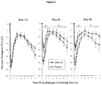
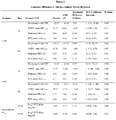
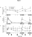
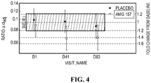

Classifications
-
- G—PHYSICS
- G01—MEASURING; TESTING
- G01N—INVESTIGATING OR ANALYSING MATERIALS BY DETERMINING THEIR CHEMICAL OR PHYSICAL PROPERTIES
- G01N33/00—Investigating or analysing materials by specific methods not covered by groups G01N1/00 - G01N31/00
- G01N33/48—Biological material, e.g. blood, urine; Haemocytometers
- G01N33/50—Chemical analysis of biological material, e.g. blood, urine; Testing involving biospecific ligand binding methods; Immunological testing
- G01N33/5005—Chemical analysis of biological material, e.g. blood, urine; Testing involving biospecific ligand binding methods; Immunological testing involving human or animal cells
- G01N33/5008—Chemical analysis of biological material, e.g. blood, urine; Testing involving biospecific ligand binding methods; Immunological testing involving human or animal cells for testing or evaluating the effect of chemical or biological compounds, e.g. drugs, cosmetics
- G01N33/5044—Chemical analysis of biological material, e.g. blood, urine; Testing involving biospecific ligand binding methods; Immunological testing involving human or animal cells for testing or evaluating the effect of chemical or biological compounds, e.g. drugs, cosmetics involving specific cell types
- G01N33/5047—Cells of the immune system
- G01N33/505—Cells of the immune system involving T-cells
-
- A—HUMAN NECESSITIES
- A61—MEDICAL OR VETERINARY SCIENCE; HYGIENE
- A61P—SPECIFIC THERAPEUTIC ACTIVITY OF CHEMICAL COMPOUNDS OR MEDICINAL PREPARATIONS
- A61P11/00—Drugs for disorders of the respiratory system
- A61P11/02—Nasal agents, e.g. decongestants
-
- A—HUMAN NECESSITIES
- A61—MEDICAL OR VETERINARY SCIENCE; HYGIENE
- A61P—SPECIFIC THERAPEUTIC ACTIVITY OF CHEMICAL COMPOUNDS OR MEDICINAL PREPARATIONS
- A61P11/00—Drugs for disorders of the respiratory system
- A61P11/06—Antiasthmatics
-
- A—HUMAN NECESSITIES
- A61—MEDICAL OR VETERINARY SCIENCE; HYGIENE
- A61P—SPECIFIC THERAPEUTIC ACTIVITY OF CHEMICAL COMPOUNDS OR MEDICINAL PREPARATIONS
- A61P17/00—Drugs for dermatological disorders
- A61P17/04—Antipruritics
-
- A—HUMAN NECESSITIES
- A61—MEDICAL OR VETERINARY SCIENCE; HYGIENE
- A61P—SPECIFIC THERAPEUTIC ACTIVITY OF CHEMICAL COMPOUNDS OR MEDICINAL PREPARATIONS
- A61P25/00—Drugs for disorders of the nervous system
-
- A—HUMAN NECESSITIES
- A61—MEDICAL OR VETERINARY SCIENCE; HYGIENE
- A61P—SPECIFIC THERAPEUTIC ACTIVITY OF CHEMICAL COMPOUNDS OR MEDICINAL PREPARATIONS
- A61P27/00—Drugs for disorders of the senses
- A61P27/02—Ophthalmic agents
-
- A—HUMAN NECESSITIES
- A61—MEDICAL OR VETERINARY SCIENCE; HYGIENE
- A61P—SPECIFIC THERAPEUTIC ACTIVITY OF CHEMICAL COMPOUNDS OR MEDICINAL PREPARATIONS
- A61P35/00—Antineoplastic agents
-
- G—PHYSICS
- G01—MEASURING; TESTING
- G01N—INVESTIGATING OR ANALYSING MATERIALS BY DETERMINING THEIR CHEMICAL OR PHYSICAL PROPERTIES
- G01N33/00—Investigating or analysing materials by specific methods not covered by groups G01N1/00 - G01N31/00
- G01N33/48—Biological material, e.g. blood, urine; Haemocytometers
- G01N33/50—Chemical analysis of biological material, e.g. blood, urine; Testing involving biospecific ligand binding methods; Immunological testing
- G01N33/53—Immunoassay; Biospecific binding assay; Materials therefor
- G01N33/531—Production of immunochemical test materials
- G01N33/532—Production of labelled immunochemicals
- G01N33/533—Production of labelled immunochemicals with fluorescent label
-
- G—PHYSICS
- G01—MEASURING; TESTING
- G01N—INVESTIGATING OR ANALYSING MATERIALS BY DETERMINING THEIR CHEMICAL OR PHYSICAL PROPERTIES
- G01N33/00—Investigating or analysing materials by specific methods not covered by groups G01N1/00 - G01N31/00
- G01N33/48—Biological material, e.g. blood, urine; Haemocytometers
- G01N33/50—Chemical analysis of biological material, e.g. blood, urine; Testing involving biospecific ligand binding methods; Immunological testing
- G01N33/58—Chemical analysis of biological material, e.g. blood, urine; Testing involving biospecific ligand binding methods; Immunological testing involving labelled substances
- G01N33/582—Chemical analysis of biological material, e.g. blood, urine; Testing involving biospecific ligand binding methods; Immunological testing involving labelled substances with fluorescent label
-
- A—HUMAN NECESSITIES
- A61—MEDICAL OR VETERINARY SCIENCE; HYGIENE
- A61K—PREPARATIONS FOR MEDICAL, DENTAL OR TOILETRY PURPOSES
- A61K39/00—Medicinal preparations containing antigens or antibodies
-
- G—PHYSICS
- G01—MEASURING; TESTING
- G01N—INVESTIGATING OR ANALYSING MATERIALS BY DETERMINING THEIR CHEMICAL OR PHYSICAL PROPERTIES
- G01N2333/00—Assays involving biological materials from specific organisms or of a specific nature
- G01N2333/435—Assays involving biological materials from specific organisms or of a specific nature from animals; from humans
- G01N2333/46—Assays involving biological materials from specific organisms or of a specific nature from animals; from humans from vertebrates
- G01N2333/47—Assays involving proteins of known structure or function as defined in the subgroups
- G01N2333/4701—Details
- G01N2333/4715—Cytokine-induced proteins
-
- G—PHYSICS
- G01—MEASURING; TESTING
- G01N—INVESTIGATING OR ANALYSING MATERIALS BY DETERMINING THEIR CHEMICAL OR PHYSICAL PROPERTIES
- G01N2333/00—Assays involving biological materials from specific organisms or of a specific nature
- G01N2333/435—Assays involving biological materials from specific organisms or of a specific nature from animals; from humans
- G01N2333/52—Assays involving cytokines
- G01N2333/525—Tumor necrosis factor [TNF]
-
- G—PHYSICS
- G01—MEASURING; TESTING
- G01N—INVESTIGATING OR ANALYSING MATERIALS BY DETERMINING THEIR CHEMICAL OR PHYSICAL PROPERTIES
- G01N2333/00—Assays involving biological materials from specific organisms or of a specific nature
- G01N2333/435—Assays involving biological materials from specific organisms or of a specific nature from animals; from humans
- G01N2333/52—Assays involving cytokines
- G01N2333/54—Interleukins [IL]
- G01N2333/5406—IL-4
-
- G—PHYSICS
- G01—MEASURING; TESTING
- G01N—INVESTIGATING OR ANALYSING MATERIALS BY DETERMINING THEIR CHEMICAL OR PHYSICAL PROPERTIES
- G01N2333/00—Assays involving biological materials from specific organisms or of a specific nature
- G01N2333/435—Assays involving biological materials from specific organisms or of a specific nature from animals; from humans
- G01N2333/52—Assays involving cytokines
- G01N2333/54—Interleukins [IL]
- G01N2333/5409—IL-5
-
- G—PHYSICS
- G01—MEASURING; TESTING
- G01N—INVESTIGATING OR ANALYSING MATERIALS BY DETERMINING THEIR CHEMICAL OR PHYSICAL PROPERTIES
- G01N2333/00—Assays involving biological materials from specific organisms or of a specific nature
- G01N2333/435—Assays involving biological materials from specific organisms or of a specific nature from animals; from humans
- G01N2333/52—Assays involving cytokines
- G01N2333/54—Interleukins [IL]
- G01N2333/5437—IL-13
-
- G—PHYSICS
- G01—MEASURING; TESTING
- G01N—INVESTIGATING OR ANALYSING MATERIALS BY DETERMINING THEIR CHEMICAL OR PHYSICAL PROPERTIES
- G01N2333/00—Assays involving biological materials from specific organisms or of a specific nature
- G01N2333/435—Assays involving biological materials from specific organisms or of a specific nature from animals; from humans
- G01N2333/52—Assays involving cytokines
- G01N2333/54—Interleukins [IL]
- G01N2333/55—IL-2
-
- G—PHYSICS
- G01—MEASURING; TESTING
- G01N—INVESTIGATING OR ANALYSING MATERIALS BY DETERMINING THEIR CHEMICAL OR PHYSICAL PROPERTIES
- G01N2333/00—Assays involving biological materials from specific organisms or of a specific nature
- G01N2333/435—Assays involving biological materials from specific organisms or of a specific nature from animals; from humans
- G01N2333/52—Assays involving cytokines
- G01N2333/555—Interferons [IFN]
- G01N2333/57—IFN-gamma
-
- G—PHYSICS
- G01—MEASURING; TESTING
- G01N—INVESTIGATING OR ANALYSING MATERIALS BY DETERMINING THEIR CHEMICAL OR PHYSICAL PROPERTIES
- G01N2800/00—Detection or diagnosis of diseases
- G01N2800/52—Predicting or monitoring the response to treatment, e.g. for selection of therapy based on assay results in personalised medicine; Prognosis
Definitions
- the present disclosure relates to a method for detecting T helper (Th) cell and polyfunctional cytotoxic T-cells (CTL) subpopulations in in a subject affected by disease or disorder having an immune component.
- the methods are also useful for determining efficacy of a treatment of the disease or disorder by detecting skewing of the T helper or CTL cells in a therapeutic or adverse direction.
- T-cell subsets include helper T-cells (CD4+) and cytotoxic T-cells (CD8+ CTL).
- CD4+ helper T cells can generally be broken down into several different subtypes. Commonly divided populations include T helper 1 (Th1) and T helper 2 (Th2) cells which have been found to play distinct roles in mediating immune-related diseases and disorders. Th1 and Th2 cells can be distinguished based on their cytokine expression profile.
- Th1 cells typically express interferon-gamma (IFN-g, IFN- ⁇ ), lymphotoxin, (LT), and can also secrete interleukin-2 (IL-2) and tumor necrosis factor-alpha (TNF-a, TNF- ⁇ ) ( Zhu et al., Annu Rev Immunol. 28: 445-489, 2010 ).
- Th2 cells are characterized by production of IL-4, IL-5 and IL-13, and do not secrete IFN- ⁇ or lymphotoxin. Some Th2 cells have been found to produce TNF- ⁇ and IL-9 (Zhu, supra ).
- Th17 cells have also recently been determined to be a different subset of T helper cells.
- Th17 cells secrete IL-17A, IL-17F, and IL-22 cytokines, and can also produce IL-21.
- the dysregulation of the ratio of Th1 to Th2 cells, as well as Th17 cells, has been associated with certain disease states, including autoimmune diseases, allergic reactions and cancers.
- Two additional CD4+ T-cell subsets that can be identified by cytokine production include Treg (IL-10) and follicular helper T cells (Tfh) (IL-21).
- Polyfunctional, CD8+ CTL are important effector cells that provide protection from intracellular pathogens and tumors where polyfunctionality is defined as production of multiple cytokines including (e.g. IFN-g, TNF-a and/or IL-2). In cancer subjects these polyfunctional T-cell responses have been shown to be repressed.
- cytokines e.g. IFN-g, TNF-a and/or IL-2.
- anti- CTLA4, anti-PD1 have been shown reverse functional suppression of CTL and combination with TVEC (talimogene laherparepvec) holds further promise for increasing the number of functional, activated CTL that can contribute to meaningful anti-tumor responses.
- Additional T cell subsets include natural killer T cells (NKT) and gamma delta T cells (gdT).
- T helper subtypes and cytokine levels include ELISA, ELISPOT, and flow cytometry assays, including intracellular cytokine staining assays.
- flow cytometry assays including intracellular cytokine staining assays.
- a common technique to identify and separate one cell type from another is flow cytometry using labeled dyes, e.g., in a fluorescence activated cells sorter (FACS).
- FACS fluorescence activated cells sorter
- combinations of cell surface markers can also be used to classify T-cells by subset (e.g. Th2 cells express CRTH2) or activation status (e.g. activated CTL express HLA-DR) by FACS.
- subset e.g. Th2 cells express CRTH2
- activation status e.g. activated CTL express HLA-DR
- U.S. Patent 6,039,969 describes animal model data demonstrating that a class of compounds including the drug imiquimod can skew the immune response away from Th2, based primarily on secreted cytokine results. Human clinical data and intracellular cytokine staining were not disclosed.
- WO 2000/024245 describes a method for targeting NFATp and/or NFAT4 to modulate Th2 cells.
- WO 2002/089832 relates to a combination of cytokine+SDF-la to skew the Thl/Th2 ratio.
- In vitro data, including intracellular cytokine staining specific for IFN-g and IL-4, using cultured cord blood T-cells are presented. Human clinical data demonstration is not included.
- Ludviksson et. al. J Immunol 160:3602-3609, 1998 ) describes association of Wegener's Granulomatosis with HLA-DR+CD4+ cells exhibiting unbalanced Th1 cell cytokine pattern and reversal with IL-10.
- Wang et. al. Int Imm 21:1065-1077, 2009 ) demonstrate that PD1 blockade reverses functional suppression of CTL in melanoma setting.
- Sfanos et. al. (Clin Cancer Res 14:3254-3261, 2008 ) demonstrate skewing of tumor infiltrating lymphocytes to Th17 and Treg subsets in prostate cancer.
- the present disclosure is directed to methods for identifying and differentiating a population of T helper or CTL cells in a subject having a disease or disorder with an immune component.
- the present technique can be carried out before and after treatment with a therapeutic agent to improve treatment regimens for patients receiving therapy.
- the present disclosure provides a method for measuring changes in T cell subset populations in a subject having a disease or disorder or subsequent to a treatment regimen.
- the method is useful to determine efficacy of a treatment regimen and assist in designing an optimal treatment regimen for a patient.
- the present disclosure provides a method for detecting a ratio of T helper 2 (Th2) and T helper 1 (Th1) cells in a sample from a patient comprising measuring levels of Th2-specific and Th1-specific cytokines, the method comprising a) contacting the sample with i) two or more specific binding agents that bind two or more Th2 cytokines, wherein the two or more specific binding agents for the Th2-specific cytokines are labeled with a first fluorophore; and ii) at least one specific binding agent that binds a Th1 cytokine, wherein the specific binding agent for the Th1 cytokine is labeled with a second fluorophore that is different from the first fluorophore; b) measuring levels of the first and second fluorophores in the sample and designating the cell as a Th1 or Th2 cell based on the level of Th1-specific and Th2-specific fluorophore detected; and c) determining the ratio of Th2 to Th1 cells in
- the method further comprises determining the ratio of Th1, Th2 and Th17 cells in the sample, comprising a) contacting the sample with iii) at least one specific binding agent that binds a Th17 cytokine, wherein the specific binding agent for the Th17 cytokine is labeled with a second fluorophore that is different from the first fluorophore or labeled with a third fluorophore; b) measuring levels of the first and second (and/or third) fluorophores in the sample and designating the cell as a Th1, Th2 or Th17 cell based on the level of Th1-specific, Th2-specific or Th17-specific fluorophore detected; and c) determining the ratio of Th1 to Th2 to Th17 cells in a sample.
- the disclosure provides a method for detecting a ratio of T helper 1 (Th1) and T helper 17 (Th17) cells in a sample from a patient comprising measuring levels of Th1-specific and Th17-specific cytokines, the method comprising: a) contacting the sample with i) two or more specific binding agents that bind two or more Th17 cytokines, wherein the two or more specific binding agents for the Th17-specific cytokines are labeled with a first fluorophore; and ii) at least one specific binding agent that binds a Th1 cytokine, wherein the specific binding agent for the Th1 cytokine is labeled with a second fluorophore that is different from the first fluorophore; b) measuring levels of the first and second fluorophores in the sample and designating the cell as a Th1 or Th17 cell based on the level of Th1-specific and Th17-specific fluorophore detected; and c) determining the ratio of
- the disclosure provides a method for detecting a ratio of T helper 2 (Th2) and T helper 17 (Th17) cells in a sample from a patient comprising measuring levels of Th2-specific and Th17-specific cytokines, the method comprising: a) contacting the sample with i) two or more specific binding agents that bind two or more Th2 cytokines, wherein the two or more specific binding agents for the Th2-specific cytokines are labeled with a first fluorophore; and ii) at least one specific binding agent that binds a Th17 cytokine, wherein the specific binding agent for the Th17 cytokine is labeled with a second fluorophore that is different from the first fluorophore; b) measuring levels of the first and second fluorophores in the sample and designating the cell as a Th2 or Th17 cell based on the level of Th2-specific and Th17-specific fluorophore detected; and c) determining the ratio of Th2 to
- the ratio of Th1 to Th2 cells is calculated using the present method, and this ratio used to determine treatment efficacy and potential change in treatment regimen for a patient.
- the converse ratio e.g., Th17/Th1, Th17/Th2 or any combination ratio for Th1, Th2 and Th17
- the converse ratio may be determined and a treatment decision made also based on the ratio of cells determined.
- the Th2 cytokines are selected from the group consisting of IL-4, IL-5 and IL-13.
- the Th1 cytokines are selected from the group consisting of interferon gamma (IFN-g), tumor necrosis factor alpha (TNF-a) and IL-2.
- the Th17 cytokines are selected from the group consisting of IL-17A, IL-17F, IFN-g and IL-22.
- the specific binding agent is an antibody specific for a Th1-specific, Th2-specific or Th17-specific cytokine.
- the method further comprises a step of administering a therapeutic agent, altering a dose regimen of a therapeutic agent or maintaining a dose regimen of a therapeutic agent in view of the ratio of T cell subsets detected by the method, wherein detection of a particular ratio indicates the treatment regimen to be administered.
- the patient is suffering from a disease or disorder selected from the group consisting of asthma, allergic rhinosinusitis, allergic conjunctivitis, atopic dermatitis, fibrotic disorders, Systemic Lupus Erythematosus (SLE), multiple sclerosis and cancers. Additional disorders contemplated in the present methods are discussed in greater detail in the Detailed Description.
- the sample is obtained before and/or after treatment with a therapeutic agent.
- the sample is whole blood, peripheral blood mononuclear cells, cerebrospinal fluid, bronchioalveolar lavage, nasal lavage, induced sputum or a biopsy from the patient.
- the therapeutic agent is an anti-TSLP antibody.
- the first fluorophore is phycoerythrin (PE) and the second fluorophore is fluorescein isothiocyanate (FITC).
- Additional fluorophores contemplated for use in the method include, but are not limited to, Alexa Fluor® 350, Alexa Fluor® 405, Alexa Fluor® 430, Alexa Fluor® 488, Alexa Fluor® 514, Alexa Fluor® 532, Alexa Fluor® 546, Alexa Fluor® 555, Alexa Fluor® 568, Alexa Fluor® 594, Alexa Fluor® 610, Alexa Fluor® 633, Alexa Fluor® 635, Alexa Fluor® 647, Alexa Fluor® 660, Alexa Fluor® 680, Alexa Fluor® 700, BODIPY FL, BODIPY 630/650, Cy2, Cy3, Cy3B, Cy3.5, Cy5, Cy5.5, Cy7, ECD, FITC, FluorX®, Cascade® Blue, Pacific Blue®
- the cytokines are contacted with the specific binding agent intracellularly. In certain embodiments, the cytokines are contacted with the specific binding agents extracellularly. In certain embodiments, the cytokines are secreted from the cells in vitro or in are detectable in fluid samples.
- the disclosure provides a method for identifying a sub-population of asthma patients responsive to treatment with a therapeutic agent comprising measuring the baseline ratio of T helper 2 (Th2) and T helper 1 (Th1) cells in a patient sample or changes in the ratio after administration of the therapeutic agent, the method comprising: a) contacting the sample with i) two or more specific binding agents that bind two or more Th2-specific cytokines, wherein the two or more specific binding agents for the Th2-specific cytokines are labeled with a first fluorophore; and ii) at least one specific binding agent that binds a Th1-specific cytokine, wherein the specific binding agent for the Th1-specific cytokine is labeled with a second fluorophore that is different from the first fluorophore; b) measuring levels of the first and second fluorophores in the sample and designating the cell as a Th1 or Th2 cell based on the level of Th2-specific and Th1-specific fluorophor
- the therapeutic agent is administered for one week, two weeks, three weeks, four weeks, six weeks, two months, three months or more prior to obtaining the sample. Additional dosing regimens contemplated by the method are discussed in greater detail in the Detailed Description.
- the therapeutic agent is an anti-TSLP antibody or anti-TSLP receptor antibody.
- the asthma patient has atopic asthma, including mild, moderate, or severe asthma.
- the Th2/Th1 ratio in an asthma patient is skewed toward the Th2 phenotype and this skewing could identify the patient as a candidate for treatment with a therapeutic agent that targets the Th2 pathway.
- the patient may be a candidate for Th2 targeted therapy if the ratio of Th2/Th1 cells is approximately 0.2 or higher.
- the asthma patient is identified as responsive to treatment if the ratio of Th2/Th1 cells decreases by 20%, 30%, 40%, 50%, 60% or more. It is contemplated that the degree of responsiveness is compared to a baseline value obtained before or at certain points during treatment. In one embodiment, a Th2/Th1 ratio at which the patient is identified as responsive to treatment is approximately 0.1 or below.
- the disclosure provides a method of altering the dose regimen of an anti-TSLP agent in treating an immune disorder comprising determining the ratio of Th2/Th1 and/or Th17 cells in a sample using the method described herein and altering the dose of anti-TSLP antibody if the ratio of Th2/Th1 and/or Th17 cells changes during treatment, wherein the dose of therapeutic is increased if the ratio of Th2/Th1 cells is stable or increases indicating skewing towards Th2 profile (i.e., increasing the proportion of Th2 cells and/or decreasing the proportion of Th1 cells); and wherein the dose of therapeutic is decreased if the ratio of Th2/Th1 cells decreases indicating reduction in Th2 profile (i.e., decreasing the proportion of Th2 cells and/or increasing the proportion of Th1 cells).
- the immune disorder treated by an anti-TSLP or anti-TSLP receptor agent is an atopic disease or disorder.
- atopic diseases and disorders include, but are not limited to, asthma, allergic rhinosinusitis, allergic conjunctivitis and atopic dermatitis.
- the immune disorder is a fibrotic disorder.
- contemplated herein is a method of altering the dose regimen of an asthma therapeutic comprising determining the ratio of Th2/Th1 and/or Th17 cells in a sample using a method described herein and altering the dose of asthma therapeutic if the ratio of Th2/Th1 and/or Th17 cells changes during treatment, wherein the dose of therapeutic is increased if the ratio of Th2/Th1 cells is stable or increases indicating skewing towards Th2 profile; and wherein the dose of therapeutic is decreased if the ratio of Th2/Th1 cells decreases indicating reduction in Th2 profile.
- the method further comprises administering a therapeutic agent that inhibits IL-17 activity or the IL-17 pathway.
- the present disclosure provides a method for detecting a subset of CTL, such as polyfunctional CTLs, the method comprising a) contacting the sample with one or more specific binding agents that bind one or more polyfunctional CTL cytokines, wherein the one or more specific binding agents for the polyfunctional CTL cytokines are labeled with a first fluorophore and/or second fluorophore and/or third fluorophore; b) measuring levels of the first and second and/or third fluorophores in the sample and designating the cell as a polyfunctional CTL cell based on the level of specific fluorophore detected; and c) determining the ratio of polyfunctional CTL to other T cells, e.g., Th, Treg, NKT or gdT, in a sample.
- T cells e.g., Th, Treg, NKT or gdT
- polyfunctional CTL cytokines are selected from the group consisting of IFN-g, IL-17, TNF-a and IL-2.
- cell surface markers are used for identification of T cell subpopulations.
- Exemplary cell surface markers include, but are not limited to, ST2, CRTH2, and CCR4 for Th2 cells, CXCR3 for Th1 cells, and/or CCR6 for Th17 cells.
- T cell subpopulations are also distinguished based on biomarkers indicative of activation state, including PD1, CTLA4, CD40L, ICOS, OX40, 41BB, TIM-3, GITR, HLA-DR and Ki67 of Th1, Th2, and Th17 cells.
- the invention includes, as an additional aspect, all embodiments of the invention narrower in scope in any way than the variations defined by specific paragraphs above.
- certain aspects of the invention that are described as a genus, and it should be understood that every member of a genus is, individually, an aspect of the invention.
- aspects described as a genus or selecting a member of a genus should be understood to embrace combinations of two or more members of the genus.
- Th2 cells are strongly implicated as contributors to establishment and progression of allergic asthma.
- the clinical trial setting understanding if blocking a therapeutic target leads to changes in the Th2 cells or the Th2/Th1 ratio may be a useful exploratory objective.
- the relative proportion of polarized helper T cells at baseline may help to match patients with optimal treatments.
- levels of Th subsets in particular Th1, Th17 and Treg
- functionality of CTL may be useful metrics for identifying and optimizing treatment regimens.
- the present disclosure describes an assay to measure the change in T helper cells and CTLs developed using broad stimulation with PMA and ionomycin followed by intracellular cytokine staining to analyze the Th cytokine responses.
- IL-4, IL-5, and IL-13 Upon comparing a combined method of Th2 cytokine staining using IL-4, IL-5, and IL-13 together with an alternate method staining each cytokine separately, a combined method to improve the detection of Th2 cells was developed.
- the term “about” or “approximately” means an acceptable error for a particular value as determined by one of ordinary skill in the art, which depends in part on how the value is measured or determined. In certain embodiments, the term “about” or “approximately” means within 1, 2, 3, or 4 standard deviations. In certain embodiments, the term “about” or “approximately” means within 30%, 25%, 20%, 15%, 10%, 9%, 8%, 7%, 6%, 5%, 4%, 3%, 2%, 1%, 0.5%, or 0.05% of a given value or range. Whenever the term “about” or “approximately” precedes the first numerical value in a series of two or more numerical values, it is understood that the term “about” or “approximately” applies to each one of the numerical values in that series.
- cytokine refers to one or more small (5-20 kD) proteins released by cells that have a specific effect on interactions and communications between cells or on the behavior of cells, such as immune cell proliferation and differentiation. Functions of cytokines in the immune system include, promoting influx of circulating leukocytes and lymphocytes into the site of immunological encounter; stimulating the development and proliferation of B cells, T cells, peripheral blood mononuclear cells (PBMCs) and other immune cells; and providing antimicrobial activity.
- PBMCs peripheral blood mononuclear cells
- Exemplary immune cytokines include but are not limited to, IL-1, IL-2, IL-3, IL-4, IL-5, IL-6, IL-7, IL-9, IL-10, IL-12, IL-13, IL-15, IL17A, IL-17F, IL-18, IL-21, IL-22, interferon (including IFN alpha, beta, and gamma), tumor necrosis factor (including TNF alpha, beta), transforming growth factor (including TGF alpha, beta), granulocyte colony stimulating factor (GCSF), granulocyte macrophage colony stimulating factor (GMCSF) and thymic stromal lymphopoietin (TSLP).
- interferon including IFN alpha, beta, and gamma
- tumor necrosis factor including TNF alpha, beta
- transforming growth factor including TGF alpha, beta
- GCSF granulocyte colony stimulating factor
- GMCSF granulocyte macrophage colony stimulating factor
- Th 1 cytokine or “Th1-specific cytokine” refers to cytokines that are expressed (intracellularly and/or secreted) by Th1 T cells, and include IFN-g, TNF-a, IL-12 and in some populations, IL-2.
- Th2 cytokine or “Th2-specific cytokine” refers to cytokines that are expressed (intracellularly and/or secreted) by Th2 T cells, including IL-4, IL-5, IL-13, IL-10, and, in certain populations IL-2.
- Th17 cytokine or “Th17-specific cytokine” refers to cytokines that are expressed (intracellularly and/or secreted) by Th17 T cells, including IL-17A, IL-17F, IL-22 and IL-21. Certain populations of Th17 cells express IFN-g and/or IL-2 in addition to the Th17 cytokines listed herein.
- a polyfunctional CTL cytokine includes IFN-g, TNF-a, IL-2 and IL-17.
- fluorophore refers to a small molecule dye or protein that accepts light energy at a given wavelength (excitation) and re-emits it at a longer wavelength (emission).
- fluorophores are attached to a specific binding agent, such as an antibody or enzyme substrate, and in certain embodiments detected by passing the labeled substance through a laser that causes excitation of the dye.
- fluorophores useful in the present methods include but are not limited to, Alexa Fluor® 350, Alexa Fluor® 405, Alexa Fluor® 430, Alexa Fluor® 488, Alexa Fluor® 514, Alexa Fluor® 532, Alexa Fluor® 546, Alexa Fluor® 555, Alexa Fluor® 568, Alexa Fluor® 594, Alexa Fluor® 610, Alexa Fluor® 633, Alexa Fluor® 635, Alexa Fluor® 647, Alexa Fluor® 660, Alexa Fluor® 680, Alexa Fluor® 700, BODIPY FL, BODIPY 630/650, Cy2, Cy3, Cy3B, Cy3.5, Cy5, Cy5.5, Cy7, ECD, FITC, FluorX®, Cascade® Blue, Pacific Blue®, Pacific Green®, Pacific Orange®, eFluor® 450, eFluor® 605NC, eFluor® 625NC, eFluor®
- first fluorophore and second fluorophore refer to fluorophores that are useful in the method of the disclosure and are used to designate use of fluorophores that emit at different wavelengths and do not overlap in the method.
- a “first fluorophore” refers to one or more fluorophores that are used to label one or more specific binding agents in the method and emit light at a first wavelength, and includes use of two different fluorophores that have overlapping spectral emission profiles within a first wavelength range.
- a “second fluorophore” refers to one or more fluorophore used in the method in which the wavelength emission of the fluorophore(s) does not overlap with the emission wavelength of the first fluorophore(s).
- fluorophores with overlapping spectral profiles include FITC and Alexa Fluor 488; APC, eFluor® 660 and Alexa Fluor® 647; PE and PE-Cy5; Alexa Fluor® 555 and Cy3; Alexa Fluor® 647 and Cy5; and others determined to be in overlapping emissions spectra.
- fluorophores with overlapping spectral profiles include FITC and Alexa Fluor 488; APC, eFluor® 660 and Alexa Fluor® 647; PE and PE-Cy5; Alexa Fluor® 555 and Cy3; Alexa Fluor® 647 and Cy5; and others determined to be in overlapping emissions spectra.
- One of skill can determine whether fluorophores have overlapping spectra using commonly available resources that describe the emission spectra of fluorophores.
- the term “specifically binds” is “antigen specific”, is “specific for”, “selective binding agent”, “specific binding agent”, “antigen target” or is “immunoreactive” with an antigen refers to an antibody or polypeptide that binds an target antigen with greater affinity than other antigens of similar sequence. It is contemplated herein that the agent specifically binds target proteins useful in identifying immune cell types, for example, a surface antigen (e.g., T cell receptor, CD3), a cytokine (e.g., TSLP, IL-4, IL-5, IL-13, IL-17, IFN-g, TNF-a) and the like.
- a surface antigen e.g., T cell receptor, CD3
- a cytokine e.g., TSLP, IL-4, IL-5, IL-13, IL-17, IFN-g, TNF-a
- antibody is used in the broadest sense and includes fully assembled antibodies, tetrameric antibodies, monoclonal antibodies, polyclonal antibodies, multispecific antibodies (e.g., bispecific antibodies), antibody fragments that can bind an antigen (e.g., Fab', F'(ab)2, Fv, single chain antibodies, diabodies), and recombinant peptides comprising the forgoing as long as they exhibit the desired biological activity.
- An "immunoglobulin” or “tetrameric antibody” is a tetrameric glycoprotein that consists of two heavy chains and two light chains, each comprising a variable region and a constant region.
- Antigen-binding portions may be produced by recombinant DNA techniques or by enzymatic or chemical cleavage of intact antibodies.
- Antibody fragments or antigen-binding portions include, inter alia, Fab, Fab', F(ab')2, Fv, domain antibody (dAb), complementarity determining region (CDR) fragments, CDR-grafted antibodies, single-chain antibodies (scFv), single chain antibody fragments, chimeric antibodies, diabodies, triabodies, tetrabodies, minibody, linear antibody; chelating recombinant antibody, a tribody or bibody, an intrabody, a nanobody, a small modular immunopharmaceutical (SMIP), an antigen-binding-domain immunoglobulin fusion protein, a camelized antibody, a VHH containing antibody, or a variant or a derivative thereof, and polypeptides that contain at least a portion of an immunoglobulin that is sufficient to confer specific antigen binding to the polypeptide, such as one
- “Monoclonal antibody” refers to an antibody obtained from a population of substantially homogeneous antibodies, i.e., the individual antibodies comprising the population are identical except for possible naturally occurring mutations that may be present in minor amounts.
- sample refers to a specimen obtained from a subject for use in the present methods, and includes urine, whole blood, plasma, serum, saliva, tissue biopsies, cerebrospinal fluid, peripheral blood mononuclear cells with in vitro stimulation, peripheral blood mononuclear cells without in vitro stimulation, gut lymphoid tissues with in vitro stimulation, gut lymphoid tissues without in vitro stimulation, gut lavage, bronchioalveolar lavage, nasal lavage, and induced sputum.
- fibroproliferative disease or “fibrotic disease or disorder” refers to conditions involving fibrosis in one or more tissues.
- fibrosis refers to the formation of fibrous tissue as a reparative or reactive process, rather than as a normal constituent of an organ or tissue.
- Fibrotic disorders include, but are not limited to, systemic and local scleroderma, keloids and hypertrophic scars, atherosclerosis, restenosis, pulmonary inflammation and fibrosis, interstitial lung disease, idiopathic pulmonary fibrosis, liver cirrhosis, fibrosis as a result of chronic hepatitis B or C infection, radiation-induced fibrosis, fibrosis arising from wound healing, kidney disease, heart disease resulting from scar tissue, and eye diseases such as macular degeneration, and retinal and vitreal retinopathy. Additional fibrotic diseases include fibrosis resulting from chemotherapeutic drugs, radiation-induced fibrosis, and injuries and burns.
- treat refers to eliminating, reducing, suppressing or ameliorating, either temporarily or permanently, either partially or completely, a clinical symptom, manifestation or progression of an event, disease or condition associated with an inflammatory disorder described herein.
- drugs employed as therapeutic agents may reduce the severity of a given disease state, but need not abolish every manifestation of the disease to be regarded as useful therapeutic agents.
- a prophylactically administered treatment need not be completely effective in preventing the onset of a condition in order to constitute a viable prophylactic agent.
- One embodiment of the invention is directed to a method for determining the efficacy of treatment comprising administering to a patient therapeutic agent in an amount and for a time sufficient to induce a sustained improvement over baseline of an indicator that reflects the severity of the particular disorder.
- terapéuticaally effective amount refers to an amount of therapeutic agent that is effective to ameliorate or lessen symptoms or signs of disease associated with a disease or disorder.
- Specific binding agents such as antibodies and antibody fragments that bind to their target antigen, e.g., a surface molecule, a cytokine and the like, are useful in the method of the invention.
- the specific binding agent is an antibody.
- the antibodies may be polyclonal including monospecific polyclonal; monoclonal (MAbs); recombinant; chimeric; humanized, such as complementarity-determining region (CDR)-grafted; human; single chain; and/or bispecific; as well as fragments; variants; or derivatives thereof.
- Antibody fragments include those portions of the antibody that bind to an epitope on the polypeptide of interest.
- fragments include Fab and F(ab') fragments generated by enzymatic cleavage of full-length antibodies.
- Other binding fragments include those generated by recombinant DNA techniques, such as the expression of recombinant plasmids containing nucleic acid sequences encoding antibody variable regions.
- Monoclonal antibodies may be modified for use as therapeutics or diagnostics.
- One embodiment is a "chimeric" antibody in which a portion of the heavy (H) and/or light (L) chain is identical with or homologous to a corresponding sequence in antibodies derived from a particular species or belonging to a particular antibody class or subclass, while the remainder of the chain(s) is/are identical with or homologous to a corresponding sequence in antibodies derived from another species or belonging to another antibody class or subclass.
- fragments of such antibodies so long as they exhibit the desired biological activity. See U.S. Pat. No. 4,816,567 ; Morrison et al., 1985, Proc. Natl. Acad. Sci. 81:6851-55 .
- a monoclonal antibody is a "humanized" antibody.
- Methods for humanizing non-human antibodies are well known in the art. See U.S. Pat. Nos. 5,585,089 and 5,693,762 .
- a humanized antibody has one or more amino acid residues introduced into it from a source that is non-human.
- Humanization can be performed, for example, using methods described in the art ( Jones et al., 1986, Nature 321:522-25 ; Riechmann et al., 1998, Nature 332:323-27 ; Verhoeyen et al., 1988, Science 239:1534-36 ), by substituting at least a portion of a rodent complementarity-determining region for the corresponding regions of a human antibody.
- human antibodies that bind antigens of interest.
- transgenic animals e.g., mice
- a polypeptide antigen i.e., having at least 6 contiguous amino acids
- a carrier i.e., having at least 6 contiguous amino acids
- Human antibodies can also be produced from phage-display libraries ( Hoogenboom et al., 1991, J. Mol. Biol. 227:381 ; Marks et al., 1991, J. Mol. Biol. 222:581 ). These processes mimic immune selection through the display of antibody repertoires on the surface of filamentous bacteriophage, and subsequent selection of phage by their binding to an antigen of choice.
- phage-display libraries Hoogenboom et al., 1991, J. Mol. Biol. 227:381 ; Marks et al., 1991, J. Mol. Biol. 222:581 .
- PCT App. No. PCT/US98/17364 describes the isolation of high affinity and functional agonistic antibodies for MPL- and msk-receptors using such an approach.
- Chimeric, CDR grafted, and humanized antibodies are typically produced by recombinant methods. Nucleic acids encoding the antibodies are introduced into host cells and expressed using materials and procedures described herein. In a preferred embodiment, the antibodies are produced in mammalian host cells, such as CHO cells. Monoclonal (e.g., human) antibodies may be produced by the expression of recombinant DNA in host cells or by expression in hybridoma cells as described herein.
- binding agents that bind cytokines useful for detecting T cell populations are contemplated herein, including but not limited to, binding agents specific for IL-4, IL-5, IL-13, IFN-g, TNF-a, IL-2, IL-17A, IL-17F, IL-22, and IL-10.
- T cell subpopulations include, but are not limited to, ST2, CRTH2, and CCR4 for Th2 cells, CXCR3 for Th1 cells, and/or CCR6 for Th17 cells.
- T cell subpopulations can also be distinguished based on biomarkers indicative of activation state, including PD1, CTLA4, CD40L, ICOS, OX40, 41BB, TIM-3, GITR, HLA-DR and Ki67 of Th1, Th2, and Th17 cells.
- therapeutic agents include antibodies that antagonize cytokines or bind receptors that alter T helper cell or CTL ratios in a patient.
- examples include, but are not limited to, antibodies to TSLP, TSLP receptor, IL-25, IL-17A, IL-17-B, IL-17C, IL-17D, IL-17E (aka IL-25), IL-17-F, IL-17RA, IL-17-RB, IL-17RC, IL-17RD, IL-17RE, IL-33, ST2, IL-4, IL-13, IL4/13R, IL-5, IL-5R, TNF, TNF-R, IL-6, IL-6R, IL-10, IL-10R, IL-12p35, IL-12p40, IL-18, IL-18R, IL-22, IL-23/p19, TGF-b, IL-2, IFN-g, IFN-a, IL-1, IL-1R,
- anti-TSLP antibodies examples include, but are not limited to, those described in US Pat. No. 7,982,016 , US Publ. Pat. Appl. No. 20090186022 , and US Pat. No. 8,232,372 .
- anti-TSLP receptor antibodies examples include, but are not limited to, those described in US Pat No. 8,101,182 .
- the anti-TSLP antibody is the antibody designated as A5 within US Pat. No. 7,982,016 .
- anti-IL-17RA antibodies examples include, but are not limited to, those described in US Pat. No. 7,767,206 , US Pat. No. 7,786,234 , US Pat. No. 7,939,070 , US Pat. No. 7,833,527 , 8,435,518 , US Pat. No.8,545,842 , and US Publ. Pat. Appl. No. 20 130022621 .
- anti-IL-17RA antibody brodalumab as described in Recommended International Nonproprietary Names: List 67 (WHO Drug Information, Vol. 26, No. 1, 2012 ; World Health Organization).
- anti-IL-17A antibodies examples include, but are not limited to, perakizumab as described in Recommended International Nonproprietary Names: List 69 (WHO Drug Information Vol. 27, No. 1, 2013 ; World Health Organization), secukinumab as described in Proposed International Nonproprietary Names: List 102 (WHO Drug Information, Vol. 23, No. 4, 2009 ; World Health Organization), ixekizumab as described in Proposed International Nonproprietary Names: List 105 (WHO Drug Information, Vol. 25, No. 2, 2011 ; World Health Organization), and tildakizumab as described in Recommended International Nonproprietary Names: List 70 (WHO Drug Information, Vol. 27, No. 3, 2013 ; World Health Organization).
- anti-ST2 antibodies examples include, but are not limited to, those described in WO2013173761 . Particularly preferred are antibodies described as Ab1, Ab2, Ab3, Ab4, Ab5, Ab6, Ab7, Ab8, Ab9, Ab10, Ab11, Ab30, Ab32, and Ab33 therein.
- Exemplary antibodies include, but are not limited to, those described in U.S. Patent No.7,947,809 and U.S. Patent Application Publication No. 20090041784 (glucagon receptor), U.S. Patent No. 7939070 , U.S. Patent No. 7,833,527 , U.S. Patent No. 7,767,206 , and U.S. Patent No. 7,786,284 (IL-17 receptor A), U.S. Patent No. 7,872,106 and U.S. Patent No. 7,592,429 (Sclerostin), U.S. Patent No. 7,871,611 , U.S.
- Patent No. 7,815,907 U.S. Patent No. 7,037,498 , U.S. Patent No. 7,700,742 , and U.S. Patent Application Publication No. 20100255538 (IGF-1 receptor), U.S. Patent No. 7,868,140 (B7RP1), U.S. Patent No. 7,807,159 and U.S. Patent Application Publication No. 20110091455 (myostatin), U.S. Patent No. 7,736,644 , U.S. Patent No. 7,628,986 , U.S. Patent No. 7,524,496 , and U.S. Patent Application Publication No. 20100111979 (deletion mutants of epidermal growth factor receptor), U.S. Patent No.
- 20110045537 (interferon-gamma), U.S. Patent No. 7,932,372 (MAdCAM), U.S. Patent No. 7,906,625 , U.S. Patent Application Publication No. 20080292639 , and U.S. Patent Application Publication No. 20110044986 (amyloid), U.S. Patent No. 7,815,907 and U.S. Patent No. 7700742 (insulin-like growth factor I), U.S. Patent No. 7,566,772 and U.S. Patent No. 7964193 (interleukin-1 ⁇ ), U.S. Patent No. 7,563,442 , U.S. Patent No. 7,288,251 , U.S. Patent No.
- 20090226447 angiopoietin-1 and angiopoietin-2
- U.S. Patent Application Publication No. 20090191212 Angiopoietin-2
- U.S. Patent Application Publication No. 20090155164 C-FMS
- U.S. Patent No. 7,537,762 activin receptor-like kinase-1
- U.S. Patent No. 7,371,381 galanin
- U.S. Patent Application Publication No. 20070196376 INSULIN-LIKE GROWTH FACTORS
- U.S. Patent No. 7,267,960 and U.S. Patent No. 7,741,115 LDCAM
- US7265212 CD45RB
- Patent Application Publication No. 20110150888 (HEPCIDIN), U.S. Patent No. 7,939,640 (B7L-1), U.S. Patent No. 7,915,391 (c-Kit), U.S. Patent No. 7,807,796 , U.S. Patent No. 7,193,058 , and U.S. Patent No. 7,427,669 (ULBP), U.S. Patent No. 7,786,271 , U.S. Patent No. 7,304,144 , and U.S. Patent Application Publication No. 20090238823 (TSLP), U.S. Patent No. 7,767,793 (SIGIRR), U.S. Patent No. 7,705,130 (HER-3), U.S. Patent No.
- the method of the invention is useful to detect T cell subsets, such as Th1, Th2 and Th17 cells, in subjects having a broad range of disease or disorders that have an immune component, i.e., involvement of immune cells in development or progression of the disease or disorder.
- Asthma is a chronic inflammatory disorder of the airways. Each year, asthma accounts for an estimated 1.1 million outpatient visits, 1.6 million emergency room visits, 444,000 hospitalizations (Defrances et al, 2008) Available at: http://www.cdc.gov/nchs/data/nhsr/nhsr005.pdf , and 3,500 deaths in the U.S. In susceptible individuals, asthmatic inflammation causes recurrent episodes of wheezing, breathlessness, chest tightness, and cough. The etiology of asthma is thought to be multi-factorial, influenced by both genetic environmental mechanisms, 1,2 with environmental allergens an important cause. 2,3 The majority of cases arise when a person becomes hypersensitive to allergens (atopy).
- Atopy is characterized by an increase in Th2 cells and Thh2 cytokine expression and IgE production. Approximately 10 million patients in the United States are thought to have allergy-induced asthma. Despite the available therapeutic options, asthma continues to be a major health problem. Worldwide, asthma currently affects approximately 300 million people; by 2020, asthma is expected to affect 400 million people ( Partridge, Eur Resp Rev. 16:67-72, 2007 ).
- Allergen inhalation by atopic asthmatics induces some of the manifestations of asthma, including reversible airflow obstruction, airway hyperresponsiveness, and eosinophilic and basophilic airway inflammation. Allergen inhalation challenge has become the predominant model of asthma in many species ( Bates et al., Am J Physiol Lung Cell Mol Physiol. 297(3):L401-10, 2009 ; Diamant et al., J Allergy Clin Immunol. 132(5):1045-1055, 2013 .)
- Eosinophils are important inflammatory cells in allergic asthma that is characteristically mediated by Th2-type CD4+ T cells.
- Neutrophilic airway inflammation is associated with corticosteroid treatment in severe asthma and can be mediated by Th1- or Th17-type T cells ( Mishra et al. Dis. Model. Mech. 6:877-888, 2013 ).
- Thymic stromal lymphopoietin is an epithelial cell-derived cytokine that is produced in response to pro-inflammatory stimuli and drives allergic inflammatory responses primarily through its activity on dendritic cells ( Gilliet, J Exp Med. 197:1059-1067, 2003 ; Soumelis, Nat Immunol. 3:673-680, 2002 ; Reche, J Immunol. 167:336-3432001 ), mast cells ( Allakhverdi, J Exp Med. 204:253-258, 2007 ) and CD34+ progenitor cells.
- TSLP Thymic stromal lymphopoietin
- TSLP 9 TSLP signals through a heterodimeric receptor consisting of the interleukin (IL)-7 receptor alpha (IL-7R ⁇ ) chain and a common ⁇ chain-like receptor (TSLPR) ( Pandey, Nat Immunol. 1:59-64, 2000 ; Park, J Exp Med. 192:659-669, 2000 ).
- IL-7R ⁇ interleukin-7 receptor alpha
- TSLPR common ⁇ chain-like receptor
- TSLP mRNA 10,11 and protein levels 11 are increased in the airways of asthmatic individuals compared to controls, and the magnitude of this expression correlates with disease severity. 10 Recent studies have demonstrated association of a single nucleotide polymorphism in the human TSLP locus with protection from asthma, atopic asthma and airway hyperresponsiveness, suggesting that differential regulation of TSLP gene expression might influence disease susceptibility. 1,12,13 These data suggest that targeting TSLP may inhibit multiple biological pathways involved in asthma.
- Fibrotic disorders Fibrosis is characterized by fibroblast accumulation and collagen deposition in excess of normal deposition in any particular tissue. Fibroblasts are connective tissue cells, which are dispersed in connective tissue throughout the body. Fibroblasts secrete a nonrigid extracellular matrix containing type I and/or type III collagen. In response to an injury to a tissue, nearby fibroblasts migrate into the wound, proliferate, and produce large amounts of collagenous extracellular matrix. Collagen is a fibrous protein rich in glycine and proline that is a major component of the extracellular matrix and connective tissue, cartilage, and bone.
- Collagen molecules are triple-stranded helical structures called .alpha.-chains, which are wound around each other in a ropelike helix. Collagen exists in several forms or types; of these, type I, the most common, is found in skin, tendon, and bone; and type III is found in skin, blood vessels, and internal organs.
- Fibrotic disorders include, but are not limited to, systemic and local scleroderma, keloids and hypertrophic scars, atherosclerosis, restenosis, pulmonary inflammation and fibrosis, interstitial lung disease, idiopathic pulmonary fibrosis, liver cirrhosis, fibrosis as a result of chronic hepatitis B or C infection, radiation-induced fibrosis, fibrosis arising from wound healing, kidney disease, heart disease resulting from scar tissue, and eye diseases such as macular degeneration, and retinal and vitreal retinopathy. Additional fibrotic diseases include fibrosis resulting from chemotherapeutic drugs, radiation-induced fibrosis, and injuries and burns.
- Scleroderma is a fibrotic disorder characterized by a thickening and induration of the skin caused by the overproduction of new collagen by fibroblasts in skin and other organs. Scleroderma may occur as a local or systemic disease. Systemic scleroderma may affect a number of organs. Systemic sclerosis is characterized by formation of hyalinized and thickened collagenous fibrous tissue, with thickening of the skin and adhesion to underlying tissues, especially of the hands and face.
- the disease may also be characterized by dysphagia due to loss of peristalsis and submucosal fibrosis of the esophagus, dyspnea due to pulmonary fibrosis, myocardial fibrosis, and renal vascular changes.
- Pulmonary fibrosis affects 30 to 70% of scleroderma patients, often resulting in restrictive lung disease ( Atamas et al. Cytokine and Growth Factor Rev 14: 537-550 (2003 )).
- Idiopathic pulmonary fibrosis is a chronic, progressive and usually lethal lung disorder, thought to be a consequence of a chronic inflammatory process ( Kelly et al., Curr Pharma Design 9: 39-49 (2003 )).
- SLE Systemic Lupus Erythematosus
- SLE is an autoimmune disease caused by recurrent injuries to blood vessels in multiple organs, including the kidney, skin, and joints. SLE is estimated to affect over 500,000 people in the United States. In patients with SLE, a faulty interaction between T cells and B cells results in the production of autoantibodies that attack the cell nucleus. These include anti-double stranded DNA and anti-Sm antibodies. Autoantibodies that bind phospholipids are also found in about half of SLE patients, and are responsible for blood vessel damage and low blood counts. Immune complexes accumulate the kidneys, blood vessels, and joints of SLE patients, where they cause inflammation and tissue damage. It has been hypothesized that Th2 cells in contribute to overproduction of autoantibodies.
- Lupus also includes subacute cutaneous lupus (SCLE).
- SCLE subacute cutaneous lupus
- Other disorders in which autoantibodies play a role include, Sjogren's syndrome and Immune thrombocytopenic purpura (ITP).
- Th1-like cells are typically involved in promoting cell-mediated immunity, initiating a cytotoxic response and generally are considered one of the host's important anti-cancer mechanism. It has been shown that the Th1 cytokine IFN-g can promote anti-tumor activity in vivo ( Ikeda et al., Cytokine Growth Factor Rev. 13:95-109, 2002 ). Ito et al.
- Exemplary cancers contemplated herein include, but are not limited to, adrenocortical carcinoma, AIDS-related cancers, AIDS-related lymphoma, anal cancer, anorectal cancer, cancer of the anal canal, appendix cancer, childhood cerebellar astrocytoma, childhood cerebral astrocytoma, basal cell carcinoma, skin cancer (non-melanoma), biliary cancer, extrahepatic bile duct cancer, intrahepatic bile duct cancer, bladder cancer, urinary bladder cancer, bone and joint cancer, osteosarcoma and malignant fibrous histiocytoma, brain cancer, brain tumor, brain stem glioma, cerebellar astrocytoma, cerebral astrocytoma/malignant glioma, ependymoma, medulloblastoma, supratentorial primitive neuroectodeimal tumors, visual pathway and hypothalamic glioma, breast cancer, bronchial
- the tumor is associated with a cancer selected from the group consisting of breast cancer, melanoma, prostate cancer, pancreatic cancer, head and neck cancer, lung cancer, non small-cell lung carcinoma, renal cancer, colorectal cancer, colon cancer, ovarian cancer, liver cancer and gastric cancer.
- the cancer is pancreatic cancer.
- Additional diseases, disorders, or conditions contemplated by the method herein include, but are not limited to, inflammation, autoimmune disease, cartilage inflammation, fibrotic disease and/or bone degradation, arthritis, rheumatoid arthritis, juvenile arthritis, juvenile rheumatoid arthritis, pauciarticular juvenile rheumatoid arthritis, polyarticular juvenile rheumatoid arthritis, systemic onset juvenile rheumatoid arthritis, juvenile ankylosing spondylitis, juvenile enteropathic arthritis, juvenile reactive arthritis, juvenile Reiter's Syndrome, SEA Syndrome (Seronegativity, Enthesopathy, Arthropathy Syndrome), juvenile dermatomyositis, juvenile psoriatic arthritis, juvenile scleroderma, juvenile systemic lupus erythematosus, juvenile vasculitis, pauciarticular rheumatoid arthritis, polyarticular rheumatoid arthritis, systemic onset rheumatoid arthritis, ankylosing
- the antibody substance is labeled to facilitate its detection.
- a “label”, “detectable label” or a “detectable moiety” is a composition detectable by spectroscopic, photochemical, biochemical, immunochemical, chemical, or other physical means.
- labels suitable for use in the present disclosure include, radioactive labels (e.g., 32P), fluorophores (e.g., fluorescein), electron dense reagents, enzymes (e.g., as commonly used in an ELISA), biotin, digoxigenin, or haptens as well as proteins which can be made detectable, e.g., by incorporating a radiolabel into the hapten or peptide, or used to detect antibodies specifically reactive with the hapten or peptide.
- radioactive labels e.g., 32P
- fluorophores e.g., fluorescein
- electron dense reagents e.g., enzymes (e.g., as commonly used in an ELISA)
- enzymes e.g., as commonly used in an ELISA
- biotin e.g., as commonly used in an ELISA
- haptens as well as proteins which can be made detectable, e.g., by incorporating a radiol
- labels include, but are not limited to, fluorescent dyes described herein, radiolabels (e.g., 3 H, 125 I, 35 S, 14 C, or 32 P), enzymes (e.g., horse radish peroxidase, alkaline phosphatase and others commonly used in an ELISA), and colorimetric labels such as colloidal gold, colored glass or plastic beads (e.g., polystyrene, polypropylene, latex, etc.).
- fluorescent dyes described herein e.g., radiolabels (e.g., 3 H, 125 I, 35 S, 14 C, or 32 P)
- enzymes e.g., horse radish peroxidase, alkaline phosphatase and others commonly used in an ELISA
- colorimetric labels such as colloidal gold, colored glass or plastic beads (e.g., polystyrene, polypropylene, latex, etc.).
- the label may be coupled directly or indirectly to the desired component of the assay according to methods well known in the art.
- the label in one embodiment is covalently bound to the biopolymer using an isocyanate reagent for conjugation of an active agent according to the disclosure.
- the bifunctional isocyanate reagents of the disclosure can be used to conjugate a label to a biopolymer to form a label biopolymer conjugate without an active agent attached thereto.
- the label biopolymer conjugate may be used as an intermediate for the synthesis of a labeled conjugate according to the disclosure or may be used to detect the biopolymer conjugate.
- Non-radioactive labels are often attached by indirect means.
- a ligand molecule e.g., biotin
- the ligand then binds to another molecules (e.g., streptavidin) molecule, which is either inherently detectable or covalently bound to a signal system, such as a detectable enzyme, a fluorescent compound, or a chemiluminescent compound.
- the compounds of the present disclosure can also be conjugated directly to signal-generating compounds, e.g., by conjugation with an enzyme or fluorophore.
- Enzymes suitable for use as labels include, but are not limited to, hydrolases, particularly phosphatases, esterases and glycosidases, or oxidotases, particularly peroxidases.
- Fluorescent compounds, i.e., fluorophores, suitable for use as labels include, but are not limited to, fluorescein and its derivatives, rhodamine and its derivatives, dansyl, umbelliferone, etc.
- fluorophores include, but are not limited to, eosin, TRITC-amine, quinine, fluorescein W, acridine yellow, lissamine rhodamine, B sulfonyl chloride erythroscein, ruthenium (tris, bipyridinium), Texas Red, nicotinamide adenine dinucleotide, flavin adenine dinucleotide, etc.
- Chemiluminescent compounds suitable for use as labels include, but are not limited to, luciferin and 2,3-dihydrophthalazinediones, e.g., luminol.
- the present disclosure provides a whole blood stimulation method that allows measurement of cell-signaling changes in T cell populations, such as Th1/Th2/Th17/Th22 cell and/or CTL subsets, in the context of therapy with a therapeutic agent.
- the disclosure demonstrates skewing away from a Th2 profile in asthmatics treated with anti-TSLP neutralizing antibody.
- determining the cytokine expression profile at baseline is useful to characterize a sub-population of patients receiving treatment and adjust levels of therapeutic administration for patients.
- This assay is useful to evaluate T cell subsets in clinical trials in multiple inflammatory diseases. This specific application has not previously been demonstrated in human clinical trials.
- the methods herein provide for detecting a ratio of Th2 and Th1 cells in a sample from a patient comprising measuring levels of Th2-specific and Th1-specific cytokines, the method comprising a) contacting the sample with i) two or more specific binding agents that bind two or more Th2 cytokines, wherein the two or more specific binding agents for the Th2-specific cytokines are labeled with a first fluorophore; and ii) at least one specific binding agent that binds a Th1 cytokine, wherein the specific binding agent for the Th1 cytokine is labeled with a second fluorophore that is different from the first fluorophore; b) measuring levels of the first and second fluorophores in the sample and designating the cell as a Th1 or Th2 cell based on the level of Th1-specific and Th2-specific fluorophore detected; and c) determining the ratio of Th2 to Th1 cells in a sample.
- the method is optionally carried out
- the method is also adapted to determine the ratio of Th1/Th17 cells and Th2/Th17 cells. It is also contemplated that the converse ratio, i.e., reverse of the numerator and denominator for a stated ratio herein, is calculated using the method above.
- the Th2 cytokines are selected from the group consisting of IL-4, IL-5 and IL-13.
- the Th1 cytokines are selected from the group consisting of interferon gamma (IFN-g), tumor necrosis factor alpha (TNF-a) and IL-2.
- the Th17 cytokines are selected from the group consisting of IL-17A, IL-17F, IFN-g and IL-22.
- the specific binding agent is an antibody specific for a Th1-specific, Th2-specific or Th17-specific cytokine.
- the present disclosure provides a method for detecting a subset of CTL, such as polyfunctional CTLs, the method comprising a) contacting the sample with one or more specific binding agents that bind one or more polyfunctional CTL cytokines, wherein the one or more specific binding agents for the polyfunctional CTL cytokines are labeled with a first fluorophore and/or second fluorophore and/or third fluorophore; b) measuring levels of the first and second and/or third fluorophores in the sample and designating the cell as a polyfunctional CTL cell based on the level of specific fluorophore detected; and c) determining the ratio of polyfunctional CTL to other T cells, e.g., Th, Treg, NKT or gdT, in a sample.
- T cells e.g., Th, Treg, NKT or gdT
- polyfunctional CTL cytokines are selected from the group consisting of IFN-g, IL-17, TNF-a and IL-2.
- the cytokines are contacted intracellularly. Contacting intracellulary is carried out following protocols as described in the art for intracellular staining for flow cytometry (See e.g., Warrington et al., Arthritis Res Ther. 8(4):R136, 2006 ; Ito et al., Anticancer Research 25:2027-2031, 2005 ). Briefly, cells are obtained from a sample and fixed and permeabilized, e.g., using a fixation permeabilization buffer. The cells are then contacting the cells with a specific binding agent that binds a target antigen such as a cytokine. The cells are optionally stimulated in vitro using PMA/ionomycin or another stimulant prior to fixation as described in the Examples.
- the cytokines are contacted extracellularly.
- the cytokines are secreted from the cells in vitro or in are detectable in fluid samples.
- the sample fluid can then be contacted with specific binding agents that bind a cytokine of interest and the level of cytokine determined, wherein the level of cytokine is predictive of the skewing to the particular Th1, Th2 or Th17 phenotype or CTL population in the subject sample. If cells are stimulated in vitro, cytokines excreted into the fluid could be measured.
- the sample is obtained at various times before and after administration of a therapeutic agent. Obtaining a sample concurrent with administration of a therapeutic agent does not require that the agent be administered at the same time as obtaining the sample, as long as there is an overlap in the time period during which the agents are exerting their therapeutic effect. Simultaneous or sequential timing is contemplated.
- the relative number (percentage) of Th1, Th2, T17 cells or CTLs is compared at various times before and after administration of a therapeutic agent.
- administration of the therapeutic agent causes a decrease in the relative number of Th2 cells present in samples collected from a subject.
- the agent is administered concurrently with obtaining a sample for analysis, with concurrently referring to agents given within 30 minutes before or after obtaining the sample.
- the therapeutic agent is administered prior to obtaining the sample.
- Prior administration refers to administration of the agent within the range of one week prior to obtaining the sample, up to 30 minutes before obtaining the sample. It is further contemplated that the agent is administered subsequent to obtaining the sample. Subsequent administration is meant to describe administration from 30 minutes after treatment up to one week after therapeutic administration.
- the therapeutic agent is administered for one week, two weeks, three weeks, four weeks, six weeks, two months, three months or more prior to obtaining the sample.
- the asthma patient has atopic asthma or mild asthma.
- the Th2/Th1 ratio is skewed toward the Th2 phenotype and this skewing could identify the patient as a candidate for treatment with a therapeutic agent that targets the Th2 pathway.
- the patient may be a candidate for Th2 targeted therapy if the ratio of Th2/Th1 cells is approximately 0.2 or higher.
- the patient is identified as responsive to treatment of asthma if the ratio of Th2/Th1 cells decreases by 20%, 30%, 40%, 50%, 60% or more. In various embodiments, the ratio of Th2/Th1 cells decreases by 20%, 25%, 30%, 35%, 40%, 45%, 50%, 55%, 60%, 65%, 70%, 75%, 80%, 85% or 90%, or within the range of 20-40%, 40-60%, 60-80% or 50-90%. In one embodiment, a Th2/Th1 ratio at which the patient is identified as responsive to treatment is approximately 0.1 or below.
- the disclosure provides a method of altering the dose regimen of an anti-TSLP agent in treating an immune disorder comprising determining the ratio of Th2/Th1 and/or Th17 cells in a sample and altering the dose of anti-TSLP antibody if the ratio of Th2/Th1 and/or Th17 cells changes during treatment, wherein the dose of therapeutic is increased if the ratio of Th2/Th1 cells is stable or increases indicating skewing towards Th2 profile (i.e., increasing the proportion of Th2 cells and/or decreasing the proportion of Th1 cells); and wherein the dose of therapeutic is decreased if the ratio of Th2/Th1 cells decreases indicating reduction in Th2 profile (i.e., decreasing the proportion of Th2 cells and/or increasing the proportion of Th1 cells).
- the dose regimen is not altered. Rather, the dose regimen is continued as a result of the present assay.
- the present method provides a validation protocol for a 7-color intracellular cytokine assay for Thl, Th2 and Th17 cells by flow cytometry and a six color immunophenotyping panel for Th17 cell surface markers and memory T cell subsets in whole blood.
- the percentage of Th2 cytokine producing cells ranges from 1 to 5 %. While unstimulated samples showed a background level of 0.16% it was found that preincubation with unlabeled detector antibodies to block staining suggested a lower limit of 0.5%. Longitudinal sampling as well as spike-recovery experiments using isolated CRTH2+ cells suggests the assay is sensitive enough to detect changes in Th2 cells over time.
- the assay is a whole blood flow cytometry method to measure cytokine production (specifically, IL-2, IL-4-5-13, 1L-17A, IL-22, IFN-y, TNF-a) from specific cell populations in human peripheral blood samples.
- cytokine production specifically, IL-2, IL-4-5-13, 1L-17A, IL-22, IFN-y, TNF-a
- This method was used to demonstrate reduction of Th2-specific intracellular cytokine production and change in the Th2/Th1 ratio by a TSLP-specific antibody in an inhaled allergen challenge study in asthmatic subjects.
- the described assay has utility in demonstrating pharmacodynamic activity, dose selection, and patient stratification for anti-inflammatory therapies, such as anti-TSLP antibodies, and for immune related disorders broadly.
- the method is also useful to determine if skewing away from or toward Th2-specific cytokine plays a role in the efficacy of treatment, e.g., using an anti-TSLP antibody.
- the method can determine if levels of IL-4, IL-5 or 11-13 are changed in the treated patient and how therapeutic administration can be changed to maximize therapeutic benefit.
- methods of the present disclosure include a step of administering a therapeutic agent, optionally in a pharmaceutically acceptable carrier or excipient.
- the pharmaceutical composition is a sterile composition.
- Administration is performed using any medically-accepted means for introducing a therapeutic directly or indirectly into a mammalian subject, including but not limited to injections, oral ingestion, intranasal, topical, transdermal, parenteral, inhalation spray, vaginal, or rectal administration.
- parenteral as used herein includes subcutaneous, intravenous, intramuscular, and intracisternal injections, as well as catheter or infusion techniques.
- Administration by, intradermal, intramammary, intraperitoneal, intrathecal, retrobulbar, intrapulmonary injection and or surgical implantation at a particular site is contemplated as well.
- administration is performed systemically or at the site of a cancer, fibrosis or affected tissue needing treatment by direct injection into the site or via a sustained delivery or sustained release mechanism, which can deliver the formulation internally.
- a sustained delivery or sustained release mechanism which can deliver the formulation internally.
- biodegradable microspheres or capsules or other biodegradable polymer configurations capable of sustained delivery of a composition e.g., a soluble polypeptide, antibody, or small molecule
- a composition e.g., a soluble polypeptide, antibody, or small molecule
- Therapeutic compositions may also be delivered to the patient at multiple sites.
- the multiple administrations may be rendered simultaneously or may be administered over a period of time. In certain cases it is beneficial to provide a continuous flow of the therapeutic composition.
- Additional therapy may be administered on a period basis, for example, hourly, daily, weekly, every 2 weeks, every 3 weeks, monthly, or at a longer interval.
- agents such as an antibody composition in conjunction with a second agent as described herein, including but not limited to a an anti-inflammatory agent, a chemotherapeutic agent or an agent useful to treat fibrosis.
- the amounts of therapeutic agent, such as an antibody, in a given dosage may vary according to the size of the individual to whom the therapy is being administered as well as the characteristics of the disorder being treated. In exemplary treatments, it may be necessary to administer about 1 mg/day, 5 mg/day, 10 mg/day, 20 mg/day, 50 mg/day, 75 mg/day, 100 mg/day, 150 mg/day, 200 mg/day, 250 mg/day, 500 mg/day or 1000 mg/day. These concentrations may be administered as a single dosage form or as multiple doses. Standard dose-response studies, first in animal models and then in clinical testing, reveal optimal dosages for particular disease states and patient populations.
- dosing may be modified if traditional therapeutics are administered in combination with therapeutics of the disclosure.
- kits which comprise one or more compounds or compositions packaged in a manner which facilitates their use to practice methods of the disclosure.
- a kit includes a compound or composition described herein (e.g., a composition comprising a specific binding agent alone or affixed to a label or fluorophore), packaged in a container such as a sealed bottle or vessel, with a label affixed to the container or included in the package that describes use of the compound or composition in practicing the method.
- the compound or composition is packaged in a unit dosage form.
- Eligible participants were nonsmoking men and women aged 18 to 60 years with mild stable atopic asthma confirmed by positive skin-prick test, forced expiratory volume (FEV) in 1 second (FEV 1 ) ⁇ 70% of predicted, and airway hyperresponsiveness. Participants were tested out of season for pollens affecting their asthma and had no other lung disease. No asthma controller treatments were allowed; inhaled short-acting ⁇ 2 agonists as rescue treatments used less than twice weekly were permitted. All other asthma medications were discontinued at least 4 weeks before enrollment. Participants were excluded for worsening of asthma, respiratory-related visits to the emergency department within 6 weeks, prior use of AMG 157, or known sensitivity to any AMG 157 excipients.
- Fractional exhaled nitric oxide (FENO) levels were measured on days -15, -13, 1, 41, 43, 83 and 85, induced sputum was measured on days -15, -14, -13, 1, 41, 42, 43, 83, 84 and 85, and blood samples were measured on days -15, 1, 29, 43, 57, 85, 113 and 169.
- the primary endpoint was the late asthmatic response (LAR) measured between 3 and 7 hours after the allergen challenge expressed as maximum percent fall in FEV 1 and area under the curve (AUC) of the time-adjusted percent fall in FEV 1 (%FEV1-time AUC).
- the secondary endpoints were the LAR measured by minimum FEV 1 and AUC of the time-adjusted minimum FEV 1 (FEVI-time AUC), early asthmatic response (EAR) measured between 0 and 2 hours after the allergen challenge, and the safety, side-effect profile and immunogenicity of AMG 157.
- Exploratory endpoints included sputum and blood eosinophils, FENO, Th2 cytokines, Th2/Th1 cell ratio and total IgE in blood, and methacholine PC 20 .
- Safety evaluations included incidence and severity of adverse events, changes in electrocardiogram, laboratory profiles, vital signs and the presence of anti-AMG 157 antibodies.
- the study protocol was approved by the institutional research ethics committees at each participating center, and all participants provided written informed consent.
- the allergen for inhalation was selected using results from skin-prick testing.
- the allergen inhalation challenge was performed as described. 14 During a screening challenge on day -14, doubling concentrations of allergen were inhaled over 2 minutes by tidal breathing from a Wright nebulizer (Roxon, Quebec) filled with 2-3 mL solution, until a ⁇ 20% fall in FEV 1 at 10 minutes post-allergen was reached. The FEV 1 was then measured at regular intervals for 7 hours. The EAR (0-2 hours) and LAR (3-7 hours) endpoints were calculated. Selection of allergen dose and methacholine challenges were performed as described. 15 Venous blood was sampled for leukocytes, total IgE and cytokines, and airway eosinophils were sampled from induced sputum using a standard method. 16 FENO measurements followed American Thoracic Society guidelines. 17
- AMG 157 treatment partially attenuated both the LAR and EAR relative to placebo at days 42 and 84 in each of the 4 allergen challenge endpoints ( Figure 1 , Figure 2 ).
- a statistically significant AMG 157-associated attenuation was achieved on the LAR minimum FEV 1 and FEV 1 -time adjusted AUC 3-7h on day 42, and on the LAR maximum percent fall in FEV 1 and minimum FEV 1 on day 84, with no additional benefit of the last injection.
- Treatment with AMG 157 did not significantly change the pre-allergen FEV 1 values measured at days 41 and 83.
- the allergen-induced shift in methacholine PC 20 (day 41 to 43, and day 83 to 85) was numerically lower with AMG 157 treatment with an increase of 0.76 and 0.49 doubling doses for AMG 157 relative to placebo, respectively; however, this difference was not statistically significant compared to placebo ( Figure 2 ).
- Th2/Th1 ratio (95% Confidence Intervals) was measured in samples from treated patients. Treatment with AMG 157 was associated with a Th2/Th1 cell ratio numerically but not statistically lower than placebo (The p-value's from the ANOVA were 0.058 for the main effect of treatment, and 0.367 for the treatment by time interaction). The decrease in Th2/Th1 ratio was driven mostly by a decrease in Th2 cells. Blood samples were collected in sodium heparin vacutainers and laboratory procedures initiated within 24 hours of collection. To classify subsets of cytokine-producing cells, whole blood was first activated with phorbol 12-myristate 13-acetate (PMA) and ionomycin in the presence of the protein transport inhibitor brefeldin A.
- PMA phorbol 12-myristate 13-acetate
- LAC leukocyte activation cocktail
- Intracelluar cytokine analysis included IFN- ⁇ (BD, clone 25723.11 FITC) and combined analysis of IL-4 (BD, clone 3010.211 PE), IL-5 (BD, clone JES1-39D10 PE) and IL-13 (BD clone JES10-5A2 PE). Fluorescence data were acquired on a validated BD FACSCanto II flow cytometer and analysis was performed in FCS Express software (De Novo, Los Angeles CA) using a standardized template.
- Th1 cells were identified as CD3 + CD8 - T-cells expressing IFN- ⁇ , but not IL-4, IL-5 or IL-13 and Th2 cells were identified as CD3 + CD8 - T-cells expressing IL-4, IL-5 or IL-13, but not IFN- ⁇ .
- the key end point was the ratio of Th2 to Th1 cells before and after AMG 157 treatment.
- Th1, Th2 and ratio of Th2/Th1 cells were analyzed after log transformation in a baseline-adjusted mixed effects model including post-dose study visit, treatment group and the interaction between visit and treatment along with subject as a random factor. Baseline values were included as a covariate. Only Days 41 and 83 were considered for statistical analysis in order to test for potential Th2/Th1 ratio changes at time-points with AMG 157 exposure. An ANOVA was used to assess whether the treatment groups differed when averaged across all visits (dose term) or at any given time (visit by treatment interaction term). Confidence intervals in the figures have been adjusted for the number of means estimated (Dunn-Sidak). *P ⁇ 0.05 compared to placebo.
- Th2/Th1 Cell Ratio Intracellular cytokine assay Day Placebo AMG157 Treatment Difference Estimate 95% Confidence Intervals Day 41 0.093 0.066 0.713 (0.55, 0.93) Day 83 0.084 0.073 0.866 (0.58, 1.30)
- Treatment with AMG 157 was not associated with changes in measured lab values, temperature, blood pressure, pulse or respiration. There were 12 adverse events with placebo treatment and 15 adverse events with AMG 157. There were no serious adverse events or deaths. One placebo-treated participant and no AMG 157-treated participants tested positive for anti-AMG 157 antibodies.
- AMG 157 This study has demonstrated that treatment with monoclonal antibody AMG 157 attenuated most measures of allergen-induced bronchoconstriction (EAR and LAR), along with markers of systemic and airway inflammation in stable allergic asthmatic participants. As such, the weight of data is consistent with the documented role of TSLP in inducing allergen-induced airway responses in murine models. 21 AMG 157 also reduced all of the inflammatory variables measured in the baseline assessment, for the duration of the study, including indices of airway inflammation (FENO and sputum eosinophils), as well as systemic inflammation (circulating eosinophils). Whether the changes in eosinophils are responsible for attenuation of allergen-induced bronchoconstriction is unknown. This proof-of-concept study suggests that TSLP is not only a pivotal cytokine in allergen-induced airway responses, but also in causing persisting airway inflammation in patients with allergic asthma.
- TSLP has been identified as a "master switch" for allergic inflammation in murine models. 22 Higher levels of TSLP were produced in epithelial cells from asthmatic versus healthy individuals, 11 and polymorphisms in the TSLP gene have been associated with both childhood and adult allergic asthma. 13,23 TSLP strongly induced the expression of human major histocompatibility complex I and II and co-stimulatory molecules such as CD40, CD80 and CD86 on myeloid dendritic cells. 6 Induction of TSLP preceded the infiltration of dendritic cells into the skin during allergen-induced late cutaneous responses. 24 TSLP can also induce human mast cell Th2 cytokine production. 8 TSLP may additionally play a role in virus-mediated processes. 25
- TSLP is thought to cause airway and blood eosinophilia in allergic asthmatics through activation of airway dendritic cells and increases in the numbers of Th2 cells, with the production of pro-inflammatory cytokines, including interleukin-5 and interleukin-13.
- 21 TSLP has also been shown to influence production of interleukin-5 and interleukin-13 from mast cells, 8 CD34 + progenitors 9 and, most recently, type 2 innate lymphoid cells. 26
- Inhibition of interleukin-5 has been previously shown to prevent allergen-induced airway eosinophilia 27 which supports this hypothesis.
- Other airway epithelial-derived cytokines, particularly interleukin-25 and interleukin-33 have also been implicated in allergen-induced airway inflammation in murine models, 28 but there currently is no direct evidence implicating them in allergic asthma in humans.
- the LAR is also caused by the allergen-induced influx of inflammatory cells, particularly basophils and eosinophils. 31,33 Therefore, AMG 157 likely attenuates these responses through effects both on mast cell activation and inflammatory cell recruitment.
- an antibody directed against the interleukin-4-receptor- ⁇ , the common component of the interleukin-4 and interleukin-13 receptors allowed removal of maintenance treatment with a combination of inhaled corticosteroids and long-acting ⁇ 2 agonist without a deterioration of asthma control.
- the production of each of these pro-inflammatory cytokines may be a consequence (downstream) of epithelial cell TSLP production and dendritic cell activation, suggesting that targeting TSLP may also provide benefit in these patient populations. However, further clinical studies will be needed to evaluate this potential benefit.
- Example 1 To determine the Th2/Th1 ratios in Example 1 a methodology was used in which intracellular cytokine staining was performed to determine the ratio of cells expressing Th2 cytokines, such as IL-4, IL-5 and IL-13, compared to those expressing Th1 cytokines, IFN-g, TNF-a and/or IL-2.
- Th2 cytokines such as IL-4, IL-5 and IL-13
- each of the IL4, IL-5 and IL-13 cytokines were detected using anti-cytokine antibodies labelled with a phycoerythrin (PE) fluorophore.
- PE phycoerythrin
- Percentages of cytokine producing cells are set out in Table 2.
- Table 2 Panel Profile Mean SD Median Min Max Panel 1 IL-4-5-13+ (IFN-g-) 3.4 0.5 3.6 2.7 4.0 IFN-g+ (IL-4-5-13-) 32.1 11.5 28.6 21.6 46.3 IL-17A+ (IL-4-5-13-) 3.6 0.7 3.5 2.7 4.6 IL-2+ (IL-4-5-13-) 54.0 9.4 56.1 41.7 65.6
- the Table is a result of six donors and the mean, max and min number of cells provided. Th are defined in this assay as a percentage of CD3+CD8- lymphocytes expressing cytokine(s) indicated.
- the data could also be expressed as numbers of cells if relative absolute T-cell count is determined in parallel.
- reference counting beads are used to determine cell counts for normalization.
- the increased signal to noise ratio (S/N) in Panel 1 suggests an advantage of using combined measurement of IL-4-5-13 for detection of Th2 cells.
- IL-4, IL-5 and IL-13 are largely expressed by the same cells, which is especially true for IL-5 and IL-13.
- Results of analysis are set out in Table 3. Results are also set out graphically in Figure 4 and show a skewing away from Th2 in treated patients.
- This present method was used to evaluate samples from psoriasis, and subacute cutaneous lupus (SCLE) patients to look for impact on Th17 cells.
- SCLE subacute cutaneous lupus
Abstract
Description
- The present disclosure relates to a method for detecting T helper (Th) cell and polyfunctional cytotoxic T-cells (CTL) subpopulations in in a subject affected by disease or disorder having an immune component. The methods are also useful for determining efficacy of a treatment of the disease or disorder by detecting skewing of the T helper or CTL cells in a therapeutic or adverse direction.
- T-cell subsets include helper T-cells (CD4+) and cytotoxic T-cells (CD8+ CTL). CD4+ helper T cells can generally be broken down into several different subtypes. Commonly divided populations include T helper 1 (Th1) and T helper 2 (Th2) cells which have been found to play distinct roles in mediating immune-related diseases and disorders. Th1 and Th2 cells can be distinguished based on their cytokine expression profile. Th1 cells typically express interferon-gamma (IFN-g, IFN-γ), lymphotoxin, (LT), and can also secrete interleukin-2 (IL-2) and tumor necrosis factor-alpha (TNF-a, TNF-α) (Zhu et al., Annu Rev Immunol. 28: 445-489, 2010). Th2 cells are characterized by production of IL-4, IL-5 and IL-13, and do not secrete IFN-γ or lymphotoxin. Some Th2 cells have been found to produce TNF-α and IL-9 (Zhu, supra). Th17 cells have also recently been determined to be a different subset of T helper cells. Th17 cells secrete IL-17A, IL-17F, and IL-22 cytokines, and can also produce IL-21. The dysregulation of the ratio of Th1 to Th2 cells, as well as Th17 cells, has been associated with certain disease states, including autoimmune diseases, allergic reactions and cancers. Two additional CD4+ T-cell subsets that can be identified by cytokine production include Treg (IL-10) and follicular helper T cells (Tfh) (IL-21).
- Polyfunctional, CD8+ CTL are important effector cells that provide protection from intracellular pathogens and tumors where polyfunctionality is defined as production of multiple cytokines including (e.g. IFN-g, TNF-a and/or IL-2). In cancer subjects these polyfunctional T-cell responses have been shown to be repressed. Several new treatments (e.g. anti- CTLA4, anti-PD1) have been shown reverse functional suppression of CTL and combination with TVEC (talimogene laherparepvec) holds further promise for increasing the number of functional, activated CTL that can contribute to meaningful anti-tumor responses. Additional T cell subsets include natural killer T cells (NKT) and gamma delta T cells (gdT).
- Determining levels of T cells in vivo and determining the levels of cytokines produced by these cells can be difficult. Recent advances in laboratory techniques have provided several methods for determining T helper subtypes and cytokine levels, including ELISA, ELISPOT, and flow cytometry assays, including intracellular cytokine staining assays. Despite the advancement in methodology, it is still difficult to identify particular T cell subtypes and their cytokine profiles in a patient with a disease or disorder because the levels of these particular cell types and cytokines in a given patient sample can be low to undetectable.
- A common technique to identify and separate one cell type from another is flow cytometry using labeled dyes, e.g., in a fluorescence activated cells sorter (FACS). Methods of carrying out flow cytometry are discussed, for example, in
U.S. Patent 8,389,291 ,U.S. Patent 7,932,503 ,U.S. Patent 7,012,689 , andU.S. Patent 6,287,791 . - In addition to cytokines, combinations of cell surface markers can also be used to classify T-cells by subset (e.g. Th2 cells express CRTH2) or activation status (e.g. activated CTL express HLA-DR) by FACS.
- Previous studies have undertaken experiments to analyze T helper cell profiles in response to stimuli. International Patent Publication
WO 1997/026883 describes intracellular cytokine staining for IL-2 only on PBMC stimulated in vitro with PMA/ionomycin with or without ribavirin®. The results provided describe a putative pharmacological effect based on in vitro data. -
U.S. Patent 6,039,969 describes animal model data demonstrating that a class of compounds including the drug imiquimod can skew the immune response away from Th2, based primarily on secreted cytokine results. Human clinical data and intracellular cytokine staining were not disclosed. -
U.S. Patent Publication 2001/0006789 describes a method used to identify antigen specific T-cells. Measurement of pharmacological effects on antigen specific T-cells is not demonstrated. - International Patent Publication
WO 2000/024245 describes a method for targeting NFATp and/or NFAT4 to modulate Th2 cells.WO 2002/089832 relates to a combination of cytokine+SDF-la to skew the Thl/Th2 ratio. In vitro data, including intracellular cytokine staining specific for IFN-g and IL-4, using cultured cord blood T-cells are presented. Human clinical data demonstration is not included. - Lore et al. (J Immunol 171:4320-28, 2003) describes analysis of CMV- and HIV-specific CD4+ and CD8+ T cells using a method in which levels of the Th1-specific cytokines IFN-g, TNF-a and IL-2 are measured in a single sample by flow cytometric analysis. The Th1 cytokines are detected using cytokine-specific antibodies, each of which is labeled with the same fluorophore. This method does not determine the ratio of Th1 to Th2 cells.
- Ludviksson et. al. (J Immunol 160:3602-3609, 1998) describes association of Wegener's Granulomatosis with HLA-DR+CD4+ cells exhibiting unbalanced Th1 cell cytokine pattern and reversal with IL-10. Wang et. al. (Int Imm 21:1065-1077, 2009) demonstrate that PD1 blockade reverses functional suppression of CTL in melanoma setting. Sfanos et. al. (Clin Cancer Res 14:3254-3261, 2008) demonstrate skewing of tumor infiltrating lymphocytes to Th17 and Treg subsets in prostate cancer.
- The present disclosure is directed to methods for identifying and differentiating a population of T helper or CTL cells in a subject having a disease or disorder with an immune component. The present technique can be carried out before and after treatment with a therapeutic agent to improve treatment regimens for patients receiving therapy.
- The present disclosure provides a method for measuring changes in T cell subset populations in a subject having a disease or disorder or subsequent to a treatment regimen. The method is useful to determine efficacy of a treatment regimen and assist in designing an optimal treatment regimen for a patient.
- The present disclosure provides a method for detecting a ratio of T helper 2 (Th2) and T helper 1 (Th1) cells in a sample from a patient comprising measuring levels of Th2-specific and Th1-specific cytokines, the method comprising a) contacting the sample with i) two or more specific binding agents that bind two or more Th2 cytokines, wherein the two or more specific binding agents for the Th2-specific cytokines are labeled with a first fluorophore; and ii) at least one specific binding agent that binds a Th1 cytokine, wherein the specific binding agent for the Th1 cytokine is labeled with a second fluorophore that is different from the first fluorophore; b) measuring levels of the first and second fluorophores in the sample and designating the cell as a Th1 or Th2 cell based on the level of Th1-specific and Th2-specific fluorophore detected; and c) determining the ratio of Th2 to Th1 cells in a sample.
- In various embodiments, the method further comprises determining the ratio of Th1, Th2 and Th17 cells in the sample, comprising a) contacting the sample with iii) at least one specific binding agent that binds a Th17 cytokine, wherein the specific binding agent for the Th17 cytokine is labeled with a second fluorophore that is different from the first fluorophore or labeled with a third fluorophore; b) measuring levels of the first and second (and/or third) fluorophores in the sample and designating the cell as a Th1, Th2 or Th17 cell based on the level of Th1-specific, Th2-specific or Th17-specific fluorophore detected; and c) determining the ratio of Th1 to Th2 to Th17 cells in a sample.
- In various embodiments, the disclosure provides a method for detecting a ratio of T helper 1 (Th1) and T helper 17 (Th17) cells in a sample from a patient comprising measuring levels of Th1-specific and Th17-specific cytokines, the method comprising: a) contacting the sample with i) two or more specific binding agents that bind two or more Th17 cytokines, wherein the two or more specific binding agents for the Th17-specific cytokines are labeled with a first fluorophore; and ii) at least one specific binding agent that binds a Th1 cytokine, wherein the specific binding agent for the Th1 cytokine is labeled with a second fluorophore that is different from the first fluorophore; b) measuring levels of the first and second fluorophores in the sample and designating the cell as a Th1 or Th17 cell based on the level of Th1-specific and Th17-specific fluorophore detected; and c) determining the ratio of Th1 to Th17 cells in a sample.
- In various embodiments, the disclosure provides a method for detecting a ratio of T helper 2 (Th2) and T helper 17 (Th17) cells in a sample from a patient comprising measuring levels of Th2-specific and Th17-specific cytokines, the method comprising: a) contacting the sample with i) two or more specific binding agents that bind two or more Th2 cytokines, wherein the two or more specific binding agents for the Th2-specific cytokines are labeled with a first fluorophore; and ii) at least one specific binding agent that binds a Th17 cytokine, wherein the specific binding agent for the Th17 cytokine is labeled with a second fluorophore that is different from the first fluorophore; b) measuring levels of the first and second fluorophores in the sample and designating the cell as a Th2 or Th17 cell based on the level of Th2-specific and Th17-specific fluorophore detected; and c) determining the ratio of Th2 to Th17 cells in a sample.
- In certain embodiments, it is also contemplated that the ratio of Th1 to Th2 cells is calculated using the present method, and this ratio used to determine treatment efficacy and potential change in treatment regimen for a patient. Similarly, for the methods described herein, it is specifically contemplated that the converse ratio (e.g., Th17/Th1, Th17/Th2 or any combination ratio for Th1, Th2 and Th17) may be determined and a treatment decision made also based on the ratio of cells determined.
- In various embodiments, the Th2 cytokines are selected from the group consisting of IL-4, IL-5 and IL-13. In various embodiments, the Th1 cytokines are selected from the group consisting of interferon gamma (IFN-g), tumor necrosis factor alpha (TNF-a) and IL-2.
- In various embodiments, the Th17 cytokines are selected from the group consisting of IL-17A, IL-17F, IFN-g and IL-22.
- In various embodiments, the specific binding agent is an antibody specific for a Th1-specific, Th2-specific or Th17-specific cytokine.
- In various embodiments, the method further comprises a step of administering a therapeutic agent, altering a dose regimen of a therapeutic agent or maintaining a dose regimen of a therapeutic agent in view of the ratio of T cell subsets detected by the method, wherein detection of a particular ratio indicates the treatment regimen to be administered.
- In various embodiments, the patient is suffering from a disease or disorder selected from the group consisting of asthma, allergic rhinosinusitis, allergic conjunctivitis, atopic dermatitis, fibrotic disorders, Systemic Lupus Erythematosus (SLE), multiple sclerosis and cancers. Additional disorders contemplated in the present methods are discussed in greater detail in the Detailed Description.
- In various embodiments, the sample is obtained before and/or after treatment with a therapeutic agent. In certain embodiments, the sample is whole blood, peripheral blood mononuclear cells, cerebrospinal fluid, bronchioalveolar lavage, nasal lavage, induced sputum or a biopsy from the patient.
- In various embodiments, the therapeutic agent is an anti-TSLP antibody.
- In various embodiments, the first fluorophore is phycoerythrin (PE) and the second fluorophore is fluorescein isothiocyanate (FITC). Additional fluorophores contemplated for use in the method include, but are not limited to, Alexa Fluor® 350, Alexa Fluor® 405, Alexa Fluor® 430, Alexa Fluor® 488, Alexa Fluor® 514, Alexa Fluor® 532, Alexa Fluor® 546, Alexa Fluor® 555, Alexa Fluor® 568, Alexa Fluor® 594, Alexa Fluor® 610, Alexa Fluor® 633, Alexa Fluor® 635, Alexa Fluor® 647, Alexa Fluor® 660, Alexa Fluor® 680, Alexa Fluor® 700, BODIPY FL, BODIPY 630/650, Cy2, Cy3, Cy3B, Cy3.5, Cy5, Cy5.5, Cy7, ECD, FITC, FluorX®, Cascade® Blue, Pacific Blue®, Pacific Green®, Pacific Orange®, eFluor® 450, eFluor® 605NC, eFluor® 625NC, eFluor® 650NC, eFluor® 660, eFluor® 710, Brilliant Violet™ (BV) fluorophores BV421, BV510, BV570, BV 605, BV650, BD Horizon™ V450, BD Horizon™ V500, Texas Red, rhodamine, cyanine, phycoerythrin (PE), phycocyanin, allophycocyanin (APC), o-phthaldehyde, fluorescamine, Oregon Green® 488, PE-APC, PE-Cy5, PerCP, PE-TR, rhodamine green and rhodol green, and tandem dyes thereof.
- In one embodiment the cytokines are contacted with the specific binding agent intracellularly. In certain embodiments, the cytokines are contacted with the specific binding agents extracellularly. In certain embodiments, the cytokines are secreted from the cells in vitro or in are detectable in fluid samples.
- In various embodiments, the disclosure provides a method for identifying a sub-population of asthma patients responsive to treatment with a therapeutic agent comprising measuring the baseline ratio of T helper 2 (Th2) and T helper 1 (Th1) cells in a patient sample or changes in the ratio after administration of the therapeutic agent, the method comprising: a) contacting the sample with i) two or more specific binding agents that bind two or more Th2-specific cytokines, wherein the two or more specific binding agents for the Th2-specific cytokines are labeled with a first fluorophore; and ii) at least one specific binding agent that binds a Th1-specific cytokine, wherein the specific binding agent for the Th1-specific cytokine is labeled with a second fluorophore that is different from the first fluorophore; b) measuring levels of the first and second fluorophores in the sample and designating the cell as a Th1 or Th2 cell based on the level of Th2-specific and Th1-specific fluorophore detected; c) determining the ratio of Th2 to Th1 cells in a sample based on the level of Th2-specific cytokine and Th1-specific cytokine detected, wherein the patient is identified as responsive to the therapeutic agent if the ratio of Th2 cells/Th1 cells decreases; and d) altering treatment with the therapeutic agent if the patient is determined to be non-responsive to the therapeutic agent or maintaining the dose of therapeutic agent if the patient is determined to be responsive to treatment with the therapeutic agent. In various embodiments, the ratio of Th17 cells to Th1 and Th2 cells in the asthma patient is measured as described herein.
- In various embodiments, the therapeutic agent is administered for one week, two weeks, three weeks, four weeks, six weeks, two months, three months or more prior to obtaining the sample. Additional dosing regimens contemplated by the method are discussed in greater detail in the Detailed Description.
- In various embodiments, the therapeutic agent is an anti-TSLP antibody or anti-TSLP receptor antibody.
- In various embodiments, the asthma patient has atopic asthma, including mild, moderate, or severe asthma. In certain embodiments, the Th2/Th1 ratio in an asthma patient is skewed toward the Th2 phenotype and this skewing could identify the patient as a candidate for treatment with a therapeutic agent that targets the Th2 pathway. In various embodiments, the patient may be a candidate for Th2 targeted therapy if the ratio of Th2/Th1 cells is approximately 0.2 or higher.
- In one embodiment, the asthma patient is identified as responsive to treatment if the ratio of Th2/Th1 cells decreases by 20%, 30%, 40%, 50%, 60% or more. It is contemplated that the degree of responsiveness is compared to a baseline value obtained before or at certain points during treatment. In one embodiment, a Th2/Th1 ratio at which the patient is identified as responsive to treatment is approximately 0.1 or below.
- In various embodiments, the disclosure provides a method of altering the dose regimen of an anti-TSLP agent in treating an immune disorder comprising determining the ratio of Th2/Th1 and/or Th17 cells in a sample using the method described herein and altering the dose of anti-TSLP antibody if the ratio of Th2/Th1 and/or Th17 cells changes during treatment, wherein the dose of therapeutic is increased if the ratio of Th2/Th1 cells is stable or increases indicating skewing towards Th2 profile (i.e., increasing the proportion of Th2 cells and/or decreasing the proportion of Th1 cells); and wherein the dose of therapeutic is decreased if the ratio of Th2/Th1 cells decreases indicating reduction in Th2 profile (i.e., decreasing the proportion of Th2 cells and/or increasing the proportion of Th1 cells).
- In various embodiments, the immune disorder treated by an anti-TSLP or anti-TSLP receptor agent is an atopic disease or disorder. Examples of atopic diseases and disorders include, but are not limited to, asthma, allergic rhinosinusitis, allergic conjunctivitis and atopic dermatitis. In certain embodiments, the immune disorder is a fibrotic disorder.
- In various embodiments, contemplated herein is a method of altering the dose regimen of an asthma therapeutic comprising determining the ratio of Th2/Th1 and/or Th17 cells in a sample using a method described herein and altering the dose of asthma therapeutic if the ratio of Th2/Th1 and/or Th17 cells changes during treatment, wherein the dose of therapeutic is increased if the ratio of Th2/Th1 cells is stable or increases indicating skewing towards Th2 profile; and wherein the dose of therapeutic is decreased if the ratio of Th2/Th1 cells decreases indicating reduction in Th2 profile.
- In certain embodiments, if a high proportion of Th17 cells is detected, the method further comprises administering a therapeutic agent that inhibits IL-17 activity or the IL-17 pathway.
- Also contemplated are methods of detecting CD8+ CTLs. The present disclosure provides a method for detecting a subset of CTL, such as polyfunctional CTLs, the method comprising a) contacting the sample with one or more specific binding agents that bind one or more polyfunctional CTL cytokines, wherein the one or more specific binding agents for the polyfunctional CTL cytokines are labeled with a first fluorophore and/or second fluorophore and/or third fluorophore; b) measuring levels of the first and second and/or third fluorophores in the sample and designating the cell as a polyfunctional CTL cell based on the level of specific fluorophore detected; and c) determining the ratio of polyfunctional CTL to other T cells, e.g., Th, Treg, NKT or gdT, in a sample.
- In various embodiments, polyfunctional CTL cytokines are selected from the group consisting of IFN-g, IL-17, TNF-a and IL-2.
- In various embodiments, cell surface markers are used for identification of T cell subpopulations. Exemplary cell surface markers include, but are not limited to, ST2, CRTH2, and CCR4 for Th2 cells, CXCR3 for Th1 cells, and/or CCR6 for Th17 cells. T cell subpopulations are also distinguished based on biomarkers indicative of activation state, including PD1, CTLA4, CD40L, ICOS, OX40, 41BB, TIM-3, GITR, HLA-DR and Ki67 of Th1, Th2, and Th17 cells.
- In addition to the foregoing, the invention includes, as an additional aspect, all embodiments of the invention narrower in scope in any way than the variations defined by specific paragraphs above. For example, certain aspects of the invention that are described as a genus, and it should be understood that every member of a genus is, individually, an aspect of the invention. Also, aspects described as a genus or selecting a member of a genus, should be understood to embrace combinations of two or more members of the genus. Although the applicant(s) invented the full scope of the invention described herein, the applicants do not intend to patent subject matter described in the prior art work of others. Therefore, in the event that statutory prior art within the scope of a paragraph is brought to the attention of the applicant(s) by a Patent Office or other entity or individual, the applicant(s) reserve the right to exercise amendment rights under applicable patent laws to redefine the subject matter of such a paragraph to specifically exclude such statutory prior art or obvious variations of statutory prior art from the scope of such a paragraph. Variations of the invention defined by such amended paragraphs also are intended as aspects of the invention.
-
-
Figure 1 illustrates the allergen-induced percent fall in FEV1. Percent FEV1-time curve at screening,day 42 andday 84 are presented Data are shown as mean ± SEM. As compared to placebo,AMG 157 treatment significantly attenuated the late allergen-induced fall in FEV1 atdays days AMG 157 compared to placebo. -
Figure 2 shows the treatment difference estimate (AMG 157 minus placebo) in allergen induced airway responses. Mean estimate was based on ANCOVA. AUC denotes area under the curve; EAR denotes early asthmatic response, FEV1 denotes forced expiratory volume in 1 second; FEV1-time AUC denotes denotes area under the curve of the time- adjusted FEV1; %FEV1-time AUC denotes area under the curve of the time- adjusted area under the percent fall curve in FEV1; LAR denotes late asthmatic response; maximum % fall in FEV1 denotes maximum percent fall in FEV1 (%); Methacholine PC20 denotes the provocative concentration of methacholine causing a 20% fall in FEV1. *P≤0.05. -
Figures 3A to 3C show changes in peripheral blood and sputum eosinophils and the fraction of exhaled nitric oxide. Peripheral blood eosinophils (Figure 3A ), sputum eosinophils (Figure 3B ) and fractional exhaled nitric oxide (Figure 3C ) were measured from pre-dosing baseline on day -15 until day 85. Data are shown as mean ± SEM. FEeNO denotes fractional exhaled nitric oxide, red arrows denote dosing, black arrows denote allergen inhalation challenge, * denotes p<0.05AMG 157 compared to placebo. -
Figure 4 shows the pharmacological skewing away from the Th2 phenotype in asthma patients receiving anti-TSLP therapy. -
Figures 5A to 5B show the cytokine profile of cells taken from an SCLE patient when unstimulated (Figure 5A ) and stimulated (Figure 5B ) with PMA/ionomycin. - Th2 cells are strongly implicated as contributors to establishment and progression of allergic asthma. In the clinical trial setting understanding if blocking a therapeutic target leads to changes in the Th2 cells or the Th2/Th1 ratio may be a useful exploratory objective. Moreover, the relative proportion of polarized helper T cells at baseline may help to match patients with optimal treatments. Similarly, in the cancer setting levels of Th subsets (in particular Th1, Th17 and Treg) and functionality of CTL may be useful metrics for identifying and optimizing treatment regimens. The present disclosure describes an assay to measure the change in T helper cells and CTLs developed using broad stimulation with PMA and ionomycin followed by intracellular cytokine staining to analyze the Th cytokine responses. Upon comparing a combined method of Th2 cytokine staining using IL-4, IL-5, and IL-13 together with an alternate method staining each cytokine separately, a combined method to improve the detection of Th2 cells was developed.
- Unless otherwise stated, the following terms used in this application, including the specification and claims, have the definitions given below.
- As used in the specification and the appended claims, the indefinite articles "a" and "an" and the definite article "the" include plural as well as singular referents unless the context clearly dictates otherwise.
- Unless otherwise defined, all technical and scientific terms used herein have the same meaning as commonly understood by one of ordinary skill in the art to which the present disclosure belongs. The following references provide one of skill with a general definition of many of the terms used in this disclosure include, but are not limited to: Singleton et al., DICTIONARY OF MICROBIOLOGY AND MOLECULAR BIOLOGY (2d Ed. 1994); THE CAMBRIDGE DICTIONARY OF SCIENCE AND TECHNOLOGY (Walker Ed., 1988); THE GLOSSARY OF GENETICS, 5th Ed., R. Rieger et al. (Eds.), Springer Verlag (1991); and Hale & Marham, THE HARPER COLLINS DICTIONARY OF BIOLOGY (1991).
- The term "about" or "approximately" means an acceptable error for a particular value as determined by one of ordinary skill in the art, which depends in part on how the value is measured or determined. In certain embodiments, the term "about" or "approximately" means within 1, 2, 3, or 4 standard deviations. In certain embodiments, the term "about" or "approximately" means within 30%, 25%, 20%, 15%, 10%, 9%, 8%, 7%, 6%, 5%, 4%, 3%, 2%, 1%, 0.5%, or 0.05% of a given value or range. Whenever the term "about" or "approximately" precedes the first numerical value in a series of two or more numerical values, it is understood that the term "about" or "approximately" applies to each one of the numerical values in that series.
- The term "cytokine" as used herein refers to one or more small (5-20 kD) proteins released by cells that have a specific effect on interactions and communications between cells or on the behavior of cells, such as immune cell proliferation and differentiation. Functions of cytokines in the immune system include, promoting influx of circulating leukocytes and lymphocytes into the site of immunological encounter; stimulating the development and proliferation of B cells, T cells, peripheral blood mononuclear cells (PBMCs) and other immune cells; and providing antimicrobial activity. Exemplary immune cytokines, include but are not limited to, IL-1, IL-2, IL-3, IL-4, IL-5, IL-6, IL-7, IL-9, IL-10, IL-12, IL-13, IL-15, IL17A, IL-17F, IL-18, IL-21, IL-22, interferon (including IFN alpha, beta, and gamma), tumor necrosis factor (including TNF alpha, beta), transforming growth factor (including TGF alpha, beta), granulocyte colony stimulating factor (GCSF), granulocyte macrophage colony stimulating factor (GMCSF) and thymic stromal lymphopoietin (TSLP).
- A "T helper (Th) 1 cytokine" or "Th1-specific cytokine" refers to cytokines that are expressed (intracellularly and/or secreted) by Th1 T cells, and include IFN-g, TNF-a, IL-12 and in some populations, IL-2. A "Th2 cytokine" or "Th2-specific cytokine" refers to cytokines that are expressed (intracellularly and/or secreted) by Th2 T cells, including IL-4, IL-5, IL-13, IL-10, and, in certain populations IL-2. A "Th17 cytokine" or "Th17-specific cytokine" refers to cytokines that are expressed (intracellularly and/or secreted) by Th17 T cells, including IL-17A, IL-17F, IL-22 and IL-21. Certain populations of Th17 cells express IFN-g and/or IL-2 in addition to the Th17 cytokines listed herein. A polyfunctional CTL cytokine includes IFN-g, TNF-a, IL-2 and IL-17.
- The term "fluorophore" refers to a small molecule dye or protein that accepts light energy at a given wavelength (excitation) and re-emits it at a longer wavelength (emission). In the present methods, fluorophores are attached to a specific binding agent, such as an antibody or enzyme substrate, and in certain embodiments detected by passing the labeled substance through a laser that causes excitation of the dye. Exemplary fluorophores useful in the present methods, include but are not limited to, Alexa Fluor® 350, Alexa Fluor® 405, Alexa Fluor® 430, Alexa Fluor® 488, Alexa Fluor® 514, Alexa Fluor® 532, Alexa Fluor® 546, Alexa Fluor® 555, Alexa Fluor® 568, Alexa Fluor® 594, Alexa Fluor® 610, Alexa Fluor® 633, Alexa Fluor® 635, Alexa Fluor® 647, Alexa Fluor® 660, Alexa Fluor® 680, Alexa Fluor® 700, BODIPY FL, BODIPY 630/650, Cy2, Cy3, Cy3B, Cy3.5, Cy5, Cy5.5, Cy7, ECD, FITC, FluorX®, Cascade® Blue, Pacific Blue®, Pacific Green®, Pacific Orange®, eFluor® 450, eFluor® 605NC, eFluor® 625NC, eFluor® 650NC, eFluor® 660, eFluor® 710, Brilliant Violet™ (BV) fluorophores BV421, BV510, BV570, BV 605, BV650, BD Horizon™ V450, BD Horizon™ V500, Texas Red, rhodamine, cyanine, phycoerythrin (PE), phycocyanin, allophycocyanin (APC), o-phthaldehyde, fluorescamine, Oregon Green® 488, PE-APC, PE-Cy5, PerCP, PE-TR, rhodamine green and rhodol green, and tandem dyes thereof.
- The terms "first fluorophore" and "second fluorophore" refer to fluorophores that are useful in the method of the disclosure and are used to designate use of fluorophores that emit at different wavelengths and do not overlap in the method. For example, a "first fluorophore" refers to one or more fluorophores that are used to label one or more specific binding agents in the method and emit light at a first wavelength, and includes use of two different fluorophores that have overlapping spectral emission profiles within a first wavelength range. A "second fluorophore" refers to one or more fluorophore used in the method in which the wavelength emission of the fluorophore(s) does not overlap with the emission wavelength of the first fluorophore(s). It is also possible to carry out the method using a "third fluorophore" or further subsequent fluorophore, which includes one or more dyes that emits light at a third wavelength, or common range of wavelengths, but does not overlap spectrally with either the first fluoophore or the second fluorophore. Examples of fluorophores with overlapping spectral profiles include FITC and Alexa Fluor 488; APC, eFluor® 660 and Alexa Fluor® 647; PE and PE-Cy5; Alexa Fluor® 555 and Cy3; Alexa Fluor® 647 and Cy5; and others determined to be in overlapping emissions spectra. One of skill can determine whether fluorophores have overlapping spectra using commonly available resources that describe the emission spectra of fluorophores.
- The term "specifically binds" is "antigen specific", is "specific for", "selective binding agent", "specific binding agent", "antigen target" or is "immunoreactive" with an antigen refers to an antibody or polypeptide that binds an target antigen with greater affinity than other antigens of similar sequence. It is contemplated herein that the agent specifically binds target proteins useful in identifying immune cell types, for example, a surface antigen (e.g., T cell receptor, CD3), a cytokine (e.g., TSLP, IL-4, IL-5, IL-13, IL-17, IFN-g, TNF-a) and the like.
- The term "antibody" is used in the broadest sense and includes fully assembled antibodies, tetrameric antibodies, monoclonal antibodies, polyclonal antibodies, multispecific antibodies (e.g., bispecific antibodies), antibody fragments that can bind an antigen (e.g., Fab', F'(ab)2, Fv, single chain antibodies, diabodies), and recombinant peptides comprising the forgoing as long as they exhibit the desired biological activity. An "immunoglobulin" or "tetrameric antibody" is a tetrameric glycoprotein that consists of two heavy chains and two light chains, each comprising a variable region and a constant region. Antigen-binding portions may be produced by recombinant DNA techniques or by enzymatic or chemical cleavage of intact antibodies. Antibody fragments or antigen-binding portions include, inter alia, Fab, Fab', F(ab')2, Fv, domain antibody (dAb), complementarity determining region (CDR) fragments, CDR-grafted antibodies, single-chain antibodies (scFv), single chain antibody fragments, chimeric antibodies, diabodies, triabodies, tetrabodies, minibody, linear antibody; chelating recombinant antibody, a tribody or bibody, an intrabody, a nanobody, a small modular immunopharmaceutical (SMIP), an antigen-binding-domain immunoglobulin fusion protein, a camelized antibody, a VHH containing antibody, or a variant or a derivative thereof, and polypeptides that contain at least a portion of an immunoglobulin that is sufficient to confer specific antigen binding to the polypeptide, such as one, two, three, four, five or six CDR sequences, as long as the antibody retains the desired biological activity.
- "Monoclonal antibody" refers to an antibody obtained from a population of substantially homogeneous antibodies, i.e., the individual antibodies comprising the population are identical except for possible naturally occurring mutations that may be present in minor amounts.
- The term "sample" or "biological sample" refers to a specimen obtained from a subject for use in the present methods, and includes urine, whole blood, plasma, serum, saliva, tissue biopsies, cerebrospinal fluid, peripheral blood mononuclear cells with in vitro stimulation, peripheral blood mononuclear cells without in vitro stimulation, gut lymphoid tissues with in vitro stimulation, gut lymphoid tissues without in vitro stimulation, gut lavage, bronchioalveolar lavage, nasal lavage, and induced sputum.
- The term "fibroproliferative disease" or "fibrotic disease or disorder" refers to conditions involving fibrosis in one or more tissues. As used herein the term "fibrosis" refers to the formation of fibrous tissue as a reparative or reactive process, rather than as a normal constituent of an organ or tissue.
- Fibrotic disorders include, but are not limited to, systemic and local scleroderma, keloids and hypertrophic scars, atherosclerosis, restenosis, pulmonary inflammation and fibrosis, interstitial lung disease, idiopathic pulmonary fibrosis, liver cirrhosis, fibrosis as a result of chronic hepatitis B or C infection, radiation-induced fibrosis, fibrosis arising from wound healing, kidney disease, heart disease resulting from scar tissue, and eye diseases such as macular degeneration, and retinal and vitreal retinopathy. Additional fibrotic diseases include fibrosis resulting from chemotherapeutic drugs, radiation-induced fibrosis, and injuries and burns.
- The terms "treat", "treating" and "treatment" refer to eliminating, reducing, suppressing or ameliorating, either temporarily or permanently, either partially or completely, a clinical symptom, manifestation or progression of an event, disease or condition associated with an inflammatory disorder described herein. As is recognized in the pertinent field, drugs employed as therapeutic agents may reduce the severity of a given disease state, but need not abolish every manifestation of the disease to be regarded as useful therapeutic agents. Similarly, a prophylactically administered treatment need not be completely effective in preventing the onset of a condition in order to constitute a viable prophylactic agent. Simply reducing the impact of a disease (for example, by reducing the number or severity of its symptoms, or by increasing the effectiveness of another treatment, or by producing another beneficial effect), or reducing the likelihood that the disease will occur or worsen in a subject, is sufficient. One embodiment of the invention is directed to a method for determining the efficacy of treatment comprising administering to a patient therapeutic agent in an amount and for a time sufficient to induce a sustained improvement over baseline of an indicator that reflects the severity of the particular disorder.
- The term "therapeutically effective amount" refers to an amount of therapeutic agent that is effective to ameliorate or lessen symptoms or signs of disease associated with a disease or disorder.
- Specific binding agents such as antibodies and antibody fragments that bind to their target antigen, e.g., a surface molecule, a cytokine and the like, are useful in the method of the invention. In one embodiment, the specific binding agent is an antibody. The antibodies may be polyclonal including monospecific polyclonal; monoclonal (MAbs); recombinant; chimeric; humanized, such as complementarity-determining region (CDR)-grafted; human; single chain; and/or bispecific; as well as fragments; variants; or derivatives thereof. Antibody fragments include those portions of the antibody that bind to an epitope on the polypeptide of interest. Examples of such fragments include Fab and F(ab') fragments generated by enzymatic cleavage of full-length antibodies. Other binding fragments include those generated by recombinant DNA techniques, such as the expression of recombinant plasmids containing nucleic acid sequences encoding antibody variable regions.
- Monoclonal antibodies may be modified for use as therapeutics or diagnostics. One embodiment is a "chimeric" antibody in which a portion of the heavy (H) and/or light (L) chain is identical with or homologous to a corresponding sequence in antibodies derived from a particular species or belonging to a particular antibody class or subclass, while the remainder of the chain(s) is/are identical with or homologous to a corresponding sequence in antibodies derived from another species or belonging to another antibody class or subclass. Also included are fragments of such antibodies, so long as they exhibit the desired biological activity. See
U.S. Pat. No. 4,816,567 ; Morrison et al., 1985, Proc. Natl. Acad. Sci. 81:6851-55. - In another embodiment, a monoclonal antibody is a "humanized" antibody. Methods for humanizing non-human antibodies are well known in the art. See
U.S. Pat. Nos. 5,585,089 and5,693,762 . Generally, a humanized antibody has one or more amino acid residues introduced into it from a source that is non-human. Humanization can be performed, for example, using methods described in the art (Jones et al., 1986, Nature 321:522-25; Riechmann et al., 1998, Nature 332:323-27; Verhoeyen et al., 1988, Science 239:1534-36), by substituting at least a portion of a rodent complementarity-determining region for the corresponding regions of a human antibody. - Also encompassed by the invention are human antibodies that bind antigens of interest. Using transgenic animals (e.g., mice) that are capable of producing a repertoire of human antibodies in the absence of endogenous immunoglobulin production such antibodies are produced by immunization with a polypeptide antigen (i.e., having at least 6 contiguous amino acids), optionally conjugated to a carrier. See, e.g., Jakobovits et al., 1993, Proc. Natl. Acad. Sci. 90:2551-55; Jakobovits et al., 1993, Nature 362:255-58; Bruggermann et al., 1993, Year in Immuno. 7:33. See also PCT App. Nos.
PCT/US96/05928 PCT/US93/06926 U.S. Pat. No. 5,545,807 , PCT App. Nos.PCT/US91/245 andPCT/GB89/01207 546073B1 546073A1 - Human antibodies can also be produced from phage-display libraries (Hoogenboom et al., 1991, J. Mol. Biol. 227:381; Marks et al., 1991, J. Mol. Biol. 222:581). These processes mimic immune selection through the display of antibody repertoires on the surface of filamentous bacteriophage, and subsequent selection of phage by their binding to an antigen of choice. One such technique is described in PCT App. No.
PCT/US98/17364 - Chimeric, CDR grafted, and humanized antibodies are typically produced by recombinant methods. Nucleic acids encoding the antibodies are introduced into host cells and expressed using materials and procedures described herein. In a preferred embodiment, the antibodies are produced in mammalian host cells, such as CHO cells. Monoclonal (e.g., human) antibodies may be produced by the expression of recombinant DNA in host cells or by expression in hybridoma cells as described herein.
- Specific binding agents that bind cytokines useful for detecting T cell populations are contemplated herein, including but not limited to, binding agents specific for IL-4, IL-5, IL-13, IFN-g, TNF-a, IL-2, IL-17A, IL-17F, IL-22, and IL-10.
- Specific binding agents that bind to cell surface markers are contemplated for use in identification of T cell subpopulations. Exemplary cell surface markers include, but are not limited to, ST2, CRTH2, and CCR4 for Th2 cells, CXCR3 for Th1 cells, and/or CCR6 for Th17 cells. T cell subpopulations can also be distinguished based on biomarkers indicative of activation state, including PD1, CTLA4, CD40L, ICOS, OX40, 41BB, TIM-3, GITR, HLA-DR and Ki67 of Th1, Th2, and Th17 cells.
- The methods described herein are useful in conjunction with therapeutic agents. Particularly preferred therapeutic agents include antibodies that antagonize cytokines or bind receptors that alter T helper cell or CTL ratios in a patient. Examples include, but are not limited to, antibodies to TSLP, TSLP receptor, IL-25, IL-17A, IL-17-B, IL-17C, IL-17D, IL-17E (aka IL-25), IL-17-F, IL-17RA, IL-17-RB, IL-17RC, IL-17RD, IL-17RE, IL-33, ST2, IL-4, IL-13, IL4/13R, IL-5, IL-5R, TNF, TNF-R, IL-6, IL-6R, IL-10, IL-10R, IL-12p35, IL-12p40, IL-18, IL-18R, IL-22, IL-23/p19, TGF-b, IL-2, IFN-g, IFN-a, IL-1, IL-1R, IL-9, IL-36 and GM-CSF. Also contemplated are chemokines that are chemoattractant for Th cells or CTLs, including but not limited to, CCL17, CCL22, CCL20, IP-10 and others.
- Examples of anti-TSLP antibodies that may be used in certain embodiments include, but are not limited to, those described in
US Pat. No. 7,982,016 ,US Publ. Pat. Appl. No. 20090186022 , andUS Pat. No. 8,232,372 . Examples of anti-TSLP receptor antibodies include, but are not limited to, those described inUS Pat No. 8,101,182 . In particularly preferred embodiments, the anti-TSLP antibody is the antibody designated as A5 withinUS Pat. No. 7,982,016 . - Examples of anti-IL-17RA antibodies that may be used in certain embodiments include, but are not limited to, those described in
US Pat. No. 7,767,206 ,US Pat. No. 7,786,234 ,US Pat. No. 7,939,070 ,US Pat. No. 7,833,527 ,8,435,518 ,US Pat. No.8,545,842 , andUS Publ. Pat. Appl. No. 20 130022621 . Also suitable for use in certain embodiments is the anti-IL-17RA antibody brodalumab as described in Recommended International Nonproprietary Names: List 67 (WHO Drug Information, Vol. 26, No. 1, 2012; World Health Organization). Examples of anti-IL-17A antibodies that can be used in certain embodiments include, but are not limited to, perakizumab as described in Recommended International Nonproprietary Names: List 69 (WHO Drug Information Vol. 27, No. 1, 2013; World Health Organization), secukinumab as described in Proposed International Nonproprietary Names: List 102 (WHO Drug Information, Vol. 23, No. 4, 2009; World Health Organization), ixekizumab as described in Proposed International Nonproprietary Names: List 105 (WHO Drug Information, Vol. 25, No. 2, 2011; World Health Organization), and tildakizumab as described in Recommended International Nonproprietary Names: List 70 (WHO Drug Information, Vol. 27, No. 3, 2013; World Health Organization). - Examples of anti-ST2 antibodies that may be used in certain embodiments include, but are not limited to, those described in
WO2013173761 . Particularly preferred are antibodies described as Ab1, Ab2, Ab3, Ab4, Ab5, Ab6, Ab7, Ab8, Ab9, Ab10, Ab11, Ab30, Ab32, and Ab33 therein. - It is contemplated that essentially any antibody may be incorporated into the methods described herein. Exemplary antibodies (and the antigen to which they specifically bind) include, but are not limited to, those described in
U.S. Patent No.7,947,809 andU.S. Patent Application Publication No. 20090041784 (glucagon receptor),U.S. Patent No. 7939070 ,U.S. Patent No. 7,833,527 ,U.S. Patent No. 7,767,206 , andU.S. Patent No. 7,786,284 (IL-17 receptor A),U.S. Patent No. 7,872,106 andU.S. Patent No. 7,592,429 (Sclerostin),U.S. Patent No. 7,871,611 ,U.S. Patent No. 7,815,907 ,U.S. Patent No. 7,037,498 ,U.S. Patent No. 7,700,742 , andU.S. Patent Application Publication No. 20100255538 (IGF-1 receptor),U.S. Patent No. 7,868,140 (B7RP1),U.S. Patent No. 7,807,159 andU.S. Patent Application Publication No. 20110091455 (myostatin),U.S. Patent No. 7,736,644 ,U.S. Patent No. 7,628,986 ,U.S. Patent No. 7,524,496 , andU.S. Patent Application Publication No. 20100111979 (deletion mutants of epidermal growth factor receptor),U.S. Patent No. 7,728,110 (SARS coronavirus),U.S. Patent No. 7,718,776 andU.S. Patent Application Publication No. 20100209435 (OPGL),U.S. Patent No. 7,658,924 andU.S. Patent No. 7,521,053 (Angiopoietin-2),U.S. Patent No. 7,601,818 ,U.S. Patent No. 7,795,413 ,U.S. Patent Application Publication No. 20090155274 ,U.S. Patent Application Publication No. 20110040076 (NGF),U.S. Patent No. 7,579,186 (TGF-β type II receptor),U.S. Patent No. 7,541,438 (connective tissue growth factor),U.S. Patent No. 7,438,910 (IL1-R1),U.S. Patent No. 7,423,128 (properdin),U.S. Patent No. 7,411,057 ,U.S. Patent No. 7,824,679 ,U.S. Patent No. 7,109,003 ,U.S. Patent No. 6,682,736 ,U.S. Patent No. 7,132,281 , andU.S. Patent No. 7,807,797 (CTLA-4),U.S. Patent No. 7,084,257 ,U.S. Patent No. 7,790,859 ,U.S. Patent No. 7,335,743 ,U.S. Patent No. 7,084,257 , andU.S. Patent Application Publication No. 20110045537 (interferon-gamma),U.S. Patent No. 7,932,372 (MAdCAM),U.S. Patent No. 7,906,625 ,U.S. Patent Application Publication No. 20080292639 , andU.S. Patent Application Publication No. 20110044986 (amyloid),U.S. Patent No. 7,815,907 andU.S. Patent No. 7700742 (insulin-like growth factor I),U.S. Patent No. 7,566,772 andU.S. Patent No. 7964193 (interleukin-1β),U.S. Patent No. 7,563,442 ,U.S. Patent No. 7,288,251 ,U.S. Patent No. 7338660 ,U.S. Patent No. 7,626,012 ,U.S. Patent No. 7,618,633 , andU.S. Patent Application Publication No. 20100098694 (CD40),U.S. Patent No. 7,498,420 (c-Met),U.S. Patent No. 7,326,414 ,U.S. Patent No. 7,592,430 , andU.S. Patent No. 7,728,113 (M-CSF),U.S. Patent No. 6,924,360 ,U.S. Patent No. 7,067,131 , andU.S. Patent No. 7,090,844 (MUC18),U.S. Patent No. 6,235,883 ,U.S. Patent No. 7,807,798 , andU.S. Patent Application Publication No. 20100305307 (epidermal growth factor receptor),U.S. Patent No. 6,716,587 ,U.S. Patent No. 7,872,113 ,U.S. Patent No. 7465450 ,U.S. Patent No. 7,186,809 ,U.S. Patent No. 7,317,090 , andU.S. Patent No. 7638606 (interleukin-4 receptor),U.S. Patent Application Publication No. 20110135657 (BETA-KLOTHO),U.S. Patent No. 7,887,799 andU.S. Patent No. 7,879,323 (fibroblast growth factor-like polypeptides),U.S. Patent No. 7,867,494 (IgE),U.S. Patent Application Publication No. 20100254975 (ALPHA-4 BETA-7),U.S. Patent Application Publication No. 20100197005 andU.S. Patent No. 7537762 (ACTIVIN RECEPTOR-LIKE KINASE-1),U.S. Patent No. 7,585,500 andU.S. Patent Application Publication No. 20100047253 (IL-13),U.S. Patent Application Publication No. 20090263383 andU.S. Patent No. 7,449,555 (CD148),U.S. Patent Application Publication No. 20090234106 (ACTIVIN A),U.S. Patent Application Publication No. 20090226447 (angiopoietin-1 and angiopoietin-2),U.S. Patent Application Publication No. 20090191212 (Angiopoietin-2),U.S. Patent Application Publication No. 20090155164 (C-FMS),U.S. Patent No. 7,537,762 (activin receptor-like kinase-1),U.S. Patent No. 7,371,381 (galanin),U.S. Patent Application Publication No. 20070196376 (INSULIN-LIKE GROWTH FACTORS),U.S. Patent No. 7,267,960 andU.S. Patent No. 7,741,115 (LDCAM),US7265212 (CD45RB),U.S. Patent No. 7,709,611 ,U.S. Patent Application Publication No. 20060127393 andU.S. Patent Application Publication No. 20100040619 (DKK1),U.S. Patent No. 7,807,795 ,U.S. Patent Application Publication No. 20030103978 andU.S. Patent No. 7,923,008 (osteoprotegerin),U.S. Patent Application Publication No. 20090208489 (OV064),U.S. Patent Application Publication No. 20080286284 (PSMA),U.S. Patent No. 7,888,482 ,U.S. Patent Application Publication No. 20110165171 , andU.S. Patent Application Publication No. 20110059063 (PAR2),U.S. Patent Application Publication No. 20110150888 (HEPCIDIN),U.S. Patent No. 7,939,640 (B7L-1),U.S. Patent No. 7,915,391 (c-Kit),U.S. Patent No. 7,807,796 ,U.S. Patent No. 7,193,058 , andU.S. Patent No. 7,427,669 (ULBP),U.S. Patent No. 7,786,271 ,U.S. Patent No. 7,304,144 , andU.S. Patent Application Publication No. 20090238823 (TSLP),U.S. Patent No. 7,767,793 (SIGIRR),U.S. Patent No. 7,705,130 (HER-3),U.S. Patent No. 7,704,501 (ataxin-1-like polypeptide),U.S. Patent No. 7,695,948 andU.S. Patent No. 7,199,224 (TNF-α converting enzyme),U.S. Patent Application Publication No. 20090234106 (ACTIVIN A),U.S. Patent Application Publication No. 20090214559 andU.S. Patent No. 7,438,910 (IL1-R1),U.S. Patent No. 7,579,186 (TGF-β type II receptor),U.S. Patent No. 7,569,387 (TNF receptor-like molecules),U.S. Patent No. 7,541,438 , (connective tissue growth factor),U.S. Patent No. 7,521,048 (TRAIL receptor-2),U.S. Patent No. 6,319,499 ,U.S. Patent No. 7,081,523 , andU.S. Patent Application Publication No. 20080182976 (erythropoietin receptor),U.S. Patent Application Publication No. 20080166352 andU.S. Patent No. 7,435,796 (B7RP1),U.S. Patent No. 7,423,128 (properdin),U.S. Patent No. 7,422,742 andU.S. Patent No. 7,141,653 (interleukin-5),U.S. Patent No. 6,740,522 andU.S. Patent No. 7,411,050 (RANKL),U.S. Patent No. 7,378,091 (carbonic anhydrase IX (CA IX) tumor antigen),U.S. Patent No. 7,318,925 andU.S. Patent No. 7,288,253 (parathyroid hormone),U.S. Patent No. 7,285,269 (TNF),U.S. Patent No. 6,692,740 andU.S. Patent No. 7,270,817 (ACPL),U.S. Patent No. 7,202,343 (monocyte chemo-attractant protein-1),U.S. Patent No. 7,144,731 (SCF),U.S. Patent No. 6,355,779 andU.S. Patent No. 7,138,500 (4-1BB),U.S. Patent No. 7,135,174 (PDGFD),U.S. Patent No. 6,630,143 andU.S. Patent No. 7,045,128 (Flt-3 ligand),U.S. Patent No. 6,849,450 (metalloproteinase inhibitor),U.S. Patent No. 6596852 (LERK-5),U.S. Patent No. 6,232,447 (LERK-6),U.S. Patent No. 6,500,429 (brain-derived neurotrophic factor),U.S. Patent No. 6,184,359 (epithelium-derived T-cell factor),U.S. Patent No. 6,143,874 (neurotrophic factor NNT-1),U.S. Patent Application Publication No. 20110027287 (PROPROTEIN CONVERTASE SUBTILISIN KEXIN TYPE 9 (PCSK9)),U.S. Patent Application Publication No. 20110014201 (IL-18 RECEPTOR), andU.S. Patent Application Publication No. 20090155164 (C-FMS). The above patents and published patent applications are incorporated herein by reference in their entirety for purposes of their disclosure of antibody polypeptides, antibody encoding nucleic acids, host cells, vectors, methods of making antibodies, pharmaceutical compositions, and methods of treating diseases associated with the respective target of the antibody. - The method of the invention is useful to detect T cell subsets, such as Th1, Th2 and Th17 cells, in subjects having a broad range of disease or disorders that have an immune component, i.e., involvement of immune cells in development or progression of the disease or disorder.
- Asthma: Asthma is a chronic inflammatory disorder of the airways. Each year, asthma accounts for an estimated 1.1 million outpatient visits, 1.6 million emergency room visits, 444,000 hospitalizations (Defrances et al, 2008) Available at: http://www.cdc.gov/nchs/data/nhsr/nhsr005.pdf, and 3,500 deaths in the U.S. In susceptible individuals, asthmatic inflammation causes recurrent episodes of wheezing, breathlessness, chest tightness, and cough. The etiology of asthma is thought to be multi-factorial, influenced by both genetic environmental mechanisms, 1,2 with environmental allergens an important cause.2,3 The majority of cases arise when a person becomes hypersensitive to allergens (atopy). Atopy is characterized by an increase in Th2 cells and Thh2 cytokine expression and IgE production. Approximately 10 million patients in the United States are thought to have allergy-induced asthma. Despite the available therapeutic options, asthma continues to be a major health problem. Worldwide, asthma currently affects approximately 300 million people; by 2020, asthma is expected to affect 400 million people (Partridge, Eur Resp Rev. 16:67-72, 2007).
- Allergen inhalation by atopic asthmatics induces some of the manifestations of asthma, including reversible airflow obstruction, airway hyperresponsiveness, and eosinophilic and basophilic airway inflammation. Allergen inhalation challenge has become the predominant model of asthma in many species (Bates et al., Am J Physiol Lung Cell Mol Physiol. 297(3):L401-10, 2009; Diamant et al., J Allergy Clin Immunol. 132(5):1045-1055, 2013.)
- Different asthma subtypes that are refractive to steroid treatment have been identified. Eosinophils are important inflammatory cells in allergic asthma that is characteristically mediated by Th2-type CD4+ T cells. Neutrophilic airway inflammation is associated with corticosteroid treatment in severe asthma and can be mediated by Th1- or Th17-type T cells (Mishra et al. Dis. Model. Mech. 6:877-888, 2013).
- Thymic stromal lymphopoietin (TSLP) is an epithelial cell-derived cytokine that is produced in response to pro-inflammatory stimuli and drives allergic inflammatory responses primarily through its activity on dendritic cells (Gilliet, J Exp Med. 197:1059-1067, 2003; Soumelis, Nat Immunol. 3:673-680, 2002; Reche, J Immunol. 167:336-3432001), mast cells (Allakhverdi, J Exp Med. 204:253-258, 2007) and CD34+ progenitor cells.9 TSLP signals through a heterodimeric receptor consisting of the interleukin (IL)-7 receptor alpha (IL-7Rα) chain and a common γ chain-like receptor (TSLPR) (Pandey, Nat Immunol. 1:59-64, 2000; Park, J Exp Med. 192:659-669, 2000).
- Human TSLP mRNA10,11 and protein levels 11 are increased in the airways of asthmatic individuals compared to controls, and the magnitude of this expression correlates with disease severity.10 Recent studies have demonstrated association of a single nucleotide polymorphism in the human TSLP locus with protection from asthma, atopic asthma and airway hyperresponsiveness, suggesting that differential regulation of TSLP gene expression might influence disease susceptibility. 1,12,13 These data suggest that targeting TSLP may inhibit multiple biological pathways involved in asthma.
- Fibrotic disorders: Fibrosis is characterized by fibroblast accumulation and collagen deposition in excess of normal deposition in any particular tissue. Fibroblasts are connective tissue cells, which are dispersed in connective tissue throughout the body. Fibroblasts secrete a nonrigid extracellular matrix containing type I and/or type III collagen. In response to an injury to a tissue, nearby fibroblasts migrate into the wound, proliferate, and produce large amounts of collagenous extracellular matrix. Collagen is a fibrous protein rich in glycine and proline that is a major component of the extracellular matrix and connective tissue, cartilage, and bone. Collagen molecules are triple-stranded helical structures called .alpha.-chains, which are wound around each other in a ropelike helix. Collagen exists in several forms or types; of these, type I, the most common, is found in skin, tendon, and bone; and type III is found in skin, blood vessels, and internal organs.
- Fibrotic disorders include, but are not limited to, systemic and local scleroderma, keloids and hypertrophic scars, atherosclerosis, restenosis, pulmonary inflammation and fibrosis, interstitial lung disease, idiopathic pulmonary fibrosis, liver cirrhosis, fibrosis as a result of chronic hepatitis B or C infection, radiation-induced fibrosis, fibrosis arising from wound healing, kidney disease, heart disease resulting from scar tissue, and eye diseases such as macular degeneration, and retinal and vitreal retinopathy. Additional fibrotic diseases include fibrosis resulting from chemotherapeutic drugs, radiation-induced fibrosis, and injuries and burns.
- Scleroderma: Scleroderma is a fibrotic disorder characterized by a thickening and induration of the skin caused by the overproduction of new collagen by fibroblasts in skin and other organs. Scleroderma may occur as a local or systemic disease. Systemic scleroderma may affect a number of organs. Systemic sclerosis is characterized by formation of hyalinized and thickened collagenous fibrous tissue, with thickening of the skin and adhesion to underlying tissues, especially of the hands and face. The disease may also be characterized by dysphagia due to loss of peristalsis and submucosal fibrosis of the esophagus, dyspnea due to pulmonary fibrosis, myocardial fibrosis, and renal vascular changes. (Stedman's Medical Dictionary, 26.sup.th Edition, Williams & Wilkins, 1995)). Pulmonary fibrosis affects 30 to 70% of scleroderma patients, often resulting in restrictive lung disease (Atamas et al. Cytokine and Growth Factor Rev 14: 537-550 (2003)). Idiopathic pulmonary fibrosis is a chronic, progressive and usually lethal lung disorder, thought to be a consequence of a chronic inflammatory process (Kelly et al., Curr Pharma Design 9: 39-49 (2003)).
- Lupus: Systemic Lupus Erythematosus (SLE) is an autoimmune disease caused by recurrent injuries to blood vessels in multiple organs, including the kidney, skin, and joints. SLE is estimated to affect over 500,000 people in the United States. In patients with SLE, a faulty interaction between T cells and B cells results in the production of autoantibodies that attack the cell nucleus. These include anti-double stranded DNA and anti-Sm antibodies. Autoantibodies that bind phospholipids are also found in about half of SLE patients, and are responsible for blood vessel damage and low blood counts. Immune complexes accumulate the kidneys, blood vessels, and joints of SLE patients, where they cause inflammation and tissue damage. It has been hypothesized that Th2 cells in contribute to overproduction of autoantibodies.
- Lupus also includes subacute cutaneous lupus (SCLE). Other disorders in which autoantibodies play a role include, Sjogren's syndrome and Immune thrombocytopenic purpura (ITP).
- Cancer: Recent research has shown that many cancers have an immune component that contributes to both progression of disease as well as killing of tumor cells. Certain cancers have been associated with skewing toward a Th1 or Th2 phenotype. Th1-like cells are typically involved in promoting cell-mediated immunity, initiating a cytotoxic response and generally are considered one of the host's important anti-cancer mechanism. It has been shown that the Th1 cytokine IFN-g can promote anti-tumor activity in vivo (Ikeda et al., Cytokine Growth Factor Rev. 13:95-109, 2002). Ito et al. (Anticancer Research 25:2027-2031, 2005) describe that in non-small cell lung carcinoma patients, subjects with a low Th1/Th2 ratio in peripheral blood had a significantly better prognosis than those with a high Th1/Th2 ratio in Stage II or III cancer. Dai et al. (J Immunother. 36(4):248-57, 2013) demonstrated that clearance of ovarian tumors and melanoma tumors in a mouse model was associated with a shift from a Th2 environment to a cytotoxic Th1 environment. Additionally, Th17 cells have been shown to be cytotoxic for tumor cells in certain studies, but also can be indicators of unfavorable outcomes in other studies (Muranski et al., Blood 121:2402-2414, 2013). The methods herein are useful to identify the Th population skewing in cancer before and after treatment and to alter administration of cancer therapeutics in order to skew the Th1 population to one which will promote regression of tumors in a subject.
- Exemplary cancers contemplated herein include, but are not limited to, adrenocortical carcinoma, AIDS-related cancers, AIDS-related lymphoma, anal cancer, anorectal cancer, cancer of the anal canal, appendix cancer, childhood cerebellar astrocytoma, childhood cerebral astrocytoma, basal cell carcinoma, skin cancer (non-melanoma), biliary cancer, extrahepatic bile duct cancer, intrahepatic bile duct cancer, bladder cancer, urinary bladder cancer, bone and joint cancer, osteosarcoma and malignant fibrous histiocytoma, brain cancer, brain tumor, brain stem glioma, cerebellar astrocytoma, cerebral astrocytoma/malignant glioma, ependymoma, medulloblastoma, supratentorial primitive neuroectodeimal tumors, visual pathway and hypothalamic glioma, breast cancer, bronchial adenomas/carcinoids, carcinoid tumor, gastrointestinal, nervous system cancer, nervous system lymphoma, central nervous system cancer, central nervous system lymphoma, cervical cancer, childhood cancers, chronic lymphocytic leukemia, chronic myelogenous leukemia, chronic myeloproliferative disorders, colon cancer, colorectal cancer, cutaneous T-cell lymphoma, lymphoid neoplasm, mycosis fungoides, Seziary Syndrome, endometrial cancer, esophageal cancer, extracranial germ cell tumor, extragonadal germ cell tumor, extrahepatic bile duct cancer, eye cancer, intraocular melanoma, retinoblastoma, gallbladder cancer, gastric (stomach) cancer, gastrointestinal carcinoid tumor, gastrointestinal stromal tumor (GIST), germ cell tumor, ovarian germ cell tumor, gestational trophoblastic tumor glioma, head and neck cancer, hepatocellular (liver) cancer, Hodgkin lymphoma, hypopharyngeal cancer, intraocular melanoma, ocular cancer, islet cell tumors (endocrine pancreas), Kaposi Sarcoma, kidney cancer, renal cancer, kidney cancer, laryngeal cancer, acute lymphoblastic leukemia, acute myeloid leukemia, chronic lymphocytic leukemia, chronic myelogenous leukemia, hairy cell leukemia, lip and oral cavity cancer, liver cancer, lung cancer, non-small cell lung cancer, small cell lung cancer, AIDS-related lymphoma, non-Hodgkin lymphoma, primary central nervous system lymphoma, Waldenstram macroglobulinemia, medulloblastoma, melanoma, intraocular (eye) melanoma, merkel cell carcinoma, mesothelioma malignant, mesothelioma, metastatic squamous neck cancer, mouth cancer, cancer of the tongue, multiple endocrine neoplasia syndrome, mycosis fungoides, myelodysplastic syndromes, myelodysplastic/myeloproliferative diseases, chronic myelogenous leukemia, acute myeloid leukemia, multiple myeloma, chronic myeloproliferative disorders, nasopharyngeal cancer, neuroblastoma, oral cancer, oral cavity cancer, oropharyngeal cancer, ovarian cancer, ovarian epithelial cancer, ovarian low malignant potential tumor, pancreatic cancer, islet cell pancreatic cancer, paranasal sinus and nasal cavity cancer, parathyroid cancer, penile cancer, pharyngeal cancer, pheochromocytoma, pineoblastoma and supratentorial primitive neuroectodermal tumors, pituitary tumor, plasma cell neoplasm/multiple myeloma, pleuropulmonary blastoma, prostate cancer, rectal cancer, renal pelvis and ureter, transitional cell cancer, retinoblastoma, rhabdomyosarcoma, salivary gland cancer, ewing family of sarcoma tumors, Kaposi Sarcoma, soft tissue sarcoma, uterine cancer, uterine sarcoma, skin cancer (non-melanoma), skin cancer (melanoma), merkel cell skin carcinoma, small intestine cancer, soft tissue sarcoma, squamous cell carcinoma, stomach (gastric) cancer, supratentorial primitive neuroectodermal tumors, testicular cancer, throat cancer, thymoma, thymoma and thymic carcinoma, thyroid cancer, transitional cell cancer of the renal pelvis and ureter and other urinary organs, gestational trophoblastic tumor, urethral cancer, endometrial uterine cancer, uterine sarcoma, uterine corpus cancer, vaginal cancer, vulvar cancer, and Wilm's Tumor. In some embodiments, the tumor is associated with a cancer selected from the group consisting of breast cancer, melanoma, prostate cancer, pancreatic cancer, head and neck cancer, lung cancer, non small-cell lung carcinoma, renal cancer, colorectal cancer, colon cancer, ovarian cancer, liver cancer and gastric cancer. In some embodiments, the cancer is pancreatic cancer.
- Additional diseases, disorders, or conditions contemplated by the method herein include, but are not limited to, inflammation, autoimmune disease, cartilage inflammation, fibrotic disease and/or bone degradation, arthritis, rheumatoid arthritis, juvenile arthritis, juvenile rheumatoid arthritis, pauciarticular juvenile rheumatoid arthritis, polyarticular juvenile rheumatoid arthritis, systemic onset juvenile rheumatoid arthritis, juvenile ankylosing spondylitis, juvenile enteropathic arthritis, juvenile reactive arthritis, juvenile Reiter's Syndrome, SEA Syndrome (Seronegativity, Enthesopathy, Arthropathy Syndrome), juvenile dermatomyositis, juvenile psoriatic arthritis, juvenile scleroderma, juvenile systemic lupus erythematosus, juvenile vasculitis, pauciarticular rheumatoid arthritis, polyarticular rheumatoid arthritis, systemic onset rheumatoid arthritis, ankylosing spondylitis, enteropathic arthritis, reactive arthritis, Reiter's Syndrome, SEA Syndrome (Seronegativity, Enthesopathy, Arthropathy Syndrome), dermatomyositis, psoriatic arthritis, scleroderma, systemic lupus erythematosus, vasculitis, myolitis, polymyolitis, dermatomyolitis, osteoarthritis, polyarteritis nodossa, Wegener's granulomatosis, arteritis, ploymyalgia rheumatica, sarcoidosis, scleroderma, sclerosis, primary biliary sclerosis, sclerosing cholangitis, Sjogren's syndrome, psoriasis, plaque psoriasis, guttate psoriasis, inverse psoriasis, pustular psoriasis, erythrodermic psoriasis, dermatitis, atopic dermatitis, atherosclerosis, Still's disease, Systemic Lupus Erythematosus (SLE), multiple sclerosis, myasthenia gravis, inflammatory bowel disease (IBD), Crohn's disease, ulcerative colitis, celiac disease, multiple schlerosis (MS), asthma, COPD, Guillain-Barre disease, Type I diabetes mellitus, Graves' disease, Addison's disease, Raynaud's phenomenon, autoimmune hepatitis, GVHD, and the like.
- In some embodiments, the antibody substance is labeled to facilitate its detection. A "label", "detectable label" or a "detectable moiety" is a composition detectable by spectroscopic, photochemical, biochemical, immunochemical, chemical, or other physical means. For example, labels suitable for use in the present disclosure include, radioactive labels (e.g., 32P), fluorophores (e.g., fluorescein), electron dense reagents, enzymes (e.g., as commonly used in an ELISA), biotin, digoxigenin, or haptens as well as proteins which can be made detectable, e.g., by incorporating a radiolabel into the hapten or peptide, or used to detect antibodies specifically reactive with the hapten or peptide.
- Examples of labels include, but are not limited to, fluorescent dyes described herein, radiolabels (e.g., 3H, 125I, 35S, 14C, or 32P), enzymes (e.g., horse radish peroxidase, alkaline phosphatase and others commonly used in an ELISA), and colorimetric labels such as colloidal gold, colored glass or plastic beads (e.g., polystyrene, polypropylene, latex, etc.).
- The label may be coupled directly or indirectly to the desired component of the assay according to methods well known in the art. Preferably, the label in one embodiment is covalently bound to the biopolymer using an isocyanate reagent for conjugation of an active agent according to the disclosure. In one aspect of the present disclosure, the bifunctional isocyanate reagents of the disclosure can be used to conjugate a label to a biopolymer to form a label biopolymer conjugate without an active agent attached thereto. The label biopolymer conjugate may be used as an intermediate for the synthesis of a labeled conjugate according to the disclosure or may be used to detect the biopolymer conjugate. As indicated above, a wide variety of labels can be used, with the choice of label depending on sensitivity required, ease of conjugation with the desired component of the assay, stability requirements, available instrumentation, and disposal provisions. Non-radioactive labels are often attached by indirect means. Generally, a ligand molecule (e.g., biotin) is covalently bound to the molecule. The ligand then binds to another molecules (e.g., streptavidin) molecule, which is either inherently detectable or covalently bound to a signal system, such as a detectable enzyme, a fluorescent compound, or a chemiluminescent compound.
- The compounds of the present disclosure can also be conjugated directly to signal-generating compounds, e.g., by conjugation with an enzyme or fluorophore. Enzymes suitable for use as labels include, but are not limited to, hydrolases, particularly phosphatases, esterases and glycosidases, or oxidotases, particularly peroxidases. Fluorescent compounds, i.e., fluorophores, suitable for use as labels include, but are not limited to, fluorescein and its derivatives, rhodamine and its derivatives, dansyl, umbelliferone, etc. Further examples of suitable fluorophores include, but are not limited to, eosin, TRITC-amine, quinine, fluorescein W, acridine yellow, lissamine rhodamine, B sulfonyl chloride erythroscein, ruthenium (tris, bipyridinium), Texas Red, nicotinamide adenine dinucleotide, flavin adenine dinucleotide, etc. Chemiluminescent compounds suitable for use as labels include, but are not limited to, luciferin and 2,3-dihydrophthalazinediones, e.g., luminol. For a review of various labeling or signal producing systems that can be used in the methods of the present disclosure, see
U.S. Patent No. 4,391,904 . - The present disclosure provides a whole blood stimulation method that allows measurement of cell-signaling changes in T cell populations, such as Th1/Th2/Th17/Th22 cell and/or CTL subsets, in the context of therapy with a therapeutic agent. For example, the disclosure demonstrates skewing away from a Th2 profile in asthmatics treated with anti-TSLP neutralizing antibody. Further, determining the cytokine expression profile at baseline is useful to characterize a sub-population of patients receiving treatment and adjust levels of therapeutic administration for patients. This assay is useful to evaluate T cell subsets in clinical trials in multiple inflammatory diseases. This specific application has not previously been demonstrated in human clinical trials.
- The methods herein provide for detecting a ratio of Th2 and Th1 cells in a sample from a patient comprising measuring levels of Th2-specific and Th1-specific cytokines, the method comprising a) contacting the sample with i) two or more specific binding agents that bind two or more Th2 cytokines, wherein the two or more specific binding agents for the Th2-specific cytokines are labeled with a first fluorophore; and ii) at least one specific binding agent that binds a Th1 cytokine, wherein the specific binding agent for the Th1 cytokine is labeled with a second fluorophore that is different from the first fluorophore; b) measuring levels of the first and second fluorophores in the sample and designating the cell as a Th1 or Th2 cell based on the level of Th1-specific and Th2-specific fluorophore detected; and c) determining the ratio of Th2 to Th1 cells in a sample. The method is optionally carried out to determine the ratio of Th17 cells, wherein the Th17 cytokines are labeled with a third fluorophore as described herein.
- The method is also adapted to determine the ratio of Th1/Th17 cells and Th2/Th17 cells. It is also contemplated that the converse ratio, i.e., reverse of the numerator and denominator for a stated ratio herein, is calculated using the method above.
- In various embodiments, the Th2 cytokines are selected from the group consisting of IL-4, IL-5 and IL-13. In some embodiments, the Th1 cytokines are selected from the group consisting of interferon gamma (IFN-g), tumor necrosis factor alpha (TNF-a) and IL-2. In various embodiments, the Th17 cytokines are selected from the group consisting of IL-17A, IL-17F, IFN-g and IL-22. In various embodiments, the specific binding agent is an antibody specific for a Th1-specific, Th2-specific or Th17-specific cytokine.
- Also contemplated are methods of detecting CD8+ CTLs. The present disclosure provides a method for detecting a subset of CTL, such as polyfunctional CTLs, the method comprising a) contacting the sample with one or more specific binding agents that bind one or more polyfunctional CTL cytokines, wherein the one or more specific binding agents for the polyfunctional CTL cytokines are labeled with a first fluorophore and/or second fluorophore and/or third fluorophore; b) measuring levels of the first and second and/or third fluorophores in the sample and designating the cell as a polyfunctional CTL cell based on the level of specific fluorophore detected; and c) determining the ratio of polyfunctional CTL to other T cells, e.g., Th, Treg, NKT or gdT, in a sample.
- In various embodiments, polyfunctional CTL cytokines are selected from the group consisting of IFN-g, IL-17, TNF-a and IL-2.
- In one embodiment the cytokines are contacted intracellularly. Contacting intracellulary is carried out following protocols as described in the art for intracellular staining for flow cytometry (See e.g., Warrington et al., Arthritis Res Ther. 8(4):R136, 2006; Ito et al., Anticancer Research 25:2027-2031, 2005). Briefly, cells are obtained from a sample and fixed and permeabilized, e.g., using a fixation permeabilization buffer. The cells are then contacting the cells with a specific binding agent that binds a target antigen such as a cytokine. The cells are optionally stimulated in vitro using PMA/ionomycin or another stimulant prior to fixation as described in the Examples.
- In some embodiments, the cytokines are contacted extracellularly. In certain embodiments, the cytokines are secreted from the cells in vitro or in are detectable in fluid samples. The sample fluid can then be contacted with specific binding agents that bind a cytokine of interest and the level of cytokine determined, wherein the level of cytokine is predictive of the skewing to the particular Th1, Th2 or Th17 phenotype or CTL population in the subject sample. If cells are stimulated in vitro, cytokines excreted into the fluid could be measured.
- It is contemplated that the sample is obtained at various times before and after administration of a therapeutic agent. Obtaining a sample concurrent with administration of a therapeutic agent does not require that the agent be administered at the same time as obtaining the sample, as long as there is an overlap in the time period during which the agents are exerting their therapeutic effect. Simultaneous or sequential timing is contemplated.
- In certain embodiments, the relative number (percentage) of Th1, Th2, T17 cells or CTLs is compared at various times before and after administration of a therapeutic agent. In certain embodiments, administration of the therapeutic agent causes a decrease in the relative number of Th2 cells present in samples collected from a subject.
- It is contemplated that the agent is administered concurrently with obtaining a sample for analysis, with concurrently referring to agents given within 30 minutes before or after obtaining the sample.
- In another aspect, the therapeutic agent is administered prior to obtaining the sample. Prior administration refers to administration of the agent within the range of one week prior to obtaining the sample, up to 30 minutes before obtaining the sample. It is further contemplated that the agent is administered subsequent to obtaining the sample. Subsequent administration is meant to describe administration from 30 minutes after treatment up to one week after therapeutic administration.
- In certain embodiments, the therapeutic agent is administered for one week, two weeks, three weeks, four weeks, six weeks, two months, three months or more prior to obtaining the sample.
- In various embodiments, the asthma patient has atopic asthma or mild asthma. In certain embodiments, the Th2/Th1 ratio is skewed toward the Th2 phenotype and this skewing could identify the patient as a candidate for treatment with a therapeutic agent that targets the Th2 pathway. In various embodiments, the patient may be a candidate for Th2 targeted therapy if the ratio of Th2/Th1 cells is approximately 0.2 or higher.
- In certain embodiments, the patient is identified as responsive to treatment of asthma if the ratio of Th2/Th1 cells decreases by 20%, 30%, 40%, 50%, 60% or more. In various embodiments, the ratio of Th2/Th1 cells decreases by 20%, 25%, 30%, 35%, 40%, 45%, 50%, 55%, 60%, 65%, 70%, 75%, 80%, 85% or 90%, or within the range of 20-40%, 40-60%, 60-80% or 50-90%. In one embodiment, a Th2/Th1 ratio at which the patient is identified as responsive to treatment is approximately 0.1 or below.
- In various embodiments, the disclosure provides a method of altering the dose regimen of an anti-TSLP agent in treating an immune disorder comprising determining the ratio of Th2/Th1 and/or Th17 cells in a sample and altering the dose of anti-TSLP antibody if the ratio of Th2/Th1 and/or Th17 cells changes during treatment, wherein the dose of therapeutic is increased if the ratio of Th2/Th1 cells is stable or increases indicating skewing towards Th2 profile (i.e., increasing the proportion of Th2 cells and/or decreasing the proportion of Th1 cells); and wherein the dose of therapeutic is decreased if the ratio of Th2/Th1 cells decreases indicating reduction in Th2 profile (i.e., decreasing the proportion of Th2 cells and/or increasing the proportion of Th1 cells). In certain embodiments, the dose regimen is not altered. Rather, the dose regimen is continued as a result of the present assay.
- In one embodiment, the present method provides a validation protocol for a 7-color intracellular cytokine assay for Thl, Th2 and Th17 cells by flow cytometry and a six color immunophenotyping panel for Th17 cell surface markers and memory T cell subsets in whole blood. In certain embodiments, the percentage of Th2 cytokine producing cells ranges from 1 to 5 %. While unstimulated samples showed a background level of 0.16% it was found that preincubation with unlabeled detector antibodies to block staining suggested a lower limit of 0.5%. Longitudinal sampling as well as spike-recovery experiments using isolated CRTH2+ cells suggests the assay is sensitive enough to detect changes in Th2 cells over time.
- In one embodiment, the assay is a whole blood flow cytometry method to measure cytokine production (specifically, IL-2, IL-4-5-13, 1L-17A, IL-22, IFN-y, TNF-a) from specific cell populations in human peripheral blood samples. This method was used to demonstrate reduction of Th2-specific intracellular cytokine production and change in the Th2/Th1 ratio by a TSLP-specific antibody in an inhaled allergen challenge study in asthmatic subjects. The described assay has utility in demonstrating pharmacodynamic activity, dose selection, and patient stratification for anti-inflammatory therapies, such as anti-TSLP antibodies, and for immune related disorders broadly.
- The method is also useful to determine if skewing away from or toward Th2-specific cytokine plays a role in the efficacy of treatment, e.g., using an anti-TSLP antibody. The method can determine if levels of IL-4, IL-5 or 11-13 are changed in the treated patient and how therapeutic administration can be changed to maximize therapeutic benefit.
- In one aspect, methods of the present disclosure include a step of administering a therapeutic agent, optionally in a pharmaceutically acceptable carrier or excipient. In certain embodiments, the pharmaceutical composition is a sterile composition.
- Administration is performed using any medically-accepted means for introducing a therapeutic directly or indirectly into a mammalian subject, including but not limited to injections, oral ingestion, intranasal, topical, transdermal, parenteral, inhalation spray, vaginal, or rectal administration. The term parenteral as used herein includes subcutaneous, intravenous, intramuscular, and intracisternal injections, as well as catheter or infusion techniques. Administration by, intradermal, intramammary, intraperitoneal, intrathecal, retrobulbar, intrapulmonary injection and or surgical implantation at a particular site is contemplated as well.
- In one embodiment, administration is performed systemically or at the site of a cancer, fibrosis or affected tissue needing treatment by direct injection into the site or via a sustained delivery or sustained release mechanism, which can deliver the formulation internally. For example, biodegradable microspheres or capsules or other biodegradable polymer configurations capable of sustained delivery of a composition (e.g., a soluble polypeptide, antibody, or small molecule) can be included in the formulations of the disclosure implanted near or at site of cancer, fibrosis or affected tissue or organ.
- Therapeutic compositions may also be delivered to the patient at multiple sites. The multiple administrations may be rendered simultaneously or may be administered over a period of time. In certain cases it is beneficial to provide a continuous flow of the therapeutic composition. Additional therapy may be administered on a period basis, for example, hourly, daily, weekly, every 2 weeks, every 3 weeks, monthly, or at a longer interval.
- Also contemplated in the present disclosure is the administration of multiple agents, such as an antibody composition in conjunction with a second agent as described herein, including but not limited to a an anti-inflammatory agent, a chemotherapeutic agent or an agent useful to treat fibrosis.
- The amounts of therapeutic agent, such as an antibody, in a given dosage may vary according to the size of the individual to whom the therapy is being administered as well as the characteristics of the disorder being treated. In exemplary treatments, it may be necessary to administer about 1 mg/day, 5 mg/day, 10 mg/day, 20 mg/day, 50 mg/day, 75 mg/day, 100 mg/day, 150 mg/day, 200 mg/day, 250 mg/day, 500 mg/day or 1000 mg/day. These concentrations may be administered as a single dosage form or as multiple doses. Standard dose-response studies, first in animal models and then in clinical testing, reveal optimal dosages for particular disease states and patient populations.
- It will also be apparent that dosing may be modified if traditional therapeutics are administered in combination with therapeutics of the disclosure.
- As an additional aspect, the disclosure includes kits which comprise one or more compounds or compositions packaged in a manner which facilitates their use to practice methods of the disclosure. In one embodiment, such a kit includes a compound or composition described herein (e.g., a composition comprising a specific binding agent alone or affixed to a label or fluorophore), packaged in a container such as a sealed bottle or vessel, with a label affixed to the container or included in the package that describes use of the compound or composition in practicing the method. Preferably, the compound or composition is packaged in a unit dosage form.
- Additional aspects and details of the disclosure will be apparent from the following examples, which are intended to be illustrative rather than limiting.
- Eligible participants were nonsmoking men and women aged 18 to 60 years with mild stable atopic asthma confirmed by positive skin-prick test, forced expiratory volume (FEV) in 1 second (FEV1) ≥70% of predicted, and airway hyperresponsiveness. Participants were tested out of season for pollens affecting their asthma and had no other lung disease. No asthma controller treatments were allowed; inhaled short-acting β2 agonists as rescue treatments used less than twice weekly were permitted. All other asthma medications were discontinued at least 4 weeks before enrollment. Participants were excluded for worsening of asthma, respiratory-related visits to the emergency department within 6 weeks, prior use of
AMG 157, or known sensitivity to anyAMG 157 excipients. - This proof-of-concept, randomized, double-blind, placebo-controlled study was conducted in 5 centers in Canada. Participants were randomly assigned by interactive voice response system 1:1 to receive 700
mg AMG 157 or placebo by 1-hour intravenous infusion onstudy days 1, 29, and 57. Allergen and methacholine inhalation challenges were performed at days -15, -14 and -13,days 41, 42 43, 83, 84 and 85. Fractional exhaled nitric oxide (FENO) levels were measured on days -15, -13, 1, 41, 43, 83 and 85, induced sputum was measured on days -15, -14, -13, 1, 41, 42, 43, 83, 84 and 85, and blood samples were measured on days -15, 1, 29, 43, 57, 85, 113 and 169. The primary endpoint was the late asthmatic response (LAR) measured between 3 and 7 hours after the allergen challenge expressed as maximum percent fall in FEV1 and area under the curve (AUC) of the time-adjusted percent fall in FEV1 (%FEV1-time AUC). The secondary endpoints were the LAR measured by minimum FEV1 and AUC of the time-adjusted minimum FEV1 (FEVI-time AUC), early asthmatic response (EAR) measured between 0 and 2 hours after the allergen challenge, and the safety, side-effect profile and immunogenicity ofAMG 157. Exploratory endpoints included sputum and blood eosinophils, FENO, Th2 cytokines, Th2/Th1 cell ratio and total IgE in blood, and methacholine PC20. Safety evaluations included incidence and severity of adverse events, changes in electrocardiogram, laboratory profiles, vital signs and the presence ofanti-AMG 157 antibodies. - The study protocol was approved by the institutional research ethics committees at each participating center, and all participants provided written informed consent.
- The allergen for inhalation was selected using results from skin-prick testing. The allergen inhalation challenge was performed as described.14 During a screening challenge on day -14, doubling concentrations of allergen were inhaled over 2 minutes by tidal breathing from a Wright nebulizer (Roxon, Quebec) filled with 2-3 mL solution, until a ≥20% fall in FEV1 at 10 minutes post-allergen was reached. The FEV1 was then measured at regular intervals for 7 hours. The EAR (0-2 hours) and LAR (3-7 hours) endpoints were calculated. Selection of allergen dose and methacholine challenges were performed as described.15 Venous blood was sampled for leukocytes, total IgE and cytokines, and airway eosinophils were sampled from induced sputum using a standard method.16 FENO measurements followed American Thoracic Society guidelines.17
- A sample size of 30 (15 per treatment arm) participants was selected based upon empirical evidence from previous studies18-20 suggesting that 15 participants per treatment group would provide adequate power to differentiate the LAR-attenuating effect of
AMG 157 placebo. The analysis population for each endpoint included all available data from all randomized participants who received at least 1 dose ofAMG 157 or placebo, with data analyzed in accordance with the initial treatment received. The EAR and LAR were analyzed using a repeated measures analysis of covariance (ANCOVA) including treatment and visit as independent variables, treatment by visit interaction term, and corresponding pre-dose measure as a model covariate. The mean treatment difference, the corresponding 95% confidence interval (CI), and two-sided P value were estimated and reported at each visit. The exploratory endpoints were analyzed using repeated measures ANCOVA (Supplemental Appendix).The summary data are reported as mean±SEM, log normally distributed endpoints are presented as geometric means (95% CI) while categorical data are presented as number (%). - A total of 31 participants were randomized, with 16 assigned to
AMG 157 and 15 to placebo. All participants received at least one dose of study drug in accordance with the randomization schedule. The study was carried out for approximately 18 months. Twenty-eight participants (90%) completed the full intervention period, and 27 (87%) completed the study. Three of the 4 participants who did not complete the study were lost to follow-up (2 placebo, 1 AMG 157) and 1 participant withdrew at day 34 due to worsening of asthma (AMG 157). One participant from each group did not complete theday 84 allergen challenge, and one participant aborted theday 84 allergen challenge before LAR measurement (AMG 157). Demographics and inhaled allergens were similar in the two groups, and there were no significant differences in any of the baseline variables measured between the two groups. -
AMG 157 treatment partially attenuated both the LAR and EAR relative to placebo atdays Figure 1 ,Figure 2 ). A statistically significant AMG 157-associated attenuation was achieved on the LAR minimum FEV1 and FEV1-time adjusted AUC3-7h onday 42, and on the LAR maximum percent fall in FEV1 and minimum FEV1 onday 84, with no additional benefit of the last injection. The maximum percentage decrease in the FEV1 during the late response was 34.0% smaller in the AMG-157 group than in the placebo group on day 42 (P = 0.09) and 45.9% smaller (a decrease of 11.7% vs. 21.6%) on day 84 (P = 0.02). Patients in the AMG-157 group, as compared with those in the placebo group, had a significant increase in the minimum FEV1 (P = 0.01) and in the AUC of the time-adjusted minimum FEV1 (P = 0.02) during the late response onday 42 and in the minimum FEV1 (P = 0.01) onday 84. In addition, during the early response, the AUC of the time-adjusted percent decrease in the FEV1 was significantly smaller and the AUC of the time-adjusted minimum FEV1 significantly greater in the AMG-157 group than in the placebo group (P = 0.03 for both comparisons) onday 42, and the AUC of the time-adjusted percent decrease in the FEV1 was significantly smaller on day 84 (P = 0.030) (Figures 1 and2 ). - The mean baseline blood eosinophil counts decreased from 296.5±40.2×10⁶/L at day - 15 to 121.9±14.7×10⁶/L at day 29 with
AMG 157, and from 281.1±57.3×10⁶/L at day -15 to 224.1±36.5×10⁶/L at day 29 with placebo. (Figure 3A ). Blood eosinophil counts increased post-allergen on days 43 and 85, however the levels were significantly lower withAMG 157 treatment relative to placebo (overall treatment effect P= 0.004). -
AMG 157 treatment decreased sputum eosinophils before and after allergen challenge. Mean pre-allergen sputum eosinophil levels were reduced from 4.1±2.3% on day -15, to 0.4±0.1% on day 41 and 0.4±0.1% onday 83. Compared to placebo,AMG 157 significantly reduced pre-allergen sputum eosinophil levels over the course of the study (overall treatment effect P=0.015) (Fig. 3B ), and significantly attenuated allergen-induced changes 24 hours post-challenge (overall treatment effect P=0.004). - FENO was elevated in both treatment groups under baseline conditions. Compared to placebo,
AMG 157 treatment significantly decreased FENO throughout the study (overall treatment effect P=0.002) and significantly attenuated allergen-induced changes 24 hours post challenge (overall treatment effect P=0.02) (Figure 3C ). - Treatment with
AMG 157 did not significantly change the pre-allergen FEV1 values measured atdays 41 and 83. There was a significant increase in methacholine PC20 ondays 83 and 85 withAMG 157 compared to placebo (p<0.05). The allergen-induced shift in methacholine PC20 (day 41 to 43, andday 83 to 85) was numerically lower withAMG 157 treatment with an increase of 0.76 and 0.49 doubling doses forAMG 157 relative to placebo, respectively; however, this difference was not statistically significant compared to placebo (Figure 2 ). There was no effect ofAMG 157 on total IgE or the quantifiable serum markers in the HumanMAP®v.2.0 panel. Interleukins 4, 5, and 13 and TNF levels were below level of quantitation in >95% of samples. - Geometric mean Th2/Th1 ratio (95% Confidence Intervals) was measured in samples from treated patients. Treatment with
AMG 157 was associated with a Th2/Th1 cell ratio numerically but not statistically lower than placebo (The p-value's from the ANOVA were 0.058 for the main effect of treatment, and 0.367 for the treatment by time interaction). The decrease in Th2/Th1 ratio was driven mostly by a decrease in Th2 cells. Blood samples were collected in sodium heparin vacutainers and laboratory procedures initiated within 24 hours of collection. To classify subsets of cytokine-producing cells, whole blood was first activated with phorbol 12-myristate 13-acetate (PMA) and ionomycin in the presence of the protein transport inhibitor brefeldin A. Specifically, 10 uL of leukocyte activation cocktail (LAC, BD Biosciences, San Jose, CA) was mixed with 500 uL RPMI and then combined 1-1 with whole blood in a 15 mL tube (BD Falcon). The LAC stimulation level was chosen because it was found to be above EC90 during establishment of the assay (comparing to titrated PMA/Ionomycin). Sample aliquots incubated similarly in the presence of brefeldin A (BD GolgiStop) alone in RPMI were included as negative controls. After a 4 hour incubation at 37°C and an overnight hold at 18°C, surface and intracellular staining procedures were performed. - Intracelluar cytokine analysis included IFN-γ (BD, clone 25723.11 FITC) and combined analysis of IL-4 (BD, clone 3010.211 PE), IL-5 (BD, clone JES1-39D10 PE) and IL-13 (BD clone JES10-5A2 PE). Fluorescence data were acquired on a validated BD FACSCanto II flow cytometer and analysis was performed in FCS Express software (De Novo, Los Angeles CA) using a standardized template. Th1 cells were identified as CD3+CD8- T-cells expressing IFN-γ, but not IL-4, IL-5 or IL-13 and Th2 cells were identified as CD3+CD8- T-cells expressing IL-4, IL-5 or IL-13, but not IFN-γ. The key end point was the ratio of Th2 to Th1 cells before and after
AMG 157 treatment. - The percent of Th1, Th2 and ratio of Th2/Th1 cells were analyzed after log transformation in a baseline-adjusted mixed effects model including post-dose study visit, treatment group and the interaction between visit and treatment along with subject as a random factor. Baseline values were included as a covariate. Only
Days 41 and 83 were considered for statistical analysis in order to test for potential Th2/Th1 ratio changes at time-points withAMG 157 exposure. An ANOVA was used to assess whether the treatment groups differed when averaged across all visits (dose term) or at any given time (visit by treatment interaction term). Confidence intervals in the figures have been adjusted for the number of means estimated (Dunn-Sidak). *P<0.05 compared to placebo. - The circulating Th2/Th1 cell ratios were reduced with
AMG 157 treatment relative to placebo with reduction of 29% on day 41 (P=0.016) and 23% on day 83 (P=0.47) (Table 1)Table 1. Th2/Th1 Cell Ratio: Intracellular cytokine assay Day Placebo AMG157 Treatment Difference Estimate 95% Confidence Intervals Day 41 0.093 0.066 0.713 (0.55, 0.93) Day 830.084 0.073 0.866 (0.58, 1.30) - Treatment with
AMG 157 was not associated with changes in measured lab values, temperature, blood pressure, pulse or respiration. There were 12 adverse events with placebo treatment and 15 adverse events withAMG 157. There were no serious adverse events or deaths. One placebo-treated participant and no AMG 157-treated participants tested positive for anti-AMG 157 antibodies. - This study has demonstrated that treatment with
monoclonal antibody AMG 157 attenuated most measures of allergen-induced bronchoconstriction (EAR and LAR), along with markers of systemic and airway inflammation in stable allergic asthmatic participants. As such, the weight of data is consistent with the documented role of TSLP in inducing allergen-induced airway responses in murine models.21AMG 157 also reduced all of the inflammatory variables measured in the baseline assessment, for the duration of the study, including indices of airway inflammation (FENO and sputum eosinophils), as well as systemic inflammation (circulating eosinophils). Whether the changes in eosinophils are responsible for attenuation of allergen-induced bronchoconstriction is unknown. This proof-of-concept study suggests that TSLP is not only a pivotal cytokine in allergen-induced airway responses, but also in causing persisting airway inflammation in patients with allergic asthma. - TSLP has been identified as a "master switch" for allergic inflammation in murine models.22 Higher levels of TSLP were produced in epithelial cells from asthmatic versus healthy individuals,11 and polymorphisms in the TSLP gene have been associated with both childhood and adult allergic asthma. 13,23 TSLP strongly induced the expression of human major histocompatibility complex I and II and co-stimulatory molecules such as CD40, CD80 and CD86 on myeloid dendritic cells.6 Induction of TSLP preceded the infiltration of dendritic cells into the skin during allergen-induced late cutaneous responses.24 TSLP can also induce human mast cell Th2 cytokine production. 8 TSLP may additionally play a role in virus-mediated processes.25
- TSLP is thought to cause airway and blood eosinophilia in allergic asthmatics through activation of airway dendritic cells and increases in the numbers of Th2 cells, with the production of pro-inflammatory cytokines, including interleukin-5 and interleukin-13.21 TSLP has also been shown to influence production of interleukin-5 and interleukin-13 from mast cells,8 CD34+ progenitors9 and, most recently,
type 2 innate lymphoid cells.26 Inhibition of interleukin-5 has been previously shown to prevent allergen-induced airway eosinophilia27 which supports this hypothesis. Other airway epithelial-derived cytokines, particularly interleukin-25 and interleukin-33, have also been implicated in allergen-induced airway inflammation in murine models,28 but there currently is no direct evidence implicating them in allergic asthma in humans. - Epidemiological evidence supports an important role for environmental allergens in the pathobiology of childhood asthma.29 Allergen inhalation by allergic asthmatics results in many manifestations of asthma, including reversible airflow obstruction, airway hyperresponsiveness30 and eosinophilic and basophilic airway inflammation.31 Allergen inhalation challenge has been a valuable clinical model for the study of the mechanisms of allergic asthma and the evaluation of potential new treatments.20,32 However, allergen inhalation is not responsible for the development or persistence of asthma in many asthmatics, who are non-allergic, or who are not exposed to allergens. Therefore, the importance of TSLP in persisting airway inflammation in these patients cannot be extrapolated from the current study. However, pharmacological attenuation of allergen-induced airway responses in allergic asthmatic participants has previously been associated with effective asthma treatments, even in non-allergic subjects.5
- Histamine and cysteinyl leukotrienes from airway mast cells and basophils contribute the major part of the EAR and LAR,33,34 The LAR is also caused by the allergen-induced influx of inflammatory cells, particularly basophils and eosinophils.31,33 Therefore,
AMG 157 likely attenuates these responses through effects both on mast cell activation and inflammatory cell recruitment. - For safety reasons and to avoid the potential modification of allergen-induced airway responses by maintenance treatments such as inhaled corticosteroids or leukotriene receptor antagonists, this study was conducted in stable, allergic asthmatics who were not on regular maintenance asthma treatment with near normal baseline pulmonary function. As in other studies evaluating this patient population,20,32,33 the participants had evidence of airway inflammation at the time of study enrolment, with increased FENO and sputum eosinophilia. The mechanism causing persistent airway inflammation in these stable asthmatics is not known. Some may be regularly exposed to ubiquitous allergens, such as house-dust mite, but this accounted for less than half of the participants studied (
Figure 2 ). Also, because the participants had near normal baseline FEV1 values, it was not possible to observe improvement in baseline FEV1. - All currently available asthma treatments attenuate components of allergen-induced airway responses. However, only inhaled corticosteroids attenuate baseline airway levels of FENO and eosinophils,35 as well as allergen-induced increases in these parameters.36 This study indicates that targeting TSLP can reduce baseline FENO, and blood and airway eosinophilia. Some patients with severe refractory asthma have persisting airway eosinophilia despite treatment with high-dose inhaled and oral corticosteroids. Targeting the Th2 cytokines interleukin-5, interleukin-13, and interleukin-4 has improved several asthma parameters. Targeting interleukin-5 in patients with severe refractory asthma reduced asthma exacerbations and allowed a reduction in maintenance doses of oral corticosteroids.37,38 These studies suggest that persisting airway eosinophilia is an important mechanism for some patients with severe refractory asthma. Antibodies directed against interleukin-13 have been shown to improve lung function in asthmatics with a "Th2 phenotype", as indicated by elevated levels of circulating periostin.39 In addition, an antibody directed against the interleukin-4-receptor-α, the common component of the interleukin-4 and interleukin-13 receptors, allowed removal of maintenance treatment with a combination of inhaled corticosteroids and long-acting β2 agonist without a deterioration of asthma control.40 The production of each of these pro-inflammatory cytokines may be a consequence (downstream) of epithelial cell TSLP production and dendritic cell activation, suggesting that targeting TSLP may also provide benefit in these patient populations. However, further clinical studies will be needed to evaluate this potential benefit.
- In summary, treatment for 12 weeks with
AMG 157 reduced baseline FENO and blood and sputum eosinophils in allergic asthmatic participants. This treatment also attenuated allergen-induced changes in these inflammatory parameters, as well as the EAR and LAR. These results support further work on mechanisms of action and investigation of the clinical benefit ofAMG 157 in patients with poorly controlled asthma. - To determine the Th2/Th1 ratios in Example 1 a methodology was used in which intracellular cytokine staining was performed to determine the ratio of cells expressing Th2 cytokines, such as IL-4, IL-5 and IL-13, compared to those expressing Th1 cytokines, IFN-g, TNF-a and/or IL-2.
- In order to maximize the detection of Th2-related cytokines, each of the IL4, IL-5 and IL-13 cytokines were detected using anti-cytokine antibodies labelled with a phycoerythrin (PE) fluorophore. The individual and collective measurement of these cytokines was compared to the level of IFN-g in the same cell population, which is indicative of Th1 cells.
- Percentages of cytokine producing cells are set out in Table 2.
Table 2 Panel Profile Mean SD Median Min Max Panel 1 IL-4-5-13+ (IFN-g-) 3.4 0.5 3.6 2.7 4.0 IFN-g+ (IL-4-5-13-) 32.1 11.5 28.6 21.6 46.3 IL-17A+ (IL-4-5-13-) 3.6 0.7 3.5 2.7 4.6 IL-2+ (IL-4-5-13-) 54.0 9.4 56.1 41.7 65.6 Panel 2IL-4+ (IFN-g-) 4.9 0.6 4.8 4.0 5.9 IL-5+ (IFN-g-) 1.8 1.4 1.5 0.7 4.5 IL-13+ (IFN-g-) 2.3 0.3 2.2 2.0 2.7 IFN-g+ (IL-13-) 30.6 10.2 17.7 17.7 42.3 - The Table is a result of six donors and the mean, max and min number of cells provided. Th are defined in this assay as a percentage of CD3+CD8- lymphocytes expressing cytokine(s) indicated.
- The data could also be expressed as numbers of cells if relative absolute T-cell count is determined in parallel. In certain embodiments, reference counting beads are used to determine cell counts for normalization.
- The increased signal to noise ratio (S/N) in
Panel 1 suggests an advantage of using combined measurement of IL-4-5-13 for detection of Th2 cells. IL-4, IL-5 and IL-13 are largely expressed by the same cells, which is especially true for IL-5 and IL-13. Results of analysis are set out in Table 3. Results are also set out graphically inFigure 4 and show a skewing away from Th2 in treated patients.Table 3 Panel Profile Mean SD Median Min Max Panel 1 IL-4-5-13+ (IFN-g-) 13.0 4.2 11.6 8.3 19.8 IFN-g+ (IL-4-5-13-) 177.9 79.3 169.2 100.0 285.7 IL-17A+ (IL-4-5-13-) 38.8 8.6 41.8 21.5 44.5 IL-2+ (IL-4-5-13-) 41.7 8.9 40.3 32.3 54.3 Panel 2IL-4 S/N (IFN-g-) 6.3 1.3 6.0 5.1 8.6 IL-5 S/N (IFN-g-) 5.3 0.4 5. 5.0 6.1 IL-13 S/N (IFN-g-) 8.1 2.1 2 6.6 12.1 IFN-g+ (IL-13-) 152.9 47.4 7.2 89.2 217.6 - This present method was used to evaluate samples from psoriasis, and subacute cutaneous lupus (SCLE) patients to look for impact on Th17 cells. A SCLE sample was unique in that unstimulated controls showed high levels of IFN-g and TNF-a, suggesting this assay may be useful as a diagnostic tool to guide treatment for patients suffering from different types of lupus-related diseases (
Figure 5 ). - Numerous modifications and variations in the invention as set forth in the above illustrative examples are expected to occur to those skilled in the art. Consequently only such limitations as appear in the appended claims should be placed on the invention.
- All publications and patent documents cited in this application are incorporated by reference in their entirety to the same extent as if the contents of each individual publication or patent document were incorporated herein.
-
- 1. Ferreira MA, Matheson MC, Tang CS, et al. Genome-wide association analysis identifies 11 risk variants associated with the asthma with hay fever phenotype. J Allergy Clin Immunol 2013.
- 2. Boulet LP, Cartier A, Thomson NC, Roberts RS, Dolovich J, Hargreave FE. Asthma and increases in nonallergic bronchial responsiveness from seasonal pollen exposure. J Allergy Clin Immunol 1983;71:399-406.
- 3. Green RM, Custovic A, Sanderson G, Hunter J, Johnston SL, Woodcock A. Synergism between allergens and viruses and risk of hospital admission with asthma: case-control study. BMJ 2002;324:763.
- 4. Inman MD, Ellis R, Wattie J, Denburg JA, O'Byrne PM. Allergen-induced increase in airway responsiveness, airway eosinophilia, and bone-marrow eosinophil progenitors in mice. Am J Respir Cell Mol Biol 1999;21:473-9.
- 5. Diamant Z, Gauvreau GM, Cockcroft DW, et al. Inhaled allergen bronchoprovocation tests. J Allergy Clin Immunol 2013; 132:1045-55.
- 6. Soumelis V, Reche PA, Kanzler H, et al. Human epithelial cells trigger dendritic cell mediated allergic inflammation by producing TSLP. Nat Immunol 2002;3:673-80.
- 7. Reche PA, Soumelis V, Gorman DM, et al. Human thymic stromal lymphopoietin preferentially stimulates myeloid cells. J Immunol 2001;167:336-43.
- 8. Allakhverdi Z, Comeau MR, Jessup HK, et al. Thymic stromal lymphopoietin is released by human epithelial cells in response to microbes, trauma, or inflammation and potently activates mast cells. J Exp Med 2007;204:253-8.
- 9. Allakhverdi Z, Comeau MR, Smith DE, et al. CD34+ hemopoietic progenitor cells are potent effectors of allergic inflammation. J Allergy Clin Immunol 2009;123:472-8.
- 10. Ying S, O'Connor B, Ratoff J, et al. Thymic stromal lymphopoietin expression is increased in asthmatic airways and correlates with expression of Th2-attracting chemokines and disease severity. J Immunol 2005;174:8183-90.
- 11. Ying S, O'Connor B, Ratoff J, et al. Expression and cellular provenance of thymic stromal lymphopoietin and chemokines in patients with severe asthma and chronic obstructive pulmonary disease. J Immunol 2008;181:2790-8.
- 12. He JQ, Hallstrand TS, Knight D, et al. A thymic stromal lymphopoietin gene variant is associated with asthma and airway hyperresponsiveness. J Allergy Clin Immunol 2009;124:222-9.
- 13. Harada M, Hirota T, Jodo AI, et al. Thymic stromal lymphopoietin gene promoter polymorphisms are associated with susceptibility to bronchial asthma. Am J Respir Cell Mol Biol 2011;44:787-93.
- 14. O'Byrne PM, Dolovich J, Hargreave FE. Late asthmatic responses. Am Rev Respir Dis 1987;136:740-51.
- 15. Cockcroft DW, Murdock KY, Kirby J, Hargreave F. Prediction of airway responsiveness to allergen from skin sensitivity to allergen and airway responsiveness to histamine. Am Rev Respir Dis 1987;135:264-7.
- 16. Pizzichini E, Pizzichini MM, Efthimiadis A, et al. Indices of airway inflammation in induced sputum: reproducibility and validity of cell and fluid-phase measurements. Am J Respir Crit Care Med 1996;154:308-17.
- 17. American Thoracic S, European Respiratory S. ATS/ERS recommendations for standardized procedures for the online and offline measurement of exhaled lower respiratory nitric oxide and nasal nitric oxide, 2005. Am J Respir Crit Care Med 2005;171:912-30.
- 18. Inman MD, Watson R, Cockcroft DW, Wong BJ, Hargreave FE, O'Byrne PM. Reproducibility of allergen-induced early and late asthmatic responses. J Allergy Clin Immunol 1995;95:1191-5.
- 19. Gauvreau GM, Watson RM, Rerecich TJ, Baswick E, Inman MD, O'Byrne PM. Repeatability of allergen-induced airway inflammation. J Allergy Clin Immunol 1999;104:66-71.
- 20. Gauvreau GM, Boulet LP, Cockcroft DW, et al. Effects of interleukin-13 blockade on allergen-induced airway responses in mild atopic asthma. Am J Respir Crit Care Med 2011;183:1007-14.
- 21. Zhou B, Comeau MR, De Smedt T, et al. Thymic stromal lymphopoietin as a key initiator of allergic airway inflammation in mice. Nat Immunol 2005;6:1047-53.
- 22. Liu YJ. Thymic stromal lymphopoietin: master switch for allergic inflammation. J Exp Med 2006;203:269-73.
- 23. Bunyavanich S, Melen E, Wilk JB, et al. Thymic stromal lymphopoietin (TSLP) is associated with allergic rhinitis in children with asthma. Clin Mol Allergy 2011;9:1.
- 24. Corrigan CJ, Jayaratnam A, Wang Y, et al. Early production of thymic stromal lymphopoietin precedes infiltration of dendritic cells expressing its receptor in allergen-induced late phase cutaneous responses in atopic subjects. Allergy 2009;64:1014-22.
- 25. Kato A, Favoreto S, Jr., Avila PC, Schleimer RP. TLR3- and Th2 cytokine-dependent production of thymic stromal lymphopoietin in human airway epithelial cells. J Immunol 2007;179:1080-7.
- 26. Mjosberg J, Bernink J, Golebski K, et al. The transcription factor GATA3 is essential for the function of .
- 27. Leckie MJ, ten Brinke A, Khan J, et al. Effects of an interleukin-5 blocking monoclonal antibody on eosinophils, airway hyper-responsiveness, and the late asthmatic response. Lancet 2000;356:2144-8.
- 28. Gavala ML, Bashir H, Gern JE. Virus/allergen interactions in asthma. Curr Allergy Asthma Rep 2013;13:298-307.
- 29. Sears MR, Burrows B, Flannery EM, Herbison GP, Holdaway MD. Atopy in childhood. I. Gender and allergen related risks for development of hay fever and asthma. Clin Exp Allergy 1993;23:941-8.
- 30. Gauvreau GM, Watson RM, O'Byrne PM. Kinetics of allergen-induced airway eosinophilic cytokine production and airway inflammation. Am J Respir Crit Care Med 1999; 160:640-7.
- 31. Gauvreau GM, Lee JM, Watson RM, Irani AM, Schwartz LB, O'Byrne PM. Increased numbers of both airway basophils and mast cells in sputum after allergen inhalation challenge of atopic asthmatics. Am J Respir Crit Care Med 2000;161:1473-8.
- 32. Gauvreau GM, Boulet LP, Cockcroft DW, et al. OX40L blockade and allergen-induced airway responses in subjects with mild asthma. Clin Exp Allergy 2014;44:29-37.
- 33. Davis BE, Illamperuma C, Gauvreau GM, et al. Single-dose desloratadine and montelukast and allergen-induced late airway responses. Eur Respir J 2009;33:1302-8.
- 34. Parameswaran K, Liang H, Fanat A, Watson R, Snider DP, O'Byrne PM. Role for cysteinyl leukotrienes in allergen-induced change in circulating dendritic cell number in asthma. J Allergy Clin Immunol 2004; 114:73-9.
- 35. Nolte H, Pavord I, Backer V, et al. Dose-dependent anti-inflammatory effect of inhaled mometasone furoate/formoterol in subjects with asthma. Respir Med 2013;107:656-64.
- 36. Dahlen B, Lantz AS, Ihre E, et al. Effect of formoterol with or without budesonide in repeated low-dose allergen challenge. Eur Respir J 2009;33:747-53.
- 37. Nair P, Pizzichini MM, Kjarsgaard M, et al. Mepolizumab for prednisone-dependent asthma with sputum eosinophilia. N Engl J Med 2009;360:985-93.
- 38. Haldar P, Brightling CE, Hargadon B, et al. Mepolizumab and exacerbations of refractory eosinophilic asthma. N Engl J Med 2009;360:973-84.
- 39. Corren J, Lemanske RF, Hanania NA, et al. Lebrikizumab treatment in adults with asthma. N Engl J Med 2011;365:1088-98.
- 40. Wenzel S, Ford L, Pearlman D, et al. Dupilumab in persistent asthma with elevated eosinophil levels. N Engl J Med 2013;368:2455-66.
- In view of the above, it will be appreciated that the invention also encompasses the following items:
- 1. A method for detecting a ratio of T helper 2 (Th2) and T helper 1 (Th1) cells in a sample from a patient comprising measuring levels of Th2-specific and Th1-specific cytokines, the method comprising:
- a) contacting the sample with
- i) two or more specific binding agents that bind two or more Th2 cytokines, wherein the two or more specific binding agents for the Th2-specific cytokines are labeled with a first fluorophore; and
- ii) at least one specific binding agent that binds a Th1 cytokine, wherein the specific binding agent for the Th1 cytokine is labeled with a second fluorophore that is different from the first fluorophore;
- b) measuring levels of the first and second fluorophores in the sample and designating the cell as a Th1 or Th2 cell based on the level of Th1-specific and Th2-specific fluorophore detected; and
- c) determining the ratio of Th2 to Th1 cells in a sample.
- a) contacting the sample with
- 2. The method of
item 1 further comprising determining the ratio of Th1, Th2 and Th17 cells in the sample, comprising- a) contacting the sample with
- iii) at least one specific binding agent that binds a Th17 cytokine, wherein the specific binding agent for the Th17 cytokine is labeled with a second fluorophore that is different from the first fluorophore or labeled with a third fluorophore.
- b) measuring levels of the first and second fluorophores in the sample and designating the cell as a Th1, Th2 or Th17 cell based on the level of Th1-specific, Th2-specific or Th17-specific fluorophore detected; and
- c) determining the ratio of Th1 to Th2 to Th17 cells in a sample.
- a) contacting the sample with
- 3. A method for detecting a ratio of T helper 1 (Th1) and T helper 17 (Th17) cells in a sample from a patient comprising measuring levels of Th1-specific and Th17-specific cytokines, the method comprising:
- a) contacting the sample with
- i) two or more specific binding agents that bind two or more Th17 cytokines, wherein the two or more specific binding agents for the Th17-specific cytokines are labeled with a first fluorophore; and
- ii) at least one specific binding agent that binds a Th1 cytokine, wherein the specific binding agent for the Th1 cytokine is labeled with a second fluorophore that is different from the first fluorophore;
- b) measuring levels of the first and second fluorophores in the sample and designating the cell as a Th1 or Th17 cell based on the level of Th1-specific and Th17-specific fluorophore detected; and
- c) determining the ratio of Th1 to Th17 cells in a sample.
- a) contacting the sample with
- 4. A method for detecting a ratio of T helper 2 (Th2) and T helper 17 (Th17) cells in a sample from a patient comprising measuring levels of Th2-specific and Th17-specific cytokines, the method comprising:
- a) contacting the sample with
- i) two or more specific binding agents that bind two or more Th2 cytokines, wherein the two or more specific binding agents for the Th2-specific cytokines are labeled with a first fluorophore; and
- ii) at least one specific binding agent that binds a Th17 cytokine, wherein the specific binding agent for the Th17 cytokine is labeled with a second fluorophore that is different from the first fluorophore;
- b) measuring levels of the first and second fluorophores in the sample and designating the cell as a Th2 or Th17 cell based on the level of Th2-specific and Th17-specific fluorophore detected; and
- c) determining the ratio of Th2 to Th17 cells in a sample.
- a) contacting the sample with
- 5. The method of any of the preceding items , wherein the Th2 cytokines are selected from the group consisting of IL-4, IL-5 and IL-13.
- 6. The method of any of the preceding items , wherein the Th1 cytokines are selected from the group consisting of interferon gamma (IFN-g), tumor necrosis factor alpha (TNF-a) and IL-2.
- 7. The method of any of the preceding items , wherein the Th17 cytokines are selected from the group consisting of IL-17A, IL-17F, IFN-g and IL-22.
- 8. The method of any of the preceding items , wherein the specific binding agent is an antibody specific for a Th1-specific, Th2-specific or Th17-specific cytokine.
- 9. The method of any of the preceding items , wherein the patient is suffering from a disease or disorder selected from the group consisting of asthma, allergic rhinosinusitis, allergic conjunctivitis, atopic dermatitis, fibrotic disorders, Systemic Lupus Erythematosus (SLE), multiple sclerosis and cancer.
- 10. The method of any of the preceding items , wherein the sample is obtained before and/or after treatment with a therapeutic agent.
- 11. The method of any of the preceding items , wherein the therapeutic agent is an anti-TSLP antibody.
- 12. The method of any of the preceding items , wherein the first fluorophore is phycoerythrin (PE) and the second flurophore is fluorescein isothiocyanate (FITC).
- 13. The method of
item 1, wherein the sample is whole blood, peripheral blood mononuclear cells, cerebrospinal fluid, bronchioalveolar lavage, nasal lavage, induced sputum or a biopsy from the patient. - 14. The method of any of the preceding items , wherein the cytokines are contacted intracellularly.
- 15. A method for identifying a sub-population of asthma patients responsive to treatment with a therapeutic agent comprising measuring the baseline ratio of T helper 2 (Th2) and T helper 1 (Th1) cells in a patient sample or changes in the ratio after administration of the therapeutic agent, the method comprising:
- a) contacting the sample with
- i) two or more specific binding agents that bind two or more Th2-specific cytokines, wherein the two or more specific binding agents for the Th2-specific cytokines are labeled with a first fluorophore; and
- ii) at least one specific binding agent that binds a Th1-specific cytokine, wherein the specific binding agent for the Th1-specific cytokine is labeled with a second fluorophore that is different from the first fluorophore;
- b) measuring levels of the first and second fluorophores in the sample and designating the cell as a Th1 or Th2 cell based on the level of Th2-specific and Th1-specific fluorophore detected; and
- c) determining the ratio of Th2 to Th1 cells in a sample based on the level of Th2-specific cytokine and Th1-specific cytokine detected, wherein the patient is identified as responsive to the therapeutic agent if the ratio of Th2 cells/Th1 cells decreases; and
- d) altering treatment with the therapeutic agent if the patient is determined to be non-responsive to the therapeutic agent or maintaining the dose of therapeutic agent if the patient is determined to be responsive to treatment with the therapeutic agent.
- a) contacting the sample with
- 16. The method of item 15, wherein the therapeutic agent is an anti-TSLP antibody.
- 17. The method of item 15, wherein the therapeutic agent is administered for one week, two weeks, three weeks, four weeks, six weeks, two months, three months or more prior to obtaining the sample.
- 18. The method of item 15, wherein the patient is identified as responsive to treatment if the ratio of Th2/Th1 cells decreases by 20%, 30%, 40%, 50%, 60% or more.
- 19. The method of item 15, a Th2/Th1 ratio at which the patient is identified as responsive to treatment is approximately 0.1 or below.
- 20. A method of altering the dose regimen of an anti-TSLP agent in treating an immune disorder comprising determining the ratio of Th2/Th1 and/or Th17 cells in a sample using the method of
item
wherein the dose of therapeutic is increased if the ratio of Th2/Th1 cells is stable or increases indicating skewing towards Th2 profile;
wherein the dose of therapeutic is decreased if the ratio of Th2/Th1 cells decreases indicating reduction in Th2 profile. - 21. The method of item 20 wherein the immune disorder is selected from the group consisting of asthma, allergic rhinosinusitis, allergic conjunctivitis, atopic dermatitis and fibrotic disorders.
- 22. A method of altering the dose regimen of an asthma therapeutic comprising determining the ratio of Th2/Th1 and/or Th17 cells in a sample using the method of
item
wherein the dose of therapeutic is increased if the ratio of Th2/Th1 cells is stable or increases indicating skewing towards Th2 profile;
wherein the dose of therapeutic is decreased if the ratio of Th2/Th1 cells decreases indicating reduction in Th2 profile.
Claims (16)
- A method for detecting a ratio of T helper 2 (Th2) and T helper 1 (Th1) cells in a sample from a patient having or suspected of having atopic asthma, including mild, moderate, or severe asthma, comprising measuring levels of Th2-specific and Th1-specific cytokines, the method comprising:a) contacting the sample withi) two or more specific binding agents that bind two or more Th2 cytokines, wherein the two or more specific binding agents for the Th2-specific cytokines are labeled with a first fluorophore; andii) at least one specific binding agent that binds a Th1 cytokine, wherein the specific binding agent for the Th1 cytokine is labeled with a second fluorophore that is different from the first fluorophore;b) measuring levels of the first and second fluorophores in the sample and designating the cell as a Th1 or Th2 cell based in the level of Th1-specific and Th2-specific fluorophore detected; andc) determining the ratio of Th2 to Th1 cells in the sample.
- The method of claim 1 further comprising determining the ratio of Th1, Th2 and Th17 cells in the sample, comprisinga) contacting the sample withiii) at least one specific binding agent that binds a Th17 cytokine, wherein the specific binding agent for the Th17 cytokine is labeled with a second fluorophore that is different from the first fluorophore or labeled with a third fluorophore.b) measuring levels of the first and second fluorophores in the sample and designating the cell as a Th1, Th2 or Th17 cell based on the level of Th1-specific, Th2-specific or Th17-specific fluorophore detected; andc) determining the ratio of Th1 to Th2 to Th17 cells in the sample.
- A method for detecting a ratio of T helper (Th1) and T helper 17 (Th17) cells in a sample from a patient having or suspected of having atopic asthma, including mild, moderate, or severe asthma, comprising measuring levels of Th1-specific and Th17-specific cytokines, the method comprising:a) contacting the sample withi) two or more specific binding agents that bind two or more Th17 cytokines, wherein the two or more specific binding agents for the Th17-specific cytokines are labeled with a first fluorophore; andii) at least one specific binding agent that binds a Th1 cytokine, wherein the specific binding agent for the Th1 cytokine is labeled with a second fluorophore that is different from the first fluorophore;b) measuring levels of the first and second fluorophores in the sample and designating the cell as a Th1 or Th17 cell based on the level of Th1-specific and Th17-specific fluorophore detected; andc) determining the ratio of Th1 to Th17 cells in the sample.
- A method for detecting a ratio of T helper 2 (Th2) and T helper 17 (Th17) cells in a sample from a patient having or suspected of having atopic asthma, including mild, moderate, or severe asthma, comprising measuring levels of Th2-specific and Th17-specific cytokines, the method comprising:a) contacting the sample withi) two or more specific binding agents that bind two or more Th2 cytokines, wherein the two or more specific binding agents for the Th2-specific cytokines are labeled with a first fluorophore; andii) at least one specific binding agent that binds a Th17 cytokine, wherein the specific binding agent for the Th17 cytokine is labeled with a second fluorophore that is different from the first fluorophore;b) measuring levels of the first and second fluorophores in the sample and designating the cell as a Th2 or Th17 cell based on the level of Th2-specific and Th17-specific fluorophore detected; andc) determining the ratio of Th2 to Th17 cells in the sample.
- The method of any of the preceding claims, whereina) the Th2 cytokines are selected from the group consisting of IL-4, IL-5 and IL-13,b) the Th1 cytokines are selected from the group consisting of interferon gamma (IFN-g), tumor necrosis factor alpha (TNF-a) and IL-2, and/orc) the Th17 cytokines are selected from the group consisting of IL-17A, IL-17F, IFN-g and IL-22.
- The method of any of the preceding claims, whereina) the specific binding agent is an antibody specific for a Th1-specific, Th2-specific or Th17-specific cytokine, and/orb) the first fluorophore is phycoerythrin (PE) and the second fluorophore is fluorescein isothiocyanate (FITC).
- The method of any of the preceding claims, wherein the patient is suffering fromi) mild atopic asthma;ii) moderate atopic asthma; oriii) severe atopic asthma.
- The method of any of the preceding claims, whereina) the sample is obtained before and/or after treatment with a therapeutic agent, and/orb) the sample is whole blood, peripheral blood mononuclear cells, cerebrospinal fluid, bronchioalveolar lavage, nasal lavage, induced sputum or a biopsy obtained from the patient.
- The method of any of the preceding claims, wherein the cytokines are contacted intracellularly.
- A method for identifying a sub-population of atopic asthma patients, including mild, moderate, or severe atopic asthma, responsive to treatment with a therapeutic agent comprising measuring in a patient sample the baseline ratio of T helper 2 (Th2) and T helper 1 (Th1) cells or changes in the ratio after administration of the therapeutic agent, the method comprising:a) contacting the sample withi) two or more specific binding agents that bind two or more Th2-specific cytokines, wherein the two or more specific binding agents for the Th2-specific cytokines are labeled with a first fluorophore; andii) at least one specific binding agent that binds a Th1-specific cytokine, wherein the specific binding agent for the Th1-specific cytokine is labeled with a second fluorophore that is different from the first fluorophore;b) measuring levels of the first and second fluorophores in the sample and designating the cell as a Th1 or Th2 cell based on the level of Th2-specific and Th1-specific fluorophore detected; andc) determining the ratio of Th2 to Th1 cells in the sample based in the level of Th2-specific cytokine and Th1-specific cytokine detected, wherein the patient is identified as responsive to the therapeutic agent if the ratio of Th2 cells/Th1 cells decreases.
- The method of any one of claims 1-10, wherein the therapeutic agent is an anti-TSLP antibody.
- The method of claim 10, wherein the therapeutic agent is administered for one week, two weeks, three weeks, four weeks, six weeks, two months, three months or more prior to obtaining the sample.
- The method of claim 10, whereina) the patient is identified as responsive to treatment if the ratio of Th2/Th1 cells decreases by 20%, 30%, 40%, 50% 60% or more, orb) a Th2/Th1 ratio at which the patient is identified as responsive to treatment is approximately 0.1 or below.
- An anti-TSLP agent for use in treating an atopic asthma, including mild, moderate, or severe asthma, wherein the dosing scheme is altered by a method comprising determining the ratio of Th2/Th1 and/or Th17 cells in a sample using the method of claim 1 or 2 and altering the dose of anti-TSLP antibody if the ratio of Th2/Th1 and/or Th17 cells changes during treatment,
wherein the dose of therapeutic is increased if the ratio of Th2/Th1 cells is stable or increases indicating skewing towards Th2 profile;
wherein the dose of therapeutic is decreased if the ratio of Th2/Th1 cells decreases indicating reduction in Th2 profile. - An asthma therapeutic for use in treating atopic asthma, including mild, moderate, or severe asthma, wherein the dosing scheme is altered by a method comprising determining the ratio of Th2/Th1 and/or Th17 cells in a sample using the method of claim 1 or 2 and altering the dose of asthma therapeutic if the ratio of Th2/Th1 and/or Th17 cells changes during treatment,
wherein the dose of therapeutic is increased if the ratio of Th2/Th1 cells is stable or increases indicating skewing towards Th2 profile;
wherein the dose of therapeutic is decreased if the ratio of Th2/Th1 cells decreases indicating reduction in Th2 profile. - A method for identifying a sub-population of atopic asthma patients, including mild, moderate, or severe atopic asthma, who may respond to a Th2 targeted therapy comprising measuring in a patient sample the ratio of T helper 2 (Th2) and T helper 1 (Th1) cells, the method comprising:a) contacting the sample withi) two or more specific binding agents that bind two or more Th2-specific cytokines, wherein the two or more specific binding agents for the Th2-specific cytokines are labeled with a first fluorophore; andii) at least one specific binding agent that binds a Th1 -specific cytokine, wherein the specific binding agent for the Th1-specific cytokine is labeled with a second fluorophore that is different from the first fluorophore;b) measuring levels of the first and second fluorophores in the sample and designating the cell as a Th1 or Th2 cell based on the level of Th2-specific and Th1-specific fluorophore detected; andc) determining the ratio of Th2 to Th1 cells in the sample based in the level of Th2-specific cytokine and Th1-specific cytokine detected, wherein the patient may be a candidate for Th2 targeted therapy if the ratio of Th2/Th1 cells is approximately 0.2 or higher.
Applications Claiming Priority (3)
| Application Number | Priority Date | Filing Date | Title |
|---|---|---|---|
| US201461994430P | 2014-05-16 | 2014-05-16 | |
| EP15727150.3A EP3143404B1 (en) | 2014-05-16 | 2015-05-15 | Assay for detecting th1 and th2 cell populations |
| PCT/US2015/030940 WO2015175861A1 (en) | 2014-05-16 | 2015-05-15 | Assay for detecting th1 and th2 cell populations |
Related Parent Applications (1)
| Application Number | Title | Priority Date | Filing Date |
|---|---|---|---|
| EP15727150.3A Division EP3143404B1 (en) | 2014-05-16 | 2015-05-15 | Assay for detecting th1 and th2 cell populations |
Publications (2)
| Publication Number | Publication Date |
|---|---|
| EP3467501A1 true EP3467501A1 (en) | 2019-04-10 |
| EP3467501B1 EP3467501B1 (en) | 2020-11-04 |
Family
ID=53284553
Family Applications (2)
| Application Number | Title | Priority Date | Filing Date |
|---|---|---|---|
| EP18190640.5A Active EP3467501B1 (en) | 2014-05-16 | 2015-05-15 | Assay for detecting th1 and th2 cell populations |
| EP15727150.3A Active EP3143404B1 (en) | 2014-05-16 | 2015-05-15 | Assay for detecting th1 and th2 cell populations |
Family Applications After (1)
| Application Number | Title | Priority Date | Filing Date |
|---|---|---|---|
| EP15727150.3A Active EP3143404B1 (en) | 2014-05-16 | 2015-05-15 | Assay for detecting th1 and th2 cell populations |
Country Status (7)
| Country | Link |
|---|---|
| US (2) | US10156562B2 (en) |
| EP (2) | EP3467501B1 (en) |
| JP (1) | JP6671298B2 (en) |
| AU (1) | AU2015259053B2 (en) |
| CA (1) | CA2949237C (en) |
| MX (1) | MX2016014761A (en) |
| WO (1) | WO2015175861A1 (en) |
Families Citing this family (3)
| Publication number | Priority date | Publication date | Assignee | Title |
|---|---|---|---|---|
| EP3446118B1 (en) | 2016-04-22 | 2024-01-24 | Becton, Dickinson and Company | Multiplex polymeric dye devices and methods for using the same |
| WO2017222998A1 (en) | 2016-06-20 | 2017-12-28 | Beckman Coulter, Inc. | Dry-down processes for dye-conjugated reagents |
| WO2021026337A1 (en) * | 2019-08-07 | 2021-02-11 | Icahn School Of Medicine At Mount Sinai | Method of treating keloids |
Citations (120)
| Publication number | Priority date | Publication date | Assignee | Title |
|---|---|---|---|---|
| US4391904A (en) | 1979-12-26 | 1983-07-05 | Syva Company | Test strip kits in immunoassays and compositions therein |
| EP0546073A1 (en) | 1990-08-29 | 1993-06-16 | Genpharm Int | Transgenic non-human animals capable of producing heterologous antibodies. |
| US5545807A (en) | 1988-10-12 | 1996-08-13 | The Babraham Institute | Production of antibodies from transgenic animals |
| US5585089A (en) | 1988-12-28 | 1996-12-17 | Protein Design Labs, Inc. | Humanized immunoglobulins |
| WO1997026883A1 (en) | 1996-01-23 | 1997-07-31 | Icn Pharmaceuticals, Inc. | Modulation of th1/th2 cytokine expression by ribavirin® and ribavirin® analogs in activated t-lymphocytes |
| US6039969A (en) | 1996-10-25 | 2000-03-21 | 3M Innovative Properties Company | Immune response modifier compounds for treatment of TH2 mediated and related diseases |
| WO2000024245A1 (en) | 1998-10-28 | 2000-05-04 | President And Fellows Of Harvard College | REGULATION OF TH2 CELL ACTIVITY BY MODULATION OF NFATp AND NFAT4 ACTIVITY |
| US6143874A (en) | 1997-02-03 | 2000-11-07 | Amgen Inc | Antibodies to the neurotrophic factor NNT-1 |
| US6184359B1 (en) | 1993-03-08 | 2001-02-06 | Immunex Corporation | Antibodies to epithelium-derived T-cell factor |
| US6232447B1 (en) | 1994-10-05 | 2001-05-15 | Immunex Corporation | Antibody immunoreactive with a human cytokine designated LERK-6 |
| US6235883B1 (en) | 1997-05-05 | 2001-05-22 | Abgenix, Inc. | Human monoclonal antibodies to epidermal growth factor receptor |
| US20010006789A1 (en) | 1996-12-06 | 2001-07-05 | Vernon C. Maino | Method for detecting t cell response to specific antigens in whole blood |
| US6287791B1 (en) | 1992-01-22 | 2001-09-11 | Becton Dickinson And Company | Multidimensional cell differential analysis |
| US6319499B1 (en) | 1994-07-26 | 2001-11-20 | Amgen Inc. | Methods for activating an erythropoietin receptor using antibodies |
| US6355779B1 (en) | 1993-05-07 | 2002-03-12 | Immunex Corporation | Cytokine designated 4-1BB ligand antibodies and human receptor that binds thereto |
| WO2002089832A2 (en) | 2001-05-09 | 2002-11-14 | Alk-Abelló A/S | Pharmaceutical compositions for preventing or treating th1 and th2 cell related diseases by modulating the th1/th2 ratio. |
| US6500429B2 (en) | 1996-07-19 | 2002-12-31 | Amgen Inc. | Antibodies against analogs of brain-derived neurotrophic factor |
| US20030103978A1 (en) | 2000-02-23 | 2003-06-05 | Amgen Inc. | Selective binding agents of osteoprotegerin binding protein |
| US6596852B2 (en) | 1994-07-08 | 2003-07-22 | Immunex Corporation | Antibodies that bind the cytokine designated LERK-5 |
| US6630143B1 (en) | 1993-05-24 | 2003-10-07 | Immunex Corporation | Antibodies against flt3 ligand |
| US6682736B1 (en) | 1998-12-23 | 2004-01-27 | Abgenix, Inc. | Human monoclonal antibodies to CTLA-4 |
| US6692740B2 (en) | 1998-01-23 | 2004-02-17 | Immunex Corporation | ACPL antibodies |
| WO2004019764A2 (en) * | 2002-08-29 | 2004-03-11 | Finch University Of Health Sciences | 'diagnosis and treatment of infertility' |
| US6716587B2 (en) | 1988-10-31 | 2004-04-06 | Immunex Corporation | Antibodies to interleukin-4 receptors and uses thereof |
| US6740522B2 (en) | 1996-12-23 | 2004-05-25 | Immunex Corporation | Antibodies against ligand for receptor activator of NF-kB |
| US6849450B2 (en) | 1989-05-19 | 2005-02-01 | Childrens Hospital Of Los Angeles | Antibodies to the metalloproteinase inhibitor |
| US6924360B2 (en) | 2001-12-28 | 2005-08-02 | Abgenix, Inc. | Antibodies against the MUC18 antigen |
| US7012689B2 (en) | 2001-05-17 | 2006-03-14 | Dako Colorado, Inc. | Flow cytometer with active automated optical alignment system |
| US7037498B2 (en) | 2001-01-05 | 2006-05-02 | Abgenix, Inc. | Antibodies to insulin-like growth factor I receptor |
| US7045128B2 (en) | 1993-05-24 | 2006-05-16 | Immunex Corporation | Antibodies against flt3-ligand |
| US20060127393A1 (en) | 2004-08-04 | 2006-06-15 | Amgen Inc. | Antibodies to Dkk-1 |
| US7067131B2 (en) | 2001-12-28 | 2006-06-27 | Abgenix, Inc. | Methods for using anti-MUC18 antibodies |
| US7084257B2 (en) | 2001-10-05 | 2006-08-01 | Amgen Inc. | Fully human antibody Fab fragments with human interferon-gamma neutralizing activity |
| US7090844B2 (en) | 2001-12-28 | 2006-08-15 | Abgenix, Inc. | Use of antibodies against the MUC18 antigen |
| US7109003B2 (en) | 1998-12-23 | 2006-09-19 | Abgenix, Inc. | Methods for expressing and recovering human monoclonal antibodies to CTLA-4 |
| US7135174B2 (en) | 2002-01-07 | 2006-11-14 | Amgen Fremont, Inc. | Antibodies directed to PDGFD and uses thereof |
| US7138500B1 (en) | 1993-05-07 | 2006-11-21 | Immunex Corporation | Antibodies to human 4-1BB |
| US7141653B2 (en) | 2002-03-29 | 2006-11-28 | Schering Corporation | Human monoclonal antibodies to interleukin-5 |
| US7144731B2 (en) | 1989-10-16 | 2006-12-05 | Amgen Inc. | SCF antibody compositions and methods of using the same |
| US7186809B2 (en) | 2000-05-26 | 2007-03-06 | Immunex Corporation | Methods and compositions relating to anti-interleukin-4 receptor antibodies |
| US7193058B2 (en) | 1997-12-17 | 2007-03-20 | Immunex Corporation | ULBP antibodies |
| US7199224B2 (en) | 1995-06-08 | 2007-04-03 | Immunex Corporation | Antibodies that bind TNF-α converting enzyme |
| US7202343B2 (en) | 2002-08-19 | 2007-04-10 | Abgenix, Inc. | Antibodies directed to monocyte chemo-attractant protein-1 (MCP-1) and uses thereof |
| US20070196376A1 (en) | 2005-12-13 | 2007-08-23 | Amgen Fremont Inc. | Binding proteins specific for insulin-like growth factors and uses thereof |
| US7265212B2 (en) | 2001-12-03 | 2007-09-04 | Amgen Fremont Inc. | Anti-CD45RB antibodies |
| US7267960B2 (en) | 2003-07-25 | 2007-09-11 | Amgen Inc. | Antagonists and agonists of LDCAM and methods of use |
| US7285269B2 (en) | 2002-12-02 | 2007-10-23 | Amgen Fremont, Inc. | Antibodies directed to tumor necrosis factor |
| US7288251B2 (en) | 2001-11-09 | 2007-10-30 | Abgenix, Inc. | Antibodies to CD40 |
| US7288253B2 (en) | 2003-08-08 | 2007-10-30 | Amgen Fremont, Inc. | Antibodies directed to parathyroid hormone (PTH) and uses thereof |
| US7304144B2 (en) | 1998-11-13 | 2007-12-04 | Immunex Corporation | Antibodies binding to human TSLP Polypeptides |
| US7318925B2 (en) | 2003-08-08 | 2008-01-15 | Amgen Fremont, Inc. | Methods of use for antibodies against parathyroid hormone |
| US7326414B2 (en) | 2003-09-10 | 2008-02-05 | Warner-Lambert Company Llc | Antibodies to M-CSF |
| US7335743B2 (en) | 2002-10-16 | 2008-02-26 | Amgen Inc. | Human anti-IFN-γ neutralizing antibodies as selective IFN-γ pathway inhibitors |
| US7371381B2 (en) | 2003-12-12 | 2008-05-13 | Amgen Inc. | Anti-galanin antibodies and uses thereof |
| US7378091B2 (en) | 2001-12-03 | 2008-05-27 | Amgen Fremont Inc. | Antibodies against carbonic anhydrase IX (CA IX) tumor antigen |
| WO2008076321A1 (en) * | 2006-12-14 | 2008-06-26 | Schering Corporation | Engineered anti-tslp antibody |
| US20080166352A1 (en) | 2005-07-18 | 2008-07-10 | Amgen Inc. | Human anti-B7RP1 Neutralizing Antibodies |
| US7423128B2 (en) | 2004-11-03 | 2008-09-09 | Amgen Fremont Inc. | Anti-properdin antibodies, and methods for making and using same |
| US7435796B1 (en) | 1999-02-03 | 2008-10-14 | Amgen Inc. | Antibodies which bind B7RP1 |
| US7438910B2 (en) | 2002-09-06 | 2008-10-21 | Amgen Inc. | Therapeutic human anti-IL1-R1 monoclonal antibody |
| US7449555B2 (en) | 2004-04-23 | 2008-11-11 | Amgen Inc. | Antibodies of angiogenesis inhibiting domains CD148 |
| US20080286284A1 (en) | 2001-10-23 | 2008-11-20 | Psma Development Company, Llc | Compositions of PSMA antibodies |
| US20080292639A1 (en) | 2005-01-24 | 2008-11-27 | Amgen Inc. | Humanized Anti-Amyloid Antibody |
| US20090041784A1 (en) | 2006-09-20 | 2009-02-12 | Amgen Inc. | Compositions and methods relating to glucagon receptor antibodies |
| US7498420B2 (en) | 2003-08-04 | 2009-03-03 | Amgen Fremont Inc. | Antibodies to c-Met |
| US7521048B2 (en) | 2005-08-31 | 2009-04-21 | Amgen Inc. | TRAIL receptor-2 polypeptides and antibodies |
| US7521053B2 (en) | 2001-10-11 | 2009-04-21 | Amgen Inc. | Angiopoietin-2 specific binding agents |
| US7537762B2 (en) | 2005-09-07 | 2009-05-26 | Amgen Fremont, Inc. | Human monoclonal antibodies to activin receptor-like kinase-1 |
| US7541438B2 (en) | 1997-12-25 | 2009-06-02 | Amgen, Inc. | Monoclonal antibody against connective tissue growth factor and medicinal uses thereof |
| US20090155164A1 (en) | 2007-08-21 | 2009-06-18 | Amgen, Inc. | Human c-fms antigen binding proteins |
| US20090155274A1 (en) | 2003-07-15 | 2009-06-18 | Amgen Inc. | Human anti-ngf neutralizing antibodies as selective ngf pathway inhibitors |
| US20090186022A1 (en) | 2006-02-23 | 2009-07-23 | Novartis Ag | Organic Compounds |
| US7566772B2 (en) | 2005-01-26 | 2009-07-28 | Amgen Fremont Inc. | Antibodies against interleukin-1β |
| US20090191212A1 (en) | 2002-10-10 | 2009-07-30 | Amgen, Inc. | Angiopoietin-2 Specific Binding Agents |
| US7569387B2 (en) | 2000-09-05 | 2009-08-04 | Amgen Inc. | Antibody to TNF receptor-like molecules |
| US20090208489A1 (en) | 2005-03-24 | 2009-08-20 | Millennium Pharmaceuticals, Inc. Intellectual Property Group | Antibodies That Bind OV064 and Methods of Use Therefor |
| US7579186B1 (en) | 1999-11-18 | 2009-08-25 | Amgen Fremont Inc. | Human monoclonal antibody against TGF-β type II receptor and medicinal use thereof |
| US7585500B2 (en) | 2004-11-17 | 2009-09-08 | Amgen Inc. | Fully human monoclonal antibodies to IL-13 |
| US20090226447A1 (en) | 2008-02-20 | 2009-09-10 | Amgen, Inc. | Antibodies directed to angiopoietin-1 and angiopoietin-2 and uses thereof |
| US20090234106A1 (en) | 2006-09-08 | 2009-09-17 | Amgen Inc. | Anti-activin a antibodies and uses thereof |
| US7592429B2 (en) | 2005-05-03 | 2009-09-22 | Ucb Sa | Sclerostin-binding antibody |
| US20090238823A1 (en) | 2007-09-10 | 2009-09-24 | Amgen Inc. | Antigen binding proteins capable of binding thymic stromal lymphopoietin |
| US20090263383A1 (en) | 2004-04-23 | 2009-10-22 | Amgen Inc. | Antibodies to angiogenesis inhibiting domains of CD148 |
| US7628986B2 (en) | 2003-06-27 | 2009-12-08 | Amgen Fremont Inc. | Antibodies directed to the deletion mutants of epidermal growth factor receptor and uses thereof |
| US7638606B2 (en) | 2003-11-07 | 2009-12-29 | Immunex Corporation | Antibodies that bind interleukin-4 receptor |
| US7704501B2 (en) | 2001-07-30 | 2010-04-27 | Immunex Corporation | Antibodies binding to human ataxin-1-like polypeptide |
| US7705130B2 (en) | 2005-12-30 | 2010-04-27 | U3 Pharma Gmbh | Antibodies directed to HER-3 and uses thereof |
| US7718776B2 (en) | 2002-04-05 | 2010-05-18 | Amgen Inc. | Human anti-OPGL neutralizing antibodies as selective OPGL pathway inhibitors |
| US7728110B2 (en) | 2006-05-19 | 2010-06-01 | Amgen, Inc. | Antibodies to SARS coronavirus |
| WO2010065929A2 (en) * | 2008-12-04 | 2010-06-10 | Massachusetts Institute Of Technology | Method for diagnosing allergic reactions |
| US7741115B2 (en) | 1998-08-07 | 2010-06-22 | Immunex Corporation | Antibodies that bind LDCAM |
| US7767793B2 (en) | 1997-12-23 | 2010-08-03 | Immunex Corporation | Antibodies to SIGIRR |
| US7767206B2 (en) | 2006-10-02 | 2010-08-03 | Amgen Inc. | Neutralizing determinants of IL-17 Receptor A and antibodies that bind thereto |
| US7786234B2 (en) | 2004-06-04 | 2010-08-31 | Designer Molecules, Inc. | Free-radical curable polyesters and methods for use thereof |
| US7807798B2 (en) | 1997-05-05 | 2010-10-05 | Amgen Fremont Inc. | Human monoclonal antibodies to epidermal growth factor receptor |
| US7807795B2 (en) | 1997-04-16 | 2010-10-05 | Amgen Inc. | Antibodies to osteoprotegerin binding proteins |
| US7807159B2 (en) | 2005-04-25 | 2010-10-05 | Amgen Fremont Inc. | Antibodies to myostatin |
| US20100254975A1 (en) | 2009-03-20 | 2010-10-07 | Amgen Inc. | Alpha-4 beta-7 heterodimer specific |
| US7867494B2 (en) | 2007-04-02 | 2011-01-11 | Amgen Fremont Inc. | Anti-IgE antibodies |
| US7871611B2 (en) | 2004-12-22 | 2011-01-18 | Amgen Inc. | Compositions and methods relating to anti IGF-1 receptor antibodies |
| US20110014201A1 (en) | 2007-07-24 | 2011-01-20 | Amgen Inc. | Il-18 receptor antigen binding proteins |
| US7879323B2 (en) | 1999-09-07 | 2011-02-01 | Amgen Inc. | Antibodies to fibroblast growth factor-like polypeptides |
| US20110027287A1 (en) | 2007-08-23 | 2011-02-03 | Amgen Inc. | Antigen binding proteins to proprotein convertase subtilisin kexin type 9 (pcsk9) |
| US7888482B2 (en) | 2006-02-10 | 2011-02-15 | Amgen Inc. | Antibodies that bind PAR-2 |
| US20110044986A1 (en) | 2007-12-21 | 2011-02-24 | Amgen Inc. | Anti-amyloid antibodies and uses thereof |
| US20110059063A1 (en) | 2007-06-29 | 2011-03-10 | Amgen Inc. | Antigen binding proteins that bind PAR-2 |
| US7915391B2 (en) | 2006-04-24 | 2011-03-29 | Amgen Inc. | Humanized c-Kit antibody |
| US7923008B2 (en) | 1997-04-16 | 2011-04-12 | Amgen Inc. | Methods for decreasing osteoclast formation or bone resorption using an antibody to osteoprotegerin binding protein |
| US7932372B2 (en) | 2004-01-09 | 2011-04-26 | Amgen Fremont Inc. | Antibodies to MAdCAM |
| US7932503B2 (en) | 2008-05-16 | 2011-04-26 | David R. Parks | Method for pre-identification of spectral overlaps within fluorescent dye and detector combinations used in flow cytometry |
| US7939640B2 (en) | 1998-08-07 | 2011-05-10 | Immunex Corporation | Antibodies that bind B7L-1 |
| US20110135657A1 (en) | 2009-12-07 | 2011-06-09 | Amgen Inc. | Human antigen binding proteins that bind beta-klotho, fgf receptors and complexes thereof |
| US20110150888A1 (en) | 2008-05-01 | 2011-06-23 | Amgen Inc. | Anti-hepcidin antibodies and methods of use |
| US8101182B2 (en) | 2007-06-20 | 2012-01-24 | Novartis Ag | Methods and compositions for treating allergic diseases |
| US20130022621A1 (en) | 2010-01-15 | 2013-01-24 | Amgen Inc | Antibody formulation and therapeutic regimens |
| US8389291B2 (en) | 2007-03-02 | 2013-03-05 | Sony Biotechnology Inc. | System and method for the measurement of multiple fluorescence emissions in a flow cytometry system |
| WO2013173761A2 (en) | 2012-05-18 | 2013-11-21 | Amgen Inc. | St2 antigen binding proteins |
| US9100245B1 (en) | 2012-02-08 | 2015-08-04 | Amazon Technologies, Inc. | Identifying protected media files |
| US9306926B2 (en) | 2013-03-15 | 2016-04-05 | Brian A. Truong | User authentication using unique hidden identifiers |
| US9605928B2 (en) | 2007-09-17 | 2017-03-28 | J. Craig Oxford | Apparatus and method for broad spectrum radiation attenuation |
Family Cites Families (8)
| Publication number | Priority date | Publication date | Assignee | Title |
|---|---|---|---|---|
| US4816567A (en) | 1983-04-08 | 1989-03-28 | Genentech, Inc. | Recombinant immunoglobin preparations |
| EP0652950B1 (en) | 1992-07-24 | 2007-12-19 | Amgen Fremont Inc. | Generation of xenogeneic antibodies |
| JP4312259B2 (en) | 1995-04-27 | 2009-08-12 | アムジェン フレモント インク. | Human antibodies derived from immunized XenoMouse |
| US6342220B1 (en) | 1997-08-25 | 2002-01-29 | Genentech, Inc. | Agonist antibodies |
| JP2004041121A (en) * | 2002-07-12 | 2004-02-12 | Hitachi Ltd | Method and system for analyzing allergy |
| CN101815945A (en) | 2007-08-02 | 2010-08-25 | Iss免疫系统刺激股份公司 | Diagnosis, staging and monitoring of inflammatory bowel disease |
| EP2681555B1 (en) * | 2011-02-25 | 2020-04-08 | Benaroya Research Institute | Detection of an allergic disorder |
| JP2015502917A (en) * | 2011-10-28 | 2015-01-29 | メルク・シャープ・アンド・ドーム・コーポレーションMerck Sharp & Dohme Corp. | IL-19 as a biomarker for TSLP treatment |
-
2015
- 2015-05-15 MX MX2016014761A patent/MX2016014761A/en active IP Right Grant
- 2015-05-15 CA CA2949237A patent/CA2949237C/en active Active
- 2015-05-15 EP EP18190640.5A patent/EP3467501B1/en active Active
- 2015-05-15 EP EP15727150.3A patent/EP3143404B1/en active Active
- 2015-05-15 AU AU2015259053A patent/AU2015259053B2/en active Active
- 2015-05-15 US US15/311,635 patent/US10156562B2/en active Active
- 2015-05-15 JP JP2016568016A patent/JP6671298B2/en active Active
- 2015-05-15 WO PCT/US2015/030940 patent/WO2015175861A1/en active Application Filing
-
2018
- 2018-12-17 US US16/222,716 patent/US11156600B2/en active Active
Patent Citations (178)
| Publication number | Priority date | Publication date | Assignee | Title |
|---|---|---|---|---|
| US4391904A (en) | 1979-12-26 | 1983-07-05 | Syva Company | Test strip kits in immunoassays and compositions therein |
| US5545807A (en) | 1988-10-12 | 1996-08-13 | The Babraham Institute | Production of antibodies from transgenic animals |
| US6716587B2 (en) | 1988-10-31 | 2004-04-06 | Immunex Corporation | Antibodies to interleukin-4 receptors and uses thereof |
| US7317090B2 (en) | 1988-10-31 | 2008-01-08 | Immunex Corporation | Antibodies to Interleukin-4 receptors and uses thereof |
| US5585089A (en) | 1988-12-28 | 1996-12-17 | Protein Design Labs, Inc. | Humanized immunoglobulins |
| US5693762A (en) | 1988-12-28 | 1997-12-02 | Protein Design Labs, Inc. | Humanized immunoglobulins |
| US6849450B2 (en) | 1989-05-19 | 2005-02-01 | Childrens Hospital Of Los Angeles | Antibodies to the metalloproteinase inhibitor |
| US7144731B2 (en) | 1989-10-16 | 2006-12-05 | Amgen Inc. | SCF antibody compositions and methods of using the same |
| EP0546073A1 (en) | 1990-08-29 | 1993-06-16 | Genpharm Int | Transgenic non-human animals capable of producing heterologous antibodies. |
| EP0546073B1 (en) | 1990-08-29 | 1997-09-10 | GenPharm International, Inc. | production and use of transgenic non-human animals capable of producing heterologous antibodies |
| US6287791B1 (en) | 1992-01-22 | 2001-09-11 | Becton Dickinson And Company | Multidimensional cell differential analysis |
| US6184359B1 (en) | 1993-03-08 | 2001-02-06 | Immunex Corporation | Antibodies to epithelium-derived T-cell factor |
| US6355779B1 (en) | 1993-05-07 | 2002-03-12 | Immunex Corporation | Cytokine designated 4-1BB ligand antibodies and human receptor that binds thereto |
| US7138500B1 (en) | 1993-05-07 | 2006-11-21 | Immunex Corporation | Antibodies to human 4-1BB |
| US6630143B1 (en) | 1993-05-24 | 2003-10-07 | Immunex Corporation | Antibodies against flt3 ligand |
| US7045128B2 (en) | 1993-05-24 | 2006-05-16 | Immunex Corporation | Antibodies against flt3-ligand |
| US6596852B2 (en) | 1994-07-08 | 2003-07-22 | Immunex Corporation | Antibodies that bind the cytokine designated LERK-5 |
| US20080182976A1 (en) | 1994-07-26 | 2008-07-31 | Amgen Inc. | Antibodies which activate an erythropoietin receptor |
| US6319499B1 (en) | 1994-07-26 | 2001-11-20 | Amgen Inc. | Methods for activating an erythropoietin receptor using antibodies |
| US7081523B2 (en) | 1994-07-26 | 2006-07-25 | Amgen Inc. | Antibodies which activate an erythropoietin receptor |
| US6232447B1 (en) | 1994-10-05 | 2001-05-15 | Immunex Corporation | Antibody immunoreactive with a human cytokine designated LERK-6 |
| US7695948B2 (en) | 1995-06-08 | 2010-04-13 | Immunex Corporation | Antibodies that bind TNF-α converting enzyme |
| US7199224B2 (en) | 1995-06-08 | 2007-04-03 | Immunex Corporation | Antibodies that bind TNF-α converting enzyme |
| WO1997026883A1 (en) | 1996-01-23 | 1997-07-31 | Icn Pharmaceuticals, Inc. | Modulation of th1/th2 cytokine expression by ribavirin® and ribavirin® analogs in activated t-lymphocytes |
| US6500429B2 (en) | 1996-07-19 | 2002-12-31 | Amgen Inc. | Antibodies against analogs of brain-derived neurotrophic factor |
| US6039969A (en) | 1996-10-25 | 2000-03-21 | 3M Innovative Properties Company | Immune response modifier compounds for treatment of TH2 mediated and related diseases |
| US20010006789A1 (en) | 1996-12-06 | 2001-07-05 | Vernon C. Maino | Method for detecting t cell response to specific antigens in whole blood |
| US6740522B2 (en) | 1996-12-23 | 2004-05-25 | Immunex Corporation | Antibodies against ligand for receptor activator of NF-kB |
| US7411050B2 (en) | 1996-12-23 | 2008-08-12 | Immunex Corporation | Monoclonal blocking antibody to human RANKL |
| US6143874A (en) | 1997-02-03 | 2000-11-07 | Amgen Inc | Antibodies to the neurotrophic factor NNT-1 |
| US7923008B2 (en) | 1997-04-16 | 2011-04-12 | Amgen Inc. | Methods for decreasing osteoclast formation or bone resorption using an antibody to osteoprotegerin binding protein |
| US7807795B2 (en) | 1997-04-16 | 2010-10-05 | Amgen Inc. | Antibodies to osteoprotegerin binding proteins |
| US6235883B1 (en) | 1997-05-05 | 2001-05-22 | Abgenix, Inc. | Human monoclonal antibodies to epidermal growth factor receptor |
| US20100305307A1 (en) | 1997-05-05 | 2010-12-02 | Amgen Fremont Inc. | Human monoclonal antibodies to epidermal growth factor receptor |
| US7807798B2 (en) | 1997-05-05 | 2010-10-05 | Amgen Fremont Inc. | Human monoclonal antibodies to epidermal growth factor receptor |
| US7807796B2 (en) | 1997-12-17 | 2010-10-05 | Immunex Corporation | ULBP antibodies |
| US7427669B2 (en) | 1997-12-17 | 2008-09-23 | Immunex Corporation | ULBP antibodies |
| US7193058B2 (en) | 1997-12-17 | 2007-03-20 | Immunex Corporation | ULBP antibodies |
| US7767793B2 (en) | 1997-12-23 | 2010-08-03 | Immunex Corporation | Antibodies to SIGIRR |
| US7541438B2 (en) | 1997-12-25 | 2009-06-02 | Amgen, Inc. | Monoclonal antibody against connective tissue growth factor and medicinal uses thereof |
| US7270817B2 (en) | 1998-01-23 | 2007-09-18 | Immunex Corporation | ACPL antibodies and methods of use thereof |
| US6692740B2 (en) | 1998-01-23 | 2004-02-17 | Immunex Corporation | ACPL antibodies |
| US7939640B2 (en) | 1998-08-07 | 2011-05-10 | Immunex Corporation | Antibodies that bind B7L-1 |
| US7741115B2 (en) | 1998-08-07 | 2010-06-22 | Immunex Corporation | Antibodies that bind LDCAM |
| WO2000024245A1 (en) | 1998-10-28 | 2000-05-04 | President And Fellows Of Harvard College | REGULATION OF TH2 CELL ACTIVITY BY MODULATION OF NFATp AND NFAT4 ACTIVITY |
| US7304144B2 (en) | 1998-11-13 | 2007-12-04 | Immunex Corporation | Antibodies binding to human TSLP Polypeptides |
| US7786271B2 (en) | 1998-11-13 | 2010-08-31 | Immunex Corporation | Antibodies that inhibit TSLP activity |
| US7109003B2 (en) | 1998-12-23 | 2006-09-19 | Abgenix, Inc. | Methods for expressing and recovering human monoclonal antibodies to CTLA-4 |
| US7807797B2 (en) | 1998-12-23 | 2010-10-05 | Amgen Fremont Inc. | Human monoclonal antibodies to CTLA-4 |
| US6682736B1 (en) | 1998-12-23 | 2004-01-27 | Abgenix, Inc. | Human monoclonal antibodies to CTLA-4 |
| US7132281B2 (en) | 1998-12-23 | 2006-11-07 | Amgen Fremont Inc. | Methods and host cells for producing human monoclonal antibodies to CTLA-4 |
| US7411057B2 (en) | 1998-12-23 | 2008-08-12 | Amgen Fremont Inc. | Nucleic acids encoding human monoclonal antibodies to CTLA-4 |
| US7824679B2 (en) | 1998-12-23 | 2010-11-02 | Amgen Fremont Inc. | Human monoclonal antibodies to CTLA-4 |
| US7435796B1 (en) | 1999-02-03 | 2008-10-14 | Amgen Inc. | Antibodies which bind B7RP1 |
| US7879323B2 (en) | 1999-09-07 | 2011-02-01 | Amgen Inc. | Antibodies to fibroblast growth factor-like polypeptides |
| US7887799B2 (en) | 1999-09-07 | 2011-02-15 | Amgen Inc. | Antibodies to fibroblast growth factor-like polypeptides |
| US7579186B1 (en) | 1999-11-18 | 2009-08-25 | Amgen Fremont Inc. | Human monoclonal antibody against TGF-β type II receptor and medicinal use thereof |
| US20030103978A1 (en) | 2000-02-23 | 2003-06-05 | Amgen Inc. | Selective binding agents of osteoprotegerin binding protein |
| US7465450B2 (en) | 2000-05-26 | 2008-12-16 | Immunex Corporation | Methods and compositions relating to anti-interleukin-4 receptor antibodies |
| US7186809B2 (en) | 2000-05-26 | 2007-03-06 | Immunex Corporation | Methods and compositions relating to anti-interleukin-4 receptor antibodies |
| US7569387B2 (en) | 2000-09-05 | 2009-08-04 | Amgen Inc. | Antibody to TNF receptor-like molecules |
| US7815907B2 (en) | 2001-01-05 | 2010-10-19 | Amgen Fremont Inc. | Antibodies to insulin-like growth factor I receptor |
| US7037498B2 (en) | 2001-01-05 | 2006-05-02 | Abgenix, Inc. | Antibodies to insulin-like growth factor I receptor |
| US20100255538A1 (en) | 2001-01-05 | 2010-10-07 | Amgen Fremont Inc. | Antibodies to insulin-like growth factor i receptor |
| US7700742B2 (en) | 2001-01-05 | 2010-04-20 | Amgen Fremont | Antibodies to insulin-like growth factor I receptor |
| WO2002089832A2 (en) | 2001-05-09 | 2002-11-14 | Alk-Abelló A/S | Pharmaceutical compositions for preventing or treating th1 and th2 cell related diseases by modulating the th1/th2 ratio. |
| US7012689B2 (en) | 2001-05-17 | 2006-03-14 | Dako Colorado, Inc. | Flow cytometer with active automated optical alignment system |
| US7704501B2 (en) | 2001-07-30 | 2010-04-27 | Immunex Corporation | Antibodies binding to human ataxin-1-like polypeptide |
| US7084257B2 (en) | 2001-10-05 | 2006-08-01 | Amgen Inc. | Fully human antibody Fab fragments with human interferon-gamma neutralizing activity |
| US7521053B2 (en) | 2001-10-11 | 2009-04-21 | Amgen Inc. | Angiopoietin-2 specific binding agents |
| US7658924B2 (en) | 2001-10-11 | 2010-02-09 | Amgen Inc. | Angiopoietin-2 specific binding agents |
| US20080286284A1 (en) | 2001-10-23 | 2008-11-20 | Psma Development Company, Llc | Compositions of PSMA antibodies |
| US7563442B2 (en) | 2001-11-09 | 2009-07-21 | Abgenix, Inc. | Antibodies to CD40 and methods of treating cancer and enhancing immune responses |
| US7626012B2 (en) | 2001-11-09 | 2009-12-01 | Amgen Fremont Inc. | Nucleic acid molecules which encode antibodies that bind CD40 |
| US20100098694A1 (en) | 2001-11-09 | 2010-04-22 | Amgen Fremont Inc. | Antibodies to cd40 |
| US7288251B2 (en) | 2001-11-09 | 2007-10-30 | Abgenix, Inc. | Antibodies to CD40 |
| US7338660B2 (en) | 2001-11-09 | 2008-03-04 | Abgenix, Inc. | Methods of treating cancer and enhancing immune responses with antibodies that bind CD40 |
| US7618633B2 (en) | 2001-11-09 | 2009-11-17 | Amgen Fremont Inc. | Antibodies that bind CD40 and methods of treating cancer and enhancing immune responses |
| US7265212B2 (en) | 2001-12-03 | 2007-09-04 | Amgen Fremont Inc. | Anti-CD45RB antibodies |
| US7378091B2 (en) | 2001-12-03 | 2008-05-27 | Amgen Fremont Inc. | Antibodies against carbonic anhydrase IX (CA IX) tumor antigen |
| US7090844B2 (en) | 2001-12-28 | 2006-08-15 | Abgenix, Inc. | Use of antibodies against the MUC18 antigen |
| US6924360B2 (en) | 2001-12-28 | 2005-08-02 | Abgenix, Inc. | Antibodies against the MUC18 antigen |
| US7067131B2 (en) | 2001-12-28 | 2006-06-27 | Abgenix, Inc. | Methods for using anti-MUC18 antibodies |
| US7135174B2 (en) | 2002-01-07 | 2006-11-14 | Amgen Fremont, Inc. | Antibodies directed to PDGFD and uses thereof |
| US7141653B2 (en) | 2002-03-29 | 2006-11-28 | Schering Corporation | Human monoclonal antibodies to interleukin-5 |
| US7422742B2 (en) | 2002-03-29 | 2008-09-09 | Schering Corporation | Methods for using human monoclonal antibodies to interleukin-5 |
| US20100209435A1 (en) | 2002-04-05 | 2010-08-19 | Amgen Inc. | Human Anti-OPGL Neutralizing Antibodies As Selective OPGL Pathway Inhibitors |
| US7718776B2 (en) | 2002-04-05 | 2010-05-18 | Amgen Inc. | Human anti-OPGL neutralizing antibodies as selective OPGL pathway inhibitors |
| US7202343B2 (en) | 2002-08-19 | 2007-04-10 | Abgenix, Inc. | Antibodies directed to monocyte chemo-attractant protein-1 (MCP-1) and uses thereof |
| WO2004019764A2 (en) * | 2002-08-29 | 2004-03-11 | Finch University Of Health Sciences | 'diagnosis and treatment of infertility' |
| US20090214559A1 (en) | 2002-09-06 | 2009-08-27 | Amgen, Inc. | Therapeutic Human Anti-IL-1R1 Monoclonal Antibody |
| US7438910B2 (en) | 2002-09-06 | 2008-10-21 | Amgen Inc. | Therapeutic human anti-IL1-R1 monoclonal antibody |
| US20090191212A1 (en) | 2002-10-10 | 2009-07-30 | Amgen, Inc. | Angiopoietin-2 Specific Binding Agents |
| US7335743B2 (en) | 2002-10-16 | 2008-02-26 | Amgen Inc. | Human anti-IFN-γ neutralizing antibodies as selective IFN-γ pathway inhibitors |
| US20110045537A1 (en) | 2002-10-16 | 2011-02-24 | Amgen Inc. | Human Anti-IFN-gamma Neutralizing Antibodies as Selective IFN-gamma Pathway Inhibitors |
| US7790859B2 (en) | 2002-10-16 | 2010-09-07 | Amgen Inc. | Human anti-IFN-γ neutralizing antibodies as selective IFN-γ pathway inhibitors |
| US7285269B2 (en) | 2002-12-02 | 2007-10-23 | Amgen Fremont, Inc. | Antibodies directed to tumor necrosis factor |
| US20100111979A1 (en) | 2003-06-27 | 2010-05-06 | Amgen Fremont Inc. | Antibodies directed to the deletion mutants of epidermal growth factor receptor and uses thereof |
| US7736644B2 (en) | 2003-06-27 | 2010-06-15 | Amgen Fremont Inc. | Antibodies directed to the deletion mutants of epidermal growth factor receptor and uses thereof |
| US7628986B2 (en) | 2003-06-27 | 2009-12-08 | Amgen Fremont Inc. | Antibodies directed to the deletion mutants of epidermal growth factor receptor and uses thereof |
| US7795413B2 (en) | 2003-07-15 | 2010-09-14 | Amgen, Inc. | Nucleic acids encoding human anti-NGF neutralizing antibodies as selective NGF pathway inhibitors |
| US7601818B2 (en) | 2003-07-15 | 2009-10-13 | Amgen, Inc. | Human anti-NGF neutralizing antibodies as selective NGF pathway inhibitors |
| US20110040076A1 (en) | 2003-07-15 | 2011-02-17 | Amgen Inc. | Human Anti-NGF Neutralizing Antibodies as Selective NGF Pathway Inhibitors |
| US20090155274A1 (en) | 2003-07-15 | 2009-06-18 | Amgen Inc. | Human anti-ngf neutralizing antibodies as selective ngf pathway inhibitors |
| US7267960B2 (en) | 2003-07-25 | 2007-09-11 | Amgen Inc. | Antagonists and agonists of LDCAM and methods of use |
| US7498420B2 (en) | 2003-08-04 | 2009-03-03 | Amgen Fremont Inc. | Antibodies to c-Met |
| US7318925B2 (en) | 2003-08-08 | 2008-01-15 | Amgen Fremont, Inc. | Methods of use for antibodies against parathyroid hormone |
| US7288253B2 (en) | 2003-08-08 | 2007-10-30 | Amgen Fremont, Inc. | Antibodies directed to parathyroid hormone (PTH) and uses thereof |
| US7592430B2 (en) | 2003-09-10 | 2009-09-22 | Amgen Fremont | Antibodies to M-CSF |
| US7326414B2 (en) | 2003-09-10 | 2008-02-05 | Warner-Lambert Company Llc | Antibodies to M-CSF |
| US7728113B2 (en) | 2003-09-10 | 2010-06-01 | Amgen Fremont Inc. | Methods of treating arthritic conditions with antibodies to M-CSF |
| US7872113B2 (en) | 2003-11-07 | 2011-01-18 | Immunex Corporation | Nucleic acids encoding antibodies that bind interleukin-4 receptor |
| US7638606B2 (en) | 2003-11-07 | 2009-12-29 | Immunex Corporation | Antibodies that bind interleukin-4 receptor |
| US7371381B2 (en) | 2003-12-12 | 2008-05-13 | Amgen Inc. | Anti-galanin antibodies and uses thereof |
| US7932372B2 (en) | 2004-01-09 | 2011-04-26 | Amgen Fremont Inc. | Antibodies to MAdCAM |
| US7449555B2 (en) | 2004-04-23 | 2008-11-11 | Amgen Inc. | Antibodies of angiogenesis inhibiting domains CD148 |
| US20090263383A1 (en) | 2004-04-23 | 2009-10-22 | Amgen Inc. | Antibodies to angiogenesis inhibiting domains of CD148 |
| US7786234B2 (en) | 2004-06-04 | 2010-08-31 | Designer Molecules, Inc. | Free-radical curable polyesters and methods for use thereof |
| US20100040619A1 (en) | 2004-08-04 | 2010-02-18 | Amgen Inc. | Treatment methods using dkk-1 antibodies |
| US7709611B2 (en) | 2004-08-04 | 2010-05-04 | Amgen Inc. | Antibodies to Dkk-1 |
| US20060127393A1 (en) | 2004-08-04 | 2006-06-15 | Amgen Inc. | Antibodies to Dkk-1 |
| US7423128B2 (en) | 2004-11-03 | 2008-09-09 | Amgen Fremont Inc. | Anti-properdin antibodies, and methods for making and using same |
| US7585500B2 (en) | 2004-11-17 | 2009-09-08 | Amgen Inc. | Fully human monoclonal antibodies to IL-13 |
| US20100047253A1 (en) | 2004-11-17 | 2010-02-25 | Amgen Inc. | Fully human monoclonal antibodies to il-13 |
| US7871611B2 (en) | 2004-12-22 | 2011-01-18 | Amgen Inc. | Compositions and methods relating to anti IGF-1 receptor antibodies |
| US7906625B2 (en) | 2005-01-24 | 2011-03-15 | Amgen Inc. | Humanized anti-amyloid antibody |
| US20080292639A1 (en) | 2005-01-24 | 2008-11-27 | Amgen Inc. | Humanized Anti-Amyloid Antibody |
| US7964193B2 (en) | 2005-01-26 | 2011-06-21 | Amgen Fremont Inc. | Antibodies against interleukin-1 β |
| US7566772B2 (en) | 2005-01-26 | 2009-07-28 | Amgen Fremont Inc. | Antibodies against interleukin-1β |
| US20090208489A1 (en) | 2005-03-24 | 2009-08-20 | Millennium Pharmaceuticals, Inc. Intellectual Property Group | Antibodies That Bind OV064 and Methods of Use Therefor |
| US7807159B2 (en) | 2005-04-25 | 2010-10-05 | Amgen Fremont Inc. | Antibodies to myostatin |
| US20110091455A1 (en) | 2005-04-25 | 2011-04-21 | Amgen Fremont Inc. | Antibodies to myostatin |
| US7872106B2 (en) | 2005-05-03 | 2011-01-18 | Amgen Inc. | Sclerostin-binding antibodies |
| US7592429B2 (en) | 2005-05-03 | 2009-09-22 | Ucb Sa | Sclerostin-binding antibody |
| US7868140B2 (en) | 2005-07-18 | 2011-01-11 | Amgen Inc. | Human anti-B7RP1 neutralizing antibodies |
| US20080166352A1 (en) | 2005-07-18 | 2008-07-10 | Amgen Inc. | Human anti-B7RP1 Neutralizing Antibodies |
| US7521048B2 (en) | 2005-08-31 | 2009-04-21 | Amgen Inc. | TRAIL receptor-2 polypeptides and antibodies |
| US7537762B2 (en) | 2005-09-07 | 2009-05-26 | Amgen Fremont, Inc. | Human monoclonal antibodies to activin receptor-like kinase-1 |
| US20100197005A1 (en) | 2005-09-07 | 2010-08-05 | Amgen Fremont Inc. | Human monoclonal antibodies to activin receptor-like kinase-1 |
| US20070196376A1 (en) | 2005-12-13 | 2007-08-23 | Amgen Fremont Inc. | Binding proteins specific for insulin-like growth factors and uses thereof |
| US7705130B2 (en) | 2005-12-30 | 2010-04-27 | U3 Pharma Gmbh | Antibodies directed to HER-3 and uses thereof |
| US20110165171A1 (en) | 2006-02-10 | 2011-07-07 | Amgen Inc. | Antibodies that bind par-2 |
| US7888482B2 (en) | 2006-02-10 | 2011-02-15 | Amgen Inc. | Antibodies that bind PAR-2 |
| US20090186022A1 (en) | 2006-02-23 | 2009-07-23 | Novartis Ag | Organic Compounds |
| US7915391B2 (en) | 2006-04-24 | 2011-03-29 | Amgen Inc. | Humanized c-Kit antibody |
| US7728110B2 (en) | 2006-05-19 | 2010-06-01 | Amgen, Inc. | Antibodies to SARS coronavirus |
| US20090234106A1 (en) | 2006-09-08 | 2009-09-17 | Amgen Inc. | Anti-activin a antibodies and uses thereof |
| US20090041784A1 (en) | 2006-09-20 | 2009-02-12 | Amgen Inc. | Compositions and methods relating to glucagon receptor antibodies |
| US7947809B2 (en) | 2006-09-20 | 2011-05-24 | Amgen Inc. | Compositions and methods relating to glucagon receptor antibodies |
| US7939070B2 (en) | 2006-10-02 | 2011-05-10 | Amgen Inc. | IL-17 receptor A antigen binding proteins |
| US8545842B2 (en) | 2006-10-02 | 2013-10-01 | Kirin-Amgen, Inc. | Polynucleotides encoding IL-17 receptor A antigen binding proteins |
| US8435518B2 (en) | 2006-10-02 | 2013-05-07 | Kirin-Amgen, Inc. | Methods of using IL-17 receptor a antibodies |
| US7767206B2 (en) | 2006-10-02 | 2010-08-03 | Amgen Inc. | Neutralizing determinants of IL-17 Receptor A and antibodies that bind thereto |
| US7786284B2 (en) | 2006-10-02 | 2010-08-31 | Amgen Inc. | Polynucleotides encoding IL-17 receptor A antigen binding proteins |
| US7833527B2 (en) | 2006-10-02 | 2010-11-16 | Amgen Inc. | Methods of treating psoriasis using IL-17 Receptor A antibodies |
| US8232372B2 (en) | 2006-12-14 | 2012-07-31 | Schering Corp. | Engineered anti-TSLP antibody |
| WO2008076321A1 (en) * | 2006-12-14 | 2008-06-26 | Schering Corporation | Engineered anti-tslp antibody |
| US8389291B2 (en) | 2007-03-02 | 2013-03-05 | Sony Biotechnology Inc. | System and method for the measurement of multiple fluorescence emissions in a flow cytometry system |
| US7867494B2 (en) | 2007-04-02 | 2011-01-11 | Amgen Fremont Inc. | Anti-IgE antibodies |
| US8101182B2 (en) | 2007-06-20 | 2012-01-24 | Novartis Ag | Methods and compositions for treating allergic diseases |
| US20110059063A1 (en) | 2007-06-29 | 2011-03-10 | Amgen Inc. | Antigen binding proteins that bind PAR-2 |
| US20110014201A1 (en) | 2007-07-24 | 2011-01-20 | Amgen Inc. | Il-18 receptor antigen binding proteins |
| US20090155164A1 (en) | 2007-08-21 | 2009-06-18 | Amgen, Inc. | Human c-fms antigen binding proteins |
| US20110027287A1 (en) | 2007-08-23 | 2011-02-03 | Amgen Inc. | Antigen binding proteins to proprotein convertase subtilisin kexin type 9 (pcsk9) |
| US20090238823A1 (en) | 2007-09-10 | 2009-09-24 | Amgen Inc. | Antigen binding proteins capable of binding thymic stromal lymphopoietin |
| US7982016B2 (en) | 2007-09-10 | 2011-07-19 | Amgen Inc. | Antigen binding proteins capable of binding thymic stromal lymphopoietin |
| US9605928B2 (en) | 2007-09-17 | 2017-03-28 | J. Craig Oxford | Apparatus and method for broad spectrum radiation attenuation |
| US20110044986A1 (en) | 2007-12-21 | 2011-02-24 | Amgen Inc. | Anti-amyloid antibodies and uses thereof |
| US20090226447A1 (en) | 2008-02-20 | 2009-09-10 | Amgen, Inc. | Antibodies directed to angiopoietin-1 and angiopoietin-2 and uses thereof |
| US20110150888A1 (en) | 2008-05-01 | 2011-06-23 | Amgen Inc. | Anti-hepcidin antibodies and methods of use |
| US7932503B2 (en) | 2008-05-16 | 2011-04-26 | David R. Parks | Method for pre-identification of spectral overlaps within fluorescent dye and detector combinations used in flow cytometry |
| WO2010065929A2 (en) * | 2008-12-04 | 2010-06-10 | Massachusetts Institute Of Technology | Method for diagnosing allergic reactions |
| US20100254975A1 (en) | 2009-03-20 | 2010-10-07 | Amgen Inc. | Alpha-4 beta-7 heterodimer specific |
| US20110135657A1 (en) | 2009-12-07 | 2011-06-09 | Amgen Inc. | Human antigen binding proteins that bind beta-klotho, fgf receptors and complexes thereof |
| US20130022621A1 (en) | 2010-01-15 | 2013-01-24 | Amgen Inc | Antibody formulation and therapeutic regimens |
| US9100245B1 (en) | 2012-02-08 | 2015-08-04 | Amazon Technologies, Inc. | Identifying protected media files |
| WO2013173761A2 (en) | 2012-05-18 | 2013-11-21 | Amgen Inc. | St2 antigen binding proteins |
| US9306926B2 (en) | 2013-03-15 | 2016-04-05 | Brian A. Truong | User authentication using unique hidden identifiers |
Non-Patent Citations (86)
| Title |
|---|
| "Stedman's Medical Dictionary", 1995, WILLIAMS & WILKINS |
| "THE CAMBRIDGE DICTIONARY OF SCIENCE AND TECHNOLOGY", 1988 |
| "THE GLOSSARY OF GENETICS", 1991, SPRINGER VERLAG |
| "WHO Drug Information", vol. 23, 2009, WORLD HEALTH ORGANIZATION, article "Proposed International Nonproprietary Names: List 102" |
| "WHO Drug Information", vol. 25, 2011, WORLD HEALTH ORGANIZATION, article "Proposed International Nonproprietary Names: List 105" |
| "WHO Drug Information", vol. 26, 2012, WORLD HEALTH ORGANIZATION, article "Recommended International Nonproprietary Names: List 67" |
| "WHO Drug Information", vol. 27, 2013, WORLD HEALTH ORGANIZATION, article "Recommended International Nonproprietary Names: List 69" |
| "WHO Drug Information", vol. 27, 2013, WORLD HEALTH ORGANIZATION, article "Recommended International Nonproprietary Names: List 70" |
| ALLAKHVERDI Z; COMEAU MR; JESSUP HK ET AL.: "Thymic stromal lymphopoietin is released by human epithelial cells in response to microbes, trauma, or inflammation and potently activates mast cells", J EXP MED, vol. 204, 2007, pages 253 - 8, XP002511407, DOI: doi:10.1084/jem.20062211 |
| ALLAKHVERDI Z; COMEAU MR; SMITH DE ET AL.: "CD34+ hemopoietic progenitor cells are potent effectors of allergic inflammation", J ALLERGY CLIN IMMUNOL, vol. 123, 2009, pages 472 - 8 |
| ALLAKHVERDI, J EXP MED., vol. 204, 2007, pages 253 - 258 |
| AMERICAN THORACIC S; EUROPEAN RESPIRATORY S: "ATS/ERS recommendations for standardized procedures for the online and offline measurement of exhaled lower respiratory nitric oxide and nasal nitric oxide", AM J RESPIR CRIT CARE MED, vol. 171, 2005, pages 912 - 30, XP055081285, DOI: doi:10.1164/rccm.200406-710ST |
| ATAMAS ET AL., CYTOKINE AND GROWTH FACTOR REV, vol. 14, 2003, pages 537 - 550 |
| BATES ET AL., AM J PHYSIOL LUNG CELL MOL PHYSIOL., vol. 297, no. 3, 2009, pages L401 - 10 |
| BOULET LP; CARTIER A; THOMSON NC; ROBERTS RS; DOLOVICH J; HARGREAVE FE: "Asthma and increases in nonallergic bronchial responsiveness from seasonal pollen exposure", J ALLERGY CLIN IMMUNOL, vol. 71, 1983, pages 399 - 406 |
| BRUGGERMANN ET AL., YEAR IN IMMUNO., vol. 7, 1993, pages 33 |
| BUNYAVANICH S; MELEN E; WILK JB ET AL.: "Thymic stromal lymphopoietin (TSLP) is associated with allergic rhinitis in children with asthma", CLIN MOL ALLERGY, vol. 9, 2011, pages 1, XP021088559, DOI: doi:10.1186/1476-7961-9-1 |
| COCKCROFT DW; MURDOCK KY; KIRBY J; HARGREAVE F: "Prediction of airway responsiveness to allergen from skin sensitivity to allergen and airway responsiveness to histamine", AM REV RESPIR DIS, vol. 135, 1987, pages 264 - 7 |
| CORREN J; LEMANSKE RF; HANANIA NA ET AL.: "Lebrikizumab treatment in adults with asthma", N ENGL J MED, vol. 365, 2011, pages 1088 - 98, XP009164420, DOI: doi:10.1056/NEJMoa1106469 |
| CORRIGAN CJ; JAYARATNAM A; WANG Y ET AL.: "Early production of thymic stromal lymphopoietin precedes infiltration of dendritic cells expressing its receptor in allergen-induced late phase cutaneous responses in atopic subjects", ALLERGY, vol. 64, 2009, pages 1014 - 22 |
| DAHLEN B; LANTZ AS; HIRE E ET AL.: "Effect of formoterol with or without budesonide in repeated low-dose allergen challenge", EUR RESPIR J, vol. 33, 2009, pages 747 - 53 |
| DAI ET AL., J IMMUNOTHER., vol. 36, no. 4, 2013, pages 248 - 57 |
| DAVIS BE; ILLAMPERUMA C; GAUVREAU GM ET AL.: "Single-dose desloratadine and montelukast and allergen-induced late airway responses", EUR RESPIR J, vol. 33, 2009, pages 1302 - 8, XP009131697, DOI: doi:10.1183/09031936.00169008 |
| DIAMANT ET AL., J ALLERGY CLIN IMMUNOL., vol. 132, no. 5, 2013, pages 1045 - 1055 |
| DIAMANT Z; GAUVREAU GM; COCKCROFT DW ET AL.: "Inhaled allergen bronchoprovocation tests", J ALLERGY CLIN IMMUNOL, vol. 132, 2013, pages 1045 - 55 |
| FERREIRA MA; MATHESON MC; TANG CS ET AL.: "Genome-wide association analysis identifies 11 risk variants associated with the asthma with hay fever phenotype", J ALLERGY CLIN IMMUNOL, 2013 |
| G. TOLDI ET AL: "Peripheral Th1/Th2/Th17/regulatory T-cell balance in asthmatic pregnancy", INTERNATIONAL IMMUNOLOGY, vol. 23, no. 11, 20 September 2011 (2011-09-20), pages 669 - 677, XP055199680, ISSN: 0953-8178, DOI: 10.1093/intimm/dxr074 * |
| GAUVREAU GM; BOULET LP; COCKCROFT DW ET AL.: "Effects of interleukin-13 blockade on allergen-induced airway responses in mild atopic asthma", AM J RESPIR CRIT CARE MED, vol. 183, 2011, pages 1007 - 14, XP055080176, DOI: doi:10.1164/rccm.201008-1210OC |
| GAUVREAU GM; BOULET LP; COCKCROFT DW ET AL.: "OX40L blockade and allergen-induced airway responses in subjects with mild asthma", CLIN EXP ALLERGY, vol. 44, 2014, pages 29 - 37 |
| GAUVREAU GM; LEE JM; WATSON RM; IRANI AM; SCHWARTZ LB; O'BYRNE PM: "Increased numbers of both airway basophils and mast cells in sputum after allergen inhalation challenge of atopic asthmatics", AM J RESPIR CRIT CARE MED, vol. 161, 2000, pages 1473 - 8 |
| GAUVREAU GM; WATSON RM; O'BYRNE PM: "Kinetics of allergen-induced airway eosinophilic cytokine production and airway inflammation", AM J RESPIR CRIT CARE MED, vol. 160, 1999, pages 640 - 7 |
| GAUVREAU GM; WATSON RM; RERECICH TJ; BASWICK E; INMAN MD; O'BYRNE PM: "Repeatability of allergen-induced airway inflammation", J ALLERGY CLIN IMMUNOL, vol. 104, 1999, pages 66 - 71, XP027487188, DOI: doi:10.1016/S0091-6749(99)70115-6 |
| GAVALA ML; BASHIR H; GERN JE: "Virus/allergen interactions in asthma", CURR ALLERGY ASTHMA REP, vol. 13, 2013, pages 298 - 307 |
| GENICHI TOJO ET AL: "Comparison of Interleukin-17- Producing Cells in Different Clinical Types of Alopecia Areata", DERMATOLOGY, vol. 227, no. 1, 1 January 2013 (2013-01-01), pages 78 - 82, XP055199636, ISSN: 1018-8665, DOI: 10.1159/000353159 * |
| GILLIET, J EXP MED., vol. 197, 2003, pages 1059 - 1067 |
| GREEN RM; CUSTOVIC A; SANDERSON G; HUNTER J; JOHNSTON SL; WOODCOCK A: "Synergism between allergens and viruses and risk of hospital admission with asthma: case-control study", BMJ, vol. 324, 2002, pages 763 |
| HALDAR P; BRIGHTLING CE; HARGADON B ET AL.: "Mepolizumab and exacerbations of refractory eosinophilic asthma", N ENGL J MED, vol. 360, 2009, pages 973 - 84, XP055151419, DOI: doi:10.1056/NEJMoa0808991 |
| HALE; MARHAM, THE HARPER COLLINS DICTIONARY OF BIOLOGY, 1991 |
| HARADA M; HIROTA T; JODO AI ET AL.: "Thymic stromal lymphopoietin gene promoter polymorphisms are associated with susceptibility to bronchial asthma", AM J RESPIR CELL MOL BIOL, vol. 44, 2011, pages 787 - 93 |
| HE JQ; HALLSTRAND TS; KNIGHT D ET AL.: "A thymic stromal lymphopoietin gene variant is associated with asthma and airway hyperresponsiveness", J ALLERGY CLIN IMMUNOL, vol. 124, 2009, pages 222 - 9, XP026390922, DOI: doi:10.1016/j.jaci.2009.04.018 |
| HOOGENBOOM ET AL., J. MOL. BIOL., vol. 227, 1991, pages 381 |
| IKEDA ET AL., CYTOKINE GROWTH FACTOR REV., vol. 13, 2002, pages 95 - 109 |
| INMAN MD; ELLIS R; WATTIE J; DENBURG JA; O'BYRNE PM: "Allergen-induced increase in airway responsiveness, airway eosinophilia, and bone-marrow eosinophil progenitors in mice", AM J RESPIR CELL MOL BIOL, vol. 21, 1999, pages 473 - 9 |
| INMAN MD; WATSON R; COCKCROFT DW; WONG BJ; HARGREAVE FE; O'BYRNE PM: "Reproducibility of allergen-induced early and late asthmatic responses", J ALLERGY CLIN IMMUNOL, vol. 95, 1995, pages 1191 - 5, XP005189267, DOI: doi:10.1016/S0091-6749(95)70075-7 |
| ITO ET AL., ANTICANCER RESEARCH, vol. 25, 2005, pages 2027 - 2031 |
| ITOI K ET AL: "Relationship between diabetic macular edema and peripheral Th1/Th2 balance", OPHTHALMOLOGICA, KARGER, BASEL, CH, vol. 222, no. 4, 1 August 2008 (2008-08-01), pages 249 - 253, XP009185128, ISSN: 0030-3755, [retrieved on 20080509] * |
| JAKOBOVITS ET AL., NATURE, vol. 362, 1993, pages 255 - 58 |
| JAKOBOVITS ET AL., PROC. NATL. ACAD. SCI., vol. 90, 1993, pages 2551 - 55 |
| JONES ET AL., NATURE, vol. 321, 1986, pages 522 - 25 |
| KATO A; FAVORETO S, JR.; AVILA PC; SCHLEIMER RP: "TLR3- and Th2 cytokine-dependent production of thymic stromal lymphopoietin in human airway epithelial cells", J IMMUNOL, vol. 179, 2007, pages 1080 - 7 |
| KELLY ET AL., CURR PHARMA DESIGN, vol. 9, 2003, pages 39 - 49 |
| LECKIE MJ; TEN BRINKE A; KHAN J ET AL.: "Effects of an interleukin-5 blocking monoclonal antibody on eosinophils, airway hyper-responsiveness, and the late asthmatic response", LANCET, vol. 356, 2000, pages 2144 - 8, XP005071953, DOI: doi:10.1016/S0140-6736(00)03496-6 |
| LIU YJ: "Thymic stromal lymphopoietin: master switch for allergic inflammation", J EXP MED, vol. 203, 2006, pages 269 - 73, XP002482347, DOI: doi:10.1084/jem.20051745 |
| LORE ET AL., J IMMUNOL, vol. 171, 2003, pages 4320 - 28 |
| LUDVIKSSON, J IMMUNOL, vol. 160, 1998, pages 3602 - 3609 |
| MARKS ET AL., J. MOL. BIOL., vol. 222, 1991, pages 581 |
| MASAYUKI KITAJIMA ET AL: "TSLP enhances the function of helper type 2 cells", EUROPEAN JOURNAL OF IMMUNOLOGY, vol. 41, no. 7, 7 July 2011 (2011-07-07), pages 1862 - 1871, XP055199744, ISSN: 0014-2980, DOI: 10.1002/eji.201041195 * |
| MISHRA ET AL., DIS. MODEL. MECH., vol. 6, 2013, pages 877 - 888 |
| MJOSBERG J; BERNINK J; GOLEBSKI K ET AL.: "The transcription factor GATA3 is essential for the function of human type 2 innate lymphoid cells", IMMUNITY, vol. 37, 2012, pages 649 - 59 |
| MORRISON ET AL., PROC. NATL. ACAD. SCI., vol. 81, 1985, pages 6851 - 55 |
| MURANSKI ET AL., BLOOD, vol. 121, 2013, pages 2402 - 2414 |
| NAIR P; PIZZICHINI MM; KJARSGAARD M ET AL.: "Mepolizumab for prednisone-dependent asthma with sputum eosinophilia", N ENGL J MED, vol. 360, 2009, pages 985 - 93, XP055015297, DOI: doi:10.1056/NEJMoa0805435 |
| NOLTE H; PAVORD I; BACKER V ET AL.: "Dose-dependent anti-inflammatory effect of inhaled mometasone furoate/formoterol in subjects with asthma", RESPIR MED, vol. 107, 2013, pages 656 - 64 |
| O'BYRNE PM; DOLOVICH J; HARGREAVE FE: "Late asthmatic responses", AM REV RESPIR DIS, vol. 136, 1987, pages 740 - 51 |
| PANDEY, NAT IMMUNOL., vol. 1, 2000, pages 59 - 64 |
| PARAMESWARAN K; LIANG H; FANAT A; WATSON R; SNIDER DP; O'BYRNE PM: "Role for cysteinyl leukotrienes in allergen-induced change in circulating dendritic cell number in asthma", J ALLERGY CLIN IMMUNOL, vol. 114, 2004, pages 73 - 9 |
| PARK, J EXP MED., vol. 192, 2000, pages 659 - 669 |
| PARTRIDGE, EUR RESP REV., vol. 16, 2007, pages 67 - 72 |
| PIZZICHINI E; PIZZICHINI MM; EFTHIMIADIS A ET AL.: "Indices of airway inflammation in induced sputum: reproducibility and validity of cell and fluid-phase measurements", AM J RESPIR CRIT CARE MED, vol. 154, 1996, pages 308 - 17 |
| RECHE PA; SOUMELIS V; GORMAN DM ET AL.: "Human thymic stromal lymphopoietin preferentially stimulates myeloid cells", J IMMUNOL, vol. 167, 2001, pages 336 - 43, XP002348557 |
| RECHE, J IMMUNOL., vol. 167, pages 336 - 3432001 |
| RIECHMANN ET AL., NATURE, vol. 332, 1998, pages 323 - 27 |
| SEARS MR; BURROWS B; FLANNERY EM; HERBISON GP; HOLDAWAY MD: "Atopy in childhood. I. Gender and allergen related risks for development of hay fever and asthma", CLIN EXP ALLERGY, vol. 23, 1993, pages 941 - 8 |
| SFANOS, CLIN CANCER RES, vol. 14, 2008, pages 3254 - 3261 |
| SINGLETON ET AL.: "DICTIONARY OF MICROBIOLOGY AND MOLECULAR BIOLOGY", 1994 |
| SØREN BREGENHOLTANT ET AL: "Increased intracellular Th1 cytokines in scid mice with inflammatory bowel disease", EUROPEAN JOURNAL OF IMMUNOLOGY, vol. 28, no. 1, 1 January 1998 (1998-01-01), pages 379 - 389, XP055199603, ISSN: 0014-2980, DOI: 10.1002/(SICI)1521-4141(199801)28:01<379::AID-IMMU379>3.0.CO;2-X * |
| SOUMELIS V; RECHE PA; KANZLER H ET AL.: "Human epithelial cells trigger dendritic cell mediated allergic inflammation by producing TSLP", NAT IMMUNOL, vol. 3, 2002, pages 673 - 80 |
| SOUMELIS, NAT IMMUNOL., vol. 3, 2002, pages 673 - 680 |
| VERHOEYEN ET AL., SCIENCE, vol. 239, 1988, pages 1534 - 36 |
| WANG, INT IMM, vol. 21, 2009, pages 1065 - 1077 |
| WARRINGTON ET AL., ARTHRITIS RES THER., vol. 8, no. 4, 2006, pages R136 |
| WENZEL S; FORD L; PEARLMAN D ET AL.: "Dupilumab in persistent asthma with elevated eosinophil levels", N ENGL J MED, vol. 368, 2013, pages 2455 - 66, XP008165248, DOI: doi:10.1056/NEJMoa1304048 |
| YING S; O'CONNOR B; RATOFF J ET AL.: "Expression and cellular provenance of thymic stromal lymphopoietin and chemokines in patients with severe asthma and chronic obstructive pulmonary disease", J IMMUNOL, vol. 181, 2008, pages 2790 - 8 |
| YING S; O'CONNOR B; RATOFF J ET AL.: "Thymic stromal lymphopoietin expression is increased in asthmatic airways and correlates with expression of Th2-attracting chemokines and disease severity", J IMMUNOL, vol. 174, 2005, pages 8183 - 90 |
| ZHOU B; COMEAU MR; DE SMEDT T ET AL.: "Thymic stromal lymphopoietin as a key initiator of allergic airway inflammation in mice", NAT IMMUNOL, vol. 6, 2005, pages 1047 - 53 |
| ZHU ET AL., ANNU REV IMMUNOL., vol. 28, 2010, pages 445 - 489 |
Also Published As
| Publication number | Publication date |
|---|---|
| US11156600B2 (en) | 2021-10-26 |
| MX2016014761A (en) | 2017-05-25 |
| WO2015175861A1 (en) | 2015-11-19 |
| EP3467501B1 (en) | 2020-11-04 |
| JP2017518494A (en) | 2017-07-06 |
| JP6671298B2 (en) | 2020-03-25 |
| US10156562B2 (en) | 2018-12-18 |
| CA2949237C (en) | 2022-08-23 |
| US20190219563A1 (en) | 2019-07-18 |
| AU2015259053B2 (en) | 2020-12-24 |
| AU2015259053A1 (en) | 2016-11-24 |
| CA2949237A1 (en) | 2015-11-19 |
| US20170082608A1 (en) | 2017-03-23 |
| EP3143404B1 (en) | 2018-08-29 |
| EP3143404A1 (en) | 2017-03-22 |
Similar Documents
| Publication | Publication Date | Title |
|---|---|---|
| Gandhi et al. | Targeting key proximal drivers of type 2 inflammation in disease | |
| Durham et al. | Targeted anti-inflammatory therapeutics in asthma and chronic obstructive lung disease | |
| US20210052726A1 (en) | Treatment of asthma with anti-tslp antibody | |
| Smith et al. | Thymic stromal lymphopoietin and IL-33 modulate migration of hematopoietic progenitor cells in patients with allergic asthma | |
| Smith et al. | Increased numbers of activated group 2 innate lymphoid cells in the airways of patients with severe asthma and persistent airway eosinophilia | |
| Pelaia et al. | The potential of biologics for the treatment of asthma | |
| Izuhara et al. | Recent developments regarding periostin in bronchial asthma | |
| EP2627673B1 (en) | Therapies for improving pulmonary function | |
| US11156600B2 (en) | Assay for detecting TH1 and TH2 cell populations | |
| Wilson et al. | IL-5 receptor α levels in patients with marked eosinophilia or mastocytosis | |
| US20200277368A1 (en) | Methods for Treating Psoriasis Using an Anti-IL-23 Antibody | |
| Wang et al. | A rational approach to compare and select biologic therapeutics in asthma | |
| EP2935626A1 (en) | Biomarkers for psoriasis treatment response | |
| WO2024042212A1 (en) | Treatment of asthma with an anti-interleukin-33 antibody | |
| AU2021369929A1 (en) | Use of an il-18 antagonist for treating and/or prevention of atopic dermatitis or a related condition | |
| Pelaia et al. | Anti-IL-4/IL-13 Biologics | |
| WO2022226342A2 (en) | Modified anti-tslp antibodies | |
| EP4244252A1 (en) | Treatment of chronic obstructive pulmonary disease with an anti-interleukin-33 antibody | |
| WEBB et al. | ADVANCES IN | |
| Durham et al. | Targeted anti-inflammatory therapeutics in asthma and chronic obstructive lung disease (COPD) |
Legal Events
| Date | Code | Title | Description |
|---|---|---|---|
| PUAI | Public reference made under article 153(3) epc to a published international application that has entered the european phase |
Free format text: ORIGINAL CODE: 0009012 |
|
| STAA | Information on the status of an ep patent application or granted ep patent |
Free format text: STATUS: THE APPLICATION HAS BEEN PUBLISHED |
|
| AC | Divisional application: reference to earlier application |
Ref document number: 3143404 Country of ref document: EP Kind code of ref document: P |
|
| AK | Designated contracting states |
Kind code of ref document: A1 Designated state(s): AL AT BE BG CH CY CZ DE DK EE ES FI FR GB GR HR HU IE IS IT LI LT LU LV MC MK MT NL NO PL PT RO RS SE SI SK SM TR |
|
| AX | Request for extension of the european patent |
Extension state: BA ME |
|
| STAA | Information on the status of an ep patent application or granted ep patent |
Free format text: STATUS: REQUEST FOR EXAMINATION WAS MADE |
|
| 17P | Request for examination filed |
Effective date: 20191010 |
|
| RAX | Requested extension states of the european patent have changed |
Extension state: ME Payment date: 20191010 Extension state: BA Payment date: 20191010 |
|
| RBV | Designated contracting states (corrected) |
Designated state(s): AL AT BE BG CH CY CZ DE DK EE ES FI FR GB GR HR HU IE IS IT LI LT LU LV MC MK MT NL NO PL PT RO RS SE SI SK SM TR |
|
| RIC1 | Information provided on ipc code assigned before grant |
Ipc: G01N 33/533 20060101AFI20200204BHEP |
|
| GRAP | Despatch of communication of intention to grant a patent |
Free format text: ORIGINAL CODE: EPIDOSNIGR1 |
|
| STAA | Information on the status of an ep patent application or granted ep patent |
Free format text: STATUS: GRANT OF PATENT IS INTENDED |
|
| INTG | Intention to grant announced |
Effective date: 20200331 |
|
| REG | Reference to a national code |
Ref country code: HK Ref legal event code: DE Ref document number: 40006590 Country of ref document: HK |
|
| GRAJ | Information related to disapproval of communication of intention to grant by the applicant or resumption of examination proceedings by the epo deleted |
Free format text: ORIGINAL CODE: EPIDOSDIGR1 |
|
| STAA | Information on the status of an ep patent application or granted ep patent |
Free format text: STATUS: REQUEST FOR EXAMINATION WAS MADE |
|
| INTC | Intention to grant announced (deleted) | ||
| GRAR | Information related to intention to grant a patent recorded |
Free format text: ORIGINAL CODE: EPIDOSNIGR71 |
|
| GRAS | Grant fee paid |
Free format text: ORIGINAL CODE: EPIDOSNIGR3 |
|
| STAA | Information on the status of an ep patent application or granted ep patent |
Free format text: STATUS: GRANT OF PATENT IS INTENDED |
|
| GRAA | (expected) grant |
Free format text: ORIGINAL CODE: 0009210 |
|
| STAA | Information on the status of an ep patent application or granted ep patent |
Free format text: STATUS: THE PATENT HAS BEEN GRANTED |
|
| INTG | Intention to grant announced |
Effective date: 20200924 |
|
| AC | Divisional application: reference to earlier application |
Ref document number: 3143404 Country of ref document: EP Kind code of ref document: P |
|
| AK | Designated contracting states |
Kind code of ref document: B1 Designated state(s): AL AT BE BG CH CY CZ DE DK EE ES FI FR GB GR HR HU IE IS IT LI LT LU LV MC MK MT NL NO PL PT RO RS SE SI SK SM TR |
|
| AX | Request for extension of the european patent |
Extension state: BA ME |
|
| REG | Reference to a national code |
Ref country code: GB Ref legal event code: FG4D |
|
| REG | Reference to a national code |
Ref country code: CH Ref legal event code: EP |
|
| REG | Reference to a national code |
Ref country code: AT Ref legal event code: REF Ref document number: 1331489 Country of ref document: AT Kind code of ref document: T Effective date: 20201115 |
|
| REG | Reference to a national code |
Ref country code: DE Ref legal event code: R096 Ref document number: 602015061742 Country of ref document: DE |
|
| REG | Reference to a national code |
Ref country code: IE Ref legal event code: FG4D |
|
| REG | Reference to a national code |
Ref country code: NL Ref legal event code: MP Effective date: 20201104 |
|
| REG | Reference to a national code |
Ref country code: AT Ref legal event code: MK05 Ref document number: 1331489 Country of ref document: AT Kind code of ref document: T Effective date: 20201104 |
|
| PG25 | Lapsed in a contracting state [announced via postgrant information from national office to epo] |
Ref country code: GR Free format text: LAPSE BECAUSE OF FAILURE TO SUBMIT A TRANSLATION OF THE DESCRIPTION OR TO PAY THE FEE WITHIN THE PRESCRIBED TIME-LIMIT Effective date: 20210205 Ref country code: NO Free format text: LAPSE BECAUSE OF FAILURE TO SUBMIT A TRANSLATION OF THE DESCRIPTION OR TO PAY THE FEE WITHIN THE PRESCRIBED TIME-LIMIT Effective date: 20210204 Ref country code: FI Free format text: LAPSE BECAUSE OF FAILURE TO SUBMIT A TRANSLATION OF THE DESCRIPTION OR TO PAY THE FEE WITHIN THE PRESCRIBED TIME-LIMIT Effective date: 20201104 Ref country code: RS Free format text: LAPSE BECAUSE OF FAILURE TO SUBMIT A TRANSLATION OF THE DESCRIPTION OR TO PAY THE FEE WITHIN THE PRESCRIBED TIME-LIMIT Effective date: 20201104 Ref country code: PT Free format text: LAPSE BECAUSE OF FAILURE TO SUBMIT A TRANSLATION OF THE DESCRIPTION OR TO PAY THE FEE WITHIN THE PRESCRIBED TIME-LIMIT Effective date: 20210304 |
|
| PG25 | Lapsed in a contracting state [announced via postgrant information from national office to epo] |
Ref country code: BG Free format text: LAPSE BECAUSE OF FAILURE TO SUBMIT A TRANSLATION OF THE DESCRIPTION OR TO PAY THE FEE WITHIN THE PRESCRIBED TIME-LIMIT Effective date: 20210204 Ref country code: SE Free format text: LAPSE BECAUSE OF FAILURE TO SUBMIT A TRANSLATION OF THE DESCRIPTION OR TO PAY THE FEE WITHIN THE PRESCRIBED TIME-LIMIT Effective date: 20201104 Ref country code: IS Free format text: LAPSE BECAUSE OF FAILURE TO SUBMIT A TRANSLATION OF THE DESCRIPTION OR TO PAY THE FEE WITHIN THE PRESCRIBED TIME-LIMIT Effective date: 20210304 Ref country code: PL Free format text: LAPSE BECAUSE OF FAILURE TO SUBMIT A TRANSLATION OF THE DESCRIPTION OR TO PAY THE FEE WITHIN THE PRESCRIBED TIME-LIMIT Effective date: 20201104 Ref country code: LV Free format text: LAPSE BECAUSE OF FAILURE TO SUBMIT A TRANSLATION OF THE DESCRIPTION OR TO PAY THE FEE WITHIN THE PRESCRIBED TIME-LIMIT Effective date: 20201104 Ref country code: ES Free format text: LAPSE BECAUSE OF FAILURE TO SUBMIT A TRANSLATION OF THE DESCRIPTION OR TO PAY THE FEE WITHIN THE PRESCRIBED TIME-LIMIT Effective date: 20201104 Ref country code: AT Free format text: LAPSE BECAUSE OF FAILURE TO SUBMIT A TRANSLATION OF THE DESCRIPTION OR TO PAY THE FEE WITHIN THE PRESCRIBED TIME-LIMIT Effective date: 20201104 |
|
| REG | Reference to a national code |
Ref country code: LT Ref legal event code: MG9D |
|
| PG25 | Lapsed in a contracting state [announced via postgrant information from national office to epo] |
Ref country code: HR Free format text: LAPSE BECAUSE OF FAILURE TO SUBMIT A TRANSLATION OF THE DESCRIPTION OR TO PAY THE FEE WITHIN THE PRESCRIBED TIME-LIMIT Effective date: 20201104 |
|
| PG25 | Lapsed in a contracting state [announced via postgrant information from national office to epo] |
Ref country code: SM Free format text: LAPSE BECAUSE OF FAILURE TO SUBMIT A TRANSLATION OF THE DESCRIPTION OR TO PAY THE FEE WITHIN THE PRESCRIBED TIME-LIMIT Effective date: 20201104 Ref country code: EE Free format text: LAPSE BECAUSE OF FAILURE TO SUBMIT A TRANSLATION OF THE DESCRIPTION OR TO PAY THE FEE WITHIN THE PRESCRIBED TIME-LIMIT Effective date: 20201104 Ref country code: CZ Free format text: LAPSE BECAUSE OF FAILURE TO SUBMIT A TRANSLATION OF THE DESCRIPTION OR TO PAY THE FEE WITHIN THE PRESCRIBED TIME-LIMIT Effective date: 20201104 Ref country code: SK Free format text: LAPSE BECAUSE OF FAILURE TO SUBMIT A TRANSLATION OF THE DESCRIPTION OR TO PAY THE FEE WITHIN THE PRESCRIBED TIME-LIMIT Effective date: 20201104 Ref country code: RO Free format text: LAPSE BECAUSE OF FAILURE TO SUBMIT A TRANSLATION OF THE DESCRIPTION OR TO PAY THE FEE WITHIN THE PRESCRIBED TIME-LIMIT Effective date: 20201104 Ref country code: LT Free format text: LAPSE BECAUSE OF FAILURE TO SUBMIT A TRANSLATION OF THE DESCRIPTION OR TO PAY THE FEE WITHIN THE PRESCRIBED TIME-LIMIT Effective date: 20201104 |
|
| REG | Reference to a national code |
Ref country code: DE Ref legal event code: R097 Ref document number: 602015061742 Country of ref document: DE |
|
| PG25 | Lapsed in a contracting state [announced via postgrant information from national office to epo] |
Ref country code: DK Free format text: LAPSE BECAUSE OF FAILURE TO SUBMIT A TRANSLATION OF THE DESCRIPTION OR TO PAY THE FEE WITHIN THE PRESCRIBED TIME-LIMIT Effective date: 20201104 |
|
| PLBE | No opposition filed within time limit |
Free format text: ORIGINAL CODE: 0009261 |
|
| STAA | Information on the status of an ep patent application or granted ep patent |
Free format text: STATUS: NO OPPOSITION FILED WITHIN TIME LIMIT |
|
| 26N | No opposition filed |
Effective date: 20210805 |
|
| PG25 | Lapsed in a contracting state [announced via postgrant information from national office to epo] |
Ref country code: IT Free format text: LAPSE BECAUSE OF FAILURE TO SUBMIT A TRANSLATION OF THE DESCRIPTION OR TO PAY THE FEE WITHIN THE PRESCRIBED TIME-LIMIT Effective date: 20201104 Ref country code: NL Free format text: LAPSE BECAUSE OF FAILURE TO SUBMIT A TRANSLATION OF THE DESCRIPTION OR TO PAY THE FEE WITHIN THE PRESCRIBED TIME-LIMIT Effective date: 20201104 Ref country code: AL Free format text: LAPSE BECAUSE OF FAILURE TO SUBMIT A TRANSLATION OF THE DESCRIPTION OR TO PAY THE FEE WITHIN THE PRESCRIBED TIME-LIMIT Effective date: 20201104 |
|
| PG25 | Lapsed in a contracting state [announced via postgrant information from national office to epo] |
Ref country code: SI Free format text: LAPSE BECAUSE OF FAILURE TO SUBMIT A TRANSLATION OF THE DESCRIPTION OR TO PAY THE FEE WITHIN THE PRESCRIBED TIME-LIMIT Effective date: 20201104 |
|
| REG | Reference to a national code |
Ref country code: CH Ref legal event code: PL |
|
| PG25 | Lapsed in a contracting state [announced via postgrant information from national office to epo] |
Ref country code: LU Free format text: LAPSE BECAUSE OF NON-PAYMENT OF DUE FEES Effective date: 20210515 Ref country code: LI Free format text: LAPSE BECAUSE OF NON-PAYMENT OF DUE FEES Effective date: 20210531 Ref country code: MC Free format text: LAPSE BECAUSE OF FAILURE TO SUBMIT A TRANSLATION OF THE DESCRIPTION OR TO PAY THE FEE WITHIN THE PRESCRIBED TIME-LIMIT Effective date: 20201104 Ref country code: CH Free format text: LAPSE BECAUSE OF NON-PAYMENT OF DUE FEES Effective date: 20210531 |
|
| REG | Reference to a national code |
Ref country code: BE Ref legal event code: MM Effective date: 20210531 |
|
| PG25 | Lapsed in a contracting state [announced via postgrant information from national office to epo] |
Ref country code: IE Free format text: LAPSE BECAUSE OF NON-PAYMENT OF DUE FEES Effective date: 20210515 |
|
| PG25 | Lapsed in a contracting state [announced via postgrant information from national office to epo] |
Ref country code: IS Free format text: LAPSE BECAUSE OF FAILURE TO SUBMIT A TRANSLATION OF THE DESCRIPTION OR TO PAY THE FEE WITHIN THE PRESCRIBED TIME-LIMIT Effective date: 20210304 |
|
| PG25 | Lapsed in a contracting state [announced via postgrant information from national office to epo] |
Ref country code: BE Free format text: LAPSE BECAUSE OF NON-PAYMENT OF DUE FEES Effective date: 20210531 |
|
| P01 | Opt-out of the competence of the unified patent court (upc) registered |
Effective date: 20230522 |
|
| PG25 | Lapsed in a contracting state [announced via postgrant information from national office to epo] |
Ref country code: CY Free format text: LAPSE BECAUSE OF FAILURE TO SUBMIT A TRANSLATION OF THE DESCRIPTION OR TO PAY THE FEE WITHIN THE PRESCRIBED TIME-LIMIT Effective date: 20201104 |
|
| PG25 | Lapsed in a contracting state [announced via postgrant information from national office to epo] |
Ref country code: HU Free format text: LAPSE BECAUSE OF FAILURE TO SUBMIT A TRANSLATION OF THE DESCRIPTION OR TO PAY THE FEE WITHIN THE PRESCRIBED TIME-LIMIT; INVALID AB INITIO Effective date: 20150515 |
|
| PGFP | Annual fee paid to national office [announced via postgrant information from national office to epo] |
Ref country code: FR Payment date: 20230420 Year of fee payment: 9 Ref country code: DE Payment date: 20230419 Year of fee payment: 9 |
|
| PGFP | Annual fee paid to national office [announced via postgrant information from national office to epo] |
Ref country code: GB Payment date: 20230420 Year of fee payment: 9 |