EP3456326A1 - Artemisinin analog, and use, method, and composition for promoting lipid catabolism and improving sugar metabolism - Google Patents
Artemisinin analog, and use, method, and composition for promoting lipid catabolism and improving sugar metabolism Download PDFInfo
- Publication number
- EP3456326A1 EP3456326A1 EP17795442.7A EP17795442A EP3456326A1 EP 3456326 A1 EP3456326 A1 EP 3456326A1 EP 17795442 A EP17795442 A EP 17795442A EP 3456326 A1 EP3456326 A1 EP 3456326A1
- Authority
- EP
- European Patent Office
- Prior art keywords
- mice
- artemether
- fat
- metabolism
- artemisinin
- Prior art date
- Legal status (The legal status is an assumption and is not a legal conclusion. Google has not performed a legal analysis and makes no representation as to the accuracy of the status listed.)
- Withdrawn
Links
- BLUAFEHZUWYNDE-NNWCWBAJSA-N artemisinin Chemical class C([C@](OO1)(C)O2)C[C@H]3[C@H](C)CC[C@@H]4[C@@]31[C@@H]2OC(=O)[C@@H]4C BLUAFEHZUWYNDE-NNWCWBAJSA-N 0.000 title claims abstract description 49
- 230000004060 metabolic process Effects 0.000 title claims abstract description 26
- 230000001737 promoting effect Effects 0.000 title claims abstract description 26
- 238000000034 method Methods 0.000 title claims description 67
- 150000002632 lipids Chemical class 0.000 title abstract description 17
- 230000015556 catabolic process Effects 0.000 title abstract 2
- 239000000203 mixture Substances 0.000 title description 12
- 210000000593 adipose tissue white Anatomy 0.000 claims abstract description 17
- 208000004930 Fatty Liver Diseases 0.000 claims abstract description 16
- 206010019708 Hepatic steatosis Diseases 0.000 claims abstract description 16
- 208000010706 fatty liver disease Diseases 0.000 claims abstract description 16
- 231100000240 steatosis hepatitis Toxicity 0.000 claims abstract description 16
- 201000010099 disease Diseases 0.000 claims abstract description 15
- 208000037265 diseases, disorders, signs and symptoms Diseases 0.000 claims abstract description 15
- 206010022489 Insulin Resistance Diseases 0.000 claims abstract description 8
- 208000001072 type 2 diabetes mellitus Diseases 0.000 claims abstract description 7
- 208000032928 Dyslipidaemia Diseases 0.000 claims abstract description 4
- 208000017170 Lipid metabolism disease Diseases 0.000 claims abstract description 4
- 201000001421 hyperglycemia Diseases 0.000 claims abstract description 3
- SXYIRMFQILZOAM-HVNFFKDJSA-N dihydroartemisinin methyl ether group Chemical group CO[C@@H]1[C@@H]([C@@H]2CC[C@H]([C@@H]3CC[C@]4(OO[C@]32[C@H](O1)O4)C)C)C SXYIRMFQILZOAM-HVNFFKDJSA-N 0.000 claims description 234
- 229960000981 artemether Drugs 0.000 claims description 233
- -1 2-aminoethoxy Chemical group 0.000 claims description 18
- 239000007924 injection Substances 0.000 claims description 16
- 238000002347 injection Methods 0.000 claims description 16
- 230000004130 lipolysis Effects 0.000 claims description 15
- 208000008589 Obesity Diseases 0.000 claims description 11
- 235000020824 obesity Nutrition 0.000 claims description 11
- 239000003814 drug Substances 0.000 claims description 9
- 238000002360 preparation method Methods 0.000 claims description 9
- 239000008194 pharmaceutical composition Substances 0.000 claims description 8
- 125000002887 hydroxy group Chemical group [H]O* 0.000 claims description 7
- KDYFGRWQOYBRFD-UHFFFAOYSA-L succinate(2-) Chemical group [O-]C(=O)CCC([O-])=O KDYFGRWQOYBRFD-UHFFFAOYSA-L 0.000 claims description 7
- VOZGXBSKSVTVKJ-UHFFFAOYSA-N 2-ethoxy-2-methoxybutanedioic acid Chemical group COC(C(=O)O)(CC(=O)O)OCC VOZGXBSKSVTVKJ-UHFFFAOYSA-N 0.000 claims description 6
- 239000002552 dosage form Substances 0.000 claims description 4
- 125000004568 thiomorpholinyl group Chemical group 0.000 claims description 4
- 239000007864 aqueous solution Substances 0.000 claims description 2
- 239000003937 drug carrier Substances 0.000 claims description 2
- 239000008187 granular material Substances 0.000 claims description 2
- 229940100692 oral suspension Drugs 0.000 claims description 2
- 239000000843 powder Substances 0.000 claims description 2
- 239000000725 suspension Substances 0.000 claims description 2
- 230000037396 body weight Effects 0.000 abstract description 5
- 239000000825 pharmaceutical preparation Substances 0.000 abstract 1
- 229940127557 pharmaceutical product Drugs 0.000 abstract 1
- 230000002265 prevention Effects 0.000 abstract 1
- 230000001225 therapeutic effect Effects 0.000 abstract 1
- 239000013585 weight reducing agent Substances 0.000 abstract 1
- 241000699670 Mus sp. Species 0.000 description 268
- 235000009200 high fat diet Nutrition 0.000 description 72
- NOESYZHRGYRDHS-UHFFFAOYSA-N insulin Chemical compound N1C(=O)C(NC(=O)C(CCC(N)=O)NC(=O)C(CCC(O)=O)NC(=O)C(C(C)C)NC(=O)C(NC(=O)CN)C(C)CC)CSSCC(C(NC(CO)C(=O)NC(CC(C)C)C(=O)NC(CC=2C=CC(O)=CC=2)C(=O)NC(CCC(N)=O)C(=O)NC(CC(C)C)C(=O)NC(CCC(O)=O)C(=O)NC(CC(N)=O)C(=O)NC(CC=2C=CC(O)=CC=2)C(=O)NC(CSSCC(NC(=O)C(C(C)C)NC(=O)C(CC(C)C)NC(=O)C(CC=2C=CC(O)=CC=2)NC(=O)C(CC(C)C)NC(=O)C(C)NC(=O)C(CCC(O)=O)NC(=O)C(C(C)C)NC(=O)C(CC(C)C)NC(=O)C(CC=2NC=NC=2)NC(=O)C(CO)NC(=O)CNC2=O)C(=O)NCC(=O)NC(CCC(O)=O)C(=O)NC(CCCNC(N)=N)C(=O)NCC(=O)NC(CC=3C=CC=CC=3)C(=O)NC(CC=3C=CC=CC=3)C(=O)NC(CC=3C=CC(O)=CC=3)C(=O)NC(C(C)O)C(=O)N3C(CCC3)C(=O)NC(CCCCN)C(=O)NC(C)C(O)=O)C(=O)NC(CC(N)=O)C(O)=O)=O)NC(=O)C(C(C)CC)NC(=O)C(CO)NC(=O)C(C(C)O)NC(=O)C1CSSCC2NC(=O)C(CC(C)C)NC(=O)C(NC(=O)C(CCC(N)=O)NC(=O)C(CC(N)=O)NC(=O)C(NC(=O)C(N)CC=1C=CC=CC=1)C(C)C)CC1=CN=CN1 NOESYZHRGYRDHS-UHFFFAOYSA-N 0.000 description 56
- 210000004027 cell Anatomy 0.000 description 48
- 235000019197 fats Nutrition 0.000 description 48
- 230000004069 differentiation Effects 0.000 description 43
- 239000007928 intraperitoneal injection Substances 0.000 description 40
- 239000002609 medium Substances 0.000 description 34
- WQZGKKKJIJFFOK-GASJEMHNSA-N Glucose Natural products OC[C@H]1OC(O)[C@H](O)[C@@H](O)[C@@H]1O WQZGKKKJIJFFOK-GASJEMHNSA-N 0.000 description 32
- 210000000577 adipose tissue Anatomy 0.000 description 32
- 210000003486 adipose tissue brown Anatomy 0.000 description 30
- 239000008280 blood Substances 0.000 description 30
- 210000004369 blood Anatomy 0.000 description 30
- 102000004877 Insulin Human genes 0.000 description 28
- 108090001061 Insulin Proteins 0.000 description 28
- 229940125396 insulin Drugs 0.000 description 28
- 108010050258 Mitochondrial Uncoupling Proteins Proteins 0.000 description 27
- 235000019864 coconut oil Nutrition 0.000 description 27
- 239000003240 coconut oil Substances 0.000 description 27
- 239000008103 glucose Substances 0.000 description 26
- 102000015494 Mitochondrial Uncoupling Proteins Human genes 0.000 description 25
- YBJHBAHKTGYVGT-ZKWXMUAHSA-N (+)-Biotin Chemical compound N1C(=O)N[C@@H]2[C@H](CCCCC(=O)O)SC[C@@H]21 YBJHBAHKTGYVGT-ZKWXMUAHSA-N 0.000 description 24
- 239000000463 material Substances 0.000 description 21
- 108090000623 proteins and genes Proteins 0.000 description 19
- 238000013116 obese mouse model Methods 0.000 description 18
- UCSJYZPVAKXKNQ-HZYVHMACSA-N streptomycin Chemical compound CN[C@H]1[C@H](O)[C@@H](O)[C@H](CO)O[C@H]1O[C@@H]1[C@](C=O)(O)[C@H](C)O[C@H]1O[C@@H]1[C@@H](NC(N)=N)[C@H](O)[C@@H](NC(N)=N)[C@H](O)[C@H]1O UCSJYZPVAKXKNQ-HZYVHMACSA-N 0.000 description 18
- 230000004584 weight gain Effects 0.000 description 18
- 235000019786 weight gain Nutrition 0.000 description 18
- 241001465754 Metazoa Species 0.000 description 17
- 235000005911 diet Nutrition 0.000 description 17
- 230000037213 diet Effects 0.000 description 17
- 230000014509 gene expression Effects 0.000 description 17
- 238000003860 storage Methods 0.000 description 17
- 230000000476 thermogenic effect Effects 0.000 description 17
- 230000004153 glucose metabolism Effects 0.000 description 16
- 238000009395 breeding Methods 0.000 description 15
- 230000001488 breeding effect Effects 0.000 description 15
- 238000013227 male C57BL/6J mice Methods 0.000 description 15
- XLYOFNOQVPJJNP-UHFFFAOYSA-N water Substances O XLYOFNOQVPJJNP-UHFFFAOYSA-N 0.000 description 15
- 238000000692 Student's t-test Methods 0.000 description 14
- 238000007405 data analysis Methods 0.000 description 14
- 239000012528 membrane Substances 0.000 description 14
- 238000011160 research Methods 0.000 description 14
- 239000006144 Dulbecco’s modified Eagle's medium Substances 0.000 description 13
- 229960002521 artenimol Drugs 0.000 description 13
- BJDCWCLMFKKGEE-ISOSDAIHSA-N artenimol Chemical compound C([C@](OO1)(C)O2)C[C@H]3[C@H](C)CC[C@@H]4[C@@]31[C@@H]2O[C@H](O)[C@@H]4C BJDCWCLMFKKGEE-ISOSDAIHSA-N 0.000 description 13
- 229930016266 dihydroartemisinin Natural products 0.000 description 13
- 229940090044 injection Drugs 0.000 description 13
- 239000000243 solution Substances 0.000 description 13
- IAZDPXIOMUYVGZ-UHFFFAOYSA-N Dimethylsulphoxide Chemical compound CS(C)=O IAZDPXIOMUYVGZ-UHFFFAOYSA-N 0.000 description 12
- 229960002685 biotin Drugs 0.000 description 12
- 235000020958 biotin Nutrition 0.000 description 12
- 239000011616 biotin Substances 0.000 description 12
- 210000000229 preadipocyte Anatomy 0.000 description 12
- 210000004003 subcutaneous fat Anatomy 0.000 description 12
- 238000004458 analytical method Methods 0.000 description 11
- 201000010063 epididymitis Diseases 0.000 description 11
- 230000001976 improved effect Effects 0.000 description 11
- 210000002966 serum Anatomy 0.000 description 11
- 238000012360 testing method Methods 0.000 description 11
- 108091003079 Bovine Serum Albumin Proteins 0.000 description 10
- 239000012091 fetal bovine serum Substances 0.000 description 10
- 102000004169 proteins and genes Human genes 0.000 description 10
- 229930182555 Penicillin Natural products 0.000 description 9
- JGSARLDLIJGVTE-MBNYWOFBSA-N Penicillin G Chemical compound N([C@H]1[C@H]2SC([C@@H](N2C1=O)C(O)=O)(C)C)C(=O)CC1=CC=CC=C1 JGSARLDLIJGVTE-MBNYWOFBSA-N 0.000 description 9
- 210000001789 adipocyte Anatomy 0.000 description 9
- 230000036760 body temperature Effects 0.000 description 9
- 230000008859 change Effects 0.000 description 9
- 230000000694 effects Effects 0.000 description 9
- 235000021590 normal diet Nutrition 0.000 description 9
- 229940049954 penicillin Drugs 0.000 description 9
- 229960005322 streptomycin Drugs 0.000 description 9
- 238000001262 western blot Methods 0.000 description 9
- 229960004191 artemisinin Drugs 0.000 description 8
- 229930101531 artemisinin Natural products 0.000 description 8
- 150000001875 compounds Chemical class 0.000 description 8
- 206010012601 diabetes mellitus Diseases 0.000 description 8
- 239000012188 paraffin wax Substances 0.000 description 8
- 239000007929 subcutaneous injection Substances 0.000 description 8
- 238000010254 subcutaneous injection Methods 0.000 description 8
- FIHJKUPKCHIPAT-AHIGJZGOSA-N artesunate Chemical compound C([C@](OO1)(C)O2)C[C@H]3[C@H](C)CC[C@@H]4[C@@]31[C@@H]2O[C@@H](OC(=O)CCC(O)=O)[C@@H]4C FIHJKUPKCHIPAT-AHIGJZGOSA-N 0.000 description 7
- 229960004991 artesunate Drugs 0.000 description 7
- 230000030944 contact inhibition Effects 0.000 description 7
- 238000007490 hematoxylin and eosin (H&E) staining Methods 0.000 description 7
- 230000000877 morphologic effect Effects 0.000 description 7
- PEDCQBHIVMGVHV-UHFFFAOYSA-N Glycerine Chemical compound OCC(O)CO PEDCQBHIVMGVHV-UHFFFAOYSA-N 0.000 description 6
- 101000686942 Homo sapiens Histone-lysine N-methyltransferase PRDM16 Proteins 0.000 description 6
- KWYUFKZDYYNOTN-UHFFFAOYSA-M Potassium hydroxide Chemical compound [OH-].[K+] KWYUFKZDYYNOTN-UHFFFAOYSA-M 0.000 description 6
- UREBDLICKHMUKA-CXSFZGCWSA-N dexamethasone Chemical compound C1CC2=CC(=O)C=C[C@]2(C)[C@]2(F)[C@@H]1[C@@H]1C[C@@H](C)[C@@](C(=O)CO)(O)[C@@]1(C)C[C@@H]2O UREBDLICKHMUKA-CXSFZGCWSA-N 0.000 description 6
- 229940093181 glucose injection Drugs 0.000 description 6
- 210000004185 liver Anatomy 0.000 description 6
- 210000005228 liver tissue Anatomy 0.000 description 6
- 239000006166 lysate Substances 0.000 description 6
- 210000002901 mesenchymal stem cell Anatomy 0.000 description 6
- 208000030159 metabolic disease Diseases 0.000 description 6
- 230000008569 process Effects 0.000 description 6
- 210000003462 vein Anatomy 0.000 description 6
- RUVJFMSQTCEAAB-UHFFFAOYSA-M 2-[3-[5,6-dichloro-1,3-bis[[4-(chloromethyl)phenyl]methyl]benzimidazol-2-ylidene]prop-1-enyl]-3-methyl-1,3-benzoxazol-3-ium;chloride Chemical compound [Cl-].O1C2=CC=CC=C2[N+](C)=C1C=CC=C(N(C1=CC(Cl)=C(Cl)C=C11)CC=2C=CC(CCl)=CC=2)N1CC1=CC=C(CCl)C=C1 RUVJFMSQTCEAAB-UHFFFAOYSA-M 0.000 description 5
- APIXJSLKIYYUKG-UHFFFAOYSA-N 3 Isobutyl 1 methylxanthine Chemical compound O=C1N(C)C(=O)N(CC(C)C)C2=C1N=CN2 APIXJSLKIYYUKG-UHFFFAOYSA-N 0.000 description 5
- 102100024594 Histone-lysine N-methyltransferase PRDM16 Human genes 0.000 description 5
- 239000002033 PVDF binder Substances 0.000 description 5
- 238000010171 animal model Methods 0.000 description 5
- 230000002146 bilateral effect Effects 0.000 description 5
- 230000015572 biosynthetic process Effects 0.000 description 5
- 210000001593 brown adipocyte Anatomy 0.000 description 5
- 244000309466 calf Species 0.000 description 5
- 239000003153 chemical reaction reagent Substances 0.000 description 5
- 238000011161 development Methods 0.000 description 5
- 229960003957 dexamethasone Drugs 0.000 description 5
- 229940079593 drug Drugs 0.000 description 5
- 238000001962 electrophoresis Methods 0.000 description 5
- 239000012737 fresh medium Substances 0.000 description 5
- 230000006698 induction Effects 0.000 description 5
- 238000012423 maintenance Methods 0.000 description 5
- 229920002981 polyvinylidene fluoride Polymers 0.000 description 5
- 210000001519 tissue Anatomy 0.000 description 5
- 230000003442 weekly effect Effects 0.000 description 5
- 210000000636 white adipocyte Anatomy 0.000 description 5
- 108091032973 (ribonucleotides)n+m Proteins 0.000 description 4
- VEXZGXHMUGYJMC-UHFFFAOYSA-N Hydrochloric acid Chemical compound Cl VEXZGXHMUGYJMC-UHFFFAOYSA-N 0.000 description 4
- YASAKCUCGLMORW-UHFFFAOYSA-N Rosiglitazone Chemical compound C=1C=CC=NC=1N(C)CCOC(C=C1)=CC=C1CC1SC(=O)NC1=O YASAKCUCGLMORW-UHFFFAOYSA-N 0.000 description 4
- 230000004913 activation Effects 0.000 description 4
- RNFNDJAIBTYOQL-UHFFFAOYSA-N chloral hydrate Chemical compound OC(O)C(Cl)(Cl)Cl RNFNDJAIBTYOQL-UHFFFAOYSA-N 0.000 description 4
- 229960002327 chloral hydrate Drugs 0.000 description 4
- 239000002299 complementary DNA Substances 0.000 description 4
- 230000001419 dependent effect Effects 0.000 description 4
- 230000018109 developmental process Effects 0.000 description 4
- 238000000605 extraction Methods 0.000 description 4
- 238000007446 glucose tolerance test Methods 0.000 description 4
- 230000012010 growth Effects 0.000 description 4
- 210000005229 liver cell Anatomy 0.000 description 4
- 108020004999 messenger RNA Proteins 0.000 description 4
- 230000002438 mitochondrial effect Effects 0.000 description 4
- 230000001105 regulatory effect Effects 0.000 description 4
- 238000010839 reverse transcription Methods 0.000 description 4
- 102100024505 Bone morphogenetic protein 4 Human genes 0.000 description 3
- 108010010234 HDL Lipoproteins Proteins 0.000 description 3
- 102000015779 HDL Lipoproteins Human genes 0.000 description 3
- 101000762379 Homo sapiens Bone morphogenetic protein 4 Proteins 0.000 description 3
- 102000013266 Human Regular Insulin Human genes 0.000 description 3
- 108010090613 Human Regular Insulin Proteins 0.000 description 3
- 108010007622 LDL Lipoproteins Proteins 0.000 description 3
- 102000007330 LDL Lipoproteins Human genes 0.000 description 3
- 241000699666 Mus <mouse, genus> Species 0.000 description 3
- 238000011529 RT qPCR Methods 0.000 description 3
- SYWYYTAWBKSKTJ-NQWKWHCYSA-N artemiside Chemical compound N1([C@H]2[C@H](C)[C@@H]3CC[C@H]([C@@H]4CC[C@]5(C)O[C@H]([C@]34OO5)O2)C)CCSCC1 SYWYYTAWBKSKTJ-NQWKWHCYSA-N 0.000 description 3
- FDMUNKXWYMSZIR-NQWKWHCYSA-N artemisone Chemical compound N1([C@H]2[C@H](C)[C@@H]3CC[C@H]([C@@H]4CC[C@]5(C)O[C@H]([C@]34OO5)O2)C)CCS(=O)(=O)CC1 FDMUNKXWYMSZIR-NQWKWHCYSA-N 0.000 description 3
- 229950004472 artemisone Drugs 0.000 description 3
- CIKUOKKWYDQNML-QKUGCFMISA-N beta-aminoarteether maleate Chemical compound OC(=O)C(O)=O.C([C@](OO1)(C)O2)C[C@H]3[C@H](C)CC[C@@H]4[C@@]31[C@@H]2O[C@H](OCCN)[C@@H]4C CIKUOKKWYDQNML-QKUGCFMISA-N 0.000 description 3
- 238000004113 cell culture Methods 0.000 description 3
- 238000001514 detection method Methods 0.000 description 3
- 238000005516 engineering process Methods 0.000 description 3
- 235000012631 food intake Nutrition 0.000 description 3
- 230000037406 food intake Effects 0.000 description 3
- 229940103471 humulin Drugs 0.000 description 3
- 238000000338 in vitro Methods 0.000 description 3
- 238000001727 in vivo Methods 0.000 description 3
- PBGKTOXHQIOBKM-FHFVDXKLSA-N insulin (human) Chemical compound C([C@@H](C(=O)N[C@@H](CC(C)C)C(=O)N[C@H]1CSSC[C@H]2C(=O)N[C@H](C(=O)N[C@@H](CO)C(=O)N[C@H](C(=O)N[C@H](C(N[C@@H](CO)C(=O)N[C@@H](CC(C)C)C(=O)N[C@@H](CC=3C=CC(O)=CC=3)C(=O)N[C@@H](CCC(N)=O)C(=O)N[C@@H](CC(C)C)C(=O)N[C@@H](CCC(O)=O)C(=O)N[C@@H](CC(N)=O)C(=O)N[C@@H](CC=3C=CC(O)=CC=3)C(=O)N[C@@H](CSSC[C@H](NC(=O)[C@H](C(C)C)NC(=O)[C@H](CC(C)C)NC(=O)[C@H](CC=3C=CC(O)=CC=3)NC(=O)[C@H](CC(C)C)NC(=O)[C@H](C)NC(=O)[C@H](CCC(O)=O)NC(=O)[C@H](C(C)C)NC(=O)[C@H](CC(C)C)NC(=O)[C@H](CC=3NC=NC=3)NC(=O)[C@H](CO)NC(=O)CNC1=O)C(=O)NCC(=O)N[C@@H](CCC(O)=O)C(=O)N[C@@H](CCCNC(N)=N)C(=O)NCC(=O)N[C@@H](CC=1C=CC=CC=1)C(=O)N[C@@H](CC=1C=CC=CC=1)C(=O)N[C@@H](CC=1C=CC(O)=CC=1)C(=O)N[C@@H]([C@@H](C)O)C(=O)N1[C@@H](CCC1)C(=O)N[C@@H](CCCCN)C(=O)N[C@@H]([C@@H](C)O)C(O)=O)C(=O)N[C@@H](CC(N)=O)C(O)=O)=O)CSSC[C@@H](C(N2)=O)NC(=O)[C@H](CCC(N)=O)NC(=O)[C@H](CCC(O)=O)NC(=O)[C@H](C(C)C)NC(=O)[C@@H](NC(=O)CN)[C@@H](C)CC)[C@@H](C)CC)[C@@H](C)O)NC(=O)[C@H](CCC(N)=O)NC(=O)[C@H](CC(N)=O)NC(=O)[C@@H](NC(=O)[C@@H](N)CC=1C=CC=CC=1)C(C)C)C1=CN=CN1 PBGKTOXHQIOBKM-FHFVDXKLSA-N 0.000 description 3
- 210000003470 mitochondria Anatomy 0.000 description 3
- 239000002244 precipitate Substances 0.000 description 3
- 230000002441 reversible effect Effects 0.000 description 3
- 238000010186 staining Methods 0.000 description 3
- QKNYBSVHEMOAJP-UHFFFAOYSA-N 2-amino-2-(hydroxymethyl)propane-1,3-diol;hydron;chloride Chemical compound Cl.OCC(N)(CO)CO QKNYBSVHEMOAJP-UHFFFAOYSA-N 0.000 description 2
- 102000007469 Actins Human genes 0.000 description 2
- 108010085238 Actins Proteins 0.000 description 2
- 208000000103 Anorexia Nervosa Diseases 0.000 description 2
- 102100022544 Bone morphogenetic protein 7 Human genes 0.000 description 2
- HEDRZPFGACZZDS-UHFFFAOYSA-N Chloroform Chemical compound ClC(Cl)Cl HEDRZPFGACZZDS-UHFFFAOYSA-N 0.000 description 2
- 241001583810 Colibri Species 0.000 description 2
- LFQSCWFLJHTTHZ-UHFFFAOYSA-N Ethanol Chemical compound CCO LFQSCWFLJHTTHZ-UHFFFAOYSA-N 0.000 description 2
- 101000899361 Homo sapiens Bone morphogenetic protein 7 Proteins 0.000 description 2
- KFZMGEQAYNKOFK-UHFFFAOYSA-N Isopropanol Chemical compound CC(C)O KFZMGEQAYNKOFK-UHFFFAOYSA-N 0.000 description 2
- 206010067125 Liver injury Diseases 0.000 description 2
- 229910020700 Na3VO4 Inorganic materials 0.000 description 2
- 206010028980 Neoplasm Diseases 0.000 description 2
- 229930040373 Paraformaldehyde Natural products 0.000 description 2
- 230000009471 action Effects 0.000 description 2
- 230000003321 amplification Effects 0.000 description 2
- 229960002970 artemotil Drugs 0.000 description 2
- NLYNIRQVMRLPIQ-XQLAAWPRSA-N artemotil Chemical compound C1C[C@H]2[C@H](C)CC[C@H]3[C@@H](C)[C@@H](OCC)O[C@H]4[C@]32OO[C@@]1(C)O4 NLYNIRQVMRLPIQ-XQLAAWPRSA-N 0.000 description 2
- FAPWYRCQGJNNSJ-UBKPKTQASA-L calcium D-pantothenic acid Chemical compound [Ca+2].OCC(C)(C)[C@@H](O)C(=O)NCCC([O-])=O.OCC(C)(C)[C@@H](O)C(=O)NCCC([O-])=O FAPWYRCQGJNNSJ-UBKPKTQASA-L 0.000 description 2
- 229960002079 calcium pantothenate Drugs 0.000 description 2
- 238000006243 chemical reaction Methods 0.000 description 2
- 230000001276 controlling effect Effects 0.000 description 2
- 239000008367 deionised water Substances 0.000 description 2
- 229910021641 deionized water Inorganic materials 0.000 description 2
- 235000018823 dietary intake Nutrition 0.000 description 2
- 238000010790 dilution Methods 0.000 description 2
- 239000012895 dilution Substances 0.000 description 2
- 238000002474 experimental method Methods 0.000 description 2
- 230000002710 gonadal effect Effects 0.000 description 2
- 230000036541 health Effects 0.000 description 2
- 231100000234 hepatic damage Toxicity 0.000 description 2
- 230000013632 homeostatic process Effects 0.000 description 2
- 230000002055 immunohistochemical effect Effects 0.000 description 2
- 238000003364 immunohistochemistry Methods 0.000 description 2
- 229960000905 indomethacin Drugs 0.000 description 2
- CGIGDMFJXJATDK-UHFFFAOYSA-N indomethacin Chemical compound CC1=C(CC(O)=O)C2=CC(OC)=CC=C2N1C(=O)C1=CC=C(Cl)C=C1 CGIGDMFJXJATDK-UHFFFAOYSA-N 0.000 description 2
- 210000001596 intra-abdominal fat Anatomy 0.000 description 2
- 230000006372 lipid accumulation Effects 0.000 description 2
- 230000008818 liver damage Effects 0.000 description 2
- 239000012139 lysis buffer Substances 0.000 description 2
- 201000004792 malaria Diseases 0.000 description 2
- 239000003550 marker Substances 0.000 description 2
- 230000004660 morphological change Effects 0.000 description 2
- 238000010899 nucleation Methods 0.000 description 2
- 238000003199 nucleic acid amplification method Methods 0.000 description 2
- 229920002866 paraformaldehyde Polymers 0.000 description 2
- 230000035790 physiological processes and functions Effects 0.000 description 2
- 238000000751 protein extraction Methods 0.000 description 2
- 229960004586 rosiglitazone Drugs 0.000 description 2
- WVYADZUPLLSGPU-UHFFFAOYSA-N salsalate Chemical compound OC(=O)C1=CC=CC=C1OC(=O)C1=CC=CC=C1O WVYADZUPLLSGPU-UHFFFAOYSA-N 0.000 description 2
- 229930000044 secondary metabolite Natural products 0.000 description 2
- 239000011550 stock solution Substances 0.000 description 2
- 239000000126 substance Substances 0.000 description 2
- KDYFGRWQOYBRFD-UHFFFAOYSA-N succinic acid Chemical compound OC(=O)CCC(O)=O KDYFGRWQOYBRFD-UHFFFAOYSA-N 0.000 description 2
- 239000006228 supernatant Substances 0.000 description 2
- 238000003786 synthesis reaction Methods 0.000 description 2
- UFTFJSFQGQCHQW-UHFFFAOYSA-N triformin Chemical compound O=COCC(OC=O)COC=O UFTFJSFQGQCHQW-UHFFFAOYSA-N 0.000 description 2
- IHIXIJGXTJIKRB-UHFFFAOYSA-N trisodium vanadate Chemical compound [Na+].[Na+].[Na+].[O-][V]([O-])([O-])=O IHIXIJGXTJIKRB-UHFFFAOYSA-N 0.000 description 2
- 230000002792 vascular Effects 0.000 description 2
- WHTVZRBIWZFKQO-AWEZNQCLSA-N (S)-chloroquine Chemical compound ClC1=CC=C2C(N[C@@H](C)CCCN(CC)CC)=CC=NC2=C1 WHTVZRBIWZFKQO-AWEZNQCLSA-N 0.000 description 1
- 230000002407 ATP formation Effects 0.000 description 1
- 208000023275 Autoimmune disease Diseases 0.000 description 1
- 0 C[C@]1[C@@](CC2)[C@]34OO[C@@]2(C)O[C@]3O[C@@](*)[C@@](C)[C@@]4CC1 Chemical compound C[C@]1[C@@](CC2)[C@]34OO[C@@]2(C)O[C@]3O[C@@](*)[C@@](C)[C@@]4CC1 0.000 description 1
- 102000029816 Collagenase Human genes 0.000 description 1
- 108060005980 Collagenase Proteins 0.000 description 1
- 102000018832 Cytochromes Human genes 0.000 description 1
- 108010052832 Cytochromes Proteins 0.000 description 1
- 102100028183 Cytohesin-interacting protein Human genes 0.000 description 1
- BJDCWCLMFKKGEE-KDTBHNEXSA-N Dihydroartemisinin (DHA) Chemical compound C([C@](OO1)(C)O2)C[C@H]3[C@H](C)CC[C@@H]4[C@@]31[C@@H]2O[C@@H](O)[C@@H]4C BJDCWCLMFKKGEE-KDTBHNEXSA-N 0.000 description 1
- 208000002476 Falciparum Malaria Diseases 0.000 description 1
- 101000916686 Homo sapiens Cytohesin-interacting protein Proteins 0.000 description 1
- 206010020772 Hypertension Diseases 0.000 description 1
- 238000012404 In vitro experiment Methods 0.000 description 1
- 102100034343 Integrase Human genes 0.000 description 1
- 108010092277 Leptin Proteins 0.000 description 1
- 241000224016 Plasmodium Species 0.000 description 1
- 206010035500 Plasmodium falciparum infection Diseases 0.000 description 1
- 201000011336 Plasmodium falciparum malaria Diseases 0.000 description 1
- 108010039918 Polylysine Proteins 0.000 description 1
- 108010092799 RNA-directed DNA polymerase Proteins 0.000 description 1
- 230000035508 accumulation Effects 0.000 description 1
- 238000009825 accumulation Methods 0.000 description 1
- 230000003213 activating effect Effects 0.000 description 1
- 239000000808 adrenergic beta-agonist Substances 0.000 description 1
- 239000008346 aqueous phase Substances 0.000 description 1
- YBHILYKTIRIUTE-UHFFFAOYSA-N berberine Chemical compound C1=C2CC[N+]3=CC4=C(OC)C(OC)=CC=C4C=C3C2=CC2=C1OCO2 YBHILYKTIRIUTE-UHFFFAOYSA-N 0.000 description 1
- 229940093265 berberine Drugs 0.000 description 1
- QISXPYZVZJBNDM-UHFFFAOYSA-N berberine Natural products COc1ccc2C=C3N(Cc2c1OC)C=Cc4cc5OCOc5cc34 QISXPYZVZJBNDM-UHFFFAOYSA-N 0.000 description 1
- 230000033228 biological regulation Effects 0.000 description 1
- 239000000872 buffer Substances 0.000 description 1
- 238000010805 cDNA synthesis kit Methods 0.000 description 1
- 230000011712 cell development Effects 0.000 description 1
- 230000024245 cell differentiation Effects 0.000 description 1
- 229960003677 chloroquine Drugs 0.000 description 1
- WHTVZRBIWZFKQO-UHFFFAOYSA-N chloroquine Natural products ClC1=CC=C2C(NC(C)CCCN(CC)CC)=CC=NC2=C1 WHTVZRBIWZFKQO-UHFFFAOYSA-N 0.000 description 1
- HVYWMOMLDIMFJA-DPAQBDIFSA-N cholesterol Chemical compound C1C=C2C[C@@H](O)CC[C@]2(C)[C@@H]2[C@@H]1[C@@H]1CC[C@H]([C@H](C)CCCC(C)C)[C@@]1(C)CC2 HVYWMOMLDIMFJA-DPAQBDIFSA-N 0.000 description 1
- 229940121657 clinical drug Drugs 0.000 description 1
- 229960002424 collagenase Drugs 0.000 description 1
- 238000001816 cooling Methods 0.000 description 1
- 230000008878 coupling Effects 0.000 description 1
- 238000010168 coupling process Methods 0.000 description 1
- 238000005859 coupling reaction Methods 0.000 description 1
- 235000021316 daily nutritional intake Nutrition 0.000 description 1
- 230000034994 death Effects 0.000 description 1
- 230000003247 decreasing effect Effects 0.000 description 1
- 235000014113 dietary fatty acids Nutrition 0.000 description 1
- 239000012154 double-distilled water Substances 0.000 description 1
- 238000007876 drug discovery Methods 0.000 description 1
- 230000027721 electron transport chain Effects 0.000 description 1
- 238000005265 energy consumption Methods 0.000 description 1
- 125000001301 ethoxy group Chemical group [H]C([H])([H])C([H])([H])O* 0.000 description 1
- 229930195729 fatty acid Natural products 0.000 description 1
- 239000000194 fatty acid Substances 0.000 description 1
- 150000004665 fatty acids Chemical class 0.000 description 1
- 235000013305 food Nutrition 0.000 description 1
- 230000002068 genetic effect Effects 0.000 description 1
- 238000013537 high throughput screening Methods 0.000 description 1
- 238000013224 high-fat diet-induced obese mouse Methods 0.000 description 1
- 230000002519 immonomodulatory effect Effects 0.000 description 1
- 230000001939 inductive effect Effects 0.000 description 1
- 238000002955 isolation Methods 0.000 description 1
- 238000002372 labelling Methods 0.000 description 1
- 230000037356 lipid metabolism Effects 0.000 description 1
- 239000007788 liquid Substances 0.000 description 1
- 239000012160 loading buffer Substances 0.000 description 1
- 230000004807 localization Effects 0.000 description 1
- 238000004519 manufacturing process Methods 0.000 description 1
- 230000007246 mechanism Effects 0.000 description 1
- 230000002503 metabolic effect Effects 0.000 description 1
- 230000010034 metabolic health Effects 0.000 description 1
- 238000002156 mixing Methods 0.000 description 1
- 238000012544 monitoring process Methods 0.000 description 1
- 231100001092 no hepatotoxicity Toxicity 0.000 description 1
- 108020004707 nucleic acids Proteins 0.000 description 1
- 102000039446 nucleic acids Human genes 0.000 description 1
- 150000007523 nucleic acids Chemical class 0.000 description 1
- 239000003921 oil Substances 0.000 description 1
- 235000019198 oils Nutrition 0.000 description 1
- 230000008520 organization Effects 0.000 description 1
- 239000008188 pellet Substances 0.000 description 1
- 230000000505 pernicious effect Effects 0.000 description 1
- 230000004962 physiological condition Effects 0.000 description 1
- 229920002401 polyacrylamide Polymers 0.000 description 1
- 229920000656 polylysine Polymers 0.000 description 1
- 229920000136 polysorbate Polymers 0.000 description 1
- 239000013641 positive control Substances 0.000 description 1
- 230000002028 premature Effects 0.000 description 1
- 239000011535 reaction buffer Substances 0.000 description 1
- 238000003753 real-time PCR Methods 0.000 description 1
- 206010039073 rheumatoid arthritis Diseases 0.000 description 1
- 239000003161 ribonuclease inhibitor Substances 0.000 description 1
- 238000011808 rodent model Methods 0.000 description 1
- 229960000953 salsalate Drugs 0.000 description 1
- 238000000926 separation method Methods 0.000 description 1
- 235000020183 skimmed milk Nutrition 0.000 description 1
- UNFWWIHTNXNPBV-WXKVUWSESA-N spectinomycin Chemical compound O([C@@H]1[C@@H](NC)[C@@H](O)[C@H]([C@@H]([C@H]1O1)O)NC)[C@]2(O)[C@H]1O[C@H](C)CC2=O UNFWWIHTNXNPBV-WXKVUWSESA-N 0.000 description 1
- 230000000638 stimulation Effects 0.000 description 1
- 238000007920 subcutaneous administration Methods 0.000 description 1
- 229960005137 succinic acid Drugs 0.000 description 1
- 208000024891 symptom Diseases 0.000 description 1
- 201000000596 systemic lupus erythematosus Diseases 0.000 description 1
- 231100000419 toxicity Toxicity 0.000 description 1
- 230000001988 toxicity Effects 0.000 description 1
- 238000012546 transfer Methods 0.000 description 1
- 230000001052 transient effect Effects 0.000 description 1
- 210000004881 tumor cell Anatomy 0.000 description 1
- 238000011144 upstream manufacturing Methods 0.000 description 1
- 238000005406 washing Methods 0.000 description 1
- 238000004260 weight control Methods 0.000 description 1
- 230000004580 weight loss Effects 0.000 description 1
Images


Classifications
-
- A—HUMAN NECESSITIES
- A61—MEDICAL OR VETERINARY SCIENCE; HYGIENE
- A61K—PREPARATIONS FOR MEDICAL, DENTAL OR TOILETRY PURPOSES
- A61K31/00—Medicinal preparations containing organic active ingredients
- A61K31/33—Heterocyclic compounds
- A61K31/335—Heterocyclic compounds having oxygen as the only ring hetero atom, e.g. fungichromin
- A61K31/357—Heterocyclic compounds having oxygen as the only ring hetero atom, e.g. fungichromin having two or more oxygen atoms in the same ring, e.g. crown ethers, guanadrel
-
- A—HUMAN NECESSITIES
- A61—MEDICAL OR VETERINARY SCIENCE; HYGIENE
- A61K—PREPARATIONS FOR MEDICAL, DENTAL OR TOILETRY PURPOSES
- A61K31/00—Medicinal preparations containing organic active ingredients
- A61K31/33—Heterocyclic compounds
- A61K31/335—Heterocyclic compounds having oxygen as the only ring hetero atom, e.g. fungichromin
- A61K31/365—Lactones
- A61K31/366—Lactones having six-membered rings, e.g. delta-lactones
-
- A—HUMAN NECESSITIES
- A61—MEDICAL OR VETERINARY SCIENCE; HYGIENE
- A61K—PREPARATIONS FOR MEDICAL, DENTAL OR TOILETRY PURPOSES
- A61K31/00—Medicinal preparations containing organic active ingredients
- A61K31/33—Heterocyclic compounds
- A61K31/395—Heterocyclic compounds having nitrogen as a ring hetero atom, e.g. guanethidine or rifamycins
- A61K31/54—Heterocyclic compounds having nitrogen as a ring hetero atom, e.g. guanethidine or rifamycins having six-membered rings with at least one nitrogen and one sulfur as the ring hetero atoms, e.g. sulthiame
- A61K31/541—Non-condensed thiazines containing further heterocyclic rings
-
- A—HUMAN NECESSITIES
- A61—MEDICAL OR VETERINARY SCIENCE; HYGIENE
- A61P—SPECIFIC THERAPEUTIC ACTIVITY OF CHEMICAL COMPOUNDS OR MEDICINAL PREPARATIONS
- A61P3/00—Drugs for disorders of the metabolism
- A61P3/04—Anorexiants; Antiobesity agents
Definitions
- the invention belongs to the field of biomedicine, and particularly relates to the use of artemether in preparing preparations for promoting lipolysis and improving glucose metabolism.
- adipose tissue is at the core position of the development and progression of obesity and plays a pivotal role in metabolic health. It is currently known that there are three kinds of adipose tissues in the human body, white fat, brown fat and beige fat.
- White adipose tissue stores energy in the form of triglyceride, while brown fat and beige fat consume energy through heat production.
- Beige fat is found in white fat and has a brown fat characteristic. Beige fat can be considered as a transitional form of white fat and brown fat in terms of phenotype. The phenomenon of the appearance of beige fat in white adipose tissue is called browning of white fat.
- thermogenic adipose tissue Although beige fat and brown fat are in different parts and have different developmental origins, they exert the same thermogenic function, have the same potential in terms of energy consumption and are called thermogenic adipose tissue. Under physiological conditions, there are two conditions that activate thermogenic adipose tissue: cold exposure and dietary intake (especially high calorie foods). During cold exposure, the thermogenic adipose tissue is activated to maintain a constant body temperature; during dietary intake, activation of the thermogenic fat relieves the transient energy peak and maintains metabolic stability.
- thermogenic adipose tissue its differentiation and development, functional regulation and clinical application are research hotspots in the field of metabolism in recent years.
- individuals with active thermogenic adipose tissue are not prone to gain weight and are not susceptible to metabolism-related diseases such as type 2 diabetes.
- Some drugs that improve metabolism, such as berberine and salicyl salicylic acid can achieve part of the effect by activating thermogenic adipose tissue.
- drugs that improve metabolism such as berberine and salicyl salicylic acid
- thermogenic brown fat in animal models, whether related genes are changed (knocked out or overexpressed), or drug stimulation ( ⁇ -adrenergic receptor agonists, etc.) is carried out, after the activation of thermogenic brown fat, it will function to control body weight and improve metabolism.
- thermogenic adipose tissue is a thermogenic adipose tissue in human body, which has the potential to consume excess energy, and can be activated under certain conditions. Activation of thermogenic adipose tissue, whether to enhance brown fat function or to promote browning of white fat, can play a role in weight control and metabolism. To date, there have been neither compound or drug discovery that specifically target thermogenic adipose tissue to achieve improved metabolism, nor high-throughput screening for this target.
- Thermogenic adipose tissue converts chemical energy stored in fatty acids into heat energy because it has a special molecule, uncoupling protein 1 (UCP1).
- UCP1 which is localized on the inner membrane of the mitochondria, can cut off the coupling of the electron transport chain and ATP synthesis and directly convert chemical energy into heat energy in the case of activation.
- UCP1 is found in thermogenic adipose tissue. Due to this unique tissue localization, UCP1 is not only a functional molecule of thermogenic adipose tissue, but also a phenotypic marker. In addition to UCP1, PGC1 ⁇ , PRDM16 and cytochrome C are important molecules involved in the browning process.
- Ob/ob mice have severe insulin resistance due to the lack of leptin gene, and have fatty liver since birth, and these metabolic disorders gradually aggravate with the growth of mice, eventually leading to premature death in mice. Due to the presence of these innate metabolic disorders, ob/ob mice are common animal models for laboratory studies of diabetes and insulin resistance.
- the commonly used cell models for in vitro study of fat cell differentiation and development are 3T3-L1 and C3H10T1/2, with the former simulating the process of differentiation of preadipocytes into mature adipocytes, and the latter reflecting the process of differentiation of mesenchymal stem cells into adipocytes after orientation.
- the cell model for studying the development of fat also includes the Stromal Vascular Fraction (SVF).
- SVF is a primary preadipocyte, which can differentiate into white adipocyte after isolation, culture and certain induction, thereby mimicking the in vivo process of differentiation of preadipocytes into white fat in an in vitro environment.
- artemether (ART) (Formula II) is an analogue of artemisinin (Formula I), which can efficiently and quickly kill erythrocyte-stage plasmodium. It is used in the treatment of chloroquine-resistant falciparum malaria and pernicious malaria, with rapid effect, good short-term efficacy and less toxicity and side effects. It is a first-line drug for the treatment of malaria and is widely used. Recent studies have shown that artemether also has an effect of killing tumor cells and immunomodulation, showing new potential in the treatment of tumors and autoimmune diseases such as systemic lupus erythematosus and rheumatoid arthritis.
- artemisinin-based drugs also include artesunate, dihydroartemisinin, arteether, SM905, artemiside, artemisone and SM934, among which dihydroartemisinin is a secondary metabolite of artemether and other artemisinin analogues in vivo.
- the inventor of the present application intend to provide novel use of artemether and artemisinin analogues in pharmaceuticals, in particular the use of artemether in the preparation of a preparation for promoting lipolysis and improving glucose metabolism.
- the object of the present invention is to provide a novel use of artemether and artemisinin analogues in pharmaceuticals, in particular to the use of artemether in the preparation of a preparation for promoting lipolysis and improving glucose metabolism, especially to the use of artemether and artemisinin analogues in the preparation of a preparation for promoting browning of white fat, reducing body weight and improving glucose metabolism.
- a first aspect of the present invention provides the use of an artemisinin analogue represented by the following general formula (A) in the preparation of a medicament for promoting lipolysis and/or preventing or treating a metabolism-related disease: wherein, R is selected from hydroxyl, methoxy, ethoxy, butanedioic acid monoester group (succinate group), 2-aminoethoxy, 2R-3- tert -butylamino-2-hydroxypropoxy, thiomorpholinyl and 1,1-dioxidothiomorpholinyl.
- A artemisinin analogue represented by the following general formula (A) in the preparation of a medicament for promoting lipolysis and/or preventing or treating a metabolism-related disease: wherein, R is selected from hydroxyl, methoxy, ethoxy, butanedioic acid monoester group (succinate group), 2-aminoethoxy, 2R-3- tert -butylamino-2-hydroxyprop
- a second aspect of the present invention provides a method for promoting lipolysis and/or preventing or treating a metabolism-related disease, said method comprises administering to a subject in need thereof a therapeutically effective amount of an artemisinin analogue represented by the following general formula (A): wherein, R is selected from hydroxyl, methoxy, ethoxy, butanedioic acid monoester group (succinate group), 2-aminoethoxy, 2R-3- tert -butylamino-2-hydroxypropoxy, thiomorpholinyl and 1,1-dioxidothiomorpholinyl.
- A artemisinin analogue represented by the following general formula (A): wherein, R is selected from hydroxyl, methoxy, ethoxy, butanedioic acid monoester group (succinate group), 2-aminoethoxy, 2R-3- tert -butylamino-2-hydroxypropoxy, thiomorpholinyl and 1,1-d
- said administering a therapeutically effective amount of said artemisinin analogue is by oral or injection; more preferably, the administration dosage of said artemisinin analogue is 20mg/kg-100mg/kg, preferably 50mg/kg-100mg/kg for oral administration, and 10mg/kg-50mg/kg, preferably 20mg/kg-40mg/kg for injection, respectively.
- a third aspect of the present invention provides an artemisinin analogue for promoting lipolysis and/or preventing or treating a metabolism-related disease
- said artemisinin analogue is represented by the following general formula (A): wherein, R is selected from hydroxyl, methoxy, ethoxy, butanedioic acid monoester group (succinate group), 2-aminoethoxy, 2R-3- tert -butylamino-2-hydroxypropoxy, thiomorpholinyl and 1,1-dioxidothiomorpholinyl.
- artemisinin analogue or method of any of the preceding aspects, said artemisinin analogue is artemether represented by formula (II):
- said promoting lipolysis comprises promoting browning of white fat.
- said promoting lipolysis and/or preventing or treating a metabolism-related disease comprises controlling weight and improving metabolism.
- said metabolism-related disease is hyperglycemia, insulin resistance, dyslipidemia, and/or fatty liver caused by obesity.
- a fourth aspect of the present invention provides a pharmaceutical composition for promoting lipolysis and/or preventing or treating a metabolism-related disease, said pharmaceutical composition comprises: (1) the artemisinin analogue according to the third aspect of the present invention, and (2) a pharmaceutically acceptable carrier.
- the dosage form of said pharmaceutical composition for oral administration is selected from an oral suspension, a powder and a granule; and the dosage form of said pharmaceutical composition for injection is selected from an aqueous solution, an oil and a suspension.
- the present invention also relates to artemisinin analogues other than artemether which have the potential to promote browning of white fat.
- the artemisinin structure is represented by formula (I), and the artemisinin analogues are represented by formula (A).
- the artemisinin analogue represented by formula (A) is dihydroartemisinin, with R being hydroxyl; arteether, with R being ethoxy; artesunate, with R being butanedioic acid monoester group (succinate group); SM934, with R being 2-aminoethoxy; SM905, with R being 2R-3- tert -butylamino-2-hydroxypropoxy; artemiside, with R being thiomorpholinyl; artemisone, with R being 1,1-dioxidothiomorpholinyl.
- the invention demonstrates by in vivo and in vitro experiments that the artemisinin-based compounds have the effect of inducing browning of white fat, and play a role in reducing body weight, improving metabolism, maintaining glucose metabolism homeostasis, alleviating fatty liver, and improving symptoms associated with diabetes by promoting browning of white fat.
- the clinical application practice of the artemisinin-based drugs shows that no intolerable side effects have been found.
- high concentrations of artemether would cause anorexia nervosa, but there is no clear evidence of dose and timing of administration. Therefore, overall, artemether has an ideal safety as a clinical drug.
- EXAMPLE 1 Artemether induced browning of 3T3-L1 white prea dipocyte during differentiation
- 3T3-L1 white preadipocytes were seeded in a 3.5cm cell culture dish, and cultured for 3 days in a maintenance medium. Cells proliferated to contact inhibition. 48 hours after the contact inhibition was reached, the medium was changed to induced differentiation medium, and the induced differentiation mixture including 0.5 mMol/L 3-isobutyl-1-methylxanthine (Mix), 1 ⁇ Mol/L dexamethasone (Dex) and 1 ⁇ g/ml insulin (Insulin) was added. The induced differentiation mixture is referred to as MDI for short. The day of adding MDI was defined as Day 0 of differentiation.
- Artemether was stored in DMSO at a storage concentration of 10mMol/L. While MDI was added on Day 0 of differentiation, artemether (5, 10 or 15 ⁇ Mol/L) was added for treatment. Hereafter, each time the medium were changed, the same concentration of artemether was added until 3T3-L1 was differentiated to mature.
- 3T3-L1 differentiated to mature was induced in a 3.5 cm culture dish, with the method of differentiation referring to Method 1.4.1.
- the cells were fully lysed with 1 ml Trizol, thoroughly mixed, and 200 ⁇ l chloroform was added. After violently shaking, the mixtre was allowed to stand at room temperature for 15 min, and centrifuged at 4 °C, with the centrifuge condition being at 12000 g for 10 min. At this time, the lysate was separated into three layers due to the chromatographic action. The upper aqueous phase solution was carefully pipetted into a new centrifuge tube, and 500 ⁇ l isopropanol was added.
- the precipitate was washed 3 times with 75% ethanol diluted with DEPC water, centrifuged at 7500 g for 5 min at 4 °C. The supernatant was discarded and air dried at room temperature. The precipitate was dissolved in 60 ⁇ l DEPC water, and quantified by Colibri Tepertec and stored at -80 °C.
- the corresponding cDNA was synthesized by reverse transcription from 1 ⁇ g RNA, and the reverse transcription kit was Thermo Scientific Revert Aid First Strand cDNA Synthesis Kit.
- the synthesis system was as follows: M-MLV Reaction Buffer 4 ⁇ l 10 mM dNTP 2 ⁇ l RNase inhibitor 1 ⁇ l M-MLV reverse transcriptase 1 ⁇ l DEPC water Up to 20 ⁇ l
- the synthesis conditions were as follows: 65 °C 5min Cooling on ice 25 °C 5min 42 °C 60min 70 °C 5min
- the synthesized cDNA was stored in a refrigerator at -20 °C.
- Reaction system CYBR Green Master Mix 15 ⁇ l Upstream primer 0.5 ⁇ l Downstream primer 0.5 ⁇ l RNA template 0.5 ⁇ l ddH 2 O 14.5 ⁇ l
- the above system had a total volume of 30 ⁇ l and was divided into three wells, each of which had a volume of 10 ⁇ l.
- Reaction conditions 50°C 2min 95°C 10min 95°C 15sec 40 cycles 60°C 30sec 95°C 30sec 60°C 15sec
- the forward primer sequence for amplification of 18S was GTAACCCGTTGAACCCCATT; the reverse primer sequence was CCATCCAATCGGTAGTAGCG.
- the forward primer sequence for amplification of UCP1 was AGGCTTCCAGTACCATTAGGT; the reverse primer was CTGAGTGAGGCAAAGCTGATTT.
- the relative amount of mRNA was calculated according to the ⁇ CT method, with 18S being used as an internal reference.
- the level of control mRNA was set to 1, and each treatment group was compared with the control group to obtain a relative amount.
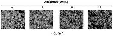
- 3T3-L1 in control group showed typical white adipocyte characteristics, the cell volume was big, the lipid droplets were big and round, and the number of lipid droplets was less.
- 3T3-L1 treated with artemether (ART) exhibited typical brown adipocyte-like characteristics, the cell volume was smaller and the lipid droplets were small and dense. And this change had a concentration-dependent tendency. As the concentration of artemether increased, the change of 3T3-L1 adipocytes was more obvious.
- the total RNA of 3T3-L1 was extracted and reverse transcribed into cDNA.
- the mRNA level of UCP1 was detected by Real time qPCR. Compared with the control group, the expression of UCP1 in ART-treated 3T3-L1 was significantly up-regulated and exhibited a concentration-dependent tendency.
- 3T3-L1 is a classic white preadipocyte. Artemether enabled 3T3-L1 to acquire brown fat-like characteristics during differentiation. It not only had the morphological characteristics of brown fat, but also exhibited the expression of marker of brown fat, UCP1.
- C3H10T1/2 maintenance medium DMEM Up to 500 ml Calf serum 10% Biotin 8mg/ml Penicillin 100U/ml Streptomycin 100U/ml
- C3H10T1/2 induced differentiation medium DMEM Up to 500 ml Fetal bovine serum 10% Biotin 8mg/ml Penicillin 100U/ml Streptomycin 100U/ml
- Dexamethasone 0.039g Dexamethasone was dissolved in 100ml water, and the solution was filtered and sterilized through 0.22 ⁇ m filter membrane.
- Insulin 0.1g Insulin was dissolved in 100ml water, a few drops of hydrochloric acid were added to dissolve the insulin, and the solution was filtered and sterilized through 0.22 ⁇ m filter membrane.
- 7.5 ⁇ 10 5 C3H10T1/2 were seeded in a 3.5 cm cell culture dish, and cultured for 3-5 days in a maintenance medium.
- BMP4 at a final concentration of 20 ng/ml was added to the medium to allow the cells to proliferate to contact inhibition.
- the medium was changed to induced differentiation medium, and the induced mixture (0.5mMol/L Mix, 1 ⁇ Mol/L Dex, 1ng/ml insulin, 100 nMol/L indometacin, InM T3) was added at the same time. This day was defined as Day 0 of differentiation.
- 7.5 ⁇ 10 5 C3H10T1/2 were seeded in a 3.5 cm cell culture dish, and cultured for 3-5 days in a maintenance medium.
- BMP7 at a final concentration of 100 ng/ml was added to the medium to allow the cells to proliferate to contact inhibition.
- the medium was changed to induced differentiation medium, and the induced mixture (0.5mM Mix, 1 ⁇ M Dex, 1ng/ml insulin, 100 nM indometacin, InM T3) was added at the same time. This day was defined as Day 0 of differentiation.
- fresh induced differentiation medium was replaced, and 1 ng/ml insulin and 1nM T3 were added.
- the fresh medium was replaced every other day until the culture was differentiated to Day 8 at which time the cells were mature. At this time, the cells differentiated to mature were brown adipocytes.
- the compound On Day 0 of differentiation, that is, when the induced differentiation mixture was added, the compound was added to the medium at a storage concentration of 10mMol/L, and the final concentrations were 5 ⁇ M ⁇ l/L, 10 ⁇ Mol/L and 15 ⁇ Mol/L, respectively.
- the fresh medium was replaced, the corresponding compound was supplemented, and the compound was always with the cells until the cells differentiated to mature.
- C3H10T1/2 differentiated to mature was induced in a 3.5 cm culture dish, with the method of differentiation referring to Method 2.4.1.
- 300 ⁇ l Trizol was added to fully lyse the cells, and the lysate was transferred into a 1.5 ml centrifuge tube and heated at 100 °C for 10 min.
- the lysate was centrifuged at 13000g for 10min at 4 °C, the lipid in the upper layer was removed, and the clarified lysate in the middle layer was transferred into a new centrifuge tube.
- 2.5 ⁇ l lysate was taken and quantified by BCA method, and 200 ⁇ l lysate was mixed with 50 ⁇ l loading buffer, heated at 100 °C for 10 min, and stored at -20 °C until use.
- C3H0T1/2 protein 30 ⁇ g C3H0T1/2 protein was loaded onto 10% polyacrylamide gel, and electrophoresis was carried out, with electrophoresis being carried out on base gel at 80V, and on separating gel at 100V, until the target band was separated.
- the protein was transferred to a PVDF membrane in an ice bath at 100 V for 100 min. After the transfer was completed, it was blocked for 1 hr in 5% skim milk prepared with TBST (TBS buffer + 0.5% Tween), and incubated with specific antibodies overnight at 4 °C.
- the PVDF membrane was washed three times with TBST at room temperature for 10 min, incubated with the corresponding secondary antibody at room temperature for 1 hr, then washed three times with TBST at the same condition as that of primary antibody, incubated with chemiluminescent reagent at room temperature for 1 min, detected with ImageQuant LAS4000 Mini Automatic chemiluminescence apparatus and exposed, and the results were recorded.
- the medium was removed and 1ml PBS was added. MitoTraker Green was added to PBS at a final concentration of 100ng/ml.
- the mixture was incubated at 37 °C for 10 min, and the green fluorescence was observed under a fluorescence microscope. Representative field of view was captured and recorded.
- C3H10T1/2 is a mesenchymal stem cell with multipotential differentiation potential, with BMP4-directed C3H10T1/2 differentiating into white adipocyte, BMP7-directed C3H10T1/2 differentiating into brown adipocyte.
- artemether (15 ⁇ Mol/L) was added simultaneously for treatment.
- BMP7-directed C3H10T1/2 had typical brown adipocyte characteristics, the cell was small, the lipid droplets were small with a large quantity; BMP4-directed C3H10T1/2 had typical white adipocyte characteristics, the cell was big, the lipid droplets were big with a small quantity; during differentiation of BMP4-directed C3H10T1/2, artemether (15 ⁇ Mol/L) was added simultaneously for treatment, and the cells exhibited brown fat-like characteristics, which was similar to BMP7-directed cell morphology (as shown in Figure 3 ).
- BMP7-directed C3H10T1/2 was used as a positive control.
- BMP4-directed C3H10T1/2 was treated with different concentrations of artemether (5, 10, 15 ⁇ Mol/L), and the protein level of brown fat-related genes was detected by Western blot with ⁇ -actin as an internal reference.
- artemether increased the protein levels of the brown fat-related genes UCP1, PGC1a, PRDM16 and CytoC in BMP4-directed C3H10T1/2 cells, and the increase had a concentration-dependent tendency (as shown in Figure 4 ).
- BMP4 enabled C3H10T1/2 mesenchymal stem cells to differentiate into cells with white fat characteristics. After treatment with artemether, BMP4-directed C3H10T1/2 differentiated to mature and exhibited brown adipocyte-like characteristics, which included the following three aspects: cell morphology, related gene expression and mitochondrial biosynthesis enhancement.
- DMEM/F12 (1:1) Up to 500 ml Fetal bovine serum 10% Biotin 8mg/ml Penicillin 100U/ml Streptomycin 100U/ml
- Tris-HCL pH6.8
- SDS 2%
- Glycerol 10%
- NaF 10mMol/L Na 3 VO 4 100mMol/L
- Inguinal fat of male mice was cut into pieces, treated with collagenase at 37 °C for 15 min, and centrifuged at 250 rpm for 10 min. The pellet was resuspended in F12/DMEM medium. 2.5 ⁇ 10 5 cells were taken and seeded in a 3.5 cm culture dish, which was pretreated with 1 ml polylysine for 30 min.
- induction was performed according to adipocyte differentiation protocol as follows: two days after induction with F12/DMEM comprising 10% fetal bovine serum, 0.5mMol/L Mix, 1 ⁇ Mol/L Dex, 5 ⁇ g/ml insulin, and 1 ⁇ Mol/L rosiglitazone; the culture was continued for 2 days with DMEM/F12 comprising 10% fetal bovine serum, 5 ⁇ g/ml insulin and 1 ⁇ Mol/L rosiglitazone; thereafter, DMEM/F12 medium containing 10% fetal bovine serum was replaced every other day until the cells differentiated to Day 8.
- F12/DMEM comprising 10% fetal bovine serum, 0.5mMol/L Mix, 1 ⁇ Mol/L Dex, 5 ⁇ g/ml insulin, and 1 ⁇ Mol/L rosiglitazone
- the method for extracting total SVF protein was the same as 2.4.3, Western blot method and catalog number of antibody was the same as 2.4.4.
- SVF brown fat-related genes in SVF cells treated with artemether were detected by Western Blot.
- SVF is a primary preadipocyte, under the action of artemether, SVF differentiated into adipocyte with brown fat characteristics in vitro.
- Artemisinin, artesunate and dihydroartemisinin were stored in DMSO at a storage concentration of 10mMol/L. The concentration shown in Figure 8 was added on Day 0 of differentiation. Fresh medium was replaced every two days, and the corresponding concentrations of compounds were added until the cells differentiated to mature.
- C3H10T1/2 Compared with control, artemether treated C3H10T1/2 exhibited brown adipocyte-like characteristics: cell was small in size, and had multi-cavity small lipid droplets. C3H10T1/2 treated with artemisinin, artesunate and dihydroartemisinin all exhibited similar morphological characteristics (as shown in Figure 8 ). Dihydroartemisinin up-regulated the expression of brown fat-related genes.
- Dihydroartemisinin is a secondary metabolite of artemisinin and its analogues. If dihydroartemisinin has an effect of promoting browning, it means that artemisinin and its analogues all have the effect of promoting browning.
- BMP4-directed C3H10T1/2 with different concentrations (1, 2.5, 5, 7.5 ⁇ Mol/L) of dihydroartemisinin
- the expression levels of brown fat-related genes PGC1 ⁇ and UCP1 were detected by Western blot, and the expression levels of brown fat-related genes were found to gradually increase with the increase of the concentration of dihydroartemisinin (as shown in Figure 9 ).
- mice were breeded in the SPF animal laboratory of the Laboratory Animal Center of Fudan University at a room temperature of 21 °C-23 °C with a 12-hour light/dark cycle.
- the high-fat diet used to induce obese mice had a fat content of 60%.
- mice were grown to 8-week old, they were randomly divided into a control group and an administration group, with 8 mice in each group. While the mice were fed with a high-fat diet, artemether was injected subcutaneously.
- Artemether was diluted in PBS at a concentration of 15 ⁇ M ⁇ l/L, and injected into bilateral inguinal region, with 200 ⁇ l being injected into each side. Mice in control group were injected with the same volume of PBS. Administration was continued for eight weeks and the weight of mice was recorded weekly.
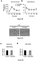
- mice injected subcutaneously with artemether was significantly lower than that of mice in control group. After six weeks of administration, the difference in weight between the two groups was statistically different, as shown in Figure 10A .
- the weight gain of mice in administration group was significantly slower than that of mice in control group, as shown in Figure 10B .
- Subcutaneous injection of artemether significantly inhibited the weight gain of mice under a high-fat diet condition.
- mice were breeded in the SPF animal laboratory of the Laboratory Animal Center of Fudan University at a room temperature of 21 °C-23 °C with a 12-hour light/dark cycle.
- the high-fat diet used to induce obese mice had a fat content of 60%.
- mice were grown to 8-week old, they were randomly divided into a control group and an administration group, with 8 mice in each group. While the mice were fed with a high-fat diet, artemether was injected subcutaneously.
- Artemether was diluted in PBS at a concentration of 15 ⁇ Mol/L, and injected into bilateral inguinal region, with 200 ⁇ l being injected into each side. Mice in control group were injected with the same volume of PBS. Administration was continued for eight weeks.
- mice were sacrificed by cervical dislocation and were dissected, and inguinal fat (Inguinal, subcutaneous fat), epididymal fat (Gonadal, visceral fat) and brown fat in the interscapular region (BAT, classic brown fat) were removed, weighed and fixed.
- inguinal fat Inguinal, subcutaneous fat
- epididymal fat Gonadal, visceral fat
- brown fat in the interscapular region BAT, classic brown fat
- the histological analysis of adipose tissue was commissioned by Shanghai Saige Biotech Co., Ltd. The histological analysis process included the steps of fixing, paraffin embedding, sectioning and HE staining.
- adipose tissue Three representative pieces of adipose tissue (inguinal fat, epididymal fat and brown fat) were removed and analyzed. It was found that volume ( Figure 11A ) and weight ( Figure 11B ) of adipose tissue in mice injected subcutaneously with artemether was significantly smaller than that of mice in control group. Sectioning and staining of adipose tissue showed that adipocytes of mice in administration group were significantly smaller than that of mice in control group ( Figure 11C ). Artemether reduced volume and weight of fat in high fat-fed mice, and the weight loss caused by artemether was mainly due to the decrease of adipose tissue.
- mice were breeded in the SPF animal laboratory of the Laboratory Animal Center of Fudan University at a room temperature of 21 °C-23 °C with a 12-hour light/dark cycle.
- the high-fat diet used to induce obese mice had a fat content of 60%.
- mice were grown to 8-week old, they were randomly divided into a control group and an administration group, with 8 mice in each group. While the mice were fed with a high-fat diet, artemether was injected subcutaneously.
- Artemether was diluted in PBS at a concentration of 15 ⁇ Mol/L, and injected into bilateral inguinal region, with 200 ⁇ l being injected into each side. Mice in control group were injected with the same volume of PBS. Administration was continued for eight weeks.
- mice were fasted for 12-16 hours and injected intraperitoneally with glucose injection at a dose of 2mg/kg.
- glucose injection at a dose of 2mg/kg.
- blood was collected from the tail vein, and blood glucose was measured with OneTouch ExtraEasy glucometer and OneTouch ExtraEasy blood glucose test strips and the results were recorded.
- Insulin was injected intraperitoneally at a dose of 0.75U/kg weight. At 0 min, 15 min, 30 min, 45 min and 60 min after injection, blood was collected from the tail vein, and blood glucose was measured.
- mice Glucose tolerance of mice injected subcutaneously with artemether after eight weeks of high-fat diet feeding was significantly better than that of mice in control group ( Figure 12A ), and insulin tolerance was significantly better than that of mice in control group ( Figure 12B ).
- glucose metabolism and insulin sensitivity of mice injected subcutaneously with artemether were significantly better than that of mice in control group, indicating that artemether played a role in maintaining glucose metabolism homeostasis.
- EXAMPLE 8 Artemether inhibited fatty liver induced by high-fat diet
- mice were breeded in the SPF animal laboratory of the Laboratory Animal Center of Fudan University at a room temperature of 21 °C-23 °C with a 12-hour light/dark cycle.
- the high-fat diet used to induce obese mice had a fat content of 60%.
- mice were grown to 8-week old, they were randomly divided into a control group and an administration group, with 8 mice in each group. While the mice were fed with a high-fat diet, artemether was injected subcutaneously.
- Artemether was diluted in PBS at a concentration of 15 ⁇ M ⁇ l/L, and injected into bilateral inguinal region, with 200 ⁇ l being injected into each side.
- Mice in control group were injected with the same volume of PBS. Administration was continued for eight weeks. The mice were sacrificed by cervical dislocation, and liver tissues of mice were removed. The histological analysis including tissue fixation, paraffin embedding, sectioning and HE staining was commissioned by Shanghai Saige Biotech Co., Ltd.
- mice were breeded in the SPF animal laboratory of the Laboratory Animal Center of Fudan University at a room temperature of 21 °C-23 °C with a 12-hour light/dark cycle.
- the high-fat diet used to induce obese mice had a fat content of 60%.
- mice were grown to 8-week old, they were randomly divided into a control group and an administration group, with 8 mice in each group. While the mice were fed with a high-fat diet, artemether was injected subcutaneously.
- Artemether was diluted in PBS at a concentration of 15 ⁇ Mol/L, and injected into bilateral inguinal region, with 200 ⁇ l being injected into each side. Mice in control group were injected with the same volume of PBS. Administration was continued for eight weeks.
- mice were fasted and housed in a breeding box at 4 °C. The rectal temperature was measured every 2 hours. The mice were sacrificed by cervical dislocation after 6 hours, and the inguinal fat was removed for histological analysis. The histological analysis including tissue fixation, paraffin embedding, sectioning and immunohistochemistry experiment of UCP1 was commissioned by Shanghai Saige Biotech Co., Ltd.
- mice injected subcutaneously with artemether were significantly higher than that of mice in control group when exposed to an environment of 4 °C ( Figure 14A ). Maintaining body temperature in a cold exposure environment is a physiological function undertaken by thermogenic adipose tissue. The epididymal fat was removed and subjected to immunohistochemical test of UCP1, and the results showed that subcutaneous fat of mice in administration group exhibited significant UCP1 expression ( Figure 14B ). After eight weeks of subcutaneous administration, mice injected subcutaneously with artemether had a significant increase in ability of maintaining body temperature, and obvious browning was found in subcutaneous fat. It was indicated that artemether inhibited weight gain and improved metabolism by promoting browning of subcutaneous fat.
- mice were breeded in the SPF animal laboratory of the Laboratory Animal Center of Fudan University at a room temperature of 21 °C-23 °C with a 12-hour light/dark cycle.
- the high-fat diet used to induce obese mice had a fat content of 60%.
- mice were grown to 8-week old, they were randomly divided into a control group and an administration group, with 8 mice in each group.
- artemether was injected intraperitoneally.
- Artemether for intraperitoneal injection was stored at 4 °C in coconut oil at a storage concentration of 6mg/ml (20mMol/L), and protected from light.
- Mice in administration group were injected with artemether at a dose of 20mg/kg weight. Administration was twice a week for eight weeks, and the weight of the mice was recorded weekly.
- mice injected intraperitoneally with artemether was significantly lower than that of mice in control group. After three weeks of administration, the difference in weight between the two groups was statistically different, as shown in Figure 15A .
- the weight gain of mice in administration group was significantly slower than that of mice in control group, as shown in Figure 15B .
- Intraperitoneal injection of artemether significantly inhibited the weight gain induced by high-fat diet.
- mice were breeded in the SPF animal laboratory of the Laboratory Animal Center of Fudan University at a room temperature of 21 °C-23 °C with a 12-hour light/dark cycle.
- the high-fat diet used to induce obese mice had a fat content of 60%.
- mice were grown to 8-week old, they were randomly divided into a control group and an administration group, with 8 mice in each group.
- artemether was injected intraperitoneally.
- Artemether for intraperitoneal injection was stored at 4 °C in coconut oil at a storage concentration of 6mg/ml (20mMol/L), and protected from light.
- Mice in administration group were injected with artemether at a dose of 20mg/kg weight.
- mice were sacrificed by cervical dislocation and were dissected, and inguinal fat (Inguinal, subcutaneous fat), epididymal fat (Gonadal, visceral fat) and brown fat in the interscapular region (BAT, classic brown fat) were removed, weighed and fixed.
- inguinal fat Inguinal, subcutaneous fat
- epididymal fat Gonadal, visceral fat
- brown fat in the interscapular region BAT, classic brown fat
- mice were breeded in the SPF animal laboratory of the Laboratory Animal Center of Fudan University at a room temperature of 21 °C-23 °C with a 12-hour light/dark cycle.
- the high-fat diet used to induce obese mice had a fat content of 60%.
- mice were grown to 8-week old, they were randomly divided into a control group and an administration group, with 8 mice in each group.
- artemether was injected intraperitoneally.
- Artemether for intraperitoneal injection was stored at 4 °C in coconut oil at a storage concentration of 6mg/ml (20mMol/L), and protected from light.
- Mice in administration group were injected with artemether at a dose of 20mg/kg weight. Administration was twice a week for eight weeks.
- mice were fasted for 12-16 hours and injected intraperitoneally with glucose injection at a dose of 2mg/kg weight.
- glucose injection at a dose of 2mg/kg weight.
- blood was collected from the tail vein, and blood glucose was measured with OneTouch ExtraEasy glucometer and OneTouch ExtraEasy blood glucose test strips and the results were recorded.
- Insulin was injected intraperitoneally at a dose of 0.75U/kg weight. At 0 min, 15 min, 30 min, 45 min and 60 min after injection, blood was collected from the tail vein, and blood glucose was measured.
- mice were anesthetized with 10% chloral hydrate and blood was taken from the orbit. The whole blood was centrifuged at 300 rpm for 5 min at 4 °C. Serum was taken and the blood glucose levels were measured by automatic biochemical analyzer and calibrator for blood glucose Cfas and kit (Roche).
- mice were breeded in the SPF animal laboratory of the Laboratory Animal Center of Fudan University at a room temperature of 21 °C-23 °C with a 12-hour light/dark cycle.
- the high-fat diet used to induce obese mice had a fat content of 60%.
- mice were grown to 8-week old, they were randomly divided into a control group and an administration group, with 8 mice in each group.
- artemether was injected intraperitoneally.
- Artemether for intraperitoneal injection was stored at 4 °C in coconut oil at a storage concentration of 6mg/ml (20mMol/L), and protected from light.
- Mice in administration group were injected with artemether at a dose of 20mg/kg weight.
- mice were sacrificed by cervical dislocation, and liver tissues of mice were removed and fixed.
- the histological analysis for liver was commissioned by Shanghai Saige Biotech Co., Ltd., and the histological analysis process included tissue fixation, paraffin embedding, sectioning and HE staining.
- EXAMPLE 14 Iintraperitoneal injection of artemether inhibited elevated blood lipid caused by high-fat diet
- mice were breeded in the SPF animal laboratory of the Laboratory Animal Center of Fudan University at a room temperature of 21 °C-23 °C with a 12-hour light/dark cycle.
- the high-fat diet used to induce obese mice had a fat content of 60%.
- mice were grown to 8-week old, they were randomly divided into a control group and an administration group, with 8 mice in each group.
- artemether was injected intraperitoneally.
- Artemether for intraperitoneal injection was stored at 4 °C in coconut oil at a storage concentration of 6mg/ml (20mMol/L), and protected from light.
- Mice in administration group were injected with artemether at a dose of 20mg/kg weight. Administration was twice a week for eight weeks.
- mice were anesthetized with 10% chloral hydrate and blood was taken from the orbit. The whole blood was centrifuged at 300 rpm for 5 min at 4 °C. Serum was taken and the serum indicators were measured by lipid calibrator Cfas Lipid, high-density lipoprotein Cfas, and low density lipoprotein Cfas, as well as detection kits for related indicators.
- triglyceride and high-density lipoprotein (HDL) in the serum of mice injected intraperitoneally with artemether were not different from the control group.
- Serum cholesterol and low density lipoprotein (LDL) were significantly decreased compared with the control group, as shown in Figure 19 .
- Artemether significantly improved dyslipidemia caused by a high-fat diet.
- mice were breeded in the SPF animal laboratory of the Laboratory Animal Center of Fudan University at a room temperature of 21 °C-23 °C with a 12-hour light/dark cycle.
- the high-fat diet used to induce obese mice had a fat content of 60%.
- mice were grown to 8-week old, they were randomly divided into a control group and an administration group, with 8 mice in each group.
- artemether was injected intraperitoneally.
- Artemether for intraperitoneal injection was stored at 4 °C in coconut oil at a storage concentration of 6mg/ml (20mMol/L), and protected from light.
- Mice in administration group were injected with artemether at a dose of 20mg/kg weight.
- mice were fasted and housed in a breeding box at 4 °C. The rectal temperature was measured every 2 hours. The mice were sacrificed by cervical dislocation after 6 hours, and the inguinal fat was removed for histological analysis. The histological analysis including tissue fixation, paraffin embedding, sectioning and immunohistochemistry experiment of UCP1 was commissioned by Shanghai Saige Biotech Co., Ltd.
- mice injected intraperitoneally with artemether were significantly higher than that of mice in control group when exposed to an environment of 4 °C ( Figure 19A ). Maintaining body temperature in a cold exposure environment was a physiological function undertaken by thermogenic adipose tissue. The epididymal fat of mice was removed and subjected to immunohistochemical test of UCP1, and the results showed that subcutaneous fat of mice in administration group exhibited significant UCP1 expression ( Figure 19B ). After eight weeks of intraperitoneal injection of artemether, mice in administration group had a significant increase in ability of maintaining body temperature, and obvious browning was found in subcutaneous fat. It was indicated that artemether inhibited weight gain by promoting browning of subcutaneous fat.
- mice were breeded in the SPF animal laboratory of the Laboratory Animal Center of Fudan University at a room temperature of 21 °C-23 °C with a 12-hour light/dark cycle. When the mice were grown to 4-week old, they were randomly divided into a control group and an administration group, with 4 mice in each group. The administration group was injected intraperitoneally with artemether. Artemether for intraperitoneal injection was stored at 4 °C in coconut oil at a storage concentration of 6mg/ml (20mMol/L), and protected from light. Ob/ob mice in administration group were injected with artemether at a dose of 20mg/kg weight. Administration was twice a week for eight weeks, and the weight of the mice was recorded weekly.
- Ob/ob mice were breeded in the SPF animal laboratory of the Laboratory Animal Center of Fudan University at a room temperature of 21 °C-23 °C with a 12-hour light/dark cycle. When the mice were grown to 4-week old, they were randomly divided into a control group and an administration group, with 4 mice in each group. Mice in administration group were injected intraperitoneally with artemether. Artemether for intraperitoneal injection was stored at 4 °C in coconut oil at a storage concentration of 6mg/ml (20mMol/L), and protected from light. Ob/ob mice in administration group were injected with artemether at a dose of 20mg/kg weight. Administration was twice a week for eight weeks. The mice were sacrificed by cervical dislocation, and liver tissues of mice were removed, fixed and paraffin embedded. The histological analysis was commissioned by Shanghai Saige Biotech Co., Ltd..
- mice were breeded in the SPF animal laboratory of the Laboratory Animal Center of Fudan University at a room temperature of 21 °C-23 °C with a 12-hour light/dark cycle. When the mice were grown to 8-week old, they were randomly divided into a control group and an administration group, with 8 mice in each group. Mice in administration group were injected intraperitoneally with artemether. Artemether for intraperitoneal injection was stored at 4 °C in coconut oil at a storage concentration of 6mg/ml (20mMol/L), and protected from light. Mice in administration group were injected with artemether at a dose of 20mg/kg weight. Mice in control group were injected with 100 ⁇ l coconut oil. Administration was twice a week for eight weeks and the weight of the mice was recorded weekly.
- mice were breeded in the SPF animal laboratory of the Laboratory Animal Center of Fudan University at a room temperature of 21 °C-23 °C with a 12-hour light/dark cycle. When the mice were grown to 8-week old, they were randomly divided into a control group and an administration group, with 8 mice in each group.
- Artemether for intraperitoneal injection was stored at 4 °C in coconut oil at a storage concentration of 6mg/ml (20mMol/L), and protected from light.
- Mice in administration group were injected with artemether at a dose of 20mg/kg weight, and mice in control group were injected with 100 ⁇ l coconut oil. Administration was twice a week for eight weeks.
- mice were fasted for 12-16 hours and injected intraperitoneally with glucose injection at a dose of 2mg/kg.
- glucose injection at a dose of 2mg/kg.
- blood was collected from the tail vein, and blood glucose was measured with OneTouch ExtraEasy glucometer and OneTouch ExtraEasy blood glucose test strips and the results were recorded.
- Insulin was injected intraperitoneally at a dose of 0.75U/kg weight. At 0 min, 15 min, 30 min, 45 min and 60 min after injection, blood was collected from the tail vein, and blood glucose was measured.
- mice Healthy lean mice were injected intraperitoneally with artemether for eight weeks.
- the glucose tolerance curve of mice in administration group did not differ from that of mice in control group, indicating that glucose metabolism was not affected by artemether, as shown in Figure 23A .
- the insulin tolerance curve of mice in administration group also did not differ from that of mice in control group, as shown in Figure 24B ; intraperitoneal injection of artemether did not alter glucose metabolism of healthy lean mice.
- mice were breeded in the SPF animal laboratory of the Laboratory Animal Center of Fudan University at a room temperature of 21 °C-23 °C with a 12-hour light/dark cycle. When the mice were grown to 8-week old, they were randomly divided into a control group and an administration group, with 8 mice in each group. Mice in administration group were injected intraperitoneally with artemether. Artemether for intraperitoneal injection was stored at 4 °C in coconut oil at a storage concentration of 6mg/ml (20mMol/L), and protected from light. Mice in administration group were injected with artemether at a dose of 20mg/kg weight. Mice in control group were injected 100 ⁇ l coconut oil. Administration was twice a week for eight weeks. The mice were sacrificed by cervical dislocation, and liver tissues of mice were removed and fixed. Liver tissue fixation, paraffin embedding, sectioning and HE staining was commissioned by Shanghai Saige Biotech Co., Ltd..
- liver cells of healthy lean mice injected intraperitoneally with artemether for eight weeks was normal, and had no difference from that of mice in control group, as shown in Figure 25 ; in the case of intraperitoneal injection of artemether at a dose of 20 mg/kg weight twice a week for eight weeks, artemether had no hepatotoxicity.
- mice were breeded in the SPF animal laboratory of the Laboratory Animal Center of Fudan University at a room temperature of 21 °C-23 °C with a 12-hour light/dark cycle. When the mice were grown to 8-week old, they were randomly divided into four groups, a high-fat control group, a high-fat administration group, a normal administration group, and a normal control group, with 8 mice in each group.
- Administration groups (high-fat and normal diet) were injected intraperitoneally with artemether.
- Artemether for intraperitoneal injection was stored at 4 °C in coconut oil at a storage concentration of 6mg/ml (20mMol/L), and protected from light.
- Mice in administration group were injected with artemether at a dose of 20mg/kg weight, and mice in control group were injected with 100 ⁇ l coconut oil. The diet was weighed weekly and the diet consumption of mice in each group was recorded.
- mice in administration group For high-fat groups, the diet consumption of mice in administration group was substantially the same as that of mice in control group with no difference, as shown in Figure 26A .
- the diet consumption of mice in administration group was not different from that of mice in control group, as shown in Figure 26B .
- intraperitoneal injection of artemether did not affect normal food intake in mice.
- artemether did not cause anorexia nervosa.
Landscapes
- Health & Medical Sciences (AREA)
- Chemical & Material Sciences (AREA)
- Medicinal Chemistry (AREA)
- Pharmacology & Pharmacy (AREA)
- Life Sciences & Earth Sciences (AREA)
- Animal Behavior & Ethology (AREA)
- General Health & Medical Sciences (AREA)
- Public Health (AREA)
- Veterinary Medicine (AREA)
- Epidemiology (AREA)
- Child & Adolescent Psychology (AREA)
- Engineering & Computer Science (AREA)
- Bioinformatics & Cheminformatics (AREA)
- Diabetes (AREA)
- Hematology (AREA)
- Obesity (AREA)
- Chemical Kinetics & Catalysis (AREA)
- General Chemical & Material Sciences (AREA)
- Nuclear Medicine, Radiotherapy & Molecular Imaging (AREA)
- Organic Chemistry (AREA)
- Pharmaceuticals Containing Other Organic And Inorganic Compounds (AREA)
- Medicinal Preparation (AREA)
Abstract
Description
- The invention belongs to the field of biomedicine, and particularly relates to the use of artemether in preparing preparations for promoting lipolysis and improving glucose metabolism.
- Data show that obesity and its related metabolic diseases have become epidemic diseases that are causing trouble worldwide. According to data released by the World Health Organization (WHO) in 2015, the obesity rate of global population is 13%; and the obesity rate of adults is 6.9% in China. It is worried that the obesity rate of adolescents is higher and exhibits a trend of sharp increase, which means that obesity rate will continue to grow over the next few decades. Studies have shown that obesity can cause a series of metabolic diseases, such as
type 2 diabetes, high blood pressure, fatty liver, etc., and even increase the incidence of certain tumors, which will bring great health burden and economic burden to the country and the people. - Studies have shown that adipose tissue is at the core position of the development and progression of obesity and plays a pivotal role in metabolic health. It is currently known that there are three kinds of adipose tissues in the human body, white fat, brown fat and beige fat. White adipose tissue stores energy in the form of triglyceride, while brown fat and beige fat consume energy through heat production. Beige fat is found in white fat and has a brown fat characteristic. Beige fat can be considered as a transitional form of white fat and brown fat in terms of phenotype. The phenomenon of the appearance of beige fat in white adipose tissue is called browning of white fat. Although beige fat and brown fat are in different parts and have different developmental origins, they exert the same thermogenic function, have the same potential in terms of energy consumption and are called thermogenic adipose tissue. Under physiological conditions, there are two conditions that activate thermogenic adipose tissue: cold exposure and dietary intake (especially high calorie foods). During cold exposure, the thermogenic adipose tissue is activated to maintain a constant body temperature; during dietary intake, activation of the thermogenic fat relieves the transient energy peak and maintains metabolic stability.
- Thermogenic adipose tissue, its differentiation and development, functional regulation and clinical application are research hotspots in the field of metabolism in recent years. According to the population survey, individuals with active thermogenic adipose tissue are not prone to gain weight and are not susceptible to metabolism-related diseases such as
type 2 diabetes. Some drugs that improve metabolism, such as berberine and salicyl salicylic acid, can achieve part of the effect by activating thermogenic adipose tissue. Similarly, in animal models, whether related genes are changed (knocked out or overexpressed), or drug stimulation (β-adrenergic receptor agonists, etc.) is carried out, after the activation of thermogenic brown fat, it will function to control body weight and improve metabolism. Therefore, in summary, beige adipose tissue is a thermogenic adipose tissue in human body, which has the potential to consume excess energy, and can be activated under certain conditions. Activation of thermogenic adipose tissue, whether to enhance brown fat function or to promote browning of white fat, can play a role in weight control and metabolism. To date, there have been neither compound or drug discovery that specifically target thermogenic adipose tissue to achieve improved metabolism, nor high-throughput screening for this target. - Thermogenic adipose tissue converts chemical energy stored in fatty acids into heat energy because it has a special molecule, uncoupling protein 1 (UCP1). UCP1, which is localized on the inner membrane of the mitochondria, can cut off the coupling of the electron transport chain and ATP synthesis and directly convert chemical energy into heat energy in the case of activation. UCP1 is found in thermogenic adipose tissue. Due to this unique tissue localization, UCP1 is not only a functional molecule of thermogenic adipose tissue, but also a phenotypic marker. In addition to UCP1, PGC1α, PRDM16 and cytochrome C are important molecules involved in the browning process.
- Studies have reported that the occurrence of obesity and metabolic diseases is the result of a combination of living environment and genetic factors. High-fat diet-induced obese mice are animal models commonly used in laboratory to study the mechanism of the development and progression of obesity and metabolic diseases. Ob/ob mice have severe insulin resistance due to the lack of leptin gene, and have fatty liver since birth, and these metabolic disorders gradually aggravate with the growth of mice, eventually leading to premature death in mice. Due to the presence of these innate metabolic disorders, ob/ob mice are common animal models for laboratory studies of diabetes and insulin resistance. The commonly used cell models for in vitro study of fat cell differentiation and development are 3T3-L1 and C3H10T1/2, with the former simulating the process of differentiation of preadipocytes into mature adipocytes, and the latter reflecting the process of differentiation of mesenchymal stem cells into adipocytes after orientation. In addition to cell lines, the cell model for studying the development of fat also includes the Stromal Vascular Fraction (SVF). SVF is a primary preadipocyte, which can differentiate into white adipocyte after isolation, culture and certain induction, thereby mimicking the in vivo process of differentiation of preadipocytes into white fat in an in vitro environment.
- The prior art has disclosed that artemether (ART) (Formula II) is an analogue of artemisinin (Formula I), which can efficiently and quickly kill erythrocyte-stage plasmodium. It is used in the treatment of chloroquine-resistant falciparum malaria and pernicious malaria, with rapid effect, good short-term efficacy and less toxicity and side effects. It is a first-line drug for the treatment of malaria and is widely used. Recent studies have shown that artemether also has an effect of killing tumor cells and immunomodulation, showing new potential in the treatment of tumors and autoimmune diseases such as systemic lupus erythematosus and rheumatoid arthritis.
- The application potential of artemether in controlling body weight and improving metabolism has not yet been found. In addition to artemether, artemisinin-based drugs also include artesunate, dihydroartemisinin, arteether, SM905, artemiside, artemisone and SM934, among which dihydroartemisinin is a secondary metabolite of artemether and other artemisinin analogues in vivo.
- Based on the research basis of the prior art, the inventor of the present application intend to provide novel use of artemether and artemisinin analogues in pharmaceuticals, in particular the use of artemether in the preparation of a preparation for promoting lipolysis and improving glucose metabolism.

- Accordingly, the object of the present invention is to provide a novel use of artemether and artemisinin analogues in pharmaceuticals, in particular to the use of artemether in the preparation of a preparation for promoting lipolysis and improving glucose metabolism, especially to the use of artemether and artemisinin analogues in the preparation of a preparation for promoting browning of white fat, reducing body weight and improving glucose metabolism.
- In order to achieve the above object, a first aspect of the present invention provides the use of an artemisinin analogue represented by the following general formula (A) in the preparation of a medicament for promoting lipolysis and/or preventing or treating a metabolism-related disease:

- A second aspect of the present invention provides a method for promoting lipolysis and/or preventing or treating a metabolism-related disease, said method comprises administering to a subject in need thereof a therapeutically effective amount of an artemisinin analogue represented by the following general formula (A):

- Preferably, said administering a therapeutically effective amount of said artemisinin analogue is by oral or injection; more preferably, the administration dosage of said artemisinin analogue is 20mg/kg-100mg/kg, preferably 50mg/kg-100mg/kg for oral administration, and 10mg/kg-50mg/kg, preferably 20mg/kg-40mg/kg for injection, respectively.
- A third aspect of the present invention provides an artemisinin analogue for promoting lipolysis and/or preventing or treating a metabolism-related disease, said artemisinin analogue is represented by the following general formula (A):

- Preferably, according to use, artemisinin analogue or method of any of the preceding aspects, said artemisinin analogue is artemether represented by formula (II):
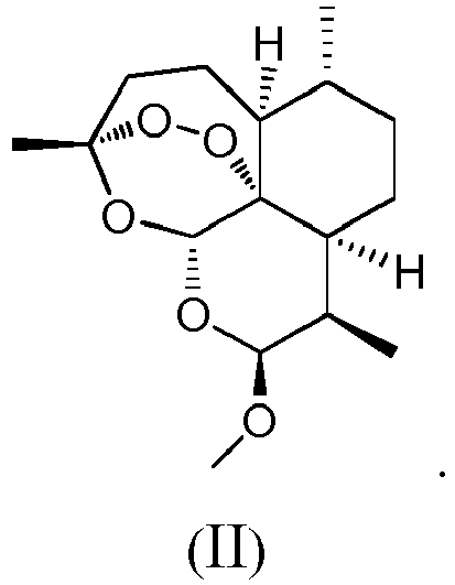
- Preferably, according to use, artemisinin analogue or method of any of the preceding aspects, said promoting lipolysis comprises promoting browning of white fat.
- Preferably, according to use, artemisinin analogue or method of any of the preceding aspects, said promoting lipolysis and/or preventing or treating a metabolism-related disease comprises controlling weight and improving metabolism.
- Preferably, according to use, artemisinin analogue or method of any of the preceding aspects, said metabolism-related disease is hyperglycemia, insulin resistance, dyslipidemia, and/or fatty liver caused by obesity.
- A fourth aspect of the present invention provides a pharmaceutical composition for promoting lipolysis and/or preventing or treating a metabolism-related disease, said pharmaceutical composition comprises: (1) the artemisinin analogue according to the third aspect of the present invention, and (2) a pharmaceutically acceptable carrier.
- Preferably, the dosage form of said pharmaceutical composition for oral administration is selected from an oral suspension, a powder and a granule; and the dosage form of said pharmaceutical composition for injection is selected from an aqueous solution, an oil and a suspension.
- The specific technical solutions of the present invention are further described below in conjunction with the concept of the present invention.
- The structure of artemether of the present invention is as shown in formula (II).

- The present invention also relates to artemisinin analogues other than artemether which have the potential to promote browning of white fat. The artemisinin structure is represented by formula (I), and the artemisinin analogues are represented by formula (A).

- The artemisinin analogue represented by formula (A) is dihydroartemisinin, with R being hydroxyl; arteether, with R being ethoxy; artesunate, with R being butanedioic acid monoester group (succinate group); SM934, with R being 2-aminoethoxy; SM905, with R being 2R-3-tert-butylamino-2-hydroxypropoxy; artemiside, with R being thiomorpholinyl; artemisone, with R being 1,1-dioxidothiomorpholinyl.
- The artemisinin analogues of the present invention have R groups as shown in the following table:
Dihydroartemisinin R=OH Artemether R=OMe Artesunate 
SM934 
SM905 
Artemiside 
Artemisone 
- The invention demonstrates by in vivo and in vitro experiments that the artemisinin-based compounds have the effect of inducing browning of white fat, and play a role in reducing body weight, improving metabolism, maintaining glucose metabolism homeostasis, alleviating fatty liver, and improving symptoms associated with diabetes by promoting browning of white fat. The clinical application practice of the artemisinin-based drugs shows that no intolerable side effects have been found. In studies of rodent models, it has been reported that high concentrations of artemether would cause anorexia nervosa, but there is no clear evidence of dose and timing of administration. Therefore, overall, artemether has an ideal safety as a clinical drug.
- Hereinafter, embodiments of the present invention will be described in detail with reference to the accompanying drawings, in which:
-
Figure 1 and Figure 2 show that artemether enabled 3T3-L1 to exhibit brown adipocyte-like characteristics. Among them,Figure 1 shows the morphological changes of 3T3-L1 cells treated with different concentrations of ART;Figure 2 shows the mRNA level of UCP1 after treating 3T3-L1 with different concentrations of ART. *p<0.05, **p<0.01. -
Figure 3-5 show that artemether enabled BMP4-directed C3H10T1/2 to exhibit brown fat-like morphological characteristics. Among them,Figure 3 shows the cell morphological characteristics of BMP7-directed C3H10T1/2, BMP4-directed C3H10T1/2, and BMP4-directed C3H10T1/2 treated with artemether;Figure 4 shows the protein levels of brown fat-related gene UCP1, PGC1a, PRDM16 and CytoC of BMP4-directed C3H10T1/2, BMP4-directed C3H10T1/2, and BMP4-directed C3H10T1/2 treated with different concentrations of artemether by Western Blot assay;Figure 5 shows the mitochondrial MitoTracker Green staining of BMP4-directed C3H10T1/2 after treatment with artemether (15 µMol/L). -
Figures 6-7 show that artemether enabled SVF primary preadipocyte to acquire brown fat-like morphological characteristics during differentiation. Among them,Figure 6 shows the cell morphological changes after treating SVF with different concentrations of artemether;Figure 7 shows the expression levels of brown fat-related gene PRDM16, PGC1α and UCP1 after treating SVF with different concentrations of artemether. -
Figures 8-9 illustrate the effect of artemisinin analogues on promoting browning. Among them,Figure 8 shows the cell morphology of BMP4-directed C3H10T1/2 treated with artemisinin and its analogues such as artemether, artesunate and dihydroartemisinin;Figure 9 shows the expression levels of brown fat-related gene after treating BMP4-directed C3H0T1/2 with different concentrations of dihydroartemisinin. -
Figure 10 shows that artemether inhibited weight gain induced by a high fat diet. Among them,Figure 10A shows the change in weight of mice subcutaneously injected with artemether under a high-fat diet condition, *p<0.05;Figure 10B shows the increase of weight in mice subcutaneously injected with artemether under a high-fat diet condition, *p<0.05. -
Figure 11 shows that subcutaneous injection of artemether reduced adipose tissue in high fat-fed mice. Among them,Figure 11A shows the volume of brown fat of inguinal fat, epididymal fat, and interscapular region of mice subcutaneously injected with artemether and mice in control group;Figure 11B shows the ratio of the weight of brown fat of inguinal fat, epididymal fat, and interscapular region of mice subcutaneously injected with artemether and mice in control group to the weight of mice, *p<0.05;Figure 11C shows the morphology of brown adipocytes of inguinal fat, epididymal fat, and interscapular region of mice subcutaneously injected with artemether and mice in control group. -
Figure 12 shows that subcutaneous injection of artemether improved glucose metabolism in high fat-induced obese mice. Among them,Figure 12A shows glucose tolerance curve of mice subcutaneously injected with artemether and mice in control group after eight weeks of high-fat diet feeding, *p<0.05;Figure 12B shows the glucose tolerance curve of mice injected subcutaneously with artemether and mice in control group after eight weeks of high-fat diet feeding, *p<0.05. -
Figure 13 shows that artemether inhibited fatty liver induced by a high-fat diet. It shows the liver cell morphology of mice injected subcutaneously with artemether and mice in control group after eight weeks of high-fat diet feeding. -
Figure 14 shows that subcutaneous injection of artemether induced browning of subcutaneous fat in high fat-fed mice. Among them,Figure 14A shows the change in body temperature of high fat-fed mice in an environment of 4 °C after eight weeks of administration, *p<0.05;Figure 14B shows the expression of UCP1 in inguinal adipose tissue of high fat-fed mice after exposure to 4 °C for 6 hours after eight weeks of administration. -
Figure 15 shows that intraperitoneal injection of artemether inhibited weight gain induced by a high fat diet. Among them,Figure 15A shows the change in weight of mice injected intraperitoneally with artemether under a high-fat diet condition, *p<0.05;Figure 15B shows the increase of weight in mice injected intraperitoneally with artemether under a high-fat diet condition, *p<0.05. -
Figure 16 shows that intraperitoneal injection of artemether significantly reduced adipose tissue in high fat-fed mice. Among them,Figure 16A shows the volume of brown fat of inguinal fat, epididymal fat, and interscapular region of mice injected intraperitoneally with artemether and mice in control group;Figure 16B shows the ratio of the weight of brown fat of inguinal fat, epididymal fat, and interscapular region of mice injected intraperitoneally with artemether and mice in control group to the weight of mice, *p<0.05;Figure 16C shows the morphology of brown adipocytes of inguinal fat, epididymal fat, and interscapular region of mice injected intraperitoneally with artemether and mice in control group. -
Figure 17 shows that intraperitoneal injection of artemether improved glucose metabolism in high fat-induced obese mice. Among them,Figure 17A shows the random blood glucose of mice injected intraperitoneally with artemether and mice in control group after eight weeks of high-fat diet feeding, *p<0.05;Figure 17B shows the glucose tolerance test results of mice injected intraperitoneally with artemether and mice in control group after eight weeks of high-fat diet feeding, *p<0.05;Figure 17C shows the insulin tolerance of mice injected intraperitoneally with artemether and mice in control group after eight weeks of high-fat diet feeding, *p<0.05. -
Figure 18 shows the histological morphology of liver cells of mice injected intraperitoneally with artemether and mice in control group after eight weeks of high-fat diet feeding. -
Figure 19 shows after eight weeks of high-fat diet feeding, changes in lipid metabolism related indexes in serum of mice injected intraperitoneally with artemether compared with mice in control group, *p<0.05, **p<0.01. -
Figure 20 shows that intraperitoneal injection of artemether induced browning of subcutaneous fat in high fat-fed mice. Among them,Figure 20A shows the change in body temperature of high fat-fed mice in an environment of 4 °C after eight weeks of intraperitoneal injection of artemether, *p<0.05;Figure 20B shows the expression of UCP1 in inguinal adipose tissue of high fat-fed mice after exposure to 4 °C for 6 hours after eight weeks of intraperitoneal injection of artemether. -
Figure 21 shows that artemether inhibited weight gain in diabetic ob/ob mice. Among them,Figure 21A shows the change in weight of ob/ob mice injected intraperitoneally with artemether for eight weeks;FIG. 21B shows the change in weight gain of ob/ob mice injected intraperitoneally with artemether for eight weeks, *p< 0.05. -
Figure 22 shows that artemether improved fatty liver in diabetic ob/ob mice, and it shows the results of HE staining of liver sections of ob/ob mice after eight weeks of intraperitoneal injection of artemether. -
Figure 23 shows that intraperitoneal injection of artemether did not affect growth of healthy mice. Among them,Figure 23A shows the weight of normal diet-fed healthy mice injected intraperitoneally with artemether for eight weeks in administration group and control group;Figure 23B shows the weight gain of normal diet-fed healthy mice injected intraperitoneally with artemether for eight weeks in administration group and control group. -
Figure 24 shows that intraperitoneal injection of artemether did not affect glucose metabolism of healthy mice. Among them,Figure 24A shows glucose tolerance of healthy lean mice injected intraperitoneally with artemether for eight weeks;Figure 24B shows glucose insulin tolerance of healthy lean mice injected intraperitoneally with artemether for eight weeks. -
Figure 25 shows that intraperitoneal injection of 20 mg/kg weight of artemether in mice did not cause liver damage, and it shows the results of HE staining of liver tissue sections of healthy lean mice after eight weeks of intraperitoneal injection of artemether. -
Figure 26 shows that artemether did not alter the food intake of mice. Among them,Figure 26A shows daily food intake per mouse of normal diet-fed lean mice injected intraperitoneally with artemether for eight weeks;Figure 26B shows daily diet consumption per mouse of high-fat diet-fed obese mice injected intraperitoneally with artemether for eight weeks. -
- 1.1.1 3T3-L1 white preadipocytes
- 1.1.2 DMEM medium (Gibco)
- 1.1.3 Fetal bovine serum (Gibco)
- 1.1.4 Calf serum (TBD)
- 1.1.5 Biotin (Sigma)
- 1.1.6 Penicillin and streptomycin
- 1.1.7 Artemether (Chengdu Pufei De Biotech Co., Ltd.) 1.1.8 Trizol (Life Technology)
- 1.1.9 Reverse transcription kit (ThermoScientific)
- 1.1.10 Power SYBR Green Master Mix (ThermoScientific)
-
- 1.2.1 Constant temperature sterile incubator (Thermo Scientific Forma Series II Water Jacket)
- 1.2.2 Real-time quantitative PCR instrument (Applied Biotech 7500 real time PCR System)
- 1.2.3 Microscope (Olympus IX711)
- 1.2.5 Refrigerated centrifuge (Eppendorf Centrifuge 5427R)
- 1.2.6 Nucleic acid quantitative instrument (Colibri)
-
1.3.1 Medium
3T3-L1 maintenance mediumDMEM Up to 500 ml Calf serum 10% Biotin 8mg/ml Penicillin 100U/ml Streptomycin 100U/ml
3T3-L1 induced differentiation mediumDMEM Up to 500 ml Fetal bovine serum 10% Biotin 8mg/ml Penicillin 100U/ml Streptomycin 100U/ml
1.3.2 Artemether stock solution was dissolved in DMSO at a storage concentration of 10 mMol/L.
1.3.3 Biotin: 160mg biotin and 80mg calcium pantothenate were dissolved in 200ml deionized water, and heated to dissolve. The solution was filtered and sterilized through 0.22µm filter membrane.
1.3.4 3-Isobutyl-1-methylxanthine (50mMol/L): 1.15g 3-isobutyl-1-methylxanthine was dissolved in 100ml 0.2M potassium hydroxide, and the solution was filtered and sterilized through 0.22µm filter membrane.
1.3.5 Dexamethasone (1mMol/L): 0.039g dexamethasone was dissolved in 100ml water, and the solution was filtered and sterilized through 0.22µm filter membrane.
1.3.6 Insulin (1mg/ml): 0.1g insulin was dissolved in 100ml water, a few drops of hydrochloric acid were added to dissolve the insulin, and the solution was filtered and sterilized through 0.22 µm filter membrane. - 2.5×10⁵ 3T3-L1 white preadipocytes were seeded in a 3.5cm cell culture dish, and cultured for 3 days in a maintenance medium. Cells proliferated to contact inhibition. 48 hours after the contact inhibition was reached, the medium was changed to induced differentiation medium, and the induced differentiation mixture including 0.5 mMol/L 3-isobutyl-1-methylxanthine (Mix), 1 µMol/L dexamethasone (Dex) and 1µg/ml insulin (Insulin) was added. The induced differentiation mixture is referred to as MDI for short. The day of adding MDI was defined as
Day 0 of differentiation. After 2 days of culture in the above medium, fresh induced differentiation medium was replaced, and 1 µg/ml insulin was added to the medium. After another 2 days of culture, the fresh induced differentiation medium was replaced, and the medium was changed every other day until the culture was differentiated toDay 8. - Artemether was stored in DMSO at a storage concentration of 10mMol/L. While MDI was added on
Day 0 of differentiation, artemether (5, 10 or 15µMol/L) was added for treatment. Hereafter, each time the medium were changed, the same concentration of artemether was added until 3T3-L1 was differentiated to mature. - 3T3-L1 differentiated to mature was induced in a 3.5 cm culture dish, with the method of differentiation referring to Method 1.4.1. After washing with PBS, the cells were fully lysed with 1 ml Trizol, thoroughly mixed, and 200 µl chloroform was added. After violently shaking, the mixtre was allowed to stand at room temperature for 15 min, and centrifuged at 4 °C, with the centrifuge condition being at 12000 g for 10 min. At this time, the lysate was separated into three layers due to the chromatographic action. The upper aqueous phase solution was carefully pipetted into a new centrifuge tube, and 500 µl isopropanol was added. After gently mixing, the mixture was allowed to stand for 10 min at room temperature, centrifuged at 12000 g for 10 min at 4 °C. The supernatant was discarded and the milky white translucent precipitate was seen at the bottom of the tube. The precipitate was washed 3 times with 75% ethanol diluted with DEPC water, centrifuged at 7500 g for 5 min at 4 °C. The supernatant was discarded and air dried at room temperature. The precipitate was dissolved in 60 µl DEPC water, and quantified by Colibri Tepertec and stored at -80 °C.
- The corresponding cDNA was synthesized by reverse transcription from 1 µg RNA, and the reverse transcription kit was Thermo Scientific Revert Aid First Strand cDNA Synthesis Kit. The synthesis system was as follows:
M-MLV Reaction Buffer 4µl 10 mM dNTP 2µl RNase inhibitor 1µl M-MLV reverse transcriptase 1µl DEPC water Up to 20µl - The synthesis conditions were as follows:
65 °C 5min Cooling on ice 25 °C 5min 42 °C 60min 70 °C 5min - The synthesized cDNA was stored in a refrigerator at -20 °C.
- Reaction system:
CYBR Green Master Mix 15µl Upstream primer 0.5µl Downstream primer 0.5µl RNA template 0.5µl ddH2O 14.5µl - The above system had a total volume of 30 µl and was divided into three wells, each of which had a volume of 10 µl.
- Reaction conditions:
50°C 2min 95°C 10min 95° C 15sec 
40 cycles 60°C 30sec 95°C 30sec 60°C 15sec - The forward primer sequence for amplification of 18S was GTAACCCGTTGAACCCCATT; the reverse primer sequence was CCATCCAATCGGTAGTAGCG.
- The forward primer sequence for amplification of UCP1 was AGGCTTCCAGTACCATTAGGT; the reverse primer was CTGAGTGAGGCAAAGCTGATTT.
- The relative amount of mRNA was calculated according to the ΔΔCT method, with 18S being used as an internal reference. The level of control mRNA was set to 1, and each treatment group was compared with the control group to obtain a relative amount.
- Data difference was analyzed by Student T test method, and p<0.05 indicates statistical difference.
- Cell morphology was observed under an Olympus 1X711 microscope and recorded. As shown in
Figure 1 , 3T3-L1 in control group showed typical white adipocyte characteristics, the cell volume was big, the lipid droplets were big and round, and the number of lipid droplets was less. 3T3-L1 treated with artemether (ART) exhibited typical brown adipocyte-like characteristics, the cell volume was smaller and the lipid droplets were small and dense. And this change had a concentration-dependent tendency. As the concentration of artemether increased, the change of 3T3-L1 adipocytes was more obvious. - The total RNA of 3T3-L1 was extracted and reverse transcribed into cDNA. The mRNA level of UCP1 was detected by Real time qPCR. Compared with the control group, the expression of UCP1 in ART-treated 3T3-L1 was significantly up-regulated and exhibited a concentration-dependent tendency.
- 3T3-L1 is a classic white preadipocyte. Artemether enabled 3T3-L1 to acquire brown fat-like characteristics during differentiation. It not only had the morphological characteristics of brown fat, but also exhibited the expression of marker of brown fat, UCP1.
-
- 2.1.1 C3H10T1/2 mesenchymal stem cell line
- 2.1.2 DMEM medium (Gibco)
- 2.1.3 Fetal bovine serum (Gibco)
- 2.1.4 Calf serum (TBD)
- 2.1.5 Biotin (Sigma)
- 2.1.6 Penicillin and streptomycin
- 2.1.7 Artemether (Chengdu Pufei De Biotech Co., Ltd.)
- 2.1.8 BMP4 (R&D)
- 2.1.9 BMP7 (Prospec)
- 2.1.8 PVDF membrane (Millipore Immunobilon)
- 2.1.9 Chemiluminescent liquid numECL ultra (NCM Biotech Co., Ltd.)
- 2.1.10 MitoTracker Green (Life Technology)
- 2.1.11 Other unmentioned reagents were purchased from Sigma.
-
- 2.2.1 Constant temperature sterile incubator (Thermo Scientific Forma Series II Water Jacket)
- 2.2.2 Microscope (Olympus IX711)
- 2.2.3 Electrophoresis apparatus (BioRad PowerPac HC)
- 2.2.4 Automatic chemiluminescence apparatus (ImageQuant LAS4000 Mini)
- C3H10T1/2 maintenance medium
DMEM Up to 500 ml Calf serum 10% Biotin 8mg/ml Penicillin 100U/ml Streptomycin 100U/ml - C3H10T1/2 induced differentiation medium
DMEM Up to 500 ml Fetal bovine serum 10% Biotin 8mg/ml Penicillin 100U/ml Streptomycin 100U/ml - 160mg biotin and 80mg calcium pantothenate were dissolved in 200ml deionized water, and heated to dissolve. The solution was filtered and sterilized through 0.22µm filter membrane.
- 1.15g 3-Isobutyl-1-methylxanthine was dissolved in 100ml 0.2M potassium hydroxide, and the solution was filtered and sterilized through 0.22µm filter membrane.
- 0.039g Dexamethasone was dissolved in 100ml water, and the solution was filtered and sterilized through 0.22µm filter membrane.
- 0.1g Insulin was dissolved in 100ml water, a few drops of hydrochloric acid were added to dissolve the insulin, and the solution was filtered and sterilized through 0.22 µm filter membrane.
- 2.3.7 Lysis buffer for cell protein extraction
Tris-HCL (pH6.8) 50mMol/ L SDS 2% (w/v) Glycerol 10% (w/v) NaF 10mMol/L Na3VO4 100mMol/L - 7.5×10⁵ C3H10T1/2 were seeded in a 3.5 cm cell culture dish, and cultured for 3-5 days in a maintenance medium. On the next day of seeding, BMP4 (at a final concentration of 20 ng/ml) was added to the medium to allow the cells to proliferate to contact inhibition. Two days after the contact inhibition was reached, the medium was changed to induced differentiation medium, and the induced mixture (0.5mMol/L Mix, 1µMol/L Dex, 1ng/ml insulin, 100 nMol/L indometacin, InM T3) was added at the same time. This day was defined as
Day 0 of differentiation. After 2 days of induction, fresh induced differentiation medium was replaced, and 1 ng/ml insulin and 1nMol/L T3 were added. After another 2 days of culture, the fresh medium was replaced every other day until the culture was differentiated toDay 8 at which time the cells were mature. At this time, the differentiated and mature cells were white adipocytes. - 7.5×10⁵ C3H10T1/2 were seeded in a 3.5 cm cell culture dish, and cultured for 3-5 days in a maintenance medium. On the next day of seeding, BMP7 (at a final concentration of 100 ng/ml) was added to the medium to allow the cells to proliferate to contact inhibition. Two days after the contact inhibition was reached, the medium was changed to induced differentiation medium, and the induced mixture (0.5mM Mix, 1µM Dex, 1ng/ml insulin, 100 nM indometacin, InM T3) was added at the same time. This day was defined as
Day 0 of differentiation. After 2 days of induction, fresh induced differentiation medium was replaced, and 1 ng/ml insulin and 1nM T3 were added. After another 2 days of culture, the fresh medium was replaced every other day until the culture was differentiated toDay 8 at which time the cells were mature. At this time, the cells differentiated to mature were brown adipocytes. - On
Day 0 of differentiation, that is, when the induced differentiation mixture was added, the compound was added to the medium at a storage concentration of 10mMol/L, and the final concentrations were 5µMοl/L, 10µMol/L and 15µMol/L, respectively. When the fresh medium was replaced, the corresponding compound was supplemented, and the compound was always with the cells until the cells differentiated to mature. - C3H10T1/2 differentiated to mature was induced in a 3.5 cm culture dish, with the method of differentiation referring to Method 2.4.1. 300µl Trizol was added to fully lyse the cells, and the lysate was transferred into a 1.5 ml centrifuge tube and heated at 100 °C for 10 min. The lysate was centrifuged at 13000g for 10min at 4 °C, the lipid in the upper layer was removed, and the clarified lysate in the middle layer was transferred into a new centrifuge tube. 2.5 µl lysate was taken and quantified by BCA method, and 200 µl lysate was mixed with 50 µl loading buffer, heated at 100 °C for 10 min, and stored at -20 °C until use.
- 30µg C3H0T1/2 protein was loaded onto 10% polyacrylamide gel, and electrophoresis was carried out, with electrophoresis being carried out on base gel at 80V, and on separating gel at 100V, until the target band was separated. The protein was transferred to a PVDF membrane in an ice bath at 100 V for 100 min. After the transfer was completed, it was blocked for 1 hr in 5% skim milk prepared with TBST (TBS buffer + 0.5% Tween), and incubated with specific antibodies overnight at 4 °C. The next day, the PVDF membrane was washed three times with TBST at room temperature for 10 min, incubated with the corresponding secondary antibody at room temperature for 1 hr, then washed three times with TBST at the same condition as that of primary antibody, incubated with chemiluminescent reagent at room temperature for 1 min, detected with ImageQuant LAS4000 Mini Automatic chemiluminescence apparatus and exposed, and the results were recorded. Catalog number of antibody and dilution ratio of antibody are shown in the table below:
Antibody Catalog number Dilution ratio β-Actin Proteintech HRP-60008 1: 10000 PRDM16 Abcam Ab 106410 1: 200 PGC1α Abcam Ab54481 1: 400 UCP1 Abcam Ab10983 1: 200 CytoC Proteintech 10993-1-AP 1: 1000 Secondary antibody Jackson Immunology 111-035-003 1: 10000 - C3H10T1/2 differentiated to mature was induced in a 3.5cm culture dish.
- The medium was removed and 1ml PBS was added. MitoTraker Green was added to PBS at a final concentration of 100ng/ml. The mixture was incubated at 37 °C for 10 min, and the green fluorescence was observed under a fluorescence microscope. Representative field of view was captured and recorded.
- C3H10T1/2 is a mesenchymal stem cell with multipotential differentiation potential, with BMP4-directed C3H10T1/2 differentiating into white adipocyte, BMP7-directed C3H10T1/2 differentiating into brown adipocyte. During differentiation of BMP4-directed C3H10T1/2, artemether (15µMol/L) was added simultaneously for treatment. Cell morphology was observed, BMP7-directed C3H10T1/2 had typical brown adipocyte characteristics, the cell was small, the lipid droplets were small with a large quantity; BMP4-directed C3H10T1/2 had typical white adipocyte characteristics, the cell was big, the lipid droplets were big with a small quantity; during differentiation of BMP4-directed C3H10T1/2, artemether (15µMol/L) was added simultaneously for treatment, and the cells exhibited brown fat-like characteristics, which was similar to BMP7-directed cell morphology (as shown in
Figure 3 ). - BMP7-directed C3H10T1/2 was used as a positive control. BMP4-directed C3H10T1/2 was treated with different concentrations of artemether (5, 10, 15 µMol/L), and the protein level of brown fat-related genes was detected by Western blot with β-actin as an internal reference. The results showed that artemether increased the protein levels of the brown fat-related genes UCP1, PGC1a, PRDM16 and CytoC in BMP4-directed C3H10T1/2 cells, and the increase had a concentration-dependent tendency (as shown in
Figure 4 ). - The mitochondria of BMP4-directed C3H10T1/2 were labeled with MitoTracker Green, and the mitochondrial biosynthesis of C3H10T1/2 after treatment with artemether (15 µMol/L) was observed to be significantly enhanced (as shown in
Figure 5 ). - BMP4 enabled C3H10T1/2 mesenchymal stem cells to differentiate into cells with white fat characteristics. After treatment with artemether, BMP4-directed C3H10T1/2 differentiated to mature and exhibited brown adipocyte-like characteristics, which included the following three aspects: cell morphology, related gene expression and mitochondrial biosynthesis enhancement.
-
- 3.1.1 Male C57BL/6J mice (4-6 week, Shanghai Slike experimental animal Co., Ltd.)
- 3.1.2 DMEM/F12 (1:1) medium (Gibco)
- 3.1.3 Fetal bovine serum (Gibco)
- 3.1.4 Biotin (Sigma)
- 3.1.5 Penicillin and streptomycin
- 3.1.6 Artemether (Chengdu Pufei De Biotech Co., Ltd.)
- 3.1.7 PVDF membrane (Millipore Immunobilon)
- 3.1.8 Chemiluminescent reagent numECL ultra (NCM Biotech Co., Ltd.)
- 3.1.10 Other unmentioned reagents were purchased from Sigma.
-
- 3.2.1 Constant temperature sterile incubator (Thermo Scientific Forma Series II Water Jacket)
- 3.2.2 Microscope (Olympus IX711)
- 3.2.3 Electrophoresis apparatus (BioRad PowerPac HC)
- 3.2.4 Automatic chemiluminescence apparatus (ImageQuant LAS4000 Mini)
-
DMEM/F12 (1:1) Up to 500 ml Fetal bovine serum 10% Biotin 8mg/ml Penicillin 100U/ml Streptomycin 100U/ml -
Tris-HCL (pH6.8) 50mMol/ L SDS 2% (w/v) Glycerol 10% (w/v) NaF 10mMol/L Na3VO4 100mMol/L - Inguinal fat of male mice was cut into pieces, treated with collagenase at 37 °C for 15 min, and centrifuged at 250 rpm for 10 min. The pellet was resuspended in F12/DMEM medium. 2.5×10⁵ cells were taken and seeded in a 3.5 cm culture dish, which was pretreated with 1 ml polylysine for 30 min. Two days after cell contact inhibition (defined as
Day 0 of differentiation), induction was performed according to adipocyte differentiation protocol as follows: two days after induction with F12/DMEM comprising 10% fetal bovine serum, 0.5mMol/L Mix, 1µMol/L Dex, 5µg/ml insulin, and 1µMol/L rosiglitazone; the culture was continued for 2 days with DMEM/F12 comprising 10% fetal bovine serum, 5µg/ml insulin and 1µMol/L rosiglitazone; thereafter, DMEM/F12 medium containing 10% fetal bovine serum was replaced every other day until the cells differentiated toDay 8. - On
Day 0 of differentiation, SVF was treated with different concentrations of artemether (5, 10, 15 µMol/L), then fresh medium was replaced, and the corresponding concentration of artemether was added until the cells differentiated to mature. - The method for extracting total SVF protein was the same as 2.4.3, Western blot method and catalog number of antibody was the same as 2.4.4.
- During the differentiation of SVF primary preadipocyte, cells were treated with different concentrations of artemether until SVF differentiated to mature. Compared with standard differentiated control cells, artemether treated cells were smaller in size, with characteristics of multi-cavity small lipid droplets. And this change had a concentration-dependent tendency (as shown in
Figure 6 ). - The expression levels of brown fat-related genes in SVF cells treated with artemether were detected by Western Blot. The expression levels of PRDM16, PGC1α and UCP1 were found to gradually increase with the increase of artemether concentration. As shown in
Figure 7 . SVF is a primary preadipocyte, under the action of artemether, SVF differentiated into adipocyte with brown fat characteristics in vitro. -
- 4.1.1 C3H10T1/2 mesenchymal stem cell line
- 4.1.2 DMEM medium (Gibco)
- 4.1.3 Fetal bovine serum (Gibco)
- 4.1.4 Calf serum (TBD)
- 4.1.5 Penicillin and streptomycin
- 2.1.6 Artemether, artemisinin, artesunate, dihydroartemisinin (Chengdu Pufei De Biotech Co., Ltd.)
- 2.1.7 PVDF membrane (Millipore Immunobilon)
- 2.1.8 Chemiluminescent No. 5 numECL ultra (NCM Biotech Co., Ltd.) 2.1.9 MitoTracker Green (Life Technology)
- 2.1.10 Other unmentioned reagents were purchased from Sigma.
-
- 4.2.1 Constant temperature sterile incubator (Thermo Scientific Forma Series II Water Jacket)
- 4.2.2 Microscope (Olympus IX711)
- 4.2.3 Electrophoresis apparatus (BioRad PowerPac HC)
- 4.2.4 Automatic chemiluminescence apparatus (ImageQuant LAS4000 Mini)
- According to method 2.4.1.
- Artemisinin, artesunate and dihydroartemisinin were stored in DMSO at a storage concentration of 10mMol/L. The concentration shown in
Figure 8 was added onDay 0 of differentiation. Fresh medium was replaced every two days, and the corresponding concentrations of compounds were added until the cells differentiated to mature. - The method for extracting C3H10T1/2 cell total protein was the same as 2.4.3;
Western blot method and catalog number of antibody was the same as 2.4.4. - Compared with control, artemether treated C3H10T1/2 exhibited brown adipocyte-like characteristics: cell was small in size, and had multi-cavity small lipid droplets. C3H10T1/2 treated with artemisinin, artesunate and dihydroartemisinin all exhibited similar morphological characteristics (as shown in
Figure 8 ). Dihydroartemisinin up-regulated the expression of brown fat-related genes. - Dihydroartemisinin (DHA) is a secondary metabolite of artemisinin and its analogues. If dihydroartemisinin has an effect of promoting browning, it means that artemisinin and its analogues all have the effect of promoting browning. After treatment of BMP4-directed C3H10T1/2 with different concentrations (1, 2.5, 5, 7.5 µMol/L) of dihydroartemisinin, the expression levels of brown fat-related genes PGC1α and UCP1 were detected by Western blot, and the expression levels of brown fat-related genes were found to gradually increase with the increase of the concentration of dihydroartemisinin (as shown in
Figure 9 ). The results confirmed that artemisinin analogues had the potential to promote browning of white fat. -
- 5.1.1 Male C57BL/6J mice (4-6 week) (Shanghai Slike experimental animal Co., Ltd.)
- 5.1.2 High-fat diet (Research Diets)
- 5.1.3 Balance (Sartorius)
- 5.1.4 Artemether (Chengdu Pufei De Biotech Co., Ltd.)
- The mice were breeded in the SPF animal laboratory of the Laboratory Animal Center of Fudan University at a room temperature of 21 °C-23 °C with a 12-hour light/dark cycle. The high-fat diet used to induce obese mice had a fat content of 60%. When the mice were grown to 8-week old, they were randomly divided into a control group and an administration group, with 8 mice in each group. While the mice were fed with a high-fat diet, artemether was injected subcutaneously. Artemether was diluted in PBS at a concentration of 15 µMοl/L, and injected into bilateral inguinal region, with 200 µl being injected into each side. Mice in control group were injected with the same volume of PBS. Administration was continued for eight weeks and the weight of mice was recorded weekly.
- Data difference was analyzed by Student T test method, and p<0.05 indicates statistical difference.
- The weight of mice injected subcutaneously with artemether was significantly lower than that of mice in control group. After six weeks of administration, the difference in weight between the two groups was statistically different, as shown in
Figure 10A . The weight gain of mice in administration group was significantly slower than that of mice in control group, as shown inFigure 10B . Subcutaneous injection of artemether significantly inhibited the weight gain of mice under a high-fat diet condition. -
- 6.1.1 Male C57BL/6J mice (4-6 week) (Shanghai Slike experimental animal Co., Ltd.)
- 6.1.2 High-fat diet (Research Diets)
- 6.1.3 Paraformaldehyde (4%, Sigma)
- 6.1.4 Artemether (Chengdu Pufei De Biotech Co., Ltd.)
- The mice were breeded in the SPF animal laboratory of the Laboratory Animal Center of Fudan University at a room temperature of 21 °C-23 °C with a 12-hour light/dark cycle. The high-fat diet used to induce obese mice had a fat content of 60%. When the mice were grown to 8-week old, they were randomly divided into a control group and an administration group, with 8 mice in each group. While the mice were fed with a high-fat diet, artemether was injected subcutaneously. Artemether was diluted in PBS at a concentration of 15 µMol/L, and injected into bilateral inguinal region, with 200 µl being injected into each side. Mice in control group were injected with the same volume of PBS. Administration was continued for eight weeks. The mice were sacrificed by cervical dislocation and were dissected, and inguinal fat (Inguinal, subcutaneous fat), epididymal fat (Gonadal, visceral fat) and brown fat in the interscapular region (BAT, classic brown fat) were removed, weighed and fixed. The histological analysis of adipose tissue was commissioned by Shanghai Saige Biotech Co., Ltd.. The histological analysis process included the steps of fixing, paraffin embedding, sectioning and HE staining.
- Data difference was analyzed by Student T test method, and p<0.05 indicates statistical difference.
- Three representative pieces of adipose tissue (inguinal fat, epididymal fat and brown fat) were removed and analyzed. It was found that volume (
Figure 11A ) and weight (Figure 11B ) of adipose tissue in mice injected subcutaneously with artemether was significantly smaller than that of mice in control group. Sectioning and staining of adipose tissue showed that adipocytes of mice in administration group were significantly smaller than that of mice in control group (Figure 11C ). Artemether reduced volume and weight of fat in high fat-fed mice, and the weight loss caused by artemether was mainly due to the decrease of adipose tissue. -
- 7.1.1 Male C57BL/6J mice (4-6 week, Shanghai Slike experimental animal Co., Ltd.)
- 7.1.2 High-fat diet (Research Diets)
- 7.1.3 Glucose injection
- 7.1.4 Insulin for Injection (Humulin)
- 7.1.5 Glucometer (Roche OneTouch ExtraEasy)
- 7.1.6 Blood glucose test strips (Roche OneTouch ExtraEasy)
- 7.1.8 Artemether (Chengdu Pufei De Biotech Co., Ltd.)
- The mice were breeded in the SPF animal laboratory of the Laboratory Animal Center of Fudan University at a room temperature of 21 °C-23 °C with a 12-hour light/dark cycle. The high-fat diet used to induce obese mice had a fat content of 60%. When the mice were grown to 8-week old, they were randomly divided into a control group and an administration group, with 8 mice in each group. While the mice were fed with a high-fat diet, artemether was injected subcutaneously. Artemether was diluted in PBS at a concentration of 15 µMol/L, and injected into bilateral inguinal region, with 200 µl being injected into each side. Mice in control group were injected with the same volume of PBS. Administration was continued for eight weeks.
- The mice were fasted for 12-16 hours and injected intraperitoneally with glucose injection at a dose of 2mg/kg. At 0 min, 30 min, 60 min, 90 min, and 120 min after injection, blood was collected from the tail vein, and blood glucose was measured with OneTouch ExtraEasy glucometer and OneTouch ExtraEasy blood glucose test strips and the results were recorded.
- Insulin was injected intraperitoneally at a dose of 0.75U/kg weight. At 0 min, 15 min, 30 min, 45 min and 60 min after injection, blood was collected from the tail vein, and blood glucose was measured.
- Data difference was analyzed by Student T test method, and p<0.05 indicates statistical difference.
- Glucose tolerance of mice injected subcutaneously with artemether after eight weeks of high-fat diet feeding was significantly better than that of mice in control group (
Figure 12A ), and insulin tolerance was significantly better than that of mice in control group (Figure 12B ). Under high-fat diet feeding condition and after eight weeks of administration, glucose metabolism and insulin sensitivity of mice injected subcutaneously with artemether were significantly better than that of mice in control group, indicating that artemether played a role in maintaining glucose metabolism homeostasis. -
- 8.1.1 Male C57BL/6J mice (4-6 week) (Shanghai Slike experimental animal Co., Ltd.)
- 8.1.2 High-fat diet (Research Diets)
- 8.1.3 Artemether (Chengdu Pufei De Biotech Co., Ltd.)
- The mice were breeded in the SPF animal laboratory of the Laboratory Animal Center of Fudan University at a room temperature of 21 °C-23 °C with a 12-hour light/dark cycle. The high-fat diet used to induce obese mice had a fat content of 60%. When the mice were grown to 8-week old, they were randomly divided into a control group and an administration group, with 8 mice in each group. While the mice were fed with a high-fat diet, artemether was injected subcutaneously. Artemether was diluted in PBS at a concentration of 15 µMοl/L, and injected into bilateral inguinal region, with 200 µl being injected into each side. Mice in control group were injected with the same volume of PBS. Administration was continued for eight weeks. The mice were sacrificed by cervical dislocation, and liver tissues of mice were removed. The histological analysis including tissue fixation, paraffin embedding, sectioning and HE staining was commissioned by Shanghai Saige Biotech Co., Ltd.
- After eight weeks of high-fat diet feeding, obvious lipid accumulation was found in the liver of mice in control group, while only a small amount of lipid was found in the liver of mice injected subcutaneously with artemether. The condition of fatty liver was significantly improved than that of mice in control group. Subcutaneous injection of artemether effectively inhibited fatty liver induced by high-fat diet.
-
- 9.1.1 Male C57BL/6J mice (4-6 week, Shanghai Slike experimental animal Co., Ltd.)
- 9.1.2 High-fat diet (Research Diets)
- 9.1.3 Mouse thermometer (Physitemp BAT-12)
- 9.1.4 UCPlantibody (Abcam, ab10983, 1:50)
- 9.1.5 Artemether (Chengdu Pufei De Biotech Co., Ltd.)
- The mice were breeded in the SPF animal laboratory of the Laboratory Animal Center of Fudan University at a room temperature of 21 °C-23 °C with a 12-hour light/dark cycle. The high-fat diet used to induce obese mice had a fat content of 60%. When the mice were grown to 8-week old, they were randomly divided into a control group and an administration group, with 8 mice in each group. While the mice were fed with a high-fat diet, artemether was injected subcutaneously. Artemether was diluted in PBS at a concentration of 15 µMol/L, and injected into bilateral inguinal region, with 200 µl being injected into each side. Mice in control group were injected with the same volume of PBS. Administration was continued for eight weeks. Mice were fasted and housed in a breeding box at 4 °C. The rectal temperature was measured every 2 hours. The mice were sacrificed by cervical dislocation after 6 hours, and the inguinal fat was removed for histological analysis. The histological analysis including tissue fixation, paraffin embedding, sectioning and immunohistochemistry experiment of UCP1 was commissioned by Shanghai Saige Biotech Co., Ltd.
- Data difference was analyzed by Student T test method, and p<0.05 indicates statistical difference.
- After eight weeks of administration, the body temperature of mice injected subcutaneously with artemether was significantly higher than that of mice in control group when exposed to an environment of 4 °C (
Figure 14A ). Maintaining body temperature in a cold exposure environment is a physiological function undertaken by thermogenic adipose tissue. The epididymal fat was removed and subjected to immunohistochemical test of UCP1, and the results showed that subcutaneous fat of mice in administration group exhibited significant UCP1 expression (Figure 14B ). After eight weeks of subcutaneous administration, mice injected subcutaneously with artemether had a significant increase in ability of maintaining body temperature, and obvious browning was found in subcutaneous fat. It was indicated that artemether inhibited weight gain and improved metabolism by promoting browning of subcutaneous fat. -
- 10.1.1 Male C57BL/6J mice (4-6 week)
- 10.1.2 High-fat diet (Research Diets)
- 10.1.3 Balance (Sartorius)
- 10.1.4 Artemether (Wuhan Feierde Biotech Co., Ltd.)
- 10.1.5 Coconut oil (Aladdin)
- The mice were breeded in the SPF animal laboratory of the Laboratory Animal Center of Fudan University at a room temperature of 21 °C-23 °C with a 12-hour light/dark cycle. The high-fat diet used to induce obese mice had a fat content of 60%. When the mice were grown to 8-week old, they were randomly divided into a control group and an administration group, with 8 mice in each group. While the mice were fed with a high-fat diet, artemether was injected intraperitoneally. Artemether for intraperitoneal injection was stored at 4 °C in coconut oil at a storage concentration of 6mg/ml (20mMol/L), and protected from light. Mice in administration group were injected with artemether at a dose of 20mg/kg weight. Administration was twice a week for eight weeks, and the weight of the mice was recorded weekly.
- Data difference was analyzed by Student T test method, and p<0.05 indicates statistical difference.
- The weight of mice injected intraperitoneally with artemether was significantly lower than that of mice in control group. After three weeks of administration, the difference in weight between the two groups was statistically different, as shown in
Figure 15A . The weight gain of mice in administration group was significantly slower than that of mice in control group, as shown inFigure 15B . Intraperitoneal injection of artemether significantly inhibited the weight gain induced by high-fat diet. -
- 11.1.1 Male C57BL/6J mice (4-6 week, Shanghai Slike experimental animal Co., Ltd.)
- 11.1.2 High-fat diet (Research Diets)
- 11.1.3 Paraformaldehyde (4%, Sigma)
- 11.1.4 Artemether (Chengdu Pufei De Biotech Co., Ltd.)
- 11.1.5 Coconut oil (Aladdin)
- The mice were breeded in the SPF animal laboratory of the Laboratory Animal Center of Fudan University at a room temperature of 21 °C-23 °C with a 12-hour light/dark cycle. The high-fat diet used to induce obese mice had a fat content of 60%. When the mice were grown to 8-week old, they were randomly divided into a control group and an administration group, with 8 mice in each group. While the mice were fed with a high-fat diet, artemether was injected intraperitoneally. Artemether for intraperitoneal injection was stored at 4 °C in coconut oil at a storage concentration of 6mg/ml (20mMol/L), and protected from light. Mice in administration group were injected with artemether at a dose of 20mg/kg weight. Administration was twice a week for eight weeks. The mice were sacrificed by cervical dislocation and were dissected, and inguinal fat (Inguinal, subcutaneous fat), epididymal fat (Gonadal, visceral fat) and brown fat in the interscapular region (BAT, classic brown fat) were removed, weighed and fixed. The histological analysis of adipose tissue was commissioned by Shanghai Saige Biotech Co., Ltd., which included the steps of fixing, paraffin embedding, sectioning and HE staining.
- Data difference was analyzed by Student T test method, and p<0.05 indicates statistical difference.
- Three representative pieces of adipose tissue were removed and analyzed. It was found that volume (
Figure 16A ) and weight (Figure 16B ) of adipose tissue in mice injected subcutaneously with artemether was significantly smaller than that of mice in control group. Sectioning and staining of adipose tissue showed that adipocytes of mice in administration group were significantly smaller than that of mice in control group (Figure 16C ). Intraperitoneal injection of artemether effectively inhibited fat accumulation caused by a high-fat diet. -
- 12.1.1 Male C57BL/6J mice (4-6 week, Slike experimental animal Co., Ltd.)
- 12.1.2 High-fat diet (Research Diets)
- 12.1.3 Artemether (Chengdu Pufei De Biotech Co., Ltd.)
- 12.1.4 Coconut oil(Aladdin)
- 12.1.5 Glucose injection
- 12.1.6 Insulin for Injection (Humulin)
- 12.1.7 Glucometer (Roche OneTouch ExtraEasy)
- 12.1.8 Blood glucose test strips (Roche OneTouch ExtraEasy)
- 12.1.9 Chloral hydrate (10%, Sigma)
- 12.1.10 Automatic biochemical analyzer and calibrator for blood glucose Cfas and kit (Roche)
- The mice were breeded in the SPF animal laboratory of the Laboratory Animal Center of Fudan University at a room temperature of 21 °C-23 °C with a 12-hour light/dark cycle. The high-fat diet used to induce obese mice had a fat content of 60%. When the mice were grown to 8-week old, they were randomly divided into a control group and an administration group, with 8 mice in each group. While the mice were fed with a high-fat diet, artemether was injected intraperitoneally. Artemether for intraperitoneal injection was stored at 4 °C in coconut oil at a storage concentration of 6mg/ml (20mMol/L), and protected from light. Mice in administration group were injected with artemether at a dose of 20mg/kg weight. Administration was twice a week for eight weeks.
- The mice were fasted for 12-16 hours and injected intraperitoneally with glucose injection at a dose of 2mg/kg weight. At 0 min, 30 min, 60 min, 90 min, and 120 min after injection, blood was collected from the tail vein, and blood glucose was measured with OneTouch ExtraEasy glucometer and OneTouch ExtraEasy blood glucose test strips and the results were recorded.
- Insulin was injected intraperitoneally at a dose of 0.75U/kg weight. At 0 min, 15 min, 30 min, 45 min and 60 min after injection, blood was collected from the tail vein, and blood glucose was measured.
- The mice were anesthetized with 10% chloral hydrate and blood was taken from the orbit. The whole blood was centrifuged at 300 rpm for 5 min at 4 °C. Serum was taken and the blood glucose levels were measured by automatic biochemical analyzer and calibrator for blood glucose Cfas and kit (Roche).
- Data difference was analyzed by Student T test method, and p<0.05 indicates statistical difference.
- Random blood glucose of mice injected intraperitoneally with artemether after eight weeks of high-fat diet feeding was significantly lower than that of mice in control group, glucose tolerance was significantly better than that of mice in control group (
Figure 12A ), and insulin sensitivity was also significantly better than that of mice in control group (Figure 12B ). Intraperitoneal injection of artemether effectively reduced blood glucose in high-fat diet-fed obese mice, and significantly improved glucose metabolism and insulin sensitivity. -
- 13.1.1 Male C57BL/6J mice (4-6 week) (Shanghai Slike experimental animal Co., Ltd.)
- 13.1.2 High-fat diet (Research Diets)
- 13.1.3 Artemether (Chengdu Pufei De Biotech Co., Ltd.)
- 13.1.4 Coconut oil (Aladdin)
- The mice were breeded in the SPF animal laboratory of the Laboratory Animal Center of Fudan University at a room temperature of 21 °C-23 °C with a 12-hour light/dark cycle. The high-fat diet used to induce obese mice had a fat content of 60%. When the mice were grown to 8-week old, they were randomly divided into a control group and an administration group, with 8 mice in each group. While the mice were fed with a high-fat diet, artemether was injected intraperitoneally. Artemether for intraperitoneal injection was stored at 4 °C in coconut oil at a storage concentration of 6mg/ml (20mMol/L), and protected from light. Mice in administration group were injected with artemether at a dose of 20mg/kg weight. Administration was twice a week for eight weeks. The mice were sacrificed by cervical dislocation, and liver tissues of mice were removed and fixed. The histological analysis for liver was commissioned by Shanghai Saige Biotech Co., Ltd., and the histological analysis process included tissue fixation, paraffin embedding, sectioning and HE staining.
- After eight weeks of high-fat diet feeding, obvious lipid accumulation was found in the liver of mice in control group, while only a small amount of lipid was found in the liver of mice injected intraperitoneally with artemether, and the morphology of the liver cells was almost normal, as shown in
Figure 18 . Intraperitoneal injection of artemether effectively inhibited fatty liver induced by high-fat diet. -
- 14.1.1 Male C57BL/6J (4-6 week) mice (Slike experimental animal Co., Ltd.)
- 14.1.2 High-fat diet (Research Diets)
- 14.1.3 Artemether (Chengdu Pufei De Biotech Co., Ltd.)
- 14.1.4 Coconut oil(Aladdin)
- 14.1.5 Chloral hydrate (10%, Sigma)
- 14.1.6 Lipid calibrator Cfas Lipid and other calibrators Cfas used in automatic biochemical analyzer, and detection kits for related indicators (Roche)
- The mice were breeded in the SPF animal laboratory of the Laboratory Animal Center of Fudan University at a room temperature of 21 °C-23 °C with a 12-hour light/dark cycle. The high-fat diet used to induce obese mice had a fat content of 60%. When the mice were grown to 8-week old, they were randomly divided into a control group and an administration group, with 8 mice in each group. While the mice were fed with a high-fat diet, artemether was injected intraperitoneally. Artemether for intraperitoneal injection was stored at 4 °C in coconut oil at a storage concentration of 6mg/ml (20mMol/L), and protected from light. Mice in administration group were injected with artemether at a dose of 20mg/kg weight. Administration was twice a week for eight weeks.
- The mice were anesthetized with 10% chloral hydrate and blood was taken from the orbit. The whole blood was centrifuged at 300 rpm for 5 min at 4 °C. Serum was taken and the serum indicators were measured by lipid calibrator Cfas Lipid, high-density lipoprotein Cfas, and low density lipoprotein Cfas, as well as detection kits for related indicators.
- Data difference was analyzed by Student T test method, and p<0.05 indicates statistical difference.
- After eight weeks of high-fat diet feeding, triglyceride and high-density lipoprotein (HDL) in the serum of mice injected intraperitoneally with artemether were not different from the control group. Serum cholesterol and low density lipoprotein (LDL) were significantly decreased compared with the control group, as shown in
Figure 19 . Artemether significantly improved dyslipidemia caused by a high-fat diet. -
- 15.1.1 Male C57BL/6J mice (4-6 week)
- 15.1.2 High-fat diet (Research Diets)
- 15.1.3 Mice thermometer (Physitemp BAT-12)
- 15.1.4 UCP1-specific antibody (Abcam, ab10983, 1:50)
- 15.1.5 Artemether (Chengdu Pufei De Biotech Co., Ltd.)
- 15.1.6 Coconut oil (Aladdin)
- The mice were breeded in the SPF animal laboratory of the Laboratory Animal Center of Fudan University at a room temperature of 21 °C-23 °C with a 12-hour light/dark cycle. The high-fat diet used to induce obese mice had a fat content of 60%. When the mice were grown to 8-week old, they were randomly divided into a control group and an administration group, with 8 mice in each group. While the mice were fed with a high-fat diet, artemether was injected intraperitoneally. Artemether for intraperitoneal injection was stored at 4 °C in coconut oil at a storage concentration of 6mg/ml (20mMol/L), and protected from light. Mice in administration group were injected with artemether at a dose of 20mg/kg weight. Administration was twice a week for eight weeks. Mice were fasted and housed in a breeding box at 4 °C. The rectal temperature was measured every 2 hours. The mice were sacrificed by cervical dislocation after 6 hours, and the inguinal fat was removed for histological analysis. The histological analysis including tissue fixation, paraffin embedding, sectioning and immunohistochemistry experiment of UCP1 was commissioned by Shanghai Saige Biotech Co., Ltd.
- Data difference was analyzed by Student T test method, and p<0.05 indicates statistical difference.
- After eight weeks of administration, the body temperature of mice injected intraperitoneally with artemether was significantly higher than that of mice in control group when exposed to an environment of 4 °C (
Figure 19A ). Maintaining body temperature in a cold exposure environment was a physiological function undertaken by thermogenic adipose tissue. The epididymal fat of mice was removed and subjected to immunohistochemical test of UCP1, and the results showed that subcutaneous fat of mice in administration group exhibited significant UCP1 expression (Figure 19B ). After eight weeks of intraperitoneal injection of artemether, mice in administration group had a significant increase in ability of maintaining body temperature, and obvious browning was found in subcutaneous fat. It was indicated that artemether inhibited weight gain by promoting browning of subcutaneous fat. -
- 16.1.1 Male ob/ob mice (4 week, Model Animal Research Center of Nanjing University)
- 16.1.2 Balance (Sartorius)
- 16.1.3 Artemether (Chengdu Pufei De Biotech Co., Ltd.)
- 16.1.4 Coconut oil (Aladdin)
- The ob/ob mice were breeded in the SPF animal laboratory of the Laboratory Animal Center of Fudan University at a room temperature of 21 °C-23 °C with a 12-hour light/dark cycle. When the mice were grown to 4-week old, they were randomly divided into a control group and an administration group, with 4 mice in each group. The administration group was injected intraperitoneally with artemether. Artemether for intraperitoneal injection was stored at 4 °C in coconut oil at a storage concentration of 6mg/ml (20mMol/L), and protected from light. Ob/ob mice in administration group were injected with artemether at a dose of 20mg/kg weight. Administration was twice a week for eight weeks, and the weight of the mice was recorded weekly.
- Data difference was analyzed by Student T test method, and p<0.05 indicates statistical difference.
- The weight of ob/ob mice in administration group tended to be lower than that of mice in control group, but was not statistically different (
Figure 21A ). However, the weight gain of the mice in administration group was significantly slower than that of mice in control group (Figure 21B ), indicating that artemether significantly inhibited the weight gain of ob/ob mice. However, the difference between the administration group and the control group was not statistically significant at the time of administration for eight weeks. Artemether effectively inhibited the weight gain of diabetic animal model ob/ob mice. -
- 17.1.1 Male ob/ob mice (4-week, Model Animal Research Center of Nanjing University)
- 17.1.2 Artemether (Wuhan Feierde Biotech Co., Ltd.)
- 17.1.3 Coconut oil (Aladdin)
- Ob/ob mice were breeded in the SPF animal laboratory of the Laboratory Animal Center of Fudan University at a room temperature of 21 °C-23 °C with a 12-hour light/dark cycle. When the mice were grown to 4-week old, they were randomly divided into a control group and an administration group, with 4 mice in each group. Mice in administration group were injected intraperitoneally with artemether. Artemether for intraperitoneal injection was stored at 4 °C in coconut oil at a storage concentration of 6mg/ml (20mMol/L), and protected from light. Ob/ob mice in administration group were injected with artemether at a dose of 20mg/kg weight. Administration was twice a week for eight weeks. The mice were sacrificed by cervical dislocation, and liver tissues of mice were removed, fixed and paraffin embedded. The histological analysis was commissioned by Shanghai Saige Biotech Co., Ltd..
- The fatty liver of ob/ob mice in artemether administration group was significantly relieved compared with that of mice in control group (
Figure 22 ); artemether effectively relieved the fatty liver of diabetic animal model ob/ob. -
- 18.1.1 Male C57BL/6J mice (4-6 week)
- 18.1.2 Normal diet (Shanghai Slike experimental animal Co., Ltd.)
- 18.1.3 Balance (Satorius)
- 18.1.4 Artemether (Wuhan Feierde Biotech Co., Ltd.)
- 18.1.5 Coconut oil(Aladdin)
- The mice were breeded in the SPF animal laboratory of the Laboratory Animal Center of Fudan University at a room temperature of 21 °C-23 °C with a 12-hour light/dark cycle. When the mice were grown to 8-week old, they were randomly divided into a control group and an administration group, with 8 mice in each group. Mice in administration group were injected intraperitoneally with artemether. Artemether for intraperitoneal injection was stored at 4 °C in coconut oil at a storage concentration of 6mg/ml (20mMol/L), and protected from light. Mice in administration group were injected with artemether at a dose of 20mg/kg weight. Mice in control group were injected with 100µl coconut oil. Administration was twice a week for eight weeks and the weight of the mice was recorded weekly.
- Data difference was analyzed by Student T test method, and p<0.05 indicates statistical difference.
- There was no difference in the weight of mice in administration group and control group during the eight weeks of administration (
Figure 23A ). The weight gain of mice in administration group was also not significantly different from that of mice in control group (Figure 23B ). Intraperitoneal injection of artemether did not affect the normal growth of healthy lean mice. -
- 19.1.1 Male C57BL/6J mice (4-6 week, Slike experimental animal Co., Ltd.)
- 19.1.2 Normal diet (Shanghai Slike experimental animal Co., Ltd.)
- 19.1.3 Glucose injection
- 19.1.4 Insulin for Injection (Humulin)
- 19.1.5 Glucometer (Roche OneTouch ExtraEasy)
- 19.1.6 Blood glucose test strips (Roche OneTouch ExtraEasy)
- The mice were breeded in the SPF animal laboratory of the Laboratory Animal Center of Fudan University at a room temperature of 21 °C-23 °C with a 12-hour light/dark cycle. When the mice were grown to 8-week old, they were randomly divided into a control group and an administration group, with 8 mice in each group. Artemether for intraperitoneal injection was stored at 4 °C in coconut oil at a storage concentration of 6mg/ml (20mMol/L), and protected from light. Mice in administration group were injected with artemether at a dose of 20mg/kg weight, and mice in control group were injected with 100µl coconut oil. Administration was twice a week for eight weeks.
- The mice were fasted for 12-16 hours and injected intraperitoneally with glucose injection at a dose of 2mg/kg. At 0 min, 30 min, 60 min, 90 min, and 120 min after injection, blood was collected from the tail vein, and blood glucose was measured with OneTouch ExtraEasy glucometer and OneTouch ExtraEasy blood glucose test strips and the results were recorded.
- Insulin was injected intraperitoneally at a dose of 0.75U/kg weight. At 0 min, 15 min, 30 min, 45 min and 60 min after injection, blood was collected from the tail vein, and blood glucose was measured.
- Data difference was analyzed by Student T test method, and p<0.05 indicates statistical difference.
- Healthy lean mice were injected intraperitoneally with artemether for eight weeks. The glucose tolerance curve of mice in administration group did not differ from that of mice in control group, indicating that glucose metabolism was not affected by artemether, as shown in
Figure 23A . The insulin tolerance curve of mice in administration group also did not differ from that of mice in control group, as shown inFigure 24B ; intraperitoneal injection of artemether did not alter glucose metabolism of healthy lean mice. -
- 20.1.1 Male C57BL/6J mice (4-6 week)
- 20.1.2 Normal diet (Shanghai Slike experimental animal Co., Ltd.)
- 20.1.3 Artemether (Wuhan Feierde Biotech Co., Ltd.)
- 20.1.4 Coconut oil (Aladdin)
- The mice were breeded in the SPF animal laboratory of the Laboratory Animal Center of Fudan University at a room temperature of 21 °C-23 °C with a 12-hour light/dark cycle. When the mice were grown to 8-week old, they were randomly divided into a control group and an administration group, with 8 mice in each group. Mice in administration group were injected intraperitoneally with artemether. Artemether for intraperitoneal injection was stored at 4 °C in coconut oil at a storage concentration of 6mg/ml (20mMol/L), and protected from light. Mice in administration group were injected with artemether at a dose of 20mg/kg weight. Mice in control group were injected 100µl coconut oil. Administration was twice a week for eight weeks. The mice were sacrificed by cervical dislocation, and liver tissues of mice were removed and fixed. Liver tissue fixation, paraffin embedding, sectioning and HE staining was commissioned by Shanghai Saige Biotech Co., Ltd..
- The morphology of liver cells of healthy lean mice injected intraperitoneally with artemether for eight weeks was normal, and had no difference from that of mice in control group, as shown in
Figure 25 ; in the case of intraperitoneal injection of artemether at a dose of 20 mg/kg weight twice a week for eight weeks, artemether had no hepatotoxicity. -
- 21.1.1 Male C57BL/6J mice (4-6 week, Shanghai Slike experimental animal Co., Ltd.)
- 21.1.2 Normal diet (Shanghai Slike experimental animal Co., Ltd.)
- 21.1.3 High-fat diet (Research Diets)
- 21.1.4 Artemether (Wuhan Feierde Biotech Co., Ltd.)
- 21.1.5 Coconut oil (Aladdin)
- The mice were breeded in the SPF animal laboratory of the Laboratory Animal Center of Fudan University at a room temperature of 21 °C-23 °C with a 12-hour light/dark cycle. When the mice were grown to 8-week old, they were randomly divided into four groups, a high-fat control group, a high-fat administration group, a normal administration group, and a normal control group, with 8 mice in each group. Administration groups (high-fat and normal diet) were injected intraperitoneally with artemether. Artemether for intraperitoneal injection was stored at 4 °C in coconut oil at a storage concentration of 6mg/ml (20mMol/L), and protected from light. Mice in administration group were injected with artemether at a dose of 20mg/kg weight, and mice in control group were injected with 100µl coconut oil. The diet was weighed weekly and the diet consumption of mice in each group was recorded.
- Data difference was analyzed by Student T test method, and p<0.05 indicates statistical difference.
- For high-fat groups, the diet consumption of mice in administration group was substantially the same as that of mice in control group with no difference, as shown in
Figure 26A . For normal groups, the diet consumption of mice in administration group was not different from that of mice in control group, as shown inFigure 26B . Under the conditions of both normal diet and high fat diet, intraperitoneal injection of artemether did not affect normal food intake in mice. Under the conditions of this example, artemether did not cause anorexia nervosa.
Claims (10)
- Use of an artemisinin analogue represented by the following general formula (A) in the preparation of a medicament for promoting lipolysis and/or preventing or treating a metabolism-related disease:

- A method for promoting lipolysis and/or preventing or treating a metabolism-related disease, characterized in that the method comprises administering to a subject in need thereof a therapeutically effective amount of an artemisinin analogue represented by the following general formula (A):

- The method according to claim 2, characterized in that, said administering a therapeutically effective amount of said artemisinin analogue is by oral or injection; preferably, the administration dosage of said artemisinin analogue is 20mg/kg-100mg/kg, preferably 50mg/kg-100mg/kg for oral administration, and 10mg/kg-50mg/kg, preferably 20mg/kg-40mg/kg for injection, respectively.
- An artemisinin analogue for promoting lipolysis and/or preventing or treating a metabolism-related disease, characterized in that, said artemisinin analogue is represented by the following general formula (A):

- The use, artemisinin analogue or method according to any of the preceding claims, characterized in that, said artemisinin analogue is artemether represented by formula (II):
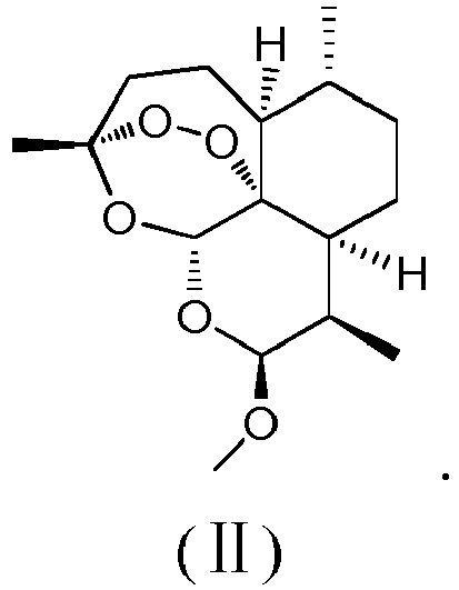
- The use, artemisinin analogue or method according to any one of claims 1 to 5, characterized in that, said promoting lipolysis comprises promoting browning of white fat.
- The use, artemisinin analogue or method according to any one of claims 1 to 5, characterized in that, said promoting lipolysis and/or preventing or treating a metabolism-related disease comprises controlling weight and improving metabolism.
- The use, artemisinin analogue or method according to any one of claims 1 to 5, characterized in that, said metabolism-related disease is hyperglycemia, insulin resistance, dyslipidemia, and/or fatty liver caused by obesity.
- A pharmaceutical composition for promoting lipolysis and/or preventing or treating a metabolism-related disease, characterized in that, said pharmaceutical composition comprises: (1) the artemisinin analogue according to any one of claims 4 to 5, and (2) a pharmaceutically acceptable carrier.
- The method according to claim 9, characterized in that, preferably, the dosage form of said pharmaceutical composition for oral administration is selected from an oral suspension, a powder and a granule; and the dosage form of said pharmaceutical composition for injection is selected from an aqueous solution, an oil and a suspension.
Applications Claiming Priority (2)
| Application Number | Priority Date | Filing Date | Title |
|---|---|---|---|
| CN201610305363.5A CN107349198A (en) | 2016-05-10 | 2016-05-10 | Artemether is preparing the purposes in promoting lipolysis and improving glycometabolism preparation |
| PCT/CN2017/082359 WO2017193824A1 (en) | 2016-05-10 | 2017-04-28 | Artemisinin analog, and use, method, and composition for promoting lipid catabolism and improving sugar metabolism |
Publications (2)
| Publication Number | Publication Date |
|---|---|
| EP3456326A1 true EP3456326A1 (en) | 2019-03-20 |
| EP3456326A4 EP3456326A4 (en) | 2020-01-15 |
Family
ID=60266197
Family Applications (1)
| Application Number | Title | Priority Date | Filing Date |
|---|---|---|---|
| EP17795442.7A Withdrawn EP3456326A4 (en) | 2016-05-10 | 2017-04-28 | ARTEMISININ ANALOG AND USE, METHOD AND COMPOSITION FOR PROMOTING FAT CATOLISM AND IMPROVING SUGAR METABOLISM |
Country Status (5)
| Country | Link |
|---|---|
| US (1) | US20190133997A1 (en) |
| EP (1) | EP3456326A4 (en) |
| JP (1) | JP2019521085A (en) |
| CN (1) | CN107349198A (en) |
| WO (1) | WO2017193824A1 (en) |
Families Citing this family (15)
| Publication number | Priority date | Publication date | Assignee | Title |
|---|---|---|---|---|
| WO2019153325A1 (en) * | 2018-02-12 | 2019-08-15 | 深圳市中医院 | Application of artemisinin derivative in preparation of medicament for preventing and treating type 2 diabetes and complications thereof, and pharmaceutical composition |
| CN108403686A (en) * | 2018-02-12 | 2018-08-17 | 深圳市中医院 | Application and pharmaceutical composition of the artemisinin derivative in preparing prevention and treatment diabetes B and its complication medicine |
| CN108403687A (en) * | 2018-03-29 | 2018-08-17 | 上海壹志医药科技有限公司 | The new pharmaceutical uses of Artemether |
| CN108478556A (en) * | 2018-03-29 | 2018-09-04 | 上海壹志医药科技有限公司 | The new pharmaceutical uses of dihydroartemisinine |
| CN110548024A (en) * | 2018-05-30 | 2019-12-10 | 中国科学技术大学 | Therapeutic use of water-soluble artemisinin derivative-aminoethylartemisinin maleate (SM934) |
| CN109172559B (en) * | 2018-09-27 | 2022-08-19 | 复旦大学 | Application of ethacrynic acid in preparing medicine for treating and preventing obesity and related metabolic diseases |
| CN109394757A (en) * | 2018-10-08 | 2019-03-01 | 澳门大学 | Artemether is preventing and is treating the application in Alzheimer's disease |
| CN110279802A (en) * | 2019-07-10 | 2019-09-27 | 珠海汉唐生物科技工程有限公司 | A kind of Chinese herbal medicine formula that treating diabetes and its preparation and application |
| CN110368383A (en) * | 2019-08-23 | 2019-10-25 | 西南大学 | Dihydroqinghaosu is preparing the application in blood lipid-lowering medicine |
| CN111235098B (en) * | 2020-01-17 | 2022-06-28 | 康妍葆(北京)干细胞科技有限公司 | Application of artemisinin in preparation of stem cell culture product, culture medium and culture method |
| CN112675163A (en) * | 2021-01-13 | 2021-04-20 | 上海交通大学 | Application of compound taking intra-epoxy bridged sesquiterpene as mother nucleus in metabolic-related fatty liver disease |
| JP7853079B2 (en) * | 2021-07-19 | 2026-04-28 | 株式会社ディーエイチシー | Evaluation methods for functional ingredients that support dieting, supplements, and diet foods. |
| CN114807015B (en) * | 2022-05-20 | 2022-11-22 | 呈诺再生医学科技(北京)有限公司 | Induction method for promoting islet alpha cells to be converted into beta cells and application thereof |
| CN117100740A (en) * | 2023-02-07 | 2023-11-24 | 中国中医科学院中药研究所 | The use of dihydroartemisinin in the treatment of hepatic glucose metabolism |
| CN116531317A (en) * | 2023-05-23 | 2023-08-04 | 上海市浦东医院(复旦大学附属浦东医院) | Antitumor hydrogel based on hunger therapy and preparation and application thereof |
Family Cites Families (3)
| Publication number | Priority date | Publication date | Assignee | Title |
|---|---|---|---|---|
| CN101879158B (en) * | 2010-07-01 | 2012-02-29 | 中国科学院广州生物医药与健康研究院 | New applications of artemisinin derivatives |
| CN104398505B (en) * | 2013-04-10 | 2017-06-13 | 上海交通大学 | Artemisine compounds are preparing the purposes in treating and preventing high blood cholesterol drug |
| EP2929881A1 (en) * | 2014-04-11 | 2015-10-14 | CeMM - Forschungszentrum für Molekulare Medizin GmbH | Medical use of artemisinin compounds and gephyrin agonists |
-
2016
- 2016-05-10 CN CN201610305363.5A patent/CN107349198A/en active Pending
-
2017
- 2017-04-28 EP EP17795442.7A patent/EP3456326A4/en not_active Withdrawn
- 2017-04-28 JP JP2018557930A patent/JP2019521085A/en active Pending
- 2017-04-28 WO PCT/CN2017/082359 patent/WO2017193824A1/en not_active Ceased
- 2017-04-28 US US16/099,195 patent/US20190133997A1/en not_active Abandoned
Also Published As
| Publication number | Publication date |
|---|---|
| WO2017193824A1 (en) | 2017-11-16 |
| CN107349198A (en) | 2017-11-17 |
| EP3456326A4 (en) | 2020-01-15 |
| US20190133997A1 (en) | 2019-05-09 |
| JP2019521085A (en) | 2019-07-25 |
Similar Documents
| Publication | Publication Date | Title |
|---|---|---|
| EP3456326A1 (en) | Artemisinin analog, and use, method, and composition for promoting lipid catabolism and improving sugar metabolism | |
| Audano et al. | Regulatory mechanisms of the early phase of white adipocyte differentiation: an overview | |
| Stine et al. | EBF2 promotes the recruitment of beige adipocytes in white adipose tissue | |
| Wu et al. | Black tea polyphenols and polysaccharides improve body composition, increase fecal fatty acid, and regulate fat metabolism in high-fat diet-induced obese rats | |
| Zhang et al. | The protective effect of resveratrol on islet insulin secretion and morphology in mice on a high-fat diet | |
| Qian et al. | Novel hydrogen sulfide-releasing compound, S-propargyl-cysteine, prevents STZ-induced diabetic nephropathy | |
| Tan et al. | Purified vitexin compound 1 suppresses tumor growth and induces cell apoptosis in a mouse model of human choriocarcinoma | |
| Bao et al. | Naringin prevents follicular atresia by inhibiting oxidative stress in the aging chicken | |
| CN104873500A (en) | Application of compound ZLN005 | |
| Gong et al. | Different roles of insulin receptor a and b in maintaining blood glucose homeostasis in zebrafish | |
| Fang et al. | Ups and downs: The PPARγ/p-PPARγ seesaw of follistatin-like 1 and integrin receptor signaling in adipogenesis | |
| Holik et al. | Nϵ‐Carboxymethyllysine Increases the Expression of miR‐103/143 and Enhances Lipid Accumulation in 3T3‐L1 Cells | |
| CN109550051B (en) | Application of histone demethylase KDM6A inhibitor in preparation of obesity treatment drug | |
| Li et al. | PGC-1α Promotes mitochondrial biosynthesis and energy metabolism of goose fatty liver | |
| Zhang et al. | Glucagon-like peptide-1 analog liraglutide reduces fat deposition in chicken adipocytes | |
| Murata et al. | EGCG down-regulates MuRF1 expression through 67-kDa laminin receptor and the receptor signaling is amplified by eriodictyol | |
| Chen et al. | Hibiscus manihot L improves obesity in mice induced by a high-fat diet | |
| Moriyama et al. | Royal jelly maintains epidermal stem cell properties by repressing senescence | |
| Chen et al. | Ovarian expression of functional MTTP and apoB for VLDL assembly and secretion in chickens | |
| Fu et al. | MUSTN1 and FABP3 interact to regulate adipogenesis and lipid deposition | |
| WO2020247918A1 (en) | Production and use of extracellular vesicle-contained enampt | |
| CN111356468B (en) | Composition for preventing or treating fibrotic diseases comprising Rhus succedanea extract as active ingredient | |
| CN110257512A (en) | Markers and compositions for diagnosis, treatment and prognosis of luminal and HER2 breast cancer | |
| KR102559516B1 (en) | Composition comprising milk exosomes for inducing browning of white adipose tissue | |
| Shah et al. | Patuletin Ameliorates Inflammation and Letrozole–Induced Polycystic Ovarian Syndrome in Rats |
Legal Events
| Date | Code | Title | Description |
|---|---|---|---|
| STAA | Information on the status of an ep patent application or granted ep patent |
Free format text: STATUS: THE INTERNATIONAL PUBLICATION HAS BEEN MADE |
|
| PUAI | Public reference made under article 153(3) epc to a published international application that has entered the european phase |
Free format text: ORIGINAL CODE: 0009012 |
|
| STAA | Information on the status of an ep patent application or granted ep patent |
Free format text: STATUS: REQUEST FOR EXAMINATION WAS MADE |
|
| 17P | Request for examination filed |
Effective date: 20181026 |
|
| AK | Designated contracting states |
Kind code of ref document: A1 Designated state(s): AL AT BE BG CH CY CZ DE DK EE ES FI FR GB GR HR HU IE IS IT LI LT LU LV MC MK MT NL NO PL PT RO RS SE SI SK SM TR |
|
| AX | Request for extension of the european patent |
Extension state: BA ME |
|
| DAV | Request for validation of the european patent (deleted) | ||
| DAX | Request for extension of the european patent (deleted) | ||
| A4 | Supplementary search report drawn up and despatched |
Effective date: 20191213 |
|
| RIC1 | Information provided on ipc code assigned before grant |
Ipc: A61P 3/04 20060101ALI20191209BHEP Ipc: A61P 3/10 20060101ALI20191209BHEP Ipc: A61P 1/16 20060101ALI20191209BHEP Ipc: A61P 3/06 20060101ALI20191209BHEP Ipc: A61K 31/541 20060101ALI20191209BHEP Ipc: A61K 31/357 20060101AFI20191209BHEP Ipc: A61K 31/366 20060101ALI20191209BHEP Ipc: A61P 5/50 20060101ALI20191209BHEP |
|
| STAA | Information on the status of an ep patent application or granted ep patent |
Free format text: STATUS: THE APPLICATION IS DEEMED TO BE WITHDRAWN |
|
| 18D | Application deemed to be withdrawn |
Effective date: 20200721 |