EP3447074A2 - Human antigen binding proteins that bind to a complex comprising beta-klotho and an fgf receptor - Google Patents
Human antigen binding proteins that bind to a complex comprising beta-klotho and an fgf receptor Download PDFInfo
- Publication number
- EP3447074A2 EP3447074A2 EP18179820.8A EP18179820A EP3447074A2 EP 3447074 A2 EP3447074 A2 EP 3447074A2 EP 18179820 A EP18179820 A EP 18179820A EP 3447074 A2 EP3447074 A2 EP 3447074A2
- Authority
- EP
- European Patent Office
- Prior art keywords
- seq
- cdrl1
- cdrh3
- cdrl3
- cdrh2
- Prior art date
- Legal status (The legal status is an assumption and is not a legal conclusion. Google has not performed a legal analysis and makes no representation as to the accuracy of the status listed.)
- Pending
Links
Images









Classifications
-
- C—CHEMISTRY; METALLURGY
- C07—ORGANIC CHEMISTRY
- C07K—PEPTIDES
- C07K16/00—Immunoglobulins [IG], e.g. monoclonal or polyclonal antibodies
- C07K16/18—Immunoglobulins [IG], e.g. monoclonal or polyclonal antibodies against material from animals or humans
- C07K16/28—Immunoglobulins [IG], e.g. monoclonal or polyclonal antibodies against material from animals or humans against receptors, cell surface antigens or cell surface determinants
- C07K16/2863—Immunoglobulins [IG], e.g. monoclonal or polyclonal antibodies against material from animals or humans against receptors, cell surface antigens or cell surface determinants against receptors for growth factors, growth regulators
-
- A—HUMAN NECESSITIES
- A61—MEDICAL OR VETERINARY SCIENCE; HYGIENE
- A61P—SPECIFIC THERAPEUTIC ACTIVITY OF CHEMICAL COMPOUNDS OR MEDICINAL PREPARATIONS
- A61P3/00—Drugs for disorders of the metabolism
-
- A—HUMAN NECESSITIES
- A61—MEDICAL OR VETERINARY SCIENCE; HYGIENE
- A61P—SPECIFIC THERAPEUTIC ACTIVITY OF CHEMICAL COMPOUNDS OR MEDICINAL PREPARATIONS
- A61P3/00—Drugs for disorders of the metabolism
- A61P3/04—Anorexiants; Antiobesity agents
-
- A—HUMAN NECESSITIES
- A61—MEDICAL OR VETERINARY SCIENCE; HYGIENE
- A61P—SPECIFIC THERAPEUTIC ACTIVITY OF CHEMICAL COMPOUNDS OR MEDICINAL PREPARATIONS
- A61P3/00—Drugs for disorders of the metabolism
- A61P3/06—Antihyperlipidemics
-
- A—HUMAN NECESSITIES
- A61—MEDICAL OR VETERINARY SCIENCE; HYGIENE
- A61P—SPECIFIC THERAPEUTIC ACTIVITY OF CHEMICAL COMPOUNDS OR MEDICINAL PREPARATIONS
- A61P3/00—Drugs for disorders of the metabolism
- A61P3/08—Drugs for disorders of the metabolism for glucose homeostasis
- A61P3/10—Drugs for disorders of the metabolism for glucose homeostasis for hyperglycaemia, e.g. antidiabetics
-
- A—HUMAN NECESSITIES
- A61—MEDICAL OR VETERINARY SCIENCE; HYGIENE
- A61P—SPECIFIC THERAPEUTIC ACTIVITY OF CHEMICAL COMPOUNDS OR MEDICINAL PREPARATIONS
- A61P9/00—Drugs for disorders of the cardiovascular system
-
- A—HUMAN NECESSITIES
- A61—MEDICAL OR VETERINARY SCIENCE; HYGIENE
- A61P—SPECIFIC THERAPEUTIC ACTIVITY OF CHEMICAL COMPOUNDS OR MEDICINAL PREPARATIONS
- A61P9/00—Drugs for disorders of the cardiovascular system
- A61P9/10—Drugs for disorders of the cardiovascular system for treating ischaemic or atherosclerotic diseases, e.g. antianginal drugs, coronary vasodilators, drugs for myocardial infarction, retinopathy, cerebrovascula insufficiency, renal arteriosclerosis
-
- C—CHEMISTRY; METALLURGY
- C07—ORGANIC CHEMISTRY
- C07K—PEPTIDES
- C07K14/00—Peptides having more than 20 amino acids; Gastrins; Somatostatins; Melanotropins; Derivatives thereof
- C07K14/435—Peptides having more than 20 amino acids; Gastrins; Somatostatins; Melanotropins; Derivatives thereof from animals; from humans
- C07K14/705—Receptors; Cell surface antigens; Cell surface determinants
- C07K14/71—Receptors; Cell surface antigens; Cell surface determinants for growth factors; for growth regulators
-
- C—CHEMISTRY; METALLURGY
- C07—ORGANIC CHEMISTRY
- C07K—PEPTIDES
- C07K16/00—Immunoglobulins [IG], e.g. monoclonal or polyclonal antibodies
- C07K16/40—Immunoglobulins [IG], e.g. monoclonal or polyclonal antibodies against enzymes
-
- C—CHEMISTRY; METALLURGY
- C12—BIOCHEMISTRY; BEER; SPIRITS; WINE; VINEGAR; MICROBIOLOGY; ENZYMOLOGY; MUTATION OR GENETIC ENGINEERING
- C12Y—ENZYMES
- C12Y302/00—Hydrolases acting on glycosyl compounds, i.e. glycosylases (3.2)
- C12Y302/01—Glycosidases, i.e. enzymes hydrolysing O- and S-glycosyl compounds (3.2.1)
- C12Y302/01031—Beta-glucuronidase (3.2.1.31)
-
- A—HUMAN NECESSITIES
- A61—MEDICAL OR VETERINARY SCIENCE; HYGIENE
- A61K—PREPARATIONS FOR MEDICAL, DENTAL OR TOILETRY PURPOSES
- A61K39/00—Medicinal preparations containing antigens or antibodies
- A61K2039/505—Medicinal preparations containing antigens or antibodies comprising antibodies
-
- C—CHEMISTRY; METALLURGY
- C07—ORGANIC CHEMISTRY
- C07K—PEPTIDES
- C07K2317/00—Immunoglobulins specific features
- C07K2317/20—Immunoglobulins specific features characterized by taxonomic origin
- C07K2317/21—Immunoglobulins specific features characterized by taxonomic origin from primates, e.g. man
-
- C—CHEMISTRY; METALLURGY
- C07—ORGANIC CHEMISTRY
- C07K—PEPTIDES
- C07K2317/00—Immunoglobulins specific features
- C07K2317/30—Immunoglobulins specific features characterized by aspects of specificity or valency
- C07K2317/31—Immunoglobulins specific features characterized by aspects of specificity or valency multispecific
-
- C—CHEMISTRY; METALLURGY
- C07—ORGANIC CHEMISTRY
- C07K—PEPTIDES
- C07K2317/00—Immunoglobulins specific features
- C07K2317/50—Immunoglobulins specific features characterized by immunoglobulin fragments
- C07K2317/52—Constant or Fc region; Isotype
-
- C—CHEMISTRY; METALLURGY
- C07—ORGANIC CHEMISTRY
- C07K—PEPTIDES
- C07K2317/00—Immunoglobulins specific features
- C07K2317/50—Immunoglobulins specific features characterized by immunoglobulin fragments
- C07K2317/56—Immunoglobulins specific features characterized by immunoglobulin fragments variable (Fv) region, i.e. VH and/or VL
-
- C—CHEMISTRY; METALLURGY
- C07—ORGANIC CHEMISTRY
- C07K—PEPTIDES
- C07K2317/00—Immunoglobulins specific features
- C07K2317/50—Immunoglobulins specific features characterized by immunoglobulin fragments
- C07K2317/56—Immunoglobulins specific features characterized by immunoglobulin fragments variable (Fv) region, i.e. VH and/or VL
- C07K2317/565—Complementarity determining region [CDR]
-
- C—CHEMISTRY; METALLURGY
- C07—ORGANIC CHEMISTRY
- C07K—PEPTIDES
- C07K2317/00—Immunoglobulins specific features
- C07K2317/70—Immunoglobulins specific features characterized by effect upon binding to a cell or to an antigen
- C07K2317/75—Agonist effect on antigen
-
- C—CHEMISTRY; METALLURGY
- C07—ORGANIC CHEMISTRY
- C07K—PEPTIDES
- C07K2319/00—Fusion polypeptide
-
- C—CHEMISTRY; METALLURGY
- C07—ORGANIC CHEMISTRY
- C07K—PEPTIDES
- C07K2319/00—Fusion polypeptide
- C07K2319/30—Non-immunoglobulin-derived peptide or protein having an immunoglobulin constant or Fc region, or a fragment thereof, attached thereto
Definitions
- the present disclosure relates to nucleic acid molecules encoding antigen binding proteins that bind to a complex comprising ⁇ -Klotho and at least one of (i) FGFR1c, (ii) FGFR2c and (iii) FGFR3c, including antigen binding proteins that induce FGF21-like signaling, as well as pharmaceutical compositions comprising antigen binding proteins that bind to a complex comprising ⁇ -Klotho and at least one of (i) FGFR1c, (ii) FGFR2c and (iii) FGFR3c, including antigen binding proteins that induce FGF21-like signaling, and methods for treating metabolic disorders using such nucleic acids, polypeptides, or pharmaceutical compositions. Diagnostic methods using the antigen binding proteins are also provided.
- Fibroblast Growth Factor 21 is a secreted polypeptide that belongs to a subfamily of Fibroblast Growth Factors (FGFs) that includes FGF19, FGF21, and FGF23 ( Itoh et al., (2004) Trend Genet. 20:563-69 ).
- FGF21 is an atypical FGF in that it is heparin independent and functions as a hormone in the regulation of glucose, lipid, and energy metabolism.
- FGF21 farnesoid GF21
- Transgenic mice overexpressing FGF21 exhibit metabolic phenotypes of slow growth rate, low plasma glucose and triglyceride levels, and an absence of age-associated type 2 diabetes, islet hyperplasia, and obesity.
- Pharmacological administration of recombinant FGF21 protein in rodent and primate models results in normalized levels of plasma glucose, reduced triglyceride and cholesterol levels, and improved glucose tolerance and insulin sensitivity.
- FGF21 reduces body weight and body fat by increasing energy expenditure, physical activity, and metabolic rate.
- Experimental research provides support for the pharmacological administration of FGF21 for the treatment of type 2 diabetes, obesity, dyslipidemia, and other metabolic conditions or disorders in humans.
- FGF21 is a liver derived endocrine hormone that stimulates glucose uptake in adipocytes and lipid homeostasis through the activation of its receptor.
- the FGF21 receptor also comprises the membrane associated ⁇ -Klotho as an essential cofactor. Activation of the FGF21 receptor leads to multiple effects on a variety of metabolic parameters.
- FGFs mediate their action via a set of four FGF receptors, FGFR1-4, that in turn are expressed in multiple spliced variants, e.g ., FGFR1c, FGFR2c, FGFR3c and FGFR4.
- Each FGF receptor contains an intracellular tyrosine kinase domain that is activated upon ligand binding, leading to downstream signaling pathways involving MAPKs (Erk1/2), RAF1, AKT1 and STATs. ( Kharitonenkov et al., (2008) BioDrugs 22:37-44 ).
- the present disclosure provides a human (or humanized) antigen binding protein, such as a monoclonal antibody, that induces FGF21-like signaling, e.g ., an agonistic antibody that mimics the function of FGF21.
- a human (or humanized) antigen binding protein such as a monoclonal antibody
- FGF21-like signaling e.g ., an agonistic antibody that mimics the function of FGF21.
- an antibody is a molecule with FGF21-like activity and selectivity but with added therapeutically desirable characteristics typical for an antibody such as protein stability, lack of immunogenicity, ease of production and long half-life in vivo.
- the instant disclosure provides antigen binding proteins that bind a complex comprising ⁇ -Klotho and at least one of (i) FGFR1c, (ii) FGFR2c and (iii) FGFR3c, including antigen binding proteins that induce FGF21-like signaling, as well as pharmaceutical compositions comprising antigen binding proteins that bind to a complex comprising ⁇ -Klotho and at least one of (i) FGFR1c, (ii) FGFR2c and (iii) FGFR3c, including antigen binding proteins that induce FGF21-like signaling.
- expression vectors and host cells transformed or transfected with the expression vectors that comprise the aforementioned isolated nucleic acid molecules that encode the antigen binding proteins disclosed herein are provided in Tables 1A and 1B; representative variable region heavy chain and light chain sequences are provided in Tables 2A and 2B; coding sequences for the variable region of the heavy and light chains are provided in Tables 2C and 2D; Tables 3A and 3B provide CDR regions of the disclosed variable heavy and light chains, and Tables 3C and 3D provide coding sequences for the disclosed CDRs.
- methods of preparing antigen binding proteins that specifically or selectively bind a complex comprising ⁇ -Klotho and at least one of (i) FGFR1c, (ii) FGFR2c and (iii) FGFR3c and comprise the step of preparing the antigen binding protein from a host cell that secretes the antigen binding protein.
- inventions provide a method of preventing or treating a condition in a subject in need of such treatment comprising administering a therapeutically effective amount of a pharmaceutical composition provided herein to a subject, wherein the condition is treatable by lowering blood glucose, insulin or serum lipid levels.
- the condition is type 2 diabetes, obesity, dyslipidemia, NASH, cardiovascular disease or metabolic syndrome.
- an "antigen binding protein” is a protein comprising a portion that binds to an antigen or target and, optionally, a scaffold or framework portion that allows the antigen binding portion to adopt a conformation that promotes binding of the antigen binding protein to the antigen.
- antigen binding proteins examples include a human antibody, a humanized antibody; a chimeric antibody; a recombinant antibody; a single chain antibody; a diabody; a triabody; a tetrabody; a Fab fragment; a F(ab') 2 fragment; an IgD antibody; an IgE antibody; an IgM antibody; an IgG1 antibody; an IgG2 antibody; an IgG3 antibody; or an IgG4 antibody, and fragments thereof.
- the antigen binding protein can comprise, for example, an alternative protein scaffold or artificial scaffold with grafted CDRs or CDR derivatives.
- Such scaffolds include, but are not limited to, antibody-derived scaffolds comprising mutations introduced to, for example, stabilize the three-dimensional structure of the antigen binding protein as well as wholly synthetic scaffolds comprising, for example, a biocompatible polymer. See, e.g., Korndorfer et al., (2003) Proteins: Structure, Function, and Bioinformatics, 53(1):121-129 ; Roque et al., (2004) Biotechnol. Prog. 20:639-654 .
- PAMs peptide antibody mimetics
- scaffolds based on antibody mimetics utilizing fibronectin components as a scaffold.
- An antigen binding protein can have, for example, the structure of a naturally occurring immunoglobulin.
- An "immunoglobulin” is a tetrameric molecule. In a naturally occurring immunoglobulin, each tetramer is composed of two identical pairs of polypeptide chains, each pair having one "light” (about 25 kDa) and one "heavy" chain (about 50-70 kDa).
- the amino-terminal portion of each chain includes a variable region of about 100 to 110 or more amino acids primarily responsible for antigen recognition.
- the carboxy-terminal portion of each chain defines a constant region primarily responsible for effector function. Human light chains are classified as kappa and lambda light chains.
- Heavy chains are classified as mu, delta, gamma, alpha, or epsilon, and define the antibody's isotype as IgM, IgD, IgG, IgA, and IgE, respectively.
- the variable and constant regions are joined by a "J" region of about 12 or more amino acids, with the heavy chain also including a "D” region of about 10 more amino acids. See generally, Fundamental Immunology 2nd ed. Ch. 7 (Paul, W., ed., Raven Press, N.Y. (1989 )), incorporated by reference in its entirety for all purposes.
- the variable regions of each light/heavy chain pair form the antibody binding site such that an intact immunoglobulin has two binding sites.
- Naturally occurring immunoglobulin chains exhibit the same general structure of relatively conserved framework regions (FR) joined by three hypervariable regions, also called complementarity determining regions or CDRs. From N-terminus to C-terminus, both light and heavy chains comprise the domains FR1, CDR1, FR2, CDR2, FR3, CDR3 and FR4.
- the assignment of amino acids to each domain can be done in accordance with the definitions of Kabat et al., (1991) "Sequences of Proteins of Immunological Interest", 5th Ed., US Dept. of Health and Human Services, PHS, NIH, NIH Publication no. 91-3242 .
- the CDRs disclosed herein can also be redefined according an alternative nomenclature scheme, such as that of Chothia (see Chothia & Lesk, (1987) J. Mol. Biol. 196:901-917 ; Chothia et al., (1989) Nature 342:878-883 or Honegger & Pluckthun, (2001) J. Mol. Biol. 309:657-670 ).
- an antigen binding protein is said to "specifically bind” or “selectively bind” its target antigen when the dissociation constant (K D ) is ⁇ 10 -8 M.
- the antibody specifically binds antigen with "high affinity” when the K D is ⁇ 5x 10 -9 M, and with “very high affinity” when the K D is ⁇ 5x 10 -10 M.
- the antibodies will bind to a complex comprising ⁇ -Klotho and an FGFR, including a complex comprising both human FGFR1c and human ⁇ -Klotho, with a K D of between about 10 -7 M and 10 -12 M, and in yet another embodiment the antibodies will bind with a K D ⁇ 5x 10 -9 .
- an “antibody” refers to an intact immunoglobulin or to an antigen binding portion thereof that competes with the intact antibody for specific binding, unless otherwise specified.
- Antigen binding portions can be produced by recombinant DNA techniques or by enzymatic or chemical cleavage of intact antibodies.
- Antigen binding portions include, inter alia, Fab, Fab', F(ab') 2 , Fv, domain antibodies (dAbs), fragments including complementarity determining regions (CDRs), single-chain antibodies (scFv), chimeric antibodies, diabodies, triabodies, tetrabodies, and polypeptides that contain at least a portion of an immunoglobulin that is sufficient to confer specific antigen binding to the polypeptide.
- a Fab fragment is a monovalent fragment having the V L , V H , C L and C H 1 domains; a F(ab') 2 fragment is a bivalent fragment having two Fab fragments linked by a disulfide bridge at the hinge region; a Fd fragment has the V H and C H 1 domains; an Fv fragment has the V L and V H domains of a single arm of an antibody; and a dAb fragment has a V H domain, a V L domain, or an antigen-binding fragment of a V H or V L domain ( US Patent Nos. 6,846,634 , and 6,696,245 ; and US App. Pub. Nos. 05/0202512 , 04/0202995 , 04/0038291 , 04/0009507 , 03/0039958 , Ward et al., Nature 341:544-546 (1989 )).
- a single-chain antibody is an antibody in which a V L and a V H region are joined via a linker (e.g., a synthetic sequence of amino acid residues) to form a continuous protein chain wherein the linker is long enough to allow the protein chain to fold back on itself and form a monovalent antigen binding site (see, e.g., Bird et al., (1988) Science 242:423-26 and Huston et al., (1988) Proc. Natl. Acad. Sci. USA 85:5879-83 ).
- a linker e.g., a synthetic sequence of amino acid residues
- Diabodies are bivalent antibodies comprising two polypeptide chains, wherein each polypeptide chain comprises V H and V L domains joined by a linker that is too short to allow for pairing between two domains on the same chain, thus allowing each domain to pair with a complementary domain on another polypeptide chain (see, e.g., Holliger et al., (1993) Proc. Natl. Acad. Sci. USA 90:6444-48 , and Poljak et al., (1994) Structure 2:1121-23 ). If the two polypeptide chains of a diabody are identical, then a diabody resulting from their pairing will have two identical antigen binding sites.
- Polypeptide chains having different sequences can be used to make a diabody with two different antigen binding sites.
- tribodies and tetrabodies are antibodies comprising three and four polypeptide chains, respectively, and forming three and four antigen binding sites, respectively, which can be the same or different.
- Complementarity determining regions (CDRs) and framework regions (FR) of a given antibody can be identified using the system described by Kabat et al., (1991) "Sequences of Proteins of Immunological Interest", 5th Ed., US Dept. of Health and Human Services, PHS, NIH, NIH Publication no. 91-3242 . Although presented using the Kabat nomenclature system, as desired, the CDRs disclosed herein can also be redefined according an alternative nomenclature scheme, such as that of Chothia (see Chothia & Lesk, (1987) J. Mol. Biol.
- One or more CDRs can be incorporated into a molecule either covalently or noncovalently to make it an antigen binding protein.
- An antigen binding protein can incorporate the CDR(s) as part of a larger polypeptide chain, can covalently link the CDR(s) to another polypeptide chain, or can incorporate the CDR(s) noncovalently.
- the CDRs permit the antigen binding protein to specifically bind to a particular antigen of interest.
- An antigen binding protein can but need not have one or more binding sites. If there is more than one binding site, the binding sites can be identical to one another or can be different. For example, a naturally occurring human immunoglobulin typically has two identical binding sites, while a "bispecific" or “bifunctional” antibody has two different binding sites. Antigen binding proteins of this bispecific form (e.g., those comprising various heavy and light chain CDRs provided herein) comprise aspects of the instant disclosure.
- human antibody includes all antibodies that have one or more variable and constant regions derived from human immunoglobulin sequences. In one embodiment, all of the variable and constant domains are derived from human immunoglobulin sequences (a fully human antibody).
- These antibodies can be prepared in a variety of ways, examples of which are described below, including through the immunization with an antigen of interest of a mouse that is genetically modified to express antibodies derived from human heavy and/or light chain-encoding genes, such as a mouse derived from a XENOMOUSE®, ULTIMABTM, HUMAB-MOUSE®, VELOCIMOUSE®, VELOCIMMUNE®, KYMOUSE, or ALIVAMAB system, or derived from human heavy chain transgenic mouse, transgenic rat human antibody repertoire, transgenic rabbit human antibody repertoire or cow human antibody repertoire or HUTARGTM technology. Phage-based approaches can also be employed.
- a humanized antibody has a sequence that differs from the sequence of an antibody derived from a non-human species by one or more amino acid substitutions, deletions, and/or additions, such that the humanized antibody is less likely to induce an immune response, and/or induces a less severe immune response, as compared to the non-human species antibody, when it is administered to a human subject.
- certain amino acids in the framework and constant domains of the heavy and/or light chains of the non-human species antibody are mutated to produce the humanized antibody.
- the constant domain(s) from a human antibody are fused to the variable domain(s) of a non-human species.
- one or more amino acid residues in one or more CDR sequences of a non-human antibody are changed to reduce the likely immunogenicity of the non-human antibody when it is administered to a human subject, wherein the changed amino acid residues either are not critical for immunospecific binding of the antibody to its antigen, or the changes to the amino acid sequence that are made are conservative changes, such that the binding of the humanized antibody to the antigen is not significantly worse than the binding of the non-human antibody to the antigen. Examples of how to make humanized antibodies can be found in U.S. Patent Nos. 6,054,297 , 5,886,152 and 5,877,293 .
- chimeric antibody refers to an antibody that contains one or more regions from one antibody and one or more regions from one or more other antibodies.
- one or more of the CDRs are derived from a human antibody that binds to a complex comprising ⁇ -Klotho and at least one of (i) FGFR1c, (ii) FGFR2c and (iii) FGFR3c.
- all of the CDRs are derived from a human antibody that binds to a complex ⁇ -Klotho and at least one of (i) FGFR1c, (ii) FGFR2c and (iii) FGFR3c.
- the CDRs from more than one human antibody that binds to a complex comprising ⁇ -Klotho and at least one of (i) FGFR1c, (ii) FGFR2c and (iii) FGFR3c are mixed and matched in a chimeric antibody.
- a chimeric antibody can comprise a CDR1 from the light chain of a first human antibody that binds to a complex comprising ⁇ -Klotho and at least one of (i) FGFR1c, (ii) FGFR2c and (iii) FGFR3c, a CDR2 and a CDR3 from the light chain of a second human antibody that binds to a complex comprising ⁇ -Klotho and at least one of (i) FGFR1c, (ii) FGFR2c and (iii) FGFR3c, and the CDRs from the heavy chain from a third antibody that binds to a complex comprising ⁇ -Klotho and at least one of (i) FGFR1c, (ii) FGFR2c and (iii) FGFR3c.
- the framework regions can be derived from one of the same antibodies that bind to a complex comprising ⁇ -Klotho and at least one of (i) FGFR1c, (ii) FGFR2c and (iii) FGFR3c, from one or more different antibodies, such as a human antibody, or from a humanized antibody.
- a portion of the heavy and/or light chain is identical with, homologous to, or derived from an antibody from a particular species or belonging to a particular antibody class or subclass, while the remainder of the chain(s) is/are identical with, homologous to, or derived from an antibody or antibodies from another species or belonging to another antibody class or subclass.
- fragments of such antibodies that exhibit the desired biological activity (e.g., the ability to specifically bind to a complex comprising ⁇ -Klotho and at least one of (i) FGFR1c, (ii) FGFR2c and (iii) FGFR3c).
- light chain includes a full-length light chain and fragments thereof having sufficient variable region sequence to confer binding specificity.
- a full-length light chain includes a variable region domain, V L , and a constant region domain, C L .
- the variable region domain of the light chain is at the amino-terminus of the polypeptide.
- Light chains include kappa (" ⁇ ") chains and lambda (“ ⁇ ”) chains.
- heavy chain includes a full-length heavy chain and fragments thereof having sufficient variable region sequence to confer binding specificity.
- a full-length heavy chain includes a variable region domain, V H , and three constant region domains, C H 1, C H 2, and C H 3.
- the V H domain is at the amino-terminus of the polypeptide
- the C H domains are at the carboxyl-terminus, with the C H 3 being closest to the carboxy-terminus of the polypeptide.
- Heavy chains can be of any isotype, including IgG (including IgG1, IgG2, IgG3 and IgG4 subtypes), IgA (including IgA1 and IgA2 subtypes), IgM and IgE.
- an antigen binding protein e.g., an antibody or immunoglobulin chain (heavy or light chain)
- an antigen binding protein comprising a portion (regardless of how that portion is obtained or synthesized) of an antibody that lacks at least some of the amino acids present in a full-length chain but which is capable of specifically binding to an antigen.
- Such fragments are biologically active in that they bind specifically to the target antigen and can compete with other antigen binding proteins, including intact antibodies, for specific binding to a given epitope.
- such a fragment will retain at least one CDR present in the full-length light or heavy chain, and in some embodiments will comprise a single heavy chain and/or light chain or portion thereof.
- These biologically active fragments can be produced by recombinant DNA techniques, or can be produced by enzymatic or chemical cleavage of antigen binding proteins, including intact antibodies.
- Immunologically functional immunoglobulin fragments include, but are not limited to, Fab, Fab', F(ab') 2 , Fv, domain antibodies and single-chain antibodies, and can be derived from any mammalian source, including but not limited to human, mouse, rat, camelid or rabbit.
- a functional portion of the antigen binding proteins disclosed herein could be covalently bound to a second protein or to a small molecule to create a therapeutic agent directed to a particular target in the body, possessing bifunctional therapeutic properties, or having a prolonged serum half-life.
- An "Fc" region contains two heavy chain fragments comprising the C H 2 and C H 3 domains of an antibody.
- the two heavy chain fragments are held together by two or more disulfide bonds and by hydrophobic interactions of the C H 3 domains.
- Fab' fragment contains one light chain and a portion of one heavy chain that contains the V H domain and the C H 1 domain and also the region between the C H 1 and C H 2 domains, such that an interchain disulfide bond can be formed between the two heavy chains of two Fab' fragments to form an F(ab') 2 molecule.
- F(ab') 2 fragment contains two light chains and two heavy chains containing a portion of the constant region between the C H 1 and C H 2 domains, such that an interchain disulfide bond is formed between the two heavy chains.
- a F(ab') 2 fragment thus is composed of two Fab' fragments that are held together by a disulfide bond between the two heavy chains.
- the "Fv region” comprises the variable regions from both the heavy and light chains, but lacks the constant regions.
- a “domain antibody” is an immunologically functional immunoglobulin fragment containing only the variable region of a heavy chain or the variable region of a light chain.
- two or more V H regions are covalently joined with a peptide linker to create a bivalent domain antibody.
- the two V H regions of a bivalent domain antibody can target the same or different antigens.
- a “hemibody” is an immunologically-functional immunoglobulin construct comprising a complete heavy chain, a complete light chain and a second heavy chain Fc region paired with the Fc region of the complete heavy chain.
- a linker can, but need not, be employed to join the heavy chain Fc region and the second heavy chain Fc region.
- a hemibody is a monovalent form of an antigen binding protein disclosed herein.
- pairs of charged residues can be employed to associate one Fc region with the second Fc region.
- a “bivalent antigen binding protein” or “bivalent antibody” comprises two antigen binding sites. In some instances, the two binding sites have the same antigen specificities.
- Bivalent antigen binding proteins and bivalent antibodies can be bispecific, as described herein, and form aspects of the instant disclosure.
- a “multispecific antigen binding protein” or “multispecific antibody” is one that targets more than one antigen or epitope, and forms another aspect of the instant disclosure.
- a "bispecific,” “dual-specific” or “bifunctional” antigen binding protein or antibody is a hybrid antigen binding protein or antibody, respectively, having two different antigen binding sites.
- Bispecific antigen binding proteins and antibodies are a species of multispecific antigen binding protein or multispecific antibody and can be produced by a variety of methods including, but not limited to, fusion of hybridomas or linking of Fab' fragments. See, e.g., Songsivilai and Lachmann, (1990) Clin. Exp. Immunol. 79:315-321 ; Kostelny et al., (1992) J. Immunol. 148:1547-1553 .
- the two binding sites of a bispecific antigen binding protein or antibody will bind to two different epitopes, which can reside on the same (e.g., ⁇ -Klotho, FGFR1c, FGFR2c, or FGFR3c) or different protein targets (e.g., ⁇ -Klotho and one of (i) FGFR1c, (ii) FGFR2c, and (iii) FGFR3c).
- FGF21-like signaling and “induces FGF21-like signaling,” when applied to an antigen binding protein of the present disclosure, means that the antigen binding protein mimics, or modulates, an in vivo biological effect induced by the binding to a complex comprising ⁇ -Klotho and at least one of (i) FGFR1c, (ii) FGFR2c and (iii) FGFR3c and induces a biological response that otherwise would result from FGF21 binding to a complex comprising ⁇ -Klotho and at least one of (i) FGFR1c, (ii) FGFR2c and (iii) FGFR3c in vivo.
- an antibody or fragment is deemed to induce a biological response when the response is equal to or greater than 5%, and preferably equal to or greater than 10%, 15%, 20%, 25%, 30%, 35%, 40%, 45%, 50%, 55%, 60%, 65%, 70%, 75%, 80%, 85%, 90% or 95%, of the activity of a wild type FGF21 standard comprising the mature form of SEQ ID NO: 2 ( i.e., the mature form of the human FGF21 sequence) and has the following properties: exhibiting an efficacy level of equal to or more than 5% of an FGF21 standard, with an EC 50 of equal to or less than 100nM, e.g., 90 nM, 80 nM, 70nM, 60nM, 50nM, 40nM, 30nM, 20nM or 10 nM in (1) the recombinant FGF21 receptor-mediated lucifera
- the "potency" of an antigen binding protein is defined as exhibiting an EC50 of equal to or less than 100nM, e.g., 90nM, 80nM, 70nM, 60nM, 50nM, 40nM, 30nM, 20nM, 10 nM and preferably less than 10nM of the antigen binding protein in the following assays: (1) the recombinant FGF21 receptor mediated luciferase-reporter cell assay of Example 4; (2) the ERK-phosphorylation in the recombinant FGF21 receptor mediated cell assay of Example 4; and (3) ERK-phosphorylation in human adipocytes as described in Example 4.
- antigen binding proteins of the present disclosure induce FGF21-mediated signaling (e.g ., that induce agonistic activity), nor is this property desirable in all circumstances. Nevertheless, antigen binding proteins that do not induce FGF21-mediated signaling form aspects of the present disclosure and may be useful as diagnostic reagents or other applications.
- FGF21R means a multimeric receptor complex that FGF21 is known or suspected to form in vivo.
- FGF21R comprises (i) an FGFR, e.g. , FGFR1c, FGFR2c, FGFR3c or FGFR4, and (ii) ⁇ -Klotho.
- polynucleotide or “nucleic acid” includes both single-stranded and double-stranded nucleotide polymers.
- the nucleotides comprising the polynucleotide can be ribonucleotides or deoxyribonucleotides or a modified form of either type of nucleotide.
- Said modifications include base modifications such as bromouridine and inosine derivatives, ribose modifications such as 2', 3'-dideoxyribose, and internucleotide linkage modifications such as phosphorothioate, phosphorodithioate, phosphoroselenoate, phosphorodiselenoate, phosphoroanilothioate, phoshoraniladate and phosphoroamidate.
- oligonucleotide means a polynucleotide comprising 200 or fewer nucleotides. In some embodiments, oligonucleotides are 10 to 60 bases in length. In other embodiments, oligonucleotides are 12, 13, 14, 15, 16, 17, 18, 19, or 20 to 40 nucleotides in length. Oligonucleotides can be single stranded or double stranded, e.g ., for use in the construction of a mutant gene. Oligonucleotides can be sense or antisense oligonucleotides.
- An oligonucleotide can include a label, including a radiolabel, a fluorescent label, a hapten or an antigenic label, for detection assays. Oligonucleotides can be used, for example, as PCR primers, cloning primers or hybridization probes.
- isolated nucleic acid molecule means a DNA or RNA of genomic, mRNA, cDNA, or synthetic origin or some combination thereof which is not associated with all or a portion of a polynucleotide in which the isolated polynucleotide is found in nature, or is linked to a polynucleotide to which it is not linked in nature.
- a nucleic acid molecule comprising a particular nucleotide sequence does not encompass intact chromosomes.
- Isolated nucleic acid molecules "comprising" specified nucleic acid sequences can include, in addition to the specified sequences, coding sequences for up to ten or even up to twenty other proteins or portions thereof, or can include operably linked regulatory sequences that control expression of the coding region of the recited nucleic acid sequences, and/or can include vector sequences.
- the left-hand end of any single-stranded polynucleotide sequence discussed herein is the 5' end; the left-hand direction of double-stranded polynucleotide sequences is referred to as the 5' direction.
- the direction of 5' to 3' addition of nascent RNA transcripts is referred to as the transcription direction; sequence regions on the DNA strand having the same sequence as the RNA transcript that are 5' to the 5' end of the RNA transcript are referred to as "upstream sequences;" sequence regions on the DNA strand having the same sequence as the RNA transcript that are 3' to the 3' end of the RNA transcript are referred to as "downstream sequences.”
- control sequence refers to a polynucleotide sequence that can affect the expression and processing of coding sequences to which it is ligated. The nature of such control sequences can depend upon the host organism.
- control sequences for prokaryotes can include a promoter, a ribosomal binding site, and a transcription termination sequence.
- control sequences for eukaryotes can include promoters comprising one or a plurality of recognition sites for transcription factors, transcription enhancer sequences, and transcription termination sequence.
- Control sequences can include leader sequences and/or fusion partner sequences.
- vector means any molecule or entity (e.g ., nucleic acid, plasmid, bacteriophage or virus) used to transfer protein coding information into a host cell.
- expression vector refers to a vector that is suitable for transformation of a host cell and contains nucleic acid sequences that direct and/or control (in conjunction with the host cell) expression of one or more heterologous coding regions operatively linked thereto.
- An expression construct can include, but is not limited to, sequences that affect or control transcription, translation, and, if introns are present, affect RNA splicing of a coding region operably linked thereto.
- operably linked means that the components to which the term is applied are in a relationship that allows them to carry out their inherent functions under suitable conditions.
- a control sequence in a vector that is "operably linked" to a protein coding sequence is ligated thereto so that expression of the protein coding sequence is achieved under conditions compatible with the transcriptional activity of the control sequences.
- the term "host cell” means a cell that has been transformed, or is capable of being transformed, with a nucleic acid sequence and thereby expresses a gene of interest.
- the term includes the progeny of the parent cell, whether or not the progeny is identical in morphology or in genetic make-up to the original parent cell, so long as the gene of interest is present.
- transduction means the transfer of genes from one bacterium to another, usually by bacteriophage. "Transduction” also refers to the acquisition and transfer of eukaryotic cellular sequences by replication-defective retroviruses.
- transfection means the uptake of foreign or exogenous DNA by a cell, and a cell has been "transfected" when the exogenous DNA has been introduced inside the cell membrane.
- transfection techniques are well known in the art and are disclosed herein. See, e.g., Graham et al., (1973) Virology 52:456 ; Sambrook et al., (2001), supra ; Davis et al., (1986) Basic Methods in Molecular Biology, Elsevier ; Chu et al., (1981) Gene 13:197 .
- Such techniques can be used to introduce one or more exogenous DNA moieties into suitable host cells.
- transformation refers to a change in a cell's genetic characteristics, and a cell has been transformed when it has been modified to contain new DNA or RNA.
- a cell is transformed where it is genetically modified from its native state by introducing new genetic material via transfection, transduction, or other techniques.
- the transforming DNA can recombine with that of the cell by physically integrating into a chromosome of the cell, or can be maintained transiently as an episomal element without being replicated, or can replicate independently as a plasmid.
- a cell is considered to have been "stably transformed” when the transforming DNA is replicated with the division of the cell.
- polypeptide or "protein” are used interchangeably herein to refer to a polymer of amino acid residues.
- the terms also apply to amino acid polymers in which one or more amino acid residues is an analog or mimetic of a corresponding naturally occurring amino acid, as well as to naturally occurring amino acid polymers.
- the terms can also encompass amino acid polymers that have been modified, e.g., by the addition of carbohydrate residues to form glycoproteins, or phosphorylated.
- Polypeptides and proteins can be produced by a naturally-occurring and non-recombinant cell, or polypeptides and proteins can be produced by a genetically-engineered or recombinant cell.
- Polypeptides and proteins can comprise molecules having the amino acid sequence of a native protein, or molecules having deletions from, additions to, and/or substitutions of one or more amino acids of the native sequence.
- polypeptide and protein encompass antigen binding proteins that specifically or selectively bind to a complex comprising ⁇ -Klotho and an FGFR ( e.g ., FGFR1c, FGFR2c or FGFR3c), or sequences that have deletions from, additions to, and/or substitutions of one or more amino acids of an antigen binding protein that specifically or selectively binds to a complex comprising ⁇ -Klotho and at least one of (i) FGFR1c, (ii) FGFR2c and (iii) FGFR3c.
- polypeptide fragment refers to a polypeptide that has an amino-terminal deletion, a carboxyl-terminal deletion, and/or an internal deletion as compared with the full-length protein. Such fragments can also contain modified amino acids as compared with the full-length protein. In certain embodiments, fragments are about five to 500 amino acids long. For example, fragments can be at least 5, 6, 8, 10, 14, 20, 50, 70, 100, 110, 150, 200, 250, 300, 350, 400, or 450 amino acids long.
- Useful polypeptide fragments include immunologically functional fragments of antibodies, including binding domains.
- useful fragments include but are not limited to a CDR region, a variable domain of a heavy or light chain, a portion of an antibody chain or just its variable region including two CDRs, and the like.
- isolated protein means that a subject protein (1) is free of at least some other proteins with which it would normally be found, (2) is essentially free of other proteins from the same source, e.g ., from the same species, (3) is expressed by a cell from a different species, (4) has been separated from at least about 50 percent of polynucleotides, lipids, carbohydrates, or other materials with which it is associated in nature, (5) is operably associated (by covalent or noncovalent interaction) with a polypeptide with which it is not associated in nature, or (6) does not occur in nature.
- an "isolated protein" constitutes at least about 5%, at least about 10%, at least about 25%, or at least about 50% of a given sample.
- Genomic DNA, cDNA, mRNA or other RNA, of synthetic origin, or any combination thereof can encode such an isolated protein.
- the isolated protein is substantially free from proteins or polypeptides or other contaminants that are found in its natural environment that would interfere with its therapeutic, diagnostic, prophylactic, research or other use.
- a "variant" of a polypeptide comprises an amino acid sequence wherein one or more amino acid residues are inserted into, deleted from and/or substituted into the amino acid sequence relative to another polypeptide sequence.
- Variants include fusion proteins.
- a “derivative" of a polypeptide is a polypeptide (e.g. , an antigen binding protein, or an antibody) that has been chemically modified in some manner distinct from insertion, deletion, or substitution variants, e.g ., by conjugation to another chemical moiety.
- Antigen binding region means a protein, or a portion of a protein, that specifically binds a specified antigen, e.g ., a complex comprising ⁇ -Klotho and an ⁇ -Klotho and at least one of (i) FGFR1c, (ii) FGFR2c and (iii) FGFR3c.
- an antigen binding region typically includes one or more "complementary binding regions" (“CDRs").
- CDRs complementary binding regions
- Certain antigen binding regions also include one or more "framework” regions.
- a "CDR” is an amino acid sequence that contributes to antigen binding specificity and affinity. "Framework” regions can aid in maintaining the proper conformation of the CDRs to promote binding between the antigen binding region and an antigen.
- recombinant antigen binding proteins that bind to a complex comprising ⁇ -Klotho and at least one of (i) FGFR1c, (ii) FGFR2c and (iii) FGFR3c, are provided.
- a "recombinant protein” is a protein made using recombinant techniques, i.e., through the expression of a recombinant nucleic acid as described herein. Methods and techniques for the production of recombinant proteins are well known in the art.
- antigen binding proteins e.g., neutralizing antigen binding proteins, neutralizing antibodies, agonistic antigen binding proteins, agonistic antibodies and binding proteins that bind to a complex comprising ⁇ -Klotho and at least one of (i) FGFR1c, (ii) FGFR2c and (iii) FGFR3c) that compete for the same epitope or binding site on a target
- FGFR1c FGFR2c
- FGFR3c ⁇ -Klotho or a fragment thereof, or a complex comprising ⁇ -Klotho and at least one of (i) FGFR1c, (ii) FGFR2c and (iii) FGFR3c).
- Numerous types of competitive binding assays can be used to determine if a test molecule competes with a reference molecule for binding.
- assays examples include solid phase direct or indirect radioimmunoassay (RIA), solid phase direct or indirect enzyme immunoassay (EIA), sandwich competition assay (see, e.g., Stahli et al., (1983) Methods in Enzymology 9:242-253 ); solid phase direct biotin-avidin EIA ( see, e.g., Kirkland et al., (1986) J. Immunol.
- RIA solid phase direct or indirect radioimmunoassay
- EIA enzyme immunoassay
- sandwich competition assay see, e.g., Stahli et al., (1983) Methods in Enzymology 9:242-253
- solid phase direct biotin-avidin EIA see, e.g., Kirkland et al., (1986) J. Immunol.
- solid phase direct labeled assay solid phase direct labeled sandwich assay (see, e.g., Harlow and Lane, (1988) supra ); solid phase direct label RIA using I-125 label ( see, e.g., Morel et al., (1988) Molec. Immunol. 25:7-15 ); solid phase direct biotin-avidin EIA ( see, e.g., Cheung, et al., (1990) Virology 176:546-552 ); and direct labeled RIA ( Moldenhauer et al., (1990) Scand. J. Immunol. 32:77-82 ).
- such an assay involves the use of a purified antigen bound to a solid surface or cells bearing either of an unlabelled test antigen binding protein or a labeled reference antigen binding protein.
- Competitive inhibition is measured by determining the amount of label bound to the solid surface or cells in the presence of the test antigen binding protein.
- the test antigen binding protein is present in excess.
- Antigen binding proteins identified by competition assay include antigen binding proteins binding to the same epitope as the reference antigen binding proteins and antigen binding proteins binding to an adjacent epitope sufficiently proximal to the epitope bound by the reference antigen binding protein for steric hindrance to occur. Additional details regarding methods for determining competitive binding are provided in the examples herein.
- a competing antigen binding protein when present in excess, it will inhibit specific binding of a reference antigen binding protein to a common antigen by at least 40%, 45%, 50%, 55%, 60%, 65%, 70% or 75%. In some instance, binding is inhibited by at least 80%, 85%, 90%, 95%, or 97% or more.
- antigen refers to a molecule or a portion of a molecule capable of being bound by a selective binding agent, such as an antigen binding protein (including, e.g ., an antibody or immunological functional fragment thereof), and may also be capable of being used in an animal to produce antibodies capable of binding to that antigen.
- a selective binding agent such as an antigen binding protein (including, e.g ., an antibody or immunological functional fragment thereof)
- an antigen can possess one or more epitopes that are capable of interacting with different antigen binding proteins, e.g ., antibodies.
- epitope means the amino acids of a target molecule that are contacted by an antigen binding protein (for example, an antibody) when the antigen binding protein is bound to the target molecule.
- an antigen binding protein for example, an antibody
- the term includes any subset of the complete list of amino acids of the target molecule that are contacted when an antigen binding protein, such as an antibody, is bound to the target molecule.
- An epitope can be contiguous or non-contiguous (e.g., (i) in a single-chain polypeptide, amino acid residues that are not contiguous to one another in the polypeptide sequence but that within in context of the target molecule are bound by the antigen binding protein, or (ii) in a multimeric receptor comprising two or more individual components, e.g ., a complex comprising ⁇ -Klotho and at least one of (i) FGFR1c, (ii) FGFR2c and (iii) FGFR3c, amino acid residues that are present on one or more of the individual components, but which are still bound by the antigen binding protein).
- epitopes can be mimetic in that they comprise a three dimensional structure that is similar to an antigenic epitope used to generate the antigen binding protein, yet comprise none or only some of the amino acid residues found in that epitope used to generate the antigen binding protein.
- epitopes reside on proteins, but in some instances can reside on other kinds of molecules, such as nucleic acids.
- Epitope determinants can include chemically active surface groupings of molecules such as amino acids, sugar side chains, phosphoryl or sulfonyl groups, and can have specific three dimensional structural characteristics, and/or specific charge characteristics.
- antigen binding proteins specific for a particular target molecule will preferentially recognize an epitope on the target molecule in a complex mixture of proteins and/or macromolecules.
- identity refers to a relationship between the sequences of two or more polypeptide molecules or two or more nucleic acid molecules, as determined by aligning and comparing the sequences. "Percent identity” means the percent of identical residues between the amino acids or nucleotides in the compared molecules and is calculated based on the size of the smallest of the molecules being compared. For these calculations, gaps in alignments (if any) must be addressed by a particular mathematical model or computer program (i.e., an "algorithm”). Methods that can be used to calculate the identity of the aligned nucleic acids or polypeptides include those described in Computational Molecular Biology, (Lesk, A.
- the sequences being compared are aligned in a way that gives the largest match between the sequences.
- the computer program used to determine percent identity is the GCG program package, which includes GAP ( Devereux et al., (1984) Nucl. Acid Res. 12:387 ; Genetics Computer Group, University of Wisconsin, Madison, WI).
- GAP Devereux et al., (1984) Nucl. Acid Res. 12:387 ; Genetics Computer Group, University of Wisconsin, Madison, WI).
- the computer algorithm GAP is used to align the two polypeptides or polynucleotides for which the percent sequence identity is to be determined.
- the sequences are aligned for optimal matching of their respective amino acid or nucleotide (the "matched span", as determined by the algorithm).
- a gap opening penalty (which is calculated as 3x the average diagonal, wherein the "average diagonal” is the average of the diagonal of the comparison matrix being used; the “diagonal” is the score or number assigned to each perfect amino acid match by the particular comparison matrix) and a gap extension penalty (which is usually 1/10 times the gap opening penalty), as well as a comparison matrix such as PAM 250 or BLOSUM 62 are used in conjunction with the algorithm.
- a standard comparison matrix see, Dayhoff et al., (1978) Atlas of Protein Sequence and Structure 5:345-352 for the PAM 250 comparison matrix; Henikoff et al., (1992) Proc. Natl. Acad. Sci. U.S.A. 89:10915-10919 for the BLOSUM 62 comparison matrix) is also used by the algorithm.
- Certain alignment schemes for aligning two amino acid sequences can result in matching of only a short region of the two sequences, and this small aligned region can have very high sequence identity even though there is no significant relationship between the two full-length sequences. Accordingly, the selected alignment method (e.g., the GAP program) can be adjusted if so desired to result in an alignment that spans at least 50 contiguous amino acids of the target polypeptide.
- the selected alignment method e.g., the GAP program
- substantially pure means that the described species of molecule is the predominant species present, that is, on a molar basis it is more abundant than any other individual species in the same mixture.
- a substantially pure molecule is a composition wherein the object species comprises at least 50% (on a molar basis) of all macromolecular species present.
- a substantially pure composition will comprise at least 80%, 85%, 90%, 95%, or 99% of all macromolecular species present in the composition.
- the object species is purified to essential homogeneity wherein contaminating species cannot be detected in the composition by conventional detection methods and thus the composition consists of a single detectable macromolecular species.
- treat and “treating” refer to any indicia of success in the treatment or amelioration of an injury, pathology or condition, including any objective or subjective parameter such as abatement; remission; diminishing of symptoms or making the injury, pathology or condition more tolerable to the patient; slowing in the rate of degeneration or decline; making the final point of degeneration less debilitating; improving a patient's physical or mental well-being.
- the treatment or amelioration of symptoms can be based on objective or subjective parameters; including the results of a physical examination, neuropsychiatric exams, and/or a psychiatric evaluation.
- certain methods presented herein can be employed to treat Type 2 diabetes, obesity and/or dyslipidemia, either prophylactically or as an acute treatment, to decrease plasma glucose levels, to decrease circulating triglyceride levels, to decrease circulating cholesterol levels and/or ameliorate a symptom associated with type 2 diabetes, obesity and dyslipidemia.
- an “effective amount” is generally an amount sufficient to reduce the severity and/or frequency of symptoms, eliminate the symptoms and/or underlying cause, prevent the occurrence of symptoms and/or their underlying cause, and/or improve or remediate the damage that results from or is associated with diabetes, obesity and dyslipidemia.
- the effective amount is a therapeutically effective amount or a prophylactically effective amount.
- a “therapeutically effective amount” is an amount sufficient to remedy a disease state (e.g ., diabetes, obesity or dyslipidemia) or symptoms, particularly a state or symptoms associated with the disease state, or otherwise prevent, hinder, retard or reverse the progression of the disease state or any other undesirable symptom associated with the disease in any way whatsoever.
- a “prophylactically effective amount” is an amount of a pharmaceutical composition that, when administered to a subject, will have the intended prophylactic effect, e.g ., preventing or delaying the onset (or reoccurrence) of diabetes, obesity or dyslipidemia, or reducing the likelihood of the onset (or reoccurrence) of diabetes, obesity or dyslipidemia or associated symptoms.
- the full therapeutic or prophylactic effect does not necessarily occur by administration of one dose, and may occur only after administration of a series of doses.
- a therapeutically or prophylactically effective amount can be administered in one or more administrations.
- amino acid takes its normal meaning in the art. The twenty naturally-occurring amino acids and their abbreviations follow conventional usage. See, Immunology-A Synthesis, 2nd Edition, (E. S. Golub and D. R. Green, eds.), Sinauer Associates: Sunderland, Mass. (1991 ), incorporated herein by reference for any purpose. Stereoisomers (e.g.
- D-amino acids of the twenty conventional amino acids, unnatural or non-naturally occurring or encoded amino acids such as ⁇ -, ⁇ -disubstituted amino acids, N-alkyl amino acids, and other unconventional amino acids can also be suitable components for polypeptides and are included in the phrase "amino acid.”
- non-natural and non-naturally encoded amino acids include: 4-hydroxyproline, ⁇ -carboxyglutamate, ⁇ -N,N,N-trimethyllysine, ⁇ -N-acetyllysine, O-phosphoserine, N-acetylserine, N-formylmethionine, 3-methylhistidine, 5-hydroxylysine, ⁇ -N-methylarginine, and other similar amino acids and imino acids (e.g ., 4-hydroxyproline).
- the left-hand direction is the amino terminal direction and the right-hand direction is the carboxyl-terminal direction, in accordance with standard usage and convention.
- a nonlimiting lists of examples of non-naturally occurring/encoded amino acids that can be inserted into an antigen binding protein sequence or substituted for a wild-type residue in an antigen binding sequence include ⁇ -amino acids, homoamino acids, cyclic amino acids and amino acids with derivatized side chains.
- Examples include (in the L-form or D-form; abbreviated as in parentheses): citrulline (Cit), homocitrulline (hCit), N ⁇ -methylcitrulline (NMeCit), N ⁇ -methylhomocitrulline (N ⁇ -MeHoCit), ornithine (Orn), N ⁇ -Methylornithine (N ⁇ -MeOrn or NMeOrn), sarcosine (Sar), homolysine (hLys or hK), homoarginine (hArg or hR), homoglutamine (hQ), N ⁇ -methylarginine (NMeR), N ⁇ -methylleucine (N ⁇ -MeL or NMeL), N-methylhomolysine (NMeHoK), N ⁇ -methylglutamine (NMeQ), norleucine (Nle), norvaline (Nva), 1,2,3,4-tetrahydroisoquinoline (Tic), Octahydroindole-2
- Antigen-binding proteins that bind to a complex comprising ⁇ -Klotho and at least one of (i) FGFR1c, (ii) FGFR2c and (iii) FGFR3c are provided herein.
- a unique property of the antigen binding proteins disclosed herein is the agonistic nature of these proteins, specifically the ability to mimic the in vivo effect of FGF21 and to induce FGF21-like signaling.
- the antigen binding proteins disclosed herein induce FGF21-like signaling in several in vitro cell-based assay, including the ELK-luciferase reporter assay of Example 4 under the following conditions: (1) the binding to and activity of the FGF21 receptor is ⁇ -Klotho dependent; (2) the activity is selective to the FGFR/ ⁇ --Klotho complex; (3) the binding to the FGFR1c/ ⁇ Klotho complex triggers FGF21-like signaling pathways; and (4) the potency (EC50) is comparable to a wild-type FGF21 standard comprising the mature form of SEQ ID NO: 2, as measured in the following cell-based assays: (1) the recombinant FGF21 receptor mediated luciferase-reporter cell assay of Example 4; (2) the ERK-phosphorylation in the recombinant FGF21 receptor mediated cell assay of Example 4; and (3) ERK-phosphorylation in human adipocytes as described in more details in Example 6.
- the disclosed antigen binding proteins therefore, are expected to exhibit activities in vivo that are consistent with the natural biological function of FGF21. This property makes the disclosed antigen binding proteins viable therapeutics for the treatment of metabolic diseases such as type 2 diabetes, obesity, dyslipidemia, NASH, cardiovascular disease, metabolic syndrome and broadly any disease or condition in which it is desirable to mimic or augment the in vivo effects of FGF21.
- metabolic diseases such as type 2 diabetes, obesity, dyslipidemia, NASH, cardiovascular disease, metabolic syndrome and broadly any disease or condition in which it is desirable to mimic or augment the in vivo effects of FGF21.
- the antigen binding proteins provided can comprise polypeptides into which one or more complementary determining regions (CDRs) can be embedded and/or joined.
- CDRs complementary determining regions
- the CDRs can be embedded into a "framework" region, which orients the CDR(s) such that the proper antigen binding properties of the CDR(s) is achieved.
- such antigen binding proteins that are provided can facilitate or enhance the interaction between an FGFR (e.g ., FGFR1c, FGFR2c or FGFR3c) and ⁇ -Klotho, and can substantially induce FGF21-like signaling.
- the antigen binding proteins provided herein mimic the in vivo role of FGF21 and are thus "agonistic” and offer potential therapeutic benefit for the range of conditions which benefit from FGF21 therapy, including type 2 diabetes, obesity, dyslipidemia, NASH, cardiovascular disease, metabolic syndrome and broadly any disease or condition in which it is desirable to mimic or augment the in vivo effects of FGF21.
- antigen binding proteins described herein are antibodies or are derived from antibodies.
- the polypeptide structure of the antigen binding proteins is based on antibodies, including, but not limited to, monoclonal antibodies, bispecific antibodies, minibodies, domain antibodies, synthetic antibodies (sometimes referred to herein as "antibody mimetics"), chimeric antibodies, humanized antibodies, human antibodies, antibody fusions (sometimes referred to herein as "antibody conjugates”), hemibodies and fragments thereof.
- the antigen binding proteins provided herein have been demonstrated to bind to a complex comprising ⁇ -Klotho and at least one of (i) FGFR1c, (ii) FGFR2c and (iii) FGFR3c, and particularly to a complex comprising human ⁇ -Klotho and a human FGFR ( e.g., a human FGFR1c, a human FGFR2c or a human FGFR3c).
- antigen binding proteins As described and shown in the Examples presented herein, based Western blot results, known commercially-available anti- ⁇ -Klotho or anti-FGFR1c antibodies bind to denatured ⁇ -Klotho or FGFR1c whereas the antigen binding protein (which are agonistic antibodies) do not. Conversely, the provided antigen binding proteins recognize the native structure of the FGFR1c and ⁇ -Klotho on the cell surface whereas the commercial antibodies do not. The antigen binding proteins that are provided therefore mimic the natural in vivo biological activity of FGF21. As a consequence, the antigen binding proteins provided herein are capable of activating FGF21-like signaling activity.
- the disclosed antigen binding proteins can have one or more of the following activities in vivo : induction of FGF21-like signal transduction pathways, lowering blood glucose levels, lowering circulating lipid levels, improving metabolic parameters and other physiological effects induced in vivo by the formation of the ternary complex of an FGFR (e.g ., FGFR1c, FGFR2c or FGFR3c), ⁇ -Klotho and FGF21, for example conditions such as type 2 diabetes, obesity, dyslipidemia, NASH, cardiovascular disease, and metabolic syndrome.
- FGFR e.g FGFR1c, FGFR2c or FGFR3c
- FGF21 for example conditions such as type 2 diabetes, obesity, dyslipidemia, NASH, cardiovascular disease, and metabolic syndrome.
- the antigen binding proteins that specifically bind to a complex comprising ⁇ -Klotho and at least one of (i) FGFR1c, (ii) FGFR2c and (iii) FGFR3c that are disclosed herein have a variety of utilities. Some of the antigen binding proteins, for instance, are useful in specific binding assays, in the affinity purification of a complex comprising ⁇ -Klotho and at least one of (i) FGFR1c, (ii) FGFR2c and (iii) FGFR3c, including the human forms of these disclosed proteins, and in screening assays to identify other agonists of FGF21-like signaling activity.
- antigen binding proteins that specifically bind to a complex comprising ⁇ -Klotho and at least one of (i) FGFR1c, (ii) FGFR2c and (iii) FGFR3c that are disclosed herein can be used in a variety of treatment applications, as explained herein.
- certain antigen binding proteins are useful for treating conditions associated with FGF21-like signaling processes in a patient, such as reducing, alleviating, or treating type 2 diabetes, obesity, dyslipidemia, NASH, cardiovascular disease, and metabolic syndrome.
- antigen binding proteins include, for example, diagnosis of diseases or conditions associated with ⁇ -Klotho, FGFR1c, FGFR2c, FGFR3c, FGFR4 or FGF21, and screening assays to determine the presence or absence of these molecules.
- Some of the antigen binding proteins described herein can be useful in treating conditions, symptoms and/or the pathology associated with decreased FGF21-like signaling activity.
- Exemplary conditions include, but are not limited to, diabetes, obesity, NASH and dyslipidemia.
- the antigen binding proteins disclosed herein induce FGF21-mediated signaling, as defined herein.
- the mature form of FGF21 is the active form of the molecule.
- the nucleotide sequence encoding full length FGF21 is provided; the nucleotides encoding the signal sequence are underlined.
- the amino acid sequence of full length FGF21 is provided; the amino acids that make up the signal sequence are underlined:
- the antigen binding proteins disclosed herein bind to FGFR1c, in particular human FGFR1c, when associated with ⁇ -Klotho.
- the nucleotide sequence encoding human FGFR1c (GenBank Accession Number NM_023110) is provided:
- the antigen binding proteins described herein bind the extracellular portion of FGFR1c.
- An example of an extracellular region of FGFR1c is:
- FGFR1c proteins can also include fragments.
- the terms are used interchangeably to mean a receptor, in particular and unless otherwise specified, a human receptor, that upon association with ⁇ -Klotho and FGF21 induces FGF21-like signaling activity.
- FGFR1c also includes post-translational modifications of the FGFR1c amino acid sequence, for example, possible N-linked glycosylation sites.
- the antigen binding proteins can bind to or be generated from proteins glycosylated at one or more of the positions.
- the antigen binding proteins disclosed herein bind to ⁇ -Klotho, in particular human ⁇ -Klotho.
- the nucleotide sequence encoding human ⁇ -Klotho (GenBank Accession Number NM_175737) is provided:
- the antigen binding proteins described herein bind the extracellular portion of ⁇ -Klotho.
- An example of an extracellular region of ⁇ -Klotho is:
- the murine form of • -Klotho, and fragments and subsequences thereof, can be of use in studying and/or constructing the molecules provided herein.
- the nucleotide sequence encoding murine • -Klotho (GenBank Accession Number NM_031180) is provided:
- ⁇ -Klotho proteins can also include fragments.
- the terms are used interchangeably to mean a co-receptor, in particular and unless otherwise specified, a human co-receptor, that upon association with FGFR1c and FGF21 induces FGF21-like signaling activity.
- ⁇ -Klotho also includes post-translational modifications of the ⁇ -Klotho amino acid sequence, for example, possible N-linked glycosylation sites.
- the antigen binding proteins can bind to or be generated from proteins glycosylated at one or more of the positions.
- Antigen Binding Proteins that Specifically Bind to a Complex Comprising ⁇ -Klotho and an FGFR (e.g. , FGFR1c, FGFR2c or FGFR3c)
- FGFR e.g. , FGFR1c, FGFR2c or FGFR3c
- a variety of selective binding agents useful for modulating FGF21-like signaling are provided. These agents include, for instance, antigen binding proteins that contain an antigen binding domain (e.g ., single chain antibodies, domain antibodies, hemibodies, immunoadhesions, and polypeptides with an antigen binding region) and specifically bind to a complex comprising ⁇ -Klotho and at least one of (i) FGFR1c, (ii) FGFR2c and (iii) FGFR3c, in particular a complex comprising human ⁇ -Klotho and a human FGFR (e.g., human FGFR1c, human FGFR2c or human FGFR3c).
- an antigen binding domain e.g ., single chain antibodies, domain antibodies, hemibodies, immunoadhesions, and polypeptides with an antigen binding region
- Some of the agents are useful in mimicking the signaling effect generated in vivo by the association of an FGFR (e.g., FGFR1c, FGFR2c or FGFR3c) with ⁇ -Klotho and with FGF21, and can thus be used to enhance or modulate one or more activities associated with FGF21-like signaling.
- an FGFR e.g., FGFR1c, FGFR2c or FGFR3c
- the antigen binding proteins that are provided typically comprise one or more CDRs as described herein (e.g., 1, 2, 3, 4, 5 or 6 CDRs).
- the antigen binding proteins are naturally expressed by clones, while in other embodiments, the antigen binding protein can comprise (a) a polypeptide framework structure and (b) one or more CDRs that are inserted into and/or joined to the polypeptide framework structure.
- a CDR forms a component of a heavy or light chains expressed by the clones described herein; in other embodiments a CDR can be inserted into a framework in which the CDR is not naturally expressed.
- a polypeptide framework structure can take a variety of different forms.
- a polypeptide framework structure can be, or comprise, the framework of a naturally occurring antibody, or fragment or variant thereof, or it can be completely synthetic in nature. Examples of various antigen binding protein structures are further described below.
- the polypeptide framework structure of an antigen binding protein is an antibody or is derived from an antibody, including, but not limited to, monoclonal antibodies, bispecific antibodies, minibodies, domain antibodies, synthetic antibodies (sometimes referred to herein as "antibody mimetics"), chimeric antibodies, humanized antibodies, antibody fusions (sometimes referred to as "antibody conjugates"), and portions or fragments of each, respectively.
- the antigen binding protein is an immunological fragment of an antibody (e.g ., a Fab, a Fab', a F(ab') 2 , or a scFv).
- an antigen binding protein specifically binds to a complex comprising ⁇ -Klotho and at least one of (i) FGFR1c, (ii) FGFR2c and (iii) FGFR3c, including the human forms of these proteins.
- an antigen binding protein specifically binds to both human FGFR1c comprising the amino acid sequence of SEQ ID NO: 4, and human ⁇ -Klotho comprising the amino acid sequence of SEQ ID NO: 7, and in another embodiment an antigen binding protein specifically binds to both human FGFR1c comprising the amino acid sequence of SEQ ID NO: 4 and human ⁇ -Klotho having the amino acid sequence of SEQ ID NO: 7 and induces FGF21-like signaling.
- an antigen binding protein can, but need not, induce FGF21-like signaling.
- the structural units of these antibodies typically comprise one or more tetramers, each composed of two identical couplets of polypeptide chains, though some species of mammals also produce antibodies having only a single heavy chain.
- each pair or couplet includes one full-length "light” chain (in certain embodiments, about 25 kDa) and one full-length "heavy” chain (in certain embodiments, about 50-70 kDa).
- Each individual immunoglobulin chain is composed of several "immunoglobulin domains,” each consisting of roughly 90 to 110 amino acids and expressing a characteristic folding pattern. These domains are the basic units of which antibody polypeptides are composed.
- the amino-terminal portion of each chain typically includes a variable domain that is responsible for antigen recognition.
- the carboxy-terminal portion is more conserved evolutionarily than the other end of the chain and is referred to as the "constant region" or "C region”.
- Human light chains generally are classified as kappa (" ⁇ ") and lambda (“ ⁇ ”) light chains, and each of these contains one variable domain and one constant domain.
- Heavy chains are typically classified as mu, delta, gamma, alpha, or epsilon chains, and these define the antibody's isotype as IgM, IgD, IgG, IgA, and IgE, respectively.
- IgG has several subtypes, including, but not limited to, IgG1, IgG2, IgG3, and IgG4.
- IgM subtypes include IgM, and IgM2.
- IgA subtypes include IgA1 and IgA2.
- the IgA and IgD isotypes contain four heavy chains and four light chains; the IgG and IgE isotypes contain two heavy chains and two light chains; and the IgM isotype contains five heavy chains and five light chains.
- the heavy chain C region typically comprises one or more domains that can be responsible for effector function.
- the number of heavy chain constant region domains will depend on the isotype.
- IgG heavy chains for example, each contain three C region domains known as C H 1, C H 2 and C H 3.
- the antibodies that are provided can have any of these isotypes and subtypes.
- an antigen binding protein that specifically binds to a complex comprising ⁇ -Klotho and at least one of (i) FGFR1c, (ii) FGFR2c and (iii) FGFR3c is an antibody of the IgG1, IgG2, or IgG4 subtype.
- variable and constant regions are joined by a "J" region of about twelve or more amino acids, with the heavy chain also including a "D” region of about ten more amino acids.
- the variable regions of each light/heavy chain pair typically form the antigen binding site.
- IgG2 heavy constant domain of an exemplary monoclonal antibody that specifically binds to a complex comprising ⁇ -Klotho and at least one of (i) FGFR1c, (ii) FGFR2c and (iii) FGFR3c has the amino acid sequence:
- One example of a kappa light constant domain of an exemplary monoclonal antibody that binds to a complex comprising ⁇ -Klotho and at least one of (i) FGFR1c, (ii) FGFR2c and (iii) FGFR3c has the amino acid sequence:
- a lambda light constant domain of an exemplary monoclonal antibody that binds to a complex comprising ⁇ -Klotho and at least one of (i) FGFR1c, (ii) FGFR2c and (iii) FGFR3c has the amino acid sequence:
- Variable regions of immunoglobulin chains generally exhibit the same overall structure, comprising relatively conserved framework regions (FR) joined by three hypervariable regions, more often called “complementarity determining regions” or CDRs.
- the CDRs from the two chains of each heavy chain/light chain pair mentioned above typically are aligned by the framework regions to form a structure that binds specifically with a specific epitope on the target protein (e.g., a complex comprising ⁇ -Klotho and at least one of (i) FGFR1c, (ii) FGFR2c and (iii) FGFR3c.
- the CDRs disclosed herein can also be redefined according an alternative nomenclature scheme, such as that of Chothia (see Chothia & Lesk, (1987) J. Mol. Biol. 196:901-917 ; Chothia et al., (1989) Nature 342:878-883 or Honegger & Pluckthun, (2001) J. Mol. Biol. 309:657-670 ).
- the various heavy chain and light chain variable regions of antigen binding proteins provided herein are depicted in Table 2. Each of these variable regions can be attached to the disclosed heavy and light chain constant regions to form a complete antibody heavy and light chain, respectively. Further, each of the so-generated heavy and light chain sequences can be combined to form a complete antibody structure. It should be understood that the heavy chain and light chain variable regions provided herein can also be attached to other constant domains having different sequences than the exemplary sequences listed above.
- Table 1A shows exemplary light chain sequences
- Table 1B shows exemplary heavy chain sequences.
- TABLE 1A Exemplary Antibody Light Chain Sequences Contained in Clone Designation SEQ ID NO: Amino Acid Sequence 63E6 L6 14 66F7 L7 15 66D4 L18 16 66B4 L11 17 65B1 L19 18 65B4 L21 19 67A4 L20 20 63A10v1 L22 21 63A10v2 L101 1835 63A10v3 L102 1836 65H11v1 L23 22 65H11v2 L103 1837 67G10v1 L9 23 67G10v2 L10 24 64C8 L24 25 64A8 L1 26 67B4 63G8v1 L104 1838 63G8v2 L105 1839 63G8v3 L106 1840 66G2 L12
- Each of the exemplary heavy chains (H1, H2, H3 etc.) listed in Table 1B, infra, can be combined with any of the exemplary light chains shown in Table 1A, infra, to form an antibody. Examples of such combinations include H1 combined with any of L1 through L100; H2 combined with any of L1 through L100; H3 combined with any of L1 through L100, and so on.
- the antibodies include at least one heavy chain and one light chain from those listed in Tables 1A and 1B, infra ; particular examples pairings of light chains and heavy chains include L1 with H1, L2 with H1, L3 with H2 or H3, L4 with H4, L5 with H5, L6 with H6, L7 with H6, L8 with H7 or H8, L9 with H9, L10 with H9, L11 with H10, L12 with H11, L13 with H12, L13 with H14, L14 with H13, L15 with H14, L16 with H15, L17 with H16, L18 with H17, L19 with H18, L20 with H19, L21 with H20, L22 with H21, L23 with H22, L24 with H23, L25 with H24, L26 with H25, L27 with H26, L28 with H27, L29 with H28, L30 with H29, L31 with H30, L32 with H31, L33 with H32, L34 with H33, L35
- a heavy chain from a first clone can be paired with a light chain from a second clone (e.g., a heavy chain from a first clone paired with a light chain from a second clone or a heavy chain from a first clone paired with a light chain from a second clone).
- a second clone e.g., a heavy chain from a first clone paired with a light chain from a second clone or a heavy chain from a first clone paired with a light chain from a second clone.
- such pairings can include VL with 90% or greater homology can be paired with the heavy chain of the naturally occurring clone.
- an antibody or immunologically functional fragment can include two L1 light chains with two H1 heavy chains, two L2 light chains with two H1 heavy chains, two L3 light chains with two H2 heavy chains or two H3 heavy chains, two L4 light chains with two H4 heavy chains, two L5 light chains with two H5 heavy chains, two L6 light chains with two H6 heavy chains, two L7 light chains with two H6 heavy chains, two L8 light chains with two H7 heavy chains or two H8 heavy chains, two L9 light chains with two H9 heavy chains, two L10 light chains with two H9 heavy chains, two L11 light chains with two H10 heavy chains, two L12 light chains with two H11 heavy chains, two L13 light chains with two H12 heavy chains, two L13 light chains with two H14 heavy chains, two L14 light chains with two H13 heavy chains, two
- a hemibody is a monovalent antigen binding protein comprising (i) an intact light chain, and (ii) a heavy chain fused to an Fc region (e.g., an IgG2 Fc region of SEQ ID NO: 11), optionally via a linker,
- the linker can be a (G 4 S) x linker (SEQ ID NO: 207) where "x" is a non-zero integer ( e.g., (G 4 S) 2 , (G 4 S) 3 , (G 4 S) 4 , (G 4 S) 5 , (G 4 S) 6 , (G 4 S) 7 , (G 4 S) 8 , (G 4 S) 9 , (G 4 S) 10, ; SEQ ID NOs: 208-216, respectively).
- Hemibodies can be constructed using the provided heavy and light chain components.
- antigen binding proteins that are provided are variants of antibodies formed by combination of the heavy and light chains shown in Tables 1A and 1B, infra and comprise light and/or heavy chains that each have at least 70%, 75%, 80%, 85%, 90%, 95%, 96%, 97%, 98% or 99% identity to the amino acid sequences of these chains.
- such antibodies include at least one heavy chain and one light chain, whereas in other instances the variant forms contain two identical light chains and two identical heavy chains.
- antigen binding proteins that contain an antibody heavy chain variable region selected from the group consisting of V H 1, V H 2, V H 3, V H 4, V H 5, V H 6, V H 7, V H 8, V H 9, V H 10, V H 11, V H 12, V H 13, V H 14, V H 15, V H 16, V H 17, V H 18, V H 19, V H 20, V H 21 V H 22, V H 23, V H 24, V H 25, V H 26, V H 27, V H 28, V H 29, V H 30, V H 31, V H 32, V H 33, V H 34, V H 35, V H 36, V H 37, V H 38, V H 39, V H 40, V H 41, V H 42, V H 43, V H 44, V H 45, V H 46, V H 47, V H 48, V H 49, V H 50, V H 51, V H 52, V H 53, V H 54, V H 55, V H 56, V H 57, V H 58, V H 59, V H 60, V H 61, V H 62, V H 63, V H 64, V H
- V L Antibody Variable Light Chains Contained in Clone Designation SEQ ID NO.
- Each of the heavy chain variable regions listed in Table 2B can be combined with any of the light chain variable regions shown in Table 2A to form an antigen binding protein.
- Examples of such combinations include V H 1 combined with any of V L 1, V L 2, V L 3, V L 4, V L 5, V L 6, V L 7, V L 8, V L 9, V L 10, V L 11, V L 12, V L 13, V L 14, V L 15, V L 16, V L 17, V L 18, V L 19, V L 20, V L 21, V L 22, V L 23, V L 24, V L 25, V L 26, V L 27, V L 28, V L 29, V L 30, V L 31, V L 32, V L 33, V L 34, V L 35, V L 36, V L 37, V L 38, V L 39, V L 40, V L 41, V L 42, V L 43, V L 44, V L 45, V L 46, V L 47, V L 48, V L 49, V L 50, V L 51, V L 52, V L 53, V L 54, V L 55, V L 56, V L 57, V L 58, V
- the antigen binding protein includes at least one heavy chain variable region and/or one light chain variable region from those listed in Tables 2A and 2B. In some instances, the antigen binding protein includes at least two different heavy chain variable regions and/or light chain variable regions from those listed in Table 2B.
- an antigen binding protein comprises (a) one V H 1, and (b) one of V H 2, V H 3, V H 4, V H 5, V H 6, V H 7, V H 8, V H 9, V H 10, V H 11, V H 12, V H 13, V H 14, V H 15, V H 16, V H 17, V H 18, V H 19, V H 20, V H 21 V H 22, V H 23, V H 24, V H 25, V H 26, V H 27, V H 28, V H 29, V H 30, V H 31, V H 32, V H 33, V H 34, V H 35, V H 36, V H 37, V H 38, V H 39, V H 40, V H 41, V H 42, V H 43, V H 44, V H 45, V H 46, V H 47, V H 48, V H 49, V H 50, V H 51, V H 52, V H 53, V H 54, V H 55, V H 56, V H 57, V H 58, V H 59, V H 60, V H 61, V H 62, V H 63, V H 64, V H 65,
- Another example comprises (a) one V H 2, and (b) one of V H 1, V H 3, V H 4, V H 5, V H 6, V H 7, V H 8, V H 9, V H 10, V H 11, V H 12, V H 13, V H 14, V H 15, V H 16, V H 17, V H 18, V H 19, V H 20, V H 21 V H 22, V H 23, V H 24, V H 25, V H 26, V H 27, V H 28, V H 29, V H 30, V H 31, V H 32, V H 33, V H 34, V H 35, V H 36, V H 37, V H 38, V H 39, V H 40, V H 41, V H 42, V H 43, V H 44, V H 45, V H 46, V H 47, V H 48, V H 49, V H 50, V H 51, V H 52, V H 53, V H 54, V H 55, V H 56, V H 57, V H 58, V H 59, V H 60, V H 61, V H 62, V H 63, V H 64, V H 65, V H 66, V
- Yet another example comprises (a) one V H 3, and (b) one of V H 1, V H 2, V H 4, V H 5, V H 6, V H 7, V H 8, V H 9, V H 10, V H 11, V H 12, V H 13, V H 14, V H 15, V H 16, V H 17, V H 18, V H 19, V H 20, V H 21 V H 22, V H 23, V H 24, V H 25, V H 26, V H 27, V H 28, V H 29, V H 30, V H 31, V H 32, V H 33, V H 34, V H 35, V H 36, V H 37, V H 38, V H 39, V H 40, V H 41, V H 42, V H 43, V H 44, V H 45, V H 46, V H 47, V H 48, V H 49, V H 50, V H 51, V H 52, V H 53, V H 54, V H 55, V H 56, V H 57, V H 58, V H 59, V H 60, V H 61, V H 62, V H 63, V H 64, V H 65, V H 66,
- Still another example of such an antigen binding protein comprises (a) one V L 1, and (b) one of V L 2, V L 3, V L 4, V L 5, V L 6, V L 7, V L 8, V L 9, V L 10, V L 11, V L 12, V L 13, V L 14, V L 15, V L 16, V L 17, V L 18, V L 19, V L 20, V L 21, V L 22, V L 23, V L 24, V L 25, V L 26, V L 27, V L 28, V L 29, V L 30, V L 31, V L 32, V L 33, V L 34, V L 35, V L 36, V L 37, V L 38, V L 39, V L 40, V L 41, V L 42, V L 43, V L 44, V L 45, V L 46, V L 47, V L 48, V L 49, V L 50, V L 51, V L 52, V L 53, V L 54, V L 55, V L 56, V L 57, V L 58, V L 59, V L 60, V L 61, V L 62, V L 63, V L 64, V L
- V L 2 comprises (a) one V L 2, and (b) one of V L 1, V L 3, V L 4, V L 5, V L 6, V L 7, V L 8, V L 9, V L 10, V L 11, V L 12, V L 13, V L 14, V L 15, V L 16, V L 17, V L 18, V L 19, V L 20, V L 21, V L 22, V L 23, V L 24, V L 25, V L 26, V L 27, V L 28, V L 29, V L 30, V L 31, V L 32, V L 33, V L 34, V L 35, V L 36, V L 37, V L 38, V L 39, V L 40, V L 41, V L 42, V L 43, V L 44, V L 45, V L 46, V L 47, V L 48, V L 49, V L 50, V L 51, V L 52, V L 53, V L 54, V L 55, V L 56, V L 57, V L 58, V L 59, V L 60, V L 61, V L 62, V L 63, V L 64, V L 65
- V L 3 comprises (a) one V L 3, and (b) one of V L 1, V L 2, V L 4, V L 5, V L 6, V L 7, V L 8, V L 9, V L 10, V L 11, V L 12, V L 13, V L 14, V L 15, V L 16, V L 17, V L 18, V L 19, V L 20, V L 21, V L 22, V L 23, V L 24, V L 25, V L 26, V L 27, V L 28, V L 29, V L 30, V L 31, V L 32, V L 33, V L 34, V L 35, V L 36, V L 37, V L 38, V L 39, V L 40, V L 41, V L 42, V L 43, V L 44, V L 45, V L 46, V L 47, V L 48, V L 49, V L 50, V L 51, V L 52, V L 53, V L 54, V L 55, V L 56, V L 57, V L 58, V L 59, V L 60, V L 61, V L 62, V L 63, V L 64, V L 65
- the various combinations of heavy chain variable regions can be combined with any of the various combinations of light chain variable regions.
- an antigen binding protein comprises two identical light chain variable regions and/or two identical heavy chain variable regions.
- the antigen binding protein can be an antibody or immunologically functional fragment thereof that includes two light chain variable regions and two heavy chain variable regions in combinations of pairs of light chain variable regions and pairs of heavy chain variable regions as listed in Tables 2A and 2B.
- Some antigen binding proteins that are provided comprise a heavy chain variable domain comprising a sequence of amino acids that differs from the sequence of a heavy chain variable domain selected from V H 1, V H 2, V H 3, V H 4, V H 5, V H 6, V H 7, V H 8, V H 9, V H 10, V H 11, V H 12, V H 13, V H 14, V H 15, V H 16, V H 17, V H 18, V H 19, V H 20, V H 21 V H 22, V H 23, V H 24, V H 25, V H 26, V H 27, V H 28, V H 29, V H 30, V H 31, V H 32, V H 33, V H 34, V H 35, V H 36, V H 37, V H 38, V H 39, V H 40, V H 41, V H 42, V H 43, V H 44, V H 45, V H 46, V H 47, V H 48, V H 49, V H 50, V H 51, V H 52, V H 53, V H 54, V H 55, V H 56, V H 57, V H 58, V H 59, V H 60, V H 61
- the heavy chain variable region in some antigen binding proteins comprises a sequence of amino acids that has at least 70%, 75%, 80%, 85%, 90%, 95%, 97% or 99% sequence identity to the amino acid sequences of the heavy chain variable region of V H 1, V H 2, V H 3, V H 4, V H 5, V H 6, V H 7, V H 8, V H 9, V H 10, V H 11, V H 12, V H 13, V H 14, V H 15, V H 16, V H 17, V H 18, V H 19, V H 20, V H 21 V H 22, V H 23, V H 24, V H 25, V H 26, V H 27, V H 28, V H 29, V H 30, V H 31, V H 32, V H 33, V H 34, V H 35, V H 36, V H 37, V H 38, V H 39, V H 40, V H 41, V H 42, V H 43, V H 44, V H 45, V H 46, V H 47, V H 48, V H 49, V H 50, V H 51, V H 52, V H 53, V H 54, V H 55, V H 56, V H
- Certain antigen binding proteins comprise a light chain variable domain comprising a sequence of amino acids that differs from the sequence of a light chain variable domain selected from V L 1, V L 2, V L 3, V L 4, V L 5, V L 6, V L 7, V L 8, V L 9, V L 10, V L 11, V L 12, V L 13, V L 14, V L 15, V L 16, V L 17, V L 18, V L 19, VL20, V L 21, V L 22, V L 23, V L 24, V L 25, V L 26, V L 27, V L 28, V L 29, V L 30, V L 31, V L 32, V L 33, V L 34, V L 35, V L 36, V L 37, V L 38, V L 39, V L 40, V L 41, V L 42, V L 43, V L 44, V L 45, V L 46, V L 47, V L 48, V L 49, V L 50, V L 51, V L 52, V L 53, V L 54, V L 55, V L 56, V L 57, V L 58, V L 59, V L 60, V L 61, V
- the light chain variable region in some antigen binding proteins comprises a sequence of amino acids that has at least 70%, 75%, 80%, 85%, 90%, 95%, 97% or 99% sequence identity to the amino acid sequences of the light chain variable region of V L 1, V L 2, V L 3, V L 4, V L 5, V L 6, V L 7, V L 8, V L 9, V L 10, V L 11, V L 12, V L 13, V L 14, V L 15, V L 16, V L 17, V L 18, V L 19, V L 20, V L 21, V L 22, V L 23, V L 24, V L 25, V L 26, V L 27, V L 28, V L 29, V L 30, V L 31, V L 32, V L 33, V L 34, V L 35, V L 36, V L 37, V L 38, V L 39, V L 40, V L 41, V L 42, V L 43, V L 44, V L 45, V L 46, V L 47, V L 48, V L 49, V L 50, V L 51, V L 52, V L 53, V L 54, V L 55, V L 56, V L
- antigen binding proteins comprise the following pairings of light chain and heavy chain variable domains: V L 1 with V H 1, V L 2 with V H 1, V L 3 with V H 2 or V H 3, V L 4 with V H 4, V L 5 with V H 5, V L 6 with V H 6, V L 7 with V H 6, V L 8 with V H 7 or V H 8, V L 9 with V H 9, V L 10 with V H 9, V L 11 with V H 10, V L 12 with V H 11, V L 13 with V H 12, V L 13 with V H 14, V L 14 with V H 13, V L 15 with V H 14, V L 16 with V H 15, V L 17 with V H 16, V L 18 with V H 17, V L 19 with V H 18, V L 20 with V H 19, V L 21 with V H 20, V L 22 with V H 21, V L 23 with V H 22, V L 24 with V H 23, V L 25 with V H 24, V L 26 with V H 25, V L 27 with V H 26, V L 28 with V H 27, V L 29 with V H 28, V L 30 with V H 29, V L 31 with V H 30,
- the antigen binding proteins in the above pairings can comprise amino acid sequences that have 70%, 75%, 80%, 85%, 90%, 95%, 96%, 97%, 98% or 99% sequence identity with the specified variable domains.
- antigen binding proteins e.g., antibodies or immunologically functional fragments
- antibodies or immunologically functional fragments include variant forms of a variant heavy chain and a variant light chain as just described.
- the antigen binding proteins disclosed herein can comprise polypeptides into which one or more CDRs are grafted, inserted and/or joined.
- An antigen binding protein can have 1, 2, 3, 4, 5 or 6 CDRs.
- An antigen binding protein thus can have, for example, one heavy chain CDR1 ("CDRH1"), and/or one heavy chain CDR2 ("CDRH2"), and/or one heavy chain CDR3 ("CDRH3"), and/or one light chain CDR1 ("CDRL1"), and/or one light chain CDR2 (“CDRL2"), and/or one light chain CDR3 ("CDRL3").
- Some antigen binding proteins include both a CDRH3 and a CDRL3. Specific heavy and light chain CDRs are identified in Tables 3A and 3B, respectively, infra.
- Complementarity determining regions (CDRs) and framework regions (FR) of a given antibody can be identified using the system described by Kabat et al., (1991) "Sequences of Proteins of Immunological Interest", 5th Ed., US Dept. of Health and Human Services, PHS, NIH, NIH Publication no. 91-3242 . Although presented in the Kabat nomenclature scheme, as desired, the CDRs disclosed herein can also be redefined according an alternative nomenclature scheme, such as that of Chothia (see Chothia & Lesk, (1987) J. Mol. Biol.
- Certain antibodies that are disclosed herein comprise one or more amino acid sequences that are identical or have substantial sequence identity to the amino acid sequences of one or more of the CDRs presented in Table 3A (CDRHs) and Table 3B (CDRLs), infra.
- CDRs within a naturally occurring antibody has been described, supra. Briefly, in a traditional antibody, the CDRs are embedded within a framework in the heavy and light chain variable region where they constitute the regions responsible for antigen binding and recognition.
- a variable region comprises at least three heavy or light chain CDRs, see, e.g., Kabat et al., (1991) "Sequences of Proteins of Immunological Interest", 5th Ed., US Dept. of Health and Human Services, PHS, NIH, NIH Publication no. 91-3242 ; see also Chothia and Lesk, (1987) J. Mol. Biol.
- CDRs can not only be used to define the antigen binding domain of a traditional antibody structure, but can be embedded in a variety of other polypeptide structures, as described herein.
- the CDRs provided are (a) a CDRH selected from the group consisting of (i) a CDRH1 selected from the group consisting of SEQ ID NOS 603-655; (ii) a CDRH2 selected from the group consisting of SEQ ID NOS 656-732; (iii) a CDRH3 selected from the group consisting of SEQ ID NOS 733-813; and (iv) a CDRH of (i), (ii) and (iii) that contains one or more amino acid substitutions, deletions or insertions of no more than five, four, three, two, or one amino acids; (B) a CDRL selected from the group consisting of (i) a CDRL1 selected from the group consisting of SEQ ID NOS 814-893; (ii) a CDRL2 selected from the group consisting of SEQ ID NOS 894-946; (iii) a CDRL3 selected from the group consisting of SEQ ID NOS 947-1020; and (
- an antigen binding protein comprises 1, 2, 3, 4, 5, or 6 variant forms of the CDRs listed in Tables 3A and 3B, infra, each having at least 80%, 85%, 90%, 95%, 96%, 97%, 98%, or 99% sequence identity to a CDR sequence listed in Tables 3A and 3B, infra.
- Some antigen binding proteins comprise 1, 2, 3, 4, 5, or 6 of the CDRs listed in Tables 3A and 3B, infra, each differing by no more than 1, 2, 3, 4 or 5 amino acids from the CDRs listed in these tables.
- an antigen binding protein includes the following associations of CDRL1, CDRL2 and CDRL3, presented for convenience in tabular form and in reference to the clone source of the association: TABLE 4 CDRL Associations Clone ID CDRL1 CDRL2 CDRL3 63G8 CDRL1-13 CDRL2-29 CDRL3-41 64A8 CDRL1-13 CDRL2-29 CDRL3-41 67B4 CDRL1-13 CDRL2-29 CDRL3-41 68D3 CDRL1-13 CDRL2-29 CDRL3-41 64E6 CDRL1-46 CDRL2-34 CDRL3-42 65E8 CDRL1-46 CDRL2-34 CDRL3-42 65F11 CDRL1-46 CDRL2-34 CDRL3-42 67G7 CDRL1-46 CDRL2-34 CDRL3-42 63B6 CDRL1-47 CDRL2-3 CDRL3-43 64D4 CDRL1-47 CDRL2-3 CDRL3-43 65C3 CDRL1-48 CDRL2-36 CDRL3-44 68D5 CDRL1-48 CDRL
- an antigen binding protein includes the following associations of CDRH1, CDRH2 and CDRH3, presented for convenience in tablular form and in reference to the clone source of the association: TABLE 5 CDRH Associations Clone ID CDRH1 CDRH2 CDRH3 63G8 CDRH1-34 CDRH2-12 CDRH3-50 64A8 CDRH1-34 CDRH2-12 CDRH3-50 67B4 CDRH1-34 CDRH2-12 CDRH3-50 68D3 CDRH1-34 CDRH2-12 CDRH3-50 64E6 CDRH1-35 CDRH2-44 CDRH3-51 65E8 CDRH1-35 CDRH2-44 CDRH3-51 65F11 CDRH1-35 CDRH2-44 CDRH3-51 67G7 CDRH1-35 CDRH2-44 CDRH3-51 63B6 CDRH1-36 CDRH2-45 CDRH3-52 64D4 CDRH1-36 CDRH2-45 CDRH3-52 64
- an antigen binding protein includes the following associations of CDRH1, CDRH2, CDRH3, CDRL1, CDRL2 and CDRL3 presented for convenience in tablular form and in reference to the clone source of the association: TABLE 6 CDRH and CDRL Associations Clone ID CDRL1 CDRL2 CDRL3 CDRH1 CDRH2 CDRH3 63G8 CDRL1-13 CDRL2-29 CDRL3-41 CDRH1-34 CDRH2-12 CDRH3-50 64A8 CDRL1-13 CDRL2-29 CDRL3-41 CDRH1-34 CDRH2-12 CDRH3-50 67B4 CDRL1-13 CDRL2-29 CDRL3-41 CDRH1-34 CDRH2-12 CDRH3-50 68D3 CDRL1-13 CDRL2-29 CDRL3-41 CDRH1-34 CDRH2-12 CDRH3-50 64E6 CDRL1-46 CDRL2-34 CDRL3-42 CDRH1-35 CDRH2-44 CDRH3-51 65E8
- the CDRs disclosed herein include consensus sequences derived from groups of related monoclonal antibodies.
- a "consensus sequence” refers to amino acid sequences having conserved amino acids common among a number of sequences and variable amino acids that vary within a given amino acid sequences.
- the CDR consensus sequences provided include CDRs corresponding to each of CDRH1, CDRH2, CDRH3, CDRL1, CDRL2 and CDRL3.
- Consensus sequences were determined using standard analyses of the CDRs corresponding to the V H and V L of the disclosed antigen binding proteins shown in Tables 3A and 3B, some of which specifically bind to a complex comprising ⁇ -Klotho and at least one of (i) FGFR1c, (ii) FGFR2c and (iii) FGFR3c.
- the consensus sequences can be determined by keeping the CDRs contiguous within the same sequence corresponding to a V H or V L .
- Group 1 QQFGSSLT (SEQ ID NO: 1439)
- Group 2 QQS Y S T S LT (SEQ ID NO: 1440)
- QQS Y S S P LT (SEQ ID NO: 1441)
- QQS F S T P LT (SEQ ID NO: 1442)
- QQS X 1 S X 2 X 3 LT (SEQ ID NO: 1443) wherein X 1 is Y or F; X 2 is T or S; and X 3 is P or S.
- MQR I EFP L T (SEQ ID NO: 1448) MQR I EFP I T (SEQ ID NO: 1449) MQR L EFP I T (SEQ ID NO: 1450) MQR X 7 EFP X 8 T (SEQ ID NO: 1451) wherein X 7 is I or L; and X 8 us I or L.
- Group 9 QQTNSFPPWT (SEQ ID NO: 1470) Group 10 GTWDSSLS A V V (SEQ ID NO: 1471) GTWDSSLS V V V (SEQ ID NO: 1472) GTWDSSLS A M V (SEQ ID NO: 1473) GTWDSSLS X 18 X 19 V (SEQ ID NO: 1474) wherein X 18 is A or V; and X 19 is V or M.
- Group 11 QQYDNLP L T SEQ ID NO: 1475) QQYDNLP F T (SEQ ID NO: 1476) QQYDNLP X 20 T (SEQ ID NO: 1477) wherein X 20 is L or F.
- Group 12 QQYGSS P PWT (SEQ ID NO: 1478) QQYGSS PWT (SEQ ID NO: 1479) QQYGSS X 21 PWT (SEQ ID NO: 1480) wherein X 21 is P or absent.
- Group 13 QQYG R S L FT (SEQ ID NO: 1481) QQYG T S P FT (SEQ ID NO: 1482) QQYG X 22 S X 23 FT (SEQ ID NO: 1483) wherein X 22 is R or T; and X 23 is L or P.
- QQYGSS R S (SEQ ID NO: 1484) QQYGSS P R S (SEQ ID NO: 1485) QQYGSS R T (SEQ ID NO: 1486) QQYGSS C S (SEQ ID NO: 1487) QQYGSS X 24 X 25 X 26 (SEQ ID NO: 1488) wherein X 24 is P or absent; X 25 is R or C and X 26 is S or T.
- Group 15 QADWSS T T W V (SEQ ID NO: 1489) QADWSS T A V (SEQ ID NO: 1490) QADWSS T W V (SEQ ID NO: 1491) QAWDSS X 27 T X 28 V (SEQ ID NO: 1492) wherein X 27 is T or absent; and X 28 is W or A.
- Group 16 QADWS G TV V (SEQ ID NO: 1493) QADWS T TV V (SEQ ID NO: 1494)
- QAWDS A TV I (SEQ ID NO: 1495) QAWDS X 29 TV X 30 (SEQ ID NO: 1496) wherein X 29 is G, T, A or absent; and X 30 is V or I.
- Group 17 QQ S YSA T FT (SEQ ID NO: 1497) QQ T YSA P FT (SEQ ID NO: 1498) QQ X 31 YSA X 32 FT (SEQ ID NO: 1499) wherein X 31 is S or T; and X 32 is T or P.
- Group 18 QQYN I YPRT (SEQ ID NO: 1500) QQYN T YPRT (SEQ ID NO: 1501) QQYN X 33 YPRT (SEQ ID NO: 1502) wherein X 33 is I or T.
- Group 19 HQ S S DLPLT (SEQ ID NO: 1503) HQ Y D DLPLT (SEQ ID NO: 1504) HQ X 34 X 35 DLPLT (SEQ ID NO: 1505) wherein X 34 is S or Y; and X 35 is S or D.
- Group 20 MQALQT P F T (SEQ ID NO: 1506) MQALQT L I T (SEQ ID NO: 1507) MQALQT X 36 X 37 T (SEQ ID NO: 1508) wherein X 36 is P or L; and X 37 is F or I.
- Group 21 QQFGRSFT (SEQ ID NO: 1509)
- Group 22 YSTDSS V NHVV (SEQ ID NO: 1510) YSTDSS G (SEQ ID NO: 1511)
- YSTDSS X 38 NHVV (SEQ ID NO: 1512) wherein X 38 is V or G.
- Group 3 GAFSRA S (SEQ ID NO: 1526) GAFSRA T (SEQ ID NO: 1527) GAFSRA X 45 (SEQ ID NO: 1528) wherein X 45 is S or T.
- Group 4 Q D T KRPS (SEQ ID NO: 1529) R D S KRPS (SEQ ID NO: 1530) E D S KRPS (SEQ ID NO: 1531) Q D S KRPS (SEQ ID NO: 1532) X 46 D X 47 KRPS (SEQ ID NO: 1533) wherein X 46 is Q, R or E; and X 47 isT or S.
- TLS Y RAS (SEQ ID NO: 1534) TLS F RAS (SEQ ID NO: 1535) TLS X 48 RAS (SEQ ID NO: 1536) wherein X 48 is Y or F.
- Group 6 AASNLQ R (SEQ ID NO: 1537) AASNLQ S (SEQ ID NO: 1538) AASNLQ X 49 (SEQ ID NO: 1539) wherein X49 is R or S.
- Group 7 G A SNRA I (SEQ ID NO: 1540) G S SNRA I (SEQ ID NO: 1541) G S SNRA T (SEQ ID NO: 1542) G X 50 SNRA X 51 (SEQ ID NO: 1543) wherein X 50 is A or S; and X 51 is I or T.
- Group 8 D A S S LQS (SEQ ID NO: 1544) D A S T LQS (SEQ ID NO: 1545) G A S S LQS (SEQ ID NO: 1546) G A S N LQS (SEQ ID NO: 1547) X 52 A S X 53 LQS (SEQ ID NO: 1548) wherein X 52 is D or G; and X 53 is S, T or N.
- Group 9 DN N KRPS (SEQ ID NO: 1549) DN D KRPS (SEQ ID NO: 1550) DN X 53 KRPS (SEQ ID NO: 1551) wherein X 53 is N or D.
- Group 10 D A SNLET (SEQ ID NO: 1552) D V SNLET (SEQ ID NO: 1553) D X 54 SNLET (SEQ ID NO: 1554) wherein X 54 is A or V.
- Group 12 Q D N K RPS (SEQ ID NO: 1558) Q N N K RPS (SEQ ID NO: 1559) Q D N E RPS (SEQ ID NO: 1560) Q X 56 N X 57 RPS (SEQ ID NO: 1561) wherein X 56 is D or N; and X 57 is K or E.
- Group 13 RDRNRPS SEQ ID NO: 1562
- Group 14 S DSNRPS SEQ ID NO: 1563) C DSNRPS (SEQ ID NO: 1564)
- X 58 DSNRPS SEQ ID NO: 1565 wherein X 58 is S or C.
- Group 15 DDSDRPS (SEQ ID NO: 1566) Group 16 A V SSLQS (SEQ ID NO: 1567) A S SSLQS (SEQ ID NO: 1568) A X 59 SSLQS (SEQ ID NO: 1569) wherein X 59 is S or V.
- Group 17 T A SSLQS (SEQ ID NO: 1570) T T SSLQS (SEQ ID NO: 1571) T X 60 SSLQS (SEQ ID NO: 1572) wherein X 60 is A or T.
- Group 18 K V SNWDS (SEQ ID NO: 1573) K G SNWDS (SEQ ID NO: 1574) K X 61 SNWDS (SEQ ID NO: 1575) wherein X 61 is V or G.
- RASQ D IRNDL G (SEQ ID NO: 1613) RASQ D IRNDL A (SEQ ID NO: 1614) RASQ G IRNDL G (SEQ ID NO: 1615) RASQ X 77 IRNDL X 78 (SEQ ID NO: 1616) wherein X 77 is D or G; and X 78 is G or A.
- RASQ D I D S WL V (SEQ ID NO: 1636) RASQ G I S R WL A (SEQ ID NO: 1637) RASQ D I S S WL A (SEQ ID NO: 1638) RASQ G I S S WL A (SEQ ID NO: 1639) RASQ X 92 I X 93 X 94 WL X 95 (SEQ ID NO: 1640) wherein X 92 is D or G; X 93 isD or S; X 94 is R or S; and X 95 is V or A.
- SGSSSNIG N NYV A (SEQ ID NO: 1641) SGSSSNIG I NYV S (SEQ ID NO: 1642) SGSSSNIG D NYV S (SEQ ID NO: 1643) SGSSSNIG N NYV S (SEQ ID NO: 1644) SGSSSNIG X 96 NYV X 97 (SEQ ID NO: 1645) wherein X 96 is N, I or D; and X 97 is A or S.
- RAS Q DISNYLA (SEQ ID NO: 1646) RAS H DISNYLA (SEQ ID NO: 1647) RAS X 98 DISNYLA (SEQ ID NO: 1648) wherein X 98 is Q or H.
- RASQ R V P SSY I V (SEQ ID NO: 1649)
- RASQ R V P SSY L V (SEQ ID NO: 1650)
- RASQ S V A SSY L V (SEQ ID NO: 1651)
- RASQ X 99 V X 100 SSY X 101 V (SEQ ID NO: 1652) wherein X 99 is R or S; X 100 is P or A; and X 101 is I or L.
- RSSQSL L HSNG Y NYLD SEQ ID NO: 1653
- RSSQSL L HSNG F NYLD SEQ ID NO: 1654
- RSSQSL Q HSNG Y NYLD SEQ ID NO: 1655
- RSSQSL X 102 HSNG X 103 NYLD SEQ ID NO: 1656 wherein X 102 is L or Q; and X 103 is Y or F.
- VTGTDAFDF SEQ ID NO: 1676
- TVTKEDYYYYGMDV SEQ ID NO: 1677
- DSSGSYYVEDYFDY SEQ ID NO: 1678
- Group 6 D W S IAVAG T FDY (SEQ ID NO: 1679)
- D L R IAVAG S FDY (SEQ ID NO: 1680)
- X 119 is W or L
- X 120 is S or R
- X 121 is T or S.
- EYYYGSGSYYP (SEQ ID NO: 1682)
- Group 8 ELGDYPFFDY (SEQ ID NO: 1683)
- Group 9 EYVAEAGFDY (SEQ ID NO: 1684)
- Group 10 VAAVYWYFDL (SEQ ID NO: 1685)
- Group 11 YNWNYGAFDF (SEQ ID NO: 1686)
- Group 12 RASRGYR F GLAFAI (SEQ ID NO: 1687)
- RASRGYR Y GLAFAI (SEQ ID NO: 1688)
- RASRGYR X 122 (SEQ ID NO: 1689) wherein X 122 is F or Y.
- Group 13 DGITMVRGVTHYYGMDV (SEQ ID NO: 1690)
- Group 14 DH S SGWYYYGMDV (SEQ ID NO: 1691) DH T SCWYYYGMDV (SEQ ID NO: 1692) DH X 123 SCWYYYGMDV (SEQ ID NO: 1693) wherein X 123 is S or T.
- VLHY S DS R GYSYY S D F (SEQ ID NO: 1697) VLHY Y DS S GYSYY F D Y (SEQ ID NO: 1698) VLHY X 127 DS X 128 GYSYY X 129 D X 130 (SEQ ID NO: 1699) wherein X 127 is S or Y; X 128 is R or S; X 129 is S or F; and X 130 is F or Y.
- EINHS E N TNYNPSLKS (SEQ ID NO: 1742) EINHS G T TNYNPSLKS (SEQ ID NO: 1743) EINHS X 152 X 153 TNYNPSLKS (SEQ ID NO: 1744) wherein X 152 is E or G; and X 153 is N or T.
- Group 5 IIYPGDS D TRYSPSFQG (SEQ ID NO: 1745) IIYPGDS E TRYSPSFQG (SEQ ID NO: 1746) IIYPGDS X 154 TRYSPSFQG (SEQ ID NO: 1747) wherein X 154 is D or E.
- Group 8 GISGSSAGTYYADSVGK (SEQ ID NO: 1756) Group 9 VIS D SGG S TYYADSVKG (SEQ ID NO: 1757) VIS G SGG D TYYADSVKG (SEQ ID NO: 1758) VIS X 162 SGG X 163 TYYADSVKG (SEQ ID NO: 1759) wherein X 162 is D or G; and X 163 is S or D.
- Group 10 RTYYRSKWYNDYAVSVKS (SEQ ID NO: 1760) Group 11 RIY I SGSTNYNPSL E N (SEQ ID NO: 1761) RIY T SGSTNYNPSL K S (SEQ ID NO: 1762) RIY X 164 SGSTNYNPSL X 165 X 166 (SEQ ID NO: 1763) wherein X 164 is I or T; X 165 is E or K; and X 166 is N or S.
- WMNPYSGSTG Y AQ N FQ G (SEQ ID NO: 1764)
- WMNPYSGSTG L AQ R FQ D (SEQ ID NO: 1765)
- WMNPYSGSTG X 167 AQ X 168 FQ X 169 (SEQ ID NO: 1766) wherein X 167 is Y or L; X 168 is N or R; and X 169 is G or D.
- Group 3 S Y GMH (SEQ ID NO: 1782) S F GMH (SEQ ID NO: 1783) T Y GMH (SEQ ID NO: 1784) F Y GMH (SEQ ID NO: 1785) X 175 X 176 GMH (SEQ ID NO: 1786) wherein X 175 is S, T or F; and X 176 is Y or F.
- Group 4 SY A M S (SEQ ID NO: 1787) SY S M N (SEQ ID NO: 1788) SY S M S (SEQ ID NO: 1789) SY X 177 M X 178 (SEQ ID NO: 1790) wherein X 177 is A or S; and X 178 is S, N or M.
- Group 5 Y YY I H (SEQ ID NO: 1791) G YY L H (SEQ ID NO: 1792) G YY K H (SEQ ID NO: 1793) G YY T H (SEQ ID NO: 1794) G YY I H (SEQ ID NO: 1795) X 179 YY X 180 H (SEQ ID NO: 1796) wherein X 179 is Y or G; and X 180 is I, L, K or T.
- Group 6 SYG I H (SEQ ID NO: 1797) SYG L H (SEQ ID NO: 1798) SYG X 181 H (SEQ ID NO: 1799) wherein X 181 is L or I.
- Group 7 NY G M H (SEQ ID NO: 1800) NY G M R (SEQ ID NO: 1801) NY N M H (SEQ ID NO: 1802) NY X 182 M X 183 (SEQ ID NO: 1803) wherein X 182 is G or N; and X 183 is H, R or M.
- Group 8 S YWIG (SEQ ID NO: 1804) G YWIG (SEQ ID NO: 1805) X 184 YWIG (SEQ ID NO: 1806) wherein X 184 is S or G.
- Group 9 GY Y MH (SEQ ID NO: 1807) GY F MH (SEQ ID NO: 1808) GY X 185 MH (SEQ ID NO: 1809) wherein X 185 is Y or F.
- Group 10 S Y DI N (SEQ ID NO: 1810) S H DI N (SEQ ID NO: 1811) S Y DI D (SEQ ID NO: 1812) S X 186 DI X 187 (SEQ ID NO: 1813) wherein X 186 is Y or H; and X 187 is N or D.
- Group 12 NAWMS (SEQ ID NO: 1817) Group 13 SSSYYWG (SEQ ID NO: 1818) Group 14 D YYWN (SEQ ID NO: 1819) S YYWN (SEQ ID NO: 1820) X 189 YYWN (SEQ ID NO: 1821) wherein X 189 is D or S.
- Group 15 SNSA T WN (SEQ ID NO: 1822) SNSA A WN (SEQ ID NO: 1823) SNSA X 190 WN (SEQ ID NO: 1824) wherein X 190 is T or A.
- Group 16 S YDMH (SEQ ID NO: 1825) T YDMH (SEQ ID NO: 1826) X 191 YDMH (SEQ ID NO: 1827) wherein X 191 is S or T.
- an antigen binding protein comprises at least one heavy chain CDR1, CDR2, or CDR3 having one of the above consensus sequences. In some cases, an antigen binding protein comprises at least one light chain CDR1, CDR2, or CDR3 having one of the above consensus sequences. In other cases, the antigen binding protein comprises at least two heavy chain CDRs according to the determined consensus sequences, and/or at least two light chain CDRs according to the determined consensus sequences. In still other cases, the antigen binding protein comprises at least three heavy chain CDRs according to the determined consensus sequences, and/or at least three light chain CDRs according to the determined consensus sequences.
- an isolated antigen binding protein comprising (a) one or more heavy chain complementary determining regions (CDRHs) comprising one or more of: (i) a CDRH1 selected from the group consisting of SEQ ID NOS 603-655; (ii) a CDRH2 selected from the group consisting of SEQ ID NOS 656-732; (iii) a CDRH3 selected from the group consisting of SEQ ID NOS 733-813; and (iv) a CDRH of (i), (ii) and (iii) that comprises ten, nine, eight, seven, six, five, four, three, two or one amino acid substitutions, deletions, insertions and combinations thereof; (b) one or more light chain complementary determining regions (CDRLs) comprising one or more of: (i) a CDRL1 selected from the group consisting of SEQ ID NOS 814-893; (ii) a CDRL2 comprising one or more of SEQ ID NOS 894-946; (ii
- the CDRHs have at least 70%, 75%, 80%, 85%, 90%, 95%, 96%, 97%, 98% or 99% sequence identity with an amino acid sequence selected from the group consisting of SEQ ID NOS 603-813, and/or the CDRLs have at least 70%, 75%, 80%, 85%, 90%, 95%, 96%, 97%, 98% or 99% sequence identity with an amino acid sequence selected from the group consisting of SEQ ID NOS 814-1020.
- the VH is selected from the group consisting of SEQ ID NOS 316-409
- the V L is selected from the group consisting of SEQ ID NOS 217-315.
- an isolated antigen binding protein comprising (a) one or more variable heavy chains (V H s) comprising one or more of: (i) SEQ ID NOS 316-409; and (ii) a V H of (i) that comprises ten, nine, eight, seven, six, five, four, three, two or one amino acid substitutions, deletions, insertions and combinations thereof; (b) one or more variable light chains (V L s) selected from the group consisting of: (i) SEQ ID NOS 217-315, and (ii) a V L of (i) that comprises ten, nine, eight, seven, six, five, four, three, two or one amino acid substitutions, deletions, insertions and combinations thereof; or (c) one or more variable heavy chains of (a) and one or more variable light chains of (b).
- V H s variable heavy chains
- V L s variable light chains
- variable heavy chain (V H ) has at least 70%, 75%, 80%, 85%, 90%, 95%, 96%, 97%, 98% or 99% sequence identity with an amino acid sequence selected from the group consisting of SEQ ID NOS 36-409
- variable light chain (V L ) has at least 70%, 75%, 80%, 85%, 90%, 95%, 96%, 97%. 98% or 99% sequence identity with an amino acid sequence selected from the group consisting of SEQ ID NOS 217-315.
- an antigen binding protein that specifically binds to a linear or three-dimensional epitope comprising one or more amino acid residues from FGFR1c, FGRF2c and FGFR3c.
- an antigen binding protein that specifically binds to a linear or three-dimensional epitope comprising one or more amino acid residues from ⁇ -Klotho.
- an isolated antigen binding protein that specifically binds to a linear or three-dimensional epitope comprising one or more amino acid residues from both ⁇ -Klotho and one or more amino acid residues from FGFR1c, FGFR2c and FGFR3c.
- the isolated antigen binding protein described hereinabove comprises a first amino acid sequence comprising at least one of the CDRH consensus sequences disclosed herein, and a second amino acid sequence comprising at least one of the CDRL consensus sequences disclosed herein.
- the first amino acid sequence comprises at least two of the CDRH consensus sequences, and/or the second amino acid sequence comprises at least two of the CDRL consensus sequences. In certain embodiments, the first and the second amino acid sequence are covalently bonded to each other.
- the first amino acid sequence of the isolated antigen binding protein comprises the CDRH3, the CDRH2 and the CDRH1 parings shown in Table 5 for each clone
- the second amino acid sequence of the isolated antigen binding protein comprises the CDRL3, the CDRL2 and the CDRL1 pairings shown in Table 4 or each clone.
- the antigen binding protein comprises at least two CDRH sequences of heavy chain sequences H1, H2, H3, H4, H5, H6, H7, H8, H9, H10, H11, H12, H13, H14, H15, H16, H17 or H18, H19, H20, H21, H22, H23, H24, H25, H26, H27, H28, H29, H30, H31, H32, H33, H34, H35, H36, H37, H38, H39, H40, H41, H42, H43, H44, H45, H146, H46, H48, H49, H50, H51, H52, H53, H54, H55, H56, H57, H58, H59, H60, H61, H62, H63, H64, H65, H66, H67, H68, H69, H70, H71, H72, H73, H74, H75, H76, H77, H78, H79,
- the antigen binding protein comprises at least two CDRL sequences of light chain sequences L1, L2, L3, L4, L5, L6, L7, L8, L9, L10, L11, L12, L13, L14, L15, L16, L17, L18, L19, L20, L21, L22, L23, L24, L25, L26, L27, L28, L29, L30, L31, L32, L33, L34, L35, L36, L37, L38, L39, L40, L41, L42, L43, L44, L45, L46, L47, L48, L49, L50, L51, L52, L53, L54, L55, L56, L57, L58, L59, L60, L61, L62, L63, L64, L65, L66, L67, L68, L69, L70, L71, L72, L73, L74, L75, L76, L77, L78, L79,
- the antigen binding protein comprises at least two CDRH sequences of heavy chain sequences H1, H2, H3, H4, H5, H6, H7, H8, H9, H10, H11, H12, H13, H14, H15, H16, H17 or H18, H19, H20, H21, H22, H23, H24, H25, H26, H27, H28, H29, H30, H31, H32, H33, H34, H35, H36, H37, H38, H39, H40, H41, H42, H43, H44, H45, H146, H46, H48, H49, H50, H51, H52, H53, H54, H55, H56, H57, H58, H59, H60, H61, H62, H63, H64, H65, H66, H67, H68, H69, H70, H71, H72, H73, H74, H75, H76, H77, H78, H79
- the antigen binding protein comprises the CDRH1, CDRH2, and CDRH3 sequences of heavy chain sequences H1, H2, H3, H4, H5, H6, H7, H8, H9, H10, H11, H12, H13, H14, H15, H16, H17 or H18, H19, H20, H21, H22, H23, H24, H25, H26, H27, H28, H29, H30, H31, H32, H33, H34, H35, H36, H37, H38, H39, H40, H41, H42, H43, H44, H45, H146, H46, H48, H49, H50, H51, H52, H53, H54, H55, H56, H57, H58, H59, H60, H61, H62, H63, H64, H65, H66, H67, H68, H69, H70, H71, H72, H73, H74, H75, H76, H77
- the antigen binding protein comprises the CDRL1, CDRL2, and CDRL3 sequences of light chain sequences L1, L2, L3, L4, L5, L6, L7, L8, L9, L10, L11, L12, L13, L14, L15, L16, L17, L18, L19, L20, L21, L22, L23, L24, L25, L26, L27, L28, L29, L30, L31, L32, L33, L34, L35, L36, L37, L38, L39, L40, L41, L42, L43, L44, L45, L46, L47, L48, L49, L50, L51, L52, L53, L54, L55, L56, L57, L58, L59, L60, L61, L62, L63, L64, L65, L66, L67, L68, L69, L70, L71, L72, L73, L74, L75, L76, L77, L78
- the antigen binding protein comprises all six CDRs of an antigen binding protein comprising the following V H and V L pairs: V L 1 with V L 1; V L 2 with V H 1; V L 3 with V H 2 or V H 3; V L 4 with V H 4; V L 5 with V H 5; V L 6 with V H 6; V L 7 with V H 6; V L 8 with V H 7 or V H 8; V L 9 with V H 9; V L 10 with V H 9; V L 11 with V H 10; V L 12 with V H 11; V L 13 with V H 12; V L 13 with V H 14; V L 14 with V H 13; V L 15 with V H 14; V L 16 with V H 15; V L 17 with V H 16; V L 18 with V H 17; V L 19 with V H 18; V L 20 with V H 19; V L 21 with V H 20; V L 22 with V H 21; V L 23 with V H 22; V L 24 with V H 23; V L 25 with V H 24; V L 26 with V H
- the isolated antigen binding proteins that specifically bind to a complex comprising ⁇ -Klotho and at least one of (i) FGFR1c, (ii) FGFR2c and (iii) FGFR3c provided herein can be a monoclonal antibody, a polyclonal antibody, a recombinant antibody, a human antibody, a humanized antibody, a chimeric antibody, a multispecific antibody, or an antibody fragment thereof.
- the antibody fragment of the isolated antigen-binding proteins provided herein can be a Fab fragment, a Fab' fragment, an F(ab') 2 fragment, an Fv fragment, a diabody, or a single chain antibody molecule.
- an isolated antigen binding protein that specifically binds to a complex comprising ⁇ -Klotho and at least one of (i) FGFR1c, (ii) FGFR2c and (iii) FGFR3c provided herein is a human antibody and can be of the IgG1-, IgG2- IgG3- or IgG4-type.
- an isolated antigen binding protein that specifically binds to a complex comprising ⁇ -Klotho and at least one of (i) FGFR1c, (ii) FGFR2c and (iii) FGFR3c comprises a light or a heavy chain polypeptide as set forth in Tables 1A-1B.
- an antigen binding protein that specifically binds to a complex comprising ⁇ -Klotho and at least one of (i) FGFR1c, (ii) FGFR2c and (iii) FGFR3c comprises a variable light or variable heavy domain such as those listed in Tables 2A-2B.
- an antigen binding protein that specifically binds to a complex comprising ⁇ -Klotho and at least one of (i) FGFR1c, (ii) FGFR2c and (iii) FGFR3c comprises one, two or three CDRHs or one, two or three CDRLs as set forth in Tables 3A-3B, 4A-4B, infra.
- Such antigen binding proteins, and indeed any of the antigen binding proteins disclosed herein, can be PEGylated with one or more PEG molecules, for examples PEG molecules having a molecular weight selected from the group consisting of 5K, 10K, 20K, 40K, 50K, 60K, 80K, 100K or greater than 100K.
- any antigen binding protein that specifically binds to a complex comprising ⁇ -Klotho and at least one of (i) FGFR1c, (ii) FGFR2c and (iii) FGFR3c provided herein can be coupled to a labeling group and can compete for binding to the extracellular portion of the individual protein components of a complex comprising ⁇ -Klotho and at least one of (i) FGFR1c, (ii) FGFR2c and (iii) FGFR3c with an antigen binding protein of one of the isolated antigen binding proteins provided herein.
- the isolated antigen binding protein provided herein can reduce blood glucose levels, decrease triglyceride and cholesterol levels or improve other glycemic parameters and cardiovascular risk factors when administered to a patient.
- any antigen binding protein comprising more than one CDR provided in Tables 3A-3B, and 4A-4B
- any combination of CDRs independently selected from the depicted sequences may be useful.
- antigen binding proteins with one, two, three, four, five or six of independently selected CDRs can be generated.
- specific embodiments generally utilize combinations of CDRs that are non-repetitive, e.g., antigen binding proteins are generally not made with two CDRH2 regions, etc.
- antigen binding proteins that specifically bind to a complex comprising ⁇ -Klotho and at least one of (i) FGFR1c, (ii) FGFR2c and (iii) FGFR3c that are provided herein are discussed in more detail below.
- an antigen binding protein When an antigen binding protein is said to bind an epitope on a complex ⁇ -Klotho and at least one of (i) FGFR1c, (ii) FGFR2c and (iii) FGFR3c, or the extracellular domain of a protein component of such a complex, what is meant is that the antigen binding protein specifically binds to a specified portion of the complex comprising ⁇ -Klotho and an FGFR ( e.g., FGFR1c, FGFR2c or FGFR3c) or to the extracellular domain of such a complex.
- an FGFR e.g., FGFR1c, FGFR2c or FGFR3c
- the antigen binding protein can specifically bind to a polypeptide consisting of specified residues (e.g., a specified segment of ⁇ -Klotho).
- the antigen binding protein in certain cases where an antigen binding protein interacts with both ⁇ -Klotho and at least one of (i) FGFR1c, (ii) FGFR2c and (iii) FGFR3c, can bind residues, sequences of residues, or regions in both ⁇ -Klotho and at least one of (i) FGFR1c, (ii) FGFR2c and (iii) FGFR3c, depending on which receptor the antigen binding protein recognizes.
- the antigen binding protein will bind residues, sequences or residues or regions of a complex comprising ⁇ -Klotho and at least one of (i) FGFR1c, (ii) FGFR2c and (iii) FGFR3c, for example FGFR1c.
- such an antigen binding protein does not need to contact every residue of ⁇ -Klotho or a complex comprising ⁇ -Klotho and at least one of (i) FGFRl c, (ii) FGFR2c and (iii) FGFR3c, or the extracellular domain of the recited proteins or complexes.
- every single amino acid substitution or deletion within ⁇ -Klotho or a complex comprising ⁇ -Klotho and at least one of (i) FGFR1c, (ii) FGFR2c and (iii) FGFR3c, or the extracellular domain of the recited proteins or complexes necessarily significantly affect binding affinity.
- Epitope specificity and the binding domain(s) of an antigen binding protein can be determined by a variety of methods. Some methods, for example, can use truncated portions of an antigen. Other methods utilize antigen mutated at one or more specific residues, such as by employing an alanine scanning or arginine scanning-type approach or by the generation and study of chimeric proteins in which various domains, regions or amino acids are swapped between two proteins (e.g. , mouse and human forms of one or more of the antigens or target proteins), or by protease protection assays.
- antigen binding proteins are provided that compete with one of the exemplified antibodies or functional fragments for binding to a complex comprising ⁇ -Klotho and at least one of (i) FGFR1c, (ii) FGFR2c and (iii) FGFR3c.
- Such antigen binding proteins can also bind to the same epitope as one of the herein exemplified antigen binding proteins, or an overlapping epitope.
- Antigen binding proteins and fragments that compete with or bind to the same epitope as the exemplified antigen binding proteins are expected to show similar functional properties.
- antigen binding proteins and fragments include those with the heavy and light chains H1-H94 and L1-L100, variable region domains V L 1-V L 100 and V H 1-V H 94, and CDRs provided herein, including those in Tables 1, 2, 3, and 4.
- the antigen binding proteins that are provided include those that compete with an antibody comprising:
- the present disclosure provides antigen binding proteins, including human antibodies, that competes for binding to a complex comprising ⁇ -Klotho and at least one of (i) FGFR1c, (ii) FGFR2c and (iii) FGFR3c with a reference antibody, wherein the reference antibody comprises a combination of light chain and heavy chain variable domain sequences selected from the group consisting of V L 1 with V H 1, V L 2 with V H 1, V L 3 with V H 2 or V H 3, V L 4 with V H 4, V L 5 with V H 5, V L 6 with V H 6, V L 7 with V H 6, V L 8 with V H 7 or V H 8, V L 9 with V H 9, V L 10 with V H 9, V L 11 with V H 10, V L 12 with V H 11, V L 13 with V H 12, V L 13 with V H 14, V L 14 with V H 13, V L 15 with V H 14, V L 16 with V H 15, V L 17 with V H 16, V L 18 with V H 17, V L 19 with V H 18, V L 20 with
- the present disclosure provides antigen binding proteins, including human antibodies, that compete for binding to a complex comprising ⁇ -Klotho and at least one of (i) FGFR1c, (ii) FGFR2c and (iii) FGFR3c with a reference antibody, wherein the reference antibody is 63G8, 64A8, 67B4, 68D3, 64E6, 65E8, 65F11, 67G7, 63B6, 64D4, 65C3, 68D5, 63E6, 66F7, 64H5, 65G4, 67G10v1, 67G10v2, 66B4, 66G2, 68G5, 63F5, 66F6, 65C1, 64A7, 66D4, 65B1, 67A4, 65B4, 63A10, 65H11, 64C8, 65E3, 65D4, 65D1, 67G8, 65B7, 64A6, 65F9, 67F5, 64B10, 68C8, 67A5, 67C10, 64H6, 63F
- an isolated antigen binding protein such as a human antibody, that binds to a complex comprising ⁇ -Klotho and at least one of (i) FGFR1c, (ii) FGFR2c and (iii) FGFR3c with substantially the same Kd as a reference antibody; initiates FGF21-like signaling in an in vitro ELK-Luciferase assay to the same degree as a reference antibody; lowers blood glucose; lowers serum lipid levels; and/or competes for binding with said reference antibody to a complex comprising ⁇ -Klotho and at least one of (i) FGFR1c, (ii) FGFR2c and (iii) FGFR3c, wherein the reference antibody is selected from the group consisting of 63G8, 64A8, 67B4, 68D3, 64E6, 65E8, 65F11, 67G7, 63B6, 64D4, 65C3, 68D5, 63E6, 66F7,
- the ability to compete with an antibody can be determined using any suitable assay, such as those described herein, in which antigen binding proteins 63G8, 64A8, 67B4, 68D3, 64E6, 65E8, 65F11, 67G7, 63B6, 64D4, 65C3, 68D5, 63E6, 66F7, 64H5, 65G4, 67G10v1, 67G10v2, 66B4, 66G2, 68G5, 63F5, 66F6, 65C1, 64A7, 66D4, 65B1, 67A4, 65B4, 63A10, 65H11, 64C8, 65E3, 65D4, 65D1, 67G8, 65B7, 64A6, 65F9, 67F5, 64B10, 68C8, 67A5, 67C10, 64H6, 63F9, 67F6, 48H11, 52A8, 52F8, 49H12, 54A1, 55G9, 49C8, 52H1, 60G5.2, 49G3, 59A10, 48F8, 53B
- the antigen binding proteins that are provided include monoclonal antibodies that bind to a complex comprising ⁇ -Klotho and at least one of (i) FGFR1c, (ii) FGFR2c and (iii) FGFR3c, and induce FGF21-like signaling to various degrees.
- Monoclonal antibodies can be produced using any technique known in the art, e.g., by immortalizing spleen cells harvested from the transgenic animal after completion of the immunization schedule.
- the spleen cells can be immortalized using any technique known in the art, e.g., by fusing them with myeloma cells to produce hybridomas.
- Myeloma cells for use in hybridoma-producing fusion procedures preferably are non-antibody-producing, have high fusion efficiency, and enzyme deficiencies that render them incapable of growing in certain selective media which support the growth of only the desired fused cells (hybridomas).
- suitable cell lines for use in mouse fusions include Sp-20, P3-X63/Ag8, P3-X63-Ag8.653, NS1/1.Ag 4 1, Sp210-Agl4, FO, NSO/U, MPC-11, MPC11-X45-GTG 1.7 and S194/5XXO Bul;
- examples of cell lines used in rat fusions include R210.RCY3, Y3-Ag 1.2.3, IR983F and 4B210.
- Other cell lines useful for cell fusions are U-266, GM1500-GRG2, LICR-LON-HMy2 and UC729-6.
- a hybridoma cell line is produced by immunizing an animal (e.g ., a transgenic animal having human immunoglobulin sequences) with an immunogen comprising (1) cell-bound receptor of CHO transfectants expressing full length human FGFR1c and ⁇ -Klotho at the cell surface, obtained by transfecting CHO cells with cDNA encoding a human full length FGFR1c polypeptide of SEQ ID NO: 4 and cDNA encoding a human ⁇ -Klotho polypeptide of SEQ ID NO: 7 with cells incubated with FGF21 prior to freezing (as shown in Example 2); or (2) cell-bound receptor of 293T transfectants expressing full length human ⁇ -Klotho and an N-terminal truncated form of human FGFR1c encompassing amino acid residue #141 to #822 polypeptide of SEQ ID NO: 4 (as shown in Example 2); harvesting spleen cells from the immunized animal; fusing an immunogen compris
- Monoclonal antibodies secreted by a hybridoma cell line can be purified using any technique known in the art.
- Hybridomas or mAbs can be further screened to identify mAbs with particular properties, such as the ability to induce FGF21-like signaling. Examples of such screens are provided herein.
- Chimeric and humanized antibodies based upon the foregoing sequences can readily be generated.
- a chimeric antibody which is an antibody composed of protein segments from different antibodies that are covalently joined to produce functional immunoglobulin light or heavy chains or immunologically functional portions thereof.
- a portion of the heavy chain and/or light chain is identical with or homologous to a corresponding sequence in antibodies derived from a particular species or belonging to a particular antibody class or subclass, while the remainder of the chain(s) is/are identical with or homologous to a corresponding sequence in antibodies derived from another species or belonging to another antibody class or subclass.
- chimeric antibodies see, for example, United States Patent No.
- a goal of making a chimeric antibody is to create a chimera in which the number of amino acids from the intended patient/recipient species is maximized.
- One example is the "CDR-grafted" antibody, in which the antibody comprises one or more complementarity determining regions (CDRs) from a particular species or belonging to a particular antibody class or subclass, while the remainder of the antibody chain(s) is/are identical with or homologous to a corresponding sequence in antibodies derived from another species or belonging to another antibody class or subclass.
- CDR-grafted antibody in which the antibody comprises one or more complementarity determining regions (CDRs) from a particular species or belonging to a particular antibody class or subclass, while the remainder of the antibody chain(s) is/are identical with or homologous to a corresponding sequence in antibodies derived from another species or belonging to another antibody class or subclass.
- the variable region or selected CDRs from a rodent antibody often are grafted into a human antibody, replacing the naturally-occurring variable regions
- a humanized antibody is produced from a monoclonal antibody raised initially in a non-human animal. Certain amino acid residues in this monoclonal antibody, typically from non-antigen recognizing portions of the antibody, are modified to be homologous to corresponding residues in a human antibody of corresponding isotype. Humanization can be performed, for example, using various methods by substituting at least a portion of a rodent variable region for the corresponding regions of a human antibody ( see, e.g., United States Patent No. 5,585,089 , and No.
- the CDRs of the light and heavy chain variable regions of the antibodies provided herein are grafted to framework regions (FRs) from antibodies from the same, or a different, phylogenetic species.
- FRs from several human heavy chain or light chain amino acid sequences can be aligned to identify a consensus amino acid sequence.
- the FRs of a heavy chain or light chain disclosed herein are replaced with the FRs from a different heavy chain or light chain.
- rare amino acids in the FRs of the heavy and light chains of an antigen binding protein e.g ., an antibody
- an antigen binding protein e.g ., an antibody
- a "rare amino acid” is a specific amino acid that is in a position in which this particular amino acid is not usually found in an FR.
- the grafted variable regions from the one heavy or light chain can be used with a constant region that is different from the constant region of that particular heavy or light chain as disclosed herein.
- the grafted variable regions are part of a single chain Fv antibody.
- constant regions from species other than human can be used along with the human variable region(s) to produce hybrid antibodies.
- Fully human antibodies are provided by the instant disclosure. Methods are available for making fully human antibodies specific for a given antigen without exposing human beings to the antigen ("fully human antibodies”).
- One specific means provided for implementing the production of fully human antibodies is the "humanization" of the mouse humoral immune system.
- Introduction of human immunoglobulin (Ig) loci into mice in which the endogenous Ig genes have been inactivated is one means of producing fully human monoclonal antibodies (mAbs) in mouse, an animal that can be immunized with any desirable antigen.
- mAbs monoclonal antibodies
- Using fully human antibodies can minimize the immunogenic and allergic responses that can sometimes be caused by administering mouse or mouse-derived mAbs to humans as therapeutic agents.
- Fully human antibodies can be produced by immunizing transgenic animals (typically mice) that are capable of producing a repertoire of human antibodies in the absence of endogenous immunoglobulin production.
- Antigens for this purpose typically have six or more contiguous amino acids, and optionally are conjugated to a carrier, such as a hapten.
- a carrier such as a hapten.
- transgenic animals are produced by incapacitating the endogenous mouse immunoglobulin loci encoding the mouse heavy and light immunoglobulin chains therein, and inserting into the mouse genome large fragments of human genome DNA containing loci that encode human heavy and light chain proteins.
- Partially modified animals which have less than the full complement of human immunoglobulin loci, are then cross-bred to obtain an animal having all of the desired immune system modifications.
- these transgenic animals produce antibodies that are immunospecific for the immunogen but have human rather than murine amino acid sequences, including the variable regions.
- antibodies of the invention can be prepared through the utilization of a transgenic mouse that has a substantial portion of the human antibody producing genome inserted but that is rendered deficient in the production of endogenous, murine antibodies. Such mice, then, are capable of producing human immunoglobulin molecules and antibodies and are deficient in the production of murine immunoglobulin molecules and antibodies. Technologies utilized for achieving this result are disclosed in the patents, applications and references disclosed in the specification, herein. In certain embodiments, one can employ methods such as those disclosed in PCT Published Application No. WO 98/24893 or in Mendez et al., (1997) Nature Genetics, 15:146-156 , which are hereby incorporated by reference for any purpose.
- Fully human monoclonal antibodies specific for a complex comprising ⁇ -Klotho and at least one of (i) FGFR1c, (ii) FGFR2c and (iii) FGFR1c can be produced as follows.
- Transgenic mice containing human immunoglobulin genes are immunized with the antigen of interest, e.g. those described herein, lymphatic cells (such as B-cells) from the mice that express antibodies are obtained.
- lymphatic cells such as B-cells
- Such recovered cells are fused with a myeloid-type cell line to prepare immortal hybridoma cell lines, and such hybridoma cell lines are screened and selected to identify hybridoma cell lines that produce antibodies specific to the antigen of interest.
- the production of a hybridoma cell line that produces antibodies specific to a complex comprising ⁇ -Klotho and at least one of (i) FGFR1c, (ii) FGFR2c and (iii) FGFR1c is provided.
- fully human antibodies can be produced by exposing human splenocytes (B or T cells) to an antigen in vitro, and then reconstituting the exposed cells in an immunocompromised mouse, e.g. SCID or nod/SCID.
- SCID immunocompromised mouse
- engraftment of human fetal tissue into SCID mice results in long-term hematopoiesis and human T-cell development.
- transplanted cells when such transplanted cells are treated either with a priming agent, such as Staphylococcal Enterotoxin A (SEA), or with anti-human CD40 monoclonal antibodies, higher levels of B cell production is detected.
- SEA Staphylococcal Enterotoxin A
- SEA Staphylococcal Enterotoxin A
- Fully human antibodies can be produced by the expression of recombinant DNA in host cells or by expression in hybridoma cells.
- antibodies can be produced using the phage display techniques described herein.
- mice were prepared through the utilization of the XENOMOUSE® technology, as described herein. Such mice, then, are capable of producing human immunoglobulin molecules and antibodies and are deficient in the production of murine immunoglobulin molecules and antibodies. Technologies utilized for achieving the same are disclosed in the patents, applications, and references disclosed in the background section herein. In particular, however, a preferred embodiment of transgenic production of mice and antibodies therefrom is disclosed in U.S. Patent Application Serial No. 08/759,620, filed December 3, 1996 and International Patent Application Nos. WO 98/24893, published June 11, 1998 and WO 00/76310, published December 21, 2000 , the disclosures of which are hereby incorporated by reference. See also Mendez et al., Nature Genetics, 15:146-156 (1997 ), the disclosure of which is hereby incorporated by reference.
- XENOMOUSE® lines of mice are immunized with an antigen of interest (e.g . an antigen provided herein), lymphatic cells (such as B-cells) are recovered from the hyper-immunized mice, and the recovered lymphocytes are fused with a myeloid-type cell line to prepare immortal hybridoma cell lines. These hybridoma cell lines are screened and selected to identify hybridoma cell lines that produced antibodies specific to the antigen of interest.
- an antigen of interest e.g . an antigen provided herein
- lymphatic cells such as B-cells
- FGFR1c FGFR1c
- FGFR2c FGFR2c
- FGFRlc FGFRlc
- antigen-specific human mAbs with the desired specificity can be produced and selected from the transgenic mice such as those described herein.
- Such antibodies can be cloned and expressed using a suitable vector and host cell, or the antibodies can be harvested from cultured hybridoma cells.
- Fully human antibodies can also be derived from phage-display libraries (as described in Hoogenboom et al., (1991) J. Mol. Biol. 227:381 ; and Marks et al., (1991) J. Mol. Biol. 222:581 ).
- Phage display techniques mimic immune selection through the display of antibody repertoires on the surface of filamentous bacteriophage, and subsequent selection of phage by their binding to an antigen of choice.
- One such technique is described in PCT Publication No. WO 99/10494 (hereby incorporated by reference), which describes the isolation of high affinity and functional agonistic antibodies for MPL- and msk-receptors using such an approach.
- bispecific and bifunctional antibodies that include one or more CDRs or one or more variable regions as described above.
- a bispecific or bifunctional antibody in some instances can be an artificial hybrid antibody having two different heavy/light chain pairs and two different binding sites.
- Bispecific antibodies can be produced by a variety of methods including, but not limited to, fusion of hybridomas or linking of Fab' fragments. See, e.g., Songsivilai & Lachmann, (1990) Clin. Exp. Immunol. 79:315-321 ; Kostelny et al., (1992) J. Immunol. 148:1547-1553 .
- an antigen binding protein of the instant disclosure binds to a complex comprising ⁇ -Klotho and at least one of (i) FGFR1c, (ii) FGFR2c and (iii) FGFR3c
- the binding may lead to the activation of FGF21-like activity as measured by the FGF21-like functional and signaling assays described in Examples 4-6; when such an antigen binding protein is an antibody it is referred to as an agonistic antibody.
- antigen binding proteins that specifically bind to a complex comprising ⁇ -Klotho and at least one of (i) FGFR1c, (ii) FGFR2c and (iii) FGFR3c that are provided in the present disclosure include variant forms of the antigen binding proteins disclosed herein ( e.g., those having the sequences listed in Tables 1-4 and 6-23).
- the antigen binding proteins disclosed herein can comprise one or more non-naturally occurring/encoded amino acids.
- some of the antigen binding proteins have one or more non-naturally occurring/encoded amino acid substitutions in one or more of the heavy or light chains, variable regions or CDRs listed in Tables 1-23.
- non-naturally occurring/encoded amino acids include: 4-hydroxyproline, ⁇ -carboxyglutamate, ⁇ -N,N,N-trimethyllysine, ⁇ -N-acetyllysine, O-phosphoserine, N-acetylserine, N-formylmethionine, 3-methylhistidine, 5-hydroxylysine, ⁇ -N-methylarginine, and other similar amino acids and imino acids (e.g. , 4-hydroxyproline).
- the left-hand direction is the amino terminal direction and the right-hand direction is the carboxyl-terminal direction, in accordance with standard usage and convention.
- a non-limiting lists of examples of non-naturally occurring/encoded amino acids that can be inserted into an antigen binding protein sequence or substituted for a wild-type residue in an antigen binding sequence include ⁇ -amino acids, homoamino acids, cyclic amino acids and amino acids with derivatized side chains.
- Examples include (in the L-form or D-form; abbreviated as in parentheses): citrulline (Cit), homocitrulline (hCit), N ⁇ -methylcitrulline (NMeCit), N ⁇ -methylhomocitrulline (N ⁇ -MeHoCit), ornithine (Orn), N ⁇ -Methylornithine (N ⁇ -MeOrn or NMeOrn), sarcosine (Sar), homolysine (hLys or hK), homoarginine (hArg or hR), homoglutamine (hQ), N ⁇ -methylarginine (NMeR), N ⁇ -methylleucine (N ⁇ -MeL or NMeL), N-methylhomolysine (NMeHoK), N ⁇ -methylglutamine (NMeQ), norleucine (Nle), norvaline (Nva), 1,2,3,4-tetrahydroisoquinoline (Tic), Octahydroindole-2
- antigen binding proteins can have one or more conservative amino acid substitutions in one or more of the heavy or light chains, variable regions or CDRs listed in Tables 1-4 and 6-23.
- Naturally-occurring amino acids can be divided into classes based on common side chain properties:
- Conservative amino acid substitutions can involve exchange of a member of one of these classes with another member of the same class.
- Conservative amino acid substitutions can encompass non-naturally occurring/encoded amino acid residues, which are typically incorporated by chemical peptide synthesis rather than by synthesis in biological systems. See Table 8, infra. These include peptidomimetics and other reversed or inverted forms of amino acid moieties.
- Non-conservative substitutions can involve the exchange of a member of one of the above classes for a member from another class. Such substituted residues can be introduced into regions of the antibody that are homologous with human antibodies, or into the non-homologous regions of the molecule.
- the hydropathic index of amino acids can be considered.
- the hydropathic profile of a protein is calculated by assigning each amino acid a numerical value ("hydropathy index") and then repetitively averaging these values along the peptide chain.
- Each amino acid has been assigned a hydropathic index on the basis of its hydrophobicity and charge characteristics.
- hydropathic profile in conferring interactive biological function on a protein is understood in the art (see, e.g., Kyte et al., 1982, J. Mol. Biol. 157:105-131 ). It is known that certain amino acids can be substituted for other amino acids having a similar hydropathic index or score and still retain a similar biological activity. In making changes based upon the hydropathic index, in certain embodiments, the substitution of amino acids whose hydropathic indices are within ⁇ 2 is included. In some aspects, those which are within ⁇ 1 are included, and in other aspects, those within ⁇ 0.5 are included.
- the substitution of like amino acids can be made effectively on the basis of hydrophilicity, particularly where the biologically functional protein or peptide thereby created is intended for use in immunological embodiments, as in the present case.
- the greatest local average hydrophilicity of a protein as governed by the hydrophilicity of its adjacent amino acids, correlates with its immunogenicity and antigen-binding or immunogenicity, that is, with a biological property of the protein.
- hydrophilicity values have been assigned to these amino acid residues: arginine (+3.0); lysine (+3.0); aspartate (+3.0 ⁇ 1); glutamate (+3.0 ⁇ 1); serine (+0.3); asparagine (+0.2); glutamine (+0.2); glycine (0); threonine (-0.4); proline (-0.5 ⁇ 1); alanine (-0.5); histidine (-0.5); cysteine (-1.0); methionine (-1.3); valine (-1.5); leucine (-1.8); isoleucine (-1.8); tyrosine (-2.3); phenylalanine (-2.5) and tryptophan (-3.4).
- the substitution of amino acids whose hydrophilicity values are within ⁇ 2 is included, in other embodiments, those which are within ⁇ 1 are included, and in still other embodiments, those within ⁇ 0.5 are included.
- Exemplary conservative amino acid substitutions are set forth in Table 8.
- Table 8 Conservative Amino Acid Substitutions Original Residue Exemplary Substitutions Ala Ser Arg Lys Asn Gln, His Asp Glu Cys Ser Gln Asn Glu Asp Gly Pro His Asn, Gln Ile Leu, Val Leu Ile, Val Lys Arg, Gln, Glu Met Leu, Ile Phe Met, Leu, Tyr Ser Thr Thr Ser Trp Tyr Tyr Trp, Phe Val Ile, Leu
- a skilled artisan will be able to determine suitable variants of polypeptides as set forth herein using well-known techniques coupled with the information provided herein.
- One skilled in the art can identify suitable areas of the molecule that can be changed without destroying activity by targeting regions not believed to be important for activity.
- the skilled artisan also will be able to identify residues and portions of the molecules that are conserved among similar polypeptides.
- even areas that can be important for biological activity or for structure can be subject to conservative amino acid substitutions without destroying the biological activity or without adversely affecting the polypeptide structure.
- One skilled in the art can also analyze the three-dimensional structure and amino acid sequence in relation to that structure in similar polypeptides. In view of such information, one skilled in the art can predict the alignment of amino acid residues of an antibody with respect to its three dimensional structure. One skilled in the art can choose not to make radical changes to amino acid residues predicted to be on the surface of the protein, since such residues can be involved in important interactions with other molecules. Moreover, one skilled in the art can generate test variants containing a single amino acid substitution at each desired amino acid residue. These variants can then be screened using assays for FGF21-like signaling, (including those described in the Examples provided herein) thus yielding information regarding which amino acids can be changed and which must not be changed. In other words, based on information gathered from such routine experiments, one skilled in the art can readily determine the amino acid positions where further substitutions should be avoided either alone or in combination with other mutations.
- One method of predicting secondary structure is based upon homology modeling. For example, two polypeptides or proteins that have a sequence identity of greater than 30%, or similarity greater than 40% can have similar structural topologies.
- the growth of the protein structural database (PDB) has provided enhanced predictability of secondary structure, including the potential number of folds within a polypeptide's or protein's structure. See, Holm et al., (1999) Nucl. Acid. Res. 27:244-247 . It has been suggested ( Brenner et al., (1997) Curr. Op. Struct. Biol. 7:369-376 ) that there are a limited number of folds in a given polypeptide or protein and that once a critical number of structures have been resolved, structural prediction will become dramatically more accurate.
- Additional methods of predicting secondary structure include “threading” ( Jones, (1997) Curr. Opin. Struct. Biol. 7:377-387 ; Sippl et al., (1996) Structure 4:15-19 ), “profile analysis” ( Bowie et al., (1991) Science 253:164-170 ; Gribskov et al., (1990) Meth. Enzym. 183:146-159 ; Gribskov et al., (1987) Proc. Nat. Acad. Sci. 84:4355-4358 ), and “evolutionary linkage” ( See, Holm, (1999) supra; and Brenner, (1997) supra ).
- amino acid substitutions are made that: (1) reduce susceptibility to proteolysis, (2) reduce susceptibility to oxidation, (3) alter binding affinity for forming protein complexes, (4) alter ligand or antigen binding affinities, and/or (4) confer or modify other physicochemical or functional properties on such polypeptides.
- single or multiple amino acid substitutions can be made in the naturally-occurring sequence.
- substitutions can be made in that portion of the antibody that lies outside the domain(s) forming intermolecular contacts.
- conservative amino acid substitutions can be used that do not substantially change the structural characteristics of the parent sequence (e.g ., one or more replacement amino acids that do not disrupt the secondary structure that characterizes the parent or native antigen binding protein).
- conservative amino acid substitutions can be used that do not substantially change the structural characteristics of the parent sequence (e.g ., one or more replacement amino acids that do not disrupt the secondary structure that characterizes the parent or native antigen binding protein). Examples of art-recognized polypeptide secondary and tertiary structures are described in Creighton, Proteins: Structures and Molecular Properties 2nd edition, 1992, W. H. Freeman & Company ; Creighton, Proteins: Structures and Molecular Principles, 1984, W. H.
- Additional preferred antibody variants include cysteine variants wherein one or more cysteine residues in the parent or native amino acid sequence are deleted from or substituted with another amino acid (e.g., serine). Cysteine variants are useful, inter alia when antibodies must be refolded into a biologically active conformation. Cysteine variants can have fewer cysteine residues than the native antibody, and typically have an even number to minimize interactions resulting from unpaired cysteines.
- the heavy and light chains, variable regions domains and CDRs that are disclosed can be used to prepare polypeptides that contain an antigen binding region that can specifically bind to a complex comprising ⁇ -Klotho and at least one of (i) FGFR1c, (ii) FGFR2c and (iii) FGFR3c and may induce FGF21-like signaling.
- one or more of the CDRs listed in Tables 3-4 and 21-23 can be incorporated into a molecule (e.g., a polypeptide) covalently or noncovalently to make an immunoadhesion.
- An immunoadhesion can incorporate the CDR(s) as part of a larger polypeptide chain, can covalently link the CDR(s) to another polypeptide chain, or can incorporate the CDR(s) noncovalently.
- the CDR(s) enable the immunoadhesion to bind specifically to a particular antigen of interest (e.g., to a complex comprising ⁇ -Klotho and at least one of (i) FGFR1c, (ii) FGFR2c and (iii) FGFR3c or an epitope thereon).
- the heavy and light chains, variable regions domains and CDRs that are disclosed can be used to prepare polypeptides that contain an antigen binding region that can specifically bind to a complex comprising ⁇ -Klotho and at least one of (i) FGFR1c, (ii) FGFR2c and (iii) FGFR3c and may induce FGF21-like signaling.
- one or more of the CDRs listed in Tables 3-4 and 21-23 can be incorporated into a molecule (e.g., a polypeptide) that is structurally similar to a "half" antibody comprising the heavy chain, the light chain of an antigen binding protein paired with a Fc fragment so that the antigen binding region is monovalent (like a Fab fragment) but with a dimeric Fc moiety.
- a molecule e.g., a polypeptide
- a polypeptide that is structurally similar to a "half" antibody comprising the heavy chain, the light chain of an antigen binding protein paired with a Fc fragment so that the antigen binding region is monovalent (like a Fab fragment) but with a dimeric Fc moiety.
- Mimetics e.g ., "peptide mimetics” or “peptidomimetics” based upon the variable region domains and CDRs that are described herein are also provided. These analogs can be peptides, non-peptides or combinations of peptide and non-peptide regions. Fauchere, (1986) Adv. Drug Res. 15:29 ; Veber and Freidinger, (1985) TINS p. 392 ; and Evans et al., (1987) J. Med. Chem. 30:1229 , which are incorporated herein by reference for any purpose. Peptide mimetics that are structurally similar to therapeutically useful peptides can be used to produce a similar therapeutic or prophylactic effect.
- peptidomimetics are proteins that are structurally similar to an antibody displaying a desired biological activity, such as here the ability to specifically bind to a complex comprising ⁇ -Klotho and at least one of (i) FGFR1c, (ii) FGFR2c and (iii) FGFR3c, but have one or more peptide linkages optionally replaced by a linkage selected from: -CH 2 NH-, -CH 2 S-, -CH 2 -CH 2 -, -CH-CH-(cis and trans), -COCH 2 -, -CH(OH)CH 2 -, and -CH 2 SO-, by methods well known in the art.
- Systematic substitution of one or more amino acids of a consensus sequence with a D-amino acid of the same type can be used in certain embodiments to generate more stable proteins.
- constrained peptides comprising a consensus sequence or a substantially identical consensus sequence variation can be generated by methods known in the art ( Rizo and Gierasch, (1992) Ann. Rev. Biochem. 61:387 ), incorporated herein by reference), for example, by adding internal cysteine residues capable of forming intramolecular disulfide bridges which cyclize the peptide.
- the derivatized antigen binding proteins can comprise any molecule or substance that imparts a desired property to the antibody or fragment, such as increased half-life in a particular use.
- the derivatized antigen binding protein can comprise, for example, a detectable (or labeling) moiety (e.g., a radioactive, colorimetric, antigenic or enzymatic molecule, a detectable bead (such as a magnetic or electrodense ( e.g ., gold) bead), or a molecule that binds to another molecule ( e.g., biotin or streptavidin), a therapeutic or diagnostic moiety (e.g., a radioactive, cytotoxic, or pharmaceutically active moiety), or a molecule that increases the suitability of the antigen binding protein for a particular use (e.g., administration to a subject, such as a human subject, or other in vivo or in vitro uses).
- a detectable (or labeling) moiety e.g., a radioactive, colorimetric, antigenic or enzymatic molecule, a detectable bead (such as a magnetic or electrodense ( e.g
- an antigen binding protein examples include albumin (e.g. , human serum albumin) and polyethylene glycol (PEG).
- Albumin-linked and PEGylated derivatives of antigen binding proteins can be prepared using techniques well known in the art.
- Certain antigen binding proteins include a PEGylated single chain polypeptide as described herein.
- the antigen binding protein is conjugated or otherwise linked to transthyretin ("TTR") or a TTR variant.
- the TTR or TTR variant can be chemically modified with, for example, a chemical selected from the group consisting of dextran, poly(n-vinyl pyrrolidone), polyethylene glycols, propropylene glycol homopolymers, polypropylene oxide/ethylene oxide co-polymers, polyoxyethylated polyols and polyvinyl alcohols.
- conjugated peptide can be a heterologous signal (or leader) polypeptide, e.g., the yeast alpha-factor leader, or a peptide such as an epitope tag.
- An antigen binding protein-containing fusion protein of the present disclosure can comprise peptides added to facilitate purification or identification of an antigen binding protein that specifically binds to a complex comprising ⁇ -Klotho and at least one of (i) FGFR1c, (ii) FGFR2c and (iii) FGFR3c (e.g., a poly-His tag) and that can induce FGF21-like signaling.
- An antigen binding protein that specifically binds to a complex comprising ⁇ -Klotho and at least one of (i) FGFR1c, (ii) FGFR2c and (iii) FGFR3c also can be linked to the FLAG peptide as described in Hopp et al., (1988) Bio/Technology 6:1204 ; and United States Patent No. 5,011,912 .
- the FLAG peptide is highly antigenic and provides an epitope reversibly bound by a specific monoclonal antibody (mAb), enabling rapid assay and facile purification of expressed recombinant protein.
- Reagents useful for preparing fusion proteins in which the FLAG peptide is fused to a given polypeptide are commercially available (Sigma, St. Louis, MO).
- Multimers that comprise one or more antigen binding proteins that specifically bind to a complex comprising ⁇ -Klotho and at least one of (i) FGFR1c, (ii) FGFR2c and (iii) FGFR3c form another aspect of the present disclosure.
- Multimers can take the form of covalently-linked or non-covalently-linked dimers, trimers, or higher multimers.
- Multimers comprising two or more antigen binding proteins that bind to a complex comprising ⁇ -Klotho and at least one of (i) FGFR1c, (ii) FGFR2c and (iii) FGFR3c and which may induce FGF21-like signaling are contemplated for use as therapeutics, diagnostics and for other uses as well, with one example of such a multimer being a homodimer.
- Other exemplary multimers include heterodimers, homotrimers, heterotrimers, homotetramers, heterotetramers, etc.
- One embodiment is directed to multimers comprising multiple antigen binding proteins that specifically bind to a complex comprising ⁇ -Klotho and at least one of (i) FGFR1c, (ii) FGFR2c and (iii) FGFR3c joined via covalent or non-covalent interactions between peptide moieties fused to an antigen binding protein that specifically binds to a complex comprising ⁇ -Klotho and at least one of (i) FGFR1c, (ii) FGFR2c and (iii) FGFR3c.
- Such peptides can be peptide linkers (spacers), or peptides that have the property of promoting multimerization.
- Leucine zippers and certain polypeptides derived from antibodies are among the peptides that can promote multimerization of antigen binding proteins attached thereto, as described in more detail herein.
- the multimers comprise from two to four antigen binding proteins that bind to a complex comprising ⁇ -Klotho and at least one of (i) FGFR1c, (ii) FGFR2c and (iii) FGFR3c.
- the antigen binding protein moieties of the multimer can be in any of the forms described above, e.g., variants or fragments.
- the multimers comprise antigen binding proteins that have the ability to specifically bind to a complex comprising ⁇ -Klotho and at least one of (i) FGFR1c, (ii) FGFR2c and (iii) FGFR3c.
- an oligomer is prepared using polypeptides derived from immunoglobulins.
- Preparation of fusion proteins comprising certain heterologous polypeptides fused to various portions of antibody-derived polypeptides (including the Fc domain) has been described, e.g., by Ashkenazi et al., (1991) Proc. Natl. Acad. Sci. USA 88:10535 ; Byrn et al., (1990) Nature 344:677 ; and Hollenbaugh et al., (1992) Current Protocols in Immunology, Suppl. 4, pages 10.19.1-10.19.11 .
- One embodiment comprises a dimer comprising two fusion proteins created by fusing an antigen binding protein that specifically binds to a complex comprising ⁇ -Klotho and at least one of (i) FGFR1c, (ii) FGFR2c and (iii) FGFR3c to the Fc region of an antibody.
- the dimer can be made by, for example, inserting a gene fusion encoding the fusion protein into an appropriate expression vector, expressing the gene fusion in host cells transformed with the recombinant expression vector, and allowing the expressed fusion protein to assemble much like antibody molecules, whereupon interchain disulfide bonds form between the Fc moieties to yield the dimer.
- Fc polypeptide as used herein includes native and mutein forms of polypeptides derived from the Fc region of an antibody. Truncated forms of such polypeptides containing the hinge region that promotes dimerization also are included. Fusion proteins comprising Fc moieties (and oligomers formed therefrom) offer the advantage of facile purification by affinity chromatography over Protein A or Protein G columns.
- Fc polypeptide described in PCT application WO 93/10151 and United States Patent No. 5,426,048 and No. 5,262,522 , is a single chain polypeptide extending from the N-terminal hinge region to the native C-terminus of the Fc region of a human IgG1 antibody.
- Another useful Fc polypeptide is the Fc mutein described in United States Patent No. 5,457,035 , and in Baum et al., (1994) EMBO J. 13:3992-4001 .
- amino acid sequence of this mutein is identical to that of the native Fc sequence presented in WO 93/10151 , except that amino acid 19 has been changed from Leu to Ala, amino acid 20 has been changed from Leu to Glu, and amino acid 22 has been changed from Gly to Ala.
- the mutein exhibits reduced affinity for Fc receptors.
- variable portion of the heavy and/or light chains of a antigen binding protein such as disclosed herein can be substituted for the variable portion of an antibody heavy and/or light chain.
- the oligomer is a fusion protein comprising multiple antigen binding proteins that specifically bind to a complex comprising ⁇ -Klotho and at least one of (i) FGFR1c, (ii) FGFR2c and (iii) FGFR3c with or without peptide linkers (spacer peptides).
- suitable peptide linkers are those described in United States Patent. No. 4,751,180 and No. 4,935,233 .
- Another method for preparing oligomeric derivatives comprising that antigen binding proteins that specifically bind to a complex comprising ⁇ -Klotho and at least one of (i) FGFR1c, (ii) FGFR2c and (iii) FGFR3c involves use of a leucine zipper.
- Leucine zipper domains are peptides that promote oligomerization of the proteins in which they are found. Leucine zippers were originally identified in several DNA-binding proteins ( Landschultz et al., (1988) Science 240:1759-64 ), and have since been found in a variety of different proteins.
- the known leucine zippers are naturally occurring peptides and derivatives thereof that dimerize or trimerize.
- leucine zipper domains suitable for producing soluble oligomeric proteins are described in PCT application WO 94/10308 , and the leucine zipper derived from lung surfactant protein D (SPD) described in Hoppe et al., (1994) FEBS Letters 344:191 , hereby incorporated by reference.
- SPD lung surfactant protein D
- the use of a modified leucine zipper that allows for stable trimerization of a heterologous protein fused thereto is described in Fanslow et al., (1994) Semin. Immunol. 6:267-278 .
- recombinant fusion proteins comprising an antigen binding protein fragment or derivative that specifically binds to a complex comprising ⁇ -Klotho and an FGFR (e.g.
- FGFR1c, FGFR2c or FGFR3c is fused to a leucine zipper peptide are expressed in suitable host cells, and the soluble oligomeric antigen binding protein fragments or derivatives that form are recovered from the culture supernatant.
- the antigen binding protein has a K D (equilibrium binding affinity) of less than 1 pM, 10 pM, 100 pM, 1 nM, 2 nM, 5 nM, 10 nM, 25 nM or 50 nM.
- the instant disclosure provides an antigen binding protein having a half-life of at least one day in vitro or in vivo (e.g., when administered to a human subject).
- the antigen binding protein has a half-life of at least three days.
- the antibody or portion thereof has a half-life of four days or longer.
- the antibody or portion thereof has a half-life of eight days or longer.
- the antibody or portion thereof has a half-life of ten days or longer.
- the antibody or portion thereof has a half-life of eleven days or longer.
- the antibody or portion thereof has a half-life of fifteen days or longer.
- the antibody or antigen-binding portion thereof is derivatized or modified such that it has a longer half-life as compared to the underivatized or unmodified antibody.
- an antigen binding protein that specifically binds to a complex comprising ⁇ -Klotho and at least one of (i) FGFR1c, (ii) FGFR2c and (iii) FGFR3c contains point mutations to increase serum half life, such as described in WO 00/09560, published Feb. 24, 2000 , incorporated by reference.
- An antigen binding protein that specifically binds to a complex comprising ⁇ -Klotho and at least one of (i) FGFR1c, (ii) FGFR2c and (iii) FGFR3c can have a glycosylation pattern that is different or altered from that found in the native species.
- glycosylation patterns can depend on both the sequence of the protein (e.g., the presence or absence of particular glycosylation amino acid residues, discussed below), or the host cell or organism in which the protein is produced. Particular expression systems are discussed below.
- N-linked refers to the attachment of the carbohydrate moiety to the side chain of an asparagine residue.
- the tri-peptide sequences asparagine-X-serine and asparagine-X-threonine, where X is any amino acid except proline, are the recognition sequences for enzymatic attachment of the carbohydrate moiety to the asparagine side chain.
- O-linked glycosylation refers to the attachment of one of the sugars N-acetylgalactosamine, galactose, or xylose, to a hydroxyamino acid, most commonly serine or threonine, although 5-hydroxyproline or 5-hydroxylysine can also be used.
- Addition of glycosylation sites to the antigen binding protein is conveniently accomplished by altering the amino acid sequence such that it contains one or more of the above-described tri-peptide sequences (for N-linked glycosylation sites).
- the alteration can also be made by the addition of, or substitution by, one or more serine or threonine residues to the starting sequence (for O-linked glycosylation sites).
- the antigen binding protein amino acid sequence can be altered through changes at the DNA level, particularly by mutating the DNA encoding the target polypeptide at preselected bases such that codons are generated that will translate into the desired amino acids.
- Another means of increasing the number of carbohydrate moieties on the antigen binding protein is by chemical or enzymatic coupling of glycosides to the protein. These procedures are advantageous in that they do not require production of the protein in a host cell that has glycosylation capabilities for N- and O-linked glycosylation.
- the sugar(s) can be attached to (a) arginine and histidine; (b) free carboxyl groups; (c) free sulfhydryl groups such as those of cysteine; (d) free hydroxyl groups such as those of serine, threonine, or hydroxyproline; (e) aromatic residues such as those of phenylalanine, tyrosine, or tryptophan; or (f) the amide group of glutamine.
- Removal of carbohydrate moieties present on the starting antigen binding protein can be accomplished chemically or enzymatically.
- Chemical deglycosylation requires exposure of the protein to the compound trifluoromethanesulfonic acid, or an equivalent compound. This treatment results in the cleavage of most or all sugars except the linking sugar (N-acetylglucosamine or N-acetylgalactosamine), while leaving the polypeptide intact.
- Chemical deglycosylation is described by Hakimuddin et al., (1987) Arch. Biochem. Biophys. 259:52-57 and by Edge et al., (1981) Anal. Biochem. 118:131-37 .
- Enzymatic cleavage of carbohydrate moieties on polypeptides can be achieved by the use of a variety of endo- and exo-glycosidases as described by Thotakura et al., (1987) Meth. Enzymol. 138:350-59 .
- Glycosylation at potential glycosylation sites can be prevented by the use of the compound tunicamycin as described by Duskin et al., (1982) J. Biol. Chem. 257:3105-09 . Tunicamycin blocks the formation of protein-N-glycoside linkages.
- aspects of the present disclosure include glycosylation variants of antigen binding proteins that specifically bind to a complex comprising ⁇ -Klotho and at least one of (i) FGFR1c, (ii) FGFR2c and (iii) FGFR3c wherein the number and/or type of glycosylation site(s) has been altered compared to the amino acid sequences of the parent polypeptide.
- antibody protein variants comprise a greater or a lesser number of N-linked glycosylation sites than the native antibody.
- An N-linked glycosylation site is characterized by the sequence: Asn-X-Ser or Asn-X-Thr, wherein the amino acid residue designated as X can be any amino acid residue except proline.
- substitution of amino acid residues to create this sequence provides a potential new site for the addition of an N-linked carbohydrate chain.
- substitutions that eliminate or alter this sequence will prevent addition of an N-linked carbohydrate chain present in the native polypeptide.
- the glycosylation can be reduced by the deletion of an Asn or by substituting the Asn with a different amino acid.
- one or more new N-linked sites are created.
- Antibodies typically have a N-linked glycosylation site in the Fc region.
- an antigen binding protein that specifically binds to a complex comprising ⁇ -Klotho and at least one of (i) FGFR1c, (ii) FGFR2c and (iii) FGFR3c comprises one or more labels.
- labeling group or “label” means any detectable label.
- labeling groups include, but are not limited to, the following: radioisotopes or radionuclides (e.g., 3 H, 14 C, 15 N, 35 S , 90 Y, 99 Tc, 111 In, 125 I, 131 I), fluorescent groups (e.g., FITC, rhodamine, lanthanide phosphors), enzymatic groups (e.g., horseradish peroxidase, ⁇ -galactosidase, luciferase, alkaline phosphatase), chemiluminescent groups, biotinyl groups, or predetermined polypeptide epitopes recognized by a secondary reporter (e.g., leucine zipper pair sequences, binding sites for secondary antibodies, metal binding domains, epitope tags).
- the labeling group is coupled to the antigen binding protein via spacer arms of various lengths to reduce potential steric hindrance.
- Various methods for labeling proteins are known in the art.
- effector group means any group coupled to an antigen binding protein that specifically binds to a complex comprising ⁇ -Klotho and at least one of (i) FGFR1c, (ii) FGFR2c and (iii) FGFR3c and that acts as a cytotoxic agent.
- suitable effector groups are radioisotopes or radionuclides (e.g ., 3 H, 14 C, 15 N, 35 S, 90 Y, 99 Tc, 111 In, 125 I, 131 I).
- Other suitable groups include toxins, therapeutic groups, or chemotherapeutic groups. Examples of suitable groups include calicheamicin, auristatins, geldanamycin and cantansine.
- the effector group is coupled to the antigen binding protein via spacer arms of various lengths to reduce potential steric hindrance.
- labels fall into a variety of classes, depending on the assay in which they are to be detected: a) isotopic labels, which can be radioactive or heavy isotopes; b) magnetic labels ( e.g ., magnetic particles); c) redox active moieties; d) optical dyes; enzymatic groups (e.g . horseradish peroxidase, ⁇ -galactosidase, luciferase, alkaline phosphatase); e) biotinylated groups; and f) predetermined polypeptide epitopes recognized by a secondary reporter (e.g.
- the labeling group is coupled to the antigen binding protein via spacer arms of various lengths to reduce potential steric hindrance.
- spacer arms of various lengths to reduce potential steric hindrance.
- optical dyes including, but not limited to, chromophores, phosphors and fluorophores, with the latter being specific in many instances.
- Fluorophores can be either "small molecule" fluores, or proteinaceous fluores.
- fluorescent label any molecule that can be detected via its inherent fluorescent properties. Suitable fluorescent labels include, but are not limited to, fluorescein, rhodamine, tetramethylrhodamine, eosin, erythrosin, coumarin, methyl-coumarins, pyrene, Malacite green, stilbene, Lucifer Yellow, Cascade Blue, Texas Red, IAEDANS, EDANS, BODIPY FL, LC Red 640, Cy 5, Cy 5.5, LC Red 705, Oregon green, the Alexa-Fluor dyes (Alexa Fluor 350, Alexa Fluor 430, Alexa Fluor 488, Alexa Fluor 546, Alexa Fluor 568, Alexa Fluor 594, Alexa Fluor 633, Alexa Fluor 647, Alexa Fluor 660, Alexa Fluor 680), Cascade Blue, Cascade Yellow and R-phycoerythrin (PE) (Molecular Probes, Eugene, OR), FITC,
- Suitable optical dyes including fluorophores, are described in Molecular Probes Handbook by Richard P. Haugland and in subsequent editions, including Molecular Probes Handbook, A Guide to Fluorescent Probes and Labeling Technologies, 11th edition, Johnson and Spence (eds ), hereby expressly incorporated by reference.
- Suitable proteinaceous fluorescent labels also include, but are not limited to, green fluorescent protein, including a Renilla, Ptilosarcus, or Aequorea species of GFP ( Chalfie et al., (1994) Science 263:802-805 ), eGFP (Clontech Labs., Inc., Genbank Accession Number U55762), blue fluorescent protein (BFP, Quantum Biotechnologies, Inc., Quebec, Canada; Stauber, (1998) Biotechniques 24:462-71 ; Heim et al., (1996) Curr. Biol. 6:178-82 ), enhanced yellow fluorescent protein (EYFP, Clontech Labs., Inc.), luciferase ( Ichiki et al., (1993) J. Immunol.
- green fluorescent protein including a Renilla, Ptilosarcus, or Aequorea species of GFP ( Chalfie et al., (1994) Science 263:802-805 ), eGFP (Clontech Labs.
- Non-human antibodies that are provided can be, for example, derived from any antibody-producing animal, such as a mouse, rat, rabbit, goat, donkey, or non-human primate (such as a monkey, ( e.g., cynomolgus or rhesus monkey) or an ap e ( e.g., chimpanzee)).
- Non-human antibodies can be used, for instance, in in vitro cell culture and cell-culture based applications, or any other application where an immune response to the antibody does not occur or is insignificant, can be prevented, is not a concern, or is desired.
- the antibodies can be produced by immunizing with cell bound receptor from CHO transfectants expressing full length human FGFR1c and ⁇ -Klotho at the cell surface following incubated with FGF21; or with cell bound receptor of 293T transfectants expressing full length human ⁇ -Klotho and an N-terminal truncated version of human FGFR1c encompassing amino acid residues 141 to 822 of the polypeptide of SEQ ID NO: 4; or with full-length ⁇ -Klotho, FGFR1c, FGFR2c or FGFR3c; or with the extracellular domain of ⁇ -Klotho, FGFR1c, FGFR2c or FGFR3c; or with two of ⁇ -Klotho, FGFR1c, FGFR2c, and FGFR3c; or with whole cells expressing FGFR1c, ⁇ -Klotho or both FGFR1c and ⁇ -Klotho; or with membrane
- the certain non-human antibodies can be raised by immunizing with amino acids which are segments of one or more of ⁇ -Klotho, FGFR1c, FGFR2c or FGFR3c that form part of the epitope to which certain antibodies provided herein bind.
- the antibodies can be polyclonal, monoclonal, or can be synthesized in host cells by expressing recombinant DNA.
- Fully human antibodies can be prepared as described above by immunizing transgenic animals containing human immunoglobulin loci or by selecting a phage display library that is expressing a repertoire of human antibodies.
- the monoclonal antibodies can be produced by a variety of techniques, including conventional monoclonal antibody methodology, e.g., the standard somatic cell hybridization technique of Kohler & Milstein, (1975) Nature 256:495-97 .
- other techniques for producing monoclonal antibodies can be employed, for example, the viral or oncogenic transformation of B-lymphocytes.
- One suitable animal system for preparing hybridomas is the murine system, which is a very well established procedure. Immunization protocols and techniques for isolation of immunized splenocytes for fusion are known in the art. For such procedures, B cells from immunized mice are fused with a suitable immortalized fusion partner, such as a murine myeloma cell line.
- mice can be immunized instead of mice and B cells from such animals can be fused with the murine myeloma cell line to form hybridomas.
- a myeloma cell line from a source other than mouse can be used. Fusion procedures for making hybridomas also are well known.
- SLAM technology can also be employed in the production of antibodies.
- the single chain antibodies that are provided can be formed by linking heavy and light chain variable domain (Fv region) fragments via an amino acid bridge (short peptide linker), resulting in a single polypeptide chain.
- Such single-chain Fvs can be prepared by fusing DNA encoding a peptide linker between DNAs encoding the two variable domain polypeptides (V L and V H ).
- the resulting polypeptides can fold back on themselves to form antigen-binding monomers, or they can form multimers ( e.g. , dimers, trimers, or tetramers), depending on the length of a flexible linker between the two variable domains ( Kortt et al., (1997) Prot. Eng.
- Single chain antibodies derived from antibodies provided herein include, but are not limited to scFvs comprising the variable domain combinations of the heavy and light chain variable regions depicted in Table 2, or combinations of light and heavy chain variable domains which include the CDRs depicted in Tables 3-4 and 6-23.
- Antibodies provided herein that are of one subclass can be changed to antibodies from a different subclass using subclass switching methods.
- IgG antibodies can be derived from an IgM antibody, for example, and vice versa.
- Such techniques allow the preparation of new antibodies that possess the antigen binding properties of a given antibody (the parent antibody), but also exhibit biological properties associated with an antibody isotype or subclass different from that of the parent antibody.
- Recombinant DNA techniques can be employed. Cloned DNA encoding particular antibody polypeptides can be employed in such procedures, e.g., DNA encoding the constant domain of an antibody of the desired isotype. See, e.g., Lantto et al., (2002) Methods Mol. Biol. 178:303-16 .
- the antibodies that are provided include those comprising, for example, the variable domain combinations described, supra., having a desired isotype (for example, IgA, IgG1, IgG2, IgG3, IgG4, IgE, and IgD) as well as Fab or F(ab') 2 fragments thereof.
- a desired isotype for example, IgA, IgG1, IgG2, IgG3, IgG4, IgE, and IgD
- an IgG4 it can also be desired to introduce a point mutation (e.g ., a mutation from CPSCP to CPPCP (SEQ ID NOs 1828 and 1829, respectively, in order of appearance) in the hinge region as described in Bloom et al., (1997) Protein Science 6:407-15 , incorporated by reference herein) to alleviate a tendency to form intra-H chain disulfide bonds that can lead to heterogeneity in the IgG4 antibodies.
- a point mutation e.g ., a mutation from CPSCP to CPPCP (SEQ ID NOs 1828 and 1829, respectively, in order of appearance
- chain shuffling involves displaying immunoglobulin variable domain gene repertoires on the surface of filamentous bacteriophage, often referred to as phage display. Chain shuffling has been used to prepare high affinity antibodies to the hapten 2-phenyloxazol-5-one, as described by Marks et al., (1992) Nature Biotechnology 10:779-83 .
- Antigen binding proteins that specifically bind to a complex comprising ⁇ -Klotho and at least one of (i) FGFR1c, (ii) FGFR2c and (iii) FGFR3c can be further modified in various ways. For example, if they are to be used for therapeutic purposes, they can be conjugated with polyethylene glycol (PEGylated) to prolong the serum half-life or to enhance protein delivery. PEG can be attached directely to the antigen binding protein or it can be attached via a linker, such as a glycosidic linkage.
- PEGylated polyethylene glycol
- the V region of the subject antibodies or fragments thereof can be fused with the Fc region of a different antibody molecule.
- the Fc region used for this purpose can be modified so that it does not bind complement, thus reducing the likelihood of inducing cell lysis in the patient when the fusion protein is used as a therapeutic agent.
- the subject antibodies or functional fragments thereof can be conjugated with human serum albumin to enhance the serum half-life of the antibody or fragment thereof.
- Another useful fusion partner for the antigen binding proteins or fragments thereof is transthyretin (TTR). TTR has the capacity to form a tetramer, thus an antibody-TTR fusion protein can form a multivalent antibody which can increase its binding avidity.
- any native residue in the polypeptide can also be substituted with alanine, as has been previously described for alanine scanning mutagenesis.
- Amino acid substitutions (whether conservative or non-conservative) of the subject antibodies can be implemented by those skilled in the art by applying routine techniques. Amino acid substitutions can be used to identify important residues of the antibodies provided herein, or to increase or decrease the affinity of these antibodies for a complex comprising ⁇ -Klotho and at least one of (i) FGFR1c, (ii) FGFR2c and (iii) FGFR3c or for modifying the binding affinity of other antigen-binding proteins described herein.
- Expression systems and constructs in the form of plasmids, expression vectors, transcription or expression cassettes that comprise at least one polynucleotide as described above are also provided herein, as well host cells comprising such expression systems or constructs.
- antigen binding proteins can be prepared by any of a number of conventional techniques.
- antigen binding proteins that specifically bind to a complex comprising ⁇ -Klotho and at least one of (i) FGFR1c, (ii) FGFR2c and (iii) FGFR3c can be produced by recombinant expression systems, using any technique known in the art. See, e.g., Monoclonal Antibodies, Hybridomas: A New Dimension in Biological Analyses, (Kennet et al., eds.) Plenum Press (1980 ) and subsequent editions; and Harlow & Lane, (1988) supra.
- Antigen binding proteins can be expressed in hybridoma cell lines (e.g., in particular antibodies can be expressed in hybridomas) or in cell lines other than hybridomas.
- Expression constructs encoding the antibodies can be used to transform a mammalian, insect or microbial host cell. Transformation can be performed using any known method for introducing polynucleotides into a host cell, including, for example packaging the polynucleotide in a virus or bacteriophage and transducing a host cell with the construct by transfection procedures known in the art, as exemplified by United States Patent Nos. 4,399,216 ; 4,912,040 ; 4,740,461 ; and 4,959,455 .
- heterologous polynucleotides into mammalian cells are well known in the art and include, but are not limited to, dextran-mediated transfection, calcium phosphate precipitation, polybrene mediated transfection, protoplast fusion, electroporation, encapsulation of the polynucleotide(s) in liposomes, mixing nucleic acid with positively-charged lipids, and direct microinjection of the DNA into nuclei.
- Recombinant expression constructs typically comprise a nucleic acid molecule encoding a polypeptide comprising one or more of the following: one or more CDRs provided herein; a light chain constant region; a light chain variable region; a heavy chain constant region ( e.g., C H 1, C H 2 and/or C H 3); and/or another scaffold portion of an antigen binding protein.
- CDRs provided herein
- a light chain constant region e.g., a light chain variable region
- a heavy chain constant region e.g., C H 1, C H 2 and/or C H 3
- another scaffold portion of an antigen binding protein e.g., C H 1, C H 2 and/or C H 3
- the heavy or light chain constant region is appended to the C-terminus of the anti- ⁇ -Klotho/FGFR (e.g ., FGFR1c, FGFR2c or FGFR3c) complex-specific heavy or light chain variable region and is ligated into an expression vector.
- the vector is typically selected to be functional in the particular host cell employed (i.e., the vector is compatible with the host cell machinery, permitting amplification and/or expression of the gene can occur).
- vectors are used that employ protein-fragment complementation assays using protein reporters, such as dihydrofolate reductase ( see, for example, U.S. Patent No. 6,270,964 , which is hereby incorporated by reference).
- Suitable expression vectors can be purchased, for example, from Invitrogen Life Technologies or BD Biosciences. Other useful vectors for cloning and expressing the antibodies and fragments include those described in Bianchi and McGrew, (2003) Biotech. Biotechnol. Bioeng. 84:439-44 , which is hereby incorporated by reference. Additional suitable expression vectors are discussed, for example, in " Gene Expression Technology,” Methods Enzymol., vol. 185, (Goeddel et al., ed.), (1990), Academic Press .
- expression vectors used in any of the host cells will contain sequences for plasmid maintenance and for cloning and expression of exogenous nucleotide sequences.
- sequences collectively referred to as “flanking sequences” in certain embodiments will typically include one or more of the following nucleotide sequences: a promoter, one or more enhancer sequences, an origin of replication, a transcriptional termination sequence, a complete intron sequence containing a donor and acceptor splice site, a sequence encoding a leader sequence for polypeptide secretion, a ribosome binding site, a polyadenylation sequence, a polylinker region for inserting the nucleic acid encoding the polypeptide to be expressed, and a selectable marker element.
- an expression vector can contain a "tag"-encoding sequence, i.e., an oligonucleotide molecule located at the 5' or 3' end of an antigen binding protein coding sequence; the oligonucleotide sequence encodes polyHis (such as hexaHis, HHHHHH (SEQ ID NO: 1830)), or another "tag” such as FLAG, HA (hemaglutinin influenza virus), or myc, for which commercially available antibodies exist.
- This tag is typically fused to the polypeptide upon expression of the polypeptide, and can serve as a means for affinity purification or detection of the antigen binding protein from the host cell. Affinity purification can be accomplished, for example, by column chromatography using antibodies against the tag as an affinity matrix.
- the tag can subsequently be removed from the purified antigen binding protein by various means such as using certain peptidases for cleavage.
- Flanking sequences can be homologous (i.e., from the same species and/or strain as the host cell), heterologous (i.e., from a species other than the host cell species or strain), hybrid ( i.e., a combination of flanking sequences from more than one source), synthetic or native.
- the source of a flanking sequence can be any prokaryotic or eukaryotic organism, any vertebrate or invertebrate organism, or any plant, provided that the flanking sequence is functional in, and can be activated by, the host cell machinery.
- Flanking sequences useful in the vectors can be obtained by any of several methods well known in the art. Typically, flanking sequences useful herein will have been previously identified by mapping and/or by restriction endonuclease digestion and can thus be isolated from the proper tissue source using the appropriate restriction endonucleases. In some cases, the full nucleotide sequence of a flanking sequence can be known. Here, the flanking sequence can be synthesized using the methods described herein for nucleic acid synthesis or cloning.
- flanking sequence can be obtained using polymerase chain reaction (PCR) and/or by screening a genomic library with a suitable probe such as an oligonucleotide and/or flanking sequence fragment from the same or another species.
- PCR polymerase chain reaction
- a fragment of DNA containing a flanking sequence can be isolated from a larger piece of DNA that can contain, for example, a coding sequence or even another gene or genes. Isolation can be accomplished by restriction endonuclease digestion to produce the proper DNA fragment followed by isolation using agarose gel purification, column chromatography or other methods known to the skilled artisan. The selection of suitable enzymes to accomplish this purpose will be readily apparent to one of ordinary skill in the art.
- An origin of replication is typically a part of those prokaryotic expression vectors purchased commercially, and the origin aids in the amplification of the vector in a host cell. If the vector of choice does not contain an origin of replication site, one can be chemically synthesized based on a known sequence, and ligated into the vector.
- the origin of replication from the plasmid pBR322 (GenBank Accession # J01749, New England Biolabs, Beverly, MA) is suitable for most gram-negative bacteria, and various viral origins (e.g., SV40, polyoma, adenovirus, vesicular stomatitus virus (VSV), or papillomaviruses such as HPV or BPV) are useful for cloning vectors in mammalian cells.
- viral origins e.g., SV40, polyoma, adenovirus, vesicular stomatitus virus (VSV), or papillomaviruses such as HPV or BPV
- the origin of replication component is not needed for mammalian expression vectors (for example, the SV40 origin is often used only because it also contains the virus early promoter).
- a transcription termination sequence is typically located 3' to the end of a polypeptide coding region and serves to terminate transcription.
- a transcription termination sequence in prokaryotic cells is a G-C rich fragment followed by a poly-T sequence. While the sequence is easily cloned from a library or even purchased commercially as part of a vector, it can also be readily synthesized using methods for nucleic acid synthesis such as those described herein.
- a selectable marker gene encodes a protein necessary for the survival and growth of a host cell grown in a selective culture medium.
- Typical selection marker genes encode proteins that (a) confer resistance to antibiotics or other toxins, e.g., ampicillin, tetracycline, or kanamycin for prokaryotic host cells; (b) complement auxotrophic deficiencies of the cell; or (c) supply critical nutrients not available from complex or defined media.
- Specific selectable markers are the kanamycin resistance gene, the ampicillin resistance gene, and the tetracycline resistance gene.
- a neomycin resistance gene can also be used for selection in both prokaryotic and eukaryotic host cells.
- selectable genes can be used to amplify the gene that will be expressed. Amplification is the process wherein genes that are required for production of a protein critical for growth or cell survival are reiterated in tandem within the chromosomes of successive generations of recombinant cells. Examples of suitable selectable markers for mammalian cells include dihydrofolate reductase (DHFR) and promoterless thymidine kinase genes. Mammalian cell transformants are placed under selection pressure wherein only the transformants are uniquely adapted to survive by virtue of the selectable gene present in the vector.
- DHFR dihydrofolate reductase
- promoterless thymidine kinase genes Mammalian cell transformants are placed under selection pressure wherein only the transformants are uniquely adapted to survive by virtue of the selectable gene present in the vector.
- Selection pressure is imposed by culturing the transformed cells under conditions in which the concentration of selection agent in the medium is successively increased, thereby leading to the amplification of both the selectable gene and the DNA that encodes another gene, such as an antigen binding protein that binds to a complex comprising ⁇ -Klotho and at least one of (i) FGFR1c, (ii) FGFR2c and (iii) FGFR3c.
- an antigen binding protein that binds to a complex comprising ⁇ -Klotho and at least one of (i) FGFR1c, (ii) FGFR2c and (iii) FGFR3c.
- a ribosome-binding site is usually necessary for translation initiation of mRNA and is characterized by a Shine-Dalgarno sequence (prokaryotes) or a Kozak sequence (eukaryotes).
- the element is typically located 3' to the promoter and 5' to the coding sequence of the polypeptide to be expressed.
- the final protein product can have, in the -1 position (relative to the first amino acid of the mature protein), one or more additional amino acids incident to expression, which may not have been totally removed.
- the final protein product can have one or two amino acid residues found in the peptidase cleavage site, attached to the amino-terminus.
- use of some enzyme cleavage sites can result in a slightly truncated form of the desired polypeptide, if the enzyme cuts at such area within the mature polypeptide.
- Expression and cloning will typically contain a promoter that is recognized by the host organism and operably linked to the molecule encoding an antigen binding protein that specifically binds to a complex comprising ⁇ -Klotho and at least one of (i) FGFR1c, (ii) FGFR2c and (iii) FGFR3c.
- Promoters are untranscribed sequences located upstream ( i.e., 5') to the start codon of a structural gene (generally within about 100 to 1000 bp) that control transcription of the structural gene. Promoters are conventionally grouped into one of two classes: inducible promoters and constitutive promoters.
- Inducible promoters initiate increased levels of transcription from DNA under their control in response to some change in culture conditions, such as the presence or absence of a nutrient or a change in temperature.
- Constitutive promoters uniformly transcribe a gene to which they are operably linked, that is, with little or no control over gene expression.
- a large number of promoters, recognized by a variety of potential host cells, are well known.
- a suitable promoter is operably linked to the DNA encoding heavy chain or light chain comprising an antigen binding protein by removing the promoter from the source DNA by restriction enzyme digestion and inserting the desired promoter sequence into the vector.
- Suitable promoters for use with yeast hosts are also well known in the art.
- Yeast enhancers are advantageously used with yeast promoters.
- Suitable promoters for use with mammalian host cells are well known and include, but are not limited to, those obtained from the genomes of viruses such as polyoma virus, fowlpox virus, adenovirus (such as Adenovirus 2), bovine papilloma virus, avian sarcoma virus, cytomegalovirus, retroviruses, hepatitis-B virus, and Simian Virus 40 (SV40).
- viruses such as polyoma virus, fowlpox virus, adenovirus (such as Adenovirus 2), bovine papilloma virus, avian sarcoma virus, cytomegalovirus, retroviruses, hepatitis-B virus, and Simian Virus 40 (SV40).
- Other suitable mammalian promoters include heterologous
- Additional promoters which can be of interest include, but are not limited to: SV40 early promoter ( Benoist & Chambon, (1981) Nature 290:304-310 ); CMV promoter ( Thornsen et al., (1984) Proc. Natl. Acad. U.S.A. 81:659-663 ); the promoter contained in the 3' long terminal repeat of Rous sarcoma virus ( Yamamoto et al., (1980) Cell 22:787-97 ); herpes thymidine kinase promoter ( Wagner et al., (1981) Proc. Natl. Acad. Sci. U.S.A.
- promoter and regulatory sequences from the metallothionine gene Prinster et al., (1982) Nature 296:39-42
- prokaryotic promoters such as the beta-lactamase promoter ( Villa-Kamaroff et al., (1978) Proc. Natl. Acad. Sci. U.S.A. 75:3727-31 ); or the tac promoter ( DeBoer et al., (1983) Proc. Natl. Acad. Sci. U.S.A. 80:21-25 ).
- elastase I gene control region that is active in pancreatic acinar cells ( Swift et al., (1984) Cell 38:639-46 ; Ornitz et al., (1986) Cold Spring Harbor Symp. Quant. Biol.
- the beta-globin gene control region that is active in myeloid cells Mogram et al., (1985) Nature 315:338-40 ; Kollias et al., (1986) Cell 46:89-94 ); the myelin basic protein gene control region that is active in oligodendrocyte cells in the brain ( Readhead et al., (1987) Cell 48:703-12 ); the myosin light chain-2 gene control region that is active in skeletal muscle ( Sani, (1985) Nature 314:283-86 ); and the gonadotropic releasing hormone gene control region that is active in the hypothalamus ( Mason et al., (1986) Science 234:1372-78 ).
- An enhancer sequence can be inserted into the vector to increase transcription of DNA encoding light chain or heavy chain comprising an antigen binding protein that specifically binds to a complex comprising ⁇ -Klotho and at least one of (i) FGFR1c, (ii) FGFR2c and (iii) FGFR3c by higher eukaryotes, e.g., a human antigen binding protein that specifically binds to a complex comprising ⁇ -Klotho and at least one of (i) FGFR1c, (ii) FGFR2c and (iii) FGFR3c.
- Enhancers are cis-acting elements of DNA, usually about 10-300 bp in length, that act on the promoter to increase transcription.
- Enhancers are relatively orientation and position independent, having been found at positions both 5' and 3' to the transcription unit.
- Several enhancer sequences available from mammalian genes are known (e.g., globin, elastase, albumin, alpha-feto-protein and insulin).
- an enhancer from a virus is used.
- the SV40 enhancer, the cytomegalovirus early promoter enhancer, the polyoma enhancer, and adenovirus enhancers known in the art are exemplary enhancing elements for the activation of eukaryotic promoters.
- an enhancer can be positioned in the vector either 5' or 3' to a coding sequence, it is typically located at a site 5' from the promoter.
- a sequence encoding an appropriate native or heterologous signal sequence can be incorporated into an expression vector, to promote extracellular secretion of the antibody.
- the choice of signal peptide or leader depends on the type of host cells in which the antibody is to be produced, and a heterologous signal sequence can replace the native signal sequence.
- Examples of signal peptides that are functional in mammalian host cells include the following: the signal sequence for interleukin-7 (IL-7) described in US Patent No. 4,965,195 ; the signal sequence for interleukin-2 receptor described in Cosman et al., (1984) Nature 312:768-71 ; the interleukin-4 receptor signal peptide described in EP Patent No. 0367 566 ; the type I interleukin-1 receptor signal peptide described in U.S. Patent No. 4,968,607 ; the type II interleukin-1 receptor signal peptide described in EP Patent No. 0 460 846 .
- Expression vectors can be constructed from a starting vector such as a commercially available vector. Such vectors can but need not contain all of the desired flanking sequences. Where one or more of the flanking sequences are not already present in the vector, they can be individually obtained and ligated into the vector. Methods used for obtaining each of the flanking sequences are well known to one skilled in the art.
- the completed vector can be inserted into a suitable host cell for amplification and/or polypeptide expression.
- transformation of an expression vector for an antigen binding protein into a selected host cell can be accomplished by well known methods including transfection, infection, calcium phosphate co-precipitation, electroporation, microinjection, lipofection, DEAE-dextran mediated transfection, or other known techniques.
- the method selected will in part be a function of the type of host cell to be used.
- a host cell when cultured under appropriate conditions, synthesizes an antigen binding protein that can subsequently be collected from the culture medium (if the host cell secretes it into the medium) or directly from the host cell producing it (if it is not secreted).
- the selection of an appropriate host cell will depend upon various factors, such as desired expression levels, polypeptide modifications that are desirable or necessary for activity (such as glycosylation or phosphorylation) and ease of folding into a biologically active molecule.
- Mammalian cell lines available as hosts for expression are well known in the art and include, but are not limited to, immortalized cell lines available from the American Type Culture Collection (ATCC), including but not limited to HeLa cells, Human Embryonic Kidney 293 cells (HEK293 cells), Chinese hamster ovary (CHO) cells, HeLa cells, baby hamster kidney (BHK) cells, monkey kidney cells (COS), human hepatocellular carcinoma cells (e.g., Hep G2), and a number of other cell lines.
- ATCC American Type Culture Collection
- HEK293 cells Human Embryonic Kidney 293 cells
- CHO Chinese hamster ovary
- HeLa cells HeLa cells
- BHK baby hamster kidney
- COS monkey kidney cells
- human hepatocellular carcinoma cells e.g., Hep G2
- cell lines can be selected through determining which cell lines have high expression levels and constitutively produce antigen binding proteins with desirable binding properties (e.g ., the ability to bind to a complex comprising ⁇ -Klotho and at least one of (i) FGFR1c, (ii) FGFR2c and (iii) FGFR3c).
- a cell line from the B cell lineage that does not make its own antibody but has a capacity to make and secrete a heterologous antibody can be selected.
- the ability to induce FGF21-like signaling can also form a selection criterion.
- the antigen binding proteins disclosed herein are useful for detecting to a complex comprising ⁇ -Klotho and at least one of (i) FGFR1c, (ii) FGFR2c and (iii) FGFR3c in biological samples and identification of cells or tissues that produce one or more of ⁇ -Klotho and at least one of (i) FGFR1c, (ii) FGFR2c and (iii) FGFR3c.
- the antigen binding proteins disclosed herein can be used in diagnostic assays, e.g., binding assays to detect and/or quantify a complex comprising ⁇ -Klotho and at least one of (i) FGFR1c, (ii) FGFR2c and (iii) FGFR3c expressed in a tissue or cell.
- Antigen binding proteins that specifically bind to a complex comprising ⁇ -Klotho and at least one of (i) FGFR1c, (ii) FGFR2c and (iii) FGFR3c can also be used in treatment of diseases related to FGF21-like signaling in a patient in need thereof, such as type 2 diabetes, obesity, dyslipidemia, NASH, cardiovascular disease, and metabolic syndrome.
- a signaling complex comprising an antigen binding protein and a complex comprising ⁇ -Klotho and at least one of (i) FGFR1c, (ii) FGFR2c and (iii) FGFR3c
- the natural in vivo activity of FGF21 which associates with a complex comprising ⁇ -Klotho and at least one of (i) FGFR1c, (ii) FGFR2c and (iii) FGFR3c in vivo to initiate signaling, can be mimicked and/or enchanced, leading to therapeutic effects.
- a disease or condition associated with human FGF21 includes any disease or condition whose onset in a patient is influenced by, at least in part, the lack of or therapeutically insufficient induction of FGF21-like signaling, which is initiated in vivo by the formation of a complex comprising ⁇ -Klotho and at least one of (i) FGFR1c, (ii) FGFR2c and (iii) FGFR3c.
- the severity of the disease or condition can also be decreased by the induction of FGF21-like signaling.
- diseases and conditions that can be treated with the antigen binding proteins provided herein include type 2 diabetes, obesity, dyslipidemia, NASH, cardiovascular disease, and metabolic syndrome.
- the antigen binding proteins described herein can be used to treat type 2 diabetes, obesity, dyslipidemia, NASH, cardiovascular disease, and metabolic syndrome, or can be employed as a prophylactic treatment administered, e.g., daily, weekly, biweekly, monthly, bimonthly, biannually, etc to prevent or reduce the frequency and/or severity of symptoms, e.g., elevated plasma glucose levels, elevated triglycerides and/or cholesterol levels, thereby providing an improved glycemic and cardiovascular risk factor profile.
- a prophylactic treatment administered e.g., daily, weekly, biweekly, monthly, bimonthly, biannually, etc to prevent or reduce the frequency and/or severity of symptoms, e.g., elevated plasma glucose levels, elevated triglycerides and/or cholesterol levels, thereby providing an improved glycemic and cardiovascular risk factor profile.
- the antigen binding proteins described herein can be used for diagnostic purposes to detect, diagnose, or monitor diseases and/or conditions associated with FGFR1c, FGFR2c, FGFR3c, ⁇ -Klotho, FGF21 and/or complexes comprising combinations thereof.
- Jalkanen et al. (1985) J. Cell. Biol. 101:976-85 ; Jalkanen et al., (1987) J. Cell Biol. 105:3087-96 ).
- the detection of a complex comprising ⁇ -Klotho and at least one of (i) FGFR1c, (ii) FGFR2c and (iii) FGFR3c can be performed in vivo or in vitro.
- Diagnostic applications provided herein include use of the antigen binding proteins to detect expression/formation of a complex comprising ⁇ -Klotho and at least one of (i) FGFR1c, (ii) FGFR2c and (iii) FGFR3c, and/or binding to a complex comprising ⁇ -Klotho and at least one of (i) FGFR1c, (ii) FGFR2c and (iii) FGFR3c.
- Examples of methods useful in the detection of the presence of a complex comprising ⁇ -Klotho and at least one of (i) FGFR1c, (ii) FGFR2c and (iii) FGFR3c include immunoassays, such as the enzyme linked immunosorbent assay (ELISA) and the radioimmunoassay (RIA).
- immunoassays such as the enzyme linked immunosorbent assay (ELISA) and the radioimmunoassay (RIA).
- the antigen binding protein typically will be labeled with a detectable labeling group.
- Suitable labeling groups include, but are not limited to, the following: radioisotopes or radionuclides (e.g., 3 H, 14 C, 15 N, 35 S, 90 Y, 99 Tc, 111 In, 125 I, 131 I), fluorescent groups (e.g., FITC, rhodamine, lanthanide phosphors), enzymatic groups (e.g., horseradish peroxidase, ⁇ -galactosidase, luciferase, alkaline phosphatase), chemiluminescent groups, biotinyl groups, or predetermined polypeptide epitopes recognized by a secondary reporter (e.g ., leucine zipper pair sequences, binding sites for secondary antibodies, metal binding domains, epitope tags).
- the labeling group is coupled to the antigen binding protein via spacer arms of various length
- an antigen binding protein can be used to identify a cell or cells that express a complex comprising ⁇ -Klotho and at least one of (i) FGFR1c, (ii) FGFR2c and (iii) FGFR3c.
- the antigen binding protein is labeled with a labeling group and the binding of the labeled antigen binding protein to a complex comprising ⁇ -Klotho and at least one of (i) FGFR1c, (ii) FGFR2c and (iii) FGFR3c is detected.
- the antigen binding protein is isolated and measured using techniques known in the art. See, for example, Harlow & Lane, (1988) supra; Current Protocols In Immunology (John E. Coligan, ed), John Wiley & Sons (1993 ed ., and supplements and/or updates).
- Another aspect provides for detecting the presence of a test molecule that competes for binding to a complex comprising ⁇ -Klotho and at least one of (i) FGFR1c, (ii) FGFR2c and (iii) FGFR3c with the antigen binding proteins provided, as disclosed herein.
- An example of one such assay could involve detecting the amount of free antigen binding protein in a solution containing an amount of a complex comprising ⁇ -Klotho and at least one of (i) FGFR1c, (ii) FGFR2c and (iii) FGFR3c in the presence or absence of the test molecule.
- an increase in the amount of free antigen binding protein i.e., the antigen binding protein not bound to a complex comprising ⁇ -Klotho and at least one of (i) FGFR1c, (ii) FGFR2c and (iii) FGFR3c) would indicate that the test molecule is capable of competing for binding to a complex comprising ⁇ -Klotho and at least one of (i) FGFR1c, (ii) FGFR2c and (iii) FGFR3c with the antigen binding protein.
- the antigen binding protein is labeled with a labeling group.
- the test molecule is labeled and the amount of free test molecule is monitored in the presence and absence of an antigen binding protein.
- an antigen binding protein is provided to a patient, which induces FGF21-like signaling.
- compositions that comprise a therapeutically effective amount of one or a plurality of the antigen binding proteins and a pharmaceutically acceptable diluent, carrier, solubilizer, emulsifier, preservative, and/or adjuvant are also provided.
- methods of treating a patient by administering such pharmaceutical composition are included.
- patient includes human patients.
- compositions comprising a therapeutically effective amount of human antigen binding proteins that specifically bind to a complex comprising ⁇ -Klotho and at least one of (i) FGFR1c, (ii) FGFR2c and (iii) FGFR3c are provided.
- acceptable formulation materials preferably are nontoxic to recipients at the dosages and concentrations employed.
- the pharmaceutical composition can contain formulation materials for modifying, maintaining or preserving, for example, the pH, osmolarity, viscosity, clarity, color, isotonicity, odor, sterility, stability, rate of dissolution or release, adsorption or penetration of the composition.
- suitable formulation materials include, but are not limited to, amino acids (such as glycine, glutamine, asparagine, arginine or lysine); antimicrobials; antioxidants (such as ascorbic acid, sodium sulfite or sodium hydrogen-sulfite); buffers (such as borate, bicarbonate, Tris-HCl, citrates, phosphates or other organic acids); bulking agents (such as mannitol or glycine); chelating agents (such as ethylenediamine tetraacetic acid (EDTA)); complexing agents (such as caffeine, polyvinylpyrrolidone, beta-cyclodextrin or hydroxypropyl-beta-cyclodextrin); fillers; monosaccharides; disaccharides; and other carbohydrates (such as glucose, mannose or dextrins); proteins (such as serum albumin, gelatin or immunoglobulins); coloring, flavoring and diluting agents; emulsifying agents;
- amino acids
- the optimal pharmaceutical composition will be determined by one skilled in the art depending upon, for example, the intended route of administration, delivery format and desired dosage. See, for example, Remington's Pharmaceutical Sciences , supra.
- such compositions can influence the physical state, stability, rate of in vivo release and rate of in vivo clearance of the antigen binding proteins disclosed.
- the primary vehicle or carrier in a pharmaceutical composition can be either aqueous or non-aqueous in nature.
- a suitable vehicle or carrier can be water for injection, physiological saline solution or artificial cerebrospinal fluid, possibly supplemented with other materials common in compositions for parenteral administration.
- Neutral buffered saline or saline mixed with serum albumin are further exemplary vehicles.
- pharmaceutical compositions comprise Tris buffer of about pH 7.0-8.5, or acetate buffer of about pH 4.0-5.5, and can further include sorbitol or a suitable substitute.
- compositions comprising antigen binding proteins that specifically bind to a complex comprising ⁇ -Klotho and at least one of (i) FGFR1c, (ii) FGFR2c and (iii) FGFR3c can be prepared for storage by mixing the selected composition having the desired degree of purity with optional formulation agents (see, Remington's Pharmaceutical Sciences , supra for examples of suitable formulation agents) in the form of a lyophilized cake or an aqueous solution.
- optional formulation agents see, Remington's Pharmaceutical Sciences , supra for examples of suitable formulation agents
- antigen binding proteins that bind to a complex comprising ⁇ -Klotho and at least one of (i) FGFR1c, (ii) FGFR2c and (iii) FGFR3c can be formulated as a lyophilizate using appropriate excipients such as sucrose.
- compositions can be selected for parenteral delivery.
- compositions can be selected for inhalation or for delivery through the digestive tract, such as orally. Preparation of such pharmaceutically acceptable compositions is within the skill of the art.
- the formulation components are present preferably in concentrations that are acceptable to the site of administration.
- buffers are used to maintain the composition at physiological pH or at a slightly lower pH, typically within a pH range of from about 5 to about 8.
- the therapeutic compositions can be provided in the form of a pyrogen-free, parenterally acceptable aqueous solution comprising the desired antigen binding protein in a pharmaceutically acceptable vehicle.
- a particularly suitable vehicle for parenteral injection is sterile distilled water in which the antigen binding protein is formulated as a sterile, isotonic solution, properly preserved.
- the preparation can involve the formulation of the desired molecule with an agent, such as injectable microspheres, bio-erodible particles, polymeric compounds (such as polylactic acid or polyglycolic acid), beads or liposomes, that can provide controlled or sustained release of the product which can be delivered via depot injection.
- hyaluronic acid can also be used, which can have the effect of promoting sustained duration in the circulation.
- implantable drug delivery devices can be used to introduce the desired antigen binding protein.
- antigen binding proteins that bind to a complex comprising ⁇ -Klotho and at least one of (i) FGFR1c, (ii) FGFR2c and (iii) FGFR3c are formulated as a dry, inhalable powder.
- antigen binding protein inhalation solutions can also be formulated with a propellant for aerosol delivery.
- solutions can be nebulized. Pulmonary administration and formulation methods therefore are further described in International Patent Application No. PCT/US94/001875 , which is incorporated by reference and describes pulmonary delivery of chemically modified proteins. Some formulations can be administered orally.
- Antigen binding proteins that specifically bind to a complex comprising ⁇ -Klotho and at least one of (i) FGFR1c, (ii) FGFR2c and (iii) FGFR3c that are administered in this fashion can be formulated with or without carriers customarily used in the compounding of solid dosage forms such as tablets and capsules.
- a capsule can be designed to release the active portion of the formulation at the point in the gastrointestinal tract when bioavailability is maximized and pre-systemic degradation is minimized.
- Additional agents can be included to facilitate absorption of an antigen binding protein. Diluents, flavorings, low melting point waxes, vegetable oils, lubricants, suspending agents, tablet disintegrating agents, and binders can also be employed.
- compositions comprise an effective quantity of one or a plurality of human antigen binding proteins that specifically bind to a complex comprising ⁇ -Klotho and at least one of (i) FGFR1c, (ii) FGFR2c and (iii) FGFR3c in a mixture with nontoxic excipients that are suitable for the manufacture of tablets.
- a complex comprising ⁇ -Klotho and at least one of (i) FGFR1c, (ii) FGFR2c and (iii) FGFR3c in a mixture with nontoxic excipients that are suitable for the manufacture of tablets.
- Suitable excipients include, but are not limited to, inert diluents, such as calcium carbonate, sodium carbonate or bicarbonate, lactose, or calcium phosphate; or binding agents, such as starch, gelatin, or acacia; or lubricating agents such as magnesium stearate, stearic acid, or talc.
- inert diluents such as calcium carbonate, sodium carbonate or bicarbonate, lactose, or calcium phosphate
- binding agents such as starch, gelatin, or acacia
- lubricating agents such as magnesium stearate, stearic acid, or talc.
- compositions will be evident to those skilled in the art, including formulations involving antigen binding proteins that specifically bind to a complex comprising ⁇ -Klotho and at least one of (i) FGFR1c, (ii) FGFR2c and (iii) FGFR3c in sustained- or controlled-delivery formulations.
- Techniques for formulating a variety of other sustained- or controlled-delivery means such as liposome carriers, bio-erodible microparticles or porous beads and depot injections, are also known to those skilled in the art. See, for example, International Patent Application No. PCT/US93/00829 , which is incorporated by reference and describes controlled release of porous polymeric microparticles for delivery of pharmaceutical compositions.
- Sustained-release preparations can include semipermeable polymer matrices in the form of shaped articles, e.g., films, or microcapsules.
- Sustained release matrices can include polyesters, hydrogels, polylactides (as disclosed in U.S. Patent No. 3,773,919 and European Patent Application Publication No. EP 058481 , each of which is incorporated by reference), copolymers of L-glutamic acid and gamma ethyl-L-glutamate ( Sidman et al., (1983) Biopolymers 2:547-556 ), poly (2-hydroxyethyl-inethacrylate) ( Langer et al., (1981) J. Biomed. Mater. Res.
- Sustained release compositions can also include liposomes that can be prepared by any of several methods known in the art. See, e.g., Eppstein et al., (1985) Proc. Natl. Acad. Sci. U.S.A. 82:3688-3692 ; European Patent Application Publication Nos. EP 036676 ; EP 088046 and EP 143949 , incorporated by reference.
- compositions used for in vivo administration are typically provided as sterile preparations. Sterilization can be accomplished by filtration through sterile filtration membranes. When the composition is lyophilized, sterilization using this method can be conducted either prior to or following lyophilization and reconstitution.
- Compositions for parenteral administration can be stored in lyophilized form or in a solution. Parenteral compositions generally are placed into a container having a sterile access port, for example, an intravenous solution bag or vial having a stopper pierceable by a hypodermic injection needle.
- cells expressing a recombinant antigen binding protein as disclosed herein are encapsulated for delivery (see, Tao et al., Invest. Ophthalmol Vis Sci (2002) 43:3292-3298 and Sieving et al., Proc. Natl. Acad. Sciences USA (2006) 103:3896-3901 ).
- an antigen binding protein has a concentration of between 10 mg/ml and 150 mg/ml.
- Some formulations contain a buffer, sucrose and polysorbate.
- An example of a formulation is one containing 50-100 mg/ml of antigen binding protein, 5-20 mM sodium acetate, 5-10% w/v sucrose, and 0.002 - 0.008% w/v polysorbate.
- Certain, formulations, for instance, contain 1-100 mg/ml of an antigen binding protein in 9-11 mM sodium acetate buffer, 8-10% w/v sucrose, and 0.005-0.006% w/v polysorbate.
- the pH of certain such formulations is in the range of 4.5-6.
- Other formulations can have a pH of 5.0-5.5.
- kits for producing a single-dose administration unit are also provided. Certain kits contain a first container having a dried protein and a second container having an aqueous formulation. In certain embodiments, kits containing single and multi-chambered pre-filled syringes (e.g ., liquid syringes and lyosyringes) are provided.
- an antigen binding protein-containing pharmaceutical composition to be employed will depend, for example, upon the therapeutic context and objectives.
- One skilled in the art will appreciate that the appropriate dosage levels for treatment will vary depending, in part, upon the molecule delivered, the indication for which the antigen binding protein is being used, the route of administration, and the size (body weight, body surface or organ size) and/or condition (the age and general health) of the patient.
- the clinician can titer the dosage and modify the route of administration to obtain the optimal therapeutic effect.
- a typical dosage can range from about 1 ⁇ g/kg to up to about 30 mg/kg or more, depending on the factors mentioned above.
- the dosage can range from 10 ⁇ g/kg up to about 35 mg/kg, optionally from 0.1 mg/kg up to about 35 mg/kg, alternatively from 0.3 mg/kg up to about 20 mg/kg.
- the dosage is from 0.5 mg/kg to 20 mg/kg and in other applications the dosage is from 21-100 mg/kg.
- an antigen binding protein is dosed at 0.3-20 mg/kg.
- the dosage schedule in some treatment regimes is at a dose of 0.3 mg/kg qW-20 mg/kg qW.
- Dosing frequency will depend upon the pharmacokinetic parameters of the particular antigen binding protein in the formulation used. Typically, a clinician administers the composition until a dosage is reached that achieves the desired effect.
- the composition can therefore be administered as a single dose, or as two or more doses (which can but need not contain the same amount of the desired molecule) over time, or as a continuous infusion via an implantation device or catheter. Appropriate dosages can be ascertained through use of appropriate dose-response data.
- the antigen binding proteins can be administered to patients throughout an extended time period.
- an antigen binding protein minimizes the adverse immune or allergic response commonly associated with antigen binding proteins that are not fully human, for example an antibody raised against a human antigen in a non-human animal, for example, a non-fully human antibody or non-human antibody produced in a non-human species.
- the route of administration of the pharmaceutical composition is in accord with known methods, e.g., orally, through injection by intravenous, intraperitoneal, intracerebral (intra-parenchymal), intracerebroventricular, intramuscular, intra-ocular, intraarterial, intraportal, or intralesional routes; by sustained release systems or by implantation devices.
- the compositions can be administered by bolus injection or continuously by infusion, or by implantation device.
- composition also can be administered locally via implantation of a membrane, sponge or another appropriate material onto which the desired molecule has been absorbed or encapsulated.
- the device can be implanted into any suitable tissue or organ, and delivery of the desired molecule can be via diffusion, timed-release bolus, or continuous administration.
- antigen binding protein pharmaceutical compositions ex vivo.
- cells, tissues or organs that have been removed from the patient are exposed to antigen binding protein pharmaceutical compositions after which the cells, tissues and/or organs are subsequently implanted back into the patient.
- antigen binding proteins that specifically bind to a complex comprising ⁇ -Klotho and at least one of (i) FGFR1c, (ii) FGFR2c and (iii) FGFR3c can be delivered by implanting certain cells that have been genetically engineered, using methods such as those described herein and known in the art, to express and secrete the polypeptide.
- such cells can be animal or human cells, and can be autologous, heterologous, or xenogeneic.
- the cells can be immortalized.
- the cells in order to decrease the chance of an immunological response, the cells can be encapsulated to avoid infiltration of surrounding tissues.
- the encapsulation materials are typically biocompatible, semi-permeable polymeric enclosures or membranes that allow the release of the protein product(s) but prevent the destruction of the cells by the patient's immune system or by other detrimental factors from the surrounding tissues.
- the present disclosure provides a method of treating a subject for diabetes with a therapeutic antigen binding protein of the present disclosure, such as the fully human therapeutic antibodies described herein, together with one or more other treatments.
- a therapeutic antigen binding protein of the present disclosure such as the fully human therapeutic antibodies described herein
- such a combination therapy achieves an additive or synergistic effect.
- the antigen binding proteins can be administered in combination with one or more of the type 2 diabetes or obesity treatments currently available. These treatments for diabetes include biguanide (metaformin), and sulfonylureas (such as glyburide, glipizide).
- Additional treatments directed at maintaining glucose homeostasis include PPAR gamma agonists (pioglitazone, rosiglitazone); glinides (meglitinide, repaglinide, and nateglinide); DPP-4 inhibitors (Januvia® and Onglyza®) and alpha glucosidase inhibitors (acarbose, voglibose).
- Additional combination treatments for diabetes include injectable treatments such as insulin and incretin mimetics (Byetta®, Exenatide®), other GLP-1 (glucagon-like peptide) analogs such as Victoza® (liraglutide), other GLP-1R agonists and Symlin® (pramlintide).
- injectable treatments such as insulin and incretin mimetics (Byetta®, Exenatide®), other GLP-1 (glucagon-like peptide) analogs such as Victoza® (liraglutide), other GLP-1R agonists and Symlin® (pramlintide).
- Additional combination treatments directed at weight loss include Meridia® and Xenical®.
- Embodiments of the present invention are disclosed in the following (Embodiments 1 to 23).
- the definitions given herein above apply accordingly.
- Nucleic acid sequences encoding the full length human FGFR1c polypepetide (SEQ ID NO: 4; Figures 1A-1B ) and a separate sequence encoding the full length human ⁇ -Klotho polypeptide (SEQ ID NO: 7; Figures 2A-2C ) were subcloned into suitable mammalian cell expression vectors (e.g ., pcDNA3.1 Zeo, pcDNA3.1 Hyg (Invitrogen, Carlsbad, CA) or pDSR ⁇ 24.
- suitable mammalian cell expression vectors e.g ., pcDNA3.1 Zeo, pcDNA3.1 Hyg (Invitrogen, Carlsbad, CA) or pDSR ⁇ 24.
- the pDSR ⁇ 24 vector contains SV40 early promoter/enhancer for expressing the gene of interest and a mouse DHFR expression cassette for selection in CHO DHFR (-) host cells such as AM1/D CHO (a derivative of DG44, CHO DHFR (-)).
- AM-1/D CHO cells were transfected with linearized DNAs of huFGFR1c and hu ⁇ -Klotho in standard mammalian cell expression vectors e.g. pcDNA3.1 puro and pcDNA3.1 Hyg with Lipofectamine 2000 (Invitrogen, Carlsbad CA).
- the transfected cells were trypsinized 2 days after transfection and seeded into media containing the corresponding selection drugs i.e. puromycin and hygromycin. After 2 weeks, the resulting transfected colonies were trypsinized and pooled. Single cell clones from the pools were isolated and screened with antibodies to huFGFR1c and hu ⁇ Klotho in FACS and Clone 16 was selected due to the high level and balanced expression of the two receptor components.
- 2x10e9 fresh cells from Clone 16 were harvested from roller bottles into a smaller volume in PBS and incubated with 10 ⁇ g/ml recombinant FGF21 (Amgen, Thousand Oaks CA) at 4C for 1 hours to form complex with the cell surface receptors.
- the cells were washed twice with cold PBS, pelleted by centrifugation and frozen in individual vials at 2x10e8 cells for immunization.
- HEK 293T cells were transfected with DNA expressing a truncated version of huFGFR1c (a signal peptide VH21 was joined to the remaining FGFR1c from amino acid residue #141 to #822 (in SEQ ID NO: 4) with a deletion that removed both the D1 domain and the acidic box (AB) and DNA expressing the full length hu ⁇ -Klotho in pcDNA3.1 series or pTT5 (an expression vector developed by Durocher, NRCC, with CMV promoter and EBV ori) based vector for transient expression.
- a signal peptide VH21 was joined to the remaining FGFR1c from amino acid residue #141 to #822 (in SEQ ID NO: 4) with a deletion that removed both the D1 domain and the acidic box (AB)
- DNA expressing the full length hu ⁇ -Klotho in pcDNA3.1 series or pTT5 an expression vector developed by Durocher, NRCC, with CMV promoter
- ⁇ -Klotho and truncated FGFR1c in the transfected 293T cells was verified by the respective specific antibodies in FACS and cells were harvested on day 3 post-transfection and frozen as cell pellet into aliquots for immunization.
- Stable CHO or transiently transfected HEK 293T cells expressing FGFR1c and ⁇ -Klotho individually or together were also generated and used for titering mouse sera by FACS after immunization and for binding screens of the hybridoma supernatants by FMAT (see Example 3).
- Immunizations were conducted using one or more suitable forms of FGF21 receptor antigen, including: (1) cell-bound receptor of CHO transfectants expressing full length human FGFR1c and ⁇ -Klotho at the cell surface, obtained by transfecting CHO cells with cDNA encoding a human full length FGFR1c polypeptide of SEQ ID NO: 4 (see also Figuresla-b) and cDNA encoding a human ⁇ -Klotho polypeptide of SEQ ID NO: 7 (see also Figures 2a-c ) in a balanced ratio with cells and incubated with FGF21 prior to freezing; (2) cell-bound receptor of 293T transfectants expressing full length human ⁇ -Klotho and an N-terminal truncated form of human FGFR1c encompassing amino acid residues 141-822 polypeptide of SEQ ID NO: 4 (D1 domain of FGFR1c deleted).
- a suitable amount of immunogen i.e., 3 - 4 x 10 6 cells/mouse of stably transfected CHO cells or transiently transfected 293T cells mentioned above was used for initial immunization in XENOMOUSE® according to the methods disclosed in U.S. Patent Application Serial No. 08/759,620, filed December 3, 1996 and International Patent Application Nos. WO 98/24893 , and WO 00/76310 , the disclosures of which are incorporated by reference.
- lymphocytes were obtained from draining lymph nodes and, if necessary, pooled for each cohort. Lymphocytes were dissociated from lymphoid tissue by grinding in a suitable medium (for example, Dulbecco's Modified Eagle Medium; DMEM; obtainable from Invitrogen, Carlsbad, CA) to release the cells from the tissues, and suspended in DMEM. B cells were selected and/or expanded using standard methods, and fused with suitable fusion partner, for example, nonsecretory myeloma P3X63Ag8.653 cells (American Type Culture Collection CRL 1580; Kearney et al, (1979) J. Immunol. 123:1548-1550 ), using techniques that were known in the art.
- suitable medium for example, Dulbecco's Modified Eagle Medium; DMEM; obtainable from Invitrogen, Carlsbad, CA
- lymphocytes were mixed with fusion partner cells at a ratio of 1:4.
- the cell mixture was gently pelleted by centrifugation at 400 x g for 4 minutes, the supernatant decanted, and the cell mixture gently mixed (for example, by using a 1 ml pipette).
- Fusion was induced with PEG/DMSO (polyethylene glycol/dimethyl sulfoxide; obtained from Sigma-Aldrich, St. Louis MO; 1 ml per million of lymphocytes).
- PEG/DMSO polyethylene glycol/dimethyl sulfoxide; obtained from Sigma-Aldrich, St. Louis MO; 1 ml per million of lymphocytes).
- PEG/DMSO was slowly added with gentle agitation over one minute followed, by one minute of mixing.
- IDMEM DMEM without glutamine; 2 ml per million of B cells
- IDMEM 8 ml per million B-cells
- the fused cells were pelleted (400 x g 6 minutes) and resuspended in 20 ml Selection media (for example, DMEM containing Azaserine and Hypoxanthine [HA] and other supplemental materials as necessary) per million B-cells. Cells were incubated for 20-30 minutes at 37°C and then resuspended in 200 ml selection media and cultured for three to four days in T175 flasks prior to 96 well plating.
- Selection media for example, DMEM containing Azaserine and Hypoxanthine [HA] and other supplemental materials as necessary
- mice were immunized with cells expressing full length FGF21R cells mixed with FGF21, or cells expressing a truncated FGFR1c and full length ⁇ -Klotho, with a range of 11-17 immunizations over a period of approximately one to three and one-half months.
- FGF21R-specific antibodies were obtained, and the antibodies were further characterized. The sequences thereof are presented herein and in the Sequence Listing, and results of various tests using these antibodies are provided.
- hybridoma supernatants were screened for FGF21R-specific monoclonal antibodies by Fluorometric Microvolume Assay Technology (FMAT) by screening against either the CHO AM1/D/huFGF21R cell line or recombinant HEK293 cells that were transfected with human FGF21R and counter-screening against parental CHO or HEK293 cells. Briefly the cells in Freestyle media (Invitrogen) were seeded into 384-well FMAT plates in a volume of 50 ⁇ L/well at a density of 4,000 cells/well for the stable transfectants, and at a density of 16,000 cells/well for the parental cells, and cells were incubated overnight at 37°C.
- FMAT Fluorometric Microvolume Assay Technology
- FGF21 reporter assay measures activation of FGFR signaling via a MAPK pathway readout.
- ⁇ -Klotho is a co-receptor for FGF21 signaling, and although it is believed not to have any inherent signaling capability due to its very short cytoplasmic domain, it is required for FGF21 to induce signaling through FGFRs.
- ELK-luciferase assays were performed using a recombinant human 293T kidney cell or CHO cell system. Specifically, the host cells were engineered to over-express ⁇ -Klotho and luciferase reporter constructs.
- the reporter constructs contain sequences encoding GAL4-ELK1 and 5xUAS-Luc, a luciferase reporter driven by a promoter containing five tandem copies of the Gal4 binding site. Activation of the FGF21 receptor complex in these recombinant reporter cell lines induces intracellular signal transduction, which in turn leads to ERK and ELK phosphorylation. Luciferase activity is regulated by the level of phosphorylated ELK, and is used to indirectly monitor and quantify FGF21 activity.
- CHO cells were transfected sequentially using the Lipofectamine 2000 transfection reagent (Invitrogen) according to the manufacturer's protocol with the receptor constructs expressing ⁇ -Klotho, FGFR1c and the reporter plasmids: 5x Gal4-Luciferase (minimal TK promoter with 5xGal4 binding sites upstream of luciferase) and Gal4-ELK1.
- Gal4-ELK1 binds to the Gal4 binding sites and activates transcription when it is phosphorylated by ERK. Luciferase transcription, and thereby the corresponding enzymatic activity in this context is regulated by the level of phosphorylated ELK1, and is used to indirectly monitor and quantify FGF21 activity.
- Clone 16 was selected as the FGF21 luciferase reporter cell line based on the optimal assay window of 10-20 fold with native FGF21 exhibiting an EC50 in the single nM range.
- the ELK-luciferase reporter cells were plated in 96 well assay plates, and serum starved overnight. FGF21 or test samples were added for 6 hours at 37 degrees. The plates were then allowed to cool to room temperature and the luciferase activity in the cell lysates was measured with Bright-Glo (Promega).
- L6 a rat myoblastic cell line
- the rat L6 cell line is a desirable host cell line for the activity assay because it is known to express minimal levels of endogeneous FGF receptors.
- the L6 cells do not respond to FGF21 even when transfected with ⁇ -Klotho expression vector and therefore provides a cleaner background.
- L6 cells were maintained in Dulbecco's modified Eagle's medium supplemented with 10% fetal bovine serum and penicillin/streptomycin. Cells were transfected with plasmids expressing ⁇ -Klotho and individual FGFR using the Lipofectamine 2000 transfection reagent (Invitrogen) according to the manufacturer's protocol.
- factor-dependent mouse BaF3 cell-based proliferation assay used frequently for cytokine receptors can also be developed and applied.
- FGF21 has been reported to signal through multiple receptor complexes including FGFR1c, 2c, 3c, and 4 when paired with ⁇ -Klotho.
- the selectivity of the FGF21 agonistic antibodies can be determined in the rat myoblastic L6 cells transfected with vectors expressing the respective FGFRs and ⁇ -Klotho as described in Example 4.2.
- the binding specificity of the reporter assay positive antibodies in the hybridoma supernatants was determined by FACS using 293T cells transiently transfected to express full length FGFR1c alone, ⁇ -Klotho alone or FGFR1c and ⁇ -Klotho together. Over 98% (141 out of 143 hybridomas) bind ⁇ -Klotho alone whereas none bind FGFR1c alone.
- FGF21 stimulates glucose uptake and lipolysis in cultured adipocytes, and adipocytes are considered to be more physiologically relevant than the recombinant reporter cell system.
- a series of competition binding experiments can be performed and measured by Biacore.
- representative agonistic FGF21 receptor antibodies (and any controls) can be immobilized on the sensor chip surface.
- Soluble human FGFR1c/ ⁇ --Klotho ECD-Fc complex or ⁇ -Klotho can then be captured on the immobilized antibody surfaces.
- several of the test FGF21 receptor antibodies can be injected individually over the captured soluble human FGF21 receptor or ⁇ -Klotho. If the injected antibody recognizes a distinct binding site relative to that recognized by the immobilized antibody, a second binding event will be observed. If the antibodies recognize very similar binding site, no more binding will be observed.
- a Biacore analysis can be carried out with biotinylated-FGF21 immobilized on the sensor ship. 10 nM soluble ⁇ -Klotho is then passed over the chip alone or mixed with the individual test antibodies at 100nM. The results are set forth below in Table 11 below.
- Cell lysates from CHO cells stably expressing FGF21 receptor (FGFR1c and ⁇ -Klotho) or CHO parental cells were diluted with sample buffer without beta-mercaptoethanol (non-reducing conditions). 20 ⁇ l of cell lysate were loaded per lane on adjacent lanes separated with a molecular weight marker lane on 4-20% SDS-PAGE gels. Following electrophoresis, the gels were blotted onto 0.2 ⁇ nitrocellulose filters. The blots were treated with Tris-buffered saline/Triton-X (TBST) plus 5% non-fat milk (blocking buffer) for 30 minutes. The blots were then cut along the molecular weight marker lanes.
- TST Tris-buffered saline/Triton-X
- the strips were probed with commercial goat anti-murine ⁇ Klotho or mouse anti-huFGFRl (R&D Diagnostics) in TBST/5% milk. Blots were incubated with the antibodies for one hour at room temperature, followed by three washes with TBST + 1% milk. The blots were then probed with anti-human or anti-goat IgG-HRP secondary antibodies for 20 min. Blots were given three 15 minute washes with TBST followed by treatment with Pierce Supersignal West Dura developing reagent (1 minute) and exposure to Kodak Biomax X-ray film.
- the commercial anti- ⁇ -Klotho and anti-FGFRl antibodies detected the corresponding receptor proteins in the SDS-PAGE indicating they bind to denatured receptor proteins.
- a FACS binding assay was performed with several commercially available FGFR1c and ⁇ -Klotho antibodies, and several of the disclosed FGF21 receptor agonistic antibodies. The experiments were performed as follows.
- CHO cells stably expressing FGF21 receptor were treated with R&D Systems mouse anti-huFGFR1, goat anti-mu ⁇ -Klotho (1 ⁇ g per 1x10 6 cells in 100 ⁇ l PBS/0.5% BSA). Cells were incubated with the antibodies at 4°C followed by two washes with PBS/BSA. Cells were then treated with FITC-labeled secondary antibodies at 4°C followed by two washes. The cells were resuspended in 1ml PBS/BSA and antibody binding was analyzed using a FACSCaliburTM instrument.
- antigen binding proteins that bind a complex comprising b-Klotho and one of FGFR1c, FGFR2c and FGFR3c can be created and characterized.
- a number of mutant FGFR1c and/or ⁇ -Klotho proteins can be constructed having arginine substitutions at select amino acid residues of human FGFR1c and/or ⁇ -Klotho.
- Arginine scanning is an art-recognized method of evaluating where antibodies, or other proteins, bind to another protein, see, e.g., Nanevicz et al., (1995) J. Biol. Chem., 270:37, 21619-25 and Zupnick et al., (2006) J. Biol. Chem., 281:29, 20464-73 .
- the arginine sidechain is positively charged and relatively bulky as compared to other amino acids, which can disrupt antibody binding to a region of the antigen where the mutation is introduced.
- Arginine scanning is a method that determines if a residue is part of a neutralizing determinant and/or an epitope.
- Various amino acids distributed throughout the human FGFR1c and/or ⁇ -Klotho extracellular domains can be selected for mutation to arginine.
- the selection can be biased towards charged or polar amino acids to maximize the possibility of the residue being on the surface and reduce the likelihood of the mutation resulting in misfolded protein.
- sense and anti-sense oligonucleotides containing the mutated residues can be designed based on criteria provided by Stratagene Quickchange® II protocol kit (Stratagene/Agilent, Santa Clara, CA). Mutagenesis of the wild-type (WT) FGFR1c and/or ⁇ -Klotho sequences can be performed using a Quickchange® II kit (Stratagene).
- Chimeric constructs can be engineered to encode a FLAG-histidine tag (six histidines (SEQ ID NO: 1830)) on the carboxy terminus of the extracellular domain to facilitate purification via the poly-His tag.
- Multiplex analysis using the Bio-Plex Workstation and software can be performed to determine neutralizing determinants on human FGFR1c and/or ⁇ -Klotho by analyzing exemplary human FGFR1c and/or ⁇ -Klotho mAbs differential binding to arginine mutants versus wild-type FGFR1c and/or ⁇ -Klotho proteins.
- Any number of bead codes of pentaHis-coated beads (“penta-His" disclosed as SEQ ID NO: 1831) (Qiagen, Valencia, CA) can be used to capture histidine-tagged protein.
- the bead codes can allow the multiplexing of FGFR1c and/or ⁇ -Klotho arginine mutants and wild-type human FGFR1c and/or ⁇ -Klotho.
- 100ul anti-FGFR1c and/or anti- ⁇ -Klotho antibodies in 4-fold dilutions are added to the wells, incubated for 1 hour at room temperature, and washed.
- 100 ⁇ l of a 1:100 dilution of PE-conjugated anti-human IgG Fc (Jackson Labs., Bar Harbor, ME) is added to each well, incubated for 1 hour at room temperature and washed. Beads are resuspended in 1% BSA, shaken for 3 minutes, and read on the Bio-Plex workstation.
- Antibody binding to FGFR1c and/or ⁇ -Klotho arginine mutant protein is compared to antibody binding to the human FGFR1c and/or ⁇ -Klotho wild-type from the same pool. A titration of antibody over approximately a 5 log scale can be performed. Median Fluorescence Intensity (MFI) of FGFR1c and/or ⁇ -Klotho arginine mutant proteins can be graphed as a percent of maximum wild-type human FGFR1c and/or ⁇ -Klotho signal.
- MFI Median Fluorescence Intensity
- mutants for which signal from all the antibodies are below a cut-off value e.g., 30% of wild-type FGFR1c and/or ⁇ -Klotho can be deemed to be either of too low a protein concentration on the bead due to poor expression in the transient culture or possibly misfolded and can be excluded from analysis.
- Mutations i.e., arginine substitutions
- a cut-off value e.g., 3-fold or greater (as calculated by, e.g ., GraphPad Prism®)
- neutralizing determinants and epitopes for various FGFR1c and/or ⁇ -Klotho antibodies are elucidated.
- Regions of the human FGF21 receptor bound by the antigen binding proteins that bind human FGF21 receptor can be identified by fragmenting human FGF21 receptor into peptides with specific proteases, e.g., AspN, Lys-C, chymotrypsin or trypsin.
- the sequence of the resulting human FGF21 receptor peptides i.e., both disulfide- and non-disulfide-containing peptide fragments from FGFR1c and ⁇ -Klotho portions) can then be determined.
- soluble forms of a human FGF21 receptor e.g., a complex comprising the FGFR1c ECD-Fc and ⁇ -Klotho ECD-Fc heterodimer described herein can be digested with AspN (which cleaves after aspartic acid and some glutamic acid residues at the amino end) by incubating about 100 ⁇ g of soluble FGF21 receptor at 1.0 mg/ml in 0.1M sodium phosphate (pH 6.5) for 20 hrs at 37°C with 2 ⁇ g of AspN.
- AspN which cleaves after aspartic acid and some glutamic acid residues at the amino end
- a peptide profile of the AspN digests can then be generated on HPLC chromatography while a control digestion with a similar amount of antibody is expected to be essentially resistant to AspN endoprotease.
- a protease protection assay can then be performed to determine the proteolytic digestion of human FGF21 receptor in the presence of the antigen binding proteins.
- the general principle of this assay is that binding of an antigen binding protein to the FGF21 receptor can result in protection of certain specific protease cleavage sites and this information can be used to determine the region or portion of FGF21 receptor where the antigen binding protein binds.
- the peptide digests can be subjected to HPLC peptide mapping; the individual peaks are collected, and the peptides are identified and mapped by on-line electrospray ionization LC-MS (ESI-LC-MS) analyses and/or by N-terminal sequencing.
- HPLC analyses for these studies can be performed using a narrow bore reverse-phase C18 column (Agilent Technologies) for off-line analysis and using a capillary reverse phase C18 column (The Separation Group) for LC-MS.
- HPLC peptide mapping can be performed with a linear gradient from 0.05% trifluoroacetic acid (mobile phase A) to 90% acetonitrile in 0.05% trifluoroacetic acid. Columns can be developed at desirable flow rate for narrow bore HPLC for off-line or on-line LC-MS analyses, and for capillary HPLC for on-line LC-MS analyses.
- Sequence analyses can be conducted by on-line LC-MS/MS and by Edman sequencing on the peptide peaks recovered from HPLC.
- On-line ESI LC-MS analyses of the peptide digest can be performed to determine the precise mass and sequence of the peptides that are separated by HPLC. The identities of selected peptides present in the peptide peaks from the protease digestion can thus be determined.
- FGFR1c and/or ⁇ -Klotho proteins between human and mouse species can be constructed, expressed in transient or stable 293 or CHO cells (as described herein) and tested.
- a chimeric FGF21 receptor can be constructed comprising native human FGFR1c, FGFR2c, FGFR3c or FGFR4 receptors.
- FGFR1c can be paired with chimeric human/mouse ⁇ -Klotho in which selected regions or sequences on the human ⁇ -Klotho are systematically replaced by the corresponding mouse-specific residues (see, e.g ., Figure 2A-2C ).
- native human ⁇ -Klotho can be paired with chimeric human/mouse FGFR1c, FGFR2c, FGFR3c or FGFR4.
- selected regions or sequences on the human FGFR1c are systematically replaced by the corresponding mouse-specific residues (see, e.g., the alignments of Figures1A-1B ).
- the critical sequences involved in the binding and/or activity of the antigen binding proteins can be derived through binding assay or activity measurements described in previous Examples 4, 5, 6, and 7 based on the chimeric FGF21 receptors.
- Human-mouse ⁇ -Klotho chimeras were constructed using the methodology described above. A schematic of the chimeras constructed is presented in Figure 4 .
- the chimeras generated comprised (from N- to C- terminus) a fusion of a human ⁇ -Klotho sequence fused to a murine ⁇ -Klotho sequence fused to a human ⁇ -Klotho sequence.
- Human ⁇ -Klotho (SEQ ID NO:8) was used as a framework into which regions of murine ⁇ -Klotho (full length sequence shown in SEQ ID NO:468) were inserted. The regions of murine ⁇ -Klotho that were inserted were as follows:
- the chimeras that were generated using the murine ⁇ -Klotho sequences comprised the following: TABLE 12 Construct Construct Identifier SEQ. ID NO. N-terminal Human ⁇ -Klotho Residues Mouse ⁇ -Klotho Residues C-terminal Human ⁇ -Klotho Residues 1 huBeta_Klotho (1-81, 523-1044) (muBetaKLOTHO 82-520) 1-81 82-520 523-1044 2 huBeta_Klotho (1-507) (muBetaKLOTHO 506F-1045S) 1-507 506-1043 3 huBeta_Klotho (194-1044) (muBetaKLOTHO 1-L193) 1-193 194-1044 4 huBeta_Klotho (1-81, 303-1044) (muBetaKLOTHO 82P-302S) 1-81 82-302 303
- the generated chimeras comprised the following amino acid sequences:
- the anti- ⁇ -Klotho antibody 2G10 (which binds both human and mouse ⁇ -Klotho) was used as the positive control for expression of each human/mouse chimera. Using this positive control it was determine the expression level of chimeras 7 and 8 were not high enough to provide robust data and therefore they were eliminated from the analysis.
- One antibody, 26H11 was found to bind to full-length mouse ⁇ -Klotho and therefore could not be assigned an epitope in this analysis.
- Other antibodies which did not cross-bind to mouse ⁇ -Klotho could be group into epitope clusters.
- the first cluster included antibodies 16H7, 46D11, and 49G3.3, which antibodies did not bind to chimera #3 and chimera #12, indicating that the epitope includes the 1-81 region. Additionally, this group of antibodies also lacked observed binding to chimeras 1, 5, 6 and 14, which indicates that the epitope also includes the 294-506 region. Taken together, this data suggests that these antibodies have a complex non-linear type of epitope.
- a second cluster included only antibody 65C3.1. This antibody lacked binding to chimeras #2, #11, and #14, indicating an epitope in the region of 849-936.
- a third cluster including antibodies 49H12.1, 54A1.1, 49C8.1, 51A8.1, 63A10.1, 64B10.1, 68C8.1 and 39F7, lacked binding to chimera #1, #5, and #6, indicating that their epitope is in the 302-416 region.
- the forth cluster included antibodies 67C10.1, 51E5.1, 52A8.1, 66G2, 167F5.1, which lacked binding on chimeras #2, #8, #9, #10, #11, and #14 indicating that the epitope for these antibodies lies within region 506-1045.
- a “+” or “-” symbol in the chart below indicates binding of the respective antibody ("+"), or lack of binding ("-") to the chimera and/or the respective ortholog of ⁇ -Klotho, or Mock Cells (negative control).
- Table 13 Chimera Binding CHIMERA # 1 2 3 4 5 6 9 10 11 12 13 14 hu ⁇ -Klotho mu ⁇ -klotho Mock cells (Neg.
- a panel of FGF21 receptor agonistic antibodies were assayed using flow cytometry for the binding to human FGFR1/human ⁇ -klotho transiently co-transfected HEK293T cells, human FGFR1c transiently transfected HEK293T cells and ⁇ -klotho transiently transfected HEK293T cells.
- binding was also tested on HEK-293T cells transiently transfected with cynomologous monkey orthologs of FGFR1c and ⁇ -klotho.
- Cells were transfected by preparing 10 u g plasmid DNA in 500 u l OptiMEMTM media (InvitrogenTM) and mixing this with 10 u l of 293fectinTM in 500 u l OptiMEMTM media, and then incubating the solution for 5 minutes at room temperature. This solution was then added dropwise to 10million HEK293T cells in 10ml of media. 24 hours following transfection, the cells were washed and 50,000 cells were stained with each primary antibody, 50 u l of unpurified hybridoma supernatant was diluted 1:2 and used for staining cells. After a one hour incubation at 4°c, the cells were washed and an anti-Human Fc-specific secondary was added.
- MHC class II binding site Immune responses against proteins are enhanced by antigen processing and presentation in the major histocompatability complex (MHC) class II binding site. This interaction is required for T cell help in maturation of antibodies that recognize the protein. Since the binding sites of MHC class II molecules have been characterized, it is possible to predict whether proteins have specific sequences that can bind to a series of common human alleles. Computer algorithms have been created based on literature references and MHC class II crystal structures to determine whether linear 9 amino acid peptide sequences have the potential to break immune tolerance. We used the TEPITOPETM program called to determine if point mutations of FGF21 are predicted to increase antigen specific T-cells in a majority of humans. Based on the linear protein sequence, none of the mutations examined are expected enhance immunogenicity.
- MHC major histocompatability complex
Landscapes
- Health & Medical Sciences (AREA)
- Chemical & Material Sciences (AREA)
- Organic Chemistry (AREA)
- Life Sciences & Earth Sciences (AREA)
- General Health & Medical Sciences (AREA)
- Medicinal Chemistry (AREA)
- Engineering & Computer Science (AREA)
- Bioinformatics & Cheminformatics (AREA)
- Immunology (AREA)
- Biochemistry (AREA)
- Genetics & Genomics (AREA)
- Chemical Kinetics & Catalysis (AREA)
- General Chemical & Material Sciences (AREA)
- Nuclear Medicine, Radiotherapy & Molecular Imaging (AREA)
- Veterinary Medicine (AREA)
- Pharmacology & Pharmacy (AREA)
- Animal Behavior & Ethology (AREA)
- Diabetes (AREA)
- Public Health (AREA)
- Proteomics, Peptides & Aminoacids (AREA)
- Biophysics (AREA)
- Molecular Biology (AREA)
- Obesity (AREA)
- Hematology (AREA)
- Zoology (AREA)
- Cell Biology (AREA)
- Gastroenterology & Hepatology (AREA)
- Toxicology (AREA)
- Cardiology (AREA)
- Heart & Thoracic Surgery (AREA)
- General Engineering & Computer Science (AREA)
- Wood Science & Technology (AREA)
- Emergency Medicine (AREA)
- Endocrinology (AREA)
- Child & Adolescent Psychology (AREA)
- Vascular Medicine (AREA)
- Urology & Nephrology (AREA)
- Peptides Or Proteins (AREA)
- Micro-Organisms Or Cultivation Processes Thereof (AREA)
- Pharmaceuticals Containing Other Organic And Inorganic Compounds (AREA)
Abstract
Description
- The present application is a divisional application of European
patent application EP 12 727 726.7 (Filing Date: June 05, 2012 U.S. Application No. 13/487,061 (filed June 01, 2012 U.S. Provisional applications 61/493,933 (filed June 6, 2011 61/501,133 (filed June 24, 2011 61/537,998 (filed September 22, 2011 - The present disclosure relates to nucleic acid molecules encoding antigen binding proteins that bind to a complex comprising β-Klotho and at least one of (i) FGFR1c, (ii) FGFR2c and (iii) FGFR3c, including antigen binding proteins that induce FGF21-like signaling, as well as pharmaceutical compositions comprising antigen binding proteins that bind to a complex comprising β-Klotho and at least one of (i) FGFR1c, (ii) FGFR2c and (iii) FGFR3c, including antigen binding proteins that induce FGF21-like signaling, and methods for treating metabolic disorders using such nucleic acids, polypeptides, or pharmaceutical compositions. Diagnostic methods using the antigen binding proteins are also provided.
- Fibroblast Growth Factor 21 (FGF21) is a secreted polypeptide that belongs to a subfamily of Fibroblast Growth Factors (FGFs) that includes FGF19, FGF21, and FGF23 (Itoh et al., (2004) Trend Genet. 20:563-69). FGF21 is an atypical FGF in that it is heparin independent and functions as a hormone in the regulation of glucose, lipid, and energy metabolism.
- It is highly expressed in liver and pancreas and is the only member of the FGF family to be primarily expressed in liver. Transgenic mice overexpressing FGF21 exhibit metabolic phenotypes of slow growth rate, low plasma glucose and triglyceride levels, and an absence of age-associated
type 2 diabetes, islet hyperplasia, and obesity. Pharmacological administration of recombinant FGF21 protein in rodent and primate models results in normalized levels of plasma glucose, reduced triglyceride and cholesterol levels, and improved glucose tolerance and insulin sensitivity. In addition, FGF21 reduces body weight and body fat by increasing energy expenditure, physical activity, and metabolic rate. Experimental research provides support for the pharmacological administration of FGF21 for the treatment oftype 2 diabetes, obesity, dyslipidemia, and other metabolic conditions or disorders in humans. - FGF21 is a liver derived endocrine hormone that stimulates glucose uptake in adipocytes and lipid homeostasis through the activation of its receptor. Interestingly, in addition to the canonical FGF receptor, the FGF21 receptor also comprises the membrane associated β-Klotho as an essential cofactor. Activation of the FGF21 receptor leads to multiple effects on a variety of metabolic parameters.
- In mammals, FGFs mediate their action via a set of four FGF receptors, FGFR1-4, that in turn are expressed in multiple spliced variants, e.g., FGFR1c, FGFR2c, FGFR3c and FGFR4. Each FGF receptor contains an intracellular tyrosine kinase domain that is activated upon ligand binding, leading to downstream signaling pathways involving MAPKs (Erk1/2), RAF1, AKT1 and STATs. (Kharitonenkov et al., (2008) BioDrugs 22:37-44). Several reports suggested that the "c"-reporter splice variants of FGFR1-3 exhibit specific affinity to β-Klotho and could act as endogenous receptor for FGF21 (Kurosu et al., (2007) J. Biol. Chem. 282:26687-95); Ogawa et al., (2007) Proc. Natl. Acad. Sci. USA 104:7432-37); Kharitonenkov et al., (2008) J. Cell Physiol. 215:1-7). In the liver, which abundantly expresses both β-Klotho and FGFR4, FGF21 does not induce phosphorylation of MAPK albeit the strong binding of FGF21 to the β--Klotho-FGFR4 complex. In 3T3-L1 cells and white adipose tissue, FGFR1 is by far the most abundant receptor, and it is therefore most likely that FGF21's main functional receptors in this tissue are the β--Klotho/FGFR1c complexes.
- The present disclosure provides a human (or humanized) antigen binding protein, such as a monoclonal antibody, that induces FGF21-like signaling, e.g., an agonistic antibody that mimics the function of FGF21. Such an antibody is a molecule with FGF21-like activity and selectivity but with added therapeutically desirable characteristics typical for an antibody such as protein stability, lack of immunogenicity, ease of production and long half-life in vivo.
- The instant disclosure provides antigen binding proteins that bind a complex comprising β-Klotho and at least one of (i) FGFR1c, (ii) FGFR2c and (iii) FGFR3c, including antigen binding proteins that induce FGF21-like signaling, as well as pharmaceutical compositions comprising antigen binding proteins that bind to a complex comprising β-Klotho and at least one of (i) FGFR1c, (ii) FGFR2c and (iii) FGFR3c, including antigen binding proteins that induce FGF21-like signaling. In another aspect, also provided are expression vectors and host cells transformed or transfected with the expression vectors that comprise the aforementioned isolated nucleic acid molecules that encode the antigen binding proteins disclosed herein. Representative heavy and light chains are provided in Tables 1A and 1B; representative variable region heavy chain and light chain sequences are provided in Tables 2A and 2B; coding sequences for the variable region of the heavy and light chains are provided in Tables 2C and 2D; Tables 3A and 3B provide CDR regions of the disclosed variable heavy and light chains, and Tables 3C and 3D provide coding sequences for the disclosed CDRs.
- In another aspect, also provided are methods of preparing antigen binding proteins that specifically or selectively bind a complex comprising β-Klotho and at least one of (i) FGFR1c, (ii) FGFR2c and (iii) FGFR3c and comprise the step of preparing the antigen binding protein from a host cell that secretes the antigen binding protein.
- Other embodiments provide a method of preventing or treating a condition in a subject in need of such treatment comprising administering a therapeutically effective amount of a pharmaceutical composition provided herein to a subject, wherein the condition is treatable by lowering blood glucose, insulin or serum lipid levels. In embodiments, the condition is
type 2 diabetes, obesity, dyslipidemia, NASH, cardiovascular disease or metabolic syndrome. - These and other aspects are described in greater detail herein. Each of the aspects provided can encompass various embodiments provided herein. It is therefore anticipated that each of the embodiments involving one element or combinations of elements can be included in each aspect described, and all such combinations of the above aspects and embodiments are expressly considered. Other features, objects, and advantages of the disclosed antigen binding proteins and associated methods and compositions are apparent in the detailed description that follows.
-
-
Figure 1a-1b is an alignment showing the sequence homology between human FGFR1c (GenBank Accession No P11362; SEQ ID NO: 4) and murine FGFR1c (GenBank Accession No NP_034336; SEQ ID NO: 1832); various features are highlighted, including the signal peptide, transmembrane sequence, heparin binding region, and a consensus sequence (SEQ ID NO: 1833) is provided. -
Figure 2a-2c is an alignment showing the sequence homology between human β-Klotho (GenBank Accession No NP_783864; SEQ ID NO: 7) and murine β-Klotho (GenBank Accession No NP_112457; SEQ ID NO: 10); various features are highlighted, including the transmembrane sequence and two glycosyl hydrolase domains, and a consensus sequence (SEQ ID NO: 1834) is provided. -
Figure 3 is a plot showing the representative data from Luciferase reporter activity screens of the antibodies disclosed herein with FGF21 and a reference antibody 16H7.1 as positive controls (insert); these hybridomas were generated by immunization with cell-bound receptor of 293T transfectants expressing full length human β-Klotho and an N-terminal truncated form of human FGFR1c encompassing amino acid residue #141 to #822 polypeptide of SEQ ID NO:4. X- and Y- axis in the plot are % FGF21 activity from two independent assays (n=1 and n=2) of the same set of hybridoma samples (gray circles) showing the consistency of the assays; several hybridoma samples were also included as negative controls (black circles); -
Figure 4 shows a schematic representation of the chimeras constructed in relation to present invention. -
Figure 5 shows the ability of the antigen binding proteins, as well as human FGF21, to activate chimeras in L6 cells. -
Figures 6a-e show the amino acid alignment of heavy and light chains of the antibodies compared to the corresponding germline V-gene sequence. - The section headings used herein are for organizational purposes only and are not to be construed as limiting the subject matter described.
- Unless otherwise defined herein, scientific and technical terms used in connection with the present application shall have the meanings that are commonly understood by those of ordinary skill in the art. Further, unless otherwise required by context, singular terms shall include pluralities and plural terms shall include the singular.
- Generally, nomenclatures used in connection with, and techniques of, cell and tissue culture, molecular biology, immunology, microbiology, genetics and protein and nucleic acid chemistry and hybridization described herein are those well known and commonly used in the art. The methods and techniques of the present application are generally performed according to conventional methods well known in the art and as described in various general and more specific references that are cited and discussed throughout the present specification unless otherwise indicated. See, e.g., Sambrook et al., Molecular Cloning: A Laboratory Manual, 3rd ed., Cold Spring Harbor Laboratory Press, Cold Spring Harbor, N.Y. (2001) and subsequent editions, Ausubel et al., Current Protocols in Molecular Biology, Greene Publishing Associates (1992), and Harlow & Lane, Antibodies: A Laboratory Manual, Cold Spring Harbor Laboratory Press, Cold Spring Harbor, N.Y. (1988), which are incorporated herein by reference. Enzymatic reactions and purification techniques are performed according to manufacturer's specifications, as commonly accomplished in the art or as described herein. The terminology used in connection with, and the laboratory procedures and techniques of, analytical chemistry, synthetic organic chemistry, and medicinal and pharmaceutical chemistry described herein are those well known and commonly used in the art. Standard techniques can be used for chemical syntheses, chemical analyses, pharmaceutical preparation, formulation, and delivery, and treatment of patients.
- It should be understood that the instant disclosure is not limited to the particular methodology, protocols, and reagents, etc., described herein and as such can vary. The terminology used herein is for the purpose of describing particular embodiments only, and is not intended to limit the scope of the present disclosure.
- Other than in the operating examples, or where otherwise indicated, all numbers expressing quantities of ingredients or reaction conditions used herein should be understood as modified in all instances by the term "about." The term "about" when used in connection with percentages can mean ±5%, e.g., 1%, 2%, 3%, or 4%.
- As used herein, the terms "a" and "an" mean "one or more" unless specifically stated otherwise.
- As used herein, an "antigen binding protein" is a protein comprising a portion that binds to an antigen or target and, optionally, a scaffold or framework portion that allows the antigen binding portion to adopt a conformation that promotes binding of the antigen binding protein to the antigen. Examples of antigen binding proteins include a human antibody, a humanized antibody; a chimeric antibody; a recombinant antibody; a single chain antibody; a diabody; a triabody; a tetrabody; a Fab fragment; a F(ab')2 fragment; an IgD antibody; an IgE antibody; an IgM antibody; an IgG1 antibody; an IgG2 antibody; an IgG3 antibody; or an IgG4 antibody, and fragments thereof. The antigen binding protein can comprise, for example, an alternative protein scaffold or artificial scaffold with grafted CDRs or CDR derivatives. Such scaffolds include, but are not limited to, antibody-derived scaffolds comprising mutations introduced to, for example, stabilize the three-dimensional structure of the antigen binding protein as well as wholly synthetic scaffolds comprising, for example, a biocompatible polymer. See, e.g., Korndorfer et al., (2003) Proteins: Structure, Function, and Bioinformatics, 53(1):121-129; Roque et al., (2004) Biotechnol. Prog. 20:639-654. In addition, peptide antibody mimetics ("PAMs") can be used, as well as scaffolds based on antibody mimetics utilizing fibronectin components as a scaffold.
- An antigen binding protein can have, for example, the structure of a naturally occurring immunoglobulin. An "immunoglobulin" is a tetrameric molecule. In a naturally occurring immunoglobulin, each tetramer is composed of two identical pairs of polypeptide chains, each pair having one "light" (about 25 kDa) and one "heavy" chain (about 50-70 kDa). The amino-terminal portion of each chain includes a variable region of about 100 to 110 or more amino acids primarily responsible for antigen recognition. The carboxy-terminal portion of each chain defines a constant region primarily responsible for effector function. Human light chains are classified as kappa and lambda light chains. Heavy chains are classified as mu, delta, gamma, alpha, or epsilon, and define the antibody's isotype as IgM, IgD, IgG, IgA, and IgE, respectively. Within light and heavy chains, the variable and constant regions are joined by a "J" region of about 12 or more amino acids, with the heavy chain also including a "D" region of about 10 more amino acids. See generally, Fundamental Immunology 2nd ed. Ch. 7 (Paul, W., ed., Raven Press, N.Y. (1989)), incorporated by reference in its entirety for all purposes. The variable regions of each light/heavy chain pair form the antibody binding site such that an intact immunoglobulin has two binding sites.
- Naturally occurring immunoglobulin chains exhibit the same general structure of relatively conserved framework regions (FR) joined by three hypervariable regions, also called complementarity determining regions or CDRs. From N-terminus to C-terminus, both light and heavy chains comprise the domains FR1, CDR1, FR2, CDR2, FR3, CDR3 and FR4. The assignment of amino acids to each domain can be done in accordance with the definitions of Kabat et al., (1991) "Sequences of Proteins of Immunological Interest", 5th Ed., US Dept. of Health and Human Services, PHS, NIH, NIH Publication no. 91-3242. Although presented herein using the Kabat nomenclature system, as desired, the CDRs disclosed herein can also be redefined according an alternative nomenclature scheme, such as that of Chothia (see Chothia & Lesk, (1987) J. Mol. Biol. 196:901-917; Chothia et al., (1989) Nature 342:878-883 or Honegger & Pluckthun, (2001) J. Mol. Biol. 309:657-670).
- In the context of the instant disclosure an antigen binding protein is said to "specifically bind" or "selectively bind" its target antigen when the dissociation constant (KD) is ≤10-8 M. The antibody specifically binds antigen with "high affinity" when the KD is ≤5x 10⁻⁹ M, and with "very high affinity" when the KD is ≤
5x 10⁻¹⁰ M. In one embodiment, the antibodies will bind to a complex comprising β-Klotho and an FGFR, including a complex comprising both human FGFR1c and human β-Klotho, with a KD of between about 10-7 M and 10-12 M, and in yet another embodiment the antibodies will bind with a KD ≤5x 10⁻⁹. - An "antibody" refers to an intact immunoglobulin or to an antigen binding portion thereof that competes with the intact antibody for specific binding, unless otherwise specified. Antigen binding portions can be produced by recombinant DNA techniques or by enzymatic or chemical cleavage of intact antibodies. Antigen binding portions include, inter alia, Fab, Fab', F(ab')2, Fv, domain antibodies (dAbs), fragments including complementarity determining regions (CDRs), single-chain antibodies (scFv), chimeric antibodies, diabodies, triabodies, tetrabodies, and polypeptides that contain at least a portion of an immunoglobulin that is sufficient to confer specific antigen binding to the polypeptide.
- A Fab fragment is a monovalent fragment having the VL, VH, CL and
C H1 domains; a F(ab')2 fragment is a bivalent fragment having two Fab fragments linked by a disulfide bridge at the hinge region; a Fd fragment has the VH andC H1 domains; an Fv fragment has the VL and VH domains of a single arm of an antibody; and a dAb fragment has a VH domain, a VL domain, or an antigen-binding fragment of a VH or VL domain (US Patent Nos. 6,846,634 , and6,696,245 ; andUS App. Pub. Nos. 05/0202512 04/0202995 04/0038291 04/0009507 03/0039958 - A single-chain antibody (scFv) is an antibody in which a VL and a VH region are joined via a linker (e.g., a synthetic sequence of amino acid residues) to form a continuous protein chain wherein the linker is long enough to allow the protein chain to fold back on itself and form a monovalent antigen binding site (see, e.g., Bird et al., (1988) Science 242:423-26 and Huston et al., (1988) Proc. Natl. Acad. Sci. USA 85:5879-83). Diabodies are bivalent antibodies comprising two polypeptide chains, wherein each polypeptide chain comprises VH and VL domains joined by a linker that is too short to allow for pairing between two domains on the same chain, thus allowing each domain to pair with a complementary domain on another polypeptide chain (see, e.g., Holliger et al., (1993) Proc. Natl. Acad. Sci. USA 90:6444-48, and Poljak et al., (1994) Structure 2:1121-23). If the two polypeptide chains of a diabody are identical, then a diabody resulting from their pairing will have two identical antigen binding sites. Polypeptide chains having different sequences can be used to make a diabody with two different antigen binding sites. Similarly, tribodies and tetrabodies are antibodies comprising three and four polypeptide chains, respectively, and forming three and four antigen binding sites, respectively, which can be the same or different.
- Complementarity determining regions (CDRs) and framework regions (FR) of a given antibody can be identified using the system described by Kabat et al., (1991) "Sequences of Proteins of Immunological Interest", 5th Ed., US Dept. of Health and Human Services, PHS, NIH, NIH Publication no. 91-3242. Although presented using the Kabat nomenclature system, as desired, the CDRs disclosed herein can also be redefined according an alternative nomenclature scheme, such as that of Chothia (see Chothia & Lesk, (1987) J. Mol. Biol. 196:901-917; Chothia et al., (1989) Nature 342:878-883 or Honegger & Pluckthun, (2001) J. Mol. Biol. 309:657-670). One or more CDRs can be incorporated into a molecule either covalently or noncovalently to make it an antigen binding protein. An antigen binding protein can incorporate the CDR(s) as part of a larger polypeptide chain, can covalently link the CDR(s) to another polypeptide chain, or can incorporate the CDR(s) noncovalently. The CDRs permit the antigen binding protein to specifically bind to a particular antigen of interest.
- An antigen binding protein can but need not have one or more binding sites. If there is more than one binding site, the binding sites can be identical to one another or can be different. For example, a naturally occurring human immunoglobulin typically has two identical binding sites, while a "bispecific" or "bifunctional" antibody has two different binding sites. Antigen binding proteins of this bispecific form (e.g., those comprising various heavy and light chain CDRs provided herein) comprise aspects of the instant disclosure.
- The term "human antibody" includes all antibodies that have one or more variable and constant regions derived from human immunoglobulin sequences. In one embodiment, all of the variable and constant domains are derived from human immunoglobulin sequences (a fully human antibody). These antibodies can be prepared in a variety of ways, examples of which are described below, including through the immunization with an antigen of interest of a mouse that is genetically modified to express antibodies derived from human heavy and/or light chain-encoding genes, such as a mouse derived from a XENOMOUSE®, ULTIMAB™, HUMAB-MOUSE®, VELOCIMOUSE®, VELOCIMMUNE®, KYMOUSE, or ALIVAMAB system, or derived from human heavy chain transgenic mouse, transgenic rat human antibody repertoire, transgenic rabbit human antibody repertoire or cow human antibody repertoire or HUTARG™ technology. Phage-based approaches can also be employed.
- A humanized antibody has a sequence that differs from the sequence of an antibody derived from a non-human species by one or more amino acid substitutions, deletions, and/or additions, such that the humanized antibody is less likely to induce an immune response, and/or induces a less severe immune response, as compared to the non-human species antibody, when it is administered to a human subject. In one embodiment, certain amino acids in the framework and constant domains of the heavy and/or light chains of the non-human species antibody are mutated to produce the humanized antibody. In another embodiment, the constant domain(s) from a human antibody are fused to the variable domain(s) of a non-human species. In another embodiment, one or more amino acid residues in one or more CDR sequences of a non-human antibody are changed to reduce the likely immunogenicity of the non-human antibody when it is administered to a human subject, wherein the changed amino acid residues either are not critical for immunospecific binding of the antibody to its antigen, or the changes to the amino acid sequence that are made are conservative changes, such that the binding of the humanized antibody to the antigen is not significantly worse than the binding of the non-human antibody to the antigen. Examples of how to make humanized antibodies can be found in
U.S. Patent Nos. 6,054,297 ,5,886,152 and5,877,293 . - The term "chimeric antibody" refers to an antibody that contains one or more regions from one antibody and one or more regions from one or more other antibodies. In one embodiment, one or more of the CDRs are derived from a human antibody that binds to a complex comprising β-Klotho and at least one of (i) FGFR1c, (ii) FGFR2c and (iii) FGFR3c. In another embodiment, all of the CDRs are derived from a human antibody that binds to a complex β-Klotho and at least one of (i) FGFR1c, (ii) FGFR2c and (iii) FGFR3c. In another embodiment, the CDRs from more than one human antibody that binds to a complex comprising β-Klotho and at least one of (i) FGFR1c, (ii) FGFR2c and (iii) FGFR3c are mixed and matched in a chimeric antibody. For instance, a chimeric antibody can comprise a CDR1 from the light chain of a first human antibody that binds to a complex comprising β-Klotho and at least one of (i) FGFR1c, (ii) FGFR2c and (iii) FGFR3c, a CDR2 and a CDR3 from the light chain of a second human antibody that binds to a complex comprising β-Klotho and at least one of (i) FGFR1c, (ii) FGFR2c and (iii) FGFR3c, and the CDRs from the heavy chain from a third antibody that binds to a complex comprising β-Klotho and at least one of (i) FGFR1c, (ii) FGFR2c and (iii) FGFR3c. Further, the framework regions can be derived from one of the same antibodies that bind to a complex comprising β-Klotho and at least one of (i) FGFR1c, (ii) FGFR2c and (iii) FGFR3c, from one or more different antibodies, such as a human antibody, or from a humanized antibody. In one example of a chimeric antibody, a portion of the heavy and/or light chain is identical with, homologous to, or derived from an antibody from a particular species or belonging to a particular antibody class or subclass, while the remainder of the chain(s) is/are identical with, homologous to, or derived from an antibody or antibodies from another species or belonging to another antibody class or subclass. Also included are fragments of such antibodies that exhibit the desired biological activity (e.g., the ability to specifically bind to a complex comprising β-Klotho and at least one of (i) FGFR1c, (ii) FGFR2c and (iii) FGFR3c).
- The term "light chain" includes a full-length light chain and fragments thereof having sufficient variable region sequence to confer binding specificity. A full-length light chain includes a variable region domain, VL, and a constant region domain, CL. The variable region domain of the light chain is at the amino-terminus of the polypeptide. Light chains include kappa ("κ") chains and lambda ("λ") chains.
- The term "heavy chain" includes a full-length heavy chain and fragments thereof having sufficient variable region sequence to confer binding specificity. A full-length heavy chain includes a variable region domain, VH, and three constant region domains,
C H1,C H2, andC H3. The VH domain is at the amino-terminus of the polypeptide, and the CH domains are at the carboxyl-terminus, with theC H3 being closest to the carboxy-terminus of the polypeptide. Heavy chains can be of any isotype, including IgG (including IgG1, IgG2, IgG3 and IgG4 subtypes), IgA (including IgA1 and IgA2 subtypes), IgM and IgE. - The term "immunologically functional fragment" (or simply "fragment") of an antigen binding protein, e.g., an antibody or immunoglobulin chain (heavy or light chain), as used herein, is an antigen binding protein comprising a portion (regardless of how that portion is obtained or synthesized) of an antibody that lacks at least some of the amino acids present in a full-length chain but which is capable of specifically binding to an antigen. Such fragments are biologically active in that they bind specifically to the target antigen and can compete with other antigen binding proteins, including intact antibodies, for specific binding to a given epitope. In one aspect, such a fragment will retain at least one CDR present in the full-length light or heavy chain, and in some embodiments will comprise a single heavy chain and/or light chain or portion thereof. These biologically active fragments can be produced by recombinant DNA techniques, or can be produced by enzymatic or chemical cleavage of antigen binding proteins, including intact antibodies. Immunologically functional immunoglobulin fragments include, but are not limited to, Fab, Fab', F(ab')2, Fv, domain antibodies and single-chain antibodies, and can be derived from any mammalian source, including but not limited to human, mouse, rat, camelid or rabbit. It is contemplated further that a functional portion of the antigen binding proteins disclosed herein, for example, one or more CDRs, could be covalently bound to a second protein or to a small molecule to create a therapeutic agent directed to a particular target in the body, possessing bifunctional therapeutic properties, or having a prolonged serum half-life.
- An "Fc" region contains two heavy chain fragments comprising the
C H2 andC H3 domains of an antibody. The two heavy chain fragments are held together by two or more disulfide bonds and by hydrophobic interactions of theC H3 domains. - An "Fab' fragment" contains one light chain and a portion of one heavy chain that contains the VH domain and the
C H1 domain and also the region between theC H1 andC H2 domains, such that an interchain disulfide bond can be formed between the two heavy chains of two Fab' fragments to form an F(ab')2 molecule. - An "F(ab')2 fragment" contains two light chains and two heavy chains containing a portion of the constant region between the
C H1 andC H2 domains, such that an interchain disulfide bond is formed between the two heavy chains. A F(ab')2 fragment thus is composed of two Fab' fragments that are held together by a disulfide bond between the two heavy chains. - The "Fv region" comprises the variable regions from both the heavy and light chains, but lacks the constant regions.
- A "domain antibody" is an immunologically functional immunoglobulin fragment containing only the variable region of a heavy chain or the variable region of a light chain. In some instances, two or more VH regions are covalently joined with a peptide linker to create a bivalent domain antibody. The two VH regions of a bivalent domain antibody can target the same or different antigens.
- A "hemibody" is an immunologically-functional immunoglobulin construct comprising a complete heavy chain, a complete light chain and a second heavy chain Fc region paired with the Fc region of the complete heavy chain. A linker can, but need not, be employed to join the heavy chain Fc region and the second heavy chain Fc region. In particular embodiments a hemibody is a monovalent form of an antigen binding protein disclosed herein. In other embodiments, pairs of charged residues can be employed to associate one Fc region with the second Fc region.
- A "bivalent antigen binding protein" or "bivalent antibody" comprises two antigen binding sites. In some instances, the two binding sites have the same antigen specificities. Bivalent antigen binding proteins and bivalent antibodies can be bispecific, as described herein, and form aspects of the instant disclosure.
- A "multispecific antigen binding protein" or "multispecific antibody" is one that targets more than one antigen or epitope, and forms another aspect of the instant disclosure.
- A "bispecific," "dual-specific" or "bifunctional" antigen binding protein or antibody is a hybrid antigen binding protein or antibody, respectively, having two different antigen binding sites. Bispecific antigen binding proteins and antibodies are a species of multispecific antigen binding protein or multispecific antibody and can be produced by a variety of methods including, but not limited to, fusion of hybridomas or linking of Fab' fragments. See, e.g., Songsivilai and Lachmann, (1990) Clin. Exp. Immunol. 79:315-321; Kostelny et al., (1992) J. Immunol. 148:1547-1553. The two binding sites of a bispecific antigen binding protein or antibody will bind to two different epitopes, which can reside on the same (e.g., β-Klotho, FGFR1c, FGFR2c, or FGFR3c) or different protein targets (e.g., β-Klotho and one of (i) FGFR1c, (ii) FGFR2c, and (iii) FGFR3c).
- The terms "FGF21-like signaling" and "induces FGF21-like signaling," when applied to an antigen binding protein of the present disclosure, means that the antigen binding protein mimics, or modulates, an in vivo biological effect induced by the binding to a complex comprising β-Klotho and at least one of (i) FGFR1c, (ii) FGFR2c and (iii) FGFR3c and induces a biological response that otherwise would result from FGF21 binding to a complex comprising β-Klotho and at least one of (i) FGFR1c, (ii) FGFR2c and (iii) FGFR3c in vivo. In assessing the binding and specificity of an antigen binding protein, e.g., an antibody or immunologically functional fragment thereof, an antibody or fragment is deemed to induce a biological response when the response is equal to or greater than 5%, and preferably equal to or greater than 10%, 15%, 20%, 25%, 30%, 35%, 40%, 45%, 50%, 55%, 60%, 65%, 70%, 75%, 80%, 85%, 90% or 95%, of the activity of a wild type FGF21 standard comprising the mature form of SEQ ID NO: 2 (i.e., the mature form of the human FGF21 sequence) and has the following properties: exhibiting an efficacy level of equal to or more than 5% of an FGF21 standard, with an EC50 of equal to or less than 100nM, e.g., 90 nM, 80 nM, 70nM, 60nM, 50nM, 40nM, 30nM, 20nM or 10 nM in (1) the recombinant FGF21 receptor-mediated luciferase reporter cell assay of Example 4; (2) ERK-phosphorylation in the recombinant FGF21 receptor mediated cell assay of Example 4; and (3) ERK-phosphorylation in human adipocytes as described in Example 4. The "potency" of an antigen binding protein is defined as exhibiting an EC50 of equal to or less than 100nM, e.g., 90nM, 80nM, 70nM, 60nM, 50nM, 40nM, 30nM, 20nM, 10 nM and preferably less than 10nM of the antigen binding protein in the following assays: (1) the recombinant FGF21 receptor mediated luciferase-reporter cell assay of Example 4; (2) the ERK-phosphorylation in the recombinant FGF21 receptor mediated cell assay of Example 4; and (3) ERK-phosphorylation in human adipocytes as described in Example 4.
- It is noted that not all of the antigen binding proteins of the present disclosure induce FGF21-mediated signaling (e.g., that induce agonistic activity), nor is this property desirable in all circumstances. Nevertheless, antigen binding proteins that do not induce FGF21-mediated signaling form aspects of the present disclosure and may be useful as diagnostic reagents or other applications.
- As used herein, the term "FGF21R" means a multimeric receptor complex that FGF21 is known or suspected to form in vivo. In various embodiments, FGF21R comprises (i) an FGFR, e.g., FGFR1c, FGFR2c, FGFR3c or FGFR4, and (ii) β-Klotho.
- The term "polynucleotide" or "nucleic acid" includes both single-stranded and double-stranded nucleotide polymers. The nucleotides comprising the polynucleotide can be ribonucleotides or deoxyribonucleotides or a modified form of either type of nucleotide. Said modifications include base modifications such as bromouridine and inosine derivatives, ribose modifications such as 2', 3'-dideoxyribose, and internucleotide linkage modifications such as phosphorothioate, phosphorodithioate, phosphoroselenoate, phosphorodiselenoate, phosphoroanilothioate, phoshoraniladate and phosphoroamidate.
- The term "oligonucleotide" means a polynucleotide comprising 200 or fewer nucleotides. In some embodiments, oligonucleotides are 10 to 60 bases in length. In other embodiments, oligonucleotides are 12, 13, 14, 15, 16, 17, 18, 19, or 20 to 40 nucleotides in length. Oligonucleotides can be single stranded or double stranded, e.g., for use in the construction of a mutant gene. Oligonucleotides can be sense or antisense oligonucleotides. An oligonucleotide can include a label, including a radiolabel, a fluorescent label, a hapten or an antigenic label, for detection assays. Oligonucleotides can be used, for example, as PCR primers, cloning primers or hybridization probes.
- An "isolated nucleic acid molecule" means a DNA or RNA of genomic, mRNA, cDNA, or synthetic origin or some combination thereof which is not associated with all or a portion of a polynucleotide in which the isolated polynucleotide is found in nature, or is linked to a polynucleotide to which it is not linked in nature. For purposes of this disclosure, it is understood that "a nucleic acid molecule comprising" a particular nucleotide sequence does not encompass intact chromosomes. Isolated nucleic acid molecules "comprising" specified nucleic acid sequences can include, in addition to the specified sequences, coding sequences for up to ten or even up to twenty other proteins or portions thereof, or can include operably linked regulatory sequences that control expression of the coding region of the recited nucleic acid sequences, and/or can include vector sequences.
- Unless specified otherwise, the left-hand end of any single-stranded polynucleotide sequence discussed herein is the 5' end; the left-hand direction of double-stranded polynucleotide sequences is referred to as the 5' direction. The direction of 5' to 3' addition of nascent RNA transcripts is referred to as the transcription direction; sequence regions on the DNA strand having the same sequence as the RNA transcript that are 5' to the 5' end of the RNA transcript are referred to as "upstream sequences;" sequence regions on the DNA strand having the same sequence as the RNA transcript that are 3' to the 3' end of the RNA transcript are referred to as "downstream sequences."
- The term "control sequence" refers to a polynucleotide sequence that can affect the expression and processing of coding sequences to which it is ligated. The nature of such control sequences can depend upon the host organism. In particular embodiments, control sequences for prokaryotes can include a promoter, a ribosomal binding site, and a transcription termination sequence. For example, control sequences for eukaryotes can include promoters comprising one or a plurality of recognition sites for transcription factors, transcription enhancer sequences, and transcription termination sequence. "Control sequences" can include leader sequences and/or fusion partner sequences.
- The term "vector" means any molecule or entity (e.g., nucleic acid, plasmid, bacteriophage or virus) used to transfer protein coding information into a host cell.
- The term "expression vector" or "expression construct" refers to a vector that is suitable for transformation of a host cell and contains nucleic acid sequences that direct and/or control (in conjunction with the host cell) expression of one or more heterologous coding regions operatively linked thereto. An expression construct can include, but is not limited to, sequences that affect or control transcription, translation, and, if introns are present, affect RNA splicing of a coding region operably linked thereto.
- As used herein, "operably linked" means that the components to which the term is applied are in a relationship that allows them to carry out their inherent functions under suitable conditions. For example, a control sequence in a vector that is "operably linked" to a protein coding sequence is ligated thereto so that expression of the protein coding sequence is achieved under conditions compatible with the transcriptional activity of the control sequences.
- The term "host cell" means a cell that has been transformed, or is capable of being transformed, with a nucleic acid sequence and thereby expresses a gene of interest. The term includes the progeny of the parent cell, whether or not the progeny is identical in morphology or in genetic make-up to the original parent cell, so long as the gene of interest is present.
- The term "transduction" means the transfer of genes from one bacterium to another, usually by bacteriophage. "Transduction" also refers to the acquisition and transfer of eukaryotic cellular sequences by replication-defective retroviruses.
- The term "transfection" means the uptake of foreign or exogenous DNA by a cell, and a cell has been "transfected" when the exogenous DNA has been introduced inside the cell membrane. A number of transfection techniques are well known in the art and are disclosed herein. See, e.g., Graham et al., (1973) Virology 52:456; Sambrook et al., (2001), supra; Davis et al., (1986) Basic Methods in Molecular Biology, Elsevier; Chu et al., (1981) Gene 13:197. Such techniques can be used to introduce one or more exogenous DNA moieties into suitable host cells.
- The term "transformation" refers to a change in a cell's genetic characteristics, and a cell has been transformed when it has been modified to contain new DNA or RNA. For example, a cell is transformed where it is genetically modified from its native state by introducing new genetic material via transfection, transduction, or other techniques. Following transfection or transduction, the transforming DNA can recombine with that of the cell by physically integrating into a chromosome of the cell, or can be maintained transiently as an episomal element without being replicated, or can replicate independently as a plasmid. A cell is considered to have been "stably transformed" when the transforming DNA is replicated with the division of the cell.
- The terms "polypeptide" or "protein" are used interchangeably herein to refer to a polymer of amino acid residues. The terms also apply to amino acid polymers in which one or more amino acid residues is an analog or mimetic of a corresponding naturally occurring amino acid, as well as to naturally occurring amino acid polymers. The terms can also encompass amino acid polymers that have been modified, e.g., by the addition of carbohydrate residues to form glycoproteins, or phosphorylated. Polypeptides and proteins can be produced by a naturally-occurring and non-recombinant cell, or polypeptides and proteins can be produced by a genetically-engineered or recombinant cell. Polypeptides and proteins can comprise molecules having the amino acid sequence of a native protein, or molecules having deletions from, additions to, and/or substitutions of one or more amino acids of the native sequence. The terms "polypeptide" and "protein" encompass antigen binding proteins that specifically or selectively bind to a complex comprising β-Klotho and an FGFR (e.g., FGFR1c, FGFR2c or FGFR3c), or sequences that have deletions from, additions to, and/or substitutions of one or more amino acids of an antigen binding protein that specifically or selectively binds to a complex comprising β-Klotho and at least one of (i) FGFR1c, (ii) FGFR2c and (iii) FGFR3c. The term "polypeptide fragment" refers to a polypeptide that has an amino-terminal deletion, a carboxyl-terminal deletion, and/or an internal deletion as compared with the full-length protein. Such fragments can also contain modified amino acids as compared with the full-length protein. In certain embodiments, fragments are about five to 500 amino acids long. For example, fragments can be at least 5, 6, 8, 10, 14, 20, 50, 70, 100, 110, 150, 200, 250, 300, 350, 400, or 450 amino acids long. Useful polypeptide fragments include immunologically functional fragments of antibodies, including binding domains. In the case of an antigen binding protein that binds to a complex β-Klotho and at least one of (i) FGFR1c, (ii) FGFR2c and (iii) FGFR3c, useful fragments include but are not limited to a CDR region, a variable domain of a heavy or light chain, a portion of an antibody chain or just its variable region including two CDRs, and the like.
- The term "isolated protein" referred means that a subject protein (1) is free of at least some other proteins with which it would normally be found, (2) is essentially free of other proteins from the same source, e.g., from the same species, (3) is expressed by a cell from a different species, (4) has been separated from at least about 50 percent of polynucleotides, lipids, carbohydrates, or other materials with which it is associated in nature, (5) is operably associated (by covalent or noncovalent interaction) with a polypeptide with which it is not associated in nature, or (6) does not occur in nature. Typically, an "isolated protein" constitutes at least about 5%, at least about 10%, at least about 25%, or at least about 50% of a given sample. Genomic DNA, cDNA, mRNA or other RNA, of synthetic origin, or any combination thereof can encode such an isolated protein. Preferably, the isolated protein is substantially free from proteins or polypeptides or other contaminants that are found in its natural environment that would interfere with its therapeutic, diagnostic, prophylactic, research or other use.
- A "variant" of a polypeptide (e.g., an antigen binding protein, or an antibody) comprises an amino acid sequence wherein one or more amino acid residues are inserted into, deleted from and/or substituted into the amino acid sequence relative to another polypeptide sequence. Variants include fusion proteins.
- A "derivative" of a polypeptide is a polypeptide (e.g., an antigen binding protein, or an antibody) that has been chemically modified in some manner distinct from insertion, deletion, or substitution variants, e.g., by conjugation to another chemical moiety.
- The term "naturally occurring" as used throughout the specification in connection with biological materials such as polypeptides, nucleic acids, host cells, and the like, refers to materials which are found in nature.
- "Antigen binding region" means a protein, or a portion of a protein, that specifically binds a specified antigen, e.g., a complex comprising β-Klotho and anβ-Klotho and at least one of (i) FGFR1c, (ii) FGFR2c and (iii) FGFR3c. For example, that portion of an antigen binding protein that contains the amino acid residues that interact with an antigen and confer on the antigen binding protein its specificity and affinity for the antigen is referred to as "antigen binding region." An antigen binding region typically includes one or more "complementary binding regions" ("CDRs"). Certain antigen binding regions also include one or more "framework" regions. A "CDR" is an amino acid sequence that contributes to antigen binding specificity and affinity. "Framework" regions can aid in maintaining the proper conformation of the CDRs to promote binding between the antigen binding region and an antigen.
- In certain aspects, recombinant antigen binding proteins that bind to a complex comprising β-Klotho and at least one of (i) FGFR1c, (ii) FGFR2c and (iii) FGFR3c, are provided. In this context, a "recombinant protein" is a protein made using recombinant techniques, i.e., through the expression of a recombinant nucleic acid as described herein. Methods and techniques for the production of recombinant proteins are well known in the art.
- The term "compete" when used in the context of antigen binding proteins (e.g., neutralizing antigen binding proteins, neutralizing antibodies, agonistic antigen binding proteins, agonistic antibodies and binding proteins that bind to a complex comprising β-Klotho and at least one of (i) FGFR1c, (ii) FGFR2c and (iii) FGFR3c) that compete for the same epitope or binding site on a target means competition between antigen binding proteins as determined by an assay in which the antigen binding protein (e.g., antibody or immunologically functional fragment thereof) under study prevents or inhibits the specific binding of a reference molecule (e.g., a reference ligand, or reference antigen binding protein, such as a reference antibody) to a common antigen (e.g., FGFR1c, FGFR2c, FGFR3c, β-Klotho or a fragment thereof, or a complex comprising β-Klotho and at least one of (i) FGFR1c, (ii) FGFR2c and (iii) FGFR3c). Numerous types of competitive binding assays can be used to determine if a test molecule competes with a reference molecule for binding. Examples of assays that can be employed include solid phase direct or indirect radioimmunoassay (RIA), solid phase direct or indirect enzyme immunoassay (EIA), sandwich competition assay (see, e.g., Stahli et al., (1983) Methods in Enzymology 9:242-253); solid phase direct biotin-avidin EIA (see, e.g., Kirkland et al., (1986) J. Immunol. 137:3614-3619) solid phase direct labeled assay, solid phase direct labeled sandwich assay (see, e.g., Harlow and Lane, (1988) supra); solid phase direct label RIA using I-125 label (see, e.g., Morel et al., (1988) Molec. Immunol. 25:7-15); solid phase direct biotin-avidin EIA (see, e.g., Cheung, et al., (1990) Virology 176:546-552); and direct labeled RIA (Moldenhauer et al., (1990) Scand. J. Immunol. 32:77-82). Typically, such an assay involves the use of a purified antigen bound to a solid surface or cells bearing either of an unlabelled test antigen binding protein or a labeled reference antigen binding protein. Competitive inhibition is measured by determining the amount of label bound to the solid surface or cells in the presence of the test antigen binding protein. Usually the test antigen binding protein is present in excess. Antigen binding proteins identified by competition assay (competing antigen binding proteins) include antigen binding proteins binding to the same epitope as the reference antigen binding proteins and antigen binding proteins binding to an adjacent epitope sufficiently proximal to the epitope bound by the reference antigen binding protein for steric hindrance to occur. Additional details regarding methods for determining competitive binding are provided in the examples herein. Usually, when a competing antigen binding protein is present in excess, it will inhibit specific binding of a reference antigen binding protein to a common antigen by at least 40%, 45%, 50%, 55%, 60%, 65%, 70% or 75%. In some instance, binding is inhibited by at least 80%, 85%, 90%, 95%, or 97% or more.
- The term "antigen" refers to a molecule or a portion of a molecule capable of being bound by a selective binding agent, such as an antigen binding protein (including, e.g., an antibody or immunological functional fragment thereof), and may also be capable of being used in an animal to produce antibodies capable of binding to that antigen. An antigen can possess one or more epitopes that are capable of interacting with different antigen binding proteins, e.g., antibodies.
- The term "epitope" means the amino acids of a target molecule that are contacted by an antigen binding protein (for example, an antibody) when the antigen binding protein is bound to the target molecule. The term includes any subset of the complete list of amino acids of the target molecule that are contacted when an antigen binding protein, such as an antibody, is bound to the target molecule. An epitope can be contiguous or non-contiguous (e.g., (i) in a single-chain polypeptide, amino acid residues that are not contiguous to one another in the polypeptide sequence but that within in context of the target molecule are bound by the antigen binding protein, or (ii) in a multimeric receptor comprising two or more individual components, e.g., a complex comprising β-Klotho and at least one of (i) FGFR1c, (ii) FGFR2c and (iii) FGFR3c, amino acid residues that are present on one or more of the individual components, but which are still bound by the antigen binding protein). In certain embodiments, epitopes can be mimetic in that they comprise a three dimensional structure that is similar to an antigenic epitope used to generate the antigen binding protein, yet comprise none or only some of the amino acid residues found in that epitope used to generate the antigen binding protein. Most often, epitopes reside on proteins, but in some instances can reside on other kinds of molecules, such as nucleic acids. Epitope determinants can include chemically active surface groupings of molecules such as amino acids, sugar side chains, phosphoryl or sulfonyl groups, and can have specific three dimensional structural characteristics, and/or specific charge characteristics. Generally, antigen binding proteins specific for a particular target molecule will preferentially recognize an epitope on the target molecule in a complex mixture of proteins and/or macromolecules.
- The term "identity" refers to a relationship between the sequences of two or more polypeptide molecules or two or more nucleic acid molecules, as determined by aligning and comparing the sequences. "Percent identity" means the percent of identical residues between the amino acids or nucleotides in the compared molecules and is calculated based on the size of the smallest of the molecules being compared. For these calculations, gaps in alignments (if any) must be addressed by a particular mathematical model or computer program (i.e., an "algorithm"). Methods that can be used to calculate the identity of the aligned nucleic acids or polypeptides include those described in Computational Molecular Biology, (Lesk, A. M., ed.), (1988) New York: Oxford University Press; Biocomputing Informatics and Genome Projects, (Smith, D. W., ed.), 1993, New York: Academic Press; Computer Analysis of Sequence Data, Part I, (Griffin, A. M., and Griffin, H. G., eds.), 1994, New Jersey: Humana Press; von Heinje, G., (1987) Sequence Analysis in Molecular Biology, New York: Academic Press; Sequence Analysis Primer, (Gribskov, M. and Devereux, J., eds.), 1991, New York: M. Stockton Press; and Carillo et al., (1988) J. Applied Math. 48:1073.
- In calculating percent identity, the sequences being compared are aligned in a way that gives the largest match between the sequences. The computer program used to determine percent identity is the GCG program package, which includes GAP (Devereux et al., (1984) Nucl. Acid Res. 12:387; Genetics Computer Group, University of Wisconsin, Madison, WI). The computer algorithm GAP is used to align the two polypeptides or polynucleotides for which the percent sequence identity is to be determined. The sequences are aligned for optimal matching of their respective amino acid or nucleotide (the "matched span", as determined by the algorithm). A gap opening penalty (which is calculated as 3x the average diagonal, wherein the "average diagonal" is the average of the diagonal of the comparison matrix being used; the "diagonal" is the score or number assigned to each perfect amino acid match by the particular comparison matrix) and a gap extension penalty (which is usually 1/10 times the gap opening penalty), as well as a comparison matrix such as
PAM 250 orBLOSUM 62 are used in conjunction with the algorithm. In certain embodiments, a standard comparison matrix (see, Dayhoff et al., (1978) Atlas of Protein Sequence and Structure 5:345-352 for thePAM 250 comparison matrix; Henikoff et al., (1992) Proc. Natl. Acad. Sci. U.S.A. 89:10915-10919 for theBLOSUM 62 comparison matrix) is also used by the algorithm. - Recommended parameters for determining percent identity for polypeptides or nucleotide sequences using the GAP program are the following:
- Algorithm: Needleman et al., 1970, J. Mol. Biol. 48:443-453;
- Comparison matrix: BLOSUM 62 from Henikoff et al., 1992, supra;
- Gap Penalty: 12 (but with no penalty for end gaps)
- Gap Length Penalty: 4
- Threshold of Similarity: 0
- Certain alignment schemes for aligning two amino acid sequences can result in matching of only a short region of the two sequences, and this small aligned region can have very high sequence identity even though there is no significant relationship between the two full-length sequences. Accordingly, the selected alignment method (e.g., the GAP program) can be adjusted if so desired to result in an alignment that spans at least 50 contiguous amino acids of the target polypeptide.
- As used herein, "substantially pure" means that the described species of molecule is the predominant species present, that is, on a molar basis it is more abundant than any other individual species in the same mixture. In certain embodiments, a substantially pure molecule is a composition wherein the object species comprises at least 50% (on a molar basis) of all macromolecular species present. In other embodiments, a substantially pure composition will comprise at least 80%, 85%, 90%, 95%, or 99% of all macromolecular species present in the composition. In other embodiments, the object species is purified to essential homogeneity wherein contaminating species cannot be detected in the composition by conventional detection methods and thus the composition consists of a single detectable macromolecular species.
- The terms "treat" and "treating" refer to any indicia of success in the treatment or amelioration of an injury, pathology or condition, including any objective or subjective parameter such as abatement; remission; diminishing of symptoms or making the injury, pathology or condition more tolerable to the patient; slowing in the rate of degeneration or decline; making the final point of degeneration less debilitating; improving a patient's physical or mental well-being. The treatment or amelioration of symptoms can be based on objective or subjective parameters; including the results of a physical examination, neuropsychiatric exams, and/or a psychiatric evaluation. For example, certain methods presented herein can be employed to treat
Type 2 diabetes, obesity and/or dyslipidemia, either prophylactically or as an acute treatment, to decrease plasma glucose levels, to decrease circulating triglyceride levels, to decrease circulating cholesterol levels and/or ameliorate a symptom associated withtype 2 diabetes, obesity and dyslipidemia. - An "effective amount" is generally an amount sufficient to reduce the severity and/or frequency of symptoms, eliminate the symptoms and/or underlying cause, prevent the occurrence of symptoms and/or their underlying cause, and/or improve or remediate the damage that results from or is associated with diabetes, obesity and dyslipidemia. In some embodiments, the effective amount is a therapeutically effective amount or a prophylactically effective amount. A "therapeutically effective amount" is an amount sufficient to remedy a disease state (e.g., diabetes, obesity or dyslipidemia) or symptoms, particularly a state or symptoms associated with the disease state, or otherwise prevent, hinder, retard or reverse the progression of the disease state or any other undesirable symptom associated with the disease in any way whatsoever. A "prophylactically effective amount" is an amount of a pharmaceutical composition that, when administered to a subject, will have the intended prophylactic effect, e.g., preventing or delaying the onset (or reoccurrence) of diabetes, obesity or dyslipidemia, or reducing the likelihood of the onset (or reoccurrence) of diabetes, obesity or dyslipidemia or associated symptoms. The full therapeutic or prophylactic effect does not necessarily occur by administration of one dose, and may occur only after administration of a series of doses. Thus, a therapeutically or prophylactically effective amount can be administered in one or more administrations.
- "Amino acid" takes its normal meaning in the art. The twenty naturally-occurring amino acids and their abbreviations follow conventional usage. See, Immunology-A Synthesis, 2nd Edition, (E. S. Golub and D. R. Green, eds.), Sinauer Associates: Sunderland, Mass. (1991), incorporated herein by reference for any purpose. Stereoisomers (e.g., D-amino acids) of the twenty conventional amino acids, unnatural or non-naturally occurring or encoded amino acids such as α-,α-disubstituted amino acids, N-alkyl amino acids, and other unconventional amino acids can also be suitable components for polypeptides and are included in the phrase "amino acid." Examples of non-natural and non-naturally encoded amino acids (which can be substituted for any naturally-occurring amino acid found in any sequence disclosed herein, as desired) include: 4-hydroxyproline, γ-carboxyglutamate, ε-N,N,N-trimethyllysine, ε-N-acetyllysine, O-phosphoserine, N-acetylserine, N-formylmethionine, 3-methylhistidine, 5-hydroxylysine, σ-N-methylarginine, and other similar amino acids and imino acids (e.g., 4-hydroxyproline). In the polypeptide notation used herein, the left-hand direction is the amino terminal direction and the right-hand direction is the carboxyl-terminal direction, in accordance with standard usage and convention. A nonlimiting lists of examples of non-naturally occurring/encoded amino acids that can be inserted into an antigen binding protein sequence or substituted for a wild-type residue in an antigen binding sequence include β-amino acids, homoamino acids, cyclic amino acids and amino acids with derivatized side chains. Examples include (in the L-form or D-form; abbreviated as in parentheses): citrulline (Cit), homocitrulline (hCit), Nα-methylcitrulline (NMeCit), Nα-methylhomocitrulline (Nα-MeHoCit), ornithine (Orn), Nα-Methylornithine (Nα-MeOrn or NMeOrn), sarcosine (Sar), homolysine (hLys or hK), homoarginine (hArg or hR), homoglutamine (hQ), Nα-methylarginine (NMeR), Nα-methylleucine (Nα-MeL or NMeL), N-methylhomolysine (NMeHoK), Nα-methylglutamine (NMeQ), norleucine (Nle), norvaline (Nva), 1,2,3,4-tetrahydroisoquinoline (Tic), Octahydroindole-2-carboxylic acid (Oic), 3-(1-naphthyl)alanine (1-Nal), 3-(2-naphthyl)alanine (2-Nal), 1,2,3,4-tetrahydroisoquinoline (Tic), 2-indanylglycine (IgI), para-iodophenylalanine (pI-Phe), para-aminophenylalanine (4AmP or 4-Amino-Phe), 4-guanidino phenylalanine (Guf), glycyllysine (abbreviated "K(Nε-glycyl)" or "K(glycyl)" or "K(gly)"), nitrophenylalanine (nitrophe), aminophenylalanine (aminophe or Amino-Phe), benzylphenylalanine (benzylphe), γ-carboxyglutamic acid (γ-carboxyglu), hydroxyproline (hydroxypro), p-carboxyl-phenylalanine (Cpa), α-aminoadipic acid (Aad), Nα-methyl valine (NMeVal), N-α-methyl leucine (NMeLeu), Nα-methylnorleucine (NMeNle), cyclopentylglycine (Cpg), cyclohexylglycine (Chg), acetylarginine (acetylarg), α, β-diaminopropionoic acid (Dpr), α, γ-diaminobutyric acid (Dab), diaminopropionic acid (Dap), cyclohexylalanine (Cha), 4-methyl-phenylalanine (MePhe), β, β-diphenyl-alanine (BiPhA), aminobutyric acid (Abu), 4-phenyl-phenylalanine (or biphenylalanine; 4Bip), α-amino-isobutyric acid (Aib), beta-alanine, beta-aminopropionic acid, piperidinic acid, aminocaprioic acid, aminoheptanoic acid, aminopimelic acid, desmosine, diaminopimelic acid, N-ethylglycine, N-ethylaspargine, hydroxylysine, allo-hydroxylysine, isodesmosine, allo-isoleucine, N-methylglycine, N-methylisoleucine, N-methylvaline, 4-hydroxyproline (Hyp), γ-carboxyglutamate, ε-N,N,N-trimethyllysine, ε-N-acetyllysine, O-phosphoserine, N-acetylserine, N-formylmethionine, 3-methylhistidine, 5-hydroxylysine, ω-methylarginine, 4-Amino-O-Phthalic Acid (4APA), and other similar amino acids, and derivatized forms of any of those specifically listed.
- Antigen-binding proteins that bind to a complex comprising β-Klotho and at least one of (i) FGFR1c, (ii) FGFR2c and (iii) FGFR3c are provided herein. A unique property of the antigen binding proteins disclosed herein is the agonistic nature of these proteins, specifically the ability to mimic the in vivo effect of FGF21 and to induce FGF21-like signaling. More remarkably and specifically, some of the antigen binding proteins disclosed herein induce FGF21-like signaling in several in vitro cell-based assay, including the ELK-luciferase reporter assay of Example 4 under the following conditions: (1) the binding to and activity of the FGF21 receptor is β-Klotho dependent; (2) the activity is selective to the FGFR/β--Klotho complex; (3) the binding to the FGFR1c/βKlotho complex triggers FGF21-like signaling pathways; and (4) the potency (EC50) is comparable to a wild-type FGF21 standard comprising the mature form of SEQ ID NO: 2, as measured in the following cell-based assays: (1) the recombinant FGF21 receptor mediated luciferase-reporter cell assay of Example 4; (2) the ERK-phosphorylation in the recombinant FGF21 receptor mediated cell assay of Example 4; and (3) ERK-phosphorylation in human adipocytes as described in more details in Example 6. The disclosed antigen binding proteins, therefore, are expected to exhibit activities in vivo that are consistent with the natural biological function of FGF21. This property makes the disclosed antigen binding proteins viable therapeutics for the treatment of metabolic diseases such as
type 2 diabetes, obesity, dyslipidemia, NASH, cardiovascular disease, metabolic syndrome and broadly any disease or condition in which it is desirable to mimic or augment the in vivo effects of FGF21. - In some embodiments of the present disclosure the antigen binding proteins provided can comprise polypeptides into which one or more complementary determining regions (CDRs) can be embedded and/or joined. In such antigen binding proteins, the CDRs can be embedded into a "framework" region, which orients the CDR(s) such that the proper antigen binding properties of the CDR(s) is achieved. In general, such antigen binding proteins that are provided can facilitate or enhance the interaction between an FGFR (e.g., FGFR1c, FGFR2c or FGFR3c) and β-Klotho, and can substantially induce FGF21-like signaling. Accordingly, the antigen binding proteins provided herein mimic the in vivo role of FGF21 and are thus "agonistic" and offer potential therapeutic benefit for the range of conditions which benefit from FGF21 therapy, including
type 2 diabetes, obesity, dyslipidemia, NASH, cardiovascular disease, metabolic syndrome and broadly any disease or condition in which it is desirable to mimic or augment the in vivo effects of FGF21. - Certain antigen binding proteins described herein are antibodies or are derived from antibodies. In certain embodiments, the polypeptide structure of the antigen binding proteins is based on antibodies, including, but not limited to, monoclonal antibodies, bispecific antibodies, minibodies, domain antibodies, synthetic antibodies (sometimes referred to herein as "antibody mimetics"), chimeric antibodies, humanized antibodies, human antibodies, antibody fusions (sometimes referred to herein as "antibody conjugates"), hemibodies and fragments thereof. The various structures are further described herein below.
- The antigen binding proteins provided herein have been demonstrated to bind to a complex comprising β-Klotho and at least one of (i) FGFR1c, (ii) FGFR2c and (iii) FGFR3c, and particularly to a complex comprising human β-Klotho and a human FGFR (e.g., a human FGFR1c, a human FGFR2c or a human FGFR3c). As described and shown in the Examples presented herein, based Western blot results, known commercially-available anti-β-Klotho or anti-FGFR1c antibodies bind to denatured β-Klotho or FGFR1c whereas the antigen binding protein (which are agonistic antibodies) do not. Conversely, the provided antigen binding proteins recognize the native structure of the FGFR1c and β-Klotho on the cell surface whereas the commercial antibodies do not. The antigen binding proteins that are provided therefore mimic the natural in vivo biological activity of FGF21. As a consequence, the antigen binding proteins provided herein are capable of activating FGF21-like signaling activity. In particular, the disclosed antigen binding proteins can have one or more of the following activities in vivo: induction of FGF21-like signal transduction pathways, lowering blood glucose levels, lowering circulating lipid levels, improving metabolic parameters and other physiological effects induced in vivo by the formation of the ternary complex of an FGFR (e.g., FGFR1c, FGFR2c or FGFR3c), β-Klotho and FGF21, for example conditions such as
type 2 diabetes, obesity, dyslipidemia, NASH, cardiovascular disease, and metabolic syndrome. - The antigen binding proteins that specifically bind to a complex comprising β-Klotho and at least one of (i) FGFR1c, (ii) FGFR2c and (iii) FGFR3c that are disclosed herein have a variety of utilities. Some of the antigen binding proteins, for instance, are useful in specific binding assays, in the affinity purification of a complex comprising β-Klotho and at least one of (i) FGFR1c, (ii) FGFR2c and (iii) FGFR3c, including the human forms of these disclosed proteins, and in screening assays to identify other agonists of FGF21-like signaling activity.
- The antigen binding proteins that specifically bind to a complex comprising β-Klotho and at least one of (i) FGFR1c, (ii) FGFR2c and (iii) FGFR3c that are disclosed herein can be used in a variety of treatment applications, as explained herein. For example, certain antigen binding proteins are useful for treating conditions associated with FGF21-like signaling processes in a patient, such as reducing, alleviating, or treating
type 2 diabetes, obesity, dyslipidemia, NASH, cardiovascular disease, and metabolic syndrome. Other uses for the antigen binding proteins include, for example, diagnosis of diseases or conditions associated with β-Klotho, FGFR1c, FGFR2c, FGFR3c, FGFR4 or FGF21, and screening assays to determine the presence or absence of these molecules. Some of the antigen binding proteins described herein can be useful in treating conditions, symptoms and/or the pathology associated with decreased FGF21-like signaling activity. Exemplary conditions include, but are not limited to, diabetes, obesity, NASH and dyslipidemia. - The antigen binding proteins disclosed herein induce FGF21-mediated signaling, as defined herein. In vivo, the mature form of FGF21 is the active form of the molecule. The nucleotide sequence encoding full length FGF21 is provided; the nucleotides encoding the signal sequence are underlined.

- The amino acid sequence of full length FGF21 is provided; the amino acids that make up the signal sequence are underlined:

- The antigen binding proteins disclosed herein bind to FGFR1c, in particular human FGFR1c, when associated with β-Klotho. The nucleotide sequence encoding human FGFR1c (GenBank Accession Number NM_023110) is provided:

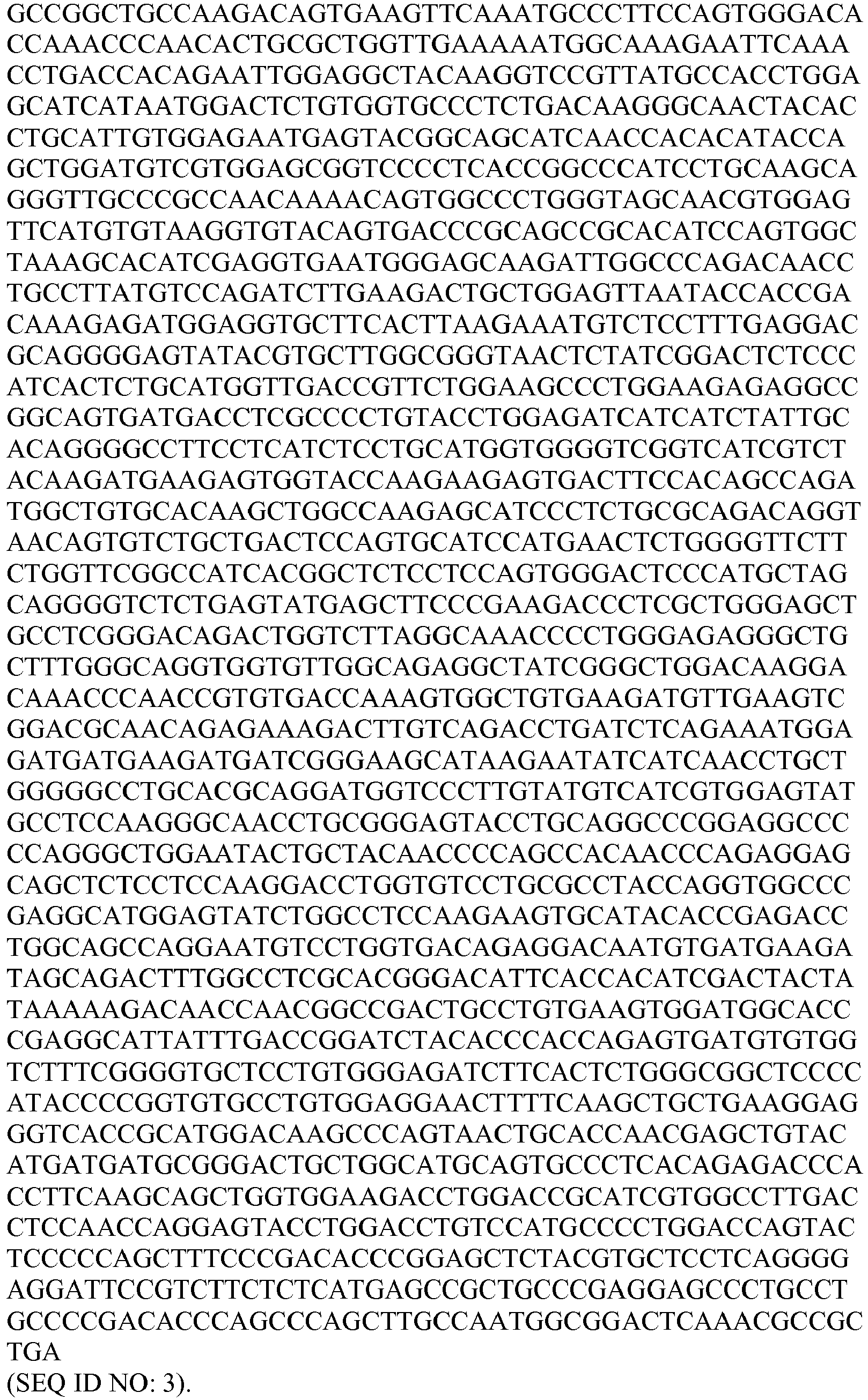
- The amino acid sequence of human FGFR1c (GenBank Accession Number NP_075598) is provided:

- The antigen binding proteins described herein bind the extracellular portion of FGFR1c. An example of an extracellular region of FGFR1c is:

- As described herein, FGFR1c proteins can also include fragments. As used herein, the terms are used interchangeably to mean a receptor, in particular and unless otherwise specified, a human receptor, that upon association with β-Klotho and FGF21 induces FGF21-like signaling activity.
- The term FGFR1c also includes post-translational modifications of the FGFR1c amino acid sequence, for example, possible N-linked glycosylation sites. Thus, the antigen binding proteins can bind to or be generated from proteins glycosylated at one or more of the positions.
- The antigen binding proteins disclosed herein bind to β-Klotho, in particular human β-Klotho. The nucleotide sequence encoding human β-Klotho (GenBank Accession Number NM_175737) is provided:


- The amino acid sequence of full length human β-Klotho (GenBank Accession Number NP_783864) is provided:


- The antigen binding proteins described herein bind the extracellular portion of β-Klotho. An example of an extracellular region of β-Klotho is:

- The murine form of • -Klotho, and fragments and subsequences thereof, can be of use in studying and/or constructing the molecules provided herein. The nucleotide sequence encoding murine • -Klotho (GenBank Accession Number NM_031180) is provided:


- The amino acid sequence of full length murine • -Klotho (GenBank Accession Number NP_112457) is provided:

- As described herein, β-Klotho proteins can also include fragments. As used herein, the terms are used interchangeably to mean a co-receptor, in particular and unless otherwise specified, a human co-receptor, that upon association with FGFR1c and FGF21 induces FGF21-like signaling activity.
- The term β-Klotho also includes post-translational modifications of the β-Klotho amino acid sequence, for example, possible N-linked glycosylation sites. Thus, the antigen binding proteins can bind to or be generated from proteins glycosylated at one or more of the positions.
- A variety of selective binding agents useful for modulating FGF21-like signaling are provided. These agents include, for instance, antigen binding proteins that contain an antigen binding domain (e.g., single chain antibodies, domain antibodies, hemibodies, immunoadhesions, and polypeptides with an antigen binding region) and specifically bind to a complex comprising β-Klotho and at least one of (i) FGFR1c, (ii) FGFR2c and (iii) FGFR3c, in particular a complex comprising human β-Klotho and a human FGFR (e.g., human FGFR1c, human FGFR2c or human FGFR3c). Some of the agents, for example, are useful in mimicking the signaling effect generated in vivo by the association of an FGFR (e.g., FGFR1c, FGFR2c or FGFR3c) with β-Klotho and with FGF21, and can thus be used to enhance or modulate one or more activities associated with FGF21-like signaling.
- In general, the antigen binding proteins that are provided typically comprise one or more CDRs as described herein (e.g., 1, 2, 3, 4, 5 or 6 CDRs). In some embodiments the antigen binding proteins are naturally expressed by clones, while in other embodiments, the antigen binding protein can comprise (a) a polypeptide framework structure and (b) one or more CDRs that are inserted into and/or joined to the polypeptide framework structure. In some of these embodiments a CDR forms a component of a heavy or light chains expressed by the clones described herein; in other embodiments a CDR can be inserted into a framework in which the CDR is not naturally expressed. A polypeptide framework structure can take a variety of different forms. For example, a polypeptide framework structure can be, or comprise, the framework of a naturally occurring antibody, or fragment or variant thereof, or it can be completely synthetic in nature. Examples of various antigen binding protein structures are further described below.
- In some embodiments in which the antigen binding protein comprises (a) a polypeptide framework structure and (b) one or more CDRs that are inserted into and/or joined to the polypeptide framework structure, the polypeptide framework structure of an antigen binding protein is an antibody or is derived from an antibody, including, but not limited to, monoclonal antibodies, bispecific antibodies, minibodies, domain antibodies, synthetic antibodies (sometimes referred to herein as "antibody mimetics"), chimeric antibodies, humanized antibodies, antibody fusions (sometimes referred to as "antibody conjugates"), and portions or fragments of each, respectively. In some instances, the antigen binding protein is an immunological fragment of an antibody (e.g., a Fab, a Fab', a F(ab')2, or a scFv).
- Certain of the antigen binding proteins as provided herein specifically bind to a complex comprising β-Klotho and at least one of (i) FGFR1c, (ii) FGFR2c and (iii) FGFR3c, including the human forms of these proteins. In one embodiment, an antigen binding protein specifically binds to both human FGFR1c comprising the amino acid sequence of SEQ ID NO: 4, and human β-Klotho comprising the amino acid sequence of SEQ ID NO: 7, and in another embodiment an antigen binding protein specifically binds to both human FGFR1c comprising the amino acid sequence of SEQ ID NO: 4 and human β-Klotho having the amino acid sequence of SEQ ID NO: 7 and induces FGF21-like signaling. Thus, an antigen binding protein can, but need not, induce FGF21-like signaling.
- Some of the antigen binding proteins that specifically bind to a complex comprising β-Klotho and at least one of (i) FGFR1c, (ii) FGFR2c and (iii) FGFR3c, including the human forms of these proteins, provided herein have a structure typically associated with naturally occurring antibodies. The structural units of these antibodies typically comprise one or more tetramers, each composed of two identical couplets of polypeptide chains, though some species of mammals also produce antibodies having only a single heavy chain. In a typical antibody, each pair or couplet includes one full-length "light" chain (in certain embodiments, about 25 kDa) and one full-length "heavy" chain (in certain embodiments, about 50-70 kDa). Each individual immunoglobulin chain is composed of several "immunoglobulin domains," each consisting of roughly 90 to 110 amino acids and expressing a characteristic folding pattern. These domains are the basic units of which antibody polypeptides are composed. The amino-terminal portion of each chain typically includes a variable domain that is responsible for antigen recognition. The carboxy-terminal portion is more conserved evolutionarily than the other end of the chain and is referred to as the "constant region" or "C region". Human light chains generally are classified as kappa ("κ") and lambda ("λ") light chains, and each of these contains one variable domain and one constant domain. Heavy chains are typically classified as mu, delta, gamma, alpha, or epsilon chains, and these define the antibody's isotype as IgM, IgD, IgG, IgA, and IgE, respectively. IgG has several subtypes, including, but not limited to, IgG1, IgG2, IgG3, and IgG4. IgM subtypes include IgM, and IgM2. IgA subtypes include IgA1 and IgA2. In humans, the IgA and IgD isotypes contain four heavy chains and four light chains; the IgG and IgE isotypes contain two heavy chains and two light chains; and the IgM isotype contains five heavy chains and five light chains. The heavy chain C region typically comprises one or more domains that can be responsible for effector function. The number of heavy chain constant region domains will depend on the isotype. IgG heavy chains, for example, each contain three C region domains known as
C H1,C H2 andC H3. The antibodies that are provided can have any of these isotypes and subtypes. In certain embodiments, an antigen binding protein that specifically binds to a complex comprising β-Klotho and at least one of (i) FGFR1c, (ii) FGFR2c and (iii) FGFR3c is an antibody of the IgG1, IgG2, or IgG4 subtype. - In full-length light and heavy chains, the variable and constant regions are joined by a "J" region of about twelve or more amino acids, with the heavy chain also including a "D" region of about ten more amino acids. See, e.g., Fundamental Immunology, 2nd ed., Ch. 7 (Paul, W., ed.) 1989, New York: Raven Press (hereby incorporated by reference in its entirety for all purposes). The variable regions of each light/heavy chain pair typically form the antigen binding site.
- One example of an IgG2 heavy constant domain of an exemplary monoclonal antibody that specifically binds to a complex comprising β-Klotho and at least one of (i) FGFR1c, (ii) FGFR2c and (iii) FGFR3c has the amino acid sequence:

- One example of a kappa light constant domain of an exemplary monoclonal antibody that binds to a complex comprising β-Klotho and at least one of (i) FGFR1c, (ii) FGFR2c and (iii) FGFR3c has the amino acid sequence:

- One example of a lambda light constant domain of an exemplary monoclonal antibody that binds to a complex comprising β-Klotho and at least one of (i) FGFR1c, (ii) FGFR2c and (iii) FGFR3c has the amino acid sequence:

- Variable regions of immunoglobulin chains generally exhibit the same overall structure, comprising relatively conserved framework regions (FR) joined by three hypervariable regions, more often called "complementarity determining regions" or CDRs. The CDRs from the two chains of each heavy chain/light chain pair mentioned above typically are aligned by the framework regions to form a structure that binds specifically with a specific epitope on the target protein (e.g., a complex comprising β-Klotho and at least one of (i) FGFR1c, (ii) FGFR2c and (iii) FGFR3c. From N-terminal to C-terminal, naturally-occurring light and heavy chain variable regions both typically conform with the following order of these elements: FR1, CDR1, FR2, CDR2, FR3, CDR3 and FR4. A numbering system has been devised for assigning numbers to amino acids that occupy positions in each of these domains. This numbering system is defined in Kabat et al., (1991) "Sequences of Proteins of Immunological Interest", 5th Ed., US Dept. of Health and Human Services, PHS, NIH, NIH Publication no. 91-3242. Although presented using the Kabat nomenclature system, as desired, the CDRs disclosed herein can also be redefined according an alternative nomenclature scheme, such as that of Chothia (see Chothia & Lesk, (1987) J. Mol. Biol. 196:901-917; Chothia et al., (1989) Nature 342:878-883 or Honegger & Pluckthun, (2001) J. Mol. Biol. 309:657-670).
- The various heavy chain and light chain variable regions of antigen binding proteins provided herein are depicted in Table 2. Each of these variable regions can be attached to the disclosed heavy and light chain constant regions to form a complete antibody heavy and light chain, respectively. Further, each of the so-generated heavy and light chain sequences can be combined to form a complete antibody structure. It should be understood that the heavy chain and light chain variable regions provided herein can also be attached to other constant domains having different sequences than the exemplary sequences listed above.
- Specific examples of some of the full length light and heavy chains of the antibodies that are provided and their corresponding amino acid sequences are summarized in Tables 1A and 1B. Table 1A shows exemplary light chain sequences, and Table 1B shows exemplary heavy chain sequences.
TABLE 1A Exemplary Antibody Light Chain Sequences Contained in Clone Designation SEQ ID NO: Amino Acid Sequence 63E6 L6 14 
66F7 L7 15 
66D4 L18 16 
66B4 L11 17 
65B1 L19 18 
65B4 L21 19 

67A4 L20 20 
63A10v1 L22 21 
63A10v2 L101 1835 
63A10v3 L102 1836 
65H11v1 L23 22 
65H11v2 L103 1837 
67G10v1 L9 23 

67G10v2 L10 24 
64C8 L24 25 
64A8 L1 26 
67B4 63G8v1 L104 1838 
63G8v2 L105 1839 
63G8v3 L106 1840 
66G2 L12 27 
68D3v1 L2 28 
68D3v2 65D1 L27 29 
64H5 L8 30 
65G4 65D4 L26 31 
65E3 L25 32 
67G8 L28 33 

65B7v1 L29 34 
65B7v2 L107 1841 
63B6 L4 35 
64D4 63F5 L14 36 
65E8 L3 37 
63H11 64E6 67G7 65F11 65C1 L16 38 
66F6 L15 39 
64A6 L30 40 
65F9 L31 41 
64A7 L17 42 
65C3 L5 43 
68D5 67F5 L32 44 
64B10v1 L33 45 
64B10v2 68C8 L34 46 
67A5 L35 47 
67C10 L36 48 
64H6 L37 49 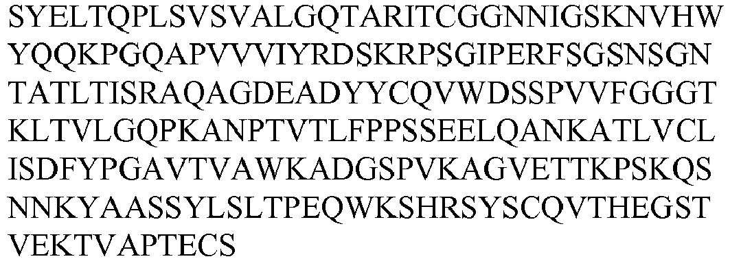
63F9 L38 50 
67F6v1 L39 51 
67F6v2 L108 1842 
48C9 L78 52 
49A12 51E2 48F3 L77 53 

48F8 L49 54 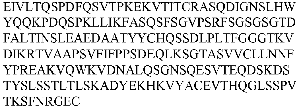
53B9 56B4 57E7 57F11 48H11 L40 55 
49A10 L65 56 
48D4 49C8 L45 57 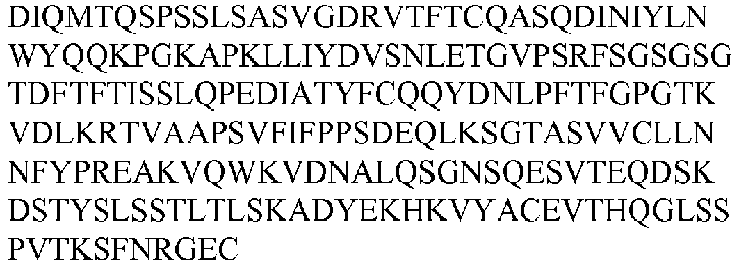
52H1 49G2 L66 58 
50C12 55G11 49G3 L47 59 
49H12 L43 60 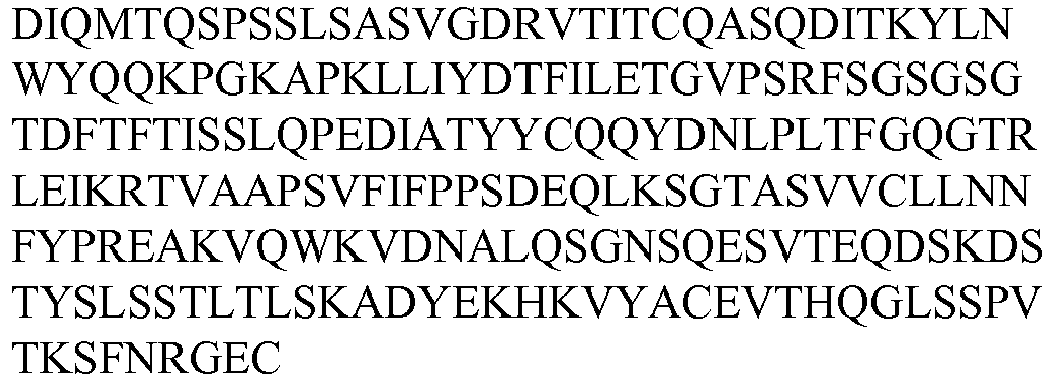
51A8 L61 61 
51C10.1 L55 62 
51C10.2 L70 63 
51E5 L79 64 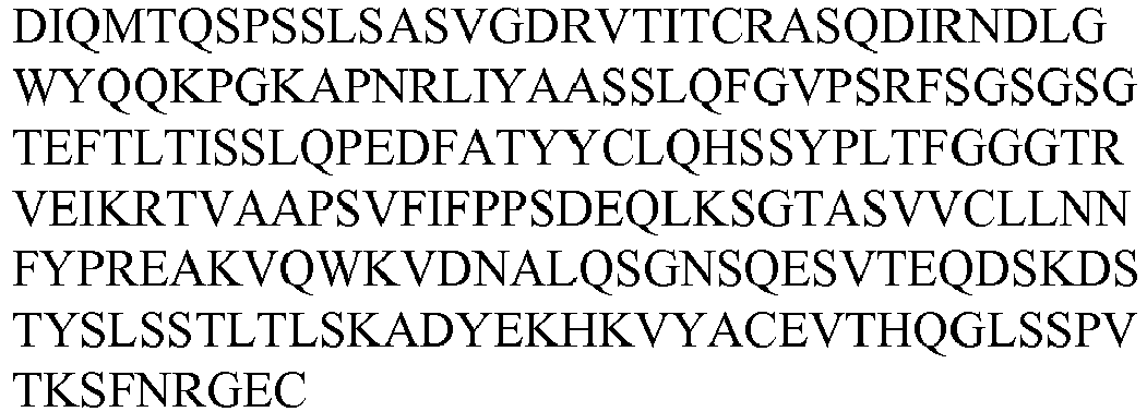
51G2 L51 65 
52A8 L41 66 
52B8 L82 67 
52C1 L67 68 
52F8 L42 69 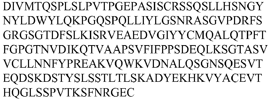
52H2 L84 70 
53F6 L63 71 

53H5.2 L62 72 
53H5.3 L80 73 
54A1 L44 74 
55G9 54H10.1 L53 75 
55D1 48H3 53C11 55D3 L71 76 
55E4 L75 77 
49B11 50H10 53C1 55E9 L68 78 
55G5 L83 79 
56A7 L52 80 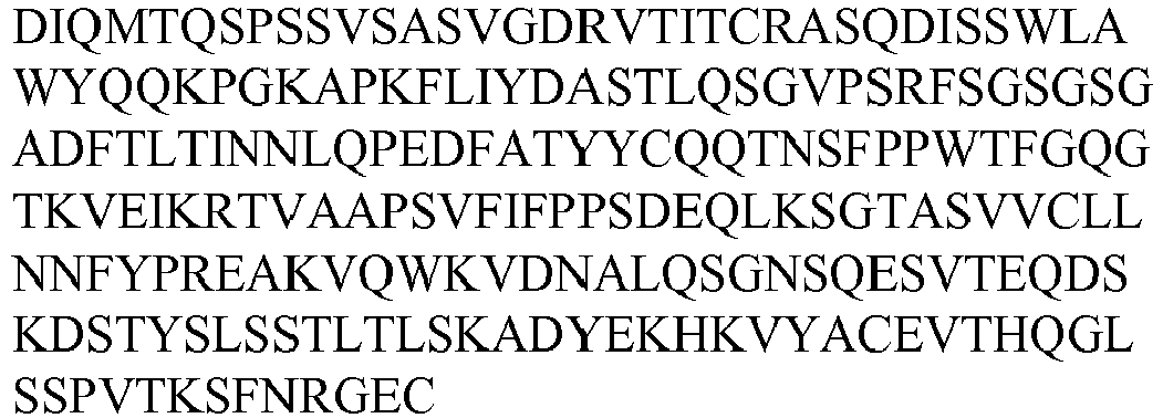
56E4 56C11 L64 81 
56E7 L86 82 
56G1 L76 83 

56G3.3 L81 84 
55B10 57B12 L72 85 
57D9 L87 86 
59A10 L48 87 
49H4 59C9 L50 88 
58A5 57A4 57F9 59G10.2 L60 89 
59G10.3 L54 90 
60D7 L69 91 
60F9 L58 92 
48B4 52D6 60G5.2 L46 93 
61G5 L59 94 
52C5 L73 95 
61H5 L88 96 
52B9 59D10v1 L56 97 
59D10v2 L57 98 
56G3.2 L85 99 
68G5 L13 100 
60G5.1 L74 1843 

48G4 L89 101 
53C3.1 50G1 L90 102 
58C2 L91 103 
50D4 L92 104 
50G5v1 L93 105 
50G5v2 L94 106 
51C1 L95 107 
53C3.2 L96 108 
54H10.3 L97 109 
55A7 L98 110 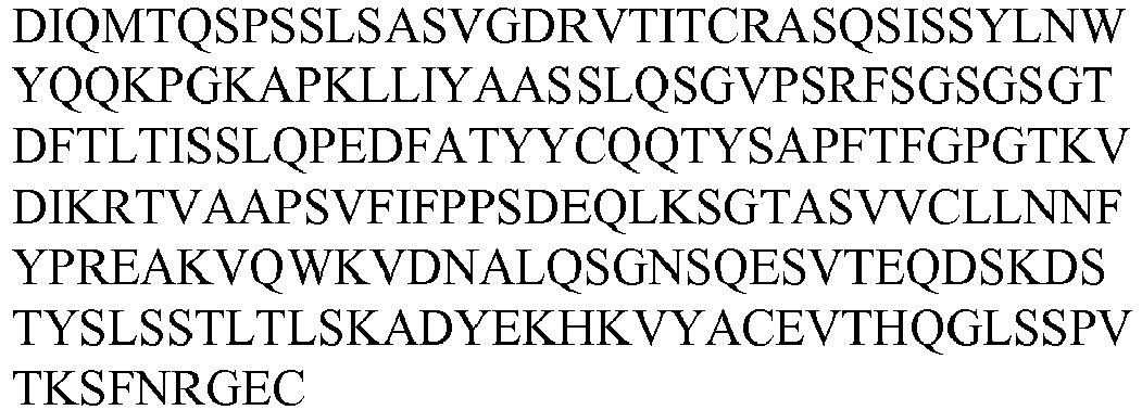
55E6 L99 111 
61E1 L100 112 
Table 1B - Exemplary Antibody Heavy Chain Sequences Contained in Clone Designation SEQ ID NO: Amino Acid Sequence 63E6 H6 113 
66F7 66D4 H17 114 
66B4 H10 115 
65B1 H18 116 

65B4 H20 117 
67A4 H19 118 
63A10v1 H21 119 
63A10v2 63A10v3 65H11v1 H22 120 
65H11v2 
67G10v1 H9 121 
67G10v2 64C8 H23 122 
63G8v1 H1 123 
63G8v2 63G8v3 68D3v1 64A8 67B4 
68D3v2 H95 1844 
66G2 H11 124 
65D1 H26 125 
64H5 H7 126 
65D4 H25 127 
65E3 H24 128 
65G4 H8 129 
68G5 H12 130 
67G8 H27 131 
65B7v1 H28 132 
65B7v2 63B6 64D4 H4 133 
63F5 H13 134 
63H11 H3 135 
64E6 H2 136 
65E8 65F11 67G7 65C1 H15 137 
66F6 H14 138 
64A6 H29 139 
65F9 H30 140 
64A7 H16 141 
65C3 H5 142 
68D5 67F5 H31 143 
64B10v1 H32 144 
64B10v2 H96 1845 
68C8 H33 145 
67A5 H34 146 
67C10 H35 147 
64H6 H36 148 
63F9 H37 149 
67F6v1 H38 150 
67F6v2 48C9 H73 151 
49A12 51E2 48F3 H72 152 
48F8 H48 153 
53B9 56B4 57E7 57F11 48H11 H39 154 
49A10 H62 155 
48D4 49C8 H44 156 
52H1 49G2 H63 157 
50C12 55G11 49G3 H46 158 
49H12 H42 159 
51A8 H58 160 
51C10.2 H67 161 
51E5 H74 162 
51G2 H50 163 
52A8 H40 164 
52B8 H77 165 
52C1 H64 166 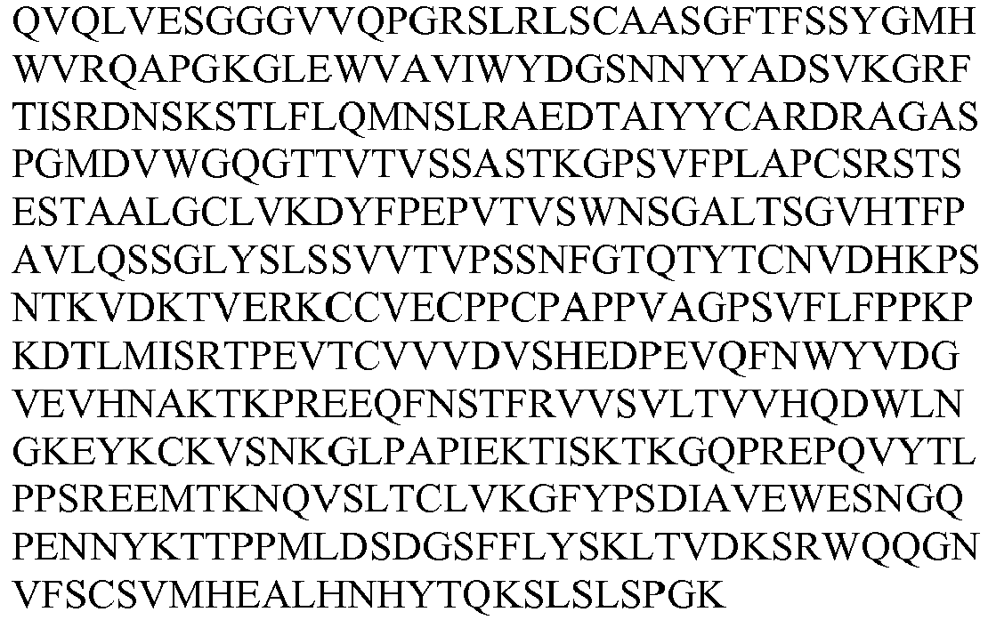
52F8 H41 167 
52H2 H79 168 
53F6 H60 169 
53H5.2 H59 170 
53H5.3 H75 171 
54A1 H43 172 
55G9 54H10.1 H52 173 
55D1 48H3 53C11 55D3 H68 174 
55E4 H70 175 
52C5 60G5.1 49B11 50H10 53C1 55E9 H65 176 
55G5 H78 177 
50G1 H84 178 
56A7 H51 179 
56E4 56C11 H61 180 
56E7 H81 181 
56G1 H71 182 
56G3.3 H76 183 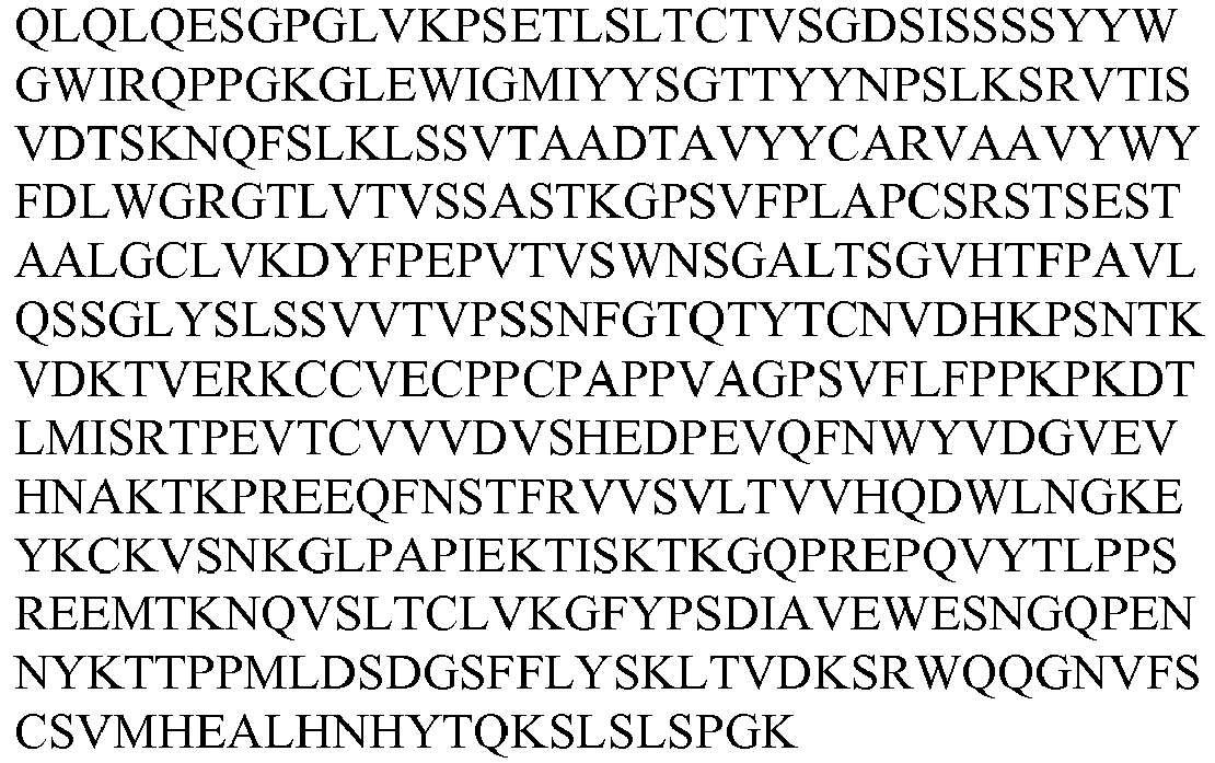
55B10 57B12 H69 184 
57D9 H82 185 
58C2 H85 186 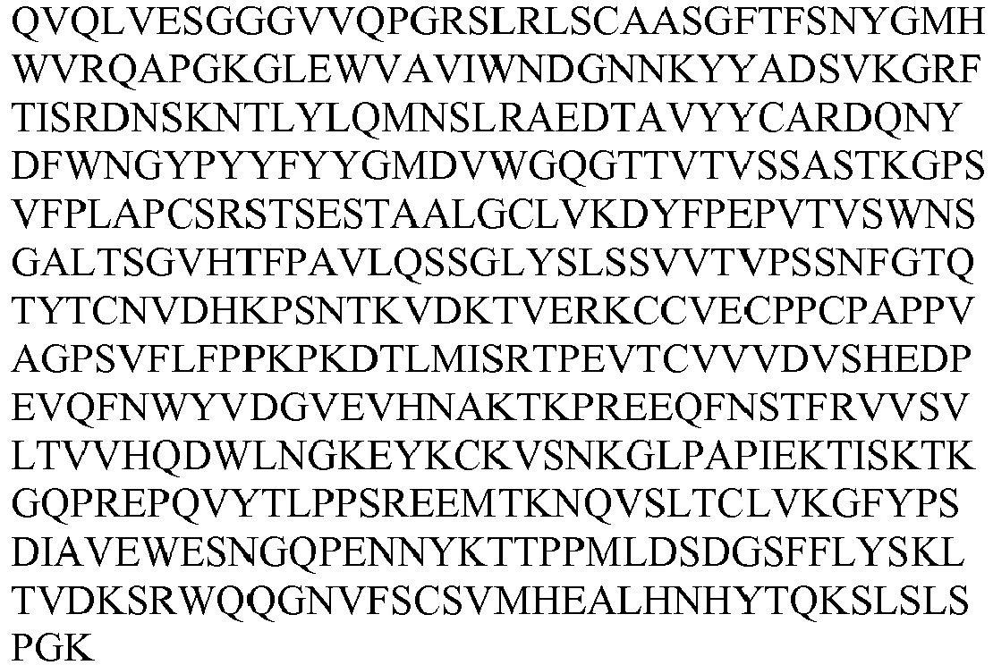
59A10 H47 187 
49H4 59C9 H49 188 
58A5 57A4 57F9 59G10.2 H57 189 
59G10.3 H53 190 
60D7 H66 191 
60F9 H55 192 
48B4 52D6 60G5.2 H45 193 
61G5 H56 194 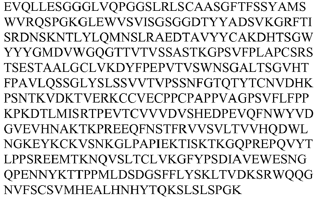
59D10v1 H54 195 
59D10v2 51C10.1 56G3.2 H80 196 
48G4 H83 197 
53C3.1 61H5 H86 198 
52B9 50D4 H87 199 
50G5v1 H88 200 
50G5v2 51C1 H89 201 
53C3.2 H90 202 
54H10.3 H91 203 
55A7 H92 204 
55E6 H93 205 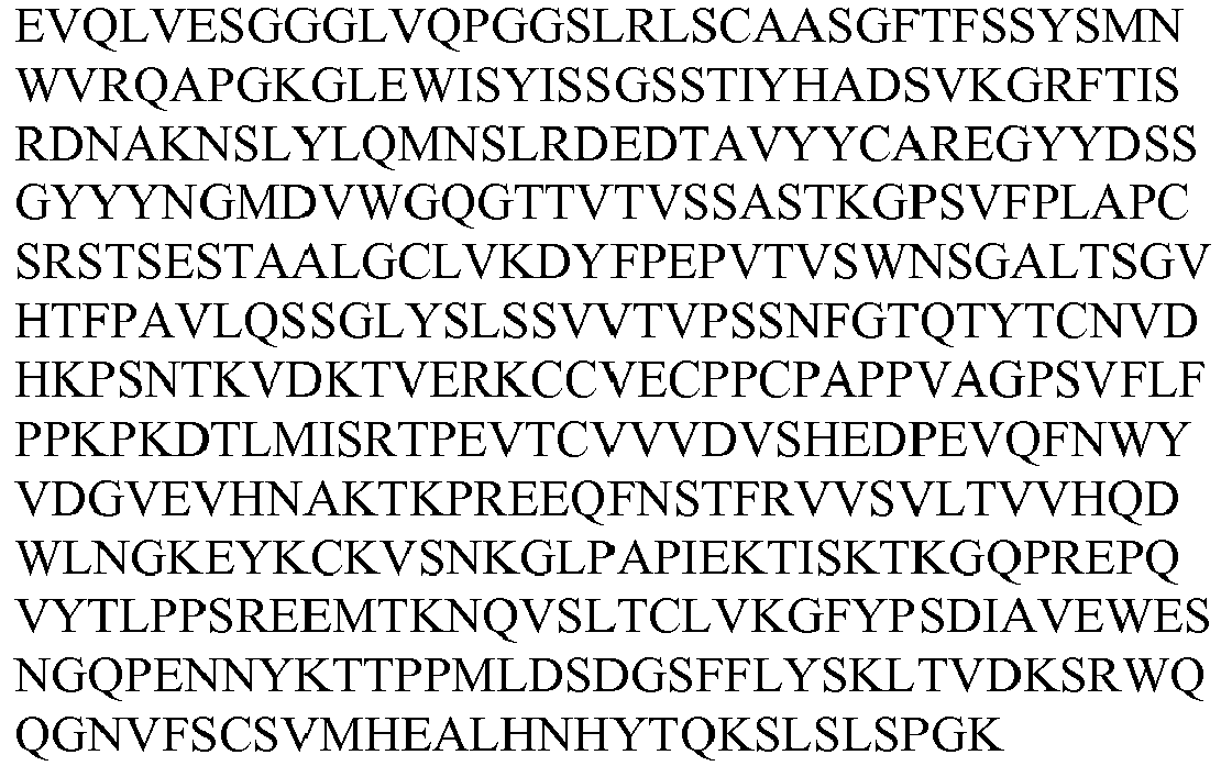
61E1 H94 206 
- Each of the exemplary heavy chains (H1, H2, H3 etc.) listed in Table 1B, infra, can be combined with any of the exemplary light chains shown in Table 1A, infra, to form an antibody. Examples of such combinations include H1 combined with any of L1 through L100; H2 combined with any of L1 through L100; H3 combined with any of L1 through L100, and so on. In some instances, the antibodies include at least one heavy chain and one light chain from those listed in Tables 1A and 1B, infra; particular examples pairings of light chains and heavy chains include L1 with H1, L2 with H1, L3 with H2 or H3, L4 with H4, L5 with H5, L6 with H6, L7 with H6, L8 with H7 or H8, L9 with H9, L10 with H9, L11 with H10, L12 with H11, L13 with H12, L13 with H14, L14 with H13, L15 with H14, L16 with H15, L17 with H16, L18 with H17, L19 with H18, L20 with H19, L21 with H20, L22 with H21, L23 with H22, L24 with H23, L25 with H24, L26 with H25, L27 with H26, L28 with H27, L29 with H28, L30 with H29, L31 with H30, L32 with H31, L33 with H32, L34 with H33, L35 with H34, L36 with H35, L37 with H36, L38 with H37, L39 with H38, L40 with H39, L41 with H40, L42 with H41, L43 with H42, L44 with H43, L45 with H44, L46 with H45, L47 with H46, L48 with H47, L49 with H48, L50 with H49, L51 with H50, L52 with H51, L53 with H52, L54 with H53, L55 with H54, and L56 with H54, L57 with H54, L58 with H55, L59 with H56, L60 with H57, L61 with H58, L62 with H59, L63 with H60, L64 with H1, L65 with H62, L66 with H63, L67 with H64, L68 with H65, L69 with H66, L70 with H67, L71 with H68, L72 with H69, L73 with H70, L74 with H70, and L75 with H70, L76 with H71, L77 with H72, L78 with H73, L79 with H74, L80 with H75, L81 with H76, L82 with H77, L83 with H78, L84 with H79, L85 with H80, L86 with H81, L87 with H82, L88 with H86, L89 with H83, L90 with H84, L91 with H85, L92 with H87, L93 with H88, L94 with H88, L95 with H89, L96 with H90, L97 with H91, L98 with H92, L99 with H93, and L100 with H94. In addition to antigen binding proteins comprising a heavy and a light chain from the same clone, a heavy chain from a first clone can be paired with a light chain from a second clone (e.g., a heavy chain from a first clone paired with a light chain from a second clone or a heavy chain from a first clone paired with a light chain from a second clone). Generally, such pairings can include VL with 90% or greater homology can be paired with the heavy chain of the naturally occurring clone.
- In some instances, the antibodies comprise two different heavy chains and two different light chains listed in Tables 1A and 1B, infra. In other instances, the antibodies contain two identical light chains and two identical heavy chains. As an example, an antibody or immunologically functional fragment can include two L1 light chains with two H1 heavy chains, two L2 light chains with two H1 heavy chains, two L3 light chains with two H2 heavy chains or two H3 heavy chains, two L4 light chains with two H4 heavy chains, two L5 light chains with two H5 heavy chains, two L6 light chains with two H6 heavy chains, two L7 light chains with two H6 heavy chains, two L8 light chains with two H7 heavy chains or two H8 heavy chains, two L9 light chains with two H9 heavy chains, two L10 light chains with two H9 heavy chains, two L11 light chains with two H10 heavy chains, two L12 light chains with two H11 heavy chains, two L13 light chains with two H12 heavy chains, two L13 light chains with two H14 heavy chains, two L14 light chains with two H13 heavy chains, two L15 light chains with two H14 heavy chains, two L16 light chains with two H15 heavy chains, two L17 light chains with two H16 heavy chains, two L18 light chains with two H17 heavy chains, two L19 light chains with two H18 heavy chains, two L20 light chains with two H19 heavy chains, two L21 light chains with two H20 heavy chains, two L22 light chains with two H21 heavy chains, two L23 light chains with two H22 heavy chains, two L24 light chains with two H23 heavy chains, two L25 light chains with two H24 heavy chains, two L26 light chains with two H25 heavy chains, two L27 light chains with two H26 heavy chains, two L28 light chains with two H27 heavy chains, two L29 light chains with two H28 heavy chains, two L30 light chains with two H29 heavy chains, two L31 light chains with two H30 heavy chains, two L32 light chains with two H31 heavy chains, two L33 light chains with two H32 heavy chains, two L34 light chains with two H33 heavy chains, two L35 chains with two H34 heavy chains, two L36 chains with two H35 heavy chains, two L37 light chains with two H36 heavy chains, two L38 light chains with two H37 heavy chains, two L39 light chains with two H38 heavy chains, two L40 light chains with two H39 heavy chains, two L41 light chains with two H40 heavy chains, two L42 light chains with two H41 heavy chains, two L43 light chains with two H42 heavy chains, two L44 light chains with two H43 heavy chains, two L45 light chains with two H44 heavy chains, two L46 light chains with two H45 heavy chains, two L47 light chains with two H46 heavy chains, two L48 light chains with two H47 heavy chains, two L49 light chains with two H48 heavy chains, two L50 light chains with two H49 heavy chains, two L51 light chains with two H50 heavy chains, two L52 light chains with two H51 heavy chains, two L53 light chains with two H52 heavy chains, two L54 light chains with two H53 heavy chains, two L55 light chains with two H54 heavy chains, and two L56 light chains with two H54 heavy chains, two L57 light chains with two H54 heavy chains, two L58 light chains with two H55 heavy chains, two L59 light chains with two H56 heavy chains, two L60 light chains with two H57 heavy chains, two L61 light chains with two H58 heavy chains, two L62 light chains with two H59 heavy chains, two L63 light chains with two H60 heavy chains, two L64 light chains with two H1 heavy chains, two L65 light chains with two H62 heavy chains, two L66 light chains with two H63 heavy chains, two L67 light chains with two H64 heavy chains, two L68 light chains with two H65 heavy chains, two L69 light chains with two H66 heavy chains, two L70 light chains with two H67 heavy chains, two L71 light chains with two H68 heavy chains, two L72 light chains with two H69 heavy chains, two L73 light chains with two H70 heavy chains, two L74 light chains with two H70 heavy chains, and two L75 light chains with two H70 heavy chains, two L76 light chains with two H71 heavy chains, two L77 light chains with two H72 heavy chains, two L78 light chains with two H73 heavy chains, two L79 light chains with two H74 heavy chains, two L80 light chains with two H75 heavy chains, two L81 light chains with two H76 heavy chains, two L82 light chains with two H77 heavy chains, two L83 light chains with two H78 heavy chains, two L84 light chains with two H79 heavy chains, two L85 light chains with two H80 heavy chains, two L86 light chains with two H81 heavy chains, two L87 light chains with two H82 heavy chains, two L88 light chains with two H86 heavy chains, two L89 light chains with two H83 heavy chains, two L90 light chains with two H84 heavy chains, two L91 light chains with two H85 heavy chains, two L92 light chains with two H87 heavy chains, two L93 light chains with two H88 heavy chains, two L94 light chains with two H88 heavy chains, two L95 light chains with two H89 heavy chains, two L96 light chains with two H90 heavy chains, two L97 light chains with two H91 heavy chains, two L98 light chains with two H92 heavy chains, two L99 light chains with two H93 heavy chains, and two L100 light chains with two H94 heavy chains, as well as other similar combinations of pairs comprising the light chains and pairs of heavy chains as listed in Tables 1A and 1B, infra.
- In another aspect of the instant disclosure, "hemibodies" are provided. A hemibody is a monovalent antigen binding protein comprising (i) an intact light chain, and (ii) a heavy chain fused to an Fc region (e.g., an IgG2 Fc region of SEQ ID NO: 11), optionally via a linker, The linker can be a (G4S)x linker (SEQ ID NO: 207) where "x" is a non-zero integer (e.g., (G4S)2, (G4S)3, (G4S)4, (G4S)5, (G4S)6, (G4S)7, (G4S)8, (G4S)9, (G4S)10,; SEQ ID NOs: 208-216, respectively). Hemibodies can be constructed using the provided heavy and light chain components.
- Other antigen binding proteins that are provided are variants of antibodies formed by combination of the heavy and light chains shown in Tables 1A and 1B, infra and comprise light and/or heavy chains that each have at least 70%, 75%, 80%, 85%, 90%, 95%, 96%, 97%, 98% or 99% identity to the amino acid sequences of these chains. In some instances, such antibodies include at least one heavy chain and one light chain, whereas in other instances the variant forms contain two identical light chains and two identical heavy chains.
- Also provided are antigen binding proteins that contain an antibody heavy chain variable region selected from the group consisting of VH1, VH2, VH3, VH4, VH5, VH6, VH7, VH8, VH9, VH10, VH11, VH12, VH13, VH14, VH15, VH16, VH17, VH18, VH19, VH20, VH21 VH22, VH23, VH24, VH25, VH26, VH27, VH28, VH29, VH30, VH31, VH32, VH33, VH34, VH35, VH36, VH37, VH38, VH39, VH40, VH41, VH42, VH43, VH44, VH45, VH46, VH47, VH48, VH49, VH50, VH51, VH52, VH53, VH54, VH55, VH56, VH57, VH58, VH59, VH60, VH61, VH62, VH63, VH64, VH65, VH66, VH67, VH68, VH69, VH70, VH71, VH72, VH73, VH74, VH75, VH76, VH77, VH78, VH79, VH80, 81, VH82, VH83, VH84, VH85, VH86, VH87, VH88, VH89, VH90, VH91, VH92, VH93, and VH94 as shown in Table 2B and/or an antibody light chain variable region selected from the group consisting of VL1, VL2, VL3, VL4, VL5, VL6, VL7, VL8, VL9, VL10, VL11, VL12, VL13, VL14, VL15, VL16, VL17, VL18, VL19, VL20, VL21, VL22, VL23, VL24, VL25, VL26, VL27, VL28, VL29, VL30, VL31, VL32, VL33, VL34, VL35, VL36, VL37, VL38, VL39, VL40, VL41, VL42, VL43, VL44, VL45, VL46, VL47, VL48, VL49, VL50, VL51, VL52, VL53, VL54, VL55, VL56, VL57, VL58, VL59, VL60, VL61, VL62, VL63, VL64, VL65, VL66, VL67, VL68, VL69, VL70, VL71, VL72, VL73, VL74, VL75, VL76, VL77, VL78, VL79, VL80, VL81, VL82, VL83, VL84, VL85, VL86, VL87, VL88, VL89, VL90, VL91, VL92, VL93, VL94, VL95, VL96, VL97, VL98, VL99 and VL100 as shown in Table 2A, and immunologically functional fragments, derivatives, muteins and variants of these light chain and heavy chain variable regions.
TABLE 2A Exemplary Antibody Variable Light (V L ) Chains Contained in Clone Designation SEQ ID NO. Amino Acid Sequence 63E6 VL6 217 
66F7 VL7 218 
66D4 VL18 219 
66B4 VL11 220 
65B1 VL19 221 
65B4 VL21 222 
67A4 VL20 223 
63A10v1 VL22 224 
63A10v2 VL101 1846 
63A10v3 VL102 1847 
65H11v1 VL23 225 
65H11v2 VL103 1848 
67G10v1 VL9 226 
67G10v2 VL10 227 
64C8 VL24 228 
64A8 VL1 229 
67B4 63G8v1 VL104 1849 
63G8v2 VL105 1850 
63G8v3 VL106 1851 
66G2 VL12 230 
68D3v1 VL2 231 
68D3v2 65D1 VL27 232 
64H5 VL8 233 
65G4 65D4 VL26 234 
65E3 VL25 235 
68G5 VL13 236 
67G8 VL28 237 
65B7v1 VL29 238 
65B7v2 VL107 1852 
63B6 VL4 239 
64D4 63F5 VL14 240 
65E8 VL3 241 
63H11 64E6 65F11 67G7 65C1 VL16 242 
66F6 VL15 243 
64A6 VL30 244 
65F9 VL31 245 
64A7 VL17 246 
65C3 VL5 247 
68D5 67F5 VL32 248 
64B10v1 VL33 249 
64B10v2 68C8 VL34 250 
67A5 VL35 251 
67C10 VL36 252 
64H6 VL37 253 
63F9 VL38 254 
67F6v1 VL39 255 
67F6v2 VL108 1853 
48C9 VL78 256 
49A12 51E2 48F3 VL77 257 
48F8 VL49 258 
53B9 56B4 57E7 57F11 48H11 VL40 259 
49A10 VL65 260 
48D4 49C8 VL45 261 
52H1 49G2 VL66 262 
50C12 55G11 49G3 VL47 263 
49H12 VL43 264 
51A8 VL61 265 
51C10.1 VL55 266 
51C10.2 VL70 267 
51E5 VL79 268 
51G2 VL51 269 
52A8 VL41 270 
52B8 VL82 271 
52C1 VL67 272 
52F8 VL42 273 
52H2 VL84 274 
53F6 VL63 275 
53H5.2 VL62 276 
53H5.3 VL80 277 
54A1 VL44 278 
55G9 54H10.1 VL53 279 
55D1 48H3 53C11 55D3 VL71 280 
55E4 VL75 281 
49B11 50H10 53C1 55E9 VL68 282 
55G5 VL83 283 
56A7 VL52 284 
56E4 56C11 VL64 285 
56E7 VL86 286 
56G1 VL76 287 
56G3.3 VL81 288 
55B10 57B12 VL72 289 
57D9 VL87 290 
59A10 VL48 291 
49H4 59C9 VL50 292 
58A5 57A4 57F9 59G10.2 VL60 293 
59G10.3 VL54 294 
60D7 VL69 295 
60F9 VL58 296 
48B4 52D6 60G5.2 VL46 297 
61G5 VL59 298 
52C5 VL73 299 
61H5 VL88 300 
52B9 59D10v1 VL56 301 
59D10v2 VL57 302 
56G3.2 VL85 303 
48G4 VL89 304 
53C3.1 50G1 VL90 305 
58C2 VL91 306 
60G5.1 VL74 1854 
50D4 VL92 307 
50G5v1 VL93 308 
50G5 v2 VL94 309 
51C1 VL95 310 
53C3.2 VL96 311 
54H10.3 VL97 312 
55A7 VL98 313 
55E6 VL99 314 
61E1 VL100 315 
TABLE 2B Exemplary Antibody Variable Heavy (V H ) Chains Contained in Clone Designa-tion SEQ ID NO. Amino Acid Sequence 63E6 VH6 316 
66F7 66D4 VH17 317 
66B4 VH10 318 
65B1 VH18 319 
65B4 VH20 320 
67A4 VH19 321 
63A10v1 VH21 322 
63A10v2 63A10v3 65H11v1 VH22 323 
65H11v2 67G10v1 VH9 324 
67G10v2 64C8 VH23 325 
63G8v1 VH1 326 
63G8v2 63G8v3 68D3v1 64A8 67B4 68D3v2 VH95 1855 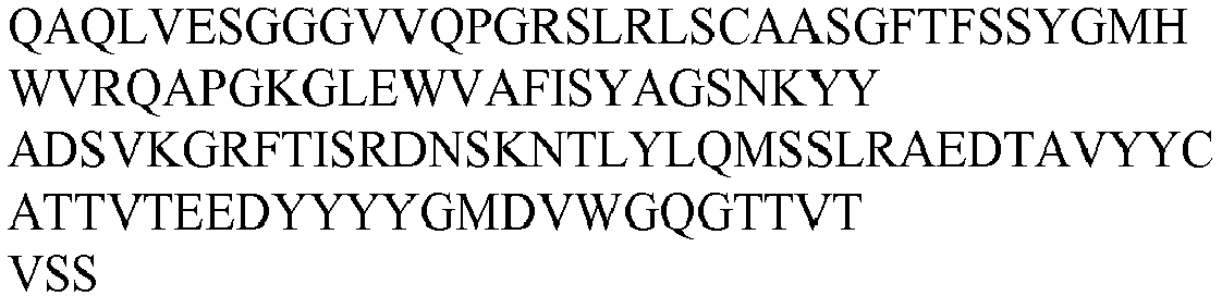
66G2 VH11 327 
65D1 VH26 328 
64H5 VH7 329 
65D4 VH25 330 
65E3 VH24 331 
65G4 VH8 332 
68G5 VH12 333 
67G8 VH27 334 
65B7v1 VH28 335 
65B7v2 63B6 64D4 VH4 336 
63F5 VH13 337 
63H11 VH3 338 
65E8 VH2 339 
64E6 65F11 67G7 65C1 VH15 340 
66F6 VH14 341 
64A6 VH29 342 
65F9 VH30 343 
64A7 VH16 344 
65C3 VH5 345 
68D5 67F5 VH31 346 
64B10v1 VH32 347 
64B10v2 VH96 1856 
68C8 VH33 348 
67A5 VH34 349 
67C10 VH35 350 
64H6 VH36 351 
63F9 VH37 352 
67F6v1 VH38 353 
67F6v2 48C9 VH73 354 
49A12 51E2 48F3 VH72 355 
48F8 VH48 356 
53B9 56B4 57E7 57F11 48H11 VH39 357 
49A10 VH62 358 
48D4 49C8 VH44 359 
52H1 49G2 VH63 360 
50C12 55G11 49G3 VH46 361 
49H12 VH42 362 
51A8 VH58 363 
51C10.1 VH54 364 59D10v1 59D10v2 
51C10.2 VH67 365 
51E5 VH74 366 
51G2 VH50 367 
52A8 VH40 368 
52B8 VH77 369 
52C1 VH64 370 
52F8 VH41 371 
52H2 VH79 372 
53F6 VH60 373 
53H5.2 VH59 374 
53H5.3 VH75 375 
54A1 55G9 VH43 376 
54H10.1 VH52 377 
55D1 48H3 53C11 55D3 VH68 378 
55E4 VH70 379 
49B11 50H10 53C1 52C5 60G5.1 55E9 VH65 380 
55G5 VH78 381 
50G1 VH84 382 
56A7 VH51 383 
56E4 56C11 VH61 384 
56E7 VH81 385 
56G1 VH71 386 
56G3.3 VH76 387 
55B10 57B12 VH69 388 
57D9 VH82 389 
58C2 VH85 390 
59A10 49H4 VH47 391 
59C9 VH49 392 
58A5 57A4 57F9 59G10.2 VH57 393 
59G10.3 VH53 394 
60D7 VH66 395 
60F9 VH55 396 
48B4 52D6 60G5.2 VH45 397 
61G5 VH56 398 
56G3.2 VH80 399 
48G4 VH83 400 
53C3.1 61H5 VH86 401 
52B9 50D4 VH87 402 
50G5v1 VH88 403 
50G5v2 51C1 VH89 404 
53C3.2 VH90 405 
54H10.3 VH91 406 
55A7 VH92 407 
55E6 VH93 408 
61E1 VH94 409 
TABLE 2C Coding Sequence for Antibody Variable Light (V L ) Chains Contained in Clone Designation SEQ ID NO. Coding Sequence 63E6 V L6 410 
66D4 VL18 411 
66B4 V L11 412 
65B1 V L19 413 
65B4 VL21 414 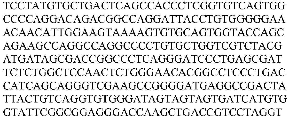
67A4 V L20 415 
63A10v1 VL22 416 
63A10v2 V L101 1857 
63A10v3 VL102 1858 
65H11v1 V L23 417 
65H11v2 VL103 1859 
67G10v1 V L9 418 
67G10v2 V L10 419 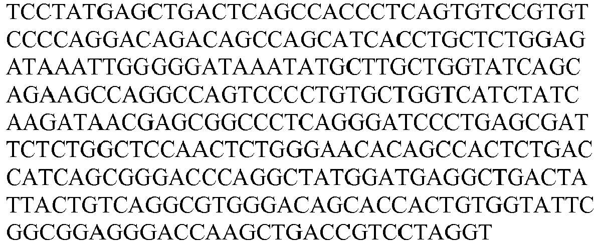
64C8 VL24 420 
64A8 V L1 421 67B4 
63G8v1 VL104 1860 
63G8v2 VL105 1861 
63G8v3 VL106 1862 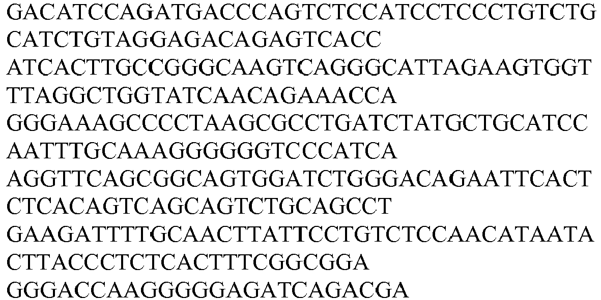
66G2 V L12 422 
68D3v1 V L2 423 
68D3v2 65D1 V L27 424 
65G4 V L8 425 
64H5 65D4 VL26 426 
65E3 VL25 427 
68G5 V L13 428 
67G8 V L28 429 
65B7v1 V L29 430 
65B7v2 VL107 1863 
63B6 V L4 431 
64D4 63F5 VL14 432 
65E8 V L3 433 
63H11 64E6 65F11 67G7 65C1 V L16 434 
66F6 VL15 435 
64A6 V L30 436 
65F9 VL31 437 
64A7 VL17 438 
65C3 V L5 439 68D5 
67F5 V L32 440 
64B10v1 VL33 441 64B10v2 
68C8 V L34 442 
67A5 VL35 443 
67C10 VL36 444 
64H6 V L37 445 
63F9 VL38 446 
67F6v1 VL39 447 
67F6v2 VL108 1864 
48C9 VL78 448 
49A12 51E2 Contained in Clone Designation SEQ ID NO. Coding Sequence 48F3 VL77 449 
48F8 VL49 450 
53B9 56B4 57E7 57F11 48H11 VL40 451 
49A10 VL65 452 
48D4 49C8 VL45 453 
52H1 49G2 VL66 454 
50C12 55G11 49G3 VL47 455 
49H12 VL43 456 
51A8 VL61 457 
51C10.1 VL55 458 
51C10.2 VL70 459 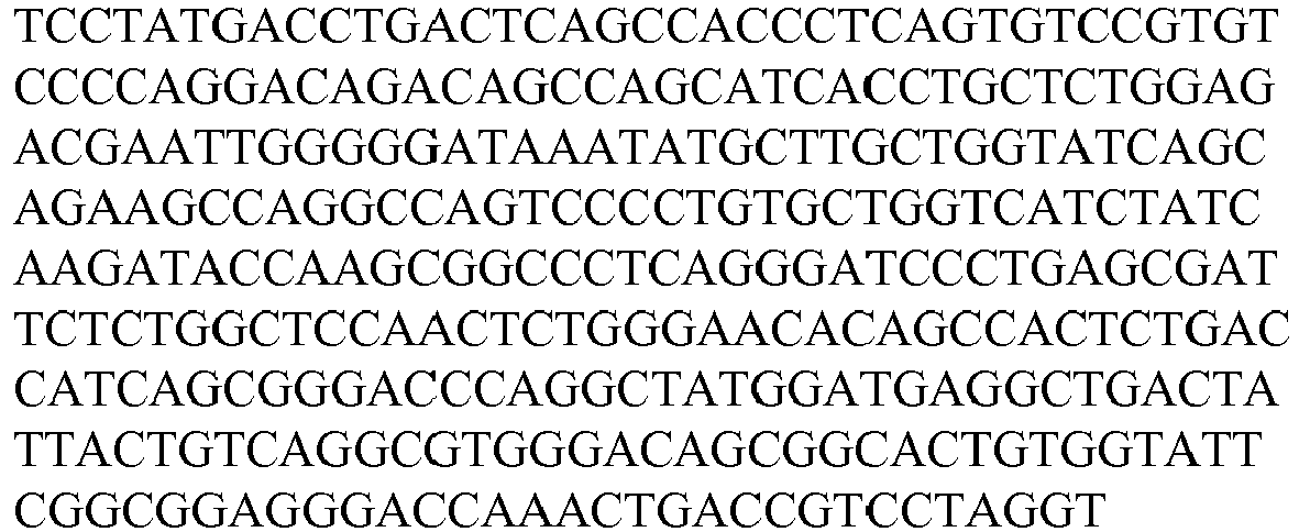
51E5 VL79 460 
51G2 VL51 461 
52A8 VL41 462 
52B8 VL82 463 
52C1 VL67 464 
52F8 VL42 465 
52H2 VL84 466 
53F6 VL63 467 
53H5.2 VL62 468 
53H5.3 VL80 469 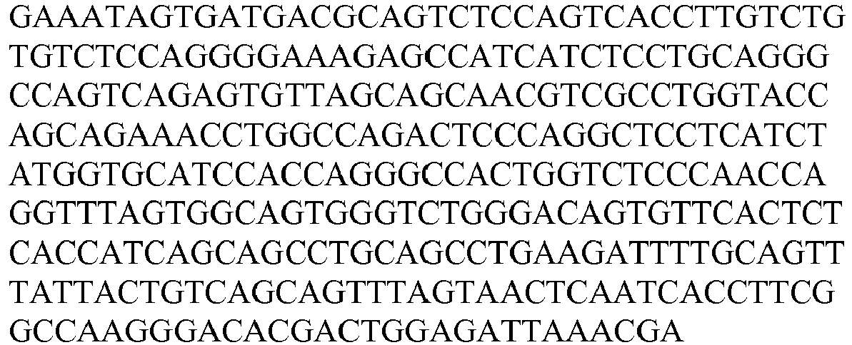
54A1 55G9 VL44 470 
54H10.1 VL53 471 
55D1 48H3 53C11 55D3 VL71 472 
55E4 VL75 473 
49B11 50H10 53C1 55E9 VL68 474 
55G5 VL83 475 
56A7 VL52 476 
56E4 56C11 VL64 477 
56E7 VL86 478 
56G1 VL76 479 
56G3.3 VL81 480 
55B10 57B12 VL72 481 
57D9 VL87 482 
59A10 VL48 483 
49H4 59C9 VL50 484 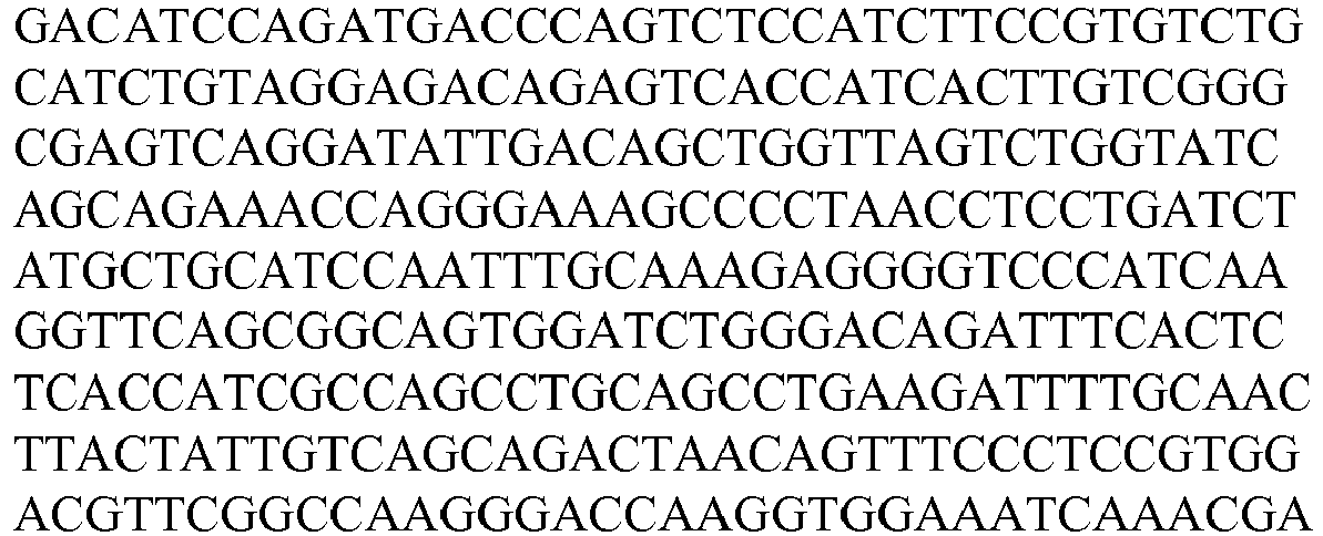
58A5 57A4 57F9 59G10.2 VL60 485 
59G10.3 VL54 486 
60D7 VL69 487 
60F9 VL58 488 
48B4 52D6 60G5.2 VL46 489 
61G5 VL59 490 
52C5 VL73 491 
61H5 VL88 492 
52B9 59D10v1 VL56 493 
59D10v2 VL57 494 
56G3.2 VL85 495 
66F7 VL7 496 
48G4 VL89 497 
53C3.1 50G1 VL90 498 
58C2 VL91 499 
60G5.1 VL74 1865 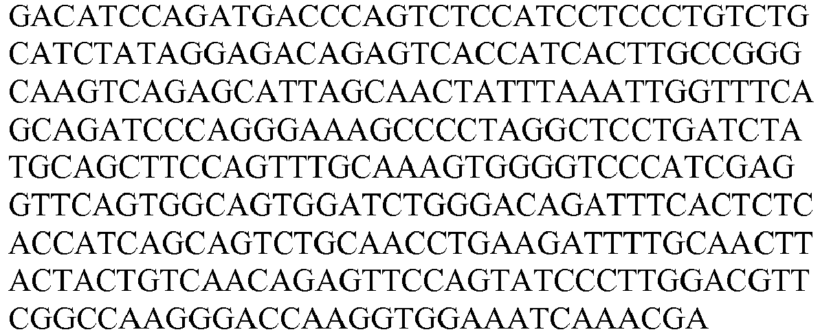
50D4 VL92 500 
50G5v1 VL93 501 
50G5v2 VL94 502 
51C1 VL95 503 
53C3.2 VL96 504 
54H10.3 VL97 505 
55A7 VL98 506 
55E6 VL99 507 
61E1 VL100 508 
Table 2D Coding Sequence for Antibody Variable Heavy (V H ) Chains Contained in Clone Designation SEQ ID NO. Coding Sequence 63E6 66F7 VH6 509 
66D4 VH17 510 
66B4 VH10 511 
65B1 VH18 512 
65B4 VH20 513 
67A4 VH19 514 
63A10v1 63A10v2 63A10v3 VH21 515 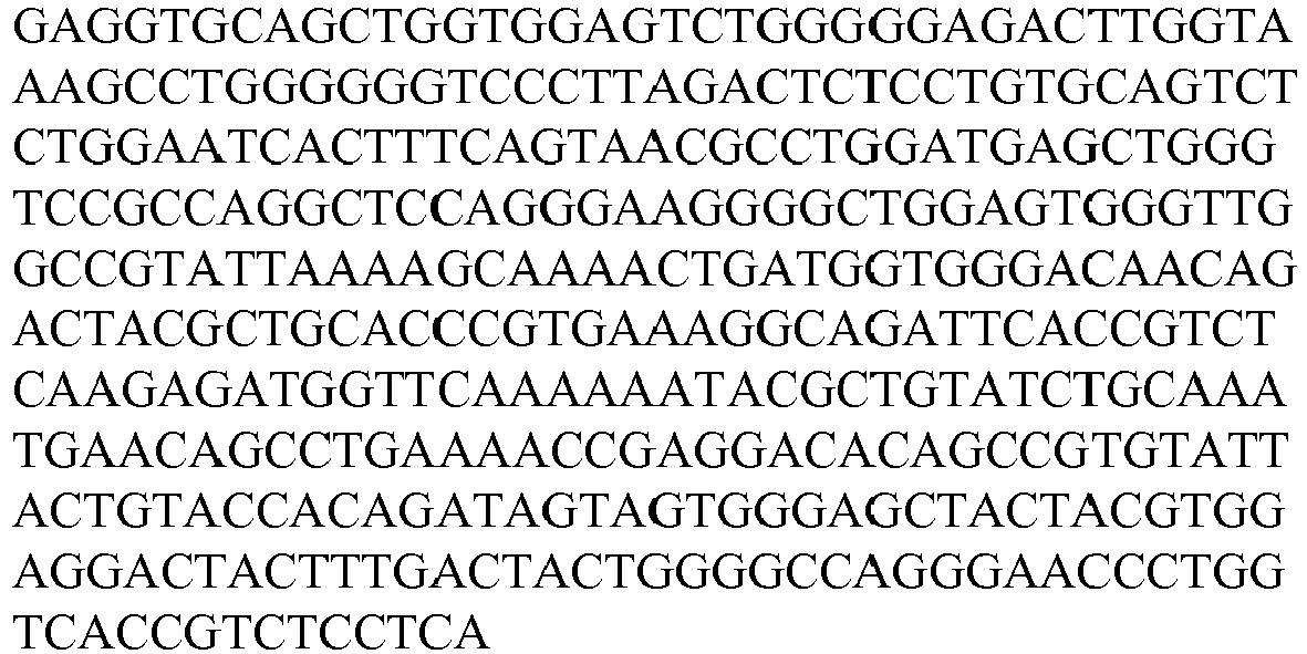
65H11v1 65H11v2 VH22 516 
67G10v1 67G10v2 VH9 517 
64C8 VH23 518 
63G8v1 63G8v2 63G8v3 68D3v1 64A8 67B4 VH1 519 
68D3v2 VH95 1866 
66G2 VH11 520 
65D1 VH26 521 
64H5 VH7 522 
65D4 VH25 523 
65E3 VH24 524 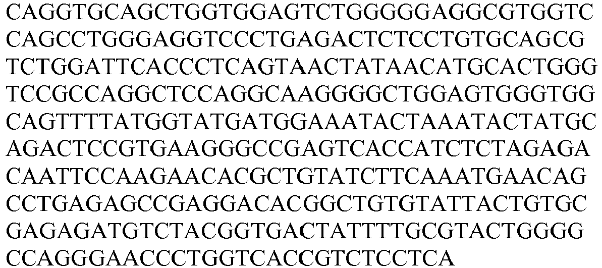
65G4 VH8 525 
68G5 VH12 526 
67G8 VH27 527 
65B7v1 65B7v2 VH28 528 
63B6 64D4 VH4 529 
63F5 VH13 530 
63H11 VH3 531 
65E8 64E6 65F11 67G7 VH2 532 
65C1 VH15 533 
66F6 VH14 534 
64A6 VH29 535 
65F9 VH30 536 
64A7 VH16 537 
65C3 68D5 VH5 538 
67F5 VH31 539 
64B10v1 VH32 540 
64B10v2 VH96 1867 
68C8 VH33 541 
67A5 VH34 542 
67C10 VH35 543 
64H6 VH36 544 
63F9 VH37 545 
67F6v1 67F6v2 VH38 546 
48C9 49A12 51E2 VH73 547 
48F3 VH72 548 
48F8 53B9 56B4 57E7 57F11 VH48 549 
48H11 VH39 550 
49A10 48D4 VH62 551 
49C8 52H1 VH44 552 
49G2 50C12 55G11 VH63 553 
49G3 VH46 554 
49H12 VH42 555 
51A8 VH58 556 
51C10.1 59D10v1 59D10v2 VH54 557 
51C10.2 VH67 558 
51E5 VH74 559 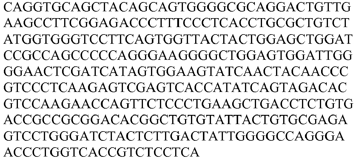
51G2 VH50 560 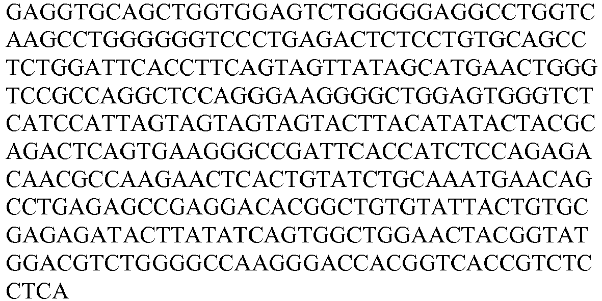
52A8 VH40 561 
52B8 VH77 562 
52C1 VH64 563 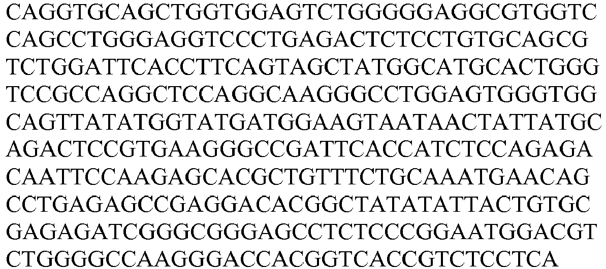
52F8 VH41 564 
52H2 VH79 565 
53F6 VH60 566 
53H5.2 VH59 567 
53H5.3 VH75 568 
54A1 55G9 VH43 569 
54H10.1 55D1 48H3 53C11 VH52 570 
55D3 VH68 571 
55E4 49B11 50H10 53C1 52C5 60G5.1 VH70 572 
55E9 VH65 573 
55G5 VH78 574 
50G1 VH84 575 
56A7 56E4 VH51 576 
56C11 VH61 577 
56E7 VH81 578 
56G1 VH71 579 
56G3.3 55B10 VH76 580 
57B12 VH69 581 
57D9 VH82 582 
58C2 VH85 583 
59A10 49H4 VH47 584 
59C9 58A5 57A4 57F9 VH49 585 
59G10.2 VH57 586 
59G10.3 VH53 587 
60D7 VH66 588 
60F9 48B4 52D6 VH55 589 
60G5.2 VH45 590 
61G5 VH56 591 
56G3.2 VH80 592 
48G4 53C3.1 VH83 593 
61H5 52B9 VH86 594 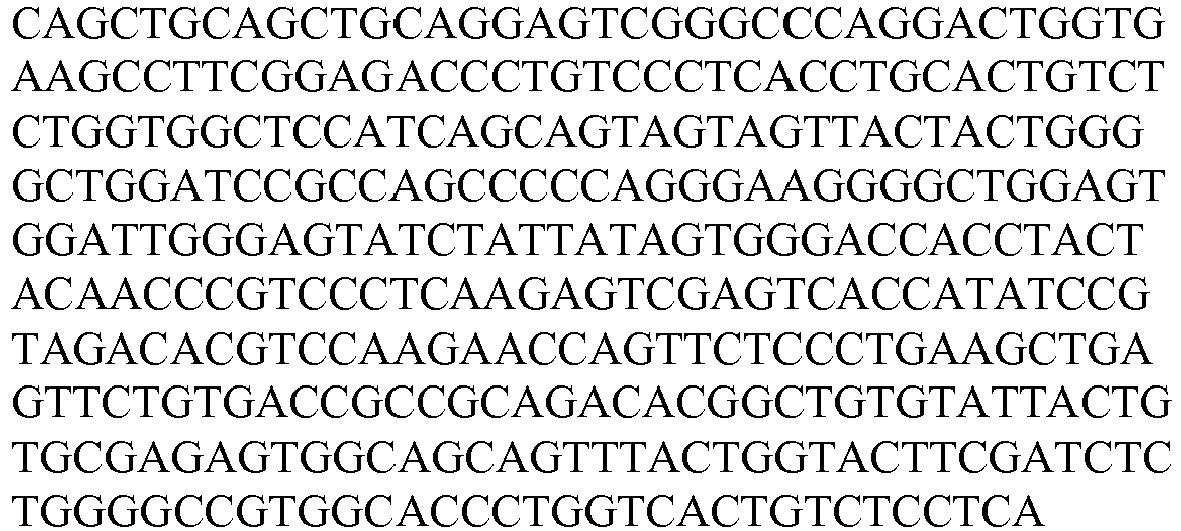
50D4 VH87 595 
50G5v1 50G5v2 VH88 596 
51C1 VH89 597 
53C3.2 VH90 598 
54H10.3 VH91 599 
55A7 VH92 600 
55E6 VH93 601 
61E1 VH94 602 
- Each of the heavy chain variable regions listed in Table 2B can be combined with any of the light chain variable regions shown in Table 2A to form an antigen binding protein. Examples of such combinations include VH1 combined with any of VL1, VL2, VL3, VL4, VL5, VL6, VL7, VL8, VL9, VL10, VL11, VL12, VL13, VL14, VL15, VL16, VL17, VL18, VL19, VL20, VL21, VL22, VL23, VL24, VL25, VL26, VL27, VL28, VL29, VL30, VL31, VL32, VL33, VL34, VL35, VL36, VL37, VL38, VL39, VL40, VL41, VL42, VL43, VL44, VL45, VL46, VL47, VL48, VL49, VL50, VL51, VL52, VL53, VL54, VL55, VL56, VL57, VL58, VL59, VL60, VL61, VL62, VL63, VL64, VL65, VL66, VL67, VL68, VL69, VL70, VL71, VL72, VL73, VL74, VL75, VL76, VL77, VL78, VL79, VL80, VL81, VL82, VL83, VL84, VL85, VL86, VL87, VL88, VL89, VL90, VL91, VL92, VL93, VL94, VL95, VL96, VL97, VL98, VL99 and VL100; VH2 combined with any of VL1, VL2, VL3, VL4, VL5, VL6, VL7, VL8, VL9, VL10, VL11, VL12, VL13, VL14, VL15, VL16, VL17, VL18, VL19, VL20, VL21, VL22, VL23, VL24, VL25, VL26, VL27, VL28, VL29, VL30, VL31, VL32, VL33, VL34, VL35, VL36, VL37, VL38, VL39, VL40, VL41, VL42, VL43, VL44, VL45, VL46, VL47, VL48, VL49, VL50, VL51, VL52, VL53, VL54, VL55, VL56, VL57, VL58, VL59, VL60, VL61, VL62, VL63, VL64, VL65, VL66, VL67, VL68, VL69, VL70, VL71, VL72, VL73, VL74, VL75, VL76, VL77, VL78, VL79, VL80, VL81, VL82, VL83, VL84, VL85, VL86, VL87, VL88, VL89, VL90, VL91, VL92, VL93, VL94, VL95, VL96, VL97, VL98, VL99 and VL100; VH3 combined with any of VL1, VL2, VL3, VL4, VL5, VL6, VL7, VL8, VL9, VL10, VL11, VL12, VL13, VL14, VL15, VL16, VL17, VL18, VL19, VL20, VL21, VL22, VL23, VL24, VL25, VL26, VL27, VL28, VL29, VL30, VL31, VL32, VL33, VL34, VL35, VL36, VL37, VL38, VL39, VL40, VL41, VL42, VL43, VL44, VL45, VL46, VL47, VL48, VL49, VL50, VL51, VL52, VL53, VL54, VL55, VL56, VL57, VL58, VL59, VL60, VL61, VL62, VL63, VL64, VL65, VL66, VL67, VL68, VL69, VL70, VL71, VL72, VL73, VL74, VL75, VL76, VL77, VL78, VL79, VL80, VL81, VL82, VL83, VL84, VL85, VL86, VL87, VL88, VL89, VL90, VL91, VL92, VL93, VL94, VL95, VL96, VL97, VL98, VL99 and VL100; and so on.
- In some instances, the antigen binding protein includes at least one heavy chain variable region and/or one light chain variable region from those listed in Tables 2A and 2B. In some instances, the antigen binding protein includes at least two different heavy chain variable regions and/or light chain variable regions from those listed in Table 2B. An example of such an antigen binding protein comprises (a) one VH1, and (b) one of VH2, VH3, VH4, VH5, VH6, VH7, VH8, VH9, VH10, VH11, VH12, VH13, VH14, VH15, VH16, VH17, VH18, VH19, VH20, VH21 VH22, VH23, VH24, VH25, VH26, VH27, VH28, VH29, VH30, VH31, VH32, VH33, VH34, VH35, VH36, VH37, VH38, VH39, VH40, VH41, VH42, VH43, VH44, VH45, VH46, VH47, VH48, VH49, VH50, VH51, VH52, VH53, VH54, VH55, VH56, VH57, VH58, VH59, VH60, VH61, VH62, VH63, VH64, VH65, VH66, VH67, VH68, VH69, VH70, VH71, VH72, VH73, VH74, VH75, VH76, VH77, VH78, VH79, VH80, 81, VH82, VH83, VH84, VH85, VH 86, VH 87, VH88, VH89, VH90, VH91, VH92, VH93, and VH94. Another example comprises (a) one VH2, and (b) one of VH1, VH3, VH4, VH5, VH6, VH7, VH8, VH9, VH10, VH11, VH12, VH13, VH14, VH15, VH16, VH17, VH18, VH19, VH20, VH21 VH22, VH23, VH24, VH25, VH26, VH27, VH28, VH29, VH30, VH31, VH32, VH33, VH34, VH35, VH36, VH37, VH38, VH39, VH40, VH41, VH42, VH43, VH44, VH45, VH46, VH47, VH48, VH49, VH50, VH51, VH52, VH53, VH54, VH55, VH56, VH57, VH58, VH59, VH60, VH61, VH62, VH63, VH64, VH65, VH66, VH67, VH68, VH69, VH70, VH71, VH72, VH73, VH74, VH75, VH76, VH77, VH78, VH79, VH80, 81, VH82, VH83, VH84, VH85, VH 86, VH 87, VH88, VH89, VH90, VH91, VH92, VH93, and VH94. Yet another example comprises (a) one VH3, and (b) one of VH1, VH2, VH4, VH5, VH6, VH7, VH8, VH9, VH10, VH11, VH12, VH13, VH14, VH15, VH16, VH17, VH18, VH19, VH20, VH21 VH22, VH23, VH24, VH25, VH26, VH27, VH28, VH29, VH30, VH31, VH32, VH33, VH34, VH35, VH36, VH37, VH38, VH39, VH40, VH41, VH42, VH43, VH44, VH45, VH46, VH47, VH48, VH49, VH50, VH51, VH52, VH53, VH54, VH55, VH56, VH57, VH58, VH59, VH60, VH61, VH62, VH63, VH64, VH65, VH66, VH67, VH68, VH69, VH70, VH71, VH72, VH73, VH74, VH75, VH76, VH77, VH78, VH79, VH80, 81, VH82, VH83, VH84, VH85, VH 86, VH 87, VH88, VH89, VH90, VH91, VH92, VH93, and VH94, etc. Still another example of such an antigen binding protein comprises (a) one VL1, and (b) one of VL2, VL3, VL4, VL5, VL6, VL7, VL8, VL9, VL10, VL11, VL12, VL13, VL14, VL15, VL16, VL17, VL18, VL19, VL20, VL21, VL22, VL23, VL24, VL25, VL26, VL27, VL28, VL29, VL30, VL31, VL32, VL33, VL34, VL35, VL36, VL37, VL38, VL39, VL40, VL41, VL42, VL43, VL44, VL45, VL46, VL47, VL48, VL49, VL50, VL51, VL52, VL53, VL54, VL55, VL56, VL57, VL58, VL59, VL60, VL61, VL62, VL63, VL64, VL65, VL66, VL67, VL68, VL69, VL70, VL71, VL72, VL73, VL74, VL75, VL76, VL77, VL78, VL79, VL80, VL81, VL82, VL83, VL84, VL85, VL86, VL87, VL88, VL89, VL90, VL91, VL92, VL93, VL94, VL95, VL96, VL97, VL98, VL99 and VL100. Again another example of such an antigen binding protein comprises (a) one VL2, and (b) one of VL1, VL3, VL4, VL5, VL6, VL7, VL8, VL9, VL10, VL11, VL12, VL13, VL14, VL15, VL16, VL17, VL18, VL19, VL20, VL21, VL22, VL23, VL24, VL25, VL26, VL27, VL28, VL29, VL30, VL31, VL32, VL33, VL34, VL35, VL36, VL37, VL38, VL39, VL40, VL41, VL42, VL43, VL44, VL45, VL46, VL47, VL48, VL49, VL50, VL51, VL52, VL53, VL54, VL55, VL56, VL57, VL58, VL59, VL60, VL61, VL62, VL63, VL64, VL65, VL66, VL67, VL68, VL69, VL70, VL71, VL72, VL73, VL74, VL75, VL76, VL77, VL78, VL79, VL80, VL81, VL82, VL83, VL84, VL85, VL86, VL87, VL88, VL89, VL90, VL91, VL92, VL93, VL94, VL95, VL96, VL97, VL98, VL99 and VL100. Again another example of such an antigen binding protein comprises (a) one VL3, and (b) one of VL1, VL2, VL4, VL5, VL6, VL7, VL8, VL9, VL10, VL11, VL12, VL13, VL14, VL15, VL16, VL17, VL18, VL19, VL20, VL21, VL22, VL23, VL24, VL25, VL26, VL27, VL28, VL29, VL30, VL31, VL32, VL33, VL34, VL35, VL36, VL37, VL38, VL39, VL40, VL41, VL42, VL43, VL44, VL45, VL46, VL47, VL48, VL49, VL50, VL51, VL52, VL53, VL54, VL55, VL56, VL57, VL58, VL59, VL60, VL61, VL62, VL63, VL64, VL65, VL66, VL67, VL68, VL69, VL70, VL71, VL72, VL73, VL74, VL75, VL76, VL77, VL78, VL79, VL80, VL81, VL82, VL83, VL84, VL85, VL86, VL87, VL88, VL89, VL90, VL91, VL92, VL93, VL94, VL95, VL96, VL97, VL98, VL99 and VL100, etc.
- The various combinations of heavy chain variable regions can be combined with any of the various combinations of light chain variable regions.
- In other embodiments, an antigen binding protein comprises two identical light chain variable regions and/or two identical heavy chain variable regions. As an example, the antigen binding protein can be an antibody or immunologically functional fragment thereof that includes two light chain variable regions and two heavy chain variable regions in combinations of pairs of light chain variable regions and pairs of heavy chain variable regions as listed in Tables 2A and 2B.
- Some antigen binding proteins that are provided comprise a heavy chain variable domain comprising a sequence of amino acids that differs from the sequence of a heavy chain variable domain selected from VH1, VH2, VH3, VH4, VH5, VH6, VH7, VH8, VH9, VH10, VH11, VH12, VH13, VH14, VH15, VH16, VH17, VH18, VH19, VH20, VH21 VH22, VH23, VH24, VH25, VH26, VH27, VH28, VH29, VH30, VH31, VH32, VH33, VH34, VH35, VH36, VH37, VH38, VH39, VH40, VH41, VH42, VH43, VH44, VH45, VH46, VH47, VH48, VH49, VH50, VH51, VH52, VH53, VH54, VH55, VH56, VH57, VH58, VH59, VH60, VH61, VH62, VH63, VH64, VH65, VH66, VH67, VH68, VH69, VH70, VH71, VH72, VH73, VH74, VH75, VH76, VH77, VH78, VH79, VH80, 81, VH82, VH83, VH84, VH85, VH 86, VH 87, VH88, VH89, VH90, VH91, VH92, VH93, and VH94 at only 1, 2, 3, 4, 5, 6, 7, 8, 9, 10, 11, 12, 13, 14 or 15 amino acid residues, wherein each such sequence difference is independently either a deletion, insertion or substitution of one amino acid, with the deletions, insertions and/or substitutions resulting in no more than 15 amino acid changes relative to the foregoing variable domain sequences. The heavy chain variable region in some antigen binding proteins comprises a sequence of amino acids that has at least 70%, 75%, 80%, 85%, 90%, 95%, 97% or 99% sequence identity to the amino acid sequences of the heavy chain variable region of VH1, VH2, VH3, VH4, VH5, VH6, VH7, VH8, VH9, VH10, VH11, VH12, VH13, VH14, VH15, VH16, VH17, VH18, VH19, VH20, VH21 VH22, VH23, VH24, VH25, VH26, VH27, VH28, VH29, VH30, VH31, VH32, VH33, VH34, VH35, VH36, VH37, VH38, VH39, VH40, VH41, VH42, VH43, VH44, VH45, VH46, VH47, VH48, VH49, VH50, VH51, VH52, VH53, VH54, VH55, VH56, VH57, VH58, VH59, VH60, VH61, VH62, VH63, VH64, VH65, VH66, VH67, VH68, VH69, VH70, VH71, VH72, VH73, VH74, VH75, VH76, VH77, VH78, VH79, VH80, 81, VH82, VH83, VH84, VH85, VH 86, VH 87, VH88, VH89, VH90, VH91, VH92, VH93, and VH94.
- Certain antigen binding proteins comprise a light chain variable domain comprising a sequence of amino acids that differs from the sequence of a light chain variable domain selected from VL1, VL2, VL3, VL4, VL5, VL6, VL7, VL8, VL9, VL10, VL11, VL12, VL13, VL14, VL15, VL16, VL17, VL18, VL19, VL20, VL21, VL22, VL23, VL24, VL25, VL26, VL27, VL28, VL29, VL30, VL31, VL32, VL33, VL34, VL35, VL36, VL37, VL38, VL39, VL40, VL41, VL42, VL43, VL44, VL45, VL46, VL47, VL48, VL49, VL50, VL51, VL52, VL53, VL54, VL55, VL56, VL57, VL58, VL59, VL60, VL61, VL62, VL63, VL64, VL65, VL66, VL67, VL68, VL69, VL70, VL71, VL72, VL73, VL74, VL75, VL76, VL77, VL78, VL79, VL80, VL81, VL82, VL83, VL84, VL85, VL86, VL87, VL88, VL89, VL90, VL91, VL92, VL93, VL94, VL95, VL96, VL97, VL98, VL99 and VL100 at only 1, 2, 3, 4, 5, 6, 7, 8, 9, 10, 11, 12, 13, 14 or 15 amino acid residues, wherein each such sequence difference is independently either a deletion, insertion or substitution of one amino acid, with the deletions, insertions and/or substitutions resulting in no more than 15 amino acid changes relative to the foregoing variable domain sequences. The light chain variable region in some antigen binding proteins comprises a sequence of amino acids that has at least 70%, 75%, 80%, 85%, 90%, 95%, 97% or 99% sequence identity to the amino acid sequences of the light chain variable region of VL1, VL2, VL3, VL4, VL5, VL6, VL7, VL8, VL9, VL10, VL11, VL12, VL13, VL14, VL15, VL16, VL17, VL18, VL19, VL20, VL21, VL22, VL23, VL24, VL25, VL26, VL27, VL28, VL29, VL30, VL31, VL32, VL33, VL34, VL35, VL36, VL37, VL38, VL39, VL40, VL41, VL42, VL43, VL44, VL45, VL46, VL47, VL48, VL49, VL50, VL51, VL52, VL53, VL54, VL55, VL56, VL57, VL58, VL59, VL60, VL61, VL62, VL63, VL64, VL65, VL66, VL67, VL68, VL69, VL70, VL71, VL72, VL73, VL74, VL75, VL76, VL77, VL78, VL79, VL80, VL81, VL82, VL83, VL84, VL85, VL86, VL87, VL88, VL89, VL90, VL91, VL92, VL93, VL94, VL95, VL96, VL97, VL98, VL99 and VL100.
- In additional instances, antigen binding proteins comprise the following pairings of light chain and heavy chain variable domains: VL1 with VH1, VL2 with VH1, VL3 with VH2 or VH3, VL4 with VH4, VL5 with VH5, VL6 with VH6, VL7 with VH6, VL8 with VH7 or VH8, VL9 with VH9, VL10 with VH9, VL11 with VH 10, VL12 with VH11, VL13 with VH12, VL13 with VH14, VL14 with VH13, VL15 with VH14, VL16 with VH15, VL17 with VH16, VL18 with VH17, VL19 with VH18, VL20 with VH19, VL21 with VH20, VL22 with VH21, VL23 with VH22, VL24 with VH23, VL25 with VH24, VL26 with VH25, VL27 with VH26, VL28 with VH27, VL29 with VH28, VL30 with VH29, VL31 with VH30, VL32 with VH31, VL33 with VH32, VL34 with VH33, VL35 with VH34, VL36 with VH35, VL37 with VH36, VL38 with VH37, VL39 with VH38, VL40 with VH39, VL41 with VH40, VL42 with VH41, VL43 with VH42, VL44 with VH43, VL45 with VH44, VL46 with VH45, VL47 with VH46, VL48 with VH47, VL49 with VH48, VL50 with VH49, VL51 with VH50, 52 with VH51, VL53 with VH52, VL54 with VH53, VL55 with 54, and VL56 with VH54, VL57 with VH54, VL58 with VH55, VL59 with VH56, VL60 with VH57, VL61 with VH58, VL62 with VH59, VL63 with VH60, VL64 with VH1, VL65 with VH62, VL66 with VH63, VL67 with VH64, VL68 with VH65, VL69 with VH66, VL70 with VH67, VL71 with VH68, VL72 with VH69, VL73 with VH70, VL74 with VH70, and VL75 with VH70, VL76 with VH71, VL77 with VH72, VL78 with VH73, VL79 with VH74, VL80 with VH75, VL81 with VH76, VL82 with VH77, VL83 with VH78, VL84 with VH79, VL85 with VH80, VL86 with VH81, VL87 with VH82, VL88 with VH86, VL89 with VH83, VL90 with VH84, VL91 with VH85, VL 92 with VH 87, VL 93 with VH 88, VL 94 with VH 88, VL 95 with VH 89, VL 96 with VH 90, VL 97 with VH 91, VL 98 with VH 92, VL 99 with VH 93, and VL 100 with VH 94.
- In some instances, the antigen binding proteins in the above pairings can comprise amino acid sequences that have 70%, 75%, 80%, 85%, 90%, 95%, 96%, 97%, 98% or 99% sequence identity with the specified variable domains.
- Still other antigen binding proteins, e.g., antibodies or immunologically functional fragments, include variant forms of a variant heavy chain and a variant light chain as just described.
- In various embodiments, the antigen binding proteins disclosed herein can comprise polypeptides into which one or more CDRs are grafted, inserted and/or joined. An antigen binding protein can have 1, 2, 3, 4, 5 or 6 CDRs. An antigen binding protein thus can have, for example, one heavy chain CDR1 ("CDRH1"), and/or one heavy chain CDR2 ("CDRH2"), and/or one heavy chain CDR3 ("CDRH3"), and/or one light chain CDR1 ("CDRL1"), and/or one light chain CDR2 ("CDRL2"), and/or one light chain CDR3 ("CDRL3"). Some antigen binding proteins include both a CDRH3 and a CDRL3. Specific heavy and light chain CDRs are identified in Tables 3A and 3B, respectively, infra.
- Complementarity determining regions (CDRs) and framework regions (FR) of a given antibody can be identified using the system described by Kabat et al., (1991) "Sequences of Proteins of Immunological Interest", 5th Ed., US Dept. of Health and Human Services, PHS, NIH, NIH Publication no. 91-3242. Although presented in the Kabat nomenclature scheme, as desired, the CDRs disclosed herein can also be redefined according an alternative nomenclature scheme, such as that of Chothia (see Chothia & Lesk, (1987) J. Mol. Biol. 196:901-917; Chothia et al., (1989) Nature 342:878-883 or Honegger & Pluckthun, (2001) J. Mol. Biol. 309:657-670). Certain antibodies that are disclosed herein comprise one or more amino acid sequences that are identical or have substantial sequence identity to the amino acid sequences of one or more of the CDRs presented in Table 3A (CDRHs) and Table 3B (CDRLs), infra.
TABLE 3A Exemplary CDRH Sequences Clone SEQ ID NO: Contained in Reference Designation Sequence 48C9 603 VH73 CDRH1-1 GYYWT 49A12 VH73 51E2 VH73 48F3 604 VH72 CDRH1-2 GYYWS 51E5 VH74 52C5 VH70 55E4 VH70 60G5.1 VH70 49B11 VH70 50H10 VH70 53C1 VH70 56G1 VH71 51C1 VH89 48F8 605 VH48 CDRH1-3 SYSMN 51G2 VH50 56A7 VH51 53B9 VH48 56B4 VH48 57E7 VH48 57F11 VH48 56E4 VH51 55E6 VH93 48H11 606 VH39 CDRH1-4 GYYKH 48G4 607 VH83 CDRH1-5 ELSIH 53C3.1 VH83 49A10 608 VH62 CDRH1-6 NYGMH 58C2 VH85 59G10.2 VH57 48D4 VH62 49C8 609 VH44 CDRH1-7 SYDID 52H1 VH44 49G2 610 VH63 CDRH1-8 NYGMR 50C12 VH63 55G11 VH63 49G3 611 VH46 CDRH1-9 NPRMGVS 49H12 612 VH42 CDRH1-10 SYDIN 54A1 VH43 55G9 VH43 50G1 613 VH84 CDRH1-11 SYGLH 51A8 614 VH58 CDRH1-12 SYGMH 52C1 VH64 53H5.2 VH59 56C11 VH61 60D7 VH66 64H5 VH7 65G4 VH8 66G2 VH11 68G5 VH12 64C8 VH23 67G8 VH27 68D3v2 51C10.1 615 VH54 CDRH1-13 NYAMS 59D10v1 VH54 59D10v2 VH54 51C10.2 616 VH67 CDRH1-14 SGGYYWS 64A6 VH29 52A8 617 VH40 CDRH1-15 GYYLH 66B4 VH10 52B8 618 VH77 CDRH1-16 YYYWS 52F8 619 VH41 CDRH1-17 GYYTH 52H2 620 VH79 CDRH1-18 TYYWS 53F6 621 VH60 CDRH1-19 TYGMH 53H5.3 622 VH75 CDRH1-20 DYYWN 54H10.1 623 VH52 CDRH1-21 SYAMS 60F9 VH55 61G5 VH56 55D1 VH52 48H3 VH52 53C11 VH52 48B4 VH55 52D6 VH55 55D3 624 VH68 CDRH1-22 SGVYYWN 55E9 625 VH65 CDRH1-23 SFGMH 55G5 626 VH78 CDRH1-24 SYYWS 65C3 VH5 68D5 VH5 67F5 VH31 55A7 VH92 56E7 627 VH81 CDRH1-25 SYWIG 67A5 VH34 67C10 VH35 64H6 VH36 56G3.2 628 VH80 CDRH1-26 SYYWN 56G3.3 629 VH76 CDRH1-27 SSSYYWG 55B10 VH76 61H5 VH86 52B9 VH86 57B12 630 VH69 CDRH1-28 SGVYYWS 57D9 631 VH82 CDRH1-29 SNSATWN 59A10 632 VH47 CDRH1-30 DSYMS 49H4 VH47 59C9 633 VH49 CDRH1-31 SYSMS 58A5 VH49 57A4 VH49 57F9 VH49 59G10.3 634 VH53 CDRH1-32 HYAMS 60G5.2 635 VH45 CDRH1-33 NYGIS 63G8 636 VH1 CDRH1-34 SYGIH 64A8 VH1 67B4 VH1 68D3 VH1 64E6 637 VH2 CDRH1-35 SGDYYWT 65E8 VH2 65F11 VH2 67G7 VH2 63H11 VH3 63F5 VH13 65C1 VH15 66F6 VH14 63B6 638 VH4 CDRH1-36 SGDYYWS 64D4 VH4 65F9 VH30 64B10 64B10v2 VH32 63E6 639 VH6 CDRH1-37 GYYMH 66F7 VH6 50G5 v1 VH88 50G5 v2 VH88 67G10v1 640 VH9 CDRH1-38 NAWMS 67G10v2 VH9VH21 63A10 65H11 VH22 53C3.2 641 VH90 CDRH1-39 SGNYYWS 64A7 642 VH16 CDRH1-40 SDTSYWG 50D4 643 VH87 CDRH1-41 SHDIN 61E1 644 VH94 CDRH1-42 SNSAAWN 66D4 645 VH17 CDRH1-43 GYYIH 54H10.3 VH91 65B1 646 VH18 CDRH1-44 GYFMH 67A4 647 VH19 CDRH1-45 TYDMH 65B4 648 VH20 CDRH1-46 SYDMH 65E3 649 VH24 CDRH1-47 NYNMH 65D4 650 VH25 CDRH1-48 FYGMH 65D1 651 VH26 CDRH1-49 YYYIH 65B7 652 VH28 CDRH1-50 SDAYYWS 68C8 653 VH33 CDRH1-51 SGDNYWS 63F9 654 VH37 CDRH1-52 SGGYYWN 67F6v1 655 VH38 CDRH1-53 GYWIG 67F6v2 VH38 48C9 656 VH73 CDRH2-1 EINHSENTNYNPSLKS 52C5 VH70 55E4 VH70 56G1 VH71 49A12 VH73 51E2 VH73 60G5.1 VH70 49B11 VH70 50H10 VH70 53C1 VH70 51C1 VH89 48F3 657 VH72 CDRH2-2 EITHTGSSNYNPSLKS 48F8 658 VH48 CDRH2-3 SISSSSSYEYYVDSVKG 53B9 VH48 56B4 VH48 57E7 VH48 57F11 VH48 48H11 659 VH39 CDRH2-4 WINPNSGATKYAQKFQG 48G4 660 VH83 CDRH2-5 GFDPEDGETIYAQKFQG 53C3.1 VH83 49A10 661 VH62 CDRH2-6 IIWYDGSNKNYADSVKG 48D4 VH62 49C8 662 VH44 CDRH2-7 WMNPNGGNTGYAQKFQG 52H1 VH44 49G2 663 VH63 CDRH2-8 LIWYDGSNKFYADSVKG 50C12 VH63 55G11 VH63 49G3 664 VH46 CDRH2-9 HIFSNDEKSYSTSLKS 49H12 665 VH42 CDRH2-10 WMNPYSGSTGYAQNFQG 50G1 666 VH84 CDRH2-11 VIWNDGSNKLYADSVKG 51A8 667 VH58 CDRH2-12 VISYDGSNKYYADSVKG 63G8 VH1 64A8 VH1 67B4 VH1 68D3 VH1 51C10.1 668 VH54 CDRH2-13 GISGSSAGTYYADSVKG 59D10v1 VH54 59D10v2 VH54 51C10.2 669 VH67 CDRH2-14 YIYYNGSPYDNPSLKR 51E5 670 VH74 CDRH2-15 ELDHSGSINYNPSLKS 51G2 671 VH50 CDRH2-16 SISSSSTYIYYADSVKG 56A7 VH51 56E4 VH51 52A8 672 VH40 CDRH2-17 WINPNSAATNYAPKFQG 52B8 673 VH77 CDRH2-18 YIYYSGSTNYNPSLKS 55A7 VH92 52C1 674 VH64 CDRH2-19 VIWYDGSNNYYADSVKG 52F8 675 VH41 CDRH2-20 WINPSSGDTKYAQKFQG 52H2 676 VH79 CDRH2-21 YIFYNGNANYSPSLKS 53F6 677 VH60 CDRH2-22 VIWYDGSNKYYADSVKG 60D7 VH66 65D4 VH25 53H5.2 678 VH59 CDRH2-23 LISYDGSNKYYADSVKG 53H5.3 679 VH75 CDRH2-24 EINHSGTTNYNPSLKS 54A1 680 VH43 CDRH2-25 WMNPHSGNTGYAQKFQG 55G9 VH43 54H10.1 681 VH52 CDRH2-26 AISGSGRTTYSADSVKG 55D1 VH52 48H3 VH52 53C11 VH52 55D3 682 VH68 CDRH2-27 YLYYSGSTYYNPSLKS 55E9 683 VH65 CDRH2-28 LIWYDGDNKYYADSVKG 55G5 684 VH78 CDRH2-29 RIYISGSTNYNPSLEN 56C11 685 VH61 CDRH2-30 VIWYDGSYQFYADSVKG 56E7 686 VH81 CDRH2-31 IIYPGDSDTRYSPSFQG 67A5 VH34 67C10 VH35 67F6v1 VH38 67F6v2 VH38 56G3.2 687 VH80 CDRH2-32 RIYTSGSTNYNPSLKS 56G3.3 688 VH76 CDRH2-33 MIYYSGTTYYNPSLKS 55B10 VH76 56G3.3 VH76 57B12 689 VH69 CDRH2-34 YIYYSGSTYYNPSLKS 63H11 VH3 66F6 VH14 65F9 VH30 57D9 690 VH82 CDRH2-35 RTYYRSKWYNDYAVSVKS 61E1 VH94 58C2 691 VH85 CDRH2-36 VIWNDGNNKYYADSVKG 59A10 692 VH47 CDRH2-37 SISSSGSIVYFADSVKG 49H4 VH47 59C9 693 VH49 CDRH2-38 SISSSSTYIYYAD SLKG 58A5 VH49 57A4 VH49 57F9 VH49 59G10.2 694 VH57 CDRH2-39 ITSYGGSNKNYADSVKG 59G10.3 695 VH53 CDRH2-40 AISGSGAGTFYADSMKG 60F9 696 VH55 CDRH2-41 VISDSGGSTYYADSVKG 48B4 VH55 52D6 VH55 60G5.2 697 VH45 CDRH2-42 WISAYNGYSNYAQKFQD 61G5 698 VH56 CDRH2-43 VISGSGGDTYYADSVKG 64E6 699 VH2 CDRH2-44 YIYYTGSTYYNPSLKS 65E8 VH2 65F11 VH2 67G7 VH2 63B6 700 VH4 CDRH2-45 YIYYSGTTYYNPSLKS 64D4 VH4 65C3 701 VH5 CDRH2-46 YIYYTGSTNYNPSLKS 68D5 VH5 63E6 702 VH6 CDRH2-47 WMNPNSGATKYAQKFQG 66F7 VH6 64H5 703 VH7 CDRH2-48 VIWDDGSNKYYADSVKG 65G4 VH8 67G10v1 704 VH9 CDRH2-49 RIKSKTDGGTTEYAAPVKG 67G10v2 VH9 63F5 705 VH13 CDRH2-50 YIYYSGSAYYNPSLKS 64A7 706 VH16 CDRH2-51 NIYYSGTTYFNPSLKS 65C1 707 VH15 CDRH2-52 YIFYSGSTYYNPSLKS 65B7 VH28 66B4 708 VH10 CDRH2-53 WINPNSGGTDYAQKFQG 66G2 709 VH11 CDRH2-54 GISYDGSNKNYADSVKG 68G5 710 VH12 CDRH2-55 VIWYDGSNKYHADSVKG 66D4 711 VH17 CDRH2-56 WINPPSGATNYAQKFRG 65B1 712 VH18 CDRH2-57 WINPNSGATNYAQKFHG 67A4 713 VH19 CDRH2-58 AIGIAGDTYYSDSVKG 65B4 714 VH20 CDRH2-59 TIDTAGDAYYPGSVKG 63A10 715 VH21 CDRH2-60 RIKSKTDGGTTDYAAPVKG 67G10v1 67G10v2 65H11 716 VH22 CDRH2-61 RIIGKTDGGTTDYAAPVKG 64C8 717 VH23 CDRH2-62 VISYDGSNKHYADSVKG 65E3 718 VH24 CDRH2-63 VLWYDGNTKYYADSVKG 65D1 719 VH26 CDRH2-64 LIWYDGSNKDYADSVKG 67G8 720 VH27 CDRH2-65 VIWYDGSNKDYADSVKG 64A6 721 VH29 CDRH2-66 YIYYSGGTHYNPSLKS 67F5 722 VH31 CDRH2-67 YIYYSGNTNYNPSLKS 64B10 723 VH32 CDRH2-68 FIYYSGGTNYNPSLKS 68C8 724 VH33 CDRH2-69 FMFYSGSTNYNPSLKS 64H6 725 VH36 CDRH2-70 IIYPGDSETRYSPSFQG 63F9 726 VH37 CDRH2-71 YIYDSGSTYYNPSLKS 61H5 727 VH86 CDRH2-72 SIYYSGTTYYNPSLKS 52B9 VH86 50G5v1 728 VH88 CDRH2-73 WINPDSGGTNYAQKFQG 50G5v2 VH88 54H10.3 729 VH91 CDRH2-74 WINPNSGGTNYAQKFRG 50D4 730 VH87 CDRH2-75 WMNPYSGSTGLAQRFQD 55E6 731 VH93 CDRH2-76 YISSGSSTIYHADSVKG 53C3.2 732 VH90 CDRH2-77 YIYHSGSAYYNPSLKS 64B10v2 1868 VH96 CDRH2-78 FIYYSGGTNYNPPLKS 68D3v2 1869 VH95 CDRH2-79 FISYAGSNKYYADSVKG 48C9 733 VH73 CDRH3-1 ESGNFPFDY 49A12 VH73 51E2 VH73 48F3 734 VH72 CDRH3-2 GGILWFGEQAFDI 48F8 735 VH48 CDRH3-3 SLSIAVAASDY 53B9 VH48 56B4 VH48 57E7 VH48 57F11 VH48 48H11 736 VH39 CDRH3-4 EVPDGIWAGSNAFDF 48G4 737 VH83 CDRH3-5 HSGSGRFYYYYYGMDV 53C3.1 VH83 49A10 738 VH62 CDRH3-6 DQDYDFWSGYPYFYYYGMDV 48D4 VH62 49C8 739 VH44 CDRH3-7 GKEFSRAEFDY 52H1 VH44 49G2 740 VH63 CDRH3-8 DRYYDFWSGYPYFFYYGLDV 50C12 VH63 55G11 VH63 49G3 741 VH46 CDRH3-9 VDTLNYHYYGMDV 49H12 742 VH42 CDRH3-10 YNWNYGAFDF 54A1 VH43 55G9 VH43 50G1 743 VH84 CDRH3-11 DQYYDFWSGYPYYHYYGMDV 51A8 744 VH58 CDRH3-12 ADGDYPYYYYYYGMDV 51C10.1 745 VH54 CDRH3-13 DWSIAVAGTFDY 59D10v1 VH54 59D10v2 VH54 51C10.2 746 VH67 CDRH3-14 GALYGMDV 51E5 747 VH74 CDRH3-15 VLGSTLDY 51G2 748 VH50 CDRH3-16 DTYISGWNYGMDV 52A8 749 VH40 CDRH3-17 EGGTYNWFDP 52B8 750 VH77 CDRH3-18 GTRAFDI 52C1 751 VH64 CDRH3-19 DRAGASPGMDV 52C5 752 VH70 CDRH3-20 VTGTDAFDF 60G5.1 VH70 49B11 VH70 50H10 VH70 53C1 VH70 51C1 VH89 55E4 VH70 56G1 VH71 52F8 753 VH41 CDRH3-21 SGWYPSYYYGMDV 52H2 754 VH79 CDRH3-22 ETDYGDYARPFEY 53F6 755 VH60 CDRH3-23 GHYDSSGPRDY 53H5.2 756 VH59 CDRH3-24 EANWGYNYYGMDV 53H5.3 757 VH75 CDRH3-25 ILRYFDWLEYYFDY 61E1 758 VH94 CDRH3-26 EGSWSSFFDY 54H10 759 VH52 CDRH3-27 EQQWLVYFDY 55D1 VH52 48H3 VH52 53C11 VH52 55D3 760 VH68 CDRH3-28 DGITMVRGVTHYYGMDV 57B12 VH69 55E6 761 VH93 CDRH3-29 EGYYDSSGYYYNGMDV 55E9 762 VH65 CDRH3-30 NSGWDYFYYYGMDV 55G5 763 VH78 CDRH3-31 SGSYSFDY 56A7 764 VH51 CDRH3-32 DIYSSGWSYGMDV 56E4 VH51 56C11 765 VH61 CDRH3-33 DHVWRTYRYIFDY 56E7 766 VH81 CDRH3-34 AQLGIFDY 50G5v1 767 VH88 CDRH3-35 GGYSYGYEDYYGMDV 50G5v2 VH88 56G3.2 768 VH80 CDRH3-36 GPLWFDY 56G3.3 769 VH76 CDRH3-37 VAAVYWYFDL 55B10 VH76 61H5 VH86 52B9 VH86 55A7 770 VH92 CDRH3-38 GITGTIDF 57D9 771 VH82 CDRH3-39 IVVVPAVLFDY 58C2 772 VH85 CDRH3-40 DQNYDFWNGYPYYFYYGMDV 59A10 773 VH47 CDRH3-41 ETFSSGWFDAFDI 49H4 VH47 59C9 774 VH49 CDRH3-42 DRWSSGWNEGFDY 58A5 VH49 57A4 VH49 57F9 VH49 53C3.2 775 VH90 CDRH3-43 TTGASDI 59G10.2 776 VH57 CDRH3-44 EAGYSFDY 59G10.3 777 VH53 CDRH3-45 DLRIAVAGSFDY 60D7 778 VH66 CDRH3-46 DQYFDFWSGYPFFYYYGMDV 60F9 779 VH55 CDRH3-47 DHSSGWYYYGMDV 48B4 VH55 52D6 VH55 60G5.2 780 VH45 CDRH3-48 EEKQLVKDYYYYGMDV 61G5 781 VH56 CDRH3-49 DHTSGWYYYGMDV 63G8 782 VH1 CDRH3-50 TVTKEDYYYYGMDV 64A8 VH1 67B4 VH1 68D3 VH1 66G2 VH11 64E6 783 VH2 CDRH3-51 MTTPYWYFDL 65E8 VH2 65F11 VH2 67G7 VH2 63H11 VH3 63F5 VH13 66F6 VH14 63B6 784 VH4 CDRH3-52 MTTPYWYFGL 64D4 VH4 65C3 785 VH5 CDRH3-53 EYYYGSGSYYP 68D5 VH5 67F5 VH31 63E6 786 VH6 CDRH3-54 ELGDYPFFDY 66F7 VH6 64H5 787 VH7 CDRH3-55 EYVAEAGFDY 65G4 VH8 67G10v1 788 VH9 CDRH3-56 DSSGSYYVEDYFDY 67G10 v2 VH9 63A10 VH21 65H11 VH22 64A7 789 VH16 CDRH3-57 LRGVYWYFDL 65C1 790 VH15 CDRH3-58 MTSPYWYFDL 66B4 791 VH10 CDRH3-59 DAATGRYYFDN 68G5 792 VH12 CDRH3-60 DPGYSYGHFDY 66D4 793 VH17 CDRH3-61 ETGTWSFFDY 65B1 794 VH18 CDRH3-62 ELGIFNWFDP 67A4 795 VH19 CDRH3-63 DRSSGRFGDYYGMDV 65B4 796 VH20 CDRH3-64 DRSSGRFGDFYGMDV 64C8 797 VH23 CDRH3-65 ELLWFGEYGVDHGMDV 65E3 798 VH24 CDRH3-66 DVYGDYFAY 65D4 799 VH25 CDRH3-67 ALNWNFFDY 65D1 800 VH26 CDRH3-68 EGTTRRGFDY 67G8 801 VH27 CDRH3-69 SAVALYNWFDP 65B7 802 VH28 CDRH3-70 ESRILYFNGYFQH 64A6 803 VH29 CDRH3-71 VLHYSDSRGYSYYSDF 65F9 804 VH30 CDRH3-72 VLHYYDSSGYSYYFDY 64B10v1 805 VH32 CDRH3-73 YSSTWDYYYGVDV 64B10v2 VH32 68C8 806 VH33 CDRH3-74 YRSDWDYYYGMDV 67A5 807 VH34 CDRH3-75 RASRGYRFGLAFAI 67C10 808 VH35 CDRH3-76 RASRGYRYGLAFAI 64H6 809 VH36 CDRH3-77 VAVSAFNWFDP 63F9 810 VH37 CDRH3-78 DVLMVYTKGGYYYYGVDV 67F6v1 811 VH38 CDRH3-79 RASRGYSYGHAFDF 67F6v2 VH38 50D4 812 VH87 CDRH3-80 DLSSGYYYYGLDV 54H10.3 813 VH91 CDRH3-81 EEDYSDHHYFDY 66D4 1870 VH17 CDRH3-82 ETGTWNFFDY 68D3v2 1871 VH95 CDRH3-83 TVTEEDYYYYGMDV TABLE 3B Exemplary CDRL Sequences Clone SEQ ID NO: Contained in Reference Designation Amino Acid Sequence 48C9 814 VL78 CDRL1-1 RASQNIRTYLN 49A12 VL78 51E2 VL78 48F3 815 VL77 CDRL1-2 RASQRISSYLN 48F8 816 VL49 CDRL1-3 RASQDIGNSLH 53B9 VL49 56B4 VL49 57E7 VL49 57F11 VL49 48H11 817 VL40 CDRL1-4 RASQNIRSYLN 49A10 818 VL65 CDRL1-5 RSSQSLLDSDDGNTYLD 48D4 VL65 49C8 819 VL45 CDRL1-6 QASQDINIYLN 52H1 VL45 49G2 820 VL66 CDRL1-7 RSSQSLLDSDDGDTYLD 50C12 VL66 55G11 VL66 60D7 VL69 50G1 VL90 49G3 821 VL47 CDRL1-8 QASQGISNYLN 49H12 822 VL43 CDRL1-9 QASQDITKYLN 51A8 823 VL61 CDRL1-10 TRSSGSIASDYVQ 51C10.1 824 VL55 CDRL1-11 SGDALPKKYAY 51C10.2 825 VL70 CDRL1-12 SGDELGDKYAC 51E5 826 VL79 CDRL1-13 RASQDIRNDLG 63G8v1 VL104 64A8 VL1 67B4 VL1 68D3 VL2 51G2 827 VL51 CDRL1-14 RASQGISSWLA 59A10 VL48 49H4 VL48 52A8 828 VL41 CDRL1-15 RASQTISSYLN 52B8 829 VL82 CDRL1-16 RASQSVSDILA 52C1 830 VL67 CDRL1-17 SGSSSNIGINYVS 52C5 831 VL73 CDRL1-18 RASQSISNYLN 55E4 VL75 49B11 VL75 50H10 VL75 53C1 VL75 56G1 VL76 51C1 VL95 60G5.1 VL74 52F8 832 VL42 CDRL1-19 RSSQSLLHSNGYNYLD 52H2 833 VL84 CDRL1-20 RASQSVRSSYLA 53F6 834 VL63 CDRL1-21 RSSQSLQHSNGYNYLD 53H5.2 835 VL62 CDRL1-22 RASQGIRNDLG 50G5 v1 VL93 66G2 VL12 53H5.3 836 VL80 CDRL1-23 RASQSVSSNVA 54A1 837 VL44 CDRL1-24 QASQDISIYLN 55G9 VL44 54H10.1 838 VL53 CDRL1-25 RASQSFSSSYLA 55D1 VL53 48H3 VL53 53C11 VL53 55D3 839 VL71 CDRL1-26 RASQDISNYLA 50D4 VL92 55E9 840 VL68 CDRL1-27 RSSQSLLHSNGFNYLD 55G5 841 VL83 CDRL1-28 SGDNLGDKYAF 56A7 842 VL52 CDRL1-29 RASQDISSWLA 56E4 VL52 56C11 843 VL64 CDRL1-30 GGNDIGSKSVH 56E7 844 VL86 CDRL1-31 QASQDIKKFLN 56G3.2 845 VL85 CDRL1-32 RARQSVGSNLI 56G3.3 846 VL81 CDRL1-33 RASQSVSRDYLA 55B10 VL81 61H5 VL88 52B9 VL88 57B12 847 VL72 CDRL1-34 RASHDISNYLA 57D9 848 VL87 CDRL1-35 RASPSVSSSYLA 53C3.2 849 VL96 CDRL1-36 RASQSISSNLA 59C9 850 VL50 CDRL1-37 RASQDIDSWLV 58A5 VL50 57A4 VL50 57F9 VL50 59D10 v1 851 VL56 CDRL1-38 SGDAVPKKYAN 59D10 v2 852 VL57 CDRL1-39 SGDKLGDKYVC 65D1 VL27 59G10.2 853 VL60 CDRL1-40 SGDNLGDKYAC 59G10.3 854 VL54 CDRL1-41 SGSSSNIGDNYVS 54H10.3 855 VL97 CDRL1-84 RASQTISIYLN 60F9 856 VL58 CDRL1-43 RASQRVPSSYIV 48B4 VL58 52D6 VL58 60G5.2 857 VL46 CDRL1-44 SGNKLGDKYVC 61G5 858 VL59 CDRL1-45 RASQRVPSSYLV 64E6 859 VL3 CDRL1-46 RASQSVRNSYLA 65E8 VL3 65F11 VL3 67G7 VL3 63H11 VL3 66F6 VL15 63B6 860 VL4 CDRL1-47 RASQSVSNSYLA 64D4 VL4 65C3 861 VL5 CDRL1-48 RASQSVSSQLA 68D5 VL5 63E6 862 VL6 CDRL1-49 RTSQSISSYLN 66F7 863 VL7 CDRL1-50 RTSQSISNYLN 64H5 864 VL8 CDRL1-51 GGNNIGSKNVH 65G4 VL8 65E3 VL25 64H6 VL37 67G10 v1 865 VL9 CDRL1-52 GGNNIGSKAVH 63A10 v1 VL22 63A10v2 VL101 67G10 v2 866 VL10 CDRL1-53 SGDKLGDKYAC 63F5 867 VL14 CDRL1-54 RASQTVRNNYLA 64A7 868 VL17 CDRL1-55 RASQSVSRNYLA 65C1 869 VL16 CDRL1-56 RASQTIRNSYLA 66B4 870 VL11 CDRL1-57 RASQGISRWLA 55A7 871 VL98 CDRL1-58 RASQSISSYLN 68G5 872 VL13 CDRL1-59 GGNNIGSINVH 66D4 873 VL18 CDRL1-60 RASQIISRYLN 65B1 874 VL19 CDRL1-61 RASQNINNYLN 67A4 875 VL20 CDRL1-62 GGNNIGSKSVH 65B4 876 VL21 CDRL1-63 GGNNIGSKSVQ 55E6 877 VL99 CDRL1-64 RASQSVSRSHLA 65H11 878 VL23 CDRL1-65 GGNNIGSKTVH 64C8 879 VL24 CDRL1-66 RSSPSLVYSDGNTYLN 65D4 880 VL26 CDRL1-67 GGNDIGSKNVH 61E1 881 VL100 CDRL1-68 RASQSIGTFLN 67G8 882 VL28 CDRL1-69 GGNNIGSYNVF 65B7 883 VL29 CDRL1-70 RASQSVSSMYLA 64A6 884 VL30 CDRL1-71 RASQSVNSNLA 65F9 885 VL31 CDRL1-72 RASQSVSSNLA 67F5 VL32 64B10 886 VL33 CDRL1-73 SGSSSNIGNNYVA 68C8 887 VL34 CDRL1-74 SGSSSNIGNNYVS 67A5 67C10 888 VL35 VL36 CDRL1-75 RSSQSLLNSDDGNTYLD 63F9 889 VL38 CDRL1-76 RASQDIRNDLA 67F6v1 890 VL39 CDRL1-77 RSSQSLLNSDAGTTYLD 50G5v2 891 VL94 CDRL1-78 RSSQRLVYSDGNTYLN 48G4 892 VL89 CDRL1-79 RASQSVASSYLV 53C3.1 VL89 58C2 893 VL91 CDRL1-81 RSSQSLFDNDDGDTYLD 68G8v2 1872 VL105 CDRL1-82 RASQGIRSGLG 68G8v3 VL106 65B7v1 1873 VL29 CDRL1-83 RASQSVSSIYLA 67F6v2 1874 VL108 CDRL1-84 RSSQSLLNSDAGTTYLD 65B7v2 1875 VL107 CDRL1-85 RSSQSLVYSDGDTYLN 65H11v2 1876 VL103 CDRL1-86 SGDKLGDRYVC 63A10v3 1877 VL102 CDRL1-87 SGDKLGNRYTC 48C9 894 VL78 CDRL2-1 VASSLES 49A12 VL78 51E2 VL78 48F3 895 VL77 CDRL2-2 AVSSLQS 48F8 896 VL49 CDRL2-3 FASQSFS 53B9 VL49 56B4 VL49 57E7 VL49 57F11 VL49 48H11 897 VL40 CDRL2-4 GASNLQS 49A10 898 VL65 CDRL2-5 TLSYRAS 48D4 VL65 49G2 VL66 50C12 VL66 55G11 VL66 60D7 VL69 67A5 VL35 67C10 VL36 50G1 VL90 58C2 VL91 49C8 899 VL45 CDRL2-6 DVSNLET 52H1 VL45 54A1 VL44 55G9 VL44 49G3 900 VL47 CDRL2-7 DASNLET 56E7 VL86 49H12 901 VL43 CDRL2-8 DTFILET 51A8 902 VL61 CDRL2-9 EDKERSS 51C10.1 903 VL55 CDRL2-10 EDSKRPS 59D10v1 VL56 51C10.2 904 VL70 CDRL2-11 QDTKRPS 59G10.2 VL60 51E5 905 VL79 CDRL2-12 AASSLQF 51G2 906 VL51 CDRL2-13 DASSLQS 52A8 907 VL41 CDRL2-14 AASSLQS 52C5 VL73 53H5.2 VL62 55D3 VL71 56G1 VL76 57B12 VL72 63E6 VL6 66F7 VL7 66D4 VL18 50G5 v1 VL93 51C1 VL95 55A7 VL98 61E1 VL100 60G5.1 VL74 Clone SEQ ID NO: Contained in Reference Designation Amino Acid Sequence 52B8 908 VL82 CDRL2-15 GASTRAT 53H5.3 VL80 65F9 VL31 52C1 909 VL67 CDRL2-16 DNNKRPS 59G10.3 VL54 68C8 VL34 52F8 910 VL42 CDRL2-17 LGSNRAS 55E9 VL68 52H2 911 VL84 CDRL2-18 GASRRAT 53F6 912 VL63 CDRL2-19 LDSNRAS 54H10.1 913 VL53 CDRL2-20 GASSRAT 55D1 VL53 48H3 VL53 53C11 VL53 57D9 VL87 61H5 VL88 52B9 VL88 63F5 VL14 64A7 VL17 65B7 VL29 55E6 VL99 55E4 914 VL75 CDRL2-21 TASSLQS 49B11 VL75 50H10 VL75 53C1 VL75 50G5v2 65B7v2 915 VL94 CDRL2-22 KVSNWDS 55G5 916 VL83 CDRL2-23 QDNKRPS 56A7 917 VL52 CDRL2-24 DASTLQS 56E4 VL52 56C11 918 VL64 CDRL2-25 DDSDRPS 67A4 VL20 65B4 VL21 56G3.2 919 VL85 CDRL2-26 GASSRDT 56G3.3 920 VL81 CDRL2-27 GASARAT 55B10 VL81 59A10 921 VL48 CDRL2-28 GASSLQS 49H4 VL48 59C9 922 VL50 CDRL2-29 AASNLQR 58A5 VL50 57A4 VL50 57F9 VL50 63G8v1 VL1 63G8v2 VL1 63G8v3 VL1 64A8 VL2 67B4 68D3 59D10 v2 923 VL57 CDRL2-30 QNNKRPS 60F9 924 VL58 CDRL2-31 GSSNRAT 48B4 VL58 52D6 VL58 60G5.2 925 VL46 CDRL2-32 QDSKRPS 65D1 VL27 65H11v2 61G5 926 VL59 CDRL2-33 GASNRAT 64E6 927 VL3 CDRL2-34 GAFSRAS 65E8 VL3 65F11 VL3 67G7 VL3 63H11 VL3 63B6 928 VL4 CDRL2-35 GAFSRAT 64D4 VL4 65C1 VL16 66F6 VL15 48G4 VL89 53C3.1 VL89 65C3 929 VL5 CDRL2-36 GASNRAI 68D5 VL5 64H5 930 VL8 CDRL2-37 RDSKRPS 65G4 VL8 67G8 VL28 64H6 VL37 67G10 v1 931 VL9 CDRL2-38 SDSNRPS 65H11 VL23 67G10 v2 932 VL10 CDRL2-39 QDNERPS 66B4 933 VL11 CDRL2-40 AASSLKS 66G2 934 VL12 CDRL2-41 AASNLQS 68G5 935 VL13 CDRL2-42 RDRNRPS 65E3 VL25 65D4 VL26 65B1 936 VL19 CDRL2-43 TTSSLQS 53C3.2 937 VL96 CDRL2-44 GTSIRAS 63A10v1 938 VL22 CDRL2-45 CDSNRPS 63A10v2 VL101 54H10.3 939 VL97 CDRL2-46 SASSLQS 64C8 940 VL24 CDRL2-47 KGSNWDS 64A6 941 VL30 CDRL2-48 GTSTRAT 67F5 942 VL32 CDRL2-49 GSSNRAI 64B10 943 VL33 CDRL2-50 DNDKRPS 63F9 944 VL38 CDRL2-51 ASSSLQS 67F6 945 VL39 CDRL2-52 TLSFRAS 67F6v2 50D4 946 VL92 CDRL2-53 AASTLLS 63A10v3 1878 VL102 CDRL2-54 QDSERPS 48C9 947 VL78 CDRL3-1 QQSDSIPRT 49A12 51E2 48F3 948 VL77 CDRL3-2 QQSYSATFT 48F8 949 VL49 CDRL3-3 HQSSDLPLT 53B9 VL49 56B4 VL49 57E7 VL49 57F11 VL49 48H11 950 VL40 CDRL3-4 QQSYNTPCS 49A10 951 VL65 CDRL3-5 MQRIEFPIT 48D4 VL65 67C10 VL36 67F6v1 VL39 67F6v1 VL39 49C8 952 VL45 CDRL3-6 QQYDNLPFT 52H1 VL45 49G2 953 VL66 CDRL3-7 MQHIEFPST 50C12 VL66 55G11 VL66 49G3 954 VL47 CDRL3-8 HQYDDLPLT 49H12 955 VL43 CDRL3-9 QQYDNLPLT 54A1 VL44 55G9 VL44 51A8 956 VL61 CDRL3-10 QSYDRNNHVV 51C10.1 957 VL55 CDRL3-11 YSTDSSVNHVV 51C10.2 958 VL70 CDRL3-12 QAWDSGTVV 51E5 959 VL79 CDRL3-13 LQHSSYPLT 51G2 960 VL51 CDRL3-14 QQTNSFPPWT 56A7 VL52 56E4 VL52 59A10 VL48 49H4 VL48 59C9 VL50 58A5 VL50 57A4 VL50 57F9 VL50 52A8 961 VL41 CDRL3-15 QQSYSTPLT 65B1 VL19 52B8 962 VL82 CDRL3-16 QQYNNWPLT 56G3.2 VL85 52C1 963 VL67 CDRL3-17 GTWDSSLSAVV 64B10 VL33 68C8 VL34 52C5 964 VL73 CDRL3-18 QQSSSIPWT 55E4 VL75 49B11 VL75 50H10 VL75 53C1 VL75 51C1 VL95 60G5.1 VL74 52F8 965 VL42 CDRL3-19 MQALQTPFT 52H2 966 VL84 CDRL3-20 QQYGSSPRS 53F6 967 VL63 CDRL3-21 MQGLQTPPT 53H5.2 968 VL62 CDRL3-22 LQHKSYPFT 53H5.3 969 VL80 CDRL3-23 QQFSNSIT 54H10.1 970 VL53 CDRL3-24 QQYGSSRT 55D1 VL53 48H3 VL53 53C11 VL53 55D3 971 VL71 CDRL3-25 QQYNIYPRT 55E9 972 VL68 CDRL3-26 MQALQTLIT 55G5 973 VL83 CDRL3-27 QAWDSATVI 56C11 974 VL64 CDRL3-28 QVWDSSSDVV 56E7 975 VL86 CDRL3-29 QQYAILPFT 56G1 976 VL76 CDRL3-30 QQSSTIPWT 56G3.3 977 VL81 CDRL3-31 QQYGRSLFT 55B10 VL81 61H5 VL88 52B9 VL88 57B12 978 VL72 CDRL3-32 QQYNTYPRT 57D9 979 VL87 CDRL3-33 HQYGTSPCS 59D10 v1 980 VL56 CDRL3-34 YSTDSSGNHVV 59D10 v2 981 VL57 CDRL3-35 QAWDSSTAV 59G10.2 982 VL60 CDRL3-36 QAWDSSTTWV 59G10.3 983 VL54 CDRL3-37 GTWDSSLSVMV 60D7 984 VL69 CDRL3-38 MQRIEFPLT 50G1 VL90 60F9 985 VL58 CDRL3-39 QQYGSSPPWT 48B4 VL58 52D6 VL58 61G5 VL59 60G5.2 986 VL46 CDRL3-40 QAWDSSTWV 63G8v1 987 VL1 CDRL3-41 LQHNSYPLT 63G8v2 VL1 64A8 VL1 67B4 VL1 68D3 VL2 64E6 988 VL3 CDRL3-42 QQFGSSLT 65E8 VL3 65F11 VL3 67G7 VL3 63H11 VL3 63F5 VL14 65C1 VL16 66F6 VL15 63B6 989 VL4 CDRL3-43 QQFGRSFT 64D4 VL4 65C3 990 VL5 CDRL3-44 QQYNNWPWT 68D5 VL5 63E6 991 VL6 CDRL3-45 QQSYSTSLT 66F7 VL7 64H5 992 VL8 CDRL3-46 QVWDSSSVV 65G4 VL8 67G10 v1 993 VL9 CDRL3-47 QVWDSSSDGV 67G10 v2 994 VL10 CDRL3-48 QAWDSTTVV 64A10v3 64A7 995 VL17 CDRL3-49 QQYGSSSLCS 66B4 996 VL11 CDRL3-50 QQANSFPPT 66G2 997 VL12 CDRL3-51 LQLNGYPLT 68G5 998 VL13 CDRL3-52 QLWDSSTVV 66D4 999 VL18 CDRL3-53 QQSYSSPLT 54H10.3 VL97 55A7 1000 VL98 CDRL3-54 QQTYSAPFT 67A4 1001 VL20 CDRL3-55 65B4 VL21 QVWDSSSDHVV 63A10 1002 VL22 CDRL3-56 HACGSSSSDGV 65H11 1003 VL23 CDRL3-57 QVWDSSCDGV 64C8 1004 VL24 CDRL3-58 IQDTHWPTCS 65E3 1005 VL25 CDRL3-59 67G8 VL28 QVWDSSTVV 65D4 1006 VL26 CDRL3-60 QVWDSNPVV 65D1 1007 VL27 CDRL3-61 QAWDSRV 65B7v1 1008 VL29 CDRL3-62 QQYGSSCS 64A6 1009 VL30 CDRL3-63 65F9 VL31 QQYNTWPWT 67F5 1010 VL32 CDRL3-64 QQYEIWPWT 55E6 1011 VL99 CDRL3-65 QQYGSSPWT 67A5 1012 VL35 CDRL3-66 58C2 VL91 MQRLEFPIT 61E1 1013 VL100 CDRL3-67 QQSFSTPLT 64H6 1014 VL37 CDRL3-68 QVWDSSPVV 63F9 1015 VL38 CDRL3-69 LQRNSYPLT 53C3.2 1016 VL96 CDRL3-70 HQYTNWPRT 48G4 1017 VL89 CDRL3-71 QQYGTSPFT 53C3.1 VL89 50G5 v1 1018 VL93 CDRL3-72 LQHNSYPRT 50D4 1019 VL92 CDRL3-74 QKYYSAPFT 50G5 v2 1020 VL94 CDRL3-75 MEGTHWPRD 63G8v3 1879 VL106 CDRL3-76 LQHNTYPLT 65B7v2 1880 VL107 CDRL3-77 MQGTHWRGWT 65H11v2 1881 VL103 CDRL3-78 QAWDSITVV 63A10v1 1882 VL22 CDRL3-79 QVWDSSSDGV TABLE 3C Coding Sequences for CDRHs Clone SEQ ID NO: Contained in Reference Designation Coding Sequences 48C9 1021 VH73 CDRH1-1 GGTTACTACTGGACC 49A12 VH73 51E2 VH73 48F3 1022 VH72 CDRH1-2 GGTTACTACTGGAGC 51E5 VH74 52C5 VH70 55E4 VH70 60G50.1 VH70 49B11 VH70 50H10 VH70 53C1 VH70 56G1 VH71 51C1 VH89 48F8 1023 VH48 CDRH1-3 AGCTATAGCATGAAC 51G2 VH50 56A7 VH51 53B9 VH48 56B4 VH48 57E7 VH48 57F11 VH48 56E4 VH51 55E6 VH93 48H11 1024 VH39 CDRH1-4 GGCTACTATAAGCAC 48G4 1025 VH83 CDRH1-5 GAATTATCCATACAC 49A10 1026 VH62 CDRH1-6 AACTATGGCATGCAC 58C2 VH85 59G10.2 VH57 48D4 VH62 49C8 1027 VH44 CDRH1-7 AGTTATGATATCGAC 52H1 49G2 1028 VH63 CDRH1-8 AACTATGGCATGCGC 50C12 VH63 55G11 VH63 49G3 1029 VH46 CDRH1-9 AATCCTAGAATGGGTGTGAGC 49H12 1030 VH42 CDRH1-10 AGTTACGATATCAAC 54A1 VH43 55G9 VH43 50G1 1031 VH84 CDRH1-11 AGCTATGGCCTGCAC 51A8 1032 VH58 CDRH1-12 AGCTATGGCATGCAC 52C1 VH64 53H5.2 VH59 56C11 VH61 60D7 VH66 64H5 VH7 65G4 VH8 66G2 VH11 68G5 VH12 64C8 VH23 67G8 VH27 68D3v2 VH8 51C10.1 1033 VH54 CDRH1-13 AACTATGCCATGAGC 59D10v1 VH54 59D10v2 VH54 51C10.2 1034 VH67 CDRH1-14 AGTGGTGGTTACTACTGGAGC 64A6 VH29 52A8 1035 VH40 CDRH1-15 GGCTACTATTTGCAC 66B4 VH10 52B8 1036 VH77 CDRH1-16 TATTATTACTGGAGT 52F8 1037 VH41 CDRH1-17 GGCTACTATACACAC 52H2 1038 VH79 CDRH1-18 ACTTACTACTGGAGC 53F6 1039 VH60 CDRH1-19 ACCTATGGCATGCAC 53H5.3 1040 VH75 CDRH1-20 GATTACTACTGGAAC 54H10.1 1041 VH52 CDRH1-21 AGCTATGCCATGAGC 60F9 VH55 61G5 VH56 55D1 VH52 48H3 VH52 53C11 VH52 48B4 VH55 52D6 VH55 55D3 1042 VH68 CDRH1-22 AGTGGTGTTTACTACTGGAAC 55E9 1043 VH65 CDRH1-23 AGCTTTGGCATGCAC 55G5 1044 VH78 CDRH1-24 AGTTACTACTGGAGC 65C3 VH5 68D5 VH5 67F5 VH31 55A7 VH92 56E7 1045 VH81 CDRH1-25 AGCTACTGGATCGGC 67A5 VH34 67C10 VH35 64H6 VH36 56G3.2 1046 VH80 CDRH1-26 AGTTACTACTGGAAC 56G3.3 1047 VH76 CDRH1-27 AGTAGTAGTTACTACTGGGGC 55B10 VH76 61H5 VH86 52B9 VH86 57B12 1048 VH69 CDRH1-28 AGTGGTGTTTACTACTGGAGC 57D9 1049 VH82 CDRH1-29 AGCAACAGTGCTACTTGGAAC 59A10 1050 VH47 CDRH1-30 GACTCCTACATGAGC 49H4 59C9 1051 VH49 CDRH1-31 AGCTATAGCATGAGT 58A5 VH49 57A4 VH49 57F9 VH49 59G10.3 1052 VH53 CDRH1-32 CACTATGCCATGAGC 60G5.2 1053 VH45 CDRH1-33 AACTATGGTATCAGC 63G8 1054 VH1 CDRH1-34 AGCTATGGCATACAC 64A8 VH1 67B4 VH1 68D3 VH1 64E6 1055 VH2 CDRH1-35 AGTGGTGATTACTACTGGACC 65E8 VH2 65F11 VH2 67G7 VH2 63H11 VH3 63F5 VH13 65C1 VH15 66F6 VH14 63B6 1056 VH4 CDRH1-36 AGTGGTGATTACTACTGGAGC 64D4 VH4 65F9 VH30 64B10v1 V H3264B10v1 V H3263E6 1057 V H6CDRH1-37 AGTGGTGATTACTACTGGACC 66F7 V H650G5 v1 VH88 50G5 v2 VH88 67G10v1 1058 V H9CDRH1-38 AACGCCTGGATGAGT 67G10v2 V H963A10 65H11 VH21 VH22 53C3.2 1059 VH90 CDRH1-39 AGTGGTAATTACTACTGGAGC 64A7 1060 V H16CDRH1-40 AGTGATACTTCCTACTGGGGC 50D4 1061 VH87 CDRH1-41 AGTCATGATATCAAC 61E1 1062 VH94 CDRH1-42 AGCAACAGTGCTGCTTGGAAC 66D4 1063 VH17 CDRH1-43 GGCTACTATATACAC 54H10.3 VH91 65B1 1064 VH18 CDRH1-44 GGCTACTTTATGCAC 67A4 1065 V H19CDRH1-45 ACCTACGACATGCAC 65B4 1066 V H20CDRH1-46 AGTTACGACATGCAC 65E3 1067 VH24 CDRH1-47 AACTATAACATGCAC 65D4 1068 VH25 CDRH1-48 TTCTATGGCATGCAC 65D1 1069 VH26 CDRH1-49 TACTATTACATTCAC 65B7 1070 V H28CDRH1-50 AGTGATGCTTACTACTGGAGC 68C8 1071 VH33 CDRH1-51 AGTGGTGATAACTACTGGAGC 63F9 1072 V H37CDRH1-52 AGTGGTGGTTACTACTGGAAC 67F6 1073 VH38 CDRH1-53 GGCTACTGGATCGGC 48C9 1074 VH73 CDRH2-1 
52C5 V H7055E4 V H7056G1 VH71 49A12 VH73 51E2 VH73 60G5.1 V H7049B11 V H7050H10 V H7053C1 V H7051C1 VH89 48F3 1075 VH72 CDRH2-2 
48F8 1076 V H48CDRH2-3 
53B9 V H4856B4 V H4857E7 V H4857F11 V H4848H11 1077 VH39 CDRH2-4 
48G4 1078 VH83 CDRH2-5 
53C3.1 49A10 1079 V H62CDRH2-6 
48D4 V H6249C8 1080 VH44 CDRH2-7 
49G2 1081 VH63 CDRH2-8 
50C12 VH63 55G11 VH63 49G3 1082 VH46 CDRH2-9 
49H12 1083 VH42 CDRH2-10 
50G1 1084 VH84 CDRH2-11 
51A8 1085 VH58 CDRH2-12 
63G8 V H164A8 V H167B4 V H168D3 V H151C10.1 1086 VH54 CDRH2-13 
59D10v1 VH54 59D10v2 VH54 51C10.2 1087 V H67CDRH2-14 
51E5 1088 VH74 CDRH2-15 
51G2 1089 V H50CDRH2-16 
56A7 V H5156E4 V H5152A8 1090 V H40CDRH2-17 
52B8 1091 VH77 CDRH2-18 
55A7 VH92 52C1 1092 VH64 CDRH2-19 
52F8 1093 V H41CDRH2-20 
52H2 1094 VH79 CDRH2-21 
53F6 1095 V H60CDRH2-22 
60D7 VH66 65D4 VH25 53H5.2 1096 VH59 CDRH2-23 
53H5.3 1097 VH75 CDRH2-24 
54A1 1098 VH43 CDRH2-25 
55G9 VH43 54H10.1 1099 V H52CDRH2-26 
55D1 V H5248H3 V H5253C11 V H5255D3 1100 VH68 CDRH2-27 
55E9 1101 VH65 CDRH2-28 
55G5 1102 VH78 CDRH2-29 
56C11 1103 VH61 CDRH2-30 
56E7 1104 VH81 CDRH2-31 
67A5 V H3467C10 VH35 67F6 VH38 56G3.2 1105 V H80CDRH2-32 
56G3.3 1106 VH76 CDRH2-33 
57B12 1107 VH69 CDRH2-34 
63H11 V H366F6 VH14 65F9 V H3057D9 1108 VH82 CDRH2-35 
61E1 VH94 58C2 1109 V H85CDRH2-36 
59A10 1110 VH47 CDRH2-37 
49H4 59C9 1111 VH49 CDRH2-38 
58A5 VH49 57A4 VH49 57F9 VH49 59G10.2 1112 V H57CDRH2-39 
59G10.3 1113 V H53CDRH2-40 
60F9 1114 VH55 CDRH2-41 
48B4 VH55 52D6 VH55 60G5.2 1115 V H45CDRH2-42 
61G5 1116 V H56CDRH2-43 
64E6 1117 V H2CDRH2-44 
65E8 V H265F11 V H267G7 V H263B6 1118 V H4CDRH2-45 
64D4 V H465C3 1119 V H5CDRH2-46 
68D5 V H563E6 1120 V H6CDRH2-47 
66F7 V H664H5 1121 V H7CDRH2-48 
65G4 V H867G10v1 1122 V H9CDRH2-49 
67G10v2 V H963F5 1123 V H13CDRH2-50 
64A7 1124 V H16CDRH2-51 
65C1 1125 VH15 CDRH2-52 
65B7 V H2866B4 1126 V H10CDRH2-53 
66G2 1127 V H11CDRH2-54 
68G5 1128 V H12CDRH2-55 
66D4 1129 VH17 CDRH2-56 
65B1 1130 VH18 CDRH2-57 
67A4 1131 V H19CDRH2-58 
65B4 1132 V H20CDRH2-59 
63A10 1133 VH21 CDRH2-60 
67G10v1 V H967G10v2 V H965H11 1134 VH22 CDRH2-61 
64C8 1135 V H23CDRH2-62 
65E3 1136 VH24 CDRH2-63 
65D1 1137 VH26 CDRH2-64 
67G8 1138 V H27CDRH2-65 
64A6 1139 V H29CDRH2-66 
67F5 1140 VH31 CDRH2-67 
64B10 1141 V H32CDRH2-68 
68C8 1142 VH33 CDRH2-69 
64H6 1143 VH36 CDRH2-70 
63F9 1144 V H37CDRH2-71 
61H5 1145 VH86 CDRH2-72 
52B9 VH86 50G5v1 1146 VH88 CDRH2-73 
50G5v2 VH88 54H10.3 1147 VH91 CDRH2-74 
50D4 1148 VH87 CDRH2-75 
55E6 1149 V H93CDRH2-76 
53C3.2 1150 VH90 CDRH2-77 
64B10v2 1883 VH96 CDRH2-78 
68D3v2 1884 VH95 CDRH2-79 
48C9 1151 VH73 CDRH3-1 GAGAGTGGGAACTTCCCCTTTGACTAC 49A12 VH73 51E2 VH73 48F3 1152 VH72 CDRH3-2 
48F8 1153 V H48CDRH3-3 
53B9 V H4856B4 V H4857E7 V H4857F11 V H4848H11 1154 VH39 CDRH3-4 
48G4 1155 VH83 CDRH3-5 
53C3.1 49A10 1156 V H62CDRH3-6 
48D4 V H6249C8 1157 VH44 CDRH3-7 
49G2 1158 VH63 CDRH3-8 
50C12 VH63 55G11 VH63 49G3 1159 VH46 CDRH3-9 
49H12 1160 VH42 CDRH3-10 TATAATTGGAACTATGGGGCTTTTGATTTC 54A1 VH43 55G9 VH43 50G1 1161 VH84 CDRH3-11 
51A8 1162 VH58 CDRH3-12 
51C10.1 1163 VH54 CDRH3-13 
59D10 v1 VH54 59D10 v2 VH54 51C10.2 1164 V H67CDRH3-14 GGGGCCCTCTACGGTATGGACGTC 51E5 1165 VH74 CDRH3-15 GTCCTGGGATCTACTCTTGACTAT 51G2 1166 V H50CDRH3-16 
52A8 1167 V H40CDRH3-17 GAGGGTGGAACTTACAACTGGTTCGACCCC 52B8 1168 VH77 CDRH3-18 GGAACTAGGGCTTTTGATATC 52C1 1169 VH64 CDRH3-19 
52C5 1170 V H70CDRH3-20 GTAACTGGAACGGATGCTTTTGATTTC 60G5.1 V H7049B11 V H7050H10 V H7053C1 V H7051C1 VH89 55E4 V H7056G1 VH71 52F8 1171 V H41CDRH3-21 
52H2 1172 VH79 CDRH3-22 
53F6 1173 V H60CDRH3-23 
53H5.2 1174 VH59 CDRH3-24 
53H5.3 1175 VH75 CDRH3-25 
61E1 1176 VH94 CDRH3-26 GAGGGCAGCTGGTCCTCCTTCTTTGACTAC 54H10.1 1177 V H52CDRH3-27 GAACAGCAGTGGCTGGTTTATTTTGACTAC 55D1 V H5248H3 V H5253C11 V H5255D3 1178 VH68 CDRH3-28 
57B12 VH69 55E6 1179 V H93CDRH3-29 
55E9 1180 VH65 CDRH3-30 
55G5 1181 VH78 CDRH3-31 AGTGGGAGCTACTCCTTTGACTAC 56A7 1182 V H51CDRH3-32 
56E4 V H5156C11 1183 VH61 CDRH3-33 
56E7 1184 VH81 CDRH3-34 GCACAACTGGGGATCTTTGACTAC 50G5v1 1185 VH88 CDRH3-35 
50G5v2 VH88 56G3.2 1186 V H80CDRH3-36 GGCCCTCTTTGGTTTGACTAC 56G3.3 1187 VH76 CDRH3-37 GTGGCAGCAGTTTACTGGTATTTCGATCTC 55B10 VH76 61H5 VH86 52B9 VH86 55A7 1188 VH92 CDRH3-38 GGGATAACTGGAACTATTGACTTC 57D9 1189 VH82 CDRH3-39 
58C2 1190 V H85CDRH3-40 
59A10 1191 VH47 CDRH3-41 
49H4 59C9 1192 VH49 CDRH3-42 
58A5 VH49 57A4 VH49 57F9 VH49 53C3.2 1193 VH90 CDRH3-43 ACTACGGGTGCTTCTGATATC 59G10.2 1194 V H57CDRH3-44 GAGGCCGGGTATAGCTTTGACTAC 59G10.3 1195 V H53CDRH3-45 
60D7 1196 VH66 CDRH3-46 
60F9 1197 VH55 CDRH3-47 48B4 VH55 
52D6 VH55 60G5.2 1198 V H45CDRH3-48 
61G5 1199 V H56CDRH3-49 
63G8 1200 V H1CDRH3-50 
64A8 V H167B4 V H168D3 V H166G2 V H1164E6 1201 V H2CDRH3-51 ATGACTACCCCTTACTGGTACTTCGATCTC 65E8 V H265F11 V H267G7 V H263H11 V H363F5 V H1366F6 VH14 63B6 1202 V H4CDRH3-52 ATGACTACTCCTTACTGGTACTTCGGTCTC 64D4 V H465C3 1203 V H5CDRH3-53 
68D5 V H567F5 V H563E6 1204 V H6CDRH3-54 GAACTCGGTGACTACCCCTTTTTTGACTAC 66F7 V H664H5 1205 V H7CDRH3-55 GAATACGTAGCAGAAGCTGGTTTTGACTAC 65G4 V H867G10v1 1206 V H9CDRH3-56 
67G10v2 V H963A10 VH21 65H11 VH22 64A7 1207 V H16CDRH3-57 CTCCGAGGGGTCTACTGGTACTTCGATCTC 65C1 1208 VH15 CDRH3-58 ATGACTTCCCCTTACTGGTACTTCGATCTC 66B4 1209 V H10CDRH3-59 
68G5 1210 V H12CDRH3-60 
66D4 1211 VH17 CDRH3-61 GAGACTGGAACTTGGAGCTTCTTTGACTAC 65B1 1212 VH18 CDRH3-62 GAACTGGGGATCTTCAACTGGTTCGACCCC 67A4 1213 V H19CDRH3-63 
65B4 1214 V H20CDRH3-64 
64C8 1215 V H23CDRH3-65 
65E3 1216 VH24 CDRH3-66 GATGTCTACGGTGACTATTTTGCGTAC 65D4 1217 VH25 CDRH3-67 GCCCTCAACTGGAACTTTTTTGACTAC 65D1 1218 VH26 CDRH3-68 GAAGGGACAACTCGACGGGGATTTGACTAC 67G8 1219 V H27CDRH3-69 
65B7 1220 V H28CDRH3-70 
64A6 1221 V H29CDRH3-71 
65F9 1222 V H30CDRH3-72 
64B10 1223 V H32CDRH3-73 
68C8 1224 VH33 CDRH3-74 
67A5 1225 V H34CDRH3-75 
67C10 1226 VH35 CDRH3-76 
64H6 1227 VH36 CDRH3-77 
63F9 1228 V H37CDRH3-78 
67F6 1229 VH38 CDRH3-79 
50D4 1230 VH87 CDRH3-80 
54H10.3 1231 VH91 CDRH3-81 
66D4 1885 VH17 CDRH3-82 GAGACTGGAACTTGGAACTTCTTTGACTAC 68D3v2 1886 VH95 CDRH3-83 
TABLE 3D Coding Sequences for CDRLs Clone SEQ ID NO: Contained in Reference Designation Coding Sequence 48C9 1232 VL78 CDRL1-1 
49A12 51E2 48F3 1233 VL77 CDRL1-2 
48F8 1234 VL49 CDRL1-3 
53B9 56B4 57E7 57F11 48H11 1235 V L40CDRL1-4 
49A10 1236 VL65 CDRL1-5 
48D4 49C8 1237 V L45CDRL1-6 
52H1 49G2 1238 VL66 CDRL1-7 
50C12 VL66 55G11 VL66 50G1 VL90 60D7 VL69 49G3 1239 VL47 CDRL1-8 
49H12 1240 VL43 CDRL1-9 
51A8 1241 VL61 CDRL1-10 
51C10.1 1242 VL55 CDRL1-11 
51C10.2 1243 V L70CDRL1-12 
51E5 1244 VL79 CDRL1-13 
63G8v1 V L164A8 V L167B4 V L168D3 V L251G2 1245 V L51CDRL1-14 
52A8 1246 V L41CDRL1-15 
52B8 1247 VL82 CDRL1-16 
52C1 1248 V L67CDRL1-17 
52C5 1249 VL73 CDRL1-18 
55E4 VL75 49B11 VL75 50H10 VL75 53C1 VL75 56G1 VL76 51C1 VL95 60G5.1 VL74 52F8 1250 VL42 CDRL1-19 
52H2 1251 VL84 CDRL1-20 
53F6 1252 VL63 CDRL1-21 
53H5.2 1253 V L62CDRL1-22 
50G5v1 V L9353H5.3 1254 V L80CDRL1-23 
54A1 1255 VL44 CDRL1-24 
55G9 VL44 54H10.1 1256 V L53CDRL1-25 
55D1 V L5348H3 V L5353C11 V L5355D3 1257 VL71 CDRL1-26 
50D4 VL92 55E9 1258 VL68 CDRL1-27 
55G5 1259 VL83 CDRL1-28 
56A7 1260 V L52CDRL1-29 
56E4 V L5256C11 1261 VL64 CDRL1-30 
56E7 1262 VL86 CDRL1-31 
56G3.2 1263 V L85CDRL1-32 
56G3.3 1264 VL81 CDRL1-33 
55B10 VL81 61H5 VL88 52B9 57B12 1265 VL72 CDRL1-34 
57D9 1266 VL87 CDRL1-35 
53C3.2 1267 VL96 CDRL1-36 
59C9 1268 V L50CDRL1-37 
58A5 V L5057A4 V L5057F9 V L5059D10 v1 1269 V L56CDRL1-38 
59D10 1270 V L57CDRL1-39 
v2 V L27 65D1 59G10.2 1271 V L60CDRL1-40 
59G10.3 1272 VL54 CDRL1-41 
54H10.3 1273 V L97CDRL1-42 
60F9 1274 VL58 CDRL1-43 
48B4 VL58 52D6 VL58 60G5.2 1275 VL46 CDRL1-44 
61G5 1276 VL59 CDRL1-45 
64E6 1277 V L3CDRL1-46 
65E8 V L365F11 V L367G7 V L363H11 V L366F6 VL15 63B6 1278 V L4CDRL1-47 
64D4 V L465C3 1279 V L5CDRL1-48 
68D5 V L563E6 1280 V L6CDRL1-49 
66F7 1281 V L7CDRL1-50 
64H5 1282 V L8CDRL1-51 
65G4 V L865E3 VL25 64H6 V L3767G10 1283 V L9CDRL1-52 
v1 63A10 VL22 63A10v2 V L10167G10 1284 V L10CDRL1-53 
v2 63F5 1285 VL14 CDRL1-54 
64A7 1286 VL17 CDRL1-55 
65C1 1287 V L16CDRL1-56 
66B4 1288 V L11CDRL1-57 
55A7 1289 VL98 CDRL1-58 
68G5 1290 V L13CDRL1-59 
66D4 1291 VL18 CDRL1-60 
65B1 1292 V L19CDRL1-61 
67A4 1293 V L20CDRL1-62 
65B4 1294 VL21 CDRL1-63 
55E6 1295 VL99 CDRL1-64 
65H11 1296 V L23CDRL1-65 
64C8 1297 VL24 CDRL1-66 
65D4 1298 VL26 CDRL1-67 
61E1 1299 V L100CDRL1-68 
67G8 1300 V L28CDRL1-69 
65B7 1301 V L29CDRL1-70 
64A6 1302 V L30CDRL1-71 
65F9 1303 VL31 CDRL1-72 
67F5 V L3264B10 1304 VL33 CDRL1-73 
68C8 1305 V L34CDRL1-74 
67A5 1306 VL35 CDRL1-75 
67C10 VL36 63F9 1307 VL38 CDRL1-76 
67F6v1 1308 VL39 CDRL1-77 
67F6v2 VL39 50G5 v2 1309 VL94 CDRL1-78 
48G4 1310 VL89 CDRL1-79 
53C3.1 VL89 58C2 1311 VL91 CDRL1-81 
65B7v1 1887 V L29CDRL1-82 
65B7v2 1888 VL107 CDRL1-83 
63G8v3 1889 VL106 CDRL1-84 
63G8v2 VL105 63A10v3 1890 VL102 CDRL1-85 
65H11v2 1891 V L23CDRL1-86 
48C9 1312 VL78 CDRL2-1 GTTGCATCCAGTTTGGAAAGT 49A12 VL78 51E2 VL78 48F3 1313 VL77 CDRL2-2 GCTGTATCCAGTTTGCAAAGT 48F8 1314 VL49 CDRL2-3 TTTGCTTCCCAGTCCTTCTCA 53B9 VL49 56B4 VL49 57E7 VL49 57F11 VL49 48H11 1315 V L40CDRL2-4 GGTGCATCTAATTTACAGAGT 49A10 1316 VL65 CDRL2-5 ACGCTTTCCTATCGGGCCTCT 48D4 VL65 49G2 VL66 50C12 VL66 55G11 VL66 60D7 VL69 67A5 VL35 67C10 VL36 50G1 VL90 60D7 VL36 58C2 VL91 49C8 1317 V L45CDRL2-6 GATGTATCCAATTTGGAAACA 52H1 V L4554A1 VL44 55G9 VL44 49G3 1318 VL47 CDRL2-7 GATGCATCCAATTTGGAAACA 56E7 VL86 49H12 1319 VL43 CDRL2-8 GATACATTCATTTTGGAAACA 51A8 1320 VL61 CDRL2-9 GAGGATAAAGAAAGATCCTCT 51C10.1 1321 VL55 CDRL2-10 GAGGACAGCAAACGACCCTCC 59D10 V L56v1 51C10.2 1322 V L70CDRL2-11 CAAAATAACAAGCGGCCCTCA 59G10.2 V L6051E5 1323 VL79 CDRL2-12 GCTGCATCCAGTTTGCAATTT 51G2 1324 V L51CDRL2-13 GATGCATCCAGTTTGCAAAGT 52A8 1325 V L41CDRL2-14 GCTGCATCCAGTTTGCAAAGT 52C5 VL73 53H5.2 V L6255D3 VL71 56G1 VL76 57B12 VL72 63E6 V L666F7 V L766D4 VL18 50G5 v1 V L93 51C1 VL95 55A7 VL98 61E1 V L10060G5.1 VL74 52B8 1326 VL82 CDRL2-15 GGTGCATCCACCAGGGCCACT 53H5.3 V L8065F9 VL31 52C1 1327 V L67CDRL2-16 GACAATAATAAGCGACCCTCA 59G10.3 VL54 68C8 V L3452F8 1328 VL42 CDRL2-17 TTGGGTTCTAATCGGGCCTCC 55E9 VL68 52H2 1329 VL84 CDRL2-18 GGTGCATCCAGGAGGGCCACT 53F6 1330 VL63 CDRL2-19 TTGGATTCTAATCGGGCCTCC 54H10.1 1331 V L53CDRL2-20 GGTGCATCCAGCAGGGCCACT 55D1 V L5348H3 V L5353C11 V L5357D9 VL87 61H5 VL88 52B9 VL88 63F5 VL14 64A7 VL17 65B7v1 V L2955E6 VL99 55E4 1332 VL75 CDRL2-21 ACAGCTTCCAGTTTGCAAAGT 49BG11 VL75 50H10 VL75 53C1 VL75 50G5v2 1333 VL94 CDRL2-22 AAGGTTTCTAACTGGGACTCT 65B7v2 VL107 55G5 1334 VL83 CDRL2-23 CAAGATACCAAGCGGCCCTCA 56A7 1335 V L52CDRL2-24 GATGCATCCACTTTGCAAAGT 56E4 V L5256C11 1336 VL64 CDRL2-25 GATGATAGCGACCGGCCCTCA 67A4 V L2065B4 VL21 56G3.2 1337 V L85CDRL2-26 GGTGCATCCAGCAGGGACACT 56G3.3 1338 VL81 CDRL2-27 GGTGCATCCGCCAGGGCCACT 55B10 VL81 59A10 1339 V L48CDRL2-28 GGTGCATCCAGTTTGCAAAGT 49H4 V L4859C9 1340 V L50CDRL2-29 GCTGCATCCAATTTGCAAAGA 58A5 V L5057A4 V L5057F9 V L5063G8v1 VL104 63GBv2 VL105 63G8v3 VL106 64A8 V L167B4 V L168D3 V L159D10 1341 V L57CDRL2-30 CAAGATACCAAGCGGCCCTCA v2 60F9 1342 VL58 CDRL2-31 GGTTCATCCAACAGGGCCACT 48B4 VL58 52D6 VL58 60G5.2 1343 VL46 CDRL2-32 CAAGATAGCAAGCGGCCCTCA 65D1 V L2765H11v2 VL103 61G5 1344 VL59 CDRL2-33 GGTGCATCCAACAGGGCCACA 64E6 1345 V L3CDRL2-34 GGTGCATTTAGCAGGGCCTCT 65E8 V L365F11 V L367G7 V L363H11 V L363B6 1346 V L4CDRL2-35 GGTGCATTCAGTAGGGCCACT 64D4 V L465C1 V L1666F6 VL15 48G4 VL83 53C3.1 VL83 65C3 1347 V L5CDRL2-36 GGTGCCTCCAACAGGGCCATT 68D5 V L564H5 1348 V L8CDRL2-37 AGGGATAGCAAGCGGCCCTCT 65G4 V L867G8 V L2864H6 V L3767G10 1349 V L9CDRL2-38 AGCGATAGCAACCGGCCCTCA v1 V L23 65H11 67G10 1350 V L10CDRL2-39 CAAGATAACGAGCGGCCCTCA v2 66B4 1351 V L11CDRL2-40 GCTGCATCCAGTTTGAAAAGT 66G2 1352 V L12CDRL2-41 GCTGCATCCAATTTGCAAAGT 68G5 1353 V L13CDRL2-42 AGGGATAGGAACCGGCCCTCT 65E3 VL25 65D4 VL26 65B1 1354 V L19CDRL2-43 ACTACATCCAGTTTGCAAAGT 53C3.2 1355 VL96 CDRL2-44 GGTACATCCATCAGGGCCAGT 63A10v1 1356 VL22 CDRL2-45 TGTGATAGCAACCGGCCCTCA 63A10v2 V L10154H10.3 1357 V L97CDRL2-46 TCTGCATCCAGTTTGCAAAGT 64C8 1358 VL24 CDRL2-47 AAGGGTTCTAACTGGGACTCA 64A6 1359 V L30CDRL2-48 GGTACATCCACCAGGGCCACT 67F5 1360 V L32CDRL2-49 GGTTCATCCAACAGGGCCATT 64B10 1361 VL33 CDRL2-50 GACAATGATAAGCGACCCTCA 63F9 1362 VL38 CDRL2-51 GCTTCATCCAGTTTGCAAAGT 67F6v2 1363 VL39 CDRL2-52 ACGCTTTCCTTTCGGGCCTCT 50D4 1364 VL92 CDRL2-53 GCTGCATCCACTTTGCTATCA 68A10v3 1892 VL102 CDRL2-54 CAAGATAGCGAGCGGCCCTCA 48C9 1365 VL78 CDRL3-1 CAACAGAGTGACAGTATCCCTCGGACG 49A12 51E2 48F3 1366 VL77 CDRL3-2 CAACAGAGTTACAGTGCTACATTCACT 48F8 1367 VL49 CDRL3-3 CATCAGAGTAGTGATTTACCGCTCACT 53B9 VL49 56B4 VL49 57E7 VL49 57F11 VL49 48H11 1368 V L40CDRL3-4 CAACAGAGTTACAATACCCCGTGCAGT 49A10 1369 VL65 CDRL3-5 ATGCAACGTATAGAGTTTCCGATCACC 48D4 VL65 67F6v2 VL108 49C8 1370 V L45CDRL3-6 CAACAATATGATAATCTCCCATTCACT 52H1 V L4567C10 VL36 67F6v1 VL39 49G2 1371 VL66 CDRL3-7 ATGCAACATATAGAATTTCCTTCGACC 50C12 VL66 55G11 VL66 49G3 1372 VL47 CDRL3-8 CACCAGTATGATGATCTCCCGCTCACT CAACAGTATGACAATTTACCGCTCACC 49H12 1373 VL43 CDRL3-9 54A1 VL44 55G9 VL44 51A8 1374 VL61 CDRL3-10 CAGTCTTATGATCGCAACAATCATGTGG TT 51C10.1 1375 VL55 CDRL3-11 TACTCAACAGACAGCAGTGTTAATCATG TGGTA 51C10.2 1376 V L70CDRL3-12 CAGGCGTGGGATAGTAGTACTGCGGTA 51E5 1377 VL79 CDRL3-13 CTACAACATAGTAGTTACCCGCTCACT 51G2 1378 V L51CDRL3-14 
56A7 V L5256E4 V L5259A10 V L4849H4 V L4859C9 V L5058A5 V L5057A4 V L5057F9 V L5052A8 1379 V L41CDRL3-15 CAGCAGAGTTACAGTACCCCGCTCACT 65B1 V L1952B8 1380 VL82 CDRL3-16 CAGCAGTATAATAACTGGCCGCTCACT 56G3.2 V L8552C1 1381 V L67CDRL3-17 
64B10 VL33 68C8 V L3452C5 1382 VL73 CDRL3-18 CAACAGAGTTCCAGTATCCCTTGGACG 55E4 VL75 49B11 VL75 50H10 VL75 53C1 VL75 51C1 VL95 60G5.1 VL74 52F8 1383 VL42 CDRL3-19 ATGCAAGCTCTACAAACTCCATTCACT 52H2 1384 VL84 CDRL3-20 CAGCAGTATGGTAGTTCACCTCGCAGT 53F6 1385 VL63 CDRL3-21 ATGCAAGGTCTACAAACTCCTCCCACT 53H5.2 1386 V L62CDRL3-22 CTACAGCATAAGAGTTACCCATTCACT 53H5.3 1387 V L80CDRL3-23 CAGCAGTTTAGTAACTCAATCACC 54H10.1 1388 V L53CDRL3-24 CAGCAGTATGGTAGCTCACGGACG 55D1 V L5348H3 V L5353C11 V L5355D3 1389 VL71 CDRL3-25 CAACAGTATAATATTTACCCTCGGACG 55E9 1390 VL68 CDRL3-26 ATGCAAGCTCTACAAACTCTCATCACC 55G5 1391 VL83 CDRL3-27 CAGGCGTGGGACAGCGGCACTGTGGTA 56C11 1392 VL64 CDRL3-28 
56E7 1393 VL86 CDRL3-29 CAACAATATGCTATTCTCCCATTCACT 56G1 1394 VL76 CDRL3-30 CAACAGAGTTCCACTATCCCTTGGACG 56G3.3 1395 VL81 CDRL3-31 CAGCAATATGGTAGATCACTATTCACT 55B10 VL81 61H5 VL88 52B9 VL88 57B12 1396 VL72 CDRL3-32 CAACAATATAATACTTACCCTCGGACG 57D9 1397 VL87 CDRL3-33 CATCAGTATGGTACCTCACCGTGCAGT 59D10 1398 V L56CDRL3-34 
v1 59D10 1399 V L57CDRL3-35 
v2 59G10.2 1400 V L60CDRL3-36 CAGGCGTGGGACAGCGCCACTGTGATT 59G10.3 1401 VL54 CDRL3-37 
60D7 1402 VL69 CDRL3-38 ATGCAACGTATAGAGTTTCCGCTCACT 50G1 VL90 60F9 1403 VL58 CDRL3-39 
48B4 VL58 52D6 VL58 61G5 VL59 60G5.2 1404 VL46 CDRL3-40 CAGGCGTGGGACAGCAGCACTTGGGTG 63G8v1 1405 VL104 CDRL3-41 CTCCAGCATAATAGTTACCCTCTCACT 63G8v2 VL105 64A8 V L167B4 V L168D3 V L264E6 1406 V L3CDRL3-42 CAGCAGTTTGGAAGCTCACTCACT 65E8 V L365F11 V L367G7 V L363H11 VL 63F5 VL14 65C1 V L1666F6 VL15 63B6 1407 V L4CDRL3-43 CAGCAGTTTGGTAGGTCATTCACT 64D4 V L465C3 1408 V L5CDRL3-44 CAGCAGTATAATAACTGGCCGTGGACG 68D5 V L563E6 1409 V L6CDRL3-45 CAACAGAGTTACAGTACCTCGCTCACT 66F7 V L764H5 1410 V L8CDRL3-46 CAGGTGTGGGACAGCAGTAGTGTGGTA 65G4 V L867G10 1411 V L9CDRL3-47 
v1 67G10 1412 V L10CDRL3-48 CAGGCGTGGGACAGCACCACTGTGGTA v2 63A10v2 V L10163A10v3 VL102 64A7 1413 VL17 CDRL3-49 
66B4 1414 V L11CDRL3-50 CAACAGGCTAACAGTTTCCCTCCGACG 66G2 1415 V L12CDRL3-51 CTACAACTTAATGGTTACCCTCTCACT 68G5 1416 V L13CDRL3-52 CAGTTGTGGGACAGCAGCACTGTGGTT 66D4 1417 VL18 CDRL3-53 CAACAGAGTTACAGTTCCCCGCTCACT 54H10.3 V L9755A7 1418 VL98 CDRL3-54 CAACAGACTTACAGTGCCCCATTCACT 67A4 1419 V L20CDRL3-55 65B4 VL21 
63A10 1420 VL22 CDRL3-56 
65H11 1421 V L23CDRL3-57 
64C8 1422 VL24 CDRL3-58 
65E3 1423 VL25 CDRL3-59 CAGGTGTGGGACAGCAGCACTGTGGTC 67G8 V L2865D4 1424 VL26 CDRL3-60 CAGGTGTGGGACAGCAACCCTGTGGTA 65D1 1425 V L27CDRL3-61 CAGGCGTGGGACAGCAGGGTA 65B7v1 1426 V L29CDRL3-62 CAGCAGTATGGTAGCTCGTGCAGT 64A6 1427 V L30CDRL3-63 CAGCAATATAATACCTGGCCGTGGACG 65F9 VL31 67F5 1428 V L32CDRL3-64 CAGCAGTATGAAATTTGGCCGTGGACG 55E6 1429 VL99 CDRL3-65 CAGCAGTATGGTAGTTCACCGTGGACG 67A5 1430 VL35 CDRL3-66 ATGCAACGTCTAGAGTTTCCTATTACC 58C2 VL91 61E1 1431 V L100CDRL3-67 CAACAGAGTTTCAGTACCCCGCTCACT 64H6 1432 V L37CDRL3-68 CAGGTGTGGGACAGCAGTCCTGTGGTA 63F9 1433 VL38 CDRL3-69 CTACAGCGTAATAGTTACCCGCTCACT 53C3.2 1434 VL96 CDRL3-70 CACCAGTATACTAACTGGCCTCGGACG 48G4 1435 VL89 CDRL3-71 CAGCAGTATGGTACCTCACCATTTACT 53C3.1 VL89 50G5 v1 1436 V L93CDRL3-72 CTACAGCATAATAGTTACCCTCGGACG 64B10v2 1893 VL33 CDRL3-73 
50D4 1437 VL92 CDRL3-74 CAAAAGTATTACAGTGCCCCTTTCACT 50G5 v2 1438 VL94 CDRL3-75 ATGGAAGGTACACACTGGCCTCGGGAC 63G8v3 1894 VL106 CDRL3-76 CTCCAACATAATACTTACCCTCTCACT 65B7v2 1895 VL107 CDRL3-77 
65H11v2 1896 VL103 CDRL3-78 CAGGCGTGGGACAGCATCACTGTGGTA 63A10v1 1897 VH21 CDRL3-79 
- The structure and properties of CDRs within a naturally occurring antibody has been described, supra. Briefly, in a traditional antibody, the CDRs are embedded within a framework in the heavy and light chain variable region where they constitute the regions responsible for antigen binding and recognition. A variable region comprises at least three heavy or light chain CDRs, see, e.g., Kabat et al., (1991) "Sequences of Proteins of Immunological Interest", 5th Ed., US Dept. of Health and Human Services, PHS, NIH, NIH Publication no. 91-3242; see also Chothia and Lesk, (1987) J. Mol. Biol. 196:901-917; Chothia et al., (1989) Nature 342: 877-883), within a framework region (designated framework regions 1-4, FR1, FR2, FR3, and FR4, by Kabat et al., (1991); see also Chothia and Lesk, (1987) supra). The CDRs provided herein, however, can not only be used to define the antigen binding domain of a traditional antibody structure, but can be embedded in a variety of other polypeptide structures, as described herein.
- In one aspect, the CDRs provided are (a) a CDRH selected from the group consisting of (i) a CDRH1 selected from the group consisting of SEQ ID NOS 603-655; (ii) a CDRH2 selected from the group consisting of SEQ ID NOS 656-732; (iii) a CDRH3 selected from the group consisting of SEQ ID NOS 733-813; and (iv) a CDRH of (i), (ii) and (iii) that contains one or more amino acid substitutions, deletions or insertions of no more than five, four, three, two, or one amino acids; (B) a CDRL selected from the group consisting of (i) a CDRL1 selected from the group consisting of SEQ ID NOS 814-893; (ii) a CDRL2 selected from the group consisting of SEQ ID NOS 894-946; (iii) a CDRL3 selected from the group consisting of SEQ ID NOS 947-1020; and (iv) a CDRL of (i), (ii) and (iii) that contains one or more amino acid substitutions, deletions or insertions of no more than 1, 2, 3, 4, or 5 amino acids amino acids.
- In another aspect, an antigen binding protein comprises 1, 2, 3, 4, 5, or 6 variant forms of the CDRs listed in Tables 3A and 3B, infra, each having at least 80%, 85%, 90%, 95%, 96%, 97%, 98%, or 99% sequence identity to a CDR sequence listed in Tables 3A and 3B, infra. Some antigen binding proteins comprise 1, 2, 3, 4, 5, or 6 of the CDRs listed in Tables 3A and 3B, infra, each differing by no more than 1, 2, 3, 4 or 5 amino acids from the CDRs listed in these tables.
- In still another aspect, an antigen binding protein includes the following associations of CDRL1, CDRL2 and CDRL3, presented for convenience in tabular form and in reference to the clone source of the association:
TABLE 4 CDRL Associations Clone ID CDRL1 CDRL2 CDRL3 63G8 CDRL1-13 CDRL2-29 CDRL3-41 64A8 CDRL1-13 CDRL2-29 CDRL3-41 67B4 CDRL1-13 CDRL2-29 CDRL3-41 68D3 CDRL1-13 CDRL2-29 CDRL3-41 64E6 CDRL1-46 CDRL2-34 CDRL3-42 65E8 CDRL1-46 CDRL2-34 CDRL3-42 65F11 CDRL1-46 CDRL2-34 CDRL3-42 67G7 CDRL1-46 CDRL2-34 CDRL3-42 63B6 CDRL1-47 CDRL2-3 CDRL3-43 64D4 CDRL1-47 CDRL2-3 CDRL3-43 65C3 CDRL1-48 CDRL2-36 CDRL3-44 68D5 CDRL1-48 CDRL2-36 CDRL3-44 63E6 CDRL1-49 CDRL2-14 CDRL3-45 66F7 CDRL1-50 CDRL2-14 CDRL3-45 64H5 CDRL1-51 CDRL2-37 CDRL3-46 65G4 CDRL1-51 CDRL2-37 CDRL3-46 67G10v1 CDRL1-52 CDRL2-38 CDRL3-47 67G10v2 CDRL1-53 CDRL2-39 CDRL3-48 66B4 CDRL1-57 CDRL2-40 CDRL3-50 66G2 CDRL1-22 CDRL2-41 CDRL3-51 68G5 CDRL1-59 CDRL2-42 CDRL3-52 63F5 CDRL1-54 CDRL2-20 CDRL3-42 66F6 CDRL1-46 CDRL2-35 CDRL3-42 65C1 CDRL1-56 CDRL2-35 CDRL3-42 64A7 CDRL1-55 CDRL2-20 CDRL3-49 66D4 CDRL1-60 CDRL2-14 CDRL3-53 65B1 CDRL1-61 CDRL2-43 CDRL3-15 67A4 CDRL1-62 CDRL2-25 CDRL3-55 65B4 CDRL1-63 CDRL2-25 CDRL3-55 63A10 CDRL1-52 CDRL2-45 CDRL3-56 65H11 CDRL1-65 CDRL2-38 CDRL3-57 64C8 CDRL1-66 CDRL2-47 CDRL3-58 65E3 CDRL1-51 CDRL2-42 CDRL3-59 65D4 CDRL1-67 CDRL2-42 CDRL3-60 65D1 CDRL1-39 CDRL2-32 CDRL3-61 67G8 CDRL1-69 CDRL2-37 CDRL3-59 65B7 CDRL1-70 CDRL2-20 CDRL3-62 64A6 CDRL1-71 CDRL2-48 CDRL3-63 65F9 CDRL1-72 CDRL2-15 CDRL3-63 67F5 CDRL1-72 CDRL2-49 CDRL3-64 64B10 CDRL1-73 CDRL2-50 CDRL3-17 68C8 CDRL1-74 CDRL2-16 CDRL3-17 67A5 CDRL1-75 CDRL2-5 CDRL3-66 67C10 CDRL1-75 CDRL2-5 CDRL3-5 64H6 CDRL1-51 CDRL2-37 CDRL3-68 63F9 CDRL1-76 CDRL2-51 CDRL3-69 67F6 CDRL1-77 CDRL2-52 CDRL3-5 48H11 CDRL1-4 CDRL2-4 CDRL3-4 52A8 CDRL1-15 CDRL2-14 CDRL3-15 52F8 CDRL1-19 CDRL2-17 CDRL3-19 49H12 CDRL1-9 CDRL2-8 CDRL3-9 54A1 CDRL1-24 CDRL2-6 CDRL3-9 55G9 CDRL1-24 CDRL2-6 CDRL3-9 49C8 CDRL1-6 CDRL2-6 CDRL3-6 52H1 CDRL1-6 CDRL2-6 CDRL3-6 60G5.2 CDRL1-44 CDRL2-32 CDRL3-40 49G3 CDRL1-8 CDRL2-7 CDRL3-8 59A10 CDRL1-14 CDRL2-28 CDRL3-14 49H4 CDRL1-14 CDRL2-28 CDRL3-14 48F8 CDRL1-3 CDRL2-3 CDRL3-3 53B9 CDRL1-3 CDRL2-3 CDRL3-3 56B4 CDRL1-3 CDRL2-3 CDRL3-3 57E7 CDRL1-3 CDRL2-3 CDRL3-3 57F11 CDRL1-3 CDRL2-3 CDRL3-3 59C9 CDRL1-37 CDRL2-29 CDRL3-14 58A5 CDRL1-37 CDRL2-29 CDRL3-14 57A4 CDRL1-37 CDRL2-29 CDRL3-14 57F9 CDRL1-37 CDRL2-29 CDRL3-14 51G2 CDRL1-14 CDRL2-13 CDRL3-14 56A7 CDRL1-29 CDRL2-24 CDRL3-14 56E4 CDRL1-29 CDRL2-24 CDRL3-14 54H10.1 CDRL1-25 CDRL2-20 CDRL3-24 55D1 CDRL1-25 CDRL2-20 CDRL3-24 48H3 CDRL1-25 CDRL2-20 CDRL3-24 53C11 CDRL1-25 CDRL2-20 CDRL3-24 59G10.3 CDRL1-41 CDRL2-16 CDRL3-37 51C10.1 CDRL1-12 CDRL2-10 CDRL3-11 59D10 v1 CDRL1-38 CDRL2-10 CDRL3-34 59D10 v2 CDRL1-39 CDRL2-30 CDRL3-35 60F9 CDRL1-43 CDRL2-31 CDRL3-39 48B4 CDRL1-43 CDRL2-31 CDRL3-39 52D6 CDRL1-43 CDRL2-31 CDRL3-39 61G5 CDRL1-45 CDRL2-33 CDRL3-39 59G10.2 CDRL1-40 CDRL2-11 CDRL3-36 51A8 CDRL1-10 CDRL2-9 CDRL3-10 53H5.2 CDRL1-22 CDRL2-14 CDRL3-22 53F6 CDRL1-21 CDRL2-19 CDRL3-21 56C11 CDRL1-30 CDRL2-25 CDRL3-28 49A10 CDRL1-5 CDRL2-5 CDRL3-5 48D4 CDRL1-5 CDRL2-5 CDRL3-5 49G2 CDRL1-7 CDRL2-5 CDRL3-7 50C12 CDRL1-7 CDRL2-5 CDRL3-7 55G11 CDRL1-7 CDRL2-5 CDRL3-7 52C1 CDRL1-17 CDRL2-16 CDRL3-17 55E9 CDRL1-27 CDRL2-17 CDRL3-26 60D7 CDRL1-1 CDRL2-5 CDRL3-38 51C10.2 CDRL1-12 CDRL2-11 CDRL3-12 55D3 CDRL1-26 CDRL2-14 CDRL3-25 57B12 CDRL1-34 CDRL2-14 CDRL3-32 52C5 CDRL1-18 CDRL2-14 CDRL3-18 55E4 CDRL1-18 CDRL2-21 CDRL3-18 49B11 CDRL1-18 CDRL2-21 CDRL3-18 50H10 CDRL1-18 CDRL2-21 CDRL3-18 53C1 CDRL1-18 CDRL2-21 CDRL3-18 56G1 CDRL1-18 CDRL2-14 CDRL3-30 48F3 CDRL1-2 CDRL2-2 CDRL3-2 48C9 CDRL1-1 CDRL2-1 CDRL3-1 49A12 CDRL1-1 CDRL2-1 CDRL3-1 51E2 CDRL1-1 CDRL2-1 CDRL3-1 51E5 CDRL1-13 CDRL2-12 CDRL3-13 53H5.3 CDRL1-23 CDRL2-15 CDRL3-23 56G3.3 CDRL1-33 CDRL2-27 CDRL3-31 55B10 CDRL1-33 CDRL2-27 CDRL3-31 52B8 CDRL1-16 CDRL2-15 CDRL3-16 55G5 CDRL1-28 CDRL2-23 CDRL3-27 52H2 CDRL1-20 CDRL2-18 CDRL3-20 56G3.2 CDRL1-32 CDRL2-26 CDRL3-16 56E7 CDRL1-31 CDRL2-7 CDRL3-29 57D9 CDRL1-35 CDRL2-20 CDRL3-33 61H5 CDRL1-33 CDRL2-20 CDRL3-31 52B9 CDRL1-33 CDRL2-20 CDRL3-31 48G4 CDRL1-79 CDRL2-35 CDRL3-71 53C3.1 CDRL1-79 CDRL2-35 CDRL3-71 50G1 CDRL1-7 CDRL2-5 CDRL3-38 58C2 CDRL1-81 CDRL2-5 CDRL3-66 60G5.1 CDRL1-18 CDRL2-14 CDRL3-18 54H10.3 CDRL1-42 CDRL2-46 CDRL3-53 50G5 v1 CDRL1-22 CDRL2-14 CDRL3-72 50G5 v2 CDRL1-78 CDRL2-22 CDRL3-75 51C1 CDRL1-18 CDRL2-14 CDRL3-18 53C3.2 CDRL1-36 CDRL2-44 CDRL3-70 50D4 CDRL1-26 CDRL2-53 CDRL3-74 55A7 CDRL1-58 CDRL2-14 CDRL3-54 55E6 CDRL1-64 CDRL2-20 CDRL3-65 61E1 CDRL1-68 CDRL2-14 CDRL3-67 63H11 CDRL1-46 CDRL2-34 CDRL3-42 - In an additional aspect, an antigen binding protein includes the following associations of CDRH1, CDRH2 and CDRH3, presented for convenience in tablular form and in reference to the clone source of the association:
TABLE 5 CDRH Associations Clone ID CDRH1 CDRH2 CDRH3 63G8 CDRH1-34 CDRH2-12 CDRH3-50 64A8 CDRH1-34 CDRH2-12 CDRH3-50 67B4 CDRH1-34 CDRH2-12 CDRH3-50 68D3 CDRH1-34 CDRH2-12 CDRH3-50 64E6 CDRH1-35 CDRH2-44 CDRH3-51 65E8 CDRH1-35 CDRH2-44 CDRH3-51 65F11 CDRH1-35 CDRH2-44 CDRH3-51 67G7 CDRH1-35 CDRH2-44 CDRH3-51 63B6 CDRH1-36 CDRH2-45 CDRH3-52 64D4 CDRH1-36 CDRH2-45 CDRH3-52 65C3 CDRH1-24 CDRH2-46 CDRH3-53 68D5 CDRH1-24 CDRH2-46 CDRH3-53 63E6 CDRH1-37 CDRH2-47 CDRH3-54 66F7 CDRH1-37 CDRH2-47 CDRH3-54 64H5 CDRH1-12 CDRH2-48 CDRH3-55 65G4 CDRH1-12 CDRH2-48 CDRH3-55 67G10v1 CDRH1-38 CDRH2-49 CDRH3-56 67G10v2 CDRH1-38 CDRH2-49 CDRH3-56 66B4 CDRH1-15 CDRH2-53 CDRH3-59 66G2 CDRH1-12 CDRH2-54 CDRH3-50 68G5 CDRH1-12 CDRH2-55 CDRH3-60 63F5 CDRH1-35 CDRH2-50 CDRH3-51 66F6 CDRH1-35 CDRH2-34 CDRH3-51 65C1 CDRH1-35 CDRH2-52 CDRH3-58 64A7 CDRH1-40 CDRH2-51 CDRH3-57 66D4 CDRH1-43 CDRH2-56 CDRH3-61 65B1 CDRH1-44 CDRH2-57 CDRH3-62 67A4 CDRH1-45 CDRH2-58 CDRH3-63 65B4 CDRH1-46 CDRH2-59 CDRH3-64 63A10 CDRH1-38 CDRH2-60 CDRH3-56 65H11 CDRH1-38 CDRH2-61 CDRH3-56 64C8 CDRH1-12 CDRH2-62 CDRH3-65 65E3 CDRH1-47 CDRH2-63 CDRH3-66 65D4 CDRH1-48 CDRH2-22 CDRH3-67 65D1 CDRH1-49 CDRH2-64 CDRH3-68 67G8 CDRH1-12 CDRH2-65 CDRH3-69 65B7 CDRH1-50 CDRH2-52 CDRH3-70 64A6 CDRH1-14 CDRH2-66 CDRH3-71 65F9 CDRH1-36 CDRH2-34 CDRH3-72 67F5 CDRH1-24 CDRH2-67 CDRH3-53 64B10 CDRH1-36 CDRH2-68 CDRH3-73 68C8 CDRH1-51 CDRH2-69 CDRH3-74 67A5 CDRH1-25 CDRH2-31 CDRH3-75 67C10 CDRH1-25 CDRH2-31 CDRH3-76 64H6 CDRH1-25 CDRH2-70 CDRH3-77 63F9 CDRH1-52 CDRH2-71 CDRH3-78 67F6 CDRH1-53 CDRH2-31 CDRH3-79 48H11 CDRH1-4 CDRH2-4 CDRH3-4 52A8 CDRH1-15 CDRH2-17 CDRH3-17 52F8 CDRH1-17 CDRH2-20 CDRH3-21 49H12 CDRH1-10 CDRH2-10 CDRH3-10 54A1 CDRH1-10 CDRH2-25 CDRH3-10 55G9 CDRH1-10 CDRH2-25 CDRH3-10 49C8 CDRH1-7 CDRH2-7 CDRH3-7 52H1 CDRH1-7 CDRH2-7 CDRH3-7 60G5.2 CDRH1-33 CDRH2-42 CDRH3-48 49G3 CDRH1-9 CDRH2-9 CDRH3-9 59A10 CDRH1-30 CDRH2-37 CDRH3-41 49H4 CDRH1-30 CDRH2-37 CDRH3-41 48F8 CDRH1-3 CDRH2-3 CDRH3-3 53B9 CDRH1-3 CDRH2-3 CDRH3-3 56B4 CDRH1-3 CDRH2-3 CDRH3-3 57E7 CDRH1-3 CDRH2-3 CDRH3-3 57F11 CDRH1-3 CDRH2-3 CDRH3-3 59C9 CDRH1-31 CDRH2-38 CDRH3-42 58A5 CDRH1-31 CDRH2-38 CDRH3-42 57A4 CDRH1-31 CDRH2-38 CDRH3-42 57F9 CDRH1-31 CDRH2-38 CDRH3-42 51G2 CDRH1-3 CDRH2-16 CDRH3-16 56A7 CDRH1-3 CDRH2-16 CDRH3-32 56E4 CDRH1-3 CDRH2-16 CDRH3-32 54H10.1 CDRH1-21 CDRH2-26 CDRH3-27 55D1 CDRH1-21 CDRH2-26 CDRH3-27 48H3 CDRH1-21 CDRH2-26 CDRH3-27 53C11 CDRH1-21 CDRH2-26 CDRH3-27 59G10.3 CDRH1-32 CDRH2-40 CDRH3-45 51C10.1 CDRH1-13 CDRH2-13 CDRH3-13 59D10 v1 CDRH1-13 CDRH2-13 CDRH3-13 59D10 v2 CDRH1-13 CDRH2-13 CDRH3-13 60F9 CDRH1-21 CDRH2-41 CDRH3-47 48B4 CDRH1-21 CDRH2-41 CDRH3-47 52D6 CDRH1-21 CDRH2-41 CDRH3-47 61G5 CDRH1-21 CDRH2-43 CDRH3-49 59G10.2 CDRH1-6 CDRH2-39 CDRH3-44 51A8 CDRH1-12 CDRH2-12 CDRH3-12 53H5.2 CDRH1-12 CDRH2-23 CDRH3-24 53F6 CDRH1-19 CDRH2-22 CDRH3-23 56C11 CDRH1-12 CDRH2-30 CDRH3-33 49A10 CDRH1-6 CDRH2-6 CDRH3-6 48D4 CDRH1-6 CDRH2-6 CDRH3-6 49G2 CDRH1-8 CDRH2-8 CDRH3-8 50C12 CDRH1-8 CDRH2-8 CDRH3-8 55G11 CDRH1-8 CDRH2-8 CDRH3-8 52C1 CDRH1-12 CDRH2-19 CDRH3-19 55E9 CDRH1-23 CDRH2-28 CDRH3-30 60D7 CDRH1-12 CDRH2-22 CDRH3-46 51C10.2 CDRH1-14 CDRH2-14 CDRH3-14 55D3 CDRH1-22 CDRH2-27 CDRH3-28 57B12 CDRH1-28 CDRH2-34 CDRH3-28 52C5 CDRH1-2 CDRH2-1 CDRH3-20 60G5.1 CDRH1-2 CDRH2-1 CDRH3-20 55E4 CDRH1-2 CDRH2-1 CDRH3-20 49B11 CDRH1-2 CDRH2-1 CDRH3-20 50H10 CDRH1-2 CDRH2-1 CDRH3-20 53C1 CDRH1-2 CDRH2-1 CDRH3-20 56G1 CDRH1-2 CDRH2-1 CDRH3-20 48F3 CDRH1-2 CDRH2-2 CDRH3-2 48C9 CDRH1-1 CDRH2-1 CDRH3-1 49A12 CDRH1-1 CDRH2-1 CDRH3-1 51E2 CDRH1-1 CDRH2-1 CDRH3-1 51E5 CDRH1-2 CDRH2-15 CDRH3-15 53H5.3 CDRH1-20 CDRH2-24 CDRH3-25 56G3.3 CDRH1-27 CDRH2-33 CDRH3-37 55B10 CDRH1-27 CDRH2-33 CDRH3-37 52B8 CDRH1-16 CDRH2-18 CDRH3-18 55G5 CDRH1-24 CDRH2-29 CDRH3-31 52H2 CDRH1-18 CDRH2-21 CDRH3-22 56G3.2 CDRH1-26 CDRH2-32 CDRH3-36 56E7 CDRH1-25 CDRH2-31 CDRH3-34 57D9 CDRH1-29 CDRH2-35 CDRH3-39 48G4 CDRH1-5 CDRH2-5 CDRH3-5 53C3.1 CDRH1-5 CDRH2-5 CDRH3-5 50G1 CDRH1-11 CDRH2-11 CDRH3-11 58C2 CDRH1-6 CDRH2-36 CDRH3-40 63H11 CDRH1-35 CDRH2-34 CDRH3-51 61H5 CDRH1-27 CDRH2-72 CDRH3-37 52B9 CDRH1-27 CDRH2-72 CDRH3-37 54H10.3 CDRH1-43 CDRH2-74 CDRH3-81 50G5 v1 CDRH1-37 CDRH2-73 CDRH3-35 50G5 v2 CDRH1-37 CDRH2-73 CDRH3-35 51C1 CDRH1-2 CDRH2-1 CDRH3-20 53C3.2 CDRH1-39 CDRH2-77 CDRH3-43 50D4 CDRH1-41 CDRH2-75 CDRH3-80 55A7 CDRH1-24 CDRH2-18 CDRH3-38 55E6 CDRH1-3 CDRH2-76 CDRH3-29 61E1 CDRH1-42 CDRH2-35 CDRH3-26 - In an additional aspect, an antigen binding protein includes the following associations of CDRH1, CDRH2, CDRH3, CDRL1, CDRL2 and CDRL3 presented for convenience in tablular form and in reference to the clone source of the association:
TABLE 6 CDRH and CDRL Associations Clone ID CDRL1 CDRL2 CDRL3 CDRH1 CDRH2 CDRH3 63G8 CDRL1-13 CDRL2-29 CDRL3-41 CDRH1-34 CDRH2-12 CDRH3-50 64A8 CDRL1-13 CDRL2-29 CDRL3-41 CDRH1-34 CDRH2-12 CDRH3-50 67B4 CDRL1-13 CDRL2-29 CDRL3-41 CDRH1-34 CDRH2-12 CDRH3-50 68D3 CDRL1-13 CDRL2-29 CDRL3-41 CDRH1-34 CDRH2-12 CDRH3-50 64E6 CDRL1-46 CDRL2-34 CDRL3-42 CDRH1-35 CDRH2-44 CDRH3-51 65E8 CDRL1-46 CDRL2-34 CDRL3-42 CDRH1-35 CDRH2-44 CDRH3-51 65F11 CDRL1-46 CDRL2-34 CDRL3-42 CDRH1-35 CDRH2-44 CDRH3-51 67G7 CDRL1-46 CDRL2-34 CDRL3-42 CDRH1-35 CDRH2-44 CDRH3-51 63B6 CDRL1-47 CDRL2-3 CDRL3-43 CDRH1-36 CDRH2-45 CDRH3-52 64D4 CDRL1-47 CDRL2-3 CDRL3-43 CDRH1-36 CDRH2-45 CDRH3-52 65C3 CDRL1-48 CDRL2-36 CDRL3-44 CDRH1-24 CDRH2-46 CDRH3-53 68D5 CDRL1-48 CDRL2-36 CDRL3-44 CDRH1-24 CDRH2-46 CDRH3-53 63E6 CDRL1-49 CDRL2-14 CDRL3-45 CDRH1-37 CDRH2-47 CDRH3-54 66F7 CDRL1-50 CDRL2-14 CDRL3-45 CDRH1-37 CDRH2-47 CDRH3-54 64H5 CDRL1-51 CDRL2-37 CDRL3-46 CDRH1-12 CDRH2-48 CDRH3-55 65G4 CDRL1-51 CDRL2-37 CDRL3-46 CDRH1-12 CDRH2-48 CDRH3-55 67G10v1 CDRL1-52 CDRL2-38 CDRL3-47 CDRH1-38 CDRH2-49 CDRH3-56 67G10v2 CDRL1-53 CDRL2-39 CDRL3-48 CDRH1-38 CDRH2-49 CDRH3-56 66B4 CDRL1-57 CDRL2-40 CDRL3-50 CDRH1-15 CDRH2-53 CDRH3-59 66G2 CDRL1-22 CDRL2-41 CDRL3-51 CDRH1-12 CDRH2-54 CDRH3-50 68G5 CDRL1-59 CDRL2-42 CDRL3-52 CDRH1-12 CDRH2-55 CDRH3-60 63F5 CDRL1-54 CDRL2-20 CDRL3-42 CDRH1-35 CDRH2-50 CDRH3-51 66F6 CDRL1-46 CDRL2-35 CDRL3-42 CDRH1-35 CDRH2-34 CDRH3-51 65C1 CDRL1-56 CDRL2-35 CDRL3-42 CDRH1-35 CDRH2-52 CDRH3-58 64A7 CDRL1-55 CDRL2-20 CDRL3-49 CDRH1-40 CDRH2-51 CDRH3-57 66D4 CDRL1-60 CDRL2-14 CDRL3-53 CDRH1-43 CDRH2-56 CDRH3-61 65B1 CDRL1-61 CDRL2-43 CDRL3-15 CDRH1-44 CDRH2-57 CDRH3-62 67A4 CDRL1-62 CDRL2-25 CDRL3-55 CDRH1-45 CDRH2-58 CDRH3-63 65B4 CDRL1-63 CDRL2-25 CDRL3-55 CDRH1-46 CDRH2-59 CDRH3-64 63A10 CDRL1-52 CDRL2-45 CDRL3-56 CDRH1-38 CDRH2-60 CDRH3-56 65H11 CDRL1-65 CDRL2-38 CDRL3-57 CDRH1-38 CDRH2-61 CDRH3-56 64C8 CDRL1-66 CDRL2-47 CDRL3-58 CDRH1-12 CDRH2-62 CDRH3-65 65E3 CDRL1-51 CDRL2-42 CDRL3-59 CDRH1-47 CDRH2-63 CDRH3-66 65D4 CDRL1-67 CDRL2-42 CDRL3-60 CDRH1-48 CDRH2-22 CDRH3-67 65D1 CDRL1-39 CDRL2-32 CDRL3-61 CDRH1-49 CDRH2-64 CDRH3-68 67G8 CDRL1-69 CDRL2-37 CDRL3-59 CDRH1-12 CDRH2-65 CDRH3-69 65B7 CDRL1-70 CDRL2-20 CDRL3-62 CDRH1-50 CDRH2-52 CDRH3-70 64A6 CDRL1-71 CDRL2-48 CDRL3-63 CDRH1-14 CDRH2-66 CDRH3-71 65F9 CDRL1-72 CDRL2-15 CDRL3-63 CDRH1-36 CDRH2-34 CDRH3-72 67F5 CDRL1-72 CDRL2-49 CDRL3-64 CDRH1-24 CDRH2-67 CDRH3-53 64B10 CDRL1-73 CDRL2-50 CDRL3-17 CDRH1-36 CDRH2-68 CDRH3-73 68C8 CDRL1-74 CDRL2-16 CDRL3-17 CDRH1-51 CDRH2-69 CDRH3-74 67A5 CDRL1-75 CDRL2-5 CDRL3-66 CDRH1-25 CDRH2-31 CDRH3-75 67C10 CDRL1-75 CDRL2-5 CDRL3-5 CDRH1-25 CDRH2-31 CDRH3-76 64H6 CDRL1-51 CDRL2-37 CDRL3-68 CDRH1-25 CDRH2-70 CDRH3-77 63F9 CDRL1-76 CDRL2-51 CDRL3-69 CDRH1-52 CDRH2-71 CDRH3-78 67F6 CDRL1-77 CDRL2-52 CDRL3-5 CDRH1-53 CDRH2-31 CDRH3-79 48H11 CDRL1-4 CDRL2-4 CDRL3-4 CDRH1-4 CDRH2-4 CDRH3-4 52A8 CDRL1-15 CDRL2-14 CDRL3-15 CDRH1-15 CDRH2-17 CDRH3-17 52F8 CDRL1-19 CDRL2-17 CDRL3-19 CDRH1-17 CDRH2-20 CDRH3-21 49H12 CDRL1-9 CDRL2-8 CDRL3-9 CDRH1-10 CDRH2-10 CDRH3-10 54A1 CDRL1-24 CDRL2-6 CDRL3-9 CDRH1-10 CDRH2-25 CDRH3-10 55G9 CDRL1-24 CDRL2-6 CDRL3-9 CDRH1-10 CDRH2-25 CDRH3-10 49C8 CDRL1-6 CDRL2-6 CDRL3-6 CDRH1-7 CDRH2-7 CDRH3-7 52H1 CDRL1-6 CDRL2-6 CDRL3-6 CDRH1-7 CDRH2-7 CDRH3-7 60G5.2 CDRL1-44 CDRL2-32 CDRL3-40 CDRH1-33 CDRH2-42 CDRH3-48 49G3 CDRL1-8 CDRL2-7 CDRL3-8 CDRH1-9 CDRH2-9 CDRH3-9 59A10 CDRL1-14 CDRL2-28 CDRL3-14 CDRH1-30 CDRH2-37 CDRH3-41 49H4 CDRL1-14 CDRL2-28 CDRL3-14 CDRH1-30 CDRH2-37 CDRH3-41 48F8 CDRL1-3 CDRL2-3 CDRL3-3 CDRH1-3 CDRH2-3 CDRH3-3 53B9 CDRL1-3 CDRL2-3 CDRL3-3 CDRH1-3 CDRH2-3 CDRH3-3 56B4 CDRL1-3 CDRL2-3 CDRL3-3 CDRH1-3 CDRH2-3 CDRH3-3 57E7 CDRL1-3 CDRL2-3 CDRL3-3 CDRH1-3 CDRH2-3 CDRH3-3 57F11 CDRL1-3 CDRL2-3 CDRL3-3 CDRH1-3 CDRH2-3 CDRH3-3 59C9 CDRL1-37 CDRL2-29 CDRL3-14 CDRH1-31 CDRH2-38 CDRH3-42 58A5 CDRL1-37 CDRL2-29 CDRL3-14 CDRH1-31 CDRH2-38 CDRH3-42 57A4 CDRL1-37 CDRL2-29 CDRL3-14 CDRH1-31 CDRH2-38 CDRH3-42 57F9 CDRL1-37 CDRL2-29 CDRL3-14 CDRH1-31 CDRH2-38 CDRH3-42 51G2 CDRL1-14 CDRL2-13 CDRL3-14 CDRH1-3 CDRH2-16 CDRH3-16 56A7 CDRL1-29 CDRL2-24 CDRL3-14 CDRH1-3 CDRH2-16 CDRH3-32 56E4 CDRL1-29 CDRL2-24 CDRL3-14 CDRH1-3 CDRH2-16 CDRH3-32 54H10.1 CDRL1-25 CDRL2-20 CDRL3-24 CDRH1-21 CDRH2-26 CDRH3-27 55D1 CDRL1-25 CDRL2-20 CDRL3-24 CDRH1-21 CDRH2-26 CDRH3-27 48H3 CDRL1-25 CDRL2-20 CDRL3-24 CDRH1-21 CDRH2-26 CDRH3-27 53C11 CDRL1-25 CDRL2-20 CDRL3-24 CDRH1-21 CDRH2-26 CDRH3-27 59G10.3 CDRL1-41 CDRL2-16 CDRL3-37 CDRH1-32 CDRH2-40 CDRH3-45 51C10.1 CDRL1-12 CDRL2-10 CDRL3-11 CDRH1-13 CDRH2-13 CDRH3-13 59D10 v1 CDRL1-38 CDRL2-10 CDRL3-34 CDRH1-13 CDRH2-13 CDRH3-13 59D10 v2 CDRL1-39 CDRL2-30 CDRL3-35 CDRH1-13 CDRH2-13 CDRH3-13 60F9 CDRL1-43 CDRL2-31 CDRL3-39 CDRH1-21 CDRH2-41 CDRH3-47 48B4 CDRL1-43 CDRL2-31 CDRL3-39 CDRH1-21 CDRH2-41 CDRH3-47 52D6 CDRL1-43 CDRL2-31 CDRL3-39 CDRH1-21 CDRH2-41 CDRH3-47 61G5 CDRL1-45 CDRL2-33 CDRL3-39 CDRH1-21 CDRH2-43 CDRH3-49 59G10.2 CDRL1-40 CDRL2-11 CDRL3-36 CDRH1-6 CDRH2-39 CDRH3-44 51A8 CDRL1-10 CDRL2-9 CDRL3-10 CDRH1-12 CDRH2-12 CDRH3-12 53H5.2 CDRL1-22 CDRL2-14 CDRL3-22 CDRH1-12 CDRH2-23 CDRH3-24 53F6 CDRL1-21 CDRL2-19 CDRL3-21 CDRH1-19 CDRH2-22 CDRH3-23 56C11 CDRL1-30 CDRL2-25 CDRL3-28 CDRH1-12 CDRH2-30 CDRH3-33 49A10 CDRL1-5 CDRL2-5 CDRL3-5 CDRH1-6 CDRH2-6 CDRH3-6 48D4 CDRL1-5 CDRL2-5 CDRL3-5 CDRH1-6 CDRH2-6 CDRH3-6 49G2 CDRL1-7 CDRL2-5 CDRL3-7 CDRH1-8 CDRH2-8 CDRH3-8 50C12 CDRL1-7 CDRL2-5 CDRL3-7 CDRH1-8 CDRH2-8 CDRH3-8 55G11 CDRL1-7 CDRL2-5 CDRL3-7 CDRH1-8 CDRH2-8 CDRH3-8 52C1 CDRL1-17 CDRL2-16 CDRL3-17 CDRH1-12 CDRH2-19 CDRH3-19 55E9 CDRL1-27 CDRL2-17 CDRL3-26 CDRH1-23 CDRH2-28 CDRH3-30 60D7 CDRL1-1 CDRL2-5 CDRL3-38 CDRH1-12 CDRH2-22 CDRH3-46 51C10.2 CDRL1-12 CDRL2-11 CDRL3-12 CDRH1-14 CDRH2-14 CDRH3-14 55D3 CDRL1-26 CDRL2-14 CDRL3-25 CDRH1-22 CDRH2-27 CDRH3-28 57B12 CDRL1-34 CDRL2-14 CDRL3-32 CDRH1-28 CDRH2-34 CDRH3-28 52C5 CDRL1-18 CDRL2-14 CDRL3-18 CDRH1-2 CDRH2-1 CDRH3-20 60G5.1 CDRL1-18 CDRL2-14 CDRL3-18 CDRH1-2 CDRH2-1 CDRH3-20 55E4 CDRL1-18 CDRL2-21 CDRL3-18 CDRH1-2 CDRH2-1 CDRH3-20 49B11 CDRL1-18 CDRL2-21 CDRL3-18 CDRH1-2 CDRH2-1 CDRH3-20 50H10 CDRL1-18 CDRL2-21 CDRL3-18 CDRH1-2 CDRH2-1 CDRH3-20 53C1 CDRL1-18 CDRL2-21 CDRL3-18 CDRH1-2 CDRH2-1 CDRH3-20 56G1 CDRL1-18 CDRL2-14 CDRL3-30 CDRH1-2 CDRH2-1 CDRH3-20 48F3 CDRL1-2 CDRL2-2 CDRL3-2 CDRH1-2 CDRH2-2 CDRH3-2 48C9 CDRL1-1 CDRL2-1 CDRL3-1 CDRH1-1 CDRH2-1 CDRH3-1 49A12 CDRL1-1 CDRL2-1 CDRL3-1 CDRH1-1 CDRH2-1 CDRH3-1 51E2 CDRL1-1 CDRL2-1 CDRL3-1 CDRH1-1 CDRH2-1 CDRH3-1 51E5 CDRL1-13 CDRL2-12 CDRL3-13 CDRH1-2 CDRH2-15 CDRH3-15 53H5.3 CDRL1-23 CDRL2-15 CDRL3-23 CDRH1-20 CDRH2-24 CDRH3-25 56G3.3 CDRL1-33 CDRL2-27 CDRL3-31 CDRH1-27 CDRH2-33 CDRH3-37 55B10 CDRL1-33 CDRL2-27 CDRL3-31 CDRH1-27 CDRH2-33 CDRH3-37 52B8 CDRL1-16 CDRL2-15 CDRL3-16 CDRH1-16 CDRH2-18 CDRH3-18 55G5 CDRL1-28 CDRL2-23 CDRL3-27 CDRH1-24 CDRH2-29 CDRH3-31 52H2 CDRL1-20 CDRL2-18 CDRL3-20 CDRH1-18 CDRH2-21 CDRH3-22 56G3.2 CDRL1-32 CDRL2-26 CDRL3-16 CDRH1-26 CDRH2-32 CDRH3-36 56E7 CDRL1-31 CDRL2-7 CDRL3-29 CDRH1-25 CDRH2-31 CDRH3-34 57D9 CDRL1-35 CDRL2-20 CDRL3-33 CDRH1-29 CDRH2-35 CDRH3-39 61H5 CDRL1-33 CDRL2-20 CDRL3-31 CDRH1-27 CDRH2-72 CDRH3-37 52B9 CDRL1-33 CDRL2-20 CDRL3-31 CDRH1-27 CDRH2-72 CDRH3-37 48G4 CDRL1-79 CDRL2-35 CDRL3-71 CDRH1-5 CDRH2-5 CDRH3-5 53C3.1 CDRL1-79 CDRL2-35 CDRL3-71 CDRH1-5 CDRH2-5 CDRH3-5 50G1 CDRL1-7 CDRL2-5 CDRL3-38 CDRH1-11 CDRH2-11 CDRH3-11 58C2 CDRL1-81 CDRL2-5 CDRL3-66 CDRH1-6 CDRH2-36 CDRH3-40 54H10.3 CDRL1-42 CDRL2-46 CDRL3-53 CDRH1-43 CDRH2-74 CDRH3-81 50G5 v1 CDRL1-22 CDRL2-14 CDRL3-72 CDRH1-37 CDRH2-73 CDRH3-35 50G5 v2 CDRL1-78 CDRL2-22 CDRL3-75 CDRH1-37 CDRH2-73 CDRH3-35 51C1 CDRL1-18 CDRL2-14 CDRL3-18 CDRH1-2 CDRH2-1 CDRH3-20 53C3.2 CDRL1-36 CDRL2-44 CDRL3-70 CDRH1-39 CDRH2-77 CDRH3-43 50D4 CDRL1-26 CDRL2-53 CDRL3-74 CDRH1-41 CDRH2-75 CDRH3-80 55A7 CDRL1-58 CDRL2-14 CDRL3-54 CDRH1-24 CDRH2-18 CDRH3-38 55E6 CDRL1-64 CDRL2-20 CDRL3-65 CDRH1-3 CDRH2-76 CDRH3-29 61E1 CDRL1-68 CDRL2-14 CDRL3-67 CDRH1-42 CDRH2-35 CDRH3-26 63H11 CDRL1-46 CDRL2-34 CDRL3-42 CDRH1-35 CDRH2-34 CDRH3-51 - In yet another aspect, the CDRs disclosed herein include consensus sequences derived from groups of related monoclonal antibodies. As described herein, a "consensus sequence" refers to amino acid sequences having conserved amino acids common among a number of sequences and variable amino acids that vary within a given amino acid sequences. The CDR consensus sequences provided include CDRs corresponding to each of CDRH1, CDRH2, CDRH3, CDRL1, CDRL2 and CDRL3.
- Consensus sequences were determined using standard analyses of the CDRs corresponding to the VH and VL of the disclosed antigen binding proteins shown in Tables 3A and 3B, some of which specifically bind to a complex comprising β-Klotho and at least one of (i) FGFR1c, (ii) FGFR2c and (iii) FGFR3c. The consensus sequences can be determined by keeping the CDRs contiguous within the same sequence corresponding to a VH or VL.
-
Group 1
QQFGSSLT (SEQ ID NO: 1439)
Group 2QQS Y S T S LT (SEQ ID NO: 1440) QQS Y S S P LT (SEQ ID NO: 1441) QQS F S T P LT (SEQ ID NO: 1442) QQS X1 S X2 X3 LT (SEQ ID NO: 1443)
Group 3LQ R N SYP L T (SEQ ID NO: 1444) LQ H N SYP R T (SEQIDNO: 1445) LQ H S SYP L T (SEQ ID NO: 1446) LQ X4 X5 SYP X6 T (SEQ ID NO: 1447)
Group 4MQR I EFP L T (SEQ ID NO: 1448) MQR I EFP I T (SEQ ID NO: 1449) MQR L EFP I T (SEQ ID NO: 1450) MQR X7 EFP X8 T (SEQ ID NO: 1451)
Group 5Q V WDS N P VV (SEQ ID NO: 1452) Q L WDS S T VV (SEQ ID NO: 1453) Q V WDS S S VV (SEQ ID NO: 1454) Q V WDS S P VV (SEQ ID NO: 1455) Q V WDS S T VV (SEQ ID NO: 1456) Q X9 WDS X10 X11 VV (SEQ ID NO: 1457)
Group 6QQYN N WP L T (SEQ ID NO: 1458) QQYN N WP W T (SEQ ID NO: 1459) QQYN T WP W T (SEQ ID NO: 1460) QQYN X12 WP X13 T (SEQ ID NO: 1461)
Group 7QVWDSS S D H V V (SEQ ID NO: 1462) QVWDSS S D V V (SEQ ID NO: 1463) QVWDSS C D G V (SEQ ID NO: 1464) QVWDSS S D G V (SEQ ID NO: 1465) QVWDSS X14 D X15 V X16 (SEQ ID NO: 1466)
Group 8QQSS S IPWT (SEQ ID NO: 1467) QQSS T IPWT (SEQ ID NO: 1468) QQSS X17 IPWT (SEQ ID NO: 1469)
Group 9
QQTNSFPPWT (SEQ ID NO: 1470)
Group 10GTWDSSLS A V V (SEQ ID NO: 1471) GTWDSSLS V V V (SEQ ID NO: 1472) GTWDSSLS A M V (SEQ ID NO: 1473) GTWDSSLS X18 X19 V (SEQ ID NO: 1474)
Group 11QQYDNLP L T (SEQ ID NO: 1475) QQYDNLP F T (SEQ ID NO: 1476) QQYDNLP X20 T (SEQ ID NO: 1477)
Group 12QQYGSS P PWT (SEQ ID NO: 1478) QQYGSS PWT (SEQ ID NO: 1479) QQYGSS X21 PWT (SEQ ID NO: 1480)
Group 13QQYG R S L FT (SEQ ID NO: 1481) QQYG T S P FT (SEQ ID NO: 1482) QQYG X22 S X23 FT (SEQ ID NO: 1483)
Group 14QQYGSS R S (SEQ ID NO: 1484) QQYGSS P R S (SEQ ID NO: 1485) QQYGSS R T (SEQ ID NO: 1486) QQYGSS C S (SEQ ID NO: 1487) QQYGSS X24 X25 X26 (SEQ ID NO: 1488)
Group 15QADWSS T T W V (SEQ ID NO: 1489) QADWSS T A V (SEQ ID NO: 1490) QADWSS T W V (SEQ ID NO: 1491) QAWDSS X27 T X28 V (SEQ ID NO: 1492)
Group 16QADWS G TV V (SEQ ID NO: 1493) QADWS T TV V (SEQ ID NO: 1494) QAWDS A TV I (SEQ ID NO: 1495) QAWDS X29 TV X30 (SEQ ID NO: 1496)
Group 17QQ S YSA T FT (SEQ ID NO: 1497) QQ T YSA P FT (SEQ ID NO: 1498) QQ X31 YSA X32 FT (SEQ ID NO: 1499)
Group 18QQYN I YPRT (SEQ ID NO: 1500) QQYN T YPRT (SEQ ID NO: 1501) QQYN X33 YPRT (SEQ ID NO: 1502)
Group 19HQ S S DLPLT (SEQ ID NO: 1503) HQ Y D DLPLT (SEQ ID NO: 1504) HQ X34 X35 DLPLT (SEQ ID NO: 1505)
Group 20MQALQT P F T (SEQ ID NO: 1506) MQALQT L I T (SEQ ID NO: 1507) MQALQT X36 X37 T (SEQ ID NO: 1508)
Group 21
QQFGRSFT (SEQ ID NO: 1509)
Group 22YSTDSS V NHVV (SEQ ID NO: 1510) YSTDSS G (SEQ ID NO: 1511) YSTDSS X38 NHVV (SEQ ID NO: 1512) - Group 1
A ASSL Q S (SEQ ID NO: 1513) S ASSL Q S (SEQ ID NO: 1514) A ASSL Q F (SEQ ID NO: 1515) A ASSL K S (SEQ ID NO: 1516) X39 ASSL X40 X41 (SEQ ID NO: 1517)
Group 2G A S S R A T (SEQ ID NO: 1518) G A S S R D T (SEQ ID NO: 1519) G T S T R A T (SEQ ID NO: 1520) G A S T R A T (SEQ ID NO: 1521) G A S A R A T (SEQ ID NO: 1522) G A S R R A T (SEQ ID NO: 1523) G A S N R A T (SEQ ID NO: 1524) G X42 S X43 R X44 T (SEQ ID NO: 1525)
Group 3GAFSRA S (SEQ ID NO: 1526) GAFSRA T (SEQ ID NO: 1527) GAFSRA X45 (SEQ ID NO: 1528)
Group 4Q D T KRPS (SEQ ID NO: 1529) R D S KRPS (SEQ ID NO: 1530) E D S KRPS (SEQ ID NO: 1531) Q D S KRPS (SEQ ID NO: 1532) X46 D X47 KRPS (SEQ ID NO: 1533)
Group 5TLS Y RAS (SEQ ID NO: 1534) TLS F RAS (SEQ ID NO: 1535) TLS X48 RAS (SEQ ID NO: 1536)
Group 6AASNLQ R (SEQ ID NO: 1537) AASNLQ S (SEQ ID NO: 1538) AASNLQ X49 (SEQ ID NO: 1539)
Group 7G A SNRA I (SEQ ID NO: 1540) G S SNRA I (SEQ ID NO: 1541) G S SNRA T (SEQ ID NO: 1542) G X50 SNRA X51 (SEQ ID NO: 1543)
Group 8D A S S LQS (SEQ ID NO: 1544) D A S T LQS (SEQ ID NO: 1545) G A S S LQS (SEQ ID NO: 1546) G A S N LQS (SEQ ID NO: 1547) X52 A S X53 LQS (SEQ ID NO: 1548)
Group 9DN N KRPS (SEQ ID NO: 1549) DN D KRPS (SEQ ID NO: 1550) DN X53 KRPS (SEQ ID NO: 1551)
Group 10D A SNLET (SEQ ID NO: 1552) D V SNLET (SEQ ID NO: 1553) D X54 SNLET (SEQ ID NO: 1554)
Group 11L G SNRAS (SEQ ID NO: 1555) L D SNRAS (SEQ ID NO: 1556) L X55 SNRAS (SEQ ID NO: 1557)
Group 12Q D N K RPS (SEQ ID NO: 1558) Q N N K RPS (SEQ ID NO: 1559) Q D N E RPS (SEQ ID NO: 1560) Q X56 N X57 RPS (SEQ ID NO: 1561)
Group 13
RDRNRPS (SEQ ID NO: 1562)
Group 14S DSNRPS (SEQ ID NO: 1563) C DSNRPS (SEQ ID NO: 1564) X58 DSNRPS (SEQ ID NO: 1565)
Group 15
DDSDRPS (SEQ ID NO: 1566)
Group 16A V SSLQS (SEQ ID NO: 1567) A S SSLQS (SEQ ID NO: 1568) A X59 SSLQS (SEQ ID NO: 1569)
Group 17T A SSLQS (SEQ ID NO: 1570) T T SSLQS (SEQ ID NO: 1571) T X60 SSLQS (SEQ ID NO: 1572)
Group 18K V SNWDS (SEQ ID NO: 1573) K G SNWDS (SEQ ID NO: 1574) K X61 SNWDS (SEQ ID NO: 1575) - Group 1
RAS Q S V S D I L A (SEQ ID NO: 1576) RAS P S V S S S Y L A (SEQ ID NO: 1577) RAS Q S F S S S Y L A (SEQ ID NO: 1578) RAS Q S V S R S H L A (SEQ ID NO: 1579) RAS Q S V S R D Y L A (SEQ ID NO: 1580) RAS Q S V S R N Y L A (SEQ ID NO: 1581) RAS Q S V S S M Y L A (SEQ ID NO: 1582) RAS Q S V S S Q L A (SEQ ID NO: 1583) RAS Q S I S S N L A (SEQ ID NO: 1584) RAS Q S V S S N L A (SEQ ID NO: 1585) RAS Q S V S S N V A (SEQ ID NO: 1586) RAS Q S V N S N L A (SEQ ID NO: 1587) RAS Q S V R S S S L A (SEQ ID NO: 1588) RAS Q S V S N S S L A (SEQ ID NO: 1589) RAS Q S V R N S S L A (SEQ ID NO: 1590) RAS X62 S X63 X64 X65 X66 X67 X68 A (SEQ ID NO: 1591)
Group 2R A SQ I I S R YLN (SEQ ID NO: 1592) R T SQ S I S S YLN (SEQ ID NO: 1593) R A SQ S I S N YLN (SEQ ID NO: 1594) R T SQ S I S S YLN (SEQ ID NO: 1595) R A SQ T I S I YLN (SEQ ID NO: 1596) R A SQ R I S S YLN (SEQ ID NO: 1597) R A SQ S I S S YLN (SEQ ID NO: 1598) R A SQ N I R T YLN (SEQ ID NO: 1599) R A SQ N I R S YLN (SEQ ID NO: 1600) R A SQ N I N N YLN (SEQ ID NO: 1601) R X69 SQ X70 I X71 X72 YLN (SEQ ID NO: 1602)
Group 3GGN N IGS Y N V H (SEQ ID NO: 1603) GGN N IGS I N V H (SEQ ID NO: 1604) GGN N IGS K S V Q (SEQ ID NO: 1605) GGN D IGS K S V H (SEQ ID NO: 1606) GGN N IGS K S V H (SEQ ID NO: 1607) GGN N IGS K T V H (SEQ ID NO: 1608) GGN N IGS K A V H (SEQ ID NO: 1609) GGN N IGS K N V H (SEQ ID NO: 1610) GGN D IGS K N V H (SEQ ID NO: 1611) GGN X73 IGS X74 X75 V X76 (SEQ ID NO: 1612)
Group 4RASQ D IRNDL G (SEQ ID NO: 1613) RASQ D IRNDL A (SEQ ID NO: 1614) RASQ G IRNDL G (SEQ ID NO: 1615) RASQ X77 IRNDL X78 (SEQ ID NO: 1616)
Group 5RSSQSL L N S D A G T TYLD (SEQ ID NO: 1617) RSSQSL F D N D D G D TYLD (SEQ ID NO: 1618) RSSQSL L N S D D G N TYLD (SEQ ID NO: 1619) RSSQSL L D S D D G D TYLD (SEQ ID NO: 1620) RSSQSL L D S D D G N TYLD (SEQ ID NO: 1621) RSSQSL X79 X80 X81 D X82 G X83 TYLD (SEQ ID NO: 1622)
Group 6SG N K LGDKY V C (SEQ ID NO: 1623) SG D K LGDKY V C (SEQ ID NO: 1624) SG D K LGDKY A C (SEQ ID NO: 1625) SG D E LGDKY A C (SEQ ID NO: 1626) SG D N LGDKY A F (SEQ ID NO: 1627) SG D N LGDKY A C (SEQ ID NO: 1628) SG X84 X85 LGDKY X86 X87 (SEQ ID NO: 1629)
Group 7QASQ G I S N Y LN (SEQ ID NO: 1630) QASQ D I K K F LN (SEQ ID NO: 1631) QASQ D I N I Y LN (SEQ ID NO: 1632) QASQ D I S I Y LN (SEQ ID NO: 1633) QASQ D I T K Y LN (SEQ ID NO: 1634) QASQ X88 I X89 X90 X91 LN (SEQ ID NO: 1635)
Group 8RASQ D I D S WL V (SEQ ID NO: 1636) RASQ G I S R WL A (SEQ ID NO: 1637) RASQ D I S S WL A (SEQ ID NO: 1638) RASQ G I S S WL A (SEQ ID NO: 1639) RASQ X92 I X93 X94 WL X95 (SEQ ID NO: 1640)
Group 9SGSSSNIG N NYV A (SEQ ID NO: 1641) SGSSSNIG I NYV S (SEQ ID NO: 1642) SGSSSNIG D NYV S (SEQ ID NO: 1643) SGSSSNIG N NYV S (SEQ ID NO: 1644) SGSSSNIG X96 NYV X97 (SEQ ID NO: 1645)
Group 10RAS Q DISNYLA (SEQ ID NO: 1646) RAS H DISNYLA (SEQ ID NO: 1647) RAS X98 DISNYLA (SEQ ID NO: 1648)
Group 11RASQ R V P SSY I V (SEQ ID NO: 1649) RASQ R V P SSY L V (SEQ ID NO: 1650) RASQ S V A SSY L V (SEQ ID NO: 1651) RASQ X99 V X100 SSY X101 V (SEQ ID NO: 1652)
Group 12RSSQSL L HSNG Y NYLD (SEQ ID NO: 1653) RSSQSL L HSNG F NYLD (SEQ ID NO: 1654) RSSQSL Q HSNG Y NYLD (SEQ ID NO: 1655) RSSQSL X102 HSNG X103 NYLD (SEQ ID NO: 1656)
Group 13RASQT V RN N YLA (SEQ ID NO: 1657) RASQT I RN S YLA (SEQ ID NO: 1658) RASQT X104 RN X105 YLA (SEQ ID NO: 1659)
Group 14RSS Q R LVYSDGNTYLN (SEQ ID NO: 1660) RSS P S LVYSDGNTYLN (SEQ ID NO: 1661) RSS X106 X107 LVYSDGNTYLN (SEQ ID NO: 1662)
Group 15SGDA L PKKYA Y (SEQ ID NO: 1663) SGDA V PKKYA N (SEQ ID NO: 1664) SGDA X108 PKKYA X109 (SEQ ID NO: 1665) -
Group 1MT T PYWYF D L (SEQ ID NO: 1666) MT S PYWYF D L (SEQ ID NO: 1667) MT T PYWYF G L (SEQ ID NO: 1668) MT X110 PYWYF X111 L (SEQ ID NO: 1669)
Group 2D R Y (SEQ ID NO: 1670) Y DFW S GYP Y F R YYG L DV D Q Y (SEQ ID NO: 1671) F DFW S GYP F F Y YYG M DV D Q D (SEQ ID NO: 1672) Y DFW S GYP Y F Y YYG M DV D Q N (SEQ ID NO: 1673) Y DFW N GYP Y Y F YYG M DV D Q Y (SEQ ID NO: 1674) Y DFW S GYP Y Y H YYG M DV D X112 X113 (SEQ ID NO: 1675) X114 DFW X115 GYP X116 X117 X118 YYG X119 DV
Group 3
VTGTDAFDF (SEQ ID NO: 1676)
Group 4
TVTKEDYYYYGMDV (SEQ ID NO: 1677)
Group 5
DSSGSYYVEDYFDY (SEQ ID NO: 1678)
Group 6D W S IAVAG T FDY (SEQ ID NO: 1679) D L R IAVAG S FDY (SEQ ID NO: 1680) D X119 X120 IAVAG X121 FDY (SEQ ID NO: 1681)
Group 7
EYYYGSGSYYP (SEQ ID NO: 1682)
Group 8
ELGDYPFFDY (SEQ ID NO: 1683)
Group 9
EYVAEAGFDY (SEQ ID NO: 1684)
Group 10
VAAVYWYFDL (SEQ ID NO: 1685)
Group 11
YNWNYGAFDF (SEQ ID NO: 1686)
Group 12RASRGYR F GLAFAI (SEQ ID NO: 1687) RASRGYR Y GLAFAI (SEQ ID NO: 1688) RASRGYR X122 GLAFAI (SEQ ID NO: 1689)
Group 13
DGITMVRGVTHYYGMDV (SEQ ID NO: 1690)
Group 14DH S SGWYYYGMDV (SEQ ID NO: 1691) DH T SCWYYYGMDV (SEQ ID NO: 1692) DH X123 SCWYYYGMDV (SEQ ID NO: 1693)
Group 15Y S T WDYYYG V DV (SEQ ID NO: 1694) Y R D WDYYYG M DV (SEQ ID NO: 1695) Y X124 X125 WDYYYG X126 DV (SEQ ID NO: 1696)
Group 16VLHY S DS R GYSYY S D F (SEQ ID NO: 1697) VLHY Y DS S GYSYY F D Y (SEQ ID NO: 1698) VLHY X127 DS X128 GYSYY X129 D X130 (SEQ ID NO: 1699) - Group 1
N I Y Y S G T T Y F NPSLKS (SEQ ID NO: 1700) F I Y Y S G G T N Y NPSLKS (SEQ ID NO: 1701) Y I Y Y S G G T H Y NPSLKS (SEQ ID NO: 1702) Y I Y H S G S A Y Y NPSLKS (SEQ ID NO: 1703) Y I Y D S G S T Y Y NPSLKS (SEQ ID NO: 1704) S I Y Y S G T T Y Y NPSLKS (SEQ ID NO: 1705) M I Y Y S G T T Y Y NPSLKS (SEQ ID NO: 1706) Y I Y Y S G T T Y Y NPSLKS (SEQ ID NO: 1707) Y I Y Y S G S A Y Y NPSLKS (SEQ ID NO: 1708) Y I F Y S G S T Y Y NPSLKS (SEQ ID NO: 1709) Y L Y Y S G S T Y Y NPSLKS (SEQ ID NO: 1710) Y I Y Y S G S T Y Y NPSLKS (SEQ ID NO: 1711) Y I Y Y T G S T Y Y NPSLKS (SEQ ID NO: 1712) Y I Y Y T G S T N Y NPSLKS (SEQ ID NO: 1713) Y I Y Y S G N T N Y NPSLKS (SEQ ID NO: 1714) Y I Y Y S G S T N Y NPSLKS (SEQ ID NO: 1715) X131 X132 X133 X134 X135 G X136 X137 X138 X139 NPSLKS (SEQ ID NO: 1716)
Group 2L I W Y DG D N K Y Y ADSVKG (SEQ ID NO: 1717) G I S Y DG S N K N Y ADSVKG (SEQ ID NO: 1718) I I W Y DG S N K N Y ADSVKG (SEQ ID NO: 1719) L I W Y DG S N K N Y ADSVKG (SEQ ID NO: 1720) L I W Y DG S N K D Y ADSVKG (SEQ ID NO: 1721) V I W Y DG S N K D Y ADSVKG (SEQ ID NO: 1722) L I S Y DG S N K Y Y ADSVKG (SEQ ID NO: 1723) V I S Y DG S N K H Y ADSVKG (SEQ ID NO: 1724) V I S Y DG S N K Y Y ADSVKG (SEQ ID NO: 1725) V I W D DG S N K Y Y ADSVKG (SEQ ID NO: 1726) V I W D DG S N N Y Y ADSVKG (SEQ ID NO: 1727) V I W Y DG S N K Y H ADSVKG (SEQ ID NO: 1728) V I W Y DG S N K Y Y ADSVKG (SEQ ID NO: 1729) V I W N DG N N K Y Y ADSVKG (SEQ ID NO: 1730) V I W N DG S N K N Y ADSVKG (SEQ ID NO: 1731) X140 I X141 X142 DG X143 N X144 X145 X146 ADSVKG (SEQ ID NO: 1732)
Group 3W I NP P SG A T N YAQKF R G (SEQ ID NO: 1733) W I NP N SG G T N YAQKF R G (SEQ ID NO: 1734) W I NP N SG A T N YAQKF H G (SEQ ID NO: 1735) W I NP S SG D T K YAQKF Q G (SEQ ID NO: 1736) W M NP N SG A T K YAQKF Q G (SEQ ID NO: 1737) W I NP N SG A T K YAQKF Q G (SEQ ID NO: 1738) W I NP D SG G T N YAQKF Q G (SEQ ID NO: 1739) W I NP N SG G T D YAQKF Q G (SEQ ID NO: 1740) W X147 NP X148 SG X149 T X150 YAQKF X151 G (SEQ ID NO: 1741)
Group 4EINHS E N TNYNPSLKS (SEQ ID NO: 1742) EINHS G T TNYNPSLKS (SEQ ID NO: 1743) EINHS X152 X153 TNYNPSLKS (SEQ ID NO: 1744)
Group 5IIYPGDS D TRYSPSFQG (SEQ ID NO: 1745) IIYPGDS E TRYSPSFQG (SEQ ID NO: 1746) IIYPGDS X154 TRYSPSFQG (SEQ ID NO: 1747)
Group 6SISSSS T Y I YY A DS V KG (SEQ ID NO: 1748) SISSSS T Y I YY A DS L KG (SEQ ID NO: 1749) SISSSS S Y E YY V DS V KG (SEQ ID NO: 1750) SISSSS X155 Y X156 YY X157 DS X158 KG (SEQ ID NO: 1751)
Group 7RI K S KTDGGTT D YAAPVKG (SEQ ID NO: 1752) RI K S KTDGGTT E YAAPVKG (SEQ ID NO: 1753) RI I G KTDGGTT D YAAPVKG (SEQ ID NO: 1754) RI X159 X160 KTDGGTT X161 YAAPVKG (SEQ ID NO: 1755)
Group 8
GISGSSAGTYYADSVGK (SEQ ID NO: 1756)
Group 9VIS D SGG S TYYADSVKG (SEQ ID NO: 1757) VIS G SGG D TYYADSVKG (SEQ ID NO: 1758) VIS X162 SGG X163 TYYADSVKG (SEQ ID NO: 1759)
Group 10
RTYYRSKWYNDYAVSVKS (SEQ ID NO: 1760)
Group 11RIY I SGSTNYNPSL E N (SEQ ID NO: 1761) RIY T SGSTNYNPSL K S (SEQ ID NO: 1762) RIY X164 SGSTNYNPSL X165 X166 (SEQ ID NO: 1763)
Group 12WMNPYSGSTG Y AQ N FQ G (SEQ ID NO: 1764) WMNPYSGSTG L AQ R FQ D (SEQ ID NO: 1765) WMNPYSGSTG X167 AQ X168 FQ X169 (SEQ ID NO: 1766) -
Group 1SG V Y YW N (SEQ ID NO: 1767) SG V Y YW S (SEQ ID NO: 1768) SG G Y YW N (SEQ ID NO: 1769) SG G Y YW S (SEQ ID NO: 1770) SG D N TW S (SEQ ID NO: 1771) SG N Y TW S (SEQ ID NO: 1772) SG D Y TW T (SEQ ID NO: 1773) SG D Y TW S (SEQ ID NO: 1774) SG X170 X171 TW X172 (SEQ ID NO: 1775)
Group 2T YYW S (SEQ ID NO: 1776) Y YYW S (SEQ ID NO: 1777) S YYW S (SEQ ID NO: 1778) G YYW S (SEQ ID NO: 1779) G YYW T (SEQ ID NO: 1780) X173 YYW X174 (SEQ ID NO: 1781)
Group 3S Y GMH (SEQ ID NO: 1782) S F GMH (SEQ ID NO: 1783) T Y GMH (SEQ ID NO: 1784) F Y GMH (SEQ ID NO: 1785) X175 X176 GMH (SEQ ID NO: 1786)
Group 4SY A M S (SEQ ID NO: 1787) SY S M N (SEQ ID NO: 1788) SY S M S (SEQ ID NO: 1789) SY X177 M X178 (SEQ ID NO: 1790)
Group 5Y YY I H (SEQ ID NO: 1791) G YY L H (SEQ ID NO: 1792) G YY K H (SEQ ID NO: 1793) G YY T H (SEQ ID NO: 1794) G YY I H (SEQ ID NO: 1795) X179 YY X180 H (SEQ ID NO: 1796)
Group 6SYG I H (SEQ ID NO: 1797) SYG L H (SEQ ID NO: 1798) SYG X181 H (SEQ ID NO: 1799)
Group 7NY G M H (SEQ ID NO: 1800) NY G M R (SEQ ID NO: 1801) NY N M H (SEQ ID NO: 1802) NY X182 M X183 (SEQ ID NO: 1803)
Group 8S YWIG (SEQ ID NO: 1804) G YWIG (SEQ ID NO: 1805) X184 YWIG (SEQ ID NO: 1806)
Group 9GY Y MH (SEQ ID NO: 1807) GY F MH (SEQ ID NO: 1808) GY X185 MH (SEQ ID NO: 1809)
Group 10S Y DI N (SEQ ID NO: 1810) S H DI N (SEQ ID NO: 1811) S Y DI D (SEQ ID NO: 1812) S X186 DI X187 (SEQ ID NO: 1813)
Group 11N YAMS (SEQ ID NO: 1814) H YAMS (SEQ ID NO: 1815) X188 YAMS (SEQ ID NO: 1816)
Group 12
NAWMS (SEQ ID NO: 1817)
Group 13
SSSYYWG (SEQ ID NO: 1818)
Group 14D YYWN (SEQ ID NO: 1819) S YYWN (SEQ ID NO: 1820) X189 YYWN (SEQ ID NO: 1821)
Group 15SNSA T WN (SEQ ID NO: 1822) SNSA A WN (SEQ ID NO: 1823) SNSA X190 WN (SEQ ID NO: 1824)
Group 16S YDMH (SEQ ID NO: 1825) T YDMH (SEQ ID NO: 1826) X191 YDMH (SEQ ID NO: 1827) - In some cases an antigen binding protein comprises at least one heavy chain CDR1, CDR2, or CDR3 having one of the above consensus sequences. In some cases, an antigen binding protein comprises at least one light chain CDR1, CDR2, or CDR3 having one of the above consensus sequences. In other cases, the antigen binding protein comprises at least two heavy chain CDRs according to the determined consensus sequences, and/or at least two light chain CDRs according to the determined consensus sequences. In still other cases, the antigen binding protein comprises at least three heavy chain CDRs according to the determined consensus sequences, and/or at least three light chain CDRs according to the determined consensus sequences.
- According to one aspect, an isolated antigen binding protein comprising (a) one or more heavy chain complementary determining regions (CDRHs) comprising one or more of: (i) a CDRH1 selected from the group consisting of SEQ ID NOS 603-655; (ii) a CDRH2 selected from the group consisting of SEQ ID NOS 656-732; (iii) a CDRH3 selected from the group consisting of SEQ ID NOS 733-813; and (iv) a CDRH of (i), (ii) and (iii) that comprises ten, nine, eight, seven, six, five, four, three, two or one amino acid substitutions, deletions, insertions and combinations thereof; (b) one or more light chain complementary determining regions (CDRLs) comprising one or more of: (i) a CDRL1 selected from the group consisting of SEQ ID NOS 814-893; (ii) a CDRL2 comprising one or more of SEQ ID NOS 894-946; (iii) a CDRL3 comprising one or more of SEQ ID NOS 947-1020; and (iv) a CDRL of (i), (ii) and (iii) that comprises ten, nine, eight, seven, six, five, four, three, four, two or one amino acid substitutions, deletions or insertions and combinations thereof; or (c) one or more heavy chain CDRHs of (a) and one or more light chain CDRLs of (b).
- In another embodiment, the CDRHs have at least 70%, 75%, 80%, 85%, 90%, 95%, 96%, 97%, 98% or 99% sequence identity with an amino acid sequence selected from the group consisting of SEQ ID NOS 603-813, and/or the CDRLs have at least 70%, 75%, 80%, 85%, 90%, 95%, 96%, 97%, 98% or 99% sequence identity with an amino acid sequence selected from the group consisting of SEQ ID NOS 814-1020. In a further embodiment, the VH is selected from the group consisting of SEQ ID NOS 316-409, and/or the VL is selected from the group consisting of SEQ ID NOS 217-315.
- According to one aspect, an isolated antigen binding protein comprising (a) one or more variable heavy chains (VHs) comprising one or more of: (i) SEQ ID NOS 316-409; and (ii) a VH of (i) that comprises ten, nine, eight, seven, six, five, four, three, two or one amino acid substitutions, deletions, insertions and combinations thereof; (b) one or more variable light chains (VLs) selected from the group consisting of: (i) SEQ ID NOS 217-315, and (ii) a VL of (i) that comprises ten, nine, eight, seven, six, five, four, three, two or one amino acid substitutions, deletions, insertions and combinations thereof; or (c) one or more variable heavy chains of (a) and one or more variable light chains of (b).
- In another embodiment, the variable heavy chain (VH) has at least 70%, 75%, 80%, 85%, 90%, 95%, 96%, 97%, 98% or 99% sequence identity with an amino acid sequence selected from the group consisting of SEQ ID NOS 36-409, and/or the variable light chain (VL) has at least 70%, 75%, 80%, 85%, 90%, 95%, 96%, 97%. 98% or 99% sequence identity with an amino acid sequence selected from the group consisting of SEQ ID NOS 217-315.
- In one aspect, also provided is an antigen binding protein that specifically binds to a linear or three-dimensional epitope comprising one or more amino acid residues from FGFR1c, FGRF2c and FGFR3c.
- In one aspect, also provided is an antigen binding protein that specifically binds to a linear or three-dimensional epitope comprising one or more amino acid residues from β-Klotho.
- In another aspect, also provided is an isolated antigen binding protein that specifically binds to a linear or three-dimensional epitope comprising one or more amino acid residues from both β-Klotho and one or more amino acid residues from FGFR1c, FGFR2c and FGFR3c.
- In yet another embodiment, the isolated antigen binding protein described hereinabove comprises a first amino acid sequence comprising at least one of the CDRH consensus sequences disclosed herein, and a second amino acid sequence comprising at least one of the CDRL consensus sequences disclosed herein.
- In one aspect, the first amino acid sequence comprises at least two of the CDRH consensus sequences, and/or the second amino acid sequence comprises at least two of the CDRL consensus sequences. In certain embodiments, the first and the second amino acid sequence are covalently bonded to each other.
- In a further embodiment, the first amino acid sequence of the isolated antigen binding protein comprises the CDRH3, the CDRH2 and the CDRH1 parings shown in Table 5 for each clone, and/or the second amino acid sequence of the isolated antigen binding protein comprises the CDRL3, the CDRL2 and the CDRL1 pairings shown in Table 4 or each clone.
- In a further embodiment, the antigen binding protein comprises at least two CDRH sequences of heavy chain sequences H1, H2, H3, H4, H5, H6, H7, H8, H9, H10, H11, H12, H13, H14, H15, H16, H17 or H18, H19, H20, H21, H22, H23, H24, H25, H26, H27, H28, H29, H30, H31, H32, H33, H34, H35, H36, H37, H38, H39, H40, H41, H42, H43, H44, H45, H146, H46, H48, H49, H50, H51, H52, H53, H54, H55, H56, H57, H58, H59, H60, H61, H62, H63, H64, H65, H66, H67, H68, H69, H70, H71, H72, H73, H74, H75, H76, H77, H78, H79, H80, H81, H82, H83, H84, H85, H86, H87, H88, H89, H90, H91, H92, H93 and H94, as shown in Tables 3A and 4A.
- In again a further embodiment, the antigen binding protein comprises at least two CDRL sequences of light chain sequences L1, L2, L3, L4, L5, L6, L7, L8, L9, L10, L11, L12, L13, L14, L15, L16, L17, L18, L19, L20, L21, L22, L23, L24, L25, L26, L27, L28, L29, L30, L31, L32, L33, L34, L35, L36, L37, L38, L39, L40, L41, L42, L43, L44, L45, L46, L47, L48, L49, L50, L51, L52, L53, L54, L55, L56, L57, L58, L59, L60, L61, L62, L63, L64, L65, L66, L67, L68, L69, L70, L71, L72, L73, L74, L75, L76, L77, L78, L79, L80, L81, L82, L83, L84, L85, L86, L87, L88, L89, L90, L91, L92, L93, L94, L95, L96, L97, L98, L99 and L100, as shown in Tables 3B and 4B.
- In still a further embodiment, the antigen binding protein comprises at least two CDRH sequences of heavy chain sequences H1, H2, H3, H4, H5, H6, H7, H8, H9, H10, H11, H12, H13, H14, H15, H16, H17 or H18, H19, H20, H21, H22, H23, H24, H25, H26, H27, H28, H29, H30, H31, H32, H33, H34, H35, H36, H37, H38, H39, H40, H41, H42, H43, H44, H45, H146, H46, H48, H49, H50, H51, H52, H53, H54, H55, H56, H57, H58, H59, H60, H61, H62, H63, H64, H65, H66, H67, H68, H69, H70, H71, H72, H73, H74, H75, H76, H77, H78, H79, H80, H81, H82, H83, H84, H85, H86, H87, H88, H89, H90, H91, H92, H93 and H94, as shown in Tables 3A and 4A, and at least two CDRLs of light chain sequences L1, L2, L3, L4, L5, L6, L7, L8, L9, L10, L11, L12, L13, L14, L15, L16, L17, L18, L19, L20, L21, L22, L23, L24, L25, L26, L27, L28, L29, L30, L31, L32, L33, L34, L35, L36, L37, L38, L39, L40, L41, L42, L43, L44, L45, L46, L47, L48, L49, L50, L51, L52, L53, L54, L55, L56, L57, L58, L59, L60, L61, L62, L63, L64, L65, L66, L67, L68, L69, L70, L71, L72, L73, L74, L75, L76, L77, L78, L79, L80, L81, L82, L83, L84, L85, L86, L87, L88, L89, L90, L91, L92, L93, L94, L95, L96, L97, L98, L99 and L100, as shown in Tables 3B and 4B.
- In again another embodiment, the antigen binding protein comprises the CDRH1, CDRH2, and CDRH3 sequences of heavy chain sequences H1, H2, H3, H4, H5, H6, H7, H8, H9, H10, H11, H12, H13, H14, H15, H16, H17 or H18, H19, H20, H21, H22, H23, H24, H25, H26, H27, H28, H29, H30, H31, H32, H33, H34, H35, H36, H37, H38, H39, H40, H41, H42, H43, H44, H45, H146, H46, H48, H49, H50, H51, H52, H53, H54, H55, H56, H57, H58, H59, H60, H61, H62, H63, H64, H65, H66, H67, H68, H69, H70, H71, H72, H73, H74, H75, H76, H77, H78, H79, H80, H81, H82, H83, H84, H85, H86, H87, H88, H89, H90, H91, H92, H93 and H94, as shown in Tables 3A and 4A.
- In yet another embodiment, the antigen binding protein comprises the CDRL1, CDRL2, and CDRL3 sequences of light chain sequences L1, L2, L3, L4, L5, L6, L7, L8, L9, L10, L11, L12, L13, L14, L15, L16, L17, L18, L19, L20, L21, L22, L23, L24, L25, L26, L27, L28, L29, L30, L31, L32, L33, L34, L35, L36, L37, L38, L39, L40, L41, L42, L43, L44, L45, L46, L47, L48, L49, L50, L51, L52, L53, L54, L55, L56, L57, L58, L59, L60, L61, L62, L63, L64, L65, L66, L67, L68, L69, L70, L71, L72, L73, L74, L75, L76, L77, L78, L79, L80, L81, L82, L83, L84, L85, L86, L87, L88, L89, L90, L91, L92, L93, L94, L95, L96, L97, L98, L99 and L100, as shown in Tables 3B and 4B.
- In yet another embodiment, the antigen binding protein comprises all six CDRs of an antigen binding protein comprising the following VH and VL pairs: VL1 with VL1; VL2 with VH1; VL3 with VH2 or VH3; VL4 with VH4; VL5 with VH5; VL6 with VH6; VL7 with VH6; VL8 with VH7 or VH8; VL9 with VH9; VL10 with VH9; VL11 with VH 10; VL12 with VH11; VL13 with VH12; VL13 with VH14; VL14 with VH13; VL15 with VH14; VL16 with VH15; VL17 with VH16; VL18 with VH17; VL19 with VH18; VL20 with VH19; VL21 with VH20; VL22 with VH21; VL23 with VH22; VL24 with VH23; VL25 with VH24; VL26 with VH25; VL27 with VH26; VL28 with VH27; VL29 with VH28; VL30 with VH29; VL31 with VH30; VL32 with VH31; VL33 with VH32;, VL34 with VH33; VL35 with VH34; VL36 with VH35; VL37 with VH36; VL38 with VH37; VL39 with VH38; VL40 with VH39; VL41 with VH40; VL42 with VH41; VL43 with VH42; VL44 with VH43; VL45 with VH44; VL46 with VH45; VL47 with VH46; VL48 with VH47; VL49 with VH48; VL50 with VH49; VL51 with VH50; VL52 with VH51; VL53 with VH52; VL54 with VH53; VL55 with VH54; VL56 with VH54; VL57 with VH54; VL58 with VH55; VL59 with VH56; VL60 with VH57; VL61 with VH58; VL62 with VH59; VL63 with VH60; VL64 with VL1; VL65 with VH62; VL66 with VH63; VL67 with VH64; VL68 with VH65; VL69 with VH66; VL70 with VH67; VL71 with VH68; VL72 with VH69; VL73 with VH70; VL74 with VH70; VL75 with VH70; VL76 with VH71; VL77 with VH72; VL78 with VH73; VL79 with VH74; VL80 with VH75; VL81 with VH76; VL82 with VH77; VL83 with VH78; VL84 with VH79; VL85 with VH80; VL86 with VH81; VL87 with VH82; VL88 with VH86; VL89 with VH83; VL90 with VH84; VL91 with VH85; VL 92 with VH 87; VL 93 with VH 88; VL 94 with VH 88; VL 95 with VH 89; VL 96 with VH 90; VL 97 with VH 91; VL 98 with VH 92; VL 99 with VH 93; and VL 100 with VH 94; as shown in Tables 2A and 2B and Tables 4A and 4B.


- In one aspect, the isolated antigen binding proteins that specifically bind to a complex comprising β-Klotho and at least one of (i) FGFR1c, (ii) FGFR2c and (iii) FGFR3c provided herein can be a monoclonal antibody, a polyclonal antibody, a recombinant antibody, a human antibody, a humanized antibody, a chimeric antibody, a multispecific antibody, or an antibody fragment thereof.
- In another embodiment, the antibody fragment of the isolated antigen-binding proteins provided herein can be a Fab fragment, a Fab' fragment, an F(ab')2 fragment, an Fv fragment, a diabody, or a single chain antibody molecule.
- In a further embodiment, an isolated antigen binding protein that specifically binds to a complex comprising β-Klotho and at least one of (i) FGFR1c, (ii) FGFR2c and (iii) FGFR3c provided herein is a human antibody and can be of the IgG1-, IgG2- IgG3- or IgG4-type.
- In another embodiment, an isolated antigen binding protein that specifically binds to a complex comprising β-Klotho and at least one of (i) FGFR1c, (ii) FGFR2c and (iii) FGFR3c comprises a light or a heavy chain polypeptide as set forth in Tables 1A-1B. In some embodiments, an antigen binding protein that specifically binds to a complex comprising β-Klotho and at least one of (i) FGFR1c, (ii) FGFR2c and (iii) FGFR3c comprises a variable light or variable heavy domain such as those listed in Tables 2A-2B. In still other embodiments, an antigen binding protein that specifically binds to a complex comprising β-Klotho and at least one of (i) FGFR1c, (ii) FGFR2c and (iii) FGFR3c comprises one, two or three CDRHs or one, two or three CDRLs as set forth in Tables 3A-3B, 4A-4B, infra. Such antigen binding proteins, and indeed any of the antigen binding proteins disclosed herein, can be PEGylated with one or more PEG molecules, for examples PEG molecules having a molecular weight selected from the group consisting of 5K, 10K, 20K, 40K, 50K, 60K, 80K, 100K or greater than 100K.
- In yet another aspect, any antigen binding protein that specifically binds to a complex comprising β-Klotho and at least one of (i) FGFR1c, (ii) FGFR2c and (iii) FGFR3c provided herein can be coupled to a labeling group and can compete for binding to the extracellular portion of the individual protein components of a complex comprising β-Klotho and at least one of (i) FGFR1c, (ii) FGFR2c and (iii) FGFR3c with an antigen binding protein of one of the isolated antigen binding proteins provided herein. In one embodiment, the isolated antigen binding protein provided herein can reduce blood glucose levels, decrease triglyceride and cholesterol levels or improve other glycemic parameters and cardiovascular risk factors when administered to a patient.
- As will be appreciated, for any antigen binding protein comprising more than one CDR provided in Tables 3A-3B, and 4A-4B, any combination of CDRs independently selected from the depicted sequences may be useful. Thus, antigen binding proteins with one, two, three, four, five or six of independently selected CDRs can be generated. However, as will be appreciated by those in the art, specific embodiments generally utilize combinations of CDRs that are non-repetitive, e.g., antigen binding proteins are generally not made with two CDRH2 regions, etc.
- Some of the antigen binding proteins that specifically bind to a complex comprising β-Klotho and at least one of (i) FGFR1c, (ii) FGFR2c and (iii) FGFR3c that are provided herein are discussed in more detail below.
- When an antigen binding protein is said to bind an epitope on a complex β-Klotho and at least one of (i) FGFR1c, (ii) FGFR2c and (iii) FGFR3c, or the extracellular domain of a protein component of such a complex, what is meant is that the antigen binding protein specifically binds to a specified portion of the complex comprising β-Klotho and an FGFR (e.g., FGFR1c, FGFR2c or FGFR3c) or to the extracellular domain of such a complex. In some embodiments, e.g., in certain cases where the antigen binding protein binds only β-Klotho, the antigen binding protein can specifically bind to a polypeptide consisting of specified residues (e.g., a specified segment of β-Klotho). In other embodiments, e.g., in certain cases where an antigen binding protein interacts with both β-Klotho and at least one of (i) FGFR1c, (ii) FGFR2c and (iii) FGFR3c, the antigen binding protein can bind residues, sequences of residues, or regions in both β-Klotho and at least one of (i) FGFR1c, (ii) FGFR2c and (iii) FGFR3c, depending on which receptor the antigen binding protein recognizes. In still other embodiments the antigen binding protein will bind residues, sequences or residues or regions of a complex comprising β-Klotho and at least one of (i) FGFR1c, (ii) FGFR2c and (iii) FGFR3c, for example FGFR1c.
- In any of the foregoing embodiments, such an antigen binding protein does not need to contact every residue of β-Klotho or a complex comprising β-Klotho and at least one of (i) FGFRl c, (ii) FGFR2c and (iii) FGFR3c, or the extracellular domain of the recited proteins or complexes. Nor does every single amino acid substitution or deletion within β-Klotho or a complex comprising β-Klotho and at least one of (i) FGFR1c, (ii) FGFR2c and (iii) FGFR3c, or the extracellular domain of the recited proteins or complexes, necessarily significantly affect binding affinity.
- Epitope specificity and the binding domain(s) of an antigen binding protein can be determined by a variety of methods. Some methods, for example, can use truncated portions of an antigen. Other methods utilize antigen mutated at one or more specific residues, such as by employing an alanine scanning or arginine scanning-type approach or by the generation and study of chimeric proteins in which various domains, regions or amino acids are swapped between two proteins (e.g., mouse and human forms of one or more of the antigens or target proteins), or by protease protection assays.
- In another aspect, antigen binding proteins are provided that compete with one of the exemplified antibodies or functional fragments for binding to a complex comprising β-Klotho and at least one of (i) FGFR1c, (ii) FGFR2c and (iii) FGFR3c. Such antigen binding proteins can also bind to the same epitope as one of the herein exemplified antigen binding proteins, or an overlapping epitope. Antigen binding proteins and fragments that compete with or bind to the same epitope as the exemplified antigen binding proteins are expected to show similar functional properties. The exemplified antigen binding proteins and fragments include those with the heavy and light chains H1-H94 and L1-L100, variable region domains VL1-
V L100 and VH1-VH94, and CDRs provided herein, including those in Tables 1, 2, 3, and 4. Thus, as a specific example, the antigen binding proteins that are provided include those that compete with an antibody comprising: - (a) 1, 2, 3, 4, 5 or all 6 of the CDRs listed for an antigen binding protein listed in Tables 3A and 3B, and 4A and 4B, infra;
- (b) a VH and a VL selected from VL1-
V L100 and VH1-VH94 and listed for an antigen binding protein listed in Tables 2A and 2B; or - (c) two light chains and two heavy chains as specified for an antigen binding protein listed in Tables 1A and 1B, infra.
- Thus, in one embodiment, the present disclosure provides antigen binding proteins, including human antibodies, that competes for binding to a complex comprising β-Klotho and at least one of (i) FGFR1c, (ii) FGFR2c and (iii) FGFR3c with a reference antibody, wherein the reference antibody comprises a combination of light chain and heavy chain variable domain sequences selected from the group consisting of VL1 with VH1, VL2 with VH1, VL3 with VH2 or VH3, VL4 with VH4, VL5 with VH5, VL6 with VH6, VL7 with VH6, VL8 with VH7 or VH8, VL9 with VH9, VL10 with VH9, VL11 with VH 10, VL12 with VH11, VL13 with VH12, VL13 with VH14, VL14 with VH13, VL15 with VH14, VL16 with VH15, VL17 with VH16, VL18 with VH17, VL19 with VH18, VL20 with VH19, VL21 with VH20, VL22 with VH21, VL23 with VH22, VL24 with VH23, VL25 with VH24, VL26 with VH25, VL27 with VH26, VL28 with VH27, VL29 with VH28, VL30 with VH29, VL31 with VH30, VL32 with VH31, VL33 with VH32, VL34 with VH33, VL35 with VH34, VL36 with VH35, VL37 with VH36, VL38 with VH37, VL39 with VH38, VL40 with VH39, VL41 with VH40, VL42 with VH41, VL43 with VH42, VL44 with VH43, VL45 with VH44, VL46 with VH45, VL47 with VH46, VL48 with VH47, VL49 with VH48, VL50 with VH49, VL51 with VH50, 52 with VH51, VL53 with VH52, VL54 with VH53, VL55 with 54, and VL56 with VH54, VL57 with VH54, VL58 with VH55, VL59 with VH56, VL60 with VH57, VL61 with VH58, VL62 with VH59, VL63 with VH60, VL64 with VH1, VL65 with VH62, VL66 with VH63, VL67 with VH64, VL68 with VH65, VL69 with VH66, VL70 with VH67, VL71 with VH68, VL72 with VH69, VL73 with VH70, VL74 with VH70, and VL75 with VH70, VL76 with VH71, VL77 with VH72, VL78 with VH73, VL79 with VH74, VL80 with VH75, VL81 with VH76, VL82 with VH77, VL83 with VH78, VL84 with VH79, VL85 with VH80, VL86 with VH81, VL87 with VH82, VL88 with VH86, VL89 with VH83, VL90 with VH84, VL91 with VH85, VL 92 with VH 87, VL 93 with VH 88, VL 94 with VH 88, VL 95 with VH 89, VL 96 with VH 90, VL 97 with VH 91, VL 98 with VH 92, VL 99 with VH 93, and VL 100 with VH 94.
- In another embodiment, the present disclosure provides antigen binding proteins, including human antibodies, that compete for binding to a complex comprising β-Klotho and at least one of (i) FGFR1c, (ii) FGFR2c and (iii) FGFR3c with a reference antibody, wherein the reference antibody is 63G8, 64A8, 67B4, 68D3, 64E6, 65E8, 65F11, 67G7, 63B6, 64D4, 65C3, 68D5, 63E6, 66F7, 64H5, 65G4, 67G10v1, 67G10v2, 66B4, 66G2, 68G5, 63F5, 66F6, 65C1, 64A7, 66D4, 65B1, 67A4, 65B4, 63A10, 65H11, 64C8, 65E3, 65D4, 65D1, 67G8, 65B7, 64A6, 65F9, 67F5, 64B10, 68C8, 67A5, 67C10, 64H6, 63F9, 67F6, 48H11, 52A8, 52F8, 49H12, 54A1, 55G9, 49C8, 52H1, 60G5.2, 49G3, 59A10, 48F8, 53B9, 56B4, 57E7, 57F11, 59C9, 58A5, 57A4, 57F9, 51G2, 56A7, 56E4, 54H10, 55D1, 48H3, 53C11, 59G10.3, 51C10.1, 59D10v1, 59D10v2, 60F9, 48B4, 52D6, 61G5, 59G10.2, 51A8, 53H5.2, 53F6, 56C11, 49A10, 48D4, 49G2, 50C12, 55G11, 52C1, 55E9, 60D7, 51C10.2, 55D3, 57B12, 52C5, 60G5.1, 55E4, 49B11, 50H10, 53C1, 56G1, 48F3, 48C9, 49A12, 51E2, 51E5, 53H5.3, 56G3.3, 55B10, 52B8, 55G5, 52H2, 56G3.2, 6E7, 57D9, 61H5, 48G4, 50G1, 58C2, 50D4, 50G5v1, 50G5v2, 51C1, 53C3.2, 54H10.3, 55A7, 55E6, 61E1, 53C3.1, 49H4, and 51E2.
- In a further embodiment, an isolated antigen binding protein, such as a human antibody, is provided that binds to a complex comprising β-Klotho and at least one of (i) FGFR1c, (ii) FGFR2c and (iii) FGFR3c with substantially the same Kd as a reference antibody; initiates FGF21-like signaling in an in vitro ELK-Luciferase assay to the same degree as a reference antibody; lowers blood glucose; lowers serum lipid levels; and/or competes for binding with said reference antibody to a complex comprising β-Klotho and at least one of (i) FGFR1c, (ii) FGFR2c and (iii) FGFR3c, wherein the reference antibody is selected from the group consisting of 63G8, 64A8, 67B4, 68D3, 64E6, 65E8, 65F11, 67G7, 63B6, 64D4, 65C3, 68D5, 63E6, 66F7, 64H5, 65G4, 67G10v1, 67G10v2, 66B4, 66G2, 68G5, 63F5, 66F6, 65C1, 64A7, 66D4, 65B1, 67A4, 65B4, 63A10, 65H11, 64C8, 65E3, 65D4, 65D1, 67G8, 65B7, 64A6, 65F9, 67F5, 64B10, 68C8, 67A5, 67C10, 64H6, 63F9, 67F6, 48H11, 52A8, 52F8, 49H12, 54A1, 55G9, 49C8, 52H1, 60G5.2, 49G3, 59A10, 48F8, 53B9, 56B4, 57E7, 57F11, 59C9, 58A5, 57A4, 57F9, 51G2, 56A7, 56E4, 54H10, 55D1, 48H3, 53C11, 59G10.3, 51C10.1, 59D10v1, 59D10v2, 60F9, 48B4, 52D6, 61G5, 59G10.2, 51A8, 53H5.2, 53F6, 56C11, 49A10, 48D4, 49G2, 50C12, 55G11, 52C1, 55E9, 60D7, 51C10.2, 55D3, 57B12, 52C5, 60G5.1, 55E4, 49B11, 50H10, 53C1, 56G1, 48F3, 48C9, 49A12, 51E2, 51E5, 53H5.3, 56G3.3, 55B10, 52B8, 55G5, 52H2, 56G3.2, 6E7, 57D9, 61H5, 48G4, 50G1, 58C2, 50D4, 50G5v1, 50G5v2, 51C1, 53C3.2, 54H10.3, 55A7, 55E6, 61E1, 53C3.1, 49H4, and 51E2.
- The ability to compete with an antibody can be determined using any suitable assay, such as those described herein, in which antigen binding proteins 63G8, 64A8, 67B4, 68D3, 64E6, 65E8, 65F11, 67G7, 63B6, 64D4, 65C3, 68D5, 63E6, 66F7, 64H5, 65G4, 67G10v1, 67G10v2, 66B4, 66G2, 68G5, 63F5, 66F6, 65C1, 64A7, 66D4, 65B1, 67A4, 65B4, 63A10, 65H11, 64C8, 65E3, 65D4, 65D1, 67G8, 65B7, 64A6, 65F9, 67F5, 64B10, 68C8, 67A5, 67C10, 64H6, 63F9, 67F6, 48H11, 52A8, 52F8, 49H12, 54A1, 55G9, 49C8, 52H1, 60G5.2, 49G3, 59A10, 48F8, 53B9, 56B4, 57E7, 57F11, 59C9, 58A5, 57A4, 57F9, 51G2, 56A7, 56E4, 54H10, 55D1, 48H3, 53C11, 59G10.3, 51C10.1, 59D10v1, 59D10v2, 60F9, 48B4, 52D6, 61G5, 59G10.2, 51A8, 53H5.2, 53F6, 56C11, 49A10, 48D4, 49G2, 50C12, 55G11, 52C1, 55E9, 60D7, 51C10.2, 55D3, 57B12, 52C5, 60G5.1, 55E4, 49B11, 50H10, 53C1, 56G1, 48F3, 48C9, 49A12, 51E2, 51E5, 53H5.3, 56G3.3, 55B10, 52B8, 55G5, 52H2, 56G3.2, 6E7, 57D9, 61H5, 48G4, 50G1, 58C2, 50D4, 50G5v1, 50G5v2, 51C1, 53C3.2, 54H10.3, 55A7, 55E6, 61E1, 53C3.1, 49H4, and 51E2 can be used as the reference antibody.
- The antigen binding proteins that are provided include monoclonal antibodies that bind to a complex comprising β-Klotho and at least one of (i) FGFR1c, (ii) FGFR2c and (iii) FGFR3c, and induce FGF21-like signaling to various degrees. Monoclonal antibodies can be produced using any technique known in the art, e.g., by immortalizing spleen cells harvested from the transgenic animal after completion of the immunization schedule. The spleen cells can be immortalized using any technique known in the art, e.g., by fusing them with myeloma cells to produce hybridomas. Myeloma cells for use in hybridoma-producing fusion procedures preferably are non-antibody-producing, have high fusion efficiency, and enzyme deficiencies that render them incapable of growing in certain selective media which support the growth of only the desired fused cells (hybridomas). Examples of suitable cell lines for use in mouse fusions include Sp-20, P3-X63/Ag8, P3-X63-Ag8.653, NS1/1.Ag 4 1, Sp210-Agl4, FO, NSO/U, MPC-11, MPC11-X45-GTG 1.7 and S194/5XXO Bul; examples of cell lines used in rat fusions include R210.RCY3, Y3-Ag 1.2.3, IR983F and 4B210. Other cell lines useful for cell fusions are U-266, GM1500-GRG2, LICR-LON-HMy2 and UC729-6.
- In some instances, a hybridoma cell line is produced by immunizing an animal (e.g., a transgenic animal having human immunoglobulin sequences) with an immunogen comprising (1) cell-bound receptor of CHO transfectants expressing full length human FGFR1c and β-Klotho at the cell surface, obtained by transfecting CHO cells with cDNA encoding a human full length FGFR1c polypeptide of SEQ ID NO: 4 and cDNA encoding a human β-Klotho polypeptide of SEQ ID NO: 7 with cells incubated with FGF21 prior to freezing (as shown in Example 2); or (2) cell-bound receptor of 293T transfectants expressing full length human β-Klotho and an N-terminal truncated form of human FGFR1c encompassing amino acid residue #141 to #822 polypeptide of SEQ ID NO: 4 (as shown in Example 2); harvesting spleen cells from the immunized animal; fusing the harvested spleen cells to a myeloma cell line, thereby generating hybridoma cells; establishing hybridoma cell lines from the hybridoma cells, and identifying a hybridoma cell line that produces an antibody that binds to a complex comprising β-Klotho and at least one of (i) FGFR1c, (ii) FGFR2c and (iii) FGFR3c and can induce FGF21-like signaling (e.g., as described in Example 4). Such hybridoma cell lines, and the monoclonal antibodies produced by them, form aspects of the present disclosure.
- Monoclonal antibodies secreted by a hybridoma cell line can be purified using any technique known in the art. Hybridomas or mAbs can be further screened to identify mAbs with particular properties, such as the ability to induce FGF21-like signaling. Examples of such screens are provided herein.
- Chimeric and humanized antibodies based upon the foregoing sequences can readily be generated. One example is a chimeric antibody, which is an antibody composed of protein segments from different antibodies that are covalently joined to produce functional immunoglobulin light or heavy chains or immunologically functional portions thereof. Generally, a portion of the heavy chain and/or light chain is identical with or homologous to a corresponding sequence in antibodies derived from a particular species or belonging to a particular antibody class or subclass, while the remainder of the chain(s) is/are identical with or homologous to a corresponding sequence in antibodies derived from another species or belonging to another antibody class or subclass. For methods relating to chimeric antibodies, see, for example, United States Patent No.
4,816,567 ; and Morrison et al., (1985) Proc. Natl. Acad. Sci. USA 81:6851-6855, which are hereby incorporated by reference. CDR grafting is described, for example, in United States Patent No.6,180,370 , No.5,693,762 , No.5,693,761 , No.5,585,089 , and No.5,530,101 . - Generally, a goal of making a chimeric antibody is to create a chimera in which the number of amino acids from the intended patient/recipient species is maximized. One example is the "CDR-grafted" antibody, in which the antibody comprises one or more complementarity determining regions (CDRs) from a particular species or belonging to a particular antibody class or subclass, while the remainder of the antibody chain(s) is/are identical with or homologous to a corresponding sequence in antibodies derived from another species or belonging to another antibody class or subclass. For use in humans, the variable region or selected CDRs from a rodent antibody often are grafted into a human antibody, replacing the naturally-occurring variable regions or CDRs of the human antibody.
- One useful type of chimeric antibody is a "humanized" antibody. Generally, a humanized antibody is produced from a monoclonal antibody raised initially in a non-human animal. Certain amino acid residues in this monoclonal antibody, typically from non-antigen recognizing portions of the antibody, are modified to be homologous to corresponding residues in a human antibody of corresponding isotype. Humanization can be performed, for example, using various methods by substituting at least a portion of a rodent variable region for the corresponding regions of a human antibody (see, e.g., United States Patent No.
5,585,089 , and No.5,693,762 ; Jones et al., (1986) Nature 321:522-525; Riechmann et al., (1988) Nature 332:323-27; Verhoeyen et al., (1988) Science 239:1534-1536). - In one aspect, the CDRs of the light and heavy chain variable regions of the antibodies provided herein (e.g., in Tables 3-4 and 21-23) are grafted to framework regions (FRs) from antibodies from the same, or a different, phylogenetic species. For example, the CDRs of the heavy and light chain variable regions VH1, VH2, VH3, VH4, VH5, VH6, VH7, VH8, VH9, VH10, VH11, VH12, VH13, VH14, VL15, VL16, VL17, VL18, VL19, VH20, VH21 VH22, VH23, VH24, VH25, VH26, VH27, VH28, VH29, VH30, VH31, VH32, VH33, VH34, VH35, VH36, VH37, VH38, VH39, VH40, VH41, VH42, VH43, VH44, VH45, VH46, VH47, VH48, VH49, VH50, VH51, VH52, VH53, VH54, VH55, VH56, VH57, VH58, VH59, VH60, VH61, VH62, VH63, VH64, VH65, VH66, VH67, VH68, VH69, VH70, VH71, VH72, VH73, VH74, VH75, VH76, VH77, VH78, VH79, VH80, 81, VH82, VH83, VH84, VH85, VH 86, VH 87, VH88, VH89, VH90, VH91, VH92, VH93, and VH94 and/or VL1, VL2, VL3, VL4, VL5, VL6, VL7, VL8, VL9, VL10, VL11, VL12, VL13, VL14, VL15, VL16, VL17, VL18, VL19, VL20, VL21, VL22, VL23, VL24, VL25, VL26, VL27, VL28, VL29, VL30, VL31, VL32, VL33, VL34, VL35, VL36, VL37, VL38, VL39, VL40, VL41, VL42, VL43, VL44, VL45, VL46, VL47, VL48, VL49, VL50, VL51, VL52, VL53, VL54, VL55, VL56, VL57, VL58, VL59, VL60, VL61, VL62, VL63, VL64, VL65, VL66, VL67, VL68, VL69, VL70, VL71, VL72, VL73, VL74, VL75, VL76, VL77, VL78, VL79, VL80, VL81, VL82, VL83, VL84, VL85, VL86, VL87, VL88, VL89, VL90, VL91, VL92, VL93, VL94, VL95, VL96, VL97, VL98, VL99 and VL100 can be grafted to consensus human FRs. To create consensus human FRs, FRs from several human heavy chain or light chain amino acid sequences can be aligned to identify a consensus amino acid sequence. In other embodiments, the FRs of a heavy chain or light chain disclosed herein are replaced with the FRs from a different heavy chain or light chain. In one aspect, rare amino acids in the FRs of the heavy and light chains of an antigen binding protein (e.g., an antibody) that specifically binds to a complex comprising β-Klotho and at least one of (i) FGFR1c, (ii) FGFR2c and (iii) FGFR3c are not replaced, while the rest of the FR amino acids are replaced. A "rare amino acid" is a specific amino acid that is in a position in which this particular amino acid is not usually found in an FR. Alternatively, the grafted variable regions from the one heavy or light chain can be used with a constant region that is different from the constant region of that particular heavy or light chain as disclosed herein. In other embodiments, the grafted variable regions are part of a single chain Fv antibody.
- In certain embodiments, constant regions from species other than human can be used along with the human variable region(s) to produce hybrid antibodies.
- Fully human antibodies are provided by the instant disclosure. Methods are available for making fully human antibodies specific for a given antigen without exposing human beings to the antigen ("fully human antibodies"). One specific means provided for implementing the production of fully human antibodies is the "humanization" of the mouse humoral immune system. Introduction of human immunoglobulin (Ig) loci into mice in which the endogenous Ig genes have been inactivated is one means of producing fully human monoclonal antibodies (mAbs) in mouse, an animal that can be immunized with any desirable antigen. Using fully human antibodies can minimize the immunogenic and allergic responses that can sometimes be caused by administering mouse or mouse-derived mAbs to humans as therapeutic agents.
- Fully human antibodies can be produced by immunizing transgenic animals (typically mice) that are capable of producing a repertoire of human antibodies in the absence of endogenous immunoglobulin production. Antigens for this purpose typically have six or more contiguous amino acids, and optionally are conjugated to a carrier, such as a hapten. See, e.g., Jakobovits et al., (1993) Proc. Natl. Acad. Sci. USA 90:2551-2555; Jakobovits et al., (1993) Nature 362:255-258; and Bruggermann et al., (1993) Year in Immunol. 7:33. In one example of such a method, transgenic animals are produced by incapacitating the endogenous mouse immunoglobulin loci encoding the mouse heavy and light immunoglobulin chains therein, and inserting into the mouse genome large fragments of human genome DNA containing loci that encode human heavy and light chain proteins. Partially modified animals, which have less than the full complement of human immunoglobulin loci, are then cross-bred to obtain an animal having all of the desired immune system modifications. When administered an immunogen, these transgenic animals produce antibodies that are immunospecific for the immunogen but have human rather than murine amino acid sequences, including the variable regions. For further details of such methods, see, e.g.,
WO96/33735 WO94/02602 5,545,807 ; No.6,713,610 ; No.6,673,986 ; No.6,162,963 ; No.5,545,807 ; No.6,300,129 ; No.6,255,458 ; No.5,877,397 ; No.5,874,299 and No.5,545,806 ; inPCT publications WO91/10741 WO90/04036 EP 546073 EP 546073 - According to certain embodiments, antibodies of the invention can be prepared through the utilization of a transgenic mouse that has a substantial portion of the human antibody producing genome inserted but that is rendered deficient in the production of endogenous, murine antibodies. Such mice, then, are capable of producing human immunoglobulin molecules and antibodies and are deficient in the production of murine immunoglobulin molecules and antibodies. Technologies utilized for achieving this result are disclosed in the patents, applications and references disclosed in the specification, herein. In certain embodiments, one can employ methods such as those disclosed in
PCT Published Application No. WO 98/24893 - Generally, fully human monoclonal antibodies specific for a complex comprising β-Klotho and at least one of (i) FGFR1c, (ii) FGFR2c and (iii) FGFR1c can be produced as follows. Transgenic mice containing human immunoglobulin genes are immunized with the antigen of interest, e.g. those described herein, lymphatic cells (such as B-cells) from the mice that express antibodies are obtained. Such recovered cells are fused with a myeloid-type cell line to prepare immortal hybridoma cell lines, and such hybridoma cell lines are screened and selected to identify hybridoma cell lines that produce antibodies specific to the antigen of interest. In certain embodiments, the production of a hybridoma cell line that produces antibodies specific to a complex comprising β-Klotho and at least one of (i) FGFR1c, (ii) FGFR2c and (iii) FGFR1c is provided.
- In certain embodiments, fully human antibodies can be produced by exposing human splenocytes (B or T cells) to an antigen in vitro, and then reconstituting the exposed cells in an immunocompromised mouse, e.g. SCID or nod/SCID. See, e.g., Brams et al., J.Immunol. 160: 2051-2058 (1998); Carballido et al., Nat. Med., 6: 103-106 (2000). In certain such approaches, engraftment of human fetal tissue into SCID mice (SCID-hu) results in long-term hematopoiesis and human T-cell development. See, e.g., McCune et al., Science, 241:1532-1639 (1988); Ifversen et al., Sem. Immunol., 8:243-248 (1996). In certain instances, humoral immune response in such chimeric mice is dependent on co-development of human T-cells in the animals. See, e.g., Martensson et al., Immunol., 83:1271-179 (1994). In certain approaches, human peripheral blood lymphocytes are transplanted into SCID mice. See, e.g., Mosier et al., Nature, 335:256-259 (1988). In certain such embodiments, when such transplanted cells are treated either with a priming agent, such as Staphylococcal Enterotoxin A (SEA), or with anti-human CD40 monoclonal antibodies, higher levels of B cell production is detected. See, e.g., Martensson et al., Immunol., 84: 224-230 (1995); Murphy et al., Blood, 86:1946-1953 (1995).
- Thus, in certain embodiments, fully human antibodies can be produced by the expression of recombinant DNA in host cells or by expression in hybridoma cells. In other embodiments, antibodies can be produced using the phage display techniques described herein.
- The antibodies described herein were prepared through the utilization of the XENOMOUSE® technology, as described herein. Such mice, then, are capable of producing human immunoglobulin molecules and antibodies and are deficient in the production of murine immunoglobulin molecules and antibodies. Technologies utilized for achieving the same are disclosed in the patents, applications, and references disclosed in the background section herein. In particular, however, a preferred embodiment of transgenic production of mice and antibodies therefrom is disclosed in
U.S. Patent Application Serial No. 08/759,620, filed December 3, 1996 WO 98/24893, published June 11, 1998 WO 00/76310, published December 21, 2000 - Through the use of such technology, fully human monoclonal antibodies to a variety of antigens have been produced. Essentially, XENOMOUSE® lines of mice are immunized with an antigen of interest (e.g. an antigen provided herein), lymphatic cells (such as B-cells) are recovered from the hyper-immunized mice, and the recovered lymphocytes are fused with a myeloid-type cell line to prepare immortal hybridoma cell lines. These hybridoma cell lines are screened and selected to identify hybridoma cell lines that produced antibodies specific to the antigen of interest. Provided herein are methods for the production of multiple hybridoma cell lines that produce antibodies specific to a complex comprising β-Klotho and at least one of (i) FGFR1c, (ii) FGFR2c and (iii) FGFRlc. Further, provided herein are characterization of the antibodies produced by such cell lines, including nucleotide and amino acid sequence analyses of the heavy and light chains of such antibodies.
- The production of the XENOMOUSE® strains of mice is further discussed and delineated in
U.S. Patent Application Serial Nos. 07/466,008, filed January 12, 1990 07/610,515, filed November 8, 1990 ,07/919,297, filed July 24, 1992 ,07/922,649, filed July 30, 1992 ,08/031,801, filed March 15, 1993 ,08/112,848, filed August 27, 1993 ,08/234,145, filed April 28, 1994 ,08/376,279, filed January 20, 1995 ,08/430, 938, filed April 27, 1995 ,08/464,584, filed June 5, 1995 ,08/464,582, filed June 5, 1995 ,08/463,191, filed June 5, 1995 ,08/462,837, filed June 5, 1995 ,08/486,853, filed June 5, 1995 ,08/486,857, filed June 5, 1995 ,08/486,859, filed June 5, 1995 ,08/462,513, filed June 5, 1995 ,08/724,752, filed October 2, 1996 ,08/759,620, filed December 3, 1996 ,U.S. Publication 2003/0093820, filed November 30, 2001 andU.S. Patent Nos. 6,162,963 ,6,150,584 ,6,114,598 ,6,075,181 , and5,939,598 and Japanese Patent Nos.3 068 180 B2 3 068 506 B2 3 068 507 B2 EP 0 463 151 B1, grant published June 12, 1996WO 94/02602, published February 3, 1994 WO 96/34096, published October 31, 1996 WO 98/24893, published June 11, 1998 WO 00/76310, published December 21, 2000 - Using hybridoma technology, antigen-specific human mAbs with the desired specificity can be produced and selected from the transgenic mice such as those described herein. Such antibodies can be cloned and expressed using a suitable vector and host cell, or the antibodies can be harvested from cultured hybridoma cells.
- Fully human antibodies can also be derived from phage-display libraries (as described in Hoogenboom et al., (1991) J. Mol. Biol. 227:381 ; and Marks et al., (1991) J. Mol. Biol. 222:581). Phage display techniques mimic immune selection through the display of antibody repertoires on the surface of filamentous bacteriophage, and subsequent selection of phage by their binding to an antigen of choice. One such technique is described in
PCT Publication No. WO 99/10494 - Also provided are bispecific and bifunctional antibodies that include one or more CDRs or one or more variable regions as described above. A bispecific or bifunctional antibody in some instances can be an artificial hybrid antibody having two different heavy/light chain pairs and two different binding sites. Bispecific antibodies can be produced by a variety of methods including, but not limited to, fusion of hybridomas or linking of Fab' fragments. See, e.g., Songsivilai & Lachmann, (1990) Clin. Exp. Immunol. 79:315-321; Kostelny et al., (1992) J. Immunol. 148:1547-1553. When an antigen binding protein of the instant disclosure binds to a complex comprising β-Klotho and at least one of (i) FGFR1c, (ii) FGFR2c and (iii) FGFR3c, the binding may lead to the activation of FGF21-like activity as measured by the FGF21-like functional and signaling assays described in Examples 4-6; when such an antigen binding protein is an antibody it is referred to as an agonistic antibody.
- Some of the antigen binding proteins that specifically bind to a complex comprising β-Klotho and at least one of (i) FGFR1c, (ii) FGFR2c and (iii) FGFR3c that are provided in the present disclosure include variant forms of the antigen binding proteins disclosed herein (e.g., those having the sequences listed in Tables 1-4 and 6-23).
- In various embodiments, the antigen binding proteins disclosed herein can comprise one or more non-naturally occurring/encoded amino acids. For instance, some of the antigen binding proteins have one or more non-naturally occurring/encoded amino acid substitutions in one or more of the heavy or light chains, variable regions or CDRs listed in Tables 1-23. Examples of non-naturally occurring/encoded amino acids (which can be substituted for any naturally-occurring amino acid found in any sequence disclosed herein, as desired) include: 4-hydroxyproline, γ-carboxyglutamate, ε-N,N,N-trimethyllysine, ε-N-acetyllysine, O-phosphoserine, N-acetylserine, N-formylmethionine, 3-methylhistidine, 5-hydroxylysine, σ-N-methylarginine, and other similar amino acids and imino acids (e.g., 4-hydroxyproline). In the polypeptide notation used herein, the left-hand direction is the amino terminal direction and the right-hand direction is the carboxyl-terminal direction, in accordance with standard usage and convention. A non-limiting lists of examples of non-naturally occurring/encoded amino acids that can be inserted into an antigen binding protein sequence or substituted for a wild-type residue in an antigen binding sequence include β-amino acids, homoamino acids, cyclic amino acids and amino acids with derivatized side chains. Examples include (in the L-form or D-form; abbreviated as in parentheses): citrulline (Cit), homocitrulline (hCit), Nα-methylcitrulline (NMeCit), Nα-methylhomocitrulline (Nα-MeHoCit), ornithine (Orn), Nα-Methylornithine (Nα-MeOrn or NMeOrn), sarcosine (Sar), homolysine (hLys or hK), homoarginine (hArg or hR), homoglutamine (hQ), Nα-methylarginine (NMeR), Nα-methylleucine (Nα-MeL or NMeL), N-methylhomolysine (NMeHoK), Nα-methylglutamine (NMeQ), norleucine (Nle), norvaline (Nva), 1,2,3,4-tetrahydroisoquinoline (Tic), Octahydroindole-2-carboxylic acid (Oic), 3-(1-naphthyl)alanine (1-Nal), 3-(2-naphthyl)alanine (2-Nal), 1,2,3,4-tetrahydroisoquinoline (Tic), 2-indanylglycine (IgI), para-iodophenylalanine (pI-Phe), para-aminophenylalanine (4AmP or 4-Amino-Phe), 4-guanidino phenylalanine (Guf), glycyllysine (abbreviated "K(Nε-glycyl)" or "K(glycyl)" or "K(gly)"), nitrophenylalanine (nitrophe), aminophenylalanine (aminophe or Amino-Phe), benzylphenylalanine (benzylphe), γ-carboxyglutamic acid (γ-carboxyglu), hydroxyproline (hydroxypro), p-carboxyl-phenylalanine (Cpa), α-aminoadipic acid (Aad), Nα-methyl valine (NMeVal), N-α-methyl leucine (NMeLeu), Nα-methylnorleucine (NMeNle), cyclopentylglycine (Cpg), cyclohexylglycine (Chg), acetylarginine (acetylarg), α, β-diaminopropionoic acid (Dpr), α, γ-diaminobutyric acid (Dab), diaminopropionic acid (Dap), cyclohexylalanine (Cha), 4-methyl-phenylalanine (MePhe), β, β-diphenyl-alanine (BiPhA), aminobutyric acid (Abu), 4-phenyl-phenylalanine (or biphenylalanine; 4Bip), α-amino-isobutyric acid (Aib), beta-alanine, beta-aminopropionic acid, piperidinic acid, aminocaprioic acid, aminoheptanoic acid, aminopimelic acid, desmosine, diaminopimelic acid, N-ethylglycine, N-ethylaspargine, hydroxylysine, allo-hydroxylysine, isodesmosine, allo-isoleucine, N-methylglycine, N-methylisoleucine, N-methylvaline, 4-hydroxyproline (Hyp), γ-carboxyglutamate, ε-N,N,N-trimethyllysine, ε-N-acetyllysine, O-phosphoserine, N-acetylserine, N-formylmethionine, 3-methylhistidine, 5-hydroxylysine, ω-methylarginine, 4-Amino-O-Phthalic Acid (4APA), and other similar amino acids, and derivatized forms of any of those specifically listed.
- Additionally, the antigen binding proteins can have one or more conservative amino acid substitutions in one or more of the heavy or light chains, variable regions or CDRs listed in Tables 1-4 and 6-23. Naturally-occurring amino acids can be divided into classes based on common side chain properties:
- 1) hydrophobic: norleucine, Met, Ala, Val, Leu, Ile;
- 2) neutral hydrophilic: Cys, Ser, Thr, Asn, Gln;
- 3) acidic: Asp, Glu;
- 4) basic: His, Lys, Arg;
- 5) residues that influence chain orientation: Gly, Pro; and
- 6) aromatic: Trp, Tyr, Phe.
- Conservative amino acid substitutions can involve exchange of a member of one of these classes with another member of the same class. Conservative amino acid substitutions can encompass non-naturally occurring/encoded amino acid residues, which are typically incorporated by chemical peptide synthesis rather than by synthesis in biological systems. See Table 8, infra. These include peptidomimetics and other reversed or inverted forms of amino acid moieties.
- Non-conservative substitutions can involve the exchange of a member of one of the above classes for a member from another class. Such substituted residues can be introduced into regions of the antibody that are homologous with human antibodies, or into the non-homologous regions of the molecule.
- In making such changes, according to certain embodiments, the hydropathic index of amino acids can be considered. The hydropathic profile of a protein is calculated by assigning each amino acid a numerical value ("hydropathy index") and then repetitively averaging these values along the peptide chain. Each amino acid has been assigned a hydropathic index on the basis of its hydrophobicity and charge characteristics. They are: isoleucine (+4.5); valine (+4.2); leucine (+3.8); phenylalanine (+2.8); cysteine/cystine (+2.5); methionine (+1.9); alanine (+1.8); glycine (-0.4); threonine (-0.7); serine (-0.8); tryptophan (-0.9); tyrosine (-1.3); proline (-1.6); histidine (-3.2); glutamate (-3.5); glutamine (-3.5); aspartate (-3.5); asparagine (-3.5); lysine (-3.9); and arginine (-4.5).
- The importance of the hydropathic profile in conferring interactive biological function on a protein is understood in the art (see, e.g., Kyte et al., 1982, J. Mol. Biol. 157:105-131). It is known that certain amino acids can be substituted for other amino acids having a similar hydropathic index or score and still retain a similar biological activity. In making changes based upon the hydropathic index, in certain embodiments, the substitution of amino acids whose hydropathic indices are within ±2 is included. In some aspects, those which are within ±1 are included, and in other aspects, those within ±0.5 are included.
- It is also understood in the art that the substitution of like amino acids can be made effectively on the basis of hydrophilicity, particularly where the biologically functional protein or peptide thereby created is intended for use in immunological embodiments, as in the present case. In certain embodiments, the greatest local average hydrophilicity of a protein, as governed by the hydrophilicity of its adjacent amino acids, correlates with its immunogenicity and antigen-binding or immunogenicity, that is, with a biological property of the protein.
- The following hydrophilicity values have been assigned to these amino acid residues: arginine (+3.0); lysine (+3.0); aspartate (+3.0±1); glutamate (+3.0±1); serine (+0.3); asparagine (+0.2); glutamine (+0.2); glycine (0); threonine (-0.4); proline (-0.5±1); alanine (-0.5); histidine (-0.5); cysteine (-1.0); methionine (-1.3); valine (-1.5); leucine (-1.8); isoleucine (-1.8); tyrosine (-2.3); phenylalanine (-2.5) and tryptophan (-3.4). In making changes based upon similar hydrophilicity values, in certain embodiments, the substitution of amino acids whose hydrophilicity values are within ±2 is included, in other embodiments, those which are within ±1 are included, and in still other embodiments, those within ±0.5 are included. In some instances, one can also identify epitopes from primary amino acid sequences on the basis of hydrophilicity. These regions are also referred to as "epitopic core regions."
- Exemplary conservative amino acid substitutions are set forth in Table 8.
Table 8 Conservative Amino Acid Substitutions Original Residue Exemplary Substitutions Ala Ser Arg Lys Asn Gln, His Asp Glu Cys Ser Gln Asn Glu Asp Gly Pro His Asn, Gln Ile Leu, Val Leu Ile, Val Lys Arg, Gln, Glu Met Leu, Ile Phe Met, Leu, Tyr Ser Thr Thr Ser Trp Tyr Tyr Trp, Phe Val Ile, Leu - A skilled artisan will be able to determine suitable variants of polypeptides as set forth herein using well-known techniques coupled with the information provided herein. One skilled in the art can identify suitable areas of the molecule that can be changed without destroying activity by targeting regions not believed to be important for activity. The skilled artisan also will be able to identify residues and portions of the molecules that are conserved among similar polypeptides. In further embodiments, even areas that can be important for biological activity or for structure can be subject to conservative amino acid substitutions without destroying the biological activity or without adversely affecting the polypeptide structure.
- Additionally, one skilled in the art can review structure-function studies identifying residues in similar polypeptides that are important for activity or structure. In view of such a comparison, one can predict the importance of amino acid residues in a protein that correspond to amino acid residues important for activity or structure in similar proteins. One skilled in the art can opt for chemically similar amino acid substitutions for such predicted important amino acid residues.
- One skilled in the art can also analyze the three-dimensional structure and amino acid sequence in relation to that structure in similar polypeptides. In view of such information, one skilled in the art can predict the alignment of amino acid residues of an antibody with respect to its three dimensional structure. One skilled in the art can choose not to make radical changes to amino acid residues predicted to be on the surface of the protein, since such residues can be involved in important interactions with other molecules. Moreover, one skilled in the art can generate test variants containing a single amino acid substitution at each desired amino acid residue. These variants can then be screened using assays for FGF21-like signaling, (including those described in the Examples provided herein) thus yielding information regarding which amino acids can be changed and which must not be changed. In other words, based on information gathered from such routine experiments, one skilled in the art can readily determine the amino acid positions where further substitutions should be avoided either alone or in combination with other mutations.
- A number of scientific publications have been devoted to the prediction of secondary structure. See, Moult, (1996) Curr. Op. in Biotech. 7:422-427; Chou et al., (1974) Biochem. 13:222-245; Chou et al., (1974) Biochemistry 113:211-222; Chou et al., (1978) Adv. Enzymol. Relat. Areas Mol. Biol. 47:45-148; Chou et al., (1979) Ann. Rev. Biochem. 47:251-276; and Chou et al., (1979) Biophys. J. 26:367-384. Moreover, computer programs are currently available to assist with predicting secondary structure. One method of predicting secondary structure is based upon homology modeling. For example, two polypeptides or proteins that have a sequence identity of greater than 30%, or similarity greater than 40% can have similar structural topologies. The growth of the protein structural database (PDB) has provided enhanced predictability of secondary structure, including the potential number of folds within a polypeptide's or protein's structure. See, Holm et al., (1999) Nucl. Acid. Res. 27:244-247. It has been suggested (Brenner et al., (1997) Curr. Op. Struct. Biol. 7:369-376) that there are a limited number of folds in a given polypeptide or protein and that once a critical number of structures have been resolved, structural prediction will become dramatically more accurate.
- Additional methods of predicting secondary structure include "threading" (Jones, (1997) Curr. Opin. Struct. Biol. 7:377-387; Sippl et al., (1996) Structure 4:15-19), "profile analysis" (Bowie et al., (1991) Science 253:164-170; Gribskov et al., (1990) Meth. Enzym. 183:146-159; Gribskov et al., (1987) Proc. Nat. Acad. Sci. 84:4355-4358), and "evolutionary linkage" (See, Holm, (1999) supra; and Brenner, (1997) supra).
- In some embodiments, amino acid substitutions are made that: (1) reduce susceptibility to proteolysis, (2) reduce susceptibility to oxidation, (3) alter binding affinity for forming protein complexes, (4) alter ligand or antigen binding affinities, and/or (4) confer or modify other physicochemical or functional properties on such polypeptides. For example, single or multiple amino acid substitutions (in some embodiments, conservative amino acid substitutions) can be made in the naturally-occurring sequence. Substitutions can be made in that portion of the antibody that lies outside the domain(s) forming intermolecular contacts. In such embodiments, conservative amino acid substitutions can be used that do not substantially change the structural characteristics of the parent sequence (e.g., one or more replacement amino acids that do not disrupt the secondary structure that characterizes the parent or native antigen binding protein). Examples of art-recognized polypeptide secondary and tertiary structures are described in Creighton, Proteins: Structures and Molecular Properties 2nd edition, 1992, W. H. Freeman & Company; Creighton, Proteins: Structures and Molecular Principles, 1984, W. H. Freeman & Company; Introduction to Protein Structure (Branden and Tooze, eds.), 2nd edition, 1999, Garland Publishing; Petsko & Ringe, Protein Structure and Function, 2004, New Science Press Ltd; and Thornton et al., (1991) Nature 354:105, which are each incorporated herein by reference.
- Additional preferred antibody variants include cysteine variants wherein one or more cysteine residues in the parent or native amino acid sequence are deleted from or substituted with another amino acid (e.g., serine). Cysteine variants are useful, inter alia when antibodies must be refolded into a biologically active conformation. Cysteine variants can have fewer cysteine residues than the native antibody, and typically have an even number to minimize interactions resulting from unpaired cysteines.
- The heavy and light chains, variable regions domains and CDRs that are disclosed can be used to prepare polypeptides that contain an antigen binding region that can specifically bind to a complex comprising β-Klotho and at least one of (i) FGFR1c, (ii) FGFR2c and (iii) FGFR3c and may induce FGF21-like signaling. For example, one or more of the CDRs listed in Tables 3-4 and 21-23 can be incorporated into a molecule (e.g., a polypeptide) covalently or noncovalently to make an immunoadhesion. An immunoadhesion can incorporate the CDR(s) as part of a larger polypeptide chain, can covalently link the CDR(s) to another polypeptide chain, or can incorporate the CDR(s) noncovalently. The CDR(s) enable the immunoadhesion to bind specifically to a particular antigen of interest (e.g., to a complex comprising β-Klotho and at least one of (i) FGFR1c, (ii) FGFR2c and (iii) FGFR3c or an epitope thereon).
- The heavy and light chains, variable regions domains and CDRs that are disclosed can be used to prepare polypeptides that contain an antigen binding region that can specifically bind to a complex comprising β-Klotho and at least one of (i) FGFR1c, (ii) FGFR2c and (iii) FGFR3c and may induce FGF21-like signaling. For example, one or more of the CDRs listed in Tables 3-4 and 21-23 can be incorporated into a molecule (e.g., a polypeptide) that is structurally similar to a "half" antibody comprising the heavy chain, the light chain of an antigen binding protein paired with a Fc fragment so that the antigen binding region is monovalent (like a Fab fragment) but with a dimeric Fc moiety.
- Mimetics (e.g., "peptide mimetics" or "peptidomimetics") based upon the variable region domains and CDRs that are described herein are also provided. These analogs can be peptides, non-peptides or combinations of peptide and non-peptide regions. Fauchere, (1986) Adv. Drug Res. 15:29; Veber and Freidinger, (1985) TINS p. 392; and Evans et al., (1987) J. Med. Chem. 30:1229, which are incorporated herein by reference for any purpose. Peptide mimetics that are structurally similar to therapeutically useful peptides can be used to produce a similar therapeutic or prophylactic effect. Such compounds are often developed with the aid of computerized molecular modeling. Generally, peptidomimetics are proteins that are structurally similar to an antibody displaying a desired biological activity, such as here the ability to specifically bind to a complex comprising β-Klotho and at least one of (i) FGFR1c, (ii) FGFR2c and (iii) FGFR3c, but have one or more peptide linkages optionally replaced by a linkage selected from: -CH2NH-, -CH2S-, -CH2-CH2-, -CH-CH-(cis and trans), -COCH2-, -CH(OH)CH2-, and -CH2SO-, by methods well known in the art. Systematic substitution of one or more amino acids of a consensus sequence with a D-amino acid of the same type (e.g., D-lysine in place of L-lysine) can be used in certain embodiments to generate more stable proteins. In addition, constrained peptides comprising a consensus sequence or a substantially identical consensus sequence variation can be generated by methods known in the art (Rizo and Gierasch, (1992) Ann. Rev. Biochem. 61:387), incorporated herein by reference), for example, by adding internal cysteine residues capable of forming intramolecular disulfide bridges which cyclize the peptide.
- Derivatives of the antigen binding proteins that specifically bind to a complex comprising β-Klotho and at least one of (i) FGFR1c, (ii) FGFR2c and (iii) FGFR3c that are described herein are also provided. The derivatized antigen binding proteins can comprise any molecule or substance that imparts a desired property to the antibody or fragment, such as increased half-life in a particular use. The derivatized antigen binding protein can comprise, for example, a detectable (or labeling) moiety (e.g., a radioactive, colorimetric, antigenic or enzymatic molecule, a detectable bead (such as a magnetic or electrodense (e.g., gold) bead), or a molecule that binds to another molecule (e.g., biotin or streptavidin), a therapeutic or diagnostic moiety (e.g., a radioactive, cytotoxic, or pharmaceutically active moiety), or a molecule that increases the suitability of the antigen binding protein for a particular use (e.g., administration to a subject, such as a human subject, or other in vivo or in vitro uses). Examples of molecules that can be used to derivatize an antigen binding protein include albumin (e.g., human serum albumin) and polyethylene glycol (PEG). Albumin-linked and PEGylated derivatives of antigen binding proteins can be prepared using techniques well known in the art. Certain antigen binding proteins include a PEGylated single chain polypeptide as described herein. In one embodiment, the antigen binding protein is conjugated or otherwise linked to transthyretin ("TTR") or a TTR variant. The TTR or TTR variant can be chemically modified with, for example, a chemical selected from the group consisting of dextran, poly(n-vinyl pyrrolidone), polyethylene glycols, propropylene glycol homopolymers, polypropylene oxide/ethylene oxide co-polymers, polyoxyethylated polyols and polyvinyl alcohols.
- Other derivatives include covalent or aggregative conjugates of the antigen binding proteins that specifically bind to a complex comprising β-Klotho and at least one of (i) FGFR1c, (ii) FGFR2c and (iii) FGFR3c that are disclosed herein with other proteins or polypeptides, such as by expression of recombinant fusion proteins comprising heterologous polypeptides fused to the N-terminus or C-terminus of an antigen binding protein that induces FGF21-like signaling. For example, the conjugated peptide can be a heterologous signal (or leader) polypeptide, e.g., the yeast alpha-factor leader, or a peptide such as an epitope tag. An antigen binding protein-containing fusion protein of the present disclosure can comprise peptides added to facilitate purification or identification of an antigen binding protein that specifically binds to a complex comprising β-Klotho and at least one of (i) FGFR1c, (ii) FGFR2c and (iii) FGFR3c (e.g., a poly-His tag) and that can induce FGF21-like signaling. An antigen binding protein that specifically binds to a complex comprising β-Klotho and at least one of (i) FGFR1c, (ii) FGFR2c and (iii) FGFR3c also can be linked to the FLAG peptide as described in Hopp et al., (1988) Bio/Technology 6:1204; and United States Patent No.
5,011,912 . The FLAG peptide is highly antigenic and provides an epitope reversibly bound by a specific monoclonal antibody (mAb), enabling rapid assay and facile purification of expressed recombinant protein. Reagents useful for preparing fusion proteins in which the FLAG peptide is fused to a given polypeptide are commercially available (Sigma, St. Louis, MO). - Multimers that comprise one or more antigen binding proteins that specifically bind to a complex comprising β-Klotho and at least one of (i) FGFR1c, (ii) FGFR2c and (iii) FGFR3c form another aspect of the present disclosure. Multimers can take the form of covalently-linked or non-covalently-linked dimers, trimers, or higher multimers. Multimers comprising two or more antigen binding proteins that bind to a complex comprising β-Klotho and at least one of (i) FGFR1c, (ii) FGFR2c and (iii) FGFR3c and which may induce FGF21-like signaling are contemplated for use as therapeutics, diagnostics and for other uses as well, with one example of such a multimer being a homodimer. Other exemplary multimers include heterodimers, homotrimers, heterotrimers, homotetramers, heterotetramers, etc.
- One embodiment is directed to multimers comprising multiple antigen binding proteins that specifically bind to a complex comprising β-Klotho and at least one of (i) FGFR1c, (ii) FGFR2c and (iii) FGFR3c joined via covalent or non-covalent interactions between peptide moieties fused to an antigen binding protein that specifically binds to a complex comprising β-Klotho and at least one of (i) FGFR1c, (ii) FGFR2c and (iii) FGFR3c. Such peptides can be peptide linkers (spacers), or peptides that have the property of promoting multimerization. Leucine zippers and certain polypeptides derived from antibodies are among the peptides that can promote multimerization of antigen binding proteins attached thereto, as described in more detail herein.
- In particular embodiments, the multimers comprise from two to four antigen binding proteins that bind to a complex comprising β-Klotho and at least one of (i) FGFR1c, (ii) FGFR2c and (iii) FGFR3c. The antigen binding protein moieties of the multimer can be in any of the forms described above, e.g., variants or fragments. Preferably, the multimers comprise antigen binding proteins that have the ability to specifically bind to a complex comprising β-Klotho and at least one of (i) FGFR1c, (ii) FGFR2c and (iii) FGFR3c.
- In one embodiment, an oligomer is prepared using polypeptides derived from immunoglobulins. Preparation of fusion proteins comprising certain heterologous polypeptides fused to various portions of antibody-derived polypeptides (including the Fc domain) has been described, e.g., by Ashkenazi et al., (1991) Proc. Natl. Acad. Sci. USA 88:10535; Byrn et al., (1990) Nature 344:677; and Hollenbaugh et al., (1992) Current Protocols in Immunology, Suppl. 4, pages 10.19.1-10.19.11.
- One embodiment comprises a dimer comprising two fusion proteins created by fusing an antigen binding protein that specifically binds to a complex comprising β-Klotho and at least one of (i) FGFR1c, (ii) FGFR2c and (iii) FGFR3c to the Fc region of an antibody. The dimer can be made by, for example, inserting a gene fusion encoding the fusion protein into an appropriate expression vector, expressing the gene fusion in host cells transformed with the recombinant expression vector, and allowing the expressed fusion protein to assemble much like antibody molecules, whereupon interchain disulfide bonds form between the Fc moieties to yield the dimer.
- The term "Fc polypeptide" as used herein includes native and mutein forms of polypeptides derived from the Fc region of an antibody. Truncated forms of such polypeptides containing the hinge region that promotes dimerization also are included. Fusion proteins comprising Fc moieties (and oligomers formed therefrom) offer the advantage of facile purification by affinity chromatography over Protein A or Protein G columns.
- One suitable Fc polypeptide, described in
PCT application WO 93/10151 5,426,048 and No.5,262,522 , is a single chain polypeptide extending from the N-terminal hinge region to the native C-terminus of the Fc region of a human IgG1 antibody. Another useful Fc polypeptide is the Fc mutein described in United States Patent No.5,457,035 , and in Baum et al., (1994) EMBO J. 13:3992-4001. The amino acid sequence of this mutein is identical to that of the native Fc sequence presented inWO 93/10151 amino acid 19 has been changed from Leu to Ala,amino acid 20 has been changed from Leu to Glu, and amino acid 22 has been changed from Gly to Ala. The mutein exhibits reduced affinity for Fc receptors. - In other embodiments, the variable portion of the heavy and/or light chains of a antigen binding protein such as disclosed herein can be substituted for the variable portion of an antibody heavy and/or light chain.
- Alternatively, the oligomer is a fusion protein comprising multiple antigen binding proteins that specifically bind to a complex comprising β-Klotho and at least one of (i) FGFR1c, (ii) FGFR2c and (iii) FGFR3c with or without peptide linkers (spacer peptides). Among the suitable peptide linkers are those described in United States Patent. No.
4,751,180 and No.4,935,233 . - Another method for preparing oligomeric derivatives comprising that antigen binding proteins that specifically bind to a complex comprising β-Klotho and at least one of (i) FGFR1c, (ii) FGFR2c and (iii) FGFR3c involves use of a leucine zipper. Leucine zipper domains are peptides that promote oligomerization of the proteins in which they are found. Leucine zippers were originally identified in several DNA-binding proteins (Landschultz et al., (1988) Science 240:1759-64), and have since been found in a variety of different proteins. Among the known leucine zippers are naturally occurring peptides and derivatives thereof that dimerize or trimerize. Examples of leucine zipper domains suitable for producing soluble oligomeric proteins are described in
PCT application WO 94/10308 - In certain embodiments, the antigen binding protein has a KD (equilibrium binding affinity) of less than 1 pM, 10 pM, 100 pM, 1 nM, 2 nM, 5 nM, 10 nM, 25 nM or 50 nM.
- In another aspect the instant disclosure provides an antigen binding protein having a half-life of at least one day in vitro or in vivo (e.g., when administered to a human subject). In one embodiment, the antigen binding protein has a half-life of at least three days. In another embodiment, the antibody or portion thereof has a half-life of four days or longer. In another embodiment, the antibody or portion thereof has a half-life of eight days or longer. In another embodiment, the antibody or portion thereof has a half-life of ten days or longer. In another embodiment, the antibody or portion thereof has a half-life of eleven days or longer. In another embodiment, the antibody or portion thereof has a half-life of fifteen days or longer. In another embodiment, the antibody or antigen-binding portion thereof is derivatized or modified such that it has a longer half-life as compared to the underivatized or unmodified antibody. In another embodiment, an antigen binding protein that specifically binds to a complex comprising β-Klotho and at least one of (i) FGFR1c, (ii) FGFR2c and (iii) FGFR3c contains point mutations to increase serum half life, such as described in
WO 00/09560, published Feb. 24, 2000 - An antigen binding protein that specifically binds to a complex comprising β-Klotho and at least one of (i) FGFR1c, (ii) FGFR2c and (iii) FGFR3c can have a glycosylation pattern that is different or altered from that found in the native species. As is known in the art, glycosylation patterns can depend on both the sequence of the protein (e.g., the presence or absence of particular glycosylation amino acid residues, discussed below), or the host cell or organism in which the protein is produced. Particular expression systems are discussed below.
- Glycosylation of polypeptides is typically either N-linked or O-linked. N-linked refers to the attachment of the carbohydrate moiety to the side chain of an asparagine residue. The tri-peptide sequences asparagine-X-serine and asparagine-X-threonine, where X is any amino acid except proline, are the recognition sequences for enzymatic attachment of the carbohydrate moiety to the asparagine side chain. Thus, the presence of either of these tri-peptide sequences in a polypeptide creates a potential glycosylation site. O-linked glycosylation refers to the attachment of one of the sugars N-acetylgalactosamine, galactose, or xylose, to a hydroxyamino acid, most commonly serine or threonine, although 5-hydroxyproline or 5-hydroxylysine can also be used.
- Addition of glycosylation sites to the antigen binding protein is conveniently accomplished by altering the amino acid sequence such that it contains one or more of the above-described tri-peptide sequences (for N-linked glycosylation sites). The alteration can also be made by the addition of, or substitution by, one or more serine or threonine residues to the starting sequence (for O-linked glycosylation sites). For ease, the antigen binding protein amino acid sequence can be altered through changes at the DNA level, particularly by mutating the DNA encoding the target polypeptide at preselected bases such that codons are generated that will translate into the desired amino acids.
- Another means of increasing the number of carbohydrate moieties on the antigen binding protein is by chemical or enzymatic coupling of glycosides to the protein. These procedures are advantageous in that they do not require production of the protein in a host cell that has glycosylation capabilities for N- and O-linked glycosylation. Depending on the coupling mode used, the sugar(s) can be attached to (a) arginine and histidine; (b) free carboxyl groups; (c) free sulfhydryl groups such as those of cysteine; (d) free hydroxyl groups such as those of serine, threonine, or hydroxyproline; (e) aromatic residues such as those of phenylalanine, tyrosine, or tryptophan; or (f) the amide group of glutamine. These methods are described in
WO 87/05330 - Removal of carbohydrate moieties present on the starting antigen binding protein can be accomplished chemically or enzymatically. Chemical deglycosylation requires exposure of the protein to the compound trifluoromethanesulfonic acid, or an equivalent compound. This treatment results in the cleavage of most or all sugars except the linking sugar (N-acetylglucosamine or N-acetylgalactosamine), while leaving the polypeptide intact. Chemical deglycosylation is described by Hakimuddin et al., (1987) Arch. Biochem. Biophys. 259:52-57 and by Edge et al., (1981) Anal. Biochem. 118:131-37. Enzymatic cleavage of carbohydrate moieties on polypeptides can be achieved by the use of a variety of endo- and exo-glycosidases as described by Thotakura et al., (1987) Meth. Enzymol. 138:350-59. Glycosylation at potential glycosylation sites can be prevented by the use of the compound tunicamycin as described by Duskin et al., (1982) J. Biol. Chem. 257:3105-09. Tunicamycin blocks the formation of protein-N-glycoside linkages.
- Hence, aspects of the present disclosure include glycosylation variants of antigen binding proteins that specifically bind to a complex comprising β-Klotho and at least one of (i) FGFR1c, (ii) FGFR2c and (iii) FGFR3c wherein the number and/or type of glycosylation site(s) has been altered compared to the amino acid sequences of the parent polypeptide. In certain embodiments, antibody protein variants comprise a greater or a lesser number of N-linked glycosylation sites than the native antibody. An N-linked glycosylation site is characterized by the sequence: Asn-X-Ser or Asn-X-Thr, wherein the amino acid residue designated as X can be any amino acid residue except proline. The substitution of amino acid residues to create this sequence provides a potential new site for the addition of an N-linked carbohydrate chain. Alternatively, substitutions that eliminate or alter this sequence will prevent addition of an N-linked carbohydrate chain present in the native polypeptide. For example, the glycosylation can be reduced by the deletion of an Asn or by substituting the Asn with a different amino acid. In other embodiments, one or more new N-linked sites are created. Antibodies typically have a N-linked glycosylation site in the Fc region.
- In some embodiments, an antigen binding protein that specifically binds to a complex comprising β-Klotho and at least one of (i) FGFR1c, (ii) FGFR2c and (iii) FGFR3c comprises one or more labels. The term "labeling group" or "label" means any detectable label. Examples of suitable labeling groups include, but are not limited to, the following: radioisotopes or radionuclides (e.g., 3H, 14C, 15N, 35S , 90Y, 99Tc, 111In, 125I, 131I), fluorescent groups (e.g., FITC, rhodamine, lanthanide phosphors), enzymatic groups (e.g., horseradish peroxidase, β-galactosidase, luciferase, alkaline phosphatase), chemiluminescent groups, biotinyl groups, or predetermined polypeptide epitopes recognized by a secondary reporter (e.g., leucine zipper pair sequences, binding sites for secondary antibodies, metal binding domains, epitope tags). In some embodiments, the labeling group is coupled to the antigen binding protein via spacer arms of various lengths to reduce potential steric hindrance. Various methods for labeling proteins are known in the art and can be used as is seen fit.
- The term "effector group" means any group coupled to an antigen binding protein that specifically binds to a complex comprising β-Klotho and at least one of (i) FGFR1c, (ii) FGFR2c and (iii) FGFR3c and that acts as a cytotoxic agent. Examples for suitable effector groups are radioisotopes or radionuclides (e.g., 3H, 14C, 15N, 35S, 90Y, 99Tc, 111In, 125I, 131I). Other suitable groups include toxins, therapeutic groups, or chemotherapeutic groups. Examples of suitable groups include calicheamicin, auristatins, geldanamycin and cantansine. In some embodiments, the effector group is coupled to the antigen binding protein via spacer arms of various lengths to reduce potential steric hindrance.
- In general, labels fall into a variety of classes, depending on the assay in which they are to be detected: a) isotopic labels, which can be radioactive or heavy isotopes; b) magnetic labels (e.g., magnetic particles); c) redox active moieties; d) optical dyes; enzymatic groups (e.g. horseradish peroxidase, β-galactosidase, luciferase, alkaline phosphatase); e) biotinylated groups; and f) predetermined polypeptide epitopes recognized by a secondary reporter (e.g., leucine zipper pair sequences, binding sites for secondary antibodies, metal binding domains, epitope tags, etc.). In some embodiments, the labeling group is coupled to the antigen binding protein via spacer arms of various lengths to reduce potential steric hindrance. Various methods for labeling proteins are known in the art.
- Specific labels include optical dyes, including, but not limited to, chromophores, phosphors and fluorophores, with the latter being specific in many instances. Fluorophores can be either "small molecule" fluores, or proteinaceous fluores.
- By "fluorescent label" is meant any molecule that can be detected via its inherent fluorescent properties. Suitable fluorescent labels include, but are not limited to, fluorescein, rhodamine, tetramethylrhodamine, eosin, erythrosin, coumarin, methyl-coumarins, pyrene, Malacite green, stilbene, Lucifer Yellow, Cascade Blue, Texas Red, IAEDANS, EDANS, BODIPY FL, LC Red 640,
Cy 5, Cy 5.5, LC Red 705, Oregon green, the Alexa-Fluor dyes (Alexa Fluor 350, Alexa Fluor 430, Alexa Fluor 488, Alexa Fluor 546, Alexa Fluor 568, Alexa Fluor 594, Alexa Fluor 633, Alexa Fluor 647, Alexa Fluor 660, Alexa Fluor 680), Cascade Blue, Cascade Yellow and R-phycoerythrin (PE) (Molecular Probes, Eugene, OR), FITC, Rhodamine, and Texas Red (Pierce, Rockford, IL), Cy5, Cy5.5, Cy7 (Amersham Life Science, Pittsburgh, PA). Suitable optical dyes, including fluorophores, are described in Molecular Probes Handbook by Richard P. Haugland and in subsequent editions, including Molecular Probes Handbook, A Guide to Fluorescent Probes and Labeling Technologies, 11th edition, Johnson and Spence (eds), hereby expressly incorporated by reference. - Suitable proteinaceous fluorescent labels also include, but are not limited to, green fluorescent protein, including a Renilla, Ptilosarcus, or Aequorea species of GFP (Chalfie et al., (1994) Science 263:802-805), eGFP (Clontech Labs., Inc., Genbank Accession Number U55762), blue fluorescent protein (BFP, Quantum Biotechnologies, Inc., Quebec, Canada; Stauber, (1998) Biotechniques 24:462-71; Heim et al., (1996) Curr. Biol. 6:178-82), enhanced yellow fluorescent protein (EYFP, Clontech Labs., Inc.), luciferase (Ichiki et al., (1993) J. Immunol. 150:5408-17), β-galactosidase (Nolan et al., (1988) Proc. Natl. Acad. Sci. U.S.A. 85:2603-07) and
Renilla (WO92/15673 WO95/07463 WO98/14605 WO98/26277 WO99/49019 5292658 ,5418155 ,5683888 ,5741668 ,5777079 ,5804387 ,5874304 ,5876995 and5925558 ). - Non-human antibodies that are provided can be, for example, derived from any antibody-producing animal, such as a mouse, rat, rabbit, goat, donkey, or non-human primate (such as a monkey, (e.g., cynomolgus or rhesus monkey) or an ape (e.g., chimpanzee)). Non-human antibodies can be used, for instance, in in vitro cell culture and cell-culture based applications, or any other application where an immune response to the antibody does not occur or is insignificant, can be prevented, is not a concern, or is desired. In certain embodiments, the antibodies can be produced by immunizing with cell bound receptor from CHO transfectants expressing full length human FGFR1c and β-Klotho at the cell surface following incubated with FGF21; or with cell bound receptor of 293T transfectants expressing full length human β-Klotho and an N-terminal truncated version of human FGFR1c encompassing amino acid residues 141 to 822 of the polypeptide of SEQ ID NO: 4; or with full-length β-Klotho, FGFR1c, FGFR2c or FGFR3c; or with the extracellular domain of β-Klotho, FGFR1c, FGFR2c or FGFR3c; or with two of β-Klotho, FGFR1c, FGFR2c, and FGFR3c; or with whole cells expressing FGFR1c, β-Klotho or both FGFR1c and β-Klotho; or with membranes prepared from cells expressing FGFR1c, β-Klotho or both FGFR1c and β-Klotho; or with fusion proteins, e.g., Fc fusions comprising FGFR1c, β-Klotho or FGFR1c and β-Klotho (or extracellular domains thereof) fused to Fc, and other methods known in the art, e.g., as described in the Examples presented herein. Alternatively, the certain non-human antibodies can be raised by immunizing with amino acids which are segments of one or more of β-Klotho, FGFR1c, FGFR2c or FGFR3c that form part of the epitope to which certain antibodies provided herein bind. The antibodies can be polyclonal, monoclonal, or can be synthesized in host cells by expressing recombinant DNA.
- Fully human antibodies can be prepared as described above by immunizing transgenic animals containing human immunoglobulin loci or by selecting a phage display library that is expressing a repertoire of human antibodies.
- The monoclonal antibodies (mAbs) can be produced by a variety of techniques, including conventional monoclonal antibody methodology, e.g., the standard somatic cell hybridization technique of Kohler & Milstein, (1975) Nature 256:495-97. Alternatively, other techniques for producing monoclonal antibodies can be employed, for example, the viral or oncogenic transformation of B-lymphocytes. One suitable animal system for preparing hybridomas is the murine system, which is a very well established procedure. Immunization protocols and techniques for isolation of immunized splenocytes for fusion are known in the art. For such procedures, B cells from immunized mice are fused with a suitable immortalized fusion partner, such as a murine myeloma cell line. If desired, rats or other mammals besides can be immunized instead of mice and B cells from such animals can be fused with the murine myeloma cell line to form hybridomas. Alternatively, a myeloma cell line from a source other than mouse can be used. Fusion procedures for making hybridomas also are well known. SLAM technology can also be employed in the production of antibodies.
- The single chain antibodies that are provided can be formed by linking heavy and light chain variable domain (Fv region) fragments via an amino acid bridge (short peptide linker), resulting in a single polypeptide chain. Such single-chain Fvs (scFvs) can be prepared by fusing DNA encoding a peptide linker between DNAs encoding the two variable domain polypeptides (VL and VH). The resulting polypeptides can fold back on themselves to form antigen-binding monomers, or they can form multimers (e.g., dimers, trimers, or tetramers), depending on the length of a flexible linker between the two variable domains (Kortt et al., (1997) Prot. Eng. 10:423; Kortt et al., (2001) Biomol. Eng. 18:95-108). By combining different VL and VH-comprising polypeptides, one can form multimeric scFvs that bind to different epitopes (Kriangkum et al., (2001) Biomol. Eng. 18:31-40). Techniques developed for the production of single chain antibodies include those described in
U.S. Pat. No. 4,946,778 ; Bird et al., (1988) Science 242:423-26; Huston et al., (1988) Proc. Natl. Acad. Sci. U.S.A. 85:5879-83; Ward et al., (1989) Nature 334:544-46, de Graaf et al., (2002) Methods Mol Biol. 178:379-387. Single chain antibodies derived from antibodies provided herein include, but are not limited to scFvs comprising the variable domain combinations of the heavy and light chain variable regions depicted in Table 2, or combinations of light and heavy chain variable domains which include the CDRs depicted in Tables 3-4 and 6-23. - Antibodies provided herein that are of one subclass can be changed to antibodies from a different subclass using subclass switching methods. Thus, IgG antibodies can be derived from an IgM antibody, for example, and vice versa. Such techniques allow the preparation of new antibodies that possess the antigen binding properties of a given antibody (the parent antibody), but also exhibit biological properties associated with an antibody isotype or subclass different from that of the parent antibody. Recombinant DNA techniques can be employed. Cloned DNA encoding particular antibody polypeptides can be employed in such procedures, e.g., DNA encoding the constant domain of an antibody of the desired isotype. See, e.g., Lantto et al., (2002) Methods Mol. Biol. 178:303-16.
- Accordingly, the antibodies that are provided include those comprising, for example, the variable domain combinations described, supra., having a desired isotype (for example, IgA, IgG1, IgG2, IgG3, IgG4, IgE, and IgD) as well as Fab or F(ab')2 fragments thereof. Moreover, if an IgG4 is desired, it can also be desired to introduce a point mutation (e.g., a mutation from CPSCP to CPPCP (SEQ ID NOs 1828 and 1829, respectively, in order of appearance) in the hinge region as described in Bloom et al., (1997) Protein Science 6:407-15, incorporated by reference herein) to alleviate a tendency to form intra-H chain disulfide bonds that can lead to heterogeneity in the IgG4 antibodies.
- Moreover, techniques for deriving antibodies having different properties (i.e., varying affinities for the antigen to which they bind) are also known. One such technique, referred to as chain shuffling, involves displaying immunoglobulin variable domain gene repertoires on the surface of filamentous bacteriophage, often referred to as phage display. Chain shuffling has been used to prepare high affinity antibodies to the hapten 2-phenyloxazol-5-one, as described by Marks et al., (1992) Nature Biotechnology 10:779-83.
- Conservative modifications can be made to the heavy and light chain variable regions described in Table 2, or the CDRs described in Tables 3A and 3B, 4A and 4B, and Tables 6-23 (and corresponding modifications to the encoding nucleic acids) to produce an antigen binding protein having functional and biochemical characteristics. Methods for achieving such modifications are described herein.
- Antigen binding proteins that specifically bind to a complex comprising β-Klotho and at least one of (i) FGFR1c, (ii) FGFR2c and (iii) FGFR3c can be further modified in various ways. For example, if they are to be used for therapeutic purposes, they can be conjugated with polyethylene glycol (PEGylated) to prolong the serum half-life or to enhance protein delivery. PEG can be attached directely to the antigen binding protein or it can be attached via a linker, such as a glycosidic linkage.
- Alternatively, the V region of the subject antibodies or fragments thereof can be fused with the Fc region of a different antibody molecule. The Fc region used for this purpose can be modified so that it does not bind complement, thus reducing the likelihood of inducing cell lysis in the patient when the fusion protein is used as a therapeutic agent. In addition, the subject antibodies or functional fragments thereof can be conjugated with human serum albumin to enhance the serum half-life of the antibody or fragment thereof. Another useful fusion partner for the antigen binding proteins or fragments thereof is transthyretin (TTR). TTR has the capacity to form a tetramer, thus an antibody-TTR fusion protein can form a multivalent antibody which can increase its binding avidity.
- Alternatively, substantial modifications in the functional and/or biochemical characteristics of the antigen binding proteins described herein can be achieved by creating substitutions in the amino acid sequence of the heavy and light chains that differ significantly in their effect on maintaining (a) the structure of the molecular backbone in the area of the substitution, for example, as a sheet or helical conformation, (b) the charge or hydrophobicity of the molecule at the target site, or (c) the bulkiness of the side chain. A "conservative amino acid substitution" can involve a substitution of a native amino acid residue with a nonnative residue that has little or no effect on the polarity or charge of the amino acid residue at that position. See, Table 8, supra. Furthermore, any native residue in the polypeptide can also be substituted with alanine, as has been previously described for alanine scanning mutagenesis.
- Amino acid substitutions (whether conservative or non-conservative) of the subject antibodies can be implemented by those skilled in the art by applying routine techniques. Amino acid substitutions can be used to identify important residues of the antibodies provided herein, or to increase or decrease the affinity of these antibodies for a complex comprising β-Klotho and at least one of (i) FGFR1c, (ii) FGFR2c and (iii) FGFR3c or for modifying the binding affinity of other antigen-binding proteins described herein.
- Expression systems and constructs in the form of plasmids, expression vectors, transcription or expression cassettes that comprise at least one polynucleotide as described above are also provided herein, as well host cells comprising such expression systems or constructs.
- The antigen binding proteins provided herein can be prepared by any of a number of conventional techniques. For example, antigen binding proteins that specifically bind to a complex comprising β-Klotho and at least one of (i) FGFR1c, (ii) FGFR2c and (iii) FGFR3c can be produced by recombinant expression systems, using any technique known in the art. See, e.g., Monoclonal Antibodies, Hybridomas: A New Dimension in Biological Analyses, (Kennet et al., eds.) Plenum Press (1980) and subsequent editions; and Harlow & Lane, (1988) supra.
- Antigen binding proteins can be expressed in hybridoma cell lines (e.g., in particular antibodies can be expressed in hybridomas) or in cell lines other than hybridomas. Expression constructs encoding the antibodies can be used to transform a mammalian, insect or microbial host cell. Transformation can be performed using any known method for introducing polynucleotides into a host cell, including, for example packaging the polynucleotide in a virus or bacteriophage and transducing a host cell with the construct by transfection procedures known in the art, as exemplified by United States Patent Nos.
4,399,216 ;4,912,040 ;4,740,461 ; and4,959,455 . The optimal transformation procedure used will depend upon which type of host cell is being transformed. Methods for introduction of heterologous polynucleotides into mammalian cells are well known in the art and include, but are not limited to, dextran-mediated transfection, calcium phosphate precipitation, polybrene mediated transfection, protoplast fusion, electroporation, encapsulation of the polynucleotide(s) in liposomes, mixing nucleic acid with positively-charged lipids, and direct microinjection of the DNA into nuclei. - Recombinant expression constructs typically comprise a nucleic acid molecule encoding a polypeptide comprising one or more of the following: one or more CDRs provided herein; a light chain constant region; a light chain variable region; a heavy chain constant region (e.g.,
C H1,C H2 and/or CH3); and/or another scaffold portion of an antigen binding protein. These nucleic acid sequences are inserted into an appropriate expression vector using standard ligation techniques. In one embodiment, the heavy or light chain constant region is appended to the C-terminus of the anti-β-Klotho/FGFR (e.g., FGFR1c, FGFR2c or FGFR3c) complex-specific heavy or light chain variable region and is ligated into an expression vector. The vector is typically selected to be functional in the particular host cell employed (i.e., the vector is compatible with the host cell machinery, permitting amplification and/or expression of the gene can occur). In some embodiments, vectors are used that employ protein-fragment complementation assays using protein reporters, such as dihydrofolate reductase (see, for example,U.S. Patent No. 6,270,964 , which is hereby incorporated by reference). Suitable expression vectors can be purchased, for example, from Invitrogen Life Technologies or BD Biosciences. Other useful vectors for cloning and expressing the antibodies and fragments include those described in Bianchi and McGrew, (2003) Biotech. Biotechnol. Bioeng. 84:439-44, which is hereby incorporated by reference. Additional suitable expression vectors are discussed, for example, in "Gene Expression Technology," Methods Enzymol., vol. 185, (Goeddel et al., ed.), (1990), Academic Press. - Typically, expression vectors used in any of the host cells will contain sequences for plasmid maintenance and for cloning and expression of exogenous nucleotide sequences. Such sequences, collectively referred to as "flanking sequences" in certain embodiments will typically include one or more of the following nucleotide sequences: a promoter, one or more enhancer sequences, an origin of replication, a transcriptional termination sequence, a complete intron sequence containing a donor and acceptor splice site, a sequence encoding a leader sequence for polypeptide secretion, a ribosome binding site, a polyadenylation sequence, a polylinker region for inserting the nucleic acid encoding the polypeptide to be expressed, and a selectable marker element.
- Optionally, an expression vector can contain a "tag"-encoding sequence, i.e., an oligonucleotide molecule located at the 5' or 3' end of an antigen binding protein coding sequence; the oligonucleotide sequence encodes polyHis (such as hexaHis, HHHHHH (SEQ ID NO: 1830)), or another "tag" such as FLAG, HA (hemaglutinin influenza virus), or myc, for which commercially available antibodies exist. This tag is typically fused to the polypeptide upon expression of the polypeptide, and can serve as a means for affinity purification or detection of the antigen binding protein from the host cell. Affinity purification can be accomplished, for example, by column chromatography using antibodies against the tag as an affinity matrix. Optionally, the tag can subsequently be removed from the purified antigen binding protein by various means such as using certain peptidases for cleavage.
- Flanking sequences can be homologous (i.e., from the same species and/or strain as the host cell), heterologous (i.e., from a species other than the host cell species or strain), hybrid (i.e., a combination of flanking sequences from more than one source), synthetic or native. As such, the source of a flanking sequence can be any prokaryotic or eukaryotic organism, any vertebrate or invertebrate organism, or any plant, provided that the flanking sequence is functional in, and can be activated by, the host cell machinery.
- Flanking sequences useful in the vectors can be obtained by any of several methods well known in the art. Typically, flanking sequences useful herein will have been previously identified by mapping and/or by restriction endonuclease digestion and can thus be isolated from the proper tissue source using the appropriate restriction endonucleases. In some cases, the full nucleotide sequence of a flanking sequence can be known. Here, the flanking sequence can be synthesized using the methods described herein for nucleic acid synthesis or cloning.
- Whether all or only a portion of the flanking sequence is known, it can be obtained using polymerase chain reaction (PCR) and/or by screening a genomic library with a suitable probe such as an oligonucleotide and/or flanking sequence fragment from the same or another species. Where the flanking sequence is not known, a fragment of DNA containing a flanking sequence can be isolated from a larger piece of DNA that can contain, for example, a coding sequence or even another gene or genes. Isolation can be accomplished by restriction endonuclease digestion to produce the proper DNA fragment followed by isolation using agarose gel purification, column chromatography or other methods known to the skilled artisan. The selection of suitable enzymes to accomplish this purpose will be readily apparent to one of ordinary skill in the art.
- An origin of replication is typically a part of those prokaryotic expression vectors purchased commercially, and the origin aids in the amplification of the vector in a host cell. If the vector of choice does not contain an origin of replication site, one can be chemically synthesized based on a known sequence, and ligated into the vector. For example, the origin of replication from the plasmid pBR322 (GenBank Accession # J01749, New England Biolabs, Beverly, MA) is suitable for most gram-negative bacteria, and various viral origins (e.g., SV40, polyoma, adenovirus, vesicular stomatitus virus (VSV), or papillomaviruses such as HPV or BPV) are useful for cloning vectors in mammalian cells. Generally, the origin of replication component is not needed for mammalian expression vectors (for example, the SV40 origin is often used only because it also contains the virus early promoter).
- A transcription termination sequence is typically located 3' to the end of a polypeptide coding region and serves to terminate transcription. Usually, a transcription termination sequence in prokaryotic cells is a G-C rich fragment followed by a poly-T sequence. While the sequence is easily cloned from a library or even purchased commercially as part of a vector, it can also be readily synthesized using methods for nucleic acid synthesis such as those described herein.
- A selectable marker gene encodes a protein necessary for the survival and growth of a host cell grown in a selective culture medium. Typical selection marker genes encode proteins that (a) confer resistance to antibiotics or other toxins, e.g., ampicillin, tetracycline, or kanamycin for prokaryotic host cells; (b) complement auxotrophic deficiencies of the cell; or (c) supply critical nutrients not available from complex or defined media. Specific selectable markers are the kanamycin resistance gene, the ampicillin resistance gene, and the tetracycline resistance gene. Advantageously, a neomycin resistance gene can also be used for selection in both prokaryotic and eukaryotic host cells.
- Other selectable genes can be used to amplify the gene that will be expressed. Amplification is the process wherein genes that are required for production of a protein critical for growth or cell survival are reiterated in tandem within the chromosomes of successive generations of recombinant cells. Examples of suitable selectable markers for mammalian cells include dihydrofolate reductase (DHFR) and promoterless thymidine kinase genes. Mammalian cell transformants are placed under selection pressure wherein only the transformants are uniquely adapted to survive by virtue of the selectable gene present in the vector. Selection pressure is imposed by culturing the transformed cells under conditions in which the concentration of selection agent in the medium is successively increased, thereby leading to the amplification of both the selectable gene and the DNA that encodes another gene, such as an antigen binding protein that binds to a complex comprising β-Klotho and at least one of (i) FGFR1c, (ii) FGFR2c and (iii) FGFR3c. As a result, increased quantities of a polypeptide such as an antigen binding protein are synthesized from the amplified DNA.
- A ribosome-binding site is usually necessary for translation initiation of mRNA and is characterized by a Shine-Dalgarno sequence (prokaryotes) or a Kozak sequence (eukaryotes). The element is typically located 3' to the promoter and 5' to the coding sequence of the polypeptide to be expressed.
- In some cases, such as where glycosylation is desired in a eukaryotic host cell expression system, one can manipulate the various pre- or pro-sequences to improve glycosylation or yield. For example, one can alter the peptidase cleavage site of a particular signal peptide, or add prosequences, which also can affect glycosylation. The final protein product can have, in the -1 position (relative to the first amino acid of the mature protein), one or more additional amino acids incident to expression, which may not have been totally removed. For example, the final protein product can have one or two amino acid residues found in the peptidase cleavage site, attached to the amino-terminus. Alternatively, use of some enzyme cleavage sites can result in a slightly truncated form of the desired polypeptide, if the enzyme cuts at such area within the mature polypeptide.
- Expression and cloning will typically contain a promoter that is recognized by the host organism and operably linked to the molecule encoding an antigen binding protein that specifically binds to a complex comprising β-Klotho and at least one of (i) FGFR1c, (ii) FGFR2c and (iii) FGFR3c. Promoters are untranscribed sequences located upstream (i.e., 5') to the start codon of a structural gene (generally within about 100 to 1000 bp) that control transcription of the structural gene. Promoters are conventionally grouped into one of two classes: inducible promoters and constitutive promoters. Inducible promoters initiate increased levels of transcription from DNA under their control in response to some change in culture conditions, such as the presence or absence of a nutrient or a change in temperature. Constitutive promoters, on the other hand, uniformly transcribe a gene to which they are operably linked, that is, with little or no control over gene expression. A large number of promoters, recognized by a variety of potential host cells, are well known. A suitable promoter is operably linked to the DNA encoding heavy chain or light chain comprising an antigen binding protein by removing the promoter from the source DNA by restriction enzyme digestion and inserting the desired promoter sequence into the vector.
- Suitable promoters for use with yeast hosts are also well known in the art. Yeast enhancers are advantageously used with yeast promoters. Suitable promoters for use with mammalian host cells are well known and include, but are not limited to, those obtained from the genomes of viruses such as polyoma virus, fowlpox virus, adenovirus (such as Adenovirus 2), bovine papilloma virus, avian sarcoma virus, cytomegalovirus, retroviruses, hepatitis-B virus, and Simian Virus 40 (SV40). Other suitable mammalian promoters include heterologous mammalian promoters, for example, heat-shock promoters and the actin promoter.
- Additional promoters which can be of interest include, but are not limited to: SV40 early promoter (Benoist & Chambon, (1981) Nature 290:304-310); CMV promoter (Thornsen et al., (1984) Proc. Natl. Acad. U.S.A. 81:659-663); the promoter contained in the 3' long terminal repeat of Rous sarcoma virus (Yamamoto et al., (1980) Cell 22:787-97); herpes thymidine kinase promoter (Wagner et al., (1981) Proc. Natl. Acad. Sci. U.S.A. 78:1444-45); promoter and regulatory sequences from the metallothionine gene (Prinster et al., (1982) Nature 296:39-42); and prokaryotic promoters such as the beta-lactamase promoter (Villa-Kamaroff et al., (1978) Proc. Natl. Acad. Sci. U.S.A. 75:3727-31); or the tac promoter (DeBoer et al., (1983) Proc. Natl. Acad. Sci. U.S.A. 80:21-25). Also of interest are the following animal transcriptional control regions, which exhibit tissue specificity and have been utilized in transgenic animals: the elastase I gene control region that is active in pancreatic acinar cells (Swift et al., (1984) Cell 38:639-46; Ornitz et al., (1986) Cold Spring Harbor Symp. Quant. Biol. 50:399-409; MacDonald, (1987) Hepatology 7:425-515); the insulin gene control region that is active in pancreatic beta cells (Hanahan, (1985) Nature 315:115-22); the immunoglobulin gene control region that is active in lymphoid cells (Grosschedl et al., (1984) Cell 38:647-58; Adames et al., (1985) Nature 318:533-38; Alexander et al., (1987) Mol. Cell. Biol. 7:1436-44); the mouse mammary tumor virus control region that is active in testicular, breast, lymphoid and mast cells (Leder et al., (1986) Cell 45:485-95); the albumin gene control region that is active in liver (Pinkert et al., (1987) Genes and Devel. 1:268-76); the alpha-feto-protein gene control region that is active in liver (Krumlauf et al., (1985) Mol. Cell. Biol. 5:1639-48; Hammer et al., (1987) Science 253:53-58); the alpha 1-antitrypsin gene control region that is active in liver (Kelsey et al., (1987) Genes and Devel. 1:161-71); the beta-globin gene control region that is active in myeloid cells (Mogram et al., (1985) Nature 315:338-40; Kollias et al., (1986) Cell 46:89-94); the myelin basic protein gene control region that is active in oligodendrocyte cells in the brain (Readhead et al., (1987) Cell 48:703-12); the myosin light chain-2 gene control region that is active in skeletal muscle (Sani, (1985) Nature 314:283-86); and the gonadotropic releasing hormone gene control region that is active in the hypothalamus (Mason et al., (1986) Science 234:1372-78).
- An enhancer sequence can be inserted into the vector to increase transcription of DNA encoding light chain or heavy chain comprising an antigen binding protein that specifically binds to a complex comprising β-Klotho and at least one of (i) FGFR1c, (ii) FGFR2c and (iii) FGFR3c by higher eukaryotes, e.g., a human antigen binding protein that specifically binds to a complex comprising β-Klotho and at least one of (i) FGFR1c, (ii) FGFR2c and (iii) FGFR3c. Enhancers are cis-acting elements of DNA, usually about 10-300 bp in length, that act on the promoter to increase transcription. Enhancers are relatively orientation and position independent, having been found at positions both 5' and 3' to the transcription unit. Several enhancer sequences available from mammalian genes are known (e.g., globin, elastase, albumin, alpha-feto-protein and insulin). Typically, however, an enhancer from a virus is used. The SV40 enhancer, the cytomegalovirus early promoter enhancer, the polyoma enhancer, and adenovirus enhancers known in the art are exemplary enhancing elements for the activation of eukaryotic promoters. While an enhancer can be positioned in the vector either 5' or 3' to a coding sequence, it is typically located at a site 5' from the promoter. A sequence encoding an appropriate native or heterologous signal sequence (leader sequence or signal peptide) can be incorporated into an expression vector, to promote extracellular secretion of the antibody. The choice of signal peptide or leader depends on the type of host cells in which the antibody is to be produced, and a heterologous signal sequence can replace the native signal sequence. Examples of signal peptides that are functional in mammalian host cells include the following: the signal sequence for interleukin-7 (IL-7) described in
US Patent No. 4,965,195 ; the signal sequence for interleukin-2 receptor described in Cosman et al., (1984) Nature 312:768-71; the interleukin-4 receptor signal peptide described inEP Patent No. 0367 566 ; the type I interleukin-1 receptor signal peptide described inU.S. Patent No. 4,968,607 ; the type II interleukin-1 receptor signal peptide described inEP Patent No. 0 460 846 . - Expression vectors can be constructed from a starting vector such as a commercially available vector. Such vectors can but need not contain all of the desired flanking sequences. Where one or more of the flanking sequences are not already present in the vector, they can be individually obtained and ligated into the vector. Methods used for obtaining each of the flanking sequences are well known to one skilled in the art.
- After the vector has been constructed and a nucleic acid molecule encoding light chain, a heavy chain, or a light chain and a heavy chain comprising an antigen binding protein that specifically binds to a complex comprising β-Klotho and at least one of (i) FGFR1c, (ii) FGFR2c and (iii) FGFR3c has been inserted into the proper site of the vector, the completed vector can be inserted into a suitable host cell for amplification and/or polypeptide expression. The transformation of an expression vector for an antigen binding protein into a selected host cell can be accomplished by well known methods including transfection, infection, calcium phosphate co-precipitation, electroporation, microinjection, lipofection, DEAE-dextran mediated transfection, or other known techniques. The method selected will in part be a function of the type of host cell to be used. These methods and other suitable methods are well known to the skilled artisan, and are set forth, for example, in Sambrook et al., (2001), supra.
- A host cell, when cultured under appropriate conditions, synthesizes an antigen binding protein that can subsequently be collected from the culture medium (if the host cell secretes it into the medium) or directly from the host cell producing it (if it is not secreted). The selection of an appropriate host cell will depend upon various factors, such as desired expression levels, polypeptide modifications that are desirable or necessary for activity (such as glycosylation or phosphorylation) and ease of folding into a biologically active molecule.
- Mammalian cell lines available as hosts for expression are well known in the art and include, but are not limited to, immortalized cell lines available from the American Type Culture Collection (ATCC), including but not limited to HeLa cells, Human Embryonic Kidney 293 cells (HEK293 cells), Chinese hamster ovary (CHO) cells, HeLa cells, baby hamster kidney (BHK) cells, monkey kidney cells (COS), human hepatocellular carcinoma cells (e.g., Hep G2), and a number of other cell lines. In certain embodiments, cell lines can be selected through determining which cell lines have high expression levels and constitutively produce antigen binding proteins with desirable binding properties (e.g., the ability to bind to a complex comprising β-Klotho and at least one of (i) FGFR1c, (ii) FGFR2c and (iii) FGFR3c). In another embodiment, a cell line from the B cell lineage that does not make its own antibody but has a capacity to make and secrete a heterologous antibody can be selected. The ability to induce FGF21-like signaling can also form a selection criterion.
- The antigen binding proteins disclosed herein are useful for detecting to a complex comprising β-Klotho and at least one of (i) FGFR1c, (ii) FGFR2c and (iii) FGFR3c in biological samples and identification of cells or tissues that produce one or more of β-Klotho and at least one of (i) FGFR1c, (ii) FGFR2c and (iii) FGFR3c. For instance, the antigen binding proteins disclosed herein can be used in diagnostic assays, e.g., binding assays to detect and/or quantify a complex comprising β-Klotho and at least one of (i) FGFR1c, (ii) FGFR2c and (iii) FGFR3c expressed in a tissue or cell.
- Antigen binding proteins that specifically bind to a complex comprising β-Klotho and at least one of (i) FGFR1c, (ii) FGFR2c and (iii) FGFR3c can also be used in treatment of diseases related to FGF21-like signaling in a patient in need thereof, such as
type 2 diabetes, obesity, dyslipidemia, NASH, cardiovascular disease, and metabolic syndrome. By forming a signaling complex comprising an antigen binding protein and a complex comprising β-Klotho and at least one of (i) FGFR1c, (ii) FGFR2c and (iii) FGFR3c, the natural in vivo activity of FGF21, which associates with a complex comprising β-Klotho and at least one of (i) FGFR1c, (ii) FGFR2c and (iii) FGFR3c in vivo to initiate signaling, can be mimicked and/or enchanced, leading to therapeutic effects. - A disease or condition associated with human FGF21 includes any disease or condition whose onset in a patient is influenced by, at least in part, the lack of or therapeutically insufficient induction of FGF21-like signaling, which is initiated in vivo by the formation of a complex comprising β-Klotho and at least one of (i) FGFR1c, (ii) FGFR2c and (iii) FGFR3c. The severity of the disease or condition can also be decreased by the induction of FGF21-like signaling. Examples of diseases and conditions that can be treated with the antigen binding proteins provided herein include
type 2 diabetes, obesity, dyslipidemia, NASH, cardiovascular disease, and metabolic syndrome. - The antigen binding proteins described herein can be used to treat
type 2 diabetes, obesity, dyslipidemia, NASH, cardiovascular disease, and metabolic syndrome, or can be employed as a prophylactic treatment administered, e.g., daily, weekly, biweekly, monthly, bimonthly, biannually, etc to prevent or reduce the frequency and/or severity of symptoms, e.g., elevated plasma glucose levels, elevated triglycerides and/or cholesterol levels, thereby providing an improved glycemic and cardiovascular risk factor profile. - The antigen binding proteins described herein can be used for diagnostic purposes to detect, diagnose, or monitor diseases and/or conditions associated with FGFR1c, FGFR2c, FGFR3c, β-Klotho, FGF21 and/or complexes comprising combinations thereof. Also provided are methods for the detection of the presence of to a complex comprising β-Klotho and at least one of (i) FGFR1c, (ii) FGFR2c and (iii) FGFR3c in a sample using classical immunohistological methods known to those of skill in the art (e.g., Tijssen, (1985) "Practice and Theory of Enzyme Immunoassays" in Laboratory Techniques in Biochemistry and Molecular Biology, 15 (Burdon & van Knippenberg, eds.), Elsevier Biomedical); Zola, (1987) Monoclonal Antibodies: A Manual of Techniques, pp. 147-58 (CRC Press, Inc.); Jalkanen et al., (1985) J. Cell. Biol. 101:976-85; Jalkanen et al., (1987) J. Cell Biol. 105:3087-96). The detection of a complex comprising β-Klotho and at least one of (i) FGFR1c, (ii) FGFR2c and (iii) FGFR3c can be performed in vivo or in vitro.
- Diagnostic applications provided herein include use of the antigen binding proteins to detect expression/formation of a complex comprising β-Klotho and at least one of (i) FGFR1c, (ii) FGFR2c and (iii) FGFR3c, and/or binding to a complex comprising β-Klotho and at least one of (i) FGFR1c, (ii) FGFR2c and (iii) FGFR3c. Examples of methods useful in the detection of the presence of a complex comprising β-Klotho and at least one of (i) FGFR1c, (ii) FGFR2c and (iii) FGFR3c include immunoassays, such as the enzyme linked immunosorbent assay (ELISA) and the radioimmunoassay (RIA).
- For diagnostic applications, the antigen binding protein typically will be labeled with a detectable labeling group. Suitable labeling groups include, but are not limited to, the following: radioisotopes or radionuclides (e.g., 3H, 14C, 15N, 35S, 90Y, 99Tc, 111In, 125I, 131I), fluorescent groups (e.g., FITC, rhodamine, lanthanide phosphors), enzymatic groups (e.g., horseradish peroxidase, β-galactosidase, luciferase, alkaline phosphatase), chemiluminescent groups, biotinyl groups, or predetermined polypeptide epitopes recognized by a secondary reporter (e.g., leucine zipper pair sequences, binding sites for secondary antibodies, metal binding domains, epitope tags). In some embodiments, the labeling group is coupled to the antigen binding protein via spacer arms of various lengths to reduce potential steric hindrance. Various methods for labeling proteins are known in the art and can be used.
- In another aspect, an antigen binding protein can be used to identify a cell or cells that express a complex comprising β-Klotho and at least one of (i) FGFR1c, (ii) FGFR2c and (iii) FGFR3c. In a specific embodiment, the antigen binding protein is labeled with a labeling group and the binding of the labeled antigen binding protein to a complex comprising β-Klotho and at least one of (i) FGFR1c, (ii) FGFR2c and (iii) FGFR3c is detected. In a further specific embodiment, the binding of the antigen binding protein to a complex comprising β-Klotho and at least one of (i) FGFR1c, (ii) FGFR2c and (iii) FGFR3c detected in vivo. In a further specific embodiment, the antigen binding protein is isolated and measured using techniques known in the art. See, for example, Harlow & Lane, (1988) supra; Current Protocols In Immunology (John E. Coligan, ed), John Wiley & Sons (1993 ed., and supplements and/or updates).
- Another aspect provides for detecting the presence of a test molecule that competes for binding to a complex comprising β-Klotho and at least one of (i) FGFR1c, (ii) FGFR2c and (iii) FGFR3c with the antigen binding proteins provided, as disclosed herein. An example of one such assay could involve detecting the amount of free antigen binding protein in a solution containing an amount of a complex comprising β-Klotho and at least one of (i) FGFR1c, (ii) FGFR2c and (iii) FGFR3c in the presence or absence of the test molecule. An increase in the amount of free antigen binding protein (i.e., the antigen binding protein not bound to a complex comprising β-Klotho and at least one of (i) FGFR1c, (ii) FGFR2c and (iii) FGFR3c) would indicate that the test molecule is capable of competing for binding to a complex comprising β-Klotho and at least one of (i) FGFR1c, (ii) FGFR2c and (iii) FGFR3c with the antigen binding protein. In one embodiment, the antigen binding protein is labeled with a labeling group. Alternatively, the test molecule is labeled and the amount of free test molecule is monitored in the presence and absence of an antigen binding protein.
- Methods of using the disclosed antigen binding proteins are also provided. In some methods, an antigen binding protein is provided to a patient, which induces FGF21-like signaling.
- Pharmaceutical compositions that comprise a therapeutically effective amount of one or a plurality of the antigen binding proteins and a pharmaceutically acceptable diluent, carrier, solubilizer, emulsifier, preservative, and/or adjuvant are also provided. In addition, methods of treating a patient by administering such pharmaceutical composition are included. The term "patient" includes human patients.
- Acceptable formulation materials are nontoxic to recipients at the dosages and concentrations employed. In specific embodiments, pharmaceutical compositions comprising a therapeutically effective amount of human antigen binding proteins that specifically bind to a complex comprising β-Klotho and at least one of (i) FGFR1c, (ii) FGFR2c and (iii) FGFR3c are provided.
- In certain embodiments, acceptable formulation materials preferably are nontoxic to recipients at the dosages and concentrations employed. In certain embodiments, the pharmaceutical composition can contain formulation materials for modifying, maintaining or preserving, for example, the pH, osmolarity, viscosity, clarity, color, isotonicity, odor, sterility, stability, rate of dissolution or release, adsorption or penetration of the composition. In such embodiments, suitable formulation materials include, but are not limited to, amino acids (such as glycine, glutamine, asparagine, arginine or lysine); antimicrobials; antioxidants (such as ascorbic acid, sodium sulfite or sodium hydrogen-sulfite); buffers (such as borate, bicarbonate, Tris-HCl, citrates, phosphates or other organic acids); bulking agents (such as mannitol or glycine); chelating agents (such as ethylenediamine tetraacetic acid (EDTA)); complexing agents (such as caffeine, polyvinylpyrrolidone, beta-cyclodextrin or hydroxypropyl-beta-cyclodextrin); fillers; monosaccharides; disaccharides; and other carbohydrates (such as glucose, mannose or dextrins); proteins (such as serum albumin, gelatin or immunoglobulins); coloring, flavoring and diluting agents; emulsifying agents; hydrophilic polymers (such as polyvinylpyrrolidone); low molecular weight polypeptides; salt-forming counterions (such as sodium); preservatives (such as benzalkonium chloride, benzoic acid, salicylic acid, thimerosal, phenethyl alcohol, methylparaben, propylparaben, chlorhexidine, sorbic acid or hydrogen peroxide); solvents (such as glycerin, propylene glycol or polyethylene glycol); sugar alcohols (such as mannitol or sorbitol); suspending agents; surfactants or wetting agents (such as Pluronics, PEG, sorbitan esters, polysorbates such as polysorbate 20, polysorbate, triton, tromethamine, lecithin, cholesterol, tyloxapal); stability enhancing agents (such as sucrose or sorbitol); tonicity enhancing agents (such as alkali metal halides, preferably sodium or potassium chloride, mannitol sorbitol); delivery vehicles; diluents; excipients and/or pharmaceutical adjuvants. See, e.g., Remington's Pharmaceutical Sciences, 18th Edition, (A.R. Gennaro, ed.), 1990, Mack Publishing Company, and subsequent editions.
- In certain embodiments, the optimal pharmaceutical composition will be determined by one skilled in the art depending upon, for example, the intended route of administration, delivery format and desired dosage. See, for example, Remington's Pharmaceutical Sciences, supra. In certain embodiments, such compositions can influence the physical state, stability, rate of in vivo release and rate of in vivo clearance of the antigen binding proteins disclosed. In certain embodiments, the primary vehicle or carrier in a pharmaceutical composition can be either aqueous or non-aqueous in nature. For example, a suitable vehicle or carrier can be water for injection, physiological saline solution or artificial cerebrospinal fluid, possibly supplemented with other materials common in compositions for parenteral administration. Neutral buffered saline or saline mixed with serum albumin are further exemplary vehicles. In specific embodiments, pharmaceutical compositions comprise Tris buffer of about pH 7.0-8.5, or acetate buffer of about pH 4.0-5.5, and can further include sorbitol or a suitable substitute. In certain embodiments, compositions comprising antigen binding proteins that specifically bind to a complex comprising β-Klotho and at least one of (i) FGFR1c, (ii) FGFR2c and (iii) FGFR3c can be prepared for storage by mixing the selected composition having the desired degree of purity with optional formulation agents (see, Remington's Pharmaceutical Sciences, supra for examples of suitable formulation agents) in the form of a lyophilized cake or an aqueous solution. Further, in certain embodiments, antigen binding proteins that bind to a complex comprising β-Klotho and at least one of (i) FGFR1c, (ii) FGFR2c and (iii) FGFR3c can be formulated as a lyophilizate using appropriate excipients such as sucrose.
- The pharmaceutical compositions can be selected for parenteral delivery. Alternatively, the compositions can be selected for inhalation or for delivery through the digestive tract, such as orally. Preparation of such pharmaceutically acceptable compositions is within the skill of the art.
- The formulation components are present preferably in concentrations that are acceptable to the site of administration. In certain embodiments, buffers are used to maintain the composition at physiological pH or at a slightly lower pH, typically within a pH range of from about 5 to about 8.
- When parenteral administration is contemplated, the therapeutic compositions can be provided in the form of a pyrogen-free, parenterally acceptable aqueous solution comprising the desired antigen binding protein in a pharmaceutically acceptable vehicle. A particularly suitable vehicle for parenteral injection is sterile distilled water in which the antigen binding protein is formulated as a sterile, isotonic solution, properly preserved. In certain embodiments, the preparation can involve the formulation of the desired molecule with an agent, such as injectable microspheres, bio-erodible particles, polymeric compounds (such as polylactic acid or polyglycolic acid), beads or liposomes, that can provide controlled or sustained release of the product which can be delivered via depot injection. In certain embodiments, hyaluronic acid can also be used, which can have the effect of promoting sustained duration in the circulation. In certain embodiments, implantable drug delivery devices can be used to introduce the desired antigen binding protein.
- Certain pharmaceutical compositions are formulated for inhalation. In some embodiments, antigen binding proteins that bind to a complex comprising β-Klotho and at least one of (i) FGFR1c, (ii) FGFR2c and (iii) FGFR3c are formulated as a dry, inhalable powder. In specific embodiments, antigen binding protein inhalation solutions can also be formulated with a propellant for aerosol delivery. In certain embodiments, solutions can be nebulized. Pulmonary administration and formulation methods therefore are further described in International Patent Application No.
PCT/US94/001875 - Some pharmaceutical compositions comprise an effective quantity of one or a plurality of human antigen binding proteins that specifically bind to a complex comprising β-Klotho and at least one of (i) FGFR1c, (ii) FGFR2c and (iii) FGFR3c in a mixture with nontoxic excipients that are suitable for the manufacture of tablets. By dissolving the tablets in sterile water, or another appropriate vehicle, solutions can be prepared in unit-dose form. Suitable excipients include, but are not limited to, inert diluents, such as calcium carbonate, sodium carbonate or bicarbonate, lactose, or calcium phosphate; or binding agents, such as starch, gelatin, or acacia; or lubricating agents such as magnesium stearate, stearic acid, or talc.
- Additional pharmaceutical compositions will be evident to those skilled in the art, including formulations involving antigen binding proteins that specifically bind to a complex comprising β-Klotho and at least one of (i) FGFR1c, (ii) FGFR2c and (iii) FGFR3c in sustained- or controlled-delivery formulations. Techniques for formulating a variety of other sustained- or controlled-delivery means, such as liposome carriers, bio-erodible microparticles or porous beads and depot injections, are also known to those skilled in the art. See, for example, International Patent Application No.
PCT/US93/00829 U.S. Patent No. 3,773,919 and European Patent Application Publication No.EP 058481 EP 133988 EP 036676 EP 088046 EP 143949 - Pharmaceutical compositions used for in vivo administration are typically provided as sterile preparations. Sterilization can be accomplished by filtration through sterile filtration membranes. When the composition is lyophilized, sterilization using this method can be conducted either prior to or following lyophilization and reconstitution. Compositions for parenteral administration can be stored in lyophilized form or in a solution. Parenteral compositions generally are placed into a container having a sterile access port, for example, an intravenous solution bag or vial having a stopper pierceable by a hypodermic injection needle.
- In certain embodiments, cells expressing a recombinant antigen binding protein as disclosed herein are encapsulated for delivery (see, Tao et al., Invest. Ophthalmol Vis Sci (2002) 43:3292-3298 and Sieving et al., Proc. Natl. Acad. Sciences USA (2006) 103:3896-3901).
- In certain formulations, an antigen binding protein has a concentration of between 10 mg/ml and 150 mg/ml. Some formulations contain a buffer, sucrose and polysorbate. An example of a formulation is one containing 50-100 mg/ml of antigen binding protein, 5-20 mM sodium acetate, 5-10% w/v sucrose, and 0.002 - 0.008% w/v polysorbate. Certain, formulations, for instance, contain 1-100 mg/ml of an antigen binding protein in 9-11 mM sodium acetate buffer, 8-10% w/v sucrose, and 0.005-0.006% w/v polysorbate. The pH of certain such formulations is in the range of 4.5-6. Other formulations can have a pH of 5.0-5.5.
- Once the pharmaceutical composition has been formulated, it can be stored in sterile vials as a solution, suspension, gel, emulsion, solid, crystal, or as a dehydrated or lyophilized powder. Such formulations can be stored either in a ready-to-use form or in a form (e.g., lyophilized) that is reconstituted prior to administration. Kits for producing a single-dose administration unit are also provided. Certain kits contain a first container having a dried protein and a second container having an aqueous formulation. In certain embodiments, kits containing single and multi-chambered pre-filled syringes (e.g., liquid syringes and lyosyringes) are provided. The therapeutically effective amount of an antigen binding protein-containing pharmaceutical composition to be employed will depend, for example, upon the therapeutic context and objectives. One skilled in the art will appreciate that the appropriate dosage levels for treatment will vary depending, in part, upon the molecule delivered, the indication for which the antigen binding protein is being used, the route of administration, and the size (body weight, body surface or organ size) and/or condition (the age and general health) of the patient. In certain embodiments, the clinician can titer the dosage and modify the route of administration to obtain the optimal therapeutic effect.
- A typical dosage can range from about 1 µg/kg to up to about 30 mg/kg or more, depending on the factors mentioned above. In specific embodiments, the dosage can range from 10 µg/kg up to about 35 mg/kg, optionally from 0.1 mg/kg up to about 35 mg/kg, alternatively from 0.3 mg/kg up to about 20 mg/kg. In some applications, the dosage is from 0.5 mg/kg to 20 mg/kg and in other applications the dosage is from 21-100 mg/kg. In some instances, an antigen binding protein is dosed at 0.3-20 mg/kg. The dosage schedule in some treatment regimes is at a dose of 0.3 mg/kg qW-20 mg/kg qW.
- Dosing frequency will depend upon the pharmacokinetic parameters of the particular antigen binding protein in the formulation used. Typically, a clinician administers the composition until a dosage is reached that achieves the desired effect. The composition can therefore be administered as a single dose, or as two or more doses (which can but need not contain the same amount of the desired molecule) over time, or as a continuous infusion via an implantation device or catheter. Appropriate dosages can be ascertained through use of appropriate dose-response data. In certain embodiments, the antigen binding proteins can be administered to patients throughout an extended time period. Chronic administration of an antigen binding protein minimizes the adverse immune or allergic response commonly associated with antigen binding proteins that are not fully human, for example an antibody raised against a human antigen in a non-human animal, for example, a non-fully human antibody or non-human antibody produced in a non-human species.
- The route of administration of the pharmaceutical composition is in accord with known methods, e.g., orally, through injection by intravenous, intraperitoneal, intracerebral (intra-parenchymal), intracerebroventricular, intramuscular, intra-ocular, intraarterial, intraportal, or intralesional routes; by sustained release systems or by implantation devices. In certain embodiments, the compositions can be administered by bolus injection or continuously by infusion, or by implantation device.
- The composition also can be administered locally via implantation of a membrane, sponge or another appropriate material onto which the desired molecule has been absorbed or encapsulated. In certain embodiments, where an implantation device is used, the device can be implanted into any suitable tissue or organ, and delivery of the desired molecule can be via diffusion, timed-release bolus, or continuous administration.
- It also can be desirable to use antigen binding protein pharmaceutical compositions ex vivo. In such instances, cells, tissues or organs that have been removed from the patient are exposed to antigen binding protein pharmaceutical compositions after which the cells, tissues and/or organs are subsequently implanted back into the patient.
- In particular, antigen binding proteins that specifically bind to a complex comprising β-Klotho and at least one of (i) FGFR1c, (ii) FGFR2c and (iii) FGFR3c can be delivered by implanting certain cells that have been genetically engineered, using methods such as those described herein and known in the art, to express and secrete the polypeptide. In certain embodiments, such cells can be animal or human cells, and can be autologous, heterologous, or xenogeneic. In certain embodiments, the cells can be immortalized. In other embodiments, in order to decrease the chance of an immunological response, the cells can be encapsulated to avoid infiltration of surrounding tissues. In further embodiments, the encapsulation materials are typically biocompatible, semi-permeable polymeric enclosures or membranes that allow the release of the protein product(s) but prevent the destruction of the cells by the patient's immune system or by other detrimental factors from the surrounding tissues.
- In another aspect, the present disclosure provides a method of treating a subject for diabetes with a therapeutic antigen binding protein of the present disclosure, such as the fully human therapeutic antibodies described herein, together with one or more other treatments. In one embodiment, such a combination therapy achieves an additive or synergistic effect. The antigen binding proteins can be administered in combination with one or more of the
type 2 diabetes or obesity treatments currently available. These treatments for diabetes include biguanide (metaformin), and sulfonylureas (such as glyburide, glipizide). Additional treatments directed at maintaining glucose homeostasis include PPAR gamma agonists (pioglitazone, rosiglitazone); glinides (meglitinide, repaglinide, and nateglinide); DPP-4 inhibitors (Januvia® and Onglyza®) and alpha glucosidase inhibitors (acarbose, voglibose). - Additional combination treatments for diabetes include injectable treatments such as insulin and incretin mimetics (Byetta®, Exenatide®), other GLP-1 (glucagon-like peptide) analogs such as Victoza® (liraglutide), other GLP-1R agonists and Symlin® (pramlintide).
- Additional combination treatments directed at weight loss include Meridia® and Xenical®.
- Embodiments of the present invention are disclosed in the following (
Embodiments 1 to 23). The definitions given herein above apply accordingly. - 1. An isolated antigen binding protein that induces FGF21-mediated signaling.
- 2. An isolated antigen binding protein that specifically binds to a complex comprising β-Klotho and at least one of (i) FGFR1c, (ii) FGFR2c and (iii) FGFR3c.
- 3. The antigen binding protein of
embodiment 1, wherein the antigen binding protein comprises one or more of:- (a) a light chain CDR3 comprising one or more of:
- (i) a light chain CDR3 sequence that differs by three , two or one amino acid additions, substitutions, deletions and combinations thereof from a CDR3 of one or more of CDRL3-1 to CDRL3-75 of Table 3B (SEQ ID NOs 947-1020, respectively, in order of appearance);
- (ii) QQFGSSLT (SEQ ID NO: 988);
- (iii) QQSX1SX2X3 LT (SEQ ID NO: 1443);
- (iv) LQX4X5SYPX6T (SEQ ID NO: 1447);
- (v) MQRX7EFPX8T (SEQ ID NO: 1451);
- (vi) QX9WDSX10 X11VV (SEQ ID NO: 1457);
- (vii) QQYNX12WP X13T (SEQ ID NO: 1461);
- (viii) QVWDSSX14 DX15VX16 (SEQ ID NO: 1466);
- (ix) QQSS X17IPWT (SEQ ID NO: 1469);
- (x) QQTNSFPPWT (SEQ ID NO: 1470);
- (xi) GTWDSSLSX18X19V (SEQ ID NO: 1474);
- (xii) QQYDNLPX20T (SEQ ID NO: 1477);
- (xiii) QQYGSSX21PWT (SEQ ID NO: 1480);
- (xiv) QQYGX22SX23FT (SEQ ID NO: 1483);
- (xv) QQYGSSX24X25X26 (SEQ ID NO: 1488);
- (xvi) QAWDSSX27TX28V (SEQ ID NO: 1492);
- (xvii) QAWDSX29TVX30 (SEQ ID NO: 1496);
- (xviii) QQX31YSAX32FT (SEQ ID NO: 1499);
- (xix) QQYNX33YPRT (SEQ ID NO: 1502);
- (xx) HQX34X35DLPLT (SEQ ID NO: 1505);
- (xxi) MQALQTX36X37T (SEQ ID NO: 1508);
- (xxii) QQFGRSFT (SEQ ID NO: 1509);
- (xxiii) YSTDSSX38NHVV (SEQ ID NO: 1512);
- (b) a heavy chain CDR3 sequence comprising one or more of:
- (i) a heavy chain CDR3 that differs by eight, seven, six, five, four, three, two or one amino acid additions, substitutions, deletions and combinations thereof from a CDR3 of one or more of CDRH3-1 to CDRH3-81 of Table 3A (SEQ ID NOs 733-813, respectively, in order of appearance);
- (ii) MTX110PYWYFX111 L (SEQ ID NO: 1669);
- (iii) DX112X113X114DFWX115GYPX116X117X118YYGX119DV (SEQ ID NO: 1675);
- (iv) VTGTDAFDF (SEQ ID NO: 1676);
- (v) TVTKEDYYYYGMDV (SEQ ID NO: 1677);
- (vi) DSSGSYYVEDYFDY (SEQ ID NO: 1678);
- (vii) DX119X120IAVAGX121FDY (SEQ ID NO: 1681);
- (viii) EYYYGSGSYYP (SEQ ID NO: 1682);
- (ix) ELGDYPFFDY (SEQ ID NO: 1683);
- (x) EYVAEAGFDY (SEQ ID NO: 1684);
- (xi) VAAVYWYFDL (SEQ ID NO: 1685);
- (xii) YNWNYGAFDF (SEQ ID NO: 1686);
- (xiii) RASRGYRX122GLAFAI (SEQ ID NO: 1689);
- (xiv) DGITMVRGVTHYYGMDV (SEQ ID NO: 1690);
- (xv) DHX123SCWYYYGMDV (SEQ ID NO: 1693);
- (xvi) YX124X125WDYYYGX126DV (SEQ ID NO: 1696);
- (xvii) VLHY X127DSX128GYSYYX129DX130 (SEQ ID NO: 1699); or
- (c) the light chain CDR3 sequence of (a) and the heavy chain CDR3 sequence of (b);
wherein,- X1 is Y or F;
- X2 is T or S;
- X3 is P or S;
- X4 is H or R;
- X5 is N or S;
- X6 is L or R;
- X7 is I or L;
- X8 is I or L;
- X9 is V or L;
- X10 is S or N;
- X11 is T, P or S;
- X12 is N or T;
- X13 is W or L;
- X14 is S or C;
- X15 is H, V or G;
- X16 is V or absent;
- X17 is S or T;
- X18 is A or V;
- X19 is V or M;
- X20 is L or F;
- X21 is P or absent;
- X22 is R or T;
- X23 is L or P;
- X24 is P or absent;
- X25 is R or C;
- X26 is S or T;
- X27 is T or absent;
- X28 is W or A;
- X29 is G, T, A or absent;
- X30 is V or I;
- X31 is S or T;
- X32 is T or P;
- X33 is I or T;
- X34 is S or Y;
- X35 is S or D;
- X36 is P or L;
- X37 is F or I;
- X38 is V or G;
- X110 is T or S;
- X111 is D or G;
- X112 is R or Q;
- X113 is Y, D or N;
- X114 is Y or F;
- X115 is S or N;
- X116 is Y or F;
- X117 is F or Y;
- X118 is R, Y, F or H;
- X119 is L or M;
- X119 is W or L;
- X120 is S or R;
- X121 is T or S;
- X122 is F or Y;
- X123 is S or T;
- X124 is S or R;
- X125 is T or D;
- X126 is V or M;
- X127 is S or Y;
- X128 is R or S;
- X129 is S or F; and
- X130 is F or Y.
- (a) a light chain CDR3 comprising one or more of:
- 4. The antigen binding protein of
embodiment 1, comprising one or more of:- (a) a light chain CDR1 sequence comprising one or more of:
- (i) a light chain CDR1 that differs by seven, six, five, four, three, two or one amino acid additions, substitutions, deletions and combinations thereof from a CDR1 sequence of one or more of CDRL1-1 to CDRL1-81 of Table 3B (SEQ ID NOs 814-893, respectively, in order of appearance);
- (ii) RASX62SX63X64X65X66X67X68A (SEQ ID NO: 1591);
- (iii) RX69SQX70IX71X72YLN (SEQ ID NO: 1602);
- (iv) GGNX73IGSX74X75VX76 (SEQ ID NO: 1612);
- (v) RASQX77IRNDLX78 (SEQ ID NO: 1616);
- (vi) RSSQSLX79X80X81DX82GX83TYLD (SEQ ID NO: 1622);
- (vii) SGX84 X85LGDKYX86X87 (SEQ ID NO: 1629);
- viii) QASQX88IX89X90X91LN (SEQ ID NO: 1635);
- (ix) RASQX92IX93 X94WLX95 (SEQ ID NO: 1640);
- (x) SGSSSNIGX96NYVX97 (SEQ ID NO: 1645);
- (xi) RASX98DISNYLA (SEQ ID NO: 1648);
- (xii) RASQX99VX100SSYX101V (SEQ ID NO: 1652);
- (xiii) RSSQSLX102HSNGX103NYLD (SEQ ID NO: 1656);
- (xiv) RASQTX104RNX105YLA (SEQ ID NO: 1659);
- (xv) RSSX106X107LVYSDGNTYLN (SEQ ID NO: 1662); and
- (xvi) SGDAX108PKKYAX109 (SEQ ID NO: 1665);
- (b) a light chain CDR2 sequence comprising one or more of:
- (i) a light chain CDR2 that differs by three, two or one amino acid additions, substitutions, deletions and combinations thereof from a CDR2 sequence of one or more of of CDRL2-1 to CDRL2-53 of Table 3B (SEQ ID NOs 894-946, respectively, in order of appearance);
- (ii) X39ASSLX40X41 (SEQ ID NO: 1517);
- (iii) GX42S X43RX44T (SEQ ID NO: 1525);
- (iv) GAFSRAX45 (SEQ ID NO: 1528);
- (v) X46DX47KRPS (SEQ ID NO: 1533);
- (vi) TLSX48RAS (SEQ ID NO: 1536);
- (vii) AASNLQX49 (SEQ ID NO: 1539);
- (viii) GX50SNRAX51 (SEQ ID NO: 1543);
- (ix) X52ASX53LQS (SEQ ID NO: 1548);
- (x) DNX53KRPS (SEQ ID NO: 1551);
- (xi) DX54SNLET (SEQ ID NO: 1554);
- (xii) LX55SNRAS (SEQ ID NO: 1557);
- (xiii) QX56NX57RPS (SEQ ID NO: 1561);
- (xiv) RDRNRPS (SEQ ID NO: 1562);
- (xv) X58DSNRPS (SEQ ID NO: 1565);
- (xvi) DDSDRPS (SEQ ID NO: 1566);
- (xvii) AX59SSLQS (SEQ ID NO: 1569);
- (xviii) TX60SSLQS (SEQ ID NO: 1572); and
- (xix) KX61SNWDS (SEQ ID NO: 1575);
- (c) a heavy chain CDR1 sequence comprising one or more of:
- (i) a heavy chain CDR1 that differs by three, two or one amino acid additions, substitutions, deletions and combinations thereof from a CDR1 sequence of one or more of CDRH1-1 to CDRH1-53 of Table 3A (SEQ ID NOs 603-655, respectively, in order of appearance);
- (ii) SGX170X171TWX172 (SEQ ID NO: 1775);
- (iii) X173YYWX174 (SEQ ID NO: 1781);
- (iv) X175X176GMS (SEQ ID NO: 1786);
- (v) SYX177MX178 (SEQ ID NO: 1790);
- (vi) X179YYX180H (SEQ ID NO: 1796);
- (vii) SYGX181H (SEQ ID NO: 1799);
- (viii) NYX182MX183 (SEQ ID NO: 1803);
- (ix) X184YWIG (SEQ ID NO: 1806);
- (x) GYX185MH (SEQ ID NO: 1809);
- (xi) SX186DIX187 (SEQ ID NO: 1813);
- (xii) X188YAMS (SEQ ID NO: 1816);
- (xiii) NAWMS (SEQ ID NO: 1817);
- (xiv) SSSYYWG (SEQ ID NO: 1818);
- (xv) X189YYWN (SEQ ID NO: 1821);
- (xvi) SNSAX190WN (SEQ ID NO: 1824); and
- (xvii) X191YDMH (SEQ ID NO: 1827);
- (d) a heavy chain CDR2 sequence comprising one or more of
- (i) a heavy chain CDR2 that differs by nine, eight, seven, six, five, four, three, two or one amino acid additions, substitutions, deletions and combinations thereof from a CDR2 sequence of one or more of CDRH2-1 to CDRH2-77 of Table 3A (SEQ ID NOs 656-732, respectively, in order of appearance);
- (ii) X131X132X133X134X135GX136X137X138X139NPSLKS (SEQ ID NO: 1716);
- (iii) X140IX141X142DGX143NX144X145X146ADSVKG (SEQ ID NO: 1732);
- (iv) WX147NPX148SGX149TX150YAQKF X151G (SEQ ID NO: 1741);
- (v) EINHSX152X153TNYNPSLKS (SEQ ID NO: 1744);
- (vi) IIYPGDSX154TRYSPSFQG (SEQ ID NO: 1747);
- (vii) SISSSSX155YXI56YY X157DSX158KG (SEQ ID NO: 1751);
- (viii) RIX159 X160KTDGGTTX161YAAPVKG (SEQ ID NO: 1755);
- (ix) GISGSSAGTYYADSVGK (SEQ ID NO: 1756);
- (x) VISX162SGGX163TYYADSVKG (SEQ ID NO: 1759);
- (xi) RTYYRSKWYNDYAVSVKS (SEQ ID NO: 1760);
- (xii) RIYX164SGSTNYNPSLX165 X166 (SEQ ID NO: 1763); and
- (xiii) WMNPYSGSTGX167 AQX168FQX169 (SEQ ID NO: 1766);
- X39 is A or S;
- X40 is Q or K;
- X41 is S or F;
- X42 is A or T;
- X43 is S, T, A, R or N;
- X44 is A or D;
- X45 is S or T;
- X46 is Q, R or E;
- X47 is T or S;
- X48 is Y or F;
- X49 is R or S;
- X50 is A or S;
- X51 is I or T;
- X52 is D or G;
- X53 is S, T or N;
- X53 is N or D;
- X54 is A or V;
- X55 is G or D;
- X56 is D or N;
- X57 is K or E;
- X58 is S or C;
- X59 is S or V;
- X60 is A or T;
- X61 is V or G;
- X62 is P or Q;
- X63 is V, I or F;
- X64 is S, R or absent;
- X65 is S, R or N;
- X66 is D, S, N or M;
- X67 is I, Y, H, Q, N or S;
- X68 is L or V;
- X69 is A or T;
- X70 is I, S, T or N;
- X71 is R, S or N;
- X72 is R, S, N, or I;
- X73 is N, or D;
- X74 is Y, I or K;
- X75 is N, S, T or A;
- X76 is H or Q;
- X77 is D or G;
- X78 is G or A;
- X79 is L or F;
- X80 is N or D;
- X81 is S or N;
- X82 is A or D;
- X83 is T, D or N;
- X84 is N or D;
- X85 is K, E or N;
- X86 is V or A;
- X87 is C or F;
- X88 isG or D;
- X89 is S, K N or T;
- X90 is N, K or I;
- X91 is Y or F;
- X92 is D or G;
- X93 isD or S;
- X94 is R or S;
- X95 is V or A;
- X96 is N, I or D;
- X97 is A or S;
- X98 is Q or H.;
- X99 is R or S;
- X100 is P or A;
- X101 is I or L;
- X102 is L or Q;
- X103 is Y or F.
- X104 is V or I;
- X105 is N or S;
- X106 is Q or P;
- X107 is R or S;
- X108 is L or V;
- X109 is Y or N;
- X131 is N, F, Y, S or M;
- X132 is I or L;
- X133 is Y or F;
- X134 is Y, H or D;
- X135 is S or T;
- X136 is T, G, S or T;
- X137 is T or A;
- X138 is Y, N or H;
- X139 is F or Y;
- X140 is L, G, I or V;
- X141 is W or S;
- X142 is Y, D or N;
- X143 is S or D;
- X144 is K or N;
- X145 is Y, N, D, or H;
- X146 is Y or H;
- X147 is I or M;
- X148 is P, N, S or D;
- X149 is A, G or D;
- X150 isN, K, or D;
- X151 is R, H or Q;
- X152 is E or G;
- X153 is N or T;
- X154 is D or E;
- X155 is T or S;
- X156 is I or E;
- X157 is A or V;
- X158 is V or L.
- X159 is K or I;
- X160 is S or G;
- X161 is D or E;
- X162 is D or G;
- X163 is S or D;
- X164 is I or T;
- X165 is E or K;
- X166 is N or S;
- X167 is Y or L;
- X168 is N or R;
- X169 is G or D;
- X170 is V, G, N or D;
- X171 is Y or N;
- X172 is N, S or T;
- X173 is T, S or G;
- X174 is S or T;
- X175 is S, T or F;
- X176 is Y or F;
- X177 is A or S;
- X178 is S, N or M;
- X179 is Y or G;
- X180 is I, L, K or T;
- X181 is L or I;
- X182 is G or N;
- X183 is H, R or M;
- X184 is S or G;
- X185 is Y or F;
- X186 is Y or H;
- X187 is N or D;
- X188 is N or H;
- X189 is D or S;
- X190 is T or A;
- X191 is S or T;
- (e) the light chain CDR1 of (a) and the light chain CDR2 of (b);
- (f) the light chain CDR1 of (a) and the heavy chain CDR1 of (c);
- (g) the light chain CDR1 of (a) and the heavy chain CDR2 of (d);
- (h) the light chain CDR1 (a) and the heavy chain CDR1 of (c);
- (i) the heavy chain CDR1 of (c) and the heavy chain CDR2 of (d);
- (j) the light chain CDR2 of (b) and the heavy chain CDR2 of (d);
- (k) the light chain CDR1 of (a), the light chain CDR2 of (b), and the heavy chain CDR1 of (c);
- (l) the light chain CDR2 of (b), the heavy CDR1 of (c), and the heavy chain CDR2 of (d);
- (m) the light chain CDR1 of (a), the heavy chain CDR1 of (c), and the heavy chain CDR2 of (d); and
- (n) the light chain CDR1 of (a), the light chain CDR2 of (b), the heavy chain CDR2 of (c), and the heavy chain CDR2 of (d).
- (a) a light chain CDR1 sequence comprising one or more of:
- 5. The antigen binding protein of
embodiment 1, comprising one or more of:- (a) a light chain variable domain comprising one or more of;
- (i) a light chain CDR1 sequence selected from CDRL1-1 to CDRL1-81 of Table 3B (SEQ ID NOs 814-893, respectively, in order of appearance);
- (ii) a light chain CDR2 sequence selected from CDRL2-1 to CDRL2-53 of Table 3B (SEQ ID NOs 894-946, respectively, in order of appearance);
- (iii) a light chain CDR3 sequence selected from CDRL3-1 to CDRL3-75 of Table 3B (SEQ ID NOs 947-1020, respectively, in order of appearance);
- (b) a heavy chain variable domain comprising one or more of:
- (i) a heavy chain CDR1 sequence selected from CDRH1-1 to CDRH1-53 of Table 3A (SEQ ID NOs 603-655, respectively, in order of appearance);
- (ii) a heavy chain CDR2 sequence selected from CDRH2-1 to CDRH2-77 of Table 3A (SEQ ID NOs 656-732, respectively, in order of appearance);
- (iii) a heavy chain CDR3 sequence selected from CDRH3-1 to CDRH3-81 of Table 3A (SEQ ID NOs 733-813, respectively, in order of appearance); and
- (c) a combination comprising a light chain variable domain of (a) and a heavy chain variable domain of (b).
- (a) a light chain variable domain comprising one or more of;
- 6. The antigen binding protein of
embodiment 1, wherein the antigen binding protein comprises one or more of:- (a) a light chain variable domain sequence comprising one or more of:
- (i) amino acids having a sequence at least 80% identical to a light chain variable domain sequence comprising one or more of VL1-
V L100 of Table 2A (SEQ ID NOs 217-315, respectively, in order of appearance); - (ii) a sequence of amino acids encoded by a polynucleotide sequence that is at least 80% identical to a polynucleotide sequence encoding the light chain variable domain sequence comprising one or more of VL1-
V L100 of Table 2A(SEQ ID NOs 217-315, respectively, in order of appearance);
- (i) amino acids having a sequence at least 80% identical to a light chain variable domain sequence comprising one or more of VL1-
- (b) a heavy chain variable domain sequence comprising one or more of:
- (i) a sequence of amino acids that is at least 80% identical to a heavy chain variable domain sequence comprising one or more of VH1-VH94 of Table 2B 9SEQ ID NOs 316-409, respectively, in order of appearance);
- (ii) a sequence of amino acids encoded by a polynucleotide sequence that is at least 80% identical to a polynucleotide sequence encoding the heavy chain variable domain sequence of VH1-VH94 of Table 2B (SEQ ID NOs 316-409, respectively, in order of appearance); and (c) a combination comprising a light chain variable domain of (a) and a heavy chain variable domain of (b).
- (a) a light chain variable domain sequence comprising one or more of:
- 7. The antigen binding protein of
embodiment 6, comprising one or more of:- (a) a light chain variable domain sequence comprising one or more of: VL1-
V L100 of Table 2A (SEQ ID NOs 217-315, respectively, in order of appearance); - (b) a heavy chain variable domain sequence comprising one or more of: VH1-VH94 of Table 2B (SEQ ID NOs 316-409, respectively, in order of appearance); and
- (c) a combination comprising a light chain variable domain of (a) and a heavy chain variable domain of (b).
- (a) a light chain variable domain sequence comprising one or more of: VL1-
- 8. The antigen binding protein of embodiment 7, wherein the light chain variable domain and the heavy chain variable domain comprise one or more of: VL1 and VH1; VL2 and VH1; VL3 and VH2 or VH3; VL4 and VH4; VL5 and VH5; VL6 and VH6; VL7 and VH6; VL8 and VH7 or VH8; VL9 and VH9; VL10 and VH9; VL11 and VH 10; VL12 and VH11; VL13 and VH12; VL13 and VH14; VL14 and VH13; VL15 and VH14; VL16 and VH15; VL17 and VH16; VL18 and VH17; VL19 and VH18; VL20 and VH19; VL21 and VH20; VL22 and VH21; VL23 and VH22; VL24 and VH23; VL25 and VH24; VL26 and VH25; VL27 and VH26; VL28 and VH27; VL29 and VH28; VL30 and VH29; VL31 and VH30; VL32 and VH31; VL33 and VH32; VL34 and VH33; VL35 and VH34; VL36 and VH35; VL37 and VH36; VL38 and VH37; VL39 and VH38; VL40 and VH39; VL41 and VH40; VL42 and VH41; VL43 and VH42; VL44 and VH43; VL45 and VH44; VL46 and VH45; VL47 and VH46; VL48 and VH47; VL49 and VH48; VL50 and VH49; VL51 and VH50; VL 52 and VH51; VL53 and VH52; VL54 and VH53; VL55 and VH54; VL56 and VH54; VL57 and VH54; VL58 and VH55; VL59 and VH56; VL60 and VH57; VL61 and VH58; VL62 and VH59; VL63 and VH60; VL64 and VH1; VL65 and VH62; VL66 and VH63; VL67 and VH64; VL68 and VH65; VL69 and VH66; VL70 and VH67; VL71 and VH68; VL72 and VH69; VL73 and VH70; VL74 and VH70; VL75 and VH70; VL76 and VH71; VL77 and VH72; VL78 and VH73; VL79 and VH74; VL80 and VH75; VL81 and VH76; VL82 and VH77; VL83 and VH78; VL84 and VH79; VL85 and VH80; VL86 and VH81; VL87 and VH82; VL88 and VH86; VL89 and VH83; VL90 and VH84; VL91 and VH85; VL92 and VH87; VL93 and VH88; VL94 and VH88; VL95 and VH89; VL96 and VH90; VL97 and VH91; VL98 and VH92; VL99 and VH93; and VL100 and VH94.
- 9. The antigen binding protein of
embodiment 8, further comprising:- (a) the kappa light chain constant sequence of SEQ ID NO: 12
- (b) the lambda light chain constant sequence of SEQ ID NO: 13
- (c) the heavy chain constant sequence of SEQ ID NO: 11; or
- (d)
- (i) the kappa light chain constant sequence of SEQ ID NO: 12 or the lambda light chain constant sequence of SEQ ID NO: 13, and
- (ii) the heavy chain constant sequence of SEQ ID NO: 11.
- 10. The antigen binding protein of
embodiment 1, wherein the antigen binding protein is a human antibody, a humanized antibody, chimeric antibody, a monoclonal antibody, a polyclonal antibody, a recombinant antibody, an antigen-binding antibody fragment, a single chain antibody, a diabody, a triabody, a tetrabody, a Fab fragment, an F(fab')2 fragment, a domain antibody, an IgD antibody, an IgE antibody, an IgM antibody, an IgG1 antibody, an IgG2 antibody, an IgG3 antibody, an IgG4 antibody, or an IgG4 antibody having at least one mutation in the hinge region. - 11. The antigen binding protein of
embodiment 1, that, when bound to to a complex comprising β-Klotho and at least one of (i) FGFR1c, (ii) FGFR2c and (iii) FGFR3c:- (a) binds to (i) β-Klotho; (ii) FGFR1c, FGFR2c, FGFR3c or FGFR4; or (iii) a complex comprising β-Klotho and one of FGFR1c, FGFR2c, FGFR3c, and FGFR4, with substantially the same Kd as a reference antibody;
- (b) induces FGF21-like signaling of 10% or greater than the signaling induced by a wild-type FGF21 standard comprising the mature form of SEQ ID NO: 2 as measured in an ELK-luciferase reporter assay;
- (c) exhibits an EC50 of 10nM or less of FGF21-like signaling in an assay comprising one of:
- (i) a FGFR1c/β-Klotho-mediated in vitro recombinant cell-based assay;
and - (ii) an in vitro human adipocyte functional assay;
- (i) a FGFR1c/β-Klotho-mediated in vitro recombinant cell-based assay;
- (d) exhibits an EC50 of less than 10nM of agonistic activity on FGFR1c in the presence of β--Klotho in an in vitro recombinant FGFR1c receptor mediated reporter assay;
- (e) exhibits an EC50 of greater than 1µM of agonistic activity on FGFR1c in the absence of β-Klotho in an in vitro recombinant FGFR1c receptor mediated reporter assay;
- (f) competes for binding with a reference antibody to (i) β-Klotho; (ii) FGFR1c, FGFR2c, FGFR3c or FGFR4; or (iii) a complex comprising β-Klotho and one of FGFR1c, FGFR2c, FGFR3c, and FGFR4, wherein the reference antibody comprises a combination of light chain and heavy chain variable domain sequences selected from the group consisting of VL1 and VH1; VL2 and VH1; VL3 and VH2 or VH3; VL4 and VH4; VL5 and VH5; VL6 and VH6; VL7 and VH6; VL8 and VH7 or VH8; VL9 and VH9; VL10 and VH9; VL11 and VH 10; VL12 and VH11; VL13 and VH12; VL13 and VH14; VL14 and VH13; VL15 and VH14; VL16 and VH15; VL17 and VH16; VL18 and VH17; VL19 and VH18; VL20 and VH19; VL21 and VH20; VL22 and VH21; VL23 and VH22; VL24 and VH23; VL25 and VH24; VL26 and VH25; VL27 and VH26; VL28 and VH27; VL29 and VH28; VL30 and VH29; VL31 and VH30; VL32 and VH31; VL33 and VH32; VL34 and VH33; VL35 and VH34; VL36 and VH35; VL37 and VH36; VL38 and VH37; VL39 and VH38; VL40 and VH39; VL41 and VH40; VL42 and VH41; VL43 and VH42; VL44 and VH43; VL45 and VH44; VL46 and VH45; VL47 and VH46; VL48 and VH47; VL49 and VH48; VL50 and VH49; VL51 and VH50; VL52 and VH51; VL53 and VH52; VL54 and VH53; VL55 and VH54; VL56 and VH54; VL57 and VH54; VL58 and VH55; VL59 and VH56; VL60 and VH57; VL61 and VH58; VL62 and VH59; VL63 and VH60; VL64 and VH1; VL65 and VH62; VL66 and VH63; VL67 and VH64; VL68 and VH65; VL69 and VH66; VL70 and VH67; VL71 and VH68; VL72 and VH69; VL73 and VH70; VL74 and VH70; VL75 and VH70; VL76 and VH71; VL77 and VH72; VL78 and VH73; VL79 and VH74; VL80 and VH75; VL81 and VH76; VL82 and VH77; VL83 and VH78; VL84 and VH79; VL85 and VH80; VL86 and VH81; VL87 and VH82; VL88 and VH86; VL89 and VH83; VL90 and VH84; VL91 and VH85; VL92 and VH87; VL93 and VH88; VL94 and VH88; VL95 and VH89; VL96 and VH90; VL97 and VH91; VL98 and VH92; VL99 and VH93; and VL100 and VH94; and
- (g) two or more of (a) - (f).
- 12. The antigen binding protein of
embodiment 11, that, when bound to a complex comprising β-Klotho and at least one of (i) FGFR1c, (ii) FGFR2c and (iii) FGFR3c and:- (a) lowers blood glucose in an animal model;
- (b) lowers serum lipid levels in an animal model;
- (c) lowers insulin levels in an animal model; or
- (d) two or more of (a) and (b) and (c).
- 13. The antigen binding protein of
embodiment 1, wherein the antigen binding protein comprises one or more of:- (a) a heavy chain comprising one of H1-H94;
- (b) a light chain comprising one of L1-L100; and
- (c) a combination comprising a heavy chain of (a) and a light chain of (b).
- 14. A pharmaceutical composition comprising one or more antigen binding proteins of embodiments 1-13 in admixture with a pharmaceutically acceptable carrier thereof.
- 15. An isolated nucleic acid comprising a polynucleotide sequence encoding the light chain variable domain amino acid sequence, the heavy chain variable domain amino acid sequence, or both amino acid sequences, of an antigen binding protein of embodiments 1-13.
- 16. The isolated nucleic acid of embodiment 15, wherein the encoded amino acid sequence comprises one or more of:
- (a) VL1-
V L100; - (b) VH1-VH94; and
- (c) a combination comprising one or more sequences of (a) and one or more sequences of (b).
- (a) VL1-
- 17. An expression vector comprising the nucleic acid of
embodiment 16. - 18. An isolated cell comprising the nucleic acid of embodiment 17.
- 19. An isolated cell comprising the expression vector of embodiment 18.
- 20. A method of producing an antigen binding protein comprising incubating the host cell of
embodiment 19 under conditions that allow it to express the antigen binding protein. - 21. A method of preventing or treating a condition in a subject in need of such treatment comprising administering a therapeutically effective amount of the composition of embodiment 14 to the subject, wherein the condition is treatable by lowering one or more of blood glucose, insulin or serum lipid levels.
- 22. The method of embodiment 21, wherein the condition is
type 2 diabetes, obesity, dyslipidemia, NASH, cardiovascular disease or metabolic syndrome. - 23. The antigen binding protein of
embodiment 1, wherein the antigen binding protein comprises one or more non-naturally occurring or encoded amino acids. - The following examples, including the experiments conducted and the results achieved, are provided for illustrative purposes only and are not to be construed as limiting.
- Nucleic acid sequences encoding the full length human FGFR1c polypepetide (SEQ ID NO: 4;
Figures 1A-1B ) and a separate sequence encoding the full length human β-Klotho polypeptide (SEQ ID NO: 7;Figures 2A-2C ) were subcloned into suitable mammalian cell expression vectors (e.g., pcDNA3.1 Zeo, pcDNA3.1 Hyg (Invitrogen, Carlsbad, CA) or pDSRα24. The pDSRα24 vector contains SV40 early promoter/enhancer for expressing the gene of interest and a mouse DHFR expression cassette for selection in CHO DHFR (-) host cells such as AM1/D CHO (a derivative of DG44, CHO DHFR (-)). - AM-1/D CHO cells were transfected with linearized DNAs of huFGFR1c and huβ-Klotho in standard mammalian cell expression vectors e.g. pcDNA3.1 puro and pcDNA3.1 Hyg with Lipofectamine 2000 (Invitrogen, Carlsbad CA). The transfected cells were trypsinized 2 days after transfection and seeded into media containing the corresponding selection drugs i.e. puromycin and hygromycin. After 2 weeks, the resulting transfected colonies were trypsinized and pooled. Single cell clones from the pools were isolated and screened with antibodies to huFGFR1c and huβKlotho in FACS and
Clone 16 was selected due to the high level and balanced expression of the two receptor components. - 2x10e9 fresh cells from
Clone 16 were harvested from roller bottles into a smaller volume in PBS and incubated with 10µg/ml recombinant FGF21 (Amgen, Thousand Oaks CA) at 4C for 1 hours to form complex with the cell surface receptors. The cells were washed twice with cold PBS, pelleted by centrifugation and frozen in individual vials at 2x10e8 cells for immunization. - HEK 293T cells were transfected with DNA expressing a truncated version of huFGFR1c (a signal peptide VH21 was joined to the remaining FGFR1c from amino acid residue #141 to #822 (in SEQ ID NO: 4) with a deletion that removed both the D1 domain and the acidic box (AB) and DNA expressing the full length huβ-Klotho in pcDNA3.1 series or pTT5 (an expression vector developed by Durocher, NRCC, with CMV promoter and EBV ori) based vector for transient expression. The removal of the D1-AB on FGFR1c was designed to expose epitopes on FGFR1c (e.g., in the D2 and D3 domains) that may be masked by this auto-inhibitory domain (see Mohammadi et al., (2005) Cytokine Growth Factor Reviews, 16, 107-137; Gupte et al., (2011) J. Mol. Biol. 408:491-502).
- The expression of β-Klotho and truncated FGFR1c in the transfected 293T cells was verified by the respective specific antibodies in FACS and cells were harvested on
day 3 post-transfection and frozen as cell pellet into aliquots for immunization. - Stable CHO or transiently transfected HEK 293T cells expressing FGFR1c and β -Klotho individually or together were also generated and used for titering mouse sera by FACS after immunization and for binding screens of the hybridoma supernatants by FMAT (see Example 3).
- Immunizations were conducted using one or more suitable forms of FGF21 receptor antigen, including: (1) cell-bound receptor of CHO transfectants expressing full length human FGFR1c and β-Klotho at the cell surface, obtained by transfecting CHO cells with cDNA encoding a human full length FGFR1c polypeptide of SEQ ID NO: 4 (see also Figuresla-b) and cDNA encoding a human β-Klotho polypeptide of SEQ ID NO: 7 (see also
Figures 2a-c ) in a balanced ratio with cells and incubated with FGF21 prior to freezing; (2) cell-bound receptor of 293T transfectants expressing full length human β-Klotho and an N-terminal truncated form of human FGFR1c encompassing amino acid residues 141-822 polypeptide of SEQ ID NO: 4 (D1 domain of FGFR1c deleted). - A suitable amount of immunogen (i.e., 3 - 4
x 106 cells/mouse of stably transfected CHO cells or transiently transfected 293T cells mentioned above was used for initial immunization in XENOMOUSE® according to the methods disclosed inU.S. Patent Application Serial No. 08/759,620, filed December 3, 1996 WO 98/24893 WO 00/76310 - Animals exhibiting suitable titers were identified, and lymphocytes were obtained from draining lymph nodes and, if necessary, pooled for each cohort. Lymphocytes were dissociated from lymphoid tissue by grinding in a suitable medium (for example, Dulbecco's Modified Eagle Medium; DMEM; obtainable from Invitrogen, Carlsbad, CA) to release the cells from the tissues, and suspended in DMEM. B cells were selected and/or expanded using standard methods, and fused with suitable fusion partner, for example, nonsecretory myeloma P3X63Ag8.653 cells (American Type Culture Collection CRL 1580; Kearney et al, (1979) J. Immunol. 123:1548-1550), using techniques that were known in the art.
- In one suitable fusion method, lymphocytes were mixed with fusion partner cells at a ratio of 1:4. The cell mixture was gently pelleted by centrifugation at 400 x g for 4 minutes, the supernatant decanted, and the cell mixture gently mixed (for example, by using a 1 ml pipette). Fusion was induced with PEG/DMSO (polyethylene glycol/dimethyl sulfoxide; obtained from Sigma-Aldrich, St. Louis MO; 1 ml per million of lymphocytes). PEG/DMSO was slowly added with gentle agitation over one minute followed, by one minute of mixing. IDMEM (DMEM without glutamine; 2 ml per million of B cells), was then added over 2 minutes with gentle agitation, followed by additional IDMEM (8 ml per million B-cells) which was added over 3 minutes.
- The fused cells were pelleted (400
x g 6 minutes) and resuspended in 20 ml Selection media (for example, DMEM containing Azaserine and Hypoxanthine [HA] and other supplemental materials as necessary) per million B-cells. Cells were incubated for 20-30 minutes at 37°C and then resuspended in 200 ml selection media and cultured for three to four days in T175 flasks prior to 96 well plating. - Cells were distributed into 96-well plates using standard techniques to maximize clonality of the resulting colonies. An alternative method was also employed and the fused cells were directly plated clonally into 384-well plates to ensure monoclonality from the start. After several days of culture, supernatants were collected and subjected to screening assays as detailed in the examples below, including confirmation of binding to human FGF21 receptor, specificity and/or cross-species reactivity. Positive cells were further selected and subjected to standard cloning and subcloning techniques. Clonal lines were expanded in vitro, and the secreted human antibodies obtained for analysis.
- In this manner, mice were immunized with cells expressing full length FGF21R cells mixed with FGF21, or cells expressing a truncated FGFR1c and full length β-Klotho, with a range of 11-17 immunizations over a period of approximately one to three and one-half months. Several cell lines secreting FGF21R-specific antibodies were obtained, and the antibodies were further characterized. The sequences thereof are presented herein and in the Sequence Listing, and results of various tests using these antibodies are provided.
- After 14 days of culture, hybridoma supernatants were screened for FGF21R-specific monoclonal antibodies by Fluorometric Microvolume Assay Technology (FMAT) by screening against either the CHO AM1/D/huFGF21R cell line or recombinant HEK293 cells that were transfected with human FGF21R and counter-screening against parental CHO or HEK293 cells. Briefly the cells in Freestyle media (Invitrogen) were seeded into 384-well FMAT plates in a volume of 50 µL/well at a density of 4,000 cells/well for the stable transfectants, and at a density of 16,000 cells/well for the parental cells, and cells were incubated overnight at 37°C. 10 µL/well of supernatant was then added, and the plates were incubated for approximately one hour at 4°C, after which 10 µL/well of anti-human IgG-Cy5 secondary antibody was added at a concentration of 2.8 µg/ml (400 ng/ml final concentration). Plates were then incubated for one hour at 4°C, and fluorescence was read using an FMAT Cellular Detection System (Applied Biosystems).
- In total, over 1,500 hybridoma supernatants were identified as binding to the FGF21 receptor expressing cells but not to parental cells by the FMAT method. These supernatants were then tested in the FGF21 functional assays as described below.
- Experiments were performed to identify functional antibodies that mimic wild-type FGF21 activity (e.g., the ability to induce FGF21-like signaling) using a suitable FGF21 reporter assay. The disclosed FGF21 reporter assay measures activation of FGFR signaling via a MAPK pathway readout. β-Klotho is a co-receptor for FGF21 signaling, and although it is believed not to have any inherent signaling capability due to its very short cytoplasmic domain, it is required for FGF21 to induce signaling through FGFRs.
- ELK-luciferase assays were performed using a recombinant human 293T kidney cell or CHO cell system. Specifically, the host cells were engineered to over-express β-Klotho and luciferase reporter constructs. The reporter constructs contain sequences encoding GAL4-ELK1 and 5xUAS-Luc, a luciferase reporter driven by a promoter containing five tandem copies of the Gal4 binding site. Activation of the FGF21 receptor complex in these recombinant reporter cell lines induces intracellular signal transduction, which in turn leads to ERK and ELK phosphorylation. Luciferase activity is regulated by the level of phosphorylated ELK, and is used to indirectly monitor and quantify FGF21 activity.
- In one example, CHO cells were transfected sequentially using the
Lipofectamine 2000 transfection reagent (Invitrogen) according to the manufacturer's protocol with the receptor constructs expressing β-Klotho, FGFR1c and the reporter plasmids: 5x Gal4-Luciferase (minimal TK promoter with 5xGal4 binding sites upstream of luciferase) and Gal4-ELK1. Gal4-ELK1 binds to the Gal4 binding sites and activates transcription when it is phosphorylated by ERK. Luciferase transcription, and thereby the corresponding enzymatic activity in this context is regulated by the level of phosphorylated ELK1, and is used to indirectly monitor and quantify FGF21 activity. -
Clone 16 was selected as the FGF21 luciferase reporter cell line based on the optimal assay window of 10-20 fold with native FGF21 exhibiting an EC50 in the single nM range. - For the assay, the ELK-luciferase reporter cells were plated in 96 well assay plates, and serum starved overnight. FGF21 or test samples were added for 6 hours at 37 degrees. The plates were then allowed to cool to room temperature and the luciferase activity in the cell lysates was measured with Bright-Glo (Promega).
- Alternative host cell lines specifically L6 (a rat myoblastic cell line) was developed and applied to identify antibodies with FGF21-like signaling activity. The rat L6 cell line is a desirable host cell line for the activity assay because it is known to express minimal levels of endogeneous FGF receptors. The L6 cells do not respond to FGF21 even when transfected with β-Klotho expression vector and therefore provides a cleaner background. (Kurosu et al., (2007) J. Biol. Chem. 282, 26687-26695).
- Human primary preadipocytes isolated from subcutaneous adipose tissues of multiple healthy nondiabetic donors were purchased from Zen-Bio, Inc. The preadipocytes were plated in 24-well plates and differentiated for 18 days into mature adipocytes. After a 3-hour starvation period, adipocytes were treated with different concentrations of test molecules for 10 minutes. Following treatment, the media was aspirated and cells were snap-frozen in liquid nitrogen. Cell lysates were prepared and ERK phosphorylation was measured using the Phospho-ERK1/2(Thr202/Tyr204;Thr185/Tyr187) /Total ERK1/2 Assay Whole Cell Lysate Kit from Meso Scale Discovery.
- L6 cells were maintained in Dulbecco's modified Eagle's medium supplemented with 10% fetal bovine serum and penicillin/streptomycin. Cells were transfected with plasmids expressing β-Klotho and individual FGFR using the
Lipofectamine 2000 transfection reagent (Invitrogen) according to the manufacturer's protocol. - Analysis of FGF signaling in L6 cells was performed as described in the literature (Kurosu et al., (2007) J. Biol. Chem. 282, 26687-26695). Cell cultures were collected 10 min after the treatment of FGF21 or test molecules and snap frozen in liquid nitrogen, homogenized in the lysis buffer and ERK phosphorylation was measured using the Phospho-ERK1/2(Thr202/Tyr204;Thr185/Tyr187) /Total ERK1/2 Assay Whole Cell Lysate Kit from Meso Scale Discovery."
- In addition, the factor-dependent mouse BaF3 cell-based proliferation assay used frequently for cytokine receptors can also be developed and applied.
- Among the hybridoma supernatants tested in the CHO cell (Clone 16) based human FGF21 ELK-luciferase reporter assay, over 140 were identified as positive (> 20% of the activity of FGF21) when compared to 20nM FGF21 as the positive control (
Figures 3 and4 ). Antibodies can be purified from the conditioned media of the hybridoma cultures of these positives and tested again in the CHO cell based ELK-luciferase reporter assay to assess the potency of the representative antibodies in the dose-responsive assay and determine the EC50. The activities and potency can be confirmed in the L6 cell based ERK1/2-phosphrylation assay. The EC50 is expected to be consistent to the ELK-luciferase assay in the CHO stablecell line Clone 16. - FGF21 has been reported to signal through multiple receptor complexes including FGFR1c, 2c, 3c, and 4 when paired with β-Klotho. The selectivity of the FGF21 agonistic antibodies can be determined in the rat myoblastic L6 cells transfected with vectors expressing the respective FGFRs and β-Klotho as described in Example 4.2.
- Observed selectivity would be strongly suggestive that the action of these antibodies is β-Klotho-dependent yet it must also involve the FGFR component of the signaling complex. The results are set forth in Table 6 below.
TABLE 9 FGFR Selectivity Molecule FGFR1c FGFR2c FGFR3c FGFR4 Fgf21 + + + - FGF19 + + + + 16H7 D58A + - - - 49H12.1 + - - - 51A8.1 + - - - 51E5.1 + - - - 54A1.1 + - - - 60D7.1 + - - - 63A10.1 + - - - 64B10.1 + - - - 65C3.1 + - - - 66G2.1 + - - - 67F5.1 + - - - 67C10.1 + - - - 68C8.1 + - - - 49C8.1 + - - - 49G3.3 + - - - 56E7.3 + - - - 52A8.1 + - - - - The binding specificity of the reporter assay positive antibodies in the hybridoma supernatants was determined by FACS using 293T cells transiently transfected to express full length FGFR1c alone, β-Klotho alone or FGFR1c and β-Klotho together. Over 98% (141 out of 143 hybridomas) bind β-Klotho alone whereas none bind FGFR1c alone.
- FGF21 stimulates glucose uptake and lipolysis in cultured adipocytes, and adipocytes are considered to be more physiologically relevant than the recombinant reporter cell system.
- A panel of the antibodies were tested in the human adipocyte assay for Erk-phosphorylation activity as described in Example 4.2 and compared with FGF21 for their EC50. The results are set forth below in Table 10 below.
TABLE 10 Activity of Antibodies on pERK Human Adipocyte Assay Molecule EC50 Fgf21 0.623 16H7 0.280 49H12.1 0.254 51A8.1 0.213 51E5.1 3.221 54A1.1 0.206 60D7.1 0.496 63A10.1 0.435 64B10.1 0.955 65C3.1 6.387 66G2.1 3.529 67F5.1 1.438 67C10.1 5.789 68C8.1 1.216 49C8.1 0.243 49G3.3 1.424 56E7.3 0.916 58C2.1 0.317 - To compare the similarity of the binding sites of the antibodies on the FGF21 receptor, a series of competition binding experiments can be performed and measured by Biacore. In one example, representative agonistic FGF21 receptor antibodies (and any controls) can be immobilized on the sensor chip surface. Soluble human FGFR1c/β--Klotho ECD-Fc complex or β-Klotho can then be captured on the immobilized antibody surfaces. Finally, several of the test FGF21 receptor antibodies can be injected individually over the captured soluble human FGF21 receptor or β-Klotho. If the injected antibody recognizes a distinct binding site relative to that recognized by the immobilized antibody, a second binding event will be observed. If the antibodies recognize very similar binding site, no more binding will be observed.
- Alternatively or additionally, a Biacore analysis can be carried out with biotinylated-FGF21 immobilized on the sensor ship. 10 nM soluble β-Klotho is then passed over the chip alone or mixed with the individual test antibodies at 100nM. The results are set forth below in Table 11 below.

- The ability of disclosed antigen binding proteins to recognize denatured and native structures was investigated. The procedure and results were as follows.
- Cell lysates from CHO cells stably expressing FGF21 receptor (FGFR1c and β-Klotho) or CHO parental cells were diluted with sample buffer without beta-mercaptoethanol (non-reducing conditions). 20µl of cell lysate were loaded per lane on adjacent lanes separated with a molecular weight marker lane on 4-20% SDS-PAGE gels. Following electrophoresis, the gels were blotted onto 0.2µ nitrocellulose filters. The blots were treated with Tris-buffered saline/Triton-X (TBST) plus 5% non-fat milk (blocking buffer) for 30 minutes. The blots were then cut along the molecular weight marker lanes. The strips were probed with commercial goat anti-murine βKlotho or mouse anti-huFGFRl (R&D Diagnostics) in TBST/5% milk. Blots were incubated with the antibodies for one hour at room temperature, followed by three washes with TBST + 1% milk. The blots were then probed with anti-human or anti-goat IgG-HRP secondary antibodies for 20 min. Blots were given three 15 minute washes with TBST followed by treatment with Pierce Supersignal West Dura developing reagent (1 minute) and exposure to Kodak Biomax X-ray film.
- The commercial anti-β-Klotho and anti-FGFRl antibodies detected the corresponding receptor proteins in the SDS-PAGE indicating they bind to denatured receptor proteins.
- A FACS binding assay was performed with several commercially available FGFR1c and β-Klotho antibodies, and several of the disclosed FGF21 receptor agonistic antibodies. The experiments were performed as follows.
- CHO cells stably expressing FGF21 receptor were treated with R&D Systems mouse anti-huFGFR1, goat anti-mu β-Klotho (1µg per 1x10⁶ cells in 100µl PBS/0.5% BSA). Cells were incubated with the antibodies at 4°C followed by two washes with PBS/BSA. Cells were then treated with FITC-labeled secondary antibodies at 4°C followed by two washes. The cells were resuspended in 1ml PBS/BSA and antibody binding was analyzed using a FACSCalibur™ instrument.
- None of the commercial anti-β-Klotho or anti-FGFRl antibodies tested bind well to cell surface FGF21 receptor, as determined by FACS. This observation further confirmed that the commercial antibodies to the receptor components bind to denatured and non-native structure whereas all of the agonistic antibodies described herein bind receptors on cell surface as shown by FACS or FMAT which were the initial screens.
- As described above, antigen binding proteins that bind a complex comprising b-Klotho and one of FGFR1c, FGFR2c and FGFR3c can be created and characterized. To determine the neutralizing determinants on human FGFR1c and/or β-Klotho that these various antigen binding proteins bound, a number of mutant FGFR1c and/or β-Klotho proteins can be constructed having arginine substitutions at select amino acid residues of human FGFR1c and/or β-Klotho. Arginine scanning is an art-recognized method of evaluating where antibodies, or other proteins, bind to another protein, see, e.g., Nanevicz et al., (1995) J. Biol. Chem., 270:37, 21619-25 and Zupnick et al., (2006) J. Biol. Chem., 281:29, 20464-73. In general, the arginine sidechain is positively charged and relatively bulky as compared to other amino acids, which can disrupt antibody binding to a region of the antigen where the mutation is introduced. Arginine scanning is a method that determines if a residue is part of a neutralizing determinant and/or an epitope.
- Various amino acids distributed throughout the human FGFR1c and/or β-Klotho extracellular domains can be selected for mutation to arginine. The selection can be biased towards charged or polar amino acids to maximize the possibility of the residue being on the surface and reduce the likelihood of the mutation resulting in misfolded protein. Using standard techniques known in the art, sense and anti-sense oligonucleotides containing the mutated residues can be designed based on criteria provided by Stratagene Quickchange® II protocol kit (Stratagene/Agilent, Santa Clara, CA). Mutagenesis of the wild-type (WT) FGFR1c and/or β-Klotho sequences can be performed using a Quickchange® II kit (Stratagene). Chimeric constructs can be engineered to encode a FLAG-histidine tag (six histidines (SEQ ID NO: 1830)) on the carboxy terminus of the extracellular domain to facilitate purification via the poly-His tag.
- Multiplex analysis using the Bio-Plex Workstation and software (BioRad, Hercules, CA) can be performed to determine neutralizing determinants on human FGFR1c and/or β-Klotho by analyzing exemplary human FGFR1c and/or β-Klotho mAbs differential binding to arginine mutants versus wild-type FGFR1c and/or β-Klotho proteins. Any number of bead codes of pentaHis-coated beads ("penta-His" disclosed as SEQ ID NO: 1831) (Qiagen, Valencia, CA) can be used to capture histidine-tagged protein. The bead codes can allow the multiplexing of FGFR1c and/or β-Klotho arginine mutants and wild-type human FGFR1c and/or β-Klotho.
- To prepare the beads, 100ul of wild-type FGFR1c and/or β-Klotho and FGFR1c and/or β-Klotho arginine mutant supernatants from transient expression culture are bound to penta-His-coated beads ("penta-His" disclosed as SEQ ID NO: 1831) overnight at 4°C or 2 hours at room temperature with vigorous shaking. The beads are then washed as per the manufacturer's protocol and the bead set pooled and aliquoted into 2 or 3 columns of a 96-well filter plate (Millipore, Bellerica, MA, product #MSBVN1250) for duplicate or triplicate assay points, respectively. 100ul anti-FGFR1c and/or anti-β-Klotho antibodies in 4-fold dilutions are added to the wells, incubated for 1 hour at room temperature, and washed. 100µl of a 1:100 dilution of PE-conjugated anti-human IgG Fc (Jackson Labs., Bar Harbor, ME) is added to each well, incubated for 1 hour at room temperature and washed. Beads are resuspended in 1% BSA, shaken for 3 minutes, and read on the Bio-Plex workstation. Antibody binding to FGFR1c and/or β-Klotho arginine mutant protein is compared to antibody binding to the human FGFR1c and/or β-Klotho wild-type from the same pool. A titration of antibody over approximately a 5 log scale can be performed. Median Fluorescence Intensity (MFI) of FGFR1c and/or β-Klotho arginine mutant proteins can be graphed as a percent of maximum wild-type human FGFR1c and/or β-Klotho signal. Those mutants for which signal from all the antibodies are below a cut-off value, e.g., 30% of wild-type FGFR1c and/or β-Klotho can be deemed to be either of too low a protein concentration on the bead due to poor expression in the transient culture or possibly misfolded and can be excluded from analysis. Mutations (i.e., arginine substitutions) that increase the EC50 for the FGFR1c and/or β-Klotho mAb by a cut-off value, e.g., 3-fold or greater (as calculated by, e.g., GraphPad Prism®) can be considered to have negatively affected FGFR1c and/or β-Klotho mAb binding. Through these methods, neutralizing determinants and epitopes for various FGFR1c and/or β-Klotho antibodies are elucidated.
- Regions of the human FGF21 receptor bound by the antigen binding proteins that bind human FGF21 receptor, e.g., FGFR1c, β-Klotho or FGFR1c and β-Klotho complex can be identified by fragmenting human FGF21 receptor into peptides with specific proteases, e.g., AspN, Lys-C, chymotrypsin or trypsin. The sequence of the resulting human FGF21 receptor peptides (i.e., both disulfide- and non-disulfide-containing peptide fragments from FGFR1c and β-Klotho portions) can then be determined. In one example, soluble forms of a human FGF21 receptor, e.g., a complex comprising the FGFR1c ECD-Fc and β-Klotho ECD-Fc heterodimer described herein can be digested with AspN (which cleaves after aspartic acid and some glutamic acid residues at the amino end) by incubating about 100 µg of soluble FGF21 receptor at 1.0 mg/ml in 0.1M sodium phosphate (pH 6.5) for 20 hrs at 37°C with 2 µg of AspN.
- A peptide profile of the AspN digests can then be generated on HPLC chromatography while a control digestion with a similar amount of antibody is expected to be essentially resistant to AspN endoprotease. A protease protection assay can then be performed to determine the proteolytic digestion of human FGF21 receptor in the presence of the antigen binding proteins. The general principle of this assay is that binding of an antigen binding protein to the FGF21 receptor can result in protection of certain specific protease cleavage sites and this information can be used to determine the region or portion of FGF21 receptor where the antigen binding protein binds.
- Briefly, the peptide digests can be subjected to HPLC peptide mapping; the individual peaks are collected, and the peptides are identified and mapped by on-line electrospray ionization LC-MS (ESI-LC-MS) analyses and/or by N-terminal sequencing. HPLC analyses for these studies can be performed using a narrow bore reverse-phase C18 column (Agilent Technologies) for off-line analysis and using a capillary reverse phase C18 column (The Separation Group) for LC-MS. HPLC peptide mapping can be performed with a linear gradient from 0.05% trifluoroacetic acid (mobile phase A) to 90% acetonitrile in 0.05% trifluoroacetic acid. Columns can be developed at desirable flow rate for narrow bore HPLC for off-line or on-line LC-MS analyses, and for capillary HPLC for on-line LC-MS analyses.
- Sequence analyses can be conducted by on-line LC-MS/MS and by Edman sequencing on the peptide peaks recovered from HPLC. On-line ESI LC-MS analyses of the peptide digest can be performed to determine the precise mass and sequence of the peptides that are separated by HPLC. The identities of selected peptides present in the peptide peaks from the protease digestion can thus be determined.
- An additional method of determining activation determinants on which these various antigen binding proteins bind is as follows. Specific chimeric FGFR1c and/or β-Klotho proteins between human and mouse species can be constructed, expressed in transient or stable 293 or CHO cells (as described herein) and tested. For example, a chimeric FGF21 receptor can be constructed comprising native human FGFR1c, FGFR2c, FGFR3c or FGFR4 receptors. By way of example, FGFR1c can be paired with chimeric human/mouse β-Klotho in which selected regions or sequences on the human β-Klotho are systematically replaced by the corresponding mouse-specific residues (see, e.g.,
Figure 2A-2C ). Similarly, native human β-Klotho can be paired with chimeric human/mouse FGFR1c, FGFR2c, FGFR3c or FGFR4. Here, selected regions or sequences on the human FGFR1c are systematically replaced by the corresponding mouse-specific residues (see, e.g., the alignments ofFigures1A-1B ). The critical sequences involved in the binding and/or activity of the antigen binding proteins can be derived through binding assay or activity measurements described in previous Examples 4, 5, 6, and 7 based on the chimeric FGF21 receptors. - Human-mouse β-Klotho chimeras were constructed using the methodology described above. A schematic of the chimeras constructed is presented in
Figure 4 . In summary, the chimeras generated comprised (from N- to C- terminus) a fusion of a human β-Klotho sequence fused to a murine β-Klotho sequence fused to a human β-Klotho sequence. Human β-Klotho (SEQ ID NO:8) was used as a framework into which regions of murine β-Klotho (full length sequence shown in SEQ ID NO:468) were inserted. The regions of murine β-Klotho that were inserted were as follows: - Murine Residues 82P-520P


- Murine Residues 506F-1043S

- Murine Residues 1M-193L

- Murine Residues 82P-302S

- Murine Residues 194Y-416G

- Murine Residues 302S-506F

- Murine Residues 416G-519P

- Murine Residues 507P-632G

- Murine Residues 520P-735A

- Murine Residues 632G-849Q

- Murine Residues 735A-963S

- Murine Residues 1M-81F

- Murine Residues 82P-193L

- The chimeras that were generated using the murine β-Klotho sequences comprised the following:
TABLE 12 Construct Construct Identifier SEQ. ID NO. N-terminal Human β-Klotho Residues Mouse β-Klotho Residues C-terminal Human β-Klotho Residues 1 huBeta_Klotho (1-81, 523-1044) (muBetaKLOTHO 82-520) 1-81 82-520 523-1044 2 huBeta_Klotho (1-507) (muBetaKLOTHO 506F-1045S) 1-507 506-1043 3 huBeta_Klotho (194-1044) (muBetaKLOTHO 1-L193) 1-193 194-1044 4 huBeta_Klotho (1-81, 303-1044) (muBetaKLOTHO 82P-302S) 1-81 82-302 303-1044 5 huBeta_Klotho (1-193, 419-1044) (muBetaKLOTHO Y194-416G) 1-193 194-416 419-1044 6 huBeta_Klotho(1-301, 509-1044) (muBetaKLOTHO S302-F506) 1-301 302-506 509-1044 7 huBeta_Klotho(1-417, 522-1044) (muBetaKLOTHO G416-F519) 1-417 416-519 522-1044 8 huBeta Klotho (1-507, 635-1044) (muBeta KLOTHO F06-G632) 1-508 507-632 635-1044 9 huBeta Klotho (1-521, 738-1044) (muBeta KLOTHO 520P-735A) 1-521 520-735 738-1044 10 huBeta_Klotho (1-633, 852-1044) (muBeta KLOTHO 632G-849Q) 1-633 632-849 852-1044 11 huBeta_Klotho (1-736, 967-1044) (muBeta KLOTHO 735A-963S) 1-736 735-963 967-1044 12 huBeta_Klotho (82-1044) (muBeta KLOTHO 1-81F) 1-81 82-1044 13 huBeta_Klotho (1-81, 194-1044) (muBeta KLOTHO 82P-193L) 1-81 82-193 194-1044 14 huBeta_Klotho (1-301, 509-743, 967-1044) (muBeta KLOTHO 302-506,742-964) 1-301 302-506, 742-964 967-1044 - The generated chimeras comprised the following amino acid sequences:
- (i) huBeta_Klotho(1-8L 523-1044)(muBetaKLOTHO 82-520)

- (ii) huBeta_Klotho(-507)muBetaKLOTHO 506F-1045S)


- (iii) huBeta_Klotho(194-1044)(muBetaKLOTHO 1-L193)

- (iv) huBeta_Klotho(1-81, 303-1044)(muBetaKLOTHO 82P-302S)


- (v) huBeta_Klotho(1-193, 419-1044)(muBetaKLOTHO Y194-416G)

- (vi) huBeta_Klotho(1-301, 509-1044)(muBetaKLOTHO S302-F506)


- (vii) huBeta_Klotho(1-417, 522-1044)(muBetaKLOTHO G416-F519)

- (viii) huBeta_Klotho(1-507, 635-1044)(muBeta KLOTHO F06-G632)


- (ix) huBeta_Klotho(1-521, 738-1044)(muBeta KLOTHO 520P-735A)

- (x) huBeta_Klotho(1-633, 852-1044)(muBeta KLOTHO 632G-849Q)


- (xi) huBeta_Klotho(1-736, 967-1044)(muBeta KLOTHO 735A-963S)

- (xii) huBeta_Klotho(82-1044)(muBeta KLOTHO 1-81F)


- (xiii) huBeta_Klotho (1-81,194-1044)(muBeta KLOTHO 82P-193L)
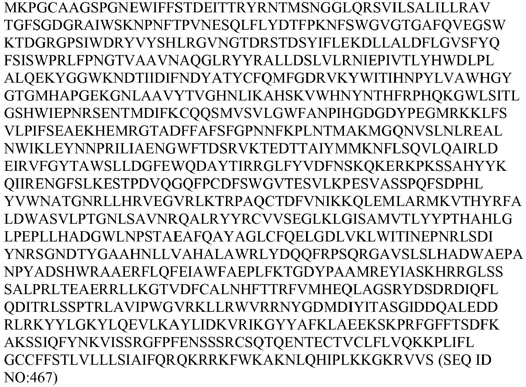
- (xiv) huBeta_Klotho (1-301, 509-743, 967-1044) (muBetaKLOTHO 302-506, 742-964)


- Various antigen binding proteins provided herein, as well as human FGF21, were tested for the ability to activate chimeras in L6 cells.
Figure 5 shows the observed results with each tested molecule. - These data indicate that while human FGF21 was able to activate FGFR1c combined with all of the human/mouse β-Klotho chimeras (the "+" sign indicates activity on the receptor), the substitutions of mouse sequences into human β-Klotho affected the activities of 16H7, 37D3, and 39F7 (See
Figure 5 ). These results suggest that β-Klotho sequences 1-81, 302-522, and 849-1044 are important for the activities of agonistic antigen binding proteins and may represent an important epitope for their function. - In addition, various antigen binding proteins were also tested for binding to the various human/mouse β-Klotho chimeras transiently expressed on the surface of HEK-293T cells by flow cytometry. Transfection and flow-cytometry was performed as described in Example 12. It will be appreciated that antibodies which do not have the ability to cross-bind full length murine β-Klotho are unable to bind the human/mouse β-Klotho chimera if the chimera spans a region of the antibody's binding site. In this manner, the binding profile of each antibody on the panel of chimeras reveals epitope information for the antibody. Data is shown below in Table 10. The anti-β-Klotho antibody 2G10 (which binds both human and mouse β-Klotho) was used as the positive control for expression of each human/mouse chimera. Using this positive control it was determine the expression level of
chimeras chimera # 3 andchimera # 12, indicating that the epitope includes the 1-81 region. Additionally, this group of antibodies also lacked observed binding tochimeras - A second cluster included only antibody 65C3.1. This antibody lacked binding to
chimeras # 2, #11, and #14, indicating an epitope in the region of 849-936. A third cluster, including antibodies 49H12.1, 54A1.1, 49C8.1, 51A8.1, 63A10.1, 64B10.1, 68C8.1 and 39F7, lacked binding tochimera # 1, #5, and #6, indicating that their epitope is in the 302-416 region. The forth cluster included antibodies 67C10.1, 51E5.1, 52A8.1, 66G2, 167F5.1, which lacked binding onchimeras # 2, #8, #9, #10, #11, and #14 indicating that the epitope for these antibodies lies within region 506-1045. A "+" or "-" symbol in the chart below indicates binding of the respective antibody ("+"), or lack of binding ("-") to the chimera and/or the respective ortholog of β-Klotho, or Mock Cells (negative control).Table 13 Chimera Binding CHIMERA # 1 2 3 4 5 6 9 10 11 12 13 14 hu β-Klotho mu β-klotho Mock cells (Neg. Cont.) Epitope Region 2G10 + + + + + + + + + + + + + + - 26H11 + + + + + + + + + + + + + + - 16H7 - + - + - - + + + - + - + - - 1-81 & 302-416 49G3.3 - + - + - - + + + - + - + - - 46D11 - + - + - - + + + - + - + - - 49H12.1 - + + + - - + + + + + - + - - 302-416 54A1.1 - + + + - - + + + + + - + - - 49C8.1 - + + + - - + + + + + - + - - 51A8.1 - + + + - - + + + + + - + - - 63A10.1 - + + + - - + + + + + - + - - 64B10.1 - + + + - - + + + + + - + - - 68C8.1 - + + + - - + + + + + - + - - 39F7 - + + + - - + + + + + - + - - 65C3.1 + - + + + + + + - + + - + - - 849 - 936 67C10.1 + - + + + + - - - + + - + - - 506-1045 51E5.1 + - + + + + - - - + + - + - - 52A8.1 + - + + + + - - - + + - + - - 66G2.1 + - + + + + - - - + + - + - - 67F5.1 + - + + + + - - - + + - + - - IgG2/K Control - - - - - - - - - - - - - - - IgG4/K Control - - - - - - - - - - - - - - - Secondar y Only - - - - - - - - - - - - - - - - A panel of FGF21 receptor agonistic antibodies were assayed using flow cytometry for the binding to human FGFR1/human β-klotho transiently co-transfected HEK293T cells, human FGFR1c transiently transfected HEK293T cells and β-klotho transiently transfected HEK293T cells. In addition, binding was also tested on HEK-293T cells transiently transfected with cynomologous monkey orthologs of FGFR1c and β-klotho. Cells were transfected by preparing 10ug plasmid DNA in 500ul OptiMEM™ media (Invitrogen™) and mixing this with 10ul of 293fectin™ in 500ul OptiMEM™ media, and then incubating the solution for 5 minutes at room temperature. This solution was then added dropwise to 10million HEK293T cells in 10ml of media. 24 hours following transfection, the cells were washed and 50,000 cells were stained with each primary antibody, 50ul of unpurified hybridoma supernatant was diluted 1:2 and used for staining cells. After a one hour incubation at 4°c, the cells were washed and an anti-Human Fc-specific secondary was added. Stained cells were then analyzed on a flow cytometer. The panel of hybridoma supernatants tested all bound specifically to human β-Klotho/human FGFR1c co-transfected cells as well as human β-Klotho transfected alone. Data is shown below in Table 11. No staining was detected for any of the antibodies on cells transfected with FGFR1c alone. All antibodies except 64B10.1 and 68C8.1 specifically detected cynomologous β-Klotho/cynoFGFR1c co-transfected cells.
TABLE 14 FGFR Antibody Selectivity Mock Transfected 293T cells Human FGFR1c Transfect 293T cells Human β-Klotho Tranfected 293T Cells HuFGFR1c/Hu β-Klotho Co-transfected 293T Cells Cyno FGFR1c/Cyno β-Klotho Co-transfected Cells Antibody GeoMean GeoMean GeoMean GeoMean GeoMean 49G3.3 648 706 14891 17919 25947 49H12.1 581 719 16213 21731 20870 51E5.1 723 747 16900 20951 36536 51A8.1 728 795 17799 22826 18476 54A1.1 709 770 14317 18701 11106 59G10.3 686 780 15669 21105 33464 63A10.1 648 834 17442 20432 32558 64B10.1 624 691 14939 19850 701 65C3.1 705 719 13720 18835 24564 66G2.1 695 780 12671 16715 21566 67F5.1 632 757 13482 13948 15784 67C10.1 688 780 15114 18896 4063 68C8.1 592 798 15905 20622 750 16H7 @ 5ug/ml 723 869 16335 20686 31319 - A total of 17 antibodies were analyzed for potential hotspots and covariance violations. The designed variants (shown below) outline amino acid substitutions capable of reducing and/or avoiding isomerization, deamidation, oxidation, covariance violations, and the like. In the data below, "02 49C8.1_VK:[F21I]" refers to a variant of the parental antibody 49C8.1 that has a mutation at position 21, from F (Phe) to I (Isoleucine). Note that a structure-based numbering scheme is followed for designating amino acid positions. It will be appreciated that these single point mutations can be combined in any combinatorial manner in order to arrive at a final desired molecule. The data are shown below in Table 15 and Table 16.
TABLE 15 Antibody 49C8.1 02 49C8.1_VK: [F21I] 03 49C8.1_VK: [F91L] 04 49C8.1_VK: [I101F] 05 49C8.1_VK: [I101V] 06 49C8.1_VK: [P141Q] 07 49C8.1_VK: [P141G] 08 49C8.1_VH: [T48P] 09 49C8.1_VH: [N61Q] 10 49C8.1_VH: [G65T] Antibody 49H12_ N83D 01 49H12_N83D_VK: [F91L] 02 49H12_N83D_VK: [I101F] 03 49H12_N83D_VK: [I101V] 04 49H12_N83D_VH: [M24K] 05 49H12_N83D_VH: [I30T] 06 49H12_N83D_VH: [T48P] 07 49H12_N83D_VH: [W57Y] 08 49H12_N83D_VH: [W111Y] Antibody 49G3.3 01 49G3.3_VK: [F91L] 02 49G3.3_VK: [I101F] 03 49G3.3_VK: [I101V] 04 49G3.3_VK: [G141Q] 05 49G3.3_VH: [E17Q] 06 49G3.3_VH: [V25F] 07 49G3.3_VH: [T56A] 08 49G3.3_VH: [T56G] 09 49G3.3_VH: [T144L] 10 49G3.3_VH: [T144M] Antibody 51A8.1 01 51A8.1_VL: [I98T] 02 51A8.1_VL: [I98A] 03 51A8.1_VH: [R17G] 04 51A8.1_VH: [D61E] 05 51A8.1_VH: [D72E] 06 51A8.1_VH: [D110E] Antibody 51E5.1 01 51E5.1_VK: [N53K] 02 51E5.1_VK: [R54L] 03 51E5.1_VK: [R54S] 04 51E5.1_VK: [G141Q] 05 51E5.1_VH: [D59E] 06 51E5.1_VH: [H60T] Antibody 52A8.1 01 52A8.1_VK: [F10S] 02 52A8.1_VK: [H44Y] 03 52A8.1_VK: [H44F] 04 52A8.1_VK: [G141Q] 05 52A8.1_VH: [W57Y] 06 52A8.1_VH: [R95S] 07 52A8.1_VH: [W135Y] Antibody 54A1.1_N83D 01 54A1.1_N83D_VK: [A5T] 02 54A1.1_N83D_VK: [L46Q] 03 54A1.1_N83D_VK: [G81S] 04 54A1.1_N83D_VK: [F91L] 05 54A1.1_N83D_VK: [I101F] 06 54A1.1_N83D_VK: [I101V] 07 54A1.1_N83D_VK: [P141G] 08 54A1.1_N83D_VK: [P141Q] 09 54A1.1_N83D_VH: [T48P] 10 54A1.1_N83D_VH: [W57Y] 11 54A1.1_N83D_VH: [W111Y] Antibody 56E7.3 01 56E7.3_VK: [N53K] 02 56E7.3_VK: [F91L] 03 56E7.3_VK: [I101F] 04 56E7.3_VK: [P141Q] 05 56E7.3_VK: [P141G] 06 56E7.3_VK: [T144K] 07 56E7.3_VK: [T144R] 08 56E7.3_VH: [L31F] 09 56E7.3_VH: [D65E] 10 56E7.3_VH: [T84K] 11 56E7.3_VH: [R95S] Antibody 58C2.1 01 58C2.1_VK: [D36E] 02 58C2.1_VH: [R17G] 03 58C2.1_VH: [D61E] 04 58C2.1_VH: [D72E] 05 58C2.1_VH: [N116Q] Antibody 60D7.1_N30T 01 60D7.1_N30T_VK: [D33E] 02 60D7.1_N30T_VK: [D36E] 03 60D7.1_N30T_VH: [R17G] 04 60D7.1_N30T_VH: [D61E] 05 60D7.1_N30T_VH: [D72E] 06 60D7.1_N30T_VH: [W115Y] Antibody 63A10.1_C58S 01 63A10.1_C58S_VL: [H9L] 02 63A10.1_C58S_VL: [H9P] 03 63A10.1_C58S_VL: [T15L] 04 63A10.1_C58S_VL: [T15P] 05 63A10.1_C58S_VL: [A16G] 06 63A10.1_C58S_VL: [M18T] 07 63A10.1_C58S_VL: [D51A] 08 63A10.1_C58S_VL: [D51S] 09 63A10.1_C58S_VL: [D51F] 10 63A10.1_C58S_VL: [D67E] 11 63A10.1_C58S_VL: [P83S] 12 63A10.1_C58S_VL: [E97Q] 13 63A10.1_C58S_VL: [D110E] 14 63A10.1_C58S_VL: [D136E] 15 63A10.1_C58S_VH: [D11G] 16 63A10.1_C58S_VH: [K14Q] 17 63A10.1_C58S_VH: [I29F] 18 63A10.1_C58S_VH: [G56S] 19 63A10.1 C58S_VH: [D64E] 20 63A10.1_C58S_VH: [G84D] 21 63A10.1_C58S_VH: [G84N] 22 63A10.1_C58S_VH: [T98A] 23 63A10.1_C58S_VH: [T107A] 24 63A10.1_C58S_VH: [T108R] 25 63A10.1_C58S_VH: [D109E] 26 63A10.1_C58S_VL: [W109Y] Antibody 63A10.3_ N20R_ C42S 01 63A10.3_ N20R_C42S_VL: [W109Y] 02 63A10.3_ _N20R_C42S_VL: [D67E] 03 63A10.3_ N20R_C42S_VL: [D110E] 04 63A10.3_N20R_C42S_VH: [D11G] 05 63A10.3_N20R_C42S_VH: [K14Q] 06 63A10.3_N20R_C42S_VH: [I29F] 07 63A10.3_N20R_C42S_VH: [G56S] 08 63A10.3_N20R_C42S_VH: [D64E] 09 63A10.3_N20R_C42S_VH: [G84N] 10 63A10.3_N20R_C42S_VH: [T98A] 11 63A10.3_N20R_C42S_VH: [T107A] 12 63A10.3_N20R_C42S_VH: [T108R] 13 63A10.3_N20R_C42S_VH: [D109E] 14 63A10.3_N20R_C42S_VL: [W109Y] Antibody 64B10.1 01 64B10.1_VL: [G92A] 02 64B10.1_VL: [G99E] 03 64B10.1_VL: [D110E] 04 64B10.1_VH: [L5Q] 05 64B10.1_VH: [T144L] 06 64B10.1_VH: [T144M] 07 64B10.1_VL: [W109Y] 08 64B10.1_VH: [W113Y] Antibody 66G2 01 66G2_VK: [R54L] 02 66G2_VK: [K88E] 03 66G2_VK: [K88D] 04 66G2_VK: [N110Q] 05 66G2_VH: [R17G] 06 66G2_VH: [D61E] 07 66G2_VH: [D72E] 08 66G2_VH: [I78F] 09 66G2_VH: [T108K] 10 66G2_VH: [T108R] Antibody 67F5 1.01 67F5_VK: [H57Y] 2.02 67F5_VK: [Q97E] 3.03 67F5_VK: [S98P] 4.04 67F5_VK: [A99E] 5.05 67F5_VK: [N105Y] 6.06 67F5_VH: [K5Q] 7.07 67F5_VK: [W135Y] 8.08 67F5_VK: [W137Y] Antibody 67C10 1.01 67C10_VK: [F2I] 2.02 67C10_VK: [D36E] 3.03 67C10_VH: [Q24K] 4.04 67C10_VH: [D65E] Antibody 68C8 1.01 68C8_VL: [G92A] 2.02 68C8_VL: [G99E] 3.03 68C8_VL: [D110E] 4.04 68C8_VH: [D29G] 5.05 68C8_VH: [H83D] 6.06 68C8_VH: [G107A] 7.07 68C8_VH: [T144L] 8.08 68C8_VH: [T144M] 9.09 68C8_VL: [W109Y] 10.10 68C8_VH: [W113Y] TABLE 16 Exemplary Substitutions >49C8.1_VK 
>49C8.1_VH 
>49H12 N83DVK 
>49H12 N83DVH 
>49G3.3_VK 
>49G3.3_VH 
>51A8.1_VL 
>51A8.1_VH 
>51E5.1_VK 
>51E5.1_VH 
>52A8.1_VK 
>52A8.1_VH 
>54A1.1_N83D_VK 
>54A1.1_N83D_VH 
>56E7.3_VK 
>56E7.3_VH 
>58C2.1_VK 
>58C2.1_VH 
>60D7.1_N30T_VK 
>60D7.1_N30T_VH 
>63A10.1_C58S_VL 
>63A10.1_C58S_VH 
>63A10.3_N20R_C42S_VL 
>63A10.3_N20R_C42S_VH 
>64B10.1_VL 
>64B10.1_VH 
>66G2_VK 
>66G2_VH 
>67F5_VK 
>67F5_VH 
>67C10_VK 
>67C10_VH 
>68C8_VL 
>68C8_VH 
>49C8.1_VK.02 
>49C8.1_VH 
>49C8.1_VK.03 
>49C8.1_VH 
>49C8.1_VK.04 
>49C8.1_VH 
>49C8.1_VK.05 
>49C8.1_VH 
>49C8.1_VK.06 
>49C8.1_VH 
>49C8.1_VK.07 
>49C8.1_VH 
>49C8.1_VK 
>49C8.1_VH.08 
>49C8.1_VK 
>49C8.1_VH.09 
>49C8.1_VK 
>49C8.1_VH.10 
>49H12_N83D_VK.01 
>49H12_N83DVH 
>49H12_N83DVK.02 
>49H12_N83DVH 
>49H12_N83D VK.03 
>49H12_N83DVH 
>49H12_N83DVK 
>49H12_N83DVH.04 
>49H12_N83DVK 
>49H12_N83DVH.05 
>49H12_N83DVK 
>49H12_N83DVH.06 
>49H12_N83DVK 
>49H12_N83D VH.07 
>49H12_N83DVK 
>49H12_N83DVH.08 
>49G3.3_VK.01 
>49G3.3_VH 
>49G3.3_VK.02 
>49G3.3_VH 
>49G3.3_VK.03 
>49G3.3_VH 
>49G3.3_VK.04 
>49G3.3_VH 
>49G3.3_VK 
>49G3.3_VH.05 
>49G3.3_VK 
>49G3.3_VH.06 
>49G3.3_VK 
>49G3.3_VH.07 
>49G3.3_VK 
>49G3.3 VH.08 
>49G3.3_VK 
>49G3.3 VH.09 
>49G3.3_VK 
>49G3.3_VH.10 
>51A8.1_VL.01 
>51A8.1_VH 
>51A8.1_VL.02 
>51A8.1_VH 
>51A8.1_VL 
>51A8.1_VH.03 
>51A8.1_VL 
>51A8.1_VH.04 
>51A8.1_VL 
>51A8.1_VH.05 
>51A8.1_VL 
>51A8.1_VH.06 
>51E5.1_VK.01 
>51E5.1_VH 
>51E5.1_VK.02 
>51E5.1_VH 
>51E5.1_VK.03 
>51E5.1_VH 
>51E5.1_VK.04 
>51E5.1_VH 
>51E5.1_VK 
>51E5.1_VH.05 
>51E5.1_VK 
>51E5.1_VH.06 
>52A8.1_VK.01 
>52A8.1_VH 
>52A8.1_VK.02 
>52A8.1_VH 
>52A8.1_VK.03 
>52A8.1_VH 
>52A8.1_VK.04 
>52A8.1_VH 
>52A8.1_VK 
>52A8.1_VH.05 
>52A8.1_VK 
>52A8.1_VH.06 
>52A8.1_VK 
>52A8.1 VH.07 
>54A1.1_N83D_VK.01 
>54A1.1_N83D_VH 
>54A1.1_N83D_VK.02 
>54A1.1_N83D_VH 
>54A1.1_N83D_VK.03 
>54A1.1_N83D_VH 
>54A1.1_N83D_VK.04 
>54A1.1_N83D_VH 
>54A1.1_N83D_VK.05 
>54A1.1_N83D_VH 
>54A1.1_N83D_VK.06 
>54A1.1_N83D_VH 
>54A1.1_N83D_VK.07 
>54A1.1_N83D_VH 
>54A1.1_N83D_VK.08 
>54A1.1_N83D_VH 
>54A1.1_N83D_VK 
>54A1.1_N83D_VH.09 
>54A1.1_N83D_VK 
>54A1.1_N83D_VH.10 
>54A1.1_N83D_VK 
>54A1.1_N83D_VH.11 
>58C2.1_VK.01 
>58C2.1_VH 
>58C2.1_VK 
>58C2.1_VH.02 
>58C2.1_VK 
>58C2.1_VH.03 
>58C2.1_VK 
>58C2.1_VH.04 
>58C2.1_VK 
>58C2.1_VH.05 
>56E7.3_VK.01 
>56E7.3_VH 
>56E7.3_VK.02 
>56E7.3 VH 
>56E7.3_VK.03 
>56E7.3_VH 
>56E7.3_VK.04 
>56E7.3_VH 
>56E7.3_VK.05 
>56E7.3_VH 
>56E7.3_VK.06 
>56E7.3_VH 
>56E7.3_VK.07 
>56E7.3_VH 
>56E7.3_VK 
>56E7.3_VH.08 
>56E7.3_VK 
>56E7.3_VH.09 
>56E7.3_VK 
>56E7.3_VH.10 
>56E7.3_VK 
>56E7.3_VH.11 
>60D7.1_N30T_VK.01 
>60D7.1_N30T_VH 
>60D7.1_N30TVK.02 
>60D7.1_N30T_VH 
>60D7.1_N30T_VK 
>60D7.1_N30T_VH.03 
>60D7.1_N30T_VK 
>60D7.1_N30T_VH.04 
>60D7.1_N30T_VK 
>60D7.1_N30T_VH.05 
>60D7.1_N30T_VK 
>60D7.1_N30T_VH.06 
>63A10.1_C58S_VL.01 
>63A10.1_C58S_VH 
>63A10.1_C58S_VL.02 
>63A10.1_C58S_VH 
>63A10.1_C58S_VL.03 
>63A10.1_C58S_VH 
>63A10.1_C58S_VL.04 
>63A10.1_C58S_VH 
>63A10.1_C58S_VL.05 
>63A10.1_C58S_VH 
>63A10.1_C58S_VL.06 
>63A10.1_C58S_VH 
>63A10.1_C58S_VL.07 
>63A10.1_C58S_VH 
>63A10.1_C58S_VL.08 
>63A10.1_C58S_VH 
>63A10.1_C58S_VL.09 
>63A10.1_C58S_VH 
>63A10.1_C58S_VL.10 
>63A10.1_C58S_VH 
>63A10.1_C58S_VL.11 
>63A10.1_C58S_VH 
>63A10.1_C58S_VL.12 
>63A10.1_C58S_VH 
>63A10.1_C58S_VL.13 
>63A10.1_C58S_VH 
>63A10.1_C58S_VL.14 
>63A10.1_C58S_VH 
>63A10.1_C58S_VL 
>63A10.1_C58S_VH.15 
>63A10.1_C58S_VL 
>63A10.1_C58S_VH.16 
>63A10.1_C58S_VL 
>63A10.1_C58S_VH.17 
>63A10.1_C58S_VL 
>63A10.1_C58S_VH.18 
>63A10.1_C58S_VL 
>63A10.1_C58S_VH.19 
>63A10.1_C58S_VL 
>63A10.1_C58S_VH.20 
>63A10.1_C58S_VL 
>63A10.1_C58S_VH.21 
>63A10.1_C58S_VL 
>63A10.1_C58S_VH.22 
>63A10.1_C58S_VL 
>63A10.1_C58S_VH.23 
>63A10.1_C58S_VL 
>63A10.1_C58S_VH.24 
>63A10.1_C58S_VL 
>63A10.1_C58S_VH.25 
>63A10.1_C58S_VL.26 
>63A10.1_C58S_VH 
>63A10.3_N20R_C42S_VL.01 
>63A10.3_N20R_C42S_VH 
>63A10.3_N20R_C42S_VL.02 
>63A10.3_N20R_C42S_VH 
>63A10.3_N20R_C42S_VL.03 
>63A10.3_N20R_C42S_VH 
>64B10.1_VL.01 
>64B10.1_VH 
>64B10.1_VL.02 
>64B10.1_VH 
>64B10.1_VL.03 
>64B10.1_VH 
>64B10.1_VL 
>64B10.1_VH.04 
>64B10.1_VL 
>64B10.1_VH.05 
>64B10.1_VL 
>64B10.1_VH.06 
>64B10.1_VL.07 
>64B10.1_VH 
>64B10.1_VL 
>64B10.1_VH.08 
>66G2_VK.01 
>66G2_VH 
>66G2_VK.02 
>66G2_VH 
>66G2_VK.03 
>66G2_VH 
>66G2_VK.04 
>66G2_VH 
>66G2_VK 
>66G2_VH.05 
>66G2_VK 
>66G2_VH.06 
>66G2_VK 
>66G2_VH.07 
>66G2_VK 
>66G2_VH.08 
>66G2_VK 
>66G2_VH.09 
>66G2_VK 
>66G2_VH.10 
>67F5_VK.01 
>67F5_VH 
>67F5_VK.02 
>67F5_VH 
>67F5_VK.03 
>67F5_VH 
>67F5_VK.04 
>67F5_VH 
>67F5_VK.05 
>67F5_VH 
>67F5_VK 
>67F5_VH.06 
>67F5_VK.07 
>67F5_VH 
>67F5_VK.08 
>67F5_VH 
>67C10_VK.01 
>67C10_VH 
>67C10VK.02 
>67C10_VH 
>67C10_VK 
>67C10_VH.03 
>67C10_VK 
>67C10_VH.04 
>68C8_VL.01 
>68C8_VH 
>68C8_VL.02 
>68C8_VH 
>68C8_VL.03 
>68C8_VH 
>68C8_VL 
>68C8_VH.04 
>68C8_VL 
>68C8_VH.05 
>68C8_VL 
>68C8_VH.06 
>68C8_VL 
>68C8_VH.07 
>68C8_VL 
>68C8_VH.08 
>68C8_VL.09 
>68C8_VH 
>68C8_VL 
>68C8_VH.10 
- Immune responses against proteins are enhanced by antigen processing and presentation in the major histocompatability complex (MHC) class II binding site. This interaction is required for T cell help in maturation of antibodies that recognize the protein. Since the binding sites of MHC class II molecules have been characterized, it is possible to predict whether proteins have specific sequences that can bind to a series of common human alleles. Computer algorithms have been created based on literature references and MHC class II crystal structures to determine whether linear 9 amino acid peptide sequences have the potential to break immune tolerance. We used the TEPITOPE™ program called to determine if point mutations of FGF21 are predicted to increase antigen specific T-cells in a majority of humans. Based on the linear protein sequence, none of the mutations examined are expected enhance immunogenicity. Results are shown in Table 17A and Table 17B below.
TABLE 17A Protein Predicted Immunogenicity Met-FGF21 Low Met-hFGF21(N106D) Low Met-FGF21 (N122D) Low hFc(R4).L15.hFGF21(G170E) Low hFc(R4).L15.hFGF21(P171A) Low hFc(R4).L15.hFGF21(S172L) Low p30.hFc.L15.hFGF21(A45K, G170E) Low p30.hFc.L15.hFGF21 (L98R, P171G) Low TABLE 17B Clone Predicted immunogenicity LC Total Agretopes LC Tolerant Agretopes LC Non-Tolerant Agretopes LC Non-Tolerant HLA DRB1 HC Total Agretopes HC Tolerant Agretopes HC Non-tolerant Agretopes HC Non-tolerant HLA DRB1 68C8 Tier 1 3 3 0 NA 12 12 0 NA 63A10 Tier 1 1 1 0 NA 12 12 0 NA 51A8 Tier 2 3 2 1 0101 16 16 0 NA 0701 51E5 Tier 2 6 6 0 NA 11 10 1 0401 0701 64B10 Tier 2 2 2 0 NA 12 11 1 0801 49H12 Tier 3 7 5 2 0101 13 13 0 NA 0701 0801 1301 1501 54A1 Tier 3 6 4 2 0301 14 14 0 NA 0801 1501 52A8 Tier 3 6 5 1 0701 13 12 1 1301 60D7 Tier 4 8 7 1 0301 16 14 2 0401 0401 1501 1101 49C8 Tier 4 7 5 2 0801 14 13 1 0401 1501 67C10 Tier 4 8 7 1 0301 14 12 2 0101 0401 701 1101 - Each reference cited herein is incorporated by reference in its entirety for all that it teaches and for all purposes.
- The present disclosure is not to be limited in scope by the specific embodiments described herein, which are intended as illustrations of individual aspects of the disclosure, and functionally equivalent methods and components form aspects of the disclosure. Indeed, various modifications of the disclosure, in addition to those shown and described herein will become apparent to those skilled in the art from the foregoing description and accompanying drawings. Such modifications are intended to fall within the scope of the appended claims.
Claims (16)
- An isolated antigen binding protein that induces FGF21-mediated signaling.
- An isolated antigen binding protein that specifically binds to a complex comprising β-Klotho and at least one of (i) FGFR1c, (ii) FGFR2c and (iii) FGFR3c.
- The antigen binding protein of claim 1, wherein the antigen binding protein comprises one or more of:(a) a light chain CDR3 comprising one or more of:(i) a light chain CDR3 sequence that differs by three , two or one amino acid additions, substitutions, deletions and combinations thereof from a CDR3 of one or more of CDRL3-1 to CDRL3-75 of Table 3B (SEQ ID NOs 947-1020, respectively, in order of appearance);(ii) QQFGSSLT (SEQ ID NO: 988);(iii)QQSX1SX2X3 LT (SEQ ID NO: 1443);(iv)LQX4X5SYPX6T (SEQ ID NO: 1447);(v) MQRX7EFPX8T (SEQ ID NO: 1451);(vi)QX9WDSX10X11VV (SEQ ID NO: 1457);(vii) QQYNX12WPX13T (SEQ ID NO: 1461);(viii) QVWDSSX14DX15VX16 (SEQ ID NO: 1466);(ix)QQSSX17IPWT (SEQ ID NO: 1469);(x) QQTNSFPPWT (SEQ ID NO: 1470);(xi) GTWDSSLSX18X19V (SEQ ID NO: 1474);(xii) QQYDNLPX20T (SEQ ID NO: 1477);(xiii) QQYGSSX21PWT (SEQ ID NO: 1480);(xiv) QQYGX22SX23FT (SEQ ID NO: 1483);(xv) QQYGSSX24X25X26 (SEQ ID NO: 1488);(xvi) QAWDSSX27TX28V (SEQ ID NO: 1492);(xvii) QAWDSX29TVX30 (SEQ ID NO: 1496);(xviii) QQX31YSAX32FT (SEQ ID NO: 1499);(xix) QQYNX33YPRT (SEQ ID NO: 1502);(xx) HQX34X35DLPLT (SEQ ID NO: 1505);(xxi) MQALQTX36X37T (SEQ ID NO: 1508);(xxii) QQFGRSFT (SEQ ID NO: 1509);(xxiii) YSTDSSX38NHVV (SEQ ID NO: 1512);(b) a heavy chain CDR3 sequence comprising one or more of:(i) a heavy chain CDR3 that differs by eight, seven, six, five, four, three, two or one amino acid additions, substitutions, deletions and combinations thereof from a CDR3 of one or more of CDRH3-1 to CDRH3-81 of Table 3A (SEQ ID NOs 733-813, respectively, in order of appearance);(ii) MTX110PYWYFX111L (SEQ ID NO: 1669);(iii)DX112X113X114DFWX115GYPX116X117X118YYGX119DV (SEQ ID NO: 1675);(iv)VTGTDAFDF (SEQ ID NO: 1676);(v) TVTKEDYYYYGMDV (SEQ ID NO: 1677);(vi)DSSGSYYVEDYFDY (SEQ ID NO: 1678);(vii) DX119X120IAVAGX121FDY (SEQ ID NO: 1681);(viii) EYYYGSGSYYP (SEQ ID NO: 1682);(ix)ELGDYPFFDY (SEQ ID NO: 1683);(x) EYVAEAGFDY (SEQ ID NO: 1684);(xi) VAAVYWYFDL (SEQ ID NO: 1685);(xii) YNWNYGAFDF (SEQ ID NO: 1686);(xiii) RASRGYRX122GLAFAI (SEQ ID NO: 1689);(xiv) DGITMVRGVTHYYGMDV (SEQ ID NO: 1690);(xv) DHX123SCWYYYGMDV (SEQ ID NO: 1693);(xvi) YX124X125WDYYYGX126DV (SEQ ID NO: 1696);(xvii) VLHYX127DSX128GYSYYX129DX130 (SEQ ID NO: 1699); or(c) the light chain CDR3 sequence of (a) and the heavy chain CDR3 sequence of (b);
wherein,X1 is Y or F;X2 is T or S;X3 is P or S;X4 is H or R;X5 is N or S;X6 is L or R;X7 is I or L;X8 is I or L;X9 is V or L;X10 is S or N;X11 is T, P or S;X12 is N or T;X13 is W or L;X14 is S or C;X15 is H, V or G;X16 is V or absent;X17 is S or T;X18 is A or V;X19 is V or M;X20 is L or F;X21 is P or absent;X22 is R or T;X23 is L or P;X24 is P or absent;X25 is R or C;X26 is S or T;X27 is T or absent;X28 is W or A;X29 is G, T, A or absent;X30 is V or I;X31 is S or T;X32 is T or P;X33 is I or T;X34 is S or Y;X35 is S or D;X36 is P or L;X37 is F or I;X38 is V or G;X100 is T or S;X111 is D or G;X112 is R or Q;X113 is Y, D or N;X114 is Y or F;X115 is S or N;X116 is Y or F;X117 is F or Y;X118 is R, Y, F or H;X119 is L or M;X119 is W or L;X120 is S or R;X121 is T or S;X122 is F or Y;X123 is S or T;X124 is S or R;X125 is T or D;X126 is V or M;X127 is S or Y;X128 is R or S;X129 is S or F; andX130 is F or Y. - The antigen binding protein of claim 1, comprising one or more of:(a) a light chain CDR1 sequence comprising one or more of:(i) a light chain CDR1 that differs by seven, six, five, four, three, two or one amino acid additions, substitutions, deletions and combinations thereof from a CDR1 sequence of one or more of CDRL1-1 to CDRL1-81 of Table 3B (SEQ ID NOs 814-893, respectively, in order of appearance);(ii) RASX62SX63X64X65X66X67X68A (SEQ ID NO: 1591);(iii) RX69SQX70IX71X72YLN (SEQ ID NO: 1602);(iv) GGNX73IGSX74X75VX76 (SEQ ID NO: 1612);(v) RASQX77IRNDLX78 (SEQ ID NO: 1616);(vi) RSSQSLX79X80X81DX82GX83TYLD (SEQ ID NO: 1622);(vii) SGX84X85LGDKYX86X87 (SEQ ID NO: 1629);viii) QASQX88IX89X90X91LN (SEQ ID NO: 1635); (ix)RASQX92IX93 X94WLX95 (SEQ ID NO: 1640);(x) SGSSSNIGX96NYVX97 (SEQ ID NO: 1645);(xi) RASX98DISNYLA (SEQ ID NO: 1648);(xii) RASQX99VX100SSYX101V (SEQ ID NO: 1652);(xiii) RSSQSLX102HSNGX103NYLD (SEQ ID NO: 1656);(xiv) RASQTX104RNX105YLA (SEQ ID NO: 1659);(xv) RSSX106X107LVYSDGNTYLN (SEQ ID NO: 1662); and(xvi) SGDAX108PKKYAX109 (SEQ ID NO: 1665);(b) a light chain CDR2 sequence comprising one or more of:(i) a light chain CDR2 that differs by three, two or one amino acid additions, substitutions, deletions and combinations thereof from a CDR2 sequence of one or more of of CDRL2-1 to CDRL2-53 of Table 3B (SEQ ID NOs 894-946, respectively, in order of appearance);(ii)X39ASSLX40X41 (SEQ ID NO: 1517);(iii) GX42S X43RX44T (SEQ ID NO: 1525);(iv) GAFSRAX45 (SEQ ID NO: 1528);(v) X46DX47KRPS (SEQ ID NO: 1533);(vi) TLSX48RAS (SEQ ID NO: 1536);(vii) AASNLQX49 (SEQ ID NO: 1539);(viii) GX50SNRAX51 (SEQ ID NO: 1543);(ix) X52ASX53LQS (SEQ ID NO: 1548);(x) DNX53KRPS (SEQ ID NO: 1551);(xi) DX54SNLET (SEQ ID NO: 1554);(xii) LX55SNRAS (SEQ ID NO: 1557);(xiii) QX56NX57RPS (SEQ ID NO: 1561);(xiv) RDRNRPS (SEQ ID NO: 1562);(xv) X58DSNRPS (SEQ ID NO: 1565);(xvi) DDSDRPS (SEQ ID NO: 1566);(xvii) AX59SSLQS (SEQ ID NO: 1569);(xviii) TX60SSLQS (SEQ ID NO: 1572); and(xix) KX61SNWDS (SEQ ID NO: 1575);(c) a heavy chain CDR1 sequence comprising one or more of:(i) a heavy chain CDR1 that differs by three, two or one amino acid additions, substitutions, deletions and combinations thereof from a CDR1 sequence of one or more of CDRH1-1 to CDRH1-53 of Table 3A (SEQ ID NOs 603-655, respectively, in order of appearance);(ii) SGX170X171TWX172 (SEQ ID NO: 1775);(iii)X173YYWX174 (SEQ ID NO: 1781);(iv)X175X176GMS (SEQ ID NO: 1786);(v) SYX177MX178 (SEQ ID NO: 1790);(vi)X179YYX180H (SEQ ID NO: 1796);(vii) SYGX181H (SEQ ID NO: 1799);(viii) NYX182MX183 (SEQ ID NO: 1803);(ix)X184YWIG (SEQ ID NO: 1806);(x) GYX185MH (SEQ ID NO: 1809);(xi)SX186DIX187 (SEQ ID NO: 1813);(xii) X188YAMS (SEQ ID NO: 1816);(xiii) NAWMS (SEQ ID NO: 1817);(xiv) SSSYYWG (SEQ ID NO: 1818);(xv) X189YYWN (SEQ ID NO: 1821);(xvi) SNSAX190WN (SEQ ID NO: 1824); and(xvii) X191YDMH (SEQ ID NO: 1827);(d) a heavy chain CDR2 sequence comprising one or more of(i) a heavy chain CDR2 that differs by nine, eight, seven, six, five, four, three, two or one amino acid additions, substitutions, deletions and combinations thereof from a CDR2 sequence of one or more of CDRH2-1 to CDRH2-77 of Table 3A (SEQ ID NOs 656-732, respectively, in order of appearance);(ii) X131X132X133X134X135GX136X137X138X139NPSLKS (SEQ ID NO: 1716);(iii)X140IX141X142DGX143NX144X145X146ADSVKG (SEQ ID NO: 1732);(iv)WX147NPX148SGX149TX150YAQKFX151G (SEQ ID NO: 1741);(v) EINHSX152X153TNYNPSLKS (SEQ ID NO: 1744);(vi) IIYPGDSX154TRYSPSFQG (SEQ ID NO: 1747);(vii) SISSSSX155YX156YYX157DSX158KG (SEQ ID NO: 1751);(viii) RIX159 X160KTDGGTTX161Y AAPVKG (SEQ ID NO: 1755);(ix) GISGSSAGTYYADSVGK (SEQ ID NO: 1756);(x) VISX162SGGX163TYYADSVKG (SEQ ID NO: 1759);(xi) RTYYRSKWYNDYAVSVKS (SEQ ID NO: 1760);(xii) RIYX164SGSTNYNPSLX165X166 (SEQ ID NO: 1763); and(xiii) WMNPYSGSTGX167AQX168FQX169 (SEQ ID NO: 1766);whereinX39 is A or S;X40 is Q or K;X41 is S or F;X42 is A or T;X43 is S, T, A, R or N;X44 is A or D;X45 is S or T;X46 is Q, R or E;X47 is T or S;X48 is Y or F;X49 is R or S;X50 is A or S;X51 is I or T;X52 is D or G;X53 is S, T or N;X53 is N or D;X54 is A or V;X55 is G or D;X56 is D or N;X57 is K or E;X58 is S or C;X59 is S or V;X60 is A or T;X61 is V or G;X62 is P or Q;X63 is V, I or F;X64 is S, R or absent;X65 is S, R or N;X66 is D, S, N or M;X67 is I, Y, H, Q, N or S;X68 is L or V;X69 is A or T;X70 is I, S, T or N;X71 is R, S or N;X72 is R, S, N, or I;X73 is N, or D;X74 is Y, I or K;X75 is N, S, T or A;X76 is H or Q;X77 is D or G;X78 is G or A;X79 is L or F;X80 is N or D;X81 is S or N;X82 is A or D;X83 is T, D or N;X84 is N or D;X85 is K, E or N;X86 is V or A;X87 is C or F;X88 isG or D;X89 is S, K N or T;X90 is N, K or I;X91 is Y or F;X92 is D or G;X93 isD or S;X94 is R or S;X95 is V or A;X96 is N, I or D;X97 is A or S;X98 is Q or H.;X99 is R or S;X100 is P or A;X101 is I or L;X102 is L or Q;X103 is Y or F.X104 is V or I;X105 is N or S;X106 is Q or P;X107 is R or S;X108 is L or V;X109 is Y or N;X131 is N, F, Y, S or M;X132 is I or L;X133 is Y or F;X134 is Y, H or D;X135 is S or T;X136 is T, G, S or T;X137 is T or A;X138 is Y, N or H;X139 is F or Y;X140 is L, G, I or V;X141 is W or S;X142 is Y, D or N;X143 is S or D;X144 is K or N;X145 is Y, N, D, or H;X146 is Y or H;X147 is I or M;X148 is P, N, S or D;X149 is A, G or D;X150 isN, K, or D;X151 is R, H or Q;X152 is E or G;X153 is N or T;X154 is D or E;X155 is T or S;X156 is I or E;X157 is A or V;X158 is V or L.X159 is K or I;X160 is S or G;X161 is D or E;X162 is D or G;X163 is S or D;X164 is I or T;X165 is E or K;X166 is N or S;X167 is Y or L;X168 is N or R;X169 is G or D;X170 is V, G, N or D;X171 is Y or N;X172 is N, S or T;X173 is T, S or G;X174 is S or T;X175 is S, T or F;X176 is Y or F;X177 is A or S;X178 is S, N or M;X179 is Y or G;X180 is I, L, K or T;X181 is L or I;X182 is G or N;X183 is H, R or M;X184 is S or G;X185 is Y or F;X186 is Y or H;X187 is N or D;X188 is N or H;X189 is D or S;X190 is T or A;X191 is S or T;(e) the light chain CDR1 of (a) and the light chain CDR2 of (b);(f) the light chain CDR1 of (a) and the heavy chain CDR1 of (c);(g) the light chain CDR1 of (a) and the heavy chain CDR2 of (d);(h) the light chain CDR1 (a) and the heavy chain CDR1 of (c);(i) the heavy chain CDR1 of (c) and the heavy chain CDR2 of (d);(j) the light chain CDR2 of (b) and the heavy chain CDR2 of (d);(k) the light chain CDR1 of (a), the light chain CDR2 of (b), and the heavy chain CDR1 of (c);(l) the light chain CDR2 of (b), the heavy CDR1 of (c), and the heavy chain CDR2 of (d);(m) the light chain CDR1 of (a), the heavy chain CDR1 of (c), and the heavy chain CDR2 of (d); and(n) the light chain CDR1 of (a), the light chain CDR2 of (b), the heavy chain CDR2 of (c), and the heavy chain CDR2 of (d).
- The antigen binding protein of claim 1, comprising one or more of:(a) a light chain variable domain comprising one or more of;(i) a light chain CDR1 sequence selected from CDRL1-1 to CDRL1-81 of Table 3B (SEQ ID NOs 814-893, respectively, in order of appearance);(ii) a light chain CDR2 sequence selected from CDRL2-1 to CDRL2-53 of Table 3B (SEQ ID NOs 894-946, respectively, in order of appearance);(iii) a light chain CDR3 sequence selected from CDRL3-1 to CDRL3-75 of Table 3B (SEQ ID NOs 947-1020, respectively, in order of appearance);(b) a heavy chain variable domain comprising one or more of:(i) a heavy chain CDR1 sequence selected from CDRH1-1 to CDRH1-53 of Table 3A (SEQ ID NOs 603-655, respectively, in order of appearance);(ii) a heavy chain CDR2 sequence selected from CDRH2-1 to CDRH2-77 of Table 3A (SEQ ID NOs 656-732, respectively, in order of appearance);(iii) a heavy chain CDR3 sequence selected from CDRH3-1 to CDRH3-81 of Table 3A (SEQ ID NOs 733-813, respectively, in order of appearance); and(c) a combination comprising a light chain variable domain of (a) and a heavy chain variable domain of (b).
- The antigen binding protein of claim 1, wherein the antigen binding protein comprises one or more of:(a) a light chain variable domain sequence comprising one or more of:(i) VL1-VL100 of Table 2A (SEQ ID NOs 217-315, respectively, in order of appearance);(ii) amino acids having a sequence at least 80% identical to a light chain variable domain sequence comprising one or more of VL1-VL100 of Table 2A (SEQ ID NOs 217-315, respectively, in order of appearance);(iii) a sequence of amino acids encoded by a polynucleotide sequence that is at least 80% identical to a polynucleotide sequence encoding the light chain variable domain sequence comprising one or more of VL1-YL100 of Table 2A(SEQ ID NOs 217-315, respectively, in order of appearance);(b) a heavy chain variable domain sequence comprising one or more of:(i) VH1-VH94 of Table 2B (SEQ ID NOs 316-409, respectively, in order of appearance);(ii) a sequence of amino acids that is at least 80% identical to a heavy chain variable domain sequence comprising one or more of VH1-VH94 of Table 2B 9SEQ ID NOs 316-409, respectively, in order of appearance);(iii) a sequence of amino acids encoded by a polynucleotide sequence that is at least 80% identical to a polynucleotide sequence encoding the heavy chain variable domain sequence of VH1-VH94 of Table 2B (SEQ ID NOs 316-409, respectively, in order of appearance); and(c) a combination comprising a light chain variable domain of (a) and a heavy chain variable domain of (b),such as an antigen binding protein in which the light chain variable domain and the heavy chain variable domain comprise one or more of: VL1 and VH1; VL2 and VH1; VL3 and VH2 or VH3; VL4 and VH4; VL5 and VH5; VL6 and VH6; VL7 and VH6; VL8 and VH7 or VH8; VL9 and VH9; VL10 and VH9; VL11 and VH 10; VL12 and VH11; VL13 and VH12; VL13 and VH14; VL14 and VH13; VL15 and VH14; VL16 and VH15; VL17 and VH16; VL18 and VH17; VL19 and VH18; VL20 and VH19; VL21 and VH20; VL22 and VH21; VL23 and VH22; VL24 and VH23; VL25 and VH24; VL26 and VH25; VL27 and VH26; VL28 and VH27; VL29 and VH28; VL30 and VH29; VL31 and VH30; VL32 and VH31; VL33 and VH32; VL34 and VH33; VL35 and VH34; VL36 and VH35; VL37 and VH36; VL38 and VH37; VL39 and VH38; VL40 and VH39; VL41 and VH40; VL42 and VH41; VL43 and VH42; VL44 and VH43; VL45 and VH44; VL46 and VH45; VL47 and VH46; VL48 and VH47; VL49 and VH48; VL50 and VH49; VL51 and VH50; VL 52 and VH51; VL53 and VH52; VL54 and VH53; VL55 and VH54; VL56 and VH54; VL57 and VH54; VL58 and VH55; VL59 and VH56; VL60 and VH57; VL61 and VH58; VL62 and VH59; VL63 and VH60; VL64 and VH1; VL65 and VH62; VL66 and VH63; VL67 and VH64; VL68 and VH65; VL69 and VH66; VL70 and VH67; VL71 and VH68; VL72 and VH69; VL73 and VH70; VL74 and VH70; VL75 and VH70; VL76 and VH71; VL77 and VH72; VL78 and VH73; VL79 and VH74; VL80 and VH75; VL81 and VH76; VL82 and VH77; VL83 and VH78; VL84 and VH79; VL85 and VH80; VL86 and VH81; VL87 and VH82; VL88 and VH86; VL89 and VH83; VL90 and VH84; VL91 and VH85; VL92 and VH87; VL93 and VH88; VL94 and VH88; VL95 and VH89; VL96 and VH90; VL97 and VH91; VL98 and VH92; VL99 and VH93; and VL100 and VH94.
- The antigen binding protein of claim 6, further comprising:(a) the kappa light chain constant sequence of SEQ ID NO: 12(b) the lambda light chain constant sequence of SEQ ID NO: 13(c) the heavy chain constant sequence of SEQ ID NO: 11; or(d)(i) the kappa light chain constant sequence of SEQ ID NO: 12 or the lambda light chain constant sequence of SEQ ID NO: 13, and(ii) the heavy chain constant sequence of SEQ ID NO: 11.
- The antigen binding protein of claim 1, wherein the antigen binding protein is a human antibody, a humanized antibody, chimeric antibody, a monoclonal antibody, a polyclonal antibody, a recombinant antibody, an antigen-binding antibody fragment, a single chain antibody, a diabody, a triabody, a tetrabody, a Fab fragment, an F(fab')2 fragment, a domain antibody, an IgD antibody, an IgE antibody, an IgM antibody, an IgG1 antibody, an IgG2 antibody, an IgG3 antibody, an IgG4 antibody, or an IgG4 antibody having at least one mutation in the hinge region.
- The antigen binding protein of claim 1, that, when bound to to a complex comprising β-Klotho and at least one of (i) FGFR1c, (ii) FGFR2c and (iii) FGFR3c:(a) binds to (i) β-Klotho; (ii) FGFR1c, FGFR2c, FGFR3c or FGFR4; or (iii) a complex comprising β-Klotho and one of FGFR1c, FGFR2c, FGFR3c, and FGFR4, with substantially the same Kd as a reference antibody;(b) induces FGF21-like signaling of 10% or greater than the signaling induced by a wild-type FGF21 standard comprising the mature form of SEQ ID NO: 2 as measured in an ELK-luciferase reporter assay;(c) exhibits an EC50 of 10nM or less of FGF21-like signaling in an assay comprising one of:(i) a FGFR1c/β-Klotho-mediated in vitro recombinant cell-based assay; and(ii) an in vitro human adipocyte functional assay;(d) exhibits an EC50 of less than 10nM of agonistic activity on FGFR1c in the presence of β-Klotho in an in vitro recombinant FGFR1c receptor mediated reporter assay;(e) exhibits an EC50 of greater than 1µM of agonistic activity on FGFR1c in the absence of β-Klotho in an in vitro recombinant FGFR1c receptor mediated reporter assay;(f) competes for binding with a reference antibody to (i) β-Klotho; (ii) FGFR1c, FGFR2c, FGFR3c or FGFR4; or (iii) a complex comprising β-Klotho and one of FGFR1c, FGFR2c, FGFR3c, and FGFR4, wherein the reference antibody comprises a combination of light chain and heavy chain variable domain sequences selected from the group consisting of VL1 and VH1; VL2 and VH1; VL3 and VH2 or VH3; VL4 and VH4; VL5 and VH5; VL6 and VH6; VL7 and VH6; VL8 and VH7 or VH8; VL9 and VH9; VL10 and VH9; VL11 and VH 10; VL12 and VH11; VL13 and VH12; VL13 and VH14; VL14 and VH13; VL15 and VH14; VL16 and VH15; VL17 and VH16; VL18 and VH17; VL19 and VH18; VL20 and VH19; VL21 and VH20; VL22 and VH21; VL23 and VH22; VL24 and VH23; VL25 and VH24; VL26 and VH25; VL27 and VH26; VL28 and VH27; VL29 and VH28; VL30 and VH29; VL31 and VH30; VL32 and VH31; VL33 and VH32; VL34 and VH33; VL35 and VH34; VL36 and VH35; VL37 and VH36; VL38 and VH37; VL39 and VH38; VL40 and VH39; VL41 and VH40; VL42 and VH41; VL43 and VH42; VL44 and VH43; VL45 and VH44; VL46 and VH45; VL47 and VH46; VL48 and VH47; VL49 and VH48; VL50 and VH49; VL51 and VH50; VL52 and VH51; VL53 and VH52; VL54 and VH53; VL55 and VH54; VL56 and VH54; VL57 and VH54; VL58 and VH55; VL59 and VH56; VL60 and VH57; VL61 and VH58; VL62 and VH59; VL63 and VH60; VL64 and VH1; VL65 and VH62; VL66 and VH63; VL67 and VH64; VL68 and VH65; VL69 and VH66; VL70 and VH67; VL71 and VH68; VL72 and VH69; VL73 and VH70; VL74 and VH70; VL75 and VH70; VL76 and VH71; VL77 and VH72; VL78 and VH73; VL79 and VH74; VL80 and VH75; VL81 and VH76; VL82 and VH77; VL83 and VH78; VL84 and VH79; VL85 and VH80; VL86 and VH81; VL87 and VH82; VL88 and VH86; VL89 and VH83; VL90 and VH84; VL91 and VH85; VL92 and VH87; VL93 and VH88; VL94 and VH88; VL95 and VH89; VL96 and VH90; VL97 and VH91; VL98 and VH92; VL99 and VH93; and VL100 and VH94; and(g) two or more of (a) - (f).
- The antigen binding protein of claim 9, that, when bound to a complex comprising β-Klotho and at least one of (i) FGFR1c, (ii) FGFR2c and (iii) FGFR3c and:(a) lowers blood glucose in an animal model;(b) lowers serum lipid levels in an animal model;(c) lowers insulin levels in an animal model; or(d) two or more of (a) and (b) and (c).
- The antigen binding protein of claim 1, wherein the antigen binding protein comprises one or more of:(a) a heavy chain comprising one of H1-H94;(b) a light chain comprising one of L1-L100; and(c) a combination comprising a heavy chain of (a) and a light chain of (b).
- A pharmaceutical composition comprising one or more antigen binding proteins of claims 1-11 in admixture with a pharmaceutically acceptable carrier thereof.
- An isolated nucleic acid comprising a polynucleotide sequence encoding the light chain variable domain amino acid sequence, the heavy chain variable domain amino acid sequence, or both amino acid sequences, of an antigen binding protein of claims 1-13, e.g. wherein the encoded amino acid sequence comprises one or more of:(a) VL1-VL100;(b) VH1-VH94; and(c) a combination comprising one or more sequences of (a) and one or more sequences of (b).
- An isolated cell comprising the nucleic acid of claim 13 and/or an expression vector comprising the nucleic acid of claim 13.
- A method of producing an antigen binding protein comprising incubating the host cell of claim 14 under conditions that allow it to express the antigen binding protein.
- The isolated antigen binding protein according to any one of claims 1 to 11, or the pharmaceutical composition according to claim 12 for use in preventing or treating a condition which is treatable by lowering one or more of blood glucose, insulin or serum lipid levels such as type 2 diabetes, obesity, dyslipidemia, NASH, cardiovascular disease or metabolic syndrome.
Applications Claiming Priority (6)
| Application Number | Priority Date | Filing Date | Title |
|---|---|---|---|
| US201161493933P | 2011-06-06 | 2011-06-06 | |
| US201161501133P | 2011-06-24 | 2011-06-24 | |
| US201161537998P | 2011-09-22 | 2011-09-22 | |
| US13/487,061 US9574002B2 (en) | 2011-06-06 | 2012-06-01 | Human antigen binding proteins that bind to a complex comprising β-Klotho and an FGF receptor |
| PCT/US2012/040941 WO2012170438A2 (en) | 2011-06-06 | 2012-06-05 | HUMAN ANTIGEN BINDING PROTEINS THAT BIND TO A COMPLEX COMPRISING β-KLOTHO AND AN FGF RECEPTOR |
| EP12727726.7A EP2718323B1 (en) | 2011-06-06 | 2012-06-05 | HUMAN ANTIGEN BINDING PROTEINS THAT BIND TO A COMPLEX COMPRISING BETA-KLOTHO AND AN FGFR1c |
Related Parent Applications (1)
| Application Number | Title | Priority Date | Filing Date |
|---|---|---|---|
| EP12727726.7A Division EP2718323B1 (en) | 2011-06-06 | 2012-06-05 | HUMAN ANTIGEN BINDING PROTEINS THAT BIND TO A COMPLEX COMPRISING BETA-KLOTHO AND AN FGFR1c |
Publications (2)
| Publication Number | Publication Date |
|---|---|
| EP3447074A2 true EP3447074A2 (en) | 2019-02-27 |
| EP3447074A3 EP3447074A3 (en) | 2019-08-28 |
Family
ID=46298687
Family Applications (2)
| Application Number | Title | Priority Date | Filing Date |
|---|---|---|---|
| EP12727726.7A Active EP2718323B1 (en) | 2011-06-06 | 2012-06-05 | HUMAN ANTIGEN BINDING PROTEINS THAT BIND TO A COMPLEX COMPRISING BETA-KLOTHO AND AN FGFR1c |
| EP18179820.8A Pending EP3447074A3 (en) | 2011-06-06 | 2012-06-05 | Human antigen binding proteins that bind to a complex comprising beta-klotho and an fgf receptor |
Family Applications Before (1)
| Application Number | Title | Priority Date | Filing Date |
|---|---|---|---|
| EP12727726.7A Active EP2718323B1 (en) | 2011-06-06 | 2012-06-05 | HUMAN ANTIGEN BINDING PROTEINS THAT BIND TO A COMPLEX COMPRISING BETA-KLOTHO AND AN FGFR1c |
Country Status (9)
| Country | Link |
|---|---|
| US (3) | US9574002B2 (en) |
| EP (2) | EP2718323B1 (en) |
| JP (1) | JP2014518640A (en) |
| AR (1) | AR086693A1 (en) |
| CA (1) | CA2837473A1 (en) |
| MX (1) | MX2013014244A (en) |
| TW (1) | TW201315741A (en) |
| UY (1) | UY34118A (en) |
| WO (1) | WO2012170438A2 (en) |
Families Citing this family (52)
| Publication number | Priority date | Publication date | Assignee | Title |
|---|---|---|---|---|
| US9574002B2 (en) | 2011-06-06 | 2017-02-21 | Amgen Inc. | Human antigen binding proteins that bind to a complex comprising β-Klotho and an FGF receptor |
| CN105601748B (en) | 2011-07-01 | 2021-08-27 | 恩格姆生物制药公司 | Compositions, uses and methods for the treatment of metabolic disorders and diseases |
| HK1214832A1 (en) | 2012-11-28 | 2016-08-05 | 恩格姆生物制药公司 | Compositions and methods for treatment of metabolic disorders and diseases |
| US9290557B2 (en) | 2012-11-28 | 2016-03-22 | Ngm Biopharmaceuticals, Inc. | Compositions comprising variants and fusions of FGF19 polypeptides |
| ES2915851T3 (en) | 2012-12-27 | 2022-06-27 | Ngm Biopharmaceuticals Inc | FGF19 chimeric peptides for use in the treatment of bile acid disorders |
| US9273107B2 (en) | 2012-12-27 | 2016-03-01 | Ngm Biopharmaceuticals, Inc. | Uses and methods for modulating bile acid homeostasis and treatment of bile acid disorders and diseases |
| CA2927592C (en) | 2013-10-28 | 2020-08-18 | Ngm Biopharmaceuticals, Inc. | Fgf-19 variants for treating a fgf-19 dependent cancer or tumor |
| TWI670283B (en) * | 2013-12-23 | 2019-09-01 | 美商建南德克公司 | Antibodies and methods of use |
| PE20170256A1 (en) | 2014-01-24 | 2017-04-22 | Ngm Biopharmaceuticals Inc | BINDING PROTEINS AND THEIR METHODS OF USE |
| GB201403775D0 (en) | 2014-03-04 | 2014-04-16 | Kymab Ltd | Antibodies, uses & methods |
| AU2015236073B2 (en) | 2014-03-25 | 2020-04-30 | Regeneron Pharmaceuticals, Inc. | FGF21 receptor agonists and uses thereof |
| WO2015183890A2 (en) | 2014-05-28 | 2015-12-03 | Ngm Biopharmaceuticals, Inc. | Methods and compositions for the treatment of metabolic disorders and diseases |
| PE20170441A1 (en) | 2014-06-06 | 2017-04-26 | Bristol Myers Squibb Co | ANTIBODIES AGAINST THE GLUCOCORTICOID-INDUCED TUMOR NECROSIS FACTOR RECEPTOR (GITR) AND ITS USES |
| AU2015277438B2 (en) | 2014-06-16 | 2020-02-27 | Ngm Biopharmaceuticals, Inc. | Methods and uses for modulating bile acid homeostasis and treatment of bile acid disorders and diseases |
| RU2729161C2 (en) | 2014-10-23 | 2020-08-04 | ЭнДжиЭм БАЙОФАРМАСЬЮТИКАЛЗ, ИНК. | Pharmaceutical compositions containing peptide versions, and methods of using them |
| US10434144B2 (en) | 2014-11-07 | 2019-10-08 | Ngm Biopharmaceuticals, Inc. | Methods for treatment of bile acid-related disorders and prediction of clinical sensitivity to treatment of bile acid-related disorders |
| CA2977544C (en) * | 2015-03-06 | 2023-12-19 | The Board Of Regents Of The University Of Texas System | Anti-lilrb antibodies and their use in detecting and treating cancer |
| MX2017015260A (en) | 2015-06-03 | 2018-02-19 | Squibb Bristol Myers Co | ANTIBODIES ANTI RECEIVER OF GLUCOCORTICOID TUMOR NECROSIS FACTOR (GITR) FOR CANCER DIAGNOSIS. |
| US10800843B2 (en) | 2015-07-29 | 2020-10-13 | Ngm Biopharmaceuticals, Inc. | Beta klotho-binding proteins |
| US11236159B2 (en) * | 2015-08-03 | 2022-02-01 | Novartis Ag | Methods of treating FGF21-associated disorders |
| AU2016326689A1 (en) | 2015-09-24 | 2018-03-22 | Genentech, Inc. | Methods for the treatment of epilepsy |
| KR102670157B1 (en) | 2015-10-28 | 2024-05-29 | 주식회사유한양행 | Dual function proteins and pharmaceutical composition comprising the same |
| KR102668200B1 (en) | 2015-10-28 | 2024-05-23 | 주식회사유한양행 | Long-acting fgf21 fusion proteins and pharmaceutical composition comprising the same |
| JP6728352B2 (en) | 2015-11-09 | 2020-07-22 | エヌジーエム バイオファーマシューティカルス,インコーポレーテッド | How to treat disorders related to bile acids |
| AU2016356780A1 (en) | 2015-11-19 | 2018-06-28 | Bristol-Myers Squibb Company | Antibodies against glucocorticoid-induced tumor necrosis factor receptor (GITR) and uses thereof |
| AU2017237543B2 (en) * | 2016-03-23 | 2020-02-06 | Institut Pasteur Korea | Antibody that binds to envelope glycoprotein of severe fever with thrombocytopenia syndrome virus, and use for same |
| RU2769282C2 (en) | 2016-06-20 | 2022-03-30 | Кимаб Лимитед | Anti-pd-l1 and il-2 cytokines |
| CN106317226B (en) | 2016-08-19 | 2017-09-05 | 安源医药科技(上海)有限公司 | Linker peptides for construction of fusion proteins |
| US11123438B2 (en) | 2016-08-19 | 2021-09-21 | Ampsource Biopharma Shanghai Inc. | Linker peptide for constructing fusion protein |
| CN106279437B (en) | 2016-08-19 | 2017-10-31 | 安源医药科技(上海)有限公司 | Hyperglycosylated human coagulation factor VIII fusion proteins and preparation method thereof and purposes |
| CA3034399A1 (en) | 2016-08-26 | 2018-03-01 | Ngm Biopharmaceuticals, Inc. | Methods of treating fibroblast growth factor 19-mediated cancers and tumors |
| EP3534947A1 (en) | 2016-11-03 | 2019-09-11 | Kymab Limited | Antibodies, combinations comprising antibodies, biomarkers, uses & methods |
| MX2019005380A (en) | 2016-11-10 | 2019-08-12 | Yuhan Corp | Pharmaceutical composition for preventing or treating hepatitis, hepatic fibrosis, and hepatic cirrhosis comprising fusion proteins. |
| EP3554544A4 (en) | 2016-12-16 | 2020-07-29 | Bluefin Biomedicine, Inc. | ANTI-CUB-CONTAINING PROTEIN 1 (CDCP1) ANTIBODIES, ANTIBODY-ACTIVE SUBSTANCE CONJUGATES AND METHOD FOR USE THEREOF |
| KR102572663B1 (en) * | 2017-02-08 | 2023-09-01 | 노파르티스 아게 | FGF21 Mimetic Antibodies and Uses Thereof |
| US11560416B2 (en) | 2017-04-21 | 2023-01-24 | Yuhan Corporation | Method for producing dual function proteins and its derivatives |
| NO344051B1 (en) * | 2017-05-04 | 2019-08-26 | Patogen As | Novel virus in Fish and Method for detection |
| CN110869392A (en) | 2017-05-16 | 2020-03-06 | 百时美施贵宝公司 | Treatment of cancer with anti-GITR agonistic antibodies |
| EP3692061A1 (en) * | 2017-10-04 | 2020-08-12 | Amgen Inc. | Transthyretin immunoglobulin fusions |
| WO2019123427A1 (en) | 2017-12-22 | 2019-06-27 | Novartis Ag | Methods of treating metabolic disorders with fgf21 variants |
| BR112021005585A2 (en) | 2018-09-27 | 2021-06-29 | Celgene Corporation | Sirpa binding proteins and methods of using them |
| US11591390B2 (en) | 2018-09-27 | 2023-02-28 | Celgene Corporation | SIRP-α binding proteins and methods of use thereof |
| KR102637908B1 (en) * | 2018-12-28 | 2024-02-20 | 씨제이제일제당 (주) | Genetic Marker for Disease Related to Helicobacter pylori |
| CN119039441A (en) * | 2019-02-21 | 2024-11-29 | 马伦戈治疗公司 | Antibody molecules that bind to NKP30 and uses thereof |
| US20230242655A1 (en) | 2019-12-03 | 2023-08-03 | Evotec International Gmbh | Interferon-associated antigen binding proteins and uses thereof |
| AU2020416273A1 (en) | 2020-01-03 | 2022-07-28 | Marengo Therapeutics, Inc. | Anti-TCR antibody molecules and uses thereof |
| US11981718B2 (en) | 2020-05-27 | 2024-05-14 | Ampsource Biopharma Shanghai Inc. | Dual-function protein for lipid and blood glucose regulation |
| WO2022031680A1 (en) * | 2020-08-04 | 2022-02-10 | Exelixis, Inc. | Cd47 binding agents and uses thereof |
| KR20220021207A (en) | 2020-08-13 | 2022-02-22 | 주식회사 나이벡 | BMP-9 or BMP-10 variants with improved therapeutic effect by reducing the side effects of ossicle formation and a pharmaceutical composition using the same |
| MX2023014695A (en) * | 2021-06-08 | 2024-01-15 | Merck Patent Gmbh | Proteins that bind cd80 and/or cd86, and ox40l. |
| PE20251175A1 (en) * | 2022-05-31 | 2025-04-23 | Pfizer | ANTI-BMP9 ANTIBODIES AND METHODS OF USING THEM |
| US12188328B2 (en) | 2023-05-15 | 2025-01-07 | Saudi Arabian Oil Company | Wellbore back pressure valve with pressure gauge |
Citations (60)
| Publication number | Priority date | Publication date | Assignee | Title |
|---|---|---|---|---|
| US3180193A (en) | 1963-02-25 | 1965-04-27 | Benedict David | Machines for cutting lengths of strip material |
| US4399216A (en) | 1980-02-25 | 1983-08-16 | The Trustees Of Columbia University | Processes for inserting DNA into eucaryotic cells and for producing proteinaceous materials |
| WO1987005330A1 (en) | 1986-03-07 | 1987-09-11 | Michel Louis Eugene Bergh | Method for enhancing glycoprotein stability |
| US4740461A (en) | 1983-12-27 | 1988-04-26 | Genetics Institute, Inc. | Vectors and methods for transformation of eucaryotic cells |
| US4751180A (en) | 1985-03-28 | 1988-06-14 | Chiron Corporation | Expression using fused genes providing for protein product |
| US4816567A (en) | 1983-04-08 | 1989-03-28 | Genentech, Inc. | Recombinant immunoglobin preparations |
| US4912040A (en) | 1986-11-14 | 1990-03-27 | Genetics Institute, Inc. | Eucaryotic expression system |
| WO1990004036A1 (en) | 1988-10-12 | 1990-04-19 | Medical Research Council | Production of antibodies from transgenic animals |
| EP0367566A1 (en) | 1988-10-31 | 1990-05-09 | Immunex Corporation | Interleukin-4 receptors |
| US4935233A (en) | 1985-12-02 | 1990-06-19 | G. D. Searle And Company | Covalently linked polypeptide cell modulators |
| US4946778A (en) | 1987-09-21 | 1990-08-07 | Genex Corporation | Single polypeptide chain binding molecules |
| US4959455A (en) | 1986-07-14 | 1990-09-25 | Genetics Institute, Inc. | Primate hematopoietic growth factors IL-3 and pharmaceutical compositions |
| US4965195A (en) | 1987-10-26 | 1990-10-23 | Immunex Corp. | Interleukin-7 |
| US4968607A (en) | 1987-11-25 | 1990-11-06 | Immunex Corporation | Interleukin-1 receptors |
| US5011912A (en) | 1986-12-19 | 1991-04-30 | Immunex Corporation | Hybridoma and monoclonal antibody for use in an immunoaffinity purification system |
| WO1991010741A1 (en) | 1990-01-12 | 1991-07-25 | Cell Genesys, Inc. | Generation of xenogeneic antibodies |
| EP0460846A1 (en) | 1990-06-05 | 1991-12-11 | Immunex Corporation | Type II interleukin-1 receptors |
| WO1992015673A1 (en) | 1991-03-11 | 1992-09-17 | The University Of Georgia Research Foundation, Inc. | Cloning and expression of renilla luciferase |
| WO1993010151A1 (en) | 1991-11-22 | 1993-05-27 | Immunex Corporation | Receptor for oncostatin m and leukemia inhibitory factor |
| EP0546073A1 (en) | 1990-08-29 | 1993-06-16 | Genpharm Int | NON-HUMAN TRANSGENETES CAPABLE OF PRODUCING HETEROLOGICAL ANTIBODIES. |
| WO1994002602A1 (en) | 1992-07-24 | 1994-02-03 | Cell Genesys, Inc. | Generation of xenogeneic antibodies |
| US5292658A (en) | 1989-12-29 | 1994-03-08 | University Of Georgia Research Foundation, Inc. Boyd Graduate Studies Research Center | Cloning and expressions of Renilla luciferase |
| WO1994010308A1 (en) | 1992-10-23 | 1994-05-11 | Immunex Corporation | Methods of preparing soluble, oligomeric proteins |
| WO1995007463A1 (en) | 1993-09-10 | 1995-03-16 | The Trustees Of Columbia University In The City Of New York | Uses of green fluorescent protein |
| US5457035A (en) | 1993-07-23 | 1995-10-10 | Immunex Corporation | Cytokine which is a ligand for OX40 |
| US5530101A (en) | 1988-12-28 | 1996-06-25 | Protein Design Labs, Inc. | Humanized immunoglobulins |
| US5545806A (en) | 1990-08-29 | 1996-08-13 | Genpharm International, Inc. | Ransgenic non-human animals for producing heterologous antibodies |
| WO1996034096A1 (en) | 1995-04-28 | 1996-10-31 | Abgenix, Inc. | Human antibodies derived from immunized xenomice |
| WO1996033735A1 (en) | 1995-04-27 | 1996-10-31 | Abgenix, Inc. | Human antibodies derived from immunized xenomice |
| US5683888A (en) | 1989-07-22 | 1997-11-04 | University Of Wales College Of Medicine | Modified bioluminescent proteins and their use |
| WO1998014605A1 (en) | 1996-10-04 | 1998-04-09 | Loma Linda University | Renilla luciferase and green fluorescent protein fusion genes |
| US5741668A (en) | 1994-02-04 | 1998-04-21 | Rutgers, The State University Of New Jersey | Expression of a gene for a modified green-fluorescent protein |
| WO1998024893A2 (en) | 1996-12-03 | 1998-06-11 | Abgenix, Inc. | TRANSGENIC MAMMALS HAVING HUMAN IG LOCI INCLUDING PLURAL VH AND Vλ REGIONS AND ANTIBODIES PRODUCED THEREFROM |
| WO1998026277A2 (en) | 1996-12-12 | 1998-06-18 | Prolume, Ltd. | Apparatus and method for detecting and identifying infectious agents |
| US5777079A (en) | 1994-11-10 | 1998-07-07 | The Regents Of The University Of California | Modified green fluorescent proteins |
| US5804387A (en) | 1996-02-01 | 1998-09-08 | The Board Of Trustees Of The Leland Stanford Junior University | FACS-optimized mutants of the green fluorescent protein (GFP) |
| US5874304A (en) | 1996-01-18 | 1999-02-23 | University Of Florida Research Foundation, Inc. | Humanized green fluorescent protein genes and methods |
| US5874299A (en) | 1990-08-29 | 1999-02-23 | Genpharm International, Inc. | Transgenic non-human animals capable of producing heterologous antibodies |
| US5877397A (en) | 1990-08-29 | 1999-03-02 | Genpharm International Inc. | Transgenic non-human animals capable of producing heterologous antibodies of various isotypes |
| US5877293A (en) | 1990-07-05 | 1999-03-02 | Celltech Therapeutics Limited | CDR grafted anti-CEA antibodies and their production |
| US5876995A (en) | 1996-02-06 | 1999-03-02 | Bryan; Bruce | Bioluminescent novelty items |
| WO1999010494A2 (en) | 1997-08-25 | 1999-03-04 | Genentech, Inc. | Agonist antibodies to the thrombopoietin receptor, and their therapeutic uses |
| US5886152A (en) | 1991-12-06 | 1999-03-23 | Sumitomo Pharmaceuticals Company, Limited | Humanized B-B10 |
| US5925558A (en) | 1996-07-16 | 1999-07-20 | The Regents Of The University Of California | Assays for protein kinases using fluorescent protein substrates |
| WO1999049019A2 (en) | 1998-03-27 | 1999-09-30 | Prolume, Ltd. | Luciferases, fluorescent proteins, nucleic acids encoding the luciferases and fluorescent proteins and the use thereof in diagnostics |
| WO2000009560A2 (en) | 1998-08-17 | 2000-02-24 | Abgenix, Inc. | Generation of modified molecules with increased serum half-lives |
| US6054297A (en) | 1991-06-14 | 2000-04-25 | Genentech, Inc. | Humanized antibodies and methods for making them |
| US6075181A (en) | 1990-01-12 | 2000-06-13 | Abgenix, Inc. | Human antibodies derived from immunized xenomice |
| US6150584A (en) | 1990-01-12 | 2000-11-21 | Abgenix, Inc. | Human antibodies derived from immunized xenomice |
| US6162963A (en) | 1990-01-12 | 2000-12-19 | Abgenix, Inc. | Generation of Xenogenetic antibodies |
| WO2000076310A1 (en) | 1999-06-10 | 2000-12-21 | Abgenix, Inc. | Transgenic animals for producing specific isotypes of human antibodies via non-cognate switch regions |
| US6255458B1 (en) | 1990-08-29 | 2001-07-03 | Genpharm International | High affinity human antibodies and human antibodies against digoxin |
| US6270964B1 (en) | 1997-01-31 | 2001-08-07 | Odyssey Pharmaceuticals Inc. | Protein fragment complementation assays for the detection of biological or drug interactions |
| US6300129B1 (en) | 1990-08-29 | 2001-10-09 | Genpharm International | Transgenic non-human animals for producing heterologous antibodies |
| US20030039958A1 (en) | 1999-12-03 | 2003-02-27 | Domantis Limited | Direct screening method |
| US20040009507A1 (en) | 2000-10-13 | 2004-01-15 | Domantis, Ltd. | Concatenated nucleic acid sequence |
| US6696245B2 (en) | 1997-10-20 | 2004-02-24 | Domantis Limited | Methods for selecting functional polypeptides |
| US6713610B1 (en) | 1990-01-12 | 2004-03-30 | Raju Kucherlapati | Human antibodies derived from immunized xenomice |
| US20040202995A1 (en) | 2003-04-09 | 2004-10-14 | Domantis | Nucleic acids, proteins, and screening methods |
| US11284893B2 (en) | 2019-04-02 | 2022-03-29 | Covidien Lp | Stapling device with articulating tool assembly |
Family Cites Families (22)
| Publication number | Priority date | Publication date | Assignee | Title |
|---|---|---|---|---|
| US3773919A (en) | 1969-10-23 | 1973-11-20 | Du Pont | Polylactide-drug mixtures |
| US4263428A (en) | 1978-03-24 | 1981-04-21 | The Regents Of The University Of California | Bis-anthracycline nucleic acid function inhibitors and improved method for administering the same |
| IE52535B1 (en) | 1981-02-16 | 1987-12-09 | Ici Plc | Continuous release pharmaceutical compositions |
| DE3374837D1 (en) | 1982-02-17 | 1988-01-21 | Ciba Geigy Ag | Lipids in the aqueous phase |
| HUT35524A (en) | 1983-08-02 | 1985-07-29 | Hoechst Ag | Process for preparing pharmaceutical compositions containing regulatory /regulative/ peptides providing for the retarded release of the active substance |
| DE3474511D1 (en) | 1983-11-01 | 1988-11-17 | Terumo Corp | Pharmaceutical composition containing urokinase |
| US6565841B1 (en) | 1991-03-15 | 2003-05-20 | Amgen, Inc. | Pulmonary administration of granulocyte colony stimulating factor |
| JPH06508758A (en) | 1991-07-05 | 1994-10-06 | モンフォート インコーポレイテッド | Low-fat meat products and their manufacturing method |
| US5159012A (en) | 1991-11-29 | 1992-10-27 | Textile Rubber & Chemical Co., Inc. | Process for the manufacture of polyurethane elastomers |
| AU4684196A (en) | 1995-12-20 | 1997-07-14 | Micropolis Corporation | Vcr-type controls for video server system |
| US7230167B2 (en) | 2001-08-31 | 2007-06-12 | Syngenta Participations Ag | Modified Cry3A toxins and nucleic acid sequences coding therefor |
| EP1697420A2 (en) | 2003-12-19 | 2006-09-06 | Five Prime Therapeutics, Inc. | Fibroblast growth factor receptors 1, 2, 3, and 4 as targets for therapeutic intervention |
| US7919297B2 (en) | 2006-02-21 | 2011-04-05 | Cornell Research Foundation, Inc. | Mutants of Aspergillus niger PhyA phytase and Aspergillus fumigatus phytase |
| JP5187837B2 (en) * | 2007-04-06 | 2013-04-24 | 独立行政法人産業技術総合研究所 | Method for activating receptor by cofactor and utilization of ligand activity |
| US7537903B2 (en) * | 2007-04-23 | 2009-05-26 | Board Of Regents, The University Of Texas System | FGF21 upregulates expression of GLUT-1 in a βklotho-dependent manner |
| WO2010139741A1 (en) | 2009-06-04 | 2010-12-09 | Novartis Ag | Fgf-21 for treating cancers |
| EP2679234A3 (en) * | 2009-12-02 | 2014-04-23 | Amgen Inc. | Binding proteins that bind to human FGFR1C, human beta-klotho and both human FGFR1C and human beta-klotho |
| UA109888C2 (en) | 2009-12-07 | 2015-10-26 | ANTIBODY OR ANTIBODILITY ANTIBODY OR ITS BINDING TO THE β-CLOTE, FGF RECEPTORS AND THEIR COMPLEXES | |
| CA2796055A1 (en) | 2010-04-15 | 2011-10-20 | Amgen Inc. | Human fgf receptor and .beta.-klotho binding proteins |
| JP2014500248A (en) | 2010-11-05 | 2014-01-09 | コヴェックス・テクノロジーズ・アイルランド・リミテッド | Anti-diabetic compounds |
| EA030462B1 (en) | 2011-05-16 | 2018-08-31 | Дженентек, Инк. | Fgfr1 agonists and methods of use thereof |
| US9574002B2 (en) | 2011-06-06 | 2017-02-21 | Amgen Inc. | Human antigen binding proteins that bind to a complex comprising β-Klotho and an FGF receptor |
-
2012
- 2012-06-01 US US13/487,061 patent/US9574002B2/en active Active
- 2012-06-05 CA CA2837473A patent/CA2837473A1/en not_active Abandoned
- 2012-06-05 MX MX2013014244A patent/MX2013014244A/en unknown
- 2012-06-05 EP EP12727726.7A patent/EP2718323B1/en active Active
- 2012-06-05 WO PCT/US2012/040941 patent/WO2012170438A2/en not_active Ceased
- 2012-06-05 EP EP18179820.8A patent/EP3447074A3/en active Pending
- 2012-06-05 JP JP2014514561A patent/JP2014518640A/en active Pending
- 2012-06-06 AR ARP120102005A patent/AR086693A1/en not_active Application Discontinuation
- 2012-06-06 TW TW101120348A patent/TW201315741A/en unknown
- 2012-06-06 UY UY0001034118A patent/UY34118A/en not_active Application Discontinuation
-
2017
- 2017-01-06 US US15/400,800 patent/US20170183412A1/en not_active Abandoned
-
2019
- 2019-02-22 US US16/282,834 patent/US11248052B2/en active Active
Patent Citations (79)
| Publication number | Priority date | Publication date | Assignee | Title |
|---|---|---|---|---|
| US3180193A (en) | 1963-02-25 | 1965-04-27 | Benedict David | Machines for cutting lengths of strip material |
| US4399216A (en) | 1980-02-25 | 1983-08-16 | The Trustees Of Columbia University | Processes for inserting DNA into eucaryotic cells and for producing proteinaceous materials |
| US4816567A (en) | 1983-04-08 | 1989-03-28 | Genentech, Inc. | Recombinant immunoglobin preparations |
| US4740461A (en) | 1983-12-27 | 1988-04-26 | Genetics Institute, Inc. | Vectors and methods for transformation of eucaryotic cells |
| US4751180A (en) | 1985-03-28 | 1988-06-14 | Chiron Corporation | Expression using fused genes providing for protein product |
| US4935233A (en) | 1985-12-02 | 1990-06-19 | G. D. Searle And Company | Covalently linked polypeptide cell modulators |
| WO1987005330A1 (en) | 1986-03-07 | 1987-09-11 | Michel Louis Eugene Bergh | Method for enhancing glycoprotein stability |
| US4959455A (en) | 1986-07-14 | 1990-09-25 | Genetics Institute, Inc. | Primate hematopoietic growth factors IL-3 and pharmaceutical compositions |
| US4912040A (en) | 1986-11-14 | 1990-03-27 | Genetics Institute, Inc. | Eucaryotic expression system |
| US5011912A (en) | 1986-12-19 | 1991-04-30 | Immunex Corporation | Hybridoma and monoclonal antibody for use in an immunoaffinity purification system |
| US4946778A (en) | 1987-09-21 | 1990-08-07 | Genex Corporation | Single polypeptide chain binding molecules |
| US4965195A (en) | 1987-10-26 | 1990-10-23 | Immunex Corp. | Interleukin-7 |
| US4968607A (en) | 1987-11-25 | 1990-11-06 | Immunex Corporation | Interleukin-1 receptors |
| WO1990004036A1 (en) | 1988-10-12 | 1990-04-19 | Medical Research Council | Production of antibodies from transgenic animals |
| US5545807A (en) | 1988-10-12 | 1996-08-13 | The Babraham Institute | Production of antibodies from transgenic animals |
| EP0367566A1 (en) | 1988-10-31 | 1990-05-09 | Immunex Corporation | Interleukin-4 receptors |
| US6180370B1 (en) | 1988-12-28 | 2001-01-30 | Protein Design Labs, Inc. | Humanized immunoglobulins and methods of making the same |
| US5693761A (en) | 1988-12-28 | 1997-12-02 | Protein Design Labs, Inc. | Polynucleotides encoding improved humanized immunoglobulins |
| US5693762A (en) | 1988-12-28 | 1997-12-02 | Protein Design Labs, Inc. | Humanized immunoglobulins |
| US5585089A (en) | 1988-12-28 | 1996-12-17 | Protein Design Labs, Inc. | Humanized immunoglobulins |
| US5530101A (en) | 1988-12-28 | 1996-06-25 | Protein Design Labs, Inc. | Humanized immunoglobulins |
| US5683888A (en) | 1989-07-22 | 1997-11-04 | University Of Wales College Of Medicine | Modified bioluminescent proteins and their use |
| US5292658A (en) | 1989-12-29 | 1994-03-08 | University Of Georgia Research Foundation, Inc. Boyd Graduate Studies Research Center | Cloning and expressions of Renilla luciferase |
| US5418155A (en) | 1989-12-29 | 1995-05-23 | University Of Georgia Research Foundation, Inc. | Isolated Renilla luciferase and method of use thereof |
| US6114598A (en) | 1990-01-12 | 2000-09-05 | Abgenix, Inc. | Generation of xenogeneic antibodies |
| JP3068180B2 (en) | 1990-01-12 | 2000-07-24 | アブジェニックス インコーポレイテッド | Generation of heterologous antibodies |
| US5939598A (en) | 1990-01-12 | 1999-08-17 | Abgenix, Inc. | Method of making transgenic mice lacking endogenous heavy chains |
| US6162963A (en) | 1990-01-12 | 2000-12-19 | Abgenix, Inc. | Generation of Xenogenetic antibodies |
| EP0463151B1 (en) | 1990-01-12 | 1996-06-12 | Cell Genesys, Inc. | Generation of xenogeneic antibodies |
| US6673986B1 (en) | 1990-01-12 | 2004-01-06 | Abgenix, Inc. | Generation of xenogeneic antibodies |
| US6713610B1 (en) | 1990-01-12 | 2004-03-30 | Raju Kucherlapati | Human antibodies derived from immunized xenomice |
| US6075181A (en) | 1990-01-12 | 2000-06-13 | Abgenix, Inc. | Human antibodies derived from immunized xenomice |
| US6150584A (en) | 1990-01-12 | 2000-11-21 | Abgenix, Inc. | Human antibodies derived from immunized xenomice |
| JP3068506B2 (en) | 1990-01-12 | 2000-07-24 | アブジェニックス インコーポレイテッド | Generation of heterologous antibodies |
| JP3068507B2 (en) | 1990-01-12 | 2000-07-24 | アブジェニックス インコーポレイテッド | Generation of heterologous antibodies |
| WO1991010741A1 (en) | 1990-01-12 | 1991-07-25 | Cell Genesys, Inc. | Generation of xenogeneic antibodies |
| EP0460846A1 (en) | 1990-06-05 | 1991-12-11 | Immunex Corporation | Type II interleukin-1 receptors |
| US5877293A (en) | 1990-07-05 | 1999-03-02 | Celltech Therapeutics Limited | CDR grafted anti-CEA antibodies and their production |
| US6300129B1 (en) | 1990-08-29 | 2001-10-09 | Genpharm International | Transgenic non-human animals for producing heterologous antibodies |
| US6255458B1 (en) | 1990-08-29 | 2001-07-03 | Genpharm International | High affinity human antibodies and human antibodies against digoxin |
| EP0546073A1 (en) | 1990-08-29 | 1993-06-16 | Genpharm Int | NON-HUMAN TRANSGENETES CAPABLE OF PRODUCING HETEROLOGICAL ANTIBODIES. |
| US5545806A (en) | 1990-08-29 | 1996-08-13 | Genpharm International, Inc. | Ransgenic non-human animals for producing heterologous antibodies |
| US5877397A (en) | 1990-08-29 | 1999-03-02 | Genpharm International Inc. | Transgenic non-human animals capable of producing heterologous antibodies of various isotypes |
| US5874299A (en) | 1990-08-29 | 1999-02-23 | Genpharm International, Inc. | Transgenic non-human animals capable of producing heterologous antibodies |
| WO1992015673A1 (en) | 1991-03-11 | 1992-09-17 | The University Of Georgia Research Foundation, Inc. | Cloning and expression of renilla luciferase |
| US6054297A (en) | 1991-06-14 | 2000-04-25 | Genentech, Inc. | Humanized antibodies and methods for making them |
| WO1993010151A1 (en) | 1991-11-22 | 1993-05-27 | Immunex Corporation | Receptor for oncostatin m and leukemia inhibitory factor |
| US5426048A (en) | 1991-11-22 | 1995-06-20 | Immunex Corporation | DNA encoding a fusion receptor for oncostatin M and leukemia inhibitory factor |
| US5262522A (en) | 1991-11-22 | 1993-11-16 | Immunex Corporation | Receptor for oncostatin M and leukemia inhibitory factor |
| US5886152A (en) | 1991-12-06 | 1999-03-23 | Sumitomo Pharmaceuticals Company, Limited | Humanized B-B10 |
| WO1994002602A1 (en) | 1992-07-24 | 1994-02-03 | Cell Genesys, Inc. | Generation of xenogeneic antibodies |
| WO1994010308A1 (en) | 1992-10-23 | 1994-05-11 | Immunex Corporation | Methods of preparing soluble, oligomeric proteins |
| US5457035A (en) | 1993-07-23 | 1995-10-10 | Immunex Corporation | Cytokine which is a ligand for OX40 |
| WO1995007463A1 (en) | 1993-09-10 | 1995-03-16 | The Trustees Of Columbia University In The City Of New York | Uses of green fluorescent protein |
| US5741668A (en) | 1994-02-04 | 1998-04-21 | Rutgers, The State University Of New Jersey | Expression of a gene for a modified green-fluorescent protein |
| US5777079A (en) | 1994-11-10 | 1998-07-07 | The Regents Of The University Of California | Modified green fluorescent proteins |
| WO1996033735A1 (en) | 1995-04-27 | 1996-10-31 | Abgenix, Inc. | Human antibodies derived from immunized xenomice |
| WO1996034096A1 (en) | 1995-04-28 | 1996-10-31 | Abgenix, Inc. | Human antibodies derived from immunized xenomice |
| US5874304A (en) | 1996-01-18 | 1999-02-23 | University Of Florida Research Foundation, Inc. | Humanized green fluorescent protein genes and methods |
| US5804387A (en) | 1996-02-01 | 1998-09-08 | The Board Of Trustees Of The Leland Stanford Junior University | FACS-optimized mutants of the green fluorescent protein (GFP) |
| US5876995A (en) | 1996-02-06 | 1999-03-02 | Bryan; Bruce | Bioluminescent novelty items |
| US5925558A (en) | 1996-07-16 | 1999-07-20 | The Regents Of The University Of California | Assays for protein kinases using fluorescent protein substrates |
| WO1998014605A1 (en) | 1996-10-04 | 1998-04-09 | Loma Linda University | Renilla luciferase and green fluorescent protein fusion genes |
| WO1998024893A2 (en) | 1996-12-03 | 1998-06-11 | Abgenix, Inc. | TRANSGENIC MAMMALS HAVING HUMAN IG LOCI INCLUDING PLURAL VH AND Vλ REGIONS AND ANTIBODIES PRODUCED THEREFROM |
| WO1998026277A2 (en) | 1996-12-12 | 1998-06-18 | Prolume, Ltd. | Apparatus and method for detecting and identifying infectious agents |
| US6270964B1 (en) | 1997-01-31 | 2001-08-07 | Odyssey Pharmaceuticals Inc. | Protein fragment complementation assays for the detection of biological or drug interactions |
| WO1999010494A2 (en) | 1997-08-25 | 1999-03-04 | Genentech, Inc. | Agonist antibodies to the thrombopoietin receptor, and their therapeutic uses |
| US20040038291A2 (en) | 1997-10-20 | 2004-02-26 | Domantis Limited | Method to screen phage display libraries with different ligands |
| US20050202512A1 (en) | 1997-10-20 | 2005-09-15 | Domantis Limited | Method to screen phage display libraries with different ligands |
| US6846634B1 (en) | 1997-10-20 | 2005-01-25 | Domantis Limited | Method to screen phage display libraries with different ligands |
| US6696245B2 (en) | 1997-10-20 | 2004-02-24 | Domantis Limited | Methods for selecting functional polypeptides |
| WO1999049019A2 (en) | 1998-03-27 | 1999-09-30 | Prolume, Ltd. | Luciferases, fluorescent proteins, nucleic acids encoding the luciferases and fluorescent proteins and the use thereof in diagnostics |
| WO2000009560A2 (en) | 1998-08-17 | 2000-02-24 | Abgenix, Inc. | Generation of modified molecules with increased serum half-lives |
| WO2000076310A1 (en) | 1999-06-10 | 2000-12-21 | Abgenix, Inc. | Transgenic animals for producing specific isotypes of human antibodies via non-cognate switch regions |
| US20030093820A1 (en) | 1999-06-10 | 2003-05-15 | Green Larry L. | Transgenic animals for producing specific isotypes of human antibodies via non-cognate switch regions |
| US20030039958A1 (en) | 1999-12-03 | 2003-02-27 | Domantis Limited | Direct screening method |
| US20040009507A1 (en) | 2000-10-13 | 2004-01-15 | Domantis, Ltd. | Concatenated nucleic acid sequence |
| US20040202995A1 (en) | 2003-04-09 | 2004-10-14 | Domantis | Nucleic acids, proteins, and screening methods |
| US11284893B2 (en) | 2019-04-02 | 2022-03-29 | Covidien Lp | Stapling device with articulating tool assembly |
Non-Patent Citations (153)
| Title |
|---|
| "Biocomputing Informatics and Genome Projects", 1993, ACADEMIC PRESS |
| "Computational Molecular Biology", 1988, OXFORD UNIVERSITY PRESS |
| "Computer Analysis of Sequence Data", 1994, HUMANA PRESS |
| "Current Protocols In Immunology", 1993, JOHN WILEY & SONS |
| "Fundamental Immunology", 1989, RAVEN PRESS |
| "GenBank", Database accession no. NM 023110 |
| "GenBank", Database accession no. NM_031180 |
| "GenBank", Database accession no. NM_175737 |
| "GenBank", Database accession no. NP_075598 |
| "GenBank", Database accession no. NP_112457 |
| "GenBank", Database accession no. NP_783864 |
| "Genbank", Database accession no. U55762 |
| "Immunology-A Synthesis", 1991, SUNDERLAND, MASS. |
| "Introduction to Protein Structure", 1999, GARLAND PUBLISHING |
| "Methods Enzymol.", vol. 185, 1990, ACADEMIC PRESS, article "Gene Expression Technology" |
| "Molecular Probes Handbook, A Guide to Fluorescent Probes and Labeling Technologies" |
| "Monoclonal Antibodies, Hybridomas: A New Dimension in Biological Analyses", 1980, PLENUM PRESS |
| "Sequence Analysis Primer", 1991, STOCKTON PRESS |
| ADAMES ET AL., NATURE, vol. 318, 1985, pages 533 - 38 |
| ALEXANDER ET AL., MOL. CELL. BIOL., vol. 7, 1987, pages 1436 - 44 |
| APLIN; WRISTON, CRC CRIT. REV. BIOCHEM., vol. 10, 1981, pages 259 - 306 |
| ASHKENAZI ET AL., PROC. NATL. ACAD. SCI. USA, vol. 88, 1991, pages 10535 |
| AUSUBEL ET AL.: "Current Protocols in Molecular Biology", 1992, GREENE PUBLISHING ASSOCIATES |
| BAUM ET AL., EMBO J., vol. 13, 1994, pages 3992 - 4001 |
| BENOIST; CHAMBON, NATURE, vol. 290, 1981, pages 304 - 310 |
| BIANCHI; MCGREW, BIOTECH. BIOTECHNOL. BIOENG., vol. 84, 2003, pages 439 - 44 |
| BIRD ET AL., SCIENCE, vol. 242, 1988, pages 423 - 26 |
| BLOOM ET AL., PROTEIN SCIENCE, vol. 6, 1997, pages 407 - 15 |
| BOWIE ET AL., SCIENCE, vol. 253, 1991, pages 164 - 170 |
| BRAMS ET AL., J.IMMUNOL., vol. 160, 1998, pages 2051 - 2058 |
| BRENNER ET AL., CURR. OP. STRUCT. BIOL., vol. 7, 1997, pages 369 - 376 |
| BRUGGERMANN ET AL., YEAR IN IMMUNOL., vol. 7, 1993, pages 33 |
| BYRN ET AL., NATURE, vol. 344, 1990, pages 677 |
| CARBALLIDO ET AL., NAT. MED., vol. 6, 2000, pages 103 - 106 |
| CARILLO ET AL., J. APPLIED MATH., vol. 48, 1988, pages 1073 |
| CHALFIE ET AL., SCIENCE, vol. 263, 1994, pages 802 - 805 |
| CHEUNG ET AL., VIROLOGY, vol. 176, 1990, pages 546 - 552 |
| CHOTHIA ET AL., NATURE, vol. 342, 1989, pages 877 - 883 |
| CHOTHIA ET AL., NATURE, vol. 342, 1989, pages 878 - 883 |
| CHOTHIA; LESK, J. MOL. BIOL., vol. 196, 1987, pages 901 - 917 |
| CHOU ET AL., ADV. ENZYMOL. RELAT. AREAS MOL. BIOL., vol. 47, 1978, pages 45 - 148 |
| CHOU ET AL., ANN. REV. BIOCHEM., vol. 47, 1979, pages 251 - 276 |
| CHOU ET AL., BIOCHEM., vol. 13, 1974, pages 222 - 245 |
| CHOU ET AL., BIOCHEMISTRY, vol. 113, 1974, pages 211 - 222 |
| CHOU ET AL., BIOPHYS. J., vol. 26, 1979, pages 367 - 384 |
| CHU ET AL., GENE, vol. 13, 1981, pages 197 |
| COSMAN ET AL., NATURE, vol. 312, 1984, pages 768 - 71 |
| CREIGHTON: "Proteins: Structures and Molecular Principles", 1984, W. H. FREEMAN & COMPANY |
| CREIGHTON: "Proteins: Structures and Molecular Properties", 1992, W. H. FREEMAN & COMPANY |
| DAVIS ET AL.: "Basic Methods in Molecular Biology", 1986, ELSEVIER |
| DAYHOFF ET AL., ATLAS OF PROTEIN SEQUENCE AND STRUCTURE, vol. 5, 1978, pages 345 - 352 |
| DE GRAAF ET AL., METHODS MOL BIOL., vol. 178, 2002, pages 379 - 387 |
| DEBOER ET AL., PROC. NATL. ACAD. SCI. U.S.A., vol. 80, 1983, pages 21 - 25 |
| DEVEREUX ET AL., NUCL. ACID RES., vol. 12, 1984, pages 387 |
| DUSKIN ET AL., J. BIOL. CHEM., vol. 257, 1982, pages 3105 - 09 |
| EDGE ET AL., ANAL. BIOCHEM., vol. 118, 1981, pages 131 - 37 |
| EVANS ET AL., J. MED. CHEM., vol. 30, 1987, pages 1229 |
| FANS LOW ET AL., SEMIN. IMMUNOL., vol. 6, 1994, pages 267 - 278 |
| FAUCHERE, ADV. DRUG RES., vol. 15, 1986, pages 29 |
| GRAHAM ET AL., VIROLOGY, vol. 52, 1973, pages 456 |
| GRIBSKOV ET AL., METH. ENZYM., vol. 183, 1990, pages 146 - 159 |
| GRIBSKOV ET AL., PROC. NAT. ACAD. SCI., vol. 84, 1987, pages 4355 - 4358 |
| GROSSCHEDL ET AL., CELL, vol. 38, 1984, pages 647 - 58 |
| HAKIMUDDIN ET AL., ARCH. BIOCHEM. BIOPHYS., vol. 259, 1987, pages 52 - 57 |
| HAMMER ET AL., SCIENCE, vol. 253, 1987, pages 53 - 58 |
| HANAHAN, NATURE, vol. 315, 1985, pages 115 - 22 |
| HARLOW; LANE: "Antibodies: A Laboratory Manual", 1988, COLD SPRING HARBOR LABORATORY PRESS |
| HEIM ET AL., CURR. BIOL., vol. 6, 1996, pages 178 - 82 |
| HEINJE, G.: "Sequence Analysis in Molecular Biology", 1987, ACADEMIC PRESS |
| HENIKOFF ET AL., PROC. NATL. ACAD. SCI. U.S.A., vol. 89, 1992, pages 10915 - 10919 |
| HOLLENBAUGH ET AL., CURRENT PROTOCOLS IN IMMUNOLOGY, vol. 4, 1992, pages 10.19.1 - 10.19.11 |
| HOLLIGER ET AL., PROC. NATL. ACAD. SCI. USA, vol. 90, 1993, pages 6444 - 48 |
| HOLM ET AL., NUCL. ACID. RES., vol. 27, 1999, pages 244 - 247 |
| HONEGGER; PLUCKTHUN, J. MOL. BIOL., vol. 309, 2001, pages 657 - 670 |
| HOOGENBOOM ET AL., J. MOL. BIOL., vol. 227, 1991, pages 381 |
| HOPP ET AL., BIO/TECHNOLOGY, vol. 6, 1988, pages 1204 |
| HOPPE ET AL., FEBS LETTERS, vol. 344, 1994, pages 191 |
| HUSTON ET AL., PROC. NATL. ACAD. SCI. U.S.A., vol. 85, 1988, pages 5879 - 83 |
| HUSTON ET AL., PROC. NATL. ACAD. SCI. USA, vol. 85, 1988, pages 5879 - 83 |
| ICHIKI ET AL., J. IMMUNOL., vol. 150, 1993, pages 5408 - 17 |
| IFVERSEN ET AL., SEM. IMMUNOL., vol. 8, 1996, pages 243 - 248 |
| ITOH ET AL., TREND GENET, vol. 20, 2004, pages 563 - 69 |
| JAKOBOVITS ET AL., NATURE, vol. 362, 1993, pages 255 - 258 |
| JAKOBOVITS ET AL., PROC. NATL. ACAD. SCI. USA, vol. 90, 1993, pages 2551 - 2555 |
| JALKANEN ET AL., J. CELL BIOL., vol. 105, 1987, pages 3087 - 96 |
| JALKANEN ET AL., J. CELL. BIOL., vol. 101, 1985, pages 976 - 85 |
| JONES ET AL., NATURE, vol. 321, 1986, pages 522 - 525 |
| JONES, CURR. OPIN. STRUCT. BIOL., vol. 7, 1997, pages 377 - 387 |
| KABAT ET AL.: "Sequences of Proteins of Immunological Interest", 1991, US DEPT. OF HEALTH AND HUMAN SERVICES, PHS, NIH, NIH |
| KELSEY ET AL., GENES AND DEVEL., vol. 1, 1987, pages 161 - 71 |
| KHARITONENKOV ET AL., BIODRUGS, vol. 22, 2008, pages 37 - 44 |
| KHARITONENKOV ET AL., J. CELL PHYSIOL., vol. 215, 2008, pages 1 - 7 |
| KIRKLAND ET AL., J. IMMUNOL., vol. 137, 1986, pages 3614 - 3619 |
| KOHLER; MILSTEIN, NATURE, vol. 256, 1975, pages 495 - 97 |
| KOLLIAS ET AL., CELL, vol. 46, 1986, pages 89 - 94 |
| KORNDORFER ET AL., PROTEINS: STRUCTURE, FUNCTION, AND BIOINFORMATICS, vol. 53, no. 1, 2003, pages 121 - 129 |
| KORTT ET AL., BIOMOL. ENG., vol. 18, 2001, pages 95 - 108 |
| KORTT ET AL., PROT. ENG., vol. 10, 1997, pages 423 |
| KOSTELNY ET AL., J. IMMUNOL., vol. 148, 1992, pages 1547 - 1553 |
| KRIANGKUM ET AL., BIOMOL. ENG., vol. 18, 2001, pages 31 - 40 |
| KRUMLAUF ET AL., MOL. CELL. BIOL., vol. 5, 1985, pages 1639 - 48 |
| KUROSU ET AL., J. BIOL. CHEM., vol. 282, 2007, pages 26687 - 95 |
| KYTE ET AL., J. MOL. BIOL., vol. 157, 1982, pages 105 - 131 |
| LANDSCHULTZ ET AL., SCIENCE, vol. 240, pages 1759 - 64 |
| LANTTO ET AL., METHODS MOL. BIOL., vol. 178, 2002, pages 303 - 16 |
| LEDER ET AL., CELL, vol. 45, 1986, pages 485 - 95 |
| MACDONALD, HEPATOLOGY, vol. 7, 1987, pages 425 - 515 |
| MARKS ET AL., J. MOL. BIOL., vol. 222, 1991, pages 581 |
| MARKS ET AL., NATURE BIOTECHNOLOGY, vol. 10, 1992, pages 779 - 83 |
| MARTENSSON ET AL., IMMUNOL., vol. 83, 1994, pages 1271 - 179 |
| MARTENSSON ET AL., IMMUNOL., vol. 84, 1995, pages 224 - 230 |
| MASON ET AL., SCIENCE, vol. 234, 1986, pages 1372 - 78 |
| MCCUNE ET AL., SCIENCE, vol. 241, 1988, pages 1532 - 1639 |
| MENDEZ ET AL., NATURE GENETICS, vol. 15, 1997, pages 146 - 156 |
| MOGRAM ET AL., NATURE, vol. 315, 1985, pages 338 - 40 |
| MOLDENHAUER ET AL., SCAND. J. IMMUNOL., vol. 32, 1990, pages 77 - 82 |
| MOREL ET AL., MOLEC. IMMUNOL., vol. 25, 1988, pages 7 - 15 |
| MORRISON ET AL., PROC. NATL. ACAD. SCI. USA, vol. 81, 1985, pages 6851 - 6855 |
| MOSIER ET AL., NATURE, vol. 335, 1988, pages 256 - 259 |
| MOULT, CURR. OP. IN BIOTECH., vol. 7, 1996, pages 422 - 427 |
| MURPHY ET AL., BLOOD, vol. 86, 1995, pages 1946 - 1953 |
| NEEDLEMAN ET AL., J. MOL. BIOL., vol. 48, 1970, pages 443 - 453 |
| NOLAN ET AL., PROC. NATL. ACAD. SCI. U.S.A., vol. 85, 1988, pages 2603 - 07 |
| OGAWA ET AL., PROC. NATL. ACAD. SCI. USA, vol. 104, 2007, pages 7432 - 37 |
| ORNITZ ET AL., COLD SPRING HARBOR SYMP. QUANT. BIOL., vol. 50, 1986, pages 399 - 409 |
| PETSKO; RINGE: "Protein Structure and Function", 2004, SCIENCE PRESS LTD |
| PINKERT ET AL., GENES AND DEVEL., vol. 1, 1987, pages 268 - 76 |
| POLJAK ET AL., STRUCTURE, vol. 2, 1994, pages 1121 - 23 |
| PRINSTER ET AL., NATURE, vol. 296, 1982, pages 39 - 42 |
| READHEAD ET AL., CELL, vol. 48, 1987, pages 703 - 12 |
| RICHARD P. HAUGLAND: "Molecular Probes Handbook" |
| RIECHMANN ET AL., NATURE, vol. 332, 1988, pages 323 - 27 |
| RIZO; GIERASCH, ANN. REV. BIOCHEM., vol. 61, 1992, pages 387 |
| ROQUE ET AL., BIOTECHNOL. PROG., vol. 20, 2004, pages 639 - 654 |
| SAMBROOK ET AL.: "Molecular Cloning: A Laboratory Manual", 2001, COLD SPRING HARBOR LABORATORY PRESS |
| SANI, NATURE, vol. 314, 1985, pages 283 - 86 |
| SIPPL ET AL., STRUCTURE, vol. 4, 1996, pages 15 - 19 |
| SONGSIVILAI; LACHMANN, CLIN. EXP. IMMUNOL., vol. 79, 1990, pages 315 - 321 |
| STAHLI ET AL., METHODS IN ENZYMOLOGY, vol. 9, 1983, pages 242 - 253 |
| STAUBER, BIOTECHNIQUES, vol. 24, 1998, pages 462 - 71 |
| SWIFT ET AL., CELL, vol. 38, 1984, pages 639 - 46 |
| THORNSEN ET AL., PROC. NATL. ACAD. U.S.A., vol. 81, 1984, pages 659 - 663 |
| THORNTON ET AL., NATURE, vol. 354, 1991, pages 105 |
| THOTAKURA ET AL., METH. ENZYMOL., vol. 138, 1987, pages 350 - 59 |
| TIJSSEN: "Laboratory Techniques in Biochemistry and Molecular Biology", vol. 15, 1985, ELSEVIER BIOMEDICAL, article "Practice and Theory of Enzyme Immunoassays" |
| VEBER; FREIDINGER, TINS, 1985, pages 392 |
| VERHOEYEN ET AL., SCIENCE, vol. 239, 1988, pages 1534 - 1536 |
| VILLA-KAMAROFF ET AL., PROC. NATL. ACAD. SCI. U.S.A., vol. 75, 1978, pages 3727 - 31 |
| WAGNER ET AL., PROC. NATL. ACAD. SCI. U.S.A., vol. 78, 1981, pages 1444 - 45 |
| WARD ET AL., NATURE, vol. 334, 1989, pages 544 - 46 |
| WARD ET AL., NATURE, vol. 341, 1989, pages 544 - 546 |
| YAMAMOTO ET AL., CELL, vol. 22, 1980, pages 787 - 97 |
| ZOLA: "Monoclonal Antibodies: A Manual of Techniques", 1987, CRC PRESS, INC., pages: 147 - 58 |
Also Published As
| Publication number | Publication date |
|---|---|
| US20170183412A1 (en) | 2017-06-29 |
| EP2718323A2 (en) | 2014-04-16 |
| US11248052B2 (en) | 2022-02-15 |
| TW201315741A (en) | 2013-04-16 |
| JP2014518640A (en) | 2014-08-07 |
| US20190248906A1 (en) | 2019-08-15 |
| EP3447074A3 (en) | 2019-08-28 |
| UY34118A (en) | 2013-01-03 |
| WO2012170438A3 (en) | 2013-04-11 |
| MX2013014244A (en) | 2014-02-27 |
| US20120328616A1 (en) | 2012-12-27 |
| WO2012170438A2 (en) | 2012-12-13 |
| CA2837473A1 (en) | 2012-12-13 |
| EP2718323B1 (en) | 2018-07-25 |
| AR086693A1 (en) | 2014-01-15 |
| US9574002B2 (en) | 2017-02-21 |
Similar Documents
| Publication | Publication Date | Title |
|---|---|---|
| US11248052B2 (en) | Antigen binding proteins that bind to a complex comprising β-Klotho and an FGF receptor | |
| US20250163160A1 (en) | Human Antigen Binding Proteins That Bind Beta-Klotho, FGF Receptors and Complexes Thereof | |
| US9517264B2 (en) | Human FGF receptor and β-Klotho binding proteins | |
| HK40005287A (en) | Human antigen binding proteins that bind to a complex comprising beta-klotho and an fgf receptor | |
| HK40043986A (en) | Human antigen binding proteins that bind beta-klotho, fgf receptors and complexes thereof | |
| HK40030167A (en) | Human fgf receptor and beta-klotho binding proteins | |
| HK1242707B (en) | Human antigen binding proteins that bind beta-klotho, fgf receptors and complexes thereof | |
| HK1242707A1 (en) | Human antigen binding proteins that bind beta-klotho, fgf receptors and complexes thereof | |
| AU2012268396A1 (en) | Human antigen binding proteins that bind to a complex comprising beta-Klotho and an FGF receptor | |
| HK1190413B (en) | Human antigen binding proteins that bind beta-klotho, fgf receptors and complexes thereof | |
| HK1190413A (en) | Human antigen binding proteins that bind beta-klotho, fgf receptors and complexes thereof |
Legal Events
| Date | Code | Title | Description |
|---|---|---|---|
| PUAI | Public reference made under article 153(3) epc to a published international application that has entered the european phase |
Free format text: ORIGINAL CODE: 0009012 |
|
| STAA | Information on the status of an ep patent application or granted ep patent |
Free format text: STATUS: THE APPLICATION HAS BEEN PUBLISHED |
|
| AC | Divisional application: reference to earlier application |
Ref document number: 2718323 Country of ref document: EP Kind code of ref document: P |
|
| AK | Designated contracting states |
Kind code of ref document: A2 Designated state(s): AL AT BE BG CH CY CZ DE DK EE ES FI FR GB GR HR HU IE IS IT LI LT LU LV MC MK MT NL NO PL PT RO RS SE SI SK SM TR |
|
| AX | Request for extension of the european patent |
Extension state: BA ME |
|
| RIC1 | Information provided on ipc code assigned before grant |
Ipc: C07K 16/28 20060101AFI20190418BHEP Ipc: A61P 3/00 20060101ALI20190418BHEP Ipc: A61P 3/04 20060101ALI20190418BHEP Ipc: A61P 3/10 20060101ALI20190418BHEP Ipc: A61K 39/00 20060101ALI20190418BHEP Ipc: C07K 14/71 20060101ALI20190418BHEP |
|
| PUAL | Search report despatched |
Free format text: ORIGINAL CODE: 0009013 |
|
| AK | Designated contracting states |
Kind code of ref document: A3 Designated state(s): AL AT BE BG CH CY CZ DE DK EE ES FI FR GB GR HR HU IE IS IT LI LT LU LV MC MK MT NL NO PL PT RO RS SE SI SK SM TR |
|
| AX | Request for extension of the european patent |
Extension state: BA ME |
|
| RIC1 | Information provided on ipc code assigned before grant |
Ipc: A61P 3/00 20060101ALI20190725BHEP Ipc: C07K 16/28 20060101AFI20190725BHEP Ipc: A61P 3/10 20060101ALI20190725BHEP Ipc: A61K 39/00 20060101ALI20190725BHEP Ipc: A61P 3/04 20060101ALI20190725BHEP Ipc: C07K 14/71 20060101ALI20190725BHEP |
|
| STAA | Information on the status of an ep patent application or granted ep patent |
Free format text: STATUS: REQUEST FOR EXAMINATION WAS MADE |
|
| 17P | Request for examination filed |
Effective date: 20200224 |
|
| RBV | Designated contracting states (corrected) |
Designated state(s): AL AT BE BG CH CY CZ DE DK EE ES FI FR GB GR HR HU IE IS IT LI LT LU LV MC MK MT NL NO PL PT RO RS SE SI SK SM TR |
|
| STAA | Information on the status of an ep patent application or granted ep patent |
Free format text: STATUS: EXAMINATION IS IN PROGRESS |
|
| REG | Reference to a national code |
Ref country code: HK Ref legal event code: DE Ref document number: 40005287 Country of ref document: HK |
|
| 17Q | First examination report despatched |
Effective date: 20200429 |
|
| RIC1 | Information provided on ipc code assigned before grant |
Ipc: A61K 39/00 20060101ALI20231025BHEP Ipc: C07K 14/71 20060101ALI20231025BHEP Ipc: A61P 3/10 20060101ALI20231025BHEP Ipc: A61P 3/04 20060101ALI20231025BHEP Ipc: A61P 3/00 20060101ALI20231025BHEP Ipc: C07K 16/28 20060101AFI20231025BHEP |