EP3421588A1 - Method for producing three-dimensional cell tissue - Google Patents
Method for producing three-dimensional cell tissue Download PDFInfo
- Publication number
- EP3421588A1 EP3421588A1 EP17756562.9A EP17756562A EP3421588A1 EP 3421588 A1 EP3421588 A1 EP 3421588A1 EP 17756562 A EP17756562 A EP 17756562A EP 3421588 A1 EP3421588 A1 EP 3421588A1
- Authority
- EP
- European Patent Office
- Prior art keywords
- cells
- dimensional cell
- cell tissue
- tissue
- cell
- Prior art date
- Legal status (The legal status is an assumption and is not a legal conclusion. Google has not performed a legal analysis and makes no representation as to the accuracy of the status listed.)
- Granted
Links
- 238000004519 manufacturing process Methods 0.000 title description 3
- 210000004027 cell Anatomy 0.000 claims abstract description 456
- 210000001519 tissue Anatomy 0.000 claims abstract description 256
- 238000000034 method Methods 0.000 claims abstract description 125
- 210000002744 extracellular matrix Anatomy 0.000 claims abstract description 79
- 102000010834 Extracellular Matrix Proteins Human genes 0.000 claims abstract description 78
- 108010037362 Extracellular Matrix Proteins Proteins 0.000 claims abstract description 78
- 239000000203 mixture Substances 0.000 claims abstract description 53
- 239000000126 substance Substances 0.000 claims abstract description 50
- 125000002091 cationic group Chemical group 0.000 claims abstract description 49
- 239000000758 substrate Substances 0.000 claims abstract description 19
- 238000002156 mixing Methods 0.000 claims abstract description 17
- 238000012258 culturing Methods 0.000 claims abstract description 7
- 239000000243 solution Substances 0.000 claims description 74
- 239000003792 electrolyte Substances 0.000 claims description 67
- 108010035532 Collagen Proteins 0.000 claims description 64
- 102000008186 Collagen Human genes 0.000 claims description 64
- 229920001436 collagen Polymers 0.000 claims description 64
- 239000007853 buffer solution Substances 0.000 claims description 48
- QKNYBSVHEMOAJP-UHFFFAOYSA-N 2-amino-2-(hydroxymethyl)propane-1,3-diol;hydron;chloride Chemical compound Cl.OCC(N)(CO)CO QKNYBSVHEMOAJP-UHFFFAOYSA-N 0.000 claims description 26
- 239000000725 suspension Substances 0.000 claims description 24
- 238000000926 separation method Methods 0.000 claims description 20
- 239000007788 liquid Substances 0.000 claims description 16
- 210000005166 vasculature Anatomy 0.000 claims description 11
- 108010067306 Fibronectins Proteins 0.000 claims description 9
- 102000016359 Fibronectins Human genes 0.000 claims description 9
- 206010028980 Neoplasm Diseases 0.000 claims description 9
- 201000011510 cancer Diseases 0.000 claims description 9
- 229960000633 dextran sulfate Drugs 0.000 claims description 9
- 239000002244 precipitate Substances 0.000 claims description 9
- JKMHFZQWWAIEOD-UHFFFAOYSA-N 2-[4-(2-hydroxyethyl)piperazin-1-yl]ethanesulfonic acid Chemical compound OCC[NH+]1CCN(CCS([O-])(=O)=O)CC1 JKMHFZQWWAIEOD-UHFFFAOYSA-N 0.000 claims description 8
- 239000007995 HEPES buffer Substances 0.000 claims description 8
- 102000007547 Laminin Human genes 0.000 claims description 8
- 108010085895 Laminin Proteins 0.000 claims description 8
- 238000001914 filtration Methods 0.000 claims description 8
- 229920000172 poly(styrenesulfonic acid) Polymers 0.000 claims description 8
- 229940005642 polystyrene sulfonic acid Drugs 0.000 claims description 8
- 229920002125 Sokalan® Polymers 0.000 claims description 7
- 210000002950 fibroblast Anatomy 0.000 claims description 7
- 239000011976 maleic acid Substances 0.000 claims description 7
- 239000004584 polyacrylic acid Substances 0.000 claims description 7
- 229920002683 Glycosaminoglycan Polymers 0.000 claims description 6
- 108010067787 Proteoglycans Proteins 0.000 claims description 6
- 102000016611 Proteoglycans Human genes 0.000 claims description 6
- 239000003153 chemical reaction reagent Substances 0.000 claims description 6
- 102000016942 Elastin Human genes 0.000 claims description 5
- 108010014258 Elastin Proteins 0.000 claims description 5
- 102100037369 Nidogen-1 Human genes 0.000 claims description 5
- QAOWNCQODCNURD-UHFFFAOYSA-L Sulfate Chemical compound [O-]S([O-])(=O)=O QAOWNCQODCNURD-UHFFFAOYSA-L 0.000 claims description 5
- 102000007000 Tenascin Human genes 0.000 claims description 5
- 108010008125 Tenascin Proteins 0.000 claims description 5
- 108010031318 Vitronectin Proteins 0.000 claims description 5
- 102100035140 Vitronectin Human genes 0.000 claims description 5
- 229920001525 carrageenan Polymers 0.000 claims description 5
- 229940113118 carrageenan Drugs 0.000 claims description 5
- 235000010418 carrageenan Nutrition 0.000 claims description 5
- 239000000679 carrageenan Substances 0.000 claims description 5
- 229920002549 elastin Polymers 0.000 claims description 5
- 108060002895 fibrillin Proteins 0.000 claims description 5
- 102000013370 fibrillin Human genes 0.000 claims description 5
- 108010008217 nidogen Proteins 0.000 claims description 5
- 210000003556 vascular endothelial cell Anatomy 0.000 claims description 5
- UHVMMEOXYDMDKI-JKYCWFKZSA-L zinc;1-(5-cyanopyridin-2-yl)-3-[(1s,2s)-2-(6-fluoro-2-hydroxy-3-propanoylphenyl)cyclopropyl]urea;diacetate Chemical compound [Zn+2].CC([O-])=O.CC([O-])=O.CCC(=O)C1=CC=C(F)C([C@H]2[C@H](C2)NC(=O)NC=2N=CC(=CC=2)C#N)=C1O UHVMMEOXYDMDKI-JKYCWFKZSA-L 0.000 claims description 5
- OWMVSZAMULFTJU-UHFFFAOYSA-N bis-tris Chemical compound OCCN(CCO)C(CO)(CO)CO OWMVSZAMULFTJU-UHFFFAOYSA-N 0.000 claims description 4
- 210000004443 dendritic cell Anatomy 0.000 claims description 4
- 210000002919 epithelial cell Anatomy 0.000 claims description 4
- 210000002865 immune cell Anatomy 0.000 claims description 4
- 210000004153 islets of langerhan Anatomy 0.000 claims description 4
- 210000005229 liver cell Anatomy 0.000 claims description 4
- 210000005073 lymphatic endothelial cell Anatomy 0.000 claims description 4
- 230000002107 myocardial effect Effects 0.000 claims description 4
- 210000002569 neuron Anatomy 0.000 claims description 4
- 230000001376 precipitating effect Effects 0.000 claims description 4
- 239000002002 slurry Substances 0.000 claims description 4
- 210000000329 smooth muscle myocyte Anatomy 0.000 claims description 4
- 210000000130 stem cell Anatomy 0.000 claims description 4
- 229920000855 Fucoidan Polymers 0.000 claims description 3
- 239000003112 inhibitor Substances 0.000 claims description 3
- 238000007885 magnetic separation Methods 0.000 claims description 3
- 239000011435 rock Substances 0.000 claims description 3
- 210000000441 neoplastic stem cell Anatomy 0.000 claims description 2
- 239000010410 layer Substances 0.000 description 68
- HTTJABKRGRZYRN-UHFFFAOYSA-N Heparin Chemical compound OC1C(NC(=O)C)C(O)OC(COS(O)(=O)=O)C1OC1C(OS(O)(=O)=O)C(O)C(OC2C(C(OS(O)(=O)=O)C(OC3C(C(O)C(O)C(O3)C(O)=O)OS(O)(=O)=O)C(CO)O2)NS(O)(=O)=O)C(C(O)=O)O1 HTTJABKRGRZYRN-UHFFFAOYSA-N 0.000 description 62
- 229920000669 heparin Polymers 0.000 description 62
- 229960002897 heparin Drugs 0.000 description 62
- QTBSBXVTEAMEQO-UHFFFAOYSA-N Acetic acid Chemical compound CC(O)=O QTBSBXVTEAMEQO-UHFFFAOYSA-N 0.000 description 54
- 238000004113 cell culture Methods 0.000 description 50
- 239000006144 Dulbecco’s modified Eagle's medium Substances 0.000 description 37
- 108091003079 Bovine Serum Albumin Proteins 0.000 description 30
- 239000012091 fetal bovine serum Substances 0.000 description 30
- 239000012188 paraffin wax Substances 0.000 description 22
- 239000007983 Tris buffer Substances 0.000 description 18
- 230000015572 biosynthetic process Effects 0.000 description 16
- 238000002360 preparation method Methods 0.000 description 15
- 230000002411 adverse Effects 0.000 description 12
- 230000000694 effects Effects 0.000 description 12
- 239000007850 fluorescent dye Substances 0.000 description 12
- 230000000052 comparative effect Effects 0.000 description 11
- 230000012010 growth Effects 0.000 description 11
- 238000010276 construction Methods 0.000 description 10
- 238000011156 evaluation Methods 0.000 description 10
- 238000005259 measurement Methods 0.000 description 9
- 239000011800 void material Substances 0.000 description 9
- 239000001963 growth medium Substances 0.000 description 8
- 230000008602 contraction Effects 0.000 description 7
- 230000005484 gravity Effects 0.000 description 7
- 239000012528 membrane Substances 0.000 description 7
- IPJDHSYCSQAODE-UHFFFAOYSA-N 5-chloromethylfluorescein diacetate Chemical compound O1C(=O)C2=CC(CCl)=CC=C2C21C1=CC=C(OC(C)=O)C=C1OC1=CC(OC(=O)C)=CC=C21 IPJDHSYCSQAODE-UHFFFAOYSA-N 0.000 description 6
- 210000003855 cell nucleus Anatomy 0.000 description 6
- 239000006285 cell suspension Substances 0.000 description 6
- 230000002792 vascular Effects 0.000 description 6
- 230000002776 aggregation Effects 0.000 description 5
- 238000004220 aggregation Methods 0.000 description 5
- 238000004062 sedimentation Methods 0.000 description 5
- 229920001287 Chondroitin sulfate Polymers 0.000 description 4
- LFQSCWFLJHTTHZ-UHFFFAOYSA-N Ethanol Chemical compound CCO LFQSCWFLJHTTHZ-UHFFFAOYSA-N 0.000 description 4
- WSFSSNUMVMOOMR-UHFFFAOYSA-N Formaldehyde Chemical compound O=C WSFSSNUMVMOOMR-UHFFFAOYSA-N 0.000 description 4
- WZUVPPKBWHMQCE-UHFFFAOYSA-N Haematoxylin Chemical compound C12=CC(O)=C(O)C=C2CC2(O)C1C1=CC=C(O)C(O)=C1OC2 WZUVPPKBWHMQCE-UHFFFAOYSA-N 0.000 description 4
- 102100024616 Platelet endothelial cell adhesion molecule Human genes 0.000 description 4
- VEXZGXHMUGYJMC-UHFFFAOYSA-N hydrochloric acid Substances Cl VEXZGXHMUGYJMC-UHFFFAOYSA-N 0.000 description 4
- 239000000463 material Substances 0.000 description 4
- 239000002904 solvent Substances 0.000 description 4
- SQDAZGGFXASXDW-UHFFFAOYSA-N 5-bromo-2-(trifluoromethoxy)pyridine Chemical compound FC(F)(F)OC1=CC=C(Br)C=N1 SQDAZGGFXASXDW-UHFFFAOYSA-N 0.000 description 3
- 102000012422 Collagen Type I Human genes 0.000 description 3
- 108010022452 Collagen Type I Proteins 0.000 description 3
- 206010009944 Colon cancer Diseases 0.000 description 3
- 229920000045 Dermatan sulfate Polymers 0.000 description 3
- 241000699666 Mus <mouse, genus> Species 0.000 description 3
- KXKPYJOVDUMHGS-OSRGNVMNSA-N chondroitin sulfate Chemical compound CC(=O)N[C@H]1[C@H](O)O[C@H](OS(O)(=O)=O)[C@H](O)[C@@H]1O[C@H]1[C@H](O)[C@@H](O)[C@H](O)[C@@H](C(O)=O)O1 KXKPYJOVDUMHGS-OSRGNVMNSA-N 0.000 description 3
- 229940059329 chondroitin sulfate Drugs 0.000 description 3
- AVJBPWGFOQAPRH-FWMKGIEWSA-L dermatan sulfate Chemical compound CC(=O)N[C@H]1[C@H](O)O[C@H](CO)[C@H](OS([O-])(=O)=O)[C@@H]1O[C@H]1[C@H](O)[C@@H](O)[C@H](O)[C@H](C([O-])=O)O1 AVJBPWGFOQAPRH-FWMKGIEWSA-L 0.000 description 3
- 229940051593 dermatan sulfate Drugs 0.000 description 3
- 238000010494 dissociation reaction Methods 0.000 description 3
- 230000005593 dissociations Effects 0.000 description 3
- -1 poly(N-isopropylacrylamide) Polymers 0.000 description 3
- 239000006228 supernatant Substances 0.000 description 3
- 230000001629 suppression Effects 0.000 description 3
- 238000013334 tissue model Methods 0.000 description 3
- HZAXFHJVJLSVMW-UHFFFAOYSA-N 2-Aminoethan-1-ol Chemical compound NCCO HZAXFHJVJLSVMW-UHFFFAOYSA-N 0.000 description 2
- HSTOKWSFWGCZMH-UHFFFAOYSA-N 3,3'-diaminobenzidine Chemical compound C1=C(N)C(N)=CC=C1C1=CC=C(N)C(N)=C1 HSTOKWSFWGCZMH-UHFFFAOYSA-N 0.000 description 2
- QTBSBXVTEAMEQO-UHFFFAOYSA-M Acetate Chemical compound CC([O-])=O QTBSBXVTEAMEQO-UHFFFAOYSA-M 0.000 description 2
- 239000012099 Alexa Fluor family Substances 0.000 description 2
- 241000283707 Capra Species 0.000 description 2
- 208000001333 Colorectal Neoplasms Diseases 0.000 description 2
- 229920002971 Heparan sulfate Polymers 0.000 description 2
- 108091000080 Phosphotransferase Proteins 0.000 description 2
- 230000001133 acceleration Effects 0.000 description 2
- 239000000853 adhesive Substances 0.000 description 2
- 230000001070 adhesive effect Effects 0.000 description 2
- 210000000988 bone and bone Anatomy 0.000 description 2
- 239000000872 buffer Substances 0.000 description 2
- 239000003814 drug Substances 0.000 description 2
- 210000002889 endothelial cell Anatomy 0.000 description 2
- 206010073071 hepatocellular carcinoma Diseases 0.000 description 2
- 238000000338 in vitro Methods 0.000 description 2
- 238000001727 in vivo Methods 0.000 description 2
- 210000005036 nerve Anatomy 0.000 description 2
- 210000004940 nucleus Anatomy 0.000 description 2
- 102000020233 phosphotransferase Human genes 0.000 description 2
- 229920003213 poly(N-isopropyl acrylamide) Polymers 0.000 description 2
- 229920000642 polymer Polymers 0.000 description 2
- 238000011084 recovery Methods 0.000 description 2
- 230000008929 regeneration Effects 0.000 description 2
- 238000011069 regeneration method Methods 0.000 description 2
- 210000003491 skin Anatomy 0.000 description 2
- 210000003606 umbilical vein Anatomy 0.000 description 2
- 230000000007 visual effect Effects 0.000 description 2
- XLYOFNOQVPJJNP-UHFFFAOYSA-N water Substances O XLYOFNOQVPJJNP-UHFFFAOYSA-N 0.000 description 2
- KIUKXJAPPMFGSW-DNGZLQJQSA-N (2S,3S,4S,5R,6R)-6-[(2S,3R,4R,5S,6R)-3-Acetamido-2-[(2S,3S,4R,5R,6R)-6-[(2R,3R,4R,5S,6R)-3-acetamido-2,5-dihydroxy-6-(hydroxymethyl)oxan-4-yl]oxy-2-carboxy-4,5-dihydroxyoxan-3-yl]oxy-5-hydroxy-6-(hydroxymethyl)oxan-4-yl]oxy-3,4,5-trihydroxyoxane-2-carboxylic acid Chemical compound CC(=O)N[C@H]1[C@H](O)O[C@H](CO)[C@@H](O)[C@@H]1O[C@H]1[C@H](O)[C@@H](O)[C@H](O[C@H]2[C@@H]([C@@H](O[C@H]3[C@@H]([C@@H](O)[C@H](O)[C@H](O3)C(O)=O)O)[C@H](O)[C@@H](CO)O2)NC(C)=O)[C@@H](C(O)=O)O1 KIUKXJAPPMFGSW-DNGZLQJQSA-N 0.000 description 1
- 241000283690 Bos taurus Species 0.000 description 1
- 101001065614 Bos taurus Lumican Proteins 0.000 description 1
- 206010006187 Breast cancer Diseases 0.000 description 1
- 208000026310 Breast neoplasm Diseases 0.000 description 1
- 108091016585 CD44 antigen Proteins 0.000 description 1
- 241000282472 Canis lupus familiaris Species 0.000 description 1
- 241000282693 Cercopithecidae Species 0.000 description 1
- 108010059480 Chondroitin Sulfate Proteoglycans Proteins 0.000 description 1
- 102000005598 Chondroitin Sulfate Proteoglycans Human genes 0.000 description 1
- 102000004190 Enzymes Human genes 0.000 description 1
- 108090000790 Enzymes Proteins 0.000 description 1
- 208000000461 Esophageal Neoplasms Diseases 0.000 description 1
- 241000282326 Felis catus Species 0.000 description 1
- 102000008055 Heparan Sulfate Proteoglycans Human genes 0.000 description 1
- 241000282412 Homo Species 0.000 description 1
- 229920000288 Keratan sulfate Polymers 0.000 description 1
- 241001465754 Metazoa Species 0.000 description 1
- 241000699670 Mus sp. Species 0.000 description 1
- 206010030155 Oesophageal carcinoma Diseases 0.000 description 1
- 241000283973 Oryctolagus cuniculus Species 0.000 description 1
- 206010033128 Ovarian cancer Diseases 0.000 description 1
- 206010061535 Ovarian neoplasm Diseases 0.000 description 1
- 206010061902 Pancreatic neoplasm Diseases 0.000 description 1
- 108010039918 Polylysine Proteins 0.000 description 1
- 206010060862 Prostate cancer Diseases 0.000 description 1
- 208000000236 Prostatic Neoplasms Diseases 0.000 description 1
- 241000700159 Rattus Species 0.000 description 1
- 208000015634 Rectal Neoplasms Diseases 0.000 description 1
- 208000005718 Stomach Neoplasms Diseases 0.000 description 1
- 241000282887 Suidae Species 0.000 description 1
- 108090000054 Syndecan-2 Proteins 0.000 description 1
- GSEJCLTVZPLZKY-UHFFFAOYSA-N Triethanolamine Chemical compound OCCN(CCO)CCO GSEJCLTVZPLZKY-UHFFFAOYSA-N 0.000 description 1
- 239000012620 biological material Substances 0.000 description 1
- 239000008280 blood Substances 0.000 description 1
- 210000004369 blood Anatomy 0.000 description 1
- 210000004204 blood vessel Anatomy 0.000 description 1
- 210000004556 brain Anatomy 0.000 description 1
- 210000000845 cartilage Anatomy 0.000 description 1
- 239000006143 cell culture medium Substances 0.000 description 1
- 230000010261 cell growth Effects 0.000 description 1
- 210000000170 cell membrane Anatomy 0.000 description 1
- 238000005119 centrifugation Methods 0.000 description 1
- 229940094517 chondroitin 4-sulfate Drugs 0.000 description 1
- 239000003593 chromogenic compound Substances 0.000 description 1
- 208000029742 colonic neoplasm Diseases 0.000 description 1
- 210000004087 cornea Anatomy 0.000 description 1
- 210000004748 cultured cell Anatomy 0.000 description 1
- 108010040063 dermatan sulfate proteoglycan Proteins 0.000 description 1
- 230000018109 developmental process Effects 0.000 description 1
- ZBCBWPMODOFKDW-UHFFFAOYSA-N diethanolamine Chemical compound OCCNCCO ZBCBWPMODOFKDW-UHFFFAOYSA-N 0.000 description 1
- 239000003085 diluting agent Substances 0.000 description 1
- 238000003255 drug test Methods 0.000 description 1
- 230000002500 effect on skin Effects 0.000 description 1
- 230000009881 electrostatic interaction Effects 0.000 description 1
- 210000001671 embryonic stem cell Anatomy 0.000 description 1
- YQGOJNYOYNNSMM-UHFFFAOYSA-N eosin Chemical compound [Na+].OC(=O)C1=CC=CC=C1C1=C2C=C(Br)C(=O)C(Br)=C2OC2=C(Br)C(O)=C(Br)C=C21 YQGOJNYOYNNSMM-UHFFFAOYSA-N 0.000 description 1
- 201000004101 esophageal cancer Diseases 0.000 description 1
- 210000003238 esophagus Anatomy 0.000 description 1
- 230000002349 favourable effect Effects 0.000 description 1
- 239000010419 fine particle Substances 0.000 description 1
- 125000000524 functional group Chemical group 0.000 description 1
- 206010017758 gastric cancer Diseases 0.000 description 1
- 239000000499 gel Substances 0.000 description 1
- 239000008273 gelatin Substances 0.000 description 1
- 229920000159 gelatin Polymers 0.000 description 1
- 239000011521 glass Substances 0.000 description 1
- 210000004209 hair Anatomy 0.000 description 1
- 210000002216 heart Anatomy 0.000 description 1
- 229920002674 hyaluronan Polymers 0.000 description 1
- 229960003160 hyaluronic acid Drugs 0.000 description 1
- 210000004263 induced pluripotent stem cell Anatomy 0.000 description 1
- SZVJSHCCFOBDDC-UHFFFAOYSA-N iron(II,III) oxide Inorganic materials O=[Fe]O[Fe]O[Fe]=O SZVJSHCCFOBDDC-UHFFFAOYSA-N 0.000 description 1
- KXCLCNHUUKTANI-RBIYJLQWSA-N keratan Chemical compound CC(=O)N[C@@H]1[C@@H](O)C[C@@H](COS(O)(=O)=O)O[C@H]1O[C@@H]1[C@@H](O)[C@H](O[C@@H]2[C@H](O[C@@H](O[C@H]3[C@H]([C@@H](COS(O)(=O)=O)O[C@@H](O)[C@@H]3O)O)[C@H](NC(C)=O)[C@H]2O)COS(O)(=O)=O)O[C@H](COS(O)(=O)=O)[C@@H]1O KXCLCNHUUKTANI-RBIYJLQWSA-N 0.000 description 1
- 239000002502 liposome Substances 0.000 description 1
- 210000004185 liver Anatomy 0.000 description 1
- 230000001926 lymphatic effect Effects 0.000 description 1
- 210000001365 lymphatic vessel Anatomy 0.000 description 1
- 208000015486 malignant pancreatic neoplasm Diseases 0.000 description 1
- 239000011159 matrix material Substances 0.000 description 1
- 244000005700 microbiome Species 0.000 description 1
- 210000003205 muscle Anatomy 0.000 description 1
- 210000003360 nephrocyte Anatomy 0.000 description 1
- 230000008520 organization Effects 0.000 description 1
- 210000000496 pancreas Anatomy 0.000 description 1
- 201000002528 pancreatic cancer Diseases 0.000 description 1
- 208000008443 pancreatic carcinoma Diseases 0.000 description 1
- 239000000825 pharmaceutical preparation Substances 0.000 description 1
- 229940127557 pharmaceutical product Drugs 0.000 description 1
- 229920003023 plastic Polymers 0.000 description 1
- 239000004033 plastic Substances 0.000 description 1
- 229920000724 poly(L-arginine) polymer Polymers 0.000 description 1
- 108010011110 polyarginine Proteins 0.000 description 1
- 229920002704 polyhistidine Polymers 0.000 description 1
- 229920000656 polylysine Polymers 0.000 description 1
- 230000002633 protecting effect Effects 0.000 description 1
- 206010038038 rectal cancer Diseases 0.000 description 1
- 201000001275 rectum cancer Diseases 0.000 description 1
- 210000005132 reproductive cell Anatomy 0.000 description 1
- 239000011347 resin Substances 0.000 description 1
- 229920005989 resin Polymers 0.000 description 1
- 239000002356 single layer Substances 0.000 description 1
- 239000007787 solid Substances 0.000 description 1
- 210000001082 somatic cell Anatomy 0.000 description 1
- 238000009331 sowing Methods 0.000 description 1
- 208000035736 spondylodysplastic type Ehlers-Danlos syndrome Diseases 0.000 description 1
- 239000012128 staining reagent Substances 0.000 description 1
- 239000010935 stainless steel Substances 0.000 description 1
- 229910001220 stainless steel Inorganic materials 0.000 description 1
- 238000003756 stirring Methods 0.000 description 1
- 201000011549 stomach cancer Diseases 0.000 description 1
- 210000000515 tooth Anatomy 0.000 description 1
- LENZDBCJOHFCAS-UHFFFAOYSA-N tris Chemical compound OCC(N)(CO)CO LENZDBCJOHFCAS-UHFFFAOYSA-N 0.000 description 1
- 210000001835 viscera Anatomy 0.000 description 1
- 238000005406 washing Methods 0.000 description 1
- 229920003169 water-soluble polymer Polymers 0.000 description 1
Images


Classifications
-
- C—CHEMISTRY; METALLURGY
- C12—BIOCHEMISTRY; BEER; SPIRITS; WINE; VINEGAR; MICROBIOLOGY; ENZYMOLOGY; MUTATION OR GENETIC ENGINEERING
- C12N—MICROORGANISMS OR ENZYMES; COMPOSITIONS THEREOF; PROPAGATING, PRESERVING, OR MAINTAINING MICROORGANISMS; MUTATION OR GENETIC ENGINEERING; CULTURE MEDIA
- C12N5/00—Undifferentiated human, animal or plant cells, e.g. cell lines; Tissues; Cultivation or maintenance thereof; Culture media therefor
- C12N5/0062—General methods for three-dimensional culture
-
- C—CHEMISTRY; METALLURGY
- C12—BIOCHEMISTRY; BEER; SPIRITS; WINE; VINEGAR; MICROBIOLOGY; ENZYMOLOGY; MUTATION OR GENETIC ENGINEERING
- C12N—MICROORGANISMS OR ENZYMES; COMPOSITIONS THEREOF; PROPAGATING, PRESERVING, OR MAINTAINING MICROORGANISMS; MUTATION OR GENETIC ENGINEERING; CULTURE MEDIA
- C12N5/00—Undifferentiated human, animal or plant cells, e.g. cell lines; Tissues; Cultivation or maintenance thereof; Culture media therefor
- C12N5/06—Animal cells or tissues; Human cells or tissues
- C12N5/0602—Vertebrate cells
- C12N5/0618—Cells of the nervous system
- C12N5/0619—Neurons
-
- C—CHEMISTRY; METALLURGY
- C12—BIOCHEMISTRY; BEER; SPIRITS; WINE; VINEGAR; MICROBIOLOGY; ENZYMOLOGY; MUTATION OR GENETIC ENGINEERING
- C12N—MICROORGANISMS OR ENZYMES; COMPOSITIONS THEREOF; PROPAGATING, PRESERVING, OR MAINTAINING MICROORGANISMS; MUTATION OR GENETIC ENGINEERING; CULTURE MEDIA
- C12N5/00—Undifferentiated human, animal or plant cells, e.g. cell lines; Tissues; Cultivation or maintenance thereof; Culture media therefor
- C12N5/06—Animal cells or tissues; Human cells or tissues
- C12N5/0602—Vertebrate cells
- C12N5/0652—Cells of skeletal and connective tissues; Mesenchyme
- C12N5/0656—Adult fibroblasts
-
- C—CHEMISTRY; METALLURGY
- C12—BIOCHEMISTRY; BEER; SPIRITS; WINE; VINEGAR; MICROBIOLOGY; ENZYMOLOGY; MUTATION OR GENETIC ENGINEERING
- C12N—MICROORGANISMS OR ENZYMES; COMPOSITIONS THEREOF; PROPAGATING, PRESERVING, OR MAINTAINING MICROORGANISMS; MUTATION OR GENETIC ENGINEERING; CULTURE MEDIA
- C12N5/00—Undifferentiated human, animal or plant cells, e.g. cell lines; Tissues; Cultivation or maintenance thereof; Culture media therefor
- C12N5/06—Animal cells or tissues; Human cells or tissues
- C12N5/0602—Vertebrate cells
- C12N5/069—Vascular Endothelial cells
-
- C—CHEMISTRY; METALLURGY
- C12—BIOCHEMISTRY; BEER; SPIRITS; WINE; VINEGAR; MICROBIOLOGY; ENZYMOLOGY; MUTATION OR GENETIC ENGINEERING
- C12N—MICROORGANISMS OR ENZYMES; COMPOSITIONS THEREOF; PROPAGATING, PRESERVING, OR MAINTAINING MICROORGANISMS; MUTATION OR GENETIC ENGINEERING; CULTURE MEDIA
- C12N5/00—Undifferentiated human, animal or plant cells, e.g. cell lines; Tissues; Cultivation or maintenance thereof; Culture media therefor
- C12N5/06—Animal cells or tissues; Human cells or tissues
- C12N5/0602—Vertebrate cells
- C12N5/0693—Tumour cells; Cancer cells
-
- C—CHEMISTRY; METALLURGY
- C12—BIOCHEMISTRY; BEER; SPIRITS; WINE; VINEGAR; MICROBIOLOGY; ENZYMOLOGY; MUTATION OR GENETIC ENGINEERING
- C12N—MICROORGANISMS OR ENZYMES; COMPOSITIONS THEREOF; PROPAGATING, PRESERVING, OR MAINTAINING MICROORGANISMS; MUTATION OR GENETIC ENGINEERING; CULTURE MEDIA
- C12N2500/00—Specific components of cell culture medium
- C12N2500/60—Buffer, e.g. pH regulation, osmotic pressure
-
- C—CHEMISTRY; METALLURGY
- C12—BIOCHEMISTRY; BEER; SPIRITS; WINE; VINEGAR; MICROBIOLOGY; ENZYMOLOGY; MUTATION OR GENETIC ENGINEERING
- C12N—MICROORGANISMS OR ENZYMES; COMPOSITIONS THEREOF; PROPAGATING, PRESERVING, OR MAINTAINING MICROORGANISMS; MUTATION OR GENETIC ENGINEERING; CULTURE MEDIA
- C12N2501/00—Active agents used in cell culture processes, e.g. differentation
- C12N2501/90—Polysaccharides
- C12N2501/91—Heparin
-
- C—CHEMISTRY; METALLURGY
- C12—BIOCHEMISTRY; BEER; SPIRITS; WINE; VINEGAR; MICROBIOLOGY; ENZYMOLOGY; MUTATION OR GENETIC ENGINEERING
- C12N—MICROORGANISMS OR ENZYMES; COMPOSITIONS THEREOF; PROPAGATING, PRESERVING, OR MAINTAINING MICROORGANISMS; MUTATION OR GENETIC ENGINEERING; CULTURE MEDIA
- C12N2501/00—Active agents used in cell culture processes, e.g. differentation
- C12N2501/998—Proteins not provided for elsewhere
-
- C—CHEMISTRY; METALLURGY
- C12—BIOCHEMISTRY; BEER; SPIRITS; WINE; VINEGAR; MICROBIOLOGY; ENZYMOLOGY; MUTATION OR GENETIC ENGINEERING
- C12N—MICROORGANISMS OR ENZYMES; COMPOSITIONS THEREOF; PROPAGATING, PRESERVING, OR MAINTAINING MICROORGANISMS; MUTATION OR GENETIC ENGINEERING; CULTURE MEDIA
- C12N2513/00—3D culture
-
- C—CHEMISTRY; METALLURGY
- C12—BIOCHEMISTRY; BEER; SPIRITS; WINE; VINEGAR; MICROBIOLOGY; ENZYMOLOGY; MUTATION OR GENETIC ENGINEERING
- C12N—MICROORGANISMS OR ENZYMES; COMPOSITIONS THEREOF; PROPAGATING, PRESERVING, OR MAINTAINING MICROORGANISMS; MUTATION OR GENETIC ENGINEERING; CULTURE MEDIA
- C12N2533/00—Supports or coatings for cell culture, characterised by material
- C12N2533/50—Proteins
- C12N2533/54—Collagen; Gelatin
Definitions
- the present invention relates to a method for producing a three-dimensional cell tissue.
- Patent Document 1 a method for forming an aggregation on a surface substrate to which a cell cannot adhere
- Patent Document 2 a method for forming an aggregation in liquid droplets
- Patent Document 3 a method for gathering cells on a permeable membrane
- Patent Documents 4 to 7 disclose a method for bringing a cell isolated by enzyme treatment and the like into contact with collagen which is a representative ECM and a water-soluble polymer having a cell protecting action, and then culturing the resultant as a three-dimensional aggregate. According to the method, water-soluble polymeric gel is formed around cells during culture.
- Patent Document 8 and Non-Patent Document 1 disclose a method for preparing cell sheets using a culture dish in which poly(N-isopropylacrylamide) (PIPAAm) which is a temperature-responsive resin is immobilized on the surface, and constructing a three-dimensional tissue by stacking the prepared cell sheets.
- PIPAAm poly(N-isopropylacrylamide)
- peeling-off of the cell sheets is essential, but there is a difficulty in performing peeling while maintaining the shape of the cells.
- Non-Patent Document 2 discloses a method for constructing a three-dimensional tissue of a cell by using magnetite cationic liposomes (MCL) containing nanomagnetic fine particles having electrostatic interaction with a cell membrane.
- MCL magnetite cationic liposomes
- Patent Document 9 discloses a method for constructing a three-dimensional tissue by repeatedly performing a step for forming a cell layer and bringing the cell layer into contact with a first substance-containing liquid and a second substance-containing liquid, respectively, and consecutively stacking the cell layer via an extracellular matrix (ECM) having a thickness of a nanometer size.
- ECM extracellular matrix
- Patent Document 10 discloses a method for constructing a three-dimensional tissue by preparing a coated cell of which the whole surface is coated with an adhesive membrane and adhering the cell via the adhesive membrane. In this method, a lot of times of centrifugal separation steps are necessary for preparation of the coated cell. For this reason, there is a possibility that the cell receives physical damage depending on the kind of the cell. In addition, there is a case where the cell is lost along with removal of a supernatant during centrifugal separation, and thus cell recovery rate is lowered.
- Non-Patent Document 3 a gel-form cell aggregate is formed (Non-Patent Document 3).
- a three-dimensional tissue was constructed by using the gel-form cell aggregate to the method disclosed in Patent Document 10.
- An object of the present invention is to provide a method capable of producing a thicker three-dimensional cell tissue in a faster and convenient manner compared to the related art.
- the present inventors performed intensive studies to achieve the above object and found a method capable of producing a thicker three-dimensional cell tissue in a faster and convenient manner compared to the related art that requires a lot of times of centrifugal treatment by using a mixture obtained by mixing cells with a cationic substance, a polymeric electrolyte, and an extracellular matrix component, thereby completing the present invention.
- a method of producing a three-dimensional cell tissue according to a first aspect of the present invention includes a step A of mixing cells with a cationic substance and an extracellular matrix component to obtain a mixture, a step B of gathering the cells from the obtained mixture to form a cell aggregate on a substrate, and a step C of culturing the cells to obtain a three-dimensional cell tissue.
- the cells may be mixed with the cationic substance, the extracellular matrix component, and a polymeric electrolyte.
- a step A'-1 of removing a liquid portion from the obtained mixture to obtain a cell aggregate and a step A'-2 of suspending the cell aggregate in a solution to obtain a suspension may be further included after the step A, and a step B' of precipitating the cells from the obtained suspension to form a cell precipitate on the substrate may be included instead of the step B.
- the cell aggregate in the step B or the step A'-1, may be a slurry viscous body.
- a method for removing the liquid portion may be centrifugal separation or filtration.
- a method for gathering the cells may be centrifugal separation, magnetic separation, or filtration.
- the cell aggregate in the step B or the cell aggregate in the step B' may be in a layer shape.
- the polymeric electrolyte may be selected from the group consisting of glycosaminoglycan, dextran sulfate, rhamnan sulfate, fucoidan, carrageenan, polystyrene sulfonic acid, polyacrylamide-2-methylpropanesulfonic acid, polyacrylic acid, and a combination thereof.
- the extracellular matrix component may be selected from the group consisting of collagen, laminin, fibronectin, vitronectin, elastin, tenascin, entactin, fibrillin, proteoglycan, and a combination thereof.
- the cationic substance may be a tris-hydrochloric acid buffer solution, a tris-maleic acid buffer solution, a bis-tris-buffer solution, or HEPES.
- a concentration of the polymeric electrolyte may be 0.05 mg/mL or more to 0.1 mg/mL or less.
- a concentration of the extracellular matrix component may be 0.05 mg/mL or more to 0.1 mg/mL or less.
- a mixture ratio of the polymeric electrolyte to the extracellular matrix component may be 1:2 to 2:1.
- the cells in the step A may be a plurality of kinds of cells.
- the plurality of kinds of cells may be selected from the group consisting of nerve cells, dendritic cells, immune cells, vascular endothelial cells, lymphatic endothelial cells, fibroblasts, cancer cells, cancer stem cells, epithelial cells, myocardial cells, liver cells, pancreatic islet cells, tissue stem cells, iPS cells, ES cells, and smooth muscle cells.
- a thickness of the obtained three-dimensional cell tissue may be 5 to 500 ⁇ m.
- the number of cell layers in the obtained three-dimensional cell tissue may be 1 to 100 layers.
- the number of cells per area of 100 ⁇ m in a thickness direction and of 50 ⁇ m in a width direction in a region including a position in which a thickness of the obtained three-dimensional cell tissue is the maximum may be 70 or less.
- the cells in the step C, may be cultured in the presence of a ROCK inhibitor.
- the number of cell layers per thickness of 10 ⁇ m of the obtained three-dimensional cell tissue may be 2.8 layers or less.
- the obtained three-dimensional cell tissue may include a plurality of kinds of cells.
- the obtained three-dimensional cell tissue may have a vasculature.
- a kit according to a second aspect of the present invention is a kit for carrying out a method for producing a three-dimensional cell tissue according to the first aspect, and includes at least one reagent selected from the cell, the cationic substance, and the extracellular matrix component.
- the kit according to the second aspect may contain a polymeric electrolyte.
- a three-dimensional cell tissue according to a third aspect of the present invention contains a cell and an extracellular matrix component, a thickness of the three-dimensional cell tissue is 150 ⁇ m or greater, and the number of cells per area of 100 ⁇ m in a thickness direction and of 50 ⁇ m in a width direction in a region including a position in which a thickness of the obtained three-dimensional cell tissue is the maximum may be 70 or less.
- the three-dimensional cell tissue according to the third aspect may further contain a polymeric electrolyte.
- the extracellular matrix component may be selected from the group consisting of collagen, laminin, fibronectin, vitronectin, elastin, tenascin, entactin, fibrillin, proteoglycan, and a combination thereof.
- the polymeric electrolyte may be selected from the group consisting of glycosaminoglycan, dextran sulfate, rhamnan sulfate, carrageenan, polystyrene sulfonic acid, polyacrylamide-2-methylpropanesulfonic acid, polyacrylic acid, and a combination thereof.
- each of the aspects of the present invention it is possible to produce a three-dimensional cell tissue in a faster and convenient manner compared to the related art.
- step (A) by mixing cells with a cationic substance and an extracellular matrix component and forming a cell aggregate from the cell mixture, it is possible to obtain a three-dimensional cell tissue in which there is a small amount of large voids inside.
- the obtained three-dimensional cell tissue is relatively stable, culture for at least several days is possible, and the tissue is hardly collapsed even during culture medium exchange.
- the cells are preferably further mixed with a polymeric electrolyte.
- a polymeric electrolyte By mixing the cells with a cationic substance, a polymeric electrolyte, and an extracellular matrix component, a three-dimensional cell tissue which has a small number of voids, and is thick is obtained more efficiently.
- the method according to the embodiment may include (A'-1) a step of removing a liquid portion from the obtained mixture and obtaining a cell aggregate and (A'-2) a step of suspending the cell aggregate in a solution, after the step (A), and (B') a step of precipitating the cell from the obtained suspension and forming a cell precipitate on the substrate, instead of the step (B). It is possible to obtain a desired tissue by carrying out the above-described steps (A) to (C). In addition, it is possible to obtain a more homogeneous tissue by performing the steps (A'-1) and (A'-2) after the step (A) and performing the step (B') instead of the step (B).
- mixing of the cells with the cationic substance, the polymeric electrolyte, and the extracellular matrix component may be performed in a suitable container such as dish, tube, flask, bottle, and plate, or may be performed on the substrate used in the step (B).
- suspension in the step (A'-2) may be performed in a suitable container such as dish, tube, flask, bottle, and plate, or may be performed on the substrate used in the step (B').
- the "three-dimensional cell tissue” means a three-dimensional aggregate including at least one kind of cell.
- the three-dimensional cell tissue constructed by the present embodiment include tissues of a living organism such as skin, hair, bones, cartilage, teeth, cornea, blood vessels, lymphatic vessels, heart, liver, pancreas, nerves, and esophagus, and solid cancer models (for example, gastric cancer, esophageal cancer, colorectal cancer, colonic cancer, rectal cancer, pancreatic cancer, breast cancer, ovarian cancer, prostate cancer, nephrocyte cancer, hepatoma, and the like), and are not limited thereto.
- the "cell aggregate” means an aggregation of cells.
- the cell aggregate also includes a cell precipitate (cell aggregate formed by precipitating cells) obtained by centrifugal separation or filtration.
- the cell aggregate is a slurry viscous body.
- the "slurry viscous body” indicates a cell aggregates as disclosed in Akihiro Nishiguchi et al., Macromol Biosci. 2015 Mar; 15(3): 312-7 (Non-Patent Document 3).
- an optional substance having a positive charge may be used as long as the substance does not have an adverse effect on the growth of cells and the formation of the cell aggregate.
- the cationic substance include cationic buffer solutions such as tris-hydrochloric acid buffer solution, tris-maleic acid buffer solution, bis-tris-buffer solution, and HEPES, ethanolamine, diethanolamine, triethanolamine, polyvinylamine, polyarylamine, polylysine, polyhistidine, and polyarginine, and are not limited thereto.
- the cationic substance used in the present embodiment is preferably a cationic buffer solution.
- the cationic substance used in the present embodiment is more preferably a tris-hydrochloric acid buffer solution.
- a concentration of the cationic substance is not particularly limited as long as the concentration of the cationic substance does not have an adverse effect on the growth of the cells and the formation of the cell aggregate.
- the concentration of the cationic substance used in the present embodiment is preferably from 10 to 100 mM.
- the concentration of the cationic substance used in the present embodiment is preferably from 20 to 90 mM, from 30 to 80 mM, from 40 to 70 mM, and from 45 to 60 mM.
- the concentration of the cationic substance used in the present embodiment is more preferably 50 mM.
- a pH of the cationic buffer solution is not particularly limited as long as the pH of the cationic buffer solution does not have an adverse effect on the growth of the cells and the formation of the cell aggregate.
- the pH of the cationic buffer solution used in the present embodiment is preferably from 6.0 to 8.0.
- the pH of the cationic buffer solution used in the present embodiment is 7.0, 7.1, 7.2, 7.3, 7.4, 7.5, 7.6, 7.7, 7.8, 7.9, or 8.0.
- the pH of the cationic buffer solution used in the present embodiment is more preferably from 7.2 to 7.6.
- the pH of the cationic buffer solution used in the present embodiment is further more preferably 7.4.
- polymeric electrolyte means a polymer having a dissociable functional group in a polymer chain.
- an optional polymeric electrolyte may be used as long as the polymeric electrolyte does not have an adverse effect on the growth of the cells and the formation of the cell aggregate.
- polymeric electrolyte examples include glycosaminoglycan such as heparin, chondroitin sulfate (for example, chondroitin 4-sulfate, chondroitin 6-sulfate), heparan sulfate, dermatan sulfate, keratan sulfate, and hyaluronic acid; dextran sulfate, rhamnan sulfate, fucoidan, carrageenan, polystyrene sulfonic acid, polyacrylamide-2-methylpropanesulfonic acid, and polyacrylic acid, but are not limited thereto.
- the polymeric electrolyte may be used alone, or may be used in combination.
- the polymeric electrolyte used in the present embodiment is preferably glycosaminoglycan.
- the polymeric electrolyte used in the present embodiment is more preferably heparin or dextran sulfate, chondroitin sulfate, or dermatan sulfate.
- the polymeric electrolyte used in the present embodiment is further more preferably heparin.
- a derivative of the polymeric electrolyte may be used as long as the derivative of the polymeric electrolyte does not have an adverse effect on the growth of the cells and the formation of the cell aggregate.
- a concentration of the polymeric electrolyte is not particularly limited as long as the concentration of the polymeric electrolyte does not have an adverse effect on the growth of the cells and the formation of the cell aggregate.
- the concentration of the polymeric electrolyte used in the present embodiment is preferably more than 0 mg/mL and less than 1.0 mg/mL.
- the concentration of the polymeric electrolyte used in the present embodiment is more preferably 0.025 mg/mL or more to 0.1 mg/mL or less.
- the concentration of the polymeric electrolyte used in the present embodiment is 0.025, 0.05, 0.075, or 0.1 mg/mL.
- the concentration of the polymeric electrolyte used in the present embodiment is further more preferably 0.05 mg/mL or more to 0.1 mg/mL or less.
- the concentration of the polymeric electrolyte used in the present embodiment is further more preferably 0.05 mg/mL.
- the polymeric electrolyte may be appropriately dissolved in a solvent and used. Examples of the solvent include water and a buffer solution, but are not limited thereto. In a case of using a cationic buffer solution as the cationic substance, the polymeric electrolyte may be dissolved in the cationic buffer solution and used.
- an optional component constituting an extracellular matrix may be used as long as the component does not have an adverse effect on the growth of the cells and the formation of the cell aggregate.
- the extracellular matrix component include collagen, laminin, fibronectin, vitronectin, elastin, tenascin, entactin, fibrillin, proteoglycan, and the like, but not limited thereto.
- the extracellular matrix component may be used alone, or may be used in combination.
- proteoglycan examples include chondroitin sulfate proteoglycan, heparan sulfate proteoglycan, keratan sulfate proteoglycan, and dermatan sulfate proteoglycan, but are not limited thereto.
- the extracellular matrix component used in the present embodiment is collagen, laminin, and fibronectin, and among these, collagen is preferable.
- a modified substance and a variant of the extracellular matrix component may be used as long as the modified substance and the variant of the extracellular matrix component do not have an adverse effect on the growth of the cells and the formation of the cell aggregate.
- a concentration of the extracellular matrix component is not particularly limited as long as the concentration of the extracellular matrix component does not have an adverse effect on the growth of the cells and the formation of the cell aggregate.
- the concentration of the extracellular matrix component used in the present embodiment is preferably more than 0 mg/mL and less than 1.0 mg/mL.
- the concentration of the extracellular matrix component used in the present embodiment is more preferably 0.025 mg/mL or more to 0.1 mg/mL or less.
- the concentration of the extracellular matrix component used in the present embodiment is 0.025, 0.05, 0.075, or 0.1 mg/mL.
- the concentration of the extracellular matrix component used in the present embodiment is further more preferably 0.05 mg/mL or more to 0.1 mg/mL or less.
- the concentration of the extracellular matrix component used in the present embodiment is further more preferably 0.05 mg/mL.
- the extracellular matrix component may be dissolved in a solvent and used.
- the solvent include water, a buffer solution, and acetate, but are not limited thereto.
- the extracellular matrix component is preferably dissolved in the buffer solution or acetate.
- a mixture ratio of the polymeric electrolyte to the extracellular matrix component is preferably 1:2 to 2:1.
- the mixture ratio of the polymeric electrolyte to the extracellular matrix component used in the present embodiment is more preferably 1:1.5 to 1.5:1.
- the mixture ratio of the polymeric electrolyte to the extracellular matrix component used in the present embodiment is further more preferably 1:1.
- Cells used in the method according to the present embodiment are not particularly limited.
- the cells are cells derived from animals such as humans, monkeys, dogs, cats, rabbits, pigs, cows, mice, and rats.
- Cell-derived sites are also not particularly limited, and may be somatic cells derived from bones, muscles, viscera, nerves, brain, skin, blood, and the like, or may be reproductive cells.
- the cells used in the method according to the present embodiment may be induced pluripotent stem cells (iPS cells) or embryonic stem cells (ES cells).
- the cells used in the method according to the present embodiment may be culture cells such as primary culture cells, subculture cells, and cell strain cells. One kind of cell may be used, or a plurality of kinds of cells may be used.
- Examples of the cells used in the method according to the present embodiment include nerve cells, dendritic cells, immune cells, vascular endothelial cells, lymphatic endothelial cells, fibroblasts, cancer cells such as hepatoma cells, epithelial cells, myocardial cells, liver cells, pancreatic islet cells, tissue stem cells, and smooth muscle cells, but are not limited thereto.
- the three-dimensional cell tissue obtained by the method according to the present embodiment may include one kind of cell, or may include a plurality of kinds of cells.
- cells included in the three-dimensional cell tissue are selected from the group consisting of nerve cells, dendritic cells, immune cells, vascular endothelial cells, lymphatic endothelial cells, fibroblasts, cancer cells, epithelial cells, myocardial cells, liver cells, pancreatic islet cells, tissue stem cells, and smooth muscle cells.
- the three-dimensional cell tissue obtained by the method according to the present embodiment may have a vasculature.
- the "vasculature" indicates a network-like structure such as vascular network and lymphatic network in tissues of a living organism.
- a method known to those skilled in the art may be used as means for removing a liquid portion in the step (A'-1).
- a liquid portion may be removed by centrifugal separation or filtration.
- Conditions of the centrifugal separation are not particularly limited as long as the conditions of the centrifugal separation do not have an adverse effect on the growth of the cells and the formation of the cell aggregate.
- a liquid portion and a cell aggregate are separated by applying centrifugal separation to a microtube into which a mixture is introduced at room temperature for one minute at 400 ⁇ g to remove the liquid portion.
- the liquid portion may be removed after gathering cells by natural sedimentation.
- the solution used in the step (A'-2) of the method according to the present embodiment is not particularly limited as long as the solution used in the step (A'-2) does not have an adverse effect on the growth of the cells and the formation of the cell aggregate.
- a cell culture medium or a buffer solution suitable for the used cells is used.
- Examples of a substrate used in the step (B) or the step (B') of the method according to the present embodiment include a culture vessel used for culture of cells.
- the culture vessel is a vessel made of a material and having a shape generally used for culture of cells or microorganisms. Examples of the material of the culture vessel include glass, stainless steel, and plastic, but are not limited thereto. Examples of the culture vessel include a dish, a tube, a flask, a bottle, and a plate, but are not limited thereto.
- the substrate is made of a material that does not pass through cells in a liquid but is able to pass through the liquid.
- the substrate used in the present embodiment is preferably a permeable membrane.
- a vessel including such a permeable membrane include cell culture inserts such as Transwell (registered trademark) insert, Netwell (registered trademark) insert, Falcon (registered trademark) cell culture insert, and Millicell (registered trademark) cell culture insert, but are not limited thereto.
- cells may be gathered by centrifugal separation, magnetic separation, or filtration.
- Conditions of the centrifugal separation are not particularly limited as long as the conditions of the centrifugal separation do not have an adverse effect on the growth of the cells and the formation of the cell aggregate.
- a mixture or suspension is sown into a cell culture insert, and subjected to centrifugal separation at 10°C for one minute for 400 ⁇ g to gather the cells.
- the cells may be gathered by natural sedimentation.
- a cell precipitate may be formed on a substrate by removing the liquid portion from the suspension by the centrifugal separation or filtration. Or, the cell precipitate may be formed on the substrate by natural sedimentation.
- the cell aggregate in the step (B) or the cell precipitate in the step (B') may be in a layer shape.
- a substance for suppressing deformation for example, contraction of a tissue, peeling-off of a tissue terminal, and the like
- examples of such a substance include Y-27632 which is a selective ROCK (Rho-associated coiled-coil forming kinase/Rho bond kinase) inhibitor, but are not particularly limited thereto.
- a step (C) is performed in the presence of the substance. By culturing a cell aggregate in the presence of the substance, contraction of the constructed three-dimensional cell tissue is suppressed. As a result, peeling-off of the constructed tissue from a substrate such as cell culture insert is suppressed.
- such a substance may be further mixed.
- such a substance may be further added to the solution used in the step (A'-2).
- such a substance may be added to a culture medium when culturing cells in the step (C).
- culture of cells may be performed in culture conditions suitable for cultured cells.
- culture conditions suitable for cultured cells Those skilled in the art may select an appropriate culture medium depending on the kind of the cells or desired functions. In addition, all conditions such as culture temperature and culture time are easily determined by those skilled in the art.
- a thickness of the constructed three-dimensional cell tissue is from about 5 to about 300 ⁇ m, about 5 to about 400 ⁇ m, or about 5 to about 500 ⁇ m.
- the thickness of the constructed three-dimensional cell tissue is preferably 150 ⁇ m or more, more preferably 200 ⁇ m or more, and further more preferably 250 ⁇ m or more.
- a three-dimensional cell tissue having a small amount of voids inside, although having the thickness of from 150 to 500 ⁇ m, is obtained.
- the number of the cell layers in the constructed three-dimensional cell tissue is preferably from 1 to about 100 layers, and more preferably from 10 to about 100 layers.
- the cell layer is defined as a separate layer at the time when a cell nucleus does not overlap with gravity and a height direction of the cell, when observed at a magnification at which the cell nucleus can be recognized, that is, at a magnification at which an entire thickness of a stained slice is within a visual field, in a slice image of a cross-section in a thickness direction of the three-dimensional cell tissue.
- a visual field in a vertical direction to gravity (horizontal direction) is secured by at least 200 ⁇ m or greater.
- a slice image of a cross-section in a thickness direction of the three-dimensional cell tissue is observed at a magnification of 100 to 200 times.
- the three-dimensional cell tissue obtained by repeating the steps (A) to (B) or (A) to (B') of the method of the present embodiment has the smaller number of cell layers per unit thickness compared to the three-dimensional cell tissue obtained by a method for stacking cells in the related art.
- the constructed three-dimensional cell tissue is obtained in vitro.
- the three-dimensional cell tissue according to the present embodiment contains a cell and an extracellular matrix component, and the number of the cell layers per 10 ⁇ m of thickness of the three-dimensional cell tissue is 2.8 layers or less, preferably 2.5 layers or less, and more preferably 2.2 layers or less.
- the number of cells per area of 100 ⁇ m in a thickness direction and 50 ⁇ m in a width direction in a region including a position (maximum point) at which the thickness becomes maximum is preferably from 5 to 70, more preferably from 10 to 60, and further more preferably from 15 to 50.
- the thickness of the three-dimensional cell tissue is a length of the tissue in the gravity direction.
- the gravity direction is a direction in which gravity is imparted.
- the measurement region is set not to include a void.
- the predetermined size of the void is set to be a maximum diameter of 50 ⁇ m or greater.
- the maximum diameter of the void is defined as a long side in a case where the void is rectangular, is defined as a diameter in a case where the void is spherical, is defined as a long diameter in a case where the void is elliptic, and is defined as a long diameter of an approximated ellipse in a case where the void is amorphous.
- the void is not stained on an HE-stained slice.
- the tissue is peeled off from the substrate.
- a region which is vicinity of the maximum value and of which the top surface is relatively plain is set as a measurement region.
- a region of 100 ⁇ m to 650 ⁇ m in a width direction including the vicinity of the maximum value of the thickness is set as the measurement region.
- a cell nucleus of which at least a portion is included in the measurement region to which counting is performed is counted as a cell nucleus included in the measurement region. From the counted cells, a number of the cells per area of 100 ⁇ m in the thickness direction and 50 ⁇ m in the width direction is calculated.
- a kit according to one embodiment of the present invention is a kit for preforming the method according to the embodiment, and includes at least one reagent selected from a cell, a cationic substance, a polymeric electrolyte, and an extracellular matrix component.
- the reagent is put in an appropriate container and provided.
- a kit may include a reagent suitable for conveniently performing the method of the embodiment, for example, a diluent, a buffer, and a washing reagent.
- the kit may include a suitable substrate such as dish, cell culture insert, tube, flask, bottle, and plate.
- the kit may include materials such as instruction necessary for carrying out the method of the embodiment.
- the number of times to subject cells to centrifugal separation is in two.
- the operation is complex and the centrifugal separation is also used a lot of times. Therefore, by using the method according to the embodiment of the present invention, it is possible to reduce the number of times of the centrifugal separation and to decrease damage of the cells. In addition, it is possible to reduce loss of the cells during operation and to achieve a high cell recovery rate.
- collagen I was used as collagen, unless otherwise explained.
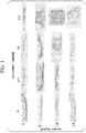
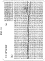
- Normal human dermal fibroblast (NHDF) cells of 3.5 ⁇ 10 6 cells were suspended in a mixture solution of 150 ⁇ L of a solution of heparin/50 mM tris-hydrochloric acid buffer solution (pH 7.4) and 150 ⁇ L of collagen/50 mM tris-hydrochloric acid buffer solution (pH 7.4). Combination of final concentrations of the used heparin and collagen is as shown in FIG. 1 .
- the obtained suspension was sown into a 24 well cell culture insert (Corning Inc, Catalog number: 3470), and centrifuged at 10°C for one minute at 400 ⁇ g. Accordingly, a cell layer was formed on the cell culture insert.
- DMEM Dulbecco's modified eagle's medium
- FBS fetal bovine serum
- a constructed tissue was collected, and a paraffin embedding slice was prepared. Preparation of the paraffin embedding slice was performed by a known method. The prepared slice was subjected to hematoxylin eosin stain (HE stain). The HE stain was performed by a known method.
- FIG. 1 The result of the HE stain is shown in FIG. 1 .
- a concentration of heparin was 1.0 mg/mL or more and a concentration of collagen was 1.0 mg/mL or more, formation of aggregation of collagen and contraction of a tissue were observed. From the result, it was shown that, in a case of using NHDF, both of the concentration of heparin and the concentration of collagen were preferably more than 0 mg/mL and less than 1.0 mg/mL.
- the formation of aggregation of collagen was improved by using a collagen/acetic acid solution (pH 3.7) instead of a solution of collagen/50 mM tris-hydrochloric acid buffer (pH 7.4) solution.
- a collagen/acetic acid solution pH 3.7
- a solution of collagen/50 mM tris-hydrochloric acid buffer pH 7.4
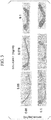
- NHDF cells of 3.5 ⁇ 10 6 cells were suspended in a mixture solution of 250 ⁇ L of a solution of 0.1 mg/mL of heparin/50 mM tris-hydrochloric acid buffer solution (pH 7.4) and 250 ⁇ L of 0.1 mg/mL of collagen/acetic acid solution (pH 3.7) (that is, each of the final concentrations of collagen and heparin was 0.05 mg/mL).
- Sample A and Sample B were prepared as follows.
- the obtained mixture was sown into a 24 well cell culture insert, and centrifuged at 10°C for one minute at 400 ⁇ g. Accordingly, a cell layer was formed on the cell culture insert. Subsequently, a DMEM containing 10% FBS was added to the cell culture insert, and culture was performed in a CO 2 incubator (37°C, 5% CO 2 ) for 24 hours. After the culture, a constructed tissue was collected, and a paraffin embedding slice was prepared. Preparation of the paraffin embedding slice was performed by a known method. The prepared slice was subjected to HE stain. The HE stain was performed by a known method.
- the obtained mixture was centrifuged at room temperature for one minute at 400 ⁇ g to obtain a viscous body.
- the obtained viscous body was suspended in a DMEM containing 10% FBS.
- the obtained suspension was sown into a 24 well cell culture insert, and centrifuged at 10°C for one minute at 400 ⁇ g (gravitational acceleration). Accordingly, a cell layer was formed on the cell culture insert.
- a DMEM containing 10% FBS was added to the cell culture insert, and culture was performed in a CO 2 incubator (37°C, 5% CO 2 ) for 24 hours.
- a constructed tissue was collected, and a paraffin embedding slice was prepared. Preparation of the paraffin embedding slice was performed by a known method.
- the prepared slice was subjected to HE stain.
- the HE stain was performed by a known method.
- FIG. 2 The result of the HE stain is shown in FIG. 2 .
- Sample A an end of the constructed tissue was peeled off from a surface of the cell culture insert. Dissociation of the tissue in the vicinity of the center was also slightly observed but a three-dimensional cell tissue was obtained as a whole.
- Sample B a more homogeneous three-dimensional cell tissue was obtained compared to Sample A. From the result, it was shown that, before sowing a mixture of cells, heparin, and collagen into a culture container, by obtaining a viscous body from the mixture, and using the viscous body, it was possible to obtain a more homogeneous three-dimensional cell tissue.
- NHDF of 3.5 ⁇ 10 6 cells were suspended in a mixture solution of 250 ⁇ L of heparin/ 50 mM tris-hydrochloric acid buffer solution (pH 7.4), and 250 ⁇ L of collagen/ acetic acid solution (pH 3.7). Combination of final concentrations of the used heparin and collagen is as shown in FIG. 3 .
- the obtained mixture was centrifuged at room temperature for one minute at 400 ⁇ g to obtain a viscous body.
- the obtained viscous body was suspended in a DMEM containing 10% FBS.
- the obtained suspension was sown into a 24 well cell culture insert, and centrifuged at 10°C for one minute at 400 ⁇ g (gravitational acceleration).
- a cell layer was formed on the cell culture insert.
- a DMEM containing 10% FBS was added to the cell culture insert, and culture was performed in a CO 2 incubator (37°C, 5% CO 2 ) for 24 hours. After the culture, a constructed tissue was collected, and a paraffin embedding slice was prepared.
- Preparation of the paraffin embedding slice was performed by a known method.
- the prepared slice was subjected to HE stain.
- the HE stain was performed by a known method.
- the result of the HE stain is shown in FIG. 3 .
- a final concentration of collagen was 0.075 mg/mL or more, peeling-off of both ends of the constructed tissue from a surface of the cell culture insert was prominently observed.
- a continuous stacked tissue was constructed.
- the procedure of construction of a tissue is as follows.
- NHDF previously subjected to fluorescent stain with cell tracker red of 1.0 ⁇ 10 6 cells were suspended in a mixture solution of 150 ⁇ L of a solution of 0.2 mg/mL of heparin/50 mM tris-hydrochloric acid buffer solution (pH 7.4) and 150 ⁇ L of 0.2 mg/mL of collagen/acetic acid solution (pH 3.7) (that is, each of the final concentrations of collagen and heparin was 0.1 mg/mL).
- a first cell layer was formed on a cell culture insert.
- a second cell layer was formed on the first cell layer.
- a third cell layer was formed on the second cell layer.
- a DMEM containing 10% FBS was added to the cell culture insert, and culture was performed in a CO 2 incubator (37°C, 5% CO 2 ) for 24 hours. After the culture, a constructed tissue was collected, and a frozen slice was prepared. Preparation of the frozen slice was performed by a known method.
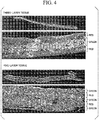
- NHDF previously subjected to fluorescent stain with cell tracker green of 0.6 ⁇ 10 6 cells were suspended in a mixture solution of 150 ⁇ L of a solution of 0.2 mg/mL of heparin/50 mM tris-hydrochloric acid buffer solution (pH 7.4) and 150 ⁇ L of 0.2 mg/mL of collagen/acetic acid solution (pH 3.7).
- a first cell layer was formed on a cell culture insert.
- a second cell layer was formed on the first cell layer.
- a third cell layer was formed on the second cell layer. Furthermore, by the same procedure, using NHDF previously subjected to fluorescent stain with cell tracker red of 0.6 ⁇ 10 6 cells, a fourth cell layer was formed on the third cell layer. Moreover, by the same procedure, using NHDF previously subjected to fluorescent stain with cell tracker green of 0.6 ⁇ 10 6 cells, a fifth cell layer was formed on the fourth cell layer. Subsequently, a DMEM containing 10% FBS was added to the cell culture insert, and culture was performed in a CO 2 incubator (37°C, 5% CO 2 ) for 24 hours. After the culture, a constructed tissue was collected, and a frozen slice was prepared. Preparation of the frozen slice was performed by a known method.
- FIG. 4 The result of fluorescence microscope observation of the slice is shown in FIG. 4 .
- the upper figure is a view of the entire tissue, and the lower figure is an enlarged view thereof, respectively.
- a layer structure can be observed in the constructed tissue.
- NHDF and colorectal cancer cells Using NHDF and colorectal cancer cells (HT29 cells), a cancer tissue model was constructed. NHDF were previously subjected to fluorescent stain with cell tracker green. In addition, HT29 cells were previously subjected to fluorescent stain with cell tracker red. 3.5 ⁇ 10 6 cells (90% NHDF, 10% HT29 cells) were suspended in a mixture solution of 250 ⁇ L of a solution of 0.1 mg/mL of heparin/50 mM tris-hydrochloric acid buffer solution (pH 7.4) and 250 ⁇ L of 0.1 mg/mL of collagen/acetic acid solution (pH 3.7) (that is, each of final concentrations of heparin and collagen was 0.05 mg/mL).
- a cell layer was formed on a cell culture insert. Subsequently, a DMEM containing 10% FBS was added to the cell culture insert, and culture was performed in a CO 2 incubator (37°C, 5% CO 2 ) for 24 hours.
- a constructed tissue was collected, and a frozen slice was prepared. Preparation of the frozen slice was performed by a known method. The prepared slice was subjected to HE stain. The HE stain was performed by a known method.
- FIG. 5(a) shows a result of the HE stain of the frozen slice.
- FIG. 5(b) shows a result of fluorescence microscope observation of the frozen slice.
- FIG. 5(c) is an enlarged view of FIG. 5(a).
- FIG. 5(d) is an enlarged view of FIG. 5(b) .
- FIG. 5(d) in an area surrounded by a white circle, HT29 cells stained with red color were observed.
- Example 3 concentrations of heparin and collagen, at which peeling-off of both ends of the tissue from a surface of the cell culture insert was observed in Example 3 ( FIG. 3 ) were employed.
- NHDF were suspended in a mixture solution of 250 ⁇ L of a solution of 0.2 mg/mL of heparin/50 mM tris-hydrochloric acid buffer solution (pH 7.4) and 250 ⁇ L of 0.2 mg/mL of collagen/acetic acid solution (pH 3.7) (that is, each of the final concentrations of collagen and heparin was 0.1 mg/mL).
- a DMEM containing 10% FBS was added to the cell culture insert, and culture was performed in a CO 2 incubator (37°C, 5% CO 2 ) for 24 hours.
- a constructed tissue was collected, and a paraffin embedding slice was prepared. Preparation of the paraffin embedding slice was performed by a known method. The prepared slice was subjected to HE stain. The HE stain was performed by a known method.
- the result of the HE stain is shown in FIG. 6 .
- Y-27632 contraction of the constructed tissue was suppressed, and thus more homogeneous tissue was obtained.
- both ends of the tissue were not peeled off from a surface of the cell culture insert (substrate), and it was possible to obtain a more homogeneous tissue.
- the average thickness of the tissue was 270 ⁇ m.
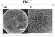
- NHDF of 2.0 ⁇ 10 6 cells and human umbilical vein endothelial cells (HUVEC) of 3.0 ⁇ 10 4 cells were suspended in a mixture solution of 150 ⁇ L of a solution of 0.2 mg/mL of heparin/50 mM tris-hydrochloric acid buffer solution (pH 7.4) and 150 ⁇ L of 0.2 mg/mL of collagen/acetic acid solution (pH 3.7) (that is, each of the final concentrations of heparin and collagen was 0.1 mg/mL), and then centrifuged at room temperature for one minute at 400 ⁇ g to obtain a viscous body.
- HAVEC human umbilical vein endothelial cells
- the obtained viscous body was suspended in a DMEM containing 10% FBS
- the obtained suspension was sown into a 24 well cell culture insert, and centrifuged at room temperature for one minute at 400 ⁇ g.
- a DMEM containing 10% FBS was added to the cell culture insert, and culture was performed in a CO 2 incubator (37°C, 5% CO 2 ) for eight days.
- an anti-CD31 antibody monoclonal mouse anti-human CD31 antibody, Clone JC70A, Code M0823, Dako
- goat anti-mouse IgG-AlexaFluor (registered trademark) 488 invitrogen (trademark)
- the immune stain was performed by a known method.
- FIG. 7 The result of the immune stain is shown in FIG. 7 .
- a vascular network was formed in the tissue.
- NHDF of 1.0 ⁇ 10 6 cells and human umbilical vein endothelial cells (HUVEC) of 1.0 ⁇ 10 5 cells were suspended in a mixture solution of 250 ⁇ L of a solution of 0.1 mg/mL of heparin/50 mM tris-hydrochloric acid buffer solution (pH 7.4) and 250 ⁇ L of 0.1 mg/mL of collagen/acetic acid solution (pH 3.7) (that is, each of the final concentrations of heparin and collagen was 0.05 mg/mL).
- a cell layer was formed on a cell culture insert.
- a DMEM containing 10% FBS was added to the cell culture insert, and culture was performed in a CO 2 incubator (37°C, 5% CO 2 ) for three days.
- the constructed tissue was collected, and a paraffin embedding slice was prepared. Preparation of the paraffin embedding slice was performed by a known method. The prepared slice was subjected to HE stain. The HE stain was performed by a known method. In addition, using an anti-CD31 antibody (monoclonal mouse anti-human CD31 antibody, Clone JC70A, Code M0823, Dako), immune stain was performed.
- an anti-CD31 antibody monoclonal mouse anti-human CD31 antibody, Clone JC70A, Code M0823, Dako
- the immune stain was performed by a known method. 3,3'-Diaminobenzidine (DAB) was used as a chromogenic substrate, and hematoxylin was used as a nuclei staining reagent.
- DAB 3,3'-Diaminobenzidine
- FIG. 8 The result of the HE stain is shown in FIG. 8 .
- a vasculature surrounded by CD31 positive cells was observed in the tissue. From the results shown in FIGS. 7 and 8 , it was shown that, by using fibroblast cells and vascular endothelial cells in combination, it was possible to obtain a three-dimensional cell tissue having a vascular network.
- NHDF of 3.5 ⁇ 10 6 cells suspended in a DMEM containing 10% FBS were sown into a 24 well cell culture insert, and culture was performed in a performed in a CO 2 incubator (37°C, 5% CO 2 ) for 24 hours. After the culture, the constructed tissue was collected, and a paraffin embedding slice was prepared. Preparation of the paraffin embedding slice was performed by a known method. The prepared slice was subjected to HE stain. The HE stain was performed by a known method.
- Comparative Example 1 The result of the HE stain of Comparative Example 1 is shown in FIG. 9 .
- Comparative Example 1 only a tissue thinner than that in examples was constructed. In addition, dissociation was observed in a central portion of the tissue.
- the cell aggregate was sown into a cell culture insert, and centrifuged at room temperature for 1 minute at 400 ⁇ g. Subsequently, a DMEM containing 10% FBS was added to the cell culture insert, and culture was performed in a CO 2 incubator (37°C, 5% CO 2 ) for 24 hours. After the culture, the constructed tissue was collected, and a paraffin embedding slice was prepared. Preparation of the paraffin embedding slice was performed by a known method. The prepared slice was subjected to HE stain. The HE stain was performed by a known method.
- FIGS. 10 to 12 The result of the HE stain of Comparative Example 2 is shown in FIGS. 10 to 12 .
- FIG. 10 shows a result obtained by using NHDF of 1 ⁇ 10 5 cells.
- a thickness of the obtained tissue is about 5 ⁇ m, and it was not possible to discern each cell in the tissue.
- FIG. 11 shows a result obtained by using NHDF of 1 ⁇ 10 6 cells.
- a thickness of the obtained tissue was about 40 ⁇ m, but in the tissue, a lot of spaces were seen between cells.
- FIG. 12 shows a result obtained by using NHDF of 3.5 ⁇ 10 6 cells.
- a thickness of the obtained tissue was about 120 ⁇ m, but in the tissue, a lot of spaces were seen between cells.
- Patent Document 5 was employed, it was not possible to obtain a desired three-dimensional cell tissue.
- NHDF of 3.5 ⁇ 10 6 cells in which fibronectin-gelatin thin membrane (FN-G thin membrane) was formed were sown into a cell culture insert.
- a DMEM containing 10% FBS was added to the cell culture insert, and culture was performed in a CO 2 incubator (37°C, 5% CO 2 ) for 24 hours.
- the constructed tissue was collected, and a paraffin embedding slice was prepared. Preparation of the paraffin embedding slice was performed by a known method. The prepared slice was subjected to HE stain. The HE stain was performed by a known method.
- NHDF of 3.5 ⁇ 10 6 cells were suspended in a mixture solution of 250 ⁇ L of a solution of 0.1 mg/mL of heparin/50 mM tris-hydrochloric acid buffer solution (pH 7.4) and 250 ⁇ L of 0.1 mg/mL of collagen/acetic acid solution (pH 3.7) (that is, each of final concentrations of heparin and collagen was 0.05 mg/mL) to prepare a mixture.
- NHDF of 3.5 ⁇ 10 6 cells were suspended in a mixture solution of 250 ⁇ L of a solution of 0.2 mg/mL of heparin/50 mM tris-hydrochloric acid buffer solution (pH 7.4) and 250 ⁇ L of 0.2 mg/mL of collagen/acetic acid solution (pH 3.7) (that is, each of final concentrations of heparin and collagen was 0.1 mg/mL) to prepare a mixture.
- NHDF of 3.5 ⁇ 10 6 cells were suspended in a mixture solution of 500 ⁇ L of a solution of 50 mM tris-hydrochloric acid buffer solution (pH 7.4) to prepare a mixture in which each of final concentration of heparin and collagen was 0 mg/mL.
- the obtained mixture was centrifuged at room temperature for one minute at 400 ⁇ g to obtain a viscous body.
- the obtained viscous body was suspended in DMEM containing 10% FBS.
- the obtained suspension was sown into a 24 well cell culture insert, and centrifuged at room temperature for one minute at 400 ⁇ g. Accordingly, a cell layer was formed on the cell culture insert.
- a DMEM containing 10% FBS was added to the cell culture insert, and culture was performed in a CO 2 incubator (37°C, 5% CO 2 ) for 24 hours.
- a constructed tissue was fixed with 10% formalin, immersed with 70% ethanol, and then a paraffin embedding slice was prepared. Preparation of the paraffin embedding slice was performed by a known method.
- the prepared slice was subjected to HE stain.
- the HE stain was performed by a known method.
- NHDF of 2.0 ⁇ 10 6 cells and HUVEC of 3.0 ⁇ 10 4 cells were suspended in a mixture solution of 150 ⁇ L of a solution of 0.2 mg/mL or 0.1 mg/mL of polymeric electrolyte/50 mM tris-hydrochloric acid buffer solution (pH 7.4) and 150 ⁇ L of 0.2 mg/mL or 0.1 mg/mL of extracellular matrix component/acetic acid solution (pH 3.7) (that is, each of final concentrations of polymeric electrolyte and extracellular matrix component was 0.1 mg/mL or 0.05 mg/mL). After that, the suspension was centrifuged at room temperature for one minute at 400 ⁇ g to obtain a viscous body.
- NHDF of 2.0 ⁇ 10 6 cells and HUVEC of 3.0 ⁇ 10 4 cells were suspended in a mixture solution of 150 ⁇ L of 50 mM tris-hydrochloric acid buffer solution (pH 7.4) and 150 ⁇ L of acetic acid solution (pH 3.7). After that, the suspension was centrifuged at room temperature for one minute at 400 ⁇ g to obtain a cell aggregate.
- NHDF of 2.0 ⁇ 10 6 cells and HUVEC of 3.0 ⁇ 10 4 cells were suspended in a mixture solution of 150 ⁇ L of DMEM and 150 ⁇ L of acetic acid solution (pH 3.7). After that, the suspension was centrifuged at room temperature for one minute at 400 ⁇ g to obtain a cell aggregate.
- the obtained cell aggregate was suspended in a DMEM containing 10% FBS, and the obtained suspension was sown into a 24 well cell culture insert and centrifuged at room temperature for one minute at 400 ⁇ g. Subsequently, a DMEM containing 10% FBS was added to the cell culture insert, and culture was performed in a CO 2 incubator (37°C, 5% CO 2 ) for five days.
- an anti-CD31 antibody monoclonal mouse anti-human CD31 antibody, Clone JC70A, Code M0823, Dako
- goat anti-mouse IgG-AlexaFluor (registered trademark) 594 invitrogen (trademark)
- the immune stain was performed by a known method. As a result, in all evaluation slices, a vascular network was formed in the tissue.
- NHDF of 3.5 ⁇ 10 6 cells were suspended in a mixture solution of 250 ⁇ L of 0.2 mg/mL or 0.1 mg/mL of polymeric electrolyte/50 mM tris-hydrochloric acid buffer solution (pH 7.4) and 250 ⁇ L of 0.2 mg/mL or 0.1 mg/mL of extracellular matrix component/acetic acid solution (pH 3.7) (that is, each of the final concentrations of the polymeric electrolyte and the extracellular matrix component was 0.1 mg/mL or 0.05 mg/mL). After that, the suspension was centrifuged at room temperature for one minute at 400 ⁇ g to obtain a viscous body.
- the obtained viscous body was suspended in a DMEM containing 10% FBS, and the obtained suspension was sown into a 24 well cell culture insert and centrifuged at room temperature for one minute at 400 ⁇ g. Subsequently, a DMEM containing 10% FBS was added to the cell culture insert, and culture was performed in a CO 2 incubator (37°C, 5% CO 2 ) for 24 hours. After the culture, a constructed tissue was fixed with 10% formalin, and immersed with 70% ethanol to prepare a paraffin embedding slice. Preparation of the paraffin embedding slice was performed by a known method. The prepared slice was subjected to HE stain. The HE stain was performed by a known method.
- a maximum thickness of each of the constructed three-dimensional cell tissues was measured.
- the tissue constructed by not mixing three components of a cationic substance, a polymeric electrolyte, and an extracellular matrix component had the thinnest maximum thickness.
- the maximum thickness of the tissue constructed by containing at least one of a cationic substance, a polymeric electrolyte, and an extracellular matrix component was equal to or greater than that of the tissue constructed by not mixing three components.
- a density of a cell layer of each of the constructed three-dimensional cell tissues was measured. Specifically, first, a maximum value of the number of cell layers of the tissue in each evaluation slice and a thickness ( ⁇ m) of a portion on which the number of cell layers becomes maximum were measured. From the maximum value of the number of the cell layers and the measurement value of the thickness, the number of cell layers per thickness of 10 ⁇ m was calculated.
- Table 3 shows the result of an evaluation slice of the tissue constructed from the cell suspension in which each of the final concentrations of the polymeric electrolyte and the extracellular matrix component was 0.1 mg/mL.
- Table 3 shows the result of the tissue in which each of the final concentrations in a mixture solution was 0.05 mg/mL.
- the upper row of Table 3 shows a relative value of the maximum thickness of each tissue in a case where the thickness (80 ⁇ m) of the tissue constructed by not mixing all the three components was 1.
- the calculated number of cell layers per thickness of 10 ⁇ m is shown in the middle row of Table 3.
- the lower row of Table 3 shows the number of cell layers obtained by subtracting the number of cell layers per thickness of 10 ⁇ m of the tissue constructed by not using all the three components from the calculated number of cell layers per thickness of 10 ⁇ m. Since only the tissue constructed by mixing cells with tris, heparin, and collagen was treated with three specimens, Table 3 shows the result of all the three specimens.
- Tris represents tris-hydrochloric acid buffer solution
- PSS polystyrene sulfonic acid
- none in the column of "extracellular matrix” means that an extracellular matrix was not added
- none in the vertical axis means that none of tris and polymeric electrolyte was added.
- Table 4 shows the result of an evaluation slice of the tissue constructed from the cell suspension in which each of the final concentrations of the polymeric electrolyte and the extracellular matrix component was 0.1 mg/mL.
- Table 4 shows the result of the tissue in which each of the final concentrations in the mixture solution was obtained as 0.05 mg/mL, in a case where a tris and collagen I was used as an extracellular matrix component without using a polymeric electrolyte, in a case where laminin was used as an extracellular matrix component, and dextran sulfate, chondroitin sulfate A, dermatan sulfate, chondroitin sulfate C, polystyrene sulfonic acid, and polyacrylic acid were used as a polymeric electrolyte, in a case where collagen I was used as an extracellular matrix component, and chondroitin sulfate C was used as a polymeric electrolyte, in a case where collagen IV was used as an extracellular matrix component and
- the upper row of Table 4 shows a relative value of a thickness of an area where the number of cells of each tissue was counted, based on the thickness of an area where the number of cells of the tissue constructed by not mixing all the three components was set as 1.
- the calculated number of cells per area of 100 ⁇ m in the thickness direction and 50 ⁇ m in the width direction is shown.
- the lower row of Table 4 shows the number of cells obtained by subtracting the number of cells per the above-mentioned area of the tissue constructed by not using all the three components from the number of cells per the area.
- the tissue constructed by using tris and an extracellular matrix component and the tissue constructed by using tris, a polymeric electrolyte, and an extracellular matrix component had almost no voids inside and were significantly favorable three-dimensional cell tissues, in spite of having a sufficient thickness.
- the tissue constructed by not using an extracellular matrix component there existed a lot of voids inside the tissue.
- adhesion between cells was low, and thus the cells were loosened and the tissue was collapsed during culture medium exchange.
- the tissue containing at least any of a polymeric electrolyte and an extracellular matrix component it was possible to perform culture medium exchange without a problem and to perform culture for a long period of time.
- the tissue constructed by using only tris had the maximum thickness about two times that of the tissue constructed by not using all the three components. However, the tissue constructed by using only tris had the same level of the densities of the cell layers and the cells as those of the tissue constructed by not using all the three components. On the contrary, the tissue containing at least any of a polymeric electrolyte and an extracellular matrix component along with tris had the maximum thickness 1.3 to 5.5 times that of the tissue constructed by not using all the three components, but had a lower densities of the cell layers and the cells than those of the tissue constructed by not using all the three components.
- the cells in vivo are surrounded by matrix components, the cells in vivo exist by being adjacent to one another without being randomly scattered.
- the present invention it is possible to produce a thicker three-dimensional cell tissue in a faster and convenient manner. Therefore, the present invention is useful in the field of regeneration medicine. In addition, the present invention is useful in the development of pharmaceutical products.
Abstract
Description
- The present invention relates to a method for producing a three-dimensional cell tissue.
- Priority is claimed on Japanese Patent Application No.
2016-030916, filed on February 22, 2016 - In recent years, it has been suggested that, in drug assay systems requiring an environment which is similar to a living organism as well as regeneration medicine, it is advantageous to use a three-dimensional cell tissue which is three-dimensionally organized rather than a cell grown on a flat plate. For this reason, various techniques for constructing a three-dimensional tissue of a cell in vitro are developed. For example, a method for forming an aggregation on a surface substrate to which a cell cannot adhere (Patent Document 1), a method for forming an aggregation in liquid droplets (Patent Document 2), a method for gathering cells on a permeable membrane (Patent Document 3), and the like are developed. For maintaining such organization of cells, extracellular matrix (ECM) such as collagen which is produced by a living organism itself is required for bond between cells or formation of a scaffold. For this reason, it has been considered that the ECM is added from outside when artificially constructing a cell tissue (Patent Documents 4 to 7). Patent Document 5 discloses a method for bringing a cell isolated by enzyme treatment and the like into contact with collagen which is a representative ECM and a water-soluble polymer having a cell protecting action, and then culturing the resultant as a three-dimensional aggregate. According to the method, water-soluble polymeric gel is formed around cells during culture.
- Patent Document 8 and
Non-Patent Document 1 disclose a method for preparing cell sheets using a culture dish in which poly(N-isopropylacrylamide) (PIPAAm) which is a temperature-responsive resin is immobilized on the surface, and constructing a three-dimensional tissue by stacking the prepared cell sheets. In this method, peeling-off of the cell sheets is essential, but there is a difficulty in performing peeling while maintaining the shape of the cells. - Non-Patent Document 2 discloses a method for constructing a three-dimensional tissue of a cell by using magnetite cationic liposomes (MCL) containing nanomagnetic fine particles having electrostatic interaction with a cell membrane. However, in this method, there is a difficulty in controlling the thickness or three-dimensional arrangement of a cell layer.
- Patent Document 9 discloses a method for constructing a three-dimensional tissue by repeatedly performing a step for forming a cell layer and bringing the cell layer into contact with a first substance-containing liquid and a second substance-containing liquid, respectively, and consecutively stacking the cell layer via an extracellular matrix (ECM) having a thickness of a nanometer size. In this method, since peeling of a cell sheet of a single layer, overlapping of a peeled cell sheet, or the like is not necessary, it is possible to produce a three-dimensional tissue with excellent reproducibility and efficiency. However, since one cell layer is cultured at a time, it takes long time to construct the three-dimensional tissue. In addition, since it is difficult to arrange various cells in the same plane, there is a limitation to variation of three-dimensional arrangement.
-
Patent Document 10 discloses a method for constructing a three-dimensional tissue by preparing a coated cell of which the whole surface is coated with an adhesive membrane and adhering the cell via the adhesive membrane. In this method, a lot of times of centrifugal separation steps are necessary for preparation of the coated cell. For this reason, there is a possibility that the cell receives physical damage depending on the kind of the cell. In addition, there is a case where the cell is lost along with removal of a supernatant during centrifugal separation, and thus cell recovery rate is lowered. - The present inventors previously found that, by mixing normal human thermal fibroblast (NHDF) cells with fibronectin (FN), heparin (Hep), or dextran sulfate (DS) and subsequently subjecting the mixture to centrifugal separation, a gel-form cell aggregate is formed (Non-Patent Document 3). In addition, a three-dimensional tissue was constructed by using the gel-form cell aggregate to the method disclosed in
Patent Document 10. However, in the above-described method, there is a difficulty in obtaining a thick tissue. -
- [Patent Document 1] Japanese Patent No.
4,159,103 - [Patent Document 2] Japanese Patent No.
5,847,733 - [Patent Document 3] Japanese Patent No.
5,669,741 - [Patent Document 4] Japanese Unexamined Patent Application, First Publication No.
S63-222685 - [Patent Document 5] Japanese Patent No.
2,824,081 - [Patent Document 6] Japanese Patent No.
5,458,259 - [Patent Document 7] Japanese Patent No.
5,409,009 - [Patent Document 8]
PCT International Publication No. WO 2002/008387 - [Patent Document 9] Japanese Patent No.
4,919,464 - [Patent Document 10] Japanese Patent No.
5,850,419 -
- [Non-Patent Document 1] Joseph Yang et al., Biomaterials, 26, 2005, 6415-6422
- [Non-Patent Document 2] Akira Ito et al., Tissue engineering, 10 (5-6), 2004, 833-840
- [Non-Patent Document 3] Akihiro Nishiguchi et al., Macromol Biosci. 2015 Mar; 15(3): 312-7
- An object of the present invention is to provide a method capable of producing a thicker three-dimensional cell tissue in a faster and convenient manner compared to the related art.
- The present inventors performed intensive studies to achieve the above object and found a method capable of producing a thicker three-dimensional cell tissue in a faster and convenient manner compared to the related art that requires a lot of times of centrifugal treatment by using a mixture obtained by mixing cells with a cationic substance, a polymeric electrolyte, and an extracellular matrix component, thereby completing the present invention.
- A method of producing a three-dimensional cell tissue according to a first aspect of the present invention includes a step A of mixing cells with a cationic substance and an extracellular matrix component to obtain a mixture, a step B of gathering the cells from the obtained mixture to form a cell aggregate on a substrate, and a step C of culturing the cells to obtain a three-dimensional cell tissue.
- In the first aspect, in the step A, the cells may be mixed with the cationic substance, the extracellular matrix component, and a polymeric electrolyte.
- In the first aspect, a step A'-1 of removing a liquid portion from the obtained mixture to obtain a cell aggregate and a step A'-2 of suspending the cell aggregate in a solution to obtain a suspension may be further included after the step A, and a step B' of precipitating the cells from the obtained suspension to form a cell precipitate on the substrate may be included instead of the step B.
- In the first aspect, in the step B or the step A'-1, the cell aggregate may be a slurry viscous body.
- In the first aspect, in the step A'-1, a method for removing the liquid portion may be centrifugal separation or filtration.
- In the first aspect, in the step B or the step B', a method for gathering the cells may be centrifugal separation, magnetic separation, or filtration.
- In the first aspect, the cell aggregate in the step B or the cell aggregate in the step B' may be in a layer shape.
- In the first aspect, the polymeric electrolyte may be selected from the group consisting of glycosaminoglycan, dextran sulfate, rhamnan sulfate, fucoidan, carrageenan, polystyrene sulfonic acid, polyacrylamide-2-methylpropanesulfonic acid, polyacrylic acid, and a combination thereof.
- In the first aspect, the extracellular matrix component may be selected from the group consisting of collagen, laminin, fibronectin, vitronectin, elastin, tenascin, entactin, fibrillin, proteoglycan, and a combination thereof.
- In the first aspect, the cationic substance may be a tris-hydrochloric acid buffer solution, a tris-maleic acid buffer solution, a bis-tris-buffer solution, or HEPES.
- In the first aspect, a concentration of the polymeric electrolyte may be 0.05 mg/mL or more to 0.1 mg/mL or less.
- In the first aspect, a concentration of the extracellular matrix component may be 0.05 mg/mL or more to 0.1 mg/mL or less.
- In the first aspect, a mixture ratio of the polymeric electrolyte to the extracellular matrix component may be 1:2 to 2:1.
- In the first aspect, the cells in the step A may be a plurality of kinds of cells.
- In the first aspect, the plurality of kinds of cells may be selected from the group consisting of nerve cells, dendritic cells, immune cells, vascular endothelial cells, lymphatic endothelial cells, fibroblasts, cancer cells, cancer stem cells, epithelial cells, myocardial cells, liver cells, pancreatic islet cells, tissue stem cells, iPS cells, ES cells, and smooth muscle cells.
- In the first aspect, a thickness of the obtained three-dimensional cell tissue may be 5 to 500 µm.
- In the first aspect, the number of cell layers in the obtained three-dimensional cell tissue may be 1 to 100 layers.
- In the first aspect, the number of cells per area of 100 µm in a thickness direction and of 50 µm in a width direction in a region including a position in which a thickness of the obtained three-dimensional cell tissue is the maximum may be 70 or less.
- In the first aspect, in the step C, the cells may be cultured in the presence of a ROCK inhibitor.
- In the first aspect, the number of cell layers per thickness of 10 µm of the obtained three-dimensional cell tissue may be 2.8 layers or less.
- In the first aspect, the obtained three-dimensional cell tissue may include a plurality of kinds of cells.
- In the first aspect, the obtained three-dimensional cell tissue may have a vasculature.
- A kit according to a second aspect of the present invention is a kit for carrying out a method for producing a three-dimensional cell tissue according to the first aspect, and includes at least one reagent selected from the cell, the cationic substance, and the extracellular matrix component.
- The kit according to the second aspect may contain a polymeric electrolyte.
- A three-dimensional cell tissue according to a third aspect of the present invention contains a cell and an extracellular matrix component, a thickness of the three-dimensional cell tissue is 150 µm or greater, and the number of cells per area of 100 µm in a thickness direction and of 50 µm in a width direction in a region including a position in which a thickness of the obtained three-dimensional cell tissue is the maximum may be 70 or less.
- The three-dimensional cell tissue according to the third aspect may further contain a polymeric electrolyte.
- In the third aspect, the extracellular matrix component may be selected from the group consisting of collagen, laminin, fibronectin, vitronectin, elastin, tenascin, entactin, fibrillin, proteoglycan, and a combination thereof.
- In the third aspect, the polymeric electrolyte may be selected from the group consisting of glycosaminoglycan, dextran sulfate, rhamnan sulfate, carrageenan, polystyrene sulfonic acid, polyacrylamide-2-methylpropanesulfonic acid, polyacrylic acid, and a combination thereof.
- According to each of the aspects of the present invention, it is possible to produce a three-dimensional cell tissue in a faster and convenient manner compared to the related art. In addition, it is possible to produce a thicker three-dimensional cell tissue compared to the related art by using a method of each of the aspects of the present invention. Accordingly, it is possible to produce a three-dimensional cell tissue with a thickness that could have not been constructed until now.
-
-
FIG. 1 shows a result of HE stain of a three-dimensional cell tissue constructed by using heparin and collagen. -
FIG. 2 shows a result of HE stain of a three-dimensional cell tissue constructed by heparin and collagen. In Sample A, a mixture of cells with heparin and collagen was sowed into a cell culture insert. In Sample B, a viscous body obtained by performing centrifugation on the mixture of cells with heparin and collagen was used. -
FIG. 3 shows a result of HE stain of a three-dimensional cell tissue constructed by using heparin and collagen. -
FIG. 4 shows a result of fluorescence microscope observation of a three-layer tissue body and a five-layer tissue body constructed by continuous stacking. -
FIG. 5 shows a result of HE stain and a result of fluorescence microscope observation of a cancer tissue model. -
FIG. 6 shows a result of examination of suppression of contraction of a tissue.FIG. 6(a) shows 3.5 × 10⁶ cells/10% FBS-containing DMEM,FIG. 6(b) shows 3.5 × 10⁶ cells/10% FBS-containing DMEM (5 µM Y-27632 addition), andFIG. 6(c) shows 5.0 × 10⁶ cells/10% FBS-containing DMEM (5 µM Y-27632 addition). -
FIG. 7 shows a result of fluorescence microscope observation of a three-dimensional cell tissue having a vasculature.FIG. 7(a) shows an entire image of cell culture insert, andFIG. 7(b) shows an enlarged view of an area surrounded by a square inFIG. 7(a) . -
FIG. 8 shows a result of HE stain and immune stain of a three-dimensional cell tissue having vasculature. -
FIG. 9 shows a tissue constructed without using heparin and collagen (Comparative Example 1). -
FIG. 10 shows a tissue in a case of using a method disclosed in Patent Document 5 (Comparative Example 2).FIG. 10(a) shows an entire view of an obtained tissue, andFIG. 10(b) shows an enlarged view thereof. NHDF of 1 × 10⁵ cells were used. -
FIG. 11 shows a tissue in a case of using a method disclosed in Patent Document 5 (Comparative Example 2).FIG. 10(a) shows an entire view of an obtained tissue, andFIG. 10(b) shows an enlarged view thereof. NHDF of 1 × 10⁶ cells were used. -
FIG. 12 shows the tissue in a case of using a method disclosed in Patent Document 5 (Comparative Example 2).FIG. 11(a) shows an entire view of an obtained tissue, andFIG. 11(b) shows an enlarged view thereof. NHDF of 3.5 × 10⁶ cells were used. - A method of producing a three-dimensional cell tissue according to an embodiment of the present invention relates to a method including:
- (A) a step of mixing cells with a cationic substance and an extracellular matrix component,
- (B) a step of gathering the cells from the obtained mixture and forming a cell aggregate on a substrate, and
- (C) a step of culturing the cells and obtaining a three-dimensional cell tissue.
- In the present invention, in the step (A), by mixing cells with a cationic substance and an extracellular matrix component and forming a cell aggregate from the cell mixture, it is possible to obtain a three-dimensional cell tissue in which there is a small amount of large voids inside. In addition, since the obtained three-dimensional cell tissue is relatively stable, culture for at least several days is possible, and the tissue is hardly collapsed even during culture medium exchange.
- In the step (A), the cells are preferably further mixed with a polymeric electrolyte. By mixing the cells with a cationic substance, a polymeric electrolyte, and an extracellular matrix component, a three-dimensional cell tissue which has a small number of voids, and is thick is obtained more efficiently.
- The method according to the embodiment may include (A'-1) a step of removing a liquid portion from the obtained mixture and obtaining a cell aggregate and (A'-2) a step of suspending the cell aggregate in a solution, after the step (A), and (B') a step of precipitating the cell from the obtained suspension and forming a cell precipitate on the substrate, instead of the step (B). It is possible to obtain a desired tissue by carrying out the above-described steps (A) to (C). In addition, it is possible to obtain a more homogeneous tissue by performing the steps (A'-1) and (A'-2) after the step (A) and performing the step (B') instead of the step (B).
- In the method of the present embodiment, mixing of the cells with the cationic substance, the polymeric electrolyte, and the extracellular matrix component may be performed in a suitable container such as dish, tube, flask, bottle, and plate, or may be performed on the substrate used in the step (B). In addition, suspension in the step (A'-2) may be performed in a suitable container such as dish, tube, flask, bottle, and plate, or may be performed on the substrate used in the step (B').
- In the present specification, the "three-dimensional cell tissue" means a three-dimensional aggregate including at least one kind of cell. Examples of the three-dimensional cell tissue constructed by the present embodiment include tissues of a living organism such as skin, hair, bones, cartilage, teeth, cornea, blood vessels, lymphatic vessels, heart, liver, pancreas, nerves, and esophagus, and solid cancer models (for example, gastric cancer, esophageal cancer, colorectal cancer, colonic cancer, rectal cancer, pancreatic cancer, breast cancer, ovarian cancer, prostate cancer, nephrocyte cancer, hepatoma, and the like), and are not limited thereto.
- In the present specification, the "cell aggregate" means an aggregation of cells. The cell aggregate also includes a cell precipitate (cell aggregate formed by precipitating cells) obtained by centrifugal separation or filtration. In one embodiment, the cell aggregate is a slurry viscous body. In the present specification, the "slurry viscous body" indicates a cell aggregates as disclosed in Akihiro Nishiguchi et al., Macromol Biosci. 2015 Mar; 15(3): 312-7 (Non-Patent Document 3).
- As the cationic substance used in the present embodiment, an optional substance having a positive charge may be used as long as the substance does not have an adverse effect on the growth of cells and the formation of the cell aggregate. Examples of the cationic substance include cationic buffer solutions such as tris-hydrochloric acid buffer solution, tris-maleic acid buffer solution, bis-tris-buffer solution, and HEPES, ethanolamine, diethanolamine, triethanolamine, polyvinylamine, polyarylamine, polylysine, polyhistidine, and polyarginine, and are not limited thereto. The cationic substance used in the present embodiment is preferably a cationic buffer solution. The cationic substance used in the present embodiment is more preferably a tris-hydrochloric acid buffer solution.
- A concentration of the cationic substance is not particularly limited as long as the concentration of the cationic substance does not have an adverse effect on the growth of the cells and the formation of the cell aggregate. The concentration of the cationic substance used in the present embodiment is preferably from 10 to 100 mM. For example, the concentration of the cationic substance used in the present embodiment is preferably from 20 to 90 mM, from 30 to 80 mM, from 40 to 70 mM, and from 45 to 60 mM. The concentration of the cationic substance used in the present embodiment is more preferably 50 mM.
- In a case of using a cationic buffer solution as the cationic substance, a pH of the cationic buffer solution is not particularly limited as long as the pH of the cationic buffer solution does not have an adverse effect on the growth of the cells and the formation of the cell aggregate. The pH of the cationic buffer solution used in the present embodiment is preferably from 6.0 to 8.0. For example, the pH of the cationic buffer solution used in the present embodiment is 7.0, 7.1, 7.2, 7.3, 7.4, 7.5, 7.6, 7.7, 7.8, 7.9, or 8.0. The pH of the cationic buffer solution used in the present embodiment is more preferably from 7.2 to 7.6. The pH of the cationic buffer solution used in the present embodiment is further more preferably 7.4.
- In the present specification, the "polymeric electrolyte" means a polymer having a dissociable functional group in a polymer chain. As the polymeric electrolyte used in the present embodiment, an optional polymeric electrolyte may be used as long as the polymeric electrolyte does not have an adverse effect on the growth of the cells and the formation of the cell aggregate. Examples of the polymeric electrolyte include glycosaminoglycan such as heparin, chondroitin sulfate (for example, chondroitin 4-sulfate, chondroitin 6-sulfate), heparan sulfate, dermatan sulfate, keratan sulfate, and hyaluronic acid; dextran sulfate, rhamnan sulfate, fucoidan, carrageenan, polystyrene sulfonic acid, polyacrylamide-2-methylpropanesulfonic acid, and polyacrylic acid, but are not limited thereto. The polymeric electrolyte may be used alone, or may be used in combination. The polymeric electrolyte used in the present embodiment is preferably glycosaminoglycan. In addition, the polymeric electrolyte used in the present embodiment is more preferably heparin or dextran sulfate, chondroitin sulfate, or dermatan sulfate. The polymeric electrolyte used in the present embodiment is further more preferably heparin. A derivative of the polymeric electrolyte may be used as long as the derivative of the polymeric electrolyte does not have an adverse effect on the growth of the cells and the formation of the cell aggregate.
- A concentration of the polymeric electrolyte is not particularly limited as long as the concentration of the polymeric electrolyte does not have an adverse effect on the growth of the cells and the formation of the cell aggregate. The concentration of the polymeric electrolyte used in the present embodiment is preferably more than 0 mg/mL and less than 1.0 mg/mL. The concentration of the polymeric electrolyte used in the present embodiment is more preferably 0.025 mg/mL or more to 0.1 mg/mL or less. For example, the concentration of the polymeric electrolyte used in the present embodiment is 0.025, 0.05, 0.075, or 0.1 mg/mL. The concentration of the polymeric electrolyte used in the present embodiment is further more preferably 0.05 mg/mL or more to 0.1 mg/mL or less. The concentration of the polymeric electrolyte used in the present embodiment is further more preferably 0.05 mg/mL. In the present embodiment, the polymeric electrolyte may be appropriately dissolved in a solvent and used. Examples of the solvent include water and a buffer solution, but are not limited thereto. In a case of using a cationic buffer solution as the cationic substance, the polymeric electrolyte may be dissolved in the cationic buffer solution and used.
- As the extracellular matrix component used in the present embodiment, an optional component constituting an extracellular matrix (ECM) may be used as long as the component does not have an adverse effect on the growth of the cells and the formation of the cell aggregate. Examples of the extracellular matrix component include collagen, laminin, fibronectin, vitronectin, elastin, tenascin, entactin, fibrillin, proteoglycan, and the like, but not limited thereto. The extracellular matrix component may be used alone, or may be used in combination. Examples of the proteoglycan include chondroitin sulfate proteoglycan, heparan sulfate proteoglycan, keratan sulfate proteoglycan, and dermatan sulfate proteoglycan, but are not limited thereto. The extracellular matrix component used in the present embodiment is collagen, laminin, and fibronectin, and among these, collagen is preferable. A modified substance and a variant of the extracellular matrix component may be used as long as the modified substance and the variant of the extracellular matrix component do not have an adverse effect on the growth of the cells and the formation of the cell aggregate.
- A concentration of the extracellular matrix component is not particularly limited as long as the concentration of the extracellular matrix component does not have an adverse effect on the growth of the cells and the formation of the cell aggregate. The concentration of the extracellular matrix component used in the present embodiment is preferably more than 0 mg/mL and less than 1.0 mg/mL. The concentration of the extracellular matrix component used in the present embodiment is more preferably 0.025 mg/mL or more to 0.1 mg/mL or less. For example, the concentration of the extracellular matrix component used in the present embodiment is 0.025, 0.05, 0.075, or 0.1 mg/mL. The concentration of the extracellular matrix component used in the present embodiment is further more preferably 0.05 mg/mL or more to 0.1 mg/mL or less. The concentration of the extracellular matrix component used in the present embodiment is further more preferably 0.05 mg/mL. In the present embodiment, the extracellular matrix component may be dissolved in a solvent and used. Examples of the solvent include water, a buffer solution, and acetate, but are not limited thereto. In the present embodiment, the extracellular matrix component is preferably dissolved in the buffer solution or acetate.
- In the present embodiment, a mixture ratio of the polymeric electrolyte to the extracellular matrix component is preferably 1:2 to 2:1. The mixture ratio of the polymeric electrolyte to the extracellular matrix component used in the present embodiment is more preferably 1:1.5 to 1.5:1. The mixture ratio of the polymeric electrolyte to the extracellular matrix component used in the present embodiment is further more preferably 1:1.
- Cells used in the method according to the present embodiment are not particularly limited. For example, the cells are cells derived from animals such as humans, monkeys, dogs, cats, rabbits, pigs, cows, mice, and rats. Cell-derived sites are also not particularly limited, and may be somatic cells derived from bones, muscles, viscera, nerves, brain, skin, blood, and the like, or may be reproductive cells. In addition, the cells used in the method according to the present embodiment may be induced pluripotent stem cells (iPS cells) or embryonic stem cells (ES cells). Or, the cells used in the method according to the present embodiment may be culture cells such as primary culture cells, subculture cells, and cell strain cells. One kind of cell may be used, or a plurality of kinds of cells may be used. Examples of the cells used in the method according to the present embodiment include nerve cells, dendritic cells, immune cells, vascular endothelial cells, lymphatic endothelial cells, fibroblasts, cancer cells such as hepatoma cells, epithelial cells, myocardial cells, liver cells, pancreatic islet cells, tissue stem cells, and smooth muscle cells, but are not limited thereto.
- The three-dimensional cell tissue obtained by the method according to the present embodiment may include one kind of cell, or may include a plurality of kinds of cells. In the present embodiment, cells included in the three-dimensional cell tissue are selected from the group consisting of nerve cells, dendritic cells, immune cells, vascular endothelial cells, lymphatic endothelial cells, fibroblasts, cancer cells, epithelial cells, myocardial cells, liver cells, pancreatic islet cells, tissue stem cells, and smooth muscle cells. In addition, the three-dimensional cell tissue obtained by the method according to the present embodiment may have a vasculature. The "vasculature" indicates a network-like structure such as vascular network and lymphatic network in tissues of a living organism.
- In the method according to the present embodiment, as means for removing a liquid portion in the step (A'-1), a method known to those skilled in the art may be used. For example, a liquid portion may be removed by centrifugal separation or filtration. Conditions of the centrifugal separation are not particularly limited as long as the conditions of the centrifugal separation do not have an adverse effect on the growth of the cells and the formation of the cell aggregate. For example, a liquid portion and a cell aggregate are separated by applying centrifugal separation to a microtube into which a mixture is introduced at room temperature for one minute at 400 × g to remove the liquid portion. Alternatively, the liquid portion may be removed after gathering cells by natural sedimentation.
- The solution used in the step (A'-2) of the method according to the present embodiment is not particularly limited as long as the solution used in the step (A'-2) does not have an adverse effect on the growth of the cells and the formation of the cell aggregate. For example, a cell culture medium or a buffer solution suitable for the used cells is used.
- Examples of a substrate used in the step (B) or the step (B') of the method according to the present embodiment include a culture vessel used for culture of cells. The culture vessel is a vessel made of a material and having a shape generally used for culture of cells or microorganisms. Examples of the material of the culture vessel include glass, stainless steel, and plastic, but are not limited thereto. Examples of the culture vessel include a dish, a tube, a flask, a bottle, and a plate, but are not limited thereto. For example, the substrate is made of a material that does not pass through cells in a liquid but is able to pass through the liquid.
- The substrate used in the present embodiment is preferably a permeable membrane. Examples of a vessel including such a permeable membrane include cell culture inserts such as Transwell (registered trademark) insert, Netwell (registered trademark) insert, Falcon (registered trademark) cell culture insert, and Millicell (registered trademark) cell culture insert, but are not limited thereto.
- In the method of the present embodiment, as means for gathering cells in the step (B) and the step (B'), a method known to those skilled in the art may be used. For example, cells may be gathered by centrifugal separation, magnetic separation, or filtration. Conditions of the centrifugal separation are not particularly limited as long as the conditions of the centrifugal separation do not have an adverse effect on the growth of the cells and the formation of the cell aggregate. For example, a mixture or suspension is sown into a cell culture insert, and subjected to centrifugal separation at 10°C for one minute for 400 × g to gather the cells. Alternatively, the cells may be gathered by natural sedimentation. In the step (B'), a cell precipitate may be formed on a substrate by removing the liquid portion from the suspension by the centrifugal separation or filtration. Or, the cell precipitate may be formed on the substrate by natural sedimentation. The cell aggregate in the step (B) or the cell precipitate in the step (B') may be in a layer shape.
- In the present embodiment, a substance for suppressing deformation (for example, contraction of a tissue, peeling-off of a tissue terminal, and the like) of the constructed three-dimensional cell tissue is used. Examples of such a substance include Y-27632 which is a selective ROCK (Rho-associated coiled-coil forming kinase/Rho bond kinase) inhibitor, but are not particularly limited thereto. In the present embodiment, a step (C) is performed in the presence of the substance. By culturing a cell aggregate in the presence of the substance, contraction of the constructed three-dimensional cell tissue is suppressed. As a result, peeling-off of the constructed tissue from a substrate such as cell culture insert is suppressed. For example, in the step (A), such a substance may be further mixed. Alternatively, such a substance may be further added to the solution used in the step (A'-2). Alternatively, such a substance may be added to a culture medium when culturing cells in the step (C).
- In the step (C) of the method according to the present embodiment, culture of cells may be performed in culture conditions suitable for cultured cells. Those skilled in the art may select an appropriate culture medium depending on the kind of the cells or desired functions. In addition, all conditions such as culture temperature and culture time are easily determined by those skilled in the art.
- By repeating the steps (A) to (B) or (A) to (B') of the method of the present embodiment, it is possible to stack cell aggregates or cell precipitates. In this manner, it is possible to construct a three-dimensional cell tissue having a plurality of layers. In this case, a three-dimensional cell tissue constituted of different kinds of cells by using the plurality of kinds of cells. In this embodiment, a thickness of the constructed three-dimensional cell tissue is from about 5 to about 300 µm, about 5 to about 400 µm, or about 5 to about 500 µm. The thickness of the constructed three-dimensional cell tissue is preferably 150 µm or more, more preferably 200 µm or more, and further more preferably 250 µm or more. By repeating the steps (A) to (B) or (A) to (B') of the method of the present invention, a three-dimensional cell tissue having a small amount of voids inside, although having the thickness of from 150 to 500 µm, is obtained. In the embodiment, the number of the cell layers in the constructed three-dimensional cell tissue is preferably from 1 to about 100 layers, and more preferably from 10 to about 100 layers. The cell layer is defined as a separate layer at the time when a cell nucleus does not overlap with gravity and a height direction of the cell, when observed at a magnification at which the cell nucleus can be recognized, that is, at a magnification at which an entire thickness of a stained slice is within a visual field, in a slice image of a cross-section in a thickness direction of the three-dimensional cell tissue. At this time, a visual field in a vertical direction to gravity (horizontal direction) is secured by at least 200 µm or greater. In the present embodiment, a slice image of a cross-section in a thickness direction of the three-dimensional cell tissue is observed at a magnification of 100 to 200 times.
- The three-dimensional cell tissue obtained by repeating the steps (A) to (B) or (A) to (B') of the method of the present embodiment has the smaller number of cell layers per unit thickness compared to the three-dimensional cell tissue obtained by a method for stacking cells in the related art. In the present embodiment, the constructed three-dimensional cell tissue is obtained in vitro. In addition, the three-dimensional cell tissue according to the present embodiment contains a cell and an extracellular matrix component, and the number of the cell layers per 10 µm of thickness of the three-dimensional cell tissue is 2.8 layers or less, preferably 2.5 layers or less, and more preferably 2.2 layers or less. In the present embodiment, in the obtained three-dimensional cell tissue, the number of cells per area of 100 µm in a thickness direction and 50 µm in a width direction in a region including a position (maximum point) at which the thickness becomes maximum is preferably from 5 to 70, more preferably from 10 to 60, and further more preferably from 15 to 50.
- In the present specification, the thickness of the three-dimensional cell tissue is a length of the tissue in the gravity direction. The gravity direction is a direction in which gravity is imparted. In the present embodiment, it is possible to measure the number of the cells as follows by using a slice formed from a cross-section in the thickness direction of the tissue. In the slice, among a region in which there is no void with a predetermined size or greater, a position (maximum point) at which the thickness of the three-dimensional cell tissue is maximum is determined. By setting a strip-like region (including a top surface to a bottom surface of the tissue) of a width of 50 µm including vicinity of the maximum value of the thickness to a measurement region, the number of the cell nuclei included in the measurement region is counted. The measurement region is set not to include a void. The predetermined size of the void is set to be a maximum diameter of 50 µm or greater. The maximum diameter of the void is defined as a long side in a case where the void is rectangular, is defined as a diameter in a case where the void is spherical, is defined as a long diameter in a case where the void is elliptic, and is defined as a long diameter of an approximated ellipse in a case where the void is amorphous. The void is not stained on an HE-stained slice. Here, in a case where the top face of the region including the maximum value of the thickness is in a convex shape, there is a case where the tissue is peeled off from the substrate. In such a case, a region which is vicinity of the maximum value and of which the top surface is relatively plain is set as a measurement region. In this case, a region of 100 µm to 650 µm in a width direction including the vicinity of the maximum value of the thickness is set as the measurement region.
- A cell nucleus of which at least a portion is included in the measurement region to which counting is performed is counted as a cell nucleus included in the measurement region. From the counted cells, a number of the cells per area of 100 µm in the thickness direction and 50 µm in the width direction is calculated.
- A kit according to one embodiment of the present invention is a kit for preforming the method according to the embodiment, and includes at least one reagent selected from a cell, a cationic substance, a polymeric electrolyte, and an extracellular matrix component. In general, the reagent is put in an appropriate container and provided. Such a kit may include a reagent suitable for conveniently performing the method of the embodiment, for example, a diluent, a buffer, and a washing reagent. In addition, the kit may include a suitable substrate such as dish, cell culture insert, tube, flask, bottle, and plate. Moreover, the kit may include materials such as instruction necessary for carrying out the method of the embodiment.
- Using the method according to the embodiment of the present invention, the number of times to subject cells to centrifugal separation is in two. In the methods in the related art, the operation is complex and the centrifugal separation is also used a lot of times. Therefore, by using the method according to the embodiment of the present invention, it is possible to reduce the number of times of the centrifugal separation and to decrease damage of the cells. In addition, it is possible to reduce loss of the cells during operation and to achieve a high cell recovery rate.
- Hereinafter, a detailed and specific description will be provided on the present invention by showing examples. However, examples do not limit the scope of the present invention.
- In the following example, collagen I was used as collagen, unless otherwise explained.
- Normal human dermal fibroblast (NHDF) cells of 3.5 × 10⁶ cells were suspended in a mixture solution of 150 µL of a solution of heparin/50 mM tris-hydrochloric acid buffer solution (pH 7.4) and 150 µL of collagen/50 mM tris-hydrochloric acid buffer solution (pH 7.4). Combination of final concentrations of the used heparin and collagen is as shown in
FIG. 1 . The obtained suspension was sown into a 24 well cell culture insert (Corning Inc, Catalog number: 3470), and centrifuged at 10°C for one minute at 400 × g. Accordingly, a cell layer was formed on the cell culture insert. Subsequently, a Dulbecco's modified eagle's medium (DMEM) containing 10% fetal bovine serum (FBS) was added to the cell culture insert, and culture was performed in a CO2 incubator (37°C, 5% CO2) for 24 hours. After the culture, a constructed tissue was collected, and a paraffin embedding slice was prepared. Preparation of the paraffin embedding slice was performed by a known method. The prepared slice was subjected to hematoxylin eosin stain (HE stain). The HE stain was performed by a known method. - The result of the HE stain is shown in
FIG. 1 . In a case of heparin alone and collagen alone, formation of a tissue was not shown. In a case where a concentration of heparin was 1.0 mg/mL or more and a concentration of collagen was 1.0 mg/mL or more, formation of aggregation of collagen and contraction of a tissue were observed. From the result, it was shown that, in a case of using NHDF, both of the concentration of heparin and the concentration of collagen were preferably more than 0 mg/mL and less than 1.0 mg/mL. The formation of aggregation of collagen was improved by using a collagen/acetic acid solution (pH 3.7) instead of a solution of collagen/50 mM tris-hydrochloric acid buffer (pH 7.4) solution. In addition, even in a case where a suspension of heparin, collagen, and cells was sown into the cell culture insert, and then a cell layer was formed by natural sedimentation without performing centrifugal operation, it was possible to construct a three-dimensional cell tissue. - NHDF cells of 3.5 × 10⁶ cells were suspended in a mixture solution of 250 µL of a solution of 0.1 mg/mL of heparin/50 mM tris-hydrochloric acid buffer solution (pH 7.4) and 250 µL of 0.1 mg/mL of collagen/acetic acid solution (pH 3.7) (that is, each of the final concentrations of collagen and heparin was 0.05 mg/mL). Sample A and Sample B were prepared as follows.
- The obtained mixture was sown into a 24 well cell culture insert, and centrifuged at 10°C for one minute at 400 × g. Accordingly, a cell layer was formed on the cell culture insert. Subsequently, a DMEM containing 10% FBS was added to the cell culture insert, and culture was performed in a CO2 incubator (37°C, 5% CO2) for 24 hours. After the culture, a constructed tissue was collected, and a paraffin embedding slice was prepared. Preparation of the paraffin embedding slice was performed by a known method. The prepared slice was subjected to HE stain. The HE stain was performed by a known method.
- The obtained mixture was centrifuged at room temperature for one minute at 400 × g to obtain a viscous body. The obtained viscous body was suspended in a DMEM containing 10% FBS. The obtained suspension was sown into a 24 well cell culture insert, and centrifuged at 10°C for one minute at 400 × g (gravitational acceleration). Accordingly, a cell layer was formed on the cell culture insert. Subsequently, a DMEM containing 10% FBS was added to the cell culture insert, and culture was performed in a CO2 incubator (37°C, 5% CO2) for 24 hours. After the culture, a constructed tissue was collected, and a paraffin embedding slice was prepared. Preparation of the paraffin embedding slice was performed by a known method. The prepared slice was subjected to HE stain. The HE stain was performed by a known method.
- The result of the HE stain is shown in
FIG. 2 . In Sample A, an end of the constructed tissue was peeled off from a surface of the cell culture insert. Dissociation of the tissue in the vicinity of the center was also slightly observed but a three-dimensional cell tissue was obtained as a whole. In Sample B, a more homogeneous three-dimensional cell tissue was obtained compared to Sample A. From the result, it was shown that, before sowing a mixture of cells, heparin, and collagen into a culture container, by obtaining a viscous body from the mixture, and using the viscous body, it was possible to obtain a more homogeneous three-dimensional cell tissue. In addition, regarding Sample B, even in a case where a suspension of the viscous body was sown into the cell culture insert, and then a cell layer was formed by natural sedimentation without performing centrifugal operation, it was possible to construct a three-dimensional cell tissue. - NHDF of 3.5 × 10⁶ cells were suspended in a mixture solution of 250 µL of heparin/ 50 mM tris-hydrochloric acid buffer solution (pH 7.4), and 250 µL of collagen/ acetic acid solution (pH 3.7). Combination of final concentrations of the used heparin and collagen is as shown in
FIG. 3 . The obtained mixture was centrifuged at room temperature for one minute at 400 × g to obtain a viscous body. The obtained viscous body was suspended in a DMEM containing 10% FBS. The obtained suspension was sown into a 24 well cell culture insert, and centrifuged at 10°C for one minute at 400 × g (gravitational acceleration). Accordingly, a cell layer was formed on the cell culture insert. Subsequently, a DMEM containing 10% FBS was added to the cell culture insert, and culture was performed in a CO2 incubator (37°C, 5% CO2) for 24 hours. After the culture, a constructed tissue was collected, and a paraffin embedding slice was prepared. - Preparation of the paraffin embedding slice was performed by a known method. The prepared slice was subjected to HE stain. The HE stain was performed by a known method.
- The result of the HE stain is shown in
FIG. 3 . In a case where a final concentration of collagen was 0.075 mg/mL or more, peeling-off of both ends of the constructed tissue from a surface of the cell culture insert was prominently observed. - Using NHDF previously subjected to fluorescent stain with cell tracker green and NHDF previously subjected to fluorescent stain with cell tracker red, a continuous stacked tissue was constructed. The procedure of construction of a tissue is as follows.
- NHDF previously subjected to fluorescent stain with cell tracker red of 1.0 × 10⁶ cells were suspended in a mixture solution of 150 µL of a solution of 0.2 mg/mL of heparin/50 mM tris-hydrochloric acid buffer solution (pH 7.4) and 150 µL of 0.2 mg/mL of collagen/acetic acid solution (pH 3.7) (that is, each of the final concentrations of collagen and heparin was 0.1 mg/mL). Following the procedure of (ii) of Example 2, a first cell layer was formed on a cell culture insert. By the same procedure, using NHDF previously subjected to fluorescent stain with cell tracker green of 1.0 × 10⁶ cells, a second cell layer was formed on the first cell layer. In addition, by the same procedure, using NHDF previously subjected to fluorescent stain with cell tracker red of 1.0 × 10⁶ cells, a third cell layer was formed on the second cell layer. Subsequently, a DMEM containing 10% FBS was added to the cell culture insert, and culture was performed in a CO2 incubator (37°C, 5% CO2) for 24 hours. After the culture, a constructed tissue was collected, and a frozen slice was prepared. Preparation of the frozen slice was performed by a known method.
- NHDF previously subjected to fluorescent stain with cell tracker green of 0.6 × 10⁶ cells were suspended in a mixture solution of 150 µL of a solution of 0.2 mg/mL of heparin/50 mM tris-hydrochloric acid buffer solution (pH 7.4) and 150 µL of 0.2 mg/mL of collagen/acetic acid solution (pH 3.7). Following the procedure of (ii) of Example 2, a first cell layer was formed on a cell culture insert. By the same procedure, using NHDF previously subjected to fluorescent stain with cell tracker red of 0.6 × 10⁶ cells, a second cell layer was formed on the first cell layer. In addition, by the same procedure, using NHDF previously subjected to fluorescent stain with cell tracker green of 0.6 × 10⁶ cells, a third cell layer was formed on the second cell layer. Furthermore, by the same procedure, using NHDF previously subjected to fluorescent stain with cell tracker red of 0.6 × 10⁶ cells, a fourth cell layer was formed on the third cell layer. Moreover, by the same procedure, using NHDF previously subjected to fluorescent stain with cell tracker green of 0.6 × 10⁶ cells, a fifth cell layer was formed on the fourth cell layer. Subsequently, a DMEM containing 10% FBS was added to the cell culture insert, and culture was performed in a CO2 incubator (37°C, 5% CO2) for 24 hours. After the culture, a constructed tissue was collected, and a frozen slice was prepared. Preparation of the frozen slice was performed by a known method.
- The result of fluorescence microscope observation of the slice is shown in
FIG. 4 . The upper figure is a view of the entire tissue, and the lower figure is an enlarged view thereof, respectively. A layer structure can be observed in the constructed tissue. - Using NHDF and colorectal cancer cells (HT29 cells), a cancer tissue model was constructed. NHDF were previously subjected to fluorescent stain with cell tracker green. In addition, HT29 cells were previously subjected to fluorescent stain with cell tracker red. 3.5 × 10⁶ cells (90% NHDF, 10% HT29 cells) were suspended in a mixture solution of 250 µL of a solution of 0.1 mg/mL of heparin/50 mM tris-hydrochloric acid buffer solution (pH 7.4) and 250 µL of 0.1 mg/mL of collagen/acetic acid solution (pH 3.7) (that is, each of final concentrations of heparin and collagen was 0.05 mg/mL). Following the procedure of (ii) of Example 2, a cell layer was formed on a cell culture insert. Subsequently, a DMEM containing 10% FBS was added to the cell culture insert, and culture was performed in a CO2 incubator (37°C, 5% CO2) for 24 hours.
- After the culture, a constructed tissue was collected, and a frozen slice was prepared. Preparation of the frozen slice was performed by a known method. The prepared slice was subjected to HE stain. The HE stain was performed by a known method.
- The result of the HE stain and the result of fluorescence microscope observation are shown in
FIG. 5. FIG. 5(a) shows a result of the HE stain of the frozen slice.FIG. 5(b) shows a result of fluorescence microscope observation of the frozen slice.FIG. 5(c) is an enlarged view ofFIG. 5(a). FIG. 5(d) is an enlarged view ofFIG. 5(b) . InFIG. 5(d) , in an area surrounded by a white circle, HT29 cells stained with red color were observed. - Using Y-27632 (Calbiochem, Catalog number: 688000), suppression of contraction of the constructed tissue was examined. In the present example, concentrations of heparin and collagen, at which peeling-off of both ends of the tissue from a surface of the cell culture insert was observed in Example 3 (
FIG. 3 ) were employed. NHDF were suspended in a mixture solution of 250 µL of a solution of 0.2 mg/mL of heparin/50 mM tris-hydrochloric acid buffer solution (pH 7.4) and 250 µL of 0.2 mg/mL of collagen/acetic acid solution (pH 3.7) (that is, each of the final concentrations of collagen and heparin was 0.1 mg/mL). Following the procedure of (ii) of Example 2, a cell layer was formed on a cell culture insert. The number of used cells and the solution used for suspension of a viscous body are as shown in the following table.[Table 1] Number of cells Solution used for suspension Sample (a) 3.5×10⁶ DMEM containing 10% FBS Sample (b) 3.5×10⁶ DMEM containing 10% FBS (5 mM Y-27632 added) Sample (c) 5.0×10⁶ DMEM containing 10% FBS (5 mM Y-27632 added) - Subsequently, a DMEM containing 10% FBS was added to the cell culture insert, and culture was performed in a CO2 incubator (37°C, 5% CO2) for 24 hours. After the culture, a constructed tissue was collected, and a paraffin embedding slice was prepared. Preparation of the paraffin embedding slice was performed by a known method. The prepared slice was subjected to HE stain. The HE stain was performed by a known method.
- The result of the HE stain is shown in
FIG. 6 . By adding Y-27632, contraction of the constructed tissue was suppressed, and thus more homogeneous tissue was obtained. In addition, even in a case of using 5.0 × 10⁶ cells, both ends of the tissue were not peeled off from a surface of the cell culture insert (substrate), and it was possible to obtain a more homogeneous tissue. The average thickness of the tissue was 270 µm. - NHDF of 2.0 × 10⁶ cells and human umbilical vein endothelial cells (HUVEC) of 3.0 × 10⁴ cells were suspended in a mixture solution of 150 µL of a solution of 0.2 mg/mL of heparin/50 mM tris-hydrochloric acid buffer solution (pH 7.4) and 150 µL of 0.2 mg/mL of collagen/acetic acid solution (pH 3.7) (that is, each of the final concentrations of heparin and collagen was 0.1 mg/mL), and then centrifuged at room temperature for one minute at 400 × g to obtain a viscous body. After the obtained viscous body was suspended in a DMEM containing 10% FBS, the obtained suspension was sown into a 24 well cell culture insert, and centrifuged at room temperature for one minute at 400 × g. Subsequently, a DMEM containing 10% FBS was added to the cell culture insert, and culture was performed in a CO2 incubator (37°C, 5% CO2) for eight days. After the culture, using an anti-CD31 antibody (monoclonal mouse anti-human CD31 antibody, Clone JC70A, Code M0823, Dako) as a primary antibody, and goat anti-mouse IgG-AlexaFluor (registered trademark) 488 (invitrogen (trademark)) as a secondary antibody, the constructed tissue was subjected to immune stain. The immune stain was performed by a known method.
- The result of the immune stain is shown in
FIG. 7 . As shown inFIG. 7 , a vascular network was formed in the tissue. - NHDF of 1.0 × 10⁶ cells and human umbilical vein endothelial cells (HUVEC) of 1.0 × 10⁵ cells were suspended in a mixture solution of 250 µL of a solution of 0.1 mg/mL of heparin/50 mM tris-hydrochloric acid buffer solution (pH 7.4) and 250 µL of 0.1 mg/mL of collagen/acetic acid solution (pH 3.7) (that is, each of the final concentrations of heparin and collagen was 0.05 mg/mL). Following the procedure of (ii) of Example 2, a cell layer was formed on a cell culture insert. Subsequently, a DMEM containing 10% FBS was added to the cell culture insert, and culture was performed in a CO2 incubator (37°C, 5% CO2) for three days.
- After the culture, the constructed tissue was collected, and a paraffin embedding slice was prepared. Preparation of the paraffin embedding slice was performed by a known method. The prepared slice was subjected to HE stain. The HE stain was performed by a known method. In addition, using an anti-CD31 antibody (monoclonal mouse anti-human CD31 antibody, Clone JC70A, Code M0823, Dako), immune stain was performed.
- The immune stain was performed by a known method. 3,3'-Diaminobenzidine (DAB) was used as a chromogenic substrate, and hematoxylin was used as a nuclei staining reagent.
- The result of the HE stain is shown in
FIG. 8 . As shown inFIG. 8 , a vasculature surrounded by CD31 positive cells was observed in the tissue. From the results shown inFIGS. 7 and8 , it was shown that, by using fibroblast cells and vascular endothelial cells in combination, it was possible to obtain a three-dimensional cell tissue having a vascular network. - NHDF of 3.5 × 10⁶ cells suspended in a DMEM containing 10% FBS were sown into a 24 well cell culture insert, and culture was performed in a performed in a CO2 incubator (37°C, 5% CO2) for 24 hours. After the culture, the constructed tissue was collected, and a paraffin embedding slice was prepared. Preparation of the paraffin embedding slice was performed by a known method. The prepared slice was subjected to HE stain. The HE stain was performed by a known method.
- The result of the HE stain of Comparative Example 1 is shown in
FIG. 9 . In Comparative Example 1, only a tissue thinner than that in examples was constructed. In addition, dissociation was observed in a central portion of the tissue. - In a case of employing the method disclosed in Patent Document 5, it was verified whether or not it is possible to construct a three-dimensional cell tissue. A culture solution of NHDF was centrifuged using a microtube, and a supernatant was removed to recover cells. The recovered cells were mixed with a 0.1% collagen solution (dissolved in a DMEM containing 10% FBS), subjected to rotary stirring at 4°C for 10 minutes to obtain a suspension of the cells. The number of the used cells was 1 × 10⁵ cells, 1 × 10⁶ cells, and 3.5 × 10⁶ cells. The cell suspension was centrifuged at room temperature for 1 minute at 400 × g, and a supernatant was removed to obtain a cell aggregate. The cell aggregate was sown into a cell culture insert, and centrifuged at room temperature for 1 minute at 400 × g. Subsequently, a DMEM containing 10% FBS was added to the cell culture insert, and culture was performed in a CO2 incubator (37°C, 5% CO2) for 24 hours. After the culture, the constructed tissue was collected, and a paraffin embedding slice was prepared. Preparation of the paraffin embedding slice was performed by a known method. The prepared slice was subjected to HE stain. The HE stain was performed by a known method.
- The result of the HE stain of Comparative Example 2 is shown in
FIGS. 10 to 12 . In each figure, (a) shows an entire view of the obtained tissue, and (b) is an enlarged view thereof.FIG. 10 shows a result obtained by using NHDF of 1 × 10⁵ cells. A thickness of the obtained tissue is about 5 µm, and it was not possible to discern each cell in the tissue.FIG. 11 shows a result obtained by using NHDF of 1 × 10⁶ cells. A thickness of the obtained tissue was about 40 µm, but in the tissue, a lot of spaces were seen between cells.FIG. 12 shows a result obtained by using NHDF of 3.5 × 10⁶ cells. A thickness of the obtained tissue was about 120 µm, but in the tissue, a lot of spaces were seen between cells. In other words, although the method disclosed in Patent Document 5 was employed, it was not possible to obtain a desired three-dimensional cell tissue. - According to the method (LbL method) for producing a three-dimensional tissue disclosed in
Patent Document 10, NHDF of 3.5 × 10⁶ cells in which fibronectin-gelatin thin membrane (FN-G thin membrane) was formed were sown into a cell culture insert. Subsequently, a DMEM containing 10% FBS was added to the cell culture insert, and culture was performed in a CO2 incubator (37°C, 5% CO2) for 24 hours. After the culture, the constructed tissue was collected, and a paraffin embedding slice was prepared. Preparation of the paraffin embedding slice was performed by a known method. The prepared slice was subjected to HE stain. The HE stain was performed by a known method. - In Comparative Example 3, dissociation was seen in a central portion of the obtained tissue. That is, by using cells of 3.5 × 10⁶ cells or greater, it was not possible to form a tissue with a thickness more than 100 µm.
- Using tris, bistris, and HEPES as a cationic substance, heparin, and collagen was mixed with cells to construct a three-dimensional cell tissue.
- NHDF of 3.5 × 10⁶ cells were suspended in a mixture solution of 250 µL of a solution of 0.1 mg/mL of heparin/50 mM tris-hydrochloric acid buffer solution (pH 7.4) and 250 µL of 0.1 mg/mL of collagen/acetic acid solution (pH 3.7) (that is, each of final concentrations of heparin and collagen was 0.05 mg/mL) to prepare a mixture.
- Using a solution of 0.1 mg/mL of heparin/50 mM bistris-hydrochloric acid buffer solution (pH 7.4), a solution of 0.1 mg/mL of heparin/50 mM tris-maleic acid buffer solution (pH 7.4), a solution of 0.1 mg/mL of heparin/50 mM HEPES buffer solution (pH 7.4), or 0.1 mg/mL of heparin/DMEM (acetic acid added, pH 7.4 to 7.8), instead of the solution of 0.1 mg/mL of heparin/50 mM tris-hydrochloric acid buffer solution (pH 7.4), a mixture in which each of final concentrations of heparin and collagen was 0.05 mg/mL was prepared in the same manner.
- In addition, NHDF of 3.5 × 10⁶ cells were suspended in a mixture solution of 250 µL of a solution of 0.2 mg/mL of heparin/50 mM tris-hydrochloric acid buffer solution (pH 7.4) and 250 µL of 0.2 mg/mL of collagen/acetic acid solution (pH 3.7) (that is, each of final concentrations of heparin and collagen was 0.1 mg/mL) to prepare a mixture.
- In addition, using a solution of 0.2 mg/mL of heparin/50 mM bistris-hydrochloric acid buffer solution (pH 7.4), a solution of 0.2 mg/mL of heparin/50 mM tris-maleic acid buffer solution (pH 7.4), a solution of 0.2 mg/mL of heparin/50 mM HEPES buffer solution (pH 7.4), or 0.2 mg/mL of heparin/DMEM (acetic acid added, pH 7.4 to 7.8), instead of the solution of 0.2 mg/mL of heparin/50 mM tris-hydrochloric acid buffer solution (pH 7.4), a mixture in which each of final concentrations of heparin and collagen was 0.1 mg/mL was prepared in the same manner.
- For comparison, NHDF of 3.5 × 10⁶ cells were suspended in a mixture solution of 500 µL of a solution of 50 mM tris-hydrochloric acid buffer solution (pH 7.4) to prepare a mixture in which each of final concentration of heparin and collagen was 0 mg/mL. In addition, using a solution of 50 mM bistris-hydrochloric acid buffer solution (pH 7.4), a solution of 50 mM tris-maleic acid buffer solution (pH 7.4), a 50 mM HEPES buffer solution (pH 7.4), or DMEM (acetic acid added, pH 7.4 to 7.8), instead of the solution of 50 mM tris-hydrochloric acid buffer solution (pH 7.4), a mixture in which each of final concentrations of heparin and collagen was 0 mg/mL was prepared in the same manner.
- The obtained mixture was centrifuged at room temperature for one minute at 400 × g to obtain a viscous body. The obtained viscous body was suspended in DMEM containing 10% FBS. The obtained suspension was sown into a 24 well cell culture insert, and centrifuged at room temperature for one minute at 400 × g. Accordingly, a cell layer was formed on the cell culture insert. Subsequently, a DMEM containing 10% FBS was added to the cell culture insert, and culture was performed in a CO2 incubator (37°C, 5% CO2) for 24 hours. After the culture, a constructed tissue was fixed with 10% formalin, immersed with 70% ethanol, and then a paraffin embedding slice was prepared. Preparation of the paraffin embedding slice was performed by a known method. The prepared slice was subjected to HE stain. The HE stain was performed by a known method.
- From each HE-stained evaluation slice, a maximum thickness of each of the constructed three-dimensional cell tissues was measured. The result of measurement is shown in Table 2. As a result, the three-dimensional cell tissue obtained by mixing cells with a cationic substance, heparin, and collagen had a sufficient thickness, and voids were almost not observed inside the tissue, regardless of which cationic substance was used for the tissue. In addition, it was possible to perform culture stably for several days, and there was no problem in culture medium exchange. On the contrary, the three-dimensional cell tissue obtained by mixing cells with DMEM, heparin, and collagen without using a cationic substance had a certain degree of thickness but there existed a lot of small voids inside the tissue. In addition, regarding the tissue obtained by containing only a cationic substance or DMEM without containing heparin and collagen, there existed a lot of small voids inside the tissue, and the tissue itself was fragile. For this reason, the tissue could not stand culture for several days and the cells were collapsed during culture medium exchange.
[Table 2] Thickness of tissue (mm) Each of concentrations of heparin and collagen in mixture (mg/mL) 0.1 0.05 0 Tris-hydrochloric acid 3.83 5.08 2.45 Bistris-hydrochloric acid 4.48 5.83 3.15 Tris-maleic acid 3.80 5.09 1.83 HEPES 3.73 4.49 3.03 DMEM 2.85 4.19 2.75 - Using tris as a cationic substance, the polymeric electrolyte, and the extracellular matrix component described in Table 3, respectively, a three-dimensional cell tissue having a vasculature was constructed.
- NHDF of 2.0 × 10⁶ cells and HUVEC of 3.0 × 10⁴ cells were suspended in a mixture solution of 150 µL of a solution of 0.2 mg/mL or 0.1 mg/mL of polymeric electrolyte/50 mM tris-hydrochloric acid buffer solution (pH 7.4) and 150 µL of 0.2 mg/mL or 0.1 mg/mL of extracellular matrix component/acetic acid solution (pH 3.7) (that is, each of final concentrations of polymeric electrolyte and extracellular matrix component was 0.1 mg/mL or 0.05 mg/mL). After that, the suspension was centrifuged at room temperature for one minute at 400 × g to obtain a viscous body.
- As a comparison in which a polymeric electrolyte and an extracellular matrix component are not used, NHDF of 2.0 × 10⁶ cells and HUVEC of 3.0 × 10⁴ cells were suspended in a mixture solution of 150 µL of 50 mM tris-hydrochloric acid buffer solution (pH 7.4) and 150 µL of acetic acid solution (pH 3.7). After that, the suspension was centrifuged at room temperature for one minute at 400 × g to obtain a cell aggregate.
- In addition, as a comparison in which a cationic substance is not used, NHDF of 2.0 × 10⁶ cells and HUVEC of 3.0 × 10⁴ cells were suspended in a mixture solution of 150 µL of DMEM and 150 µL of acetic acid solution (pH 3.7). After that, the suspension was centrifuged at room temperature for one minute at 400 × g to obtain a cell aggregate.
- The obtained cell aggregate was suspended in a DMEM containing 10% FBS, and the obtained suspension was sown into a 24 well cell culture insert and centrifuged at room temperature for one minute at 400 × g. Subsequently, a DMEM containing 10% FBS was added to the cell culture insert, and culture was performed in a CO2 incubator (37°C, 5% CO2) for five days. After the culture, using an anti-CD31 antibody (monoclonal mouse anti-human CD31 antibody, Clone JC70A, Code M0823, Dako) as a primary antibody, and goat anti-mouse IgG-AlexaFluor (registered trademark) 594 (invitrogen (trademark)) as a secondary antibody, the constructed tissue was subjected to immune stain. The immune stain was performed by a known method. As a result, in all evaluation slices, a vascular network was formed in the tissue.
- As a result, in all evaluation slices, that is, in both of the tissue constructed from a cell suspension in which each of the final concentrations of the polymeric electrolyte and the extracellular matrix component was 0.1 mg/mL and the tissue constructed from a cell suspension in which each of the final concentrations of both components was 0.05 mg/mL, regardless of the kind of the polymeric electrolyte and the extracellular matrix component, a vascular network was formed in the tissue. However, although the tissue constructed by not using all the three components and the tissue constructed by using only tris were three-dimensional cell tissues in which a vascular network was formed immediately after construction, adhesion between cells was low, and thus the cells were loosened and the tissue was collapsed during culture medium exchange.
- Subsequently, the density of the cell layer of each of the constructed three-dimensional cell tissues was measured.
- Specifically, NHDF of 3.5 × 10⁶ cells were suspended in a mixture solution of 250 µL of 0.2 mg/mL or 0.1 mg/mL of polymeric electrolyte/50 mM tris-hydrochloric acid buffer solution (pH 7.4) and 250 µL of 0.2 mg/mL or 0.1 mg/mL of extracellular matrix component/acetic acid solution (pH 3.7) (that is, each of the final concentrations of the polymeric electrolyte and the extracellular matrix component was 0.1 mg/mL or 0.05 mg/mL). After that, the suspension was centrifuged at room temperature for one minute at 400 × g to obtain a viscous body. The obtained viscous body was suspended in a DMEM containing 10% FBS, and the obtained suspension was sown into a 24 well cell culture insert and centrifuged at room temperature for one minute at 400 × g. Subsequently, a DMEM containing 10% FBS was added to the cell culture insert, and culture was performed in a CO2 incubator (37°C, 5% CO2) for 24 hours. After the culture, a constructed tissue was fixed with 10% formalin, and immersed with 70% ethanol to prepare a paraffin embedding slice. Preparation of the paraffin embedding slice was performed by a known method. The prepared slice was subjected to HE stain. The HE stain was performed by a known method.
- From each HE-stained evaluation slice, a maximum thickness of each of the constructed three-dimensional cell tissues was measured. As a result, the tissue constructed by not mixing three components of a cationic substance, a polymeric electrolyte, and an extracellular matrix component had the thinnest maximum thickness. The maximum thickness of the tissue constructed by containing at least one of a cationic substance, a polymeric electrolyte, and an extracellular matrix component was equal to or greater than that of the tissue constructed by not mixing three components.
- In addition, from each HE-stained evaluation slice, a density of a cell layer of each of the constructed three-dimensional cell tissues was measured. Specifically, first, a maximum value of the number of cell layers of the tissue in each evaluation slice and a thickness (µm) of a portion on which the number of cell layers becomes maximum were measured. From the maximum value of the number of the cell layers and the measurement value of the thickness, the number of cell layers per thickness of 10 µm was calculated.
- Table 3 shows the result of an evaluation slice of the tissue constructed from the cell suspension in which each of the final concentrations of the polymeric electrolyte and the extracellular matrix component was 0.1 mg/mL. Here, in a case where laminin was used as an extracellular matrix component and polystyrene sulfonic acid was used as a polymeric electrolyte, Table 3 shows the result of the tissue in which each of the final concentrations in a mixture solution was 0.05 mg/mL.
- The upper row of Table 3 shows a relative value of the maximum thickness of each tissue in a case where the thickness (80 µm) of the tissue constructed by not mixing all the three components was 1. In the middle row of Table 3, the calculated number of cell layers per thickness of 10 µm is shown. The lower row of Table 3 shows the number of cell layers obtained by subtracting the number of cell layers per thickness of 10 µm of the tissue constructed by not using all the three components from the calculated number of cell layers per thickness of 10 µm. Since only the tissue constructed by mixing cells with tris, heparin, and collagen was treated with three specimens, Table 3 shows the result of all the three specimens. In the table, "Tris" represents tris-hydrochloric acid buffer solution, "PSS" represents polystyrene sulfonic acid, "none" in the column of "extracellular matrix" means that an extracellular matrix was not added, and "none" in the vertical axis means that none of tris and polymeric electrolyte was added.


- In addition, from each HE-stained evaluation slice, the number of cells of each of the three-dimensional cell tissues constructed by using the method according to the embodiment was counted.
- Table 4 shows the result of an evaluation slice of the tissue constructed from the cell suspension in which each of the final concentrations of the polymeric electrolyte and the extracellular matrix component was 0.1 mg/mL. Here, Table 4 shows the result of the tissue in which each of the final concentrations in the mixture solution was obtained as 0.05 mg/mL, in a case where a tris and collagen I was used as an extracellular matrix component without using a polymeric electrolyte, in a case where laminin was used as an extracellular matrix component, and dextran sulfate, chondroitin sulfate A, dermatan sulfate, chondroitin sulfate C, polystyrene sulfonic acid, and polyacrylic acid were used as a polymeric electrolyte, in a case where collagen I was used as an extracellular matrix component, and chondroitin sulfate C was used as a polymeric electrolyte, in a case where collagen IV was used as an extracellular matrix component and heparin was used as a polymeric electrolyte, and in a case where fibronectin was used as an extracellular matrix component and polyacrylic acid was used as a polymeric electrolyte.
- The upper row of Table 4 shows a relative value of a thickness of an area where the number of cells of each tissue was counted, based on the thickness of an area where the number of cells of the tissue constructed by not mixing all the three components was set as 1. In the middle row, the calculated number of cells per area of 100 µm in the thickness direction and 50 µm in the width direction is shown. The lower row of Table 4 shows the number of cells obtained by subtracting the number of cells per the above-mentioned area of the tissue constructed by not using all the three components from the number of cells per the area.

- As a result, the tissue constructed by using tris and an extracellular matrix component and the tissue constructed by using tris, a polymeric electrolyte, and an extracellular matrix component had almost no voids inside and were significantly favorable three-dimensional cell tissues, in spite of having a sufficient thickness. On the contrary, in the tissue constructed by not using an extracellular matrix component, there existed a lot of voids inside the tissue. In addition, in the tissue constructed by not using all the three components and the tissue constructed by using only tris, adhesion between cells was low, and thus the cells were loosened and the tissue was collapsed during culture medium exchange. On the contrary, regarding the tissue containing at least any of a polymeric electrolyte and an extracellular matrix component, it was possible to perform culture medium exchange without a problem and to perform culture for a long period of time.
- The tissue constructed by using only tris had the maximum thickness about two times that of the tissue constructed by not using all the three components. However, the tissue constructed by using only tris had the same level of the densities of the cell layers and the cells as those of the tissue constructed by not using all the three components. On the contrary, the tissue containing at least any of a polymeric electrolyte and an extracellular matrix component along with tris had the maximum thickness 1.3 to 5.5 times that of the tissue constructed by not using all the three components, but had a lower densities of the cell layers and the cells than those of the tissue constructed by not using all the three components. In an evaluation slice of the tissue constructed from the cell suspension in which each of the final concentrations of the polymeric electrolyte and the extracellular matrix component was 0.05 mg/mL, almost the same result was obtained. From the result, it was recognized that, by mixing cells with at least an extracellular matrix component in addition to a cationic substance, a three-dimensional cell tissue with a low density of the cell layers and a certain thickness was obtained.
- When the values of Table 3 and Table 4 are compared with each other, it was recognized that the tissue not containing a polymeric electrolyte and containing a tris and an extracellular matrix component had smaller number of layers per 10 µm and almost the same number of cells than the tissue constructed by using only tris. It is considered that this is because there are a lot of cells in which the height of the cell nucleus is at the same position (position seen to be the same layer), in the tissue. In other words, it is considered that this is because, in the tissue, a lot of cells at the position at which the cell (nucleus) is seen to be the same layer are in a width direction (horizontal direction) of the tissue. For this reason, in the tissue not containing a polymeric electrolyte and containing a tris and an extracellular matrix component, cells easily gather on the same layer compared to the tissue constructed by using only tris, whereas in the tissue constructed by using only tris, cells closely gather in the gravity direction.
- Although the cells in vivo are surrounded by matrix components, the cells in vivo exist by being adjacent to one another without being randomly scattered. In view of this point, according to the present invention, it is possible to construct a three-dimensional tissue in which cells are in a state of being appropriately adjacent to one another and are not collapsed in the gravity direction.
- According to the present invention, it is possible to produce a thicker three-dimensional cell tissue in a faster and convenient manner. Therefore, the present invention is useful in the field of regeneration medicine. In addition, the present invention is useful in the development of pharmaceutical products.
Claims (27)
- A method of producing a three-dimensional cell tissue, comprising:a step A of mixing cells with a cationic substance and an extracellular matrix component to obtain a mixture;a step B of gathering the cells from the obtained mixture to form a cell aggregate on a substrate; anda step C of culturing the cells to obtain a three-dimensional cell tissue.
- The method of producing a three-dimensional cell tissue according to Claim 1, wherein
in the step A, the cells are mixed with the cationic substance, the extracellular matrix component, and a polymeric electrolyte. - The method of producing a three-dimensional cell tissue according to Claim 1 or 2, further comprising:a step A'-1 of removing a liquid portion from the obtained mixture to obtain a cell aggregate, and a step A'-2 of suspending the cell aggregate in a solution to obtain a suspension, after the step A; anda step B' of precipitating the cells from the obtained suspension to form a cell precipitate on the substrate, instead of the step B.
- The method of producing a three-dimensional cell tissue according to any one of Claims 1 to 3, wherein
in the step B or the step A'-1, the cell aggregate is a slurry viscous body. - The method of producing a three-dimensional cell tissue according to Claim 3 or 4, wherein
in the step A'-1, a method for removing the liquid portion is centrifugal separation or filtration. - The method of producing a three-dimensional cell tissue according to any one of Claims 1 to 5, wherein
in the step B or the step B', a method for gathering the cells is centrifugal separation, magnetic separation, or filtration. - The method of producing a three-dimensional cell tissue according to any one of Claims 1 to 6, wherein
the cell aggregate in the step B or the cell precipitate in the step B' is in a layer shape. - The method of producing a three-dimensional cell tissue according to any one of Claims 2 to 7, wherein
the polymeric electrolyte is selected from the group consisting of glycosaminoglycan, dextran sulfate, rhamnan sulfate, fucoidan, carrageenan, polystyrene sulfonic acid, polyacrylamide-2-methylpropanesulfonic acid, polyacrylic acid, and a combination thereof. - The method of producing a three-dimensional cell tissue according to any one of Claims 1 to 8, wherein
the extracellular matrix component is selected from the group consisting of collagen, laminin, fibronectin, vitronectin, elastin, tenascin, entactin, fibrillin, proteoglycan, and a combination thereof. - The method of producing a three-dimensional cell tissue according to any one of Claims 1 to 9, wherein
the cationic substance is a tris-hydrochloric acid buffer solution, a tris-maleic acid buffer solution, a bis-tris-buffer solution, or HEPES. - The method of producing a three-dimensional cell tissue according to any one of Claims 1 to 10, wherein
a concentration of the polymeric electrolyte is 0.05 mg/mL or more to 0.1 mg/mL or less. - The method of producing a three-dimensional cell tissue according to any one of Claims 1 to 11, wherein
a concentration of the extracellular matrix component is from 0.05 mg/mL or more to 0.1 mg/mL or less. - The method of producing a three-dimensional cell tissue according to any one of Claims 1 to 12, wherein
a mixture ratio of the polymeric electrolyte to the extracellular matrix component is 1:2 to 2:1. - The method of producing a three-dimensional cell tissue according to any one of Claims 1 to 13, wherein
the cells in the step A are a plurality of kinds of cells. - The method of producing a three-dimensional cell tissue according to Claim 14, wherein
the plurality of kinds of cells are selected from the group consisting of nerve cells, dendritic cells, immune cells, vascular endothelial cells, lymphatic endothelial cells, fibroblasts, cancer cells, cancer stem cells, epithelial cells, myocardial cells, liver cells, pancreatic islet cells, tissue stem cells, iPS cells, ES cells, and smooth muscle cells. - The method of producing a three-dimensional cell tissue according to any one of Claims 1 to 15, wherein
a thickness of the obtained three-dimensional cell tissue is 5 to 500 µm. - The method of producing a three-dimensional cell tissue according to any one of Claims 1 to 16, wherein
the number of cell layers in the obtained three-dimensional cell tissue is 1 to 100 layers. - The method of producing a three-dimensional cell tissue according to any one of Claims 1 to 16, wherein
the number of cells per area of 100 µm in a thickness direction and of 50 µm in a width direction in a region including a position in which a thickness of the obtained three-dimensional cell tissue is the maximum is 70 or less. - The method of producing a three-dimensional cell tissue according to any one of Claims 1 to 18, wherein
in the step C, the cells are cultured in the presence of a ROCK inhibitor. - The method of producing a three-dimensional cell tissue according to any one of Claims 1 to 19, wherein
the obtained three-dimensional cell tissue includes a plurality of kinds of cells. - The method of producing a three-dimensional cell tissue according to any one of Claims 1 to 20, wherein
the obtained three-dimensional cell tissue has a vasculature. - A kit for performing the method according to any one of Claims 1 to 21, comprising:
at least one reagent selected from the cell, the cationic substance, and the extracellular matrix component. - The kit according to Claim 22, further comprising:
a polymeric electrolyte. - A three-dimensional cell tissue comprising:a cell; andan extracellular matrix component, whereinthe three-dimensional cell tissue has a thickness of 150 µm or greater, andthe number of cells per area of 100 µm in a thickness direction and of 50 µm in a width direction in a region including a position in which a thickness is the maximum is 70 or less.
- The three-dimensional cell tissue according to Claim 24, further comprising:
a polymeric electrolyte. - The three-dimensional cell tissue according to Claim 24 or 25, wherein
the extracellular matrix component is selected from the group consisting of collagen, laminin, fibronectin, vitronectin, elastin, tenascin, entactin, fibrillin, proteoglycan, and a combination thereof. - The three-dimensional cell tissue according to any one of Claims 24 to 26, wherein
the polymeric electrolyte is selected from the group consisting of glycosaminoglycan, dextran sulfate, rhamnan sulfate, carrageenan, polystyrene sulfonic acid, polyacrylamide-2-methylpropanesulfonic acid, polyacrylic acid, and a combination thereof.
Priority Applications (1)
| Application Number | Priority Date | Filing Date | Title |
|---|---|---|---|
| EP24159075.1A EP4349972A2 (en) | 2016-02-22 | 2017-02-22 | Method for producing three-dimensional cell tissue |
Applications Claiming Priority (2)
| Application Number | Priority Date | Filing Date | Title |
|---|---|---|---|
| JP2016030916 | 2016-02-22 | ||
| PCT/JP2017/006691 WO2017146124A1 (en) | 2016-02-22 | 2017-02-22 | Method for producing three-dimensional cell tissue |
Related Child Applications (2)
| Application Number | Title | Priority Date | Filing Date |
|---|---|---|---|
| EP24159075.1A Division EP4349972A2 (en) | 2016-02-22 | 2017-02-22 | Method for producing three-dimensional cell tissue |
| EP24159075.1A Division-Into EP4349972A2 (en) | 2016-02-22 | 2017-02-22 | Method for producing three-dimensional cell tissue |
Publications (3)
| Publication Number | Publication Date |
|---|---|
| EP3421588A1 true EP3421588A1 (en) | 2019-01-02 |
| EP3421588A4 EP3421588A4 (en) | 2019-07-24 |
| EP3421588B1 EP3421588B1 (en) | 2024-04-10 |
Family
ID=59686259
Family Applications (2)
| Application Number | Title | Priority Date | Filing Date |
|---|---|---|---|
| EP24159075.1A Pending EP4349972A2 (en) | 2016-02-22 | 2017-02-22 | Method for producing three-dimensional cell tissue |
| EP17756562.9A Active EP3421588B1 (en) | 2016-02-22 | 2017-02-22 | Method for producing three-dimensional cell tissue |
Family Applications Before (1)
| Application Number | Title | Priority Date | Filing Date |
|---|---|---|---|
| EP24159075.1A Pending EP4349972A2 (en) | 2016-02-22 | 2017-02-22 | Method for producing three-dimensional cell tissue |
Country Status (5)
| Country | Link |
|---|---|
| US (1) | US20180355308A1 (en) |
| EP (2) | EP4349972A2 (en) |
| JP (3) | JP6639634B2 (en) |
| CN (1) | CN108699512A (en) |
| WO (1) | WO2017146124A1 (en) |
Cited By (4)
| Publication number | Priority date | Publication date | Assignee | Title |
|---|---|---|---|---|
| WO2023059555A1 (en) * | 2021-10-05 | 2023-04-13 | ACADEMIA, Sinica | Cell culture platforms, methods and uses thereof |
| US11713441B2 (en) | 2020-12-31 | 2023-08-01 | Academia Sinica | Cell culture substrates, methods and uses thereof |
| EP4063486A4 (en) * | 2019-11-19 | 2024-01-10 | Toppan Inc | Cell structure, production method thereof, and method for evaluating hepatotoxicity of substance to be tested |
| EP4349972A2 (en) | 2016-02-22 | 2024-04-10 | Osaka University | Method for producing three-dimensional cell tissue |
Families Citing this family (23)
| Publication number | Priority date | Publication date | Assignee | Title |
|---|---|---|---|---|
| US10485829B2 (en) | 2009-11-17 | 2019-11-26 | Astellas Institute For Regenerative Medicine | Methods of producing human RPE cells and pharmaceutical preparations of human RPE cells |
| CN111051495A (en) * | 2017-08-21 | 2020-04-21 | 凸版印刷株式会社 | Primary culture method |
| US20210115377A1 (en) * | 2018-04-27 | 2021-04-22 | Toppan Printing Co., Ltd. | Extracellular-matrix-containing composition, temporary scaffold for three-dimensional tissue formation, three-dimensional tissue formation agent, and method for recovering cells from three-dimensional tissue |
| US20220170911A1 (en) | 2019-04-01 | 2022-06-02 | Toppan Inc. | Cell construct and cell construct production method |
| CN113677788A (en) | 2019-04-01 | 2021-11-19 | 凸版印刷株式会社 | Three-dimensional tissue body, method for producing same, and method for producing cell-containing composition |
| JP6797389B1 (en) * | 2019-04-01 | 2020-12-09 | 凸版印刷株式会社 | Extracellular matrix-containing composition and its production method, and three-dimensional tissue and its production method |
| JP7480476B2 (en) | 2019-06-19 | 2024-05-10 | Toppanホールディングス株式会社 | Method and kit for culturing three-dimensional cell tissue |
| CN114258428A (en) | 2019-09-17 | 2022-03-29 | 凸版印刷株式会社 | Cell structure and method for producing same |
| CN112852738B (en) * | 2019-11-27 | 2023-02-28 | 四川省肿瘤医院 | Preparation method of prostate cancer cell membrane |
| WO2021149661A1 (en) * | 2020-01-20 | 2021-07-29 | 凸版印刷株式会社 | Method for producing three-dimensional cell structure |
| WO2022039261A1 (en) * | 2020-08-20 | 2022-02-24 | 凸版印刷株式会社 | Production method for three-dimensional cell structure, and three-dimensional cell structure |
| WO2022039209A1 (en) * | 2020-08-21 | 2022-02-24 | 凸版印刷株式会社 | Method for producing three-dimensional cell tissue, and three-dimensional cell tissue |
| WO2022039219A1 (en) * | 2020-08-21 | 2022-02-24 | 凸版印刷株式会社 | Method of producing conditioned medium for culturing patient-derived cancer cells |
| JPWO2022050393A1 (en) * | 2020-09-03 | 2022-03-10 | ||
| US20230389540A1 (en) | 2020-10-29 | 2023-12-07 | Osaka University | Method for freezing cell structure |
| JP2022079931A (en) | 2020-11-17 | 2022-05-27 | 凸版印刷株式会社 | Image processing method and image processing program |
| WO2022114128A1 (en) * | 2020-11-26 | 2022-06-02 | 凸版印刷株式会社 | Three-dimensional cell tissue production method and three-dimensional cell tissue |
| JPWO2022113540A1 (en) | 2020-11-26 | 2022-06-02 | ||
| JPWO2022173058A1 (en) | 2021-02-15 | 2022-08-18 | ||
| WO2023017800A1 (en) * | 2021-08-11 | 2023-02-16 | 凸版印刷株式会社 | Method for producing three-dimensional cell tissue, and three-dimensional cell tissue |
| CN117836423A (en) | 2021-09-13 | 2024-04-05 | 凸版控股株式会社 | Method for evaluating migration ability of transitional cells |
| WO2023120573A1 (en) * | 2021-12-22 | 2023-06-29 | 凸版印刷株式会社 | Method for culturing three-dimensional cell tissue |
| JP2023104174A (en) * | 2022-01-17 | 2023-07-28 | 凸版印刷株式会社 | Method for producing three-dimensional cellular tissue |
Family Cites Families (21)
| Publication number | Priority date | Publication date | Assignee | Title |
|---|---|---|---|---|
| JPS4919464B1 (en) | 1969-07-23 | 1974-05-17 | ||
| JPS549009B1 (en) | 1971-02-17 | 1979-04-20 | ||
| JPS5850419B2 (en) | 1975-04-16 | 1983-11-10 | 松下電器産業株式会社 | Method for manufacturing piezoelectric thin film |
| US4376469A (en) | 1981-03-23 | 1983-03-15 | Scm Corporation | Stepped acoustic transport member |
| FR2610006B1 (en) | 1987-01-22 | 1990-03-30 | Sanofi Sa | IN VITRO RECONSTRUCTION PROCESS FOR TUMORS |
| JP2824081B2 (en) | 1989-05-25 | 1998-11-11 | 株式会社バイオマテリアル研究所 | Cell culture method |
| US6372494B1 (en) * | 1999-05-14 | 2002-04-16 | Advanced Tissue Sciences, Inc. | Methods of making conditioned cell culture medium compositions |
| DE60143892D1 (en) | 2000-07-21 | 2011-03-03 | Cellseed Ind | HEART MUSCLES-SIMILAR CELL LAYER, THREE-DIMENSIONAL CONSTRUCT, HEART MUSCLE-SIMILAR TISSUE, AND METHOD OF MANUFACTURE |
| JP5409009B2 (en) | 2005-12-01 | 2014-02-05 | エイジェンシー フォー サイエンス,テクノロジー アンド リサーチ | Three-dimensional reconstructed extracellular matrix as a scaffold for tissue engineering |
| JP5397934B2 (en) | 2006-02-21 | 2014-01-22 | Scivax株式会社 | Spheroids and method for producing the same |
| JP4919464B2 (en) | 2006-03-02 | 2012-04-18 | 国立大学法人大阪大学 | A method for producing a three-dimensional tissue and a method for producing an extracellular matrix used therefor. |
| CN101448527B (en) * | 2006-05-19 | 2013-12-18 | 香港大学 | Cell-matrix microspheres, methods for preparation and applications |
| JP4981374B2 (en) * | 2006-07-10 | 2012-07-18 | パーパス株式会社 | Cell or tissue culture apparatus and culture method |
| EP2350259A4 (en) | 2008-10-22 | 2013-12-18 | Regenemed Inc | Culture systems |
| JP5458259B2 (en) | 2009-06-25 | 2014-04-02 | 富士フイルム株式会社 | Cell stack |
| EP2529238A4 (en) | 2010-01-28 | 2014-10-08 | The Regents Of The University Of Michigan | Hanging drop devices, systems and/or methods |
| JP5850419B2 (en) * | 2010-11-11 | 2016-02-03 | 国立大学法人大阪大学 | Cell three-dimensional structure and method for producing the same |
| JP2012205516A (en) * | 2011-03-29 | 2012-10-25 | Osaka Univ | Method for producing artificial skin model, and artificial skin model |
| WO2014174899A1 (en) * | 2013-04-23 | 2014-10-30 | 国立大学法人徳島大学 | Compressed cell or compressed tissue as cell model of pathological condition and manufacturing method therefor |
| JP6312138B2 (en) | 2014-07-28 | 2018-04-18 | オムロンオートモーティブエレクトロニクス株式会社 | Door opening / closing control device |
| EP4349972A2 (en) | 2016-02-22 | 2024-04-10 | Osaka University | Method for producing three-dimensional cell tissue |
-
2017
- 2017-02-22 EP EP24159075.1A patent/EP4349972A2/en active Pending
- 2017-02-22 JP JP2018501746A patent/JP6639634B2/en active Active
- 2017-02-22 WO PCT/JP2017/006691 patent/WO2017146124A1/en active Application Filing
- 2017-02-22 CN CN201780013395.0A patent/CN108699512A/en active Pending
- 2017-02-22 EP EP17756562.9A patent/EP3421588B1/en active Active
-
2018
- 2018-01-04 JP JP2018000342A patent/JP6427836B2/en active Active
- 2018-08-20 US US16/105,625 patent/US20180355308A1/en active Pending
-
2019
- 2019-07-04 JP JP2019125558A patent/JP7018635B2/en active Active
Cited By (4)
| Publication number | Priority date | Publication date | Assignee | Title |
|---|---|---|---|---|
| EP4349972A2 (en) | 2016-02-22 | 2024-04-10 | Osaka University | Method for producing three-dimensional cell tissue |
| EP4063486A4 (en) * | 2019-11-19 | 2024-01-10 | Toppan Inc | Cell structure, production method thereof, and method for evaluating hepatotoxicity of substance to be tested |
| US11713441B2 (en) | 2020-12-31 | 2023-08-01 | Academia Sinica | Cell culture substrates, methods and uses thereof |
| WO2023059555A1 (en) * | 2021-10-05 | 2023-04-13 | ACADEMIA, Sinica | Cell culture platforms, methods and uses thereof |
Also Published As
| Publication number | Publication date |
|---|---|
| CN108699512A (en) | 2018-10-23 |
| JP6427836B2 (en) | 2018-11-28 |
| EP3421588A4 (en) | 2019-07-24 |
| JP6639634B2 (en) | 2020-02-05 |
| EP4349972A2 (en) | 2024-04-10 |
| JP7018635B2 (en) | 2022-02-14 |
| US20180355308A1 (en) | 2018-12-13 |
| JP2018113959A (en) | 2018-07-26 |
| JP2019187443A (en) | 2019-10-31 |
| JPWO2017146124A1 (en) | 2018-07-26 |
| WO2017146124A1 (en) | 2017-08-31 |
| EP3421588B1 (en) | 2024-04-10 |
Similar Documents
| Publication | Publication Date | Title |
|---|---|---|
| EP3421588B1 (en) | Method for producing three-dimensional cell tissue | |
| US11873514B2 (en) | Method of screening for a substance that acts on a cell mass | |
| EP3150704B1 (en) | Culture method and cell mass | |
| EP3578638A1 (en) | Three-dimensional tissue body, method for producing same, and formation agent for three-dimensional tissue body | |
| US11781115B2 (en) | Primary culture method | |
| JP6953894B2 (en) | Three-dimensional liver tissue structure and its manufacturing method | |
| JP7416185B2 (en) | Method for manufacturing three-dimensional cell structures | |
| JP7239068B2 (en) | Method for producing three-dimensional cell tissue and three-dimensional cell tissue | |
| JP7480476B2 (en) | Method and kit for culturing three-dimensional cell tissue | |
| WO2023120573A1 (en) | Method for culturing three-dimensional cell tissue | |
| JP7107450B1 (en) | Method for producing three-dimensional cell structure | |
| WO2022039209A1 (en) | Method for producing three-dimensional cell tissue, and three-dimensional cell tissue | |
| WO2023136278A1 (en) | Method for producing three-dimensional cell tissue | |
| JP2023156998A (en) | Method for manufacturing three-dimensional cell tissue and three-dimensional cell tissue | |
| CN110475860B (en) | Three-dimensional culture of primary cancer cells using tumor tissue | |
| JP2023118400A (en) | Cell structure and method for producing the same | |
| WO2022039219A1 (en) | Method of producing conditioned medium for culturing patient-derived cancer cells | |
| JP2023036084A (en) | Cell structure and method for manufacturing the same |
Legal Events
| Date | Code | Title | Description |
|---|---|---|---|
| STAA | Information on the status of an ep patent application or granted ep patent |
Free format text: STATUS: THE INTERNATIONAL PUBLICATION HAS BEEN MADE |
|
| PUAI | Public reference made under article 153(3) epc to a published international application that has entered the european phase |
Free format text: ORIGINAL CODE: 0009012 |
|
| STAA | Information on the status of an ep patent application or granted ep patent |
Free format text: STATUS: REQUEST FOR EXAMINATION WAS MADE |
|
| 17P | Request for examination filed |
Effective date: 20180911 |
|
| AK | Designated contracting states |
Kind code of ref document: A1 Designated state(s): AL AT BE BG CH CY CZ DE DK EE ES FI FR GB GR HR HU IE IS IT LI LT LU LV MC MK MT NL NO PL PT RO RS SE SI SK SM TR |
|
| AX | Request for extension of the european patent |
Extension state: BA ME |
|
| DAV | Request for validation of the european patent (deleted) | ||
| DAX | Request for extension of the european patent (deleted) | ||
| A4 | Supplementary search report drawn up and despatched |
Effective date: 20190621 |
|
| RIC1 | Information provided on ipc code assigned before grant |
Ipc: C12N 5/077 20100101ALI20190614BHEP Ipc: C12N 5/09 20100101ALI20190614BHEP Ipc: C12N 5/0793 20100101ALI20190614BHEP Ipc: C12N 5/00 20060101ALI20190614BHEP Ipc: C12N 5/07 20100101AFI20190614BHEP Ipc: C12N 5/071 20100101ALI20190614BHEP |
|
| STAA | Information on the status of an ep patent application or granted ep patent |
Free format text: STATUS: EXAMINATION IS IN PROGRESS |
|
| STAA | Information on the status of an ep patent application or granted ep patent |
Free format text: STATUS: EXAMINATION IS IN PROGRESS |
|
| 17Q | First examination report despatched |
Effective date: 20201207 |
|
| STAA | Information on the status of an ep patent application or granted ep patent |
Free format text: STATUS: EXAMINATION IS IN PROGRESS |
|
| GRAP | Despatch of communication of intention to grant a patent |
Free format text: ORIGINAL CODE: EPIDOSNIGR1 |
|
| STAA | Information on the status of an ep patent application or granted ep patent |
Free format text: STATUS: GRANT OF PATENT IS INTENDED |
|
| INTG | Intention to grant announced |
Effective date: 20231031 |
|
| GRAS | Grant fee paid |
Free format text: ORIGINAL CODE: EPIDOSNIGR3 |
|
| GRAA | (expected) grant |
Free format text: ORIGINAL CODE: 0009210 |
|
| STAA | Information on the status of an ep patent application or granted ep patent |
Free format text: STATUS: THE PATENT HAS BEEN GRANTED |
|
| AK | Designated contracting states |
Kind code of ref document: B1 Designated state(s): AL AT BE BG CH CY CZ DE DK EE ES FI FR GB GR HR HU IE IS IT LI LT LU LV MC MK MT NL NO PL PT RO RS SE SI SK SM TR |
|
| REG | Reference to a national code |
Ref country code: GB Ref legal event code: FG4D |
|
| REG | Reference to a national code |
Ref country code: CH Ref legal event code: EP |
|
| REG | Reference to a national code |
Ref country code: DE Ref legal event code: R096 Ref document number: 602017080857 Country of ref document: DE |