EP3418378B1 - Neuartige verfahren zur erzeugung und verwendung von menschlich induzierten neuronalen grenzstammzellen - Google Patents
Neuartige verfahren zur erzeugung und verwendung von menschlich induzierten neuronalen grenzstammzellen Download PDFInfo
- Publication number
- EP3418378B1 EP3418378B1 EP17177514.1A EP17177514A EP3418378B1 EP 3418378 B1 EP3418378 B1 EP 3418378B1 EP 17177514 A EP17177514 A EP 17177514A EP 3418378 B1 EP3418378 B1 EP 3418378B1
- Authority
- EP
- European Patent Office
- Prior art keywords
- cells
- neural
- culturing
- medium
- inbscs
- Prior art date
- Legal status (The legal status is an assumption and is not a legal conclusion. Google has not performed a legal analysis and makes no representation as to the accuracy of the status listed.)
- Active
Links
Images


Classifications
-
- C—CHEMISTRY; METALLURGY
- C12—BIOCHEMISTRY; BEER; SPIRITS; WINE; VINEGAR; MICROBIOLOGY; ENZYMOLOGY; MUTATION OR GENETIC ENGINEERING
- C12N—MICROORGANISMS OR ENZYMES; COMPOSITIONS THEREOF; PROPAGATING, PRESERVING, OR MAINTAINING MICROORGANISMS; MUTATION OR GENETIC ENGINEERING; CULTURE MEDIA
- C12N5/00—Undifferentiated human, animal or plant cells, e.g. cell lines; Tissues; Cultivation or maintenance thereof; Culture media therefor
- C12N5/06—Animal cells or tissues; Human cells or tissues
- C12N5/0602—Vertebrate cells
- C12N5/0618—Cells of the nervous system
- C12N5/0619—Neurons
-
- C—CHEMISTRY; METALLURGY
- C12—BIOCHEMISTRY; BEER; SPIRITS; WINE; VINEGAR; MICROBIOLOGY; ENZYMOLOGY; MUTATION OR GENETIC ENGINEERING
- C12N—MICROORGANISMS OR ENZYMES; COMPOSITIONS THEREOF; PROPAGATING, PRESERVING, OR MAINTAINING MICROORGANISMS; MUTATION OR GENETIC ENGINEERING; CULTURE MEDIA
- C12N5/00—Undifferentiated human, animal or plant cells, e.g. cell lines; Tissues; Cultivation or maintenance thereof; Culture media therefor
- C12N5/06—Animal cells or tissues; Human cells or tissues
- C12N5/0602—Vertebrate cells
- C12N5/0618—Cells of the nervous system
- C12N5/0622—Glial cells, e.g. astrocytes, oligodendrocytes; Schwann cells
-
- C—CHEMISTRY; METALLURGY
- C12—BIOCHEMISTRY; BEER; SPIRITS; WINE; VINEGAR; MICROBIOLOGY; ENZYMOLOGY; MUTATION OR GENETIC ENGINEERING
- C12N—MICROORGANISMS OR ENZYMES; COMPOSITIONS THEREOF; PROPAGATING, PRESERVING, OR MAINTAINING MICROORGANISMS; MUTATION OR GENETIC ENGINEERING; CULTURE MEDIA
- C12N5/00—Undifferentiated human, animal or plant cells, e.g. cell lines; Tissues; Cultivation or maintenance thereof; Culture media therefor
- C12N5/06—Animal cells or tissues; Human cells or tissues
- C12N5/0602—Vertebrate cells
- C12N5/0618—Cells of the nervous system
- C12N5/0623—Stem cells
-
- C—CHEMISTRY; METALLURGY
- C12—BIOCHEMISTRY; BEER; SPIRITS; WINE; VINEGAR; MICROBIOLOGY; ENZYMOLOGY; MUTATION OR GENETIC ENGINEERING
- C12N—MICROORGANISMS OR ENZYMES; COMPOSITIONS THEREOF; PROPAGATING, PRESERVING, OR MAINTAINING MICROORGANISMS; MUTATION OR GENETIC ENGINEERING; CULTURE MEDIA
- C12N2501/00—Active agents used in cell culture processes, e.g. differentation
- C12N2501/10—Growth factors
- C12N2501/105—Insulin-like growth factors [IGF]
-
- C—CHEMISTRY; METALLURGY
- C12—BIOCHEMISTRY; BEER; SPIRITS; WINE; VINEGAR; MICROBIOLOGY; ENZYMOLOGY; MUTATION OR GENETIC ENGINEERING
- C12N—MICROORGANISMS OR ENZYMES; COMPOSITIONS THEREOF; PROPAGATING, PRESERVING, OR MAINTAINING MICROORGANISMS; MUTATION OR GENETIC ENGINEERING; CULTURE MEDIA
- C12N2501/00—Active agents used in cell culture processes, e.g. differentation
- C12N2501/10—Growth factors
- C12N2501/11—Epidermal growth factor [EGF]
-
- C—CHEMISTRY; METALLURGY
- C12—BIOCHEMISTRY; BEER; SPIRITS; WINE; VINEGAR; MICROBIOLOGY; ENZYMOLOGY; MUTATION OR GENETIC ENGINEERING
- C12N—MICROORGANISMS OR ENZYMES; COMPOSITIONS THEREOF; PROPAGATING, PRESERVING, OR MAINTAINING MICROORGANISMS; MUTATION OR GENETIC ENGINEERING; CULTURE MEDIA
- C12N2501/00—Active agents used in cell culture processes, e.g. differentation
- C12N2501/10—Growth factors
- C12N2501/115—Basic fibroblast growth factor (bFGF, FGF-2)
-
- C—CHEMISTRY; METALLURGY
- C12—BIOCHEMISTRY; BEER; SPIRITS; WINE; VINEGAR; MICROBIOLOGY; ENZYMOLOGY; MUTATION OR GENETIC ENGINEERING
- C12N—MICROORGANISMS OR ENZYMES; COMPOSITIONS THEREOF; PROPAGATING, PRESERVING, OR MAINTAINING MICROORGANISMS; MUTATION OR GENETIC ENGINEERING; CULTURE MEDIA
- C12N2501/00—Active agents used in cell culture processes, e.g. differentation
- C12N2501/10—Growth factors
- C12N2501/155—Bone morphogenic proteins [BMP]; Osteogenins; Osteogenic factor; Bone inducing factor
-
- C—CHEMISTRY; METALLURGY
- C12—BIOCHEMISTRY; BEER; SPIRITS; WINE; VINEGAR; MICROBIOLOGY; ENZYMOLOGY; MUTATION OR GENETIC ENGINEERING
- C12N—MICROORGANISMS OR ENZYMES; COMPOSITIONS THEREOF; PROPAGATING, PRESERVING, OR MAINTAINING MICROORGANISMS; MUTATION OR GENETIC ENGINEERING; CULTURE MEDIA
- C12N2501/00—Active agents used in cell culture processes, e.g. differentation
- C12N2501/20—Cytokines; Chemokines
- C12N2501/23—Interleukins [IL]
- C12N2501/235—Leukemia inhibitory factor [LIF]
-
- C—CHEMISTRY; METALLURGY
- C12—BIOCHEMISTRY; BEER; SPIRITS; WINE; VINEGAR; MICROBIOLOGY; ENZYMOLOGY; MUTATION OR GENETIC ENGINEERING
- C12N—MICROORGANISMS OR ENZYMES; COMPOSITIONS THEREOF; PROPAGATING, PRESERVING, OR MAINTAINING MICROORGANISMS; MUTATION OR GENETIC ENGINEERING; CULTURE MEDIA
- C12N2501/00—Active agents used in cell culture processes, e.g. differentation
- C12N2501/40—Regulators of development
- C12N2501/41—Hedgehog proteins; Cyclopamine (inhibitor)
-
- C—CHEMISTRY; METALLURGY
- C12—BIOCHEMISTRY; BEER; SPIRITS; WINE; VINEGAR; MICROBIOLOGY; ENZYMOLOGY; MUTATION OR GENETIC ENGINEERING
- C12N—MICROORGANISMS OR ENZYMES; COMPOSITIONS THEREOF; PROPAGATING, PRESERVING, OR MAINTAINING MICROORGANISMS; MUTATION OR GENETIC ENGINEERING; CULTURE MEDIA
- C12N2501/00—Active agents used in cell culture processes, e.g. differentation
- C12N2501/40—Regulators of development
- C12N2501/415—Wnt; Frizzeled
-
- C—CHEMISTRY; METALLURGY
- C12—BIOCHEMISTRY; BEER; SPIRITS; WINE; VINEGAR; MICROBIOLOGY; ENZYMOLOGY; MUTATION OR GENETIC ENGINEERING
- C12N—MICROORGANISMS OR ENZYMES; COMPOSITIONS THEREOF; PROPAGATING, PRESERVING, OR MAINTAINING MICROORGANISMS; MUTATION OR GENETIC ENGINEERING; CULTURE MEDIA
- C12N2501/00—Active agents used in cell culture processes, e.g. differentation
- C12N2501/40—Regulators of development
- C12N2501/42—Notch; Delta; Jagged; Serrate
-
- C—CHEMISTRY; METALLURGY
- C12—BIOCHEMISTRY; BEER; SPIRITS; WINE; VINEGAR; MICROBIOLOGY; ENZYMOLOGY; MUTATION OR GENETIC ENGINEERING
- C12N—MICROORGANISMS OR ENZYMES; COMPOSITIONS THEREOF; PROPAGATING, PRESERVING, OR MAINTAINING MICROORGANISMS; MUTATION OR GENETIC ENGINEERING; CULTURE MEDIA
- C12N2501/00—Active agents used in cell culture processes, e.g. differentation
- C12N2501/60—Transcription factors
- C12N2501/602—Sox-2
-
- C—CHEMISTRY; METALLURGY
- C12—BIOCHEMISTRY; BEER; SPIRITS; WINE; VINEGAR; MICROBIOLOGY; ENZYMOLOGY; MUTATION OR GENETIC ENGINEERING
- C12N—MICROORGANISMS OR ENZYMES; COMPOSITIONS THEREOF; PROPAGATING, PRESERVING, OR MAINTAINING MICROORGANISMS; MUTATION OR GENETIC ENGINEERING; CULTURE MEDIA
- C12N2501/00—Active agents used in cell culture processes, e.g. differentation
- C12N2501/60—Transcription factors
- C12N2501/604—Klf-4
-
- C—CHEMISTRY; METALLURGY
- C12—BIOCHEMISTRY; BEER; SPIRITS; WINE; VINEGAR; MICROBIOLOGY; ENZYMOLOGY; MUTATION OR GENETIC ENGINEERING
- C12N—MICROORGANISMS OR ENZYMES; COMPOSITIONS THEREOF; PROPAGATING, PRESERVING, OR MAINTAINING MICROORGANISMS; MUTATION OR GENETIC ENGINEERING; CULTURE MEDIA
- C12N2501/00—Active agents used in cell culture processes, e.g. differentation
- C12N2501/70—Enzymes
- C12N2501/72—Transferases [EC 2.]
- C12N2501/727—Kinases (EC 2.7.)
-
- C—CHEMISTRY; METALLURGY
- C12—BIOCHEMISTRY; BEER; SPIRITS; WINE; VINEGAR; MICROBIOLOGY; ENZYMOLOGY; MUTATION OR GENETIC ENGINEERING
- C12N—MICROORGANISMS OR ENZYMES; COMPOSITIONS THEREOF; PROPAGATING, PRESERVING, OR MAINTAINING MICROORGANISMS; MUTATION OR GENETIC ENGINEERING; CULTURE MEDIA
- C12N2501/00—Active agents used in cell culture processes, e.g. differentation
- C12N2501/999—Small molecules not provided for elsewhere
-
- C—CHEMISTRY; METALLURGY
- C12—BIOCHEMISTRY; BEER; SPIRITS; WINE; VINEGAR; MICROBIOLOGY; ENZYMOLOGY; MUTATION OR GENETIC ENGINEERING
- C12N—MICROORGANISMS OR ENZYMES; COMPOSITIONS THEREOF; PROPAGATING, PRESERVING, OR MAINTAINING MICROORGANISMS; MUTATION OR GENETIC ENGINEERING; CULTURE MEDIA
- C12N2506/00—Differentiation of animal cells from one lineage to another; Differentiation of pluripotent cells
- C12N2506/08—Differentiation of animal cells from one lineage to another; Differentiation of pluripotent cells from cells of the nervous system
-
- C—CHEMISTRY; METALLURGY
- C12—BIOCHEMISTRY; BEER; SPIRITS; WINE; VINEGAR; MICROBIOLOGY; ENZYMOLOGY; MUTATION OR GENETIC ENGINEERING
- C12N—MICROORGANISMS OR ENZYMES; COMPOSITIONS THEREOF; PROPAGATING, PRESERVING, OR MAINTAINING MICROORGANISMS; MUTATION OR GENETIC ENGINEERING; CULTURE MEDIA
- C12N2506/00—Differentiation of animal cells from one lineage to another; Differentiation of pluripotent cells
- C12N2506/11—Differentiation of animal cells from one lineage to another; Differentiation of pluripotent cells from blood or immune system cells
- C12N2506/115—Differentiation of animal cells from one lineage to another; Differentiation of pluripotent cells from blood or immune system cells from monocytes, from macrophages
-
- C—CHEMISTRY; METALLURGY
- C12—BIOCHEMISTRY; BEER; SPIRITS; WINE; VINEGAR; MICROBIOLOGY; ENZYMOLOGY; MUTATION OR GENETIC ENGINEERING
- C12N—MICROORGANISMS OR ENZYMES; COMPOSITIONS THEREOF; PROPAGATING, PRESERVING, OR MAINTAINING MICROORGANISMS; MUTATION OR GENETIC ENGINEERING; CULTURE MEDIA
- C12N2506/00—Differentiation of animal cells from one lineage to another; Differentiation of pluripotent cells
- C12N2506/13—Differentiation of animal cells from one lineage to another; Differentiation of pluripotent cells from connective tissue cells, from mesenchymal cells
- C12N2506/1307—Differentiation of animal cells from one lineage to another; Differentiation of pluripotent cells from connective tissue cells, from mesenchymal cells from adult fibroblasts
-
- C—CHEMISTRY; METALLURGY
- C12—BIOCHEMISTRY; BEER; SPIRITS; WINE; VINEGAR; MICROBIOLOGY; ENZYMOLOGY; MUTATION OR GENETIC ENGINEERING
- C12N—MICROORGANISMS OR ENZYMES; COMPOSITIONS THEREOF; PROPAGATING, PRESERVING, OR MAINTAINING MICROORGANISMS; MUTATION OR GENETIC ENGINEERING; CULTURE MEDIA
- C12N2506/00—Differentiation of animal cells from one lineage to another; Differentiation of pluripotent cells
- C12N2506/22—Differentiation of animal cells from one lineage to another; Differentiation of pluripotent cells from pancreatic cells
-
- C—CHEMISTRY; METALLURGY
- C12—BIOCHEMISTRY; BEER; SPIRITS; WINE; VINEGAR; MICROBIOLOGY; ENZYMOLOGY; MUTATION OR GENETIC ENGINEERING
- C12N—MICROORGANISMS OR ENZYMES; COMPOSITIONS THEREOF; PROPAGATING, PRESERVING, OR MAINTAINING MICROORGANISMS; MUTATION OR GENETIC ENGINEERING; CULTURE MEDIA
- C12N2510/00—Genetically modified cells
Definitions
- This invention relates to a novel approach for the generation of human induced neural border stem cells (iNBSCs) by the direct reprogramming of somatic cells (peripheral blood, skin biopsies), including the differentiation of these stem cells into cell types of the CNS and the neural crest lineages.
- iNBSCs human induced neural border stem cells
- NSPCs neural stem and progenitor cells
- PSCs pluripotent stem cells
- fetal and adult brain tissue 1 , 2 , 3 , 4 , 5 , 6 , 7 .
- NSPCs exhibit large variability with respect to self-renewal and differentiation capacity, which depends on (a) their cellular origin (i.e. embryonic stem cells (ESCs), induced pluripotent stem cells (iPSCs), primary tissues), (b) species (mouse, human) and (c) culture condition.
- ESCs embryonic stem cells
- iPSCs induced pluripotent stem cells
- primary tissues i.e. embryonic stem cells (ESCs), induced pluripotent stem cells (iPSCs), primary tissues
- species mimouse, human
- c culture condition.
- ESC- and iPSC-derived NSPCs suffers from technical hurdles such as lengthy and inefficient differentiation protocols leading to populations comprised of heterogeneous cell types and the risk of co-maintaining tumour-prone pluripotent remnants.
- The, variability of NSPC generated by directed differentiation and cell types derived therof is particularly problematic when future clinical applications are envisaged.
- stage-specific transcription factors in combination with providing adequate signalling cues by the growth medium, the direct reprogramming of adult somatic cells to early embryonic neural progenitors with stem cell features could be achieved.
- the present invention relates to an in vitro method for generating induced neural border stem cells by the direct reprogramming of somatic human cells, comprising the step of culturing said somatic human cells in the presence of a mixture of transcription factors, wherein said mixture comprises the factors BRN2, SOX2, KLF4 and ZIC3, and wherein said culturing is performed in the presence of GSK-3 inhibitor Chir99021; Alk5 inhibitor II; and hedgehog/smoothened agonist Purmorphamine.
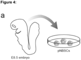
- the present application further describes an in vitro method for the direct differentiation of pluripotent human stem cells, particularly embryonic stem cells (ESCs) or induced pluripotent stem cells (iPSCs), comprising the step of culturing said pluripotent human stem cells in the presence of a GSK-3 inhibitor, particularly Chir99021; an Alk5 inhibitor, particularly Alk5 inhibitor II; and a hedgehog/smoothened agonist, particularly Purmorphamine.
- a GSK-3 inhibitor particularly Chir99021
- Alk5 inhibitor particularly Alk5 inhibitor II
- a hedgehog/smoothened agonist particularly Purmorphamine.
- the present application further describes an in vitro method for generation of induced neural border stem cells, comprising the step of culturing mature human cells in the presence of a mixture of transcription factors, wherein said mixture comprises the factors BRN2, SOX2, KLF4 and ZIC3, and wherein said culturing is performed in the presence of a GSK-3 inhibitor, particularly Chir99021; an Alk5 inhibitor, particularly Alk5 inhibitor II; and a hedgehog/smoothened agonist, particularly Purmorphamine.
- a GSK-3 inhibitor particularly Chir99021
- an Alk5 inhibitor particularly Alk5 inhibitor II
- a hedgehog/smoothened agonist particularly Purmorphamine.
- the present application further describes an in vitro method for the generation of neural border stem cells, comprising the step of culturing pluripotent human stem cells, particularly embryonic stem cells (ESCs) or induced pluripotent stem cells (iPSCs), in the presence of a GSK-3 inhibitor, particularly Chir99021; an Alk5 inhibitor, particularly Alk5 inhibitor II; and a hedgehog/smoothened agonist, particularly Purmorphamine.
- pluripotent human stem cells particularly embryonic stem cells (ESCs) or induced pluripotent stem cells (iPSCs)
- GSK-3 inhibitor particularly Chir99021
- Alk5 inhibitor particularly Alk5 inhibitor II
- a hedgehog/smoothened agonist particularly Purmorphamine.
- the present application further describes a nucleic acid sequence encoding BRN2, SOX2, KLF4 and ZIC3.
- the present application further describes a polycistronic vector encoding BRN2, SOX2, KLF4 and ZIC3.
- kits comprising at least two, more particularly all three components selected from: a GSK-3 inhibitor, particularly Chir99021; an Alk5 inhibitor, particularly Alk5 inhibitor II; and a hedgehog/smoothened agonist, particularly Purmorphamine.
- the present application further describes an isolated (induced) neural border stem cell line.
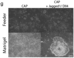
- the present application further describes an in vitro method of expanding the isolated (induced) neural border stem cell line as described in the present application, comprising the step of culturing cells from said isolated (induced) neural border stem cell line, particularly wherein said culturing is performed in the presence of proliferation-supporting cytokines, particularly Notch-signaling activating substances, particularly a substance selected from DLL1, DLL3 and DLL4, Jagged-1, and Jagged-2, more particularly from DLL4 and JAGGED-1.
- proliferation-supporting cytokines particularly Notch-signaling activating substances, particularly a substance selected from DLL1, DLL3 and DLL4, Jagged-1, and Jagged-2, more particularly from DLL4 and JAGGED-1.
- the present invention relates to an in vitro method for generating differentiated induced neural border stem cells, comprising the steps of (i) performing the in vitro method of the present invention for generating induced neural border stem cells, and (ii) culturing said induced neural border stem cells or cells from said isolated induced neural border stem cell line in the presence of differentiation factors.
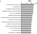
- the present application further describes isolated central nervous system primed neural progenitor cell line of the central nervous system lineage, particularly (i) wherein said cell line is of the same development status as primary neural progenitor cells obtainable from embryos of gestation week 8 to 12, and/or (ii) wherein said cell line is characterized by progenitor markers LONRF2, ZNF217, NESTIN, SOX1 and SOX2, particularly LONRF2 and ZNF217, and by being negative for MSX1, PAX3 and TFAP2, and/or (iii) wherein said cell line is characterized by epigenetically corresponding to mature human cells, particularly wherein said cell line has been obtained from said mature human cells in a direct reprogramming method according to the present invention.
- the present invention relates to an in vitro method for generating CNS progenitor cells, comprising the steps of (i) performing the in vitro method of the present invention for generating induced neural border stem cells, and (ii) culturing induced neural border stem cells or cells from said isolated induced neural border stem cell line in a medium comprising GSK-3 inhibitor Chir99021; aALK inhibitor SB431542; hedgehog/smoothened agonist Purmorphamine; bFGF; and LIF.
- the present application further describes an isolated central nervous system progenitor cell line, particularly wherein said cell line is characterized by epigenetically corresponding to mature human cells, particularly wherein said cell line has been obtained from said mature human cells in a direct reprogramming method according to the present invention.
- the present invention relates to an in vitro method of generating differentiated central nervous system progenitor cells as described in the present application, comprising the steps of (i) performing the in vitro method of the present invention for generating CNS progenitor cells, and (ii) culturing said central nervous system progenitor cells in the presence of differentiation factors.
- the present application further describes an isolated cell population having a radial glia type stem cell phenotype, particularly wherein said cell population is characterized by epigenetically corresponding to mature human cells, particularly wherein said cell population has been obtained from said mature human cells in a direct reprogramming method according to the present invention.
- the present application further describes an isolated differentiated (induced) neural border stem cell line of the neural crest lineage, particularly wherein said cell line is characterized by epigenetically corresponding to mature human cells, particularly wherein said cell line has been obtained from said mature human cells in a direct reprogramming method according to the present invention.
- the present invention relates to an in vitro method for generating neural crest progenitor cells, comprising the steps of (i) performing the in vitro method of the present invention for generating induced neural border stem cells, and (ii) culturing induced neural border stem cells or cells from said isolated induced neural border stem cell line for three days in the presence of GSK-3 inhibitor Chir99021; Alk5 inhibitor II; and BMP4; followed by culturing in the presence of GSK-3 inhibitor Chir99021, FGF8, IGF1 and DAPT.
- the present application further describes an isolated neural crest progenitor cell line, particularly wherein said cell line is characterized by epigenetically corresponding to mature human cells, particularly wherein said cell line has been obtained from said mature human cells in a direct reprogramming method according to the present invention.
- the present invention relates to an in vitro method of generating a differentiated neural crest progenitor cell line, comprising the step of (i) performing the in vitro method of the present invention for generating said neural crest progenitor cell line, and (ii) culturing cells from said neural crest progenitor cell line in the presence of differentiation factors.
- the present application further describes an isolated cell population having a neural border stem cell phenotype, particularly wherein said cell line is characterized by epigenetically corresponding to mature human cells, particularly wherein said cell population has been obtained from said mature human cells in a direct reprogramming method according to the present invention.
- the present application further describes an in vitro method for the generation of dopaminergic neurons, comprising the steps of (i) culturing (induced) neural border stem cells in a medium comprising a GSK-3 inhibitor, particularly Chir99021; an Alk5 inhibitor, particularly Alk5 inhibitor II; a hedgehog/smoothened agonist, particularly Purmorphamine, (ii) changing to a medium that is supplemented with FGF8 and a hedgehog/smoothened agonist, particularly Purmorphamine, on murine fibroblasts; (iii) culturing the cells in the medium according to (ii) for 7 days on a gelatinous protein mixture secreted by Engelbreth-Holm-Swarm mouse sarcoma cells (Matrigel), (iv) changing to a medium that is supplemented with a hedgehog/smoothened agonist, particularly Purmorphamine; (v) culturing the cells in the medium according to (iv) for 2 days, and (vi) changing the medium to maturation
- the present application further describes an in vitro method for the generation of motor neurons, comprising the steps of (i) culturing (induced) neural border stem cells in a medium comprising a GSK-3 inhibitor, particularly Chir99021; an Alk5 inhibitor, particularly Alk5 inhibitor II; a hedgehog/smoothened agonist, particularly Purmorphamine, on murine fibroblasts, (ii) changing to a medium that is supplemented with a hedgehog/smoothened agonist, particularly Purmorphamine; (iii) culturing the cells in the medium according to (ii) for 2 days on a gelatinous protein mixture secreted by Engelbreth-Holm-Swarm mouse sarcoma cells (Matrigel), (iv) changing to a medium that is supplemented with a hedgehog/smoothened agonist, particularly Purmorphamine, and all-trans retinoic acid; (v) culturing the cells in the medium according to (iv) for 7 days, and (vi) changing the medium
- the present application further describes an in vitro method for the generation of glutamatergic and gabaergic neurons, comprising the steps of (i) culturing (induced) neural border stem cells in a medium comprising a GSK-3 inhibitor, particularly Chir99021; an Alk5 inhibitor, particularly Alk5 inhibitor II; a hedgehog/smoothened agonist, particularly Purmorphamine, on murine fibroblasts, (ii) changing to a medium that is supplemented with a hedgehog/smoothened agonist, particularly Purmorphamine; (iii) culturing the cells in the medium according to (ii) for 7 days on a gelatinous protein mixture secreted by Engelbreth-Holm-Swarm mouse sarcoma cells (Matrigel), (iv) changing the medium to maturation medium comprising BDNF and GDNF; and (v) culturing the cells for 5 weeks in said maturation medium.
- a GSK-3 inhibitor particularly Chir99021
- Alk5 inhibitor particularly Alk5
- the present application further describes an in vitro method for the generation of astrocytes, comprising the steps of (i) culturing (induced) neural border stem cells in a medium comprising a GSK-3 inhibitor, particularly Chir99021; an Alk5 inhibitor, particularly Alk5 inhibitor II; a hedgehog/smoothened agonist, particularly Purmorphamine, on murine fibroblasts, (ii) changing to a medium that is supplemented with a hedgehog/smoothened agonist, particularly Purmorphamine; (iii) culturing the cells in the medium according to (ii) for 7 days on a gelatinous protein mixture secreted by Engelbreth-Holm-Swarm mouse sarcoma cells (Matrigel), (iv) changing the medium to a maturation medium comprising BDNF, GDNF and 1% FCS, and (v) culturing the cells for 5 weeks in said maturation medium.
- a GSK-3 inhibitor particularly Chir99021
- Alk5 inhibitor particularly Alk
- the present application further describes an in vitro method for the generation of oligodendrocytes, comprising the steps of (i) culturing (induced) neural border stem cells in a medium comprising a GSK-3 inhibitor, particularly Chir99021; an Alk5 inhibitor, particularly Alk5 inhibitor II; a hedgehog/smoothened agonist, particularly Purmorphamine, on murine fibroblasts, (ii) changing to a medium that is supplemented with a hedgehog/smoothened agonist, particularly Purmorphamine; (iii) culturing the cells in the medium according to (ii) for 7 days on a gelatinous protein mixture secreted by Engelbreth-Holm-Swarm mouse sarcoma cells (Matrigel), (iv) changing the medium to a medium comprising T3, IGF, Forskolin, PDGF, and EGF, (v) culturing the cells for 2 weeks in the medium according to (iv), (vi) changing the medium to a medium comprising T
- the present application further describes an in vitro method for the generation of neural crest-derived neurons, comprising the steps of (i) culturing (induced) neural border stem cells in a medium comprising a GSK-3 inhibitor, particularly Chir99021; an Alk5 inhibitor, particularly Alk5 inhibitor II; a hedgehog/smoothened agonist, particularly Purmorphamine, on murine fibroblasts, (ii) changing to a medium that is supplemented with a GSK-3 inhibitor, particularly Chir99021; an Alk5 inhibitor, particularly Alk5 inhibitor II, and BMP4; (iii) culturing the cells in the medium according to (ii) for 3 days, (iv) changing the medium to a medium comprising a GSK-3 inhibitor, particularly Chir99021; an FGF inhibitor, particularly SU5402, a Notch inhibitor, particularly DAPT, and NGF, (v) culturing the cells for 10 days in the medium according to (iv), (vi) changing the medium to a maturation medium comprising
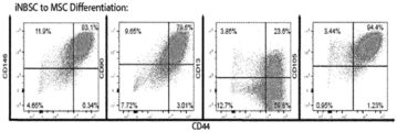
- the present application further describes an in vitro method for the generation of cells having a mesenchymal stem cell phenotype, comprising the steps of (i) culturing (induced) neural border stem cells in a medium comprising a GSK-3 inhibitor, particularly Chir99021; an Alk5 inhibitor, particularly Alk5 inhibitor II; a hedgehog/smoothened agonist, particularly Purmorphamine, on murine fibroblasts, (ii) changing to a medium that is supplemented with a GSK-3 inhibitor, particularly Chir99021; an Alk5 inhibitor, particularly Alk5 inhibitor II, and BMP4; (iii) culturing the cells in the medium according to (ii) for 3 days, (iv) changing the medium to a medium comprising a GSK-3 inhibitor, particularly Chir99021; FGF8, IGF, and a Notch inhibitor, particularly DAPT, (v) culturing the cells for 7 days in the medium according to (iv), (vi) changing the medium to a maturation medium compris
- the present application further describes an in vitro method for the generation of cells having a mesenchymal stem cell phenotype, comprising the steps of (i) seeding iNBSCs on plates coated with a gelatinous protein mixture secreted by Engelbreth-Holm-Swarm mouse sarcoma cells (Matrigel),(ii) culturing the cells in 4 ⁇ M Chir99028, 10 ng/ml BMP4 and 10 ⁇ M DAPT for 7 days; (iii) culturing the cells in basal medium containing 10 ng/ml bFGF and 10 ng/ml IGF-1 for at least 5 passages; (iv) stabilizing the cells by switching the cultures to mesenchymal stem cell medium and culturing for at least 2 passages.
- a gelatinous protein mixture secreted by Engelbreth-Holm-Swarm mouse sarcoma cells Matrigel
- culturing the cells in 4 ⁇ M Chir99028, 10
- the present application further describes an in vitro method for the differentiation of cells having a mesenchymal stem cell phenotype into adipocytes, comprising the steps of (i) generating said cells having a mesenchymal stem cell phenotype by the in vitro method as described in the present application, (ii) changing the medium to a mesenchymal induction medium comprising 10% FCS; (iii) culturing the cells in the medium according to (ii) for 5 days, (iv) changing the medium to a adipogenesis differentiation medium, and (v) culturing the cells in the medium according to (iv).
- the present application further describes an in vitro method for the differentiation of cells having a mesenchymal stem cell phenotype into chondrocytes, comprising the steps of (i) generating said cells having a mesenchymal stem cell phenotype by the in vitro method as described in the present application, (ii) changing the medium to a mesenchymal induction medium comprising10% FCS; (iii) culturing the cells in the medium according to (ii) for 5 days, (iv) changing the medium to a chondrocyte differentiation medium, and (v) culturing the cells in the medium according to (iv).
- the present application further describes an in vitro method for the differentiation of cells having a mesenchymal stem cell phenotype into smooth muscle cells, comprising the steps of (i) generating said cells having a mesenchymal stem cell phenotype by the in vitro method as described in the present application, (ii) changing the medium to a mesenchymal induction medium comprising10% FCS; and (iii) culturing the cells in the medium according to (ii) for 3 to 5 weeks.
- the present application further describes an in vitro method for the generation of a neural tube-like 3D culture, comprising the steps of (i) culturing (induced) neural border stem cells in a medium comprising a GSK-3 inhibitor, particularly Chir99021; an Alk5 inhibitor, particularly Alk5 inhibitor II; a hedgehog/smoothened agonist, particularly Purmorphamine, on murine fibroblasts, (ii) embedding of a single cell suspension of the cells cultured according to step (i) in a gelatinous protein mixture secreted by Engelbreth-Holm-Swarm mouse sarcoma cells (Matrigel) and adding a medium comprising SB and a hedgehog/smoothened agonist, particularly Purmorphamine; (iii) culturing said single cell suspension according to (ii) for 9 days; (iv) changing the medium to a medium comprising a GSK-3 inhibitor, particularly Chir99021, SB, a hedgehog/smoothened agonist, particularly Purmorphamine, and
- the present application further describes an in vitro method for the generation of a neural crest-like 3D culture, comprising the steps of (i) culturing (induced) neural border stem cells in a medium comprising a GSK-3 inhibitor, particularly Chir99021; an Alk5 inhibitor, particularly Alk5 inhibitor II; a hedgehog/smoothened agonist, particularly Purmorphamine, on murine fibroblasts, (ii) embedding of a single cell suspension of the cells cultured according to step (i) in a gelatinous protein mixture secreted by Engelbreth-Holm-Swarm mouse sarcoma cells (Matrigel) and adding a medium comprising a medium comprising a GSK-3 inhibitor, particularly Chir99021, an Alk5 inhibitor, particularly Alk5 inhibitor II, BMP4, and FGF2, and (iii) culturing for 12 days.
- a GSK-3 inhibitor particularly Chir99021
- an Alk5 inhibitor particularly Alk5 inhibitor II
- BMP4 Engelbreth-
- the present application further describes an in vitro method for the generation of cells representing a mutant phenotype, comprising the steps of (i) causing or allowing the modification of a gene sequence, the transcription or translation of a gene sequence, and/or of a protein encoded by a gene sequence of cells from an isolated (induced) neural border stem cell line as described in the present application, an isolated differentiated (induced) neural border stem cell line of the central nervous system lineage as described in the present application, an isolated central nervous system progenitor cell line as described in the present application, an isolated cell population having a radial glia type stem cell phenotype as described in the present application, an isolated differentiated (induced) neural border stem cell line of the neural crest lineage as described in the present application, an isolated neural crest progenitor cell line as described in the present application, an isolated cell population having a neural border stem cell phenotype as described in the present application, or cells generated by the method as described in the present application.
- the present application further describes an in vitro method for drug screening, comprising the step of exposing cells from an isolated (induced) neural border stem cell line as described in the present application, an isolated differentiated (induced) neural border stem cell line of the central nervous system lineage as described in the present application, an isolated central nervous system progenitor cell as described in the present application, an isolated cell population having a radial glia type stem cell phenotype as described in the present application, an isolated differentiated (induced) neural border stem cell line of the neural crest lineage as described in the present application, an isolated neural crest progenitor cell line as described in the present application, an isolated cell population having a neural border stem cell phenotype as described in the present application, cells generated by the method as described in the present application, or cells representing a mutant phenotype that are obtained according to the method as described in the present application to a drug substance.
- the present application further describes a pharmaceutical composition comprising cells from an isolated (induced) neural border stem cell line as described in the present application, an isolated differentiated (induced) neural border stem cell line of the central nervous system lineage as described in the present application, an isolated central nervous system progenitor cell line as described in the present application, an isolated cell population having a radial glia type stem cell phenotype as described in the present application, an isolated differentiated (induced) neural border stem cell line of the neural crest lineage as described in the present application, an isolated neural crest progenitor cell line as described in the present application, an isolated cell population having a neural border stem cell phenotype as described in the present application, cells generated by the method as described in the present application, or cells representing a mutant phenotype that are obtained according to the method as described in the present application.
- the present application further describes a cell from an isolated (induced) neural border stem cell line as described in the present application, an isolated differentiated (induced) neural border stem cell line of the central nervous system lineage as described in the present application, an isolated central nervous system progenitor cell line as described in the present application, an isolated cell population having a radial glia type stem cell phenotype as described in the present application, an isolated differentiated (induced) neural border stem cell line of the neural crest lineage as described in the present application, an isolated neural crest progenitor cell line as described in the present application, an isolated cell population having a neural border stem cell phenotype as described in the present application, cells generated by the method as described in the present application, or cells representing a mutant phenotype that are obtained according to the method as described in the present application for use in the treatment of a patient suffering from a neural disorder.
- the present application relates to an in vitro method for the direct reprogramming of mature human cells, comprising the step of culturing said mature human cells in the presence of a mixture of transcription factors, wherein said mixture comprises the factors BRN2, SOX2, KLF4 and ZIC3, and wherein said culturing is performed in the presence of a GSK-3 inhibitor, particularly Chir99021; an Alk5 inhibitor, particularly Alk5 inhibitor II; and a hedgehog/smoothened agonist, particularly Purmorphamine.
- a GSK-3 inhibitor particularly Chir99021
- an Alk5 inhibitor particularly Alk5 inhibitor II
- a hedgehog/smoothened agonist particularly Purmorphamine.
- the present invention relates to an in vitro method for generating induced neural border stem cells by the direct reprogramming of somatic human cells, comprising the step of culturing said somatic human cells in the presence of a mixture of transcription factors, wherein said mixture comprises the factors BRN2, SOX2, KLF4 and ZIC3, and wherein said culturing is performed in the presence of GSK-3 inhibitor Chir99021; Alk5 inhibitor II; and hedgehog/smoothened agonist Purmorphamine.
- the present application relates to an in vitro method for the direct differentiation of pluripotent human stem cells, particularly embryonic stem cells (ESCs) or induced pluripotent stem cells (iPSCs), comprising the step of culturing said pluripotent human stem cells in the presence of a GSK-3 inhibitor, particularly Chir99021; an Alk5 inhibitor, particularly Alk5 inhibitor II; and a hedgehog/smoothened agonist, particularly Purmorphamine.
- a GSK-3 inhibitor particularly Chir99021
- Alk5 inhibitor particularly Alk5 inhibitor II
- a hedgehog/smoothened agonist particularly Purmorphamine.
- the present application relates to an in vitro method for the direct differentiation of pluripotent non-human stem cells, particularly embryonic stem cells (ESCs) or induced pluripotent stem cells (iPSCs), comprising the step of culturing said pluripotent human stem cells in the presence of a GSK-3 inhibitor, particularly Chir99021; an Alk5 inhibitor, particularly Alk5 inhibitor II; and a hedgehog/smoothened agonist, particularly Purmorphamine.
- a GSK-3 inhibitor particularly Chir99021
- Alk5 inhibitor particularly Alk5 inhibitor II
- a hedgehog/smoothened agonist particularly Purmorphamine.
- the pluripotent non-human stem cells are pluripotent murine stem cells.
- hematopoietic stem and progenitor cells abbreviated HSPCs, collectively refers to hematopoietic stem cells (HSCs) and progenitors thereof, which are the first stages of differentiation of HSCs.
- the term “comprises” or “comprising” means “including, but not limited to”.
- the term is intended to be open-ended, to specify the presence of any stated features, elements, integers, steps or components, but not to preclude the presence or addition of one or more other features, elements, integers, steps, components, or groups thereof.
- the term “comprising” thus includes the more restrictive terms “consisting of” and “consisting essentially of”.
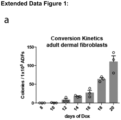
- ADFs human adult dermal fibroblasts
- the present application furthermore describes, that said culturing is performed in the additional presence of an inhibitor of monoamine-oxidase, particularly Tranylcypromine.
- said culturing is performed in the additional presence of monoamine-oxidase inhibitor Tranylcypromine.
- Tranylcypromine refers to an inhibitor of monoamine-oxidase (MAO) and of CYP2 enzymes: A6, C19, and D6 (CAPT).
- Tranylcypromine is only present during an induction phase, particularly in the first 12 to 21 days of said culturing, particularly in the first 12 to 16 days for pMSCs, and 17 to 21 days wherein said somatic cells are PBMCs.
- said somatic cells are selected from adult fibroblast cells; pancreas-derived mesenchymal stromal cells; and peripheral blood cells, particularly peripheral blood mononuclear cells.
- said step of culturing is performed on supportive feeder cells, particularly on murine fibroblast cells.
- a culture comprising said mature human cells is transduced with said factors BRN2, SOX2, KLF4 and ZIC3.
- said factors BRN2, SOX2, KLF4 and ZIC3 are comprised in a vector.
- said vector is a polycistronic vector.
- said vector is a doxycycline-inducible vector, particularly wherein said vector is vector pHAGE2-TetOminiCV-BRN22AKlf4-IRES-Sox2E2AZic3-W according to SEQ ID NO: 1
- said culturing is performed in the presence of doxycycline for at least 12 days after transduction, particularly for 12, 13, 14, 15 or 16 days.
- said method further comprises the step of clonally expanding single colonies.
- said colonies are expanded until a day selected from day 19 to day 24 after transduction.
- said vector further comprises loxP sites flanking the nucleic acid sequence encoding said factors BRN2, SOX2, KLF4 and ZIC3, particularly wherein said vector is vector pHAGE2-TetOminiCV-BRN22AKlf4-IRES-Sox2E2AZic3-W-loxp according to SEQ ID NO: 2.
- nucleic acid sequence encoding said factors BRN2, SOX2, KLF4 and ZIC3 comprised in said vector is excised by Cre recombinase.
- said in vitro method of the present invention comprises the step of transducing the cells with a plasmid encoding said Cre recombinase.
- said Cre recombinase is the Cherry-Cre recombinase.
- the present application further describes a nucleic acid sequence encoding BRN2, SOX2, KLF4 and ZIC3.
- the present application further describes a polycistronic vector encoding BRN2, SOX2, KLF4 and ZIC3.
- said vector is a polycistronic vector.
- said vector is a doxycycline-inducible vector, particularly wherein said vector is vector pHAGE2-TetOminiCV-BRN22AKlf4-IRES-Sox2E2AZic3-W according to SEQ ID NO: 1.
- said vector further comprises a loxP site, particularly wherein said vector is vector pHAGE2-TetOminiCV-BRN22AKlf4-IRES-Sox2E2AZic3-W-loxp according to SEQ ID NO: 2.
- the present application furthermore describes a kit comprising at least two, more particularly all three components selected from: a GSK-3 inhibitor, particularly Chir99021; an Alk5 inhibitor, particularly Alk5 inhibitor II; and a hedgehog/smoothened agonist, particularly Purmorphamine.
- a GSK-3 inhibitor particularly Chir99021
- an Alk5 inhibitor particularly Alk5 inhibitor II
- a hedgehog/smoothened agonist particularly Purmorphamine.
- said kit is further comprising an inhibitor of monoamine-oxidase, particularly Tranylcypromine.
- said kit further comprises one or more components selected from: a vector as used in a method of the present invention; supportive feeder cells, particularly murine fibroblast cells; and a plasmid encoding a Cre recombinase, particularly the Cherry-Cre recombinase.
- the present application furthermore describes an isolated (induced) neural border stem cell line.
- terms such as "(induced) neural border stem cell line” or “(induced) neural border stem cells” refer either to an induced neural border stem cell line/induced neural border stem cells obtained by reprogramming mature human cells, or to neural border stem cell line/neural border stem cells obtained by differentiating pluripotent stem cells, such as ESCs or iPSCs.
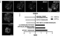
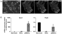
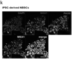
- said isolated (induced) neural border stem cell line is characterized by being positive both (i) for early neural markers, particularly PAX6, ASCL1, BRN2 and SOX1; and (ii) for stem cell markers, particularly NESTIN and SOX2.
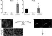
- said isolated (induced) neural border stem cell line is further characterized by expressing MSX1, ZIC1 and PAX3.
- said isolated (induced) neural border stem cell line is characterized by being additionally positive for HES5, SOX3 and HOXA2.
- said isolated (induced) neural border stem cell line has been generated by the in vitro method of the present invention.
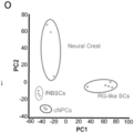
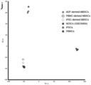
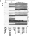
- said isolated (induced) neural border stem cell line has been generated by the in vitro method of the present invention, wherein the results of a principle component analysis of a comparative global gene expression analysis of the cell line (i) does not cluster, in the case of an isolated induced neural border stem cell line, with the results of a principle component analysis of a comparative global gene expression analysis of said mature human cells, and (ii) does not cluster with the results of a principle component analysis of a comparative global gene expression analysis of human induced pluripotent stem cells.
- the method further comprises expanding said induced neural border stem cells or said isolated induced neural border stem cell line, comprising the step of culturing said induced neural border stem cells or cells from said isolated induced neural border stem cell line, particularly wherein said culturing is performed in the presence of a substance selected from DLL1, DLL3 and DLL4, Jagged-1, and Jagged-2, more particularly from DLL4 and JAGGED-1.
- the formation of the nervous system initiates with the neural plate stage shortly after gastrulation.
- Signalling pathways such as WNTs, BMPs and SHH orchestrate the diversification of neural committed cells, which underlie the development of the various brain regions, spinal cord as well as the neural crest ( 13 , 14 , 15 ).
- said culturing is performed in the presence of GSK-3 inhibitor Chir99021; an Alk5 inhibitor II; and hedgehog/smoothened agonist Purmorphamine, particularly wherein said culturing is performed at 5% O 2 .
- said culturing is performed on a layer of supportive feeder cells, particularly on murine fibroblast cells.
- said culturing is performed for up to 40 passages.
- the present invention relates to an in vitro method for generating and differentiating induced neural border stem cells, comprising the steps of (i) performing the in vitro method of the present invention for generating induced neural border stem cells, and (ii) of culturing said induced neural border stem cells or cells from said isolated induced neural border stem cell line in the presence of differentiation factors.
- said (induced) neural border stem cells are differentiated to cells of a central nervous system lineage.
- said induced neural border stem cells are cultured in the presence of a GSK-3 inhibitor, particularly Chir99021; an ALK 4,5,7 inhibitor, particularly SB431542; and a hedgehog/smoothened agonist, particularly Purmorphamine, and wherein bFGF is added.
- a GSK-3 inhibitor particularly Chir99021
- an ALK 4,5,7 inhibitor particularly SB431542
- a hedgehog/smoothened agonist particularly Purmorphamine
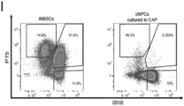
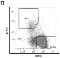
- said method is characterized by an increase in CD133 + / P75 neg cells.
- said method is characterized by an enrichment of mRNA for CNS-related genes, particularly PAX6, and by a downregulation of neural border-related genes, particularly TFAP2a and SOX10.
- the in vitro method further comprises the step of isolationg a central nervous system primed neural progenitor cell line of the central nervous system lineage.
- the present application further describes an isolated central nervous system primed neural progenitor cell line of the central nervous system lineage, particularly (i) wherein said cell line is of the same development status as primary neural progenitor cells obtainable from embryos of gestation week 8 to 12, and/or (ii) wherein said cell line is characterized by progenitor markers LONRF2, ZNF217, NESTIN, SOX1 and SOX2, particularly LONRF2 and ZNF217, and by being negative for MSX1, PAX3 and TFAP2, and/or (iii) wherein said cell line is characterized by epigenetically corresponding to mature human cells, particularly wherein said cell line has been obtained from said mature human cells in a direct reprogramming method according to the present invention.
- the present application further describes an isolated differentiated (induced) neural border stem cell line of the central nervous system lineage, which is generated by the method of the present invention.
- the present invention relates to an in vitro method for generating CNS progenitor cells, comprising the steps of (i) performing the in vitro method of the present invention for generating induced neural border stem cells, and (ii) culturing induced neural border stem cells or cells from said isolated induced neural border stem cell line, optionally after first differentiating (induced) neural border stem cells in a method of the present invention, in a medium comprising GSK-3 inhibitor Chir99021; ALK inhibitor SB431542; hedgehog/smoothened agonist Purmorphamine; bFGF; and LIF.
- the culture is maintained on a gelatinous protein mixture secreted by Engelbreth-Holm-Swarm mouse sarcoma cells (Matrigel).
- the present application further describes an isolated central nervous system progenitor cell line, particularly wherein said cell line is characterized by epigenetically corresponding to mature human cells, particularly wherein said cell line has been obtained from said mature human cells in a direct reprogramming method according to the present invention.
- the isolated central nervous system progenitor cell line is generated by the method of the present invention.
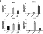
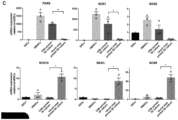
- the isolated central nervous system progenitor cell line generated by the method of the present invention is characterized by (ia) downregulation of FGFR3, HES5, ASCL1, CLDN5 und ZIC3, and (ib) maintained expression of PAX6, SOX1, SOX2 and NESTIN; in both cases when compared to (induced) neural border stem cells; and/or (ii) wherein said cell line is characterized by epigenetically corresponding to mature human cells, particularly wherein said cell line has been obtained from said mature human cells in a direct reprogramming method according to the method of the present invention.
- the present invention relates to an in vitro method of generating and differentiating central nervous system progenitor cells as described in the present application, comprising the steps of (i) performing the in vitro method of the present invention for generating CNS progenitor cells, and (ii) culturing said central nervous system progenitor cells in the presence of differentiation factors.
- said method is performed for seven weeks, followed by culturing in an expansion medium supplemented with bFGF, EGF and LIF.
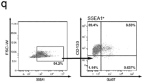
- the resulting cell population is positive for SSEA1, CD133 and Glutamate Aspartate Transporter (GLAST; SLC1A3).
- the present application further describes an isolated cell population having a radial glia type stem cell phenotype, particularly wherein said cell population is characterized by epigenetically corresponding to mature human cells, particularly wherein said cell population has been obtained from said mature human cells in a direct reprogramming method according to the present invention.
- said method comprises the steps of (i) seeding iNBSCs on plates coated with a gelatinous protein mixture secreted by Engelbreth-Holm-Swarm mouse sarcoma cells (Matrigel) and culturing in basal medium supplemented with 1 ⁇ M Purmorphamine and 10 ng/ml FGF8 for one week, (ii) culturing in basal medium with 1 ⁇ M Purmorphamine for one additional day; (iii) growing the cultures in basal medium containing 10 ng/ml BDNF and 10 ng/ml GDNF for 7 more weeks; and (iv) culturing the cells in radial glia medium, comprised of basal medium, 20 ng/ml bFGF, 20 ng/ml EGF and 10 ng/ml LIF.
- said isolated cell population is characterized by cells (ia) being triple-positive for SSEA1, CD133 and GLAST, (ib) strongly expressing the glial markers VIMENTIN, GFAP and GLAST; and (ic) being positive for the stem cell markers PAX6, NESTIN, SOX1 and BLBP; and/or (ii) wherein said cell line is characterized by epigenetically corresponding to mature human cells, particularly wherein said cell population has been obtained from said mature human cells in a direct reprogramming method according to the present invention.
- said (induced) neural border stem cells are differentiated to cells of a neural crest lineage.
- said (induced) neural border stem cells are cultured in the presence of a GSK-3 inhibitor, particularly Chir99021; an Alk5 inhibitor, particularly Alk5 inhibitor II; and BMP4.
- said method is characterized by an increase in P75 + /CD133 neg cells.
- said method is characterized by an enrichment of mRNA for neural crest associated genes, particularly SOX10 and AP2a.
- the present application describes an isolated differentiated (induced) neural border stem cell line of the neural crest lineage, particularly wherein said cell line is characterized by epigenetically corresponding to mature human cells, particularly wherein said cell line has been obtained from said mature human cells in a direct reprogramming method according to the present invention.
- said isolated differentiated (induced) neural border stem stell line of the neural crest lineage is generated by the in vitro method of the present invention.
- the present invention relates to an in vitro method for generating neural crest progenitor cells, comprising the steps (i) performing the in vitro method of the present invention for generating induced neural border stem cells, and (ii) culturing induced neural border stem cells or cells from said isolated induced neural border stem cell line for three days in the presence of GSK-3 inhibitor Chir99021; Alk5 inhibitor II; and BMP4; followed by culturing in the presence of GSK-3 inhibitor Chir99021, FGF8, IGF1 and DAPT.
- the present application further describes an isolated neural crest progenitor cell line, particularly wherein said cell line is characterized by epigenetically corresponding to mature human cells, particularly wherein said cell line has been obtained from said mature human cells in a direct reprogramming method according to the present invention.
- said isolated neural crest progenitor cell line is generated by the in vitro method of the present invention.
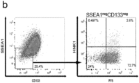
- the isolated neural crest progenitor cell line is characterized by (ia) the induction of migratory crest markers P75 and HNK1; (ib) a decrease in CD133 and SSEA1 levels; (ic) presence of SOX10, and (id) absence of PAX6; in each case when compared to (induced) neural border stem cells; and/or (ii) wherein said cell line is characterized by epigenetically corresponding to mature human cells, particularly wherein said cell line has been obtained from said mature human cells in a direct reprogramming method according to the present invention.
- the neural crest progenitor cell line is further characterized by KANK4, BGN, TFAP2A and SOX10 being among the strongest upregulated genes, with neural progenitor markers, in particular HES5 and PAX6, being downregulated, in each case when compared to (induced) neural border stem cells.
- the present invention relates to an in vitro method of generating and differentiating a neural crest progenitor cell line, comprising the step of (i) performing the in vitro method of the present invention for generating said neural crest progenitor cell line, and (ii) culturing cells from said neural crest progenitor cell line in the presence of differentiation factors.
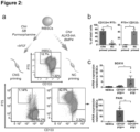
- said cells are obtained by performing the method comprising the steps of (i) seeding 1 ⁇ 10 5 / 6 Well iNBSCs on plates coated with a gelatinous protein mixture secreted by Engelbreth-Holm-Swarm mouse sarcoma cells (Matrigel) and growing in basal medium supplemented with 4 ⁇ M Chir99028, 5 ⁇ M Alk5 Inhibitor II and 10 ng/ml BMP4 for three days; (ii) growing the cells in a basal medium supplemented with 4 ⁇ M Chir99028, 10 ng/ml FGF8, 10 ng/ml IGF1 and 1 ⁇ M DAPT for five days; (iii) puryingin the NCSC-like cells by cell sorting for SSEA-1 neg CD133 neg P75 + HNK1 + ; in particular wherein all cultures are grown at 37°C, 5% CO 2 and 20% O 2 ..
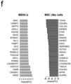
- the resulting cell population is positive for first markers shown in Figure 2f , particularly HNK1, P75, and SOX10, and negative for second markers shown in Figure 2f , particularly CD133, SSEA1, HES5, and PAX6.
- the present application further describes an isolated cell population having a neural border stem cell phenotype, particularly wherein said cell line is characterized by epigenetically corresponding to mature human cells, particularly wherein said cell population has been obtained from said mature human cells in a direct reprogramming method according to the present invention.
- said isolated cell population is obtained by a method of the present invention.
- said isolated cell population is characterized by (i) the induction of migratory crest markers P75 and HNK1; (ii) a decrease in CD133 and SSEA1 levels; (iii) the presence of SOX10, and (iv) the absence of PAX6; in each case when compared to (induced) neural border stem cells.
- said isolated cell population is further characterized by KANK4, BGN, TFAP2A and SOX10 being among the strongest upregulated genes, with neural progenitor markers, in particular HES5 and PAX6 being downregulated, in each case when compared to (induced) neural border stem cells.
- the present application further describes an in vitro method for the generation of dopaminergic neurons, comprising the steps of (i) culturing (induced) neural border stem cells in a medium comprising a GSK-3 inhibitor, particularly Chir99021; an Alk5 inhibitor, particularly Alk5 inhibitor II; a hedgehog/smoothened agonist, particularly Purmorphamine, (ii) changing to a medium that is supplemented with FGF8 and a hedgehog/smoothened agonist, particularly Purmorphamine, on murine fibroblasts; (iii) culturing the cells in the medium according to (ii) for 7 days on a gelatinous protein mixture secreted by Engelbreth-Holm-Swarm mouse sarcoma cells (Matrigel), (iv) changing to a medium that is supplemented with a hedgehog/smoothened agonist, particularly Purmorphamine; (v) culturing the cells in the medium according to (iv) for 2 days, and (vi) changing the medium to maturation
- the present application further describes an in vitro method for the generation of motor neurons, comprising the steps of (i) culturing (induced) neural border stem cells in a medium comprising a GSK-3 inhibitor, particularly Chir99021; an Alk5 inhibitor, particularly Alk5 inhibitor II; a hedgehog/smoothened agonist, particularly Purmorphamine, on murine fibroblasts, (ii) changing to a medium that is supplemented with a hedgehog/smoothened agonist, particularly Purmorphamine; (iii) culturing the cells in the medium according to (ii) for 2 days on a gelatinous protein mixture secreted by Engelbreth-Holm-Swarm mouse sarcoma cells (Matrigel), (iv) changing to a medium that is supplemented with a hedgehog/smoothened agonist, particularly Purmorphamine, and all-trans retinoic acid; (v) culturing the cells in the medium according to (iv) for 7 days, and (vi) changing the medium
- the present application further describes an in vitro method for the generation of glutamatergic and gabaergic neurons, comprising the steps of (i) culturing (induced) neural border stem cells in a medium comprising a GSK-3 inhibitor, particularly Chir99021; an Alk5 inhibitor, particularly Alk5 inhibitor II; a hedgehog/smoothened agonist, particularly Purmorphamine, on murine fibroblasts, (ii) changing to a medium that is supplemented with a hedgehog/smoothened agonist, particularly Purmorphamine; (iii) culturing the cells in the medium according to (ii) for 7 days on a gelatinous protein mixture secreted by Engelbreth-Holm-Swarm mouse sarcoma cells (Matrigel), (iv) changing the medium to maturation medium comprising BDNF and GDNF; and (v) culturing the cells for 5 weeks in said maturation medium.
- a GSK-3 inhibitor particularly Chir99021
- Alk5 inhibitor particularly Alk5
- the present application further describes an in vitro method for the generation of serotonergic neurons, comprising the steps of (i) culturing (induced) neural border stem cells in basal medium comprising 3 ⁇ M Chir99021, an Alk5 inhibitor, particularly 3 ⁇ M SB431542, and a hedgehog/smoothened agonist, particularly 1 ⁇ M Purmorphamine, on plates covered by a gelatinous protein mixture secreted by Engelbreth-Holm-Swarm mouse sarcoma cells (Matrigel) for one week, (ii) followed by culture in 3 ⁇ M Chir99028, an Alk5 inhibitor, particularly 3 ⁇ M SB431542, and a hedgehog/smoothened agonist, particularly 1 ⁇ M Purmorphamine, and 10 ng/ml FGF4 for another week, (iii) followed by switching to and a hedgehog/smoothened agonist, particularly 1 ⁇ M Purmorphamine, for two days, and (iv) subsequently growing the cells in neuronal maturation medium compris
- the present application further describes an in vitro method for the generation of astrocytes, comprising the steps of (i) culturing (induced) neural border stem cells in a medium comprising a GSK-3 inhibitor, particularly Chir99021; an Alk5 inhibitor, particularly Alk5 inhibitor II; a hedgehog/smoothened agonist, particularly Purmorphamine, on murine fibroblasts, (ii) changing to a medium that is supplemented with a hedgehog/smoothened agonist, particularly Purmorphamine; (iii) culturing the cells in the medium according to (ii) for 7 days on a gelatinous protein mixture secreted by Engelbreth-Holm-Swarm mouse sarcoma cells (Matrigel), (iv) changing the medium to a maturation medium comprising BDNF, GDNF and 1% FCS, and (v) culturing the cells for 5 weeks in said maturation medium.
- a GSK-3 inhibitor particularly Chir99021
- Alk5 inhibitor particularly Alk
- the present application further describes an in vitro method for the generation of oligodendrocytes, comprising the steps of (i) culturing (induced) neural border stem cells in a medium comprising a GSK-3 inhibitor, particularly Chir99021; an Alk5 inhibitor, particularly Alk5 inhibitor II; a hedgehog/smoothened agonist, particularly Purmorphamine, on murine fibroblasts, (ii) changing to a medium that is supplemented with a hedgehog/smoothened agonist, particularly Purmorphamine; (iii) culturing the cells in the medium according to (ii) for 7 days on a gelatinous protein mixture secreted by Engelbreth-Holm-Swarm mouse sarcoma cells (Matrigel), (iv) changing the medium to a medium comprising T3, IGF, Forskolin, PDGF, and EGF, (v) culturing the cells for 2 weeks in the medium according to (iv), (vi) changing the medium to a medium comprising T
- the present application further describes an in vitro method for the generation of neural crest-derived neurons, comprising the steps of (i) culturing (induced) neural border stem cells in a medium comprising a GSK-3 inhibitor, particularly Chir99021; an Alk5 inhibitor, particularly Alk5 inhibitor II; a hedgehog/smoothened agonist, particularly Purmorphamine, on murine fibroblasts, (ii) changing to a medium that is supplemented with a GSK-3 inhibitor, particularly Chir99021; an Alk5 inhibitor, particularly Alk5 inhibitor II, and BMP4; (iii) culturing the cells in the medium according to (ii) for 3 days, (iv) changing the medium to a medium comprising a GSK-3 inhibitor, particularly Chir99021; an FGF inhibitor, particularly SU5402, a Notch inhibitor, particularly DAPT, and NGF, (v) culturing the cells for 10 days in the medium according to (iv), (vi) changing the medium to a maturation medium comprising
- the present application further describes an in vitro method for the generation of cells having a mesenchymal stem cell phenotype, comprising the steps of (i) culturing (induced) neural border stem cells in a medium comprising a GSK-3 inhibitor, particularly Chir99021; an Alk5 inhibitor, particularly Alk5 inhibitor II; a hedgehog/smoothened agonist, particularly Purmorphamine, on murine fibroblasts, (ii) changing to a medium that is supplemented with a GSK-3 inhibitor, particularly Chir99021; an Alk5 inhibitor, particularly Alk5 inhibitor II, and BMP4; (iii) culturing the cells in the medium according to (ii) for 3 days, (iv) changing the medium to a medium comprising a GSK-3 inhibitor, particularly Chir99021; FGF8, IGF, and a Notch inhibitor, particularly DAPT, (v) culturing the cells for 7 days in the medium according to (iv), (vi) changing the medium to a maturation medium compris
- the present application further describes an in vitro method for the generation of cells having a mesenchymal stem cell phenotype, comprising the steps of (i) seeding iNBSCs on plates coated with a gelatinous protein mixture secreted by Engelbreth-Holm-Swarm mouse sarcoma cells (Matrigel),(ii) culturing the cells in 4 ⁇ M Chir99028, 10 ng/ml BMP4 and 10 ⁇ M DAPT for 7 days; (iii) culturing the cells in basal medium containing 10 ng/ml bFGF and 10 ng/ml IGF-1 for at least 5 passages; (iv) stabilizing the cells by switching the cultures to mesenchymal stem cell medium and culturing for at least 2 passages.
- a gelatinous protein mixture secreted by Engelbreth-Holm-Swarm mouse sarcoma cells Matrigel
- culturing the cells in 4 ⁇ M Chir99028, 10
- the present application further describes an in vitro method for the differentiation of cells having a mesenchymal stem cell phenotype into adipocytes, comprising the steps of (i) generating said cells having a mesenchymal stem cell phenotype by the in vitro method of the present invention, (ii) changing the medium to a mesenchymal induction medium comprising 10% FCS; (iii) culturing the cells in the medium according to (ii) for 5 days, (iv) changing the medium to a adipogenesis differentiation medium, and (v) culturing the cells in the medium according to (iv).
- the present application further describes an in vitro method for the differentiation of cells having a mesenchymal stem cell phenotype into chondrocytes, comprising the steps of (i) generating said cells having a mesenchymal stem cell phenotype by the in vitro method as described in the present application, (ii) changing the medium to a mesenchymal induction medium comprising10% FCS; (iii) culturing the cells in the medium according to (ii) for 5 days, (iv) changing the medium to a chondrocyte differentiation medium, and (v) culturing the cells in the medium according to (iv).
- the present application further describes an in vitro method for the differentiation of cells having a mesenchymal stem cell phenotype into smooth muscle cells, comprising the steps of (i) generating said cells having a mesenchymal stem cell phenotype by the in vitro method as described in the present application, (ii) changing the medium to a mesenchymal induction medium comprising10% FCS; and (iii) culturing the cells in the medium according to (ii) for 3 to 5 weeks.
- the present application further describes an in vitro method for the generation of a neural tube-like 3D culture, comprising the steps of (i) culturing (induced) neural border stem cells in a medium comprising a GSK-3 inhibitor, particularly Chir99021; an Alk5 inhibitor, particularly Alk5 inhibitor II; a hedgehog/smoothened agonist, particularly Purmorphamine, on murine fibroblasts, (ii) embedding of a single cell suspension of the cells cultured according to step (i) in a gelatinous protein mixture secreted by Engelbreth-Holm-Swarm mouse sarcoma cells (Matrigel) and adding a medium comprising SB and a hedgehog/smoothened agonist, particularly Purmorphamine; (iii) culturing said single cell suspension according to (ii) for 9 days; (iv) changing the medium to a medium comprising a GSK-3 inhibitor, particularly Chir99021, SB, a hedgehog/smoothened agonist, particularly Purmorphamine, and
- the present application further describes an in vitro method for the generation of a neural crest-like 3D culture, comprising the steps of (i) culturing (induced) neural border stem cells in a medium comprising a GSK-3 inhibitor, particularly Chir99021; an Alk5 inhibitor, particularly Alk5 inhibitor II; a hedgehog/smoothened agonist, particularly Purmorphamine, on murine fibroblasts, (ii) embedding of a single cell suspension of the cells cultured according to step (i) in a gelatinous protein mixture secreted by Engelbreth-Holm-Swarm mouse sarcoma cells (Matrigel) and adding a medium comprising a medium comprising a GSK-3 inhibitor, particularly Chir99021, an Alk5 inhibitor, particularly Alk5 inhibitor II, BMP4, and FGF2, and (iii) culturing for 12 days.
- a GSK-3 inhibitor particularly Chir99021
- an Alk5 inhibitor particularly Alk5 inhibitor II
- BMP4 Engelbreth-
- the present application further describes an in vitro method for the generation of cells representing a mutant phenotype, comprising the steps of (i) causing or allowing the modification of a gene sequence, the transcription or translation of a gene sequence, and/or of a protein encoded by a gene sequence of cells from an isolated (induced) neural border stem cell line as described in the present application, an isolated differentiated (induced) neural border stem cell line of the central nervous system lineage as described in the present application, an isolated central nervous system progenitor cell line as described in the present application, an isolated cell population having a radial glia type stem cell phenotype as described in the present application, an isolated differentiated (induced) neural border stem cell line of the neural crest lineage as described in the present application, an isolated neural crest progenitor cell line as described in the present application, an isolated cell population having a neural border stem cell phenotype as described in the present application, or cells generated by the method as described in the present application.
- step (i) is performed by using gene editing, particularly by using a CRISPR Cas9-mediated knockout.
- said step (i) is performed by using protein inhibitors, particularly by using antibodies directed against a protein.
- the present application further describes an in vitro method for drug screening, comprising the step of exposing cells from an isolated (induced) neural border stem cell line as described in the present application, an isolated differentiated (induced) neural border stem cell line of the central nervous system lineage as described in the present application, an isolated central nervous system progenitor cell as described in the present application, an isolated cell population having a radial glia type stem cell phenotype as described in the present application, an isolated differentiated (induced) neural border stem cell line of the neural crest lineage as described in the present application, an isolated neural crest progenitor cell line as described in the present application, an isolated cell population having a neural border stem cell phenotype as described in the present application, cells generated by the method as described in the present application, or cells representing a mutant phenotype that are obtained according to the method as described in the present application to a drug substance.
- the in vitro method further comprises the determination of one of more factors that are potentially affected by interaction with said drug substances.
- the present application further describes a pharmaceutical composition comprising cells from an isolated (induced) neural border stem cell line as described in the present application, an isolated differentiated (induced) neural border stem cell line of the central nervous system lineage as described in the present application, an isolated central nervous system progenitor cell line as described in the present application, an isolated cell population having a radial glia type stem cell phenotype as described in the present application, an isolated differentiated (induced) neural border stem cell line of the neural crest lineage as described in the present application, an isolated neural crest progenitor cell line as described in the present application, an isolated cell population having a neural border stem cell phenotype as described in the present application, cells generated by the method as described in the present application, or cells representing a mutant phenotype that are obtained according to the method as described in the present application.
- the present application further describes a cell from an isolated (induced) neural border stem cell line as described in the present application, an isolated differentiated (induced) neural border stem cell line of the central nervous system lineage as described in the present application, an isolated central nervous system progenitor cell line as described in the present application, an isolated cell population having a radial glia type stem cell phenotype as described in the present application, an isolated differentiated (induced) neural border stem cell line of the neural crest lineage as described in the present application, an isolated neural crest progenitor cell line as described in the present application, an isolated cell population having a neural border stem cell phenotype as described in the present application, cells generated by the method as described in the present application, or cells representing a mutant phenotype that are obtained according to the method as described in the present application for use in the treatment of a patient suffering from a neural disorder.
- iNBSC Neural Border Stem Cell population
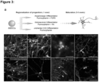
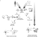
- iNBSCs Upon differentiation, iNBSCs pass through successive developmental stages and can give rise to either (1) CNS-primed progenitors, radial glia-type stem cells, dopaminergic and serotonergic neurons, motoneurons, astrocytes and oligodendrocytes or (2) neural crest lineage including peripheral neurons.
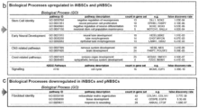
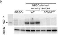
- CRISPR/Cas9 edited iNBSCs carrying a mutant SCN9a gene, can be expanded and differentiated into sensory neurons. These show impaired functional properties mimicking a human pain syndrome.
- iNBSCs open novel possibilities for patient-specific and mechanism-based research, which can be associated to high throughput drug screens or cell based regenerative medicine.
- the goal of this study was to overcome these limitations of the prior art and to explore the possibility of reprogramming human adult somatic cells into early, defined and self-renewing neural progenitors with broad but specific differentiation potential.
- the formation of the nervous system initiates with the neural plate stage shortly after gastrulation.
- Signalling pathways such as WNTs, BMPs, and SHH orchestrate the diversification of neural committed cells, which underlie the development of the various brain regions, spinal cord as well as the neural crest ( 13 , 14 , 15 ).
- overexpression of stage-specific transcription factors in combination with adequate signalling cues provided by the growth medium, might allow for the direct reprogramming of adult somatic cells to early embryonic neural progenitors with stem cell features including self-renewal and multipotency.
- ADFs human adult dermal fibroblasts
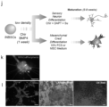
- ADFs were transduced with a polycistronic, doxycycline (DOX)-inducible vector containing BKSZ, and subsequently cultured in the presence of CAPT.
- DOX doxycycline
- Fig. 1a,b a modified version containing a loxP site
- Converted clonal neural cell lines derived from ADFs, FPFs and PBMCs could be cultured on a layer of supportive feeders and in medium containing Chir99021, Alk5 Inhibitor II and Purmorphamine (CAP) for more than 40 passages (> 7 months), without loss of proliferative potential and maintenance of high expression of early neural markers such as PAX6 and SOX1 ( Fig. 1d , Extended Data Fig. 1i ).
- the cultures were homogenously positive for the stem cell markers NESTIN and SOX2, and expressed also MSX1, ZIC1 and PAX3, suggesting a neural border-like identity of the converted cells ( Fig. 1d , Extended Data Fig 1i ).
- iNBSCs i nduced N eural B order S tem C ells
- iNBSCs iNBSCs from ADFs and pluripotent stem cells
- Fig. 1g the neural stem cell markers NESTIN, PAX6 and HES5, as well as neural border and neural crest markers including MSX1, TPAP2a, SOX3 and HOXB2
- pluripotency markers OCT4/ POU5F1 and NANOG
- Fig. 1g Extended Data Fig. 1l
- mesoderm and fibroblast markers i.e.
- BRACHYURY/ T, THY1 were strongly down-regulated, some residual expression of COL3A1 was detected in ADF- but not PBMC-derived iNBSCs, which might be indicative of some epigenetic memory (Extended Data Fig. 1l ).
- the methylation pattern of cells is rather stably associated with the identity and fate of the cell and is independent on certain cellular states (i.e. actively dividing or quiescent).
- ADF-converted and iPSC- differentiated (i)NBSCs clustered closely, confirming high similarity also at the level of the methylome.
- the comparison of ADFs with ADF-derived and iPSC-differentiated (i)NBSCs resulted in 9067 differentially methylated promoter regions (FDR adjusted p-val ⁇ 0.05).
- Gene set enrichment analysis of hypomethylated promoter sites of iNBSCs compared to ADFs revealed an enrichment for gene ontology terms such as pattern specification, skeletal system development and neuron fate commitment. These data are consistent with the reprogramming into an NBSC identity also at the epigenetic level ( Fig 1i ).
- the analysis of hypermethylated promoter sites revealed processes related to fibroblast identity such as cell-cell adhesion via plasma membrane adhesion molecules, calcium dependent cell-cell adhesion, innate immune response and collagene fibril organisation (Extended Data Fig 1m ), which became apparently silenced. Together, these results demonstrate robust changes in the methylation and transcriptional landscape after reprogramming of ADFs into iNBSCs.
- iNBSCs that are characterized by high expression of neural border markers, sustained self-renewal capacity and the acquisition of an expression and epigenomic landscape consistent with a neural border-like phenotype.
- iNBSCs give rise to central nervous system and neural crest progenitors
- iNBSCs represent a developmental stage prior to the separation into the CNS and NC lineage.
- iNBSCs clones were cultured either in the presence of (1) Chir99021, SB431542 (ALK 4,5,7 Inhibitor) and Purmorphamine (CSP) followed by addition of bFGF to induce CNS differentiation or (2) medium containing Chir99021, Alk-5 Inhibitor and BMP4 (CAB) to promote differentiation towards a NC fate ( Fig 2a ).
- iNBSC-lines had been established from cultures seeded at clonal density (see also above), iNBSC clones showed a fraction of P75 + /CD133 neg cells in CAP medium, indicating that some spontaneous differentiation also occurs under maintenance condition.
- Quantitative PCRs on sorted CD133 + / P75 neg or P75 + / CD133 neg confirmed the enrichment for CNS related genes (e.g. PAX6) or NC associated genes (e.g. SOX10 and AP2a), respectively ( Fig. 2c , Extended Data Fig. 2a ).
- CNS related genes e.g. PAX6
- NC associated genes e.g. SOX10 and AP2a
- NC stem cell NC stem cell
- NC-primed cells could also be differentiated to phenotypically defined CD105 + /CD44 + /CD13 + /CD90 + /CD146 + mesenchymal stem cells (Extended Data Fig. 2e ).
- the analysis of gene expression profiles of MSC-like cells compared to iNBSCs demonstrated an up-regulation of genes involved in maintenance and differentiation of MSCs such as COL1A1, GREMLIN1 and TAGLN and was further supported by gene ontology analysis implying biological processes such as extracellular matrix organization and skeletal system development (Extended Data Fig. 2f , g ).
- extended Data Fig. 2f , g Extended Data Fig. 2f , g .
- NPCs While iNBSCs were positive for CD133 and showed intermediate levels of P75 (CD133 + P75 int ), NPCs did not express significant level of P75 (CD133 + P75 neg ) (Extended Data Fig. 2i, j ). Interestingly, the neural multipotent progenitor marker CD184 was strongly and homogenously expressed in iNBSCs, while NPCs showed reduced levels, ranging from low to intermediate expression (Extended Data Fig 2k ). To explore whether the differentiation of iNBSCs into NPCs was reversible, NPCs were seeded on feeders in CAP medium (without FGF2 and LIF).
- NPCs remained negative for P75 and also retained low expression levels of CD184/CXCR4 (Extended Data Fig 2l , m ) no evidence for back-differentiation into iNBSCs could be obtained.
- NPCs also harbour the potential to give rise to the NC lineages.
- the NPCs showed only a very small fraction of P75 + / CD133 neg cells (Extended Data Fig. 2n , o ), indicating that NPCs represent neural progenitors with robust CNS-priming (cNPCs) .
- iNBSCs expressed the anterior hindbrain markers GBX2, IRX3, HOXA2 and HOXB2, cNPCs preferentially expressed GBX2, EN1, FGF8 and PAX2 (Extended Data Fig 2p ).
- EMX1, FOXG1 forebrain markers
- OTX2 farnesoid endothelial growth factor
- cNPCs expressed the ventral marker NKX6.1 but not NKX2.2, while dorsal markers such as MSX1, PAX3 and PAX7 were much lower expressed compared to iNBSCs (Suppl. Fig 2 h , p ).
- the transcription factor landscape of iNBSCs is mostly compatible with a dorsal anterior hindbrain fate, while cNPCs exhibit a ventral mid-hindbrain identity.
- iNBSCs, cNPCs, RG-like SCs and NCSCs clustered separately and showed an increase in distance with respect to the NBSC-state.
- these data indicate that the iNBSC population behaves in a multipotent manner as these embryonic neural progenitors that can be directed towards a CNS or NC fate.
- a CNS or NC fate Notably, within each linage even more restricted progenitors that correspond to previously defined development stages can be generated and stabilized.
- iNBSCs To examine the differentiation potential of iNBSCs towards mature cell types, they were subjected to specific differentiation cues. To derive neurons with CNS identity, cells were first cultured in presence of Purmorphamine for 7 days, supplemented with either FGF8 (dopaminergic neurons), all-trans retinoic acid (ATRA) (motor neurons) or without additional cues (GABAergic/ glutamatergic differentiation). Subsequently cultures were switched to maturation medium and analyzed five weeks later ( Fig 3a ).
- FGF8 dopaminergic neurons
- ATRA all-trans retinoic acid
- GABAergic/ glutamatergic differentiation GABAergic/ glutamatergic differentiation
- iNBSCs displayed a high neurogenic potential and differentiated rapidly upon removal of CAP medium.
- Immunofluorescence staining and qPCR analysis revealed the presence of glutamatergic (vGLUT2), dopaminergic (FOXA2, TH, EN1), motor neurons (CHAT, ISLET1) and GABAergic neurons (GABA, GAD67) ( Fig 3a , b; Extended Data Fig. 3a ).
- vGLUT2 glutamatergic
- FOXA2, TH, EN1 dopaminergic
- CHAT motor neurons
- GABAergic neurons GABA, GAD67
- iNBSCs Three-dimensional and organoid cultures have been recently appreciated as a valuable system to spatially model neural development and recapitulate the underlying differentiation processes in vitro ( 25 , 26 , ( 27 , 28 ).
- iNBSCs were first cultured in SP for nine days, and subsequently switched to CSP supplemented with bFGF.
- single iNBSCs efficiently gave rise to three-dimensional spheres, some of which developed a central lumen during the differentiation process resembling the morphology of a neural tube ( Fig. 3e and see below).
- NC-primed 3D cultures confirmed down-regulation of PAX6 and SOX1 and concomitant induction of SOX10 and SOX9. Furthermore there was an up-regulation of the crest-associated EMT marker SNAIL (Extended Data Fig. 3c ). Thus, there was a clear divergence in phenotype and gene expression of iNBSC-derived 3D cultures grown in CNS- or NC-priming conditions.
- GFP-positive cells included NeuN-positive neurons, GFAP-positive astrocytes and MBP-positive oligodendrocytes demonstrating the differentiation potential of iNBSCs to all three major neural linages and survival of the progeny in vivo ( Fig. 3g , Extended Data Fig.3e ).
- iNBSC-derived neurons exhibit mature functional properties in vivo
- whole cell patch-clamp analysis of transplanted neurons in acute brain slices was performed. Indeed, GFP-positive neurons exhibited repetitive action potentials upon depolarizing current injection (6 cells from 3 mice; Fig. 3h ). The neuronal identity was further confirmed by morphological reconstructions of biocytin-filled patched neurons ( Fig. 3i ).
- iNBSCs To investigate the potential of iNBSCs towards differentiated NC cell types, they were first cultured in NC induction medium and then switched to neural ( 29 ) or mesenchymal maturation media. After four weeks differentiation was initiated, BRN3a/PERIPHERIN/NAV1.7 triple positive neurons, characteristic for peripheral sensory neurons, could be detected ( Fig 3j , Extended Data Figure 3f ). In contrast, differentiation of iNBSCs in mesenchymal differentiation media produced smooth muscle actin (SMA) positive cells, alcian blue positive cartilage formation and oil red positive fat cells ( Fig 3k ).
- SMA smooth muscle actin
- Fig 3k oil red positive fat cells
- iNBSC primary Neural Border Stem Cells
- pNBSCs upon single cell sorting pNBSCs showed an up to 10-times higher clonogenic capacity compared to cultured primary radial glia stem cells isolated from E13.5 stage embryos (Extended Data Fig 4f ) ( 1 ).
- the culture of pNBSC was also feeder-dependent, although they differentiated more rapidly in the absence of feeders compared to human iNBSCs. The rapid differentiation in the absence of feeders could be partially halted when pNBSCs were cultured on MG in the presence of the Notch-ligands DII4 and Jagged1 (Extended Data Fig. 4g ). This indicates that the feeder-mediated maintenance of the NBSC niche is at least in part mediated by Notch signalling.
- mouse pNBSCs show a similar regionalization as human iNBSCs, a scenario that is compatible with the notion that they share a dorsal mid-hindbrain to anterior hindbrain identity.
- NBSCs have the potential to give rise to CNS, RG-like stem cells and NC progeny, and thus constitute self-renewing, multipotent neural progenitors, akin to human iNBSCs obtained by direct reprogramming.
- pNBSCs were differentiated and subsequently FACS-sorted for RG-like SCs (Ssea1 + , Glast + ) or NC (P75 + , Glast-, Ssea1 - ).
- Principal component analysis revealed that pNBSCs and their RG-like and NC progeny clustered separately ( Fig. 4j ).
- RG-like SC isolated from E13.5 stage embryos showed great overlap with RG-like SCs derived from pNBSC, confirming their identity ( Fig. 4j ).
- pNBSCs showed a specific up-regulation of progenitor markers such as Hes5, Sox1 and Lin28, RG-like cells expressed glia-related markers such as Olig 2, Omg and S100b ( Fig. 4k ).
- the NC progeny on the other hand showed up-regulation of NC-associated genes such as Dlx2 and Pdgf receptors and down-regulation of neural progenitor and glia markers respectively ( Fig. 4k ).
- neither pNBSCs nor their progeny showed expression of pluripotency associated markers such as Nanog, Oct4 or Utf1 ( Fig. 4k ).
- mouse embryo derived pNBSCs are highly similar to directly reprogrammed human iNBSCs. This is reflected by the requirement of the same signalling cues for maintenance, early marker expression, stable long-term proliferation and the potential to recapitulate early neural development by giving rise to naive as well as mature progeny of the CNS and NC lineage.
- pNBSCs represent a so far unidentified neural progenitor cell population active during early mouse brain organogenesis and also representing a unique stable pre-rosette population with sustained expansion potential.
- Human iNBSCs and mouse pNBSCs share many characteristics, including long-term self-renewal capacity, expression of specific markers, and developmental potential. To further extend the comparison to the molecular expression landscape, we analysed the global gene expression profile of mouse pNBSCs and human iNBSCs.
- This comprises members of the Notch signalling pathway such as HES5, DLL1 and NOTCH1 as well a prominent neural transcription factors such as SOX2, ASCL1 and PAX6.
- the network comprised two of the four factors used for iNBSC reprogramming, such as POU3f2 (BRN2), SOX2 as well as the ZIC family member ZIC2.
- iNBSCs The self-renewal and extensive differentiation potential of iNBSCs make them a powerful tool to model genetically based human neuronal syndromes.
- CRISPR/Cas9 mediated gene editing to mutate the voltage-gated sodium-channel Nav1.7, a nociceptor encoded by the SCN9a gene. This channel is expressed in the peripheral nervous system and while gain of function mutations in SCN9a lead to primary erythromelalgie and paroxysmal pain disorder, loss of function mutations result in congenital insensitivity to pain.
- iNBSCs represent a novel bona fide embryonic, multipotent and self-renewing progenitor, that can be derived either by direct reprogramming of adult somatic cells or by isolation from primary embryonic brain tissue ( Fig. 6f ).
- iNBSCs by over-expression of a specific set of transcription factors (BKSZ) with chemically defined, serum-free culture medium and a collection of small molecules. They are transgene-independent, show sustained self-renewal, high clonogenicity and a neural border-like expression signature.
- BKSZ transcription factors
- iNBSCs can differentiate into mature progeny of the CNS and NC and also recapitulate early neural development when embedded into a 3D matrix.
- iNBSCs represent a neural progenitor with well-defined molecular and functional features.
- NBSCs can also be generated by directed differentiation starting from pluripotent stem cells.
- SHH and Notch ligands Jagged-1 and DII4 has been reported to stabilize rosette-type NSC (R-NSCs), i.e. early progenitors capable of giving rise to CNS and NC progeny ( 37 ).
- R-NSCs rosette-type NSC
- the R-NSC state is transient and long-term culture results in a heterogeneous population.
- the use of small molecules has opened new possibilities to stabilize expandable NPCs.
- (i)NBSCs present a thus far unidentified self-renewing neural progenitor with broadest differentiation capacity.
- the term 'neural progenitor' has been used ambiguously to describe a variety of populations that differ in developmental stage, region of origin and expansion capacity ( 37 , 3 , 4 , 6 ). Therefore a molecular and functional comparison to the corresponding in vivo cell type represents an essential step for the proper characterization, especially in case of ectopically reprogrammed cells.
- iNBSCs are highly clonogenic, proliferative and multipotent and can be derived from any individual, healthy or diseased. These characteristics make them an ideal cell population for gene editing approaches.
- CRISPR/Cas9-mediated loss-of-function-mutations in SCN9a render sensory neurons insensitivity to ⁇ , ⁇ -Methylene-ATP, a P2RX3 agonist involved in inflammatory pain.
- the pathophysiological response in vitro mimics the functional deficit of SCN9a in patients with congenital insensitivity to pain, who do not experience any modality of pain except for neuropathic pain ( 38 ).
- iNBSCs in conjunction with CRISPR/Cas9 gene editing, serve as an ideal tool to model and/ or repair genetic defects underlying neural diseases.
- (i)NBSCs and progenitors derived thereof can be generated from various somatic cells, thus offering alternative approaches in obtaining defined and scalable neural progenitors.
- This strategy not only avoids the generation of pluripotent and cancer prone iPSCs, but also shortcuts inefficient differentiation steps from iPSCs into neural progenitors.
- iNBSCs when used in conjunction with genome editing, constitute a useful tool to study both neural development and, as exemplified in this study, mature cell types with a disease related phenotype.
- (i)NBSCs might proof valuable to model and/ or repair genetic defects underlying neural diseases such as for example Parkinson's disease, ataxia or neuropathic diseases and will eventually open new options for regenerative medicine.
- Example 1 Virus production and reprogramming into induced neural border stem cells
- plasmids encoding for pHAGE2-TetO-BKSZ-flox or m2RTTA 39 , Addgene plasmid #20342 were transfected together with helper plasmids psPAX2 (Addgene plasmid #12260) and pMD2.G (Addgene plasmid #12259) into 293FT cell lines (Life technologies) as described elsewhere ( 40 ).
- Lentiviral supernatants containing BKSZ-flox and m2RTTA were mixed in a ratio of 2:1, supplemented with 5 ⁇ g/ml polybrene (Sigma) and used freshly for transduction of 8 ⁇ 10 5 / 6 Well primary ADF and pancreas fibroblasts.
- PBMCs were transduced in QBSF-60 Stem Cell Medium (Quality Biological) containing 50 ⁇ g/ml Ascorbic Acid (Sigma), 50 ng/ml SCF (R&D Systems), 10 ng/ml IL-3 (R&D Systems), 2 U/ml EPO (R&D Systems), 40 ng/ml IGF-1 (Peprotech), 1 ⁇ M Dexamethasone (Sigma) and 5 ⁇ g/ml polybrene (for details see 41 ). The other day 2 ⁇ 10 5 transduced PBMCs were transferred onto one well of a 6 Well plate, coated with inactivated MEFs and reprogramming was initiated one day thereafter.
- QBSF-60 Stem Cell Medium Quality Biological
- DMEM/F-12 Glutamax (Life Technologies) containing 64 ⁇ g/ml L-Ascorbic acid 2-phosphate (LAAP, Sigma), ITS-X (1:100, Life Technologies), NEAA (1:100, Life Technologies), 2% FCS, 8% Serum Replacement (Life Technologies) supplemented with 4 ⁇ M Chir99021 (Sigma), 5 ⁇ M Alk5 Inhibitor II (Enzolifesciences), 0.5 ⁇ M Purmorphamine (Sigma), 5 ⁇ M Tranylcypromine (Sigma) and 1 ⁇ g/ml Doxycycline (Sigma) and incubated at 37°C in 5% O 2 and 5% CO 2 .
- NBSC maintenance medium is composed of DMEM/ F-12 Glutamax (for iNBSCs) or Adv. DMEM/ F12 Glutamax (for pNBSCs) and Neurobasal Medium (1:1) containing 64 ⁇ g/ml LAAP, N2 Supplement (1:100, Life Technologies), B27 without RA (1:50, Life Technologies), 18 ⁇ g/ml Albumax I (Life Technologies) and Glutamax (1:200, Life Technologies), referred to as 'basal medium', and 4 ⁇ M Chir99028, 5 ⁇ M Alk5 Inhibitor II and 0.5 ⁇ M Purmorphamine.
- iNBSC maintenance medium a layer of inactivated mouse embryonic fibroblasts (feeder) at 37°C in 5% O 2 and 5% CO 2 .
- Established iNBSC lines were tested for mycoplasma, Squirrel monkey retrovirus, and Epstein-Barr virus contamination prior to analysis. iNBSC lines were routinely split after 4-5 days by treatment with Accutase (Life Technologies) and transferred onto fresh feeders.
- Example 3 Derivation of human PBMCs and ADFs
- PBMCs were derived from healthy male donors (Age 24-30) and isolated using the Ficoll gradient procedure. PBMCs were used either directly or frozen in 90% Serum Replacement/ 10% DMSO as previously described (for details see 41 ).
- Human Primary Pancreatic Fibroblast were obtained from Vitro Biopharma and cultured in MSC-Gro TM medium (Vitro Biopharma) supplemented with 10% FCS. Cells were expanded for 4 passages prior to use. Cells were grown at 37°C in 5% CO 2 and 20% O 2 .
- Example 4 Culture of human iPSCs and differentiation into NBSCs
- Human iPSCs were routinely grown on feeder cells in DMEM/ F12, 64 ⁇ g/mL LAAP, 1x NEAA, 15% Serum Replacement and 20 ng/ml bFGF. Prior to the differentiation into NBSCs, human IPSCs were cultured for one passage on Matrigel TM -coated plates in basal medium supplemented with 20 ng/ml bFGF and 1 ng/ml TGFß1.
- iPSCs When cells reached confluency, iPSCs were harvested by treatment with 1 mg/ml Collagenase II (Life Technologies) and cell clumps were transferred into uncoated petri dishes containing NBSC maintenance medium. At this point the culture was switched from 20% O 2 to 5% O 2 and subsequently grown under this condition. After five days spheres were plated onto feeder cells and grown in NBSC maintenance medium. The other day, attached spheres were split by treatment with Accutase and 3 ⁇ 10 4 cells were seeded on feeder containing 10 cm dishes. Single colonies were manually picked and clonal lines established.
- NBSCs Differentiation towards cNPCs was initiated by seeding NBSCs on Matrigel TM -coated plates (1:36, growth factor reduced, BD Biosciences) and switch from NBSC maintenance medium to basal medium with 4 ⁇ M Chir99028, 3 ⁇ M SB431542 (Sigma), 0.5 ⁇ m Purmorphamine. After 5 days 10 ng/ml bFGF (Peprotech) and 10 ng/ml LIF was added to the medium (Peprotech) (CSPFL).
- cNPC subclones could be maintained on MG-coated plates in CSPFL for > 30 passages (>3 months) and were routinely split after 3 days by treatment with Accutase. All cultures were incubated at 37°C, 5% CO 2 and 5% O 2 .
- NBSCs were seeded on Matrigel TM -coated plates and cultured in basal medium supplemented with 1 ⁇ M Purmorphamine and 10 ng/ml FGF8 for one week, followed by culture in basal medium with 1 ⁇ M Purmorphamine for one additional day. Thereafter cultures were grown in basal medium containing 10 ng/ml BDNF and 10 ng/ml GDNF for 7 more weeks. Subsequently, cells were cultured in radial glia medium, comprised of basal medium, 20 ng/ml bFGF, 20 ng/ml EGF and 10 ng/ml LIF.
- Example 5.4 Differentiation towards MSC-like cells and neural crest progeny
- iNBSCs were first seeded on Matrigel-coated plates and cultured in 4 ⁇ M Chir99028, 10 ng/ml BMP4 and 10 ⁇ M DAPT for 7 days. Thereafter, cells were cultured in basal medium containing 10 ng/ml bFGF and 10 ng/ml IGF-1 for > 5 passages. Subsequently, MSC-like cells were stabilized by switching cultures to mesenchymal stem cell medium (Gibco). MSC-like cells were cultured for > 2 passages prior to analysis.
- NC-primed cultures were treated for 5 days in mesenchymal induction medium comprising DMEM/ F12, 64 ⁇ g/ml LAAP and 10% FCS. Thereafter, cells were cultured in StemPro ® Adipogenesis Differentiation Kit (Life Technologies), StemPro ® Chondrocyte Differentiation Kit (Life Technologies) or kept in mesenchymal induction medium to generate adipocytes, chondrocytes or smooth muscle, respectively.
- iNBSCs were grown on MG-coated plates in basal medium in presence of 3 ⁇ M Chir99021, 10 ⁇ M DAPT (Sigma) and 10 ⁇ M SU5402 (Sigma) for 10 days. Subsequently cultures were grown in maturation medium comprising basal medium, 10 ng/ml BDNF, 10 ng/ml GDNF, 10 ng/ml NT-3 (Peprotech) and 25 ng/ml NGF (Peprotech) for at least another three weeks ( 44 ).
- iNBSCs were seeded on MG-coated plates and iNBSCs maintenance medium was switched to neural induction medium containing 1 ⁇ M Purmorphamine (undirected differentiation), 1 ⁇ M Purmorphamine and 10 ng/ml FGF8 (dopaminergic differentiation) or 1 ⁇ M Purmorhamine and 1 ⁇ M all-trans retinoic acid (Sigma) (motoneural differentiation) for one week.
- Serotonergic differentiation was initiated by culture of iNBSCs in basal medium, 3 ⁇ M Chir99028, 3 ⁇ M SB431542 and 1 ⁇ M Purmorphamine for one week, followed by culture in 3 ⁇ M Chir99028, 3 ⁇ M SB431542, 1 ⁇ M Purmorphamine and 10 ng/ml FGF4 for another week and finally switching to 1 ⁇ M Purmorphamine for two days.
- neuronal maturation medium comprising basal medium, 500 ⁇ M dbcAMP (Sigma), 1 ng/ml TGFß3, 10 ng/ml BDNF and 10 ng/ml GDNF for at least 5 more weeks.
- Astrocytes and oligodendrocytes could be found within neuronal cultures, starting after 5 weeks of differentiation.
- iNBSCs and cNPCs were differentiated as three-dimensional cultures.
- a single cell suspension of 2 ⁇ 10 4 was resuspended in 10 ⁇ l of basal medium and mixed with 150 ⁇ l of matrigel on ice.
- 30 ⁇ l drops of the mix were distributed on cover slips and incubated at 37°C for 10 minutes. After gelling the differentiation medium was applied onto the embedded cells.
- embedded iNBSCs cultures were first treated with basal medium supplemented with 3 ⁇ M SB431542 and 1 ⁇ M Purmorphamine for nine days. Thereafter cultures were switched to basal medium supplemented with 3 ⁇ M Chir99028, 3 ⁇ M SB431542, 1 ⁇ M Purmorphamine and 20 ng/ml bFGF and cultured for 4-6 days. Embedded cNPCs did not have to undergo CNS-priming and were directly cultured in basal medium supplemented with 3 ⁇ M Chir99028, 3 ⁇ M SB431542, 1 ⁇ M Purmorphamine and 20 ng/ml bFGF.
- embedded iNBSC cultures were cultured in basal medium supplemented with 4 ⁇ M Chir99028, 5 ⁇ M Alk5-Inh., 10 ng/ml BMP4 and 20 ng/ml bFGF for at least 12 days.
- mice Six- to 12-week-old mice were used throughout the study. Embryos at different gestation stages were derived from Tomato-mice (Gt(ROSA)26Sor tm4(ACTB-tdTomato,-EGFP)Luo ). Transplantation were performed into female NOD .Prkdc scid .Il2rg null (NSG) mice. All mice were maintained at the DKFZ under specific pathogen-free (SPF) conditions in individually ventilated cages (IVCs). No statistical methods were used to estimate sample size and mouse experiments were not randomised nor blinded.
- Gt(ROSA)26Sor tm4(ACTB-tdTomato,-EGFP)Luo Transplantation were performed into female NOD .Prkdc scid .Il2rg null (NSG) mice. All mice were maintained at the DKFZ under specific pathogen-free (SPF) conditions in individually ventilated cages (IVCs). No statistical
- iNBSCs Prior to transplation iNBSCs were allowed to initiate differentiation by cultivation in basal medium supplemented with 1 ⁇ M Purmorphamine and 10 ng/mL FGF8 on MG coated plates for 8 days. On the day of transplantation primed cultures were dissociated to a single cell suspension and resuspended in medium at a concentration of 5 ⁇ 10 4 cells per ⁇ l. 6 weeks old female NSG mice were anesthetized with isoflurane, mounted in a stereotactic apparatus and kept under isoflurane anaesthesia during surgery. 3 ⁇ l of neural cell suspension was bilaterally transfused into the striatum using a glass micropipette. The following coordinates were used for transplantation: from bregma and the brain surface, anterior/posterior: 0 mm; medial/lateral ⁇ 2.5; dorsal/ventral -2.5 mm.
- the scalp incision was sutured, and post-surgery analgesics were given to aid recovery (0.03 mg/kg KG Metamizol).
- mice Male and female tomato mice were paired and checked for vaginal plug the following morning. Positive plug test was considered 0.5 days postcoitum. Embryos were collected at day 8.5 postcoitum and optic and non-neural tissue was removed mechanically. Neural tissue was digested with Accutase and the resulting single cell suspension was seeded onto a layer of mouse embryonic fibroblasts and cultured in NBSC maintenance medium in 5% CO 2 , 5% O 2 at 37°C. Two to three days after seeding, single colonies were mechanically picked and clonal pNBSC lines established. pNBSC lines were routinely split every three days on fresh feeder cells by treatment with Accuase and could be maintained in culture for > 40 passages (> 4 months).
- pNBSCs were seeded on matrigel-coated plates and cultured in basal medium supplemented with 1 ⁇ M Purmorphamine for 3 days. Rossette-like structures could be observed 2-4 days after differentiation was initiated.
- RG-like SCs were stabilized by switching culture medium of rosette-like cells to basal medium, supplemented with 20 ng/ml bFGF and 20 ng/ml EGF. Cells were expanded for > 3 passages before RG-like SCs were further enriched by FACS sorting for Ssea1 + Glast + .
- Mature progeny such as neurons, astrocytes and oligodendrocytes were derived by culturing pNBSCs in basal medium supplemented with1 ⁇ M Purmorphamine for 3 days and subsequent switch to basal medium containing 10 ng/ml Bdnf and 10 ng/ml Gdnf for > 3 weeks.
- NCSC-like cells 1 ⁇ 10 4 pNBSCs were seeded onto a matrigel-coated 6 well plate and cultured in basal medium supplemented with 4 ⁇ M Chir99028 and 10 ng/ml BMP4 for 3 days. Thereafter, cells were cultured in basal medium supplemented with 10ng/ml bFGF and 10 ng/ml EGF for 4 days and enriched for NCSC-like cells by FASC sorting for P75 + Glast - Ssea1 - .
- Oil red positive adipocytes were obtained by culture of pNBSCs in 4 ⁇ M Chir99028 and 10 ng/ml BMP4 for 3 days, followed by addition of 10 ng/ml bFGF to the medium for 2 days and switch to basal medium supplemented with 10 ng/ml bFGF and 10 ng/ml EGF for another 3 weeks.
- Chondrocytes and smooth muscle cells were derived by culture of pNBSCs in 4 ⁇ M Chir99028 and 10 ng/ml BMP4 for 3 days, addition of 10 ng/ml bFGF to the medium for 2 days and subsequent switch to basal medium supplemented with 10% FCS.
- Peripheral neurons were differentiated from pNBSCs by culture in Chir99028 and 10 ng/ml BMP4 for 3 days, followed by switching to basal medium supplemented with 10 ng/ml BDNF and 10 ng/ml GDNF for two weeks.
- mice Male and female tomato mice were paired and checked for vaginal plug the following morning. Positive plug test was considered 0.5 days postcoitum. Embryos were collected at day 13.5 postcoitum and medial and lateral ganglionic eminences were mechanically isolated. Neural tissue from individual embryos was digested by treatment with Accutase. The resulting single cell suspension was transferred to petri-dishes and cultured in basal medium supplemented with 20 ng/ ml bFGF and 20 ng/ml EGF for 5 days. The resulting spheres were plated onto fibronectin-coated cell culture dishes and expanded for at least 3 passages before FACS sorting for Glast+Ssea1+ cells was performed and used for downstream analysis.
- a single cell suspension of 2 ⁇ 10 4 pNBSCs was resuspended in 10 ⁇ l of basal medium and mixed with 150 ⁇ l of matrigel on ice. Next, 30 ⁇ l drops of the mix was distributed on cover slips and incubated at 37°C for 10 minutes. After gelling the differentiation medium was applied onto the embedded cells.
- embedded pNBSCs cultures were treated with basal medium supplemented with 4 ⁇ M Chir99028, 3 ⁇ M SB431542 and 1 ⁇ M Purmorphamine for six days until neural tube-like structures had formed.
- embedded pNBSC cultures were cultured in basal medium supplemented with 4 ⁇ M Chir99028, 5 ⁇ M Alk5-Inh., 10 ng/ml BMP4 and 20 ng/ml bFGF for at least 10 days.
- Electrophysiological recordings were performed from 6 to 12 weeks old female mice. We recorded from acute coronal slices (300 ⁇ m) containing striatum.
- mice were deeply anaesthetized with inhaled isoflurane, followed by transcardially perfusion with ⁇ 30 ml ice-cold sucrose solution containing (in mM) 212 sucrose, 26 NaHCOs, 1.25 NaH 2 PO 4 , 3 KCl, 7 MgCl 2 , 10 glucose and 0.2 CaCl2, oxygenated with carbogen gas (95% O 2 / 5% CO 2 , pH 7.4).
- Sections were cut in ice-cold oxygenated sucrose solution, followed by incubation in oxygenated extracellular solution containing (in mM) 12.5 NaCl, 2.5 NaHCOs, 0.125 NaH 2 PO 4 , 0.25 KCl, 2 CaCl 2 , 1 MgCl 2 and 25 glucose. Cells in striatum were visualized with DIC optics and epifluorescence was used to detect GFP fluorescence.
- the following intracellular solutions were used: low Cl - potassium-based solution containing (in mM) 130 K-gluconate, 10 Nagluconate, 10 Hepes, 10 phosphocreatine, 4 NaCl, 4 Mg-ATP and 0.3 GTP, pH adjusted to 7.2 with KOH for firing patterns. High Cl - solution containing (in mM) 127.5 KCl, 11 EGTA, 10 Hepes, 1 CaCl 2 , 2 MgCl 2 and 2 Mg-ATP (pH 7.3) for spontaneous activity in cell culture.
- biocytin (circa 10 mg/ ml; Sigma) was added to the respective intracellular solution.
- Spontaneous activity of the neurons was recorded at a holding potential of -70 mV.
- Acute slices with biocytin-filled cells in the MEC were fixed overnight in 4% paraformaldehyde, followed by extensive washing with PBS.
- biocytin-filled MEC cells were identified via 3,3'-diaminbenzidine (DAB) staining. Sections quenched in 1% H2O2 for 5 min. After renewed washing, sections were permeabilized in PBS with 1% Triton X-100 for 1 hr. Subsequently, sections were incubated with avidin-biotin-horseradish-peroxidase complex in PBS for 2 hrs at room temperature. Following washing in PBS, sections were developed in DAB and mounted in Mowiol. Labeled cells were reconstructed using the Neurolucida (MBF bioscience, Willston, VT, USA) tracing program.
- DAB 3,3'-diaminbenzidine
- Cell cultures were incubated in a solution containing the cell-permeable fluorescent Ca2+ indicator Fluo-3 AM (2 ⁇ M; F1242 Thermo ScientificTM) and Cell Tracker Red CMTPX (1 ⁇ M; C34552, Thermo ScientificTM) to delineate cell membranes.
- Dye loading was carried out in an incubator at 37°C, 5% CO 2 for 30 min.
- Imaging of fluorescently labeled cells was performed on a TCS SP5 microscope (Leica) equipped with a 20x (1 numerical aperture) water-immersion objective. Images (512 ⁇ 512 pixels) were acquired at 1000 Hz speed every 0.8-1.6 seconds with 0.5 ⁇ m per pixel resolution in the xy dimension, and 3-4 ⁇ m steps in the z dimension. Argon and HeNe-543 lasers were used to excite Fluo-3 AM and Cell Tracker Red CMTPX dyes, respectively.
- FACS analysis was performed on LSRII or LSR Fortessa flow cytometers (Becton Dickinson, San Jose, CA). Data were analyzed using the FlowJo software (Tree Star, Ashland, OR). Cell sorting experiments were carried out on a BD FACSAria TM III sorter (Becton Dickinson, San Jose, CA). The following sort parameters were used: 100 ⁇ m nozzle; ca. 2000 events/second.
- mice transplanted mouse brains were fixed with 4% paraformaldehyde over night and consecutively dehydrated using 20% sucrose in PBS. Thereafter, brains were embedded in 3% agarose and 100 ⁇ m coronal sections were prepared using a Leica VT1000S sliding microtome. Brain slices were blocked in 3% goat serum, 0.25% Triton-X in TBS for one hour at 4°C. Primary antibodies (Extended Data Table 1) were applied in 3% goat serum, 0.25% Triton-X in TBS for 72 hours at 4°C. After washing slices in TBS for three times, secondary antibodies were incubated in 3% goat serum, 0.25% Triton-X in TBS for two hours at room temperature. Following DAPI staining, brain slices were mounted and analyzed on a LSM 710 ConfoCor 3 confocal microscope (Zeiss).
- Adipocyte differentiations were fixed in 4% paraformaldehyde for 15 minutes at room temperature. After washing twice with ddH 2 0, cells were incubated with 60% isopropanol for 5 minutes and completely dried afterwards. Fresh Oil Red staining solution, consisting of 3.5 mg/ml Red O (Sigma) in 60% isopropanol, was applied on cells for 10 minutes at room temperature. After washing cells 4 times with ddH 2 O microscope images (Nikon Eclipse Ti-E) were acquired.
- Chondrocyte differentiations were fixed in 4% paraformaldehyde for 15 minutes and rinsed three times with PBS.
- Alcian blue solution consisting of 10 mg/ml Alcian Blue GX (Sigma) in 3% acetic acid, was applied for 1 hour and rinsed twice with 0.1 M HCl. After washing cells twice with PBS microscope images (Nikon Eclipse TiE) were acquired.
- WT and SCN9a -/- iNBSCs were differentiated into sensory neurons for at least three weeks before whole cell lysates were prepared using RIPA buffer (Cell Signaling Technology), 1 mM PMSF (Sigma), 1 mM EDTA and Halt Protease-Phosphates Inhibitor Cocktail (Pierce). After denaturing lysates in 5% SDS at 95°C, protein samples were resolved on 4-12% TGX gels (Criterion, Bio-Rad) with TGS (Tris-Glycine-SDS) running buffer (Bio-Rad) and blotted on PVDF membranes (Trans-Blot TURBO, Bio-Rad).
- Membranes were blocked with TBS containing 0.3% (vol/vol) Tween-20 and 5% (wt/vol) BSA powder for 1 hour. Primary antibodies (Extended Data Table 1) were incubated over night at 4°C on a shaker. After thorough washing, Secondary HRP-coupled antibodies were incubated in TBS containing 0.3% Tween-20 for 1 hour at RT. Membranes were washed and immunocomplexes were visualized using the ECL kit (Amersham Internationsal).
- RNAs were ordered as DNA oligos (Sigma) and cloned into the pSpCas9(BB)-2A-Puro vector (PX459) (A gift from Feng Zhang, via Addgene) as described elsewhere ( 46 ).
- 0.5-2 ⁇ 10 6 iNBSCs were nucleofected with 0.5-2ng of plasmid DNA using Nucleofector TM Kits for Mouse Neural Stem Cells (Lonza) with program A-033 and seeded on fresh feeder cells. Transfected cultures were harvested after 48 hours, sorted for GFP fluorescence and plated onto feeders. Individual colonies were manually isolated 5-7 days later. Genomic DNA was extracted using the QIAamp DNA mini kit (QIAGEN) and guide RNA target sites were amplified and analyzed by Sanger sequencing (GATC). Biallelic sequences were deconvoluted using CRISP-ID ( 47 )
- Raw data were obtained from DKFZ Genomics and Proteomics Core Facility and merged with publically available expression data from the Gene Expression Omnibus database. Datasets used in this study:
- Gene ontology analysis and geneset enrichment analysis were performed using STRING Version 10.0 ( 48 ), EnrichR ( 49 ) and the Broad Institute GSEA software 50 ). Geneset enrichment analyses were run on 'Hallmark gene sets' and gene set from the 'Wikipathway2017' library.
- iNBSCs/ADFs and pNBSCs/Mefs were analyzed using the AGDEX package ( 52 ) using homology information from BioMart (R). Log2 expression fold changes were correlated and genes that were differentially expressed in both comparisons (p ⁇ 0.05) to an absolute fold change of > 1 were analyzed for gene ontology enrichment.
- Genomic DNA was extracted using the DNeasy Blood and Tissue Kit (Qiagen) according to the manufacturer's instructions. DNA methylation profiling using the Illumina Infinium HumanMethylation450 BeadChip array was performed according to the manufacturer's instructions at the DKFZ Genomics and Proteomics Core Facility (Heidelberg, Germany). From the GEO repository, reference data from the following datasets was acquired:
- promoter methylation was analyzed using the RnBeads ( 54 ) package (Bioconductor). Promoters were ranked based on their combined rank statistic (RnBeads) and ordered from most significantly hyper- to most significantly hypomethylated and used as input for GSEA with a gene set file reduced to genes covered by the 450k array. GSEA was run with the "classical” enrichment statistic ( 50 ).
- Antigen/ color Dilution company clone/ Product-ID rb-555 1000x Abcam ab150075 m s-647 1000x Abcam ab150115 gt-488 1000x Abcam ab150129 ms-555 1000x Abcam ab150106 ms-488 1000x Abcam ab150117 rb-488 1000x Cell Signaling 4412S rb-647 1000x Abcam ab150075 rt-647 1000x Abcam ab150155 SAV-555 30x eBioscience 12431787 ck-488 1000x Abcam ab150169 SAV-488 30x eBioscience 11431787
- PLoS ONE, 8(3), e59252 GGTGGAGGAAGCTGACAACA ATCTGCTGCAGTGTGGGTTT This study CAGCAGTGTCGCTCCAATTCA GCCAAGCACCGAATTCACAG Reinhardt, P. et al. (2013).
Landscapes
- Engineering & Computer Science (AREA)
- Health & Medical Sciences (AREA)
- Biomedical Technology (AREA)
- Life Sciences & Earth Sciences (AREA)
- Zoology (AREA)
- Organic Chemistry (AREA)
- Biotechnology (AREA)
- Chemical & Material Sciences (AREA)
- Bioinformatics & Cheminformatics (AREA)
- Genetics & Genomics (AREA)
- Wood Science & Technology (AREA)
- Neurology (AREA)
- Microbiology (AREA)
- Cell Biology (AREA)
- Biochemistry (AREA)
- General Engineering & Computer Science (AREA)
- General Health & Medical Sciences (AREA)
- Neurosurgery (AREA)
- Developmental Biology & Embryology (AREA)
- Micro-Organisms Or Cultivation Processes Thereof (AREA)
Claims (14)
- In-vitro-Verfahren zur Erzeugung von induzierten Stammzellen der neuralen Grenze durch direkte Umprogrammierung somatischer menschlicher Zellen, umfassend den Schritt der Kultivierung der besagten somatischen menschlichen Zellen in Gegenwart eines Gemisches von Transkriptionsfaktoren, wobei das Gemisch die Faktoren BRN2, S0X2, KLF4 und ZIC3 umfasst, und wobei die Kultivierung in Gegenwart des GSK-3-Inhibitors Chir99021, Alk5-Inhibitors II und des Hedgehog/Smoothened-Agonisten Purmorphamin durchgeführt wird.
- In-vitro-Verfahren nach Anspruch 1, wobei die Kultivierung in zusätzlicher Anwesenheit des Monoamin-Oxidase-Inhibitors Tranylcypromin durchgeführt wird.
- In-vitro-Verfahren nach Anspruch 2, wobei Tranylcypromin nur während einer Induktionsphase vorhanden ist, insbesondere in den ersten 12 bis 21 Tagen der Kultivierung, insbesondere in den ersten 12 bis 16 Tagen, wenn es sich bei den somatischen Zellen um pMSCs handelt, und in den ersten 17 bis 21 Tagen, wenn es sich bei den somatischen Zellen um PBMCs handelt.
- In vitro-Verfahren nach einem der Ansprüche 1 bis 3, das ferner den Schritt der Isolierung einer induzierten Stammzelllinie der neuralen Grenze umfasst, wobei die induzierte neurale Stammzelllinie der neuralen Grenze dadurch gekennzeichnet ist, dass sie sowohl (i) für frühe neurale Marker PAX6, ASCL1, BRN2 und SOX1 als auch (ii) für Stammzellmarker NESTIN und SOX2 positiv ist.
- In-vitro-Verfahren nach einem der Ansprüche 1 bis 4, das ferner den Schritt des Expandierens der induzierten Stammzellen der neuralen Grenze oder der isolierten induzierten Stammzelllinie der neuralen Grenze umfasst, der den Schritt des Kultivierens der induzierten Stammzellen der neuralen Grenze oder der Zellen aus der isolierten induzierten Stammzelllinie der neuralen Grenze umfasst, wobei das Kultivieren insbesondere in Gegenwart einer Substanz durchgeführt wird, die ausgewählt ist aus DLL1, DLL3 und DLL4, Jagged-1 und Jagged-2, insbesondere aus DLL4 und JAGGED-1.
- In-vitro-Verfahren nach Anspruch 5, wobei die Kultivierung auf einer Schicht aus murinen Fibroblastenzellen als unterstützende Fütterzellen durchgeführt wird.
- In-vitro-Verfahren zur Erzeugung und Differenzierung von induzierten Stammzellen der neuralen Grenze, umfassend die Schritte: (i) Durchführen des Verfahrens nach einem der Ansprüche 1 bis 6 und (ii) Kultivieren der induzierten Stammzellen der neuralen Grenze oder von Zellen aus der isolierten induzierten Stammzelllinie der neuralen Grenze in Gegenwart von Differenzierungsfaktoren.
- In vitro-Verfahren nach Anspruch 7, das ferner den Schritt der Isolierung einer für das Zentralnervensystem vorbereiteten neuralen Vorläuferzelllinie der Zentralnervensystemlinie umfasst, wobei besagte Zelllinie insbesondere durch die Vorläufermarker LONRF2, ZNF217, NESTIN, SOX1 und S0X2, insbesondere LONRF2 und ZNF217 gekennzeichnet ist und dadurch, dass sie negativ für MSX1, PAX3 und TFAP2 ist.
- In-vitro-Verfahren zur Erzeugung von ZNS-Vorläuferzellen, umfassend die Schritte: (i) Durchführen des Verfahrens nach einem der Ansprüche 1 bis 6 und (ii) Kultivieren von induzierten Stammzellen der neuralen Grenze oder Zellen aus der isolierten induzierten Stammzelllinie der neuralen Grenze in einem Medium, das den GSK-3-Inhibitor Chir99021, den ALK 4,5,7-Inhibitor SB431542, den Hedgehog/Smoothened-Agonisten Purmorphamin, bFGF und LIF umfasst.
- In-vitro-Verfahren nach Anspruch 9, wobei die Kultur auf einem gelatinösen Proteingemisch gehalten wird, das von Engelbreth-Holm-Swarm-Maus-Sarkomzellen (Matrigel) sezerniert wird.
- In-vitro-Verfahren zur Erzeugung und Differenzierung von Vorläuferzellen des Zentralnervensystems, umfassend die Schritte: (i) Durchführung des Verfahrens nach Anspruch 9 oder 10 und (ii) Kultivierung der Vorläuferzellen des Zentralnervensystems in Gegenwart von Differenzierungsfaktoren.
- In vitro-Verfahren nach Anspruch 7, wobei die induzierten Stammzellen der neuralen Grenze zu Zellen einer Neuralleistenlinie differenziert werden.
- In-vitro-Verfahren zur Erzeugung von Vorläuferzellen der Neuralleiste, umfassend die Schritte: (i) Durchführen des Verfahrens nach einem der Ansprüche 1 bis 6 und (ii) Kultivieren von induzierten Stammzellen der neuralen Grenze oder Zellen aus der isolierten induzierten Stammzelllinie der neuralen Grenze für drei Tage in Gegenwart des GSK-3-Inhibitors Chir99021, Alk5-Inhibitors II und von BMP4, gefolgt von Kultivieren in Gegenwart des GSK-3-Inhibitors Chir99021, von FGF8, IGF1 und DAFT.
- In-vitro-Verfahren zur Erzeugung und Differenzierung einer Vorläuferzelllinie der Neuralleiste, umfassend die Schritte: (i) Durchführung des Verfahrens nach Anspruch 13 und (ii) Kultivierung der Vorläuferzellen der Neuralleiste in Gegenwart von Differenzierungsfaktoren.
Priority Applications (6)
| Application Number | Priority Date | Filing Date | Title |
|---|---|---|---|
| EP17177514.1A EP3418378B1 (de) | 2017-06-22 | 2017-06-22 | Neuartige verfahren zur erzeugung und verwendung von menschlich induzierten neuronalen grenzstammzellen |
| JP2019569915A JP7211979B2 (ja) | 2017-06-22 | 2018-06-21 | ヒト誘導神経ボーダー幹細胞の生成と利用のための新規な方法 |
| PCT/EP2018/066646 WO2018234491A1 (en) | 2017-06-22 | 2018-06-21 | Novel methods for the generation and use of human induced neural border stem cells |
| CA3067447A CA3067447A1 (en) | 2017-06-22 | 2018-06-21 | Novel methods for the generation and use of human induced neural border stem cells |
| EP18731852.2A EP3642333B1 (de) | 2017-06-22 | 2018-06-21 | Neuartige verfahren zur erzeugung und verwendung von menschlich induzierten neuronalen grenzstammzellen |
| US16/621,741 US20200140812A1 (en) | 2017-06-22 | 2018-06-21 | Novel methods for the generation and use of human induced neural border stem cells |
Applications Claiming Priority (1)
| Application Number | Priority Date | Filing Date | Title |
|---|---|---|---|
| EP17177514.1A EP3418378B1 (de) | 2017-06-22 | 2017-06-22 | Neuartige verfahren zur erzeugung und verwendung von menschlich induzierten neuronalen grenzstammzellen |
Publications (3)
| Publication Number | Publication Date |
|---|---|
| EP3418378A1 EP3418378A1 (de) | 2018-12-26 |
| EP3418378B1 true EP3418378B1 (de) | 2024-12-25 |
| EP3418378C0 EP3418378C0 (de) | 2024-12-25 |
Family
ID=59152734
Family Applications (2)
| Application Number | Title | Priority Date | Filing Date |
|---|---|---|---|
| EP17177514.1A Active EP3418378B1 (de) | 2017-06-22 | 2017-06-22 | Neuartige verfahren zur erzeugung und verwendung von menschlich induzierten neuronalen grenzstammzellen |
| EP18731852.2A Active EP3642333B1 (de) | 2017-06-22 | 2018-06-21 | Neuartige verfahren zur erzeugung und verwendung von menschlich induzierten neuronalen grenzstammzellen |
Family Applications After (1)
| Application Number | Title | Priority Date | Filing Date |
|---|---|---|---|
| EP18731852.2A Active EP3642333B1 (de) | 2017-06-22 | 2018-06-21 | Neuartige verfahren zur erzeugung und verwendung von menschlich induzierten neuronalen grenzstammzellen |
Country Status (5)
| Country | Link |
|---|---|
| US (1) | US20200140812A1 (de) |
| EP (2) | EP3418378B1 (de) |
| JP (1) | JP7211979B2 (de) |
| CA (1) | CA3067447A1 (de) |
| WO (1) | WO2018234491A1 (de) |
Families Citing this family (6)
| Publication number | Priority date | Publication date | Assignee | Title |
|---|---|---|---|---|
| US20220340870A1 (en) * | 2019-09-10 | 2022-10-27 | The Board Of Trustees Of The Leland Stanford Junior University | Functional neuromodulatory assembloids |
| EP4107255A1 (de) * | 2020-02-21 | 2022-12-28 | The United States Of America, As Represented By The Secretary, Department Of Health And Human Services | Radiale glia- und astrozytendifferenzierung aus humanen pluripotenten stammzellen |
| CN114231489A (zh) * | 2020-09-08 | 2022-03-25 | 纽伦捷生物医药科技(苏州)有限公司 | 用于重编程的功能性片段、组合及其应用 |
| CN116655774A (zh) * | 2022-02-18 | 2023-08-29 | 谛邈生物科技(北京)有限公司 | 过表达tlx的人诱导多能干细胞及其应用 |
| CN116004535A (zh) * | 2022-10-12 | 2023-04-25 | 杭州医学院 | 一种外周血来源的神经嵴源细胞的分离获取方法 |
| CN116941576B (zh) * | 2023-05-19 | 2025-06-27 | 东南大学 | 一种Foxg1 p.Tyr392X点突变小鼠模型的构建方法及其应用 |
Family Cites Families (6)
| Publication number | Priority date | Publication date | Assignee | Title |
|---|---|---|---|---|
| US8043853B2 (en) * | 2002-08-14 | 2011-10-25 | The Regents Of The University Of Michigan | Postnatal gut neural crest stem cells |
| EP2614829A1 (de) * | 2012-01-11 | 2013-07-17 | Max-Planck-Gesellschaft zur Förderung der Wissenschaften e.V. | Säuger-Plattenumrandungs-Stammzelllen, die zur Bildung von Neuralrohr- und Neuralleisten-Zellinien mit zentralen und periphären Nervenzellen in der Lage sind |
| US10160950B2 (en) | 2013-03-01 | 2018-12-25 | Wisconsin Alumni Research Foundation | Methods of maintaining, expanding and differentiating neuronal subtype specific progenitors |
| WO2016086052A1 (en) * | 2014-11-25 | 2016-06-02 | International Stem Cell Corporation | Derivation of nueral crest stem cells and uses thereof |
| US10443043B2 (en) | 2015-05-07 | 2019-10-15 | New York University | Methods for making induced pluripotent stem cells |
| LU92771B1 (en) | 2015-07-10 | 2017-01-30 | Univ Luxembourg | Long-term self-renewing neural stem cells |
-
2017
- 2017-06-22 EP EP17177514.1A patent/EP3418378B1/de active Active
-
2018
- 2018-06-21 US US16/621,741 patent/US20200140812A1/en active Pending
- 2018-06-21 EP EP18731852.2A patent/EP3642333B1/de active Active
- 2018-06-21 CA CA3067447A patent/CA3067447A1/en active Pending
- 2018-06-21 JP JP2019569915A patent/JP7211979B2/ja active Active
- 2018-06-21 WO PCT/EP2018/066646 patent/WO2018234491A1/en not_active Ceased
Also Published As
| Publication number | Publication date |
|---|---|
| EP3418378A1 (de) | 2018-12-26 |
| EP3642333A1 (de) | 2020-04-29 |
| CA3067447A1 (en) | 2018-12-27 |
| JP7211979B2 (ja) | 2023-01-24 |
| EP3642333B1 (de) | 2025-09-10 |
| US20200140812A1 (en) | 2020-05-07 |
| WO2018234491A1 (en) | 2018-12-27 |
| JP2020524504A (ja) | 2020-08-20 |
| EP3642333C0 (de) | 2025-09-10 |
| EP3418378C0 (de) | 2024-12-25 |
Similar Documents
| Publication | Publication Date | Title |
|---|---|---|
| EP3642333B1 (de) | Neuartige verfahren zur erzeugung und verwendung von menschlich induzierten neuronalen grenzstammzellen | |
| EP3119881B1 (de) | Herstellung von von dopaminergischen neuronen aus dem mittelhirn | |
| Thier et al. | Identification of embryonic neural plate border stem cells and their generation by direct reprogramming from adult human blood cells | |
| JP2022120163A (ja) | 細胞の再プログラミングのための方法とその用途 | |
| AU2014236150B2 (en) | In vitro production of medial ganglionic eminence precursor cells | |
| Su et al. | Direct conversion of fibroblasts into neural progenitor-like cells by forced growth into 3D spheres on low attachment surfaces | |
| US20180072988A1 (en) | Generation of functional cells from stem cells | |
| EP2982747B1 (de) | Verfahren zur herstellung neuprogrammierter und abgeleiteter neuronaler stammzellen aus nicht-neuronalen zellen mittels hmga2 | |
| US20190322981A1 (en) | Means and methods for the generation of oligodendrocytes | |
| Li et al. | Engraftable neural crest stem cells derived from cynomolgus monkey embryonic stem cells | |
| WO2013124309A1 (en) | Direct reprogramming of somatic cells into neural stem cells | |
| HK40002194B (en) | Novel methods for the generation and use of human induced neural border stem cells | |
| HK40002194A (en) | Novel methods for the generation and use of human induced neural border stem cells | |
| US20180148687A1 (en) | Generating induced neural progenitor cells from blood | |
| WO2023010209A1 (en) | Neural progenitor cells and therapeutic uses of same | |
| Arora | Step-wise differentiation of cerebral organoids towards hippocampal and choroid plexus progeny by sustained expression of early NSC stage-specific microRNA-20b | |
| HK40124029A (en) | In vitro production of medial ganglionic eminence precursor cells | |
| Mazzara | TWO FACTOR BASED REPROGRAMMING OF FIBROBLASTS AND INDUCED PLURIPOTENT STEM CELLS INTO MYELINOGENIC SCHWANN CELLS | |
| Liao | Patient-Specific Induced Pluripotent Stem Cell Models of Parkinson’s Disease | |
| Shahbazi et al. | Narges Zare Mehrjardi, 3 Patrick Günther, 4 Angelika Lampert, 5, 6 Kristian Händler, 4 Firuze Fulya Hatay, 3 Diana Schmidt, 5, 7 Marek Molcanyi, 3 Jürgen Hescheler, 3 Joachim L. Schultze, 4 Tomo Saric, 3,* and Hossein Baharvand | |
| Miskinyte | Generation of cortical neurons through reprogramming technology | |
| HK40020361A (en) | In vitro production of medial ganglionic eminence precursor cells | |
| Porteira | Rett Syndrome Modeling from Human Induced Pluripotent Stem Cells Using a Monolayer Culture System | |
| Meader | Understanding the role of miRNA expression in the differentiation of haematopoietic stem cells from pluripotent stem cells | |
| Tsai | Pluripotency reprogramming potential of dermal papilla cells and the role of Wnt signaling in dermal papilla cells during hair follicle morphogenesis |
Legal Events
| Date | Code | Title | Description |
|---|---|---|---|
| PUAI | Public reference made under article 153(3) epc to a published international application that has entered the european phase |
Free format text: ORIGINAL CODE: 0009012 |
|
| STAA | Information on the status of an ep patent application or granted ep patent |
Free format text: STATUS: THE APPLICATION HAS BEEN PUBLISHED |
|
| AK | Designated contracting states |
Kind code of ref document: A1 Designated state(s): AL AT BE BG CH CY CZ DE DK EE ES FI FR GB GR HR HU IE IS IT LI LT LU LV MC MK MT NL NO PL PT RO RS SE SI SK SM TR |
|
| AX | Request for extension of the european patent |
Extension state: BA ME |
|
| STAA | Information on the status of an ep patent application or granted ep patent |
Free format text: STATUS: REQUEST FOR EXAMINATION WAS MADE |
|
| 17P | Request for examination filed |
Effective date: 20190626 |
|
| RBV | Designated contracting states (corrected) |
Designated state(s): AL AT BE BG CH CY CZ DE DK EE ES FI FR GB GR HR HU IE IS IT LI LT LU LV MC MK MT NL NO PL PT RO RS SE SI SK SM TR |
|
| REG | Reference to a national code |
Ref country code: HK Ref legal event code: DE Ref document number: 40002194 Country of ref document: HK |
|
| STAA | Information on the status of an ep patent application or granted ep patent |
Free format text: STATUS: EXAMINATION IS IN PROGRESS |
|
| 17Q | First examination report despatched |
Effective date: 20201109 |
|
| GRAP | Despatch of communication of intention to grant a patent |
Free format text: ORIGINAL CODE: EPIDOSNIGR1 |
|
| STAA | Information on the status of an ep patent application or granted ep patent |
Free format text: STATUS: GRANT OF PATENT IS INTENDED |
|
| INTG | Intention to grant announced |
Effective date: 20240716 |
|
| GRAS | Grant fee paid |
Free format text: ORIGINAL CODE: EPIDOSNIGR3 |
|
| GRAA | (expected) grant |
Free format text: ORIGINAL CODE: 0009210 |
|
| STAA | Information on the status of an ep patent application or granted ep patent |
Free format text: STATUS: THE PATENT HAS BEEN GRANTED |
|
| AK | Designated contracting states |
Kind code of ref document: B1 Designated state(s): AL AT BE BG CH CY CZ DE DK EE ES FI FR GB GR HR HU IE IS IT LI LT LU LV MC MK MT NL NO PL PT RO RS SE SI SK SM TR |
|
| REG | Reference to a national code |
Ref country code: GB Ref legal event code: FG4D |
|
| REG | Reference to a national code |
Ref country code: CH Ref legal event code: EP |
|
| REG | Reference to a national code |
Ref country code: DE Ref legal event code: R096 Ref document number: 602017086920 Country of ref document: DE |
|
| REG | Reference to a national code |
Ref country code: IE Ref legal event code: FG4D |
|
| U01 | Request for unitary effect filed |
Effective date: 20250124 |
|
| U07 | Unitary effect registered |
Designated state(s): AT BE BG DE DK EE FI FR IT LT LU LV MT NL PT RO SE SI Effective date: 20250129 |
|
| PG25 | Lapsed in a contracting state [announced via postgrant information from national office to epo] |
Ref country code: HR Free format text: LAPSE BECAUSE OF FAILURE TO SUBMIT A TRANSLATION OF THE DESCRIPTION OR TO PAY THE FEE WITHIN THE PRESCRIBED TIME-LIMIT Effective date: 20241225 |
|
| PG25 | Lapsed in a contracting state [announced via postgrant information from national office to epo] |
Ref country code: NO Free format text: LAPSE BECAUSE OF FAILURE TO SUBMIT A TRANSLATION OF THE DESCRIPTION OR TO PAY THE FEE WITHIN THE PRESCRIBED TIME-LIMIT Effective date: 20250325 |
|
| PG25 | Lapsed in a contracting state [announced via postgrant information from national office to epo] |
Ref country code: GR Free format text: LAPSE BECAUSE OF FAILURE TO SUBMIT A TRANSLATION OF THE DESCRIPTION OR TO PAY THE FEE WITHIN THE PRESCRIBED TIME-LIMIT Effective date: 20250326 |
|
| PG25 | Lapsed in a contracting state [announced via postgrant information from national office to epo] |
Ref country code: RS Free format text: LAPSE BECAUSE OF FAILURE TO SUBMIT A TRANSLATION OF THE DESCRIPTION OR TO PAY THE FEE WITHIN THE PRESCRIBED TIME-LIMIT Effective date: 20250325 |
|
| PG25 | Lapsed in a contracting state [announced via postgrant information from national office to epo] |
Ref country code: SM Free format text: LAPSE BECAUSE OF FAILURE TO SUBMIT A TRANSLATION OF THE DESCRIPTION OR TO PAY THE FEE WITHIN THE PRESCRIBED TIME-LIMIT Effective date: 20241225 |
|
| PG25 | Lapsed in a contracting state [announced via postgrant information from national office to epo] |
Ref country code: PL Free format text: LAPSE BECAUSE OF FAILURE TO SUBMIT A TRANSLATION OF THE DESCRIPTION OR TO PAY THE FEE WITHIN THE PRESCRIBED TIME-LIMIT Effective date: 20241225 |
|
| PG25 | Lapsed in a contracting state [announced via postgrant information from national office to epo] |
Ref country code: ES Free format text: LAPSE BECAUSE OF FAILURE TO SUBMIT A TRANSLATION OF THE DESCRIPTION OR TO PAY THE FEE WITHIN THE PRESCRIBED TIME-LIMIT Effective date: 20241225 |
|
| PG25 | Lapsed in a contracting state [announced via postgrant information from national office to epo] |
Ref country code: IS Free format text: LAPSE BECAUSE OF FAILURE TO SUBMIT A TRANSLATION OF THE DESCRIPTION OR TO PAY THE FEE WITHIN THE PRESCRIBED TIME-LIMIT Effective date: 20250425 |
|
| PG25 | Lapsed in a contracting state [announced via postgrant information from national office to epo] |
Ref country code: SK Free format text: LAPSE BECAUSE OF FAILURE TO SUBMIT A TRANSLATION OF THE DESCRIPTION OR TO PAY THE FEE WITHIN THE PRESCRIBED TIME-LIMIT Effective date: 20241225 |
|
| PG25 | Lapsed in a contracting state [announced via postgrant information from national office to epo] |
Ref country code: CZ Free format text: LAPSE BECAUSE OF FAILURE TO SUBMIT A TRANSLATION OF THE DESCRIPTION OR TO PAY THE FEE WITHIN THE PRESCRIBED TIME-LIMIT Effective date: 20241225 |
|
| U20 | Renewal fee for the european patent with unitary effect paid |
Year of fee payment: 9 Effective date: 20250623 |
|
| PLBE | No opposition filed within time limit |
Free format text: ORIGINAL CODE: 0009261 |
|
| STAA | Information on the status of an ep patent application or granted ep patent |
Free format text: STATUS: NO OPPOSITION FILED WITHIN TIME LIMIT |
|
| REG | Reference to a national code |
Ref country code: CH Ref legal event code: L10 Free format text: ST27 STATUS EVENT CODE: U-0-0-L10-L00 (AS PROVIDED BY THE NATIONAL OFFICE) Effective date: 20251105 |
|
| 26N | No opposition filed |
Effective date: 20250926 |
|
| REG | Reference to a national code |
Ref country code: CH Ref legal event code: H13 Free format text: ST27 STATUS EVENT CODE: U-0-0-H10-H13 (AS PROVIDED BY THE NATIONAL OFFICE) Effective date: 20260127 |
|
| PG25 | Lapsed in a contracting state [announced via postgrant information from national office to epo] |
Ref country code: MC Free format text: LAPSE BECAUSE OF FAILURE TO SUBMIT A TRANSLATION OF THE DESCRIPTION OR TO PAY THE FEE WITHIN THE PRESCRIBED TIME-LIMIT Effective date: 20241225 |