EP3412208A1 - Bereitstellen eines medizinischen bildes - Google Patents
Bereitstellen eines medizinischen bildes Download PDFInfo
- Publication number
- EP3412208A1 EP3412208A1 EP18153904.0A EP18153904A EP3412208A1 EP 3412208 A1 EP3412208 A1 EP 3412208A1 EP 18153904 A EP18153904 A EP 18153904A EP 3412208 A1 EP3412208 A1 EP 3412208A1
- Authority
- EP
- European Patent Office
- Prior art keywords
- image
- patient
- projection data
- functional
- reconstructed
- Prior art date
- Legal status (The legal status is an assumption and is not a legal conclusion. Google has not performed a legal analysis and makes no representation as to the accuracy of the status listed.)
- Granted
Links
Images


Classifications
-
- G—PHYSICS
- G06—COMPUTING OR CALCULATING; COUNTING
- G06T—IMAGE DATA PROCESSING OR GENERATION, IN GENERAL
- G06T7/00—Image analysis
- G06T7/30—Determination of transform parameters for the alignment of images, i.e. image registration
-
- G—PHYSICS
- G06—COMPUTING OR CALCULATING; COUNTING
- G06T—IMAGE DATA PROCESSING OR GENERATION, IN GENERAL
- G06T12/00—Tomographic reconstruction from projections
- G06T12/30—Image post-processing, e.g. metal artefact correction
-
- A—HUMAN NECESSITIES
- A61—MEDICAL OR VETERINARY SCIENCE; HYGIENE
- A61B—DIAGNOSIS; SURGERY; IDENTIFICATION
- A61B6/00—Apparatus or devices for radiation diagnosis; Apparatus or devices for radiation diagnosis combined with radiation therapy equipment
- A61B6/02—Arrangements for diagnosis sequentially in different planes; Stereoscopic radiation diagnosis
- A61B6/03—Computed tomography [CT]
- A61B6/032—Transmission computed tomography [CT]
-
- A—HUMAN NECESSITIES
- A61—MEDICAL OR VETERINARY SCIENCE; HYGIENE
- A61B—DIAGNOSIS; SURGERY; IDENTIFICATION
- A61B6/00—Apparatus or devices for radiation diagnosis; Apparatus or devices for radiation diagnosis combined with radiation therapy equipment
- A61B6/48—Diagnostic techniques
- A61B6/481—Diagnostic techniques involving the use of contrast agents
-
- A—HUMAN NECESSITIES
- A61—MEDICAL OR VETERINARY SCIENCE; HYGIENE
- A61B—DIAGNOSIS; SURGERY; IDENTIFICATION
- A61B6/00—Apparatus or devices for radiation diagnosis; Apparatus or devices for radiation diagnosis combined with radiation therapy equipment
- A61B6/48—Diagnostic techniques
- A61B6/482—Diagnostic techniques involving multiple energy imaging
-
- A—HUMAN NECESSITIES
- A61—MEDICAL OR VETERINARY SCIENCE; HYGIENE
- A61B—DIAGNOSIS; SURGERY; IDENTIFICATION
- A61B6/00—Apparatus or devices for radiation diagnosis; Apparatus or devices for radiation diagnosis combined with radiation therapy equipment
- A61B6/52—Devices using data or image processing specially adapted for radiation diagnosis
- A61B6/5211—Devices using data or image processing specially adapted for radiation diagnosis involving processing of medical diagnostic data
- A61B6/5229—Devices using data or image processing specially adapted for radiation diagnosis involving processing of medical diagnostic data combining image data of a patient, e.g. combining a functional image with an anatomical image
- A61B6/5235—Devices using data or image processing specially adapted for radiation diagnosis involving processing of medical diagnostic data combining image data of a patient, e.g. combining a functional image with an anatomical image combining images from the same or different ionising radiation imaging techniques, e.g. PET and CT
-
- A—HUMAN NECESSITIES
- A61—MEDICAL OR VETERINARY SCIENCE; HYGIENE
- A61B—DIAGNOSIS; SURGERY; IDENTIFICATION
- A61B6/00—Apparatus or devices for radiation diagnosis; Apparatus or devices for radiation diagnosis combined with radiation therapy equipment
- A61B6/52—Devices using data or image processing specially adapted for radiation diagnosis
- A61B6/5211—Devices using data or image processing specially adapted for radiation diagnosis involving processing of medical diagnostic data
- A61B6/5229—Devices using data or image processing specially adapted for radiation diagnosis involving processing of medical diagnostic data combining image data of a patient, e.g. combining a functional image with an anatomical image
- A61B6/5235—Devices using data or image processing specially adapted for radiation diagnosis involving processing of medical diagnostic data combining image data of a patient, e.g. combining a functional image with an anatomical image combining images from the same or different ionising radiation imaging techniques, e.g. PET and CT
- A61B6/5241—Devices using data or image processing specially adapted for radiation diagnosis involving processing of medical diagnostic data combining image data of a patient, e.g. combining a functional image with an anatomical image combining images from the same or different ionising radiation imaging techniques, e.g. PET and CT combining overlapping images of the same imaging modality, e.g. by stitching
-
- A—HUMAN NECESSITIES
- A61—MEDICAL OR VETERINARY SCIENCE; HYGIENE
- A61B—DIAGNOSIS; SURGERY; IDENTIFICATION
- A61B6/00—Apparatus or devices for radiation diagnosis; Apparatus or devices for radiation diagnosis combined with radiation therapy equipment
- A61B6/52—Devices using data or image processing specially adapted for radiation diagnosis
- A61B6/5258—Devices using data or image processing specially adapted for radiation diagnosis involving detection or reduction of artifacts or noise
- A61B6/5264—Devices using data or image processing specially adapted for radiation diagnosis involving detection or reduction of artifacts or noise due to motion
-
- G—PHYSICS
- G06—COMPUTING OR CALCULATING; COUNTING
- G06T—IMAGE DATA PROCESSING OR GENERATION, IN GENERAL
- G06T12/00—Tomographic reconstruction from projections
-
- G—PHYSICS
- G06—COMPUTING OR CALCULATING; COUNTING
- G06T—IMAGE DATA PROCESSING OR GENERATION, IN GENERAL
- G06T12/00—Tomographic reconstruction from projections
- G06T12/10—Image preprocessing, e.g. calibration, positioning of sources or scatter correction
-
- G—PHYSICS
- G06—COMPUTING OR CALCULATING; COUNTING
- G06T—IMAGE DATA PROCESSING OR GENERATION, IN GENERAL
- G06T7/00—Image analysis
- G06T7/30—Determination of transform parameters for the alignment of images, i.e. image registration
- G06T7/33—Determination of transform parameters for the alignment of images, i.e. image registration using feature-based methods
- G06T7/337—Determination of transform parameters for the alignment of images, i.e. image registration using feature-based methods involving reference images or patches
-
- G—PHYSICS
- G06—COMPUTING OR CALCULATING; COUNTING
- G06T—IMAGE DATA PROCESSING OR GENERATION, IN GENERAL
- G06T7/00—Image analysis
- G06T7/30—Determination of transform parameters for the alignment of images, i.e. image registration
- G06T7/33—Determination of transform parameters for the alignment of images, i.e. image registration using feature-based methods
- G06T7/344—Determination of transform parameters for the alignment of images, i.e. image registration using feature-based methods involving models
-
- G—PHYSICS
- G16—INFORMATION AND COMMUNICATION TECHNOLOGY [ICT] SPECIALLY ADAPTED FOR SPECIFIC APPLICATION FIELDS
- G16H—HEALTHCARE INFORMATICS, i.e. INFORMATION AND COMMUNICATION TECHNOLOGY [ICT] SPECIALLY ADAPTED FOR THE HANDLING OR PROCESSING OF MEDICAL OR HEALTHCARE DATA
- G16H30/00—ICT specially adapted for the handling or processing of medical images
- G16H30/20—ICT specially adapted for the handling or processing of medical images for handling medical images, e.g. DICOM, HL7 or PACS
-
- A—HUMAN NECESSITIES
- A61—MEDICAL OR VETERINARY SCIENCE; HYGIENE
- A61B—DIAGNOSIS; SURGERY; IDENTIFICATION
- A61B6/00—Apparatus or devices for radiation diagnosis; Apparatus or devices for radiation diagnosis combined with radiation therapy equipment
- A61B6/04—Positioning of patients; Tiltable beds or the like
- A61B6/0492—Positioning of patients; Tiltable beds or the like using markers or indicia for aiding patient positioning
-
- A—HUMAN NECESSITIES
- A61—MEDICAL OR VETERINARY SCIENCE; HYGIENE
- A61B—DIAGNOSIS; SURGERY; IDENTIFICATION
- A61B6/00—Apparatus or devices for radiation diagnosis; Apparatus or devices for radiation diagnosis combined with radiation therapy equipment
- A61B6/50—Apparatus or devices for radiation diagnosis; Apparatus or devices for radiation diagnosis combined with radiation therapy equipment specially adapted for specific body parts; specially adapted for specific clinical applications
- A61B6/504—Apparatus or devices for radiation diagnosis; Apparatus or devices for radiation diagnosis combined with radiation therapy equipment specially adapted for specific body parts; specially adapted for specific clinical applications for diagnosis of blood vessels, e.g. by angiography
-
- A—HUMAN NECESSITIES
- A61—MEDICAL OR VETERINARY SCIENCE; HYGIENE
- A61B—DIAGNOSIS; SURGERY; IDENTIFICATION
- A61B6/00—Apparatus or devices for radiation diagnosis; Apparatus or devices for radiation diagnosis combined with radiation therapy equipment
- A61B6/52—Devices using data or image processing specially adapted for radiation diagnosis
- A61B6/5258—Devices using data or image processing specially adapted for radiation diagnosis involving detection or reduction of artifacts or noise
- A61B6/5264—Devices using data or image processing specially adapted for radiation diagnosis involving detection or reduction of artifacts or noise due to motion
- A61B6/527—Devices using data or image processing specially adapted for radiation diagnosis involving detection or reduction of artifacts or noise due to motion using data from a motion artifact sensor
-
- A—HUMAN NECESSITIES
- A61—MEDICAL OR VETERINARY SCIENCE; HYGIENE
- A61B—DIAGNOSIS; SURGERY; IDENTIFICATION
- A61B6/00—Apparatus or devices for radiation diagnosis; Apparatus or devices for radiation diagnosis combined with radiation therapy equipment
- A61B6/52—Devices using data or image processing specially adapted for radiation diagnosis
- A61B6/5288—Devices using data or image processing specially adapted for radiation diagnosis involving retrospective matching to a physiological signal
-
- G—PHYSICS
- G06—COMPUTING OR CALCULATING; COUNTING
- G06T—IMAGE DATA PROCESSING OR GENERATION, IN GENERAL
- G06T2207/00—Indexing scheme for image analysis or image enhancement
- G06T2207/10—Image acquisition modality
- G06T2207/10072—Tomographic images
- G06T2207/10081—Computed x-ray tomography [CT]
-
- G—PHYSICS
- G06—COMPUTING OR CALCULATING; COUNTING
- G06T—IMAGE DATA PROCESSING OR GENERATION, IN GENERAL
- G06T2207/00—Indexing scheme for image analysis or image enhancement
- G06T2207/20—Special algorithmic details
- G06T2207/20112—Image segmentation details
- G06T2207/20124—Active shape model [ASM]
-
- G—PHYSICS
- G06—COMPUTING OR CALCULATING; COUNTING
- G06T—IMAGE DATA PROCESSING OR GENERATION, IN GENERAL
- G06T2207/00—Indexing scheme for image analysis or image enhancement
- G06T2207/20—Special algorithmic details
- G06T2207/20212—Image combination
- G06T2207/20224—Image subtraction
-
- G—PHYSICS
- G06—COMPUTING OR CALCULATING; COUNTING
- G06T—IMAGE DATA PROCESSING OR GENERATION, IN GENERAL
- G06T2207/00—Indexing scheme for image analysis or image enhancement
- G06T2207/30—Subject of image; Context of image processing
- G06T2207/30004—Biomedical image processing
-
- G—PHYSICS
- G06—COMPUTING OR CALCULATING; COUNTING
- G06T—IMAGE DATA PROCESSING OR GENERATION, IN GENERAL
- G06T2211/00—Image generation
- G06T2211/40—Computed tomography
- G06T2211/408—Dual energy
Definitions
- the invention relates to a method for providing a medical image of a patient recorded by means of a computer tomograph, a computer tomography system for providing a medical image of a patient and a computer program product.
- an imaging measurement sequence which is typically feasible in a medical imaging modality, in particular in a computer tomograph, is preferably optimized in such a way that primarily a physiological state of a patient in a medical image can be visible. Due to different technical challenges, it is usually not possible or only with great effort to make several, in particular different, physiological states of the patient visible in the medical image, in particular if the medical image has a comparatively large measuring range or if several medical images are distributed over the measuring range are, which should each have the physiological states.
- first physiological conditions in particular according to a respiration of the patient
- second physiological conditions in particular according to a contrast agent accumulation in the patient and / or according to an iodine distribution in the patient
- the imaging measurement sequence can typically be optimized either with regard to the detection of the first physiological states or to the detection of the second physiological states.
- usually a maximum field of view of the medical imaging modality is smaller than that for the simultaneous detection of the first physiological conditions and the second physiological States necessary measuring range of the imaging measurement sequence.
- the object of the invention is to provide a method for providing a medical image of a patient recorded by a computer tomograph, a computer tomography system for providing a medical image of a patient and a computer program product, wherein the medical image may have different physiological states or combinations of states.
- the acquisition of the first projection data and / or the second projection data may include performing an imaging measurement sequence in a medical imaging modality, in particular in the computed tomography device.
- the patient is stored for example on a patient couch and / or positioned in the medical imaging modality.
- the first projection data and / or the second projection data can be detected by means of the computer tomograph.
- the first measuring range is preferably imaged and / or described by the first projection data, in particular when performing a first imaging measuring sequence.
- the second measuring range is preferably imaged and / or described by the second projection data, in particular when carrying out a second imaging measuring sequence.
- the first imaging measurement sequence is performed before the second imaging measurement sequence.
- the second imaging measurement sequence can be performed before the first imaging measurement sequence.
- the first projection data and / or the second projection data may, for example, comprise raw data of the computer tomograph.
- the at least one respiratory correlated image and / or the at least one functional image of the patient are typically reconstructed from the first projection data or from the second projection data by means of an iterative reconstruction method and / or by means of a filtered backprojection.
- the at least one respiratory correlated image of the patient and / or the at least one functional image of the patient preferably each have a distribution of Image values, in particular a Hounsfield Unit Distribution (HU).
- HU Hounsfield Unit Distribution
- the respiration of the patient can be determined, for example, by a physiological sensor system, in particular during the first imaging measurement sequence.
- the physiological sensor system may include, for example, a breathing belt and / or a camera.
- several respiratory states of the patient's breathing can be determined.
- the multiple breath states are associated with the first projection data and / or the at least one breath-correlated image.
- the multiple breath states are typically cyclic and / or include, for example, the conditions of inhalation, mid-respiration and / or exhalation.
- the respiration of the patient in particular by means of an image recognition algorithm, can be determined from the at least one respiration-correlated image.
- the at least one respiratory correlated image preferably comprises the respiration, in particular the multiple respiratory states of the respiration, of the patient.
- the at least one functional image has, for example, a plurality of states of the functional process, in particular a contrast enhancement, typically after injection of a contrast agent bolus, in the patient.
- the multiple contrast agent states become visible through injection of the contrast agent bolus into the patient.
- the multiple contrast agent states have, for example, the states no contrast enhancement, increasing contrast enhancement, maximum contrast enhancement and / or decreasing contrast enhancement.
- the at least one functional image can be reconstructed from the second projection data such that the at least one functional image is a mixed image, a monoenergetic image, a virtual non-contrast image, an image with a fat distribution, an image with an iodine distribution and / or an image having an iron distribution.
- the picture with the iodine distribution have the contrast enhancement.
- the first projection data and / or the second projection data can be subdivided into measurement data blocks.
- a measurement data block may, for example, comprise those projection data which have been acquired during a time interval.
- the time interval may correlate with a time resolution of the imaging measurement sequence or correspond to the time resolution of the imaging measurement sequence.
- the measurement data block may include those projection data which comprise and / or image a part, in particular a spatial section, of the first measurement area and / or of the second measurement area.
- the spatial section may have a z-position of the first measuring area and / or the second measuring area and / or an extension of the first measuring area and / or the second measuring area along the longitudinal axis of the patient bed.
- the patient couch is typically parallel to the z-axis of the medical imaging modality.
- the measurement data blocks can each have a state, in particular respiration and / or contrast enhancement.
- the first measuring range and the second measuring range are congruent.
- the first measurement range and / or the second measurement range are larger than a maximum field of view of the medical imaging modality, in particular as a maximum field of view of the computed tomography scanner.
- an intersection between the first measuring range and the second measuring range, in particular parallel to the longitudinal axis of the patient couch is more than 50%, particularly preferably more than 90%.
- the intersection between the first measuring range and the second measuring range may be referred to, for example, as a common measuring range.
- the first measuring area and / or the second measuring area may have an organ of the patient, for example a liver and / or a lung and / or a heart of the patient.
- the first imaging measurement sequence and the second imaging measurement sequence can have different measurement parameters.
- a table feed rate of the second imaging measurement sequence is faster than a table feed rate of the first imaging measurement sequence, in particular by a factor of 1.5 or 2, particularly advantageously by a factor of 5 faster.
- the measurement parameters of the first imaging measurement sequence are optimized with respect to a time-resolved volumetric acquisition of the first projection data and / or the measurement parameters of the second imaging measurement sequence are optimized with regard to detecting the injection of the contrast agent bolus.
- the first imaging measurement sequence is preferably designed such that the first projection data have the respiration of the patient, in particular at every z position of the first measurement area.
- the first projection data has the same breathing states at every z-position of the first measuring range.
- the first projection data is acquired until the first projection data has an equal number of breath states, in particular the same breath states, at each z position of the first measurement range.
- the first measurement sequence can describe a spiral CT measurement sequence and / or a sequential CT measurement sequence.
- the reconstruction rule has a weighting of the projection data, in particular of the second projection data.
- a contrast of the reconstructed image in particular of the at least one functional image and / or the reference image, varies.
- the contrast typically depends on the distribution of image values.
- different images and / or different distributions of image values can be reconstructed in each case, in particular with regard to the contrast.
- the deformation field can have a vector field which maps a change between two registration images, in particular between a start image and a target image.
- the two registration images have the at least one breath-correlated image and the reference image.
- the deformation field may include a plurality of vector fields describing a path from the start image to the destination image.
- the deformation can be reversed by inversion of the deformation field.
- the at least one breath-correlated image and the reference image are each the starting image or the target image, or vice versa.
- the deformation field can typically be generated starting from the at least one breath-correlated image or starting from the at least one functional image and applied accordingly.
- the reference image corresponds to the at least one functional image of the patient, in particular the at least one functional image is used for the registration.
- the at least one functional image is the starting image or the target image.
- the reference image is reconstructed from the second projection data according to a second reconstruction rule, in particular an image different from the at least one functional image is reconstructed from the second projection data.
- the second reconstruction rule typically differs from the first reconstruction rule such that in the respective application different, in particular functional, images are reconstructed onto the second projection data.
- the second reconstruction rule optimized so that the registration of the reference image according to the second reconstruction rule provides better results, in particular a more accurate deformation model, as the registration of the reference image according to the first reconstruction rule.
- the registration may include minimizing an image-based distance measure.
- the image-value-based distance measure may, for example, describe a sum or an average value of pointwise formed square differences between the reference image, in particular its distribution to image values, and the at least one breath-correlated image, in particular its distribution to image values of the patient.
- the image-value-based distance measure may be unsuitable, for example due to a systemic change in the patient, for example the contrast enhancement. In this case, for example, a local-crosscorrelation and / or a mutual-information distance measure can be used.
- the application of the deformation model comprises in particular the deformation of the at least one functional image such that contours of the at least one functional image resemble the contours of the at least one breath-correlated image. If the reference image is reconstructed from the second projection data according to a second reconstruction rule, the deformation model is advantageously determined for first registration images and the deformation model applied to second registration images, wherein the first registration images and the second registration images typically only a single image, in particular the target image, ie typically that have at least one breath-correlated image, each in common.
- the combining comprises offsetting the deformed at least one functional image of the patient with the at least one breath-correlated image of the patient such that the medical image includes image information of the deformed one has at least one functional image and image information of the at least one breath-correlated image, for example amplified and / or differentiated.
- the medical image typically emerges as the only image from the combination of the at least one functional image and the at least one breath-correlated image.
- the combining may be performed such that the medical image comprises a medical image data set, the medical image data set comprising the at least one functional image and the at least one breath-correlated image.
- the at least one functional image and the at least one breath-correlated image are contained in a single medical image data set, but typically as separate images. It is conceivable that the medical image data set has the separate images and a combined medical image.
- Providing the medical image may include transmitting the medical image from the computed tomograph into a radiology information system and / or a PACS image archiving system.
- the medical image in particular the user, can be displayed on a monitor.
- the monitor is designed, for example, for retrieving the medical image from the radiology information system or the PACS image archiving system and / or from the medical imaging modality, and / or for displaying the medical image.
- the medical image can be exchanged via a network which connects, for example, the radiology information system and / or the PACS image archiving system and / or the medical imaging modality and stored or retrieved, for example, on a server.
- the first projection data and / or the second projection data may be provided, in particular transmitted to the radiology information system and / or the PACS image archiving system and / or displayed on the monitor.
- the generation of the medical image is furthermore advantageous with regard to a reduction of a duration of examination of the patient.
- the duration of the examination of the patient when performing the imaging measurement sequence in the computer tomograph correlates with potentially harmful X-rays for the patient, in particular an X-ray dose.
- the reference image is reconstructed from the second projection data according to the second reconstruction rule, it is preferably possible to reconstruct an image that is optimal with respect to the registration based on the second projection data according to the second reconstruction rule and the deformation model generated therefrom can be applied to the at least one functional image, for example.
- the deformed at least one functional image of the patient is combined with the at least one respiratory correlated image of the patient in such a way that the medical image of the patient has a subtraction image of the patient having the contrast enhancement of the patient.
- the subtraction image only has the contrast enhancement of the patient.
- tissue or bone material enriched with contrast medium is not subtracted out of the medical image by the subtraction.
- the subtraction image has the image of the iodine distribution, the image of the fat distribution and / or the image of the iron distribution.
- the deformed at least one functional image of the patient and the at least one respiratory correlated image of the patient are charged such that the at least one respiratory correlated image of the patient is subtracted from the deformed at least one functional image of the patient.
- the difference describes contrast enhancement in the patient before and after injection of the contrast agent bolus.
- the subtraction image is generated by combining a pre-contrast image and a post-contrast image, wherein typically the pre-contrast image has image information before contrast enhancement and the post-contrast image has image information after contrast enhancement in the respective measurement region ,
- An embodiment provides that after the acquisition of the first projection data and before the detection of the second projection data the contrast agent bolus is injected into the patient.
- the pre-contrast image can not be reconstructed from the second projection data, but typically only from the first projection data.
- the pre-contrast image is reconstructed from the first projection data and the post-contrast image from the second projection data. This is particularly advantageous because the acquisition of the second projection data may be shorter, which may in particular reduce the X-ray dose for the patient. This embodiment therefore differs in particular from digital subtraction angiography.
- the pre-contrast image and the post-contrast image are reconstructed from different projection data, whereby the first imaging measurement sequence and the second imaging measurement sequence can be parameterized independently of one another and / or optimized, in particular with regard to the X-ray dose.
- digital subtraction angiography typically requires that the pre-contrast image and the post-contrast image be reconstructed from the same projection data.
- the second projection data and the reference image reconstructed therefrom comprise the plurality of states of contrast enhancement in the patient and wherein the reference image is registered with the at least one respiratory correlated image of the patient depending on the plurality of states of contrast enhancement.
- the reference image is registered with the at least one respiratory correlated image of the patient as a function of the plurality of states of the contrast enhancement, the reference image is preferably registered in accordance with a sequence of the states of the contrast enhancement.
- the reference image which is preferably comparatively most similar to the at least one breath-correlated image, is registered with the at least one breath-correlated image, and so on.
- the second projection data are sorted such that the contrast enhancement increases or decreases.
- Other rankings are conceivable.
- the deformation model, in particular the vector fields can be generated stepwise in accordance with the increase in contrast enhancement and / or applied to the at least one functional image.
- the at least one respiration-correlated image is reconstructed from the first projection data for a plurality of respiratory states of the patient, and multiple deformation models are generated by registering the reference image with the respective at least one respiratory correlated image for the multiple respiratory states of the patient's respiration ,
- the plurality of deformation models may be generated depending on the multiple breathing conditions.
- One embodiment provides that by applying the plurality of deformation models to the at least one functional image of the patient and combining the deformed at least one functional image for the plurality of respiratory conditions of the patient with the at least one respiratory correlated image of the patient, the medical image for the plurality Breathing conditions of the patient's breathing is generated.
- the at least one breath-correlated image for several respiratory states of the patient's respiration therefore has, for example, image information on inhalation and image information on exhalation.
- the plurality of deformation models make it possible to deform the at least one functional image such that in particular the contours of the deformed at least one functional image of the patient are respectively adapted to inhalation or exhalation.
- the medical image for the multiple respiratory states of the patient's respiration comprises the deformed at least one functional image, for example during inhalation and / or exhalation.
- the second projection data is acquired by means of an imaging spectral measurement sequence in the computer tomograph and has a first energy range and a second energy range.
- the second imaging measurement sequence corresponds to the imaging spectral measurement sequence.
- the first energy range and the second energy range differ with respect to an energy of the X-rays.
- the image information of the at least one breath-correlated image and / or the image information of the at least one functional image depends on the first energy range and / or the second energy range.
- an X-ray source generates the X-rays distributed over an X-ray spectrum as a function of the applied acceleration voltage between the cathode and the anode.
- the first energy range and the second energy range may differ, for example, in the acceleration voltage. It is usually possible to vary the acceleration voltage within the same X-ray source in time and / or to use two X-ray sources, in which case typically both X-ray sources are operated with different acceleration voltages.
- a computed tomography machine having two x-ray emitters is typically called a dual-source and / or dual-energy computed tomography machine.
- both X-ray emitters can emit X-rays simultaneously, in particular according to the second imaging measurement sequence.
- the X-rays are detected by means of an energy-sensitive X-ray detector, for example a photon counting detector.
- the second projection data has the first energy range and the second energy range, preferably the mixed image, in particular at 120 keV, the monoenergetic image, the virtual non-contrast image, the image with the fat distribution
- the image can be selected from the second projection data be reconstructed with the iodine distribution and / or the image with the iron distribution.
- An embodiment provides that the reference image is reconstructed from the second projection data according to the second reconstruction rule, wherein the first reconstruction rule describes a first mixture of the second projection data according to the first energy range and the second energy range, and wherein the second reconstruction rule specifies a second mixture of the second projection data according to describes the first energy range and the second energy range.
- the first mixture and / or the second mixture typically each describe combining the first projection data with the second projection data and / or applying a multi-band filter to the first projection data and / or the second projection data having different weightings.
- the second projection data are acquired in a dual-source computer tomograph and / or by means of the photon counting detector.
- the reference image according to the second reconstruction rule for example the mixed image and / or the virtual non-contrast image, in particular image value-based, is more similar to the at least one breath-correlated image than the at least one functional image according to the first reconstruction rule, in particular the image with the fat distribution, the image with the iodine distribution and / or the image with the iron distribution.
- the reference image is reconstructed from the second projection data according to the second reconstruction rule, wherein the first reconstruction rule is optimized with respect to a functional contrast and wherein the second reconstruction rule is optimized with respect to an anatomical contrast.
- the first reconstruction rule, in particular the first mixture, and the second reconstruction rule, in particular the second mixture are designed such that preferably the reference image according to the second reconstruction rule and the at least one breath-correlated image have an anatomy of the patient, the at least one functional image according to the first reconstruction rule preferably the functional process, for example, the contrast enhancement has.
- the reference image has the optimized anatomical contrast, which typically improves the registration of the reference image with the at least one breath-correlated image and thus the deformation model.
- an optimal deformation model can be calculated, wherein the optimal deformation model is advantageously applied to the at least one functional image, which in turn is based on the second projection data.
- the computed tomography system for providing a medical image of a patient has the computed tomograph and a computing unit, wherein the computed tomograph has at least one x-ray emitter and at least one x-ray detector.
- the computer tomograph is preferably designed to carry out the first imaging measurement sequence and / or the second imaging measurement sequence.
- the arithmetic unit may, for example, be connected by means of the network to the radiology information system and / or the PACS image archiving system for providing the medical image.
- the arithmetic unit is preferably connected to the computer tomograph in such a way that the first projection data, which are acquired in particular during the execution of the first imaging measurement sequence, and / or the second projection data, which are acquired in particular during the execution of the second imaging measurement sequence, from the computer tomograph Computing unit can be transferred.
- the computer tomograph has two x-ray emitters and two x-ray detectors.
- the computed tomograph dual-source computed tomography and / or dual-energy computed tomography.
- the computer program product according to the invention has program code means which can be loaded into the arithmetic unit in order to carry out a method according to the invention for providing a medical image of a patient when the program code means are executed in the arithmetic unit.
- the computer program product may be a computer program or comprise a computer program.
- the computer program product has in particular the program code means which map the method steps according to the invention.
- the method according to the invention can be defined and executed in a repeatable manner and a control exercised over a passing on of the method according to the invention can be exercised.
- the computer program product is preferably configured in such a way that the arithmetic unit can execute the method steps according to the invention by means of the computer program product.
- the program code means can in particular be loaded into a memory of the arithmetic unit and typically executed by means of a processor of the arithmetic unit with access to the memory.
- the computer program product in particular the program code means, is executed in the arithmetic unit, all embodiments of the described method according to the invention can typically be carried out.
- the computer program product is for example stored on a physical, computer-readable medium and / or stored digitally as a data packet in a computer network.
- the computer program product may represent the physical, computer-readable medium and / or the data packet in the computer network.
- the invention may also be based on the physical, computer-readable medium and / or the data packet in the computer network.
- the physical, computer-readable medium is typically connectable directly to the computing device, such as by inserting the physical, computer-readable medium into a DVD drive or plugging it into a USB port, thereby moving the computing device to the physical, computer-readable medium Medium in particular reading can access.
- the data packet may preferably be retrieved from the computer network.
- the computer network can have the arithmetic unit or be indirectly connected to the arithmetic unit by means of a wide area network (WAN) or a (wireless) local area network connection (WLAN or LAN).
- WAN wide area network
- WLAN wireless local area network connection
- the computer program product can be stored digitally on a cloud server at a storage location of the computer network, by means of the WAN via the Internet and / or by means of the WLAN or LAN to the arithmetic unit, in particular by calling a download link which points to the storage location of the computer program product , be transmitted.
- FIG. 12 shows a flow diagram of a method for providing a medical image of a patient recorded by means of a computer tomograph in a first exemplary embodiment.
- Method step S101 characterizes the acquisition of first projection data of a first measuring range by means of the computer tomograph, wherein at least one respiration-correlated image of the patient is reconstructed from the first projection data in accordance with a respiration of the patient.
- Method step S102 characterizes the acquisition of second projection data of a second measuring range by means of the computer tomograph, wherein at least one functional image of the patient is reconstructed from the second projection data according to a first reconstruction rule and wherein the first measuring range and the second measuring range overlap at least partially.
- Method step S103A marks the registration of a reference image with the at least one breath-correlated image of the patient.
- Process step S103B and process step S103C form two alternatives A and B. In other words, either process step S103B or process step S103C is performed. In step S103B and step S103C, two independent ways A, B provide the reference image.
- Method step S103B indicates that the reference image corresponds to the at least one functional image of the patient.
- Method step S103C indicates that the reference image is reconstructed from the second projection data in accordance with a second reconstruction rule.
- Step S103D indicates that a deformation model is generated.
- Method step S104 indicates applying the deformation model to the at least one functional image of the patient.
- Method step S105 characterizes combining the deformed at least one functional image of the patient with the at least one respiratory correlated image of the patient, wherein the medical image of the patient is generated.
- Method step S106 indicates providing the medical image of the patient.
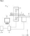
- Fig. 2 1 shows a computer tomography system 20 for providing a medical image of a patient P, the computer tomography system 20 having a computer tomograph 21 and a computer unit 22.
- the computer tomograph 21 has at least one X-ray emitter 23 and at least one X-ray detector 24, wherein the at least one X-ray emitter 23 and the at least one X-ray detector 24 are rotatably arranged around a patient couch 25.
- the patient P is mounted on the patient bed 25, wherein the patient bed 25 is formed parallel to the spatial axis z.
- the arithmetic unit 22 has a monitor 26, which has, for example, a graphical user interface as well as input means.
- the user and / or the doctor can determine measurement parameters of a first imaging measurement sequence and / or a second imaging measurement sequence by means of the graphical user interface.
- the arithmetic unit 22 is connected to a radiology information system 27 and a PACS image archiving system 28.
- the computer tomograph 21 it is conceivable for the computer tomograph 21 to have two x-ray emitters and two x-ray detectors, wherein preferably both x-ray emitters have different acceleration voltages, in particular during the second imaging measurement sequence.
- the second projection data in this case have a first energy range and a second energy range.
- Fig. 3 shows a flowchart of a method for providing a captured by a computer tomograph medical image of a patient in a second embodiment.
- steps S107 to S114 can be virtually arbitrarily combined.
- Method step S107 indicates that after detecting the first projection data and before detecting the second projection data, a contrast agent bolus is injected into the patient.
- Method step S108 indicates that the second projection data is acquired by means of an imaging spectral measurement sequence in the computer tomograph and has a first energy range and a second energy range.
- Method step S110 indicates that the reference image is reconstructed from the second projection data according to the second reconstruction rule, wherein the first reconstruction rule describes a first mixture of the second projection data according to the first energy range and the second energy range, and wherein the second reconstruction rule comprises a second mixture of the second projection data according to the describes the first energy range and the second energy range.
- Method step S111 indicates that the reference image is reconstructed from the second projection data according to the second reconstruction rule, wherein the first reconstruction rule is optimized with respect to a functional contrast and wherein the second reconstruction rule is optimized with respect to an anatomical contrast.
- Method step S112A indicates that, from the first projection data, the at least one breath-correlated image for several breathing states of the patient's breathing is reconstructed.
- Method step S112B indicates that a plurality of deformation models are generated by registering the reference image with the respective at least one breath-correlated image for the multiple respiratory states of the patient's respiration.
- Method step S113A indicates that the plurality of deformation models are applied to the at least one functional image of the patient.
- Method step S113B indicates that combining the deformed at least one functional image for the multiple breath states of the patient with the at least one breath-correlated image of the patient generates the medical image for the multiple breath states of the patient's respiration.
- the medical image may include a medical image data set, the medical image data set having separate images for the respective respiratory states of the respiration of the patient.
- Method step S114 indicates that the second projection data and the reference image reconstructed therefrom comprise a plurality of states of contrast enhancement in the patient, the reference image being registered with the at least one respiratory correlated image of the patient as a function of the plurality of states of the contrast enhancement.
- Method step S109 indicates that the deformed at least one functional image of the patient is combined with the at least one respiratory correlated image of the patient such that the patient's medical image has a subtraction image of the patient having contrast enhancement of the patient.
Landscapes
- Engineering & Computer Science (AREA)
- Health & Medical Sciences (AREA)
- Life Sciences & Earth Sciences (AREA)
- Medical Informatics (AREA)
- Physics & Mathematics (AREA)
- Public Health (AREA)
- General Health & Medical Sciences (AREA)
- Radiology & Medical Imaging (AREA)
- Nuclear Medicine, Radiotherapy & Molecular Imaging (AREA)
- Pathology (AREA)
- Animal Behavior & Ethology (AREA)
- Optics & Photonics (AREA)
- Biophysics (AREA)
- Veterinary Medicine (AREA)
- Biomedical Technology (AREA)
- Heart & Thoracic Surgery (AREA)
- Molecular Biology (AREA)
- Surgery (AREA)
- High Energy & Nuclear Physics (AREA)
- Theoretical Computer Science (AREA)
- Computer Vision & Pattern Recognition (AREA)
- General Physics & Mathematics (AREA)
- Pulmonology (AREA)
- Epidemiology (AREA)
- Primary Health Care (AREA)
- Apparatus For Radiation Diagnosis (AREA)
Abstract
- Erfassen von ersten Projektionsdaten, wobei aus den ersten Projektionsdaten zumindest ein atemkorreliertes Bild gemäß einer Atmung rekonstruiert wird,
- Erfassen von zweiten Projektionsdaten, wobei aus den zweiten Projektionsdaten zumindest ein funktionelles Bild gemäß einer ersten Rekonstruktionsvorschrift rekonstruiert wird,
- Registrieren eines Referenzbildes mit dem zumindest einen atemkorrelierten Bild, wobei das Referenzbild dem zumindest einen funktionellen Bild entspricht oder gemäß einer zweiten Rekonstruktionsvorschrift aus den zweiten Projektionsdaten rekonstruiert wird, wobei ein Deformationsmodell erzeugt wird,
- Anwenden des Deformationsmodells auf das zumindest eine funktionelle Bild,
- Kombinieren des deformierten zumindest einen funktionellen Bildes mit dem zumindest einen atemkorrelierten Bild, wobei das medizinische Bild erzeugt wird, und
- Bereitstellen des medizinischen Bildes des Patienten.
Description
- Die Erfindung betrifft ein Verfahren für ein Bereitstellen eines mittels eines Computertomographen erfassten medizinischen Bildes eines Patienten, eine Computertomographieanlage für ein Bereitstellen eines medizinischen Bildes eines Patienten und ein Computerprogrammprodukt.
- In der medizinischen Bildgebung ist eine bildgebende Messsequenz, welche typischerweise in einer medizinischen Bildgebungsmodalität, insbesondere in einem Computertomographen, durchführbar ist, vorzugsweise derart optimiert, dass in erster Linie ein physiologischer Zustand eines Patienten in einem medizinischen Bild sichtbar sein kann. Aufgrund unterschiedlicher technischer Herausforderungen ist es üblicherweise nicht oder nur unter hohem Aufwand möglich, mehrere, insbesondere unterschiedliche, physiologische Zustände des Patienten in dem medizinischen Bild sichtbar zu machen, insbesondere wenn das medizinische Bild einen vergleichsweise großen Messbereich aufweist oder mehrere medizinische Bilder über den Messbereich aufgeteilt sind, welche jeweils die physiologischen Zustände aufweisen sollen.
- Ein Problem kann daraus entstehen, dass erste physiologische Zustände, insbesondere gemäß einer Atmung des Patienten, und zweite physiologische Zustände, insbesondere gemäß einer Konstrastmittelanreicherung in dem Patienten und/oder gemäß einer Jod-Verteilung in dem Patienten, sich in der jeweiligen Dynamik unterscheiden. Insbesondere aufgrund der unterschiedlichen Dynamik kann die bildgebende Messsequenz typischerweise entweder im Hinblick auf das Erfassen der ersten physiologischen Zustände oder auf das Erfassen der zweiten physiologischen Zustände optimiert werden. Ferner ist üblicherweise ein maximales Gesichtsfeld der medizinischen Bildgebungsmodalität kleiner als der für das gleichzeitige Erfassen der ersten physiologischen Zustände und der zweiten physiologischen Zustände notwendige Messbereich der bildgebenden Messsequenz. Sprich die ersten physiologischen Zustände und die zweiten physiologischen Zustände aufweisende Projektionsdaten können typischerweise nacheinander, insbesondere nicht gleichzeitig, erfasst werden.
- Der Erfindung liegt die Aufgabe zu Grunde, ein Verfahren für ein Bereitstellen eines mittels eines Computertomographen erfassten medizinischen Bildes eines Patienten, eine Computertomographieanlage für ein Bereitstellen eines medizinischen Bildes eines Patienten und ein Computerprogrammproduktanzugeben, wobei das medizinische Bild unterschiedliche physiologische Zustände oder Zustandskombinationen aufweisen kann.
- Die Aufgabe wird durch die Merkmale der unabhängigen Ansprüche gelöst. Vorteilhafte Ausgestaltungen sind in den Unteransprüchen beschrieben.
- Das erfindungsgemäße Verfahren für ein Bereitstellen eines mittels eines Computertomographen erfassten medizinischen Bildes eines Patienten weist folgende Schritte auf:
- Erfassen von ersten Projektionsdaten eines ersten Messbereichs mittels des Computertomographen, wobei aus den ersten Projektionsdaten zumindest ein atemkorreliertes Bild des Patienten gemäß einer Atmung des Patienten rekonstruiert wird,
- Erfassen von zweiten Projektionsdaten eines zweiten Messbereichs mittels des Computertomographen, wobei aus den zweiten Projektionsdaten zumindest ein funktionelles Bild des Patienten gemäß einer ersten Rekonstruktionsvorschrift rekonstruiert wird und wobei sich der erste Messbereich und der zweite Messbereich zumindest teilweise überlappen,
- Registrieren eines Referenzbildes mit dem zumindest einen atemkorrelierten Bild des Patienten, wobei das Referenzbild dem zumindest einen funktionellen Bild des Patienten entspricht oder gemäß einer zweiten Rekonstruktionsvorschrift aus den zweiten Projektionsdaten rekonstruiert wird, wobei ein Deformationsmodell erzeugt wird,
- Anwenden des Deformationsmodells auf das zumindest eine funktionelle Bild des Patienten,
- Kombinieren des deformierten zumindest einen funktionellen Bildes des Patienten mit dem zumindest einen atemkorrelierten Bild des Patienten, wobei das medizinische Bild des Patienten erzeugt wird, und
- Bereitstellen des medizinischen Bildes des Patienten.
- Das Erfassen der ersten Projektionsdaten und/oder der zweiten Projektionsdaten kann ein Durchführen einer bildgebenden Messsequenz in einer medizinischen Bildgebungsmodalität, insbesondere in dem Computertomographen, umfassen. Typischerweise ist der Patient während der bildgebenden Messsequenz in der medizinischen Bildgebungsmodalität beispielsweise auf einer Patientenliege gelagert und/oder bei der medizinischen Bildgebungsmodalität positioniert. Vorzugsweise können die ersten Projektionsdaten und/oder die zweiten Projektionsdaten mittels des Computertomographen erfasst werden. Der erste Messbereich wird vorzugsweise von den ersten Projektionsdaten, insbesondere bei einer Durchführung einer ersten bildgebenden Messsequenz, abgebildet und/oder beschrieben. Der zweite Messbereich wird vorzugsweise von den zweiten Projektionsdaten, insbesondere bei einer Durchführung einer zweiten bildgebenden Messsequenz, abgebildet und/oder beschrieben. Typischerweise wird die erste bildgebende Messsequenz vor der zweiten bildgebenden Messsequenz durchgeführt. Grundsätzlich kann die zweite bildgebende Messsequenz vor der ersten bildgebenden Messsequenz durchgeführt werden.
- Die ersten Projektionsdaten und/oder die zweiten Projektionsdaten können beispielsweise Rohdaten des Computertomographen aufweisen. Das zumindest eine atemkorrelierte Bild und/oder das zumindest eine funktionelle Bild des Patienten werden typischerweise mittels eines iterativen Rekonstruktionsverfahrens und/oder mittels einer gefilterten Rückprojektion aus den ersten Projektionsdaten bzw. aus den zweiten Projektionsdaten rekonstruiert. Das zumindest eine atemkorrelierte Bild des Patienten und/oder das zumindest eine funktionelle Bild des Patienten weisen vorzugsweise jeweils eine Verteilung von Bildwerten, insbesondere eine Hounsfield-Unit-Verteilung (HU), auf.
- Die Atmung des Patienten kann beispielsweise durch ein physiologisches Sensorsystem, insbesondere während der ersten bildgebenden Messsequenz, ermittelt werden. Das physiologische Sensorsystem kann beispielsweise einen Atemgurt und/oder eine Kamera aufweisen. Beispielsweise können mittels des Atemgurts oder der Kamera mehrere Atemzustände der Atmung des Patienten ermittelt werden. Vorzugsweise werden die mehreren Atemzustände den ersten Projektionsdaten und/oder dem zumindest einen atemkorrelierten Bild zugeordnet. Die mehreren Atemzustände sind typischerweise zyklisch und/oder weisen beispielsweise die Zustände Einatmen, mittleres Atmen und/oder Ausatmen auf. Alternativ oder zusätzlich kann die Atmung des Patienten, insbesondere mittels eines Bilderkennungsalgorithmus, aus dem zumindest einen atemkorrelierten Bild bestimmt werden. Das zumindest eine atemkorrelierte Bild weist vorzugsweise die Atmung, insbesondere die mehreren Atemzustände der Atmung, des Patienten auf.
- Das zumindest eine funktionelle Bild weist beispielsweise mehrere Zustände des funktionellen Vorgangs, insbesondere einer Kontrastmittelanreicherung typischerweise nach Injektion eines Kontrastmittelbolus, im Patienten auf. Üblicherweise werden die mehreren Kontrastmittelzustände durch die Injektion des Kontrastmittelbolus in den Patienten sichtbar. Die mehreren Kontrastmittelzustände weisen beispielsweise die Zustände keine Kontrastmittelanreicherung, ansteigende Kontrastmittelanreicherung, maximale Kontrastmittelanreicherung und/oder absteigende Kontrastmittelanreicherung auf. Alternativ oder zusätzlich kann das zumindest eine funktionelle Bild derart aus den zweiten Projektionsdaten rekonstruiert werden, dass das zumindest eine funktionelle Bild ein Mischbild, ein monoenergetisches Bild, ein virtual-non-contrast-Bild, ein Bild mit einer Fett-Verteilung, ein Bild mit einer Jod-Verteilung und/oder ein Bild mit einer Eisen-Verteilung aufweist. Das Bild mit der Jod-Verteilung kann beispielsweise die Kontrastmittelanreicherung aufweisen.
- Typischerweise können die ersten Projektionsdaten und/oder die zweiten Projektionsdaten in Messdatenblöcke unterteilt werden. Ein Messdatenblock kann beispielsweise diejenigen Projektionsdaten aufweisen, welche während eines Zeitintervalls erfasst worden sind. Typischerweise kann das Zeitintervall mit einer Zeitauflösung der bildgebenden Messsequenz korrelieren oder der Zeitauflösung der bildgebenden Messsequenz entsprechen. Alternativ oder zusätzlich kann der Messdatenblock diejenigen Projektionsdaten aufweisen, welche einen Teil, insbesondere einen räumlichen Abschnitt, des ersten Messbereichs und/oder des zweiten Messbereichs aufweisen und/oder abbilden. Beispielsweise kann der räumliche Abschnitt eine z-Position des ersten Messbereichs und/oder des zweiten Messbereichs und/oder eine Ausdehnung des ersten Messbereichs und/oder des zweiten Messbereichs entlang der Längsachse der Patientenliege aufweisen. Die Patientenliege ist typischerweise parallel zur z-Achse der medizinischen Bildgebungsmodalität. Die Messdatenblöcke können jeweils einen Zustand, insbesondere der Atmung und/oder der Kontrastmittelanreicherung, aufweisen.
- Vorzugsweise sind der erste Messbereich und der zweite Messbereich deckungsgleich. Typischerweise sind der erste Messbereich und/oder der zweite Messbereich größer als ein maximales Gesichtsfeld der medizinischen Bildgebungsmodalität, insbesondere als ein maximales Gesichtsfeld des Computertomographen. Vorzugsweise beträgt eine Schnittmenge zwischen dem ersten Messbereich und dem zweiten Messbereich, insbesondere parallel zur Längsachse der Patientenliege, mehr als 50%, besonders bevorzugt mehr als 90%. Die Schnittmenge zwischen dem ersten Messbereich und dem zweiten Messbereich kann beispielsweise als gemeinsamer Messbereich bezeichnet werden. Der erste Messbereich und/oder der zweite Messbereich können ein Organ des Patienten, beispielsweise eine Leber und/oder eine Lunge und/oder ein Herz des Patienten, aufweisen. Vorteilhafterweise können die erste bildgebende Messsequenz und die zweite bildgebende Messsequenz unterschiedliche Messparameter aufweisen. Beispielsweise ist eine Tischvorschubgeschwindigkeit der zweiten bildgebenden Messsequenz schneller als eine Tischvorschubgeschwindigkeit der ersten bildgebenden Messsequenz, insbesondere um einen Faktor 1,5 oder 2, besonders vorteilhafterweise um einen Faktor 5 schneller. Vorzugsweise sind die Messparameter der ersten bildgebenden Messsequenz in Hinblick auf ein zeitaufgelöstes volumetrisches Erfassen der ersten Projektionsdaten optimiert und/oder die Messparameter der zweiten bildgebenden Messsequenz sind in Hinblick auf das Erfassen der Injektion des Kontrastmittelbolus optimiert. Die erste bildgebende Messsequenz ist vorzugsweise derart ausgebildet, dass die ersten Projektionsdaten die Atmung des Patienten insbesondere an jeder z-Position des ersten Messbereichs aufweisen. Vorteilhafterweise weisen die ersten Projektionsdaten die gleichen Atemzustände an jeder z-Position des ersten Messbereichs auf. Typischerweise werden die ersten Projektionsdaten so lange erfasst, bis die ersten Projektionsdaten eine gleiche Anzahl an Atemzuständen, insbesondere die gleichen Atemzustände, an jeder z-Position des ersten Messbereichs aufweisen. In diesem Fall kann die erste Messsequenz eine Spiral-CT-Messsequenz und/oder einer sequentiellen CT-Messsequenz beschreiben.
- Die Rekonstruktionsvorschrift weist insbesondere eine Gewichtung der Projektionsdaten, insbesondere der zweiten Projektionsdaten, auf. In Abhängigkeit der Gewichtung der Projektionsdaten variiert beispielsweise ein Kontrast des rekonstruierten Bildes, insbesondere des zumindest einen funktionellen Bildes und/oder des Referenzbildes. Der Kontrast hängt typischerweise von der Verteilung an Bildwerten ab. Vorzugsweise können gemäß der ersten Rekonstruktionsvorschrift und der zweiten Rekonstruktionsvorschrift jeweils, insbesondere im Hinblick auf den Kontrast, unterschiedliche Bilder und/oder unterschiedliche Verteilungen an Bildwerten rekonstruiert werden.
- Das Deformationsfeld kann insbesondere ein Vektorfeld aufweisen, welches eine Veränderung zwischen zwei Registrierungsbildern, insbesondere zwischen einem Startbild und einem Zielbild, abbildet. Beispielsweise weisen die zwei Registrierungsbilder das zumindest eine atemkorrelierte Bild und das Referenzbild auf. Das Deformationsfeld kann mehrere Vektorfelder aufweisen, welche einen Pfad von dem Startbild zum Zielbild beschreiben. Durch das Anwenden des Deformationsfelds werden die Projektionsdaten, insbesondere das zumindest eine atemkorrelierte Bild und/oder das Referenzbild, derart verformt, dass das Startbild vorzugsweise dem Zielbild, insbesondere im Hinblick auf Konturen, gleicht. Die Konturen können beispielsweise das Organ und/oder das Gewebe und/oder Gefäße und/oder Knochenmaterial und/oder einen Tumor beschreiben. Vorzugsweise kann durch Inversion des Deformationsfelds die Verformung umgedreht werden. Typischerweise sind das zumindest eine atemkorrelierte Bild und das Referenzbild jeweils das Startbild bzw. das Zielbild oder umgekehrt. Sprich je nach Ausführung kann das Deformationsfeld typischerweise ausgehend von dem zumindest einen atemkorrelierten Bild oder ausgehend von dem zumindest einen funktionellen Bild erzeugt und entsprechend angewendet werden.
- Wenn das Referenzbild dem zumindest einen funktionellen Bild des Patienten entspricht, wird insbesondere das zumindest eine funktionelle Bild für das Registrieren verwendet. In diesem Fall ist typischerweise das zumindest eine funktionelle Bild das Startbild oder das Zielbild.
- Wenn das Referenzbild gemäß einer zweiten Rekonstruktionsvorschrift aus den zweiten Projektionsdaten rekonstruiert wird, wird insbesondere ein zu dem zumindest einen funktionellen Bild verschiedenes Bild aus den zweiten Projektionsdaten rekonstruiert. Die zweite Rekonstruktionsvorschrift unterscheidet sich typischerweise derart von der ersten Rekonstruktionsvorschrift, dass bei der jeweiligen Anwendung auf die zweiten Projektionsdaten unterschiedliche, insbesondere funktionelle, Bilder rekonstruiert werden. Vorzugsweise ist die zweite Rekonstruktionsvorschrift derart optimiert, dass das Registrieren des Referenzbildes gemäß der zweiten Rekonstruktionsvorschrift bessere Ergebnisse, insbesondere ein exakteres Deformationsmodell, liefert als das Registrieren des Referenzbildes gemäß der ersten Rekonstruktionsvorschrift.
- Grundsätzlich kann das Registrieren ein Minimieren eines bildwertbasierten Abstandsmaßes umfassen. Das bildwertbasierte Abstandsmaß kann beispielsweise eine Summe oder einen Mittelwert von punktweise gebildeten quadratischen Differenzen zwischen dem Referenzbild, insbesondere dessen Verteilung an Bildwerten, und dem zumindest einen atemkorrelierten Bild, insbesondere dessen Verteilung an Bildwerten, des Patienten beschreiben. Das bildwertbasierte Abstandsmaß kann beispielsweise aufgrund einer systemischen Veränderung im Patienten, beispielsweise der Kontrastmittelanreicherung, ungeeignet sein. In diesem Fall kann beispielsweise ein local-crosscorrelation- und/oder ein mutual-information-Abstandsmaß verwendet werden.
- Das Anwenden des Deformationsmodell umfasst insbesondere das Verformen des zumindest einen funktionellen Bildes derart, dass Konturen des zumindest einen funktionellen Bildes den Konturen des zumindest einen atemkorrelierten Bildes gleichen. Wenn das Referenzbild gemäß einer zweiten Rekonstruktionsvorschrift aus den zweiten Projektionsdaten rekonstruiert wird, wird vorteilhafterweise das Deformationsmodell bei ersten Registrierungsbildern ermittelt und das Deformationsmodell bei zweiten Registrierungsbildern angewendet, wobei die ersten Registrierungsbilder und die zweiten Registrierungsbilder typischerweise lediglich ein einziges Bild, insbesondere das Zielbild, also typischerweise das zumindest eine atemkorrelierte Bild, jeweils gemein haben.
- Vorzugsweise umfasst das Kombinieren ein Verrechnen des deformierten zumindest einen funktionellen Bildes des Patienten mit dem zumindest einen atemkorrelierten Bild des Patienten derart, dass das medizinische Bild Bildinformation des deformierten zumindest einen funktionellen Bildes und Bildinformation des zumindest einen atemkorrelierten Bildes, beispielsweise verstärkt und/oder differenziert, aufweist. In diesem Fall geht typischerweise das medizinische Bild als einziges Bild aus der Kombination des zumindest einen funktionellen Bildes und des zumindest einen atemkorrelierten Bildes hervor. Alternativ oder zusätzlich kann das Kombinieren derart erfolgen, dass das medizinische Bild einen medizinischen Bilddatensatz aufweist, wobei der medizinische Bilddatensatz das zumindest eine funktionelle Bild und das zumindest eine atemkorrelierte Bild umfasst. In anderen Worten sind das zumindest eine funktionelle Bild und das zumindest eine atemkorrelierte Bild in einem einzelnen medizinischen Bilddatensatz, typischerweise allerdings als separate Bilder, enthalten. Es ist denkbar, dass der medizinische Bilddatensatz die separaten Bilder und ein kombiniertes medizinisches Bild aufweist.
- Das Bereitstellen des medizinischen Bildes kann ein Übertragen des medizinischen Bildes von dem Computertomographen in ein Radiologieinformationssystem und/oder ein PACS-Bildarchivierungssystem umfassen. Alternativ oder zusätzlich kann das medizinische Bild, insbesondere dem Nutzer, auf einem Monitor angezeigt werden. Vorzugsweise ist der Monitor, beispielsweise zu dem Abrufen des medizinischen Bildes aus dem Radiologieinformationssystem bzw. dem PACS-Bildarchivierungssystem und/oder von der medizinischen Bildgebungsmodalität ausgebildet, und/oder zu einem Anzeigen des medizinischen Bildes ausgebildet. Grundsätzlich ist es denkbar, dass das medizinische Bild über ein Netzwerk, welches beispielsweise das Radiologieinformationssystem und/oder das PACS-Bildarchivierungssystem und/oder die medizinische Bildgebungsmodalität verbindet, ausgetauscht und beispielsweise auf einem Server gespeichert bzw. abgerufen werden kann. Zusätzlich zu dem medizinischen Bild können beispielsweise die ersten Projektionsdaten und/oder die zweiten Projektionsdaten bereitgestellt, insbesondere in das Radiologieinformationssystem und/oder das PACS-Bildarchivierungssystem übertragen und/oder auf dem Monitor angezeigt werden.
- Das erfindungsgemäße Verfahren für das Bereitstellen des medizinischen Bildes des Patienten bringt insbesondere folgende Vorteile mit sich:
- Das Erzeugen des medizinischen Bildes, welches vorzugsweise die Bildinformation von gemäß zwei separat durchgeführten bildgebenden Messsequenzen rekonstruierten Bildern aufweist, ist insbesondere für einen Nutzer, insbesondere einen Arzt, vorteilhaft, wenn der Arzt beispielsweise eine Bestrahlungsplanung für eine Krebstherapie des Patienten durchführt. Insbesondere wenn die Bestrahlungsplanung ein Organ betrifft, welches aufgrund der Positionierung aufgrund von Atmung oder Herzschlag relativ zum Thorax des Patienten beweglich ist, kann das medizinische Bild ermöglichen basierend auf dem zumindest einen atemkorrelierten Bild die Bewegung des Organs und basierend auf dem zumindest einen funktionellen Bild den funktionellen Vorgang, insbesondere eine Nekrose und/oder eine Durchblutung und/oder eine Kappilarisierung innerhalb des Organs, abzuschätzen. Typischerweise kann das medizinische Bild vorteilhafterweise die Jod-Verteilung im Patienten beim Atemzustand Einatmen aufweisen und/oder beispielsweise die Jod-Verteilung im Patienten beim Atemzustand Ausatmen aufweisen.
- Das Erzeugen des medizinischen Bildes ist weiterhin in Hinblick auf eine Reduzierung von einer Untersuchungsdauer des Patienten vorteilhaft. Typischerweise korreliert die Untersuchungsdauer des Patienten beim Durchführen der bildgebenden Messsequenz in dem Computertomographen mit potentiell für den Patienten schädlichen Röntgenstrahlen, insbesondere einer Röntgendosis. Durch das Kombinieren der ersten Projektionsdaten, insbesondere des zumindest einen atemkorrelierten Bildes, und der zweiten Projektionsdaten, insbesondere des zumindest einen funktionellen Bildes, kann die Untersuchungsdauer und/oder vorzugsweise die mit der Röntgenstrahlung korrespondierende Röntgendosis verringert werden.
- Wenn das Referenzbild gemäß der zweiten Rekonstruktionsvorschrift aus den zweiten Projektionsdaten rekonstruiert wird, ist vorzugsweise möglich basierend auf den zweiten Projektionsdaten ein in Hinblick auf die Registrierung optimales Bild gemäß der zweiten Rekonstruktionsvorschrift zu rekonstruieren und das daraus erzeugte Deformationsmodell beispielsweise auf das zumindest eine funktionelle Bild anzuwenden.
- Eine Ausführungsform sieht vor, dass das deformierte zumindest eine funktionelle Bild des Patienten derart mit dem zumindest einen atemkorrelierten Bild des Patienten kombiniert wird, dass das medizinische Bild des Patienten ein Subtraktionsbild des Patienten aufweist, welches die Kontrastmittelanreicherung des Patienten aufweist. Vorzugsweise weist das Subtraktionsbild lediglich die Kontrastmittelanreicherung des Patienten auf. In anderen Worten wird insbesondere nicht mit Kontrastmittel angereichertes Gewebe oder Knochenmaterial durch die Subtraktion aus dem medizinischen Bild herausgerechnet. Grundsätzlich ist es denkbar, dass das Subtraktionsbild das Bild der Jod-Verteilung, das Bild der Fett-Verteilung und/oder das Bild der Eisen-Verteilung aufweist. Beispielsweise wird das deformierte zumindest eine funktionelle Bild des Patienten und das zumindest eine atemkorrelierte Bild des Patienten derart verrechnet, dass das zumindest eine atemkorrelierte Bild des Patienten von dem deformierten zumindest einen funktionellen Bild des Patienten abgezogen wird. Typischerweise beschreibt die Differenz die Kontrastmittelanreicherung im Patienten vor und nach der Injektion des Kontrastmittelbolus. Das Subtraktionsbild wird insbesondere durch das Kombinieren eines Prä-Kontrast-Bildes und eines Post-Kontrast-Bildes erzeugt, wobei typischerweise das Prä-Kontrast-Bild Bildinformation vor der Kontrastmittelanreicherung und das Post-Kontrast-Bild Bildinformation nach der Kontrastmittelanreicherung in dem jeweiligen Messbereich aufweist.
- Eine Ausführungsform sieht vor, dass nach dem Erfassen der ersten Projektionsdaten und vor dem Erfassen der zweiten Projektionsdaten der Kontrastmittelbolus in den Patienten injiziert wird. In diesem Fall kann das Prä-Kontrast-Bild nicht aus den zweiten Projektionsdaten, sondern typischerweise lediglich aus den ersten Projektionsdaten rekonstruiert werden. Vorzugsweise werden das Prä-Kontrast-Bild aus den ersten Projektionsdaten und das Post-Kontrast-Bild aus den zweiten Projektionsdaten rekonstruiert. Dies ist insbesondere vorteilhaft, weil das Erfassen der zweiten Projektionsdaten kürzer ausfallen kann, wodurch insbesondere die Röntgendosis für den Patienten verringert sein kann. Diese Ausführungsform grenzt sich daher insbesondere zu der digitalen Subtraktions-Angiographie ab. Vorteilhafterweise werden das Prä-Kontrast-Bild und das Post-Kontrast-Bild aus unterschiedlichen Projektionsdaten rekonstruiert, wodurch die erste bildgebende Messsequenz und die zweite bildgebende Messsequenz unabhängig voneinander parametrisiert sein können und/oder, insbesondere in Hinblick auf die Röntgendosis, optimiert werden können. Die digitale Subtraktions-Angiographie erfordert im Gegensatz dazu üblicherweise, dass das Prä-Kontrast-Bild und das Post-Kontrast-Bild aus denselben Projektionsdaten rekonstruiert werden.
- Eine Ausführungsform sieht vor, dass die zweiten Projektionsdaten und das daraus rekonstruierte Referenzbild die mehreren Zustände der Kontrastmittelanreicherung in dem Patienten aufweisen und wobei das Referenzbild in Abhängigkeit von den mehreren Zuständen der Kontrastmittelanreicherung mit dem zumindest einen atemkorrelierten Bild des Patienten registriert wird. Wenn das Referenzbild in Abhängigkeit von den mehreren Zuständen der Kontrastmittelanreicherung mit dem zumindest einen atemkorrelierten Bild des Patienten registriert wird, wird vorzugsweise gemäß einer Reihung der Zustände der Kontrastmittelanreicherung das Referenzbild registriert. In anderen Worten wird vorzugsweise zunächst dasjenige Referenzbild, welches vorzugsweise dem zumindest einen atemkorrelierten Bild vergleichsweise am ähnlichsten ist, mit dem zumindest einen atemkorrelierten Bild registriert und so weiter. In Abhängigkeit von den mehreren Zuständen der Kontrastmittelanreicherung bedeutet insbesondere, dass beispielsweise die zweiten Projektionsdaten derart sortiert werden, dass die Kontrastmittelanreicherung ansteigt oder absteigt. Auch andere Reihungen sind denkbar. Vorzugsweise kann das Deformationsmodell, insbesondere die Vektorfelder, schrittweise gemäß dem Anstieg der Kontrastmittelanreicherung erzeugt und/oder auf das zumindest eine funktionelle Bild angewendet werden.
- Eine Ausführungsform sieht vor, dass aus den ersten Projektionsdaten das zumindest eine atemkorrelierte Bild für mehrere Atemzustände der Atmung des Patienten rekonstruiert wird und wobei durch das Registrieren des Referenzbildes mit dem jeweiligen zumindest einen atemkorrelierten Bild für die mehreren Atemzustände der Atmung des Patienten mehrere Deformationsmodelle erzeugt werden. In anderen Worten können vorzugsweise die mehreren Deformationsmodelle in Abhängigkeit von den mehreren Atemzuständen erzeugt werden.
- Eine Ausführungsform sieht vor, dass durch das Anwenden der mehreren Deformationsmodelle auf das zumindest eine funktionelle Bild des Patienten und durch das Kombinieren des deformierten zumindest einen funktionellen Bildes für die mehreren Atemzustände des Patienten mit dem zumindest einen atemkorrelierten Bild des Patienten das medizinische Bild für die mehreren Atemzustände der Atmung des Patienten erzeugt wird. Das zumindest eine atemkorrelierte Bild für mehrere Atemzustände der Atmung des Patienten weist daher beispielsweise Bildinformation beim Einatmen und Bildinformation beim Ausatmen auf. Die mehreren Deformationsmodelle ermöglichen in diesem Fall das Verformen des zumindest einen funktionellen Bildes derart, dass insbesondere die Konturen des deformierten zumindest einen funktionellen Bildes des Patienten jeweils an das Einatmen oder an das Ausatmen angepasst werden. Vorteilhafterweise weist das medizinische Bild für die mehreren Atemzustände der Atmung des Patienten das deformierte zumindest eine funktionelle Bild beispielsweise beim Einatmen und/oder beim Ausatmen auf.
- Eine Ausführungsform sieht vor, dass die zweiten Projektionsdaten mittels einer bildgebenden spektralen Messsequenz in dem Computertomographen erfasst werden und einen ersten Energiebereich sowie einen zweiten Energiebereich aufweisen. In diesem Fall entspricht insbesondere die zweite bildgebende Messsequenz der bildgebenden spektralen Messsequenz. Beispielsweise unterscheiden sich der erste Energiebereich und der zweite Energiebereich in Hinblick auf eine Energie der Röntgenstrahlen. Üblicherweise ist die Bildinformation des zumindest einen atemkorrelierten Bildes und/oder die Bildinformation des zumindest einen funktionellen Bildes abhängig von dem ersten Energiebereich und/oder dem zweiten Energiebereich. Beispielsweise erzeugt ein Röntgenstrahler die Röntgenstrahlen verteilt über ein Röntgenspektrum in Abhängigkeit der angelegten Beschleunigungsspannung zwischen Kathode und Anode. Der erste Energiebereich und der zweite Energiebereich können sich beispielsweise in der Beschleunigungsspannung unterscheiden. Üblicherweise ist es möglich, die Beschleunigungsspannung innerhalb desselben Röntgenstrahlers zeitlich zu variieren und/oder zwei Röntgenstrahler zu verwenden, wobei in diesem Fall typischerweise beide Röntgenstrahler mit unterschiedlichen Beschleunigungsspannungen betrieben werden. Ein Computertomograph, welcher zwei Röntgenstrahler aufweist, wird typischerweise Dual-Source- und/oder Dual-Energy-Computertomograph genannt. Vorzugsweise können beide Röntgenstrahler gleichzeitig, insbesondere gemäß der zweiten bildgebenden Messsequenz, die Röntgenstrahlen emittieren. Alternativ oder zusätzlich werden die Röntgenstrahlen mittels eines energiesensitiven Röntgendetektors, beispielsweise eines Photon-Counting-Detektor, erfasst. Wenn die zweiten Projektionsdaten den ersten Energiebereich und den zweiten Energiebereich aufweisen, kann vorzugsweise aus den zweiten Projektionsdaten das Mischbild, insbesondere bei 120 keV, das monoenergetische Bild, das virtual-non-contrast-Bild, das Bild mit der Fett-Verteilung, das Bild mit der Jod-Verteilung und/oder das Bild mit der Eisen-Verteilung rekonstruiert werden.
- Eine Ausführungsform sieht vor, dass das Referenzbild gemäß der zweiten Rekonstruktionsvorschrift aus den zweiten Projektionsdaten rekonstruiert wird, wobei die erste Rekonstruktionsvorschrift eine erste Mischung der zweiten Projektionsdaten gemäß dem ersten Energiebereich und dem zweiten Energiebereich beschreibt und wobei die zweite Rekonstruktionsvorschrift eine zweite Mischung der zweiten Projektionsdaten gemäß dem ersten Energiebereich und dem zweiten Energiebereich beschreibt. Die erste Mischung und/oder die zweite Mischung beschreiben typischerweise jeweils ein Kombinieren der ersten Projektionsdaten mit den zweiten Projektionsdaten und/oder ein Anwenden eines Multiband-Filters auf die ersten Projektionsdaten und/oder die zweiten Projektionsdaten mit unterschiedlichen Gewichtungen. Besonders vorteilhafterweise werden für diese Ausführungsform die zweiten Projektionsdaten in einem Dual-Source-Computertomographen und/oder mittels des Photon-Counting-Detektors erfasst. Typischerweise ist das Referenzbild gemäß der zweiten Rekonstruktionsvorschrift, beispielsweise das Mischbild und/oder das virtual-non-contrast-Bild, insbesondere bildwert-basiert, ähnlicher zum zumindest einen atemkorrelierten Bild als das zumindest eine funktionelle Bild gemäß der ersten Rekonstruktionsvorschrift, insbesondere das Bild mit der Fett-Verteilung, das Bild mit der Jod-Verteilung und/oder das Bild mit der Eisen-Verteilung.
- Eine Ausführungsform sieht vor, dass das Referenzbild gemäß der zweiten Rekonstruktionsvorschrift aus den zweiten Projektionsdaten rekonstruiert wird, wobei die erste Rekonstruktionsvorschrift in Hinblick auf einen funktionellen Kontrast optimiert ist und wobei die zweite Rekonstruktionsvorschrift in Hinblick auf einen anatomischen Kontrast optimiert ist. In anderen Worten ist die erste Rekonstruktionsvorschrift, insbesondere die erste Mischung, und die zweite Rekonstruktionsvorschrift, insbesondere die zweite Mischung, derart ausgebildet, dass vorzugsweise das Referenzbild gemäß der zweiten Rekonstruktionsvorschrift und das zumindest eine atemkorrelierte Bild eine Anatomie des Patienten aufweisen, wobei das zumindest eine funktionelle Bild gemäß der ersten Rekonstruktionsvorschrift vorzugsweise den funktionellen Vorgang, beispielsweise die Kontrastmittelanreicherung, aufweist. Vorteilhafterweise weist das Referenzbild den optimierten anatomischen Kontrast auf, wodurch typischerweise das Registrieren des Referenzbildes mit dem zumindest einen atemkorrelierten Bild und somit das Deformationsmodell verbessert wird. In anderen Worten kann vorteilhafterweise basierend auf den zweiten Projektionsdaten allein ein optimales Deformationsmodell berechnet werden, wobei das optimale Deformationsmodell vorteilhafterweise auf das zumindest eine funktionelle Bild angewendet wird, welches wiederum auf den zweiten Projektionsdaten basiert.
- Die erfindungsgemäße Computertomographieanlage für ein Bereitstellen eines medizinischen Bildes eines Patienten weist den Computertomographen und eine Recheneinheit auf, wobei der Computertomograph zumindest einen Röntgenstrahler und zumindest einen Röntgendetektor aufweist. Der Computertomograph ist vorzugsweise zur Durchführung der ersten bildgebenden Messsequenz und/oder der zweiten bildgebenden Messsequenz ausgebildet. Die Recheneinheit kann beispielsweise mittels des Netzwerks mit dem Radiologieinformationssystem und/oder dem PACS-Bildarchivierungssystem zu dem Bereitstellen des medizinischen Bildes verbunden sein. Alternativ oder zusätzlich kann die Computertomographieanlage für das Bereitstellen des medizinischen Bildes des Patienten, insbesondere die Recheneinheit, den Monitor aufweisen. Die Recheneinheit ist vorzugsweise mit dem Computertomographen derart verbunden, dass die ersten Projektionsdaten, welche insbesondere bei der Durchführung der ersten bildgebenden Messsequenz erfasst werden, und/oder die zweiten Projektionsdaten, welche insbesondere bei der Durchführung der zweiten bildgebenden Messsequenz erfasst werden, von dem Computertomographen zur Recheneinheit übertragen werden können.
- Eine Ausführungsform sieht vor, dass der Computertomograph zwei Röntgenstrahler und zwei Röntgendetektoren aufweist. In diesem Fall kann der Computertomograph Dual-Source-Computertomograph und/oder Dual-Energy-Computertomograph genannt werden.
- Das erfindungsgemäße Computerprogrammprodukt weist Programmcodemittel auf, welche in die Recheneinheit ladbar sind, um ein erfindungsgemäßes Verfahren für ein Bereitstellen eines medizinischen Bildes eines Patienten auszuführen, wenn die Programmcodemittel in der Recheneinheit ausgeführt werden.
- Das Computerprogrammprodukt kann ein Computerprogramm sein oder ein Computerprogramm umfassen. Das Computerprogrammprodukt weist insbesondere die Programmcodemittel auf, welche die erfindungsgemäßen Verfahrensschritte abbilden. Dadurch kann das erfindungsgemäße Verfahren definiert und wiederholbar ausgeführt sowie eine Kontrolle über eine Weitergabe des erfindungsgemäßen Verfahrens ausgeübt werden. Das Computerprogrammprodukt ist vorzugsweise derart konfiguriert, dass die Recheneinheit mittels des Computerprogrammprodukts die erfindungsgemäßen Verfahrensschritte ausführen kann. Die Programmcodemittel können insbesondere in einen Speicher der Recheneinheit geladen und typischerweise mittels eines Prozessors der Recheneinheit mit Zugriff auf den Speicher ausgeführt werden. Wenn das Computerprogrammprodukt, insbesondere die Programmcodemittel, in der Recheneinheit ausgeführt wird, können typischerweise alle erfindungsgemäßen Ausführungsformen des beschriebenen Verfahrens durchgeführt werden. Das Computerprogrammprodukt ist beispielsweise auf einem physischen, computerlesbaren Medium gespeichert und/oder digital als Datenpaket in einem Computernetzwerk hinterlegt. Das Computerprogrammprodukt kann das physische, computerlesbare Medium und/oder das Datenpaket in dem Computernetzwerk darstellen. So kann die Erfindung auch von dem physischen, computerlesbaren Medium und/oder dem Datenpaket in dem Computernetzwerk ausgehen. Das physische, computerlesbare Medium ist üblicherweise unmittelbar mit der Recheneinheit verbindbar, beispielsweise indem das physische, computerlesbare Medium in ein DVD-Laufwerk eingelegt oder in einen USB-Port gesteckt wird, wodurch die Recheneinheit auf das physische, computerlesbare Medium insbesondere lesend zugreifen kann. Das Datenpaket kann vorzugsweise aus dem Computernetzwerk abgerufen werden. Das Computernetzwerk kann die Recheneinheit aufweisen oder mittels einer Wide-Area-Network- (WAN) bzw. einer (Wireless-)Local-Area-Network-Verbindung (WLAN oder LAN) mit der Recheneinheit mittelbar verbunden sein. Beispielsweise kann das Computerprogrammprodukt digital auf einem Cloud-Server an einem Speicherort des Computernetzwerks hinterlegt sein, mittels des WAN über das Internet und/oder mittels des WLAN bzw. LAN auf die Recheneinheit insbesondere durch das Aufrufen eines Downloadlinks, welcher zu dem Speicherort des Computerprogrammprodukts verweist, übertragen werden.
- Im Folgenden wird die Erfindung anhand der in den Figuren dargestellten Ausführungsbeispiele näher beschrieben und erläutert. Grundsätzlich werden in der folgenden Figurenbeschreibung im Wesentlichen gleich bleibende Strukturen und Einheiten mit demselben Bezugszeichen wie beim erstmaligen Auftreten der jeweiligen Struktur oder Einheit benannt.
- Es zeigen:
-
Fig. 1 ein Flussdiagramm eines Verfahrens für ein Bereitstellen eines mittels eines Computertomographen erfassten medizinischen Bildes eines Patienten in einem ersten Ausführungsbeispiel, -
Fig. 2 eine Computertomographieanlage für ein Bereitstellen eines medizinischen Bildes eines Patienten, -
Fig. 3 ein Flussdiagramm eines Verfahrens für ein Bereitstellen eines mittels eines Computertomographen erfassten medizinischen Bildes eines Patienten in einem zweiten Ausführungsbeispiel. -
Fig. 1 zeigt ein Flussdiagramm eines Verfahrens für ein Bereitstellen eines mittels eines Computertomographen erfassten medizinischen Bildes eines Patienten in einem ersten Ausführungsbeispiel. - Verfahrensschritt S101 kennzeichnet das Erfassen von ersten Projektionsdaten eines ersten Messbereichs mittels des Computertomographen, wobei aus den ersten Projektionsdaten zumindest ein atemkorreliertes Bild des Patienten gemäß einer Atmung des Patienten rekonstruiert wird.
- Verfahrensschritt S102 kennzeichnet das Erfassen von zweiten Projektionsdaten eines zweiten Messbereichs mittels des Computertomographen, wobei aus den zweiten Projektionsdaten zumindest ein funktionelles Bild des Patienten gemäß einer ersten Rekonstruktionsvorschrift rekonstruiert wird und wobei sich der erste Messbereich und der zweite Messbereich zumindest teilweise überlappen.
- Verfahrensschritt S103A kennzeichnet das Registrieren eines Referenzbildes mit dem zumindest einen atemkorrelierten Bild des Patienten.
- Verfahrensschritt S103B und Verfahrensschritt S103C bilden zwei Alternativen A und B. In anderen Worten wird entweder Verfahrensschritt S103B oder Verfahrensschritt S103C durchgeführt. Sprich Verfahrensschritt S103B und Verfahrensschritt S103C bieten zwei unabhängige Möglichkeiten A, B das Referenzbild zu erhalten.
- Verfahrensschritt S103B kennzeichnet, dass das Referenzbild dem zumindest einen funktionellen Bild des Patienten entspricht.
- Verfahrensschritt S103C kennzeichnet, dass das Referenzbild gemäß einer zweiten Rekonstruktionsvorschrift aus den zweiten Projektionsdaten rekonstruiert wird.
- Verfahrensschritt S103D kennzeichnet, dass ein Deformationsmodell erzeugt wird.
- Verfahrensschritt S104 kennzeichnet das Anwenden des Deformationsmodells auf das zumindest eine funktionelle Bild des Patienten.
- Verfahrensschritt S105 kennzeichnet das Kombinieren des deformierten zumindest einen funktionellen Bildes des Patienten mit dem zumindest einen atemkorrelierten Bild des Patienten, wobei das medizinische Bild des Patienten erzeugt wird.
- Verfahrensschritt S106 kennzeichnet das Bereitstellen des medizinischen Bildes des Patienten.
-
Fig. 2 zeigt eine Computertomographieanlage 20 für ein Bereitstellen eines medizinischen Bildes eines Patienten P, wobei die Computertomographieanlage 20 einen Computertomographen 21 und eine Recheneinheit 22 aufweist. Der Computertomograph 21 weist zumindest einen Röntgenstrahler 23 und zumindest einen Röntgendetektor 24 auf, wobei der zumindest eine Röntgenstrahler 23 und der zumindest eine Röntgendetektor 24 um eine Patientenliege 25 drehbar angeordnet sind. Der Patient P ist auf der Patientenliege 25 gelagert, wobei die Patientenliege 25 parallel zur Raumachse z ausgebildet ist. Die Recheneinheit 22 weist einen Monitor 26 auf, welcher beispielsweise eine graphische Benutzeroberfläche sowie Eingabemittel aufweist. Beispielsweise kann der Nutzer und/oder der Arzt mittels der graphischen Benutzeroberfläche Messparameter einer ersten bildgebenden Messsequenz und/oder einer zweiten bildgebenden Messsequenz festlegen. Die Recheneinheit 22 ist mit einem Radiologieinformationssystem 27 und einem PACS-Bildarchivierungssystem 28 verbunden. - Grundsätzlich ist es denkbar, dass der Computertomograph 21 zwei Röntgenstrahler und zwei Röntgendetektoren aufweist, wobei vorzugsweise beide Röntgenstrahler unterschiedliche Beschleunigungsspannungen, insbesondere während der zweiten bildgebenden Messsequenz, aufweisen. Vorzugsweise weisen die zweiten Projektionsdaten in diesem Fall einen ersten Energiebereich und einen zweiten Energiebereich auf.
-
Fig. 3 zeigt ein Flussdiagramm eines Verfahrens für ein Bereitstellen eines mittels eines Computertomographen erfassten medizinischen Bildes eines Patienten in einem zweiten Ausführungsbeispiel. Die inFig. 3 gezeigten Verfahrensschritte S107 bis S114 können quasi beliebig kombiniert werden. - Verfahrensschritt S107 kennzeichnet, dass nach dem Erfassen der ersten Projektionsdaten und vor dem Erfassen der zweiten Projektionsdaten ein Kontrastmittelbolus in den Patienten injiziert wird.
- Verfahrensschritt S108 kennzeichnet, dass die zweiten Projektionsdaten mittels einer bildgebenden spektralen Messsequenz in dem Computertomographen erfasst werden und einen ersten Energiebereich sowie einen zweiten Energiebereich aufweisen.
- Verfahrensschritt S110 kennzeichnet, dass das Referenzbild gemäß der zweiten Rekonstruktionsvorschrift aus den zweiten Projektionsdaten rekonstruiert wird, wobei die erste Rekonstruktionsvorschrift eine erste Mischung der zweiten Projektionsdaten gemäß dem ersten Energiebereich und dem zweiten Energiebereich beschreibt und wobei die zweite Rekonstruktionsvorschrift eine zweite Mischung der zweiten Projektionsdaten gemäß dem ersten Energiebereich und dem zweiten Energiebereich beschreibt.
- Verfahrensschritt S111 kennzeichnet, dass das Referenzbild gemäß der zweiten Rekonstruktionsvorschrift aus den zweiten Projektionsdaten rekonstruiert wird, wobei die erste Rekonstruktionsvorschrift in Hinblick auf einen funktionellen Kontrast optimiert ist und wobei die zweite Rekonstruktionsvorschrift in Hinblick auf einen anatomischen Kontrast optimiert ist.
- Verfahrensschritt S112A kennzeichnet, dass aus den ersten Projektionsdaten das zumindest eine atemkorrelierte Bild für mehrere Atemzustände der Atmung des Patienten rekonstruiert wird.
- Verfahrensschritt S112B kennzeichnet, dass durch das Registrieren des Referenzbildes mit dem jeweiligen zumindest einen atemkorrelierten Bild für die mehreren Atemzustände der Atmung des Patienten mehrere Deformationsmodelle erzeugt werden.
- Verfahrensschritt S113A kennzeichnet, dass die mehreren Deformationsmodelle auf das zumindest eine funktionelle Bild des Patienten angewendet werden.
- Verfahrensschritt S113B kennzeichnet, dass durch das Kombinieren des deformierten zumindest einen funktionellen Bildes für die mehreren Atemzustände des Patienten mit dem zumindest einen atemkorrelierten Bild des Patienten das medizinische Bild für die mehreren Atemzustände der Atmung des Patienten erzeugt wird. Das medizinische Bild kann einen medizinischen Bilddatensatz aufweisen, wobei der medizinische Bilddatensatz separate Bilder für die jeweiligen Atemzustände der Atmung des Patienten aufweist.
- Verfahrensschritt S114 kennzeichnet, dass die zweiten Projektionsdaten und das daraus rekonstruierte Referenzbild mehrere Zustände einer Kontrastmittelanreicherung in dem Patienten aufweisen, wobei das Referenzbild in Abhängigkeit von den mehreren Zuständen der Kontrastmittelanreicherung mit dem zumindest einen atemkorrelierten Bild des Patienten registriert wird.
- Verfahrensschritt S109 kennzeichnet, dass das deformierte zumindest eine funktionelle Bild des Patienten derart mit dem zumindest einen atemkorrelierten Bild des Patienten kombiniert wird, dass das medizinische Bild des Patienten ein Subtraktionsbild des Patienten aufweist, welches eine Kontrastmittelanreicherung des Patienten aufweist.
- Obwohl die Erfindung im Detail durch die bevorzugten Ausführungsbeispiele näher illustriert und beschrieben wurde, so ist die Erfindung dennoch nicht durch die offenbarten Beispiele eingeschränkt und andere Variationen können vom Fachmann hieraus abgeleitet werden, ohne den Schutzumfang der Erfindung zu verlassen.
Claims (12)
- Verfahren für ein Bereitstellen eines mittels eines Computertomographen erfassten medizinischen Bildes eines Patienten, mit den folgenden Schritten:- Erfassen von ersten Projektionsdaten eines ersten Messbereichs mittels des Computertomographen, wobei aus den ersten Projektionsdaten zumindest ein atemkorreliertes Bild des Patienten gemäß einer Atmung des Patienten rekonstruiert wird,- Erfassen von zweiten Projektionsdaten eines zweiten Messbereichs mittels des Computertomographen, wobei aus den zweiten Projektionsdaten zumindest ein funktionelles Bild des Patienten gemäß einer ersten Rekonstruktionsvorschrift rekonstruiert wird und wobei sich der erste Messbereich und der zweite Messbereich zumindest teilweise überlappen,- Registrieren eines Referenzbildes mit dem zumindest einen atemkorrelierten Bild des Patienten, wobei das Referenzbild dem zumindest einen funktionellen Bild des Patienten entspricht oder gemäß einer zweiten Rekonstruktionsvorschrift aus den zweiten Projektionsdaten rekonstruiert wird, wobei ein Deformationsmodell erzeugt wird,- Anwenden des Deformationsmodells auf das zumindest eine funktionelle Bild des Patienten,- Kombinieren des deformierten zumindest einen funktionellen Bildes des Patienten mit dem zumindest einen atemkorrelierten Bild des Patienten, wobei das medizinische Bild des Patienten erzeugt wird, und- Bereitstellen des medizinischen Bildes des Patienten.
- Verfahren nach Anspruch 1, wobei das deformierte zumindest eine funktionelle Bild des Patienten derart mit dem zumindest einen atemkorrelierten Bild des Patienten kombiniert wird, dass das medizinische Bild des Patienten ein Subtraktionsbild des Patienten aufweist, welches eine Kontrastmittelanreicherung des Patienten aufweist.
- Verfahren nach einem der vorhergehenden Ansprüche, wobei nach dem Erfassen der ersten Projektionsdaten und vor dem Erfassen der zweiten Projektionsdaten ein Kontrastmittelbolus in den Patienten injiziert wird.
- Verfahren nach einem der vorhergehenden Ansprüche, wobei die zweiten Projektionsdaten und das daraus rekonstruierte Referenzbild mehrere Zustände einer Kontrastmittelanreicherung in dem Patienten aufweisen und wobei das Referenzbild in Abhängigkeit von den mehreren Zuständen der Kontrastmittelanreicherung mit dem zumindest einen atemkorrelierten Bild des Patienten registriert wird.
- Verfahren nach einem der vorhergehenden Ansprüche, wobei aus den ersten Projektionsdaten das zumindest eine atemkorrelierte Bild für mehrere Atemzustände der Atmung des Patienten rekonstruiert wird und wobei durch das Registrieren des Referenzbildes mit dem jeweiligen zumindest einen atemkorrelierten Bild für die mehreren Atemzustände der Atmung des Patienten mehrere Deformationsmodelle erzeugt werden.
- Verfahren nach Anspruch 5, wobei durch das Anwenden der mehreren Deformationsmodelle auf das zumindest eine funktionelle Bild des Patienten und durch das Kombinieren des deformierten zumindest einen funktionellen Bildes für die mehreren Atemzustände des Patienten mit dem zumindest einen atemkorrelierten Bild des Patienten das medizinische Bild für die mehreren Atemzustände der Atmung des Patienten erzeugt wird.
- Verfahren nach einem der vorhergehenden Ansprüche, wobei die zweiten Projektionsdaten mittels einer bildgebenden spektralen Messsequenz in dem Computertomographen erfasst werden und einen ersten Energiebereich sowie einen zweiten Energiebereich aufweisen.
- Verfahren nach Anspruch 7, wobei das Referenzbild gemäß der zweiten Rekonstruktionsvorschrift aus den zweiten Projektionsdaten rekonstruiert wird und wobei die erste Rekonstruktionsvorschrift eine erste Mischung der zweiten Projektionsdaten gemäß dem ersten Energiebereich und dem zweiten Energiebereich beschreibt und wobei die zweite Rekonstruktionsvorschrift eine zweite Mischung der zweiten Projektionsdaten gemäß dem ersten Energiebereich und dem zweiten Energiebereich beschreibt.
- Verfahren nach einem der vorhergehenden Ansprüche, wobei das Referenzbild gemäß der zweiten Rekonstruktionsvorschrift aus den zweiten Projektionsdaten rekonstruiert wird, wobei die erste Rekonstruktionsvorschrift in Hinblick auf einen funktionellen Kontrast optimiert ist und wobei die zweite Rekonstruktionsvorschrift in Hinblick auf einen anatomischen Kontrast optimiert ist.
- Computertomographieanlage für ein Bereitstellen eines medizinischen Bildes eines Patienten, aufweisend- einen Computertomographen, wobei der Computertomograph zumindest einen Röntgenstrahler und zumindest einen Röntgendetektor aufweist, und- eine Recheneinheit,wobei die Computertomographieanlage zur Durchführung eines Verfahrens nach einem der Ansprüche 1 bis 9 ausgebildet ist.
- Computertomographieanlage nach Anspruch 10, wobei der Computertomograph zwei Röntgenstrahler und zwei Röntgendetektoren aufweist.
- Computerprogrammprodukt, aufweisend Programmcodemittel, welche in eine Recheneinheit ladbar sind, um ein Verfahren nach einem der Ansprüche 1 bis 9 auszuführen, wenn die Programmcodemittel in der Recheneinheit ausgeführt werden.
Priority Applications (3)
| Application Number | Priority Date | Filing Date | Title |
|---|---|---|---|
| EP18153904.0A EP3412208B1 (de) | 2018-01-29 | 2018-01-29 | Bereitstellen eines medizinischen bildes |
| US16/253,269 US11514623B2 (en) | 2018-01-29 | 2019-01-22 | Providing a medical image |
| CN201910085046.0A CN110097607B (zh) | 2018-01-29 | 2019-01-29 | 提供医学图像的方法、计算机断层扫描设备和程序产品 |
Applications Claiming Priority (1)
| Application Number | Priority Date | Filing Date | Title |
|---|---|---|---|
| EP18153904.0A EP3412208B1 (de) | 2018-01-29 | 2018-01-29 | Bereitstellen eines medizinischen bildes |
Publications (2)
| Publication Number | Publication Date |
|---|---|
| EP3412208A1 true EP3412208A1 (de) | 2018-12-12 |
| EP3412208B1 EP3412208B1 (de) | 2021-05-26 |
Family
ID=61074412
Family Applications (1)
| Application Number | Title | Priority Date | Filing Date |
|---|---|---|---|
| EP18153904.0A Active EP3412208B1 (de) | 2018-01-29 | 2018-01-29 | Bereitstellen eines medizinischen bildes |
Country Status (3)
| Country | Link |
|---|---|
| US (1) | US11514623B2 (de) |
| EP (1) | EP3412208B1 (de) |
| CN (1) | CN110097607B (de) |
Cited By (1)
| Publication number | Priority date | Publication date | Assignee | Title |
|---|---|---|---|---|
| US11684262B2 (en) | 2018-01-29 | 2023-06-27 | Siemens Healthcare Gmbh | Providing a medical image |
Families Citing this family (3)
| Publication number | Priority date | Publication date | Assignee | Title |
|---|---|---|---|---|
| US10580132B2 (en) * | 2017-04-13 | 2020-03-03 | Canon Kabushiki Kaisha | Medical image processing apparatus, control method therefor, and non-transitory storage medium storing program |
| WO2019149718A1 (en) * | 2018-02-01 | 2019-08-08 | Koninklijke Philips N.V. | Low radiation dose computed tomography perfusion (ctp) with improved quantitative analysis |
| CN113100803B (zh) * | 2021-04-20 | 2024-07-19 | 西门子数字医疗科技(上海)有限公司 | 用于显示静脉血栓的方法、装置、计算机设备和介质 |
Citations (4)
| Publication number | Priority date | Publication date | Assignee | Title |
|---|---|---|---|---|
| US20040081269A1 (en) * | 2002-10-23 | 2004-04-29 | Tin-Su Pan | Retrospective respiratory gating for imaging and treatment |
| EP1542165A1 (de) * | 2003-11-28 | 2005-06-15 | GE Medical Systems Global Technology Company LLC | Verfahren und Vorrichtung zur Bewegungskorrektur in dreidimensionalen medizinischen Bildsequenzen |
| US20070270689A1 (en) * | 2006-05-16 | 2007-11-22 | Mark Lothert | Respiratory gated image fusion of computed tomography 3D images and live fluoroscopy images |
| EP2037413A2 (de) * | 2007-09-17 | 2009-03-18 | Wisconsin Alumni Research Foundation | Verfahren zur Verringerung von Bewegungsartefakten in stark eingeschränkten medizinischen Bildern |
Family Cites Families (6)
| Publication number | Priority date | Publication date | Assignee | Title |
|---|---|---|---|---|
| NL8603059A (nl) * | 1986-12-01 | 1988-07-01 | Philips Nv | Inrichting en werkwijze met bewegingsartefactreductie voor verschilbeeldbepaling. |
| US7158610B2 (en) * | 2003-09-05 | 2007-01-02 | Varian Medical Systems Technologies, Inc. | Systems and methods for processing x-ray images |
| DE102011006188B4 (de) * | 2011-03-28 | 2017-09-07 | Siemens Healthcare Gmbh | Verfahren und Computertomographie-System zur Erstellung tomographischer Bilddarstellung mit mindestens zwei Strahler-Detektor-Systemen |
| DE102011076348B4 (de) * | 2011-05-24 | 2014-02-13 | Siemens Aktiengesellschaft | Verfahren und Computertomographiesystem zur Erzeugung tomographischer Bilddatensätze |
| US9521985B2 (en) * | 2011-07-29 | 2016-12-20 | Siemens Healthcare Gmbh | Automatic detection of contrast injection |
| DE102016222093A1 (de) * | 2016-11-10 | 2017-12-28 | Siemens Healthcare Gmbh | Simultaner Einsatz von unterschiedlichen Kontrastmitteln bei CT-Bildgebungsverfahren |
-
2018
- 2018-01-29 EP EP18153904.0A patent/EP3412208B1/de active Active
-
2019
- 2019-01-22 US US16/253,269 patent/US11514623B2/en active Active
- 2019-01-29 CN CN201910085046.0A patent/CN110097607B/zh active Active
Patent Citations (4)
| Publication number | Priority date | Publication date | Assignee | Title |
|---|---|---|---|---|
| US20040081269A1 (en) * | 2002-10-23 | 2004-04-29 | Tin-Su Pan | Retrospective respiratory gating for imaging and treatment |
| EP1542165A1 (de) * | 2003-11-28 | 2005-06-15 | GE Medical Systems Global Technology Company LLC | Verfahren und Vorrichtung zur Bewegungskorrektur in dreidimensionalen medizinischen Bildsequenzen |
| US20070270689A1 (en) * | 2006-05-16 | 2007-11-22 | Mark Lothert | Respiratory gated image fusion of computed tomography 3D images and live fluoroscopy images |
| EP2037413A2 (de) * | 2007-09-17 | 2009-03-18 | Wisconsin Alumni Research Foundation | Verfahren zur Verringerung von Bewegungsartefakten in stark eingeschränkten medizinischen Bildern |
Cited By (1)
| Publication number | Priority date | Publication date | Assignee | Title |
|---|---|---|---|---|
| US11684262B2 (en) | 2018-01-29 | 2023-06-27 | Siemens Healthcare Gmbh | Providing a medical image |
Also Published As
| Publication number | Publication date |
|---|---|
| US20190236818A1 (en) | 2019-08-01 |
| EP3412208B1 (de) | 2021-05-26 |
| CN110097607A (zh) | 2019-08-06 |
| CN110097607B (zh) | 2023-06-06 |
| US11514623B2 (en) | 2022-11-29 |
Similar Documents
| Publication | Publication Date | Title |
|---|---|---|
| EP4129189B1 (de) | Verfahren zum betreiben eines medizinischen bildgebungsgeräts zur lagerichtigen darstellung von nicht-anatomischen strukturen während einer bildgebenden untersuchung und vorrichtung hierfür | |
| DE102014201134B4 (de) | Verfahren und Vorrichtung zur Erzeugung eines 2-D-Projektionsbildes eines Gefäßsystems nebst korrespondierenden Gegenständen | |
| DE10317367B4 (de) | Verfahren zur Durchführung der digitalen Subtraktionsangiographie unter Verwendung nativer Volumendatensätze | |
| EP3437559A1 (de) | Ermitteln eines funktionsparameters betreffend eine lokale gewebefunktion für mehrere gewebebereiche | |
| DE102019212929A1 (de) | Registrieren eines 2D Bildes mit einem 3D Bild | |
| DE102019201079A1 (de) | Verfahren und bildgebendes Gerät zum Erzeugen eines bewegungskompensierten Bildes, Computerprogramm und Speichermedium | |
| DE102021210879A1 (de) | Bereitstellen eines Vergleichsdatensatzes | |
| DE102009051384A1 (de) | Strahlaufhärtungskorrektur für CT-Perfusionsmessungen | |
| DE102012201798A1 (de) | Verfahren und Vorrichtung zur Planung einer Röntgenbildgebung mit geringer Strahlenbelastung | |
| EP3412208B1 (de) | Bereitstellen eines medizinischen bildes | |
| DE102009015386A1 (de) | Verfahren und Vorrichtung zur Erzeugung eines funktionellen Datensatzes eines perfundierten Bereichs des menschlichen oder tierischen Körpers | |
| EP3539475B1 (de) | Verfahren zur bewegungskorrektur von spektralen computertomographiedaten, sowie ein energiesensitives computertomographiegerät | |
| DE102008021835A1 (de) | Verfahren und Tomographiegerät zur Normierung von Bilddaten hinsichtlich eines durch ein Kontrastmittel in den Bilddaten hervorgerufenen Kontrastes | |
| DE102011006398A1 (de) | Verfahren, Bildverarbeitungseinrichtung und Computertomographiesystem zur Ermittlung eines Anteils von nekrotischem Gewebe sowie Computerprogrammprodukt mit Programmcodeabschnitten zur Ermittlung eines Anteils von nekrotischem Gewebe | |
| DE102020201928A1 (de) | Verfahren zur Deformationskorrektur | |
| DE102020210192A1 (de) | Bereitstellen einer synthetisch kontrastierten Szene | |
| EP3797698B1 (de) | Verfahren zum erzeugen eines synthetischen mammogramms basierend auf einer dual-energy-tomosyntheseaufnahme | |
| DE102013202313A1 (de) | Verfahren und Vorrichtung zur Korrektur von Bewegungsartefakten bei einem computertomographischen Bild | |
| DE102007045313A1 (de) | Verfahren und Vorrichtung zur getrennten dreidimensionalen Darstellung von Arterien und Venen in einem Untersuchungsobjekt und Vorrichtung | |
| EP3363363B1 (de) | Verfahren zum durchführen einer bildgebenden untersuchung | |
| EP3413317B1 (de) | Bereitstellen eines medizinischen bildes | |
| DE102020211944A1 (de) | Verfahren zur Ansteuerung eines medizinischen Bildgebungsgeräts | |
| DE102020216011A1 (de) | Bereitstellen eines Datensatzes | |
| DE102019218770A1 (de) | Bereitstellen eines Gefäßbilddatensatzes | |
| DE102020216557B4 (de) | Verfahren und Datenverarbeitungssystem zum Bereitstellen einer respiratorischen Information |
Legal Events
| Date | Code | Title | Description |
|---|---|---|---|
| PUAI | Public reference made under article 153(3) epc to a published international application that has entered the european phase |
Free format text: ORIGINAL CODE: 0009012 |
|
| STAA | Information on the status of an ep patent application or granted ep patent |
Free format text: STATUS: REQUEST FOR EXAMINATION WAS MADE |
|
| 17P | Request for examination filed |
Effective date: 20181105 |
|
| AK | Designated contracting states |
Kind code of ref document: A1 Designated state(s): AL AT BE BG CH CY CZ DE DK EE ES FI FR GB GR HR HU IE IS IT LI LT LU LV MC MK MT NL NO PL PT RO RS SE SI SK SM TR |
|
| AX | Request for extension of the european patent |
Extension state: BA ME |
|
| RIN1 | Information on inventor provided before grant (corrected) |
Inventor name: HOFMANN, CHRISTIAN Inventor name: TANADINI-LANG, STEPHANIE Inventor name: RITTER, ANDRE Inventor name: GUCKENBERGER, MATTHIAS |
|
| GRAP | Despatch of communication of intention to grant a patent |
Free format text: ORIGINAL CODE: EPIDOSNIGR1 |
|
| STAA | Information on the status of an ep patent application or granted ep patent |
Free format text: STATUS: GRANT OF PATENT IS INTENDED |
|
| GRAJ | Information related to disapproval of communication of intention to grant by the applicant or resumption of examination proceedings by the epo deleted |
Free format text: ORIGINAL CODE: EPIDOSDIGR1 |
|
| STAA | Information on the status of an ep patent application or granted ep patent |
Free format text: STATUS: REQUEST FOR EXAMINATION WAS MADE |
|
| INTG | Intention to grant announced |
Effective date: 20201124 |
|
| INTC | Intention to grant announced (deleted) | ||
| GRAP | Despatch of communication of intention to grant a patent |
Free format text: ORIGINAL CODE: EPIDOSNIGR1 |
|
| STAA | Information on the status of an ep patent application or granted ep patent |
Free format text: STATUS: GRANT OF PATENT IS INTENDED |
|
| INTG | Intention to grant announced |
Effective date: 20210126 |
|
| RIN1 | Information on inventor provided before grant (corrected) |
Inventor name: RITTER, ANDRE Inventor name: HOFMANN, CHRISTIAN Inventor name: TANADINI-LANG, STEPHANIE Inventor name: GUCKENBERGER, MATTHIAS |
|
| GRAS | Grant fee paid |
Free format text: ORIGINAL CODE: EPIDOSNIGR3 |
|
| GRAA | (expected) grant |
Free format text: ORIGINAL CODE: 0009210 |
|
| STAA | Information on the status of an ep patent application or granted ep patent |
Free format text: STATUS: THE PATENT HAS BEEN GRANTED |
|
| AK | Designated contracting states |
Kind code of ref document: B1 Designated state(s): AL AT BE BG CH CY CZ DE DK EE ES FI FR GB GR HR HU IE IS IT LI LT LU LV MC MK MT NL NO PL PT RO RS SE SI SK SM TR |
|
| REG | Reference to a national code |
Ref country code: GB Ref legal event code: FG4D Free format text: NOT ENGLISH |
|
| REG | Reference to a national code |
Ref country code: CH Ref legal event code: EP |
|
| REG | Reference to a national code |
Ref country code: DE Ref legal event code: R096 Ref document number: 502018005376 Country of ref document: DE |
|
| REG | Reference to a national code |
Ref country code: AT Ref legal event code: REF Ref document number: 1395394 Country of ref document: AT Kind code of ref document: T Effective date: 20210615 |
|
| REG | Reference to a national code |
Ref country code: IE Ref legal event code: FG4D Free format text: LANGUAGE OF EP DOCUMENT: GERMAN |
|
| REG | Reference to a national code |
Ref country code: LT Ref legal event code: MG9D |
|
| PG25 | Lapsed in a contracting state [announced via postgrant information from national office to epo] |
Ref country code: BG Free format text: LAPSE BECAUSE OF FAILURE TO SUBMIT A TRANSLATION OF THE DESCRIPTION OR TO PAY THE FEE WITHIN THE PRESCRIBED TIME-LIMIT Effective date: 20210826 Ref country code: HR Free format text: LAPSE BECAUSE OF FAILURE TO SUBMIT A TRANSLATION OF THE DESCRIPTION OR TO PAY THE FEE WITHIN THE PRESCRIBED TIME-LIMIT Effective date: 20210526 Ref country code: FI Free format text: LAPSE BECAUSE OF FAILURE TO SUBMIT A TRANSLATION OF THE DESCRIPTION OR TO PAY THE FEE WITHIN THE PRESCRIBED TIME-LIMIT Effective date: 20210526 Ref country code: LT Free format text: LAPSE BECAUSE OF FAILURE TO SUBMIT A TRANSLATION OF THE DESCRIPTION OR TO PAY THE FEE WITHIN THE PRESCRIBED TIME-LIMIT Effective date: 20210526 |
|
| REG | Reference to a national code |
Ref country code: NL Ref legal event code: MP Effective date: 20210526 |
|
| PG25 | Lapsed in a contracting state [announced via postgrant information from national office to epo] |
Ref country code: GR Free format text: LAPSE BECAUSE OF FAILURE TO SUBMIT A TRANSLATION OF THE DESCRIPTION OR TO PAY THE FEE WITHIN THE PRESCRIBED TIME-LIMIT Effective date: 20210827 Ref country code: IS Free format text: LAPSE BECAUSE OF FAILURE TO SUBMIT A TRANSLATION OF THE DESCRIPTION OR TO PAY THE FEE WITHIN THE PRESCRIBED TIME-LIMIT Effective date: 20210926 Ref country code: PT Free format text: LAPSE BECAUSE OF FAILURE TO SUBMIT A TRANSLATION OF THE DESCRIPTION OR TO PAY THE FEE WITHIN THE PRESCRIBED TIME-LIMIT Effective date: 20210927 Ref country code: PL Free format text: LAPSE BECAUSE OF FAILURE TO SUBMIT A TRANSLATION OF THE DESCRIPTION OR TO PAY THE FEE WITHIN THE PRESCRIBED TIME-LIMIT Effective date: 20210526 Ref country code: LV Free format text: LAPSE BECAUSE OF FAILURE TO SUBMIT A TRANSLATION OF THE DESCRIPTION OR TO PAY THE FEE WITHIN THE PRESCRIBED TIME-LIMIT Effective date: 20210526 Ref country code: NO Free format text: LAPSE BECAUSE OF FAILURE TO SUBMIT A TRANSLATION OF THE DESCRIPTION OR TO PAY THE FEE WITHIN THE PRESCRIBED TIME-LIMIT Effective date: 20210826 Ref country code: SE Free format text: LAPSE BECAUSE OF FAILURE TO SUBMIT A TRANSLATION OF THE DESCRIPTION OR TO PAY THE FEE WITHIN THE PRESCRIBED TIME-LIMIT Effective date: 20210526 Ref country code: RS Free format text: LAPSE BECAUSE OF FAILURE TO SUBMIT A TRANSLATION OF THE DESCRIPTION OR TO PAY THE FEE WITHIN THE PRESCRIBED TIME-LIMIT Effective date: 20210526 |
|
| PG25 | Lapsed in a contracting state [announced via postgrant information from national office to epo] |
Ref country code: NL Free format text: LAPSE BECAUSE OF FAILURE TO SUBMIT A TRANSLATION OF THE DESCRIPTION OR TO PAY THE FEE WITHIN THE PRESCRIBED TIME-LIMIT Effective date: 20210526 |
|
| PG25 | Lapsed in a contracting state [announced via postgrant information from national office to epo] |
Ref country code: RO Free format text: LAPSE BECAUSE OF FAILURE TO SUBMIT A TRANSLATION OF THE DESCRIPTION OR TO PAY THE FEE WITHIN THE PRESCRIBED TIME-LIMIT Effective date: 20210526 Ref country code: CZ Free format text: LAPSE BECAUSE OF FAILURE TO SUBMIT A TRANSLATION OF THE DESCRIPTION OR TO PAY THE FEE WITHIN THE PRESCRIBED TIME-LIMIT Effective date: 20210526 Ref country code: DK Free format text: LAPSE BECAUSE OF FAILURE TO SUBMIT A TRANSLATION OF THE DESCRIPTION OR TO PAY THE FEE WITHIN THE PRESCRIBED TIME-LIMIT Effective date: 20210526 Ref country code: SM Free format text: LAPSE BECAUSE OF FAILURE TO SUBMIT A TRANSLATION OF THE DESCRIPTION OR TO PAY THE FEE WITHIN THE PRESCRIBED TIME-LIMIT Effective date: 20210526 Ref country code: SK Free format text: LAPSE BECAUSE OF FAILURE TO SUBMIT A TRANSLATION OF THE DESCRIPTION OR TO PAY THE FEE WITHIN THE PRESCRIBED TIME-LIMIT Effective date: 20210526 Ref country code: EE Free format text: LAPSE BECAUSE OF FAILURE TO SUBMIT A TRANSLATION OF THE DESCRIPTION OR TO PAY THE FEE WITHIN THE PRESCRIBED TIME-LIMIT Effective date: 20210526 Ref country code: ES Free format text: LAPSE BECAUSE OF FAILURE TO SUBMIT A TRANSLATION OF THE DESCRIPTION OR TO PAY THE FEE WITHIN THE PRESCRIBED TIME-LIMIT Effective date: 20210526 |
|
| REG | Reference to a national code |
Ref country code: DE Ref legal event code: R097 Ref document number: 502018005376 Country of ref document: DE |
|
| PLBE | No opposition filed within time limit |
Free format text: ORIGINAL CODE: 0009261 |
|
| STAA | Information on the status of an ep patent application or granted ep patent |
Free format text: STATUS: NO OPPOSITION FILED WITHIN TIME LIMIT |
|
| 26N | No opposition filed |
Effective date: 20220301 |
|
| PG25 | Lapsed in a contracting state [announced via postgrant information from national office to epo] |
Ref country code: IS Free format text: LAPSE BECAUSE OF FAILURE TO SUBMIT A TRANSLATION OF THE DESCRIPTION OR TO PAY THE FEE WITHIN THE PRESCRIBED TIME-LIMIT Effective date: 20210926 Ref country code: AL Free format text: LAPSE BECAUSE OF FAILURE TO SUBMIT A TRANSLATION OF THE DESCRIPTION OR TO PAY THE FEE WITHIN THE PRESCRIBED TIME-LIMIT Effective date: 20210526 |
|
| PG25 | Lapsed in a contracting state [announced via postgrant information from national office to epo] |
Ref country code: IT Free format text: LAPSE BECAUSE OF FAILURE TO SUBMIT A TRANSLATION OF THE DESCRIPTION OR TO PAY THE FEE WITHIN THE PRESCRIBED TIME-LIMIT Effective date: 20210526 |
|
| PG25 | Lapsed in a contracting state [announced via postgrant information from national office to epo] |
Ref country code: MC Free format text: LAPSE BECAUSE OF FAILURE TO SUBMIT A TRANSLATION OF THE DESCRIPTION OR TO PAY THE FEE WITHIN THE PRESCRIBED TIME-LIMIT Effective date: 20210526 |
|
| REG | Reference to a national code |
Ref country code: CH Ref legal event code: PL |
|
| REG | Reference to a national code |
Ref country code: BE Ref legal event code: MM Effective date: 20220131 |
|
| PG25 | Lapsed in a contracting state [announced via postgrant information from national office to epo] |
Ref country code: LU Free format text: LAPSE BECAUSE OF NON-PAYMENT OF DUE FEES Effective date: 20220129 |
|
| PG25 | Lapsed in a contracting state [announced via postgrant information from national office to epo] |
Ref country code: BE Free format text: LAPSE BECAUSE OF NON-PAYMENT OF DUE FEES Effective date: 20220131 |
|
| PG25 | Lapsed in a contracting state [announced via postgrant information from national office to epo] |
Ref country code: LI Free format text: LAPSE BECAUSE OF NON-PAYMENT OF DUE FEES Effective date: 20220131 Ref country code: CH Free format text: LAPSE BECAUSE OF NON-PAYMENT OF DUE FEES Effective date: 20220131 |
|
| PG25 | Lapsed in a contracting state [announced via postgrant information from national office to epo] |
Ref country code: IE Free format text: LAPSE BECAUSE OF NON-PAYMENT OF DUE FEES Effective date: 20220129 |
|
| REG | Reference to a national code |
Ref country code: AT Ref legal event code: MM01 Ref document number: 1395394 Country of ref document: AT Kind code of ref document: T Effective date: 20230129 |
|
| REG | Reference to a national code |
Ref country code: DE Ref legal event code: R081 Ref document number: 502018005376 Country of ref document: DE Owner name: UNIVERSITAET ZUERICH, CH Free format text: FORMER OWNERS: SIEMENS HEALTHCARE GMBH, MUENCHEN, DE; UNIVERSITAET ZUERICH, ZUERICH, CH Ref country code: DE Ref legal event code: R081 Ref document number: 502018005376 Country of ref document: DE Owner name: SIEMENS HEALTHINEERS AG, DE Free format text: FORMER OWNERS: SIEMENS HEALTHCARE GMBH, MUENCHEN, DE; UNIVERSITAET ZUERICH, ZUERICH, CH |
|
| PG25 | Lapsed in a contracting state [announced via postgrant information from national office to epo] |
Ref country code: HU Free format text: LAPSE BECAUSE OF FAILURE TO SUBMIT A TRANSLATION OF THE DESCRIPTION OR TO PAY THE FEE WITHIN THE PRESCRIBED TIME-LIMIT; INVALID AB INITIO Effective date: 20180129 |
|
| PG25 | Lapsed in a contracting state [announced via postgrant information from national office to epo] |
Ref country code: AT Free format text: LAPSE BECAUSE OF NON-PAYMENT OF DUE FEES Effective date: 20230129 |
|
| PG25 | Lapsed in a contracting state [announced via postgrant information from national office to epo] |
Ref country code: MK Free format text: LAPSE BECAUSE OF FAILURE TO SUBMIT A TRANSLATION OF THE DESCRIPTION OR TO PAY THE FEE WITHIN THE PRESCRIBED TIME-LIMIT Effective date: 20210526 Ref country code: CY Free format text: LAPSE BECAUSE OF FAILURE TO SUBMIT A TRANSLATION OF THE DESCRIPTION OR TO PAY THE FEE WITHIN THE PRESCRIBED TIME-LIMIT Effective date: 20210526 Ref country code: AT Free format text: LAPSE BECAUSE OF NON-PAYMENT OF DUE FEES Effective date: 20230129 |
|
| PG25 | Lapsed in a contracting state [announced via postgrant information from national office to epo] |
Ref country code: MT Free format text: LAPSE BECAUSE OF FAILURE TO SUBMIT A TRANSLATION OF THE DESCRIPTION OR TO PAY THE FEE WITHIN THE PRESCRIBED TIME-LIMIT Effective date: 20210526 |
|
| REG | Reference to a national code |
Ref country code: DE Ref legal event code: R082 Ref document number: 502018005376 Country of ref document: DE |
|
| PG25 | Lapsed in a contracting state [announced via postgrant information from national office to epo] |
Ref country code: TR Free format text: LAPSE BECAUSE OF FAILURE TO SUBMIT A TRANSLATION OF THE DESCRIPTION OR TO PAY THE FEE WITHIN THE PRESCRIBED TIME-LIMIT Effective date: 20210526 |
|
| PGFP | Annual fee paid to national office [announced via postgrant information from national office to epo] |
Ref country code: GB Payment date: 20260210 Year of fee payment: 9 |
|
| PGFP | Annual fee paid to national office [announced via postgrant information from national office to epo] |
Ref country code: DE Payment date: 20260320 Year of fee payment: 9 |
|
| PGFP | Annual fee paid to national office [announced via postgrant information from national office to epo] |
Ref country code: AT Payment date: 20260410 Year of fee payment: 5 |
|
| PGFP | Annual fee paid to national office [announced via postgrant information from national office to epo] |
Ref country code: FR Payment date: 20260122 Year of fee payment: 9 |