EP3405909B1 - Systems and methods for segmentation of intra-patient medical images - Google Patents
Systems and methods for segmentation of intra-patient medical images Download PDFInfo
- Publication number
- EP3405909B1 EP3405909B1 EP17703003.8A EP17703003A EP3405909B1 EP 3405909 B1 EP3405909 B1 EP 3405909B1 EP 17703003 A EP17703003 A EP 17703003A EP 3405909 B1 EP3405909 B1 EP 3405909B1
- Authority
- EP
- European Patent Office
- Prior art keywords
- image
- medical image
- images
- medical
- label map
- Prior art date
- Legal status (The legal status is an assumption and is not a legal conclusion. Google has not performed a legal analysis and makes no representation as to the accuracy of the status listed.)
- Active
Links
Images
Classifications
-
- G—PHYSICS
- G06—COMPUTING OR CALCULATING; COUNTING
- G06V—IMAGE OR VIDEO RECOGNITION OR UNDERSTANDING
- G06V10/00—Arrangements for image or video recognition or understanding
- G06V10/70—Arrangements for image or video recognition or understanding using pattern recognition or machine learning
- G06V10/77—Processing image or video features in feature spaces; using data integration or data reduction, e.g. principal component analysis [PCA] or independent component analysis [ICA] or self-organising maps [SOM]; Blind source separation
- G06V10/778—Active pattern-learning, e.g. online learning of image or video features
- G06V10/7784—Active pattern-learning, e.g. online learning of image or video features based on feedback from supervisors
- G06V10/7788—Active pattern-learning, e.g. online learning of image or video features based on feedback from supervisors the supervisor being a human, e.g. interactive learning with a human teacher
-
- A—HUMAN NECESSITIES
- A61—MEDICAL OR VETERINARY SCIENCE; HYGIENE
- A61B—DIAGNOSIS; SURGERY; IDENTIFICATION
- A61B6/00—Apparatus or devices for radiation diagnosis; Apparatus or devices for radiation diagnosis combined with radiation therapy equipment
- A61B6/02—Arrangements for diagnosis sequentially in different planes; Stereoscopic radiation diagnosis
- A61B6/03—Computed tomography [CT]
- A61B6/032—Transmission computed tomography [CT]
-
- A—HUMAN NECESSITIES
- A61—MEDICAL OR VETERINARY SCIENCE; HYGIENE
- A61B—DIAGNOSIS; SURGERY; IDENTIFICATION
- A61B6/00—Apparatus or devices for radiation diagnosis; Apparatus or devices for radiation diagnosis combined with radiation therapy equipment
- A61B6/52—Devices using data or image processing specially adapted for radiation diagnosis
- A61B6/5294—Devices using data or image processing specially adapted for radiation diagnosis involving using additional data, e.g. patient information, image labeling, acquisition parameters
-
- G—PHYSICS
- G06—COMPUTING OR CALCULATING; COUNTING
- G06F—ELECTRIC DIGITAL DATA PROCESSING
- G06F18/00—Pattern recognition
- G06F18/20—Analysing
- G06F18/21—Design or setup of recognition systems or techniques; Extraction of features in feature space; Blind source separation
- G06F18/214—Generating training patterns; Bootstrap methods, e.g. bagging or boosting
-
- G—PHYSICS
- G06—COMPUTING OR CALCULATING; COUNTING
- G06F—ELECTRIC DIGITAL DATA PROCESSING
- G06F18/00—Pattern recognition
- G06F18/20—Analysing
- G06F18/24—Classification techniques
-
- G—PHYSICS
- G06—COMPUTING OR CALCULATING; COUNTING
- G06F—ELECTRIC DIGITAL DATA PROCESSING
- G06F18/00—Pattern recognition
- G06F18/40—Software arrangements specially adapted for pattern recognition, e.g. user interfaces or toolboxes therefor
- G06F18/41—Interactive pattern learning with a human teacher
-
- G—PHYSICS
- G06—COMPUTING OR CALCULATING; COUNTING
- G06T—IMAGE DATA PROCESSING OR GENERATION, IN GENERAL
- G06T7/00—Image analysis
- G06T7/0002—Inspection of images, e.g. flaw detection
- G06T7/0012—Biomedical image inspection
-
- G—PHYSICS
- G06—COMPUTING OR CALCULATING; COUNTING
- G06T—IMAGE DATA PROCESSING OR GENERATION, IN GENERAL
- G06T7/00—Image analysis
- G06T7/10—Segmentation; Edge detection
- G06T7/11—Region-based segmentation
-
- G—PHYSICS
- G06—COMPUTING OR CALCULATING; COUNTING
- G06T—IMAGE DATA PROCESSING OR GENERATION, IN GENERAL
- G06T7/00—Image analysis
- G06T7/10—Segmentation; Edge detection
- G06T7/143—Segmentation; Edge detection involving probabilistic approaches, e.g. Markov random field [MRF] modelling
-
- G—PHYSICS
- G06—COMPUTING OR CALCULATING; COUNTING
- G06T—IMAGE DATA PROCESSING OR GENERATION, IN GENERAL
- G06T7/00—Image analysis
- G06T7/30—Determination of transform parameters for the alignment of images, i.e. image registration
-
- G—PHYSICS
- G06—COMPUTING OR CALCULATING; COUNTING
- G06T—IMAGE DATA PROCESSING OR GENERATION, IN GENERAL
- G06T7/00—Image analysis
- G06T7/30—Determination of transform parameters for the alignment of images, i.e. image registration
- G06T7/38—Registration of image sequences
-
- G—PHYSICS
- G06—COMPUTING OR CALCULATING; COUNTING
- G06T—IMAGE DATA PROCESSING OR GENERATION, IN GENERAL
- G06T7/00—Image analysis
- G06T7/97—Determining parameters from multiple pictures
-
- G—PHYSICS
- G06—COMPUTING OR CALCULATING; COUNTING
- G06T—IMAGE DATA PROCESSING OR GENERATION, IN GENERAL
- G06T2207/00—Indexing scheme for image analysis or image enhancement
- G06T2207/20—Special algorithmic details
- G06T2207/20084—Artificial neural networks [ANN]
-
- G—PHYSICS
- G06—COMPUTING OR CALCULATING; COUNTING
- G06T—IMAGE DATA PROCESSING OR GENERATION, IN GENERAL
- G06T2207/00—Indexing scheme for image analysis or image enhancement
- G06T2207/20—Special algorithmic details
- G06T2207/20112—Image segmentation details
- G06T2207/20128—Atlas-based segmentation
-
- G—PHYSICS
- G06—COMPUTING OR CALCULATING; COUNTING
- G06T—IMAGE DATA PROCESSING OR GENERATION, IN GENERAL
- G06T2207/00—Indexing scheme for image analysis or image enhancement
- G06T2207/30—Subject of image; Context of image processing
- G06T2207/30004—Biomedical image processing
-
- G—PHYSICS
- G06—COMPUTING OR CALCULATING; COUNTING
- G06T—IMAGE DATA PROCESSING OR GENERATION, IN GENERAL
- G06T2207/00—Indexing scheme for image analysis or image enhancement
- G06T2207/30—Subject of image; Context of image processing
- G06T2207/30004—Biomedical image processing
- G06T2207/30081—Prostate
-
- G—PHYSICS
- G06—COMPUTING OR CALCULATING; COUNTING
- G06V—IMAGE OR VIDEO RECOGNITION OR UNDERSTANDING
- G06V2201/00—Indexing scheme relating to image or video recognition or understanding
- G06V2201/03—Recognition of patterns in medical or anatomical images
- G06V2201/031—Recognition of patterns in medical or anatomical images of internal organs
Definitions
- This disclosure relates generally to image segmentation. More specifically, this disclosure relates to systems and methods for accurate medical image segmentation using a patient's prior image information to aid in the segmentation of the patient's succeeding images.
- Image segmentation techniques are widely used for segmenting medical images and determining contours between anatomical structures within the images. For example, in radiation therapy, automatic segmentation of organs is usually performed to reduce contouring time, and improve contour accuracy and consistency over various hospitals. However, automated segmentation of images remains to be a very difficult task due to noises, limited image contrast and/or low image quality.
- medical images having lower image quality such as some computer tomography (CT) or cone-beam computer tomography (CBCT) images that may be used to treat cancer patients, are known to have lower contrast and little texture for most soft tissue structures. Therefore, conventional image segmentation methods based primarily on image contrast often fail to find an accurate contour between the background and anatomical structures (e.g., organs or tumors) of interest, or between different anatomical structures in a medical image.
- CT computer tomography
- CBCT cone-beam computer tomography
- Atlas-based auto-segmentation Two main methods of medical image segmentation include Atlas-based auto-segmentation and statistical learning segmentation.
- Atlas-based auto-segmentation (ABAS) registers an anatomy labeled image onto an unlabeled image and transfers the labels, wherein the labels identify the anatomic structures in the images (e.g., prostate, bladder, rectum and the like).
- Statistical learning segmentation classifies image voxels according to the voxel properties of the anatomic structures in the images, wherein the voxel properties include, for example, intensity, texture features and the like.
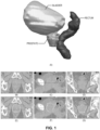
- FIG. 1 illustrates an exemplary three-dimensional (3D) CT image from a typical prostate cancer patient.
- Illustration (A) shows a pelvic region of the patient in a 3D view, which includes the patient's bladder, prostate, and rectum.
- Images (B), (C), and (D) are axial, sagittal and coronal views from a 3D CT image of this pelvic region.
- images (E), (F), and (G) show the expected prostate contour on the same 3D CT image.
- conventional image segmentation methods based solely on contrast and textures presented in the image would likely fail when used to segment this exemplary 3D CT image.
- Some methods propose combining atlas and statistical methods to gain improved segmentation accuracy. For example, one method bases its on-line learning and patient-specific classification on location-adaptive image contexts. The location-adaptive classifiers are trained on static image appearance features and image context features as well. Such a method has been used on serial prostate images with the stated goal of refining the patient's prostate segmentation as radiotherapy progressed. However, it requires that the data be treated as a three-dimensional data object using the spatial distribution of the voxel patch-pairs as features themselves, and in addition, using a random forest (RF) method.
- RF random forest
- Another method proposes combining bladder volume metric and landmark locations with conventional deformable registration in a study of atlas based segmentation of serial CT images of the cervix-uterus.
- the method can be computationally expensive and error prone since use of landmarks requires human intervention.
- Another method uses random forests for brain tissue segmentation (in a semi-supervised learning mode) such that the decision forest is trained on anatomy-labeled pre-surgical imagery and unlabeled post-surgical images of the same patient.
- this approach avoids the registration step and thus, fails to take advantage of useful information in Atlas-based registration.
- random forest segmentation has been combined with image registration to perform image segmentation. This allows for inter-patient segmentation, and through the mechanism of an "atlas forest", multiple patients' images were registered to a common coordinate frame, with one image per forest model.
- the method requires different compositions of training data to provide resulting models.
- deformable registration and random forests are used to segment the bones of the face and teeth from dental Cone Beam CT images.
- Patient images are registered to an atlas for an initial prediction or estimate of the segmentation. This is followed by a sequence of random forest classifiers that use context features as part of the features to be trained on at each stage of the sequence.
- to the method requires using deformable registration and RF classifiers to form RF models based on prior images of the same patient.
- the disclosed methods and systems are designed to solve one or more problems discussed above, by combining Atlas-based segmentation and statistical learning, combining population with intra-patient image and structure information, and enabling convenient updating of a patient segmentation model in order to improve segmentation performance on medical images in radiation therapy or related fields.
- Systems and methods consistent with the present disclosure are directed to segmenting a medical image of an object using learning algorithms trained using mapped atlases derived based on prior images of the same object.
- embodiments of the present disclosure provide various combinations of atlas-based auto-segmentation (ABAS) and Random Forest (RF) segmentations of serial CT images, incorporating patient-specific RF models learned on prior-day images and their structures.
- ABAS atlas-based auto-segmentation
- RF Random Forest
- a "learning algorithm” refers to any algorithm that can learn a model or a pattern based on existing information or knowledge. The learned model or pattern can be then used to predict or estimate output using input of new information or knowledge.
- the learning algorithm may be a machine learning algorithm or any other suitable learning algorithm.
- a supervised learning algorithm such as a Support Vector Machine (SVM), Adaboost/Logitboost, Random Forests, and neural network (e.g., a convolutional neural network), may be used.
- SVM Support Vector Machine
- Adaboost/Logitboost Adaboost/Logitboost
- Random Forests e.g., a convolutional neural network
- semi-supervised learning algorithms may also be used.
- Supervised learning is a branch of machine learning that infers a prediction model given a set of training data.
- Each individual sample of the training data is a pair containing a data vector (such as a series of measurements) and a desired output value.
- a supervised learning algorithm analyzes the training data and produces a predictor function.
- the predictor function is called a classifier or a classification model when the output is discrete, such as a list of labels identifying different groups.
- the predictor function once derived through training, is capable of predicting the correct output value for a valid input.
- image segmentation may be formulated as a learning-based classification function, which classifies each image point of the medical image into one of the anatomical structures.
- an "image point" refers to an image element in a digital image that corresponds to a physical point in the underlying object.
- the image point is a pixel in a 2D image or a voxel in a 3D image.
- the image segmentation may also classify image blocks rather than image points.
- an "image block” is a group of image points to be classified together.
- the image block may be a super-pixel in a 2D image, or a super-voxel in a 3D image.
- classifying based on image blocks may be more efficient and accurate. Accordingly, whenever the term "image point” is used throughout this disclosure, it intends to cover both the basic "image point” and also the "image block” as defined above.
- a label map refers to a map of structure labels each identifying a corresponding image point as being within a particular structure of interest.
- a label map may also be a probability map, which contains structure labels that each represents the probability of the image point belonging to the structure. For example, when segmenting an image including multiple structures, a structure label of an image point may provide a set of probability values indicting how likely the image point belonging to each of the structures under consideration.
- the classifier is trained using a set of training images.
- a "training image” is an image where the image points are already classified and labeled.
- a training image may be an atlas.
- an "atlas” includes an image and corresponding structure delineations (annotations) indicating what structure(s) the image points belong to.
- the image in an atlas also referred to as an atlas image, can be a previous image of the subject patient taken at an earlier time.
- the structure delineations can be represented as, for example, structure label maps, structure surfaces, or structure contours. The description below uses the label maps as an example of the structure delineations and is similarly applied to the scenarios of structure surfaces and contours.
- the training process uses features extracted from the atlas image and the corresponding structure delineations to train a classifier. Once properly trained using the process discussed in more detail below, such an algorithm can be used to segment a new image.
- the atlases used in the training process may be derived from prior images of the same object as the new image to be segmented. Similarity between such training images and the new image to be segmented can improve the segmentation accuracy and efficiency.
- the atlases used to train a classifier may be obtained by segmenting a previous image using a previously trained classifier.
- the atlases used may be registered to the current image.
- intra-patient images obtained during the course of radiation therapy are used.
- the intra-patient images are typically obtained one or more days between images to check on the patient's response to the prescribed therapy.
- Delineating the anatomy of serial images of the same patient's anatomy using the patient's prior-image information aids in the segmentation of succeeding images. Accurate serial image segmentation is a prerequisite for adaptive therapy plan assessment/re-planning and dose accumulation.
- the combination of Atlas-based auto-segmentation (ABAS) and statistical learning (SL) segmentations of serial CT images may improve segmented serial images of the same patient during adaptive planning and determining dose accumulation. This combination may also improve the prediction accuracy and thus the quality of image segmentation.
- the use of prior days' images and structures deformably registered to succeeding days' images to form statistical models may provide more accurate segmentations than models based on unregistered images.
- Atlas-based segmentation registers an image with anatomy labels attached to its voxels (the atlas) to a target image, and then assigns the atlas labels to the corresponding target voxels.
- Statistical learning assigns target voxel labels using a classifier program in which labels are associated with regions of a feature space.
- the features include voxel intensity, appearance (local variation measures), and structural properties of the images.
- the label-feature association is learned by training the classifier program on labeled images.
- the statistical learning method used is the Random Forests (RF) method.
- a random forest is a set of decision trees. Starting with a random sample of the training data (such as voxel average intensities for voxel patches, and pairs of patches located randomly around the index voxel), and a random selection of variables embedded in the data, the method determines the best parameter split of the samples into the label categories, splits the data sample, and then follows the split data down to the next pair of nodes where the best split is found. In an embodiment, this method is recursively performed until terminating at the maximum tree depth, or until a minimum sample size is reached. The terminal tree nodes (e.g., leaves) provide a label assignment.
- the terminal tree nodes e.g., leaves
- Trees thus trained perform classification when new feature data is processed through the trees' nodes. Multiple trees increase the discriminative power of the classifier. Random forest classifiers resist overfitting, and can be used for efficient image segmentations. Segmentation methods incorporating patient-specific RF models learned on prior-day images and their structures provide more accurate segmentation than methods than methods that solely use a population model.
- the disclosed image segmentation systems and methods can be applied to segment medical images obtained from any type of imaging modalities, including, but not limited to X-ray, CT, CBCT, spiral CT, magnetic resonance imaging (MRI), ultrasound (US), positron emission tomography (PET), single-photon emission computed tomography (SPECT), and optical images. Furthermore, the disclosed image segmentation systems and methods can be adapted to segment both 2D and 3D images.
- FIG. 2A is a block diagram showing an exemplary radiotherapy system 100, according to some embodiments of the present disclosure.
- Radiotherapy system 100 may be an IGRT system.
- radiotherapy system 100 may include a control console 110, a database 120, a radiotherapy device 130, and an image acquisition device 140.
- radiotherapy device 130 and image acquisition device 140 may be integrated into a single image-guided radiotherapy device 150, as indicated by the dashed box 150 in FIG. 2A .
- radiotherapy device 130 and image acquisition device 140 may be separate devices.
- radiotherapy device 130 and image acquisition device 140 may be physically or communicative connected to each other, as indicated by a dotted-dashed line between radiotherapy device 130 and image acquisition device 140 in FIG. 2A .
- Control console 110 may include hardware and software components to control radiotherapy device 130 and image acquisition device 140 and/or to perform functions or operations such as treatment planning, treatment execution, image acquisition, image processing, motion tracking, motion management, or other tasks involved in a radiotherapy process.
- the hardware components of control console 110 may include one or more computers (e.g., general purpose computers, workstations, servers, terminals, portable/mobile devices, etc.); processor devices (e.g., central processing units (CPUs), graphics processing units (GPUs), microprocessors, digital signal processors (DSPs), field programmable gate arrays (FPGAs), special-purpose or specially-designed processors, etc.); memory/storage devices (e.g., read-only memories (ROMs), random access memories (RAMs), flash memories, hard drives, optical disks, solid-state drives (SSDs), etc.); input devices (e.g., keyboards, mice, touch screens, mics, buttons, knobs, trackballs, levers, handles, joysticks, etc.); output devices
- control console 110 may include operation system software, application software, etc.
- control console 110 may include treatment planning/delivery software 115 that may be stored in a memory/storage device of control console 110.
- Software 115 may include computer readable and executable codes or instructions for performing the processes described in detail below.
- a processor device of control console 110 may be communicatively connected to a memory/storage device storing software 115 to access and execute the codes or instructions. The execution of the codes or instructions may cause the processor device to perform operations to achieve one or more functions consistent with the disclosed embodiments.
- Control console 110 may be communicatively connected to database 120 to access data.
- database 120 may be implemented using local hardware devices, such as one or more hard drives, optical disks, and/or servers that are in the proximity of control console 110.
- database 120 may be implemented in a data center or a server located remotely with respect to control console 110.
- Control console 110 may access data stored in database 120 through wired or wireless communication.
- Database 120 may include patient data 122.
- Patient data may include information such as (1) imaging data associated with a patient anatomical region, organ, or volume of interest segmentation data (e.g., MRI, CT, X-ray, PET, SPECT, and the like); (2) functional organ modeling data (e.g., serial versus parallel organs, and appropriate dose response models); (3) radiation dosage data (e.g., may include dose-volume histogram (DVH) information); or (4) other clinical information about the patient or course of treatment.
- imaging data associated with a patient anatomical region, organ, or volume of interest segmentation data e.g., MRI, CT, X-ray, PET, SPECT, and the like
- functional organ modeling data e.g., serial versus parallel organs, and appropriate dose response models
- radiation dosage data e.g., may include dose-volume histogram (DVH) information
- Machine data 124 may include information associated with radiotherapy device 130, image acquisition device 140, or other machines relevant to radiotherapy, such as radiation beam size, arc placement, on/off time duration, radiation treatment plan data, multi-leaf collimator (MLC) configuration, MRI pulse sequence, and the like.
- MLC multi-leaf collimator
- Image acquisition device 140 may provide medical images of a patient.
- image acquisition device 140 may provide one or more of MRI images (e.g., 2D MRI, 3D MRI, 2D streaming MRI, 4D volumetric MRI, 4D cine MRI); Computed Tomography (CT) images; Cone-Beam CT images; Positron Emission Tomography (PET) images; functional MRI images (e.g., fMRI, DCE-MRI, diffusion MRI); X-ray images; fluoroscopic images; ultrasound images; radiotherapy portal images; Single-Photo Emission Computed Tomography (SPECT) images; and the like.
- image acquisition device 140 may include an MRI imaging device, a CT imaging device, a PET imaging device, an ultrasound imaging device, a fluoroscopic device, a SPECT imaging device, or other medical imaging devices for obtaining the medical images of the patient.
- Radiotherapy device 130 may include a Leksell Gamma Knife, a linear accelerator or LINAC, or other suitable devices capable of delivering radiation to an anatomical region of interest of a patient in a controllable manner.
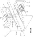
- FIG. 2B depicts an exemplary image-guided radiotherapy system 200, consistent with disclosed embodiments.
- the disclosed image segmentation system may be part of a radiotherapy system as described with reference to FIG. 2A .
- system 200 may include a couch 210, an image acquisition device 220, and a radiation delivery device 230.
- System 200 delivers radiation therapy to a patient in accordance with a radiotherapy treatment plan.
- Couch 210 may support a patient (not shown) during a treatment session.
- couch 210 may move along a horizontal, translation axis (labelled "I"), such that couch 210 can move the patient resting on couch 210 into and/or out of system 200.
- Couch 210 may also rotate around a central vertical axis of rotation, transverse to the translation axis.
- couch 210 may have motors (not shown) enabling the couch to move in various directions and to rotate along various axes.
- a controller (not shown) may control these movements or rotations in order to properly position the patient according to a treatment plan.
- image acquisition device 220 may include an MRI machine used to acquire 2D or 3D MRI images of the patient before, during, and/or after a treatment session.
- Image acquisition device 220 may include a magnet 221 for generating a primary magnetic field for magnetic resonance imaging.
- the magnetic field lines generated by operation of magnet 221 may run substantially parallel to the central translation axis I.
- Magnet 221 may include one or more coils with an axis that runs parallel to the translation axis I.
- the one or more coils in magnet 221 may be spaced such that a central window 223 of magnet 221 is free of coils.
- the coils in magnet 221 may be thin enough or of a reduced density such that they are substantially transparent to radiation of the wavelength generated by radiotherapy device 230.
- Image acquisition device 220 may also include one or more shielding coils, which may generate a magnetic field outside magnet 221 of approximately equal magnitude and opposite polarity in order to cancel or reduce any magnetic field outside of magnet 221.
- radiation source 231 of radiotherapy device 230 may be positioned in the region where the magnetic field is cancelled, at least to a first order, or reduced.
- Image acquisition device 220 may also include two gradient coils 225 and 226, which may generate a gradient magnetic field that is superposed on the primary magnetic field. Coils 225 and 226 may generate a gradient in the resultant magnetic field that allows spatial encoding of the protons so that their position can be determined. Gradient coils 225 and 226 may be positioned around a common central axis with the magnet 221, and may be displaced along that central axis. The displacement may create a gap, or window, between coils 225 and 226. In the embodiments wherein magnet 221 also includes a central window 223 between coils, the two windows may be aligned with each other.
- image acquisition device 220 may be an imaging device other than an MRI, such as an X-ray, a CT, a CBCT, a spiral CT, a PET, a SPECT, an optical tomography, a fluorescence imaging, ultrasound imaging, or radiotherapy portal imaging device.
- an imaging device other than an MRI such as an X-ray, a CT, a CBCT, a spiral CT, a PET, a SPECT, an optical tomography, a fluorescence imaging, ultrasound imaging, or radiotherapy portal imaging device.
- Radiotherapy device 230 may include the source of radiation 231, such as an X-ray source or a linear accelerator, and a multi-leaf collimator (MLC) 233. Radiotherapy device 230 may be mounted on a chassis 235. One or more chassis motors (not shown) may rotate chassis 235 around couch 210 when couch 210 is inserted into the treatment area. In an embodiment, chassis 235 may be continuously rotatable around couch 210, when couch 210 is inserted into the treatment area. Chassis 235 may also have an attached radiation detector (not shown), preferably located opposite to radiation source 231 and with the rotational axis of chassis 335 positioned between radiation source 231 and the detector. Further, device 230 may include control circuitry (not shown) used to control, for example, one or more of couch 210, image acquisition device 220, and radiotherapy device 230. The control circuitry of radiotherapy device 230 may be integrated within system 200 or remote from it.
- MLC multi-leaf collimator
- a patient may be positioned on couch 210.
- System 200 may then move couch 310 into the treatment area defined by magnetic coils 221, 225, 226, and chassis 235.
- Control console 240 may then control radiation source 231, MLC 233, and the chassis motor(s) to deliver radiation to the patient through the window between coils 225 and 226 according to a radiotherapy treatment plan.
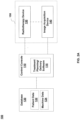
- FIG. 3 illustrates the image segmentation system 300 for segmenting medical images, according invention.
- Image segmentation system 300 includes a medical image database 301, a classifier training unit 302, a structure classification unit 303, and a network 305.
- image segmentation system 300 may include more or less of the components shown in FIG. 3 .
- image segmentation system 300 may only include structure classification unit 303, medical image database 301, and, optionally, network 305.
- the various components of image segmentation system 300 may locate remotely from each other, and be connected through network 305. In some alternative embodiments, certain components of image segmentation system 300 may be located on the same site or inside one device.
- medical image database 301 may be located on site with classifier training unit 302, or be part of classifier training unit 302.
- classifier training unit 302 and structure classification unit 303 may be inside the same computer or processing device.
- Image segmentation system 300 may be used to segment serial intra-patient CT images stored in medical image database 301.
- serial images may be images acquired during serial radiotherapy treatment sessions of a patient.
- the serial radiotherapy treatment sessions may be performed at a set frequency (e.g., every day, every week, etc.) or at discrete time points, according to a radiotherapy treatment plan for the patient.
- the serial images may include current images and prior images.
- a "current image” may be a current day medical image of a patient, e.g., an image taken during the treatment session of a patient that occurred on the present day. As shown in FIG.
- classifier training unit 302 may communicate with medical image database 301 to receive one or more "prior images" of the same patient.
- a "prior image” may be an image taken during a treatment session of the same patient but occurred on a previous day.
- the prior images stored in medical image database 301 may be obtained from a medical image database, which contain images of previous radiotherapy treatment sessions.
- the prior images may be pre-segmented.
- the prior images may be segmented automatically by image segmentation system 300, or manually by user 313.
- User 313 may be an expert, e.g., a radiologist or another physician familiar with anatomical structures in medical images, who provides expert structure label maps associated with the prior images.
- the prior images and their corresponding structure label maps become atlases that can be readily used by classifier training unit 302 to train a structure classifier.
- structure classification unit 303 may use the most recently trained classifier to segment the prior images, or a merged classifier of selected previously trained classifiers for the segmentation.
- Structure classification unit 303 may provide a structure label map for each prior image. Structure classification unit 303 may then provide the atlases, consisting of the prior images along with their corresponding structure label maps, to classifier training unit 302, as training images.
- Classifier training unit 302 may use the training images received from medical image database 301 to generate a structure classifier using learning algorithms. As shown in FIG. 3 , classifier training unit 302 may include an atlas registration module 321, a feature extraction module 322 and a training module 323. Classifier training unit 302 may additionally include input and output interfaces (not shown) to communicate with medical image database 301, network 305, and/or user 312. Consistent with some embodiments, classifier training unit 302 may be implemented with hardware (e.g., as disclosed in FIG. 4 ) specially programmed by software that performs an anatomical classifier training process.
- Atlas registration module 321 may register the prior images to the current images.
- Image registration is a process that transforms different sets of data into one coordinate system. Typical image registration algorithms are either intensity-based or feature-based, or the combination of the two. In particular, feature-based methods find correspondence between image features such as points, lines, and contours.
- the registration process may include mapping the image points of an atlas image to the image points of a current image.
- the registration process may include mapping both the atlas images and the current image to a reference image.
- the reference image can be, for example, an average atlas image or a common template image. As such, the atlas images are "indirectly" mapped to the current image.
- image registration methods can be used, such as one or a combination of any of a linear registration, an object-driven "poly-smooth" non-linear registration, or a shape-constrained dense deformable registration.
- image registration an image transformation from the atlas image to the reference image is calculated for each atlas.
- Atlas registration module 321 may further map the delineations (e.g., structure labels) of each atlas to the space of the reference image using the corresponding image transformation for the atlas.
- the mapped structure labels represent independent classification data, i.e., independent segmentation results, of the current image from the corresponding atlas.
- a mapped atlas image and corresponding mapped structure labels constitute a mapped atlas, also referred to as a "registered atlas.”
- classifier training unit 302 may use the mapped atlases to train the structure classifier for segmenting the current images.
- classifier training unit 302 may use the mapped atlas images along with expert structure label maps for the training.
- Feature extraction module 322 may determine and derive, for each selected image point, one or more features such as image intensity, image texture, image patch, and curvature of an intensity profile. This feature extraction process may repeat for a set of selected image points in a training image until all image points in the training image have been selected and processed.
- Training module 323 may use the selected image points as training data, to train the classifier.
- the training may be based on learning algorithms, such as supervised machine learning algorithms.
- learning algorithms such as Support Vector Machine (SVM), Adaboost/Logitboost, Random Forests, and Neural Networks may be used.
- SVM Support Vector Machine
- Adaboost/Logitboost Adaboost/Logitboost
- Random Forests Random Forests
- Neural Networks may be used.
- the classifier is trained such that when the features for a particular image point in the training image are input to the model, the model outputs a prediction of the anatomical structure that matches the predetermined structure label of the image point. After being trained using numerous image points from numerous training images, the classifier becomes competent enough to predict the anatomical structure of an unclassified image point in any new image.
- Structure classification unit 303 may receive the trained structure classifier from classifier training unit 302. As shown in FIG. 3 , structure classification unit 303 may include a feature extraction module 331 and a classification module 332. Structure classification unit 303 may additionally include input and output interfaces (not shown) to communicate with medical image database 301, network 305 and user 313. Consistent with some embodiments, structure classification unit 303 may be implemented with hardware (e.g., as disclosed in FIG. 4 ) specially programmed by software that performs an anatomical classifier training process.
- Structure classification unit 303 may communicate with medical image database 301 to receive one or more current images.
- the current images may be of a same object as the prior images.
- Feature extraction module 331 may have similar hardware and software structures as feature extraction module 322.
- Feature extraction module 331 may identify one or more features on each current image received from medical image database 301.
- the features extracted by feature extraction module 331 may be the same or similar to those used during the training stage by feature extraction module 322.
- the determined features may be provided to classification module 332.
- Classification module 332 may use the trained structure classifier received from classifier training unit 302, and the features received from feature extraction module 331, to predict the structure labels for the respective image points. When all the selected image points are classified, classification module 332 may output the segmented image. In some embodiments, the segmented image may be displayed to user 313, and/or provided to treatment planning/delivery software 115 for further treatment usage. In some embodiments, the segmented image may be automatically stored in medical image database 301 and become a prior image.
- Network 305 may provide connections between any of the above described components in image segmentation system 300.
- network 305 may be a local area network (LAN), a wireless network, a cloud computing environment (e.g., software as a service, platform as a service, infrastructure as a service), a client-server, a wide area network (WAN), and the like.
- LAN local area network
- wireless network e.g., a wireless local area network
- cloud computing environment e.g., software as a service, platform as a service, infrastructure as a service
- client-server e.g., a client-server
- WAN wide area network
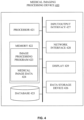
- FIG. 4 illustrates an exemplary medical image processing device 400, according to some embodiments of the present disclosure.
- Medical image processing device 400 may be an embodiment of classifier training unit 302, structure classification unit 303, or their combination. In another embodiment, processing device 400 may be integrated into control console 110 or radiotherapy device 230, shown in FIGS. 2A-(B) .
- medical image processing device 400 may be a special-purpose computer, or a general-purpose computer. For example, medical image processing device 400 may be a computer custom built for hospitals to handle image acquisition and image processing tasks.
- medical image processing device 400 may include a processor 421, a memory 422, a database 425, a data storage device 426, an input/output interface 427, a network interface 428, and a display 429. Components of processing device 400 may be connected via a BUS.
- Processor 421 may be one or more general-purpose processing devices, such as a microprocessor, central processing unit (CPU), graphics processing unit (GPU), or the like. More particularly, processor 421 may be a complex instruction set computing (CISC) microprocessor, reduced instruction set computing (RISC) microprocessor, very long instruction Word (VLIW) microprocessor, a processor implementing other instruction sets, or processors implementing a combination of instruction sets. Processor 421 may also be one or more special-purpose processing devices such as an application specific integrated circuit (ASIC), a field programmable gate array (FPGA), a digital signal processor (DSP), a System on a Chip (SoC), or the like. Processor 421 may be communicatively coupled to memory 422 and configured to execute the computer executable instructions stored thereon.
- ASIC application specific integrated circuit
- FPGA field programmable gate array
- DSP digital signal processor
- SoC System on a Chip
- Memory 422 may include a read-only memory (ROM), a flash memory, a random access memory (RAM), and a static memory, for example.
- memory 422 may store computer executable instructions, such as one or more image processing programs 423, as well as data used or generated while executing the computer programs, such as medical image data 424.
- Processor 421 may execute image processing programs 423 to implement functionalities of anatomical classifier training unit 302 and/or structure classification unit 303.
- Processor 421 may also send/receive medical image data 424 from memory 422. For example, processor 421 may receive prior image data or current image data stored in memory 422. Processor 421 may also generate intermediate data such as image features and structure labels, and send them to memory 422.
- Medical image processing device 400 may optionally include a database 425, which may include or in communication with medical image database 301.
- Database 425 may include a plurality of devices located either in a central or distributed manner.
- Processor 421 may communicate with database 425 to read images into memory 422 or store segmented images from memory 422 to medical image data 424.
- Data storage device 426 may be an additional storage available to store data associated with image processing tasks performed by processor 421.
- data storage device 426 may include a machine-readable storage medium. While the machine-readable storage medium in an embodiment may be a single medium, the term “machine-readable storage medium” should be taken to include a single medium or multiple media (e.g., a centralized or distributed database, and/or associated caches and servers) that store the one or more sets of computer executable instructions or data.
- the term “machine-readable storage medium” shall also be taken to include any medium that is capable of storing or encoding a set of instructions for execution by the machine and that cause the machine to perform any one or more of the methodologies of the present disclosure.
- the term “machine-readable storage medium” shall accordingly be taken to include, but not be limited to, solid-state memories, optical and magnetic media.
- Input/output 427 may be configured to allow data to be received and/or transmitted by medical image processing device 400.
- Input/output 427 may include one or more digital and/or analog communication devices that allow processing device 400 to communicate with user or other machines and devices.
- input/output 427 may include a keyboard and a mouse for user 312 or user 313 to provide input.
- Network interface 428 may include a network adaptor, a cable connector, a serial connector, a USB connector, a parallel connector, a high-speed data transmission adaptor such as fiber, USB 3.0, thunderbolt, and the like, a wireless network adaptor such as a WiFi adaptor, a telecommunication (3G, 4G/LTE and the like) adaptor, and the like.
- Medical image processing device 400 may be connected to network 305 through network interface 428.
- Display 429 may be any display device that suitable for displaying the medical images.
- display 429 may be an LCD, CRT, or LED display.
- the segmented current image may be displayed to a user on display 428.
- the segmented current image may be provided to treatment planning/delivery software 115 for future medical usage and/or stored in medical image database 301 for future image segmentation tasks.
- An accurately segmented image, or a well-defined contour of an anatomical structure may benefit various applications that rely on segmentation results.
- structure classification results may also help generate accurate estimation of volume size of the anatomical structure.
- volume sizes are important in calculating the deformation field and dose optimization for treatments.
- the volume sizes may change significantly at different treatment sessions. Therefore, accurate estimation of its size will give important prior knowledge about the relative locations or deformations around the bladder, and thus help calculate the deformation field or optimize dose distribution on the fly.
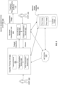
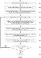
- FIG. 5 is a flowchart illustrating an exemplary image segmentation method 500 using combined atlas-based segmentation and statistical learning segmentation.
- the method 500 is performed by components of image segmentation system 300, such as classifier training unit 302 and structure classification unit 303. It is contemplated that method 500 can be applied to segment one structure of interest or a group of structures of interest at the same time, such as a bladder, prostate, and rectum, which are spatially adjacent and highly correlated.
- Various machine learning methods such as RF method, can handle segmentation of multiple structures at the same time.
- a multi-structure classifier model may be beneficial when the multiple structures are spatially adjacent and thus highly correlated.
- medical image processing device 400 receives an initial day medical image I 1 from a database, such as medical image database 301.
- medical image I 1 represents a prior day medical image of a patient at day one.
- medical image processing device 400 may identify at least one feature from the day one image I 1 .
- the at least one feature may be identified by feature extraction module 332 of structure classification unit 303, for example.
- the features can be the same types of features used for training the structure classifier, as will be described in more detail below in connection with FIG. 6 .
- Various methods may be used to compute the attributes, including using machine learning models such as convolutional neural network models.
- the day one image I 1 is segmented by a population-trained classifier model M 1 .
- the day one image may be segmented by a random forests model M 1 trained on training images from a population of patients.
- the day one image I 1 may be segmented by the population-trained M 1 to produce a structure label map S 1 , e.g., representing the prostate, bladder, rectum, and/or other organs.
- medical image processing device 400 receives the j -th succeeding day medical image I j from a database, such as medical image database 301.
- day one image I 1 and succeeding day medical image I j are acquired during serial radiotherapy treatment sessions of the same patient.
- the medical images may be acquired by image acquisition device 220 during successive treatment therapy sessions delivered by image-guided radiotherapy system 200.
- I 1 , S 1 is used as an atlas to deformably register I 1 to day two image I 2 to obtain the registered image I 2 (1) and day two structure label map S 2 (1) .
- the superscript refers to the atlas-day number, and serves to distinguish the preliminary segmentation result S j j ⁇ 1 from the full-model classification S j described below in 508.
- a classifier model M temp is trained on the image I j j ⁇ 1 and the structure map S j j ⁇ 1 obtained as described above in step 505.
- the model may be trained according to an exemplary training method described in FIG. 6 .
- a new random forest model M temp may be trained on the set I j ( j ⁇ 1) , S j j ⁇ 1 .
- the trained new classifier model M temp is merged with the prior day classifier model M j-1 to obtain a new classifier model M j .
- Various methods may be used to merge the models.
- the model parameters may be determined based on the model parameters of model M temp and model M j .
- the combined model is used to segment the current day image I j , to produce the structure label map Sy.
- This Structure label map S j is derived from the model M j while the structure label map S j j ⁇ 1 is obtained by atlas-based segmentation using the preceding day data I j-1 , S j-1 .
- feature extractions may be performed on the j-th prior day image, similar to 502.
- the method determines whether all succeeding prior day images for that patient have been used for training the classifier model. If there are more succeeding prior day images for that patient, medical image processing device 400 repeats steps 504-509 to train model M j (where j represents the j-th treatment day) until all the succeeding prior day images are processed. At 510, the segmentation of current image day I j is complete.
- the training is done in stages, one prior day per stage, resulting in a model M temp that is then combined with prior day's models, as the statistical learning method permits.
- the training data ensemble could contain other forms of the structure label maps than that derived from the atlas-based segmentation.
- the structure label map could be atlas-based segmentation structures corrected by expert programs or by human experts.
- the population based model M 1 (here included in the resulting model M j ) could be dissociated from the models M 2 to M j .
- composition of the training data is varied such that the training data for each j -th treatment day is accumulated from the beginning of treatment and a model created from all the accumulated training data, rather than sequential merging successive days' trained models.
- This accommodates statistical learning methods such as neural networks that, unlike random or decision forests, have models that are not easily decomposed into daily data-trained increments.
- method 500 may be performed prior to radiotherapy treatment delivery to a patient, and the determined structure label map S j may be provided to treatment planning/delivery software 115. This segmentation result may be used to adjust the radiotherapy treatment plan. For that purpose, the segmentation (method 500) may be performed immediately prior to the upcoming radiotherapy treatment session, or one day prior to the upcoming radiotherapy treatment session.
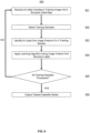
- FIG. 6 is a flowchart illustrating an exemplary training method 600 for training a structure classifier model using an atlas.
- method 600 may be used to implement step 506 of FIG. 5 described above.
- method 600 may be performed by classifier training unit 302.
- the structure classifier model may be a Random Forests model, such as the classifier model M j of FIG. 5 .
- classifier training unit 302 may receive an atlas that includes a training image and a corresponding structure label map indicating the structures that the image points of the training image belong to.
- the atlas may be a registered atlas, such as atlas I j j ⁇ 1 , S j j ⁇ 1 .
- feature extraction module 322 may select a plurality of training samples from the mapped atlas image of each training atlas.
- Each training sample can correspond to a single image point or a group of image points (such a group of image points is also referred to as a super image point).
- the training samples from a mapped atlas image can include all or a portion of the image points on the mapped atlas image.
- a sample selection can be performed to determine what image points are used.
- the training samples can be selected fully randomly over the entire mapped atlas image, or be selected from a region within a certain distance to the border of the structure of interest.
- the sample selection can be guided by the registration results such that more samples can be selected from an ambiguous region, i.e., the region where structure labels from different mapped atlases do not completely agree with each other or the disagreement is larger than a certain level (for example, three or more out often mapped atlases have a different determination than the other mapped atlases).
- an ambiguous region i.e., the region where structure labels from different mapped atlases do not completely agree with each other or the disagreement is larger than a certain level (for example, three or more out often mapped atlases have a different determination than the other mapped atlases).
- feature extraction module 322 may determine at least one image feature for an image point.
- Various types of features can be extracted, such as, for example, image intensity value, image location, image gradient and gradient magnitude, eigen-values of a Hessian matrix of the image, image texture measures such as energy, entropy, contrast, homogeneity, and correlation of local co-occurrence matrix, local image patches of varying sizes.
- attributes or features may also be automatically and adaptively computed using machine learning models.
- a convolutional neural network model may be trained to extract relevant features from sample images, and the pre-trained model can be applied to the training samples to produce attributes.
- a convolutional neural network typically includes several convolution layers, among other layers, that produce feature maps of various sizes.
- the feature maps contain generic features characterizing the input image (or a selected portion of the input image), and thus can be used as features in the structure classifier to further improve classification results.
- Features from various convolution layers e.g., top layers, middle layers, lower layers, or a selection of these layers, may be used.
- computation of attributes can be omitted if the training atlases already include the attributes for the atlas image points that are to be used by the machine learning algorithm.
- training module 323 may apply a learning algorithm to generate an anatomical classification model based on the identified image features for the image point.
- the machine learning algorithm can be a supervised learning algorithm, which seeks to infer a prediction model given a set of training data.
- the machine learning algorithm for training the structure classifier can be the random forests (RF) machine learning algorithm, which can naturally handle multiple classes, i.e., one classifier to classify several structures.
- the output of an RF classifier can be a probability estimation of which class the input data belongs to, i.e., which structure the corresponding image point belongs to.
- classifier training unit 302 may check if all training samples have been process.
- method 600 may proceed to 606, where classifier training unit 302 outputs the trained classifier model, e.g., to be used by steps 507-508 of method 500. Otherwise (605: no), method 600 may return to 601 to process the next training sample.
- an exemplary image segmentation process uses prior day images and expert structures or contours C to segment succeeding-days images, without utilizing image registration.
- the expert drawn or edited contours C are distinguished from the atlas segmentation contours S (e.g., the ABAS structures have been edited by an expert).
- an exemplary image segmentation process utilizes method 500 disclosed in FIG. 5 , but replaces the prior days' ABAS structures S with that day's expert structures C.
- the method uses ABAS structures from the previous day.
- the classifier model may be a convolutional neural network model.
- a convolutional neural network may include a stack of distinct layers that transform an input image into an output structure label map.
- the layers may form two stages: an encoding stage and a decoding stage.
- the layers may differ in input size, output size, and the relationship between the input and the output for the layer.
- Each layer may be connected to one or more upstream and downstream layers in the stack of layers.
- the performance of a convolutional neural network may thus depend on the number of layers, and the convolutional neural network's complexity may increase as the number of layers increases.
- a convolutional neural network may be viewed as "deep” if it has more than one stages of non-linear feature transformation, which typically means the number of layers in the network is above a certain number. For example, some convolutional neural networks may include about 10-30 layers, or in some cases more than a few hundred layers. Examples of convolutional neural network models include AlexNet, VGGNet, GoogLeNet, ResNet, etc. These convolutional neural network models can be used at the encoding stage of the full convolutional neural network model.
- Models could be weighted to de-emphasize the population, for instance, or to delete it all together.
- the individual day's images and structures could be trained in single-image RF models that could later be merged.
- Other combinations of registered and non-registered sets of images/structures may be possible.
- context features may be used in addition to the appearance features described above.
- a machine or computer readable storage medium may cause a machine to perform the functions or operations described, and includes any mechanism that stores information in a form accessible by a machine (e.g., computing device, electronic system, and the like), such as recordable/non-recordable media (e.g., read only memory (ROM), random access memory (RAM), magnetic disk storage media, optical storage media, flash memory devices, and the like).
- a communication interface includes any mechanism that interfaces to any of a hardwired, wireless, optical, and the like, medium to communicate to another device, such as a memory bus interface, a processor bus interface, an Internet connection, a disk controller, and the like.
- the communication interface can be configured by providing configuration parameters and/or sending signals to prepare the communication interface to provide a data signal describing the software content.
- the communication interface can be accessed via one or more commands or signals sent to the communication interface.
- the present disclosure also relates to a system for performing the operations herein.
- This system may be specially constructed for the required purposes, or it may comprise a general-purpose computer selectively activated or reconfigured by a computer program stored in the computer.
- a computer program may be stored in a computer readable storage medium, such as, but not limited to, any type of disk including floppy disks, optical disks, CDROMs, and magnetic-optical disks, read-only memories (ROMs), random access memories (RAMs), EPROMs, EEPROMs, magnetic or optical cards, or any type of media suitable for storing electronic instructions, each coupled to a computer system bus.
- Embodiments of the present disclosure may be implemented with computer-executable instructions.
- the computer-executable instructions may be organized into one or more computer-executable components or modules.
- Aspects of the present disclosure may be implemented with any number and organization of such components or modules.
- aspects of the present disclosure are not limited to the specific computer-executable instructions or the specific components or modules illustrated in the figures and described herein.
- Other embodiments of the present disclosure may include different computer-executable instructions or components having more or less functionality than illustrated and described herein.
Landscapes
- Engineering & Computer Science (AREA)
- Theoretical Computer Science (AREA)
- Physics & Mathematics (AREA)
- Computer Vision & Pattern Recognition (AREA)
- General Physics & Mathematics (AREA)
- Health & Medical Sciences (AREA)
- Life Sciences & Earth Sciences (AREA)
- Medical Informatics (AREA)
- General Health & Medical Sciences (AREA)
- Databases & Information Systems (AREA)
- Evolutionary Computation (AREA)
- Artificial Intelligence (AREA)
- Data Mining & Analysis (AREA)
- Software Systems (AREA)
- Radiology & Medical Imaging (AREA)
- Nuclear Medicine, Radiotherapy & Molecular Imaging (AREA)
- Bioinformatics & Computational Biology (AREA)
- General Engineering & Computer Science (AREA)
- Bioinformatics & Cheminformatics (AREA)
- Evolutionary Biology (AREA)
- Surgery (AREA)
- Animal Behavior & Ethology (AREA)
- Veterinary Medicine (AREA)
- Biophysics (AREA)
- High Energy & Nuclear Physics (AREA)
- Public Health (AREA)
- Optics & Photonics (AREA)
- Pathology (AREA)
- Molecular Biology (AREA)
- Biomedical Technology (AREA)
- Heart & Thoracic Surgery (AREA)
- Probability & Statistics with Applications (AREA)
- Multimedia (AREA)
- Computing Systems (AREA)
- Pulmonology (AREA)
- Quality & Reliability (AREA)
- Human Computer Interaction (AREA)
- Apparatus For Radiation Diagnosis (AREA)
- Magnetic Resonance Imaging Apparatus (AREA)
Applications Claiming Priority (3)
| Application Number | Priority Date | Filing Date | Title |
|---|---|---|---|
| US201662281652P | 2016-01-21 | 2016-01-21 | |
| US15/385,732 US10169871B2 (en) | 2016-01-21 | 2016-12-20 | Systems and methods for segmentation of intra-patient medical images |
| PCT/US2017/013962 WO2017127439A1 (en) | 2016-01-21 | 2017-01-18 | Systems and methods for segmentation of intra-patient medical images |
Publications (2)
| Publication Number | Publication Date |
|---|---|
| EP3405909A1 EP3405909A1 (en) | 2018-11-28 |
| EP3405909B1 true EP3405909B1 (en) | 2023-11-15 |
Family
ID=59359077
Family Applications (1)
| Application Number | Title | Priority Date | Filing Date |
|---|---|---|---|
| EP17703003.8A Active EP3405909B1 (en) | 2016-01-21 | 2017-01-18 | Systems and methods for segmentation of intra-patient medical images |
Country Status (6)
| Country | Link |
|---|---|
| US (3) | US10169871B2 (enExample) |
| EP (1) | EP3405909B1 (enExample) |
| JP (1) | JP6742421B2 (enExample) |
| CN (1) | CN109074500B (enExample) |
| AU (1) | AU2017209046B2 (enExample) |
| WO (1) | WO2017127439A1 (enExample) |
Cited By (11)
| Publication number | Priority date | Publication date | Assignee | Title |
|---|---|---|---|---|
| US12178666B2 (en) | 2019-07-29 | 2024-12-31 | Augmedics Ltd. | Fiducial marker |
| US12186028B2 (en) | 2020-06-15 | 2025-01-07 | Augmedics Ltd. | Rotating marker for image guided surgery |
| US12206837B2 (en) | 2015-03-24 | 2025-01-21 | Augmedics Ltd. | Combining video-based and optic-based augmented reality in a near eye display |
| US12201384B2 (en) | 2018-11-26 | 2025-01-21 | Augmedics Ltd. | Tracking systems and methods for image-guided surgery |
| US12239385B2 (en) | 2020-09-09 | 2025-03-04 | Augmedics Ltd. | Universal tool adapter |
| US12354227B2 (en) | 2022-04-21 | 2025-07-08 | Augmedics Ltd. | Systems for medical image visualization |
| US12383369B2 (en) | 2019-12-22 | 2025-08-12 | Augmedics Ltd. | Mirroring in image guided surgery |
| US12417595B2 (en) | 2021-08-18 | 2025-09-16 | Augmedics Ltd. | Augmented-reality surgical system using depth sensing |
| US12461375B2 (en) | 2022-09-13 | 2025-11-04 | Augmedics Ltd. | Augmented reality eyewear for image-guided medical intervention |
| US12458411B2 (en) | 2017-12-07 | 2025-11-04 | Augmedics Ltd. | Spinous process clamp |
| US12491044B2 (en) | 2021-07-29 | 2025-12-09 | Augmedics Ltd. | Rotating marker and adapter for image-guided surgery |
Families Citing this family (89)
| Publication number | Priority date | Publication date | Assignee | Title |
|---|---|---|---|---|
| US10169871B2 (en) | 2016-01-21 | 2019-01-01 | Elekta, Inc. | Systems and methods for segmentation of intra-patient medical images |
| US10098606B2 (en) * | 2016-02-29 | 2018-10-16 | Varian Medical Systems, Inc. | Automatic organ-dose-estimation for patient-specific computed tomography scans |
| US10055839B2 (en) * | 2016-03-04 | 2018-08-21 | Siemens Aktiengesellschaft | Leveraging on local and global textures of brain tissues for robust automatic brain tumor detection |
| CN106097444B (zh) * | 2016-05-30 | 2017-04-12 | 百度在线网络技术(北京)有限公司 | 高精地图生成方法和装置 |
| CN109964249A (zh) * | 2016-09-21 | 2019-07-02 | 皇家飞利浦有限公司 | 用于对身体部分的自适应轮廓勾画的装置 |
| US10607342B2 (en) * | 2016-09-30 | 2020-03-31 | Siemenes Healthcare GmbH | Atlas-based contouring of organs at risk for radiation therapy |
| US11138790B2 (en) | 2016-10-14 | 2021-10-05 | Axial Medical Printing Limited | Method for generating a 3D physical model of a patient specific anatomic feature from 2D medical images |
| GB201617507D0 (en) | 2016-10-14 | 2016-11-30 | Axial3D Limited | Axial3D UK |
| US11195600B2 (en) | 2016-10-17 | 2021-12-07 | International Business Machines Corporation | Automatic discrepancy detection in medical data |
| US10410348B2 (en) * | 2016-12-21 | 2019-09-10 | Elekta, Inc. | Online learning enhanced atlas-based auto-segmentation |
| WO2018165025A1 (en) * | 2017-03-05 | 2018-09-13 | The Charles Stark Draper Laboratory, Inc. | System and method for image guided tracking to enhance radiation therapy |
| EP3392832A1 (en) * | 2017-04-21 | 2018-10-24 | General Electric Company | Automated organ risk segmentation machine learning methods and systems |
| US11748877B2 (en) | 2017-05-11 | 2023-09-05 | The Research Foundation For The State University Of New York | System and method associated with predicting segmentation quality of objects in analysis of copious image data |
| WO2018215183A1 (en) * | 2017-05-26 | 2018-11-29 | Asml Netherlands B.V. | Actuator, linear motor and lithographic apparatus |
| US10699410B2 (en) * | 2017-08-17 | 2020-06-30 | Siemes Healthcare GmbH | Automatic change detection in medical images |
| WO2019051356A1 (en) * | 2017-09-08 | 2019-03-14 | The General Hospital Corporation | SYSTEM AND METHOD FOR AUTOMATICALLY LABELING AND ANNOUNTING NON-STRUCTURED MEDICAL DATA SETS |
| KR101995900B1 (ko) * | 2017-09-11 | 2019-07-04 | 뉴로핏 주식회사 | 3차원 뇌지도 생성 방법 및 프로그램 |
| US11723579B2 (en) | 2017-09-19 | 2023-08-15 | Neuroenhancement Lab, LLC | Method and apparatus for neuroenhancement |
| EP3462373A1 (en) * | 2017-10-02 | 2019-04-03 | Promaton Holding B.V. | Automated classification and taxonomy of 3d teeth data using deep learning methods |
| US11562487B2 (en) * | 2017-10-18 | 2023-01-24 | Koninklijke Philips N.V. | Landmark visualization for medical image segmentation |
| US10607135B2 (en) * | 2017-10-19 | 2020-03-31 | General Electric Company | Training an auto-encoder on a single class |
| US10460440B2 (en) | 2017-10-24 | 2019-10-29 | General Electric Company | Deep convolutional neural network with self-transfer learning |
| WO2019085985A1 (en) * | 2017-11-02 | 2019-05-09 | Shenzhen United Imaging Healthcare Co., Ltd. | Systems and methods for generating semantic information for scanning image |
| EA201700561A1 (ru) | 2017-11-16 | 2019-05-31 | Общество С Ограниченной Ответственностью "Доммар" | Способ и система выравнивания зубов на основании моделирования движения коронок и корней |
| US11717686B2 (en) | 2017-12-04 | 2023-08-08 | Neuroenhancement Lab, LLC | Method and apparatus for neuroenhancement to facilitate learning and performance |
| US10548552B2 (en) * | 2017-12-21 | 2020-02-04 | Shenzhen Keya Medical Technology Corporation | Method and device for generating anatomical labels for a physiological tree structure |
| US11478603B2 (en) | 2017-12-31 | 2022-10-25 | Neuroenhancement Lab, LLC | Method and apparatus for neuroenhancement to enhance emotional response |
| US12280219B2 (en) | 2017-12-31 | 2025-04-22 | NeuroLight, Inc. | Method and apparatus for neuroenhancement to enhance emotional response |
| EP3735176A4 (en) | 2018-01-03 | 2021-10-20 | Ramot at Tel-Aviv University Ltd. | MULTIMODAL IMAGE DATA SEGMENTATION SYSTEMS AND METHODS |
| US10878576B2 (en) * | 2018-02-14 | 2020-12-29 | Elekta, Inc. | Atlas-based segmentation using deep-learning |
| US10910099B2 (en) * | 2018-02-20 | 2021-02-02 | Siemens Healthcare Gmbh | Segmentation, landmark detection and view classification using multi-task learning |
| US11657087B2 (en) | 2018-03-19 | 2023-05-23 | Verily Life Sciences Llc | Surgical video retrieval based on preoperative images |
| US20190295709A1 (en) * | 2018-03-20 | 2019-09-26 | Siemens Healthcare Gmbh | Automatic analysis of a large patient population using medical image data |
| KR102565278B1 (ko) * | 2018-03-26 | 2023-08-09 | 삼성전자주식회사 | 영상 분할 방법, 영상 분할 장치, 및 영상 분할을 위한 학습 방법 |
| US11364361B2 (en) | 2018-04-20 | 2022-06-21 | Neuroenhancement Lab, LLC | System and method for inducing sleep by transplanting mental states |
| US11980507B2 (en) | 2018-05-02 | 2024-05-14 | Augmedics Ltd. | Registration of a fiducial marker for an augmented reality system |
| CN108921782B (zh) * | 2018-05-17 | 2023-04-14 | 腾讯科技(深圳)有限公司 | 一种图像处理方法、装置及存储介质 |
| WO2019246580A1 (en) * | 2018-06-21 | 2019-12-26 | Procept Biorobotics Corporation | Artificial intelligence for robotic surgery |
| CN110638477B (zh) * | 2018-06-26 | 2023-08-11 | 佳能医疗系统株式会社 | 医用图像诊断装置以及对位方法 |
| US12005270B2 (en) * | 2018-06-26 | 2024-06-11 | The Medical College Of Wisconsin, Inc. | Systems and methods for accelerated online adaptive radiation therapy |
| US10943115B2 (en) * | 2018-07-24 | 2021-03-09 | Apical Ltd. | Processing image data to perform object detection |
| US10902588B2 (en) | 2018-08-13 | 2021-01-26 | International Business Machines Corporation | Anatomical segmentation identifying modes and viewpoints with deep learning across modalities |
| WO2020056418A1 (en) | 2018-09-14 | 2020-03-19 | Neuroenhancement Lab, LLC | System and method of improving sleep |
| US11080643B2 (en) | 2018-09-28 | 2021-08-03 | I.D. Systems, Inc. | Cargo sensors, cargo-sensing units, cargo-sensing systems, and methods of using the same |
| JP7129870B2 (ja) * | 2018-10-01 | 2022-09-02 | 富士フイルム株式会社 | 疾患領域を判別する判別器の学習装置、方法及びプログラム、疾患領域を判別する判別器、並びに疾患領域判別装置及びプログラム |
| EP3865070B1 (en) * | 2018-10-12 | 2024-07-31 | FUJIFILM Corporation | Ultrasound diagnosis device and ultrasound diagnosis device control method |
| US10930386B2 (en) | 2018-12-11 | 2021-02-23 | International Business Machines Corporation | Automated normality scoring of echocardiograms |
| US10832392B2 (en) * | 2018-12-19 | 2020-11-10 | Siemens Healthcare Gmbh | Method, learning apparatus, and medical imaging apparatus for registration of images |
| CN109741346B (zh) * | 2018-12-30 | 2020-12-08 | 上海联影智能医疗科技有限公司 | 感兴趣区域提取方法、装置、设备及存储介质 |
| GB201900437D0 (en) | 2019-01-11 | 2019-02-27 | Axial Medical Printing Ltd | Axial3d big book 2 |
| JP7244280B2 (ja) * | 2019-01-15 | 2023-03-22 | キヤノンメディカルシステムズ株式会社 | 医用画像診断装置、および医用画像診断方法 |
| CN110097557B (zh) * | 2019-01-31 | 2021-02-12 | 卫宁健康科技集团股份有限公司 | 基于3D-UNet的医学图像自动分割方法及系统 |
| CN109887077B (zh) * | 2019-03-07 | 2022-06-03 | 百度在线网络技术(北京)有限公司 | 用于生成三维模型的方法和装置 |
| KR20210128505A (ko) * | 2019-03-13 | 2021-10-26 | 주식회사 토모큐브 | 3차원 정량적 위상 이미징을 이용하여 미생물들을 식별 |
| US20200302176A1 (en) * | 2019-03-18 | 2020-09-24 | Nvidia Corporation | Image identification using neural networks |
| WO2020221737A1 (en) * | 2019-04-29 | 2020-11-05 | UCB Biopharma SRL | Medical image analysis system and method for identification of lesions |
| CN109920002B (zh) * | 2019-05-15 | 2019-08-02 | 南京邮电大学 | 基于三维随机森林模型的头影测量图像中特征点定位方法 |
| CN110189323B (zh) * | 2019-06-05 | 2022-12-13 | 深圳大学 | 一种基于半监督学习的乳腺超声图像病灶分割方法 |
| US11263497B2 (en) * | 2019-06-21 | 2022-03-01 | StraxCorp Pty. Ltd. | Method and system for machine learning classification based on structure or material segmentation in an image |
| EP3756728A1 (en) | 2019-06-24 | 2020-12-30 | Vision RT Limited | Patient motion tracking system configured for automatic roi generation |
| JP7446570B2 (ja) * | 2019-06-28 | 2024-03-11 | 国立大学法人東京農工大学 | プログラム、学習装置、学習方法、学習済みプログラムおよび骨格セグメンテーション装置 |
| CN110313930B (zh) * | 2019-07-24 | 2023-07-04 | 沈阳智核医疗科技有限公司 | 一种扫描部位的确定方法、装置及终端设备 |
| EP3772720B1 (en) * | 2019-08-08 | 2021-09-29 | Siemens Healthcare GmbH | Method and system for image analysis |
| JP7321511B2 (ja) * | 2019-08-08 | 2023-08-07 | 国立大学法人 筑波大学 | 判定装置、光照射装置、及びプログラム |
| EP4035127A4 (en) * | 2019-09-24 | 2023-10-18 | Applied Materials, Inc. | INTERACTIVE TRAINING OF A MACHINE LEARNING MODEL FOR TISSUE SEGMENTATION |
| US11100640B2 (en) * | 2019-11-30 | 2021-08-24 | Ai Metrics, Llc | Systems and methods for lesion analysis |
| US12142365B2 (en) * | 2019-12-10 | 2024-11-12 | Siemens Healthineers Ag | Method for registration of image data and for provision of corresponding trained facilities, apparatus for doing so and corresponding computer program product |
| CN111062977B (zh) * | 2019-12-13 | 2021-05-04 | 推想医疗科技股份有限公司 | 样本数据生成方法、装置、计算机设备及存储介质 |
| US11842498B2 (en) * | 2019-12-16 | 2023-12-12 | Siemens Healthineers International Ag | Systems and methods for automatic segmentation in medical imaging with multiple anatomical structure segmentation models |
| RU2765884C2 (ru) * | 2019-12-17 | 2022-02-04 | Общество с ограниченной ответственностью "Аби Продакшн" | Идентификация блоков связанных слов в документах сложной структуры |
| US11847819B2 (en) * | 2019-12-19 | 2023-12-19 | Brainlab Ag | Medical image analysis using machine learning and an anatomical vector |
| JP6885517B1 (ja) * | 2020-03-17 | 2021-06-16 | 株式会社村田製作所 | 診断支援装置及びモデル生成装置 |
| US11562482B2 (en) | 2020-03-30 | 2023-01-24 | Varian Medical Systems International Ag | Systems and methods for pseudo image data augmentation for training machine learning models |
| CN113469180A (zh) | 2020-03-31 | 2021-10-01 | 阿里巴巴集团控股有限公司 | 医学图像的处理方法和系统、数据处理方法 |
| CN111738295B (zh) * | 2020-05-22 | 2024-03-22 | 南通大学 | 图像的分割方法及存储介质 |
| US11935230B2 (en) | 2020-06-03 | 2024-03-19 | Siemens Healthineers Ag | AI-based image analysis for the detection of normal images |
| CN111899217B (zh) * | 2020-06-19 | 2024-12-27 | 上海联影智能医疗科技有限公司 | 脑部图像处理方法和可读存储介质 |
| DE102020211945A1 (de) * | 2020-09-23 | 2022-03-24 | Siemens Healthcare Gmbh | Verfahren und Anordnung zur automatischen Lokalisierung von Organsegmenten in einem dreidimensionalen Bild |
| CN113808181B (zh) * | 2020-10-30 | 2025-02-07 | 上海联影智能医疗科技有限公司 | 医学图像的处理方法、电子设备和存储介质 |
| RU2757713C1 (ru) | 2020-11-24 | 2021-10-20 | АБИ Девелопмент Инк. | Распознавание рукописного текста посредством нейронных сетей |
| GB202101908D0 (en) | 2021-02-11 | 2021-03-31 | Axial Medical Printing Ltd | Axial3D pathology |
| US12444506B2 (en) | 2021-02-11 | 2025-10-14 | Axial Medical Printing Limited | Systems and methods for automated segmentation of patient specific anatomies for pathology specific measurements |
| DE102021202672A1 (de) * | 2021-03-18 | 2022-09-22 | Siemens Healthcare Gmbh | Verfahren zum Bereitstellen einer trainierbaren Funktion zur Bestimmung von synthetischen Bilddaten |
| CN113689938B (zh) * | 2021-07-14 | 2024-03-05 | 福建自贸试验区厦门片区Manteia数据科技有限公司 | 医学影像的勾画方法、装置、存储介质及处理器 |
| CN113487656B (zh) * | 2021-07-26 | 2022-11-22 | 推想医疗科技股份有限公司 | 图像配准方法及装置,训练方法及装置,控制方法及装置 |
| CN113506302B (zh) * | 2021-07-27 | 2023-12-12 | 四川九洲电器集团有限责任公司 | 一种交互式对象更新方法、装置及处理系统 |
| CN113688266B (zh) * | 2021-09-13 | 2024-08-27 | 上海联影医疗科技股份有限公司 | 图像优先级确定方法和图像处理方法、装置、设备、介质 |
| CN114066827B (zh) * | 2021-11-02 | 2025-04-29 | 武汉联影智融医疗科技有限公司 | 核团区域确定方法、装置、计算机设备和可读存储介质 |
| CN117649922B (zh) * | 2023-11-23 | 2025-09-05 | 陕西理工大学 | 一种基于区块链的医学图像储存系统 |
Family Cites Families (35)
| Publication number | Priority date | Publication date | Assignee | Title |
|---|---|---|---|---|
| US6708184B2 (en) * | 1997-04-11 | 2004-03-16 | Medtronic/Surgical Navigation Technologies | Method and apparatus for producing and accessing composite data using a device having a distributed communication controller interface |
| US6549646B1 (en) * | 2000-02-15 | 2003-04-15 | Deus Technologies, Llc | Divide-and-conquer method and system for the detection of lung nodule in radiological images |
| US8543519B2 (en) * | 2000-08-07 | 2013-09-24 | Health Discovery Corporation | System and method for remote melanoma screening |
| JP2002330951A (ja) * | 2001-05-11 | 2002-11-19 | Canon Inc | 画像符号化装置及び復号装置及び方法及びコンピュータプログラム及び記憶媒体 |
| US7158692B2 (en) * | 2001-10-15 | 2007-01-02 | Insightful Corporation | System and method for mining quantitive information from medical images |
| US20080292194A1 (en) * | 2005-04-27 | 2008-11-27 | Mark Schmidt | Method and System for Automatic Detection and Segmentation of Tumors and Associated Edema (Swelling) in Magnetic Resonance (Mri) Images |
| WO2007029467A1 (ja) * | 2005-09-05 | 2007-03-15 | Konica Minolta Medical & Graphic, Inc. | 画像処理方法及び画像処理装置 |
| EP2070024B1 (en) * | 2006-09-22 | 2018-11-14 | Koninklijke Philips N.V. | Methods for feature selection using classifier ensemble based genetic algorithms |
| EP2252972B1 (en) * | 2008-02-15 | 2017-05-24 | Koninklijke Philips N.V. | Apparatus for segmenting an object comprising sub-objects |
| CA2716598A1 (en) * | 2008-03-04 | 2009-09-11 | Tomotherapy Incorporated | Method and system for improved image segmentation |
| GB0811671D0 (en) * | 2008-06-25 | 2008-07-30 | Imp Innovations Ltd | Morphological analysis |
| EP2189942A3 (en) * | 2008-11-25 | 2010-12-15 | Algotec Systems Ltd. | Method and system for registering a medical image |
| EP2194486A1 (en) * | 2008-12-04 | 2010-06-09 | Koninklijke Philips Electronics N.V. | A method, apparatus, and computer program product for acquiring medical image data |
| US8073220B2 (en) | 2009-04-20 | 2011-12-06 | Siemens Aktiengesellschaft | Methods and systems for fully automatic segmentation of medical images |
| GB0917154D0 (en) * | 2009-09-30 | 2009-11-11 | Imp Innovations Ltd | Method and apparatus for processing medical images |
| US8160357B2 (en) * | 2010-07-30 | 2012-04-17 | Kabushiki Kaisha Toshiba | Image segmentation |
| US9087259B2 (en) * | 2010-07-30 | 2015-07-21 | Koninklijke Philips N.V. | Organ-specific enhancement filter for robust segmentation of medical images |
| KR101883258B1 (ko) * | 2010-08-13 | 2018-07-31 | 스미스 앤드 네퓨, 인크. | 해부학적 계측점의 검출 방법 |
| US9710730B2 (en) * | 2011-02-11 | 2017-07-18 | Microsoft Technology Licensing, Llc | Image registration |
| CN102737379A (zh) * | 2012-06-07 | 2012-10-17 | 中山大学 | 一种基于自适应学习的ct图像分割方法 |
| US9122950B2 (en) | 2013-03-01 | 2015-09-01 | Impac Medical Systems, Inc. | Method and apparatus for learning-enhanced atlas-based auto-segmentation |
| US9235781B2 (en) * | 2013-08-09 | 2016-01-12 | Kabushiki Kaisha Toshiba | Method of, and apparatus for, landmark location |
| US9655563B2 (en) * | 2013-09-25 | 2017-05-23 | Siemens Healthcare Gmbh | Early therapy response assessment of lesions |
| US9406142B2 (en) * | 2013-10-08 | 2016-08-02 | The Trustees Of The University Of Pennsylvania | Fully automatic image segmentation of heart valves using multi-atlas label fusion and deformable medial modeling |
| US9668699B2 (en) * | 2013-10-17 | 2017-06-06 | Siemens Healthcare Gmbh | Method and system for anatomical object detection using marginal space deep neural networks |
| EP2881916B1 (en) * | 2013-12-06 | 2018-01-31 | Siemens Healthcare GmbH | Query-specific generation and retrieval of medical volume images |
| WO2015128775A1 (en) * | 2014-02-27 | 2015-09-03 | Koninklijke Philips N.V. | Unsupervised training for an atlas-based registration |
| US9092691B1 (en) * | 2014-07-18 | 2015-07-28 | Median Technologies | System for computing quantitative biomarkers of texture features in tomographic images |
| KR20160049897A (ko) * | 2014-10-28 | 2016-05-10 | 삼성전자주식회사 | 연속적인 의료 영상을 이용한 컴퓨터 보조 진단 장치 및 방법 |
| US9767380B2 (en) * | 2015-04-13 | 2017-09-19 | Varian Medical Systems International Ag. | Image comparison tool tolerant to deformable image matching |
| US10037603B2 (en) * | 2015-05-04 | 2018-07-31 | Siemens Healthcare Gmbh | Method and system for whole body bone removal and vascular visualization in medical image data |
| US9684967B2 (en) * | 2015-10-23 | 2017-06-20 | International Business Machines Corporation | Imaging segmentation using multi-scale machine learning approach |
| US10169871B2 (en) | 2016-01-21 | 2019-01-01 | Elekta, Inc. | Systems and methods for segmentation of intra-patient medical images |
| US20170337682A1 (en) * | 2016-05-18 | 2017-11-23 | Siemens Healthcare Gmbh | Method and System for Image Registration Using an Intelligent Artificial Agent |
| US10695150B2 (en) * | 2016-12-16 | 2020-06-30 | Align Technology, Inc. | Augmented reality enhancements for intraoral scanning |
-
2016
- 2016-12-20 US US15/385,732 patent/US10169871B2/en active Active
-
2017
- 2017-01-18 EP EP17703003.8A patent/EP3405909B1/en active Active
- 2017-01-18 JP JP2018538580A patent/JP6742421B2/ja active Active
- 2017-01-18 AU AU2017209046A patent/AU2017209046B2/en active Active
- 2017-01-18 CN CN201780007353.6A patent/CN109074500B/zh active Active
- 2017-01-18 WO PCT/US2017/013962 patent/WO2017127439A1/en not_active Ceased
-
2018
- 2018-11-27 US US16/201,620 patent/US10867385B2/en active Active
-
2020
- 2020-11-09 US US16/949,648 patent/US11386557B2/en active Active
Cited By (13)
| Publication number | Priority date | Publication date | Assignee | Title |
|---|---|---|---|---|
| US12206837B2 (en) | 2015-03-24 | 2025-01-21 | Augmedics Ltd. | Combining video-based and optic-based augmented reality in a near eye display |
| US12458411B2 (en) | 2017-12-07 | 2025-11-04 | Augmedics Ltd. | Spinous process clamp |
| US12201384B2 (en) | 2018-11-26 | 2025-01-21 | Augmedics Ltd. | Tracking systems and methods for image-guided surgery |
| US12178666B2 (en) | 2019-07-29 | 2024-12-31 | Augmedics Ltd. | Fiducial marker |
| US12383369B2 (en) | 2019-12-22 | 2025-08-12 | Augmedics Ltd. | Mirroring in image guided surgery |
| US12186028B2 (en) | 2020-06-15 | 2025-01-07 | Augmedics Ltd. | Rotating marker for image guided surgery |
| US12239385B2 (en) | 2020-09-09 | 2025-03-04 | Augmedics Ltd. | Universal tool adapter |
| US12491044B2 (en) | 2021-07-29 | 2025-12-09 | Augmedics Ltd. | Rotating marker and adapter for image-guided surgery |
| US12417595B2 (en) | 2021-08-18 | 2025-09-16 | Augmedics Ltd. | Augmented-reality surgical system using depth sensing |
| US12475662B2 (en) | 2021-08-18 | 2025-11-18 | Augmedics Ltd. | Stereoscopic display and digital loupe for augmented-reality near-eye display |
| US12354227B2 (en) | 2022-04-21 | 2025-07-08 | Augmedics Ltd. | Systems for medical image visualization |
| US12412346B2 (en) | 2022-04-21 | 2025-09-09 | Augmedics Ltd. | Methods for medical image visualization |
| US12461375B2 (en) | 2022-09-13 | 2025-11-04 | Augmedics Ltd. | Augmented reality eyewear for image-guided medical intervention |
Also Published As
| Publication number | Publication date |
|---|---|
| US20210056687A1 (en) | 2021-02-25 |
| JP6742421B2 (ja) | 2020-08-19 |
| US20170213339A1 (en) | 2017-07-27 |
| WO2017127439A1 (en) | 2017-07-27 |
| US20190108635A1 (en) | 2019-04-11 |
| AU2017209046A1 (en) | 2018-07-19 |
| JP2019506208A (ja) | 2019-03-07 |
| CN109074500A (zh) | 2018-12-21 |
| US10169871B2 (en) | 2019-01-01 |
| EP3405909A1 (en) | 2018-11-28 |
| US10867385B2 (en) | 2020-12-15 |
| US11386557B2 (en) | 2022-07-12 |
| AU2017209046B2 (en) | 2020-06-04 |
| CN109074500B (zh) | 2020-05-08 |
Similar Documents
| Publication | Publication Date | Title |
|---|---|---|
| US11386557B2 (en) | Systems and methods for segmentation of intra-patient medical images | |
| CN115361998B (zh) | 放射疗法治疗计划的对抗性预测 | |
| CN114037726B (zh) | 使用深度学习的基于图谱集的分割 | |
| US10546014B2 (en) | Systems and methods for segmenting medical images based on anatomical landmark-based features | |
| CN108815721B (zh) | 一种照射剂量确定方法及系统 | |
| US10672128B2 (en) | Online learning enhanced atlas-based auto-segmentation | |
| US20200129780A1 (en) | Machine learning approach to real-time patient motion monitoring | |
| CN107072624A (zh) | 用于自动治疗计划的系统和方法 | |
| CN108770373A (zh) | 使用特征回归模型根据mr数据的伪ct生成 | |
| CN111028914A (zh) | 人工智能引导的剂量预测方法与系统 | |
| US20250229104A1 (en) | Automated generation of radiotherapy plans |
Legal Events
| Date | Code | Title | Description |
|---|---|---|---|
| STAA | Information on the status of an ep patent application or granted ep patent |
Free format text: STATUS: UNKNOWN |
|
| STAA | Information on the status of an ep patent application or granted ep patent |
Free format text: STATUS: THE INTERNATIONAL PUBLICATION HAS BEEN MADE |
|
| PUAI | Public reference made under article 153(3) epc to a published international application that has entered the european phase |
Free format text: ORIGINAL CODE: 0009012 |
|
| STAA | Information on the status of an ep patent application or granted ep patent |
Free format text: STATUS: REQUEST FOR EXAMINATION WAS MADE |
|
| 17P | Request for examination filed |
Effective date: 20180821 |
|
| AK | Designated contracting states |
Kind code of ref document: A1 Designated state(s): AL AT BE BG CH CY CZ DE DK EE ES FI FR GB GR HR HU IE IS IT LI LT LU LV MC MK MT NL NO PL PT RO RS SE SI SK SM TR |
|
| AX | Request for extension of the european patent |
Extension state: BA ME |
|
| DAV | Request for validation of the european patent (deleted) | ||
| DAX | Request for extension of the european patent (deleted) | ||
| STAA | Information on the status of an ep patent application or granted ep patent |
Free format text: STATUS: EXAMINATION IS IN PROGRESS |
|
| 17Q | First examination report despatched |
Effective date: 20200324 |
|
| REG | Reference to a national code |
Ref country code: DE Ref legal event code: R079 Ref document number: 602017076480 Country of ref document: DE Free format text: PREVIOUS MAIN CLASS: G06K0009620000 Ipc: G06T0007110000 |
|
| GRAP | Despatch of communication of intention to grant a patent |
Free format text: ORIGINAL CODE: EPIDOSNIGR1 |
|
| STAA | Information on the status of an ep patent application or granted ep patent |
Free format text: STATUS: GRANT OF PATENT IS INTENDED |
|
| INTG | Intention to grant announced |
Effective date: 20230630 |
|
| RIC1 | Information provided on ipc code assigned before grant |
Ipc: G06V 10/778 20220101ALI20230616BHEP Ipc: G06T 7/30 20170101ALI20230616BHEP Ipc: G06T 7/143 20170101ALI20230616BHEP Ipc: G06T 7/11 20170101AFI20230616BHEP |
|
| GRAS | Grant fee paid |
Free format text: ORIGINAL CODE: EPIDOSNIGR3 |
|
| P01 | Opt-out of the competence of the unified patent court (upc) registered |
Effective date: 20230831 |
|
| GRAA | (expected) grant |
Free format text: ORIGINAL CODE: 0009210 |
|
| STAA | Information on the status of an ep patent application or granted ep patent |
Free format text: STATUS: THE PATENT HAS BEEN GRANTED |
|
| AK | Designated contracting states |
Kind code of ref document: B1 Designated state(s): AL AT BE BG CH CY CZ DE DK EE ES FI FR GB GR HR HU IE IS IT LI LT LU LV MC MK MT NL NO PL PT RO RS SE SI SK SM TR |
|
| REG | Reference to a national code |
Ref country code: CH Ref legal event code: EP Ref country code: GB Ref legal event code: FG4D |
|
| REG | Reference to a national code |
Ref country code: DE Ref legal event code: R096 Ref document number: 602017076480 Country of ref document: DE |
|
| REG | Reference to a national code |
Ref country code: IE Ref legal event code: FG4D |
|
| REG | Reference to a national code |
Ref country code: LT Ref legal event code: MG9D |
|
| REG | Reference to a national code |
Ref country code: NL Ref legal event code: MP Effective date: 20231115 |
|
| PG25 | Lapsed in a contracting state [announced via postgrant information from national office to epo] |
Ref country code: GR Free format text: LAPSE BECAUSE OF FAILURE TO SUBMIT A TRANSLATION OF THE DESCRIPTION OR TO PAY THE FEE WITHIN THE PRESCRIBED TIME-LIMIT Effective date: 20240216 |
|
| PG25 | Lapsed in a contracting state [announced via postgrant information from national office to epo] |
Ref country code: IS Free format text: LAPSE BECAUSE OF FAILURE TO SUBMIT A TRANSLATION OF THE DESCRIPTION OR TO PAY THE FEE WITHIN THE PRESCRIBED TIME-LIMIT Effective date: 20240315 |
|
| PG25 | Lapsed in a contracting state [announced via postgrant information from national office to epo] |
Ref country code: LT Free format text: LAPSE BECAUSE OF FAILURE TO SUBMIT A TRANSLATION OF THE DESCRIPTION OR TO PAY THE FEE WITHIN THE PRESCRIBED TIME-LIMIT Effective date: 20231115 |
|
| REG | Reference to a national code |
Ref country code: AT Ref legal event code: MK05 Ref document number: 1632471 Country of ref document: AT Kind code of ref document: T Effective date: 20231115 |
|
| PG25 | Lapsed in a contracting state [announced via postgrant information from national office to epo] |
Ref country code: NL Free format text: LAPSE BECAUSE OF FAILURE TO SUBMIT A TRANSLATION OF THE DESCRIPTION OR TO PAY THE FEE WITHIN THE PRESCRIBED TIME-LIMIT Effective date: 20231115 |
|
| PG25 | Lapsed in a contracting state [announced via postgrant information from national office to epo] |
Ref country code: AT Free format text: LAPSE BECAUSE OF FAILURE TO SUBMIT A TRANSLATION OF THE DESCRIPTION OR TO PAY THE FEE WITHIN THE PRESCRIBED TIME-LIMIT Effective date: 20231115 |
|
| PG25 | Lapsed in a contracting state [announced via postgrant information from national office to epo] |
Ref country code: ES Free format text: LAPSE BECAUSE OF FAILURE TO SUBMIT A TRANSLATION OF THE DESCRIPTION OR TO PAY THE FEE WITHIN THE PRESCRIBED TIME-LIMIT Effective date: 20231115 |
|
| PG25 | Lapsed in a contracting state [announced via postgrant information from national office to epo] |
Ref country code: NL Free format text: LAPSE BECAUSE OF FAILURE TO SUBMIT A TRANSLATION OF THE DESCRIPTION OR TO PAY THE FEE WITHIN THE PRESCRIBED TIME-LIMIT Effective date: 20231115 Ref country code: LT Free format text: LAPSE BECAUSE OF FAILURE TO SUBMIT A TRANSLATION OF THE DESCRIPTION OR TO PAY THE FEE WITHIN THE PRESCRIBED TIME-LIMIT Effective date: 20231115 Ref country code: IS Free format text: LAPSE BECAUSE OF FAILURE TO SUBMIT A TRANSLATION OF THE DESCRIPTION OR TO PAY THE FEE WITHIN THE PRESCRIBED TIME-LIMIT Effective date: 20240315 Ref country code: GR Free format text: LAPSE BECAUSE OF FAILURE TO SUBMIT A TRANSLATION OF THE DESCRIPTION OR TO PAY THE FEE WITHIN THE PRESCRIBED TIME-LIMIT Effective date: 20240216 Ref country code: ES Free format text: LAPSE BECAUSE OF FAILURE TO SUBMIT A TRANSLATION OF THE DESCRIPTION OR TO PAY THE FEE WITHIN THE PRESCRIBED TIME-LIMIT Effective date: 20231115 Ref country code: BG Free format text: LAPSE BECAUSE OF FAILURE TO SUBMIT A TRANSLATION OF THE DESCRIPTION OR TO PAY THE FEE WITHIN THE PRESCRIBED TIME-LIMIT Effective date: 20240215 Ref country code: AT Free format text: LAPSE BECAUSE OF FAILURE TO SUBMIT A TRANSLATION OF THE DESCRIPTION OR TO PAY THE FEE WITHIN THE PRESCRIBED TIME-LIMIT Effective date: 20231115 Ref country code: PT Free format text: LAPSE BECAUSE OF FAILURE TO SUBMIT A TRANSLATION OF THE DESCRIPTION OR TO PAY THE FEE WITHIN THE PRESCRIBED TIME-LIMIT Effective date: 20240315 |
|
| PG25 | Lapsed in a contracting state [announced via postgrant information from national office to epo] |
Ref country code: SE Free format text: LAPSE BECAUSE OF FAILURE TO SUBMIT A TRANSLATION OF THE DESCRIPTION OR TO PAY THE FEE WITHIN THE PRESCRIBED TIME-LIMIT Effective date: 20231115 Ref country code: RS Free format text: LAPSE BECAUSE OF FAILURE TO SUBMIT A TRANSLATION OF THE DESCRIPTION OR TO PAY THE FEE WITHIN THE PRESCRIBED TIME-LIMIT Effective date: 20231115 Ref country code: PL Free format text: LAPSE BECAUSE OF FAILURE TO SUBMIT A TRANSLATION OF THE DESCRIPTION OR TO PAY THE FEE WITHIN THE PRESCRIBED TIME-LIMIT Effective date: 20231115 Ref country code: NO Free format text: LAPSE BECAUSE OF FAILURE TO SUBMIT A TRANSLATION OF THE DESCRIPTION OR TO PAY THE FEE WITHIN THE PRESCRIBED TIME-LIMIT Effective date: 20240215 Ref country code: LV Free format text: LAPSE BECAUSE OF FAILURE TO SUBMIT A TRANSLATION OF THE DESCRIPTION OR TO PAY THE FEE WITHIN THE PRESCRIBED TIME-LIMIT Effective date: 20231115 Ref country code: HR Free format text: LAPSE BECAUSE OF FAILURE TO SUBMIT A TRANSLATION OF THE DESCRIPTION OR TO PAY THE FEE WITHIN THE PRESCRIBED TIME-LIMIT Effective date: 20231115 |
|
| PG25 | Lapsed in a contracting state [announced via postgrant information from national office to epo] |
Ref country code: DK Free format text: LAPSE BECAUSE OF FAILURE TO SUBMIT A TRANSLATION OF THE DESCRIPTION OR TO PAY THE FEE WITHIN THE PRESCRIBED TIME-LIMIT Effective date: 20231115 |
|
| PG25 | Lapsed in a contracting state [announced via postgrant information from national office to epo] |
Ref country code: CZ Free format text: LAPSE BECAUSE OF FAILURE TO SUBMIT A TRANSLATION OF THE DESCRIPTION OR TO PAY THE FEE WITHIN THE PRESCRIBED TIME-LIMIT Effective date: 20231115 |
|
| PG25 | Lapsed in a contracting state [announced via postgrant information from national office to epo] |
Ref country code: SK Free format text: LAPSE BECAUSE OF FAILURE TO SUBMIT A TRANSLATION OF THE DESCRIPTION OR TO PAY THE FEE WITHIN THE PRESCRIBED TIME-LIMIT Effective date: 20231115 |
|
| PG25 | Lapsed in a contracting state [announced via postgrant information from national office to epo] |
Ref country code: SM Free format text: LAPSE BECAUSE OF FAILURE TO SUBMIT A TRANSLATION OF THE DESCRIPTION OR TO PAY THE FEE WITHIN THE PRESCRIBED TIME-LIMIT Effective date: 20231115 Ref country code: SK Free format text: LAPSE BECAUSE OF FAILURE TO SUBMIT A TRANSLATION OF THE DESCRIPTION OR TO PAY THE FEE WITHIN THE PRESCRIBED TIME-LIMIT Effective date: 20231115 Ref country code: RO Free format text: LAPSE BECAUSE OF FAILURE TO SUBMIT A TRANSLATION OF THE DESCRIPTION OR TO PAY THE FEE WITHIN THE PRESCRIBED TIME-LIMIT Effective date: 20231115 Ref country code: IT Free format text: LAPSE BECAUSE OF FAILURE TO SUBMIT A TRANSLATION OF THE DESCRIPTION OR TO PAY THE FEE WITHIN THE PRESCRIBED TIME-LIMIT Effective date: 20231115 Ref country code: EE Free format text: LAPSE BECAUSE OF FAILURE TO SUBMIT A TRANSLATION OF THE DESCRIPTION OR TO PAY THE FEE WITHIN THE PRESCRIBED TIME-LIMIT Effective date: 20231115 Ref country code: DK Free format text: LAPSE BECAUSE OF FAILURE TO SUBMIT A TRANSLATION OF THE DESCRIPTION OR TO PAY THE FEE WITHIN THE PRESCRIBED TIME-LIMIT Effective date: 20231115 Ref country code: CZ Free format text: LAPSE BECAUSE OF FAILURE TO SUBMIT A TRANSLATION OF THE DESCRIPTION OR TO PAY THE FEE WITHIN THE PRESCRIBED TIME-LIMIT Effective date: 20231115 |
|
| REG | Reference to a national code |
Ref country code: DE Ref legal event code: R097 Ref document number: 602017076480 Country of ref document: DE |
|
| PG25 | Lapsed in a contracting state [announced via postgrant information from national office to epo] |
Ref country code: MC Free format text: LAPSE BECAUSE OF FAILURE TO SUBMIT A TRANSLATION OF THE DESCRIPTION OR TO PAY THE FEE WITHIN THE PRESCRIBED TIME-LIMIT Effective date: 20231115 |
|
| PG25 | Lapsed in a contracting state [announced via postgrant information from national office to epo] |
Ref country code: MC Free format text: LAPSE BECAUSE OF FAILURE TO SUBMIT A TRANSLATION OF THE DESCRIPTION OR TO PAY THE FEE WITHIN THE PRESCRIBED TIME-LIMIT Effective date: 20231115 |
|
| REG | Reference to a national code |
Ref country code: CH Ref legal event code: PL |
|
| PG25 | Lapsed in a contracting state [announced via postgrant information from national office to epo] |
Ref country code: LU Free format text: LAPSE BECAUSE OF NON-PAYMENT OF DUE FEES Effective date: 20240118 |
|
| PLBE | No opposition filed within time limit |
Free format text: ORIGINAL CODE: 0009261 |
|
| STAA | Information on the status of an ep patent application or granted ep patent |
Free format text: STATUS: NO OPPOSITION FILED WITHIN TIME LIMIT |
|
| PG25 | Lapsed in a contracting state [announced via postgrant information from national office to epo] |
Ref country code: LU Free format text: LAPSE BECAUSE OF NON-PAYMENT OF DUE FEES Effective date: 20240118 |
|
| PG25 | Lapsed in a contracting state [announced via postgrant information from national office to epo] |
Ref country code: BE Free format text: LAPSE BECAUSE OF NON-PAYMENT OF DUE FEES Effective date: 20240131 |
|
| PG25 | Lapsed in a contracting state [announced via postgrant information from national office to epo] |
Ref country code: CH Free format text: LAPSE BECAUSE OF NON-PAYMENT OF DUE FEES Effective date: 20240131 |
|
| 26N | No opposition filed |
Effective date: 20240819 |
|
| PG25 | Lapsed in a contracting state [announced via postgrant information from national office to epo] |
Ref country code: SI Free format text: LAPSE BECAUSE OF FAILURE TO SUBMIT A TRANSLATION OF THE DESCRIPTION OR TO PAY THE FEE WITHIN THE PRESCRIBED TIME-LIMIT Effective date: 20231115 |
|
| PG25 | Lapsed in a contracting state [announced via postgrant information from national office to epo] |
Ref country code: SI Free format text: LAPSE BECAUSE OF FAILURE TO SUBMIT A TRANSLATION OF THE DESCRIPTION OR TO PAY THE FEE WITHIN THE PRESCRIBED TIME-LIMIT Effective date: 20231115 Ref country code: CH Free format text: LAPSE BECAUSE OF NON-PAYMENT OF DUE FEES Effective date: 20240131 Ref country code: BE Free format text: LAPSE BECAUSE OF NON-PAYMENT OF DUE FEES Effective date: 20240131 |
|
| REG | Reference to a national code |
Ref country code: BE Ref legal event code: MM Effective date: 20240131 |
|
| PGFP | Annual fee paid to national office [announced via postgrant information from national office to epo] |
Ref country code: GB Payment date: 20241205 Year of fee payment: 9 |
|
| PGFP | Annual fee paid to national office [announced via postgrant information from national office to epo] |
Ref country code: FR Payment date: 20241209 Year of fee payment: 9 |
|
| PG25 | Lapsed in a contracting state [announced via postgrant information from national office to epo] |
Ref country code: IE Free format text: LAPSE BECAUSE OF NON-PAYMENT OF DUE FEES Effective date: 20240118 |
|
| PG25 | Lapsed in a contracting state [announced via postgrant information from national office to epo] |
Ref country code: IE Free format text: LAPSE BECAUSE OF NON-PAYMENT OF DUE FEES Effective date: 20240118 |
|
| PGFP | Annual fee paid to national office [announced via postgrant information from national office to epo] |
Ref country code: DE Payment date: 20241203 Year of fee payment: 9 |
|
| PG25 | Lapsed in a contracting state [announced via postgrant information from national office to epo] |
Ref country code: CY Free format text: LAPSE BECAUSE OF FAILURE TO SUBMIT A TRANSLATION OF THE DESCRIPTION OR TO PAY THE FEE WITHIN THE PRESCRIBED TIME-LIMIT; INVALID AB INITIO Effective date: 20170118 |
|
| PG25 | Lapsed in a contracting state [announced via postgrant information from national office to epo] |
Ref country code: HU Free format text: LAPSE BECAUSE OF FAILURE TO SUBMIT A TRANSLATION OF THE DESCRIPTION OR TO PAY THE FEE WITHIN THE PRESCRIBED TIME-LIMIT; INVALID AB INITIO Effective date: 20170118 |
|
| PG25 | Lapsed in a contracting state [announced via postgrant information from national office to epo] |
Ref country code: FI Free format text: LAPSE BECAUSE OF FAILURE TO SUBMIT A TRANSLATION OF THE DESCRIPTION OR TO PAY THE FEE WITHIN THE PRESCRIBED TIME-LIMIT Effective date: 20231115 |