EP3398967A1 - Antibodies against carcinoembryonic antigen for cancer therapy and diagnosis - Google Patents
Antibodies against carcinoembryonic antigen for cancer therapy and diagnosis Download PDFInfo
- Publication number
- EP3398967A1 EP3398967A1 EP17305498.2A EP17305498A EP3398967A1 EP 3398967 A1 EP3398967 A1 EP 3398967A1 EP 17305498 A EP17305498 A EP 17305498A EP 3398967 A1 EP3398967 A1 EP 3398967A1
- Authority
- EP
- European Patent Office
- Prior art keywords
- antibody
- cea
- iga
- seq
- sequence
- Prior art date
- Legal status (The legal status is an assumption and is not a legal conclusion. Google has not performed a legal analysis and makes no representation as to the accuracy of the status listed.)
- Withdrawn
Links
Images




Classifications
-
- C—CHEMISTRY; METALLURGY
- C07—ORGANIC CHEMISTRY
- C07K—PEPTIDES
- C07K16/00—Immunoglobulins [IG], e.g. monoclonal or polyclonal antibodies
- C07K16/18—Immunoglobulins [IG], e.g. monoclonal or polyclonal antibodies against material from animals or humans
- C07K16/28—Immunoglobulins [IG], e.g. monoclonal or polyclonal antibodies against material from animals or humans against receptors, cell surface antigens or cell surface determinants
- C07K16/30—Immunoglobulins [IG], e.g. monoclonal or polyclonal antibodies against material from animals or humans against receptors, cell surface antigens or cell surface determinants from tumour cells
- C07K16/3007—Carcino-embryonic Antigens
-
- A—HUMAN NECESSITIES
- A61—MEDICAL OR VETERINARY SCIENCE; HYGIENE
- A61P—SPECIFIC THERAPEUTIC ACTIVITY OF CHEMICAL COMPOUNDS OR MEDICINAL PREPARATIONS
- A61P35/00—Antineoplastic agents
-
- A—HUMAN NECESSITIES
- A61—MEDICAL OR VETERINARY SCIENCE; HYGIENE
- A61K—PREPARATIONS FOR MEDICAL, DENTAL OR TOILETRY PURPOSES
- A61K45/00—Medicinal preparations containing active ingredients not provided for in groups A61K31/00 - A61K41/00
- A61K45/06—Mixtures of active ingredients without chemical characterisation, e.g. antiphlogistics and cardiaca
-
- C—CHEMISTRY; METALLURGY
- C07—ORGANIC CHEMISTRY
- C07K—PEPTIDES
- C07K2317/00—Immunoglobulins specific features
- C07K2317/30—Immunoglobulins specific features characterized by aspects of specificity or valency
-
- C—CHEMISTRY; METALLURGY
- C07—ORGANIC CHEMISTRY
- C07K—PEPTIDES
- C07K2317/00—Immunoglobulins specific features
- C07K2317/50—Immunoglobulins specific features characterized by immunoglobulin fragments
- C07K2317/56—Immunoglobulins specific features characterized by immunoglobulin fragments variable (Fv) region, i.e. VH and/or VL
- C07K2317/565—Complementarity determining region [CDR]
Definitions
- the invention relates to antibodies against carcinoembryonic antigen (CEA) which have a direct tumor cell growth inhibition activity and to their use for the treatment and diagnosis of cancer.
- CEA carcinoembryonic antigen
- Carcinoembryonic antigen (CEA or CEACAM-5) is a tumor associated antigen of the carcinoembryonic antigen-related cell adhesion molecule (CEACAM) family.
- CEACAM is a large, highly conserved gene family comprising 12 molecules with diverse functions in cell adhesion, in intracellular and intercellular signaling, and during complex biological processes such as cancer progression, inflammation, angiogenesis and metastasis (Reviewed in Beauchemin, M and Arabzadeh A, Cancer Metastasis Rev., 2013, 32, 643-671 ). All CEACAM proteins are highly glycosylated protein, which belong to the immunoglobulin (Ig) supergene family and have a similar structure.
- Ig immunoglobulin
- CEACAM5 comprises one variable (V)-like domain, identified as the N domain, followed by three repeating units comprising, in total, six constant C2-like domains, and is associated with the membrane through a glycosylphosphatidylinositol (GPI) linkage.
- V variable
- GPI glycosylphosphatidylinositol
- CEA is considered a valid clinical biomarker and promising therapeutic target for various cancers.
- CEA is detected in carcinomas of lung (including small cell lung cancer), pancreas, gallbladder, urinary bladder, mucinous ovarian and endometrium and CEA is significantly over-expressed in a variety of mucosal epithelium cancers, such as colorectal (CRC), breast and gastric cancer.
- MAbs anti-CEA monoclonal antibodies
- 99m Tc arcitumomab is a F(ab') 2 fragment of IMMU-4 a murine IgG1 monoclonal antibody anti-CEA, labelled with 99m-technetium, which is marketed by Immunomedics for single photon emission computed tomography (SPECT) diagnosis imaging of colorectal cancers.
- CEA-Scan was shown suitable for the diagnosis of local recurrence of colorectal carcinoma but not suitable for the detection of distant metastases (liver, bone and lung) and lymph node involvement ( Willkomm et al., J. Nucl. Med., 2000, 41, 1657-1663 ).
- IMMU-130 (hMN14-SN38 or Labetuzumab-SN38), an antibody-drug conjugate targeting CEA is currently being tested in a phase I/II trial.
- WO 92/01059 disclose recombinant humanized or chimeric anti-CEA antibody derived from the anti-CEA mouse Mab A5B7 which have a binding affinity to CEA that is not substantially adversely affected by the humanization process.
- US 8,771,690 disclose human, humanized or chimeric anti-CEA monoclonal antibodies (MAbs) attached to human IgG1 or IgG4 constant region sequences which bind to CEA and CEACAM6 and inhibit the adhesion of tumor cells to endothelial cells.
- MAbs monoclonal antibodies
- Anti-CEA MAb were considered potential candidates for cytotoxic therapy against cancer since they killed CEA-expressing tumor cells via Antibody-dependent-cellular-cytotoxicity (ADCC) by recruiting immune effector cells expressing Ig Fc receptor (FcR), including IgG FcR (Fc-gammaR) and IgA FcR (Fc-alphaR) or via Complement Dependent Cytotoxicity (CDC; Staff et al., J. Clin. Immunol., 2012, 32, 855-865 ; Zhao et al., Oncol. Res., 2008, 17, 217-222 ).
- ADCC Antibody-dependent-cellular-cytotoxicity
- mucosal epithelium cancer diagnosis and treatment is still difficult since one in two patients diagnosed with CRC suffers already from liver and lung metastasis. Therefore, to improve early detection and treatment of mucosal epithelium cancers, there is a need for anti-CEA antibodies that can achieve an early and efficient targeting of mucosal epithelium tumors including primary tumors and metastasis.
- IgG is the most widely used antibody in therapy and clinical trials. Intensively studies for many years, IgG is well known for inducing cellular cytotoxicity functions via the various receptors FcgammaR (FcyR).
- FcyR FcgammaR
- IgG immunoglobulin G
- IgA could represent a promising alternative to IgG, particularly to target mucosal tumours, considering that IgA constitutes the major Ig class at the mucosal surface.
- IgA is the most heavily produced isotype (66 mg/kg/day) and the second-most prevalent circulating isotype, after IgG. Long regarded as an anti-inflammatory antibody involved in maintaining homeostatic balance at the level of the mucous membrane, it has been demonstrated recently that IgA can enable or inhibit different inflammatory responses.
- IgA is expressed in three different forms: monomeric (in the blood; 1 to 3 g/L), dimeric/polymeric (in mucous membranes) and secretory (in the mucosal organs). Although monomeric IgA is predominantly concentrated in blood produced by bone marrow plasma cells, dimeric IgA are preferentially expressed in the lamina limbal.
- This dimerization requires a 15-kDa joining (J) chain covalently bind to tailpiece of two IgA.
- J chain in the IgA dimer is critical for its transport onto mucosal surfaces, because it mediates the recognition by the polymeric Ig receptor (pIgR) on the basolateral surface of epithelial cells allowing the IgA binding. After endocytotic internalization and transcytosis, pIgR is cleaved at the luminal surface, the extracellular loop of the receptor remains covalently linked to the dimer, releasing secretory IgA.
- IgA-secreting B lymphocytes represent less than 1% of normal mouse splenocytes (even fewer are found in mucosal lymphoid compartments: 0.01% in the lamina intestinal and 0.1% in Peyer's patches).
- the recently developed HAMIGATM technology allows this limitation to be bypassed ( EP patent 1 680 449 B1 ). By replacing the S ⁇ domain with a human alpha 1 constant gene downstream of variable gene segments, the population of IgA-secreting lymphocytes B in the spleen was increased significantly and could thus easily generate highly specific, monoclonal humanised IgA.
- the inventors have produced IgA antibodies against CEA using the HAMIGATM technology. Surprisingly, they have found anti-CEA antibodies which have a direct cancer cell growth inhibition activity on cancer cells expressing CEA. This activity is present in IgA and IgG antibodies. This unique antitumor effect is further enhanced by the ability of the anti-CEA antibodies to recruit the complement pathway and the immune cell-effectors of antibody dependent cell cytotoxicity (ADCC) leading to cancer cell lysis. All these antitumor effects are specific for the targeted tumor cells because the antibodies are specific for CEA.
- ADCC antibody dependent cell cytotoxicity
- these anti-CEA antibodies have a fast and strong targeting power for the primary tumor and early metastasis (before macroscopic detection) that could prevent efficiently tumor growth in a mucosal environment. These results support the potential of these anti-CEA antibodies for the diagnostic and treatment of mucosal tumors.
- the invention relates to an anti-CEA antibody which has a direct tumor cell growth inhibition activity on tumor cells expressing CEA.
- the antibody of the invention alone is capable of inhibiting the proliferation of tumor cells expressing CEA by directly inducing apoptosis in targeted tumor cells.
- the antibody of the invention is effective in the absence of immune effector cells that mediate Antibody-Dependent Cellular Cytotoxicity (ADCC) or Complement-Dependent Cellular cytotoxicity (CDC) or cytotoxic agent, conjugated (immunoconjugate approach) to the antibody or not conjugated to the antibody (complement or other free cytotoxic molecules).
- ADCC Antibody-Dependent Cellular Cytotoxicity
- CDC Complement-Dependent Cellular cytotoxicity
- cytotoxic agent conjugated (immunoconjugate approach) to the antibody or not conjugated to the antibody (complement or other free cytotoxic molecules).
- the experimental data provided in the examples show that binding of the anti-CEA antibody of the invention to CEA expressed on tumor cells directly activates apoptotic signaling in targeted tumor cells. Without being bound by theory, it is considered that the direct antiproliferative effect of the anti-CEA antibody of the invention is mediated by its variable Fab regions. This is in contrast with ADCC and CDC which are indirect antibody-mediated target cell killing mechanisms which require interaction of antibodies constant regions with complement proteins or Fc receptors on immune effector cells such as natural killer (NK) cells, monocytes, macrophages and polynuclear cells.
- NK natural killer
- the antibody of the invention In addition to having a direct cell growth inhibitory effect on tumor cells expressing CEA, the antibody of the invention also exhibits ADCC and CDC in the presence of immune effectors having complement proteins or Fc receptors on their surface.
- the cancer cell growth inhibitory activity of the antibody of the invention may be measured on tumor cells expressing CEA using standard in vitro assays such as with no limitations: MTT, LDH leakage, total cellular protein measurement, neutral red, alamarBlue® or uridine incorporation assay.
- induction of apoptosis by the antibody of the invention directly in tumor cells expressing CEA may be assayed by detection of apoptosis markers such as phosphatidylserine exposure, caspase, calpain and cathepsin activation, changes in mitochondrial transmembrane potential, cell membrane blebbing and nuclear condensation, using conventional techniques that are available in the art.
- antibody refers to a glycoprotein produced by B cells in response to stimulation with an immunogen.
- Antibodies possess the ability to react in vitro and in vivo specifically and selectively with an antigenic determinant or epitope eliciting their production or with an antigenic determinant closely related to the homologous antigen.
- antibody and “immunoglobulin” are equivalent and used indifferently.
- Antibody is designated “Ab” and immunoglobulin is designated “Ig”.
- immunoglobulin immunoglobulin is designated “Ig”.
- the expressions "anti-CEA antibody”, “antibody to CEA”, “antibody against CEA”, “antibody directed against CEA” or “antibody directed to CEA” are equivalent and used indifferently.
- CEA refers to CEA protein.
- the CEA protein is also designated CEA antigen.
- the anti-CEA antibody recognizes specifically CEA.
- the antibody according to the invention has a relatively high affinity to one or more epitopes of CEA, but do not substantially recognize and bind to peptides other than the one(s) of interest.
- the term "relatively high affinity” means a binding affinity between the antibody and the protein of interest of at least 10 -6 M, and preferably of at least about 10 -7 M and even more preferably 10 -8 M to 10 -10 M. Determination of such affinity is preferably conducted under standard competitive binding immunoassay conditions which is common knowledge to the person of ordinary skill in the art.
- An antibody according to the invention may comprise a whole antibody or antigen-binding fragment thereof.
- the antibody fragment may be selected from the group consisting of: Fv, ScFv, Fab, F(ab) 2 , Fab' fragments and single domain antibodies (VHH).
- the constant region domains may be IgA, IgM, IgE, IgG or IgD domains.
- the antibody may be monoclonal or polyclonal, non-recombinant, recombinant, chimeric or humanized.
- the term "antibody” is meant to encompass an aggregate, polymer, derivative, or conjugate of antibody or antibody fragment. Examples of derivative include variants and constructions using the antigen-binding fragment of such an antibody such as multivalent and/or multispecific antibodies.
- CEA antigen is well-known in the art; nucleotide and protein coding sequences for CEA are available in public sequence data bases.
- human CEA amino acid sequence is available under GenBank AAA51967.1, GenBank AAA62835.1 or UniProtKB/Swiss-Prot: P06731.3.
- said antibody is a monoclonal antibody (mAb), preferably human, humanized or chimeric.
- mAb monoclonal antibody
- a chimeric antibody has human constant domains and variable domains from a non-human source, generally mouse (human/mouse chimeric antibody).
- a more preferred antibody of the invention is a human/mouse chimeric monoclonal antibody.
- said antibody comprises light-chain (VL) and heavy-chain (VH) variable domains complementarity-determining region (CDR) sequences selected from the group consisting of:
- the six CDR sequences in c) altogether do not comprise more than 6 amino acid differences (i.e. 1, 2, 3, 4, 5 or 6 amino acid differences) in the sequences AAT, YAS and SEQ ID NO: 1 to 10.
- the amino acid differences are conservative substitutions, i.e., substitutions of one amino acid with another having similar chemical or physical properties (size, charge or polarity), which substitution generally does not adversely affect the biochemical, biophysical and/or biological properties of the antibody.
- substitution does not disrupt the interaction of the antibody with the CEA antigen.
- Said conservative substitution(s) are advantageously chosen within one of the following five groups: Group 1-small aliphatic, non-polar or slightly polar residues (A, S, T, P, G); Group 2-polar, negatively charged residues and their amides (D, N, E, Q); Group 3-polar, positively charged residues (H, R, K); Group 4-large aliphatic, nonpolar residues (M, L, I, V, C); and Group 5-large, aromatic residues (F, Y, W).
- said antibody has a variable region formed by the association of a VL domain comprising or consisting of SEQ ID NO: 11 and a VH domain comprising or consisting of SEQ ID NO: 12 such as the monoclonal antibody 15B3 or by a VL domain comprising or consisting of SEQ ID NO: 13 and a VH domain comprising or consisting of SEQ ID NO: 14 such as the monoclonal antibody 14G8.
- said antibody binds to the epitope bound by the antibody having the VH-CDR and VL-CDR sequences as defined above.
- said antibody binds to the epitope bound by a monoclonal antibody having a variable region formed by the association of a VL domain comprising or consisting of SEQ ID NO: 11 and a VH domain comprising or consisting of SEQ ID NO: 12 such as the monoclonal antibody 15B3 or by a VL domain comprising or consisting of SEQ ID NO: 13 and a VH domain comprising or consisting of SEQ ID NO: 14 such as the monoclonal antibody 14G8.
- said antibody is an IgG or IgA, preferably an IgA.
- said antibody is polymeric.
- the polymeric antibody comprises or consists of Ig polymers.
- the Ig polymers comprise or consist of dimers.
- the polymeric antibody usually comprises immunoglobulin joining (J) chain(s) in addition to Ig molecules.
- J chain is a 137 amino acid polypeptide expressed by plasma or myeloma cells which regulate Ig polymer formation by binding covalently to two Ig molecules through disulfide bonds between cysteine residues.
- dimeric antibodies are formed by two monomeric Ig molecules, which covalently bind to a J chain.
- said antibody is a polymeric IgA.
- the antibody is a secretory antibody.
- a secretory antibody can be transported across epithelial cells to the luminal surface of serosal tissues.
- the secretory antibody is usually a polymeric antibody, preferably a polymeric IgA, comprising a complex of J-chain-containing polymer of Ig and secretory component (SC).
- the secretory component is a proteolytic cleavage product of the extracellular part of the polymeric immunoglobulin receptor (pIgR) which binds to J-chain containing polymeric Ig.
- the antibody of the invention may be directed against a CEA protein from any mammal. In some embodiments, said antibody is directed against human CEA.
- the antibody is specific for CEA and does not cross-react with other CEACAM molecule(s). In a preferred embodiment, the antibody does not cross-react with CEACAM6.
- Examples of preferred antibodies according to the invention include:
- the antibody is coupled to a labeling agent which produces a detectable and/or quantifiable signal, in particular a radioactive, magnetic or luminescent (radioluminescent, chemiluminescent, bioluminescent, fluorescent or phosphorescent) agent.
- a labeling agent which produces a detectable and/or quantifiable signal
- a radioactive, magnetic or luminescent (radioluminescent, chemiluminescent, bioluminescent, fluorescent or phosphorescent) agent in particular a radioactive, magnetic or luminescent (radioluminescent, chemiluminescent, bioluminescent, fluorescent or phosphorescent) agent.
- the labeled antibody may be labeled directly or indirectly, via covalent or non-covalent bonds, using standard conjugation techniques that are well-known to those skilled in the art.
- the labeled antibody is linked covalently to a radioactive agent, preferably Technetium-99 ( 99 Tc).
- Covalent coupling of the labeling agent, for example a radioactive agent, to the antibody may be achieved by incorporating a reactive group in a recombinant or synthetic protein, and then using the group to link the labeling agent covalently, as illustrated in the examples of the present application.
- the anti-CEA antibody of the invention can be produced by the conventional techniques known to those skilled in the art.
- monoclonal antibodies are produced from hybridomas obtained by fusion of B lymphocytes of an animal immunized with CEA antigen, with myelomas, according to the technique of Köhler and Milstein (Nature, 1975, 256, 495-497 ); the hybridomas are cultured in vitro , in particular in fermenters.
- Chimeric and/or humanized recombinant antibody and antibody fragments can be prepared from hybridoma cells specific for the antigen by the conventional techniques of recombinant DNA cloning and expression.
- Human antibody can be obtained from a transgenic mouse possessing human immunoglobulin loci.
- Human/mouse chimeric monoclonal IgA antibody are advantageously produced from transgenic mouse HAMIGATM ( EP patent 1 680 449 B1 ) immunized with recombinant CEA antigen.
- HAMIGATM is a humanized transgenic mouse strain expressing human/murine chimeric IgAs.
- hybridomas are produced using standard techniques, as described above.
- the hybridomas are obtained from the mouse myeloma cell line Sp2/0 cell, which expresses immunoglobulin J chain.
- Polymeric antibodies are produced by plasma or myeloma cells expressing the immunoglobulin J chain, such as for example the mouse myeloma cell line Sp2/0 cell.
- the invention relates also to an isolated polynucleotide encoding the antibody of the invention in expressible form.
- the polynucleotide encoding the antibody in expressible form refers to a nucleic acid molecule which, upon expression in a cell or a cell-free system, results in a functional antibody.
- the polynucleotide either synthetic or recombinant, may be DNA, RNA or combination thereof, either single- and/or double-stranded.
- the polynucleotide is operably linked to at least one transcriptional regulatory sequence and, optionally to at least one translational regulatory sequence.
- said polynucleotide encodes the VH and/or VL domain of the monoclonal antibody 15B3 or 14G8.
- the polynucleotide comprises at least one the following nucleotide sequences:
- a recombinant vector comprising said polynucleotide.
- the recombinant vector is advantageously an expression vector capable of expressing said polynucleotide when transfected or transformed into a host cell such as a mammalian, bacterial or fungal cell.
- Recombinant vectors include usual vectors such as for example plasmids and viral vectors.
- a further aspect of the invention provides a host cell transformed with said polynucleotide or recombinant vector.
- polynucleotide, vector, cell of the invention are useful for the production of the protein of the invention using well-known recombinant DNA techniques.
- the polynucleotide according to the invention is prepared by the conventional methods known in the art. For example, it is produced by amplification of a nucleic sequence by PCR or RT-PCR, by screening genomic DNA libraries by hybridization with a homologous probe, or else by total or partial chemical synthesis.
- the recombinant vectors are constructed and introduced into host cells by the conventional recombinant DNA and genetic engineering techniques, which are known in the art.
- the antibody according to the invention is used to target tumor cells overexpressing CEA for diagnostic and therapeutic purposes.
- tumor or cancer cells overexpressing CEA refers to tumor or cancer cells exhibiting a level of expression of CEA which is significantly higher compared to that of normal cells of the corresponding tissue or organ in a healthy individual.
- CEA expression level is measured by standard gene expression assays based on quantitative analysis of mRNA (RT-PCR and others) or protein (immunoassay such as ELISA and others).
- the invention relates also to a pharmaceutical composition
- a pharmaceutical composition comprising at least an antibody according to the invention and a pharmaceutically acceptable vehicle.
- the antibody is labeled with a radioactive agent suitable for cancer therapy such as with no limitations: Yttrium90, Lutetium177 and Bismuth213.
- the antibody is a polymeric or secretory antibody, preferably an IgA.
- the pharmaceutical vehicles are those appropriate to the planned route of administration, which are well known in the art.
- composition of the invention comprises a therapeutically effective dose of antibody, sufficient to inhibit tumor cell proliferation and produce an antitumor effect in the individual having tumor(s) overexpressing CEA to whom it is administered.
- composition according to the invention can be readily verified by various assays, which are known to the person of ordinary skill in the art such as those described in the examples of the present Application.
- the effective dose is determined and adjusted depending on factors such as the composition used, the route of administration, the physical characteristics of the individual under consideration such as sex, age and weight, concurrent medication, and other factors, that those skilled in the medical arts will recognize.
- the composition further comprises at least an anticancer and/or immunomodulatory agent.
- the anticancer agent may be a chemotherapeutic agent such as for example: Irinotecan, Oxaliplatin, Folinic acid (Leucovorin), Fluorouracil (5FU), Floxuridine (5-fluorodeoxyuridine), Gemcitabine, Folfox (Folinic acid plus 5-FU), Folfiri (Folinic acid plus 5-FU and Irinotecan) and Xelox (Capecitabine plus Oxaliplatin).
- the anticancer agent may also be another antibody such as an anti-VEGF-receptor antibody.
- the immunomodulatory agent may be an anti-PD1 or anti-PDL1 agent, in particular an anti-PD1 or anti-PDL1 antibody; a cytokine, for example IL2 or engineered IL-2 variant (IL-2v) with abolished IL-2R ⁇ (CD25) binding, or others.
- the anticancer or and/or immunomodulatory agent may be advantageously linked to the antibody according to the invention by standard means that are known in the art such as by covalent coupling or making of a genetic fusion.
- the invention provides also an antibody or pharmaceutical composition according to the invention for use as a medicament, in particular as anticancer medicament.
- the invention provides also an antibody or pharmaceutical composition according to the invention for use in the treatment of a cancer overexpressing CEA.
- the invention provides also a method for treating a cancer overexpressing CEA, comprising: administering to an individual a therapeutically effective amount of the composition as described above.
- composition of the present invention is generally administered according to known procedures, at dosages and for periods of time effective to induce anti-tumor effect in the individual.
- the administration may be by injection or by oral, sublingual, intranasal, rectal or vaginal administration, inhalation, or transdermal application.
- the injection may be subcutaneous, intramuscular, intravenous, intraperitoneal, intradermal or else.
- a secretory antibody preferably an IgA
- a secretory antibody is advantageously used to target the initial tumor.
- a polymeric antibody preferably an IgA, is advantageously administered by injection.
- a polymeric antibody is advantageously used to prevent metastasis formation and cancer recurrence.
- the antibody of the invention is advantageously used in combination with surgery, radiotherapy, chemotherapy, and/or immunotherapy with immunomodulatory agents.
- the antibody of the invention is used for the treatment of humans.
- said cancer is a mucosal epithelium cancer such as gastrointestinal, respiratory and genitourinary, and breast cancers.
- gastrointestinal, respiratory and genitourinary, and breast cancers include colorectal, gastric, thyroid, lung, breast, pancreas, gallbladder, urinary bladder, ovary and endometrium cancers.
- said cancer is colorectal carcinoma.
- a subject of the present invention is also the use of antibody according to the invention, in vitro , for diagnosing a cancer overexpressing CEA.
- Another subject of the present invention is the antibody for use, in vivo , for diagnosing a cancer overexpressing CEA.
- the antibody preferably a labeled antibody
- CEA over-expression may be detected, in situ , in a tissue from a patient, in comparison to the same type of tissue from a healthy individual.
- kits for diagnosing cancer overexpressing CEA comprising at least an antibody according to the invention, preferably a labeled antibody, and optionally instructions for the use of the antibody.
- a subject of the present invention is also the use of the antibody according to the invention, as a research tool for studying CEA.
- Another subject of the present invention is a method for detecting CEA, in vitro and/or in vivo , comprising at least the steps of:
- the labeled cells are detected by standard techniques known to those skilled in the art.
- the detection of CEA, in vivo , in the body of a mammal comprises a prior step of administering said peptide to said mammal (parenteral injection, oral administration).
- the invention encompasses the use of mixtures or combinations of antibodies such as mixtures of different anti-CEA antibodies according to the invention or mixtures of antibodies according to the invention and other antibodies.
- Non-limitative examples of such mixtures include mixtures of IgA and IgG antibodies directed to the CEA antigen alone or the CEA antigen and another tumor-associated antigen and mixtures of at least one anti-CEA antibody according to the invention with one or more of anti-CD3, in particular anti-CD3 epsilon chain, anti-VEGF receptor, anti-PD1 and anti-PDL1 antibodies.
- the invention encompasses also the multivalent and multispecific antibodies corresponding to the above mixtures or combinations of antibodies.
- HAMIGATM transgenic mice EP Patent 1 680 449 B1
- HAMIGATM transgenic mice 4 mice
- HAMIGATM transgenic mice 4 mice
- HAMIGATM transgenic mice 4 mice
- mice were immunized by intraperitoneal route twice at two weeks interval with human recombinant CEA (Eurobio-Abcys) or irrelevant antigen (peanut)
- human recombinant CEA Eurobio-Abcys
- CEA EAA
- peanut irrelevant antigen
- Freund adjuvant 10 ⁇ g/mouse/injection, ratio 1:1 CFA (SIGMA) and 10 ⁇ g/mouse/injection, ratio 1:1 IFA (SIGMA).
- Human chimeric monoclonal IgA (IgA1) against CEA or irrelevant antigen (peanut) were prepared by immortalization of B-cell lymphocytes from CEA-immunized HAMIGATM mice, according to standard protocol ( Kohler, G. & Milstein, C., European Journal of Immunology, 1976, 6, 511-9 ). Briefly, all splenocytes from CEA-immunized mice were harvested and pelleted before being fused with mice myeloma cells (P3X63 Sp2/0:AG14; ATCC CRL-1581; cellular ration 1 SP2/0 for 3 splenocytes). The hybridomas were subcloned on 96-wells plate.
- Ig cloning and sequencing was performed using standard cloning and sequencing techniques.
- Recombinant human-mouse chimeric IgG1 anti-CEA was synthesised after cloning the mouse variable regions of the heavy and light chains of anti-CEA human-mouse chimeric IgA1. It was then produced in human embryonic kidney cells (HEK 293).
- IgA radiolabelling method was adapted from the radiolabelling method previously described for IgG ( Carpenet et al., PLoS One, 2015 Oct 6;10(10):e0139835 ). Briefly, the first step was thiol-derivatisation of Ig with 2-iminothiolane. Next, 0.5 to 2.2 nmol IgA and IgG (300 ⁇ L in PBS) were incubated with 2-IT (3.8 ⁇ M, 25°C, 120 min). The solutions were purified by size exclusion chromatography. The number of thiol groups was determined by a micromethod using Ellman's reagent (5.5'-dithiobis-2-nitrobenzoic acid, DTNB).
- the second step was synthesis of the tricarbonyl precursor [ 99m Tc(CO) 3 (H 2 O) 3 ] + .
- 0.8-1 mL of freshly eluted [Na 99m TcO 4 ] (CisBio, Codolet, France) in fixed activities (2,220-3,700 MBq) was added to the IsoLink ® kit (Covidien, Petten, The Netherlands) and incubated for 25 min at 100°C.
- Radiochemical purity (RCP) analysis was performed by thin-layer chromatography (TLC) using two systems to separate the [ 99m Tc(CO) 3 (H 2 O) 3 ] + from free [ 99m Tc]-pertechnetate, reduced 99m Tc and hydrolysed [ 99m Tc(OH) n (H 2 O) y ] (Baker-flex aluminium, MeOH/HCl (95/5 v/v); instant thin layer chromatography-silica gel (ITLC-SG), MeOH; JT Baker Inc., Phillipsburg, NJ, USA).
- the 99m Tc-Isolink ® labelling yields were superior to 98%.
- the third and last step was the radiolabelling of native or derivatised Ig with [ 99m Tc(CO) 3 (H 2 O) 3 ] + .
- RCP was determined by TLC with ITLC-SG/NaCl 0.9%.
- the antibodies were purified by affinity chromatography using a Tricorn Column 5/100 with protein A-Sepharose at a flow rate of 1.0 mL/min (GE Healthcare, Waukesha, WI, USA) and were eluted with glycine (0.1 M pH 2.7) equilibrated in Tris/base (1.0 M). Subsequently, IgA and IgG were dialysed against phosphate-buffered saline (PBS) by centrifugation (1,000 x g , 15 min) using Amicon 30 kDa (Millipore, Saint-Quentin, France).
- PBS phosphate-buffered saline
- the protein concentrations were determined before and after radiolabelling using Micro Bicinchoninic Acid (BCATM) Protein Assay kit (ThermoFisher Scientific, Elancourt, France), using bovine serum albumin (BSA) as a standard with quantification limits of 2.5 and 100 ⁇ g/mL.
- BCATM Micro Bicinchoninic Acid
- BSA bovine serum albumin
- IgA Total IgA were purified using CAPTURESELECTTM IgA Affinity Matrix (ThermoFisher Scientific), according to manufacturer's instructions. Monomeric and polymeric forms of IgA were then separated by size exclusion column chromatography using HILOADTM 26/600 SUPERDEXTM 200pg (GE Healthcare), according to manufacturer's instructions. An enriched fraction of the monomeric (mIgA) or polymeric (pIgA) form was obtained (purities of 95% and 85%, respectively).
- Anti-CEA secretory IgA was produced by in vitro covalent bond with recombinant human secretory component (hSC), as previously described ( Rindisbacher et al., JBC, 1995, 270, 14220-14228 ; Koteswara et al., PNAS, 1997, 94, 6364-6368 ). Briefly, human secretory component cDNA was amplified by PCR from ileum RNA preparation and inserted into mammalian expressing vector (pCDNA.3, Invitrogen). Recombinant hSC was expressed in HEK-293 cells and purified by affinity chromatography. In vitro covalent assembly of hSC and dimeric IgA was performed by incubating hSC and dimeric IgA during 1h at 37°C (at a protein mass ration 1:1).
- CEA specific IgA were assessed by ELISA using Maxisorp ® 96-wells plates (NUNC) coated with 1 to 5 ⁇ g/mL of antigens overnight at 4 °C. Crude supernatants of unpurified IgA (diluted in PBS/Gelatin 0.2%) were incubated 2 hours at 37 °C. Specific IgA binding was revealed with an AP-conjugated goat anti-human IgA antibody (1/2000 diluted, Beckman Coulter).
- Purified IgA (purified by affinity chromatography) and secretory IgA were titered by ELISA. Briefly, 96-well plates (NUNC, Maxisorp ®) were coated with 1 ⁇ g/mL of goat anti-human IgA (Beckman Coulter) in PBS buffer at 4 ° C overnight. Wells were saturated in PBS buffer containing 2 % BSA during 30 minutes at 37 °C. Incubation of the samples (secretory IgA, diluted 10 times in PBS containing 0.2 %BSA; purified IgA, diluted 100 times in PBS containing 0.2 % BSA) was performed at 37 ° C during 2 h.
- AP Alkaline - Phosphatase
- WiDr a human colorectal cell line expressing CEA, derived from a primary adenocarcinoma of the rectosigmoid, was purchased from ATCC (VA, USA) (WiDr ATCC® CCL-218TM).
- HT-29 human cell line from colorectal adenocarcinoma was purchased from ATCC (HTB-38TM).
- FCS fetal calf serum
- FCS fetal calf serum
- sodium pyruvate 1%
- penicillin - streptomycin 100 ⁇ g/ml
- Medium was also supplemented with 1% of nonessential amino acids and 1% of glutamine.
- Apoptosis assay was performed using PE Annexin V Apoptosis Detection kit I, according to manufacturer's instructions (BD PHARMINGENTM). Briefly, cells harvested when reached the log phase growth stage were plated at 50,000 cells/well in 100 ⁇ L and incubated overnight. Culture media was removed and replaced by culture media (DMEM/SVF10%) containing various concentrations of the antibodies (anti-CEA IgA, irrelevant anti-Peanut IgA, recombinant IgG anti-CEA). After 48h of incubation, cells were harvested, washed several times in PBS and diluted at 10 6 cells/mL in Binding Buffer (BD PHARMINGENTM).
- BD PHARMINGENTM PE-conjugated Annexin V
- 7AAD Viability Staining Solution (BD PHARMINGENTM) was added just prior analyzing by flow cytometry.
- ADCC In vivo antibody-dependent cellular cytotoxicity
- a transgenic mouse model (provided by Pr Jeannette Leusen, Utrecht University, Netherland) in which neutrophils and monocytes express the human CD89 has been used. 10 7 cells were incubated at 10 6 cells/mL with polymeric IgA anti-CEA, polymeric IgA anti-Peanut or recombinant IgG anti-CEA (at 20 ⁇ g/mL) during 1h at 4°C.
- Target cells CEA positive -WiDr adenocarcinoma
- OCMI Direct orthotopic cell microinjection
- WiDr cells (2.10 5 cells suspended in 10 ⁇ l of PBS in a sterile micropipette) were slowly injected between the mucosa and the muscularis externa layers of the caecum wall, under a binocular lens, with an approximate 30 ° angle. After injection, the caecum was returned to the abdominal cavity. Animals were treated by veterinary antibiotics to prevent intraperitoneal infection. Peritoneal cavity was closed by surgical laparotomy. If animals demonstrated clinical alteration or weight loss, animals were euthanized by anesthesia and cervical dislocation.
- Biodistribution experiments were carried out in 7-week-old male BALB/c mice (Charles River Laboratories, chalaronne, the L'ARBRESLE Cedex) or xenografted nude mice.
- 99m Tc-IgA-SH monomeric or 99m Tc-IgA-SH polymeric or 99mTc-IgG-SH 40 MBq, 170 ⁇ g of antibody was injected intravenously (tail vein) to BALB/c mice. 6 to 8 weeks after OCMI procedure, xenografted nude mice were divided into 2 groups.
- Nude mouse controls received the same 99m Tc-anti-CEA pIgA-SH. Animals were euthanized by anesthesia and cervical dislocation, at different times after administration (4h, 8h, 18h, 24h, 48h post-injection for BALB/c mice; 4h and 8h for xenografted nude mice).
- mice organs were transferred to 4% formalin and include in paraffin after an automated cycling of dehydration system. 4 ⁇ m sections were prepared using microtome. For histological analysis, slides were stained with hematoxylin eosin and saffron (HES analysis), with alcian blue (secreting mucus analysis). For vascularization analysis, CD31 staining was made using VENTANA robot.
- Example 2 Identification of anti-CEA monoclonal antibodies having direct growth-inhibitory activity on CEA expressing tumor cells
- IgA monoclonal antibodies (Mabs) anti-CEA were generated by immunization of HAMIGATM transgenic mice, a transgenic mouse line producing human/mouse chimeric antibodies having human IgA heavy chain constant regions and mouse light chain constant regions and (heavy and light chain) mouse variable regions. Hybridomas were produced after fusion of B cells of the immunized HAMIGATM mice with the Sp2/0 mouse myeloma cell line.
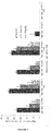
- the hybridomas which were obtained produce distinct forms of IgA MAbs anti-CEA: a monomeric form (without J chain) and a polymeric form which contain J chain(s) and includes dimeric form and higher polymeric forms of IgA ( Figure 1 ).
- the level of expression of the various forms of IgA depends on the selected hybridoma. The different forms can be purified and separated by chromatography.
- Hybridoma clones were selected for direct cancer cell growth-inhibitory activity on various CEA expressing human tumor cell lines using ALAMARBLUETM assay.
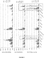
- Anti-CEA IgA of clone 15B3 (also named #15B3) and clone 14G8 also named #14G8) block cell growth of two cell lines derived from human colorectal adenocarcinoma (WiDr, 18.2% ⁇ 1.9% for 15B3 and 18.1 ⁇ 1.2 for 14G8 at 25 ⁇ g/mL (culture supernatant); Figure 2 ). These positive clones were selected for further analysis.
- Variable regions from IgA heavy and light chains (VH and VL) were cloned and sequenced.
- the VL and VH amino acid sequences correspond to SEQ ID NO: 11 and 12 and SEQ ID NO: 13 and 14, respectively for 15B3 and 14G8.
- the nucleotide sequences encoding said amino acid sequences correspond to SEQ ID NO: 15 and 16 and SEQ ID NO: 17 and 18, respectively for 15B3 and 14G8.
- a recombinant anti-CEA IgG1 derived from clone 15B3 was constructed using the cloned VH and VL domains and expressed in Sp2/0 or HEK cell line. RecIgG1 anti-CEA #15B3 shows a counterpart level of growth inhibition in comparison to IgA anti-CEA (clone #15B3).
- the affinity of the antibody (mainly carried by the variable regions forming the "paratope") is preserved during the process of transformation of an IgA1 towards an IgG1.
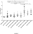
- IgA recruits elements of the complement leading to lysis of the cell target via the alternate pathway. Same tests of cell growth inhibition of WiDr cancer cells were performed in the presence of human sera (human sera alone did not affect biologically WiDr cell culture). A significant rise of growth inhibition was recorded in the presence of human complement for the two selected anti-CEA IgA clones (clone #15B3 and clone #14G8; Figure 3 ).
- IgA recruits also immune effector cells that express a high affinity IgA receptor (Fc-alphaR or CD89), mainly neutrophils and monocytes, two particularly numerous cells in blood cell population.
- Fc-alphaR or CD89 high affinity IgA receptor
- neutrophils and monocytes two particularly numerous cells in blood cell population.
- CD89 high affinity IgA receptor
- Flow cytometry (FACS) analysis shows the presence of a new population of doubly selected cells (Effector Cell (CD89 +) : WiDr target cells (CEA + ) in mice treated with the anti-CEA IgA (31% ⁇ 15%) compared with mice treated with irrelevant IgA (anti-peanut IgA; 10.2% ⁇ 3.5%, Figure 4 ).
- IgG-mediated ADCC is induced by the NK cells, strongly expressing the FcgammaR.
- the recombinant IgG generated from the variable regions of the IgA anti-CEA clone (#15B3) induces a cytotoxic cell lysis of WiDr-CEA + as strong as the "original" IgA as only 11.3% ⁇ 8.6% of WiDr were harvested by peritoneal washing after treatment with recIgG anti-CEA ( Figure 4 ).
- the similar cytotoxic effect of the two antibodies supports the demonstration that the IgA is able to induce the ADCC mechanisms as quickly and effectively as the IgG. It also validates that the transformation of the IgA towards IgG does dot modify the affinity of the antibody for its target.
- the anti-CEA antibodies according to the invention have a direct cell growth inhibition effect on cancer cells expressing CEA.
- This unique antitumor effect is further enhanced by their ability to recruit the complement pathway and the immune cell-effectors of antibody dependent cell cytotoxicity (ADCC) leading to cancer cell lysis. All these antitumor effects are specific for the targeted tumor cells because the antibodies are specific for CEA.
- ADCC antibody dependent cell cytotoxicity
- the circulating IgA is quickly captured by the liver, directed to the gallbladder to be excreted with bile into the lumen of the digestive tract. This is what explains the strong labelling of the liver on the one hand and of the intestinal fluid and stools on the other hand.
- the secretory IgA is detectable in the lumen of the mucosal organs.
- the polymeric IgA tropism for the caecum is the strongest and the fastest, superior to the tropism of the monomeric IgA, but also that of the IgG, already described to persist longer in blood.
- CRC tumour model was created in which human cancer cells were grafted in the mucosal environment.
- Pathological microscopic analysis clearly revealed a structural glandular architecture of the grafted tumour and the presence of large vacuoles in the WiDr cell line, consistent with muco-secretions in the lamina propria layer.
- Cancer cells invaded the normal caecum, under the muscle layer through the lamina propria , to produce protruding polyps in the lumen.
- different stages of CRC have been observed from localised tumours to metastasis in the lungs.
- Immunohistochemical analysis in tumours revealed that tumour cells were present within tumour vessels, suggesting cellular dissemination by the vascular system. All of these factors led to consider colorectal orthotopic grafts as being useful models of human CRC, because they share the same characteristics as human tumours.
- the antitumor effect of the anti-CEA antibody was evaluated in the orthotopic mouse model of human colorectal carcinoma disclosed in example 1. Preliminary results showed a significant benefit of treatment with the polymeric form of anti-CEA IgA on the survival of the animals and the decrease in tumor growth. In all conditions, the treatment was administered 8 days after the implantation of human tumor cells in the caecum of mouse (Balb/c Nude, Harlan). Two routes of administration have been evaluated: the intravenous (by administration of a dose/day for 5 days, 8 days after implantation of the tumor) for the polymeric IgA and the oral (by administration of a dose/day for 11 days, 8 days after tumor implantation) for the secretory IgA.
Landscapes
- Health & Medical Sciences (AREA)
- Chemical & Material Sciences (AREA)
- Immunology (AREA)
- Organic Chemistry (AREA)
- Life Sciences & Earth Sciences (AREA)
- General Health & Medical Sciences (AREA)
- Medicinal Chemistry (AREA)
- Proteomics, Peptides & Aminoacids (AREA)
- Nuclear Medicine, Radiotherapy & Molecular Imaging (AREA)
- Genetics & Genomics (AREA)
- Biochemistry (AREA)
- Molecular Biology (AREA)
- Cell Biology (AREA)
- Oncology (AREA)
- Chemical Kinetics & Catalysis (AREA)
- General Chemical & Material Sciences (AREA)
- Biophysics (AREA)
- Pharmacology & Pharmacy (AREA)
- Animal Behavior & Ethology (AREA)
- Public Health (AREA)
- Veterinary Medicine (AREA)
- Peptides Or Proteins (AREA)
- Medicines Containing Antibodies Or Antigens For Use As Internal Diagnostic Agents (AREA)
- Preparation Of Compounds By Using Micro-Organisms (AREA)
Priority Applications (12)
| Application Number | Priority Date | Filing Date | Title |
|---|---|---|---|
| EP17305498.2A EP3398967A1 (en) | 2017-05-04 | 2017-05-04 | Antibodies against carcinoembryonic antigen for cancer therapy and diagnosis |
| ES18722497T ES2975798T3 (es) | 2017-05-04 | 2018-05-03 | Anticuerpos contra antígeno carcinoembrionario para tratamiento y diagnóstico de cáncer |
| PL18722497.7T PL3619237T3 (pl) | 2017-05-04 | 2018-05-03 | Przeciwciała przeciwko antygenowi rakowo-płodowemu w terapii i diagnostyce nowotworów złośliwych |
| CA3062233A CA3062233C (en) | 2017-05-04 | 2018-05-03 | Antibodies against carcinoembryonic antigen for cancer therapy and diagnosis |
| PCT/EP2018/061385 WO2018202794A1 (en) | 2017-05-04 | 2018-05-03 | Antibodies against carcinoembryonic antigen for cancer therapy and diagnosis |
| EP18722497.7A EP3619237B1 (en) | 2017-05-04 | 2018-05-03 | Antibodies against carcinoembryonic antigen for cancer therapy and diagnosis |
| PT187224977T PT3619237T (pt) | 2017-05-04 | 2018-05-03 | Anticorpos contra antigénio carcinoembriónico para terapia e diagnóstico do cancro |
| DK18722497.7T DK3619237T5 (da) | 2017-05-04 | 2018-05-03 | Antistoffer mod karcinoembryonalt antigen til cancerterapi og -diagnosticering |
| US16/609,878 US11427646B2 (en) | 2017-05-04 | 2018-05-03 | Antibodies against carcinoembryonic antigen for cancer therapy and diagnosis |
| AU2018262648A AU2018262648B2 (en) | 2017-05-04 | 2018-05-03 | Antibodies against carcinoembryonic antigen for cancer therapy and diagnosis |
| FIEP18722497.7T FI3619237T3 (fi) | 2017-05-04 | 2018-05-03 | Vasta-aineita karsinoembryonaalista antigeenia vastaan syövän hoitamiseksi ja diagnosoimiseksi |
| HUE18722497A HUE066183T2 (hu) | 2017-05-04 | 2018-05-03 | Karcinoembrionális antigén elleni ellenanyagok rákterápiához és diagnosztizáláshoz |
Applications Claiming Priority (1)
| Application Number | Priority Date | Filing Date | Title |
|---|---|---|---|
| EP17305498.2A EP3398967A1 (en) | 2017-05-04 | 2017-05-04 | Antibodies against carcinoembryonic antigen for cancer therapy and diagnosis |
Publications (1)
| Publication Number | Publication Date |
|---|---|
| EP3398967A1 true EP3398967A1 (en) | 2018-11-07 |
Family
ID=58707444
Family Applications (2)
| Application Number | Title | Priority Date | Filing Date |
|---|---|---|---|
| EP17305498.2A Withdrawn EP3398967A1 (en) | 2017-05-04 | 2017-05-04 | Antibodies against carcinoembryonic antigen for cancer therapy and diagnosis |
| EP18722497.7A Active EP3619237B1 (en) | 2017-05-04 | 2018-05-03 | Antibodies against carcinoembryonic antigen for cancer therapy and diagnosis |
Family Applications After (1)
| Application Number | Title | Priority Date | Filing Date |
|---|---|---|---|
| EP18722497.7A Active EP3619237B1 (en) | 2017-05-04 | 2018-05-03 | Antibodies against carcinoembryonic antigen for cancer therapy and diagnosis |
Country Status (10)
| Country | Link |
|---|---|
| US (1) | US11427646B2 (da) |
| EP (2) | EP3398967A1 (da) |
| AU (1) | AU2018262648B2 (da) |
| DK (1) | DK3619237T5 (da) |
| ES (1) | ES2975798T3 (da) |
| FI (1) | FI3619237T3 (da) |
| HU (1) | HUE066183T2 (da) |
| PL (1) | PL3619237T3 (da) |
| PT (1) | PT3619237T (da) |
| WO (1) | WO2018202794A1 (da) |
Families Citing this family (1)
| Publication number | Priority date | Publication date | Assignee | Title |
|---|---|---|---|---|
| WO2025257185A1 (en) | 2024-06-11 | 2025-12-18 | Abcely | Humanized anti-human-carcinoembryonic antigen antibody |
Citations (4)
| Publication number | Priority date | Publication date | Assignee | Title |
|---|---|---|---|---|
| WO1992001059A1 (en) | 1990-07-05 | 1992-01-23 | Celltech Limited | Cdr grafted anti-cea antibodies and their production |
| EP1680449B1 (fr) | 2003-10-24 | 2009-10-14 | Centre National De La Recherche Scientifique (Cnrs) | Mammifere non-humain transgenique pour la region constante de la chaine lourde des immunoglobulines humaines de classe a et ses applications |
| WO2013012414A1 (en) * | 2011-07-18 | 2013-01-24 | Medimmune, Llc | Dosing regimens for treatment of cea-expressing cancers |
| US8771690B2 (en) | 2009-09-16 | 2014-07-08 | Immunomedics, Inc. | Class I anti-CEA antibodies and uses thereof |
-
2017
- 2017-05-04 EP EP17305498.2A patent/EP3398967A1/en not_active Withdrawn
-
2018
- 2018-05-03 PT PT187224977T patent/PT3619237T/pt unknown
- 2018-05-03 AU AU2018262648A patent/AU2018262648B2/en active Active
- 2018-05-03 ES ES18722497T patent/ES2975798T3/es active Active
- 2018-05-03 FI FIEP18722497.7T patent/FI3619237T3/fi active
- 2018-05-03 PL PL18722497.7T patent/PL3619237T3/pl unknown
- 2018-05-03 HU HUE18722497A patent/HUE066183T2/hu unknown
- 2018-05-03 DK DK18722497.7T patent/DK3619237T5/da active
- 2018-05-03 EP EP18722497.7A patent/EP3619237B1/en active Active
- 2018-05-03 US US16/609,878 patent/US11427646B2/en active Active
- 2018-05-03 WO PCT/EP2018/061385 patent/WO2018202794A1/en not_active Ceased
Patent Citations (4)
| Publication number | Priority date | Publication date | Assignee | Title |
|---|---|---|---|---|
| WO1992001059A1 (en) | 1990-07-05 | 1992-01-23 | Celltech Limited | Cdr grafted anti-cea antibodies and their production |
| EP1680449B1 (fr) | 2003-10-24 | 2009-10-14 | Centre National De La Recherche Scientifique (Cnrs) | Mammifere non-humain transgenique pour la region constante de la chaine lourde des immunoglobulines humaines de classe a et ses applications |
| US8771690B2 (en) | 2009-09-16 | 2014-07-08 | Immunomedics, Inc. | Class I anti-CEA antibodies and uses thereof |
| WO2013012414A1 (en) * | 2011-07-18 | 2013-01-24 | Medimmune, Llc | Dosing regimens for treatment of cea-expressing cancers |
Non-Patent Citations (23)
| Title |
|---|
| BACAC ET AL., CLIN. CANCER RES., vol. 22, 2016, pages 3286 |
| BEAUCHEMIN, M; ARABZADEH A, CANCER METASTASIS REV., vol. 32, 2013, pages 643 - 671 |
| CARPENET ET AL., PLOS ONE, vol. 10, no. 10, 6 October 2015 (2015-10-06), pages E0139835 |
| CARSTEN-O. SAHLMANN ET AL: "Repeated adjuvant anti-CEA radioimmunotherapy after resection of colorectal liver metastases: Safety, feasibility, and long-term efficacy results of a prospective phase 2 study : Adjuvant Anti-CEA RIT Post-CLM Resection", CANCER., vol. 123, no. 4, 20 October 2016 (2016-10-20), US, pages 638 - 649, XP055411437, ISSN: 0008-543X, DOI: 10.1002/cncr.30390 * |
| CESPEDES ET AL., AM.J. PATHOL., vol. 170, 2007, pages 1077 - 1085 |
| HART FELIX ET AL: "Human Cell Line-Derived Monoclonal IgA Antibodies for Cancer Immunotherapy.", BIOENGINEERING (BASEL, SWITZERLAND) 08 MAY 2017, vol. 4, no. 2, 8 May 2017 (2017-05-08), XP002774324, ISSN: 2306-5354 * |
| KOGA H ET AL: "Mouse-human chimeric monoclonal antibody to carcinoembryonic antigen (CEA): in vitro and in vivo activities.", HYBRIDOMA FEB 1990, vol. 9, no. 1, February 1990 (1990-02-01), pages 43 - 56, XP008074735, ISSN: 0272-457X * |
| KOHLER, G.; MILSTEIN, C., EUROPEAN JOURNAL OF IMMUNOLOGY, vol. 6, 1976, pages 511 - 9 |
| KOHLER; MILSTEIN, NATURE, vol. 256, 1975, pages 495 - 497 |
| KOTESWARA ET AL., PNAS, vol. 94, 1997, pages 6364 - 6368 |
| KOTESWARA, PNAS, vol. 94, 1997, pages 6364 - 6368 |
| RINDISBACHER ET AL., JBC, vol. 270, 1995, pages 14220 - 14228 |
| ROGNUM T O ET AL: "Immunohistochemical study of secretory component, secretory IgA and carcinoembryonic antigen in large bowel carcinomas", PATHOLOGY - RESEARCH AND PRACTICE, ELSEVIER, AMSTERDAM, NL, vol. 170, no. 1-3, 30 November 1980 (1980-11-30), pages 126 - 145, XP009500427, ISSN: 0344-0338 * |
| S. KIM-SCHULZE ET AL: "Intrarectal Vaccination with Recombinant Vaccinia Virus Expressing Carcinoembronic Antigen Induces Mucosal and Systemic Immunity and Prevents Progression of Colorectal Cancer", THE JOURNAL OF IMMUNOLOGY, vol. 181, no. 11, 1 December 2008 (2008-12-01), US, pages 8112 - 8119, XP055410760, ISSN: 0022-1767, DOI: 10.4049/jimmunol.181.11.8112 * |
| S. V. GOVINDAN ET AL: "CEACAM5-Targeted Therapy of Human Colonic and Pancreatic Cancer Xenografts with Potent Labetuzumab-SN-38 Immunoconjugates", CLINICAL CANCER RESEARCH, vol. 15, no. 19, 29 September 2009 (2009-09-29), pages 6052 - 6061, XP055186131, ISSN: 1078-0432, DOI: 10.1158/1078-0432.CCR-09-0586 * |
| SENBA T ET AL: "Tumor growth suppression by a mouse/human chimeric anti-CEA antibody and lymphokine-activated killer cells in vitro and in SCID mouse xenograft model.", ANTICANCER RESEARCH, vol. 18, no. 1A, January 1998 (1998-01-01), pages 17 - 24, XP055410713, ISSN: 0250-7005 * |
| SHIBAGUCHI HIROTOMO ET AL: "cDNA cloning and sequencing of a novel monoclonal antibody to carcinoembryonic antigen and construction of a mouse/human chimeric antibody.", ANTICANCER RESEARCH, vol. 23, no. 6a, November 2003 (2003-11-01), pages 4383 - 4388, XP055410709, ISSN: 0250-7005 * |
| STAFF ET AL., J. CLIN. IMMUNOL., vol. 32, 2012, pages 855 - 865 |
| TERSKIKH A ET AL: "Dimeric recombinant IgA directed against carcino-embryonic antigen, a novel tool for carcinoma localization", MOLECULAR IMMUNOLOGY, PERGAMON, GB, vol. 31, no. 17, 1 December 1994 (1994-12-01), pages 1313 - 1319, XP023681574, ISSN: 0161-5890, [retrieved on 19941201], DOI: 10.1016/0161-5890(94)90049-3 * |
| WEIDLE ET AL., SEMINARS IN ONCOLOGY, vol. 41, 2014, pages 653 - 660 |
| WILLKOMM ET AL., J. NUCL. MED., vol. 41, 2000, pages 1657 - 1663 |
| ZHAO ET AL., ONCOL. RES., vol. 17, 2008, pages 217 - 222 |
| ZHAO JUN ET AL: "Recombinant human monoclonal igA antibody against CEA to recruit neutrophils to CEA-expressing cells.", ONCOLOGY RESEARCH, vol. 17, no. 5, 1 May 2008 (2008-05-01), pages 217 - 222, XP009500426, ISSN: 0965-0407, DOI: 10.3727/096504008786111374 * |
Also Published As
| Publication number | Publication date |
|---|---|
| FI3619237T3 (fi) | 2024-04-19 |
| DK3619237T5 (da) | 2024-09-02 |
| EP3619237A1 (en) | 2020-03-11 |
| AU2018262648A1 (en) | 2019-11-07 |
| EP3619237B1 (en) | 2024-01-17 |
| PL3619237T3 (pl) | 2024-06-24 |
| WO2018202794A1 (en) | 2018-11-08 |
| HUE066183T2 (hu) | 2024-07-28 |
| PT3619237T (pt) | 2024-03-05 |
| US20200055952A1 (en) | 2020-02-20 |
| DK3619237T3 (da) | 2024-04-08 |
| CA3062233A1 (en) | 2018-11-08 |
| AU2018262648B2 (en) | 2024-08-08 |
| ES2975798T3 (es) | 2024-07-15 |
| US11427646B2 (en) | 2022-08-30 |
Similar Documents
| Publication | Publication Date | Title |
|---|---|---|
| US12258418B2 (en) | Claudin18.2 binding moieties and uses thereof | |
| US8901278B2 (en) | Pharmaceutical antibody compositions with resistance to soluble CEA | |
| US7090844B2 (en) | Use of antibodies against the MUC18 antigen | |
| US8071730B2 (en) | Anti-JAM-A antibodies | |
| US20230357385A1 (en) | Anti-gpc3 antibody, anti-gpc3 chimeric antigen receptor and gpc3/cd3 bispecific antibody | |
| JP7667857B2 (ja) | 新規な腫瘍エンゲージャ治療剤の開発及び使用 | |
| EP4279507A1 (en) | Cd73-binding protein and use thereof | |
| WO2023041065A1 (zh) | 一种靶向人ceacam5/6的抗体、制备方法和应用 | |
| EP4269442A1 (en) | Mesothelin binding molecule and application thereof | |
| EP3619237B1 (en) | Antibodies against carcinoembryonic antigen for cancer therapy and diagnosis | |
| CA3062233C (en) | Antibodies against carcinoembryonic antigen for cancer therapy and diagnosis | |
| WO2024114410A1 (zh) | 靶向gpc3的抗体及其用途 | |
| WO2025257185A1 (en) | Humanized anti-human-carcinoembryonic antigen antibody | |
| CN121219323A (zh) | 抗cldn6抗体及其用途 | |
| CN117430711A (zh) | 用于治疗肝细胞性肝癌的双特异性融合蛋白及其应用 | |
| NZ569144A (en) | Pharmaceutical antibody compositions with resistance to soluble CEA - comprising IgG1antibodies that bind to CEA |
Legal Events
| Date | Code | Title | Description |
|---|---|---|---|
| PUAI | Public reference made under article 153(3) epc to a published international application that has entered the european phase |
Free format text: ORIGINAL CODE: 0009012 |
|
| STAA | Information on the status of an ep patent application or granted ep patent |
Free format text: STATUS: THE APPLICATION HAS BEEN PUBLISHED |
|
| AK | Designated contracting states |
Kind code of ref document: A1 Designated state(s): AL AT BE BG CH CY CZ DE DK EE ES FI FR GB GR HR HU IE IS IT LI LT LU LV MC MK MT NL NO PL PT RO RS SE SI SK SM TR |
|
| AX | Request for extension of the european patent |
Extension state: BA ME |
|
| STAA | Information on the status of an ep patent application or granted ep patent |
Free format text: STATUS: THE APPLICATION IS DEEMED TO BE WITHDRAWN |
|
| 18D | Application deemed to be withdrawn |
Effective date: 20190508 |