EP3397622B1 - Co-administration of alpha-amino ester of hydroxypropylthiazolidine carboxamide derivative and a tocolytic agent - Google Patents
Co-administration of alpha-amino ester of hydroxypropylthiazolidine carboxamide derivative and a tocolytic agent Download PDFInfo
- Publication number
- EP3397622B1 EP3397622B1 EP17700391.0A EP17700391A EP3397622B1 EP 3397622 B1 EP3397622 B1 EP 3397622B1 EP 17700391 A EP17700391 A EP 17700391A EP 3397622 B1 EP3397622 B1 EP 3397622B1
- Authority
- EP
- European Patent Office
- Prior art keywords
- compound
- administered
- subject
- contractions
- salt
- Prior art date
- Legal status (The legal status is an assumption and is not a legal conclusion. Google has not performed a legal analysis and makes no representation as to the accuracy of the status listed.)
- Active
Links
- 229940125712 tocolytic agent Drugs 0.000 title claims description 56
- 239000003675 tocolytic agent Substances 0.000 title claims description 56
- MFYNLGQKRUZXHD-UHFFFAOYSA-N 2-(3-hydroxypropyl)-1,3-thiazolidine-2-carboxamide Chemical class OCCCC1(SCCN1)C(=O)N MFYNLGQKRUZXHD-UHFFFAOYSA-N 0.000 title description 2
- 238000011260 co-administration Methods 0.000 title 1
- 150000001875 compounds Chemical class 0.000 claims description 393
- 150000003839 salts Chemical class 0.000 claims description 160
- VWXRQYYUEIYXCZ-OBIMUBPZSA-N atosiban Chemical group C1=CC(OCC)=CC=C1C[C@@H]1C(=O)N[C@@H]([C@@H](C)CC)C(=O)N[C@@H]([C@@H](C)O)C(=O)N[C@@H](CC(N)=O)C(=O)N[C@H](C(=O)N2[C@@H](CCC2)C(=O)N[C@@H](CCCN)C(=O)NCC(N)=O)CSSCCC(=O)N1 VWXRQYYUEIYXCZ-OBIMUBPZSA-N 0.000 claims description 118
- 229960002403 atosiban Drugs 0.000 claims description 118
- 108700007535 atosiban Proteins 0.000 claims description 118
- HYIMSNHJOBLJNT-UHFFFAOYSA-N nifedipine Chemical group COC(=O)C1=C(C)NC(C)=C(C(=O)OC)C1C1=CC=CC=C1[N+]([O-])=O HYIMSNHJOBLJNT-UHFFFAOYSA-N 0.000 claims description 89
- 229960001597 nifedipine Drugs 0.000 claims description 89
- 208000006399 Premature Obstetric Labor Diseases 0.000 claims description 64
- RJKFOVLPORLFTN-LEKSSAKUSA-N Progesterone Chemical compound C1CC2=CC(=O)CC[C@]2(C)[C@@H]2[C@@H]1[C@@H]1CC[C@H](C(=O)C)[C@@]1(C)CC2 RJKFOVLPORLFTN-LEKSSAKUSA-N 0.000 claims description 62
- 239000008194 pharmaceutical composition Substances 0.000 claims description 55
- OLUJSZLBWZWGJT-HGBKYHTQSA-N [(2s,4z)-2-(hydroxymethyl)-4-methoxyiminopyrrolidin-1-yl]-[4-(2-methylphenyl)phenyl]methanone Chemical compound C1C(=N/OC)\C[C@@H](CO)N1C(=O)C1=CC=C(C=2C(=CC=CC=2)C)C=C1 OLUJSZLBWZWGJT-HGBKYHTQSA-N 0.000 claims description 49
- 229950000833 nolasiban Drugs 0.000 claims description 48
- 229940122828 Oxytocin receptor antagonist Drugs 0.000 claims description 44
- 239000003246 corticosteroid Substances 0.000 claims description 41
- 108090000312 Calcium Channels Proteins 0.000 claims description 31
- 102000003922 Calcium Channels Human genes 0.000 claims description 31
- 239000000186 progesterone Substances 0.000 claims description 31
- JYGXADMDTFJGBT-VWUMJDOOSA-N hydrocortisone Chemical compound O=C1CC[C@]2(C)[C@H]3[C@@H](O)C[C@](C)([C@@](CC4)(O)C(=O)CO)[C@@H]4[C@@H]3CCC2=C1 JYGXADMDTFJGBT-VWUMJDOOSA-N 0.000 claims description 28
- CSNNHWWHGAXBCP-UHFFFAOYSA-L Magnesium sulfate Chemical compound [Mg+2].[O-][S+2]([O-])([O-])[O-] CSNNHWWHGAXBCP-UHFFFAOYSA-L 0.000 claims description 27
- 230000003454 betamimetic effect Effects 0.000 claims description 27
- 229940125400 channel inhibitor Drugs 0.000 claims description 25
- UWHCWRQFNKUYCG-QUZACWSFSA-N Epelsiban Chemical compound O=C([C@H](N1[C@@H](C(N[C@@H](C1=O)C1CC2=CC=CC=C2C1)=O)[C@@H](C)CC)C=1C(=NC(C)=CC=1)C)N1CCOCC1 UWHCWRQFNKUYCG-QUZACWSFSA-N 0.000 claims description 20
- PLVGDGRBPMVYPB-FDUHJNRSSA-N Retosiban Chemical compound O=C([C@H](N1[C@@H](C(N[C@@H](C1=O)C1CC2=CC=CC=C2C1)=O)[C@@H](C)CC)C=1N=C(C)OC=1)N1CCOCC1 PLVGDGRBPMVYPB-FDUHJNRSSA-N 0.000 claims description 20
- UGNGRKKDUVKQDF-IHOMMZCZSA-N barusiban Chemical compound N1C(=O)CCSCC[C@@H](C(=O)N(C)[C@H](CO)CCCN)NC(=O)[C@H](CC(N)=O)NC(=O)[C@H]([C@H](C)CC)NC(=O)[C@H]([C@@H](C)CC)NC(=O)[C@H]1CC1=CNC2=CC=CC=C12 UGNGRKKDUVKQDF-IHOMMZCZSA-N 0.000 claims description 20
- 108010040145 barusiban Proteins 0.000 claims description 20
- 229950009748 barusiban Drugs 0.000 claims description 20
- 229950009963 epelsiban Drugs 0.000 claims description 20
- 159000000003 magnesium salts Chemical class 0.000 claims description 20
- 239000002840 nitric oxide donor Substances 0.000 claims description 20
- 229950010622 retosiban Drugs 0.000 claims description 20
- 229960002537 betamethasone Drugs 0.000 claims description 15
- UREBDLICKHMUKA-DVTGEIKXSA-N betamethasone Chemical compound C1CC2=CC(=O)C=C[C@]2(C)[C@]2(F)[C@@H]1[C@@H]1C[C@H](C)[C@@](C(=O)CO)(O)[C@@]1(C)C[C@@H]2O UREBDLICKHMUKA-DVTGEIKXSA-N 0.000 claims description 15
- 229960003957 dexamethasone Drugs 0.000 claims description 15
- UREBDLICKHMUKA-CXSFZGCWSA-N dexamethasone Chemical compound C1CC2=CC(=O)C=C[C@]2(C)[C@]2(F)[C@@H]1[C@@H]1C[C@@H](C)[C@@](C(=O)CO)(O)[C@@]1(C)C[C@@H]2O UREBDLICKHMUKA-CXSFZGCWSA-N 0.000 claims description 15
- 229960000890 hydrocortisone Drugs 0.000 claims description 14
- 229910052943 magnesium sulfate Inorganic materials 0.000 claims description 14
- 235000019341 magnesium sulphate Nutrition 0.000 claims description 14
- 229950000801 hydroxyprogesterone caproate Drugs 0.000 claims description 13
- DOMWKUIIPQCAJU-LJHIYBGHSA-N Hydroxyprogesterone caproate Chemical compound C1CC2=CC(=O)CC[C@]2(C)[C@@H]2[C@@H]1[C@@H]1CC[C@@](C(C)=O)(OC(=O)CCCCC)[C@@]1(C)CC2 DOMWKUIIPQCAJU-LJHIYBGHSA-N 0.000 claims description 10
- 238000005481 NMR spectroscopy Methods 0.000 claims description 10
- SNIOPGDIGTZGOP-UHFFFAOYSA-N Nitroglycerin Chemical compound [O-][N+](=O)OCC(O[N+]([O-])=O)CO[N+]([O-])=O SNIOPGDIGTZGOP-UHFFFAOYSA-N 0.000 claims description 10
- 229960003711 glyceryl trinitrate Drugs 0.000 claims description 10
- XWTYSIMOBUGWOL-UHFFFAOYSA-N (+-)-Terbutaline Chemical compound CC(C)(C)NCC(O)C1=CC(O)=CC(O)=C1 XWTYSIMOBUGWOL-UHFFFAOYSA-N 0.000 claims description 8
- PTGXAUBQBSGPKF-UHFFFAOYSA-N 4-[1-hydroxy-2-(4-phenylbutan-2-ylamino)propyl]phenol Chemical compound C=1C=C(O)C=CC=1C(O)C(C)NC(C)CCC1=CC=CC=C1 PTGXAUBQBSGPKF-UHFFFAOYSA-N 0.000 claims description 8
- LSLYOANBFKQKPT-DIFFPNOSSA-N 5-[(1r)-1-hydroxy-2-[[(2r)-1-(4-hydroxyphenyl)propan-2-yl]amino]ethyl]benzene-1,3-diol Chemical compound C([C@@H](C)NC[C@H](O)C=1C=C(O)C=C(O)C=1)C1=CC=C(O)C=C1 LSLYOANBFKQKPT-DIFFPNOSSA-N 0.000 claims description 8
- ZBBHBTPTTSWHBA-UHFFFAOYSA-N Nicardipine Chemical compound COC(=O)C1=C(C)NC(C)=C(C(=O)OCCN(C)CC=2C=CC=CC=2)C1C1=CC=CC([N+]([O-])=O)=C1 ZBBHBTPTTSWHBA-UHFFFAOYSA-N 0.000 claims description 8
- NDAUXUAQIAJITI-UHFFFAOYSA-N albuterol Chemical compound CC(C)(C)NCC(O)C1=CC=C(O)C(CO)=C1 NDAUXUAQIAJITI-UHFFFAOYSA-N 0.000 claims description 8
- 229960003455 buphenine Drugs 0.000 claims description 8
- 229960001022 fenoterol Drugs 0.000 claims description 8
- 229960000708 hexoprenaline Drugs 0.000 claims description 8
- OXLZNBCNGJWPRV-UHFFFAOYSA-N hexoprenaline Chemical compound C=1C=C(O)C(O)=CC=1C(O)CNCCCCCCNCC(O)C1=CC=C(O)C(O)=C1 OXLZNBCNGJWPRV-UHFFFAOYSA-N 0.000 claims description 8
- LMOINURANNBYCM-UHFFFAOYSA-N metaproterenol Chemical compound CC(C)NCC(O)C1=CC(O)=CC(O)=C1 LMOINURANNBYCM-UHFFFAOYSA-N 0.000 claims description 8
- 229960001783 nicardipine Drugs 0.000 claims description 8
- 229960002657 orciprenaline Drugs 0.000 claims description 8
- IOVGROKTTNBUGK-SJCJKPOMSA-N ritodrine Chemical compound N([C@@H](C)[C@H](O)C=1C=CC(O)=CC=1)CCC1=CC=C(O)C=C1 IOVGROKTTNBUGK-SJCJKPOMSA-N 0.000 claims description 8
- 229960001634 ritodrine Drugs 0.000 claims description 8
- 229960002052 salbutamol Drugs 0.000 claims description 8
- 229960000195 terbutaline Drugs 0.000 claims description 8
- 239000000843 powder Substances 0.000 claims description 5
- 239000000546 pharmaceutical excipient Substances 0.000 claims description 4
- 239000002775 capsule Substances 0.000 claims description 2
- 239000007897 gelcap Substances 0.000 claims description 2
- 239000006193 liquid solution Substances 0.000 claims description 2
- 239000006194 liquid suspension Substances 0.000 claims description 2
- 239000003826 tablet Substances 0.000 claims description 2
- 230000008602 contraction Effects 0.000 description 274
- HTSGKJQDMSTCGS-UHFFFAOYSA-N 1,4-bis(4-chlorophenyl)-2-(4-methylphenyl)sulfonylbutane-1,4-dione Chemical compound C1=CC(C)=CC=C1S(=O)(=O)C(C(=O)C=1C=CC(Cl)=CC=1)CC(=O)C1=CC=C(Cl)C=C1 HTSGKJQDMSTCGS-UHFFFAOYSA-N 0.000 description 242
- 102400000050 Oxytocin Human genes 0.000 description 173
- XNOPRXBHLZRZKH-UHFFFAOYSA-N Oxytocin Natural products N1C(=O)C(N)CSSCC(C(=O)N2C(CCC2)C(=O)NC(CC(C)C)C(=O)NCC(N)=O)NC(=O)C(CC(N)=O)NC(=O)C(CCC(N)=O)NC(=O)C(C(C)CC)NC(=O)C1CC1=CC=C(O)C=C1 XNOPRXBHLZRZKH-UHFFFAOYSA-N 0.000 description 173
- 101800000989 Oxytocin Proteins 0.000 description 173
- XNOPRXBHLZRZKH-DSZYJQQASA-N oxytocin Chemical compound C([C@H]1C(=O)N[C@H](C(N[C@@H](CCC(N)=O)C(=O)N[C@@H](CC(N)=O)C(=O)N[C@@H](CSSC[C@H](N)C(=O)N1)C(=O)N1[C@@H](CCC1)C(=O)N[C@@H](CC(C)C)C(=O)NCC(N)=O)=O)[C@@H](C)CC)C1=CC=C(O)C=C1 XNOPRXBHLZRZKH-DSZYJQQASA-N 0.000 description 173
- 229960001723 oxytocin Drugs 0.000 description 173
- 230000000694 effects Effects 0.000 description 161
- 230000002632 myometrial effect Effects 0.000 description 133
- 230000002269 spontaneous effect Effects 0.000 description 115
- NLFBCYMMUAKCPC-KQQUZDAGSA-N ethyl (e)-3-[3-amino-2-cyano-1-[(e)-3-ethoxy-3-oxoprop-1-enyl]sulfanyl-3-oxoprop-1-enyl]sulfanylprop-2-enoate Chemical compound CCOC(=O)\C=C\SC(=C(C#N)C(N)=O)S\C=C\C(=O)OCC NLFBCYMMUAKCPC-KQQUZDAGSA-N 0.000 description 104
- 238000012384 transportation and delivery Methods 0.000 description 100
- 239000000203 mixture Substances 0.000 description 90
- PXGPLTODNUVGFL-BRIYLRKRSA-N (E,Z)-(1R,2R,3R,5S)-7-(3,5-Dihydroxy-2-((3S)-(3-hydroxy-1-octenyl))cyclopentyl)-5-heptenoic acid Chemical compound CCCCC[C@H](O)C=C[C@H]1[C@H](O)C[C@H](O)[C@@H]1CC=CCCCC(O)=O PXGPLTODNUVGFL-BRIYLRKRSA-N 0.000 description 80
- 238000005259 measurement Methods 0.000 description 72
- 239000000243 solution Substances 0.000 description 72
- 230000000875 corresponding effect Effects 0.000 description 70
- 238000002474 experimental method Methods 0.000 description 69
- 238000000034 method Methods 0.000 description 68
- IAZDPXIOMUYVGZ-UHFFFAOYSA-N Dimethylsulphoxide Chemical compound CS(C)=O IAZDPXIOMUYVGZ-UHFFFAOYSA-N 0.000 description 62
- 229960001760 dimethyl sulfoxide Drugs 0.000 description 62
- 241000699670 Mus sp. Species 0.000 description 59
- 210000004027 cell Anatomy 0.000 description 52
- CSCPPACGZOOCGX-UHFFFAOYSA-N Acetone Chemical compound CC(C)=O CSCPPACGZOOCGX-UHFFFAOYSA-N 0.000 description 51
- 238000000634 powder X-ray diffraction Methods 0.000 description 51
- 208000037805 labour Diseases 0.000 description 50
- 238000001574 biopsy Methods 0.000 description 48
- 210000001519 tissue Anatomy 0.000 description 47
- 230000001965 increasing effect Effects 0.000 description 44
- YMWUJEATGCHHMB-UHFFFAOYSA-N Dichloromethane Chemical compound ClCCl YMWUJEATGCHHMB-UHFFFAOYSA-N 0.000 description 39
- YXFVVABEGXRONW-UHFFFAOYSA-N Toluene Chemical compound CC1=CC=CC=C1 YXFVVABEGXRONW-UHFFFAOYSA-N 0.000 description 39
- 239000002158 endotoxin Substances 0.000 description 39
- 229920006008 lipopolysaccharide Polymers 0.000 description 39
- 238000011282 treatment Methods 0.000 description 39
- XEKOWRVHYACXOJ-UHFFFAOYSA-N Ethyl acetate Chemical compound CCOC(C)=O XEKOWRVHYACXOJ-UHFFFAOYSA-N 0.000 description 37
- AFVFQIVMOAPDHO-UHFFFAOYSA-N Methanesulfonic acid Chemical class CS(O)(=O)=O AFVFQIVMOAPDHO-UHFFFAOYSA-N 0.000 description 37
- 230000006698 induction Effects 0.000 description 37
- 239000003814 drug Substances 0.000 description 36
- 230000032696 parturition Effects 0.000 description 36
- 230000016160 smooth muscle contraction Effects 0.000 description 36
- 238000012360 testing method Methods 0.000 description 35
- 150000003841 chloride salts Chemical class 0.000 description 33
- 238000001228 spectrum Methods 0.000 description 33
- -1 amino, hydroxyl Chemical group 0.000 description 32
- VKHAHZOOUSRJNA-GCNJZUOMSA-N mifepristone Chemical compound C1([C@@H]2C3=C4CCC(=O)C=C4CC[C@H]3[C@@H]3CC[C@@]([C@]3(C2)C)(O)C#CC)=CC=C(N(C)C)C=C1 VKHAHZOOUSRJNA-GCNJZUOMSA-N 0.000 description 30
- 238000007619 statistical method Methods 0.000 description 29
- 229960003387 progesterone Drugs 0.000 description 28
- 208000024891 symptom Diseases 0.000 description 28
- 239000003981 vehicle Substances 0.000 description 28
- OKKJLVBELUTLKV-UHFFFAOYSA-N Methanol Chemical compound OC OKKJLVBELUTLKV-UHFFFAOYSA-N 0.000 description 27
- 229940124597 therapeutic agent Drugs 0.000 description 26
- 238000009472 formulation Methods 0.000 description 25
- 238000001990 intravenous administration Methods 0.000 description 23
- 238000004458 analytical method Methods 0.000 description 21
- 108090000623 proteins and genes Proteins 0.000 description 21
- IJGRMHOSHXDMSA-UHFFFAOYSA-N Atomic nitrogen Chemical compound N#N IJGRMHOSHXDMSA-UHFFFAOYSA-N 0.000 description 20
- 239000003795 chemical substances by application Substances 0.000 description 20
- 238000001704 evaporation Methods 0.000 description 20
- 230000008020 evaporation Effects 0.000 description 20
- 102000004169 proteins and genes Human genes 0.000 description 20
- 239000013553 cell monolayer Substances 0.000 description 19
- 239000012458 free base Substances 0.000 description 19
- 230000014509 gene expression Effects 0.000 description 19
- 238000001325 log-rank test Methods 0.000 description 19
- 235000018102 proteins Nutrition 0.000 description 19
- 102000000471 Prostaglandin F receptors Human genes 0.000 description 18
- 108050008995 Prostaglandin F receptors Proteins 0.000 description 18
- 230000015572 biosynthetic process Effects 0.000 description 18
- 239000013078 crystal Substances 0.000 description 18
- 239000000463 material Substances 0.000 description 18
- VZCYOOQTPOCHFL-OWOJBTEDSA-N Fumaric acid Chemical class OC(=O)\C=C\C(O)=O VZCYOOQTPOCHFL-OWOJBTEDSA-N 0.000 description 17
- 239000012981 Hank's balanced salt solution Substances 0.000 description 16
- MWUXSHHQAYIFBG-UHFFFAOYSA-N Nitric oxide Chemical class O=[N] MWUXSHHQAYIFBG-UHFFFAOYSA-N 0.000 description 16
- 208000036029 Uterine contractions during pregnancy Diseases 0.000 description 16
- DBPWSSGDRRHUNT-CEGNMAFCSA-N 17α-hydroxyprogesterone Chemical compound C1CC2=CC(=O)CC[C@]2(C)[C@@H]2[C@@H]1[C@@H]1CC[C@@](C(=O)C)(O)[C@@]1(C)CC2 DBPWSSGDRRHUNT-CEGNMAFCSA-N 0.000 description 15
- 150000003180 prostaglandins Chemical class 0.000 description 15
- 125000006239 protecting group Chemical group 0.000 description 15
- 102000005962 receptors Human genes 0.000 description 15
- 108020003175 receptors Proteins 0.000 description 15
- 230000003195 tocolytic effect Effects 0.000 description 15
- 238000006243 chemical reaction Methods 0.000 description 14
- 238000002360 preparation method Methods 0.000 description 14
- 238000000425 proton nuclear magnetic resonance spectrum Methods 0.000 description 14
- 238000003786 synthesis reaction Methods 0.000 description 14
- 230000032258 transport Effects 0.000 description 14
- 230000035899 viability Effects 0.000 description 14
- 230000003834 intracellular effect Effects 0.000 description 13
- 239000002002 slurry Substances 0.000 description 13
- 239000007787 solid Substances 0.000 description 13
- 238000009739 binding Methods 0.000 description 12
- 230000009467 reduction Effects 0.000 description 12
- 108010037462 Cyclooxygenase 2 Proteins 0.000 description 11
- VEXZGXHMUGYJMC-UHFFFAOYSA-N Hydrochloric acid Chemical compound Cl VEXZGXHMUGYJMC-UHFFFAOYSA-N 0.000 description 11
- 230000027455 binding Effects 0.000 description 11
- 239000003112 inhibitor Substances 0.000 description 11
- 239000012528 membrane Substances 0.000 description 11
- 229940094443 oxytocics prostaglandins Drugs 0.000 description 11
- 230000035935 pregnancy Effects 0.000 description 11
- 229920006395 saturated elastomer Polymers 0.000 description 11
- NLXLAEXVIDQMFP-UHFFFAOYSA-N Ammonia chloride Chemical compound [NH4+].[Cl-] NLXLAEXVIDQMFP-UHFFFAOYSA-N 0.000 description 10
- RTZKZFJDLAIYFH-UHFFFAOYSA-N Diethyl ether Chemical compound CCOCC RTZKZFJDLAIYFH-UHFFFAOYSA-N 0.000 description 10
- 208000005171 Dysmenorrhea Diseases 0.000 description 10
- 206010013935 Dysmenorrhoea Diseases 0.000 description 10
- WYURNTSHIVDZCO-UHFFFAOYSA-N Tetrahydrofuran Chemical compound C1CCOC1 WYURNTSHIVDZCO-UHFFFAOYSA-N 0.000 description 10
- FSGKNSDQCZJRHO-UHFFFAOYSA-N [1-(4-fluorophenyl)-1-[[3-(4-phenylphenyl)sulfonyl-1,3-thiazolidine-2-carbonyl]amino]propyl] 3-methyl-2-[(2-methylpropan-2-yl)oxycarbonylamino]butanoate Chemical compound C1(=CC=C(C=C1)S(=O)(=O)N1C(SCC1)C(=O)NC(CC)(C1=CC=C(C=C1)F)OC(C(C(C)C)NC(=O)OC(C)(C)C)=O)C1=CC=CC=C1 FSGKNSDQCZJRHO-UHFFFAOYSA-N 0.000 description 10
- 239000000872 buffer Substances 0.000 description 10
- 230000005764 inhibitory process Effects 0.000 description 10
- 210000004379 membrane Anatomy 0.000 description 10
- 229910052757 nitrogen Inorganic materials 0.000 description 10
- 239000000126 substance Substances 0.000 description 10
- 230000001225 therapeutic effect Effects 0.000 description 10
- 102000007469 Actins Human genes 0.000 description 9
- 108010085238 Actins Proteins 0.000 description 9
- 201000009273 Endometriosis Diseases 0.000 description 9
- ZWEHNKRNPOVVGH-UHFFFAOYSA-N Methyl ethyl ketone Natural products CCC(C)=O ZWEHNKRNPOVVGH-UHFFFAOYSA-N 0.000 description 9
- FAPWRFPIFSIZLT-UHFFFAOYSA-M Sodium chloride Chemical compound [Na+].[Cl-] FAPWRFPIFSIZLT-UHFFFAOYSA-M 0.000 description 9
- 206010046788 Uterine haemorrhage Diseases 0.000 description 9
- 206010046910 Vaginal haemorrhage Diseases 0.000 description 9
- 238000010521 absorption reaction Methods 0.000 description 9
- 238000012512 characterization method Methods 0.000 description 9
- 230000006870 function Effects 0.000 description 9
- 238000001727 in vivo Methods 0.000 description 9
- 238000011534 incubation Methods 0.000 description 9
- 230000002401 inhibitory effect Effects 0.000 description 9
- 239000003446 ligand Substances 0.000 description 9
- 210000000754 myometrium Anatomy 0.000 description 9
- 239000012074 organic phase Substances 0.000 description 9
- 230000035699 permeability Effects 0.000 description 9
- 238000005070 sampling Methods 0.000 description 9
- 238000001262 western blot Methods 0.000 description 9
- 108091006594 SLC15A1 Proteins 0.000 description 8
- UIIMBOGNXHQVGW-UHFFFAOYSA-M Sodium bicarbonate Chemical compound [Na+].OC([O-])=O UIIMBOGNXHQVGW-UHFFFAOYSA-M 0.000 description 8
- 239000000808 adrenergic beta-agonist Substances 0.000 description 8
- 239000005557 antagonist Substances 0.000 description 8
- 230000002860 competitive effect Effects 0.000 description 8
- 230000001939 inductive effect Effects 0.000 description 8
- 230000003993 interaction Effects 0.000 description 8
- 210000004347 intestinal mucosa Anatomy 0.000 description 8
- 238000007918 intramuscular administration Methods 0.000 description 8
- 238000011835 investigation Methods 0.000 description 8
- 125000002496 methyl group Chemical group [H]C([H])([H])* 0.000 description 8
- 230000000770 proinflammatory effect Effects 0.000 description 8
- 239000002356 single layer Substances 0.000 description 8
- 238000002336 sorption--desorption measurement Methods 0.000 description 8
- OYPRJOBELJOOCE-UHFFFAOYSA-N Calcium Chemical compound [Ca] OYPRJOBELJOOCE-UHFFFAOYSA-N 0.000 description 7
- IVOMOUWHDPKRLL-KQYNXXCUSA-N Cyclic adenosine monophosphate Chemical compound C([C@H]1O2)OP(O)(=O)O[C@H]1[C@@H](O)[C@@H]2N1C(N=CN=C2N)=C2N=C1 IVOMOUWHDPKRLL-KQYNXXCUSA-N 0.000 description 7
- 102000010907 Cyclooxygenase 2 Human genes 0.000 description 7
- 102000046014 Peptide Transporter 1 Human genes 0.000 description 7
- 241000700159 Rattus Species 0.000 description 7
- 101150103667 Slc15a1 gene Proteins 0.000 description 7
- 239000013543 active substance Substances 0.000 description 7
- 210000001691 amnion Anatomy 0.000 description 7
- 239000011575 calcium Substances 0.000 description 7
- 229910052791 calcium Inorganic materials 0.000 description 7
- 229960001334 corticosteroids Drugs 0.000 description 7
- 238000000113 differential scanning calorimetry Methods 0.000 description 7
- 229940079593 drug Drugs 0.000 description 7
- 230000001605 fetal effect Effects 0.000 description 7
- DLBFLQKQABVKGT-UHFFFAOYSA-L lucifer yellow dye Chemical compound [Li+].[Li+].[O-]S(=O)(=O)C1=CC(C(N(C(=O)NN)C2=O)=O)=C3C2=CC(S([O-])(=O)=O)=CC3=C1N DLBFLQKQABVKGT-UHFFFAOYSA-L 0.000 description 7
- 239000002609 medium Substances 0.000 description 7
- 238000011084 recovery Methods 0.000 description 7
- 150000003467 sulfuric acid derivatives Chemical class 0.000 description 7
- 238000011870 unpaired t-test Methods 0.000 description 7
- XLYOFNOQVPJJNP-UHFFFAOYSA-N water Substances O XLYOFNOQVPJJNP-UHFFFAOYSA-N 0.000 description 7
- 230000004580 weight loss Effects 0.000 description 7
- YWIHGHOTVHUWJN-UHFFFAOYSA-N 2-aminoacetic acid;2-(methylamino)acetic acid Chemical compound NCC(O)=O.CNCC(O)=O YWIHGHOTVHUWJN-UHFFFAOYSA-N 0.000 description 6
- 206010001052 Acute respiratory distress syndrome Diseases 0.000 description 6
- 229940127291 Calcium channel antagonist Drugs 0.000 description 6
- LFQSCWFLJHTTHZ-UHFFFAOYSA-N Ethanol Chemical compound CCO LFQSCWFLJHTTHZ-UHFFFAOYSA-N 0.000 description 6
- ZMXDDKWLCZADIW-UHFFFAOYSA-N N,N-Dimethylformamide Chemical compound CN(C)C=O ZMXDDKWLCZADIW-UHFFFAOYSA-N 0.000 description 6
- 208000013616 Respiratory Distress Syndrome Diseases 0.000 description 6
- HEMHJVSKTPXQMS-UHFFFAOYSA-M Sodium hydroxide Chemical compound [OH-].[Na+] HEMHJVSKTPXQMS-UHFFFAOYSA-M 0.000 description 6
- MMWCIQZXVOZEGG-HOZKJCLWSA-N [(1S,2R,3S,4S,5R,6S)-2,3,5-trihydroxy-4,6-diphosphonooxycyclohexyl] dihydrogen phosphate Chemical compound O[C@H]1[C@@H](O)[C@H](OP(O)(O)=O)[C@@H](OP(O)(O)=O)[C@H](O)[C@H]1OP(O)(O)=O MMWCIQZXVOZEGG-HOZKJCLWSA-N 0.000 description 6
- 230000004913 activation Effects 0.000 description 6
- 230000009286 beneficial effect Effects 0.000 description 6
- DKPFZGUDAPQIHT-UHFFFAOYSA-N butyl acetate Chemical compound CCCCOC(C)=O DKPFZGUDAPQIHT-UHFFFAOYSA-N 0.000 description 6
- 239000000480 calcium channel blocker Substances 0.000 description 6
- KRKNYBCHXYNGOX-UHFFFAOYSA-N citric acid Chemical compound OC(=O)CC(O)(C(O)=O)CC(O)=O KRKNYBCHXYNGOX-UHFFFAOYSA-N 0.000 description 6
- 238000002648 combination therapy Methods 0.000 description 6
- 208000037265 diseases, disorders, signs and symptoms Diseases 0.000 description 6
- 238000004895 liquid chromatography mass spectrometry Methods 0.000 description 6
- 230000007246 mechanism Effects 0.000 description 6
- 230000009871 nonspecific binding Effects 0.000 description 6
- 230000002265 prevention Effects 0.000 description 6
- PXGPLTODNUVGFL-UHFFFAOYSA-N prostaglandin F2alpha Natural products CCCCCC(O)C=CC1C(O)CC(O)C1CC=CCCCC(O)=O PXGPLTODNUVGFL-UHFFFAOYSA-N 0.000 description 6
- 239000011541 reaction mixture Substances 0.000 description 6
- 239000000758 substrate Substances 0.000 description 6
- 238000005160 1H NMR spectroscopy Methods 0.000 description 5
- 108010078791 Carrier Proteins Proteins 0.000 description 5
- 241000124008 Mammalia Species 0.000 description 5
- UUIBKACUTXYSAK-YCVJPRETSA-N [(3S)-3-(4-fluorophenyl)-3-[[(2S)-3-(4-phenylphenyl)sulfonyl-1,3-thiazolidine-2-carbonyl]amino]propyl] (2S)-2-amino-3-methylbutanoate Chemical compound N[C@@H](C(C)C)C(=O)OCC[C@@H](C1=CC=C(C=C1)F)NC(=O)[C@@H]1SCCN1S(=O)(=O)C1=CC=C(C=C1)C1=CC=CC=C1 UUIBKACUTXYSAK-YCVJPRETSA-N 0.000 description 5
- BKHLLRSGQHTVMA-VFGMFICWSA-N [(3S)-3-(4-fluorophenyl)-3-[[(2S)-3-(4-phenylphenyl)sulfonyl-1,3-thiazolidine-2-carbonyl]amino]propyl] (2S)-2-amino-3-methylbutanoate hydrochloride Chemical compound Cl.CC(C)[C@H](N)C(=O)OCC[C@H](NC(=O)[C@@H]1SCCN1S(=O)(=O)c1ccc(cc1)-c1ccccc1)c1ccc(F)cc1 BKHLLRSGQHTVMA-VFGMFICWSA-N 0.000 description 5
- 235000019270 ammonium chloride Nutrition 0.000 description 5
- 239000012298 atmosphere Substances 0.000 description 5
- 239000006285 cell suspension Substances 0.000 description 5
- 238000002425 crystallisation Methods 0.000 description 5
- 230000008025 crystallization Effects 0.000 description 5
- ZOOGRGPOEVQQDX-KHLHZJAASA-N cyclic guanosine monophosphate Chemical compound C([C@H]1O2)O[P@](O)(=O)O[C@@H]1[C@H](O)[C@H]2N1C(N=C(NC2=O)N)=C2N=C1 ZOOGRGPOEVQQDX-KHLHZJAASA-N 0.000 description 5
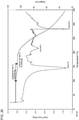
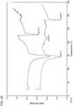
- 238000001938 differential scanning calorimetry curve Methods 0.000 description 5
- 239000012065 filter cake Substances 0.000 description 5
- 230000035800 maturation Effects 0.000 description 5
- 230000001404 mediated effect Effects 0.000 description 5
- 238000012544 monitoring process Methods 0.000 description 5
- 210000000107 myocyte Anatomy 0.000 description 5
- VLKPHJQULXJQSP-UHFFFAOYSA-N n-[1-(4-fluorophenyl)-3-hydroxypropyl]-3-(4-phenylphenyl)sulfonyl-1,3-thiazolidine-2-carboxamide Chemical compound C=1C=C(F)C=CC=1C(CCO)NC(=O)C1SCCN1S(=O)(=O)C(C=C1)=CC=C1C1=CC=CC=C1 VLKPHJQULXJQSP-UHFFFAOYSA-N 0.000 description 5
- 230000036961 partial effect Effects 0.000 description 5
- 230000008569 process Effects 0.000 description 5
- 239000000651 prodrug Substances 0.000 description 5
- 229940002612 prodrug Drugs 0.000 description 5
- 230000004044 response Effects 0.000 description 5
- 230000019491 signal transduction Effects 0.000 description 5
- 239000011780 sodium chloride Substances 0.000 description 5
- 238000001757 thermogravimetry curve Methods 0.000 description 5
- VHYFNPMBLIVWCW-UHFFFAOYSA-N 4-Dimethylaminopyridine Chemical compound CN(C)C1=CC=NC=C1 VHYFNPMBLIVWCW-UHFFFAOYSA-N 0.000 description 4
- 108090000371 Esterases Proteins 0.000 description 4
- 241000699666 Mus <mouse, genus> Species 0.000 description 4
- 102000003505 Myosin Human genes 0.000 description 4
- 108060008487 Myosin Proteins 0.000 description 4
- 108010067385 Myosin Light Chains Proteins 0.000 description 4
- 102000016349 Myosin Light Chains Human genes 0.000 description 4
- IMNFDUFMRHMDMM-UHFFFAOYSA-N N-Heptane Chemical compound CCCCCCC IMNFDUFMRHMDMM-UHFFFAOYSA-N 0.000 description 4
- 208000002193 Pain Diseases 0.000 description 4
- 208000005107 Premature Birth Diseases 0.000 description 4
- QAOWNCQODCNURD-UHFFFAOYSA-N Sulfuric acid Chemical compound OS(O)(=O)=O QAOWNCQODCNURD-UHFFFAOYSA-N 0.000 description 4
- 238000002441 X-ray diffraction Methods 0.000 description 4
- 230000008859 change Effects 0.000 description 4
- 238000000354 decomposition reaction Methods 0.000 description 4
- 230000001419 dependent effect Effects 0.000 description 4
- 125000004925 dihydropyridyl group Chemical group N1(CC=CC=C1)* 0.000 description 4
- 201000010099 disease Diseases 0.000 description 4
- 239000001963 growth medium Substances 0.000 description 4
- 230000004054 inflammatory process Effects 0.000 description 4
- 210000004072 lung Anatomy 0.000 description 4
- 238000004519 manufacturing process Methods 0.000 description 4
- 230000005906 menstruation Effects 0.000 description 4
- 238000000655 nuclear magnetic resonance spectrum Methods 0.000 description 4
- 238000003653 radioligand binding assay Methods 0.000 description 4
- 238000013341 scale-up Methods 0.000 description 4
- 210000002460 smooth muscle Anatomy 0.000 description 4
- 235000017557 sodium bicarbonate Nutrition 0.000 description 4
- 229910000030 sodium bicarbonate Inorganic materials 0.000 description 4
- 230000002195 synergetic effect Effects 0.000 description 4
- VZCYOOQTPOCHFL-UHFFFAOYSA-N trans-butenedioic acid Natural products OC(=O)C=CC(O)=O VZCYOOQTPOCHFL-UHFFFAOYSA-N 0.000 description 4
- YNGDWRXWKFWCJY-UHFFFAOYSA-N 1,4-Dihydropyridine Chemical compound C1C=CNC=C1 YNGDWRXWKFWCJY-UHFFFAOYSA-N 0.000 description 3
- ALBQXDHCMLLQMB-UHFFFAOYSA-N 4-phenylbenzenesulfonyl chloride Chemical compound C1=CC(S(=O)(=O)Cl)=CC=C1C1=CC=CC=C1 ALBQXDHCMLLQMB-UHFFFAOYSA-N 0.000 description 3
- WEVYAHXRMPXWCK-UHFFFAOYSA-N Acetonitrile Chemical compound CC#N WEVYAHXRMPXWCK-UHFFFAOYSA-N 0.000 description 3
- 108091003079 Bovine Serum Albumin Proteins 0.000 description 3
- 206010061218 Inflammation Diseases 0.000 description 3
- 206010022840 Intraventricular haemorrhage Diseases 0.000 description 3
- SJRJJKPEHAURKC-UHFFFAOYSA-N N-Methylmorpholine Chemical compound CN1CCOCC1 SJRJJKPEHAURKC-UHFFFAOYSA-N 0.000 description 3
- 206010051606 Necrotising colitis Diseases 0.000 description 3
- 102000008299 Nitric Oxide Synthase Human genes 0.000 description 3
- 108010021487 Nitric Oxide Synthase Proteins 0.000 description 3
- 229940121791 Prostaglandin F2 alpha receptor antagonist Drugs 0.000 description 3
- XUIMIQQOPSSXEZ-UHFFFAOYSA-N Silicon Chemical compound [Si] XUIMIQQOPSSXEZ-UHFFFAOYSA-N 0.000 description 3
- 230000008901 benefit Effects 0.000 description 3
- 125000001797 benzyl group Chemical group [H]C1=C([H])C([H])=C(C([H])=C1[H])C([H])([H])* 0.000 description 3
- 210000000170 cell membrane Anatomy 0.000 description 3
- 239000000306 component Substances 0.000 description 3
- 230000037020 contractile activity Effects 0.000 description 3
- 210000004246 corpus luteum Anatomy 0.000 description 3
- 210000003785 decidua Anatomy 0.000 description 3
- 238000011161 development Methods 0.000 description 3
- 230000018109 developmental process Effects 0.000 description 3
- 231100000673 dose–response relationship Toxicity 0.000 description 3
- 230000002357 endometrial effect Effects 0.000 description 3
- 210000002889 endothelial cell Anatomy 0.000 description 3
- 230000001747 exhibiting effect Effects 0.000 description 3
- 239000012091 fetal bovine serum Substances 0.000 description 3
- 210000003754 fetus Anatomy 0.000 description 3
- 239000000706 filtrate Substances 0.000 description 3
- 125000002485 formyl group Chemical group [H]C(*)=O 0.000 description 3
- 125000000524 functional group Chemical group 0.000 description 3
- 210000001035 gastrointestinal tract Anatomy 0.000 description 3
- 238000000338 in vitro Methods 0.000 description 3
- 208000015181 infectious disease Diseases 0.000 description 3
- 230000000977 initiatory effect Effects 0.000 description 3
- 125000001449 isopropyl group Chemical group [H]C([H])([H])C([H])(*)C([H])([H])[H] 0.000 description 3
- 239000000155 melt Substances 0.000 description 3
- 230000008018 melting Effects 0.000 description 3
- 238000002844 melting Methods 0.000 description 3
- 229940098779 methanesulfonic acid Drugs 0.000 description 3
- 238000000386 microscopy Methods 0.000 description 3
- 210000003205 muscle Anatomy 0.000 description 3
- 230000004118 muscle contraction Effects 0.000 description 3
- 208000004995 necrotizing enterocolitis Diseases 0.000 description 3
- 235000015097 nutrients Nutrition 0.000 description 3
- 210000000056 organ Anatomy 0.000 description 3
- 239000012044 organic layer Substances 0.000 description 3
- 201000006195 perinatal necrotizing enterocolitis Diseases 0.000 description 3
- 230000005855 radiation Effects 0.000 description 3
- 239000013557 residual solvent Substances 0.000 description 3
- 229910052710 silicon Inorganic materials 0.000 description 3
- 239000010703 silicon Substances 0.000 description 3
- 238000001179 sorption measurement Methods 0.000 description 3
- 125000001424 substituent group Chemical group 0.000 description 3
- 230000009885 systemic effect Effects 0.000 description 3
- TVXOQBUDZTYJHF-UHFFFAOYSA-N tert-butyl 2-[[1-(4-fluorophenyl)-3-hydroxypropyl]carbamoyl]-1,3-thiazolidine-3-carboxylate Chemical compound CC(C)(C)OC(=O)N1CCSC1C(=O)NC(CCO)C1=CC=C(F)C=C1 TVXOQBUDZTYJHF-UHFFFAOYSA-N 0.000 description 3
- 125000004044 trifluoroacetyl group Chemical group FC(C(=O)*)(F)F 0.000 description 3
- 230000013948 uterine smooth muscle contraction Effects 0.000 description 3
- 210000004291 uterus Anatomy 0.000 description 3
- DZUXGQBLFALXCR-UHFFFAOYSA-N (+)-(9alpha,11alpha,13E,15S)-9,11,15-trihydroxyprost-13-en-1-oic acid Natural products CCCCCC(O)C=CC1C(O)CC(O)C1CCCCCCC(O)=O DZUXGQBLFALXCR-UHFFFAOYSA-N 0.000 description 2
- RYHBNJHYFVUHQT-UHFFFAOYSA-N 1,4-Dioxane Chemical compound C1COCCO1 RYHBNJHYFVUHQT-UHFFFAOYSA-N 0.000 description 2
- FCEHBMOGCRZNNI-UHFFFAOYSA-N 1-benzothiophene Chemical compound C1=CC=C2SC=CC2=C1 FCEHBMOGCRZNNI-UHFFFAOYSA-N 0.000 description 2
- CNWINRVXAYPOMW-FCNJXWMTSA-N 1-stearoyl-2-arachidonoyl-sn-glycero-3-phospho-1D-myo-inositol 4,5-biphosphate Chemical compound CCCCC\C=C/C\C=C/C\C=C/C\C=C/CCCC(=O)O[C@H](COC(=O)CCCCCCCCCCCCCCCCC)COP(O)(=O)O[C@@H]1[C@H](O)[C@H](O)[C@@H](OP(O)(O)=O)[C@H](OP(O)(O)=O)[C@H]1O CNWINRVXAYPOMW-FCNJXWMTSA-N 0.000 description 2
- 125000003903 2-propenyl group Chemical group [H]C([*])([H])C([H])=C([H])[H] 0.000 description 2
- FPQQSJJWHUJYPU-UHFFFAOYSA-N 3-(dimethylamino)propyliminomethylidene-ethylazanium;chloride Chemical compound Cl.CCN=C=NCCCN(C)C FPQQSJJWHUJYPU-UHFFFAOYSA-N 0.000 description 2
- QCQCHGYLTSGIGX-GHXANHINSA-N 4-[[(3ar,5ar,5br,7ar,9s,11ar,11br,13as)-5a,5b,8,8,11a-pentamethyl-3a-[(5-methylpyridine-3-carbonyl)amino]-2-oxo-1-propan-2-yl-4,5,6,7,7a,9,10,11,11b,12,13,13a-dodecahydro-3h-cyclopenta[a]chrysen-9-yl]oxy]-2,2-dimethyl-4-oxobutanoic acid Chemical compound N([C@@]12CC[C@@]3(C)[C@]4(C)CC[C@H]5C(C)(C)[C@@H](OC(=O)CC(C)(C)C(O)=O)CC[C@]5(C)[C@H]4CC[C@@H]3C1=C(C(C2)=O)C(C)C)C(=O)C1=CN=CC(C)=C1 QCQCHGYLTSGIGX-GHXANHINSA-N 0.000 description 2
- KRKNYBCHXYNGOX-UHFFFAOYSA-K Citrate Chemical compound [O-]C(=O)CC(O)(CC([O-])=O)C([O-])=O KRKNYBCHXYNGOX-UHFFFAOYSA-K 0.000 description 2
- 206010010774 Constipation Diseases 0.000 description 2
- 206010051244 Dyschezia Diseases 0.000 description 2
- 208000004483 Dyspareunia Diseases 0.000 description 2
- 238000002965 ELISA Methods 0.000 description 2
- 102000004190 Enzymes Human genes 0.000 description 2
- 108090000790 Enzymes Proteins 0.000 description 2
- 238000005079 FT-Raman Methods 0.000 description 2
- 229910016860 FaSSIF Inorganic materials 0.000 description 2
- 229910005429 FeSSIF Inorganic materials 0.000 description 2
- 229910000530 Gallium indium arsenide Inorganic materials 0.000 description 2
- 101000685323 Homo sapiens Succinate dehydrogenase [ubiquinone] flavoprotein subunit, mitochondrial Proteins 0.000 description 2
- 102000004016 L-Type Calcium Channels Human genes 0.000 description 2
- 108090000420 L-Type Calcium Channels Proteins 0.000 description 2
- RHGKLRLOHDJJDR-BYPYZUCNSA-N L-citrulline Chemical compound NC(=O)NCCC[C@H]([NH3+])C([O-])=O RHGKLRLOHDJJDR-BYPYZUCNSA-N 0.000 description 2
- ZDXPYRJPNDTMRX-VKHMYHEASA-N L-glutamine Chemical compound OC(=O)[C@@H](N)CCC(N)=O ZDXPYRJPNDTMRX-VKHMYHEASA-N 0.000 description 2
- 229930182816 L-glutamine Natural products 0.000 description 2
- 102100035044 Myosin light chain kinase, smooth muscle Human genes 0.000 description 2
- 108010074596 Myosin-Light-Chain Kinase Proteins 0.000 description 2
- JGFZNNIVVJXRND-UHFFFAOYSA-N N,N-Diisopropylethylamine (DIPEA) Chemical compound CCN(C(C)C)C(C)C JGFZNNIVVJXRND-UHFFFAOYSA-N 0.000 description 2
- PXHVJJICTQNCMI-UHFFFAOYSA-N Nickel Chemical compound [Ni] PXHVJJICTQNCMI-UHFFFAOYSA-N 0.000 description 2
- 102000004279 Oxytocin receptors Human genes 0.000 description 2
- 108090000876 Oxytocin receptors Proteins 0.000 description 2
- 229910019142 PO4 Inorganic materials 0.000 description 2
- 208000000450 Pelvic Pain Diseases 0.000 description 2
- 208000001300 Perinatal Death Diseases 0.000 description 2
- 206010036600 Premature labour Diseases 0.000 description 2
- 206010036603 Premature rupture of membranes Diseases 0.000 description 2
- 238000001069 Raman spectroscopy Methods 0.000 description 2
- VYPSYNLAJGMNEJ-UHFFFAOYSA-N Silicium dioxide Chemical compound O=[Si]=O VYPSYNLAJGMNEJ-UHFFFAOYSA-N 0.000 description 2
- PMZURENOXWZQFD-UHFFFAOYSA-L Sodium Sulfate Chemical compound [Na+].[Na+].[O-]S([O-])(=O)=O PMZURENOXWZQFD-UHFFFAOYSA-L 0.000 description 2
- 102000003673 Symporters Human genes 0.000 description 2
- 108090000088 Symporters Proteins 0.000 description 2
- 102000014384 Type C Phospholipases Human genes 0.000 description 2
- 108010079194 Type C Phospholipases Proteins 0.000 description 2
- OLUJSZLBWZWGJT-KOXKPCSVSA-N [(2s,4e)-2-(hydroxymethyl)-4-methoxyiminopyrrolidin-1-yl]-[4-(2-methylphenyl)phenyl]methanone Chemical compound C1C(=N/OC)/C[C@@H](CO)N1C(=O)C1=CC=C(C=2C(=CC=CC=2)C)C=C1 OLUJSZLBWZWGJT-KOXKPCSVSA-N 0.000 description 2
- KXNLCSXBJCPWGL-UHFFFAOYSA-N [Ga].[As].[In] Chemical compound [Ga].[As].[In] KXNLCSXBJCPWGL-UHFFFAOYSA-N 0.000 description 2
- 230000001594 aberrant effect Effects 0.000 description 2
- 125000002777 acetyl group Chemical group [H]C([H])([H])C(*)=O 0.000 description 2
- 239000000556 agonist Substances 0.000 description 2
- 229910052782 aluminium Inorganic materials 0.000 description 2
- XAGFODPZIPBFFR-UHFFFAOYSA-N aluminium Chemical compound [Al] XAGFODPZIPBFFR-UHFFFAOYSA-N 0.000 description 2
- 238000010171 animal model Methods 0.000 description 2
- 239000007864 aqueous solution Substances 0.000 description 2
- 238000002820 assay format Methods 0.000 description 2
- SRSXLGNVWSONIS-UHFFFAOYSA-M benzenesulfonate Chemical compound [O-]S(=O)(=O)C1=CC=CC=C1 SRSXLGNVWSONIS-UHFFFAOYSA-M 0.000 description 2
- 125000001584 benzyloxycarbonyl group Chemical group C(=O)(OCC1=CC=CC=C1)* 0.000 description 2
- 102000016966 beta-2 Adrenergic Receptors Human genes 0.000 description 2
- 108010014499 beta-2 Adrenergic Receptors Proteins 0.000 description 2
- 230000002457 bidirectional effect Effects 0.000 description 2
- 210000004204 blood vessel Anatomy 0.000 description 2
- 206010008129 cerebral palsy Diseases 0.000 description 2
- 230000001684 chronic effect Effects 0.000 description 2
- 150000001860 citric acid derivatives Chemical class 0.000 description 2
- 201000010897 colon adenocarcinoma Diseases 0.000 description 2
- 208000029742 colonic neoplasm Diseases 0.000 description 2
- 230000002596 correlated effect Effects 0.000 description 2
- 239000002178 crystalline material Substances 0.000 description 2
- 230000034994 death Effects 0.000 description 2
- 238000003795 desorption Methods 0.000 description 2
- 150000001982 diacylglycerols Chemical class 0.000 description 2
- 230000004069 differentiation Effects 0.000 description 2
- 239000003085 diluting agent Substances 0.000 description 2
- LOKCTEFSRHRXRJ-UHFFFAOYSA-I dipotassium trisodium dihydrogen phosphate hydrogen phosphate dichloride Chemical compound P(=O)(O)(O)[O-].[K+].P(=O)(O)([O-])[O-].[Na+].[Na+].[Cl-].[K+].[Cl-].[Na+] LOKCTEFSRHRXRJ-UHFFFAOYSA-I 0.000 description 2
- 208000035475 disorder Diseases 0.000 description 2
- 239000006185 dispersion Substances 0.000 description 2
- 238000010494 dissociation reaction Methods 0.000 description 2
- 230000005593 dissociations Effects 0.000 description 2
- 238000001035 drying Methods 0.000 description 2
- 206010013990 dysuria Diseases 0.000 description 2
- 210000004696 endometrium Anatomy 0.000 description 2
- 210000002919 epithelial cell Anatomy 0.000 description 2
- 238000011067 equilibration Methods 0.000 description 2
- 239000003797 essential amino acid Substances 0.000 description 2
- 235000020776 essential amino acid Nutrition 0.000 description 2
- 150000002148 esters Chemical class 0.000 description 2
- CCIVGXIOQKPBKL-UHFFFAOYSA-M ethanesulfonate Chemical compound CCS([O-])(=O)=O CCIVGXIOQKPBKL-UHFFFAOYSA-M 0.000 description 2
- 238000001914 filtration Methods 0.000 description 2
- 239000011521 glass Substances 0.000 description 2
- 102000046038 human SDHA Human genes 0.000 description 2
- 210000005260 human cell Anatomy 0.000 description 2
- 230000007062 hydrolysis Effects 0.000 description 2
- 238000006460 hydrolysis reaction Methods 0.000 description 2
- 230000002102 hyperpolarization Effects 0.000 description 2
- 230000006872 improvement Effects 0.000 description 2
- 239000012535 impurity Substances 0.000 description 2
- 229910052738 indium Inorganic materials 0.000 description 2
- APFVFJFRJDLVQX-UHFFFAOYSA-N indium atom Chemical compound [In] APFVFJFRJDLVQX-UHFFFAOYSA-N 0.000 description 2
- CGIGDMFJXJATDK-UHFFFAOYSA-N indomethacin Chemical compound CC1=C(CC(O)=O)C2=CC(OC)=CC=C2N1C(=O)C1=CC=C(Cl)C=C1 CGIGDMFJXJATDK-UHFFFAOYSA-N 0.000 description 2
- 239000000411 inducer Substances 0.000 description 2
- 238000002347 injection Methods 0.000 description 2
- 239000007924 injection Substances 0.000 description 2
- 238000000670 ligand binding assay Methods 0.000 description 2
- 230000029860 luteolysis Effects 0.000 description 2
- 238000012423 maintenance Methods 0.000 description 2
- 238000007726 management method Methods 0.000 description 2
- 238000004949 mass spectrometry Methods 0.000 description 2
- 125000001434 methanylylidene group Chemical group [H]C#[*] 0.000 description 2
- 210000000110 microvilli Anatomy 0.000 description 2
- 239000006225 natural substrate Substances 0.000 description 2
- 102000039446 nucleic acids Human genes 0.000 description 2
- 108020004707 nucleic acids Proteins 0.000 description 2
- 150000007523 nucleic acids Chemical class 0.000 description 2
- 230000003647 oxidation Effects 0.000 description 2
- 238000007254 oxidation reaction Methods 0.000 description 2
- 239000003336 oxytocin antagonist Substances 0.000 description 2
- 229940121361 oxytocin antagonists Drugs 0.000 description 2
- 230000037361 pathway Effects 0.000 description 2
- 230000035515 penetration Effects 0.000 description 2
- UYWQUFXKFGHYNT-UHFFFAOYSA-N phenylmethyl ester of formic acid Natural products O=COCC1=CC=CC=C1 UYWQUFXKFGHYNT-UHFFFAOYSA-N 0.000 description 2
- 239000002953 phosphate buffered saline Substances 0.000 description 2
- 230000035479 physiological effects, processes and functions Effects 0.000 description 2
- 210000002826 placenta Anatomy 0.000 description 2
- 210000002381 plasma Anatomy 0.000 description 2
- 229920001184 polypeptide Polymers 0.000 description 2
- 102000004196 processed proteins & peptides Human genes 0.000 description 2
- 108090000765 processed proteins & peptides Proteins 0.000 description 2
- 239000000047 product Substances 0.000 description 2
- 238000011321 prophylaxis Methods 0.000 description 2
- 238000010926 purge Methods 0.000 description 2
- 229940044551 receptor antagonist Drugs 0.000 description 2
- 239000002464 receptor antagonist Substances 0.000 description 2
- 230000004648 relaxation of smooth muscle Effects 0.000 description 2
- 230000000241 respiratory effect Effects 0.000 description 2
- 230000002441 reversible effect Effects 0.000 description 2
- 210000000813 small intestine Anatomy 0.000 description 2
- 210000000329 smooth muscle myocyte Anatomy 0.000 description 2
- 229910052938 sodium sulfate Inorganic materials 0.000 description 2
- 235000011152 sodium sulphate Nutrition 0.000 description 2
- 239000002904 solvent Substances 0.000 description 2
- 239000011877 solvent mixture Substances 0.000 description 2
- 230000003595 spectral effect Effects 0.000 description 2
- 239000007858 starting material Substances 0.000 description 2
- 239000003270 steroid hormone Substances 0.000 description 2
- 230000004936 stimulating effect Effects 0.000 description 2
- 238000003860 storage Methods 0.000 description 2
- 239000006228 supernatant Substances 0.000 description 2
- YLQBMQCUIZJEEH-UHFFFAOYSA-N tetrahydrofuran Natural products C=1C=COC=1 YLQBMQCUIZJEEH-UHFFFAOYSA-N 0.000 description 2
- 238000002560 therapeutic procedure Methods 0.000 description 2
- 125000002221 trityl group Chemical group [H]C1=C([H])C([H])=C([H])C([H])=C1C([*])(C1=C(C(=C(C(=C1[H])[H])[H])[H])[H])C1=C([H])C([H])=C([H])C([H])=C1[H] 0.000 description 2
- 238000001291 vacuum drying Methods 0.000 description 2
- KZSNJWFQEVHDMF-UHFFFAOYSA-M valinate Chemical class CC(C)C(N)C([O-])=O KZSNJWFQEVHDMF-UHFFFAOYSA-M 0.000 description 2
- 125000002987 valine group Chemical group [H]N([H])C([H])(C(*)=O)C([H])(C([H])([H])[H])C([H])([H])[H] 0.000 description 2
- 229940124549 vasodilator Drugs 0.000 description 2
- 239000003071 vasodilator agent Substances 0.000 description 2
- 230000036266 weeks of gestation Effects 0.000 description 2
- SZXBQTSZISFIAO-ZETCQYMHSA-N (2s)-3-methyl-2-[(2-methylpropan-2-yl)oxycarbonylamino]butanoic acid Chemical compound CC(C)[C@@H](C(O)=O)NC(=O)OC(C)(C)C SZXBQTSZISFIAO-ZETCQYMHSA-N 0.000 description 1
- KWEKXPWNFQBJAY-UHFFFAOYSA-N (dimethyl-$l^{3}-silanyl)oxy-dimethylsilicon Chemical compound C[Si](C)O[Si](C)C KWEKXPWNFQBJAY-UHFFFAOYSA-N 0.000 description 1
- YDGXLVKDGGLWPF-UHFFFAOYSA-N 1,3-thiazolidine-2-carboxamide Chemical class NC(=O)C1NCCS1 YDGXLVKDGGLWPF-UHFFFAOYSA-N 0.000 description 1
- LMDZBCPBFSXMTL-UHFFFAOYSA-N 1-Ethyl-3-(3-dimethylaminopropyl)carbodiimide Substances CCN=C=NCCCN(C)C LMDZBCPBFSXMTL-UHFFFAOYSA-N 0.000 description 1
- ASOKPJOREAFHNY-UHFFFAOYSA-N 1-Hydroxybenzotriazole Chemical compound C1=CC=C2N(O)N=NC2=C1 ASOKPJOREAFHNY-UHFFFAOYSA-N 0.000 description 1
- DBPWSSGDRRHUNT-UHFFFAOYSA-N 17alpha-hydroxy progesterone Natural products C1CC2=CC(=O)CCC2(C)C2C1C1CCC(C(=O)C)(O)C1(C)CC2 DBPWSSGDRRHUNT-UHFFFAOYSA-N 0.000 description 1
- UTQNKKSJPHTPBS-UHFFFAOYSA-N 2,2,2-trichloroethanone Chemical group ClC(Cl)(Cl)[C]=O UTQNKKSJPHTPBS-UHFFFAOYSA-N 0.000 description 1
- 125000000453 2,2,2-trichloroethyl group Chemical group [H]C([H])(*)C(Cl)(Cl)Cl 0.000 description 1
- JKMHFZQWWAIEOD-UHFFFAOYSA-N 2-[4-(2-hydroxyethyl)piperazin-1-yl]ethanesulfonic acid Chemical compound OCC[NH+]1CCN(CCS([O-])(=O)=O)CC1 JKMHFZQWWAIEOD-UHFFFAOYSA-N 0.000 description 1
- YOETUEMZNOLGDB-UHFFFAOYSA-N 2-methylpropyl carbonochloridate Chemical compound CC(C)COC(Cl)=O YOETUEMZNOLGDB-UHFFFAOYSA-N 0.000 description 1
- 108060003345 Adrenergic Receptor Proteins 0.000 description 1
- 102000017910 Adrenergic receptor Human genes 0.000 description 1
- 229910000809 Alumel Inorganic materials 0.000 description 1
- BVKZGUZCCUSVTD-UHFFFAOYSA-M Bicarbonate Chemical compound OC([O-])=O BVKZGUZCCUSVTD-UHFFFAOYSA-M 0.000 description 1
- ZPKWRFVFDJPAGS-KAAWRQNASA-N CC(C)[C@@H](C(OCC[C@@H](c(cc1)ccc1F)NC([C@@H](C1)SCCCCC1C(Cc(cc1)ccc1-c1ccccc1)C=O)=O)=O)N Chemical compound CC(C)[C@@H](C(OCC[C@@H](c(cc1)ccc1F)NC([C@@H](C1)SCCCCC1C(Cc(cc1)ccc1-c1ccccc1)C=O)=O)=O)N ZPKWRFVFDJPAGS-KAAWRQNASA-N 0.000 description 1
- GMXWXCXHPVRXLT-QWTTYJLQSA-N CC(C)[C@@H](C(OCC[C@@H](c(cc1)ccc1F)NC([C@@H](C1)SCCCCC1S(c(cc1)ccc1-c1ccccc1)=O)=O)=O)N Chemical compound CC(C)[C@@H](C(OCC[C@@H](c(cc1)ccc1F)NC([C@@H](C1)SCCCCC1S(c(cc1)ccc1-c1ccccc1)=O)=O)=O)N GMXWXCXHPVRXLT-QWTTYJLQSA-N 0.000 description 1
- 0 CC(C)[C@@](C(OCCC(c(cc1)ccc1F)NC(C1SCCN11)=*=S1(c(cc1)ccc1-c1ccccc1)=O)=O)N Chemical compound CC(C)[C@@](C(OCCC(c(cc1)ccc1F)NC(C1SCCN11)=*=S1(c(cc1)ccc1-c1ccccc1)=O)=O)N 0.000 description 1
- 208000017033 Cerebral visual impairment Diseases 0.000 description 1
- 108091006146 Channels Proteins 0.000 description 1
- 102000008186 Collagen Human genes 0.000 description 1
- 108010035532 Collagen Proteins 0.000 description 1
- 241000694440 Colpidium aqueous Species 0.000 description 1
- 108010069241 Connexin 43 Proteins 0.000 description 1
- 102000001045 Connexin 43 Human genes 0.000 description 1
- XDTMQSROBMDMFD-UHFFFAOYSA-N Cyclohexane Chemical compound C1CCCCC1 XDTMQSROBMDMFD-UHFFFAOYSA-N 0.000 description 1
- 102000004127 Cytokines Human genes 0.000 description 1
- 108090000695 Cytokines Proteins 0.000 description 1
- 206010011878 Deafness Diseases 0.000 description 1
- OKKJLVBELUTLKV-MZCSYVLQSA-N Deuterated methanol Chemical compound [2H]OC([2H])([2H])[2H] OKKJLVBELUTLKV-MZCSYVLQSA-N 0.000 description 1
- 239000012591 Dulbecco’s Phosphate Buffered Saline Substances 0.000 description 1
- 238000012286 ELISA Assay Methods 0.000 description 1
- JOYRKODLDBILNP-UHFFFAOYSA-N Ethyl urethane Chemical compound CCOC(N)=O JOYRKODLDBILNP-UHFFFAOYSA-N 0.000 description 1
- 102000007665 Extracellular Signal-Regulated MAP Kinases Human genes 0.000 description 1
- 108010007457 Extracellular Signal-Regulated MAP Kinases Proteins 0.000 description 1
- 108091072036 F family Proteins 0.000 description 1
- 229920002683 Glycosaminoglycan Polymers 0.000 description 1
- 239000007995 HEPES buffer Substances 0.000 description 1
- 241000282412 Homo Species 0.000 description 1
- UFHFLCQGNIYNRP-UHFFFAOYSA-N Hydrogen Chemical compound [H][H] UFHFLCQGNIYNRP-UHFFFAOYSA-N 0.000 description 1
- 206010020772 Hypertension Diseases 0.000 description 1
- 102000004310 Ion Channels Human genes 0.000 description 1
- 108090000862 Ion Channels Proteins 0.000 description 1
- ODKSFYDXXFIFQN-BYPYZUCNSA-N L-arginine Chemical compound OC(=O)[C@@H](N)CCCN=C(N)N ODKSFYDXXFIFQN-BYPYZUCNSA-N 0.000 description 1
- 229930064664 L-arginine Natural products 0.000 description 1
- 235000014852 L-arginine Nutrition 0.000 description 1
- 206010067508 Low birth weight baby Diseases 0.000 description 1
- 102100037611 Lysophospholipase Human genes 0.000 description 1
- FYYHWMGAXLPEAU-UHFFFAOYSA-N Magnesium Chemical compound [Mg] FYYHWMGAXLPEAU-UHFFFAOYSA-N 0.000 description 1
- BZLVMXJERCGZMT-UHFFFAOYSA-N Methyl tert-butyl ether Chemical compound COC(C)(C)C BZLVMXJERCGZMT-UHFFFAOYSA-N 0.000 description 1
- 229910017502 Nd:YVO4 Inorganic materials 0.000 description 1
- 239000000006 Nitroglycerin Substances 0.000 description 1
- ILJLDSHCWARJDW-VCEDCGQNSA-N OCC[C@@H](c(cc1)ccc1F)NC([C@@H](C1)SCCCCC1S(c(cc1)ccc1-c1ccccc1)=O)=O Chemical compound OCC[C@@H](c(cc1)ccc1F)NC([C@@H](C1)SCCCCC1S(c(cc1)ccc1-c1ccccc1)=O)=O ILJLDSHCWARJDW-VCEDCGQNSA-N 0.000 description 1
- 229930182555 Penicillin Natural products 0.000 description 1
- JGSARLDLIJGVTE-MBNYWOFBSA-N Penicillin G Chemical compound N([C@H]1[C@H]2SC([C@@H](N2C1=O)C(O)=O)(C)C)C(=O)CC1=CC=CC=C1 JGSARLDLIJGVTE-MBNYWOFBSA-N 0.000 description 1
- FQYUMYWMJTYZTK-UHFFFAOYSA-N Phenyl glycidyl ether Chemical compound C1OC1COC1=CC=CC=C1 FQYUMYWMJTYZTK-UHFFFAOYSA-N 0.000 description 1
- 108010058864 Phospholipases A2 Proteins 0.000 description 1
- 239000004698 Polyethylene Substances 0.000 description 1
- 206010036590 Premature baby Diseases 0.000 description 1
- 102000008866 Prostaglandin E receptors Human genes 0.000 description 1
- 108010088540 Prostaglandin E receptors Proteins 0.000 description 1
- 102100024448 Prostaglandin E2 receptor EP2 subtype Human genes 0.000 description 1
- 101710115969 Prostaglandin E2 receptor EP2 subtype Proteins 0.000 description 1
- 229940101758 Prostaglandin F receptor antagonist Drugs 0.000 description 1
- 102000015433 Prostaglandin Receptors Human genes 0.000 description 1
- 108010050183 Prostaglandin Receptors Proteins 0.000 description 1
- 108010029485 Protein Isoforms Proteins 0.000 description 1
- 102000001708 Protein Isoforms Human genes 0.000 description 1
- 238000001237 Raman spectrum Methods 0.000 description 1
- 102000007637 Soluble Guanylyl Cyclase Human genes 0.000 description 1
- 108010007205 Soluble Guanylyl Cyclase Proteins 0.000 description 1
- QTENRWWVYAAPBI-YZTFXSNBSA-N Streptomycin sulfate Chemical compound OS(O)(=O)=O.OS(O)(=O)=O.OS(O)(=O)=O.CN[C@H]1[C@H](O)[C@@H](O)[C@H](CO)O[C@H]1O[C@@H]1[C@](C=O)(O)[C@H](C)O[C@H]1O[C@H]1[C@H](N=C(N)N)[C@@H](O)[C@H](N=C(N)N)[C@@H](O)[C@@H]1O.CN[C@H]1[C@H](O)[C@@H](O)[C@H](CO)O[C@H]1O[C@@H]1[C@](C=O)(O)[C@H](C)O[C@H]1O[C@H]1[C@H](N=C(N)N)[C@@H](O)[C@H](N=C(N)N)[C@@H](O)[C@@H]1O QTENRWWVYAAPBI-YZTFXSNBSA-N 0.000 description 1
- QAOWNCQODCNURD-UHFFFAOYSA-L Sulfate Chemical compound [O-]S([O-])(=O)=O QAOWNCQODCNURD-UHFFFAOYSA-L 0.000 description 1
- NINIDFKCEFEMDL-UHFFFAOYSA-N Sulfur Chemical compound [S] NINIDFKCEFEMDL-UHFFFAOYSA-N 0.000 description 1
- 102000019197 Superoxide Dismutase Human genes 0.000 description 1
- 108010012715 Superoxide dismutase Proteins 0.000 description 1
- 102000004142 Trypsin Human genes 0.000 description 1
- 108090000631 Trypsin Proteins 0.000 description 1
- 238000010162 Tukey test Methods 0.000 description 1
- 239000002253 acid Substances 0.000 description 1
- 125000005076 adamantyloxycarbonyl group Chemical group C12(CC3CC(CC(C1)C3)C2)OC(=O)* 0.000 description 1
- 210000004404 adrenal cortex Anatomy 0.000 description 1
- 208000026935 allergic disease Diseases 0.000 description 1
- 125000003277 amino group Chemical group 0.000 description 1
- 210000004381 amniotic fluid Anatomy 0.000 description 1
- 239000002269 analeptic agent Substances 0.000 description 1
- 238000004164 analytical calibration Methods 0.000 description 1
- 230000003042 antagnostic effect Effects 0.000 description 1
- 230000000708 anti-progestin effect Effects 0.000 description 1
- 239000003418 antiprogestin Substances 0.000 description 1
- 239000008346 aqueous phase Substances 0.000 description 1
- 238000003491 array Methods 0.000 description 1
- 238000003556 assay Methods 0.000 description 1
- 238000000429 assembly Methods 0.000 description 1
- 230000000712 assembly Effects 0.000 description 1
- 125000004429 atom Chemical group 0.000 description 1
- 239000002585 base Substances 0.000 description 1
- 229940077388 benzenesulfonate Drugs 0.000 description 1
- 125000003236 benzoyl group Chemical group [H]C1=C([H])C([H])=C(C([H])=C1[H])C(*)=O 0.000 description 1
- 210000004369 blood Anatomy 0.000 description 1
- 239000008280 blood Substances 0.000 description 1
- 239000012503 blood component Substances 0.000 description 1
- 210000004556 brain Anatomy 0.000 description 1
- 206010006475 bronchopulmonary dysplasia Diseases 0.000 description 1
- 150000001669 calcium Chemical class 0.000 description 1
- 230000003185 calcium uptake Effects 0.000 description 1
- 229910052799 carbon Inorganic materials 0.000 description 1
- 150000004649 carbonic acid derivatives Chemical class 0.000 description 1
- 150000003857 carboxamides Chemical group 0.000 description 1
- 125000003178 carboxy group Chemical group [H]OC(*)=O 0.000 description 1
- 230000015556 catabolic process Effects 0.000 description 1
- 210000001175 cerebrospinal fluid Anatomy 0.000 description 1
- 238000007385 chemical modification Methods 0.000 description 1
- 239000003153 chemical reaction reagent Substances 0.000 description 1
- 230000035606 childbirth Effects 0.000 description 1
- 125000002668 chloroacetyl group Chemical group ClCC(=O)* 0.000 description 1
- 210000004252 chorionic villi Anatomy 0.000 description 1
- 238000004587 chromatography analysis Methods 0.000 description 1
- 229960002173 citrulline Drugs 0.000 description 1
- 229920001436 collagen Polymers 0.000 description 1
- 238000011284 combination treatment Methods 0.000 description 1
- 230000009137 competitive binding Effects 0.000 description 1
- 230000037011 constitutive activity Effects 0.000 description 1
- 230000008878 coupling Effects 0.000 description 1
- 238000010168 coupling process Methods 0.000 description 1
- 238000005859 coupling reaction Methods 0.000 description 1
- 239000012043 crude product Substances 0.000 description 1
- 125000004122 cyclic group Chemical group 0.000 description 1
- 125000001511 cyclopentyl group Chemical group [H]C1([H])C([H])([H])C([H])([H])C([H])(*)C1([H])[H] 0.000 description 1
- 230000001086 cytosolic effect Effects 0.000 description 1
- 230000003247 decreasing effect Effects 0.000 description 1
- 230000006735 deficit Effects 0.000 description 1
- 238000006731 degradation reaction Methods 0.000 description 1
- 230000000593 degrading effect Effects 0.000 description 1
- 230000018044 dehydration Effects 0.000 description 1
- 238000006297 dehydration reaction Methods 0.000 description 1
- 238000010511 deprotection reaction Methods 0.000 description 1
- 238000004807 desolvation Methods 0.000 description 1
- YMWUJEATGCHHMB-DICFDUPASA-N dichloromethane-d2 Chemical compound [2H]C([2H])(Cl)Cl YMWUJEATGCHHMB-DICFDUPASA-N 0.000 description 1
- PREAJPIKNPDDON-APAIHEESSA-N dideuterio-[dichloro(deuterio)methyl]-lambda3-chlorane Chemical compound C(Cl([2H])[2H])(Cl)(Cl)[2H] PREAJPIKNPDDON-APAIHEESSA-N 0.000 description 1
- 238000010790 dilution Methods 0.000 description 1
- 239000012895 dilution Substances 0.000 description 1
- 238000009826 distribution Methods 0.000 description 1
- 239000002552 dosage form Substances 0.000 description 1
- 230000007783 downstream signaling Effects 0.000 description 1
- 239000003937 drug carrier Substances 0.000 description 1
- 239000000890 drug combination Substances 0.000 description 1
- 230000008482 dysregulation Effects 0.000 description 1
- 230000002500 effect on skin Effects 0.000 description 1
- 239000012636 effector Substances 0.000 description 1
- 238000007337 electrophilic addition reaction Methods 0.000 description 1
- 230000003511 endothelial effect Effects 0.000 description 1
- 230000002708 enhancing effect Effects 0.000 description 1
- 206010015037 epilepsy Diseases 0.000 description 1
- 125000001495 ethyl group Chemical group [H]C([H])([H])C([H])([H])* 0.000 description 1
- 230000005284 excitation Effects 0.000 description 1
- 210000002219 extraembryonic membrane Anatomy 0.000 description 1
- 239000012530 fluid Substances 0.000 description 1
- 238000000198 fluorescence anisotropy Methods 0.000 description 1
- 235000013305 food Nutrition 0.000 description 1
- 239000001530 fumaric acid Substances 0.000 description 1
- 210000003736 gastrointestinal content Anatomy 0.000 description 1
- PCHJSUWPFVWCPO-UHFFFAOYSA-N gold Chemical compound [Au] PCHJSUWPFVWCPO-UHFFFAOYSA-N 0.000 description 1
- 239000010931 gold Substances 0.000 description 1
- 229910052737 gold Inorganic materials 0.000 description 1
- 230000010370 hearing loss Effects 0.000 description 1
- 231100000888 hearing loss Toxicity 0.000 description 1
- 208000016354 hearing loss disease Diseases 0.000 description 1
- 238000010438 heat treatment Methods 0.000 description 1
- UQEAIHBTYFGYIE-UHFFFAOYSA-N hexamethyldisiloxane Chemical compound C[Si](C)(C)O[Si](C)(C)C UQEAIHBTYFGYIE-UHFFFAOYSA-N 0.000 description 1
- 238000004128 high performance liquid chromatography Methods 0.000 description 1
- 229910052739 hydrogen Inorganic materials 0.000 description 1
- 239000001257 hydrogen Substances 0.000 description 1
- 125000004435 hydrogen atom Chemical group [H]* 0.000 description 1
- 230000002706 hydrostatic effect Effects 0.000 description 1
- 125000002887 hydroxy group Chemical group [H]O* 0.000 description 1
- NPZTUJOABDZTLV-UHFFFAOYSA-N hydroxybenzotriazole Substances O=C1C=CC=C2NNN=C12 NPZTUJOABDZTLV-UHFFFAOYSA-N 0.000 description 1
- 238000010874 in vitro model Methods 0.000 description 1
- 230000000415 inactivating effect Effects 0.000 description 1
- 238000010348 incorporation Methods 0.000 description 1
- 229960000905 indomethacin Drugs 0.000 description 1
- 238000002329 infrared spectrum Methods 0.000 description 1
- 238000001802 infusion Methods 0.000 description 1
- 239000004615 ingredient Substances 0.000 description 1
- 230000031891 intestinal absorption Effects 0.000 description 1
- 210000005027 intestinal barrier Anatomy 0.000 description 1
- 230000007358 intestinal barrier function Effects 0.000 description 1
- 239000007928 intraperitoneal injection Substances 0.000 description 1
- 238000010253 intravenous injection Methods 0.000 description 1
- 150000002500 ions Chemical class 0.000 description 1
- 230000007794 irritation Effects 0.000 description 1
- 230000002045 lasting effect Effects 0.000 description 1
- 239000004816 latex Substances 0.000 description 1
- 229920000126 latex Polymers 0.000 description 1
- 201000003723 learning disability Diseases 0.000 description 1
- 150000002632 lipids Chemical class 0.000 description 1
- 239000007788 liquid Substances 0.000 description 1
- 230000007774 longterm Effects 0.000 description 1
- 239000011777 magnesium Substances 0.000 description 1
- 229910052749 magnesium Inorganic materials 0.000 description 1
- 230000008774 maternal effect Effects 0.000 description 1
- 239000002207 metabolite Substances 0.000 description 1
- 229910052751 metal Inorganic materials 0.000 description 1
- 239000002184 metal Substances 0.000 description 1
- 244000005700 microbiome Species 0.000 description 1
- 229960003248 mifepristone Drugs 0.000 description 1
- 230000004048 modification Effects 0.000 description 1
- 238000012986 modification Methods 0.000 description 1
- 238000010172 mouse model Methods 0.000 description 1
- 230000017074 necrotic cell death Effects 0.000 description 1
- 229910052759 nickel Inorganic materials 0.000 description 1
- 229960000965 nimesulide Drugs 0.000 description 1
- HYWYRSMBCFDLJT-UHFFFAOYSA-N nimesulide Chemical compound CS(=O)(=O)NC1=CC=C([N+]([O-])=O)C=C1OC1=CC=CC=C1 HYWYRSMBCFDLJT-UHFFFAOYSA-N 0.000 description 1
- 238000010899 nucleation Methods 0.000 description 1
- 239000003921 oil Substances 0.000 description 1
- 238000001543 one-way ANOVA Methods 0.000 description 1
- 230000002018 overexpression Effects 0.000 description 1
- 230000027758 ovulation cycle Effects 0.000 description 1
- 125000006503 p-nitrobenzyl group Chemical group [H]C1=C([H])C(=C([H])C([H])=C1[N+]([O-])=O)C([H])([H])* 0.000 description 1
- 125000000636 p-nitrophenyl group Chemical group [H]C1=C([H])C(=C([H])C([H])=C1*)[N+]([O-])=O 0.000 description 1
- 238000012856 packing Methods 0.000 description 1
- 230000001575 pathological effect Effects 0.000 description 1
- 239000008188 pellet Substances 0.000 description 1
- 229940049954 penicillin Drugs 0.000 description 1
- 230000009984 peri-natal effect Effects 0.000 description 1
- 230000003285 pharmacodynamic effect Effects 0.000 description 1
- 230000000144 pharmacologic effect Effects 0.000 description 1
- 239000012071 phase Substances 0.000 description 1
- 235000021317 phosphate Nutrition 0.000 description 1
- 239000008363 phosphate buffer Substances 0.000 description 1
- 150000003013 phosphoric acid derivatives Chemical group 0.000 description 1
- 125000000612 phthaloyl group Chemical group C(C=1C(C(=O)*)=CC=CC1)(=O)* 0.000 description 1
- 230000000704 physical effect Effects 0.000 description 1
- 230000003169 placental effect Effects 0.000 description 1
- 230000036470 plasma concentration Effects 0.000 description 1
- 238000001907 polarising light microscopy Methods 0.000 description 1
- 239000004417 polycarbonate Substances 0.000 description 1
- 229920000515 polycarbonate Polymers 0.000 description 1
- 229920000573 polyethylene Polymers 0.000 description 1
- 239000011148 porous material Substances 0.000 description 1
- 239000002244 precipitate Substances 0.000 description 1
- 208000026440 premature labor Diseases 0.000 description 1
- 239000003755 preservative agent Substances 0.000 description 1
- 230000002335 preservative effect Effects 0.000 description 1
- 238000002203 pretreatment Methods 0.000 description 1
- 230000001737 promoting effect Effects 0.000 description 1
- 230000002685 pulmonary effect Effects 0.000 description 1
- 238000000746 purification Methods 0.000 description 1
- 238000001953 recrystallisation Methods 0.000 description 1
- 238000006722 reduction reaction Methods 0.000 description 1
- 230000002829 reductive effect Effects 0.000 description 1
- 239000012925 reference material Substances 0.000 description 1
- 238000010992 reflux Methods 0.000 description 1
- 230000001105 regulatory effect Effects 0.000 description 1
- 210000003296 saliva Anatomy 0.000 description 1
- 230000003248 secreting effect Effects 0.000 description 1
- 230000028327 secretion Effects 0.000 description 1
- 230000035945 sensitivity Effects 0.000 description 1
- 210000002966 serum Anatomy 0.000 description 1
- 230000011664 signaling Effects 0.000 description 1
- 239000000377 silicon dioxide Substances 0.000 description 1
- 239000011343 solid material Substances 0.000 description 1
- 239000007790 solid phase Substances 0.000 description 1
- 238000003797 solvolysis reaction Methods 0.000 description 1
- 230000009870 specific binding Effects 0.000 description 1
- 238000009987 spinning Methods 0.000 description 1
- 238000013112 stability test Methods 0.000 description 1
- 238000000528 statistical test Methods 0.000 description 1
- 239000008174 sterile solution Substances 0.000 description 1
- 230000003637 steroidlike Effects 0.000 description 1
- 230000000638 stimulation Effects 0.000 description 1
- 239000012258 stirred mixture Substances 0.000 description 1
- 238000007920 subcutaneous administration Methods 0.000 description 1
- 238000010254 subcutaneous injection Methods 0.000 description 1
- 239000007929 subcutaneous injection Substances 0.000 description 1
- 229910052717 sulfur Inorganic materials 0.000 description 1
- 239000011593 sulfur Substances 0.000 description 1
- 230000008093 supporting effect Effects 0.000 description 1
- 238000002198 surface plasmon resonance spectroscopy Methods 0.000 description 1
- 230000004083 survival effect Effects 0.000 description 1
- 230000002459 sustained effect Effects 0.000 description 1
- 238000013268 sustained release Methods 0.000 description 1
- 239000012730 sustained-release form Substances 0.000 description 1
- 208000011580 syndromic disease Diseases 0.000 description 1
- 230000002194 synthesizing effect Effects 0.000 description 1
- 230000001839 systemic circulation Effects 0.000 description 1
- 125000000999 tert-butyl group Chemical group [H]C([H])([H])C(*)(C([H])([H])[H])C([H])([H])[H] 0.000 description 1
- FGTJJHCZWOVVNH-UHFFFAOYSA-N tert-butyl-[tert-butyl(dimethyl)silyl]oxy-dimethylsilane Chemical compound CC(C)(C)[Si](C)(C)O[Si](C)(C)C(C)(C)C FGTJJHCZWOVVNH-UHFFFAOYSA-N 0.000 description 1
- 125000005931 tert-butyloxycarbonyl group Chemical group [H]C([H])([H])C(OC(*)=O)(C([H])([H])[H])C([H])([H])[H] 0.000 description 1
- CZDYPVPMEAXLPK-UHFFFAOYSA-N tetramethylsilane Chemical compound C[Si](C)(C)C CZDYPVPMEAXLPK-UHFFFAOYSA-N 0.000 description 1
- 238000010257 thawing Methods 0.000 description 1
- 210000001578 tight junction Anatomy 0.000 description 1
- 231100000419 toxicity Toxicity 0.000 description 1
- 230000001988 toxicity Effects 0.000 description 1
- WILBTFWIBAOWLN-UHFFFAOYSA-N triethyl(triethylsilyloxy)silane Chemical compound CC[Si](CC)(CC)O[Si](CC)(CC)CC WILBTFWIBAOWLN-UHFFFAOYSA-N 0.000 description 1
- 239000012588 trypsin Substances 0.000 description 1
- 230000003827 upregulation Effects 0.000 description 1
- 210000002700 urine Anatomy 0.000 description 1
- 230000000007 visual effect Effects 0.000 description 1
- 230000004393 visual impairment Effects 0.000 description 1
- 230000004584 weight gain Effects 0.000 description 1
- 235000019786 weight gain Nutrition 0.000 description 1
- 230000005186 women's health Effects 0.000 description 1
Images


Classifications
-
- A—HUMAN NECESSITIES
- A61—MEDICAL OR VETERINARY SCIENCE; HYGIENE
- A61K—PREPARATIONS FOR MEDICAL, DENTAL OR TOILETRY PURPOSES
- A61K31/00—Medicinal preparations containing organic active ingredients
- A61K31/33—Heterocyclic compounds
- A61K31/395—Heterocyclic compounds having nitrogen as a ring hetero atom, e.g. guanethidine or rifamycins
- A61K31/41—Heterocyclic compounds having nitrogen as a ring hetero atom, e.g. guanethidine or rifamycins having five-membered rings with two or more ring hetero atoms, at least one of which being nitrogen, e.g. tetrazole
- A61K31/425—Thiazoles
- A61K31/426—1,3-Thiazoles
-
- A—HUMAN NECESSITIES
- A61—MEDICAL OR VETERINARY SCIENCE; HYGIENE
- A61K—PREPARATIONS FOR MEDICAL, DENTAL OR TOILETRY PURPOSES
- A61K31/00—Medicinal preparations containing organic active ingredients
- A61K31/33—Heterocyclic compounds
- A61K31/395—Heterocyclic compounds having nitrogen as a ring hetero atom, e.g. guanethidine or rifamycins
- A61K31/435—Heterocyclic compounds having nitrogen as a ring hetero atom, e.g. guanethidine or rifamycins having six-membered rings with one nitrogen as the only ring hetero atom
- A61K31/44—Non condensed pyridines; Hydrogenated derivatives thereof
- A61K31/4422—1,4-Dihydropyridines, e.g. nifedipine, nicardipine
-
- A—HUMAN NECESSITIES
- A61—MEDICAL OR VETERINARY SCIENCE; HYGIENE
- A61K—PREPARATIONS FOR MEDICAL, DENTAL OR TOILETRY PURPOSES
- A61K38/00—Medicinal preparations containing peptides
- A61K38/04—Peptides having up to 20 amino acids in a fully defined sequence; Derivatives thereof
- A61K38/08—Peptides having 5 to 11 amino acids
- A61K38/095—Oxytocins; Vasopressins; Related peptides
-
- A—HUMAN NECESSITIES
- A61—MEDICAL OR VETERINARY SCIENCE; HYGIENE
- A61K—PREPARATIONS FOR MEDICAL, DENTAL OR TOILETRY PURPOSES
- A61K38/00—Medicinal preparations containing peptides
- A61K38/04—Peptides having up to 20 amino acids in a fully defined sequence; Derivatives thereof
- A61K38/12—Cyclic peptides, e.g. bacitracins; Polymyxins; Gramicidins S, C; Tyrocidins A, B or C
-
- A—HUMAN NECESSITIES
- A61—MEDICAL OR VETERINARY SCIENCE; HYGIENE
- A61K—PREPARATIONS FOR MEDICAL, DENTAL OR TOILETRY PURPOSES
- A61K45/00—Medicinal preparations containing active ingredients not provided for in groups A61K31/00 - A61K41/00
- A61K45/06—Mixtures of active ingredients without chemical characterisation, e.g. antiphlogistics and cardiaca
-
- A—HUMAN NECESSITIES
- A61—MEDICAL OR VETERINARY SCIENCE; HYGIENE
- A61P—SPECIFIC THERAPEUTIC ACTIVITY OF CHEMICAL COMPOUNDS OR MEDICINAL PREPARATIONS
- A61P15/00—Drugs for genital or sexual disorders; Contraceptives
-
- A—HUMAN NECESSITIES
- A61—MEDICAL OR VETERINARY SCIENCE; HYGIENE
- A61P—SPECIFIC THERAPEUTIC ACTIVITY OF CHEMICAL COMPOUNDS OR MEDICINAL PREPARATIONS
- A61P15/00—Drugs for genital or sexual disorders; Contraceptives
- A61P15/06—Antiabortive agents; Labour repressants
-
- A—HUMAN NECESSITIES
- A61—MEDICAL OR VETERINARY SCIENCE; HYGIENE
- A61P—SPECIFIC THERAPEUTIC ACTIVITY OF CHEMICAL COMPOUNDS OR MEDICINAL PREPARATIONS
- A61P29/00—Non-central analgesic, antipyretic or antiinflammatory agents, e.g. antirheumatic agents; Non-steroidal antiinflammatory drugs [NSAID]
-
- C—CHEMISTRY; METALLURGY
- C07—ORGANIC CHEMISTRY
- C07D—HETEROCYCLIC COMPOUNDS
- C07D277/00—Heterocyclic compounds containing 1,3-thiazole or hydrogenated 1,3-thiazole rings
- C07D277/02—Heterocyclic compounds containing 1,3-thiazole or hydrogenated 1,3-thiazole rings not condensed with other rings
- C07D277/04—Heterocyclic compounds containing 1,3-thiazole or hydrogenated 1,3-thiazole rings not condensed with other rings having no double bonds between ring members or between ring members and non-ring members
- C07D277/06—Heterocyclic compounds containing 1,3-thiazole or hydrogenated 1,3-thiazole rings not condensed with other rings having no double bonds between ring members or between ring members and non-ring members with carbon atoms having three bonds to hetero atoms with at the most one bond to halogen, e.g. ester or nitrile radicals, directly attached to ring carbon atoms
Definitions
- the invention relates to chemical compositions, such as compounds, salts, and crystal polymorphs, that are capable of binding and inhibiting the activity of prostaglandin F2 ⁇ (PGF2 ⁇ ) receptor, as well as methods of preventing preterm labor at the early gestational stage by administration of these compositions to a patient in need of treatment.
- PPF2 ⁇ prostaglandin F2 ⁇
- Preterm delivery represents a prevalent cause of perinatal mortality in the developed world and occurs in approximately 7% to 10% of all deliveries ( Berkowitz et al. Epidemiol. Rev. 15:414-443 (1993 )). Severe morbidity, especially respiratory distress syndrome, intraventricular hemorrhage, bronchopulmonary dysplasia, and necrotizing enterocolitis, are far more common in preterm than in term infants. Long-term impairment, such as cerebral palsy, visual impairment, and hearing loss, are also more common in preterm infants.
- preterm birth remains a leading cause of infant mortality and morbidity in the United States, where, despite the significant improvements in obstetrical medicine, the infant mortality rate is higher than in many other industrialized nations, causing costs exceeding $5 billion per year for neonatal intensive care of low birth-weight babies.
- the actual costs associated with this care are even higher when taking into consideration the healthcare provision of preterm childbirth-related ailments, such as respiratory distress syndrome, heart conditions, cerebral palsy, epilepsy, and severe learning disabilities.
- term and preterm labor are similar processes in that they share a common physiological endpoint characterized by uterine contractions, cervical dilatation, and activation of the fetal membranes. The differences lie in the gestational age at which these processes occur and the mechanisms by which they are activated.
- Term labor is thought to result from physiological activation of the terminal pathway, whereas preterm labor is a pathological condition characterized by multiple etiologies in which one or more components of this pathway are aberrantly activated.
- Uterine contractility is stimulated or inhibited by various receptors in myometrial cells. It is hypothesized that activation of the myometrium results from the coordinated expression of contraction-associated proteins (CAPs), including actin, myosin, connexin-43, and the receptors for oxytocin and prostaglandins.
- CAPs contraction-associated proteins
- CAPs contraction-associated proteins
- actin actin
- myosin cyclic adenosine monophosphate
- cAMP cyclic adenosine monophosphate
- FP cyclic adenosine monophosphate
- PGE2 prostaglandins E2
- PGF2 ⁇ F2 ⁇
- Activation of the FP receptor in the human myometrium by PGF2 ⁇ results in the elevation of intracellular calcium concentration, which, in turn, leads to contraction of the uterine smooth cell muscle ( Abramovitz et al. J. Biol. Chem. 269:2632-2636 (1994 ) and Senior et al. Br. J. Pharmacol. 108:501-506 (1993 )).
- FP receptors are up-regulated in uterine tissues towards term ( Al-Matubsi et al. Biol.
- the invention encompasses alpha-amino esters of a hydroxypropylthiazolidine carboxamide derivative, as well as salts thereof, that are capable of antagonizing the interaction between prostaglandin F2 ⁇ (PGF2 ⁇ ) and the prostaglandin F receptor, for use as defined in the appended claims.
- PPF2 ⁇ prostaglandin F2 ⁇
- These compounds can be administered to a subject, such as a pregnant human female subject, in order to treat or prevent preterm labor.
- the invention provides a compound represented by formula (I), (3S)-3-( ⁇ [(2S)-3-(biphenyl-4-ylsulfonyl)-1,3-thiazolidin-2-yl]carbonyl ⁇ -amino)-3-(4-fluorophenyl)propyl L-valinate, or a pharmaceutically acceptable salt thereof, for use in treating or preventing preterm labor in a human patient, wherein the compound is administered to the patient with an additional tocolytic agent.
- formula (I) 3S)-3-( ⁇ [(2S)-3-(biphenyl-4-ylsulfonyl)-1,3-thiazolidin-2-yl]carbonyl ⁇ -amino)-3-(4-fluorophenyl)propyl L-valinate, or a pharmaceutically acceptable salt thereof, for use in treating or preventing preterm labor in a human patient, wherein the compound is administered to the patient with an additional tocolytic agent.
- the compound is represented by formula (III), (3S)-3-( ⁇ [(2S)-3-(biphenyl-4-ylsulfonyl)-1,3-thiazolidin-2-yl]carbonyl ⁇ -amino)-3-(4-fluorophenyl)propyl L-valinate hydrochloride.
- the compound represented by formula (III) is in a crystalline state
- the compound exhibits 1 H nuclear magnetic resonance (NMR) peaks centered at 1.1 ppm, 3.3 ppm, 4.9 ppm, 5.4 ppm, 7.1 ppm, 7.7 ppm, 7.9 ppm, and 8.0 ppm.
- NMR nuclear magnetic resonance
- compositions containing the compound of any of the above-described aspects.
- the pharmaceutical composition may optionally contain one or more excipients.
- the compound and/or pharmaceutical composition is formulated for oral administration to a subject.
- the pharmaceutical composition is a tablet, capsule, gel cap, powder, liquid solution, or liquid suspension.
- the compound and/or pharmaceutical composition is formulated for intravenous administration to a subject.
- a pharmaceutical composition for use according to the invention may be administered to a subject to delay the onset of labor in the subject, e.g., by one or more days or weeks, such as from about 1 day to about 16 weeks (e.g., 1, 2, 3, 4, 5, 6, 7, 8, 9, 10, 11, 12, 13, 14, 15, 16, 17, 18, 19, 20, 21, 22, 23, 24, 25, 26, 27, 28, 29, or 30 days, or about 1, 2, 3, 4, 5, 6, 7, 8, 9, 10, 11, 12, 13, 14, 15, or 16 weeks).
- the subject is undergoing preterm labor.
- the pharmaceutical composition is administered to the subject (e.g., a human subject) prior to the initiation of preterm labor.
- a pharmaceutical composition for use according to the invention can be administered to a subject (e.g., a human subject) to prevent labor prior to cesarean delivery.
- a pharmaceutical composition for use according to the invention can be administered to a subject (e.g., a human subject) for the treatment or prevention of dysmenorrhea.
- a pharmaceutical composition for use according to the invention can be administered to a subject, such as a pregnant female human subject, in order to alleviate one or more symptoms associated with labor, such as vaginal bleeding and rupture of uterine membranes.
- the additional therapeutic agent is an additional tocolytic agent.
- the pharmaceutical composition comprises a compound represented by formula (I), or a pharmaceutically acceptable salt thereof, and an additional tocolytic agent.
- the pharmaceutical composition comprises a compound represented by formula (III) and an additional tocolytic agent.
- the additional tocolytic agent is an oxytocin receptor antagonist, such as atosiban, retosiban, barusiban, epelsiban, and nolasiban, or one or more variants, formulations, crystalline forms, or derivatives thereof.
- an oxytocin receptor antagonist such as atosiban, retosiban, barusiban, epelsiban, and nolasiban, or one or more variants, formulations, crystalline forms, or derivatives thereof.
- the pharmaceutical composition comprises a compound represented by formula (I), or a pharmaceutically acceptable salt thereof, and atosiban. In some embodiments, the pharmaceutical composition comprises a compound represented by formula (III) and atosiban. In some embodiments, the pharmaceutical composition comprises a compound represented by formula (I), or a pharmaceutically acceptable salt thereof, and a variant of atosiban, such as a variant described in US Patent No. 4,504,469 or 4,402,942 . In some embodiments, the pharmaceutical composition comprises a compound represented by formula (III) and a variant of atosiban, such as a variant described in US Patent No. 4,504,469 or 4,402,942 .
- the pharmaceutical composition comprises a compound represented by formula (I), or a pharmaceutically acceptable salt thereof, and retosiban.
- the pharmaceutical composition comprises a compound represented by formula (III) and retosiban.
- the pharmaceutical composition comprises a compound represented by formula (I), or a pharmaceutically acceptable salt thereof, and a variant of retosiban, such as a variant described in US Patent No. 7,514,437 ; 8,367,673 ; 8,541,579 ; 8,071,594 ; 8,357,685 ; 8,937,179 ; or US 2016/0074413 .
- the pharmaceutical composition comprises a compound represented by formula (III) and a variant of retosiban, such as a variant described in US Patent No. 7,514,437 ; 8,367,673 ; 8,541,579 ; 8,071,594 ; 8,357,685 ; 8,937,179 ; or US 2016/0074413 .
- the pharmaceutical composition comprises a compound represented by formula (I), or a pharmaceutically acceptable salt thereof, and barusiban. In some embodiments, the pharmaceutical composition comprises a compound represented by formula (III) and barusiban. In some embodiments, the pharmaceutical composition comprises a compound represented by formula (I), or a pharmaceutically acceptable salt thereof, and a variant of barusiban, such as a variant described in US Patent No. 6,143,722 ; 7,091,314 ; 7,816,489 ; or US 2016/0175283 . In some embodiments, the pharmaceutical composition comprises a compound represented by formula (III) and a variant of barusiban, such as a variant described in US Patent No. 6,143,722 ; 7,091,314 ; 7,816,489 ; or US 2016/0175283 .
- the pharmaceutical composition comprises a compound represented by formula (I), or a pharmaceutically acceptable salt thereof, and epelsiban.
- the pharmaceutical composition comprises a compound represented by formula (III) and epelsiban.
- the pharmaceutical composition comprises a compound represented by formula (I), or a pharmaceutically acceptable salt thereof, and a variant of epelsiban, such as a variant described in US Patent No. 7,514,437 ; 8,367,673 ; 8,541,579 ; 7,550,462 ; 7,919,492 ; 8,202,864 ; 8,742,099 ; 9,408,851 ; 8,716,286 ; or 8,815,856 .
- the pharmaceutical composition comprises a compound represented by formula (III) and a variant of epelsiban, such as a variant described in US Patent No. 7,514,437 ; 8,367,673 ; 8,541,579 ; 7,550,462 ; 7,919,492 ; 8,202,864 ; 8,742,099 ; 9,408,851 ; 8,716,286 ; or 8,815,856 .
- the pharmaceutical composition comprises a compound represented by formula (I), or a pharmaceutically acceptable salt thereof, and nolasiban. In some embodiments, the pharmaceutical composition comprises a compound represented by formula (III) and nolasiban. In some embodiments, the pharmaceutical composition comprises a compound represented by formula (I), or a pharmaceutically acceptable salt thereof, and a variant, formulation, or crystalline form of nolasiban, such as a variant, formulation, or crystalline form described in US Patent No. 7,115,754 or US Patent Application Publication No. 2015/0073032 ; 2015/0164859 ; or 2016/0002160 .
- the pharmaceutical composition comprises a compound represented by formula (III) and a variant, formulation, or crystalline form of nolasiban, such as a variant, formulation, or crystalline form described in US Patent No. 7,115,754 or US Patent Application Publication No. 2015/0073032 ; 2015/0164859 ; or 2016/0002160 .
- the additional tocolytic agent is a betamimetic, such as terbutaline, ritodrine, hexoprenaline, albuterol, fenoterol, nylidrin, or orciprenaline.
- betamimetic such as terbutaline, ritodrine, hexoprenaline, albuterol, fenoterol, nylidrin, or orciprenaline.
- the additional tocolytic agent is a calcium channel inhibitor, such as a dihydropyridine.
- the calcium channel inhibitor is nifedipine.
- the calcium channel inhibitor is nicardipine.
- the additional tocolytic agent is a magnesium salt, such as magnesium sulfate.
- the additional tocolytic agent is a nitric oxide donor, such as nitroglycerine.
- the additional tocolytic agent is an oxytocin receptor antagonist, such as atosiban, retosiban, barusiban, epelsiban, nolasiban, or a variant, formulation, crystalline form, or derivative thereof, for instance, as described herein.
- an oxytocin receptor antagonist such as atosiban, retosiban, barusiban, epelsiban, nolasiban, or a variant, formulation, crystalline form, or derivative thereof, for instance, as described herein.
- the compound represented by formula (I), or a pharmaceutically acceptable salt thereof is formulated for oral administration, and the additional tocolytic agent is formulated for oral administration.
- the compound represented by formula (I), or a pharmaceutically acceptable salt thereof is formulated for intravenous administration, and the additional tocolytic agent is formulated for intravenous administration.
- the compound represented by formula (I), or a pharmaceutically acceptable salt thereof is formulated for oral administration, and the additional tocolytic agent is formulated for intravenous administration.
- the compound represented by formula (I), or a pharmaceutically acceptable salt thereof is formulated for intravenous administration, and the additional tocolytic agent is formulated for oral administration.
- the compound represented by formula (I), or a pharmaceutically acceptable salt thereof is formulated for oral administration, and the additional tocolytic agent is formulated for intramuscular administration. In some embodiments, the compound represented by formula (I), or a pharmaceutically acceptable salt thereof, is formulated for intravenous administration, and the additional tocolytic agent is formulated for intramuscular administration.
- the compound represented by formula (III) is formulated for oral administration, and the additional tocolytic agent is formulated for oral administration. In some embodiments, the compound represented by formula (III) is formulated for intravenous administration, and the additional tocolytic agent is formulated for intravenous administration. In some embodiments, the compound represented by formula (III) is formulated for oral administration, and the additional tocolytic agent is formulated for intravenous administration. In some embodiments, the compound represented by formula (III) is formulated for intravenous administration, and the additional tocolytic agent is formulated for oral administration. In some embodiments, the compound represented by formula (III) is formulated for oral administration, and the additional tocolytic agent is formulated for intramuscular administration. In some embodiments, the compound represented by formula (III) is formulated for intravenous administration, and the additional tocolytic agent is formulated for intramuscular administration.
- an additional therapeutic agent is progesterone or a variant or derivative thereof, such as 17-a-hydroxyprogesterone caproate.
- the pharmaceutical composition comprises a compound represented by formula (I), or a pharmaceutically acceptable salt thereof, and progesterone or 17- ⁇ -hydroxyprogesterone caproate.
- the compound represented by formula (I), or a pharmaceutically acceptable salt thereof is formulated for oral administration and the progesterone or 17-a-hydroxyprogesterone caproate is formulated for intravaginal administration.
- the compound represented by formula (I), or a pharmaceutically acceptable salt thereof is formulated for intravenous administration and the progesterone or 17-a-hydroxyprogesterone caproate is formulated for intravaginal administration.
- both the compound represented by formula (I), or a pharmaceutically acceptable salt thereof, and the progesterone or 17-a-hydroxyprogesterone caproate are formulated for oral administration.
- the compound represented by formula (I), or a pharmaceutically acceptable salt thereof is formulated for intravenous administration and the progesterone or 17-a-hydroxyprogesterone caproate is formulated for oral administration.
- the pharmaceutical composition comprises a compound represented by formula (III) and progesterone or 17-a-hydroxyprogesterone caproate.
- the compound represented by formula (III) is formulated for oral administration and the progesterone or 17-a-hydroxyprogesterone caproate is formulated for intravaginal administration.
- the compound represented by formula (III) is formulated for intravenous administration and the progesterone or 17-a-hydroxyprogesterone caproate is formulated for intravaginal administration.
- both the compound represented by formula (III) and the progesterone or 17-a-hydroxyprogesterone caproate are formulated for oral administration.
- the compound represented by formula (III) is formulated for intravenous administration and the progesterone or 17- ⁇ -hydroxyprogesterone caproate is formulated for oral administration.
- an additional therapeutic agent is a corticosteroid.
- the corticosteroid is betamethasone.
- the corticosteroid is dexamethasone.
- the corticosteroid is hydrocortisone.
- the compound represented by formula (I), or a pharmaceutically acceptable salt thereof is formulated for oral administration and the corticosteroid (e.g., betamethasone, dexamethasone, or hydrocortisone) is formulated for intramuscular administration.
- the compound represented by formula (I), or a pharmaceutically acceptable salt thereof is formulated for intravenous administration and the corticosteroid (e.g., betamethasone, dexamethasone, or hydrocortisone) is formulated for intramuscular administration.
- the compound represented by formula (I), or a pharmaceutically acceptable salt thereof is formulated for oral administration and the corticosteroid (e.g., betamethasone, dexamethasone, or hydrocortisone) is formulated for oral administration.
- the compound represented by formula (I), or a pharmaceutically acceptable salt thereof is formulated for intravenous administration and the corticosteroid (e.g., betamethasone, dexamethasone, or hydrocortisone) is formulated for oral administration.
- the corticosteroid e.g., betamethasone, dexamethasone, or hydrocortisone
- the corticosteroid e.g., betamethasone, dexamethasone, or hydrocortisone
- the compound represented by formula (III) is formulated for intravenous administration and the corticosteroid (e.g., betamethasone, dexamethasone, or hydrocortisone) is formulated for intramuscular administration.
- the compound represented by formula (III) is formulated for oral administration and the corticosteroid (e.g., betamethasone, dexamethasone, or hydrocortisone) is formulated for oral administration.
- the corticosteroid e.g., betamethasone, dexamethasone, or hydrocortisone
- the compound represented by formula (III) is formulated for intravenous administration and the corticosteroid (e.g., betamethasone, dexamethasone, or hydrocortisone) is formulated for oral administration.
- the disclosure provides a method of treating preterm labor in a subject by administering to the subject a therapeutically effective amount of the compound or pharmaceutical composition of any of the above-described aspects of the invention.
- the disclosure provides a method of preventing preterm labor in a subject by administering to the subject a therapeutically effective amount of the compound or pharmaceutical composition of any of the above-described aspects of the invention.
- the subject is characterized by a gestational age of from about 24 to about 34 weeks.
- the method includes orally administering the compound or pharmaceutical composition to the subject. In some embodiments, the method includes intravenously administering the compound or pharmaceutical composition to the subject.
- the compound is administered to the subject in combination with an additional tocolytic agent.
- the compound may be administered to the subject in combination with an oxytocin receptor antagonist.
- the method may include orally administering the oxytocin receptor antagonist to the subject.
- the method may include intravenously administering the oxytocin receptor antagonist to the subject.
- the compound may be administered to the subject at the same time as the oxytocin receptor antagonist is administered.
- the compound may be administered to the subject before administration of the oxytocin receptor antagonist to the subject.
- the compound may be administered to the subject after administration of the oxytocin receptor antagonist to the subject.
- the compound may be admixed with the oxytocin receptor antagonist, and these agents are administered to the subject concurrently.
- the oxytocin receptor antagonist is atosiban, retosiban, barusiban, epelsiban, or nolasiban, or a variant, formulation, crystalline form, or derivative thereof.
- the oxytocin receptor antagonist is atosiban, or a variant of atosiban, such as a variant described in US Patent No. 4,504,469 or 4,402,942 .
- the oxytocin receptor antagonist is retosiban, or a variant of retosiban, such as a variant described in US Patent No. 7,514,437 ; 8,367,673 ; 8,541,579 ; 8,071,594 ; 8,357,685 ; 8,937,179 ; or US 2016/0074413 .
- the oxytocin receptor antagonist is barusiban, or a variant of barusiban, such as a variant described in US Patent No. 6,143,722 ; 7,091,314 ; 7,816,489 ; or US 2016/0175283 .
- the oxytocin receptor antagonist is epelsiban, or a variant of epelsiban, such as a variant described in US Patent No. 7,514,437 ; 8,367,673 ; 8,541,579 ; 7,550,462 ; 7,919,492 ; 8,202,864 ; 8,742,099 ; 9,408,851 ; 8,716,286 ; or 8,815,856 .
- the oxytocin receptor antagonist is nolasiban, or a variant, formulation, or crystalline form of nolasiban, such as a variant, formulation, or crystalline form described in US Patent No. 7,115,754 or US Patent Application Publication No. 2015/0073032 ; 2015/0164859 ; or 2016/0002160 .
- the compound may be administered to the subject in combination with a betamimetic, such as terbutaline, ritodrine, hexoprenaline, albuterol, fenoterol, nylidrin, or orciprenaline.
- the method may include orally administering the betamimetic to the subject.
- the method may include intravenously administering the betamimetic to the subject.
- the compound may be administered to the subject at the same time as the betamimetic is administered.
- the compound may be administered to the subject before administration of the betamimetic to the subject.
- the compound may be administered to the subject after administration of the betamimetic to the subject.
- the compound may be admixed with the betamimetic, and these agents are administered to the subject concurrently.
- the compound may be administered to the subject in combination with a calcium channel inhibitor, such as a dihydropyridine.
- the calcium channel inhibitor is nifedipine.
- the calcium channel inhibitor is nicardipine.
- the method may include orally administering the calcium channel inhibitor to the subject.
- the method may include intravenously administering the calcium channel inhibitor to the subject.
- the compound may be administered to the subject at the same time as the calcium channel inhibitor is administered.
- the compound may be administered to the subject before administration of the calcium channel inhibitor to the subject.
- the compound may be administered to the subject after administration of the calcium channel inhibitor to the subject.
- the compound may be admixed with the calcium channel inhibitor, and these agents are administered to the subject concurrently.
- the compound may be administered to the subject in combination with a magnesium salt, such as magnesium sulfate.
- the method may include intravenously administering the magnesium salt to the subject.
- the method may include intramuscularly administering the magnesium salt to the subject.
- the method may include orrally administering the magnesium salt to the subject.
- the compound may be administered to the subject at the same time as the magnesium salt is administered.
- the compound may be administered to the subject before administration of the magnesium salt to the subject.
- the compound may be administered to the subject after administration of the magnesium salt to the subject.
- the compound may be admixed with the magnesium salt, and these agents are administered to the subject concurrently.
- the compound may be administered to the subject in combination with a nitric oxide donor, such as nitroglycerin.
- the method may include orally administering the nitric oxide donor to the subject.
- the method may include intravenously administering the nitric oxide donor to the subject.
- the compound may be administered to the subject at the same time as the nitric oxide donor is administered.
- the compound may be administered to the subject before administration of the nitric oxide donor to the subject.
- the compound may be administered to the subject after administration of the nitric oxide donor to the subject.
- the compound may be admixed with the nitric oxide donor, and these agents are administered to the subject concurrently.
- the compound may be administered to the subject in combination with progesterone or a variant or derivative thereof, such as 17-a-hydroxyprogesterone caproate.
- the method may include orally administering the progesterone or a variant or derivative thereof, such as 17- ⁇ -hydroxyprogesterone caproate, to the subject.
- the method may include intravaginally administering the progesterone or a variant or derivative thereof, such as 17-a-hydroxyprogesterone caproate, to the subject.
- the compound may be administered to the subject at the same time as the progesterone or a variant or derivative thereof, such as 17-a-hydroxyprogesterone caproate, is administered.
- the compound may be administered to the subject before administration of the progesterone or a variant or derivative thereof, such as 17-a-hydroxyprogesterone caproate, to the subject.
- the compound may be administered to the subject after administration of the progesterone or a variant or derivative thereof, such as 17-a-hydroxyprogesterone caproate, to the subject.
- the compound may be admixed with the progesterone or a variant or derivative thereof, such as 17-a-hydroxyprogesterone caproate (e.g., in an oral formulation, among others), and these agents are administered to the subject concurrently.
- the compound may be administered to the subject in combination with a corticosteroid.
- the corticosteroid is betamethasone.
- the corticosteroid is dexamethasone.
- the method may include orally administering the corticosteroid to the subject.
- the method may include intramuscularly administering the corticosteroid to the subject.
- the compound may be administered to the subject at the same time as the corticosteroid is administered.
- the compound may be administered to the subject before administration of the corticosteroid to the subject.
- the compound may be administered to the subject after administration of the corticosteroid to the subject.
- the compound may be admixed with the corticosteroid (e.g., in an oral formulation, among others), and these agents are administered to the subject concurrently.
- the term "about” refers to a value that is within 10% above or below the value being described.
- affinity refers to the strength of a binding interaction between two molecules, such as a ligand and a receptor.
- Ki is intended to refer to the inhibition constant of an antagonist for a particular molecule of interest, and is expressed as a molar concentration (M). K i values for antagonist-target interactions can be determined, e.g., using methods established in the art. Methods that can be used to determine the K i of an antagonist for a molecular target include competitive binding experiments, such as competitive radioligand binding assays, e.g., as described in US 8,415,480 .
- K d is intended to refer to the dissociation constant, which can be obtained, e.g., from the ratio of the rate constant for the dissociation of the two molecules (k d ) to the rate constant for the association of the two molecules (k a ) and is expressed as a molar concentration (M).
- K d values for receptor-ligand interactions can be determined, e.g., using methods established in the art. Methods that can be used to determine the K d of a receptor-ligand interaction include surface plasmon resonance, e.g., through the use of a biosensor system such as a BIACORE ® system.
- corticosteroid refers to any of the steroid hormones produced by the adrenal cortex or their synthetic equivalents.
- exemplary corticosteroids include betamethasone, dexamethasone, and hydrocortisone, among others, as well as variants thereof.
- Corticosteroids for use in conjunction with the compositions and methods described herein include those capable of inducing fetal lung maturation, for instance, so as to prevent the development of respiratory distress syndrome in preterm infants.
- Exemplary corticosteroids for use in conjunction with the compositions and methods described herein include those described in Jobe et al. Am. J. Obstet. Gynecol. 190:878-881 (2004 ) and Miracle et al. J. Perinat. Med. 36:191-196 (2008 ).
- crystalline or “crystalline form” means having a physical state that is a regular three-dimensional array of atoms, ions, molecules or molecular assemblies. Crystalline forms have lattice arrays of building blocks called asymmetric units that are arranged according to well-defined symmetries into unit cells that are repeated in three-dimensions. In contrast, the term “amorphous” or “amorphous form” refers to an unorganized (no orderly) structure.
- the physical state of a therapeutic compound may be determined by exemplary techniques such as x-ray diffraction, polarized light microscopy and/or differential scanning calorimetry.
- endogenous describes a molecule (e.g., a polypeptide, nucleic acid, or cofactor) that is found naturally in a particular organism (e.g., a human) or in a particular location within an organism (e.g., an organ, a tissue, or a cell, such as a human cell).
- a particular organism e.g., a human
- a particular location within an organism e.g., an organ, a tissue, or a cell, such as a human cell.
- exogenous describes a molecule (e.g., a polypeptide, nucleic acid, or cofactor) that is not found naturally in a particular organism (e.g., a human) or in a particular location within an organism (e.g., an organ, a tissue, or a cell, such as a human cell).
- Exogenous materials include those that are provided from an external source to an organism or to cultured matter extracted therefrom.
- the term "gestational age” describes how far along a particular pregnancy is, and is measured from the first day of a pregnant female subject's last menstrual cycle to the current date.
- the term “labor” (which may also be termed birth) relates to the expulsion of the fetus and placenta from the uterus of a pregnant female subject.
- labor may occur at a gestational age of about 40 weeks.
- Preterm labor refers to a condition in which labor commences more than three weeks before the full gestation period, which is typically about 40 weeks. That is, preterm labor occurs at any stage prior to, e.g., 38 weeks of gestation.
- Preterm labor typically leads to the occurrence of labor, or physiological changes associated with labor in a pregnant female subject, if not treated.
- Preterm labor may or may not be associated with vaginal bleeding or rupture of uterine membranes.
- Preterm labor may also be referred to as premature labor.
- the avoidance of preterm labor in a subject will prolong the term of pregnancy and may therefore avoid preterm delivery, thus reducing the risk of neonatal mortality and morbidity.
- ICso refers to the concentration of a substance (antagonist) that reduces the efficacy of a reference agonist or the constitutive activity of a biological target by 50%, e.g., as measured in a competitive ligand binding assay.
- exemplary competitive ligand binding assays include competitive radioligand binding assays, competitive enzyme-linked immunosorbent assays (ELISA), and fluorescence anisotropy-based assays, among others known in the art.
- the phrase "in combination with” refers to the delivery of two or more therapeutic agents to a subject (e.g., a mammalian subject, such as a human subject), for instance, either concurrently or at different times.
- a subject e.g., a mammalian subject, such as a human subject
- one therapeutic agent may be administered to a subject in combination with another by administering both agents to the subject concurrently, such as in a single pharmaceutical composition or in separate compositions that are administered to the subject simultaneously (e.g., by different routes of administration).
- one therapeutic agent may be administered to a subject in combination with another by first administering to the subject one therapeutic agent and subsequently administering the other therapeutic agent, either by the same or different route of administration.
- nolasiban refers to (3Z,5S)-5-(hydroxymethyl)-1-[(2'-methyl-1,1'-biphenyl-4-yl)carbonyl]pyrrolidin-3-one O-methyloxime, represented by the following structural formula:
- Variants, formulations, and crystalline form of nolasiban are described, e.g., in US Patent No. 7,115,754 and US Patent Application Publication No. 2015/0073032 ; 2015/0164859 ; and 2016/0002160 .
- oral bioavailability refers to the fraction of a compound administered to a subject, such as a mammal (e.g., a human) that reaches systemic circulation in the subject, and that is not sequestered in a non-target organ or excreted without absorption via the gastrointestinal tract.
- the term refers to a blood plasma concentration that is integrated over time and is typically expressed as a percentage of the orally administered dose.
- oxytocin receptor antagonist or “oxytocin antagonist” refers to a compound capable of inhibiting the interaction between oxytocin and the oxytocin receptor, for example, such that activity of one or more downstream signaling molecules in the oxytocin signal transduction cascade is inhibited.
- Oxytocin antagonists for use with the compositions and methods described herein include compounds that bind and inhibit the oxytocin receptor, such as atosiban, retosiban, barusiban, epelsiban, and nolasiban, as well as variants, formulations, crystalline forms, and derivatives thereof, including those described herein, among others.
- the term "pharmaceutically acceptable” refers to those compounds, materials, compositions and/or dosage forms, which are suitable for contact with the tissues of a subject, such as a mammal (e.g., a human) without excessive toxicity, irritation, allergic response and other problem complications commensurate with a reasonable benefit/risk ratio.
- the term "pharmaceutical composition” means a mixture containing a therapeutic compound to be administered to a subject, such as a mammal, e.g., a human, in order to prevent, treat or control a particular disease or condition affecting the mammal, such as preterm labor or dysmenorrhea, among others, e.g., as described herein.
- protecting group refers to a chemical moiety which, when bound to a functional group, renders the functional group inert to one or more chemical reactions. Such reactions may modify one or more substituents of the compound and, in the absence of a protecting group, might result in undesired chemical modification (e.g., electrophilic addition, solvolysis, oxidation, reduction, or functional group interconversion) of a moiety of interest (e.g., an amino, hydroxyl, carboxyl, or carboxamide moiety).
- Protecting groups may, at the appropriate time, be chemically reacted so as to regenerate the original functionality.
- the identity of the protecting group can be selected so as to be compatible with the remainder of the molecule, e.g., such that the protecting group is not removed during other steps of the synthesis or modification of the molecule, and optionally, such that the reaction conditions used to effect the removal of the protecting group do not result in the removal of different protecting groups located at other substituents on the molecule.
- exemplary protecting groups include those that can be covalently bound to, e.g., an amino substituent, such as the amino group of an a-amino ester.
- the subsequent removal of a protecting group referred to herein as the "deprotection" of a chemical moiety, can be achieved using reagents and conditions known in the art.
- protecting groups include, without limitation, benzyl, acetyl, oxyacetyl, carboxybenzyl, 9-fluorenyloxycarbonyl, 2-chloro-1-indanylmethoxy-carbonyl, benz [f] indene-3-methoxycarbonyl, 2-( terf- butylsulfonyl)-2-propenyloxycarbonyl, benzothiophene sulfone-2-methylcarbonyl, tert- butoxycarbonyl, tert -amyloxycarbonyl, ß-trimethylsilylethyloxycarbonyl, adamantyloxycarbonyl, 1-methylcyclobutyloxycarbonyl, 2-(p-biphenylyl)propyl-2-oxycarbonyl, 2-(p-phenylazophenyl)propyl-2- oxycarbonyl, 2-2-dimethyl-3,5- dimethyloxybenzyloxycarbonyl, 2-phenyl
- Protecting groups may be suitable for a particular chemical substituent.
- examples of hydroxyl protecting groups include, without limitation, benzyl, p-methoxybenzyl, p-nitrobenzyl, allyl, trityl, dialkylsilylethers, such as dimethylsilyl ether, and trialkylsilyl ethers such as trimethylsilyl ether, triethylsilyl ether, and t-butyldimethylsilyl ether; esters such as benzoyl, acetyl, phenylacetyl, formyl, mono-, di-, and trihaloacetyl such as chloroacetyl, dichloroacetyl, trichloroacetyl, trifluoroacetyl; and carbonates such as methyl, ethyl, 2,2,2-trichloroethyl, allyl, benzyl, and p-nitrophenyl.
- protecting groups may be found, e.g., in Greene and Wuts, Protective Groups in Organic Synthesis, 2d Ed., 1991, John Wiley & Sons , as well as in McOmie, Protective Groups in Organic Chemistry, 1975, Plenum Press .
- Other examples of protecting groups are described, e.g., in US Patent Nos. 3,835,175 ; 4,508,657 ; 3,839,396 ; 4,581,167 ; 4,460,501 ; and 4,108,846 .
- the terms “provide” and “providing” refer to the delivery of a therapeutic agent to a subject (e.g., a mammalian subject, such as a human) in need of treatment, such as a subject experiencing or at risk of undergoing preterm labor.
- a therapeutic agent may be provided to a subject in need thereof, for instance, by direct administration of the therapeutic agent to the subject, or by administration of a prodrug that is converted in vivo to the therapeutic agent upon administration of the prodrug to the subject.
- exemplary prodrugs include, without limitation, esters, phosphates, and other chemical functionalities susceptible to hydrolysis upon administration to a subject.
- Prodrugs include those known in the art, such as those described, for instance, in Vig et al., Adv. Drug Deliv. Rev. 65:1370-1385 (2013 ), and Huttunen et al., Pharmacol. Rev. 63:750-771 (2011 ).
- sample refers to a specimen (e.g., blood, blood component (e.g., serum or plasma), urine, saliva, amniotic fluid, cerebrospinal fluid, tissue (e.g., placental or dermal), pancreatic fluid, chorionic villus sample, and cells) isolated from a subject.
- a specimen e.g., blood, blood component (e.g., serum or plasma), urine, saliva, amniotic fluid, cerebrospinal fluid, tissue (e.g., placental or dermal), pancreatic fluid, chorionic villus sample, and cells
- the phrases “specifically binds” and “binds” refer to a binding reaction which is determinative of the presence of a particular protein in a heterogeneous population of proteins and other biological molecules that is recognized, e.g., by a ligand with particularity.
- a ligand e.g., a protein, proteoglycan, or glycosaminoglycan
- a ligand that specifically binds to a protein will bind to the protein, e.g., with a K D of less than 100 nM.
- a ligand that specifically binds to a protein may bind to the protein with a K D of up to 100 nM (e.g., between 1 pM and 100 nM).
- a ligand that does not exhibit specific binding to a protein or a domain thereof will exhibit a K D of greater than 100 nM (e.g., greater than 200 nM, 300 nM, 400 nM, 500 nM, 600 nm, 700 nM, 800 nM, 900 nM, 1 ⁇ M, 100 ⁇ M, 500 ⁇ M, or 1 mM) for that particular protein or domain thereof.
- K D K D of greater than 100 nM (e.g., greater than 200 nM, 300 nM, 400 nM, 500 nM, 600 nm, 700 nM, 800 nM, 900 nM, 1 ⁇ M, 100 ⁇ M, 500 ⁇ M, or 1 mM) for that particular protein or domain thereof.
- assay formats may be used to determine the affinity of a ligand for a specific protein. For example, solid-phase ELISA assays are routinely used to identify ligands that specifically bind a target
- the terms "subject” and “patient” are interchangeable and refer to an organism that receives treatment for a particular disease or condition as described herein (such as preterm labor or dysmenorrhea) or that is diagnosed as having a disease or condition according to the methods described herein.
- subjects and patients include mammals, such as humans, receiving treatment for diseases or conditions, for example, preterm labor at an early gestational age (e.g., 24-34 weeks).
- a compound, salt form, crystal polymorph, therapeutic agent, or other composition described herein may be referred to as being characterized by graphical data "substantially as depicted in" a figure.
- Such data may include, without limitation, powder X-ray diffractograms, NMR spectra, differential scanning calorimetry curves, and thermogravimetric analysis curves, among others.
- graphical data may provide additional technical information to further define the compound, salt form, crystal polymorph, therapeutic agent, or other composition.
- such graphical representations of data may be subject to small variations, e.g., in peak relative intensities and peak positions due to factors such as variations in instrument response and variations in sample concentration and purity.
- a crystal form of (3S)-3-( ⁇ [(2S)-3-(biphenyl-4-ylsulfonyl)-1,3-thiazolidin-2-yl]carbonyl ⁇ -amino)-3-(4-fluorophenyl)propyl L-valinate hydrochloride referred to herein as being characterized by graphical data "substantially as depicted in" a figure will thus be understood to include any crystal form of (3S)-3-( ⁇ [(2S)-3-(biphenyl-4-ylsulfonyl)-1,3-thiazolidin-2-yl]carbonyl ⁇ -amino)-3-(4-fluorophenyl)propyl L-valinate hydrochloride characterized by the graphical data, optionally having one or more of small variations, e.g., one or more variations described above or known to one of skill in the art.
- the terms “treat” or “treatment” refer to therapeutic treatment, in which the object is to prevent or slow down (lessen) an undesired physiological change or disorder, such as the progression of preterm labor at an early gestational age (e.g., 24-34 weeks).
- Beneficial or desired clinical results include, but are not limited to, alleviation of symptoms, such as vaginal bleeding or membrane rupture, and the delay or slowing of labor.
- Those in need of treatment include, e.g., pregnant female subjects already experiencing preterm labor, as well as those prone to developing this condition.
- tocolytic agent refers to a substance capable of delaying the onset of labor in a subject (e.g., a mammalian subject, such as a human subject). Tocolytic agents may function to suppress uterine contractility, for instance, by increasing cytoplasmic cAMP levels and inhibiting the mobilization of intracellular Ca 2+ . Exemplary tocolytic agents are described, for instance, in Haas et al. Int. J. Womens Health. 6:343-349 (2014 ). Tocolytic agents for use in conjunction with the compositions and methods described herein include, without limitation, the substances listed in Table 1, below. Table 1.
- Exemplary tocolytic agents Pharmacological class Exemplary tocolytic aqents Reference Betamimetics Terbutaline, ritodrine, hexoprenaline, albuterol, fenoterol, nylidrin, orciprenaline Conde-Agudelo et al. Am. J. Obstet. Gynecol. 204:e1-e20 (2011 ); Creasy et al. Creast and Resnik's Maternal Fetal Medicine: Principles and Practice. Ed. 6. Philadelphia, PA (2009) Calcium channel inhibitors Dihydropyridines, such as nifedipine, nicardipine Nassar et al. Am. J. Perinatol.
- the invention relates to (3S)-3-( ⁇ [(2S)-3-(biphenyl-4-ylsulfonyl)-1,3-thiazolidin-2-yl]carbonyl ⁇ -amino)-3-(4-fluorophenyl)propyl L-valinate, as well as salt forms and crystal polymorphs thereof.
- These compounds are capable of inhibiting the activity of proteins of the prostaglandin F receptor (FP-R) family, such as prostaglandin F2 ⁇ (PGF2 ⁇ ) receptor.
- FP-R prostaglandin F receptor
- PPF2 ⁇ prostaglandin F2 ⁇
- the compounds, salts, and crystal polymorphs described herein can be used to inhibit the activity of the prostaglandin F receptor in vitro and in vivo, and represent effective therapeutic compositions for the treatment of preterm labor.
- the compounds, salts, and crystal polymorphs described herein can be administered to a subject (e.g., a mammalian subject, such as a human) that is undergoing or is at risk of undergoing labor at an early gestational age, e.g., prior to 38 weeks (e.g., from about 20 to about 37 weeks, such as a gestational age of about 20 weeks, 21 weeks, 22 weeks, 23 weeks, 24 weeks, 25 weeks, 26 weeks, 27 weeks, 28 weeks, 29 weeks, 30 weeks, 31 weeks, 32 weeks, 33 weeks, 34 weeks, 35 weeks, 36 weeks, or 37 weeks, preferably from about 24 to about 34 weeks, such as a gestational age of about 24 weeks, 25 weeks, 26 weeks, 27 weeks, 28 weeks, 29 weeks, 30 weeks, 31 weeks, 32 weeks, 33 weeks, or 34 weeks).
- a subject e.g., a mammalian subject, such as a human
- a subject e.g., a mammalian subject, such as a human
- the disclosure additionally provides methods of synthesizing (3S)-3-( ⁇ [(2S)-3-(biphenyl-4-ylsulfonyl)-1,3-thiazolidin-2-yl]carbonyl ⁇ -amino)-3-(4-fluorophenyl)propyl L-valinate, as well as processes for preparing salt forms and crystal polymorphs thereof. Further described are methods of treating preterm labor in a subject by administering an alpha-amino ester of the invention to a subject in need of treatment, such as a subject experiencing preterm labor or a subject at risk of undergoing preterm labor, in combination with one or more additional therapeutic agents as described herein.
- the compound 3-([1,1'-biphenyl]-4-ylsulfonyl)- N -[1-(4-fluorophenyl)-3-hydroxypropyl]-1,3-thiazolidine-2-carboxamide may be provided to a subject (e.g., a mammalian subject, such as a human) that is undergoing or is at risk of undergoing labor at an early gestational age, e.g., prior to 38 weeks (e.g., from about 20 to about 37 weeks, such as a gestational age of about 20 weeks, 21 weeks, 22 weeks, 23 weeks, 24 weeks, 25 weeks, 26 weeks, 27 weeks, 28 weeks, 29 weeks, 30 weeks, 31 weeks, 32 weeks, 33 weeks, 34 weeks, 35 weeks, 36 weeks, or 37 weeks, preferably from about 24 to about 34 weeks, such as a gestational age of about 24 weeks, 25 weeks, 26 weeks, 27 weeks, 28 weeks, 29 weeks, 30 weeks, 31 weeks, 32 weeks, 33 weeks, or 34 weeks), in
- the invention is based on the discovery that compound I ((3S)-3-( ⁇ [(2S)-3-(biphenyl-4-ylsulfonyl)-1,3-thiazolidin-2-yl]carbonyl ⁇ -amino)-3-(4-fluorophenyl)propyl L-valinate, represented by formula I, below) and salts thereof are converted in vivo to 3-([1,1'-biphenyl]-4-ylsulfonyl)- N -[1-(4-fluorophenyl)-3-hydroxypropyl]-1,3-thiazolidine-2-carboxamide (represented by formula II, below).
- Compound II is an antagonist of the prostaglandin F receptor, as this compound exhibits an inhibition constant (Ki) of 6 nM for human FP-R as determined by competitive radioligand binding assays (experimental details of competitive radioligand binding assays useful for the determination of Ki values are described, e.g., in US 8,415,480 , Example 51).
- Ki inhibition constant
- compound I has been found to be de-esterified in vivo so as to form compound II due to the activity of endogenous esterases, such as those present in the gastrointestinal tract.
- compound I is an inhibitor of the prostaglandin F receptor, as compound I inhibits human FP-R with a Ki of 1 nM.
- Compound I exhibits improvements in several physicochemical characteristics relative to compound II, including solubility in water as well as in media that simulate the small intestinal contents in the fed (FeSSIF) and fasted (FaSSIF) states. These data are summarized in Table 2, below. Table 2.
- compound I and salts thereof feature a surprising and beneficial absorption mechanism.
- compound I is de-esterified by ambient esterases in the small intestine and subsequently penetrates the small intestinal epithelium passively.
- compound I and salts thereof are not substrates for the Pept1 transporter protein, a proton-coupled co-transporter that mediates the absorption of peptidic nutrients.
- Pept1 is known to mediate the absorption of a variety of valinate esters, as described, for example, in Vig et al., Adv. Drug Deliv. Rev. 65:1370-1385 (2013 ).
- Pept1 exhibits broad substrate specificity, as evidenced by the structural diversity of compounds that are transported across the intestinal epithelium by this protein. Despite the presence the valinate ester functionality, compound I and salts thereof are not dependent upon this transporter for absorption across the small intestinal epithelium. This is an advantageous property, as compound I and salts thereof (for instance, compound III) thus do not compete with natural substrates of Pept1, such as peptidic nutrients, for binding to and transport by this protein. Rather, compound I and salts thereof are converted in vivo to a form that is readily absorbed in a manner independent of energy and local proton gradient.
- compound III exhibits a low hygroscopicity and thus does not demonstrate a propensity to absorb moisture from the local atmosphere.
- Compound III therefore exhibits a resistance to chemical changes, such as hydrolysis, as well as a resistance to the incorporation of impurities. For instance, impurities associated with atmospheric water are not readily integrated into the crystalline form of compound III.
- Compound III can be administered to a subject, such as a pregnant female human subject, in order to delay the onset of labor in a subject, e.g., by one or more days or weeks, such as from about 1 day to about 16 weeks (e.g., 1, 2, 3, 4, 5, 6, 7, 8, 9, 10, 11, 12, 13, 14, 15, 16, 17, 18, 19, 20, 21, 22, 23, 24, 25, 26, 27, 28, 29, or 30 days, or about 1, 2, 3, 4, 5, 6, 7, 8, 9, 10, 11, 12, 13, 14, 15, or 16 weeks).
- Compound III also be administered to a subject, such as a pregnant female human subject, in order to alleviate one or more symptoms associated with labor, such as vaginal bleeding and rupture of uterine membranes.
- Compound I, or a pharmaceutically acceptable salt thereof, such as compound III, is administered in combination with one or more additional agents, namely additional tocolytic agents, such as an oxytocin receptor antagonist described herein, including, e.g., atosiban, retosiban, barusiban, epelsiban, and nolasiban, which is (3Z, 5S)-5-(hydroxymethyl)-1-[(2'-methyl-1,1'-biphenyl-4-yl)carbonyl]pyrrolidin-3-one O-methyl oxime, or a variant, formulation, crystalline form, or derivative thereof.
- additional agents such as an oxytocin receptor antagonist described herein, including, e.g., atosiban, retosiban, barusiban, epelsiban, and nolasiban, which is (3Z, 5S)-5-(hydroxymethyl)-1-[(2'-methyl-1,1'-biphenyl-4-yl)carbony
- oxytocin receptor antagonists may synergize with the prostaglandin F2 ⁇ receptor antagonists described herein to slow or halt uterine contractions, for instance, in a patient undergoing or at risk of undergoing (e.g., presenting with one or more symptoms of) preterm labor.
- Exemplary additional tocolytic agents include betamimetics, such as terbutaline, ritodrine, hexoprenaline, albuterol, fenoterol, nylidrin, and orciprenaline, which may function to inactive myosin light-chain kinase and/or to deplete myometrial Ca 2+ reserves by upregulation of cAMP, thereby suppressing uterine contractility.
- betamimetics such as terbutaline, ritodrine, hexoprenaline, albuterol, fenoterol, nylidrin, and orciprenaline, which may function to inactive myosin light-chain kinase and/or to deplete myometrial Ca 2+ reserves by upregulation of cAMP, thereby suppressing uterine contractility.
- Calcium channel inhibitors such as dihydropyridines (e.g., nifedipine and nicardipine)
- a compound of the invention can additionally or alternatively be administered in conjunction with a compound of the invention, for instance, to modulate myometrial [Ca 2+ ] and suppress Ca 2+ -mediated activation of myosin filaments that leads to myometrial contractions.
- Magnesium salts such as magnesium sulfate, can additionally or alternatively be administered on conjunction with a compound of the invention, for instance, to hyperpolarize the plasma membrane and/or to compete with Ca 2+ for binding to the myosin light-chain.
- nitric oxide donors such as nitroglycerine
- nitroglycerine can be administered in conjunction with a compound described herein, for instance, to augment myometrial cyclic guanosine monophosphate levels, thereby inactivating myosin light-chain filaments.
- the compound I or a pharmaceutically acceptable salt thereof, such as compound III can additionally or alternatively be administered in conjunction with progesterone or a variant or derivative thereof, such as 17- ⁇ -hydroxyprogesterone, to suppress uterine contractility in a subject undergoing or at risk of (e.g., presenting with one or more symptoms of) preterm labor.
- the compound can be administered in conjunction with a corticosteroid described herein or known in the art, for instance, to promote fetal lung maturation so as to prevent the occurrence of respiratory distress syndrome, among other infantile disorders.
- compound III may be formulated into a pharmaceutical composition, such as a pharmaceutical composition formulated as described below.
- Compound I represent robust inhibitors of the prostaglandin F receptor and can be used to antagonize the interaction between prostaglandin F family members, such as prostaglandin F2 ⁇ , with the corresponding prostaglandin F receptor in vivo in order to attenuate uterine contractions.
- Compound I and salts thereof can be administered to a subject, such as a pregnant human female subject, in order to treat or prevent preterm labor.
- Endogenous prostaglandin F2 ⁇ is synthesized in and released by uterine epithelial cells in response to the signal transduction cascades initiated by oxytocin.
- phospholipase C cleaves phosphatidylinositol-4,5-bisphosphate (PIP 2 ) to yield diacylglycerol (DAG) and inositol-1,4,5-trisphosphate (IP 3 ).
- IP 3 in turn potentiates the release of intracellular calcium (Ca 2+ ) sarcoplasmic reticula.
- Ca 2+ intracellular calcium
- Compound I and salts thereof, such as compound III may attenuate the phospholipase C-mediated formation of IP 3 , and the subsequent mobilization of intracellular calcium stores, by inhibiting the association of PGF2 ⁇ with the PGF2 ⁇ R.
- Compound I or a salt thereof, such as compound III can thus be administered to subjects, such as pregnant female human subjects, in order to delay the onset of labor in a subject, e.g., by one or more days or weeks, such as from about 1 day to about 16 weeks (e.g., 1, 2, 3, 4, 5, 6, 7, 8, 9, 10, 11, 12, 13, 14, 15, 16, 17, 18, 19, 20, 21, 22, 23, 24, 25, 26, 27, 28, 29, or 30 days, or about 1, 2, 3, 4, 5, 6, 7, 8, 9, 10, 11, 12, 13, 14, 15, or 16 weeks).
- compound I or a salt thereof, such as compound III can be administered to a subject in order to prevent labor prior to cesarean delivery.
- compound I or a salt thereof, such as compound III can be administered to a subject for the prophylaxis and/or treatment of dysmenorrhea.
- Compound I or a salt thereof, such as compound III can also be administered to a subject, such as a pregnant female human subject, in order to alleviate one or more symptoms associated with labor, such as vaginal bleeding and rupture of uterine membranes.
- the compound can be used to treat endometriosis in a patient (e.g., a human patient).
- a patient e.g., a human patient.
- Prostaglandin F2 ⁇ receptor overexpression has been correlated with aberrant endometrial growth.
- the compounds e.g., compound (I) or a salt thereof, such as compound (III)
- the compounds can also be administered to a patient in order to alleviate one or more symptoms of endometriosis, such pain symptoms including dysmenorrhea, dyspareunia, chronic pelvic pain, dysuria, and dyschezia during and/or apart from menstruation.
- Successful treatment of endometriosis by administration of a compound of the invention to a patient can be indicated by, e.g., a reduction in the growth of endometrial tissue, and/or a reduction in pain symptoms during and/or apart from menstruation.
- compound II can be provided to a subject, such as a pregnant human female subject, in order to treat or prevent preterm labor.
- Compound II is a competent antagonist of the PGF2 ⁇ receptor and can thus inhibit the association of this receptor with PGF2 ⁇ .
- Compound II can thus be provided to subjects, such as pregnant female human subjects, in order to delay the onset of labor in a subject, e.g., by one or more days or weeks, such as from about 1 day to about 16 weeks (e.g., 1, 2, 3, 4, 5, 6, 7, 8, 9, 10, 11, 12, 13, 14, 15, 16, 17, 18, 19, 20, 21, 22, 23, 24, 25, 26, 27, 28, 29, or 30 days, or about 1, 2, 3, 4, 5, 6, 7, 8, 9, 10, 11, 12, 13, 14, 15, or 16 weeks).
- compound II can be provided to a subject in order to prevent labor prior to cesarean delivery.
- compound II can be provided to a subject for the prophylaxis and/or treatment of dysmenorrhea.
- Compound II can also be provided to a subject, such as a pregnant female human subject, in order to alleviate one or more symptoms associated with labor, such as vaginal bleeding and rupture of uterine membranes.
- compound II can be provided to a subject to treat endometriosis in a patient (e.g., a human patient).
- a patient e.g., a human patient
- compound II can be provided to a patient suffering from endometriosis in order to treat this indication.
- Compound II can be provided to a patient in order to alleviate one or more symptoms of endometriosis, such pain symptoms including dysmenorrhea, dyspareunia, chronic pelvic pain, dysuria, and dyschezia during and/or apart from menstruation.
- Successful treatment of endometriosis by providing compound II to the subject can be indicated by, e.g., a reduction in the growth of endometrial tissue, and/or a reduction in pain symptoms during and/or apart from menstruation.
- Oxytocin is thought to initiate labor by exerting two distinct effects: directly inducing contraction of the uterine myometrium, and enhancing the synthesis and release of contractile prostaglandins from the uterine endometrium/decidua.
- oxytocin signal transduction By inhibiting oxytocin signal transduction, the direct (contractile) and indirect (enhanced prostaglandin synthesis) effects of oxytocin on the uterus may be achieved.
- treatment of human decidua with oxytocin results in the stimulation of prostaglandin F2 ⁇ production.
- a combination therapy including a prostaglandin F2 ⁇ receptor antagonist, such as compound I or a salt thereof (e.g., compound III) and an oxytocin receptor antagonist may therefore be useful for the treatment and/or prevention or preterm labor.
- an oxytocin receptor antagonist and a prostaglandin F2 ⁇ receptor antagonist may be more efficacious for treating preterm labor than current regimens. Synergistic effects may be observed, and are described herein, in the prevention of both contractile and inflammatory processes that underlie preterm labor, as the dose(s) of an oxytocin receptor antagonist administered to a patient may be lower when administered in combination with a prostaglandin F receptor antagonist relative to the doses that may be administered to a patient receiving an oxytocin receptor antagonist alone.
- Compound I or a salt thereof, such as compound III can be administered with one or more additional agents, such as an oxytocin receptor antagonist, in order to reduce the occurrence of uterine contractions and to delay the onset of labor.
- compound I or a salt thereof, such as compound III can be administered simultaneously with, admixed with, or administered separately from an oxytocin receptor antagonist.
- exemplary oxytocin receptor antagonists for use in conjunction with the compositions and methods of the invention include atosiban, retosiban, barusiban, epelsiban, and nolasiban, or a variant, formulation, crystalline form, or derivative thereof.
- compound I or a salt thereof, such as compound III may be administered prior to, after, or simultaneously with nolasiban, or a variant, formulation, crystalline form, or derivative thereof, in order to delay the onset of labor in a subject, e.g., by one or more days or weeks, such as from about 1 day to about 16 weeks (e.g., 1, 2, 3, 4, 5, 6, 7, 8, 9, 10, 11, 12, 13, 14, 15, 16, 17, 18, 19, 20, 21, 22, 23, 24, 25, 26, 27, 28, 29, or 30 days, or about 1, 2, 3, 4, 5, 6, 7, 8, 9, 10, 11, 12, 13, 14, 15, or 16 weeks).
- days or weeks such as from about 1 day to about 16 weeks (e.g., 1, 2, 3, 4, 5, 6, 7, 8, 9, 10, 11, 12, 13, 14, 15, 16, 17, 18, 19, 20, 21, 22, 23, 24, 25, 26, 27, 28, 29, or 30 days, or about 1, 2, 3, 4, 5, 6, 7, 8, 9, 10, 11, 12, 13, 14, 15, or 16 weeks).
- compound I or a pharmaceutically acceptable salt thereof, such as compound III can be administered to a patient undergoing or at risk of (e.g., displaying one or more symptoms of) preterm labor in conjunction with a betamimetic.
- Betamimetics such as terbutaline, ritodrine, hexoprenaline, albuterol, fenoterol, nylidrin, and orciprenaline, may function to deplete intracellular Ca 2+ levels (e.g., intracellular myometrial Ca 2+ levels) through potentiation of ⁇ -2 adrenergic receptors, thereby upregulating cAMP and exhausting intracellular Ca 2+ reserves that would otherwise be available to stimulate uterine contractility.
- intracellular Ca 2+ levels e.g., intracellular myometrial Ca 2+ levels
- betamimetics for use in conjunction with the compositions and methods described herein, as well as exemplary methods for the administration of betamimetics in conjunction with the compositions and methods described herein, are described, for example, in Gyetvai et al. Obstet. Gynecol. 94:869-877 (1999 ).
- compound I or a pharmaceutically acceptable salt thereof, such as compound III can be administered to a patient undergoing or at risk of (e.g., displaying one or more symptoms of) preterm labor in conjunction with a calcium channel inhibitor, such as an L-type calcium channel inhibitor.
- a calcium channel inhibitor such as an L-type calcium channel inhibitor.
- Calcium channel inhibitors including dihydropyridines, such as nifedipine and nicardipine, may function by suppressing the release of Ca 2+ from sarcoplasmic reticula, thereby preventing the mobilization of Ca 2+ that stimulates uterine muscle contractions.
- Exemplary calcium channel inhibitors for use in conjunction with the compositions and methods described herein, as well as exemplary methods for the administration of calcium channel inhibitors in conjunction with the compositions and methods described herein, are described, for example, in Wojcieszek et al. Cochrane Database Syst. Rev. 6:CD002255 (2014 ).
- compound I or a pharmaceutically acceptable salt thereof, such as compound III can be administered to a patient undergoing or at risk of (e.g., displaying one or more symptoms of) preterm labor in conjunction with a magnesium salt, such as magnesium sulfate.
- a magnesium salt such as magnesium sulfate.
- Magnesium salts, such as magnesium sulfate can modulate uterine contractility through multiple mechanisms, such as by inducing hyperpolarization of the plasma membrane and/or by competing with Ca 2+ for binding to the myosin light-chain, thereby suppressing contraction of myosin filaments in uterine myocytes.
- compound I or a pharmaceutically acceptable salt thereof, such as compound III can be administered to a patient undergoing or at risk of (e.g., displaying one or more symptoms of) preterm labor in conjunction with a nitric oxide donor.
- Nitric oxide a vasodilator that is essential for the maintenance of normal smooth-muscle tone, is produced in a variety of cells. Nitric oxide is synthesized during the oxidation of L-arginine to L-citrulline. This reaction is catalyzed by nitric oxide synthase, which exists in several isoforms.
- nitric oxide synthases are expressed in myometrial cells and blood-vessel endothelial cells, whereas endothelial (type 3) nitric oxide synthase is expressed exclusively in blood-vessel endothelial cells.
- endothelial (type 3) nitric oxide synthase is expressed exclusively in blood-vessel endothelial cells.
- the interaction between nitric oxide and soluble guanylyl cyclase, which is present in nearby effector cells, represents a widespread signal transduction mechanism that couples diverse extracellular stimuli of nitric oxide formation to the synthesis of cyclic guanosine monophosphate (cGMP) in target cells.
- cGMP cyclic guanosine monophosphate
- cGMP smooth-muscle cells
- smooth-muscle cells such as uterine myocytes
- myosin light-chain kinases leading to smooth-muscle relaxation.
- nitric oxide donors such as nitroglycerine
- compound I or a pharmaceutically acceptable salt thereof, such as compound III can be administered to a patient undergoing or at risk of (e.g., displaying one or more symptoms of) preterm labor in conjunction with progesterone or a variant thereof, such as 17- ⁇ -hydroxyprogesterone caproate.
- Progesterone is a steroid hormone secreted by the corpus luteum and by the placenta after about 8 weeks of gestation.
- Progesterone and variants thereof, such as 17- ⁇ -hydroxyprogesterone caproate may regulate uterine quiescence by directly modulating myometrial [Ca 2+ ] and prostaglandin synthesis, as described, for instance, in Muglia et al. New Engl. J. Med.
- compound I or a pharmaceutically acceptable salt thereof, such as compound III can be administered to a patient undergoing or at risk of (e.g., displaying one or more symptoms of) preterm labor in conjunction with a corticosteroid.
- Antenatal corticosteroids such as betamethasone, dexamethasone, and hydrocortisone, represent a class of therapeutic agents that can be administered to a subject, such as a pregnant female subject during preterm labor or to a subject at risk of preterm labor (e.g., a subject exhibiting one or more symptoms of preterm labor, such as vaginal bleeding and rupture of uterine membranes) to accelerate fetal lung maturation.
- antenatal corticosteroids Treatment with antenatal corticosteroids is associated with an overall reduction in neonatal death, respiratory distress syndrome, intraventricular hemorrhage, necrotizing enterocolitis, respiratory support, intensive care admissions, and systemic infections in the first 48 h of life. Additionally, antenatal corticosteroid therapy is effective in women with premature rupture of membranes (PROM) and pregnancy-related hypertension syndromes. There is evidence to suggest benefit across a wide range of gestational ages, such as from about 26 to about 34 weeks, among others ( Miracle et al. J. Perinat. Med. 36:191-196 (2008 )).
- compound II can be provided (for instance, by direct administration or by administration of a prodrug thereof) to a subject in need of treatment (e.g., a human subject undergoing or at risk of undergoing preterm labor, or a human subject suffering from dysmenorrhea or endometriosis) with one or more additional agents, such as an oxytocin receptor antagonist, for example, in order to reduce the occurrence of uterine contractions and to delay the onset of labor.
- a subject in need of treatment e.g., a human subject undergoing or at risk of undergoing preterm labor, or a human subject suffering from dysmenorrhea or endometriosis
- additional agents such as an oxytocin receptor antagonist, for example, in order to reduce the occurrence of uterine contractions and to delay the onset of labor.
- compound II can be provided simultaneously with, admixed with, or provided separately from an oxytocin receptor antagonist.
- Exemplary oxytocin receptor antagonists for use in conjunction with the compositions and methods of the invention include atosiban, retosiban, barusiban, epelsiban, and nolasiban, or a variant, formulation, crystalline form, or derivative thereof.
- compound II may be provided prior to, after, or simultaneously with nolasiban, or a variant, formulation, crystalline form, or derivative thereof, in order to delay the onset of labor in a subject, e.g., by one or more days or weeks, such as from about 1 day to about 16 weeks (e.g., 1, 2, 3, 4, 5, 6, 7, 8, 9, 10, 11, 12, 13, 14, 15, 16, 17, 18, 19, 20, 21, 22, 23, 24, 25, 26, 27, 28, 29, or 30 days, or about 1, 2, 3, 4, 5, 6, 7, 8, 9, 10, 11, 12, 13, 14, 15, or 16 weeks).
- days or weeks such as from about 1 day to about 16 weeks (e.g., 1, 2, 3, 4, 5, 6, 7, 8, 9, 10, 11, 12, 13, 14, 15, 16, 17, 18, 19, 20, 21, 22, 23, 24, 25, 26, 27, 28, 29, or 30 days, or about 1, 2, 3, 4, 5, 6, 7, 8, 9, 10, 11, 12, 13, 14, 15, or 16 weeks).
- compound II can be provided to a patient undergoing or at risk of (e.g., displaying one or more symptoms of) preterm labor in conjunction with a betamimetic.
- betamimetics such as terbutaline, ritodrine, hexoprenaline, albuterol, fenoterol, nylidrin, and orciprenaline, may function to deplete intracellular Ca 2+ levels (e.g., intracellular myometrial Ca 2+ levels) through potentiation of ⁇ -2 adrenergic receptors, thereby upregulating cAMP and exhausting intracellular Ca 2+ reserves that would otherwise be available to stimulate uterine contractility.
- Ca 2+ levels e.g., intracellular myometrial Ca 2+ levels
- betamimetics for use in conjunction with the compositions and methods described herein, as well as exemplary methods for the administration of betamimetics in conjunction with the compositions and methods described herein, are described, for example, in Gyetvai et al. Obstet. Gynecol. 94:869-877 (1999 ).
- compound II can be provided to a patient undergoing or at risk of (e.g., displaying one or more symptoms of) preterm labor in conjunction with a calcium channel inhibitor, such as an L-type calcium channel inhibitor.
- a calcium channel inhibitor such as an L-type calcium channel inhibitor.
- calcium channel inhibitors including dihydropyridines, such as nifedipine and nicardipine, may function by suppressing the release of Ca 2+ from sarcoplasmic reticula, thereby preventing the mobilization of Ca 2+ that stimulates uterine muscle contractions.
- Exemplary calcium channel inhibitors for use in conjunction with the compositions and methods described herein, as well as exemplary methods for the administration of calcium channel inhibitors in conjunction with the compositions and methods described herein, are described, for example, in Wojcieszek et al. Cochrane Database Syst. Rev. 6:CD002255 (2014 ).
- compound II can be provided to a patient undergoing or at risk of (e.g., displaying one or more symptoms of) preterm labor in conjunction with a magnesium salt, such as magnesium sulfate.
- a magnesium salt such as magnesium sulfate
- magnesium salts can modulate uterine contractility through multiple mechanisms, such as by inducing hyperpolarization of the plasma membrane and/or by competing with Ca 2+ for binding to the myosin light-chain, thereby suppressing contraction of myosin filaments in uterine myocytes.
- compound II can be provided to a patient undergoing or at risk of (e.g., displaying one or more symptoms of) preterm labor in conjunction with a nitric oxide donor.
- nitric oxide a vasodilator that is essential for the maintenance of normal smooth-muscle tone
- smooth-muscle cells such as uterine myocytes leads to smooth-muscle relaxation.
- the tocolytic effects of nitric oxide donors, such as nitroglycerine are described, for instance, in Simhan et al. New Engl. J. Med. 357:477-487 (2007 ).
- compound II can be provided to a patient undergoing or at risk of (e.g., displaying one or more symptoms of) preterm labor in conjunction with progesterone or a variant thereof, such as 17- ⁇ -hydroxyprogesterone caproate.
- progesterone and variants thereof, such as 17- ⁇ -hydroxyprogesterone caproate may regulate uterine quiescence by directly modulating myometrial [Ca 2+ ] and prostaglandin synthesis, as described, for instance, in Muglia et al. New Engl. J. Med. 362:529-535 (2010 ); Simhan et al. New Engl. J. Med. 357:477-487 (2007 ); Smith et al. Eur.
- compound II can be provided to a patient undergoing or at risk of (e.g., displaying one or more symptoms of) preterm labor in conjunction with a corticosteroid.
- antenatal corticosteroids such as betamethasone, dexamethasone, and hydrocortisone
- a subject such as a pregnant female subject during preterm labor or to a subject at risk of preterm labor (e.g., a subject exhibiting one or more symptoms of preterm labor, such as vaginal bleeding and rupture of uterine membranes) to accelerate fetal lung maturation
- treatment with antenatal corticosteroids is associated with an overall reduction in neonatal death, respiratory distress syndrome, intraventricular hemorrhage, necrotizing enterocolitis, respiratory support, intensive care admissions, and systemic infections in the first 48 h of life.
- Compound I or a salt thereof, such as compound III can be formulated into a pharmaceutical composition for administration to a subject, such as a pregnant female human subject, in a biologically compatible form suitable for administration in vivo. Accordingly, in one aspect, the present disclosure provides a pharmaceutical composition containing compound I or a salt thereof, such as compound III, in admixture with a suitable diluent, carrier, or excipient. Compound I or a salt thereof, such as compound III, can be administered, for example, orally or by intravenous injection.
- compositions containing compound II may include compound II in admixture with a suitable diluent, carrier, or excipient.
- a pharmaceutical composition may contain a preservative, e.g., to prevent the growth of microorganisms.
- a preservative e.g., to prevent the growth of microorganisms.
- Conventional procedures and ingredients for the selection and preparation of suitable formulations are described, for example, in Remington: The Science and Practice of Pharmacy (2012, 22nd ed .) and in The United States Pharmacopeia: The National Formulary (2015, USP 38 NF 33 ).
- compositions may include sterile aqueous solutions, dispersions, or powders, e.g., for the extemporaneous preparation of sterile solutions or dispersions.
- the form may be sterilized using techniques known in the art and may be fluidized to the extent that may be easily administered to a subject in need of treatment.
- a pharmaceutical composition may be administered to a subject, e.g., a human subject, alone or in combination with pharmaceutically acceptable carriers, as noted herein, the proportion of which may be determined by the solubility and/or chemical nature of the compound, chosen route of administration, and standard pharmaceutical practice.
- compositions for Combination Therapy are Compositions for Combination Therapy
- Compound I or a salt thereof, such as compound III is use in combination with one or more additional agents useful for the inhibition of uterine contractions and/or luteolysis, such as atosiban, retosiban, barusiban, epelsiban, and nolasiban, or a variant, formulation, crystalline form, or derivative thereof, among other therapeutic agents (e.g., tocolytic agents) described herein.
- additional agents useful for the inhibition of uterine contractions and/or luteolysis such as atosiban, retosiban, barusiban, epelsiban, and nolasiban, or a variant, formulation, crystalline form, or derivative thereof, among other therapeutic agents (e.g., tocolytic agents) described herein.
- Compound I or a salt thereof, such as compound III can be admixed with an additional active agent, such as an oxytocin receptor antagonist, betamimetic, calcium channel inhibitor, magnesium salt, nitric oxide donor, progesterone or variant thereof, or corticosteroid described herein, and administered to a patient in a single composition, or compound I or a salt thereof, such as compound III, can be administered to a patient separately from an additional active agent. For instance, compound I or a salt thereof, such as compound III, and an additional active agent can be sequentially administered to a patient.
- an additional active agent such as an oxytocin receptor antagonist, betamimetic, calcium channel inhibitor, magnesium salt, nitric oxide donor, progesterone or variant thereof, or corticosteroid described herein
- an additional active agent such as an oxytocin receptor antagonist, betamimetic, calcium channel inhibitor, magnesium salt, nitric oxide donor, progesterone or variant thereof, or corticosteroid described herein
- compound II can be provided to a subject in combination with one or more additional agents useful for the inhibition of uterine contractions and/or luteolysis, such as atosiban, retosiban, barusiban, epelsiban, and nolasiban, or a variant, formulation, crystalline form, or derivative thereof, among other therapeutic agents (e.g., tocolytic agents) described herein.
- additional agents useful for the inhibition of uterine contractions and/or luteolysis such as atosiban, retosiban, barusiban, epelsiban, and nolasiban, or a variant, formulation, crystalline form, or derivative thereof, among other therapeutic agents (e.g., tocolytic agents) described herein.
- Compound II can be admixed with an additional active agent, such as an oxytocin receptor antagonist, betamimetic, calcium channel inhibitor, magnesium salt, nitric oxide donor, progesterone or variant thereof, or corticosteroid described herein, and administered to a patient in a single composition, or compound II can be provided to a patient separately from an additional active agent.
- an additional active agent such as an oxytocin receptor antagonist, betamimetic, calcium channel inhibitor, magnesium salt, nitric oxide donor, progesterone or variant thereof, or corticosteroid described herein.
- an additional active agent such as an oxytocin receptor antagonist, betamimetic, calcium channel inhibitor, magnesium salt, nitric oxide donor, progesterone or variant thereof, or corticosteroid described herein.
- an additional active agent such as an oxytocin receptor antagonist, betamimetic, calcium channel inhibitor, magnesium salt, nitric oxide donor, progesterone or variant thereof, or corticosteroid described herein
- a composition for combination therapy described herein such as a pharmaceutical composition described herein, may be administered to a subject to delay the onset of labor in the subject, e.g., by one or more days or weeks, such as from about 1 day to about 16 weeks (e.g., 1, 2, 3, 4, 5, 6, 7, 8, 9, 10, 11, 12, 13, 14, 15, 16, 17, 18, 19, 20, 21, 22, 23, 24, 25, 26, 27, 28, 29, or 30 days, or about 1, 2, 3, 4, 5, 6, 7, 8, 9, 10, 11, 12, 13, 14, 15, or 16 weeks).
- the subject is undergoing preterm labor.
- the pharmaceutical composition is administered to the subject (e.g., a human subject) prior to the initiation of preterm labor.
- a pharmaceutical composition can be administered to a subject (e.g., a human subject) to prevent labor prior to cesarean delivery.
- a pharmaceutical composition can be administered to a subject (e.g., a human subject) for the treatment or prevention of dysmenorrhea.
- a pharmaceutical composition can be administered to a subject, such as a pregnant female human subject, in order to alleviate one or more symptoms associated with labor, such as vaginal bleeding and rupture of uterine membranes.
- An additional therapeutic agent present within a composition for combination therapy according to the present invention is another tocolytic agent.
- the additional tocolytic agent may be, for instance, an oxytocin receptor antagonist, such as atosiban, retosiban, barusiban, epelsiban, and nolasiban, as well as one or more variants, formulations, crystalline forms, or derivatives thereof.
- an oxytocin receptor antagonist such as atosiban, retosiban, barusiban, epelsiban, and nolasiban
- atosiban and variants thereof are described in, e.g., US Patent No. 4,504,469 and 4,402,942 .
- Retosiban and variants thereof are described, e.g., in US Patent No.
- Nolasiban and variants, formulations, and crystalline forms thereof are described, e.g., in US Patent No. 7,115,754 and US Patent Application Publication No. 2015/0073032 ; 2015/0164859 ; and 2016/0002160 .
- the additional tocolytic agent is a betamimetic, such as terbutaline, ritodrine, hexoprenaline, albuterol, fenoterol, nylidrin, or orciprenaline.
- the additional tocolytic agent is a calcium channel inhibitor, such as a dihydropyridine, such as nifedipine or nicardipine.
- the additional tocolytic agent is a magnesium salt, such as magnesium sulfate.
- the additional tocolytic agent is a nitric oxide donor, such as nitroglycerine.
- an additional therapeutic agent is progesterone or a variant or derivative thereof, such as 17- ⁇ -hydroxyprogesterone caproate.
- an additional therapeutic agent is a corticosteroid.
- the corticosteroid is betamethasone.
- the corticosteroid is dexamethasone.
- the corticosteroid is hydrocortisone.
- the dosages of one or more of the therapeutic compounds may be reduced from standard dosages when administered alone.
- doses may be determined empirically from drug combinations and permutations or may be deduced by a physician of skill in the art.
- the ( 3 S)-amino-3-(4-fluorophenyl)propan-1-ol solution of vessel B was then added to the reaction vessel A while maintaining the temperature between -30° C and -40° C.
- the flask contents were then allowed to warm to 15° C to 25° C over a period of 1 h to 24 h.
- the reaction mixture was stirred at 15° C to 25° C until the reaction was observed to be complete.
- the reaction mixture was concentrated to dryness, and ethyl acetate was subsequently added to the residue, followed by saturated aqueous ammonium chloride.
- the organic phase was separated and washed with saturated aqueous ammonium chloride solution.
- the organic phase was then separated and washed with saturated aqueous sodium hydrogen carbonate solution.
- N , N -diisopropylethylamine was then added to the mixture (8.16 vol) while maintaining the temperature between -15° C and -45° C.
- 4-dimethylaminopyridine (0.032 wt) was then added to the vessel while maintaining the temperature between -15° C and -45° C.
- the 4-biphenylsulfonyl chloride solution from vessel B was added to the reaction vessel A while maintaining the temperature between -15° C and -45° C.
- the contents of the reaction mixture were then allowed to warm to 15° C to 25° C over a period of 1 h to 24 h.
- Ethyl acetate was subsequently added to the flask, followed by saturated aqueous ammonium chloride solution.
- the organic phase was separated and washed with saturated aqueous ammonium chloride solution followed by saturated aqueous hydrogen carbonate solution.
- the organic phase was then dried over sodium sulfate and filtered. The filtrate was concentrated at 35° C to 40° C until a solid residue was obtained.
- Stage 3A Preparation of 2- tert -butoxycarbonylamino-3-methylbutyric acid 3- ⁇ [3-(biphenyl-4-sulfonyl)thiazolidine-2-carbonyl]amino ⁇ -3-(4-fluorophenyl)-3-propyl ester
- Solution A was then added to solution B while maintaining the temperature between 15° C and 30° C. The mixture was stirred at this temperature until the reaction was observed to be complete. The reaction mixture was concentrated to remove volatile solvents. Ethyl acetate was subsequently added to the flask, followed by 10% w/w aqueous citric acid solution. The aqueous phase was separated and extracted with ethyl acetate. The combined organic phases were washed with a mixture of 10% w/w aqueous citric acid solution and saturated aqueous sodium chloride solution were added, followed by saturated aqueous ammonium chloride solution, saturated aqueous sodium hydrogen carbonate and saturated aqueous sodium chloride solution.
- Stage 3B Purification of 2- tert -butoxycarbonylamino-3-methylbutyric acid 3- ⁇ [3-(biphenyl-4-sulfonyl)thiazolidine-2-carbonyl]amino ⁇ -3-(4-fluorophenyl)-3-propyl ester
- 2-amino-3-methylbutyric acid 3- ⁇ [3-(biphenyl-4-sulfonyl)thiazolidine-2-carbonyl]amino ⁇ -3-(4-fluorophenyl) propyl ester methanesulfonate (1 wt) was added, followed by dichloromethane.
- the flask contents were subsequently cooled to 5° C to 15° C.
- Aqueous sodium hydrogen carbonate solution was added to the mixture while maintaining the temperature between 5° C and 25° C.
- the phases were subsequently separated, and the organic phase was re-added to the vessel, followed by saturated aqueous sodium hydrogen carbonate solution while maintaining the temperature at 5° C to 25° C.
- vessel B 2-amino-3-methylbutyric acid 3- ⁇ [3-(biphenyl-4-sulfonyl)thiazolidine-2-carbonyl]amino ⁇ -3-(4-fluorophenyl) propyl ester (1 wt) was added, followed by ethanol. The contents of the flask were subsequently mixed to dissolve the bulk solids and clarify the solution.
- Compound I and salts thereof are rapidly converted to compound II following gastrointestinal tract administration.
- the potency of inhibition of compound I and compound II on prostaglandin F2 ⁇ receptor was assessed by analyzing the affinity of these compounds for recombinant FP receptor expressed in HEK293-EBNA cells. The results show high binding affinity of compound I and compound II to the human receptor (see Table 2).
- the percent variation of the AUC values relative to the spontaneous uterine response observed after each compound administration was calculated in comparison to the value recorded before the first dose-administration (basal value).
- the effect of compound I or compound II was evaluated by comparing pre- and post-treatment luminal uterine pressure values.
- For the oral administration the same computation procedure was applied at different time points after treatment. Statistical differences between treatment groups at each time-point were determined by using one-way ANOVA followed by Tukey test. Both compounds intravenously or orally administered were able to markedly reduce spontaneous uterine contractions by around 40-50% (maximal effect obtained at 30 mg/kg by i.v. route and 60 mg/kg by oral route).
- the intravenous activity was comparable or slightly higher than that of the tocolytic drug atosiban licensed in the European Union.
- This example describes experiments conducted to generate and characterize crystalline salt forms of compound I.
- the mesylate salt of compound I was determined to be amorphous by XRPD. Attempts to crystallize the material were not successful.
- the free base was synthesized from the mesylate salt and was used in the preparation of a variety of salts.
- a crystalline hydrosulfate salt was obtained directly from the salt synthesis.
- Three salts were crystallized using different solvent mixtures and crystallization techniques: hydrochloride, fumarate and dihydrophosphate.
- the hydrochloride salt appeared to exhibit low hygroscopicity, extended stability at elevated relative humidity (RH), and assumes a single crystal form when crystallized from a variety of distinct experimental conditions.
- the crystalline HCI salt was obtained in two evaporation experiments and a slurry experiment. The same XRPD pattern was observed in each case. Based on thermal data, the material had some residual solvent; a probable melting point was approximately 146-147°C. Partial decomposition likely occurred during the melt.
- the hydrochloride salt was non-hygroscopic based on moisture balance data.
- the crystalline hydrosulfate salt was likely solvated and decomposed above approximately 100°C.
- the material was stable at relative humidities up to approximately 65%.
- the crystalline dihydrophosphate and fumarate salt were hygroscopic at approximately 65% RH. Attempts to scale up the salts were not successful due to high laboratory humidity. Thus, only partial characterization was available for these salts.
- hydrochloride, hydrosulfate, and fumarate salt showed comparable aqueous solubilities (below 1mg/mL, see Figure 8 ).
- X-ray powder diffraction analyses described herein were carried out on a Shimadzu XRD-6000 X-ray powder diffractometer using Cu K ⁇ radiation.
- the instrument is equipped with a long fine focus X-ray tube.
- the tube voltage and amperage were set at 40 kV and 40 mA, respectively.
- the divergence and scattering slits were set at 1° and the receiving slit was set at 0.15 mm.
- Diffracted radiation was detected by a Nal scintillation detector.
- a theta-two theta continuous scan at 3 °/min (0.4 sec/0.02° step) from 2.5 to 40 °2 ⁇ was used.
- a silicon standard was analyzed each day to check the instrument alignment. Samples were analyzed with a silicon sample holder.
- X-ray powder diffraction analyses described herein were also performed on an Inel XRG-3000 diffractometer, equipped with a curved position-sensitive detector with a 2 ⁇ range of 120°.
- Real time data was collected using Cu K ⁇ radiation starting at approximately 4 °2 ⁇ at a resolution of 0.03 °2 ⁇ .
- the tube voltage and amperage were set to 40 kV and 30 mA, respectively.
- the monochromator slit was set at 5 mm by 160 ⁇ m. Patterns are displayed from 2.5 to 40 °2 ⁇ .
- Samples were prepared for analysis by packing them into thin-walled glass capillaries. Each capillary was mounted onto a goniometer head that is motorized to permit spinning of the capillary during data acquisition. The samples were analyzed for 5 or 10 min. Instrument calibration was performed daily using a silicon reference standard.
- the DSC analyses described herein were carried out on a TA Instruments differential scanning calorimeter 2920.
- the instrument was calibrated using indium as the reference material. Samples were placed into a standard aluminum DSC pan, the pan was crimped, and the weight accurately recorded. The samples were equilibrated at 25 °C and heated under a nitrogen purge at a rate of 10 °C/min up to 350 °C. Indium metal was used as calibration standard.
- the TG analyses described herein were carried out on a TA Instruments 2950 thermogravimetric analyzer.
- the calibration standards were nickel and ALUMEL TM .
- Samples were placed in an aluminum sample pan and inserted into the TG furnace. The samples were first equilibrated at 25 °C, then heated under a stream of nitrogen at a heating rate of 10°C/min up to 350 °C.
- the solution 1 H nuclear magnetic resonance (NMR) spectra described herein were acquired at ambient temperature with a Varian UNITYINOVA-400 spectrometer at a 1 H Larmor frequency of 399.8 MHz. Samples were dissolved in methanol- d4 , methylene chloride- d2 , or chloroform-d3. The spectra were acquired with a 1 H pulse width of 7.8 or 8.6 ⁇ s, a 2.50 second acquisition time, a 5 second delay between scans, a spectral width of 4095 or 6400 Hz with 20474 or 32000 data points, and 16 or 40 co-added scans.
- the free induction decay (FID) was processed using the Varian VNMR 6.1 C software with 65536 points and an exponential line broadening factor of 0.2 Hz to improve the signal-to-noise ratio.
- the spectra were referenced to internal tetramethylsilane (TMS) at 0.0 ppm or the residual solvent peak.
- the FT-Raman spectra described herein were acquired on a FT-Raman 960 or 860 spectrometer (Thermo Nicolet). This spectrometer uses an excitation wavelength of 1064 nm. Approximately 0.5-0.7W of Nd:YVO 4 laser power was used to irradiate the samples. The Raman spectra were measured with an indium gallium arsenide (InGaAs) detector. The samples were prepared for analysis by placing the material in a glass capillary and then into a gold-coated capillary holder in the accessory. A total of 256 sample scans were collected from 3600 to 100 cm-1 at a spectral resolution of 4 cm-1, using Happ-Genzel apodization. Wavelength calibration was performed using sulfur and cyclohexane.
- InGaAs indium gallium arsenide
- Moisture sorption/desorption (MB) data were collected on a VTI SGA-100 Vapor Sorption Analyzer. Sorption and desorption data were collected over a range of 5% to 95% relative humidity (RH) at 10% RH intervals under a nitrogen purge. Samples were not dried prior to analysis. Equilibrium criteria used for analysis were less than 0.0100% weight change in 5 minutes, with a maximum equilibration time of 3 hours if the weight criterion was not met. Data were not corrected for the initial moisture content of the samples. NaCl and PVP were used as calibration standards.
- the chloride salt was crystallized from a 1:1 mixture of acetone: toluene, a mixture of methylene chloride: ethyl ether, and an acetone slurry.
- the same XRPD pattern was observed in all the experiments and was designated as form A ( Figure 7 ).
- the crystalline fumarate salt was obtained from slow evaporation of a 1:1 methanol: toluene solution.
- the X-ray pattern was designated as pattern B.
- the hydrosulfate and dihydrophosphate salt exhibited very similar XRPD patterns (designated as pattern X).
- the counterions HSO 4 - and H 2 PO 4 - are similar in size and small compared to the free base molecule, therefore, similar crystal structures are likely for the hydrosulfate and dihydrophosphate salt. Attempts to crystallize the mesylate salt yielded viscous or glassy solid materials.
- the proton NMR spectrum of the free base showed two doublets at approximately 1 ppm corresponding to the methyl groups of the valine fragment.
- the methyl groups are at the chiral carbon center and, therefore, are not equivalent in proton NMR.
- Two doublets for the methyl groups were observed for the following compound I salts: besylate, citrate, esylate, hydrosulfate (more overlapped) and sulfate (more overlapped).
- the doublet at ⁇ 1ppm corresponding to six hydrogen atoms resulted from a complete overlap of two doublets of the methyl groups ( Figure 13 and Figure 21 ).
- the crystalline chloride salt was analyzed by thermal techniques, 1 H NMR and automated moisture sorption/desorption analysis.
- the endotherm at approximately 147 °C in DSC appeared broader than what is typically observed for the melting endotherm.
- a weight loss of approximately 4% was observed from 25 to 160 °C (acetone slurry sample analyzed, Figure 20 ).
- the 1 H NMR of the chloride salt was consistent with the structure ( Figure 21 ). However, the data cannot be correlated with the weight loss in the thermal analyses because a different sample was analyzed (slow evaporation of a 1:1 acetone: toluene mixture).
- the chloride salt from an acetone slurry was vacuum-dried at approximately 50 °C for 1 day.
- Both the hydrosulfate and sulfate salt of compound I were prepared.
- the hydrosulfate salt was precipitated from an acetone solution of the free base by addition of approximately 25 molar excess of sulfuric acid. The precipitate was found to be crystalline by XRPD ( Figure 38 ).
- Thermal data for the hydrosulfate salt are given in Figure 32 .
- a broad endotherm at approximately 68 °C corresponded to a weight loss of approximately 1% and was likely due to desolvation (dehydration). Decomposition occurred at higher temperatures. It did not deliquesce after 3 days at approximately 65% RH ( Figure 32 ).
- the sulfate salt was prepared using two equivalents of the free base per one equivalent of the acid.
- the dihydrophosphate salt was crystallized from a 1:1 methyl ethyl ketone: n-butyl acetate mixture ( Figures 5-7 ). It exhibited an X-ray pattern similar to that of the hydrosulfate salt ( Figure 43 ). Characterization of the dihydrophosphate salt was limited to XRPD due to sample loss during the analysis. Attempts to prepare additional quantities of the crystalline salt were not successful. A low crystalline material was generated during the first attempt ( Figures 5-7 ). A recrystallization of the low crystalline salt yielded a viscous solid. The material remained viscous after it had been dried in vacuum. The laboratory humidity was approximately 62% RH during the scale-up crystallization and likely affected the material due to its hygroscopicity. No further attempts to crystallize the dihydrophosphate salt were undertaken.
- the fumarate salt appeared to be moisture sensitive.
- the crystalline salt was stable at approximately 43 and 53% relative humidities, and began to deliquesce within the first day at approximately 65% RH. Yellow oil formed after 3 days at 65% RH (approximately 4% of moisture gained).
- the mesylate salt of compound I was found to be amorphous by XRPD. Attempts to crystallize the material were not successful.
- the free base of compound I was synthesized from the mesylate salt and used in preparation of 12 salts.
- a crystalline hydrosulfate salt was obtained directly from the salt synthesis. Three salts were crystallized using different solvent mixtures and crystallization techniques: hydrochloride, fumarate and dihydrophosphate.
- the chloride salt appeared to be the best candidate for further development.
- the crystalline hydrosulfate salt was likely solvated and decomposed above approximately 100°C.
- the material was stable at relative humidities up to approximately 65%.
- the crystalline HCI salt was obtained in two evaporation experiments and a slurry experiment. The same XRPD pattern was observed. Based on thermal data, the material had some residual solvent; a probable melting point was approximately 146-147°C.
- Partial decomposition likely occurred during the melt.
- the chloride salt was non-hygroscopic based on moisture balance data.
- the crystalline dihydrophosphate and fumarate salt were hydroscopic at approximately 65% RH. Attempts to scale up the salts were not successful due to high laboratory humidity. Thus, only partial characterization was available for these salts.
- Caco-2 cells derived from a human colon adenocarcinoma, established by J. Fogh for its ability to achieve a higher degree of enterocytic differentiation, can be used as an in vitro model for the investigation of transport of drugs through the intestinal epithelium. These cells form a monolayer of polarized epithelial cells when grown onto collagen-coated polycarbonate membrane. The monolayer of differentiated cells represents a relevant model for the small intestinal epithelium.
- the process of differentiation starting at cell confluence leads to the formation of a brush border with well-developed microvilli, tight apical junctions, and a polarized distribution of membrane components, including enzymes, receptors, transport systems, ion channels and lipid molecules.
- the purpose of the study was in a first step to assess the non-specific binding of compound I in the Caco-2 cell test system (without cells) and, in a second step, to assess the conversion of compound I into compound II and to determine if the transport of compound I across Caco-2 cell monolayers is mediated by the PepT1 transporter protein.
- Caco-2 cell line human colon adenocarcinoma cells
- Dulbecco's modified Eagles's Medium DMEM
- Fetal Bovine Serum Non essential amino acids solution
- L-Glutamine 200 mM Penicillin/Streptomycin Solution
- Trypsin-EDTA solution without Calcium and Magnesium were purchased from Celbio (Milan, Italy).
- HEPES Hank's Balanced Salt Solution (HBSS), Dulbecco's Phosphate Buffered Saline (PBS), Dimethyl Sulphoxide (DMSO), Glycine-Sarcosine (Gly-Sar) were purchased from Sigma (Milan, Italy).
- the Caco-2 cells were cultured in DMEM supplemented with 10% Fetal Bovine Serum, 2% L-Glutamine 200mM and 1% non-essential amino acids solution.
- the cells were stored frozen in cryotubes under liquid nitrogen, as 1 mL volumes of cell suspension in Fetal Bovine Serum containing 10% DMSO. Cells used for the experiments will be kept in culture for no longer than one month.
- Trypsin-EDTA solution was added to the cell monolayer, incubated at 37° C and tapped gently at intervals to dislodge the cells. Complete detachment and disaggregation of the cell monolayer was confirmed by microscopy examination. The cells were then re-suspended in 10 mL of complete medium and centrifuged for 7 minutes at 900-1000 rpm. The supernatant was discarded; the cells were resuspended in culture medium and plated at 2.5x10⁵ cell/mL in 175 cm2 flasks.
- the cells from flasks of near-confluent cultures were detached and disaggregated by treatment with trypsin as described above.
- the cells were resuspended in culture medium and counted.
- the cell suspension was diluted with medium to give about 1 ⁇ 10 6 cells/mL and 300 ⁇ L of cell suspension was put onto the apical compartment of each Transwell (6.5 mm diameter, 0.4 ⁇ m pore size). 600 ⁇ L of culture medium were put into the basolateral compartment.
- the plates were incubated at 37° C in a humidified atmosphere of 5 % CO2 in air for 15-21 days, changing the medium every 48-72 hours.
- TEER Transepithelial Electrical resistance
- each Caco-2 cell monolayer was evaluated at the end of the incubation time by Lucifer Yellow.
- Post experiment the Transwells were washed twice with transport buffer. 200 ⁇ L of Lucifer Yellow at the concentration of 100 ⁇ M in HBSS were distributed in the apical compartment, while 400 ⁇ L of HBSS were added to the basolateral compartment. The transwells were incubated at 37° C for 1 hour. The amount of Lucifer Yellow was quantitated in the basolateral compartment at 535 nm wavelength against a standard Lucifer Yellow curve in the same saline solution, using a Microplate Spectrofluorometer (EG & G WALLAC). The monolayer is considered not damaged if ⁇ 1% Lucifer Yellow is detected in the basolateral compartment.
- Non-specific binding and recovery was assessed across cell-free transwells.
- Compound I was tested at 1.5, 3 and 6 ⁇ M in duplicate cell-free transwells. The test was performed in a pH gradient between the apical and the basolateral compartment. The apical compartment (donor) had a buffer pH of 6.5 while the basolateral compartment (receiver) had a buffer pH of 7.4. The following sampling times were performed: 60 and 120 min for the basolateral compartment (receiver) and 120 min for the apical compartment (donor). Samples obtained were analyzed by LC-MS, both compound I and compound II were monitored in order to assess percent of recovery.
- Compound I was dissolved in HBSS buffer (1%DMSO final concentration) at the concentrations of 1.5, 3 and 6 ⁇ M. Each concentration/sampling time was run in duplicate well. The test was performed in a gradient pH: the apical compartment (mucosal) was at pH 6.5, the basolateral compartment (serosal) was at pH 7.4.
- Compound II was dissolved in HBSS buffer (1%DMSO final concentration) at the concentrations of 1.5, 3 and 6 ⁇ M. Each concentration/sampling time was run in duplicate well. The test was performed in a gradient pH: the apical compartment (mucosal) was at pH 6.5, the basolateral compartment (serosal) was at pH 7.4.
- the differentiated cells were pre-treated for 30 min. with 10 mM of Gly-Sar in order to block the active transporter PepT1.
- Compound I was dissolved in HBSS buffer (1%DMSO final concentration) at the concentrations of 1.5, 3 and 6 ⁇ M. Each concentration/sampling time was run in duplicate well. The test was performed in a gradient pH: the apical compartment (mucosal) was at pH 6.5, the basolateral compartment (serosal) was at pH 7.4.
- the concentrations of compound II and compound I in the post-incubation samples were determined by a high performance liquid chromatography/mass spectrometry (LC/MS) method reported in Appendices (section 7.1) without any further dilution.
- LC/MS liquid chromatography/mass spectrometry
- TEER values of the Caco-2 cell monolayers used ranged from 850 to 1160 ⁇ cm 2 , indicating confluent monolayer with tight junctions. At the end of the experiments TEER values decreased in average of 170 ⁇ ⁇ cm 2 (from 680 to 990 ⁇ ⁇ cm 2 ) with no influence of cell monolayer integrity.
- the Lucifer Yellow test confirmed the integrity of all monolayers post-experiments, in fact the amount of Lucifer Yellow detected in the basolateral compartments post-experiments was always ⁇ 1% in all wells.
- Figure 55 reports data obtained in the non-specific binding test on compound I. In the test conditions compound I proved to be recovered in the apical compartment at all the doses tested.
- Figures 57a-57e report data obtained in the bi-directional permeability test on compound II. This compound showed a good percentage of passage apical to basolateral and a low rate of permeability from basolateral to apical compartment. Papp was calculated because concentration in the donor compartments was known. Compound II has a good passive passage through the Caco-2 monolayer. No efflux was detected.
- Figures 58a-58c report data obtained in the inhibition test, in which the Caco-2 cell monolayer was pre-treated with 10 mM Gly-Sar (in order to saturate PepT1 transporter). Compound I was not detected in the receiving compartment, while a passage of compound II was observed. The percentage of passage was not linear in this test.
- Pept1 exhibits broad substrate specificity, as evidenced by the structural diversity of compounds that are transported across the intestinal epithelium by this protein. Unexpectedly, despite the presence the valinate ester functionality, compound I and salts thereof are not dependent upon this transporter for absorption across the small intestinal epithelium. This is an advantageous property, as Compound I and salts thereof thus do not compete with natural substrates of Pept1, such as peptidic nutrients, for binding to and transport by this protein. Rather, compound I and salts thereof are converted in vivo to a form that is readily absorbed in a manner independent of energy and local proton gradient.
- Example 5 Combination therapy including an additional tocolytic agent
- Compound I or a salt thereof, such as compound III is administered to a subject, such as a human subject, in combination with one or more additional agents, namely with an additional tocolytic agent such as an oxytocin receptor antagonist, betamimetic, calcium channel inhibitor, magnesium salt, or nitric oxide donor, for instance, in order to reduce the occurrence of uterine contractions and to delay the onset of labor.
- a subject such as a human subject
- an additional tocolytic agent such as an oxytocin receptor antagonist, betamimetic, calcium channel inhibitor, magnesium salt, or nitric oxide donor, for instance, in order to reduce the occurrence of uterine contractions and to delay the onset of labor.
- a physician of skill in the art can administer compound I or a salt thereof, such as compound III, simultaneously with, as an admixture with, or separately from an oxytocin receptor antagonist.
- exemplary oxytocin receptor antagonists for use in conjunction with the compositions and methods of the invention include atosiban, retosiban, barusiban, epelsiban, and nolasiban, or a variant, formulation, crystalline form, or derivative thereof.
- compound I or a salt thereof, such as compound III may be administered prior to, after, or simultaneously with nolasiban, or a variant, formulation, crystalline form, or derivative thereof, in order to delay the onset of labor in a subject, e.g., by one or more days or weeks, such as from about 1 day to about 16 weeks (e.g., 1, 2, 3, 4, 5, 6, 7, 8, 9, 10, 11, 12, 13, 14, 15, 16, 17, 18, 19, 20, 21, 22, 23, 24, 25, 26, 27, 28, 29, or 30 days, or about 1, 2, 3, 4, 5, 6, 7, 8, 9, 10, 11, 12, 13, 14, 15, or 16 weeks).
- days or weeks such as from about 1 day to about 16 weeks (e.g., 1, 2, 3, 4, 5, 6, 7, 8, 9, 10, 11, 12, 13, 14, 15, 16, 17, 18, 19, 20, 21, 22, 23, 24, 25, 26, 27, 28, 29, or 30 days, or about 1, 2, 3, 4, 5, 6, 7, 8, 9, 10, 11, 12, 13, 14, 15, or 16 weeks).
- a physician of skill in the art can administer compound I or a salt thereof, such as compound III, simultaneously with, as an admixture with, or separately from a betamimetic, such as a betamimetic described herein.
- compound I or a salt thereof, such as compound III may be administered prior to, after, or simultaneously with a betamimetic described herein or known in the art in order to delay the onset of labor in a subject, e.g., by one or more days or weeks, such as from about 1 day to about 16 weeks (e.g., 1, 2, 3, 4, 5, 6, 7, 8, 9, 10, 11, 12, 13, 14, 15, 16, 17, 18, 19, 20, 21, 22, 23, 24, 25, 26, 27, 28, 29, or 30 days, or about 1, 2, 3, 4, 5, 6, 7, 8, 9, 10, 11, 12, 13, 14, 15, or 16 weeks).
- a physician of skill in the art can administer compound I or a salt thereof, such as compound III, simultaneously with, as an admixture with, or separately from a calcium channel inhibitor, such as a calcium channel inhibitor described herein.
- compound I or a salt thereof, such as compound III may be administered prior to, after, or simultaneously with a calcium channel inhibitor described herein or known in the art in order to delay the onset of labor in a subject, e.g., by one or more days or weeks, such as from about 1 day to about 16 weeks (e.g., 1, 2, 3, 4, 5, 6, 7, 8, 9, 10, 11, 12, 13, 14, 15, 16, 17, 18, 19, 20, 21, 22, 23, 24, 25, 26, 27, 28, 29, or 30 days, or about 1, 2, 3, 4, 5, 6, 7, 8, 9, 10, 11, 12, 13, 14, 15, or 16 weeks).
- a physician of skill in the art can administer compound I or a salt thereof, such as compound III, simultaneously with, as an admixture with, or separately from a magnesium salt, such as magnesium sulfate.
- compound I or a salt thereof, such as compound III may be administered prior to, after, or simultaneously with magnesium sulfate in order to delay the onset of labor in a subject, e.g., by one or more days or weeks, such as from about 1 day to about 16 weeks (e.g., 1, 2, 3, 4, 5, 6, 7, 8, 9, 10, 11, 12, 13, 14, 15, 16, 17, 18, 19, 20, 21, 22, 23, 24, 25, 26, 27, 28, 29, or 30 days, or about 1, 2, 3, 4, 5, 6, 7, 8, 9, 10, 11, 12, 13, 14, 15, or 16 weeks).
- a physician of skill in the art can administer compound I or a salt thereof, such as compound III, simultaneously with, as an admixture with, or separately from a nitric oxide donor, such as nitroglycerine.
- compound I or a salt thereof, such as compound III may be administered prior to, after, or simultaneously with nitroglycerine in order to delay the onset of labor in a subject, e.g., by one or more days or weeks, such as from about 1 day to about 16 weeks (e.g., 1, 2, 3, 4, 5, 6, 7, 8, 9, 10, 11, 12, 13, 14, 15, 16, 17, 18, 19, 20, 21, 22, 23, 24, 25, 26, 27, 28, 29, or 30 days, or about 1, 2, 3, 4, 5, 6, 7, 8, 9, 10, 11, 12, 13, 14, 15, or 16 weeks).
- a physician of skill in the art can administer compound I or a salt thereof, such as compound III, simultaneously with, as an admixture with, or separately from progesterone or a derivative or variant thereof, such as a derivative or variant described herein or known in the art.
- compound I or a salt thereof, such as compound III may be administered prior to, after, or simultaneously with progesterone or a variant or derivative thereof described herein or known in the art in order to delay the onset of labor in a subject, e.g., by one or more days or weeks, such as from about 1 day to about 16 weeks (e.g., 1, 2, 3, 4, 5, 6, 7, 8, 9, 10, 11, 12, 13, 14, 15, 16, 17, 18, 19, 20, 21, 22, 23, 24, 25, 26, 27, 28, 29, or 30 days, or about 1, 2, 3, 4, 5, 6, 7, 8, 9, 10, 11, 12, 13, 14, 15, or 16 weeks).
- days or weeks such as from about 1 day to about 16 weeks (e.g., 1, 2, 3, 4, 5, 6, 7, 8, 9, 10, 11, 12, 13, 14, 15, 16, 17, 18, 19, 20, 21, 22, 23, 24, 25, 26, 27, 28, 29, or 30 days, or about 1, 2, 3, 4, 5, 6, 7, 8, 9, 10, 11, 12, 13, 14, 15, or 16 weeks).
- primigravid pregnant CD-1 mice were treated with established inducers of labor at an early gestational age of 17 days and were subsequently administered various dosages of the chloride salt of compound I (compound III; 10 mg/kg, 30 mg/kg, or 100 mg/kg, each administered orally) alone or in combination with nifedipine (5 mg/kg, administered orally) or atosiban (300 mg/kg, administered subcutaneously).
- Tocolytic effects were assessed by measuring the time from induction to delivery of the first pup for each mouse in the treatment and control cohorts, the time from time from induction to completion of delivery among all mice in each cohort, and viability of offspring among mice in each cohort.
- the inducers of preterm parturition used in this study were RU486 (also referred to as mifepristone), a steroidal antiprogestin that promotes cervical dilatation and provokes enhanced uterine contractility and sensitivity to prostaglandins, and lipopolysaccharide (LPS), a mediator of inflammation.
- mouse cohorts were subject to continuous visual monitoring to assess the time elapsed between induction and delivery of the first pup for each mouse, as well as the proportion of mice in each cohort that had undergone delivery as a function of time. The viability of pups delivered in each cohort was assessed by galenic hydrostatic pulmonary docimasy.
- mice 96% were delivered alive, and 48% of pups delivered to LPS-treated mice were delivered alive ( Figures 60 and 61 ). 3% of mice treated with RU486 were excluded from this investigation due to death or sacrifice during the study; 34% of mice treated with LPS were excluded from this investigation due to death or sacrifice during the study.
- nifedipine and compound III resulted in a particularly pronounced tocolytic effect ( Figures 65 and 69 ).
- Oral administration of nifedipine (5 mg/kg) and compound III (100 mg/kg) to RU486-treated mice resulted in a clear synergistic effect, as this combination induced a significant increase in time to delivery relative to vehicle (27.91 ⁇ 0.35 hours versus 21.19 ⁇ 1.00 hours), the same dosage of nifedipine alone (27.91 ⁇ 0.35 hours versus 23.53 ⁇ 0.99 hours), and the same dosage of compound III alone (27.91 ⁇ 0.35 hours versus 23.70 ⁇ 0.60 hours).
- nifedipine (5 mg/kg) and compound III (10 mg/kg) to LPS-treated mice resulted in a significant increase in time to delivery relative to the cohort treated with 10 mg/kg compound III alone (31.01 ⁇ 1.89 hours versus 23.98 ⁇ 0.66 hours).
- Oral administration of 10 mg/kg compound III in combination with 5 mg/kg nifedipine also promoted an increase in the viability of pups delivered by LPS-treated mice relative to mice that were administered the same dosage of compound III alone (94.23% ⁇ 3.68% versus 57.90% ⁇ 14.89%) and relative to mice that were administered vehicle alone (94.23% ⁇ 3.68% versus 48.20% ⁇ 16.45%; Figure 68 ).
- the combination of atosiban and compound III additionally potentiated the tocolytic effect of each compound used alone.
- Subcutaneous administration of atosiban (300 mg/kg) and oral administration of compound III (100 mg/kg) to LPS-treated mice induced a significant increase in time to delivery relative to mice that were administered vehicle alone (33.23 ⁇ 2.95 hours versus 26.17 ⁇ 1.98 hours) and relative to mice that were administered the same dosage of atosiban alone (33.23 ⁇ 2.95 hours versus 28.41 ⁇ 2.99 hours; Figure 71 ).
- This combination also exhibited a propensity to increase the fractional viability of offspring compared to mice treated with vehicle alone, the same dosage of atosiban alone, or the same dosage of compound III alone ( Figure 70 ).
- This study further illustrates the tocolytic effect of a salt of FP antagonist compound I in two distinct animal models of preterm parturition and supports the usage of compound I and salts thereof to treat and prevent preterm labor regardless of the underlying biochemical etiology.
- This investigation additionally supports the usage of FP antagonists, such as compound I and salts thereof (e.g., compound III) in combination with each of a calcium channel antagonist and an oxytocin receptor antagonist for the prevention of preterm birth.
- compound III in combination with each of nifedipine and atosiban significantly exceeded the therapeutic effects of individual components, and demonstrates that compound I and salts thereof, such as compound III, may synergize with additional tocolytic agents.
- Myometrial samples were then challenged by the addition increasing concentrations of a contraction-stimulating agent, such as oxytocin, PGF2 ⁇ , or PGE2, over the course of sequential 10-minute intervals, and the contraction frequency, peak amplitude, duration, work done per contraction, and total work done by all contractions were measured accordingly.
- a contraction-stimulating agent such as oxytocin, PGF2 ⁇ , or PGE2
- Oxytocin, PGF2 ⁇ , and PGE each represent distinct modulators of uterine contractility and preterm parturition. Oxytocin directly induces contraction of the uterine myometrium and enhances the synthesis and release of contractile prostaglandins from the uterine endometrium and decidua.
- Oxytocin has also been implicated in promoting the production of prostaglandins in human myometrial cells via potentiation of cyclooxygenase 2 (COX-2).
- the prostaglandins PGF2 ⁇ and PGE2 have been shown to induce cervical changes and elicit uterine contractility, two key events in the physiology of labor and parturition.
- Activation of the FP receptor in the human myometrium by PGF2 ⁇ results in the elevation of intracellular calcium concentration, which, in turn, leads to contraction of the uterine smooth cell muscle.
- Another aim of this investigation was to evaluate the ability of compound II to attenuate uterine contractile activity as induced by three distinct biochemical modalities.
- compound II is capable of suppressing smooth muscle activity that can lead to preterm parturition as induced by distinct modulators of uterine contractility.
- compound II exhibits an unexpected synergistic effect on the attenuation of uterine contractions when used in combination with oxytocin receptor antagonists and calcium channel blockers. This synergy is manifest both at the level of smooth muscle activity and in the reduction of proinflammatory gene expression in myometrial and amnion biopsies, and demonstrates various benefits of providing compound II in combination with one or more additional tocolytic agents to a subject in need of treatment, such as a subject undergoing or at risk of undergoing preterm labor.
Description
- The invention relates to chemical compositions, such as compounds, salts, and crystal polymorphs, that are capable of binding and inhibiting the activity of prostaglandin F2α (PGF2α) receptor, as well as methods of preventing preterm labor at the early gestational stage by administration of these compositions to a patient in need of treatment.
- Preterm delivery represents a prevalent cause of perinatal mortality in the developed world and occurs in approximately 7% to 10% of all deliveries (Berkowitz et al. Epidemiol. Rev. 15:414-443 (1993)). Severe morbidity, especially respiratory distress syndrome, intraventricular hemorrhage, bronchopulmonary dysplasia, and necrotizing enterocolitis, are far more common in preterm than in term infants. Long-term impairment, such as cerebral palsy, visual impairment, and hearing loss, are also more common in preterm infants. At present, preterm birth remains a leading cause of infant mortality and morbidity in the United States, where, despite the significant improvements in obstetrical medicine, the infant mortality rate is higher than in many other industrialized nations, causing costs exceeding $5 billion per year for neonatal intensive care of low birth-weight babies. The actual costs associated with this care are even higher when taking into consideration the healthcare provision of preterm childbirth-related ailments, such as respiratory distress syndrome, heart conditions, cerebral palsy, epilepsy, and severe learning disabilities.
- During the past 40 years of clinical investigations, and despite the use of multiple therapeutic agents, the rate of preterm birth has not drastically declined. The prevention of preterm labor is difficult and although tocolytic therapy remains the cornerstone of management of preterm labor, there is not universal agreement as to its value in this condition. The available tocolytic agents on their own do not prolong labor for more than 48 hours, and the majority of these agents lack uterine selectivity and can thus cause potentially serious side effects both for the mother and the fetus.
- Fundamentally, term and preterm labor are similar processes in that they share a common physiological endpoint characterized by uterine contractions, cervical dilatation, and activation of the fetal membranes. The differences lie in the gestational age at which these processes occur and the mechanisms by which they are activated. Term labor is thought to result from physiological activation of the terminal pathway, whereas preterm labor is a pathological condition characterized by multiple etiologies in which one or more components of this pathway are aberrantly activated.
- Uterine contractility is stimulated or inhibited by various receptors in myometrial cells. It is hypothesized that activation of the myometrium results from the coordinated expression of contraction-associated proteins (CAPs), including actin, myosin, connexin-43, and the receptors for oxytocin and prostaglandins. In general, receptors that provoke calcium entry or calcium release from intracellular stores stimulate contractility. However, receptors coupled to the production of cyclic nucleotides, such as cyclic adenosine monophosphate (cAMP) relax the uterus. For instance, oxytocin and prostaglandin F (FP) receptors are stimulatory, while β2 adrenoceptors and prostaglandin E2 receptors coupled to cAMP formation are inhibitory.
- In uterine tissues, prostaglandins E2 (PGE2) and F2α (PGF2α) have been shown to induce cervical changes and elicit uterine contractility, two key events in the physiology of labor and parturition. Activation of the FP receptor in the human myometrium by PGF2α results in the elevation of intracellular calcium concentration, which, in turn, leads to contraction of the uterine smooth cell muscle (Abramovitz et al. J. Biol. Chem. 269:2632-2636 (1994) and Senior et al. Br. J. Pharmacol. 108:501-506 (1993)). FP receptors are up-regulated in uterine tissues towards term (Al-Matubsi et al. Biol. Reprod. 65:1029-1037 (2001)). Inhibitors of prostaglandin synthesis (such as indomethacin and nimesulide) have shown some tocolytic effect but are not devoid of side effects and their un-licensed use in the clinic has raised concerns regarding fetal safety (Norton et al. New Engl. J. Med. 329:1602-1067 (1993) and Peruzzi et al. New Engl. J. Med. 354:1615 (1999)).
WO 2003/082278 describes thiazolidine carboxamide derivatives that can be used to treat and/or prevent preterm labor, premature birth, and dysmenorrhea, as well as to stop labor prior to cesarean delivery. There remains a need to develop therapeutics with myometrial selectivity that permit lasting inhibition of uterine contractions that lead to labor and that prolong pregnancy to a stage where increased fetal maturation raises the chances of survival. - The invention encompasses alpha-amino esters of a hydroxypropylthiazolidine carboxamide derivative, as well as salts thereof, that are capable of antagonizing the interaction between prostaglandin F2α (PGF2α) and the prostaglandin F receptor, for use as defined in the appended claims. These compounds can be administered to a subject, such as a pregnant human female subject, in order to treat or prevent preterm labor.
- In a first aspect, the invention provides a compound represented by formula (I),


- In another aspect, the compound represented by formula (III)

- In some embodiments, the compound exhibits 1H nuclear magnetic resonance (NMR) peaks centered at 1.1 ppm, 3.3 ppm, 4.9 ppm, 5.4 ppm, 7.1 ppm, 7.7 ppm, 7.9 ppm, and 8.0 ppm.
- Also described is a pharmaceutical composition containing the compound of any of the above-described aspects. The pharmaceutical composition may optionally contain one or more excipients. In some embodiments, the compound and/or pharmaceutical composition is formulated for oral administration to a subject. In some embodiments, the pharmaceutical composition is a tablet, capsule, gel cap, powder, liquid solution, or liquid suspension. In some embodiments, the compound and/or pharmaceutical composition is formulated for intravenous administration to a subject.
- A pharmaceutical composition for use according to the invention may be administered to a subject to delay the onset of labor in the subject, e.g., by one or more days or weeks, such as from about 1 day to about 16 weeks (e.g., 1, 2, 3, 4, 5, 6, 7, 8, 9, 10, 11, 12, 13, 14, 15, 16, 17, 18, 19, 20, 21, 22, 23, 24, 25, 26, 27, 28, 29, or 30 days, or about 1, 2, 3, 4, 5, 6, 7, 8, 9, 10, 11, 12, 13, 14, 15, or 16 weeks). In some embodiments, the subject is undergoing preterm labor. In some embodiments, the pharmaceutical composition is administered to the subject (e.g., a human subject) prior to the initiation of preterm labor. A pharmaceutical composition for use according to the invention can be administered to a subject (e.g., a human subject) to prevent labor prior to cesarean delivery. A pharmaceutical composition for use according to the invention can be administered to a subject (e.g., a human subject) for the treatment or prevention of dysmenorrhea. A pharmaceutical composition for use according to the invention can be administered to a subject, such as a pregnant female human subject, in order to alleviate one or more symptoms associated with labor, such as vaginal bleeding and rupture of uterine membranes.
- The additional therapeutic agent is an additional tocolytic agent.
- The pharmaceutical composition comprises a compound represented by formula (I), or a pharmaceutically acceptable salt thereof, and an additional tocolytic agent. In some embodiments, the pharmaceutical composition comprises a compound represented by formula (III) and an additional tocolytic agent.
- In some embodiments, the additional tocolytic agent is an oxytocin receptor antagonist, such as atosiban, retosiban, barusiban, epelsiban, and nolasiban, or one or more variants, formulations, crystalline forms, or derivatives thereof.
- In some embodiments, the pharmaceutical composition comprises a compound represented by formula (I), or a pharmaceutically acceptable salt thereof, and atosiban. In some embodiments, the pharmaceutical composition comprises a compound represented by formula (III) and atosiban. In some embodiments, the pharmaceutical composition comprises a compound represented by formula (I), or a pharmaceutically acceptable salt thereof, and a variant of atosiban, such as a variant described in
US Patent No. 4,504,469 or4,402,942 . In some embodiments, the pharmaceutical composition comprises a compound represented by formula (III) and a variant of atosiban, such as a variant described inUS Patent No. 4,504,469 or4,402,942 . - In some embodiments, the pharmaceutical composition comprises a compound represented by formula (I), or a pharmaceutically acceptable salt thereof, and retosiban. In some embodiments, the pharmaceutical composition comprises a compound represented by formula (III) and retosiban. In some embodiments, the pharmaceutical composition comprises a compound represented by formula (I), or a pharmaceutically acceptable salt thereof, and a variant of retosiban, such as a variant described in
US Patent No. 7,514,437 ;8,367,673 ;8,541,579 ;8,071,594 ;8,357,685 ;8,937,179 ; orUS 2016/0074413 . In some embodiments, the pharmaceutical composition comprises a compound represented by formula (III) and a variant of retosiban, such as a variant described inUS Patent No. 7,514,437 ;8,367,673 ;8,541,579 ;8,071,594 ;8,357,685 ;8,937,179 ; orUS 2016/0074413 . - In some embodiments, the pharmaceutical composition comprises a compound represented by formula (I), or a pharmaceutically acceptable salt thereof, and barusiban. In some embodiments, the pharmaceutical composition comprises a compound represented by formula (III) and barusiban. In some embodiments, the pharmaceutical composition comprises a compound represented by formula (I), or a pharmaceutically acceptable salt thereof, and a variant of barusiban, such as a variant described in
US Patent No. 6,143,722 ;7,091,314 ;7,816,489 ; orUS 2016/0175283 . In some embodiments, the pharmaceutical composition comprises a compound represented by formula (III) and a variant of barusiban, such as a variant described inUS Patent No. 6,143,722 ;7,091,314 ;7,816,489 ; orUS 2016/0175283 . - In some embodiments, the pharmaceutical composition comprises a compound represented by formula (I), or a pharmaceutically acceptable salt thereof, and epelsiban. In some embodiments, the pharmaceutical composition comprises a compound represented by formula (III) and epelsiban. In some embodiments, the pharmaceutical composition comprises a compound represented by formula (I), or a pharmaceutically acceptable salt thereof, and a variant of epelsiban, such as a variant described in
US Patent No. 7,514,437 ;8,367,673 ;8,541,579 ;7,550,462 ;7,919,492 ;8,202,864 ;8,742,099 ;9,408,851 8,716,286 ; or8,815,856 . In some embodiments, the pharmaceutical composition comprises a compound represented by formula (III) and a variant of epelsiban, such as a variant described inUS Patent No. 7,514,437 ;8,367,673 ;8,541,579 ;7,550,462 ;7,919,492 ;8,202,864 ;8,742,099 ;9,408,851 8,716,286 ; or8,815,856 . - In some embodiments, the pharmaceutical composition comprises a compound represented by formula (I), or a pharmaceutically acceptable salt thereof, and nolasiban. In some embodiments, the pharmaceutical composition comprises a compound represented by formula (III) and nolasiban. In some embodiments, the pharmaceutical composition comprises a compound represented by formula (I), or a pharmaceutically acceptable salt thereof, and a variant, formulation, or crystalline form of nolasiban, such as a variant, formulation, or crystalline form described in
US Patent No. 7,115,754 orUS Patent Application Publication No. 2015/0073032 ;2015/0164859 ; or2016/0002160 . In some embodiments, the pharmaceutical composition comprises a compound represented by formula (III) and a variant, formulation, or crystalline form of nolasiban, such as a variant, formulation, or crystalline form described inUS Patent No. 7,115,754 orUS Patent Application Publication No. 2015/0073032 ;2015/0164859 ; or2016/0002160 . - In some embodiments, the additional tocolytic agent is a betamimetic, such as terbutaline, ritodrine, hexoprenaline, albuterol, fenoterol, nylidrin, or orciprenaline.
- In some embodiments, the additional tocolytic agent is a calcium channel inhibitor, such as a dihydropyridine. In some embodiments, the calcium channel inhibitor is nifedipine. In some embodiments, the calcium channel inhibitor is nicardipine.
- In some embodiments, the additional tocolytic agent is a magnesium salt, such as magnesium sulfate.
- In some embodiments, the additional tocolytic agent is a nitric oxide donor, such as nitroglycerine.
- In some embodiments, the additional tocolytic agent is an oxytocin receptor antagonist, such as atosiban, retosiban, barusiban, epelsiban, nolasiban, or a variant, formulation, crystalline form, or derivative thereof, for instance, as described herein.
- In some embodiments, the compound represented by formula (I), or a pharmaceutically acceptable salt thereof, is formulated for oral administration, and the additional tocolytic agent is formulated for oral administration. In some embodiments, the compound represented by formula (I), or a pharmaceutically acceptable salt thereof, is formulated for intravenous administration, and the additional tocolytic agent is formulated for intravenous administration. In some embodiments, the compound represented by formula (I), or a pharmaceutically acceptable salt thereof, is formulated for oral administration, and the additional tocolytic agent is formulated for intravenous administration. In some embodiments, the compound represented by formula (I), or a pharmaceutically acceptable salt thereof, is formulated for intravenous administration, and the additional tocolytic agent is formulated for oral administration. In some embodiments, the compound represented by formula (I), or a pharmaceutically acceptable salt thereof, is formulated for oral administration, and the additional tocolytic agent is formulated for intramuscular administration. In some embodiments, the compound represented by formula (I), or a pharmaceutically acceptable salt thereof, is formulated for intravenous administration, and the additional tocolytic agent is formulated for intramuscular administration.
- In some embodiments, the compound represented by formula (III) is formulated for oral administration, and the additional tocolytic agent is formulated for oral administration. In some embodiments, the compound represented by formula (III) is formulated for intravenous administration, and the additional tocolytic agent is formulated for intravenous administration. In some embodiments, the compound represented by formula (III) is formulated for oral administration, and the additional tocolytic agent is formulated for intravenous administration. In some embodiments, the compound represented by formula (III) is formulated for intravenous administration, and the additional tocolytic agent is formulated for oral administration. In some embodiments, the compound represented by formula (III) is formulated for oral administration, and the additional tocolytic agent is formulated for intramuscular administration. In some embodiments, the compound represented by formula (III) is formulated for intravenous administration, and the additional tocolytic agent is formulated for intramuscular administration.
- In some embodiments, an additional therapeutic agent is progesterone or a variant or derivative thereof, such as 17-a-hydroxyprogesterone caproate.
- In some embodiments, the pharmaceutical composition comprises a compound represented by formula (I), or a pharmaceutically acceptable salt thereof, and progesterone or 17-α-hydroxyprogesterone caproate. In some embodiments, the compound represented by formula (I), or a pharmaceutically acceptable salt thereof, is formulated for oral administration and the progesterone or 17-a-hydroxyprogesterone caproate is formulated for intravaginal administration. In some embodiments, the compound represented by formula (I), or a pharmaceutically acceptable salt thereof, is formulated for intravenous administration and the progesterone or 17-a-hydroxyprogesterone caproate is formulated for intravaginal administration. In some embodiments, both the compound represented by formula (I), or a pharmaceutically acceptable salt thereof, and the progesterone or 17-a-hydroxyprogesterone caproate are formulated for oral administration. In some embodiments, the compound represented by formula (I), or a pharmaceutically acceptable salt thereof, is formulated for intravenous administration and the progesterone or 17-a-hydroxyprogesterone caproate is formulated for oral administration.
- In some embodiments, the pharmaceutical composition comprises a compound represented by formula (III) and progesterone or 17-a-hydroxyprogesterone caproate. In some embodiments, the compound represented by formula (III) is formulated for oral administration and the progesterone or 17-a-hydroxyprogesterone caproate is formulated for intravaginal administration. In some embodiments, the compound represented by formula (III) is formulated for intravenous administration and the progesterone or 17-a-hydroxyprogesterone caproate is formulated for intravaginal administration. In some embodiments, both the compound represented by formula (III) and the progesterone or 17-a-hydroxyprogesterone caproate are formulated for oral administration. In some embodiments, the compound represented by formula (III) is formulated for intravenous administration and the progesterone or 17-α-hydroxyprogesterone caproate is formulated for oral administration.
- In some embodiments, an additional therapeutic agent is a corticosteroid. In some embodiments, the corticosteroid is betamethasone. In some embodiments, the corticosteroid is dexamethasone. In some embodiments, the corticosteroid is hydrocortisone. In some embodiments, the compound represented by formula (I), or a pharmaceutically acceptable salt thereof, is formulated for oral administration and the corticosteroid (e.g., betamethasone, dexamethasone, or hydrocortisone) is formulated for intramuscular administration. In some embodiments, the compound represented by formula (I), or a pharmaceutically acceptable salt thereof, is formulated for intravenous administration and the corticosteroid (e.g., betamethasone, dexamethasone, or hydrocortisone) is formulated for intramuscular administration. In some embodiments, the compound represented by formula (I), or a pharmaceutically acceptable salt thereof, is formulated for oral administration and the corticosteroid (e.g., betamethasone, dexamethasone, or hydrocortisone) is formulated for oral administration. In some embodiments, the compound represented by formula (I), or a pharmaceutically acceptable salt thereof, is formulated for intravenous administration and the corticosteroid (e.g., betamethasone, dexamethasone, or hydrocortisone) is formulated for oral administration. In some embodiments, the compound represented by formula (III) is formulated for oral administration and the corticosteroid (e.g., betamethasone, dexamethasone, or hydrocortisone) is formulated for intramuscular administration. In some embodiments, the compound represented by formula (III) is formulated for intravenous administration and the corticosteroid (e.g., betamethasone, dexamethasone, or hydrocortisone) is formulated for intramuscular administration. In some embodiments, the compound represented by formula (III) is formulated for oral administration and the corticosteroid (e.g., betamethasone, dexamethasone, or hydrocortisone) is formulated for oral administration. In some embodiments, the compound represented by formula (III) is formulated for intravenous administration and the corticosteroid (e.g., betamethasone, dexamethasone, or hydrocortisone) is formulated for oral administration.
- In an additional aspect, the disclosure provides a method of treating preterm labor in a subject by administering to the subject a therapeutically effective amount of the compound or pharmaceutical composition of any of the above-described aspects of the invention.
- In an additional aspect, the disclosure provides a method of preventing preterm labor in a subject by administering to the subject a therapeutically effective amount of the compound or pharmaceutical composition of any of the above-described aspects of the invention.
- In some embodiments, the subject is characterized by a gestational age of from about 24 to about 34 weeks.
- In some embodiments, the method includes orally administering the compound or pharmaceutical composition to the subject. In some embodiments, the method includes intravenously administering the compound or pharmaceutical composition to the subject.
- The compound is administered to the subject in combination with an additional tocolytic agent.
- The compound may be administered to the subject in combination with an oxytocin receptor antagonist. The method may include orally administering the oxytocin receptor antagonist to the subject. The method may include intravenously administering the oxytocin receptor antagonist to the subject. The compound may be administered to the subject at the same time as the oxytocin receptor antagonist is administered. The compound may be administered to the subject before administration of the oxytocin receptor antagonist to the subject. The compound may be administered to the subject after administration of the oxytocin receptor antagonist to the subject. The compound may be admixed with the oxytocin receptor antagonist, and these agents are administered to the subject concurrently. In some embodiments, the oxytocin receptor antagonist is atosiban, retosiban, barusiban, epelsiban, or nolasiban, or a variant, formulation, crystalline form, or derivative thereof.
- In some embodiments, the oxytocin receptor antagonist is atosiban, or a variant of atosiban, such as a variant described in
US Patent No. 4,504,469 or4,402,942 . - In some embodiments, the oxytocin receptor antagonist is retosiban, or a variant of retosiban, such as a variant described in
US Patent No. 7,514,437 ;8,367,673 ;8,541,579 ;8,071,594 ;8,357,685 ;8,937,179 ; orUS 2016/0074413 . - In some embodiments, the oxytocin receptor antagonist is barusiban, or a variant of barusiban, such as a variant described in
US Patent No. 6,143,722 ;7,091,314 ;7,816,489 ; orUS 2016/0175283 . -
- In some embodiments, the oxytocin receptor antagonist is nolasiban, or a variant, formulation, or crystalline form of nolasiban, such as a variant, formulation, or crystalline form described in
US Patent No. 7,115,754 orUS Patent Application Publication No. 2015/0073032 ;2015/0164859 ; or2016/0002160 . - The compound may be administered to the subject in combination with a betamimetic, such as terbutaline, ritodrine, hexoprenaline, albuterol, fenoterol, nylidrin, or orciprenaline. The method may include orally administering the betamimetic to the subject. The method may include intravenously administering the betamimetic to the subject. The compound may be administered to the subject at the same time as the betamimetic is administered. The compound may be administered to the subject before administration of the betamimetic to the subject. The compound may be administered to the subject after administration of the betamimetic to the subject. The compound may be admixed with the betamimetic, and these agents are administered to the subject concurrently.
- The compound may be administered to the subject in combination with a calcium channel inhibitor, such as a dihydropyridine. In some embodiments, the calcium channel inhibitor is nifedipine. In some embodiments, the calcium channel inhibitor is nicardipine. The method may include orally administering the calcium channel inhibitor to the subject. The method may include intravenously administering the calcium channel inhibitor to the subject. The compound may be administered to the subject at the same time as the calcium channel inhibitor is administered. The compound may be administered to the subject before administration of the calcium channel inhibitor to the subject. The compound may be administered to the subject after administration of the calcium channel inhibitor to the subject. The compound may be admixed with the calcium channel inhibitor, and these agents are administered to the subject concurrently.
- The compound may be administered to the subject in combination with a magnesium salt, such as magnesium sulfate. The method may include intravenously administering the magnesium salt to the subject. The method may include intramuscularly administering the magnesium salt to the subject. The method may include orrally administering the magnesium salt to the subject. The compound may be administered to the subject at the same time as the magnesium salt is administered. The compound may be administered to the subject before administration of the magnesium salt to the subject. The compound may be administered to the subject after administration of the magnesium salt to the subject. The compound may be admixed with the magnesium salt, and these agents are administered to the subject concurrently.
- The compound may be administered to the subject in combination with a nitric oxide donor, such as nitroglycerin. The method may include orally administering the nitric oxide donor to the subject. The method may include intravenously administering the nitric oxide donor to the subject. The compound may be administered to the subject at the same time as the nitric oxide donor is administered. The compound may be administered to the subject before administration of the nitric oxide donor to the subject. The compound may be administered to the subject after administration of the nitric oxide donor to the subject. The compound may be admixed with the nitric oxide donor, and these agents are administered to the subject concurrently.
- The compound may be administered to the subject in combination with progesterone or a variant or derivative thereof, such as 17-a-hydroxyprogesterone caproate. The method may include orally administering the progesterone or a variant or derivative thereof, such as 17-α-hydroxyprogesterone caproate, to the subject. The method may include intravaginally administering the progesterone or a variant or derivative thereof, such as 17-a-hydroxyprogesterone caproate, to the subject. The compound may be administered to the subject at the same time as the progesterone or a variant or derivative thereof, such as 17-a-hydroxyprogesterone caproate, is administered. The compound may be administered to the subject before administration of the progesterone or a variant or derivative thereof, such as 17-a-hydroxyprogesterone caproate, to the subject. The compound may be administered to the subject after administration of the progesterone or a variant or derivative thereof, such as 17-a-hydroxyprogesterone caproate, to the subject. The compound may be admixed with the progesterone or a variant or derivative thereof, such as 17-a-hydroxyprogesterone caproate (e.g., in an oral formulation, among others), and these agents are administered to the subject concurrently.
- The compound may be administered to the subject in combination with a corticosteroid. In some embodiments, the corticosteroid is betamethasone. In some embodiments, the corticosteroid is dexamethasone. The method may include orally administering the corticosteroid to the subject. The method may include intramuscularly administering the corticosteroid to the subject. The compound may be administered to the subject at the same time as the corticosteroid is administered.
- The compound may be administered to the subject before administration of the corticosteroid to the subject. The compound may be administered to the subject after administration of the corticosteroid to the subject. The compound may be admixed with the corticosteroid (e.g., in an oral formulation, among others), and these agents are administered to the subject concurrently.
- As used herein, the term "about" refers to a value that is within 10% above or below the value being described.
- As used herein, the term "affinity" refers to the strength of a binding interaction between two molecules, such as a ligand and a receptor. The term "Ki", as used herein, is intended to refer to the inhibition constant of an antagonist for a particular molecule of interest, and is expressed as a molar concentration (M). Ki values for antagonist-target interactions can be determined, e.g., using methods established in the art. Methods that can be used to determine the Ki of an antagonist for a molecular target include competitive binding experiments, such as competitive radioligand binding assays, e.g., as described in
US 8,415,480 . The term "Kd", as used herein, is intended to refer to the dissociation constant, which can be obtained, e.g., from the ratio of the rate constant for the dissociation of the two molecules (kd) to the rate constant for the association of the two molecules (ka) and is expressed as a molar concentration (M). Kd values for receptor-ligand interactions can be determined, e.g., using methods established in the art. Methods that can be used to determine the Kd of a receptor-ligand interaction include surface plasmon resonance, e.g., through the use of a biosensor system such as a BIACORE® system. - As used herein, the term "corticosteroid" refers to any of the steroid hormones produced by the adrenal cortex or their synthetic equivalents. Exemplary corticosteroids include betamethasone, dexamethasone, and hydrocortisone, among others, as well as variants thereof. Corticosteroids for use in conjunction with the compositions and methods described herein include those capable of inducing fetal lung maturation, for instance, so as to prevent the development of respiratory distress syndrome in preterm infants. Exemplary corticosteroids for use in conjunction with the compositions and methods described herein include those described in Jobe et al. Am. J. Obstet. Gynecol. 190:878-881 (2004) and Miracle et al. J. Perinat. Med. 36:191-196 (2008).
- As used herein, the term "crystalline" or "crystalline form" means having a physical state that is a regular three-dimensional array of atoms, ions, molecules or molecular assemblies. Crystalline forms have lattice arrays of building blocks called asymmetric units that are arranged according to well-defined symmetries into unit cells that are repeated in three-dimensions. In contrast, the term "amorphous" or "amorphous form" refers to an unorganized (no orderly) structure. The physical state of a therapeutic compound may be determined by exemplary techniques such as x-ray diffraction, polarized light microscopy and/or differential scanning calorimetry.
- As used herein, the term "endogenous" describes a molecule (e.g., a polypeptide, nucleic acid, or cofactor) that is found naturally in a particular organism (e.g., a human) or in a particular location within an organism (e.g., an organ, a tissue, or a cell, such as a human cell).
- As used herein, the term "exogenous" describes a molecule (e.g., a polypeptide, nucleic acid, or cofactor) that is not found naturally in a particular organism (e.g., a human) or in a particular location within an organism (e.g., an organ, a tissue, or a cell, such as a human cell). Exogenous materials include those that are provided from an external source to an organism or to cultured matter extracted therefrom.
- As used herein, the term "gestational age" describes how far along a particular pregnancy is, and is measured from the first day of a pregnant female subject's last menstrual cycle to the current date. As used herein, the term "labor" (which may also be termed birth) relates to the expulsion of the fetus and placenta from the uterus of a pregnant female subject. For a normal pregnancy, labor may occur at a gestational age of about 40 weeks. "Preterm labor" as used herein refers to a condition in which labor commences more than three weeks before the full gestation period, which is typically about 40 weeks. That is, preterm labor occurs at any stage prior to, e.g., 38 weeks of gestation. Preterm labor typically leads to the occurrence of labor, or physiological changes associated with labor in a pregnant female subject, if not treated. Preterm labor may or may not be associated with vaginal bleeding or rupture of uterine membranes. Preterm labor may also be referred to as premature labor. The avoidance of preterm labor in a subject will prolong the term of pregnancy and may therefore avoid preterm delivery, thus reducing the risk of neonatal mortality and morbidity.
- As used herein, the term "ICso" refers to the concentration of a substance (antagonist) that reduces the efficacy of a reference agonist or the constitutive activity of a biological target by 50%, e.g., as measured in a competitive ligand binding assay. Exemplary competitive ligand binding assays include competitive radioligand binding assays, competitive enzyme-linked immunosorbent assays (ELISA), and fluorescence anisotropy-based assays, among others known in the art.
- As used herein in the context of providing or administering two or more therapeutic agents to a subject, the phrase "in combination with" refers to the delivery of two or more therapeutic agents to a subject (e.g., a mammalian subject, such as a human subject), for instance, either concurrently or at different times. For example, one therapeutic agent may be administered to a subject in combination with another by administering both agents to the subject concurrently, such as in a single pharmaceutical composition or in separate compositions that are administered to the subject simultaneously (e.g., by different routes of administration). In another example, one therapeutic agent may be administered to a subject in combination with another by first administering to the subject one therapeutic agent and subsequently administering the other therapeutic agent, either by the same or different route of administration.
- As used herein, the term "nolasiban" refers to (3Z,5S)-5-(hydroxymethyl)-1-[(2'-methyl-1,1'-biphenyl-4-yl)carbonyl]pyrrolidin-3-one O-methyloxime, represented by the following structural formula:

- Variants, formulations, and crystalline form of nolasiban are described, e.g., in
US Patent No. 7,115,754 andUS Patent Application Publication No. 2015/0073032 ;2015/0164859 ; and2016/0002160 . - As used herein, the term "oral bioavailability" refers to the fraction of a compound administered to a subject, such as a mammal (e.g., a human) that reaches systemic circulation in the subject, and that is not sequestered in a non-target organ or excreted without absorption via the gastrointestinal tract. The term refers to a blood plasma concentration that is integrated over time and is typically expressed as a percentage of the orally administered dose.
- As used herein, the term "oxytocin receptor antagonist" or "oxytocin antagonist" refers to a compound capable of inhibiting the interaction between oxytocin and the oxytocin receptor, for example, such that activity of one or more downstream signaling molecules in the oxytocin signal transduction cascade is inhibited. Oxytocin antagonists for use with the compositions and methods described herein include compounds that bind and inhibit the oxytocin receptor, such as atosiban, retosiban, barusiban, epelsiban, and nolasiban, as well as variants, formulations, crystalline forms, and derivatives thereof, including those described herein, among others.
- As used herein, the term "pharmaceutically acceptable" refers to those compounds, materials, compositions and/or dosage forms, which are suitable for contact with the tissues of a subject, such as a mammal (e.g., a human) without excessive toxicity, irritation, allergic response and other problem complications commensurate with a reasonable benefit/risk ratio.
- As used herein, the term "pharmaceutical composition" means a mixture containing a therapeutic compound to be administered to a subject, such as a mammal, e.g., a human, in order to prevent, treat or control a particular disease or condition affecting the mammal, such as preterm labor or dysmenorrhea, among others, e.g., as described herein.
- As used herein, the term "protecting group" refers to a chemical moiety which, when bound to a functional group, renders the functional group inert to one or more chemical reactions. Such reactions may modify one or more substituents of the compound and, in the absence of a protecting group, might result in undesired chemical modification (e.g., electrophilic addition, solvolysis, oxidation, reduction, or functional group interconversion) of a moiety of interest (e.g., an amino, hydroxyl, carboxyl, or carboxamide moiety). Protecting groups may, at the appropriate time, be chemically reacted so as to regenerate the original functionality. The identity of the protecting group can be selected so as to be compatible with the remainder of the molecule, e.g., such that the protecting group is not removed during other steps of the synthesis or modification of the molecule, and optionally, such that the reaction conditions used to effect the removal of the protecting group do not result in the removal of different protecting groups located at other substituents on the molecule. Exemplary protecting groups include those that can be covalently bound to, e.g., an amino substituent, such as the amino group of an a-amino ester. The subsequent removal of a protecting group, referred to herein as the "deprotection" of a chemical moiety, can be achieved using reagents and conditions known in the art. Examples of protecting groups include, without limitation, benzyl, acetyl, oxyacetyl, carboxybenzyl, 9-fluorenyloxycarbonyl, 2-chloro-1-indanylmethoxy-carbonyl, benz [f] indene-3-methoxycarbonyl, 2-(terf-butylsulfonyl)-2-propenyloxycarbonyl, benzothiophene sulfone-2-methylcarbonyl, tert-butoxycarbonyl, tert-amyloxycarbonyl, ß-trimethylsilylethyloxycarbonyl, adamantyloxycarbonyl, 1-methylcyclobutyloxycarbonyl, 2-(p-biphenylyl)propyl-2-oxycarbonyl, 2-(p-phenylazophenyl)propyl-2- oxycarbonyl, 2-2-dimethyl-3,5- dimethyloxybenzyloxycarbonyl, 2-phenylpropyl-2-oxycarbonyl, benzyloxycarbonyl, p- toluenesulfonylaminocarbonyl, o-nitrophenylsulfenyl, dithiasuccinoyl, phthaloyl, piperidinooxycarbonyl, formyl, trifluoroacetyl, 2,4,6-trimethoxybenzyl, 2,3,6-trimethyl-4 methoxybenzenesulfonyl, tert-butoxymethyl, pentamethylchromanesulfonyl, adamantly, ß-trimethylsilylethyl, ß-trimethylilylethyloxycarbonyl, tert-butyl, tert-butylbenzyl, cyclopentyl, triphenylmethyl, benzyloxycarbonyl, formyl, and trifluoroacetyl, among others. Protecting groups may be suitable for a particular chemical substituent. For instance, examples of hydroxyl protecting groups include, without limitation, benzyl, p-methoxybenzyl, p-nitrobenzyl, allyl, trityl, dialkylsilylethers, such as dimethylsilyl ether, and trialkylsilyl ethers such as trimethylsilyl ether, triethylsilyl ether, and t-butyldimethylsilyl ether; esters such as benzoyl, acetyl, phenylacetyl, formyl, mono-, di-, and trihaloacetyl such as chloroacetyl, dichloroacetyl, trichloroacetyl, trifluoroacetyl; and carbonates such as methyl, ethyl, 2,2,2-trichloroethyl, allyl, benzyl, and p-nitrophenyl. Additional examples of protecting groups may be found, e.g., in Greene and Wuts, Protective Groups in Organic Synthesis, 2d Ed., 1991, John Wiley & Sons, as well as in McOmie, Protective Groups in Organic Chemistry, 1975, Plenum Press. Other examples of protecting groups are described, e.g., in
US Patent Nos. 3,835,175 ;4,508,657 ;3,839,396 ;4,581,167 ;4,460,501 ; and4,108,846 . - As used herein in the context of therapeutic treatment, the terms "provide" and "providing" refer to the delivery of a therapeutic agent to a subject (e.g., a mammalian subject, such as a human) in need of treatment, such as a subject experiencing or at risk of undergoing preterm labor. A therapeutic agent may be provided to a subject in need thereof, for instance, by direct administration of the therapeutic agent to the subject, or by administration of a prodrug that is converted in vivo to the therapeutic agent upon administration of the prodrug to the subject. Exemplary prodrugs include, without limitation, esters, phosphates, and other chemical functionalities susceptible to hydrolysis upon administration to a subject. Prodrugs include those known in the art, such as those described, for instance, in Vig et al., Adv. Drug Deliv. Rev. 65:1370-1385 (2013), and Huttunen et al., Pharmacol. Rev. 63:750-771 (2011).
- As used herein, the term "sample" refers to a specimen (e.g., blood, blood component (e.g., serum or plasma), urine, saliva, amniotic fluid, cerebrospinal fluid, tissue (e.g., placental or dermal), pancreatic fluid, chorionic villus sample, and cells) isolated from a subject.
- As used herein, the phrases "specifically binds" and "binds" refer to a binding reaction which is determinative of the presence of a particular protein in a heterogeneous population of proteins and other biological molecules that is recognized, e.g., by a ligand with particularity. A ligand (e.g., a protein, proteoglycan, or glycosaminoglycan) that specifically binds to a protein will bind to the protein, e.g., with a KD of less than 100 nM. For example, a ligand that specifically binds to a protein may bind to the protein with a KD of up to 100 nM (e.g., between 1 pM and 100 nM). A ligand that does not exhibit specific binding to a protein or a domain thereof will exhibit a KD of greater than 100 nM (e.g., greater than 200 nM, 300 nM, 400 nM, 500 nM, 600 nm, 700 nM, 800 nM, 900 nM, 1 µM, 100 µM, 500 µM, or 1 mM) for that particular protein or domain thereof. A variety of assay formats may be used to determine the affinity of a ligand for a specific protein. For example, solid-phase ELISA assays are routinely used to identify ligands that specifically bind a target protein. See, e.g., Harlow & Lane, Antibodies, A Laboratory Manual, Cold Spring Harbor Press, New York (1988) and Harlow & Lane, Using Antibodies, A Laboratory Manual, Cold Spring Harbor Press, New York (1999), for a description of assay formats and conditions that can be used to determine specific protein binding.
- As used herein, the terms "subject" and "patient" are interchangeable and refer to an organism that receives treatment for a particular disease or condition as described herein (such as preterm labor or dysmenorrhea) or that is diagnosed as having a disease or condition according to the methods described herein. Examples of subjects and patients include mammals, such as humans, receiving treatment for diseases or conditions, for example, preterm labor at an early gestational age (e.g., 24-34 weeks).
- A compound, salt form, crystal polymorph, therapeutic agent, or other composition described herein may be referred to as being characterized by graphical data "substantially as depicted in" a figure. Such data may include, without limitation, powder X-ray diffractograms, NMR spectra, differential scanning calorimetry curves, and thermogravimetric analysis curves, among others. As is known in the art, such graphical data may provide additional technical information to further define the compound, salt form, crystal polymorph, therapeutic agent, or other composition. As is understood by one of skill in the art, such graphical representations of data may be subject to small variations, e.g., in peak relative intensities and peak positions due to factors such as variations in instrument response and variations in sample concentration and purity. Nonetheless, one of skill in the art will readily be capable of comparing the graphical data in the figures herein with graphical data generated for a compound, salt form, crystal polymorph, therapeutic agent, or other composition and confirm whether the two sets of graphical data are characterizing the same material or two different materials. For instance, a crystal form of (3S)-3-({[(2S)-3-(biphenyl-4-ylsulfonyl)-1,3-thiazolidin-2-yl]carbonyl}-amino)-3-(4-fluorophenyl)propyl L-valinate hydrochloride referred to herein as being characterized by graphical data "substantially as depicted in" a figure will thus be understood to include any crystal form of (3S)-3-({[(2S)-3-(biphenyl-4-ylsulfonyl)-1,3-thiazolidin-2-yl]carbonyl}-amino)-3-(4-fluorophenyl)propyl L-valinate hydrochloride characterized by the graphical data, optionally having one or more of small variations, e.g., one or more variations described above or known to one of skill in the art.
- As used herein, the terms "treat" or "treatment" refer to therapeutic treatment, in which the object is to prevent or slow down (lessen) an undesired physiological change or disorder, such as the progression of preterm labor at an early gestational age (e.g., 24-34 weeks). Beneficial or desired clinical results include, but are not limited to, alleviation of symptoms, such as vaginal bleeding or membrane rupture, and the delay or slowing of labor. Those in need of treatment include, e.g., pregnant female subjects already experiencing preterm labor, as well as those prone to developing this condition.
- As used herein, the term "tocolytic agent" refers to a substance capable of delaying the onset of labor in a subject (e.g., a mammalian subject, such as a human subject). Tocolytic agents may function to suppress uterine contractility, for instance, by increasing cytoplasmic cAMP levels and inhibiting the mobilization of intracellular Ca2+. Exemplary tocolytic agents are described, for instance, in Haas et al. Int. J. Womens Health. 6:343-349 (2014). Tocolytic agents for use in conjunction with the compositions and methods described herein include, without limitation, the substances listed in Table 1, below.
Table 1. Exemplary tocolytic agents Pharmacological class Exemplary tocolytic aqents Reference Betamimetics Terbutaline, ritodrine, hexoprenaline, albuterol, fenoterol, nylidrin, orciprenaline Conde-Agudelo et al. Am. J. Obstet. Gynecol. 204:e1-e20 (2011); Creasy et al. Creast and Resnik's Maternal Fetal Medicine: Principles and Practice. Ed. 6. Philadelphia, PA (2009) Calcium channel inhibitors Dihydropyridines, such as nifedipine, nicardipine Nassar et al. Am. J. Perinatol. 281:57-66 (2011) Magnesium salts Magnesium sulfate Mercer et al. Obstet. Gynecol. 114:650-668 (2009) Oxytocin receptor antagonists atosiban, retosiban, barusiban, epelsiban, nolasiban Papatsonis et al. Cochrane Database Syst. Rev. 3:CD004452 (2005) Nitric oxide donors Nitroglycerine Duckitt et al. Cochrane Database Syst. Rev. 3:CD002860 (2002) -
-
Figure 1 is a graph demonstrating the effect of compound II and compound III on spontaneous uterine contractility in late-term pregnant rats following intravenous administration. -
Figure 2 is a graph showing the dose-dependent and reversible effect of compound I on spontaneous uterine contraction in late-term pregnant rats. -
Figure 3 is a graph demonstrating the effect of compound II and compound III on spontaneous uterine contractility in late-term pregnant rats following oral administration. -
Figure 4 is a table summarizing various methods used to generate the free base of compound I, as well as observations regarding the physical characteristics and NMR spectra of compound I as generated by each method. -
Figure 5 is a table summarizing various methods used to generate salts of compound I, as well as observations regarding the physical characteristics and NMR spectra of these salts as generated by each method. -
Figure 6 is a table summarizing physical characteristics as well as X-ray powder diffraction (XRPD) spectra of various salts of compound I. -
Figure 7 is a table summarizing methods used to generate crystal forms of various compound I salts, as well as observations regarding the physical properties and XRPD spectra of each crystal form. -
Figure 8 is a table summarizing the solubility of various compound I salts in aqueous solution. -
Figure 9 is a table summarizing the stability of crystal forms of various compound I salts at the indicated relative humidity (RH). -
Figure 10 is a table summarizing various characteristics of compound III as determined by X-ray powder diffraction (XRPD), differential scanning calorimetry (DSC), thermogravimetric (TG) analysis, moisture sorption/desorption (MB), and 1H nuclear magnetic resonance (NMR). -
Figure 11 is a table summarizing various characteristics of the hydrosulfate salt of compound I as determined by X-ray powder diffraction (XRPD), differential scanning calorimetry (DSC), thermogravimetric (TG) analysis, and 1H nuclear magnetic resonance (NMR). -
Figure 12 shows an XRPD spectrum of the mesylate salt of compound I. -
Figure 13 shows a 1H NMR spectrum of the mesylate salt of compound I. -
Figure 14 shows an XRPD spectrum of the free base of compound I. -
Figure 15 shows a 1H NMR spectrum of the free base of compound I. -
Figure 16 shows a Raman infrared spectrum of the free base of compound I. -
Figure 17 shows a 1H NMR spectrum of the mesylate salt of compound I. The mesylate salt was prepared by addition of methanesulfonic acid to a solution of the free base of compound I in diethyl ether. -
Figure 18 shows a series of 1H NMR spectra of the free base of compound I recorded during homonuclear decoupling experiments. -
Figure 19 shows a series of XRPD spectra of the chloride salt of compound I as produced from an acetone slurry (top), from evaporation of a methylene chloride:ethyl ether mixture (second from top), and from slow evaporation of a 1:1 acetone:toluene mixture (second from bottom and bottom). -
Figure 20 shows an overlay of a differential scanning calorimetry curve (ranging from about -0.5 to about 1.3 W/g) and a thermogravimetric analysis curve (ranging from about 0% to about 100% by weight) recorded for the chloride salt of compound I as produced from an acetone slurry. -
Figure 21 shows a 1H NMR spectrum of the chloride salt of compound I as produced from a 1:1 acetone:toluene mixture. -
Figure 22 shows a series of XRPD spectra of the chloride salt of compound I as produced from an acetone slurry (top) and after being vacuum-dried at about 50° C for 1 day (bottom). -
Figure 23 shows an overlay of a differential scanning calorimetry curve (ranging from about -1.0 to about 0.2 W/g) and a thermogravimetric analysis curve (ranging from about 30% to about 100% by weight) recorded for the chloride salt of compound I after being vacuum-dried at about 50° C for 1 day. -
Figure 24 shows an overlay of thermogravimetric analysis curves of the chloride salt of compound I as produced from an acetone slurry (top) and after being vacuum-dried at about 50° C for 1 day (bottom). -
Figure 25 shows an overlay of differential scanning calorimetry curves recorded for the chloride salt of compound I as produced from an acetone slurry (top) and after being vacuum-dried at about 50° C for 1 day (bottom). -
Figure 26 shows a moisture sorption/desorption curve recorded for the chloride salt of compound I. Values on the y-axis show the percent change in the weight of the chloride salt as a function of the relative humidity (RH) in the atmosphere surrounding the salt. -
Figure 27 is a table reporting the data obtained from moisture sorption/desorption experiments performed with the chloride salt of compound I. -
Figure 28 shows a moisture sorption/desorption curve recorded for the chloride salt of compound I. Values on the y-axis show the percent change in the weight of the chloride salt as a function of the time over which the relative humidity in the atmosphere surrounding the salt was altered. -
Figure 29 shows an overlay of XRPD spectra of the chloride salt of compound I following (top) and prior to performing (bottom) moisture sorption/desorption experiments. -
Figure 30 shows an overlay of an XRPD spectrum of the fumarate salt of compound I produced by slow evaporation of a 1:1 methanol:toluene mixture (top) and an XRPD of fumaric acid (bottom). -
Figure 31 shows an overlay of an XRPD spectrum of the dihydrophosphate salt of compound I (top) and an XRPD of the hydrosulfate salt of compound I (bottom). -
Figure 32 shows an overlay of a differential scanning calorimetry curve (ranging from about -1.9 to about 0 W/g) and a thermogravimetric analysis curve (ranging from about 25% to about 95% by weight) recorded for the hydrosulfate salt of compound I. -
Figure 33 shows a 1H NMR spectrum of the hydrosulfate salt of compound I. -
Figure 34 shows a 1H NMR spectrum of the sulfate salt of compound I. -
Figure 35 shows an XRPD spectrum of the mesylate salt of compound I. -
Figure 36 shows an XRPD spectrum of the citrate salt of compound I. -
Figure 37 shows an XRPD spectrum of the edisylate salt of compound I. -
Figure 38 shows an XRPD spectrum of the hydrosulfate salt of compound I. -
Figure 39 shows an XRPD spectrum of the citrate salt of compound I as produced by slow evaporation of a 1:2 methanol:toluene mixture. -
Figure 40 shows an XRPD spectrum of the hydrosulfate salt of compound I as produced by slow evaporation of a 6:1 ethyl acetate:heptane mixture. -
Figure 41 shows an XRPD spectrum of the hydrosulfate salt of compound I as produced by slow evaporation of an ethyl acetate mixture. -
Figure 42 shows an XRPD spectrum of the dihydrophosphate salt of compound I as produced by slow evaporation of a 1:2 methanol:acetonitrile mixture. -
Figure 43 shows an XRPD spectrum of the dihydrophosphate salt of compound I as produced by slow evaporation of a 1:1 methyl ethyl ketone:n-butyl acetate mixture. -
Figure 44 shows an XRPD spectrum recorded from a duplicate XRPD experiment of the dihydrophosphate salt of compound I as produced by slow evaporation of a 1:1 methyl ethyl ketone:n-butyl acetate mixture. -
Figure 45 shows an XRPD spectrum of the chloride salt of compound I as produced by slow evaporation of a 1:1 acetone:toluene mixture. -
Figure 46 shows an XRPD spectrum recorded from a duplicate XRPD experiment of the chloride salt of compound I as produced by slow evaporation of a 1:1 acetone:toluene mixture. -
Figure 47 shows an XRPD spectrum of the chloride salt of compound I as produced by slow evaporation of a diethyl ether:methylene chloride mixture. -
Figure 48 shows an XRPD spectrum of the chloride salt of compound I as produced from an acetone slurry. -
Figure 49 shows an XRPD spectrum of the chloride salt of compound I after being vacuum dried. -
Figure 50 shows an XRPD spectrum of the fumarate salt of compound I as produced by slow evaporation of a 1:1 methanol:toluene mixture. -
Figure 51 shows an XRPD spectrum of the fumarate salt of compound I as produced by slow evaporation of a 1:1 methanol:ethyl acetate mixture. -
Figure 52 shows an XRPD spectrum of the fumarate salt of compound I as produced by vacuum drying a 1:1 methanol:toluene mixture. -
Figure 53 shows an XRPD spectrum of the edisylate salt of compound I as produced by slow evaporation of a 1:1:1 methanol:methyl ethyl ketone:toluene mixture. -
Figure 54 shows an overlay of XRPD spectra of the chloride salt of compound I prior to (bottom) and following (top) storage at 40° C and 75% relative humidity. -
Figure 55 is a table summarizing the stability of the mesylate salt of compound I and compound II in the buffer used in Caco-2 penetration experiments: Hank's Balanced Salt Solution (HBSS) buffer, 2% final concentration of DMSO. -
Figure 56a is a table reporting data obtained from analysis of the ability of the mesylate salt of compound I to pass from the apical to the basolateral compartment of a transwell coated with a Caco-2 cell monolayer. Cultured Caco-2 cells were incubated with the indicated concentration of the mesylate salt of compound I in the apical compartment of the transwell, and aliquots from the basolateral compartment were sampled at the indicated sampling times in order to determine the presence of compound I or compound II. The data reports the concentration of compound II in the basolateral compartment as a percentage of the indicated initial concentration of the mesylate salt of compound I.Figure 56b is a table reporting data obtained from analysis of the ability of the mesylate salt of compound I to pass from the basolateral to the apical compartment of a transwell coated with a Caco-2 cell monolayer. Cultured Caco-2 cells were incubated with the indicated concentration of the mesylate salt of compound I in the basolateral compartment of the transwell, and aliquots from the apical compartment were sampled at the indicated sampling times in order to determine the presence of compound I or compound II. The data reports the concentration of compound II in the basolateral compartment as a percentage of the indicated initial concentration of the mesylate salt of compound I.Figure 56c is a graph showing the relative concentration of compound II in the basolateral compartment as a percentage of the initial concentration of the mesylate salt of compound I in the apical compartment.Figure 56d is a graph showing the relative concentration of compound II in the apical compartment as a percentage of the initial concentration of the mesylate salt of compound I in the basolateral compartment. Compound I was not detected in the basolateral compartment following 60 or 120 minutes of incubation in the apical compartment. Additionally, compound I was not detected in the apical compartment following 60 or 120 minute of incubation in the basolateral compartment. Rather, compound II was detected in each case.Figure 56e is a table showing the recovery of compound I in the apical compartment following 120 minutes of incubation. The initial compound was primarily recovered in the form of the de-esterified variant, compound II. -
Figure 57a is a table reporting data obtained from analysis of the ability of compound II to pass from the apical to the basolateral compartment of a transwell coated with a Caco-2 cell monolayer. Cultured Caco-2 cells were incubated with the indicated concentration of compound II in the apical compartment of the transwell, and aliquots from the basolateral compartment were sampled at the indicated sampling times in order to determine the presence of compound II. The data reports the concentration of compound II in the basolateral compartment as a percentage of the indicated initial concentration of compound II.Figure 57b is a table reporting data obtained from analysis of the ability of compound II to pass from the basolateral to the apical compartment of a transwell coated with a Caco-2 cell monolayer. Cultured Caco-2 cells were incubated with the indicated concentration of compound II in the basolateral compartment of the transwell, and aliquots from the apical compartment were sampled at the indicated sampling times in order to determine the presence of compound II. The data reports the concentration of compound II in the basolateral compartment as a percentage of the indicated initial concentration of compound II.Figure 57c is a table showing the recovery of compound II in the apical compartment following 60 and 120 minutes of incubation in the basolateral compartment, as well as the permeability rate of compound II through the Caco-2 cell monolayer.Figure 57d is a graph showing the relative concentration of compound II in the basolateral compartment as a percentage of the initial concentration of compound II in the apical compartment.Figure 57e is a graph showing the relative concentration of compound II in the apical compartment as a percentage of the initial concentration of compound II in the basolateral compartment. -
Figure 58a is a table reporting data obtained from analysis of the ability of the mesylate salt of compound I to pass from the apical to the basolateral compartment of a transwell coated with a Caco-2 cell monolayer. Cultured Caco-2 cells were incubated with the indicated concentration of the mesylate salt of compound I in the apical compartment of the transwell, and aliquots from the basolateral compartment were sampled at the indicated sampling times in order to determine the presence of compound I or compound II. The data reports the concentration of compound II in the basolateral compartment as a percentage of the indicated initial concentration of the mesylate salt of compound I. Compound I was not detected in the basolateral compartment following 60 or 120 minutes of incubation in the apical compartment.Figure 58b is a graph showing the relative concentration of compound II in the basolateral compartment as a percentage of the initial concentration of the mesylate salt of compound I in the apical compartment.Figure 58c is a table showing the recovery of compound I in the apical compartment following 120 minutes of incubation. The initial compound was primarily recovered in the form of the de-esterified compound variant, compound II. -
Figure 59 is a table summarizing the chromatography and mass spectrometry parameters used for the analysis of concentrations of compound I and compound II in Caco-2 cell penetration experiments described herein. -
Figure 60a is a graph illustrating the fractional viability of offspring of CD-1 mice treated with RU486 or lipopolysaccharide (LPS) at a gestational age of 17 days so as to induce parturition. Values represent the mean plus/minus standard error of the mean. Asterisk designates a p value of p<0.05. Statistical analyses were conducted using a Mann-Whitney test versus the corresponding group.Figure 60b is a graph illustrating the quantity of viable and non-viable offspring of CD-1 mice treated with RU486 or LPS at a gestational age of 17 days so as to induce parturition. -
Figure 61a is a graph illustrating the time from induction to first pup delivery for CD-1 mice treated with RU486 or LPS at a gestational age of 17 days so as to induce parturition. Values represent the mean plus/minus standard error of the mean.Figures 61b and61c are graphs illustrating the time from induction to completion of delivery among CD-1 mice treated with RU486 or LPS at a gestational age of 17 days so as to induce parturition. Values along the Y-axis denote the proportion of CD-1 mice that have completed labor. In each figure, an asterisk designates a p value of p<0.05. Statistical analyses were conducted using a Mann-Whitney test or Log-rank test versus the corresponding group. -
Figure 62a is a graph demonstrating the effects of atosiban (300 mg/kg, administered subcutaneously) and nifedipine (5 mg/kg, administered orally) on the fractional viability of offspring of CD-1 mice treated with RU486 or lipopolysaccharide (LPS) at a gestational age of 17 days so as to induce parturition. Values represent the mean plus/minus standard error of the mean. Asterisk designates a p value of p<0.05; "ns" designates a p value of p>0.05. Statistical analyses were conducted using a Mann-Whitney test or unpaired t test versus the corresponding vehicle group.Figure 62b is a graph demonstrating the effects of atosiban (300 mg/kg, administered subcutaneously) and nifedipine (5 mg/kg, administered orally) on the quantity of viable and non-viable offspring of CD-1 mice treated with RU486 or LPS at a gestational age of 17 days so as to induce parturition. -
Figure 63a is a graph demonstrating the effects of compound III (10 mg/kg, 30 mg/kg, and 100 mg/kg, administered orally) on the fractional viability of offspring of CD-1 mice treated with RU486 or LPS at a gestational age of 17 days so as to induce parturition. Values represent the mean plus/minus standard error of the mean. "ns" designates a p value of p>0.05. Statistical analyses were conducted using a Mann-Whitney test versus the corresponding vehicle group.Figure 63b is a graph demonstrating the effects of compound III (10 mg/kg, 30 mg/kg, and 100 mg/kg, administered orally) on the quantity of viable and non-viable offspring of CD-1 mice treated with RU486 or LPS at a gestational age of 17 days so as to induce parturition. -
Figure 64a is a graph demonstrating the effects of nifedipine (5 mg/kg, administered orally), compound III (100 mg/kg, administered orally), and a combination thereof on the fractional viability of offspring of CD-1 mice treated with RU486 at a gestational age of 17 days so as to induce parturition. Values represent the mean plus/minus standard error of the mean. "ns" designates a p value of p>0.05 versus the corresponding group; "NS" designates a p value of p>0.05 versus the corresponding vehicle group. Statistical analyses were conducted using a Mann-Whitney test versus the corresponding group of interest.Figure 64b is a graph demonstrating the effects of nifedipine (5 mg/kg, administered orally), compound III (100 mg/kg, administered orally), and a combination thereof on the quantity of viable and non-viable offspring of CD-1 mice treated with RU486 at a gestational age of 17 days so as to induce parturition. -
Figure 65a is a graph demonstrating the effects of nifedipine (5 mg/kg, administered orally), compound III (100 mg/kg, administered orally), and a combination thereof on the time from induction to first pup delivery for CD-1 mice treated with RU486 at a gestational age of 17 days so as to induce parturition. Values represent the mean plus/minus standard error of the mean. Three asterisks designate a p value of p<0.001 versus the corresponding group; two asterisks designate a p value of p<0.01 versus the corresponding group. Nifedipine, compound III, and combination arms exhibited p values of p=0.0576, p=0.0601, and p<0.001 (indicated by "$$$" symbol), respectively, relative to the group treated with vehicle alone. Statistical analyses were conducted using a Mann-Whitney test or unpaired t test versus the corresponding group of interest.Figure 65b is a graph demonstrating the effects of nifedipine (5 mg/kg, administered orally), compound III (100 mg/kg, administered orally), and a combination thereof on the time from induction to completion of delivery among CD-1 mice treated with RU486 at a gestational age of 17 days so as to induce parturition. Values along the Y-axis denote the proportion of CD-1 mice that have completed labor.Figure 65c is a graph showing the time from induction to completion of offspring delivery for the vehicle and combination arms shown inFigure 65b . Three asterisks designate a p value of p<0.001 versus the corresponding group. Statistical analyses were conducted using a Log-rank test versus the corresponding group of interest.Figure 65d is a graph showing the time from induction to completion of offspring delivery for the compound III and combination arms shown inFigure 65b . Three asterisks designate a p value of p<0.001 versus the corresponding group. Statistical analyses were conducted using a Log-rank test versus the corresponding group of interest.Figure 65e is a graph showing the time from induction to completion of offspring delivery for the nifedipine and combination arms shown inFigure 65b . Two asterisks designate a p value of p<0.01 versus the corresponding group. Statistical analyses were conducted using a Log-rank test versus the corresponding group of interest. -
Figure 66a is a graph demonstrating the effects of atosiban (300 mg/kg, administered subcutaneously), compound III (100 mg/kg, administered orally), and a combination thereof on the fractional viability of offspring of CD-1 mice treated with RU486 at a gestational age of 17 days so as to induce parturition. Values represent the mean plus/minus standard error of the mean. "ns" designates a p value of p>0.05 versus the corresponding group; "NS" designates a p value of p>0.05 versus the corresponding vehicle group. Statistical analyses were conducted using a Mann-Whitney test versus the corresponding group of interest.Figure 66b is a graph demonstrating the effects of atosiban (300 mg/kg, administered subcutaneously), compound III (100 mg/kg, administered orally), and a combination thereof on the quantity of viable and non-viable offspring of CD-1 mice treated with LPS at a gestational age of 17 days so as to induce parturition. -
Figure 67a is a graph demonstrating the effects of atosiban (300 mg/kg, administered subcutaneously), compound III (100 mg/kg, administered orally), and a combination thereof on the time from induction to first pup delivery for CD-1 mice treated with RU486 at a gestational age of 17 days so as to induce parturition. Values represent the mean plus/minus standard error of the mean. "ns" designates a p value of p>0.05 versus the corresponding group; "NS" designates a p value of p>0.05 versus the corresponding vehicle group. Atosiban, compound III, and combination arms exhibited p values of p>0.05, p=0.0601, and p>0.05, respectively, relative to vehicle group. Statistical analyses were conducted using an unpaired t test versus the corresponding group of interest.Figure 67b is a graph demonstrating the effects of atosiban (300 mg/kg, administered subcutaneously), compound III (100 mg/kg, administered orally), and a combination thereof on the time from induction to completion of delivery among CD-1 mice treated with RU486 at a gestational age of 17 days so as to induce parturition. Values along the Y-axis denote the proportion of CD-1 mice that have completed labor.Figure 67c is a graph showing the time from induction to completion of offspring delivery for the vehicle and combination arms shown inFigure 67b . "ns" designates a p value of p>0.05 versus the corresponding group. Statistical analyses were conducted using a Log-rank test versus the corresponding group of interest.Figure 67d is a graph showing the time from induction to completion of offspring delivery for the compound III and combination arms shown inFigure 67b . The combination arm exhibited a p value of p=0.0832 relative to the compound III arm. Statistical analyses were conducted using a Log-rank test versus the corresponding group of interest.Figure 67e is a graph showing the time from induction to completion of offspring delivery for the atosiban and combination arms shown inFigure 67b . "ns" designates a p value of p>0.05 versus the corresponding group. Statistical analyses were conducted using a Log-rank test versus the corresponding group of interest. -
Figure 68a is a graph demonstrating the effects of nifedipine (5 mg/kg, administered orally), compound III (10 mg/kg, 30 mg/kg, and 100 mg/kg, administered orally), and combinations thereof on the fractional viability of offspring of CD-1 mice treated with LPS at a gestational age of 17 days so as to induce parturition. Values represent the mean plus/minus standard error of the mean. "ns" designates a p value of p>0.05 versus the corresponding group; "NS" designates a p value of p>0.05 versus the corresponding vehicle group. The nifedipine arm exhibited a p value of p=0.0859 relative to the group treated with vehicle alone. Statistical analyses were conducted using a Mann-Whitney test or unpaired t test versus the corresponding group of interest.Figure 68b is a graph demonstrating the effects of nifedipine (5 mg/kg, administered orally), compound III (10 mg/kg, 30 mg/kg, and 100 mg/kg, administered orally), and combinations thereof on the quantity of viable and non-viable offspring of CD-1 mice treated with LPS at a gestational age of 17 days so as to induce parturition. -
Figure 69a is a graph demonstrating the effects of nifedipine (5 mg/kg, administered orally), compound III (10 mg/kg, 30 mg/kg, and 100 mg/kg, administered orally), and combinations thereof on the time from induction to first pup delivery for CD-1 mice treated with LPS at a gestational age of 17 days so as to induce parturition. Values represent the mean plus/minus standard error of the mean. Two asterisks designate a p value of p<0.01 versus the corresponding group as assessed by a Mann-Whitney test versus the corresponding group; "ns" designates a p value of p>0.05 versus the corresponding group as assessed by a Mann-Whitney test versus the corresponding group; "NS" designates a p value of p>0.05 versus the corresponding vehicle group as assessed by an unpaired t test versus the corresponding group; "no test" designates that no statistical test was conducted for the indicated pair.Figure 69b is a graph demonstrating the effects of nifedipine (5 mg/kg, administered orally), compound III (10 mg/kg, administered orally), and combinations thereof on the time from induction to completion of delivery among CD-1 mice treated with LPS at a gestational age of 17 days so as to induce parturition.Figure 69c is a graph demonstrating the effects of nifedipine (5 mg/kg, administered orally), compound III (30 mg/kg, administered orally), and combinations thereof on the time from induction to completion of delivery among CD-1 mice treated with LPS at a gestational age of 17 days so as to induce parturition.Figure 69d is a graph demonstrating the effects of nifedipine (5 mg/kg, administered orally), compound III (100 mg/kg, administered orally), and combinations thereof on the time from induction to completion of delivery among CD-1 mice treated with LPS at a gestational age of 17 days so as to induce parturition.Figure 69e is a graph showing the time from induction to completion of offspring delivery for the vehicle and combination arms shown inFigure 69b . "ns" designates a p value of p>0.05 versus the corresponding group. Statistical analyses were conducted using a Log-rank test versus the corresponding group.Figure 69f is a graph showing the time from induction to completion of offspring delivery for the compound III and combination arms shown inFigure 69b . Two asterisks designate a p value of p<0.01 versus the corresponding group. Statistical analyses were conducted using a Log-rank test versus the corresponding group of interest.Figure 69g is a graph showing the time from induction to completion of offspring delivery for the nifedipine and combination arms shown inFigure 69b . "ns" designates a p value of p>0.05 versus the corresponding group. Statistical analyses were conducted using a Log-rank test versus the corresponding group.Figure 69h is a graph showing the time from induction to completion of offspring delivery for the vehicle and combination arms shown inFigure 69c . "ns" designates a p value of p>0.05 versus the corresponding group. Statistical analyses were conducted using a Log-rank test versus the corresponding group.Figure 69i is a graph showing the time from induction to completion of offspring delivery for the compound III and combination arms shown inFigure 69c . "ns" designates a p value of p>0.05 versus the corresponding group. Statistical analyses were conducted using a Log-rank test versus the corresponding group.Figure 69j is a graph showing the time from induction to completion of offspring delivery for the nifedipine and combination arms shown inFigure 69c . "ns" designates a p value of p>0.05 versus the corresponding group. Statistical analyses were conducted using a Log-rank test versus the corresponding group.Figure 69k is a graph showing the time from induction to completion of offspring delivery for the vehicle and combination arms shown inFigure 69d . "ns" designates a p value of p>0.05 versus the corresponding group. Statistical analyses were conducted using a Log-rank test versus the corresponding group.Figure 69I is a graph showing the time from induction to completion of offspring delivery for the compound III and combination arms shown inFigure 69d . "ns" designates a p value of p>0.05 versus the corresponding group. Statistical analyses were conducted using a Log-rank test versus the corresponding group.Figure 69m is a graph showing the time from induction to completion of offspring delivery for the nifedipine and combination arms shown inFigure 69d . "ns" designates a p value of p>0.05 versus the corresponding group. Statistical analyses were conducted using a Log-rank test versus the corresponding group. -
Figure 70a is a graph demonstrating the effects of atosiban (300 mg/kg, administered subcutaneously), compound III (100 mg/kg, administered orally), and a combination thereof on the fractional viability of offspring of CD-1 mice treated with LPS at a gestational age of 17 days so as to induce parturition. Values represent the mean plus/minus standard error of the mean. "ns" designates a p value of p>0.05 versus the corresponding group; "NS" designates a p value of p>0.05 versus the corresponding vehicle group. Statistical analyses were conducted using a Mann-Whitney test or unpaired t test versus the corresponding group of interest.Figure 70b is a graph demonstrating the effects of atosiban (300 mg/kg, administered subcutaneously), compound III (100 mg/kg, administered orally), and a combination thereof on the quantity of viable and non-viable offspring of CD-1 mice treated with LPS at a gestational age of 17 days so as to induce parturition. -
Figure 71a is a graph demonstrating the effects of atosiban (300 mg/kg, administered subcutaneously), compound III (100 mg/kg, administered orally), and a combination thereof on the time from induction to first pup delivery for CD-1 mice treated with LPS at a gestational age of 17 days so as to induce parturition. Values represent the mean plus/minus standard error of the mean. "ns" designates a p value of p>0.05 versus the corresponding group; "NS" designates a p value of p>0.05 versus the corresponding vehicle group; "$" designates a p value of p<0.05 versus the corresponding vehicle group. The combination arm exhibited a p value of p=0.0909 relative to the arm treated with atosiban alone. Statistical analyses were conducted using a Mann-Whitney test or unpaired t test versus the corresponding group of interest.Figure 71b is a graph demonstrating the effects of atosiban (300 mg/kg, administered subcutaneously), compound III (100 mg/kg, administered orally), and a combination thereof on the time from induction to completion of delivery among CD-1 mice treated with LPS at a gestational age of 17 days so as to induce parturition.Figure 71c is a graph showing the time from induction to completion of offspring delivery for the vehicle and combination arms shown inFigure 71b . Two asterisks designate a p value of p<0.01 versus the corresponding group. Statistical analyses were conducted using a Log-rank test versus the corresponding group of interest.Figure 71d is a graph showing the time from induction to completion of offspring delivery for the compound III and combination arms shown inFigure 71b . The combination arm exhibited a p value of p=0.0964 relative to the compound III arm. Statistical analyses were conducted using a Log-rank test versus the corresponding group of interest.Figure 71e is a graph showing the time from induction to completion of offspring delivery for the atosiban and combination arms shown inFigure 71b . Asterisk designates a p value of p<0.05 versus the corresponding group. Statistical analyses were conducted using a Log-rank test versus the corresponding group of interest. -
Figure 72a is a graph demonstrating the effects of varying concentrations of compound II (6 nM, 60 nM, 600 nM, and 6000 nM) on the frequency of PGF2a-induced smooth muscle contractions in N=6 term, pre-laboring myometrial biopsies collected from human female subjects undergoing caesarean section delivery. Experiments were performed using aDMT Myograph 800 MS (ADINSTRUMENTS™) in oxygenated Kreb's solution with ADI Powerlab software. Once regular contractions had been established for at least 20 minutes, baseline measurements of spontaneous contraction frequency were recorded. The measurement of spontaneous contraction frequency is represented on the x-axis as "Spon." A DMSO control or compound II was then added to each myometrial sample at the indicated concentrations and the effects of control or compound II on contractile frequency were measured over the ensuing 10-minute period. This time point is represented on the x-axis as "Compound II." The effects of compound II on contractile frequency in the presence of PGF2α were subsequently measured by challenging the myometrial tissue samples with increasing concentrations of PGF2α (1 nM, 10 nM, and 100 nM) at sequential 10-minute intervals. These time points are represented on the x-axis as "PGF2a 1 nM," "PGF2a 10 nM," and "PGF2a 100 nM," respectively. Values along the y-axis represent the frequency of contractions as a percentage of the frequency of spontaneous baseline contractions. The "#" symbol designates a p value of p<0.05 versus the DMSO control.Figure 72b is a graph demonstrating the effects of varying concentrations of compound II (6 nM, 60 nM, 600 nM, and 6000 nM) on the work done per contraction (area under the curve, or "AUC") of PGF2a-induced smooth muscle contractions in N=6 term, pre-laboring myometrial biopsies collected from human female subjects undergoing caesarean section delivery. Experiments were performed using aDMT Myograph 800 MS (ADINSTRUMENTS™) in oxygenated Kreb's solution with ADI Powerlab software. Once regular contractions had been established for at least 20 minutes, baseline measurements of spontaneous work done per contraction were recorded. The measurement of spontaneous work done per contraction is represented on the x-axis as "Spon." A DMSO control or compound II was then added to each myometrial sample at the indicated concentrations and the effects of control or compound II on work done per contraction were measured over the ensuing 10-minute period. This time point is represented on the x-axis as "Compound II." The effects of compound II on work done per contraction in the presence of PGF2α were subsequently measured by challenging the myometrial tissue samples with increasing concentrations of PGF2α (1 nM, 10 nM, and 100 nM) at sequential 10-minute intervals. These time points are represented on the x-axis as "PGF2a 1 nM," "PGF2a 10 nM," and "PGF2a 100 nM," respectively. Values along the y-axis represent the work done per contraction as a percentage of the work done per contraction for spontaneous baseline contractions. The "#" symbol designates a p value of p<0.05 versus the DMSO control.Figure 72c is a graph demonstrating the effects of varying concentrations of compound II (6 nM, 60 nM, 600 nM, and 6000 nM) on the peak amplitude of PGF2a-induced smooth muscle contractions in N=6 term, pre-laboring myometrial biopsies collected from human female subjects undergoing caesarean section delivery. Experiments were performed using aDMT Myograph 800 MS (ADINSTRUMENTS™) in oxygenated Kreb's solution with ADI Powerlab software. Once regular contractions had been established for at least 20 minutes, baseline measurements of spontaneous contraction peak amplitude were recorded. The measurement of spontaneous contraction peak amplitude is represented on the x-axis as "Spon." A DMSO control or compound II was then added to each myometrial sample at the indicated concentrations and the effects of control or compound II on contraction peak amplitude were measured over the ensuing 10-minute period. This time point is represented on the x-axis as "Compound II." The effects of compound II on contraction peak amplitude in the presence of PGF2α were subsequently measured by challenging the myometrial tissue samples with increasing concentrations of PGF2α (1 nM, 10 nM, and 100 nM) at sequential 10-minute intervals. These time points are represented on the x-axis as "PGF2a 1 nM," "PGF2a 10 nM," and "PGF2a 100 nM," respectively. Values along the y-axis represent the contraction peak amplitude as a percentage of the peak amplitude of spontaneous baseline contractions. The "#" symbol designates a p value of p<0.05 versus the DMSO control.Figure 72d is a graph demonstrating the effects of varying concentrations of compound II (6 nM, 60 nM, 600 nM, and 6000 nM) on the duration of PGF2α-induced smooth muscle contractions in N=6 term, pre-laboring myometrial biopsies collected from human female subjects undergoing caesarean section delivery. Experiments were performed using aDMT Myograph 800 MS (ADINSTRUMENTS™) in oxygenated Kreb's solution with ADI Powerlab software. Once regular contractions had been established for at least 20 minutes, baseline measurements of spontaneous contraction duration were recorded. The measurement of spontaneous contraction duration is represented on the x-axis as "Spon." A DMSO control or compound II was then added to each myometrial sample at the indicated concentrations and the effects of control or compound II on contraction duration were measured over the ensuing 10-minute period. This time point is represented on the x-axis as "Compound II." The effects of compound II on contraction duration in the presence of PGF2α were subsequently measured by challenging the myometrial tissue samples with increasing concentrations of PGF2α (1 nM, 10 nM, and 100 nM) at sequential 10-minute intervals. These time points are represented on the x-axis as "PGF2a 1 nM," "PGF2a 10 nM," and "PGF2a 100 nM," respectively. Values along the y-axis represent the contraction duration as a percentage of the duration of spontaneous baseline contractions.Figure 72e is a graph demonstrating the effects of varying concentrations of compound II (6 nM, 60 nM, 600 nM, and 6000 nM) on the total work done by all contractions (sum of area under the curve for all contractions) for PGF2a-induced smooth muscle contractions in N=6 term, pre-laboring myometrial biopsies collected from human female subjects undergoing caesarean section delivery. Experiments were performed using aDMT Myograph 800 MS (ADINSTRUMENTS™) in oxygenated Kreb's solution with ADI Powerlab software. Once regular contractions had been established for at least 20 minutes, baseline measurements of work done for all spontaneous contractions were recorded. The measurement of work done for all spontaneous contractions is represented on the x-axis as "Spon." A DMSO control or compound II was then added to each myometrial sample at the indicated concentrations and the effects of control or compound II on total work done for all subsequent contractions were measured over the ensuing 10-minute period. This time point is represented on the x-axis as "Compound II." The effects of compound II on total work done by contractions in the presence of PGF2α were subsequently measured by challenging the myometrial tissue samples with increasing concentrations of PGF2α (1 nM, 10 nM, and 100 nM) at sequential 10-minute intervals. These time points are represented on the x-axis as "PGF2a 1 nM," "PGF2a 10 nM," and "PGF2a 100 nM," respectively. Values along the y-axis represent the total work done by contractions as a percentage of the total work done by spontaneous baseline contractions. The "#" symbol designates a p value of p<0.05 versus the DMSO control. -
Figure 73a is a graph demonstrating the effects of varying concentrations of compound II (6 nM, 60 nM, 600 nM, and 6000 nM) on the frequency of oxytocin (OT)-induced smooth muscle contractions in N=6 term, pre-laboring myometrial biopsies collected from human female subjects undergoing caesarean section delivery. Experiments were performed using aDMT Myograph 800 MS (ADINSTRUMENTS™) in oxygenated Kreb's solution with ADI Powerlab software. Once regular contractions had been established for at least 20 minutes, baseline measurements of spontaneous contraction frequency were recorded. The measurement of spontaneous contraction frequency is represented on the x-axis as "Spon." A DMSO control or compound II was then added to each myometrial sample at the indicated concentrations and the effects of control or compound II on contractile frequency were measured over the ensuing 10-minute period. This time point is represented on the x-axis as "Compound II." The effects of compound II on contractile frequency in the presence of OT were subsequently measured by challenging the myometrial tissue samples with increasing concentrations of OT (1 nM, 10 nM, and 100 nM) at sequential 10-minute intervals. These time points are represented on the x-axis as "OT 1 nM," "OT 10 nM," and "OT 100 nM," respectively. Values along the y-axis represent the frequency of contractions as a percentage of the frequency of spontaneous baseline contractions. Asterisk designates a p value of p<0.05 versus the DMSO control.Figure 73b is a graph demonstrating the effects of varying concentrations of compound II (6 nM, 60 nM, 600 nM, and 6000 nM) on the work done per contraction (area under the curve, or "AUC") of OT-induced smooth muscle contractions in N=6 term, pre-laboring myometrial biopsies collected from human female subjects undergoing caesarean section delivery. Experiments were performed using aDMT Myograph 800 MS (ADINSTRUMENTS™) in oxygenated Kreb's solution with ADI Powerlab software. Once regular contractions had been established for at least 20 minutes, baseline measurements of spontaneous work done per contraction were recorded. The measurement of spontaneous work done per contraction is represented on the x-axis as "Spon." A DMSO control or compound II was then added to each myometrial sample at the indicated concentrations and the effects of control or compound II on work done per contraction were measured over the ensuing 10-minute period. This time point is represented on the x-axis as "Compound II." The effects of compound II on work done per contraction in the presence of OT were subsequently measured by challenging the myometrial tissue samples with increasing concentrations of OT (1 nM, 10 nM, and 100 nM) at sequential 10-minute intervals. These time points are represented on the x-axis as "OT 1 nM," "OT 10 nM," and "OT 100 nM," respectively. Values along the y-axis represent the work done per contraction as a percentage of the work done per contraction for spontaneous baseline contractions.Figure 73c is a graph demonstrating the effects of varying concentrations of compound II (6 nM, 60 nM, 600 nM, and 6000 nM) on the peak amplitude of OT-induced smooth muscle contractions in N=6 term, pre-laboring myometrial biopsies collected from human female subjects undergoing caesarean section delivery. Experiments were performed using aDMT Myograph 800 MS (ADINSTRUMENTS™) in oxygenated Kreb's solution with ADI Powerlab software. Once regular contractions had been established for at least 20 minutes, baseline measurements of spontaneous contraction peak amplitude were recorded. The measurement of spontaneous contraction peak amplitude is represented on the x-axis as "Spon." A DMSO control or compound II was then added to each myometrial sample at the indicated concentrations and the effects of control or compound II on contraction peak amplitude were measured over the ensuing 10-minute period. This time point is represented on the x-axis as "Compound II." The effects of compound II on contraction peak amplitude in the presence of OT were subsequently measured by challenging the myometrial tissue samples with increasing concentrations of OT (1 nM, 10 nM, and 100 nM) at sequential 10-minute intervals. These time points are represented on the x-axis as "OT 1 nM," "OT 10 nM," and "OT 100 nM," respectively. Values along the y-axis represent the contraction peak amplitude as a percentage of the peak amplitude of spontaneous baseline contractions. Asterisk designates a p value of p<0.05 versus the DMSO control.Figure 73d is a graph demonstrating the effects of varying concentrations of compound II (6 nM, 60 nM, 600 nM, and 6000 nM) on the duration of OT-induced smooth muscle contractions in N=6 term, pre-laboring myometrial biopsies collected from human female subjects undergoing caesarean section delivery. Experiments were performed using aDMT Myograph 800 MS (ADINSTRUMENTS™) in oxygenated Kreb's solution with ADI Powerlab software. Once regular contractions had been established for at least 20 minutes, baseline measurements of spontaneous contraction duration were recorded. The measurement of spontaneous contraction duration is represented on the x-axis as "Spon." A DMSO control or compound II was then added to each myometrial sample at the indicated concentrations and the effects of control or compound II on contraction duration were measured over the ensuing 10-minute period. This time point is represented on the x-axis as "Compound II." The effects of compound II on contraction duration in the presence of OT were subsequently measured by challenging the myometrial tissue samples with increasing concentrations of OT (1 nM, 10 nM, and 100 nM) at sequential 10-minute intervals. These time points are represented on the x-axis as "OT 1 nM," "OT 10 nM," and "OT 100 nM," respectively. Values along the y-axis represent the contraction duration as a percentage of the duration of spontaneous baseline contractions.Figure 73e is a graph demonstrating the effects of varying concentrations of compound II (6 nM, 60 nM, 600 nM, and 6000 nM) on the total work done by all contractions (sum of area under the curve for all contractions) for OT-induced smooth muscle contractions in N=6 term, pre-laboring myometrial biopsies collected from human female subjects undergoing caesarean section delivery. Experiments were performed using aDMT Myograph 800 MS (ADINSTRUMENTS™) in oxygenated Kreb's solution with ADI Powerlab software. Once regular contractions had been established for at least 20 minutes, baseline measurements of work done for all spontaneous contractions were recorded. The measurement of work done for all spontaneous contractions is represented on the x-axis as "Spon." A DMSO control or compound II was then added to each myometrial sample at the indicated concentrations and the effects of control or compound II on total work done for all subsequent contractions were measured over the ensuing 10-minute period. This time point is represented on the x-axis as "Compound II." The effects of compound II on total work done by contractions in the presence of OT were subsequently measured by challenging the myometrial tissue samples with increasing concentrations of OT (1 nM, 10 nM, and 100 nM) at sequential 10-minute intervals. These time points are represented on the x-axis as "OT 1 nM," "OT 10 nM," and "OT 100 nM," respectively. Values along the y-axis represent the total work done by contractions as a percentage of the total work done by spontaneous baseline contractions. Asterisk designates a p value of p<0.05 versus the DMSO control. -
Figure 74a is a graph demonstrating the effects of varying concentrations of atosiban (6 nM, 60 nM, and 600 nM) on the frequency of PGF2a-induced smooth muscle contractions in N=6 term, pre-laboring myometrial biopsies collected from human female subjects undergoing caesarean section delivery. Experiments were performed using aDMT Myograph 800 MS (ADINSTRUMENTS™) in oxygenated Kreb's solution with ADI Powerlab software. Once regular contractions had been established for at least 20 minutes, baseline measurements of spontaneous contraction frequency were recorded. The measurement of spontaneous contraction frequency is represented on the x-axis as "Spon." A DMSO control or atosiban ("Ato") was then added to each myometrial sample at the indicated concentrations and the effects of control or atosiban on contractile frequency were measured over the ensuing 10-minute period. This time point is represented on the x-axis as "Ato." The effects of atosiban on contractile frequency in the presence of PGF2α were subsequently measured by challenging the myometrial tissue samples with increasing concentrations of PGF2α (1 nM, 10 nM, and 100 nM) at sequential 10-minute intervals. These time points are represented on the x-axis as "PGF2a 1 nM," "PGF2a 10 nM," and "PGF2a 100 nM," respectively. Values along the y-axis represent the frequency of contractions as a percentage of the frequency of spontaneous baseline contractions. Asterisk designates a p value of p<0.05 versus the DMSO control.Figure 74b is a graph demonstrating the effects of varying concentrations of atosiban (6 nM, 60 nM, and 600 nM) on the work done per contraction (area under the curve, or "AUC") of PGF2a-induced smooth muscle contractions in N=6 term, pre-laboring myometrial biopsies collected from human female subjects undergoing caesarean section delivery. Experiments were performed using aDMT Myograph 800 MS (ADINSTRUMENTS™) in oxygenated Kreb's solution with ADI Powerlab software. Once regular contractions had been established for at least 20 minutes, baseline measurements of spontaneous work done per contraction were recorded. The measurement of spontaneous work done per contraction is represented on the x-axis as "Spon." A DMSO control or atosiban was then added to each myometrial sample at the indicated concentrations and the effects of control or atosiban on work done per contraction were measured over the ensuing 10-minute period. This time point is represented on the x-axis as "Ato." The effects of atosiban on work done per contraction in the presence of PGF2α were subsequently measured by challenging the myometrial tissue samples with increasing concentrations of PGF2α (1 nM, 10 nM, and 100 nM) at sequential 10-minute intervals. These time points are represented on the x-axis as "PGF2a 1 nM," "PGF2a 10 nM," and "PGF2a 100 nM," respectively. Values along the y-axis represent the work done per contraction as a percentage of the work done per contraction for spontaneous baseline contractions.Figure 74c is a graph demonstrating the effects of varying concentrations of atosiban (6 nM, 60 nM, and 600 nM) on the peak amplitude of PGF2a-induced smooth muscle contractions in N=6 term, pre-laboring myometrial biopsies collected from human female subjects undergoing caesarean section delivery. Experiments were performed using aDMT Myograph 800 MS (ADINSTRUMENTS™) in oxygenated Kreb's solution with ADI Powerlab software. Once regular contractions had been established for at least 20 minutes, baseline measurements of spontaneous contraction peak amplitude were recorded. The measurement of spontaneous contraction peak amplitude is represented on the x-axis as "Spon." A DMSO control or atosiban was then added to each myometrial sample at the indicated concentrations and the effects of control or atosiban on contraction peak amplitude were measured over the ensuing 10-minute period. This time point is represented on the x-axis as "Ato." The effects of atosiban on contraction peak amplitude in the presence of PGF2α were subsequently measured by challenging the myometrial tissue samples with increasing concentrations of PGF2α (1 nM, 10 nM, and 100 nM) at sequential 10-minute intervals. These time points are represented on the x-axis as "PGF2a 1 nM," "PGF2a 10 nM," and "PGF2a 100 nM," respectively. Values along the y-axis represent the contraction peak amplitude as a percentage of the peak amplitude of spontaneous baseline contractions.Figure 74d is a graph demonstrating the effects of varying concentrations of atosiban (6 nM, 60 nM, and 600 nM) on the duration of PGF2a-induced smooth muscle contractions in N=6 term, pre-laboring myometrial biopsies collected from human female subjects undergoing caesarean section delivery. Experiments were performed using aDMT Myograph 800 MS (ADINSTRUMENTS™) in oxygenated Kreb's solution with ADI Powerlab software. Once regular contractions had been established for at least 20 minutes, baseline measurements of spontaneous contraction duration were recorded. The measurement of spontaneous contraction duration is represented on the x-axis as "Spon." A DMSO control or atosiban was then added to each myometrial sample at the indicated concentrations and the effects of control or atosiban on contraction duration were measured over the ensuing 10-minute period. This time point is represented on the x-axis as "Ato." The effects of atosiban on contraction duration in the presence of PGF2α were subsequently measured by challenging the myometrial tissue samples with increasing concentrations of PGF2α (1 nM, 10 nM, and 100 nM) at sequential 10-minute intervals. These time points are represented on the x-axis as "PGF2a 1 nM," "PGF2a 10 nM," and "PGF2a 100 nM," respectively. Values along the y-axis represent the contraction duration as a percentage of the duration of spontaneous baseline contractions.Figure 74e is a graph demonstrating the effects of varying concentrations of atosiban (6 nM, 60 nM, and 600 nM) on the total work done by all contractions (sum of area under the curve for all contractions) for PGF2a-induced smooth muscle contractions in N=6 term, pre-laboring myometrial biopsies collected from human female subjects undergoing caesarean section delivery. Experiments were performed using aDMT Myograph 800 MS (ADINSTRUMENTS™) in oxygenated Kreb's solution with ADI Powerlab software. Once regular contractions had been established for at least 20 minutes, baseline measurements of work done for all spontaneous contractions were recorded. The measurement of work done for all spontaneous contractions is represented on the x-axis as "Spon." A DMSO control or atosiban was then added to each myometrial sample at the indicated concentrations and the effects of control or atosiban on total work done for all subsequent contractions were measured over the ensuing 10-minute period. This time point is represented on the x-axis as "Ato." The effects of atosiban on total work done by contractions in the presence of PGF2α were subsequently measured by challenging the myometrial tissue samples with increasing concentrations of PGF2α (1 nM, 10 nM, and 100 nM) at sequential 10-minute intervals. These time points are represented on the x-axis as "PGF2a 1 nM," "PGF2a 10 nM," and "PGF2a 100 nM," respectively. Values along the y-axis represent the total work done by contractions as a percentage of the total work done by spontaneous baseline contractions. Asterisk designates a p value of p<0.05 versus the DMSO control. -
Figure 75a is a graph demonstrating the effects of varying concentrations of atosiban (6 nM, 60 nM, and 600 nM) on the frequency of PGE2-induced smooth muscle contractions in N=6 term, pre-laboring myometrial biopsies collected from human female subjects undergoing caesarean section delivery. Experiments were performed using aDMT Myograph 800 MS (ADINSTRUMENTS™) in oxygenated Kreb's solution with ADI Powerlab software. Once regular contractions had been established for at least 20 minutes, baseline measurements of spontaneous contraction frequency were recorded. The measurement of spontaneous contraction frequency is represented on the x-axis as "Spon." A DMSO control or atosiban ("Ato") was then added to each myometrial sample at the indicated concentrations and the effects of control or atosiban on contractile frequency were measured over the ensuing 10-minute period. This time point is represented on the x-axis as "Ato." The effects of atosiban on contractile frequency in the presence of PGE2 were subsequently measured by challenging the myometrial tissue samples with increasing concentrations of PGE2 (1 nM, 10 nM, and 100 nM) at sequential 10-minute intervals. These time points are represented on the x-axis as "PGE2 1 nM," "PGE2 10 nM," and "PGE2 100 nM," respectively. Values along the y-axis represent the frequency of contractions as a percentage of the frequency of spontaneous baseline contractions. Three asterisks designate a p value of p<0.001 versus the DMSO control.Figure 75b is a graph demonstrating the effects of varying concentrations of atosiban (6 nM, 60 nM, and 600 nM) on the work done per contraction (area under the curve, or "AUC") of PGE2-induced smooth muscle contractions in N=6 term, pre-laboring myometrial biopsies collected from human female subjects undergoing caesarean section delivery. Experiments were performed using aDMT Myograph 800 MS (ADINSTRUMENTS™) in oxygenated Kreb's solution with ADI Powerlab software. Once regular contractions had been established for at least 20 minutes, baseline measurements of spontaneous work done per contraction were recorded. The measurement of spontaneous work done per contraction is represented on the x-axis as "Spon." A DMSO control or atosiban was then added to each myometrial sample at the indicated concentrations and the effects of control or atosiban on work done per contraction were measured over the ensuing 10-minute period. This time point is represented on the x-axis as "Ato." The effects of atosiban on work done per contraction in the presence of PGE2 were subsequently measured by challenging the myometrial tissue samples with increasing concentrations of PGE2 (1 nM, 10 nM, and 100 nM) at sequential 10-minute intervals. These time points are represented on the x-axis as "PGE2 1 nM," "PGE2 10 nM," and "PGE2 100 nM," respectively. Values along the y-axis represent the work done per contraction as a percentage of the work done per contraction for spontaneous baseline contractions.Figure 75c is a graph demonstrating the effects of varying concentrations of atosiban (6 nM, 60 nM, and 600 nM) on the peak amplitude of PGE2-induced smooth muscle contractions in N=6 term, pre-laboring myometrial biopsies collected from human female subjects undergoing caesarean section delivery. Experiments were performed using aDMT Myograph 800 MS (ADINSTRUMENTS™) in oxygenated Kreb's solution with ADI Powerlab software. Once regular contractions had been established for at least 20 minutes, baseline measurements of spontaneous contraction peak amplitude were recorded. The measurement of spontaneous contraction peak amplitude is represented on the x-axis as "Spon." A DMSO control or atosiban was then added to each myometrial sample at the indicated concentrations and the effects of control or atosiban on contraction peak amplitude were measured over the ensuing 10-minute period. This time point is represented on the x-axis as "Ato." The effects of atosiban on contraction peak amplitude in the presence of PGE2 were subsequently measured by challenging the myometrial tissue samples with increasing concentrations of PGE2 (1 nM, 10 nM, and 100 nM) at sequential 10-minute intervals. These time points are represented on the x-axis as "PGE2 1 nM," "PGE2 10 nM," and "PGE2 100 nM," respectively. Values along the y-axis represent the contraction peak amplitude as a percentage of the peak amplitude of spontaneous baseline contractions. Asterisk designates a p value of p<0.05 versus the DMSO control.Figure 75d is a graph demonstrating the effects of varying concentrations of atosiban (6 nM, 60 nM, and 600 nM) on the duration of PGE2-induced smooth muscle contractions in N=6 term, pre-laboring myometrial biopsies collected from human female subjects undergoing caesarean section delivery. Experiments were performed using aDMT Myograph 800 MS (ADINSTRUMENTS™) in oxygenated Kreb's solution with ADI Powerlab software. Once regular contractions had been established for at least 20 minutes, baseline measurements of spontaneous contraction duration were recorded. The measurement of spontaneous contraction duration is represented on the x-axis as "Spon." A DMSO control or atosiban was then added to each myometrial sample at the indicated concentrations and the effects of control or atosiban on contraction duration were measured over the ensuing 10-minute period. This time point is represented on the x-axis as "Ato." The effects of atosiban on contraction duration in the presence of PGE2 were subsequently measured by challenging the myometrial tissue samples with increasing concentrations of PGE2 (1 nM, 10 nM, and 100 nM) at sequential 10-minute intervals. These time points are represented on the x-axis as "PGE2 1 nM," "PGE2 10 nM," and "PGE2 100 nM," respectively. Values along the y-axis represent the contraction duration as a percentage of the duration of spontaneous baseline contractions.Figure 75e is a graph demonstrating the effects of varying concentrations of atosiban (6 nM, 60 nM, and 600 nM) on the total work done by all contractions (sum of area under the curve for all contractions) for PGE2-induced smooth muscle contractions in N=6 term, pre-laboring myometrial biopsies collected from human female subjects undergoing caesarean section delivery. Experiments were performed using aDMT Myograph 800 MS (ADINSTRUMENTS™) in oxygenated Kreb's solution with ADI Powerlab software. Once regular contractions had been established for at least 20 minutes, baseline measurements of work done for all spontaneous contractions were recorded. The measurement of work done for all spontaneous contractions is represented on the x-axis as "Spon." A DMSO control or atosiban was then added to each myometrial sample at the indicated concentrations and the effects of control or atosiban on total work done for all subsequent contractions were measured over the ensuing 10-minute period. This time point is represented on the x-axis as "Ato." The effects of atosiban on total work done by contractions in the presence of PGE2 were subsequently measured by challenging the myometrial tissue samples with increasing concentrations of PGE2 (1 nM, 10 nM, and 100 nM) at sequential 10-minute intervals. These time points are represented on the x-axis as "PGE2 1 nM," "PGE2 10 nM," and "PGE2 100 nM," respectively. Values along the y-axis represent the total work done by contractions as a percentage of the total work done by spontaneous baseline contractions. Asterisk designates a p value of p<0.05 versus the DMSO control. Three asterisks designate a p value of p<0.001 versus the DMSO control. -
Figure 76a is a graph demonstrating the effects of varying concentrations of compound II (60 nM and 600 nM), atosiban (6 nM), and combinations of compound II and atosiban on the frequency of OT-induced smooth muscle contractions in N=3 term, pre-laboring myometrial biopsies collected from human female subjects undergoing caesarean section delivery. Experiments were performed using aDMT Myograph 800 MS (ADINSTRUMENTS™) in oxygenated Kreb's solution with ADI Powerlab software. Once regular contractions had been established for at least 20 minutes, baseline measurements of spontaneous contraction frequency were recorded. The measurement of spontaneous contraction frequency is represented on the x-axis as "Spon." A DMSO control, compound II, and/or atosiban was then added to each myometrial sample at the indicated concentrations and the effects of control, compound II, and/or atosiban on contractile frequency were measured over the ensuing 10-minute period. This time point is represented on the x-axis as "ANT." The effects of compound II and/or atosiban on contractile frequency in the presence of OT were subsequently measured by challenging the myometrial tissue samples with increasing concentrations of OT (1 nM, 10 nM, and 100 nM) at sequential 10-minute intervals. These time points are represented on the x-axis as "OT 1 nM," "OT 10 nM," and "OT 100 nM," respectively. Values along the y-axis represent the frequency of contractions as a percentage of the frequency of spontaneous baseline contractions.Figure 76b is a graph demonstrating the effects of varying concentrations of compound II (60 nM and 600 nM), atosiban (6 nM), and combinations of compound II and atosiban on the work done per contraction (area under the curve, or "AUC") of OT-induced smooth muscle contractions in N=3 term, pre-laboring myometrial biopsies collected from human female subjects undergoing caesarean section delivery. Experiments were performed using aDMT Myograph 800 MS (ADINSTRUMENTS™) in oxygenated Kreb's solution with ADI Powerlab software. Once regular contractions had been established for at least 20 minutes, baseline measurements of spontaneous work done per contraction were recorded. The measurement of spontaneous work done per contraction is represented on the x-axis as "Spon." A DMSO control, compound II, and/or atosiban was then added to each myometrial sample at the indicated concentrations and the effects of control, compound II, and/or atosiban on work done per contraction were measured over the ensuing 10-minute period. This time point is represented on the x-axis as "ANT." The effects of compound II and/or atosiban on work done per contraction in the presence of OT were subsequently measured by challenging the myometrial tissue samples with increasing concentrations of OT (1 nM, 10 nM, and 100 nM) at sequential 10-minute intervals. These time points are represented on the x-axis as "OT 1 nM," "OT 10 nM," and "OT 100 nM," respectively. Values along the y-axis represent the work done per contraction as a percentage of the work done per contraction for spontaneous baseline contractions.Figure 76c is a graph demonstrating the effects of varying concentrations of compound II (60 nM and 600 nM), atosiban (6 nM), and combinations of compound II and atosiban on the peak amplitude of OT-induced smooth muscle contractions in N=3 term, pre-laboring myometrial biopsies collected from human female subjects undergoing caesarean section delivery. Experiments were performed using aDMT Myograph 800 MS (ADINSTRUMENTS™) in oxygenated Kreb's solution with ADI Powerlab software. Once regular contractions had been established for at least 20 minutes, baseline measurements of spontaneous contraction peak amplitude were recorded. The measurement of spontaneous contraction peak amplitude is represented on the x-axis as "Spon." A DMSO control, compound II, and/or atosiban was then added to each myometrial sample at the indicated concentrations and the effects of control, compound II, and/or atosiban on contraction peak amplitude were measured over the ensuing 10-minute period. This time point is represented on the x-axis as "ANT." The effects of compound II and/or atosiban on contraction peak amplitude in the presence of OT were subsequently measured by challenging the myometrial tissue samples with increasing concentrations of OT (1 nM, 10 nM, and 100 nM) at sequential 10-minute intervals. These time points are represented on the x-axis as "OT 1 nM," "OT 10 nM," and "OT 100 nM," respectively. Values along the y-axis represent the contraction peak amplitude as a percentage of the peak amplitude of spontaneous baseline contractions.Figure 76d is a graph demonstrating the effects of varying concentrations of compound II (60 nM and 600 nM), atosiban (6 nM), and combinations of compound II and atosiban on the duration of OT-induced smooth muscle contractions in N=3 term, pre-laboring myometrial biopsies collected from human female subjects undergoing caesarean section delivery. Experiments were performed using aDMT Myograph 800 MS (ADINSTRUMENTS™) in oxygenated Kreb's solution with ADI Powerlab software. Once regular contractions had been established for at least 20 minutes, baseline measurements of spontaneous contraction duration were recorded. The measurement of spontaneous contraction duration is represented on the x-axis as "Spon." A DMSO control, compound II, and/or atosiban was then added to each myometrial sample at the indicated concentrations and the effects of control, compound II, and/or atosiban on contraction duration were measured over the ensuing 10-minute period. This time point is represented on the x-axis as "ANT." The effects of compound II and/or atosiban on contraction duration in the presence of OT were subsequently measured by challenging the myometrial tissue samples with increasing concentrations of OT (1 nM, 10 nM, and 100 nM) at sequential 10-minute intervals. These time points are represented on the x-axis as "OT 1 nM," "OT 10 nM," and "OT 100 nM," respectively. Values along the y-axis represent the contraction duration as a percentage of the duration of spontaneous baseline contractions.Figure 76e is a graph demonstrating the effects of varying concentrations of compound II (60 nM and 600 nM), atosiban (6 nM), and combinations of compound II and atosiban on the total work done by all contractions (sum of area under the curve for all contractions) for OT-induced smooth muscle contractions in N=3 term, pre-laboring myometrial biopsies collected from human female subjects undergoing caesarean section delivery. Experiments were performed using aDMT Myograph 800 MS (ADINSTRUMENTS™) in oxygenated Kreb's solution with ADI Powerlab software. Once regular contractions had been established for at least 20 minutes, baseline measurements of work done for all spontaneous contractions were recorded. The measurement of work done for all spontaneous contractions is represented on the x-axis as "Spon." A DMSO control, compound II, and/or atosiban was then added to each myometrial sample at the indicated concentrations and the effects of control, compound II, and/or atosiban on total work done for all subsequent contractions were measured over the ensuing 10-minute period. This time point is represented on the x-axis as "ANT." The effects of compound II and/or atosiban on total work done by contractions in the presence of OT were subsequently measured by challenging the myometrial tissue samples with increasing concentrations of OT (1 nM, 10 nM, and 100 nM) at sequential 10-minute intervals. These time points are represented on the x-axis as "OT 1 nM," "OT 10 nM," and "OT 100 nM," respectively. Values along the y-axis represent the total work done by contractions as a percentage of the total work done by spontaneous baseline contractions. Three asterisks designate a p value of p<0.001 versus the DMSO control. Two "#" symbols designate a p value of p<0.01 versus treatment with atosiban at a concentration of 6 nM. -
Figure 77a is a graph demonstrating the effects of varying concentrations of nifedipine (1 nM, 6 nM, 60 nM, 600 nM, and 10 µM) on the frequency of OT-induced smooth muscle contractions in N=2 term, pre-laboring myometrial biopsies collected from human female subjects undergoing caesarean section delivery. Experiments were performed using aDMT Myograph 800 MS (ADINSTRUMENTS™) in oxygenated Kreb's solution with ADI Powerlab software. Once regular contractions had been established for at least 20 minutes, baseline measurements of spontaneous contraction frequency were recorded. The measurement of spontaneous contraction frequency is represented on the x-axis as "Spon." A DMSO control or nifedipine was then added to each myometrial sample at the indicated concentrations and the effects of control or nifedipine on contractile frequency were measured over the ensuing 10-minute period. This time point is represented on the x-axis as "Nif." The effects of nifedipine on contractile frequency in the presence of OT were subsequently measured by challenging the myometrial tissue samples with increasing concentrations of OT (1 nM, 10 nM, and 100 nM) at sequential 10-minute intervals. These time points are represented on the x-axis as "OT 1 nM," "OT 10 nM," and "OT 100 nM," respectively. Values along the y-axis represent the frequency of contractions as a percentage of the frequency of spontaneous baseline contractions.Figure 77b is a graph demonstrating the effects of varying concentrations of nifedipine (1 nM, 6 nM, 60 nM, 600 nM, and 10 µM) on the work done per contraction (area under the curve, or "AUC") of OT-induced smooth muscle contractions in N=2 term, pre-laboring myometrial biopsies collected from human female subjects undergoing caesarean section delivery. Experiments were performed using aDMT Myograph 800 MS (ADINSTRUMENTS™) in oxygenated Kreb's solution with ADI Powerlab software. Once regular contractions had been established for at least 20 minutes, baseline measurements of spontaneous work done per contraction were recorded. The measurement of spontaneous work done per contraction is represented on the x-axis as "Spon." A DMSO control or nifedipine was then added to each myometrial sample at the indicated concentrations and the effects of control or nifedipine on work done per contraction were measured over the ensuing 10-minute period. This time point is represented on the x-axis as "Nif." The effects of nifedipine on work done per contraction in the presence of OT were subsequently measured by challenging the myometrial tissue samples with increasing concentrations of OT (1 nM, 10 nM, and 100 nM) at sequential 10-minute intervals. These time points are represented on the x-axis as "OT 1 nM," "OT 10 nM," and "OT 100 nM," respectively. Values along the y-axis represent the work done per contraction as a percentage of the work done per contraction for spontaneous baseline contractions.Figure 77c is a graph demonstrating the effects of varying concentrations of nifedipine (1 nM, 6 nM, 60 nM, 600 nM, and 10 µM) on the peak amplitude of OT-induced smooth muscle contractions in N=2 term, pre-laboring myometrial biopsies collected from human female subjects undergoing caesarean section delivery. Experiments were performed using aDMT Myograph 800 MS (ADINSTRUMENTS™) in oxygenated Kreb's solution with ADI Powerlab software. Once regular contractions had been established for at least 20 minutes, baseline measurements of spontaneous contraction peak amplitude were recorded. The measurement of spontaneous contraction peak amplitude is represented on the x-axis as "Spon." A DMSO control or nifedipine was then added to each myometrial sample at the indicated concentrations and the effects of control or nifedipine on contraction peak amplitude were measured over the ensuing 10-minute period. This time point is represented on the x-axis as "Nif." The effects of nifedipine on contraction peak amplitude in the presence of OT were subsequently measured by challenging the myometrial tissue samples with increasing concentrations of OT (1 nM, 10 nM, and 100 nM) at sequential 10-minute intervals. These time points are represented on the x-axis as "OT 1 nM," "OT 10 nM," and "OT 100 nM," respectively. Values along the y-axis represent the contraction peak amplitude as a percentage of the peak amplitude of spontaneous baseline contractions.Figure 77d is a graph demonstrating the effects of varying concentrations of nifedipine (1 nM, 6 nM, 60 nM, 600 nM, and 10 µM) on the duration of OT-induced smooth muscle contractions in N=2 term, pre-laboring myometrial biopsies collected from human female subjects undergoing caesarean section delivery. Experiments were performed using aDMT Myograph 800 MS (ADINSTRUMENTS™) in oxygenated Kreb's solution with ADI Powerlab software. Once regular contractions had been established for at least 20 minutes, baseline measurements of spontaneous contraction duration were recorded. The measurement of spontaneous contraction duration is represented on the x-axis as "Spon." A DMSO control or nifedipine was then added to each myometrial sample at the indicated concentrations and the effects of control or nifedipine on contraction duration were measured over the ensuing 10-minute period. This time point is represented on the x-axis as "Nif." The effects of nifedipine on contraction duration in the presence of OT were subsequently measured by challenging the myometrial tissue samples with increasing concentrations of OT (1 nM, 10 nM, and 100 nM) at sequential 10-minute intervals. These time points are represented on the x-axis as "OT 1 nM," "OT 10 nM," and "OT 100 nM," respectively. Values along the y-axis represent the contraction duration as a percentage of the duration of spontaneous baseline contractions.Figure 77e is a graph demonstrating the effects of varying concentrations of nifedipine (1 nM, 6 nM, 60 nM, 600 nM, and 10 µM) on the total work done by all contractions (sum of area under the curve for all contractions) for OT-induced smooth muscle contractions in N=2 term, pre-laboring myometrial biopsies collected from human female subjects undergoing caesarean section delivery. Experiments were performed using aDMT Myograph 800 MS (ADINSTRUMENTS™) in oxygenated Kreb's solution with ADI Powerlab software. Once regular contractions had been established for at least 20 minutes, baseline measurements of work done for all spontaneous contractions were recorded. The measurement of work done for all spontaneous contractions is represented on the x-axis as "Spon." A DMSO control or nifedipine was then added to each myometrial sample at the indicated concentrations and the effects of control or nifedipine on total work done for all subsequent contractions were measured over the ensuing 10-minute period. This time point is represented on the x-axis as "Nif." The effects of nifedipine on total work done by contractions in the presence of OT were subsequently measured by challenging the myometrial tissue samples with increasing concentrations of OT (1 nM, 10 nM, and 100 nM) at sequential 10-minute intervals. These time points are represented on the x-axis as "OT 1 nM," "OT 10 nM," and "OT 100 nM," respectively. Values along the y-axis represent the total work done by contractions as a percentage of the total work done by spontaneous baseline contractions. -
Figure 78a is a graph demonstrating the effects of varying concentrations of compound II (60 nM and 600 nM), nifedipine (6 nM), and combinations of compound II and nifedipine on the frequency of OT-induced smooth muscle contractions in N=5 term, pre-laboring myometrial biopsies collected from human female subjects undergoing caesarean section delivery. Experiments were performed using aDMT Myograph 800 MS (ADINSTRUMENTS™) in oxygenated Kreb's solution with ADI Powerlab software. Once regular contractions had been established for at least 20 minutes, baseline measurements of spontaneous contraction frequency were recorded. The measurement of spontaneous contraction frequency is represented on the x-axis as "Spon." A DMSO control, compound II, and/or nifedipine was then added to each myometrial sample at the indicated concentrations and the effects of control, compound II, and/or nifedipine on contractile frequency were measured over the ensuing 10-minute period. This time point is represented on the x-axis as "ANT." The effects of compound II and/or nifedipine on contractile frequency in the presence of OT were subsequently measured by challenging the myometrial tissue samples with increasing concentrations of OT (1 nM, 10 nM, and 100 nM) at sequential 10-minute intervals. These time points are represented on the x-axis as "OT 1 nM," "OT 10 nM," and "OT 100 nM," respectively. Values along the y-axis represent the frequency of contractions as a percentage of the frequency of spontaneous baseline contractions. -
Figure 78b is a graph demonstrating the effects of varying concentrations of compound II (60 nM and 600 nM), nifedipine (6 nM), and combinations of compound II and nifedipine on the work done per contraction (area under the curve, or "AUC") of OT-induced smooth muscle contractions in N=5 term, pre-laboring myometrial biopsies collected from human female subjects undergoing caesarean section delivery. Experiments were performed using aDMT Myograph 800 MS (ADINSTRUMENTS™) in oxygenated Kreb's solution with ADI Powerlab software. Once regular contractions had been established for at least 20 minutes, baseline measurements of spontaneous work done per contraction were recorded. The measurement of spontaneous work done per contraction is represented on the x-axis as "Spon." A DMSO control, compound II, and/or nifedipine was then added to each myometrial sample at the indicated concentrations and the effects of control, compound II, and/or nifedipine on work done per contraction were measured over the ensuing 10-minute period. This time point is represented on the x-axis as "ANT." The effects of compound II and/or nifedipine on work done per contraction in the presence of OT were subsequently measured by challenging the myometrial tissue samples with increasing concentrations of OT (1 nM, 10 nM, and 100 nM) at sequential 10-minute intervals. These time points are represented on the x-axis as "OT 1 nM," "OT 10 nM," and "OT 100 nM," respectively. Values along the y-axis represent the work done per contraction as a percentage of the work done per contraction for spontaneous baseline contractions.Figure 78c is a graph demonstrating the effects of varying concentrations of compound II (60 nM and 600 nM), nifedipine (6 nM), and combinations of compound II and nifedipine on the peak amplitude of OT-induced smooth muscle contractions in N=5 term, pre-laboring myometrial biopsies collected from human female subjects undergoing caesarean section delivery. Experiments were performed using aDMT Myograph 800 MS (ADINSTRUMENTS™) in oxygenated Kreb's solution with ADI Powerlab software. Once regular contractions had been established for at least 20 minutes, baseline measurements of spontaneous contraction peak amplitude were recorded. The measurement of spontaneous contraction peak amplitude is represented on the x-axis as "Spon." A DMSO control, compound II, and/or nifedipine was then added to each myometrial sample at the indicated concentrations and the effects of control, compound II, and/or nifedipine on contraction peak amplitude were measured over the ensuing 10-minute period. This time point is represented on the x-axis as "ANT." The effects of compound II and/or nifedipine on contraction peak amplitude in the presence of OT were subsequently measured by challenging the myometrial tissue samples with increasing concentrations of OT (1 nM, 10 nM, and 100 nM) at sequential 10-minute intervals. These time points are represented on the x-axis as "OT 1 nM," "OT 10 nM," and "OT 100 nM," respectively. Values along the y-axis represent the contraction peak amplitude as a percentage of the peak amplitude of spontaneous baseline contractions.Figure 78d is a graph demonstrating the effects of compound II (60 nM and 600 nM), nifedipine (6 nM), and combinations of compound II and nifedipine on the duration of OT-induced smooth muscle contractions in N=5 term, pre-laboring myometrial biopsies collected from human female subjects undergoing caesarean section delivery. Experiments were performed using aDMT Myograph 800 MS (ADINSTRUMENTS™) in oxygenated Kreb's solution with ADI Powerlab software. Once regular contractions had been established for at least 20 minutes, baseline measurements of spontaneous contraction duration were recorded. The measurement of spontaneous contraction duration is represented on the x-axis as "Spon." A DMSO control, compound II, and/or nifedipine was then added to each myometrial sample at the indicated concentrations and the effects of control, compound II, and/or nifedipine on contraction duration were measured over the ensuing 10-minute period. This time point is represented on the x-axis as "ANT." The effects of compound II and/or nifedipine on contraction duration in the presence of OT were subsequently measured by challenging the myometrial tissue samples with increasing concentrations of OT (1 nM, 10 nM, and 100 nM) at sequential 10-minute intervals. These time points are represented on the x-axis as "OT 1 nM," "OT 10 nM," and "OT 100 nM," respectively. Values along the y-axis represent the contraction duration as a percentage of the duration of spontaneous baseline contractions.Figure 78e is a graph demonstrating the effects of compound II (60 nM and 600 nM), nifedipine (6 nM), and combinations of compound II and nifedipine on the total work done by all contractions (sum of area under the curve for all contractions) for OT-induced smooth muscle contractions in N=5 term, pre-laboring myometrial biopsies collected from human female subjects undergoing caesarean section delivery. Experiments were performed using aDMT Myograph 800 MS (ADINSTRUMENTS™) in oxygenated Kreb's solution with ADI Powerlab software. Once regular contractions had been established for at least 20 minutes, baseline measurements of work done for all spontaneous contractions were recorded. The measurement of work done for all spontaneous contractions is represented on the x-axis as "Spon." A DMSO control, compound II, and/or nifedipine was then added to each myometrial sample at the indicated concentrations and the effects of control, compound II, and/or nifedipine on total work done for all subsequent contractions were measured over the ensuing 10-minute period. This time point is represented on the x-axis as "ANT." The effects of compound II and/or nifedipine on total work done by contractions in the presence of OT were subsequently measured by challenging the myometrial tissue samples with increasing concentrations of OT (1 nM, 10 nM, and 100 nM) at sequential 10-minute intervals. These time points are represented on the x-axis as "OT 1 nM," "OT 10 nM," and "OT 100 nM," respectively. Values along the y-axis represent the total work done by contractions as a percentage of the total work done by spontaneous baseline contractions. Asterisk designates a p value of p<0.05 versus the DMSO control. Two asterisks designate a p value of p<0.01 versus the DMSO control. Three asterisks designate a p value of p<0.001 versus the DMSO control. Three "+" symbols designate a p value of p<0.001 versus treatment with compound II at a concentration of 60 nM. -
Figure 79a is a Western blot showing the effects of oxytocin, nolasiban, and a combination thereof on the expression of phosphorylated p65 (p-p65), phosphorylated p38 (p-p38), and phosphorylated extracellular signal-regulated kinase (p-ERK) in N=6 term, pre-laboring myometrial biopsies collected from human female subjects undergoing caesarean section delivery. Samples were either unstimulated ("NS"), stimulated with oxytocin ("OT"), treated with nolasiban at a concentration of 1 µM, or treated with both oxytocin and nolasiban at a concentration of 1 µM for the indicated periods of time. A blot against β-actin was performed as a control.Figure 79b is a Western blot showing the effects of oxytocin and/or varying concentration of compound II, optionally in combination with nolasiban, on the expression of p-p65, p-p38, and p-ERK in N=6 term, pre-laboring myometrial biopsies collected from human female subjects undergoing caesarean section delivery. Samples were either unstimulated ("NS"), stimulated with oxytocin ("OT"), treated with compound II at a concentration of 3 µM, or treated with both oxytocin and compound II at varying concentrations of compound II, both in the presence and absence of nolasiban at a concentration of 1 µM for the indicated periods of time. A blot against β-actin was performed as a control.Figure 79c is a Western blot showing the effects of oxytocin, nolasiban, and a combination thereof on the expression of the proinflammatory genes cyclooxygenase 2 (COX-2) and phosphorylated calcium-dependent phospholipase A2 (p-cPLA2) in N=6 term, pre-laboring myometrial biopsies collected from human female subjects undergoing caesarean section delivery. Samples were either unstimulated ("NS"), stimulated with oxytocin ("OT"), treated with nolasiban at a concentration of 1 µM, or treated with both oxytocin and nolasiban at a concentration of 1 µM for the indicated periods of time. A blot against β-actin was performed as a control.Figure 79d is a Western blot showing the effects of oxytocin and/or varying concentration of compound II, optionally in combination with nolasiban, on the expression of the proinflammatory genes COX-2 and p-cPLA2 in N=6 term, pre-laboring myometrial biopsies collected from human female subjects undergoing caesarean section delivery. Samples were either unstimulated ("NS"), stimulated with oxytocin ("OT"), treated with compound II at a concentration of 3 µM, or treated with both oxytocin and compound II at varying concentrations of compound II, both in the presence and absence of nolasiban at a concentration of 1 µM for the indicated periods of time. A blot against β-actin was performed as a control.Figure 79e is a graph quantitating the expression of p-p65 shown inFigures 79a and79b .Figure 79f is a graph quantitating the expression of p-p38 shown inFigures 79a and79b .Figure 79g is a graph quantitating the expression of p-ERK shown inFigures 79a and79b .Figure 79h is a graph quantitating the expression of COX-2 shown inFigures 79c and 79d . Asterisk designates a p value of p<0.05 versus the unstimulated ("NS") samples. Two asterisks designate a p value of p<0.01 versus the unstimulated samples. Three asterisks designate a p value of p<0.001 versus the unstimulated samples. Three "#" symbols designate a p value of p<0.001 versus oxytocin (OT)-treated samples.Figure 79i is a graph quantitating the expression of p-cPLA2 shown inFigures 79c and 79d . Asterisk designates a p value of p<0.05 versus the unstimulated ("NS") samples. Three asterisks designate a p value of p<0.001 versus the unstimulated samples. -
Figure 80a is a Western blot showing the effects of oxytocin, nolasiban, and a combination thereof on the expression of p-p65, p-p38, and p-ERK in N=3 term, pre-laboring amnion biopsies collected from human female subjects undergoing caesarean section delivery. Samples were either unstimulated ("NS"), stimulated with oxytocin ("OT"), treated with nolasiban at a concentration of 1 µM, or treated with both oxytocin and nolasiban at a concentration of 1 µM for the indicated periods of time. A blot against β-actin was performed as a control.Figure 80b is a Western blot showing the effects of oxytocin and/or varying concentration of compound II, optionally in combination with nolasiban, on the expression of p-p65, p-p38, and p-ERK in N=3 term, pre-laboring amnion biopsies collected from human female subjects undergoing caesarean section delivery. Samples were either unstimulated ("NS"), stimulated with oxytocin ("OT"), treated with compound II at a concentration of 3 µM, or treated with both oxytocin and compound II at varying concentrations of compound II, both in the presence and absence of nolasiban at a concentration of 1 µM for the indicated periods of time. A blot against β-actin was performed as a control.Figure 80c is a Western blot showing the effects of oxytocin, nolasiban, and a combination thereof on the expression of the proinflammatory genes COX-2 and p-cPLA2 in N=3 term, pre-laboring amnion biopsies collected from human female subjects undergoing caesarean section delivery. Samples were either unstimulated ("NS"), stimulated with oxytocin ("OT"), treated with nolasiban at a concentration of 1 µM, or treated with both oxytocin and nolasiban at a concentration of 1 µM for the indicated periods of time. A blot against β-actin was performed as a control.Figure 80d is a Western blot showing the effects of oxytocin and/or varying concentration of compound II, optionally in combination with nolasiban, on the expression of the proinflammatory genes COX-2 and p-cPLA2 in N=3 term, pre-laboring amnion biopsies collected from human female subjects undergoing caesarean section delivery. Samples were either unstimulated ("NS"), stimulated with oxytocin ("OT"), treated with compound II at a concentration of 3 µM, or treated with both oxytocin and compound II at varying concentrations of compound II, both in the presence and absence of nolasiban at a concentration of 1 µM for the indicated periods of time. A blot against β-actin was performed as a control. - The invention relates to (3S)-3-({[(2S)-3-(biphenyl-4-ylsulfonyl)-1,3-thiazolidin-2-yl]carbonyl}-amino)-3-(4-fluorophenyl)propyl L-valinate, as well as salt forms and crystal polymorphs thereof. These compounds are capable of inhibiting the activity of proteins of the prostaglandin F receptor (FP-R) family, such as prostaglandin F2α (PGF2α) receptor. The compounds, salts, and crystal polymorphs described herein can be used to inhibit the activity of the prostaglandin F receptor in vitro and in vivo, and represent effective therapeutic compositions for the treatment of preterm labor. The compounds, salts, and crystal polymorphs described herein can be administered to a subject (e.g., a mammalian subject, such as a human) that is undergoing or is at risk of undergoing labor at an early gestational age, e.g., prior to 38 weeks (e.g., from about 20 to about 37 weeks, such as a gestational age of about 20 weeks, 21 weeks, 22 weeks, 23 weeks, 24 weeks, 25 weeks, 26 weeks, 27 weeks, 28 weeks, 29 weeks, 30 weeks, 31 weeks, 32 weeks, 33 weeks, 34 weeks, 35 weeks, 36 weeks, or 37 weeks, preferably from about 24 to about 34 weeks, such as a gestational age of about 24 weeks, 25 weeks, 26 weeks, 27 weeks, 28 weeks, 29 weeks, 30 weeks, 31 weeks, 32 weeks, 33 weeks, or 34 weeks). The disclosure additionally provides methods of synthesizing (3S)-3-({[(2S)-3-(biphenyl-4-ylsulfonyl)-1,3-thiazolidin-2-yl]carbonyl}-amino)-3-(4-fluorophenyl)propyl L-valinate, as well as processes for preparing salt forms and crystal polymorphs thereof. Further described are methods of treating preterm labor in a subject by administering an alpha-amino ester of the invention to a subject in need of treatment, such as a subject experiencing preterm labor or a subject at risk of undergoing preterm labor, in combination with one or more additional therapeutic agents as described herein.
- The compound 3-([1,1'-biphenyl]-4-ylsulfonyl)-N-[1-(4-fluorophenyl)-3-hydroxypropyl]-1,3-thiazolidine-2-carboxamide may be provided to a subject (e.g., a mammalian subject, such as a human) that is undergoing or is at risk of undergoing labor at an early gestational age, e.g., prior to 38 weeks (e.g., from about 20 to about 37 weeks, such as a gestational age of about 20 weeks, 21 weeks, 22 weeks, 23 weeks, 24 weeks, 25 weeks, 26 weeks, 27 weeks, 28 weeks, 29 weeks, 30 weeks, 31 weeks, 32 weeks, 33 weeks, 34 weeks, 35 weeks, 36 weeks, or 37 weeks, preferably from about 24 to about 34 weeks, such as a gestational age of about 24 weeks, 25 weeks, 26 weeks, 27 weeks, 28 weeks, 29 weeks, 30 weeks, 31 weeks, 32 weeks, 33 weeks, or 34 weeks), in combination with one or more additional therapeutic agents as described herein.
- The invention is based on the discovery that compound I ((3S)-3-({[(2S)-3-(biphenyl-4-ylsulfonyl)-1,3-thiazolidin-2-yl]carbonyl}-amino)-3-(4-fluorophenyl)propyl L-valinate, represented by formula I, below) and salts thereof are converted in vivo to 3-([1,1'-biphenyl]-4-ylsulfonyl)-N-[1-(4-fluorophenyl)-3-hydroxypropyl]-1,3-thiazolidine-2-carboxamide (represented by formula II, below). Compound II, previously described in
US 8,415,480 , is an antagonist of the prostaglandin F receptor, as this compound exhibits an inhibition constant (Ki) of 6 nM for human FP-R as determined by competitive radioligand binding assays (experimental details of competitive radioligand binding assays useful for the determination of Ki values are described, e.g., inUS 8,415,480 , Example 51). Following administration to a subject, compound I has been found to be de-esterified in vivo so as to form compound II due to the activity of endogenous esterases, such as those present in the gastrointestinal tract.
- It has been discovered that compound I is an inhibitor of the prostaglandin F receptor, as compound I inhibits human FP-R with a Ki of 1 nM. Compound I exhibits improvements in several physicochemical characteristics relative to compound II, including solubility in water as well as in media that simulate the small intestinal contents in the fed (FeSSIF) and fasted (FaSSIF) states. These data are summarized in Table 2, below.
Table 2. Comparison of physicochemical properties of compound I and compound II Parameter Compound I Compound II Solubility in water (µg/mL) 380 0.4 Solubility in FaSSIF (µg/mL) pH 6.5 70 0.4 Solubility in FeSSIF (µg/mL) pH 5.0 90 10 Human FP-R Ki (nM) 1 6 - In addition to exhibiting enhanced aqueous solubility, compound I and salts thereof feature a surprising and beneficial absorption mechanism. As described in the Examples below, compound I is de-esterified by ambient esterases in the small intestine and subsequently penetrates the small intestinal epithelium passively. Surprisingly, compound I and salts thereof are not substrates for the Pept1 transporter protein, a proton-coupled co-transporter that mediates the absorption of peptidic nutrients. This discovery represents an unexpected and pharmacologically beneficial property. Pept1 is known to mediate the absorption of a variety of valinate esters, as described, for example, in Vig et al., Adv. Drug Deliv. Rev. 65:1370-1385 (2013). Pept1 exhibits broad substrate specificity, as evidenced by the structural diversity of compounds that are transported across the intestinal epithelium by this protein. Despite the presence the valinate ester functionality, compound I and salts thereof are not dependent upon this transporter for absorption across the small intestinal epithelium. This is an advantageous property, as compound I and salts thereof (for instance, compound III) thus do not compete with natural substrates of Pept1, such as peptidic nutrients, for binding to and transport by this protein. Rather, compound I and salts thereof are converted in vivo to a form that is readily absorbed in a manner independent of energy and local proton gradient. This unexpected property, coupled with the high aqueous solubility of compound I and salts thereof, collectively provide a beneficial pharmacokinetic profile by which compounds of the invention readily dissolve in an aqueous environment and are in turn converted into a form capable of transporter-independent absorption.
- It has been discovered that the chloride salt of compound I ((3S)-3-({[(2S)-3-(biphenyl-4-ylsulfonyl)-1,3-thiazolidin-2-yl]carbonyl}-amino)-3-(4-fluorophenyl)propyl L-valinate hydrochloride, designated as formula III below) is readily crystallized using a several distinct experimental procedures, as described in the Examples below. Compound III assumes a single, reproducible crystal form upon crystallization from a variety of media and under different ambient conditions. Moreover, this crystal form of compound III exhibits extended stability under ambient conditions and in the presence of elevated relative humidity. As is described in further detail in the Examples presented below, compound III exhibits a low hygroscopicity and thus does not demonstrate a propensity to absorb moisture from the local atmosphere. Compound III therefore exhibits a resistance to chemical changes, such as hydrolysis, as well as a resistance to the incorporation of impurities. For instance, impurities associated with atmospheric water are not readily integrated into the crystalline form of compound III. Compound III can be administered to a subject, such as a pregnant female human subject, in order to delay the onset of labor in a subject, e.g., by one or more days or weeks, such as from about 1 day to about 16 weeks (e.g., 1, 2, 3, 4, 5, 6, 7, 8, 9, 10, 11, 12, 13, 14, 15, 16, 17, 18, 19, 20, 21, 22, 23, 24, 25, 26, 27, 28, 29, or 30 days, or about 1, 2, 3, 4, 5, 6, 7, 8, 9, 10, 11, 12, 13, 14, 15, or 16 weeks). Compound III also be administered to a subject, such as a pregnant female human subject, in order to alleviate one or more symptoms associated with labor, such as vaginal bleeding and rupture of uterine membranes.

- Compound I, or a pharmaceutically acceptable salt thereof, such as compound III, is administered in combination with one or more additional agents, namely additional tocolytic agents, such as an oxytocin receptor antagonist described herein, including, e.g., atosiban, retosiban, barusiban, epelsiban, and nolasiban, which is (3Z, 5S)-5-(hydroxymethyl)-1-[(2'-methyl-1,1'-biphenyl-4-yl)carbonyl]pyrrolidin-3-one O-methyl oxime, or a variant, formulation, crystalline form, or derivative thereof. By suppressing oxytocin signal transduction, oxytocin receptor antagonists may synergize with the prostaglandin F2α receptor antagonists described herein to slow or halt uterine contractions, for instance, in a patient undergoing or at risk of undergoing (e.g., presenting with one or more symptoms of) preterm labor. Exemplary additional tocolytic agents include betamimetics, such as terbutaline, ritodrine, hexoprenaline, albuterol, fenoterol, nylidrin, and orciprenaline, which may function to inactive myosin light-chain kinase and/or to deplete myometrial Ca2+ reserves by upregulation of cAMP, thereby suppressing uterine contractility. Calcium channel inhibitors, such as dihydropyridines (e.g., nifedipine and nicardipine), can additionally or alternatively be administered in conjunction with a compound of the invention, for instance, to modulate myometrial [Ca2+] and suppress Ca2+-mediated activation of myosin filaments that leads to myometrial contractions. Magnesium salts, such as magnesium sulfate, can additionally or alternatively be administered on conjunction with a compound of the invention, for instance, to hyperpolarize the plasma membrane and/or to compete with Ca2+ for binding to the myosin light-chain. Additionally or alternatively, nitric oxide donors, such as nitroglycerine, can be administered in conjunction with a compound described herein, for instance, to augment myometrial cyclic guanosine monophosphate levels, thereby inactivating myosin light-chain filaments.
- The compound I or a pharmaceutically acceptable salt thereof, such as compound III, can additionally or alternatively be administered in conjunction with progesterone or a variant or derivative thereof, such as 17-α-hydroxyprogesterone, to suppress uterine contractility in a subject undergoing or at risk of (e.g., presenting with one or more symptoms of) preterm labor.
- Additionally or alternatively, the compound can be administered in conjunction with a corticosteroid described herein or known in the art, for instance, to promote fetal lung maturation so as to prevent the occurrence of respiratory distress syndrome, among other infantile disorders.
- Additionally, compound III may be formulated into a pharmaceutical composition, such as a pharmaceutical composition formulated as described below.
- Compound I, as well as salts thereof, represent robust inhibitors of the prostaglandin F receptor and can be used to antagonize the interaction between prostaglandin F family members, such as prostaglandin F2α, with the corresponding prostaglandin F receptor in vivo in order to attenuate uterine contractions. Compound I and salts thereof can be administered to a subject, such as a pregnant human female subject, in order to treat or prevent preterm labor. Endogenous prostaglandin F2α is synthesized in and released by uterine epithelial cells in response to the signal transduction cascades initiated by oxytocin. Upon binding of PGF2α to PGF2α-R on the extracellular surface of a uterine myocyte, phospholipase C cleaves phosphatidylinositol-4,5-bisphosphate (PIP2) to yield diacylglycerol (DAG) and inositol-1,4,5-trisphosphate (IP3). IP3 in turn potentiates the release of intracellular calcium (Ca2+) sarcoplasmic reticula. The sudden increase in calcium stores ultimately leads to uterine muscle contractions and a necrosis of endothelial cells of the corpus luteum, a progesterone-secreting structure that supports a developing fetus. The aberrant initiation of uterine contractions and degradation of the corpus luteum caused by dysregulation of PGF2α secretion can lead to preterm labor. Compound I and salts thereof, such as compound III, may attenuate the phospholipase C-mediated formation of IP3, and the subsequent mobilization of intracellular calcium stores, by inhibiting the association of PGF2α with the PGF2αR. Compound I or a salt thereof, such as compound III, can thus be administered to subjects, such as pregnant female human subjects, in order to delay the onset of labor in a subject, e.g., by one or more days or weeks, such as from about 1 day to about 16 weeks (e.g., 1, 2, 3, 4, 5, 6, 7, 8, 9, 10, 11, 12, 13, 14, 15, 16, 17, 18, 19, 20, 21, 22, 23, 24, 25, 26, 27, 28, 29, or 30 days, or about 1, 2, 3, 4, 5, 6, 7, 8, 9, 10, 11, 12, 13, 14, 15, or 16 weeks). For instance, compound I or a salt thereof, such as compound III, can be administered to a subject in order to prevent labor prior to cesarean delivery. Additionally, compound I or a salt thereof, such as compound III, can be administered to a subject for the prophylaxis and/or treatment of dysmenorrhea. Compound I or a salt thereof, such as compound III, can also be administered to a subject, such as a pregnant female human subject, in order to alleviate one or more symptoms associated with labor, such as vaginal bleeding and rupture of uterine membranes.
- Additionally, the compound can be used to treat endometriosis in a patient (e.g., a human patient). Prostaglandin F2α receptor overexpression has been correlated with aberrant endometrial growth. As antagonists of prostaglandin F2α receptor activity, the compounds (e.g., compound (I) or a salt thereof, such as compound (III)) can be administered to a patient suffering from endometriosis in order to treat this indication. The compounds can also be administered to a patient in order to alleviate one or more symptoms of endometriosis, such pain symptoms including dysmenorrhea, dyspareunia, chronic pelvic pain, dysuria, and dyschezia during and/or apart from menstruation. Successful treatment of endometriosis by administration of a compound of the invention to a patient can be indicated by, e.g., a reduction in the growth of endometrial tissue, and/or a reduction in pain symptoms during and/or apart from menstruation.
- In addition to the above, possible are methods of therapeutic treatment by providing compound II to a subject in need of treatment for the conditions described herein. For instance, compound II can be provided to a subject, such as a pregnant human female subject, in order to treat or prevent preterm labor. Compound II is a competent antagonist of the PGF2α receptor and can thus inhibit the association of this receptor with PGF2α. Compound II can thus be provided to subjects, such as pregnant female human subjects, in order to delay the onset of labor in a subject, e.g., by one or more days or weeks, such as from about 1 day to about 16 weeks (e.g., 1, 2, 3, 4, 5, 6, 7, 8, 9, 10, 11, 12, 13, 14, 15, 16, 17, 18, 19, 20, 21, 22, 23, 24, 25, 26, 27, 28, 29, or 30 days, or about 1, 2, 3, 4, 5, 6, 7, 8, 9, 10, 11, 12, 13, 14, 15, or 16 weeks). For instance, compound II can be provided to a subject in order to prevent labor prior to cesarean delivery. Additionally, compound II can be provided to a subject for the prophylaxis and/or treatment of dysmenorrhea. Compound II can also be provided to a subject, such as a pregnant female human subject, in order to alleviate one or more symptoms associated with labor, such as vaginal bleeding and rupture of uterine membranes.
- Additionally, compound II can be provided to a subject to treat endometriosis in a patient (e.g., a human patient). As a PGF2α receptor antagonist, compound II can be provided to a patient suffering from endometriosis in order to treat this indication. Compound II can be provided to a patient in order to alleviate one or more symptoms of endometriosis, such pain symptoms including dysmenorrhea, dyspareunia, chronic pelvic pain, dysuria, and dyschezia during and/or apart from menstruation. Successful treatment of endometriosis by providing compound II to the subject can be indicated by, e.g., a reduction in the growth of endometrial tissue, and/or a reduction in pain symptoms during and/or apart from menstruation.
- Though the processes involved in the onset of labor are not yet fully defined, there is increasing evidence supporting the significance of inflammation in both term and preterm parturition. During the onset of labor, there is a systemic increase in a number of proinflammatory factors including prostaglandins, cytokines, and manganese superoxide dismutase. In addition, inflammation has been strongly implicated in infection-driven preterm labor.
- Oxytocin is thought to initiate labor by exerting two distinct effects: directly inducing contraction of the uterine myometrium, and enhancing the synthesis and release of contractile prostaglandins from the uterine endometrium/decidua. By inhibiting oxytocin signal transduction, the direct (contractile) and indirect (enhanced prostaglandin synthesis) effects of oxytocin on the uterus may be achieved. Additionally, treatment of human decidua with oxytocin results in the stimulation of prostaglandin F2α production. This suggests that a complimentary role for oxytocin signaling in uterine tissues exists, whereby oxytocin can interact not only both directly with the myometrium in stimulating uterine contractions, but also indirectly via the formation of prostaglandins in other tissues.
- There is recent evidence correlating the activity of the contractile prostaglandin F receptor with the onset and during the progression of labor. Recent reports also indicate that oxytocin induces production of prostaglandins in human myometrial cells via potentiation of cyclooxygenase 2 (COX-2). Such a mechanism may explain the sustained release of prostaglandins in uterine tissue that promotes labor. A combination therapy including a prostaglandin F2α receptor antagonist, such as compound I or a salt thereof (e.g., compound III) and an oxytocin receptor antagonist may therefore be useful for the treatment and/or prevention or preterm labor. Additionally, the combination of an oxytocin receptor antagonist and a prostaglandin F2α receptor antagonist may be more efficacious for treating preterm labor than current regimens. Synergistic effects may be observed, and are described herein, in the prevention of both contractile and inflammatory processes that underlie preterm labor, as the dose(s) of an oxytocin receptor antagonist administered to a patient may be lower when administered in combination with a prostaglandin F receptor antagonist relative to the doses that may be administered to a patient receiving an oxytocin receptor antagonist alone.
- Compound I or a salt thereof, such as compound III, can be administered with one or more additional agents, such as an oxytocin receptor antagonist, in order to reduce the occurrence of uterine contractions and to delay the onset of labor. For instance, compound I or a salt thereof, such as compound III, can be administered simultaneously with, admixed with, or administered separately from an oxytocin receptor antagonist. Exemplary oxytocin receptor antagonists for use in conjunction with the compositions and methods of the invention include atosiban, retosiban, barusiban, epelsiban, and nolasiban, or a variant, formulation, crystalline form, or derivative thereof. For instance, compound I or a salt thereof, such as compound III, may be administered prior to, after, or simultaneously with nolasiban, or a variant, formulation, crystalline form, or derivative thereof, in order to delay the onset of labor in a subject, e.g., by one or more days or weeks, such as from about 1 day to about 16 weeks (e.g., 1, 2, 3, 4, 5, 6, 7, 8, 9, 10, 11, 12, 13, 14, 15, 16, 17, 18, 19, 20, 21, 22, 23, 24, 25, 26, 27, 28, 29, or 30 days, or about 1, 2, 3, 4, 5, 6, 7, 8, 9, 10, 11, 12, 13, 14, 15, or 16 weeks).
- Additionally or alternatively, compound I or a pharmaceutically acceptable salt thereof, such as compound III, can be administered to a patient undergoing or at risk of (e.g., displaying one or more symptoms of) preterm labor in conjunction with a betamimetic. Betamimetics, such as terbutaline, ritodrine, hexoprenaline, albuterol, fenoterol, nylidrin, and orciprenaline, may function to deplete intracellular Ca2+ levels (e.g., intracellular myometrial Ca2+ levels) through potentiation of β-2 adrenergic receptors, thereby upregulating cAMP and exhausting intracellular Ca2+ reserves that would otherwise be available to stimulate uterine contractility. Exemplary betamimetics for use in conjunction with the compositions and methods described herein, as well as exemplary methods for the administration of betamimetics in conjunction with the compositions and methods described herein, are described, for example, in Gyetvai et al. Obstet. Gynecol. 94:869-877 (1999).
- Additionally or alternatively, compound I or a pharmaceutically acceptable salt thereof, such as compound III, can be administered to a patient undergoing or at risk of (e.g., displaying one or more symptoms of) preterm labor in conjunction with a calcium channel inhibitor, such as an L-type calcium channel inhibitor. Calcium channel inhibitors, including dihydropyridines, such as nifedipine and nicardipine, may function by suppressing the release of Ca2+ from sarcoplasmic reticula, thereby preventing the mobilization of Ca2+ that stimulates uterine muscle contractions. Exemplary calcium channel inhibitors for use in conjunction with the compositions and methods described herein, as well as exemplary methods for the administration of calcium channel inhibitors in conjunction with the compositions and methods described herein, are described, for example, in Wojcieszek et al. Cochrane Database Syst. Rev. 6:CD002255 (2014).
- Additionally or alternatively, compound I or a pharmaceutically acceptable salt thereof, such as compound III, can be administered to a patient undergoing or at risk of (e.g., displaying one or more symptoms of) preterm labor in conjunction with a magnesium salt, such as magnesium sulfate. Magnesium salts, such as magnesium sulfate, can modulate uterine contractility through multiple mechanisms, such as by inducing hyperpolarization of the plasma membrane and/or by competing with Ca2+ for binding to the myosin light-chain, thereby suppressing contraction of myosin filaments in uterine myocytes.
- Additionally or alternatively, compound I or a pharmaceutically acceptable salt thereof, such as compound III, can be administered to a patient undergoing or at risk of (e.g., displaying one or more symptoms of) preterm labor in conjunction with a nitric oxide donor. Nitric oxide, a vasodilator that is essential for the maintenance of normal smooth-muscle tone, is produced in a variety of cells. Nitric oxide is synthesized during the oxidation of L-arginine to L-citrulline. This reaction is catalyzed by nitric oxide synthase, which exists in several isoforms. Both inducible (type 2) and brain (type 1) nitric oxide synthases are expressed in myometrial cells and blood-vessel endothelial cells, whereas endothelial (type 3) nitric oxide synthase is expressed exclusively in blood-vessel endothelial cells. The interaction between nitric oxide and soluble guanylyl cyclase, which is present in nearby effector cells, represents a widespread signal transduction mechanism that couples diverse extracellular stimuli of nitric oxide formation to the synthesis of cyclic guanosine monophosphate (cGMP) in target cells. The increase in cGMP content in smooth-muscle cells, such as uterine myocytes, inactivates myosin light-chain kinases, leading to smooth-muscle relaxation. The tocolytic effects of nitric oxide donors, such as nitroglycerine, are described, for instance, in Simhan et al. New Engl. J. Med. 357:477-487 (2007).
- Additionally or alternatively, compound I or a pharmaceutically acceptable salt thereof, such as compound III, can be administered to a patient undergoing or at risk of (e.g., displaying one or more symptoms of) preterm labor in conjunction with progesterone or a variant thereof, such as 17-α-hydroxyprogesterone caproate. Progesterone is a steroid hormone secreted by the corpus luteum and by the placenta after about 8 weeks of gestation. Progesterone and variants thereof, such as 17-α-hydroxyprogesterone caproate, may regulate uterine quiescence by directly modulating myometrial [Ca2+] and prostaglandin synthesis, as described, for instance, in Muglia et al. New Engl. J. Med. 362:529-535 (2010); Simhan et al. New Engl. J. Med. 357:477-487 (2007); Smith et al. Eur. J. Obstet. Gynecol. Reprod. Biol. 142:3-11 (2009); Bernal. Sem. Cell Dev. Biol. 18:340-347 (2007); and Hubinont et al. J. Pregnancy. 941057 (2011).
- Additionally or alternatively, compound I or a pharmaceutically acceptable salt thereof, such as compound III, can be administered to a patient undergoing or at risk of (e.g., displaying one or more symptoms of) preterm labor in conjunction with a corticosteroid. Antenatal corticosteroids, such as betamethasone, dexamethasone, and hydrocortisone, represent a class of therapeutic agents that can be administered to a subject, such as a pregnant female subject during preterm labor or to a subject at risk of preterm labor (e.g., a subject exhibiting one or more symptoms of preterm labor, such as vaginal bleeding and rupture of uterine membranes) to accelerate fetal lung maturation. Treatment with antenatal corticosteroids is associated with an overall reduction in neonatal death, respiratory distress syndrome, intraventricular hemorrhage, necrotizing enterocolitis, respiratory support, intensive care admissions, and systemic infections in the first 48 h of life. Additionally, antenatal corticosteroid therapy is effective in women with premature rupture of membranes (PROM) and pregnancy-related hypertension syndromes. There is evidence to suggest benefit across a wide range of gestational ages, such as from about 26 to about 34 weeks, among others (Miracle et al. J. Perinat. Med. 36:191-196 (2008)).
- In addition to the above, according to the methods described herein, compound II can be provided (for instance, by direct administration or by administration of a prodrug thereof) to a subject in need of treatment (e.g., a human subject undergoing or at risk of undergoing preterm labor, or a human subject suffering from dysmenorrhea or endometriosis) with one or more additional agents, such as an oxytocin receptor antagonist, for example, in order to reduce the occurrence of uterine contractions and to delay the onset of labor. For instance, compound II can be provided simultaneously with, admixed with, or provided separately from an oxytocin receptor antagonist. Exemplary oxytocin receptor antagonists for use in conjunction with the compositions and methods of the invention include atosiban, retosiban, barusiban, epelsiban, and nolasiban, or a variant, formulation, crystalline form, or derivative thereof. For instance, compound II may be provided prior to, after, or simultaneously with nolasiban, or a variant, formulation, crystalline form, or derivative thereof, in order to delay the onset of labor in a subject, e.g., by one or more days or weeks, such as from about 1 day to about 16 weeks (e.g., 1, 2, 3, 4, 5, 6, 7, 8, 9, 10, 11, 12, 13, 14, 15, 16, 17, 18, 19, 20, 21, 22, 23, 24, 25, 26, 27, 28, 29, or 30 days, or about 1, 2, 3, 4, 5, 6, 7, 8, 9, 10, 11, 12, 13, 14, 15, or 16 weeks).
- Additionally or alternatively, compound II can be provided to a patient undergoing or at risk of (e.g., displaying one or more symptoms of) preterm labor in conjunction with a betamimetic. As described above, betamimetics, such as terbutaline, ritodrine, hexoprenaline, albuterol, fenoterol, nylidrin, and orciprenaline, may function to deplete intracellular Ca2+ levels (e.g., intracellular myometrial Ca2+ levels) through potentiation of β-2 adrenergic receptors, thereby upregulating cAMP and exhausting intracellular Ca2+ reserves that would otherwise be available to stimulate uterine contractility. Exemplary betamimetics for use in conjunction with the compositions and methods described herein, as well as exemplary methods for the administration of betamimetics in conjunction with the compositions and methods described herein, are described, for example, in Gyetvai et al. Obstet. Gynecol. 94:869-877 (1999).
- Additionally or alternatively, compound II can be provided to a patient undergoing or at risk of (e.g., displaying one or more symptoms of) preterm labor in conjunction with a calcium channel inhibitor, such as an L-type calcium channel inhibitor. As described above, calcium channel inhibitors, including dihydropyridines, such as nifedipine and nicardipine, may function by suppressing the release of Ca2+ from sarcoplasmic reticula, thereby preventing the mobilization of Ca2+ that stimulates uterine muscle contractions. Exemplary calcium channel inhibitors for use in conjunction with the compositions and methods described herein, as well as exemplary methods for the administration of calcium channel inhibitors in conjunction with the compositions and methods described herein, are described, for example, in Wojcieszek et al. Cochrane Database Syst. Rev. 6:CD002255 (2014).
- Additionally or alternatively, compound II can be provided to a patient undergoing or at risk of (e.g., displaying one or more symptoms of) preterm labor in conjunction with a magnesium salt, such as magnesium sulfate. As described above, magnesium salts, such as magnesium sulfate, can modulate uterine contractility through multiple mechanisms, such as by inducing hyperpolarization of the plasma membrane and/or by competing with Ca2+ for binding to the myosin light-chain, thereby suppressing contraction of myosin filaments in uterine myocytes.
- Additionally or alternatively, compound II can be provided to a patient undergoing or at risk of (e.g., displaying one or more symptoms of) preterm labor in conjunction with a nitric oxide donor. As described above, nitric oxide, a vasodilator that is essential for the maintenance of normal smooth-muscle tone, is produced in a variety of cells, and the nitric oxide-induced increase in cGMP content in smooth-muscle cells, such as uterine myocytes leads to smooth-muscle relaxation. The tocolytic effects of nitric oxide donors, such as nitroglycerine, are described, for instance, in Simhan et al. New Engl. J. Med. 357:477-487 (2007).
- Additionally or alternatively, compound II can be provided to a patient undergoing or at risk of (e.g., displaying one or more symptoms of) preterm labor in conjunction with progesterone or a variant thereof, such as 17-α-hydroxyprogesterone caproate. As described above, progesterone and variants thereof, such as 17-α-hydroxyprogesterone caproate, may regulate uterine quiescence by directly modulating myometrial [Ca2+] and prostaglandin synthesis, as described, for instance, in Muglia et al. New Engl. J. Med. 362:529-535 (2010); Simhan et al. New Engl. J. Med. 357:477-487 (2007); Smith et al. Eur. J. Obstet. Gynecol. Reprod. Biol. 142:3-11 (2009); Bernal. Sem. Cell Dev. Biol. 18:340-347 (2007); and Hubinont et al. J. Pregnancy. 941057 (2011).
- Additionally or alternatively, compound II can be provided to a patient undergoing or at risk of (e.g., displaying one or more symptoms of) preterm labor in conjunction with a corticosteroid. As described above, antenatal corticosteroids, such as betamethasone, dexamethasone, and hydrocortisone, represent a class of therapeutic agents that can be administered to a subject, such as a pregnant female subject during preterm labor or to a subject at risk of preterm labor (e.g., a subject exhibiting one or more symptoms of preterm labor, such as vaginal bleeding and rupture of uterine membranes) to accelerate fetal lung maturation, and treatment with antenatal corticosteroids is associated with an overall reduction in neonatal death, respiratory distress syndrome, intraventricular hemorrhage, necrotizing enterocolitis, respiratory support, intensive care admissions, and systemic infections in the first 48 h of life.
- Compound I or a salt thereof, such as compound III, can be formulated into a pharmaceutical composition for administration to a subject, such as a pregnant female human subject, in a biologically compatible form suitable for administration in vivo. Accordingly, in one aspect, the present disclosure provides a pharmaceutical composition containing compound I or a salt thereof, such as compound III, in admixture with a suitable diluent, carrier, or excipient. Compound I or a salt thereof, such as compound III, can be administered, for example, orally or by intravenous injection.
- The present disclosure additionally provides pharmaceutical compositions containing compound II. Such compositions may include compound II in admixture with a suitable diluent, carrier, or excipient.
- Under ordinary conditions of storage and use, a pharmaceutical composition may contain a preservative, e.g., to prevent the growth of microorganisms. Conventional procedures and ingredients for the selection and preparation of suitable formulations are described, for example, in Remington: The Science and Practice of Pharmacy (2012, 22nd ed.) and in The United States Pharmacopeia: The National Formulary (2015, ).
- Pharmaceutical compositions may include sterile aqueous solutions, dispersions, or powders, e.g., for the extemporaneous preparation of sterile solutions or dispersions. In all cases the form may be sterilized using techniques known in the art and may be fluidized to the extent that may be easily administered to a subject in need of treatment.
- A pharmaceutical composition may be administered to a subject, e.g., a human subject, alone or in combination with pharmaceutically acceptable carriers, as noted herein, the proportion of which may be determined by the solubility and/or chemical nature of the compound, chosen route of administration, and standard pharmaceutical practice.
- Compound I or a salt thereof, such as compound III, is use in combination with one or more additional agents useful for the inhibition of uterine contractions and/or luteolysis, such as atosiban, retosiban, barusiban, epelsiban, and nolasiban, or a variant, formulation, crystalline form, or derivative thereof, among other therapeutic agents (e.g., tocolytic agents) described herein. Compound I or a salt thereof, such as compound III, can be admixed with an additional active agent, such as an oxytocin receptor antagonist, betamimetic, calcium channel inhibitor, magnesium salt, nitric oxide donor, progesterone or variant thereof, or corticosteroid described herein, and administered to a patient in a single composition, or compound I or a salt thereof, such as compound III, can be administered to a patient separately from an additional active agent. For instance, compound I or a salt thereof, such as compound III, and an additional active agent can be sequentially administered to a patient.
- In addition to the above, compound II can be provided to a subject in combination with one or more additional agents useful for the inhibition of uterine contractions and/or luteolysis, such as atosiban, retosiban, barusiban, epelsiban, and nolasiban, or a variant, formulation, crystalline form, or derivative thereof, among other therapeutic agents (e.g., tocolytic agents) described herein. Compound II can be admixed with an additional active agent, such as an oxytocin receptor antagonist, betamimetic, calcium channel inhibitor, magnesium salt, nitric oxide donor, progesterone or variant thereof, or corticosteroid described herein, and administered to a patient in a single composition, or compound II can be provided to a patient separately from an additional active agent. For instance, compound II and an additional active agent can be sequentially provided to a patient, for example, by providing compound II to the patient followed by administration of the additional active agent to the patient.
- A composition for combination therapy described herein, such as a pharmaceutical composition described herein, may be administered to a subject to delay the onset of labor in the subject, e.g., by one or more days or weeks, such as from about 1 day to about 16 weeks (e.g., 1, 2, 3, 4, 5, 6, 7, 8, 9, 10, 11, 12, 13, 14, 15, 16, 17, 18, 19, 20, 21, 22, 23, 24, 25, 26, 27, 28, 29, or 30 days, or about 1, 2, 3, 4, 5, 6, 7, 8, 9, 10, 11, 12, 13, 14, 15, or 16 weeks). In some embodiments, the subject is undergoing preterm labor. In some embodiments, the pharmaceutical composition is administered to the subject (e.g., a human subject) prior to the initiation of preterm labor. A pharmaceutical composition can be administered to a subject (e.g., a human subject) to prevent labor prior to cesarean delivery. A pharmaceutical composition can be administered to a subject (e.g., a human subject) for the treatment or prevention of dysmenorrhea. A pharmaceutical composition can be administered to a subject, such as a pregnant female human subject, in order to alleviate one or more symptoms associated with labor, such as vaginal bleeding and rupture of uterine membranes.
- An additional therapeutic agent present within a composition for combination therapy according to the present invention is another tocolytic agent. The additional tocolytic agent may be, for instance, an oxytocin receptor antagonist, such as atosiban, retosiban, barusiban, epelsiban, and nolasiban, as well as one or more variants, formulations, crystalline forms, or derivatives thereof. For example, atosiban and variants thereof are described in, e.g.,
US Patent No. 4,504,469 and4,402,942 . Retosiban and variants thereof are described, e.g., inUS Patent No. 7,514,437 ;8,367,673 ;8,541,579 ;8,071,594 ;8,357,685 ;8,937,179 ; andUS 2016/0074413 . Barusiban and variants thereof are described, e.g., inUS Patent No. 6,143,722 ;7,091,314 ;7,816,489 ; andUS 2016/0175283 . Epelsiban and variants thereof are described, e.g., inUS Patent No. 7,514,437 ;8,367,673 ;8,541,579 ;7,550,462 ;7,919,492 ;8,202,864 ;8,742,099 ;9,408,851 8,716,286 ; and8,815,856 . Nolasiban and variants, formulations, and crystalline forms thereof are described, e.g., inUS Patent No. 7,115,754 andUS Patent Application Publication No. 2015/0073032 ;2015/0164859 ; and2016/0002160 . - In some embodiments, the additional tocolytic agent is a betamimetic, such as terbutaline, ritodrine, hexoprenaline, albuterol, fenoterol, nylidrin, or orciprenaline. In some embodiments, the additional tocolytic agent is a calcium channel inhibitor, such as a dihydropyridine, such as nifedipine or nicardipine. In some embodiments, the additional tocolytic agent is a magnesium salt, such as magnesium sulfate. In some embodiments, the additional tocolytic agent is a nitric oxide donor, such as nitroglycerine.
- In some embodiments, an additional therapeutic agent is progesterone or a variant or derivative thereof, such as 17-α-hydroxyprogesterone caproate.
- In some embodiments, an additional therapeutic agent is a corticosteroid. In some embodiments, the corticosteroid is betamethasone. In some embodiments, the corticosteroid is dexamethasone. In some embodiments, the corticosteroid is hydrocortisone.
- In combination treatments, the dosages of one or more of the therapeutic compounds may be reduced from standard dosages when administered alone. For example, doses may be determined empirically from drug combinations and permutations or may be deduced by a physician of skill in the art.
- The following examples are put forth so as to provide those of ordinary skill in the art with a description of how the compositions and methods described herein may be used, made, and evaluated.
- Compound I, and the chloride salt thereof (compound III), were prepared according to
Scheme 1, shown below. This Example will describe each of the stages carried out to synthesize compound I, designated Stages 1-6. -
Scheme 1. Preparation of compound I and the chloride salt thereof
- Stage 1: Preparation of 2-[1-(4-fluorophenyl)-3-hydroxypropylcarbamoyl]thiazolidine-3-carboxylic acid tert-butyl ester

- To a suitably sized flask (vessel A), 3-(butoxycarbonyl)-1,3-thiazolidine-(2S)-carboxylic acid (1 wt) was added, followed by tetrahydrofuran and the flask contents were subsequently cooled to -35° C to about -45° C. N-methylmorpholine (1.18 vol) were then added to the flask while maintaining the temperature between -30° C and -40° C. Isobutyl chloroformate (0.58 vol) were then added to the flask while maintaining the temperature between -30° C and -40° C.
- To a separate vessel (vessel B), (3S)-amino-3-(4-fluorophenyl)propan-1-ol (0.76 wt) and THF were added and the vessel was mixed thoroughly until the bulk solids dissolved.
- The (3S)-amino-3-(4-fluorophenyl)propan-1-ol solution of vessel B was then added to the reaction vessel A while maintaining the temperature between -30° C and -40° C. The flask contents were then allowed to warm to 15° C to 25° C over a period of 1 h to 24 h. The reaction mixture was stirred at 15° C to 25° C until the reaction was observed to be complete. The reaction mixture was concentrated to dryness, and ethyl acetate was subsequently added to the residue, followed by saturated aqueous ammonium chloride. The organic phase was separated and washed with saturated aqueous ammonium chloride solution. The organic phase was then separated and washed with saturated aqueous sodium hydrogen carbonate solution. The organic phase was then dried over sodium sulfate, filtered, and the filtrate concentrated at 35° C to 40° C until the ethyl acetate content was ≤ 10% by weight (w/w) to yield 2-[1-(4-fluorophenyl)-3-hydroxypropylcarbamoyl]thiazolidine-3-carboxylic acid tert-butyl ester.
- Stage 2: Preparation of 3-(biphenyl-4-sulfonyl)thiazolidine-2-carboxylic acid [1-(4-fluorophenyl)-3-hydroxypropyl]-amide

- To a suitably sized flask (vessel A), 2-[1-(4-fluorophenyl)-3-hydroxypropylcarbamoyl]thiazolidine-3-carboxylic acid tert-butyl ester (1 wt) was added, followed by dichloromethane. The flask contents were subsequently cooled to -15° C to -20° C. Hydrochloric acid (3.3 vol) was then added to the flask while maintaining the temperature between -15° C and -20° C until the reaction was observed to be complete. The reaction mixture was then cooled to -35° C to -40° C and tetrahydrofuran was added to the mixture while maintaining the temperature between -30° C and -40° C. N,N-diisopropylethylamine was then added to the mixture (8.16 vol) while maintaining the temperature between -15° C and -45° C. 4-dimethylaminopyridine (0.032 wt) was then added to the vessel while maintaining the temperature between -15° C and -45° C.
- In a separate vessel (vessel B), 4-biphenylsulfonyl chloride (0.85 wt) was added, followed by THF.
- The 4-biphenylsulfonyl chloride solution from vessel B was added to the reaction vessel A while maintaining the temperature between -15° C and -45° C. The contents of the reaction mixture were then allowed to warm to 15° C to 25° C over a period of 1 h to 24 h. Ethyl acetate was subsequently added to the flask, followed by saturated aqueous ammonium chloride solution. The organic phase was separated and washed with saturated aqueous ammonium chloride solution followed by saturated aqueous hydrogen carbonate solution. The organic phase was then dried over sodium sulfate and filtered. The filtrate was concentrated at 35° C to 40° C until a solid residue was obtained. Dichloromethane was then added to the residue and mixed at 30° C to 35° C. After evaporation, ethyl acetate was then added to the residue, and the slurry was transferred to a suitable vessel. The stirred slurry was then warmed to reflux, and then cooled to 0° C to 5° C. The precipitated solid was collected by filtration. The filter cake was washed with ethyl acetate followed by tert-butyl methyl ether and the filter cake was pulled dry for 1 h to 24 h under nitrogen to yield 3-(biphenyl-4-sulfonyl)thiazolidine-2-carboxylic acid [1-(4-fluorophenyl)-3-hydroxypropyl]-amide.
- Stage 3A: Preparation of 2-tert-butoxycarbonylamino-3-methylbutyric acid 3-{[3-(biphenyl-4-sulfonyl)thiazolidine-2-carbonyl]amino}-3-(4-fluorophenyl)-3-propyl ester

- To a suitably sized flask (vessel A), Boc-L-valine (0.48 wt), dichloromethane, and N,N-dimethylformamide were added and the mixture was subsequently stirred under nitrogen at 15° C to 25° C. 1-hydroxybenzotriazole (HOBt, 0.3 wt) and 1-(3-dimethylaminopropyl)-3-ethylcarbodiimide hydrochloride (EDCI, 0.42 wt) were then added to the vessel while maintaining the temperature at 15° C to 25° C. The mixture was subsequently stirred at 15° C to 25° C until the bulk solids dissolved in order to yield solution A.
- To a separate vessel (vessel B), 3-(biphenyl-4-sulfonyl)thiazolidine-2-carboxylic acid [1-(4-fluorophenyl)-3-hydroxypropyl]amide (1.0 wt), dichloromethane, and N,N-dimethylformamide were added, and the mixture was subsequently stirred at 15° C to 25° C under nitrogen. 4-dimethylaminopyridine (0.27 wt) was then added to the vessel while maintaining the temperature between 15° C to 25° C. The mixture was stirred at this temperature until the bulk solids dissolved (typically 5 to 15 minutes) to yield solution B.
- Solution A was then added to solution B while maintaining the temperature between 15° C and 30° C. The mixture was stirred at this temperature until the reaction was observed to be complete. The reaction mixture was concentrated to remove volatile solvents. Ethyl acetate was subsequently added to the flask, followed by 10% w/w aqueous citric acid solution. The aqueous phase was separated and extracted with ethyl acetate. The combined organic phases were washed with a mixture of 10% w/w aqueous citric acid solution and saturated aqueous sodium chloride solution were added, followed by saturated aqueous ammonium chloride solution, saturated aqueous sodium hydrogen carbonate and saturated aqueous sodium chloride solution. The organic phase was then dried over magnesium sulfate, filtered, and the filter cake washed with ethyl acetate. The filtrates were concentrated until a solid residue was obtained to yield crude 2-tert-butoxycarbonylamino-3-methylbutyric acid 3-{[3-(biphenyl-4-sulfonyl)thiazolidine-2-carbonyl]amino}-3-(4-fluorophenyl)-3-propyl ester.
- Stage 3B: Purification of 2-tert-butoxycarbonylamino-3-methylbutyric acid 3-{[3-(biphenyl-4-sulfonyl)thiazolidine-2-carbonyl]amino}-3-(4-fluorophenyl)-3-propyl ester

- To purify 2-tert-butoxycarbonylamino-3-methylbutyric acid 3-{[3-(biphenyl-4-sulfonyl)thiazolidine-2-carbonyl]amino}-3-(4-fluorophenyl)-3-propyl ester, the crude product (1 wt) and dichloromethane were mixed in a vessel until the bulk solids dissolved. The solution was then loaded on to silica followed by the addition of dichloromethane. The product was eluted with ethyl acetate:heptanes. Fractions containing the product were combined and concentrated to dryness under vacuum at a water bath temperature of 35° C to 40° C. to yield purified 2-tert-butoxycarbonylamino-3-methylbutyric acid 3-{[3-(biphenyl-4-sulfonyl)thiazolidine-2-carbonyl]amino}-3-(4-fluorophenyl)-3-propyl ester.
- Stage 4: Preparation of 2-amino-3-methylbutyric acid 3-{[3-(biphenyl-4-sulfonyl)thiazolidine-2-carbonyl]amino}-3-(4-fluorophenyl) propyl ester methanesulfonate

- To a suitably sized flask, 2-tert-butoxycarbonylamino-3-methylbutyric acid 3-{[3-(biphenyl-4-sulfonyl)thiazolidine-2-carbonyl]amino}-3-(4-fluorophenyl)-3-propyl ester (1 wt) was added, followed by 1,4-dioxane and the mixture was stirred under nitrogen. Methanesulfonic acid (0.18 wt) was subsequently added, and the flask contents were heated to 68° C to 73° C. The reaction was stirred at this temperature until the reaction was observed to be complete by 1H NMR analysis. The reaction mixture was subsequently cooled to 35° C to 40° C and concentrated to dryness at this temperature. The residue was then dissolved in THF and concentrated to dryness at 35° C to 40° C. This azeo-drying cycle was repeated until the 1,4-dioxane content was less than 1.0% w/w to yield 2-amino-3-methylbutyric acid 3-{[3-(biphenyl-4-sulfonyl)thiazolidine-2-carbonyl]amino}-3-(4-fluorophenyl) propyl ester methanesulfonate.
- Stage 5: Preparation of 2-amino-3-methylbutyric acid 3-{[3-(biphenyl-4-sulfonyl)thiazolidine-2-carbonyl]amino}-3-(4-fluorophenyl) propyl ester (Compound I)

- To a suitably sized flask, 2-amino-3-methylbutyric acid 3-{[3-(biphenyl-4-sulfonyl)thiazolidine-2-carbonyl]amino}-3-(4-fluorophenyl) propyl ester methanesulfonate (1 wt) was added, followed by dichloromethane. The flask contents were subsequently cooled to 5° C to 15° C. Aqueous sodium hydrogen carbonate solution was added to the mixture while maintaining the temperature between 5° C and 25° C. The phases were subsequently separated, and the organic phase was re-added to the vessel, followed by saturated aqueous sodium hydrogen carbonate solution while maintaining the temperature at 5° C to 25° C. The aqueous and organic layers were then separated, and the organic phase was dried over magnesium sulfate, filtered, and the filter cake washed with dichloromethane. The combined organic layers were then concentrated to dryness at 40° C to 45° C until the dichloromethane content was ≤ 2% w/w to yield 2-amino-3-methylbutyric acid 3-{[3-(biphenyl-4-sulfonyl)thiazolidine-2-carbonyl]amino}-3-(4-fluorophenyl) propyl ester (compound I).
- Stage 6: Preparation of 2-amino-3-methylbutyric acid 3-{[3-(biphenyl-4-sulfonyl)thiazolidine-2-carbonyl]amino}-3-(4-fluorophenyl) propyl ester hydrochloride (Compound III)

- To a suitably sized flask, water (1.66 vol) was added, followed by hydrochloric acid (0.18 vol), and the temperature of the mixture was adjusted to 15° C to 25° C. The solution was then filtered, and the filtered solution was added to a suitably sized flask (vessel A) followed by ethanol and ethyl acetate. The resulting mixture was stirred under nitrogen at 15° C to 25° C for at least 5 minutes.
- Ina suitably sized vessel (vessel B), 2-amino-3-methylbutyric acid 3-{[3-(biphenyl-4-sulfonyl)thiazolidine-2-carbonyl]amino}-3-(4-fluorophenyl) propyl ester (1 wt) was added, followed by ethanol. The contents of the flask were subsequently mixed to dissolve the bulk solids and clarify the solution.
- The solution of vessel B was then added to vessel A while maintaining the temperature at 15° C to 25° C. The stirred mixture was cooled to 0° C to 5° C and stirred at this temperature for 50 to 70 minutes. The solid was collected by filtration and the filter cake pulled dry under nitrogen for at least 12 hours to yield crude 2-amino-3-methylbutyric acid 3-{[3-(biphenyl-4-sulfonyl)thiazolidine-2-carbonyl]amino}-3-(4-fluorophenyl) propyl ester hydrochloride.
- Compound I and salts thereof are rapidly converted to compound II following gastrointestinal tract administration. Compound II is a competitive and reversible prostaglandin F2α receptor antagonist (human FP2α receptor Ki=6 nM) that is under development for the management of preterm labor by inhibition of premature uterine contractions. Efficacy pharmacology (tocolytic effect) has been demonstrated in a model of spontaneous uterine activity in late-term pregnant rats.
- The potency of inhibition of compound I and compound II on prostaglandin F2α receptor was assessed by analyzing the affinity of these compounds for recombinant FP receptor expressed in HEK293-EBNA cells. The results show high binding affinity of compound I and compound II to the human receptor (see Table 2).
- Selectivity of compound II was tested against all eight prostaglandin receptor subtypes. Selectivity was approximately 10-fold versus prostaglandin E receptor 2 (EP2) and higher than 100-fold against other receptors. Testing the effect of 1 µM compound II against a panel of 50 receptors, channels and enzymes binding sites showed high selectivity for FP.
- The functional characterization of compound II on human FP was performed in transfected HEK293-EBNA cells. Compound II was able to dose-dependently inhibit the synthesis of IP3 with IC50 value 60 nM. When added alone to FP/HEK293-EBNA cells, compound II tested up to 10 µM did not induce any synthesis of IP3, indicating that the compound is devoid of agonist activity.
- The tocolytic effects of compound I and compound II were investigated in a model of spontaneous uterine activity in late-term (19-21 days of gestation) anaesthetized pregnant rat (Kawarabayashi et al. Am. J. Obstet. Gynecol. 175:1348-1355 (1996) and Shinkai et al. J. Pharm. Pharmacol. 52:1417-1423 (2000)). Briefly, late-term pregnant female rats were anaesthetized with urethane. One pregnant uterine horn was exposed and a polyethylene catheter bearing on the tip a latex balloon filled with saline was inserted into the lumen. The catheter was connected to an amplifying/recording system via a pressure-transducer. Increasing doses of compound I (as mesylate salt) or compound II were orally administered or injected by a 10-min i.v. infusion. For the i.v. administration the uterine contractile activity was quantified by calculating the AUC during the 10 min injection period.
- The percent variation of the AUC values relative to the spontaneous uterine response observed after each compound administration was calculated in comparison to the value recorded before the first dose-administration (basal value). The effect of compound I or compound II was evaluated by comparing pre- and post-treatment luminal uterine pressure values. For the oral administration the same computation procedure was applied at different time points after treatment. Statistical differences between treatment groups at each time-point were determined by using one-way ANOVA followed by Tukey test. Both compounds intravenously or orally administered were able to markedly reduce spontaneous uterine contractions by around 40-50% (maximal effect obtained at 30 mg/kg by i.v. route and 60 mg/kg by oral route). The intravenous activity was comparable or slightly higher than that of the tocolytic drug atosiban licensed in the European Union.
- The inhibitory effect following the oral administration appeared with a fast onset (5-15 min after administration) and remained at sustained level up to the end of the observation period of 3h. (
Figure 3 ) - By single oral dose, significant inhibition of uterine contractions are achieved at 30 mg/kg.
- In vitro pharmacology studies thus showed the high affinity of compound I and compound II for the human FP receptor. When administered by the intravenous or oral route, these compounds were able to markedly reduce spontaneous uterine contractions by around 40-50% when investigated in a model of spontaneous uterine activity in late-term (19-21 days of gestation) anaesthetized pregnant rats.
- This example describes experiments conducted to generate and characterize crystalline salt forms of compound I.
- The mesylate salt of compound I was determined to be amorphous by XRPD. Attempts to crystallize the material were not successful. The free base was synthesized from the mesylate salt and was used in the preparation of a variety of salts. A crystalline hydrosulfate salt was obtained directly from the salt synthesis. Three salts were crystallized using different solvent mixtures and crystallization techniques: hydrochloride, fumarate and dihydrophosphate. The hydrochloride salt appeared to exhibit low hygroscopicity, extended stability at elevated relative humidity (RH), and assumes a single crystal form when crystallized from a variety of distinct experimental conditions.
- The crystalline HCI salt was obtained in two evaporation experiments and a slurry experiment. The same XRPD pattern was observed in each case. Based on thermal data, the material had some residual solvent; a probable melting point was approximately 146-147°C. Partial decomposition likely occurred during the melt. The hydrochloride salt was non-hygroscopic based on moisture balance data.
- The crystalline hydrosulfate salt was likely solvated and decomposed above approximately 100°C. The material was stable at relative humidities up to approximately 65%.
- The crystalline dihydrophosphate and fumarate salt were hygroscopic at approximately 65% RH. Attempts to scale up the salts were not successful due to high laboratory humidity. Thus, only partial characterization was available for these salts.
- The hydrochloride, hydrosulfate, and fumarate salt showed comparable aqueous solubilities (below 1mg/mL, see
Figure 8 ). - X-ray powder diffraction analyses described herein were carried out on a Shimadzu XRD-6000 X-ray powder diffractometer using Cu Kα radiation. The instrument is equipped with a long fine focus X-ray tube. The tube voltage and amperage were set at 40 kV and 40 mA, respectively. The divergence and scattering slits were set at 1° and the receiving slit was set at 0.15 mm. Diffracted radiation was detected by a Nal scintillation detector. A theta-two theta continuous scan at 3 °/min (0.4 sec/0.02° step) from 2.5 to 40 °2θ was used. A silicon standard was analyzed each day to check the instrument alignment. Samples were analyzed with a silicon sample holder.
- X-ray powder diffraction analyses described herein were also performed on an Inel XRG-3000 diffractometer, equipped with a curved position-sensitive detector with a 2θ range of 120°. Real time data was collected using Cu Kα radiation starting at approximately 4 °2θ at a resolution of 0.03 °2θ. The tube voltage and amperage were set to 40 kV and 30 mA, respectively. The monochromator slit was set at 5 mm by 160 µm. Patterns are displayed from 2.5 to 40 °2θ. Samples were prepared for analysis by packing them into thin-walled glass capillaries. Each capillary was mounted onto a goniometer head that is motorized to permit spinning of the capillary during data acquisition. The samples were analyzed for 5 or 10 min. Instrument calibration was performed daily using a silicon reference standard.
- The DSC analyses described herein were carried out on a TA Instruments differential scanning calorimeter 2920. The instrument was calibrated using indium as the reference material. Samples were placed into a standard aluminum DSC pan, the pan was crimped, and the weight accurately recorded. The samples were equilibrated at 25 °C and heated under a nitrogen purge at a rate of 10 °C/min up to 350 °C. Indium metal was used as calibration standard.
- The TG analyses described herein were carried out on a TA Instruments 2950 thermogravimetric analyzer. The calibration standards were nickel and ALUMEL™. Samples were placed in an aluminum sample pan and inserted into the TG furnace. The samples were first equilibrated at 25 °C, then heated under a stream of nitrogen at a heating rate of 10°C/min up to 350 °C.
- The solution 1H nuclear magnetic resonance (NMR) spectra described herein were acquired at ambient temperature with a Varian UNITYINOVA-400 spectrometer at a 1H Larmor frequency of 399.8 MHz. Samples were dissolved in methanol-d4, methylene chloride-d2, or chloroform-d3. The spectra were acquired with a 1H pulse width of 7.8 or 8.6 µs, a 2.50 second acquisition time, a 5 second delay between scans, a spectral width of 4095 or 6400 Hz with 20474 or 32000 data points, and 16 or 40 co-added scans. The free induction decay (FID) was processed using the Varian VNMR 6.1 C software with 65536 points and an exponential line broadening factor of 0.2 Hz to improve the signal-to-noise ratio. The spectra were referenced to internal tetramethylsilane (TMS) at 0.0 ppm or the residual solvent peak.
- The FT-Raman spectra described herein were acquired on a FT-
Raman 960 or 860 spectrometer (Thermo Nicolet). This spectrometer uses an excitation wavelength of 1064 nm. Approximately 0.5-0.7W of Nd:YVO4 laser power was used to irradiate the samples. The Raman spectra were measured with an indium gallium arsenide (InGaAs) detector. The samples were prepared for analysis by placing the material in a glass capillary and then into a gold-coated capillary holder in the accessory. A total of 256 sample scans were collected from 3600 to 100 cm-1 at a spectral resolution of 4 cm-1, using Happ-Genzel apodization. Wavelength calibration was performed using sulfur and cyclohexane. - Moisture sorption/desorption (MB) data were collected on a VTI SGA-100 Vapor Sorption Analyzer. Sorption and desorption data were collected over a range of 5% to 95% relative humidity (RH) at 10% RH intervals under a nitrogen purge. Samples were not dried prior to analysis. Equilibrium criteria used for analysis were less than 0.0100% weight change in 5 minutes, with a maximum equilibration time of 3 hours if the weight criterion was not met. Data were not corrected for the initial moisture content of the samples. NaCl and PVP were used as calibration standards.
- Multiple attempts were made to generate the free base of compound I from the mesylate salt, the results of which are described in
Figure 4 . Initially, one equivalent of sodium hydroxide was used per equivalent of the salt. Proton NMR indicated presence of methanesulfonic acid peaks. A complete reaction was achieved when the mesylate salt in methylene chloride and a NaOH solution in water were mixed at a 1:2 salt: base ratio. The organic layer was separated after several washes and evaporated. The resulting paste-like or viscous oily material was dried in vacuum to yield an amorphous solid. The free base was analyzed by 1H NMR and Raman spectroscopy (Figure 15 andFigure 16 , respectively). Subsequent salt screen studies used the free base as the starting material (summarized inFigures 5-7 ). - Twelve salts of compound I were prepared. A crystalline hydrosulfate salt was precipitated by addition of approximately 25 molar excess of sulfuric acid to a free base solution in acetone. The other salts from the synthesis step appeared to be non-birefringent by microscopy or amorphous by XRPD (
Figures 5-7 ). The benzenesulfonate, citrate, ethanesulfonate, hydrochloride, hydrosulfate and sulfate salts were analyzed by proton NMR. - Crystallization experiments on the compound I salts are summarized in
Figures 5-7 . The following salts were crystallized: hydrochloride, fumarate, and dihydrophosphate. - The chloride salt was crystallized from a 1:1 mixture of acetone: toluene, a mixture of methylene chloride: ethyl ether, and an acetone slurry. The same XRPD pattern was observed in all the experiments and was designated as form A (
Figure 7 ). The crystalline fumarate salt was obtained from slow evaporation of a 1:1 methanol: toluene solution. The X-ray pattern was designated as pattern B. The hydrosulfate and dihydrophosphate salt exhibited very similar XRPD patterns (designated as pattern X). The counterions HSO4 - and H2PO4 - are similar in size and small compared to the free base molecule, therefore, similar crystal structures are likely for the hydrosulfate and dihydrophosphate salt. Attempts to crystallize the mesylate salt yielded viscous or glassy solid materials. - The proton NMR spectrum of the free base showed two doublets at approximately 1 ppm corresponding to the methyl groups of the valine fragment. The methyl groups are at the chiral carbon center and, therefore, are not equivalent in proton NMR. Two doublets for the methyl groups were observed for the following compound I salts: besylate, citrate, esylate, hydrosulfate (more overlapped) and sulfate (more overlapped). In the 1H NMR spectra of the mesylate salt and the chloride salt, the doublet at ~ 1ppm corresponding to six hydrogen atoms resulted from a complete overlap of two doublets of the methyl groups (
Figure 13 andFigure 21 ). - A homonuclear decoupling 1H NMR experiment on the free base confirmed the methyne (CH) hydrogen multiplet at approximately 2 ppm (
Figure 18 ). A 1H NMR spectrum of the free base recorded in the absence of pre-irradiation of either methyl group is shown at the bottom ofFigure 18 . Irradiation of each methyl group (top, middle) resulted in a simplified methyne multiplet with the same number of lines (5). If the two doublets corresponded to different diastereoisomers, two types of multiplets, the original and the simplified, would be observed. - The crystalline chloride salt was analyzed by thermal techniques, 1H NMR and automated moisture sorption/desorption analysis. The endotherm at approximately 147 °C in DSC appeared broader than what is typically observed for the melting endotherm. A weight loss of approximately 4% was observed from 25 to 160 °C (acetone slurry sample analyzed,
Figure 20 ). The 1H NMR of the chloride salt was consistent with the structure (Figure 21 ). However, the data cannot be correlated with the weight loss in the thermal analyses because a different sample was analyzed (slow evaporation of a 1:1 acetone: toluene mixture). The chloride salt from an acetone slurry was vacuum-dried at approximately 50 °C for 1 day. The resulting sample was similar to the original salt by XRPD (Figure 22 ). The thermal data are presented inFigure 23 . Based on comparison of the thermal data, the dried material had lower weight losses between 25 and 100 °C (0.2% vs. 0.6% for the original chloride salt) and 100 and 160°C (2.5% vs. 3.5%) (Figure 24 ). This indicated that some solvent had been removed on vacuum drying. However, the endotherm at approximately 146-147 °C in DSC was still broad (Figure 25 ). Partial decomposition probably occurred during the melt (note the degrading baseline and the corresponding weight loss in TG). - The chloride salt of compound I did not deliquesce after 2 days at approximately 95% RH. Moisture sorption/desorption data are summarized in
Figure 27 and displayed inFigures 26 and28 . Minimal weight loss was observed on equilibration at 5% RH. Approximately 0.9% weight gain occurred on sorption from 5 to 95% relative humidity. The sample displayed approximately 0.7% weight loss upon desorption. XRPD analysis on the post-MB sample exhibited an X-ray pattern similar to that for the starting material (Figure 29 ). - Both the hydrosulfate and sulfate salt of compound I were prepared. The hydrosulfate salt was precipitated from an acetone solution of the free base by addition of approximately 25 molar excess of sulfuric acid. The precipitate was found to be crystalline by XRPD (
Figure 38 ). Thermal data for the hydrosulfate salt are given inFigure 32 . A broad endotherm at approximately 68 °C corresponded to a weight loss of approximately 1% and was likely due to desolvation (dehydration). Decomposition occurred at higher temperatures. It did not deliquesce after 3 days at approximately 65% RH (Figure 32 ). The sulfate salt was prepared using two equivalents of the free base per one equivalent of the acid. Attempts to crystallize the sulfate salt of compound I were not successful (Figures 5-7 ). The hydrosulfate and the sulfate salt were analyzed by proton NMR (Figure 33 andFigure 34 ). Differences were noted in the NMR spectra. For example, the methyl groups of the valine fragment appeared to have different coupling. - The dihydrophosphate salt was crystallized from a 1:1 methyl ethyl ketone: n-butyl acetate mixture (
Figures 5-7 ). It exhibited an X-ray pattern similar to that of the hydrosulfate salt (Figure 43 ). Characterization of the dihydrophosphate salt was limited to XRPD due to sample loss during the analysis. Attempts to prepare additional quantities of the crystalline salt were not successful. A low crystalline material was generated during the first attempt (Figures 5-7 ). A recrystallization of the low crystalline salt yielded a viscous solid. The material remained viscous after it had been dried in vacuum. The laboratory humidity was approximately 62% RH during the scale-up crystallization and likely affected the material due to its hygroscopicity. No further attempts to crystallize the dihydrophosphate salt were undertaken. - A small amount of the fumarate salt was crystallized from a methanol: toluene 1:1 mixture (
Figures 5-7 ). Attempts to scale up the crystalline salt were carried out at the laboratory humidity of approximately 62% RH and were not successful. Mostly oily materials resulted, although some crystalline solid was present by microscopy. Drying the viscous solid in vacuum yielded mostly amorphous material. The originally prepared crystalline salt was used for seeding experiments. However, no crystalline materials were generated. The hygroscopic nature of the fumarate salt was confirmed in relative humidity studies. - The fumarate salt appeared to be moisture sensitive. The crystalline salt was stable at approximately 43 and 53% relative humidities, and began to deliquesce within the first day at approximately 65% RH. Yellow oil formed after 3 days at 65% RH (approximately 4% of moisture gained).
- The mesylate salt of compound I was found to be amorphous by XRPD. Attempts to crystallize the material were not successful.
- The free base of compound I was synthesized from the mesylate salt and used in preparation of 12 salts. A crystalline hydrosulfate salt was obtained directly from the salt synthesis. Three salts were crystallized using different solvent mixtures and crystallization techniques: hydrochloride, fumarate and dihydrophosphate. The chloride salt appeared to be the best candidate for further development. The crystalline hydrosulfate salt was likely solvated and decomposed above approximately 100°C. The material was stable at relative humidities up to approximately 65%. The crystalline HCI salt was obtained in two evaporation experiments and a slurry experiment. The same XRPD pattern was observed. Based on thermal data, the material had some residual solvent; a probable melting point was approximately 146-147°C. Partial decomposition likely occurred during the melt. The chloride salt was non-hygroscopic based on moisture balance data. The crystalline dihydrophosphate and fumarate salt were hydroscopic at approximately 65% RH. Attempts to scale up the salts were not successful due to high laboratory humidity. Thus, only partial characterization was available for these salts.
- The bioavailability of orally administered drugs depends to a great extent on the capability of being transported across the intestinal barriers. Caco-2 cells, derived from a human colon adenocarcinoma, established by J. Fogh for its ability to achieve a higher degree of enterocytic differentiation, can be used as an in vitro model for the investigation of transport of drugs through the intestinal epithelium. These cells form a monolayer of polarized epithelial cells when grown onto collagen-coated polycarbonate membrane. The monolayer of differentiated cells represents a relevant model for the small intestinal epithelium. The process of differentiation starting at cell confluence leads to the formation of a brush border with well-developed microvilli, tight apical junctions, and a polarized distribution of membrane components, including enzymes, receptors, transport systems, ion channels and lipid molecules.
- The purpose of the study was in a first step to assess the non-specific binding of compound I in the Caco-2 cell test system (without cells) and, in a second step, to assess the conversion of compound I into compound II and to determine if the transport of compound I across Caco-2 cell monolayers is mediated by the PepT1 transporter protein.
- Caco-2 cell line (human colon adenocarcinoma cells) was obtained from controlled cell Banks (Biosearch S.p.A, Gerenzano-Italy). Dulbecco's modified Eagles's Medium (DMEM), Fetal Bovine Serum, Non essential amino acids solution, L-
Glutamine 200 mM, Penicillin/Streptomycin Solution, Trypsin-EDTA solution without Calcium and Magnesium were purchased from Celbio (Milan, Italy). HEPES, Hank's Balanced Salt Solution (HBSS), Dulbecco's Phosphate Buffered Saline (PBS), Dimethyl Sulphoxide (DMSO), Glycine-Sarcosine (Gly-Sar) were purchased from Sigma (Milan, Italy). - The Caco-2 cells were cultured in DMEM supplemented with 10% Fetal Bovine Serum, 2% L-Glutamine 200mM and 1% non-essential amino acids solution.
- The cells were stored frozen in cryotubes under liquid nitrogen, as 1 mL volumes of cell suspension in Fetal Bovine Serum containing 10% DMSO. Cells used for the experiments will be kept in culture for no longer than one month.
- When necessary, frozen vials of Caco-2 cells were rapidly thawed at 37°C in a water bath by gently swirling up to semi-complete thawing. Then the cell suspension was added drop by drop to 10 mL of culture medium. The cell suspension was then centrifuged for 7 minutes at 900-1000 rpm, the supernatant was removed and the cell pellet reconstituted in the medium and distributed into 75 cm2 flasks containing medium. The flasks were incubated at 37°C in an atmosphere of 5 % CO2. The cells were serially subcultured when near-confluent monolayers were obtained. The medium of each flask was removed and the monolayer was washed with 10-15 mL of Dulbecco's Phosphate Buffer Saline (PBS).
- Trypsin-EDTA solution was added to the cell monolayer, incubated at 37° C and tapped gently at intervals to dislodge the cells. Complete detachment and disaggregation of the cell monolayer was confirmed by microscopy examination. The cells were then re-suspended in 10 mL of complete medium and centrifuged for 7 minutes at 900-1000 rpm. The supernatant was discarded; the cells were resuspended in culture medium and plated at 2.5x10⁵ cell/mL in 175 cm2 flasks.
- The cells from flasks of near-confluent cultures were detached and disaggregated by treatment with trypsin as described above. The cells were resuspended in culture medium and counted. The cell suspension was diluted with medium to give about 1×10⁶ cells/mL and 300 µL of cell suspension was put onto the apical compartment of each Transwell (6.5 mm diameter, 0.4 µm pore size). 600 µL of culture medium were put into the basolateral compartment. The plates were incubated at 37° C in a humidified atmosphere of 5 % CO2 in air for 15-21 days, changing the medium every 48-72 hours.
- The integrity of each Caco-2 cell monolayer was evaluated by Transepithelial Electrical resistance (TEER), both pre-experiment and at the end of the incubation time.
TEER, expressed as ohms × cm2, was measured in the Transwells using the Millicell-ERS (Millipore). The monolayer is considered well differentiated when TEER value is higher than 800 ohms × cm2. - The integrity of each Caco-2 cell monolayer was evaluated at the end of the incubation time by Lucifer Yellow. Post experiment the Transwells were washed twice with transport buffer. 200 µL of Lucifer Yellow at the concentration of 100 µM in HBSS were distributed in the apical compartment, while 400 µL of HBSS were added to the basolateral compartment. The transwells were incubated at 37° C for 1 hour. The amount of Lucifer Yellow was quantitated in the basolateral compartment at 535 nm wavelength against a standard Lucifer Yellow curve in the same saline solution, using a Microplate Spectrofluorometer (EG & G WALLAC). The monolayer is considered not damaged if <1% Lucifer Yellow is detected in the basolateral compartment.
- Non-specific binding and recovery was assessed across cell-free transwells. Compound I was tested at 1.5, 3 and 6 µM in duplicate cell-free transwells. The test was performed in a pH gradient between the apical and the basolateral compartment. The apical compartment (donor) had a buffer pH of 6.5 while the basolateral compartment (receiver) had a buffer pH of 7.4. The following sampling times were performed: 60 and 120 min for the basolateral compartment (receiver) and 120 min for the apical compartment (donor). Samples obtained were analyzed by LC-MS, both compound I and compound II were monitored in order to assess percent of recovery.
- Stability of both compound I and compound II was assessed during the test. These compounds were dissolved in HBSS buffer (1%DMSO final concentration) at the concentrations of 1.5, 3 and 6 µM. An aliquot of each solution was sampled at time zero (t=0) to assess the starting concentrations of the compounds. The solutions were incubated at 37°C for the duration of the transport experiment. An aliquot of each solution was sampled at the end of experiment (t=120) to assess the final concentrations of compound I and compound II. Samples were analyzed by LC-MS.
- Compound I was dissolved in HBSS buffer (1%DMSO final concentration) at the concentrations of 1.5, 3 and 6 µM. Each concentration/sampling time was run in duplicate well. The test was performed in a gradient pH: the apical compartment (mucosal) was at pH 6.5, the basolateral compartment (serosal) was at pH 7.4.
- Apical to basolateral (A→B, mucosal to serosal) transport: 200 µL of each concentration of compound I was added to the apical compartment and 400 µL of HBSS was added to the basolateral compartment. The plates were incubated at 37°C. An aliquot of the basolateral compartment was sampled after 60 and 120 min (t=60 and t=120). An aliquot of the apical compartment was sampled at the starting time (t=0) and after 120 min. (t=120).
- Basolateral to apical (B→A, serosal to mucosal) transport: 400 µL of each concentration of compound I was added to the basolateral compartment and 200 µL of HBSS was added to the apical compartment. The plates were incubated at 37°C. An aliquot of the apical compartment was sampled after 60 and 120 min (t=60 and t=120). An aliquot of the basolateral compartment was sampled at the starting time (t=0) and after 120 min. (t=120). All samples were analyzed by LC/MS monitoring both compound I and the appearance of compound II.
- Compound II was dissolved in HBSS buffer (1%DMSO final concentration) at the concentrations of 1.5, 3 and 6 µM. Each concentration/sampling time was run in duplicate well. The test was performed in a gradient pH: the apical compartment (mucosal) was at pH 6.5, the basolateral compartment (serosal) was at pH 7.4.
- Apical to basolateral (A→B, mucosal to serosal) transport: 200 µL of each concentration of compound II was added to the apical compartment and 400 µL of HBSS was added to the basolateral compartment. The plates were incubated at 37°C. An aliquot of the basolateral compartment was sampled after 60 and 120 min (t=60 and t=120). An aliquot of the apical compartment was sampled at the starting time (t=0) and after 120 min. (t=120).
- Basolateral to apical (B→A, serosal to mucosal) transport: 400 µL of each concentration of compound II was added to the basolateral compartment and 200 µL of HBSS was added to the apical compartment. The plates were incubated at 37°C. An aliquot of the apical compartment was sampled after 60 and 120 min (t=60 and t=120). An aliquot of the basolateral compartment was sampled at the starting time (t=0) and after 120 min. (t=120). All samples were analyzed by LC/MS monitoring compound II.
- The differentiated cells were pre-treated for 30 min. with 10 mM of Gly-Sar in order to block the active transporter PepT1.
- Compound I was dissolved in HBSS buffer (1%DMSO final concentration) at the concentrations of 1.5, 3 and 6 µM. Each concentration/sampling time was run in duplicate well. The test was performed in a gradient pH: the apical compartment (mucosal) was at pH 6.5, the basolateral compartment (serosal) was at pH 7.4.
- Apical to basolateral (A→B, mucosal to serosal) transport: 200 µL of each concentration of compound I was added to the apical compartment and 400 µL of HBSS was added to the basolateral compartment. The plates were incubated at 37°C. An aliquot of the basolateral compartment was sampled after 60 and 120 min (t=60 and t=120). An aliquot of the apical compartment was sampled at the starting time (t=0) and after 120 min. (t=120). All samples were analyzed by LC/MS monitoring both compound I and the appearance of compound II.
- The concentrations of compound II and compound I in the post-incubation samples were determined by a high performance liquid chromatography/mass spectrometry (LC/MS) method reported in Appendices (section 7.1) without any further dilution.
- Pre-experiments TEER values of the Caco-2 cell monolayers used ranged from 850 to 1160 Ω× cm2, indicating confluent monolayer with tight junctions. At the end of the experiments TEER values decreased in average of 170 Ω × cm2 (from 680 to 990 Ω × cm2) with no influence of cell monolayer integrity. The Lucifer Yellow test confirmed the integrity of all monolayers post-experiments, in fact the amount of Lucifer Yellow detected in the basolateral compartments post-experiments was always <1% in all wells.
Figure 55 reports data obtained in the non-specific binding test on compound I. In the test conditions compound I proved to be recovered in the apical compartment at all the doses tested. Compound I was not detected in the basolateral compartment at any dose tested. Non-specific binding of compound I was excluded. Compound II was not detected in any compartment.Figure 55 reports data obtained in the stability test on compound I and compound II. Both compounds proved to be stable in the test conditions: HBSS buffer (2%DMSO final concentration) at 37°C for 60 and 120 minutes.Figures 56a-56e report data obtained in the bi-directional permeability test on compound I. This compound did not pass through the cell monolayer. In the apical to basolateral test compound I was not detected in the receiving compartment after both 60 and 120 minutes, while increasing concentrations of compound II were detected at the end of the experiment in basolateral compartment. The percentage of passage of compound II is reported in the table. At the end of the apical to basolateral experiment, in the apical compartment low recovery of compound I was observed, while increased concentrations of compound II were detected (high recovery). The increased concentration of compound II after 120 min in the apical compartment could be explained by the presence of extra- and intracellular esterases in the Caco-2 cell able to de-esterify compounds (Kern et al. J. Agric. Food Chem. 51: 7884-7891 (2003)). In the basolateral to apical test compound I was not detected in the receiving compartment, while low concentrations of compound II were detected. Therefore, compound I is likely transferred and transported as compound II through the Caco-2 monolayer.Figures 57a-57e report data obtained in the bi-directional permeability test on compound II. This compound showed a good percentage of passage apical to basolateral and a low rate of permeability from basolateral to apical compartment. Papp was calculated because concentration in the donor compartments was known. Compound II has a good passive passage through the Caco-2 monolayer. No efflux was detected.Figures 58a-58c report data obtained in the inhibition test, in which the Caco-2 cell monolayer was pre-treated with 10 mM Gly-Sar (in order to saturate PepT1 transporter). Compound I was not detected in the receiving compartment, while a passage of compound II was observed. The percentage of passage was not linear in this test. - In this study the non-specific binding of compound I in the Caco-2 cell test system (without cells) was evaluated and excluded. Compound I was stable in the test conditions. The conversion of compound I into compound II was evaluated and confirmed in the bi-directional permeability test. Compound I did not pass through the cell monolayer under the conditions tested. Compound I is therefore likely transferred and transported as de-esterified compound II through the Caco-2 cell monolayer.
- In the bi-directional permeability test compound II showed a good passive passage through the Caco-2 cell monolayer. Evidence was not found that compound II might be a substrate for an efflux transporter.
- The test with Gly-Sar pre-treatment (in order to saturate PepT1 transporter) showed no passage of compound I and a rate of passage of compound II. The transport of compound I across Caco-2 cell monolayers is likely not mediated by PepT1.
- These experiments indicate that intestinal absorption of compound I and salts thereof is not mediated by the Pept1 transporter protein. Instead, the foregoing results demonstrate that compound I is de-esterified by ambient esterases in the small intestine and subsequently penetrates the small intestinal epithelium passively. That compound I and salts thereof are not substrates for Pept1 represents a surprising and pharmacologically beneficial property. Pept1 is a pH-dependent co-transporter known to mediate the absorption of a variety of valinate esters, as described, for example, in Vig et al., Adv. Drug Deliv. Rev. 65:1370-1385 (2013). Pept1 exhibits broad substrate specificity, as evidenced by the structural diversity of compounds that are transported across the intestinal epithelium by this protein. Unexpectedly, despite the presence the valinate ester functionality, compound I and salts thereof are not dependent upon this transporter for absorption across the small intestinal epithelium. This is an advantageous property, as Compound I and salts thereof thus do not compete with natural substrates of Pept1, such as peptidic nutrients, for binding to and transport by this protein. Rather, compound I and salts thereof are converted in vivo to a form that is readily absorbed in a manner independent of energy and local proton gradient. This unexpected property, coupled with the high aqueous solubility of compound I and salts thereof, collectively provide a beneficial pharmacokinetic profile by which these therapeutics readily dissolve in an aqueous environment and are in turn converted into a form capable of transporter-independent absorption.
- Compound I or a salt thereof, such as compound III, is administered to a subject, such as a human subject, in combination with one or more additional agents, namely with an additional tocolytic agent such as an oxytocin receptor antagonist, betamimetic, calcium channel inhibitor, magnesium salt, or nitric oxide donor, for instance, in order to reduce the occurrence of uterine contractions and to delay the onset of labor.
- A physician of skill in the art can administer compound I or a salt thereof, such as compound III, simultaneously with, as an admixture with, or separately from an oxytocin receptor antagonist. Exemplary oxytocin receptor antagonists for use in conjunction with the compositions and methods of the invention include atosiban, retosiban, barusiban, epelsiban, and nolasiban, or a variant, formulation, crystalline form, or derivative thereof. For instance, compound I or a salt thereof, such as compound III, may be administered prior to, after, or simultaneously with nolasiban, or a variant, formulation, crystalline form, or derivative thereof, in order to delay the onset of labor in a subject, e.g., by one or more days or weeks, such as from about 1 day to about 16 weeks (e.g., 1, 2, 3, 4, 5, 6, 7, 8, 9, 10, 11, 12, 13, 14, 15, 16, 17, 18, 19, 20, 21, 22, 23, 24, 25, 26, 27, 28, 29, or 30 days, or about 1, 2, 3, 4, 5, 6, 7, 8, 9, 10, 11, 12, 13, 14, 15, or 16 weeks).
- Additionally, a physician of skill in the art can administer compound I or a salt thereof, such as compound III, simultaneously with, as an admixture with, or separately from a betamimetic, such as a betamimetic described herein. For instance, compound I or a salt thereof, such as compound III, may be administered prior to, after, or simultaneously with a betamimetic described herein or known in the art in order to delay the onset of labor in a subject, e.g., by one or more days or weeks, such as from about 1 day to about 16 weeks (e.g., 1, 2, 3, 4, 5, 6, 7, 8, 9, 10, 11, 12, 13, 14, 15, 16, 17, 18, 19, 20, 21, 22, 23, 24, 25, 26, 27, 28, 29, or 30 days, or about 1, 2, 3, 4, 5, 6, 7, 8, 9, 10, 11, 12, 13, 14, 15, or 16 weeks).
- Additionally, a physician of skill in the art can administer compound I or a salt thereof, such as compound III, simultaneously with, as an admixture with, or separately from a calcium channel inhibitor, such as a calcium channel inhibitor described herein. For instance, compound I or a salt thereof, such as compound III, may be administered prior to, after, or simultaneously with a calcium channel inhibitor described herein or known in the art in order to delay the onset of labor in a subject, e.g., by one or more days or weeks, such as from about 1 day to about 16 weeks (e.g., 1, 2, 3, 4, 5, 6, 7, 8, 9, 10, 11, 12, 13, 14, 15, 16, 17, 18, 19, 20, 21, 22, 23, 24, 25, 26, 27, 28, 29, or 30 days, or about 1, 2, 3, 4, 5, 6, 7, 8, 9, 10, 11, 12, 13, 14, 15, or 16 weeks).
- Additionally, a physician of skill in the art can administer compound I or a salt thereof, such as compound III, simultaneously with, as an admixture with, or separately from a magnesium salt, such as magnesium sulfate. For instance, compound I or a salt thereof, such as compound III, may be administered prior to, after, or simultaneously with magnesium sulfate in order to delay the onset of labor in a subject, e.g., by one or more days or weeks, such as from about 1 day to about 16 weeks (e.g., 1, 2, 3, 4, 5, 6, 7, 8, 9, 10, 11, 12, 13, 14, 15, 16, 17, 18, 19, 20, 21, 22, 23, 24, 25, 26, 27, 28, 29, or 30 days, or about 1, 2, 3, 4, 5, 6, 7, 8, 9, 10, 11, 12, 13, 14, 15, or 16 weeks).
- Additionally, a physician of skill in the art can administer compound I or a salt thereof, such as compound III, simultaneously with, as an admixture with, or separately from a nitric oxide donor, such as nitroglycerine. For instance, compound I or a salt thereof, such as compound III, may be administered prior to, after, or simultaneously with nitroglycerine in order to delay the onset of labor in a subject, e.g., by one or more days or weeks, such as from about 1 day to about 16 weeks (e.g., 1, 2, 3, 4, 5, 6, 7, 8, 9, 10, 11, 12, 13, 14, 15, 16, 17, 18, 19, 20, 21, 22, 23, 24, 25, 26, 27, 28, 29, or 30 days, or about 1, 2, 3, 4, 5, 6, 7, 8, 9, 10, 11, 12, 13, 14, 15, or 16 weeks).
- Additionally, a physician of skill in the art can administer compound I or a salt thereof, such as compound III, simultaneously with, as an admixture with, or separately from progesterone or a derivative or variant thereof, such as a derivative or variant described herein or known in the art. For instance, compound I or a salt thereof, such as compound III, may be administered prior to, after, or simultaneously with progesterone or a variant or derivative thereof described herein or known in the art in order to delay the onset of labor in a subject, e.g., by one or more days or weeks, such as from about 1 day to about 16 weeks (e.g., 1, 2, 3, 4, 5, 6, 7, 8, 9, 10, 11, 12, 13, 14, 15, 16, 17, 18, 19, 20, 21, 22, 23, 24, 25, 26, 27, 28, 29, or 30 days, or about 1, 2, 3, 4, 5, 6, 7, 8, 9, 10, 11, 12, 13, 14, 15, or 16 weeks).
- To investigate the therapeutic effects of compound I in combination with a calcium channel blocker or an oxytocin receptor antagonist in animal models of preterm parturition, primigravid pregnant CD-1 mice were treated with established inducers of labor at an early gestational age of 17 days and were subsequently administered various dosages of the chloride salt of compound I (compound III; 10 mg/kg, 30 mg/kg, or 100 mg/kg, each administered orally) alone or in combination with nifedipine (5 mg/kg, administered orally) or atosiban (300 mg/kg, administered subcutaneously). Tocolytic effects were assessed by measuring the time from induction to delivery of the first pup for each mouse in the treatment and control cohorts, the time from time from induction to completion of delivery among all mice in each cohort, and viability of offspring among mice in each cohort. The inducers of preterm parturition used in this study were RU486 (also referred to as mifepristone), a steroidal antiprogestin that promotes cervical dilatation and provokes enhanced uterine contractility and sensitivity to prostaglandins, and lipopolysaccharide (LPS), a mediator of inflammation.
- To induce labor at an early gestational age, a single dose of RU486 was administered to each mouse subcutaneously at 2.5 mg/kg (t=0). Mice treated with LPS received a single intraperitoneal injection of LPS at 2 mg/kg (t=0). Atosiban was administered to CD-1 mice by subcutaneous injection at 300 mg/kg at two distinct sites. These injections were performed at 5 hours (t=5) and 29 hours (t=29) following treatment with the inducing agent RU486 or LPS. Nifedipine was administered to CD-1 mice orally at 5 mg/kg at 5 hours (t=5), 19 hours (t=19), 29 hours (t=29), and 43 hours (t=43) following treatment with the inducing agent RU486 or LPS. Compound III was administered to CD-1 mice orally at either 10 mg/kg, 30 mg/kg, or 100 mg/kg at 5 hours (t=5), 19 hours (t=19), 29 hours (t=29), and 43 hours (t=43) following treatment with the inducing agent RU486 or LPS. Following induction with RU486 or LPS and subsequent administration of atosiban, nifedipine, and/or compound III, mouse cohorts were subject to continuous visual monitoring to assess the time elapsed between induction and delivery of the first pup for each mouse, as well as the proportion of mice in each cohort that had undergone delivery as a function of time. The viability of pups delivered in each cohort was assessed by galenic hydrostatic pulmonary docimasy.
- The ability of RU486 and LPS to induce preterm parturition was confirmed, as treatment of CD-1 mice with RU486 at a gestational age of 17 days resulted in a mean delivery time of about 21 hours following induction (t=21; calculated mean=21 ± 1.00 hours), while CD-1 mice treated with LPS at a gestational age of 17 days exhibited a mean delivery time of about 26 hours following induction (t=26; calculated mean=26 ± 2.34 hours). In contrast, term delivery in CD-1 mice occurs at a gestational age of from about 19 days to about 21 days, more than 50 hours after
day 17 of gestation. Among pups delivered from RU486-treated mice, 96% were delivered alive, and 48% of pups delivered to LPS-treated mice were delivered alive (Figures 60 and61 ). 3% of mice treated with RU486 were excluded from this investigation due to death or sacrifice during the study; 34% of mice treated with LPS were excluded from this investigation due to death or sacrifice during the study. - During the investigation, it was observed that treatment with nifedipine alone induced a significant increase in mean time to delivery compared to vehicle (23.53 ± 0.99 hours versus 21.19 ± 1.00 hours;
Figure 65 ) in RU486-treated mice. Treatment with nifedipine alone additionally promoted an increase in time to delivery and significantly increased fractional viability of offspring in LPS-treated mice compared to vehicle (90.39% ± 5.34% versus 48.20% ± 16.45%;Figures 68 and69 ). Administration of atosiban similarly resulted in an increase in time to delivery in LPS-treated mice (Figure 70 ). - Compound III was found to promote an increase in time to delivery compared to vehicle in RU486-treated mice (
Figures 65 and67 ). Particularly, RU486-treated mice administered compound III orally at 30 mg/kg and 100 mg/kg exhibited increases in time to delivery relative to vehicle (p=0.0871 and p=0.0601, respectively). Additionally, administration of compound III to LPS-treated mice resulted in a dose-dependent increase in the fractional viability of offspring (69.41% ± 15.76% viability observed in response to 100 mg/kg compound III versus 48.20% ± 16.45% observed in response to vehicle;Figure 68 ). - The combination of nifedipine and compound III resulted in a particularly pronounced tocolytic effect (
Figures 65 and69 ). Oral administration of nifedipine (5 mg/kg) and compound III (100 mg/kg) to RU486-treated mice resulted in a clear synergistic effect, as this combination induced a significant increase in time to delivery relative to vehicle (27.91 ± 0.35 hours versus 21.19 ± 1.00 hours), the same dosage of nifedipine alone (27.91 ± 0.35 hours versus 23.53 ± 0.99 hours), and the same dosage of compound III alone (27.91 ± 0.35 hours versus 23.70 ± 0.60 hours). Additionally, oral administration of nifedipine (5 mg/kg) and compound III (10 mg/kg) to LPS-treated mice resulted in a significant increase in time to delivery relative to the cohort treated with 10 mg/kg compound III alone (31.01 ± 1.89 hours versus 23.98 ± 0.66 hours). Oral administration of 10 mg/kg compound III in combination with 5 mg/kg nifedipine also promoted an increase in the viability of pups delivered by LPS-treated mice relative to mice that were administered the same dosage of compound III alone (94.23% ± 3.68% versus 57.90% ± 14.89%) and relative to mice that were administered vehicle alone (94.23% ± 3.68% versus 48.20% ± 16.45%;Figure 68 ). - The combination of atosiban and compound III additionally potentiated the tocolytic effect of each compound used alone. Subcutaneous administration of atosiban (300 mg/kg) and oral administration of compound III (100 mg/kg) to LPS-treated mice induced a significant increase in time to delivery relative to mice that were administered vehicle alone (33.23 ± 2.95 hours versus 26.17 ± 1.98 hours) and relative to mice that were administered the same dosage of atosiban alone (33.23 ± 2.95 hours versus 28.41 ± 2.99 hours;
Figure 71 ). This combination also exhibited a propensity to increase the fractional viability of offspring compared to mice treated with vehicle alone, the same dosage of atosiban alone, or the same dosage of compound III alone (Figure 70 ). - This study further illustrates the tocolytic effect of a salt of FP antagonist compound I in two distinct animal models of preterm parturition and supports the usage of compound I and salts thereof to treat and prevent preterm labor regardless of the underlying biochemical etiology. This investigation additionally supports the usage of FP antagonists, such as compound I and salts thereof (e.g., compound III) in combination with each of a calcium channel antagonist and an oxytocin receptor antagonist for the prevention of preterm birth. The use of compound III in combination with each of nifedipine and atosiban significantly exceeded the therapeutic effects of individual components, and demonstrates that compound I and salts thereof, such as compound III, may synergize with additional tocolytic agents.
- To investigate the therapeutic effects of compound II, the active metabolite of compound I and salts thereof (such as compound III), in combination with oxytocin receptor antagonists and calcium channel blockers, myometrial biopsies were obtained from term, pre-laboring human female subjects undergoing caesarean section delivery. Among the aims of this investigation was to characterize the effects of compound II, alone and in combination with additional tocolytic agents, on the frequency, peak amplitude, and duration of myometrial contractions, as well as on the work done per contraction and total work done by all contractions. To this end, experiments were performed using a
DMT Myograph 800 MS (ADINSTRUMENTS™) in oxygenated Kreb's solution with ADI Powerlab software, which facilitated the simultaneous measurement of multiple muscle preparations in parallel. - Experiments in myometrial biopsies were initiated by allowing smooth muscle contractions to establish a baseline for at least 20 minutes. Following this time period, baseline measurements of spontaneous contraction frequency, peak amplitude, duration, work done per contraction, and total work done by all contractions were recorded. Myometrial biopsy samples were subsequently treated with a DMSO control, compound II, atosiban, nifedipine, a combination of compound II and atosiban, or a combination of compound II and nifedipine. The effects of these agents on the frequency, amplitude, and duration of, as well as work done by, myometrial contractions were subsequently measured over the ensuing 10-minute period. Myometrial samples were then challenged by the addition increasing concentrations of a contraction-stimulating agent, such as oxytocin, PGF2α, or PGE2, over the course of sequential 10-minute intervals, and the contraction frequency, peak amplitude, duration, work done per contraction, and total work done by all contractions were measured accordingly. Oxytocin, PGF2α, and PGE each represent distinct modulators of uterine contractility and preterm parturition. Oxytocin directly induces contraction of the uterine myometrium and enhances the synthesis and release of contractile prostaglandins from the uterine endometrium and decidua. Oxytocin has also been implicated in promoting the production of prostaglandins in human myometrial cells via potentiation of cyclooxygenase 2 (COX-2). The prostaglandins PGF2α and PGE2 have been shown to induce cervical changes and elicit uterine contractility, two key events in the physiology of labor and parturition. Activation of the FP receptor in the human myometrium by PGF2α results in the elevation of intracellular calcium concentration, which, in turn, leads to contraction of the uterine smooth cell muscle. Thus, another aim of this investigation was to evaluate the ability of compound II to attenuate uterine contractile activity as induced by three distinct biochemical modalities.
- The results of these experiments demonstrate that compound II alone is capable of suppressing both PGF2α-induced and OT-induced myometrial contractility in a dose-dependent fashion (
Figures 72 and73 ). Moreover, it has presently been discovered that compound II exhibits a surprising synergistic effect on the reduction of myometrial contractility when used in combination with the oxytocin receptor antagonist atosiban (Figure 76 ) and the calcium channel blocker nifedipine (Figure 78 ). Surprisingly, doses of compound II that exhibited lower potency towards the reduction of myometrial contractility when used in the absence of an additional tocolytic agent (such as 60 nM,Figures 72 and73 ) exhibited a striking increase in inhibitory activity when combined with atosiban (Figure 76 ) and nifedipine (Figure 78 ). Similarly, doses of atosiban (6 nM,Figures 74 and75 ) and nifedipine (6 nM,Figure 77 ) that were found to be sub-optimal towards the reduction of myometrial contractility when used in the absence of compound II exhibited an unexpected increase in anti-contractile potency when combined with compound II (Figures 76 and78 ). These data demonstrate that compound II is capable of synergizing with additional tocolytic agents, such as oxytocin receptor antagonists and calcium channel blockers, to suppress uterine contractile activity that can lead to preterm parturition. - In addition to suppressing myometrial contractility, the tocolytic effects of compound II are also manifest in the ability of this agent to attenuate the expression of downstream proinflammatory genes in human myometrial and amnion biopsies (
Figures 79 and80 ). Western blots were performed in order to characterize the ability of compound II, alone and in combination with additional tocolytic agents, to modulate the expression of various proteins in myometrial and amnion samples isolated from term, pre-laboring human female subjects undergoing caesarean section delivery. The results of these studies demonstrate that compound II is capable of reducing the expression of various proinflammatory proteins, and exhibits a surprising synergy when used in combination with nolasiban towards the reduction of COX-2 expression. - Collectively, the data generated from these experiments demonstrate that compound II is capable of suppressing smooth muscle activity that can lead to preterm parturition as induced by distinct modulators of uterine contractility. Moreover, compound II exhibits an unexpected synergistic effect on the attenuation of uterine contractions when used in combination with oxytocin receptor antagonists and calcium channel blockers. This synergy is manifest both at the level of smooth muscle activity and in the reduction of proinflammatory gene expression in myometrial and amnion biopsies, and demonstrates various benefits of providing compound II in combination with one or more additional tocolytic agents to a subject in need of treatment, such as a subject undergoing or at risk of undergoing preterm labor.
Claims (18)
- A compound represented by formula (I)

- The compound for use according to claim 1, wherein the additional tocolytic agent is an oxytocin receptor antagonist.
- The compound for use according to claim 2, wherein the oxytocin receptor antagonist is atosiban, retosiban, barusiban, epelsiban, or nolasiban.
- The compound for use according to claim 3, wherein the oxytocin receptor antagonist is atosiban.
- The compound for use according to claim 1, wherein the additional tocolytic agent is a calcium channel inhibitor.
- The compound for use according to claim 5, wherein the calcium channel inhibitor is nifedipine or nicardipine.
- The compound for use according to any one of claims 1-6, wherein the compound and the additional tocolytic agent are administered to the patient with a betamimetic, a magnesium salt, a nitric oxide donor, progesterone or 17-α-hydroxyprogesterone caproate, or a corticosteroid.
- The compound for use according to claim 7, wherein the compound and the additional tocolytic agent are administered to the patient with a betamimetic selected from the group consisting of terbutaline, ritodrine, hexoprenaline, albuterol, fenoterol, nylidrin, and orciprenaline.
- The compound for use according to claim 7, wherein the compound and the additional tocolytic agent are administered to the patient with magnesium sulfate.
- The compound for use according to claim 7, wherein the compound and the additional tocolytic agent are administered to the patient with nitroglycerine.
- The compound for use according to claim 7, wherein the compound and the additional tocolytic agent are administered to the patient with a corticosteroid selected from the group consisting of betamethasone, dexamethasone, and hydrocortisone.
- The compound for use according to any one of claims 1-11, wherein the compound is represented by formula (III)

- The compound for use according to claim 12, wherein the compound is in a crystalline state.
- The compound for use according to claim 13, wherein the compound exhibits 1H nuclear magnetic resonance (NMR) peaks centered at 1.1 ppm, 3.3 ppm, 4.9 ppm, 5.4 ppm, 7.1 ppm, 7.7 ppm, 7.9 ppm, and 8.0 ppm.
- The compound for use according to any one of claims 1-14, wherein the compound is orally administered to the patient.
- The compound for use according to claim 15, wherein the compound is formulated as a tablet, capsule, gel cap, powder, liquid solution, or liquid suspension.
- The compound for use according to any one of claims 1-16, wherein the patient is characterized by a gestational age of from 24 weeks to 34 weeks.
- The compound for use according to any one of claims 1-17, wherein the compound is present within a pharmaceutical composition further comprising one or more pharmaceutically acceptable excipients.
Priority Applications (5)
| Application Number | Priority Date | Filing Date | Title |
|---|---|---|---|
| EP22153566.9A EP4144351A1 (en) | 2016-01-04 | 2017-01-04 | Hydroxypropylthiazolidine carboxamide derivatives as inhibitors of the prostaglandin f receptor |
| PL17700391T PL3397622T3 (en) | 2016-01-04 | 2017-01-04 | Co-administration of alpha-amino ester of hydroxypropylthiazolidine carboxamide derivative and a tocolytic agent |
| SI201731162T SI3397622T1 (en) | 2016-01-04 | 2017-01-04 | Co-administration of alpha-amino ester of hydroxypropylthiazolidine carboxamide derivative and a tocolytic agent |
| HRP20220674TT HRP20220674T1 (en) | 2016-01-04 | 2017-01-04 | Co-administration of alpha-amino ester of hydroxypropylthiazolidine carboxamide derivative and a tocolytic agent |
| RS20220548A RS63286B1 (en) | 2016-01-04 | 2017-01-04 | Co-administration of alpha-amino ester of hydroxypropylthiazolidine carboxamide derivative and a tocolytic agent |
Applications Claiming Priority (5)
| Application Number | Priority Date | Filing Date | Title |
|---|---|---|---|
| US201662274674P | 2016-01-04 | 2016-01-04 | |
| US14/987,586 US9447055B1 (en) | 2016-01-04 | 2016-01-04 | α-amino esters of hydroxypropylthiazolidine carboxamide derivative and salt form, crystal polymorph thereof |
| US201662395664P | 2016-09-16 | 2016-09-16 | |
| US201662407918P | 2016-10-13 | 2016-10-13 | |
| PCT/EP2017/050099 WO2017118639A1 (en) | 2016-01-04 | 2017-01-04 | Alpha-amino esters of hydroxypropylthiazolidine carboxamide derivative and salt form, crystal polymorph thereof |
Related Child Applications (2)
| Application Number | Title | Priority Date | Filing Date |
|---|---|---|---|
| EP22153566.9A Division EP4144351A1 (en) | 2016-01-04 | 2017-01-04 | Hydroxypropylthiazolidine carboxamide derivatives as inhibitors of the prostaglandin f receptor |
| EP22153566.9A Division-Into EP4144351A1 (en) | 2016-01-04 | 2017-01-04 | Hydroxypropylthiazolidine carboxamide derivatives as inhibitors of the prostaglandin f receptor |
Publications (2)
| Publication Number | Publication Date |
|---|---|
| EP3397622A1 EP3397622A1 (en) | 2018-11-07 |
| EP3397622B1 true EP3397622B1 (en) | 2022-03-09 |
Family
ID=57799697
Family Applications (4)
| Application Number | Title | Priority Date | Filing Date |
|---|---|---|---|
| EP17700392.8A Active EP3400217B1 (en) | 2016-01-04 | 2017-01-04 | L-valinate of hydroxypropylthiazolidine carboxamide derivative, its salt and crystal form thereof |
| EP22152238.6A Pending EP4140988A1 (en) | 2016-01-04 | 2017-01-04 | L-valinate of hydroxypropylthiazolidine carboxamide derivative and salt form, crystal polymorph thereof |
| EP22153566.9A Pending EP4144351A1 (en) | 2016-01-04 | 2017-01-04 | Hydroxypropylthiazolidine carboxamide derivatives as inhibitors of the prostaglandin f receptor |
| EP17700391.0A Active EP3397622B1 (en) | 2016-01-04 | 2017-01-04 | Co-administration of alpha-amino ester of hydroxypropylthiazolidine carboxamide derivative and a tocolytic agent |
Family Applications Before (3)
| Application Number | Title | Priority Date | Filing Date |
|---|---|---|---|
| EP17700392.8A Active EP3400217B1 (en) | 2016-01-04 | 2017-01-04 | L-valinate of hydroxypropylthiazolidine carboxamide derivative, its salt and crystal form thereof |
| EP22152238.6A Pending EP4140988A1 (en) | 2016-01-04 | 2017-01-04 | L-valinate of hydroxypropylthiazolidine carboxamide derivative and salt form, crystal polymorph thereof |
| EP22153566.9A Pending EP4144351A1 (en) | 2016-01-04 | 2017-01-04 | Hydroxypropylthiazolidine carboxamide derivatives as inhibitors of the prostaglandin f receptor |
Country Status (23)
| Country | Link |
|---|---|
| EP (4) | EP3400217B1 (en) |
| JP (5) | JP7169573B2 (en) |
| KR (2) | KR20180100120A (en) |
| CN (1) | CN108779087A (en) |
| AU (4) | AU2017205254B2 (en) |
| CA (2) | CA3009573A1 (en) |
| DK (2) | DK3397622T3 (en) |
| EA (1) | EA036990B1 (en) |
| ES (2) | ES2916835T3 (en) |
| HR (2) | HRP20220673T1 (en) |
| HU (2) | HUE058872T2 (en) |
| IL (4) | IL284912B (en) |
| LT (2) | LT3397622T (en) |
| MA (2) | MA43580A (en) |
| MX (3) | MX2018008157A (en) |
| PL (2) | PL3400217T3 (en) |
| PT (2) | PT3400217T (en) |
| RS (2) | RS63285B1 (en) |
| SG (4) | SG11201804789QA (en) |
| SI (2) | SI3400217T1 (en) |
| UA (2) | UA125118C2 (en) |
| WO (2) | WO2017118641A1 (en) |
| ZA (1) | ZA201903769B (en) |
Families Citing this family (3)
| Publication number | Priority date | Publication date | Assignee | Title |
|---|---|---|---|---|
| HRP20220673T1 (en) * | 2016-01-04 | 2022-07-08 | Merck Serono S.A. | L-valinate of hydroxypropylthiazolidine carboxamide derivative, its salt and crystal form thereof |
| RU2711615C1 (en) * | 2019-09-12 | 2020-01-17 | Федеральное государственное бюджетное научное учреждение "Научно-исследовательский институт акушерства, гинекологии и репродуктологии имени Д.О. Отта" | Method of endometriosis treatment based on an experimental model in rats |
| US20220339176A1 (en) * | 2019-09-23 | 2022-10-27 | The Board Of Trustees Of The Leland Stanford Junior University | Methods of Treatments to Prolong Gestation and Complications of Menstruation or Gestation |
Family Cites Families (30)
| Publication number | Priority date | Publication date | Assignee | Title |
|---|---|---|---|---|
| US3835175A (en) | 1971-03-15 | 1974-09-10 | Research Corp | 9-fluorenylmethanol haloformates, carbonates and thiocarbonates |
| US3839396A (en) | 1971-07-15 | 1974-10-01 | Shionogi & Co | 9-lower alkyl-9-fluorenyl carbonates |
| US4108846A (en) | 1977-02-01 | 1978-08-22 | Hoffmann-La Roche Inc. | Solid phase synthesis with base N alpha-protecting group cleavage |
| SE430885B (en) | 1980-03-24 | 1983-12-19 | Ferring Ab | OXYTOCIN DERIVATIVES |
| US4581167A (en) | 1982-01-19 | 1986-04-08 | Research Corporation | Peptide synthesis and amino acid blocking agents |
| US4508657A (en) | 1982-01-19 | 1985-04-02 | Research Corporation | Peptide synthesis and amino acid blocking agents |
| US4504469A (en) | 1982-12-21 | 1985-03-12 | Ferring Ab | Vasotocin derivatives |
| US4460501A (en) | 1983-08-30 | 1984-07-17 | Research Corporation | Process for the synthesis of peptides utilizing thioxanthylmethyloxycarbonyl dioxides |
| US5370135A (en) * | 1993-10-13 | 1994-12-06 | Biex, Inc. | Use of estriol measurement to monitor tocolytic therapy |
| DE4434488A1 (en) * | 1994-09-14 | 1996-03-21 | Schering Ag | Steroid esters and amides, processes for their preparation and their pharmaceutical use |
| SE9604341D0 (en) | 1996-11-26 | 1996-11-26 | Ferring Bv | Hepta-peptide oxytocin analogue |
| IN188411B (en) * | 1997-03-27 | 2002-09-21 | Yuhan Corp | |
| AU2001261161A1 (en) * | 2000-05-30 | 2001-12-11 | Ortho-Mcneil Pharmaceutical, Inc. | Dihydropyridine compounds for inhibition of calcium-influx |
| GB0130677D0 (en) | 2001-12-21 | 2002-02-06 | Glaxo Group Ltd | Medicaments and novel compounds |
| EP1480998B1 (en) | 2002-02-27 | 2006-11-22 | Ferring BV | Intermediates and methods for making heptapeptide oxytocin analogues |
| EA007328B1 (en) * | 2002-03-28 | 2006-08-25 | Апплайд Резеч Системз Арс Холдинг Н. В. | Thiazolidine carboxamide derivatives as modulators of the prostaglandin f receptor |
| GB0208785D0 (en) | 2002-04-17 | 2002-05-29 | Medical Res Council | Treatment methtods |
| US7521530B2 (en) | 2002-06-11 | 2009-04-21 | Universite De Montreal | Peptides and peptidomimetics useful for inhibiting the activity of prostaglandin F2α receptor |
| UA78058C2 (en) | 2002-07-05 | 2007-02-15 | Applied Research Systems | Pyrrolidine derivative as oxitocin antagonists |
| RU2005121904A (en) * | 2002-12-12 | 2006-01-20 | Айденикс (Кайман) Лимитед (Ky) | METHOD FOR PRODUCING 2`-BRANCHED NUCLEOSIDES |
| GB0314738D0 (en) | 2003-06-24 | 2003-07-30 | Glaxo Group Ltd | Novel compounds |
| GB0414093D0 (en) | 2004-06-23 | 2004-07-28 | Glaxo Group Ltd | Novel compounds |
| US8716286B2 (en) | 2009-10-30 | 2014-05-06 | Glaxo Group Limited | Crystalline forms of (3R, 6R)-3-(2,3-dihydro-1H-inden-2-yl)-1-[(1R)-1-(2,6-dimethyl-3-pyridinyl)-2-(4-morpholinyl)-2-oxoethyl]-6-[(1S)-1-methylpropyl]-2,5-piperazinedione |
| EP2845850A1 (en) | 2013-09-10 | 2015-03-11 | ObsEva S.A. | Pyrrolidine derivatives as oxytocin/vasopressin V1a receptors antagonists |
| EP2886107A1 (en) | 2013-12-17 | 2015-06-24 | ObsEva S.A. | Oral formulations of pyrrolydine derivatives |
| KR20170029507A (en) | 2014-06-16 | 2017-03-15 | 글락소스미스클라인 인털렉츄얼 프로퍼티 디벨로프먼트 리미티드 | Retosiban for the treatment of pre-term labour |
| CN106795110A (en) | 2014-07-02 | 2017-05-31 | 奥布赛瓦股份公司 | Available crystal (3Z, 5S) 5 (methylol) 1 [(base of 2 ' 1,1 ' biphenyl of methyl 4) carbonyl] the ketone O methyloximes of pyrrolidines 3 in the method for the related illness for the treatment of OT R activity |
| EP3501533A1 (en) | 2014-12-22 | 2019-06-26 | Ferring B.V. | Oxytocin receptor antagonist therapy in the luteal phase for implantation and pregnancy in women undergoing assisted reproductive technologies |
| US9447055B1 (en) * | 2016-01-04 | 2016-09-20 | Merck Serono S.A. | α-amino esters of hydroxypropylthiazolidine carboxamide derivative and salt form, crystal polymorph thereof |
| HRP20220673T1 (en) | 2016-01-04 | 2022-07-08 | Merck Serono S.A. | L-valinate of hydroxypropylthiazolidine carboxamide derivative, its salt and crystal form thereof |
-
2017
- 2017-01-04 HR HRP20220673TT patent/HRP20220673T1/en unknown
- 2017-01-04 CA CA3009573A patent/CA3009573A1/en active Pending
- 2017-01-04 SG SG11201804789QA patent/SG11201804789QA/en unknown
- 2017-01-04 DK DK17700391.0T patent/DK3397622T3/en active
- 2017-01-04 PT PT177003928T patent/PT3400217T/en unknown
- 2017-01-04 RS RS20220547A patent/RS63285B1/en unknown
- 2017-01-04 MA MA043580A patent/MA43580A/en unknown
- 2017-01-04 JP JP2018529284A patent/JP7169573B2/en active Active
- 2017-01-04 JP JP2018529634A patent/JP6933322B2/en active Active
- 2017-01-04 EP EP17700392.8A patent/EP3400217B1/en active Active
- 2017-01-04 EP EP22152238.6A patent/EP4140988A1/en active Pending
- 2017-01-04 AU AU2017205254A patent/AU2017205254B2/en active Active
- 2017-01-04 DK DK17700392.8T patent/DK3400217T3/en active
- 2017-01-04 EP EP22153566.9A patent/EP4144351A1/en active Pending
- 2017-01-04 WO PCT/EP2017/050101 patent/WO2017118641A1/en active Application Filing
- 2017-01-04 SI SI201731161T patent/SI3400217T1/en unknown
- 2017-01-04 SG SG10202009723PA patent/SG10202009723PA/en unknown
- 2017-01-04 PL PL17700392T patent/PL3400217T3/en unknown
- 2017-01-04 HU HUE17700391A patent/HUE058872T2/en unknown
- 2017-01-04 CA CA3009576A patent/CA3009576A1/en active Pending
- 2017-01-04 WO PCT/EP2017/050099 patent/WO2017118639A1/en active Application Filing
- 2017-01-04 KR KR1020187018312A patent/KR20180100120A/en not_active Application Discontinuation
- 2017-01-04 KR KR1020187018929A patent/KR20180099708A/en not_active Application Discontinuation
- 2017-01-04 SI SI201731162T patent/SI3397622T1/en unknown
- 2017-01-04 MA MA043549A patent/MA43549A/en unknown
- 2017-01-04 RS RS20220548A patent/RS63286B1/en unknown
- 2017-01-04 UA UAA201806830A patent/UA125118C2/en unknown
- 2017-01-04 SG SG11201804594YA patent/SG11201804594YA/en unknown
- 2017-01-04 MX MX2018008157A patent/MX2018008157A/en unknown
- 2017-01-04 UA UAA201806074A patent/UA125377C2/en unknown
- 2017-01-04 EA EA201891095A patent/EA036990B1/en unknown
- 2017-01-04 LT LTEPPCT/EP2017/050099T patent/LT3397622T/en unknown
- 2017-01-04 IL IL284912A patent/IL284912B/en unknown
- 2017-01-04 LT LTEPPCT/EP2017/050101T patent/LT3400217T/en unknown
- 2017-01-04 PL PL17700391T patent/PL3397622T3/en unknown
- 2017-01-04 AU AU2017205670A patent/AU2017205670B2/en active Active
- 2017-01-04 SG SG10202009769WA patent/SG10202009769WA/en unknown
- 2017-01-04 PT PT177003910T patent/PT3397622T/en unknown
- 2017-01-04 ES ES17700392T patent/ES2916835T3/en active Active
- 2017-01-04 HR HRP20220674TT patent/HRP20220674T1/en unknown
- 2017-01-04 MX MX2018008304A patent/MX2018008304A/en unknown
- 2017-01-04 EP EP17700391.0A patent/EP3397622B1/en active Active
- 2017-01-04 ES ES17700391T patent/ES2915948T3/en active Active
- 2017-01-04 HU HUE17700392A patent/HUE058871T2/en unknown
- 2017-01-04 CN CN201780015011.9A patent/CN108779087A/en active Pending
-
2018
- 2018-05-31 IL IL259742A patent/IL259742B/en unknown
- 2018-06-26 IL IL260286A patent/IL260286B/en unknown
- 2018-06-29 MX MX2022000154A patent/MX2022000154A/en unknown
-
2019
- 2019-05-24 ZA ZA2019/03769A patent/ZA201903769B/en unknown
-
2021
- 2021-08-05 JP JP2021129297A patent/JP7432284B2/en active Active
- 2021-08-16 AU AU2021218009A patent/AU2021218009B2/en active Active
- 2021-12-22 IL IL289256A patent/IL289256A/en unknown
-
2022
- 2022-10-18 JP JP2022166814A patent/JP7377588B2/en active Active
-
2023
- 2023-09-21 AU AU2023233151A patent/AU2023233151A1/en active Pending
- 2023-10-25 JP JP2023183345A patent/JP2024012376A/en active Pending
Non-Patent Citations (1)
| Title |
|---|
| None * |
Also Published As
Similar Documents
| Publication | Publication Date | Title |
|---|---|---|
| US11524003B2 (en) | Alpha-amino esters of hydroxypropylthiazolidine carboxamide derivative and salt form, crystal polymorph thereof | |
| AU2021218009B2 (en) | L-valinate of hydroxypropylthiazolidine carboxamide derivative and salt form, crystal polymorph thereof | |
| US20210323935A1 (en) | Alpha-amino esters of hydroxypropylthiazolidine carboxamide derivative and salt form, crystal polymorph thereof | |
| EA039545B1 (en) | Methods of preventing preterm labor using a combination of a pgf2 inhibitor and a tocolytic agent | |
| BR112018013585B1 (en) | COMPOUND, PHARMACEUTICAL COMPOSITION, METHOD FOR SYNTHESISTING A COMPOUND, USE OF A COMPOUND AND KIT |
Legal Events
| Date | Code | Title | Description |
|---|---|---|---|
| REG | Reference to a national code |
Ref country code: HR Ref legal event code: TUEP Ref document number: P20220674T Country of ref document: HR |
|
| STAA | Information on the status of an ep patent application or granted ep patent |
Free format text: STATUS: UNKNOWN |
|
| STAA | Information on the status of an ep patent application or granted ep patent |
Free format text: STATUS: THE INTERNATIONAL PUBLICATION HAS BEEN MADE |
|
| PUAI | Public reference made under article 153(3) epc to a published international application that has entered the european phase |
Free format text: ORIGINAL CODE: 0009012 |
|
| STAA | Information on the status of an ep patent application or granted ep patent |
Free format text: STATUS: REQUEST FOR EXAMINATION WAS MADE |
|
| 17P | Request for examination filed |
Effective date: 20180731 |
|
| AK | Designated contracting states |
Kind code of ref document: A1 Designated state(s): AL AT BE BG CH CY CZ DE DK EE ES FI FR GB GR HR HU IE IS IT LI LT LU LV MC MK MT NL NO PL PT RO RS SE SI SK SM TR |
|
| AX | Request for extension of the european patent |
Extension state: BA ME |
|
| REG | Reference to a national code |
Ref country code: HK Ref legal event code: DE Ref document number: 40000026 Country of ref document: HK |
|
| STAA | Information on the status of an ep patent application or granted ep patent |
Free format text: STATUS: EXAMINATION IS IN PROGRESS |
|
| 17Q | First examination report despatched |
Effective date: 20200409 |
|
| STAA | Information on the status of an ep patent application or granted ep patent |
Free format text: STATUS: EXAMINATION IS IN PROGRESS |
|
| REG | Reference to a national code |
Ref country code: DE Ref legal event code: R079 Ref document number: 602017054307 Country of ref document: DE Free format text: PREVIOUS MAIN CLASS: C07D0277060000 Ipc: A61K0031426000 |
|
| RIC1 | Information provided on ipc code assigned before grant |
Ipc: A61P 15/06 20060101ALI20210819BHEP Ipc: A61P 15/00 20060101ALI20210819BHEP Ipc: A61K 45/06 20060101ALI20210819BHEP Ipc: A61K 38/12 20060101ALI20210819BHEP Ipc: A61K 38/095 20190101ALI20210819BHEP Ipc: A61K 31/4422 20060101ALI20210819BHEP Ipc: A61K 31/426 20060101AFI20210819BHEP |
|
| GRAP | Despatch of communication of intention to grant a patent |
Free format text: ORIGINAL CODE: EPIDOSNIGR1 |
|
| STAA | Information on the status of an ep patent application or granted ep patent |
Free format text: STATUS: GRANT OF PATENT IS INTENDED |
|
| INTG | Intention to grant announced |
Effective date: 20211001 |
|
| GRAS | Grant fee paid |
Free format text: ORIGINAL CODE: EPIDOSNIGR3 |
|
| GRAA | (expected) grant |
Free format text: ORIGINAL CODE: 0009210 |
|
| STAA | Information on the status of an ep patent application or granted ep patent |
Free format text: STATUS: THE PATENT HAS BEEN GRANTED |
|
| AK | Designated contracting states |
Kind code of ref document: B1 Designated state(s): AL AT BE BG CH CY CZ DE DK EE ES FI FR GB GR HR HU IE IS IT LI LT LU LV MC MK MT NL NO PL PT RO RS SE SI SK SM TR |
|
| REG | Reference to a national code |
Ref country code: CH Ref legal event code: EP Ref country code: AT Ref legal event code: REF Ref document number: 1473559 Country of ref document: AT Kind code of ref document: T Effective date: 20220315 |
|
| REG | Reference to a national code |
Ref country code: IE Ref legal event code: FG4D |
|
| REG | Reference to a national code |
Ref country code: DE Ref legal event code: R096 Ref document number: 602017054307 Country of ref document: DE |
|
| REG | Reference to a national code |
Ref country code: FI Ref legal event code: FGE |
|
| REG | Reference to a national code |
Ref country code: RO Ref legal event code: EPE Ref country code: DK Ref legal event code: T3 Effective date: 20220603 |
|
| REG | Reference to a national code |
Ref country code: NL Ref legal event code: FP |
|
| REG | Reference to a national code |
Ref country code: SE Ref legal event code: TRGR |
|
| REG | Reference to a national code |
Ref country code: PT Ref legal event code: SC4A Ref document number: 3397622 Country of ref document: PT Date of ref document: 20220615 Kind code of ref document: T Free format text: AVAILABILITY OF NATIONAL TRANSLATION Effective date: 20220607 |
|
| REG | Reference to a national code |
Ref country code: ES Ref legal event code: FG2A Ref document number: 2915948 Country of ref document: ES Kind code of ref document: T3 Effective date: 20220627 |
|
| REG | Reference to a national code |
Ref country code: NO Ref legal event code: T2 Effective date: 20220309 |
|
| REG | Reference to a national code |
Ref country code: HR Ref legal event code: T1PR Ref document number: P20220674 Country of ref document: HR |
|
| REG | Reference to a national code |
Ref country code: EE Ref legal event code: FG4A Ref document number: E022388 Country of ref document: EE Effective date: 20220607 |
|
| REG | Reference to a national code |
Ref country code: GR Ref legal event code: EP Ref document number: 20220401155 Country of ref document: GR Effective date: 20220707 |
|
| REG | Reference to a national code |
Ref country code: HU Ref legal event code: AG4A Ref document number: E058872 Country of ref document: HU |
|
| VS25 | Lapsed in a validation state [announced via postgrant information from nat. office to epo] |
Ref country code: MD Free format text: LAPSE BECAUSE OF FAILURE TO SUBMIT A TRANSLATION OF THE DESCRIPTION OR TO PAY THE FEE WITHIN THE PRESCRIBED TIME-LIMIT Effective date: 20220309 |
|
| REG | Reference to a national code |
Ref country code: DE Ref legal event code: R097 Ref document number: 602017054307 Country of ref document: DE |
|
| PLBE | No opposition filed within time limit |
Free format text: ORIGINAL CODE: 0009261 |
|
| STAA | Information on the status of an ep patent application or granted ep patent |
Free format text: STATUS: NO OPPOSITION FILED WITHIN TIME LIMIT |
|
| REG | Reference to a national code |
Ref country code: AT Ref legal event code: UEP Ref document number: 1473559 Country of ref document: AT Kind code of ref document: T Effective date: 20220309 |
|
| REG | Reference to a national code |
Ref country code: HR Ref legal event code: ODRP Ref document number: P20220674 Country of ref document: HR Payment date: 20230103 Year of fee payment: 7 |
|
| 26N | No opposition filed |
Effective date: 20221212 |
|
| PGFP | Annual fee paid to national office [announced via postgrant information from national office to epo] |
Ref country code: NO Payment date: 20230127 Year of fee payment: 7 Ref country code: LU Payment date: 20230127 Year of fee payment: 7 Ref country code: IE Payment date: 20230127 Year of fee payment: 7 Ref country code: FR Payment date: 20230125 Year of fee payment: 7 Ref country code: FI Payment date: 20230127 Year of fee payment: 7 Ref country code: ES Payment date: 20230201 Year of fee payment: 7 Ref country code: DK Payment date: 20230127 Year of fee payment: 7 Ref country code: CH Payment date: 20230130 Year of fee payment: 7 Ref country code: BG Payment date: 20230118 Year of fee payment: 7 Ref country code: AT Payment date: 20230103 Year of fee payment: 7 |
|
| PGFP | Annual fee paid to national office [announced via postgrant information from national office to epo] |
Ref country code: TR Payment date: 20230104 Year of fee payment: 7 Ref country code: SI Payment date: 20230103 Year of fee payment: 7 Ref country code: SE Payment date: 20230127 Year of fee payment: 7 Ref country code: PL Payment date: 20230119 Year of fee payment: 7 Ref country code: IT Payment date: 20230120 Year of fee payment: 7 Ref country code: HU Payment date: 20230111 Year of fee payment: 7 Ref country code: GR Payment date: 20230127 Year of fee payment: 7 Ref country code: GB Payment date: 20230127 Year of fee payment: 7 Ref country code: DE Payment date: 20230127 Year of fee payment: 7 Ref country code: CY Payment date: 20230109 Year of fee payment: 7 Ref country code: BE Payment date: 20230127 Year of fee payment: 7 |
|
| P01 | Opt-out of the competence of the unified patent court (upc) registered |
Effective date: 20230524 |
|
| PGFP | Annual fee paid to national office [announced via postgrant information from national office to epo] |
Ref country code: NL Payment date: 20230126 Year of fee payment: 7 |
|
| PGFP | Annual fee paid to national office [announced via postgrant information from national office to epo] |
Ref country code: AL Payment date: 20230116 Year of fee payment: 7 |
|
| REG | Reference to a national code |
Ref country code: HR Ref legal event code: ODRP Ref document number: P20220674 Country of ref document: HR Payment date: 20231219 Year of fee payment: 8 |
|
| PGFP | Annual fee paid to national office [announced via postgrant information from national office to epo] |
Ref country code: SK Payment date: 20231219 Year of fee payment: 8 |
|
| PGFP | Annual fee paid to national office [announced via postgrant information from national office to epo] |
Ref country code: MC Payment date: 20231221 Year of fee payment: 8 |
|
| PGFP | Annual fee paid to national office [announced via postgrant information from national office to epo] |
Ref country code: IS Payment date: 20231220 Year of fee payment: 8 |
|
| PGFP | Annual fee paid to national office [announced via postgrant information from national office to epo] |
Ref country code: SM Payment date: 20231220 Year of fee payment: 8 Ref country code: RS Payment date: 20231221 Year of fee payment: 8 Ref country code: RO Payment date: 20231221 Year of fee payment: 8 Ref country code: PT Payment date: 20231219 Year of fee payment: 8 Ref country code: MT Payment date: 20231221 Year of fee payment: 8 Ref country code: LV Payment date: 20231219 Year of fee payment: 8 Ref country code: LT Payment date: 20231219 Year of fee payment: 8 Ref country code: HR Payment date: 20231219 Year of fee payment: 8 Ref country code: EE Payment date: 20231219 Year of fee payment: 8 Ref country code: CZ Payment date: 20231220 Year of fee payment: 8 |
|
| PGFP | Annual fee paid to national office [announced via postgrant information from national office to epo] |
Ref country code: PL Payment date: 20231219 Year of fee payment: 8 Ref country code: NL Payment date: 20240126 Year of fee payment: 8 Ref country code: MK Payment date: 20230103 Year of fee payment: 7 |
|
| PGFP | Annual fee paid to national office [announced via postgrant information from national office to epo] |
Ref country code: LU Payment date: 20240129 Year of fee payment: 8 |
|
| PGFP | Annual fee paid to national office [announced via postgrant information from national office to epo] |
Ref country code: MK Payment date: 20231219 Year of fee payment: 8 |
|
| PGFP | Annual fee paid to national office [announced via postgrant information from national office to epo] |
Ref country code: GR Payment date: 20240126 Year of fee payment: 8 |
|
| PGFP | Annual fee paid to national office [announced via postgrant information from national office to epo] |
Ref country code: ES Payment date: 20240201 Year of fee payment: 8 Ref country code: IE Payment date: 20240129 Year of fee payment: 8 |