EP3387432B1 - Modified nanopores, compositions comprising the same, and uses thereof - Google Patents
Modified nanopores, compositions comprising the same, and uses thereof Download PDFInfo
- Publication number
- EP3387432B1 EP3387432B1 EP16838062.4A EP16838062A EP3387432B1 EP 3387432 B1 EP3387432 B1 EP 3387432B1 EP 16838062 A EP16838062 A EP 16838062A EP 3387432 B1 EP3387432 B1 EP 3387432B1
- Authority
- EP
- European Patent Office
- Prior art keywords
- nanopore
- clya
- amino acid
- dna
- modified
- Prior art date
- Legal status (The legal status is an assumption and is not a legal conclusion. Google has not performed a legal analysis and makes no representation as to the accuracy of the status listed.)
- Active
Links
Images








Classifications
-
- G—PHYSICS
- G01—MEASURING; TESTING
- G01N—INVESTIGATING OR ANALYSING MATERIALS BY DETERMINING THEIR CHEMICAL OR PHYSICAL PROPERTIES
- G01N33/00—Investigating or analysing materials by specific methods not covered by groups G01N1/00 - G01N31/00
- G01N33/48—Biological material, e.g. blood, urine; Haemocytometers
- G01N33/483—Physical analysis of biological material
- G01N33/487—Physical analysis of biological material of liquid biological material
- G01N33/48707—Physical analysis of biological material of liquid biological material by electrical means
- G01N33/48721—Investigating individual macromolecules, e.g. by translocation through nanopores
-
- C—CHEMISTRY; METALLURGY
- C07—ORGANIC CHEMISTRY
- C07K—PEPTIDES
- C07K14/00—Peptides having more than 20 amino acids; Gastrins; Somatostatins; Melanotropins; Derivatives thereof
- C07K14/001—Peptides having more than 20 amino acids; Gastrins; Somatostatins; Melanotropins; Derivatives thereof by chemical synthesis
-
- C—CHEMISTRY; METALLURGY
- C12—BIOCHEMISTRY; BEER; SPIRITS; WINE; VINEGAR; MICROBIOLOGY; ENZYMOLOGY; MUTATION OR GENETIC ENGINEERING
- C12Q—MEASURING OR TESTING PROCESSES INVOLVING ENZYMES, NUCLEIC ACIDS OR MICROORGANISMS; COMPOSITIONS OR TEST PAPERS THEREFOR; PROCESSES OF PREPARING SUCH COMPOSITIONS; CONDITION-RESPONSIVE CONTROL IN MICROBIOLOGICAL OR ENZYMOLOGICAL PROCESSES
- C12Q1/00—Measuring or testing processes involving enzymes, nucleic acids or microorganisms; Compositions therefor; Processes of preparing such compositions
- C12Q1/68—Measuring or testing processes involving enzymes, nucleic acids or microorganisms; Compositions therefor; Processes of preparing such compositions involving nucleic acids
- C12Q1/6869—Methods for sequencing
-
- C—CHEMISTRY; METALLURGY
- C12—BIOCHEMISTRY; BEER; SPIRITS; WINE; VINEGAR; MICROBIOLOGY; ENZYMOLOGY; MUTATION OR GENETIC ENGINEERING
- C12Q—MEASURING OR TESTING PROCESSES INVOLVING ENZYMES, NUCLEIC ACIDS OR MICROORGANISMS; COMPOSITIONS OR TEST PAPERS THEREFOR; PROCESSES OF PREPARING SUCH COMPOSITIONS; CONDITION-RESPONSIVE CONTROL IN MICROBIOLOGICAL OR ENZYMOLOGICAL PROCESSES
- C12Q2565/00—Nucleic acid analysis characterised by mode or means of detection
- C12Q2565/60—Detection means characterised by use of a special device
- C12Q2565/631—Detection means characterised by use of a special device being a biochannel or pore
Definitions
- ClyA cytolysin A
- Methods of translocating a DNA through such nanopores; and methods of characterizing a target polynucleotide using such nanopores, are also provided.
- Transmembrane pores have been used to identify small molecules or folded proteins and to monitor chemical or enzymatic reactions at the single molecule level.
- the electrophoretic translocation of DNA across nanopores reconstituted into artificial membranes holds great promise for practical applications such as DNA sequencing, and biomarker recognition.
- translocation of double-stranded or single-stranded DNA through nanopores having internal surface facing negatively charged amino acids are not efficient.
- the surface potential produced by the electric-double layer (EDL) on the inner nanopore walls overlaps, resulting in a large electrostatic barrier for the entry of DNA into the nanopore.
- EDL electric-double layer
- WO 2014/153625 discloses nanopore biosensors for detection of proteins and nucleic acids, based on a modified cytolysin protein.
- WO 2015/051378 discloses systems and methods for nanopore-based analysis of nucleic acids, such as RNA and DNA polymers, and discloses methods of analyzing a nucleic acid including application of nonconstant electrical potentials as the nucleic acid translocates through the nanopore.
- WO 2015/166276 discloses methods of improving the movement of a target polynucleotide with respect to a transmembrane pore, wherein the movement is controlled by a polynucleotide binding protein, and also discloses transmembrane pores and polynucleotide binding proteins for use in such methods.
- a modified Cytolysin A (ClyA) nanopore comprising a cis opening, a mid-section, and a trans opening, wherein an internal surface of the cis opening comprises a first positively-charged amino acid substitution; an internal surface of the mid-section comprises a second positively-charged amino acid substitution; and the trans opening comprises an electronegative constriction; wherein the first positively charged substitution is located at E106, S110, D114, D121, D122, E129, E85, E78, D268, D267, D265, or E258 of SEQ ID NO: 1 or 2; and/or is at a position within a range of 78 to 106 of SEQ ID NO: 1 or 2; and wherein the second positively-charged amino acid substitution corresponds to replacement of a negatively charged amino acid with a positively-charged amino acid at one or more positions corresponding to D74, D71, D64, E53, E161, D158, E46, E42, and D41 of SEQ ID NO
- the first and second positively-charged amino acid substitutions may each include an arginine.
- the first positively-charged amino acid substitution may correspond to a S110R mutation in the amino acid sequence of SEQ ID NO: 2 and/or the second positively-charged amino acid substitution may correspond to a D64R mutation in the amino acid sequence of SEQ ID NO: 2.
- the modified ClyA pore may be a dodecameric pore.
- Also provided is a method of translocating a DNA through a ClyA nanopore comprising:
- polynucleotide comprising a nucleotide sequence encoding a subunit polypeptide of a modified ClyA nanopore as described herein
- a homo-multimeric modified ClyA nanopore comprising a plurality of subunit polypeptides of a modified ClyA nanopore as described herein.
- hetero-multimeric modified ClyA nanopore comprising at least one subunit polypeptide of a modified ClyA nanopore as described herein.
- the modified ClyA nanopore provided herein may comprise a subunit polypeptide having an amino acid sequence that is at least 95% identical to the amino acid sequence as set forth in SEQ ID NO: 1 or SEQ ID NO: 2.
- the modified ClyA nanopore provided herein may comprise a subunit polypeptide having up to ten substitutions compared with the amino acid sequence as set forth in SEQ ID NO: 1 or SEQ ID NO: 2 including the first and second positively-charged amino acid substitutions.
- composition comprising at least one modified ClyA nanopore as described herein.
- the present disclosure is based, at least in part, on the unexpected discovery that while certain protein nanopores, for example, a cytolysin A (ClyA) nanopore, has a negatively-charged narrow constriction (or a region which inhibits or reduces efficiency of translocation), successful capture and translocation of a negatively-charged molecule or polymer (e.g., double stranded or single stranded DNA) through such a protein nanopore having a negatively-charged narrow constriction in low ionic strength solutions can be achieved by introducing positive charges, for example, positively-charged amino acids (e.g., arginines), within the luminal surface of the protein nanopore (e.g., ClyA nanopore) to capture and orient the negatively-charged molecule or polymer (e.g., double stranded or single stranded DNA) within the nanopore.
- positively-charged amino acids e.g., arginines
- positive charges e.g., positively-charged amino acids (e.g., arginines) can be introduced within the luminal surface of a protein nanopore having a negatively-charged narrow constriction (e.g., ClyA nanopore) near its opening (e.g., an opening for entry of a negatively-charged molecule or polymer) and within its midsection.
- positively-charged amino acids e.g., arginines
- ClyA-AS an engineered ClyA version selected for its advantageous properties in planar lipid bilayers, can be used to create modified ClyA nanopores as described herein.
- the internal charges of ClyA-AS may be rearranged to induce the capture of DNA by the nanopores at physiological ionic strengths.
- the modified ClyA nanopore provided herein comprises a cis opening, a mid-section, and a trans opening, wherein an internal surface of the cis opening comprises a first positively-charged amino acid substitution; an internal surface of the midsection comprises a second positively-charged amino acid substitution; and the trans opening comprises an electronegative constriction.
- the first positively-charged amino acid substitution may correspond to a S110R mutation in the amino acid sequence of ClyA-AS and/or the second positively-charged amino acid substitution may correspond to a D64R mutation in the amino acid sequence of ClyA-AS.
- the modified ClyA nanopore may permit capture of a negatively-charged polymer into the modified ClyA nanopore and/or translocation of the negatively-charged polymer through the modified ClyA nanopore.
- the modified ClyA nanopore comprises a first opening, a mid-section, a second opening, and a lumen extending from the first opening through the mid-section to the second opening, wherein a luminal surface of the first opening comprises the first positively-charged amino acid substitution and a luminal surface of the mid-section comprises the second positively charged amino acid substitution.
- the luminal surface of the second opening defines an electronegative constriction.
- the distance within the lumen from the first positively-charged amino acid substitution to the second positively charged amino acid substitution may vary within a range of about 0.5 nm to about 10 nm.
- the distance within the lumen from the first positively-charged amino acid substitution to the first opening surface may vary within a range of about 3 nm to about 7 nm.
- ClyA may be used to produce the modified ClyA nanopore described herein.
- the modified ClyA nanopore may comprise a subunit polypeptide having an amino acid sequence that is at least about 80% (including, e.g., at least about 85%, at least about 90%, at least about 95%, or higher) identical to the amino acid sequence as set forth in SEQ ID NO: 1, which corresponds to the wild-type ClyA.
- the modified ClyA nanopore may comprise a subunit polypeptide having an amino acid sequence that is at least about 80% (including, e.g., at least about 85%, at least about 90%, at least about 95%, or higher) identical to the amino acid sequence as set forth in SEQ ID NO: 2, which corresponds to ClyA-AS.
- the modified ClyA nanopore may comprise up to 15 substitutions compared to the amino acid sequences as set forth in SEQ ID NO: 1 or SEQ ID NO: 2 including the first and second positively-charged amino acid substitutions.
- the first positively-charged amino acid substitution may be positioned within the first opening so as to permit capture of a negatively charged polymer (e.g., but not limited to a deoxyribonucleic acid (DNA) such as double stranded DNA or single-stranded DNA) within a solution exposed to the first opening.
- a negatively charged polymer e.g., but not limited to a deoxyribonucleic acid (DNA) such as double stranded DNA or single-stranded DNA
- substitution with a positively-charged amino acid may take place at one of more of the following positions: E106, D114, D121, D122, E129, E85, E78, D268, D267, D265, E258 of SEQ ID NO: 1 or SEQ ID NO: 2.
- the second positively-charged amino acid substitution may be positioned within the mid-section so as to permit translocation of the negatively charged polymer (e.g., but not limited to a deoxyribonucleic acid (DNA) such as double stranded DNA or single-stranded DNA) through the lumen of the pore.
- the positively-charged amino acid takes place at one of more of the following positions: D74, D71, D64, E53, E161, D158, E46, E42, D41 of SEQ NO: 1 or SEQ ID NO: 2.
- the distance between the first and second positively-charged substitutions is preferably from about 0.5 nm to about 10 nm.
- the distance may be between from about 3 nm to about 7nm.
- the modified ClyA nanopore can be homo-multimeric (e.g., all subunits within the nanopore are the same) or hetero-multimeric (e.g., at least one subunit is different from others within the nanopore).
- the modified ClyA nanopore may comprise any number of subunit polypeptides that are sufficient to form a lumen large enough to permit a target polymer (e.g., polynucleotide) pass through.
- the modified ClyA nanopore may comprise 12 subunit polypeptides or more, including, e.g., 13 subunit polypeptides, and 14 subunit polypeptides, wherein at least one or more of the subunit polypeptides comprises the first and second positively-charged amino acid substitutions as described herein.
- the first and second positively-charged amino acid substitutions may take place in all the subunits of the nanopore.
- a modified ClyA nanopore subunit polypeptide may comprise an amino acid sequence that is at least about 80% (including, e.g., at least about 85%, at least about 90%, at least about 95%, or higher) identical to the amino acid sequence as set forth in SEQ ID NO: 1 or SEQ ID NO: 2, and wherein the amino acid sequence comprises a first positive charge modification (e.g., a first positively-charged amino acid substitution) at a position within a range of 106-78 of SEQ ID NO: 1 or SEQ ID NO: 2 and a second positive charge modification (e.g., a second positively-charged amino acid substitution) at a position within a range of 41-74 of SEQ ID NO: 1 or SEQ ID NO: 2.
- a first positive charge modification e.g., a first positively-charged amino acid substitution
- a second positive charge modification e.g., a second positively-charged amino acid substitution
- the first positive charge modification (e.g., the first positively-charged amino acid substitution) may be located at position 110 of SEQ ID NO: 1 or SEQ ID NO: 2; and/or the second positive charge modification (e.g., the second positively-charged amino acid substitution) may be located at position 64 of SEQ ID NO: 1 or SEQ ID NO:2.
- the first and/or second positively-charged amino acid substitutions include, but are not limited to substitution with one of an arginine, a histidine, and a lysine.
- compositions comprising at least one of the modified ClyA nanopores described herein.
- the composition may further comprise a membrane (e.g., an artificial membrane) in which the modified ClyA nanopore is situated.
- the composition may further comprise a low ionic strength solution, for example, a salt solution having an ionic strength of about 100 mM to about 300 mM or about 150 mM to about 300 mM. More generally the salt solution may have an ionic strength of about 50mM to about 1M.
- the composition may further comprise a polynucleotide binding protein, which can be optionally coupled to the modified ClyA nanopore.
- the modified ClyA nanopores and compositions as described herein can be used for various biosensor or analyte detection applications, but not limited to polynucleotide sequencing.
- the analyte may be a protein.
- Provided herein is a method of translocating a DNA through a ClyA nanopore at a low ionic strength.
- the method comprises (a) providing, in a low ionic strength solution of 150 mM to 300 mM, any one of the modified ClyA nanopores described herein and an artificial membrane, wherein the modified ClyA nanopore is present in the membrane such that the cis opening of the modified ClyA nanopore is present in a cis side of the low ionic strength solution and the trans opening of the modified ClyA nanopore is present in a trans side of the low ionic strength solution; (b) providing a DNA in the cis side of the low ionic strength solution; and (c) applying an electrical potential across the modified ClyA nanopore so that the DNA is translocated through the modified ClyA nanopore from the cis side to the trans side.
- the low ionic strength solution may be a salt solution (e.g., a sodium chloride solution).
- a method of characterizing a target polynucleotide comprises (a) providing, in a low ionic strength solution of 150 mM to 300 mM, any one of the modified ClyA nanopores described herein and a membrane, wherein the modified ClyA nanopore is present in the membrane; (b) adding in the low ionic strength solution of step (a) the target polynucleotide; and (c) measuring, during application of a potential across the nanopore, ion flow through the modified ClyA nanopore, wherein the ion flow measurements are indicative of one or more characteristics of the target polynucleotide.
- Non-limiting examples of the characteristics of the target polynucleotides that can be determined using such methods include (i) the length of the target polynucleotide, (ii) the identity of the target polynucleotide, (iii) the sequence of the target polynucleotide, (iv) the secondary structure of the target polynucleotide, (v) whether or not the target polynucleotide is modified, and thereby characterizing the target polynucleotide, and any combinations thereof.
- the target polynucleotide can be a single-stranded DNA or a double-stranded DNA.
- the method can further comprise adding a polynucleotide binding protein in the low ionic strength solution such that the polynucleotide binding protein binds to the target polynucleotide and controls the movement of the target polynucleotide through the modified ClyA nanopore.
- transmembrane pores e.g., protein nanopores or solid state nanopores
- translocation of a biopolymer, e.g., a polynucleotide through certain nanopores at low ionic strengths e.g., about 150 mM to about 300 mM
- nanopores having a portion with a negative internal surface charge and radii comparable to the size of a negatively-charged biopolymer can create a large electrostatic barrier for the entry of the negative-charged biopolymer into the nanopore at low ionic strengths. Accordingly, there is a need to engineer transmembrane nanopores that permit more efficient capture and/or translocation of a negatively-charged biopolymer, e.g., a polynucleotide, across the nanopores, which can be useful for practical applications such as polynucleotide mapping or sequencing.
- a negatively-charged biopolymer e.g., a polynucleotide
- the present disclosure is based, at least in part, on the unexpected discovery that positive charges can be introduced into the luminal surface of a transmembrane nanopore, for example, a cytolysin A (ClyA), at certain positions to overcome the entropic and electrostatic barriers for DNA translocation through the negatively charged narrow constriction (e.g., with a dimension of about 3.3 nm).
- a transmembrane nanopore for example, a cytolysin A (ClyA)
- ClyA cytolysin A
- modified ClyA nanopores are provided herein.
- the modified ClyA nanopores as described herein can be used for various practical applications such as characterizing a polynucleotide.
- described herein are also methods and compositions for characterizing a polynucleotide such as a double stranded or single stranded polynucleotide.
- the methods and compositions described herein provide efficient translocation of doubled stranded or single stranded polynucleotide at physiological ionic strengths (e.g., 50 mM-300 mM) or low ionic strengths (e.g., less than 2 M or less than 1 M).
- modified ClyA nanopores and methods described herein permit unidirectional translocation of a polynucleotide, namely the polynucleotide is unable to enter and transit the nanopore in the trans to cis direction.
- This enables for example the filtering of polynucleotide (e.g., DNA) in the cis to trans direction.
- nanostructures having a similar nanopore structure as that of the ClyA nanopore can adopt similar modification strategy to allow DNA translocation in low ionic strength solutions.
- a cylindrical lumen with a larger diameter (e.g., 5-7 nm) at the cis opening and a negatively charged narrower constriction (e.g., 3-4 nm in diameter) at the trans opening can adopt similar modification strategy to allow DNA translocation in low ionic strength solutions.
- the modified cytolysin A (ClyA) nanopore provided herein typically comprises a modified ClyA nanopore subunit polypeptide.
- a modified ClyA nanopore subunit polypeptide is a polypeptide whose sequence varies from that of a reference ClyA amino acid sequence.
- the amino acid sequence of a modified ClyA nanopore subunit polypeptide comprises (i) a cis opening-forming amino acid sequence, (ii) a midsection-forming amino acid sequence, and (iii) a trans opening-forming amino acid sequence.
- the cis opening-forming amino acid sequence is a portion of the amino acid sequence that forms part of a cis opening of a nanopore when the modified ClyA nanopore subunit polypeptide interacts with other subunit polypeptides to form the nanopore in a membrane.
- the midsection-forming amino acid sequence is a portion of the amino acid sequence that forms part of a mid-section of the nanopore when the modified ClyA nanopore subunit polypeptides interacts with other subunit polypeptides to form the nanopore in a membrane.
- the trans opening-forming amino acid sequence is a portion of the amino acid sequence that forms part of a trans opening of a nanopore when the modified ClyA nanopore subunit polypeptide interacts with other subunit polypeptides to form the nanopore in a membrane.
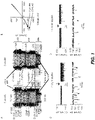
- a nanopore a portion of which is embedded into a membrane can be constructed by homology modeling from a known ClyA structure using VMD, e.g., as described in Humphrey et al., "VMD: Visual Molecular Dynamics” J. Mol. Graphics (1996) 14: 33-38 ; and NAMD, e.g., as described in Phillips et al., "Scalable Molecular Dynamics with NAMD” J. Comput. Chem. (2005) 26: 1781-1802 . See, e.g., FIG. 1A .
- the term "reference ClyA amino acid sequence” refers to a known amino acid sequence of a ClyA nanopore subunit.
- ClyA nanopore subunits include, e.g., but not limited to ClyA wild-type (ClyA-WT), ClyA-SS, ClyA-CS, and ClyA-AS. See, e.g., Soskine et al. "Tuning the size and properties of ClyA nanopores assisted by directed evolution" J Am Chem Soc.
- ClyA-SS ClyA-SS
- ClyA-CS ClyA-CS
- ClyA-AS ClyA-AS
- Any ClyA amino acid sequences described in WO 2016/166232 and WO 2014/153625 can also be used as a reference ClyA amino acid sequence.
- the reference ClyA amino acid sequence may be an amino acid sequence of ClyA-WT as set forth in SEQ ID NO: 1.
- the reference amino acid may be an amino acid sequence of ClyA-AS as set forth in SEQ ID NO: 2, which contains the following mutations: C87A, L99Q, E103G, F166Y, I203V, C285S, K294R, as compared to the amino sequence of ClyA-WT as set forth in SEQ ID NO: 1.
- the amino acid sequence of ClyA-AS can further include H307Y, as compared to the amino acid sequence of ClyA-WT.
- a modified ClyA nanopore subunit polypeptide may comprise an amino acid sequence that is at least about 80% (including, e.g., at least about 85%, at least about 90%, at least about 95%, at least about 96%, at least about 97%, at least about 98%, at least about 99% or higher) identical to a reference ClyA amino acid sequence.
- Standard methods in the art may be used to determine homology.
- the UWGCG Package provides the BESTFIT program which can be used to calculate homology, for example used on its default settings ( Devereux et al (1984) Nucleic Acids Research 12, p387-395 ).
- the PILEUP and BLAST algorithms can be used to calculate homology or line up sequences (such as identifying equivalent residues or corresponding sequences (typically on their default settings)), for example as described in Altschul S. F. (1993) J Mol Evol 36:290-300 ; Altschul, S.F et al (1990) J Mol Biol 215:403-10 .
- Software for performing BLAST analyses is publicly available through the National Center for Biotechnology Information (http://www.ncbi.nlm.nih.gov/).
- the amino acid sequence of a modified ClyA nanopore subunit polypeptide may comprise (i) a first positive charge modification (e.g., a first positively-charged amino acid substitution) at a position within the cis opening-forming amino acid sequence; and (ii) a second positive charge modification (e.g., a second positively-charged amino acid substitution) at a position within the midsection-forming amino acid sequence.
- a first positive charge modification e.g., a first positively-charged amino acid substitution
- a second positive charge modification e.g., a second positively-charged amino acid substitution
- the first and second positive charge modifications are selected to provide higher frequency of capture and/or translocation of a negatively-charged polymer (e.g., a polynucleotide such as double stranded or single stranded DNA) through the nanopore, as compared to a reference ClyA amino acid sequence.
- a negatively-charged polymer e.g., a polynucleotide such as double stranded or single stranded DNA
- the first positively-charged amino acid substitution may be at position 110 of the amino acid sequence as set forth in SEQ ID NO: 1 or SEQ ID NO: 2.
- Substitution with a positively-charged amino acid may take place at one of more of the following positions: E106, D114, D121, D122, E129, E85, E78, D268, D267, D265, E258 of SEQ ID NO: 1 or SEQ ID NO: 2.
- a ClyA amino acid sequence (e.g., as set forth in SEQ ID NO 1 or 2) may be modified or engineered to include additional amino acids "MI" at its N-terminus.
- the second positively-charged amino acid substitution may be at position 64 of the amino acid sequence as set forth in SEQ ID NO: 1 or SEQ ID NO: 2. Substitution with a positively-charged amino acid takes place at one of more of the following positions: D74, D71, D64, E53, E161, D158, E46, E42, D41 of SEQ NO: 1 or SEQ ID NO: 2.
- positively-charged amino acid substitution refers to a modification to a reference amino acid that increases the net positive charge, or decreases the net negative charge, of the reference amino acid, e.g., as detected at pH 7.0-8.0 (e.g., at pH 8.0) and at room temperature, e.g., at 20-25°C.
- a positively-charged amino acid substitution can include, but is not limited to, (i) replacement of a negatively-charged amino acid with a less negatively charged amino acid, neutral amino acid, or positively-charged amino acid, (ii) replacement of a neutral amino acid with a positively-charged amino acid, or (iii) replacement of a positively charged amino acid with a more positively-charged amino acid.
- a positively-charged amino acid substitution may include deletion of a negatively-charged amino acid or addition of a positively-charged amino acid.
- a positively-charged amino acid substitution may include one or more chemical modifications of one or more negatively charged amino acids which neutralize their negative charge. For instance, the one or more negatively charged amino acids may be reacted with a carbodiimide.
- a positively-charged amino acid is an amino acid having an isoelectric point (pI) that is higher than the pH of a solution so that the amino acid in the solution carries a net positive charge.
- examples of a positively-charged amino acid as detected at pH 7.0-8.0 (e.g., at pH 8.0) and at room temperature, e.g., at 20-25°C include, but are not limited to arginine (R), histidine (H), and lysine (K).
- a negatively-charged amino acid is an amino acid having a pI that is lower than the pH of a solution so that the amino acid in the solution carries a net negative charge.
- Examples of a negatively-charged amino acid as detected at pH 7.0-8.0 (e.g., at pH 8.0) and at room temperature, e.g., at 20-25°C, include, but are not limited to aspartic acid (D), glutamic acid (E), serine (S), glutamine (Q).
- a neutral amino acid is an amino acid having an isoelectric point (pI) that is same as the pH of a solution so that the amino acid in the solution carries no net charge.
- the pI values of amino acids are known in the art.
- amino acid can be an naturally-occurring or synthetic amino acid.
- the first and/or second positively-charged amino acid substitutions may include, but are not limited to substitution of a reference amino acid with one of an arginine, a histidine, and a lysine.
- the first positively-charged amino acid substitution may be S110R, wherein position 110 corresponds to amino acid 110 of SEQ ID NO: 1 or SEQ ID NO: 2.
- the second positively-charged amino acid substitution may be D64R, wherein position 64 corresponds to amino acid 64 of SEQ ID NO: 1 or SEQ ID NO: 2.
- amino acid substitutions may be made to a reference ClyA amino acid sequence, for example up to 1, 2, 3, 4, 5, 6, 7, 8, 9, 10, 15, 20, or 30 substitutions.
- Conservative substitutions replace amino acids with other amino acids of similar chemical structure, similar chemical properties or similar side-chain volume.
- the amino acids introduced may have similar polarity, hydrophilicity, hydrophobicity, basicity, acidity, neutrality or charge to the amino acids they replace.
- the conservative substitution may introduce another amino acid that is aromatic or aliphatic in the place of a pre-existing aromatic or aliphatic amino acid.
- amino acids are well-known in the art and may be selected in accordance with the properties of the 20 main amino acids as defined in Table A below. Where amino acids have similar polarity, this can also be determined by reference to the hydropathy scale for amino acid side chains in Table A.
- One or more amino acid residues of the amino acid sequence of SEQ ID NO: 1 or 2 may additionally be deleted from the polypeptides described above. Up to 1, 2, 3, 4, 5, 10, 20 or 30 residues may be deleted, or more.
- One or more amino acids may be alternatively or additionally added to the polypeptides described above.
- An extension may be provided at the amino terminal or carboxy terminal of the amino acid sequence of SEQ ID NO: 1 or 2 or polypeptide variant or fragment thereof.
- the extension may be quite short, for example from 1 to 10 amino acids in length. Alternatively, the extension may be longer, for example up to 50 or 100 amino acids.
- a carrier protein may be fused to an amino acid sequence, e.g., an amino acid sequence of a modified ClyA nanopore subunit polypeptide. Other fusion proteins are discussed in more detail below.
- a reference amino acid may be substituted with a target amino acid by replacing the codon for the reference amino acid with a codon for the target amino acid at the relevant position in a polynucleotide encoding a modified ClyA nanopore subunit polypeptide.
- the polynucleotide can then be expressed as discussed below.
- the amino acid is a non-naturally-occurring amino acid, it may be introduced by including synthetic aminoacyltRNAs in the IVTT system used to express the modified ClyA nanopore subunit polypeptide. Alternatively, it may be introduced by expressing the modified ClyA nanopore subunit polypeptide in E.
- coli that are auxotrophic for specific amino acids in the presence of synthetic (i.e., non-naturally-occurring) analogues of those specific amino acids. They may also be produced by naked ligation if the modified ClyA nanopore subunit polypeptide is produced using partial peptide synthesis.
- the trans opening-forming amino acid sequence of a modified ClyA nanopore subunit polypeptide may carry a net negative charge, e.g., as detected at pH 7.0-8.0 (e.g., at pH 8.0) and room temperature (e.g., at 20-25°C), which is comparable to (e.g., within 10%, within 5%, within 4%, within 3%, within 2%, within 1%, or lower) the net negative charge of the corresponding trans opening-forming portion of a reference ClyA amino acid sequence.
- the trans opening forming amino acid sequence of the modified ClyA nanopore subunit polypeptide can be at least about 95% or higher (including, e.g., at least about 96%, at least about 97%, at least about 98%, at least about 99% or up to 100%) identical to the corresponding trans opening-forming portion of a reference ClyA amino acid sequence, e.g., as set forth in SEQ ID NO: 1 or SEQ ID NO: 2.
- the trans opening-forming amino acid sequence of a modified ClyA nanopore subunit polypeptide may be 100% identical to the corresponding trans opening-forming portion of the amino acid sequence as set forth in SEQ ID NO: 2.
- modified ClyA nanopore subunit polypeptides described herein may be used to form a homo-multimeric nanopore or hetero-multimeric nanopore as described herein. Accordingly, modified ClyA nanopore subunit polypeptides retain the ability to form a nanopore with other subunit polypeptides. Methods for assessing the ability of modified monomers to form nanopores are well-known in the art. For instance, a modified ClyA nanopore subunit polypeptide may be inserted into an amphiphilic layer along with other appropriate subunits and its ability to oligomerize to form a pore may be determined. Methods are known in the art for inserting subunits into membranes, such as amphiphilic layers.
- subunits may be suspended in a purified form in a solution containing a triblock copolymer membrane such that it diffuses to the membrane and is inserted by binding to the membrane and assembling into a functional state.
- subunits may be directly inserted into the membrane using the "pick and place" method described in M.A. Holden, H. Bayley. J. Am. Chem. Soc. 2005, 127, 6502-6503 and International Application No. PCT/GB2006/001057 (published as WO 2006/100484 ).
- Modified ClyA nanopore subunit polypeptides may contain non-specific modifications as long as they do not interfere with nanopore formation.
- a number of non-specific side chain modifications are known in the art and may be made to the side chains of the amino acids. Such modifications include, for example, reductive alkylation of amino acids by reaction with an aldehyde followed by reduction with NaBH 4 , amidination with methylacetimidate or acylation with acetic anhydride.
- Modified ClyA nanopore subunit polypeptides can be produced using standard methods known in the art. Modified ClyA nanopore subunit polypeptides may be made synthetically or by recombinant means. For example, modified ClyA nanopore subunit polypeptides may be synthesized by in vitro translation and transcription (IVTT). Suitable methods for producing pores and modified ClyA nanopore subunit polypeptides are discussed in International Application Nos. PCT/GB09/001690 (published as WO 2010/004273 ), PCT/GB09/001679 (published as WO 2010/004265 ) or PCT/GB10/000133 (published as WO 2010/086603 ).
- Modified ClyA nanopore subunit polypeptides as described herein may be produced using D-amino acids.
- modified ClyA nanopore subunit polypeptides as described herein may comprise a mixture of L-amino acids and D-amino acids. This is conventional in the art for producing such proteins or peptides.
- Modified ClyA nanopore subunit polypeptides may be chemically modified.
- Modified ClyA nanopore subunit polypeptides can be chemically modified in any way and at any site.
- modified ClyA nanopore subunit polypeptides may be chemically modified by attachment of a dye or a fluorophore.
- Modified ClyA nanopore subunit polypeptide may be chemically modified by attachment of a molecule to one or more cysteines (cysteine linkage), attachment of a molecule to one or more lysines, attachment of a molecule to one or more nonnatural amino acids, enzyme modification of an epitope or modification of a terminus. Suitable methods for carrying out such modifications are well-known in the art.
- Modified ClyA nanopore subunit polypeptides may be chemically modified with a molecular adaptor that facilitates the interaction between a nanopore comprising the modified ClyA nanopore subunit polypeptide and a target nucleotide or target polynucleotide sequence.
- the presence of the adaptor improves the host-guest chemistry of the nanopore and the nucleotide or polynucleotide sequence and thereby improves the sequencing ability of pores formed from the modified ClyA nanopore subunit polypeptides.
- the principles of host-guest chemistry are well-known in the art.
- the adaptor has an effect on the physical or chemical properties of the nanopore that improves its interaction with the nucleotide or polynucleotide sequence.
- the adaptor may alter the charge of the barrel or channel of the pore or specifically interact with or bind to the nucleotide or polynucleotide sequence thereby facilitating its interaction with the pore.
- a molecular adaptor may be a cyclic molecule, a cyclodextrin, a species that is capable of hybridization, a DNA binder or interchelator, a peptide or peptide analogue, a synthetic polymer, an aromatic planar molecule, a small positively-charged molecule or a small molecule capable of hydrogen-bonding.
- a molecular adaptor can be covalently attached to a modified ClyA nanopore subunit polypeptide.
- the adaptor can be covalently attached to the nanopore using any method known in the art.
- the adaptor is typically attached via chemical linkage. If the molecular adaptor is attached via cysteine linkage, one or more cysteines can be introduced to the modified ClyA nanopore subunit polypeptide by substitution.
- a modified ClyA nanopore subunit polypeptide may be attached or coupled to a polynucleotide binding protein, e.g., helicases, exonucleases, and polymerases.
- a modified ClyA nanopore subunit polypeptide may be attached or coupled to a helicase, e.g., a DNA helicase. Examples of helicases, exonucleases, and polymerases that are suitable for use in nanopore sequencing are known in the art.
- the modified ClyA nanopore subunit polypeptide may be attached or coupled to a helicase, e.g., a DNA helicase, a Hel308 helicase (e.g., as described in WO 2013/057495 ), a RecD helicase (e.g., as described in WO2013/098562 ), a XPD helicase (e.g., as described in WO201/098561 ), or a Dda helicase (e.g., as described in WO2015/055981 ).
- a helicase e.g., a DNA helicase, a Hel308 helicase (e.g., as described in WO 2013/057495 ), a RecD helicase (e.g., as described in WO2013/098562 ), a XPD helicase (e.g., as described in WO201/098561 ), or a Dda helicase (e.g.
- the translocation speed control may be determined by the type of polynucleotide binding protein and/or amount of fuel (ATP) added to the system.
- ATP fuel
- the rate of translocation of the double stranded DNA analyte may be controlled by a double stranded DNA translocase such as FtsK.
- the translocation speed of a target polynucleotide can be between about 30 B/s and 1000 B/s.
- a polynucleotide binding protein can be covalently attached to a modified ClyA nanopore subunit polypeptide.
- the polynucleotide binding protein can be covalently attached to the modified ClyA nanopore subunit polypeptide using any method known in the art.
- the modified ClyA nanopore subunit polypeptide and the polynucleotide binding protein may be chemically fused or genetically fused.
- the modified ClyA nanopore subunit polypeptide and the polynucleotide binding protein are genetically fused if the whole construct is expressed from a single polynucleotide sequence. Genetic fusion of a modified ClyA nanopore subunit polypeptide to a polynucleotide binding protein is discussed in International Application No. PCT/GB09/001679 ( published as WO 2010/004265 ).
- a modified ClyA nanopore subunit polypeptide may be chemically modified with a molecular adaptor and a polynucleotide binding protein.
- any of the proteins described herein may be modified to assist their identification or purification, for example by the addition of histidine residues (a his tag), aspartic acid residues (an asp tag), a streptavidin tag, a flag tag, a SUMO tag, a GST tag or a MBP tag, or by the addition of a signal sequence to promote their secretion from a cell where the polypeptide does not naturally contain such a sequence.
- His tag histidine residues
- asp tag aspartic acid residues
- streptavidin tag a flag tag
- SUMO tag a SUMO tag
- GST tag a GST tag
- MBP tag a MBP tag
- An alternative to introducing a genetic tag is to chemically react a tag onto a native or engineered position on the protein.
- any of the proteins described herein may be labelled with a detectable label.
- the detectable label may be any suitable label which allows the protein to be detected. Suitable labels include, but are not limited to, fluorescent molecules, radioisotopes, e.g., 1251, 35S, enzymes, antibodies, antigens, polynucleotides and ligands such as biotin.
- any of the proteins described herein, including the modified ClyA nanopore subunit polypeptide described herein, can be produced using standard methods known in the art.
- Polynucleotide sequences encoding a protein may be derived and replicated using standard methods in the art.
- Polynucleotide sequences encoding a protein may be expressed in a bacterial host cell using standard techniques in the art.
- the protein may be produced in a cell by in situ expression of the polypeptide from a recombinant expression vector.
- the expression vector optionally carries an inducible promoter to control the expression of the polypeptide.
- Proteins may be produced in large scale following purification by any protein liquid chromatography system from protein producing organisms or after recombinant expression.
- Typical protein liquid chromatography systems include FPLC, AKTA systems, the Bio-Cad system, the Bio-Rad BioLogic system and the Gilson HPLC system.
- polynucleotide sequence encoding a subunit polypeptide of any one of the modified ClyA nanopores provided herein.
- Polynucleotide sequences may be derived and replicated using standard methods in the art. Chromosomal DNA encoding wild-type ClyA may be extracted from a pore producing organism, such as Salmonella typhi. The gene encoding the pore subunit may be amplified using PCR involving specific primers. The amplified sequence may then undergo site-directed mutagenesis. Suitable methods of site-directed mutagenesis are known in the art and include, for example, combine chain reaction. Polynucleotides encoding a subunit polypeptide of any one of the modified ClyA nanopores provided herein can be made using well-known techniques, such as those described in Sambrook, J. and Russell, D. (2001). Molecular Cloning: A Laboratory Manual, 3rd Edition. Cold Spring Harbor Laboratory Press, Cold Spring Harbor, NY .
- the resulting polynucleotide sequence may then be incorporated into a recombinant replicable vector such as a cloning vector.
- the vector may be used to replicate the polynucleotide in a compatible host cell.
- polynucleotide sequences may be made by introducing a polynucleotide into a replicable vector, introducing the vector into a compatible host cell, and growing the host cell under conditions which bring about replication of the vector.
- the vector may be recovered from the host cell. Suitable host cells for cloning of polynucleotides are known in the art.
- a modified ClyA nanopore subunit polypeptide or a construct described herein may be produced by expressing a polynucleotide encoding a subunit polypeptide of any of a modified ClyA nanopore in a suitable host cell.
- the polynucleotide is preferably part of a vector and is preferably operably linked to a promoter.
- the nanopore typically permits capture of a negatively-charged polymer (e.g., polynucleotide such as DNA or RNA) into the modified ClyA nanopore and/or translocation of the negatively-charged polymer through the modified ClyA nanopore.
- the modified ClyA nanopore comprises a first opening, a mid-section, a second opening, and a lumen extending from the first opening through the midsection to the second opening, wherein a luminal surface of the first opening comprises a first positively-charged amino acid substitution and a luminal surface of the mid-section comprises a second positively charged amino acid substitution.
- the luminal surface of the second opening defines an electronegative constriction.
- the first positive-charged amino acid substitution and the second charged amino acid substation are described in detail in the section "Modified ClyA nanopore subunit polypeptide" above.
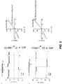
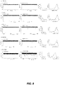
- FIG. 1 shows a modified ClyA nanopore.
- the modified ClyA nanopore comprises a first opening 102, a mid-section 104, and a second opening 106.
- the lumen 108 extends from the first opening 102 through the mid-section 104 to the second opening 106 and has a total length of about 13 nm to about 15 nm.
- the first opening 102 and the mid-section 104 have a diameter of about 5 nm to about 7 nm.
- the luminal surface of the second opening 106 defines an electronegative constriction 112, wherein the narrowest cross-section has a diameter of about 3 nm to about 4 nm.
- the second opening 106 (with a length of about 3 nm to about 5 nm) of the modified ClyA nanopore is inserted into a membrane (e.g., a bilayer) 110 such that a solution in which the modified ClyA nanopore is present is separated into two sides and the first opening 102 is present in one side of the solution while the electronegative constriction 112 is present in another side of the solution.
- a target polymer e.g., target polynucleotide
- the first opening 102 is a cis opening and the second opening 106 is a trans opening.
- luminal surface refers to the internal surface of a lumen that is exposed to a solution.
- electronegative constriction or “negatively-charged constriction” refers to a constriction having a net negative surface charge.
- the luminal surface of the second opening that defines an electronegative constriction has a net negative surface charge as shown in FIG. 1 (panel A).
- the distance within the lumen from the first positively-charged amino acid substitution to the second positively charged amino acid substitution may vary within a range of about 0.5 nm to about 10 nm, or about 3 nm to about 7 nm.
- the distance within the lumen from the first positively-charged amino acid substitution to the second positively charged amino acid substitution may be about 1 nm, about 2 nm, about 3 nm, about 4 nm, about 5 nm, about 6 nm, about 7 nm, about 8 nm, or about 9 nm.
- any forms of ClyA may be used to produce the modified ClyA nanopore described herein.
- the amino acid sequences of various forms of ClyA including, e.g., but not limited to wild-type ClyA (ClyA-WT) and ClyA-AS, and nucleotide sequences encoding the same are known in the art.
- the modified ClyA nanopore may comprise a subunit polypeptide having an amino acid sequence that is at least about 80% (including, e.g., at least about 85%, at least about 90%, at least about 95%, or higher) identical to a reference ClyA amino acid sequence as described herein.
- the modified ClyA nanopore may comprise a subunit polypeptide having an amino acid sequence that is at least about 80% (including, e.g., at least about 85%, at least about 90%, at least about 95%, or higher) identical to the amino acid sequence as set forth in SEQ ID NO: 1, which corresponds to the wild-type ClyA.
- the modified ClyA nanopore may comprise a subunit polypeptide having an amino acid sequence that is at least about 80% (including, e.g., at least about 85%, at least about 90%, at least about 95%, or higher) identical to the amino acid sequence as set forth in SEQ ID NO: 2, which corresponds to the amino acid sequence of ClyA-AS.
- the modified ClyA nanopore may comprise up to 15 substitutions (e.g., 2, 3, 4, 5, 6, 7, 8, 9, 10, 11, 12, 13, 14, or 15 substitutions) compared to the amino acid sequences as set forth in SEQ ID NO: 1 or SEQ ID NO: 2 including the first and second positive charge modifications (e.g., the first and second positively-charged amino acid substitutions).
- substitutions e.g., 2, 3, 4, 5, 6, 7, 8, 9, 10, 11, 12, 13, 14, or 15 substitutions
- the first positively charged substitution is located at E106, S110, D114, D121, D122, E129, E85, E78, D268, D267, D265, or E258 of SEQ ID NO: 1 or 2; and/or is at a position within a range of 78 to 106 of SEQ ID NO: 1 or 2.
- the first positive charge modification e.g., the first positively-charged amino acid substitution
- the first positive charge modification may be located at position E106, S110, D114, D121, D122, E129, E85, E78, D268, D267, D265, E258, or combinations thereof in the amino acid sequence as set forth in SEQ ID NO: 1 or SEQ ID NO: 2.
- the second positively-charged amino acid substitution is located at position D74, D71, D64, E53, E161, D158, E46, E42, D41, or combinations thereof in the amino acid sequence as set forth in SEQ NO: 1 or SEQ ID NO: 2.
- the modified ClyA nanopore can be homo-multimeric (e.g., all subunits within the nanopore are the same) or hetero-multimeric (e.g., at least one subunit is different from others within the nanopore).
- the modified ClyA nanopore may comprise any number of subunit polypeptides that are sufficient to form a lumen large enough to permit a target polymer (e.g., polynucleotide) pass through.
- the modified ClyA nanopore may comprise 12 subunit polypeptides or more, including, e.g., 13 subunit polypeptides, and 14 subunit polypeptides, wherein at least one or more of the subunit polypeptides comprises the first and second positively-charged amino acid substitutions as described herein.
- the modified ClyA nanopores can be used for distinguishing double stranded polynucleotides from single stranded polynucleotides, e.g., based on the dwell time in the nanopore and the current flowing through the pore.
- the modified ClyA nanopores can be used for characterizing, such as sequencing, polynucleotide sequences.
- the modified ClyA nanopores can also be used to distinguish modified bases, e.g., between methylated and unmethylated nucleotides.
- the modified ClyA nanopores described herein provide higher frequency of capture and/or translocation of a polynucleotide through the nanopores in low ionic strength solutions, as compared to a ClyA nanopore without the first and second positively-charged substitutions described herein.
- low ionic strength solution refers to a solution with an ionic strength of less than 2 M, including, e.g., less than 1 M, less than 900 mM, less than 800 mM, less than 700 mM, less than 600 mM, less than 500 mM, less than 400 mM, less than 300 mM, less than 200 mM, less than 150 mM, or lower.
- a lower ionic strength solution may have an ionic strength of at least about 50 mM, at least about 100 mM, at least about 150 mM, at least about 200 mM, at least about 300 mM, at least about 400 mM, at least about 500 mM, at least about 600 mM, at least about 700 mM, at least about 800 mM, at least about 900 mM, at least about 1 M, or higher. Combinations of the above-references ranges are also encompassed.
- a low ionic strength solution may have an ionic strength of about 100 mM to about 600 mM, or about 150 mM to about 300 mM. Any salt can be used to yield a solution with appropriate ionic strength.
- Alkaline salt e.g., but not limited to potassium chloride or sodium chloride
- Alkaline salt can be used in the low ionic strength solution.
- the modified ClyA nanopores can discriminate between different nucleotides under a range of conditions.
- the pores can discriminate between nucleotides under conditions that are favorable to the characterizing, such as sequencing, of nucleic acids.
- the extent to which the modified ClyA nanopores can discriminate between different nucleotides can be controlled by altering the applied potential, the salt concentration, the buffer, the temperature and the presence of additives, such as urea, betaine and DTT. This allows the function of the pores to be fine-tuned, particularly when sequencing. This is discussed in more detail below.
- the modified ClyA nanopores may also be used to identify polynucleotide polymers from the interaction with one or more monomers rather than on a nucleotide by nucleotide basis.
- Modified ClyA nanopores provided herein may be used for characterizing nucleic acid-protein interactions.
- the nanopores can be used to interrogate protein-nucleic acids using different sensing modes such as, for example, by scanning and mapping the locations of binding sites along a nucleic acid and/or by probing the strength of interactions between a protein and nucleic acid.
- Native charges of a nucleic acid may be leveraged to apply an electrophoretic force to a nucleic acid-protein complex.
- DNA-protein interactions may be evaluated using voltage-driven threading of single DNA molecules through a protein nanopore.
- Modified ClyA nanopores provided herein may be used for detection and characterization of nucleic acid-protein interactions involving nucleic acid and other nucleic acid binding proteins such as transcription factors, enzymes, DNA packaging proteins and others.
- the modified ClyA nanopores may be isolated, substantially isolated, purified or substantially purified.
- the modified ClyA nanopores can be isolated or purified if it is completely free of any other components, such as lipids or other pores.
- a pore is substantially isolated if it is mixed with carriers or diluents which will not interfere with its intended use.
- a pore is substantially isolated or substantially purified if it is present in a form that comprises less than 10%, less than 5%, less than 2% or less than 1% of other components, such as triblock copolymers, lipids or other pores.
- one or more of the modified ClyA nanopores may be present in a membrane. Suitable membranes are discussed below.
- the modified ClyA nanopore may be present as an individual or single pore. Alternatively, the modified ClyA nanopores may be present in a homologous or heterologous population of two or more pores.
- the modified ClyA nanopores may be arranged in an array of microwells, wherein each microwell contains at least one nanopore is in a membrane.
- homo-multimeric nanopores comprising identical modified ClyA nanopore subunit polypeptides are also provided herein.
- the homo-multimeric nanopore may comprise any of the modified ClyA nanopore subunit polypeptides described herein.
- the homo-multimeric nanopore can be used for characterizing, such as sequencing, polynucleotides, and/or detecting the presence or absence of single stranded polynucleotide vs double stranded polynucleotide.
- the homo-multimeric nanopore described herein may have any of the advantages discussed above.
- the homo-multimeric pore may contain any number of modified ClyA nanopore subunit polypeptides.
- the pore typically comprises at least 10, at least 11, at least 12, at least 13, or at least 14 identical modified ClyA nanopore subunit polypeptides, such as 12, 13, or 14 identical modified ClyA nanopore subunit polypeptides.
- Hetero-multimeric nanopores comprising at least one modified ClyA nanopore subunit polypeptides are also provided herein.
- the hetero-multimeric nanopores can be used for characterizing, such as sequencing, polynucleotides, and/or detecting the presence or absence of single stranded polynucleotide vs double stranded polynucleotide.
- Hetero-multimeric nanopores can be made using methods known in the art (e.g., Protein Sci. 2002 Jul; 11(7):1813-24 ).
- the hetero-multimeric pore contains sufficient subunit polypeptide to form the pore.
- the subunit polypeptides may be of any type.
- the pore typically comprises at least 10, at least 11, at least 12, at least 13, or at least 14 subunit polypeptides, such as 12, 13, or 14 subunit polypeptides.
- all of the subunit polypeptides are modified ClyA nanopore subunit polypeptides and at least one of them differs from the others.
- the pore comprises 12 or 13 modified ClyA nanopore subunit polypeptides and at least one of them differs from the others. They may all differ from one another.
- At least one of the subunit polypeptides is not a modified ClyA nanopore subunit polypeptide as described herein.
- the remaining monomers may be any one of the modified ClyA nanopore subunit polypeptides described herein.
- the pore may comprise 12, 11, 10, 9, 8, 7, 6, 5, 4, 3, 2, or 1 modified ClyA nanopore subunit polypeptide(s).
- the modified ClyA nanopore subunit polypeptide(s) that form the nanopore can be the same or different.
- the method comprises: (a) providing, in a low ionic strength solution of 150 mM to 300 mM, a modified ClyA nanopore as described herein and a membrane, wherein the modified ClyA nanopore is present in the membrane; (b) adding in the low ionic strength solution of step (a) the target polynucleotide; and (c) measuring, during application of a potential across the nanopore, ion flow through the modified ClyA nanopore, wherein the ion flow measurements are indicative of one or more characteristics of the target polynucleotide.
- the target polynucleotide may be added to the cis side of the low ionic strength solution.
- the target polynucleotide may also be called the template polynucleotide or the polynucleotide of interest.
- a polynucleotide such as a nucleic acid
- the polynucleotide or nucleic acid may comprise any combination of any nucleotides.
- the nucleotides can be naturally occurring or artificial.
- One or more nucleotides in the polynucleotide can be oxidized or methylated.
- One or more nucleotides in the polynucleotide may be damaged.
- the polynucleotide may comprise a pyrimidine dimer. Such dimers are typically associated with damage by ultraviolet light and are the primary cause of skin melanomas.
- One or more nucleotides in the polynucleotide may be modified, for instance with a label or a tag. Suitable labels are described below.
- the polynucleotide may comprise one or more spacers.
- a nucleotide typically contains a nucleobase, a sugar and at least one phosphate group.
- the nucleobase and sugar form a nucleoside.
- the nucleobase is typically heterocyclic. Nucleobases include, but are not limited to, purines and pyrimidines and more specifically adenine (A), guanine (G), thymine (T), uracil (U) and cytosine (C).
- A adenine
- G guanine
- T thymine
- U uracil
- C cytosine
- the sugar is typically a pentose sugar.
- Nucleotide sugars include, but are not limited to, ribose and deoxyribose.
- the sugar is preferably a deoxyribose.
- the polynucleotide preferably comprises the following nucleosides: deoxyadenosine (dA), deoxyuridine (dU) and/or thymidine (dT), deoxyguanosine (dG) and deoxycytidine (dC).
- the nucleotide is typically a ribonucleotide or deoxyribonucleotide.
- the nucleotide typically contains a monophosphate, diphosphate or triphosphate.
- the nucleotide may comprise more than three phosphates, such as 4 or 5 phosphates. Phosphates may be attached on the 5' or 3' side of a nucleotide.
- Nucleotides include, but are not limited to, adenosine monophosphate (AMP), guanosine monophosphate (GMP), thymidine monophosphate (TMP), uridine monophosphate (UMP), 5-methylcytidine monophosphate, 5-hydroxymethylcytidine monophosphate, cytidine monophosphate (CMP), cyclic adenosine monophosphate (cAMP), cyclic guanosine monophosphate (cGMP), deoxyadenosine monophosphate (dAMP), deoxyguanosine monophosphate (dGMP), deoxythymidine monophosphate (dTMP), deoxyuridine monophosphate (dUMP), deoxycytidine monophosphate (dCMP) and deoxymethylcytidine monophosphate.
- the nucleotides are preferably selected from AMP, TMP, GMP, CMP, UMP, dAMP, dTMP, dGMP, dCMP and dUMP.
- a nucleotide may be abasic (i.e., lack a nucleobase).
- a nucleotide may also lack a nucleobase and a sugar.
- the nucleotides in the polynucleotide may be attached to each other in any manner.
- the nucleotides are typically attached by their sugar and phosphate groups as in nucleic acids.
- the nucleotides may be connected via their nucleobases as in pyrimidine dimers.
- the polynucleotide may be single stranded or double stranded. At least a portion of the polynucleotide is preferably double stranded.
- the polynucleotide can be a nucleic acid, such as deoxyribonucleic acid (DNA) or ribonucleic acid (RNA).
- the polynucleotide can comprise one strand of RNA hybridized to one strand of DNA.
- the polynucleotide may be any synthetic nucleic acid known in the art, such as peptide nucleic acid (PNA), glycerol nucleic acid (GNA), threose nucleic acid (TNA), locked nucleic acid (LNA) or other synthetic polymers with nucleotide side chains.
- PNA backbone is composed of repeating N-(2-aminoethyl)-glycine units linked by peptide bonds.
- the GNA backbone is composed of repeating glycol units linked by phosphodiester bonds.
- the TNA backbone is composed of repeating threose sugars linked together by phosphodiester bonds.
- LNA is formed from ribonucleotides as discussed above having an extra bridge connecting the 2' oxygen and 4' carbon in the ribose moiety.
- the polynucleotide is most preferably ribonucleic nucleic acid (RNA) or deoxyribonucleic acid (DNA).
- the polynucleotide can be any length.
- the polynucleotide can be at least 10, at least 50, at least 100, at least 150, at least 200, at least 250, at least 300, at least 400 or at least 500 nucleotides or nucleotide pairs in length.
- the polynucleotide can be 1000 or more nucleotides or nucleotide pairs, 5000 or more nucleotides or nucleotide pairs in length or 100000 or more nucleotides or nucleotide pairs in length.
- any number of polynucleotides can be investigated.
- the method described herein may concern characterizing 2, 3, 4, 5, 6, 7, 8, 9, 10, 20, 30, 50, 100 or more polynucleotides. If two or more polynucleotides are characterized, they may be different polynucleotides or two instances of the same polynucleotide.
- the polynucleotide can be naturally occurring or artificial.
- the method may be used to verify the sequence of a manufactured oligonucleotide.
- the method is typically carried out in vitro.
- the polynucleotide may comprise an attached species such as a protein or analyte.
- the polynucleotide may comprise a hybridized probe.
- Each analyte is typically present in any suitable sample.
- the method can be carried out on two or more samples that are known to contain or suspected to contain the analytes. Alternatively, the method may be carried out on two or more samples to confirm the identity of two or more analytes whose presence in the samples is known or expected. The method may be carried out on samples to distinguish double stranded polynucleotides from single-stranded polynucleotides.
- the first sample and/or second sample may be a biological sample.
- the methods described herein may be carried out in vitro using at least one sample obtained from or extracted from any organism or microorganism.
- the first sample and/or second sample may be a non-biological sample.
- the non-biological sample can be a fluid sample. Examples of non-biological samples include surgical fluids, water such as drinking water, sea water or river water, and reagents for laboratory tests.
- the first sample and/or second sample is typically processed prior to being used in the methods described herein, for example by centrifugation or by passage through a membrane that filters out unwanted molecules or cells, such as red blood cells.
- the first sample and/or second sample may be measured immediately upon being taken.
- the first sample and/or second sample may also be typically stored prior to assay, preferably below -70°C.
- the method may involve measuring two, three, four or five or more characteristics of the polynucleotide.
- the one or more characteristics are preferably selected from (i) the length of the polynucleotide, (ii) the identity of the polynucleotide, (iii) the sequence of the polynucleotide, (iv) the secondary structure of the polynucleotide and (v) whether or not the polynucleotide is modified.
- any combination of (i) to (v) may be measured in accordance with the methods described herein, such as ⁇ i ⁇ , ⁇ ii ⁇ , ⁇ iii ⁇ , ⁇ iv ⁇ , ⁇ v ⁇ , ⁇ i,ii ⁇ , ⁇ i,ii ⁇ , ⁇ i,iv ⁇ , ⁇ i,v ⁇ , ⁇ ii,ii ⁇ , ⁇ iiiiv ⁇ , ⁇ iii,v ⁇ , ⁇ iii,v ⁇ , ⁇ iv,v ⁇ , ⁇ i,iiii ⁇ , ⁇ i,iii,v ⁇ , ⁇ i,iii,v ⁇ , ⁇ i,iiii,v ⁇ , ⁇ i,iiii,v ⁇ , ⁇ i,iiii,v ⁇ , ⁇ i,iiii,v ⁇ , ⁇ i,iiii,v ⁇ , ⁇ iiiii,v ⁇ , ⁇ iiiii,v ⁇ , ⁇ iiiii,v ⁇ ,
- the length of the polynucleotide may be measured for example by determining the number of interactions between the polynucleotide and the pore or the duration of interaction between the polynucleotide and the pore.
- the identity of the polynucleotide may be measured in a number of ways.
- the identity of the polynucleotide may be measured in conjunction with measurement of the sequence of the polynucleotide or without measurement of the sequence of the polynucleotide.
- the former is straightforward; the polynucleotide is sequenced and thereby identified.
- the latter may be done in several ways. For instance, the presence of a particular motif in the polynucleotide may be measured (without measuring the remaining sequence of the polynucleotide). Alternatively, the measurement of a particular electrical and/or optical signal in the method may identify the polynucleotide as coming from a particular source.
- the sequence of the polynucleotide can be determined as described previously. Suitable sequencing methods, particularly those using electrical measurements, are described in Stoddart D et al., Proc Natl Acad Sci, 12;106(19):7702-7 , Lieberman KR et al, J Am Chem Soc. 2010;132(50):17961-72 , and International Application WO 2000/28312 .
- the secondary structure may be measured in a variety of ways. For instance, if the method involves an electrical measurement, the secondary structure may be measured using a change in dwell time or a change in current flowing through the pore. This allows regions of single-stranded and double-stranded polynucleotide to be distinguished.
- the presence or absence of any modification may be measured.
- the method preferably comprises determining whether or not the polynucleotide is modified by methylation, by oxidation, by damage, with one or more proteins or with one or more labels, tags or spacers. Specific modifications will result in specific interactions with the pore which can be measured using the methods described below. For instance, methylcyotsine may be distinguished from cytosine on the basis of the current flowing through the pore during its interaction with each nucleotide.
- the target polynucleotide is contacted with any one of the modified ClyA nanopores described herein.
- the pore is typically present in a membrane. Suitable membranes are discussed below.
- the method may be carried out using any apparatus that is suitable for investigating a membrane/pore system in which a pore is present in a membrane.
- the method may be carried out using any apparatus that is suitable for transmembrane pore sensing.
- the apparatus comprises a chamber comprising an aqueous solution and a barrier that separates the chamber into two sections.
- the barrier typically has an aperture in which the membrane containing the pore is formed.
- the barrier forms the membrane in which the pore is present.
- the method may be carried out using the apparatus described in International Application No. PCT/GB08/000562 ( WO 2008/102120 ).
- a variety of different types of measurements may be made. This includes without limitation: electrical measurements and optical measurements. Possible electrical measurements include: current measurements, impedance measurements, tunneling measurements ( Ivanov AP et al., Nano Lett. 2011 Jan 12;11(1):279-85 ), and FET measurements (International Application WO 2005/124888 ). Optical measurements may be combined with electrical measurements ( Soni GV et al., Rev Sci Instrum. 2010 Jan;81(1):014301 ). The measurement may be a transmembrane current measurement such as measurement of ionic current flowing through the pore. Alternatively the measurement may be a fluorescence measurement indicative of ion flow through the channel such as disclosed by Heron et al, J. Am. Chem. Soc., 2009, 131 (5), 1652-1653 or measurement of a voltage across the membrane using a FET.
- Electrical measurements may be made using standard single channel recording equipment as describe in Stoddart D et al., Proc Natl Acad Sci, 12;106(19):7702-7 , Lieberman KR et al, J Am Chem Soc. 2010;132(50):17961-72 , and International Application WO 2000/28312 .
- electrical measurements may be made using a multi-channel system, for example as described in International Application WO 2009/077734 and International Application WO 2011/067559 .
- the method can be carried out with a potential applied across the membrane.
- the applied potential may be a voltage potential.
- the applied potential may be a chemical potential.
- An example of this is using a salt gradient across a membrane, such as an amphiphilic layer. A salt gradient is disclosed in Holden et al., J Am Chem Soc. 2007 Jul 11; 129(27):8650-5 .
- the current passing through the pore as a polynucleotide moves with respect to the pore is used to estimate or determine the sequence of the polynucleotide. This may be described as strand sequencing.
- the method may involve measuring the current passing through the pore as the polynucleotide moves with respect to the pore. Therefore the apparatus used in the method may also comprise an electrical circuit capable of applying a potential and measuring an electrical signal across the membrane and pore.
- the methods may be carried out using a patch clamp or a voltage clamp. The methods preferably involve the use of a voltage clamp.
- the method may involve the measuring of a current passing through the pore as the polynucleotide moves with respect to the pore. Suitable conditions for measuring ionic currents through transmembrane protein pores are known in the art and disclosed in the Example.
- the method is typically carried out with a voltage applied across the membrane and pore.
- the voltage used is typically from +5 V to -5 V, such as from +4 V to -4 V, +3 V to -3 V or +2 V to -2 V.
- the voltage used is typically from -600 mV to +600mV or -400 mV to +400 mV.
- the voltage used is preferably in a range having a lower limit selected from -400 mV, -300 mV, - 200 mV, -150 mV, -100 mV, -50 mV, -20mV and 0 mV and an upper limit independently selected from +10 mV, + 20 mV, +50 mV, +100 mV, +150 mV, +200 mV, +300 mV and +400 mV.
- the voltage used is more preferably in the range 100 mV to 240 mV and most preferably in the range of 120 mV to 220 mV. It is possible to increase discrimination between different nucleotides by a pore by using an increased applied potential.
- the method is typically carried out in the presence of any charge carriers, such as metal salts, for example alkali metal salt, halide salts, for example chloride salts, such as alkali metal chloride salt.
- Charge carriers may include ionic liquids or organic salts, for example tetramethyl ammonium chloride, trimethylphenyl ammonium chloride, phenyltrimethyl ammonium chloride, or 1-ethyl-3-methyl imidazolium chloride.
- the salt is present in the aqueous solution in the chamber.
- Potassium chloride (KCl), sodium chloride (NaCl), caesium chloride (CsCl) or a mixture of potassium ferrocyanide and potassium ferricyanide is typically used.
- KCl, NaCl and a mixture of potassium ferrocyanide and potassium ferricyanide are preferred.
- the charge carriers may be asymmetric across the membrane. For instance, the type and/or concentration of the charge carriers may be different on each side of the membrane.
- the salt concentration may be at saturation.
- the salt concentration may be 3 M or lower and is typically from 0.1 to 2.5 M, from 0.3 to 1.9 M, from 0.5 to 1.8 M, from 0.7 to 1.7 M, from 0.9 to 1.6 M or from 1 M to 1.4 M.
- the salt concentration is preferably from 150 mM to 1 M.
- the method is preferably carried out using a salt concentration of at least 0.3 M, such as at least 0.4 M, at least 0.5 M, at least 0.6 M, at least 0.8 M, at least 1.0 M, at least 1.5 M, at least 2.0 M, at least 2.5 M or at least 3.0 M.
- High salt concentrations provide a high signal to noise ratio and allow for currents indicative of the presence of a nucleotide to be identified against the background of normal current fluctuations.
- the modified ClyA nanopores described herein can be used to characterize a polynucleotide at high salt solution, the modified ClyA nanopores can permit efficient capture and/or translocation of a polynucleotide (e.g., double stranded DNA or single stranded DNA) through the nanopore even in low ionic strength solutions as described above.
- the method is typically carried out in the presence of a buffer.
- the buffer is present in the aqueous solution in the chamber. Any buffer may be used in the methods described herein.
- the buffer is phosphate buffer.
- Other suitable buffers are HEPES and Tris-HCl buffer.
- the methods are typically carried out at a pH of from 4.0 to 12.0, from 4.5 to 10.0, from 5.0 to 9.0, from 5.5 to 8.8, from 6.0 to 8.7 or from 7.0 to 8.8 or 7.5 to 8.5.
- the pH used is preferably about 7.5 or 8.0.
- the method may be carried out at from 0 °C to 100 °C, from 15 °C to 95 °C, from 16 °C to 90 °C, from 17 °C to 85 °C, from 18 °C to 80 °C, 19 °C to 70 °C, or from 20 °C to 60 °C.
- the methods are typically carried out at room temperature.
- the methods are optionally carried out at a temperature that supports enzyme function, such as about 37 °C.
- the method for characterizing a target polynucleotide may include adding a polynucleotide binding protein in the low ionic strength solution such that the polynucleotide binding protein binds to the target polynucleotide and controls the movement of the target polynucleotide through the modified ClyA nanopore.
- the polynucleotide binding protein may be any protein that is capable of binding to the polynucleotide and controlling its movement through the pore.
- the polynucleotide binding proteins include, but are not limited to helicases, polymerases, exonucleases, DNA clamps, etc.
- the polynucleotide may be contacted with the polynucleotide binding protein and the pore in any order. It is preferred that, when the polynucleotide is contacted with the polynucleotide binding protein, such as a helicase, and the pore, the polynucleotide firstly forms a complex with the protein. When the voltage is applied across the pore, the polynucleotide/protein complex then forms a complex with the pore and controls the movement of the polynucleotide through the pore.
- Any steps in the method using a polynucleotide binding protein are typically carried out in the presence of free nucleotides or free nucleotide analogues and an enzyme cofactor that facilitates the action of the polynucleotide binding protein.
- the method may comprise:
- the modified ClyA nanopores described herein may be present in a membrane.
- the polynucleotide is typically contacted with a modified ClyA nanopore in a membrane.
- Any membrane may be used. Suitable membranes are well-known in the art.
- the membrane is preferably an amphiphilic layer.
- An amphiphilic layer is a layer formed from amphiphilic molecules, such as phospholipids, which have both hydrophilic and lipophilic properties. The amphiphilic molecules may be synthetic or naturally occurring.
- Non-naturally occurring amphiphiles and amphiphiles which form a monolayer are known in the art and include, for example, block copolymers ( Gonzalez-Perez et al., Langmuir, 2009, 25, 10447-10450 ).
- Block copolymers are polymeric materials in which two or more monomer sub-units that are polymerized together to create a single polymer chain. Block copolymers typically have properties that are contributed by each monomer sub-unit. However, a block copolymer may have unique properties that polymers formed from the individual subunits do not possess.
- Block copolymers can be engineered such that one of the monomer subunits is hydrophobic or lipophilic, whilst the other sub-unit(s) are hydrophilic whilst in aqueous media.
- the block copolymer may possess amphiphilic properties and may form a structure that mimics a biological membrane.
- the block copolymer may be a diblock (consisting of two monomer sub-units), but may also be constructed from more than two monomer sub-units to form more complex arrangements that behave as amphipiles.
- the copolymer may be a triblock, tetrablock or pentablock copolymer.
- the membrane is preferably a triblock copolymer membrane.
- Archaebacterial bipolar tetraether lipids are naturally occurring lipids that are constructed such that the lipid forms a monolayer membrane. These lipids are generally found in extremophiles that survive in harsh biological environments, thermophiles, halophiles and acidophiles. Their stability is believed to derive from the fused nature of the final bilayer. It is straightforward to construct block copolymer materials that mimic these biological entities by creating a triblock polymer that has the general motif hydrophilic-hydrophobic-hydrophilic. This material may form monomeric membranes that behave similarly to lipid bilayers and encompass a range of phase behaviors from vesicles through to laminar membranes. Membranes formed from these triblock copolymers hold several advantages over biological lipid membranes. Because the triblock copolymer is synthesized, the exact construction can be carefully controlled to provide the correct chain lengths and properties to form membranes and to interact with pores and other proteins.
- Block copolymers may also be constructed from sub-units that are not classed as lipid sub-materials; for example a hydrophobic polymer may be made from siloxane or other nonhydrocarbon based monomers.
- the hydrophilic sub-section of block copolymer can also possess low protein binding properties, which allows the creation of a membrane that is highly resistant when exposed to raw biological samples.
- This head group unit may also be derived from non-classical lipid head-groups.
- Triblock copolymer membranes also have increased mechanical and environmental stability compared with biological lipid membranes, for example a much higher operational temperature or pH range.
- the synthetic nature of the block copolymers provides a platform to customize polymer based membranes for a wide range of applications.
- the membrane is most preferably one of the membranes disclosed in International Application No. PCT/GB2013/052766 or PCT/GB2013/052767 .
- amphiphilic molecules may be chemically-modified or functionalized to facilitate coupling of the polynucleotide.
- the amphiphilic layer may be a monolayer or a bilayer.
- the amphiphilic layer is typically planar.
- the amphiphilic layer may be curved.
- the amphiphilic layer may be supported.
- Amphiphilic membranes are typically naturally mobile, essentially acting as two dimensional fluids with lipid diffusion rates of approximately 10-8 cm s-1. This means that the pore and coupled polynucleotide can typically move within an amphiphilic membrane.
- the membrane may be a lipid bilayer.
- Lipid bilayers are models of cell membranes and serve as excellent platforms for a range of experimental studies. For example, lipid bilayers can be used for in vitro investigation of membrane proteins by single-channel recording. Alternatively, lipid bilayers can be used as biosensors to detect the presence of a range of substances.
- the lipid bilayer may be any lipid bilayer. Suitable lipid bilayers include, but are not limited to, a planar lipid bilayer, a supported bilayer or a liposome. The lipid bilayer is preferably a planar lipid bilayer. Suitable lipid bilayers are disclosed in International Application No. PCT/GB08/000563 (published as WO 2008/102121 ), International Application No. PCT/GB08/004127 (published as WO 2009/077734 ) and International Application No. PCT/GB2006/001057 (published as WO 2006/100484 ).
- the polynucleotide can be coupled to the membrane comprising any one of the modified ClyA nanopores described herein.
- the method may comprise coupling the polynucleotide to the membrane comprising any one of the modified ClyA nanopores described herein.
- the polynucleotide is preferably coupled to the membrane using one or more anchors.
- the polynucleotide may be coupled to the membrane using any known method.
- the polynucleotide may be double stranded. If the polynucleotide is double stranded, the method may further comprise before the contacting step ligating a hairpin adaptor to one end of the polynucleotide. The two strands of the polynucleotide may then be separated as or before the polynucleotide is contacted or interacted with a modified ClyA nanopore as described herein. The two strands may be separated as the polynucleotide movement through the pore is controlled by a polynucleotide binding protein, such as a helicase, or molecular brake. This is described in International Application No. PCT/GB2012/051786 (published as WO 2013/014451 ). Linking and interrogating both strands on a double stranded construct in this way increases the efficiency and accuracy of characterization.
- a polynucleotide binding protein such as a helicase, or molecular brake.
- a target double stranded polynucleotide may be provided with a hairpin loop adaptor at one end and the method may comprise contacting the polynucleotide with any one of the modified ClyA nanopores described herein such that both strands of the polynucleotide move through the pore and taking one or more measurements as the both strands of the polynucleotide move with respect to the pore wherein the measurements are indicative of one or more characteristics of the strands of the polynucleotide and thereby characterizing the target double stranded polynucleotide.
- the method preferably comprises attaching to the polynucleotide a leader sequence which preferentially threads into the pore.
- the leader sequence facilitates any of the methods described herein.
- the leader sequence is designed to preferentially thread into any one of the modified ClyA nanopores described herein and thereby facilitate the movement of polynucleotide through the nanopore.
- the leader sequence can also be used to link the polynucleotide to the one or more anchors as discussed above.
- a target polynucleotide may be modified by contacting the polynucleotide with a polymerase and a population of free nucleotides under conditions in which the polymerase forms a modified polynucleotide using the target polynucleotide as a template, wherein the polymerase replaces one or more of the nucleotide species in the target polynucleotide with a different nucleotide species when forming the modified polynucleotide.
- the modified polynucleotide may then be provided with one or more helicases attached to the polynucleotide and one or more molecular brakes attached to the polynucleotide. This type of modification is described in International Application No. PCT/GB2015/050483 . Any of the polymerases discussed herein may be used.
- the template polynucleotide may be contacted with the polymerase under conditions in which the polymerase forms a modified polynucleotide using the template polynucleotide as a template.
- Such conditions are known in the art.
- the polynucleotide is typically contacted with the polymerase in commercially available polymerase buffer, such as buffer from New England Biolabs ® .
- a primer or a 3' hairpin is typically used as the nucleation point for polymerase extension.
- Characterization, such as sequencing, of a polynucleotide using a transmembrane pore typically involves analyzing polymer units made up of k nucleotides where k is a positive integer (i.e., 'k-mers'). This is discussed in International Application No. PCT/GB2012/052343 (published as WO 2013/041878 ). While it is desirable to have clear separation between current measurements for different k-mers, it is common for some of these measurements to overlap.
- the modified polynucleotide contains k-mers which differ from those in the target polynucleotide.
- the different k-mers in the modified polynucleotide are capable of producing different current measurements from the k-mers in the target polynucleotide and so the modified polynucleotide provides different information from the target polynucleotide.
- the additional information from the modified polynucleotide can make it easier to characterize the target polynucleotide.
- the modified polynucleotide itself may be easier to characterize.
- the modified polynucleotide may be designed to include k-mers with an increased separation or a clear separation between their current measurements or k-mers which have a decreased noise.
- the polymerase preferably replaces two or more of the nucleotide species in the target polynucleotide with different nucleotide species when forming the modified polynucleotide.
- the polymerase may replace each of the two or more nucleotide species in the target polynucleotide with a distinct nucleotide species.
- the polymerase may replace each of the two or more nucleotide species in the target polynucleotide with the same nucleotide species.
- the different nucleotide species in the modified typically comprises a nucleobase which differs from adenine, guanine, thymine, cytosine or methylcytosine and/or comprises a nucleoside which differs from deoxyadenosine, deoxyguanosine, thymidine, deoxycytidine or deoxymethylcytidine.
- the different nucleotide species in the modified polynucleotide typically comprises a nucleobase which differs from adenine, guanine, uracil, cytosine or methylcytosine and/or comprises a nucleoside which differs from adenosine, guanosine, uridine, cytidine or methylcytidine.
- the different nucleotide species may be any of the universal nucleotides discussed above.
- the polymerase may replace the one or more nucleotide species with a different nucleotide species which comprises a chemical group or atom absent from the one or more nucleotide species.
- the chemical group may be a propynyl group, a thio group, an oxo group, a methyl group, a hydroxymethyl group, a formyl group, a carboxy group, a carbonyl group, a benzyl group, a propargyl group or a propargylamine group.
- the polymerase may replace the one or more nucleotide species with a different nucleotide species which lacks a chemical group or atom present in the one or more nucleotide species.
- the polymerase may replace the one or more of the nucleotide species with a different nucleotide species having an altered electronegativity.
- the different nucleotide species having an altered electronegativity preferably comprises a halogen atom.
- the method preferably further comprises selectively removing the nucleobases from the one or more different nucleotides species in the modified polynucleotide.
- a polynucleotide may be characterized by detecting labelled species that are released as a polymerase incorporates nucleotides into the polynucleotide.
- the polymerase uses the polynucleotide as a template. Each labelled species is specific for each nucleotide.
- the polynucleotide may be contacted with a modified ClyA nanopore described herein, a polymerase and labelled nucleotides such that phosphate labelled species are sequentially released when nucleotides are added to the polynucleotide(s) by the polymerase, wherein the phosphate species contain a label specific for each nucleotide.
- the polymerase may be any of those discussed above.
- the phosphate labelled species are detected using the pore and thereby characterizing the polynucleotide. This type of method is disclosed in European Application No. 13187149.3 (published as EP 2682460 ).
- kits for characterizing a target polynucleotide comprises any one of the modified ClyA nanopores described herein and the components of a membrane.
- the membrane is preferably formed from the components.
- the pore is preferably present in the membrane.
- the kit may comprise components of any of the membranes disclosed above, such as an amphiphilic layer or a triblock copolymer membrane.
- the kit may further comprise a polynucleotide binding protein.
- the kit may further comprise one or more anchors for coupling the polynucleotide to the membrane.
- the kit may additionally comprise one or more other reagents or instruments.
- reagents or instruments include one or more of the following: suitable buffer(s) (aqueous solutions), means to obtain a sample from a subject (such as a vessel or an instrument comprising a needle), means to amplify and/or express polynucleotides or voltage or patch clamp apparatus.
- Reagents may be present in the kit in a dry state such that a fluid sample resuspends the reagents.
- the kit may also, optionally, comprise instructions to enable the kit to be used in any one of the methods described herein or details regarding for which organism the method may be used.
- the apparatus comprises a plurality of modified ClyA nanopores as described herein and a plurality of membranes.
- the plurality of the modified ClyA nanopores may be present in the plurality of membranes.
- the numbers of modified ClyA nanopores and membranes may be equal.
- a single modified ClyA nanopore may be present in each membrane.
- the apparatus can further comprise instructions for carrying out any of the methods as described herein.
- the apparatus may be any conventional apparatus for polynucleotide analysis, such as an array or a chip.
- the apparatus may further comprise any of the features present in the kit disclosed herein.
- the apparatus may be set up to carry out any of the methods described herein, e.g., for characterizing a target polynucleotide.
- the apparatus may comprise: (a) a sensor device that is capable of supporting the plurality of modified ClyA nanopores and membranes and that is operable to perform polynucleotide characterization using the nanopores and membranes; and (b) at least one port for delivery of material for performing the characterization.
- the apparatus may comprise: (a) a sensor device that is capable of supporting the plurality of modified ClyA nanopores and membranes and that is operable to perform polynucleotide characterization using the nanopores and membranes; and (b) at least one reservoir for holding material for performing the characterization.
- the apparatus may comprise: (a) a sensor device that is capable of supporting the membrane and plurality of modified ClyA nanopores and membranes and that is operable to perform polynucleotide characterizing using the pores and membranes; (b) at least one reservoir for holding material for performing the characterizing; (c) a fluidics system configured to controllably supply material from the at least one reservoir to the sensor device; and (d) one or more containers for receiving respective samples, the fluidics system being configured to supply the samples selectively from one or more containers to the sensor device.
- the apparatus may be any of those described in International Application No. No. PCT/GB08/004127 ( published as WO 2009/077734 ), PCT/GB10/000789 (published as WO 2010/122293 ), International Application No. PCT/GB10/002206 (published as WO 2011/067559 ) or International Application No. PCT/US99/25679 (published as WO 00/28312 ).
- the electronegative inner surface of the nanopore is proposed to facilitate the sliding of negatively charged DNA, while the role of the positive charges is thought to facilitate this process.
- this Example it is found that at physiological ionic strengths the electrophoretic translocation of DNA across ClyA nanopores, which have the same a fold, size and overall internal charge of portal proteins, can be observed only if two rings of positive charges are engineered at wide-entrance and mid-section of the nanopore.
- the strongly electronegative 3.3 nm internal constriction of the nanopore did not require modifications.
- the findings indicate that the engineered positive charges are important to align the DNA in order to overcome the entropic and electrostatic barriers for DNA translocation through the narrow constriction. Without wishing to be bound by theory, in order to translocate through narrow nanopores with negative charge density a DNA molecule should be oriented.
- the ionic current flowing through biological nanopores reconstituted into lipid membranes has been used to identify small molecules or folded proteins and to monitor chemical or enzymatic reactions at the single-molecule level.
- the electrophoretic translocation of DNA across nanopores reconstituted into artificial membranes holds great promise for practical applications such as DNA sequencing, and biomarker recognition.
- ⁇ 29 portal protein which is not a membrane protein per se, was found to insert into black lipid bilayers and such nanopores electrophoretically translocated dsDNA at 1.0/0.5 mM NaCl.
- the exact hydrophobic modifications of the nanopore that allowed membrane insertion were not known. Indeed, ⁇ 29 nanopores occasionally released from the lipid membranes, thus posing limitations in practical applications.
- dsDNA has been shown to translocate through artificial nanopores prepared on solid-state membranes, which with the exception of atom-thin material such as graphene or bilayer of molybdenum disulfide, mostly have a negative internal surface charge.
- the surface potential produced by the electric-double layer (EDL) on the inner nanopore walls overlaps, resulting in a large electrostatic barrier for the entry of DNA into the nanopore.
- EDL electric-double layer
- the ClyA nanopore a dodecameric protein with an internal constriction of ⁇ 3.3 nm ( FIG. 1 , Panel A) has been used as a tool to investigate folded proteins. Although dsDNA translocation across the nanopore was observed at 2.5 M NaCl solutions, the strong negative interior of the pore ( FIG. 1 , Panel A) prevented DNA translocation at lower ionic strengths. In this Example, the ClyA nanopore was engineered, enabling it to translocate of DNA at physiological ionic strengths. This is useful in many applications where electrostatic interactions between molecules and DNA are important, for example in DNA sequencing or mapping where enzymes are used to control the translocation of DNA across the nanopore or to study DNA-protein interactions.
- the DNA translocation was observed after two rings of positive charges were added at wider cis side of the nanopore, while modification of the more constricted trans entry of the nanopore did not improve the efficiency of DNA translocation. In addition, the modifications did not change the ion selectivity of the nanopore and mirrored the charge distribution of ⁇ 29 portal protein. Further, the engineered pores allowed the translocation of DNA only from the wide-side of the nanopore. Interestingly, many proteins that slide on DNA display a surface charge similar to the engineered ClyA nanopores, indicating that the alternation of positive and negative charges might provide a general mechanism for improving the translocation of DNA across nanoscales. This Example shows that the precise engineering of the shape and internal surface charge of the nanopore is important for the translocation and sliding of DNA across nano-scale pores with diameter similar to that of DNA.
- ClyA-AS ( FIG. 1 , Panel A; FIG. 11 , Panel A) is an engineered version of cytolysin A from Salmonella typhi selected for its favorable proprieties in planar lipid bilayers and in which the translocation of ssDNA or dsDNA is only observed above 2.0 M NaCl ionic strengths. Most likely, at low ionic strengths, the strong negative electrostatic potential inside the nanopore ( FIG. 11 , Panel B) prevents DNA entry and translocation, while at high ionic strengths, the charges of the nanopore surface are effectively screened. To induce the capture of DNA by the nanopores at physiological ionic strengths, the internal charges of the ClyA-AS nanopore were modified (Tables 1 and 2 and FIG.
- VG gating voltage
- ClyA-RR was the only ClyA mutant that showed DNA induced current events following the addition of either ssDNA or dsDNA to the cis side of the nanopore (+70 mV, FIGS. 1C-D and 6 ).
- the substitution of the first amino acid in Region A may have at least a delta 1 of added positive charge (namely substitution of a neutral amino acid by a positively charged amino acid) and the substitution in Region B may have at least a delta 2 of added positive charge (namely substitution of a negatively charged amino-acid by a positively charged amino acid).
- Table 1 Electrical properties of engineered ClyA nanopore variants. The activities of the nanopores were tested by adding ⁇ 0.1 ng of oligomeric proteins to the cis chamber. A negative activity indicates that no channel insertions were observed. VG is the gating voltage and represents the highest applied voltage at which no gating events were observed within a 30-second timespan. DNA translocation indicates that a dsDNA rotaxane could be formed. Each data point is the average of at least three experiments and the error is the standard deviation. Experiments were carried out in 0.15 M NaCl, 15 mM Tris HCl, pH 7.5 solutions.
- a rotaxane is a dumbbell shaped molecule formed by a macrocycle that encircles a thread locked by two stoppers.
- two nanopore/DNA rotaxanes were formed in 150 mM NaCl solutions to prove the translocation of ssDNA and dsDNA through the nanopore.
- the first rotaxane was formed using a 100 mer 5'-biotinylated ssDNA molecule as the initial thread (2a, Table 3) added to the cis compartment.
- the second rotaxane was formed using a 3'-biotinylated 59 base pairs dsDNA molecule extended with a 31 bases 3' biotin overhang (la/lc, Table 3).
- the rotaxanes were locked by adding on the opposite side of the nanopore another biotinylated ssDNA molecule, 2b (50 mer, 5'-biotinylated) or 1d (31 mer, 3'-biotinylated), designed to hybridize with the overhangs of 2a or la/lc, respectively.
- Both cis and trans solutions contained Neutravidin (NA, 0.3 ⁇ M), which complexed with biotin and prevented the full translocation of the DNA strands across the nanopore.
- the subsequent addition of the DNA:neutravidin stoppers to the trans solutions induced a permanent blockade at both potentials, indicating the assembly of a DNA rotaxane, and showing that both threads translocated the nanopore.
- DNA Capture / threading and translocation depends on the ionic strength of the solution
- the capture rate k on which is the inverse of the inter-event time inverse of the inter-event time ⁇ on (Table 7, +70 mV, 1 ⁇ M DNA), increased with the Debye length of the solution ( ⁇ D ) for both ssDNA and dsDNA ( FIG. 3 , Panels B-C; FIG. 13 , Panels A-B).
- ⁇ D Debye length of the solution
- ssDNA capture rate increased linearly with ⁇ D
- ssDNA capture rate increased exponentially with ⁇ D ( FIG. 13 , Panel B). This indicate, therefore, different capture mechanisms for dsDNA and ssDNA.
- ClyA nanopores were engineered to allow the electrophoretic translocation of DNA at physiological ionic strengths.
- DNA translocation was observed when two sets of positive charges were introduced at the entry and in the midsection of the ClyA nanopore ( FIG. 11 , Panel A).
- the trans entry of the nanopore which provides the highest entropic and electrostatic barriers for DNA translocation ( FIG. 11 , Panels A-B) did not require modification.
- DNA translocation could be observed only when initiated from the wider cis entry of the nanopore.
- the frequency of dsDNA translocation through ClyA-RR nanopores increased with the Debye length of the solution ( FIG.
- a DNA molecule translocating through a nanopore is subjected to the electrical driving force, and the hydrodynamic viscous drag force arising from the electroosmotic flow (EOF) inside the nanopore that opposes the translocation of DNA.
- ClyA and most solid-state nanopores have a negative surface charge that is electrostatically balanced by a layer of cations in the immediate contact with the surface usually called electric double layers (EDL).
- EDL electric double layers
- the EDL force decays in an exponential fashion over the diffuse layer.
- the range of this force is given by the Debye length and its strength by the surface potential.
- the thickness of the EDLs including the diffuse layer might be comparable to the size of the nanopores, yielding overlapped EDLs. Under this regime a DNA molecule (diameter 2.2 nm) approaching such nanopores will experience a strong surface potential that for nanopores with negative surface charge will oppose the entry of DNA into the nanopore.
- ClyA can be approximated by a cylindrical cis lumen (5.5 nm diameter and 10 nm length) followed by a smaller and negatively charged trans constriction (3.3 nm diameter and 3.0 nm length, FIG. 1 ), which is expected to oppose the main electrophoretic and entropic barrier for DNA translocation.
- ClyA-RR cis lumen of the nanopore
- FIG. 5 , Panel A A model is proposed for where the translocation of dsDNA through the trans constriction at physiological ionic strengths is obtained when the dsDNA strand is pre-aligned by the cis lumen of the nanopore ( FIG. 5 , Panel A).
- the dsDNA initially interacts with the charges at the cis entry and then enter the lumen of the pore where it further interacts with the arginine residues at the mid-section of the nanopore ( FIG. 1 , Panel A).
- These electrostatic interactions "grab" the phosphate groups of DNA preventing the exit of the DNA back to the cis solution.
- the dsDNA is aligned to enter the trans constriction, where the electrophoretic force is the strongest, allowing the smooth translocation of DNA across the nanopore ( FIG. 5 , Panel A).
- ssDNA has a coiled structure (persistence length ⁇ 1.5 nm) with a gyration radius, which is the average squared distance of any point in the polymer coil from its center of mass, of ⁇ 6 nm. Since the gyration radius is similar to the diameter of the cis entrance of the nanopore ( FIG. 5 , Panel B), ssDNA most likely enters the cis side of the nanopore as a partially coiled structure ( FIG. 5 , Panel B). As the ssDNA moves from the cis reservoir to the trans side, it must then gradually uncoil in order to navigate through the trans constriction of the nanopore and then recoil on the opposite side ( FIG. 5 , Panel B).
- dsDNA Capture is Diffusion-Limited and ssDNA Capture is Reaction-Limited
- the DNA translocation experiments at different salt concentrations showed two different capture mechanisms for dsDNA and ssDNA ( FIG. 13 , Panels A-B, and FIG. 14 , Panels A-B, respectively).
- the behavior of dsDNA is consistent with a diffusion-limited capture process. This is because the dsDNA used in this work is shorter than its persistence length (150 bp) and behaves as a rigid uniformly charged rod.
- the electric field attracts the DNA toward the pore and aligns it along the field lines so that it hits the pore entry with one end ( FIG. 14 , Panel A, i).
- the capture frequency can be estimated by the following: k on ⁇ 14 ⁇ D s nm ⁇ M ⁇ 1
- ⁇ F b is the barrier height and ⁇ is a characteristic attempt rate for barrier crossing.
- the exponential factor gives the probability of a successful crossing event.
- Estimating ⁇ F b from model inputs can be accomplished ; it was shown that the probability of successful translocation contains a term proportional to the electrophoretic mobility, which in turn is proportional to ⁇ D . This would explain the exponential dependence of k on on ⁇ D ( FIG. 13 , Panel B).
- k on is obtained from the inverse inter-event time, not all measured current blockades necessarily describe a translocation event. Part of these blockades may be due to the entry of a DNA strand followed by a retraction back to the cis side ( FIG. 14 , Panel B, iii to i). Nevertheless, the formation of rotaxanes shows that at least some molecules successfully translocate. In any case, the argument leading to an exponential dependence of k on on ⁇ D remains valid.
- ClyA-RR an engineered ClyA dodecameric nanopore, ClyA-RR, upon introduction of two rings of positive charges, to translocate dsDNA and ssDNA at physiological ionic strengths.
- ClyA-RR can be used to study protein-DNA interactions at the single-molecule level and can be employed in DNA mapping and sequencing applications, where an enzyme controls the translocation of the nucleic acid through the nanopore. It was found that the introduction of rings of positive charges, attractive interactions, at the wider entry (the cis side) of the nanopore is important to induce DNA translocation through the narrow and negatively charged trans constriction. Surprisingly, the constriction itself did not require modifications.
- ssDNA enters the nanopore as a coiled structure that needs to be uncoiled in order to translocate through the constriction of the nanopore.
- These finding can be used to help the engineering of solid-state nanopores.
- a nano-scale chamber with a favorable surface charge and a diameter similar to the gyration radius of DNA placed above the nanopore should favor the translocation of DNA, especially at low ionic strengths.
- modifications to the ClyA nanopore that allow DNA translocation are mirrored in viral portal proteins, indicating that the precise engineering of biological nanopores is important for the efficient packing and ejection of DNA in and out the viral capsid.
- DNA was purchased from Integrated DNA Technologies (IDT). Neutravidin was acquired from Thermo Fisher and 1,2-diphytanoyl- sn -glycero-3-phosphocholine from Avanti Polar Lipids. ⁇ - Dodecyl maltoside (DDM) was purchased from GLYCON Biochemicals GmbH. Enzymes were bought from Fermentas and all other materials from Sigma, unless otherwise specified.
- ClyA was expressed in E. cloni ® EXPRESS BL21 (DE3) cells by using a pT7 plasmid.
- Monomers were purified by using Ni-NTA affinity chromatography and oligomerized in the presence of 0.5% ⁇ -dodecyl maltoside (GLYCON Biochemicals GmbH) as described in Waduge et al., ACS Nano (2015) 9: 7352-7359 .
- Monomers (containing a C-terminal oligo-histidine tag) were expressed in E. coli BL21 cells and the soluble fraction purified using Ni-NTA affinity buffer (150 mM NaCl, 15 mM Tris HCl, pH 7.5, 0.2% DDM and 1 mM EDTA) and stored at 4°C.
- dsDNA 1 was prepared by first mixing equimolar concentrations of 1a and 1b (Table 3). The mix was brought to 95°C and the temperature stepped down at regular intervals. The DNA was purified from the excess of ssDNA with affinity chromatography using a biotin-binding column containing monomeric avidin immobilized on agarose beads (Thermo Scientific Pierce). The ds DNA was then eluted from the column according to the manufacturer's protocol. The elution fraction was concentrated and further purified using a PCR quick purification kit (QIAGEN). Typically, a DNA concentration of 0.2 ⁇ g/mL was obtained. 1a/1c duplex was annealed as explained for 1 but not purified.
- QIAGEN PCR quick purification kit
- I-V curves under asymmetric conditions were collected by adding ClyA to the cis chamber under symmetric conditions (150 mM NaCl, 15 mM Tris-HCl pH 7.5 in both cis and trans solutions). The electrodes were then balanced, and the electrolyte concentration in cis was increased up to 1 M by adding aliquots of 5 M NaCl stock solutions to the cis compartment. The volume of the trans chamber was adjusted by adding the same volume added to the cis side using the same buffer of the cis solution (150 mM NaCl).
- Permeability ratios (P Na+ /P Cl- , Table 4) were calculated using the Goldman-Hodgkin-Katz equation (below) using the measured reverse potential (Vr) values, which were extrapolated from the I-V curves.
- P Na + / P Cl ⁇ a Cl ⁇ trans ⁇ a Cl ⁇ cis e V r F / RT a Na + trans e V r F / RT ⁇ a Na + cis
- R is the universal gas constant (8.314 J K -1 mol -1 ), T the temperature in Kelvin, F the Faraday's constant (96 485 C mol -1 ), P N3+ and P Cl- are the relative membrane permeability for the ions Na + or Cl - , and a Na+ and a Cl- are their respective activities.
- the cis chamber was the ground. Ag/AgCl electrodes with 2.5% agarose bridges containing 2.5 M NaCl were used to perform all of the experiments.
- Ionic currents were measured by recording from planar bilayers formed from diphytanoyl- sn -glycero-3-phosphocholine (Avanti Polar Lipids, Alabaster, AL). Currents were measured with Ag/AgCl electrodes submerged in agar bridges (3% w/v low-melt agarose in 2.5 M NaCl buffer) using a patch-clamp amplifier (Axopatch 200B, Axon Instruments, Foster City, CA) as described in Ho et al., Sci. Adv. (2015) 1, e1500905 ; and Maglia et al., Methods Enzymol. (2010) 475: 591-623 .
- Single channels were characterized by measuring the current versus applied voltage relationship (I-V curve, the potential was applied in 10 mV steps from -100 to +100 mV in 21s , FIGs. 6 , 10 and Table 5).
- I-V curve the potential was applied in 10 mV steps from -100 to +100 mV in 21s , FIGs. 6 , 10 and Table 5.
- ionic currents were recorded by applying a 2 kHz low-pass Bessel filter and using a 10 kHz sampling rate. At higher salt concentrations, ionic currents were sampled at 50 kHz and the low-pass Bessel filter was set at 10 kHz.
- Current traces at 0.3 and 0.5 M NaCl were filtered post-acquisition with a 4 kHz Bessel digital filter ( FIGs. 16 , 17 ). The use of different filtering frequencies influences the overall number of detected events.
- Single point mutations to the ClyA-AS gene were performed by using the "mega primer” method. Typically, two PCR cycles were performed to prepare a new DNA construct: In the first PCR reaction the plasmid DNA was amplified with two primers: the forward primer was a oligonucleotide 20-30 bases in length that carried the base substitution, the reverse primer was either the T7 promoter or T7 terminator. For mutations at the transmembrane region the reverse primer was a 25 mer oligo complementary to a stretch in the middle of protein sequence (Table 3).
- the PCR product containing the mega primers (200-300 bp), was loaded into an agarose gel (2% agarose/ TAE and crystal violet), the megaprimer cut out and purified using a PCR quick purification kit (QIAGEN). 5 ⁇ L of purified mega primers were loaded on 2% agarose/TAE gel to check for purity and 5-10 ⁇ L of the megaprimer were employed for a 2 nd PCR reaction.
- the 2 nd PCR product was then first digested with DpnI (1-2 h, 37°C, fast digest DpnI, Fermenthas) to eliminate the ClyA-AS template DNA and then ⁇ 1 ⁇ L used for transformation with electrocompetent cells E. cloni ® EXPRESS BL21 (DE3) (maker).
- dsDNA 1 was formed by incubating 1a, 3'-biotinylated ssDNA molecule (Table 3), with a 20% excess of complementary ssDNA 1b (Table 3). The temperature was brought to 95°C for 1 min and then decreased stepwise to room temperature. At around the estimated annealing temperature of 70°C, the temperature was decreased in 2°C steps to 21°C. Each step lasted for 1 minute. The DNA was then purified from the excess of ssDNA with affinity chromatography using a biotin-binding column containing monomeric avidin immobilized on agarose beads (Thermo Scientific Pierce). The dsDNA was then eluted with Biotin Blocking/Elution Buffer according to the manufacturer protocol.
- the elution fraction was concentrated and further purified using a PCR quick purification kit (QIAGEN). Typically, a DNA concentration of 0.2 ⁇ g/mL was obtained.
- the size and purity of the dsDNA was checked by using a 2% agarose gel in TAE buffer and quantified spectroscopically.
- the purified dsDNA was stored at -20°C in the presence of 1 mM EDTA.
- 1a:1c was formed by incubating a 3'- biotinylated ssDNA molecule (1a, Table 3) with equal molar concentration of a 1c. The temperature was brought to 95°C for 1 minute and then decreased stepwise to room temperature. At around the estimated annealing temperature 70°C, the temperature was decreased in 2°C steps, each held for 1 minute.
- Artificial planar lipid bilayers were prepared as described above. If not otherwise specified, the signal was collected at a sampling rate of 50 kHz after processing with a 10-kHz Bessel filter.
- the lipid bilayer was formed by pretreating a small aperture ( ⁇ 100 ⁇ m) on a Teflon film (Goodfellow, UK) with 1-2 ⁇ l of a 10% solution of 1,2-diphytanoylsn- glycero-3-phosphocholine in pentane.
- the electrical potential was applied by using Ag/AgCl electrodes submerged in agar bridges (3% w/v low melt agarose in 2.5 M NaCl buffer). The applied potential refers to the potential of the working electrode connected to the trans compartment of the apparatus.
- ClyA nanopore solutions (0.01-0.1 ng/mL) were added to the cis compartment, which was connected to the ground electrode. After the insertion of a single pore, excess protein was removed by several cycles of perfusion. Electrical recordings were carried out in 0.15-2.5 M NaCl, 15 mM Tris HCl, pH 8.0, at 22°C. In 0.15 M NaCl data were recorded by applying a 2-kHz low-pass Bessel filter and using a 10 kHz sampling rate. While at higher salt concentration data were sampled at 50 kHz and the low-pass Bessel filter was set at 10 kHz. Current traces at 0.3 and 0.5 M NaCl were filtered post-acquisition with a 4-kHz Bessel digital filter.
- Pores inserted from the cis chamber showed higher conductance at positive applied potential, helping to assess the orientation of the inserted channel.
- Single channels were characterized by measuring the current versus applied voltage relationship, (I-V curve, the potential was applied in 10 mV steps from -100 to +100 mV in 21 seconds).
- the pore rectification was obtained from the ratio of the open pore current at +100 mV and that at -100 mV (I 0+100mV /I 0-100mV ).
- the propensity for gating of the nanopores was assessed by the continuous measurement of the open pores current at a given applied potential.
- V MAX was then given by the applied potential at which no gating events were observed within a 30 second timespan.
- Permeability ratios for ClyA nanopores were calculated by measurement of the reversal potential in asymmetric salt condition: 150 mM NaCl trans, 1 M NaCl cis.
- the protein nanopores were added to the cis chamber and a single channel was first characterized in symmetric condition (150 mM NaCl, 15 mM Tris HCl pH 7.5 in both cis and trans solutions). After the electrodes were balanced, the electrolyte concentration in cis was increased up to 1 M, by adding aliquots of 5 M NaCl stock solutions to the cis compartment. The volume of the trans chamber was adjusted by adding the same volume added to the cis side using the same buffer of the cis solution (150 mM NaCl).
- V r The reversal potential (V r , Table 3), which is the electrical potential used to obtain a zero current, was obtained by current-voltage (IV) curve (Table 6).
- Ion selectivities (P Na +/P Cl ) were calculated from the V r by using the Goldman-Hodgkin-Kats (GHK) equation. According both to the GHK equation positive value for V r observed for the ClyA nanopores show a preferential movement of the cations through the pore, indicating that the pores are cationic selective channels.
- the cis chamber was at ground and Ag/AgCl electrodes with 2.5% agarose bridges containing 2.5 M NaCl were used to perform all the experiments.
- the approach relates to one developed by Grosberg and Rabin.
- the ClyA nanopore-membrane is described as a planar dielectric surface of thickness l with a cylindrical hole of diameter d .
- the dsDNA is approximated as a charged point particle performing a diffusive motion with diffusion constant D and with an electrophoretic drift characterized by an electrophoretic mobility ⁇ .
- the Einstein relation does not hold for this system (i.e., D ⁇ ⁇ kBT ), hence one cannot simply relate D and ⁇ .
- the capture radius r ⁇ was finally computed.
- D k B T 6 ⁇ R H
- R H is the hydrodynamic radius.
- dsDNA a cylinder of radius 1 nm and length 34 nm (100bp)
- Hansen et al. J. Chem. Phys. (2004) 121: 9111-9115
- R H ⁇ 6 nm.
- r * 3 d 2 2 l ⁇ D R H ab e ⁇ V k B T ⁇ 50 nm
- ⁇ D 0.5 nm and k B T ⁇ 25 meV.
- the capture radius is two orders of magnitude larger than the Debye length and much larger than the pore radius, hence the approximation used in Eq. (7) is justified.
- the ssDNA coil reduces its configurational entropy when the end is forced to get closer to the pore entry. If the strand is sufficiently long, the entropic repulsion dominates over the electrostatic attraction resulting in a barrier ( FIG. 19 ).
- this potential contains also kinetic parameters as the electrophoretic mobility ⁇ and the solvent viscosity ⁇ from ⁇ ⁇ ⁇ -1 .
- the potential has a minimum close to distance R g and a maximum close to the pore entry defining a barrier height: ⁇ F b ⁇ F b max - F b min .
- the barrier can be lowered by increasing the applied voltage ⁇ V so to strengthen the electrostatic attraction towards the pore.
- Eq. (15) implies that a similar effect can be obtained by increasing ⁇ , the electrophoretic mobility of the ssDNA.
- One obvious way to modify ⁇ is through a change of the ionic strength of the solution as this modifies the Debye length. As shown in Eq. (9), the electrophoretic mobility is proportional to ⁇ D .
- the salt concentration has also an effect on the ssDNA persistence length and thus on the entropic contribution to the barrier U ( r ), however this effect is expected to be weaker.
- Table 3 DNA molecules used in this work. 1 was formed by incubating 1a with a 20% excess of 1b and purified by affinity chromatography as described in Methods. 1 ⁇ was formed by incubating 1a with a 20% excess of 1b without further purification. The complementary sequences in the two DNA strands are shown in italics. The suffix bio indicates a biotin moiety. Table 3 Name DNA sequence 1a 1b 1c 1d 5'-CTG TAT AAT AAA TAT CTG GAT CAG GTC ATC C /3Bio/-3' (SEQ ID NO: 8) 2a 2b
- Table 4 Ionic selectivity of selected ClyA nanopores. Permeability ratio (P Na+ / P Cl- ) and reversal potential (Vr) for ClyA variant nanopores reported as average ⁇ standard deviation. Four or more single channels were measured for each variant. The buffer used were: 15 mM TRIS.HCl pH 7.5, with 1 M NaCl in the cis chamber and 150 mM in the trans chamber. Table 4 Pore variants V r , mV P Na+ /P Cl- ClyA-AS +11.5 ⁇ 0.7 1.92 ⁇ 0.08 ClyA-R +11.9 ⁇ 1.6 1.97 ⁇ 0.08 ClyA-RR +11.4 ⁇ 0.9 1.91 ⁇ 0.10
- Table 5 IV curves for ClyA mutants. The electrical recordings were carried out in 0.15 M NaCl, 15 mM Tris HCl, pH 7.5, at 22°C. Each data point is the average of at least three experiments and the error is the standard deviation.
- Table 6 IV curves of ClyA variants under asymmetric salt concentrations. Four or more single channels were measured for each variant. Each data is reported as the average ⁇ standard deviation.
- the buffer used was 15 mM TRIS.HCl pH 7.5, while the cis chamber contained 1 M NaCl and the trans chamber 150 mM.
- the electrical recordings were carried out in 0.15 M NaCl, 15 mM Tris HCl, pH 7.5, at 22°C. Data were recorded by applying a 2-kHz low-pass Bessel filter and using a 100 ⁇ s (10 kHz) sampling rate.
- Table 7 ssDNA ( 1a ) and dsDNA (1) translocation through ClyA-RR nanopores. Three or more single channels were measured for each condition. Data are reported as the average ⁇ standard deviation. The electrical recordings were carried out in 15 mM Tris- HCl. pH 7.5 at 22°C. Data were recorded by applying a 10-kHz low-pass Bessel filter and using a 20 ⁇ s (50 kHz) sampling rate.
- ClyA-WT typhi Cly A SEQ ID NO: 1 Protein sequence for ClyA-AS SEQ ID NO: 2 Nucleotide sequence for S . typhi ClyA (ClyA-WT) SEQ ID NO: 3 Nucleotide sequence for ClyA-AS SEQ ID NO: 4
Landscapes
- Chemical & Material Sciences (AREA)
- Life Sciences & Earth Sciences (AREA)
- Health & Medical Sciences (AREA)
- Engineering & Computer Science (AREA)
- Physics & Mathematics (AREA)
- Organic Chemistry (AREA)
- Proteomics, Peptides & Aminoacids (AREA)
- Biomedical Technology (AREA)
- Molecular Biology (AREA)
- General Health & Medical Sciences (AREA)
- Biochemistry (AREA)
- Biophysics (AREA)
- Zoology (AREA)
- Wood Science & Technology (AREA)
- Immunology (AREA)
- Analytical Chemistry (AREA)
- Genetics & Genomics (AREA)
- Medicinal Chemistry (AREA)
- Bioinformatics & Cheminformatics (AREA)
- General Engineering & Computer Science (AREA)
- Biotechnology (AREA)
- Microbiology (AREA)
- General Physics & Mathematics (AREA)
- Food Science & Technology (AREA)
- Nanotechnology (AREA)
- Spectroscopy & Molecular Physics (AREA)
- Pathology (AREA)
- Hematology (AREA)
- Urology & Nephrology (AREA)
- General Chemical & Material Sciences (AREA)
- Chemical Kinetics & Catalysis (AREA)
- Gastroenterology & Hepatology (AREA)
- Measuring Or Testing Involving Enzymes Or Micro-Organisms (AREA)
- Peptides Or Proteins (AREA)
- Investigating Or Analyzing Materials By The Use Of Electric Means (AREA)
- Investigating Or Analysing Biological Materials (AREA)
- Compositions Of Macromolecular Compounds (AREA)
- Apparatus Associated With Microorganisms And Enzymes (AREA)
Description
- Provided herein are modified or mutant forms of cytolysin A (ClyA) and compositions comprising the same. Methods of translocating a DNA through such nanopores; and methods of characterizing a target polynucleotide using such nanopores, are also provided.
- Transmembrane pores (e.g., nanopores) have been used to identify small molecules or folded proteins and to monitor chemical or enzymatic reactions at the single molecule level. The electrophoretic translocation of DNA across nanopores reconstituted into artificial membranes holds great promise for practical applications such as DNA sequencing, and biomarker recognition. However, translocation of double-stranded or single-stranded DNA through nanopores having internal surface facing negatively charged amino acids are not efficient. In particular, in nanopores having a negative internal surface charge and radii comparable to the Debye length of the solution, the surface potential produced by the electric-double layer (EDL) on the inner nanopore walls overlaps, resulting in a large electrostatic barrier for the entry of DNA into the nanopore. As a consequence, the translocation of DNA across such nanopores has only been observed using large nanopores (e.g., 10 nm) or using small nanopores (e.g., ~3.5 nm) in high ionic strength solutions or under asymmetry salt concentrations.
-
WO 2014/153625 discloses nanopore biosensors for detection of proteins and nucleic acids, based on a modified cytolysin protein.WO 2015/051378 discloses systems and methods for nanopore-based analysis of nucleic acids, such as RNA and DNA polymers, and discloses methods of analyzing a nucleic acid including application of nonconstant electrical potentials as the nucleic acid translocates through the nanopore.WO 2015/166276 discloses methods of improving the movement of a target polynucleotide with respect to a transmembrane pore, wherein the movement is controlled by a polynucleotide binding protein, and also discloses transmembrane pores and polynucleotide binding proteins for use in such methods. Franceschini et al, Nature Communications 4, 2415 (2013) discloses an engineered version of Cytolysin A (ClyA) from Salmonella typhi as a nanopore machine which promotes the vectorial transport of DNA across membranes. Guasch et al, Journal of Molecular Biology, 315 (4), 663-676 (2002) discloses the high-resolution structure of the bacteriophage Φ29 connector particle. - Provided herein is a modified Cytolysin A (ClyA) nanopore comprising a cis opening, a mid-section, and a trans opening, wherein an internal surface of the cis opening comprises a first positively-charged amino acid substitution; an internal surface of the mid-section comprises a second positively-charged amino acid substitution; and the trans opening comprises an electronegative constriction; wherein the first positively charged substitution is located at E106, S110, D114, D121, D122, E129, E85, E78, D268, D267, D265, or E258 of SEQ ID NO: 1 or 2; and/or is at a position within a range of 78 to 106 of SEQ ID NO: 1 or 2; and wherein the second positively-charged amino acid substitution corresponds to replacement of a negatively charged amino acid with a positively-charged amino acid at one or more positions corresponding to D74, D71, D64, E53, E161, D158, E46, E42, and D41 of SEQ ID NO: 1 or SEQ ID NO: 2.
- The first and second positively-charged amino acid substitutions may each include an arginine.
- The first positively-charged amino acid substitution may correspond to a S110R mutation in the amino acid sequence of SEQ ID NO: 2 and/or the second positively-charged amino acid substitution may correspond to a D64R mutation in the amino acid sequence of SEQ ID NO: 2.
- The modified ClyA pore may be a dodecameric pore.
- Also provided is a method of translocating a DNA through a ClyA nanopore comprising:
- a. providing, in a low ionic strength solution of 150 mM to 300 mM, a modified ClyA nanopore as described herein and an artificial membrane, wherein the modified ClyA nanopore is present in an artificial membrane such that the cis opening of the modified ClyA nanopore is present in a cis side of the low ionic strength solution and the trans opening of the modified ClyA nanopore is present in a trans side of the low ionic strength solution;
- b. providing a DNA in the cis side of the low ionic strength solution; and
- c. applying an electrical potential across the modified ClyA nanopore, thereby translocating the DNA through the modified ClyA nanopore from the cis side to the trans side.
- Also provided is a polynucleotide comprising a nucleotide sequence encoding a subunit polypeptide of a modified ClyA nanopore as described herein
- Also provided is a homo-multimeric modified ClyA nanopore comprising a plurality of subunit polypeptides of a modified ClyA nanopore as described herein.
- Also provided is a hetero-multimeric modified ClyA nanopore comprising at least one subunit polypeptide of a modified ClyA nanopore as described herein.
- The modified ClyA nanopore provided herein may comprise a subunit polypeptide having an amino acid sequence that is at least 95% identical to the amino acid sequence as set forth in SEQ ID NO: 1 or SEQ ID NO: 2.
- The modified ClyA nanopore provided herein may comprise a subunit polypeptide having up to ten substitutions compared with the amino acid sequence as set forth in SEQ ID NO: 1 or SEQ ID NO: 2 including the first and second positively-charged amino acid substitutions.
- Also provided is a composition comprising at least one modified ClyA nanopore as described herein.
- Also provided is a method of characterizing a target polynucleotide, the method comprising
- (a) providing, in a low ionic strength solution of 150 mM to 300 mM, a modified ClyA nanopore as described herein and a membrane, wherein the modified ClyA nanopore is present in the membrane;
- (b) adding in the low ionic strength solution of step (a) the target polynucleotide; and
- (c) measuring, during application of a potential across the nanopore, ion flow through the modified ClyA nanopore, wherein the ion flow measurements are indicative of one or more characteristics of the target polynucleotide.
- The present disclosure is based, at least in part, on the unexpected discovery that while certain protein nanopores, for example, a cytolysin A (ClyA) nanopore, has a negatively-charged narrow constriction (or a region which inhibits or reduces efficiency of translocation), successful capture and translocation of a negatively-charged molecule or polymer (e.g., double stranded or single stranded DNA) through such a protein nanopore having a negatively-charged narrow constriction in low ionic strength solutions can be achieved by introducing positive charges, for example, positively-charged amino acids (e.g., arginines), within the luminal surface of the protein nanopore (e.g., ClyA nanopore) to capture and orient the negatively-charged molecule or polymer (e.g., double stranded or single stranded DNA) within the nanopore. For example, positive charges, e.g., positively-charged amino acids (e.g., arginines) can be introduced within the luminal surface of a protein nanopore having a negatively-charged narrow constriction (e.g., ClyA nanopore) near its opening (e.g., an opening for entry of a negatively-charged molecule or polymer) and within its midsection.
- ClyA-AS, an engineered ClyA version selected for its advantageous properties in planar lipid bilayers, can be used to create modified ClyA nanopores as described herein. The internal charges of ClyA-AS may be rearranged to induce the capture of DNA by the nanopores at physiological ionic strengths.
- As discussed herein, the modified ClyA nanopore provided herein comprises a cis opening, a mid-section, and a trans opening, wherein an internal surface of the cis opening comprises a first positively-charged amino acid substitution; an internal surface of the midsection comprises a second positively-charged amino acid substitution; and the trans opening comprises an electronegative constriction. The first positively-charged amino acid substitution may correspond to a S110R mutation in the amino acid sequence of ClyA-AS and/or the second positively-charged amino acid substitution may correspond to a D64R mutation in the amino acid sequence of ClyA-AS.
- The modified ClyA nanoporemay permit capture of a negatively-charged polymer into the modified ClyA nanopore and/or translocation of the negatively-charged polymer through the modified ClyA nanopore.
- The modified ClyA nanopore comprises a first opening, a mid-section, a second opening, and a lumen extending from the first opening through the mid-section to the second opening, wherein a luminal surface of the first opening comprises the first positively-charged amino acid substitution and a luminal surface of the mid-section comprises the second positively charged amino acid substitution. The luminal surface of the second opening defines an electronegative constriction. The distance within the lumen from the first positively-charged amino acid substitution to the second positively charged amino acid substitution may vary within a range of about 0.5 nm to about 10 nm. The distance within the lumen from the first positively-charged amino acid substitution to the first opening surface may vary within a range of about 3 nm to about 7 nm.
- Any forms of ClyA may be used to produce the modified ClyA nanopore described herein. For example, the amino acid sequences of wild-type ClyA (ClyA-WT) and ClyA-AS, and nucleotide sequences encoding the same are known in the art. The modified ClyA nanopore may comprise a subunit polypeptide having an amino acid sequence that is at least about 80% (including, e.g., at least about 85%, at least about 90%, at least about 95%, or higher) identical to the amino acid sequence as set forth in SEQ ID NO: 1, which corresponds to the wild-type ClyA. Alternatively, the modified ClyA nanopore may comprise a subunit polypeptide having an amino acid sequence that is at least about 80% (including, e.g., at least about 85%, at least about 90%, at least about 95%, or higher) identical to the amino acid sequence as set forth in SEQ ID NO: 2, which corresponds to ClyA-AS. The modified ClyA nanopore may comprise up to 15 substitutions compared to the amino acid sequences as set forth in SEQ ID NO: 1 or SEQ ID NO: 2 including the first and second positively-charged amino acid substitutions.
- In any of the modified ClyA nanopores described herein, the first positively-charged amino acid substitution may be positioned within the first opening so as to permit capture of a negatively charged polymer (e.g., but not limited to a deoxyribonucleic acid (DNA) such as double stranded DNA or single-stranded DNA) within a solution exposed to the first opening. For example, substitution with a positively-charged amino acid may take place at one of more of the following positions: E106, D114, D121, D122, E129, E85, E78, D268, D267, D265, E258 of SEQ ID NO: 1 or SEQ ID NO: 2.
- In any of the modified ClyA nanopores described herein, the second positively-charged amino acid substitution may be positioned within the mid-section so as to permit translocation of the negatively charged polymer (e.g., but not limited to a deoxyribonucleic acid (DNA) such as double stranded DNA or single-stranded DNA) through the lumen of the pore. Substitution with the positively-charged amino acid takes place at one of more of the following positions: D74, D71, D64, E53, E161, D158, E46, E42, D41 of SEQ NO: 1 or SEQ ID NO: 2.
- The distance between the first and second positively-charged substitutions is preferably from about 0.5 nm to about 10 nm. The distance may be between from about 3 nm to about 7nm.
- The modified ClyA nanopore can be homo-multimeric (e.g., all subunits within the nanopore are the same) or hetero-multimeric (e.g., at least one subunit is different from others within the nanopore). The modified ClyA nanopore may comprise any number of subunit polypeptides that are sufficient to form a lumen large enough to permit a target polymer (e.g., polynucleotide) pass through. The modified ClyA nanopore may comprise 12 subunit polypeptides or more, including, e.g., 13 subunit polypeptides, and 14 subunit polypeptides, wherein at least one or more of the subunit polypeptides comprises the first and second positively-charged amino acid substitutions as described herein.
- The first and second positively-charged amino acid substitutions may take place in all the subunits of the nanopore.
- Polynucleotides comprising nucleotide sequences encoding a subunit polypeptide of the modified ClyA nanopore are also provided herein. A modified ClyA nanopore subunit polypeptide may comprise an amino acid sequence that is at least about 80% (including, e.g., at least about 85%, at least about 90%, at least about 95%, or higher) identical to the amino acid sequence as set forth in SEQ ID NO: 1 or SEQ ID NO: 2, and wherein the amino acid sequence comprises a first positive charge modification (e.g., a first positively-charged amino acid substitution) at a position within a range of 106-78 of SEQ ID NO: 1 or SEQ ID NO: 2 and a second positive charge modification (e.g., a second positively-charged amino acid substitution) at a position within a range of 41-74 of SEQ ID NO: 1 or SEQ ID NO: 2. In one example, the first positive charge modification (e.g., the first positively-charged amino acid substitution) may be located at
position 110 of SEQ ID NO: 1 or SEQ ID NO: 2; and/or the second positive charge modification (e.g., the second positively-charged amino acid substitution) may be located at position 64 of SEQ ID NO: 1 or SEQ ID NO:2. Examples of the first and/or second positively-charged amino acid substitutions include, but are not limited to substitution with one of an arginine, a histidine, and a lysine. - Also provided herein is a composition comprising at least one of the modified ClyA nanopores described herein. The composition may further comprise a membrane (e.g., an artificial membrane) in which the modified ClyA nanopore is situated. The composition may further comprise a low ionic strength solution, for example, a salt solution having an ionic strength of about 100 mM to about 300 mM or about 150 mM to about 300 mM. More generally the salt solution may have an ionic strength of about 50mM to about 1M. The composition may further comprise a polynucleotide binding protein, which can be optionally coupled to the modified ClyA nanopore.
- The modified ClyA nanopores and compositions as described herein can be used for various biosensor or analyte detection applications, but not limited to polynucleotide sequencing. The analyte may be a protein. Provided herein is a method of translocating a DNA through a ClyA nanopore at a low ionic strength. The method comprises (a) providing, in a low ionic strength solution of 150 mM to 300 mM, any one of the modified ClyA nanopores described herein and an artificial membrane, wherein the modified ClyA nanopore is present in the membrane such that the cis opening of the modified ClyA nanopore is present in a cis side of the low ionic strength solution and the trans opening of the modified ClyA nanopore is present in a trans side of the low ionic strength solution; (b) providing a DNA in the cis side of the low ionic strength solution; and (c) applying an electrical potential across the modified ClyA nanopore so that the DNA is translocated through the modified ClyA nanopore from the cis side to the trans side. The low ionic strength solution may be a salt solution (e.g., a sodium chloride solution).
- A method of characterizing a target polynucleotide is also provided herein. The method comprises (a) providing, in a low ionic strength solution of 150 mM to 300 mM, any one of the modified ClyA nanopores described herein and a membrane, wherein the modified ClyA nanopore is present in the membrane; (b) adding in the low ionic strength solution of step (a) the target polynucleotide; and (c) measuring, during application of a potential across the nanopore, ion flow through the modified ClyA nanopore, wherein the ion flow measurements are indicative of one or more characteristics of the target polynucleotide. Non-limiting examples of the characteristics of the target polynucleotides that can be determined using such methods include (i) the length of the target polynucleotide, (ii) the identity of the target polynucleotide, (iii) the sequence of the target polynucleotide, (iv) the secondary structure of the target polynucleotide, (v) whether or not the target polynucleotide is modified, and thereby characterizing the target polynucleotide, and any combinations thereof. The target polynucleotide can be a single-stranded DNA or a double-stranded DNA. The method can further comprise adding a polynucleotide binding protein in the low ionic strength solution such that the polynucleotide binding protein binds to the target polynucleotide and controls the movement of the target polynucleotide through the modified ClyA nanopore.
- The following drawings form part of the present specification and are included to further demonstrate certain aspects of the present disclosure, which can be better understood by reference to one or more of these drawings in combination with the detailed description of specific embodiments presented herein.
-
FIG. 1 . Engineering ClyA nanopore for DNA translocation. Panel A) Cross section for ClyA-AS and ClyA-RR nanopores imbedded into a lipid bilayer constructed by homology modeling from the E. coli ClyA structure (PDB: 2WCD, 90% sequence identity). The inner pore lumen is shown as surface representation and shaded according to the "in vacuo" electrostatics (darker shade for negative regions, and lighter shade for positive regions, Pymol). The amino acid substitution that were tested are indicated in ClyA-AS (left). ClyA-RR pores contain two additional arginine residues per proteomer atpositions 110 and 64 (right). Panel B) Current versus voltage relationship for ClyA-AS and ClyA-RR. Panel C) ssDNA (la, 1 µM) and (Panel D) dsDNA (1, 170 nM) translocation through ClyA-RR nanopores at physiological ionic strength at +70 mV. The bottom current traces show a magnification of the DNA translocation events. The current signal was acquired at 10 kHz applying a 2-kHz low-pass Bessel filter. The buffer was 150 mM NaCl, 15 mM Tris HCl, pH 7.5, and thetemperature 22°C. -
FIG. 2 . DNA rotaxane formation in 150 mM NaCl solutions. Panel A) A dsDNA rotaxane was formed at +50 mV by adding a hybrid dsDNA/ssDNA thread 1a/1c (1.0 µM) complexed with neutravidin (1.2 µM, monomer) to the cis compartment. 1a/1c contained a 31 bases single stranded overhang at the 5' that was used to hybridize with Id (1.0 µM), a biotinylated ssDNA molecule complementary to the ssDNA overhang of 1a/1c. Thus, a nanopore/DNA rotaxane is formed only if 1a/1c translocates the nanopore. When DNA occupied the lumen of ClyA the open pore current was reduced at positive applied potentials (IRES+50 = 84±7, average ± S.D., N=3) and enhanced at negative applied potentials (IRES-50 = 1.11+0.06, average + S.D., N=3). Panel B) A ssDNA/dsDNA hybrid rotaxane was formed at +50 mV by adding a 5' biotinylated ssDNA thread 2a (1.0 µM, black line) complexed with neutravidin (1.2 µM, monomer) to the cis compartment of a ClyA-RR nanopore. A second 5' biotinylated ssDNA molecule 2b (1.0 µM) complementary to the 3' end of 2a and complexed with neutravidin (1.2 µM, monomer) was added to the trans compartment. Upon rotaxane formation, the reversal of the applied potential to -50 mV induced a current enhancement (IRES-50 = 1.16 ± 0.03, average ± S.D., N=3), indicating that the hybrid ssDNA/dsDNA is assembled. The right of the current traces show the voltage relationship (IV curve) for free ClyA-RR and ClyA-RR in a rotaxane configuration. The black and grey lines inFIG. 2 , Panels A and B, indicate the DNA configuration of the two rotaxanes. The buffer used was 15 mM Tris HCl, pH 7.5, and thetemperature 22°C. The DNA sequences are shown in Table 3. -
FIG. 3 . Ionic strength dependency of DNA translocation and threading. Panel A) Debye strength dependency of the frequency of translocation for dsDNA (circles) and ssDNA (triangles). The frequency of dsDNA translocation events fitted well to a linear regression (R2=0.98), while the frequency of ssDNA fitted better to a single exponential (R2=0.99) than a linear regression (R2=0.78). Panel B) Dependency of the residual current of dsDNA (triangles) and Neutravidin:dsDNA complex (circles) blockades on the solution Debye length. The lines represent linear regressions. Panel C) Same as in Panel B but for ssDNA. Panel D) Ionic strength dependency of DNA threading. Under +70 mV applied potential, the initial addition of ssDNA (la, 1 µM) to the cis side of ClyA-RR induced fast current blockades to ClyA-RR open pore current. The subsequent addition of Neutravidin (1.2 µM, cis) induced long lasting current blockades in 150 and 300 mM NaCl solutions, which are most likely due to the threading of ssDNA. This was not observed in 1 M NaCl solution (or higher), where the blockades remained transient. Further addition of the complimentary ssDNA (la, 1 µM, cis) induced permanent blockades at all ionic strengths due to the threading of dsDNA. After each permanent DNA capture event, the open pore was regenerated by manual reversal of the potential to -70mV. Spikes above and below the open pore current level represent capacitive transients following the potential reversal. The electrical recordings were carried out in 15 mM Tris HCl, pH 7.5, at 22°C. Data were by applying a 10-kHz low-pass Bessel filter and using a 20 µs (50 kHz) sampling rate and are listed in Table 7. At 150 mM NaCl and additional digital 2-kHz low-pass Bessel filter was applied to the current traces. -
FIG. 4 . Unidirectional DNA translocation through ClyA-RR nanopores. Panel A) In 150 mM NaCl solutions, the addition of 3 µM ofdsDNA 1 to both the cis and trans sides of a ClyA-RR nanopores induced transient current blockades (grey vertical lines) only under positive applied potentials. Panel B) In 1.0 M NaCl solutions, the DNA blockades are observed under both applied potentials. DNA induced blockades are shown as grey vertical lines. The applied potential was automatically changed from +70 to -70 mV (Panel A) or from +100 to - 100 mV (Panel B) in 21 seconds. The electrical recordings were carried out in 15 mM Tris HCl, pH 7.5, at 22°C. Data were recorded by applying a 2-kHz (Panel A) and 10-kHz (Panel B) low-pass Bessel filter and using a 100 µs (10 kHz, Panel A) and 50 kHz (Panel B) sampling rate. -
FIG. 5 . Mechanism of dsDNA and ssDNA translocation through ClyA nanopores. Panel A) dsDNA translocation. (1) dsDNA initially interact with the charges at the cis entrance of the nanopore. (2) dsDNA penetrate inside the nanopore where it interacts with the second engineered charge. Both charges are important to align the DNA for productive translocation through the negatively charged trans constriction. (3) The dsDNA can then translocate and then (4) exit the pore. Panel B) (1) The additional charges at the cis entrance mediate the efficient capture of the DNA inside the nanopore. (2) ssDNA enters the cis lumen most likely as a coiled structure. (3) In order to translocate the trans constriction, ssDNA needs to uncoil to then recoil outside the nanopore. (4) DNA exit the nanopore. The DNA molecules and the nanopore are drawn in scale. Rg indicates the gyration radius of ssDNA. Under the experimental conditions, dsDNA is a rigid rod and ssDNA is a coiled structure with a gyration radius of ~6 nm. -
FIG. 6 . DNA translocation from the cis side of ClyA nanopores in 0.15 M NaCl solutions. For each indicated mutant (Panels A-G) it is reported: the IV relationship (voltage ramp from +100 to -100 mV in 21 s and 10 mV voltage steps) and a representative current trace under positive VG applied potential (Table 5) before and after adding 1 µM of a biotinylated ssDNA (la, Table 3) to the cis compartment. A variety of current traces is also shown after the subsequent addition of 1.2 µM neutravidin (monomer) and 1 µM of the complementary ssDNA (1b Table 1) to the cis solution. The electrical recordings were carried out in 0.15 M NaCl, 15 mM Tris-HCl. pH 7.5 at 22°C. Data were recorded by applying a 2-kHz low-pass Bessel filter and using a 100 µs (10 kHz) sampling rate. -
FIG. 7 . Ionic strength dependency of ssDNA translocation. Panels A-F show data for different salt concentrations or ionic strengths. (Left side) Representative current trace showing the open pore current of ClyA-RR nanopores before and after adding 1 µM of a biotinylated ssDNA (la, Table 3) to the cis side of the pore under + 70 mV at different NaCl concentrations. The histograms on the right side represent the dwell times (tOFF, left histogram) and inter-event time (tON, right histogram) of individual ssDNA translocation events. Individual toff and inter-event time ton events were collected individually by using the "single channel search" function in the Clampfit Software (Molecular devices) using a data acquisition threshold of 0.05 ms. The average DNA translocation dwell times tOFF were calculated from single exponential fits from cumulative histograms. The inter-event times tON were calculated from exponential logarithmic probability fitting from histograms using logarithmic bins (base 10). The electrical recordings were carried out in 15 mM Tris-HCl. pH 7.5 at 22°C. Data were recorded by applying a 10-kHz low-pass Bessel filter and using a 20 µs (50 kHz) sampling rate. An additional 2-kHz low-pass Bessel filter was used for the data collected at 0.15 M NaCl solutions. -
FIG. 8 . Ionic strength dependency of dsDNA translocation. Panels A-E show data for different salt concentrations or ionic strengths. The current traces show the open pore current of ClyA-RR nanopores before (left) and after (right) the addition of 170 nM dsDNA (1, Table 3) added to the cis side of the pore under + 70 mV and at indicated NaCl concentrations. The histograms on the right side represent the dwell times (tOFF, left histogram) and inter-event time (tON, right histogram) of individual ssDNA translocation events. Individual tOFF and inter-event time tON events were collected individually by using the "single channel search" function in the Clampfit Software (Molecular devices) using a data acquisition threshold of 0.05 ms. The average DNA translocation dwell times τoff were calculated from single exponential fits from cumulative histograms. The inter-event times τon were calculated from exponential logarithmic probability fitting from histograms using logarithmic bins (base 10). The electrical recordings were carried out in 15 mM Tris-HCl. pH 7.5 at 22°C. Data were recorded by applying a 10-kHz low-pass Bessel filter and using a 20 µs (50 kHz) sampling rate. An additional 2-kHz low-pass Bessel filter was used for the data collected at 0.15 M NaCl solutions. -
FIG. 9 . Formation of a DNA rotaxane from the trans side at 1 M NaCl. Panel A) The dsDNA rotaxane was formed under -70 mV applied potential by adding a hybrid dsDNA/ssDNA thread T1d (1a and 1c, 1.0 µM, Table 3, shown as a black line above the current trace) complexed with neutravidin (1.2 uM, monomer) to the trans nanopore compartment. A 3' biotinylated ssDNA molecule, 1d (1.0 µM, Table 3, corresponding to the grey line above the current trace) complementary to the overhang of T1d was added to the cis compartment. Since the nanopore/DNA rotaxane can only formed if T1d translocates through the nanopore to hybridizes with 1, this experiments proves the translocation of DNA through ClyA from cis to trans. At -70 mV the blocked pore current of the threaded DNA was 64±2.0, average ± S.D., N=3). After rotaxane formation, the reversal of the applied potential to +70 mV showed a blocked pore current (IRES+70 = 73±0.5, average ± S.D., N=3), indicating that dsDNA occupied the nanopore. Panel B) IV relationship for ClyA-RR and ClyA-RR in a rotaxane configuration. -
FIG. 10 . Pore engineering for observing the translocation of DNA from the trans side in 0.15 M NaCl solutions. For each mutant indicated in Panels A-I, it is reported: the IV relationship (voltage ramp from +100 to -100 mV in 21 s and 10 mV voltage steps) and a representative current trace under positive VG applied potential before and after adding 1 µM of a biotinylated ssDNA (la, Table 3) to the trans compartment. A variety of current traces are also shown after the subsequent addition of 1.2 µM neutravidin (monomer) and 1 µM of the complementary ssDNA (1b Table 1) to the trans solution. Although ClyA-3R-E7S showed current blockades following the addition of DNA to the trans chamber, a rotaxane could not be formed, suggesting the blockades are not due to the translocation of DNA. The electrical recordings were carried out in 0.15 M NaCl, 15 mM Tris-HCl. pH 7.5 at 22°C. Data were recorded by applying a 2-kHz low-pass Bessel filter and using a 100 µs (10 kHz) sampling rate. -
FIG. 11 . Engineering the ClyA nanopore for DNA translocation. Panel A) Cross sections of the ClyA-AS (left) and ClyA-RR (right) nanopores imbedded into a lipid bilayer constructed by homology modeling from the Escherichia coli ClyA structure using VMD and NAMD (PDB: 2WCD, 90% sequence identity). The inner pore lumen is shown using the solvent-accessible surface area as calculated by PyMOL (version 1.8 Schrödinger, LLC) and shaded according to the electrostatic potential in a 150 mM NaCl solution as calculated by the adaptive Poisson-Boltzmann solver (APBS). Shaded regions correspond to negative and positive potentials (range -2 to +2 kBT/e or -51.4 to +51.4 mV). Panel B) Electrostatic potential at the center of ClyA-AS and ClyA-RR nanopores at 150 mM NaCl concentration. -
FIG. 12 . DNA rotaxane formation in 150 mM NaCl solutions at +50 mV. Panel A) dsDNA rotaxane was formed by adding 1a/1c (1.0 µM, black lines) and 1d (1.0 µM, grey line) to the cis and trans compartments, respectively. Neutravidin (NA, 0.3 µM, tetramer) was also added in both solutions. Panel B) ssDNA/dsDNA hybrid rotaxane was formed by addition of a 5'-biotinylated ssDNA thread 2a (1.0 µM, black line) to the cis compartment and a 5'-biotinylated ssDNA molecule complementary to the 3' end of 2a (2b, 1.0 µM, grey line) to the trans compartment. NA (0.3 µM, tetramer) was present on both sides. The graphs on the righthand side of the current traces show the voltage relationship (I-V curve) for ClyA-RR and ClyA-RR in a rotaxane configuration. Experiments were carried out in a buffer containing 150 mM NaCl and 15 mM Tris-HCl (pH 7.5) at 22 °C. The DNA sequences are shown in Table 3. -
FIG. 13 . Ionic strength dependence of DNA translocation and threading under +70 mV. Panels A-B) Debye length dependence of the frequency of dsDNA (Panel A) and ssDNA (Panel B) translocation per 1 µM DNA. The dotted line in (Panel A) depicts the theoretical prediction of translocation frequencies for a diffusion-limited process. The line in (Panel B) is an exponential regression indicating a barrier-limited process. -
FIG. 14 . Mechanism of dsDNA and ssDNA translocation through ClyA-RR nanopores. Panel A) dsDNA translocation is diffusion-limited. (i) dsDNA, which under the experimental conditions is a rigid rod, is aligned by the electric field lines and enters the nanopore with a defined orientation. (ii) dsDNA penetrates inside the nanopore, where it interacts with the second layer of engineered charges. (iii) dsDNA can then translocate the constriction and (iv) exit the pore. The charges at the cis entry of the nanopore aid in the initial capture. Panel B) ssDNA translocation is reaction-limited. (i) ssDNA has a coiled structure with a gyration radius (Rg ≈ 6 nm), which is about twice the radius of the nanopore. (ii) ssDNA is not yet in the pore, and it searches for the entry. (iii) One end of ssDNA finds the entry of the cis lumen and starts to uncoil. Because there is an entropic energy barrier to enter the nanopore, several attempts can be made before a successful translocation event. (iv) In order to translocate the constriction, ssDNA needs to fully uncoil. (v) DNA exits the nanopore and then recoils. The additional charges at the cis entry most likely mediate the efficient capture of the DNA inside the nanopore. The DNA molecules and the nanopore are drawn to scale. -
FIG. 15 . Ionic strength dependency of DNA threading. ssDNA (1a, 1.0 µM) was first added to the cis side of ClyA-RR, then Neutravidin (NA, 0.3 µM, cis), and finally the complementary ssDNA (lb, 1 µM, cis). In 150 and 500 mM NaCl solutions the ssDNA:NA complex induced long-lasting current blockades, which are most likely due to the threading of ssDNA. In 1.0 M NaCl solution (or higher) the ssDNA:NA blockades were transient, suggesting that ssDNA could not fully thread the pore. The dsDNA:NA complex induced permanent blockades at all ionic strengths. Spikes above and below the open pore current level represent capacitive transients following the manual potential reversal used to free the nanopore from the DNA. The electrical recordings were carried out in 15 mM Tris-HCl, pH 7.5, at 22 °C. -
FIG. 16 . Ionic strength dependency of ssDNA translocation through ClyA-RR nanopores. Panels A-F show data for different salt concentrations or ionic strengths. The current traces show the open pore current of ClyA-RR before and after adding 1.0 µM of a biotinylated ssDNA (la, Table 3) to the cis side of the pore under +70 mV at different NaCl concentrations. The histograms on the right side of the traces represent dwell times (left histogram, conventional binning single exponential fit) and inter-event times (right histogram,logarithmic base 10, exponential logarithmic probability fit) of the dsDNA translocation events. The scattered plots represent currents versus dwell times. The electrical recordings were carried out in 15 mM Tris-HCl. pH 7.5 at 22 °C. Data were recorded by applying a 10-kHz low-pass Bessel filter and using a 20 µs (50 kHz) sampling rate. An additional 2-kHz low-pass Bessel filter was used for the data collected at 0.15 M NaCl solutions. -
FIG. 17 . Ionic strength dependency of dsDNA translocation through ClyA-RR nanopores. Panels A-E show data for different salt concentrations or ionic strengths. The current traces show the open pore current of ClyA-RR before and after adding 140-170 nM of a biotinylated dsDNA (1, Table 3) to the cis side of the pore under +70 mV at different NaCl concentrations. The histograms on the right side of the traces represent dwell times (left histogram, conventional binning single exponential fit) and inter-event times (right histogram,logarithmic base 10, exponential logarithmic probability fit) of the dsDNA translocation events. The scattered plot represents currents versus dwell times. The electrical recordings were carried out in 15 mM Tris-HCl pH 7.5 at 22 °C. Data were recorded by applying a 10-kHz low-pass Bessel filter and using a 50 kHz sampling rate. An additional 2-kHz low-pass Bessel filter was used for the data collected at 0.15 M NaCl solutions. -
FIG. 18 . Ionic strength dependency of the DNA translocation frequency filtered at 1 kHz. Salt dependency of the event frequencies for (Panel A) dsDNA and (Panel B) ssDNA as determined from current traces filtered using a 1 kHz digital Gaussian filter (Clampfit, Molecular Devices). The lines show linear (Panel A) and exponential (Panel B) regression fits. -
FIG. 19 . Entropic and electrophoretic forces acting on ssDNA near a nanopore. ssDNA has a coiled shape and is expected to be captured by the pore via a barrier crossing (reaction-limited process). The barrier originates from a repulsive force of entropic origin in the vicinity of the pore which acts on top of the attractive electrophoretic force. The free energies for these two contributions are indicated with thin lines, while the thick line is the sum of the two (Eq. (15)). The top part of the figure shows two characteristic configurations of the ssDNA characterized by reaction coordinates ra and rb, respectively. The configuration (b) has a lower entropy and corresponds to a state close to the top of the barrier. -
FIG. 20 shows the structure of ClyA and the cis section denoted as A, the mid-section, denoted as B and the trans section, denoted as C. The negatively charged amino-acids D and E are shown at the left hand side of the figure (along with the polar uncharged amino-acids S and Q). Substitution of one of more of the polar uncharged amino-acid or the negatively charged amino-acids can take place in A, substitution of one or more of the negatively charged amino-acids can take place in B. Region C which contains a number of negatively charged amino-acids can remain as it is, with no substitutions with neutral or positively charged amino-acids. - While transmembrane pores (e.g., protein nanopores or solid state nanopores) are useful as sensors to detect or characterize a biopolymer, translocation of a biopolymer, e.g., a polynucleotide through certain nanopores at low ionic strengths (e.g., about 150 mM to about 300 mM) could be challenging. In particular, nanopores having a portion with a negative internal surface charge and radii comparable to the size of a negatively-charged biopolymer (e.g., ~2.2 nm for the B-form of dsDNA and ~ 1 nm for ssDNA) can create a large electrostatic barrier for the entry of the negative-charged biopolymer into the nanopore at low ionic strengths. Accordingly, there is a need to engineer transmembrane nanopores that permit more efficient capture and/or translocation of a negatively-charged biopolymer, e.g., a polynucleotide, across the nanopores, which can be useful for practical applications such as polynucleotide mapping or sequencing.
- The present disclosure is based, at least in part, on the unexpected discovery that positive charges can be introduced into the luminal surface of a transmembrane nanopore, for example, a cytolysin A (ClyA), at certain positions to overcome the entropic and electrostatic barriers for DNA translocation through the negatively charged narrow constriction (e.g., with a dimension of about 3.3 nm). For example, it was discovered that introduction of positive changes (e.g., positively-charged amino acids such as arginines) at the wider entry (the cis side) and midsection of the ClyA nanopore are sufficient to "grab" and orient the DNA (e.g., double stranded or single stranded) for effective electrophoretic-driven sliding through the narrow and negatively charged trans constriction, even in the absence of any modifications to the negatively charged trans constriction itself. Further, it was discovered that such modifications permit DNA translocation at low ionic strengths, e.g., as low as 50 mM. In principle the modifications allow the methods described herein to be carried out at even lower ionic strengths than 50mM. However lower ionic strengths may give rise to correspondingly lower ionic currents and therefore, in some circumstances, may not be desirable. Without such modifications, translocation of single-stranded or double-stranded DNA through the nanopore was only observed above 2.0 M ionic strength.
- Provided herein are modified ClyA nanopores . The modified ClyA nanopores as described herein can be used for various practical applications such as characterizing a polynucleotide. Accordingly, described herein are also methods and compositions for characterizing a polynucleotide such as a double stranded or single stranded polynucleotide. The methods and compositions described herein provide efficient translocation of doubled stranded or single stranded polynucleotide at physiological ionic strengths (e.g., 50 mM-300 mM) or low ionic strengths (e.g., less than 2 M or less than 1 M).
- The modified ClyA nanopores and methods described herein permit unidirectional translocation of a polynucleotide, namely the polynucleotide is unable to enter and transit the nanopore in the trans to cis direction. This enables for example the filtering of polynucleotide (e.g., DNA) in the cis to trans direction.
- It is also contemplated that other nanostructures having a similar nanopore structure as that of the ClyA nanopore (e.g., a cylindrical lumen with a larger diameter (e.g., 5-7 nm) at the cis opening and a negatively charged narrower constriction (e.g., 3-4 nm in diameter) at the trans opening can adopt similar modification strategy to allow DNA translocation in low ionic strength solutions.
- The modified cytolysin A (ClyA) nanopore provided herein typically comprises a modified ClyA nanopore subunit polypeptide.
- A modified ClyA nanopore subunit polypeptide is a polypeptide whose sequence varies from that of a reference ClyA amino acid sequence. The amino acid sequence of a modified ClyA nanopore subunit polypeptide comprises (i) a cis opening-forming amino acid sequence, (ii) a midsection-forming amino acid sequence, and (iii) a trans opening-forming amino acid sequence. The cis opening-forming amino acid sequence is a portion of the amino acid sequence that forms part of a cis opening of a nanopore when the modified ClyA nanopore subunit polypeptide interacts with other subunit polypeptides to form the nanopore in a membrane. The midsection-forming amino acid sequence is a portion of the amino acid sequence that forms part of a mid-section of the nanopore when the modified ClyA nanopore subunit polypeptides interacts with other subunit polypeptides to form the nanopore in a membrane. The trans opening-forming amino acid sequence is a portion of the amino acid sequence that forms part of a trans opening of a nanopore when the modified ClyA nanopore subunit polypeptide interacts with other subunit polypeptides to form the nanopore in a membrane. Methods to identify portions of the ClyA amino acid sequence that correspond to the cis portion, mid-section, and trans portion of a ClyA nanopore are known in the art and also described in the Examples. For example, a nanopore, a portion of which is embedded into a membrane can be constructed by homology modeling from a known ClyA structure using VMD, e.g., as described in Humphrey et al., "VMD: Visual Molecular Dynamics" J. Mol. Graphics (1996) 14: 33-38; and NAMD, e.g., as described in Phillips et al., "Scalable Molecular Dynamics with NAMD" J. Comput. Chem. (2005) 26: 1781-1802. See, e.g.,
FIG. 1A . - As used herein, the term "reference ClyA amino acid sequence" refers to a known amino acid sequence of a ClyA nanopore subunit. Various forms of ClyA nanopore subunits are known in the art, including, e.g., but not limited to ClyA wild-type (ClyA-WT), ClyA-SS, ClyA-CS, and ClyA-AS. See, e.g., Soskine et al. "Tuning the size and properties of ClyA nanopores assisted by directed evolution" J Am Chem Soc. (2013) 135: 13456-13463, which describes different mutations in ClyA-SS, ClyA-CS, and ClyA-AS, relative to ClyA-WT, and methods of making them. Any ClyA amino acid sequences described in
WO 2016/166232 andWO 2014/153625 can also be used as a reference ClyA amino acid sequence. The reference ClyA amino acid sequence may be an amino acid sequence of ClyA-WT as set forth in SEQ ID NO: 1. The reference amino acid may be an amino acid sequence of ClyA-AS as set forth in SEQ ID NO: 2, which contains the following mutations: C87A, L99Q, E103G, F166Y, I203V, C285S, K294R, as compared to the amino sequence of ClyA-WT as set forth in SEQ ID NO: 1. The amino acid sequence of ClyA-AS can further include H307Y, as compared to the amino acid sequence of ClyA-WT. - A modified ClyA nanopore subunit polypeptide may comprise an amino acid sequence that is at least about 80% (including, e.g., at least about 85%, at least about 90%, at least about 95%, at least about 96%, at least about 97%, at least about 98%, at least about 99% or higher) identical to a reference ClyA amino acid sequence. Standard methods in the art may be used to determine homology. For example the UWGCG Package provides the BESTFIT program which can be used to calculate homology, for example used on its default settings (Devereux et al (1984) ). The PILEUP and BLAST algorithms can be used to calculate homology or line up sequences (such as identifying equivalent residues or corresponding sequences (typically on their default settings)), for example as described in Altschul S. F. (1993) J Mol Evol 36:290-300; Altschul, S.F et al (1990) J Mol Biol 215:403-10. Software for performing BLAST analyses is publicly available through the National Center for Biotechnology Information (http://www.ncbi.nlm.nih.gov/).
- The amino acid sequence of a modified ClyA nanopore subunit polypeptide may comprise (i) a first positive charge modification (e.g., a first positively-charged amino acid substitution) at a position within the cis opening-forming amino acid sequence; and (ii) a second positive charge modification (e.g., a second positively-charged amino acid substitution) at a position within the midsection-forming amino acid sequence. The first and second positive charge modifications (e.g., the first and second positively-charged substitutions) are selected to provide higher frequency of capture and/or translocation of a negatively-charged polymer (e.g., a polynucleotide such as double stranded or single stranded DNA) through the nanopore, as compared to a reference ClyA amino acid sequence.
- In the provided modified ClyA nanopore, the first positively-charged amino acid substitution may be at
position 110 of the amino acid sequence as set forth in SEQ ID NO: 1 or SEQ ID NO: 2. Substitution with a positively-charged amino acid may take place at one of more of the following positions: E106, D114, D121, D122, E129, E85, E78, D268, D267, D265, E258 of SEQ ID NO: 1 or SEQ ID NO: 2. A ClyA amino acid sequence (e.g., as set forth inSEQ ID NO 1 or 2) may be modified or engineered to include additional amino acids "MI" at its N-terminus. - In the provided modified ClyA nanopore The second positively-charged amino acid substitution may be at position 64 of the amino acid sequence as set forth in SEQ ID NO: 1 or SEQ ID NO: 2. Substitution with a positively-charged amino acid takes place at one of more of the following positions: D74, D71, D64, E53, E161, D158, E46, E42, D41 of SEQ NO: 1 or SEQ ID NO: 2.
- The term "positively-charged amino acid substitution" as used herein refers to a modification to a reference amino acid that increases the net positive charge, or decreases the net negative charge, of the reference amino acid, e.g., as detected at pH 7.0-8.0 (e.g., at pH 8.0) and at room temperature, e.g., at 20-25°C. For example, a positively-charged amino acid substitution can include, but is not limited to, (i) replacement of a negatively-charged amino acid with a less negatively charged amino acid, neutral amino acid, or positively-charged amino acid, (ii) replacement of a neutral amino acid with a positively-charged amino acid, or (iii) replacement of a positively charged amino acid with a more positively-charged amino acid. A positively-charged amino acid substitution may include deletion of a negatively-charged amino acid or addition of a positively-charged amino acid. A positively-charged amino acid substitution may include one or more chemical modifications of one or more negatively charged amino acids which neutralize their negative charge. For instance, the one or more negatively charged amino acids may be reacted with a carbodiimide.
- A positively-charged amino acid is an amino acid having an isoelectric point (pI) that is higher than the pH of a solution so that the amino acid in the solution carries a net positive charge. For example, examples of a positively-charged amino acid as detected at pH 7.0-8.0 (e.g., at pH 8.0) and at room temperature, e.g., at 20-25°C, include, but are not limited to arginine (R), histidine (H), and lysine (K). A negatively-charged amino acid is an amino acid having a pI that is lower than the pH of a solution so that the amino acid in the solution carries a net negative charge. Examples of a negatively-charged amino acid as detected at pH 7.0-8.0 (e.g., at pH 8.0) and at room temperature, e.g., at 20-25°C, include, but are not limited to aspartic acid (D), glutamic acid (E), serine (S), glutamine (Q). A neutral amino acid is an amino acid having an isoelectric point (pI) that is same as the pH of a solution so that the amino acid in the solution carries no net charge. The pI values of amino acids are known in the art. By comparing the pI value of an amino acid of interest to the pH of a solution, one of ordinary skill in the art will readily determine whether the amino acid present in the solution is a positively charged amino acid, a neutral amino acid, or a negatively-charged amino acid. As used herein, the term "amino acid" can be an naturally-occurring or synthetic amino acid.
- The first and/or second positively-charged amino acid substitutions, e.g., as detected at pH 7.0-8.0 (e.g., at pH 8.0) and at room temperature, e.g., at 20-25°C, may include, but are not limited to substitution of a reference amino acid with one of an arginine, a histidine, and a lysine.
- The first positively-charged amino acid substitution may be S110R, wherein
position 110 corresponds toamino acid 110 of SEQ ID NO: 1 or SEQ ID NO: 2. - The second positively-charged amino acid substitution may be D64R, wherein position 64 corresponds to amino acid 64 of SEQ ID NO: 1 or SEQ ID NO: 2.
- In addition to the first and second positively-charged amino acid substitutions present in the ClyA nanopores provided herein, amino acid substitutions may be made to a reference ClyA amino acid sequence, for example up to 1, 2, 3, 4, 5, 6, 7, 8, 9, 10, 15, 20, or 30 substitutions. Conservative substitutions replace amino acids with other amino acids of similar chemical structure, similar chemical properties or similar side-chain volume. The amino acids introduced may have similar polarity, hydrophilicity, hydrophobicity, basicity, acidity, neutrality or charge to the amino acids they replace. Alternatively, the conservative substitution may introduce another amino acid that is aromatic or aliphatic in the place of a pre-existing aromatic or aliphatic amino acid. Conservative amino acid changes are well-known in the art and may be selected in accordance with the properties of the 20 main amino acids as defined in Table A below. Where amino acids have similar polarity, this can also be determined by reference to the hydropathy scale for amino acid side chains in Table A.
Table A - Chemical properties of amino acids Ala aliphatic, hydrophobic, neutral Met hydrophobic, neutral Cys polar, hydrophobic, neutral Asn polar, hydrophilic, neutral Asp polar, hydrophilic, charged (-) Pro hydrophobic, neutral Glu polar, hydrophilic, charged (-) Gln polar, hydrophilic, neutral Phe aromatic, hydrophobic, neutral Arg polar, hydrophilic, charged (+) Gly aliphatic, neutral Ser polar, hydrophilic, neutral His aromatic, polar, hydrophilic, charged (+) Thr polar, hydrophilic, neutral Ile aliphatic, hydrophobic, neutral Val aliphatic, hydrophobic, neutral Lys polar, hydrophilic, charged(+) Trp aromatic, hydrophobic, neutral Leu aliphatic, hydrophobic, neutral Tyr aromatic, polar, hydrophobic Table B - Hydropathy scale Side Chain Hydropathy Ile 4.5 Val 4.2 Leu 3.8 Phe 2.8 Cys 2.5 Met 1.9 Ala 1.8 Gly -0.4 Thr -0.7 Ser -0.8 Trp -0.9 Tyr -1.3 Pro -1.6 His -3.2 Glu -3.5 Gln -3.5 Asp -3.5 Asn -3.5 Lys -3.9 Arg -4.5 - One or more amino acid residues of the amino acid sequence of SEQ ID NO: 1 or 2 may additionally be deleted from the polypeptides described above. Up to 1, 2, 3, 4, 5, 10, 20 or 30 residues may be deleted, or more.
- One or more amino acids may be alternatively or additionally added to the polypeptides described above. An extension may be provided at the amino terminal or carboxy terminal of the amino acid sequence of SEQ ID NO: 1 or 2 or polypeptide variant or fragment thereof. The extension may be quite short, for example from 1 to 10 amino acids in length. Alternatively, the extension may be longer, for example up to 50 or 100 amino acids. A carrier protein may be fused to an amino acid sequence, e.g., an amino acid sequence of a modified ClyA nanopore subunit polypeptide. Other fusion proteins are discussed in more detail below.
- Methods for modifying amino acids (e.g., by substitution , addition, or deletion) are well known in the art. For instance, a reference amino acid may be substituted with a target amino acid by replacing the codon for the reference amino acid with a codon for the target amino acid at the relevant position in a polynucleotide encoding a modified ClyA nanopore subunit polypeptide. The polynucleotide can then be expressed as discussed below. If the amino acid is a non-naturally-occurring amino acid, it may be introduced by including synthetic aminoacyltRNAs in the IVTT system used to express the modified ClyA nanopore subunit polypeptide. Alternatively, it may be introduced by expressing the modified ClyA nanopore subunit polypeptide in E. coli that are auxotrophic for specific amino acids in the presence of synthetic (i.e., non-naturally-occurring) analogues of those specific amino acids. They may also be produced by naked ligation if the modified ClyA nanopore subunit polypeptide is produced using partial peptide synthesis.
- The trans opening-forming amino acid sequence of a modified ClyA nanopore subunit polypeptide may carry a net negative charge, e.g., as detected at pH 7.0-8.0 (e.g., at pH 8.0) and room temperature (e.g., at 20-25°C), which is comparable to (e.g., within 10%, within 5%, within 4%, within 3%, within 2%, within 1%, or lower) the net negative charge of the corresponding trans opening-forming portion of a reference ClyA amino acid sequence. For example, the trans opening forming amino acid sequence of the modified ClyA nanopore subunit polypeptide can be at least about 95% or higher (including, e.g., at least about 96%, at least about 97%, at least about 98%, at least about 99% or up to 100%) identical to the corresponding trans opening-forming portion of a reference ClyA amino acid sequence, e.g., as set forth in SEQ ID NO: 1 or SEQ ID NO: 2. The trans opening-forming amino acid sequence of a modified ClyA nanopore subunit polypeptide may be 100% identical to the corresponding trans opening-forming portion of the amino acid sequence as set forth in SEQ ID NO: 2.
- The modified ClyA nanopore subunit polypeptides described herein may be used to form a homo-multimeric nanopore or hetero-multimeric nanopore as described herein. Accordingly, modified ClyA nanopore subunit polypeptides retain the ability to form a nanopore with other subunit polypeptides. Methods for assessing the ability of modified monomers to form nanopores are well-known in the art. For instance, a modified ClyA nanopore subunit polypeptide may be inserted into an amphiphilic layer along with other appropriate subunits and its ability to oligomerize to form a pore may be determined. Methods are known in the art for inserting subunits into membranes, such as amphiphilic layers. For example, subunits may be suspended in a purified form in a solution containing a triblock copolymer membrane such that it diffuses to the membrane and is inserted by binding to the membrane and assembling into a functional state. Alternatively, subunits may be directly inserted into the membrane using the "pick and place" method described in M.A. Holden, H. Bayley. J. Am. Chem. Soc. 2005, 127, 6502-6503 and International Application No.
PCT/GB2006/001057 WO 2006/100484 ). - Modified ClyA nanopore subunit polypeptides may contain non-specific modifications as long as they do not interfere with nanopore formation. A number of non-specific side chain modifications are known in the art and may be made to the side chains of the amino acids. Such modifications include, for example, reductive alkylation of amino acids by reaction with an aldehyde followed by reduction with NaBH4, amidination with methylacetimidate or acylation with acetic anhydride.
- Modified ClyA nanopore subunit polypeptides can be produced using standard methods known in the art. Modified ClyA nanopore subunit polypeptides may be made synthetically or by recombinant means. For example, modified ClyA nanopore subunit polypeptides may be synthesized by in vitro translation and transcription (IVTT). Suitable methods for producing pores and modified ClyA nanopore subunit polypeptides are discussed in International Application Nos.
PCT/GB09/001690 WO 2010/004273 ),PCT/GB09/001679 WO 2010/004265 ) orPCT/GB10/000133 WO 2010/086603 ). - Modified ClyA nanopore subunit polypeptides as described herein may be produced using D-amino acids. For instance, modified ClyA nanopore subunit polypeptides as described herein may comprise a mixture of L-amino acids and D-amino acids. This is conventional in the art for producing such proteins or peptides.
- Modified ClyA nanopore subunit polypeptides may be chemically modified. Modified ClyA nanopore subunit polypeptides can be chemically modified in any way and at any site. For instance, modified ClyA nanopore subunit polypeptides may be chemically modified by attachment of a dye or a fluorophore. Modified ClyA nanopore subunit polypeptide may be chemically modified by attachment of a molecule to one or more cysteines (cysteine linkage), attachment of a molecule to one or more lysines, attachment of a molecule to one or more nonnatural amino acids, enzyme modification of an epitope or modification of a terminus. Suitable methods for carrying out such modifications are well-known in the art.
- Modified ClyA nanopore subunit polypeptides may be chemically modified with a molecular adaptor that facilitates the interaction between a nanopore comprising the modified ClyA nanopore subunit polypeptide and a target nucleotide or target polynucleotide sequence. The presence of the adaptor improves the host-guest chemistry of the nanopore and the nucleotide or polynucleotide sequence and thereby improves the sequencing ability of pores formed from the modified ClyA nanopore subunit polypeptides. The principles of host-guest chemistry are well-known in the art. The adaptor has an effect on the physical or chemical properties of the nanopore that improves its interaction with the nucleotide or polynucleotide sequence. The adaptor may alter the charge of the barrel or channel of the pore or specifically interact with or bind to the nucleotide or polynucleotide sequence thereby facilitating its interaction with the pore.
- A molecular adaptor may be a cyclic molecule, a cyclodextrin, a species that is capable of hybridization, a DNA binder or interchelator, a peptide or peptide analogue, a synthetic polymer, an aromatic planar molecule, a small positively-charged molecule or a small molecule capable of hydrogen-bonding.
- A molecular adaptor can be covalently attached to a modified ClyA nanopore subunit polypeptide. The adaptor can be covalently attached to the nanopore using any method known in the art. The adaptor is typically attached via chemical linkage. If the molecular adaptor is attached via cysteine linkage, one or more cysteines can be introduced to the modified ClyA nanopore subunit polypeptide by substitution.
- A modified ClyA nanopore subunit polypeptide may be attached or coupled to a polynucleotide binding protein, e.g., helicases, exonucleases, and polymerases. A modified ClyA nanopore subunit polypeptide may be attached or coupled to a helicase, e.g., a DNA helicase. Examples of helicases, exonucleases, and polymerases that are suitable for use in nanopore sequencing are known in the art. The modified ClyA nanopore subunit polypeptide may be attached or coupled to a helicase, e.g., a DNA helicase, a Hel308 helicase (e.g., as described in
WO 2013/057495 ), a RecD helicase (e.g., as described inWO2013/098562 ), a XPD helicase (e.g., as described inWO201/098561 WO2015/055981 ). This forms a modular sequencing system that may be used in the methods of characterizing a target polynucleotide. Polynucleotide binding proteins are discussed below. The translocation speed control may be determined by the type of polynucleotide binding protein and/or amount of fuel (ATP) added to the system. For example, the rate of translocation of the double stranded DNA analyte may be controlled by a double stranded DNA translocase such as FtsK. Depending upon the fuel (ATP) added to the system, the translocation speed of a target polynucleotide can be between about 30 B/s and 1000 B/s. - A polynucleotide binding protein can be covalently attached to a modified ClyA nanopore subunit polypeptide. The polynucleotide binding protein can be covalently attached to the modified ClyA nanopore subunit polypeptide using any method known in the art. The modified ClyA nanopore subunit polypeptide and the polynucleotide binding protein may be chemically fused or genetically fused. The modified ClyA nanopore subunit polypeptide and the polynucleotide binding protein are genetically fused if the whole construct is expressed from a single polynucleotide sequence. Genetic fusion of a modified ClyA nanopore subunit polypeptide to a polynucleotide binding protein is discussed in International Application No.
PCT/GB09/001679 published as WO 2010/004265 ). - A modified ClyA nanopore subunit polypeptide may be chemically modified with a molecular adaptor and a polynucleotide binding protein.
- Any of the proteins described herein, such as the modified ClyA nanopore subunit polypeptides and nanopores described herein, may be modified to assist their identification or purification, for example by the addition of histidine residues (a his tag), aspartic acid residues (an asp tag), a streptavidin tag, a flag tag, a SUMO tag, a GST tag or a MBP tag, or by the addition of a signal sequence to promote their secretion from a cell where the polypeptide does not naturally contain such a sequence. An alternative to introducing a genetic tag is to chemically react a tag onto a native or engineered position on the protein. An example of this would be to react a gel-shift reagent to a cysteine engineered on the outside of the protein. This has been demonstrated as a method for separating hemolysin hetero-oligomers (Chem Biol. 1997 Jul;4(7):497-505).
- Any of the proteins described herein, such as the modified ClyA nanopore subunit polypeptide and nanopores described herein, may be labelled with a detectable label. The detectable label may be any suitable label which allows the protein to be detected. Suitable labels include, but are not limited to, fluorescent molecules, radioisotopes, e.g., 1251, 35S, enzymes, antibodies, antigens, polynucleotides and ligands such as biotin.
- Any of the proteins described herein, including the modified ClyA nanopore subunit polypeptide described herein, can be produced using standard methods known in the art. Polynucleotide sequences encoding a protein may be derived and replicated using standard methods in the art. Polynucleotide sequences encoding a protein may be expressed in a bacterial host cell using standard techniques in the art. The protein may be produced in a cell by in situ expression of the polypeptide from a recombinant expression vector. The expression vector optionally carries an inducible promoter to control the expression of the polypeptide. These methods are described in Sambrook, J. and Russell, D. (2001). Molecular Cloning: A Laboratory Manual, 3rd Edition. Cold Spring Harbor Laboratory Press, Cold Spring Harbor, NY.
- Proteins may be produced in large scale following purification by any protein liquid chromatography system from protein producing organisms or after recombinant expression. Typical protein liquid chromatography systems include FPLC, AKTA systems, the Bio-Cad system, the Bio-Rad BioLogic system and the Gilson HPLC system.
- Provided herein is also a polynucleotide sequence encoding a subunit polypeptide of any one of the modified ClyA nanopores provided herein.
- Polynucleotide sequences may be derived and replicated using standard methods in the art. Chromosomal DNA encoding wild-type ClyA may be extracted from a pore producing organism, such as Salmonella typhi. The gene encoding the pore subunit may be amplified using PCR involving specific primers. The amplified sequence may then undergo site-directed mutagenesis. Suitable methods of site-directed mutagenesis are known in the art and include, for example, combine chain reaction. Polynucleotides encoding a subunit polypeptide of any one of the modified ClyA nanopores provided herein can be made using well-known techniques, such as those described in Sambrook, J. and Russell, D. (2001). Molecular Cloning: A Laboratory Manual, 3rd Edition. Cold Spring Harbor Laboratory Press, Cold Spring Harbor, NY.
- The resulting polynucleotide sequence may then be incorporated into a recombinant replicable vector such as a cloning vector. The vector may be used to replicate the polynucleotide in a compatible host cell. Thus polynucleotide sequences may be made by introducing a polynucleotide into a replicable vector, introducing the vector into a compatible host cell, and growing the host cell under conditions which bring about replication of the vector. The vector may be recovered from the host cell. Suitable host cells for cloning of polynucleotides are known in the art.
- A modified ClyA nanopore subunit polypeptide or a construct described herein may be produced by expressing a polynucleotide encoding a subunit polypeptide of any of a modified ClyA nanopore in a suitable host cell. The polynucleotide is preferably part of a vector and is preferably operably linked to a promoter.
- Provided herein is a modified ClyA nanopore as described herein. The nanopore typically permits capture of a negatively-charged polymer (e.g., polynucleotide such as DNA or RNA) into the modified ClyA nanopore and/or translocation of the negatively-charged polymer through the modified ClyA nanopore. The modified ClyA nanopore comprises a first opening, a mid-section, a second opening, and a lumen extending from the first opening through the midsection to the second opening, wherein a luminal surface of the first opening comprises a first positively-charged amino acid substitution and a luminal surface of the mid-section comprises a second positively charged amino acid substitution. The luminal surface of the second opening defines an electronegative constriction. The first positive-charged amino acid substitution and the second charged amino acid substation are described in detail in the section "Modified ClyA nanopore subunit polypeptide" above.
- For illustrative purpose only,
FIG. 1 (panel A) shows a modified ClyA nanopore. The modified ClyA nanopore comprises afirst opening 102, a mid-section 104, and asecond opening 106. Thelumen 108 extends from thefirst opening 102 through the mid-section 104 to thesecond opening 106 and has a total length of about 13 nm to about 15 nm. Thefirst opening 102 and the mid-section 104 have a diameter of about 5 nm to about 7 nm. The luminal surface of thesecond opening 106 defines anelectronegative constriction 112, wherein the narrowest cross-section has a diameter of about 3 nm to about 4 nm. The second opening 106 (with a length of about 3 nm to about 5 nm) of the modified ClyA nanopore is inserted into a membrane (e.g., a bilayer) 110 such that a solution in which the modified ClyA nanopore is present is separated into two sides and thefirst opening 102 is present in one side of the solution while theelectronegative constriction 112 is present in another side of the solution. When a target polymer (e.g., target polynucleotide) is added on the same side as thefirst opening 102, thefirst opening 102 is a cis opening and thesecond opening 106 is a trans opening. - As used herein, the term "luminal surface" refers to the internal surface of a lumen that is exposed to a solution.
- As used interchangeably herein, the term "electronegative constriction" or "negatively-charged constriction" refers to a constriction having a net negative surface charge. For example, the luminal surface of the second opening that defines an electronegative constriction has a net negative surface charge as shown in
FIG. 1 (panel A). - In any of the modified ClyA nanopores described herein, the distance within the lumen from the first positively-charged amino acid substitution to the second positively charged amino acid substitution may vary within a range of about 0.5 nm to about 10 nm, or about 3 nm to about 7 nm. The distance within the lumen from the first positively-charged amino acid substitution to the second positively charged amino acid substitution may be about 1 nm, about 2 nm, about 3 nm, about 4 nm, about 5 nm, about 6 nm, about 7 nm, about 8 nm, or about 9 nm.
- Any forms of ClyA may be used to produce the modified ClyA nanopore described herein. For example, as described above, the amino acid sequences of various forms of ClyA, including, e.g., but not limited to wild-type ClyA (ClyA-WT) and ClyA-AS, and nucleotide sequences encoding the same are known in the art. Accordingly, the modified ClyA nanopore may comprise a subunit polypeptide having an amino acid sequence that is at least about 80% (including, e.g., at least about 85%, at least about 90%, at least about 95%, or higher) identical to a reference ClyA amino acid sequence as described herein. The modified ClyA nanopore may comprise a subunit polypeptide having an amino acid sequence that is at least about 80% (including, e.g., at least about 85%, at least about 90%, at least about 95%, or higher) identical to the amino acid sequence as set forth in SEQ ID NO: 1, which corresponds to the wild-type ClyA. Alternatively, the modified ClyA nanopore may comprise a subunit polypeptide having an amino acid sequence that is at least about 80% (including, e.g., at least about 85%, at least about 90%, at least about 95%, or higher) identical to the amino acid sequence as set forth in SEQ ID NO: 2, which corresponds to the amino acid sequence of ClyA-AS. The modified ClyA nanopore may comprise up to 15 substitutions (e.g., 2, 3, 4, 5, 6, 7, 8, 9, 10, 11, 12, 13, 14, or 15 substitutions) compared to the amino acid sequences as set forth in SEQ ID NO: 1 or SEQ ID NO: 2 including the first and second positive charge modifications (e.g., the first and second positively-charged amino acid substitutions).
- In the modified ClyA nanopores provided herein, the first positively charged substitution is located at E106, S110, D114, D121, D122, E129, E85, E78, D268, D267, D265, or E258 of SEQ ID NO: 1 or 2; and/or is at a position within a range of 78 to 106 of SEQ ID NO: 1 or 2. For example, the first positive charge modification (e.g., the first positively-charged amino acid substitution) may be located at position E106, S110, D114, D121, D122, E129, E85, E78, D268, D267, D265, E258, or combinations thereof in the amino acid sequence as set forth in SEQ ID NO: 1 or SEQ ID NO: 2.
- In the modified ClyA nanopores provided herein, the second positively-charged amino acid substitution is located at position D74, D71, D64, E53, E161, D158, E46, E42, D41, or combinations thereof in the amino acid sequence as set forth in SEQ NO: 1 or SEQ ID NO: 2.
- The modified ClyA nanopore can be homo-multimeric (e.g., all subunits within the nanopore are the same) or hetero-multimeric (e.g., at least one subunit is different from others within the nanopore). The modified ClyA nanopore may comprise any number of subunit polypeptides that are sufficient to form a lumen large enough to permit a target polymer (e.g., polynucleotide) pass through. The modified ClyA nanopore may comprise 12 subunit polypeptides or more, including, e.g., 13 subunit polypeptides, and 14 subunit polypeptides, wherein at least one or more of the subunit polypeptides comprises the first and second positively-charged amino acid substitutions as described herein.
- The modified ClyA nanopores can be used for distinguishing double stranded polynucleotides from single stranded polynucleotides, e.g., based on the dwell time in the nanopore and the current flowing through the pore. In addition, the modified ClyA nanopores can be used for characterizing, such as sequencing, polynucleotide sequences. The modified ClyA nanopores can also be used to distinguish modified bases, e.g., between methylated and unmethylated nucleotides.
- The modified ClyA nanopores described herein provide higher frequency of capture and/or translocation of a polynucleotide through the nanopores in low ionic strength solutions, as compared to a ClyA nanopore without the first and second positively-charged substitutions described herein.
- As used herein, the term "low ionic strength solution" refers to a solution with an ionic strength of less than 2 M, including, e.g., less than 1 M, less than 900 mM, less than 800 mM, less than 700 mM, less than 600 mM, less than 500 mM, less than 400 mM, less than 300 mM, less than 200 mM, less than 150 mM, or lower. A lower ionic strength solution may have an ionic strength of at least about 50 mM, at least about 100 mM, at least about 150 mM, at least about 200 mM, at least about 300 mM, at least about 400 mM, at least about 500 mM, at least about 600 mM, at least about 700 mM, at least about 800 mM, at least about 900 mM, at least about 1 M, or higher. Combinations of the above-references ranges are also encompassed. For example, a low ionic strength solution may have an ionic strength of about 100 mM to about 600 mM, or about 150 mM to about 300 mM. Any salt can be used to yield a solution with appropriate ionic strength. Alkaline salt (e.g., but not limited to potassium chloride or sodium chloride) can be used in the low ionic strength solution.
- The modified ClyA nanopores can discriminate between different nucleotides under a range of conditions. In particular, the pores can discriminate between nucleotides under conditions that are favorable to the characterizing, such as sequencing, of nucleic acids. The extent to which the modified ClyA nanopores can discriminate between different nucleotides can be controlled by altering the applied potential, the salt concentration, the buffer, the temperature and the presence of additives, such as urea, betaine and DTT. This allows the function of the pores to be fine-tuned, particularly when sequencing. This is discussed in more detail below. The modified ClyA nanopores may also be used to identify polynucleotide polymers from the interaction with one or more monomers rather than on a nucleotide by nucleotide basis.
- Modified ClyA nanopores provided herein may be used for characterizing nucleic acid-protein interactions. The nanopores can be used to interrogate protein-nucleic acids using different sensing modes such as, for example, by scanning and mapping the locations of binding sites along a nucleic acid and/or by probing the strength of interactions between a protein and nucleic acid. Native charges of a nucleic acid may be leveraged to apply an electrophoretic force to a nucleic acid-protein complex. For example, DNA-protein interactions may be evaluated using voltage-driven threading of single DNA molecules through a protein nanopore. Electrical force applied to an individual DNA protein complex (e.g., a DNA-exonuclease I complex, a DNA-helicase complex, a DNA-clamp complex) may pull the two molecules apart, while at the same time ion current changes may be used to evaluate the dissociation rate of the complex. Modified ClyA nanopores provided herein may be used for detection and characterization of nucleic acid-protein interactions involving nucleic acid and other nucleic acid binding proteins such as transcription factors, enzymes, DNA packaging proteins and others.
- The modified ClyA nanopores may be isolated, substantially isolated, purified or substantially purified. The modified ClyA nanopores can be isolated or purified if it is completely free of any other components, such as lipids or other pores. A pore is substantially isolated if it is mixed with carriers or diluents which will not interfere with its intended use. For instance, a pore is substantially isolated or substantially purified if it is present in a form that comprises less than 10%, less than 5%, less than 2% or less than 1% of other components, such as triblock copolymers, lipids or other pores. Alternatively, one or more of the modified ClyA nanopores may be present in a membrane. Suitable membranes are discussed below.
- The modified ClyA nanopore may be present as an individual or single pore. Alternatively, the modified ClyA nanopores may be present in a homologous or heterologous population of two or more pores. The modified ClyA nanopores may be arranged in an array of microwells, wherein each microwell contains at least one nanopore is in a membrane.
- Homo-multimeric nanopores comprising identical modified ClyA nanopore subunit polypeptides are also provided herein. The homo-multimeric nanopore may comprise any of the modified ClyA nanopore subunit polypeptides described herein. The homo-multimeric nanopore can be used for characterizing, such as sequencing, polynucleotides, and/or detecting the presence or absence of single stranded polynucleotide vs double stranded polynucleotide. The homo-multimeric nanopore described herein may have any of the advantages discussed above.
- The homo-multimeric pore may contain any number of modified ClyA nanopore subunit polypeptides. The pore typically comprises at least 10, at least 11, at least 12, at least 13, or at least 14 identical modified ClyA nanopore subunit polypeptides, such as 12, 13, or 14 identical modified ClyA nanopore subunit polypeptides.
- Methods for making pores are discussed in more detail below.
- Hetero-multimeric nanopores comprising at least one modified ClyA nanopore subunit polypeptides are also provided herein. The hetero-multimeric nanopores can be used for characterizing, such as sequencing, polynucleotides, and/or detecting the presence or absence of single stranded polynucleotide vs double stranded polynucleotide. Hetero-multimeric nanopores can be made using methods known in the art (e.g., Protein Sci. 2002 Jul; 11(7):1813-24).
- The hetero-multimeric pore contains sufficient subunit polypeptide to form the pore. The subunit polypeptides may be of any type. The pore typically comprises at least 10, at least 11, at least 12, at least 13, or at least 14 subunit polypeptides, such as 12, 13, or 14 subunit polypeptides.
- Sometimes, all of the subunit polypeptides (such as 12, 13, or 14 of the subunit polypeptides) are modified ClyA nanopore subunit polypeptides and at least one of them differs from the others. Sometimes, the pore comprises 12 or 13 modified ClyA nanopore subunit polypeptides and at least one of them differs from the others. They may all differ from one another.
- Sometimes, at least one of the subunit polypeptides is not a modified ClyA nanopore subunit polypeptide as described herein. The remaining monomers may be any one of the modified ClyA nanopore subunit polypeptides described herein. Hence, the pore may comprise 12, 11, 10, 9, 8, 7, 6, 5, 4, 3, 2, or 1 modified ClyA nanopore subunit polypeptide(s). The modified ClyA nanopore subunit polypeptide(s) that form the nanopore can be the same or different.
- Methods for making pores are discussed in more detail below.
- Also provided herein is a method of characterizing a target polynucleotide. The method comprises: (a) providing, in a low ionic strength solution of 150 mM to 300 mM, a modified ClyA nanopore as described herein and a membrane, wherein the modified ClyA nanopore is present in the membrane; (b) adding in the low ionic strength solution of step (a) the target polynucleotide; and (c) measuring, during application of a potential across the nanopore, ion flow through the modified ClyA nanopore, wherein the ion flow measurements are indicative of one or more characteristics of the target polynucleotide. The target polynucleotide may be added to the cis side of the low ionic strength solution.
- The target polynucleotide may also be called the template polynucleotide or the polynucleotide of interest.
- A polynucleotide, such as a nucleic acid, is a macromolecule comprising two or more nucleotides. The polynucleotide or nucleic acid may comprise any combination of any nucleotides. The nucleotides can be naturally occurring or artificial. One or more nucleotides in the polynucleotide can be oxidized or methylated. One or more nucleotides in the polynucleotide may be damaged. For instance, the polynucleotide may comprise a pyrimidine dimer. Such dimers are typically associated with damage by ultraviolet light and are the primary cause of skin melanomas. One or more nucleotides in the polynucleotide may be modified, for instance with a label or a tag. Suitable labels are described below. The polynucleotide may comprise one or more spacers.
- A nucleotide typically contains a nucleobase, a sugar and at least one phosphate group. The nucleobase and sugar form a nucleoside.
- The nucleobase is typically heterocyclic. Nucleobases include, but are not limited to, purines and pyrimidines and more specifically adenine (A), guanine (G), thymine (T), uracil (U) and cytosine (C).
- The sugar is typically a pentose sugar. Nucleotide sugars include, but are not limited to, ribose and deoxyribose. The sugar is preferably a deoxyribose.
- The polynucleotide preferably comprises the following nucleosides: deoxyadenosine (dA), deoxyuridine (dU) and/or thymidine (dT), deoxyguanosine (dG) and deoxycytidine (dC).
- The nucleotide is typically a ribonucleotide or deoxyribonucleotide. The nucleotide typically contains a monophosphate, diphosphate or triphosphate. The nucleotide may comprise more than three phosphates, such as 4 or 5 phosphates. Phosphates may be attached on the 5' or 3' side of a nucleotide. Nucleotides include, but are not limited to, adenosine monophosphate (AMP), guanosine monophosphate (GMP), thymidine monophosphate (TMP), uridine monophosphate (UMP), 5-methylcytidine monophosphate, 5-hydroxymethylcytidine monophosphate, cytidine monophosphate (CMP), cyclic adenosine monophosphate (cAMP), cyclic guanosine monophosphate (cGMP), deoxyadenosine monophosphate (dAMP), deoxyguanosine monophosphate (dGMP), deoxythymidine monophosphate (dTMP), deoxyuridine monophosphate (dUMP), deoxycytidine monophosphate (dCMP) and deoxymethylcytidine monophosphate. The nucleotides are preferably selected from AMP, TMP, GMP, CMP, UMP, dAMP, dTMP, dGMP, dCMP and dUMP.
- A nucleotide may be abasic (i.e., lack a nucleobase). A nucleotide may also lack a nucleobase and a sugar.
- The nucleotides in the polynucleotide may be attached to each other in any manner. The nucleotides are typically attached by their sugar and phosphate groups as in nucleic acids. The nucleotides may be connected via their nucleobases as in pyrimidine dimers.
- The polynucleotide may be single stranded or double stranded. At least a portion of the polynucleotide is preferably double stranded.
- The polynucleotide can be a nucleic acid, such as deoxyribonucleic acid (DNA) or ribonucleic acid (RNA). The polynucleotide can comprise one strand of RNA hybridized to one strand of DNA. The polynucleotide may be any synthetic nucleic acid known in the art, such as peptide nucleic acid (PNA), glycerol nucleic acid (GNA), threose nucleic acid (TNA), locked nucleic acid (LNA) or other synthetic polymers with nucleotide side chains. The PNA backbone is composed of repeating N-(2-aminoethyl)-glycine units linked by peptide bonds. The GNA backbone is composed of repeating glycol units linked by phosphodiester bonds. The TNA backbone is composed of repeating threose sugars linked together by phosphodiester bonds. LNA is formed from ribonucleotides as discussed above having an extra bridge connecting the 2' oxygen and 4' carbon in the ribose moiety.
- The polynucleotide is most preferably ribonucleic nucleic acid (RNA) or deoxyribonucleic acid (DNA).
- The polynucleotide can be any length. For example, the polynucleotide can be at least 10, at least 50, at least 100, at least 150, at least 200, at least 250, at least 300, at least 400 or at least 500 nucleotides or nucleotide pairs in length. The polynucleotide can be 1000 or more nucleotides or nucleotide pairs, 5000 or more nucleotides or nucleotide pairs in length or 100000 or more nucleotides or nucleotide pairs in length.
- Any number of polynucleotides can be investigated. For instance, the method described herein may concern characterizing 2, 3, 4, 5, 6, 7, 8, 9, 10, 20, 30, 50, 100 or more polynucleotides. If two or more polynucleotides are characterized, they may be different polynucleotides or two instances of the same polynucleotide.
- The polynucleotide can be naturally occurring or artificial. For instance, the method may be used to verify the sequence of a manufactured oligonucleotide. The method is typically carried out in vitro.
- The polynucleotide may comprise an attached species such as a protein or analyte. The polynucleotide may comprise a hybridized probe.
- Each analyte is typically present in any suitable sample. The method can be carried out on two or more samples that are known to contain or suspected to contain the analytes. Alternatively, the method may be carried out on two or more samples to confirm the identity of two or more analytes whose presence in the samples is known or expected. The method may be carried out on samples to distinguish double stranded polynucleotides from single-stranded polynucleotides.
- The first sample and/or second sample may be a biological sample. The methods described herein may be carried out in vitro using at least one sample obtained from or extracted from any organism or microorganism. The first sample and/or second sample may be a non-biological sample. The non-biological sample can be a fluid sample. Examples of non-biological samples include surgical fluids, water such as drinking water, sea water or river water, and reagents for laboratory tests.
- The first sample and/or second sample is typically processed prior to being used in the methods described herein, for example by centrifugation or by passage through a membrane that filters out unwanted molecules or cells, such as red blood cells. The first sample and/or second sample may be measured immediately upon being taken. The first sample and/or second sample may also be typically stored prior to assay, preferably below -70°C.
- The method may involve measuring two, three, four or five or more characteristics of the polynucleotide. The one or more characteristics are preferably selected from (i) the length of the polynucleotide, (ii) the identity of the polynucleotide, (iii) the sequence of the polynucleotide, (iv) the secondary structure of the polynucleotide and (v) whether or not the polynucleotide is modified. Any combination of (i) to (v) may be measured in accordance with the methods described herein, such as {i}, {ii}, {iii}, {iv}, {v}, {i,ii}, {i,iii}, {i,iv}, {i,v}, {ii,iii}, {ii,iv}, {ii,v}, {iii,iv}, {iii,v}, {iv,v}, {i,ii,iii}, {i,ii,iv}, {i,ii,v}, {i,iii,iv}, {i,iii,v}, {i,iv,v}, {ii,iii,iv}, {ii,iii,v}, {ii,iv,v}, {iii,iv,v}, {i,ii,iii,iv}, {i,ii,iii,v}, {i,ii,iv,v}, {i,iii,iv,v}, {ii,iii,iv,v} or {i,ii,iii,iv,v}. Different combinations of (i) to (v) may be measured for the first polynucleotide compared with the second polynucleotide, including any of those combinations listed above.
- For (i), the length of the polynucleotide may be measured for example by determining the number of interactions between the polynucleotide and the pore or the duration of interaction between the polynucleotide and the pore.
- For (ii), the identity of the polynucleotide may be measured in a number of ways. The identity of the polynucleotide may be measured in conjunction with measurement of the sequence of the polynucleotide or without measurement of the sequence of the polynucleotide. The former is straightforward; the polynucleotide is sequenced and thereby identified. The latter may be done in several ways. For instance, the presence of a particular motif in the polynucleotide may be measured (without measuring the remaining sequence of the polynucleotide). Alternatively, the measurement of a particular electrical and/or optical signal in the method may identify the polynucleotide as coming from a particular source.
- For (iii), the sequence of the polynucleotide can be determined as described previously. Suitable sequencing methods, particularly those using electrical measurements, are described in Stoddart D et al., Proc Natl Acad Sci, 12;106(19):7702-7, Lieberman KR et al, J Am Chem Soc. 2010;132(50):17961-72, and International Application
WO 2000/28312 - For (iv), the secondary structure may be measured in a variety of ways. For instance, if the method involves an electrical measurement, the secondary structure may be measured using a change in dwell time or a change in current flowing through the pore. This allows regions of single-stranded and double-stranded polynucleotide to be distinguished.
- For (v), the presence or absence of any modification may be measured. The method preferably comprises determining whether or not the polynucleotide is modified by methylation, by oxidation, by damage, with one or more proteins or with one or more labels, tags or spacers. Specific modifications will result in specific interactions with the pore which can be measured using the methods described below. For instance, methylcyotsine may be distinguished from cytosine on the basis of the current flowing through the pore during its interaction with each nucleotide.
- The target polynucleotide is contacted with any one of the modified ClyA nanopores described herein. The pore is typically present in a membrane. Suitable membranes are discussed below. The method may be carried out using any apparatus that is suitable for investigating a membrane/pore system in which a pore is present in a membrane. The method may be carried out using any apparatus that is suitable for transmembrane pore sensing. For example, the apparatus comprises a chamber comprising an aqueous solution and a barrier that separates the chamber into two sections. The barrier typically has an aperture in which the membrane containing the pore is formed. Alternatively the barrier forms the membrane in which the pore is present.
- The method may be carried out using the apparatus described in International Application No.
PCT/GB08/000562 WO 2008/102120 ). - A variety of different types of measurements may be made. This includes without limitation: electrical measurements and optical measurements. Possible electrical measurements include: current measurements, impedance measurements, tunneling measurements (Ivanov AP et al., Nano Lett. 2011 ), and FET measurements (International Application
WO 2005/124888 ). Optical measurements may be combined with electrical measurements (Soni GV et al., Rev Sci Instrum. 2010 Jan;81(1):014301). The measurement may be a transmembrane current measurement such as measurement of ionic current flowing through the pore. Alternatively the measurement may be a fluorescence measurement indicative of ion flow through the channel such as disclosed by Heron et al, J. Am. Chem. Soc., 2009, 131 (5), 1652-1653 or measurement of a voltage across the membrane using a FET. - Electrical measurements may be made using standard single channel recording equipment as describe in Stoddart D et al., Proc Natl Acad Sci, 12;106(19):7702-7, Lieberman KR et al, J Am Chem Soc. 2010;132(50):17961-72, and International Application
WO 2000/28312 WO 2009/077734 and International ApplicationWO 2011/067559 . - The method can be carried out with a potential applied across the membrane. The applied potential may be a voltage potential. Alternatively, the applied potential may be a chemical potential. An example of this is using a salt gradient across a membrane, such as an amphiphilic layer. A salt gradient is disclosed in Holden et al., J Am Chem Soc. 2007 . In some instances, the current passing through the pore as a polynucleotide moves with respect to the pore is used to estimate or determine the sequence of the polynucleotide. This may be described as strand sequencing.
- The method may involve measuring the current passing through the pore as the polynucleotide moves with respect to the pore. Therefore the apparatus used in the method may also comprise an electrical circuit capable of applying a potential and measuring an electrical signal across the membrane and pore. The methods may be carried out using a patch clamp or a voltage clamp. The methods preferably involve the use of a voltage clamp.
- The method may involve the measuring of a current passing through the pore as the polynucleotide moves with respect to the pore. Suitable conditions for measuring ionic currents through transmembrane protein pores are known in the art and disclosed in the Example. The method is typically carried out with a voltage applied across the membrane and pore. The voltage used is typically from +5 V to -5 V, such as from +4 V to -4 V, +3 V to -3 V or +2 V to -2 V. The voltage used is typically from -600 mV to +600mV or -400 mV to +400 mV. The voltage used is preferably in a range having a lower limit selected from -400 mV, -300 mV, - 200 mV, -150 mV, -100 mV, -50 mV, -20mV and 0 mV and an upper limit independently selected from +10 mV, + 20 mV, +50 mV, +100 mV, +150 mV, +200 mV, +300 mV and +400 mV. The voltage used is more preferably in the
range 100 mV to 240 mV and most preferably in the range of 120 mV to 220 mV. It is possible to increase discrimination between different nucleotides by a pore by using an increased applied potential. - The method is typically carried out in the presence of any charge carriers, such as metal salts, for example alkali metal salt, halide salts, for example chloride salts, such as alkali metal chloride salt. Charge carriers may include ionic liquids or organic salts, for example tetramethyl ammonium chloride, trimethylphenyl ammonium chloride, phenyltrimethyl ammonium chloride, or 1-ethyl-3-methyl imidazolium chloride. In the exemplary apparatus discussed above, the salt is present in the aqueous solution in the chamber. Potassium chloride (KCl), sodium chloride (NaCl), caesium chloride (CsCl) or a mixture of potassium ferrocyanide and potassium ferricyanide is typically used. KCl, NaCl and a mixture of potassium ferrocyanide and potassium ferricyanide are preferred. The charge carriers may be asymmetric across the membrane. For instance, the type and/or concentration of the charge carriers may be different on each side of the membrane.
- The salt concentration may be at saturation. The salt concentration may be 3 M or lower and is typically from 0.1 to 2.5 M, from 0.3 to 1.9 M, from 0.5 to 1.8 M, from 0.7 to 1.7 M, from 0.9 to 1.6 M or from 1 M to 1.4 M. The salt concentration is preferably from 150 mM to 1 M. The method is preferably carried out using a salt concentration of at least 0.3 M, such as at least 0.4 M, at least 0.5 M, at least 0.6 M, at least 0.8 M, at least 1.0 M, at least 1.5 M, at least 2.0 M, at least 2.5 M or at least 3.0 M. High salt concentrations provide a high signal to noise ratio and allow for currents indicative of the presence of a nucleotide to be identified against the background of normal current fluctuations. While the modified ClyA nanopores described herein can be used to characterize a polynucleotide at high salt solution, the modified ClyA nanopores can permit efficient capture and/or translocation of a polynucleotide (e.g., double stranded DNA or single stranded DNA) through the nanopore even in low ionic strength solutions as described above.
- The method is typically carried out in the presence of a buffer. In the exemplary apparatus discussed above, the buffer is present in the aqueous solution in the chamber. Any buffer may be used in the methods described herein. Typically, the buffer is phosphate buffer. Other suitable buffers are HEPES and Tris-HCl buffer. The methods are typically carried out at a pH of from 4.0 to 12.0, from 4.5 to 10.0, from 5.0 to 9.0, from 5.5 to 8.8, from 6.0 to 8.7 or from 7.0 to 8.8 or 7.5 to 8.5. The pH used is preferably about 7.5 or 8.0.
- The method may be carried out at from 0 °C to 100 °C, from 15 °C to 95 °C, from 16 °C to 90 °C, from 17 °C to 85 °C, from 18 °C to 80 °C, 19 °C to 70 °C, or from 20 °C to 60 °C. The methods are typically carried out at room temperature. The methods are optionally carried out at a temperature that supports enzyme function, such as about 37 °C.
- The method for characterizing a target polynucleotide may include adding a polynucleotide binding protein in the low ionic strength solution such that the polynucleotide binding protein binds to the target polynucleotide and controls the movement of the target polynucleotide through the modified ClyA nanopore.
- The polynucleotide binding protein may be any protein that is capable of binding to the polynucleotide and controlling its movement through the pore. Examples of the polynucleotide binding proteins include, but are not limited to helicases, polymerases, exonucleases, DNA clamps, etc. The polynucleotide may be contacted with the polynucleotide binding protein and the pore in any order. It is preferred that, when the polynucleotide is contacted with the polynucleotide binding protein, such as a helicase, and the pore, the polynucleotide firstly forms a complex with the protein. When the voltage is applied across the pore, the polynucleotide/protein complex then forms a complex with the pore and controls the movement of the polynucleotide through the pore.
- Any steps in the method using a polynucleotide binding protein are typically carried out in the presence of free nucleotides or free nucleotide analogues and an enzyme cofactor that facilitates the action of the polynucleotide binding protein.
- The method may comprise:
- (a) providing the polynucleotide with one or more helicases and one or more molecular brakes attached to the polynucleotide;
- (b) adding the polynucleotide in the low ionic strength solution that comprises a modified ClyA nanopore present in a membrane, and applying a potential across the pore such that the one or more helicases and the one or more molecular brakes are brought together and both control the movement of the polynucleotide through the pore;
- (c) measuring, during application of a potential across the nanopore, ion flow through the modified ClyA nanopore, as the polynucleotide moves with respect to the pore wherein the ion flow measurements are indicative of one or more characteristics of the polynucleotide and thereby characterizing the polynucleotide. This type of method is discussed in detail in International Application No.
PCT/GB2014/052737 - The modified ClyA nanopores described herein may be present in a membrane. In the method of characterizing a polynucleotide , the polynucleotide is typically contacted with a modified ClyA nanopore in a membrane. Any membrane may be used. Suitable membranes are well-known in the art. The membrane is preferably an amphiphilic layer. An amphiphilic layer is a layer formed from amphiphilic molecules, such as phospholipids, which have both hydrophilic and lipophilic properties. The amphiphilic molecules may be synthetic or naturally occurring. Non-naturally occurring amphiphiles and amphiphiles which form a monolayer are known in the art and include, for example, block copolymers (Gonzalez-Perez et al., Langmuir, 2009, 25, 10447-10450). Block copolymers are polymeric materials in which two or more monomer sub-units that are polymerized together to create a single polymer chain. Block copolymers typically have properties that are contributed by each monomer sub-unit. However, a block copolymer may have unique properties that polymers formed from the individual subunits do not possess. Block copolymers can be engineered such that one of the monomer subunits is hydrophobic or lipophilic, whilst the other sub-unit(s) are hydrophilic whilst in aqueous media. In this case, the block copolymer may possess amphiphilic properties and may form a structure that mimics a biological membrane. The block copolymer may be a diblock (consisting of two monomer sub-units), but may also be constructed from more than two monomer sub-units to form more complex arrangements that behave as amphipiles. The copolymer may be a triblock, tetrablock or pentablock copolymer. The membrane is preferably a triblock copolymer membrane.
- Archaebacterial bipolar tetraether lipids are naturally occurring lipids that are constructed such that the lipid forms a monolayer membrane. These lipids are generally found in extremophiles that survive in harsh biological environments, thermophiles, halophiles and acidophiles. Their stability is believed to derive from the fused nature of the final bilayer. It is straightforward to construct block copolymer materials that mimic these biological entities by creating a triblock polymer that has the general motif hydrophilic-hydrophobic-hydrophilic. This material may form monomeric membranes that behave similarly to lipid bilayers and encompass a range of phase behaviors from vesicles through to laminar membranes. Membranes formed from these triblock copolymers hold several advantages over biological lipid membranes. Because the triblock copolymer is synthesized, the exact construction can be carefully controlled to provide the correct chain lengths and properties to form membranes and to interact with pores and other proteins.
- Block copolymers may also be constructed from sub-units that are not classed as lipid sub-materials; for example a hydrophobic polymer may be made from siloxane or other nonhydrocarbon based monomers. The hydrophilic sub-section of block copolymer can also possess low protein binding properties, which allows the creation of a membrane that is highly resistant when exposed to raw biological samples. This head group unit may also be derived from non-classical lipid head-groups.
- Triblock copolymer membranes also have increased mechanical and environmental stability compared with biological lipid membranes, for example a much higher operational temperature or pH range. The synthetic nature of the block copolymers provides a platform to customize polymer based membranes for a wide range of applications.
- The membrane is most preferably one of the membranes disclosed in International Application No.
PCT/GB2013/052766 PCT/GB2013/052767 - The amphiphilic molecules may be chemically-modified or functionalized to facilitate coupling of the polynucleotide.
- The amphiphilic layer may be a monolayer or a bilayer. The amphiphilic layer is typically planar. The amphiphilic layer may be curved. The amphiphilic layer may be supported.
- Amphiphilic membranes are typically naturally mobile, essentially acting as two dimensional fluids with lipid diffusion rates of approximately 10-8 cm s-1. This means that the pore and coupled polynucleotide can typically move within an amphiphilic membrane.
- The membrane may be a lipid bilayer. Lipid bilayers are models of cell membranes and serve as excellent platforms for a range of experimental studies. For example, lipid bilayers can be used for in vitro investigation of membrane proteins by single-channel recording. Alternatively, lipid bilayers can be used as biosensors to detect the presence of a range of substances. The lipid bilayer may be any lipid bilayer. Suitable lipid bilayers include, but are not limited to, a planar lipid bilayer, a supported bilayer or a liposome. The lipid bilayer is preferably a planar lipid bilayer. Suitable lipid bilayers are disclosed in International Application No.
PCT/GB08/000563 WO 2008/102121 ), International Application No.PCT/GB08/004127 WO 2009/077734 ) and International Application No.PCT/GB2006/001057 WO 2006/100484 ). - The polynucleotide can be coupled to the membrane comprising any one of the modified ClyA nanopores described herein. The method may comprise coupling the polynucleotide to the membrane comprising any one of the modified ClyA nanopores described herein. The polynucleotide is preferably coupled to the membrane using one or more anchors. The polynucleotide may be coupled to the membrane using any known method.
- The polynucleotide may be double stranded. If the polynucleotide is double stranded, the method may further comprise before the contacting step ligating a hairpin adaptor to one end of the polynucleotide. The two strands of the polynucleotide may then be separated as or before the polynucleotide is contacted or interacted with a modified ClyA nanopore as described herein. The two strands may be separated as the polynucleotide movement through the pore is controlled by a polynucleotide binding protein, such as a helicase, or molecular brake. This is described in International Application No.
PCT/GB2012/051786 WO 2013/014451 ). Linking and interrogating both strands on a double stranded construct in this way increases the efficiency and accuracy of characterization. - A target double stranded polynucleotide may be provided with a hairpin loop adaptor at one end and the method may comprise contacting the polynucleotide with any one of the modified ClyA nanopores described herein such that both strands of the polynucleotide move through the pore and taking one or more measurements as the both strands of the polynucleotide move with respect to the pore wherein the measurements are indicative of one or more characteristics of the strands of the polynucleotide and thereby characterizing the target double stranded polynucleotide.
- Before the contacting step, the method preferably comprises attaching to the polynucleotide a leader sequence which preferentially threads into the pore. The leader sequence facilitates any of the methods described herein. The leader sequence is designed to preferentially thread into any one of the modified ClyA nanopores described herein and thereby facilitate the movement of polynucleotide through the nanopore. The leader sequence can also be used to link the polynucleotide to the one or more anchors as discussed above.
- Before characterization, a target polynucleotide may be modified by contacting the polynucleotide with a polymerase and a population of free nucleotides under conditions in which the polymerase forms a modified polynucleotide using the target polynucleotide as a template, wherein the polymerase replaces one or more of the nucleotide species in the target polynucleotide with a different nucleotide species when forming the modified polynucleotide. The modified polynucleotide may then be provided with one or more helicases attached to the polynucleotide and one or more molecular brakes attached to the polynucleotide. This type of modification is described in International Application No.
PCT/GB2015/050483 - The template polynucleotide may be contacted with the polymerase under conditions in which the polymerase forms a modified polynucleotide using the template polynucleotide as a template. Such conditions are known in the art. For instance, the polynucleotide is typically contacted with the polymerase in commercially available polymerase buffer, such as buffer from New England Biolabs®. A primer or a 3' hairpin is typically used as the nucleation point for polymerase extension.
- Characterization, such as sequencing, of a polynucleotide using a transmembrane pore typically involves analyzing polymer units made up of k nucleotides where k is a positive integer (i.e., 'k-mers'). This is discussed in International Application No.
PCT/GB2012/052343 WO 2013/041878 ). While it is desirable to have clear separation between current measurements for different k-mers, it is common for some of these measurements to overlap. Especially with high numbers of polymer units in the k-mer, i.e., high values of k, it can become difficult to resolve the measurements produced by different k-mers, to the detriment of deriving information about the polynucleotide, for example an estimate of the underlying sequence of the polynucleotide. - By replacing one or more nucleotide species in the target polynucleotide with different nucleotide species in the modified polynucleotide, the modified polynucleotide contains k-mers which differ from those in the target polynucleotide. The different k-mers in the modified polynucleotide are capable of producing different current measurements from the k-mers in the target polynucleotide and so the modified polynucleotide provides different information from the target polynucleotide. The additional information from the modified polynucleotide can make it easier to characterize the target polynucleotide. In some instances, the modified polynucleotide itself may be easier to characterize. For instance, the modified polynucleotide may be designed to include k-mers with an increased separation or a clear separation between their current measurements or k-mers which have a decreased noise.
- The polymerase preferably replaces two or more of the nucleotide species in the target polynucleotide with different nucleotide species when forming the modified polynucleotide. The polymerase may replace each of the two or more nucleotide species in the target polynucleotide with a distinct nucleotide species. The polymerase may replace each of the two or more nucleotide species in the target polynucleotide with the same nucleotide species.
- If the target polynucleotide is DNA, the different nucleotide species in the modified typically comprises a nucleobase which differs from adenine, guanine, thymine, cytosine or methylcytosine and/or comprises a nucleoside which differs from deoxyadenosine, deoxyguanosine, thymidine, deoxycytidine or deoxymethylcytidine. If the target polynucleotide is RNA, the different nucleotide species in the modified polynucleotide typically comprises a nucleobase which differs from adenine, guanine, uracil, cytosine or methylcytosine and/or comprises a nucleoside which differs from adenosine, guanosine, uridine, cytidine or methylcytidine. The different nucleotide species may be any of the universal nucleotides discussed above.
- The polymerase may replace the one or more nucleotide species with a different nucleotide species which comprises a chemical group or atom absent from the one or more nucleotide species. The chemical group may be a propynyl group, a thio group, an oxo group, a methyl group, a hydroxymethyl group, a formyl group, a carboxy group, a carbonyl group, a benzyl group, a propargyl group or a propargylamine group.
- The polymerase may replace the one or more nucleotide species with a different nucleotide species which lacks a chemical group or atom present in the one or more nucleotide species. The polymerase may replace the one or more of the nucleotide species with a different nucleotide species having an altered electronegativity. The different nucleotide species having an altered electronegativity preferably comprises a halogen atom.
- The method preferably further comprises selectively removing the nucleobases from the one or more different nucleotides species in the modified polynucleotide.
- A polynucleotide may be characterized by detecting labelled species that are released as a polymerase incorporates nucleotides into the polynucleotide. The polymerase uses the polynucleotide as a template. Each labelled species is specific for each nucleotide. The polynucleotide may be contacted with a modified ClyA nanopore described herein, a polymerase and labelled nucleotides such that phosphate labelled species are sequentially released when nucleotides are added to the polynucleotide(s) by the polymerase, wherein the phosphate species contain a label specific for each nucleotide. The polymerase may be any of those discussed above. The phosphate labelled species are detected using the pore and thereby characterizing the polynucleotide. This type of method is disclosed in
European Application No. 13187149.3 EP 2682460 ). - Also disclosed herein are kits for characterizing a target polynucleotide. The kit comprises any one of the modified ClyA nanopores described herein and the components of a membrane. The membrane is preferably formed from the components. The pore is preferably present in the membrane. The kit may comprise components of any of the membranes disclosed above, such as an amphiphilic layer or a triblock copolymer membrane.
- The kit may further comprise a polynucleotide binding protein.
- The kit may further comprise one or more anchors for coupling the polynucleotide to the membrane.
- The kit may additionally comprise one or more other reagents or instruments. Such reagents or instruments include one or more of the following: suitable buffer(s) (aqueous solutions), means to obtain a sample from a subject (such as a vessel or an instrument comprising a needle), means to amplify and/or express polynucleotides or voltage or patch clamp apparatus. Reagents may be present in the kit in a dry state such that a fluid sample resuspends the reagents. The kit may also, optionally, comprise instructions to enable the kit to be used in any one of the methods described herein or details regarding for which organism the method may be used.
- Also disclosed herein is an apparatus for characterizing a target polynucleotide. The apparatus comprises a plurality of modified ClyA nanopores as described herein and a plurality of membranes. The plurality of the modified ClyA nanopores may be present in the plurality of membranes. The numbers of modified ClyA nanopores and membranes may be equal. A single modified ClyA nanopore may be present in each membrane.
- The apparatus can further comprise instructions for carrying out any of the methods as described herein. The apparatus may be any conventional apparatus for polynucleotide analysis, such as an array or a chip. The apparatus may further comprise any of the features present in the kit disclosed herein.
- The apparatus may be set up to carry out any of the methods described herein, e.g., for characterizing a target polynucleotide.
- The apparatus may comprise: (a) a sensor device that is capable of supporting the plurality of modified ClyA nanopores and membranes and that is operable to perform polynucleotide characterization using the nanopores and membranes; and (b) at least one port for delivery of material for performing the characterization.
- Alternatively, the apparatus may comprise: (a) a sensor device that is capable of supporting the plurality of modified ClyA nanopores and membranes and that is operable to perform polynucleotide characterization using the nanopores and membranes; and (b) at least one reservoir for holding material for performing the characterization.
- The apparatus may comprise: (a) a sensor device that is capable of supporting the membrane and plurality of modified ClyA nanopores and membranes and that is operable to perform polynucleotide characterizing using the pores and membranes; (b) at least one reservoir for holding material for performing the characterizing; (c) a fluidics system configured to controllably supply material from the at least one reservoir to the sensor device; and (d) one or more containers for receiving respective samples, the fluidics system being configured to supply the samples selectively from one or more containers to the sensor device.
- The apparatus may be any of those described in International Application No. No.
PCT/GB08/004127 published as WO 2009/077734 ),PCT/GB10/000789 WO 2010/122293 ), International Application No.PCT/GB10/002206 WO 2011/067559 ) or International Application No.PCT/US99/25679 WO 00/28312 - Without further elaboration, it is believed that one skilled in the art can, based on the above description, utilize the present disclosure to its fullest extent. The following specific embodiments are, therefore, to be construed as merely illustrative, and not limitative of the remainder of the disclosure in any way whatsoever.
- Many important processes in biology involve the translocation of a biopolymer through nanometer-scale pores, such as nucleic acid transport across nuclear pores, protein translocation through membrane channels, and viral DNA injection into target cells. Moreover, biological and artificial nanopores embedded in insulating membranes provide useful tools to investigate this process and may find applications in rapid DNA or protein sequencing, single molecule DNA sequencing and analysis, and biomarker sensing. The mechanism of DNA translocation across nanopores has been particularly investigated. The crystal structure of several portal bacteriophage proteins revealed that during DNA packing and injection, dsDNA translocates across a narrow nanopore (~3.5 nm) with a strong negative surface that is decorated by rings of positive charges. The electronegative inner surface of the nanopore is proposed to facilitate the sliding of negatively charged DNA, while the role of the positive charges is thought to facilitate this process. In this Example, it is found that at physiological ionic strengths the electrophoretic translocation of DNA across ClyA nanopores, which have the same a fold, size and overall internal charge of portal proteins, can be observed only if two rings of positive charges are engineered at wide-entrance and mid-section of the nanopore. Surprisingly, the strongly electronegative 3.3 nm internal constriction of the nanopore did not require modifications. The findings indicate that the engineered positive charges are important to align the DNA in order to overcome the entropic and electrostatic barriers for DNA translocation through the narrow constriction. Without wishing to be bound by theory, in order to translocate through narrow nanopores with negative charge density a DNA molecule should be oriented.
- The ionic current flowing through biological nanopores reconstituted into lipid membranes has been used to identify small molecules or folded proteins and to monitor chemical or enzymatic reactions at the single-molecule level. The electrophoretic translocation of DNA across nanopores reconstituted into artificial membranes holds great promise for practical applications such as DNA sequencing, and biomarker recognition. φ29 portal protein, which is not a membrane protein per se, was found to insert into black lipid bilayers and such nanopores electrophoretically translocated dsDNA at 1.0/0.5 mM NaCl. However, the exact hydrophobic modifications of the nanopore that allowed membrane insertion were not known. Indeed, φ29 nanopores occasionally released from the lipid membranes, thus posing limitations in practical applications. dsDNA has been shown to translocate through artificial nanopores prepared on solid-state membranes, which with the exception of atom-thin material such as graphene or bilayer of molybdenum disulfide, mostly have a negative internal surface charge. In such nanopores with radii comparable to the Debye length of the solution, the surface potential produced by the electric-double layer (EDL) on the inner nanopore walls overlaps, resulting in a large electrostatic barrier for the entry of DNA into the nanopore. As a consequence, the translocation of DNA across solid-state nanopores at physiological ionic strength using large nanopores (10 nm) or using small nanopores (~3.5 nm) in 340 nM salt or under asymmetry salt concentrations. Additionally, the translocation of DNA across solid-state nanopores with diameters comparable to the size of DNA (~2.2 nm for the B-form of dsDNA and ∼ 1 nm for ssDNA) has yet to be observed at physiological ionic strengths.
- The ClyA nanopore, a dodecameric protein with an internal constriction of ~3.3 nm (
FIG. 1 , Panel A) has been used as a tool to investigate folded proteins. Although dsDNA translocation across the nanopore was observed at 2.5 M NaCl solutions, the strong negative interior of the pore (FIG. 1 , Panel A) prevented DNA translocation at lower ionic strengths. In this Example, the ClyA nanopore was engineered, enabling it to translocate of DNA at physiological ionic strengths. This is useful in many applications where electrostatic interactions between molecules and DNA are important, for example in DNA sequencing or mapping where enzymes are used to control the translocation of DNA across the nanopore or to study DNA-protein interactions. The DNA translocation was observed after two rings of positive charges were added at wider cis side of the nanopore, while modification of the more constricted trans entry of the nanopore did not improve the efficiency of DNA translocation. In addition, the modifications did not change the ion selectivity of the nanopore and mirrored the charge distribution of ϕ29 portal protein. Further, the engineered pores allowed the translocation of DNA only from the wide-side of the nanopore. Interestingly, many proteins that slide on DNA display a surface charge similar to the engineered ClyA nanopores, indicating that the alternation of positive and negative charges might provide a general mechanism for improving the translocation of DNA across nanoscales. This Example shows that the precise engineering of the shape and internal surface charge of the nanopore is important for the translocation and sliding of DNA across nano-scale pores with diameter similar to that of DNA. - ClyA-AS (
FIG. 1 , Panel A;FIG. 11 , Panel A) is an engineered version of cytolysin A from Salmonella typhi selected for its favorable proprieties in planar lipid bilayers and in which the translocation of ssDNA or dsDNA is only observed above 2.0 M NaCl ionic strengths. Most likely, at low ionic strengths, the strong negative electrostatic potential inside the nanopore (FIG. 11 , Panel B) prevents DNA entry and translocation, while at high ionic strengths, the charges of the nanopore surface are effectively screened. To induce the capture of DNA by the nanopores at physiological ionic strengths, the internal charges of the ClyA-AS nanopore were modified (Tables 1 and 2 andFIG. 1 , Panel A;FIG.11 , Panel A). Occasionally ClyA variants showed transient reduction of the open pore conductance (gating). As a measurement of gating the gating voltage (VG), defined as the applied voltage at which a typical nanopore remained open for a 30 seconds timespan (Table 1) was used. The translocation of DNA through the modified nanopore was tested at VG by adding 1 µM of a 90 meric 3' -biotinylated ssDNA molecule (FIG. 1 , Panel A, Table 3), followed by its complementary strand at equimolar concentration (FIG. 1 , Panel B, Table 3), and finally neutravidin (1.2 µM, monomer). - A single ring of positive charges in the form of arginine residues was introduced at the cis entry of ClyA-AS (S110R, ClyA-R,
FIG. 1 , Panel A;FIG. 11 , Panel A), and then three sections of the nanopore: the cis entry, the midsection, and the trans constriction were modified (FIG. 1 , Panel A;FIG. 11 , Panel A). The substitution of neutral residues with positive residues at the cis opening of ClyA-R showed no DNA translocation in 150 mM NaCl (Table 1, Table 2). Additional positive charges at the cis opening showed either no channel insertion into planar lipid bilayers (ClyA-R-E106R and ClyA-R-D114R) or no DNA translocation in 150 mM NaCl (ClyA-R-D122R and ClyA-R-D129R). Arginine rings in the midsection of the ClyA-R nanopore induced ssDNA (FIG. 1 , Panel C) and dsDNA (FIG. 1 , Panel D) translocation when the negatively charged glutamate residues at position 64 were replaced by arginine (D64R, ClyA-RR) but not when a neutral side chain at a nearby position was substituted with arginine (Q56R). The substitution of either a neutral side chain at a nearby position with arginine (Q56R), the removal of negatively charged residues in the transmembrane region (ClyA-RE11S) or the addition of a positively charged residue (ClyA-R-Q8K) induced no DNA translocation events in 150 mM NaCl solutions (FIG. 6 ). Surprisingly, the substitution of neutral residues with positively charged residues in both the midsection and trans entry of ClyA-R (ClyA-R-Q56R-Q8K) also did not induce DNA translocation events (FIG. 6 ). All mutations tested except ClyA-R-D129R reduced the gating voltage (Table 1). ClyA-RR was the only ClyA mutant that showed DNA induced current events following the addition of either ssDNA or dsDNA to the cis side of the nanopore (+70 mV,FIGS. 1C-D and6 ). Despite the observation that only ClyA-RR allowed DNA translocation, ClyA-RR, ClyA-R and ClyA-AS all showed the same ion selectivity (PNa+/PCl- =1.9±0.7, 2.0±1.6, 1.9±0.9, respectively, Table 4), indicating that the ion selectivity of the nanopore is dominated by the charge distribution of the transmembrane region of the nanopore and is not induced by an enhanced electro-osmotic flow through the nanopore. - More generally, the substitution of the first amino acid in Region A (as denoted in
Fig 20 ) may have at least adelta 1 of added positive charge (namely substitution of a neutral amino acid by a positively charged amino acid) and the substitution in Region B may have at least adelta 2 of added positive charge (namely substitution of a negatively charged amino-acid by a positively charged amino acid). - In order to obtain a greater insight into the changes of the electrostatic potential caused by the two additional arginine rings, full-atom homology models of ClyA-AS and ClyA-RR were constructed using VMD (Humphrey et al. J. Mol. Graphics (1996) 14: 33-38) and NAMD (Phillips et al., J. Comput. Chem. (2005) 26: 1781-1802) starting from the E. coli ClyA crystal structure. The adaptive Poisson-Boltzmann solver (APBS), e.g., described in Baker et al., PNAS (2001) 98: 10037-10041; Dolinsky et al., Nucleic Acids Res. (2004) 32: W665-W667; and Dolinsky et al., Nucleic Acid Res. (2007) 35: W522-W525) was employed to calculate the electrical potential distribution of both pores in 150 mM NaCl (
FIG. 11 , Panel B). In ClyA-AS, the potential at the center of the pore was found to be increasingly negative moving from the cis entry, through the midsection, and to the trans entry (averaging -2.6, -4.8, and -15.2 mV, respectively). In the case of ClyA-RR, a rise in the potential could be observed at both the cis entry and the midsection of the pore (averaging -0.3 and -1.1 mV, respectively). The potential in the trans constriction appeared to decrease further to an average of -17.3 mV. It should be noted that these values are calculated when no external bias is applied. - Table 1: Electrical properties of engineered ClyA nanopore variants. The activities of the nanopores were tested by adding ~0.1 ng of oligomeric proteins to the cis chamber. A negative activity indicates that no channel insertions were observed. VG is the gating voltage and represents the highest applied voltage at which no gating events were observed within a 30-second timespan. DNA translocation indicates that a dsDNA rotaxane could be formed. Each data point is the average of at least three experiments and the error is the standard deviation. Experiments were carried out in 0.15 M NaCl, 15 mM Tris HCl, pH 7.5 solutions.
Table 1 Pore variants Bilayer activity IO+100mV IO1-00Mv (pA) Rectification ratio VG (mV) DNA Capture (cis) DNA Translocation (cis) ClyA-AS + +190±13 -138±6 1.4:1:0.1 +100 - - ClyA-ASS110R (ClyA-R) + +198±1 -127±2 1.6±0.0 +100 - - ClyA-RE106R - - - - - - ClyA-R-D114R - - - - - - ClyA-R-D122R + +207±2 -99.8±2 2.1±0.1 +50 - - ClyA-RE129R + +171+25 -161±24 1.1±0.2 +100 - - ClyA-R-D64R (ClyA-RR) + +198±8 -110±4 1.8±0.1 +70 + cis ClyA-R-Q56R + +202±8 -128±3 1.6±0.1 +50 - - ClyA-R-Q8K + +202±15 -147±18 1.4±0.2 +50 - - ClyA-RE11S + +194±4 -154±0 1.3±0.03 +70 - - ClyA-R-Q56R-Q8K + +207±20-150±15 1.4±0.2 +50 - - - A rotaxane is a dumbbell shaped molecule formed by a macrocycle that encircles a thread locked by two stoppers. In this Example, two nanopore/DNA rotaxanes were formed in 150 mM NaCl solutions to prove the translocation of ssDNA and dsDNA through the nanopore. The first rotaxane was formed using a 100 mer 5'-biotinylated ssDNA molecule as the initial thread (2a, Table 3) added to the cis compartment. The second rotaxane was formed using a 3'-biotinylated 59 base pairs dsDNA molecule extended with a 31 bases 3' biotin overhang (la/lc, Table 3). The rotaxanes were locked by adding on the opposite side of the nanopore another biotinylated ssDNA molecule, 2b (50 mer, 5'-biotinylated) or 1d (31 mer, 3'-biotinylated), designed to hybridize with the overhangs of 2a or la/lc, respectively. Both cis and trans solutions contained Neutravidin (NA, 0.3 µM), which complexed with biotin and prevented the full translocation of the DNA strands across the nanopore.
- In 150 mM NaCl and at +50 mV, both ssDNA and dsDNA/ssDNA threaded the nanopore (IRES+50 92±0.02, and 0.84:1:0.07, respectively, N=3), but were ejected from the pore when the applied potential was reversed to -50 mV (
FIG. 2 , Panels A-B). The subsequent addition of the DNA:neutravidin stoppers to the trans solutions induced a permanent blockade at both potentials, indicating the assembly of a DNA rotaxane, and showing that both threads translocated the nanopore. At negative applied potentials the blocked ionic current was higher than the open pore current for both rotaxanes (IRES-50 = 1.16:1:0.03 and 1.11+0.06, for ssDNA and dsDNA/ssDNA threads, respectively, N=3 independent nanopore experiments,FIG. 2 , Panels, A-B;FIG. 12 , Panels A-B). This effect was previously observed for the translocation of DNA through 10 nm solid-state nanopores at low ionic strengths and was explained by the accumulation of counterions inside the DNA blocked pore. By contrast, at positive applied potential the open pore current was higher than the blocked current (FIG. 1 , Panels C-D andFIG. 2 , Panels A-B;FIG. 12 , Panels A-B), indicating that in this configuration neutravidin might interact with the lumen of the nanopore and that the accumulation of counterions on the DNA differs at the cis and trans sides of the nanopore. - The capture rate kon, which is the inverse of the inter-event time inverse of the inter-event time τon (Table 7, +70 mV, 1 µM DNA), increased with the Debye length of the solution (λD) for both ssDNA and dsDNA (
FIG. 3 , Panels B-C;FIG. 13 , Panels A-B). However, while the dsDNA capture rate increased linearly with λD (FIG. 13 , Panel A), ssDNA capture rate increased exponentially with λD (FIG. 13 , Panel B). This indicate, therefore, different capture mechanisms for dsDNA and ssDNA. The frequency of dsDNA translocation, added on the cis side, increased linearly with the Debye length of the solution (+70 mV,FIGS. 3A ,7 and8 ), indicating that the electrostatic interactions between the DNA and the nanopore are important for DNA entry and translocation. As reported before with solid-state nanopores, the residual current of DNA blocked nanopores increased as the ionic strength of the solution decreased (e.g., from 0.78±0.09 in 2.5 M NaCl to 0.92±0.02 in 150 mM NaCl). Interestingly, it was found a linear relationship between the IRES of the DNA blockades and the Debye length of the solution (FIG. 3 , Panels B-C). For dsDNA in complex with Neutravidin the residual current was ~10% lower than during free DNA translocation, indicating that Neutravidin contributed to the overall ionic current of the blockade, most likely by interacting with the nanopore lumen. - The frequency of ssDNA translocation increased exponentially (R2=0.99) rather than linearly (R2=0.78) with the Debye length of the solution (
FIG. 3 , Panel C), indicating that additional factors other than the interaction between the engineered positive charges in the ClyA lumen and DNA play an important role for the nanopore entry and /or translocation. At 150 mM NaCl, ssDNA molecules in complex with Neutravidin showed permanent blockades to ClyA-RR nanopores, while at 1 M NaCl or higher, the blockades were transient (FIG. 3 , Panel D,FIG. 10 ). A likely explanation for these data is that at high ionic strengths ssDNA entered and escaped the pore from the cis side. At ionic strengths ≥ 1 M the IRES values for ssDNA in the presence and absence of Neutravidin were the same (FIG. 3 , Panel C;FIG. 10 ), indicating that under these conditions ssDNA might not fully thread the nanopore, preventing Neutravidin from interacting with the lumen of ClyA. - In 150 mM NaCl solutions and under negative applied potentials (up to -100 mV), the addition of 1 µM of ssDNA or dsDNA to the cis and trans compartments of ClyA-RR did not induced DNA blockades, indicating that DNA cannot enter the nanopore from the trans entrance of the nanopore (
FIG. 4 , Panel A). Under a positive applied bias, the current blockades appeared at potentials higher than ~+50 mV, suggesting the existence of a voltage threshold for the translocation of ssDNA from the cis side of the nanopore. The entry (FIG. 4 , Panel B) and translocation (FIG. 9 ) of DNA from the trans compartment, however, was observed in 1 M NaCl solutions, indicating that the energy barrier that prevents the translocation from the trans compartment at 150 mM NaCl is electrostatic in nature. - To observe the entry of DNA from the trans compartment under physiological ionic strengths, the charges of the transmembrane region of ClyA- RR nanopores were remodeled (Table 5 and
FIG. 10 ). It was found that the substitution of the negatively charged residue in the transmembrane region of the nanopore did not induce current blockades upon the addition of 1 µM ofdsDNA 1 to the trans chamber under negatively applied potentials (FIG. 10 ), indicating a relatively large asymmetric barrier for the translocation of DNA from the cis and trans sides of the ClyA-RR nanopore under these conditions. - In this Example, ClyA nanopores were engineered to allow the electrophoretic translocation of DNA at physiological ionic strengths. DNA translocation was observed when two sets of positive charges were introduced at the entry and in the midsection of the ClyA nanopore (
FIG. 11 , Panel A). Surprisingly, the trans entry of the nanopore, which provides the highest entropic and electrostatic barriers for DNA translocation (FIG. 11 , Panels A-B), did not require modification. Further, despite extensive remodeling to the charge of the trans entry of ClyA (Tables 1-2), DNA translocation could be observed only when initiated from the wider cis entry of the nanopore. Moreover, the frequency of dsDNA translocation through ClyA-RR nanopores increased with the Debye length of the solution (FIG. 13 , Panel A), showing that the favorable electrostatic interactions of dsDNA with the cis entry of ClyA-RR dominate over the unfavorable electrostatic repulsion of the DNA with the nanopore constriction. It should be noted that the stiffness of dsDNA does not change significantly over the range of ionic strength tested. Further, the increased electro-osmotic flow as the ionic strength is lowered cannot account for the increased frequency of DNA translocation because the electro-osmotic flow opposes DNA entry and translocation. These data indicate, therefore, that the cis lumen of the nanopore is important to initiate the translocation of DNA through the constriction of the nanopore. - A DNA molecule translocating through a nanopore is subjected to the electrical driving force, and the hydrodynamic viscous drag force arising from the electroosmotic flow (EOF) inside the nanopore that opposes the translocation of DNA. ClyA and most solid-state nanopores have a negative surface charge that is electrostatically balanced by a layer of cations in the immediate contact with the surface usually called electric double layers (EDL). Under the applied electric field, the movement of the ions in the EDLs induces the preferential translocation of the counterion, which in turn generate an EOF and makes the nanopore ion selective (e.g., ClyA-AS PNa/Pk=1.9, Table 2). Due to the screening by the electrolyte, the EDL force decays in an exponential fashion over the diffuse layer. The range of this force is given by the Debye length and its strength by the surface potential. In narrow nanopores, especially in the regime of low salt concentration, the thickness of the EDLs including the diffuse layer might be comparable to the size of the nanopores, yielding overlapped EDLs. Under this regime a DNA molecule (diameter 2.2 nm) approaching such nanopores will experience a strong surface potential that for nanopores with negative surface charge will oppose the entry of DNA into the nanopore.
- ClyA can be approximated by a cylindrical cis lumen (5.5 nm diameter and 10 nm length) followed by a smaller and negatively charged trans constriction (3.3 nm diameter and 3.0 nm length,
FIG. 1 ), which is expected to oppose the main electrophoretic and entropic barrier for DNA translocation. Surprisingly, the translocation of DNA through ClyA nanopores was observed when a set of positive charges was added to the cis lumen of the nanopore (ClyA-RR); while the constriction of the nanopore did not require any modification. Despite extensive modification to the trans entrance of ClyA (Table 2), DNA translocation could be observed only when initiated from the wider cis side of the nanopore, suggesting that the cis lumen of the nanopore is important to initiate the translocation of DNA through the nanopore. The frequency of double stranded DNA translocation through ClyA-RR nanopores increased linearly with the Debye length of the solution (FIG. 3 , Panel A), indicating that the electrostatic interactions of dsDNA with the engineered charges in ClyA-RR favor rather than oppose the translocation process. A model is proposed for where the translocation of dsDNA through the trans constriction at physiological ionic strengths is obtained when the dsDNA strand is pre-aligned by the cis lumen of the nanopore (FIG. 5 , Panel A). In this view, the dsDNA initially interacts with the charges at the cis entry and then enter the lumen of the pore where it further interacts with the arginine residues at the mid-section of the nanopore (FIG. 1 , Panel A). These electrostatic interactions "grab" the phosphate groups of DNA preventing the exit of the DNA back to the cis solution. In this configuration, the dsDNA is aligned to enter the trans constriction, where the electrophoretic force is the strongest, allowing the smooth translocation of DNA across the nanopore (FIG. 5 , Panel A). - It was observed that the Debye length dependency of ssDNA blockades fitted well to a single exponential (
FIG. 3 , Panel A) rather than a linear function as observed for dsDNA, suggesting that additional factors influence the translocation of ssDNA compare to dsDNA. In the experiments, the DNA contour length, which is the total length of the DNA when it is stretched completely, is lower than the persistence length of dsDNA (~50 nm), indicating that the dsDNA molecules translocate as a rigid rod (FIG. 5 , Panel A). By contrast, ssDNA has a coiled structure (persistence length ~1.5 nm) with a gyration radius, which is the average squared distance of any point in the polymer coil from its center of mass, of ~6 nm. Since the gyration radius is similar to the diameter of the cis entrance of the nanopore (FIG. 5 , Panel B), ssDNA most likely enters the cis side of the nanopore as a partially coiled structure (FIG. 5 , Panel B). As the ssDNA moves from the cis reservoir to the trans side, it must then gradually uncoil in order to navigate through the trans constriction of the nanopore and then recoil on the opposite side (FIG. 5 , Panel B). This entropic uncoiling and recoiling force related to the conformational change of DNA in transition, which at high ionic strengths promotes the cis ejection of immobilized ssDNA from the nanopore against the applied potential (FIG. 3 , Panel D), decreases with decreasing the ionic strength of the solution, augmenting the efficiency of DNA translocation as the ionic strength of the solution is reduced. It should be noted that the ion concentration and Debye length inside the DNA blocked nanopores are not known. Nonetheless, both correlate to the nanopore current, which in turn is linked to the concentration of bulk electrolyte (FIG. 3 , Panels B and C). - The DNA translocation experiments at different salt concentrations showed two different capture mechanisms for dsDNA and ssDNA (
FIG. 13 , Panels A-B, andFIG. 14 , Panels A-B, respectively). The behavior of dsDNA is consistent with a diffusion-limited capture process. This is because the dsDNA used in this work is shorter than its persistence length (150 bp) and behaves as a rigid uniformly charged rod. Within the capture radius (about 50 nm from the nanopore center for a λD of 0.5 nm), the electric field attracts the DNA toward the pore and aligns it along the field lines so that it hits the pore entry with one end (FIG. 14 , Panel A, i). Once inside the pore, the engineered charges interact with the DNA, preventing the retraction back to the cis solution (FIG. 14 , Panel A,ii-iv). Therefore, the dynamics of DNA capture can be approximated by that of a diffusing particle in a purely attractive potential of electrophoretic origin. In this case, the electrophoretic mobility of the dsDNA is proportional to the Debye length of the solution and the corresponding drift-diffusion equation can be solved exactly, which is further described in detail below. By approximating the geometry of the ClyA nanopore with a cylinder of length l = 13 nm and a capture diameter d = 6 nm (FIG. 11 , Panel A), the capture frequency can be estimated by the following:
- This is in remarkably good agreement with the experimental data for λD (at high salt concentrations,
FIG. 13 , Panel A). This is striking because no fitting parameters are used. However, some care should be taken in this comparison, as the choice of the pore parameters is to some extent arbitrary since ClyA's geometry deviates significantly from a perfect cylinder. At low salt concentrations (0.15 M NaCl, λD = 0.8 nm), the capture rate is higher than predicted by the equation above (FIG. 13 , Panel A). Likely, the positive charges at the ClyA-RR entry, which are not taken into account in the model, speed up the capture at low salt concentrations, while at higher salt concentrations, these charges are more effectively screened. - For ssDNA, the relation between kon and λD is exponential, which is consistent with a barrier crossing (reaction-limited process). In solution, the ssDNA assumes a coiled conformation while it is pulled toward the nanopore by the electrophoretic force as DNA approaches the nanopore (
FIG. 14 , Panel B,i). In the vicinity of the entry of the pore, however, a successful translocation event can only take place if one end of the strand faces the pore entry (FIG. 14 , Panel B,ii) and if the ssDNA is uncoiled (FIG. 14 , Panel B,iii,iv). This additional repulsive force of entropic origin effectively results in an energy barrier that must be crossed prior to translocation. The theory of such barrier-limited translocation has been discussed and on general grounds, the capture rate is given by: k on = ωe -ΔFb /kB T - Here, ΔFb is the barrier height and ω is a characteristic attempt rate for barrier crossing. The exponential factor gives the probability of a successful crossing event. Estimating ΔFb from model inputs can be accomplished; it was shown that the probability of successful translocation contains a term proportional to the electrophoretic mobility, which in turn is proportional to λD. This would explain the exponential dependence of kon on λD (
FIG. 13 , Panel B). It should be noticed that while kon is obtained from the inverse inter-event time, not all measured current blockades necessarily describe a translocation event. Part of these blockades may be due to the entry of a DNA strand followed by a retraction back to the cis side (FIG. 14 , Panel B, iii to i). Nevertheless, the formation of rotaxanes shows that at least some molecules successfully translocate. In any case, the argument leading to an exponential dependence of kon on λD remains valid. - Interestingly, the modifications that allowed the translocation of DNA through ClyA nanopores are also observed in proteins which biological function is to slide along DNA. In bacteriophages, DNA is transferred into the procapsid by packing proteins that align and push the DNA through portal proteins that have similar dimension, stoichiometry, internal surface charge, and internal constructions size similar to that of ClyA. A negative internal surface charge appears to be important for the smooth translocation of DNA across the portal proteins, as it is observed in other proteins that encircle and slide along DNA such as β-clamp proteins. Portal proteins and β-clamp proteins also have positively charged rings that have been proposed to play a direct role in genomic DNA packaging by interacting with the negatively charged phosphate backbone of the translocating DNA. The electrophoretic translocation of DNA through ClyA nanopores could be observed when two rings of positive charged residues are introduced at the cis entrance and mid- section of the nanopore, aligning the DNA for the passage through the narrow and electronegative constriction. In the absence of such interactions, that is, during the threading from the trans side, DNA translocation could not be observed. The results presented herein indicate, therefore, that in connector proteins such rings of positive charges might be important to initiate the ejection of the DNA out of the capsid into the infected cell.
- Presented in this Example is an engineered ClyA dodecameric nanopore, ClyA-RR, upon introduction of two rings of positive charges, to translocate dsDNA and ssDNA at physiological ionic strengths. ClyA-RR can be used to study protein-DNA interactions at the single-molecule level and can be employed in DNA mapping and sequencing applications, where an enzyme controls the translocation of the nucleic acid through the nanopore. It was found that the introduction of rings of positive charges, attractive interactions, at the wider entry (the cis side) of the nanopore is important to induce DNA translocation through the narrow and negatively charged trans constriction. Surprisingly, the constriction itself did not require modifications. These results indicate that attractive interactions at the entry and in the middle of the nanopore are important to "grab" and orient the DNA for effective electrophoretic-driven sliding through the narrow and negatively charged trans constriction. Interestingly, the charge distribution in ClyA-RR is mirrored in viral portal proteins, indicating that the precise engineering of biological nanopores is important for the efficient packing and ejection of DNA in and out the viral capsid. Further, the linear and exponential ionic strength dependencies of the frequency of dsDNA and ssDNA translocations, respectively, indicate a likely mechanism where the dsDNA capture follows a diffusion-limited process, while the ssDNA capture a reaction-limited process. It was also showed that ssDNA enters the nanopore as a coiled structure that needs to be uncoiled in order to translocate through the constriction of the nanopore. These finding can be used to help the engineering of solid-state nanopores. For example, a nano-scale chamber with a favorable surface charge and a diameter similar to the gyration radius of DNA placed above the nanopore should favor the translocation of DNA, especially at low ionic strengths. In addition, it was found that the modifications to the ClyA nanopore that allow DNA translocation are mirrored in viral portal proteins, indicating that the precise engineering of biological nanopores is important for the efficient packing and ejection of DNA in and out the viral capsid.
- DNA was purchased from Integrated DNA Technologies (IDT). Neutravidin was acquired from Thermo Fisher and 1,2-diphytanoyl-sn-glycero-3-phosphocholine from Avanti Polar Lipids. β-Dodecyl maltoside (DDM) was purchased from GLYCON Biochemicals GmbH. Enzymes were bought from Fermentas and all other materials from Sigma, unless otherwise specified.
- Protein purification. Single-point mutations to the ClyA-AS gene were performed by using the "mega primer" method as described in Soskine et al., J. Am. Chem. Soc. (2013) 135: 13456-13463 and Miyazaki et al., Methods Enzymol. (2011) 498: 399-406. ClyA was expressed in E. cloni ® EXPRESS BL21 (DE3) cells by using a pT7 plasmid. Monomers were purified by using Ni-NTA affinity chromatography and oligomerized in the presence of 0.5% β-dodecyl maltoside (GLYCON Biochemicals GmbH) as described in Waduge et al., ACS Nano (2015) 9: 7352-7359. Monomers (containing a C-terminal oligo-histidine tag) were expressed in E. coli BL21 cells and the soluble fraction purified using Ni-NTA affinity buffer (150 mM NaCl, 15 mM Tris HCl, pH 7.5, 0.2% DDM and 1 mM EDTA) and stored at 4°C.
- DNA preparation.
dsDNA 1 was prepared by first mixing equimolar concentrations of 1a and 1b (Table 3). The mix was brought to 95°C and the temperature stepped down at regular intervals. The DNA was purified from the excess of ssDNA with affinity chromatography using a biotin-binding column containing monomeric avidin immobilized on agarose beads (Thermo Scientific Pierce). The ds DNA was then eluted from the column according to the manufacturer's protocol. The elution fraction was concentrated and further purified using a PCR quick purification kit (QIAGEN). Typically, a DNA concentration of 0.2µg/mL was obtained. 1a/1c duplex was annealed as explained for 1 but not purified. - Ion permeability. I-V curves under asymmetric conditions (Table 6) were collected by adding ClyA to the cis chamber under symmetric conditions (150 mM NaCl, 15 mM Tris-HCl pH 7.5 in both cis and trans solutions). The electrodes were then balanced, and the electrolyte concentration in cis was increased up to 1 M by adding aliquots of 5 M NaCl stock solutions to the cis compartment. The volume of the trans chamber was adjusted by adding the same volume added to the cis side using the same buffer of the cis solution (150 mM NaCl).
- Permeability ratios (PNa+/PCl-, Table 4) were calculated using the Goldman-Hodgkin-Katz equation (below) using the measured reverse potential (Vr) values, which were extrapolated from the I-V curves.

- R is the universal gas constant (8.314 J K-1 mol-1), T the temperature in Kelvin, F the Faraday's constant (96 485 C mol-1), PN3+ and PCl- are the relative membrane permeability for the ions Na+ or Cl-, and a Na+ and a Cl- are their respective activities. The cis chamber was the ground. Ag/AgCl electrodes with 2.5% agarose bridges containing 2.5 M NaCl were used to perform all of the experiments.
- Electrical Recordings. Ionic currents were measured by recording from planar bilayers formed from diphytanoyl-sn-glycero-3-phosphocholine (Avanti Polar Lipids, Alabaster, AL). Currents were measured with Ag/AgCl electrodes submerged in agar bridges (3% w/v low-melt agarose in 2.5 M NaCl buffer) using a patch-clamp amplifier (Axopatch 200B, Axon Instruments, Foster City, CA) as described in Ho et al., Sci. Adv. (2015) 1, e1500905; and Maglia et al., Methods Enzymol. (2010) 475: 591-623. Single channels were characterized by measuring the current versus applied voltage relationship (I-V curve, the potential was applied in 10 mV steps from -100 to +100 mV in 21s ,
FIGs. 6 ,10 and Table 5). In 0.15 M NaCl, ionic currents were recorded by applying a 2 kHz low-pass Bessel filter and using a 10 kHz sampling rate. At higher salt concentrations, ionic currents were sampled at 50 kHz and the low-pass Bessel filter was set at 10 kHz. Current traces at 0.3 and 0.5 M NaCl were filtered post-acquisition with a 4 kHz Bessel digital filter (FIGs. 16 ,17 ). The use of different filtering frequencies influences the overall number of detected events. For example, applying a 2 kHz digital Gaussian filter to a trace sampled at 50 kHz while applying a 10 kHz Bessel filter increases the inter-event time by about 50% (from 221 to 311 ms, 0.17 µM dsDNA, 1 M NaCl, average dwell time of 0.12 ms). Therefore, to test the effect of excessive filtering on the Debye length dependence of the DNA capture frequency, the data described inFIG. 13 , Panel A were plotted after applying a 1 kHz Gaussian filter to all current traces (FIG. 18 ). It was found that the ssDNA and dsDNA blockades fitted well to an exponential and a linear regression, respectively (FIG. 18 ). - Data analysis. Current blockade events were collected individually by using the "single channel search" function of the Clampfit software (Molecular Devices) using a data acquisition threshold of 0.05 ms. Open and blocked pore current were obtained were calculated from Gaussian fitting to all-point histograms. Residual currents were calculated by dividing the blocked pore current values for the open pore current values. The DNA translocation dwell times (τoff) values were calculated from a single exponential fit from event histograms of DNA blockade dwell-times, while (τοn) values were calculated using an exponential logarithmic probability fit from logarithmic histograms of the inter-event times (
FIG. 13 , Table 7, andFIGs. 16 ,17 ). The errors indicate the standard deviation from the average from at least three independent nanopore experiments, the number of which is indicated by N. - Single point mutations to the ClyA-AS gene were performed by using the "mega primer" method. Typically, two PCR cycles were performed to prepare a new DNA construct: In the first PCR reaction the plasmid DNA was amplified with two primers: the forward primer was a oligonucleotide 20-30 bases in length that carried the base substitution, the reverse primer was either the T7 promoter or T7 terminator. For mutations at the transmembrane region the reverse primer was a 25 mer oligo complementary to a stretch in the middle of protein sequence (Table 3). The PCR product containing the mega primers (200-300 bp), was loaded into an agarose gel (2% agarose/ TAE and crystal violet), the megaprimer cut out and purified using a PCR quick purification kit (QIAGEN). 5 µL of purified mega primers were loaded on 2% agarose/TAE gel to check for purity and 5-10 µL of the megaprimer were employed for a 2nd PCR reaction. The 2nd PCR product was then first digested with DpnI (1-2 h, 37°C, fast digest DpnI, Fermenthas) to eliminate the ClyA-AS template DNA and then ~1 µL used for transformation with electrocompetent cells E. cloni ® EXPRESS BL21 (DE3) (maker).
-
dsDNA 1 was formed by incubating 1a, 3'-biotinylated ssDNA molecule (Table 3), with a 20% excess of complementary ssDNA 1b (Table 3). The temperature was brought to 95°C for 1 min and then decreased stepwise to room temperature. At around the estimated annealing temperature of 70°C, the temperature was decreased in 2°C steps to 21°C. Each step lasted for 1 minute. The DNA was then purified from the excess of ssDNA with affinity chromatography using a biotin-binding column containing monomeric avidin immobilized on agarose beads (Thermo Scientific Pierce). The dsDNA was then eluted with Biotin Blocking/Elution Buffer according to the manufacturer protocol. The elution fraction was concentrated and further purified using a PCR quick purification kit (QIAGEN). Typically, a DNA concentration of 0.2 µg/mL was obtained. The size and purity of the dsDNA was checked by using a 2% agarose gel in TAE buffer and quantified spectroscopically. The purified dsDNA was stored at -20°C in the presence of 1 mM EDTA. 1a:1c was formed by incubating a 3'- biotinylated ssDNA molecule (1a, Table 3) with equal molar concentration of a 1c. The temperature was brought to 95°C for 1 minute and then decreased stepwise to room temperature. At around the estimatedannealing temperature 70°C, the temperature was decreased in 2°C steps, each held for 1 minute. - Artificial planar lipid bilayers were prepared as described above. If not otherwise specified, the signal was collected at a sampling rate of 50 kHz after processing with a 10-kHz Bessel filter. The lipid bilayer was formed by pretreating a small aperture (~ 100 µm) on a Teflon film (Goodfellow, UK) with 1-2 µl of a 10% solution of 1,2-diphytanoylsn- glycero-3-phosphocholine in pentane. The electrical potential was applied by using Ag/AgCl electrodes submerged in agar bridges (3% w/v low melt agarose in 2.5 M NaCl buffer). The applied potential refers to the potential of the working electrode connected to the trans compartment of the apparatus. ClyA nanopore solutions (0.01-0.1 ng/mL) were added to the cis compartment, which was connected to the ground electrode. After the insertion of a single pore, excess protein was removed by several cycles of perfusion. Electrical recordings were carried out in 0.15-2.5 M NaCl, 15 mM Tris HCl, pH 8.0, at 22°C. In 0.15 M NaCl data were recorded by applying a 2-kHz low-pass Bessel filter and using a 10 kHz sampling rate. While at higher salt concentration data were sampled at 50 kHz and the low-pass Bessel filter was set at 10 kHz. Current traces at 0.3 and 0.5 M NaCl were filtered post-acquisition with a 4-kHz Bessel digital filter. Current blockade events were collected individually by using the "single channel search" function of the Clampfit software (Molecular devices) using a data acquisition threshold of 0.05 ms. IO and IB values were calculated from Gaussian fitting to all-point histograms of the open and blocked pore currents, respectively. The DNA translocation dwell time τoff was calculated by a single exponential standard fits from an event histogram of the block pore current events (toff). The inter-event time τon was calculated by using an exponential logarithmic probability fit from the logarithmic histogram of the inter-event times (ton) between block pore current events. The errors indicate the standard deviation from the average from at least three independent repeats, the number of which is indicated by "n."
- Pores inserted from the cis chamber showed higher conductance at positive applied potential, helping to assess the orientation of the inserted channel. Single channels were characterized by measuring the current versus applied voltage relationship, (I-V curve, the potential was applied in 10 mV steps from -100 to +100 mV in 21 seconds). The pore rectification was obtained from the ratio of the open pore current at +100 mV and that at -100 mV (I0+100mV/I0-100mV ). The propensity for gating of the nanopores was assessed by the continuous measurement of the open pores current at a given applied potential. VMAX was then given by the applied potential at which no gating events were observed within a 30 second timespan. Spontaneous reversible gating of the ionic current were observed at applied voltages higher than VMAX. DNA entry and translocation through the pore was tested by adding 1 µM of 3' end
biotinylated ssDNA 1a followed by the addition of the complementary ssDNA 1b (Table 3) and then neutravidin (1.2 µM, monomer) to the cis chamber under an applied potential equal to VMAX. - Permeability ratios for ClyA nanopores were calculated by measurement of the reversal potential in asymmetric salt condition: 150 mM NaCl trans, 1 M NaCl cis. The protein nanopores were added to the cis chamber and a single channel was first characterized in symmetric condition (150 mM NaCl, 15 mM Tris HCl pH 7.5 in both cis and trans solutions). After the electrodes were balanced, the electrolyte concentration in cis was increased up to 1 M, by adding aliquots of 5 M NaCl stock solutions to the cis compartment. The volume of the trans chamber was adjusted by adding the same volume added to the cis side using the same buffer of the cis solution (150 mM NaCl). The reversal potential (Vr, Table 3), which is the electrical potential used to obtain a zero current, was obtained by current-voltage (IV) curve (Table 6). Ion selectivities (PNa+/PCl) were calculated from the Vr by using the Goldman-Hodgkin-Kats (GHK) equation. According both to the GHK equation positive value for Vr observed for the ClyA nanopores show a preferential movement of the cations through the pore, indicating that the pores are cationic selective channels. The cis chamber was at ground and Ag/AgCl electrodes with 2.5% agarose bridges containing 2.5 M NaCl were used to perform all the experiments.

- Details are presented on the derivation of Eqs. (1) and (2), describing the capture rates of dsDNA and ssDNA, respectively.
- The approach relates to one developed by Grosberg and Rabin. The ClyA nanopore-membrane is described as a planar dielectric surface of thickness l with a cylindrical hole of diameter d. Characteristic distances for a ClyA pore are l = 13 nm and d = 6 nm. Using ΔV to represent the potential difference between the cis and trans side of the membrane, it can be shown that the electric potential in the cis side is given by:

Fig. 19 ). - The dsDNA is approximated as a charged point particle performing a diffusive motion with diffusion constant D and with an electrophoretic drift characterized by an electrophoretic mobility µ. The resulting drift-diffusion equation in radial coordinates for the dsDNA concentration c(r,t) is given by:

- The stationary solution (∂c∂t/=0) of Eq. (2) is:


- From the solution (3) and the previous relation one obtains the radial particle current density:

- And the rate is obtained from integrating the current density over a half spherical shell of radius r (accounting for the surface available on the cis side):

- Combining (4) and (7) one obtains:

- Note that D cancels out from the previous equation since r* is inversely proportional to D.
- To proceed further µ was estimated. The total charge on a dsDNA molecule with length L is Q=-2αeLa/where a = 0.34 nm is the distance between two bases and α < 1 is a numerical coefficient which reflects the fact that not all of the phosphate groups are ionized. Approximating the DNA as a cylinder of surface area A, the drag force was estimated as (ηAλD /)v where η=10-3 kg m-1 s-2 is the water viscosity and λD the Debye length. Using the definition v=-µE one gets:


- The capture radius r∗ was finally computed. For this purpose the diffusion coefficient is estimated using Stokes' law:

radius 1 nm and length 34 nm (100bp), using the expression given by Hansen et al. (J. Chem. Phys. (2004) 121: 9111-9115), it was estimated that RH ≈6 nm. Combining (11) and (4):
- The discussion of ssDNA capture is inspired by the approach developed in by Rowghanian et al.(Phys. Rev. E (2013) 87: 042723) for a barrier-limited process. This case is much more complex than the diffusion-limited case and the theory less established. The model is based on a drift-diffusion equation using a single "reaction" coordinate r, which is the distance of one end from the pore entry. Sufficiently far from the pore the ssDNA is subject only to an attractive electrophoretic force as described by equation (1). In the vicinity of the pore at a distance ≲Rg , where Rg is the equilibrium radius of gyration there is an additional repulsive force of entropic origin: the ssDNA coil reduces its configurational entropy when the end is forced to get closer to the pore entry. If the strand is sufficiently long, the entropic repulsion dominates over the electrostatic attraction resulting in a barrier (
FIG. 19 ). - Indicating with U(r) the entropic potential the following radial current density:

- Where µ is the electrophoretic mobility, while µ̃ is the mobility associated to a generic non-electric force, in this case the entropic repulsion. While µ does not fulfill the Einstein relation (D ≠ µkBT), the generic mobility µ̃ does satisfy this relation (µ̃=D/kBT). The particle current in Eq.(13) can be rewritten as follows:

- Where:
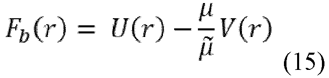
- Hence the problem consists in a diffusive motion of a particle in a potential Fb . Because of the violation of the Einstein relation, this potential contains also kinetic parameters as the electrophoretic mobility µ and the solvent viscosity η from µ̃ ~ η -1. The potential has a minimum close to distance Rg and a maximum close to the pore entry defining a barrier height: ΔFb≡Fb max-Fb min. According to Kramers' theory the capture rate k on depends exponentially on the barrier height:

- The barrier can be lowered by increasing the applied voltage ΔV so to strengthen the electrostatic attraction towards the pore. Eq. (15) implies that a similar effect can be obtained by increasing µ, the electrophoretic mobility of the ssDNA. One obvious way to modify µ is through a change of the ionic strength of the solution as this modifies the Debye length. As shown in Eq. (9), the electrophoretic mobility is proportional to λD. Note that the salt concentration has also an effect on the ssDNA persistence length and thus on the entropic contribution to the barrier U(r), however this effect is expected to be weaker. The main effect of a change in the salt concentration on the barrier height is expected to contain a term linear in λD :

- With a, b > 0 which, together with (15) explains the exponential growth of k on on λD observed in the experiments.
- Table 2. Pore engineering DNA translocation from the trans side. Each data point is the average of at least three experiments and the error is the standard deviation. Experiments were carried out in 0.15 M NaCl, 15 mM Tris HCl, pH 7.5 solutions. The activity of the nanopores were tested by adding 0.01- 0.1 ng oligomeric protein to the trans chamber. A negative activity indicates that no channel insertions were observed. VG represents the maximum applied voltage at which no gating events were observed within 30 s. DNA capture indicates that only transient current blockades were observed upon the addition of biotinylated dsDNA in complex with neutravidin. DNA translocation indicates that a dsDNA rotaxane could be formed.
Table 2 Pore variants IO+100mV IO100Mv (pA) Rectification ratio VG, (mV) DNA Capture DNA Translocation ClyA-RR-E7S +186±2 -110±2 1.7±0.0 -70 + - ClyA-RR-E11S +214±27 -124±14 1.7±0.3 - 100 - - ClyA-RR-D21S +193±9 -113±9 1.7±0.2 -70 - - ClyA-RR-D21K +149±0 -112±0 1.3±0.0 - 50 - - ClyA-RR-D32N +196±5 -104±5 1.9±0.1 -150 - - ClyA-RR-E7S-D32N +182±4 -104±6 1.8±0.1 -70 + - ClyA-RR-E7S-D21S +182±5 -121±3 1.5±0.1 -70 + - ClyA-RR-E129R No activity No activity No activity No No ClyA-RR-1'R +184±8 -101±3 1.8±0.1 -150 + - ClyA-RR-1'R-E7S +176±5 -109±3 1.6±0.1 - 50 + - ClyA-RR-1'R-D21S +179±9 -108±5 1.7±0.1 - 50 + - - Table 3: DNA molecules used in this work. 1 was formed by incubating 1a with a 20% excess of 1b and purified by affinity chromatography as described in Methods. 1∗ was formed by incubating 1a with a 20% excess of 1b without further purification. The complementary sequences in the two DNA strands are shown in italics. The suffix bio indicates a biotin moiety.
Table 3 Name DNA sequence 1a 
1b 
1c 
1d 5'-CTG TAT AAT AAA TAT CTG GAT CAG GTC ATC C /3Bio/-3' (SEQ ID NO: 8) 2a 
2b 
- Table 4: Ionic selectivity of selected ClyA nanopores. Permeability ratio (PNa+ / PCl-) and reversal potential (Vr) for ClyA variant nanopores reported as average ± standard deviation. Four or more single channels were measured for each variant. The buffer used were: 15 mM TRIS.HCl pH 7.5, with 1 M NaCl in the cis chamber and 150 mM in the trans chamber.
Table 4 Pore variants Vr, mV PNa+/PCl- ClyA-AS +11.5±0.7 1.92±0.08 ClyA-R +11.9±1.6 1.97±0.08 ClyA-RR +11.4±0.9 1.91±0.10 - Table 5: IV curves for ClyA mutants. The electrical recordings were carried out in 0.15 M NaCl, 15 mM Tris HCl, pH 7.5, at 22°C. Each data point is the average of at least three experiments and the error is the standard deviation.


- Table 6: IV curves of ClyA variants under asymmetric salt concentrations. Four or more single channels were measured for each variant. Each data is reported as the average ± standard deviation. The buffer used was 15 mM TRIS.HCl pH 7.5, while the cis chamber contained 1 M NaCl and the
trans chamber 150 mM. The electrical recordings were carried out in 0.15 M NaCl, 15 mM Tris HCl, pH 7.5, at 22°C. Data were recorded by applying a 2-kHz low-pass Bessel filter and using a 100 µs (10 kHz) sampling rate.Table 6 IV ClyA Ionic permeability
Open pore current, pA (Average ± Standard Deviation)Voltage (mV) ClyA-AS ClyA-R ClyA-RR 20 26.5±0.7 36.7±14.1 32.4±4.3 19 23.1±0.9 32.5±13 28.6±2.7 18 19.8±0.5 27.6±12.8 25.2±2.5 17 16.6±0.9 23.4±10.8 21.3±1.8 16 13.6±1.8 17.9±10.7 16.6±1.2 15 9.9±1.9 14.4±9.7 12.8±1.9 14 7.2±1.2 7.8±9.3 10.2±1.2 13 4.4±1.1 2.9±8.9 7.2±1.4 12 2.5±1.4 -1.7±9.4 1.8±1.3 11 -0.7±2.5 -5.6±8.6 -0.8±1.9 10 -3.8±2.7 -12.7±7.3 -4±1 9 -7.3±2.7 -15.6±8 -9.3±3.1 8 -10.7±1.5 -22.3±6.3 -11.9±0.5 7 -13.4±4.3 -24.5±5.9 -15.5±2.6 6 -16.2±0.9 -31.2±5.8 -19.8±3.2 5 -18±2 -35.2±4.5 -23.1±3.1 4 -22.3±2.5 -40.4±5.2 -25.7±1.7 3 -25±2 -43.7±3.5 -30.1±2.9 2 -27.8±3.1 -51.2±4.1 -33.8±4.5 1 -30.5±3.1 -55.6±2.1 -36.9±4.8 0 -34.9±2.7 -60.7±2 -40.7±5 -1 -37.1±3.5 -65.1±2.6 -44.4±3.8 -2 -41±3 -68.8±3.8 -48.2±4.2 -3 -42.1±3.9 -74.8±3.6 -51.3±6.5 -4 -46.2±4.2 -79.8±2.1 -54.8±7.9 -5 -48.8±4.2 -85.2±2.5 -57.8±6.5 IV ClyA Ionic permeability
Open pore current, pA (Average ± Standard Deviation)Voltage (mV) ClyA-AS ClyA-R ClyA-RR -6 -51.5±3.5 -90±3 -61.2±7.8 -7 -55.1±6.3 -94±4 -66.2±7.3 -8 -57.8±5.5 -100±3 -68.6±10.7 -9 -61±4 -103±3 -73.1±8.3 -10 -62.8±4.9 -109±4 -76.5±7.9 -11 -66.2±5 -114±4 -80.1±9.1 -12 -69.7±6 -117±4 -83.6±9.9 -13 -74.7±5.5 -123±4 -86.6±8.7 -14 -74.8±6.1 -129±5 -91.1±11.2 -15 -78.3±5.7 -134±7 -93.5±10.7 -16 -80.2±6.2 -137±8 -96.7±9.7 -17 -84.2±6.4 -144±7 -100±13 -18 -87.6±7.6 -148±8 -104±12 -19 -90.4±7.7 -153±8 -108±12 -20 -92.4±7.3 -158±8 -112±11 - Table 7: ssDNA (1a) and dsDNA (1) translocation through ClyA-RR nanopores. Three or more single channels were measured for each condition. Data are reported as the average ± standard deviation. The electrical recordings were carried out in 15 mM Tris- HCl. pH 7.5 at 22°C. Data were recorded by applying a 10-kHz low-pass Bessel filter and using a 20 µs (50 kHz) sampling rate.
Table 7 ssDNA (1a) [NaCl] (M) IRES τoff (ms) τon (ms) 0.15 0.92±0.00 0.54±0.28 8.5±1.1 0.3 0.89±0.01 0.18±0.04 44±1 0.5 0.88±0.02 0.12±0.02 112±14 1 0.82±0.01 0.13±0.01 232±36 2 0.84±0.01 0.12±0.02 393±17 2.5 0.78±0.01 0.18±0.02 500±50 dsDNA (1) [NaCl] (M) IRES τoff (ms) τon (ms) 0.15 0.92±0.00 0.29±0.07 40±13 0.6 0.83±0.03 0.26±0.09 162±31 1 0.76±0.01 0.26±0.09 214±18 2 0.75±0.04 0.33±0.07 532±52 2.5 0.75±0.01 0.60±0.48 641±37 SEQUENCE LISTING: Description Sequence Protein sequence for S. typhi Cly A (ClyA-WT) SEQ ID NO: 1 
Protein sequence for ClyA-AS SEQ ID NO: 2 
Nucleotide sequence for S. typhi ClyA (ClyA-WT) SEQ ID NO: 3 
Nucleotide sequence for ClyA-AS SEQ ID NO: 4 
-
- <110> Katholieke Universiteit Leuven KU Leuven Research & Development
- <120> MODIFIED NANOPORES, COMPOSITIONS COMPRISING THE SAME, AND USES THEREOF
- <130> 00366.70046WO00
- <140> Not Yet Assigned
<141> 2016-12-08 - <150>
US 62/264,709
<151> 2015-12-08 - <160> 10
- <170> PatentIn version 3.5
- <210> 1
<211> 303
<212> PRT
<213> S. typhi - <400> 1


- <210> 2
<211> 312
<212> PRT
<213> S. typhi - <400> 2


- <210> 3
<211> 1194
<212> DNA
<213> S. typhi - <400> 3

- <210> 4
<211> 945
<212> DNA
<213> S. typhi - <400> 4


- <210> 5
<211> 90
<212> DNA
<213> Artificial Sequence - <220>
<223> Synthetic Polynucleotide - <220>
<221> mod_res
<222> (90)..(90)
<223> modified by 3' /3Bio/ - <400> 5

- <210> 6
<211> 90
<212> DNA
<213> Artificial Sequence - <220>
<223> Synthetic Polynucleotide - <400> 6

- <210> 7
<211> 59
<212> DNA
<213> Artificial Sequence - <220>
<223> Synthetic Polynucleotide - <220>
<221> mod_res
<222> (59)..(59)
<223> modified by 3' /3Bio/ - <400> 7
ggtaatcgat ctctctctct ttttgtgaaa agtgcgattg ggctgacggt gcgctggac 59 - <210> 8
<211> 31
<212> DNA
<213> Artificial Sequence - <220>
<223> Synthetic Polynucleotide - <220>
<221> mod_res
<222> (31)..(31)
<223> modified by 3' /3Bio/ - <400> 8
ctgtataata aatatctgga tcaggtcatc c 31 - <210> 9
<211> 100
<212> DNA
<213> Artificial Sequence - <220>
<223> Synthetic Polynucleotide - <220>
<221> mod_res
<222> (1)..(1)
<223> modified by 5' /5Bio/ - <400> 9

- <210> 10
<211> 50
<212> DNA
<213> Artificial Sequence - <220>
<223> Synthetic Polynucleotide - <220>
<221> mod_res
<222> (1)..(1)
<223> modified by 5' /5Bio/ - <400> 10
ggtaatcgat ctctctctct ttttgtgaaa agtgcgattg ggctgacggt 50
Claims (12)
- A modified Cytolysin A (ClyA) nanopore comprising a cis opening, a mid-section, and a trans opening, wherein an internal surface of the cis opening comprises a first positively-charged amino acid substitution; an internal surface of the mid-section comprises a second positively-charged amino acid substitution; and the trans opening comprises an electronegative constriction;wherein the first positively charged substitution is located at E106, S110, D114, D121, D122, E129, E85, E78, D268, D267, D265, or E258 of SEQ ID NO: 1 or 2; and/or is at a position within a range of 78 to 106 of SEQ ID NO: 1 or 2; andwherein the second positively-charged amino acid substitution corresponds to replacement of a negatively charged amino acid with a positively-charged amino acid at one or more positions corresponding to D74, D71, D64, E53, E161, D158, E46, E42, and D41 of SEQ ID NO: 1 or SEQ ID NO: 2.
- The modified ClyA nanopore of claim 1, wherein the first and second positively-charged amino acid substitutions each includes an arginine.
- The modified ClyA nanopore of claim 1 or claim 2, wherein the first positively-charged amino acid substitution corresponds to a S110R mutation in the amino acid sequence of SEQ ID NO: 2 and/or
wherein the second positively-charged amino acid substitution corresponds to a D64R mutation in the amino acid sequence of SEQ ID NO: 2. - The modified ClyA nanopore of any one of claims 1-3, wherein the modified ClyA pore is a dodecameric pore.
- A method of translocating a DNA through a ClyA nanopore comprising:a. providing, in a low ionic strength solution of 150 mM to 300 mM, a modified ClyA nanopore of any one of claims 1-4 and an artificial membrane, wherein the modified ClyA nanopore is present in an artificial membrane such that the cis opening of the modified ClyA nanopore is present in a cis side of the low ionic strength solution and the trans opening of the modified ClyA nanopore is present in a trans side of the low ionic strength solution;b. providing a DNA in the cis side of the low ionic strength solution; andc. applying an electrical potential across the modified ClyA nanopore, thereby translocating the DNA through the modified ClyA nanopore from the cis side to the trans side.
- A polynucleotide comprising a nucleotide sequence encoding a subunit polypeptide of the modified ClyA nanopore of any one of claims 1-4.
- A homo-multimeric modified ClyA nanopore comprising a plurality of subunit polypeptides of the modified ClyA nanopore of any one of claims 1-4.
- A hetero-multimeric modified ClyA nanopore comprising at least one subunit polypeptide of the modified ClyA nanopore of any one of claims 1-4.
- The modified ClyA nanopore of claim 1, comprising a subunit polypeptide having an amino acid sequence that is at least 95% identical to the amino acid sequence as set forth in SEQ ID NO: 1 or SEQ ID NO: 2.
- The modified ClyA nanopore of claim 1, comprising a subunit polypeptide having up to ten substitutions compared with the amino acid sequence as set forth in SEQ ID NO: 1 or SEQ ID NO: 2 including the first and second positively-charged amino acid substitutions.
- A composition comprising at least one modified ClyA nanopore of any one of claims 1 to 4 or 7 to 10.
- A method of characterizing a target polynucleotide, the method comprising(a) providing, in a low ionic strength solution of 150 mM to 300 mM, a modified ClyA nanopore of any one of claims 1 to 4 or 7 to 10 and a membrane, wherein the modified ClyA nanopore is present in the membrane;(b) adding in the low ionic strength solution of step (a) the target polynucleotide; and(c) measuring, during application of a potential across the nanopore, ion flow through the modified ClyA nanopore, wherein the ion flow measurements are indicative of one or more characteristics of the target polynucleotide.
Applications Claiming Priority (2)
| Application Number | Priority Date | Filing Date | Title |
|---|---|---|---|
| US201562264709P | 2015-12-08 | 2015-12-08 | |
| PCT/IB2016/001841 WO2017098322A1 (en) | 2015-12-08 | 2016-12-08 | Modified nanopores, compositions comprising the same, and uses thereof |
Publications (2)
| Publication Number | Publication Date |
|---|---|
| EP3387432A1 EP3387432A1 (en) | 2018-10-17 |
| EP3387432B1 true EP3387432B1 (en) | 2022-09-28 |
Family
ID=58098655
Family Applications (1)
| Application Number | Title | Priority Date | Filing Date |
|---|---|---|---|
| EP16838062.4A Active EP3387432B1 (en) | 2015-12-08 | 2016-12-08 | Modified nanopores, compositions comprising the same, and uses thereof |
Country Status (8)
| Country | Link |
|---|---|
| US (1) | US10976300B2 (en) |
| EP (1) | EP3387432B1 (en) |
| JP (2) | JP7237586B2 (en) |
| KR (1) | KR102837999B1 (en) |
| CN (1) | CN109072295B (en) |
| AU (1) | AU2016369071B2 (en) |
| CA (1) | CA3011830C (en) |
| WO (1) | WO2017098322A1 (en) |
Families Citing this family (36)
| Publication number | Priority date | Publication date | Assignee | Title |
|---|---|---|---|---|
| GB0905140D0 (en) | 2009-03-25 | 2009-05-06 | Isis Innovation | Method |
| JP6271505B2 (en) | 2012-04-10 | 2018-01-31 | オックスフォード ナノポール テクノロジーズ リミテッド | Mutant lysenin pore |
| GB201313477D0 (en) | 2013-07-29 | 2013-09-11 | Univ Leuven Kath | Nanopore biosensors for detection of proteins and nucleic acids |
| CA2937411C (en) | 2014-01-22 | 2023-09-26 | Oxford Nanopore Technologies Limited | Method for attaching one or more polynucleotide binding proteins to a target polynucleotide |
| EP3137627B1 (en) | 2014-05-02 | 2025-07-30 | Oxford Nanopore Technologies plc | Method of improving the movement of a target polynucleotide with respect to a transmembrane pore |
| KR20170042794A (en) | 2014-09-01 | 2017-04-19 | 브이아이비 브이지더블유 | Mutant csgg pores |
| GB201502809D0 (en) | 2015-02-19 | 2015-04-08 | Oxford Nanopore Tech Ltd | Mutant pore |
| GB201502810D0 (en) | 2015-02-19 | 2015-04-08 | Oxford Nanopore Tech Ltd | Method |
| WO2016166232A1 (en) | 2015-04-14 | 2016-10-20 | Katholieke Universiteit Leuven | Nanopores with internal protein adaptors |
| AU2017225374B2 (en) | 2016-03-02 | 2020-03-05 | Oxford Nanopore Technologies Limited | Mutant pore |
| EP4122949B1 (en) | 2016-04-06 | 2024-06-05 | Oxford Nanopore Technologies plc | Mutant pore |
| US11531020B2 (en) * | 2017-01-18 | 2022-12-20 | Youhealth Biotech, Limited | Methods and compositions for nanopore sequencing |
| GB201707122D0 (en) | 2017-05-04 | 2017-06-21 | Oxford Nanopore Tech Ltd | Pore |
| CN117106037B (en) | 2017-06-30 | 2025-07-25 | 弗拉芒区生物技术研究所 | Novel protein pores |
| WO2020010589A1 (en) * | 2018-07-12 | 2020-01-16 | Shenzhen Genorivision Technology Co., Ltd. | An apparatus for biopolymer sequencing |
| WO2020241752A1 (en) | 2019-05-28 | 2020-12-03 | The University Of Tokyo | Analyzing apparatus and method using a pore device |
| AU2020389020A1 (en) * | 2019-11-19 | 2022-06-09 | Rijksuniversiteit Groningen | Artificial nanopores and uses and methods relating thereto |
| EP4070092B1 (en) * | 2019-12-02 | 2023-08-30 | Oxford Nanopore Technologies PLC | Method of characterising a target polypeptide using a nanopore |
| NL2024579B1 (en) * | 2019-12-24 | 2021-09-06 | Univ Delft Tech | Protein and peptide fingerprinting and sequencing by nanopore translocation of peptide-oligonucleotide complexes |
| CN115398008B (en) * | 2020-04-14 | 2025-05-09 | 成都今是科技有限公司 | Nanopore preparation and detection method and detection device |
| US20230184767A1 (en) * | 2020-05-20 | 2023-06-15 | The Board Of Trustees Of The University Of Illinois | Methods and compositions for rapid direct detection and differentiation of infectious from noninfectious virus |
| US20230295712A1 (en) * | 2020-06-18 | 2023-09-21 | Oxford Nanopore Technologies Plc | A method of selectively characterising a polynucleotide using a detector |
| CN112179955B (en) * | 2020-09-27 | 2024-05-10 | 西北工业大学 | Method for preparing pH response nanofluid diode based on polyarginine and polyglutamic acid layer-by-layer modified nanopores |
| KR102309424B1 (en) * | 2020-09-28 | 2021-10-07 | 한국생명공학연구원 | Screening method for the riboswitch-targeted substances using nanopores |
| CN117255945A (en) | 2021-03-31 | 2023-12-19 | 伊鲁米纳公司 | Nanopore sensor device |
| GB2610380A (en) | 2021-08-23 | 2023-03-08 | Cambridge Entpr Ltd | Nucleic acid detection |
| GB202113935D0 (en) | 2021-09-29 | 2021-11-10 | Cambridge Entpr Ltd | Nucleic acid characterisation |
| GB202118908D0 (en) | 2021-12-23 | 2022-02-09 | Oxford Nanopore Tech Ltd | Method |
| GB202118906D0 (en) | 2021-12-23 | 2022-02-09 | Oxford Nanopore Tech Ltd | Method |
| CN114768530B (en) * | 2022-04-29 | 2023-03-28 | 中国工程物理研究院材料研究所 | Application of molybdenum disulfide in hydrogen isotope electrolytic separation |
| CN120435657A (en) | 2022-10-28 | 2025-08-05 | 格罗宁根大学 | Nanopore-based analysis of proteins |
| GB202216162D0 (en) | 2022-10-31 | 2022-12-14 | Oxford Nanopore Tech Plc | Method |
| GB202307486D0 (en) | 2023-05-18 | 2023-07-05 | Oxford Nanopore Tech Plc | Method |
| GB202317028D0 (en) | 2023-11-06 | 2023-12-20 | Oxford Nanopore Tech Ltd | Method |
| WO2026052817A1 (en) | 2024-09-06 | 2026-03-12 | Oxford Nanopore Technologies Plc | Method |
| WO2026052811A2 (en) | 2024-09-06 | 2026-03-12 | Oxford Nanopore Technologies Plc | Method |
Family Cites Families (90)
| Publication number | Priority date | Publication date | Assignee | Title |
|---|---|---|---|---|
| US6362002B1 (en) | 1995-03-17 | 2002-03-26 | President And Fellows Of Harvard College | Characterization of individual polymer molecules based on monomer-interface interactions |
| US5795782A (en) | 1995-03-17 | 1998-08-18 | President & Fellows Of Harvard College | Characterization of individual polymer molecules based on monomer-interface interactions |
| JP3891620B2 (en) | 1996-11-14 | 2007-03-14 | 株式会社アイシン・コスモス研究所 | Nucleic acid probe molecule having a hairpin structure, and nucleic acid detection method using the nucleic acid probe molecule |
| US20020197614A1 (en) | 1997-05-16 | 2002-12-26 | Mosaic Technologies, Inc. | Electrophoretic analysis of target molecules using adapter molecules |
| WO1999005167A1 (en) | 1997-07-25 | 1999-02-04 | University Of Massachusetts | Designed protein pores as components for biosensors |
| US6743605B1 (en) | 1998-06-24 | 2004-06-01 | Enzo Life Sciences, Inc. | Linear amplification of specific nucleic acid sequences |
| US6267872B1 (en) | 1998-11-06 | 2001-07-31 | The Regents Of The University Of California | Miniature support for thin films containing single channels or nanopores and methods for using same |
| US6426231B1 (en) | 1998-11-18 | 2002-07-30 | The Texas A&M University System | Analyte sensing mediated by adapter/carrier molecules |
| NO986133D0 (en) | 1998-12-23 | 1998-12-23 | Preben Lexow | Method of DNA Sequencing |
| AU5631200A (en) | 1999-06-22 | 2001-01-09 | President And Fellows Of Harvard College | Control of solid state dimensional features |
| EP1208207A2 (en) | 1999-08-31 | 2002-05-29 | Michael Niederweis | Method for producing a channel-forming protein |
| EP2083015B1 (en) | 2000-02-11 | 2016-04-06 | The Texas A & M University System | Biosensor compositions and methods of use |
| US7001792B2 (en) | 2000-04-24 | 2006-02-21 | Eagle Research & Development, Llc | Ultra-fast nucleic acid sequencing device and a method for making and using the same |
| WO2002042496A2 (en) | 2000-11-27 | 2002-05-30 | The Regents Of The University Of California | Methods and devices for characterizing duplex nucleic acid molecules |
| US6863833B1 (en) | 2001-06-29 | 2005-03-08 | The Board Of Trustees Of The Leland Stanford Junior University | Microfabricated apertures for supporting bilayer lipid membranes |
| AU2003245272A1 (en) | 2002-05-10 | 2003-11-11 | The Texas A And M University System | Stochastic sensing through covalent interactions |
| EP2194123B1 (en) | 2003-03-25 | 2012-08-22 | Stratagene California | DNA polymerase fusions and uses thereof |
| US7163658B2 (en) | 2003-04-23 | 2007-01-16 | Rouvain Bension | Rapid sequencing of polymers |
| JP2005253427A (en) | 2004-03-15 | 2005-09-22 | Aisin Seiki Co Ltd | Nucleic acid detection method and nucleic acid isolation method |
| WO2006028508A2 (en) | 2004-03-23 | 2006-03-16 | President And Fellows Of Harvard College | Methods and apparatus for characterizing polynucleotides |
| WO2005124888A1 (en) | 2004-06-08 | 2005-12-29 | President And Fellows Of Harvard College | Suspended carbon nanotube field effect transistor |
| US20060105461A1 (en) | 2004-10-22 | 2006-05-18 | May Tom-Moy | Nanopore analysis system |
| WO2007084103A2 (en) | 2004-12-21 | 2007-07-26 | The Texas A & M University System | High temperature ion channels and pores |
| GB0505971D0 (en) | 2005-03-23 | 2005-04-27 | Isis Innovation | Delivery of molecules to a lipid bilayer |
| KR100730350B1 (en) | 2005-10-17 | 2007-06-19 | 삼성전자주식회사 | DNA detection method and detection device using surface treated nanopores |
| GB0523282D0 (en) | 2005-11-15 | 2005-12-21 | Isis Innovation | Methods using pores |
| EP1963530B1 (en) | 2005-12-22 | 2011-07-27 | Pacific Biosciences of California, Inc. | Active surface coupled polymerases |
| US7638034B2 (en) | 2006-09-21 | 2009-12-29 | Los Alamos National Security, Llc | Electrochemical detection of single molecules using abiotic nanopores having electrically tunable dimensions |
| US20100196203A1 (en) | 2007-02-20 | 2010-08-05 | Gurdial Singh Sanghera | Formation of Lipid Bilayers |
| WO2008124107A1 (en) | 2007-04-04 | 2008-10-16 | The Regents Of The University Of California | Compositions, devices, systems, and methods for using a nanopore |
| GB0716264D0 (en) | 2007-08-21 | 2007-09-26 | Isis Innovation | Bilayers |
| US8698481B2 (en) | 2007-09-12 | 2014-04-15 | President And Fellows Of Harvard College | High-resolution molecular sensor |
| GB2453377A (en) | 2007-10-05 | 2009-04-08 | Isis Innovation | Transmembrane protein pores and molecular adapters therefore. |
| GB0724736D0 (en) | 2007-12-19 | 2008-01-30 | Oxford Nanolabs Ltd | Formation of layers of amphiphilic molecules |
| US8231969B2 (en) | 2008-03-26 | 2012-07-31 | University Of Utah Research Foundation | Asymmetrically functionalized nanoparticles |
| EP3269824A1 (en) | 2008-03-28 | 2018-01-17 | Pacific Biosciences Of California, Inc. | Compositions and methods for nucleic acid sequencing |
| WO2009143425A1 (en) | 2008-05-22 | 2009-11-26 | The Regents Of The University Of California | Membrane precursors and membranes formed therefrom |
| US8652771B2 (en) | 2008-05-28 | 2014-02-18 | University of Souther California | Measurement of succinate in urine samples as a biomarker of kidney damage in diabetic subjects |
| EP2307540B1 (en) | 2008-07-07 | 2017-04-19 | Oxford Nanopore Technologies Limited | Enzyme-pore constructs |
| WO2010004273A1 (en) | 2008-07-07 | 2010-01-14 | Oxford Nanopore Technologies Limited | Base-detecting pore |
| CN102216783B (en) | 2008-09-22 | 2015-04-01 | 华盛顿大学 | MSP nanopores and related methods |
| US9080211B2 (en) | 2008-10-24 | 2015-07-14 | Epicentre Technologies Corporation | Transposon end compositions and methods for modifying nucleic acids |
| GB0820927D0 (en) | 2008-11-14 | 2008-12-24 | Isis Innovation | Method |
| US20100148126A1 (en) | 2008-11-26 | 2010-06-17 | Board Of Regents, The University Of Texas System | Genomic sequencing using modified protein pores and ionic liquids |
| WO2010086622A1 (en) | 2009-01-30 | 2010-08-05 | Oxford Nanopore Technologies Limited | Adaptors for nucleic acid constructs in transmembrane sequencing |
| US20120100530A1 (en) | 2009-01-30 | 2012-04-26 | Oxford Nanopore Technologies Limited | Enzyme mutant |
| GB0905140D0 (en) | 2009-03-25 | 2009-05-06 | Isis Innovation | Method |
| WO2010122293A1 (en) | 2009-04-20 | 2010-10-28 | Oxford Nanopore Technologies Limited | Lipid bilayer sensor array |
| EP2507387B1 (en) | 2009-12-01 | 2017-01-25 | Oxford Nanopore Technologies Limited | Biochemical analysis instrument and method |
| WO2011106456A2 (en) | 2010-02-23 | 2011-09-01 | University Of Washington | Artificial mycolic acid membranes |
| US9073990B2 (en) * | 2010-04-05 | 2015-07-07 | Bar-Llan University | Protease-activatable pore-forming polypeptides |
| US9593370B2 (en) | 2010-10-01 | 2017-03-14 | Oxford Nanopore Technologies Ltd. | Biochemical analysis apparatus and rotary valve |
| CN102116783B (en) | 2010-12-31 | 2013-05-29 | 北京普源精电科技有限公司 | A Waveform Display Method |
| EP2673638B1 (en) | 2011-02-11 | 2019-10-30 | Oxford Nanopore Technologies Limited | Mutant pores |
| EP3633370B1 (en) | 2011-05-27 | 2024-05-01 | Oxford Nanopore Technologies plc | Method and apparatus for determining the presence, absence or characteristics of an analyte |
| US9580480B2 (en) | 2011-05-31 | 2017-02-28 | Massachusetts Institute Of Technology | Cell-directed synthesis of multifunctional nanopatterns and nanomaterials |
| IN2014DN00221A (en) | 2011-07-25 | 2015-06-05 | Oxford Nanopore Tech Ltd | |
| CA3113287C (en) | 2011-09-23 | 2022-12-20 | Oxford Nanopore Technologies Limited | Analysis of a polymer comprising polymer units |
| EP2987870B1 (en) | 2011-10-21 | 2020-02-19 | Oxford Nanopore Technologies Limited | Method of characterizing a target polynucleotide using a transmembrane pore and molecular motor |
| WO2013098561A1 (en) | 2011-12-29 | 2013-07-04 | Oxford Nanopore Technologies Limited | Method for characterising a polynucelotide by using a xpd helicase |
| WO2013098562A2 (en) | 2011-12-29 | 2013-07-04 | Oxford Nanopore Technologies Limited | Enzyme method |
| WO2013109970A1 (en) | 2012-01-20 | 2013-07-25 | Genia Technologies, Inc. | Nanopore based molecular detection and sequencing |
| EP2815235B1 (en) | 2012-02-15 | 2020-04-15 | Oxford Nanopore Technologies Limited | Aptamer method |
| JP6271505B2 (en) | 2012-04-10 | 2018-01-31 | オックスフォード ナノポール テクノロジーズ リミテッド | Mutant lysenin pore |
| TWI655213B (en) | 2012-07-13 | 2019-04-01 | Menicon Co., Ltd. | Method for producing self-organized peptide derivative |
| WO2014013259A1 (en) | 2012-07-19 | 2014-01-23 | Oxford Nanopore Technologies Limited | Ssb method |
| JP6429773B2 (en) | 2012-07-19 | 2018-11-28 | オックスフォード ナノポール テクノロジーズ リミテッド | Enzyme construct |
| US10808231B2 (en) | 2012-07-19 | 2020-10-20 | Oxford Nanopore Technologies Limited | Modified helicases |
| CA2889664C (en) | 2012-10-26 | 2020-12-29 | Oxford Nanopore Technologies Limited | Droplet interfaces |
| GB201313121D0 (en) | 2013-07-23 | 2013-09-04 | Oxford Nanopore Tech Ltd | Array of volumes of polar medium |
| EP2954320B1 (en) | 2013-02-07 | 2018-04-18 | Yissum Research Development Company of the Hebrew University of Jerusalem Ltd. | Hybrid nanopores and uses thereof for detection of analytes |
| GB201314695D0 (en) | 2013-08-16 | 2013-10-02 | Oxford Nanopore Tech Ltd | Method |
| BR112015021788B1 (en) | 2013-03-08 | 2023-02-28 | Oxford Nanopore Technologies Plc | METHODS FOR MOVING ONE OR MORE IMMOBILIZED HELICASES, FOR CONTROLLING THE MOVEMENT OF A TARGET POLYNUCLEOTIDE, FOR CHARACTERIZING A TARGET POLYNUCLEOTIDE, AND FOR CONTROLLING THE LOADING OF ONE OR MORE HELICASES INTO A TARGET POLYNUCLEOTIDE, USE OF A TRANSMEMBRANE PORE AND AN APPLIED POTENTIAL AND OF ONE OR MORE SPACERS, COMPLEX, AND, KIT |
| WO2014153047A1 (en) | 2013-03-14 | 2014-09-25 | The Trustees Of Boston University | Optoelectronic control of solid-state nanopores |
| GB201313477D0 (en) | 2013-07-29 | 2013-09-11 | Univ Leuven Kath | Nanopore biosensors for detection of proteins and nucleic acids |
| WO2015051378A1 (en) * | 2013-10-04 | 2015-04-09 | University Of Washington Through Its Center For Commercialization | Systems and methods for nanopore-based analysis of nucleic acids |
| CN117947149A (en) | 2013-10-18 | 2024-04-30 | 牛津纳米孔科技公开有限公司 | Modified enzymes |
| GB201406151D0 (en) | 2014-04-04 | 2014-05-21 | Oxford Nanopore Tech Ltd | Method |
| CA2937411C (en) | 2014-01-22 | 2023-09-26 | Oxford Nanopore Technologies Limited | Method for attaching one or more polynucleotide binding proteins to a target polynucleotide |
| GB201406155D0 (en) | 2014-04-04 | 2014-05-21 | Oxford Nanopore Tech Ltd | Method |
| GB201403096D0 (en) | 2014-02-21 | 2014-04-09 | Oxford Nanopore Tech Ltd | Sample preparation method |
| EP3126515B1 (en) | 2014-04-04 | 2018-08-29 | Oxford Nanopore Technologies Limited | Method for characterising a double stranded nucleic acid using a nano-pore and anchor molecules at both ends of said nucleic acid |
| EP3137627B1 (en) | 2014-05-02 | 2025-07-30 | Oxford Nanopore Technologies plc | Method of improving the movement of a target polynucleotide with respect to a transmembrane pore |
| FR3023394B1 (en) | 2014-07-02 | 2017-12-29 | Adn Access Data Networks | DEVICE FOR FACILITATING THE TEACHING OF THE AMBARIC LANGUAGE AND NORMALIZING THE WRITING |
| KR20170042794A (en) | 2014-09-01 | 2017-04-19 | 브이아이비 브이지더블유 | Mutant csgg pores |
| WO2016055778A1 (en) | 2014-10-07 | 2016-04-14 | Oxford Nanopore Technologies Limited | Mutant pores |
| GB201502810D0 (en) | 2015-02-19 | 2015-04-08 | Oxford Nanopore Tech Ltd | Method |
| GB201502809D0 (en) | 2015-02-19 | 2015-04-08 | Oxford Nanopore Tech Ltd | Mutant pore |
| WO2016166232A1 (en) | 2015-04-14 | 2016-10-20 | Katholieke Universiteit Leuven | Nanopores with internal protein adaptors |
| AU2017225374B2 (en) | 2016-03-02 | 2020-03-05 | Oxford Nanopore Technologies Limited | Mutant pore |
-
2016
- 2016-12-08 AU AU2016369071A patent/AU2016369071B2/en active Active
- 2016-12-08 EP EP16838062.4A patent/EP3387432B1/en active Active
- 2016-12-08 CN CN201680081416.8A patent/CN109072295B/en active Active
- 2016-12-08 US US16/060,762 patent/US10976300B2/en active Active
- 2016-12-08 JP JP2018530754A patent/JP7237586B2/en active Active
- 2016-12-08 KR KR1020187019163A patent/KR102837999B1/en active Active
- 2016-12-08 CA CA3011830A patent/CA3011830C/en active Active
- 2016-12-08 WO PCT/IB2016/001841 patent/WO2017098322A1/en not_active Ceased
-
2021
- 2021-12-08 JP JP2021198939A patent/JP2022046503A/en active Pending
Also Published As
| Publication number | Publication date |
|---|---|
| US20180364214A1 (en) | 2018-12-20 |
| JP2019507583A (en) | 2019-03-22 |
| WO2017098322A1 (en) | 2017-06-15 |
| KR20180089499A (en) | 2018-08-08 |
| CA3011830A1 (en) | 2017-06-15 |
| JP7237586B2 (en) | 2023-03-13 |
| CA3011830C (en) | 2024-11-12 |
| EP3387432A1 (en) | 2018-10-17 |
| AU2016369071A1 (en) | 2018-06-28 |
| AU2016369071B2 (en) | 2022-05-19 |
| US10976300B2 (en) | 2021-04-13 |
| CN109072295A (en) | 2018-12-21 |
| JP2022046503A (en) | 2022-03-23 |
| CN109072295B (en) | 2022-05-31 |
| KR102837999B1 (en) | 2025-07-24 |
Similar Documents
| Publication | Publication Date | Title |
|---|---|---|
| US10976300B2 (en) | Modified nanopores, compositions comprising the same, and uses thereof | |
| US12473595B2 (en) | Coupling method | |
| US20250042952A1 (en) | Transmembrane pore consisting of two csgg pores | |
| EP3137490B1 (en) | Mutant pores | |
| EP2350122B1 (en) | Methods of enhancing translocation of charged analytes through transmembrane protein pores | |
| CN114107457B (en) | Method for delivering analytes to transmembrane pores | |
| EP3259281B1 (en) | Hetero-pores | |
| EP4269617A2 (en) | Methods and systems for characterizing analytes using nanopores |
Legal Events
| Date | Code | Title | Description |
|---|---|---|---|
| STAA | Information on the status of an ep patent application or granted ep patent |
Free format text: STATUS: UNKNOWN |
|
| STAA | Information on the status of an ep patent application or granted ep patent |
Free format text: STATUS: THE INTERNATIONAL PUBLICATION HAS BEEN MADE |
|
| PUAI | Public reference made under article 153(3) epc to a published international application that has entered the european phase |
Free format text: ORIGINAL CODE: 0009012 |
|
| STAA | Information on the status of an ep patent application or granted ep patent |
Free format text: STATUS: REQUEST FOR EXAMINATION WAS MADE |
|
| 17P | Request for examination filed |
Effective date: 20180628 |
|
| AK | Designated contracting states |
Kind code of ref document: A1 Designated state(s): AL AT BE BG CH CY CZ DE DK EE ES FI FR GB GR HR HU IE IS IT LI LT LU LV MC MK MT NL NO PL PT RO RS SE SI SK SM TR |
|
| AX | Request for extension of the european patent |
Extension state: BA ME |
|
| RIN1 | Information on inventor provided before grant (corrected) |
Inventor name: MAGLIA, GIOVANNI Inventor name: BROUNS, TINE Inventor name: HERON, ANDREW JOHN Inventor name: JAYASINGHE, LAKMAL NISHANTHA Inventor name: FRANCESCHINI, LORENZO Inventor name: WALLACE, ELIZABETH JAYNE |
|
| DAV | Request for validation of the european patent (deleted) | ||
| DAX | Request for extension of the european patent (deleted) | ||
| STAA | Information on the status of an ep patent application or granted ep patent |
Free format text: STATUS: EXAMINATION IS IN PROGRESS |
|
| 17Q | First examination report despatched |
Effective date: 20190823 |
|
| REG | Reference to a national code |
Ref country code: DE Ref legal event code: R079 Ref document number: 602016075328 Country of ref document: DE Free format text: PREVIOUS MAIN CLASS: G01N0033487000 Ipc: C12Q0001686900 |
|
| RIC1 | Information provided on ipc code assigned before grant |
Ipc: G01N 33/487 20060101ALI20220311BHEP Ipc: C12Q 1/6869 20180101AFI20220311BHEP |
|
| GRAP | Despatch of communication of intention to grant a patent |
Free format text: ORIGINAL CODE: EPIDOSNIGR1 |
|
| STAA | Information on the status of an ep patent application or granted ep patent |
Free format text: STATUS: GRANT OF PATENT IS INTENDED |
|
| INTG | Intention to grant announced |
Effective date: 20220506 |
|
| GRAS | Grant fee paid |
Free format text: ORIGINAL CODE: EPIDOSNIGR3 |
|
| GRAA | (expected) grant |
Free format text: ORIGINAL CODE: 0009210 |
|
| STAA | Information on the status of an ep patent application or granted ep patent |
Free format text: STATUS: THE PATENT HAS BEEN GRANTED |
|
| AK | Designated contracting states |
Kind code of ref document: B1 Designated state(s): AL AT BE BG CH CY CZ DE DK EE ES FI FR GB GR HR HU IE IS IT LI LT LU LV MC MK MT NL NO PL PT RO RS SE SI SK SM TR |
|
| REG | Reference to a national code |
Ref country code: GB Ref legal event code: FG4D |
|
| REG | Reference to a national code |
Ref country code: CH Ref legal event code: EP |
|
| REG | Reference to a national code |
Ref country code: DE Ref legal event code: R096 Ref document number: 602016075328 Country of ref document: DE |
|
| REG | Reference to a national code |
Ref country code: AT Ref legal event code: REF Ref document number: 1521276 Country of ref document: AT Kind code of ref document: T Effective date: 20221015 |
|
| REG | Reference to a national code |
Ref country code: IE Ref legal event code: FG4D |
|
| REG | Reference to a national code |
Ref country code: LT Ref legal event code: MG9D |
|
| PG25 | Lapsed in a contracting state [announced via postgrant information from national office to epo] |
Ref country code: SE Free format text: LAPSE BECAUSE OF FAILURE TO SUBMIT A TRANSLATION OF THE DESCRIPTION OR TO PAY THE FEE WITHIN THE PRESCRIBED TIME-LIMIT Effective date: 20220928 Ref country code: RS Free format text: LAPSE BECAUSE OF FAILURE TO SUBMIT A TRANSLATION OF THE DESCRIPTION OR TO PAY THE FEE WITHIN THE PRESCRIBED TIME-LIMIT Effective date: 20220928 Ref country code: NO Free format text: LAPSE BECAUSE OF FAILURE TO SUBMIT A TRANSLATION OF THE DESCRIPTION OR TO PAY THE FEE WITHIN THE PRESCRIBED TIME-LIMIT Effective date: 20221228 Ref country code: LV Free format text: LAPSE BECAUSE OF FAILURE TO SUBMIT A TRANSLATION OF THE DESCRIPTION OR TO PAY THE FEE WITHIN THE PRESCRIBED TIME-LIMIT Effective date: 20220928 Ref country code: LT Free format text: LAPSE BECAUSE OF FAILURE TO SUBMIT A TRANSLATION OF THE DESCRIPTION OR TO PAY THE FEE WITHIN THE PRESCRIBED TIME-LIMIT Effective date: 20220928 Ref country code: FI Free format text: LAPSE BECAUSE OF FAILURE TO SUBMIT A TRANSLATION OF THE DESCRIPTION OR TO PAY THE FEE WITHIN THE PRESCRIBED TIME-LIMIT Effective date: 20220928 |
|
| PGFP | Annual fee paid to national office [announced via postgrant information from national office to epo] |
Ref country code: NL Payment date: 20221226 Year of fee payment: 7 |
|
| REG | Reference to a national code |
Ref country code: NL Ref legal event code: MP Effective date: 20220928 |
|
| REG | Reference to a national code |
Ref country code: AT Ref legal event code: MK05 Ref document number: 1521276 Country of ref document: AT Kind code of ref document: T Effective date: 20220928 |
|
| PG25 | Lapsed in a contracting state [announced via postgrant information from national office to epo] |
Ref country code: HR Free format text: LAPSE BECAUSE OF FAILURE TO SUBMIT A TRANSLATION OF THE DESCRIPTION OR TO PAY THE FEE WITHIN THE PRESCRIBED TIME-LIMIT Effective date: 20220928 Ref country code: GR Free format text: LAPSE BECAUSE OF FAILURE TO SUBMIT A TRANSLATION OF THE DESCRIPTION OR TO PAY THE FEE WITHIN THE PRESCRIBED TIME-LIMIT Effective date: 20221229 |
|
| PG25 | Lapsed in a contracting state [announced via postgrant information from national office to epo] |
Ref country code: SM Free format text: LAPSE BECAUSE OF FAILURE TO SUBMIT A TRANSLATION OF THE DESCRIPTION OR TO PAY THE FEE WITHIN THE PRESCRIBED TIME-LIMIT Effective date: 20220928 Ref country code: RO Free format text: LAPSE BECAUSE OF FAILURE TO SUBMIT A TRANSLATION OF THE DESCRIPTION OR TO PAY THE FEE WITHIN THE PRESCRIBED TIME-LIMIT Effective date: 20220928 Ref country code: PT Free format text: LAPSE BECAUSE OF FAILURE TO SUBMIT A TRANSLATION OF THE DESCRIPTION OR TO PAY THE FEE WITHIN THE PRESCRIBED TIME-LIMIT Effective date: 20230130 Ref country code: ES Free format text: LAPSE BECAUSE OF FAILURE TO SUBMIT A TRANSLATION OF THE DESCRIPTION OR TO PAY THE FEE WITHIN THE PRESCRIBED TIME-LIMIT Effective date: 20220928 Ref country code: CZ Free format text: LAPSE BECAUSE OF FAILURE TO SUBMIT A TRANSLATION OF THE DESCRIPTION OR TO PAY THE FEE WITHIN THE PRESCRIBED TIME-LIMIT Effective date: 20220928 Ref country code: AT Free format text: LAPSE BECAUSE OF FAILURE TO SUBMIT A TRANSLATION OF THE DESCRIPTION OR TO PAY THE FEE WITHIN THE PRESCRIBED TIME-LIMIT Effective date: 20220928 |
|
| PG25 | Lapsed in a contracting state [announced via postgrant information from national office to epo] |
Ref country code: SK Free format text: LAPSE BECAUSE OF FAILURE TO SUBMIT A TRANSLATION OF THE DESCRIPTION OR TO PAY THE FEE WITHIN THE PRESCRIBED TIME-LIMIT Effective date: 20220928 Ref country code: PL Free format text: LAPSE BECAUSE OF FAILURE TO SUBMIT A TRANSLATION OF THE DESCRIPTION OR TO PAY THE FEE WITHIN THE PRESCRIBED TIME-LIMIT Effective date: 20220928 Ref country code: IS Free format text: LAPSE BECAUSE OF FAILURE TO SUBMIT A TRANSLATION OF THE DESCRIPTION OR TO PAY THE FEE WITHIN THE PRESCRIBED TIME-LIMIT Effective date: 20230128 Ref country code: EE Free format text: LAPSE BECAUSE OF FAILURE TO SUBMIT A TRANSLATION OF THE DESCRIPTION OR TO PAY THE FEE WITHIN THE PRESCRIBED TIME-LIMIT Effective date: 20220928 |
|
| P01 | Opt-out of the competence of the unified patent court (upc) registered |
Effective date: 20230331 |
|
| REG | Reference to a national code |
Ref country code: DE Ref legal event code: R097 Ref document number: 602016075328 Country of ref document: DE |
|
| PG25 | Lapsed in a contracting state [announced via postgrant information from national office to epo] |
Ref country code: NL Free format text: LAPSE BECAUSE OF FAILURE TO SUBMIT A TRANSLATION OF THE DESCRIPTION OR TO PAY THE FEE WITHIN THE PRESCRIBED TIME-LIMIT Effective date: 20220928 Ref country code: AL Free format text: LAPSE BECAUSE OF FAILURE TO SUBMIT A TRANSLATION OF THE DESCRIPTION OR TO PAY THE FEE WITHIN THE PRESCRIBED TIME-LIMIT Effective date: 20220928 |
|
| PG25 | Lapsed in a contracting state [announced via postgrant information from national office to epo] |
Ref country code: DK Free format text: LAPSE BECAUSE OF FAILURE TO SUBMIT A TRANSLATION OF THE DESCRIPTION OR TO PAY THE FEE WITHIN THE PRESCRIBED TIME-LIMIT Effective date: 20220928 |
|
| PLBE | No opposition filed within time limit |
Free format text: ORIGINAL CODE: 0009261 |
|
| STAA | Information on the status of an ep patent application or granted ep patent |
Free format text: STATUS: NO OPPOSITION FILED WITHIN TIME LIMIT |
|
| REG | Reference to a national code |
Ref country code: BE Ref legal event code: MM Effective date: 20221231 |
|
| PG25 | Lapsed in a contracting state [announced via postgrant information from national office to epo] |
Ref country code: LU Free format text: LAPSE BECAUSE OF NON-PAYMENT OF DUE FEES Effective date: 20221208 |
|
| 26N | No opposition filed |
Effective date: 20230629 |
|
| PG25 | Lapsed in a contracting state [announced via postgrant information from national office to epo] |
Ref country code: SI Free format text: LAPSE BECAUSE OF FAILURE TO SUBMIT A TRANSLATION OF THE DESCRIPTION OR TO PAY THE FEE WITHIN THE PRESCRIBED TIME-LIMIT Effective date: 20220928 Ref country code: BE Free format text: LAPSE BECAUSE OF NON-PAYMENT OF DUE FEES Effective date: 20221231 |
|
| PG25 | Lapsed in a contracting state [announced via postgrant information from national office to epo] |
Ref country code: HU Free format text: LAPSE BECAUSE OF FAILURE TO SUBMIT A TRANSLATION OF THE DESCRIPTION OR TO PAY THE FEE WITHIN THE PRESCRIBED TIME-LIMIT; INVALID AB INITIO Effective date: 20161208 |
|
| PG25 | Lapsed in a contracting state [announced via postgrant information from national office to epo] |
Ref country code: CY Free format text: LAPSE BECAUSE OF FAILURE TO SUBMIT A TRANSLATION OF THE DESCRIPTION OR TO PAY THE FEE WITHIN THE PRESCRIBED TIME-LIMIT Effective date: 20220928 |
|
| PG25 | Lapsed in a contracting state [announced via postgrant information from national office to epo] |
Ref country code: MK Free format text: LAPSE BECAUSE OF FAILURE TO SUBMIT A TRANSLATION OF THE DESCRIPTION OR TO PAY THE FEE WITHIN THE PRESCRIBED TIME-LIMIT Effective date: 20220928 Ref country code: IT Free format text: LAPSE BECAUSE OF FAILURE TO SUBMIT A TRANSLATION OF THE DESCRIPTION OR TO PAY THE FEE WITHIN THE PRESCRIBED TIME-LIMIT Effective date: 20220928 |
|
| PG25 | Lapsed in a contracting state [announced via postgrant information from national office to epo] |
Ref country code: MC Free format text: LAPSE BECAUSE OF FAILURE TO SUBMIT A TRANSLATION OF THE DESCRIPTION OR TO PAY THE FEE WITHIN THE PRESCRIBED TIME-LIMIT Effective date: 20220928 |
|
| PG25 | Lapsed in a contracting state [announced via postgrant information from national office to epo] |
Ref country code: TR Free format text: LAPSE BECAUSE OF FAILURE TO SUBMIT A TRANSLATION OF THE DESCRIPTION OR TO PAY THE FEE WITHIN THE PRESCRIBED TIME-LIMIT Effective date: 20220928 Ref country code: MC Free format text: LAPSE BECAUSE OF FAILURE TO SUBMIT A TRANSLATION OF THE DESCRIPTION OR TO PAY THE FEE WITHIN THE PRESCRIBED TIME-LIMIT Effective date: 20220928 |
|
| PG25 | Lapsed in a contracting state [announced via postgrant information from national office to epo] |
Ref country code: BG Free format text: LAPSE BECAUSE OF FAILURE TO SUBMIT A TRANSLATION OF THE DESCRIPTION OR TO PAY THE FEE WITHIN THE PRESCRIBED TIME-LIMIT Effective date: 20220928 |
|
| PG25 | Lapsed in a contracting state [announced via postgrant information from national office to epo] |
Ref country code: MT Free format text: LAPSE BECAUSE OF FAILURE TO SUBMIT A TRANSLATION OF THE DESCRIPTION OR TO PAY THE FEE WITHIN THE PRESCRIBED TIME-LIMIT Effective date: 20220928 |
|
| REG | Reference to a national code |
Ref country code: CH Ref legal event code: U11 Free format text: ST27 STATUS EVENT CODE: U-0-0-U10-U11 (AS PROVIDED BY THE NATIONAL OFFICE) Effective date: 20260101 |
|
| PGFP | Annual fee paid to national office [announced via postgrant information from national office to epo] |
Ref country code: GB Payment date: 20251229 Year of fee payment: 10 |
|
| PGFP | Annual fee paid to national office [announced via postgrant information from national office to epo] |
Ref country code: FR Payment date: 20251226 Year of fee payment: 10 |
|
| PGFP | Annual fee paid to national office [announced via postgrant information from national office to epo] |
Ref country code: IE Payment date: 20251229 Year of fee payment: 10 |
|
| PGFP | Annual fee paid to national office [announced via postgrant information from national office to epo] |
Ref country code: DE Payment date: 20251229 Year of fee payment: 10 |
|
| PGFP | Annual fee paid to national office [announced via postgrant information from national office to epo] |
Ref country code: CH Payment date: 20260101 Year of fee payment: 10 |