EP3383281B1 - Devices for anchoring tissue - Google Patents
Devices for anchoring tissue Download PDFInfo
- Publication number
- EP3383281B1 EP3383281B1 EP16871284.2A EP16871284A EP3383281B1 EP 3383281 B1 EP3383281 B1 EP 3383281B1 EP 16871284 A EP16871284 A EP 16871284A EP 3383281 B1 EP3383281 B1 EP 3383281B1
- Authority
- EP
- European Patent Office
- Prior art keywords
- assembly
- suture
- delivery tube
- distal end
- ejector
- Prior art date
- Legal status (The legal status is an assumption and is not a legal conclusion. Google has not performed a legal analysis and makes no representation as to the accuracy of the status listed.)
- Active
Links
- 238000004873 anchoring Methods 0.000 title description 5
- 210000003813 thumb Anatomy 0.000 claims description 6
- 210000003811 finger Anatomy 0.000 claims description 4
- 230000000717 retained effect Effects 0.000 claims 1
- 210000000988 bone and bone Anatomy 0.000 description 32
- 210000001519 tissue Anatomy 0.000 description 15
- 210000004872 soft tissue Anatomy 0.000 description 13
- 238000000034 method Methods 0.000 description 9
- 210000002683 foot Anatomy 0.000 description 8
- 210000001872 metatarsal bone Anatomy 0.000 description 8
- 206010006585 Bunion Diseases 0.000 description 4
- 230000001054 cortical effect Effects 0.000 description 4
- 210000000056 organ Anatomy 0.000 description 3
- 238000001356 surgical procedure Methods 0.000 description 3
- 238000003780 insertion Methods 0.000 description 2
- 230000037431 insertion Effects 0.000 description 2
- 239000000463 material Substances 0.000 description 2
- 230000008439 repair process Effects 0.000 description 2
- 206010061159 Foot deformity Diseases 0.000 description 1
- 208000001963 Hallux Valgus Diseases 0.000 description 1
- 241000287107 Passer Species 0.000 description 1
- 239000004696 Poly ether ether ketone Substances 0.000 description 1
- 229910001069 Ti alloy Inorganic materials 0.000 description 1
- 230000004075 alteration Effects 0.000 description 1
- JUPQTSLXMOCDHR-UHFFFAOYSA-N benzene-1,4-diol;bis(4-fluorophenyl)methanone Chemical compound OC1=CC=C(O)C=C1.C1=CC(F)=CC=C1C(=O)C1=CC=C(F)C=C1 JUPQTSLXMOCDHR-UHFFFAOYSA-N 0.000 description 1
- 210000000845 cartilage Anatomy 0.000 description 1
- 230000008859 change Effects 0.000 description 1
- 230000008878 coupling Effects 0.000 description 1
- 238000010168 coupling process Methods 0.000 description 1
- 238000005859 coupling reaction Methods 0.000 description 1
- 230000001419 dependent effect Effects 0.000 description 1
- 230000000994 depressogenic effect Effects 0.000 description 1
- 230000006866 deterioration Effects 0.000 description 1
- 229910003460 diamond Inorganic materials 0.000 description 1
- 239000010432 diamond Substances 0.000 description 1
- 239000007943 implant Substances 0.000 description 1
- 230000004048 modification Effects 0.000 description 1
- 238000012986 modification Methods 0.000 description 1
- 229920002530 polyetherether ketone Polymers 0.000 description 1
- 229920000642 polymer Polymers 0.000 description 1
- 238000004904 shortening Methods 0.000 description 1
- 239000010935 stainless steel Substances 0.000 description 1
- 229910001220 stainless steel Inorganic materials 0.000 description 1
- 210000002784 stomach Anatomy 0.000 description 1
- 238000006467 substitution reaction Methods 0.000 description 1
Images


Classifications
-
- A—HUMAN NECESSITIES
- A61—MEDICAL OR VETERINARY SCIENCE; HYGIENE
- A61B—DIAGNOSIS; SURGERY; IDENTIFICATION
- A61B17/00—Surgical instruments, devices or methods, e.g. tourniquets
- A61B17/04—Surgical instruments, devices or methods, e.g. tourniquets for suturing wounds; Holders or packages for needles or suture materials
- A61B17/0401—Suture anchors, buttons or pledgets, i.e. means for attaching sutures to bone, cartilage or soft tissue; Instruments for applying or removing suture anchors
-
- A—HUMAN NECESSITIES
- A61—MEDICAL OR VETERINARY SCIENCE; HYGIENE
- A61B—DIAGNOSIS; SURGERY; IDENTIFICATION
- A61B17/00—Surgical instruments, devices or methods, e.g. tourniquets
- A61B17/16—Bone cutting, breaking or removal means other than saws, e.g. Osteoclasts; Drills or chisels for bones; Trepans
- A61B17/1662—Bone cutting, breaking or removal means other than saws, e.g. Osteoclasts; Drills or chisels for bones; Trepans for particular parts of the body
- A61B17/1682—Bone cutting, breaking or removal means other than saws, e.g. Osteoclasts; Drills or chisels for bones; Trepans for particular parts of the body for the foot or ankle
-
- A—HUMAN NECESSITIES
- A61—MEDICAL OR VETERINARY SCIENCE; HYGIENE
- A61F—FILTERS IMPLANTABLE INTO BLOOD VESSELS; PROSTHESES; DEVICES PROVIDING PATENCY TO, OR PREVENTING COLLAPSING OF, TUBULAR STRUCTURES OF THE BODY, e.g. STENTS; ORTHOPAEDIC, NURSING OR CONTRACEPTIVE DEVICES; FOMENTATION; TREATMENT OR PROTECTION OF EYES OR EARS; BANDAGES, DRESSINGS OR ABSORBENT PADS; FIRST-AID KITS
- A61F2/00—Filters implantable into blood vessels; Prostheses, i.e. artificial substitutes or replacements for parts of the body; Appliances for connecting them with the body; Devices providing patency to, or preventing collapsing of, tubular structures of the body, e.g. stents
- A61F2/02—Prostheses implantable into the body
- A61F2/08—Muscles; Tendons; Ligaments
- A61F2/0805—Implements for inserting tendons or ligaments
-
- A—HUMAN NECESSITIES
- A61—MEDICAL OR VETERINARY SCIENCE; HYGIENE
- A61F—FILTERS IMPLANTABLE INTO BLOOD VESSELS; PROSTHESES; DEVICES PROVIDING PATENCY TO, OR PREVENTING COLLAPSING OF, TUBULAR STRUCTURES OF THE BODY, e.g. STENTS; ORTHOPAEDIC, NURSING OR CONTRACEPTIVE DEVICES; FOMENTATION; TREATMENT OR PROTECTION OF EYES OR EARS; BANDAGES, DRESSINGS OR ABSORBENT PADS; FIRST-AID KITS
- A61F2/00—Filters implantable into blood vessels; Prostheses, i.e. artificial substitutes or replacements for parts of the body; Appliances for connecting them with the body; Devices providing patency to, or preventing collapsing of, tubular structures of the body, e.g. stents
- A61F2/02—Prostheses implantable into the body
- A61F2/08—Muscles; Tendons; Ligaments
- A61F2/0811—Fixation devices for tendons or ligaments
-
- A—HUMAN NECESSITIES
- A61—MEDICAL OR VETERINARY SCIENCE; HYGIENE
- A61B—DIAGNOSIS; SURGERY; IDENTIFICATION
- A61B17/00—Surgical instruments, devices or methods, e.g. tourniquets
- A61B17/04—Surgical instruments, devices or methods, e.g. tourniquets for suturing wounds; Holders or packages for needles or suture materials
- A61B17/06—Needles ; Sutures; Needle-suture combinations; Holders or packages for needles or suture materials
- A61B17/06114—Packages or dispensers for needles or sutures
- A61B17/06119—Packages or dispensers for needles or sutures of cylindrical shape
- A61B17/06128—Elongate cylinders, i.e. tubes
-
- A—HUMAN NECESSITIES
- A61—MEDICAL OR VETERINARY SCIENCE; HYGIENE
- A61B—DIAGNOSIS; SURGERY; IDENTIFICATION
- A61B17/00—Surgical instruments, devices or methods, e.g. tourniquets
- A61B2017/00367—Details of actuation of instruments, e.g. relations between pushing buttons, or the like, and activation of the tool, working tip, or the like
-
- A—HUMAN NECESSITIES
- A61—MEDICAL OR VETERINARY SCIENCE; HYGIENE
- A61B—DIAGNOSIS; SURGERY; IDENTIFICATION
- A61B17/00—Surgical instruments, devices or methods, e.g. tourniquets
- A61B17/04—Surgical instruments, devices or methods, e.g. tourniquets for suturing wounds; Holders or packages for needles or suture materials
- A61B17/0401—Suture anchors, buttons or pledgets, i.e. means for attaching sutures to bone, cartilage or soft tissue; Instruments for applying or removing suture anchors
- A61B2017/0404—Buttons
-
- A—HUMAN NECESSITIES
- A61—MEDICAL OR VETERINARY SCIENCE; HYGIENE
- A61B—DIAGNOSIS; SURGERY; IDENTIFICATION
- A61B17/00—Surgical instruments, devices or methods, e.g. tourniquets
- A61B17/04—Surgical instruments, devices or methods, e.g. tourniquets for suturing wounds; Holders or packages for needles or suture materials
- A61B17/0401—Suture anchors, buttons or pledgets, i.e. means for attaching sutures to bone, cartilage or soft tissue; Instruments for applying or removing suture anchors
- A61B2017/0409—Instruments for applying suture anchors
-
- A—HUMAN NECESSITIES
- A61—MEDICAL OR VETERINARY SCIENCE; HYGIENE
- A61B—DIAGNOSIS; SURGERY; IDENTIFICATION
- A61B17/00—Surgical instruments, devices or methods, e.g. tourniquets
- A61B17/04—Surgical instruments, devices or methods, e.g. tourniquets for suturing wounds; Holders or packages for needles or suture materials
- A61B17/0401—Suture anchors, buttons or pledgets, i.e. means for attaching sutures to bone, cartilage or soft tissue; Instruments for applying or removing suture anchors
- A61B2017/0417—T-fasteners
-
- A—HUMAN NECESSITIES
- A61—MEDICAL OR VETERINARY SCIENCE; HYGIENE
- A61B—DIAGNOSIS; SURGERY; IDENTIFICATION
- A61B90/00—Instruments, implements or accessories specially adapted for surgery or diagnosis and not covered by any of the groups A61B1/00 - A61B50/00, e.g. for luxation treatment or for protecting wound edges
- A61B90/03—Automatic limiting or abutting means, e.g. for safety
- A61B2090/033—Abutting means, stops, e.g. abutting on tissue or skin
- A61B2090/034—Abutting means, stops, e.g. abutting on tissue or skin abutting on parts of the device itself
-
- A—HUMAN NECESSITIES
- A61—MEDICAL OR VETERINARY SCIENCE; HYGIENE
- A61B—DIAGNOSIS; SURGERY; IDENTIFICATION
- A61B90/00—Instruments, implements or accessories specially adapted for surgery or diagnosis and not covered by any of the groups A61B1/00 - A61B50/00, e.g. for luxation treatment or for protecting wound edges
- A61B90/06—Measuring instruments not otherwise provided for
- A61B2090/062—Measuring instruments not otherwise provided for penetration depth
-
- A—HUMAN NECESSITIES
- A61—MEDICAL OR VETERINARY SCIENCE; HYGIENE
- A61F—FILTERS IMPLANTABLE INTO BLOOD VESSELS; PROSTHESES; DEVICES PROVIDING PATENCY TO, OR PREVENTING COLLAPSING OF, TUBULAR STRUCTURES OF THE BODY, e.g. STENTS; ORTHOPAEDIC, NURSING OR CONTRACEPTIVE DEVICES; FOMENTATION; TREATMENT OR PROTECTION OF EYES OR EARS; BANDAGES, DRESSINGS OR ABSORBENT PADS; FIRST-AID KITS
- A61F2/00—Filters implantable into blood vessels; Prostheses, i.e. artificial substitutes or replacements for parts of the body; Appliances for connecting them with the body; Devices providing patency to, or preventing collapsing of, tubular structures of the body, e.g. stents
- A61F2/02—Prostheses implantable into the body
- A61F2/08—Muscles; Tendons; Ligaments
- A61F2/0811—Fixation devices for tendons or ligaments
- A61F2002/0847—Mode of fixation of anchor to tendon or ligament
- A61F2002/0852—Fixation of a loop or U-turn, e.g. eyelets, anchor having multiple holes
-
- A—HUMAN NECESSITIES
- A61—MEDICAL OR VETERINARY SCIENCE; HYGIENE
- A61F—FILTERS IMPLANTABLE INTO BLOOD VESSELS; PROSTHESES; DEVICES PROVIDING PATENCY TO, OR PREVENTING COLLAPSING OF, TUBULAR STRUCTURES OF THE BODY, e.g. STENTS; ORTHOPAEDIC, NURSING OR CONTRACEPTIVE DEVICES; FOMENTATION; TREATMENT OR PROTECTION OF EYES OR EARS; BANDAGES, DRESSINGS OR ABSORBENT PADS; FIRST-AID KITS
- A61F2/00—Filters implantable into blood vessels; Prostheses, i.e. artificial substitutes or replacements for parts of the body; Appliances for connecting them with the body; Devices providing patency to, or preventing collapsing of, tubular structures of the body, e.g. stents
- A61F2/02—Prostheses implantable into the body
- A61F2/08—Muscles; Tendons; Ligaments
- A61F2/0811—Fixation devices for tendons or ligaments
- A61F2002/0876—Position of anchor in respect to the bone
- A61F2002/0882—Anchor in or on top of a bone tunnel, i.e. a hole running through the entire bone
Definitions
- Examples of the present invention relate generally to devices for anchoring soft tissue, tissue grafts, and the like to a bone.
- Various conditions may affect skeletal joints such as the deterioration, elongation, shortening, or rupture of soft tissues, cartilage, and/or bone associated with the joint and consequent laxity, pain, and/or deformity. It may be desirable to change the angular alignment of a bone or a portion of a bone to restore function and/or reduce pain. In such a medical procedure it may be necessary to affix soft tissue or a tissue graft to a bone. For example, in a medical procedure to correct an angular deformity of a first ray of a human foot, e.g.
- US5531678 provides a spring-loaded reciprocable stylet holder assembly that includes a cap; a hollow body element partially nested within and reciprocable a major part of its length into the cap; a spring urging the body element outwardly of the cap; and a stylet supported at one end by the cap and extending through and beyond the body element.
- the remote end of the body element has a small borehole through which the stylet extends.
- the remote end is also provided with attachment means for a hypodermic needle with slotted tip for T-fastener emplacement, the needle telescopically surrounding the full length of the stylet.
- an insufflation adapter having a side port for attachment of a hypodermic syringe is placed in line between the body element and the hypodermic needle.
- the stylet holder assembly with a slotted hypodermic needle attached thereto may be used for emplacing a T-fastener in a hollow organ of a person, such as the stomach.
- KR20150107824 discloses an organ fixing instrument that comprises: a plurality of suturing tools comprising a rod-shaped engaging portion and a suture connected to the engaging portion; and a puncture needle housing a plurality of engaging portions; wherein a plurality of the engaging portions are pushed out from the puncture needle one by one by the operation of an operation unit main body.
- the suturing tools provide that at least one of the sutures connected to the engaging portion among a plurality of engaging portions is inserted into the interior of the puncture needle, and the other sutures are led out from the interior of the puncture needle to the exterior.
- a repeating type organ fixing instrument that comprises: a plurality of suturing tools comprising a rod-shaped engaging portion and a suture, one end of which is connected to the engaging portion; and a puncture needle housing a plurality of the engaging portions side by side in the tip base end direction; wherein a plurality of the engaging portions are pushed out from the puncture needle one by one by the operation of an operation unit main body, and the string passages for inserting the sutures of the suturing tools housed at the tip end side into the interior of the puncture needle are formed at the engaging portions of the suturing tools housed in the base end side.
- US6656182 provides an apparatus and method for manipulating and anchoring tissue is provided.
- the invention is directed to solving the problem of manipulating and anchoring tissue within a joint when access to that tissue is limited, for example, during arthroscopic surgery.
- an assembly includes a suture anchor, suture, and inserter.
- the invention is defined by the appended independent claim 1. Further embodiments are provided by the dependent claims.
- the following illustrative examples describe implants and instruments for connecting soft tissue to bone.
- the use of the illustrative devices is illustrated to attach soft tissue detached in conjunction with a bunionectomy during a corrective procedure performed on a first ray of a human foot.
- the illustrative devices are illustrated to reattach capsular tissue adjacent to an MTP joint.
- the inventive devices may be used to attach tissue at other locations in the body.
- suture and suture strand are used herein to mean any flexible member, natural or synthetic, useful in a surgical procedure and that are easily flexed. Examples include polymer sutures, wires, surgical tapes, tissue derived strands, and other suitable flexible strands or members. Sutures may be monofilament or multi-filament structures.
- transverse is used herein to mean crossing as in non-parallel.
- tissue is used herein to mean a patient's body tissue as well as a tissue graft which may be allograft, xenograft, or synthetic.
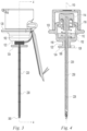
- FIGS. 1-5 illustrate an example of a suture anchor assembly 100 according to the present invention.
- the suture anchor assembly 100 includes a base member 102 having a proximal end 104 and a distal end 106 ( FIG. 5 ).
- a bore 108 extends through the base member 102 from the distal end 106 to the proximal end 104 and defines a longitudinal axis 110 ( FIG. 4 ).
- a cavity 112 is formed in a proximal portion of the base member 102 and is enlarged radially relative to the bore 108.
- An actuator in the form of a push button 114 is mounted in the cavity for axial translation between a first position in which it extends proximally from the cavity and abuts a shoulder adjacent the distal end of the cavity 112 and a second position distal to the first position.
- a spring 118 biases the button 114 into the first position.
- the base member 102 includes a radial projection in the form of a flange 120. With one or more fingers positioned distal to the flange 120 and a thumb positioned proximal to the button 114, a user can easily press the button by moving the thumb and fingers together.
- the base member further includes a trunnion 122 about which suture may be wound.
- the trunnion 122 includes a distal flange 124 to prevent wound suture from slipping distally off of the trunnion 122.
- a notch 126 is formed radially in the flange to allow a suture to pass from the trunnion distally. Suture may be wound around the trunnion 122 and then passed through the notch to secure the suture to the base member.
- a safety device is used to block operation of the pushbutton 114.
- the safety device is a cap 130 that may be moved by a user from a safe position ( FIG. 1 ) in which it prevents the user from pressing the button 114 to a delivery position ( FIG.
- the cap 130 With the cap 130 in the safe position a user can grip the assembly with a thumb on the cap 130 and manipulate the assembly without risk of prematurely pressing the button 114. With the cap 130 in the delivery position a user can easily access the button 114 and press it.
- the cap 130 is hinged to the base member so that it may be moved without becoming detached from the base member. By remaining attached to the base member, the cap does not need to be separately accounted for by the surgical staff.
- a delivery tube 220 is fixed within the bore 108 and extends from the distal end 106 of the base member 102.
- the delivery tube 220 has a proximal end 222, a distal end 224, and a longitudinal passage 226.
- the delivery tube 220 includes a slot 228 in the sidewall of the delivery tube adjacent the distal end 224 of the delivery tube.
- the slot 228 communicates through the sidewall to the longitudinal passage 226 and is open at the distal end 224.
- the outer surface of the delivery tube 220 includes a plurality of reference marks 230 indicating a plurality of length increments. In the illustrative example of FIGS.
- the reference marks are spaced 5mm apart with the first reference mark being 5mm from the proximal end of the slot 228. Every other mark is wider than the prior mark to indicate increments of 10mm.
- the delivery tube 220 is sized for delivering a suture anchor to a bone of a human foot and preferably has a diameter of 2-3mm and a length of 30-60mm. More preferably the delivery tube 220 has a diameter of 2.4mm.
- An ejector in the form of a push rod 250 has a proximal end 252 and a distal end 254.
- the push rod 250 is positioned within the delivery tube 220 for axial translation between a first position in which the distal end 254 of the ejector is proximal to the distal end 224 of the delivery tube and a second position in which the distal end 254 of the ejector is distal to the first position.
- the proximal end 252 of the ejector is fixed to the pushbutton 114 and moves with the pushbutton 114 between the first and second positions.
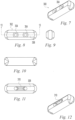
- a suture anchor 300 is positioned in the delivery tube 220 distal to the pushrod 250.
- the suture anchor is best illustrated in FIGS. 7-13 .
- the suture anchor 300 includes a generally rectangular body 302 having one or more holes through the body for receiving one or more suture strands.
- the suture anchor includes a pair of parallel holes 304 extending through the body 302 from a first side 306 to a second opposite side 308.
- the second side 308 includes a groove 310 extending between the holes 304 to receive a suture 350 ( FIG. 13 ) extending through the holes 304.
- the suture anchor 300 is placed in the distal end of the delivery tube, the suture is passed through the slot 228, through the notch 126, and is then wound around the trunnion 122.
- the suture anchor is sized for use in bones of a human foot and preferably has a width of 1.5-4mm and a length of 4-8mm. More preferably the suture anchor has a width of 2mm and a length of 6.5mm.
- the illustrative suture anchor has a pair of holes sized to receive a single strand of 2-0 high strength suture. Additional holes and holes of different sizes may be provided to receive more strands of suture and/or larger or smaller sutures. For example, 4 holes may be provided to receive 2 strands of suture.
- the suture ends may have needles attached.
- the suture anchor may be formed of any suitable material such as for example PEEK, stainless steel, titanium alloys, and resorbable materials.
- a suture reservoir may be provided to hold an additional portion of the suture.
- the suture reservoir may include a body having a circumference greater than that of the trunnion 122 so that an additional length of the suture may be wound around the reservoir body with fewer wraps than would be required for the trunnion.
- the suture reservoir is in the form of a flat card-like member 360 having a tab 362 extending from one end and positionable under the cap 130. When the cap is in the safe position it traps the tab to retain the member 360 on the base member 102. When the cap is in the delivery position the tab may be removed from under the cap and the member 360 separated from the base member.
- the tab inserts through a gap between opposed pivots of a cap hinge assembly 364.
- a hole forming instrument may be provided in a diameter suitable for forming a hole for insertion of the delivery tube 220 and anchor 300 into a bone.
- the hole forming instrument may be a punch, drill, wire or other suitable member.
- the hole forming instrument is a k-wire 400 having an elongate shaft extending between a proximal end 402 and a distal end 404. A diamond tip is formed at the distal end 404.
- the k-wire 400 is sized to form a hole slightly larger than the delivery tube 220 diameter. For example, for a 2.4mm diameter delivery tube, the k-wire is sized to form a 2.5mm diameter hole.
- the k-wire 400 includes indicia in the form of reference marks 406 indicating a plurality of length increments and corresponding to the reference marks 230 on the delivery tube 220.
- the reference marks are spaced 5mm apart with the first reference mark being 5mm from the distal end of the k-wire 400. Every other mark is wider than the prior mark to indicate increments of 10mm.
- the k-wire 400 may be driven by a wire driver, drill, or other suitable device to form a hole in a bone.
- the k-wire 400 is used to form a hole into a bone from a proximal bone surface to a distal bone surface.
- the depth of the hole may be determined by reading the reference marks 406 on the k-wire shaft relative to the proximal bone surface.
- the k-wire may then be removed and the suture anchor assembly 100 may be used to position the anchor 300.
- a user can grip the assembly with a thumb on the cap 130 and manipulate the assembly without risk of prematurely pressing the button 114 and ejecting the anchor 300.
- the anchor 300 and delivery tube 220 may be inserted into the hole formed by the k-wire 400.
- the delivery tube 220 is inserted to an indicated depth corresponding to the indicated depth of the hole formed by the k-wire 400. Due to the reference marks 230 of the delivery tube 220 being offset from the distal end 224 of the delivery tube 220 by the length of the slot 228, the delivery tube 220 will extend through the bone hole further than the k-wire by a distance equal to the slot length. This ensures that the anchor 300 will have room beyond the distal bone surface to rotate out of the end of the delivery tube 220 without the tube extending unnecessarily far.
- the safety cap 130 is flipped open and the button 114 is depressed ( FIG. 2 ). As the push rod 250 presses the anchor 300 out of the distal end of the delivery tube 220, tension in the suture portion extending between the trunnion 122 and the anchor 300 causes the anchor 300 to pivot into a deployed position.
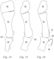
- FIG. 14 illustrates a medial view of cuneiform 500, metatarsal 502, and proximal phalangeal 504 bones of a human foot while FIG. 15 illustrates a dorsal view of the same bones.
- the bones are in the first ray of a human foot on which a bunion corrective procedure is to be performed.
- FIGS. 16-24 illustrate a surgical procedure utilizing the illustrative examples of FIGS. 1-13 .
- soft tissue 506 adjacent the metatarsophalangeal (MTP) joint 508, e.g. capsular tissue extending between the metatarsal and phalangeal bones has been dissected away from the metatarsal bone 502.
- a bunionectomy has been performed to remove a protruding bone portion, e.g. a bunion, on the medial side of the metatarsal bone 502 leaving a decorticated region 510.
- the decorticated region 510 with its lack of a cortical outer layer, has little strength to support a traditional bone anchor.
- the k-wire 400 of FIG. 6 has been driven into the decorticated region 510, across the metatarsal bone 502, and out through the cortical bone 512 opposite the decorticated region 510.
- the insertion depth of the k-wire is noted by reading the reference marks 406 on the k-wire shaft relative to the proximal bone surface.
- the k-wire has been driven to the fourth reference mark from the proximal end of the k-wire.
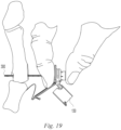
- the k-wire has been removed and the suture anchor 300 and delivery tube 220 are inserted into the hole formed by the k-wire 400.
- the user's thumb 550 and at least one finger 552 grip the cap 130 and flange 120 and are used to manipulate the assembly 100 as needed.
- the delivery tube is inserted until the fourth reference mark 230 as measured from the proximal end of the delivery tube is aligned with the proximal bone surface of the decorticated region 510 corresponding to the k-wire indicated depth.
- the safety cap 130 has been flipped open to arm the assembly 100 for deploying the anchor 300.
- the suture reservoir 360 has been dislodged from the cap 130 and allowed to hang below the rest of the assembly 100.
- the button 114 has been pressed and the suture anchor 300 has been ejected from the end of the delivery tube causing it to begin to rotate.
- the suture 350 has been unwound from the trunnion and tensioned to further rotate the anchor 300 and position it against the cortical bone 512 opposite the decorticated region 510.
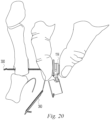
- the suture 350 is optionally tensioned with the delivery tube still extending through the metatarsal bone 502. The delivery tube blocks the hole through the bone and thus ensures that the anchor will rotate and be positioned against the cortical bone in the deployed position rather than be pulled back through the hole.
- FIG. 22 the delivery tube has been removed. Any number of anchors may be positioned by repeating the steps illustrated in FIGS. 17-22 .
- the suture may be unwound from the suture reservoir 360 and used to repair the soft tissue 506.
- FIGS. 23 and 24 illustrate dorsal and medial views of the medial capsular soft tissue 506 reattached over the decorticated region by passing the ends of the suture 350 through the soft tissue and tying them in a knot.
- the suture 350 may be passed through the soft tissue with a suture passer, manually with a needle attached to the ends of the suture 350, or by other suitable means known in the art.
- suture anchors may include features, such as additional holes, for coupling any number of sutures to the suture anchor. Needles may be supplied pre-attached to the suture ends and such needles may be attached to the suture reservoir 360.
Description
- Examples of the present invention relate generally to devices for anchoring soft tissue, tissue grafts, and the like to a bone.
- Various conditions may affect skeletal joints such as the deterioration, elongation, shortening, or rupture of soft tissues, cartilage, and/or bone associated with the joint and consequent laxity, pain, and/or deformity. It may be desirable to change the angular alignment of a bone or a portion of a bone to restore function and/or reduce pain. In such a medical procedure it may be necessary to affix soft tissue or a tissue graft to a bone. For example, in a medical procedure to correct an angular deformity of a first ray of a human foot, e.g. hallux valgus, it is often desirable to surgically remove a protruding bone portion or bunion in a procedure known as a bunionectomy adjacent the metatarsophalangeal (MTP) joint. To gain exposure to the surgical site, soft tissues surrounding the joint and the bunion are cut and dissected away. Often these tissues are not reattached for lack of workable devices and methods. Devices and methods to reattach such tissues to provide an anatomic repair are needed.
-
US5531678 provides a spring-loaded reciprocable stylet holder assembly that includes a cap; a hollow body element partially nested within and reciprocable a major part of its length into the cap; a spring urging the body element outwardly of the cap; and a stylet supported at one end by the cap and extending through and beyond the body element. The remote end of the body element has a small borehole through which the stylet extends. The remote end is also provided with attachment means for a hypodermic needle with slotted tip for T-fastener emplacement, the needle telescopically surrounding the full length of the stylet. In a useful modification of the device an insufflation adapter having a side port for attachment of a hypodermic syringe is placed in line between the body element and the hypodermic needle. The stylet holder assembly with a slotted hypodermic needle attached thereto may be used for emplacing a T-fastener in a hollow organ of a person, such as the stomach. -
KR20150107824 -
US6656182 provides an apparatus and method for manipulating and anchoring tissue is provided. The invention is directed to solving the problem of manipulating and anchoring tissue within a joint when access to that tissue is limited, for example, during arthroscopic surgery. - The present invention provides devices for anchoring soft tissue, tissue grafts, and the like to a bone. In one example an assembly includes a suture anchor, suture, and inserter. The invention is defined by the appended independent claim 1. Further embodiments are provided by the dependent claims.
- Various examples of the present invention will be discussed with reference to the appended drawings. These drawings depict only illustrative examples of the invention and are not to be considered limiting of its scope.
-
FIG. 1 is a perspective view of an illustrative example of an assembly according to an example of the present invention; -
FIG. 2 is a perspective view of the assembly ofFIG. 1 showing a different operative state of the assembly; -
FIG. 3 is a side elevation view of the assembly ofFIG. 1 ; -
FIG. 4 is a section view of the assembly ofFIG. 1 taken along line 4-4 ofFIG. 3 with the suture omitted for clarity; -
FIG. 5 is an exploded view of the assembly ofFIG. 1 with the suture omitted for clarity; -
FIG. 6 is a side elevation view of an illustrative example of a hole forming device according to an example of the present invention; -
FIG. 7 is a perspective view of the suture anchor of the assembly ofFIG. 1 ; -
FIG. 8 is a top plan view of the suture anchor of the assembly ofFIG. 1 ; -
FIG. 9 is a side elevation view of the suture anchor of the assembly ofFIG. 1 ; -
FIG. 10 is a front elevation view of the suture anchor of the assembly ofFIG. 1 ; -
FIG. 11 is a bottom plan view of the suture anchor of the assembly ofFIG. 1 ; -
FIG. 12 is a perspective view of the suture anchor of the assembly ofFIG. 1 ; -
FIG. 13 is a sectional view of the suture anchor of the assembly ofFIG. 1 taken along line 13-13 ofFIG. 8 and showing a suture routed through the suture anchor; -
FIG. 14 is a medical view of the cuneiform, metatarsal, and proximal phalanx bones of the first ray of a human foot; -
FIG. 15 is a dorsal view of the cuneiform, metatarsal, and proximal phalanx bones of the first ray of a human foot; -
FIG. 16 is a dorsal view similar toFIG. 15 illustrating a bunionectomy; and -
FIGS. 17-24 illustrate a method of using the assembly ofFIG. 1 to anchor soft tissue. - The following illustrative examples describe implants and instruments for connecting soft tissue to bone. The use of the illustrative devices is illustrated to attach soft tissue detached in conjunction with a bunionectomy during a corrective procedure performed on a first ray of a human foot. In particular the illustrative devices are illustrated to reattach capsular tissue adjacent to an MTP joint. The inventive devices may be used to attach tissue at other locations in the body.
- The terms "suture" and "suture strand" are used herein to mean any flexible member, natural or synthetic, useful in a surgical procedure and that are easily flexed. Examples include polymer sutures, wires, surgical tapes, tissue derived strands, and other suitable flexible strands or members. Sutures may be monofilament or multi-filament structures. The term "transverse" is used herein to mean crossing as in non-parallel. The term "tissue" is used herein to mean a patient's body tissue as well as a tissue graft which may be allograft, xenograft, or synthetic.
-
FIGS. 1-5 illustrate an example of asuture anchor assembly 100 according to the present invention. Thesuture anchor assembly 100 includes abase member 102 having aproximal end 104 and a distal end 106 (FIG. 5 ). Abore 108 extends through thebase member 102 from thedistal end 106 to theproximal end 104 and defines a longitudinal axis 110 (FIG. 4 ). Acavity 112 is formed in a proximal portion of thebase member 102 and is enlarged radially relative to thebore 108. An actuator in the form of apush button 114 is mounted in the cavity for axial translation between a first position in which it extends proximally from the cavity and abuts a shoulder adjacent the distal end of thecavity 112 and a second position distal to the first position. Aspring 118 biases thebutton 114 into the first position. Thebase member 102 includes a radial projection in the form of aflange 120. With one or more fingers positioned distal to theflange 120 and a thumb positioned proximal to thebutton 114, a user can easily press the button by moving the thumb and fingers together. The base member further includes atrunnion 122 about which suture may be wound. Thetrunnion 122 includes adistal flange 124 to prevent wound suture from slipping distally off of thetrunnion 122. Anotch 126 is formed radially in the flange to allow a suture to pass from the trunnion distally. Suture may be wound around thetrunnion 122 and then passed through the notch to secure the suture to the base member. A safety device is used to block operation of thepushbutton 114. In the illustrative example ofFIGS. 1-5 , the safety device is acap 130 that may be moved by a user from a safe position (FIG. 1 ) in which it prevents the user from pressing thebutton 114 to a delivery position (FIG. 2 ) in which it allows the user to press thebutton 114. With thecap 130 in the safe position a user can grip the assembly with a thumb on thecap 130 and manipulate the assembly without risk of prematurely pressing thebutton 114. With thecap 130 in the delivery position a user can easily access thebutton 114 and press it. In the illustrative example ofFIGS. 1-5 , thecap 130 is hinged to the base member so that it may be moved without becoming detached from the base member. By remaining attached to the base member, the cap does not need to be separately accounted for by the surgical staff. - A
delivery tube 220 is fixed within thebore 108 and extends from thedistal end 106 of thebase member 102. Thedelivery tube 220 has aproximal end 222, adistal end 224, and alongitudinal passage 226. Thedelivery tube 220 includes aslot 228 in the sidewall of the delivery tube adjacent thedistal end 224 of the delivery tube. Theslot 228 communicates through the sidewall to thelongitudinal passage 226 and is open at thedistal end 224. The outer surface of thedelivery tube 220 includes a plurality of reference marks 230 indicating a plurality of length increments. In the illustrative example ofFIGS. 1-5 , the reference marks are spaced 5mm apart with the first reference mark being 5mm from the proximal end of theslot 228. Every other mark is wider than the prior mark to indicate increments of 10mm. In the illustrative example ofFIGS. 1-5 , thedelivery tube 220 is sized for delivering a suture anchor to a bone of a human foot and preferably has a diameter of 2-3mm and a length of 30-60mm. More preferably thedelivery tube 220 has a diameter of 2.4mm. - An ejector in the form of a
push rod 250 has aproximal end 252 and adistal end 254. Thepush rod 250 is positioned within thedelivery tube 220 for axial translation between a first position in which thedistal end 254 of the ejector is proximal to thedistal end 224 of the delivery tube and a second position in which thedistal end 254 of the ejector is distal to the first position. Theproximal end 252 of the ejector is fixed to thepushbutton 114 and moves with thepushbutton 114 between the first and second positions. - A
suture anchor 300 is positioned in thedelivery tube 220 distal to thepushrod 250. The suture anchor is best illustrated inFIGS. 7-13 . Thesuture anchor 300 includes a generallyrectangular body 302 having one or more holes through the body for receiving one or more suture strands. In the illustrative example ofFIGS. 7-13 , the suture anchor includes a pair ofparallel holes 304 extending through thebody 302 from afirst side 306 to a secondopposite side 308. Thesecond side 308 includes agroove 310 extending between theholes 304 to receive a suture 350 (FIG. 13 ) extending through theholes 304. With thesuture 350 extending into onehole 304, along thegroove 310, and out theother hole 304, the suture is contained in thegroove 310 to reduce the overall thickness of the suture anchor and suture assembly. Referring back toFIGS. 1-3 , thesuture anchor 300 is placed in the distal end of the delivery tube, the suture is passed through theslot 228, through thenotch 126, and is then wound around thetrunnion 122. In the illustrative example ofFIGS. 7-13 , the suture anchor is sized for use in bones of a human foot and preferably has a width of 1.5-4mm and a length of 4-8mm. More preferably the suture anchor has a width of 2mm and a length of 6.5mm. The illustrative suture anchor has a pair of holes sized to receive a single strand of 2-0 high strength suture. Additional holes and holes of different sizes may be provided to receive more strands of suture and/or larger or smaller sutures. For example, 4 holes may be provided to receive 2 strands of suture. The suture ends may have needles attached. The suture anchor may be formed of any suitable material such as for example PEEK, stainless steel, titanium alloys, and resorbable materials. - A suture reservoir may be provided to hold an additional portion of the suture. For example, the suture reservoir may include a body having a circumference greater than that of the
trunnion 122 so that an additional length of the suture may be wound around the reservoir body with fewer wraps than would be required for the trunnion. In the illustrative example ofFIGS. 1-5 , the suture reservoir is in the form of a flat card-like member 360 having atab 362 extending from one end and positionable under thecap 130. When the cap is in the safe position it traps the tab to retain themember 360 on thebase member 102. When the cap is in the delivery position the tab may be removed from under the cap and themember 360 separated from the base member. In the illustrative example ofFIGS. 1-5 the tab inserts through a gap between opposed pivots of acap hinge assembly 364. - Referring to
FIG. 6 , a hole forming instrument may be provided in a diameter suitable for forming a hole for insertion of thedelivery tube 220 andanchor 300 into a bone. The hole forming instrument may be a punch, drill, wire or other suitable member. In the illustrative example ofFIG. 6 , the hole forming instrument is a k-wire 400 having an elongate shaft extending between aproximal end 402 and a distal end 404. A diamond tip is formed at the distal end 404. The k-wire 400 is sized to form a hole slightly larger than thedelivery tube 220 diameter. For example, for a 2.4mm diameter delivery tube, the k-wire is sized to form a 2.5mm diameter hole. The k-wire 400 includes indicia in the form of reference marks 406 indicating a plurality of length increments and corresponding to the reference marks 230 on thedelivery tube 220. In the illustrative example ofFIG. 6 , the reference marks are spaced 5mm apart with the first reference mark being 5mm from the distal end of the k-wire 400. Every other mark is wider than the prior mark to indicate increments of 10mm. The k-wire 400 may be driven by a wire driver, drill, or other suitable device to form a hole in a bone. - In use, the k-
wire 400 is used to form a hole into a bone from a proximal bone surface to a distal bone surface. The depth of the hole may be determined by reading the reference marks 406 on the k-wire shaft relative to the proximal bone surface. The k-wire may then be removed and thesuture anchor assembly 100 may be used to position theanchor 300. With thecap 130 in the safe position a user can grip the assembly with a thumb on thecap 130 and manipulate the assembly without risk of prematurely pressing thebutton 114 and ejecting theanchor 300. Theanchor 300 anddelivery tube 220 may be inserted into the hole formed by the k-wire 400. Preferably thedelivery tube 220 is inserted to an indicated depth corresponding to the indicated depth of the hole formed by the k-wire 400. Due to the reference marks 230 of thedelivery tube 220 being offset from thedistal end 224 of thedelivery tube 220 by the length of theslot 228, thedelivery tube 220 will extend through the bone hole further than the k-wire by a distance equal to the slot length. This ensures that theanchor 300 will have room beyond the distal bone surface to rotate out of the end of thedelivery tube 220 without the tube extending unnecessarily far. Once thedelivery tube 220 is positioned, thesafety cap 130 is flipped open and thebutton 114 is depressed (FIG. 2 ). As thepush rod 250 presses theanchor 300 out of the distal end of thedelivery tube 220, tension in the suture portion extending between thetrunnion 122 and theanchor 300 causes theanchor 300 to pivot into a deployed position. -
FIG. 14 illustrates a medial view ofcuneiform 500,metatarsal 502, and proximal phalangeal 504 bones of a human foot whileFIG. 15 illustrates a dorsal view of the same bones. In the illustrative example ofFIGS. 14-15 , the bones are in the first ray of a human foot on which a bunion corrective procedure is to be performed. -
FIGS. 16-24 illustrate a surgical procedure utilizing the illustrative examples ofFIGS. 1-13 . InFIG. 16 ,soft tissue 506 adjacent the metatarsophalangeal (MTP)joint 508, e.g. capsular tissue extending between the metatarsal and phalangeal bones, has been dissected away from themetatarsal bone 502. A bunionectomy has been performed to remove a protruding bone portion, e.g. a bunion, on the medial side of themetatarsal bone 502 leaving adecorticated region 510. Thedecorticated region 510, with its lack of a cortical outer layer, has little strength to support a traditional bone anchor. - In
FIG. 17 , the k-wire 400 ofFIG. 6 has been driven into thedecorticated region 510, across themetatarsal bone 502, and out through thecortical bone 512 opposite thedecorticated region 510. The insertion depth of the k-wire is noted by reading the reference marks 406 on the k-wire shaft relative to the proximal bone surface. In the illustrative example ofFIG. 17 , the k-wire has been driven to the fourth reference mark from the proximal end of the k-wire. - In
FIG. 18 , the k-wire has been removed and thesuture anchor 300 anddelivery tube 220 are inserted into the hole formed by the k-wire 400. The user'sthumb 550 and at least onefinger 552 grip thecap 130 andflange 120 and are used to manipulate theassembly 100 as needed. The delivery tube is inserted until thefourth reference mark 230 as measured from the proximal end of the delivery tube is aligned with the proximal bone surface of thedecorticated region 510 corresponding to the k-wire indicated depth. - In
FIG. 19 , thesafety cap 130 has been flipped open to arm theassembly 100 for deploying theanchor 300. - In
FIG. 20 , thesuture reservoir 360 has been dislodged from thecap 130 and allowed to hang below the rest of theassembly 100. Thebutton 114 has been pressed and thesuture anchor 300 has been ejected from the end of the delivery tube causing it to begin to rotate. - In
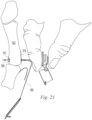
FIG. 21 , thesuture 350 has been unwound from the trunnion and tensioned to further rotate theanchor 300 and position it against thecortical bone 512 opposite thedecorticated region 510. In the illustrative example ofFIG. 21 , thesuture 350 is optionally tensioned with the delivery tube still extending through themetatarsal bone 502. The delivery tube blocks the hole through the bone and thus ensures that the anchor will rotate and be positioned against the cortical bone in the deployed position rather than be pulled back through the hole. - In
FIG. 22 , the delivery tube has been removed. Any number of anchors may be positioned by repeating the steps illustrated inFIGS. 17-22 . The suture may be unwound from thesuture reservoir 360 and used to repair thesoft tissue 506. -
FIGS. 23 and 24 illustrate dorsal and medial views of the medial capsularsoft tissue 506 reattached over the decorticated region by passing the ends of thesuture 350 through the soft tissue and tying them in a knot. Thesuture 350 may be passed through the soft tissue with a suture passer, manually with a needle attached to the ends of thesuture 350, or by other suitable means known in the art. - Various examples have been illustrated and described. The various examples may be substituted and combined and other alterations made within the scope of the invention. For example, among other substitutions, male and female features may be reversed. The suture anchors may include features, such as additional holes, for coupling any number of sutures to the suture anchor. Needles may be supplied pre-attached to the suture ends and such needles may be attached to the
suture reservoir 360.
Claims (12)
- A suture anchor assembly (100) comprising:a base member (102) having a proximal end (104) and a distal end (106);a delivery tube (220) extending from the distal end of the base member, the delivery tube having a proximal end (222), a distal end (224), a longitudinal axis extending between the proximal end and the distal end, and a longitudinal passage (226);an ejector (250) having a proximal end (252) and a distal end (254), the ejector being positioned within the delivery tube for axial translation between a first position in which the distal end of the ejector is proximal to the distal end of the delivery tube and a second position in which the distal end of the ejector is distal to the first position, the ejector being moveable by a user to the second position;an actuator coupled to the proximal end of the ejector (250), the actuator extending from the proximal end of the base member (102);a safety device (130) operable to prevent or allow movement of the ejector by a user to the second position; anda flip-type suture anchor (300) positioned in the delivery tube distal to the distal end of the ejector;wherein the actuator is in the form of a push button (114),
wherein the safety device may be moved by the user from a safe position to a delivery position in which the safety device allows the user to press the push button (114),characterised in that the safety device comprises a cap covering the actuator in the safe position that prevents the user from pressing the push button (114). - The assembly of claim 1 wherein the cap (130) is hinged to the base member (102).
- The assembly of any preceding claim wherein the ejector (250) is resiliently biased by a first force to the first position, the actuator (114) being responsive to a user applied force greater than the first force to move the ejector from the first position to the second position.
- The assembly of any preceding claim wherein the base member (102) includes a radial projection (120) engageable by one or more of a user's fingers positioned distal to the radial projection and the actuator comprises a button (114) engageable by a user's thumb positioned proximal to the button.
- The assembly of any preceding claim further including a slot (228) in a sidewall of the delivery tube (220) adjacent the distal end (224) of the delivery tube, the slot communicating through the sidewall to the longitudinal passage (226) and being open at the distal end, the assembly further comprising a suture (350) extending from the base member (102) distally outside of the delivery tube (220), through the slot, and into engagement with the suture anchor (300).
- The assembly of claim 5 being responsive to movement of the ejector (250) into the second position to press the suture anchor (300) distally, wherein tension from the suture (350) causes the suture anchor to rotate.
- The assembly of claim 5 or claim 6 wherein the base member (102) further comprises a trunnion (122) about which a portion of the suture (350) is wound.
- The assembly of any of claims 5-7 further comprising a suture reservoir (360) removably attached to the assembly, the reservoir containing a portion of the suture (350).
- The assembly of claim 8 wherein the reservoir (360) comprises a body with a portion of the suture wound around the body.
- The assembly of claim 9 wherein the body comprises a flat card-like member (360) having a tab (362) and the safety device comprises a cap (130), the tab being removably retained by the cap.
- The assembly of any preceding claim further comprising a hole forming instrument (400) having an elongate shaft extending between a proximal end (402) and a distal end (404), the elongate shaft having one or more reference marks (406) indicating a plurality of length increments, the delivery tube (220) having corresponding reference marks (230) indicating the plurality of length increments.
- The assembly of claim 11 wherein the reference marks (230) of the delivery tube (220) are offset proximally from the distal end (224) of the delivery tube.
Applications Claiming Priority (2)
| Application Number | Priority Date | Filing Date | Title |
|---|---|---|---|
| US201562263250P | 2015-12-04 | 2015-12-04 | |
| PCT/US2016/063063 WO2017095681A1 (en) | 2015-12-04 | 2016-11-21 | Devices for anchoring tissue |
Publications (3)
| Publication Number | Publication Date |
|---|---|
| EP3383281A1 EP3383281A1 (en) | 2018-10-10 |
| EP3383281A4 EP3383281A4 (en) | 2019-08-14 |
| EP3383281B1 true EP3383281B1 (en) | 2024-01-24 |
Family
ID=58797925
Family Applications (1)
| Application Number | Title | Priority Date | Filing Date |
|---|---|---|---|
| EP16871284.2A Active EP3383281B1 (en) | 2015-12-04 | 2016-11-21 | Devices for anchoring tissue |
Country Status (5)
| Country | Link |
|---|---|
| US (2) | US10653409B2 (en) |
| EP (1) | EP3383281B1 (en) |
| AU (2) | AU2016364969B2 (en) |
| CA (1) | CA3005616A1 (en) |
| WO (1) | WO2017095681A1 (en) |
Families Citing this family (4)
| Publication number | Priority date | Publication date | Assignee | Title |
|---|---|---|---|---|
| US10653409B2 (en) * | 2015-12-04 | 2020-05-19 | Crossroads Extremity Systems, Llc | Devices and methods for anchoring tissue |
| EP3796848B1 (en) * | 2018-05-23 | 2024-01-17 | Smith&Nephew, Inc. | Anchor delivery systems |
| US11172920B2 (en) * | 2018-09-24 | 2021-11-16 | Sportwelding Gmbh | Surgical methods for the treatment of plantar plate injury |
| WO2021222698A1 (en) * | 2020-05-01 | 2021-11-04 | Acumed Llc | Automatic release of a near bone suture button from a button inserter |
Family Cites Families (265)
| Publication number | Priority date | Publication date | Assignee | Title |
|---|---|---|---|---|
| US2485531A (en) | 1948-01-13 | 1949-10-18 | Dzus William | Surgical toggle bolt |
| US3620216A (en) | 1969-06-25 | 1971-11-16 | Abbott Lab | Implant trocar |
| US3664345A (en) | 1970-07-06 | 1972-05-23 | Clyde Harwell Dabbs | Surgical buttons |
| US3910281A (en) | 1973-10-09 | 1975-10-07 | Bio Medicus Inc | Suture anchor |
| US5417691A (en) | 1982-05-20 | 1995-05-23 | Hayhurst; John O. | Apparatus and method for manipulating and anchoring tissue |
| US6656182B1 (en) * | 1982-05-20 | 2003-12-02 | John O. Hayhurst | Tissue manipulation |
| US4741330A (en) | 1983-05-19 | 1988-05-03 | Hayhurst John O | Method and apparatus for anchoring and manipulating cartilage |
| US5601557A (en) | 1982-05-20 | 1997-02-11 | Hayhurst; John O. | Anchoring and manipulating tissue |
| US4750492A (en) | 1985-02-27 | 1988-06-14 | Richards Medical Company | Absorbable suture apparatus, method and installer |
| US4898156A (en) * | 1987-05-18 | 1990-02-06 | Mitek Surgical Products, Inc. | Suture anchor |
| US5441508A (en) | 1989-04-27 | 1995-08-15 | Gazielly; Dominique | Reinforcement and supporting device for the rotator cuff of a shoulder joint of a person |
| US7208013B1 (en) | 1990-06-28 | 2007-04-24 | Bonutti Ip, Llc | Composite surgical devices |
| US5593425A (en) | 1990-06-28 | 1997-01-14 | Peter M. Bonutti | Surgical devices assembled using heat bonable materials |
| US5269785A (en) | 1990-06-28 | 1993-12-14 | Bonutti Peter M | Apparatus and method for tissue removal |
| US5269809A (en) | 1990-07-02 | 1993-12-14 | American Cyanamid Company | Locking mechanism for use with a slotted suture anchor |
| US5041129A (en) | 1990-07-02 | 1991-08-20 | Acufex Microsurgical, Inc. | Slotted suture anchor and method of anchoring a suture |
| US5814073A (en) | 1996-12-13 | 1998-09-29 | Bonutti; Peter M. | Method and apparatus for positioning a suture anchor |
| US5306301A (en) | 1993-02-11 | 1994-04-26 | American Cyanamid Company | Graft attachment device and method of using same |
| US5306290A (en) | 1993-02-12 | 1994-04-26 | Mitek Surgical Products, Inc. | Suture button |
| US5549630A (en) | 1993-05-14 | 1996-08-27 | Bonutti; Peter M. | Method and apparatus for anchoring a suture |
| US5845645A (en) | 1993-05-14 | 1998-12-08 | Bonutti; Peter M. | Method of anchoring a suture |
| US5403348A (en) | 1993-05-14 | 1995-04-04 | Bonutti; Peter M. | Suture anchor |
| US5500000A (en) | 1993-07-01 | 1996-03-19 | United States Surgical Corporation | Soft tissue repair system and method |
| CA2124651C (en) | 1993-08-20 | 2004-09-28 | David T. Green | Apparatus and method for applying and adjusting an anchoring device |
| US5540718A (en) | 1993-09-20 | 1996-07-30 | Bartlett; Edwin C. | Apparatus and method for anchoring sutures |
| US6123185A (en) | 1994-01-13 | 2000-09-26 | Ethicon, Inc. | Needle sorting device |
| US5464425A (en) | 1994-02-23 | 1995-11-07 | Orthopaedic Biosystems, Ltd. | Medullary suture anchor |
| US5683418A (en) | 1994-04-29 | 1997-11-04 | Mitek Surgical Products, Inc. | Wedge shaped suture anchor and method of implantation |
| US5470337A (en) | 1994-05-17 | 1995-11-28 | Moss; Gerald | Surgical fastener |
| US5601571A (en) | 1994-05-17 | 1997-02-11 | Moss; Gerald | Surgical fastener implantation device |
| US5529075A (en) | 1994-09-12 | 1996-06-25 | Clark; David | Fixation device and method for repair of pronounced hallux valgus |
| US5531699A (en) | 1994-09-19 | 1996-07-02 | Abbott Laboratories | Spring-loaded reciprocable stylet holder |
| GB9524861D0 (en) | 1995-12-05 | 1996-02-07 | United Surgical Services Ltd | Surgical anchorage |
| US5626614A (en) | 1995-12-22 | 1997-05-06 | Applied Medical Resources Corporation | T-anchor suturing device and method for using same |
| US5713921A (en) | 1996-03-29 | 1998-02-03 | Bonutti; Peter M. | Suture anchor |
| US5961538A (en) | 1996-04-10 | 1999-10-05 | Mitek Surgical Products, Inc. | Wedge shaped suture anchor and method of implantation |
| US6491714B1 (en) * | 1996-05-03 | 2002-12-10 | William F. Bennett | Surgical tissue repair and attachment apparatus and method |
| US6013083A (en) | 1997-05-02 | 2000-01-11 | Bennett; William F. | Arthroscopic rotator cuff repair apparatus and method |
| US5782862A (en) | 1996-07-01 | 1998-07-21 | Bonutti; Peter M. | Suture anchor inserter assembly and method |
| US5718717A (en) | 1996-08-19 | 1998-02-17 | Bonutti; Peter M. | Suture anchor |
| US6007567A (en) | 1996-08-19 | 1999-12-28 | Bonutti; Peter M. | Suture anchor |
| US5810848A (en) | 1996-08-21 | 1998-09-22 | Hayhurst; John O. | Suturing system |
| US5814072A (en) | 1996-11-15 | 1998-09-29 | Bonutti; Peter M. | Method and apparatus for use in anchoring a suture |
| US5948002A (en) | 1996-11-15 | 1999-09-07 | Bonutti; Peter M. | Apparatus and method for use in positioning a suture anchor |
| CA2224366C (en) | 1996-12-11 | 2006-10-31 | Ethicon, Inc. | Meniscal repair device |
| FR2758975B1 (en) | 1997-02-05 | 1999-04-30 | Ethnor | MATERIAL FOR FIXING A TENDON OF MUSCLE ON A BONE |
| US5769894A (en) | 1997-02-05 | 1998-06-23 | Smith & Nephew, Inc. | Graft attachment device and method of attachment |
| US5954057A (en) | 1997-02-12 | 1999-09-21 | Li Medical Technologies, Inc. | Soft tissue suspension clip, clip assembly, emplacement tool and method |
| CA2280812A1 (en) | 1997-02-13 | 1998-08-20 | Rodney Brenneman | Percutaneous and hiatal devices and methods for use in minimally invasive pelvic surgery |
| CA2280757A1 (en) | 1997-02-13 | 1998-08-20 | William Pintauro | Method and apparatus for minimally invasive pelvic surgery |
| US5814051A (en) | 1997-06-06 | 1998-09-29 | Mitex Surgical Products, Inc. | Suture anchor insertion system |
| AUPP000797A0 (en) | 1997-10-24 | 1997-11-20 | Cryptych Pty Ltd | Fixation of cruciate ligament grafts |
| US6068648A (en) | 1998-01-26 | 2000-05-30 | Orthodyne, Inc. | Tissue anchoring system and method |
| US5921986A (en) | 1998-02-06 | 1999-07-13 | Bonutti; Peter M. | Bone suture |
| US6045551A (en) | 1998-02-06 | 2000-04-04 | Bonutti; Peter M. | Bone suture |
| US5964764A (en) | 1998-03-24 | 1999-10-12 | Hugh S. West, Jr. | Apparatus and methods for mounting a ligament graft to a bone |
| US6099530A (en) | 1998-04-09 | 2000-08-08 | Smith & Nephew, Inc. | Soft-tissue intra-tunnel fixation device |
| US6102934A (en) | 1998-06-02 | 2000-08-15 | Li; Lehmann K. | Anchor tool and method and apparatus for emplacing anchor in a borehole |
| US6221107B1 (en) | 1998-08-03 | 2001-04-24 | Mark E. Steiner | Ligament fixation device and method |
| US6110183A (en) * | 1998-12-22 | 2000-08-29 | Cook Incorporated | Suture anchor device |
| US6306159B1 (en) | 1998-12-23 | 2001-10-23 | Depuy Orthopaedics, Inc. | Meniscal repair device |
| DE69931018T2 (en) | 1998-12-30 | 2006-11-23 | Ethicon, Inc. | Thread belay device |
| US6660022B1 (en) | 1999-06-01 | 2003-12-09 | Smith & Nephew, Inc. | Rotor blade anchor and tool for installing same |
| US6592609B1 (en) | 1999-08-09 | 2003-07-15 | Bonutti 2003 Trust-A | Method and apparatus for securing tissue |
| US8632590B2 (en) * | 1999-10-20 | 2014-01-21 | Anulex Technologies, Inc. | Apparatus and methods for the treatment of the intervertebral disc |
| US6736829B1 (en) | 1999-11-11 | 2004-05-18 | Linvatec Corporation | Toggle anchor and tool for insertion thereof |
| US7887551B2 (en) | 1999-12-02 | 2011-02-15 | Smith & Nephew, Inc. | Soft tissue attachment and repair |
| US7153312B1 (en) | 1999-12-02 | 2006-12-26 | Smith & Nephew Inc. | Closure device and method for tissue repair |
| GB9929599D0 (en) | 1999-12-15 | 2000-02-09 | Atlantech Medical Devices Limi | A graft suspension device |
| US6635073B2 (en) | 2000-05-03 | 2003-10-21 | Peter M. Bonutti | Method of securing body tissue |
| EP1665991B1 (en) | 2000-06-05 | 2017-12-13 | Boston Scientific Limited | Devices for the treatment of urinary continence |
| US7963966B2 (en) | 2000-06-06 | 2011-06-21 | Cole J Dean | Bone fixation system and method of use |
| US6451030B2 (en) | 2000-06-30 | 2002-09-17 | Li Medical Technologies, Inc. | Rotor blade anchor and tool for installing same particularlly for arthroscopic installation |
| US7037324B2 (en) | 2000-09-15 | 2006-05-02 | United States Surgical Corporation | Knotless tissue anchor |
| US6887259B2 (en) | 2000-10-18 | 2005-05-03 | Depuy Mitek, Inc. | Suture anchor system and method of use |
| US6527795B1 (en) | 2000-10-18 | 2003-03-04 | Ethicon, Inc. | Knotless suture anchor system and method of use |
| US7083638B2 (en) | 2001-02-12 | 2006-08-01 | Arthrocare Corporation | Method and apparatus for attaching connective tissues to bone using a knotless suture anchoring device |
| US6770076B2 (en) | 2001-02-12 | 2004-08-03 | Opus Medical, Inc. | Method and apparatus for attaching connective tissues to bone using a knotless suture anchoring device |
| US6533802B2 (en) | 2001-05-16 | 2003-03-18 | Smith & Nephew, Inc. | Endobutton continuous loop for bone-tendon-bone |
| US7235091B2 (en) | 2002-06-20 | 2007-06-26 | Brian Thornes | Apparatus and method for fixation of ankle syndesmosis |
| US9005245B2 (en) | 2002-08-30 | 2015-04-14 | Arthrex, Inc. | Acromioclavicular joint fixation technique |
| US8512376B2 (en) | 2002-08-30 | 2013-08-20 | Arthrex, Inc. | Method and apparatus for internal fixation of an acromioclavicular joint dislocation of the shoulder |
| US6773436B2 (en) | 2001-09-28 | 2004-08-10 | Depuy Mitek, Inc. | Absorbable bone anchor |
| US6656183B2 (en) | 2001-11-08 | 2003-12-02 | Smith & Nephew, Inc. | Tissue repair system |
| US6645227B2 (en) | 2001-11-21 | 2003-11-11 | Stryker Endoscopy | Suture anchor |
| US20070112338A1 (en) | 2005-11-01 | 2007-05-17 | Microfabrica Inc. | Microdevices for tissue approximation and retention, methods for using, and methods for making |
| US6855157B2 (en) | 2002-02-04 | 2005-02-15 | Arthrocare Corporation | Method and apparatus for attaching connective tissues to bone using a knotless suture anchoring device |
| AU2003220546A1 (en) | 2002-03-26 | 2003-10-13 | Ethicon, Inc. | System and method for biopsy management |
| US6972027B2 (en) | 2002-06-26 | 2005-12-06 | Stryker Endoscopy | Soft tissue repair system |
| EP1531737B1 (en) | 2002-08-14 | 2008-12-17 | Boston Scientific Limited | Systems and devices relating to delivery of medical implants |
| US7837669B2 (en) | 2002-11-01 | 2010-11-23 | Valentx, Inc. | Devices and methods for endolumenal gastrointestinal bypass |
| US9060844B2 (en) | 2002-11-01 | 2015-06-23 | Valentx, Inc. | Apparatus and methods for treatment of morbid obesity |
| US7214206B2 (en) | 2003-04-03 | 2007-05-08 | Valera Pharmaceuticals, Inc. | Implanting device and method of using same |
| US7320701B2 (en) | 2003-06-02 | 2008-01-22 | Linvatec Corporation | Push-in suture anchor, insertion tool, and method for inserting a push-in suture anchor |
| US8133257B2 (en) | 2003-06-27 | 2012-03-13 | Depuy Mitek, Inc. | Bioabsorbable suture anchor system for use in small joints |
| US7021316B2 (en) | 2003-08-07 | 2006-04-04 | Tools For Surgery, Llc | Device and method for tacking a prosthetic screen |
| US7217279B2 (en) | 2003-11-14 | 2007-05-15 | Ethicon, Inc. | Suture loop anchor |
| US7597705B2 (en) * | 2003-12-03 | 2009-10-06 | St. Jude Medical Puerto Rico Llc | Vascular puncture seal anchor nest |
| US7357810B2 (en) | 2003-12-18 | 2008-04-15 | Ethicon, Inc. | High strength suture with absorbable core and suture anchor combination |
| US7608092B1 (en) | 2004-02-20 | 2009-10-27 | Biomet Sports Medicince, LLC | Method and apparatus for performing meniscus repair |
| US7390332B2 (en) | 2004-02-24 | 2008-06-24 | Depuy Mitek, Inc. | Methods and devices for repairing tissue |
| US7455683B2 (en) | 2004-02-26 | 2008-11-25 | Depuy Mitek, Inc. | Methods and devices for repairing triangular fibrocartilage complex tears |
| EP1750595A4 (en) | 2004-05-07 | 2008-10-22 | Valentx Inc | Devices and methods for attaching an endolumenal gastrointestinal implant |
| US7785348B2 (en) | 2004-05-14 | 2010-08-31 | Ethicon Endo-Surgery, Inc. | Devices and methods of locking and cutting a suture in a medical procedure |
| US7819898B2 (en) | 2004-06-09 | 2010-10-26 | Biomet Sports Medicine, Llc | Method and apparatus for soft tissue fixation |
| US7500983B1 (en) | 2004-06-09 | 2009-03-10 | Biomet Sports Medicine, Llc | Apparatus for soft tissue attachment |
| US8109965B2 (en) | 2004-06-09 | 2012-02-07 | Biomet Sports Medicine, LLP | Method and apparatus for soft tissue fixation |
| US7695503B1 (en) | 2004-06-09 | 2010-04-13 | Biomet Sports Medicine, Llc | Method and apparatus for soft tissue attachment |
| US20050288694A1 (en) | 2004-06-23 | 2005-12-29 | Stepehen Solomon | Adjustable percutaneous stomach lumen restriction device |
| US7879055B1 (en) | 2004-06-23 | 2011-02-01 | Biomet Sports Medicine, Llc | Method and apparatus for sizing a material |
| US8273093B2 (en) | 2004-06-29 | 2012-09-25 | Depuy Products, Inc. | Instrumentation for recording and replicating orthopaedic implant orientation |
| WO2006012630A2 (en) | 2004-07-23 | 2006-02-02 | Calypso Medical Technologies, Inc. | Apparatuses and methods for percutaneously implanting objects in patients |
| US20120046747A1 (en) | 2004-09-07 | 2012-02-23 | Medicinelodge, Inc. Dba Imds Co-Innovation | Systems and methods for zipknot acl fixation |
| US20060155375A1 (en) | 2004-09-27 | 2006-07-13 | Jonathan Kagan | Devices and methods for attachment of a gastrointestinal sleeve |
| US20060089646A1 (en) | 2004-10-26 | 2006-04-27 | Bonutti Peter M | Devices and methods for stabilizing tissue and implants |
| US9271766B2 (en) | 2004-10-26 | 2016-03-01 | P Tech, Llc | Devices and methods for stabilizing tissue and implants |
| US7909851B2 (en) | 2006-02-03 | 2011-03-22 | Biomet Sports Medicine, Llc | Soft tissue repair device and associated methods |
| US7905904B2 (en) | 2006-02-03 | 2011-03-15 | Biomet Sports Medicine, Llc | Soft tissue repair device and associated methods |
| US8137382B2 (en) | 2004-11-05 | 2012-03-20 | Biomet Sports Medicine, Llc | Method and apparatus for coupling anatomical features |
| US8840645B2 (en) | 2004-11-05 | 2014-09-23 | Biomet Sports Medicine, Llc | Method and apparatus for coupling soft tissue to a bone |
| US8303604B2 (en) | 2004-11-05 | 2012-11-06 | Biomet Sports Medicine, Llc | Soft tissue repair device and method |
| US7857830B2 (en) | 2006-02-03 | 2010-12-28 | Biomet Sports Medicine, Llc | Soft tissue repair and conduit device |
| US8361113B2 (en) | 2006-02-03 | 2013-01-29 | Biomet Sports Medicine, Llc | Method and apparatus for coupling soft tissue to a bone |
| US7572275B2 (en) | 2004-12-08 | 2009-08-11 | Stryker Endoscopy | System and method for anchoring suture to bone |
| US20060178702A1 (en) | 2005-02-10 | 2006-08-10 | Inion Ltd. | Apparatus for attaching sutures |
| US9089323B2 (en) | 2005-02-22 | 2015-07-28 | P Tech, Llc | Device and method for securing body tissue |
| EP2881082B8 (en) | 2005-04-06 | 2021-12-29 | Boston Scientific Medical Device Limited | Systems and devices for treating pelvic floor disorders |
| US20060229671A1 (en) | 2005-04-08 | 2006-10-12 | Musculoskeletal Transplant Foundation | Suture anchor and suture anchor installation tool |
| WO2006119273A2 (en) | 2005-05-04 | 2006-11-09 | Ams Research Corporation | Apparatus for securing and tensioning a urethral sling to public bone |
| US7645286B2 (en) | 2005-05-20 | 2010-01-12 | Neotract, Inc. | Devices, systems and methods for retracting, lifting, compressing, supporting or repositioning tissues or anatomical structures |
| US7896891B2 (en) | 2005-05-20 | 2011-03-01 | Neotract, Inc. | Apparatus and method for manipulating or retracting tissue and anatomical structure |
| US8603106B2 (en) | 2005-05-20 | 2013-12-10 | Neotract, Inc. | Integrated handle assembly for anchor delivery system |
| US7758594B2 (en) | 2005-05-20 | 2010-07-20 | Neotract, Inc. | Devices, systems and methods for treating benign prostatic hyperplasia and other conditions |
| US20060271060A1 (en) | 2005-05-26 | 2006-11-30 | Arthrocare Corporation | Threaded knotless suture anchoring device and method |
| US20060293709A1 (en) | 2005-06-24 | 2006-12-28 | Bojarski Raymond A | Tissue repair device |
| US20070083236A1 (en) | 2005-06-24 | 2007-04-12 | Smith & Nephew, Inc. | Methods and devices for tissue repair |
| EP1948073B1 (en) | 2005-11-14 | 2014-03-19 | C.R.Bard, Inc. | Sling anchor system |
| US8323338B2 (en) | 2005-12-22 | 2012-12-04 | Smith & Nephew, Inc. | Tissue graft fixation |
| US9468433B2 (en) | 2006-02-03 | 2016-10-18 | Biomet Sports Medicine, Llc | Method and apparatus for forming a self-locking adjustable loop |
| US8506597B2 (en) | 2011-10-25 | 2013-08-13 | Biomet Sports Medicine, Llc | Method and apparatus for interosseous membrane reconstruction |
| US8852250B2 (en) | 2006-05-18 | 2014-10-07 | Linvatec Corporation | Graft fixation implant |
| US7758598B2 (en) | 2006-05-19 | 2010-07-20 | Ethicon Endo-Surgery, Inc. | Combination knotting element and suture anchor applicator |
| WO2008021771A2 (en) | 2006-08-07 | 2008-02-21 | Howmedica Osteonics Corp. | Insertion system for implanting a medical device and surgical methods |
| US20080033487A1 (en) | 2006-08-07 | 2008-02-07 | Bioduct, Llc | Medical device for repair of tissue and method for implantation and fixation |
| ES2337737T3 (en) | 2006-08-16 | 2010-04-28 | Arthrex, Inc. | BUTTON AND CONTINUOUS TIE FOR THE SETTING OF LIGAMENTS. |
| US8882833B2 (en) | 2006-08-16 | 2014-11-11 | Arthrex, Inc. | Drill pin for fixation of ligaments using button/loop construct |
| DE102006042633A1 (en) | 2006-08-31 | 2008-03-13 | Karl Storz Gmbh & Co. Kg | Device for introducing at least one anchor piece into a cavity of a living being |
| US8758367B2 (en) * | 2006-09-05 | 2014-06-24 | Smith & Nephew, Inc. | Anchor delivery system |
| US8166978B2 (en) | 2006-10-04 | 2012-05-01 | Ethicon Endo-Surgery, Inc. | Methods and systems for manipulating tissue |
| US7674275B2 (en) | 2006-10-05 | 2010-03-09 | Ethicon Endo-Surgery, Inc. | Suture anchor |
| US20080103527A1 (en) | 2006-10-27 | 2008-05-01 | Martin David T | Flexible endoscopic suture anchor applier |
| US7686838B2 (en) | 2006-11-09 | 2010-03-30 | Arthrocare Corporation | External bullet anchor apparatus and method for use in surgical repair of ligament or tendon |
| US7901431B2 (en) | 2007-01-17 | 2011-03-08 | Arthrex, Inc. | Lisfranc repair using suture-button construct |
| US7875058B2 (en) | 2007-01-17 | 2011-01-25 | Arthrex, Inc. | Bunion repair using suture-button construct |
| US7875057B2 (en) | 2007-01-19 | 2011-01-25 | Arthrex, Inc. | Method and suture-button construct for stabilization of cranial cruciate ligament deficient stifle |
| US8617185B2 (en) | 2007-02-13 | 2013-12-31 | P Tech, Llc. | Fixation device |
| EP1958591B1 (en) | 2007-02-13 | 2016-05-11 | Arthrex, Inc. | Intraarticular graft length gauge |
| US7815662B2 (en) | 2007-03-08 | 2010-10-19 | Ethicon Endo-Surgery, Inc. | Surgical suture anchors and deployment device |
| EP2164558A4 (en) | 2007-06-08 | 2010-08-04 | Valentx Inc | Methods and devices for intragastric support of functional or prosthetic gastrointestinal devices |
| US8348960B2 (en) | 2007-07-12 | 2013-01-08 | Arthrex, Inc. | Applicator for suture/button construct |
| US8187176B2 (en) | 2007-07-18 | 2012-05-29 | Ethicon Endo-Surgery, Inc. | Device for insufflating the interior of a gastric cavity of a patient |
| US7963972B2 (en) | 2007-09-12 | 2011-06-21 | Arthrocare Corporation | Implant and delivery system for soft tissue repair |
| CN101842060B (en) | 2007-09-21 | 2014-07-16 | Ams研究公司 | Pelvic floor treatments and related tools and implants |
| US8771314B2 (en) | 2007-09-28 | 2014-07-08 | Ethicon, Inc. | Surgical anchor device |
| CA2702044C (en) | 2007-10-12 | 2013-09-10 | Edward Jordan Stoll, Jr. | Toggle bolt suture anchor kit |
| US20090112234A1 (en) | 2007-10-31 | 2009-04-30 | Lawrence Crainich | Reloadable laparoscopic fastener deploying device for use in a gastric volume reduction procedure |
| US8496684B2 (en) | 2007-10-31 | 2013-07-30 | Ethicon Endo-Surgery, Inc. | Method for deploying a device for gastric volume reduction |
| US20090118762A1 (en) | 2007-10-31 | 2009-05-07 | Lawrence Crainch | Disposable cartridge for use in a gastric volume reduction procedure |
| US8162997B2 (en) | 2007-11-05 | 2012-04-24 | Steven Struhl | Device for treatment of acromioclavicular joint dislocations |
| US8652166B2 (en) | 2007-11-30 | 2014-02-18 | Radi Medical Systems Ab | Insertion tool for a medical closure device |
| EP2098172B1 (en) | 2008-03-04 | 2017-02-22 | Arthrex, Inc. | System for meniscal repair using suture implant cinch construct |
| US8828054B2 (en) | 2008-04-02 | 2014-09-09 | Liavatec Corporation | Method and apparatus for meniscal repair |
| WO2009124215A1 (en) | 2008-04-02 | 2009-10-08 | Sequent Tissue Repair, Inc. | Method and apparatus for meniscal repair |
| US8241305B2 (en) | 2008-05-08 | 2012-08-14 | Biomet Sports Medicine, Llc | Method for repairing a meniscal tear |
| US8192450B2 (en) | 2008-09-17 | 2012-06-05 | Entrigue Surgical, Inc. | Methods and systems for medializing a turbinate |
| US8480686B2 (en) | 2008-09-25 | 2013-07-09 | Ethicon Endo-Surgery, Inc. | Methods and devices for delivering and applying suture anchors |
| US8795293B2 (en) | 2008-09-29 | 2014-08-05 | Karl Storz Gmbh & Co. Kg | Flipp tack pusher |
| WO2010048333A2 (en) | 2008-10-21 | 2010-04-29 | Cayenne Medical, Inc. | Meniscal repair systems and methods |
| US8944990B2 (en) | 2008-10-27 | 2015-02-03 | Ams Research Corporation | Surgical needle and anchor system with retractable features |
| US8876900B2 (en) | 2008-11-17 | 2014-11-04 | Arthrex, Inc. | AC joint repair using suture button graft construct and method of surgery |
| AU2009319897B2 (en) * | 2008-11-26 | 2015-11-26 | Smith & Nephew, Inc. | Tissue repair device |
| US8425554B2 (en) | 2008-12-16 | 2013-04-23 | Arthrex, Inc. | Suture passing K-wire |
| US9220493B2 (en) | 2009-02-06 | 2015-12-29 | Karl Storz Gmbh & Co. Kg | Suture anchor kit |
| US9445806B2 (en) | 2009-02-06 | 2016-09-20 | Karl Storz Gmbh & Co. Kg | Suture holder delivery system |
| US9662102B2 (en) | 2009-02-06 | 2017-05-30 | Karl Storz Gmbh & Co. Kg | Suture holding system |
| US8870876B2 (en) | 2009-02-13 | 2014-10-28 | Tarsus Medical Inc. | Methods and devices for treating hallux valgus |
| US8439976B2 (en) | 2009-03-31 | 2013-05-14 | Arthrex, Inc. | Integrated adjustable button-suture-graft construct with two fixation devices |
| CA2896582C (en) | 2009-03-31 | 2016-02-02 | Imds Corporation | Double bundle acl repair |
| US8460379B2 (en) | 2009-03-31 | 2013-06-11 | Arthrex, Inc. | Adjustable suture button construct and methods of tissue reconstruction |
| US8535377B2 (en) | 2009-03-31 | 2013-09-17 | Imds Corporation | Double bundle ACL repair system |
| US20100262185A1 (en) | 2009-04-10 | 2010-10-14 | Suspension Orthopaedic Solutions, Llc | Method and apparatus for aperture fixation by securing flexible material with a knotless fixation device |
| DE102009051367B4 (en) | 2009-04-28 | 2016-07-28 | Mathys Ag Bettlach | Implantable system with continuous dissolution mechanism during healing |
| US8864797B2 (en) | 2009-07-02 | 2014-10-21 | Coorstek Medical Llc | Systems and methods for intra-operative tension and fixation of zipknot ACL fixation |
| US8790369B2 (en) | 2009-07-24 | 2014-07-29 | Depuy Mitek, Llc | Apparatus and method for repairing tissue |
| US9480463B2 (en) * | 2009-08-18 | 2016-11-01 | Devicor Medical Products, Inc. | Multi-button biopsy device |
| US8277459B2 (en) | 2009-09-25 | 2012-10-02 | Tarsus Medical Inc. | Methods and devices for treating a structural bone and joint deformity |
| US20130123841A1 (en) | 2009-09-30 | 2013-05-16 | Thomas Lyon | Apparatus and Method for a Suture Button |
| EP2482742A1 (en) | 2009-09-30 | 2012-08-08 | Strathmore Industries Inc. | Apparatus and method for a suture button |
| US8236011B2 (en) | 2009-10-06 | 2012-08-07 | Ethicon Endo-Surgery, Inc. | Method for deploying fasteners for use in a gastric volume reduction procedure |
| US8753375B2 (en) | 2009-10-14 | 2014-06-17 | Arthrex, Inc. | Z-shaped button for tissue repair |
| JP5719374B2 (en) | 2009-11-09 | 2015-05-20 | セテリックス オーソピーディクス インコーポレイテッド | Device, system, and method for repairing a meniscus |
| US8652141B2 (en) | 2010-01-21 | 2014-02-18 | Tarsus Medical Inc. | Methods and devices for treating hallux valgus |
| US8398678B2 (en) | 2010-03-15 | 2013-03-19 | Arthrex, Inc. | Hallux valgus repairs using suture-button construct |
| GB2486497B (en) | 2010-12-17 | 2013-06-19 | Neosurgical Ltd | Laparoscopic trocar system |
| AU2011298049B2 (en) | 2010-08-29 | 2016-02-18 | Bonfix Ltd. | Orthopedic implant for treatment of bone deformities |
| EP2433571B1 (en) * | 2010-09-23 | 2022-09-28 | Tornier, Inc. | System for bone anchor inserter depth indication |
| AU2011308656B2 (en) | 2010-09-30 | 2015-10-08 | Smith & Nephew, Inc. | Tissue fixator |
| WO2012047925A2 (en) | 2010-10-04 | 2012-04-12 | Medicinelodge, Inc. Dba Imds Co-Innovation | Intra-operative acl graft instrumentation |
| WO2012065173A2 (en) | 2010-11-12 | 2012-05-18 | Suspension Orthopaedic Solutions, Llc | Methods and devices for soft tissue and joint repair |
| EP3527144B1 (en) | 2010-11-17 | 2023-12-13 | Arthrex Inc | Adjustable suture-button construct for ankle syndesmosis repair |
| EP2455001B1 (en) | 2010-11-17 | 2020-07-22 | Arthrex, Inc. | Adjustable suture-button constructs for ligament reconstruction |
| EP2455040B1 (en) | 2010-11-17 | 2015-03-04 | Arthrex, Inc. | Adjustable suture-button construct for knotless stabilization of cranial cruciate deficient ligament stifle |
| EP2462876B1 (en) | 2010-12-09 | 2015-10-14 | Arthrex, Inc. | Suture button construct with dog-bone shaped button for acromioclavicular joint fixation |
| US20140155937A1 (en) | 2011-01-11 | 2014-06-05 | Padmakar Shinde | Bridge button for ligament reconstruction |
| US9713463B2 (en) | 2011-01-13 | 2017-07-25 | Howmedica Osteonics Corp | Toggle bolt assembly and method of assembly |
| WO2012109453A1 (en) | 2011-02-09 | 2012-08-16 | Allegheny-Singer Research Institute | Method and devise for tendon repair |
| US9592044B2 (en) * | 2011-02-09 | 2017-03-14 | C. R. Bard, Inc. | T-fastener suture delivery system |
| WO2012125617A1 (en) | 2011-03-14 | 2012-09-20 | Reznik Alan M | Nonlinear self seating suture anchor for confined spaces |
| US8840644B2 (en) | 2011-03-24 | 2014-09-23 | Howmedica Osteonics Corp. | Toggle bolt suture anchor |
| US8986382B2 (en) * | 2011-05-03 | 2015-03-24 | Boston Scientific Neuromodulation Corporation | Tissue fixation and repair systems and methods |
| US20120290002A1 (en) | 2011-05-12 | 2012-11-15 | Smith & Nephew, Inc. | Tissue graft anchoring |
| WO2012166467A1 (en) | 2011-05-27 | 2012-12-06 | Stryker Corporation | Assembly for percutaneously inserting an implantable medical device, steering the device to a target location and deploying the device |
| US8926661B2 (en) | 2011-06-02 | 2015-01-06 | Smith & Nephew, Inc. | Surgical fastening |
| WO2012177305A1 (en) | 2011-06-23 | 2012-12-27 | Synthes Usa, Llc | Strand for minimally invasive removal of t-anchor |
| US9192368B2 (en) | 2011-08-04 | 2015-11-24 | Smith & Nephew, Inc. | Suspension device to anchor tissue graft |
| EP2744422B1 (en) | 2011-08-18 | 2019-05-01 | Anchor Orthopedics XT Inc. | Bi-directional suture passer and kit |
| US20140222148A1 (en) | 2011-08-24 | 2014-08-07 | Padmaker Shinde | Bridge button for ligament reconstruction |
| US8721683B2 (en) | 2011-09-29 | 2014-05-13 | Smith & Nephew, Inc. | Attachment device to attach tissue graft |
| US8834523B2 (en) | 2011-09-29 | 2014-09-16 | Smith & Nephew, Inc. | Attachment device to attach tissue graft |
| US8784426B2 (en) | 2011-10-03 | 2014-07-22 | Smith & Nephew, Inc. | Double-loop endobutton, ovoid tunnel guide, and method of ACL re-construction using the ovoid tunnel guide and the double-loop endobutton |
| US10245016B2 (en) | 2011-10-12 | 2019-04-02 | Arthrex, Inc. | Adjustable self-locking loop constructs for tissue repairs and reconstructions |
| US9333069B2 (en) | 2011-10-14 | 2016-05-10 | Biomet Sports Medicine, Llc | Method and apparatus for attaching soft tissue to bone |
| US9962149B2 (en) | 2011-11-14 | 2018-05-08 | Arthrocare Corporation | Tissue repair assembly |
| EP2599449B1 (en) | 2011-11-29 | 2015-02-25 | Arthrex, Inc. | Applicator for suture/button construct with positive retention and control |
| US9259217B2 (en) | 2012-01-03 | 2016-02-16 | Biomet Manufacturing, Llc | Suture Button |
| US8926662B2 (en) | 2012-02-01 | 2015-01-06 | Smith & Nephew, Inc. | Tissue graft anchoring |
| US8545558B2 (en) | 2012-02-02 | 2013-10-01 | Depuy Mitek, Llc | Flipping-type graft fixation device and method |
| US8961575B2 (en) | 2012-03-14 | 2015-02-24 | Arthrex, Inc. | CMC repair using suture-button construct |
| AU2013235641B2 (en) | 2012-03-23 | 2017-07-27 | Smith & Nephew, Inc. | Tissue graft anchoring |
| WO2013164819A1 (en) | 2012-04-30 | 2013-11-07 | Melamed Eyal Aharon | Device and method for hallux valgus repair by intermedullary spring clip |
| US9357990B2 (en) | 2012-05-22 | 2016-06-07 | Riverpoint Medical, Llc | Continuous loop and button assembly |
| US9173759B2 (en) | 2012-05-31 | 2015-11-03 | Valentx, Inc. | Devices and methods for gastrointestinal bypass |
| US9681975B2 (en) | 2012-05-31 | 2017-06-20 | Valentx, Inc. | Devices and methods for gastrointestinal bypass |
| US9451960B2 (en) | 2012-05-31 | 2016-09-27 | Valentx, Inc. | Devices and methods for gastrointestinal bypass |
| US20130345751A1 (en) | 2012-06-20 | 2013-12-26 | Tornier, Inc. | Suture anchor insertion apparatus, suture anchor, and methods of using the same |
| US9186241B2 (en) | 2012-06-22 | 2015-11-17 | Peter Michael Sutherland Walker | Graft fixation device |
| US8998904B2 (en) | 2012-07-17 | 2015-04-07 | Fastforward Surgical Inc. | Winged tether plate and method of use for reducing angular bone deformity |
| JP5926461B2 (en) * | 2012-09-21 | 2016-05-25 | ボストン サイエンティフィック ニューロモデュレイション コーポレイション | Tissue fixture delivery device |
| US8986327B2 (en) * | 2012-10-18 | 2015-03-24 | Smith & Nephew, Inc. | Flexible anchor delivery system |
| US20140188245A1 (en) | 2012-12-22 | 2014-07-03 | Valentx, Inc. | Devices for gastrointestinal bypass having tissue ingrowth features |
| US10076377B2 (en) | 2013-01-05 | 2018-09-18 | P Tech, Llc | Fixation systems and methods |
| US20140207159A1 (en) | 2013-01-24 | 2014-07-24 | Valentx, Inc. | Devices and methods for delivering an anchored device |
| US9056003B2 (en) | 2013-01-25 | 2015-06-16 | Smith & Nephew, Inc. | Tissue graft fixation |
| US9301842B2 (en) | 2013-01-31 | 2016-04-05 | St. Jude Medical, Inc. | Method and device for heart valve repair |
| US10342527B2 (en) | 2013-02-22 | 2019-07-09 | Sumitomo Bakelite Company Limited | Repeating-type organ-fastening tool |
| US9795426B2 (en) | 2013-03-07 | 2017-10-24 | DePuy Synthes Products, Inc. | Implants |
| US9974643B2 (en) | 2013-03-11 | 2018-05-22 | Medos International Sàrl | Implant having adjustable filament coils |
| US9757264B2 (en) | 2013-03-13 | 2017-09-12 | Valentx, Inc. | Devices and methods for gastrointestinal bypass |
| US9492200B2 (en) | 2013-04-16 | 2016-11-15 | Arthrosurface Incorporated | Suture system and method |
| US9757113B2 (en) | 2013-07-31 | 2017-09-12 | Medos International Sàrl | Adjustable graft fixation device |
| US10881499B2 (en) | 2013-08-21 | 2021-01-05 | Arthrex, Inc. | Bone tendon constructs and methods of tissue fixation |
| US20150164498A1 (en) | 2013-12-11 | 2015-06-18 | Arthrex, Inc. | Suture washer |
| US20150173738A1 (en) | 2013-12-23 | 2015-06-25 | Arthrex, Inc. | Surgical fixation device with antibacterial substance |
| US10206694B2 (en) | 2014-01-31 | 2019-02-19 | Arthrex, Inc. | Proximal biceps tenodesis with biceps button |
| US10653409B2 (en) * | 2015-12-04 | 2020-05-19 | Crossroads Extremity Systems, Llc | Devices and methods for anchoring tissue |
-
2016
- 2016-11-21 US US15/357,197 patent/US10653409B2/en active Active
- 2016-11-21 WO PCT/US2016/063063 patent/WO2017095681A1/en active Application Filing
- 2016-11-21 CA CA3005616A patent/CA3005616A1/en active Pending
- 2016-11-21 EP EP16871284.2A patent/EP3383281B1/en active Active
- 2016-11-21 AU AU2016364969A patent/AU2016364969B2/en active Active
-
2020
- 2020-05-11 US US16/871,454 patent/US11806005B2/en active Active
-
2021
- 2021-12-09 AU AU2021282488A patent/AU2021282488A1/en not_active Abandoned
Also Published As
| Publication number | Publication date |
|---|---|
| CA3005616A1 (en) | 2017-06-08 |
| EP3383281A4 (en) | 2019-08-14 |
| EP3383281A1 (en) | 2018-10-10 |
| AU2016364969B2 (en) | 2021-09-16 |
| US10653409B2 (en) | 2020-05-19 |
| WO2017095681A1 (en) | 2017-06-08 |
| US11806005B2 (en) | 2023-11-07 |
| US20170156717A1 (en) | 2017-06-08 |
| AU2016364969A1 (en) | 2018-06-07 |
| US20200281581A1 (en) | 2020-09-10 |
| AU2021282488A1 (en) | 2022-01-06 |
Similar Documents
| Publication | Publication Date | Title |
|---|---|---|
| US20200345340A1 (en) | Soft Tissue Fixation Device And Methods | |
| US11806005B2 (en) | Devices and methods for anchoring tissue | |
| JP4331609B2 (en) | Bone anchor insertion device | |
| US11103233B2 (en) | Transosseous suture anchor | |
| EP2014241B1 (en) | Applicator for suture/button construct | |
| US7217279B2 (en) | Suture loop anchor | |
| EP2389880A1 (en) | Device for ligament repair | |
| US20080065120A1 (en) | Surgical instrument, kit and method for creating mattress-type stitches | |
| JP2015510789A (en) | Suture-based knotless repair | |
| AU2003204950A1 (en) | Suture anchor | |
| JP2005144180A5 (en) | ||
| US20130123809A1 (en) | Transosseous attachment instruments | |
| CN111050669A (en) | Anchoring system and method for securing sutures to a pre-drilled hole | |
| US20200107840A1 (en) | Multi-barrel drill guide and anchor deployment assembly | |
| US20230115148A1 (en) | Soft tissue implant systems, instruments, and related methods | |
| US20220346768A1 (en) | Tigon medical button system |
Legal Events
| Date | Code | Title | Description |
|---|---|---|---|
| STAA | Information on the status of an ep patent application or granted ep patent |
Free format text: STATUS: THE INTERNATIONAL PUBLICATION HAS BEEN MADE |
|
| PUAI | Public reference made under article 153(3) epc to a published international application that has entered the european phase |
Free format text: ORIGINAL CODE: 0009012 |
|
| STAA | Information on the status of an ep patent application or granted ep patent |
Free format text: STATUS: REQUEST FOR EXAMINATION WAS MADE |
|
| 17P | Request for examination filed |
Effective date: 20180625 |
|
| AK | Designated contracting states |
Kind code of ref document: A1 Designated state(s): AL AT BE BG CH CY CZ DE DK EE ES FI FR GB GR HR HU IE IS IT LI LT LU LV MC MK MT NL NO PL PT RO RS SE SI SK SM TR |
|
| AX | Request for extension of the european patent |
Extension state: BA ME |
|
| DAV | Request for validation of the european patent (deleted) | ||
| DAX | Request for extension of the european patent (deleted) | ||
| A4 | Supplementary search report drawn up and despatched |
Effective date: 20190712 |
|
| RIC1 | Information provided on ipc code assigned before grant |
Ipc: A61B 90/00 20160101ALN20190708BHEP Ipc: A61B 17/16 20060101ALI20190708BHEP Ipc: A61B 17/06 20060101ALN20190708BHEP Ipc: A61F 2/08 20060101ALI20190708BHEP Ipc: A61B 17/04 20060101AFI20190708BHEP Ipc: A61B 17/00 20060101ALN20190708BHEP |
|
| RAP1 | Party data changed (applicant data changed or rights of an application transferred) |
Owner name: CROSSROADS EXTREMITY SYSTEMS, LLC |
|
| GRAP | Despatch of communication of intention to grant a patent |
Free format text: ORIGINAL CODE: EPIDOSNIGR1 |
|
| STAA | Information on the status of an ep patent application or granted ep patent |
Free format text: STATUS: GRANT OF PATENT IS INTENDED |
|
| RIC1 | Information provided on ipc code assigned before grant |
Ipc: A61B 17/00 20060101ALN20230323BHEP Ipc: A61B 90/00 20160101ALN20230323BHEP Ipc: A61B 17/06 20060101ALN20230323BHEP Ipc: A61B 17/16 20060101ALI20230323BHEP Ipc: A61F 2/08 20060101ALI20230323BHEP Ipc: A61B 17/04 20060101AFI20230323BHEP |
|
| INTG | Intention to grant announced |
Effective date: 20230413 |
|
| GRAJ | Information related to disapproval of communication of intention to grant by the applicant or resumption of examination proceedings by the epo deleted |
Free format text: ORIGINAL CODE: EPIDOSDIGR1 |
|
| STAA | Information on the status of an ep patent application or granted ep patent |
Free format text: STATUS: REQUEST FOR EXAMINATION WAS MADE |
|
| GRAP | Despatch of communication of intention to grant a patent |
Free format text: ORIGINAL CODE: EPIDOSNIGR1 |
|
| STAA | Information on the status of an ep patent application or granted ep patent |
Free format text: STATUS: GRANT OF PATENT IS INTENDED |
|
| INTG | Intention to grant announced |
Effective date: 20230821 |
|
| RIC1 | Information provided on ipc code assigned before grant |
Ipc: A61B 17/00 20060101ALN20230807BHEP Ipc: A61B 90/00 20160101ALN20230807BHEP Ipc: A61B 17/06 20060101ALN20230807BHEP Ipc: A61B 17/16 20060101ALI20230807BHEP Ipc: A61F 2/08 20060101ALI20230807BHEP Ipc: A61B 17/04 20060101AFI20230807BHEP |
|
| GRAS | Grant fee paid |
Free format text: ORIGINAL CODE: EPIDOSNIGR3 |
|
| GRAA | (expected) grant |
Free format text: ORIGINAL CODE: 0009210 |
|
| STAA | Information on the status of an ep patent application or granted ep patent |
Free format text: STATUS: THE PATENT HAS BEEN GRANTED |
|
| RAP3 | Party data changed (applicant data changed or rights of an application transferred) |
Owner name: CROSSROADS EXTREMITY SYSTEMS, LLC |
|
| AK | Designated contracting states |
Kind code of ref document: B1 Designated state(s): AL AT BE BG CH CY CZ DE DK EE ES FI FR GB GR HR HU IE IS IT LI LT LU LV MC MK MT NL NO PL PT RO RS SE SI SK SM TR |
|
| REG | Reference to a national code |
Ref country code: GB Ref legal event code: FG4D |
|
| REG | Reference to a national code |
Ref country code: CH Ref legal event code: EP |
|
| REG | Reference to a national code |
Ref country code: DE Ref legal event code: R096 Ref document number: 602016085526 Country of ref document: DE |
|
| REG | Reference to a national code |
Ref country code: IE Ref legal event code: FG4D |