EP3377906B1 - Verfahren zur analyse der aktivität eines ionenkanals - Google Patents
Verfahren zur analyse der aktivität eines ionenkanals Download PDFInfo
- Publication number
- EP3377906B1 EP3377906B1 EP16797916.0A EP16797916A EP3377906B1 EP 3377906 B1 EP3377906 B1 EP 3377906B1 EP 16797916 A EP16797916 A EP 16797916A EP 3377906 B1 EP3377906 B1 EP 3377906B1
- Authority
- EP
- European Patent Office
- Prior art keywords
- droplet
- droplets
- amphiphilic molecules
- channel
- ion channels
- Prior art date
- Legal status (The legal status is an assumption and is not a legal conclusion. Google has not performed a legal analysis and makes no representation as to the accuracy of the status listed.)
- Active
Links
- DDCPKNYKNWXULB-UHFFFAOYSA-N CC(C)(C)OCC(C(O)=O)N Chemical compound CC(C)(C)OCC(C(O)=O)N DDCPKNYKNWXULB-UHFFFAOYSA-N 0.000 description 1
- BLMLZYILSIZQTL-UHFFFAOYSA-O CC(C)(C)OCC[NH3+] Chemical compound CC(C)(C)OCC[NH3+] BLMLZYILSIZQTL-UHFFFAOYSA-O 0.000 description 1
Images




Classifications
-
- G—PHYSICS
- G01—MEASURING; TESTING
- G01N—INVESTIGATING OR ANALYSING MATERIALS BY DETERMINING THEIR CHEMICAL OR PHYSICAL PROPERTIES
- G01N33/00—Investigating or analysing materials by specific methods not covered by groups G01N1/00 - G01N31/00
- G01N33/48—Biological material, e.g. blood, urine; Haemocytometers
- G01N33/50—Chemical analysis of biological material, e.g. blood, urine; Testing involving biospecific ligand binding methods; Immunological testing
- G01N33/68—Chemical analysis of biological material, e.g. blood, urine; Testing involving biospecific ligand binding methods; Immunological testing involving proteins, peptides or amino acids
- G01N33/6872—Intracellular protein regulatory factors and their receptors, e.g. including ion channels
-
- B—PERFORMING OPERATIONS; TRANSPORTING
- B01—PHYSICAL OR CHEMICAL PROCESSES OR APPARATUS IN GENERAL
- B01L—CHEMICAL OR PHYSICAL LABORATORY APPARATUS FOR GENERAL USE
- B01L3/00—Containers or dishes for laboratory use, e.g. laboratory glassware; Droppers
- B01L3/50—Containers for the purpose of retaining a material to be analysed, e.g. test tubes
- B01L3/502—Containers for the purpose of retaining a material to be analysed, e.g. test tubes with fluid transport, e.g. in multi-compartment structures
- B01L3/5027—Containers for the purpose of retaining a material to be analysed, e.g. test tubes with fluid transport, e.g. in multi-compartment structures by integrated microfluidic structures, i.e. dimensions of channels and chambers are such that surface tension forces are important, e.g. lab-on-a-chip
- B01L3/502761—Containers for the purpose of retaining a material to be analysed, e.g. test tubes with fluid transport, e.g. in multi-compartment structures by integrated microfluidic structures, i.e. dimensions of channels and chambers are such that surface tension forces are important, e.g. lab-on-a-chip specially adapted for handling suspended solids or molecules independently from the bulk fluid flow, e.g. for trapping or sorting beads or physically stretching molecules
-
- G—PHYSICS
- G01—MEASURING; TESTING
- G01N—INVESTIGATING OR ANALYSING MATERIALS BY DETERMINING THEIR CHEMICAL OR PHYSICAL PROPERTIES
- G01N33/00—Investigating or analysing materials by specific methods not covered by groups G01N1/00 - G01N31/00
- G01N33/48—Biological material, e.g. blood, urine; Haemocytometers
- G01N33/50—Chemical analysis of biological material, e.g. blood, urine; Testing involving biospecific ligand binding methods; Immunological testing
- G01N33/92—Chemical analysis of biological material, e.g. blood, urine; Testing involving biospecific ligand binding methods; Immunological testing involving lipids, e.g. cholesterol, lipoproteins, or their receptors
-
- B—PERFORMING OPERATIONS; TRANSPORTING
- B01—PHYSICAL OR CHEMICAL PROCESSES OR APPARATUS IN GENERAL
- B01L—CHEMICAL OR PHYSICAL LABORATORY APPARATUS FOR GENERAL USE
- B01L2200/00—Solutions for specific problems relating to chemical or physical laboratory apparatus
- B01L2200/06—Fluid handling related problems
- B01L2200/0647—Handling flowable solids, e.g. microscopic beads, cells, particles
- B01L2200/0652—Sorting or classification of particles or molecules
-
- B—PERFORMING OPERATIONS; TRANSPORTING
- B01—PHYSICAL OR CHEMICAL PROCESSES OR APPARATUS IN GENERAL
- B01L—CHEMICAL OR PHYSICAL LABORATORY APPARATUS FOR GENERAL USE
- B01L2300/00—Additional constructional details
- B01L2300/08—Geometry, shape and general structure
- B01L2300/0861—Configuration of multiple channels and/or chambers in a single devices
- B01L2300/0867—Multiple inlets and one sample wells, e.g. mixing, dilution
-
- G—PHYSICS
- G01—MEASURING; TESTING
- G01N—INVESTIGATING OR ANALYSING MATERIALS BY DETERMINING THEIR CHEMICAL OR PHYSICAL PROPERTIES
- G01N2500/00—Screening for compounds of potential therapeutic value
Definitions
- the present invention relates to a method for analyzing the activity of an ion channel which can be used for performing a high-throughput screening (HTS).
- HTTP high-throughput screening
- Ion channels are pore-forming membrane proteins which allow the flow of ions across the cell membrane. Ion channels are present in the membranes of all cells and constitute essential pharmacological drug targets in order to develop notably painkillers, anticonvulsants, antiarrhythmics, anticancer agents, epilepsy treatments, hearing disorder treatments, anti-inflammatory agents and neuromuscular blockers. They are generally specific to one type of ion.
- the first methods developed to measure the activity of ion channels were based on live cell recordings. Overexpression of the ion channel of interest was often required for such approaches, which changes the cellular physiological state with unpredictable consequences on channel activity. This leads in many instances to toxicity for cell and their apoptosis. In these methods, the activity of the ion channels was measured mainly by patch-clamp or fluorescence.
- the patch-clamp systems involve an electrophysiology-based measurement.
- An electrode is dipped into the cell and a reference electrode is outside in the solution containing the cell.
- ions go through the channels, a current is generated across the membrane and is measured by the electrodes. If the channels are non-functional or blocked by compounds, no current will be recorded.
- the fluorescence-based systems use ion-specific (or pH-dependent) fluorescent molecules.
- ion-specific fluorescent molecules do not exist yet for all ions.
- cell-free methods have been developed still based on patch-clamp or fluorescence measures.
- ion channels are expressed, purified and reconstituted in model membranes, such as droplet interface bilayer (DIB) (Bayley et al. 2008; Barriga et al. 2014; Villar et al. 2011; WO 2008/012552 ), possibly in a microfluidic platform (Schlicht and Zagnoni 2015).
- DIB droplet interface bilayer
- Such a DIB can be notably produced at the interface between a droplet surrounded by a monolayer of amphiphilic molecules and a plane substrate covered also with a monolayer of amphiphilic molecules ( US 2012/0220481 ) or between two droplets surrounded by a monolayer of amphiphilic molecules ( US 8,268,627 ; US 8,293,339 ; US 8,506,905 ).
- the lipids forming the membrane have to be carefully chosen in order to let the channel fold correctly.
- pharmaceutic compounds targeting ion channels such as painkillers, anticonvulsants, antiarrhythmics, anticancer agents, epilepsy, hearing disorder treatments, anti-inflammatory agents or neuromuscular blockers
- HTS high-throughput screening
- One challenge to increase the discovery rate of drugs is to provide a method allowing measuring the passage of ions through membrane-imbedded channels in high throughput screening methods.
- the inventor of the present invention has thus developed an electrode and fluorescence-free method in which the ion channel activity is analysed by simple optical detection means such as a camera, notably in transmission light or brightfield, which overcome thus the problems of the prior art methods and allows thus the use of this method in high throughput screening.
- the present invention relates thus to a method to analyze the activity of an ion channel (3) comprising the following steps:
- the droplets D 1 and D 2 are maintained into contact until equilibrium is reached.
- the size of the two contacted droplets is also measured when the droplets are brought into contact so as to be able to compare the size/number of droplets when the droplets are brought into contact (initial state) and at the equilibrium (equilibrium state).
- the bilayer of amphiphilic molecules (2) formed in step (iii) mimics cell membrane and thus is permeable to water but impermeable to ions which can flow from a droplet to another one only through a specific ion channel (3).
- the ion is specific to an ion channel (3) (see Figure 3 ).
- the measure of their size or the determination of the number of resulting droplet(s), after the droplets have been brought into contact and an equilibrium state has been reached allows determining if the ion channel (3) is active or not.
- Such a measurement or determination can be performed by simple optical detection means such as a camera, notably in transmission light or brightfield, and more particularly a brightfield high-speed camera which is useable in a HTS method under flow of the droplets.
- equilibrium in the present invention a state where the size of the two droplets does not evolve anymore. Indeed, the flow of ions I and/or water through the bilayer membrane can take some time, in general few minutes, before reaching an equilibrium as illustrated on Figure 5 . It is thus important to wait for the equilibrium state, so that the size of the droplets does not evolve anymore, to analyse the difference of droplet size between the initial state and the equilibrium state.
- the method according to the invention can be used to determine the ability of a substance (e.g. a chemical compound) to inactivate or not the ion channel (3).
- a substance e.g. a chemical compound
- the substance has to be present in one of aqueous solutions AS 1 and AS 2 .
- Channel blockers, as well as channel regulators, can thus be screened.
- the ions I will be unable to flow through the ion channel (3) so that only water transfer will occur in order to equilibrate the osmolarity between the two droplets and a change of size of the two droplets (and possibly the merge of the two droplets) will be observed at the equilibrium.
- the ions I will be capable of flowing through the ion channel (3) so that the size of the two droplets will not change substantially at the equilibrium.
- Such a method can also be used for example to analyse an ion channel (3) in order to determine which kind of ions can flow through this ion channel (3).
- this method will be mainly useful to determine the activity of a substance (as a blocker or a regulator) in relation to an ion channel (3).
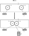
- a parameter to evaluate the size of a droplet is its radius (see Figure 1b where R 1 represents the radius of the D 1 droplet and R 2 represents the radius of the D 2 droplet) which can be measured for example by a brightfield high-speed camera.
- measuring the size of a droplet in the framework of the present invention is performed by measuring its radius.
- the ion channel (3) is inactive if only one droplet is obtained at equilibrium state or if there is a significant change of size of the droplets, e.g. the shrinking one which is the droplet having the initial lower concentration in ions I, and more particularly if the difference of the radius of at least one droplet (in particular the droplet having the initial lower concentration in ions I) between its initial state (R i ) and its equilibrium state (R e ) reached after being contacted with the other droplet is at least 10%, notably at least 20%, preferably at least 30%.
- IR i -R e Ix100/R i should be above 10%, notably above 20%, preferably above 30%.
- the ion channel (3) is active if there is no significant change of size of the droplets, and more particularly if the difference of the radius of at least one droplet (in particular the droplet having the initial lower concentration in ions I) between its initial state (R i ) and its equilibrium state (R e ) after being contacted with the other droplet is less than 10%, notably less than 5%, preferably less than 3%.
- IR i -R e Ix100/R i should be inferior to 10%, notably inferior to 5%, preferably inferior to 3%.
- the ion channel (3) is inactive if only one droplet is obtained at equilibrium state or if the difference of the radius of at least one droplet (in particular the droplet having the initial lower concentration in ions I) between its initial state (R i ) and its equilibrium state (R e ) reached after being contacted with the other droplet is at least 10% and that the ion channel (3) is active if the difference of the radius of at least one droplet (in particular the droplet having the initial lower concentration in ions I) between its initial state (R i ) and its equilibrium state (R e ) reached after being contacted with the other droplet is less than 10%.
- a “ion channel” (3) is a pore-forming membrane protein which allows the flow of ions across the cell membrane.
- the ion channel (3) can be selective or not to an ion, the ion being commonly Na + , K + , Ca 2+ , H + or Cl - .
- any ion channel (3) can be used. It can be notably a sodium channel, a potassium channel (e.g. Kv1.1 to Kv1.5 channel), a calcium channel, a proton channel or a chloride channel or a porine (e.g. ⁇ -hemolysin).
- the ion I will be in particular Na + , K + , Ca 2+ , H + or Cl - , more particularly K + or Cl - . More particularly, the ion I is able to pass through the ion channel (3) to be analysed, i.e. that Na + will be used with a sodium channel, K + will be used with a potassium channel, Ca 2+ will be used with a calcium channel, H + will be used with a proton channel, and Cl - will be used with a chloride channel.
- the "hydrophobic medium” (1) used in the invention is a medium which is not miscible with water and in particular with the aqueous solutions AS 1 and AS 2 mentioned previously.
- an oil such as a vegetable oil (e.g. soybean or mineral oil); triglycerides; silicone oil; a hydrocarbon; squalene or a mixture thereof.
- Triglycerides (also called TG, triacylglycerol, TAG, or triacylglyceride) is an ester derived from glycerol and three fatty acids, which can be identical or different, notably identical.
- a triglyceride has more particularly the following formula (I): wherein A 1 , A 2 and A 3 , identical or different, notably identical, are a hydrocarbon chain of a fatty acid.
- the triglycerides used in the present invention can be in the form of a mixture of various triglycerides, such as triglycerides present in a vegetable oil (for ex. soybean oil).
- fatty acid refers to a linear, saturated or unsaturated carboxylic acid (RCO 2 H) comprising from 4 to 30, such as from 6 to 30, notably from 8 to 28, in particular from 10 to 24, for example from 12 to 22 carbon atoms (including the carbon atom of the carboxylic acid function). It can be notably lauric acid, myristic acid, palmitic acid, staric acid, arachidic acid, myristoleic acid, myristelaidic acid, palmitoleic acid, palmitelaidic acid, oleic acid, elaidic acid, linoleic acid, linolenic acid, ecosenoic acid or arachidonic acid.
- RCO 2 H linear, saturated or unsaturated carboxylic acid
- hydrocarbon chain of a fatty acid refers to the hydrocarbon chain (R) linked to the acid function of the fatty acid (RCO 2 H).
- R thus represents a linear, saturated or unsaturated hydrocarbon chain comprising from 3 to 29, such as from 5 to 29, notably from 7 to 27, in particular from 9 to 23, for example from 11 to 21 carbon atoms.
- hydrocarbon chain lauric acid myristic acid, palmitic acid, staric acid, arachidic acid, myristoleic acid, myristelaidic acid, palmitoleic acid, palmitelaidic acid, oleic acid, elaidic acid, linoleic acid, linolenic acid, ecosenoic acid or arachidonic acid.
- the triglycerides can be in particular triglycerides of formula (I) with A1, A2 and A3, identical or different, representing a linear, saturated or unsaturated hydrocarbon chain comprising from 3 to 23, notably from 5 to 19 carbon atoms.
- the triglycerides can be present in the form of a mixture of various triglycerides, such as those present in a vegetable oil, for ex. soybean oil.
- the hydrocarbon will contain at least 5 carbon atoms, notably at least 10 carbon atoms, in particular from 5 to 40 carbon atoms, such as from 10 to 30 carbon atoms provided that the hydrocarbon is liquid at the temperature at which the method is performed, typically room temperature (i.e. from 15 to 40°C, preferably from 20 to 30°C). It can be notably pentane, decane, hexadecane or squalene, in particular squalene. It can be also in the form of a mixture of hydrocarbons such as a mineral oil.
- the hydrophobic medium (1) will comprise a mixture of triglycerides, notably present in a vegetable oil, such as soybean oil, and hydrocarbon such as squalene, preferably with an amount of hydrocarbon (e.g. squalene) in this mixture of at least 15 % w/w.
- This kind of hydrophobic medium (1) allows improving the stability of the droplets and having a higher contact angle. Having higher contact angles is preferable as the bilayer area will be higher and thus so as the water/ion transfer rates. For example, for 250 ⁇ m-sized droplets, the few minutes time scale of the simulation for total water exchange in Figure 5 was obtained by using a contact angle of 95°/2. Increasing that angle would speed up the exchange rate.
- amphiphilic molecule (2) is a molecule comprising a hydrophilic part and a hydrophobic part.
- the amphiphilic molecules (2) used in the present invention can be in particular lipids, such as phospholipids, glycolipids, cholesterol, and mixtures thereof which are the types of lipids commonly present in cell membranes.
- the lipids can have a natural or synthetic origin. More particularly, the amphiphilic molecules (2) can be phospholipids optionally in mixture with glycolipids and/or cholesterol.
- the amphiphilic molecules (2) will be phospholipids optionally in mixture with cholesterol.
- the amphiphilic molecules (2) will be phospholipids.
- Phospholipids are amphiphilic lipids comprising a phosphoric acid mono- or di-ester moiety as hydrophilic part.
- the phospholipids provide good stability to droplets, and thereupon to the bilayer, and, thanks to their charged headgroups, allow better incorporation of the ion channels (3). They can have a natural or synthetic origin. Preferably, synthetic phospholipids will be used.
- phospholipids can be a phosphatidylcholine (PC), a phosphatidylethanolamine (PE), a phosphatidylglycerol (PG), a phosphatidylserine (PS), a phosphatidic acid (PA), a phosphatidylinositol (PI), or a mixture thereof.
- PC phosphatidylcholine
- PE phosphatidylethanolamine
- PG phosphatidylglycerol
- PS a phosphatidylserine
- PA phosphatidic acid
- PI phosphatidylinositol
- Phospholipids formed with fatty acids having at least 12 carbon atoms offer better stability to the droplets.
- Preferred phospholipids are those comprising a C18 fatty acid (i.e. with a hydrocarbon chain comprising 17 carbon atoms) such as oleic acid.
- the phospholipids will be a PC, a PE, a PS, a PG, or a mixture thereof, such as DOPC (dioleoylphosphatidylcholine), DPPC (dipalmitoylphosphatidylcholine), DOPE (dioleoylphosphatidylethanolamine), DOPS (dioleoylphosphatidylserine notably as a sodium salt), DOPG (dioleoylphosphatidylglycerone notably as a sodium salt) or a mixture thereof.
- DOPC dioleoylphosphatidylcholine
- DPPC dipalmitoylphosphatidylcholine
- DOPE dioleoylphosphatidylethanolamine
- DOPS dioleoylphosphatidylserine notably as a sodium salt
- DOPG dioleoylphosphatidylglycerone notably as a sodium salt
- amphiphilic molecules (2) used in the present invention will be a PC, a PE or a mixture thereof, such as DOPC, DOPE or a mixture thereof.
- a mixture of DOPC and DOPE can be used advantageously with a molar ratio DOPC/DOPE ranging from 99/1 to 30/70.
- a mixture of DOPC and DOPE in a molar ratio DOPC/DOPE ranging from 70/30 to 50/50 gives a particular good stability to the droplets.
- the droplets have a diameter comprised between 0.5 ⁇ m and 1000 ⁇ m, notably comprised between 20 ⁇ m and 500 ⁇ m, and preferably comprised between 50 ⁇ m and 200 ⁇ m.
- the droplets have thus substantially a spherical form.
- the diameter of the droplets can be measured by a brightfield high-speed camera.
- the droplets D 1 and D 2 have substantially the same size, i.e. that the diameter d 1 of the droplet D 1 and the diameter d 2 of the droplet D 2 should advantageously satisfied the following equation: Id 1 -d 2 I/(d 1 +d 2 ) ⁇ 0.9, notably ⁇ 0.5, preferably ⁇ 0.2.
- aqueous solutions AS 1 and AS 2 of the droplets D 1 and D 2 respectively contain advantageously a salt of ion I dissolved in water with respectively a concentration c 1 or c 2 as defined previously.
- the ion I can be more particularly Na + , K + , Ca 2+ , H + or Cl - .
- These ions I can be introduced in the aqueous solution in the form of a salt such as:
- the aqueous solution will contain also counter-ions C of opposite charged in comparison to ions I.
- the nature of the counter-ion C will depend on the salt used to introduce the ion I in the aqueous solution.
- the droplet aqueous solution AS 1 contains no ion I (or only traces).
- c 1 and c 2 are different from 0, they are advantageously substantially different, notably as defined in the following equation: c 1 ⁇ c 2 / c 1 + c 2 > 0.1 , notably ⁇ 0.5 , preferably ⁇ 0.9 . notably ⁇ 0.5, preferably ⁇ 0.9.
- the higher concentration of c 1 and c 2 is comprised between 1 mM and 1 M, notably between 100 mM and 800 mM, preferably between 300 mM and 700 mM.
- one of the concentrations c 1 and c 2 is 0 and the other is different from 0, and is advantageously comprised between 1 mM and 1 M, notably between 100 mM and 800 mM, preferably between 300 mM and 700 mM.
- c 1 0.
- the aqueous solution AS 1 and/or AS 2 could also contain a substance (such as a chemical compound) to be tested for its blocking activity of the ion channel (3) or a dye substance (for ex. red rhodamine dye) in order to be able to visually distinguish the droplets AS 2 from the droplets AS 1 .
- a substance such as a chemical compound
- a dye substance for ex. red rhodamine dye
- Figure 1b represents D 1 and D 2 droplets after contacting, forming thus a bilayer of amphiphilic molecules (2) containing also ion channels (3).
- the various radii (R p , R 1 and R 2 ) can be measured by a brightfield high-speed camera.
- the contact angle ⁇ is comprised between 1 and 90°, notably between 30 and 90°, preferably between 50 and 90°.
- the droplet D 1 surrounded by a monolayer of amphiphilic molecules (2) can be provided by forming a droplet of the aqueous solution AS 1 in the hydrophobic medium (1) which contains the amphiphilic molecules (2).
- the amphiphilic molecules (2) will then spontaneously place themselves at the interface between the aqueous droplet and the hydrophobic medium (1).
- the volume of the hydrophobic medium (1) is at least twice the volume of the aqueous solution AS 1 used to form the droplets.
- the concentration of the amphiphilic molecules (2) in the hydrophobic medium (1) is advantageously at least 0.025 % (w/w).
- the method to provide a droplet D 2 surrounded by a monolayer of amphiphilic molecules (2) comprising the ion channels (3) will depend if the ion channel (3) is used in liposomes or not. Indeed, if the ion channels (3) are not soluble in the aqueous solution AS 2 , it will be necessary to use it in liposomes. Indeed, liposomes will guarantee the safe carriage of the protein (ion channel) without its denaturation. Furthermore, the liposome protein content can be relocated into the oil/water surface at the interface of the droplets with the hydrophobic medium.
- the D 2 droplet surrounded by a monolayer of amphiphilic molecules (2) comprising the ion channels (3) to be analyzed can be provided by forming a droplet of the aqueous solution AS 2 which contains also the ion channels (3) in the hydrophobic medium (1) which contains the amphiphilic molecules (2).
- the amphiphilic molecules (2) will then spontaneously place themselves at the interface between the aqueous droplet and the hydrophobic medium (1) to form a monolayer of amphiphilic molecules (2) around the D 2 droplet.
- the ion channel (3) tends to migrate and associate with the hydrophobic medium (1) forming the monolayer of amphiphilic molecules (2) and ion channels (3) around the droplet.
- the droplet of the aqueous solution AS 2 which contains also the ion channels (3) in proteo-liposomes will be formed in the hydrophobic medium (1) containing no amphiphilic molecule (2).
- the droplet thus formed is then shaken (notably vortexed or by flowing the droplets D 2 in a zig-zag array in microfluidics) in order to explode the proteo-liposomes on the water-hydrophobic medium (1) interface so that to place the ion channels (3) at the water-hydrophobic medium (1) interface.
- amphiphilic molecules (2) are added to the hydrophobic medium (1) in order to form spontaneously the monolayer of amphiphilic molecules (2) which contains also the ion channels (3) to be analysed around the droplet.
- the volume of the hydrophobic medium (1) is at least twice the volume of the aqueous solution AS 2 used to form the droplets.
- the concentration of the amphiphilic molecules (2) in the hydrophobic medium (1) is advantageously at least 0.025 % (w/w).
- the monolayer formed around the droplet D 2 can contain at least one ion channel (3) for 100 millions, notably 80 millions, preferably 50 millions, of amphiphilic molecules (2).
- the two droplets D 1 and D 2 are brought into contact by any means (for ex. by mixing binary droplets or by changing the flow rate in microfluidics) used in the art.
- droplets D 1 and D 2 are formed separately in a hydrophobic medium (1) before merging together the two hydrophobic media (1) containing respectively droplets D 1 and D 2 .
- the measurement of the size of the droplets at different time points or at equilibrium by determining droplets contour variation, or the determinations of the number of final droplet(s) (one big droplet or two droplets) at the equilibrium allows determining if the ion channel (3) is active or not.
- Such a measurement or determination can be performed by an optical detection system such as a camera, notably in transmission light or brightfield, and more particularly a brightfield high-speed camera, notably by measuring the radius of the droplets.
- the size of the contacted droplets will be measured when the two droplets are brought into contact (initial state) and at the equilibrium (or the number of droplets will be determined if there remains only one droplet at the equilibrium) and these two measures will be compared.
- the ion channel (3) is inactive if only one droplet is obtained at equilibrium state or if there is a significant change of size of the droplets, e.g. the shrinking one which is the droplet having the initial lower concentration in ions I, and more particularly if the difference of the radius of at least one droplet (in particular the droplet having the initial lower concentration in ions I) between its initial state (R i ) and its equilibrium state (R e ) reached after being contacted with the other droplet is at least 10%, notably at least 20%, preferably at least 30%.
- IR i -R e Ix100/R i should be above 10%, notably above 20%, preferably above 30%.
- the ion channel (3) is active if there is no significant change of size of the droplets, and more particularly if the difference of the radius of at least one droplet (in particular the droplet having the initial lower concentration in ions I) between its initial state (R i ) and its equilibrium state (R e ) after being contacted with the other droplet is less than 10%, notably less than 5%, preferably less than 3%.
- IR i -R e Ix100/R i should be inferior to 10%, notably inferior to 5%, preferably inferior to 3%.
- the ion channel (3) is inactive if only one droplet is obtained at equilibrium state or if the difference of the radius of at least one droplet (in particular the droplet having the initial lower concentration in ions I) between its initial state (R i ) and its equilibrium state (R e ) reached after being contacted with the other droplet is at least 10% and that the ion channel (3) is active if the difference of the radius of at least one droplet (in particular the droplet having the initial lower concentration in ions I) between its initial state (R i ) and its equilibrium state (R e ) reached after being contacted with the other droplet is less than 10%.
- the determination of the activity of the ion channels (3) to be analyzed can be performed by a simple binary readout (one big droplet or two droplets at the equilibrium) by means of an optical detection system, such as a camera, notably in transmission light or brightfield, and more particularly a brightfield high-speed camera.
- an optical detection system such as a camera, notably in transmission light or brightfield, and more particularly a brightfield high-speed camera.
- the time necessary to reach the equilibrium will depend on various parameters such as the initial size of the droplets D 1 and D 2 , the contact angle, the concentrations c 1 and c 2 , the nature of the ion channel (3) to be analyzed, the number of ion channels (3) in the bilayer, etc.
- the initial size of the droplets D 1 and D 2 the contact angle
- the concentrations c 1 and c 2 the nature of the ion channel (3) to be analyzed
- the number of ion channels (3) in the bilayer etc.
- the method according to the invention can be performed in high-throughput, in particular in a HTS method.
- Such a high-throughput method can be performed by the use of a microfluidic analysis system (4).
- Such an analysis system (4) could comprise:
- microfluidic channel (5) is meant in the context of the present invention a channel having a cross section which has dimensions in the micrometer range.
- the microfluidic channel (5) will have a width and a depth comprised between 10 ⁇ m and 1000 ⁇ m, in particular between 10 ⁇ m and 400 ⁇ m, notably between 10 ⁇ m and 250 ⁇ m, preferably between 10 and 100 ⁇ m or between 150 and 250 ⁇ m.
- the length of the microfluidic channel (5) can be in the centimeter, decimeter or even meter range.
- Droplets D 1 are formed in the microfluidic device (18) by any means from the hydrophobic medium (1) containing the amphiphilic molecules (2) and the aqueous solution AS 1 which enter the microfluidic device (18) through its two first inlets (11a,11b). Once formed, droplets D 1 flow in the hydrophobic medium (1) until the inlet (6) of the microfluidic channel (5). Droplets D 1 can be formed for example by shearing a flow of aqueous solution AS 1 with two flows of hydrophobic medium (1) containing the amphiphilic molecules (2), method well-known to the one skilled in the art. The monolayer of amphiphilic molecules (2) is then formed spontaneously around the droplets D 1 .
- Droplets D 2 are formed in a similar way as for droplets D 2 . Once formed, droplets D 2 flow in the hydrophobic medium (1) until the inlet (6) of the microfluidic channel (5).
- the droplets D 2 will be formed in the microfluidic device (18) by any means from the hydrophobic medium (1) containing the amphiphilic molecules (2) and the aqueous solution AS 2 further containing the ion channels (3) which enter the microfluidic device (18) through its two last inlets (11c,11d).
- Droplets D 2 can be formed also by shearing a flow of aqueous solution AS 2 further containing the ion channels (3) with two flows of hydrophobic medium (1) containing the amphiphilic molecules (2), method well-known to the one skilled in the art.
- the monolayer of amphiphilic molecules (2) associated to ion channels (3) is then formed spontaneously around the droplets D 2 .
- the droplets D 2 When the ion channels (3) are present in liposomes, the droplets D 2 will be formed in the microfluidic device (18) by any means from the hydrophobic medium (1) (containing no amphiphilic molecule (2)) and the aqueous solution AS 2 further containing the ion channels (3) which enter the microfluidic device (18) through two inlets (11c,11d). Droplets D 2 can be formed notably by shearing a flow of aqueous solution AS 2 further containing the ion channels (3) with two flows of hydrophobic medium (1) (containing no amphiphilic molecule (2)).
- droplets D 2 Once the droplets D 2 are formed, they need to be shaken in order to explode the liposomes containing the ion channels (3) on the water-hydrophobic medium interface so that to place the ion channels (3) at the water-hydrophobic medium interface.
- droplets D 2 can flow in a zig-zag array (notably a zig-zag microfluidic channel).
- amphiphilic molecules (2) (optionally in mixture with hydrophobic medium (1)) have to be added to the flow of hydrophobic medium (1) containing the droplets D 2 by another inlet (11e) of the microfluidic device. This allows the spontaneous formation of the monolayer of amphiphilic molecules (2) associated to ion channels (3) around the droplets D 2 .
- droplets D 1 and D 2 enter alternately the main microfluidic channel (5) through its inlet (6) and flow through this main microfluidic channel (5) from its inlet (6) to its outlet (7). Pairs of droplets D 1 and D 2 are brought into contact in this main microfluidic channel (5), for ex. by varying the pressure / flow rate in the microfluidic channel (5), so as to form the bilayer of amphiphilic molecules (2) containing the ion channels (3).
- the main microfluidic channel (5) will be linear.
- the pairs of droplets D 1 and D 2 will flow through this main microfluidic channel (5) until the end of this microfluidic channel (5) where the size or number, preferably the number, of the "pair of droplets" will be analyzed by the detection device (17).
- the main microfluidic channel (5) should thus be sufficiently long so that equilibrium is reached when the "pair of droplets" arrived at the end of main microfluidic channel (5) to be analyzed by the detection device (17).
- a valve controlled by the detection device (17) can be present at the end of the main microfluidic channel (5).
- the detection device (17) will move the valve so that the droplets containing active ion channels (3) are moved towards the first receiver container (9) through the first outlet (8a) of the microfluidic device (18) and the droplets containing inactive ion channels (3) are moved towards the second receiver container (10) through the second outlet (8b) of the microfluidic device (18).
- the detection device (18) comprises advantageously an optical detection system, such as a camera, notably in transmission light or brightfield, and more particularly a brightfield high-speed camera. It can be more particularly a basic sorting device having ideally an optical detection system, such as a camera, notably a high speed camera, and more particularly a brightfield high-speed camera, coupled with a software capable of analysing the droplet size/number to provide a binary value allowing for example moving a valve in the required position in order to direct the "pair of droplets" in the appropriate receive container (9,10).
- microfluidic valve which will be only timely opened for allowing the sorting of a droplet that has been positive for ion channel activity by the software; the software would for example send a 1 message to the valve for opening and a 0 message for closing; sorted droplets will simply be collected after the valve where the valve signal will be 1.
- Such a microfluidic analysis system (4) will allow forming and testing numerous pairs of droplets with an automated treatment of the results.
- it will be possible to introduce various substances to be analyzed in aqueous solutions S 1 and/or S 2 .
- the droplets containing inactive ion channels (3) collected at the second outlet (8b) will correspond to the droplets containing substances capable to block the ion channels (3).
- An analysis of these droplets notably by mass spectrometry (possibly after HPLC to separate the various substances), will allow the identification of the active substances.
- Two water-in-oil emulsion droplets (Oil/Water (8/1, v/v) were separately formed before being mixed and vortexed.
- the first class of water droplets contained 0 mM KCl and is formed in the oil phase (soybean oil/squalene (50/50 w/w)) containing phospholipids from Avanti Polar Lipids (DOPC/DOPE (70/30 mol/mol) at 0.2% w/w relative to the oil), which decorated the droplet interface.
- Phospholipids (PLs) in chloroform were placed in a glass vial so as to evaporate under vacuum the chloroform; after they were dried and oil is added in a sufficient amount to reach the target concentration; the mixture is thoroughly mixed - e.g. 20 ⁇ l of PLs at 25 mg/ml were dried and 250 mg of the oil was added to reach 0.2%.
- the different solutions (aqueous and oil phases) were then mixed and vortexed (50 ⁇ L of the aqueous phase + 350 ⁇ L of the oil phase) to generate population D 1 of droplets.
- the second class of aqueous droplets containing 0.5 M KCl or 0.25 M MgCl 2 (and red rhodamine dye (for eye visualization) and differentiation from the D 1 class of droplets) (the previous droplet fabrication procedure was repeated with 0.5 M KCl or 0.25 M MgCl 2 and 0.01% w/w of red rhodamine dye in the water in place of water) was formed in the oil devoid of phospholipids (50 ⁇ L of the aqueous phase + 350 ⁇ L of the oil phase) in order to reach a population D 2 of droplets.
- the two emulsions were then mixed together.
- the equilibrium of red and white droplets (i.e. D 2 and D 1 droplets respectively) forming DIBs was then followed over time.
- White droplets of lower osmolarity completely transferred their water content to the red droplets of higher osmolarity after 30 min contact.
- ion channels in the DIBs were repeated with the presence of ion channels in the DIBs, and more particularly the following ion channels: Kv1.5 (0.01 mg/ml) and Alpha haemolysin (0.02 mg/ml) respectively.
- Alpha haemolysin is soluble in the aqueous phase and thus the ion channels were directly added in the aqueous phase (the one formed with MgCl 2 ) used to generate the D 2 droplets.
- Kv1.5 channels are not soluble in the aqueous phase and thus were bound to DPhPC (diphytanoylphosphatidylcholine) liposomes as described below.
- Human Kv1.5 channel (gene ID: NM_002234.3) 1.843 kb cDNAs was subcloned into pIVEX 2.4a plasmid (Roche Applied Science, Germany) between Ncol and Xhol restriction sites. This construction encodes the Kv1.5 channel with 6 Histidine tag amino acids upstream of the first methionine.
- RNA transcription and protein translation were performed in a reaction volume of 2 mL as described by Schwartz et al. 2007, using ribosomes of E. coli, 16 ⁇ g/mL plasmid and 0.9 mM Brij® 35 detergent.
- the cell free synthesis reaction mixture was then purified using a NiNTA column eluted with 300 mM imidazole and 0.09 mM Brij® 35.
- OG Octyl Glucoside
- the detergent was then removed by dialysis using MIDI GeBa flex-tube (MWCO 3.5 kDa) in a 1L buffer containing 1g biobeads (Biorad, USA).
- the proteo-liposomes were then added in the aqueous phase (the one formed with KCl) used to generate the D 2 droplets.
- the D 2 droplets containing the proteo-liposomes were thoroughly vortexed in the oil solution; in microfluidics, this step is achieved by forming the droplets in the oil and flowing them in a zig-zag array.
- This step allows exploding the proteo-liposomes to the oil-water interface and relocating the proteins (ion channels) at the oil-water interface.
- this step was not necessary.
- the proteins (Kv1.5 or Alpha haemolysin ion channels) were located in the DIB bilayer and allow K + (for Kv1.5) or Cl - (for Alpha haemolysin) ion flux establishment in a direction opposite to the water flux, which goes from D 1 to D 2 droplets. After one hour, we assumed equilibrium was reached, D 1 -DS 2 adhering droplets reached equi-osmolarity and were still visible with no substantial change in their initial size.
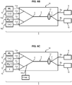
- Figure 7 illustrates the formation of pairs of D 1 /D 2 droplets (involving Kv1.5 channels) in a continuous manner thanks to a microfluidic device.
- AS 1 and AS 2 aqueous phases and the oil phases are as defined above in the case of Kv1.5 channels, except that the oil phase, where the droplets are formed with AS 2 aqueous phase, does not contain phospholipids.
- a flow of D 2 droplets (light grey) in an oil phase and a flow of D 1 droplets (dark grey) in another oil phase are formed and meet at the entry of a main channel (here a linear channel) so as to allow the alternate entry of the D 1 and D 2 droplets which can then be brought into contact, notably by varying the pressure.
- a main channel here a linear channel
- Pairs of D 1 and D 2 droplets are thus formed in the main channel and analysed at the end of the main channel (when equilibrium is reached) by optical readout.
- the number of droplets is analysed: one droplet means an inactive channel and will be directed towards a first outlet, whereas two droplets mean an active channel and will be directed towards a second outlet.
Landscapes
- Health & Medical Sciences (AREA)
- Life Sciences & Earth Sciences (AREA)
- Chemical & Material Sciences (AREA)
- Molecular Biology (AREA)
- Engineering & Computer Science (AREA)
- Hematology (AREA)
- Biomedical Technology (AREA)
- Immunology (AREA)
- Urology & Nephrology (AREA)
- Analytical Chemistry (AREA)
- General Health & Medical Sciences (AREA)
- Physics & Mathematics (AREA)
- Cell Biology (AREA)
- Medicinal Chemistry (AREA)
- Pathology (AREA)
- Microbiology (AREA)
- Biotechnology (AREA)
- Biochemistry (AREA)
- Food Science & Technology (AREA)
- General Physics & Mathematics (AREA)
- Proteomics, Peptides & Aminoacids (AREA)
- Fluid Mechanics (AREA)
- Dispersion Chemistry (AREA)
- Clinical Laboratory Science (AREA)
- Chemical Kinetics & Catalysis (AREA)
- Biophysics (AREA)
- Endocrinology (AREA)
- Measuring Or Testing Involving Enzymes Or Micro-Organisms (AREA)
- Investigating Or Analyzing Non-Biological Materials By The Use Of Chemical Means (AREA)
Claims (15)
- Verfahren zum Analysieren der Aktivität eines Ionenkanals (3), das die folgenden Schritte umfasst:(i) Bereitstellen, in einem hydrophoben Medium (1), eines ersten Tropfens D1 einer wässrigen Lösung AS1, die eine Konzentration c1 eines Ions I mit c1 ≥ 0 aufweist, wobei der Tropfen D1 von einer Monoschicht von amphiphilen Molekülen (2) umgeben ist,(ii) im hydrophoben Medium (1), Bereitstellen eines zweiten Tropfens D2 einer wässrigen Lösung AS2, die eine Konzentration c2 des Ions I mit c2 ≥ 0 und c2 + c1 aufweist, wobei der Tropfen D2 von einer Monoschicht der amphiphilen Moleküle (2) umgeben ist, die ferner die zu analysierenden Ionenkanäle (3) umfassen,(iii) Bringen des ersten Tropfens D1 und des zweiten Tropfens D2 in Kontakt, um so eine Doppelschicht von amphiphilen Molekülen (2) im Kontaktbereich zu bilden, wobei die Doppelschicht von amphiphilen Molekülen (2) ferner zu analysierende Ionenkanäle (3) enthält, und Messen des Radius von zwei Tropfen, wenn sie in Kontakt gebracht werden, was als Anfangszustand bezeichnet wird, und(iv) Halten des ersten Tropfens D1 und des zweiten Tropfens D2 in Kontakt, bis das Gleichgewicht erreicht wird, und Messen des Radius der zwei in Kontakt gebrachten Tropfen oder Bestimmen der Anzahl der resultierenden Tropfen im Gleichgewicht, das als Gleichgewichtszustand bezeichnet wird,wobei der Ionenkanal (3) inaktiv ist, wenn die Differenz des Radius von mindestens einem Tropfen zwischen dem Anfangszustand und seinem Gleichgewichtszustand mindestens 10 % beträgt, oder wenn nur ein Tropfen im Gleichgewichtszustand erhalten wird.
- Verfahren nach Anspruch 1, wobei der Ionenkanal (3) ein Natriumkanal, ein Kaliumkanal, ein Calciumkanal, ein Protonenkanal oder ein Chloridkanal ist und das Ion I Na+, K+, Ca2+, H+ bzw. Cl- ist.
- Verfahren nach einem der Ansprüche 1 bis 2, wobei das hydrophobe Medium (1) ein Öl ist, wie zum Beispiel ein Pflanzenöl; Triglyceride; Silikonöl; ein Kohlenwasserstoff oder eine Mischung derselben; und ist vorzugsweise eine Mischung von Triglyceriden und Kohlenwasserstoff, wie zum Beispiel Squalen.
- Verfahren nach einem der Ansprüche 1 bis 3, wobei die amphiphilen Moleküle (2) Phospholipide, Gylcolipide, Cholesterol oder eine Mischung derselben sind.
- Verfahren nach Anspruch 4, wobei die amphiphilen Moleküle (2) Phospholipide sind und spezieller Phosphatidylcholine (PC), Phosphatidylethanolamine (PE) oder eine Mischung derselben.
- Verfahren nach Anspruch 5, wobei die amphiphilen Moleküle (2) ausgewählt werden aus Dioleoylphosphatidylcholin (DOPC), Dipalmitoylphosphatidylcholin (DPPC), Dioleoylphosphatidylethanolamin (DOPE), Dioleoylphosphatidylserin (DOPS), insbesondere als Natriumsalz, Dioleoylphosphatidylglycerol (DOPG), insbesondere als Natriumsalz und Mischungen derselben, insbesondere ausgewählt aus DOPC, DOPE und Mischungen derselben.
- Verfahren nach einem der Ansprüche 1 bis 6, wobei der erste und zweite Tropfen D1 und D2 einen Durchmesser haben, der zwischen 0,5 µm und 1000 µm liegt, insbesondere zwischen 20 µm und 500 µm, vorzugsweise zwischen 50 µm und 200 µm.
- Verfahren nach einem der Ansprüche 1 bis 7, wobei die Konzentration c1 oder c2 0 ist, oder die Konzentrationen c1 und c2 erfüllen die folgende Gleichung: |c1-c2|/(c1+c2| > 0,1, insbesondere ≥0,5, vorzugsweise ≥0,99.
- Verfahren nach einem der Ansprüche 1 bis 8, wobei die höhere Konzentration von c1 und c2 zwischen 1 mM und 1 M, insbesondere zwischen 100 mM und 800 mM, vorzugsweise zwischen 300 mM und 700 mM liegt.
- Verfahren nach einem der Ansprüche 1 bis 9, wobei Schritt (i) ausgeführt wird durch:- Bereitstellen einer wässrigen Lösung AS1,- Bereitstellen des hydrophoben Mediums (1), das ferner die amphiphilen Moleküle (2) enthält,- Bilden eines Tropfens der wässrigen Lösung AS1 im hydrophoben Medium (1), in der die amphiphilen Moleküle (2) vorhanden sind.
- Verfahren nach einem der Ansprüche 1 bis 10, wobei die Ionenkanäle (3) in der wässrigen Lösung AS2 löslich sind, und Schritt (ii) wird ausgeführt durch:- Bereitstellen der wässrigen Lösung AS2, die ferner die Ionenkanäle (3) enthält,- Bereitstellen des hydrophoben Mediums (1), das ferner die amphiphilen Moleküle (2) enthält, und- Bilden eines Tropfens der wässrigen Lösung AS2, die die Ionenkanäle (3) im hydrophoben Medium (1) enthält, welches die amphiphilen Moleküle (2) enthält.
- Verfahren nach einem der Ansprüche 1 bis 10, wobei Schritt (ii) ausgeführt wird durch:- Bereitstellen der wässrigen Lösung AS2, die ferner die Ionenkanäle (3) enthält, welche in Liposomen vorhanden sind,- Bilden eines Tropfens der wässrigen Lösung AS2, die die Ionenkanäle (3) enthält, welche in Liposomen im hydrophoben Medium (1) vorhanden sind, welches keine amphiphilen Moleküle (2) enthält,- Schütteln des so gebildeten Tropfens, und- Zufügen der amphiphilen Moleküle (2) zum hydrophoben Medium (1).
- Verfahren nach einem der Ansprüche 1 bis 12, wobei der Radius der Tropfen mittels einer optischen Nachweisvorrichtung, wie zum Beispiel einer Kamera und insbesondere einer Hellfeld-Hochgeschwindigkeitskamera, gemessen wird.
- Verfahren nach einem der Ansprüche 1 bis 13, das mit hohem Durchsatz verwendet wir, wie zum Beispiel bei einem Screening mit hohem Durchsatz.
- Verfahren nach einem der Ansprüche 1 bis 14, das mittels eines mikrofluidischen Analysesystem (4) ausgeführt wird, umfassend:- eine mikrofluidische Vorrichtung (18), umfassend:▪ einen mikrofluidischen Hauptkanal (5), der einen Einlass (6) und einen Auslass (7) umfasst, durch die alternierend Tropfen D1 und D2 in das hydrophobe Medium (1) vom Einlass (6) zum Auslass (7) des mikrofluidischen Kanals (5) fließen kann,▪ mindestens zwei Auslässe (8a, 8b), die mit dem Auslass (7) des mikrofluidischen Hauptkanals (5) verbunden sind, und wobei:der erste Auslass (8a) der mikrofluidischen Vorrichtung (18) ferner mit einem ersten Empfangsbehälter (9) verbunden ist, der dazu vorgesehen ist, die Tropfen aufzunehmen, die als aktive Ionenkanäle (3) enthaltend analysiert werden, undder zweite Auslass (8b) der mikrofluidischen Vorrichtung (18) ist ferner mit einem zweiten Empfangsbehälter (10) verbunden, der dazu vorgesehen ist, die Tropfen aufzunehmen, die als inaktive Ionenkanäle (3) enthaltend analysiert werden,▪ mindestens vier Einlässe (11a, 11b, 11c, 11d), die mit dem Einlass (6) des mikrofluidischen Kanals (5) verbunden sind, und wobei:die ersten zwei Einlässe (11a, 11b) der mikrofluidischen Vorrichtung (18) sind ferner jeweils mit einem Reservoir (12) verbunden, das dafür vorgesehen ist, die wässrige Lösung AS1 zu enthalten, und mit einem Reservoir (14), das dafür vorgesehen ist, das hydrophobe Medium (1) und die amphiphilen Moleküle (2) zu enthalten, unddie letzten zwei Einlässe (11c, 11d) sind ferner jeweils mit einem Reservoir (13) verbunden, das dafür vorgesehen ist, die wässrige Lösung AS2 und die Ionenkanäle (3) zu enthalten, die optional in Liposomen vorhanden sind, und mit einem Reservoir (14 oder 15), die dafür vorgesehen sind, das hydrophobe Medium (1) und optional die amphiphilen Moleküle (2) zu enthalten,wobei, wenn die Ionenkanäle (3) in der wässrigen Lösung AS2 in Liposomen vorhanden sind, die mikrofluidische Vorrichtung (18) einen fünften Einlass (11e) umfasst, der mit dem Einlass (6) des mikrofluidischen Hauptkanals (5) und mit einem Reservoir (16) verbunden ist, welcher dafür vorgesehen ist, die amphiphilen Moleküle (2) und optional das hydrophobe Medium (1) zu enthalten.- mindestens drei Reservoire zum Enthalten in dieser Reihenfolge ausgelegt sind (i) die wässrige Lösung AS1 (Reservoir 12), (ii) die wässrige Lösung AS2 und die Ionenkanäle (3), die optional in Liposomen vorhanden sind (Reservoir 13), und (iii) das hydrophobe Medium (1) und/oder die amphiphilen Moleküle (2) (Reservoir(e) 14, 15, 16),- mindestens zwei Empfangsbehälter (9, 10), die zum Aufnehmen in dieser Reihenfolge ausgelegt sind (i) die Tropfen, die als aktive Ionenkanäle (3) enthaltend analysiert werden, und (ii) die Tropfen, die als inaktive Ionenkanäle (3) enthaltend analysiert werden, und- eine Nachweisvorrichtung (17), die am Ende des mikrofluidischen Hauptkanals (5) platziert ist.
Applications Claiming Priority (2)
| Application Number | Priority Date | Filing Date | Title |
|---|---|---|---|
| EP15306823 | 2015-11-17 | ||
| PCT/EP2016/078013 WO2017085189A1 (en) | 2015-11-17 | 2016-11-17 | A method for analyzing the activity of an ion channel |
Publications (2)
| Publication Number | Publication Date |
|---|---|
| EP3377906A1 EP3377906A1 (de) | 2018-09-26 |
| EP3377906B1 true EP3377906B1 (de) | 2019-12-25 |
Family
ID=54780233
Family Applications (1)
| Application Number | Title | Priority Date | Filing Date |
|---|---|---|---|
| EP16797916.0A Active EP3377906B1 (de) | 2015-11-17 | 2016-11-17 | Verfahren zur analyse der aktivität eines ionenkanals |
Country Status (4)
| Country | Link |
|---|---|
| US (1) | US10690680B2 (de) |
| EP (1) | EP3377906B1 (de) |
| CN (1) | CN108780096B (de) |
| WO (1) | WO2017085189A1 (de) |
Families Citing this family (8)
| Publication number | Priority date | Publication date | Assignee | Title |
|---|---|---|---|---|
| WO2019045615A1 (en) * | 2017-09-04 | 2019-03-07 | Bergstroem Christel | DEVICE FOR DISPOSING OF INTESTINAL MEDICINE IN VITRO |
| JP7179330B2 (ja) * | 2019-01-28 | 2022-11-29 | 国立大学法人福井大学 | 脂質二重膜の水透過性の評価システム、脂質二重膜の水透過性の評価方法、および、脂質二重膜の水透過性を制御する薬剤のスクリーニング方法 |
| WO2020167862A1 (en) | 2019-02-12 | 2020-08-20 | 10X Genomics, Inc. | Systems and methods for transfer of reagents between droplets |
| US12305239B2 (en) | 2019-02-12 | 2025-05-20 | 10X Genomics, Inc. | Analysis of nucleic acid sequences |
| US12275993B2 (en) | 2019-02-12 | 2025-04-15 | 10X Genomics, Inc. | Analysis of nucleic acid sequences |
| EP3756762B1 (de) * | 2019-06-25 | 2025-08-06 | Sharp Life Science (EU) Limited | Verfahren zur manipulation von tropfen in einer mikrofluidischen vorrichtung |
| EP4470670A1 (de) * | 2019-06-25 | 2024-12-04 | Sharp Life Science (EU) Limited | Mikrofluidische vorrichtung und verfahren zur manipulation von tröpfchen darin |
| EP4698900A1 (de) * | 2023-04-19 | 2026-02-25 | Paris Sciences et Lettres | Verfahren zur durchführung morphologischer veränderungen extrazellulärer organellenvesikel zur untersuchung der transportaktivität von membrantransportproteinen und damit zusammenhängenden verfahren |
Family Cites Families (7)
| Publication number | Priority date | Publication date | Assignee | Title |
|---|---|---|---|---|
| US7205099B2 (en) * | 2002-05-31 | 2007-04-17 | John Cuppoletti | Methods for studying ion channels incorporated onto a solid-supported membrane |
| JP5114702B2 (ja) | 2005-07-29 | 2013-01-09 | 国立大学法人 東京大学 | 両親媒性単分子膜の接触による二分子膜の形成方法およびその装置 |
| GB0614835D0 (en) | 2006-07-26 | 2006-09-06 | Isis Innovation | Formation of bilayers of amphipathic molecules |
| US8293339B2 (en) | 2007-09-17 | 2012-10-23 | Sri International, Inc. | Droplet bilayers |
| GB0913823D0 (en) * | 2009-08-07 | 2009-09-16 | Isis Innovation | Bilayers |
| US9150598B2 (en) * | 2011-10-05 | 2015-10-06 | The Regents Of The University Of California | Masking apertures enabling automation and solution exchange in sessile bilayers |
| US20140023697A1 (en) * | 2012-07-17 | 2014-01-23 | Iona College | Methods and systems for nanomembrane crystallization |
-
2016
- 2016-11-17 WO PCT/EP2016/078013 patent/WO2017085189A1/en not_active Ceased
- 2016-11-17 CN CN201680079246.XA patent/CN108780096B/zh not_active Expired - Fee Related
- 2016-11-17 EP EP16797916.0A patent/EP3377906B1/de active Active
- 2016-11-17 US US15/776,685 patent/US10690680B2/en active Active
Non-Patent Citations (1)
| Title |
|---|
| None * |
Also Published As
| Publication number | Publication date |
|---|---|
| US10690680B2 (en) | 2020-06-23 |
| CN108780096A (zh) | 2018-11-09 |
| CN108780096B (zh) | 2021-03-23 |
| US20180356430A1 (en) | 2018-12-13 |
| EP3377906A1 (de) | 2018-09-26 |
| WO2017085189A1 (en) | 2017-05-26 |
Similar Documents
| Publication | Publication Date | Title |
|---|---|---|
| US10690680B2 (en) | Method for analyzing the activity of an ion channel | |
| Lira et al. | Posing for a picture: vesicle immobilization in agarose gel | |
| Simunovic et al. | Celebrating Soft Matter's 10th anniversary: screening of the calcium-induced spontaneous curvature of lipid membranes | |
| Langner et al. | Electrostatics of phosphoinositide bilayer membranes. Theoretical and experimental results | |
| Disalvo et al. | Electrophoretic mobility and zeta potential of liposomes due to arginine and polyarginine adsorption | |
| EP2047266A1 (de) | Bildung von doppelschichten amphipathischer moleküle | |
| WO2011015870A1 (en) | Bilayers | |
| Lopez et al. | Enthalpic effects of chain length and unsaturation on water permeability across droplet bilayers of homologous monoglycerides | |
| Phapal et al. | Spontaneous formation of single component liposomes from a solution | |
| Lira et al. | The Hidden Cost of Fusion: Intrinsic Asymmetry in Vesicle Fusion Restricts Synthetic Cell Growth | |
| Honaryar et al. | Investigating the morphological transitions in an associative surfactant ternary system | |
| Kataoka-Hamai et al. | Determination of the Coverage of Phosphatidylcholine Monolayers Formed at Silicone Oil–Water Interfaces by Vesicle Fusion | |
| Dimitriou et al. | Manipulation of encapsulated artificial phospholipid membranes using sub-micellar lysolipid concentrations | |
| US10816560B2 (en) | Method for analyzing the ability of a substance to cross a bilayer membrane | |
| Huckabay et al. | Hydration effects on membrane structure probed by single molecule orientations | |
| Kataoka-Hamai et al. | Hydrocarbon Penetration into Phospholipid Monolayers Formed at Hydrocarbon–Water Interfaces | |
| Stepanyants | Lipid Nanotubes as a Model for Highly Curved Cellular Membrane Structures | |
| Alessandrini et al. | Model Bio‐Membranes Investigated by AFM and AFS: A Suitable Tool to Unravel Lipid Organization and their Interaction with Proteins | |
| Zabala-Ferrera | Electrostatic Effects on Lipid Bilayer Physicochemal Properties and Vesicle Adhesion | |
| Ahmed | Effects of salt concentrations on the size distribution of giant unilamellar vesicles | |
| Czekalska | Droplet microfluidic systems for formation and studies of lipid bilayers | |
| JP2010048714A (ja) | 分子の分別方法およびそれに用いられる分別チップ | |
| Komorowski | Vesicle Adhesion, Fusion, and Neurotransmitter Uptake Studied by Small-Angle X-ray Scattering | |
| Kang et al. | Molecular dynamics simulations of sphingomyelin-cholesterol bilayers | |
| de Lange | Towards controlled mesomorphic phase behaviour of lipid bilayers |
Legal Events
| Date | Code | Title | Description |
|---|---|---|---|
| STAA | Information on the status of an ep patent application or granted ep patent |
Free format text: STATUS: UNKNOWN |
|
| STAA | Information on the status of an ep patent application or granted ep patent |
Free format text: STATUS: THE INTERNATIONAL PUBLICATION HAS BEEN MADE |
|
| PUAI | Public reference made under article 153(3) epc to a published international application that has entered the european phase |
Free format text: ORIGINAL CODE: 0009012 |
|
| STAA | Information on the status of an ep patent application or granted ep patent |
Free format text: STATUS: REQUEST FOR EXAMINATION WAS MADE |
|
| 17P | Request for examination filed |
Effective date: 20180614 |
|
| AK | Designated contracting states |
Kind code of ref document: A1 Designated state(s): AL AT BE BG CH CY CZ DE DK EE ES FI FR GB GR HR HU IE IS IT LI LT LU LV MC MK MT NL NO PL PT RO RS SE SI SK SM TR |
|
| AX | Request for extension of the european patent |
Extension state: BA ME |
|
| DAV | Request for validation of the european patent (deleted) | ||
| DAX | Request for extension of the european patent (deleted) | ||
| GRAP | Despatch of communication of intention to grant a patent |
Free format text: ORIGINAL CODE: EPIDOSNIGR1 |
|
| STAA | Information on the status of an ep patent application or granted ep patent |
Free format text: STATUS: GRANT OF PATENT IS INTENDED |
|
| INTG | Intention to grant announced |
Effective date: 20190604 |
|
| GRAS | Grant fee paid |
Free format text: ORIGINAL CODE: EPIDOSNIGR3 |
|
| GRAA | (expected) grant |
Free format text: ORIGINAL CODE: 0009210 |
|
| STAA | Information on the status of an ep patent application or granted ep patent |
Free format text: STATUS: THE PATENT HAS BEEN GRANTED |
|
| AK | Designated contracting states |
Kind code of ref document: B1 Designated state(s): AL AT BE BG CH CY CZ DE DK EE ES FI FR GB GR HR HU IE IS IT LI LT LU LV MC MK MT NL NO PL PT RO RS SE SI SK SM TR |
|
| REG | Reference to a national code |
Ref country code: GB Ref legal event code: FG4D |
|
| REG | Reference to a national code |
Ref country code: CH Ref legal event code: EP |
|
| REG | Reference to a national code |
Ref country code: AT Ref legal event code: REF Ref document number: 1217701 Country of ref document: AT Kind code of ref document: T Effective date: 20200115 |
|
| REG | Reference to a national code |
Ref country code: DE Ref legal event code: R096 Ref document number: 602016026988 Country of ref document: DE |
|
| REG | Reference to a national code |
Ref country code: IE Ref legal event code: FG4D |
|
| REG | Reference to a national code |
Ref country code: NL Ref legal event code: MP Effective date: 20191225 |
|
| PG25 | Lapsed in a contracting state [announced via postgrant information from national office to epo] |
Ref country code: LV Free format text: LAPSE BECAUSE OF FAILURE TO SUBMIT A TRANSLATION OF THE DESCRIPTION OR TO PAY THE FEE WITHIN THE PRESCRIBED TIME-LIMIT Effective date: 20191225 Ref country code: LT Free format text: LAPSE BECAUSE OF FAILURE TO SUBMIT A TRANSLATION OF THE DESCRIPTION OR TO PAY THE FEE WITHIN THE PRESCRIBED TIME-LIMIT Effective date: 20191225 Ref country code: NO Free format text: LAPSE BECAUSE OF FAILURE TO SUBMIT A TRANSLATION OF THE DESCRIPTION OR TO PAY THE FEE WITHIN THE PRESCRIBED TIME-LIMIT Effective date: 20200325 Ref country code: BG Free format text: LAPSE BECAUSE OF FAILURE TO SUBMIT A TRANSLATION OF THE DESCRIPTION OR TO PAY THE FEE WITHIN THE PRESCRIBED TIME-LIMIT Effective date: 20200325 Ref country code: GR Free format text: LAPSE BECAUSE OF FAILURE TO SUBMIT A TRANSLATION OF THE DESCRIPTION OR TO PAY THE FEE WITHIN THE PRESCRIBED TIME-LIMIT Effective date: 20200326 Ref country code: SE Free format text: LAPSE BECAUSE OF FAILURE TO SUBMIT A TRANSLATION OF THE DESCRIPTION OR TO PAY THE FEE WITHIN THE PRESCRIBED TIME-LIMIT Effective date: 20191225 Ref country code: FI Free format text: LAPSE BECAUSE OF FAILURE TO SUBMIT A TRANSLATION OF THE DESCRIPTION OR TO PAY THE FEE WITHIN THE PRESCRIBED TIME-LIMIT Effective date: 20191225 |
|
| REG | Reference to a national code |
Ref country code: LT Ref legal event code: MG4D |
|
| PG25 | Lapsed in a contracting state [announced via postgrant information from national office to epo] |
Ref country code: RS Free format text: LAPSE BECAUSE OF FAILURE TO SUBMIT A TRANSLATION OF THE DESCRIPTION OR TO PAY THE FEE WITHIN THE PRESCRIBED TIME-LIMIT Effective date: 20191225 Ref country code: HR Free format text: LAPSE BECAUSE OF FAILURE TO SUBMIT A TRANSLATION OF THE DESCRIPTION OR TO PAY THE FEE WITHIN THE PRESCRIBED TIME-LIMIT Effective date: 20191225 |
|
| PG25 | Lapsed in a contracting state [announced via postgrant information from national office to epo] |
Ref country code: AL Free format text: LAPSE BECAUSE OF FAILURE TO SUBMIT A TRANSLATION OF THE DESCRIPTION OR TO PAY THE FEE WITHIN THE PRESCRIBED TIME-LIMIT Effective date: 20191225 |
|
| PG25 | Lapsed in a contracting state [announced via postgrant information from national office to epo] |
Ref country code: EE Free format text: LAPSE BECAUSE OF FAILURE TO SUBMIT A TRANSLATION OF THE DESCRIPTION OR TO PAY THE FEE WITHIN THE PRESCRIBED TIME-LIMIT Effective date: 20191225 Ref country code: RO Free format text: LAPSE BECAUSE OF FAILURE TO SUBMIT A TRANSLATION OF THE DESCRIPTION OR TO PAY THE FEE WITHIN THE PRESCRIBED TIME-LIMIT Effective date: 20191225 Ref country code: PT Free format text: LAPSE BECAUSE OF FAILURE TO SUBMIT A TRANSLATION OF THE DESCRIPTION OR TO PAY THE FEE WITHIN THE PRESCRIBED TIME-LIMIT Effective date: 20200520 Ref country code: CZ Free format text: LAPSE BECAUSE OF FAILURE TO SUBMIT A TRANSLATION OF THE DESCRIPTION OR TO PAY THE FEE WITHIN THE PRESCRIBED TIME-LIMIT Effective date: 20191225 Ref country code: NL Free format text: LAPSE BECAUSE OF FAILURE TO SUBMIT A TRANSLATION OF THE DESCRIPTION OR TO PAY THE FEE WITHIN THE PRESCRIBED TIME-LIMIT Effective date: 20191225 |
|
| PG25 | Lapsed in a contracting state [announced via postgrant information from national office to epo] |
Ref country code: SM Free format text: LAPSE BECAUSE OF FAILURE TO SUBMIT A TRANSLATION OF THE DESCRIPTION OR TO PAY THE FEE WITHIN THE PRESCRIBED TIME-LIMIT Effective date: 20191225 Ref country code: IS Free format text: LAPSE BECAUSE OF FAILURE TO SUBMIT A TRANSLATION OF THE DESCRIPTION OR TO PAY THE FEE WITHIN THE PRESCRIBED TIME-LIMIT Effective date: 20200425 Ref country code: SK Free format text: LAPSE BECAUSE OF FAILURE TO SUBMIT A TRANSLATION OF THE DESCRIPTION OR TO PAY THE FEE WITHIN THE PRESCRIBED TIME-LIMIT Effective date: 20191225 |
|
| REG | Reference to a national code |
Ref country code: DE Ref legal event code: R097 Ref document number: 602016026988 Country of ref document: DE |
|
| PG25 | Lapsed in a contracting state [announced via postgrant information from national office to epo] |
Ref country code: ES Free format text: LAPSE BECAUSE OF FAILURE TO SUBMIT A TRANSLATION OF THE DESCRIPTION OR TO PAY THE FEE WITHIN THE PRESCRIBED TIME-LIMIT Effective date: 20191225 Ref country code: DK Free format text: LAPSE BECAUSE OF FAILURE TO SUBMIT A TRANSLATION OF THE DESCRIPTION OR TO PAY THE FEE WITHIN THE PRESCRIBED TIME-LIMIT Effective date: 20191225 |
|
| PLBE | No opposition filed within time limit |
Free format text: ORIGINAL CODE: 0009261 |
|
| STAA | Information on the status of an ep patent application or granted ep patent |
Free format text: STATUS: NO OPPOSITION FILED WITHIN TIME LIMIT |
|
| REG | Reference to a national code |
Ref country code: AT Ref legal event code: MK05 Ref document number: 1217701 Country of ref document: AT Kind code of ref document: T Effective date: 20191225 |
|
| PG25 | Lapsed in a contracting state [announced via postgrant information from national office to epo] |
Ref country code: SI Free format text: LAPSE BECAUSE OF FAILURE TO SUBMIT A TRANSLATION OF THE DESCRIPTION OR TO PAY THE FEE WITHIN THE PRESCRIBED TIME-LIMIT Effective date: 20191225 |
|
| 26N | No opposition filed |
Effective date: 20200928 |
|
| PG25 | Lapsed in a contracting state [announced via postgrant information from national office to epo] |
Ref country code: AT Free format text: LAPSE BECAUSE OF FAILURE TO SUBMIT A TRANSLATION OF THE DESCRIPTION OR TO PAY THE FEE WITHIN THE PRESCRIBED TIME-LIMIT Effective date: 20191225 Ref country code: IT Free format text: LAPSE BECAUSE OF FAILURE TO SUBMIT A TRANSLATION OF THE DESCRIPTION OR TO PAY THE FEE WITHIN THE PRESCRIBED TIME-LIMIT Effective date: 20191225 |
|
| PG25 | Lapsed in a contracting state [announced via postgrant information from national office to epo] |
Ref country code: PL Free format text: LAPSE BECAUSE OF FAILURE TO SUBMIT A TRANSLATION OF THE DESCRIPTION OR TO PAY THE FEE WITHIN THE PRESCRIBED TIME-LIMIT Effective date: 20191225 |
|
| PG25 | Lapsed in a contracting state [announced via postgrant information from national office to epo] |
Ref country code: MC Free format text: LAPSE BECAUSE OF FAILURE TO SUBMIT A TRANSLATION OF THE DESCRIPTION OR TO PAY THE FEE WITHIN THE PRESCRIBED TIME-LIMIT Effective date: 20191225 |
|
| REG | Reference to a national code |
Ref country code: CH Ref legal event code: PL |
|
| PG25 | Lapsed in a contracting state [announced via postgrant information from national office to epo] |
Ref country code: LU Free format text: LAPSE BECAUSE OF NON-PAYMENT OF DUE FEES Effective date: 20201117 |
|
| REG | Reference to a national code |
Ref country code: BE Ref legal event code: MM Effective date: 20201130 |
|
| PG25 | Lapsed in a contracting state [announced via postgrant information from national office to epo] |
Ref country code: LI Free format text: LAPSE BECAUSE OF NON-PAYMENT OF DUE FEES Effective date: 20201130 Ref country code: CH Free format text: LAPSE BECAUSE OF NON-PAYMENT OF DUE FEES Effective date: 20201130 |
|
| PG25 | Lapsed in a contracting state [announced via postgrant information from national office to epo] |
Ref country code: IE Free format text: LAPSE BECAUSE OF NON-PAYMENT OF DUE FEES Effective date: 20201117 |
|
| REG | Reference to a national code |
Ref country code: DE Ref legal event code: R081 Ref document number: 602016026988 Country of ref document: DE Owner name: UNIVERSITE PARIS CITE, FR Free format text: FORMER OWNERS: COMMISSARIAT A L'ENERGIE ATOMIQUE ET AUX ENERGIES ALTERNATIVES, PARIS, FR; PARIS SCIENCES ET LETTRES - QUARTIER LATIN, PARIS, FR; UNIVERSITE PARIS DIDEROT - PARIS 7, PARIS, FR Ref country code: DE Ref legal event code: R081 Ref document number: 602016026988 Country of ref document: DE Owner name: UNIVERSITE PARIS DIDEROT - PARIS 7, FR Free format text: FORMER OWNERS: COMMISSARIAT A L'ENERGIE ATOMIQUE ET AUX ENERGIES ALTERNATIVES, PARIS, FR; PARIS SCIENCES ET LETTRES - QUARTIER LATIN, PARIS, FR; UNIVERSITE PARIS DIDEROT - PARIS 7, PARIS, FR Ref country code: DE Ref legal event code: R081 Ref document number: 602016026988 Country of ref document: DE Owner name: PARIS SCIENCES ET LETTRES, FR Free format text: FORMER OWNERS: COMMISSARIAT A L'ENERGIE ATOMIQUE ET AUX ENERGIES ALTERNATIVES, PARIS, FR; PARIS SCIENCES ET LETTRES - QUARTIER LATIN, PARIS, FR; UNIVERSITE PARIS DIDEROT - PARIS 7, PARIS, FR Ref country code: DE Ref legal event code: R081 Ref document number: 602016026988 Country of ref document: DE Owner name: COMMISSARIAT A L'ENERGIE ATOMIQUE ET AUX ENERG, FR Free format text: FORMER OWNERS: COMMISSARIAT A L'ENERGIE ATOMIQUE ET AUX ENERGIES ALTERNATIVES, PARIS, FR; PARIS SCIENCES ET LETTRES - QUARTIER LATIN, PARIS, FR; UNIVERSITE PARIS DIDEROT - PARIS 7, PARIS, FR |
|
| PG25 | Lapsed in a contracting state [announced via postgrant information from national office to epo] |
Ref country code: TR Free format text: LAPSE BECAUSE OF FAILURE TO SUBMIT A TRANSLATION OF THE DESCRIPTION OR TO PAY THE FEE WITHIN THE PRESCRIBED TIME-LIMIT Effective date: 20191225 Ref country code: MT Free format text: LAPSE BECAUSE OF FAILURE TO SUBMIT A TRANSLATION OF THE DESCRIPTION OR TO PAY THE FEE WITHIN THE PRESCRIBED TIME-LIMIT Effective date: 20191225 Ref country code: CY Free format text: LAPSE BECAUSE OF FAILURE TO SUBMIT A TRANSLATION OF THE DESCRIPTION OR TO PAY THE FEE WITHIN THE PRESCRIBED TIME-LIMIT Effective date: 20191225 |
|
| PG25 | Lapsed in a contracting state [announced via postgrant information from national office to epo] |
Ref country code: MK Free format text: LAPSE BECAUSE OF FAILURE TO SUBMIT A TRANSLATION OF THE DESCRIPTION OR TO PAY THE FEE WITHIN THE PRESCRIBED TIME-LIMIT Effective date: 20191225 |
|
| PG25 | Lapsed in a contracting state [announced via postgrant information from national office to epo] |
Ref country code: BE Free format text: LAPSE BECAUSE OF NON-PAYMENT OF DUE FEES Effective date: 20201130 |
|
| REG | Reference to a national code |
Ref country code: GB Ref legal event code: 732E Free format text: REGISTERED BETWEEN 20220707 AND 20220713 |
|
| REG | Reference to a national code |
Ref country code: DE Ref legal event code: R081 Ref document number: 602016026988 Country of ref document: DE Owner name: UNIVERSITE PARIS CITE, FR Free format text: FORMER OWNERS: COMMISSARIAT A L'ENERGIE ATOMIQUE ET AUX ENERGIES ALTERNATIVES, PARIS, FR; PARIS SCIENCES ET LETTRES, PARIS, FR; UNIVERSITE PARIS DIDEROT - PARIS 7, PARIS, FR Ref country code: DE Ref legal event code: R081 Ref document number: 602016026988 Country of ref document: DE Owner name: PARIS SCIENCES ET LETTRES, FR Free format text: FORMER OWNERS: COMMISSARIAT A L'ENERGIE ATOMIQUE ET AUX ENERGIES ALTERNATIVES, PARIS, FR; PARIS SCIENCES ET LETTRES, PARIS, FR; UNIVERSITE PARIS DIDEROT - PARIS 7, PARIS, FR Ref country code: DE Ref legal event code: R081 Ref document number: 602016026988 Country of ref document: DE Owner name: COMMISSARIAT A L'ENERGIE ATOMIQUE ET AUX ENERG, FR Free format text: FORMER OWNERS: COMMISSARIAT A L'ENERGIE ATOMIQUE ET AUX ENERGIES ALTERNATIVES, PARIS, FR; PARIS SCIENCES ET LETTRES, PARIS, FR; UNIVERSITE PARIS DIDEROT - PARIS 7, PARIS, FR |
|
| PGFP | Annual fee paid to national office [announced via postgrant information from national office to epo] |
Ref country code: GB Payment date: 20231123 Year of fee payment: 8 |
|
| PGFP | Annual fee paid to national office [announced via postgrant information from national office to epo] |
Ref country code: FR Payment date: 20231120 Year of fee payment: 8 Ref country code: DE Payment date: 20231121 Year of fee payment: 8 |
|
| REG | Reference to a national code |
Ref country code: DE Ref legal event code: R119 Ref document number: 602016026988 Country of ref document: DE |
|
| GBPC | Gb: european patent ceased through non-payment of renewal fee |
Effective date: 20241117 |
|
| PG25 | Lapsed in a contracting state [announced via postgrant information from national office to epo] |
Ref country code: DE Free format text: LAPSE BECAUSE OF NON-PAYMENT OF DUE FEES Effective date: 20250603 |
|
| PG25 | Lapsed in a contracting state [announced via postgrant information from national office to epo] |
Ref country code: GB Free format text: LAPSE BECAUSE OF NON-PAYMENT OF DUE FEES Effective date: 20241117 |
|
| PG25 | Lapsed in a contracting state [announced via postgrant information from national office to epo] |
Ref country code: FR Free format text: LAPSE BECAUSE OF NON-PAYMENT OF DUE FEES Effective date: 20241130 |